Apgar Score, Silverman–And - MDPI
12
Journal of Clinical Medicine Article Discriminant Analysis of Main Prognostic Factors Associated with Hemodynamically Significant PDA: Apgar Score, Silverman–Anderson Score, and NT-Pro-BNP Level Anna V. Permyakova 1 , Artem Porodikov 2 , Alex G. Kuchumov 3, * , Alexey Biyanov 2,4 , Vagram Arutunyan 2 , Evgeniy G. Furman 5 and Yuriy S. Sinelnkov 2 Citation: Permyakova, A.V.; Porodikov, A.; Kuchumov, A.G.; Biyanov, A.; Arutunyan, V.; Furman, E.G.; Sinelnkov, Y.S. Discriminant Analysis of Main Prognostic Factors Associated with Hemodynamically Significant PDA: Apgar Score, Silverman–Anderson Score, and NT-Pro-BNP Level. J. Clin. Med. 2021, 10, 3729. https://doi.org/10.3390/ jcm10163729 Academic Editor: Emmanuel Androulakis Received: 3 July 2021 Accepted: 17 August 2021 Published: 22 August 2021 Publisher’s Note: MDPI stays neutral with regard to jurisdictional claims in published maps and institutional affil- iations. Copyright: © 2021 by the authors. Licensee MDPI, Basel, Switzerland. This article is an open access article distributed under the terms and conditions of the Creative Commons Attribution (CC BY) license (https:// creativecommons.org/licenses/by/ 4.0/). 1 Department of Pediatric Infectious Diseases, Perm State Medical University, 614990 Perm, Russia; [email protected] 2 Federal Center of Cardiovascular Surgery, 614990 Perm, Russia; [email protected] (A.P.); [email protected] (A.B.); [email protected] (V.A.); [email protected] (Y.S.S.) 3 Department of Computational Mathematics, Mechanics, and Biomechanics, Perm National Research Polytechnic University, 614990 Perm, Russia 4 Department of Pediatrics, Perm State Medical University, 614990 Perm, Russia 5 Department of the Intermediate Level and Hospital Pediatrics, Perm State Medical University, 614990 Perm, Russia; [email protected] * Correspondence: [email protected]; Tel.: +7-(342)2-39-17-02 Abstract: Hemodynamically significant patent ductus arteriosus (hsPDA) in premature newborns is associated with a risk of PDA-related morbidities. Classification into risk groups may have a clinical utility in cases of suspected hsPDA to decrease the need for echocardiograms and unnecessary treatment. This prospective observational study included 99 premature newborns with extremely low body weight, who had an echocardiogram performed within the first three days of life. Discriminant analysis was utilized to find the best combination of prognostic factors for evaluation of hsPDA. We used binary logistic regression analysis to predict the relationship between parameters and hsPDA. The cohort’s mean and standard deviation gestational age was 27.6 ± 2.55 weeks, the mean birth weight was 1015 ± 274 g. Forty-six (46.4%) infants had a PDA with a mean diameter of 2.78 mm. Median NT-pro-BNP levels were 17,600 pg/mL for infants with a PDA and 2773 pg/mL in the non-hsPDA group. The combination of prognostic factors of hsPDA in newborns of extremely low body weight on the third day of life was determined: NT-pro-BNP, Apgar score, Silverman–Anderson score (Se = 82%, Sp = 88%). A cut-off value of NT-pro-BNP of more than 8500 pg/mL can predict hsPDA (Se = 84%, Sp = 86%). Keywords: premature newborns; hemodynamically significant patent ductus arteriosus; Apgar score; Silverman–Anderson score; NT-pro-BNP 1. Introduction Patent ductus arteriosus (PDA) is an extra blood vessel found in babies before birth and just after birth. In full-term newborns, it usually closes shortly after birth, and in premature infants it functions in most cases for up to one month [1]. Long-term persistence of the duct contributes to its hemodynamic significance (hs), when the increasing hypoperfusion of the brain and internal organs causes the development of serious long-term outcomes (such as bronchopulmonary dysplasia, intraventricular haemorrhage, necrotizing enterocolitis [2,3], and chronic lung disease [4,5]) associated with an increase in mortality [6]. The current therapeutic approach in the form of prescribing preterm infants non- steroidal anti-inflammatory drugs (COX inhibitors) in order to close the PDA earlier is largely subjective and is associated with side effects, when the harm from treatment may outweigh the benefits [7], since PDA can close spontaneously by 44 weeks of postnatal age or later [8]. As Benitz et al. [9] reported, “The ductus is likely to close without treatment J. Clin. Med. 2021, 10, 3729. https://doi.org/10.3390/jcm10163729 https://www.mdpi.com/journal/jcm
-
Upload
khangminh22 -
Category
Documents
-
view
2 -
download
0
Transcript of Apgar Score, Silverman–And - MDPI

Journal of
Clinical Medicine
Article
Discriminant Analysis of Main Prognostic Factors Associatedwith Hemodynamically Significant PDA: Apgar Score,Silverman–Anderson Score, and NT-Pro-BNP Level
Anna V. Permyakova 1 , Artem Porodikov 2, Alex G. Kuchumov 3,* , Alexey Biyanov 2,4, Vagram Arutunyan 2,Evgeniy G. Furman 5 and Yuriy S. Sinelnkov 2
�����������������
Citation: Permyakova, A.V.;
Porodikov, A.; Kuchumov, A.G.;
Biyanov, A.; Arutunyan, V.; Furman,
E.G.; Sinelnkov, Y.S. Discriminant
Analysis of Main Prognostic Factors
Associated with Hemodynamically
Significant PDA: Apgar Score,
Silverman–Anderson Score, and
NT-Pro-BNP Level. J. Clin. Med. 2021,
10, 3729. https://doi.org/10.3390/
jcm10163729
Academic Editor: Emmanuel
Androulakis
Received: 3 July 2021
Accepted: 17 August 2021
Published: 22 August 2021
Publisher’s Note: MDPI stays neutral
with regard to jurisdictional claims in
published maps and institutional affil-
iations.
Copyright: © 2021 by the authors.
Licensee MDPI, Basel, Switzerland.
This article is an open access article
distributed under the terms and
conditions of the Creative Commons
Attribution (CC BY) license (https://
creativecommons.org/licenses/by/
4.0/).
1 Department of Pediatric Infectious Diseases, Perm State Medical University, 614990 Perm, Russia;[email protected]
2 Federal Center of Cardiovascular Surgery, 614990 Perm, Russia; [email protected] (A.P.);[email protected] (A.B.); [email protected] (V.A.); [email protected] (Y.S.S.)
3 Department of Computational Mathematics, Mechanics, and Biomechanics, Perm National ResearchPolytechnic University, 614990 Perm, Russia
4 Department of Pediatrics, Perm State Medical University, 614990 Perm, Russia5 Department of the Intermediate Level and Hospital Pediatrics, Perm State Medical University,
614990 Perm, Russia; [email protected]* Correspondence: [email protected]; Tel.: +7-(342)2-39-17-02
Abstract: Hemodynamically significant patent ductus arteriosus (hsPDA) in premature newbornsis associated with a risk of PDA-related morbidities. Classification into risk groups may have aclinical utility in cases of suspected hsPDA to decrease the need for echocardiograms and unnecessarytreatment. This prospective observational study included 99 premature newborns with extremely lowbody weight, who had an echocardiogram performed within the first three days of life. Discriminantanalysis was utilized to find the best combination of prognostic factors for evaluation of hsPDA. Weused binary logistic regression analysis to predict the relationship between parameters and hsPDA.The cohort’s mean and standard deviation gestational age was 27.6 ± 2.55 weeks, the mean birthweight was 1015 ± 274 g. Forty-six (46.4%) infants had a PDA with a mean diameter of 2.78 mm.Median NT-pro-BNP levels were 17,600 pg/mL for infants with a PDA and 2773 pg/mL in thenon-hsPDA group. The combination of prognostic factors of hsPDA in newborns of extremely lowbody weight on the third day of life was determined: NT-pro-BNP, Apgar score, Silverman–Andersonscore (Se = 82%, Sp = 88%). A cut-off value of NT-pro-BNP of more than 8500 pg/mL can predicthsPDA (Se = 84%, Sp = 86%).
Keywords: premature newborns; hemodynamically significant patent ductus arteriosus; Apgarscore; Silverman–Anderson score; NT-pro-BNP
1. Introduction
Patent ductus arteriosus (PDA) is an extra blood vessel found in babies before birth andjust after birth. In full-term newborns, it usually closes shortly after birth, and in prematureinfants it functions in most cases for up to one month [1]. Long-term persistence of the ductcontributes to its hemodynamic significance (hs), when the increasing hypoperfusion of thebrain and internal organs causes the development of serious long-term outcomes (such asbronchopulmonary dysplasia, intraventricular haemorrhage, necrotizing enterocolitis [2,3],and chronic lung disease [4,5]) associated with an increase in mortality [6].
The current therapeutic approach in the form of prescribing preterm infants non-steroidal anti-inflammatory drugs (COX inhibitors) in order to close the PDA earlier islargely subjective and is associated with side effects, when the harm from treatment mayoutweigh the benefits [7], since PDA can close spontaneously by 44 weeks of postnatal ageor later [8]. As Benitz et al. [9] reported, “The ductus is likely to close without treatment
J. Clin. Med. 2021, 10, 3729. https://doi.org/10.3390/jcm10163729 https://www.mdpi.com/journal/jcm

J. Clin. Med. 2021, 10, 3729 2 of 12
in infants born at >28 weeks’ gestation (73%) [10] and in those with birth weight >1000 g(94%) [11]”.
Surgical PDA ligation is also significantly associated with a high likelihood of adverseoutcomes and has no long-term benefits [12,13].
Contemporary tactics for managing patients with PDA are observational in nature. Thedecision on therapy is made in the case of hemodynamic significance of PDA. Accordingto the American Academy of Pediatrics, a combination of standard echocardiographicassessments of PDA and biomarkers should be preferred [11,12].
It should be noted that there are several approaches to define hsPDA [14]. The hemo-dynamic effects of the PDA using clinical signs, echocardiographic parameters, and otherobjective assessments can be used to declare a PDA as hemodynamically significant [15].
Brain natriuretic peptide and its inactive fragment N-terminal pro-BNP (NT-pro-BNP)are reliable markers of ventricular dysfunction in adults and children [16]. It was noticedthat BNP and NT-pro-BNP are similarly useful for assessing PDA size in preterm in-fants [17]. Serial BNP measurement is also valuable for monitoring treatment response [18];however, whether plasma BNP has value as a marker for predicting treatment response toCOX inhibitors remains unclear.
Therefore, timely prediction of the hemodynamic significance of PDA in prematureinfants is especially important in the early neonatal period (during the first 48–72 h), andcontributes to the timely appointment of adequate therapy [19,20].
Recently, BNP estimation was shown to be a useful prognostic marker of all-cause mor-tality in extremely low body weight infants with bronchopulmonary dysplasia-associatedpulmonary hypertension [21]. All currently known methods of PDA closure are associatedwith adverse effects, when the treatment harms may outweigh benefits [7]. One of theways to solve the problem is to develop prediction models to permit early identificationprobabilities of persistent PDA [22]. Studies in preterm infants highlighted echocardio-graphic [23], biochemical [24], and clinical markers [8,25] correlating with hemodynamicsignificance of a patent ductus arteriosus. It is believed that prognostic factor combinationsprovide better risk assessment than separate markers [26]. Our study included 40 differentclinical, laboratory, instrumental, and anamnestic criteria, including assessments accordingto the standard Apgar and Silverman–Anderson scores.
Thus, the hypothesis of the present study was to establish a combination of clinicalpredictors with an acceptable degree of specificity and sensitivity predicting hsPDA inpreterm infants on day 3 of life. To solve the problem, discriminant analysis was applied.
2. Materials and Methods2.1. Subjects and Data Collection
A retrospective study was carried out in Perm Krai Perinatal Center. Data on 99 infantsborn from October of 2018 to April of 2020 were collected. Patients with gestational ageranging from 25 to 32 weeks and birth weights ranging from 500 up to 1500 g were includedin the study (Figure 1). Newborns with developmental defects (including combinedcongenital heart defects), infectious diseases, or severe somatic disorders were excludedfrom the study. The Apgar score assessed the state of the newborn child at the fifth minuteof life. To identify current or threatening respiratory distress syndrome during the firsthour of life, an assessment of the respiratory severity score (RSS) designed by Silvermanand Anderson in 1956 to quantify respiratory distress among neonates was used [27].Hedstrom et al. [28] pointed out that this clinical scoring system would be correlated toother laboratory parameters by which respiratory distress is evaluated in high-resourcesettings to prognosticate a patient’s respiratory trajectory.

J. Clin. Med. 2021, 10, 3729 3 of 12J. Clin. Med. 2021, 10, x FOR PEER REVIEW 3 of 12
Figure 1. Study flow chart.
NT-pro-BNP level in plasma was evaluated on the third day of life by reagent set (JSC VECTOR-BEST, Novosibirsk, Russia). Moreover, echocardiogram initial screening to visualize PDA was performed on the third day of life.
The definition of an hsPDA was based on previously established criteria [14,29,30] and was defined by the presence of the following three factors: narrowest ductal diameter >1.5 mm; LA/AO ratio of more than 1.4:1; retrograde descending aortic flow more than or equal to 50% of antegrade flow.
A Vivid Q cardiovascular ultrasound system (General Electric, Boston, MA, USA) was utilized during the study.
Clinical, demographic, and biochemical data were obtained retrospectively from medical records.
Written informed consent was obtained from patients’ parents. The study was ap-proved by the Ethics Committee of S.G. Sukhanov Cardiovascular Center, Perm, Russia.
2.2. Data Analysis In the case of describing quantitative indicators with a normal distribution, the data
obtained were combined into variation series, where the arithmetic mean values (M), standard deviations (SD), and confidence limits (a 95% confidence level) were calculated. Comparisons were performed using a Student’s t-test. Aggregates of quantitative indica-tors, the distribution of which differed from normal, were described using the values of the median (Me) and the lower and upper quartiles (Q1–Q3) and compared by the Mann–Whitney test. The nominal data were described with the indication of absolute values and percentages. Comparison of nominal data was carried out using a Pearson χ2 test.
Preterm newborns with mass less 1500 g, gestation age 25–32 weeks
Study group (n = 99)
hsPDA (n = 46) Non hsPDA (n = 53)
Therapy (n = 44) Ligation (n = 2)
Responder (n = 43) Non-responder (n = 1)
Figure 1. Study flow chart.
NT-pro-BNP level in plasma was evaluated on the third day of life by reagent set(JSC VECTOR-BEST, Novosibirsk, Russia). Moreover, echocardiogram initial screening tovisualize PDA was performed on the third day of life.
The definition of an hsPDA was based on previously established criteria [14,29,30]and was defined by the presence of the following three factors: narrowest ductal diameter>1.5 mm; LA/AO ratio of more than 1.4:1; retrograde descending aortic flow more than orequal to 50% of antegrade flow.
A Vivid Q cardiovascular ultrasound system (General Electric, Boston, MA, USA) wasutilized during the study.
Clinical, demographic, and biochemical data were obtained retrospectively frommedical records.
Written informed consent was obtained from patients’ parents. The study was ap-proved by the Ethics Committee of S.G. Sukhanov Cardiovascular Center, Perm, Russia.
2.2. Data Analysis
In the case of describing quantitative indicators with a normal distribution, the dataobtained were combined into variation series, where the arithmetic mean values (M),standard deviations (SD), and confidence limits (a 95% confidence level) were calculated.Comparisons were performed using a Student’s t-test. Aggregates of quantitative indi-cators, the distribution of which differed from normal, were described using the valuesof the median (Me) and the lower and upper quartiles (Q1–Q3) and compared by theMann–Whitney test. The nominal data were described with the indication of absolute

J. Clin. Med. 2021, 10, 3729 4 of 12
values and percentages. Comparison of nominal data was carried out using a Pearsonχ2 test.
BNP and NT-pro-BNP were log-transformed to obtain a normal distribution foranalysis. The correlation coefficients were obtained to describe the relationship betweenBNP and NT-pro-BNP and the presence and size of a PDA. A p-level of <0.05 was consideredsignificant.
To create a predictive model, the discriminant analysis method was used. The PDAhemodynamical significance indicator was defined as a dependent variable, taking twovalues, which were coded as 1 (yes) and 0 (no), respectively. Quantitative indicators wereused as independent variables.
In total, 40 variables were used for discriminant analysis: anthropometric indicators(weight and height at birth), gestational age, clinical assessment of the cardiovascular andrespiratory systems at birth (Apgar score at 1, 5, and 10 min of life, respiratory severityscore by Silverman–Anderson method during the first hour of life), baby therapy after birth(surfactant, inotropes), type of respiratory support (DUOPAP, NCPAP, mechanical ventila-tion), results of neurosonography and radiography, laboratory data (hemoglobin, platelets,NT-pro-BNP), echocardiographic criteria, maternal history (the number of pregnancies,births, abortions), obstetric history, and somatic diseases of the mother. The groups ofvariables for analysis are provided in Table S1 (see Supplementary material).
The model was created on the principle of the possibility of predicting the dependentvariable based on the values of the measured factor signs and was presented in the form ofthe following equation:
y = a0 + a1x1 + a2x2 + . . . + anxn, (1)
where y is a dependent variable, a0 is a constant, a1 . . . n are regression coefficients, x1 . . . nare independent variables (factor attribute values). The stepwise method selected thediscriminant variables on basis of Wilks’ lambda statistic, and in general, the F value wasset at F Entry = 3.84 and F Removal = 2.71. Assuming that the mean discriminant score ofthe controls was Za, Zb for the cases and Z for the total, then Z = (Za+ Zb)/2. Accordingto the discriminant function, we calculated the discriminant score of Zi for each subject;if Zi > Z, the subject is considered highly likely to be a case, and if Zi ≤ Z, the subject isregarded as a control. The diagnostic efficiency of the model was defined as the proportionof correctly predicted values with respect to the total number of analyzed observations.
We used binary logistic regression analysis to predict the relationships between NT-pro-BNP and hsPDA. The predictive model can be written as follows:
P =1
1 + e−z (2)
z = a0 + a1x1 + a2x2 + . . . + anxn, (3)
where P is the probability of hsPDA, x1...xn are risk factor values, a1...an are regression co-efficients. The selection of independent variables was carried out by the step-by-step directselection method using Wald statistics as an exclusion criterion. The statistical significanceof the resulting model was determined using the χ2 criterion. To assess the diagnosticsignificance of quantitative signs in predicting the outcome, calculated using a regressionmodel, the method of analysis of ROC curves was used. The relationships were graphicallyrepresented by probability curves. Receiver operator characteristic (ROC) analysis wasused to determine the area under the curve (AUC). The highest value of the Youden indexwas used to determine the optimal cut-off point. Sensitivity, specificity, positive predictivevalues (PPV), negative predictive values (NPV), and positive and negative likelihood ra-tios (LR+ and LR−) were calculated. Statistical analysis was performed using IBM SPSSStatistics v.26 software.

J. Clin. Med. 2021, 10, 3729 5 of 12
3. Results3.1. Baseline Characteristics
The clinical characteristics of patients are shown in Table 1. According to the definitionof echo criteria for the significance of a functioning ductus arteriosus, the following datawere obtained: the duct is significant in 46 patients (46.4%) and not significant in 53 (53.6%).Only 26 (26.3%) babies were born naturally; the rest were born operatively (cesareansection). In the hsPDA group, vaginal delivery was more often, 76.1% (35/46) compared to32.4% in the non-hsPDA group (23/54), OR = 4.3 (95% CI 1.8–10.2). Threat of miscarriage asan indicator in infants with hsPDA is noticed in 74% (34/46) of cases and in 39.6% (21/53)of cases in the non-hsPDA group (р = 0.001), OR = 3.46 (95% CI 1.3–9.15). A total of 45(45.4%) mothers received antenatal corticosteroid therapy: 41.3% (19/46) in the hsPDAgroup, 49.0% (26/53) in the non-hsPDA group, OR = 0.73 (95% CI 0.3–1.6). Respiratorysupport was required in 91.3% (42/46) and 84.9% (45/53), OR = 1.8 (95% CI 0.5–6.6) in thehsPDA group and in the non-hsPDA group, respectively. An Apgar score 5 min after birth≤6 points was found in 26.1% (12/46) in the hsPDA group, 9.4% (5/53) in the non-hsPDAgroup, OR = 3.4 (95% CI 1.1–10.5). A Silverman–Anderson score (respiratory severityscore) [27] of respiratory distress over 5 was found in 71.7% (33/46) and 49.1% (26/53),OR = 2.6 (95% CI 4–6.1), in the hsPDA group and non-hsPDA group, respectively (seeTable 1).
Table 1. Clinical characteristics of patients.
Characteristics hsPDA Non-hsPDA p-Value
weight (g) 976 ± 287 1049 ± 261 0.078gestation (weeks) 27.2 ± 2.62 27.9 ± 2.47 0.072
male gender 24 (52.2) 29 (54.7) 0.879vaginal delivery 8 (17.4) 18 (33.9) 0.071
antenatal steroids 19 (41.3) 26 (49.0) 0.493Apgar score (≤6) 12 (26.1) 5 (9.4) 0.021
Silverman–Anderson score(≥5) 33 (71.7) 2 (49.1) 0.022
NT-pro-BNP level (pg/mL) 18.967 ± 12.518 3448 ± 3926 0.001Categorical data are shown as n (%) of each group. Continuous variables are shown as mean ± standarddeviation.
Echocardiographic evaluation of PDA in the preterm infants was assessed on thethird day of life. The average (SD) diameter of the PDA in children with hsPDA is2.78 ± 0.72 mm, in children with non-hsPDA—1.26 ± 0.66 mm (p = 0.001). Pulmonaryhyperperfusion was assessed by an LA/Ao ratio ≥1.5, which indicates a heavy load on theleft side of the heart: in the hsPDA group, 73.9% (34/46), with non-hsPDA—3.8% (2/53),OR = 72.3 (95% CI 15.2–343). Systemic hypoperfusion was assessed by the presence ofretrograde blood flow equal to or greater than 50% of direct blood flow in the descendingaorta: in the hsPDA group, 76.1% (35/46), in children with non-hsPDA—7.5% (4/53),OR = 39 (95% CI 11.5–133) (Table 2).
Table 2. Echocardiographic parameters.
Parameters hsPDA Non-hsPDA
DA (mm) 2.78 ± 0.72 1.26 ± 0.66DA ≥ 1.5 mm 45 (97.8) 22 (41.5)
LA/Ao ≥ 1.4 mm 34 (73.9) 2 (3.8)Retrograde descending aortic flow ≥ 50%
of antegrade blood flow 35 (76.1) 4 (7.5)

J. Clin. Med. 2021, 10, 3729 6 of 12
BNP levels in the hsPDA group were significantly higher: the median value (IQR) ofBNP was 17,600 (Q1–Q3: 9172–27,237) pg/mL, and 2773 (Q1–Q3: 1532–3739) pg/mL in thenon-hsPDA group (p = 0.001). Figure 2 contains values of BNP in plasma, Apgar score, andSilverman–Anderson score in patients with hsPDA and non-hsPDA.
J. Clin. Med. 2021, 10, x FOR PEER REVIEW 6 of 12
the hsPDA group, 76.1% (35/46), in children with non-hsPDA—7.5% (4/53), OR = 39 (95% CI 11.5–133) (Table 2).
BNP levels in the hsPDA group were significantly higher: the median value (IQR) of BNP was 17,600 (Q1–Q3: 9172–27,237) pg/mL, and 2773 (Q1–Q3: 1532–3739) pg/mL in the non-hsPDA group (p = 0.001). Figure 2 contains values of BNP in plasma, Apgar score, and Silverman–Anderson score in patients with hsPDA and non-hsPDA.
(a) (b)
(c)
Figure 2. Main factors affecting hsPDA. (a) Plasma BNP values in the groups with an hsPDA and non-hsPDA; (b) Apgar score; (c) Silverman–Anderson score. The lower and upper bounds of the boxes indicate the 25th and 75th percentile val-ues, respectively. The horizontal lines indicate the median and the whisker bars represent the 10th and 90th percentiles.
There was a direct correlation between the NT-pro-BNP values and hsPDA echo cri-teria: for PDA r = 0.60, p = 0.01 (95% CI 0.31–0.68), determination coefficient r2 = 0.36 (Fig-ure 3), for retrograde blood flow in the postductal aorta r = 0.51, p = 0.05 (95% CI 0.26–0.66), r2 = 0.26, for the left atrium/aorta LA/Ao ratio r = 0.51, p = 0.05 (95% CI 0.34–0.70), r2 = 0.26. Correlation analysis showed no dependence of NT-pro-BNP level on gestational age (r = −0.09, p = 0.343) and body weight (r = −0.10, p = 0.316).
Figure 2. Main factors affecting hsPDA. (a) Plasma BNP values in the groups with an hsPDA and non-hsPDA; (b) Apgarscore; (c) Silverman–Anderson score. The lower and upper bounds of the boxes indicate the 25th and 75th percentile values,respectively. The horizontal lines indicate the median and the whisker bars represent the 10th and 90th percentiles.
There was a direct correlation between the NT-pro-BNP values and hsPDA echocriteria: for PDA r = 0.60, p = 0.01 (95% CI 0.31–0.68), determination coefficient r2 = 0.36(Figure 3), for retrograde blood flow in the postductal aorta r = 0.51, p = 0.05 (95% CI0.26–0.66), r2 = 0.26, for the left atrium/aorta LA/Ao ratio r = 0.51, p = 0.05 (95% CI0.34–0.70), r2 = 0.26. Correlation analysis showed no dependence of NT-pro-BNP level ongestational age (r = −0.09, p = 0.343) and body weight (r = −0.10, p = 0.316).

J. Clin. Med. 2021, 10, 3729 7 of 12Insects 2021, 12, x 4 of 17
2. Materials and Methods 2.1. Plant and Aphid Cultures
The laboratory culture of Pisum sativum-derived Acyrthosiphon pisum was maintained as a multiclonal colony on P. sativum cv. “Milwa” in the laboratory at 20 °C, 65% r.h., and a L16:D8 photoperiod in a growing chamber, Sanyo MLR-351H (Sanyo Electronics Co. Ltd.). Two-to-three-day-old adult apterous females of A. pisum and three-week-old plants with two to three fully developed leaves were used for experiments. The plants used for experiments were the same plant species and cultivar that was used for the rearing of aphids. All experiments were carried out under the same conditions of temperature, r.h., and photoperiod as used for the rearing of plants and aphids.
2.2. Application of Flavonoids Flavonoids apigenin, daidzein, genistein and kaempferol (Figure 1) were purchased
from Sigma Aldrich, Poland. To mimic the natural environment under laboratory condi-tions, the flavonoids were offered to aphids by application through their host plants. The preparation and application of the compounds followed the procedure originally de-scribed by [44] with later modifications [38]. Briefly, each compound was dissolved in 70% ethanol to obtain the 0.1% solution. One leaf of an intact plant was dipped in the ethanolic solution of a given compound for 30 s, so all compounds were applied on the adaxial and abaxial leaf surfaces. Leaves of a similar size to the control plants were immersed in 70% ethanol, which was used as a solvent for the studied flavonoids. No negative effects of ethanol on plants or aphids were observed after it had been used according to the de-scribed procedure [42]. Our previous studies and works by other authors that used aphids as sensors demonstrated that exogenously applied compounds of various chemical groups, including flavonoids, penetrated the cuticle and epidermis and passed into deeper plant tissue layers. The transcuticular application of some of those compounds caused considerable disturbances in plant recognition and acceptance by aphids, which were reflected in the alterations of EPG-monitored aphid probing behavior [38,42–44]. The treated and control leaves were allowed to dry for 1 h before the start of the experiment to permit the evaporation of the solvent [38]. Every plant and aphid was used only once.
Figure 3. Correlation of BNP and PDA size. R = 0.60, p = 0.001.
3.2. Prediction Model
Using the results of the univariate logistic regression analysis, a risk prediction modelof hsPDA was constructed by a stepwise Fisher discriminant analysis (F Entry = 3.84, FRemoval = 2.71) based on the three screened variables that were statistically significant.The stepwise discriminant analysis showed that Wilks’ lambda, as a test of the discriminantfunction, was significant (lambda = 0.48, chi-square = 70.0, df = 3, p < 0.001), and threevariables were selected, as follows: Apgar score (X1), Silverman–Anderson score (X2), andlg BNP (pcg/mL) (X3).
The final standardized discriminant function was computed as follows:
Z = −10.2 − 0.039 × Х1 + 0.277 × Х2 + 2.433 × Х3. (4)
In the discriminant analysis, Znon-hsPDA = −0.961, ZhsPDA = 1.107, and Z = (0.852− 0.823)/2 = 0.073. Then, we calculated the discriminant function value of Z for eachsubject; if Z > 0.073, the subject was considered highly likely to be a case of hsPDA, and ifZ ≤ 0.073, the subject was regarded as normal. The classification results obtained usingthe discriminant function are presented in Table 3. The rates of correct prediction were82.6% for the hsPDA cases (sensitivity) and 88.7% for the controls (specificity), and thepositive and negative predictive values were 84.0% and 86.0%, respectively. After that,cross-validation, which gives a more “optimistic” forecast than the conventional validationmethod, was performed. Thus, as a result of cross-validation, we a obtained sensitivityvalue of 85.9%, which determines the applicability of the obtained classification in practice.
Table 3. Classification results *.
Actual ClassificationPredicted Group Membership
TotalNon-hsPDA hsPDA
non-hsPDA 47 (88.7) 6 (11.3) 53hsPDA 8 (17.4) 38 (82.6) 46Total 55 44 99
* There were 47 non-hsPDA (88.7%) and 38 hsPDA (82.6%) cases correctly classified (n = 99). The positive andnegative predictive values were 84.0 and 86.0%, respectively.

J. Clin. Med. 2021, 10, 3729 8 of 12
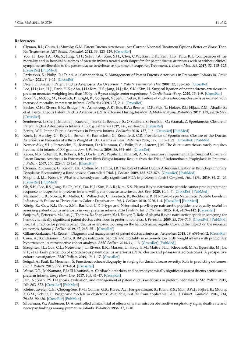
Analysis using a receiver operating characteristic curve shown in Figure 4 identified apeak NT-pro-BNP cut-off value of 8500 pg/mL to have the best combination of sensitivity(83.7%) and specificity (94.2%) for predicting hsPDA. ROC curves were generated forvarious NT-pro-BNP levels in the diagnosis of hsPDA. The area under the curve wasmaximum (0.89, 95% CI 0.83 to 0.91) at a cut-off value of 8500 pg/mL. The positivepredictive value at this cut-off was 94% while the negative predictive value was 85%. Thelikelihood ratio for a positive test was 14 and for a negative test it was 0.2.
J. Clin. Med. 2021, 10, x FOR PEER REVIEW 8 of 12
Figure 4. Receiver operating characteristic curve demonstrating that a NT-pro-BNP level of 8500 pg/mL results in a sensitivity of 94.2% and a specificity of 83.7% for predicting hsPDA.
4. Discussion Early diagnosis of hsPDA in preterm infants is undoubtedly a clinical problem as it
is associated with serious clinical outcomes and in-hospital mortality. However, over the past decade, there has been a tendency towards a decrease in the use of both medical and surgical treatment of hsPDA in this group of patients [31].
We analyzed the statistical data of the Perm Krai Perinatal Center in the period from 2015 to 2019, according to which the proportion of children who received surgical PDA ligation decreased 3.7 times (from 17.0% to 4.5%). The median age of the operated children was 20 months. Of the patients participating in our study, two children (aged 32 and 43 days) underwent surgery.
In our practice, we adhere to the conservative approach aimed at improving the ef-fects of left-to-right shunting by fluid restriction, diuretics prescription, etc. Good results of conservative treatment without surgery have been reported in a number of studies, according to which the frequency of spontaneous closure of PDA at 36 weeks postmen-strual age reaches 83–85% [8,32,33].
However, at present, there is a problem of lack of evidence in favor of using a con-servative approach to the treatment of premature infants with PDA, since the currently existing echocardiography-guided approach to PDA management is characterized by sig-nificant variability [29]. Ultrasound, although accepted as the gold standard, has a number of limitations, as it is expensive, requires interpretation by a cardiologist, can cause stress in such fragile and immature newborns, and may not be available in resource-limited set-tings.
Echo criteria traditionally used for hsPDA diagnosis, such as PDA diameter, for ex-ample, do not allow prediction of serious clinical outcomes [34,35]. For this reason, a great importance is attached to predictive models that take into account clinical, laboratory, and instrumental data [36–38].
For example, Kindler et al. [39] developed a short clinical score with number of signs in addition to conventional echocardiography criteria for prediction of hsPDA. The signs used in the study were pulsations of the precordium, heart rate, apnea or mechanical ven-tilation, femoral pulses, systolic murmur, hepatomegaly, acidosis, and pulmonary deteri-oration. In our study, respiratory failure was assessed during the first hour of life with the Silverman–Anderson score. We hypothesized that newborns with high RSS (≥5) have
Figure 4. Receiver operating characteristic curve demonstrating that a NT-pro-BNP level of8500 pg/mL results in a sensitivity of 94.2% and a specificity of 83.7% for predicting hsPDA.
4. Discussion
Early diagnosis of hsPDA in preterm infants is undoubtedly a clinical problem as itis associated with serious clinical outcomes and in-hospital mortality. However, over thepast decade, there has been a tendency towards a decrease in the use of both medical andsurgical treatment of hsPDA in this group of patients [31].
We analyzed the statistical data of the Perm Krai Perinatal Center in the period from2015 to 2019, according to which the proportion of children who received surgical PDAligation decreased 3.7 times (from 17.0% to 4.5%). The median age of the operated childrenwas 20 months. Of the patients participating in our study, two children (aged 32 and43 days) underwent surgery.
In our practice, we adhere to the conservative approach aimed at improving theeffects of left-to-right shunting by fluid restriction, diuretics prescription, etc. Good resultsof conservative treatment without surgery have been reported in a number of studies,according to which the frequency of spontaneous closure of PDA at 36 weeks postmenstrualage reaches 83–85% [8,32,33].
However, at present, there is a problem of lack of evidence in favor of using a conserva-tive approach to the treatment of premature infants with PDA, since the currently existingechocardiography-guided approach to PDA management is characterized by significantvariability [29]. Ultrasound, although accepted as the gold standard, has a number oflimitations, as it is expensive, requires interpretation by a cardiologist, can cause stress insuch fragile and immature newborns, and may not be available in resource-limited settings.

J. Clin. Med. 2021, 10, 3729 9 of 12
Echo criteria traditionally used for hsPDA diagnosis, such as PDA diameter, forexample, do not allow prediction of serious clinical outcomes [34,35]. For this reason, agreat importance is attached to predictive models that take into account clinical, laboratory,and instrumental data [36–38].
For example, Kindler et al. [39] developed a short clinical score with number of signsin addition to conventional echocardiography criteria for prediction of hsPDA. The signsused in the study were pulsations of the precordium, heart rate, apnea or mechanicalventilation, femoral pulses, systolic murmur, hepatomegaly, acidosis, and pulmonarydeterioration. In our study, respiratory failure was assessed during the first hour of lifewith the Silverman–Anderson score. We hypothesized that newborns with high RSS (≥5)have higher carbon dioxide partial pressure (PCO2) values, as a result of which there is adecrease in muscle wall tone, leading to the duct staying open. The advantages of applyingthe Silverman–Anderson scale include simplicity and the possibility of use in conditionswith limited resources. In our study, we initially used 40 diagnostic criteria, but the greatestability in predicting probabilities of persistent PDA was established for a combination ofcriteria: Apgar score ≤ 6 points, Silverman–Anderson score ≥ 5 points, and NT-pro-BNPover lg3.9 pg/mL.
The novel contribution of our work is that our suggested combination of predictorsis new, having not been previously described, and is substantiated by the results of dis-criminant and variance analyses. We have identified the combination of predictors thathas high specificity values (88.7%, 95% CI 0.77–0.94), leading to a low probability of afalse positive diagnosis, which is of practical importance for making a clinical decision onsurgical correction of the duct.
As for the clinical utility of measuring the biomarker NT-pro-BNP for predictionof hsPDA, the increase in its plasma concentration is significantly correlated with theincrease in the left atrium arising from PDA [40]. The biomarker NT-pro-BNP in preterminfants is described by many authors both for diagnosis and for the initiation of medicalor surgical treatment of hsPDA [41]. We found one systematic review that included11 studies that differed in methodological quality, age at testing, gestational age, type ofcommercial assay, thresholds, etc. After analyzing the results presented in the review, theauthors reported that, as a result, the sensitivity and specificity for NT-pro-BNP in thediagnosis of hsPDA was 0.90 (95% CI, 0.79–0.96), and summary specificity was 0.84 (95%CI, 0.77–0.90) [42]. In the 11 included studies that evaluated NT-pro-BNP, there were eightin which measurements were performed on day 3 of life. The best predictive values forhsPDA were established for a threshold level of NT-pro-BNP in plasma of 11,395 pg/mL,100% sensitivity with narrow confidence intervals (95% CI, 0.81–1.00) [43].
In two of the eight studies, sensitivity was lower than 0.7, with 42,285 (0.68, 95% CI0.46–0.85) [44] and 10,253 (0.58 95% CI (0.33–0.8) used as a threshold [45].
In other studies, the sensitivity ranged from 0.85 to 0.95 (95% CI from 0.59 to 1.0) forNT-pro-BNP cut-off values of 5000 to 24,102 [46–48].
In comparison with the presented studies, the result of our study, which determinedthe threshold for NT-pro-BNP as lg3.9 pg/mL, shows one of the best combinations of highsensitivity (94.2%, CI 0.85 to 0.98) and specificity (83.7%, CI 0.71 to 0.92) and a narrowconfidence interval. We used the obtained threshold value of NT-pro-BNP to create amodel capable of dividing patients with extremely low body weight according to thedegree of hemodynamic significance of PDA into two functional classes. The predictivemodel derived from discriminant analysis successfully classified 85.9% of participants withan AUC of 0.89.
The use of Apgar and Silverman–Anderson scores in our predictive model as pre-dictors has its advantages and disadvantages. The advantages include the unification ofdecision making, and the possibility of quantitative assessment. The disadvantages arethe uncertainty of the forecast time lag, the static nature of the forecast, dependence on thepopulation, and the absence of standards. Additionally, a limitation of the study is thatsurgeons participating in the study were aware of NT-pro-BNP values in patients. Another

J. Clin. Med. 2021, 10, 3729 10 of 12
limitation of our study was the small cohort of patients; however, the predictor correlationvalues seem reasonable. There was some selection bias in the study cohort: we includedonly premature infants for blood sampling and the decision to request an echocardiogramwas based on the opinion of the attending physician. Strictly speaking, scales can be usedif it is proven that their application reliably improves prognosis, compared to decisionsmade by a doctor without using scales. We believe that in conditions of limited resources,this provision is important, since it allows replacement of the standard echocardiographicassessment of hsPDA in newborns of VLBW on the third day of life by a combination ofthe following factors: NT-pro-BNP level more than lg3.9 pg/mL, Apgar score ≤ 6 points,Silverman–Anderson score (RSS) ≥ 5 points.
In the future, we plan to conduct a study in which we will compare our modelwith a group in which only NT-pro-BNP will be determined, the group in which onlyechocardiographic assessment will be used as an internal control.
Undoubtedly, therapeutic approaches to the problem of PDA closure continue toevolve, and much is being rethought and re-evaluated. We believe that future researchshould be as individualized as possible and we hope to make a small contribution to thebenefit of patients.
5. Conclusions
The results of this study show that the accuracy of the clinical prognosis of hsPDA inpremature infants weighing less than 1500 g can be significantly improved using affordableand inexpensive predictors that allow newborns to be classified according to hsPDA riskgroups: NT-pro-BNP level more than lg3.9 pg/mL, Apgar score ≤ 6 points, and Silverman–Anderson score ≥ 5 points. Further research in this area will help to improve understandingof the problem of functioning PDA in children born very prematurely, and awareness of thefine line between norm and pathology determines the use of the most sparing therapeuticapproaches in this group of patients.
Supplementary Materials: The following are available online at https://www.mdpi.com/article/10.3390/jcm10163729/s1, Table S1: Variables groups used for discriminant analysis.
Author Contributions: Conceptualization, A.G.K. and A.V.P.; methodology, A.P.; software, A.G.K.;validation, A.V.P. and A.G.K.; investigation, A.V.P., A.P., and A.G.K.; resources, A.G.K. and A.P.; datacuration, A.P.; writing—original draft preparation, A.V.P., A.P., and A.G.K.; writing—review andediting, A.B., E.G.F., A.V.P., V.A., Y.S.S., and A.G.K.; visualization, A.P.; supervision, Y.S.S. and E.G.F.;project administration, A.P., Y.S.S., and E.G.F.; funding acquisition, A.G.K. and A.P. All authors haveread and agreed to the published version of the manuscript.
Funding: Alex G. Kuchumov acknowledges the financial support of the Ministry of Science andHigher Education of the Russian Federation in the framework of the program of activities of thePerm Scientific and Educational Center “Rational Subsoil Use”. Artem Porodikov acknowledgesfinancial support of RFBR and Perm Territory, project number 20-41-596005.
Institutional Review Board Statement: The study was conducted according to the guidelines of theDeclaration of Helsinki, and approved by the Ethics Committee of S.G. Sukhanov CardiovascularCenter, Perm, Russia (protocol No. 5 on 23 May 2018).
Informed Consent Statement: Informed consent was obtained from parents of patients involved inthe study.
Data Availability Statement: The Russian Ethics Review Authority only granted publication ofaggregated data, which means that individual data cannot be shared.
Conflicts of Interest: The authors declare no conflict of interest.
J. Clin. Med. 2021, 10, 3729 11 of 12
References1. Clyman, R.I.; Couto, J.; Murphy, G.M. Patent Ductus Arteriosus: Are Current Neonatal Treatment Options Better or Worse Than
No Treatment at All? Semin. Perinatol. 2012, 36, 123–129. [CrossRef]2. Yoo, H.; Lee, J.A.; Oh, S.; Jung, Y.H.; Sohn, J.A.; Shin, S.H.; Choi, C.W.; Kim, E.K.; Kim, H.S.; Kim, B. Il Comparison of the
mortality and in-hospital outcomes of preterm infants treated with ibuprofen for patent ductus arteriosus with or without clinicalsymptoms attributable to the patent ductus arteriosus at the time of ibuprofen Treatment. J. Korean Med. Sci. 2017, 32, 115–123.[CrossRef] [PubMed]
3. Parkerson, S.; Philip, R.; Talati, A.; Sathanandam, S. Management of Patent Ductus Arteriosus in Premature Infants in. FrontPediatr. 2021, 8, 1–11. [CrossRef]
4. Dice, J.E.; Bhatia, J. Patent Ductus Arteriosus: An Overview. J. Pediatr. Pharmacol. Ther. 2007, 12, 138–146. [CrossRef]5. Lee, J.H.; Lee, H.J.; Park, H.K.; Ahn, J.H.; Kim, H.S.; Jang, H.J.; Ro, S.K.; Kim, H. Surgical ligation of patent ductus arteriosus in
preterm neonates weighing less than 1500g: A 9-year single center experience. J. Cardiothorac. Surg. 2020, 15, 1–9. [CrossRef]6. Noori, S.; McCoy, M.; Friedlich, P.; Bright, B.; Gottipati, V.; Seri, I.; Sekar, K. Failure of ductus arteriosus closure is associated with
increased mortality in preterm infants. Pediatrics 2009, 123, 2–4. [CrossRef]7. Backes, C.H.; Rivera, B.K.; Bridge, J.A.; Armstrong, A.K.; Boe, B.A.; Berman, D.P.; Fick, T.; Holzer, R.J.; Hijazi, Z.M.; Abadir, S.;
et al. Percutaneous Patent Ductus Arteriosus (PDA) Closure During Infancy: A Meta-analysis. Pediatrics 2017, 139, e20162927.[CrossRef]
8. Semberova, J.; Sirc, J.; Miletin, J.; Kucera, J.; Berka, I.; Sebkova, S.; O’Sullivan, S.; Franklin, O.; Stranak, Z. Spontaneous Closure ofPatent Ductus Arteriosus in Infants ≤1500 g. Pediatrics 2017, 140, e20164258. [CrossRef]
9. Benitz, W.E. Patent Ductus Arteriosus in Preterm Infants. Pediatrics 2016, 137, 1–6. [CrossRef] [PubMed]10. Koch, J.; Hensley, G.; Roy, L.; Brown, S.; Ramaciotti, C.; Rosenfeld, C.R. Prevalence of Spontaneous Closure of the Ductus
Arteriosus in Neonates at a Birth Weight of 1000 Grams or Less. Pediatrics 2006, 117, 1113–1121. [CrossRef] [PubMed]11. Nemerofsky, S.L.; Parravicini, E.; Bateman, D.; Kleinman, C.; Polin, R.A.; Lorenz, J.M. The ductus arteriosus rarely requires
treatment in infants >1000 grams. Am. J. Perinatol. 2008, 25, 661–666. [CrossRef]12. Kabra, N.S.; Schmidt, B.; Roberts, R.S.; Doyle, L.W.; Papile, L.; Fanaroff, A. Neurosensory Impairment after Surgical Closure of
Patent Ductus Arteriosus in Extremely Low Birth Weight Infants: Results from the Trial of Indomethacin Prophylaxis in Preterms.J. Pediatr. 2007, 150, 229.e1–234.e1. [CrossRef]
13. Clyman, R.; Cassady, G.; Kirklin, J.K.; Collins, M.; Philips, J.B. The Role of Patent Ductus Arteriosus Ligation in BronchopulmonaryDysplasia: Reexamining a Randomized Controlled Trial. J. Pediatr. 2009, 154, 873–876. [CrossRef] [PubMed]
14. Shepherd, J.L.; Noori, S. What is a hemodynamically significant PDA in preterm infants? Congenit. Heart Dis. 2019, 14, 21–26.[CrossRef] [PubMed]
15. Oh, S.H.; Lee, B.S.; Jung, E.; Oh, M.Y.; Do, H.J.; Kim, E.A.R.; Kim, K.S. Plasma B-type natriuretic peptide cannot predict treatmentresponse to ibuprofen in preterm infants with patent ductus arteriosus. Sci. Rep. 2020, 10, 1–7. [CrossRef] [PubMed]
16. Mänhardt, L.B.; Norozi, K.; Müller, C.; Willaschek, C.; Kostuch, B.; Buchhorn, R. NT-Pro-B-Type Natriuretic Peptide Levels inInfants with Failure to Thrive due to Caloric Deprivation. Int. J. Pediatr. 2010, 2010, 1–4. [CrossRef] [PubMed]
17. König, K.; Guy, K.J.; Drew, S.M.; Barfield, C.P. B-type and N-terminal pro-B-type natriuretic peptides are equally useful inassessing patent ductus arteriosus in very preterm infants. Acta Paediatr. Int. J. Paediatr. 2015, 104, e139–e142. [CrossRef]
18. Sanjeev, S.; Pettersen, M.; Lua, J.; Thomas, R.; Shankaran, S.; L’Ecuyer, T. Role of plasma B-type natriuretic peptide in screening forhemodynamically significant patent ductus arteriosus in preterm neonates. J. Perinatol. 2005, 25, 709–713. [CrossRef] [PubMed]
19. Lee, J.A. Practice for preterm patent ductus arteriosus; focusing on the hemodynamic significance and the impact on the neonataloutcomes. Korean J. Pediatr. 2019, 62, 245–251. [CrossRef]
20. Gillam-Krakauer, M.; Reese, J. Diagnosis and management of patent ductus arteriosus. Neoreviews 2018, 19, e394–e402. [CrossRef]21. Cuna, A.; Kandasamy, J.; Sims, B. B-type natriuretic peptide and mortality in extremely low birth weight infants with pulmonary
hypertension: A retrospective cohort analysis. BMC Pediatr. 2014, 14, 1–6. [CrossRef] [PubMed]22. Slaughter, J.L.; Cua, C.L.; Notestine, J.L.; Rivera, B.K.; Marzec, L.; Hade, E.M.; Maitre, N.L.; Klebanoff, M.A.; Ilgenfritz, M.; Le,
V.T.; et al. Early prediction of spontaneous patent ductus arteriosus (PDA) closure and pdaassociated outcomes: A prospectivecohort investigation. BMC Pediatr. 2019, 19, 1–17. [CrossRef]
23. Sehgal, A.; Paul, E.; Menahem, S. Functional echocardiography in staging for ductal disease severity: Role in predicting outcomes.Eur. J. Pediatr. 2013, 172, 179–184. [CrossRef]
24. Weisz, D.E.; McNamara, P.J.; El-Khuffash, A. Cardiac biomarkers and haemodynamically significant patent ductus arteriosus inpreterm infants. Early Hum. Dev. 2017, 105, 41–47. [CrossRef]
25. Jain, A.; Shah, P.S. Diagnosis, evaluation, and management of patent ductus arteriosus in preterm neonates. JAMA Pediatr. 2015,169, 863–872. [CrossRef] [PubMed]
26. Kleinrouweler, C.E.; Cheong-See, F.M.; Collins, G.S.; Kwee, A.; Thangaratinam, S.; Khan, K.S.; Mol, B.W.J.; Pajkrt, E.; Moons,K.G.M.; Schuit, E. Prognostic models in obstetrics: Available, but far from applicable. Am. J. Obstet. Gynecol. 2016, 214,79.e36–90.e36. [CrossRef] [PubMed]
27. Silverman, W.; Anderson, D. A controlled clinical trial of effects of water mist on obstructive respiratory signs, death rate andnecropsy findings among premature infants. Pediatrics 1956, 17, 1–10.

J. Clin. Med. 2021, 10, 3729 12 of 12
28. Hedstrom, A.B.; Gove, N.E.; Mayock, D.E.; Batra, M. Performance of the Silverman Anderson Respiratory Severity Score inpredicting PCO2 and respiratory support in newborns: A prospective cohort study. J. Perinatol. 2018, 38, 505–511. [CrossRef][PubMed]
29. Smith, A.; El-Khuffash, A.F. Defining “haemodynamic significance” of the patent ductus arteriosus: Do we have all the answers?Neonatology 2020, 117, 225–232. [CrossRef]
30. Mitra, S.; Disher, T. Early treatment versus expectant management of hemodynamically significant patent ductus arteriosus forpreterm infants. Cochrane Database Syst. Rev. 2019, 12, CD013278. [CrossRef]
31. Hagadorn, J.I.; Brownell, E.A.; Trzaski, J.M.; Johnson, K.R.; Lainwala, S.; Campbell, B.T.; Herbst, K.W. Trends and variationin management and outcomes of very low-birth-weight infants with patent ductus arteriosus. Pediatr. Res. 2016, 80, 785–792.[CrossRef]
32. Rolland, A.; Shankar-Aguilera, S.; Diomandé, D.; Zupan-Simunek, V.; Boileau, P. Natural evolution of patent ductus arteriosus inthe extremely preterm infant. Arch. Dis. Child. Fetal Neonatal Ed. 2015, 100, F55–F58. [CrossRef]
33. Sung, S.I.; Chang, Y.S.; Chun, J.Y.; Yoon, S.A.; Yoo, H.S.; Ahn, S.Y.; Park, W.S. Mandatory Closure Versus Nonintervention forPatent Ductus Arteriosus in Very Preterm Infants. J. Pediatr. 2016, 177, 66.e1–71.e1. [CrossRef]
34. El-Khuffash, A.; Weisz, D.E.; McNamara, P.J. Reflections of the changes in patent ductus arteriosus management during the last10 € years. Arch. Dis. Child. Fetal Neonatal Ed. 2016, 101, F474–F478. [CrossRef]
35. El-Khuffash, A.; James, A.T.; Corcoran, J.D.; Dicker, P.; Franklin, O.; Elsayed, Y.N.; Ting, J.Y.; Sehgal, A.; Malikiwi, A.; Harabor, A.;et al. A Patent Ductus Arteriosus Severity Score Predicts Chronic Lung Disease or Death before Discharge. J. Pediatr. 2015, 167,1354.e2–1361.e2. [CrossRef] [PubMed]
36. Prescott, S.; Keim-Malpass, J. Patent Ductus Arteriosus in the Preterm Infant: Diagnostic and Treatment Options. Adv. NeonatalCare 2017, 17, 10–18. [CrossRef] [PubMed]
37. Coviello, C.; Tataranno, M.L.; Corsini, I.; Leonardi, V.; Longini, M.; Bazzini, F.; Buonocore, G.; Dani, C. Isoprostanes as Biomarkerfor Patent Ductus Arteriosus in Preterm Infants. Front. Pediatr. 2020, 8, 3–6. [CrossRef] [PubMed]
38. Bardanzellu, F.; Piras, C.; Atzei, A.; Neroni, P.; Fanos, V. Early Urinary Metabolomics in Patent Ductus Arteriosus Anticipates theFate: Preliminary Data. Front. Pediatr. 2020, 8, 1–18. [CrossRef]
39. Kindler, A.; Seipolt, B.; Heilmann, A.; Range, U.; Rüdiger, M.; Hofmann, S.R. Development of a diagnostic clinical score forhemodynamically significant patent ductus arteriosus. Front. Pediatr. 2017, 5, 1–8. [CrossRef] [PubMed]
40. Andersson, S.; Tikkanen, I.; Pesonen, E.; Meretoja, O.; Hynynen, M.; Fyhrquist, F. Atrial natriuretic peptide in patent ductusarteriosus. Pediatr. Res. 1987, 21, 396–398. [CrossRef]
41. Holmström, H.; Hall, C.; Thaulow, E. Plasma levels of natriuretic peptides and hemodynamic assessment of patent ductusarteriosus in preterm infants. Acta Paediatr. Int. J. Paediatr. 2001, 90, 184–191. [CrossRef]
42. Nuntnarumit, P.; Khositseth, A.; Thanomsingh, P. N-terminal probrain natriuretic peptide and patent ductus arteriosus in preterminfants. J. Perinatol. 2009, 29, 137–142. [CrossRef] [PubMed]
43. Gokulakrishnan, G.; Sathappan, V.S.; Kulkarni, M.; Leeflang, M.M.G.; Fernandes, C.J.; Price, J.; Pammi, M. Brain natriureticpeptide and N-terminal brain natriuretic peptide for the diagnosis of hemodynamically significant patent ductus arteriosus inpreterm neonates. Cochrane Database Syst. Rev. 2018, 2018, CD013129. [CrossRef]
44. El-Khuffash, A.F.; Amoruso, M.; Culliton, M.; Molloy, E.J. N-terminal pro-B-type natriuretic peptide as a marker of ductalhaemodynamic significance in preterm infants: A prospective observational study. Arch. Dis. Child. Fetal Neonatal Ed. 2007, 92,F421–F422. [CrossRef] [PubMed]
45. Letzner, J.; Berger, F.; Schwabe, S.; Benzing, J.; Morgenthaler, N.G.; Bucher, H.U.; Bührer, C.; Arlettaz, R.; Wellmann, S. PlasmaC-terminal pro-endothelin-1 and the natriuretic pro-peptides NT-proBNP and MR-proANP in very preterm infants with patentductus arteriosus. Neonatology 2012, 101, 116–124. [CrossRef] [PubMed]
46. Cambonie, G.; Dupuy, A.M.; Combes, C.; Vincenti, M.; Mesnage, R.; Cristol, J.P. Can a clinical decision rule help ductus arteriosusmanagement in preterm neonates? Acta Paediatr. Int. J. Paediatr. 2012, 101, 213–218. [CrossRef]
47. Ramakrishnan, S.; Heung, Y.M.; Round, J.; Morris, T.P.; Collinson, P.; Williams, A.F. Early N-terminal pro-brain natriuretic peptidemeasurements predict clinically significant ductus arteriosus in preterm infants. Acta Paediatr. Int. J. Paediatr. 2009, 98, 1254–1259.[CrossRef] [PubMed]
48. Dhuper, S.; Kim, R.; Weichbrod, L.; Mahdi, E.; Shah, N.; Kona, S.; Sokal, M.; Buddhe, S. NT-proBNP levels improve the ability ofpredicting a hemodynamically significant patent ductus arteriosus in very low-birth-weight infants. J. Clin. Neonatol. 2012, 1,Sa82. [CrossRef] [PubMed]