antioxidants - MDPI
28
antioxidants Review State of the Art of Anthocyanins: Antioxidant Activity, Sources, Bioavailability, and Therapeutic Effect in Human Health Noelia Tena 1, * , Julia Martín 2 and Agustín G. Asuero 1 1 Departamento de Química Analítica, Facultad de Farmacia, Universidad de Sevilla, Prof. García González 2, E-41012 Sevilla, Spain; [email protected] 2 Departamento de Química Analítica, Escuela Politécnica Superior, Universidad de Sevilla, C/Virgen de África 7, E-41011 Sevilla, Spain; [email protected] * Correspondence: [email protected] Received: 21 April 2020; Accepted: 19 May 2020; Published: 23 May 2020 Abstract: The antioxidant activity of anthocyanins in food is well known. Numerous antioxidant assays have been proposed to measure the capacity of anthocyanins to prevent the oxidation process that naturally occurs. Different solvents, temperatures, and pH levels are applied in each assay, and these factors should be taken into account in order to obtain useful and reproducible results. The concentration and the structure of these compounds are directly related to their antioxidant capacity and their environment. However, the effectiveness of the anthocyanin ingestion against diseases is also influenced by its bioavailability. Novel methodologies that simulate the digestion process have been developed in order to facilitate the current knowledge of anthocyanins bioavailability. Studies highlight the potential synergy effect between parent compounds and their derivatives (metabolites, conjugated products, and microbe-generated metabolites). The aim of this review is to provide an overview of advantages and disadvantages of the most common methods to determine the antioxidant activity of anthocyanins, chemical structure, and concentration of these compounds in different edible fruits, vegetables, and plants; their bioavailability after intake; as well as the main therapeutic effect described in the scientific literature. Keywords: anthocyanins; antioxidant activity; anthocyanin content; bioavailability; encapsulation; therapeutic effects 1. Introduction In recent years, the interest in plants and food containing antioxidant properties has increased. The chemical compounds present in vegetables and fruits with these capacities are: vitamins C and E, carotenoids, and flavonoids. The anthocyanins, which are the most important group of flavonoids in plants, are pigments with a flavylium cation (AH + ) structure that act as acid. This structure is directly related to its antioxidant activity. Most of the functional properties and the sensory quality of the anthocyanins can be explained by their chemical reactivity. The structures and properties of anthocyanins are dependent on different factors such as pH, temperature, and solvents which should be controlled to carry out antioxidant activity studies of these compounds [1–9]. Free radicals, reactive oxygen species (ROS), and/or reactive nitrogen species (RNS) are required for the proper performance of the human body and its organs. These radicals are on-balance by a redox homeostasis in our body. However, the body may be occasionally affected by an oxidative stress resulting from an off-balance state. This stress is important in the development of chronic degenerative diseases including coronary heart disease, cancer, and aging [2]. Anthocyanins have been described as compounds that prevent or inhibit, the oxidation by scavenging free radicals and Antioxidants 2020, 9, 451; doi:10.3390/antiox9050451 www.mdpi.com/journal/antioxidants
-
Upload
khangminh22 -
Category
Documents
-
view
2 -
download
0
Transcript of antioxidants - MDPI
antioxidants
Review
State of the Art of Anthocyanins: AntioxidantActivity, Sources, Bioavailability, and TherapeuticEffect in Human Health
Noelia Tena 1,* , Julia Martín 2 and Agustín G. Asuero 1
1 Departamento de Química Analítica, Facultad de Farmacia, Universidad de Sevilla, Prof. García González 2,E-41012 Sevilla, Spain; [email protected]
2 Departamento de Química Analítica, Escuela Politécnica Superior, Universidad de Sevilla,C/Virgen de África 7, E-41011 Sevilla, Spain; [email protected]
* Correspondence: [email protected]
Received: 21 April 2020; Accepted: 19 May 2020; Published: 23 May 2020�����������������
Abstract: The antioxidant activity of anthocyanins in food is well known. Numerous antioxidantassays have been proposed to measure the capacity of anthocyanins to prevent the oxidationprocess that naturally occurs. Different solvents, temperatures, and pH levels are applied in eachassay, and these factors should be taken into account in order to obtain useful and reproducibleresults. The concentration and the structure of these compounds are directly related to theirantioxidant capacity and their environment. However, the effectiveness of the anthocyanin ingestionagainst diseases is also influenced by its bioavailability. Novel methodologies that simulate thedigestion process have been developed in order to facilitate the current knowledge of anthocyaninsbioavailability. Studies highlight the potential synergy effect between parent compounds and theirderivatives (metabolites, conjugated products, and microbe-generated metabolites). The aim of thisreview is to provide an overview of advantages and disadvantages of the most common methods todetermine the antioxidant activity of anthocyanins, chemical structure, and concentration of thesecompounds in different edible fruits, vegetables, and plants; their bioavailability after intake; as wellas the main therapeutic effect described in the scientific literature.
Keywords: anthocyanins; antioxidant activity; anthocyanin content; bioavailability; encapsulation;therapeutic effects
1. Introduction
In recent years, the interest in plants and food containing antioxidant properties has increased.The chemical compounds present in vegetables and fruits with these capacities are: vitamins C and E,carotenoids, and flavonoids. The anthocyanins, which are the most important group of flavonoidsin plants, are pigments with a flavylium cation (AH+) structure that act as acid. This structure isdirectly related to its antioxidant activity. Most of the functional properties and the sensory qualityof the anthocyanins can be explained by their chemical reactivity. The structures and properties ofanthocyanins are dependent on different factors such as pH, temperature, and solvents which shouldbe controlled to carry out antioxidant activity studies of these compounds [1–9].
Free radicals, reactive oxygen species (ROS), and/or reactive nitrogen species (RNS) are requiredfor the proper performance of the human body and its organs. These radicals are on-balance by aredox homeostasis in our body. However, the body may be occasionally affected by an oxidativestress resulting from an off-balance state. This stress is important in the development of chronicdegenerative diseases including coronary heart disease, cancer, and aging [2]. Anthocyanins havebeen described as compounds that prevent or inhibit, the oxidation by scavenging free radicals and
Antioxidants 2020, 9, 451; doi:10.3390/antiox9050451 www.mdpi.com/journal/antioxidants
Antioxidants 2020, 9, 451 2 of 28
reducing the oxidative stress. On a regular basis, anthocyanins act as H-atom donator or as singleelectron transfer. Different methods of analysis based on both mechanisms have been proposed todetermine the antioxidant activity of anthocyanins. The antioxidant activity of these compoundsdepends on their total concentration, structure, and environment. A literature compilation about theconcentrations of the most common anthocyanins in different foods is presented in this review in orderto have an overview of the different sources of anthocyanins.
The beneficial properties attributed to the dietary ingestion of anthocyanin-rich foods (eye health,cardiovascular diseases, antiobesity, antidiabetic, antimicrobial, anticancer or neuroprotective effect)have been deeply documented in studies carried out with experimental models. These health benefitscontrast with the apparent small portion (<1–2%) of these compounds absorbed by our organism [3–5].During the digestion process, anthocyanins undergo to an intense variation in pH that together withthe enzymatic and bacterial action can cause the hydrolysis and transformation of anthocyanins intometabolites, conjugated products, or simpler phenolic compounds [7,10–12]. The question is: Howcan anthocyanins be so influential in health? Are anthocyanins the only responsible of their beneficialeffects? Last scientific developments highlight the potential synergy effect between parent compounds,metabolites (phases I and II), conjugated products, and microbe-generated metabolites to explain thosebiological events [4,11–13].
Due to their particular physicochemical features, bioavailability of anthocyanins is very difficultto assess. The first studies were performed analyzing blood and urine to determine the anthocyaninconcentration levels after the ingestion of foods rich in anthocyanins [14–16]. However, the lowabsorption percentage obtained led to in vitro assays (mostly using cell culture systems) in order tofacilitate the knowledge of their biochemical and chemical changes as well as the influence of thedigestion steps. Last studies have emphasized the key role of the microbiota in the transformation ofanthocyanins, which is not considered in in vitro assays but it is still poorly considered in in vivo andex vivo studies [14].
This review aims to highlight some aspects regarding the antioxidant activity of anthocyanins andtheir bioavailability after intake. The first part includes an exposition of the most common antioxidantbioassays used to determine in vitro the antioxidant activity of anthocyanins, being the advantagesand disadvantages of each bioassay identified. Afterwards, the effect of the chemical structure andthe environment in the ability of the anthocyanins to prevent oxidation is discussed and presentedtogether with information about different sources and range of concentration of these compounds infood. The second part of the manuscript exposes the bioavailability and metabolism of anthocyaninsas well as a summary including the main therapeutic effects of anthocyanins on different diseases.
2. Antioxidant Bioassays for Anthocyanins
Numerous antioxidant assays have been proposed to measure the ability of anthocyanins toprevent the oxidation process that naturally occurs. Depending on the source of the anthocyanins andtheir nature, in most of the cases, an extraction step before carrying out the antioxidant bioassay isneeded. The extraction process is a critical step in the determination of the antioxidant activity bioassay,presenting a challenge due to the low stability of anthocyanins after extraction and their tendency toremain bound to the matrix of the sample. Multiple alternatives have been proposed in literature for thisprocedure [17]. Results show that temperature, pH, solvent system, solvent-to-solid ratio, and numberof extractions are factors that play an important role in the extraction efficiency and that should beoptimized for each sample [18,19]. Once the anthocyanins are in liquid solution, the antioxidant activitycould be determined by different bioassays. In general, two different mechanisms can be used to explainthe antioxidant activity of anthocyanins: Hydrogen atom donator (HAT) and single-electron transfer(SET). In HAT mechanism, the free radical R• removes a hydrogen atom from the antioxidant (AH+)converting the free radical to a more stable product. In the SET mechanism, the antioxidant (AH+)donates an electron to the free radical reducing the oxidized intermediates into the stable form [20].However, the difficulty in distinguishing between HAT and SET reactions is high. In most situations,
Antioxidants 2020, 9, 451 3 of 28
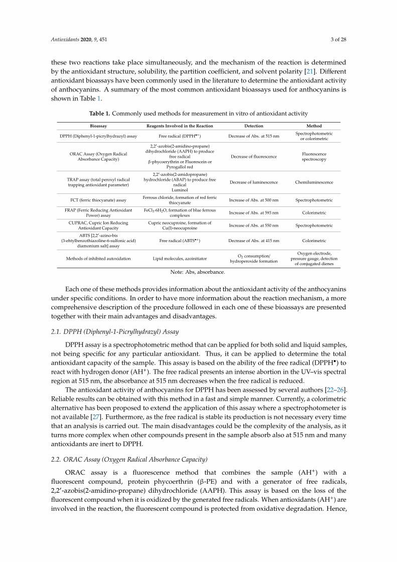
these two reactions take place simultaneously, and the mechanism of the reaction is determinedby the antioxidant structure, solubility, the partition coefficient, and solvent polarity [21]. Differentantioxidant bioassays have been commonly used in the literature to determine the antioxidant activityof anthocyanins. A summary of the most common antioxidant bioassays used for anthocyanins isshown in Table 1.
Table 1. Commonly used methods for measurement in vitro of antioxidant activity
Bioassay Reagents Involved in the Reaction Detection Method
DPPH (Diphenyl-1-picrylhydrazyl) assay Free radical (DPPH•+) Decrease of Abs. at 515 nm Spectrophotometricor colorimetric
ORAC Assay (Oxygen RadicalAbsorbance Capacity)
2,2′-azobis(2-amidino-propane)dihydrochloride (AAPH) to produce
free radicalβ-phycoerythrin or Fluorescein or
Pyrogallol red
Decrease of fluorescence Fluorescencespectroscopy
TRAP assay (total peroxyl radicaltrapping antioxidant parameter)
2,2′-azobis(2-amidopropane)hydrochloride (ABAP) to produce free
radicalLuminol
Decrease of luminescence Chemiluminescence
FCT (ferric thiocyanate) assay Ferrous chloride, formation of red ferricthiocyanate Increase of Abs. at 500 nm Spectrophotometric
FRAP (Ferric Reducing AntioxidantPower) assay
FeCl3·6H2O, formation of blue ferrouscomplexes Increase of Abs. at 593 nm Colorimetric
CUPRAC, Cupric Ion ReducingAntioxidant Capacity
Cupric neocuproine, formation ofCu(I)-neocuproine Increase of Abs. at 550 nm Spectrophotometric
ABTS [2,2′-azino-bis(3-ehtylbenzothiazoline-6-sulfonic acid)
diamonium salt] assayFree radical (ABTS•+) Decrease of Abs. at 415 nm Colorimetric
Methods of inhibited autoxidation Lipid molecules, azoinitiator O2 consumption/hydroperoxide formation
Oxygen electrode,pressure gauge, detection
of conjugated dienes
Note: Abs, absorbance.
Each one of these methods provides information about the antioxidant activity of the anthocyaninsunder specific conditions. In order to have more information about the reaction mechanism, a morecomprehensive description of the procedure followed in each one of these bioassays are presentedtogether with their main advantages and disadvantages.
2.1. DPPH (Diphenyl-1-Picrylhydrazyl) Assay
DPPH assay is a spectrophotometric method that can be applied for both solid and liquid samples,not being specific for any particular antioxidant. Thus, it can be applied to determine the totalantioxidant capacity of the sample. This assay is based on the ability of the free radical (DPPH•) toreact with hydrogen donor (AH+). The free radical presents an intense abortion in the UV–vis spectralregion at 515 nm, the absorbance at 515 nm decreases when the free radical is reduced.
The antioxidant activity of anthocyanins for DPPH has been assessed by several authors [22–26].Reliable results can be obtained with this method in a fast and simple manner. Currently, a colorimetricalternative has been proposed to extend the application of this assay where a spectrophotometer isnot available [27]. Furthermore, as the free radical is stable its production is not necessary every timethat an analysis is carried out. The main disadvantages could be the complexity of the analysis, as itturns more complex when other compounds present in the sample absorb also at 515 nm and manyantioxidants are inert to DPPH.
2.2. ORAC Assay (Oxygen Radical Absorbance Capacity)
ORAC assay is a fluorescence method that combines the sample (AH+) with afluorescent compound, protein phycoerthrin (β-PE) and with a generator of free radicals,2,2′-azobis(2-amidino-propane) dihydrochloride (AAPH). This assay is based on the loss of thefluorescent compound when it is oxidized by the generated free radicals. When antioxidants (AH+) areinvolved in the reaction, the fluorescent compound is protected from oxidative degradation. Hence,
Antioxidants 2020, 9, 451 4 of 28
the fluorescence signal remains. The fluorescence signal is monitored during 1 h at λem = 565 nm andλex = 540 nm respectively. The antioxidant activity of anthocyanins has been assessed by ORAC assayby several authors [26,28–30]. This is considered a good method to determine the antioxidant capacityof hydrophilic and hydrophobic samples, as it is easily adaptive to different samples changing thegenerator of free radicals. One disadvantage is the non-specificity of the fluorescence compounds thatcan react with the sample losing fluorescence even without the addition of a free radical generator.
2.3. TRAP Assay (Total Peroxyl Radical Trapping Antioxidant Parameter)
TRAP assay is a chemiluminescence method that consists of the following components:(i) thermolabile azo-radical initiator (e.g., 2,2′-azobis(2-amidopropane) hydrochloride (ABAP)), whichproduces radicals (R•) that react rapidly with O2 to give a peroxyl radicals (ROO•); (ii) oxidizablecompounds with chemiluminescence properties to monitor the reaction progress (e.g., Luminol); and(iii) the sample with the antioxidant properties (AH+).
The antioxidant activity of anthocyanins has been assessed by TRAP assay by several authors [31–34].This assay is sensitive to all known chain-breaking antioxidants. However, an important disadvantage tohighlight is the difficulty comparing the results between laboratories due to the amount of different endpoints that can be used. It is relatively complex method, time-consuming, and requires a high degree ofexperience. Furthermore, and such as ORAC assay, the oxidizable compounds are non-specific and othernon-radical chain reaction could occur.
2.4. FCT (Ferric Thiocyanate) Assay
The FTC assay is a spectrophotometric method that consists of the oxidation of ferrous chlorideto ferric ion by reacting with peroxide. The peroxides are formed during the reaction that takesplace when the sample is mixed with ethanol, water, phosphate buffer solution (pH = 7), and linoleicacid. Then, ferrous chloride in hydrochloric acid is added to the reaction and the ferric ion formed iscombined with ammonium thiocyanate producing ferric thiocyanate, which is red. The absorbance ofthe sample is measured at 500 nm until the maximum value is reached.
This assay is used to measure the amount of peroxide produced during the initial stages of oxidation.In the case of the anthocyanins this method has been applied to determine the antioxidant activityavoiding the peroxidation of polyunsaturated fatty acid [35]. This assay is simple and reproducible.Nevertheless, results are not reliable in the case that compounds within the sample absorb around500 nm. This drawback is common not only in this method, but in other spectrophotometric assays.
2.5. FRAP (Ferric Reducing Antioxidant Power) Assay
FRAP assay is a colorimetric method that can be employed for the determination ofthe total antioxidant activity of anthocyanins. It is based on the reduction of complexes of2,4,6-tripyridyl-s-triazine (TPTZ) with ferric chloride hexahydrate (FeCl3·6H2O) under acidic conditions.The solution turns slightly brownish, forming blue ferrous complexes once the reduction is completed.The absorbance is measured at 593 nm against the blank.
This method has been extensively applied to determine the antioxidant activity of anthocyaninsin different matrices: in elderberry [36]; in Roselle extract [37]; in raspberries, blackberries, redcurrants, gooseberries, and Cornelian cherries [38]; or in carrots, cabbage, cauliflower, potatoes, onions,asparagus and eggplant [39]. However, the results of this method are in the most of the cases comparedto the results of other antioxidant assays [40]. Rapid and reproducible results are obtained with thismethod. However, some limitations related to FRAP assay should be considered. On the one hand,the samples must be aqueous and the pH value is critical. On the other hand, some compoundswithout antioxidant properties can reduce Fe3+ to Fe2+ inducing an overestimation of the antioxidantactivity of the sample.
Antioxidants 2020, 9, 451 5 of 28
2.6. CUPRAC (Cupric Ion Reducing Antioxidant Capacity) Assay
CUPRAC assay is a spectrophotometric method similar to FRAP. In this method the anthocyaninsreact with the CUPRAC reagent (cupric neocuproine) producing the Cu(I)-neocuproine which is achromophore that absorbs at 450 nm. This method has been successfully applied to various foodextracts [7].
The main advantages of this method is related to the positive characteristics of the CUPRAC reagent:availability and easy accessibility, rapidity, stability, low-cost, sensitivity towards thiol-type antioxidantsunlike FRAP, and responsiveness to both hydrophilic and lipophilic antioxidants. Nevertheless,the detection is based on the absorption at 450 nm which is non-specific enough and the presence ofother compounds present in the sample could be interfered in the results.
2.7. ABTS (2,2′-Azino-bis (3-ehtylbenzothiazoline-6-sulfonic acid) Diamonium Salt) Assay
The most recent ABTS assay method is based on decolorization techniques. It consists of theproduction of a stable radical, blue/green ABTS chromophore, by the reaction of ABTS with potassiumpersulfate. This stable radical has a maximum absorbance at 415 nm. A drop in absorbance of thiscompound occurs when the radical reacts with the antioxidant. This method can determine theantioxidant activity of mixtures of substances, helping to distinguish between additive and synergisticeffects. The antioxidant activity is calculated relatively to the reactivity of Trolox standard undersimilar conditions.
This assay is frequently combined with DPPH assay for the determination of antioxidant activityof anthocyanins [41–43]. This method is simple and not a large sample volume and time of analysisare necessary. Nevertheless, a standard solution is required in order to obtain accurate results.
This assay is also described in the literature as TEAC [6-hydroxy-2,5,7,8-tetramethylchroman-2-carboxylic acid (Trolox)] equivalent antioxidant capacity. The same free radical is used both TEACand ABTS assays. However, different reagents have been proposed for the generation of the green–blueABTS•+, resulting in different TEAC assays. This radical is produced by the oxidation of 2,2′-azinobis(3-ethylbenzothiazoline-6-sulfonic acid; ABTS). The oxidation can be reached in different ways: (i) inTEAC assay I, metmyoglobin reacted with H2O2 generating the ferrylmyoglobin radical, which thenreacted with ABTS. (ii) In TEAC assay II, the ABTS•+ is formed by the reaction with manganese dioxide.(iii) In TEAC assay III, enzymatic reaction using horseradish peroxidase is applied. Other proposalsconsist of applying electrochemical oxidation or using 2,2′-azobis-2-amidinopropane, dihydrochloride(AAPH) or potassium persulfate (K2S2O8) as oxidants.
These alternatives are interchangeable when the appropriate solvent is selected. However,differences in assay conditions can be found, such as the reaction time or the wavelength used for thedetection, for instance, sometimes in order to avoid interferences 734 nm is preferable to 415 nm.
These assays have been used to determine the antioxidant capacity of the anthocyanins, as aconsequence some results has been reported in wine [44]; in corn [45]; in pomegranate juice [46] or inblueberries [47].
The advantages of TEAC assay I are the simplicity, reproducibility, and flexibility to determinethe antioxidant capacity in hydrophilic and lipophilic foods. However, the pre-addiction of the samplebefore the radical generation could result in an overestimation of the antioxidant capacity. Otherpossible disadvantages of this assay may be the fact that ABTS is not found naturally, and that anycompound with a redox potential lower than ABTS•+ may react with the radical.
Other alternatives applied to determine the antioxidant activity of anthocyanins involve the use ofenzymes [48] or the use of chromatographic techniques. The latter alternative can not only extract theanthocyanins to determine their antioxidant activity [49], but also determining the total anthocyaninsconcentration (TAC), and the identification and quantification of the presence of each anthocyaninpresents in the sample [50,51].
The in vitro assays exposed in this manuscript provide information about the antioxidant activityof the anthocyanins determined by their capacity to neutralize the initiators of the oxidation process
Antioxidants 2020, 9, 451 6 of 28
(absorbing photons, neutralizing ROS or chelating metals ion) stopping the initiation steps of theautoxidation process. Thus, these assays are based on the reaction of the anthocyanins with: (i) somecolored persistent radical such as the free radicals used in DPPH test and TEAC test; or (ii) otheroxidizing agents like Fe3+ ions used in FRAP test or Cu2+ ions used in CUPRAC test. This neutralizationcapacity is measured under specific conditions of temperature, light, or the combination of both. Otherassays to measure the antioxidant capacity of anthocyanins are based on a competitive probe reactionwhere the competitive reaction of radicals with the anthocyanins or with a probe, is monitored byfluorimetric techniques in the case of the ORAC assay, among others. All of these methods offerinformation on the actual antioxidant activity. They trap free radical and should be considered aschain-breaking antioxidants. This property can be measured properly only with methods basedon inhibited autoxidation, while others (such as DPPH, ABTS) provide only a rough estimation.However, different specific studies have been carried out to determine the preventive antioxidantcapacity [52]. Other specific assays to gather information about the inhibition of lipid substrateoxidation, where different factors and mechanism of oxidation are studied simultaneously, can befound in the literature [53].
As it has been explained above, the assays presented in this manuscript follow different mechanismsof reaction to measure the antioxidant capacity. Therefore, the information provided by them andthe interpretation of their results should be carefully considered. For example, that is the case of theresults provided by FRAP and CUPRAC assays versus ORAC assay which consist in a radical-trappingreaction and directly measures the capacity to neutralize initiators.
Despite the fact that the methods mentioned above are the most commonly used to determinethe antioxidant capacity of anthocyanins, there is currently a claim for the development of differentalternatives in order to provide direct information about the capacity to prevent the autoxidation in thebiological system. Diverse alternatives have been proposed to measure the inhibition of autoxidation.That is the case exposed by Matera et al. [54], where the antioxidant activity is measured by studyingthe inhibited autoxidation in aqueous micelles. For that purpose, linoleic acid and Triton-X100 weremixed under controlled pH together with an initiator and the extracted anthocyanins. The reactionwas tracked by monitoring the oxygen consumption with a Clark-type electrode. Another alternativeto measure the capacity of anthocyanins to inhibit autoxidation was proposed by Tisuda et al. [55],where the antioxidative activity was measured by using linoleic acid autoxidation in different systems;liposome, rabbit erythrocyte membrane, and rat liver microsomal. This type of study provides directinformation about the relation between the results of antioxidants assays and their biological activityin the prevention of the autoxidation. Currently, the European Food Safety Authority (EFSA) haspublished a guidance for the scientific requirements for health claims related to antioxidants, oxidativedamage, and cardiovascular health. In this guidance, the European Health Claims Regulation has givenrecommendation to determine the biological effect of food supplements with purported antioxidantactivity. According to this document, the antioxidant properties of foods are based on scavengingfree radicals that are in vitro studied in model systems and it is not established that this capabilityhas a beneficial physiological effect in humans. Thus, the in vivo human studies in plasma usingmethods—such as TRAP, TEAC, FRAP, and ORAC—carried out to establish the antioxidant capacityof the plasma do not demonstrate that it has a beneficial physiological effect in humans. Consequently,the protection of cells from premature ageing to promote healthy ageing in relation to the antioxidantproperties of food are not sufficiently defined because they are established by non-specific criteria.In order to provide information about the prevention of the autoxidation under specific criteria,the EFSA guidance recommend some assays for specific biological molecules. For example, it proposes:(i) the monitoring of F2-isoprostanes to determine the capacity for the protection of the lipids fromoxidative damage; (ii) the analyses of 8-hydroxy-2-deoxy-guanosine in blood, tissue and urine to assessoxidative damage to DNA; and (iii) the monitoring of protein carbonyls by ELISA, that can be appliedto determine the protection of protein from oxidation damage [56].
Antioxidants 2020, 9, 451 7 of 28
All the assays explained in detail in this manuscript provide results that generally express theantioxidant activity as mmol Trolox Equivalent per kg of fresh weight. However, these results donot report exactly about the antioxidative activity of foods. For this purpose, information aboutthe size portion of each food in the diet is also important to evaluate its antioxidant activity [57].Due to the relevance of this type of results with regard to the antioxidant activity of anthocyanins,the USDA National Nutrient Database for Standard Reference has published a database where thetotal concentration of anthocyanins of different fruits and vegetables has been determined togetherwith the service size that should be ingested from each fruit or vegetable [58].
3. Classification and Natural Sources of Anthocyanins
Health and therapeutic effects of anthocyanins are related to their chemical and biochemicalreactivity, which are partially explained by their antioxidative activities [59,60]. However,the antioxidative activity of anthocyanins does not necessarily transfer to biological activity because anyactions on the body depends both on bioavailability and cellular molecular targets [61]. Furthermore,not all the blue, red, and purple fruits, vegetables, and flowers have the same composition andconcentration of anthocyanins and in consequence the same antioxidative activity. The fruits with thehighest concentration of anthocyanins are berries, currants, grapes, and some tropical fruit. In thegroup of edible vegetables, leafy vegetables, grains, roots, and tubers show the highest concentrationof anthocyanins as well [62]. Furthermore, the presence of anthocyanins can be detected in differentparts of the plant such as stem, leaves, and storage organs.
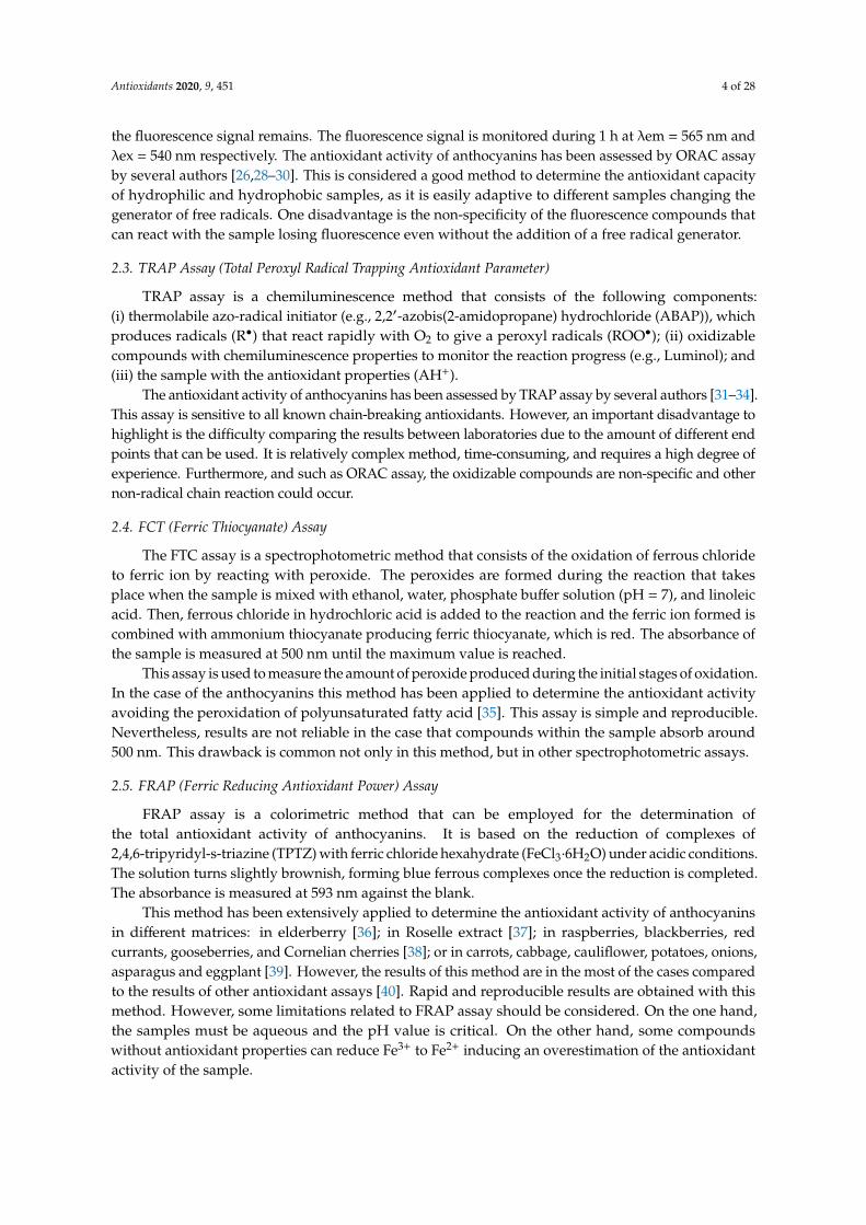
In nature, these pigments are commonly present as anthocyanin, which is in the form of glycoside,and as anthocyanidin also known as aglycone (one or more saccharide bonded with the aglycone).The base structure of anthocyanins is shown in Figure 1. Nowadays the number of anthocyaninsidentified in nature is higher than 600 [6]. Among these anthocyanins, glycoside forms of delphinidin,cyanidin, petunidin, peonidin, malvidin, and pelargonidin, are the most abundant [54,55,62–65].Table 2 shows the most common anthocyanins that have been identified in different fruits, vegetables,and edible flowers and the numerical code that has been assigned to each anthocyanin to refer them inthe manuscript.
Antioxidants 2020, 9, 451 7 of 27
All the assays explained in detail in this manuscript provide results that generally express the antioxidant activity as mmol Trolox Equivalent per kg of fresh weight. However, these results do not report exactly about the antioxidative activity of foods. For this purpose, information about the size portion of each food in the diet is also important to evaluate its antioxidant activity [57]. Due to the relevance of this type of results with regard to the antioxidant activity of anthocyanins, the USDA National Nutrient Database for Standard Reference has published a database where the total concentration of anthocyanins of different fruits and vegetables has been determined together with the service size that should be ingested from each fruit or vegetable [58].
3. Classification and Natural Sources of Anthocyanins
Health and therapeutic effects of anthocyanins are related to their chemical and biochemical reactivity, which are partially explained by their antioxidative activities [59,60]. However, the antioxidative activity of anthocyanins does not necessarily transfer to biological activity because any actions on the body depends both on bioavailability and cellular molecular targets [61]. Furthermore, not all the blue, red, and purple fruits, vegetables, and flowers have the same composition and concentration of anthocyanins and in consequence the same antioxidative activity. The fruits with the highest concentration of anthocyanins are berries, currants, grapes, and some tropical fruit. In the group of edible vegetables, leafy vegetables, grains, roots, and tubers show the highest concentration of anthocyanins as well [62]. Furthermore, the presence of anthocyanins can be detected in different parts of the plant such as stem, leaves, and storage organs.
In nature, these pigments are commonly present as anthocyanin, which is in the form of glycoside, and as anthocyanidin also known as aglycone (one or more saccharide bonded with the aglycone). The base structure of anthocyanins is shown in Figure 1. Nowadays the number of anthocyanins identified in nature is higher than 600 [6]. Among these anthocyanins, glycoside forms of delphinidin, cyanidin, petunidin, peonidin, malvidin, and pelargonidin, are the most abundant [54,55,62–65]. Table 2 shows the most common anthocyanins that have been identified in different fruits, vegetables, and edible flowers and the numerical code that has been assigned to each anthocyanin to refer them in the manuscript.
Figure 1. Structure of anthocyanins R3 = sugar, and anthocyanidins R3 = H.
In supplementary information, Tables S1–S6 show the most common anthocyanins identified in different fruits, vegetables, and flowers, grouped according to their chemical structure: delphinidin and its derivatives (Table S1), cyanidin and its derivatives (Table S2), petunidin (Table S3), peonidin (Table S4), malvidin (Table S5) and pelargonidin and its derivatives (Table S6). Each table also indicates the natural source of the anthocyanins, the type of extraction that was applied, the chromatographic method used to identify and quantify them and the antioxidant assays applied to determine the antioxidant capacity.
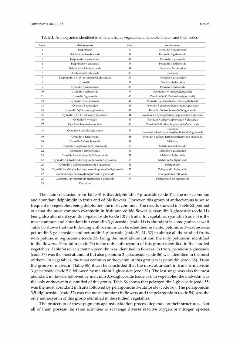
Table 2. Anthocyanins identified in different fruits, vegetables, and edible flowers and their codes.
Code Anthocyanin Code Anthocyanin
Figure 1. Structure of anthocyanins R3 = sugar, and anthocyanidins R3 = H.
In supplementary information, Tables S1–S6 show the most common anthocyanins identified indifferent fruits, vegetables, and flowers, grouped according to their chemical structure: delphinidinand its derivatives (Table S1), cyanidin and its derivatives (Table S2), petunidin (Table S3), peonidin(Table S4), malvidin (Table S5) and pelargonidin and its derivatives (Table S6). Each table also indicatesthe natural source of the anthocyanins, the type of extraction that was applied, the chromatographicmethod used to identify and quantify them and the antioxidant assays applied to determine theantioxidant capacity.
Antioxidants 2020, 9, 451 8 of 28
Table 2. Anthocyanins identified in different fruits, vegetables, and edible flowers and their codes.
Code Anthocyanin Code Anthocyanin
1 Delphinidin 30 Petunidin 3-arabinoside
2 Delphinidin 3-arabinoside 31 Petunidin 3-galactoside
3 Delphinidin 3-galactoside 32 Petunidin 3-glucoside
4 Delphinidin 3-glucoside 33 Petunidin 3-halactoside
5 Delphinidin 3,5-diglucoside 34 Petunidin 3-rutinoside
6 Delphinidin 3-rutinoside 35 Peonidin
7 Delphinidin 3-O-(6”-p-coumaroyl-glucoside) 36 Peonidin 3-galactoside
8 Cyanidin 37 Peonidin 3-glucoside
9 Cyanidin 3-arabidoside 38 Peonidin 3-rutinoside
10 Cyanidin 3-galactoside 39 Peonidin 3-(6′-malonylglucoside)
11 Cyanidin 3-glucoside 40 Peonidin 3-(3”,6”-dimalonylglucoside)
12 Cyanidin 3,5-diglucoside 41 Peonidin 3-glucoside/malvidin 3-galactoside
13 Cyanidin 3-rutinoside 42 Peonidin 3-arabinoside/malvidin 3-glucoside
14 Cyanidin 3-(6′-malonylglucoside) 43 Peonidin 3-O-sophoroside-5-O-glucoside
15 Cyanidin 3-(3”,6”-dimalonylglucoside) 44 Peonidin 3-p-hydroxybenzoylsophoroside-5-glucoside
16 Cyanidin 3-xyloside 45 Peonidin 3-caffeoylsophoroside-5-glucoside
17 Cyanidin 3-xylosylrutinoside 46 Peonidin 3-dicaffeoylsophoroside-5-glucoside
18 Cyanidin 3-dioxaloylglucoside 47 Peonidin3-caffeoyl-p-hydroxybenzoylsophoroside-5-glucoside
19 Cyanidin 3-halavtoside 48 Peonidin 3-caffeoy-feruloylsophoroside-5-glucoside
20 Cyanidin 3-O-sophoroside 49 Malvidin
21 Cyanidin 3-sophoroside-5-rhamnoside 50 Malvidin 3-arabinoside
22 Cyanidin 3-sambubioside 51 Malvidin 3-galactoside
23 Cyanidin 3-sambubioside-5-rhamnoside 52 Malvidin 3-glucoside
24 Cyanidin-3-p-hydroxybenzoylsophoroside-5-glucoside 53 Malvidin 3,5-diglucoside
25 Cyanidin-3-caffeoylsophoroside-5-glucoside 54 Pelargonidin
26 Cyanidin-3-caffeoyl-p-hydroxybenzoylsophoroside-5-glucoside 55 Pelargonidin 3-glucoside
27 Cyanidin 3-(p-coumaroyl)-diglucoside-5-glucoside 56 Pelargonidin 3-rutinoside
28 Cyanidin 3-(p-coumaroyl)-diglucoside-5-glucoside 57 Pelargonidin 3,5-diglucoside
29 Petunidin
The main conclusion from Table S1 is that delphinidin 3-glucoside (code 4) is the most commonand abundant delphinidin in fruits and edible flowers. However, this group of anthocyanins is not sofrequent in vegetables, being delphinine the most common. The results showed in Table S2 pointedout that the most common cyanindin in fruit and edible flower is cyanidin 3-glucoside (code 11),being also abundant cyanidin 3-galactoside (code 10) in fruits. In vegetables, cyanidin (code 8) is themost common and abundant but cyanidin 3-glucoside (code 11) is abundant in some grains as well.Table S3 shows that the following anthocyanins can be identified in fruits: petunidin 3-arabinoside,petunidin 3-galactoside, and petunidin 3-glucoside (code 30, 31, 32) in almost all the studied fruits,with petunidin 3-glucoside (code 32) being the most abundant and the only petunidin identifiedin the flowers. Petunidin (code 29) is the only anthocyanin of this group identified in the studiedvegetables. Table S4 reveals that no peonidin was identified in flowers. In fruits, peonidin 3-glucoside(code 37) was the most abundant but also peonidin 3-galactoside (code 36) was identified in the mostof them. In vegetables, the most common anthocyanin of this group was peonidin (code 35). Fromthe group of malvidin (Table S5) it can be concluded that the most abundant in fruits is malvidin3-galactoside (code 51) followed by malvidin 3-glucoside (code 52). The last stage was also the mostabundant in flowers followed by malvidin 3,5-diglucoside (code 53). In vegetables, the malvidin wasthe only anthocyanin quantified of this group. Table S6 shows that pelargonidin 3-glucoside (code 55)was the most abundant in fruits followed by pelargonidin 3-rutinoside (code 56). The pelargonidin3,5-diglucoside (code 57) was the most abundant in flowers and the pelargonidin (code 54) was theonly anthocyanin of this group identified in the studied vegetables.
The protection of these pigments against oxidation process depends on their structures. Notall of them possess the same activities to scavenge diverse reactive oxygen or nitrogen species.
Antioxidants 2020, 9, 451 9 of 28
The antioxidant ability of anthocyanins depends on the ring orientation since it will determinethe willingness to donate a proton and the capacity to transfer and electron. The number of freehydroxyls around the pyrone ring and their positions also play a key role in the antioxidant activity [2].The presence of other types of radicals in the main structure has an important role in the antioxidantactivity as well. Hence, anthocyanins chalcones and quinoidal bases with a double bond conjugatedto the keto group are efficient antioxidants at scavenging free radicals. Also, the glycosylated B-ringstructure of anthocyanins contributes to the high antioxidant activity, where orthohydroxylation andmethoxylation substantially increase the antioxidant activity. Furthermore, anthocyanidins havehigher antioxidant activity in comparison with anthocyanins, which has been reported in the literature.The reason may be the lower stability of the anthocyanidin compared to the anthocyanin due to itsstructure, what consequently makes anthocyanidin highly reactive [66]. Acylation of anthocyanin withone or more phenolic acids has a significant increase in antioxidant activity [54,67], but glycosylationleads to a reduction in the activity [66,68].
The efficacy of scavenging diverse free radicals differs from one anthocyanin to the other.Pelargonidin-3-glucoside, cyanidin-3-glucoside, and delphinidin-3-glucoside and their standardaglycones have strong antioxidative activity in a liposomal system and reduced formation ofmalondialdehyde by UVB irradiation [55,69]. Furthermore, the results pointed out the highest inhibitoryeffect on lipid peroxidation and O2
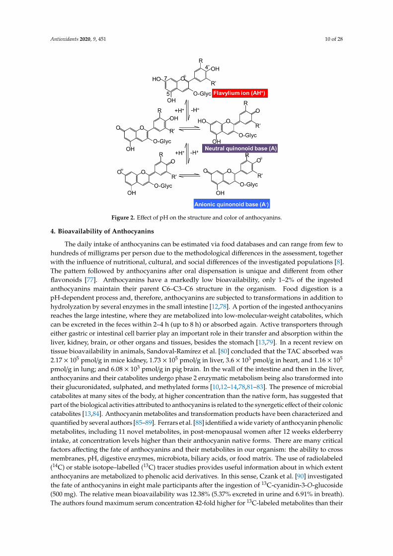
• scavenging activity of delphinidin and delphinidin-3-glucosidefollowed by cyanidin and pelargonidin [2]. On the contrary, pelargonidin had the highest inhibitoryeffect on hydroxyl radical scavenging activity [66]. Moreover, a study demonstrates the highestinhibitory effect on copper (II)-induced low-density lipoprotein (LDL) oxidation of cyanidin andcyanidin-3-glucoside compared with other phenolic acids, anthocyanins, and anthocyanin aglycones,whereas delphinidin has intermediate efficacy [70]. Another study analyzed the antioxidantactivity of malvidin-3-glucoside and the result showed that the quinoidal-base and pseudo-baseof malvidin-3-glucoside significantly inhibited peroxidation of linoleate compared with catechin,malvidin, and resveratrol [71]. However, the oxidation activity assigned to these anthocyanins isdependent on the type of reactive species and in consequence on the type of antioxidant assay carriedout for the determination of the antioxidant activity. Thus, FRAP and TEAC assays have reportedthe significant reduction of the antioxidant activity by the metoxilation in the position 5 or 3 and 5 inpetunidin and malvidin monoglucoside respectively [72]. Another factor implicated in the reactivityof the anthocyanins and also in their antioxidative activity is the pH. In the literature different studieshave demonstrated the effect of the pH in the antioxidant capacity of the anthocyanins from differentsources; Roselle [26], wine [71,72], black rice complexed with cycloamylose [73,74], palm juice [75], andHibiscus acetosella [76]. The pH is an important factor that should be controlled in order to determinethe reactivity of the anthocyanins. The acid nature of the anthocyanin structure is shown in Figure 2.This acid nature is due to the conjugation of the double bonds in the rings of the main structure andthe hydroxyl groups at C4′, C5, and C7 respectively. The hydroxyl group at C7 is the strongest acid.The deprotonation can be produced at acid pH~4 yielding a neutral quinonoid base stabilized bytautomerization with the hydroxyl group at C5. The hydroxyl group at C4 is also susceptible to bedeprotonated at higher pH~7 yielding the anionic base. If the pH level is still rising to basic pH,higher than 8, the deprotonation is produced in the C5 yielding the dianionic base which can leadto the chalcone anion [1]. Hence, the pH of the solution controls the proportions of protonated anddeprotonated hydrated and isomeric form of anthocyanin, affecting its reactivity. Therefore, the pHshould be controlled during the extractions process of anthocyanins and also during the antioxidativebioassays because their results are pH dependent [2,26].
Once the identification of the most common anthocyanins in different foods has been carried outand the influence of the structure and the environment in the capacity of anthocyanins to prevent theoxidation has been discussed, the next step was to determine the bioavailability of these anthocyaninsand their implication, enhancing human health.
Antioxidants 2020, 9, 451 10 of 28Antioxidants 2020, 9, 451 10 of 27
Figure 2. Effect of pH on the structure and color of anthocyanins.
4. Bioavailability of Anthocyanins
The daily intake of anthocyanins can be estimated via food databases and can range from few to hundreds of milligrams per person due to the methodological differences in the assessment, together with the influence of nutritional, cultural, and social differences of the investigated populations [8]. The pattern followed by anthocyanins after oral dispensation is unique and different from other flavonoids [77]. Anthocyanins have a markedly low bioavailability, only 1–2% of the ingested anthocyanins maintain their parent C6–C3–C6 structure in the organism. Food digestion is a pH-dependent process and, therefore, anthocyanins are subjected to transformations in addition to hydrolyzation by several enzymes in the small intestine [12,78]. A portion of the ingested anthocyanins reaches the large intestine, where they are metabolized into low-molecular-weight catabolites, which can be excreted in the feces within 2–4 h (up to 8 h) or absorbed again. Active transporters through either gastric or intestinal cell barrier play an important role in their transfer and absorption within the liver, kidney, brain, or other organs and tissues, besides the stomach [13,79]. In a recent review on tissue bioavailability in animals, Sandoval-Ramírez et al. [80] concluded that the TAC absorbed was 2.17 × 10⁵ pmol/g in mice kidney, 1.73 × 10⁵ pmol/g in liver, 3.6 × 10³ pmol/g in heart, and 1.16 × 10⁵ pmol/g in lung; and 6.08 × 10³ pmol/g in pig brain. In the wall of the intestine and then in the liver, anthocyanins and their catabolites undergo phase 2 enzymatic metabolism being also transformed into their glucuronidated, sulphated, and methylated forms [10,12–14,78,81–83]. The presence of microbial catabolites at many sites of the body, at higher concentration than the native form, has suggested that part of the biological activities attributed to anthocyanins is related to the synergetic effect of their colonic catabolites [13,84]. Anthocyanin metabolites and transformation products have been characterized and quantified by several authors [85–89]. Ferrars et al. [88] identified a wide variety of anthocyanin phenolic metabolites, including 11 novel metabolites, in post-menopausal women after 12 weeks elderberry intake, at concentration levels higher than their anthocyanin native forms. There are many critical factors affecting the fate of anthocyanins and their metabolites in our organism: the ability to cross membranes, pH, digestive enzymes, microbiota, biliary acids, or food matrix. The use of radiolabeled (14C) or stable isotope–labelled (13C) tracer studies provides useful information about in which extent anthocyanins are metabolized to phenolic acid derivatives. In this sense, Czank et al. [90] investigated the fate of anthocyanins in eight male participants after the ingestion of 13C-cyanidin-3-O-glucoside (500 mg). The relative mean bioavailability was 12.38% (5.37% excreted in urine and 6.91% in breath). The
Figure 2. Effect of pH on the structure and color of anthocyanins.
4. Bioavailability of Anthocyanins
The daily intake of anthocyanins can be estimated via food databases and can range from few tohundreds of milligrams per person due to the methodological differences in the assessment, togetherwith the influence of nutritional, cultural, and social differences of the investigated populations [8].The pattern followed by anthocyanins after oral dispensation is unique and different from otherflavonoids [77]. Anthocyanins have a markedly low bioavailability, only 1–2% of the ingestedanthocyanins maintain their parent C6–C3–C6 structure in the organism. Food digestion is apH-dependent process and, therefore, anthocyanins are subjected to transformations in addition tohydrolyzation by several enzymes in the small intestine [12,78]. A portion of the ingested anthocyaninsreaches the large intestine, where they are metabolized into low-molecular-weight catabolites, whichcan be excreted in the feces within 2–4 h (up to 8 h) or absorbed again. Active transporters througheither gastric or intestinal cell barrier play an important role in their transfer and absorption within theliver, kidney, brain, or other organs and tissues, besides the stomach [13,79]. In a recent review ontissue bioavailability in animals, Sandoval-Ramírez et al. [80] concluded that the TAC absorbed was2.17 × 105 pmol/g in mice kidney, 1.73 × 10⁵ pmol/g in liver, 3.6 × 10³ pmol/g in heart, and 1.16 × 10⁵
pmol/g in lung; and 6.08 × 10³ pmol/g in pig brain. In the wall of the intestine and then in the liver,anthocyanins and their catabolites undergo phase 2 enzymatic metabolism being also transformed intotheir glucuronidated, sulphated, and methylated forms [10,12–14,78,81–83]. The presence of microbialcatabolites at many sites of the body, at higher concentration than the native form, has suggested thatpart of the biological activities attributed to anthocyanins is related to the synergetic effect of their coloniccatabolites [13,84]. Anthocyanin metabolites and transformation products have been characterized andquantified by several authors [85–89]. Ferrars et al. [88] identified a wide variety of anthocyanin phenolicmetabolites, including 11 novel metabolites, in post-menopausal women after 12 weeks elderberryintake, at concentration levels higher than their anthocyanin native forms. There are many criticalfactors affecting the fate of anthocyanins and their metabolites in our organism: the ability to crossmembranes, pH, digestive enzymes, microbiota, biliary acids, or food matrix. The use of radiolabeled(14C) or stable isotope–labelled (13C) tracer studies provides useful information about in which extentanthocyanins are metabolized to phenolic acid derivatives. In this sense, Czank et al. [90] investigatedthe fate of anthocyanins in eight male participants after the ingestion of 13C-cyanidin-3-O-glucoside(500 mg). The relative mean bioavailability was 12.38% (5.37% excreted in urine and 6.91% in breath).The authors found maximum serum concentration 42-fold higher for 13C-labeled metabolites than their
Antioxidants 2020, 9, 451 11 of 28
respective native compound 13C-cyanidin-3-glucoside. Up to 49 metabolites were detected includingamong others: phase II conjugates of cyanidin-3-glucoside and cyanidin (cyanidin-glucuronide,methyl cyanidin-glucuronide, and methyl cyanidin-3-glucoside-glucuronide); degradation products(protocatechuic acid, phloroglucinaldehyde, and phloroglucinaldehyde); phase II conjugates ofprotocatechuic acid, phenylacetic acids, phenylpropenoic acids, and hippuric acid.
The mechanisms through which anthocyanins may exert their bioactivity are not fully understoodas it is not clear whether their activity is linked to native forms, their derivatives, or both. The distinctionof their different biological roles is a very challenging task. Some comparative studies have beenconducted on the antioxidant activity of anthocyanin metabolites [91]. Recently, Kim et al. [92] providedbasic information of the chemical changes of cyanidin glycosides during in vitro gastrointestinaldigestion. Cyanidin-3-O-galactoside was degraded into caffeoylquinic acid, which was not found afterin vitro digestion of cyanidin-3-O-glucoside. The bioactivity (DPPH) of the anthocyanin metabolitesdecreased in the intestinal fraction. However, the bioactivity increased after simulated colonicdigestion, possibly because of the newly formed colonic metabolites. Furthermore, anthocyaninmetabolites from the chokeberry extract exhibited higher DPPH radical activities than those from themulberry extract. In another study, α-glucosidase inhibitory activity and ROS scavenging activitiesof conjugated-pelargonidin-3-O-glucoside samples were potentially increased after gastrointestinaldigestion [93].
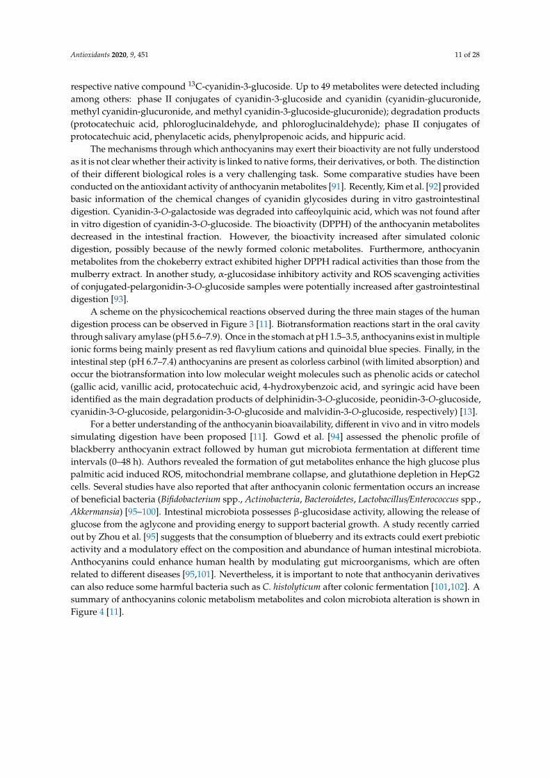
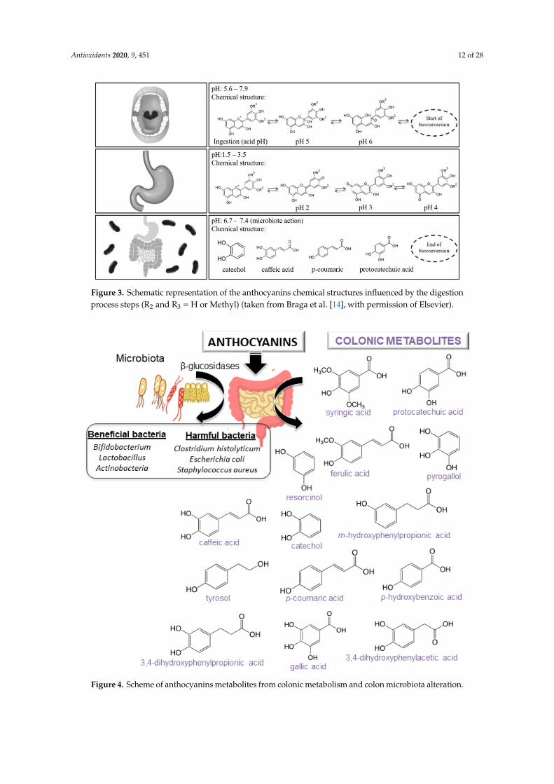
A scheme on the physicochemical reactions observed during the three main stages of the humandigestion process can be observed in Figure 3 [11]. Biotransformation reactions start in the oral cavitythrough salivary amylase (pH 5.6–7.9). Once in the stomach at pH 1.5–3.5, anthocyanins exist in multipleionic forms being mainly present as red flavylium cations and quinoidal blue species. Finally, in theintestinal step (pH 6.7–7.4) anthocyanins are present as colorless carbinol (with limited absorption) andoccur the biotransformation into low molecular weight molecules such as phenolic acids or catechol(gallic acid, vanillic acid, protocatechuic acid, 4-hydroxybenzoic acid, and syringic acid have beenidentified as the main degradation products of delphinidin-3-O-glucoside, peonidin-3-O-glucoside,cyanidin-3-O-glucoside, pelargonidin-3-O-glucoside and malvidin-3-O-glucoside, respectively) [13].
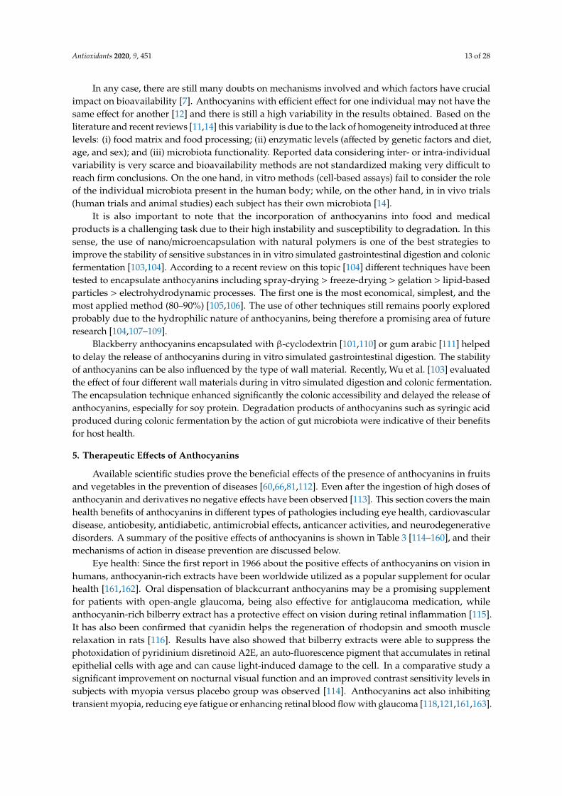
For a better understanding of the anthocyanin bioavailability, different in vivo and in vitro modelssimulating digestion have been proposed [11]. Gowd et al. [94] assessed the phenolic profile ofblackberry anthocyanin extract followed by human gut microbiota fermentation at different timeintervals (0–48 h). Authors revealed the formation of gut metabolites enhance the high glucose pluspalmitic acid induced ROS, mitochondrial membrane collapse, and glutathione depletion in HepG2cells. Several studies have also reported that after anthocyanin colonic fermentation occurs an increaseof beneficial bacteria (Bifidobacterium spp., Actinobacteria, Bacteroidetes, Lactobacillus/Enterococcus spp.,Akkermansia) [95–100]. Intestinal microbiota possesses β-glucosidase activity, allowing the release ofglucose from the aglycone and providing energy to support bacterial growth. A study recently carriedout by Zhou et al. [95] suggests that the consumption of blueberry and its extracts could exert prebioticactivity and a modulatory effect on the composition and abundance of human intestinal microbiota.Anthocyanins could enhance human health by modulating gut microorganisms, which are oftenrelated to different diseases [95,101]. Nevertheless, it is important to note that anthocyanin derivativescan also reduce some harmful bacteria such as C. histolyticum after colonic fermentation [101,102]. Asummary of anthocyanins colonic metabolism metabolites and colon microbiota alteration is shown inFigure 4 [11].
Antioxidants 2020, 9, 451 12 of 28
Antioxidants 2020, 9, 451 11 of 27
authors found maximum serum concentration 42-fold higher for 13C-labeled metabolites than their respective native compound 13C-cyanidin-3-glucoside. Up to 49 metabolites were detected including among others: phase II conjugates of cyanidin-3-glucoside and cyanidin (cyanidin-glucuronide, methyl cyanidin-glucuronide, and methyl cyanidin-3-glucoside-glucuronide); degradation products (protocatechuic acid, phloroglucinaldehyde, and phloroglucinaldehyde); phase II conjugates of protocatechuic acid, phenylacetic acids, phenylpropenoic acids, and hippuric acid.
The mechanisms through which anthocyanins may exert their bioactivity are not fully understood as it is not clear whether their activity is linked to native forms, their derivatives, or both. The distinction of their different biological roles is a very challenging task. Some comparative studies have been conducted on the antioxidant activity of anthocyanin metabolites [91]. Recently, Kim et al. [92] provided basic information of the chemical changes of cyanidin glycosides during in vitro gastrointestinal digestion. Cyanidin-3-O-galactoside was degraded into caffeoylquinic acid, which was not found after in vitro digestion of cyanidin-3-O-glucoside. The bioactivity (DPPH) of the anthocyanin metabolites decreased in the intestinal fraction. However, the bioactivity increased after simulated colonic digestion, possibly because of the newly formed colonic metabolites. Furthermore, anthocyanin metabolites from the chokeberry extract exhibited higher DPPH radical activities than those from the mulberry extract. In another study, α-glucosidase inhibitory activity and ROS scavenging activities of conjugated-pelargonidin-3-O-glucoside samples were potentially increased after gastrointestinal digestion [93].
A scheme on the physicochemical reactions observed during the three main stages of the human digestion process can be observed in Figure 3 [11]. Biotransformation reactions start in the oral cavity through salivary amylase (pH 5.6–7.9). Once in the stomach at pH 1.5–3.5, anthocyanins exist in multiple ionic forms being mainly present as red flavylium cations and quinoidal blue species. Finally, in the intestinal step (pH 6.7–7.4) anthocyanins are present as colorless carbinol (with limited absorption) and occur the biotransformation into low molecular weight molecules such as phenolic acids or catechol (gallic acid, vanillic acid, protocatechuic acid, 4-hydroxybenzoic acid, and syringic acid have been identified as the main degradation products of delphinidin-3-O-glucoside, peonidin-3-O-glucoside, cyanidin-3-O-glucoside, pelargonidin-3-O-glucoside and malvidin-3-O-glucoside, respectively) [13].
Figure 3. Schematic representation of the anthocyanins chemical structures influenced by the digestion process steps (R2 and R3 = H or Methyl) (taken from Braga et al. [14], with permission of Elsevier).
Figure 3. Schematic representation of the anthocyanins chemical structures influenced by the digestionprocess steps (R2 and R3 = H or Methyl) (taken from Braga et al. [14], with permission of Elsevier).Antioxidants 2020, 9, 451 13 of 27
Figure 4. Scheme of anthocyanins metabolites from colonic metabolism and colon microbiota alteration.
It is also important to note that the incorporation of anthocyanins into food and medical products is a challenging task due to their high instability and susceptibility to degradation. In this sense, the use of nano/microencapsulation with natural polymers is one of the best strategies to improve the stability of sensitive substances in in vitro simulated gastrointestinal digestion and colonic fermentation [103,104]. According to a recent review on this topic [104] different techniques have been tested to encapsulate anthocyanins including spray-drying > freeze-drying > gelation > lipid-based particles > electrohydrodynamic processes. The first one is the most economical, simplest, and the most applied method (80–90%) [105,106]. The use of other techniques still remains poorly explored probably due to the hydrophilic nature of anthocyanins, being therefore a promising area of future research [104,107–109].
Blackberry anthocyanins encapsulated with β-cyclodextrin [101,110] or gum arabic [111] helped to delay the release of anthocyanins during in vitro simulated gastrointestinal digestion. The stability of anthocyanins can be also influenced by the type of wall material. Recently, Wu et al. [103] evaluated the effect of four different wall materials during in vitro simulated digestion and colonic fermentation. The encapsulation technique enhanced significantly the colonic accessibility and delayed the release of anthocyanins, especially for soy protein. Degradation products of anthocyanins such as syringic acid produced during colonic fermentation by the action of gut microbiota were indicative of their benefits for host health.
5. Therapeutic Effects of Anthocyanins
Figure 4. Scheme of anthocyanins metabolites from colonic metabolism and colon microbiota alteration.
Antioxidants 2020, 9, 451 13 of 28
In any case, there are still many doubts on mechanisms involved and which factors have crucialimpact on bioavailability [7]. Anthocyanins with efficient effect for one individual may not have thesame effect for another [12] and there is still a high variability in the results obtained. Based on theliterature and recent reviews [11,14] this variability is due to the lack of homogeneity introduced at threelevels: (i) food matrix and food processing; (ii) enzymatic levels (affected by genetic factors and diet,age, and sex); and (iii) microbiota functionality. Reported data considering inter- or intra-individualvariability is very scarce and bioavailability methods are not standardized making very difficult toreach firm conclusions. On the one hand, in vitro methods (cell-based assays) fail to consider the roleof the individual microbiota present in the human body; while, on the other hand, in in vivo trials(human trials and animal studies) each subject has their own microbiota [14].
It is also important to note that the incorporation of anthocyanins into food and medicalproducts is a challenging task due to their high instability and susceptibility to degradation. In thissense, the use of nano/microencapsulation with natural polymers is one of the best strategies toimprove the stability of sensitive substances in in vitro simulated gastrointestinal digestion and colonicfermentation [103,104]. According to a recent review on this topic [104] different techniques have beentested to encapsulate anthocyanins including spray-drying > freeze-drying > gelation > lipid-basedparticles > electrohydrodynamic processes. The first one is the most economical, simplest, and themost applied method (80–90%) [105,106]. The use of other techniques still remains poorly exploredprobably due to the hydrophilic nature of anthocyanins, being therefore a promising area of futureresearch [104,107–109].
Blackberry anthocyanins encapsulated with β-cyclodextrin [101,110] or gum arabic [111] helpedto delay the release of anthocyanins during in vitro simulated gastrointestinal digestion. The stabilityof anthocyanins can be also influenced by the type of wall material. Recently, Wu et al. [103] evaluatedthe effect of four different wall materials during in vitro simulated digestion and colonic fermentation.The encapsulation technique enhanced significantly the colonic accessibility and delayed the release ofanthocyanins, especially for soy protein. Degradation products of anthocyanins such as syringic acidproduced during colonic fermentation by the action of gut microbiota were indicative of their benefitsfor host health.
5. Therapeutic Effects of Anthocyanins
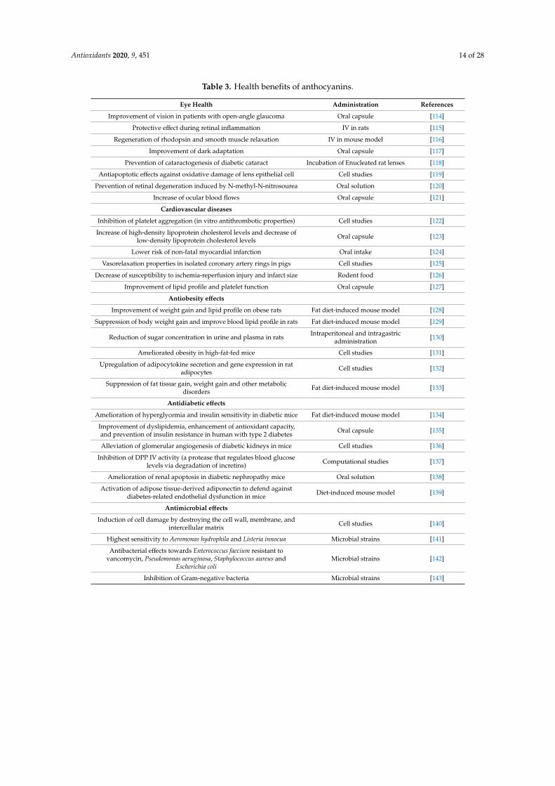
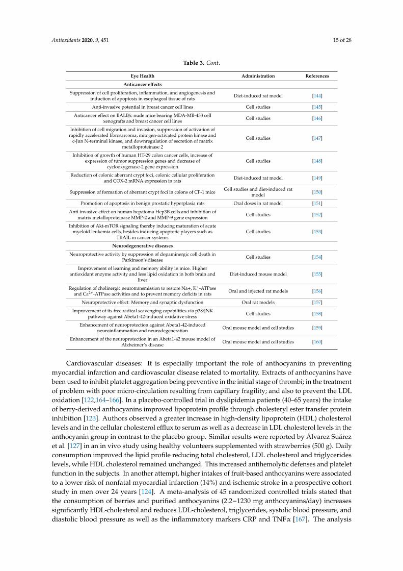
Available scientific studies prove the beneficial effects of the presence of anthocyanins in fruitsand vegetables in the prevention of diseases [60,66,81,112]. Even after the ingestion of high doses ofanthocyanin and derivatives no negative effects have been observed [113]. This section covers the mainhealth benefits of anthocyanins in different types of pathologies including eye health, cardiovasculardisease, antiobesity, antidiabetic, antimicrobial effects, anticancer activities, and neurodegenerativedisorders. A summary of the positive effects of anthocyanins is shown in Table 3 [114–160], and theirmechanisms of action in disease prevention are discussed below.
Eye health: Since the first report in 1966 about the positive effects of anthocyanins on vision inhumans, anthocyanin-rich extracts have been worldwide utilized as a popular supplement for ocularhealth [161,162]. Oral dispensation of blackcurrant anthocyanins may be a promising supplementfor patients with open-angle glaucoma, being also effective for antiglaucoma medication, whileanthocyanin-rich bilberry extract has a protective effect on vision during retinal inflammation [115].It has also been confirmed that cyanidin helps the regeneration of rhodopsin and smooth musclerelaxation in rats [116]. Results have also showed that bilberry extracts were able to suppress thephotoxidation of pyridinium disretinoid A2E, an auto-fluorescence pigment that accumulates in retinalepithelial cells with age and can cause light-induced damage to the cell. In a comparative study asignificant improvement on nocturnal visual function and an improved contrast sensitivity levels insubjects with myopia versus placebo group was observed [114]. Anthocyanins act also inhibitingtransient myopia, reducing eye fatigue or enhancing retinal blood flow with glaucoma [118,121,161,163].
Antioxidants 2020, 9, 451 14 of 28
Table 3. Health benefits of anthocyanins.
Eye Health Administration References
Improvement of vision in patients with open-angle glaucoma Oral capsule [114]
Protective effect during retinal inflammation IV in rats [115]
Regeneration of rhodopsin and smooth muscle relaxation IV in mouse model [116]
Improvement of dark adaptation Oral capsule [117]
Prevention of cataractogenesis of diabetic cataract Incubation of Enucleated rat lenses [118]
Antiapoptotic effects against oxidative damage of lens epithelial cell Cell studies [119]
Prevention of retinal degeneration induced by N-methyl-N-nitrosourea Oral solution [120]
Increase of ocular blood flows Oral capsule [121]
Cardiovascular diseases
Inhibition of platelet aggregation (in vitro antithrombotic properties) Cell studies [122]
Increase of high-density lipoprotein cholesterol levels and decrease oflow-density lipoprotein cholesterol levels Oral capsule [123]
Lower risk of non-fatal myocardial infarction Oral intake [124]
Vasorelaxation properties in isolated coronary artery rings in pigs Cell studies [125]
Decrease of susceptibility to ischemia-reperfusion injury and infarct size Rodent food [126]
Improvement of lipid profile and platelet function Oral capsule [127]
Antiobesity effects
Improvement of weight gain and lipid profile on obese rats Fat diet-induced mouse model [128]
Suppression of body weight gain and improve blood lipid profile in rats Fat diet-induced mouse model [129]
Reduction of sugar concentration in urine and plasma in rats Intraperitoneal and intragastricadministration [130]
Ameliorated obesity in high-fat-fed mice Cell studies [131]
Upregulation of adipocytokine secretion and gene expression in ratadipocytes Cell studies [132]
Suppression of fat tissue gain, weight gain and other metabolicdisorders Fat diet-induced mouse model [133]
Antidiabetic effects
Amelioration of hyperglycemia and insulin sensitivity in diabetic mice Fat diet-induced mouse model [134]
Improvement of dyslipidemia, enhancement of antioxidant capacity,and prevention of insulin resistance in human with type 2 diabetes Oral capsule [135]
Alleviation of glomerular angiogenesis of diabetic kidneys in mice Cell studies [136]
Inhibition of DPP IV activity (a protease that regulates blood glucoselevels via degradation of incretins) Computational studies [137]
Amelioration of renal apoptosis in diabetic nephropathy mice Oral solution [138]
Activation of adipose tissue-derived adiponectin to defend againstdiabetes-related endothelial dysfunction in mice Diet-induced mouse model [139]
Antimicrobial effects
Induction of cell damage by destroying the cell wall, membrane, andintercellular matrix Cell studies [140]
Highest sensitivity to Aeromonas hydrophila and Listeria innocua Microbial strains [141]
Antibacterial effects towards Enterococcus faecium resistant tovancomycin, Pseudomonas aeruginosa, Staphylococcus aureus and
Escherichia coliMicrobial strains [142]
Inhibition of Gram-negative bacteria Microbial strains [143]
Antioxidants 2020, 9, 451 15 of 28
Table 3. Cont.
Eye Health Administration References
Anticancer effects
Suppression of cell proliferation, inflammation, and angiogenesis andinduction of apoptosis in esophageal tissue of rats Diet-induced rat model [144]
Anti-invasive potential in breast cancer cell lines Cell studies [145]
Anticancer effect on BALB/c nude mice bearing MDA-MB-453 cellxenografts and breast cancer cell lines Cell studies [146]
Inhibition of cell migration and invasion, suppression of activation ofrapidly accelerated fibrosarcoma, mitogen-activated protein kinase and
c-Jun N-terminal kinase, and downregulation of secretion of matrixmetalloproteinase 2
Cell studies [147]
Inhibition of growth of human HT-29 colon cancer cells, increase ofexpression of tumor suppression genes and decrease of
cyclooxygenase-2 gene expressionCell studies [148]
Reduction of colonic aberrant crypt foci, colonic cellular proliferationand COX-2 mRNA expression in rats Diet-induced rat model [149]
Suppression of formation of aberrant crypt foci in colons of CF-1 mice Cell studies and diet-induced ratmodel [150]
Promotion of apoptosis in benign prostatic hyperplasia rats Oral doses in rat model [151]
Anti-invasive effect on human hepatoma Hep3B cells and inhibition ofmatrix metalloproteinase MMP-2 and MMP-9 gene expression Cell studies [152]
Inhibition of Akt-mTOR signaling thereby inducing maturation of acutemyeloid leukemia cells, besides inducing apoptotic players such as
TRAIL in cancer systemsCell studies [153]
Neurodegenerative diseases
Neuroprotective activity by suppression of dopaminergic cell death inParkinson’s disease Cell studies [154]
Improvement of learning and memory ability in mice. Higherantioxidant enzyme activity and less lipid oxidation in both brain and
liverDiet-induced mouse model [155]
Regulation of cholinergic neurotransmission to restore Na+, K+-ATPaseand Ca2+-ATPase activities and to prevent memory deficits in rats Oral and injected rat models [156]
Neuroprotective effect: Memory and synaptic dysfunction Oral rat models [157]
Improvement of its free radical scavenging capabilities via p38/JNKpathway against Abeta1-42-induced oxidative stress Cell studies [158]
Enhancement of neuroprotection against Abeta1-42-inducedneuroinflammation and neurodegeneration Oral mouse model and cell studies [159]
Enhancement of the neuroprotection in an Abeta1-42 mouse model ofAlzheimer’s disease Oral mouse model and cell studies [160]
Cardiovascular diseases: It is especially important the role of anthocyanins in preventingmyocardial infarction and cardiovascular disease related to mortality. Extracts of anthocyanins havebeen used to inhibit platelet aggregation being preventive in the initial stage of thrombi; in the treatmentof problem with poor micro-circulation resulting from capillary fragility; and also to prevent the LDLoxidation [122,164–166]. In a placebo-controlled trial in dyslipidemia patients (40–65 years) the intakeof berry-derived anthocyanins improved lipoprotein profile through cholesteryl ester transfer proteininhibition [123]. Authors observed a greater increase in high-density lipoprotein (HDL) cholesterollevels and in the cellular cholesterol efflux to serum as well as a decrease in LDL cholesterol levels in theanthocyanin group in contrast to the placebo group. Similar results were reported by Álvarez Suárezet al. [127] in an in vivo study using healthy volunteers supplemented with strawberries (500 g). Dailyconsumption improved the lipid profile reducing total cholesterol, LDL cholesterol and triglycerideslevels, while HDL cholesterol remained unchanged. This increased antihemolytic defenses and plateletfunction in the subjects. In another attempt, higher intakes of fruit-based anthocyanins were associatedto a lower risk of nonfatal myocardial infarction (14%) and ischemic stroke in a prospective cohortstudy in men over 24 years [124]. A meta-analysis of 45 randomized controlled trials stated thatthe consumption of berries and purified anthocyanins (2.2−1230 mg anthocyanins/day) increasessignificantly HDL-cholesterol and reduces LDL-cholesterol, triglycerides, systolic blood pressure, anddiastolic blood pressure as well as the inflammatory markers CRP and TNFα [167]. The analysis
Antioxidants 2020, 9, 451 16 of 28
also suggested that some individuals are more susceptible to the protective effects of anthocyaninconsumption: (i) overweight; (ii) over 50 years; and (iii) those with increased risk of cardiovasculardisease. Another meta-analysis of 99 randomized controlled trials showed that the consumption ofanthocyanin rich-products decreased significantly both systolic and diastolic blood pressure regardlessof the health status of the participants [168].
In in vitro assays, anthocyanins have also shown inhibition of the porcine pancreatic elastase [169],an enzyme that plays a significant function in pathologies such as arteriosclerosis, emphysema, orrheumatoid arthritis, etc., by attacking fibers and collagen. Moreover, acceleration in the cicatrizationprocess due to anthocyanin-rich extract has been demonstrated, showing preventive and curativeactivity against gastroduodenal ulcers induced in rats [7]. Their influence on the biosynthesis ofmucopolysaccharides provably improves the efficacy of the gastric mucous layer, and increases thebase substance of the connective tissue and of the capillaries [170].
Antiobesity and Antidiabetic effects: Anthocyanins have shown anti-obesity effects throughmultiple mechanisms such as inhibiting lipid absorption, regulating lipid metabolism, increasingenergy expenditure, suppressing food intake and regulating gut microbiota, which suggestsanthocyanins are promising candidates in anti-obesity therapies [171]. Kwon et al. [128] observed thatanthocyanins-added diet from black soybean in rats decreases body weight gains, being significantlylowered in the rats fed with a high fat diet plus black soybean anthocyanins compared with the rats fedwith high fat diet without black soybean. Anthocyanins also improved the lipid profile and suppressedthe high fat diet-induced weight gain in liver intermediately and decreased the weights of epididymaland perirenal fat pads.
In addition, type 2 diabetes is closely related to obesity [66]. Anthocyanins can alleviatecomplications in type 2 diabetes by inhibiting intestinal glucose absorption, inducing pancreatic insulinsecretion, upregulating glucose transporter type 4, and suppressing hepatic gluconeogenesis [172].After the supplementation of a high-fat diet during 13 weeks with different berries in mice, Heymanet al. [173] observed that those supplemented mice gained lesser body weight and presented lowerfasting insulin levels than the control group as well as mediated positive effects on glucose homeostasis.Jankowski et al. [130] described a substantial decrease in the sugar concentration in urine and bloodserum after streptozotocin injection in fed rats with grapes. The mechanisms of anthocyanins suggestedby the authors were the reduction of the biosynthesis of collagen, lipoproteins, and glycoproteins,as well as the reduction of the activity of elastase and adenosine deaminase (both high in diabeticpatients). Treatment with cherries in rats resulted in a significant reduction of blood glucose andurinary microalbumin and an increase of the creatinine secretion level in urea [174]. The pulp, seedand skin from “red chilto” (a red fruit from Argentina) had a hypoglycemic effect and acted increasingglucose absorption, decreasing glucose diffusion rate and promoting glucose transport across the cellmembrane [175] in an in vitro simulated gastroduodenal digestion. Consumption of blueberries andapples/pears in humans was also associated to a lower risk of type 2 diabetes [176].
Antimicrobial effects: The antimicrobial activity of anthocyanins against a wide range ofmicroorganisms is also well documented. Possible mechanisms induced cell damage by destroying thecell wall, membrane and intercellular matrix [66,140,177]. Blackberry extracts have antibacterial activitywith the highest sensitivity to Aeromonas hydrophilia and Listeria innocua [141]. Cranberry extracts haveantibacterial activity towards Enterococcus faecium resistant to vancomycin, Pseudomonas aeruginosa,Staphylococcus aureus, and Escherichia coli [142]. Different types of berry extracts inhibit Gram-negativebacteria but not Gram-positive bacteria [143] probably because Gram-negative bacteria acts as apreventive barrier against hydrophobic compounds but not against hydrophilic compounds [178].
Anticancer activity: Possible mechanisms of the anticancer activity of anthocyanins have beendescribed by many authors: antimutagenic activity; inhibition of oxidative DNA damage andcarcinogen activation; induction of phase II enzymes for detoxification; cell cycle arrest; inhibition ofcyclooxygenase-2 enzymes; as well as induction of apoptosis and antiangiogenesis [179–184].
Antioxidants 2020, 9, 451 17 of 28
In breast cancer, anthocyanins cause the inhibition of key modulators that promote its progressionand development by acting directly in the DNA fragmentation and promoting the death of MCF-7 cancercells [185,186]. In addition, the studies indicate that anthocyanins exert extensive in vitro anti-invasiveand in vivo anti-metastatic activities. For example, delphinidin can act as a potential antimetastatic agentthat suppresses PMA-induced cancer cell invasion through the specific inhibition of NF-κB-dependentMMP-9 gene expression [187,188]. In lung cancer, the treatment of cyanidin-3-glucoside and cyanidin3-rutinoside, isolated from mulberry, inhibits the migration and invasion of A549 cells and alsodecreases MMP-2 and uPA and enhances TIMP-2 and PAI. Anthocyanins also inhibit the growth ofcarcinogenic cells that provoke colon cancer, induce the apoptosis effect, and are even able to act asmodulators of the macrophages in the immune response [180]. Forester et al. [189] also reported thepositive effect of anthocyanin metabolites decreasing cell viability and causing cell cycle arrest andapoptosis in colon cancer. In oral and cervical cancer, the invasion of SCC-4 cells and HeLa cells werediminished by the treatment of peonidin 3-glucoside and cyanidin-3-glucoside [190].
It is also important to note that the structures of anthocyanins have a considerable influence ontheir biological activities [191–193]. In this sense, the type of aglycones, sugars, and acylated acids,and the position and degree of glycosylation and acylation seem to be the main factors influencingthe anticancer property [191]. Jing et al. [192] compared the anticancer properties of anthocyanin-richextracts using human colon cancer HT29 cell line. Authors reported the following growth inhibitoryactivity rates: purple corn > chokeberry and bilberry > purple carrot and grape > radish and elderberry.Those non-acylated monoglycosylated anthocyanins had greater anticancer property than those withpelargonidin, triglycoside, and/or acylation with cinnamic acid.
Neurodegenerative diseases: Anthocyanins are also uniquely suited for the treatment ofneurodegenerative diseases such as Alzheimer’s, Parkinson’s, or amyotrophic lateral sclerosis.Their main mechanisms include antioxidant pathways, calcium homeostasis, inflammation, proteinhomeostasis, and the balance of pro-survival and pro-apoptotic signaling [194,195].
In a primary cell model of Parkinson’s disease, dopaminergic cell death elicited by rotenonewas suppressed by extracts prepared from blueberries, grape seed, hibiscus, blackcurrant, andmulberry [154]. Moreover, Strathearn et al. [154] observed that those extracts rich in anthocyanins andproanthocyanidins exhibited greater neuroprotective activity than extracts rich in other polyphenols.
The oral dispensation of anthocyanins (200 mg/kg) in rats was able to regulate cholinergicneurotransmission, to restore Na+, K+-ATPase and Ca2+-ATPase activities, and to prevent memorydeficits caused by scopolamine dispensation [156]. Rehman et al. [157] showed the neuroprotectiveeffect of anthocyanins based on an artificial ageing model using D-galactose to induce oxidative stressand inflammatory response. The potential mechanisms of their action included: decreased expressionof the receptor for advance glycation end product, reduced level of ROS, and lipid peroxidation. Shihet al. [155] observed that mice fed with anthocyanin-rich mulberry extracts demonstrated significantlyless amyloid β protein and showed improvement of learning and memory ability in avoidance responsetests. The fed mice also showed a higher antioxidant enzyme activity and less lipid oxidation inboth brain and liver, as compared to the control mice. Besides, the treatment with anthocyanin-richmulberry extract has been proved to decrease the levels of serum aspartate aminotransferase, alanineaminotransferase, triglyceride, and total cholesterol that increase with ageing.
Furthermore, the therapeutic profile of anthocyanins can be improved by encapsulation [158–160].For instance, in Alzheimer’s disease Amin et al. [158] showed that encapsulated nanoparticles loadedwith anthocyanins are rapidly taken up by cells enhancing their neuroprotective profile against amyloidbeta toxicity above that of anthocyanins alone. Similar activity was also observed in in vivo studies inmice [158,160].
6. Conclusions
In order to establish the antioxidant activity of anthocyanins and how their intake affects the humanhealth, many factors should be taken into account. Firstly, an evaluation of the antioxidant activity
Antioxidants 2020, 9, 451 18 of 28
from a multiparametric perspective is requires as the total concentration, the structure, the natureof the sample, the pH and the mechanism of the reaction play an important role in their effect. Ona regular basis, more than one antioxidant assay—one for each mechanism—should be carried out.Besides identification and quantification of the anthocyanins is also highly recommended to establishan accurate value for the antioxidant activity of a sample. Secondly, it is required to know thebioavailability of these compounds after their intake. Many studies that have demonstrated the benefitsof anthocyanin-rich extracts in the prevention of diseases. Nonetheless, it is important to note that theirefficacy depends on their bioavailability. Along the digestion process, anthocyanins are metabolizedinto various conjugates, which then ultimately metabolize into phenolic acid degradation products aswell. The accumulated evidence suggests the synergy effect between all possible forms to explain theirattributed health-promoting properties. An inter- and intra-individual variability in anthocyaninsabsorption, metabolism, distribution, and excretion is also evident. Among the main factors thatprobably affect this variability are: food matrix and processing, enzymatic levels and microbiotafunctionality. Attention should be paid in different lines: (i) to perform well-designed standardizedmethods when evaluating bioavailability; (ii) to consider inter or intra-individual variability inanthocyanin metabolism; (iii) to assess the antioxidant activity of anthocyanin metabolites duringgastrointestinal digestion; and, since anthocyanin might exert different biological activities, (iv) todraw a more accurate characterization profiles.
Supplementary Materials: The following are available online at http://www.mdpi.com/2076-3921/9/5/451/s1,Table S1: Natural source, type of extraction, chromatographic method and antioxidant assay applied to identifyand quantify delphinidin and its derivatives identified with codes, Table S2: Natural source, type of extraction,chromatographic method and antioxidant assay applied to identify and quantify cyanidin and its derivativesidentified with codes, Table S3: Natural source, type of extraction, chromatographic method and antioxidant assayapplied to identify and quantify petunidin and its derivatives identified with codes, Table S4: Natural source,type of extraction, chromatographic method and antioxidant assay applied to identify and quantify peonidinand its derivatives identified with codes, Table S5: Natural source, type of extraction, chromatographic methodand antioxidant assay applied to identify and quantify malvidin and its derivatives identified with codes, TableS6: Natural source, type of extraction, chromatographic method and antioxidant assay applied to identify andquantify pelargonidin and its derivatives identified with codes.
Author Contributions: Investigation, N.T., J.M., and A.G.A.; Project administration, A.G.A.; Supervision, A.G.A.;Writing—original draft, N.T. and J.M.; Writing—review and editing, N.T. and J.M. All authors have read andagreed to the published version of the manuscript.
Funding: This research received no external funding.
Acknowledgments: The authors would like to thank the Vicerrectorado de Investigación de la Universidadde Sevilla for its support. We would also like to thank the reviewers for their helpful comments that havegreatly contributed to the improvement of the document. We are also grateful for the very helpful revision byIrene García Sáez.
Conflicts of Interest: The authors declare no conflict of interest.
References
1. Dangles, O.; Fenger, J.A. The Chemical Reactivity of Anthocyanins and Its Consequences in Food Scienceand Nutrition. Molecules 2018, 23, 1970. [CrossRef] [PubMed]
2. Miguel, M.G. Anthocyanins: Antioxidant and/or anti-inflammatory activities. J. Appl. Pharm. Sci. 2011, 1,7–15.
3. Kay, C.D.; Pereira-Caro, G.; Ludwig, I.A.; Clifford, M.N.; Crozier, A. Anthocyanins and flavanones are morebioavailable than previously perceived: A review of recent evidence. Annu. Rev. Food Sci. Technol. 2017, 8,155–180. [CrossRef] [PubMed]
4. Lila, M.A.; Burton-Freeman, B.; Grace, M.; Kalt, W. Unraveling anthocyanin bioavailability for human health.Annu. Rev. Food Technol. 2016, 7, 375–393. [CrossRef] [PubMed]
5. Fang, J. Bioavailability of anthocyanins. Drug Metab. Rev. 2014, 46, 508–520. [CrossRef]6. Lingua, M.S.; Fabani, M.P.; Wunderlin, D.A.; Baroni, M.V. From grape to wine: Changes in phenolic
composition and its influence on antioxidant activity. Food Chem. 2016, 208, 228–238. [CrossRef]
Antioxidants 2020, 9, 451 19 of 28
7. Martín, J.; Kuskoski, E.M.; Navas, M.J.; Asuero, A.G. Antioxidant Capacity of Anthocyanin Pigments.In Flavonoids—From Biosynthesis to Human Health; Justino, J., Ed.; Science, Technology and Medicine OpenAccess Publisher: Rijeka, Croatia, 2017; Chapter 11; pp. 205–255.
8. Martín Bueno, J.; Sáez-Plaza, P.; Ramos-Escudero, F.; Jímenez, A.M.; Fett, R.; Asuero, A.G. Analysis andantioxidant capacity of anthocyanin pigments. Part II: Chemical structure, color, and intake of anthocyanins.Crit. Rev. Anal. Chem. 2012, 42, 126–151. [CrossRef]
9. Navas, M.J.; Jiménez-Moreno, A.M.; Martín Bueno, J.; Sáez-Plaza, P.; Asuero, A.G. Analysis and antioxidantcapacity of anthocyanin pigments. Part IV: Extraction of anthocyanins. Crit. Rev. Anal. Chem. 2012, 42,313–342. [CrossRef]
10. Gamel, T.H.; Wright, A.J.; Tucker, A.J.; Pickard, M.; Rabalski, I.; Podgorski, M.; Di Ilio, N.; O’Brien, C.;Abdel-Aal, E.M. Absorption and metabolites of anthocyanins and phenolic acids after consumption of purplewheat crackers and bars by healthy adults. J. Cereal Sci. 2019, 86, 60–68. [CrossRef]
11. Cavalcante Braga, A.R.; Murador, D.C.; Mendes de Souza Mesquita, L.; Vera de Rosso, V. Bioavailability ofanthocyanins: Gaps in knowledge, challenges and future research. J. Food Compos. Anal. 2018, 68, 31–40.[CrossRef]
12. Fernandes, I.; Faria, A.; Calhau, C.; de Freitas, V.; Mateus, N. Bioavailability of anthocyanins and derivatives.J. Funct. Foods 2014, 7, 54–66. [CrossRef]
13. Fernandes, I.; Faria, A.; de Freitas, V.; Calhau, C.; Mateus, N. Multiple-approach studies to assess anthocyaninbioavailability. Phytochem. Rev. 2015, 14, 899–919. [CrossRef]
14. Eker, M.E.; Aaby, K.; Budic-Leto, I.; Rimac Brncˇic, S.; El, S.N.; Karakaya, S.; Simsek, S.; Manach, C.;Wiczkowski, W.; de Pascual-Teresa, S. A Review of Factors Affecting Anthocyanin Bioavailability: PossibleImplications for the Inter-Individual Variability. Foods 2020, 9, 2. [CrossRef] [PubMed]
15. Mülleder, U.; Murkovic, M.; Pfannhauser, W. Urinary excretion of cyanidin glycosides. J. Biochem. Biophys.Methods 2002, 53, 61–66.
16. Bub, A.; Watzl, B.; Heeb, D.; Rechkemmer, G.; Briviba, K. Malvidin-3-glucoside bioavailability in humansafter ingestion of red wine, dealcoholized red wine and red grape juice. Eur. J. Nutr. 2001, 40, 113–120.[CrossRef] [PubMed]
17. Hong, H.T.; Netzel, M.E.; O’Hare, T.J. Optimization of extraction procedure and development of LC–DAD–MSmethodology for anthocyanin analysis in anthocyanin-pigmented corn kernels. Food Chem. 2020, 319, 126515.[CrossRef] [PubMed]
18. Castañeda-Ovando, A.; Pacheco-Hernandez, M.L.; Paez-Hernandez, M.E.; Rodriguez, J.A.; Galan-Vidal, C.A.Chemical studies of anthocyanins: A review. Food Chem. 2009, 113, 859–871. [CrossRef]
19. Fernandez-Aulis, F.; Hernandez-Vazquez, L.; Aguilar-Osorio, G.; Arrieta-Baez, D.; Navarro-Ocan, A.Extraction and Identification of Anthocyanins in Corn Cob and Corn Husk from Cacahuacintle Maize. J.Food Sci. 2019, 84, 954–962. [CrossRef]
20. Siti Azima, A.M.; Noriham, A.; Manshoor, N. Anthocyanin content in relation to the antioxidant activity andcolour properties of Garcinia mangostana peel, Syzigium cumini and Clitoria ternatea extracts. Int. Food Res.J. 2014, 21, 2369–2375.
21. Liang, N.; Kitts, D.D. Antioxidant Property of Coffee Components: Assessment of Methods that DefineMechanisms of Action. Molecules 2014, 19, 19180–19208. [CrossRef]
22. Shalaby, E.A.; Shanab, S.M.M. Antioxidant compounds, assays of determination and mode of action. Afr. J.Pharm. Pharmacol. 2013, 7, 528–539. [CrossRef]
23. Goupy, P.; Bautista-Ortin, A.-B.; Fulcrand, H.; Dangles, O. Antioxidant activity of wine pigments derivedfrom anthocyanins: Hydrogen transfer reactions to the DPPH radical and inhibition of the heme-inducedperoxidation of linoleic acid. J. Agric. Food Chem. 2009, 57, 5762–5770. [CrossRef] [PubMed]
24. Molyneux, P. The use of the stable free radical diphenylpicrylhydrazyl (DPPH) for estimating antioxidantactivity. Songklanakarin. J. Sci. Technol. 2004, 26, 211–219.
25. Sudheeran, P.K.; Feygenberg, O.; Maurer, D.; Alkan, N. Improved Cold Tolerance of Mango Fruit withEnhanced Anthocyanin and Flavonoid Contents. Molecules 2018, 23, 1832. [CrossRef]
26. Wu, H.Y.; Yang, K.M.; Chiang, P.Y. Roselle Anthocyanins: Antioxidant Properties and Stability to Heat andpH. Molecules 2018, 23, 1357. [CrossRef]
Antioxidants 2020, 9, 451 20 of 28
27. Akar, Z.; Kucuk, M.; Dogan, H. A new colorimetric DPPH scavenging activity method with no need for aspectrophotometer applied on synthetic and natural antioxidants and medicinal herbs. J. Enzym. Inhib. Med.Chem. 2017, 32, 640–647. [CrossRef]
28. Ehlenfeldt, M.K.; Prior, R.L. Oxygen radical absorbance capacity (ORAC) and phenolic and anthocyaninconcentrations in fruit and leaf tissues of highbush blueberry. J. Agric. Food Chem. 2001, 49, 2222–2227.[CrossRef]
29. Bellido, G.G.; Beta, T. Anthocyanin Composition and Oxygen Radical Scavenging Capacity (ORAC) of Milledand Pearled Purple, Black, and Common Barley. J. Agric. Food Chem. 2009, 57, 1022–1028. [CrossRef]
30. Zheng, Y.; Wang, S.Y.; Wang, C.Y.; Zheng, W. Changes in strawberry phenolics, anthocyanins, and antioxidantcapacity in response to high oxygen treatments. LWT 2007, 40, 49–57. [CrossRef]
31. Denev, P.; Ciz, M.; Ambrozova, G.; Lojek, A.; Yanakieva, I.; Kratchanova, M. Solid-phase extraction of berries’anthocyanins and evaluation of their antioxidative properties. Food Chem. 2010, 123, 1055–1061. [CrossRef]
32. Rossetto, M.; Vanzani, P.; Lunelli, M.; Scarpa, M.; Mattivi, F.; Rigo, A. Peroxyl radical trapping activity ofanthocyanins and generation of free radical intermediates. Free Radic. Res. 2007, 41, 854–859. [CrossRef][PubMed]
33. Dresch, R.R.; Kreinecker-Dresch, M.T.; Biegelmeyer, R.; Fretes-Argenta, D.; Fagundes da Rocha, R.;Ferreira-Teixeira, H.; Fonseca-Moreira, J.C.; Henriques, A.T. Potential use of secondary products of theagri-food industry for topical formulations and comparative analysis of antioxidant activity of grape leafpolyphenols. Nat. Prod. Res. 2018, 32, 486–492. [CrossRef] [PubMed]
34. Philpott, M.; Lim, C.C.; Ferguson, L.R. Dietary Protection Against Free Radicals: A Case for Multiple Testingto Establish Structure-activity Relationships for Antioxidant Potential of Anthocyanic Plant Species. Int. J.Mol. Sci. 2009, 10, 1081–1103. [CrossRef] [PubMed]
35. Su, M.S.; Chien, P.J. Antioxidant activity, anthocyanins, and phenolics of rabbiteye blueberry (Vacciniumashei) fluid products as affected by fermentation. Food Chem. 2007, 104, 182–187. [CrossRef]
36. Özgen, M.; Scheerens, J.C.; Neil Reese, R.; Miller, R.A. Total phenolic, anthocyanin contents and antioxidantcapacity of selected elderberry (Sambucus canadensis L.) accessions. Pharm. Mag. 2010, 6, 198–203. [CrossRef]
37. Tsai, P.J.; Intosh, J.M.; Pearce, P.; Camden, B.; Jordan, B.R. Anthocyanin and antioxidant capacity in Roselle(Hibiscus Sabdariffa L.) extract. Food Res. Int. 2002, 35, 351–356. [CrossRef]
38. Pantelidis, G.E.; Vasilakakis, M.; Manganaris, G.A.; Diamantidis, G. Antioxidant capacity, phenol, anthocyaninand ascorbic acid contents in raspberries, blackberries, red currants, gooseberries and Cornelian cherries.Food Chem. 2007, 102, 777–783. [CrossRef]
39. Li, H.; Deng, Z.; Zhu, H.; Hu, C.; Liu, R.; Young, J.C.; Tsao, R. Highly pigmented vegetables: Anthocyanincompositions and their role in antioxidant activities. Food Res. Int. 2012, 46, 250–259. [CrossRef]
40. Moon, J.K.; Shibamoto, T. Antioxidant Assays for Plant and Food Components. J. Agric. Food Chem. 2009, 57,1655–1666. [CrossRef]
41. Ge, Q.; Ma, X. Composition and antioxidant activity of anthocyanins isolated from Yunnan edible rose (Anning). Food Sci. Hum. Wellness 2013, 2, 68–74. [CrossRef]
42. Frond, A.D.; Iuhas, C.I.; Stirbu, I.; Leopold, L.; Socaci, S.; Andreea, S.; Ayvaz, H.; Andreea, S.;Mihai, S.; Diaconeasa, Z.; et al. Phytochemical Characterization of Five Edible Purple-Reddish Vegetables:Anthocyanins, Flavonoids, and Phenolic Acid Derivatives. Molecules 2019, 24, 1536. [CrossRef] [PubMed]
43. Pasko, P.; Barton, H.; Zagrodzki, P.; Gorinstein, S.; Fołta, M.; Zachwieja, Z. Anthocyanins, total polyphenolsand antioxidant activity in amaranth andquinoa seeds and sprouts during their growth. Food Chem. 2009,115, 994–998. [CrossRef]
44. Villaño, D.; Fernández-Pachón, M.S.; Troncoso, A.M.; García-Parrilla, M.C. The Antioxidant Activity of WinesDetermined by the ABTS (+) Method: Influence of Sample Dilution and Time. Talanta 2004, 64, 501–509.
45. Hu, Q.P.; Xu, J.G. Profiles of carotenoids, anthocyanins, phenolics, and antioxidant activity of selected colorwaxy corn grains during maturation. J. Agric. Food Chem. 2011, 59, 2026–2033. [CrossRef]
46. Pala, C.U.; Toklucu, A.K. Effect of UV-C light on anthocyanin content and other quality parameters ofpomegranate juice. J. Food Compos. Anal. 2011, 24, 790–795. [CrossRef]
47. Bunea, A.; Rugina, D.O.; Pintea, A.M.; Conta, Z.; Bunea, C.I.; Socaciu, C. Comparative Polyphenolic Contentand Antioxidant Activities of Some Wild and Cultivated Blueberries from Romania. Not. Bot. Horti Agrobo.2011, 39, 70–76. [CrossRef]
Antioxidants 2020, 9, 451 21 of 28
48. Moniruzzaman, M.; Khalil, M.I.; Sulaiman, S.A.; Gan, S.H. Advances in the analytical methods for determiningthe antioxidant properties of honey: A review. Afr. J. Tradit. Complement. Altern. Med. 2012, 9, 36–42.[CrossRef]
49. Degenhardt, A.; Knapp, H.; Winterhalter, P. Separation and Purification of Anthocyanins by High-SpeedCountercurrent Chromatography and Screening for Antioxidant Activity. J. Agric. Food Chem. 2000, 48,338–343. [CrossRef]
50. Rubinskiene, M.; Jasutiene, I.; Venskutonis, P.R.; Viskelis, P. HPLC Determination of the Composition andStability of Blackcurrant Anthocyanins. J. Chromatogr. Sci. 2005, 43, 478–482. [CrossRef]
51. Welch, C.R.; Wub, Q.; Simon, J.E. Recent Advances in Anthocyanin Analysis and Characterization. Curr.Anal. Chem. 2008, 4, 75–101. [CrossRef]
52. Amorati, R.; Valgimigli, L. Methods to Measure the Antioxidant Activity of Phytochemicals and PlantExtracts. J. Agric. Food Chem. 2018, 66, 3324–3329. [CrossRef] [PubMed]
53. Félix, R.; Valentão, P.; Andrade, P.B.; Félix, C.; Novais, S.C.; Lemos, M.F.L. Evaluating the in Vitro Potential ofNatural Extracts to Protect Lipids from Oxidative Damage. Antioxidants 2020, 9, 231. [CrossRef] [PubMed]
54. Matera, R.; Gabbanini, S.; Berretti, S.; Amorati, R.; De Nicola, G.R.; Iori, R.; Valgimigli, L. Acylatedanthocyanins from sprouts of Raphanus sativus cv. Sango: Isolation, structure elucidation and antioxidantactivity. Food Chem. 2015, 166, 397–406. [CrossRef] [PubMed]
55. Tsuda, T.; Watanabe, M.; Ohshima, K.; Norinobu, S.; Choi, S.W.; Kawakishi, S.; Osawa, T. AntioxidativeActivity of the Anthocyanin Pigments Cyanidin 3-o-β-D-Glucoside and Cyanidin. J. Agri. Food Chem. 1994,42, 2407–2410. [CrossRef]
56. EFSA. Guidance for the scientific requirements for health claims related to antioxidants, oxidative damageand cardiovascular health (Revision 1). EFSA J. 2018, 16, 5136.
57. Prior, R.L.; Go, L.; Wu, X.; Jacob, R.A.; Sotoudeh, G.; Kader, A.A.; Cook, R.A. Plasma antioxidant capacitychanges following a meal as a measure of the ability of a food to alter in vivo antioxidant status. J. Am. Coll.Nutr. 2007, 26, 170–181. [CrossRef]
58. Comert, E.D.; Mogol, B.A.; Gokmen, V. Relationship between color and antioxidant capacity of fruits andvegetables. Curr. Res. Food Sci. 2020, 2, 1–10. [CrossRef]
59. Bakuradze, T.; Tausend, A.; Galan, J.; Groh, I.A.M.; Berry, D.; Tur, J.A.; Marko, D.; Richling, E. Antioxidativeactivity and health benefits of anthocyanin-rich fruit juice in healthy volunteers. Free Radic. Res. 2019, 53,1045–1055. [CrossRef]
60. Khoo, H.E.; Lim, S.M.; Azlan, A. Evidence-based therapeutic effects of anthocyanins from foods. Pak. J. Nutr.2019, 18, 1–11.
61. Williamson, G. The role of polyphenols in modern nutrition. Nutr. Bull. 2017, 42, 226–235. [CrossRef]62. Chaiyasut, C.; Sivamaruthi, B.S.; Pengkumsri, N.; Sirilun, S.; Peerajan, S.; Chaiyasut, K.; Kesika, P. Anthocyanin
profile and its antioxidant activity of widely used fruits, vegetables, and flowers in Thailand Asian. J. Pharm.Clin. Res. 2016, 9, 218–224.
63. Ogawa, K.; Sakakibara, H.; Iwata, R.; Ishii, T.; Sato, T.; Goda, T.; Shimoi, K.; Kumazawa, S. AnthocyaninComposition and Antioxidant Activity of the Crowberry (Empetrum nigrum) and Other Berries. J. Agric.Food Chem. 2008, 56, 4457–4462. [CrossRef] [PubMed]
64. Zykin, P.A.; Andreeva, E.A.; Lykholay, A.N.; Tsvetkova, N.V.; Voylokov, A.V. Anthocyanin Composition andContent in Rye Plants with Different Grain Color. Molecules 2018, 23, 948. [CrossRef] [PubMed]
65. De Morais, J.S.; Sant’Ana, A.S.; Dantas, A.M.; Silva, B.S.; Lima, M.S.; Borges, G.C.; Magnani, M. Antioxidantactivity and bioaccessibility of phenolic compounds in white, red, blue, purple, yellow and orange edibleflowers through a simulated intestinal barrier. Food Res. Int. 2020, 131, 109046. [CrossRef] [PubMed]
66. Khoo, H.E.; Azlan, A.; Tang, S.T.; Lim, S.M. Anthocyanidins and anthocyanins: Colored pigments as food,pharmaceutical ingredients, and the potential health benefits. Food Nutr. Res. 2017, 61, 1361779. [CrossRef]
67. Tamura, H.; Yamagami, A. Antioxidative activity of monoacylated anthocyanins isolated from Muscat BaileyA grape. J. Agric. Food Chem. 1994, 42, 1612–1615. [CrossRef]
68. Stintzing, F.C.; Stintzing, A.S.; Carle, R.; Frei, B.; Wrolstad, R.E. Color and antioxidant properties ofcyanidin-based anthocyanin pigments. J. Agric. Food Chem. 2002, 50, 6172–6181. [CrossRef]
69. Tsuda, T.; Shiga, K.; Ohshima, K.; Kawakishi, S.; Osawa, T. Inhibition of lipid peroxidation and the activeoxygen radical scavenging effect of anthocyanin pigments isolated from Phaseolus vulgaris L. Biochem.Pharmacol. 1996, 52, 1033–1110. [CrossRef]
Antioxidants 2020, 9, 451 22 of 28
70. Brown, J.E.; Kelly, M.F. Inhibition of lipid peroxidation by anthocyanins, anthocyanidins and their phenolicdegradation products. Eur. J. Lipid Sci. Technol. 2007, 109, 66–71. [CrossRef]
71. Lapidot, T.; Harel, S.; Akiri, B.; Granit, R.; Kanner, J. pH-dependent forms of red wine anthocyanins asantioxidants. J. Agric. Food Chem. 1999, 47, 67–70. [CrossRef]
72. Muselík, J.; García-Alonso, M.; Martín-López, M.P.; Žemlicka, M.; Rivas-Gonzalo, J.C. Measurement ofantioxidant activity of wine catechins, procyanidins, anthocyanins and pyranoanthocyanins. Int. J. Mol. Sci.2007, 8, 797–809. [CrossRef]
73. Jung, Y.K.; Joo, K.S.; Rho, S.J.; Kim, Y.R. pH-dependent antioxidant stability of black rice anthocyanincomplexed with cycloamylose. LWT Food Sci. Technol. 2020, 129, 109474. [CrossRef]
74. Sui, X.; Dong, X.; Zhou, W. Combined effect of pH and high temperature on the stability and antioxidantcapacity of two anthocyanins in aqueous solution. Food Chem. 2014, 163, 163–170. [CrossRef] [PubMed]
75. Ghosh, S.; Chakraborty, R.; Raychaudhuri, U. Determination of pH-dependent antioxidant activity of palm(Borassus flabellifer) polyphenol compounds by photoluminol and DPPH methods: A comparison of redoxreaction sensitivity. 3 Biotech 2015, 5, 633–640. [CrossRef] [PubMed]
76. Março, P.H.; Poppi, R.J.; Scarminio, I.S.; Tauler, R. Investigation of the pH effect and UV radiation on kineticdegradation of anthocyanin mixtures extracted from Hibiscus acetosella. Food Chem. 2011, 125, 1020–1102.
77. Manach, C.; Williamson, G.; Morand, C.; Scalbert, A.; Remesy, C. Bioavailability and bioefficacy of polyphenolsin humans. I. Review of 97 bioavailability studies. Am. J. Clin. Nutr. 2005, 81, 230–242. [CrossRef]
78. Felgines, C.; Talavéra, S.; Texier, O.; Gil-Izquierdo, A.; Lamaison, J.-L.; Remesy, C. Blackberry anthocyaninsare mainly recovered from urine as methylated and glucuronidated conjugates in humans. J. Agric. FoodChem. 2005, 53, 7721–7727. [CrossRef]
79. Passamonti, S.; Vanzo, A.; Vrhovsek, U.; Terdoslavich, M.; Cocolo, A.; Decorti, G.; Mattivi, F. Hepatic uptakeof grape anthocyanins and the role of bilitranslocase. Food Res. Int. 2005, 38, 953–960. [CrossRef]
80. Sandoval-Ramírez, B.A.; Catalann, U.; Fernanndez-Castillejo, S.; Rubió, L.; Maciá, A.; Sola, R. Anthocyanintissue bioavailability in animals: Possible implications for human health. A systematic review. J. Agric. FoodChem. 2018, 66, 11531–11543. [CrossRef]
81. Lucioli, S. Anthocyanins: Mechanism of action and therapeutic efficacy. In Medicinal Plants as AntioxidantAgents: Understanding Their Mechanism of Action and Therapeutic Efficacy; Capasso, A., Ed.; Research Signpost:Kerala, India, 2012; pp. 27–57.
82. Jin, Y.A.D.; George, T.G.M.; Lovegrove, J.A.A. Randomised trial to investigate the effects of acute consumptionof a blackcurrant juice drink on markers of vascular reactivity and bioavailability of anthocyanins in humansubjects. Eur. J. Clin. Nutr. 2011, 65, 849–857. [CrossRef]
83. Wu, X.; Cao, G.; Prior, R.L. Absorption and metabolism of anthocyanins in elderly women after consumptionof elderberry or blueberry. J. Nutr. 2002, 132, 1865–1871. [CrossRef] [PubMed]
84. Williamson, G.; Clifford, M.N. Colonic metabolites of berry polyphenols: The missing link to biologicalactivity? Br. J. Nutr. 2010, 104 (Suppl. S3), S48–S66. [CrossRef] [PubMed]
85. Kaiser, M.; Müller-Ehl, L.; Passon, M.; Schieber, A. Development and validation of methods for thedetermination of anthocyanins in physiological fluids via UHPLC-MSn. Molecules 2020, 25, 518. [CrossRef][PubMed]
86. Ludwig, I.A.; Mena, P.; Calani, L.; Borges, G.; Pereira-Caro, G.; Bresciani, L.; Del Rio, D.; Lean, M.E.J.;Crozier, A. New insights into the bioavailability of red raspberry anthocyanins and ellagitannins. Free Radic.Biol. Med. 2015, 89, 758–769. [CrossRef]
87. Ferrars, R.M.; Cassidy, A.; Curtis, P.; Kay, C.D. Phenolic metabolites of anthocyanins following a dietaryintervention study in post-menopausal women. Mol. Nutr. Food Res. 2014, 58, 490–502. [CrossRef]
88. Ferrars, R.M.; Czank, C.; Saha, S.; Needs, P.W.; Zhang, Q.; Raheem, K.S.; Botting, N.P.; Kroon, P.A.; Kay, C.D.Methods for isolating, identifying, and quantifying anthocyanin metabolites in clinical samples. Anal. Chem.2014, 86, 10052–10058. [CrossRef]
89. Nurmi, T.; Mursu, J.; Heinonen, M.; Nurmi, A.; Hiltunen, R.; Voutilainen, S. Metabolism of berry anthocyaninsto phenolic acids in humans. J. Agric. Food Chem. 2009, 57, 2274–2281. [CrossRef]
90. Czank, C.; Cassidy, A.; Zhang, Q.; Morrison, D.J.; Preston, T.; Kroon, P.A.; Botting, N.P.; Kay, C.D. Humanmetabolism and elimination of the anthocyanin, cyanidin-3-glucoside: A 13C-tracer study. Am. J. Clin. Nutr.2013, 97, 995–1003. [CrossRef]
Antioxidants 2020, 9, 451 23 of 28
91. Novotny, J.A. Anthocyanin Bioavailability: Past Progress and Current Challenges. In Emerging Trends inDietary Components for Preventing and Combating Disease; ACS Symposium Series; American Chemical Society:Washington, DC, USA, 2012; Chapter 32; pp. 559–568.
92. Kim, I.; Moon, J.K.; Hur, S.J.; Lee, J. Structural changes in mulberry (Morus Microphylla. Buckl) andchokeberry (Aronia melanocarpa) anthocyanins during simulated in vitro human digestion. Food Chem.2020, 318, 126449. [CrossRef]
93. Gowd, V.; Karim, N.; Xie, L.; Shishir, M.R.I.; Xu, Y.; Chen, W. In vitro study of bioaccessibility, antioxidant,and α-glucosidase inhibitory effect of pelargonidin-3-O-glucoside after interacting with beta-lactoglobulinand chitosan/pectin. Int. J. Biol. Macromol. 2020, 154, 380–389. [CrossRef]
94. Gowd, V.; Bao, T.; Chen, W. Antioxidant potential and phenolic profile of blackberry anthocyanin extractfollowed by human gut microbiota fermentation. Food Res. Int. 2019, 120, 523–533. [CrossRef] [PubMed]
95. Zhou, L.; Xiec, M.; Yanga, F.; Liu, J. Antioxidant activity of high purity blueberry anthocyanins and the effectson human intestinal microbiota. Food Sci. Technol. 2020, 117, 628101. [CrossRef]
96. Zhu, Y.; Sun, H.; He, S.; Lou, Q.; Yu, M.; Tang, M.; Tu, L. Metabolism and prebiotics activity of anthocyaninsfrom black rice (Oryza sativa L.) in vitro. PLoS ONE 2018, 13, 0195754. [CrossRef] [PubMed]
97. Hidalgo, M.; Oruna-Concha, M.J.; Kolida, S.; Walton, G.E.; Kallithraka, S.; Spencer, J.P.E.; Gibson, G.R.; dePascual-Teresa, S. Metabolism of anthocyanins by human gut microflora and their influence on gut bacterialgrowth. J. Agric. Food Chem. 2012, 60, 3882–3890. [CrossRef]
98. Boto-Ordóñez, M.; Urpi-Sarda, M.; Queipo-Ortuño, M.I.; Tulipani, S.; Tinahones, F.J.; Andres-Lacueva, C.High levels of Bifidobacteria are associated with increased levels of anthocyanin microbial metabolites: Arandomized clinical trial. Food Funct. 2014, 5, 1932–1938. [CrossRef]
99. Garcia-Mazcorro, J.F.; Lage, N.N.; Mertens-Talcott, S.; Talcott, S.; Chew, B.; Dowd, S.E.; Kawas, J.R.;Noratto, G.D. Effect of dark sweet cherry powder consumption on the gut microbiota, short-chain fatty acids,and biomarkers of gut health in obese db/db mice. Peer J. 2018, 2018, 1–31. [CrossRef]
100. Lacombe, A.; Li, R.W.; Klimis-Zacas, D.; Kristo, A.S.; Tadepalli, S.; Krauss, E.; Young, R.; Wu, V.C.H. Lowbushwild blueberries have the potential to modify gut microbiota and xenobiotic metabolism in the rat colon.PLoS ONE 2013, 8, 67497. [CrossRef]
101. Flores, G.; Ruiz del Castillo, M.L.; Costabile, A.; Klee, A.; Bigetti Guergoletto, K.; Gibson, G.R. In vitrofermentation of anthocyanins encapsulated with cyclodextrins: Release, metabolism and influence on gutmicrobiota growth. J. Funct. Foods 2015, 16, 50–57. [CrossRef]
102. Guergoletto, K.B.; Costabile, A.; Flores, G.; Garcia, S.; Gibson, G.R. In vitro fermentation of juçara pulp(Euterpe edulis) by human colonic microbiota. Food Chem. 2016, 196, 251–258. [CrossRef]
103. Wu, Y.; Han, Y.; Tao, Y.; Li, D.; Xie, G.; Show, P.L.; Lee, S.Y. In vitro gastrointestinal digestion and fecalfermentation reveal the effect of different encapsulation materials on the release, degradation and modulationof gut microbiota of blueberry anthocyanin extract. Food Res. Int. 2020, 132, 109098. [CrossRef]
104. Sharif, N.; Khoshnoudi-Nia, S.; Jafari, S.M. Nano/microencapsulation of anthocyanins; a systematic reviewand meta-analysis. Food Res. Int. 2020, 132, 109077. [CrossRef] [PubMed]
105. Yousuf, B.; Gul, K.; Wani, A.A.; Singh, P. Health benefits of anthocyanins and their encapsulation for potentialuse in food systems: A review. Crit. Rev. Food Sci. Nutr. 2016, 56, 2223–2230. [CrossRef] [PubMed]
106. Mahdavi, S.A.; Jafari, S.M.; Ghorbani, M.; Assadpoor, E. Spray-drying microencapsulation of anthocyaninsby natural biopolymers: A review. Dry Technol. 2014, 32, 509–518. [CrossRef]
107. Robert, P.; Fredes, C. The encapsulation of anthocyanins from berry-type fruits. Trends in foods. Molecules2015, 20, 5875–5888. [CrossRef]
108. Cavalcanti, R.N.; Santos, D.T.; Meireles, M.A.A. Non-thermal stabilization mechanisms of anthocyanins inmodel and food systems—An overview. Food Res. Int. 2011, 44, 499–509. [CrossRef]
109. Fang, Z.; Bhandari, B. Encapsulation of polyphenols—A review. Trends Food Sci. Technol. 2010, 21, 510–523.[CrossRef]
110. Fernandes, A.; Rocha, M.; Santos, L.; Brás, J.; Oliveira, J.; Mateus, N.; de Freitas, V. Blackberry anthocyanins:β-Cyclodextrin fortification for thermal and gastro- intestinal stabilization. Food Chem. 2018, 245, 426–431.[CrossRef]
111. Huang, Y.; Zhou, W.B. Microencapsulation of anthocyanins through two-step emulsification and releasecharacteristics during in vitro digestion. Food Chem. 2019, 278, 357–363. [CrossRef]

Antioxidants 2020, 9, 451 24 of 28
112. De Pascual-Teresa, S.; Sanchez-Ballesta, M.T. Anthocyanins: From plant to health. Phytochem. Rev. 2008, 7,281–299. [CrossRef]
113. Clifford, M.N. Anthocyanins—Nature, occurrence and dietary sources. J. Sci. Food Agric. 2000, 80, 1063–1072.[CrossRef]
114. Shim, S.H.; Kim, J.M.; Choi, C.Y.; Kim, C.Y.; Park, K.H. Ginkgo biloba extract and bilberry anthocyaninsimprove visual function in patients with normal tension glaucoma. J. Med. Food 2012, 15, 818–823. [CrossRef][PubMed]
115. Ahmad, A.; Kaleem, M.; Ahmed, Z.; Shafiq, H. Therapeutic potential of flavonoids and their mechanism ofaction against microbial and viral infections—A review. Food Res. Int. 2015, 77, 221–235. [CrossRef]
116. Miyake, S.; Takahashi, N.; Sasaki, M.; Takahashi, N.; Ozawa, Y. Vision preservation during retinal inflammationby anthocyanin- rich bilberry extract: Cellular and molecular mechanism. Lab. Investig. 2012, 92, 102–109.[CrossRef] [PubMed]
117. Lee, J.; Lee, H.K.; Kim, C.Y.; Hong, Y.J.; Choe, C.M.; You, T.W.; Seong, G.J. Purified high-dose anthocyanosideoligomer administration improves nocturnal vision and clinical symptoms in myopia subjects. Br. J. Nutr.2005, 93, 895–899. [CrossRef]
118. Thiraphatthanavong, P.; Wattanathorn, J.; Muchimapura, S.; Wipawee, T.M.; Wannanon, P.; Terdthai, T.U.;Suriharn, B.; Lertrat, K. Preventive effect of Zea mays L. (purple waxy corn) on experimental diabetic cataract.BioMed Res. Int. 2014, 2014, 507435. [CrossRef]
119. Mok, J.W.; Chang, D.J.; Joo, C.K. Antiapoptotic effects of anthocyanin from the seed coat of black soybeanagainst oxidative damage of human lens epithelial cell induced by H2O2. Curr. Eye Res. 2014, 39, 1090–1098.[CrossRef]
120. Paik, S.S.; Jeong, E.; Jung, S.W.; Ha, T.J.; Kang, S.; Sim, S.; Jeon, J.H.; Chun, M.H.; Kim, I.B. Anthocyaninsfrom the seed coat of black soybean reduce retinal degeneration induced by N-methyl-N-nitrosourea. Exp.Eye Res. 2012, 97, 55–62. [CrossRef]
121. Ohguro, H.; Ohguro, I.; Katai, M.; Tanaka, S. Two-year randomized, placebo-controlled study of black currantanthocyanins on visual field in glaucoma. Ophthalmologica 2012, 228, 26–35. [CrossRef]
122. Yang, Y.; Shi, Z.; Reheman, A.; Jin, W.; Li, C.; Zhu, G.; Wang, Y.; Freedman, J.J.; Ling, W.; Ni, H. Anthocyaninsinhibit platelet activation and attenuate thrombus growth in both human and murine thrombosis models.Blood 2010, 116, 3197. [CrossRef]
123. Qin, Y.; Xia, M.; Ma, J.; Hao, Y.; Liu, J.; Mou, H.; Cao, L.; Ling, W. Anthocyanin supplementation improvesserum LDL-and HDL-cholesterol concentrations associated with the inhibition of cholesteryl ester transferprotein in dyslipidemic subjects. Am. J. Clin. Nutr. 2009, 90, 485–492. [CrossRef]
124. Cassidy, A.; Bertoia, M.; Chiuve, S.; Flint, A.; Forman, J.; Rimm, E.B. Habitual intake of anthocyanins andflavanones and risk of cardiovascular disease in men. Am. J. Clin. Nutr. 2016, 104, 587–594. [CrossRef][PubMed]
125. Bell, D.R.; Gochenaur, K. Direct vasoactive and vasoprotective properties of anthocyanin-rich extracts. J.Appl. Physiol. 2006, 100, 1164–1170. [CrossRef] [PubMed]
126. Toufektsian, M.C.; De Lorgeril, M.; Nagy, N.; Salen, P.; Donati, M.B.; Giordano, L.; Mock, H.-P.; Peterek, S.;Matros, A.; Petroni., K.; et al. Chronic dietary intake of plant-derived anthocyanins protects the rat heartagainst ischemia-reperfusion injury. J. Nutr. 2008, 138, 747–752. [CrossRef] [PubMed]
127. Alvarez-Suarez, J.M.; Giampieri, F.; Tulipani, S.; Casoli, T.; di Stefano, G. One month strawberry-richanthocyanin supplementation ameliorates cardiovascular risk, oxidative stress markers and platelet activationin humans. J. Nutr. Biochem. 2014, 25, 289–294. [CrossRef] [PubMed]
128. Kwon, S.H.; Ahn, I.S.; Kim, S.O.; Kong, C.S.; Chung, H.Y.; Do, M.S.; Park, K.Y. Anti-obesity and hypolipidemiceffects of black soybean anthocyanins. J. Med. Food 2007, 10, 552–556. [CrossRef] [PubMed]
129. Wu, T.; Yu, Z.; Tang, Q.; Song, H.; Gao, Z.; Chen, W.; Zheng, X. Honeysuckle anthocyanin supplementationprevents diet-induced obesity in C57BL/6 mice. Food Funct. 2013, 4, 1654–1661. [CrossRef] [PubMed]
130. Jankowski, A.; Jankowska, B.; Niedworok, J. The effects of anthocyanin dye from grapes on experimentaldiabetes. Folia Med. Cracov. 2000, 41, 5–15.
131. Jayaprakasam, B.; Vareed, S.K.; Olson, L.K.; Nair, M.G. Insulin secretion by bioactive anthocyanins andanthocyanidins present in fruits. J. Agric. Food Chem. 2005, 53, 28–31. [CrossRef]
Antioxidants 2020, 9, 451 25 of 28
132. Tsuda, T.; Ueno, Y.; Aoki, H.; Koda, T.; Horio, F.; Takahashi, N.; Kawada, T.; Osawa, T. Anthocyanin enhancesadipocytokine secretion and adipocyte-specific gene expression in isolated rat adipocytes. Biochem. Biophys.Res. Commun. 2004, 316, 149–157. [CrossRef]
133. Tsuda, T.; Horio, F.; Uchida, K.; Aoki, H.; Osawa, T. Dietary cyanidin 3- O-β-D-glucoside-rich purple corncolor prevents obesity and ameliorates hyperglycemia in mice. J. Nutr. 2003, 133, 2125–2130. [CrossRef]
134. Takikawa, M.; Inoue, S.; Horio, F.; Tsuda, T. Dietary anthocyanin-rich bilberry extract ameliorateshyperglycemia and insulin sensitivity via activation of AMP-activated protein kinase in diabetic mice.J. Nutr. 2010, 140, 527–533. [CrossRef] [PubMed]
135. Li, D.; Zhang, Y.; Liu, Y.; Sun, R.; Xia, M. Purified anthocyanin supplementation reduces dyslipidemia,enhances antioxidant capacity, and prevents insulin resistance in diabetic patients. J. Nutr. 2015, 145, 742–748.[CrossRef] [PubMed]
136. Kang, M.K.; Lim, S.S.; Lee, J.Y.; Yeo, K.M.; Kang, Y.-H. Anthocyanin-rich purple corn extract inhibitdiabetes-associated glomerular angiogenesis. PLoS ONE 2013, 8, 79823. [CrossRef] [PubMed]
137. Fan, J.; Johnson, M.H.; Lila, M.A.; Yousef, G.; de Mejia, E.G. Berry and citrus phenolic compounds inhibitdipeptidyl peptidase IV: Implications in diabetes management. Evid. Based Complement. Alternat. Med. 2013,2013, 479505. [CrossRef]
138. Koh, E.S.; Lim, J.H.; Kim, M.Y.; Chung, S.; Shin, S.J.; Choi, B.S.; Kim, H.W.; Hwang, S.Y.; Kim, S.W.; Park, C.W.;et al. Anthocyanin-rich Seoritae extract ameliorates renal lipotoxicity via activation of AMP-activated proteinkinase in diabetic mice. J. Transl. Med. 2015, 13, 203. [CrossRef]
139. Liu, Y.; Li, D.; Zhang, Y.; Sun, R.; Xia, M. Anthocyanin increases adiponectin secretion and protects againstdiabetes-related endothelial dysfunction. Am. J. Physiol. Endocrinol. Metab. 2014, 306, E975–E988. [CrossRef]
140. Pojer, E.; Mattivi, F.; Johnson, D.; Stockley, C.S. The case for anthocyanin consumption to promote humanhealth: A review. Compr. Rev. Food Sci. Food Saf. 2013, 12, 483–508. [CrossRef]
141. Genskowsky, E.; Puente, L.A.; Perez-Alvarez, J.A.; Fernández-López, J.; Muñoz, L.A.; Viuda-Martos, M.Determination of polyphenolic profile, antioxidant activity and antibacterial properties of maqui [Aristoteliachilensis (Molina) Stuntz] a Chilean blackberry. J. Sci. Food Agr. 2016, 96, 4235–4242. [CrossRef]
142. Côté, J.; Caillet, S.; Doyon, G.; Ng, K. Antimicrobial effect of cranberry juice and extracts. Food Cont. 2011, 22,1413–1418. [CrossRef]
143. Puupponen-Pimiä, R.; Nohynek, L.; Meier, C.; Kähkönen, M.; Heinonen, M.; Hopia, A.;Oksman-Caldentey, K.M. Antimicrobial properties of phenolic compounds from berries. J. Appl. Microbiol.2001, 90, 494–507. [CrossRef]
144. Wang, L.S.; Hecht, S.S.; Carmella, S.G.; Yu, N.; Larue, B.; Henry, C.; McIntyre, C.; Rocha, C.; Lechner, J.F.;Stoner, G.D. Anthocyanins in black raspberries prevent esophageal tumors in rats. Cancer Prev. Res. 2009, 2,84–93. [CrossRef] [PubMed]
145. Faria, A.; Pestana, D.; Teixeira, D.; de Freitas, V.; Mateus, N.; Calhau, C. Blueberry anthocyanins and pyruvicacid adducts: Anticancer properties in breast cancer cell lines. Phytother. Res. 2010, 24, 1862–1869. [CrossRef]
146. Hui, C.; Bin, Y.; Xiaoping, Y.; Long, Y.; Chunye, C.; Mantian, M.; Wenhua, L. Anticancer activities of ananthocyanin-rich extract from black rice against breast cancer cells in vitro and in vivo. Nutr. Cancer 2010,62, 1128–1136. [CrossRef] [PubMed]
147. Chen, X.Y.; Zhou, J.; Luo, L.P.; Han, B.; Li, F.; Chen, J.Y.; Zhu, Y.F.; Chen, W.; Yu, X.P. Black rice anthocyaninssuppress metastasis of breast cancer cells by targeting RAS/RAF/MAPK pathway. BioMed Res. Int. 2015,2015, 414250. [CrossRef] [PubMed]
148. Malik, M.; Zhao, C.; Schoene, N.; Guisti, M.M.; Moyer, M.P.; Magnuson, B.A. Anthocyanin-rich extract fromAronia meloncarpa E. induces a cell cycle block in colon cancer but not normal colonic cells. Nutr. Cancer2003, 46, 186–196. [CrossRef] [PubMed]
149. Lala, G.; Malik, M.; Zhao, C.; He, J.; Kwon, Y.; Giusti, M.M.; Magnuson, B.A. Anthocyanin-rich extractsinhibit multiple biomarkers of colon cancer in rats. Nutr. Cancer 2006, 54, 84–93. [CrossRef] [PubMed]
150. Lim, S.; Xu, J.; Kim, J.; Chen, T.Y.; Su, X.; Standard, J.; Carey, E.; Griffin, J.; Herndon, B.; Katz, B.; et al. Role ofanthocyanin-enriched purple-fleshed sweet potato p40 in colorectal cancer prevention. Mol. Nutr. Food Res.2013, 57, 1908–1917. [CrossRef]
151. Jang, H.; Ha, U.S.; Kim, S.J.; Yoon, B.I.; Han, D.S.; Yuk, S.M.; Kim, S.W. Anthocyanin extracted from blacksoybean reduces prostate weight and promotes apoptosis in the prostatic hyperplasia-induced rat model. J.Agric. Food Chem. 2010, 58, 12686–12691. [CrossRef]
Antioxidants 2020, 9, 451 26 of 28
152. Shin, D.Y.; Lee, W.S.; Kim, S.H.; Kim, M.J.; Yun, J.W.; Lu, J.N.; Lee, S.J.; Tsoy, I.; Kim, H.J.; Ryu, C.H.; et al.Anti-invasive activity of anthocyanins isolated from Vitis coignetiae in human hepatocarcinoma cells. J. Med.Food. 2009, 12, 967–972. [CrossRef]
153. Bontempo, P.; de Masi, L.; Carafa, V.; Rigano, D.; Scisciola, L.; Iside, C.; Grassi, R.; Molinari, A.M.; Aversano, R.;Nebbioso, A.; et al. Anticancer activities of anthocyanin extract from genotyped Solanum tuberosum L.“Vitelotte”. J. Funct. Foods 2015, 19, 584–593. [CrossRef]
154. Strathearn, K.E.; Yousef, G.G.; Grace, M.H.; Roy, S.L.; Tambe, M.A.; Ferruzzi, M.G.; Wu, Q.-L.; Simon, J.E.;Lila, M.A.; Rochet, J.-C. Neuroprotective effects of anthocyanin and proanthocyanidin-rich extracts in cellularmodels of Parkinson’s disease. Brain Res. 2014, 1555, 60–77. [CrossRef] [PubMed]
155. Shih, P.-H.; Chan, Y.-C.; Liao, J.-W.; Wang, M.-F.; Yen, G.-C. Antioxidant and cognitive promotion effectsof anthocyanin-rich mulberry (Morus atropurpurea L.) on senescence-accelerated mice and prevention ofAlzheimer’s disease. J. Nutr. Biochem. 2010, 21, 598–605. [CrossRef] [PubMed]
156. Gutierres, J.M.; Carvalho, F.B.; Schetinger, M.R.C.; Agostinho, P.; Marisco, P.C. Neuroprotective effect ofanthocyanins on acetylcholinesterase activity and attenuation of scopolamine-induced amnesia in rats. Int. J.Dev. Neurosci. 2014, 33, 88–97. [CrossRef] [PubMed]
157. Rehman, S.U.; Shah, S.A.; Ali, T.; Chung, J.I.; Kim, M.O. Anthocyanins reversed D-galactose-inducedoxidative stress and neuroinflammation mediated cognitive impairment in adult rats. Mol. Neurobiol. 2017,54, 255–271. [CrossRef] [PubMed]
158. Amin, F.U.; Shah, S.A.; Badshah, H.; Khan, M.; Kim, M.O. Anthocyanins encapsulated by PLGA@PEGnanoparticles potentially improved its free radical scavenging capabilities via p38/JNK pathway againstAbeta1-42-induced oxidative stress. J. Nanobiotechnol. 2017, 15, 12. [CrossRef] [PubMed]
159. Kim, M.J.; Rehman, S.U.; Amin, F.U.; Kim, M.O. Enhanced neuroprotection of anthocyanin-loadedPEG-gold nanoparticles against Abeta1-42-induced neuroinflammation and neurodegeneration via theNF-KB /JNK/GSK3beta signaling pathway. Nanomedicine 2017, 13, 2533–2544. [CrossRef]
160. Ali, T.; Kim, M.J.; Rehman, S.U.; Ahmad, A.; Kim, M.O. Anthocyanin-Loaded PEG-Gold nanoparticlesenhanced the neuroprotection of anthocyanins in an Abeta1-42 mouse model of Alzheimer’s disease. Mol.Neurobiol. 2017, 54, 6490–6506. [CrossRef]
161. Nomi, Y.; Iwasaki-Kurashige, K.; Matsumoto, H. Therapeutic effects of anthocyanins for vision and eyehealth. Molecules 2019, 24, 3311. [CrossRef]
162. Ghosh, D.; Konishi, T. Anthocyanins and anthocyanin-rich extracts: Role in diabetes and eye function. AsianPac. J. Clin. Nutr. 2007, 16, 200–208.
163. Tsuda, T. Anthocyanins as functional food factors: Chemistry, nutrition and health promotion. Food Sci.Technol. Res. 2012, 18, 315–324. [CrossRef]
164. Smeriglio, A.; Monteleone, D.; Trombetta, D. Health effects of Vaccinium myrtillus L.: Evaluation of efficacyand technological strategies for preservation of active ingredients. Mini Rev. Med. Chem. 2014, 14, 567–584.[CrossRef] [PubMed]
165. Liu, L.K.; Lee, H.J.; Shih, Y.W.; Chyau, C.C.; Wang, C.J. Mulberry anthocyanins extracts inhibit LDL oxidationand macrophage-derived foam cell formation induced by oxidative LDL. J. Food Sci. 2008, 73, 4113–4121.[CrossRef] [PubMed]
166. Rechner, A.R.; Kroner, C. Anthocyanins and colonic metabolites of dietary polyphenols inhibit plateletfunction. Thromb. Res. 2005, 116, 327–334. [CrossRef] [PubMed]
167. Luís, Â.; Domingues, F.; Pereira, L. Association between berries intake and cardiovascular diseases riskfactors: A systematic review with meta-analysis and trial sequential analysis of randomized controlled trials.Food Funct. 2018, 9, 740–757. [CrossRef] [PubMed]
168. García-Conesa, M.T.; Chambers, K.; Combet, E.; Pinto, P.; Garcia-Aloy, M.; Andresn-Lacueva, C.;dePascual-Teresa, S.; Mena, P.; Konic Ristic, A.; Hollands, W.J.; et al. Meta-Analysis of the effects offoods and derived products containing ellagitannins and anthocyanins on cardiometabolic biomarkers:Analysis of factors influencing variability of the individual responses. Int. J. Mol. Sci. 2018, 19, 694.[CrossRef] [PubMed]
169. Ksonzékova, P.; Mariychuk, R.; Eliasova, A.; Mudronova, D.; Csank, T.; Kiraly, J.; Marcincakova, D.; Pistl, J.;Tkacikova, L. In vitro studies of biological activities of anthocyanin-rich berry extracts on porcine intestinalepithelial cells. J. Sci. Food Agric. 2016, 96, 1093–1100. [CrossRef]
Antioxidants 2020, 9, 451 27 of 28
170. Bone, K.; Mills, S. Principles and Practice of Phytotherapy: Modern Herbal Medicine; Churchill Livingstone:Edinburg, TX, USA, 2013.
171. Xie, L.; Su, H.; Sun, C.; Zheng, X.; Chen, W. Recent advances in understanding the anti-obesity activity ofanthocyanins and their biosynthesis in microorganisms. Trends Food Sci. Technol. 2018, 72, 13–24. [CrossRef]
172. Rupasinghe, H.P.V.; Arumuggam, N. Health benefits of anthocyanins. Food Chem. Func. Anal. 2019, 2019,123–158.
173. Heyman, L.; Axling, U.; Blanco, N.; Sterner, O.; Holm, C.; Berger, K. Evaluation of beneficial metabolic effectsof berries in high-fat fed C57BL/6J mice. J. Nutr. Metab. 2014, 2014, 403041. [CrossRef]
174. Lachin, T.; Reza, H. Anti diabetic effect of cherries in alloxan induced diabetic rats. Recent Pat. Endocr. Metab.Immune Drug Discov. 2012, 6, 67–72. [CrossRef]
175. Orqueda, M.E.; Torres, S.; Zampini, I.C.; Cattaneo, F.; Fernandez Di Pardo, A.; Valle, E.M.; Jimenez-Aspee, F.;Schmeda-Hirschmann, G.; Isla, M.I. Integral use of Argentinean Solanum betaceum red fruits as functionalfood ingredient to prevent metabolic syndrome: Effect of in vitro simulated gastroduodenal digestión.Heliyon 2020, 6, 03387. [CrossRef] [PubMed]
176. Wedick, N.M.; Pan, A.; Cassidy, A.; Rimm, E.B.; Sampson, L.; Rosner, B.; Willett, W.; Hu, F.B.; Sun, Q.; vanDam, R.M. Dietary flavonoid intakes and risk of type 2 diabetes in US men and women. Am. J. Clin. Nutr.2012, 95, 925–933. [CrossRef] [PubMed]
177. Cisowska, A.; Wojnicz, D.; Hendrich, A.B. Anthocyanins as antimicrobial agents of natural plant origin. Nat.Prod. Commun. 2011, 6, 149–156. [CrossRef] [PubMed]
178. Helander, I.M.; Alakomi, H.L.; Latva-Kala, K.; Mattila-Sandholm, T.; Pol, I.; Smid, E.J.; Gorris, L.G.M.; vonWright, A. Characterization of the action of selected essential oil components on Gram-negative bacteria. J.Agric. Food Chem. 1998, 46, 3590–3595. [CrossRef]
179. Riaz, M.; Zia-Ul-Haq, M.; Saad, B. Anthocyanins and Human Health: Biomolecular and Therapeutical Aspects;Springer: New York, NY, USA, 2016.
180. Wallace, T.C.; Giusti, M.M. Anthocyanins in Health and Disease; CRC Press: Boca Raton, FL, USA, 2014.181. Fraga, C.G. (Ed.) Plant Phenolics and Human Health: Biochemistry, Nutrition and Pharmacology; Wiley: New
York, NY, USA, 2010.182. He, J.; Giusti, M.M. Anthocyanins: Natural colorants with health-promoting properties. Annu. Rev. Food Sci.
Technol. 2010, 1, 163–187. [CrossRef]183. Galvano, F.; Salamone, F.; Nicolosi, A.; Vitaglione, P. Anthocyanins-based drugs for colon cancer treatment:
The nutriotionist’s point of view. Cancer Chemother. Pharmacol. 2009, 64, 431–432. [CrossRef]184. Webb, M.R.; Min, K.; Ebeler, S.E. Anthocyanins interactions with DNA: Intercalation, topoisomerase I
inhibition and oxidative reactions. J. Food Biochem. 2008, 32, 176–196. [CrossRef]185. Fernandes, I.; Faria, A.; Azevedo, J.; Soares, S.; Calhau, C.; De Freitas, V.; Mateus, N. Influence of anthocyanins,
derivative pigments and other catechol and pyrogallol-type phenolics on breast cancer cell proliferation. J.Agric. Food Chem. 2010, 58, 3785–3792. [CrossRef]
186. Sun, J.; Hai Liu, R. Cranberry phytochemical extracts induce cell cycle arrest and apoptosis in human MCF-7breast cancer cells. Cancer Lett. 2006, 241, 124–134. [CrossRef] [PubMed]
187. Hoshyar, R.; Mahboob, Z.; Zarban, A. The antioxidant and chemical properties of Berberis vulgaris and itscytotoxic effect on human breast carcinoma cells. Cytotechnology 2015, 68, 1207–1213. [CrossRef]
188. Im, N.K.; Jang, W.J.; Jeong, C.H.; Jeong, G.S. Delphinidin suppresses PMA-induced MMP-9 expression byblocking the NF-kappaB activation through MAPK signaling pathways in MCF-7 human breast carcinomacells. J. Med. Food. 2014, 17, 855–861. [CrossRef] [PubMed]
189. Forester, S.C.; Choy, Y.Y.; Waterhouse, A.L.; Oteiza, P.I. The anthocyanin metabolites gallic acid,3-O-methylgallic acid, and 2,4,6- trihydroxybenzaldehyde decrease human colon cancer cell viabilityby regulating pro-oncogenic signals. Mol. Carcinog. 2014, 53, 432–439. [CrossRef] [PubMed]
190. Chen, P.N.; Kuo, W.H.; Chiang, C.L.; Chiou, H.L.; Hsieh, Y.S.; Chu, S.C. Black rice anthocyanins inhibitcancer cells invasion via repressions of MMPs and u-PA expression. Chem. Biol. Interact. 2006, 163, 218–229.[CrossRef] [PubMed]
191. Li, D.; Wang, P.; Luo, Y.; Zhao, M.; Chen, F. Health benefits of anthocyanins and molecular mechanisms:Update from recent decade. Crit. Rev. Food Sci. Nutr. 2017, 57, 1729–1741. [CrossRef] [PubMed]
Antioxidants 2020, 9, 451 28 of 28
192. Jing, P.; Bomser, J.A.; Schwartz, S.J.; He, J.; Magnuson, B.A.; Giusti, M.M. Structure–function relationships ofanthocyanins from various anthocyanin-rich extracts on the inhibition of colon cancer cell growth. J. Agric.Food Chem. 2008, 56, 9391–9398. [CrossRef] [PubMed]
193. Zhao, C.; Giusti, M.M.; Malik, M.; Moyer, M.P.; Magnuson, B.A. Effects of commercial anthocyanin-richextracts on colonic cancer and nontumorigenic colonic cell growth. J. Agric. Food Chem. 2004, 52, 6122–6128.[CrossRef]
194. Winter, A.N.; Bickford, P.C. Anthocyanins and their metabolites as therapeutic agents for neurodegenerativedisease. Antioxidants 2019, 8, 333. [CrossRef]
195. Klinkenberg, I.; Blokland, A. The validity of scopolamine as a pharmacological model for cognitiveimpairment: A review of animal behavioral studies. Neurosci. Biobehav. Rev. 2010, 34, 1307–1350. [CrossRef]
© 2020 by the authors. Licensee MDPI, Basel, Switzerland. This article is an open accessarticle distributed under the terms and conditions of the Creative Commons Attribution(CC BY) license (http://creativecommons.org/licenses/by/4.0/).