
AnnuAl RepoRt 2013 - Rajiv Gandhi Centre for Biotechnology
304
Rajiv Gandhi Centre for Biotechnology iruvananthapuram, Kerala Phone: +91 471 2341716, 2347975 Fax: +91 471 2348096, 2346333 E-mail: [email protected] Website: www.rgcb.res.in ANNUAL REPORT 2013
-
Upload
khangminh22 -
Category
Documents
-
view
0 -
download
0
Transcript of AnnuAl RepoRt 2013 - Rajiv Gandhi Centre for Biotechnology
Rajiv Gandhi Centre for BiotechnologyThiruvananthapuram, Kerala
Phone: +91 471 2341716, 2347975Fax: +91 471 2348096, 2346333E-mail: [email protected]
Website: www.rgcb.res.in
AnnuAl RepoRt2013

Director’s Report 05
Welcome to RGCB 09
Cancer Biology Cancer Research Program Laboratory - 1 10
Cancer Research Program Laboratory - 2 23
Cancer Research Program Laboratory - 3 31
Cancer Research Program Laboratory - 4 39
Cancer Research Program Laboratory - 5 53
Cancer Research Program Laboratory - 6 64
Cancer Research Program Laboratory - 7 69
Cancer Research Program Laboratory - 8 72
Cancer Research Program Laboratory - 9 79
Cancer Research Program Laboratory - 10 101
Cancer Research Program Laboratory - 11 104
Cardiovascular & Diabetes Disease Biology Cardiovascular Disease Biology Laboratory 106 Diabetes Disease Biology Laboratory 122
Tropical Disease Biology Mycobacterium Research Group - 1 130
Mycobacterium Research Group - 2 137
Molecular Virology Laboratory 144
Viral Disease Biology Laboratory 151
Parasite Biology Laboratory 154
Malaria Biology Laboratory 155
CholeraandBiofilmResearchLaboratory 156
Reproductive Biology Molecular Reproduction Laboratory - 1 169
Molecular Reproduction Laboratory - 2 179
C o n t e n t s
Neurobiology Molecular Neurobiology Laboratory 189
Neuro-Stem Cell Biology Laboratory 193
Neuro-Bio-Physics Laboratory 201
Human Molecular Genetics Laboratory 204
Chemical Biology Chemical Biology Laboratory - 1 213
Chemical Biology Laboratory - 2 223
Molecular Ecology Laboratory 233
Environmental Biology Laboratory 241
Plant Disease Biology & Biotechnology PDBB Laboratory - 1 251
PDBB Laboratory - 2 259
PDBB Laboratory - 3 265
PDBB Laboratory - 4 270
Laboratory Medicine and Molecular Diagnostics 275
Regional Facility for DNA Fingerprinting 279
Mass Spectrometry and Proteomic Core Facility 280
Bio Imaging Facility 283
Instrumentation Engineering 284
IT Group 287
Distributed Information Sub-centre 288
Library and Information Services 290
Animal Research Facility 292
RGCB Administration 294
Staff List 295
Events at RGCB 299
rajiv gandhi centre for biotechnology 5
annual report 2013
Excellence is a key philosophy for RGCB – whether it’s our research, our infrastructure, the way we teach our students, the way we look after our staff welfare and how the institute is managed. We have done well this year in leading research addressing significant questions and key issues relevant to major human health challenges. Our focus on fundamental mechanisms of disease using the power of modern genetics, genomics, proteomics as well as molecular, cellular and developmental biology has succeeded. The institute thus continued to promotes excellence in research, with a strong trust in both the creativity of individual researchers and the benefit of synergistic interactions.
One of RGCB’s fundamental missions is to ensure that our scholarship benefits our PhD students who were drawn to the institute as an
DIRECTOR’S REPORT - 2013
“We are what we repeatedly do.
Excellence, then, is not an act, but a habit”
- Aristotle
intellectual destination because of a distinctive culture and environment. To ensure high standards of teaching, we completely revamped our PhD program. New structured courses were introduced to strengthen foundation knowledge in biological sciences. A new PhD program in Translational Science and Medicine (TSM) specifically for candidates coming with medical, veterinary and pharmacy degrees was introduced. Such students will spearhead the bidirectional translation of discoveries between the “bench” and “bedside” to improve human health by facilitating research in population-based translational science, patient-based translational science or laboratory-based translational science including biomarkers and diagnostic development.
Human complex diseases such as cancer, cardiovascular and
rajiv gandhi centre for biotechnology 6
annual report 2013
neurodegenerative disorders are major biomedical challenges, because they are common but difficult to decipher. The complexity of these diseases is reflected by their phenotypic heterogeneity and intricate interactions among genetic, environmental and developmental factors that all modify disease susceptibility and severity. Understanding such complex diseases is a prime requirement because these conditions impose a huge burden on our society, state, country and the world over. However this goal cannot be achieved by isolated research investigators or groups. It requires a novel paradigm that successfully integrates basic and clinical research across multiple fields and translates mechanisms into phenotypes and phenotypes into treatments. RGCB this year provided three excellent examples on how this sort of teamwork can produce excellent progress in translational research.
Our cardiovascular biology research group reported the validation of a biomarker for vascular disease in patients with diabetes. Diabetes is a pro-inflammatory state and elevated plasma C-reactive protein, cytokines, chemokines, adhesion molecules and monocytic activity characterize the pro-inflammatory phenotype in diabetes. Monocyte activation and adhesion to
the arterial endothelium are key events in the pathogenesis of atherosclerosis. These cellular events are intensified in type 2 diabetes and lead to accelerated development of atherosclerotic vascular lesions in diabetes. Hyperglycemia is widely recognized to be a potent stimulator of monocyte activity, which is a crucial event in the pathogenesis of atherosclerosis. Analysis of the monocyte proteome after glucose priming detected a predominantly down-regulated protein identified as cyclophilin A. Cyclophilin A was also detected in the plasma of patients with diabetes. Having concluded that cyclophilin A is secreted by monocytes in response to high glucose, the scientists went on with a clinical study that examined plasma levels of cyclophilin A in 212 patients with type 2 diabetes (DM) and coronary artery disease (CAD) 101 patients with diabetes, 122 patients with CAD and 121 normal healthy volunteers. The study convincingly revealed that plasma cyclophilin levels were significantly higher in diabetes patients with or without CAD compared to normal subjects. Further age, fasting blood sugar levels and HbA1C levels were positively associated with increased plasma cyclophilin. Patients using metformin had significantly reduced levels of plasma cyclophilin. This landmark RGCB study thus demonstrates the
rajiv gandhi centre for biotechnology 7
annual report 2013
value of cyclophilin A as a biomarker of vascular disease in type 2 diabetes.
Data produced by our Neurobiology – Human Genetics Group demonstrated how DNA Methyl Transferase (DNMT) gene polymorphisms could be a primary event in epigenetic susceptibility to schizophrenia. DNA methyl transferases are involved in maintaining and establishing new methylation patterns. The study investigated inherent genetic variations within DNA methyl transferase genes in predisposing to susceptibility to schizophrenia. Polymorphisms in DNA methyl transferases, DNMT1, D N M T 3 A , D N M T 3 B a n d DNMT3L were screened in 330 schizophrenia patients and 302 healthy controls for association with Schizophrenia in a south Indian population. These polymorphisms were also tested for subgroup analysis with patient’s gender, age of onset and family history. DNMT1 rs2114724 and rs2228611 were found to be significantly associated at genotypic and allelic level with schizophrenia in South Indian population. DNMT3B rs2424932 genotype increased the risk of developing schizophrenia in males but not in females. DNMT3B rs1569686 was found to be associated with early onset of schizophrenia and also with family history and
early onset. DNMT3L rs2070565 confers an increased risk of developing schizophrenia at an early age in individuals with family history. In-silico prediction also indicated functional relevance of these SNPs. These observations might thus be crucial in addressing and understanding the genetic control of methylation level differences from an ethnic viewpoint.
We un-blinded India’s first multi center randomized double blind placebo controlled chemoprevention study to determine the clinical efficacy and safety of curcumin in oral premalignant lesions. The primary bioactive constituents in turmeric have been found to be the phenolic curcuminoids, the most important of which is Curcumin (diferuloylmethane). The rhizomes are also used as a spice, a main ingredient in curry powder, and as a food preservative. Traditional Indian (Ayurveda) and Chinese medicine has used turmeric for its various anti inflammatory and antiseptic properties. This study was to evaluate the clinical efficacy and safety of oral Curcumin therapy (3.6gms/day) for a period of 6 months in subjects with oral premalignant lesions (OPL) by evaluation of clinical response (reduction in size of all lesions, prevention of malignant transformation in the index lesion
rajiv gandhi centre for biotechnology 8
annual report 2013
and occurrence of any new lesions) as well ashistological response (change in histological grade). The trial was designed as a multi-centric phase II, randomized, double blind, placebo controlled, chemoprevention study to evaluate the efficacy, safety and tolerability of oral curcumin and included 223 subjects. One hundred and eleven subjects were randomized to the curcumin arm and 112 subjects were randomized to placebo arm. A p-value of 0.0201 was obtained when the treatment groups were compared which was statistically significant which means that the curcumin arm showed a good clinical response at 6 months when compared with placebo. This study has therefore set the stage
for larger multi centric studies over different countries for higher study power and in different populations.
As always the inputs from our chairman and members of the RGCB Governing Council and Scientific Advisory Council gave us outstanding support and guidance. Dr. Bindu Dey our face and voice at the Department of Biotechnology made sure that there were no delays or hiccups in our affairs with the Government of India. RGCB thus continues on its journey to be an internationally visible center of excellence.
Jai Hind
professor M. Radhakrishna pillai
rajiv gandhi centre for biotechnology 9
annual report 2013
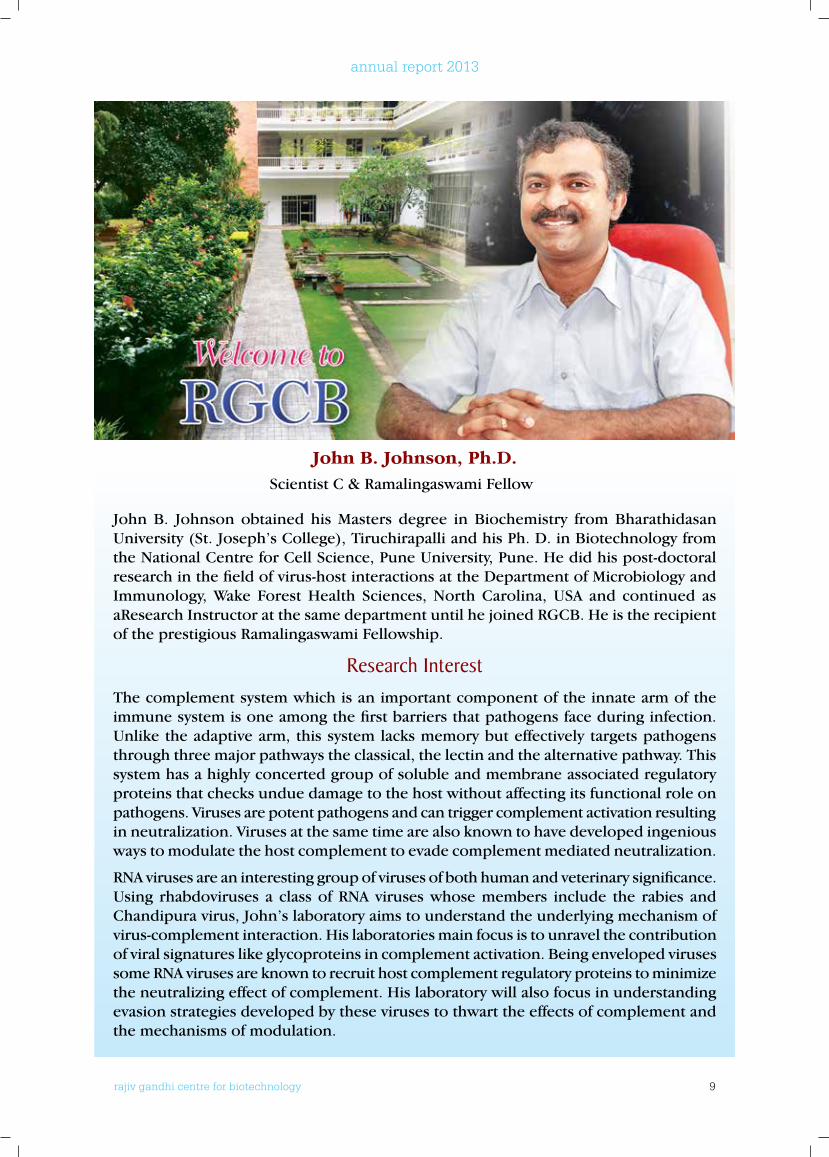
John B. Johnson, Ph.D.Scientist C & Ramalingaswami Fellow
John B. Johnson obtained his Masters degree in Biochemistry from Bharathidasan University (St. Joseph’s College), Tiruchirapalli and his Ph. D. in Biotechnology from the National Centre for Cell Science, Pune University, Pune. He did his post-doctoral research in the field of virus-host interactions at the Department of Microbiology and Immunology, Wake Forest Health Sciences, North Carolina, USA and continued as aResearch Instructor at the same department until he joined RGCB. He is the recipient of the prestigious Ramalingaswami Fellowship.
Research InterestThe complement system which is an important component of the innate arm of the immune system is one among the first barriers that pathogens face during infection. Unlike the adaptive arm, this system lacks memory but effectively targets pathogens through three major pathways the classical, the lectin and the alternative pathway. This system has a highly concerted group of soluble and membrane associated regulatory proteins that checks undue damage to the host without affecting its functional role on pathogens. Viruses are potent pathogens and can trigger complement activation resulting in neutralization. Viruses at the same time are also known to have developed ingenious ways to modulate the host complement to evade complement mediated neutralization.
RNA viruses are an interesting group of viruses of both human and veterinary significance. Using rhabdoviruses a class of RNA viruses whose members include the rabies and Chandipura virus, John’s laboratory aims to understand the underlying mechanism of virus-complement interaction. His laboratories main focus is to unravel the contribution of viral signatures like glycoproteins in complement activation. Being enveloped viruses some RNA viruses are known to recruit host complement regulatory proteins to minimize the neutralizing effect of complement. His laboratory will also focus in understanding evasion strategies developed by these viruses to thwart the effects of complement and the mechanisms of modulation.
rajiv gandhi centre for biotechnology 10
annual report 2013
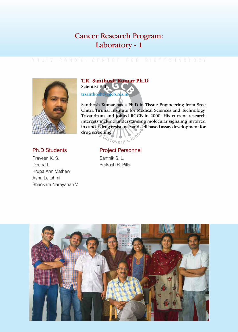
Cancer Research Program:Laboratory - 1
T.R. Santhosh Kumar Ph.DScientist E II
Ph.D StudentsPraveen K. S. Deepa I.Krupa Ann MathewAsha LekshmiShankara Narayanan V.
r a j i v g a n d h i c e n t r e f o r b i o t e c h n o l o g y
Santhosh Kumar has a Ph.D in Tissue Engineering from Sree Chitra Tirunal Institute for Medical Sciences and Technology, Trivandrum and joined RGCB in 2000. His current research interests include understanding molecular signaling involved in cancer drug resistance and cell based assay development for drug screening.
Project PersonnelSanthik S. L.Prakash R. Pillai
rajiv gandhi centre for biotechnology 11
annual report 2013
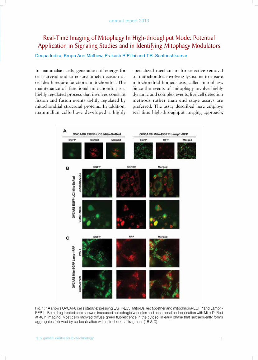
In mammalian cells, generation of energy for cell survival and to ensure timely decision of cell death require functional mitochondria. The maintenance of functional mitochondria is a highly regulated process that involves constant fission and fusion events tightly regulated bymitochondrial structural proteins. In addition, mammalian cells have developed a highly
specialized mechanism for selective removal of mitochondria involving lysosome to ensure mitochondrial homeostasis, called mitophagy. Since the events of mitophagy involve highly dynamic and complex events, live cell detection methods rather than end stage assays are preferred. The assay described here employs real time high-throughput imaging approach;
Real-Time Imaging of Mitophagy In High-throughput Mode: Potential Application in Signaling Studies and in Identifying Mitophagy Modulators
Deepa Indira, Krupa Ann Mathew, Prakash R Pillai and T.R. Santhoshkumar
Fig. 1: 1A shows OVCAR8 cells stably expressing EGFP-LC3, Mito-DsRed together and mitochndria-EGFP and Lamp1-RFP 1. Both drug treated cells showed increased autophagic vacuoles and occasional co-localisation with Mito-DsRed at 48 h imaging. Most cells showed diffuse green fluorescence in the cytosol in early phase that subsequently forms aggregates followed by co-localisation with mitochondrial fragment (1B & C).
rajiv gandhi centre for biotechnology 12
annual report 2013
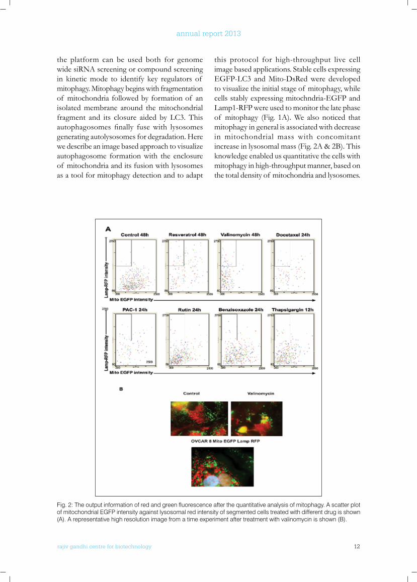
the platform can be used both for genome wide siRNA screening or compound screening in kinetic mode to identify key regulators of mitophagy. Mitophagy begins with fragmentation of mitochondria followed by formation of an isolated membrane around the mitochondrial fragment and its closure aided by LC3. This autophagosomes finally fusewith lysosomesgenerating autolysosomes for degradation. Here we describe an image based approach to visualize autophagosome formation with the enclosure of mitochondria and its fusion with lysosomes as a tool for mitophagy detection and to adapt
this protocol for high-throughput live cell image based applications. Stable cells expressing EGFP-LC3 and Mito-DsRed were developed to visualize the initial stage of mitophagy, while cells stably expressing mitochndria-EGFP and Lamp1-RFP were used to monitor the late phase of mitophagy (Fig. 1A). We also noticed that mitophagy in general is associated with decrease in mitochondrial mass with concomitant increase in lysosomal mass (Fig. 2A & 2B). This knowledge enabled us quantitative the cells with mitophagy in high-throughput manner, based on the total density of mitochondria and lysosomes.
Fig. 2: The output information of red and green fluorescence after the quantitative analysis of mitophagy. A scatter plot of mitochondrial EGFP intensity against lysosomal red intensity of segmented cells treated with different drug is shown (A). A representative high resolution image from a time experiment after treatment with valinomycin is shown (B).
rajiv gandhi centre for biotechnology 13
annual report 2013
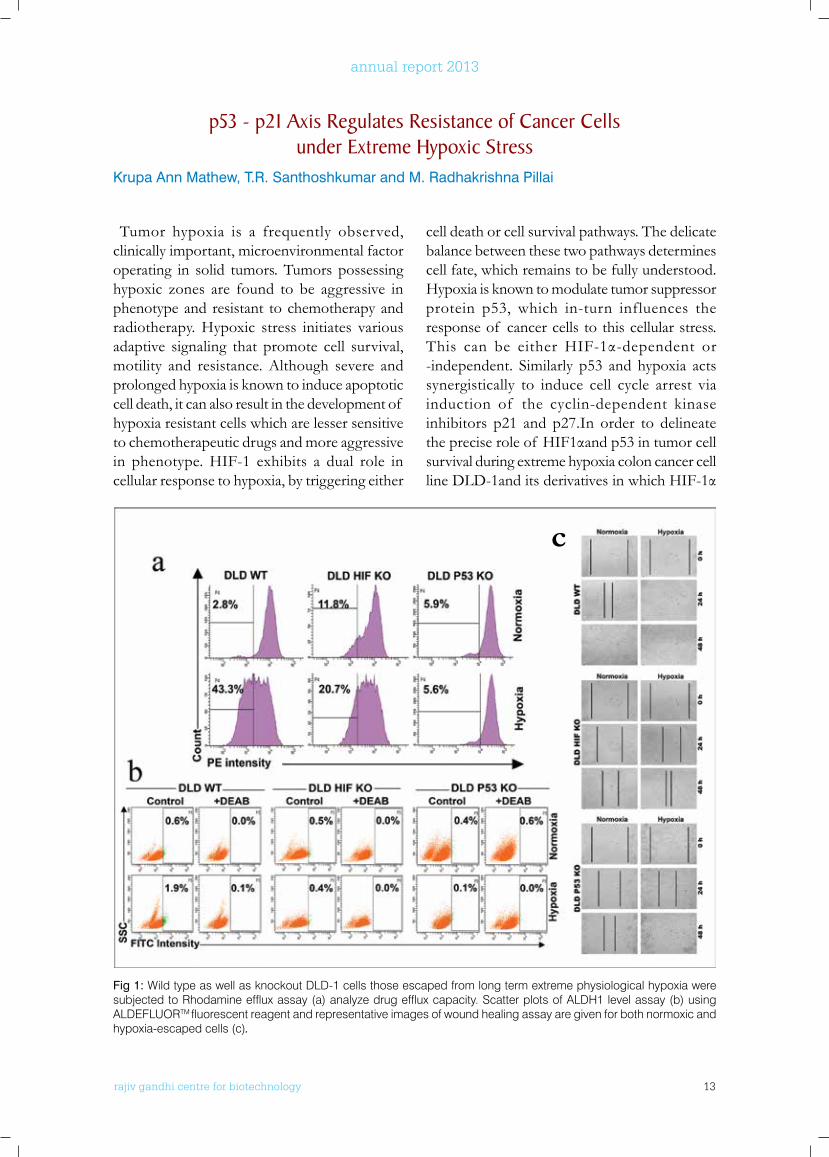
Tumor hypoxia is a frequently observed, clinically important, microenvironmental factor operating in solid tumors. Tumors possessing hypoxic zones are found to be aggressive in phenotype and resistant to chemotherapy and radiotherapy. Hypoxic stress initiates various adaptive signaling that promote cell survival, motility and resistance. Although severe and prolonged hypoxia is known to induce apoptotic cell death, it can also result in the development of hypoxia resistant cells which are lesser sensitive to chemotherapeutic drugs and more aggressive in phenotype. HIF-1 exhibits a dual role in cellular response to hypoxia, by triggering either
cell death or cell survival pathways. The delicate balance between these two pathways determines cell fate, which remains to be fully understood. Hypoxia is known to modulate tumor suppressor protein p53, which in-turn influences the response of cancer cells to this cellular stress. This can be either HIF-1α-dependent or-independent. Similarly p53 and hypoxia acts synergistically to induce cell cycle arrest via induction of the cyclin-dependent kinase inhibitors p21 and p27.In order to delineate thepreciseroleof HIF1αandp53intumorcellsurvival during extreme hypoxia colon cancer cell lineDLD-1anditsderivativesinwhichHIF-1α
p53 - p21 Axis Regulates Resistance of Cancer Cells under Extreme Hypoxic Stress
Krupa Ann Mathew, T.R. Santhoshkumar and M. Radhakrishna Pillai
Fig 1: Wild type as well as knockout DLD-1 cells those escaped from long term extreme physiological hypoxia were subjected to Rhodamine efflux assay (a) analyze drug efflux capacity. Scatter plots of ALDH1 level assay (b) using ALDEFLUORTM fluorescent reagent and representative images of wound healing assay are given for both normoxic and hypoxia-escaped cells (c).
c
rajiv gandhi centre for biotechnology 14
annual report 2013
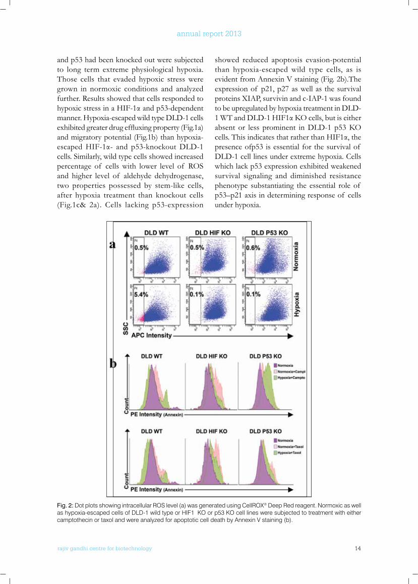
Fig. 2: Dot plots showing intracellular ROS level (a) was generated using CellROX® Deep Red reagent. Normoxic as well as hypoxia-escaped cells of DLD-1 wild type or HIF1α KO or p53 KO cell lines were subjected to treatment with either camptothecin or taxol and were analyzed for apoptotic cell death by Annexin V staining (b).
and p53 had been knocked out were subjected to long term extreme physiological hypoxia. Those cells that evaded hypoxic stress were grown in normoxic conditions and analyzed further. Results showed that cells responded to hypoxicstressinaHIF-1αandp53-dependentmanner. Hypoxia-escaped wild type DLD-1 cells exhibitedgreaterdrugeffluxingproperty(Fig.1a)and migratory potential (Fig.1b) than hypoxia-escapedHIF-1α- and p53-knockoutDLD-1cells. Similarly, wild type cells showed increased percentage of cells with lower level of ROS and higher level of aldehyde dehydrogenase, two properties possessed by stem-like cells, after hypoxia treatment than knockout cells (Fig.1c& 2a). Cells lacking p53-expression
showed reduced apoptosis evasion-potential than hypoxia-escaped wild type cells, as is evident from Annexin V staining (Fig. 2b).The expression of p21, p27 as well as the survival proteins XIAP, survivin and c-IAP-1 was found to be upregulated by hypoxia treatment in DLD-1WTandDLD-1HIF1αKOcells,butiseitherabsent or less prominent inDLD-1p53KOcells.ThisindicatesthatratherthanHIF1α,thepresence ofp53 is essential for the survival of DLD-1 cell lines under extreme hypoxia. Cells which lack p53 expression exhibited weakened survival signaling and diminished resistance phenotype substantiating the essential role of p53–p21 axis in determining response of cells under hypoxia.
rajiv gandhi centre for biotechnology 15
annual report 2013
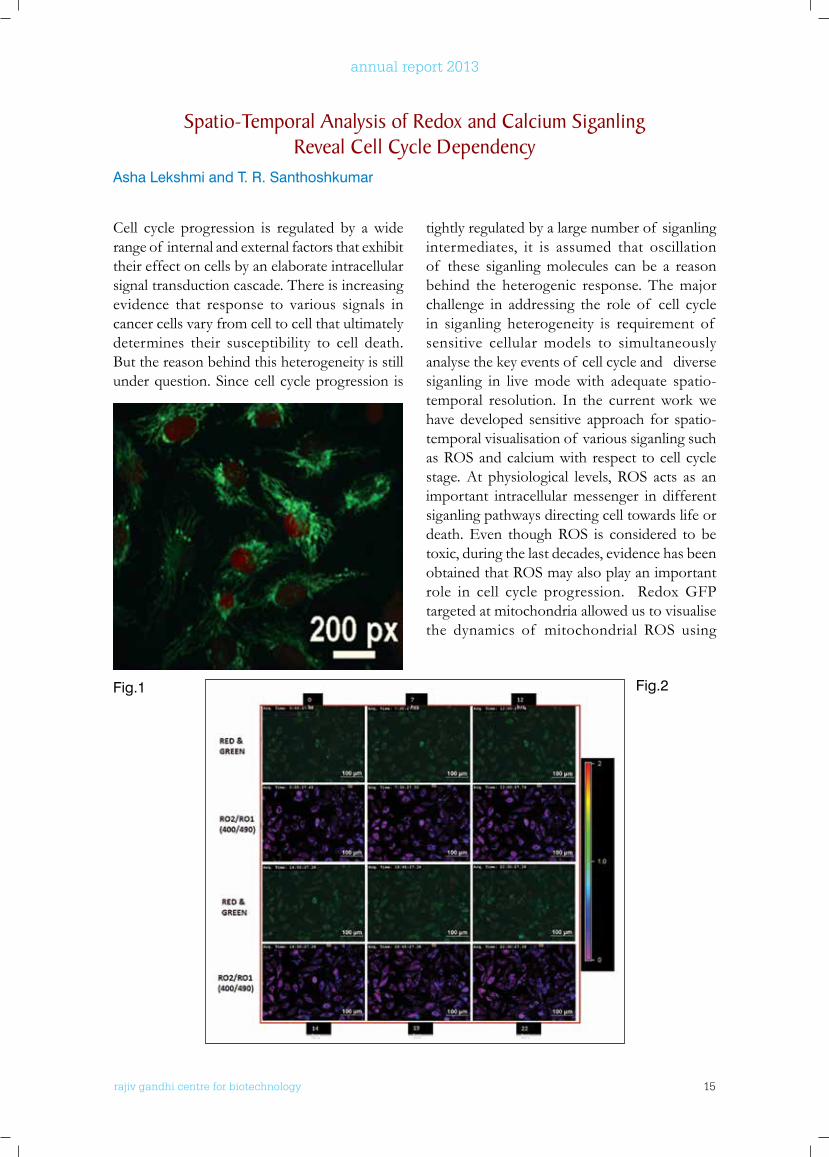
Cell cycle progression is regulated by a wide range of internal and external factors that exhibit their effect on cells by an elaborate intracellular signal transduction cascade. There is increasing evidence that response to various signals in cancer cells vary from cell to cell that ultimately determines their susceptibility to cell death. But the reason behind this heterogeneity is still under question. Since cell cycle progression is
tightly regulated by a large number of siganling intermediates, it is assumed that oscillation of these siganling molecules can be a reason behind the heterogenic response. The major challenge in addressing the role of cell cycle in siganling heterogeneity is requirement of sensitive cellular models to simultaneously analyse the key events of cell cycle and diverse siganling in live mode with adequate spatio-temporal resolution. In the current work we have developed sensitive approach for spatio-temporal visualisation of various siganling such as ROS and calcium with respect to cell cycle stage. At physiological levels, ROS acts as an important intracellular messenger in different siganling pathways directing cell towards life or death. Even though ROS is considered to be toxic, during the last decades, evidence has been obtained that ROS may also play an important role in cell cycle progression. Redox GFP targeted at mitochondria allowed us to visualise the dynamics of mitochondrial ROS using
Spatio-Temporal Analysis of Redox and Calcium Siganling Reveal Cell Cycle Dependency
Asha Lekshmi and T. R. Santhoshkumar
Fig.1 Fig.2
rajiv gandhi centre for biotechnology 16
annual report 2013
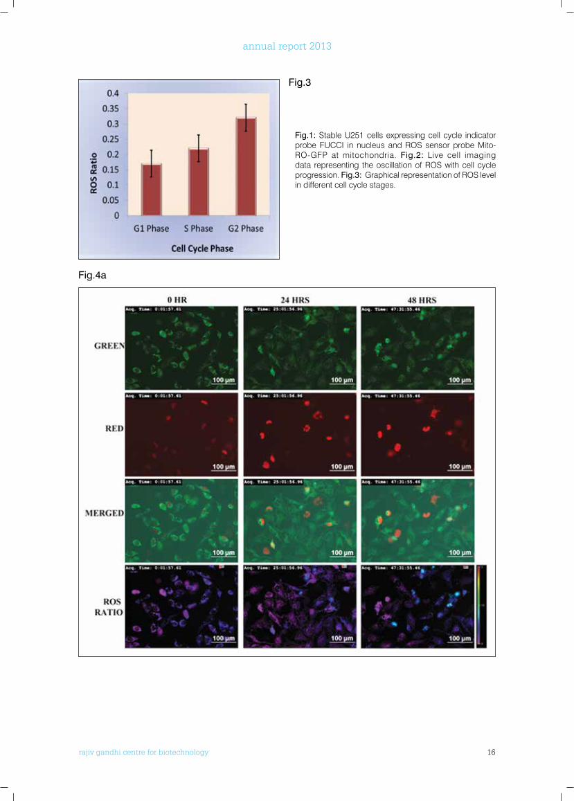
Fig.1: Stable U251 cells expressing cell cycle indicator probe FUCCI in nucleus and ROS sensor probe Mito-RO-GFP at mitochondria. Fig.2: Live cell imaging data representing the oscillation of ROS with cell cycle progression. Fig.3: Graphical representation of ROS level in different cell cycle stages.
Fig.3
Fig.4a
rajiv gandhi centre for biotechnology 17
annual report 2013
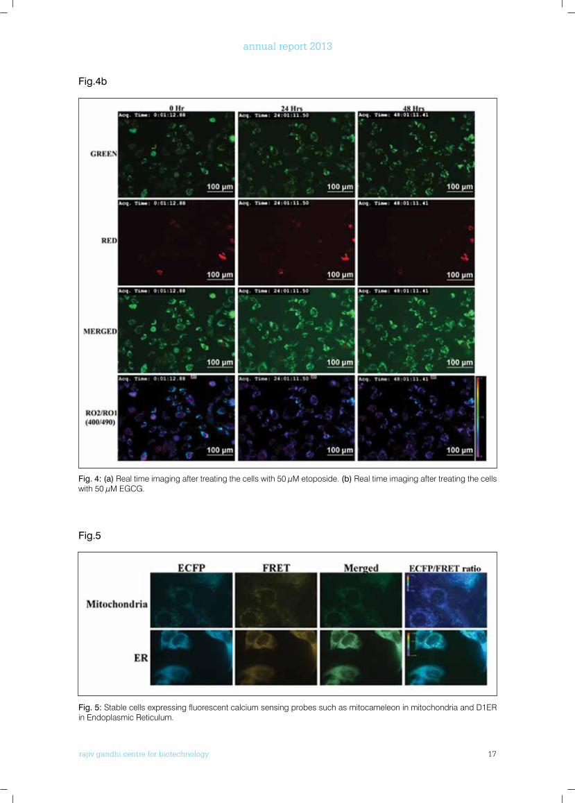
Fig. 4: (a) Real time imaging after treating the cells with 50 µM etoposide. (b) Real time imaging after treating the cells with 50 µM EGCG.
Fig. 5: Stable cells expressing fluorescent calcium sensing probes such as mitocameleon in mitochondria and D1ER in Endoplasmic Reticulum.
Fig.4b
Fig.5
rajiv gandhi centre for biotechnology 18
annual report 2013
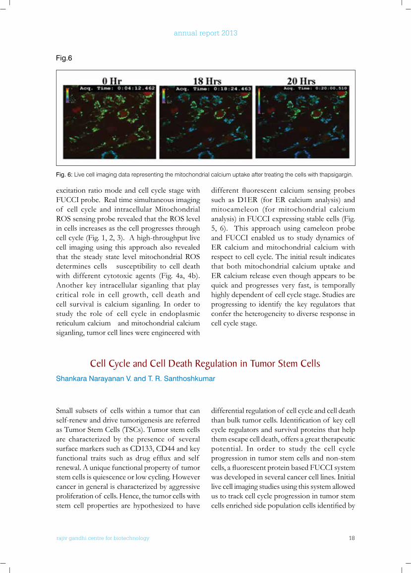
excitation ratio mode and cell cycle stage with FUCCI probe. Real time simultaneous imaging of cell cycle and intracellular Mitochondrial ROS sensing probe revealed that the ROS level in cells increases as the cell progresses through cell cycle (Fig. 1, 2, 3). A high-throughput live cell imaging using this approach also revealed that the steady state level mitochondrial ROS determines cells susceptibility to cell death with different cytotoxic agents (Fig. 4a, 4b). Another key intracellular siganling that play critical role in cell growth, cell death and cell survival is calcium siganling. In order to study the role of cell cycle in endoplasmic reticulum calcium and mitochondrial calcium siganling, tumor cell lines were engineered with
different fluorescent calcium sensing probessuch as D1ER (for ER calcium analysis) and mitocameleon (for mitochondrial calcium analysis) in FUCCI expressing stable cells (Fig. 5, 6). This approach using cameleon probe and FUCCI enabled us to study dynamics of ER calcium and mitochondrial calcium with respect to cell cycle. The initial result indicates that both mitochondrial calcium uptake and ER calcium release even though appears to be quick and progresses very fast, is temporally highly dependent of cell cycle stage. Studies are progressing to identify the key regulators that confer the heterogeneity to diverse response in cell cycle stage.
Fig. 6: Live cell imaging data representing the mitochondrial calcium uptake after treating the cells with thapsigargin.
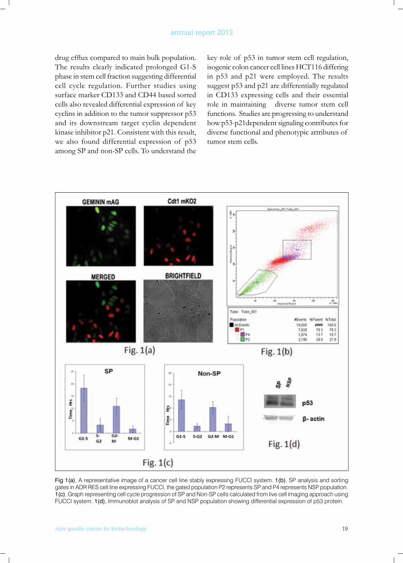
Cell Cycle and Cell Death Regulation in Tumor Stem CellsShankara Narayanan V. and T. R. Santhoshkumar
Small subsets of cells within a tumor that can self-renew and drive tumorigenesis are referred as Tumor Stem Cells (TSCs). Tumor stem cells are characterized by the presence of several surface markers such as CD133, CD44 and key functional traits such as drug efflux and self renewal. A unique functional property of tumor stem cells is quiescence or low cycling. However cancer in general is characterized by aggressive proliferation of cells. Hence, the tumor cells with stem cell properties are hypothesized to have
differential regulation of cell cycle and cell death thanbulktumorcells.Identificationof keycellcycle regulators and survival proteins that help them escape cell death, offers a great therapeutic potential. In order to study the cell cycle progression in tumor stem cells and non-stem cells,afluorescentproteinbasedFUCCIsystemwas developed in several cancer cell lines. Initial live cell imaging studies using this system allowed us to track cell cycle progression in tumor stem cellsenrichedsidepopulationcellsidentifiedby
Fig.6
rajiv gandhi centre for biotechnology 19
annual report 2013
drugeffluxcomparedtomainbulkpopulation.The results clearly indicated prolonged G1-S phase in stem cell fraction suggesting differential cell cycle regulation. Further studies using surface marker CD133 and CD44 based sorted cells also revealed differential expression of key cyclins in addition to the tumor suppressor p53 and its downstream target cyclin dependent kinase inhibitor p21. Consistent with this result, we also found differential expression of p53 among SP and non-SP cells. To understand the
key role of p53 in tumor stem cell regulation, isogenic colon cancer cell lines HCT116 differing in p53 and p21 were employed. The results suggest p53 and p21 are differentially regulated in CD133 expressing cells and their essential role in maintaining diverse tumor stem cell functions. Studies are progressing to understand how p53-p21dependent signaling contributes for diverse functional and phenotypic attributes of tumor stem cells.
Fig 1(a), A representative image of a cancer cell line stably expressing FUCCI system. 1(b), SP analysis and sorting gates in ADR RES cell line expressing FUCCI, the gated population P2 represents SP and P4 represents NSP population. 1(c), Graph representing cell cycle progression of SP and Non-SP cells calculated from live cell imaging approach using FUCCI system. 1(d), Immunoblot analysis of SP and NSP population showing differential expression of p53 protein.
rajiv gandhi centre for biotechnology 20
annual report 2013
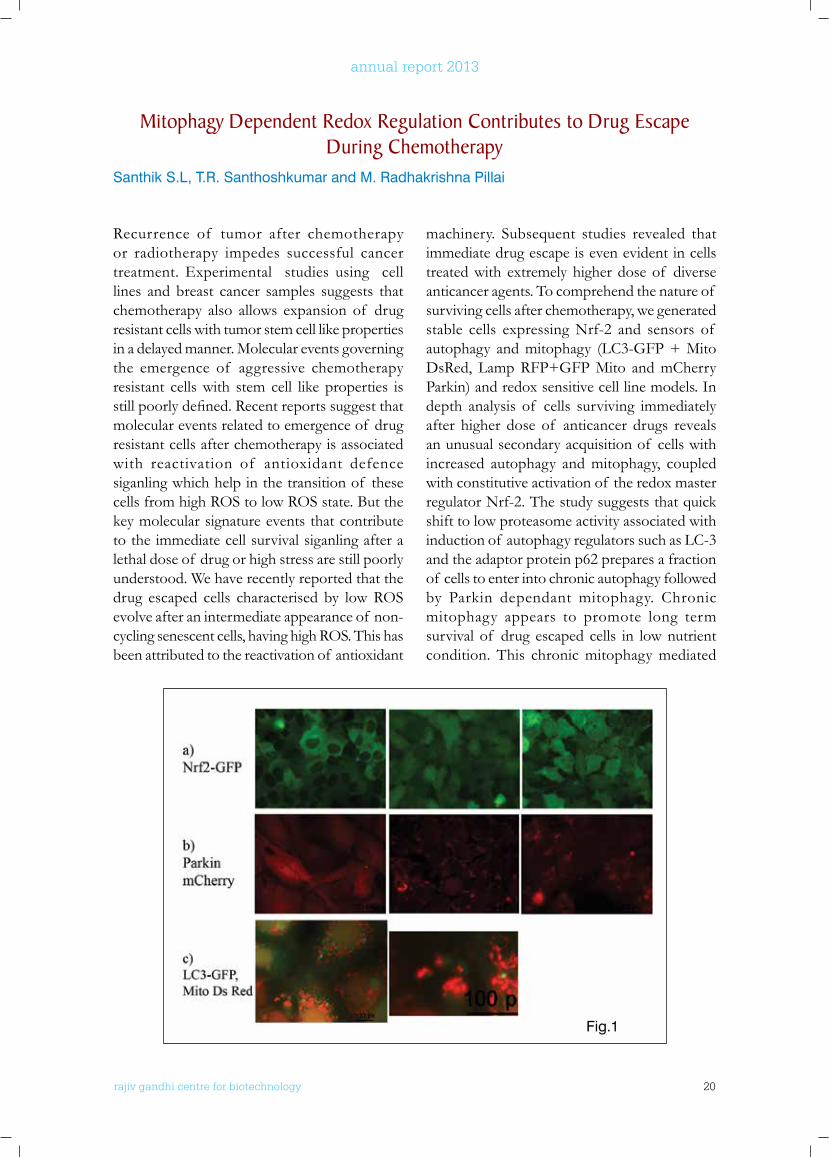
Recurrence of tumor after chemotherapy or radiotherapy impedes successful cancer treatment. Experimental studies using cell lines and breast cancer samples suggests that chemotherapy also allows expansion of drug resistant cells with tumor stem cell like properties in a delayed manner. Molecular events governing the emergence of aggressive chemotherapy resistant cells with stem cell like properties is stillpoorlydefined.Recentreportssuggestthatmolecular events related to emergence of drug resistant cells after chemotherapy is associated with reactivation of antioxidant defence siganling which help in the transition of these cells from high ROS to low ROS state. But the key molecular signature events that contribute to the immediate cell survival siganling after a lethal dose of drug or high stress are still poorly understood. We have recently reported that the drug escaped cells characterised by low ROS evolve after an intermediate appearance of non-cycling senescent cells, having high ROS. This has been attributed to the reactivation of antioxidant
machinery. Subsequent studies revealed that immediate drug escape is even evident in cells treated with extremely higher dose of diverse anticancer agents. To comprehend the nature of surviving cells after chemotherapy, we generated stable cells expressing Nrf-2 and sensors of autophagy and mitophagy (LC3-GFP + Mito DsRed, Lamp RFP+GFP Mito and mCherry Parkin) and redox sensitive cell line models. In depth analysis of cells surviving immediately after higher dose of anticancer drugs reveals an unusual secondary acquisition of cells with increased autophagy and mitophagy, coupled with constitutive activation of the redox master regulator Nrf-2. The study suggests that quick shift to low proteasome activity associated with induction of autophagy regulators such as LC-3 and the adaptor protein p62 prepares a fraction of cells to enter into chronic autophagy followed by Parkin dependant mitophagy. Chronic mitophagy appears to promote long term survival of drug escaped cells in low nutrient condition. This chronic mitophagy mediated
Mitophagy Dependent Redox Regulation Contributes to Drug Escape During Chemotherapy
Santhik S.L, T.R. Santhoshkumar and M. Radhakrishna Pillai
Fig.1
rajiv gandhi centre for biotechnology 21
annual report 2013
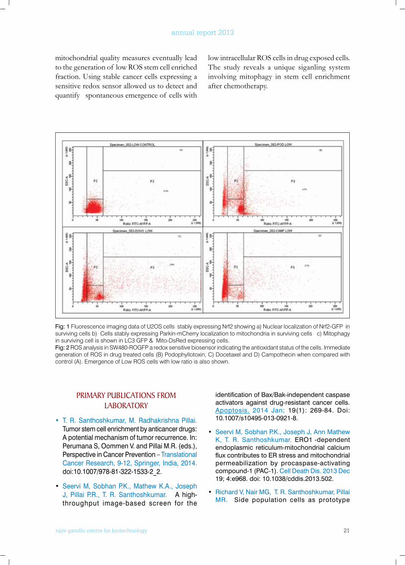
mitochondrial quality measures eventually lead to the generation of low ROS stem cell enriched fraction. Using stable cancer cells expressing a sensitive redox sensor allowed us to detect and quantify spontaneous emergence of cells with
low intracellular ROS cells in drug exposed cells. The study reveals a unique siganling system involving mitophagy in stem cell enrichment after chemotherapy.
Fig: 1 Fluorescence imaging data of U2OS cells stably expressing Nrf2 showing a) Nuclear localization of Nrf2-GFP in surviving cells b) Cells stably expressing Parkin-mCherry localization to mitochondria in surviving cells c) Mitophagy in surviving cell is shown in LC3 GFP & Mito-DsRed expressing cells.Fig: 2 ROS analysis in SW480-ROGFP a redox sensitive biosensor indicating the antioxidant status of the cells. Immediate generation of ROS in drug treated cells (B) Podophyllotoxin, C) Docetaxel and D) Campothecin when compared with control (A). Emergence of Low ROS cells with low ratio is also shown.
PRIMARy PublICATIonS fRoM lAboRAToRy
• T. R. Santhoshkumar, M. Radhakrishna Pillai. Tumor stem cell enrichment by anticancer drugs: A potential mechanism of tumor recurrence. In: Perumana S, Oommen V. and Pillai M.R. (eds.), Perspective in Cancer Prevention – Translational Cancer Research, 9-12, Springer, India, 2014. doi:10.1007/978-81-322-1533-2_2.
• Seervi M, Sobhan P.K., Mathew K.A., Joseph J, Pillai P.R., T. R. Santhoshkumar. A high-throughput image-based screen for the
identification of Bax/Bak-independent caspase activators against drug-resistant cancer cells. Apoptosis. 2014 Jan; 19(1): 269-84. Doi: 10.1007/s10495-013-0921-8.
• Seervi M, Sobhan P.K., Joseph J, Ann Mathew K, T. R. Santhoshkumar. ERO1α-dependent endoplasmic reticulum-mitochondrial calcium flux contributes to ER stress and mitochondrial permeabilization by procaspase-activating compound-1 (PAC-1). Cell Death Dis. 2013 Dec 19; 4:e968. doi: 10.1038/cddis.2013.502.
• Richard V, Nair MG, T. R. Santhoshkumar, Pillai MR. Side population cells as prototype
rajiv gandhi centre for biotechnology 22
annual report 2013
of chemoresistant, tumor-initiating cells. Biomed Res Int. 2013; 2013:517237. doi: 10.1155/2013/517237. Epub 2013 Nov 4.
• Richard V, Sebastian P, Nair M.G., Nair S.N., Malieckal T.T., TR. Santhoshkumar, Pillai MR. Multiple drug resistant, tumorigenic stem-like cells in oral cancer. Cancer Lett. 2013 Sep 28; 338(2):300-16. doi: 10.1016/j.canlet.2013.06.011. Epub 2013 Jun 18.
PublICATIonS wITH CollAboRAToRS
• Ramesha B.T., Suma H.K., Senthilkumar U, Priti V, Ravikanth G, Vasudeva R, T. R.
ExTRA MuRAl RESEARCH GRAnTS
Sl.no. Title of the Project Funding agency Duration
1 Design and Development of New Generation Caspase Sensor Fret Probe Expressing Stable Cancer Cells for Anticancer Drug Screening: From In Vitro HTS Screen to Whole Animal Imaging
Department of Biotechnology, Government of India
2013-2017
2 High throughput screening of compounds against key targets in cancer.
Piramal Life Sciences, Mumbai
2013-2015
Santhoshkumar, Ganeshaiah K.N., Shaanker R.U. New plant sources of the anti-cancer alkaloid, camptothecine from the Icacinaceae taxa, India. Phytomedicine. 2013 Apr 15; 20(6):521-7. doi: 10.1016/j.phymed.2012.12.003. Epub 2013 Mar 7.
• Dhanya R, Arun K.B, Syama H.P, Nisha P, S u n d a r e s a n A , T. R . S a n t h o s h Kumar, Jayamurthy P. Rutin and quercetin enhance glucose uptake in L6 myotubes under oxidative stress induced by tertiary butyl hydrogen peroxide. Food Chem. 2014 Sep 1; 158:546-54. doi: 10.1016/j.foodchem.2014.02.151. Epub 2014 Mar 12.
rajiv gandhi centre for biotechnology 23
annual report 2013
Cancer Research Program:Laboratory - 2
Ph.D StudentsJayesh Antony, SRFArun Kumar T.T, SRFLekshmi R. Nath, SRF
r a j i v g a n d h i c e n t r e f o r b i o t e c h n o l o g y
Ruby John Anto took her Ph.D in Biochemistry from Amala Cancer Research Centre, Thrissur and did post doctoral training at RGCB and MD Anderson Cancer Centre, Houston, Texas, before joining RGCB in 2004.
Project PersonnelDr. Vinod V, SRF
Ruby John Anto Ph.D Scientist EII
rajiv gandhi centre for biotechnology 24
annual report 2013
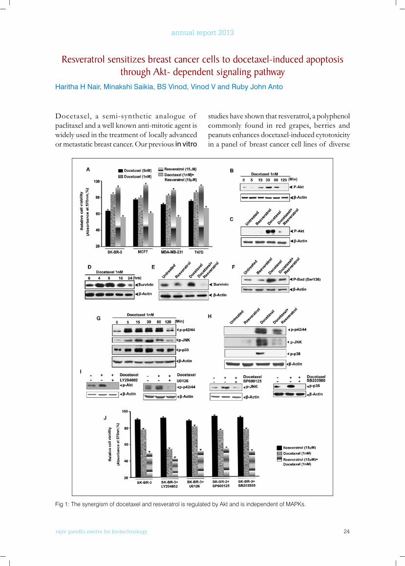
Docetaxel, a semi-synthetic analogue of paclitaxel and a well known anti-mitotic agent is widely used in the treatment of locally advanced or metastatic breast cancer. Our previous in vitro
studies have shown that resveratrol, a polyphenol commonly found in red grapes, berries and peanuts enhances docetaxel-induced cytotoxicity in a panel of breast cancer cell lines of diverse
Resveratrol sensitizes breast cancer cells to docetaxel-induced apoptosis through Akt- dependent signaling pathway
Haritha H Nair, Minakshi Saikia, BS Vinod, Vinod V and Ruby John Anto
Fig 1: The synergism of docetaxel and resveratrol is regulated by Akt and is independent of MAPKs.

rajiv gandhi centre for biotechnology 25
annual report 2013
receptor status, with highest cytotoxic effect inSKBR3.Resveratrol is capableof inducingapoptosis by regulating the expression of certain pivotal molecules like Bcl-2, Bcl-XL, survivin, caspases, PARP etc. As there are several reports stating the connection of Akt andMAPKpathways in docetaxel-induced chemoresistance, we investigated the involvement of these two survival pathways in regulating the synergism between docetaxel and resveratrol. Our results indicate that there is a time-dependent transient phosphorylation of Akt andMAPKs upontreatment with docetaxel and when given in
combination with resveratrol, there was a significantdown-regulationof thesame.Theseresults indicate that resveratrol could effectively down-regulate docetaxel-induced activation of Akt andMAPKs. Interestingly,MAPKswerefound to have no direct role in regulating the synergistic effect while Akt was playing a crucial role in regulating the same as evidenced by the existence of synergism even after the inhibition of MAPKs and abrogation of synergismbyinhibition of Akt using the corresponding inhibitors.
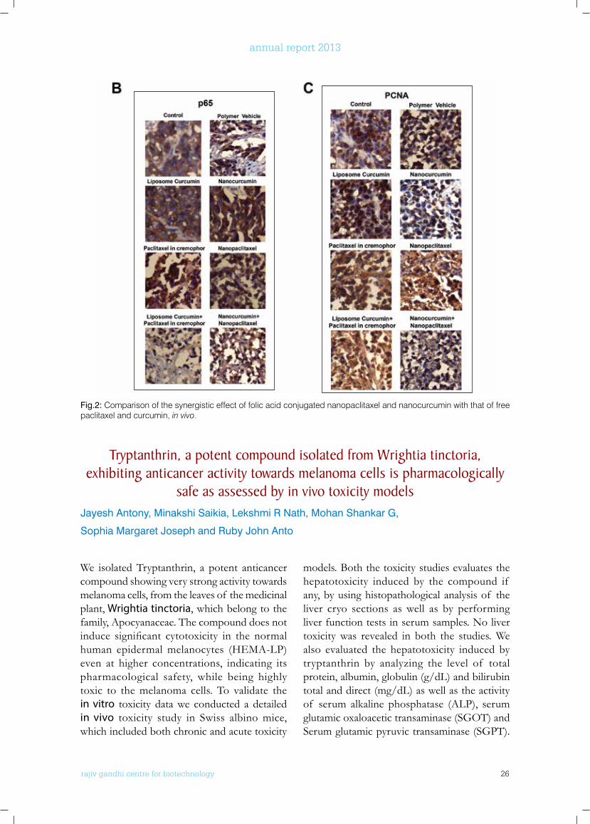
folic acid conjugation of curcumin and paclitaxel improves their synergistic effect as assessed by cervical xenograft model
Arun Kumar T Thulasidasan, GS Vinod Kumar*, K Lekha Nair*, Devika N*
and Ruby John Anto
Collaborators: *Chemical Biology, Rajiv Gandhi Centre for Biotechnology.
In this study, we have used curcumin/paclitaxel loaded PLGA nanoparticles prepared by solvent evaporation and their anticancer activity was studied in comparison with that of their free counterparts. The preliminary data obtained from the in vivo study using cervical xenograft
model indicate that the nanoformulation enhancestheefficacyof thesynergismof thesecompounds. However, toxicity and bioavailability of curcumin and paclitaxel while using these formulations are yet to be studied.
rajiv gandhi centre for biotechnology 26
annual report 2013
Fig.2: Comparison of the synergistic effect of folic acid conjugated nanopaclitaxel and nanocurcumin with that of free paclitaxel and curcumin, in vivo.
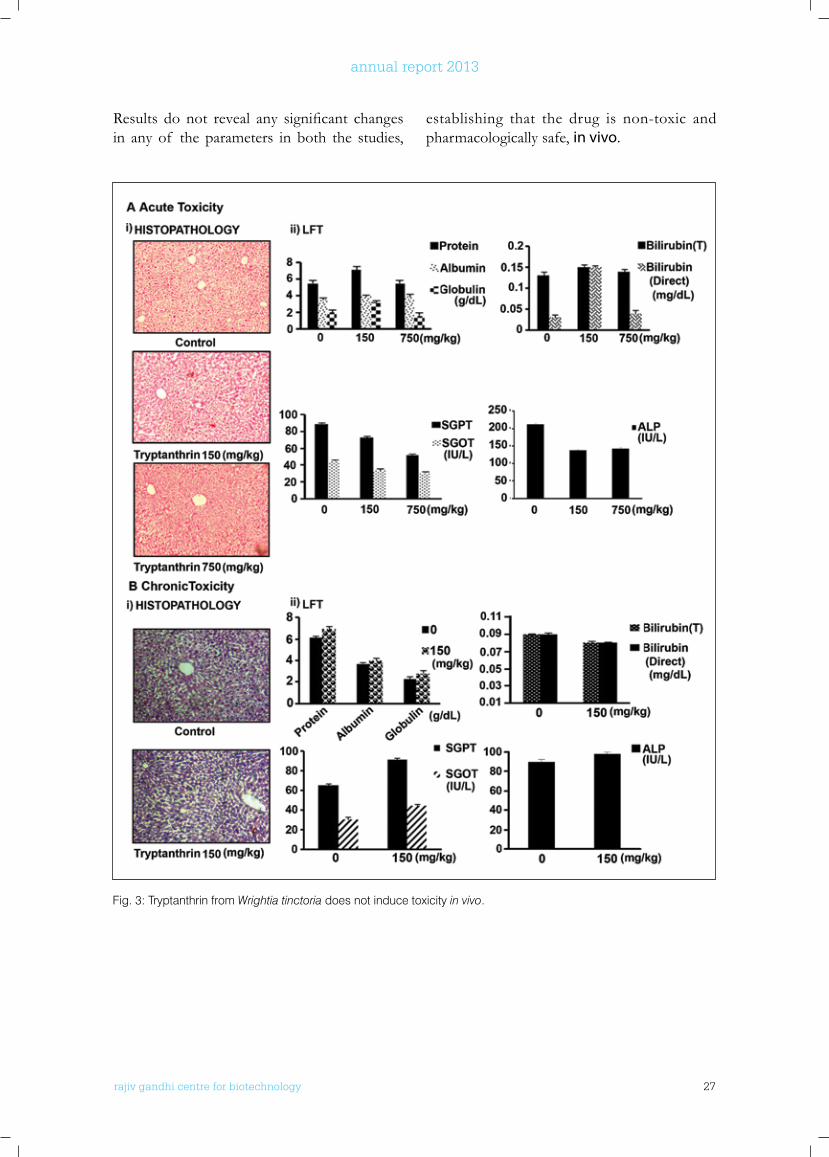
Tryptanthrin, a potent compound isolated from wrightia tinctoria, exhibiting anticancer activity towards melanoma cells is pharmacologically
safe as assessed by in vivo toxicity modelsJayesh Antony, Minakshi Saikia, Lekshmi R Nath, Mohan Shankar G,
Sophia Margaret Joseph and Ruby John Anto
We isolated Tryptanthrin, a potent anticancer compound showing very strong activity towards melanoma cells, from the leaves of the medicinal plant, Wrightia tinctoria, which belong to the family, Apocyanaceae. The compound does not induce significant cytotoxicity in the normalhuman epidermal melanocytes (HEMA-LP) even at higher concentrations, indicating its pharmacological safety, while being highly toxic to the melanoma cells. To validate the in vitro toxicity data we conducted a detailed in vivo toxicity study in Swiss albino mice, which included both chronic and acute toxicity
models. Both the toxicity studies evaluates the hepatotoxicity induced by the compound if any, by using histopathological analysis of the liver cryo sections as well as by performing liver function tests in serum samples. No liver toxicity was revealed in both the studies. We also evaluated the hepatotoxicity induced by tryptanthrin by analyzing the level of total protein, albumin, globulin (g/dL) and bilirubin total and direct (mg/dL) as well as the activity of serum alkaline phosphatase (ALP), serum glutamic oxaloacetic transaminase (SGOT) and Serum glutamic pyruvic transaminase (SGPT).
rajiv gandhi centre for biotechnology 27
annual report 2013
Results do not reveal any significant changesin any of the parameters in both the studies,
Fig. 3: Tryptanthrin from Wrightia tinctoria does not induce toxicity in vivo.
establishing that the drug is non-toxic and pharmacologically safe, in vivo.
rajiv gandhi centre for biotechnology 28
annual report 2013
of Scientific and Industrial Research (CSIR), Thiruvananthapuram-695019, Kerala, India.
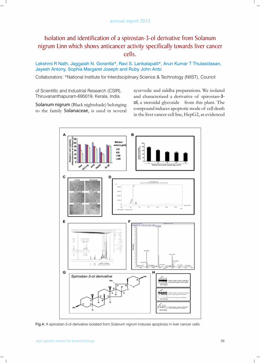
Solanum nigrum (Black nightshade) belonging to the family Solanaceae, is used in several
ayurvedic and siddha preparations. We isolated and characterized a derivative of spirostan-3-ol, a steroidal glycoside from this plant. The compound induces apoptotic mode of cell death in the liver cancer cell line, HepG2, as evidenced
Isolation and identification of a spirostan-3-ol derivative from Solanum nigrum linn which shows anticancer activity specifically towards liver cancer
cells.Lekshmi R Nath, Jaggaiah N. Gorantla*, Ravi S. Lankalapalli*, Arun Kumar T Thulasidasan, Jayesh Antony, Sophia Margaret Joseph and Ruby John Anto
Collaborators: *National Institute for Interdisciplinary Science & Technology (NIIST), Council
Fig.4: A spirostan-3-ol derivative isolated from Solanum nigrum induces apoptosis in liver cancer cells.
rajiv gandhi centre for biotechnology 29
annual report 2013
by cleavage of pro-caspase 9, pro-caspase-7, pro-caspase 8.
PRIMARy PublICATIonS fRoM lAboRAToRy
• BS Vinod, J Antony, HH Nair, VT Puliyappadamba, M Saikia, S Shyam Narayanan, A Bevin and Ruby John Anto. Mechanistic evaluation of the signaling events regulating curcumin-mediated chemosensitization of breast cancer cells to 5-fluorouracil. Cell Death Dis., 4, e 505; doi:10.1038/cddis.2013.26, 2013.
• Balachandran S Vinod, Tessy T Maliekal and Ruby John Anto, Phytochemical As Chemosensitizers: From Molecular Mechanism to Clinical Significance. Comprehensive Invited Review. Antioxidants & Redox Signaling. 18, 1307–1348, 2013.
• Minakshi Saikia, and Ruby John Anto. Acute myeloid leukemia: Causes diagnosis, classification and treatment modalities. Amala Research Bulletin (2013) 33, 147-154.
PublICATIonS wITH CollAboRAToRS
• M. S. R. Murty, B. Ramalingeswara Rao, Mohana Rao Katiki, Lekshmi R. Nath and Ruby John Anto, Synthesis of piperazinyl benzothiazole/benzoxazole derivatives coupled with 1,3,4 oxadiazole-2-thiol: novel hybrid heterocycles as anticancer agents. Med Chem Res., 22, 4980-4991, 2013.
• Jaggaiah N. Gorantla, Jamsheena Vellekkatt, Lekshmi R.Nath, Ruby John Anto and Ravi S. Lankalapalli. Cytotoxicity studies of semi-synthetic derivatives of theveside derived from the aqueous extract of leaves of ‘suicide tree’ Cerbera odollam, Natural Product Research, DOI: 10.1080/14786419.2014.913242.
• Kumar SN, Nambisan Bala, Sundaresan A, Mohandas C and Ruby John Anto, Isolation and identification of antimicrobial secondary metabolites from Bacillus cereus associated with a rhabditid entomopathogenic nematode. Ann Microbiol., 64, 209–218, 2014
CHAPTERS In TExT bookS
• *Jayesh Antony, *Minakshi Saikia and Ruby John Anto. Phytochemicals from Fruits and Vegetables as potential anti-cancer agents: special reference to skin cancer. In: Anticancer properties of fruits and vegetables: A scientific
review. Ajay Kunnummakkara (Ed). Pubd: World Scientific Publishing Co.; 2014 edition, pp 277-307 (*equal authorship)
• CN Sreekanth, Smitha VB, Arun Kumar TT, N P Anto, VT Cheriyan, Vineshkumar TP, SG Menon, SD Ravichandran and Ruby John Anto - Curcumin: A Potent Candidate to be Evaluated as a Chemosensitizer in Paclitaxel Chemotherapy Against Cervical Cancer. In: Perspectives in Cancer Prevention-Translational Cancer Research. P Sudhakaran P, Oommen V, Pillai, MR (Eds.). Pubd: Springer; 2014 edition (October 25, 2013).pp 21-43
ConfEREnCE PRESEnTATIonS
• Arun Kumar T Thulasidasan, GS Vinod Kumar, K Lekha Nair, G Deepa and Ruby John Anto. In vitro and in vivo validation of nanoparticle-based drug delivery systems to improve the chemosensitizing efficacy of curcumin in paclitaxel chemotherapy. 6th International Conference on Drug Discovery & Therapy, February 10-12, 2014, Dubai (Sessions Talk).
• Jayesh Antony, Minakshi Saikia And Ruby John Anto, Identification and characterization of Tryptanthrin, the active principle from Wrightia tinctoria and the validation of its anticancer efficacy in vitro and in vivo. Oral presentation, 26th Kerala Science Congress, 28-31 January 2014, Wayanad, Kerala
• Jayesh Antony, Minakshi Saiki, Sophia Margeret Joseph, Vinod.V, Lekshmi. R. Nath, Mohana Rao Katiki, M.S.R. Murty and Ruby John Anto. In vitro and in vivo validation of anticancer efficacy of tryptanthrin, isolated from Wrightia tinctoria [Roxb.] R.Br. Poster, 33rd Annual convention of Indian Association for Cancer Research, February 13-15, 2014, Kollam, Kerala.
• Arun Kumar T Thulasidasan, GS Vinod Kumar, K Lekha Nair, G Deepa and Ruby John Anto.
Identification of better modes nanoparticle-based drug releasing systems for improving the efficacy of cervical cancer chemotherapy. Poster, 33rd Annual convention of Indian Association for Cancer Research, February 13-15, 2014 Kollam, Kerala.
• Lekshmi.R.Nath, Vinod V, Arun Kumar T Thulasidasan, Jaggaiah N. Gorantla, Ravi S. Lankalapalli and Ruby John Anto Mechanistic evaluation of the anticancer effect of a spirostan-3-ol derivative isolated from Solanum nigrum Linn in liver cancer, Poster, 33rd Annual
rajiv gandhi centre for biotechnology 30
annual report 2013
convention of Indian Association for Cancer Research, February 13-15, 2014 Kollam, Kerala.
• Haritha H Nair, Balachandran S Vinod, Jayesh Antony, Minakshi Saikia and Ruby John Anto. Thymidylate synthase-dependent NF-κB down-regulation plays pivotal role in the efficacy of Curcumin in chemosensitizing breast cancer cells to 5-FU, Poster, 33rd Annual convention of Indian Association of Cancer Research held at from 13th -15th February 2014 Kollam, Kerala
AwARDS, HonoRS, ETC:
• Vinod BS: PhD awarded: 2014 (Identification of effective and non-toxic chemosensitizers,
which can be used in combination with the conventional chemotherapeutic drugs used for breast cancer treatment)
• Jayesh Antony and Ruby John Anto: Molecular Evaluation of Anticancer Properties of the Active Principle/s from the Indegenous Medicinal Plant Wrightia tinctoria. RGCB Merit Award: 2013 for the best research presentation, November, 2013.
• Haritha H Nair, Balachandran S Vinod, Jayesh Antony, Minakshi Saikia and Ruby John Anto. Thymidylate synthase-dependent NF-κB down-regulation plays pivotal role in the efficacy of Curcumin in chemosensitizing breast cancer
cells to 5-FU. Best poster award at 33rd Annual convention of Indian Association of Cancer Research, 13th -15th February 2014. Kollam, Kerala.
ExTRA MuRAl RESEARCH GRAnTS
Sl. No. Title of Project Funding Agency Duration
1 Isolation and identification of anticancer principle from the mistletoe growing on Chrysophyllum spp
Council for Scientific & Industrial Research, Government of India
2012-2015

rajiv gandhi centre for biotechnology 31
annual report 2013
Cancer Research Program:Laboratory - 3
Ph.D StudentsShashikala S Smreti Vasudevan Reshma Thamkachy Rohith Kumar NJ.S.Sreeja
r a j i v g a n d h i c e n t r e f o r b i o t e c h n o l o g y
Suparna Sen Gupta received her Ph.D. in Biochemistry from Bose Institute, Calcutta. She did her post doctoral training at University of Kansas, USA and as a CSIR Pool-Officer at National Institute of Immunology, New Delhi, before joining RGCB in 2000.
Suparna Sengupta Ph.D Scientist EII
Visiting Scientist (DST Program)Anasuya Ray
rajiv gandhi centre for biotechnology 32
annual report 2013
2 Comparison of the chemopreventive efficacy of free curcumin and biodegradable polymer based nano curcumin in Benzo[a] pyrene–in-duced lung carcinogenesis
Department of Science & Technology, Government of India
2013-2016
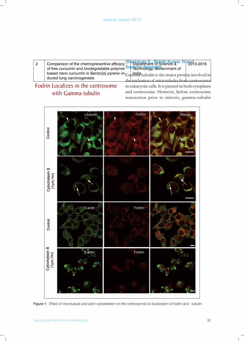
fodrin localizes in the centrosome with Gamma-tubulin
Shashikala S., Rohith Kumar. N and Suparna Sengupta
Gamma-tubulin is the major protein involved in the nucleation of microtubules from centrosomes in eukaryotic cells. It is present in both cytoplasm and centrosome. However, before centrosome maturation prior to mitosis, gamma-tubulin
Figure 1: Effect of microtubule and actin cytoskeleton on the centrosomal co-localization of fodrin and α-tubulin
rajiv gandhi centre for biotechnology 33
annual report 2013
concentration increases dramatically in the centrosome, the mechanism of which is not known. Cytoplasmic gamma tubulin ring complex waspurifiedfrombraininourlaboratory.Non-erythroid spectrin or fodrin, an isoform of spectrin abundant in erythrocytes, was found to be associated with this brain gamma-tubulin ring complex which was not reported earlier in other systems. The major role of erythroid spectrin is to help in the membrane organisation and integrity. However, fodrin or non-erythroid spectrin has a distinct pattern of localisation in brain cells and evidently some special functions
over its erythroid counterpart. In this study, we havefoundthatfodrinandγ-tubulinarepresenttogether in both the cytoplasm and centrosomes in all brain cells except differentiated neurons and astrocytes. Immuno-precipitation studies inpurifiedcentrosomesfrombraintissueandbraincelllinesconfirmthatfodrinandγ-tubulininteract with each other in centrosomes. Fodrin dissociates from centrosome just after the onset of mitosis,whentheconcentrationof γ-tubulinattains a maximum at centrosomes. Further it is observed that the interaction between fodrin
andγ-tubulininthecentrosomeisdependentonactinasdepolymerisationof microfilamentsstopsfodrinlocalization(Figure1).Imageanalysisrevealedthatγ-tubulinconcentrationalsodecreaseddrastically in the centrosome under this condition. This indicates towards a role of fodrin as a regulatorytransporterof γ-tubulintothecentrosomesfornormalprogressionof mitosis.
Analysis of fodrin Association with Gamma Tubulin Complex
in Mammalian brainRohith Kumar, Nisha E Thomas, Shashikala Sasidharan and Suparna Sengupta
Gamma tubulin complex have been implicated as the nucleator of microtubules in eukaryotic cells. Microtubules, one of the major cytoskeleton proteins, are involved in the transportation within the cell as well as involved in proper cell divisionbyformationof spindlefibers.Insimpleeukaryotes, Gamma tubulin complex is in the formof γTuSCcomprisingof 2moleculesof γtubulin and one each of GCP2, GCP3 (Gamma tubulin complex protein). In higher eukaryotes, γTuRCs takes over the role of efficientlynucleatingthemicrotubules.γTuRCsconsistof 7γTuSClongwithotherGCPsnamelyGCP4,GCP5, GCP6. These GCPs share two common motifs, GRIP1, GRIP2. GRIP1 is required for interaction with other GCPs. The GRIP2 motif has been recently acknowledged to be the region responsible for the direct binding with gamma tubulin. In our laboratory, nonerythroid spectrin orFodrinwasidentifiedasanovelcomponentof the gamma tubulin ring complex isolated
from brain. This association has been further verifiedinneuronalandglialcells.Todeterminethe direct binding partner of fodrin within the γ-TuRC, fodrinwas purified from goat brainhomogenate using standard protocol and used for far western analysis. A 52 kDa band was foundtointeractwhichwasidentifiedasgammatubulin. Since fodrin is a heterodimer with high molecular weight, in silico methods were applied to determine the probable interacting region within fodrin. Amino acids sequence 1907-1990 of alpha fodrin showed high homology with the GRIP 2 motif. Fodrin sequence was then modelled and docked with gamma tubulin, interactionwas observedwith the identifiedregion. Further, molecular dynamic simulations validated the interaction. We have further cloned different fodrin fragments, Fod1 ( 1-1500amino acids), Fod2 (2689-40) in EGFP-C1 vector, expressed inHek293andverified interactionof γ tubulin with the fodrin frgments byimmunoprecipitationusingγtubulinantibody.These fragments didn’t show any interaction withγ tubulins suggestingother regionsneedto be studied. Further we downregulated fodrin using Fodrin ShRNA in neuroblastomal cell line IMR32 and observed no change in the amount of γ tubulin aswell as in other cytoskeleton
rajiv gandhi centre for biotechnology 34
annual report 2013
Reshma Thamkachy, Nisha Elizabeth Thomas and Suparna Sengupta
Collaborators: Dr. K.N.Rajasekharan, University of Kerala and Dr. Lynne Cassimeris, Lehigh University
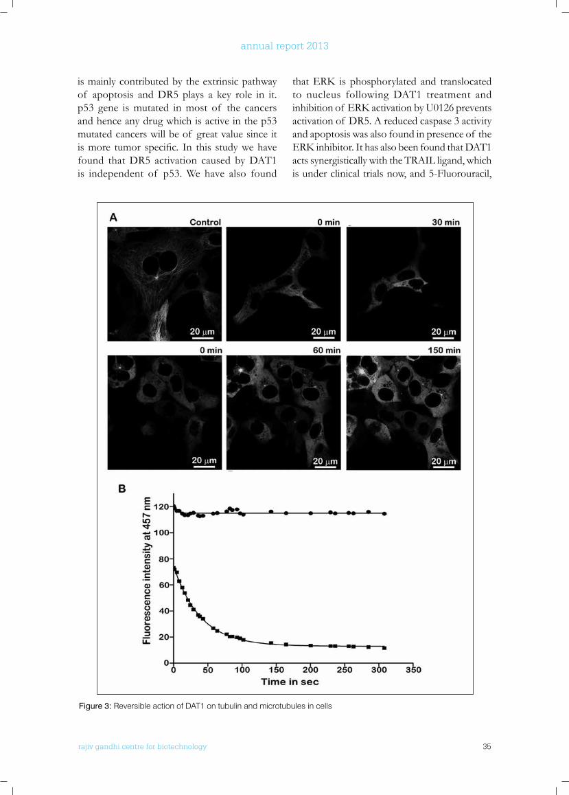
Diaminothiazoles are under study in our laboratoryduetoitsefficacytowardsdifferentcancer cell lines. They show potent antimitotic and anti-angiogenic activity upon binding to the colchicine-binding site of tubulin. In a search for their mechanism of action at the molecular level, we have found that a reversible binding to tubulin with a fast conformational change allows the lead diaminothiazole DAT1 [4-amino-5-benzoyl-2-(4-methoxy phenyl amino) thiazole] to cause a reversible mitotic block. DAT1 also suppresses microtubule dynamic instability at much lower concentration than its IC50 in cancer cells. Both growth and shortening events were reduced by DAT1 in a concentration
dependent way. Colchicine, the long studied tubulin binding drug, has previously failed in the treatment of cancer due to its toxicity, even though it generates a strong apoptotic response. The toxicity is attributable to its slow removal from the cell due to irreversible tubulin binding caused by a slow conformational change. DAT1 binds to tubulin at an opimal pH lower than colchicine. Tubulin conformational studies showed that the binding environment of DAT1 and colchicine are different. Molecular dynamic simulations showed a difference in the number of H-bonding interactions that accounts for the different pH optima. This study gives an insight of the action of compounds targeting tubulin’s colchicine binding site, as many such compounds have entered into clinical trials recently.
Earlier studies have shown that apoptosis mediated by the lead diaminothiazole DAT1
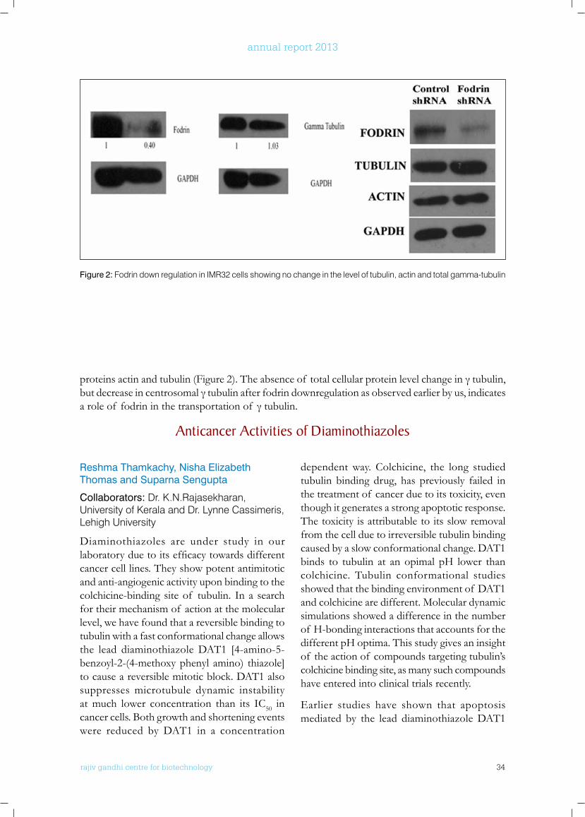
Figure 2: Fodrin down regulation in IMR32 cells showing no change in the level of tubulin, actin and total gamma-tubulin
proteinsactinandtubulin(Figure2).Theabsenceof totalcellularproteinlevelchangeinγtubulin,butdecreaseincentrosomalγtubulinafterfodrindownregulationasobservedearlierbyus,indicatesaroleof fodrininthetransportationof γtubulin.
Anticancer Activities of Diaminothiazoles
rajiv gandhi centre for biotechnology 35
annual report 2013
is mainly contributed by the extrinsic pathway of apoptosis and DR5 plays a key role in it. p53 gene is mutated in most of the cancers and hence any drug which is active in the p53 mutated cancers will be of great value since it ismore tumor specific. In this studywehavefound that DR5 activation caused by DAT1 is independent of p53. We have also found
thatERK is phosphorylated and translocatedto nucleus following DAT1 treatment and inhibitionof ERKactivationbyU0126preventsactivation of DR5. A reduced caspase 3 activity and apoptosis was also found in presence of the ERKinhibitor.IthasalsobeenfoundthatDAT1acts synergistically with the TRAIL ligand, which is under clinical trials now, and 5-Fluorouracil,
Figure 3: Reversible action of DAT1 on tubulin and microtubules in cells
rajiv gandhi centre for biotechnology 36
annual report 2013
Mechanism of Resistance of Cancer Cells Against Antimitotic Agents
Smreti Vasudevan and Suparna Sengupta
Antimitotic agents do show appreciable potential in tumor regression, nevertheless their success is overshadowed by the development of resistance of cancer cells against them. Clinically anticancer drug resistance is a burgeoning problem, and considerable attention is being laid in the development of newer drugs that are less prone to develop resistance, novel combination regimens and treatment modalities. Mechanistically antimitotic drug resistance is a complex phenomenon, where an intricately wovennetworkinvolvingeffluxpumpstospecificmodulations in drug target, cell cycle checkpoint signaling and apoptotic machinery occurs. To understand the basis of antimitotic drug resistanceandtospecificallydissectsmalland
large molecule mediated resistance mechanisms, we have generated taxol (a large molecule) and diaminothiazole (a class of antimitotic agents which are small molecules) resistant cancer cell lines in our laboratory. It was found that over-expressionof P-glycoproteineffluxpumpwas the prime resistance mechanism aroused in cells against taxol which contributed to broad spectrum resistance. Cells also had altered tubulin isotype composition, compromised apoptotic proteins and subdued mitotic checkpoints. Diaminothiazoles were found to be cytotoxic in multidrug resistant cancer cell lines. They induced mitotic block leading to the activation of spindle assembly checkpoint proteins, thereby channeling the resistant cells towards caspase 3 mediated apoptosis. Moreover, the DAT1 resistant subline did not over express P-glycoproteinandexhibitedspecificity in theresistance process. Also, cells lost their resistance against DAT1 upon the removal of drug and
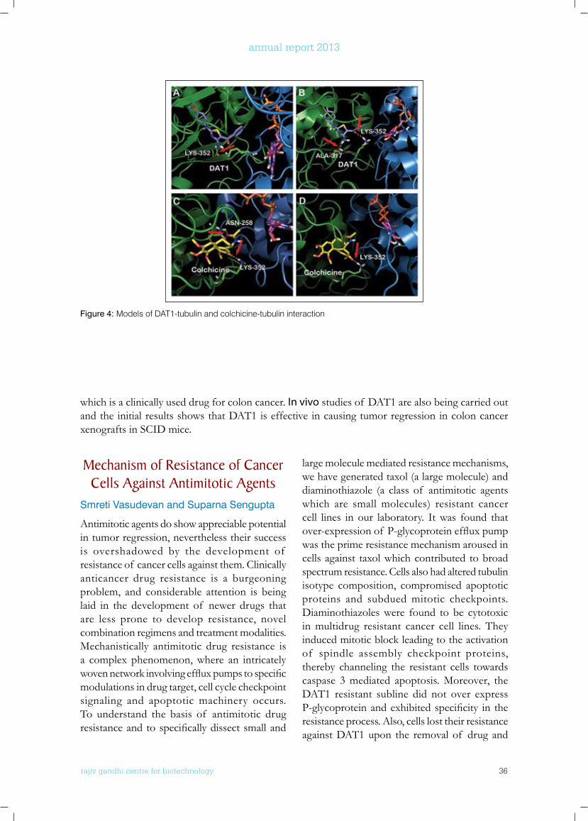
Figure 4: Models of DAT1-tubulin and colchicine-tubulin interaction
which is a clinically used drug for colon cancer. In vivo studies of DAT1 are also being carried out and the initial results shows that DAT1 is effective in causing tumor regression in colon cancer xenografts in SCID mice.
rajiv gandhi centre for biotechnology 37
annual report 2013
6-shogaol inhibits breast cancer spheroid formation
Anasuya Ray, Smreti Vasudevan and Suparna Sengupta
Shogaols are found in dried ginger and they are primarily the dehydrated product of gingerols. Among the shogaols, 6-shogaol exhibited potent cytotoxic activity against gastric carcinoma, hepatocarcinoma, non small cell lung carcinoma, ovarian carcinoma, colorectal carcinoma etc. We have investigated its inhibitory effect against breast cancer stem cell spheroids formed frombreastcancercellsunderspecificgrowthconditions. The stem cell properties were verifiedby checking theCD44/CD24markerexpression.6-shogaolshowedefficientcytotoxicactivity in MCF-7 and MDA-MB-231 spheroids in a condition under which taxol did not show any noticeable cytotoxicity. 6-shogaol inhibited the number and size of primary and secondary spheroids. Only 4 primary spheroid colonies with a 24 folds inhibition were observed by the treatmentof 40μM6-shogaol.Nosecondaryspheroidwasfoundbeyond10μMof 6-shogaol
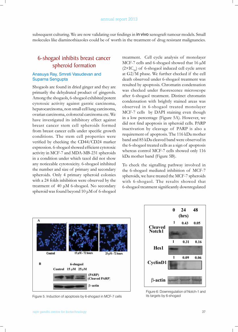
treatment. Cell cycle analysis of monolayer MCF-7 cells and 6-shogaol showed that 16 µM (2×IC50) of 6-shogaol induced cell cycle arrest at G2/M phase. We further checked if the cell death observed under 6-shogaol treatment was resulted by apoptosis. Chromatin condensation was checked under fluorescencemicroscopeafter 6-shogaol treatment. Distinct chromatin condensation with brightly stained areas was observed in 6-shogaol treated monolayer MCF-7 cells by DAPI staining even though in a low percentage (Figure 5A). However, we didnotfindapoptosisinspheroidcells.PARPinactivation by cleavage of PARP is also a requirement of apoptosis. The 116 kDa mother band and 85 kDa cleaved band were observed in the 6-shogaol treated cells as a sign of apoptosis whereas control MCF-7 cells showed only 116 kDa mother band (Figure 5B).
To check the signalling pathway involved in the 6-shogaol mediated inhibition of MCF-7 spheroids, we have treated the MCF-7 spheroids with 6-shogaol. The results showed that 6-shogaoltreatmentsignificantlydownregulated
Figure 5: Induction of apoptosis by 6-shogaol in MCF-7 cellsFigure 6: Downregulation of Notch-1 and its targets by 6-shogaol
subsequentculturing.Wearenowvalidatingourfindingsinin vivo xenograft tumour models. Small molecules like diaminothiazoles could be of worth in the treatment of drug resistant malignancies.
rajiv gandhi centre for biotechnology 38
annual report 2013
cleaved Notch1 expression. Notch target genes Hes1 and Cyclin D1 were also downregulated by 6-shogaol treatment (Figure 6). Thus, the results explain that 6-shogaol inhibits the MCF-7 spheroid formation by altering the Notch signaling pathway.
PAPERS PublISHED
• Nisha E Thomas, Reshma Thamkachy, Krishnankutty C. Shivakumar, Sreedevi K.J., Xavier Lieben Louis, Sannu A. Thomas, Rohith Kumar, Kallikat N. Rajasekharan, Lynne Cassimeris & Suparna Sengupta (2013): Reversible Action of Diaminothiazoles in Cancer Cells is Implicated by the Induction of a Fast Conformational Change of Tubulin and Suppression of Microtubule Dynamics. Molecular Cancer Therapeutics DOI: 10.1158/1535-7163.MCT-13-0479
• Sasidharan Shashikala, Rohith Kumar, Nisha E. Thomas, Dhanesh Sivadasan, Jackson James & Suparna Sengupta (2013): Fodrin in Centrosomes: Implication of a Role of Fodrin in the Transport of Gamma-Tubulin Complex in Brain. PLoS ONE 8(10): e76613. doi:10.1371/journal.pone.0076613
• S. Thalamuthu, B. Annaraj, Smreti Vasudevan, Suparna Sengupta & M.A. Neelakantan (2013): DNA binding, nuclease and colon cancer cell inhibitory activity of a Cu(II) complex of a thiazolidine-4-carboxylic acid derivative, Journal of Coordination Chemistry, DOI:10.1080/00958972.2013.791393
• Mathan Sankaran, Chokkalingam Uvarani, Kumarasamy Chandraprakash, Swathi U. Lekshmi, Sengupta Suparna, James Platts, Palathurai Subramaniam Mohan (2014): A regioselective multicomponent protocol for the synthesis of novel bioactive 4-hydroxyquinolin-2(1H)-one grafted monospiropyrrolidine and thiapyrrolizidine hybrids. Molecular Diversity DOI 10.1007/s11030-013-9498-y
ConfEREnCE PRESEnTATIonS
• Sasidhran Shashikala, Rihith Kumar, Nisha E. Thomas, Dhanesh Sivadasan, Jackson James and Suparna Sengupta: Fodrin in Centrosomes”: 22nd Annual Meeting of the Protein Society, Boston, USA, 20-23 July, 2013
• Vasudevan S, Thomas SA, Komalam RJ, Sreerekha KV, Rajasekharan KN and Sengupta S: “Promising in vitro and in vivo activity of diaminothiazoles in multidrug resistant cancer: a mechanistic study: 33rd Annual Convention of Indian Association for Cancer Research, Thiruvananthapuram, India, 13-15 February 2014
• Reshma Thamkachy, Sannu Ann Thomas, K.N,Rajashekharan and Suparna Sengupta: The Diaminothiazole DAT1 is effective in colon cancer cell lines irrespective of their p53 status through Extra Cellular Signal regulated stress kinase (ERK) mediated upregulation of Death Receptor 5: 33rd Annual Convention of Indian
rajiv gandhi centre for biotechnology 39
annual report 2013
Cancer Research Program:Laboratory - 4
Ph.D StudentsDiana DavidSaneesh Babu.P.SDhanya. KChithra JSTapas Pradhan
r a j i v g a n d h i c e n t r e f o r b i o t e c h n o l o g y
Asha Nair took her Ph.D from the University of Kerala woking at Regional Cancer Centre, Thiruvananthapuram, Kerala. She trained as a post doctoral fellow at Harvard Medical School and M.D. Anderson Cancer Centre Houston, Texas, USA before joining RGCB in 2006.
S. Asha Nair, Ph.DScientist E II
Research FellowsChandraprabha M.G (ICMR SRF)Krishnanand Padmanabhan (CSIR SRF)Nisha Asok Kumar (DBT JRF)
Project AssistantManu Prasad M (DST)
Technical AssistantsPrameela Kumari TKMeera Nair
rajiv gandhi centre for biotechnology 40
annual report 2013
33rd Annual convention of Indian Association for Cancer Research, Thiruvananthapuram, India, February 13-15, 2014
Smurf2 and CnkSR2: implications in breast cancer cell proliferation
Diana David and S. Asha Nair
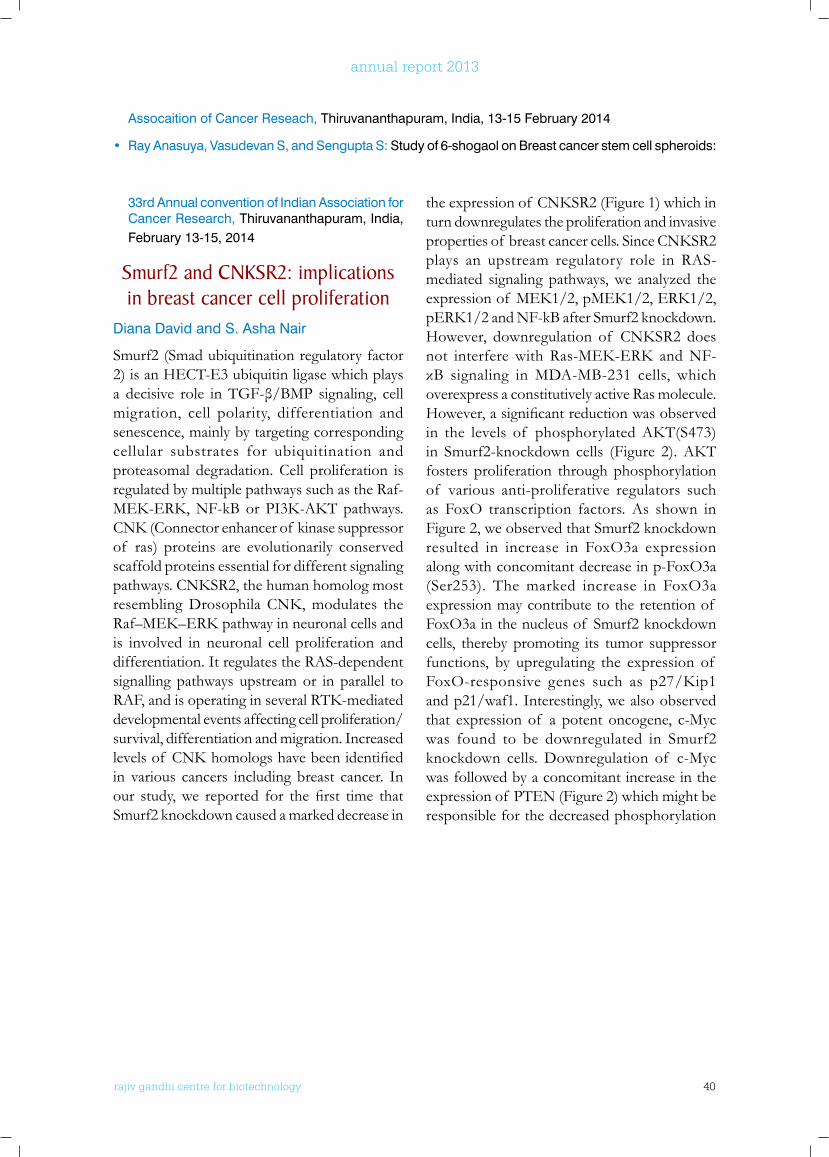
Smurf2 (Smad ubiquitination regulatory factor 2) is an HECT-E3 ubiquitin ligase which plays a decisive role inTGF-β/BMP signaling, cellmigration, cell polarity, differentiation and senescence, mainly by targeting corresponding cellular substrates for ubiquitination and proteasomal degradation. Cell proliferation is regulated by multiple pathways such as the Raf-MEK-ERK,NF-kBorPI3K-AKTpathways.CNK(Connectorenhancerof kinasesuppressorof ras) proteins are evolutionarily conserved scaffold proteins essential for different signaling pathways.CNKSR2,thehumanhomologmostresemblingDrosophilaCNK,modulates theRaf–MEK–ERKpathwayinneuronalcellsandis involved in neuronal cell proliferation and differentiation. It regulates the RAS-dependent signalling pathways upstream or in parallel to RAF,andisoperatinginseveralRTK-mediateddevelopmental events affecting cell proliferation/survival, differentiation and migration. Increased levelsof CNKhomologshavebeenidentifiedin various cancers including breast cancer. In our study,we reported for thefirst time thatSmurf2 knockdown caused a marked decrease in
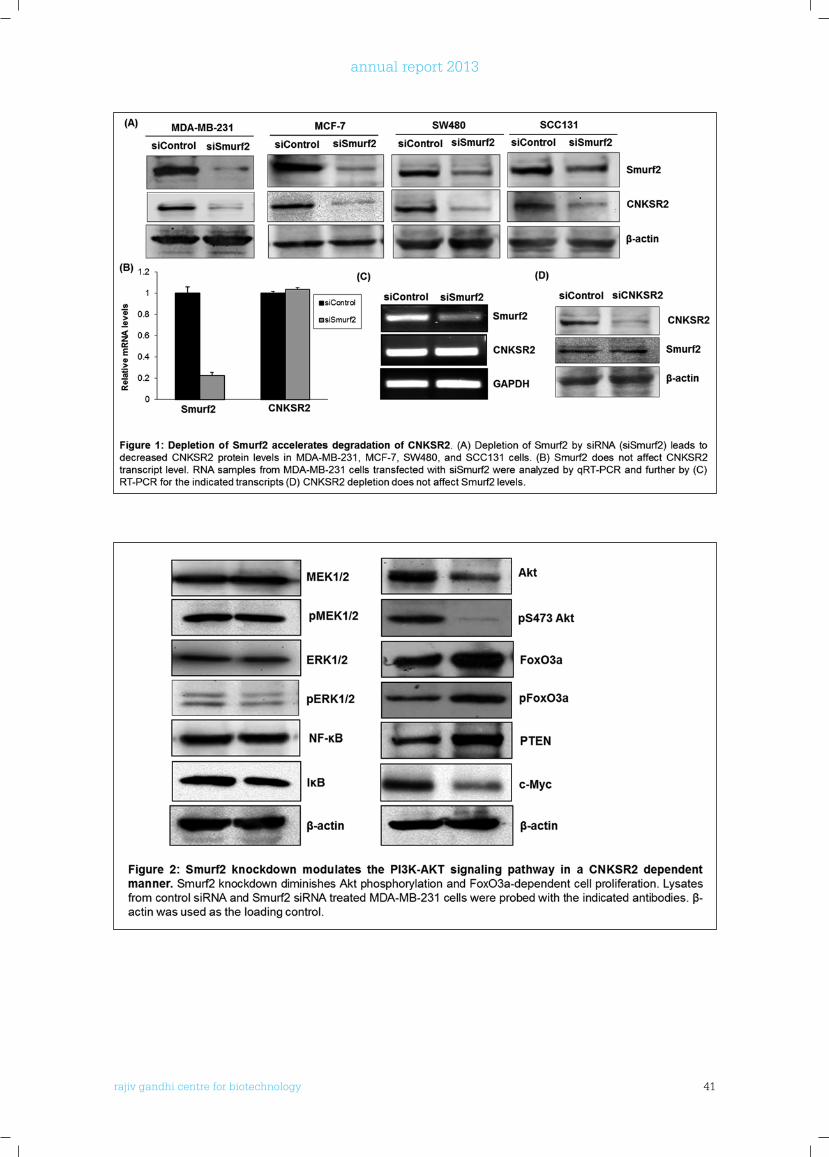
theexpressionof CNKSR2(Figure1)whichinturn downregulates the proliferation and invasive propertiesof breastcancercells.SinceCNKSR2plays an upstream regulatory role in RAS-mediated signaling pathways, we analyzed the expressionof MEK1/2,pMEK1/2,ERK1/2,pERK1/2andNF-kBafterSmurf2knockdown.However, downregulation of CNKSR2 doesnot interferewith Ras-MEK-ERK andNF-κB signaling inMDA-MB-231 cells, whichoverexpress a constitutively active Ras molecule. However,asignificantreductionwasobservedin the levels of phosphorylatedAKT(S473)in Smurf2-knockdown cells (Figure 2).AKTfosters proliferation through phosphorylation of various anti-proliferative regulators such as FoxO transcription factors. As shown in Figure 2, we observed that Smurf2 knockdown resulted in increase in FoxO3a expression along with concomitant decrease in p-FoxO3a (Ser253). The marked increase in FoxO3a expression may contribute to the retention of FoxO3a in the nucleus of Smurf2 knockdown cells, thereby promoting its tumor suppressor functions, by upregulating the expression of FoxO-responsive genes such as p27/Kip1and p21/waf1. Interestingly, we also observed that expression of a potent oncogene, c-Myc was found to be downregulated in Smurf2 knockdown cells. Downregulation of c-Myc was followed by a concomitant increase in the expression of PTEN (Figure 2) which might be responsible for the decreased phosphorylation
Assocaition of Cancer Reseach, Thiruvananthapuram, India, 13-15 February 2014
• Ray Anasuya, Vasudevan S, and Sengupta S: Study of 6-shogaol on Breast cancer stem cell spheroids:
rajiv gandhi centre for biotechnology 41
annual report 2013
rajiv gandhi centre for biotechnology 42
annual report 2013
unfolded protein response.Saneesh Babu P.S*, D. Ramaiah*, S. Asha Nair, and M.RadhakrishnaPillai
Collaborator: *Photochemistry and Photonics Division, National Institute for Interdisciplinary Science and Technology (NIIST), Thiruvananthapuram.
Photodynamic therapy is a novel treatment for cancer and certain noncancerous conditions that are generally characterized by overgrowth of unwanted or abnormal cells. PDT involves the administration of a photosensitizing compound, which accumulates in the target cells, followed by selective irradiation of the lesion with visible light. This procedure results in a sequence of photochemical events that generate reactive oxygen species (ROS), which induce oxidative damage ultimately causing the killing of cancerous cells or other targets of therapeutic interest. In order to enhance the efficacy of PDT and extend its applications, a variety of second generation photosensitizers, such as squaraines are now being assessed for their efficacyincancertherapy,anditisimportanttoelucidate their mechanisms of action in PDT. Squaraines are a class of dyes possessing sharp and intense absorption in the near infrared
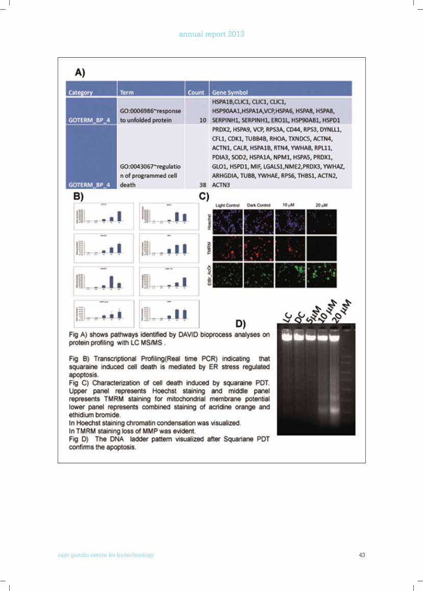
regionandexhibitsignificanttripletandquantumyields. In vitro cytotoxicity and mutagenicity investigations of the dye, using both mammalian cell lines and bacterial strains reveal that it is weakly photomutagenic but highly phototoxic. Squaraine have been proved to possess targeted accumulation in tumor cells so that it can be used for selective destruction without effecting normal cells. To investigate the cellular response to squaraine based photodynamic therapy we conducted a transcriptional profiling using LC-MS/M. Tryptically digested extracts of MDAMB231 cells after PDT with 20mM squaraine were analyzed by a data- independent-acquisition workflow (LC-MS/MS) in three technical replicates. Transcriptional activation of identifiedpathwaywereconfirmedbyrealtime analysis of selected genes and apoptosis was studied by using Hoechst and combined staining of acridine orange and ethidium bromide and also DNA ladder assay was done. Based on our transcriptional profiling and pathway analysiswe observed an unfold protein response and endoplasmic reticulum (ER) mediated cell death. These eventswere further confirmedbyTranscriptionalProfiling(RealtimePCR)of ER stress related genes (HSP 70, ATF 4, BAD,
of AKTatS473whichisconsistentwiththewell-establishedinverserelationshipbetweenMMAC/PTENexpressionandAKTactivation.ThusSmurf2knockdowndownregulatesproliferationof breastcancercellsinaCNKSR2dependentmannerbymodulatingthePI3K-PTEN-AKT-FoxO3apathway.
Squaraine based photodynamic therapy induces cancer cell apoptosis by the
rajiv gandhi centre for biotechnology 43
annual report 2013
rajiv gandhi centre for biotechnology 44
annual report 2013
Thiostrepton a foxM1 and proteasome inhibitor targets
mutant p53 for degradation by a proteasome independent pathway.
Dhanya K, Manu Prasad and S. Asha Nair
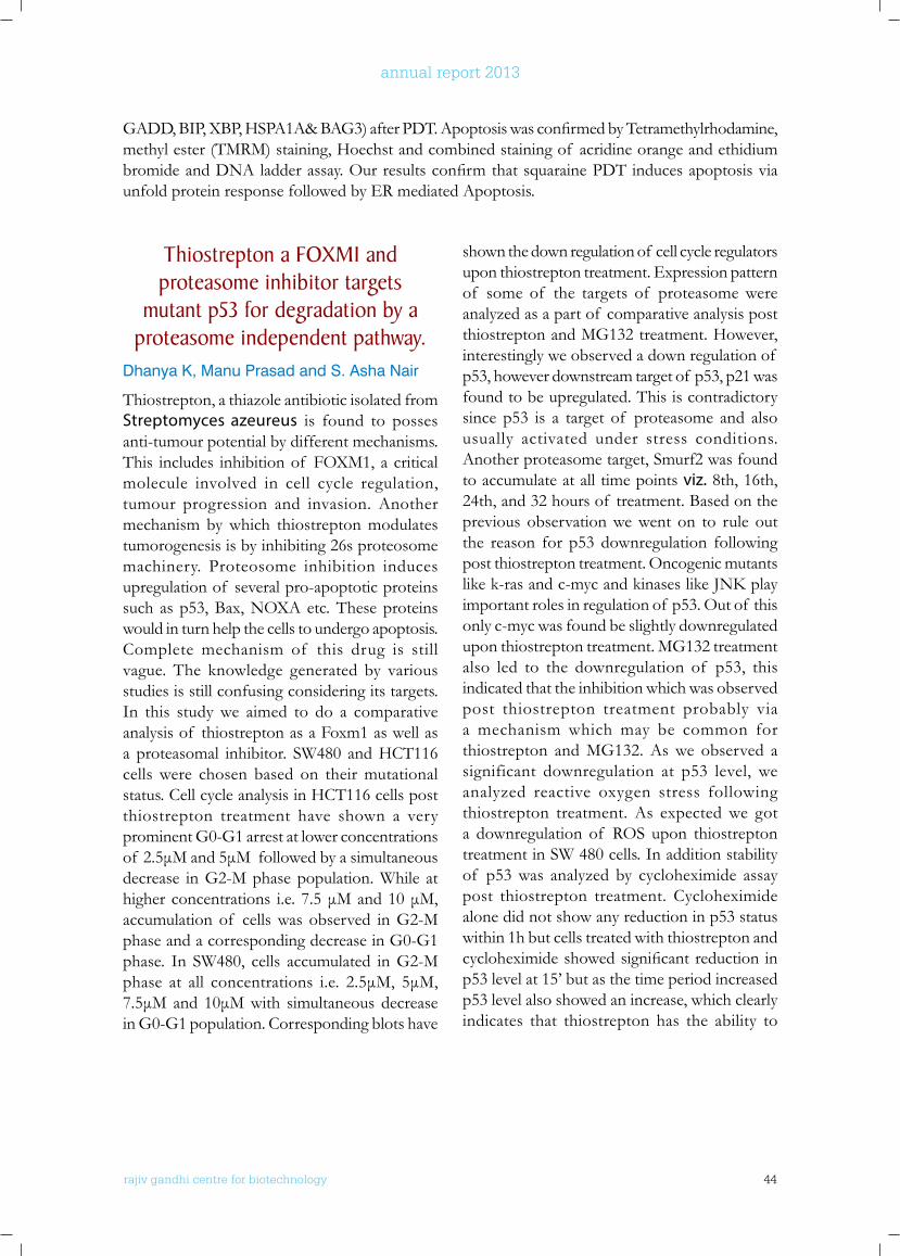
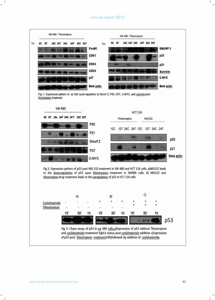
Thiostrepton, a thiazole antibiotic isolated from Streptomyces azeureus is found to posses anti-tumour potential by different mechanisms. This includes inhibition of FOXM1, a critical molecule involved in cell cycle regulation, tumour progression and invasion. Another mechanism by which thiostrepton modulates tumorogenesis is by inhibiting 26s proteosome machinery. Proteosome inhibition induces upregulation of several pro-apoptotic proteins such as p53, Bax, NOXA etc. These proteins would in turn help the cells to undergo apoptosis. Complete mechanism of this drug is still vague. The knowledge generated by various studies is still confusing considering its targets. In this study we aimed to do a comparative analysis of thiostrepton as a Foxm1 as well as a proteasomal inhibitor. SW480 and HCT116 cells were chosen based on their mutational status. Cell cycle analysis in HCT116 cells post thiostrepton treatment have shown a very prominent G0-G1 arrest at lower concentrations of 2.5μMand5μMfollowedbyasimultaneousdecrease in G2-M phase population. While at higherconcentrations i.e.7.5μMand10μM,accumulation of cells was observed in G2-M phase and a corresponding decrease in G0-G1 phase. In SW480, cells accumulated in G2-M phase at all concentrations i.e. 2.5μM, 5μM,7.5μMand 10μMwith simultaneous decreasein G0-G1 population. Corresponding blots have
shown the down regulation of cell cycle regulators upon thiostrepton treatment. Expression pattern of some of the targets of proteasome were analyzed as a part of comparative analysis post thiostrepton and MG132 treatment. However, interestingly we observed a down regulation of p53, however downstream target of p53, p21 was found to be upregulated. This is contradictory since p53 is a target of proteasome and also usually activated under stress conditions. Another proteasome target, Smurf2 was found to accumulate at all time points viz. 8th, 16th, 24th, and 32 hours of treatment. Based on the previous observation we went on to rule out the reason for p53 downregulation following post thiostrepton treatment. Oncogenic mutants likek-rasandc-mycandkinaseslikeJNKplayimportant roles in regulation of p53. Out of this only c-myc was found be slightly downregulated upon thiostrepton treatment. MG132 treatment also led to the downregulation of p53, this indicated that the inhibition which was observed post thiostrepton treatment probably via a mechanism which may be common for thiostrepton and MG132. As we observed a significant downregulation at p53 level, we analyzed reactive oxygen stress following thiostrepton treatment. As expected we got a downregulation of ROS upon thiostrepton treatment in SW 480 cells. In addition stability of p53 was analyzed by cycloheximide assay post thiostrepton treatment. Cycloheximide alone did not show any reduction in p53 status within 1h but cells treated with thiostrepton and cycloheximideshowedsignificantreductioninp53 level at 15’ but as the time period increased p53 level also showed an increase, which clearly indicates that thiostrepton has the ability to
GADD,BIP,XBP,HSPA1A&BAG3)afterPDT.ApoptosiswasconfirmedbyTetramethylrhodamine,methyl ester (TMRM) staining, Hoechst and combined staining of acridine orange and ethidium bromideandDNAladderassay.OurresultsconfirmthatsquarainePDTinducesapoptosisviaunfold protein response followed by ER mediated Apoptosis.
rajiv gandhi centre for biotechnology 45
annual report 2013
rajiv gandhi centre for biotechnology 46
annual report 2013
REGulATIon of STEMnESS In EnDoMETRIAl CAnCER by MTA1Chithra J.S, Rema. P*, S. Asha Nair and M. Radhakrishna Pillai
Collaborator: * Department of Surgical Oncology, Regional Cancer Centre,Trivandrum
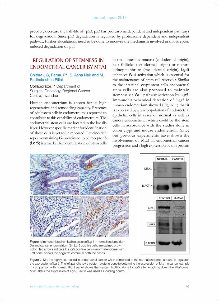
Human endometrium is known for its high regenerative and remodeling capacity. Presence of adult stem cells in endometrium is reported to contribute to this capability of endometrium. The endometrial stem cells are located in the basalis layer.Howeverspecificmarkerforidentificationof these cells is yet to be reported. Leucine-rich repeat-containing G-protein-coupled receptor 5 (Lgr5)isamarkerforidentificationof stemcells
in small intestine mucosa (endodermal origin), hair follicles (ectodermal origin) or mature kidney nephrons (mesodermal origin). Lgr5 enhances Wnt activation which is essential for the maintenance of stem cell reservoir. Similar to the intestinal crypt stem cells endometrial stem cells are also proposed to maintain stemness via Wnt pathway activation by Lgr5. Immunohistochemical detection of Lgr5 in human endometrium showed (Figure 1) that it is expressed by a rare population of endometrial epithelial cells in cases of normal as well as cancer endometrium which could be the stem cells in accordance with the studies done in colon crypt and mouse endometrium. Since our previous experiments have shown the involvement of Mta1 in endometrial cancer progression and a high expression of this protein
probably decrease the half-life of p53. p53 has proteasome dependent and independent pathways for degradation. Since p53 degradation is regulated by proteasome dependent and independent pathway, further elucidations need to be done to uncover the mechanism involved in thiostrepton induced degradation of p53
Figure 2: Mta1 is highly expressed in endometrial cancer when compared to the normal endometrium and it regulates the expression of Lgr5. The left panel shows western blotting done to determine the expression of Mta1 in cancer sample in comparison with normal. Right panel shows the western blotting done forLgr5 after knocking down the Mta1gene. Mta1 alters the expression of Lgr5. α actin was used as loading control.
Figure 1: Immunohistochemical detection of Lgr5 in normal endometrium (A) and cancer endometrium (B). Lgr5 positive cells are stained brown in color. Red arrows indicate the lgr5 positive cells in normal endometrium. Left panel shows the negative control in both the cases
rajiv gandhi centre for biotechnology 47
annual report 2013
Regulatory network between Stat3 And Skp2 - The E3 ubiquitin
ligase: Implications for Signal Transduction To Cell Cycle In
Colorectal CancerChandraprabha M.G. and S. Asha Nair
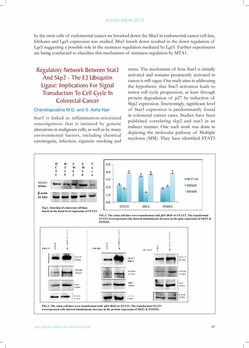
Stat3 is linked to inflammation-associated tumorigenesis that is initiated by genetic alterations in malignant cells, as well as by many environmental factors, including chemical carcinogens, infection, cigarette smoking and
stress. The mechanism of how Stat3 is initially activated and remains persistently activated in cancer is still vague. Our study aims in addressing the hypothesis; that Stat3 activation leads to tumor cell cycle progression, at least through protein degradation of p27 by induction of Skp2expression.Interestingly,significantlevelof Stat3 expression is predominantly found in colorectal cancer cases. Studies have been published correlating skp2 and stat3 in an indirect manner. One such work was done in depicting the molecular pathway of Multiple myeloma (MM).They have identified STAT3
by the stem cells of endometrial cancer we knocked down the Mta1 in endometrial cancer cell line, Ishikawa and Lgr5 expression was studied. Mta1 knock down resulted in the down regulation of Lgr5 suggesting a possible role in the stemness regulation mediated by Lgr5. Further experiments are being conducted to elucidate this mechanism of stemness regulation by MTA1.
FIG.2. The colon cell lines were transformed with pEF-BOS wt STAT3. The transformed STAT3 overexpressed cells showed simultaneous increase in the protein expression of SKP2 & FOXM1.
HCT1
HT29
SW62
SW48
Caco
STAT3 85kDa
β-actin 42 kDa
FIG.3. The colon cell lines were transformed with pEF-BOS wt STAT3. The transformed STAT3 overexpressed cells showed simultaneous incraese in the gene expression of SKP2 & FOXM1.
Fig.1. Selection of colorectal cell lines based on the basal level expression of STAT3
rajiv gandhi centre for biotechnology 48
annual report 2013
tobedownstreamtargetsof CKS1Bactivationindependentonthecomplexof SKP2/p27Kip1.The results of our project so far pertain to the relation between STAT3 /SKP2 expressionin correlation with the activation of FOXM1 in colon cells. The preliminary data depicting theSTAT3/SKP2correlationwas carriedoutusing the STAT3 inhibitor, Stattic. Further, real timePCRwasdonetoconfirmthesameatthetranscriptional level. FOXM1 belonging to the forkhead box (Fox) transcription factors, is a nuclear protein that regulates the expression of proteins and enzymes required for mitosis andcytokinesis.FoxM1hasbeenidentifiedtopromote transcription of skp2. Over expression of STAT3 using plasmid construct, pEF-BOSwtSTAT3significantly increasedFOXM1followedbyincreaseinSKP2levelsinSW620,SW480 and HCT116 cells. Also, knockdown of Stat3 by small interfering RNA (siRNA)
decreasedFOXM1,SKP2proteinlevelsalongwith the accumulation of p27 in colon cancer cells. Together, these data might suggest a possible correlation between Stat3 in regulating FOXM1/Skp2 expression in colon cancer cells. Thenextphaseof thestudywastheidentificationof STAT3 binding consensus sequences at the FoxM1 gene promoter. For the same, In Silico DNA sequence analysis of 1000 base pairs from the FoxM1 promoter was done, which revealed the consensus sequences for STAT3 protein binding. Further, to determine the in vivo binding of STAT3 to the FoxM1 promoter DNA sequence, primers were designed based on the FoxM1 sequences from the upstream promoter region, which contained the STAT3 consensus binding site (underlined), FoxM1: 5’-TCAAAGG AACTTAGTCTAATCGGGGGGAGC-3’.
Conclusively, the results so far showed a dependent signal networking between STAT3
and FOXM1 in colorectal cancer. Further, the study is routed in identifying the role of FoxM1 in our cancer model and also to increase the understanding in FOXM1/STAT3 signalling. This might exhibit its role in proliferation, survival, drug resistance and DNA repair in colorectal cancer.
Tumor Stem like Cells As unique Targets In Residual Disease
Nisha Asok Kumar, Chandramohan .K *, S. Asha Nair and M.Radhakrishna Pillai
Collaborator: * Department of Surgical Oncology, Regional Cancer Centre, Trivandrum
Colorectal cancer (CRC) is one of the major causes of death worldwide. Despite surgery followed by adjuvant therapy remains the mainstay for the disease, often majority of the patients undergo recurrence and metastases. This phenomenon frequently correlates with an acquired resistance to conventional therapies such as chemo- and radio-therapy. Novel insights in cancer research suggested that the capacity to initiate and sustain tumor growth, is a unique characteristic of a small subset of cancer cells with stemness properties within the
tumor mass, called “cancer stem cells” (CSCs) or “tumor-initiating cells” Evidences suggest that CSCs, isolated from a variety of tumor types, retain tumorigenic capacity in vivo and are responsible for the self renewal, relapse and metastasis. They are also characterized by high resistance to drugs and general toxins, which target rapidly proliferating cells and spare the slow dividing cells, due to an up-regulation of several ATP-binding cassette transporters, active DNA-repair capacity, over-expression of anti-apoptotic molecules that cause changes in the signaling pathways controlling proliferation, differentiation and apoptosis. There are several molecular mechanisms that may account for the resistance to apoptosis of cancer stem cells- the most important being the cell cycle kinetics. Many cancer stem cells are not cycling and are in G0 and thus resistant to cell cycle–specific chemotherapeutic agents. CSCs are
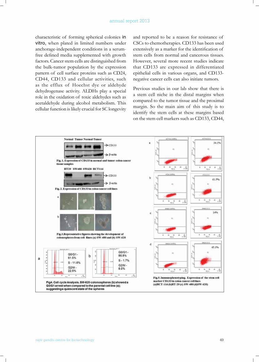
rajiv gandhi centre for biotechnology 49
annual report 2013
characteristic of forming spherical colonies in vitro, when plated in limited numbers under anchorage-independent conditions in a serum-freedefinedmediasupplementedwithgrowthfactors. Cancer stem cells are distinguished from the bulk-tumor population by the expression pattern of cell surface proteins such as CD24, CD44, CD133 and cellular activities, such as the efflux of Hoechst dye or aldehyde dehydrogenase activity. ALDHs play a special role in the oxidation of toxic aldehydes such as acetaldehyde during alcohol metabolism. This cellular function is likely crucial for SC longevity
and reported to be a reason for resistance of CSCs to chemotherapies. CD133 has been used extensivelyasamarkerfortheidentificationof stem cells from normal and cancerous tissues. However, several more recent studies indicate that CD133 are expressed in differentiated epithelial cells in various organs, and CD133-negative cancer cells can also initiate tumors.
Previous studies in our lab show that there is a stem cell niche in the distal margins when compared to the tumor tissue and the proximal margin. So the main aim of this study is to identify the stem cells at these margins based on the stem cell markers such as CD133, CD44,
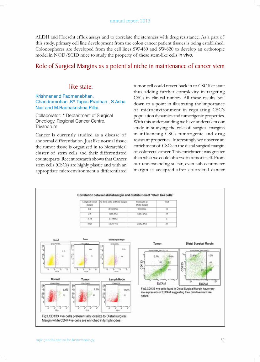
rajiv gandhi centre for biotechnology 50
annual report 2013
like state.Krishnanand Padmanabhan, Chandramohan .K* Tapas Pradhan , S Asha Nair and M.Radhakrishna Pillai.
Collaborator: * Deptartment of Surgical Oncology, Regional Cancer Centre, Trivandrum
Cancer is currently studied as a disease of abnormal differentiation. Just like normal tissue the tumor tissue is organized in to hierarchical cluster of stem cells and their differentiated counterparts. Recent research shows that Cancer stem cells (CSCs) are highly plastic and with an appropriate microenvironment a differentiated
tumor cell could revert back in to CSC like state thus adding further complexity in targeting CSCs in clinical tumors. All these results boil down to a point in illustrating the importance of microenvironment in regulating CSC’s population dynamics and tumorigenic properties. With this understanding we have undertaken our study in studying the role of surgical margins in influencing CSCs tumorigenic and drug resistant properties. Interestingly we observe an enrichment of CSCs in the distal surgical margin of colorectal cancer. This enrichment was greater than what we could observe in tumor itself. From our understanding so far, even sub-centimeter margin is accepted after colorectal cancer
ALDHandHoeschteffluxassaysandtocorrelatethestemnesswithdrugresistance.Asapartof this study, primary cell line development from the colon cancer patient tissues is being established. Colonospheres are developed from the cell lines SW-480 and SW-620 to develop an orthotopic model in NOD/SCID mice to study the property of these stem-like cells in vivo.
Role of Surgical Margins as a potential niche in maintenance of cancer stem
rajiv gandhi centre for biotechnology 51
annual report 2013
stemness associated transcriptional factors such as OCT4, SOX2 and SOX9 at transcriptional level in distal surgical margin compared to Normal and Tumor tissue. Our current aim is to understand the existence of clonal heterogeneity among CSCs within a single tumor and effect of this phenomenon in attributing tumorigenic and drug resistant properties of a tumor.
PublICATIonS
• Suneesh C. Karunakaran, P. S. Saneesh Babu,
Bollapalli Madhuri, Betsy Marydasan, Albish K. Paul, S. Asha Nair, K. Sridhar Rao, A. Srinivasan,
Tavarekere K. Chandrashekar, Ch. Mohan Rao,* M. Radhakrishan Pillai,* and Danaboyina Ramaiah (2013). In Vitro Demonstration of Apoptosis Mediated Photodynamic Activity and NIR Nucleus Imaging through a Novel Porphyrin ACS Chemical Biology ;8(1):127-32
• Diana David, Asha .S. Nair*, Pillai MR (2013). Smurf E3 Ubiquitin Ligases at the Cross Roads of Oncogenesis and Tumor Suppression. BBA Reviews On Cancer 1835(1):119-28
• Krishnan, Anand, Vivek R. Gopinath, Ajit Johnson, S. Asha Nair, and M. Radhakrishna Pillai. “Cell-cycle analysis and micronuclei frequency reveals G0/G1 blockers as weak micronuclei inducers.” Drug Chem Toxicol. 2013
Apr;36(2):249-54
• Jagadeeshan S, Krishnamoorthy YR, Singhal M, Subramanian A, Mavuluri J, Lakshmi A, Roshini A, Baskar G, Ravi M, Joseph LD, Sadasivan K,Krishnan A, S. Asha Nair, Venkatraman G, Rayala S K (2014) Transcriptional regulation of fibronectin by p21-activated kinase-1 modulates pancreatic tumorigenesis. Oncogene. Feb 24. doi: 10.1038/onc.2013.576. [Epub ahead of print]
ConfEREnCE PRESEnTATIonS
PoSTeR PReSeNTATioN:
• Chithra J.S., S. Asha Nair, M. Radhakrishna Pillai, “Lethal seeds in the womb: endometrial cancer stem cells express metastasis tumor antigen” on Amritha Bioquest - 2013 International conference on Biotechnology for Innivative Applications. August 10 - 14, 2013
• Dhanya K., S. Asha Nair, “Silencing of Forkhead box protein FoxM1 leads to the down regulation of CDK1-A Gatekeeper of M phase entry” on 17th ADNAT convention, Symposium on genomics in personalized medicine and public health held at Rajiv Gandhi center for biotechnology during Feb 23-25, 2014
surgery. But the very fact that CSCs are present more than that distance challenges the very concept of radicality which we uphold today. We now aim to understand the reason behind this preferential localization. Interestingly we have found that CSCs of different properties show differences in their preference of localization. The CD133+ve CSCs are preferentially localized to distal surgical margin while CD44 + ve cells, whichspecificallymarkthemetastaticCSCs,areenriched in lymphnodes adjacent to tumor. More importantly CD133+ve cells localized in distal margin have very low expression of EpCAM compared to tumor which has CD133+ve cells with high EpCAM expression. This further
indicates that tumor primordial cells within distal surgical margin remain in a more primitive stem like state. From our observations we presume that CD133+ve cells might be critical in maintenance of primary tumor while CD44+ve cells might be important for distant metastasis. Interestingly the expression of MDR proteins (ABCC1 and ABCG2) was enriched in distal surgical margin only after post chemo/RT treatment. This indicates a possibility of CSCs to migrate from tumor tissue towards distal surgical margin upon exposure to chemotherapeutic agents and radiation. To support our above observations we have observed an increased expression of

rajiv gandhi centre for biotechnology 52
annual report 2013
• Saneesh Babu P.S, D Ramaiah, S. Asha Nair, and M. Radhakrishna Pillai “Understanding reactive oxygen species resistant pathways activated by sub-lethal dose of photodynamic therapy to identify new therapeutic targets.“ On 33rd Annual Convention of Indian Association for Cancer Research Jointly Organized by Rajiv Gandhi Centre for Biotechnology,Thiruvananthapuram & Srinivasa Ramanujan Institute for Basic Sciences, Kottayam February 13 - 15, 2014
ExTRAMuRAl GRAnTS
Sl.No Project Title Period Funding agency Investigator
1Functional significance of fork head box protein,foxm1b in cdk1 turnover - A molecular analysis
2013 -2016Department of Science & Technology, Government of India
Principal Investigator
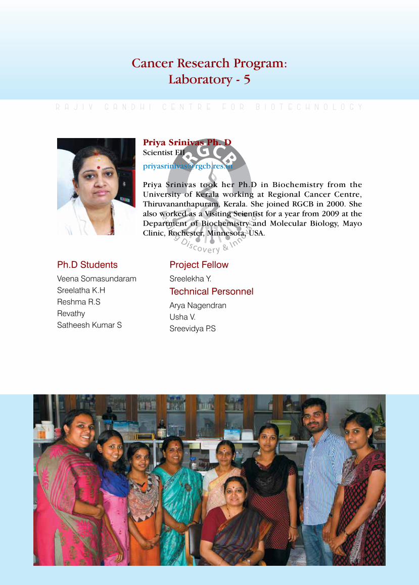
rajiv gandhi centre for biotechnology 53
annual report 2013
Cancer Research Program:Laboratory - 5
Ph.D StudentsVeena Somasundaram Sreelatha K.HReshma R.SRevathySatheesh Kumar S
r a j i v g a n d h i c e n t r e f o r b i o t e c h n o l o g y
Priya Srinivas took her Ph.D in Biochemistry from the University of Kerala working at Regional Cancer Centre, Thiruvananthapuram, Kerala. She joined RGCB in 2000. She also worked as a Visiting Scientist for a year from 2009 at the Department of Biochemistry and Molecular Biology, Mayo Clinic, Rochester, Minnesota, USA.
Priya Srinivas Ph. D Scientist EII
Project FellowSreelekha Y.
Technical Personnel Arya NagendranUsha V.Sreevidya P.S
rajiv gandhi centre for biotechnology 54
annual report 2013
Collaborators: Dr. Nirmala C, Professor and Head of the Department, Dept of O&G, SAT Hospital, Medical College, Thiruvananthapuram, Dr. Jayshree V. Vaman Additional Professor, Dept of O&G, T D Medical College, Alappuzha, Dr. Santha Sadasivan, Professor and Head of the Department, Dept of Pathology, Medical College, Thiruvananthapuram, Dr. Aysha P.V, Senior Gynecologist, PRS Hospital, Thiruvananthapuram, Dr. Balaraman Nair, Principal - Medical College (Retd) and Chief Pathologist, DDRC SRL Diagnostics, Thiruvananthapuram.)
Gestational Trophoblastic Diseases (GTD) encompasses an intriguing group of inter-related diseases derived from placental trophoblasts and they are characterized by the abnormally elevatedlevelsof β-hCG.Theyrangefromthepremalignant disorders of complete and partial hydatidiform moles to the malignant disorders of invasive mole, choriocarcinoma, placental site trophoblastic tumor and the epithelioid trophoblastic tumors. GTDs exhibit a higher incidence in South-East Asia, with an increased rateof 5.1/1000deliveries inKerala(Sekharanet.al, 2006 & Lybol et.al, 2011). Unraveling the etiopathology of GTD, would help in the prediction of malignant potential and recurrence of the disease and also to improve its treatment modalities. hCG is a glycoprotein hormone synthesized by the trophoblast cells in the placenta.β-hCGlevelpeaksupduringpregnancy,hence used as a pregnancy marker, but it shoots up much beyond this higher level during GTD cases. The levels of glycosylation vary depending
upon the stage of pregnancy and also the type of tumor (in case of GTD). hCG is involved in both pregnancy induced breast cancer protection and pregnancy maintenance. BRCA1 plays the role as the growth and morphogenetic regulator of mammary epithelial cells but the loss of its function may lead to increased tumor aggression andmetastasis.ThoughBRCA1playsasignificantrole in DNA repair pathways, its particular role in cancer development especially in breast/ovarian cancers is still not known completely. Various positiveandnegativemodifiersof BRCA1havebeenidentified,thebestcharacterizedstimulantbeing estrogen, thus controlling proliferation and malignancies of hormone regulated tissues like breast, ovary and prostate. The cohort studies show antagonistic reports about the role of hCG exposure to the breast cancer incidence. The risk of breast cancer was shown to be enhanced in the patients with hydatidiform moles owing to their increased exposure to elevated levels of hCG (Erlandsson et. al, 2000). Also, Gudipudi et. al, reported of a decrease in the breast cancer risk (Gudipudi et. al, 2008) in the hydatidiform moles. In addition, β-hCGmolecule is foundto over-expressed in even breast cancers. In this study, we aim to analyze a possible link for the hCG expression to the breast cancer at the molecular level by trying to look into the possible expression levels of BRCA1, which is a prime tumor suppressor gene associated with the breast cancers. Analyzing GTD is an ideal system to studytheeffectof β-hCGonBRCA1asβ-hCGpeaks to the maximum in this condition. We have analyzed the levels of β-hCGandBRCA1 innormalfirsttrimesterplacentalcontrolsandGTD
2.Molecular mechanism of drug resistance in colorectal cancer: tumor stem like cells as unique targets in residual disease
2013-2016Department of Biotechnology, Government of India
Principal Investigator
3.Transcriptional and translational profiling of drug resistance genes following therapeutic intervention in colorectal cancer
2013-2016Council for Scientific & Industrial Research
Principal Investigator
βhCG and bRCA1 in Gestational Trophoblastic DiseasesRevathy, Sreelekha Y, Sreevidya P S and Priya Srinivas
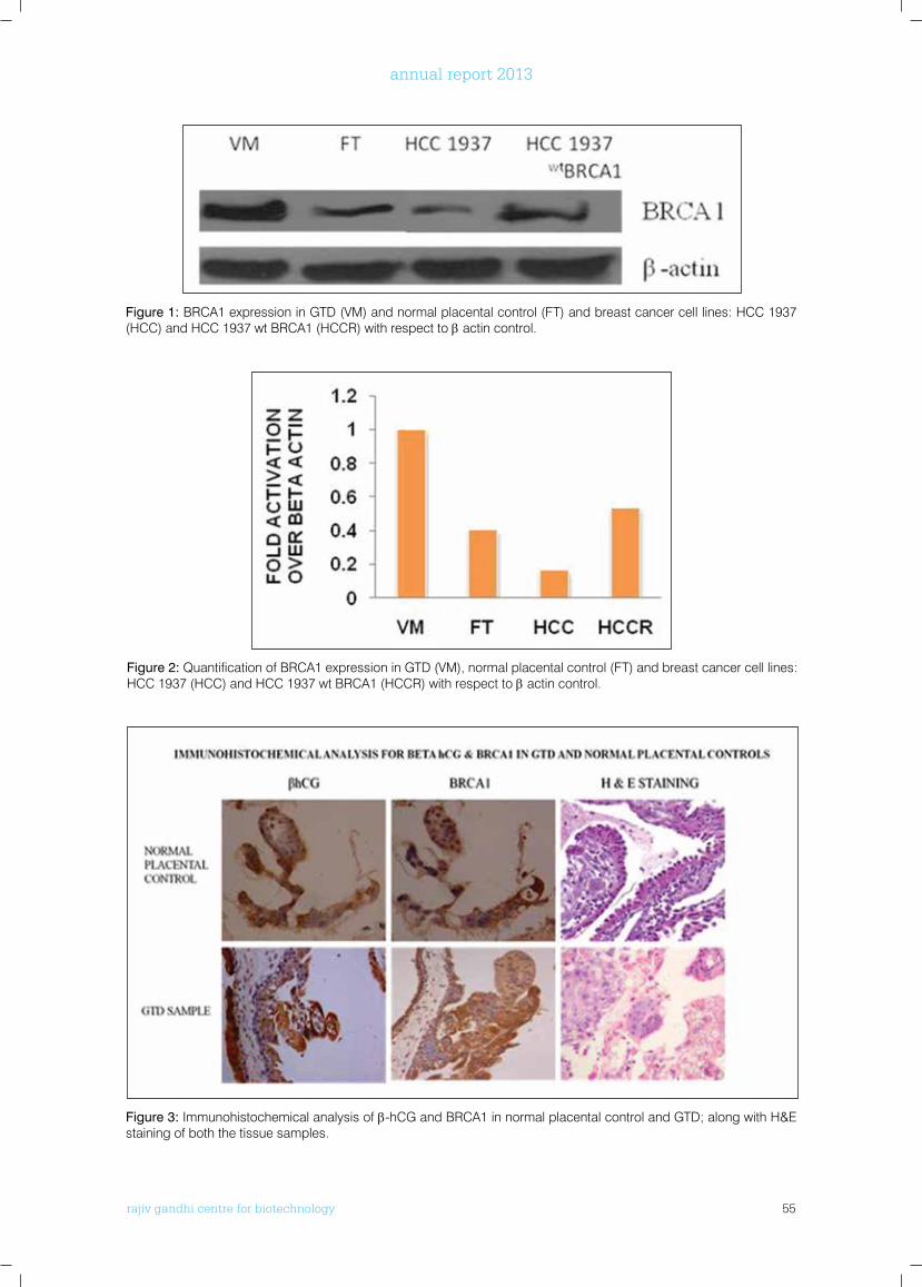
rajiv gandhi centre for biotechnology 55
annual report 2013
Figure 1: BRCA1 expression in GTD (VM) and normal placental control (FT) and breast cancer cell lines: HCC 1937 (HCC) and HCC 1937 wt BRCA1 (HCCR) with respect to β actin control.
Figure 2: Quantification of BRCA1 expression in GTD (VM), normal placental control (FT) and breast cancer cell lines: HCC 1937 (HCC) and HCC 1937 wt BRCA1 (HCCR) with respect to β actin control.
Figure 3: Immunohistochemical analysis of β-hCG and BRCA1 in normal placental control and GTD; along with H&E staining of both the tissue samples.
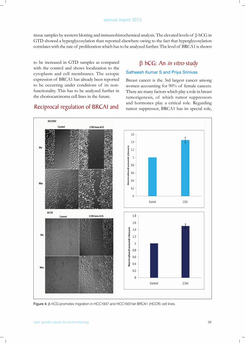
rajiv gandhi centre for biotechnology 56
annual report 2013
to be increased in GTD samples as compared with the control and shows localization to the cytoplasm and cell membranes. The ectopic expression of BRCA1 has already been reported to be occurring under conditions of its non-functionality. This has to be analyzed further in the choriocarcinoma cell lines in the future.
Reciprocal regulation of bRCA1 and
β hCG: An in vitro studySatheesh Kumar S and Priya Srinivas
Breast cancer is the 3rd largest cancer among women accounting for 90% of female cancers. There are many factors which play a role in breast tumorigenesis, of which tumor suppressors and hormones play a critical role. Regarding tumor suppressor, BRCA1 has its special role,
tissuesamplesbywesternblottingandimmunohistochemicalanalysis.Theelevatedlevelsof β-hCGinGTD showed a hyperglycosylation than reported elsewhere owing to the fact that hyperglycosylation correlates with the rate of proliferation which has to be analyzed further. The level of BRCA1 is shown
Figure 4: β-hCG promotes migration in HCC1937 and HCC1937/wt BRCA1 (HCCR) cell lines.
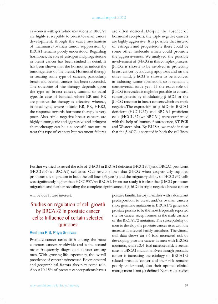
rajiv gandhi centre for biotechnology 57
annual report 2013
as women with germ-line mutations in BRCA1 are highly susceptible to breast/ovarian cancer development, though the exact mechanism of mammary/ovarian tumor suppression by BRCA1 remains poorly understood. Regarding hormones, the role of estrogen and progesterone in breast cancer has been studied in detail. It has been shown that the hormones induce the tumorigenesis of the breast. Hormonal therapy in treating some type of cancers, particularly breast and ovarian cancers has been successful. The outcome of the therapy depends upon the type of breast cancer, luminal or basal type. In case of luminal, where ER and PR are positive the therapy is effective, whereas, in basal type, where it lacks ER, PR, HER2, the response towards hormone therapy is very poor. Also triple negative breast cancers are highly tumorigenic and aggressive and stringent chemotherapy can be a successful measure to treat this type of cancers but treatment failures
are often noticed. Despite the absence of hormonal receptors, the triple negative cancers are highly aggressive. It is possible that instead of estrogen and progesterone there could be some other molecule which could promote the aggressiveness. We analyzed the possible involvementof β-hCGinthiscomplexprocess.β-hCG is shown tobe involved inprotectingbreast cancer by inducing apoptosis and on the other hand, β-hCG is shown to be involvedin inducing tumor formation, so it remains a controversial issue yet . If the exact role of β-hCGisrevealeditmightbepossibletocontroltumorigenesis bymodulating β-hCG or theβ-hCGreceptorinbreastcancerswhicharetriplenegative.Theexpressionof β-hCGinBRCA1deficient (HCC1937) andBRCA1 proficientcells (HCC1937/wtBRCA1)were confirmedwiththehelpof immunoflourescence,RT-PCRand Western blot. By ELISA, we made it clear thattheβ-hCGissecretedinboththecelllines.
Furtherwetriedtorevealtheroleof β-hCGinBRCA1deficient(HCC1937)andBRCA1proficient(HCC1937/wtBRCA1) cell lines.Our results shows that β-hCGwhen exogenously suppliedpromotes the migration in both the cell lines (Figure 4) and the migratory ability of HCC1937 cells wassignificantlyhigherthanHCC1937/wtBRCA1.Fromourstudy,itisclearthatβ-hCGpromotesmigrationandfurtherrevealingthecompletesignificanceof β-hCGintriplenegativebreastcancer
will be our future interest.
Studies on regulation of cell growth by bRCA1/2 in prostate cancer
cells: Influence of certain selected quinones
Reshma R S, Priya Srinivas
Prostate cancer ranks fifth among the most common cancers worldwide and is the second most frequently diagnosed cancer among men. With growing life expectancy, the overall prevalence of cancer has increased. Environmental and geographical factors also play some role. About 10-15% of prostate cancer patients have a
positive familial history. Families with a dominant predisposition to breast and/or ovarian cancers show germline mutations in BRCA1/2 genes and prostate persists to be the most frequently reported site for cancer receptiveness in the male carriers of the BRCA1/2 mutation. The susceptibility of men to develop the prostate cancer rises with the increase in affected family members. The clinical trial data shows an 8.6-fold increased risk of developing prostate cancer in men with BRCA2 mutation, while a 3.4- fold increased risk is seen in case of BRCA1 mutation. Even though prostatic cancer is increasing the etiology of BRCA1/2 related prostatic cancer and their risk remains poorly understood, also their optimal clinical managementisnotyetdefined.Numerousstudies
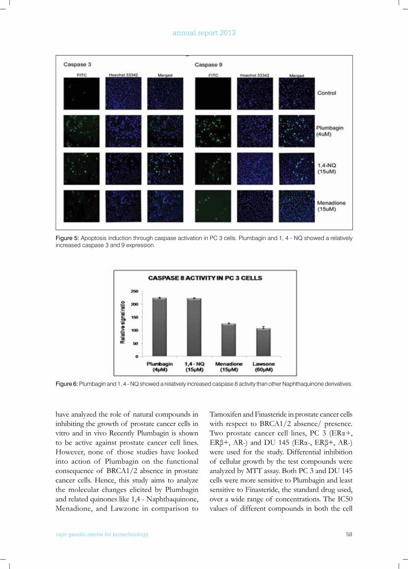
rajiv gandhi centre for biotechnology 58
annual report 2013
Figure 5: Apoptosis induction through caspase activation in PC 3 cells. Plumbagin and 1, 4 - NQ showed a relatively increased caspase 3 and 9 expression.
Figure 6: Plumbagin and 1, 4 - NQ showed a relatively increased caspase 8 activity than other Naphthaquinone derivatives.
have analyzed the role of natural compounds in inhibiting the growth of prostate cancer cells in vitro and in vivo Recently Plumbagin is shown to be active against prostate cancer cell lines. However, none of those studies have looked into action of Plumbagin on the functional consequence of BRCA1/2 absence in prostate cancer cells. Hence, this study aims to analyze the molecular changes elicited by Plumbagin and related quinones like 1,4 - Naphthaquinone, Menadione, and Lawzone in comparison to
Tamoxifen and Finasteride in prostate cancer cells with respect to BRCA1/2 absence/ presence.Twoprostate cancer cell lines, PC 3 (ERα+,ERβ+,AR-)andDU145 (ERα-,ERβ+,AR-)were used for the study. Differential inhibition of cellular growth by the test compounds were analyzed by MTT assay. Both PC 3 and DU 145 cells were more sensitive to Plumbagin and least sensitive to Finasteride, the standard drug used, over a wide range of concentrations. The IC50 values of different compounds in both the cell
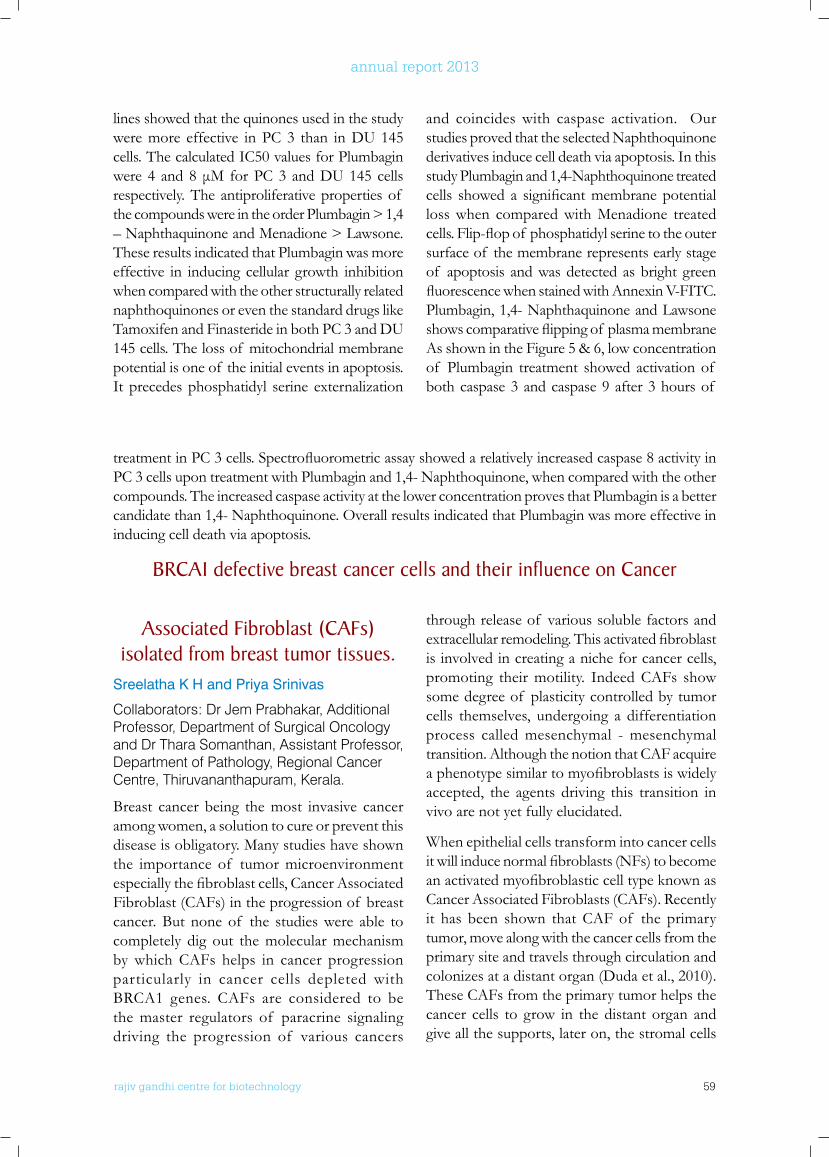
rajiv gandhi centre for biotechnology 59
annual report 2013
Associated fibroblast (CAfs) isolated from breast tumor tissues.
Sreelatha K H and Priya Srinivas
Collaborators: Dr Jem Prabhakar, Additional Professor, Department of Surgical Oncology and Dr Thara Somanthan, Assistant Professor, Department of Pathology, Regional Cancer Centre, Thiruvananthapuram, Kerala.
Breast cancer being the most invasive cancer among women, a solution to cure or prevent this disease is obligatory. Many studies have shown the importance of tumor microenvironment especiallythefibroblastcells,CancerAssociatedFibroblast (CAFs) in the progression of breast cancer. But none of the studies were able to completely dig out the molecular mechanism by which CAFs helps in cancer progression particularly in cancer cells depleted with BRCA1 genes. CAFs are considered to be the master regulators of paracrine signaling driving the progression of various cancers
through release of various soluble factors and extracellularremodeling.Thisactivatedfibroblastis involved in creating a niche for cancer cells, promoting their motility. Indeed CAFs show some degree of plasticity controlled by tumor cells themselves, undergoing a differentiation process called mesenchymal - mesenchymal transition. Although the notion that CAF acquire aphenotypesimilartomyofibroblastsiswidelyaccepted, the agents driving this transition in vivo are not yet fully elucidated.
When epithelial cells transform into cancer cells itwillinducenormalfibroblasts(NFs)tobecomeanactivatedmyofibroblasticcelltypeknownasCancer Associated Fibroblasts (CAFs). Recently it has been shown that CAF of the primary tumor, move along with the cancer cells from the primary site and travels through circulation and colonizes at a distant organ (Duda et al., 2010). These CAFs from the primary tumor helps the cancer cells to grow in the distant organ and give all the supports, later on, the stromal cells
treatmentinPC3cells.Spectrofluorometricassayshowedarelativelyincreasedcaspase8activityinPC 3 cells upon treatment with Plumbagin and 1,4- Naphthoquinone, when compared with the other compounds. The increased caspase activity at the lower concentration proves that Plumbagin is a better candidate than 1,4- Naphthoquinone. Overall results indicated that Plumbagin was more effective in inducing cell death via apoptosis.
bRCA1 defective breast cancer cells and their influence on Cancer
lines showed that the quinones used in the study were more effective in PC 3 than in DU 145 cells. The calculated IC50 values for Plumbagin were4 and8μM forPC3 andDU145 cellsrespectively. The antiproliferative properties of the compounds were in the order Plumbagin > 1,4 – Naphthaquinone and Menadione > Lawsone. These results indicated that Plumbagin was more effective in inducing cellular growth inhibition when compared with the other structurally related naphthoquinones or even the standard drugs like Tamoxifen and Finasteride in both PC 3 and DU 145 cells. The loss of mitochondrial membrane potential is one of the initial events in apoptosis. It precedes phosphatidyl serine externalization
and coincides with caspase activation. Our studies proved that the selected Naphthoquinone derivatives induce cell death via apoptosis. In this study Plumbagin and 1,4-Naphthoquinone treated cells showed a significantmembranepotentialloss when compared with Menadione treated cells.Flip-flopof phosphatidylserinetotheoutersurface of the membrane represents early stage of apoptosis and was detected as bright green fluorescencewhenstainedwithAnnexinV-FITC.Plumbagin, 1,4- Naphthaquinone and Lawsone showscomparativeflippingof plasmamembraneAs shown in the Figure 5 & 6, low concentration of Plumbagin treatment showed activation of both caspase 3 and caspase 9 after 3 hours of
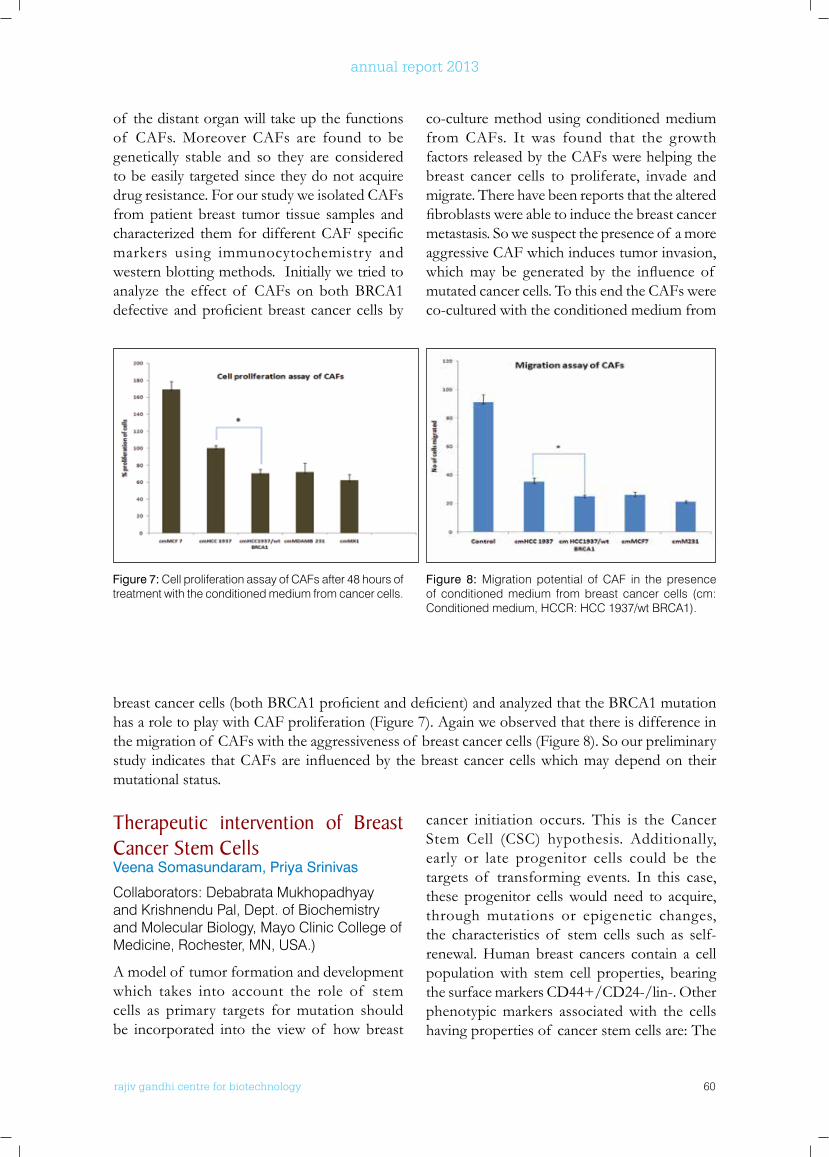
rajiv gandhi centre for biotechnology 60
annual report 2013
of the distant organ will take up the functions of CAFs. Moreover CAFs are found to be genetically stable and so they are considered to be easily targeted since they do not acquire drug resistance. For our study we isolated CAFs from patient breast tumor tissue samples and characterized them fordifferentCAFspecificmarkers using immunocytochemistry and western blotting methods. Initially we tried to analyze the effect of CAFs on both BRCA1 defective andproficientbreast cancer cellsby
co-culture method using conditioned medium from CAFs. It was found that the growth factors released by the CAFs were helping the breast cancer cells to proliferate, invade and migrate. There have been reports that the altered fibroblastswereabletoinducethebreastcancermetastasis. So we suspect the presence of a more aggressive CAF which induces tumor invasion, whichmay be generated by the influence of mutated cancer cells. To this end the CAFs were co-cultured with the conditioned medium from
Figure 7: Cell proliferation assay of CAFs after 48 hours of treatment with the conditioned medium from cancer cells.
Figure 8: Migration potential of CAF in the presence of conditioned medium from breast cancer cells (cm: Conditioned medium, HCCR: HCC 1937/wt BRCA1).
breastcancercells(bothBRCA1proficientanddeficient)andanalyzedthattheBRCA1mutationhas a role to play with CAF proliferation (Figure 7). Again we observed that there is difference in the migration of CAFs with the aggressiveness of breast cancer cells (Figure 8). So our preliminary study indicates thatCAFsare influencedby thebreastcancercellswhichmaydependon theirmutational status.
Therapeutic intervention of breast Cancer Stem CellsVeena Somasundaram, Priya Srinivas
Collaborators: Debabrata Mukhopadhyay and Krishnendu Pal, Dept. of Biochemistry and Molecular Biology, Mayo Clinic College of Medicine, Rochester, MN, USA.)
A model of tumor formation and development which takes into account the role of stem cells as primary targets for mutation should be incorporated into the view of how breast
cancer initiation occurs. This is the Cancer Stem Cell (CSC) hypothesis. Additionally, early or late progenitor cells could be the targets of transforming events. In this case, these progenitor cells would need to acquire, through mutations or epigenetic changes, the characteristics of stem cells such as self-renewal. Human breast cancers contain a cell population with stem cell properties, bearing the surface markers CD44+/CD24-/lin-. Other phenotypic markers associated with the cells having properties of cancer stem cells are: The
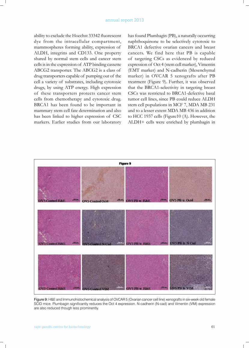
rajiv gandhi centre for biotechnology 61
annual report 2013
Figure 9: H&E and Immunohistochemical analysis of OVCAR 5 (Ovarian cancer cell line) xenografts in six-week old female SCID mice. Plumbagin significantly reduces the Oct 4 expression. N-cadherin (N-cad) and Vimentin (VIM) expression are also reduced though less prominently.
abilitytoexcludetheHoechst33342fluorescentdye from the intracellular compartment, mammospheres forming ability, expression of ALDH, integrins and CD133. One property shared by normal stem cells and cancer stem cells is in the expression of ATP binding cassette ABCG2 transporter. The ABCG2 is a class of drug transporters capable of pumping out of the cell a variety of substrates, including cytotoxic drugs, by using ATP energy. High expression of these transporters protects cancer stem cells from chemotherapy and cytotoxic drug. BRCA1 has been found to be important in mammary stem cell fate determination and also has been linked to higher expression of CSC markers. Earlier studies from our laboratory
has found Plumbagin (PB), a naturally occurring naphthoquinone to be selectively cytotoxic to BRCA1 defective ovarian cancers and breast cancers. We find here that PB is capable of targeting CSCs as evidenced by reduced expression of Oct 4 (stem cell marker), Vimentin (EMT marker) and N-cadherin (Mesenchymal marker) in OVCAR 5 xenografts after PB treatment (Figure 9). Further, it was observed that the BRCA1-selectivity in targeting breast CSCs was restricted to BRCA1-defective basal tumor cell lines, since PB could reduce ALDH stem cell populations in MCF 7, MDA MB 231 and to a lesser extent MDA MB 436 in addition to HCC 1937 cells (Figure10 (A). However, the ALDH+ cells were enriched by plumbagin in
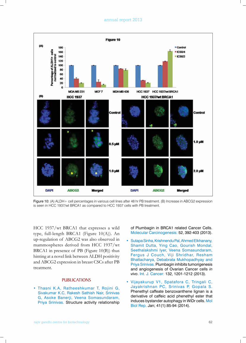
rajiv gandhi centre for biotechnology 62
annual report 2013
HCC 1937/wt BRCA1 that expresses a wild type, full-length BRCA1 (Figure 10(A)). An up-regulation of ABCG2 was also observed in mammospheres derived from HCC 1937/wt BRCA1 in presence of PB (Figure 10(B)) thus hinting at a novel link between ALDH positivity and ABCG2 expression in breast CSCs after PB treatment.
PublICATIonS
• Thasni K.A, Ratheeshkumar T, Rojini G, Sivakumar K.C, Rakesh Sathish Nair, Srinivas G, Asoke Banerji, Veena Somasundaram, Priya Srinivas. Structure activity relationship
of Plumbagin in BRCA1 related Cancer Cells. Molecular Carcinogenesis: 52, 392-403 (2013).
• Sutapa Sinha, Krishnendu Pal, Ahmed Elkhanany, Shamit Dutta, Ying Cao, Gourish Mondal, Seethalakshmi Iyer, Veena Somasundaram, Fergus J Couch, Vij i Shridhar, Resham Bhattacharya, Debabrata Mukhopadhyay and Priya Srinivas. Plumbagin inhibits tumorigenesis and angiogenesis of Ovarian Cancer cells in vivo. Int. J. Cancer: 132, 1201-1212 (2013).
• Vijayakurup V1, Spatafora C, Tringali C, Jayakrishnan PC, Srinivas P, Gopala S. Phenethyl caffeate benzoxanthene lignan is a derivative of caffeic acid phenethyl ester that induces bystander autophagy in WiDr cells. Mol Biol Rep. Jan; 41(1):85-94 (2014).
Figure 10: (A) ALDH+ cell percentages in various cell lines after 48 hr PB treatment. (B) Increase in ABCG2 expression is seen in HCC 1937/wt BRCA1 as compared to HCC 1937 cells with PB treatment.
rajiv gandhi centre for biotechnology 63
annual report 2013
ConfEREnCE PRESEnTATIonS
• Veena Somasundaram, Reshma RS, Sreelatha KH, Revathy, Satheesh Kumar S, Priya Srinivas. Inducers of NO and ROS: Implications in Homologous Recombination Defective Breast Cancer Cells. Poster presented at the Legacy of Nitric Oxide Discovery: Impact on Disease Biology, Rajiv Gandhi Centre for Biotechnology, Kerala, India, November 5th to 6th , 2013.
• S. Satheesh Kumar, Revathy, Sreelatha K.H, Veena Somasundaram and Priya Srinivas. β-hCG: a key molecule in Triple Negative BRCA1-/- Breast Cancer?. Poster presented on 33nd Annual conference on Indian Association of Cancer Research, Kollam 13th to 15th February 2014.
• Revathy, Sreelekha Y, Sreevidya P S, Sreelatha K H, Veena Somasundaram and Priya Srinivas. Etiopathogenesis of Gestational Trophoblastic
Diseases in Indian population. Poster presented at 33rd Annual Convention of Indian Association of Cancer Research, Kollam 13th to 15th February 2014.
• Reshma R S and Priya Srinivas. Identification of genes with altered expression in response to Vitamin K3 analogue in prostate cancer cells. Poster presented at 33rd Annual Convention of Indian Association of Cancer Research, Kollam 13th to 15th February 2014.
rajiv gandhi centre for biotechnology 64
annual report 2013
Cancer Research Program:Laboratory - 6
Ph.D StudentsJazir Haneef Parvathy M. Lakshmi M. L
r a j i v g a n d h i c e n t r e f o r b i o t e c h n o l o g y
Sreeja received a Ph.D in Biotechnology from University of Kerala for work on Molecular mechanisms of Estrogen action. She joined RGCB as a scientist in 2001.
S.Sreeja Ph. D Scientist EI
Project FellowHima SithulJuberiya. M. AzeezVini Ravindran Viji Remadevi
Technical Personnel Savitha H.
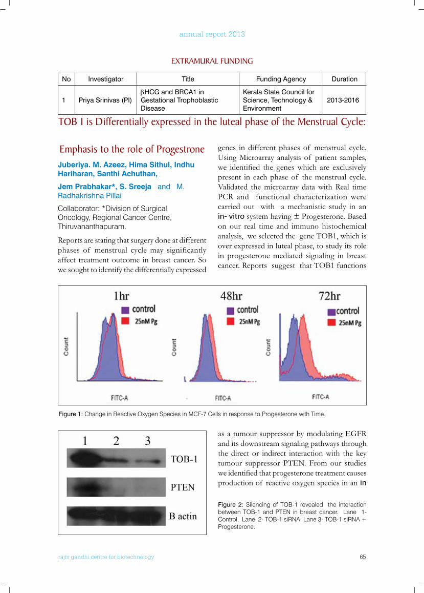
rajiv gandhi centre for biotechnology 65
annual report 2013
Emphasis to the role of Progestrone Juberiya. M. Azeez, Hima Sithul, indhu Hariharan, Santhi Achuthan,
Jem Prabhakar*, S. Sreeja and M. Radhakrishna Pillai
Collaborator: *Division of Surgical Oncology, Regional Cancer Centre, Thiruvananthapuram.
Reports are stating that surgery done at different phases of menstrual cycle may significantly affect treatment outcome in breast cancer. So we sought to identify the differentially expressed
genes in different phases of menstrual cycle. Using Microarray analysis of patient samples, we identified the geneswhich are exclusivelypresent in each phase of the menstrual cycle. Validated the microarray data with Real time PCR and functional characterization were carried out with a mechanistic study in an in- vitro system having ± Progesterone. Based on our real time and immuno histochemical analysis, we selected the gene TOB1, which is over expressed in luteal phase, to study its role in progesterone mediated signaling in breast cancer. Reports suggest that TOB1 functions
ExTRAMuRAl funDInG
No Investigator Title Funding Agency Duration
1 Priya Srinivas (PI)βHCG and BRCA1 in Gestational Trophoblastic Disease
Kerala State Council for Science, Technology & Environment
2013-2016
Tob 1 is Differentially expressed in the luteal phase of the Menstrual Cycle:
Figure 1: Change in Reactive Oxygen Species in MCF-7 Cells in response to Progesterone with Time.
Figure 2: Silencing of TOB-1 revealed the interaction between TOB-1 and PTEN in breast cancer. Lane 1- Control, Lane 2- TOB-1 siRNA, Lane 3- TOB-1 siRNA + Progesterone.
as a tumour suppressor by modulating EGFR and its downstream signaling pathways through the direct or indirect interaction with the key tumour suppressor PTEN. From our studies weidentifiedthatprogesteronetreatmentcausesproduction of reactive oxygen species in an in
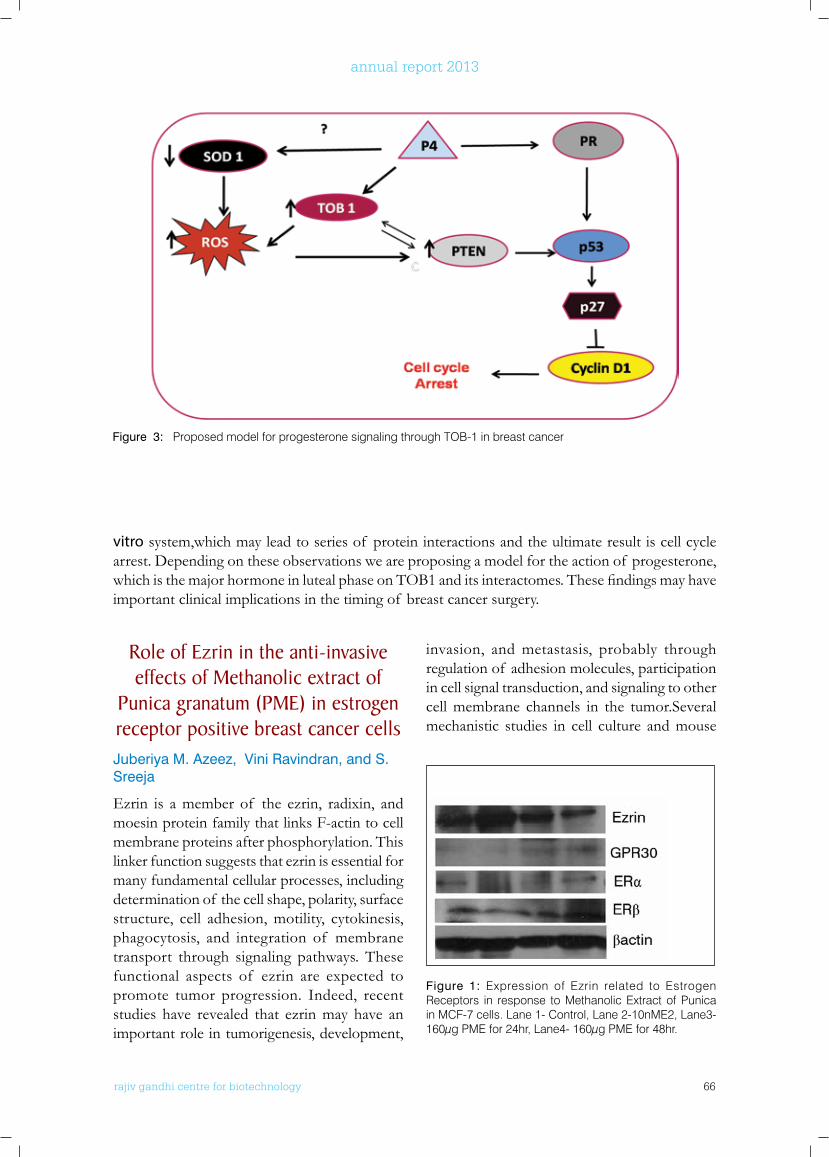
rajiv gandhi centre for biotechnology 66
annual report 2013
Role of Ezrin in the anti-invasive effects of Methanolic extract of
Punica granatum (PME) in estrogen receptor positive breast cancer cellsJuberiya M. Azeez, Vini Ravindran, and S. Sreeja
Ezrin is a member of the ezrin, radixin, and moesin protein family that links F-actin to cell membrane proteins after phosphorylation. This linker function suggests that ezrin is essential for many fundamental cellular processes, including determination of the cell shape, polarity, surface structure, cell adhesion, motility, cytokinesis, phagocytosis, and integration of membrane transport through signaling pathways. These functional aspects of ezrin are expected to promote tumor progression. Indeed, recent studies have revealed that ezrin may have an important role in tumorigenesis, development,
invasion, and metastasis, probably through regulation of adhesion molecules, participation in cell signal transduction, and signaling to other cell membrane channels in the tumor.Several mechanistic studies in cell culture and mouse
Figure 3: Proposed model for progesterone signaling through TOB-1 in breast cancer
vitro system,which may lead to series of protein interactions and the ultimate result is cell cycle arrest. Depending on these observations we are proposing a model for the action of progesterone, whichisthemajorhormoneinlutealphaseonTOB1anditsinteractomes.Thesefindingsmayhaveimportant clinical implications in the timing of breast cancer surgery.
Figure 1: Expression of Ezrin related to Estrogen Receptors in response to Methanolic Extract of Punica in MCF-7 cells. Lane 1- Control, Lane 2-10nME2, Lane3- 160µg PME for 24hr, Lane4- 160µg PME for 48hr.
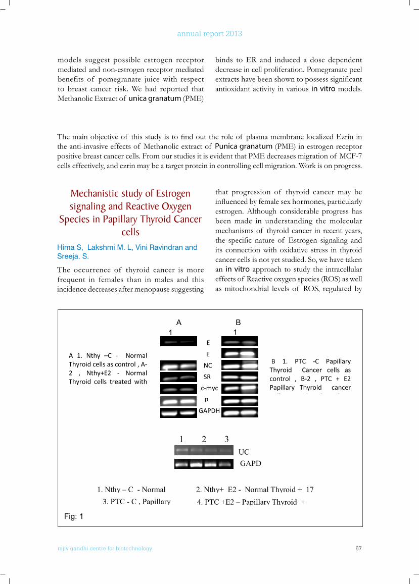
rajiv gandhi centre for biotechnology 67
annual report 2013
Mechanistic study of Estrogen signaling and Reactive oxygen
Species in Papillary Thyroid Cancer cells
Hima S, Lakshmi M. L, Vini Ravindran and Sreeja. S.
The occurrence of thyroid cancer is more frequent in females than in males and this incidence decreases after menopause suggesting
that progression of thyroid cancer may be influencedbyfemalesexhormones,particularlyestrogen. Although considerable progress has been made in understanding the molecular mechanisms of thyroid cancer in recent years, the specificnature of Estrogen signaling andits connection with oxidative stress in thyroid cancer cells is not yet studied. So, we have taken an in vitro approach to study the intracellular effects of Reactive oxygen species (ROS) as well as mitochondrial levels of ROS, regulated by
Themainobjectiveof thisstudyistofindouttheroleof plasmamembranelocalizedEzrininthe anti-invasive effects of Methanolic extract of Punica granatum (PME) in estrogen receptor positive breast cancer cells. From our studies it is evident that PME decreases migration of MCF-7 cells effectively, and ezrin may be a target protein in controlling cell migration. Work is on progress.
73
Fig: 1
Fig 2
ER
ER β
p
UCP 2 β actin
A B 1 2 1 2
B 1. PTC -C Papillary Thyroid Cancer cells as control , B-2 , PTC + E2 Papillary Thyroid cancer Cells treated with 17 beta Estradiol
A 1. Nthy –C - Normal Thyroid cells as control , A-2 , Nthy+E2 - Normal Thyroid cells treated with
E
E
GAPDH
NC SR
c-myc p
B 1. PTC -C Papillary Thyroid Cancer cells as control , B-2 , PTC + E2 Papillary Thyroid cancer C ll d i h 17 b
UC GAPD
1. Nthy – C - Normal
2. Nthy+ E2 - Normal Thyroid + 17 3. PTC - C , Papillary
4. PTC +E2 – Papillary Thyroid +
1 2 3
A B 1
1
A 1. Nthy –C - Normal Thyroid cells as control , A-2 , Nthy+E2 - Normal Thyroid cells treated with 17 b E di l
models suggest possible estrogen receptor mediated and non-estrogen receptor mediated benefits of pomegranate juice with respect to breast cancer risk. We had reported that Methanolic Extract of unica granatum (PME)
binds to ER and induced a dose dependent decrease in cell proliferation. Pomegranate peel extractshavebeenshowntopossesssignificantantioxidant activity in various in vitro models.
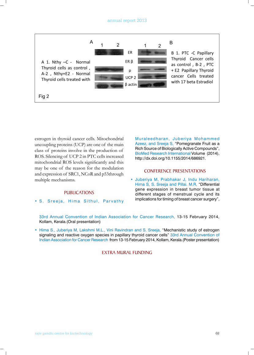
rajiv gandhi centre for biotechnology 68
annual report 2013
73
Fig: 1
Fig 2
ER
ER β
p
UCP 2 β actin
A B 1 2 1 2
B 1. PTC -C Papillary Thyroid Cancer cells as control , B-2 , PTC + E2 Papillary Thyroid cancer Cells treated with 17 beta Estradiol
A 1. Nthy –C - Normal Thyroid cells as control , A-2 , Nthy+E2 - Normal Thyroid cells treated with
E
E
GAPDH
NC SR
c-myc p
B 1. PTC -C Papillary Thyroid Cancer cells as control , B-2 , PTC + E2 Papillary Thyroid cancer C ll d i h 17 b
UC GAPD
1. Nthy – C - Normal
2. Nthy+ E2 - Normal Thyroid + 17 3. PTC - C , Papillary
4. PTC +E2 – Papillary Thyroid +
1 2 3
A B 1
1
A 1. Nthy –C - Normal Thyroid cells as control , A-2 , Nthy+E2 - Normal Thyroid cells treated with 17 b E di l
estrogen in thyroid cancer cells. Mitochondrial uncoupling proteins (UCP) are one of the main class of proteins involve in the production of ROS. Silencing of UCP 2 in PTC cells increased mitochondrialROSlevelssignificantlyandthismay be one of the reason for the modulation and expression of SRC1, NCoR and p53through multiple mechanisms.
PublICATIonS
• S . S r e e j a , H i m a S i t h u l , P a r v a t h y
Mura leedharan, Juber iya Mohammed Azeez, and Sreeja S, “Pomegranate Fruit as a Rich Source of Biologically Active Compounds”, BioMed Research International Volume (2014), http://dx.doi.org/10.1155/2014/686921.
ConfEREnCE PRESEnTATIonS
• Juberiya M, Prabhakar J, Indu Hariharan, Hima S, S. Sreeja and Pillai. M.R, “Differential gene expression in breast tumor tissue at different stages of menstrual cycle and its implications for timing of breast cancer surgery”,
33rd Annual Convention of Indian Association for Cancer Research, 13-15 February 2014, Kollam, Kerala.(Oral presentation)
• Hima S., Juberiya M, Lakshmi M.L., Vini Ravindran and S. Sreeja, “Mechanistic study of estrogen signaling and reactive oxygen species in papillary thyroid cancer cells” 33rd Annual Convention of Indian Association for Cancer Research from 13-15 February 2014, Kollam, Kerala.(Poster presentation)
ExTRA MuRAl funDInG
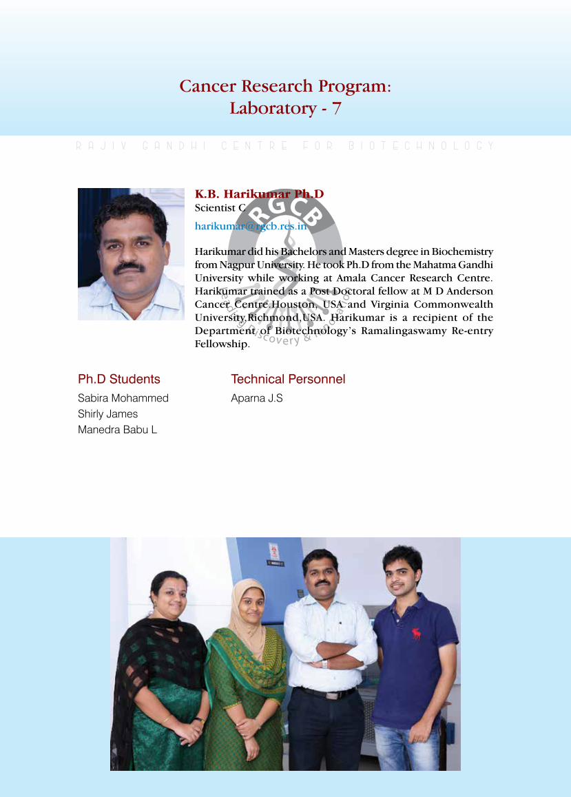
rajiv gandhi centre for biotechnology 69
annual report 2013
Cancer Research Program:Laboratory - 7
Ph.D StudentsSabira MohammedShirly JamesManedra Babu L
r a j i v g a n d h i c e n t r e f o r b i o t e c h n o l o g y
Harikumar did his Bachelors and Masters degree in Biochemistry from Nagpur University. He took Ph.D from the Mahatma Gandhi University while working at Amala Cancer Research Centre. Harikumar trained as a Post Doctoral fellow at M D Anderson Cancer Centre.Houston, USA and Virginia Commonwealth University,Richmond,USA. Harikumar is a recipient of the Department of Biotechnology’s Ramalingaswamy Re-entry Fellowship.
K.B. Harikumar Ph.DScientist C
Technical Personnel Aparna J.S
rajiv gandhi centre for biotechnology 70
annual report 2013
1 S. Sreeja An in-vitro investigation on the role of Estrogen and Reactive Oxygen Species in the invasion of Thyroid Cancer cells with emphasis on TGF - beta signaling
Kerala State Council for Science, Technology and Environment
2013 -2016
Role of sphingolipid system in innate immune responses
and host defense mechanism Sabira Mohammed, K.B.Harikumar
Sphingolipids are a class of bioactive lipid mediators characterized by the presence of a serine head group with one or two fatty acid tails. Sphingosine, one of the major sphingolipids,
Sl. No. Investigator Title Funding Agency Duration
and its phosphorylated product sphingosine 1-phosphate (S1P), have emerged as the modulators of multiple cellular processes, such as cell growth, survival, differentiation, and T cell egress, and have therapeutic potential. We are interested in studying the role of sphingolipids in modulating
innate immune system in response to infections. We are using mouse cytomegalovirus (MCMV) as our model system.
Spice derived nutraceuticals for colorectal cancer chemoprevention
Shirly James, Sabira Mohammed, K.B.Harikumar
Colorectal cancer (CRC) is the third most common cancer in men and second in women worldwide. There is a considerable increase in the pharmacological effects of nutraceuticals for cancer treatment and prevention. Many of thenutraceuticalsarepotentanti-inflammatory
agents. Therefore these molecules have a potential in prevention and treatment of colon cancer. In this project we are mainly focusing on nutraceuticals derived from spices. One of the compound is Cardamonin (2E)-1-(2,4-Dihydroxy-6-methoxyphenyl)-3-phenyl-2-propen-1-one, (8CI); Alpinetin chalcone, (E)-2′,4′-Dihydroxy-6′-methoxy-chalcone).Thecompound belongs to the class of chalcones. We are using a mouse model of colonic inflammation(colitis)forourstudy.Thecolitiscondition is experimentally induced by treating the mouse with dextran sodium sulphate (DSS). This model of mouse colitis mimics the clinical and histological features of IBDs (intestinal
rajiv gandhi centre for biotechnology 71
annual report 2013
bowel diseases) that has a major implication on colorectal cancer. We observed that simultaneous treatmentof cardamoninsignificantlyreducedthe severity of colitis in mice as seen from gain of body weight and increased colon length as compared to controls. Presently we are analysing the molecular mechanism behind the anti-inflammatoryactionof cardamonin.
PublICATIonS
• Harikumar KB, Yester JW, Surace MJ, Oyeniran C, Price MM, Huang WC, Hait NC, Allegood JC, Yamada A, Kong X, Lazear HM, Bhardwaj R, Takabe K, Diamond MS, Luo C, Milstien S, Spiegel S, Kordula T. K63-linked polyubiquitination of transcription factor IRF1 is essential for IL-1-
induced production of chemokines CXCL10 and CCL5. Nature Immunology. 2014;15(3):231-238.
• Bhaskaran-Nair Harikumar K, Hardman R, Aranjani JM, Vimala Raveendran V, Thejass P. Immunomodulatory activity of complementary and alternative medicines. Evidence Based Complementary and Alternative Medicine. 2014;2014:765107. doi:10.1155/2014/765107. Epub 2014 Mar 2
ExTRA-MuRAl funDInG
K.B.Harikumar- Principal investigator No Title Funding Agency Duration
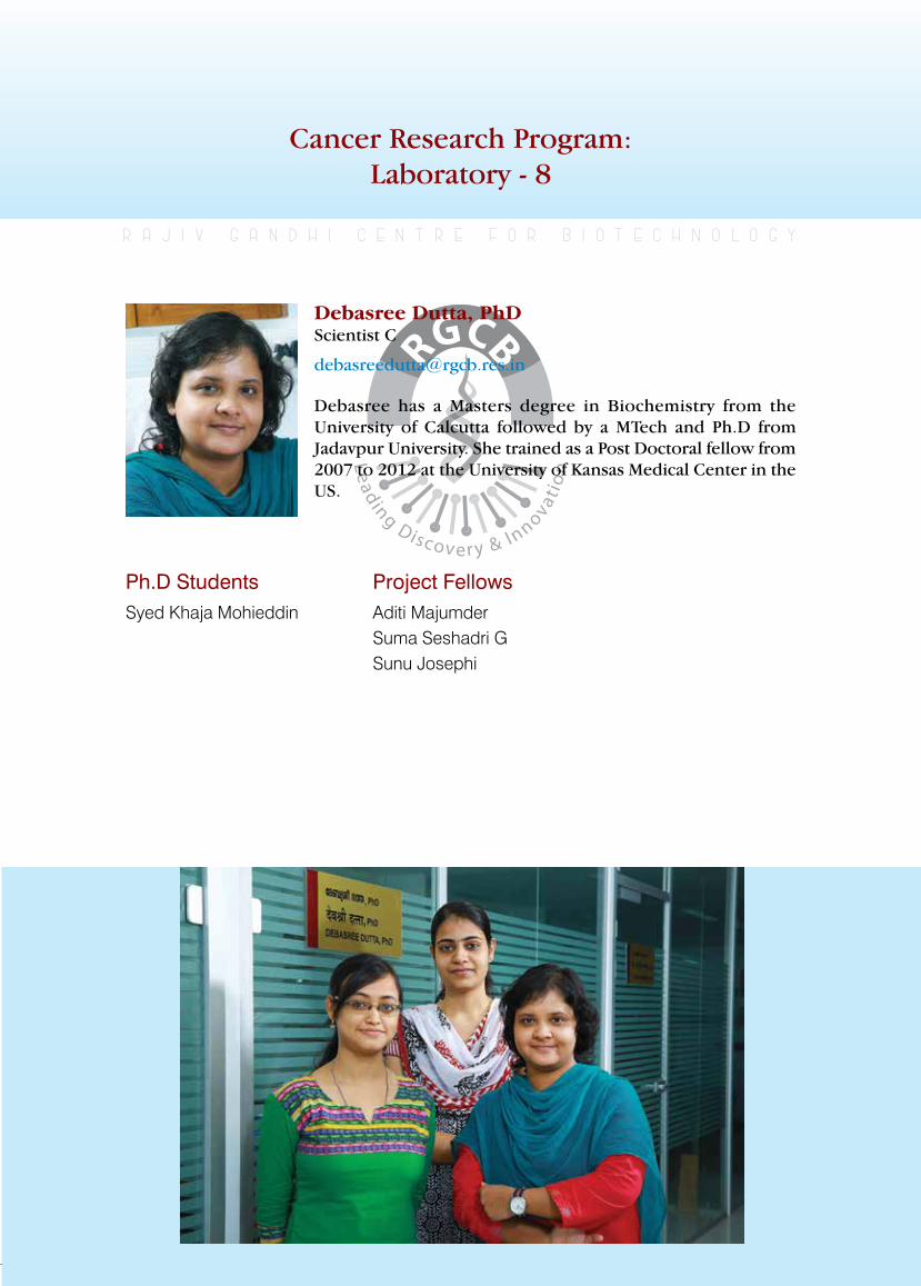
rajiv gandhi centre for biotechnology 72
annual report 2013
Cancer Research Program:Laboratory - 8
Ph.D StudentsSyed Khaja Mohieddin
r a j i v g a n d h i c e n t r e f o r b i o t e c h n o l o g y
Debasree has a Masters degree in Biochemistry from the University of Calcutta followed by a MTech and Ph.D from Jadavpur University. She trained as a Post Doctoral fellow from 2007 to 2012 at the University of Kansas Medical Center in the US.
Debasree Dutta, PhD Scientist C
Project FellowsAditi MajumderSuma Seshadri GSunu Josephi
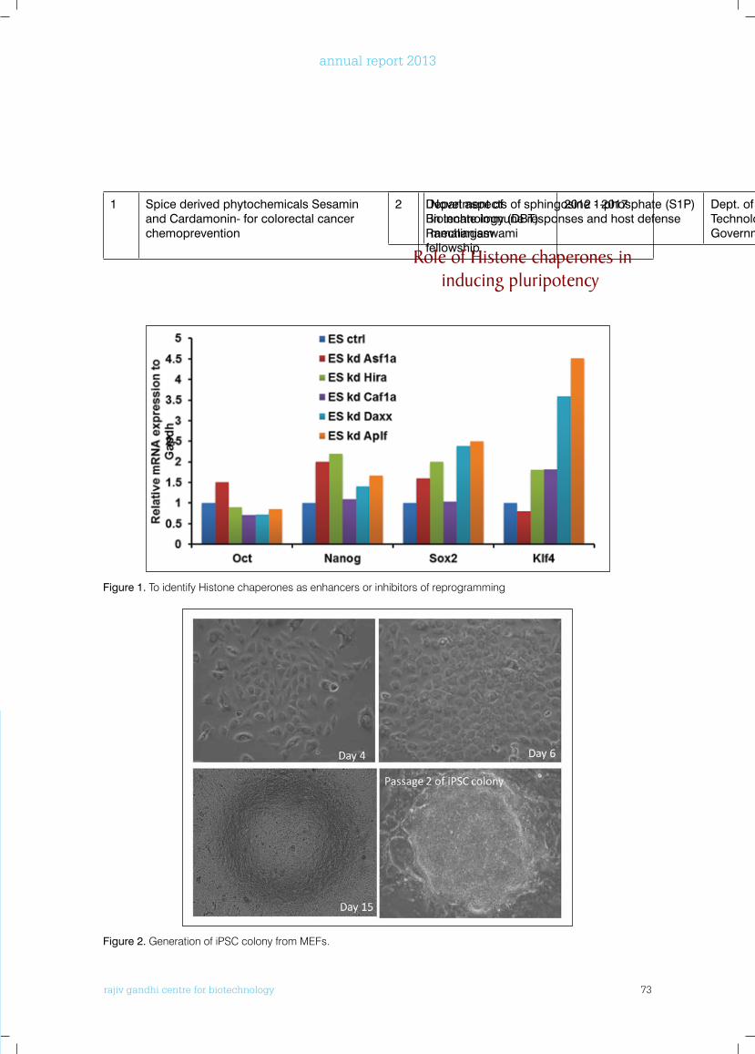
rajiv gandhi centre for biotechnology 73
annual report 2013
1 Spice derived phytochemicals Sesamin and Cardamonin- for colorectal cancer chemoprevention
Department of Biotechnology (DBT) Ramalingaswami fellowship
2012 - 20172 Novel aspects of sphingosine 1-phosphate (S1P) in innate immune responses and host defense mechanism
Dept. of Science and Technology (DST), Government of India
2013 - 2016
Role of Histone chaperones in inducing pluripotency
Figure 1. To identify Histone chaperones as enhancers or inhibitors of reprogramming
Figure 2. Generation of iPSC colony from MEFs.
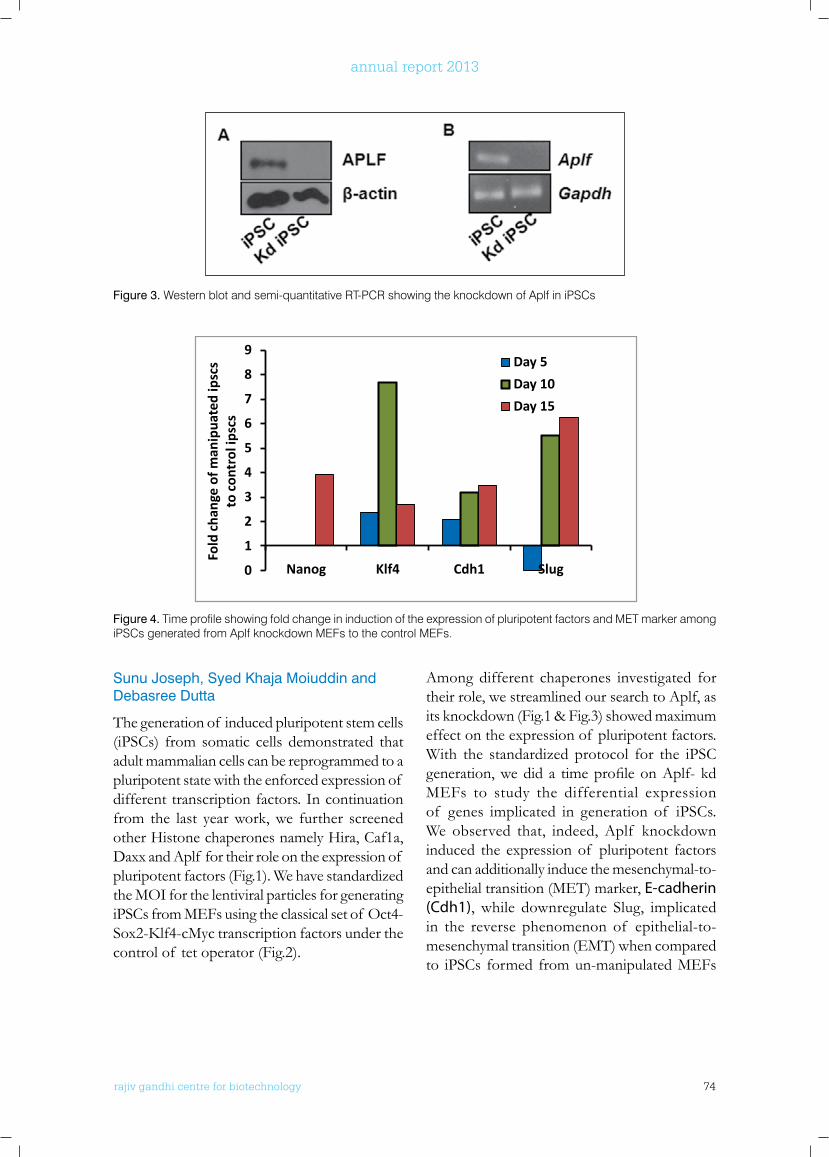
rajiv gandhi centre for biotechnology 74
annual report 2013
Sunu Joseph, Syed Khaja Moiuddin and Debasree Dutta
The generation of induced pluripotent stem cells (iPSCs) from somatic cells demonstrated that adult mammalian cells can be reprogrammed to a pluripotent state with the enforced expression of different transcription factors. In continuation from the last year work, we further screened other Histone chaperones namely Hira, Caf1a, Daxx and Aplf for their role on the expression of pluripotent factors (Fig.1). We have standardized the MOI for the lentiviral particles for generating iPSCs from MEFs using the classical set of Oct4-Sox2-Klf4-cMyctranscriptionfactorsunderthecontrol of tet operator (Fig.2).
Among different chaperones investigated for their role, we streamlined our search to Aplf, as its knockdown (Fig.1 & Fig.3) showed maximum effect on the expression of pluripotent factors. With the standardized protocol for the iPSC generation,wedid a timeprofileonAplf-kdMEFs to study the differential expression of genes implicated in generation of iPSCs. We observed that, indeed, Aplf knockdown induced the expression of pluripotent factors and can additionally induce the mesenchymal-to-epithelial transition (MET) marker, E-cadherin (Cdh1), while downregulate Slug, implicated in the reverse phenomenon of epithelial-to-mesenchymal transition (EMT) when compared to iPSCs formed from un-manipulated MEFs
Figure 3. Western blot and semi-quantitative RT-PCR showing the knockdown of Aplf in iPSCs
0
1
2
3
4
5
6
7
8
9
Nanog Klf4 Cdh1 Slug
Fold
cha
nge
of m
anip
uate
d ip
scs
to c
ontr
ol ip
scs
Day 5
Day 10
Day 15
Merge
RUNX1
HIRA
Nucleus
Figure 4. Time profile showing fold change in induction of the expression of pluripotent factors and MET marker among iPSCs generated from Aplf knockdown MEFs to the control MEFs.
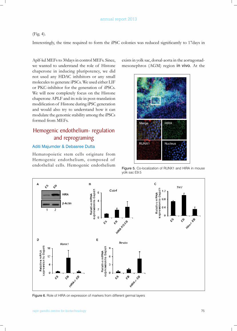
rajiv gandhi centre for biotechnology 75
annual report 2013
Aplf-kd MEFs to 30days in control MEFs. Since, we wanted to understand the role of Histone chaperone in inducing pluripotency, we did not used any HDAC inhibitors or any small molecules to generate iPSCs. We used either LIF orPKC-inhibitorforthegenerationof iPSCs.We will now completely focus on the Histone chaperone APLF and its role in post-translation modificationof HistoneduringiPSCgenerationand would also try to understand how it can modulate the genomic stability among the iPSCs formed from MEFs.
Hemogenic endothelium- regulation and reprograming
Aditi Majumder & Debasree Dutta
Hematopoietic stem cells originate from Hemogenic endothelium, composed of endothelial cells. Hemogenic endothelium
exists in yolk sac, dorsal-aorta in the aortagonad-mesonephros (AGM) region in vivo. At the
(Fig. 4).
Interestingly,thetimerequiredtoformtheiPSCcolonieswasreducedsignificantlyto17daysin
0
1
2
3
4
5
6
7
8
9
Nanog Klf4 Cdh1 Slug
Fold
cha
nge
of m
anip
uate
d ip
scs
to c
ontr
ol ip
scs
Day 5
Day 10
Day 15
Merge
RUNX1
HIRA
Nucleus
Figure 5. Co-localization of RUNX1 and HIRA in mouse yolk sac E9.5
Figure 6. Role of HIRA on expression of markers from different germal layers
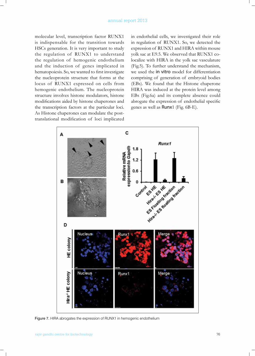
rajiv gandhi centre for biotechnology 76
annual report 2013
molecular level, transcription factor RUNX1 is indispensable for the transition towards HSCs generation. It is very important to study the regulation of RUNX1 to understand the regulation of hemogenic endothelium and the induction of genes implicated in hematopoiesis.So,wewantedtofirstinvestigatethe nucleoprotein structure that forms at the locus of RUNX1 expressed on cells from hemogenic endothelium. The nucleoprotein structure involves histone modulators, histone modificationsaidedbyhistonechaperonesandthe transcription factors at the particular loci. As Histone chaperones can modulate the post-translationalmodification of loci implicated
in endothelial cells, we investigated their role in regulation of RUNX1. So, we detected the expression of RUNX1 and HIRA within mouse yolk sac at E9.5. We observed that RUNX1 co-localize with HIRA in the yolk sac vasculature (Fig.5). To further understand the mechanism, we used the in vitro model for differentiation comprising of generation of embryoid bodies (EBs). We found that the Histone chaperone HIRA was induced at the protein level among EBs (Fig.6a) and its complete absence could abrogatetheexpressionof endothelialspecificgenes as well as Runx1 (Fig. 6B-E).
Figure 7. HIRA abrogates the expression of RUNX1 in hemogenic endothelium
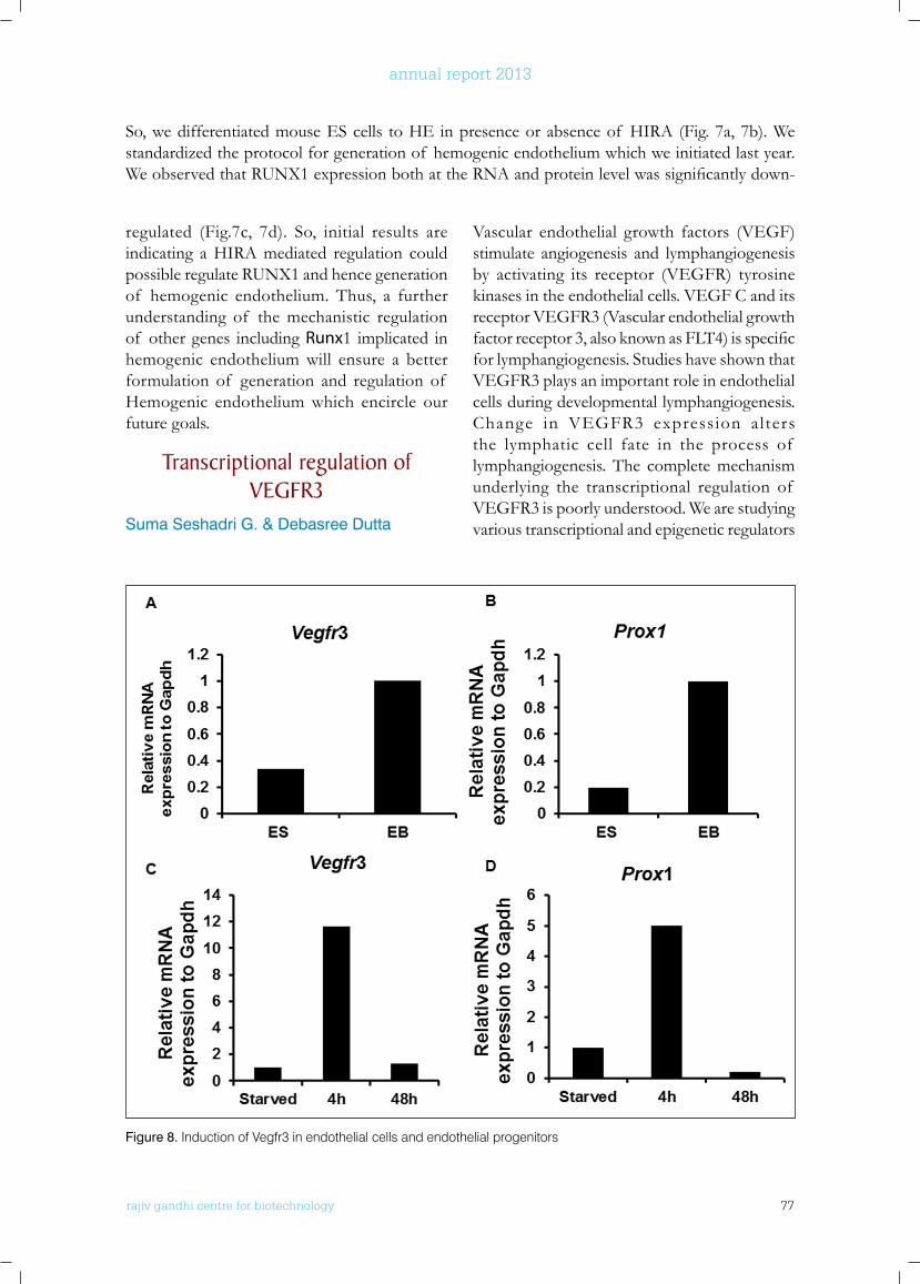
rajiv gandhi centre for biotechnology 77
annual report 2013
regulated (Fig.7c, 7d). So, initial results are indicating a HIRA mediated regulation could possible regulate RUNX1 and hence generation of hemogenic endothelium. Thus, a further understanding of the mechanistic regulation of other genes including Runx1 implicated in hemogenic endothelium will ensure a better formulation of generation and regulation of Hemogenic endothelium which encircle our future goals.
Transcriptional regulation of VEGfR3
Suma Seshadri G. & Debasree Dutta
So, we differentiated mouse ES cells to HE in presence or absence of HIRA (Fig. 7a, 7b). We standardized the protocol for generation of hemogenic endothelium which we initiated last year. WeobservedthatRUNX1expressionbothattheRNAandproteinlevelwassignificantlydown-
Vascular endothelial growth factors (VEGF) stimulate angiogenesis and lymphangiogenesis by activating its receptor (VEGFR) tyrosine kinases in the endothelial cells. VEGF C and its receptor VEGFR3 (Vascular endothelial growth factorreceptor3,alsoknownasFLT4)isspecificfor lymphangiogenesis. Studies have shown that VEGFR3 plays an important role in endothelial cells during developmental lymphangiogenesis. Change in VEGFR3 expression alters the lymphatic cell fate in the process of lymphangiogenesis. The complete mechanism underlying the transcriptional regulation of VEGFR3 is poorly understood. We are studying various transcriptional and epigenetic regulators
Figure 8. Induction of Vegfr3 in endothelial cells and endothelial progenitors
rajiv gandhi centre for biotechnology 78
annual report 2013
involved in the regulation of VEGFR3 in case of lymphangiogenesis and angiogenesis. For this, we constituted two model systems to study the regulation of VEGFR3. We differentiated the ES cells toEBs using an endothelial specificserum to generate endothelial progenitors. We observed an increased expression of VEGFR3 in ES differentiated cells both at the RNA and protein level (Fig.8a). So, in the next step, we
wanted to study how VEGFR3 was regulated by VEGFC, involved in lymphangiogenesis, in endothelial cells. We cultured mouse yolk sac endothelial cells (YSECs) for our study. A time course analysis of YSECs in presence of VEGFC showed a maximal increase in the mRNA and protein level of VEGFR3 at around 4h (Fig.8c). We then detected the relative expression pattern for transcription factors
induced during a similar time point in YSECs as well as in differentiated ES or EBs. We found that Prox1 was predominantly induced under all these conditions (Fig. 8b, 8d). Then, we identifiedwhichsignalingpathwayiscriticalforthe induction of VEGFR3 in endothelial cells. Hence, we measured the induction of Vegfr3 in presenceof inhibitorsof NOTCH,ERKandTGF-βsignaling.Weobservedthatoninhibitionof ERK signaling, expressionof Vegfr3 and
Prox1 were downregulated at the mRNA level (not shown). We will further investigate the function of Prox1 and the nucleoprotein complex being formed at the Vegfr3 locus in lymphangiogenesis and angiogenesis.
PublICATIonS
• Dutta D. (2013). Signaling pathways dictating pluripotency in Embryonic Stem cells. Int J Dev Biol. 57(9-10): 667-75
• Rajendran G*, Dutta D*, Hong J, Paul A, Saha B, Mahato B, Ray S, Home P, Ganguly A, Weiss ML, Paul S (2013). Inhibition of protein kinase C signaling maintains rat embryonic stem cell pluripotency. J Biol Chem. 288(34): 24351 - 62. [* Equal contribution]
ConfEREnCE PRESEnTATIonS
• Annual meeting of “International Society for Stem Cell Research” (ISSCR) 2013 at Boston, USA (12th June- 15th June).
AwARDS
rajiv gandhi centre for biotechnology 79
annual report 2013
Cancer Research Program:Laboratory - 9
r a j i v g a n d h i c e n t r e f o r b i o t e c h n o l o g y
Radhakrishna Pillai joined RGCB in 2005 moving from the Regional Cancer Centre at Trivandrum where he was Professor of Molecular Medicine. Dr. Pillai is a Fellow of the Royal College of Pathologists, London, the National Academy of Medical Sciences, India, the National Academy of Sciences, India and the Indian Academy of Sciences.
Professor M Radhakrishna Pillai FRCPath, [email protected]
• International travel grant from Department of Biotechnology, Government of India for attending ISSCR conference at Boston, USA.
ExTRA-MuRAl funDInG
No Title Funding Agency Duration
1 Role of Histone chaperones in inducing pluripotency
Department of Biotechnology,Government of India.
2012-2015
2 Transcriptional regulation of VEGFR 3 Council of Scientific & Industrial Research, Government of India.
2013-2016
3 Hemogenic endothelium- regulation and reprograming
Deptartment of Science and Technology, Government of India.
2013-2016
Cancer Biology Computational BiologyHuman Papillomavirus Research Facility
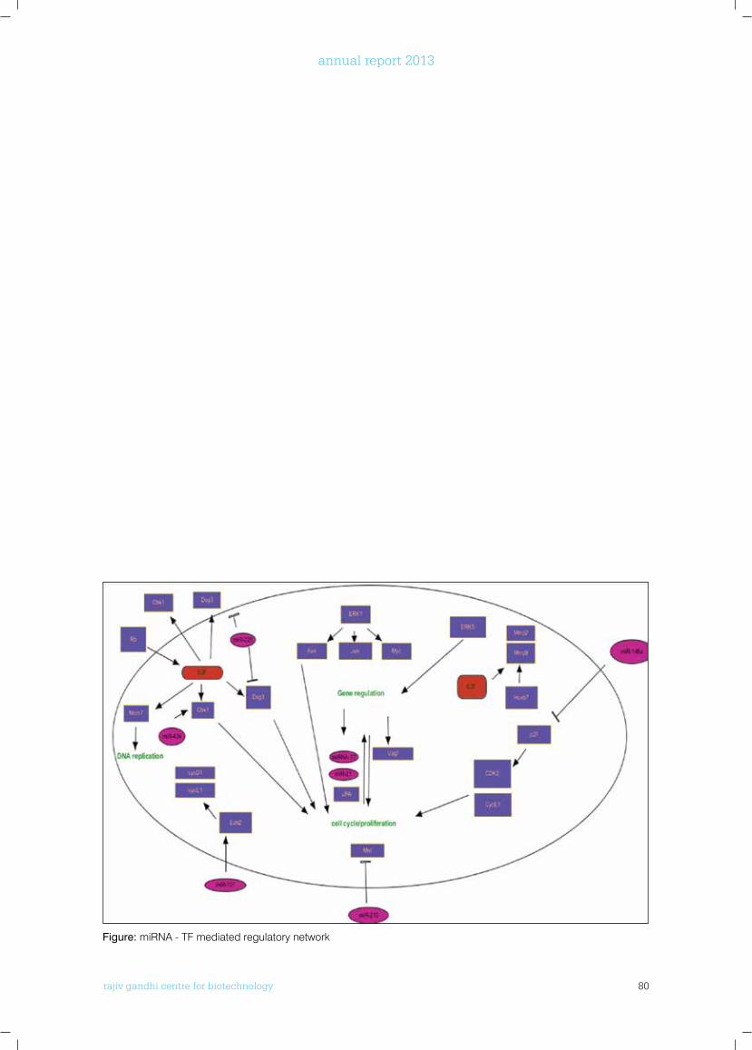
rajiv gandhi centre for biotechnology 80
annual report 2013
Figure: miRNA - TF mediated regulatory network
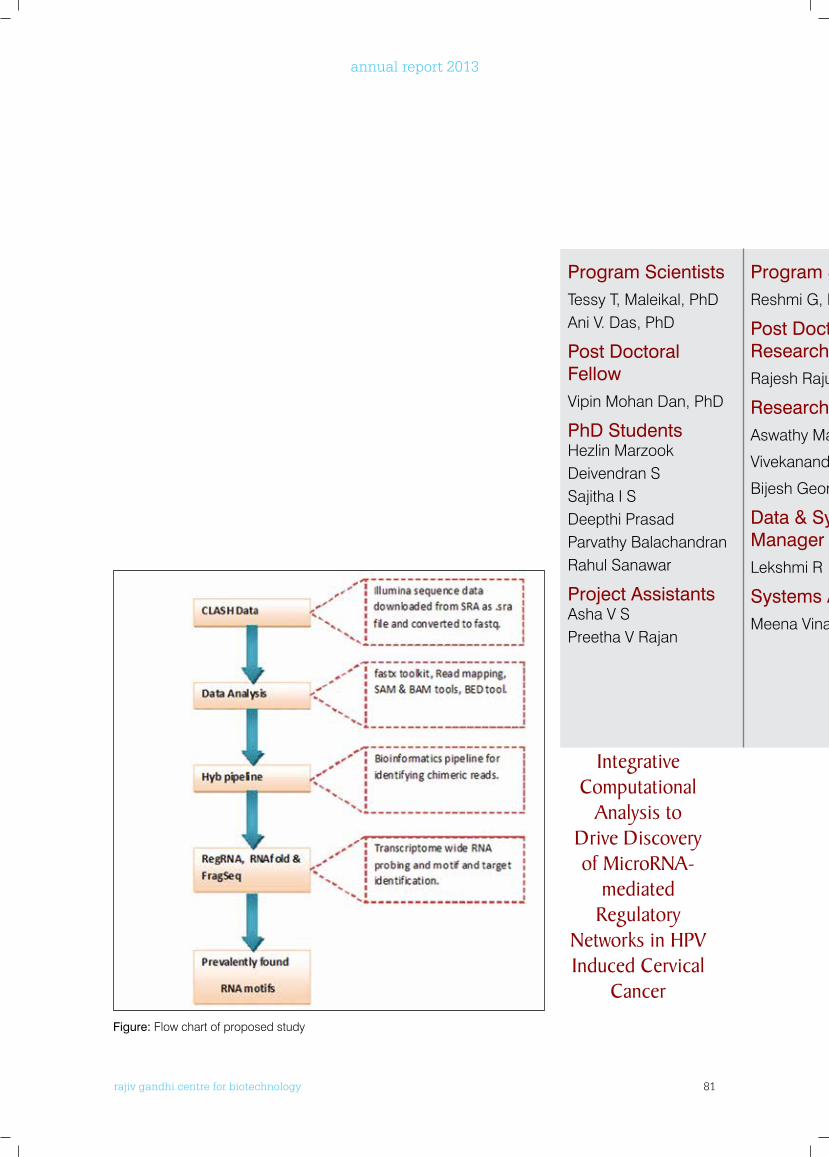
rajiv gandhi centre for biotechnology 81
annual report 2013
Program ScientistsTessy T, Maleikal, PhDAni V. Das, PhD
Post Doctoral FellowVipin Mohan Dan, PhD
PhD Students Hezlin MarzookDeivendran SSajitha I SDeepthi PrasadParvathy BalachandranRahul Sanawar
Project AssistantsAsha V SPreetha V Rajan
Program ScientistReshmi G, PhD
Post Doctoral Research AssociateRajesh Raju, PhD
Research FellowsAswathy Mary Paul
Vivekanand A
Bijesh George
Data & Systems Manager Lekshmi R
Systems AnalystMeena Vinay
Post Doctoral Fellows
Srinivas K P, PhDParamita Palit, PhD
PhD StudentsSumithra ShankarJanki Mohan Babu VPriya R Prabhu
Research FellowJayalekshmi D
Data ManagerSubha Sankaran
Technical AssociateAnurup KG
Technical AssistantsRintu VargheseEdwin S
Project AssistantsKannan T RAiswarya R SJinu Austin
Integrative Computational
Analysis to Drive Discovery of MicroRnA-
mediated Regulatory
networks in HPV Induced Cervical
Cancer
Figure: Flow chart of proposed study
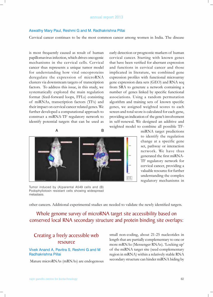
rajiv gandhi centre for biotechnology 82
annual report 2013
is most frequently caused as result of human papillomavirus infection, which drives oncogenic mechanisms in the cervical cells. Cervical cancer thus represents a unique tumor model for understanding how viral oncoproteins deregulate the expression of microRNA clusters via downstream targets of transcription factors. To address this issue, in this study, we systematically explored the main regulation format (feed-forward loops, FFLs) consisting of miRNAs, transcription factors (TFs) and their impact on cervical cancer related genes. We further developed a computational approach to construct a miRNA-TF regulatory network to identify potential targets that can be used as
early detection or prognostic markers of human cervical cancer. Starting with known genes thathavebeenverifiedforaberrantexpressionand functions in cervical cancer and those implicated in literature, we combined gene expressionprofileswithfunctionalmicroarraygene expression data sets (GEO) and RNA seq from SRA to generate a network containing a numberof geneslinkedbyspecificfunctionalassociations. Using a random permutation algorithmand trainingsetsof knownspecificgenes, we assigned weighted scores to each screen and total score is calculated for each gene, providing an indication of the gene’s involvement in self-renewal. We designed an additive and weighted model to combine all possible TF-
miRNA target predictions to identify the regulation change at a specific gene set, pathway or interaction network. We have thus generatedthefirstmiRNA-TF regulatory network for cervical cancer, providing a valuable resource for further understanding the complex regulatory mechanisms in
Tumor induced by (A)parental A549 cells and (B) Podophyllotoxin resistant cells showing widespread metastasis.
Aswathy Mary Paul, Reshmi G and M. Radhakrishna Pillai
Cervical cancer continues to be the most common cancer among women in India. The disease
A B
othercancers.Additionalexperimentalstudiesareneededtovalidatethenewlyidentifiedtargets.
whole genome survey of microRnA target site accessibility based on conserved local RnA secondary structure and protein binding site overlaps:
Creating a freely accessible web resource
Vivek Anand A, Pavitra S, Reshmi G and M Radhakrishna Pillai
Mature microRNAs (mRNAs) are endogenous
small non-coding, about 21-25 nucleotides in length that are partially complementary to one or more mRNAs (Messenger RNAs). ‘Locking up’ of the miRNA target site (seed complementary region in mRNA) within a relatively stable RNA secondary structure can hinder miRNA biding by
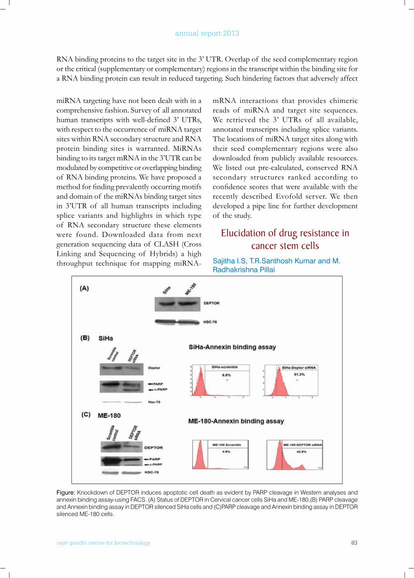
rajiv gandhi centre for biotechnology 83
annual report 2013
miRNA targeting have not been dealt with in a comprehensive fashion. Survey of all annotated human transcriptswithwell-defined3’UTRs,with respect to the occurrence of miRNA target sites within RNA secondary structure and RNA protein binding sites is warranted. MiRNAs binding to its target mRNA in the 3’UTR can be modulated by competitive or overlapping binding of RNA binding proteins. We have proposed a methodforfindingprevalentlyoccurringmotifsand domain of the miRNAs binding target sites in 3’UTR of all human transcripts including splice variants and highlights in which type of RNA secondary structure these elements were found. Downloaded data from next generation sequencing data of CLASH (Cross Linking and Sequencing of Hybrids) a high throughput technique for mapping miRNA-
mRNA interactions that provides chimeric reads of miRNA and target site sequences. We retrieved the 3’ UTRs of all available, annotated transcripts including splice variants. The locations of miRNA target sites along with their seed complementary regions were also downloaded from publicly available resources. We listed out pre-calculated, conserved RNA secondary structures ranked according to confidencescoresthatwereavailablewiththerecently described Evofold server. We then developed a pipe line for further development of the study.
Elucidation of drug resistance in cancer stem cells
Sajitha I.S, T.R.Santhosh Kumar and M. Radhakrishna Pillai
RNA binding proteins to the target site in the 3’ UTR. Overlap of the seed complementary region or the critical (supplementary or complementary) regions in the transcript within the binding site for a RNA binding protein can result in reduced targeting. Such hindering factors that adversely affect
Figure: Knockdown of DEPTOR induces apoptotic cell death as evident by PARP cleavage in Western analyses and annexin binding assay-using FACS. (A) Status of DEPTOR in Cervical cancer cells SiHa and ME-180,(B) PARP cleavage and Annexin binding assay in DEPTOR silenced SiHa cells and (C)PARP cleavage and Annexin binding assay in DEPTOR silenced ME-180 cells.
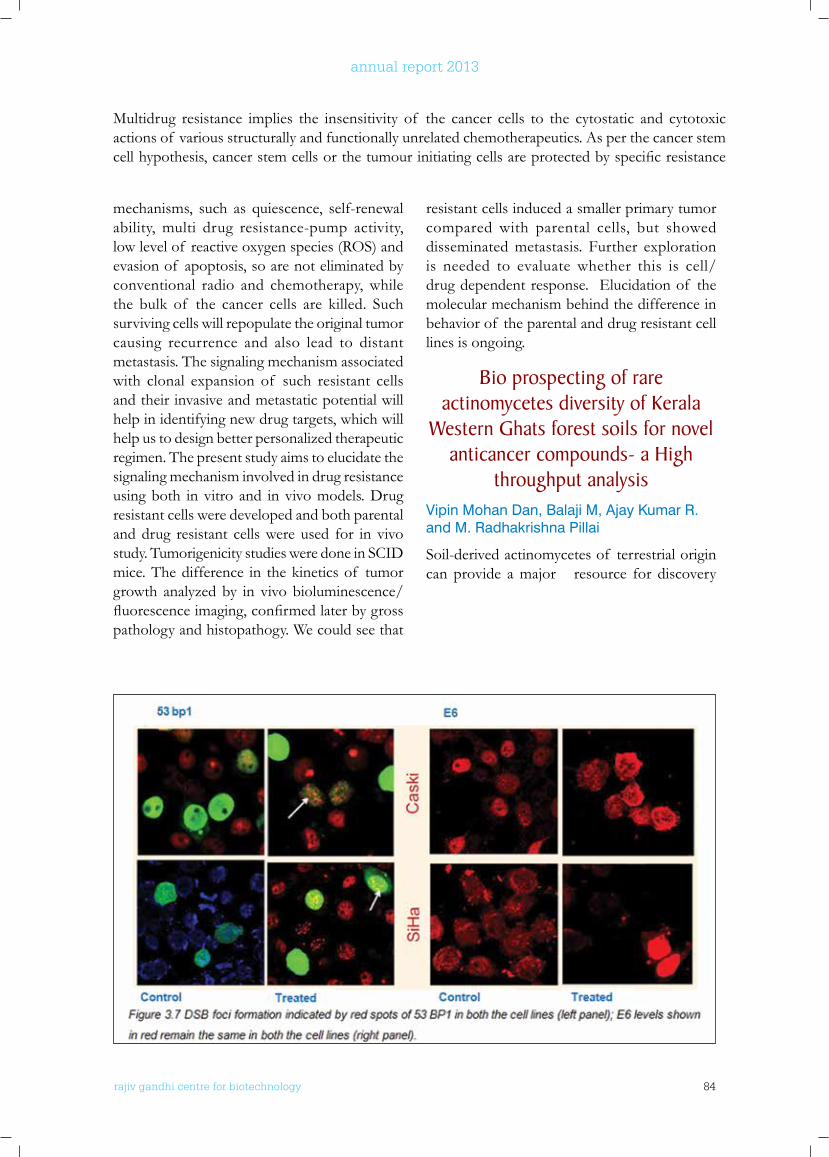
rajiv gandhi centre for biotechnology 84
annual report 2013
mechanisms, such as quiescence, self-renewal ability, multi drug resistance-pump activity, low level of reactive oxygen species (ROS) and evasion of apoptosis, so are not eliminated by conventional radio and chemotherapy, while the bulk of the cancer cells are killed. Such surviving cells will repopulate the original tumor causing recurrence and also lead to distant metastasis. The signaling mechanism associated with clonal expansion of such resistant cells and their invasive and metastatic potential will help in identifying new drug targets, which will help us to design better personalized therapeutic regimen. The present study aims to elucidate the signaling mechanism involved in drug resistance using both in vitro and in vivo models. Drug resistant cells were developed and both parental and drug resistant cells were used for in vivo study. Tumorigenicity studies were done in SCID mice. The difference in the kinetics of tumor growth analyzed by in vivo bioluminescence/ fluorescenceimaging,confirmedlaterbygrosspathology and histopathogy. We could see that
resistant cells induced a smaller primary tumor compared with parental cells, but showed disseminated metastasis. Further exploration is needed to evaluate whether this is cell/ drug dependent response. Elucidation of the molecular mechanism behind the difference in behavior of the parental and drug resistant cell lines is ongoing.
bio prospecting of rare actinomycetes diversity of kerala
western Ghats forest soils for novel anticancer compounds- a High
throughput analysisVipin Mohan Dan, Balaji M, Ajay Kumar R. and M. Radhakrishna Pillai
Soil-derived actinomycetes of terrestrial origin can provide a major resource for discovery
Multidrug resistance implies the insensitivity of the cancer cells to the cytostatic and cytotoxic actions of various structurally and functionally unrelated chemotherapeutics. As per the cancer stem cellhypothesis,cancerstemcellsorthetumourinitiatingcellsareprotectedbyspecificresistance
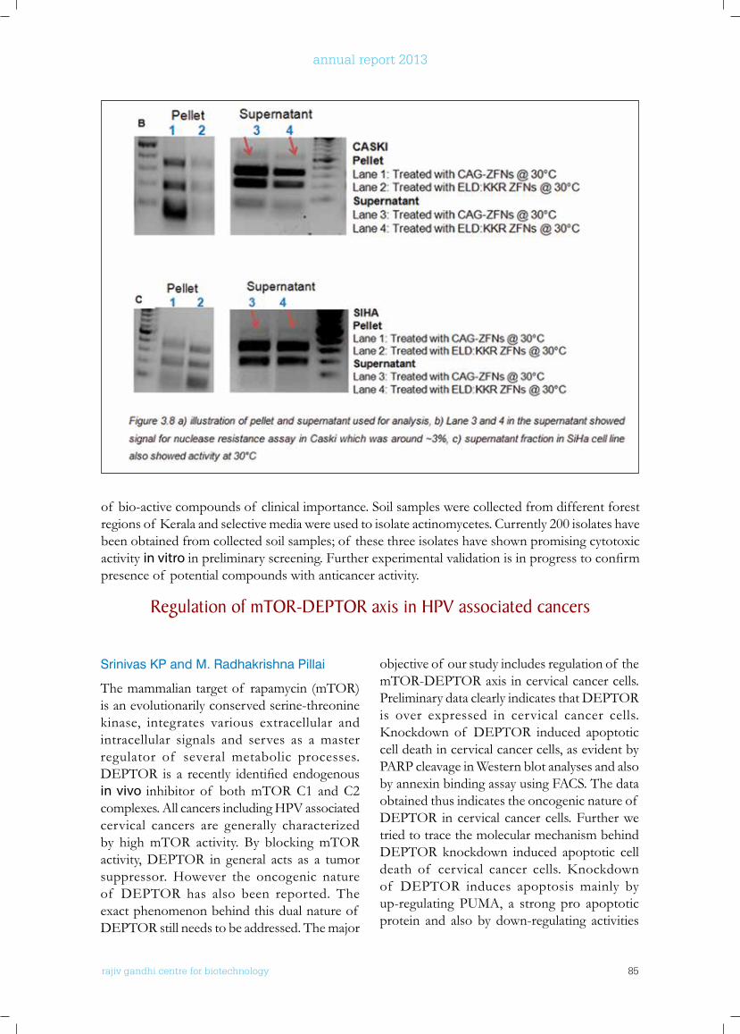
rajiv gandhi centre for biotechnology 85
annual report 2013
Srinivas KP and M. Radhakrishna Pillai
The mammalian target of rapamycin (mTOR) is an evolutionarily conserved serine-threonine kinase, integrates various extracellular and intracellular signals and serves as a master regulator of several metabolic processes. DEPTOR is a recently identifiedendogenousin vivo inhibitor of both mTOR C1 and C2 complexes. All cancers including HPV associated cervical cancers are generally characterized by high mTOR activity. By blocking mTOR activity, DEPTOR in general acts as a tumor suppressor. However the oncogenic nature of DEPTOR has also been reported. The exact phenomenon behind this dual nature of DEPTOR still needs to be addressed. The major
objective of our study includes regulation of the mTOR-DEPTOR axis in cervical cancer cells. Preliminary data clearly indicates that DEPTOR is over expressed in cervical cancer cells. Knockdownof DEPTOR induced apoptoticcell death in cervical cancer cells, as evident by PARP cleavage in Western blot analyses and also by annexin binding assay using FACS. The data obtained thus indicates the oncogenic nature of DEPTOR in cervical cancer cells. Further we tried to trace the molecular mechanism behind DEPTOR knockdown induced apoptotic cell death of cervical cancer cells. Knockdownof DEPTOR induces apoptosis mainly by up-regulating PUMA, a strong pro apoptotic protein and also by down-regulating activities
of bio-active compounds of clinical importance. Soil samples were collected from different forest regionsof Keralaandselectivemediawereusedtoisolateactinomycetes.Currently200isolateshavebeen obtained from collected soil samples; of these three isolates have shown promising cytotoxic activity in vitro inpreliminaryscreening.Furtherexperimentalvalidationisinprogresstoconfirmpresence of potential compounds with anticancer activity.
Regulation of mToR-DEPToR axis in HPV associated cancers
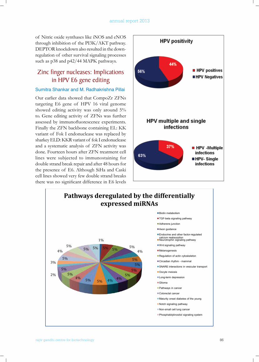
rajiv gandhi centre for biotechnology 86
annual report 2013
of Nitric oxide synthases like iNOS and eNOS throughinhibitionof thePI3K/AKTpathway.DEPTOR knockdown also resulted in the down- regulation of other survival signaling processes suchasp38andp42/44MAPKpathways.
Zinc finger nucleases: Implications in HPV E6 gene editing
Sumitra Shankar and M. Radhakrishna Pillai
Our earlier data showed that CompoZr ZFNs targeting E6 gene of HPV 16 viral genome showed editing activity was only around 5% to. Gene editing activity of ZFNs was further assessedby immunofluorescenceexperiments.FinallytheZFNbackbonecontainingEL:KKvariant of Fok I endonuclease was replaced by sharkeyELD:KKRvariantof fokIendonucleaseand a systematic analysis of ZFN activity was done. Fourteen hours after ZFN treatment cell lines were subjected to immunostaining for double strand break repair and after 48 hours for the presence of E6. Although SiHa and Caski cell lines showed very few double strand breaks therewasnosignificantdifferenceinE6levels
94
Randomized trial of two versus three doses of Human Papillomavirus (HPV)
vaccine in India
Janki Mohan Babu, Priya R Prabhu, Jayalekshmi D, Subha Sankaran, Kannan T R, Anurup K G, Rintu T Varghese , Edwin S, Jinu Austin, Ayswarya R S, *R. Sankaranarayanan, *Masimmo Tomassino, *Tarik Gheit **Michael Pawlita, and M. Radhakrishna Pillai
Collaborators:* International Agency for Research on Cancer (IARC), Lyon, France and **German Cancer Research Centre (DKFZ), Heidelberg, Germany
Cancer of the uterine cervix is the third most common cancer among women worldwide, with an estimated 83,195 new cases and 35,673 deaths reported in 2012 and India represents 26.4 % of women dying of cervical cancer globally. Cancer of the cervix is primarily caused by Human Papilloma Virus (HPV) infection. The vaccination trial initiated by International Agency for Research on Cancer (IARC) recruiting 20,000 unmarried girls aged 10-18 years from different geographical
1%
5% 5%5%
4%
5%5%
5%5%
4%4%5%5%4%5%
5%2%
3%5%
4%5%
5% 5%
Pathways deregulated by the differentially expressed miRNAs
Biotin metabolism
TGF-beta signaling pathway
Adherens junction
Axon guidance
Endocrine and other factor-regulated calcium reabsorptionNeurotrophin signaling pathway
Wnt signaling pathway
Melanogenesis
Regulation of actin cytoskeleton
Circadian rhythm - mammal
SNARE interactions in vesicular transport
Oocyte meiosis
Long-term depression
Glioma
Pathways in cancer
Colorectal cancer
Maturity onset diabetes of the young
Notch signaling pathway
Non-small cell lung cancer
Phosphatidylinositol signaling system
rajiv gandhi centre for biotechnology 87
annual report 2013
In conclusion we analyzed the ZFN editing of CompoZr ZFNs targeting the E6 gene and found that gene editing albeit occurred at a low rate was not suitable therapeutic knockout, it could be improved by screening for more ZFN designs using platforms such as toolgen, OPEN or CODA. Therefore different combinations of ZFN pairs with suitable spacer region need to befurthervalidatedtoimprovetheefficacyof ZFNs in E6 gene knockout.
biological Significance of micro-RnAs in HPV and HIV Associated
Cervical CancersJanki Mohan Babu, Smita Joshi*, Tarik Gheit**, Massimo Tomassino**, R.Sankaranarayanan** and M. Radhakrishna Pillai
Collaborators: *Hirabai Cowasji Jehangir Medical Research Institute, Pune, India and **International Agency for Research on Cancer, Lyon, France
Human papillomavirus (HPV) and Human immunodeficiency virus (HIV) are the two sexually transmitted infections (STI) with extensive public health impact. Both, HIV and HPV share many common behavioral risk factors and interact in important ways. HIV-infected women have a high prevalence of a broad range of HPV genotypes, multiple concurrent infections and persistent infections which progress at a faster pace to neoplasia as compared HIV negative women. MicroRNAs (miRNAs) are small, non-coding RNA molecules able to regulate expression of target genes post-transcriptionally are associated with regulation of diverse physiological processes including cell differentiation and cell division. A
number of microRNAs have been shown to be intrinsically involved in cancer pathogenesis and progression. Given the importance of microRNAs as key regulators of gene expression and since compared with HIV-negative women, HIV-positive women are more likely to develop cervical cancer, have a worse prognosis and a higher risk of recurrence, studying the miRNA deregulation in these patient subsets is likely to yield molecular insights into carcinogenesis. In spite of having the third largest burden of HIV-infected individuals and one fourth of the global burden of cervical cancer, very few studies have addressed HPV prevalence, genotype distribution and cervical cancer prevention in HIV-infected women in India. In this study we evaluated the HPV prevalence and HPV type distribution in HIV-infected women in Maharashtra, India using a high throughput and highly robust Luminex suspension array platform. The study also focused on identifying the differentially expressed miRNAs in cervical cancer patients infected with only one virus [i.e. either HIV or HPV (Group-1)] and patients infected with both viruses [HIV and HPV]. The pathways regulated by these select set of microRNAs was also identified using a in- silico approach. Our study revealed 44% HPV positivity in 600 HIV positive patients analyzed. We also observed 65% single infections and 35% multiple infections in this study population. HPV 16 and 31 were the most predominant sub-types found in this population. The miRNA arraydataidentified5miRNAswhichincreasedin fold expression when both the viruses HIV and HPV16 were infected in comparison to the healthy population, not infected with any virus. Wealsoidentifiedonemicro-RNAwhichwasdown regulated in the patient subset infected
after treatment with ZFNs. There was low expression at 24 hours. Treated cells in both cell lines showed red spots indicating activation of 53bp1 after 14 hours of transfection (left panel); E6 levels remainedmoreorlessthesameinboththecelllinesaftertreatment(rightpanel)asshowninfigure1.
We also replaced CompoZr Fok I by another variant sharkey. Since treatment at 37 degrees did not produce any uncut band with nuclease resistance assay, we performed a transient cold shock treatment with ZFNs in these cell lines. Both SiHa and Caski cell lines showed around 3% ZFN activity in the supernatant fraction with incubation at 300 C. Figure 2 shows ZFN treatment at 30 degrees Celsius.
rajiv gandhi centre for biotechnology 88
annual report 2013
with only HPV-16, while this micro-RNA was up regulated in when co-infected with both HIV and HPV-16.
Randomized trial of two versus three doses of Human Papillomavirus
(HPV) vaccine in IndiaJanki Mohan Babu, Priya R Prabhu, Jayalekshmi D, Subha Sankaran, Kannan T R, Anurup K G, Rintu T Varghese , Edwin S, Jinu Austin, Ayswarya R S, *R. Sankaranarayanan, *Masimmo Tomassino, *Tarik Gheit **Michael Pawlita, and M. Radhakrishna Pillai
Collaborators: * International Agency for Research on Cancer (IARC), Lyon, France and **German Cancer Research Centre (DKFZ), Heidelberg, Germany
Cancer of the uterine cervix is the third most common cancer among women worldwide, with an estimated 83,195 new cases and 35,673 deaths reported in 2012 and India represents 26.4 % of women dying of cervical cancer globally. Cancer of the cervix is primarily caused by Human Papilloma Virus (HPV) infection. The vaccination trial initiated by International Agency for Research on Cancer (IARC) recruiting 20,000 unmarried girls aged 10-18 years from different geographical regions of India is progressing well with RGCB acting as the central biorepository and national facility for all experimental and cyto-pathological analysis. The quadrivalent vaccine (Gardasil™) containing HPV 16, 18, 6 and 11virus like particles (VLPs) provided free of cost by the manufacturer Merck is being used in the study. After vaccination, blood samples
of the cervix and collection of cervical cells from these volunteers was done 18 months after their marriage or 6 months after delivery and one year intervals thereafter for further confirmationof thevaccineefficacybyrulingout infection due to HPV subtypes included in the vaccine. The participating clinical centres of this study are Tata Memorial Hospital (Mumbai), Nargis Dutt Memorial Cancer Hospital (Barshi), Jehangir Clinical Development Centre (Pune), Christian Fellowship Community Health
dose till 48 months after vaccination. To date, blood collection till 36 months after vaccination and analysis for antibody responses utilizing the Multiplex serology based on Luminex technology has been completed at RGCB. The 48th month collection is in progress at different participating centres. Plasma samples totalling to 6546 were grouped based on the doses of vaccine they received and the window period between each doses and were analysed for their response evaluation. Those who were immunized completely according to protocol (2 & 3 doses) were termed Group 1 where as
Centre (Ambilikai), Gujarat Cancer Research Institute (Ahmedabad), All India Institute of Medical Sciences (New Delhi), MNJ Institute of Oncology and Regional Cancer Centre (Hyderabad) and Cancer Foundation of India (Kolkata).Details of the study participantsand their corresponding samples are being stored and updated in the specially created online database (LabReg) by each participating centre. Participants in the HPV vaccine trial are being followed up for their prolonged
Group 2 were those who were immunization completely, but outside protocol (2 & 3doses). Those who received only a single dose were in Group 3 whilst Group 4 participants received 2 doses at very short window period (Days 1-60). For appropriate experimental validation, 184 samples previously tested in May 2012 were included as bridging panel to this assay.
Preliminary data indicate that the 2-dose schedule is non-inferior to the 3-dose schedule if the lower bound of the 95% CI for the MFI ratio was above 0.5 (2-fold difference). The immunogenicity of the 3-dose (days 1, 60, >180-
were collected for evaluation of immune response at different time points. A speculum examination
antibody response at different time points starting from one month after they receive their last
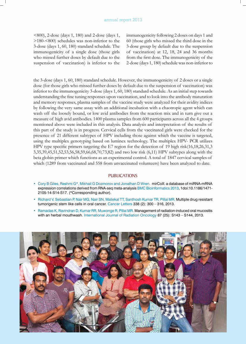
rajiv gandhi centre for biotechnology 89
annual report 2013
<800), 2-dose (days 1, 180) and 2-dose (days 1, >180-<800) schedules was non-inferior to the 3-dose (days 1, 60, 180) standard schedule. The immunogenicity of a single dose (those girls who missed further doses by default due to the suspension of vaccination) is inferior to the
immunogenicity following 2 doses on days 1 and 60 (those girls who missed the third dose in the 3-dose group by default due to the suspension of vaccination) at 12, 18, 24 and 36 months fromthefirstdose.Theimmunogenicityof the2-dose (days 1, 180) schedule was non-inferior to
the 3-dose (days 1, 60, 180) standard schedule. However, the immunogenicity of 2 doses or a single dose (for those girls who missed further doses by default due to the suspension of vaccination) was inferior to the immunogenicity 3-dose (days 1, 60, 180) standard schedule. As an initial step towards understandingthefinetuningresponsesuponvaccination,andtolookintotheantibodymaturationand memory responses, plasma samples of the vaccine study were analyzed for their avidity indices by following the very same assay with an additional incubation with a chaotropic agent which can wash off the loosely bound, or low avid antibodies from the reaction mix and in turn give out a measure of high avid antibodies. 1400 plasma samples from 600 participants across all the 4 groups mentioned above were included in this analysis. Data analysis and interpretation of the results of this part of the study is in progress. Cervical cells from the vaccinated girls were checked for the presence of 21 different subtypes of HPV including those against which the vaccine is targeted, using the multiplex genotyping based on luminex technology. The multiplex HPV- PCR utilizes HPVtypespecificprimerstargetingtheE7regionforthedetectionof 19highrisk(16,18,26,31,33,35,39,45,51,52,53,56,58,59,66,68,70,73,82) and two low risk (6,11) HPV subtypes along with the beta globin primer which functions as an experimental control. A total of 1847 cervical samples of which (1289 from vaccinated and 558 from unvaccinated volunteers) have been analyzed to date.
PublICATIonS
• Cory B Giles, Reshmi G*, Mikhail G Dozmorov and Jonathan D Wren. mirCoX: a database of miRNA-mRNA expression correlations derived from RNA-seq meta-analysis BMC Bioinformatics 2013, 1doi:10.1186/1471-2105-14-S14-S17. (*Corresponding author).
• Richard V, Sebastian P, Nair MG, Nair SN, Maliekal TT, Santhosh Kumar TR, Pillai MR. Multiple drug resistant tumorigenic stem like cells in oral cancer. Cancer Letters 338 (2): 300 – 316, 2013.
• Ramadas K, Ravindran D, Kumar RR, Muwonge R, Pillai MR. Management of radiation-induced oral mucositis with an herbal mouthwash. International Journal of Radiation Oncology 87 (25): S143 – S144, 2013.
rajiv gandhi centre for biotechnology 90
annual report 2013
Cancer Research Program:Laboratory - 9
r a j i v g a n d h i c e n t r e f o r b i o t e c h n o l o g y
Upon the recommendation of the RGCB Governing Body, Professor Rakesh Kumar was invited to join the Rajiv Gandhi Center for Biotechnology as a Visiting Distinguished Professor of Biotechnology to establish and co-direct a joint cancer research program with Professor Radhakrishna Pillai.
Rakesh Kumar, Ph.DVisiting Distinguished Professor
Co-mentored Ph.D StudentsHezlin MarzookDeivendran SParvathy Muraleedharan
• Richard V, Nair MG, Santhosh Kumar TR, Pillai MR. Side population cells as prototype to chemoresistant, tumor-initiating cells. BioMed Research International 2013:517237. doi: 10.1155/2013/517237, 2013.
• Joshi S, Babu JM, Jayalakshmi D, Kulkarni V, Divate U, Muwonge R, Gheit T, Tommasino M, Sankaranarayanan R, Pillai MR. Human papillomavirus infection among human immunodeficiency virus-infected women in Maharashtra, India. Vaccine 32 (2014) 1079– 1085.
ConfEREnCE & woRkSHoP PRESEnTATIonS
• Pavithra S and Aswathy Mary Paul Workshop on Analysing Medical and Health Data Using R at the Achutha Menon Centre for Health Science Studies, Sree Chitra Thirunal Institute for Medical Science and Technology, Trivandrum
• Pavithra S, Aswathy Mary Paul, Reshmi G, M. Radhakrishna Pillai, Whole Human Genome Survey Of Motif And Domain in RNA Secondary Regions of MicroRNA Target sites. Poster presented at Bioinformatica Indica, Trivandrum, January 2014
• Sumitra Sankar and M Radhakrishna Pillai “Synthetic Nucleases: Implications in HPV E6 gene editing”, Presented Poster at IACR (Indian
Association for Cancer Research) conference organized by Rajiv Gandhi Centre for Biotechnology, Feb 13th to 15th 2014.
• Aswathy Mary Paul, Pavithra S Reshmi G and M. Radhakrishna Pillai, ”A computational approach to analyze microRNA mediated Regulatory Networks in HPV induced Cervical Cancer”, Presented Poster at IACR (Indian Association for Cancer Research) conference organized by Rajiv Gandhi Centre for Biotechnology, Feb 13th to 15th 2014.
• Sajitha I.S, T.R.Santhosh Kumar and M.Radhakrishna Pillai.”Generation of cancer stem cell enriched drug resistant cells in response to antitumour drugs”, implication for tumour recurrence. Presented Poster at IACR (Indian Association for Cancer Research) conference organized by Rajiv Gandhi
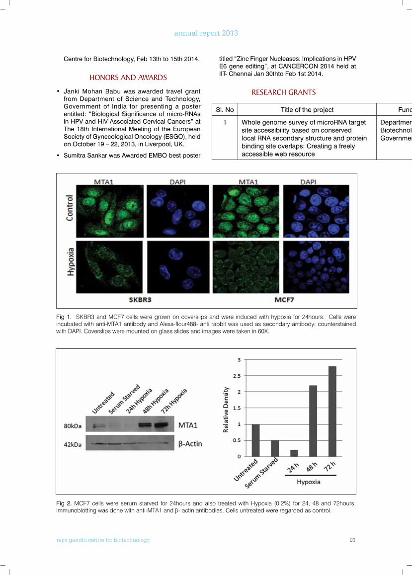
rajiv gandhi centre for biotechnology 91
annual report 2013
Centre for Biotechnology, Feb 13th to 15th 2014.
HonoRS AnD AwARDS
• Janki Mohan Babu was awarded travel grant from Department of Science and Technology, Government of India for presenting a poster entitled: “Biological Significance of micro-RNAs in HPV and HIV Associated Cervical Cancers” at The 18th International Meeting of the European Society of Gynecological Oncology (ESGO), held on October 19 – 22, 2013, in Liverpool, UK.
• Sumitra Sankar was Awarded EMBO best poster
titled “Zinc Finger Nucleases: Implications in HPV E6 gene editing”, at CANCERCON 2014 held at IIT- Chennai Jan 30thto Feb 1st 2014.
RESEARCH GRAnTS
Sl. No Title of the project Funding Agency Duration
1 Whole genome survey of microRNA target site accessibility based on conserved local RNA secondary structure and protein binding site overlaps: Creating a freely accessible web resource
Department of Biotechnology,Government of India.
2012 - 2015
Fig 1. SKBR3 and MCF7 cells were grown on coverslips and were induced with hypoxia for 24hours. Cells were incubated with anti-MTA1 antibody and Alexa-flour488- anti rabbit was used as secondary antibody; counterstained with DAPI. Coverslips were mounted on glass slides and images were taken in 60X.
Fig 2. MCF7 cells were serum starved for 24hours and also treated with Hypoxia (0.2%) for 24, 48 and 72hours. Immunoblotting was done with anti-MTA1 and β- actin antibodies. Cells untreated were regarded as control.
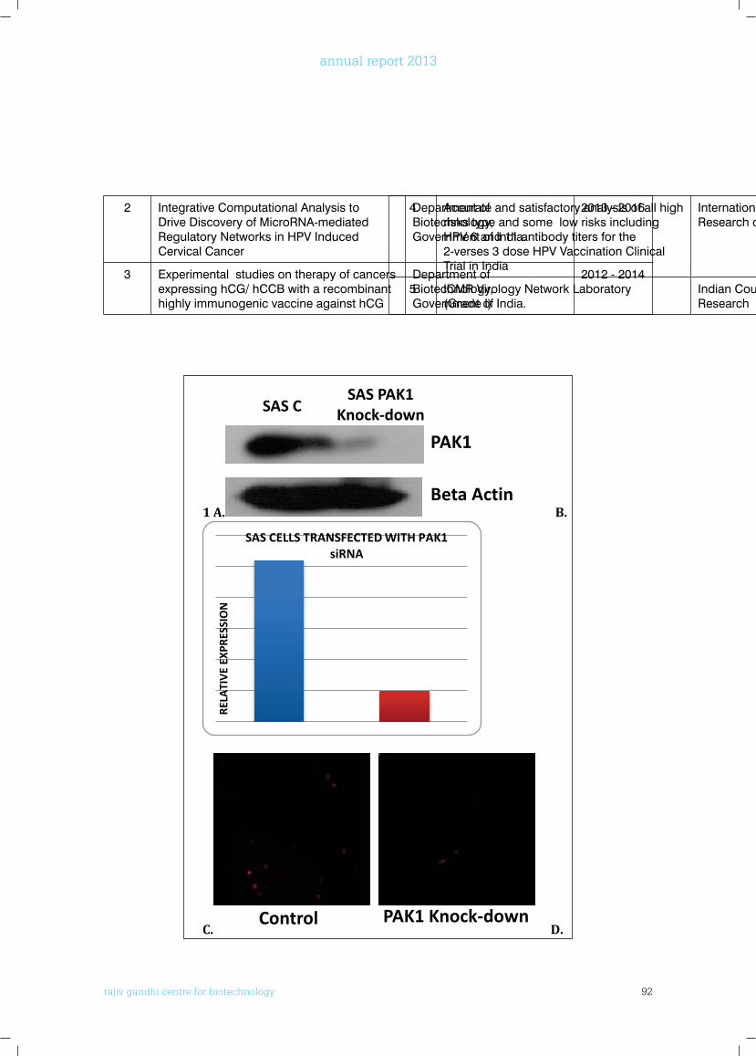
rajiv gandhi centre for biotechnology 92
annual report 2013
2 Integrative Computational Analysis to Drive Discovery of MicroRNA-mediated Regulatory Networks in HPV Induced Cervical Cancer
Department of Biotechnology,Government of India.
2013 - 2016
3 Experimental studies on therapy of cancers expressing hCG/ hCCB with a recombinant highly immunogenic vaccine against hCG
Department of Biotechnology,Government of India.
2012 - 2014
4 Accurate and satisfactory analysis of all high risks type and some low risks including HPV 6 and 11 antibody titers for the 2-verses 3 dose HPV Vaccination Clinical Trial in India
International Agency for Research on Cancer
2009 - 2016
5 ICMR Virology Network Laboratory(Grade I)
Indian Council of Medical Research
2009 - 2016
102
1 A. B.
C. D.
RELA
TIV
E EX
PRES
SIO
N
SAS CELLS TRANSFECTED WITH PAK1 siRNA
AV
ERA
GE
NU
MB
ER O
F C
ELLS
SAS C SAS PAK1 KNOCK-DOWN
Control PAK1 Knock-down
SAS C SAS PAK1
Knock-down
PAK1
Beta Actin
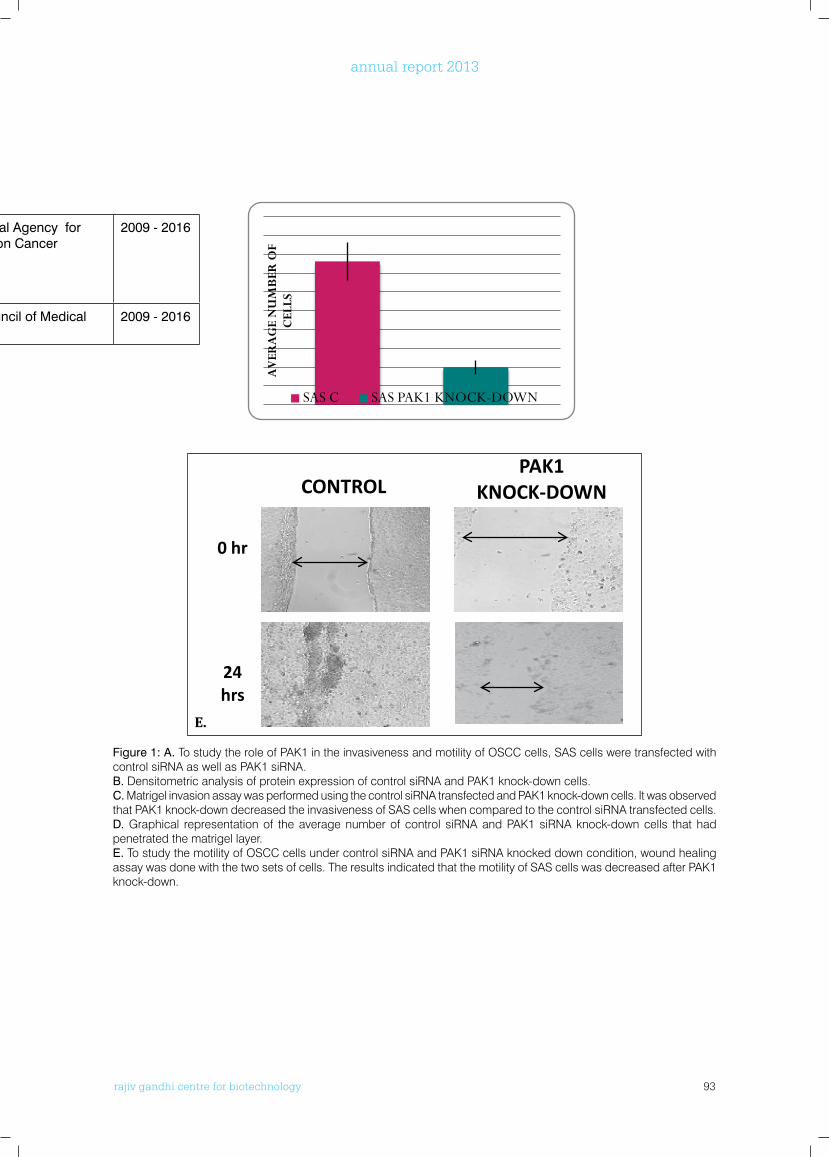
rajiv gandhi centre for biotechnology 93
annual report 2013
4 Accurate and satisfactory analysis of all high risks type and some low risks including HPV 6 and 11 antibody titers for the 2-verses 3 dose HPV Vaccination Clinical Trial in India
International Agency for Research on Cancer
2009 - 2016
5 ICMR Virology Network Laboratory(Grade I)
Indian Council of Medical Research
2009 - 2016
Figure 1: A. To study the role of PAK1 in the invasiveness and motility of OSCC cells, SAS cells were transfected with control siRNA as well as PAK1 siRNA. B. Densitometric analysis of protein expression of control siRNA and PAK1 knock-down cells.C. Matrigel invasion assay was performed using the control siRNA transfected and PAK1 knock-down cells. It was observed that PAK1 knock-down decreased the invasiveness of SAS cells when compared to the control siRNA transfected cells.D. Graphical representation of the average number of control siRNA and PAK1 siRNA knock-down cells that had penetrated the matrigel layer.E. To study the motility of OSCC cells under control siRNA and PAK1 siRNA knocked down condition, wound healing assay was done with the two sets of cells. The results indicated that the motility of SAS cells was decreased after PAK1 knock-down.
102
1 A. B.
C. D.
RELA
TIV
E EX
PRES
SIO
N
SAS CELLS TRANSFECTED WITH PAK1 siRNA
AV
ERA
GE
NU
MB
ER O
F C
ELLS
SAS C SAS PAK1 KNOCK-DOWN
Control PAK1 Knock-down
SAS C SAS PAK1
Knock-down
PAK1
Beta Actin
103
E.
Figure 1: A. To study the role of PAK1 in the invasiveness and motility of OSCC cells, SAS cells were transfected with control siRNA as well as PAK1 siRNA.
B. Densitometric analysis of protein expression of control siRNA and PAK1 knock-down cells.
C. Matrigel invasion assay was performed using the control siRNA transfected and PAK1 knock-down cells. It was observed that PAK1 knock-down decreased the invasiveness of SAS cells when compared to the control siRNA transfected cells.
D. Graphical representation of the average number of control siRNA and PAK1 siRNA knock-down cells that had penetrated the matrigel layer.
E. To study the motility of OSCC cells under control siRNA and PAK1 siRNA knocked down condition, wound healing assay was done with the two sets of cells. The results indicated that the motility of SAS cells was decreased after PAK1 knock-down.
Epigenetic Regulation of Cancer by MTA1
Deivendran S, T. R. Santhoshkumar, M. Radhakrishna Pillai and Rakesh Kumar
The MTA1, a major coregulator overexpressed in many human cancers. Being a dual
coregulator, MTA1 acts both as a corepressor as well as coactivator of its target genes.
Recent studies suggest that MTA1’s corepressive activity depends on its interaction
with the Nucleosome Remodelling complex (NuRD), while coactivator activity is
independent of the NuRD complex and profoundly affected by the NuRF complex. Since
MTA1 contains has a BAH domain and owing to the fact that BAH domain is associated
with the methylation of DNA, we hypothesize a role of MTA1 and/or MTA1-containing
complexes in target gene methylation and consecutively functions of resulting gene
products. Initially we analysed the mRNA expression levels of MTA1 and DNMT3a in the
0 hr
CONTROL PAK1
KNOCK-DOWN
24 hrs
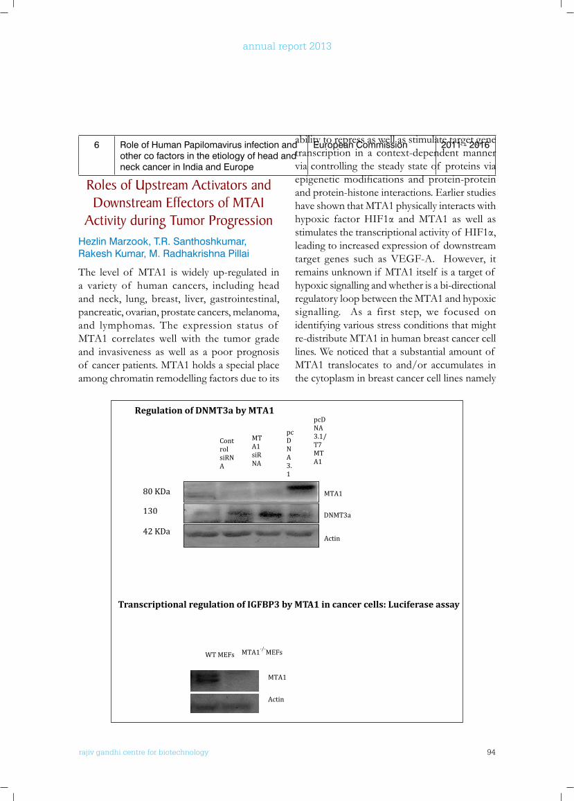
rajiv gandhi centre for biotechnology 94
annual report 2013
6 Role of Human Papilomavirus infection and other co factors in the etiology of head and neck cancer in India and Europe
European Commission 2011 - 2016
Roles of upstream Activators and Downstream Effectors of MTA1
Activity during Tumor ProgressionHezlin Marzook, T.R. Santhoshkumar, Rakesh Kumar, M. Radhakrishna Pillai
The level of MTA1 is widely up-regulated in a variety of human cancers, including head and neck, lung, breast, liver, gastrointestinal, pancreatic, ovarian, prostate cancers, melanoma, and lymphomas. The expression status of MTA1 correlates well with the tumor grade and invasiveness as well as a poor prognosis of cancer patients. MTA1 holds a special place among chromatin remodelling factors due to its
ability to repress as well as stimulate target gene transcription in a context-dependent manner via controlling the steady state of proteins via epigeneticmodifications and protein-proteinand protein-histone interactions. Earlier studies have shown that MTA1 physically interacts with hypoxic factorHIF1α andMTA1 aswell asstimulatesthetranscriptionalactivityof HIF1α,leading to increased expression of downstream target genes such as VEGF-A. However, it remains unknown if MTA1 itself is a target of hypoxic signalling and whether is a bi-directional regulatory loop between the MTA1 and hypoxic signalling. As a first step, we focused on identifying various stress conditions that might re-distribute MTA1 in human breast cancer cell lines. We noticed that a substantial amount of MTA1 translocates to and/or accumulates in the cytoplasm in breast cancer cell lines namely
104
existing databases and found that negative correlation exists between them. Further we
analysed the mRNA levels of MTA1 and DNMT3a in MCF-7 cells and noticed decrease in
the level of DNMT3a upon MTA1 overexpression in MCF7 cells. By western blotting we
showed the increased expression of DNMT3a by MTA1 silencing and decrease in
DNMT3a when MTA1 is overexpressed. Our current results from on-going studies
reveal the presence of DNA methyl transferase 3a (DNMT3a) in MTA1-containing multi-
protein complex by immunoprecipitation in MCF-7 cells. We have also identified IGFBP3
as a new target of MTA1. We could demonstrate regulation of IGFBP-3 both at the level
of transcription and post transcriptional level. Overall, these observations suggest that
status of MTA1 influences cancer cell progression by regulating the expression and
functions of DNMT3a and its targets.
Regulation of DNMT3a by MTA1
Actin
DNMT3a
MTA1
Control siRNA
MTA1 siRNA
pcDNA 3.1
pcDNA 3.1/T7 MTA1
Actin
MTA1
WT MEFs MTA1-/-MEFs
Transcriptional regulation of IGFBP3 by MTA1 in cancer cells: Luciferase assay
80 KDa
130
42 KDa
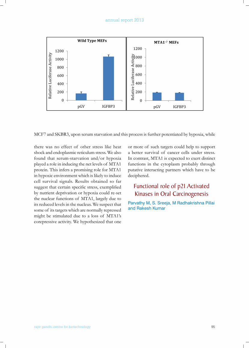
rajiv gandhi centre for biotechnology 95
annual report 2013
105
Conference Presentations
Parvathy Muraleedharan, Sreeja S, MR Pillai and Rakesh Kumar. Importance of Targeting p21 Activated Kinases (PAKs) in Oral Cancer. UAE Cancer Congress 2013, Dubai, October, 2013.
Parvathy Muraleedharan, S. Sreeharshan, M.R. Pillai andRakesh Kumar. P21-activated kinase 1: potential therapeutic target in oral carcinogenesis. 5th EMBO Meeting, Amsterdam, The Netherlands, September 2013.
Hezlin Marzook, Li,D.Q., T. R. SanthoshKumar, M. Radhakrishna Pillai and Rakesh Kumar. Transcriptional Regulation by Metastasis Tumor Antigen 1 on an emerging E3 Ligase Rnf144A. UAE Cancer Congress 2013, 3-5 October 2013, Dubai
Hezlin Marzook, T. R. SanthoshKumar, M. Radhakrishna Pillai and Rakesh Kumar. Revelation of physiological stress response of Metastasis Tumor Antigen 1(MTA1) in breast cancer cells.33rd Annual convention of Indian Association for Cancer Research IACR 2014, 13-15 February 2014, Kollam. Kerala
Deivendran S, T. R. Santhosh Kumar, M. Radhakrishna Pillai and Rakesh Kumar and; Regulation of DNMT3a by Metastasis associated protein-1 (MTA1) in cancer cells; 33rd Annual convention of Indian Association for Cancer Research IACR 2014, 13-15 February 2014, Kollam. Kerala
0
200
400
600
800
1000
1200
pGV IGFBP3
Rela
tive
Luci
fera
se A
ctiv
ity
Wild Type MEFs
0
200
400
600
800
1000
1200
pGV IGFBP3
Rela
tive
Luci
fera
se A
ctiv
ity
MTA1-/- MEFs
there was no effect of other stress like heat shock and endoplasmic reticulum stress. We also found that serum-starvation and/or hypoxia played a role in inducing the net levels of MTA1 protein. This infers a promising role for MTA1 in hypoxic environment which is likely to induce cell survival signals. Results obtained so far suggestthatcertainspecificstress,exemplifiedby nutrient deprivation or hypoxia could re-set the nuclear functions of MTA1, largely due to its reduced levels in the nucleus. We suspect that some of its targets which are normally repressed might be stimulated due to a loss of MTA1’s corepressive activity. We hypothesized that one
or more of such targets could help to support a better survival of cancer cells under stress. In contrast, MTA1 is expected to exert distinct functions in the cytoplasm probably through putative interacting partners which have to be deciphered.
functional role of p21 Activated kinases in oral Carcinogenesis
Parvathy M, S. Sreeja, M Radhakrishna Pillai and Rakesh Kumar
MCF7andSKBR3,uponserumstarvationandthisprocessisfurtherpotentiatedbyhypoxia,while
rajiv gandhi centre for biotechnology 96
annual report 2013
Cancer Research Program:Laboratory - 9
r a j i v g a n d h i c e n t r e f o r b i o t e c h n o l o g y
Tessy Thomas Maliekal has a Ph.D in Biotechnology from University of Kerala in 2002 and did her post-doctoral training at Regional Cancer Centre, Thiruvanananthapuram and then at National Centre for Biological Sciences, Bangalore. She joined RGCB as a Visiting Scientist in 2010.
Tessy Thomas Maliekal
Project FellowsAnnie Agnes SuganyaJiss Maria Louis
p21 activated kinases (PAKs), a family of serine/threonine kinases are involved in numerousphysiological processes like cytoskeletal remodelling, cell survival, motility, angiogenesis, invasion and regulation of cell cycle and mitosis, which are highly important in the context of cancer. So far, sixPAKfamilymembershavebeenidentifiedinmammalsandareclassifiedintotwosub-groups-GroupIPAKs(PAKs1-3)andGroupIIPAKs(PAKs4-6).Thefocusof thecurrentresearchistoelucidatethecontributionof PAK1inoralcancerprogression.Tostudythebiologicalroleof PAK1intheinvasivenessandmotilityof OSCCcells,PAK1wasknockeddownintheOSCCcellline,SAS(Fig 1A and B). The results obtained after matrigel invasion assay (Fig 1C and D), and wound healing
assay(Fig1E)indicatedthatPAK1knock-downcells compromise invasiveness and motility of SAS cells, indicating thatPAK1was involvedin the invasiveness and motility of OSCC cells.
Epigenetic Regulation of Cancer by MTA1
Deivendran S, T. R. Santhoshkumar, M. Radhakrishna Pillai and Rakesh Kumar
The MTA1, a major coregulator overexpressed in many human cancers. Being a dual coregulator,
MTA1 acts both as a corepressor as well as coactivator of its target genes. Recent studies suggest that MTA1’s corepressive activity depends on its interaction with the Nucleosome Remodelling complex (NuRD), while coactivator activity is independent of the NuRD complex and profoundly affected by the NuRF complex. Since MTA1 contains has a BAH domain and owing to the fact that BAH domain is associated with the methylation of DNA, we hypothesize a role of MTA1 and/or MTA1-containing complexes in target gene methylation and consecutively
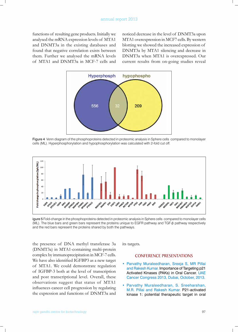
rajiv gandhi centre for biotechnology 97
annual report 2013
functions of resulting gene products. Initially we analysed the mRNA expression levels of MTA1 and DNMT3a in the existing databases and found that negative correlation exists between them. Further we analysed the mRNA levels of MTA1 and DNMT3a in MCF-7 cells and
noticed decrease in the level of DNMT3a upon MTA1 overexpression in MCF7 cells. By western blotting we showed the increased expression of DNMT3a by MTA1 silencing and decrease in DNMT3a when MTA1 is overexpressed. Our current results from on-going studies reveal
Figure 4 Venn diagram of the phosphoproteins detected in proteomic analysis in Sphere cells compared to monolayer cells (ML). Hyperphosphorylation and hypophosphorylation was calculated with 2-fold cut off.
igure 5 Fold-change in the phosphoproteins detected in proteomic analysis in Sphere cells compared to monolayer cells (ML). The blue bars and green bars represent the proteins unique to EGFR pathway and TGF-β pathway respectively and the red bars represent the proteins shared by both the pathways.
the presence of DNA methyl transferase 3a (DNMT3a) in MTA1-containing multi-protein complex by immunoprecipitation in MCF-7 cells. WehavealsoidentifiedIGFBP3asanewtargetof MTA1. We could demonstrate regulation of IGFBP-3 both at the level of transcription and post transcriptional level. Overall, these observations suggest that status of MTA1 influencescancercellprogressionbyregulatingthe expression and functions of DNMT3a and
its targets.
ConfEREnCE PRESEnTATIonS
• Parvathy Muraleedharan, Sreeja S, MR Pillai and Rakesh Kumar. Importance of Targeting p21 Activated Kinases (PAKs) in Oral Cancer. UAE Cancer Congress 2013, Dubai, October, 2013.
• Parvathy Muraleedharan, S. Sreeharshan, M.R. Pillai and Rakesh Kumar. P21-activated kinase 1: potential therapeutic target in oral
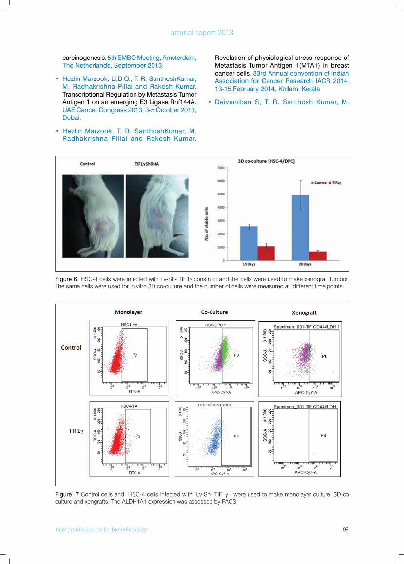
rajiv gandhi centre for biotechnology 98
annual report 2013
carcinogenesis. 5th EMBO Meeting, Amsterdam, The Netherlands, September 2013.
• Hezlin Marzook, Li,D.Q., T. R. SanthoshKumar, M. Radhakrishna Pillai and Rakesh Kumar. Transcriptional Regulation by Metastasis Tumor Antigen 1 on an emerging E3 Ligase Rnf144A. UAE Cancer Congress 2013, 3-5 October 2013, Dubai.
• Hezlin Marzook, T. R. SanthoshKumar, M. Radhakrishna Pillai and Rakesh Kumar.
Revelation of physiological stress response of Metastasis Tumor Antigen 1(MTA1) in breast cancer cells. 33rd Annual convention of Indian Association for Cancer Research IACR 2014, 13-15 February 2014, Kollam. Kerala
• Deivendran S, T. R. Santhosh Kumar, M.
Figure 7 Control cells and HSC-4 cells infected with Lv-Sh- TIF1γ were used to make monolayer culture, 3D-co culture and xengrafts. The ALDH1A1 expression was assessed by FACS
Figure 6 HSC-4 cells were infected with Lv-Sh- TIF1γ construct and the cells were used to make xenograft tumors. The same cells were used for in vitro 3D co-culture and the number of cells were measured at different time points.
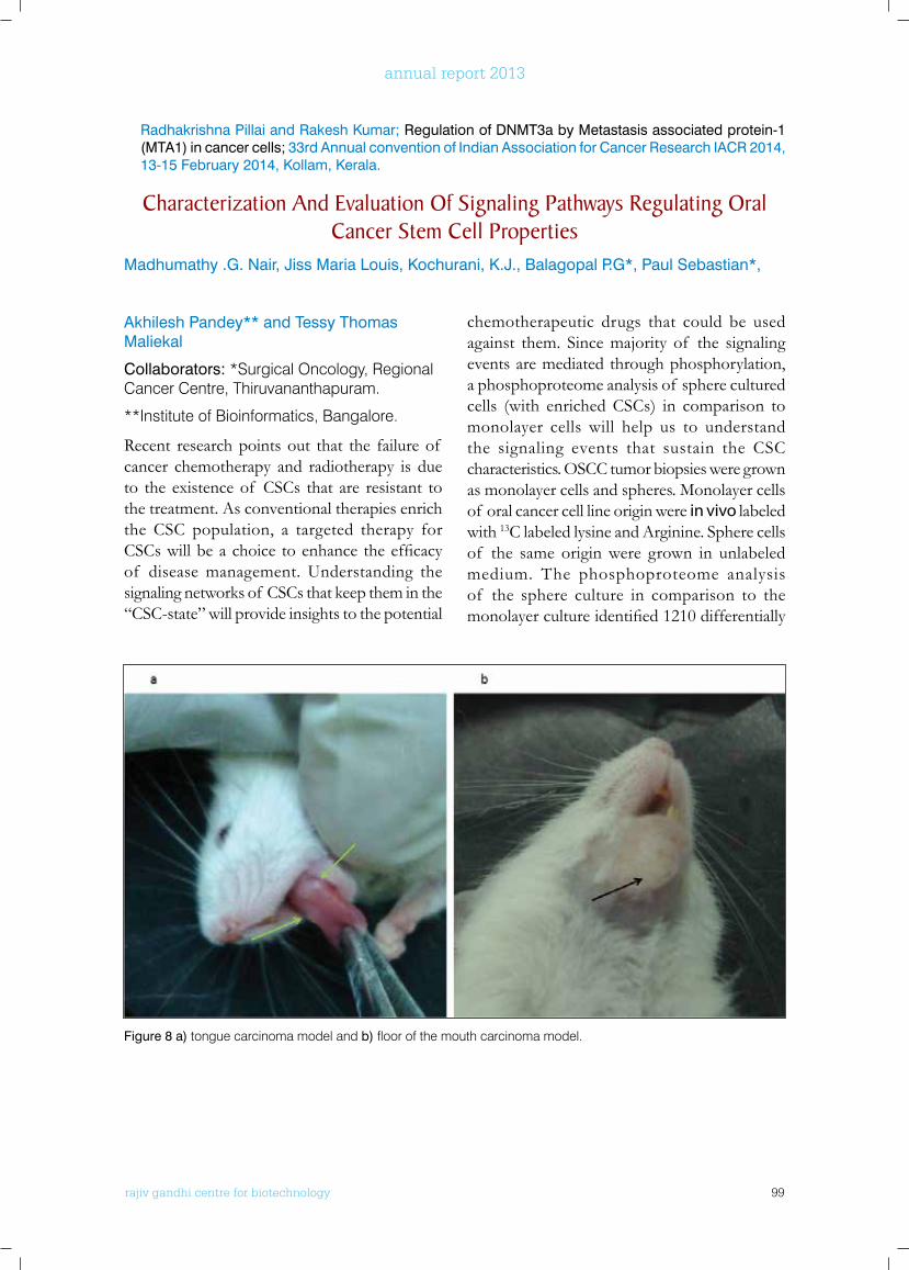
rajiv gandhi centre for biotechnology 99
annual report 2013
Akhilesh Pandey** and Tessy Thomas Maliekal
Collaborators: *Surgical Oncology, Regional Cancer Centre, Thiruvananthapuram.
**Institute of Bioinformatics, Bangalore.
Recent research points out that the failure of cancer chemotherapy and radiotherapy is due to the existence of CSCs that are resistant to the treatment. As conventional therapies enrich the CSC population, a targeted therapy for CSCswillbeachoice toenhancetheefficacyof disease management. Understanding the signaling networks of CSCs that keep them in the “CSC-state” will provide insights to the potential
chemotherapeutic drugs that could be used against them. Since majority of the signaling events are mediated through phosphorylation, a phosphoproteome analysis of sphere cultured cells (with enriched CSCs) in comparison to monolayer cells will help us to understand the signaling events that sustain the CSC characteristics. OSCC tumor biopsies were grown as monolayer cells and spheres. Monolayer cells of oral cancer cell line origin were in vivo labeled with 13C labeled lysine and Arginine. Sphere cells of the same origin were grown in unlabeled medium. The phosphoproteome analysis of the sphere culture in comparison to the monolayercultureidentified1210differentially
Radhakrishna Pillai and Rakesh Kumar; Regulation of DNMT3a by Metastasis associated protein-1 (MTA1) in cancer cells; 33rd Annual convention of Indian Association for Cancer Research IACR 2014, 13-15 February 2014, Kollam, Kerala.
Characterization And Evaluation of Signaling Pathways Regulating oral Cancer Stem Cell Properties
Madhumathy .G. Nair, Jiss Maria Louis, Kochurani, K.J., Balagopal P.G*, Paul Sebastian*,
Figure 8 a) tongue carcinoma model and b) floor of the mouth carcinoma model.
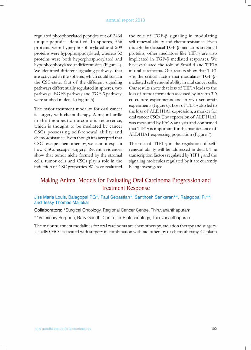
rajiv gandhi centre for biotechnology 100
annual report 2013
regulated phosphorylated peptides out of 2464 unique peptides identified. In spheres, 556 proteins were hyperphosphorylated and 209 proteins were hypophosphorylated, whereas 32 proteins were both hyperphosphorylated and hypophosphorylated at different sites (Figure 4). Weidentifieddifferentsignalingpathwaysthatare activated in the spheres, which could sustain the CSC-state. Out of the different signaling pathways differentially regulated in spheres, two pathways,EGFRpathwayandTGF-βpathway,were studied in detail. (Figure 5)
The major treatment modality for oral cancer is surgery with chemotherapy. A major hurdle in the therapeutic outcome is recurrence, which is thought to be mediated by cancer CSCs possessing self-renewal ability and chemoresistance. Even though it is accepted that CSCs escape chemotherapy, we cannot explain how CSCs escape surgery. Recent evidences show that tumor niche formed by the stromal cells, tumor cells and CSCs play a role in the induction of CSC properties. We have evaluated
the role of TGF-β signaling inmodulatingself-renewal ability and chemoresistance. Even thoughtheclassicalTGF-βmediatorsareSmadproteins, othermediators likeTIF1γ are alsoimplicated inTGF-βmediated responses.Wehaveevaluatedtheroleof Smad4andTIF1γin oral carcinoma. Our results show that TIF1 γ is the critical factor thatmodulatesTGF-β-mediated self-renewal ability in oral cancer cells. Ourresultsshowthatlossof TIF1γleadstotheloss of tumor formation assessed by in vitro 3D co-culture experiments and in vivo xenograft experiments(Figure6).Lossof TIF1γalsoledtothe loss of ALDH1A1 expression, a marker for oral cancer CSCs. The expression of ALDH1A1 wasmeasuredbyFACSanalysisandconfirmedthatTIF1γisimportantforthemaintenanceof ALDHIA1 expressing population (Figure 7).
The roleof TIF1 γ in the regulationof self-renewal ability will be addressed in detail. The transcriptionfactorsregulatedbyTIF1γandthesignaling molecules regulated by it are currently being investigated.
Making Animal Models for Evaluating oral Carcinoma Progression and Treatment Response
Jiss Maria Louis, Balagopal P.G*, Paul Sebastian*, Santhosh Sankaran**, Rajagopal R.**, and Tessy Thomas Maliekal
Collaborators: *Surgical Oncology, Regional Cancer Centre, Thiruvananthapuram.
**Veterinary Surgeon, Rajiv Gandhi Centre for Biotechnology, Thiruvananthapuram.
The major treatment modalities for oral carcinoma are chemotherapy, radiation therapy and surgery. Usually OSCC is treated with surgery in combination with radiotherapy or chemotherapy. Cisplatin
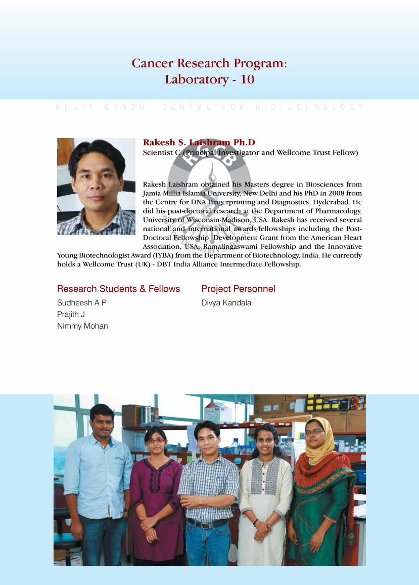
rajiv gandhi centre for biotechnology 101
annual report 2013
Cancer Research Program:Laboratory - 10
Research Students & FellowsSudheesh A PPrajith JNimmy Mohan
r a j i v g a n d h i c e n t r e f o r b i o t e c h n o l o g y
Rakesh Laishram obtained his Masters degree in Biosciences from Jamia Millia Islamia University, New Delhi and his PhD in 2008 from the Centre for DNA Fingerprinting and Diagnostics, Hyderabad. He did his post-doctoral research at the Department of Pharmacology, University of Wisconsin-Madison, USA. Rakesh has received several national and international awards/fellowships including the Post-Doctoral Fellowship Development Grant from the American Heart Association, USA; Ramalingaswami Fellowship and the Innovative
Young Biotechnologist Award (IYBA) from the Department of Biotechnology, India. He currently holds a Wellcome Trust (UK) - DBT India Alliance Intermediate Fellowship.
Rakesh S. Laishram Ph.DScientist C (Principal Investigator and Wellcome Trust Fellow)
Project PersonnelDivya Kandala
rajiv gandhi centre for biotechnology 102
annual report 2013
Role of Star-PAP in alternative polyadenylation and 3’-end processing of target genes
In eukaryotes, almost all pre-mRNAs are processed at the 3’-untranslated region (UTR) in a two concerted step -endonucleolytic cleavage followed by subsequent polyadenylation. The poly(A) tail of an mRNAis crucial for its stability, export, and translation efficiency.Interestingly, more than half of the genes in the human genomeharbour more than one polyadenylation site at the 3’-UTR, and thus are polyadenylated alternatively resulting in multiple mRNA transcripts with different 3’-UTR length. Alternative polyadenylation (APA) has been implicated in various human diseases including cardiovascular diseases (CVDs) and cancer, yet the mechanism of APA site selection is still elusive.Apart from the canonical poly(A) polymerase (PAPα) responsible for
general polyadenylation of mRNAs in the nucleus, recently an alternative PAP, Star-PAP (SpeckleTargetedPIPKIalphaRegulatedPoly(A)Polymerase) involved in polyadenylation of selectmRNAswas identified. In this projectwe investigate role of Star-PAP in APA of a set of human genes, including those keyto the cardiovascular system.Wehave identifiedgenome wide Star-PAP target genes which are regulated through alternative polyadenylation. Wehave confirmed theAPA regulationwithselect genes and among the genes NQO1 and PAK1willbeexploredforfurthermechanisticanalysis. Currently we are in the process of establishing reporter assays for the mentioned genestodefinethemechanismof APAand3’-end processing mediated by Star-PAP.
Regulation of cleavage and polyadenylation in oxidative stress response and human diseases
The disruptions of the cellular redox state cause toxic effects through the generation of peroxides and free radicals that damage cell components including proteins, lipids and nucleic acids. Several antioxidant proteins are expressed in the cell that protect against the oxidative stress. In humans, oxidative stress is involved in many diseases including cardiovascular diseases (CVDs) such as myocardial infarction, ischemia,heart failure and stroke. Several genes involved in oxidative stress response such asheme oxygenase-1 (HO-1), NAD(P)H quinine oxidoreductase (NQO-1), aldehyde dehydrogenase (ALDH) etc. are regulated at the 3’-UTR. Star-PAP is one of the polymerase that regulates such genes. The stress response in
heart is critical, and is maintained by a plethora of signalling events. The cytoprotective enzymes HO-1 and NQO-1 are the criticalantioxidant response proteins in cardiovascular system. We haveconfirmedtheexpressionprofileof severalgenes critical for cardiovascular system (such as ANAX7,VEGF,CAST,NOS2,FOG2,PAK1,AGTRII, TGFβ, GATA4, HAND2, HO1, NQO1)andcancer(suchasANA7,PTEN,BIK)in presence and absence of Star-PAP knockdown by quantitative real-time PCR analysis. ANX7 with two isoforms involved in prostrate cacer is also a target of Star-PAP. Currently we are in the process of identifying how 3’-end processing controls various heart conditions.
rajiv gandhi centre for biotechnology 103
annual report 2013
Elucidate the molecular function of RnA binding protein, RbM10 in the cell
RBM10 (RNA binding motif 10 protein)is a nuclear protein that belongs to a family proteins that contain an RNA-binding motif. Its function has not been defined yet. From sequence homology, it is likely to be a poly(G) or poly(U) stretch binding protein and may function in post transcriptional processing possibly in alternate splicing. However, its role in splicing has not been established yet. Defects in this gene have been implicated in humanX-linked recessive disorder, TARP syndrome. Recently, role of RBM10 in apoptosis has been proposed.
Interestingly, mass spectrometry analysis of Star-PAPpurifiedcomplexfromthecellhasidentified
RBM10 as aprotein associated with Star-PAP. However, the RBM10 has not been reported to be associated with PAPα in the earlier study which defined the proteins associated in thehuman 3’-end processing complex suggesting thatRBM10couldbe specific factor requiredonly for 3’-end processing. However, co-immunoprecipitation experiments demonstrated that RBM10 is present in both Star-PAP and PAPα specific complexes. Real time analysissuggests that RBM10 selectively regulates gene expression. We observed that not all Star-PAP targets or PAPα regulated genes are controlled by RBM10.
PublICATIonS
• Rakesh S. Laishram. 2014. Poly(A) Polymerase (PAP) diversity in the cell: Star-PAP vs canonical PAP. FEBS Letters.588: 2185–2197.
• D. Ray, H. Kazan, K. Cook, M.Weirauch, H. N, X. Li, S. G, Albu, H. Zheng, H. Na, M. Irimia, L. Matzat, S. Smith, C. Y, S. K, B. Nabet, Rakesh S. Laishram, M. Qiao H. Lipshitz, F. Piano,
A. Yang, A. corbett, R. Crastens, et. al. 2013. A compendium of RNA-binding motifs for decoding gene regulation. Nature. 499, 172–177 (doi:10.1038/nature12311).
• Wiemin Li*, Rakesh S. Laishram*, and Richard A Anderson. 2013. The novel poly(A) polymerase Star-PAP is a signal-regulated switch at the 3′-end of mRNAs. Adv. Biol. Reg. 53: 64-76 (*First authorship)
Mechanism of specificities of the two nuclear poly(A) polymerases – Star-PAP and PAPα in gene expression
Polyadenylation is a critical event in gene expression. Defects in polyadenylation have been implicated in several human diseases including cancer, CVDs and other genetic disorders. Poly(A) polymerases are the enzymes that polyadenylate mRNAs in the cell. At least two poly(A) polymerases are reported so far in the nucleus involved in general polyadenylation of mRNAs – PAPα and Star-PAP. Star-PAP binds the target pre-mRNA and recruits the cleavage factors while PAPα is recruited on the target mRNAs by cleavage factors – CPSF and CstF. We have reported that Star-PAP target mRNAs
are not processed by PAPα and vice versa and that they assemble distinct processing complexes. However, it is not clear how the two PAPs maintainspecifictargets.Weinvestigateseveralpossible ways – tarns acting factors association, binding to target pre-mRNA, requirement of CstF, and competition with cleavage factors. We are currently doing mass spectrometry analysis to identify the associated factors with Star-PAP and PAPα respectively. In addition, the competition with cleavage and role of CstFare being investigated.
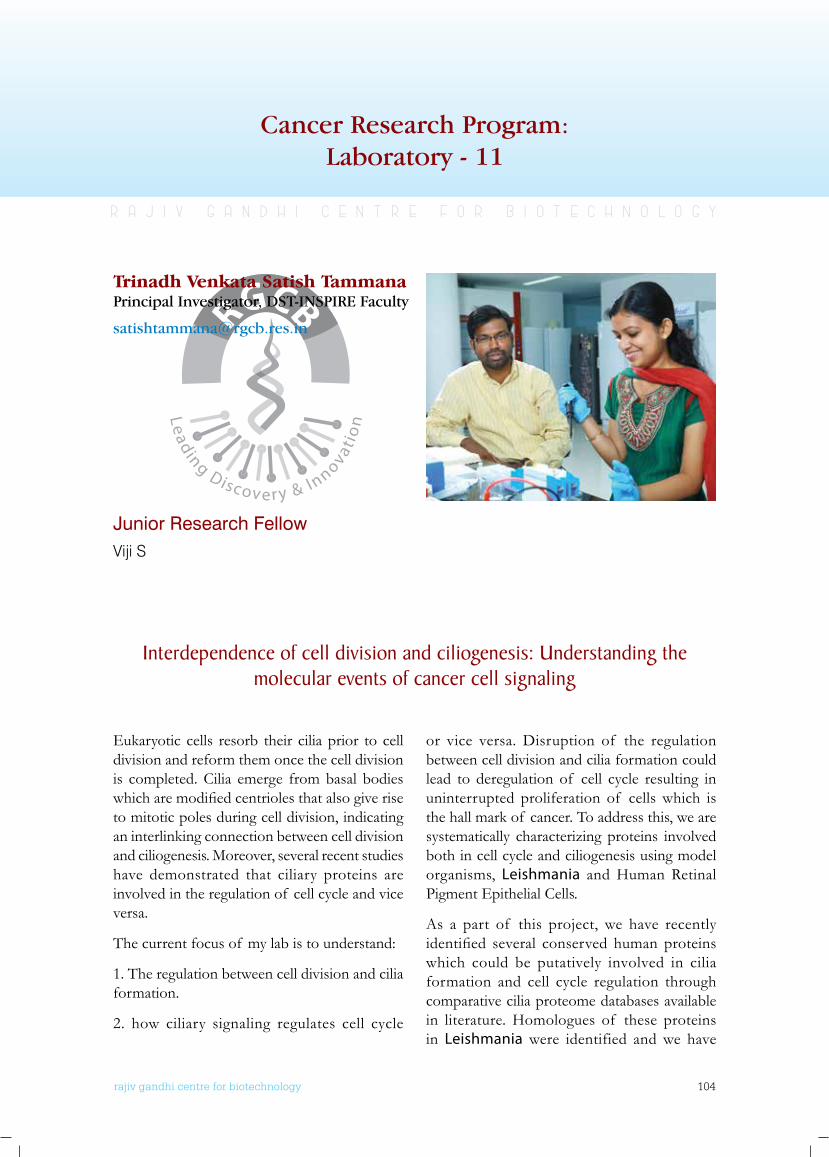
rajiv gandhi centre for biotechnology 104
annual report 2013
Cancer Research Program:Laboratory - 11
Junior Research FellowViji S
r a j i v g a n d h i c e n t r e f o r b i o t e c h n o l o g y
Trinadh Venkata Satish TammanaPrincipal Investigator, DST-INSPIRE Faculty
Interdependence of cell division and ciliogenesis: understanding the molecular events of cancer cell signaling
Eukaryotic cells resorb their cilia prior to cell division and reform them once the cell division is completed. Cilia emerge from basal bodies whicharemodifiedcentriolesthatalsogiveriseto mitotic poles during cell division, indicating an interlinking connection between cell division and ciliogenesis. Moreover, several recent studies have demonstrated that ciliary proteins are involved in the regulation of cell cycle and vice versa.
The current focus of my lab is to understand:
1. The regulation between cell division and cilia formation.
2. how ciliary signaling regulates cell cycle
or vice versa. Disruption of the regulation between cell division and cilia formation could lead to deregulation of cell cycle resulting in uninterrupted proliferation of cells which is the hall mark of cancer. To address this, we are systematically characterizing proteins involved both in cell cycle and ciliogenesis using model organisms, Leishmania and Human Retinal Pigment Epithelial Cells.
As a part of this project, we have recently identified several conserved human proteinswhich could be putatively involved in cilia formation and cell cycle regulation through comparative cilia proteome databases available in literature. Homologues of these proteins in Leishmania were identified and we have

rajiv gandhi centre for biotechnology 105
annual report 2013
successfully tagged two of these gene targets with GFP for in-vivo localization studies. Further the genes were also cloned into bacterial expression vectors and recombinant proteins were being purified to utilize as antigens toraising polyclonal antibodies. Functional analysis of the newly identified genes by generatingknock out strains of Leishmania cells is under progress. Finally localization and functional analysis of the mammalian homologs of these newlyidentifiedproteinswillbecarriedout.
An alternative approach to understand the relation between cell division and cilia formation will be screening various known anti-cancer drugs and cell cycle inhibitors to understand their effect on cilia formation and function using model organisms, Leishmaniadonovani and Human Retinal Pigment Epithelial cells. To address this, a recombinant Leishmaniadonovani strain expressing GFP in the cytosol and flagellum has been generated which will be used further for drug screening experiments.
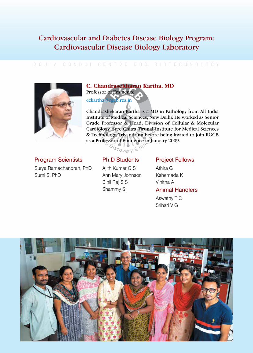
rajiv gandhi centre for biotechnology 106
annual report 2013
Cardiovascular and Diabetes Disease Biology Program: Cardiovascular Disease Biology Laboratory
Program ScientistsSurya Ramachandran, PhDSumi S, PhD
r a j i v g a n d h i c e n t r e f o r b i o t e c h n o l o g y
Chandrashekaran Kartha is a MD in Pathology from All India Institute of Medical Sciences, New Delhi. He worked as Senior Grade Professor & Head, Division of Cellular & Molecular Cardiology, Sree Chitra Tirunal Institute for Medical Sciences & Technology, Trivandrum before being invited to join RGCB as a Professor of Eminence in January 2009.
C. Chandrasekharan Kartha, MD Professor of Eminence
Ph.D StudentsAjith Kumar G SAnn Mary Johnson Binil Raj S S Shammy S
Project FellowsAthira G Kshemada K Vinitha A
Animal HandlersAswathy T CSrihari V G
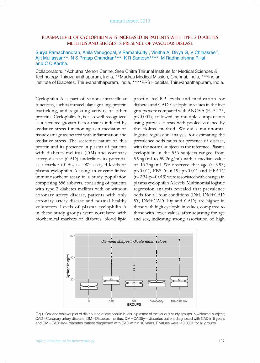
rajiv gandhi centre for biotechnology 107
annual report 2013
Cyclophilin A is part of various intracellular functions, such as intracellular signaling, protein trafficking, and regulating activity of other proteins. Cyclophilin A, is also well recognized as a secreted growth factor that is induced by oxidative stress functioning as a mediator of tissuedamageassociatedwithinflammationandoxidative stress. The secretory nature of this protein and its presence in plasma of patients with diabetes mellitus (DM) and coronary artery disease (CAD) underlines its potential as a marker of disease. We assayed levels of plasma cyclophilin A using an enzyme linked immunosorbent assay in a study population comprising 556 subjects, consisting of patients with type 2 diabetes mellitus with or without coronary artery disease, patients with only coronary artery disease and normal healthy volunteers. Levels of plasma cyclophilin A in these study groups were correlated with biochemical markers of diabetes, blood lipid
profile, hsCRP levels and medication for diabetesandCAD.Cyclophilinvaluesinthefivegroups were compared with ANOVA (F=54.75, p<0.001), followed by multiple comparisons using pairwise t tests with pooled variance by the Holms’ method. We did a multinomial logistic regression analysis for estimating the prevalence odds ratios for presence of disease, with the normal subjects as the reference. Plasma cyclophilin in the 556 subjects ranged from 5.9ng/ml to 59.2ng/ml) with a median value of 16.7ng/ml. We observed that age (t=3.93; p<0.01), FBS (t=6.19; p<0.01) and HbA1C (t=2.34; p=0.019) were associated with changes in plasma cyclophilin A levels. Multinomial logistic regression analysis revealed that prevalence odds for all four conditions (DM, DM+CAD 5Y, DM+CAD 10y and CAD) are higher in those with high cyclophilin values, compared to those with lower values, after adjusting for age and sex, indicating strong association of high
PlASMA lEVEl of CyCloPHIlIn A IS InCREASED In PATIEnTS wITH TyPE 2 DIAbETES MEllITuS AnD SuGGESTS PRESEnCE of VASCulAR DISEASE
Surya Ramachandran, Anila Venugopal, V RamanKutty*, Vinitha A, Divya G, V Chitrasree**, Ajit Mullassari**, N S Pratap Chandran***, K R Santosh****, M Radhakrishna Pillai and C C Kartha.
Collaborators: *Achutha Menon Centre, Sree Chitra Thirunal Institute for Medical Sciences & Technology, Thiruvananthapuram, India, **Madras Medical Mission, Chennai, India, ***Indian Institute of Diabetes, Thiruvananthapuram, India, ****PRS Hospital, Thiruvananthapuram, India.
Fig 1: Box and whisker plot of distribution of cyclophilin levels in plasma of the various study groups. N=Normal subject; CAD=Coronary artery disease; DM=Diabetes mellitus, DM+CAD5y= diabetes patient diagnosed with CAD in 5 years and DM+CAD10y= diabetes patient diagnosed with CAD within 10 years. P values were <0.0001 for all groups.
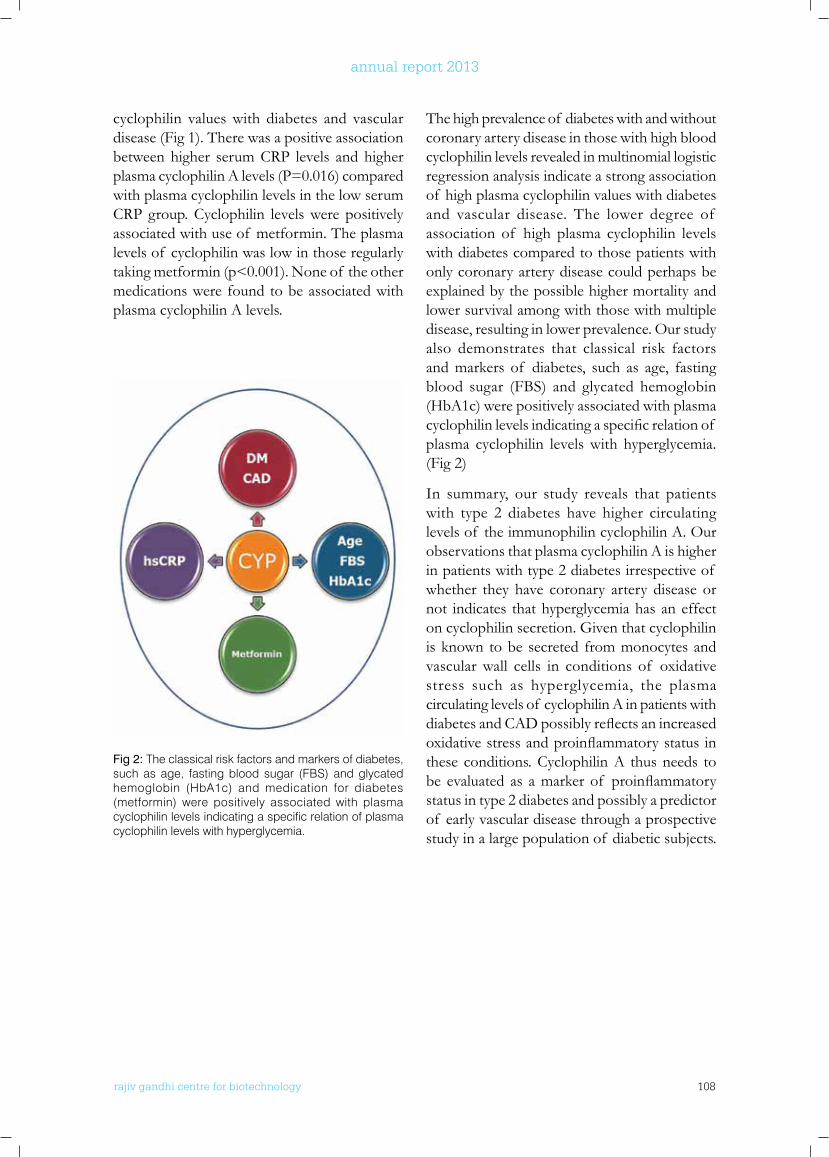
rajiv gandhi centre for biotechnology 108
annual report 2013
cyclophilin values with diabetes and vascular disease (Fig 1). There was a positive association between higher serum CRP levels and higher plasma cyclophilin A levels (P=0.016) compared with plasma cyclophilin levels in the low serum CRP group. Cyclophilin levels were positively associated with use of metformin. The plasma levels of cyclophilin was low in those regularly taking metformin (p<0.001). None of the other medications were found to be associated with plasma cyclophilin A levels.
The high prevalence of diabetes with and without coronary artery disease in those with high blood cyclophilin levels revealed in multinomial logistic regression analysis indicate a strong association of high plasma cyclophilin values with diabetes and vascular disease. The lower degree of association of high plasma cyclophilin levels with diabetes compared to those patients with only coronary artery disease could perhaps be explained by the possible higher mortality and lower survival among with those with multiple disease, resulting in lower prevalence. Our study also demonstrates that classical risk factors and markers of diabetes, such as age, fasting blood sugar (FBS) and glycated hemoglobin (HbA1c) were positively associated with plasma cyclophilinlevelsindicatingaspecificrelationof plasma cyclophilin levels with hyperglycemia. (Fig 2)
In summary, our study reveals that patients with type 2 diabetes have higher circulating levels of the immunophilin cyclophilin A. Our observations that plasma cyclophilin A is higher in patients with type 2 diabetes irrespective of whether they have coronary artery disease or not indicates that hyperglycemia has an effect on cyclophilin secretion. Given that cyclophilin is known to be secreted from monocytes and vascular wall cells in conditions of oxidative stress such as hyperglycemia, the plasma circulating levels of cyclophilin A in patients with diabetesandCADpossiblyreflectsanincreasedoxidativestressandproinflammatorystatusinthese conditions. Cyclophilin A thus needs to be evaluated as amarkerof proinflammatorystatus in type 2 diabetes and possibly a predictor of early vascular disease through a prospective study in a large population of diabetic subjects.
Fig 2: The classical risk factors and markers of diabetes, such as age, fasting blood sugar (FBS) and glycated hemoglobin (HbA1c) and medication for diabetes (metformin) were positively associated with plasma cyclophilin levels indicating a specific relation of plasma cyclophilin levels with hyperglycemia.
rajiv gandhi centre for biotechnology 109
annual report 2013
Coronary artery disease (CAD) is a serious complication and leading cause of death in patients with type 2 diabetes mellitus (T2DM). CAD is one of the most common causes of mortality among diabetic patients with a two to three fold higher prevalence compared with non-diabetic people. Studies indicate that insulin deficiency and insulin resistance accompaniedwith endothelial dysfunction leads to CAD and ischemic heart disease in patients with T2DM. Insulin resistance (IR) is partially under genetic control with several IR genes reported to play a modulating role in the development and severity of both T2DM and its cardiovascular complications. The ectoenzyme nucleotide pyrophosphate phosphodiesterase (ENPP1) is a class II membrane glycoprotein that adversely influencesinsulinsensitivitybyinhibitinginsulinreceptor signalling. A functional missense polymorphism(K121Q)of theENPP1 gene has been recently described which imparts stronger inhibition to insulin signalling and is associated withinsulinresistance.Conflictingresultshavebeen reported about the effect of the ENPP1 121Q variant on the risk for T2DM and vascular diseases in various populations. Fractalkine (FKN)/CX3CR1 system represents amajorregulatory mechanism for pancreatic islet beta cell function and insulin secretion. A decreased FKN/CX3CR1signallingwasreportedtobeasignificanteventunderlyingbetacelldysfunctionand insulin secretion. Two polymorphisms in the open reading frame of CX3CR1 (V249I and T280M)resulted indecreasedFKN/CX3CR1signalling. While these polymorphisms have a protective effect on vascular complications in non-diabetic individuals, their role in T2DM has not been evaluated. We studied the prevalence
and association of CX3CR1 and ENPP1 gene polymorphisms in patients with T2DM, CAD, and T2DM with CAD. Study subjects were recruited from Madras Medical Mission, Chennai, PRS hospital and Indian Institute of Diabetes, Thiruvananthapuram. Whole blood samples were collected from 852 subjects (age 24-93 years) in four study groups; 101 T2DM patients, 199 patients with CAD, 281 patients with T2DM with CAD and 271 healthy volunteers. The genotyping of ENPP1 K121Q, CX3CR1 V249I and CX3CR1 T280M polymorphisms were performed using polymerase chain reaction-restriction fragment length polymorphism (PCR-RFLP) analysis. PCR DNA sequencing (40 samples from T2DM and 70 samples from other groups)was performed to confirm theRFLP results. Data of continuous variables were expressed as mean ± standard deviation (SD) and data of non continuous variables as frequency (N,%). Hardy-Weinberg equilibrium was tested for a goodness-of-fit using aChisquare test. The genetic variants and their risk for disease were computed by odds ratios (OR) and 95% confidence intervals (CI) bylogistic regression analysis. Table 1 summarizes demographic and clinical characteristics of the four study populations. Our results indicate that ENPP1 K121Qpolymorphism is a consistentmarker for both T2DM and CAD. CX3CR1 V249I polymorphism is atheroprotective in our population as reported earlier. Contrary to the earlier observations that T280M polymorphism of CX3CR1 has an atheroprotective effect, we observed that CX3CR1 280M is associated with high risk of diabetes mellitus and also increased risk of CAD in patients with T2DM in our cohort (Tables 2 & 3).
Cx3CR1 and EnPP1 gene polymorphisms and their association with coronary artery disease in patients with type 2 diabetes mellitus
Sumi S, Surya Ramachandran, Vinitha A, Raman Kutty V*, Santhosh KR**,
Pratap Chandran***, Ajit Mullassari**** and Kartha CC.
Collaborators: *Achutha Menon Centre for Health Science Studies, Sree Chitra Thirunal Institute for Medical Sciences & Technology, Thiruvananthapuram, India, **PRS Hospital, Thiruvananthapuram, India, ***Indian Institute of Diabetes, Thiruvananthapuram, India, **** Madras Medical Mission, Chennai, India
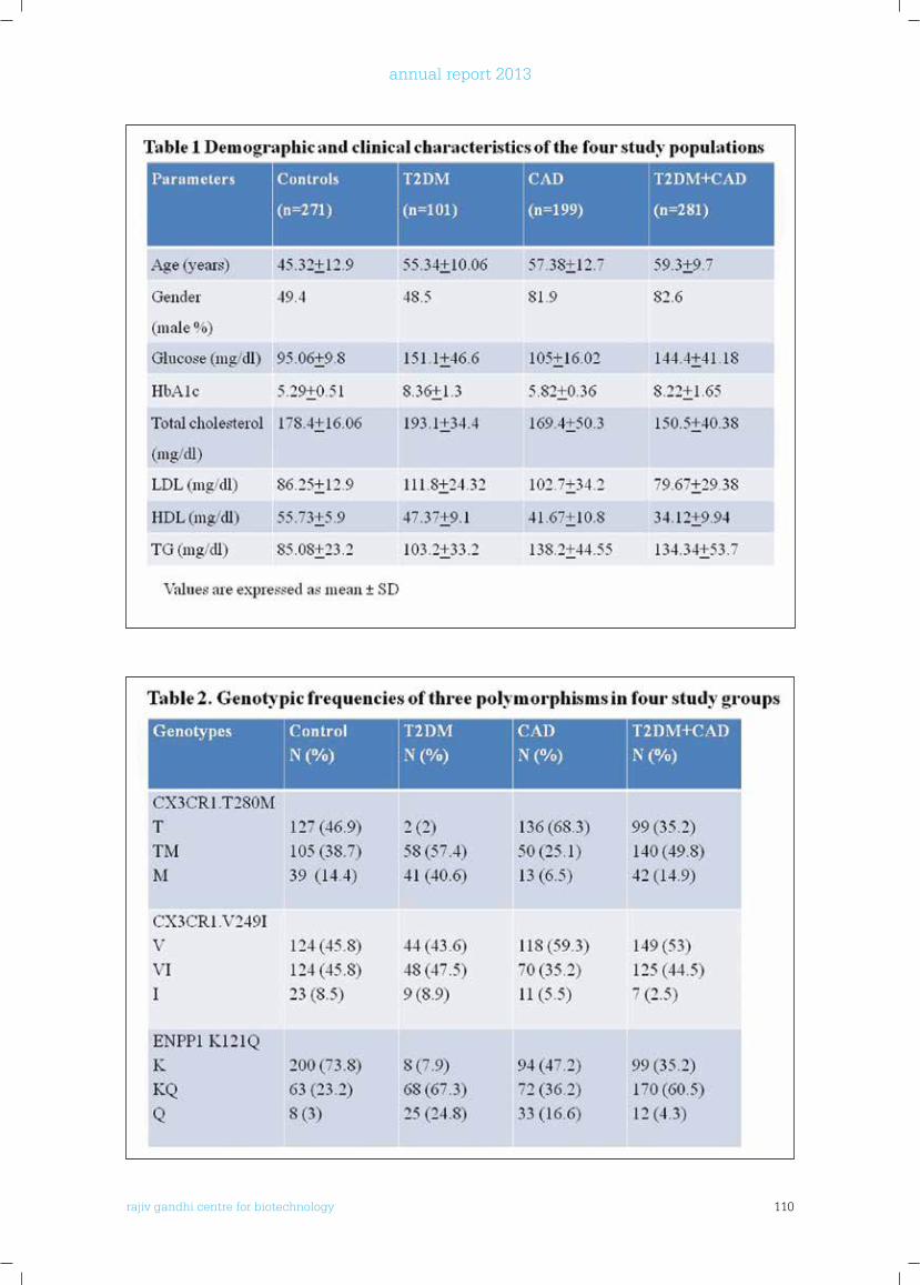
rajiv gandhi centre for biotechnology 110
annual report 2013
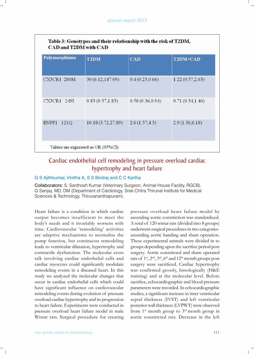
rajiv gandhi centre for biotechnology 111
annual report 2013
Heart failure is a condition in which cardiac output becomes insufficient to meet the body’s needs and it invariably worsens with time. Cardiovascular ‘remodeling’ activities are adaptive mechanisms to normalise the pump function, but continuous remodeling leads to ventricular dilatation, hypertrophy and contractile dysfunction. The molecular cross talk involving cardiac endothelial cells and cardiacmyocytes could significantlymodulateremodeling events in a diseased heart. In this study we analyzed the molecular changes that occur in cardiac endothelial cells which could have significant influence on cardiovascular remodeling events during evolution of pressure overload cardiac hypertrophy and its progression to heart failure. Experiments were conducted in pressure overload heart failure model in male Wistar rats. Surgical procedure for creating
pressure overload heart failure model by ascending aortic constriction was standardized. A total of 120 wistar rats (divided into 8 groups) underwent surgical procedures in two categories-ascending aortic banding and sham operation. These experimental animals were divided in to groupsdependinguponthesacrificeperiodpostsurgery. Aortic constricted and sham operated rats of 1st, 2nd, 3rd, 6th and 12th month groups post surgerywere sacrificed.Cardiac hypertrophywas confirmed grossly, histologically (H&E staining) and at the molecular level. Before sacrifice,echocardiographicandbloodpressureparameters were recorded. In echocardiographic studies,asignificantincreaseininterventricularseptal thickness (IVST) and left ventricular posterior wall thickness (LVPWT) were observed from 1st month group to 3rd month group in aortic constricted rats. Decrease in the left
Cardiac endothelial cell remodeling in pressure overload cardiac hypertrophy and heart failure
G S Ajithkumar, Vinitha A, S S Binilraj and C C Kartha
Collaborators: S. Santhosh Kumar (Veterinary Surgeon, Animal House Facility, RGCB), G Sanjay, MD, DM (Department of Cardiology, Sree Chitra Thirunal Institute for Medical Sciences & Technology, Thiruvananthapuram).
rajiv gandhi centre for biotechnology 112
annual report 2013
ventricular lumen size in aortic constricted rats also followed a gradual shift in 1st month group to 3rdmonthgroup.Nosignificantchangewasobserved in ejection fraction and fractional shortening in the 1st, 2nd and 3rd month group animals. These observations are suggestive of a compensatory cardiac hypertrophy stage. ECHO parameters were also observed in 6th month group animals except the fact that a reduction in ejection fraction was observed. In 12th month group of animals, IVST and LVPWT were decreased and the LV lumen size was increased compared to 3rd and 6th month groups (Fig.3).
Ejection fraction was also considerably reduced in the 12th month group, which is indicative of progression to heart failure stage. BNP level in 12th month group of rats with aortic constriction were above 300 pg/ml of blood, which is indicates onset of heart failure (Fig.5). Heart weight/ body weight ratio and ventricular
weight/bodyweightratiowerealsosignificantlyhigher in aortic constricted rats. In the 1st month group to 3rd month group, heart weight: body weight & left ventricular weight: body weight ratiowassignificantlyincreased.Inthe6th and 12th month group the parameters were not significantly different from that in 3rd month group (Fig.4).
Heart tissue samples were collected from sacrificedanimalsandmicrovascularendothelialcells were isolated. Molecular level analysis of microvascular endothelial cells by quantitative real time PCR (Fig.6) and immunoblotting assay (Fig.7)revealedasignificantincreaseinthelevelof expression of nitric oxide synthase enzymes (eNOS, iNOS) and ABCG2 in compensatory cardiac hypertrophy. A gradual decrease in expression was observed for all these molecules during the progression to heart failure stage.
Fig 3: Representative pictures of trans-thoracic M- mode echocardiographic parameters of functional status of heart at the time of sacrifice, after 1 month to 12 months post surgery. IVST= Inter ventricular septal thickness; LVPWT= Left ventricular posterior wall thickness; LVID= Left ventricular internal diameter.
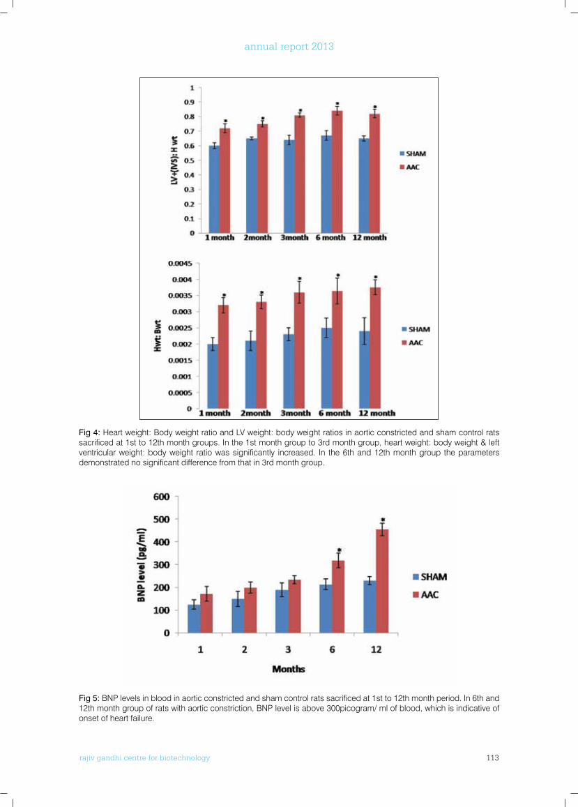
rajiv gandhi centre for biotechnology 113
annual report 2013
Fig 4: Heart weight: Body weight ratio and LV weight: body weight ratios in aortic constricted and sham control rats sacrificed at 1st to 12th month groups. In the 1st month group to 3rd month group, heart weight: body weight & left ventricular weight: body weight ratio was significantly increased. In the 6th and 12th month group the parameters demonstrated no significant difference from that in 3rd month group.
Fig 5: BNP levels in blood in aortic constricted and sham control rats sacrificed at 1st to 12th month period. In 6th and 12th month group of rats with aortic constriction, BNP level is above 300picogram/ ml of blood, which is indicative of onset of heart failure.
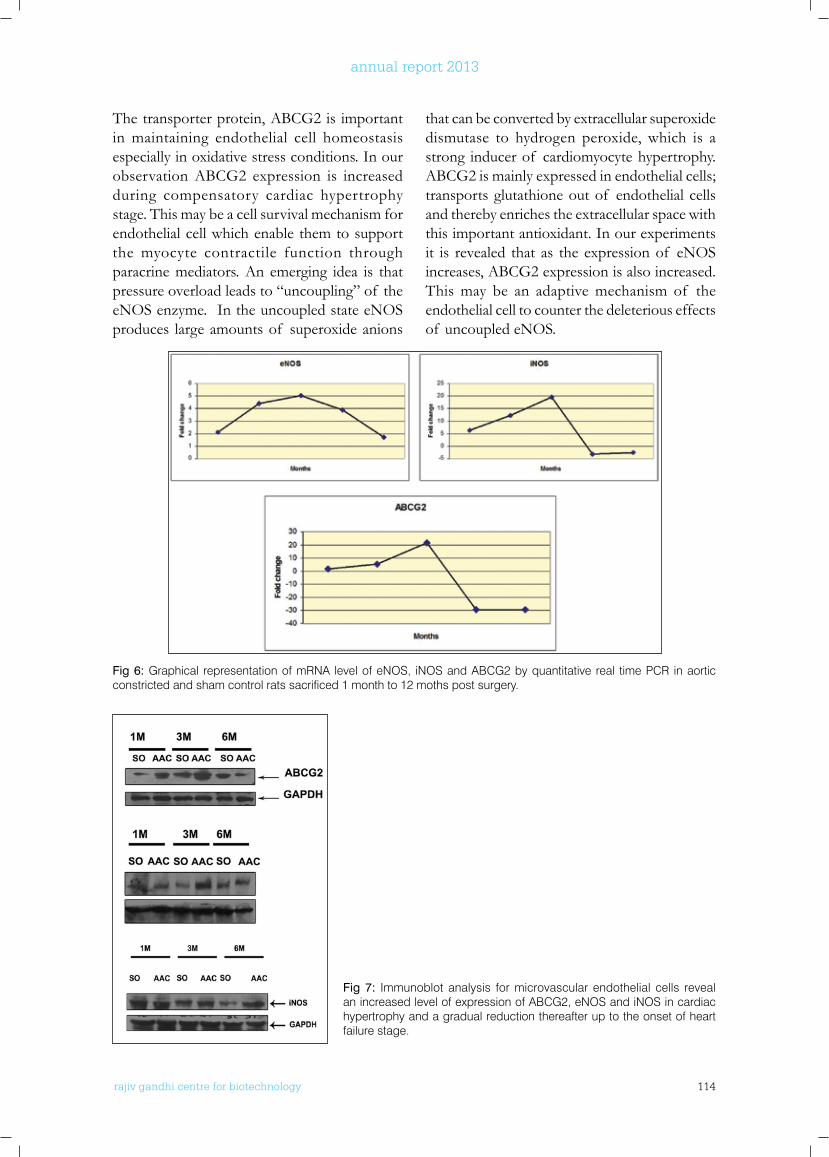
rajiv gandhi centre for biotechnology 114
annual report 2013
The transporter protein, ABCG2 is important in maintaining endothelial cell homeostasis especially in oxidative stress conditions. In our observation ABCG2 expression is increased during compensatory cardiac hypertrophy stage. This may be a cell survival mechanism for endothelial cell which enable them to support the myocyte contractile function through paracrine mediators. An emerging idea is that pressure overload leads to “uncoupling” of the eNOS enzyme. In the uncoupled state eNOS produces large amounts of superoxide anions
that can be converted by extracellular superoxide dismutase to hydrogen peroxide, which is a strong inducer of cardiomyocyte hypertrophy. ABCG2 is mainly expressed in endothelial cells; transports glutathione out of endothelial cells and thereby enriches the extracellular space with this important antioxidant. In our experiments it is revealed that as the expression of eNOS increases, ABCG2 expression is also increased. This may be an adaptive mechanism of the endothelial cell to counter the deleterious effects of uncoupled eNOS.
Fig 6: Graphical representation of mRNA level of eNOS, iNOS and ABCG2 by quantitative real time PCR in aortic constricted and sham control rats sacrificed 1 month to 12 moths post surgery.
Fig 7: Immunoblot analysis for microvascular endothelial cells reveal an increased level of expression of ABCG2, eNOS and iNOS in cardiac hypertrophy and a gradual reduction thereafter up to the onset of heart failure stage.
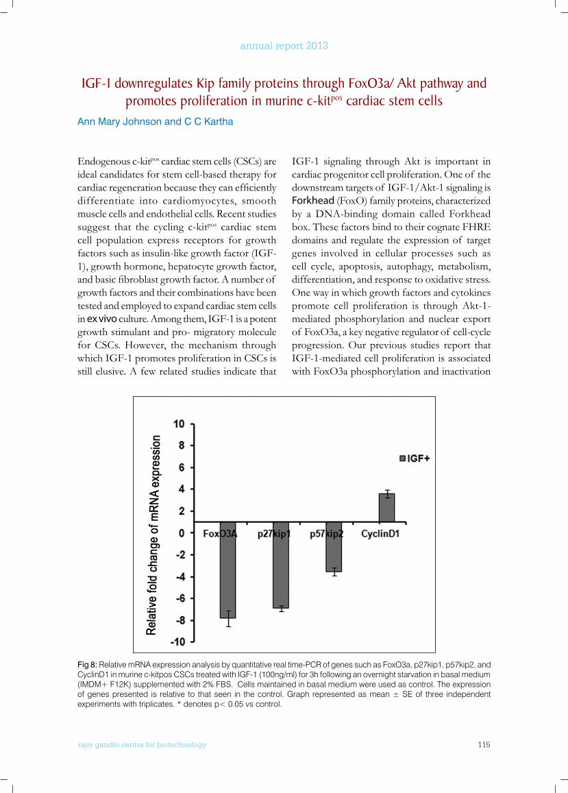
rajiv gandhi centre for biotechnology 115
annual report 2013
Endogenous c-kitpos cardiac stem cells (CSCs) are ideal candidates for stem cell-based therapy for cardiacregenerationbecausetheycanefficientlydifferentiate into cardiomyocytes, smooth muscle cells and endothelial cells. Recent studies suggest that the cycling c-kitpos cardiac stem cell population express receptors for growth factors such as insulin-like growth factor (IGF-1), growth hormone, hepatocyte growth factor, andbasicfibroblastgrowthfactor.Anumberof growth factors and their combinations have been tested and employed to expand cardiac stem cells in ex vivo culture. Among them, IGF-1 is a potent growth stimulant and pro- migratory molecule for CSCs. However, the mechanism through which IGF-1 promotes proliferation in CSCs is still elusive. A few related studies indicate that
IGF-1 signaling through Akt is important in cardiac progenitor cell proliferation. One of the downstream targets of IGF-1/Akt-1 signaling is Forkhead (FoxO) family proteins, characterized by a DNA-binding domain called Forkhead box. These factors bind to their cognate FHRE domains and regulate the expression of target genes involved in cellular processes such as cell cycle, apoptosis, autophagy, metabolism, differentiation, and response to oxidative stress. One way in which growth factors and cytokines promote cell proliferation is through Akt-1-mediated phosphorylation and nuclear export of FoxO3a, a key negative regulator of cell-cycle progression. Our previous studies report that IGF-1-mediated cell proliferation is associated with FoxO3a phosphorylation and inactivation
IGf-1 downregulates kip family proteins through foxo3a/ Akt pathway and promotes proliferation in murine c-kitpos cardiac stem cells
Ann Mary Johnson and C C Kartha
Fig 8: Relative mRNA expression analysis by quantitative real time-PCR of genes such as FoxO3a, p27kip1, p57kip2, and CyclinD1 in murine c-kitpos CSCs treated with IGF-1 (100ng/ml) for 3h following an overnight starvation in basal medium (IMDM+ F12K) supplemented with 2% FBS. Cells maintained in basal medium were used as control. The expression of genes presented is relative to that seen in the control. Graph represented as mean ± SE of three independent experiments with triplicates. * denotes p< 0.05 vs control.
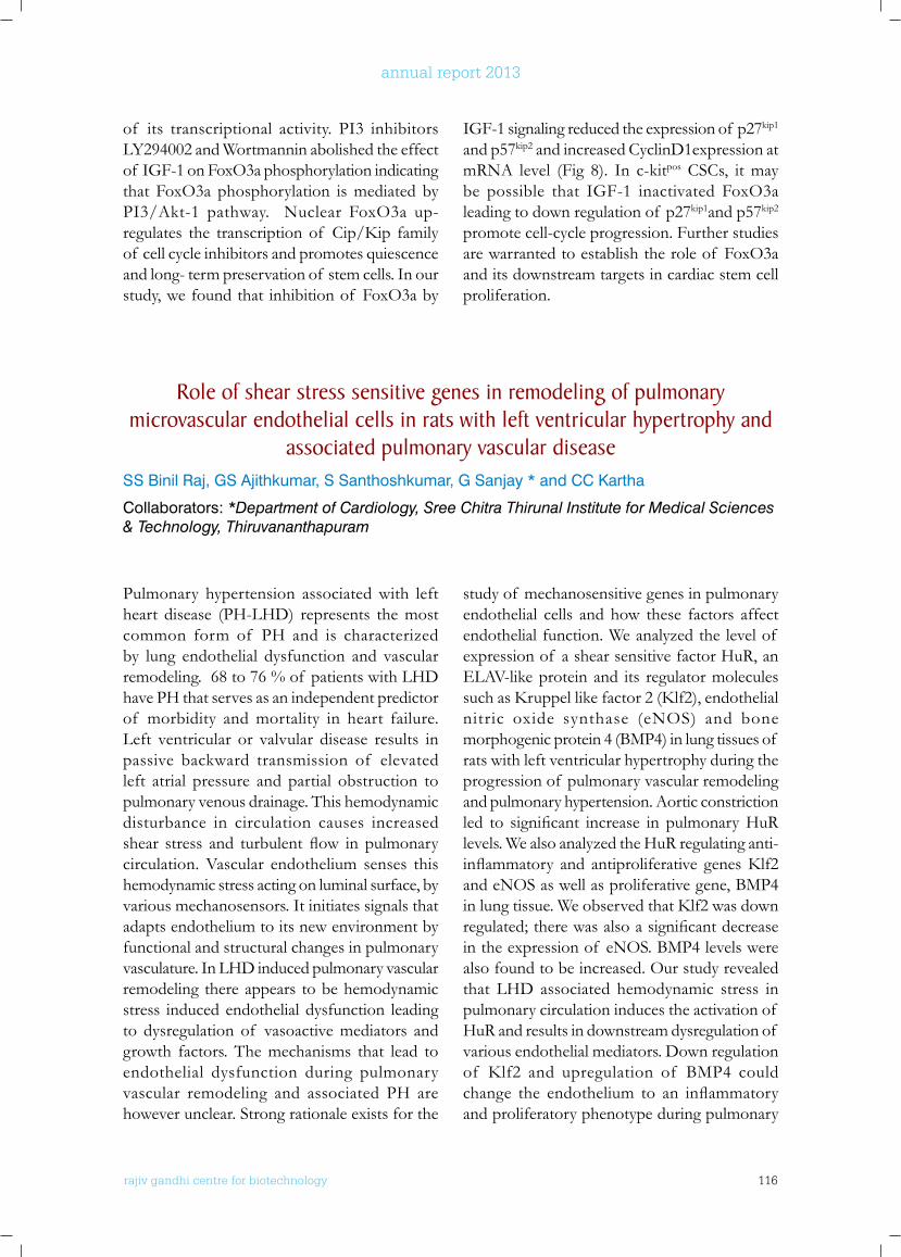
rajiv gandhi centre for biotechnology 116
annual report 2013
of its transcriptional activity. PI3 inhibitors LY294002 and Wortmannin abolished the effect of IGF-1 on FoxO3a phosphorylation indicating that FoxO3a phosphorylation is mediated by PI3/Akt-1 pathway. Nuclear FoxO3a up-regulates the transcriptionof Cip/Kip familyof cell cycle inhibitors and promotes quiescence and long- term preservation of stem cells. In our study, we found that inhibition of FoxO3a by
IGF-1 signaling reduced the expression of p27kip1
and p57kip2 and increased CyclinD1expression at mRNA level (Fig 8). In c-kitpos CSCs, it may be possible that IGF-1 inactivated FoxO3a leading to down regulation of p27kip1and p57kip2 promote cell-cycle progression. Further studies are warranted to establish the role of FoxO3a and its downstream targets in cardiac stem cell proliferation.
Role of shear stress sensitive genes in remodeling of pulmonary microvascular endothelial cells in rats with left ventricular hypertrophy and
associated pulmonary vascular diseaseSS Binil Raj, GS Ajithkumar, S Santhoshkumar, G Sanjay * and CC Kartha
Collaborators: *Department of Cardiology, Sree Chitra Thirunal Institute for Medical Sciences & Technology, Thiruvananthapuram
Pulmonary hypertension associated with left heart disease (PH-LHD) represents the most common form of PH and is characterized by lung endothelial dysfunction and vascular remodeling. 68 to 76 % of patients with LHD have PH that serves as an independent predictor of morbidity and mortality in heart failure. Left ventricular or valvular disease results in passive backward transmission of elevated left atrial pressure and partial obstruction to pulmonary venous drainage. This hemodynamic disturbance in circulation causes increased shear stress and turbulent flow in pulmonarycirculation. Vascular endothelium senses this hemodynamic stress acting on luminal surface, by various mechanosensors. It initiates signals that adapts endothelium to its new environment by functional and structural changes in pulmonary vasculature. In LHD induced pulmonary vascular remodeling there appears to be hemodynamic stress induced endothelial dysfunction leading to dysregulation of vasoactive mediators and growth factors. The mechanisms that lead to endothelial dysfunction during pulmonary vascular remodeling and associated PH are however unclear. Strong rationale exists for the
study of mechanosensitive genes in pulmonary endothelial cells and how these factors affect endothelial function. We analyzed the level of expression of a shear sensitive factor HuR, an ELAV-like protein and its regulator molecules suchasKruppellikefactor2(Klf2),endothelialnitric oxide synthase (eNOS) and bone morphogenic protein 4 (BMP4) in lung tissues of rats with left ventricular hypertrophy during the progression of pulmonary vascular remodeling and pulmonary hypertension. Aortic constriction led to significant increase in pulmonaryHuRlevels. We also analyzed the HuR regulating anti-inflammatory and antiproliferative genesKlf2and eNOS as well as proliferative gene, BMP4 inlungtissue.WeobservedthatKlf2wasdownregulated;therewasalsoasignificantdecreasein the expression of eNOS. BMP4 levels were also found to be increased. Our study revealed that LHD associated hemodynamic stress in pulmonary circulation induces the activation of HuR and results in downstream dysregulation of various endothelial mediators. Down regulation of Klf2 and upregulation of BMP4 couldchange the endothelium to an inflammatoryand proliferatory phenotype during pulmonary
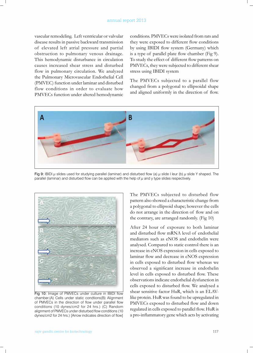
rajiv gandhi centre for biotechnology 117
annual report 2013
vascular remodeling. Left ventricular or valvular disease results in passive backward transmission of elevated left atrial pressure and partial obstruction to pulmonary venous drainage. This hemodynamic disturbance in circulation causes increased shear stress and disturbed flow in pulmonary circulation. We analyzed the Pulmonary Microvascular Endothelial Cell (PMVEC) function under laminar and disturbed flow conditions in order to evaluate how PMVECs function under altered hemodynamic
conditions. PMVECs were isolated from rats and theywereexposedtodifferentflowconditionsbyusingIBIDIflowsystem(Germany)whichisatypeof parallelplateflowchamber(Fig9).Tostudytheeffectof differentflowpatternsonPMVECs, they were subjected to different shear stress using IBIDI system
The PMVECs subjected to a parallel flow changed from a polygonal to ellipsoidal shape andaligneduniformlyinthedirectionof flow.
Fig 9: IBIDI µ slides used for studying parallel (laminar) and disturbed flow (a) µ slide I leur (b) µ slide Y shaped. The parallel (laminar) and disturbed flow can be applied with the help of µ and y type slides respectively.
Fig 10: Image of PMVECs under culture in IBIDI flow chamber.(A) Cells under static condtions(B) Alignment of PMVECs in the direction of flow under parallel flow conditions (10 dynes/cm2 for 24 hrs.) (C) Random alignment of PMVECs under disturbed flow conditions (10 dynes/cm2 for 24 hrs.) [Arrow indicates direction of flow]
The PMVECs subjected to disturbed flow pattern also showed a characteristic change from a polygonal to ellipsoid shape; however the cells donotarrangeinthedirectionof flowandonthe contrary, are arranged randomly. (Fig 10)
After 24 hour of exposure to both laminar anddisturbedflowmRNAlevelof endothelialmediators such as eNOS and endothelin were analysed. Compared to static control there is an increase in eNOS expression in cells exposed to laminarflowanddecreaseineNOSexpressionincellsexposedtodisturbedflowwhereasweobserved a significant increase in endothelinlevelincellsexposedtodisturbedflow.Theseobservations indicate endothelial dysfunction in cellsexposedtodisturbedflow.Weanalysedashear sensitive factor HuR, which is an ELAV-like protein. HuR was found to be upregulated in PMVECsexposedtodisturbedflowanddownregulatedincellsexposedtoparallelflow.HuRisapro-inflammatorygenewhichactsbyactivating
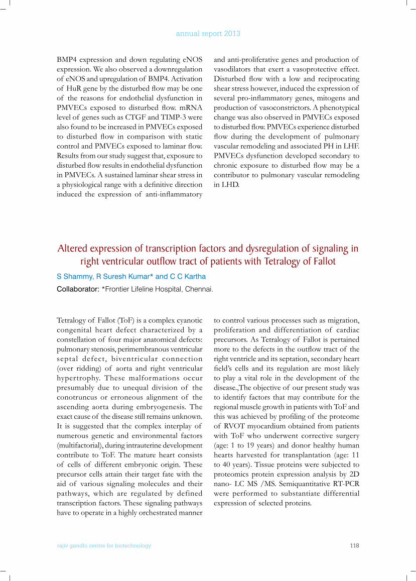
rajiv gandhi centre for biotechnology 118
annual report 2013
BMP4 expression and down regulating eNOS expression. We also observed a downregulation of eNOS and upregulation of BMP4. Activation of HuRgenebythedisturbedflowmaybeoneof the reasons for endothelial dysfunction in PMVECs exposed to disturbedflow.mRNAlevel of genes such as CTGF and TIMP-3 were also found to be increased in PMVECs exposed to disturbed flow in comparisonwith staticcontrolandPMVECsexposedtolaminarflow.Results from our study suggest that, exposure to disturbedflowresultsinendothelialdysfunctionin PMVECs. A sustained laminar shear stress in aphysiologicalrangewithadefinitivedirectioninduced the expression of anti-inflammatory
and anti-proliferative genes and production of vasodilators that exert a vasoprotective effect. Disturbedflowwith a low and reciprocatingshear stress however, induced the expression of severalpro-inflammatorygenes,mitogensandproduction of vasoconstrictors. A phenotypical change was also observed in PMVECs exposed todisturbedflow.PMVECsexperiencedisturbedflow during the development of pulmonaryvascular remodeling and associated PH in LHF. PMVECs dysfunction developed secondary to chronic exposure to disturbedflowmaybe acontributor to pulmonary vascular remodeling in LHD.
Altered expression of transcription factors and dysregulation of signaling in right ventricular outflow tract of patients with Tetralogy of fallot
S Shammy, R Suresh Kumar* and C C Kartha
Collaborator: *Frontier Lifeline Hospital, Chennai.
Tetralogy of Fallot (ToF) is a complex cyanotic congenital heart defect characterized by a constellation of four major anatomical defects: pulmonary stenosis, perimembranous ventricular septal defect, biventricular connection (over ridding) of aorta and right ventricular hypertrophy. These malformations occur presumably due to unequal division of the conotruncus or erroneous alignment of the ascending aorta during embryogenesis. The exact cause of the disease still remains unknown. It is suggested that the complex interplay of numerous genetic and environmental factors (multifactorial), during intrauterine development contribute to ToF. The mature heart consists of cells of different embryonic origin. These precursor cells attain their target fate with the aid of various signaling molecules and their pathways, which are regulated by defined transcription factors. These signaling pathways have to operate in a highly orchestrated manner
to control various processes such as migration, proliferation and differentiation of cardiac precursors. As Tetralogy of Fallot is pertained moretothedefectsintheoutflowtractof theright ventricle and its septation, secondary heart field’s cells and its regulation aremost likelyto play a vital role in the development of the disease.,The objective of our present study was to identify factors that may contribute for the regional muscle growth in patients with ToF and thiswasachievedbyprofilingof theproteomeof RVOT myocardium obtained from patients with ToF who underwent corrective surgery (age: 1 to 19 years) and donor healthy human hearts harvested for transplantation (age: 11 to 40 years). Tissue proteins were subjected to proteomics protein expression analysis by 2D nano- LC MS /MS. Semiquantitative RT-PCR were performed to substantiate differential expression of selected proteins.
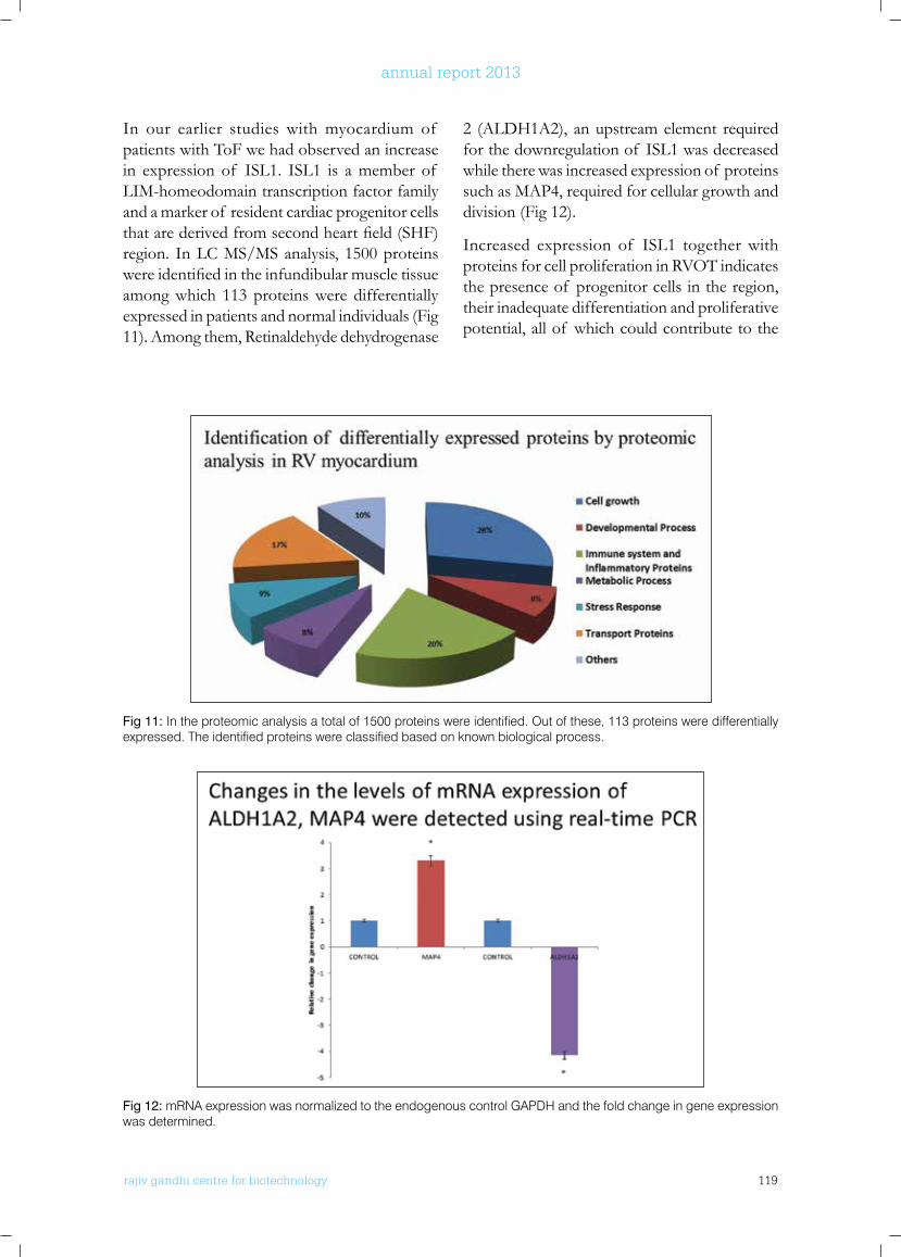
rajiv gandhi centre for biotechnology 119
annual report 2013
In our earlier studies with myocardium of patients with ToF we had observed an increase in expression of ISL1. ISL1 is a member of LIM-homeodomain transcription factor family and a marker of resident cardiac progenitor cells thatarederivedfromsecondheartfield(SHF)region. In LC MS/MS analysis, 1500 proteins wereidentifiedintheinfundibularmuscletissueamong which 113 proteins were differentially expressed in patients and normal individuals (Fig 11). Among them, Retinaldehyde dehydrogenase
2 (ALDH1A2), an upstream element required for the downregulation of ISL1 was decreased while there was increased expression of proteins such as MAP4, required for cellular growth and division (Fig 12).
Increased expression of ISL1 together with proteins for cell proliferation in RVOT indicates the presence of progenitor cells in the region, their inadequate differentiation and proliferative potential, all of which could contribute to the
Fig 11: In the proteomic analysis a total of 1500 proteins were identified. Out of these, 113 proteins were differentially expressed. The identified proteins were classified based on known biological process.
Fig 12: mRNA expression was normalized to the endogenous control GAPDH and the fold change in gene expression was determined.

rajiv gandhi centre for biotechnology 120
annual report 2013
infundibular stenosis. The results obtained from proteomics protein expression analysis and qRT-PCR analysis also suggest involvement of Retinoic acid pathway in TOF. In experimental animal models it has been demonstrated that lack of RA signalling increase cardiac side population numbers and retinoic acid induced downregulation of ISL1 is essential for the
proper development of RVOT. Further studies are required to analyze the expression pattern of significantcomponentsinretinoicacidsignalingpathway. Immunofluorescence experiments in tissue sections are also to be performed to characterize the cells present in the infundibular muscletissuetovalidatethefindings.
PublICATIonS
• Santhosh Kumar Sankaran, Ajith Kumar, G.S, Binil Raj S S and Rajagopal R. Two different rat models for cardiac hypertrophy by constriction of ascending and abdominal aorta. Journal of Laboratory Animal Science (2014) March 2:1; 54-57.
• Sumi Su rend ran , A th i r a G i r i j amma, Radhakrishnan Nair, Kalpana S. Ramegowda, Divya H. Nair, Jissa V. Thulaseedharan, Ravikumar B. Lakkappa, Giridhar Kamalapurkar, Chandrasekharan C. Kartha. Forkhead box C2 promoter variant c.-512C>T is associated with increased susceptibility to chronic venous diseases. PLoS ONE (2014) March; 9(3).
• Ramachandran S, Venugopal A, Ramankutty V, Vinitha A, Divya G, V Chitrasree, Ajit Mullassari, N S Pratap Chandran, K R Santosh, M Radhakrishna Pillai, C C Kartha. Plasma Level of Cyclophilin A is Increased in Patients with Type 2 Diabetes Mellitus and Suggests Presence of Vascular Disease. Cardiovasc Diabetol. (2014) Feb 7;13:38.
ConfEREnCE AbSTRACTS
• G.S. Ajith Kumar, S.S. Binil Raj, A. Vinitha, T.R. Santhosh Kumar, C.C. Kartha. High expression level of ABCG2 in endocardial endothelial cells protect against oxidative Stress. J.Mol.Cell.Cardiology (2013), 65S37-S38
• Ann Mary Johnson and CC Kartha. IGF-1 Induced Proliferation In c-kitpos Mouse Cardiac Stem Cells Is Mediated By Foxo3a Phosphorylation and Translocation. Regenerative Medicine Suppl. (2013) Pp 203.
• Shammy S, Gayathri AK, Kartha CC, Suresh Kumar R. Decreased expression of Gata4 and eNOS in the RV outflow tract myocardium of patients with tetralogy of Fallot, Annals of Pediatric Cardiology (2014) Vol 7;1
ConfEREnCE PRESEnTATIonS
• Shammy S, Gayathri AK, Kartha CC, Suresh Kumar R, “Decreased expression of GATA4 and eNOS in the RV outflow tract myocardium of patients with tetralogy of Fallot” at The 5th Congress of Asia Pacific Pediatric Cardiac Society on 6th-9th March 2014(Oral presentation)
• GS Ajithkumar, A Vinitha, SS Binilraj and CC Kartha. High expression of ABCG2 in endocardial endothelial cells protect them against oxidative stress induced by anticancer agents. Gordon Research Conference (oxygen radicals). PS 35, Feb 9-14, 2014, Ventura, California, USA. (Poster presentation)
• Ann Mary Johnson and CC Kartha, Cardiac stem cell proliferation is associated with foxo3a phosphorylation, 6th International Conference on Recent Advances In Cardiovascular Sciences at Delhi institute of Pharmaceutical Sciences & Research, 31st January & 1st February 2014. (Poster presentation)
• Sumi S, Athira G, Radhakrishnan N, Kalpana SR, Jissa VT, Kartha CC. Genetic variants in FoxC2 are associated with increased susceptibility to chronic venous diseases at 17th ADNAT Convention, Symposium on Genomics in Personalised Medicine and Public Health held at Thiruvananthapuram from 23rd to 25th February, 2014. (Poster presentation)
• Shammy S, Suresh Kumar R, Kartha CC, ‘Altered expression of transcription factors and dysregulation of signaling in right ventricular outflow tract of patients with tetralogy of Fallot’ at 6th International Conference on Recent Advances In Cardiovascular Sciences at DELHI institute of Pharmaceutical Sciences & Research, on 31st January & 1st February 2014(Oral presentation)
• Surya Ramachandran, Monocyte proteins as markers of inflammatory vascular disease
rajiv gandhi centre for biotechnology 121
annual report 2013
in type 2 diabetes Indo-US Symposium on Metabolomics and Disease Biology, Poovar Island resorts, January 22-23, 2014 (Invited Speaker)
• Shammy S, Suresh Kumar R, Kartha CC ‘Nitric oxide signaling pathway is down regulated in the myocardium of patients with Tetralogy of Fallot in LNOD:IDB-2013 Conference held on 5th and 6th November, 2013 at Rajiv Gandhi Centre for Biotechnology, Thiruvananthapuram (Poster presentation)
• Surya Ramachandran, Venugopa l A , Ramankutty V, Vinitha A, Divya G, V Chitrasree, Ajit Mullassari, N S Pratap Chandran, K R Santosh, M Radhakrishna Pillai, C C Kartha “Plasma Cyclophilin A as a potential marker of vascular inflammation in type 2 diabetes.” at 5th Annual meeting of Proteomics Society of India, IISC, Bangalore, November 28-30, 2013 (Poster presentation
AwARDS AnD HonouRS
• Professor C C Kartha received the Professor Manjeet Singh Oration award given by the Indian Section of International Society for Heart Research 2014
• Ann Mary Johnson received ‘RGCB Merit Award 2013 for the best research presentation entitled “Mechanisms of cell cycle regulation in adult cardiac stem cells”.
• Ajith Kumar GS was awarded foreign travel grants from CSIR and CICS for presenting a poster at the “Gordon Research Conference on Oxygen Radicals” held during Feb 9-14, 2014 at Ventura, California, USA.
• Shammy S received outstanding poster presentation award from Nobel Laureate Professor Freid Murrad during the conference “Legacy of Nitric Oxide Discovery: Impact on Disease Biology-2013” held on 5th and 6th November 2013 at Rajiv Gandhi Centre for Biotechnology, Thiruvananthapuram.
• Ann Mary Johnson was awarded an International Travel Grant from Department of Biotechnology, Government of India to attend The World Conference on Regenerative Medicine held on October 21st – 23rd, 2013 at Leipzig.
ExTRA –MuRAl RESEARCH GRAnTS
Sl. No. Name of Grant Funding Agency Duration
1 Remodeling of cardiac endothelium in progressive heart failure
Department of Biotechnology, Government of India
2011-2014
2 Molecular mechanisms of pulmonary microvascular endothelial dysfunctionunder fluid shear stress
Glaxo Smithkline Foundation, UK 2011-2014
3 Can Amalaki rasayana attenuate cardiac dysfunction associated with cardiac failure and aging
Department of Science and Technology, Government of India
2012-2015
rajiv gandhi centre for biotechnology 122
annual report 2013
Cardiovascular and Diabetes Disease Biology Program: Diabetes Biology Laboratory
Ph.D StudentAneesh Kumar A
r a j i v g a n d h i c e n t r e f o r b i o t e c h n o l o g y
Abdul Jaleel obtained his Ph.D from Jamia Millia Islamia, New Delhi while doing his research fellowship in All India Institute of Medical Sciences, New Delhi. He worked as a post doctoral fellow and as a faculty at Mayo Clinic, Rochester, Minnesota. He joined RGCB in December 2011.
Abdul Jaleel K. A Ph.D Scientist E-2
The objective of this project is to understand the metabolic transition process associated with the onset of type 2 diabetes (T2D). Metabolic alterations, which may be the foundation for metabolic diseases such as diabetes, could be identified by performingmass spectrometrybased metabolomics analysis in the blood of normal healthy study participants who are at the risk of developing T2D (such as people having family history of diabetes, obesity, etc). Tools of metabolomics measures chemical phenotypes that are the net result of genomic, transcriptomic, and proteomic variability,
therefore,providethemostintegratedprofileof biological status. Such studies are likely to offer substantial data and rationale for developing hypothesis based mechanistic studies. To date 124 normal healthy people were recruited and human studies were performed on them. Though all study participants are normal healthy people, we selected and recruited participants including those who have family history diabetes (FHxD) as well as people who have their body mass index (BMI) above than normal although not obese. We also recruited another 20 people who were categorized as pre-diabetic, as determined by
Metabolic Profiling of “normal Healthy” People in keralaAneesh Kumar, G. Vijayakumar1, V Raman Kutty2, Abdul Jaleel.
Collaborators: 1Medical Trust Hospital & Diabetes Centre, Kulanada, Pathanamthitta.2Achutha Menon Centre for Health Science Studies, Sree Chitra Thirunal Institute for Medical Sciences & Technology, Thiruvananthapuram, India.
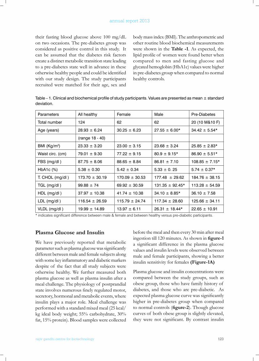
rajiv gandhi centre for biotechnology 123
annual report 2013
their fasting blood glucose above 100 mg/dL on two occasions. The pre-diabetes group was considered as positive control in this study. It can be assumed that the diabetes risk factors create a distinct metabolic transition state leading to a pre-diabetes state well in advance in these otherwisehealthypeopleandcouldbeidentifiedwith our study design. The study participants recruited were matched for their age, sex and
body mass index (BMI). The anthropometric and other routine blood biochemical measurements were shown in the Table -1. As expected, the lipidprofileof womenwerefoundbetterwhencompared to men and fasting glucose and glycated hemoglobin (HbA1c) values were higher in pre-diabetes group when compared to normal healthy controls.
Table – 1. Clinical and biochemical profile of study participants. Values are presented as mean ± standard deviation.
Parameters All healthy Female Male Pre-Diabetes
Total number 124 62 62 20 (10 M&10 F)
Age (years) 28.93 ± 6.24 30.25 ± 6.23 27.55 ± 6.00* 34.42 ± 5.54*
(range 18 - 40)
BMI (Kg/m2) 23.33 ± 3.20 23.00 ± 3.15 23.68 ± 3.24 25.85 ± 2.83*
Waist circ. (cm) 79.01 ± 9.30 77.22 ± 9.15 80.9 ± 9.15* 86.90 ± 5.51*
FBS (mg/dl ) 87.75 ± 8.06 88.65 ± 8.84 86.81 ± 7.10 108.85 ± 7.15*
HbA1c (%) 5.38 ± 0.30 5.42 ± 0.34 5.33 ± 0. 25 5.74 ± 0.37*
T. CHOL (mg/dl ) 173.70 ± 30.19 170.09 ± 30.53 177.48 ± 29.62 184.76 ± 38.15
TGL (mg/dl ) 99.88 ± 74 69.92 ± 30.59 131.35 ± 92.45* 113.28 ± 54.59
HDL (mg/dl ) 37.97 ± 10.38 41.74 ± 10.38 34.10 ± 8.85* 36.10 ± 7.58
LDL (mg/dl ) 116.54 ± 26.59 115.79 ± 24.74 117.34 ± 28.60 125.66 ± 34.11
VLDL (mg/dl ) 19.99 ± 14.89 13.97 ± 6.11 26.31 ± 18.44* 22.65 ± 10.91
* indicates significant difference between male & female and between healthy versus pre-diabetic participants.
Plasma Glucose and Insulin
We have previously reported that metabolic parametersuchasplasmaglucosewassignificantlydifferent between male and female subjects along withsomekeyinflammatoryanddiabeticmarkersdespite of the fact that all study subjects were otherwise healthy. We further measured both plasma glucose as well as plasma insulin after a meal challenge. The physiology of postprandial stateinvolvesnumerousfinelyregulatedmotor,secretory, hormonal and metabolic events, where insulin plays a major role. Meal challenge was performed with a standard mixed meal (25 kcal/kg ideal body weight; 55% carbohydrate, 30% fat, 15% protein). Blood samples were collected
before the meal and then every 30 min after meal ingestion till 120 minutes. As shown in figure-1 a significant difference in the plasma glucosevalues and insulin levels were observed between male and female participants, showing a better insulin sensitivity for females (Figure-1A)
Plasma glucose and insulin concentrations were compared between the study groups, such as obese group, those who have family history of diabetes, and those who are pre-diabetic. As expectedplasmaglucosecurvewassignificantlyhigher in pre-diabetes group when compared to normal controls (figure-2). Though glucose curves of both obese group is slightly elevated, theywere not significant.By contrast insulin
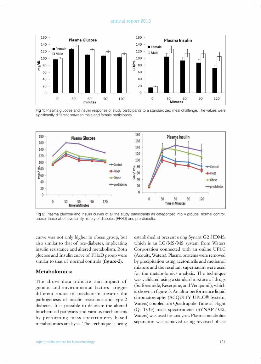
rajiv gandhi centre for biotechnology 124
annual report 2013
curve was not only higher in obese group, but also similar to that of pre-diabetes, implicating insulin resistance and altered metabolism. Both glucose and Insulin curve of FHxD group were similar to that of normal controls (figure-2).
Metabolomics:
The above data indicate that impact of genetic and environmental factors trigger different routes of mechanism towards the pathogenesis of insulin resistance and type 2 diabetes. It is possible to deliniate the altered biochemical pathways and various mechanisms by performing mass spectrometry based metabolomics analsysis. The technique is being
established at present using Synapt G2 HDMS, which is an LC/MS/MS system from Waters Corporation connected with an online UPLC (Acquity, Waters). Plasma proteins were removed by precipitation using acetonitrile and methanol mixture and the resultant supernatant were used for the metabolomics analysis. The technique was validated using a standard mixture of drugs (Sulfonamide, Reserpine, and Verapamil), which isshowninfigure-3.Anultra-performanceliquidchromatography (ACQUITY UPLC® System, Waters) coupled to a Quadrupole-Time of Flight (Q- TOF) mass spectrometer (SYNAPT-G2, Waters) was used for analyses. Plasma metabolite separation was achieved using reversed-phase
Fig 1: Plasma glucose and insulin response of study participants to a standardized meal challenge. The values were significantly different between male and female participants
Fig 2: Plasma glucose and insulin curves of all the study participants as categorized into 4 groups, normal control, obese, those who have family history of diabetes (FHxD) and pre-diabetic.
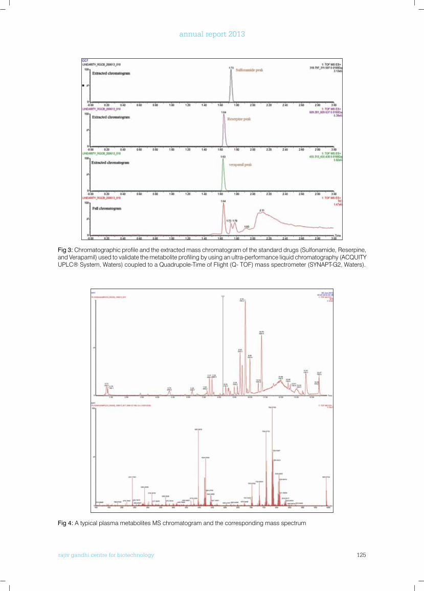
rajiv gandhi centre for biotechnology 125
annual report 2013
Fig 3: Chromatographic profile and the extracted mass chromatogram of the standard drugs (Sulfonamide, Reserpine, and Verapamil) used to validate the metabolite profiling by using an ultra-performance liquid chromatography (ACQUITY UPLC® System, Waters) coupled to a Quadrupole-Time of Flight (Q- TOF) mass spectrometer (SYNAPT-G2, Waters).
Fig 4: A typical plasma metabolites MS chromatogram and the corresponding mass spectrum
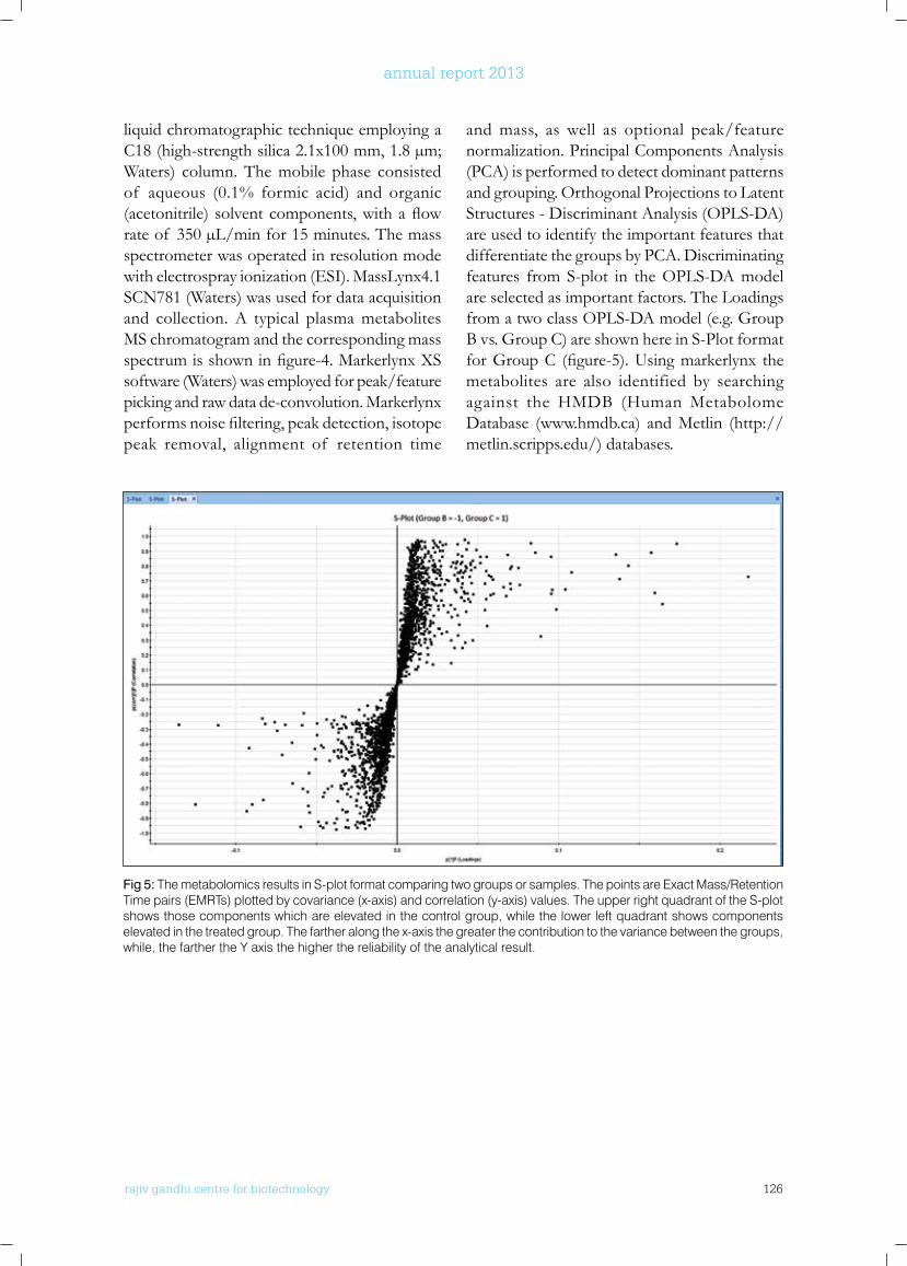
rajiv gandhi centre for biotechnology 126
annual report 2013
liquid chromatographic technique employing a C18 (high-strength silica 2.1x100 mm, 1.8 µm; Waters) column. The mobile phase consisted of aqueous (0.1% formic acid) and organic (acetonitrile)solventcomponents,withaflowrate of 350 µL/min for 15 minutes. The mass spectrometer was operated in resolution mode with electrospray ionization (ESI). MassLynx4.1 SCN781 (Waters) was used for data acquisition and collection. A typical plasma metabolites MS chromatogram and the corresponding mass spectrumisshowninfigure-4.MarkerlynxXSsoftware (Waters) was employed for peak/feature picking and raw data de-convolution. Markerlynx performsnoisefiltering,peakdetection,isotopepeak removal, alignment of retention time
and mass, as well as optional peak/feature normalization. Principal Components Analysis (PCA) is performed to detect dominant patterns and grouping. Orthogonal Projections to Latent Structures - Discriminant Analysis (OPLS-DA) are used to identify the important features that differentiate the groups by PCA. Discriminating features from S-plot in the OPLS-DA model are selected as important factors. The Loadings from a two class OPLS-DA model (e.g. Group B vs. Group C) are shown here in S-Plot format forGroupC (figure-5).Usingmarkerlynx themetabolites are also identified by searching against the HMDB (Human Metabolome Database (www.hmdb.ca) and Metlin (http://metlin.scripps.edu/) databases.
Fig 5: The metabolomics results in S-plot format comparing two groups or samples. The points are Exact Mass/Retention Time pairs (EMRTs) plotted by covariance (x-axis) and correlation (y-axis) values. The upper right quadrant of the S-plot shows those components which are elevated in the control group, while the lower left quadrant shows components elevated in the treated group. The farther along the x-axis the greater the contribution to the variance between the groups, while, the farther the Y axis the higher the reliability of the analytical result.
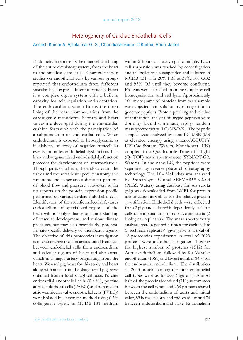
rajiv gandhi centre for biotechnology 127
annual report 2013
Endothelium represents the inner cellular lining of the entire circulatory system, from the heart to the smallest capillaries. Characterization studies on endothelial cells by various groups reported that endothelium from different vascular beds express different proteins. Heart is a complex organ-system with a built-in capacity for self-regulation and adaptation. The endocardium, which forms the inner lining of the heart chamber, arises from the cardiogenic mesoderm. Septum and heart valves are developed during the endocardial cushion formation with the participation of a subpopulation of endocardial cells. When endothelium is exposed to hyperglycemia as in diabetes, an array of negative intracellular events promotes endothelial dysfunction. It is known that generalized endothelial dysfunction precedes the development of atherosclerosis. Though parts of a heart, the endocardium, the valvesandtheaortahavespecificanatomyandfunctions and experiences different patterns of bloodflow andpressure.However, so farno reports on the protein expression profileperformed on various cardiac endothelial cells. Identificationof thespecificmolecularfeaturesendothelium of specialized regions of the heart will not only enhance our understanding of vascular development, and various disease processes but may also provide the potential forsite-specificdeliveryof therapeuticagents.The objective of this proteomics investigation is to characterize the similarities and differences between endothelial cells from endocardium and valvular regions of heart and also aorta, which is a major artery originating from the heart. We used pig heart for this study and heart along with aorta from the slaughtered pig, were obtained from a local slaughterhouse. Porcine endocardial endothelial cells (PEEC), porcine aortic endothelial cells (PAEC)) and porcine left atrio-ventricular valve endothelial cells (PVEC)) were isolated by enzymatic method using 0.2% collagenase type-2 in MCDB 131 medium
within 2 hours of receiving the sample. Each cell suspension was washed by centrifugation and the pellet was resuspended and cultured in MCDB 131 with 20% FBS at 370C, 5% CO2 and 95% O2 until they become confluent. Proteins were extracted from the sample by cell homogenization and cell lysis. Approximately 100 micrograms of proteins from each sample was subjected to in-solution trypsin digestion to generatepeptides.Proteinprofilingandrelativequantificationanalysisof trypicpeptidesweredone by Liquid Chromatography- tandem mass spectrometry (LC/MS/MS). The peptide samples were analyzed by nano-LC–MSE (MS at elevated energy) using a nanoACQUITY UPLC® System (Waters,Manchester, UK)coupled to a Quadrupole-Time of Flight (Q- TOF) mass spectrometer (SYNAPT-G2, Waters). In the nano-LC, the peptides were separated by reverse phase chromatography technology. The LC- MSE data was analyzed by ProteinLynx Global SERVER™ v2.5.3 (PLGS, Waters) using database for sus scrofa (pig) was downloaded from NCBI for protein identificationaswellasfortherelativeproteinquantification.Endothelialcellswerecollectedfrom 2 pigs and cultured independently each for cells of endocradium, mitral valve and aorta (2 biological replicates). The mass spectrometry analyses were repeated 3 times for each isolate (3 technical replicates), giving rise to a total of 18 proteomics experiments. A total of 2023 proteinswere identified altogether, showingthe highest number of proteins (1512) for Aortic endothelium, followed by for Valvular endothelium (1361) and lowest number (997) for the endocardial endothelium. The distribution of 2023 proteins among the three endothelial cell typeswere as follows (figure 1);Almosthalf of theproteinsidentified(711)ascommonbetween the cell types, and 268 proteins shared between the endothelium of aorta and mitral valve, 83 between aorta and endocardium and 74 between endocardium and valve. Endothelium
Heterogeneity of Cardiac Endothelial CellsAneesh Kumar A, Ajithkumar G. S., Chandrashekaran C Kartha, Abdul Jaleel
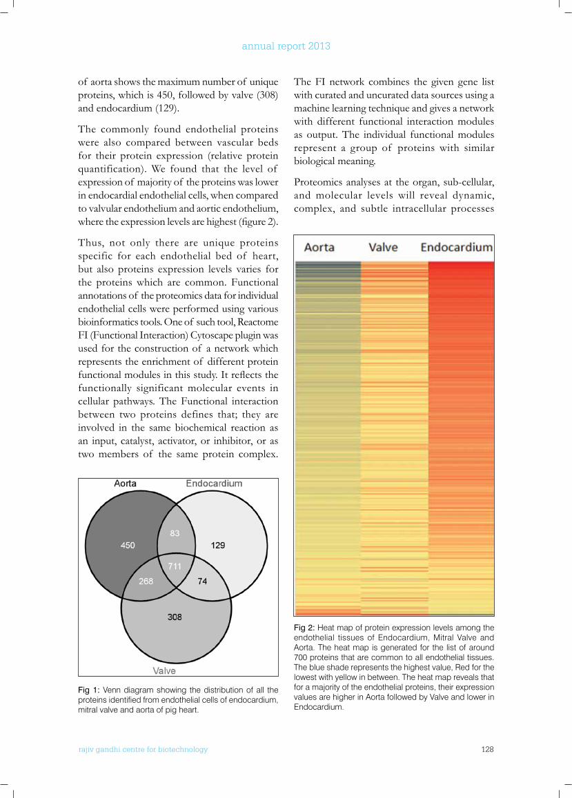
rajiv gandhi centre for biotechnology 128
annual report 2013
of aorta shows the maximum number of unique proteins, which is 450, followed by valve (308) and endocardium (129).
The commonly found endothelial proteins were also compared between vascular beds for their protein expression (relative protein quantification). We found that the level of expression of majority of the proteins was lower in endocardial endothelial cells, when compared to valvular endothelium and aortic endothelium, wheretheexpressionlevelsarehighest(figure2).
Thus, not only there are unique proteins specific for each endothelial bed of heart, but also proteins expression levels varies for the proteins which are common. Functional annotations of the proteomics data for individual endothelial cells were performed using various bioinformatics tools. One of such tool, Reactome FI (Functional Interaction) Cytoscape plugin was used for the construction of a network which represents the enrichment of different protein functionalmodulesinthisstudy.Itreflectsthefunctionally significant molecular events in cellular pathways. The Functional interaction between two proteins defines that; they are involved in the same biochemical reaction as an input, catalyst, activator, or inhibitor, or as two members of the same protein complex.
The FI network combines the given gene list with curated and uncurated data sources using a machine learning technique and gives a network with different functional interaction modules as output. The individual functional modules represent a group of proteins with similar biological meaning.
Proteomics analyses at the organ, sub-cellular, and molecular levels will reveal dynamic, complex, and subtle intracellular processes
Fig 1: Venn diagram showing the distribution of all the proteins identified from endothelial cells of endocardium, mitral valve and aorta of pig heart.
Fig 2: Heat map of protein expression levels among the endothelial tissues of Endocardium, Mitral Valve and Aorta. The heat map is generated for the list of around 700 proteins that are common to all endothelial tissues. The blue shade represents the highest value, Red for the lowest with yellow in between. The heat map reveals that for a majority of the endothelial proteins, their expression values are higher in Aorta followed by Valve and lower in Endocardium.
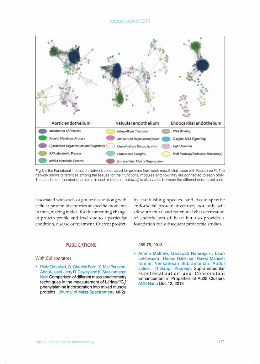
rajiv gandhi centre for biotechnology 129
annual report 2013
associated with each organ or tissue along with cellularproteininventoriesatspecificmomentsin time, making it ideal for documenting change inproteinprofileandlevelduetoaparticularcondition, disease or treatment. Current project,
by establishing species- and tissue-specific endothelial protein inventory not only will allow structural and functional characterization of endothelium of heart but also provides a foundation for subsequent proteomic studies.
Fig 3 is the Functional Interaction Network constructed for proteins from each endothelial tissue with Reactome FI. The network shows differences among the tissues for their functional modules and how they are connected to each other. The enrichment (number of proteins in each module or pathway) is also varies between the different endothelial cells.
PublICATIonS
with Collaborators
• Piotr Zabielski, G. Charles Ford, X. Mai Persson, Abdul Jaleel, Jerry D. Dewey and K. Sreekumaran Nair. Comparison of different mass spectrometry techniques in the measurement of L-[ring-13C6] phenylalanine incorporation into mixed muscle proteins. Journal of Mass Spectrometry 48(2):
269-75, 2013
• Ammu Mathew, Ganapati Natarajan , Lauri Lehtovaara , Hannu Häkkinen, Ravva Mahesh Kumar, Venkatesan Subramanian, Abdul Jaleel, Thalappil Pradeep. Supramolecular Fu n c t i o n a l i z a t i o n a n d C o n c o m i t a n t Enhancement in Properties of Au25 Clusters ACS Nano Dec 12, 2013
rajiv gandhi centre for biotechnology 130
annual report 2013
TROPICAL DISEASE BIOLOGY Mycobacterium Research Group - 1
Ph.D StudentsMadhavilatha G KBiljo V JosephSunil Kumar V JDhansooraj DMahesh P PAnnapoorna KRetnakumar R J
r a j i v g a n d h i c e n t r e f o r b i o t e c h n o l o g y
Sathish Mundayoor obtained his Ph.D from All India Institute of Medical Sciences, New Delhi, did Post- Doctoral training at Forshungsinstitut Borstel, Germany and then at Washington University in St Louis, Missouri. He was also a Visiting Scientist at Centres for Disease Control, Atlanta, Georgia before joining RGCB in 1995.
Sathish Mundayoor Ph.D. Scientist G
Technical OfficerLaiza K Paul
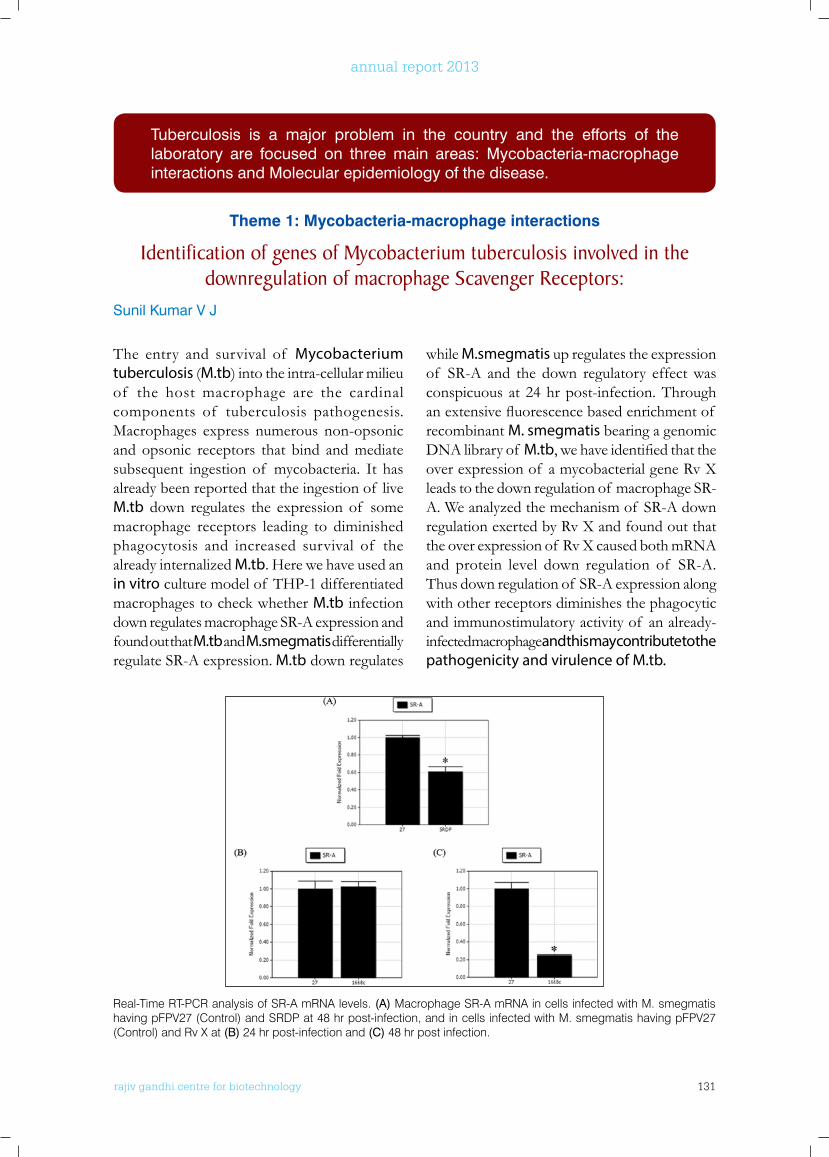
rajiv gandhi centre for biotechnology 131
annual report 2013
The entry and survival of Mycobacterium tuberculosis (M.tb) into the intra-cellular milieu of the host macrophage are the cardinal components of tuberculosis pathogenesis. Macrophages express numerous non-opsonic and opsonic receptors that bind and mediate subsequent ingestion of mycobacteria. It has already been reported that the ingestion of live M.tb down regulates the expression of some macrophage receptors leading to diminished phagocytosis and increased survival of the already internalized M.tb. Here we have used an in vitro culture model of THP-1 differentiated macrophages to check whether M.tb infection down regulates macrophage SR-A expression and found out that M.tb and M.smegmatis differentially regulate SR-A expression. M.tb down regulates
while M.smegmatis up regulates the expression of SR-A and the down regulatory effect was conspicuous at 24 hr post-infection. Through anextensivefluorescencebasedenrichmentof recombinant M. smegmatis bearing a genomic DNA library of M.tb,wehaveidentifiedthattheover expression of a mycobacterial gene Rv X leads to the down regulation of macrophage SR-A. We analyzed the mechanism of SR-A down regulation exerted by Rv X and found out that the over expression of Rv X caused both mRNA and protein level down regulation of SR-A. Thus down regulation of SR-A expression along with other receptors diminishes the phagocytic and immunostimulatory activity of an already-infected macrophage and this may contribute to the pathogenicity and virulence of M.tb.
Tuberculosis is a major problem in the country and the efforts of the laboratory are focused on three main areas: Mycobacteria-macrophage interactions and Molecular epidemiology of the disease.
Theme 1: Mycobacteria-macrophage interactions
Identification of genes of Mycobacterium tuberculosis involved in the downregulation of macrophage Scavenger Receptors:
Sunil Kumar V J
Real-Time RT-PCR analysis of SR-A mRNA levels. (A) Macrophage SR-A mRNA in cells infected with M. smegmatis having pFPV27 (Control) and SRDP at 48 hr post-infection, and in cells infected with M. smegmatis having pFPV27 (Control) and Rv X at (B) 24 hr post-infection and (C) 48 hr post infection.
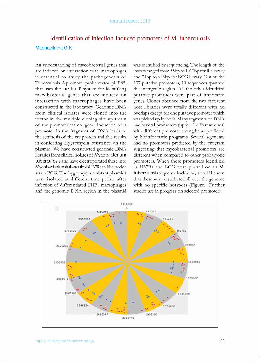
rajiv gandhi centre for biotechnology 132
annual report 2013
An understanding of mycobacterial genes that are induced on interaction with macrophages is essential to study the pathogenesis of Tuberculosis. A promoter probe vector, pHP85, that uses the cre-lox P system for identifying mycobacterial genes that are induced on interaction with macrophages have been constructed in the laboratory. Genomic DNA from clinical isolates were cloned into the vector in the multiple cloning site upstream of the promoterless cre gene. Induction of a promoter in the fragment of DNA leads to the synthesis of the cre protein and this results in conferring Hygromycin resistance on the plasmid. We have constructed genomic DNA libraries from clinical isolates of Mycobacterium tuberculosis and have electroporated these into Mycobacterium tuberculosis H37Ra and the vaccine strain BCG. The hygromycin resistant plasmids were isolated at different time points after infection of differentiated THP1 macrophages and the genomic DNA region in the plasmid
wasidentifiedbysequencing.Thelengthof theinserts ranged from 55bp to 1012bp for Rv library and 71bp to 643bp for BCG library. Out of the 137 putative promoters, 10 sequences spanned the intergenic region.All the other identifiedputative promoters were part of annotated genes. Clones obtained from the two different host libraries were totally different with no overlaps except for one putative promoter which was picked up by both. Many segments of DNA had several promoters (upto 12 different ones) with different promoter strengths as predicted by bioinformatic programs. Several segments had no promoters predicted by the program suggesting that mycobacterial promoters are different when compared to other prokaryotic promoters.When these promoters identifiedin H37Ra and BCG were plotted on an M. tuberculosis sequence backbone, it could be seen that these were distributed all over the genome with no specific hotspots (Figure). Further studies are in progress on selected promoters.
Identification of Infection-induced promoters of M. tuberculosisMadhavilatha G K
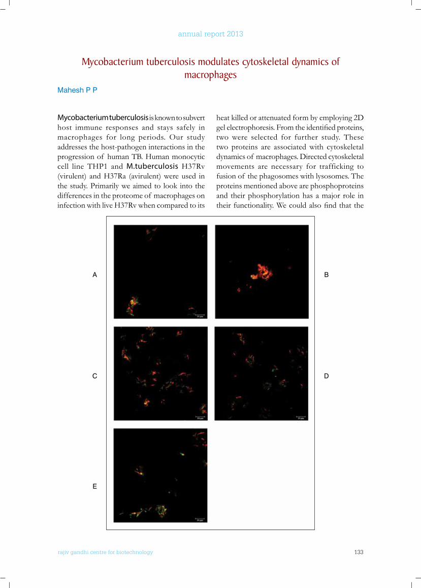
rajiv gandhi centre for biotechnology 133
annual report 2013
Mycobacterium tuberculosis is known to subvert host immune responses and stays safely in macrophages for long periods. Our study addresses the host-pathogen interactions in the progression of human TB. Human monocytic cell line THP1 and M.tuberculosis H37Rv (virulent) and H37Ra (avirulent) were used in the study. Primarily we aimed to look into the differences in the proteome of macrophages on infection with live H37Rv when compared to its
heat killed or attenuated form by employing 2D gelelectrophoresis.Fromtheidentifiedproteins,two were selected for further study. These two proteins are associated with cytoskeletal dynamics of macrophages. Directed cytoskeletal movements are necessary for trafficking to fusion of the phagosomes with lysosomes. The proteins mentioned above are phosphoproteins and their phosphorylation has a major role in theirfunctionality.Wecouldalsofindthatthe
Mycobacterium tuberculosis modulates cytoskeletal dynamics of macrophages
Mahesh P P
A
C
E
B
D
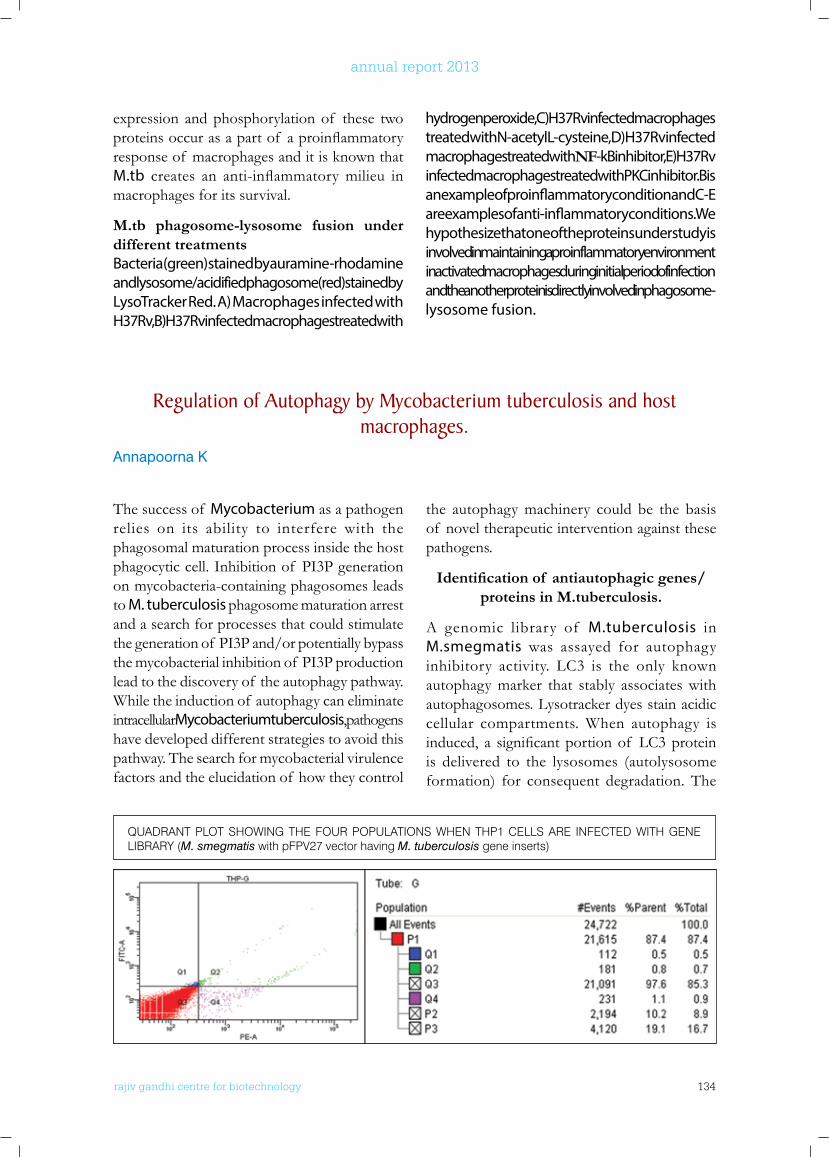
rajiv gandhi centre for biotechnology 134
annual report 2013
expression and phosphorylation of these two proteinsoccurasapartof aproinflammatoryresponse of macrophages and it is known that M.tb creates an anti-inflammatorymilieu inmacrophages for its survival.
M.tb phagosome-lysosome fusion under different treatmentsBacteria (green) stained by auramine-rhodamine and lysosome/acidified phagosome (red) stained by LysoTracker Red. A) Macrophages infected with H37Rv, B) H37Rv infected macrophages treated with
hydrogen peroxide, C) H37Rv infected macrophages treated with N-acetyl L-cysteine, D) H37Rv infected macrophages treated with NF-kB inhibitor, E) H37Rv infected macrophages treated with PKC inhibitor. B is an example of proinflammatory condition and C-E are examples of anti-inflammatory conditions. We hypothesize that one of the proteins under study is involved in maintaining a proinflammatory environment in activated macrophages during initial period of infection and the another protein is directly involved in phagosome-lysosome fusion.
QUADRANT PLOT SHOWING THE FOUR POPULATIONS WHEN THP1 CELLS ARE INFECTED WITH GENE LIBRARY (M. smegmatis with pFPV27 vector having M. tuberculosis gene inserts)
Regulation of Autophagy by Mycobacterium tuberculosis and host macrophages.
Annapoorna K
The success of Mycobacterium as a pathogen relies on its ability to interfere with the phagosomal maturation process inside the host phagocytic cell. Inhibition of PI3P generation on mycobacteria-containing phagosomes leads to M. tuberculosis phagosome maturation arrest and a search for processes that could stimulate the generation of PI3P and/or potentially bypass the mycobacterial inhibition of PI3P production lead to the discovery of the autophagy pathway. While the induction of autophagy can eliminate intracellular Mycobacterium tuberculosis, pathogens have developed different strategies to avoid this pathway. The search for mycobacterial virulence factors and the elucidation of how they control
the autophagy machinery could be the basis of novel therapeutic intervention against these pathogens.
Identification of antiautophagic genes/proteins in M.tuberculosis.
A genomic library of M.tuberculosis in M.smegmatis was assayed for autophagy inhibitory activity. LC3 is the only known autophagy marker that stably associates with autophagosomes. Lysotracker dyes stain acidic cellular compartments. When autophagy is induced, a significantportionof LC3proteinis delivered to the lysosomes (autolysosome formation) for consequent degradation. The
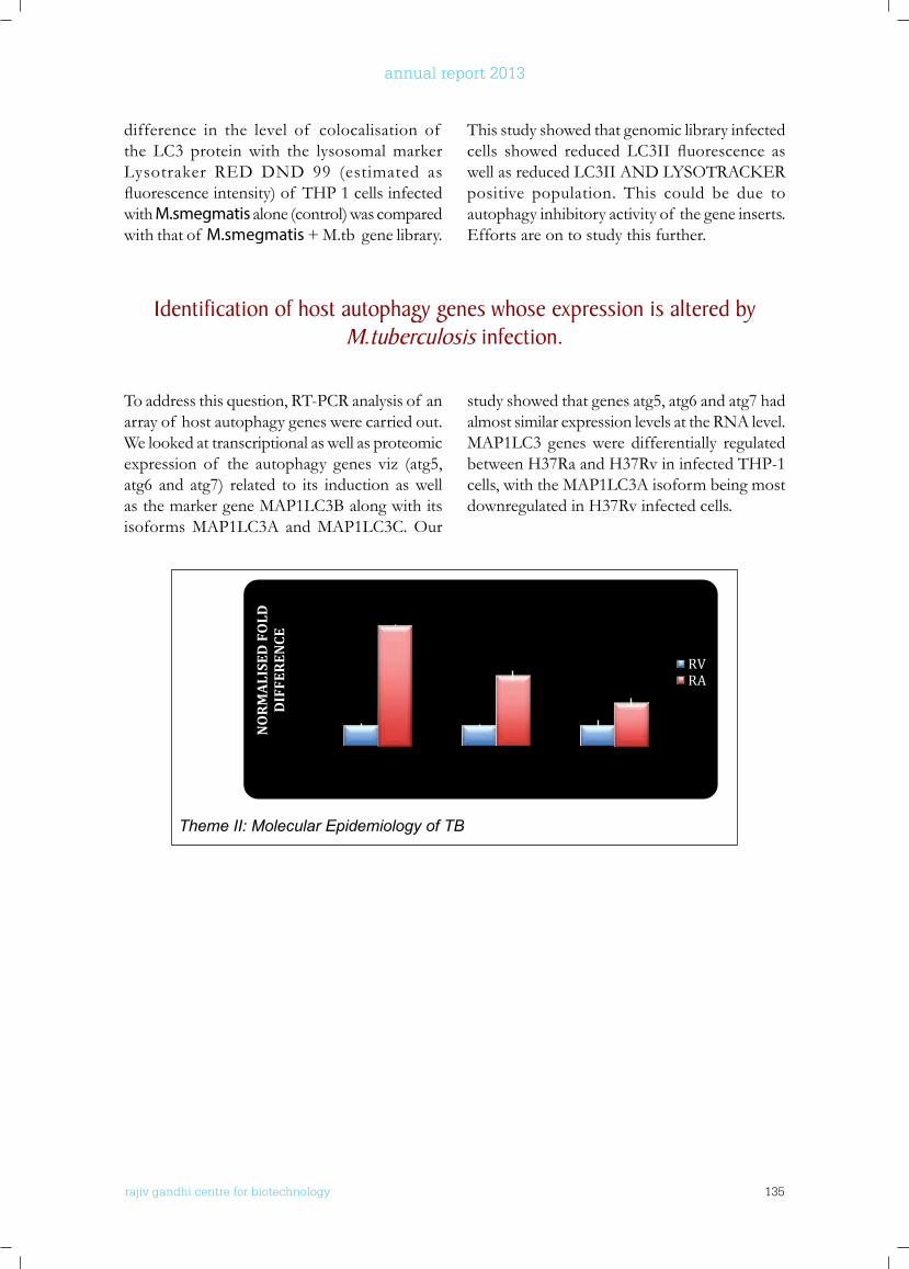
rajiv gandhi centre for biotechnology 135
annual report 2013
difference in the level of colocalisation of the LC3 protein with the lysosomal marker Lysotraker RED DND 99 (estimated as fluorescenceintensity)of THP1cellsinfectedwith M.smegmatis alone (control) was compared with that of M.smegmatis + M.tb gene library.
This study showed that genomic library infected cells showed reduced LC3II fluorescence aswellasreducedLC3IIANDLYSOTRACKERpositive population. This could be due to autophagy inhibitory activity of the gene inserts. Efforts are on to study this further.
Identification of host autophagy genes whose expression is altered by M.tuberculosis infection.
To address this question, RT-PCR analysis of an array of host autophagy genes were carried out. We looked at transcriptional as well as proteomic expression of the autophagy genes viz (atg5, atg6 and atg7) related to its induction as well as the marker gene MAP1LC3B along with its isoforms MAP1LC3A and MAP1LC3C. Our
study showed that genes atg5, atg6 and atg7 had almost similar expression levels at the RNA level. MAP1LC3 genes were differentially regulated between H37Ra and H37Rv in infected THP-1 cells, with the MAP1LC3A isoform being most downregulated in H37Rv infected cells.
rajiv gandhi center forbiotechnology6
2013-14 Annual Report
autophagy genes viz (atg5, atg6 and atg7) related to its induction as well as the marker gene MAP1LC3B along with its isoforms MAP1LC3A and MAP1LC3C. Our study showed that genes atg5, atg6 and atg7 had almost similar expression levels at the RNA level. MAP1LC3 genes were differentially regulated between H37Ra and H37Rv in infected THP-1 cells, with the MAP1LC3A isoform being most downregulated in H37Rv infected cells.
Theme II: Molecular Epidemiology of TB
Molecular epidemiology of Mycobacterium tuberculosis isolates from Kerala, India: Differentiation of major SIT by
MIRU-VNTRs. Biljo V Joseph
Tuberculosis (TB) continues to be a major health problem in India, and there is very little information about the prevalent genotypes of tubercle bacilli that cause TB in India, especially in Kerala. Our aim was to study the different circulating strains of Mycobacterium tuberculosis (MTB) that are prevalent in Kerala, India. We analyzed 168 MTB isolates from as many pulmonary TB patients using IS6110-RFLP, spoligotyping and MIRU-VNTRs. The present study evaluated the ability of MIRU–VNTRs to discriminate clinical isolates of Mycobacterium tuberculosis belonging to the SIT11/EAI3-IND ancestral genotypic lineage. Starting from 168 Mycobacterium tuberculosis (MTB) clinical isolates, spoligotyping (discriminatory index of 0.9113), differentiated the strains into 68 distinct patterns, the biggest cluster being SIT11/48 SIT11 (n=48). We show that 12-loci MIRUs and 3 ETRs allowed an efficient discrimination of these isolates (discriminatory indexes of 0.7819 and 0.5523, respectively).
NO
RM
ALI
SED
FO
LD
DIF
FER
ENCE
RVRA
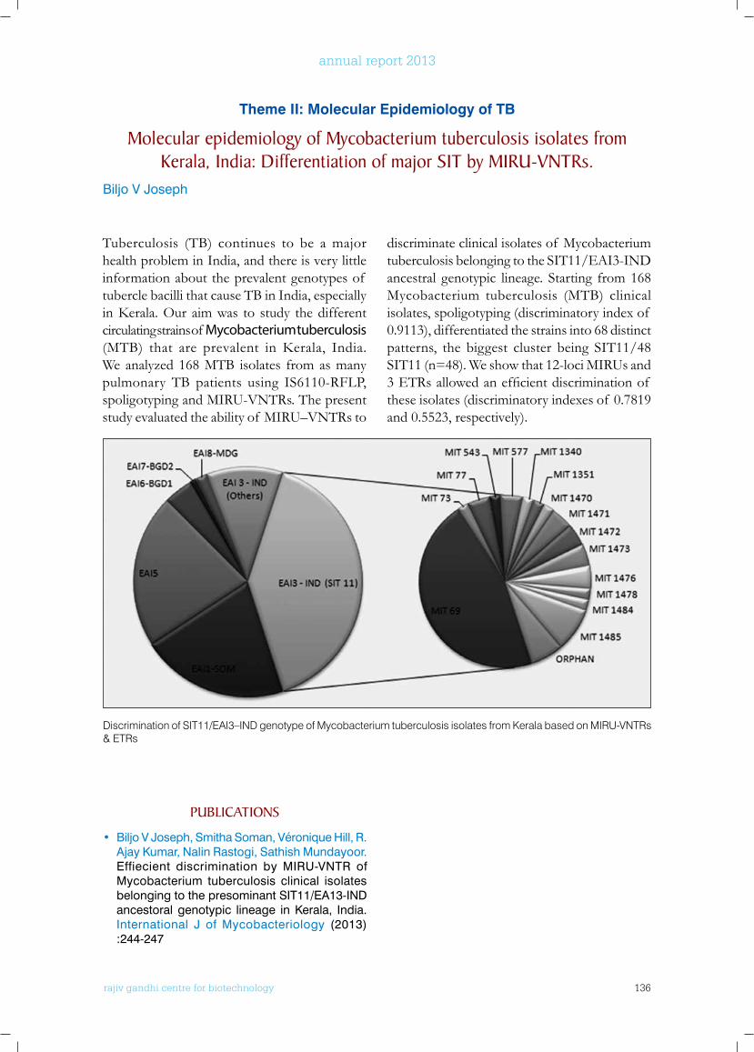
rajiv gandhi centre for biotechnology 136
annual report 2013
Tuberculosis (TB) continues to be a major health problem in India, and there is very little information about the prevalent genotypes of tubercle bacilli that cause TB in India, especially inKerala.Ouraimwas to study thedifferentcirculating strains of Mycobacterium tuberculosis (MTB) that are prevalent inKerala, India.We analyzed 168 MTB isolates from as many pulmonary TB patients using IS6110-RFLP, spoligotyping and MIRU-VNTRs. The present study evaluated the ability of MIRU–VNTRs to
discriminate clinical isolates of Mycobacterium tuberculosis belonging to the SIT11/EAI3-IND ancestral genotypic lineage. Starting from 168 Mycobacterium tuberculosis (MTB) clinical isolates, spoligotyping (discriminatory index of 0.9113), differentiated the strains into 68 distinct patterns, the biggest cluster being SIT11/48 SIT11 (n=48). We show that 12-loci MIRUs and 3ETRsallowedanefficientdiscriminationof these isolates (discriminatory indexes of 0.7819 and 0.5523, respectively).
Theme ii: Molecular epidemiology of TB
Molecular epidemiology of Mycobacterium tuberculosis isolates from kerala, India: Differentiation of major SIT by MIRu-VnTRs.
Biljo V Joseph
PublICATIonS
• Biljo V Joseph, Smitha Soman, Véronique Hill, R. Ajay Kumar, Nalin Rastogi, Sathish Mundayoor. Effiecient discrimination by MIRU-VNTR of Mycobacterium tuberculosis clinical isolates belonging to the presominant SIT11/EA13-IND ancestoral genotypic lineage in Kerala, India. International J of Mycobacteriology (2013) :244-247
Discrimination of SIT11/EAI3–IND genotype of Mycobacterium tuberculosis isolates from Kerala based on MIRU-VNTRs & ETRs
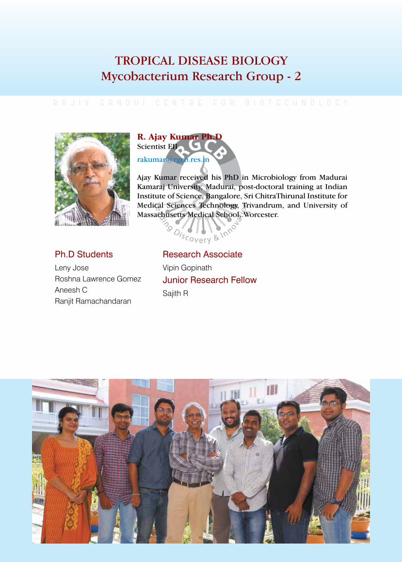
rajiv gandhi centre for biotechnology 137
annual report 2013
TROPICAL DISEASE BIOLOGY Mycobacterium Research Group - 2
Ph.D StudentsLeny JoseRoshna Lawrence GomezAneesh CRanjit Ramachandaran
r a j i v g a n d h i c e n t r e f o r b i o t e c h n o l o g y
Ajay Kumar received his PhD in Microbiology from Madurai Kamaraj University, Madurai, post-doctoral training at Indian Institute of Science, Bangalore, Sri ChitraThirunal Institute for Medical Sciences Technology, Trivandrum, and University of Massachusetts Medical School, Worcester.
R. Ajay Kumar Ph.D Scientist EII
Research AssociateVipin Gopinath
Junior Research FellowSajith R
rajiv gandhi centre for biotechnology 138
annual report 2013
We developed a simple in vitro reactivation model for M. tuberculosis based on Wayne’s dormancy modelandcomparedtheproteomeprofilesof dormant and reactivated bacteria using label-free one-dimensional LC-MS/MS analysis. Proteins were isolated from the normoxic control, two phases of dormancy (non-replicating persistent phase 1, NRP1, 12th day of dormancy), and NRP2 (21st day of dormancy), and two phases of reactivation (6th and 24th hours – R6 and R24). Proteome analysis was carried out using Synapt LC-MS/MS (Waters) system. A total of 1532proteinswereidentifiedfromthebacterialysates of thes five stages. From the culture grown in normoxic condition (aerobically grown control) we identified 1354 proteins.Two hundred and thirty seven proteins were obtained from the 12th day of dormancy (D12), 628 proteins from 21st day of dormancy (D21), 714 proteins from 6th hour of reactivation (R6) and 972 proteins from 24th hour of reactivation (R24). At NRP1 164 proteins and at NRP2 341 proteins were differentially expressed. Similarly upon reactivation, 208 proteins were differentially expressed at R6 and 220 proteins atR24.Theidentifiedproteinswerefunctionallyannotated using online tool DAVID (http://david.abcc.ncifcrf.gov/). The results obtained are shown in Fig 1. By analyzing the protein enrichment data we found that proteins in the
major biological processes were very few at D12, and the number gradually increased at D21. This shows that at NRP2, the bacteria gradually got relieved from the initial hypoxic shock of the NRP1, and became acclimatized to the hypoxic condition, and started synthesizing proteins required for maintaining minimal biological functions for survival. Interestingly we observed up-regulation of proteins in metabolic processes causing glyoxylate shunt during NRP2 and some of the electron transport components such as Cytochrome D terminal oxidase complex were up-regulated. Fatty acid biosynthesis was down-regulated during dormancy and returned to normal (control) levels upon reactivation, while the number of enzymes required for fatty acid breakdown was comparatively higher during dormancy. A decrease in protein enrichment was observed in glycolytic pathway during NRP1 and NRP2. Complete shutdown of the cell wall macromolecule biosynthetic process was observed at NRP1 and NRP2. Proteins of stress-response pathway were present during dormancy (D12, D21) and in the initial phase of reactivation (R6), but were completely absent in the control and later phase of reactivation (R24). Interestingly the number of stress proteins at R6 was higher than that in both phases of dormancy. This perhaps suggests that the rapid increase in the concentration of oxygen (normoxic state)
Mycobacterium tuberculosis is the causative agent of tuberculosis (TB) and is responsible for approximately 1.4 million deaths a year across the world. The WHO estimates that one third of the world’s population is asymptomatically infected with TB and 10% of this population will eventually develop the disease. The rise in the number of TB patients worldwide highlights the necessity for new approaches to treat this disease. The search for novel strategies for drug target identification and development is an important step in this process.
Identification of transcriptional regulators expressed in Mycobacterium tuberculosis during reactivation from dormancy in vitro.
Vipin Gopinath, Sajith R, Sathish Mundayoor, Abdul Jaleel* and R. Ajay Kumar.
*Collaborator: Diabetes Biology Laboratory, RGCB
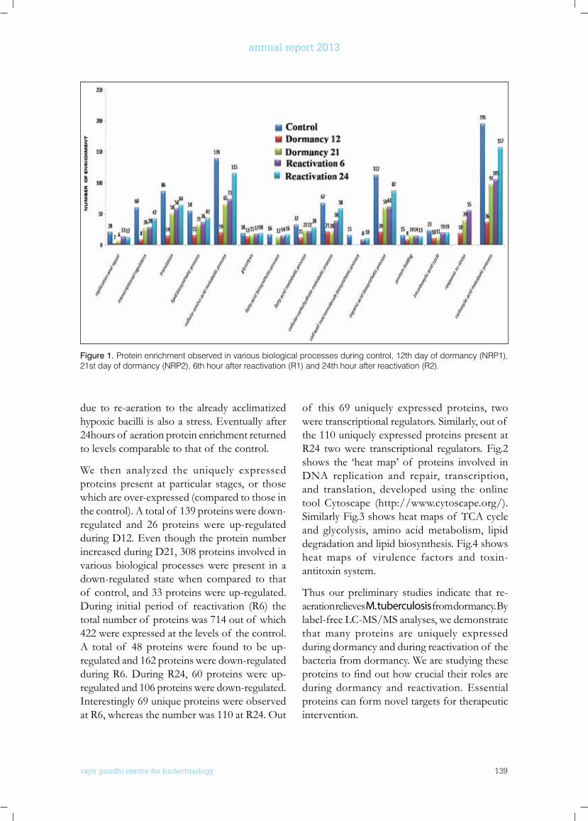
rajiv gandhi centre for biotechnology 139
annual report 2013
Figure 1. Protein enrichment observed in various biological processes during control, 12th day of dormancy (NRP1), 21st day of dormancy (NRP2), 6th hour after reactivation (R1) and 24th hour after reactivation (R2).
due to re-aeration to the already acclimatized hypoxic bacilli is also a stress. Eventually after 24hours of aeration protein enrichment returned to levels comparable to that of the control.
We then analyzed the uniquely expressed proteins present at particular stages, or those which are over-expressed (compared to those in the control). A total of 139 proteins were down-regulated and 26 proteins were up-regulated during D12. Even though the protein number increased during D21, 308 proteins involved in various biological processes were present in a down-regulated state when compared to that of control, and 33 proteins were up-regulated. During initial period of reactivation (R6) the total number of proteins was 714 out of which 422 were expressed at the levels of the control. A total of 48 proteins were found to be up-regulated and 162 proteins were down-regulated during R6. During R24, 60 proteins were up-regulated and 106 proteins were down-regulated. Interestingly 69 unique proteins were observed at R6, whereas the number was 110 at R24. Out
of this 69 uniquely expressed proteins, two were transcriptional regulators. Similarly, out of the 110 uniquely expressed proteins present at R24 two were transcriptional regulators. Fig.2 shows the ‘heat map’ of proteins involved in DNA replication and repair, transcription, and translation, developed using the online tool Cytoscape (http://www.cytoscape.org/). Similarly Fig.3 shows heat maps of TCA cycle and glycolysis, amino acid metabolism, lipid degradation and lipid biosynthesis. Fig.4 shows heat maps of virulence factors and toxin-antitoxin system.
Thus our preliminary studies indicate that re-aeration relieves M. tuberculosis from dormancy. By label-free LC-MS/MS analyses, we demonstrate that many proteins are uniquely expressed during dormancy and during reactivation of the bacteria from dormancy. We are studying these proteinstofindouthowcrucialtheirrolesareduring dormancy and reactivation. Essential proteins can form novel targets for therapeutic intervention.
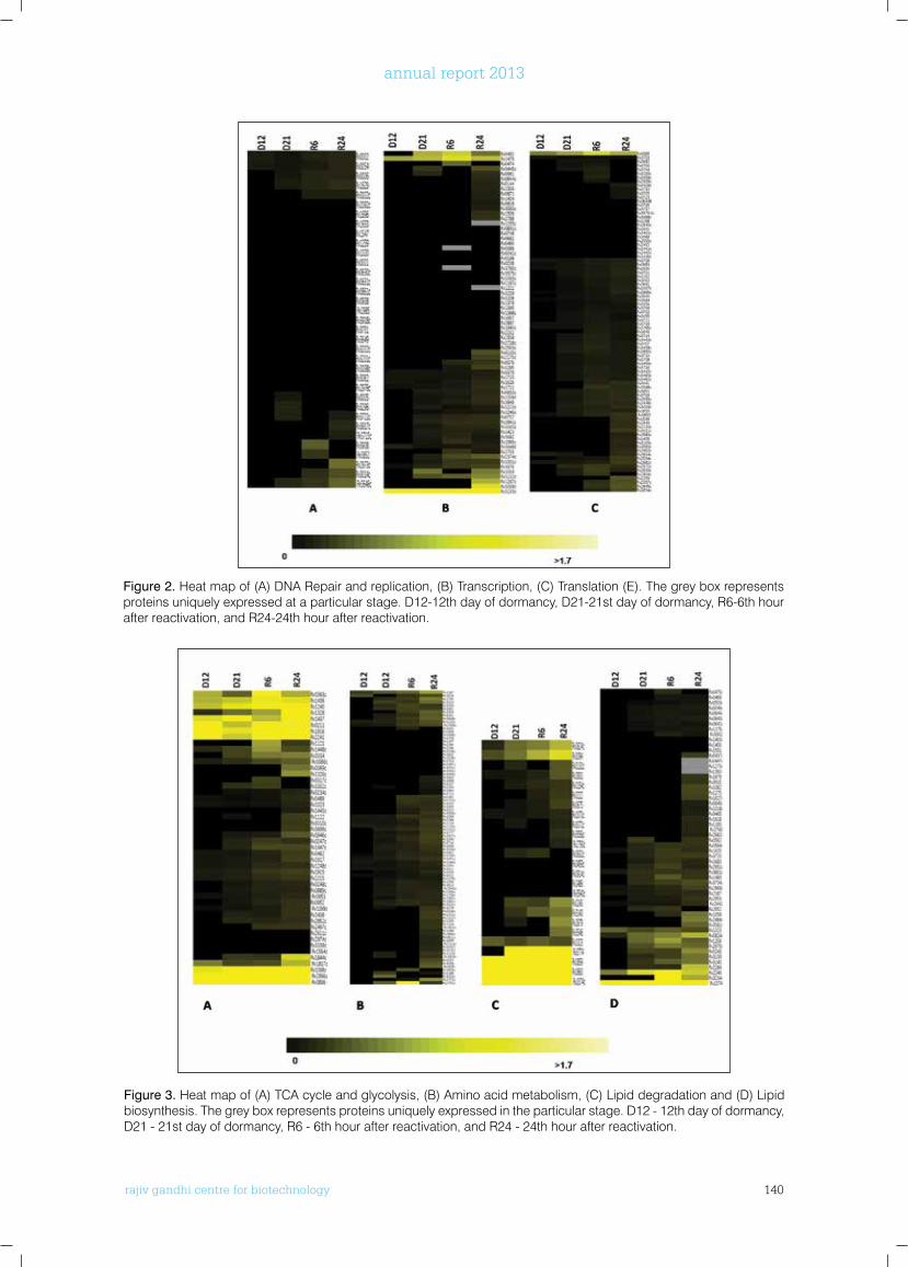
rajiv gandhi centre for biotechnology 140
annual report 2013
Figure 2. Heat map of (A) DNA Repair and replication, (B) Transcription, (C) Translation (E). The grey box represents proteins uniquely expressed at a particular stage. D12-12th day of dormancy, D21-21st day of dormancy, R6-6th hour after reactivation, and R24-24th hour after reactivation.
Figure 3. Heat map of (A) TCA cycle and glycolysis, (B) Amino acid metabolism, (C) Lipid degradation and (D) Lipid biosynthesis. The grey box represents proteins uniquely expressed in the particular stage. D12 - 12th day of dormancy, D21 - 21st day of dormancy, R6 - 6th hour after reactivation, and R24 - 24th hour after reactivation.
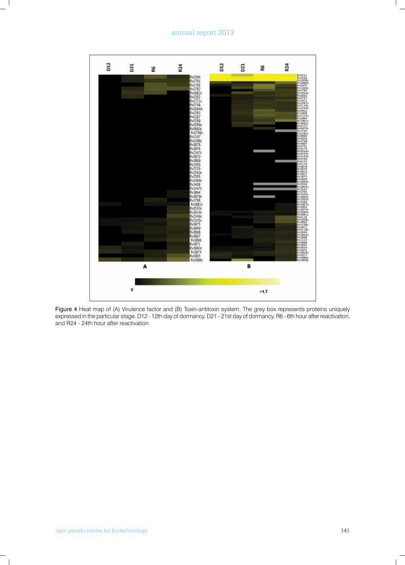
rajiv gandhi centre for biotechnology 141
annual report 2013
Figure 4 Heat map of (A) Virulence factor and (B) Toxin-antitoxin system. The grey box represents proteins uniquely expressed in the particular stage. D12 - 12th day of dormancy, D21 - 21st day of dormancy, R6 - 6th hour after reactivation, and R24 - 24th hour after reactivation.
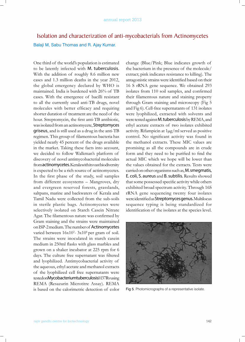
rajiv gandhi centre for biotechnology 142
annual report 2013
One third of the world’s population is estimated to be latently infected with M. tuberculosis. With the addition of roughly 8.6 million new cases and 1.3 million deaths in the year 2012, the global emergency declared by WHO is maintained. India is burdened with 26% of TB cases. With the emergence of bacilli resistant to all the currently used anti-TB drugs, novel moleculeswith better efficacy and requiringshorter duration of treatment are the need of the hour.Streptomycin,thefirstanti-TBantibiotic,was isolated from an actinomycete, Streptomyces griseus, and is still used as a drug in the anti-TB regimen.Thisgroupof filamentousbacteriahasyielded nearly 45 percent of the drugs available in the market. Taking these facts into account, we decided to follow Walkman’s platform of discovery of novel antimycobacterial molecules from actinomycetes.Keralawithitsvastbiodiversityis expected to be a rich source of actinomycetes. In the first phase of the study, soil samplesfrom different ecosystems – Mangroves, dry and evergreen reserved forests, grasslands, saltpans,marineandbackwatersof KeralaandTamil Nadu were collected from the sub-soils in sterile plastic bags. Actinomycetes were selectively isolated on Starch Casein Nitrate Agar.ThefilamentousnaturewasconfirmedbyGram staining and the strains were maintained on ISP-2 medium. The number of Actinomycetes varied between 16x103- 3x10⁶ per gram of soil. The strains were inoculated in starch casein mediumin250mlflaskswithglassmarblesandgrown on a shaker incubator at 225 rpm for 6 days.Theculturefreesupernatantwasfilteredand lyophilized. Antimycobacterial activity of the aqueous, ethyl acetate and methanol extracts of the lyophilized cell free supernatants were tested on Mycobacterium tuberculosis H37Rv using REMA (Resazurin Microtitre Assay). REMA is based on the calorimetric detection of color
change (Blue/Pink; Blue indicates growth of the bacterium in the presence of the molecule/extract; pink indicates resistance to killing). The antagonisticstrainswereidentifiedbasedontheir16 S rRNA gene sequence. We obtained 293 isolatesfrom110soilsamples,andconfirmedtheirfilamentousnatureandstainingpropertythrough Gram staining and microscopy (Fig 5 and Fig 6). Cell-free supernatants of 131 isolates were lyophilized, extracted with solvents and were tested against M. tuberculosis by REMA, and ethyl acetate extracts of two isolates exhibited activity. Rifampicin at 1µg/ml served as positive control.No significant activitywas found inthe methanol extracts. These MIC values are promising as all the compounds are in crude formandtheyneedtobepurifiedtofindtheactual MIC which we hope will be lower than the values obtained for the extracts. Tests were carried on other organisms such as, M. smegmatis, E. coli, S. aureus and B. subtilis. Results showed thatsomepossessedspecificactivitywhileothersexhibited broad spectrum activity. Through 16S rRNA gene sequencing twenty four isolates wereidentifiedasStreptomyces genus. Multilocus sequence typing is being standardized for identificationof theisolatesatthespecieslevel.
Fig 5. Photomicrographs of a representative isolate.
Isolation and characterization of anti-mycobacterials from ActinomycetesBalaji M, Sabu Thomas and R. Ajay Kumar.
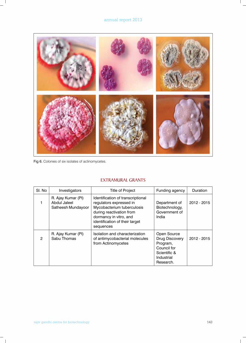
rajiv gandhi centre for biotechnology 143
annual report 2013
ExTRAMuRAl GRAnTS
Sl. No Investigators Title of Project Funding agency Duration
1R. Ajay Kumar (PI)Abdul Jaleel Satheesh Mundayoor
Identification of transcriptional regulators expressed in Mycobacterium tuberculosis during reactivation from dormancy in vitro, and identification of their target sequences
Department of Biotechnology, Government of India
2012 - 2015
2R. Ajay Kumar (PI)Sabu Thomas
Isolation and characterization of antimycobacterial molecules from Actinomycetes
Open Source Drug Discovery Program, Council for Scientific & Industrial Research.
2012 - 2015
Fig 6. Colonies of six isolates of actinomycetes.
rajiv gandhi centre for biotechnology 144
annual report 2013
TROPICAL DISEASE BIOLOGY Molecular Virology
Ph.D StudentsAnoop MRachy AbrahamPrashant MudaliarAnupriya M.GSneha Singh
r a j i v g a n d h i c e n t r e f o r b i o t e c h n o l o g y
E.Sreekumar is a post-graduate in Veterinary Immunology, and has a Ph.D in Biotechnology fromUniversity of Kerala. He joined RGCB in 2004.
E. Sreekumar, MVSc., Ph.D Scientist E I
Technical StaffUnnikrishnan V.R.
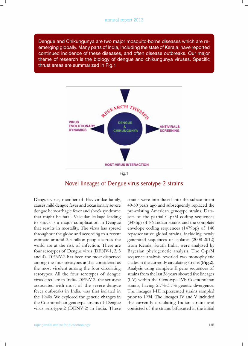
rajiv gandhi centre for biotechnology 145
annual report 2013
Dengue virus, member of Flaviviridae family, causes mild dengue fever and occasionally severe dengue hemorrhagic fever and shock syndrome that might be fatal. Vascular leakage leading to shock is a major complication in Dengue that results in mortality. The virus has spread throughout the globe and according to a recent estimate around 3.5 billion people across the world are at the risk of infection. There are four serotypes of Dengue virus (DENV-1, 2, 3 and 4). DENV-2 has been the most dispersed among the four serotypes and is considered as the most virulent among the four circulating serotypes. All the four serotypes of dengue virus circulate in India. DENV-2, the serotype associated with most of the severe dengue fever outbreaks in India,was first isolated inthe 1940s. We explored the genetic changes in the Cosmopolitan genotype strains of Dengue virus serotype-2 (DENV-2) in India. These
strains were introduced into the subcontinent 40-50 years ago and subsequently replaced the pre-existing American genotype strains. Data-sets of the partial C-prM coding sequences (348bp) of 86 Indian strains and the complete envelope coding sequences (1479bp) of 140 representative global strains, including newly generated sequences of isolates (2008-2012) fromKerala, South India, were analyzed byBayesian phylogenetic analysis. The C-prM sequence analysis revealed two monophyletic clades in the currently circulating strains (Fig.2). Analysis using complete E gene sequences of strainsfromthelast38yearsshowedfivelineages(I-V) within the Genotype IVb Cosmopolitan strains, having 2.7%-3.7% genetic divergence. The lineages I-III represented strains sampled prior to 1994. The lineages IV and V included the currently circulating Indian strains and consisted of the strains bifurcated in the initial
Dengue and Chikungunya are two major mosquito-borne diseases which are re-emerging globally. Many parts of India, including the state of Kerala, have reported continued incidence of these diseases, and often disease outbreaks. Our major theme of research is the biology of dengue and chikungunya viruses. Specific thrust areas are summarized in Fig.1
Fig.1
novel lineages of Dengue virus serotype-2 strains

rajiv gandhi centre for biotechnology 146
annual report 2013
C-prM sequence analysis, re-confirming the observation. Molecular clock analysis indicated the separation of lineage IV and V strains around 30 years ago. The lineage IV strains had a higher mean evolutionary rate (substitutions/site/year) (14.7×10⁻⁴) compared to that of the lineage V strains (7.1×10⁻⁴). Co-occurrence of four
signature substitutions differentiated the lineage IV and the lineage V. The envelope protein changes were not found to affect the protein structure in molecular dynamic simulation studies. We presume that the lineage V strains are more adapted and sturdy, and might eventually replace the lineage IV strains in the subcontinent.
Fig 2. Bayesian MCMC tree using C-PrM protein coding region of DENV-2 strains. Posterior probability values are indicated at the nodes and the scale bar represents the time scale in years.
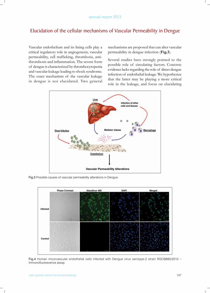
rajiv gandhi centre for biotechnology 147
annual report 2013
Vascular endothelium and its lining cells play a critical regulatory role in angiogenesis, vascular permeability, cell trafficking, thrombosis, anti-thrombosisandinflammation.Thesevereformof dengue is characterized by thrombocytopenia and vascular leakage leading to shock syndrome. The exact mechanism of the vascular leakage in dengue is not elucidated. Two general
mechanisms are proposed that can alter vascular permeability in dengue infection (Fig.3).
Several studies have strongly pointed to the possible role of circulating factors. Concrete evidence lacks regarding the role of direct dengue infection of endothelial leakage. We hypothesize that the latter may be playing a more critical role in the leakage, and focus on elucidating
Elucidation of the cellular mechanisms of Vascular Permeability in Dengue
Fig.3 Possible causes of vascular permeability alterations in Dengue.
Fig.4 Human micorvascular endothelial cells infected with Dengue virus serotype-2 strain RGCB880/2010 – Immunofluorescence assay.
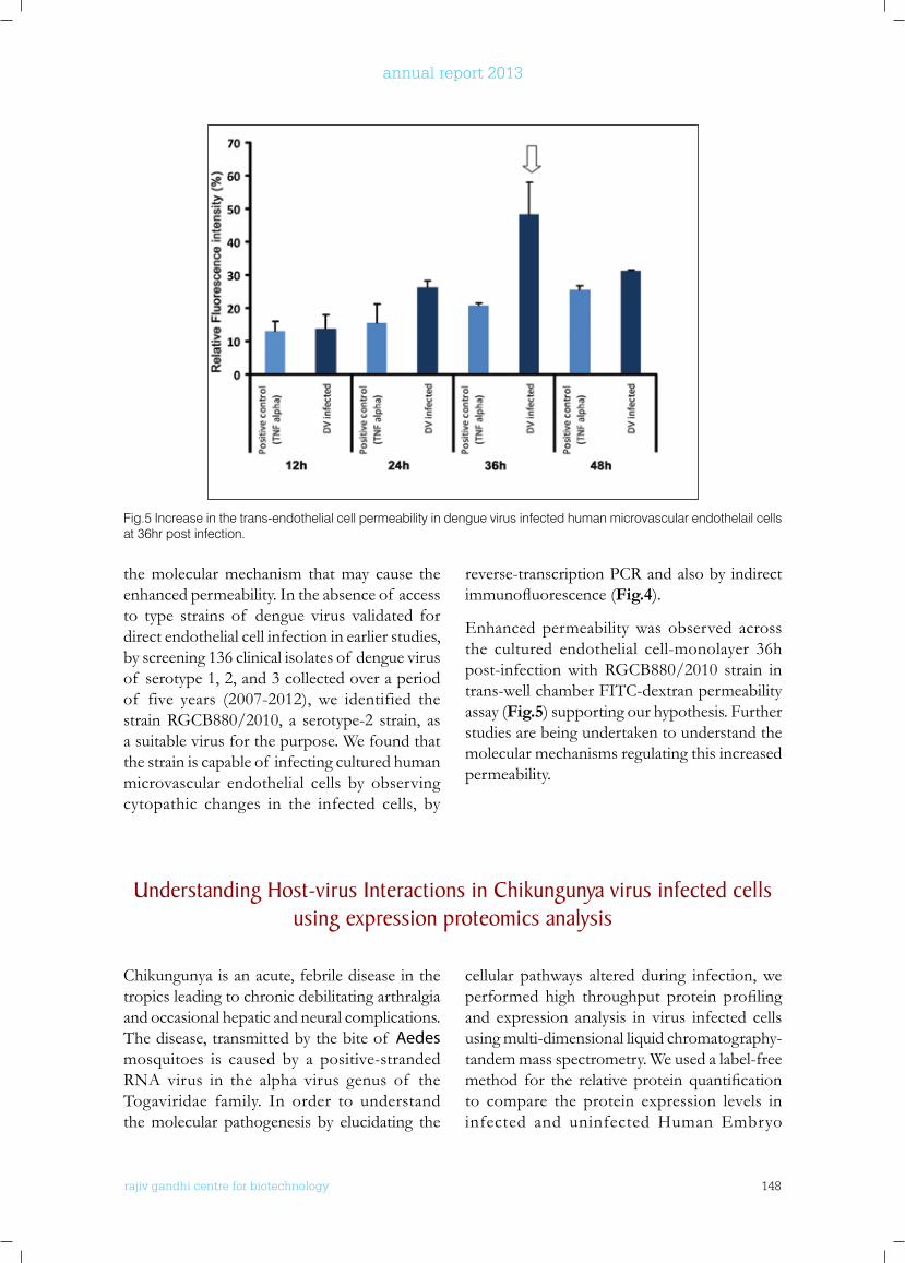
rajiv gandhi centre for biotechnology 148
annual report 2013
the molecular mechanism that may cause the enhanced permeability. In the absence of access to type strains of dengue virus validated for direct endothelial cell infection in earlier studies, by screening 136 clinical isolates of dengue virus of serotype 1, 2, and 3 collected over a period of five years (2007-2012), we identified the strain RGCB880/2010, a serotype-2 strain, as a suitable virus for the purpose. We found that the strain is capable of infecting cultured human microvascular endothelial cells by observing cytopathic changes in the infected cells, by
reverse-transcription PCR and also by indirect immunofluorescence(Fig.4).
Enhanced permeability was observed across the cultured endothelial cell-monolayer 36h post-infection with RGCB880/2010 strain in trans-well chamber FITC-dextran permeability assay (Fig.5) supporting our hypothesis. Further studies are being undertaken to understand the molecular mechanisms regulating this increased permeability.
Fig.5 Increase in the trans-endothelial cell permeability in dengue virus infected human microvascular endothelail cells at 36hr post infection.
understanding Host-virus Interactions in Chikungunya virus infected cells using expression proteomics analysis
Chikungunya is an acute, febrile disease in the tropics leading to chronic debilitating arthralgia and occasional hepatic and neural complications. The disease, transmitted by the bite of Aedes mosquitoes is caused by a positive-stranded RNA virus in the alpha virus genus of the Togaviridae family. In order to understand the molecular pathogenesis by elucidating the
cellular pathways altered during infection, we performed high throughput protein profilingand expression analysis in virus infected cells using multi-dimensional liquid chromatography- tandem mass spectrometry. We used a label-free method for the relative protein quantificationto compare the protein expression levels in infected and uninfected Human Embryo
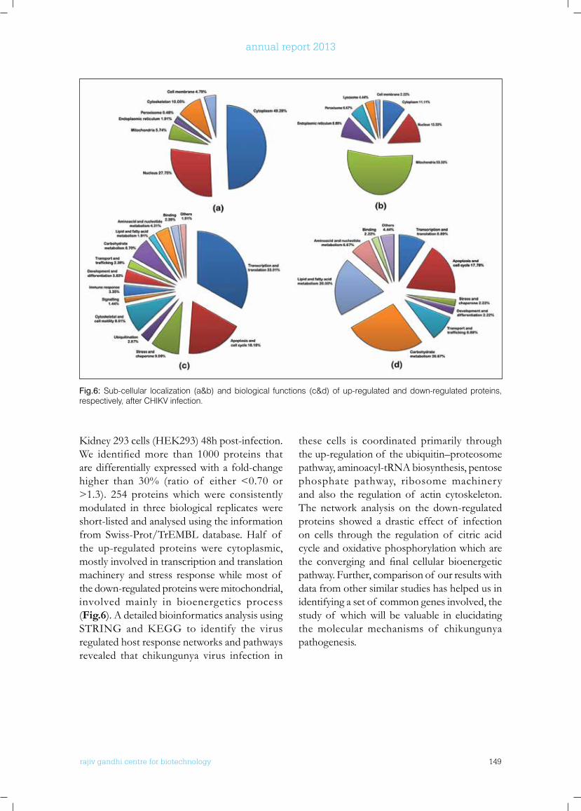
rajiv gandhi centre for biotechnology 149
annual report 2013
Kidney293cells(HEK293)48hpost-infection.We identifiedmore than 1000 proteins thatare differentially expressed with a fold-change higher than 30% (ratio of either <0.70 or >1.3). 254 proteins which were consistently modulated in three biological replicates were short-listed and analysed using the information from Swiss-Prot/TrEMBL database. Half of the up-regulated proteins were cytoplasmic, mostly involved in transcription and translation machinery and stress response while most of the down-regulated proteins were mitochondrial, involved mainly in bioenergetics process (Fig.6). A detailed bioinformatics analysis using STRING andKEGG to identify the virusregulated host response networks and pathways revealed that chikungunya virus infection in
these cells is coordinated primarily through the up-regulation of the ubiquitin–proteosome pathway, aminoacyl-tRNA biosynthesis, pentose phosphate pathway, ribosome machinery and also the regulation of actin cytoskeleton. The network analysis on the down-regulated proteins showed a drastic effect of infection on cells through the regulation of citric acid cycle and oxidative phosphorylation which are the converging andfinal cellular bioenergeticpathway. Further, comparison of our results with data from other similar studies has helped us in identifying a set of common genes involved, the study of which will be valuable in elucidating the molecular mechanisms of chikungunya pathogenesis.
Fig.6: Sub-cellular localization (a&b) and biological functions (c&d) of up-regulated and down-regulated proteins, respectively, after CHIKV infection.
rajiv gandhi centre for biotechnology 150
annual report 2013
PublICATIonSfRoM THE lAboRAToRy:
• Rachy Abraham, Prashant Mudaliar, Aiswaria Padmanabhan, easwaran Sreekumar (2013) Induction of cytopathogenicity in human glioblastoma cells by Chikungunya virus. PLoS ONE 8(9):e75854. doi:10.1371/journal.pone.0075854
• Anoop Manakkadan, Iype Joseph, Raji Rajendran Prasanna, Riaz Ismail kunju , Lalitha Kailas, easwaran Sreekumar (2013). Lineage shift in Indian strains of Dengue virus serotype-3 (Genotype III), evidenced by detection of lineage IV strains in clinical cases from Kerala. Virol J. 10(1): 37.
fRoM CollAboRATIonS:
• Praveen K. Sobhan, Mahendra Seervi, Lokesh Deb, Saneesh Varghese1, Anjana Soman, Jeena Joseph1, Krupa Ann Mathew, Godi Raghu, George Thomas, Sreekumar e, Manjula S, Santosh Kumar T. R (2013). Calpain and Reactive Oxygen Species Targets Bax for Mitochondrial Permeabilisation and Caspase Activation in Zerumbone Induced Apoptosis. PLoS ONE 8(4): e59350. doi:10.1371/journal.pone.0059350
ConfEREnCES/ SEMInARS/woRkSHoPS/ InTERnATIonAl fEllowSHIPS
• Rachy Abrahm attended a 9 months Fulbright fellowship at the Department of Molecular Microbiology and Immunology, Bloomberg School of Public Health, John Hopkins University, Baltimore, Maryland, USA.
• Sneha Singh. The 2nd National Symposium on nanobiotechnology (NSNB-2013) and a two day workshop on Transmission Electron Microscopy held at IIT, Mandi (H.P.), India on 9-11 December, 2013.
GEnbAnk SubMISSIonS
• Dengue virus 2 strain C-prM gene, partial cds - KF364516-22; KF364526-27; KF364529-34 (15 sequences)
• Dengue virus 2 strain envelope protein gene, complete cds - KF364497-KF364514 (18 sequences)
RESEARCH GRAnTS
No. Title Investigator(s) Funding Agency Duration
1. Characterization of Neurovirulence of Chikungunya virus in cellular and animal models
E. Sreekumar & Jackson James
Department of Biotechnology, Government of India
2012-2015
2 Elucidation of the role of endothelial cell signaling pathways in vascular permeability modulation in Dengue virus infection
E. Sreekumar & T.R. Santhosh Kumar
Indian Council of Medical Research, New Delhi
2013-2016

rajiv gandhi centre for biotechnology 151
annual report 2013
TROPICAL DISEASE BIOLOGY Viral Disease Biology
r a j i v g a n d h i c e n t r e f o r b i o t e c h n o l o g y
India accounts for nearly half of the global measlesmortality.Kerala, the southernmoststate, has better health parameter indicators and immunization coverage compared to many other regions of the country. However, the disease is still endemic in the state. Data on circulating viral genotypes, which would become crucial during the measles elimination phase is lacking fromKerala.Inthiscontext,wehaveinitiated
a study to identify the circulating measles virus genotypesinKerala.UsinganRT-PCRassayfordetection of measles RNA, we have tested clinical specimens from 200 children with suspicion of measles in Thiruvananthapuram. Measles RNA was detected in 163 specimens. Using Vero-SLAM cells (transgenic cell lines expressing human SLAM, the receptor for measles virus), we were able to obtain viral isolates from 18 samples.
Joshy Jacob, PhD [Senior Consultant]M. Radhakrishna Pillai, FRCPath, PhD [Professor]Vijesh Sreedhar, MD [Program Scientist]Sara Jones, PhD [Research Associate]Pradip V. Fulmali [Scientific Officer]
Molecular characterization of measles viruses circulating in keralaVijesh Sreedhar K, MD
rajiv gandhi centre for biotechnology 152
annual report 2013
Manipulation of the cell death machinery by west nile virus (wnV) and its variants
Pradip V Fulmali and M Radhakrishna Pillai
To investigate whether the WNV (804994) growth showed similar growth pattern in different type of cell lines of human origin, growth kinetics of was studied. We have already demonstrated thatthenatureof celltype-specificrestrictionof infection by analyzing the replication of WNV (804994) strain in neuroblastoma (IMR), astrocyte (U87MG), macrophage (THP) and HumanKidneycell line(HEK).All thesecelllines supported growth of the virus and peak titer was attained by 4th post infection day (PID).WNVreplicatesfasterinHEK293andIMR-32 cell line during early hours of infection compared to U87MG and THP cells. It was also seen that U87MG and THP cells produced lessvirusprogenythanIMRandHEKcellline.We also demonstrated differential cell viability pattern in these cell lines and showed correlation between the increased in viral genomic copies with decrease in the percent cell viability. The WNV 804994 strain took <60hr post infection toinduce50%celldeathintheHEK(fibroblast)cell line and >72 hours post infection in IMR (neuroblastoma) cell line However, in U87MG (astrocyte) and THP (macrophage) cell line, the virus did not exhibit 50% cell mortality even after 96 hours post infection. For mock-infected cells,
the number of viable cells remained relatively constant throughout the experiment. These studies strongly suggest that difference in cell type affect the replication potential of the west Nile virus and the cell death induced by it. We further investigated whether the difference in replication potential of the WNV (804994) had any impact on the cell death mechanisms among the infected cell. It was observed that thefibroblast(HEK)cellsinfectedwithWNVshowed a very less population of early apoptotic cells after 24 hours of infection whereas no apoptotic cell population was observed at 48 hrs. Substantial increase in the necrotic cell population was observed as infection progress. We also found very low cleaved caspase-3 activity within 24 hours of infection whereas the necrotic death marker lactase dehydrogenase (LDH) levelshowedsignificantincreaseafter24and48hours of infection (Fig 1). The neuroblastoma cell (IMR-32) infected with WNV showed the increase in late apoptotic cell population as well assignificantincreaseincleavedcaspase-3levelsafter 24 hours of infection compared to un-infected cells. However necrotic cell population aswellasLDHlevelssignificantlyincreasedafter48 hours of infection
Genetic characterization of the virus was carried out based on the 450 nucleotides coding for the –COOH terminal of the nucleoprotein of measles virus as per WHO recommendation. Preliminary analysis was carried out with high quality sequence reads obtained from 10 samples. Phylogenetic analysis using these N gene sequences and the WHO reference strain sequences revealed that measles virus strain in one sample belonged to the genotype D8. The remaining nine samples had virus strains closely related to the B3 genotype reference strains, indicating the presence of this virus genotype
inKerala.Thenucleotide sequencesof sevenstrains among them were identical indicating a single chain of transmission. Virus strains from two cases showed sequence divergence indicating independent sources of infection. In phylogenetic analysis using a dataset of global measles B3 genotype strain sequences, the strains fromKerala formed a separate cluster. Thiscluster also contained a strain from Germany. The strains in this cluster showed closer identity to a measles strain from New York, USA. Presence of measles virus genotype B3 has not been detected in India previously.
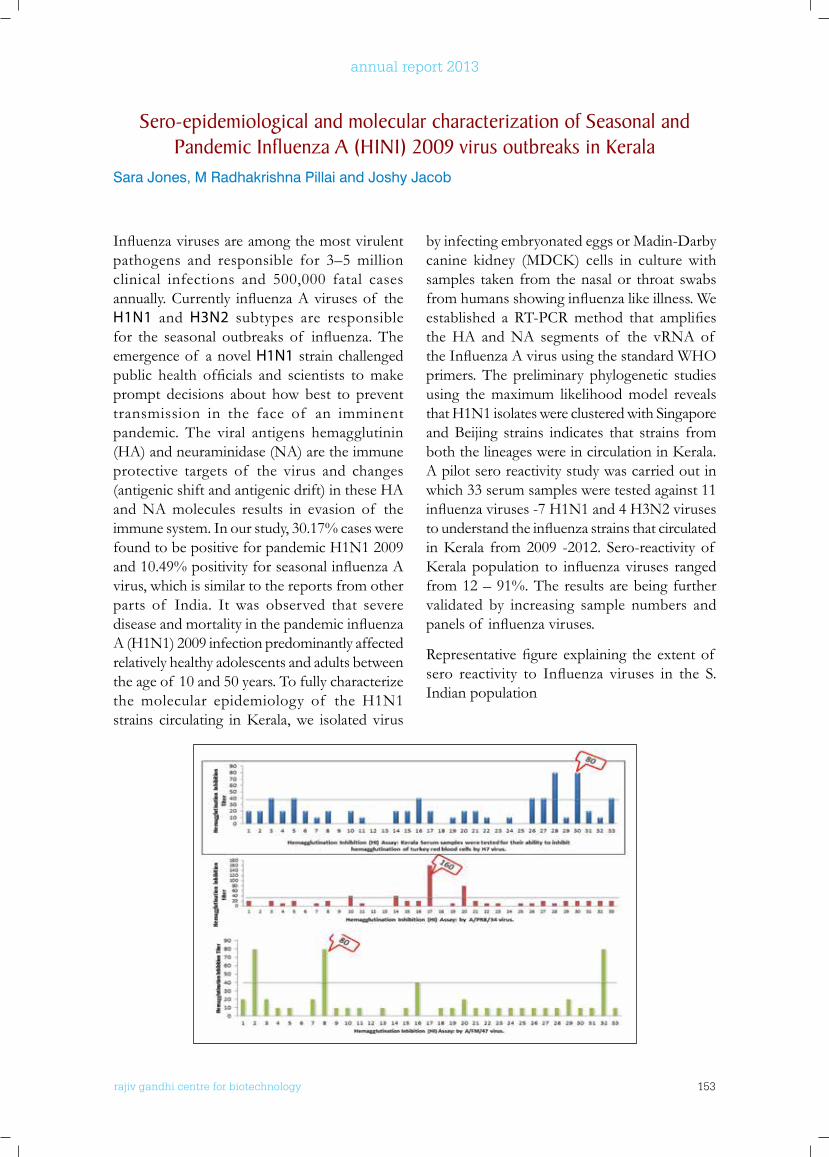
rajiv gandhi centre for biotechnology 153
annual report 2013
Sero-epidemiological and molecular characterization of Seasonal and Pandemic Influenza A (H1n1) 2009 virus outbreaks in kerala
Sara Jones, M Radhakrishna Pillai and Joshy Jacob
Influenzavirusesareamongthemostvirulentpathogens and responsible for 3–5 million clinical infections and 500,000 fatal cases annually.Currently influenzaAvirusesof theH1N1 and H3N2 subtypes are responsible for the seasonal outbreaks of influenza.Theemergence of a novel H1N1 strain challenged public health officials and scientists tomakeprompt decisions about how best to prevent transmission in the face of an imminent pandemic. The viral antigens hemagglutinin (HA) and neuraminidase (NA) are the immune protective targets of the virus and changes (antigenic shift and antigenic drift) in these HA and NA molecules results in evasion of the immune system. In our study, 30.17% cases were found to be positive for pandemic H1N1 2009 and10.49%positivityforseasonalinfluenzaAvirus, which is similar to the reports from other parts of India. It was observed that severe diseaseandmortalityinthepandemicinfluenzaA (H1N1) 2009 infection predominantly affected relatively healthy adolescents and adults between the age of 10 and 50 years. To fully characterize the molecular epidemiology of the H1N1 strains circulating inKerala,we isolated virus
by infecting embryonated eggs or Madin-Darby canine kidney (MDCK) cells in culturewithsamples taken from the nasal or throat swabs fromhumansshowinginfluenzalikeillness.Weestablished aRT-PCRmethod that amplifiesthe HA and NA segments of the vRNA of theInfluenzaAvirususingthestandardWHOprimers. The preliminary phylogenetic studies using the maximum likelihood model reveals that H1N1 isolates were clustered with Singapore and Beijing strains indicates that strains from boththelineageswereincirculationinKerala.A pilot sero reactivity study was carried out in which 33 serum samples were tested against 11 influenzaviruses-7H1N1and4H3N2virusestounderstandtheinfluenzastrainsthatcirculatedinKerala from2009 -2012. Sero-reactivityof Keralapopulation to influenzaviruses rangedfrom 12 – 91%. The results are being further validated by increasing sample numbers and panelsof influenzaviruses.
Representativefigureexplaining theextentof sero reactivity to Influenza viruses in the S.Indian population
rajiv gandhi centre for biotechnology 154
annual report 2013
r a j i v g a n d h i c e n t r e f o r b i o t e c h n o l o g y
Leptospirosis is a complex bacterial disease with multiple modes of transmission, numerous hosts, multitude of pathogenic serovars (>200), various clinical manifestations, and need complex testingtoprovidelaboratoryconfirmation.Case-fatality rates for the disease range from <5% to 30%. Humans and animals may become infected through direct contact with contaminated urine or indirectly through exposure to contaminated water or soil. In the current year, rodent studies were initiated in Ernakulam city and Trivandrum district. The gold-standard test for Leptospira diagnosis in man and animals, the Micro-Agglutination Test (MAT) was established in this laboratory. For this purpose, a new set of 12 Reference strains of Leptospira was received from RMRC-ICMR. They are being maintained
in the laboratory. Rodent studies have been initiated in a Government Animal Farm under theAnimalHusbandryDepartmentof Keralaand other scattered areas in Trivandrum District and in the Ernakulam Market. The Animal farm had one human patient and few animals with suspected leptospirosis. The market has been selected due to the high prevalence of rodents. To date 45 wild-caught rats were studied. Aspirates from the kidneys and the urine were inoculated in Ellinghausen-McCullough-Johnson-Harris (EMJH) medium and observed for six weeks for leptospires by Dark Field examination. All cultures were negative. One serum sample gave positive result at 1:80 titre in MAT test for Hebdomadis serogroup.
TROPICAL DISEASE BIOLOGY Parasite Biology Laboratory
Iype Joseph MB.BS, MPH Program Scientist
Epidemiology of leptospirosis in kerala, India Iype Joseph
rajiv gandhi centre for biotechnology 155
annual report 2013
TROPICAL DISEASE BIOLOGY Malaria Biology Laboratory
A r u m u g a m R a j a v e l u obtained his M.Sc Medical Microbiology from Post G r a d u a t e I n s t i t u t e of Medical Sc iences, University of Madras. He completed his PhD from Jacobs University of Bremen- Germany in 2011, and then worked as Post-doctoral fellow at Stuttgart University for two years. He joined RGCB in December 2013 as a DST INSPIRE faculty.
Arumugam Rajavelu Principal Investigator
Malaria is an infectious disease caused by apicomplexan parasite. Five Plasmodium species P. falciparum, P. vivax, P. Malariae, P. ovaleand P. knowlesihave been identifiedwhich infectshumans.Amongthefivespecies,P.falciparumcauses the most severe form of malaria. The female anopheles mosquito injects 20-100 sporozoites to human dermis to initiate the infection; these sporozoites reach the liver via blood stream. In liver the parasites replicate and release the merozoites into blood stream, which further infects RBC and continues as asexual intraerythrocytic developmental cycle (IDC). The asexual growth of parasite consist different stages, including rings, trophozoites and schizonts, the mature schizonts ruptures the RBC and releases upto 30 merozoites that can invade into new RBCs and continue as
asexual life cycle. The clinical manifestations of malarial disease occur during the IDC cycle of P. falciparum, which includes the severe form of cerebral malaria. Recent evidence indicates that P. falciparum under goes massive changes in “transcriptional activity” during IDC cycle, which suggested the role of epigenetic players at different stages of parasite development.
Research work in my laboratory focuses on malarial biology and malarial epigenetics. We work on the tight regulation of chromatin in malarial parasite during its development in RBC andidentificationof modificationsontheRNAin the parasites. We also study the epigenetic protein’s role in differential gene expression in parasites, which eventually pave way to identify new drug targets.
r a j i v g a n d h i c e n t r e f o r b i o t e c h n o l o g y
rajiv gandhi centre for biotechnology 156
annual report 2013
TROPICAL DISEASE BIOLOGY Cholera and Biofilm Research Laboratory
Ph.D StudentsWilson Peter AbrahamDivya M.PAkhilandeswarre DKarthika S
r a j i v g a n d h i c e n t r e f o r b i o t e c h n o l o g y
Sabu Thomas received his Ph.D in aquatic biology from the University of Kerala and joined RGCB in 2001.
Sabu Thomas Ph.D Scientist E I
Project FellowsDiana JoseDeepa Mathew P
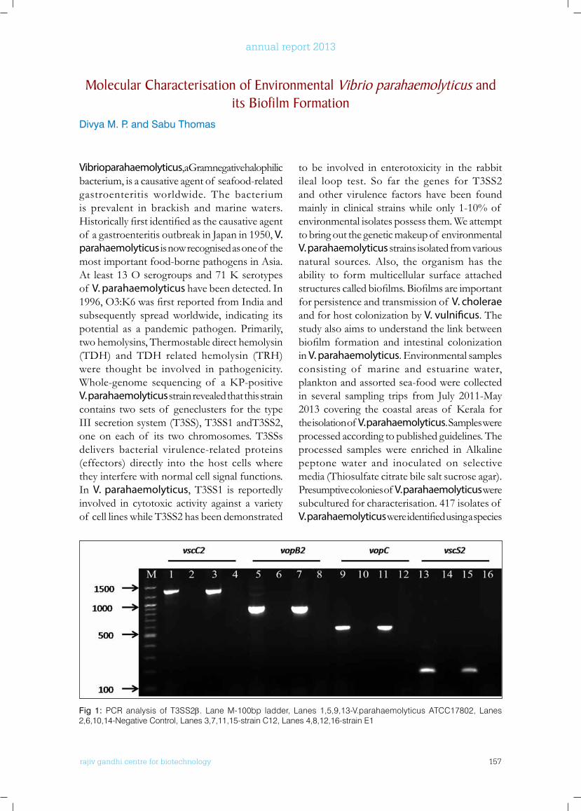
rajiv gandhi centre for biotechnology 157
annual report 2013
Vibrio parahaemolyticus, a Gram negative halophilic bacterium, is a causative agent of seafood-related gastroenteritis worldwide. The bacterium is prevalent in brackish and marine waters. Historicallyfirstidentifiedasthecausativeagentof a gastroenteritis outbreak in Japan in 1950, V. parahaemolyticus is now recognised as one of the most important food-borne pathogens in Asia. At least13Oserogroupsand71Kserotypesof V. parahaemolyticus have been detected. In 1996,O3:K6wasfirstreportedfromIndiaandsubsequently spread worldwide, indicating its potential as a pandemic pathogen. Primarily, two hemolysins, Thermostable direct hemolysin (TDH) and TDH related hemolysin (TRH) were thought be involved in pathogenicity. Whole-genome sequencing of aKP-positiveV. parahaemolyticus strain revealed that this strain contains two sets of geneclusters for the type III secretion system (T3SS), T3SS1 andT3SS2, one on each of its two chromosomes. T3SSs delivers bacterial virulence-related proteins (effectors) directly into the host cells where they interfere with normal cell signal functions. In V. parahaemolyticus, T3SS1 is reportedly involved in cytotoxic activity against a variety of cell lines while T3SS2 has been demonstrated
to be involved in enterotoxicity in the rabbit ileal loop test. So far the genes for T3SS2 and other virulence factors have been found mainly in clinical strains while only 1-10% of environmental isolates possess them. We attempt to bring out the genetic makeup of environmental V. parahaemolyticus strains isolated from various natural sources. Also, the organism has the ability to form multicellular surface attached structurescalledbiofilms.Biofilmsareimportantfor persistence and transmission of V. cholerae and for host colonization by V. vulnificus. The study also aims to understand the link between biofilm formation and intestinal colonizationin V. parahaemolyticus. Environmental samples consisting of marine and estuarine water, plankton and assorted sea-food were collected in several sampling trips from July 2011-May 2013 covering the coastal areasof Kerala forthe isolation of V. parahaemolyticus. Samples were processed according to published guidelines. The processed samples were enriched in Alkaline peptone water and inoculated on selective media (Thiosulfate citrate bile salt sucrose agar). Presumptive colonies of V. parahaemolyticus were subcultured for characterisation. 417 isolates of V. parahaemolyticuswereidentifiedusingaspecies
Molecular Characterisation of Environmental Vibrio parahaemolyticus and its biofilm formation
Divya M. P. and Sabu Thomas
Fig 1: PCR analysis of T3SS2β. Lane M-100bp ladder, Lanes 1,5,9,13-V.parahaemolyticus ATCC17802, Lanes 2,6,10,14-Negative Control, Lanes 3,7,11,15-strain C12, Lanes 4,8,12,16-strain E1
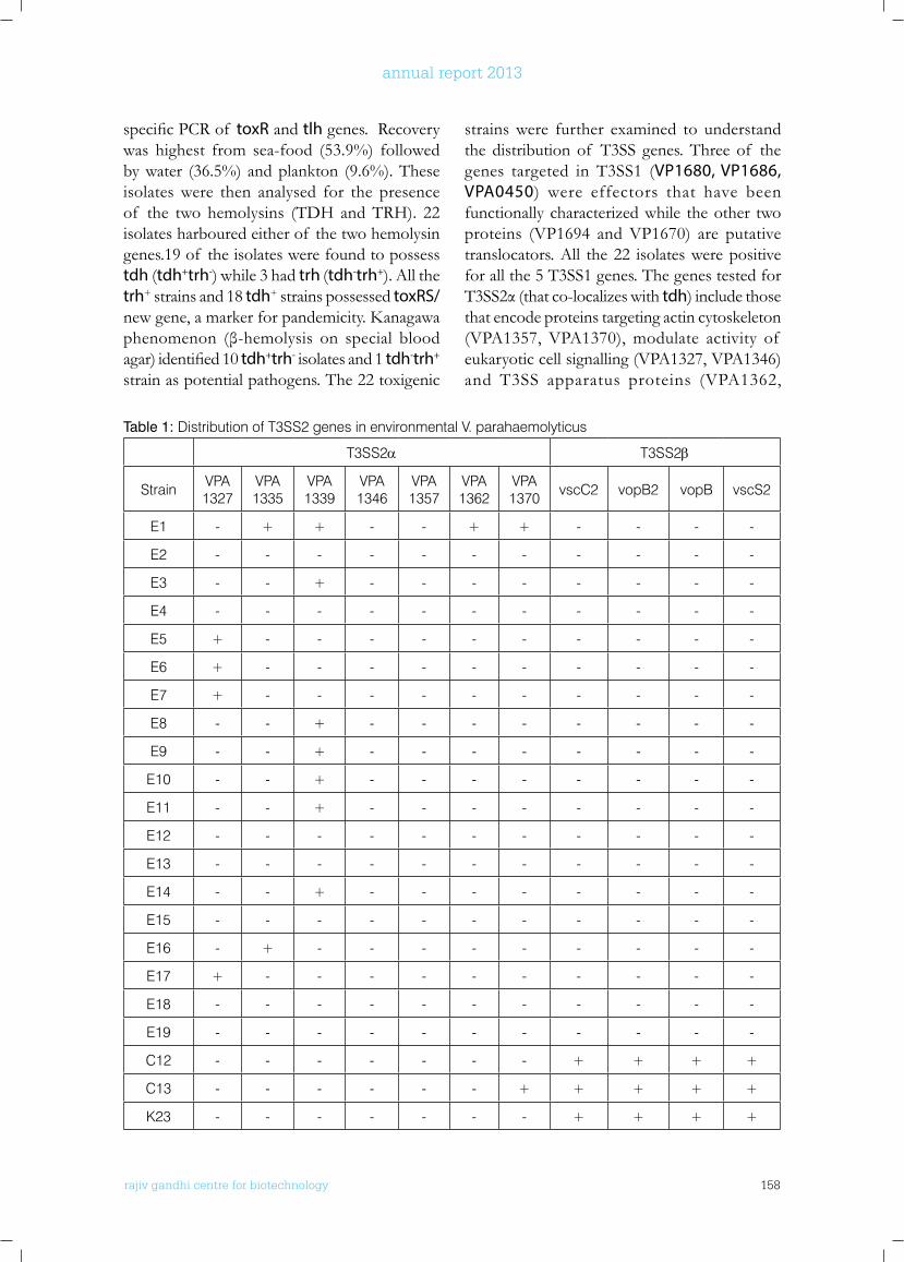
rajiv gandhi centre for biotechnology 158
annual report 2013
specificPCRof toxR and tlh genes. Recovery was highest from sea-food (53.9%) followed by water (36.5%) and plankton (9.6%). These isolates were then analysed for the presence of the two hemolysins (TDH and TRH). 22 isolates harboured either of the two hemolysin genes.19 of the isolates were found to possess tdh (tdh+trh-) while 3 had trh (tdh-trh+). All the trh+ strains and 18 tdh+ strains possessed toxRS/newgene,amarkerforpandemicity.Kanagawaphenomenon (β-hemolysis on special bloodagar)identified10tdh+trh- isolates and 1 tdh-trh+ strain as potential pathogens. The 22 toxigenic
strains were further examined to understand the distribution of T3SS genes. Three of the genes targeted in T3SS1 (VP1680, VP1686, VPA0450) were effectors that have been functionally characterized while the other two proteins (VP1694 and VP1670) are putative translocators. All the 22 isolates were positive for all the 5 T3SS1 genes. The genes tested for T3SS2α(thatco-localizeswithtdh) include those that encode proteins targeting actin cytoskeleton (VPA1357, VPA1370), modulate activity of eukaryotic cell signalling (VPA1327, VPA1346) and T3SS apparatus proteins (VPA1362,
Table 1: Distribution of T3SS2 genes in environmental V. parahaemolyticus
T3SS2α T3SS2β
Strain VPA1327
VPA1335
VPA1339
VPA1346
VPA1357
VPA1362
VPA1370 vscC2 vopB2 vopB vscS2
E1 - + + - - + + - - - -
E2 - - - - - - - - - - -
E3 - - + - - - - - - - -
E4 - - - - - - - - - - -
E5 + - - - - - - - - - -
E6 + - - - - - - - - - -
E7 + - - - - - - - - - -
E8 - - + - - - - - - - -
E9 - - + - - - - - - - -
E10 - - + - - - - - - - -
E11 - - + - - - - - - - -
E12 - - - - - - - - - - -
E13 - - - - - - - - - - -
E14 - - + - - - - - - - -
E15 - - - - - - - - - - -
E16 - + - - - - - - - - -
E17 + - - - - - - - - - -
E18 - - - - - - - - - - -
E19 - - - - - - - - - - -
C12 - - - - - - - + + + +
C13 - - - - - - + + + + +
K23 - - - - - - - + + + +
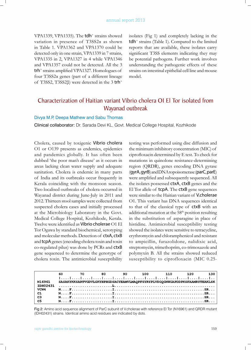
rajiv gandhi centre for biotechnology 159
annual report 2013
VPA1339, VPA1335). The tdh+ strains showed variation in presence of T3SS2α as shownin Table 1. VPA1362 and VPA1370 could be detected only in one strain, VPA1339 in 7 strains, VPA1335 in 2, VPA1327 in 4 while VPA1346 and VPA1357 could not be detected. All the 3 trh+strainsamplifiedVPA1327.Homologuesof fourT3SS2αgenes(partof adifferentlineageof T3SS2,T3SS2β)weredetectedinthe3trh+
isolates (Fig 1) and completely lacking in the tdh+ strains (Table 1). Compared to the limited reports that are available, these isolates carry significantT3SSelements indicating theymaybe potential pathogens. Further work involves understanding the pathogenic effects of these strains on intestinal epithelial cell line and mouse model.
Characterization of Haitian variant Vibrio cholera o1 El Tor isolated from wayanad outbreak
Divya M.P, Deepa Mathew and Sabu Thomas
Clinical collaborator: Dr. Sarada Devi KL, Govt. Medical College Hospital, Kozhikode
Cholera, caused by toxigenic Vibrio cholera O1 or O139 presents as endemics, epidemics and pandemics globally. It has often been dubbed ‘the poor man’s disease’ as it occurs in areas lacking clean water supply and adequate sanitation. Cholera is endemic in many parts of India and its outbreaks occur frequently in Kerala coincidingwith themonsoon season.Two localised outbreaks of cholera occurred in Wayanad district during June-July in 2011 and 2012. Thirteen stool samples were collected from suspected cholera cases and initially processed at the Microbiology Laboratory in the Govt. MedicalCollegeHospital,Kozhikode,Kerala.TwelvewereidentifiedasVibrio cholerae O1 El Tor Ogawa by standard biochemical, serotyping and molecular methods. Detection of ctxA, ctxB and tcpA genes (encoding cholera toxin and toxin co-regulated pilus) was done by PCRs and ctxB gene sequenced to determine the genotype of cholera toxin. The antimicrobial susceptibility
testing was performed using disc diffusion and the minimum inhibitory concentration (MIC) of ciprofloxacindeterminedbyEtest.Tocheckformutations in quinolone resistance-determining region (QRDR), genes encoding DNA gyrase (gyrA, gyrB) and DNA topoisomerase (parC, parE) wereamplifiedandsubsequentlysequenced.Allthe isolates possessed ctxA, ctxB genes and the El Tor allele of tcpA. The ctxB gene sequences were similar to the Haitian variant of V.cholerae O1. This variant has DNA sequences identical to that of the classical type of ctxB with an additional mutation at the 58th position resulting in the substitution of asparagine in place of histidine. Antimicrobial susceptibility testing showed the isolates were sensitive to tetracycline, erythromycin and chloramphenicol and resistant to ampicillin, furazolidone, nalidixic acid, streptomycin, trimethoprim, co-trimoxazole and polymyxin B. All the strains showed reduced susceptibility to ciprofloxacin (MIC 0.25-
Fig 2: Amino acid sequence alignment of ParC subunit of V.cholerae with reference El Tor (N16961) and QRDR mutant (IDH02431) strains. Identical amino acid residues are indicated by dots.
rajiv gandhi centre for biotechnology 160
annual report 2013
0.5mg/L). The QRDRs of gyrA, gyrB, parC and parEwereamplifiedinalltheisolates.Sinceall the isolates showed a common antibiotic susceptibility pattern, four were selected for the sequencing of gyrase and topoisomerase genes. Sequenceanalysisrevealedfivenovelmutationsin the parC gene: TCG to AAT (Ser-60Asn), TAC to TTT (Tyr-65Phe), TCG to ATC (Ser-85Ile), GCC to TCC (Ala-128Ser) and AAA to CGG (Lys-129Arg) (Fig 2) and a single mutation in gyrA (Ser-83Ile) while no changes were detected in gyrB and parE genes. To understand the effect of thesemutationsonciprofloxacinresistance,moleculardockinganalysisof ciprofloxacinwasperformed on wild type (native) and mutant ParC. In native ParC protein, the optimal binding
energy in QRDR was -4.95kcal/mol whereas in mutantnociprofloxacinbindingwasobserved.Toexplainthelackof ciprofloxacininteractionaffinity in theQRDRof mutated protein, alarge scale dynamic simulation for 50ns was performed. The Root Mean Square Fluctuation (RMSF) score of the backbone residues indicated that the mutation of S85I affects the binding conformationof ciprofloxacinresultinginthelossof stabilityof ciprofloxacinintheQRDRdomain thereby contributing to resistance. The mutated residues except S85I did not exhibit a large conformational fluctuation during thesimulation. Thus, V. cholerae is evolving and needs close monitoring. Analysis of recent outbreak strains is ongoing in our lab.
Identification of potential biofilm inhibiting targets in Vibrio parahaemolyticus and V. cholerae
Akhilandeswarre D and Sabu Thomas
Collaborators: Dr. R Sowdhamini, National Centre for Biological Sciences, Bangalore and Dr. Abdul Jaleel K.A, (RGCB)
Vibrio cholerae and V. parahaemolyticus are two important human pathogen of Vibrionaceae family which are ubiquitous in marine and estuarine environment. According to World Health Organization (WHO) and Centre for Disease Control and Prevention (CDC) reports, both the species are rated as leading causative agents of foodborne diseases in humans. Recent researches have revealed the significance of biofilmmodeof lifeinV. cholerae which plays a crucial role in pathogenicity, host colonization, transmission to host and in the emergence of multidrug resistant strains. Moreover, the biofilm associated bacteria are difficult to eradicate because of their higher resistance to antimicrobial agents and host immune responses. Inthiscontext,wearetryingtofindaconservedbiofilminhibitingdrugtargetinboththespecieswhich could be used in virtual high throughput screening technique to accelerate the discovery of anti-biofilm drugs. In order to identify
the conserved potential biofilm inhibiting targets, the transcriptome and proteome of V. Parahaemolyticus duringplanktonicandbiofilmstageswere analyzed to identify biofilm stagespecificgenesandpathwaysinV. parahaemolyticus. The results are then compared with that of the available transcriptomic and proteomic results of V. cholerae to identify conserved biofilm inhibiting target. Initially, the biofilmformation over the course of time was studied in the pandemic strain, V.parahaemolyticus SC192 belongingtoO3:K6serovar.Thefirststepwasto develop a non-gel based protocol to attain maximumproteomecoverage inbiofilmstageemploying LC-MS analysis. Two protocols were standardized that differ in their detergent concentration and cell lysis methods and adapted toprofilethetotalproteomeatthreedifferenttime points of stationary planktonic stage viz., 12 h, 24 h and 48 h and two different time points of biofilmstageviz.,24h&48h(Figure3A
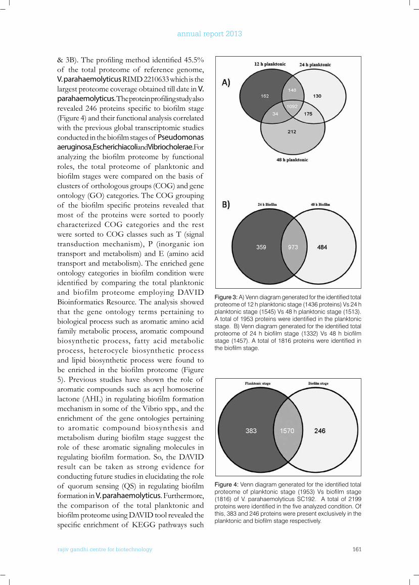
rajiv gandhi centre for biotechnology 161
annual report 2013
Figure 3: A) Venn diagram generated for the identified total proteome of 12 h planktonic stage (1436 proteins) Vs 24 h planktonic stage (1545) Vs 48 h planktonic stage (1513). A total of 1953 proteins were identified in the planktonic stage. B) Venn diagram generated for the identified total proteome of 24 h biofilm stage (1332) Vs 48 h biofilm stage (1457). A total of 1816 proteins were identified in the biofilm stage.
Figure 4: Venn diagram generated for the identified total proteome of planktonic stage (1953) Vs biofilm stage (1816) of V. parahaemolyticus SC192. A total of 2199 proteins were identified in the five analyzed condition. Of this, 383 and 246 proteins were present exclusively in the planktonic and biofilm stage respectively.
&3B).Theprofilingmethodidentified45.5%of the total proteome of reference genome, V. parahaemolyticus RIMD 2210633 which is the largest proteome coverage obtained till date in V. parahaemolyticus.Theproteinprofilingstudyalsorevealed246proteinsspecifictobiofilmstage(Figure 4) and their functional analysis correlated with the previous global transcriptomic studies conductedinthebiofilmstagesof Pseudomonas aeruginosa, Escherichia coli and Vibrio cholerae. For analyzing the biofilmproteomeby functionalroles, the total proteome of planktonic and biofilmstageswerecomparedonthebasisof clusters of orthologous groups (COG) and gene ontology (GO) categories. The COG grouping of the biofilm specific proteins revealed thatmost of the proteins were sorted to poorly characterized COG categories and the rest were sorted to COG classes such as T (signal transduction mechanism), P (inorganic ion transport and metabolism) and E (amino acid transport and metabolism). The enriched gene ontologycategories inbiofilmconditionwereidentified by comparing the total planktonicand biofilm proteome employing DAVID Bioinformatics Resource. The analysis showed that the gene ontology terms pertaining to biological process such as aromatic amino acid family metabolic process, aromatic compound biosynthetic process, fatty acid metabolic process, heterocycle biosynthetic process and lipid biosynthetic process were found to be enriched in the biofilmproteome (Figure5). Previous studies have shown the role of aromatic compounds such as acyl homoserine lactone(AHL)inregulatingbiofilmformationmechanism in some of the Vibrio spp., and the enrichment of the gene ontologies pertaining to aromatic compound biosynthesis and metabolismduring biofilm stage suggest therole of these aromatic signaling molecules in regulating biofilm formation. So, theDAVIDresult can be taken as strong evidence for conducting future studies in elucidating the role of quorumsensing (QS) in regulatingbiofilmformation in V. parahaemolyticus. Furthermore, the comparison of the total planktonic and biofilmproteomeusingDAVIDtoolrevealedthespecificenrichmentof KEGGpathwayssuch

rajiv gandhi centre for biotechnology 162
annual report 2013
as Arginine and proline metabolism, fatty acid biosynthesis and sulfurmetabolism inbiofilmcondition. The requirement of proteins involved in sulfur metabolism is a strong evidence of anaerobic metabolism and reveal the metabolic heterogeneity of the surface associated bacteria. Besides, the similar enrichment observed for KEGGpathwayssuchasglycolysis,citratecycle,purine metabolism, pyruvate metabolism and ribosomeinplanktonicandbiofilmstagerevealedthat the biofilm associated cells were viable
and engaged in division like their planktonic counterparts.Incontinuation,theidentified246biofilmspecificproteinsarefurthervalidatedbylabel-free quantitative proteomic method and transcriptome sequencing which is in progress to identify the biofilm specific functions andpathways of V. parahaemolyticus. The obtained data will then be compared with that of V. choleraetoidentifyconservedbiofilminhibitingtarget common to both the pathogens.
Figure 5: Comparison of enriched gene ontology (GO) terms in the planktonic and biofilm stage by clustering the DAVID results using Euclidean distance matrix. Heat map shows the specific enrichment of GO terms such as aromatic amino acid family metabolic process, aromatic compound biosynthetic process, fatty acid metabolic process, heterocycle biosynthetic process and lipid biosynthetic process in biofilm stage.
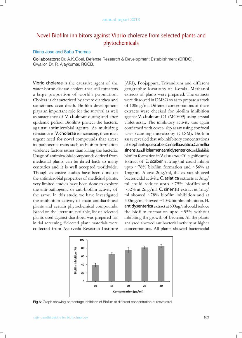
rajiv gandhi centre for biotechnology 163
annual report 2013
novel biofilm inhibitors against Vibrio cholerae from selected plants and phytochemicals
Diana Jose and Sabu Thomas
Collaborators: Dr. A.K.Goel, Defense Research & Development Establishment (DRDO), Gwalior, Dr. R. Ajaykumar, RGCB.
Vibrio cholerae is the causative agent of the water-borne disease cholera that still threatens a large proportion of world’s population. Cholera is characterized by severe diarrhea and sometimes even death. Biofilm developmentplays an important role for the survival as well as sustenance of V. cholerae during and after epidemicperiod.Biofilmsprotect thebacteriaagainst antimicrobial agents. As multidrug resistance in V. cholerae is increasing, there is an urgent need for novel compounds that arrest itspathogenictraitssuchasbiofilmformationvirulence factors rather than killing the bacteria. Usage of antimicrobial compounds derived from medicinal plants can be dated back to many centuries and it is well accepted worldwide. Though extensive studies have been done on the antimicrobial properties of medicinal plants, very limited studies have been done to explore the anti-pathogenic or anti-biofilm activity of the same. In this study, we have investigated theantibiofilmactivityof mainantidiarrhoealplants and certain phytochemical compounds. Based on the literature available, list of selected plants used against diarrhoea was prepared for initial screening. Selected plant materials were collected from Ayurveda Research Institute
(ARI), Poojappura, Trivandrum and different geographic locations of Kerala.Methanolextracts of plants were prepared. The extracts were dissolved in DMSO so as to prepare a stock of 100mg/ml. Different concentrations of these extractswere checked for biofilm inhibitionagainst V. cholerae O1 (MCV09) using crystal violet assay. The inhibitory activity was again confirmedwithcover-slipassayusingconfocallaser scanning microscopy (CLSM). Biofilm assay revealed that sub inhibitory concentrations of Elephantopus scaber, Centella asiatica, Camellia sinensis and Holarrhena antidysenterica could inhibit biofilmformationinV. choleraeO1significantly.Extract of E. scaber at 2mg/ml could inhibit upto~76%biofilm formation and~56% at1mg/ml. Above 2mg/ml, the extract showed bactericidal activity. C. asiatica extracts at 3mg/ml could reduce upto ~75% biofilm and ~52% at 2mg/ml. C. sinensis extract at 1mg/ml showed ~78% biofilm inhibition and at 500mg/mlshowed~70%biofilminhibition.H. antidysenterica extract at 600µg/ml could reduce the biofilm formation upto ~55% without inhibiting the growth of bacteria. All the plants analysed showed antibacterial activity at higher concentrations. All plants showed bactericidal
Fig 6: Graph showing percentage inhibition of Biofilm at different concentration of resveratrol.
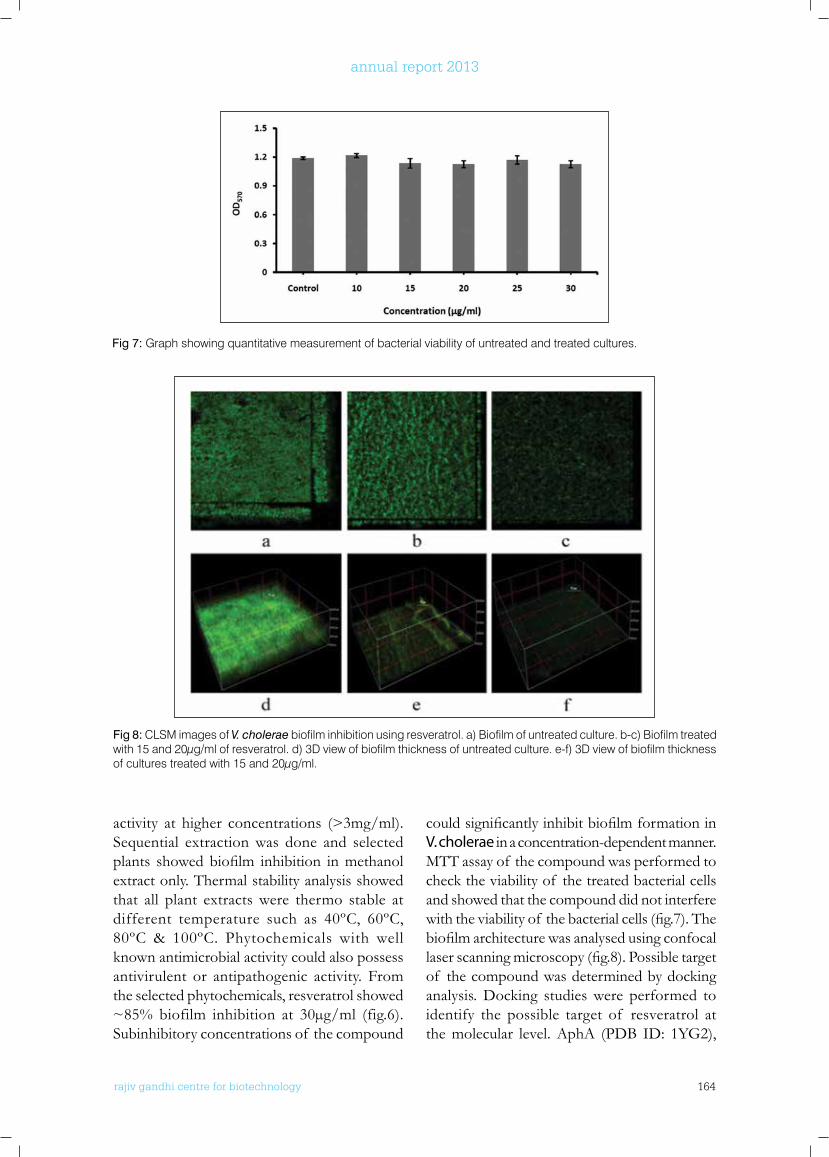
rajiv gandhi centre for biotechnology 164
annual report 2013
Fig 7: Graph showing quantitative measurement of bacterial viability of untreated and treated cultures.
Fig 8: CLSM images of V. cholerae biofilm inhibition using resveratrol. a) Biofilm of untreated culture. b-c) Biofilm treated with 15 and 20µg/ml of resveratrol. d) 3D view of biofilm thickness of untreated culture. e-f) 3D view of biofilm thickness of cultures treated with 15 and 20µg/ml.
activity at higher concentrations (>3mg/ml). Sequential extraction was done and selected plants showedbiofilm inhibition inmethanolextract only. Thermal stability analysis showed that all plant extracts were thermo stable at different temperature such as 40ºC, 60ºC, 80ºC & 100ºC. Phytochemicals with well known antimicrobial activity could also possess antivirulent or antipathogenic activity. From the selected phytochemicals, resveratrol showed ~85% biofilm inhibition at 30µg/ml (fig.6). Subinhibitory concentrations of the compound
couldsignificantlyinhibitbiofilmformationinV. cholerae in a concentration-dependent manner. MTT assay of the compound was performed to check the viability of the treated bacterial cells and showed that the compound did not interfere withtheviabilityof thebacterialcells(fig.7).Thebiofilmarchitecturewasanalysedusingconfocallaserscanningmicroscopy(fig.8).Possibletargetof the compound was determined by docking analysis. Docking studies were performed to identify the possible target of resveratrol at the molecular level. AphA (PDB ID: 1YG2),
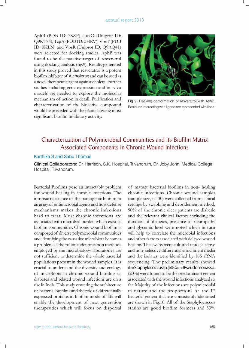
rajiv gandhi centre for biotechnology 165
annual report 2013
AphB (PDB ID: 3SZP), LuxO (Uniprot ID: Q9KT84),TcpA(PDBID:3HRV),VpsT(PDBID:3KLN)andVpsR(UniprotID:Q9AQ41)were selected for docking studies. AphB was found to be the putative target of resveratrol usingdockinganalysis(fig.9).Resultsgeneratedin this study proved that resveratrol is a potent biofilminhibitorof V. cholerae and can be used as a novel therapeutic agent against cholera. Further studies including gene expression and in- vivo models are needed to explore the molecular mechanismof actionindetail.Purificationandcharacterization of the bioactive compound would be preceded with the plant showing most significantbiofilminhibitoryactivity.
of mature bacterial biofilms in non- healingchronic infections. Chronic wound samples (sample size, n=30) were collected from clinical settings by swabbing and debridement method. 90% of the chronic ulcer patients are diabetic and the relevant clinical factors including the duration of diabetes, presence of neuropathy and glycemic level were noted which in turn will help to correlate the microbial infections and other factors associated with delayed wound healing. The swabs were cultured onto selective and non- selective differential enrichment media and the isolateswere identifiedby16S rRNAsequencing. The preliminary results showed that Staphylococcus sp. (60%) and Pseudomonas sp. (20%) were found to be the predominant genera associated with the wound infections analyzed so far. Majority of the infections are polymicrobial in nature and the proportions of the 17 bacterialgenerathatareconsistentlyidentifiedare shown in Fig.10. All of the Staphylococcus strains are good biofilm formers and 33%
Fig 9: Docking conformation of resveratrol with AphB.
Residues interacting with ligand are represented with lines.
Characterization of Polymicrobial Communities and its biofilm Matrix Associated Components in Chronic wound Infections
Karthika S and Sabu Thomas
Clinical Collaborators: Dr. Harrison, S.K. Hospital, Trivandrum, Dr. Joby John, Medical College Hospital, Trivandrum.
BacterialBiofilmsposeanintractableproblemfor wound healing in chronic infections. The intrinsicresistanceof thepathogenicbiofilmtoan array of antimicrobial agents and host defense mechanisms makes the chronic infections hard to treat. Most chronic infections are associated with microbial burden which exist as biofilmcommunities.Chronicwoundbiofilmiscomposed of diverse polymicrobial communities and identifying the causative microbiota becomes aproblemastheroutineidentificationmethodsemployed by the microbiology laboratories are notsufficienttodeterminethewholebacterialpopulations present in the wound samples. It is crucial to understand the diversity and ecology of microbiota in chronicwound biofilms asdiabetes and related wound infections are on a rise in India. This study centering the architecture of bacterialbiofilmsandtheroleof differentiallyexpressedproteinsinbiofilmmodeof lifewillenable the development of next generation therapeutics which will focus on dispersal
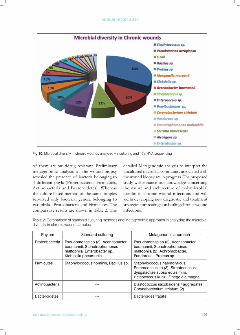
rajiv gandhi centre for biotechnology 166
annual report 2013
Fig 10: Microbial diversity in chronic wounds analyzed via culturing and 16SrRNA sequencing
of them are multidrug resistant. Preliminary metagenomic analysis of the wound biopsy revealed the presence of bacteria belonging to 4 different phyla (Proteobacteria, Firmicutes, Actinobacteria and Bacteroidetes). Whereas the culture based method of the same samples reported only bacterial genera belonging to two phyla –Proteobacteria and Firmicutes. The comparative results are shown in Table 2. The
detailed Metagenomic analysis to interpret the uncultured microbial community associated with the wound biopsy are in progress. The proposed study will enhance our knowledge concerning the nature and architecture of polymicrobial biofilm in chronicwound infections andwillaid in developing new diagnostic and treatment strategies for treating non healing chronic wound infections.
Table 2: Comparison of standard culturing methods and Metagenomic approach in analyzing the microbial diversity in chronic wound samples
Phylum Standard culturing Metagenomic approach
Proteobacteria Pseudomonas sp.(3), Acenitobacter baumannii, Stenotrophomonas maltophila, Enterobacter sp., Klebsiella pneumonia
Pseudomonas sp.(3), Acenitobacter baumannii, Stenotrophomonas maltophila (2), Achromobacter, Pandoraea , Proteus sp.
Firmicutes Staphylococcus hominis, Bacillus sp. Staphylococcus haemolyticus, Enterococcus sp.(3), Streptococcus dysgalactiae subsp equisimilis, Helcococcus kunzi, Finegoldia magna
Actinobacteria --- Blastococcus saxobsidens / aggregates, Corynebacterium striatum (2)
Bacteroidetes --- Bacteroides fragilis
rajiv gandhi centre for biotechnology 167
annual report 2013
Screening of cold tolerant genes from Pseudomonas psychrophilla isolated from the Arctic fjord
Wilson Peter Abraham and Sabu Thomas
PublICATIonS
PRIMARy PublICATIonS fRoM lAboRAToRy
• Nimmy Augustine, A.K. Goel, K.C. Sivakumar, R. Ajay Kumar, Sabu Thomas, 2013. Resveratrol - A potential inhibitor of biofilm formation in Vibrio cholerae., Phytomedicine, 21: 286-289
• Wilson P Abraham and Sabu Thomas, 2013. Diversity and Bioprospecting potential of bacteria isolated from the Arctic: A preliminary study, Mapana Journal of Science, 12(4): 19-28
• Karth ika S and Sabu Thomas, 2014. Biotechnology and Environmental Conservation
– A Perspective, Journal of Basic and Applied Biology, 8(1): 26-31
• Divya M P, Deepa Mathew P, Jyothi R, RamaniBai and Sabu Thomas, 2013.Mutations in Gyr A and Par C genes of Shigellaflexneri 2a determining the fluoroquinolone resistance therapeutic applications, Indian Journal of Medical Research. (Accepted)
PublICATIonS wITH CollAboRAToRS
• B.Sadia Khan, Anil Kumar, Divya MP, Sabu Thomas, Deepa Harichandran and Shamsul Karim, 2013. Fatal non-O1/ non-O139 V.cholerae Septicaemia in A Patient with Chronic Liver
The permanently cold environments have been successfully colonized by a group of extremophilic microorganisms that are known as psychrophiles and psychrotrophs. The psychrotrophic and psychrophilic bacteria that could grow at 0°C or below, populate the cold environments of our planet including Arctic and Antarctica regions. These bacteria are of particular importance in the global ecology since more than 80% of the earth biosphere is permanently or seasonally subjected to temperatures below 5°C. How these bacteria cope with low temperatures induced stress is poorly understood. The ability of these organisms to survive and proliferate at low temperatures implies that they have overcome key barriers inherent to permanently cold environments. These challenges include reduced enzyme activity, decreasedmembranefluidity,altered transport of nutrients and waste products, decreased rates of transcription, translation and cell division, inappropriate protein folding and intracellular ice formation. RGCB team collected the bacterial samples from four different regions of Kongsfjordensystem,Ny-Alesund,anislandin the Svalbard Archipelago (79°55´N, 11°56´E), during the Indian Arctic Expedition-2009,
organized by the Ministry of Earth Science, Govt. of India. Around 100 bacterial isolates were collected from the water and sediment. The bacterial isolates were checked for their growth at different temperatures ranging 4 to 37°C suggesting that all the isolates are psychrotrophs. The psychrotrophic, gram negative, rod shaped, aerobic strain A166 isolated from Arctic fjord water has been chosen for this proposed study. Nearest phylogenetic relation of the test organism was found to be Pseudomonas psychrophila by sequencing 16S rRNA gene. Upon Suppressive Subtractive Hybridization (SSH) 87 gene clones have been obtained. Of these, 50 plasmids were isolated and the inserts were checked by T7SP6 PCR. On sequencing 40 clones, we are able to identify 5 genes (DNA mismatch repair protein, Transport related membrane protein, Periplasmic lipoprotein, Methyl accepting chemotaxis transducer, RNA polymerase sigma factor) and 2 hypothetical proteins. Transcriptional quantificationof thesegenesusingRealTimePCR is in progress. Simultaneously, proteome profilingof Pseudomonas psychrophila at two different temperatures using mass spectrometry is also in progress.
rajiv gandhi centre for biotechnology 168
annual report 2013
Disease: A Case Report and Review of Literature. Journal of Medical Microbiology, 62,917-921
ConfEREnCE PRESEnTATIonS
• Akhilandeswarre D, Eshita Mutt, Abdul Jaleel, Sowdhamini Rand Sabu Thomas. Identification of potential anti-infective drug targets for Vibrio parahaemolyticus by proteomic analysis over the course of biofilm formation, 5th Annual meeting of proteomics society-India, Indian Institute of Science, Bangalore, India.
• Karthika S, Reshma M J, Wilson P A, Anita Das R, Sarma U S, Harikrishnan K and Sabu Thomas. Characterization and Evaluation and of Phenol Degrading Bacillus Consortium for Enhancing the Softening of Coir Fibre, 54th Annual Conference of Association of
Microbiology of India (AMI) International Conference FDMIR-2013. Nov. 17-20, M D University, Rohtak, Haryana, India.
• Wilson Peter Abraham and Sabu Thomas. Cloning And Sequencing of cspA Like Gene from a Bacteria Isolated from Arctic and its Molecular Identification, 54th Annual Conference of Association of Microbiology of India (AMI) International Conference FDMIR-2013, Nov.17-20, M D University, Rohtak, Haryana, India.
InVITED lECTuRE
• Sabu Thomas, 2013. Role of Biotechnology in Environmental Conservation. National Seminar on Biological Conservation and Sustainability: Issues and Strategies 1-2 August, Organised by K.G.College, Pampady, Sponsored by UGC.
Extramural Grants
Title Investigators Funding Agency Duration
Genetic manipulation of Coirret-for application on coir for quality improvement
U. S. Sarma (PI) Anita Das (Co-PI) Sabu Thomas (Co-PI) HariKrishnan. K (Co-PI)
Coir Board, Ministry of MSME, Government of India
2011-13
Novel Biofilm inhibitors against Vibrio cholerae from selected plants: isolation and characterisation
Sabu Thomas (PI) R. Ajay Kumar (Co-I)A.K. Goel (Co-I)
Defense Research & Development Establishment, Government of India
2012-14

rajiv gandhi centre for biotechnology 169
annual report 2013
Molecular ReproductionLaboratory - 1
Post-Doctoral FellowIndu S
r a j i v g a n d h i c e n t r e f o r b i o t e c h n o l o g y
Pradeep Kumar was awarded Ph.D in Life Sciences from Devi Ahilya University, Indore in 1988 and joined the Faculty of Life Sciences of the same university in the year 1989. He worked as a Fellow-in-residence at the Centre for Biomedical Research at the Rockefeller University, New York, NY; visiting faculty in University of Virginia, Charlottesville, VA and as visiting faculty at University of Florida, Gainesville, FL. He joined RGCB in the year 2004.
Pradeep Kumar G Ph.DScientist G
Ph.D StudentsBhagya KP Sreesha Sree Divya Saro VargheseNomesh YaduKarthika Radhakrishnan Soumya ADevi AN
Technical StaffTessy Ann MaryJonhy G
Project PersonnelAnil Kumar TRJeeva S
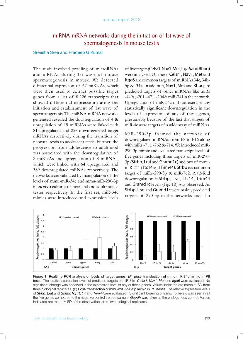
rajiv gandhi centre for biotechnology 170
annual report 2013
The study involved profiling of microRNAsand mRNAs during 1st wave of mouse spermatogenesis in mouse. We detected differential expression of 67 miRNAs, which were then used to extract possible target genes from a list of 8,226 transcripts that showed differential expression during the initiation and establishment of 1st wave of spermatogenesis. The miRNA-mRNA networks generated revealed the downregulation of 4 & upregulation of 19 miRNAs were linked with 81 upregulated and 228-downregulated target mRNAs respectively during the transition of neonatal testis to adolescent testis. Further, the progression from adolescence to adulthood was associated with the downregulation of 2 miRNAs and upregulation of 8 miRNAs, which were linked with 64 upregulated and 389 downregulated mRNAs respectively. The networks were validated by manipulation of the levels of mmu-miR-34c and mmu-miR-290-3p in ex vivo cultures of neonatal and adult mouse testes respectively. In the first set, miR-34c mimics were introduced and expression levels
of fivetargets(Celsr1, Nav1, Met, Itga6 and Rhoq) were analyzed. Of these, Celsr1, Nav1, Met and Itga6 are common targets of miRNAs 34c, 34b-5p & -34a. In addition, Nav1, Met and Rhoq are predicted targets of other miRNAs like miRs -449a, -201, -471, -204& miR-741in the network. Upregulation of miR-34c did not exercise any statistically significant downregulation in thelevels of expression of any of these genes, presumably because of the fact that targets of miR-4c were targets of a wide array of miRNAs.
MiR-290-3p for med the ne twork of downregulated miRNAs from P8 to P16 along with miRs -711, -762 &-714. We introduced miR-290-3p mimic and evaluated transcript levels of fivegenesincludingthreetargetsof miR-290-3p (Strbp, Lrat and Gramd1c) and two of mmu-miR-711 (Ttc14 and Trim44). Strbp is a common target of miRs-290-3p & miR-762. A>2-fold downregulation inStrbp, Lrat, Ttc14, Trim44 and Gramd1c levels (Fig. 1B) was observed. As Strbp, Lrat and Gramd1c were mainly predicted targets of 290-3p in the networks and also
Figure 1. Realtime PCR analysis of levels of target genes, (A) post- transfection of mmu-miR-34c mimic in P8 testis. The relative expression levels of predicted targets of miR-34c- Celsr1, Nav1, Met and Itga6 were evaluated. No significant change was observed in the expression level of any of these genes. Values indicated are mean ± SD from three biological replicates. (B) Post- transfection of mmu-miR-290-3p mimic in P16 testis. The relative expression levels of Strbp, Lrat and Gramd1c, Ttc14 and Trim44were evaluated. Significant lowering of transcript levels was seen in all the five genes compared to the negative control treated sample. Gapdh was taken as the endogenous control. Values indicated are mean ± SD of the observations from two biological replicates.
miRnA-mRnA networks during the initiation of 1st wave of spermatogenesis in mouse testis
Sreesha Sree and Pradeep G Kumar
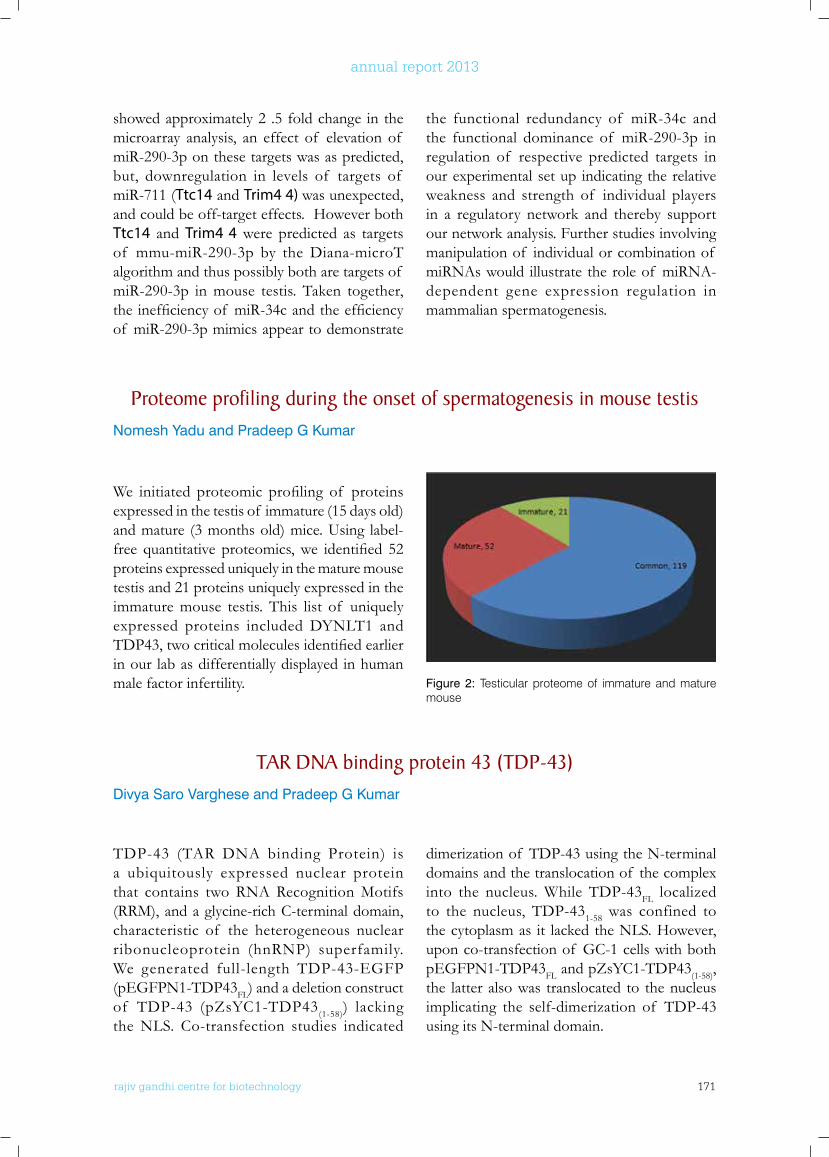
rajiv gandhi centre for biotechnology 171
annual report 2013
showed approximately 2 .5 fold change in the microarray analysis, an effect of elevation of miR-290-3p on these targets was as predicted, but, downregulation in levels of targets of miR-711 (Ttc14 and Trim4 4) was unexpected, and could be off-target effects. However both Ttc14 and Trim4 4 were predicted as targets of mmu-miR-290-3p by the Diana-microT algorithm and thus possibly both are targets of miR-290-3p in mouse testis. Taken together, theinefficiencyof miR-34candtheefficiencyof miR-290-3p mimics appear to demonstrate
the functional redundancy of miR-34c and the functional dominance of miR-290-3p in regulation of respective predicted targets in our experimental set up indicating the relative weakness and strength of individual players in a regulatory network and thereby support our network analysis. Further studies involving manipulation of individual or combination of miRNAs would illustrate the role of miRNA-dependent gene expression regulation in mammalian spermatogenesis.
Figure 2: Testicular proteome of immature and mature mouse
Proteome profiling during the onset of spermatogenesis in mouse testisNomesh Yadu and Pradeep G Kumar
We initiated proteomic profiling of proteinsexpressed in the testis of immature (15 days old) and mature (3 months old) mice. Using label-free quantitative proteomics,we identified 52proteins expressed uniquely in the mature mouse testis and 21 proteins uniquely expressed in the immature mouse testis. This list of uniquely expressed proteins included DYNLT1 and TDP43,twocriticalmoleculesidentifiedearlierin our lab as differentially displayed in human male factor infertility.
TAR DnA binding protein 43 (TDP-43)Divya Saro Varghese and Pradeep G Kumar
TDP-43 (TAR DNA binding Protein) is a ubiquitously expressed nuclear protein that contains two RNA Recognition Motifs (RRM), and a glycine-rich C-terminal domain, characteristic of the heterogeneous nuclear ribonucleoprotein (hnRNP) superfamily. We generated full-length TDP-43-EGFP (pEGFPN1-TDP43FL) and a deletion construct of TDP-43 (pZsYC1-TDP43(1-58)) lacking the NLS. Co-transfection studies indicated
dimerization of TDP-43 using the N-terminal domains and the translocation of the complex into the nucleus. While TDP-43FL localized to the nucleus, TDP-431-58 was confined to the cytoplasm as it lacked the NLS. However, upon co-transfection of GC-1 cells with both pEGFPN1-TDP43FL and pZsYC1-TDP43(1-58), the latter also was translocated to the nucleus implicating the self-dimerization of TDP-43 using its N-terminal domain.
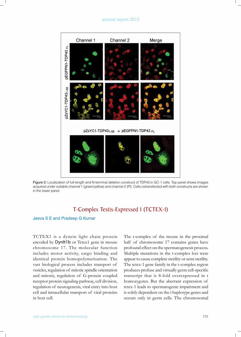
rajiv gandhi centre for biotechnology 172
annual report 2013
TCTEX1 is a dynein light chain protein encoded by Dynlt1b or Tctex1 gene in mouse chromosome 17. The molecular function includes motor activity, cargo binding and identical protein homopolymerisation. The vast biological process includes transport of vesicles, regulation of mitotic spindle orientation and mitosis, regulation of G-protein coupled receptor protein signaling pathway, cell division, regulation of neurogenesis, viral entry into host cell and intracellular transport of viral proteins in host cell.
The t-complex of the mouse in the proximal half of chromosome 17 contains genes have profound effect on the spermatogenesis process. Multiple mutations in the t-complex loci were appear to cause complete sterility or semi sterility. The tctex-1 gene family in the t-complex region producesprofuseandvirtuallygermcell-specifictranscript that is 8-fold overexpressed in t homozygotes. But the aberrant expression of tctex-1 leads to spermatogenic impairment and is solely dependent on the t haplotype genes and occurs only in germ cells. The chromosomal
Figure 3: Localization of full-length and N-terminal deletion construct of TDP43 in GC-1 cells. Top panel shows images acquired under suitable channel 1 (green/yellow) and channel 2 (PI). Cells cotransfected with both constructs are shown in the lower panel.
T-Complex Testis-Expressed 1 (TCTEx-1)Jeeva S E and Pradeep G Kumar
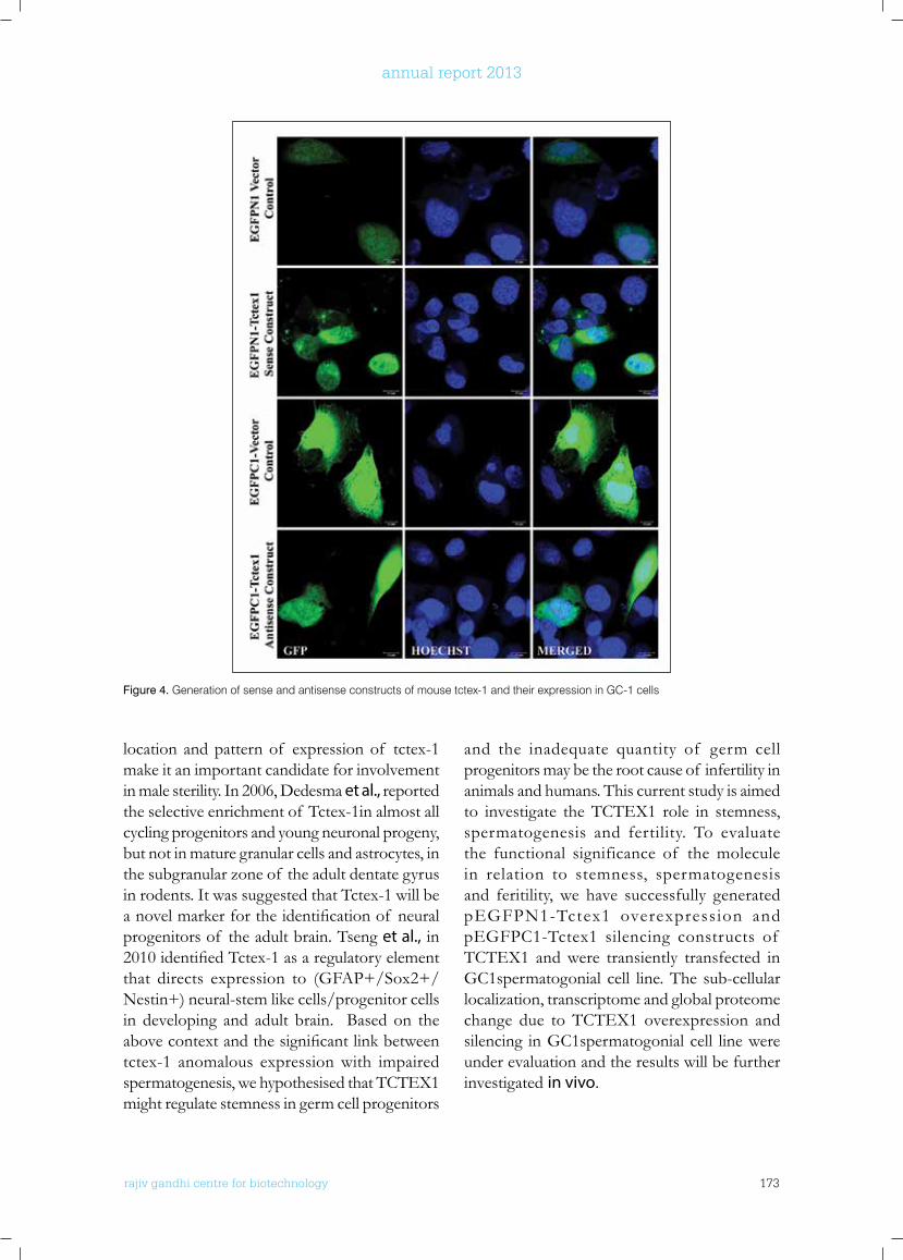
rajiv gandhi centre for biotechnology 173
annual report 2013
location and pattern of expression of tctex-1 make it an important candidate for involvement in male sterility. In 2006, Dedesma et al., reported the selective enrichment of Tctex-1in almost all cycling progenitors and young neuronal progeny, but not in mature granular cells and astrocytes, in the subgranular zone of the adult dentate gyrus in rodents. It was suggested that Tctex-1 will be anovelmarkerfortheidentificationof neuralprogenitors of the adult brain. Tseng et al., in 2010identifiedTctex-1asaregulatoryelementthat directs expression to (GFAP+/Sox2+/ Nestin+) neural-stem like cells/progenitor cells in developing and adult brain. Based on the abovecontextandthesignificantlinkbetweentctex-1 anomalous expression with impaired spermatogenesis, we hypothesised that TCTEX1 might regulate stemness in germ cell progenitors
and the inadequate quantity of germ cell progenitors may be the root cause of infertility in animals and humans. This current study is aimed to investigate the TCTEX1 role in stemness, spermatogenesis and fertility. To evaluate the functional significance of the molecule in relation to stemness, spermatogenesis and feritility, we have successfully generated pEGFPN1-Tctex1 overexpression and pEGFPC1-Tctex1 silencing constructs of TCTEX1 and were transiently transfected in GC1spermatogonial cell line. The sub-cellular localization, transcriptome and global proteome change due to TCTEX1 overexpression and silencing in GC1spermatogonial cell line were under evaluation and the results will be further investigated in vivo.
Figure 4. Generation of sense and antisense constructs of mouse tctex-1 and their expression in GC-1 cells
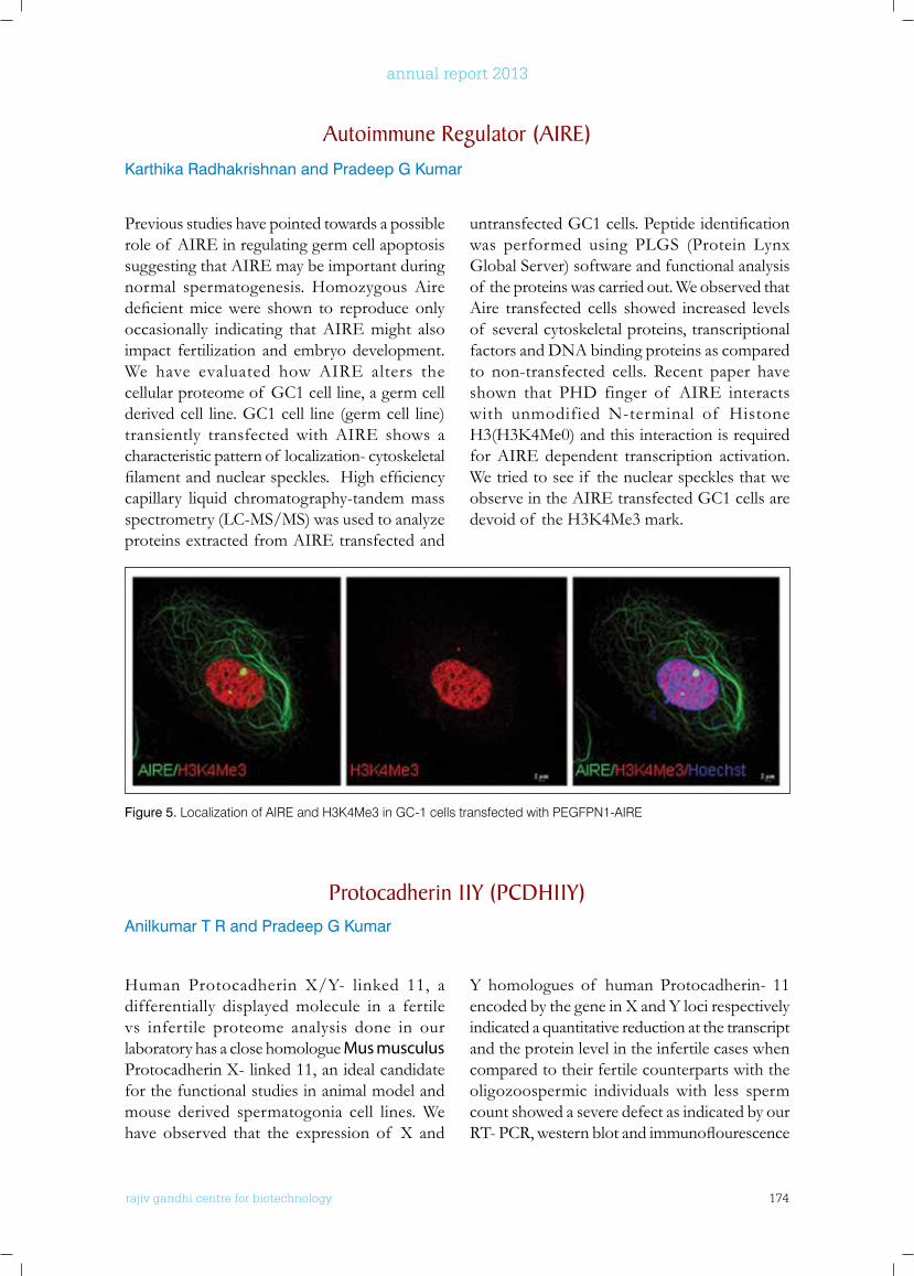
rajiv gandhi centre for biotechnology 174
annual report 2013
Previous studies have pointed towards a possible role of AIRE in regulating germ cell apoptosis suggesting that AIRE may be important during normal spermatogenesis. Homozygous Aire deficientmicewere shown to reproduceonlyoccasionally indicating that AIRE might also impact fertilization and embryo development. We have evaluated how AIRE alters the cellular proteome of GC1 cell line, a germ cell derived cell line. GC1 cell line (germ cell line) transiently transfected with AIRE shows a characteristic pattern of localization- cytoskeletal filamentandnuclearspeckles.Highefficiencycapillary liquid chromatography-tandem mass spectrometry (LC-MS/MS) was used to analyze proteins extracted from AIRE transfected and
untransfectedGC1cells.Peptideidentificationwas performed using PLGS (Protein Lynx Global Server) software and functional analysis of the proteins was carried out. We observed that Aire transfected cells showed increased levels of several cytoskeletal proteins, transcriptional factors and DNA binding proteins as compared to non-transfected cells. Recent paper have shown that PHD finger of AIRE interacts with unmodified N-terminal of Histone H3(H3K4Me0)andthisinteractionisrequiredfor AIRE dependent transcription activation. We tried to see if the nuclear speckles that we observe in the AIRE transfected GC1 cells are devoidof theH3K4Me3mark.
Autoimmune Regulator (AIRE)Karthika Radhakrishnan and Pradeep G Kumar
Figure 5. Localization of AIRE and H3K4Me3 in GC-1 cells transfected with PEGFPN1-AIRE
Protocadherin 11y (PCDH11y)Anilkumar T R and Pradeep G Kumar
Human Protocadherin X/Y- linked 11, a differentially displayed molecule in a fertile vs infertile proteome analysis done in our laboratory has a close homologue Mus musculus Protocadherin X- linked 11, an ideal candidate for the functional studies in animal model and mouse derived spermatogonia cell lines. We have observed that the expression of X and
Y homologues of human Protocadherin- 11 encoded by the gene in X and Y loci respectively indicated a quantitative reduction at the transcript and the protein level in the infertile cases when compared to their fertile counterparts with the oligozoospermic individuals with less sperm count showed a severe defect as indicated by our RT-PCR,westernblotandimmunoflourescence

rajiv gandhi centre for biotechnology 175
annual report 2013
analysis. We also observed that the expression of PCDH11X in mouse derived spermatogonial cell line is being up regulated by Retinoic acid treatment suggesting protocadherin over expression in retinoic acid signalling, a pathway involved in differentiation of stem cells. We have also shown that the PCDH11X expression is more in the post meiotic cells than the pre meiotic cells as evidenced by the imunohistochemical, RT- PCR and western blot analysis.Thesefindingsassumessignificanceof its a role as a key molecule involved spematogenic differentiation. Silencing of PCDH11X was done in spermatogonia cell line, which could be a useful tool in establishing its functional interactome and possible signalling pathways involved.
spermatozoa was carried out and the expression of NPHP1 at the transcript and translational level was determined. Furthermore, screening of fertile and infertile males has revealed the aberrant expression of Nephrocystin in infertile males,whichimplicatesthecrucialsignificanceof the molecule in relation to male factor infertility. Moreover, Nephrocystin could be a possible marker for the diagnosis of male factor infertility. In order to evaluate the functional significanceof themoleculeweextrapolatedourstudies in murine system in which the expression profiling of murine testicular nephrocystin correspondingtotheagespecificexpressionof Nephrocystin with respect to different stages of spermatogenesis was carried out .The results proved the expression of Nephrocystin at all stages of male germ cell development. Further, we have successfully generated the pEGFP-SH3
Figure 6. Silencing of pcdh11x in GC-1 cells using shRNA constructs.
nephrycystin 1 (nPHP1)Devi A N and Pradeep G Kumar
Nephrocystin coded by the gene NPHP1 is a protein associated with nephronophthisis, an autosomal recessive kidney disease responsible for chronic renal failure in children and young adults. It contains an SH3 domain, two glutamic acid-rich domains, and a nephrocystin homology domain. SH3 domains are modular protein binding domains which are involved in signalling pathways controlling cell adhesion and cytoskeleton organization. Moreover, Nephrocystin is one of thedifferentiallydisplayedproteinsidentifiedin our laboratory as a molecule associated with male infertility. Controlled gene expression in testis is an important factor that regulates sperm production and also an important parameter in determining the fertility status. Hence, the role of Nephrocystin in relation to spermatogenesis and ultimately male factor infertility has to be elucidated. Characterization of NPHP1 in human
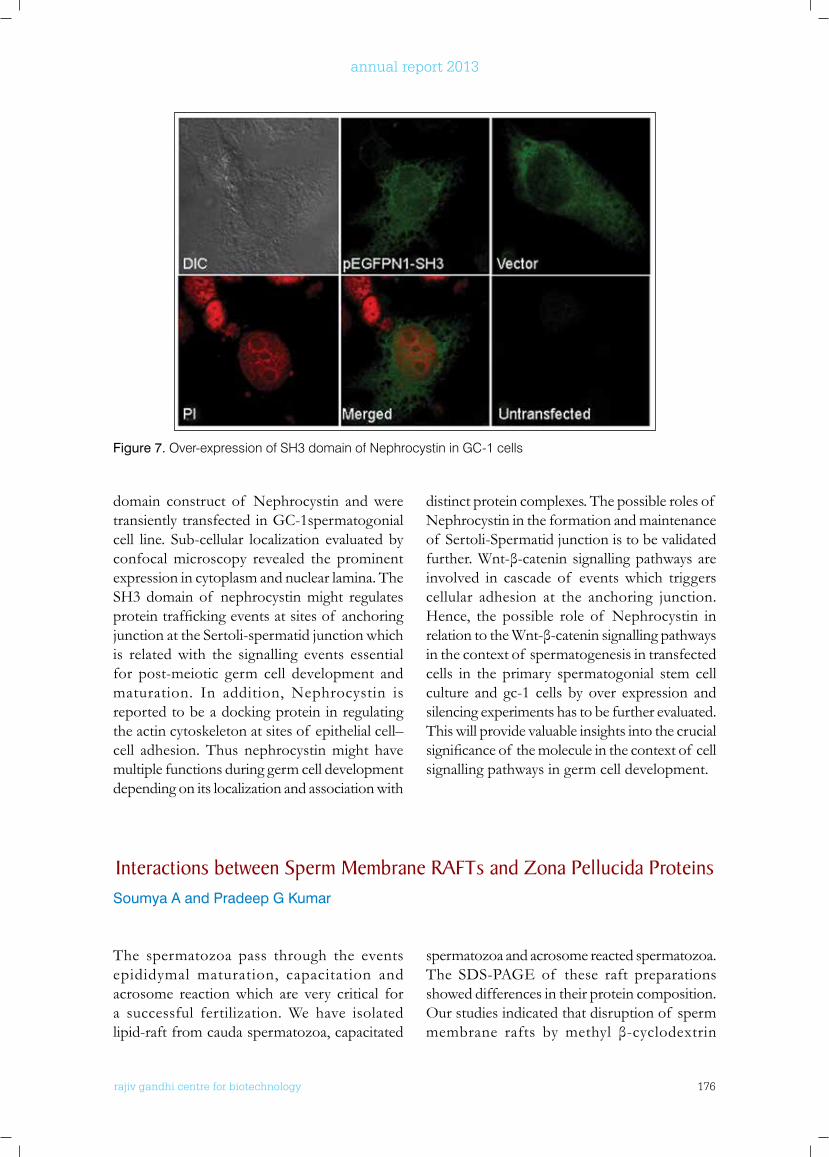
rajiv gandhi centre for biotechnology 176
annual report 2013
domain construct of Nephrocystin and were transiently transfected in GC-1spermatogonial cell line. Sub-cellular localization evaluated by confocal microscopy revealed the prominent expression in cytoplasm and nuclear lamina. The SH3 domain of nephrocystin might regulates proteintraffickingeventsatsitesof anchoringjunction at the Sertoli-spermatid junction which is related with the signalling events essential for post-meiotic germ cell development and maturation. In addition, Nephrocystin is reported to be a docking protein in regulating the actin cytoskeleton at sites of epithelial cell–cell adhesion. Thus nephrocystin might have multiple functions during germ cell development depending on its localization and association with
distinct protein complexes. The possible roles of Nephrocystin in the formation and maintenance of Sertoli-Spermatid junction is to be validated further.Wnt-β-cateninsignallingpathwaysareinvolved in cascade of events which triggers cellular adhesion at the anchoring junction. Hence, the possible role of Nephrocystin in relationtotheWnt-β-cateninsignallingpathwaysin the context of spermatogenesis in transfected cells in the primary spermatogonial stem cell culture and gc-1 cells by over expression and silencing experiments has to be further evaluated.This will provide valuable insights into the crucial significanceof themoleculeinthecontextof cellsignalling pathways in germ cell development.
Figure 7. Over-expression of SH3 domain of Nephrocystin in GC-1 cells
Interactions between Sperm Membrane RAfTs and Zona Pellucida ProteinsSoumya A and Pradeep G Kumar
The spermatozoa pass through the events epididymal maturation, capacitation and acrosome reaction which are very critical for a successful fertilization. We have isolated lipid-raft from cauda spermatozoa, capacitated
spermatozoa and acrosome reacted spermatozoa. The SDS-PAGE of these raft preparations showed differences in their protein composition. Our studies indicated that disruption of sperm membrane rafts by methyl β-cyclodextrin
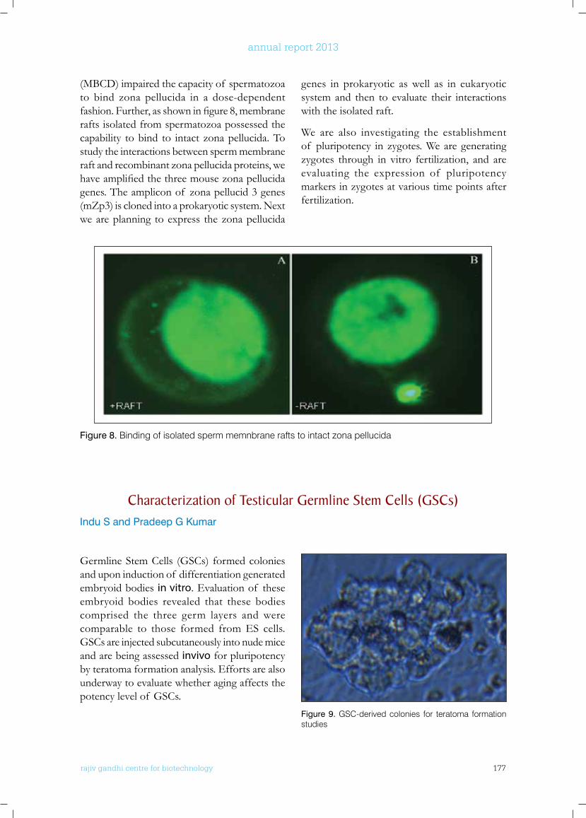
rajiv gandhi centre for biotechnology 177
annual report 2013
(MBCD) impaired the capacity of spermatozoa to bind zona pellucida in a dose-dependent fashion.Further,asshowninfigure8,membranerafts isolated from spermatozoa possessed the capability to bind to intact zona pellucida. To study the interactions between sperm membrane raft and recombinant zona pellucida proteins, we haveamplifiedthethreemousezonapellucidagenes. The amplicon of zona pellucid 3 genes (mZp3) is cloned into a prokaryotic system. Next we are planning to express the zona pellucida
genes in prokaryotic as well as in eukaryotic system and then to evaluate their interactions with the isolated raft.
We are also investigating the establishment of pluripotency in zygotes. We are generating zygotes through in vitro fertilization, and are evaluating the expression of pluripotency markers in zygotes at various time points after fertilization.
Figure 8. Binding of isolated sperm memnbrane rafts to intact zona pellucida
Figure 9. GSC-derived colonies for teratoma formation studies
Characterization of Testicular Germline Stem Cells (GSCs)Indu S and Pradeep G Kumar
Germline Stem Cells (GSCs) formed colonies and upon induction of differentiation generated embryoid bodies in vitro. Evaluation of these embryoid bodies revealed that these bodies comprised the three germ layers and were comparable to those formed from ES cells. GSCs are injected subcutaneously into nude mice and are being assessed invivo for pluripotency by teratoma formation analysis. Efforts are also underway to evaluate whether aging affects the potency level of GSCs.
rajiv gandhi centre for biotechnology 178
annual report 2013
PHD AwARDED
• Indu S (2013) Identification and functional evaluation of factors regulating testicular germline stem cell division and differentiation. University of Kerala, Trivandrum.
ConfEREnCE PRESEnTATIonS InVITED lECTuRES
• Kumar PG (2013) Where to go? Inspire Internship Science Camp, St. Xaviers Catholic College of Engineering, Chunkankadai, Nagercoil, Tamilnadu, 15-19 July, 2014.
• Kumar PG (2014) MicroRNA-mRNA networks during 1st wave of spermatogenesis in mouse testis. International Conference on Reproductive Health: Issues and Strategies under Changing Climate Scenario, and the 24th Annual Meeting of Indian Society for Study of Reproduction and Fertility (ISSRF), 6-8 February, 2004 at Indian Veterinary Research Institute, Izatnagar, Bareilly, UP, India.
• Kumar PG (2013) MicroRNA-mRNA networks during the initiation of 1st wave of spermatogenesis in mouse testis. National Symposium on Recent Advances in Reproductive Health, Centre for Advanced Studies in Zoology, Banaras Hindu University, November 28-30, 2013.
• Kumar PG (2013) MicroRNA-mRNA networks during 1st wave of spermatogenesis in mouse testis. 82nd Annual Meeting of the Society of Biological Chemists (India) and International Symposium on Genomes: Mechanism and Function, School of Life Sciences, University of Hyderabad, India, December 2-5, 2013.
PoSTER PRESEnTATIonS
• Yadu N and Kumar PG (2013) An in vitro model to study mechanisms of busulfan induced germ cell death. 21st ECDO Euroconference on Apoptosis on “Cell death:a Biomedical paradigm, Paris, France, September 25-28, 2013.
• Sreesha S and Kumar PG (2013) MicroRNA-mRNA interactome regulating mammalian spermatogenesis. 5th EMBO Meeting - Advancing the Life Science, Amsterdam, September 21-24, 2013.
• Varghese DS and Kumar PG (2013) Functional evaluation of tdp-43 in spermatogenesis and human male factor infertility. 5th EMBO Meeting - Advancing the Life Science, Amsterdam, September 21-24, 2013.
• Sreesha S and Kumar PG (2013) MicroRNA-mRNA interactome regulating mammalian spermatogenesis. International Conference on Developmetn and Genetics, Isle sur la Sorgue, Avignon, France, November 12-15, 2013.
• Varaghese DS and Kumar PG (2013) Functional evaluation of tdp-43 in spermatogenesis and human male factor infertility. International Conference on Developmetn and Genetics, Isle sur la Sorgue, Avignon, France, November 12-15, 2013.
ExTRA-MuRAl funDInG
No. Investigator(s) Title Funding Agency Duration
1 Pradeep Kumar G Molecular evaluation of interactions between sperm membrane rafts and zona pellucida proteins
Department of Biotechnology
2011-2014
2 Pradeep Kumar G Association between stemness and TCTEX1 expression in testicular germ cells from adult mouse testis
Board of Research in Nuclear Sciences
2011-2014
3 Pradeep Kumar G Role of CLP-1 in cell cycle regulation in spermatogenic cells
Department of Science & Technology
2013-2016
4 Pradeep Kumar G Evaluation of cellular aging and genome stability in spermatogonial stem cells
Council for Scientific & Industrial Research
2014-2017

rajiv gandhi centre for biotechnology 179
annual report 2013
Molecular ReproductionLaboratory - 2
Post-Doctoral FellowRenjini AP (RA-ICMR)
r a j i v g a n d h i c e n t r e f o r b i o t e c h n o l o g y
Malini Laloraya received her Ph.D from Devi Ahilya Vishwavidyalya Indore and was also a faculty there previously. She was a Fellow-in-Residence at Centre for Biomedical Research, Population Council, Rockefeller University, New York and was a Visiting Faculty at University of Virginia, VA and University of Florida, Gainesville, FL, USA before joining RGCB in 2004.
Malini Laloraya Ph.D Scientists F
Ph.D StudentsPhilip Litto Thomas Meera Krishna B. Prashanth Narayan Annu Joseph, Soumya V
Technical StaffG. Sheela
Project PersonnelAnand G
rajiv gandhi centre for biotechnology 180
annual report 2013
Embryo implantation is effected by a myriad of signaling cascades acting on the embryo–endometrium axis. IL-6 family cytokines are expressed during implantation in the mouse (Bhatt et al., 1991) and human (Charnock-Jones et al., 1994).They exert their influenceby invoking signaling cascades via STAT. STAT3, a multifaceted member of the signal transducers and activators of transcription (STAT) family, intercede axial responses for the cytokine family. STAT3 on activation by cytokines, gets phosphorylated and increased levels of STAT3 phosphorylation were significantlyassociatedwithelevatedexpressionof potential downstream targets of STAT3 which include apoptosis inhibitors (Survivin, Mcl-1, HSP27, Adrenomedullin, and Bcl-xL), cell-cycle regulators (cyclin D1, p21WAF1, c-Fos, MEK5, and c-Myc), and many more (Hsieh et al.,
2005). Mcl-1wasfirstidentifiedinascreenfordifferentiation induced genes activated in the humanmonocyteleukemiacellline(Kozopasetal., 1993). Experiments authenticate the role of Mcl-1 in oocyte survival v/s atresia, in fertility where it affects ovulation, embryogenesis and placental development (Sano et al., 2000). Mcl-1 deficiencyresultsinperi-implantationembryoniclethality due to incapability to implant in utero while the null blastocysts fail to hatch or attach in vitro, indicating a trophectoderm defect, proving that Mcl-1 is essential for preimplantation development and implantation (Rinkenberger et al., 2000). The activation of Mcl-1 happens viamany pathways, JAK/STAT3 being oneamong them. Correlating all the facts related to STAT3 and Mcl-1, we strongly hypothesized the possibility of interaction between both these molecules in the event of embryo implantation.
Reproduction is key to the continued existence of man-kind. The ability to reproduce which is a natural process is dependent upon complex biology including male and female repro-ductive health. This is the only facet of biology where two highly differentiated cells (the sperm and ova) unite to create a totipotent zygote. Reproduction failure results in infertil-ity. Pregnancy is central to this process in mammals. Despite giant leaps in technologies in assisted reproduction, our success rate is pathetically low. This is because in women being treated for infertility, even when healthy embryos are transplanted, the failure rate is high: “up to 70 % fail to dock successfully in the uterine lining”. This is largely due to our inability to understand embryo implantation.My lab endeavors to understand this complex phenomenon by focusing our attention on understanding nuclear receptor co-regulators, tissue remodeling, immune tolerance, adhesion, invasion, angiogenesis and embryonic pluripotency and thereby identify factors critical for successful pregnancy. Reproductive success is largely based on maternal health. Duringpregnancy a major complication is the development of Type 2 diabetes (Gestational Diabetes). Aberrant insulin signaling path-way is associated with Polycystic Ovarian Syndrome leading to Type 2 Diabetes. The NOD mice - an animal model for Type 1 Diabetes show a strong female prevalence in development of diabetes.PCOS and Type 1 diabetes are associated with subfertility, while gestational diabetes contributes to complications in maternal health and health of the baby. Thus, it is important to study metabolic disorders and hence my lab also focuses on understanding polycystic ovarian syndrome and type 1 diabetes.
STAT3 and Mcl-1 liaise to direct mesenchymal epithelial transition.Renjini A P, Shiny Titus, Megha Murali, Prashanth Narayan, Rajesh Kumar Jha, and Malini Laloraya
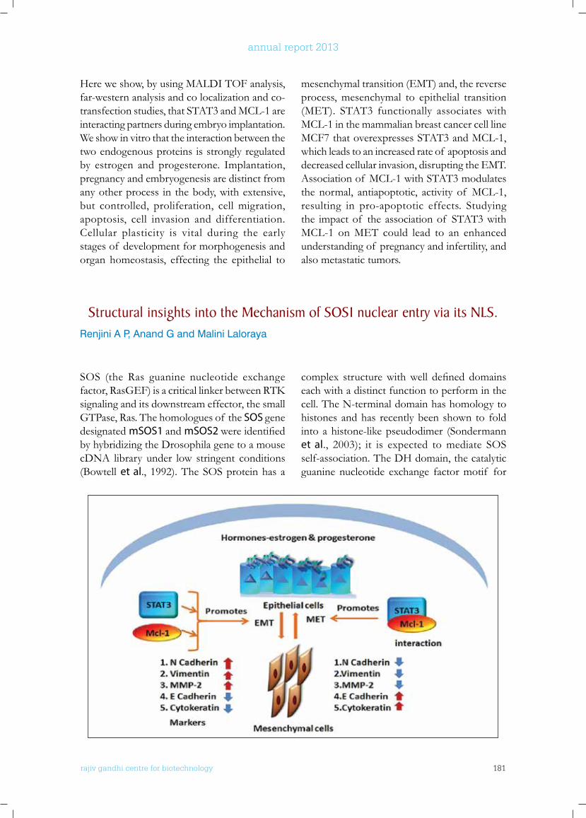
rajiv gandhi centre for biotechnology 181
annual report 2013
Here we show, by using MALDI TOF analysis, far-western analysis and co localization and co-transfection studies, that STAT3 and MCL-1 are interacting partners during embryo implantation. We show in vitro that the interaction between the two endogenous proteins is strongly regulated by estrogen and progesterone. Implantation, pregnancy and embryogenesis are distinct from any other process in the body, with extensive, but controlled, proliferation, cell migration, apoptosis, cell invasion and differentiation. Cellular plasticity is vital during the early stages of development for morphogenesis and organ homeostasis, effecting the epithelial to
mesenchymal transition (EMT) and, the reverse process, mesenchymal to epithelial transition (MET). STAT3 functionally associates with MCL-1 in the mammalian breast cancer cell line MCF7 that overexpresses STAT3 and MCL-1, which leads to an increased rate of apoptosis and decreased cellular invasion, disrupting the EMT. Association of MCL-1 with STAT3 modulates the normal, antiapoptotic, activity of MCL-1, resulting in pro-apoptotic effects. Studying the impact of the association of STAT3 with MCL-1 on MET could lead to an enhanced understanding of pregnancy and infertility, and also metastatic tumors.
Structural insights into the Mechanism of SoS1 nuclear entry via its nlS.Renjini A P, Anand G and Malini Laloraya
SOS (the Ras guanine nucleotide exchange factor,RasGEF)isacriticallinkerbetweenRTKsignaling and its downstream effector, the small GTPase, Ras. The homologues of the SOS gene designated mSOS1 and mSOS2 wereidentifiedby hybridizing the Drosophila gene to a mouse cDNA library under low stringent conditions (Bowtell et al., 1992). The SOS protein has a
complex structurewithwell defineddomainseach with a distinct function to perform in the cell. The N-terminal domain has homology to histones and has recently been shown to fold into a histone-like pseudodimer (Sondermann et al., 2003); it is expected to mediate SOS self-association. The DH domain, the catalytic guanine nucleotide exchange factor motif for
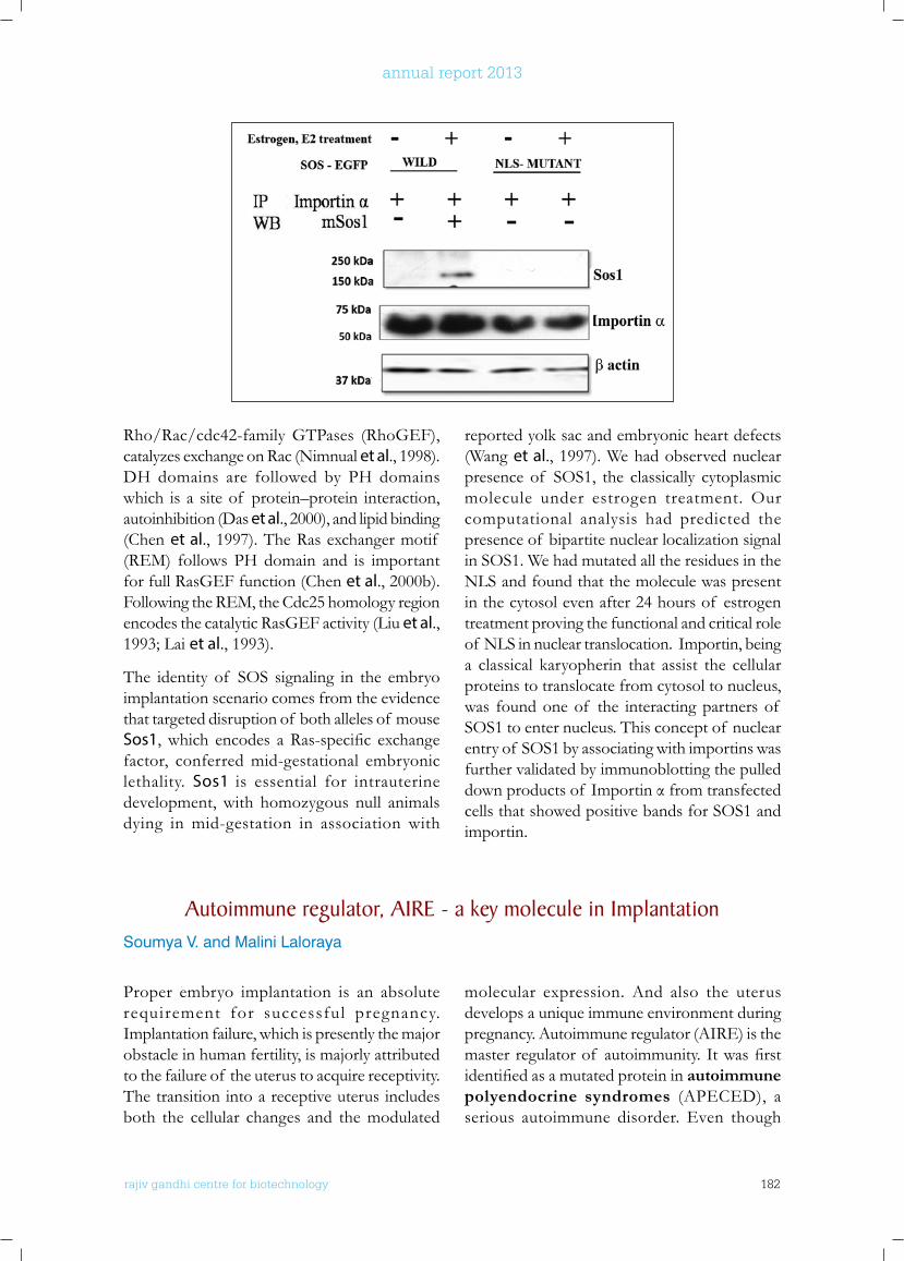
rajiv gandhi centre for biotechnology 182
annual report 2013
Rho/Rac/cdc42-family GTPases (RhoGEF), catalyzes exchange on Rac (Nimnual et al., 1998). DH domains are followed by PH domains which is a site of protein–protein interaction, autoinhibition (Das et al., 2000), and lipid binding (Chen et al., 1997). The Ras exchanger motif (REM) follows PH domain and is important for full RasGEF function (Chen et al., 2000b). Following the REM, the Cdc25 homology region encodes the catalytic RasGEF activity (Liu et al., 1993; Lai et al., 1993).
The identity of SOS signaling in the embryo implantation scenario comes from the evidence that targeted disruption of both alleles of mouse Sos1,which encodes aRas-specific exchangefactor, conferred mid-gestational embryonic lethality. Sos1 is essential for intrauterine development, with homozygous null animals dying in mid-gestation in association with
reported yolk sac and embryonic heart defects (Wang et al., 1997). We had observed nuclear presence of SOS1, the classically cytoplasmic molecule under estrogen treatment. Our computational analysis had predicted the presence of bipartite nuclear localization signal in SOS1. We had mutated all the residues in the NLS and found that the molecule was present in the cytosol even after 24 hours of estrogen treatment proving the functional and critical role of NLS in nuclear translocation. Importin, being a classical karyopherin that assist the cellular proteins to translocate from cytosol to nucleus, was found one of the interacting partners of SOS1 to enter nucleus. This concept of nuclear entry of SOS1 by associating with importins was further validated by immunoblotting the pulled downproductsof Importinαfromtransfectedcells that showed positive bands for SOS1 and importin.
Autoimmune regulator, AIRE - a key molecule in ImplantationSoumya V. and Malini Laloraya
Proper embryo implantation is an absolute requirement for successful pregnancy. Implantation failure, which is presently the major obstacle in human fertility, is majorly attributed to the failure of the uterus to acquire receptivity. The transition into a receptive uterus includes both the cellular changes and the modulated
molecular expression. And also the uterus develops a unique immune environment during pregnancy. Autoimmune regulator (AIRE) is the masterregulatorof autoimmunity.Itwasfirstidentifiedasamutatedproteininautoimmune polyendocrine syndromes (APECED), a serious autoimmune disorder. Even though
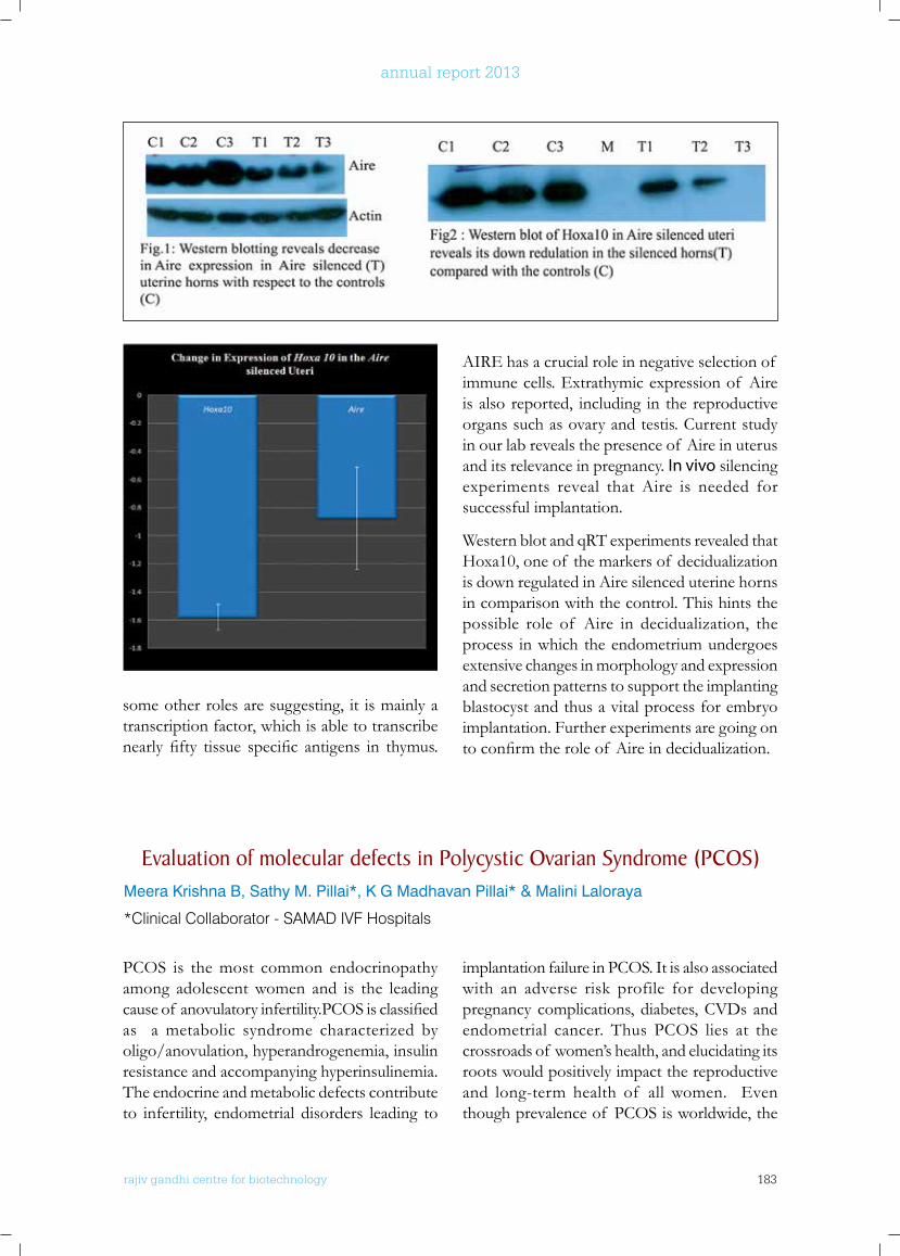
rajiv gandhi centre for biotechnology 183
annual report 2013
Evaluation of molecular defects in Polycystic ovarian Syndrome (PCoS)Meera Krishna B, Sathy M. Pillai*, K G Madhavan Pillai* & Malini Laloraya
*Clinical Collaborator - SAMAD IVF Hospitals
PCOS is the most common endocrinopathy among adolescent women and is the leading causeof anovulatoryinfertility.PCOSisclassifiedas a metabolic syndrome characterized by oligo/anovulation, hyperandrogenemia, insulin resistance and accompanying hyperinsulinemia. The endocrine and metabolic defects contribute to infertility, endometrial disorders leading to
implantation failure in PCOS. It is also associated with an adverse risk profile for developing pregnancy complications, diabetes, CVDs and endometrial cancer. Thus PCOS lies at the crossroads of women’s health, and elucidating its roots would positively impact the reproductive and long-term health of all women. Even though prevalence of PCOS is worldwide, the
AIRE has a crucial role in negative selection of immune cells. Extrathymic expression of Aire is also reported, including in the reproductive organs such as ovary and testis. Current study in our lab reveals the presence of Aire in uterus and its relevance in pregnancy. In vivo silencing experiments reveal that Aire is needed for successful implantation.
Western blot and qRT experiments revealed that Hoxa10, one of the markers of decidualization is down regulated in Aire silenced uterine horns in comparison with the control. This hints the possible role of Aire in decidualization, the process in which the endometrium undergoes extensive changes in morphology and expression and secretion patterns to support the implanting blastocyst and thus a vital process for embryo implantation. Further experiments are going on toconfirmtheroleof Aireindecidualization.
some other roles are suggesting, it is mainly a transcription factor, which is able to transcribe nearlyfifty tissue specific antigens in thymus.
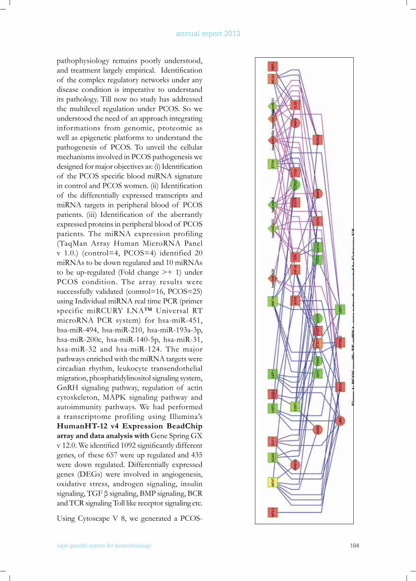
rajiv gandhi centre for biotechnology 184
annual report 2013
pathophysiology remains poorly understood, andtreatmentlargelyempirical.Identificationof the complex regulatory networks under any disease condition is imperative to understand its pathology. Till now no study has addressed the multilevel regulation under PCOS. So we understood the need of an approach integrating informations from genomic, proteomic as well as epigenetic platforms to understand the pathogenesis of PCOS. To unveil the cellular mechanisms involved in PCOS pathogenesis we designedformajorobjectivesas:(i)Identificationof thePCOSspecificbloodmiRNAsignatureincontrolandPCOSwomen.(ii)Identificationof the differentially expressed transcripts and miRNA targets in peripheral blood of PCOS patients. (iii) Identification of the aberrantlyexpressed proteins in peripheral blood of PCOS patients. The miRNA expression profiling (TaqMan Array Human MicroRNA Panel v 1.0.) (control=4, PCOS=4) identified 20 miRNAs to be down regulated and 10 miRNAs to be up-regulated (Fold change >+ 1) under PCOS condition. The array results were successfully validated (control=16, PCOS=25) using Individual miRNA real time PCR (primer specific miRCURY LNA™ Universal RT microRNA PCR system) for hsa-miR-451, hsa-miR-494, hsa-miR-210, hsa-miR-193a-3p, hsa-miR-200c, hsa-miR-140-5p, hsa-miR-31, hsa-miR-32 and hsa-miR-124. The major pathways enriched with the miRNA targets were circadian rhythm, leukocyte transendothelial migration, phosphatidylinositol signaling system, GnRH signaling pathway, regulation of actin cytoskeleton,MAPK signaling pathway andautoimmunity pathways. We had performed a transcriptome profiling using Illumina’s HumanHT-12 v4 Expression BeadChip array and data analysis with Gene Spring GX v12.0.Weidentified1092significantlydifferentgenes, of these 657 were up regulated and 435 were down regulated. Differentially expressed genes (DEGs) were involved in angiogenesis, oxidative stress, androgen signaling, insulin signaling,TGFβsignaling,BMPsignaling,BCRand TCR signaling Toll like receptor signaling etc.
Using Cytoscape V 8, we generated a PCOS-
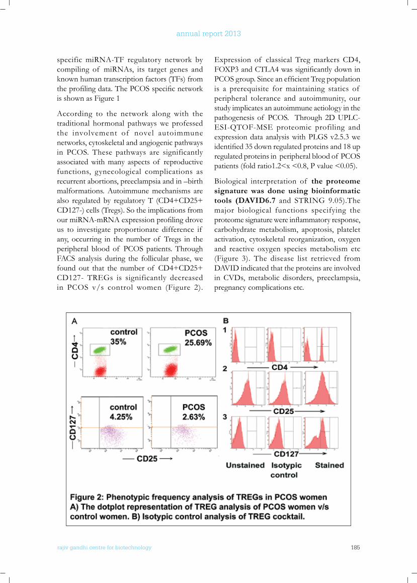
rajiv gandhi centre for biotechnology 185
annual report 2013
specific miRNA-TF regulatory network by compiling of miRNAs, its target genes and known human transcription factors (TFs) from theprofilingdata.ThePCOSspecificnetworkis shown as Figure 1
According to the network along with the traditional hormonal pathways we professed the involvement of novel autoimmune networks, cytoskeletal and angiogenic pathways in PCOS. These pathways are significantly associated with many aspects of reproductive functions, gynecological complications as recurrent abortions, preeclampsia and in –birth malformations. Autoimmune mechanisms are also regulated by regulatory T (CD4+CD25+ CD127-) cells (Tregs). So the implications from ourmiRNA-mRNAexpressionprofilingdroveus to investigate proportionate difference if any, occurring in the number of Tregs in the peripheral blood of PCOS patients. Through FACS analysis during the follicular phase, we found out that the number of CD4+CD25+ CD127- TREGs is significantly decreased in PCOS v/s control women (Figure 2).
Expression of classical Treg markers CD4, FOXP3andCTLA4wassignificantlydowninPCOSgroup.SinceanefficientTregpopulationis a prerequisite for maintaining statics of peripheral tolerance and autoimmunity, our study implicates an autoimmune aetiology in the pathogenesis of PCOS. Through 2D UPLC-ESI-QTOF-MSE proteomic profiling and expression data analysis with PLGS v2.5.3 we identified35downregulatedproteinsand18upregulated proteins in peripheral blood of PCOS patients (fold ratio1.2<x <0.8, P value <0.05).
Biological interpretation of the proteome signature was done using bioinformatic tools (DAVID6.7 and STRING 9.05).The major biological functions specifying the proteomesignaturewereinflammatoryresponse,carbohydrate metabolism, apoptosis, platelet activation, cytoskeletal reorganization, oxygen and reactive oxygen species metabolism etc (Figure 3). The disease list retrieved from DAVID indicated that the proteins are involved in CVDs, metabolic disorders, preeclampsia, pregnancy complications etc.
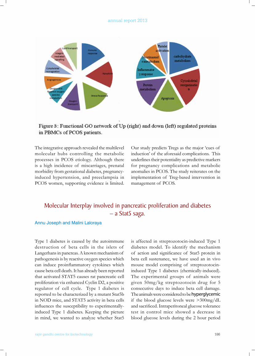
rajiv gandhi centre for biotechnology 186
annual report 2013
The integrative approach revealed the multilevel molecular hubs controlling the metabolic processes in PCOS etiology. Although there is a high incidence of miscarriages, prenatal morbidity from gestational diabetes, pregnancy-induced hypertension, and preeclampsia in PCOS women, supporting evidence is limited.
Our study predicts Tregs as the major ‘cues of induction’ of the aforesaid complications. This underlines their potentiality as predictive markers for pregnancy complications and metabolic anomalies in PCOS. The study reiterates on the implementation of Treg-based intervention in management of PCOS.
Molecular Interplay involved in pancreatic proliferation and diabetes – a Stat5 saga.
Annu Joseph and Malini Laloraya
Type 1 diabetes is caused by the autoimmune destruction of beta cells in the islets of Langerhans in pancreas. A known mechanism of pathogenesis is by reactive oxygen species which can induce proinflammatory cytokineswhichcause beta cell death. It has already been reported that activated STAT5 causes rat pancreatic cell proliferation via enhanced Cyclin D2, a positive regulator of cell cycle. Type 1 diabetes is reported to be characterized by a mutant Stat5b in NOD mice, and STAT5 activity in beta cells influencesthesusceptibilitytoexperimentally-inducedType 1 diabetes.Keeping thepicturein mind, we wanted to analyze whether Stat5
is affected in streptozotocin-induced Type 1 diabetes model. To identify the mechanism of actionand significanceof Stat5protein inbeta cell sustenance, we have used an in vivo mouse model comprising of streptozotocin-induced Type 1 diabetes (chemically-induced). The experimental groups of animals were given 50mg/kg streptozotocin drug for 5 consecutive days to induce beta cell damage. The animals were considered to be hyperglycemic if the blood glucose levels were >300mg/dL andsacrificed.Intraperitonealglucosetolerancetest in control mice showed a decrease in blood glucose levels during the 2 hour period
The integrative approach revealed the multilevel molecular hubs controlling the metabolic
processes in PCOS etiology. Although there is a high incidence of miscarriages, prenatal
morbidity from gestational diabetes, pregnancy-induced hypertension, and preeclampsia in
PCOS women, supporting evidence is limited. Our study predicts Tregs as the major ‘cues of
induction’ of the aforesaid complications. This underlines their potentiality as predictive
markers for pregnancy complications and metabolic anomalies in PCOS. The study
reiterates on the implementation of Treg-based intervention in management of PCOS.
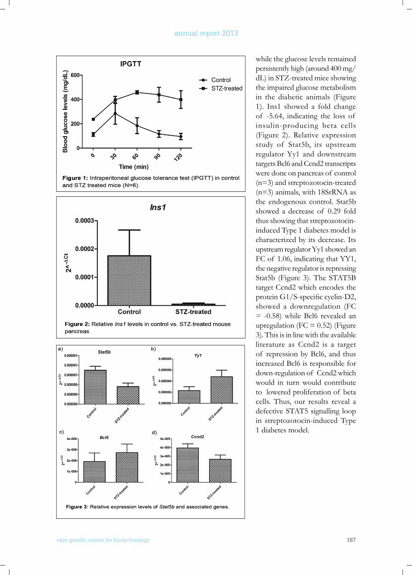
rajiv gandhi centre for biotechnology 187
annual report 2013
while the glucose levels remained persistently high (around 400 mg/dL) in STZ-treated mice showing the impaired glucose metabolism in the diabetic animals (Figure 1). Ins1 showed a fold change of -5.64, indicating the loss of insulin-producing beta cells (Figure 2). Relative expression study of Stat5b, its upstream regulator Yy1 and downstream targets Bcl6 and Ccnd2 transcripts were done on pancreas of control (n=3) and streptozotocin-treated (n=3) animals, with 18SrRNA as the endogenous control. Stat5b showed a decrease of 0.29 fold thus showing that streptozotocin-induced Type 1 diabetes model is characterized by its decrease. Its upstream regulator Yy1 showed an FC of 1.06, indicating that YY1, the negative regulator is repressing Stat5b (Figure 3). The STAT5B target Ccnd2 which encodes the proteinG1/S-specificcyclin-D2,showed a downregulation (FC = -0.58) while Bcl6 revealed an upregulation (FC = 0.52) (Figure 3). This is in line with the available literature as Ccnd2 is a target of repression by Bcl6, and thus increased Bcl6 is responsible for down-regulation of Ccnd2 which would in turn would contribute to lowered proliferation of beta cells. Thus, our results reveal a defective STAT5 signalling loop in streptozotocin-induced Type 1 diabetes model.
rajiv gandhi centre for biotechnology 188
annual report 2013
PRIMARy PublICATIonS
• Renjini A P, Titus S, Narayan P, Murali M, Jha R K, Laloraya M. STAT3 and MCL-1 associate to cause a mesenchymal epithelial transition. J Cell Sci. 2014 Apr 15;127(Pt 8):1738-50. doi: 10.1242/jcs.138214. Epub 2014 Jan 30.
CollAboRATIVE PublICATIonS
• Maurya V K, Jha R K, Kumar V, Joshi A, Chadchan S, Mohan JJ , Laloraya M. Transforming growth factor-beta 1 (TGF-B1) liberation from its latent complex during embryo implantation and its regulation by estradiol in mouse. Biol Reprod. 2013 Oct 10;89(4):84. doi: 10.1095/biolreprod.112.106542.
AwARDS
• Dr. Malini Laloraya - Labshetwar Award (2014) of Indian Society for the Study of Reproduction & Fertility. Award Lecture entitled “Embryo implantation : a vital node in reproductive success”.
• Ms. Meera Krishna B. Travel award winner at 12th Congress for International Society for Immunology Reproduction (ISIR-2013), Boston, Massachusetts,U.S.A (May 28-June 1, 2013).
• Ms. Meera Krishna B. Winner Under 34 Competi t ion, 16th World Congress of Gynecological endocrinology, (ISGE-2014) Florence, Italy (March 5-8, 2014).
ConfEREnCE PRESEnTATIonS - InVITED TAlkS/oRAl/PoSTER (2013-2014)
• Malini Laloraya*. Intersection of Proteomics and Bioinformatics in deciphering novel functions of proteins. Invited lecture at “3rd International Conference on Proteomics & Bioinformatics” held on July 15-17, 2013 by the OMICS Group at Courtyard by Marriott Philadelphia Downtown, USA.
• Jasna J. Mohan, Pradeep Kumar G and Malini Laloraya* Silencing of DOCK180 is Deleterious for Establishment of a Successful Pregnancy. Poster presented at SSR 2013, 46th Annual Meeting Reproductive Health: Nano to Global 22–26 July 2013, Montréal, Québec, Canada held at Palais des congrès de Montréal (1001 Place Jean-Paul-Riopelle, Montréal, QC, Canada).
• Annu Joseph and Malini Laloraya* Differential pancreatic protein expression profile in type 1 diabetes mouse model: Novel insights and implications. Presented at the 17th Association for Promotion of DNA Fingerprinting and other DNA Technologies (ADNAT) convention and symposium on Genomics in Personalized Medicine and Public Health 23rd – 25th February 2014 at RGCB, Thiruvananthapuram.
• Meera Krishna B, Sathy M. Pillai and Malini Laloraya*. Low Circulating T Regulatory Cells: indication of autoimmune etiology in PCOS. Oral and poster presentation at 12th Congress of the International Society for Immunology of Reproduction (ISIR) hosted by the American Society for Reproductive Immunology (ASRI), Boston, MA (May 28-June 1, 2013).
• Meera Krishna B, Sathy M. Pillai and Malini Laloraya*. An Integrative mRNA-Transcription Factor Analysis Reveals Important PCOS Hallmarks. Oral presentation at 16th World congres on Gyneacological Endocrinology (ISGE 2014), Florence , Italy (March 5-8, 2014).
ExTRA-MuRAl funDInG
Investigator Title Funding Agency Duration
Dr. Malini Laloraya(PI) Deciphering the SOS-GRB signaling at embryo Implantation.
Board of Research in Nuclear Sciences
2010 to 2014
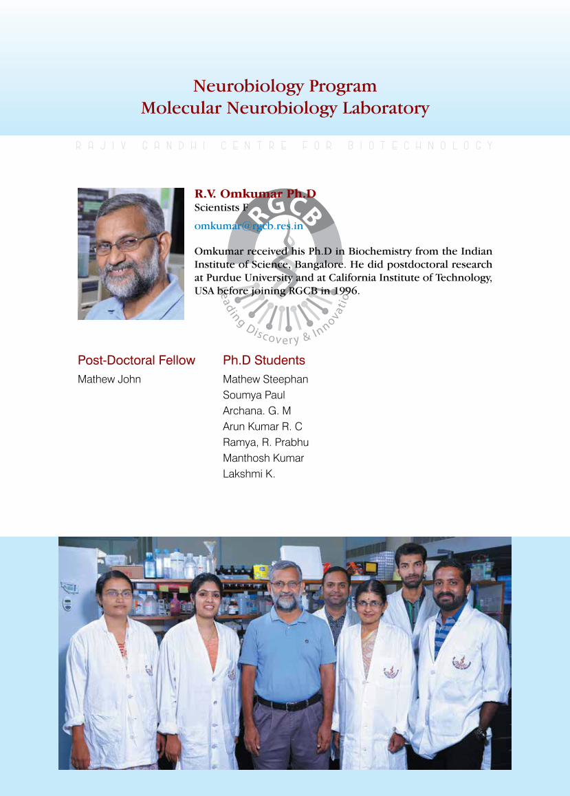
rajiv gandhi centre for biotechnology 189
annual report 2013
Neurobiology ProgramMolecular Neurobiology Laboratory
Post-Doctoral FellowMathew John
r a j i v g a n d h i c e n t r e f o r b i o t e c h n o l o g y
Omkumar received his Ph.D in Biochemistry from the Indian Institute of Science, Bangalore. He did postdoctoral research at Purdue University and at California Institute of Technology, USA before joining RGCB in 1996.
R.V. Omkumar Ph.D Scientists F
Ph.D StudentsMathew SteephanSoumya PaulArchana. G. MArun Kumar R. C Ramya, R. PrabhuManthosh KumarLakshmi K.
rajiv gandhi centre for biotechnology 190
annual report 2013
Synaptic plasticity is a cellular event that is thought to give rise to learning and memory in the whole organism. Calcium signalling at synapses plays a major role in the mechanisms underlying synaptic plasticity. However, under pathological conditions calcium signalling pathways lose their regulation leading to intracellular calcium overload and cell death, a process termed as excitotoxicity. Excitotoxicity is a mechanism of cell death in many neurological and neurodegenerative diseases. Molecules like calcium/calmodulin dependent protein kinaseII(CaMKII)andN-methyl-D-aspartatereceptors (NMDAR), are primary players in synaptic calcium signalling and has key roles in synaptic plasticity and excitotoxicity. We have earlierfoundthatdephosphorylationof CaMKIIis inhibited when it is bound by the GluN2B subunit of NMDAR. Since GluN2B binds at theT-siteof CaMKIIwearenowtesting theeffect of other T-site binding proteins on the properties of CaMKII. Binding interactionbetweenCaMKII andNMDAR is regulated
by the phosphorylation status of the GluN2B subnit. We have monitored the phosphorylation status of GluN2B in primary cortical neurons subsequent to differernt types of stimuli. There were changes in the phosphorylation level of GluN2B. The changes in levels of phosphorylation were found to be mediated by protein phosphatase. We are currently investigating whether the calcium signal mediated alterations in GluN2B phosphorylation levels happen in vivo. Administration in vivo, of agonists and antagonists of various calcium channels followed by biochemical and histochemical analysis of brain tissue, is being undertaken for this purpose. We have also been studying calcium signalling in cerebellar granule (CG) cells since the molecules related to calcium signalling that are present in CG cells are different. This study is expected to reveal how the calcium signalling pathways inCG cells have beenmodified toadapt to the presence of different molecules. For this purpose, we have standardised the culture of CG cells.
bioprospecting for neuroprotectantsMathew Steephan, Soumya Paul, Arun kumar R.C., Mantosh Kumar, Mayadevi M., and Omkumar R.V.
Synaptic Calcium Signaling and associated biochemical MechanismsArchana G. M., Ramya.R.Prabhu, Mathew John, Lakshmi, K., Mayadevi M., and Omkumar R.V.
We have found that the ethanolic extracts of two plants were found to have antagonist properties against calcium conducting NR1/GluN2B channels. One of the extracts was
tested on primary cortical neurons and also in a rat model of monosodiumglutamate induced excitotoxicity. In both models, the extract was found to show neuroprotection.
rajiv gandhi centre for biotechnology 191
annual report 2013
A novel Technology platform for the assay of Ca2+ channelsMathew Steephan., Soumya Paul., Arunkumar R.C., Manthosh Kumar, Mayadevi,M. and Omkumar R.V
We have developed an assay system based on the specific interactionbetweenCaMKII andGluN2B subunit of NMDAR. A calcium sensor cell line that stably expressesGFP-a-CaMKIIand mitochondrially localised GluN2B sequence (MLS-NR2B) has been developed. This cell line responds to a calcium influxby formingmitochondrially localised green fluorescent
punctae that represents the complex between CaMKIIandGluN2Basasignal forcalcium.This cell line can be used for observing the activity of any calcium channel. Currently this cell line is under detailed characterisation with respect to its Ca2+-sensitivity, growth characteristics, etc.
ConfEREnCE PRESEnTATIonS
• Omkumar, R. V., Sebastian John and Mayadevi, M. CaMKIINa and GluN2B exhibit similar modes of regulation of CaMKII function. Poster presentation at the Annual Meeting of the Society for Neuroscience, USA, held at San Diego, California, USA, November 9-13, 2013.
• Arunkumar, R. C. , Mathew Steephan, Rajeevkumar, R., Sumapriya, S., Soumya Paul, Archana, G M., Mayadevi, M., Omkumar, R. V. A new cell line for screening of P2X2 channel
activity modulators. Poster presentation at the Annual Meeting of the Society for Neuroscience, USA, held at San Diego, California, USA, November 9-13, 2013.
PATEnT
• Indian patent granted for the invention “Assay for Detection of Transient Intracellular Calcium” by Omkumar, R. V., Rajeevkumar, R., Mathew Steephan, Mayadevi, M. and Suma Priya.
ExTRA MuRAl GRAnTS
Sl.No. Title Funding Agency Total Amount Duration
1 A Novel Technology platform for the assay of Ca2+ channels
Department of Biotechnology
Rs 81.05 lakhs 2011-2013
rajiv gandhi centre for biotechnology 192
annual report 2013
Neurobiology ProgramMolecular Neurobiology Laboratory
r a j i v g a n d h i c e n t r e f o r b i o t e c h n o l o g y
Mayadevi received her M.Sc. in Chemistry from the University of Kerala and worked at Case Western Reserve University and Baylor Research Institute, USA, before joining RGCB.
Mayadevi M. Scientists C
non-inhibitory functions of CaMkIIna inhibitor protein
CaMKIINainhibitorproteinisanendogenousinhibitor of Calcium/calmodulin dependent proteinkinaseII(CaMKII),thekinaseinvolvedin calcium signaling pathways. The expression of CaMKIINa inhibitor protein is elevatedin the brain in certain diseased states. How CaMKIINaregulatesCaMKIIsignalinghasnotbeen completely elucidated. We are investigating the functions other than the inhibitory functions of CaMKII.GluN2B,thesubunitof NMDAreceptor and CaMKIINa can both bind to
T-site of CaMKII. We are addressing thequestion whether T-site mediated interactions of CaMKIINawithCaMKIIwouldelicitsimilarkind of consequences as that of GluN2B-CaMKII interactions. Dephosphorylation of Thr286-CaMKII by PP1 is inhibited whenCaMKIINa binds to CaMKII. Thus theinteractionof CaMKIINawithT-siteof CaMKIIcould possibly stabilize the autophosphorylated stateof CaMKII.
ConfEREnCE PRESEnTATIonS
• R. V. Omkumar, S. John and M. Madhavan, CaMKIINa and GluN2B exhibit similar modes of regulation of CaMKII function. (SFN) Neuroscience 2013, San Diego, California, USA, November 9-13, 2013.
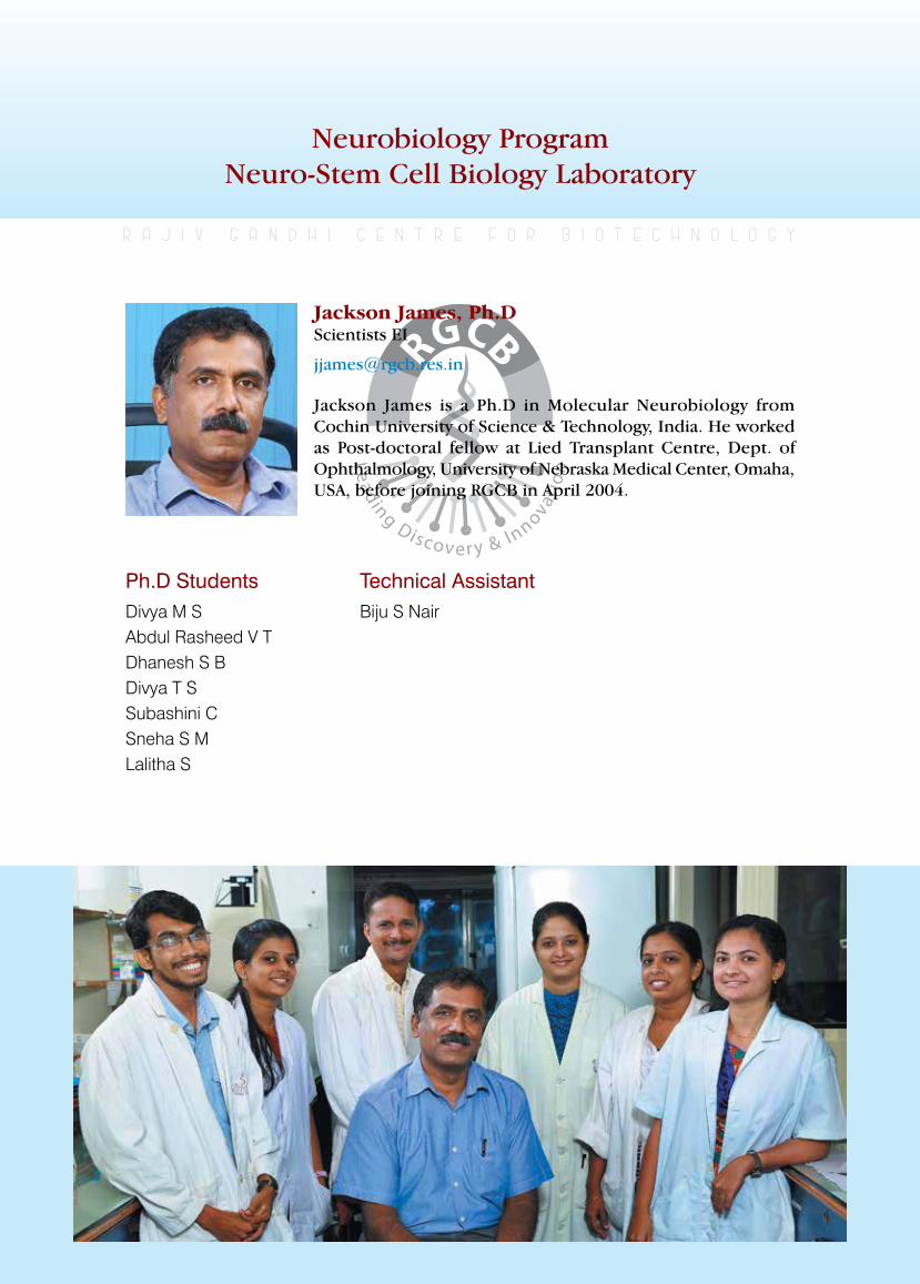
rajiv gandhi centre for biotechnology 193
annual report 2013
Neurobiology ProgramNeuro-Stem Cell Biology Laboratory
Ph.D StudentsDivya M SAbdul Rasheed V T Dhanesh S BDivya T SSubashini CSneha S M Lalitha S
r a j i v g a n d h i c e n t r e f o r b i o t e c h n o l o g y
Jackson James is a Ph.D in Molecular Neurobiology from Cochin University of Science & Technology, India. He worked as Post-doctoral fellow at Lied Transplant Centre, Dept. of Ophthalmology, University of Nebraska Medical Center, Omaha, USA, before joining RGCB in April 2004.
Jackson James, Ph.D Scientists EI
Technical AssistantBiju S Nair
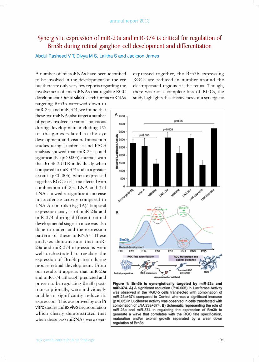
rajiv gandhi centre for biotechnology 194
annual report 2013
Anumberof microRNAshavebeenidentifiedto be involved in the development of the eye but there are only very few reports regarding the involvement of microRNAs that regulate RGC development. Our in silico search for microRNAs targeting Brn3b narrowed down to miR-23a and miR-374, we found that these two miRNAs also target a number of genes involved in various functions during development including 1% of the genes related to the eye development and vision. Interaction studies using Luciferase and FACS analysis showed that miR-23a could significantly (p<0.005) interactwiththe Brn3b 3’UTR individually when compared to miR-374 and to a greater extent (p<0.005) when expressed together. RGC-5 cells transfected with combination of 23a LNA and 374 LNA showed a significant increasein Luciferase activity compared to LNA-A controls (Fig-1A).Temporal expression analysis of miR-23a and miR-374 during different retinal developmental stages in mice was also done to understand the expression pattern of these miRNAs. These analyses demonstrate that miR-23a and miR-374 expressions were well orchestrated to regulate the expression of Brn3b pattern during mouse retinal development. From our results it appears that miR-23a and miR-374 although predicted and proven to be regulating Brn3b post-transcriptionally, were individually unable to significantly reduce its expression. This was proved by our in vitro studies and ex vivo electroporation which clearly demonstrated that when these two miRNAs were over-
expressed together, the Brn3b expressing RGCs are reduced in number around the electroporated regions of the retina. Though, there was not a complete loss of RGCs, the study highlights the effectiveness of a synergistic
Synergistic expression of miR-23a and miR-374 is critical for regulation of brn3b during retinal ganglion cell development and differentiation
Abdul Rasheed V T, Divya M S, Lalitha S and Jackson James
rajiv gandhi centre for biotechnology 195
annual report 2013
post-transcriptional regulation of Brn3b rather than an individual effect. Previous in vitro studies in RGC-5 cells have also shown the effectiveness of a combination of miR-23a and miR-214 in targeting Brn3b (Calissano and Latchman, Mol Cell Neurosci, 45:317-323, 2010).Thisalsojustifiesthe earlier notion that two or more miRNAs in combination can target a single gene bringing a synergistic effect similarly; more than one gene may be targeted by the same miRNA and vice versa.
Our results also demonstrated that miR-23a and miR-374 may have a role in regulating the wave of Brn3b expression during development (Fig-1B). There is typically an initial spike in Brn3b expression peaking at E14 during which most of the RGCs are born and the second wave of Brn3b expression may be required for the survival and axonal extension of the differentiated RGCs. It is known that a large number of RGCs are initially generated during thefirstwaveof Brn3bexpressionoutof which
only a small percentage of RGCs survive and put out their axons through the optic nerve. It is possible that these RGCs only might have the second wave of Brn3b expression and continue to survive/populate the GCL and form functionally active RGCs. Currently, we have no clear idea regarding the fate of RGCs that fail to express the second wave of Brn3b. The down regulation of Brn3b may aid in the expression of other genes that were repressed by Brn3b for the complete differentiation of RGCs and hence these Brn3b-ve RGC progenitors may either undergo apoptosis or shift to a different fate. These possibilities need to be explored further. It is possible that the transcription factors binding to the Brn3b promoter could be regulating the two waves of Brn3b expression but there is not much convincing literature available to prove this notion. In conclusion,we identified twomiRNAs, miR-23a and miR-374, which may play a role in timing of Brn3b expression in RGCs during development.
Characterisation of neural progenitors having notch independent Hes-1 expression in developing neocortex
Dhanesh SB and Jackson James
Collaborators: Shubha ToleTata Institute of Fundamental Research, Mumbai; R V Omkumar, Molecular Neurobiology Laboratory, RGCB; Santhosh Kumar Sankaran, Animal Research Facility, RGCB.
Our lab had previously reported the presence of a novel Notch independent Hes-1 expression in ES cell derived neural progenitors that is mediated throughFGF2-JNKpathway (Sanalet.al. J. Neurochem., 113, 2010, 807–818). We also showed that these progenitors are present in the VZ of the developing neo-cortex. Further in utero electroporation studies in E14 embryos were carried out with reporter systems that were able to differentiate between the Notch dependent and Notch independent Hes-1 expressing progenitors. Even though we found a lesser number of EGFP positive Notch independent Hes-1 expressing progenitors compared to wild type Hes-1, it is clear that
Notch independent Hes-1 expressing neuronal precursors are present and restricted to the Venticular Zone of the mouse neo-cortex. Another interesting observation was the position of EGFP expressing cells in both cases. The wild type Hes-1 expressing cells were found to be migrating from the VZ to SVZ. Surprisingly, we could not observe much of this phenomenon in case of Notch/CBF1 independent Hes-1 expressing cells which resides more in the apical surface near the ventricle. To further demonstrate that the Notch independent Hes-1 expressing progenitors resides predominantly in the VZ we carried out in utero electroporation into E14 embryos with wtHes1-d2EGFP and mtCBF1-
rajiv gandhi centre for biotechnology 196
annual report 2013
d2EGFP vectors and analyzed the cortex after 12hours of electroporation. d2EGFP is a destabilized form of EGFP with a half-life of 2 hourswhichwillreflectthedynamicexpressionof Hes-1 and not that of the accumulated GFP protein. Our result clearly showed that the Notch independent Hes-1 expressing progenitors (transfected with mtCBF-1) reside in the VZ and do not readily migrate into the cortex, whereas the wtHes-1 transfected progenitors were found to be migrating within the cortex and express Hes-1 through the canonical Notch pathway. The position and typical morphological characteristics of EGFP expressing progenitors which markedly discriminate both populations lead us to investigate the mode of division followed by these cells during development. The primary neural stem/progenitor cells will undergo both symmetric and asymmetric division during development. In order to understand the mode of division followed by both Notch dependent and independent Hes-1 expressing
progenitors, we blocked the Notch signaling in Hes1-d2EGFP mice with γ-secretase inhibitor, DAPT which blocks canonical Notch signaling pathway. Intra peritoneal (IP) administration of DAPT (100mg/Kg)was given tomother andE14 embryos were harvested after six hours of drug administration. BrdU was also given for the same period to analyze the rate of proliferation during this small window. We checked the mode of division in apical neural progenitors by analyzing the cleavage orientation of mitotic cells in the DAPI stained EGFP positive cells. As shown in Fig-2, we were able to observe more number of symmetrically dividing cells having vertical cleavage plane with respect to the ventricular surface in DAPT treated cortex. From these observations, it becomes quite clear that Notch independent Hes-1 expressing progenitors follows symmetric division during neo-cortical development and is required for exponential expansion of the neural progenitors in the VZ to maintain the progenitor pool.
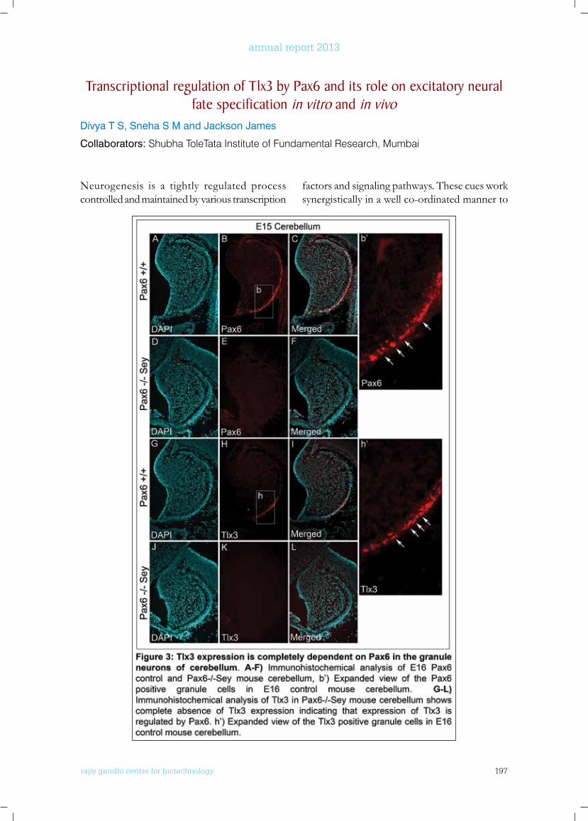
rajiv gandhi centre for biotechnology 197
annual report 2013
Transcriptional regulation of Tlx3 by Pax6 and its role on excitatory neural fate specification in vitro and in vivo
Divya T S, Sneha S M and Jackson James
Collaborators: Shubha ToleTata Institute of Fundamental Research, Mumbai
Neurogenesis is a tightly regulated process controlled and maintained by various transcription
factors and signaling pathways. These cues work synergistically in a well co-ordinated manner to
rajiv gandhi centre for biotechnology 198
annual report 2013
direct the neuronal fate to either glutamatergic or GABAergic. Tlx3, a homeobox gene, is identifiedasapost-mitoticselectorgeneinvolvedin glutamatergic neurogenesis and is found to be expressed in a subset of spinal neurons, brainstem and cerebellum. Previous studies of Tlx3 in our lab have proved that Hes-1, a Notch target gene acts as a repressor of Tlx3 expression (Indulekha et al, 2012; Cell Mol Life Sci, 69;611-627). Bioinformatics analysis of Tlx3 promoter has revealed the presence of conserved binding sites for Pax6, a proneural gene which is found to have an evident role in glutamatergic neuron fate determination. Immunohistochemical analysis proved that Pax6 and Tlx3 are co-expressed together in granule neurons of cerebellum. Studies done in cerebellar granule neuron cells (CGNs) proved that Pax6 could act as a possible activator of Tlx3. Analysis of Pax6-/- Sey mouse cerebellum proved that Tlx3 expression is completely dependent on Pax6 and its expression is completely abolished in
the absence of Pax6 (Fig-3). Co-expression analysisof Ki67/Tlx3andBrdU/Tlx3showedthat Tlx3 expression starts at a mitotic stage in CGNs and maintains till those cells migrate from external granule layer to internal granule layerwhichwasconfirmedbyDCX/Tlx3andvGlut1/Tlx3 immunohistochemical analysis. Further examination of calcium binding protein, Parvalbumin in control and Pax6-/-
Sey mouse cerebellum proved that there is a compartmentalization in expression pattern of parvalbumin and this in turn may regulate the proliferation rate of CGNs through the regulation of cyclin B1. To further analyze the specific mechanism involved in Tlx3 mediated regulation of CGN development, the anterior and posterior regions of cerebellum were dissected out separately and microarray analysis was carried out. Further analysis of the differentially expressed genes will help to unravel the possible downstream mechanism involved in Tlx3 mediated neuronal development.
Elucidation of role of wnt signaling in neural subtype specificationSubashini C and Jackson James
Collaborator: Rejji Kuruvilla, Johns Hopkins University, Baltimore, USA
Wnt s ignal ing exer ts diverse roles in embryogenesis and development because of its various ligands and respective downstream target gene activation. A major non-canonical Wnt ligand, Wnt5a is known to play pivotal role in development especially in neurogenesis. It has been reported that Wnt5a promotes dopaminergic neurogenesis in midbrain and GABAergic neurogenesis in forebrain but its functional significance in cerebellumhas notbeen reported. It is well known that signals from isthmic organizer and roof plate, together with various transcription factors coordinately regulate the process of cerebellum development. Various studies demonstrated the role of other signaling pathways such as Notch, Wnt and Shh in neural progenitor maintenance and in differentiation of cerebellar granule progenitor
cells (CGNP’s) but the role of non-canonical Wnt signaling mediated by Wnt5a in the cerebellar development is not well understood. In order to characterize the Wnt5a expression pattern incerebellum,wefirstanalyzedtheexpressionpattern of Wnt5a in mouse cerebellum during the postnatal stages PN1 to PN7 using semi quantitative RT-PCR and western blot analysis. We found Wnt5a to be expressed in the postnatal stages PN1-PN7. Further we analyzed the spatio temporal expression of wnt5a in mouse cerebellum during embryonic and postnatal development using In situ hybridization. Wnt5a was robustly expressed in cerebellum during embryonic and postnatal development. In E14 cerebellum,Wnt5aexpressionwasconfinedtothe ventricular zone and the rhombic lip while in E16 cerebellum, the expression of Wnt5a
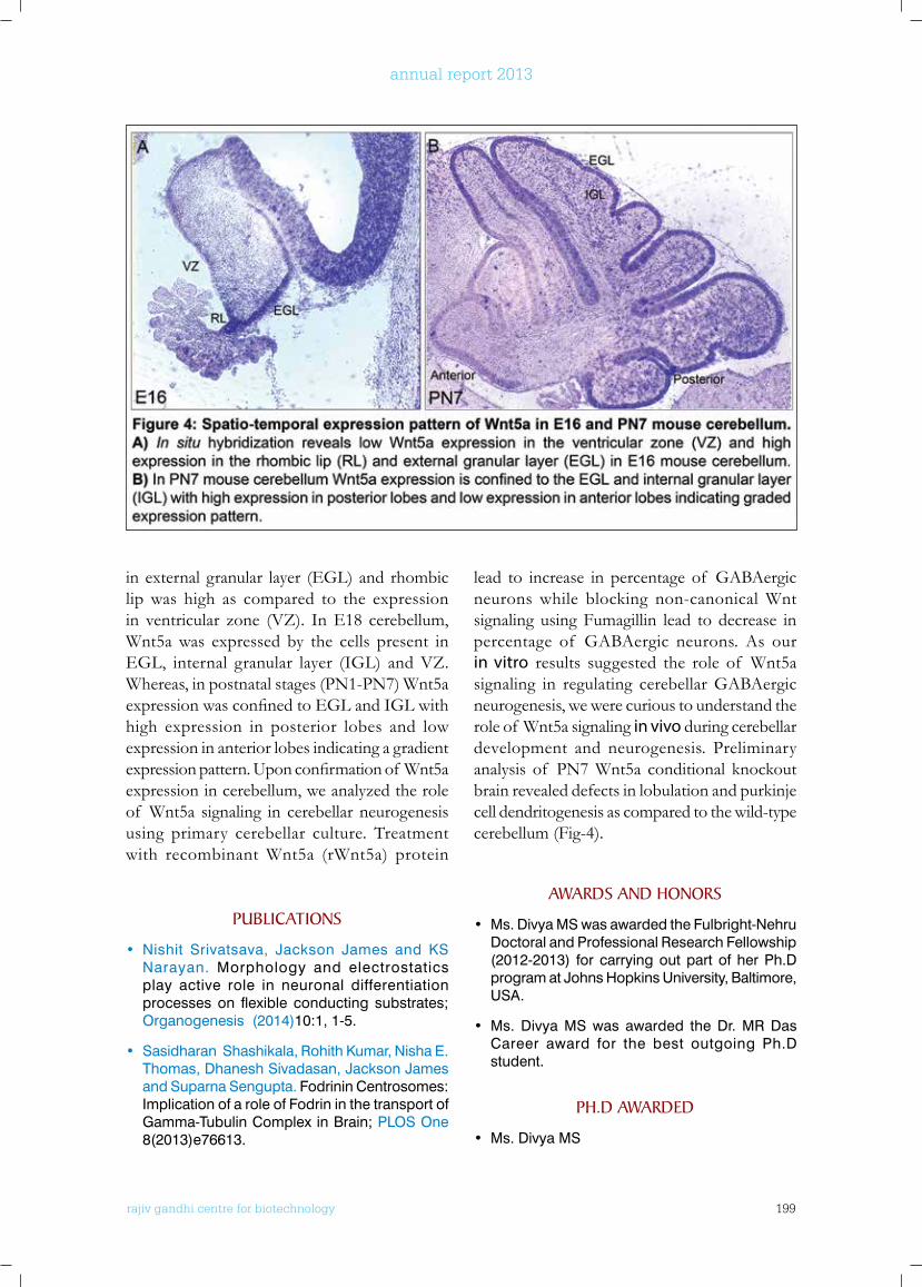
rajiv gandhi centre for biotechnology 199
annual report 2013
in external granular layer (EGL) and rhombic lip was high as compared to the expression in ventricular zone (VZ). In E18 cerebellum, Wnt5a was expressed by the cells present in EGL, internal granular layer (IGL) and VZ. Whereas, in postnatal stages (PN1-PN7) Wnt5a expressionwasconfinedtoEGLandIGLwithhigh expression in posterior lobes and low expression in anterior lobes indicating a gradient expressionpattern.Uponconfirmationof Wnt5aexpression in cerebellum, we analyzed the role of Wnt5a signaling in cerebellar neurogenesis using primary cerebellar culture. Treatment with recombinant Wnt5a (rWnt5a) protein
lead to increase in percentage of GABAergic neurons while blocking non-canonical Wnt signaling using Fumagillin lead to decrease in percentage of GABAergic neurons. As our in vitro results suggested the role of Wnt5a signaling in regulating cerebellar GABAergic neurogenesis, we were curious to understand the role of Wnt5a signaling in vivo during cerebellar development and neurogenesis. Preliminary analysis of PN7 Wnt5a conditional knockout brain revealed defects in lobulation and purkinje cell dendritogenesis as compared to the wild-type cerebellum (Fig-4).
PublICATIonS
• Nishit Srivatsava, Jackson James and KS Narayan. Morphology and electrostatics play active role in neuronal differentiation processes on flexible conducting substrates; Organogenesis (2014)10:1, 1-5.
• Sasidharan Shashikala, Rohith Kumar, Nisha E. Thomas, Dhanesh Sivadasan, Jackson James and Suparna Sengupta. Fodrinin Centrosomes: Implication of a role of Fodrin in the transport of Gamma-Tubulin Complex in Brain; PLOS One 8(2013)e76613.
AwARDS AnD HonoRS
• Ms. Divya MS was awarded the Fulbright-Nehru Doctoral and Professional Research Fellowship (2012-2013) for carrying out part of her Ph.D program at Johns Hopkins University, Baltimore, USA.
• Ms. Divya MS was awarded the Dr. MR Das Career award for the best outgoing Ph.D student.
PH.D AwARDED
• Ms. Divya MS
rajiv gandhi centre for biotechnology 200
annual report 2013
AbSTRACTS AnD ConfEREnCES
• Divya M S, Abdul Rasheed V T, Hattar Samer, James Jackson; RGC differentiation from ES cells: Influence of FGF2 and Notch Signaling, The Association for Vision and Ophthalmology (ARVO) 2013 Annual Meeting, Seatt le, Washington, May 05 - 09, 2013.
• Abdul Rasheed V T, Sreekanth S, Divya M S, Divya T S, Dhanesh S B, Subashini C, Ani Das V & Jackson James. Regulation of retinal ganglion cell fate specification and differentiation by miR-23a and miR-374 during retinal development. 43rd Annual Meeting of the Society for Neuroscience, November 9-13, 2013, San Diego, CA.
• Divya T S & Jackson James. Transcriptional regulation of Tlx3 by Pax6 and its influence on excitatory vs. inhibitory neural fate specification in Cerebellum. 37th All India Cell Biology Conference & Symposium on Cell Dynamics & Cell Fate, Bangalore, December 2013.
• Divya T S, Subashini C, Dhanesh S B, Abdul Rasheed V T & Jackson James. Novel Role of Tlx3 as a Mitotic factor and its Regulation by Pax6 during Cerebellar Granule Neuron Development. Adult neurogenesis: from stem cells to therapies conference, TIFR, Mumbai, February 2014
• Subashini C, Dhanesh S B, Divya T S and Jackson James. Elucidation of role of Wnt signaling in cerebellar neurogenesis. Adult neurogenesis: from stem cells to therapies conference, TIFR, Mumbai, February 2014.
• Dhanesh S B, Subashini C and Jackson James. Characterization of neural progenitors having Notch independent Hes-1 expression in developing neocortex. Adult neurogenesis: from stem cells to therapies conference, TIFR, Mumbai, February 2014.
RESEARCH GRAnTS
No Investigator(s) Title Funding Agency Duration
1 Jackson James (PI) Dr. RV Omkumar, Santhosh Kumar TR
Characterization of Notch independent Hes-1 mediated maintenance and fate specification of neural progenitors
Department of Biotechnology, Government of India
2013-2016
2 Dr. Jackson James (PI) Dr. RV Omkumar
Transcriptional regulation of Tlx3(Hox11L2) by Notch signaling and its involvement in excitatory Vs. inhibitory fate specification of neural progenitors
Department of Science & Technology, Government of India
2013-2016
3 Dr. Sreekumar E (PI) Dr. Jackson James (Co-PI)
Characterization of Neurovirulence of Chikungunya virus in cellular and animal models
Department of Biotechnology, Government of India
2012-2015
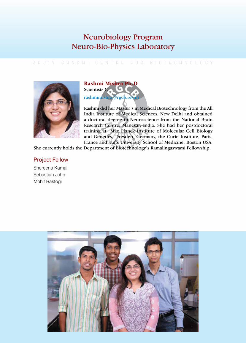
rajiv gandhi centre for biotechnology 201
annual report 2013
Neurobiology ProgramNeuro-Bio-Physics Laboratory
Project FellowShereena KamalSebastian JohnMohit Rastogi
r a j i v g a n d h i c e n t r e f o r b i o t e c h n o l o g y
Rashmi did her Master’s in Medical Biotechnology from the All India Institute of Medical Sciences, New Delhi and obtained a doctoral degree in Neuroscience from the National Brain Research Centre, Manesar, India. She had her postdoctoral training at Max Planck Institute of Molecular Cell Biology and Genetics, Dresden, Germany, the Curie Institute, Paris, France and Tufts University School of Medicine, Boston USA.
She currently holds the Department of Biotechnology’s Ramalingaswami Fellowship.
Rashmi Mishra Ph.D Scientists C
rajiv gandhi centre for biotechnology 202
annual report 2013
Galectins are a unique family of proteins so called essentially due to their binding to β-galactoside containing oligosaccharides anddisplay of lectin like clustering activity. The galectin family so far consists of 16 proteins all of which have an evolutionary conserved carbohydrate recognition domain (CRD) of about 130 amino acids that interacts with β- galactosides on the headgroups of lipid/protein glycoconjugates. Even though galectins are found in cytoplasm, nucleus and other intracellular organelles, the CRDs predominantly enable the surface-associated extracellular functions of galectins through CRD-glycan or CRD-amino acid interactions. Galectins have been associated with various pathologies, most notably cancers, where they have been implicated in tumor immune escape, tumor angiogenesis and homo-heterotypic adhesion of tumor cells thereby mediating tumor metastasis. We are interested in the structure-function analysis of galectins and how galectins’ mediated signaling cascades can be exploited in immunotherapy, bacterial/viral infections, cancers, regenerative medicine and developmental disorders.
In this project, we initially aim to derive the underlying roles of galectin family in development, signalling and tumorigenesis of human brain and expand our studies to the tumors of different tissue origins such as breast, cervical, colon, prostate etc to verify if the brain tumor mechanisms have a co-relation to other tumors. Recently, we found both through wet experiments and synthetic biology approach, a crucial biological property of Gal-9 in stem cells, wherein it’s binding to the major glycosphingolipid cell surface receptor is
demonstrated to be pH sensitive. This has led to a model wherein galectin-glycosphingolipid couples form a vectorial cargo recycling circuit for maintenance of polarized apico-basal compartments in dividing stem cells. A raft glycosphingolipid (GL) is identified as the surface receptor that binds to Gal-9 and a drop in pH leads to dissociation of this interaction. At normal pH, the galectin-glycosphingolipid couples caused membrane clustering in a concentration dependent manner, which beyond line tension causes membrane curvature and endocytosis. Since the pH of endosome is far below physiological pH (4.9-5.3), some dissociation of Gal-9 from its binding partner is expected to occur. These endosomes carrying Gal-9-GL disassemblies fuse with Trans Golgi Network (at pH-6), where GL is synthesized and can now bind to free Gal-9 delivered by the endosomal fusion. This leads to mirror process that was encountered at the plasma membrane and will cause the formation of post-Golgi vesicular cargo that vectorially fuses to apical membrane and rejuvenates its composition and architecture that is imperative for its definedfunction. These studies provide insights into merging of non-classical mode of protein secretion into the classical biosynthetic pathway and generate a mechanistic link between the twoevolvedpathwaysof membranetrafficking.Such cargo circuitries may crucially impact the proliferative outcome of stem cells, as novel endosomal mediated signalling in stem cells is emerging as a crucial event in symmetric vs. asymmetric fate of progenies and this may further act as a seed for tumor progression. Perturbations of such circuits may prove to be novel therapeutics in cancer control.
Role of Galectins in cell signalling and tumorigenesisShereena Kamal and Rashmi Mishra
rajiv gandhi centre for biotechnology 203
annual report 2013
Normal as well as diseased cells respond to their physical environment such as osmotic, rigidity and shear stress by translating mechanical stimuli into biochemical signals, a process collectively described as mechanotransduction. We are interested in deciphering the mechanisms underlying how normal neural stem cells and different neuronal subtypes respond to altered osmotic and hemodynamic pressure, shear stress, rigidity forces and further exploring the link between signal transduction and physiological/pathophysiological responses. In context of neural stem cells, our real time PCR data showed differential expression of caveolins in different neural stem cell niches as well as in brain tumors in different stages of progression. Caveolins, apart from various intracellular functions, also serve as crucial components of
membrane structures called caveolae. Caveolae hence can serve as excess membrane reservoirs for buffering membrane stretch and damage in situations of high mechanical forces. We found that a difference in caveolin-1 expression is not only co-related with stretchability/expansion of membranes but also inhibitions of symmetric cell growth or clonogenicity. Our proteomics data alsobroughtforththesignificanceof galectin-9,FOXO3AandCAMKIIandproteinsinvolvedin redox pathways as crucial downstream players of caveolae in stem cell dynamics. We are now investigating a plausible connection between caveolin, caveolae and associated interactome in the ability to buffer biomechanical stretch/membrane tension changes with cell cycle proliferation kinetics.
biophysical forces in stem cells and neuronal dynamicsSebastian John, Mohit Rastogi and Rashmi Mishra
ExTRA MuRAl RESEARCH GRAnTS
Sl.No Grant Title Funding Agency Duration
1Rapid Grant for Young InvestigatorHow Neurons Respond to Biomechanical Forces: Implications to Brain Functions andNeurodegeneration
Department of Biotechnology, Government of India
2013-2016
2Ramalingaswami FellowshipMechanotransduction through Caveolae: LipidRafts in homeostatic control of cell proliferation signaling and tumorigenesis
Department of Biotechnology, Government of India
2012-2017
3Neuro TaskForce GrantMechanotransduction through Caveolae in Neural Stem Cell Niches: Role in Cell Signaling and Proliferation Control
Department of Biotechnology, Government of India
2013-2016
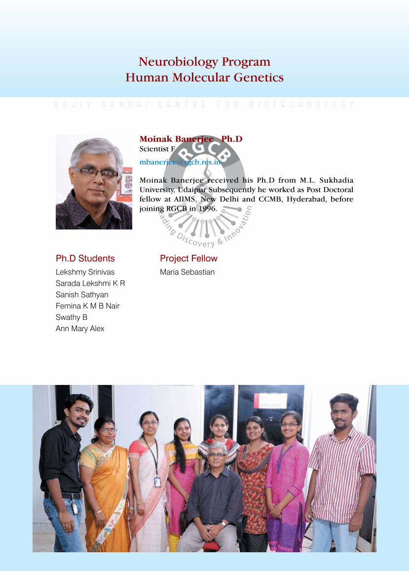
rajiv gandhi centre for biotechnology 204
annual report 2013
Neurobiology ProgramHuman Molecular Genetics
Ph.D StudentsLekshmy SrinivasSarada Lekshmi K RSanish SathyanFemina K M B NairSwathy BAnn Mary Alex
r a j i v g a n d h i c e n t r e f o r b i o t e c h n o l o g y
Moinak Banerjee received his Ph.D from M.L. Sukhadia University, Udaipur Subsequently he worked as Post Doctoral fellow at AIIMS, New Delhi and CCMB, Hyderabad, before joining RGCB in 1996.
Moinak Banerjee Ph.DScientist F
Project FellowMaria Sebastian
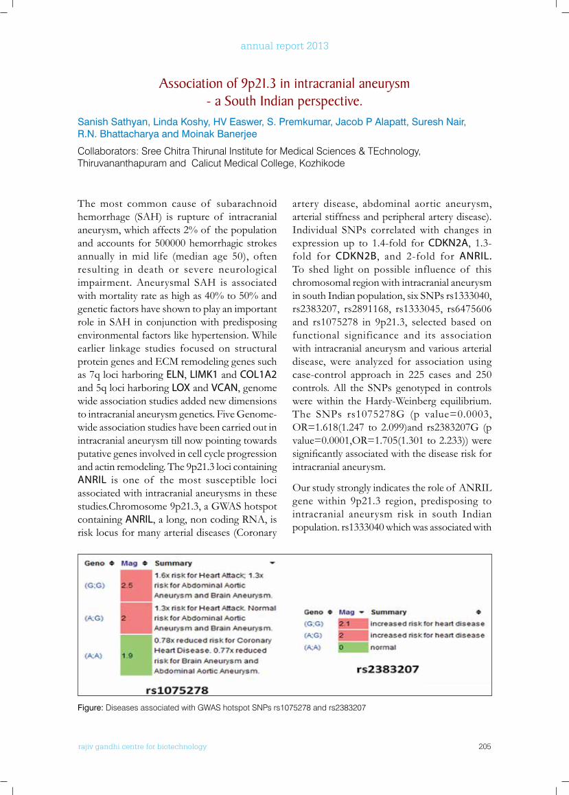
rajiv gandhi centre for biotechnology 205
annual report 2013
The most common cause of subarachnoid hemorrhage (SAH) is rupture of intracranial aneurysm, which affects 2% of the population and accounts for 500000 hemorrhagic strokes annually in mid life (median age 50), often resulting in death or severe neurological impairment. Aneurysmal SAH is associated with mortality rate as high as 40% to 50% and genetic factors have shown to play an important role in SAH in conjunction with predisposing environmental factors like hypertension. While earlier linkage studies focused on structural protein genes and ECM remodeling genes such as 7q loci harboring ELN, LIMK1 and COL1A2 and 5q loci harboring LOX and VCAN, genome wide association studies added new dimensions to intracranial aneurysm genetics. Five Genome-wide association studies have been carried out in intracranial aneurysm till now pointing towards putative genes involved in cell cycle progression and actin remodeling. The 9p21.3 loci containing ANRIL is one of the most susceptible loci associated with intracranial aneurysms in these studies.Chromosome 9p21.3, a GWAS hotspot containing ANRIL, a long, non coding RNA, is risk locus for many arterial diseases (Coronary
artery disease, abdominal aortic aneurysm, arterial stiffness and peripheral artery disease). Individual SNPs correlated with changes in expression up to 1.4-fold for CDKN2A, 1.3-fold for CDKN2B, and 2-fold for ANRIL. To shed light on possible influence of this chromosomal region with intracranial aneurysm in south Indian population, six SNPs rs1333040, rs2383207, rs2891168, rs1333045, rs6475606 and rs1075278 in 9p21.3, selected based on functional significance and its association with intracranial aneurysm and various arterial disease, were analyzed for association using case-control approach in 225 cases and 250 controls. All the SNPs genotyped in controls were within the Hardy-Weinberg equilibrium. The SNPs rs1075278G (p value=0.0003, OR=1.618(1.247 to 2.099)and rs2383207G (p value=0.0001,OR=1.705(1.301 to 2.233)) were significantlyassociatedwiththediseaseriskforintracranial aneurysm.
Our study strongly indicates the role of ANRIL gene within 9p21.3 region, predisposing to intracranial aneurysm risk in south Indian population. rs1333040 which was associated with
Association of 9p21.3 in intracranial aneurysm - a South Indian perspective.
Sanish Sathyan, Linda Koshy, HV Easwer, S. Premkumar, Jacob P Alapatt, Suresh Nair, R.N. Bhattacharya and Moinak Banerjee
Collaborators: Sree Chitra Thirunal Institute for Medical Sciences & TEchnology, Thiruvananthapuram and Calicut Medical College, Kozhikode
Figure: Diseases associated with GWAS hotspot SNPs rs1075278 and rs2383207
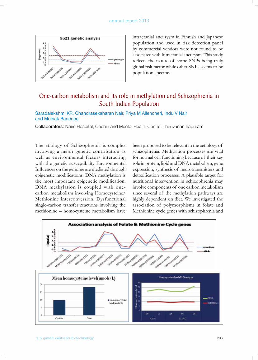
rajiv gandhi centre for biotechnology 206
annual report 2013
intracranial aneurysm in Finnish and Japanese population and used in risk detection panel by commercial vendors were not found to be associated with Intracranial aneurysm. This study reflects thenature of someSNPsbeing trulyglobal risk factor while other SNPs seems to be populationspecific.
one-carbon metabolism and its role in methylation and Schizophrenia in South Indian Population
Saradalekshmi KR, Chandrasekaharan Nair, Priya M Allencheri, Indu V Nair and Moinak Banerjee
Collaborators: Nairs Hospital, Cochin and Mental Health Centre, Thiruvananthapuram
The etiology of Schizophrenia is complex involving a major genetic contribution as well as environmental factors interacting with the genetic susceptibility Environmental Influencesonthegenomearemediatedthroughepigeneticmodifications.DNAmethylation isthemost important epigeneticmodification.DNA methylation is coupled with one-carbon metabolism involving Homocysteine/Methionine interconversion. Dysfunctional single-carbon transfer reactions involving the methionine – homocysteine metabolism have
been proposed to be relevant in the aetiology of schizophrenia. Methylation processes are vital for normal cell functioning because of their key role in protein, lipid and DNA metabolism, gene expression, synthesis of neurotransmitters and detoxificationprocesses.Aplausibletargetfornutritional intervention in schizophrenia may involve components of one carbon metabolism since several of the methylation pathways are highly dependent on diet. We investigated the association of polymorphisms in folate and Methionine cycle genes with schizophrenia and
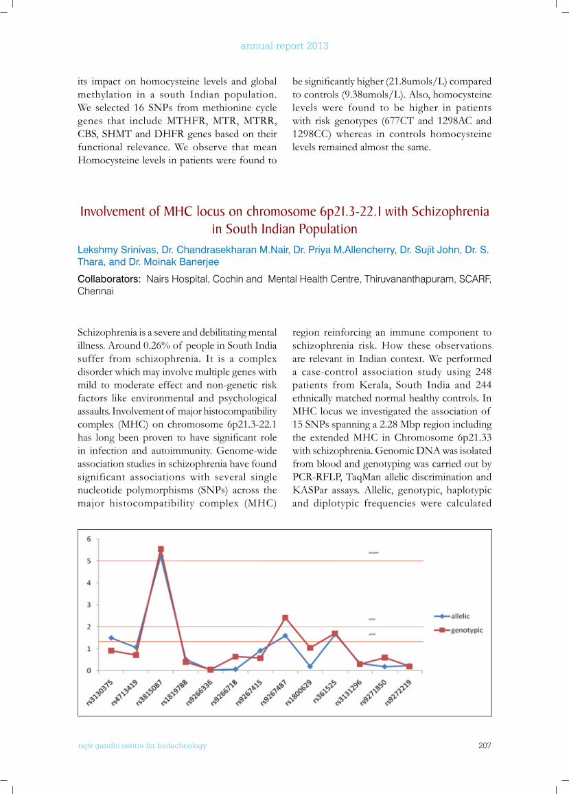
rajiv gandhi centre for biotechnology 207
annual report 2013
its impact on homocysteine levels and global methylation in a south Indian population. We selected 16 SNPs from methionine cycle genes that include MTHFR, MTR, MTRR, CBS, SHMT and DHFR genes based on their functional relevance. We observe that mean Homocysteine levels in patients were found to
besignificantlyhigher(21.8umols/L)comparedto controls (9.38umols/L). Also, homocysteine levels were found to be higher in patients with risk genotypes (677CT and 1298AC and 1298CC) whereas in controls homocysteine levels remained almost the same.
Involvement of MHC locus on chromosome 6p21.3-22.1 with Schizophrenia in South Indian Population
Lekshmy Srinivas, Dr. Chandrasekharan M.Nair, Dr. Priya M.Allencherry, Dr. Sujit John, Dr. S. Thara, and Dr. Moinak Banerjee
Collaborators: Nairs Hospital, Cochin and Mental Health Centre, Thiruvananthapuram, SCARF, Chennai
Schizophrenia is a severe and debilitating mental illness. Around 0.26% of people in South India suffer from schizophrenia. It is a complex disorder which may involve multiple genes with mild to moderate effect and non-genetic risk factors like environmental and psychological assaults. Involvement of major histocompatibility complex (MHC) on chromosome 6p21.3-22.1 has longbeenproven tohave significant rolein infection and autoimmunity. Genome-wide association studies in schizophrenia have found significant associations with several single nucleotide polymorphisms (SNPs) across the major histocompatibility complex (MHC)
region reinforcing an immune component to schizophrenia risk. How these observations are relevant in Indian context. We performed a case-control association study using 248 patients fromKerala, South India and 244ethnically matched normal healthy controls. In MHC locus we investigated the association of 15 SNPs spanning a 2.28 Mbp region including the extended MHC in Chromosome 6p21.33 with schizophrenia. Genomic DNA was isolated from blood and genotyping was carried out by PCR-RFLP, TaqMan allelic discrimination and KASPar assays.Allelic, genotypic, haplotypicand diplotypic frequencies were calculated
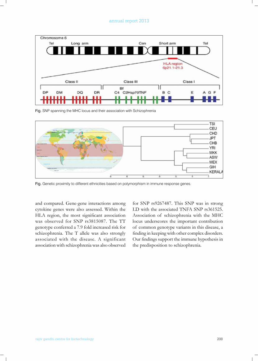
rajiv gandhi centre for biotechnology 208
annual report 2013
and compared. Gene-gene interactions among cytokine genes were also assessed. Within the HLA region, themost significant associationwas observed for SNP rs3815087. The TT genotype conferred a 7.9 fold increased risk for schizophrenia. The T allele was also strongly associated with the disease. A significant association with schizophrenia was also observed
for SNP rs9267487. This SNP was in strong LD with the associated TNFA SNP rs361525. Association of schizophrenia with the MHC locus underscores the important contribution of common genotype variants in this disease, a findinginkeepingwithothercomplexdisorders.Ourfindingssupporttheimmunehypothesisinthe predisposition to schizophrenia.
Fig. SNP spanning the MHC locus and their association with Schizophrenia
Fig. Genetic proximity to different ethnicities based on polymorphism in immune response genes.
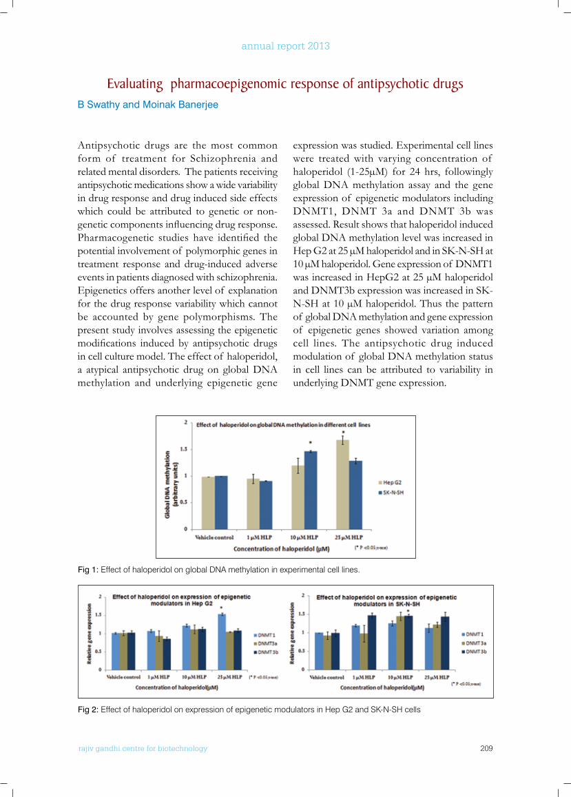
rajiv gandhi centre for biotechnology 209
annual report 2013
Evaluating pharmacoepigenomic response of antipsychotic drugsB Swathy and Moinak Banerjee
Antipsychotic drugs are the most common form of treatment for Schizophrenia and related mental disorders. The patients receiving antipsychotic medications show a wide variability in drug response and drug induced side effects which could be attributed to genetic or non-geneticcomponentsinfluencingdrugresponse.Pharmacogenetic studies have identified thepotential involvement of polymorphic genes in treatment response and drug-induced adverse events in patients diagnosed with schizophrenia. Epigenetics offers another level of explanation for the drug response variability which cannot be accounted by gene polymorphisms. The present study involves assessing the epigenetic modifications inducedby antipsychotic drugsin cell culture model. The effect of haloperidol, a atypical antipsychotic drug on global DNA methylation and underlying epigenetic gene
expression was studied. Experimental cell lines were treated with varying concentration of haloperidol (1-25µM) for 24 hrs, followingly global DNA methylation assay and the gene expression of epigenetic modulators including DNMT1, DNMT 3a and DNMT 3b was assessed. Result shows that haloperidol induced global DNA methylation level was increased in HepG2at25µMhaloperidolandinSK-N-SHat10 µM haloperidol. Gene expression of DNMT1 was increased in HepG2 at 25 µM haloperidol andDNMT3bexpressionwasincreasedinSK-N-SH at 10 µM haloperidol. Thus the pattern of global DNA methylation and gene expression of epigenetic genes showed variation among cell lines. The antipsychotic drug induced modulation of global DNA methylation status in cell lines can be attributed to variability in underlying DNMT gene expression.
Fig 2: Effect of haloperidol on expression of epigenetic modulators in Hep G2 and SK-N-SH cells
Fig 1: Effect of haloperidol on global DNA methylation in experimental cell lines.
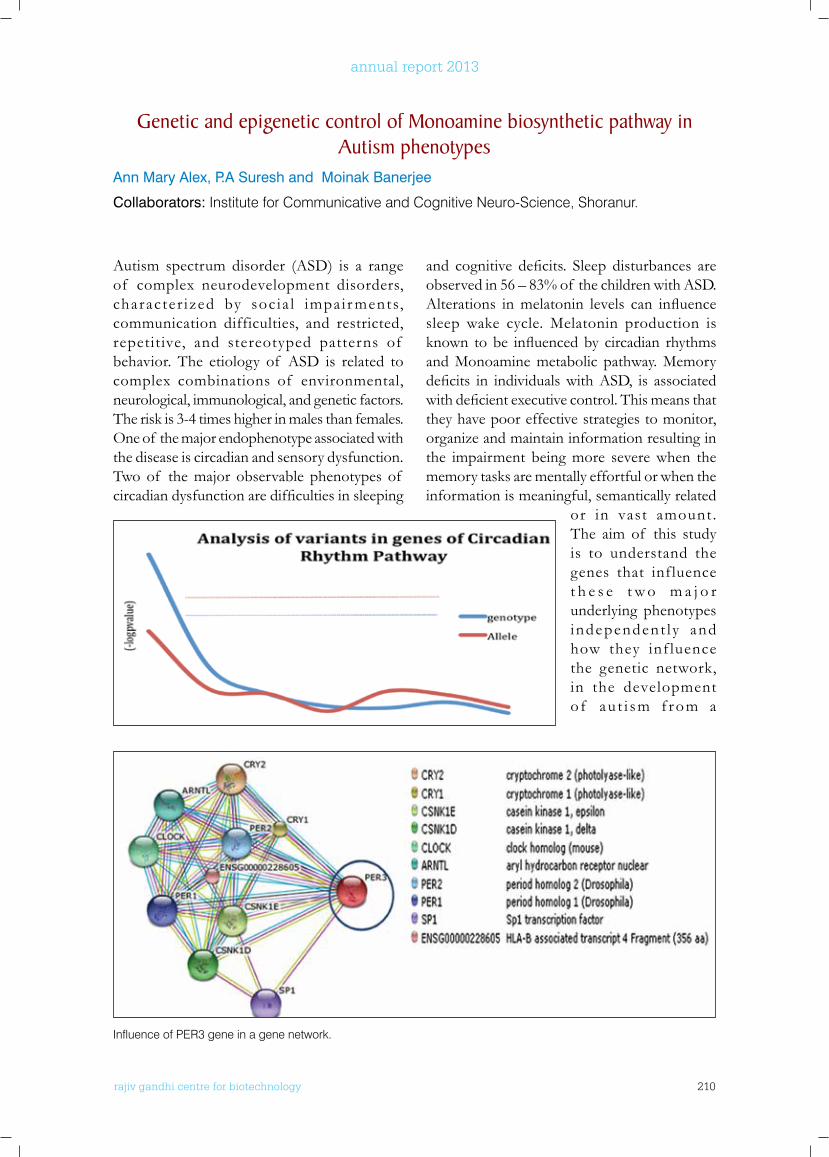
rajiv gandhi centre for biotechnology 210
annual report 2013
Autism spectrum disorder (ASD) is a range of complex neurodevelopment disorders, character ized by soc ia l impai r ments, communication difficulties, and restricted, repetitive, and stereotyped patterns of behavior. The etiology of ASD is related to complex combinations of environmental, neurological, immunological, and genetic factors. The risk is 3-4 times higher in males than females. One of the major endophenotype associated with the disease is circadian and sensory dysfunction. Two of the major observable phenotypes of circadiandysfunctionaredifficultiesinsleeping
and cognitive deficits. Sleep disturbances areobserved in 56 – 83% of the children with ASD. Alterations inmelatonin levels can influencesleep wake cycle. Melatonin production is known tobe influencedby circadian rhythmsand Monoamine metabolic pathway. Memory deficits in individualswithASD, is associatedwithdeficientexecutivecontrol.Thismeansthatthey have poor effective strategies to monitor, organize and maintain information resulting in the impairment being more severe when the memory tasks are mentally effortful or when the information is meaningful, semantically related
or in vast amount. The aim of this study is to understand the genes that influence t h e s e t w o m a j o r underlying phenotypes independent l y and how they inf luence the genetic network, in the development o f au t i sm f rom a
Genetic and epigenetic control of Monoamine biosynthetic pathway in Autism phenotypes
Ann Mary Alex, P.A Suresh and Moinak Banerjee
Collaborators: Institute for Communicative and Cognitive Neuro-Science, Shoranur.
Influence of PER3 gene in a gene network.
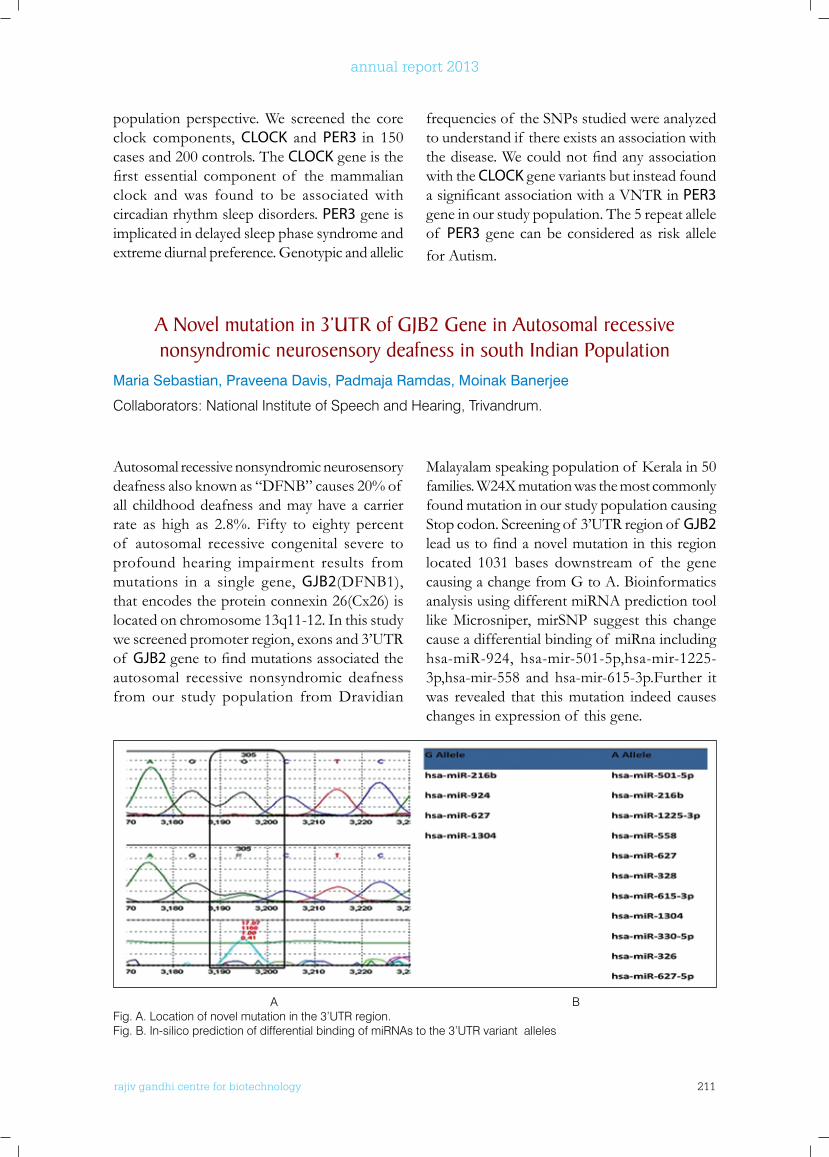
rajiv gandhi centre for biotechnology 211
annual report 2013
population perspective. We screened the core clock components, CLOCK and PER3 in 150 cases and 200 controls. The CLOCK gene is the first essential component of themammalianclock and was found to be associated with circadian rhythm sleep disorders. PER3 gene is implicated in delayed sleep phase syndrome and extreme diurnal preference. Genotypic and allelic
frequencies of the SNPs studied were analyzed to understand if there exists an association with thedisease.Wecouldnotfindanyassociationwith the CLOCK gene variants but instead found asignificantassociationwithaVNTRinPER3 gene in our study population. The 5 repeat allele of PER3 gene can be considered as risk allele for Autism.
A novel mutation in 3’uTR of GJb2 Gene in Autosomal recessive nonsyndromic neurosensory deafness in south Indian Population
Maria Sebastian, Praveena Davis, Padmaja Ramdas, Moinak Banerjee
Collaborators: National Institute of Speech and Hearing, Trivandrum.
Autosomal recessive nonsyndromic neurosensory deafness also known as “DFNB” causes 20% of all childhood deafness and may have a carrier rate as high as 2.8%. Fifty to eighty percent of autosomal recessive congenital severe to profound hearing impairment results from mutations in a single gene, GJB2(DFNB1), that encodes the protein connexin 26(Cx26) is located on chromosome 13q11-12. In this study we screened promoter region, exons and 3’UTR of GJB2 genetofindmutationsassociatedtheautosomal recessive nonsyndromic deafness from our study population from Dravidian
Malayalamspeakingpopulationof Keralain50families. W24X mutation was the most commonly found mutation in our study population causing Stop codon. Screening of 3’UTR region of GJB2 leadustofindanovelmutationinthisregionlocated 1031 bases downstream of the gene causing a change from G to A. Bioinformatics analysis using different miRNA prediction tool like Microsniper, mirSNP suggest this change cause a differential binding of miRna including hsa-miR-924, hsa-mir-501-5p,hsa-mir-1225-3p,hsa-mir-558 and hsa-mir-615-3p.Further it was revealed that this mutation indeed causes changes in expression of this gene.
A BFig. A. Location of novel mutation in the 3’UTR region.Fig. B. In-silico prediction of differential binding of miRNAs to the 3’UTR variant alleles
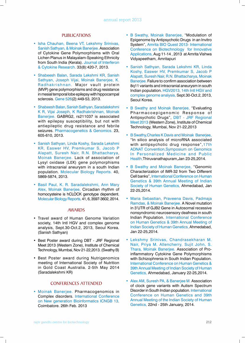
rajiv gandhi centre for biotechnology 212
annual report 2013
PublICATIonS
• Isha Chauhan, Beena VT, Lekshmy Srinivas, Sanish Sathyan, & Moinak Banerjee. Association of Cytokine Gene Polymorphisms with Oral Lichen Planus in Malayalam-Speaking Ethnicity from South India (Kerala). Journal of Interferon & Cytokine Research. 33(8):420-7, 2013.
• Shabeesh Balan, Sarada Lekshmi KR, Sanish Sathyan, Joseph Vijai, Moinak Banerjee, K. Radhakr i shnan . Ma jo r vau l t p ro te in (MVP) gene polymorphisms and drug resistance in mesial temporal lobe epilepsy with hippocampal sclerosis. Gene 526(2):449-53, 2013.
• Shabeesh Balan, Sanish Sathyan, Saradalekshmi K R, Vijai Joseph, K Radhakrishnan, Moinak Banerjee. GABRG2, rs211037 is associated with epilepsy susceptibility, but not with antiepileptic drug resistance and febrile seizures. Pharmacogenetics & Genomics. 23, 605-610, 2013.
• Sanish Sathyan, Linda Koshy, Sarada Lekshmi KR, Easwer HV, Premkumar S, Jacob P Alapatt, Suresh Nair, R.N. Bhattacharya, Moinak Banerjee. Lack of association of Lysyl oxidase (L0X) gene polymorphisms with intracranial aneurysm in a south Indian population. Molecular Biology Reports. 40, 5869-5874, 2013.
• Basil Paul, K. R. Saradalekshmi, Ann Mary Alex, Moinak Banerjee, Circadian rhythm of homocysteine is hCLOCK genotype dependent. Molecular Biology Reports, 41, 6, 3597-3602, 2014.
AwARDS
• Travel award of Human Genome Variation society, 14th Intl HGV and complex genome analysis, Sept.30-Oct.2, 2013, Seoul Korea.(Sanish Sathyan)
• Best Poster award during DBT - JRF Regional Meet 2013 (Western Zone), Institute of Chemical Technology, Mumbai, Nov 21-22,2013. (Swathy B)
• Best Poster award during Nutrigenomics meeting of International Society of Nutrition in Gold Coast Australia. 2-5th May 2014 (Saradalekshmi KR)
ConfEREnCES ATTEnDED
• Moinak Banerjee. Pharmacogenomics in Complex disorders. International Conference on New generation Bioinformatics ICNGB 13, Coimbatore. 26th Feb. 2013
• B Swathy, Moinak Banerjee, “Modulation of Epigenome by Antipsychotic Drugs in an Invitro System”, Amrita BIO Quest 2013- International Conference on Biotechnology for Innovative Applications, Aug 11-14 , 2013 at Amrita Vishwa Vidyapeetham, Amritapuri
• Sanish Sathyan, Sarada Lekshmi KR, Linda Koshy, Easwer HV, Premkumar S, Jacob P Alapatt, Suresh Nair, R.N. Bhattacharya, Moinak Banerjee. Failure to confirm association between 8q11 variants and intracranial aneurysm in south Indian population. HGV2013, 14th Intl HGV and complex genome analysis, Sept.30-Oct.2, 2013, Seoul Korea.
• B Swathy and Moinak Banerjee, “Evaluating P h a r m a c o e p i g e n o m i c R e s p o n s e o f Antipsychotic Drugs”, DBT - JRF Regional Meet 2013 (Western Zone), Institute of Chemical Technology, Mumbai, Nov 21-22,2013
• B Swathy,Charles K Davis and Moinak Banerjee, “In silico analysis of microRNA associated with antipsychotic drug response”,17th ADNAT Convention,Symposium on Genomics In Personal ized Medic ine and Publ ic Health,Thiruvanathapuram,Jan 23-25,2014.
• B Swathy and Moinak Banerjee, “Genomic Characterization of IMR-32 from Two Different Cell banks”, International Conference on Human Genetics & 39th Annual Meeting of Indian Society of Human Genetics, Ahmedabad, Jan 22-25,2014.
• Maria Sebastian, Praveena Davis, Padmaja Ramdas, & Moinak Banerjee. A Novel mutation in 3’UTR of GJB2 Gene in Autosomal recessive nonsyndromic neurosensory deafness in south Indian Population. International Conference on Human Genetics & 39th Annual Meeting of Indian Society of Human Genetics, Ahmedabad, Jan 22-25,2014.
• Lekshmy Srinivas, Chandrasekharan M. Nair, Priya M. Allencherry, Sujit John, S. Thara, Moinak Banerjee. Association of Pro-inflammatory Cytokine Gene Polymorphisms with Schizophrenia in South Indian Population. International Conference on Human Genetics & 39th Annual Meeting of Indian Society of Human Genetics, Ahmedabad, January 22-25,2014.
• Alex AM, Suresh PA, & Banerjee M. Association of clock gene variants with Autism Spectrum Disorder in South Indian population. International Conference on Human Genetics and 39th Annual Meeting of the Indian Society of Human Genetics, 22nd - 25th January, 2014.
rajiv gandhi centre for biotechnology 213
annual report 2013
Chemical Biology ProgramChemical Biology Laboratory - 1
Ph.D StudentsParvin AbrahamReshmi V.Preethi. P. CAsha. R
r a j i v g a n d h i c e n t r e f o r b i o t e c h n o l o g y
Santhosh Kumar is a Ph.D in Chemistry from the School of Chemical Sciences, Mahatma Gandhi University, Kottayam. He did his Postdoctoral training at the Department of Biochemistry, University of Illinois, Urbana-Champaign, USA and joined RGCB in 1996.
K. Santhosh Kumar Ph.D Scientists E II
Research AssistantSmitha Devi
Technical Assistant Aswani Kumar
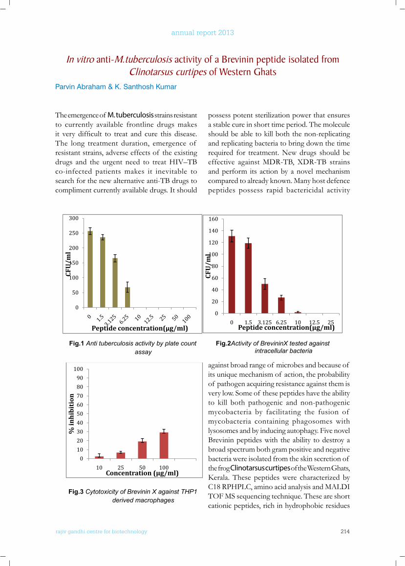
rajiv gandhi centre for biotechnology 214
annual report 2013
The emergence of M. tuberculosis strains resistant to currently available frontline drugs makes it very difficult to treat and cure this disease.The long treatment duration, emergence of resistant strains, adverse effects of the existing drugs and the urgent need to treat HIV–TB co-infected patients makes it inevitable to search for the new alternative anti-TB drugs to compliment currently available drugs. It should
possess potent sterilization power that ensures a stable cure in short time period. The molecule should be able to kill both the non-replicating and replicating bacteria to bring down the time required for treatment. New drugs should be effective against MDR-TB, XDR-TB strains and perform its action by a novel mechanism compared to already known. Many host defence peptides possess rapid bactericidal activity
In vitro anti-M.tuberculosis activity of a brevinin peptide isolated from Clinotarsus curtipes of western Ghats
Parvin Abraham & K. Santhosh Kumar
RAJIV GANDHI CENTER FOR BIOTECHNOLOGY 3
0
50
100
150
200
250
300
CFU
/ml
Peptide concentration(µg/ml)
0
20
40
60
80
100
120
140
160
0 1.5 3.125 6.25 10 12.5 25
CFU
/ml
Peptide concentration(µg/ml)
Fig.1 Anti tuberculosis activity by plate count assay
Fig.2Activity of BrevininX tested against intracellular bacteria
Fig.3 Cytotoxicity of Brevinin X against THP1 derived macrophages
Structure- function studies of peptide amides from the skin secretion of Hylarana temporalis.
Reshmy V and Santhosh Kumar K
Host defence peptides (HDPs) are endogenous antibiotics secreted by the endocrine glands
and play a multifunctional role in the innate immunity of vertebrates. They are synthesized
constitutively or inducible at epithelial surfaces, the location where the initial contact with all
0102030405060708090
100
10 25 50 100
% in
hib
itio
n
Concentration (µg/ml)
against broad range of microbes and because of its unique mechanism of action, the probability of pathogen acquiring resistance against them is very low. Some of these peptides have the ability to kill both pathogenic and non-pathogenic mycobacteria by facilitating the fusion of mycobacteria containing phagosomes with lysosomes and by inducing autophagy. Five novel Brevinin peptides with the ability to destroy a broad spectrum both gram positive and negative bacteria were isolated from the skin secretion of the frog Clinotarsus curtipes of the Western Ghats, Kerala.These peptideswere characterized byC18 RPHPLC, amino acid analysis and MALDI TOF MS sequencing technique. These are short cationic peptides, rich in hydrophobic residues
rajiv gandhi centre for biotechnology 215
annual report 2013
can destroy E. coli, V. cholerae, S. aureus, multi drug resistant S. aureus, B. coagulans, B. subtilis etc with MIC range between 6.25-100µg/ml. Analysis using several truncated and substituted synthetic analogs showed that N-terminal amino acids, their position, cationicity and helicity are playing important role on antimicrobial nature. BrevininX isolated from the skin secretion is highly effective against M. tuberculosis H37Rv strains at 10µg/ml and at this concentration it can also kill the pathogen residing inside the macrophages. This peptide did not showed any significantcytotoxiceffectagainstTHP1derivedmacrophages even at concentrations higher
than the MIC. MTT assay using THP1 derived macrophages showed an IC50 value of 100µg/ml indicating that peptide was not cytotoxic at concentrations required to inhibit M. tuberculosis. The mechanism by which it enters into the macrophage without disturbing it and how it kills the pathogen is still not known. Several de novo peptides were designed using BrevininX as a template and chemically synthesised to understand the importance of amino acids and the effect of their sequential arrangement in the primary structure on anti-TB activity. This data may help to develop novel short peptides as potential therapeutic anti-M. tuberculosis agents.
Structure- function studies of peptide amides from the skin secretion of Hylarana temporalis.
Reshmy V and Santhosh Kumar K
Host defence peptides (HDPs) are endogenous antibiotics secreted by the endocrine glands and play a multifunctional role in the innate immunity of vertebrates. They are synthesized constitutively or inducible at epithelial surfaces, the location where the initial contact with all microbes, from symbionts to pathogens, takes place. These Antimicrobial peptides (AMPs) showed broad-spectrum antimicrobial activity and are considered promising candidates for the development of novel anti-infective preparations. These polycationic peptides vary in size, sequence, conformation and can fold into an amphipathic helicalorβ-sheetedstructure.ThefamilyRanidae includes the most diverse and widely distributed group of anuran amphibians endowed with an excellent chemical defence system composed of pharmacologically active compounds. Their holocrine skin secretion contains peptides with broad spectrum antimicrobial activity, cytosolic components and intact polyadenylated mRNAs encoding peptides and are stored in the granular skin glands and released on disturbances. The present work describes the isolation, characterization and synthesis of Esculentin peptides E2HLte3 and E2HLte4 and Brevinin
peptides Brevinin-1 and Brevinin-2 present in the skin secretion of Indian bronzed frog Hylarana temporalis. cDNA deduced peptides were chemically prepared by solid-phase synthesis (SPPS) using Fmoc-amino acids. To gain an insight into the structural importance of the peptide, especially at the N-terminal, new N-terminal deleted and truncated analogs were synthesised. These synthetic peptides were then purified byRP-HPLC and their identitywasconfirmed by MALDI-TOF-MS technique. Parent peptides showed good MIC against both gram positive and gram negative clinical strains and least hemolytic and cytotoxic nature towards eukaryotic cells. Esculentin-2 peptides were found to be more active even at lower concentrations than the Brevinin-1 peptides. Kinetics analysis showed that these peptidesare bactericidal in nature and they kill the cells within 15 minutes of their addition. NPN assay bymonitoringthefluorescenceatexcitationandemission wavelengths of 350 and 420nm showed that peptides can permeate the outer membrane. NPN is normally excluded from outer membrane but is partitioned into perturbed outer membrane exhibitingincreasedfluorescence.
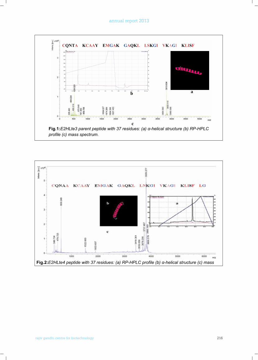
rajiv gandhi centre for biotechnology 216
annual report 2013
RAJIV GANDHI CENTER FOR BIOTECHNOLOGY 5
Fig.2:E2HLte4 peptide with 37 residues: (a) RP-HPLC profile (b) α-helical structure (c) mass spectrum.
CQNTA KCAAY EMGAK GAQKL LSKGI VKAGI KLISF
0.0 2.5 5.0 7.5 10.0 12.5 15.0 17.5 20.0 22.5 25.0 27.5 30.0 min
-250
0
250
500
750
1000
1250
1500
1750
2000
2250
mAU
0
10
20
30
40
50
60
70
80
90
%B.Conc.(Method)
Ch1-254nm,4nm (1.00)Extract-190nm,4nm (1.00)
a b
c Fig.1:E2HLte3 parent peptide with 37 residues: (a) α-helical structure (b) RP-HPLC profile (c) mass spectrum.
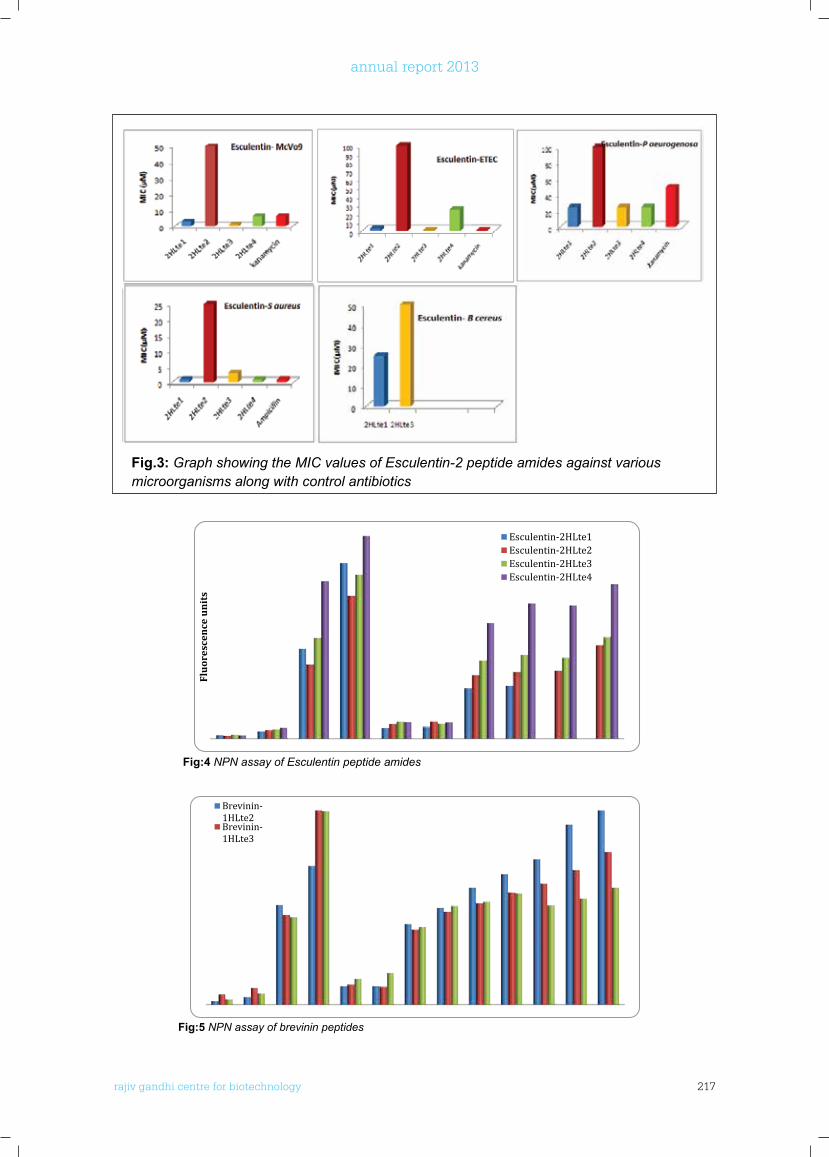
rajiv gandhi centre for biotechnology 217
annual report 2013
6 RAJIV GANDHI CENTER FOR BIOTECHNOLOGY
2013-14 Annual Report
Fig:4 NPN assay of Esculentin peptide amides
Flu
ores
cen
ce u
nit
s
Esculentin-2HLte1Esculentin-2HLte2Esculentin-2HLte3Esculentin-2HLte4
Fig.3: Graph showing the MIC values of Esculentin-2 peptide amides against various microorganisms along with control antibiotics
RAJIV GANDHI CENTER FOR BIOTECHNOLOGY 7
Fig:5 NPN assay of brevinin peptides
Arginine rich cell penetrating peptides and their structure function studies
Asha R and K. Santhosh Kumar The process of introducing biologically active molecules into cells to interact with
intracellulartargets is a major challenge for researchers and the pharmaceutical industry. In the
last two decades several peptides that can translocate across biological membranes were
identified and are named as cell-penetrating peptides (CPPs). Most of these peptides are
arginine rich and are nontoxic to the cell. Another class of membrane active peptide called
Antimicrobial Peptides (AMPs) that are found universally in all living organisms have the ability
to destroy the cell membrane and thus provide a first-line of defence to the animal against the
invading pathogens. The mechanism by which these two classes of peptides achieve their
respective property is still not clearly understood. The common feature for both the classes of
molecules is their cationicity and amphipathicity. A thorough analysis of their primary and
secondary structure, amphipathicity and charge distribution using de novo peptides may help to
understand the various factors that influence these molecules to achieve their respective
characteristics.
Brevinin-1HLte2Brevinin-1HLte3
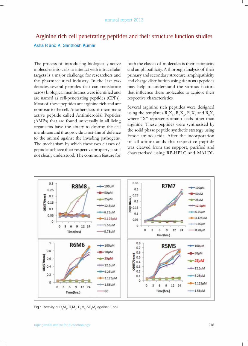
rajiv gandhi centre for biotechnology 218
annual report 2013
The process of introducing biologically active molecules into cells to interact with intracellular targets is a major challenge for researchers and the pharmaceutical industry. In the last two decades several peptides that can translocate acrossbiologicalmembraneswereidentifiedandare named as cell-penetrating peptides (CPPs). Most of these peptides are arginine rich and are nontoxic to the cell. Another class of membrane active peptide called Antimicrobial Peptides (AMPs) that are found universally in all living organisms have the ability to destroy the cell membraneandthusprovideafirst-lineof defenceto the animal against the invading pathogens. The mechanism by which these two classes of peptides achieve their respective property is still not clearly understood. The common feature for
both the classes of molecules is their cationicity and amphipathicity. A thorough analysis of their primary and secondary structure, amphipathicity and charge distribution using de novo peptides may help to understand the various factors thatinfluencethesemoleculestoachievetheirrespective characteristics.
Several arginine rich peptides were designed using the templates R5X5, R6X6, R7X7 and R8X8 where “X” represents amino acids other than arginine. These peptides were synthesised by the solid phase peptide synthetic strategy using Fmoc amino acids. After the incorporation of all amino acids the respective peptide was cleaved from the support, purified and characterised using RP-HPLC and MALDI-
Arginine rich cell penetrating peptides and their structure function studiesAsha R and K. Santhosh Kumar
Fig 1. Activity of R8M8, R7M7, R6M6 &R5M5 against E.coli
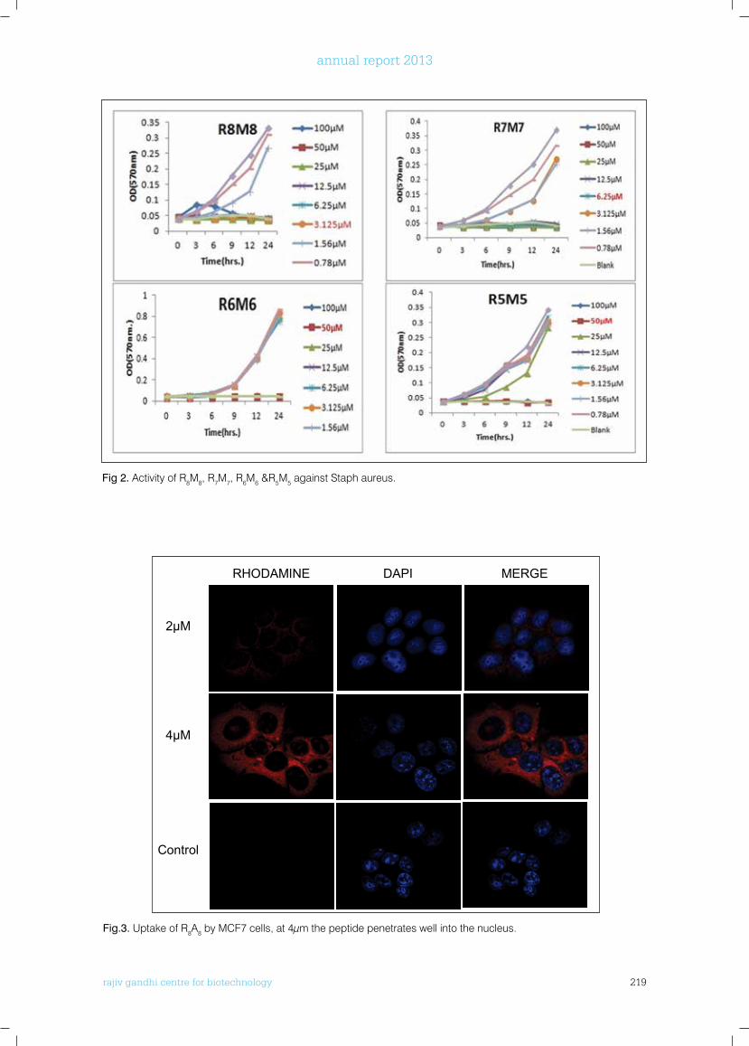
rajiv gandhi centre for biotechnology 219
annual report 2013
Fig 2. Activity of R8M8, R7M7, R6M6 &R5M5 against Staph aureus.
Fig.3. Uptake of R8A8 by MCF7 cells, at 4µm the peptide penetrates well into the nucleus.
RAJIV GANDHI CENTER FOR BIOTECHNOLOGY 9
amino acids the respective peptide was cleaved from the support, purified and characterised
using RP-HPLC and MALDI-TOF-MS. Peptides having five arginine residues R5V5, R5I5, R5L5
and R5M5 in the alternate positions were found to be very active against both gram positive and
gram negative bacteria. The arginine-methionine peptides combinations R3M3, R4M4, R5M5,
R6M6, R7M7 and R8M8 are the most interesting combination since their MICs against gram
positive and gram-negative bacteria are very low and their potency increases with number of
methionine residues. Microscopic visualisation of the effect of R7M7 and R8M8 on living bacteria
though showed appreciable membrane permeabilization the total cell disruption is not taking
place. Peptide R8A8 showed no antibacterial activity when R and A are placed in the alternative
position. But the confocal microscopic analysis of this peptide conjugated with rhodamine B
showed that it can penetrate in to the cell without causing cytotoxicity to the cell. The analysis
also showed that this peptide is able to penetrate into the cell nucleus as its concentration in the
solution increases.
RHODAMINE DAPI MERGE
2µM
4µM
Control
Fig.3.Uptake of R8A8 by MCF7 cells, at 4µm the peptide penetrates well into the nucleus.
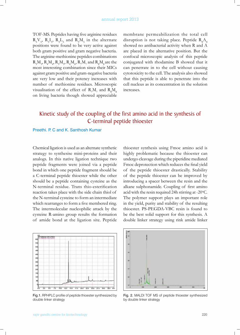
rajiv gandhi centre for biotechnology 220
annual report 2013
TOF-MS.PeptideshavingfivearginineresiduesR5V5, R5I5, R5L5 and R5M5 in the alternate positions were found to be very active against both gram positive and gram negative bacteria. The arginine-methionine peptides combinations R3M3, R4M4, R5M5, R6M6, R7M7 and R8M8 are the most interesting combination since their MICs against gram positive and gram-negative bacteria are very low and their potency increases with number of methionine residues. Microscopic visualisation of the effect of R7M7 and R8M8 on living bacteria though showed appreciable
membrane permeabilization the total cell disruption is not taking place. Peptide R8A8 showed no antibacterial activity when R and A are placed in the alternative position. But the confocal microscopic analysis of this peptide conjugated with rhodamine B showed that it can penetrate in to the cell without causing cytotoxicity to the cell. The analysis also showed that this peptide is able to penetrate into the cell nucleus as its concentration in the solution increases.
kinetic study of the coupling of the first amino acid in the synthesis of C-terminal peptide thioester
Preethi. P. C and K. Santhosh Kumar
Chemical ligation is used as an alternate synthetic strategy to synthesise mini-proteins and their analogs. In this native ligation technique two peptide fragments were joined via a peptide bond in which one peptide fragment should be a C-terminal peptide thioester while the other should be a peptide containing cysteine as the N-terminal residue. Trans thio-esterificationreaction takes place with the side chain thiol of the N-terminal cysteine to form an intermediate whichrearrangestoformafivememberedring.The intermolecular nucleophilic attack by the cysteine R-amino group results the formation of amide bond at the ligation site. Peptide
thioester synthesis using Fmoc amino acid is highly problematic because the thioester can undergo cleavage during the piperidine mediated Fmocdeprotectionwhichreducesthefinalyieldof the peptide thioester drastically. Stability of the peptide thioester can be improved by introducing a spacer between the resin and the alkanesulphonamide.Couplingof firstaminoacid with the resin required 24h stirring at -20oC. The polymer support plays an important role in the yield, purity and stability of the resulting thioester. PS-PEGDA-VBC resin is found to be the best solid support for this synthesis. A double linker strategy using rink amide linker
Fig.1. RPHPLC profile of peptide thioester synthesized by double linker strategy
Fig. 2. MALDI TOF MS of peptide thioester synthesized by double linker strategy
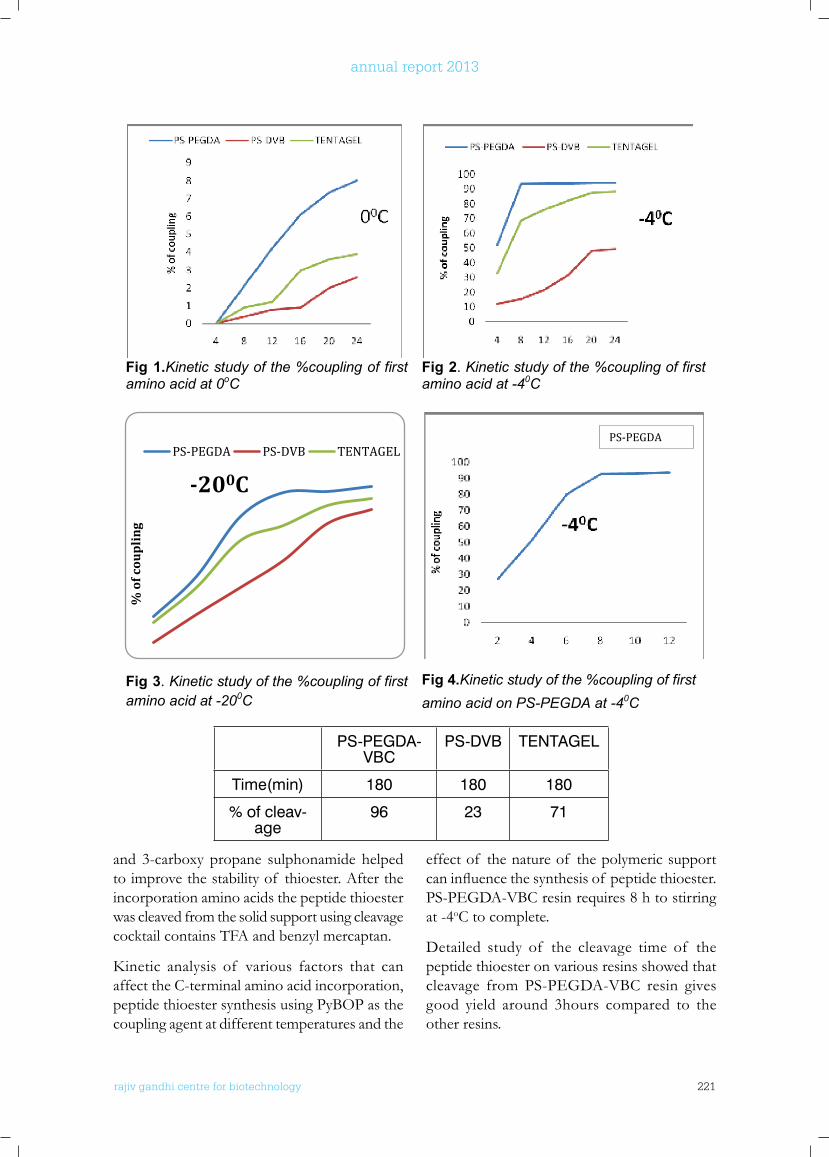
rajiv gandhi centre for biotechnology 221
annual report 2013
and 3-carboxy propane sulphonamide helped to improve the stability of thioester. After the incorporation amino acids the peptide thioester was cleaved from the solid support using cleavage cocktail contains TFA and benzyl mercaptan.
Kinetic analysis of various factors that canaffect the C-terminal amino acid incorporation, peptide thioester synthesis using PyBOP as the coupling agent at different temperatures and the
effect of the nature of the polymeric support caninfluencethesynthesisof peptidethioester.PS-PEGDA-VBC resin requires 8 h to stirring at -4oC to complete.
Detailed study of the cleavage time of the peptide thioester on various resins showed that cleavage from PS-PEGDA-VBC resin gives good yield around 3hours compared to the other resins.
PS-PEGDA-VBC
PS-DVB TENTAGEL
Time(min) 180 180 180
% of cleav-age
96 23 71
RAJIV GANDHI CENTER FOR BIOTECHNOLOGY 11
the effect of the nature of the polymeric support can influence the synthesis of peptide thioester.
PS-PEGDA-VBC resin requires 8 h to stirring at -4oC to complete.
Fig 1.Kinetic study of the %coupling of first amino acid at 0oC
Fig 2. Kinetic study of the %coupling of first amino acid at -40C
Fig 3. Kinetic study of the %coupling of first amino acid at -200C
Fig 4.Kinetic study of the %coupling of first amino acid on PS-PEGDA at -40C
Detailed study of the cleavage time of the peptide thioester on various resins showed that
cleavage from PS-PEGDA-VBC resin gives good yield around 3hours compared to the other
resins.
% o
f cou
pli
ng
-200C
PS-PEGDA PS-DVB TENTAGELPS-PEGDA
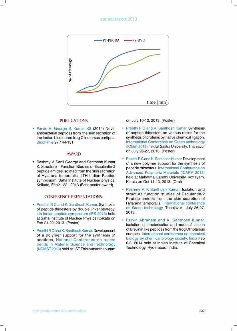
rajiv gandhi centre for biotechnology 222
annual report 2013
PublICATIonS
• Parvin A, George S, Kumar KS (2014) Novel antibacterial peptides from the skin secretion of the Indian bicoloured frog Clinotarsus curtipes. Biochimie 97:144-151.
AwARD
• Reshmy V, Sanil George and Santhosh Kumar K. Structure – Function Studies of Esculentin-2 peptide amides isolated from the skin secretion of Hylarana temporalis. 4TH Indian Peptide symposium, Saha Institute of Nuclear physics, Kolkata, Feb21-22 , 2013.(Best poster award).
ConfEREnCE PRESEnTATIonS:
• Preethi. P. C and K. Santhosh Kumar, Synthesis of peptide thioesters by double linker strategy, 4th Indian peptide symposium (IPS-2013) held at Saha Institute of Nuclear Physics Kolkata on Feb 21-22, 2013. (Poster)
• Preethi P. C and K. Santhosh Kumar. Development of a polymer support for the synthesis of peptides, National Conference on recent trends in Material Science and Technology (NCMST-2013) held at IIST Thiruvananthapuram
on July 10-12, 2013. (Poster)
• Preethi P. C and K. Santhosh Kumar. Synthesis of peptide thioesters on various resins for the synthesis of proteins by native chemical ligation, International Conference on Green technology (ICGeT-2013) held at Sastra University, Thanjavur on July 26-27, 2013. (Poster)
• Preethi P. C and K. Santhosh Kumar. Development of a new polymer support for the synthesis of peptide thioesters, International Conference on Advanced Polymeric Materials (ICAPM 2013) held at Mahatma Gandhi University, Kottayam, Kerala on Oct 11-13, 2013. (Oral)
• Reshmy V, K Santhosh Kumar. Isolation and structure function studies of Esculentin-2 Peptide amides from the skin secretion of Hylarana temporalis. International conference on Green technology, Thanjavur, July 26-27. 2013 .
• Parvin Abraham and K. Santhosh Kumar. Isolation, characterisation and mode of action of Brevinin like peptides from the frog Clinotarsus curtipes. International conference on chemical biology by chemical biology society, India Feb 6-8, 2014 held at Indian Institute of Chemical Technology, Hyderabad, India.
12 RAJIV GANDHI CENTER FOR BIOTECHNOLOGY
2013-14 Annual Report
Publications
1. Parvin A, George S, Kumar KS (2014) Novel antibacterial peptides from the skin secretion of the Indian bicoloured frog Clinotarsus curtipes. Biochimie97:144-151.
Award
1. Reshmy V, Sanil George and Santhosh Kumar K. Structure – Function Studies of Esculentin-2 peptide amides isolated from the skin secretion of Hylarana temporalis. 4TH Indian Peptide symposium, Saha Institute of Nuclear physics, Kolkata, Feb21-22 , 2013.(Best poster award).
Conference Presentations:
1. Preethi.P.C and K. Santhosh Kumar, Synthesis of peptide thioesters by double linker strategy, 4th Indian peptide symposium (IPS-2013) held at Saha Institute of Nuclear Physics Kolkata on Feb 21-22, 2013. (Poster)
2. Preethi P.C and K. Santhosh Kumar. Development of a polymer support for the synthesis of peptides, National Conference on recent trends in Material Science and Technology (NCMST-2013) held at IIST Thiruvananthapuram on July 10-12, 2013. (Poster)
3. Preethi P.C and K. Santhosh Kumar. Synthesis of peptide thioesters on various resins for the synthesis of proteins by native chemical ligation, International Conference on
% o
f cle
avag
e
time (min)
PS-PEGDA PS-DVB
PS-PEGDA-VBC PS-DVB TENTAGEL
Time(min) 180 180 180
% of cleavage 96 23 71
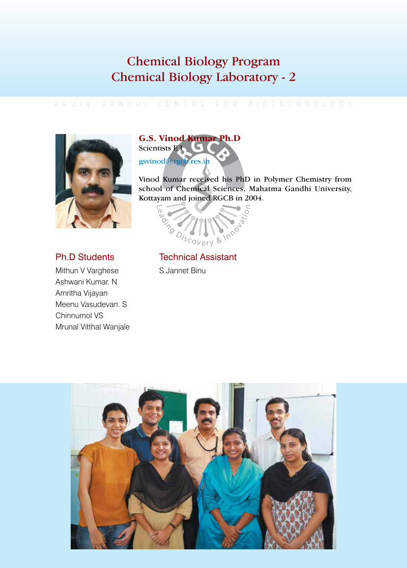
rajiv gandhi centre for biotechnology 223
annual report 2013
Chemical Biology ProgramChemical Biology Laboratory - 2
Ph.D StudentsMithun V VargheseAshwani Kumar. NAmritha VijayanMeenu Vasudevan. SChinnumol VSMrunal Vitthal Wanjale
r a j i v g a n d h i c e n t r e f o r b i o t e c h n o l o g y
Vinod Kumar received his PhD in Polymer Chemistry from school of Chemical Sciences, Mahatma Gandhi University, Kottayam and joined RGCB in 2004.
G.S. Vinod Kumar Ph.D Scientists E I
Technical Assistant S.Jannet Binu
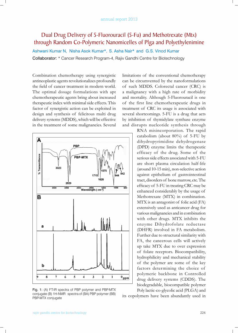
rajiv gandhi centre for biotechnology 224
annual report 2013
Combination chemotherapy using synergistic antineoplastic agents revolutionalizes profoundly thefieldof cancertreatmentinmodernworld.The optimal dosage formulations with apt chemotherapeutic agents bring about increased therapeutic index with minimal side effects. This factor of synergistic action can be exploited in design and synthesis of felicitous multi drug delivery systems (MDDS), which will be effective in the treatment of some malignancies. Several
limitations of the conventional chemotherapy can be circumvented by the nanoformulations of such MDDS. Colorectal cancer (CRC) is a malignancy with a high rate of morbidity and mortality. Although 5-Fluorouracil is one of the first line chemotherapeutic drugs in treatment of CRC its usage is associated with several shortcomings. 5-FU is a drug that acts by inhibition of thymidylate synthase enzyme and disrupts nucleotide synthesis through
RNA misincorporation. The rapid catabolism (about 80%) of 5-FU by dihydropyrimidine dehydrogenase (DPD) enzyme limits the therapeutic efficacy of the drug. Some of the serious side effects associated with 5-FU are short plasma circulation half-life (around 10-15 min), non-selective action against epithelium of gastrointestinal tract, disorders of bone marrow, etc. The efficacyof 5-FUintreatingCRCmaybeenhanced considerably by the usage of Methotrexate (MTX) in combination. MTX is an antagonist of folic acid (FA) extensively used as anticancer drug for various malignancies and in combination with other drugs. MTX inhibits the enzyme Dihydrofolate reductase (DHFR) involved in FA metabolism. Further due to structural similarity with FA, the cancerous cells will actively up take MTX due to over expression of folate receptors. Biocompatibility, hydrophilicity and mechanical stability of the polymer are some of the key factors determining the choice of polymeric backbone in Controlled drug delivery systems (CDDS). The biodegradable, biocompatible polymer Poly lactic-co-glycolic acid (PLGA) and
its copolymers have been abundantly used in
Dual Drug Delivery of 5-fluorouracil (5-fu) and Methotrexate (Mtx) through Random Co-Polymeric nanomicelles of Plga and Polyethylenimine
Ashwani Kumar N, Nisha Asok Kumar*, S. Asha Nair* and G.S. Vinod Kumar
Collaborator: * Cancer Research Program-4, Rajiv Gandhi Centre for Biotechnology
Fig. 1: (A) FT-IR spectra of PBP polymer and PBP-MTX conjugate (B) 1H-NMR spectra of (BA) PBP polymer (BB) PBP-MTX conjugate
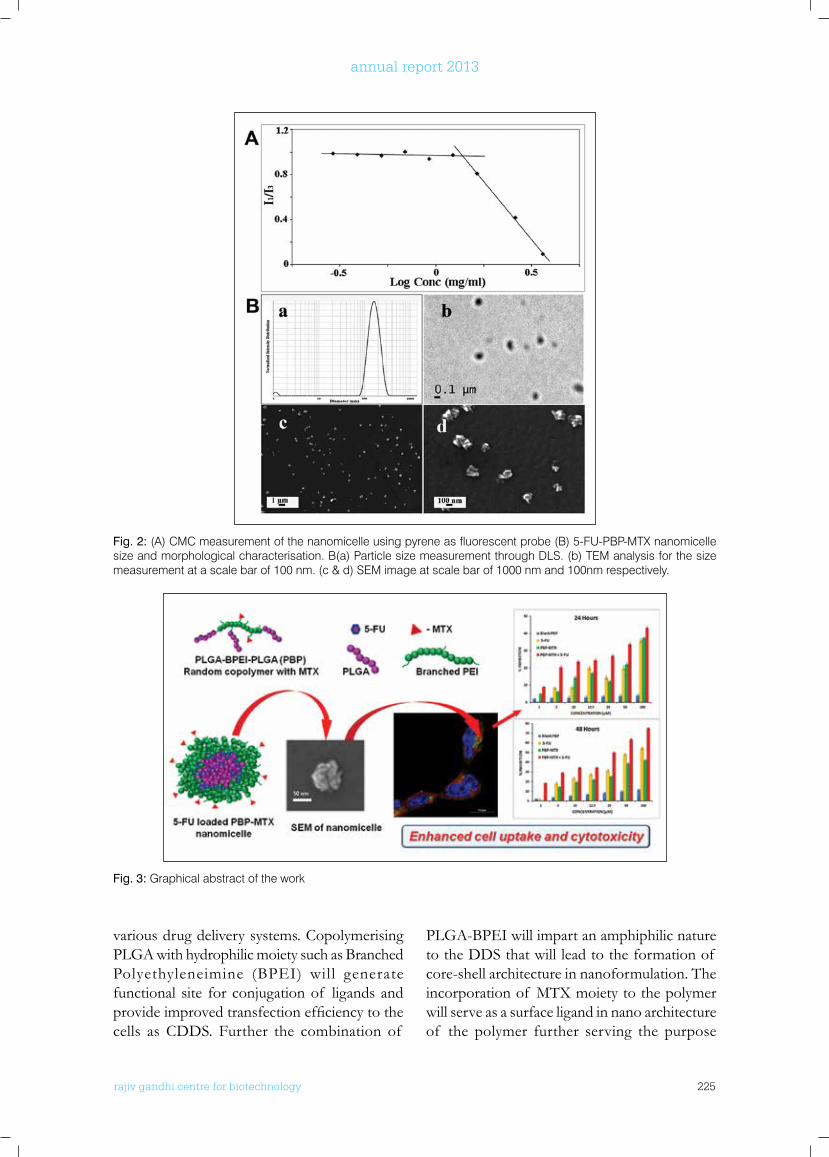
rajiv gandhi centre for biotechnology 225
annual report 2013
various drug delivery systems. Copolymerising PLGA with hydrophilic moiety such as Branched Polyethyleneimine (BPEI) will generate functional site for conjugation of ligands and provideimprovedtransfectionefficiencytothecells as CDDS. Further the combination of
PLGA-BPEI will impart an amphiphilic nature to the DDS that will lead to the formation of core-shell architecture in nanoformulation. The incorporation of MTX moiety to the polymer will serve as a surface ligand in nano architecture of the polymer further serving the purpose
Fig. 3: Graphical abstract of the work
Fig. 2: (A) CMC measurement of the nanomicelle using pyrene as fluorescent probe (B) 5-FU-PBP-MTX nanomicelle size and morphological characterisation. B(a) Particle size measurement through DLS. (b) TEM analysis for the size measurement at a scale bar of 100 nm. (c & d) SEM image at scale bar of 1000 nm and 100nm respectively.
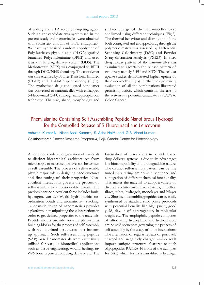
rajiv gandhi centre for biotechnology 226
annual report 2013
of a drug and a FA receptor targeting agent. Such an apt candidate was synthesised in the present study and nanomicelles were obtained with consistent amount of 5-FU entrapment. We have synthesised random copolymer of Poly-lactic-co-glycolic acid (PLGA) grafted branched Polyethylenimine (BPEI) and used it as a multi drug delivery system (DDS). The Methotrexate (MTX) was conjugated to BPEI through DCC/NHS chemistry. The copolymer was characterised by Fourier Transform Infrared (FT-IR) and H1-NMR spectroscopy (Fig.1). The synthesised drug conjugated copolymer was converted to nanomicelles with entrapped 5-Fluorouracil (5-FU) through nanoprecipitation technique. The size, shape, morphology and
surface charge of the nanomicelles were confirmed using different techniques (Fig.2).The thermal behaviour and distribution of the both conjugated and entrapped drug through the polymeric matrix was assessed by Differential Scanning Calorimetry (DSC) and Powder X-ray diffraction Analysis (PXRD). In-vitro drug release pattern of the nanomicelles was examined to ascertain the release pattern of two drugs namely 5-FU and MTX. The cellular uptake studies demonstrated higher uptake of the nanomicelles (Fig.3). Further the cytotoxicity evaluation of all the combinations illustrated promising action,which confirms the use of the system as a potential candidate as a DDS to Colon Cancer.
Phenylalanine Containing Self Assembling Peptide nanofibrous Hydrogel for the Controlled Release of 5-fluorouracil and leucovorin
Ashwani Kumar N, Nisha Asok Kumar*, S. Asha Nair* and G.S. Vinod Kumar
Collaborator: * Cancer Research Program-4, Rajiv Gandhi Centre for Biotechnology
Autonomous ordered organisation of materials to distinct hierarchical architectures from microscopic to macroscopic level can be termed as self assembly. The process of self-assembly plays a major role in designing nanostructures and fine-tuning of their properties. Non-covalent interactions govern the process of self-assembly to a considerable extent. The predominant non-covalent force includes ionic, hydrogen, van der Waals, hydrophobic, co-ordination bonds and aromatic π-π stacking. Tailor made design of nanomaterials provides a platform in manipulating these interactions in order to get desired properties to the materials. Peptide motifs provide versatile platform as building blocks for the process of self-assembly with well defined structures in a bottom up approach. Such self-assembling peptide (SAP) based nanomaterials were extensively utilised for various biomedical applications such as tissue engineering, wound healing, in-vivo bone regeneration, drug delivery etc. The
fascination of researchers in peptide based drug delivery systems is due to its advantages like biocompatibility and biodegradable nature. Thedistinct self-assemblypatterncanbefinetuned by altering amino acid sequence and conjugation of different chemical functionality. This makes the material to adopt a variety of diverse architectures like vesicles, micelles, fibres,tubes,hydrogels,monolayerandbilayeretc. Short self-assembling peptides can be easily synthesised by standard solid phase protocols with potential benefits like high purity, goodyield, devoid of heterogeneity in molecular weight etc. The amphiphilic peptide comprises of alternating hydrophilic and hydrophobic amino acid sequences governing the process of self-assembly by the usage of ionic interactions. The alternation of regular repeats of positively charged and negatively charged amino acids imparts unique structural features to such oligopeptides. RATEA-16 is one of the examples forSAP,which formsananofibroushydrogel
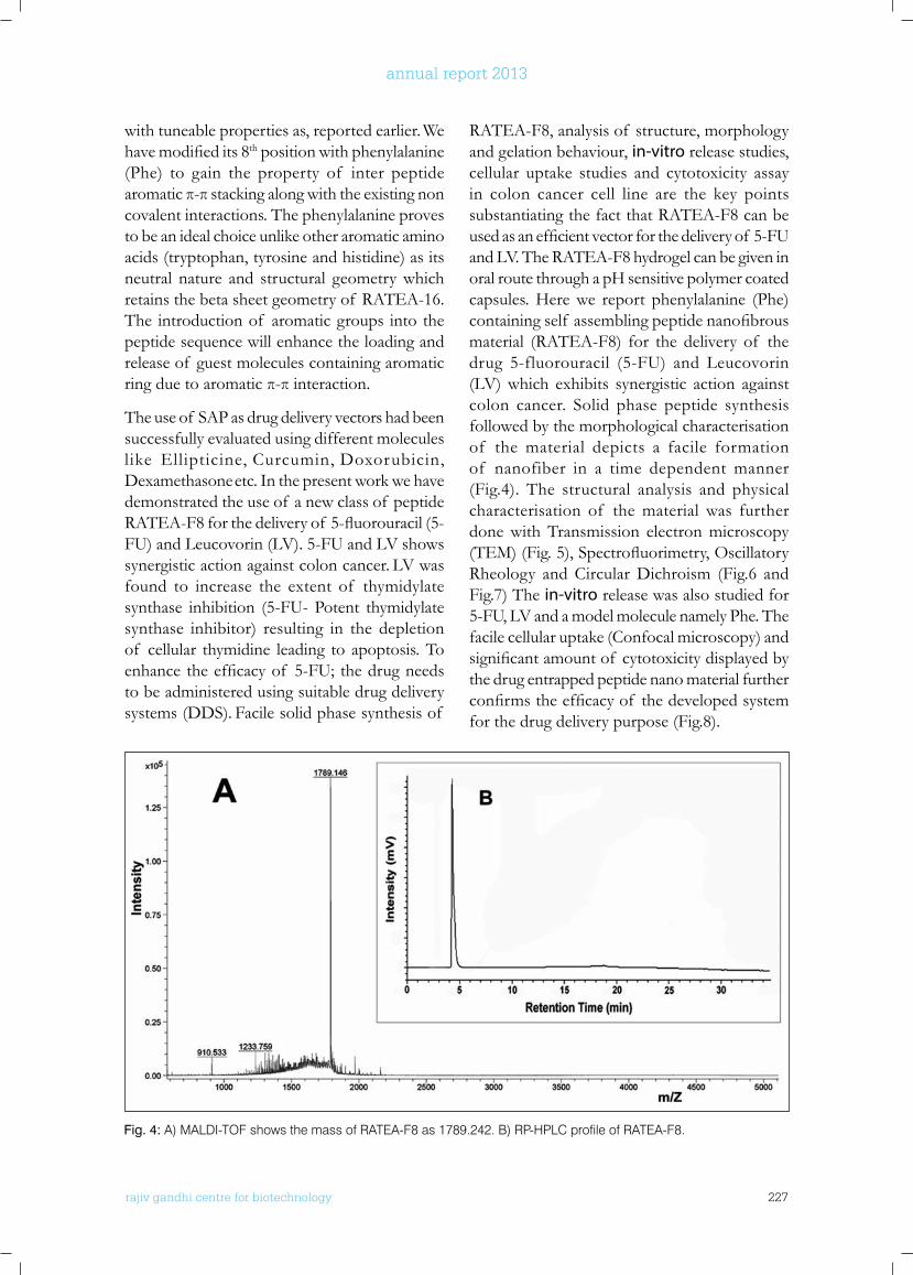
rajiv gandhi centre for biotechnology 227
annual report 2013
with tuneable properties as, reported earlier. We havemodifiedits8th position with phenylalanine (Phe) to gain the property of inter peptide aromaticπ-πstackingalongwiththeexistingnoncovalent interactions. The phenylalanine proves to be an ideal choice unlike other aromatic amino acids (tryptophan, tyrosine and histidine) as its neutral nature and structural geometry which retains the beta sheet geometry of RATEA-16. The introduction of aromatic groups into the peptide sequence will enhance the loading and release of guest molecules containing aromatic ringduetoaromaticπ-πinteraction.
The use of SAP as drug delivery vectors had been successfully evaluated using different molecules like Ellipticine, Curcumin, Doxorubicin, Dexamethasone etc. In the present work we have demonstrated the use of a new class of peptide RATEA-F8forthedeliveryof 5-fluorouracil(5-FU) and Leucovorin (LV). 5-FU and LV shows synergistic action against colon cancer. LV was found to increase the extent of thymidylate synthase inhibition (5-FU- Potent thymidylate synthase inhibitor) resulting in the depletion of cellular thymidine leading to apoptosis. To enhancetheefficacyof 5-FU;thedrugneedsto be administered using suitable drug delivery systems (DDS). Facile solid phase synthesis of
RATEA-F8, analysis of structure, morphology and gelation behaviour, in-vitro release studies, cellular uptake studies and cytotoxicity assay in colon cancer cell line are the key points substantiating the fact that RATEA-F8 can be usedasanefficientvectorforthedeliveryof 5-FUand LV. The RATEA-F8 hydrogel can be given in oral route through a pH sensitive polymer coated capsules. Here we report phenylalanine (Phe) containingself assemblingpeptidenanofibrousmaterial (RATEA-F8) for the delivery of the drug 5-fluorouracil (5-FU) and Leucovorin (LV) which exhibits synergistic action against colon cancer. Solid phase peptide synthesis followed by the morphological characterisation of the material depicts a facile formation of nanofiber in a time dependent manner (Fig.4). The structural analysis and physical characterisation of the material was further done with Transmission electron microscopy (TEM)(Fig.5),Spectrofluorimetry,OscillatoryRheology and Circular Dichroism (Fig.6 and Fig.7) The in-vitro release was also studied for 5-FU, LV and a model molecule namely Phe. The facile cellular uptake (Confocal microscopy) and significantamountof cytotoxicitydisplayedbythe drug entrapped peptide nano material further confirmstheefficacyof thedevelopedsystemfor the drug delivery purpose (Fig.8).
Fig. 4: A) MALDI-TOF shows the mass of RATEA-F8 as 1789.242. B) RP-HPLC profile of RATEA-F8.
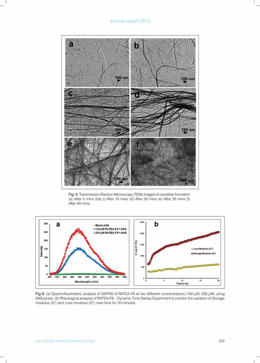
rajiv gandhi centre for biotechnology 228
annual report 2013
Fig 6: (a) Spectrofluorimetric analysis of SAPNS of RATEA-F8 at two different concentrations (100 µM, 200 µM) using ANS probe. (b) Rheological analysis of RATEA-F8 – Dynamic Time Sweep Experiment to monitor the variation of Storage modulus (G’) and Loss modulus (G”) over time for 20 minutes.
Fig. 5: Transmission Electron Microscopy (TEM) images of nanofiber formation (a) After 5 mins (b& c) After 10 mins (d) After 20 mins (e) After 30 mins (f) After 60 mins.
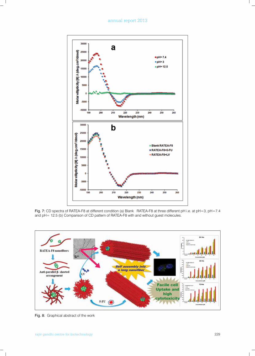
rajiv gandhi centre for biotechnology 229
annual report 2013
Fig. 7: CD spectra of RATEA-F8 at different condition (a) Blank RATEA-F8 at three different pH i.e. at pH=3, pH=7.4 and pH= 12.5 (b) Comparison of CD pattern of RATEA-F8 with and without guest molecules.
Fig. 8: Graphical abstract of the work
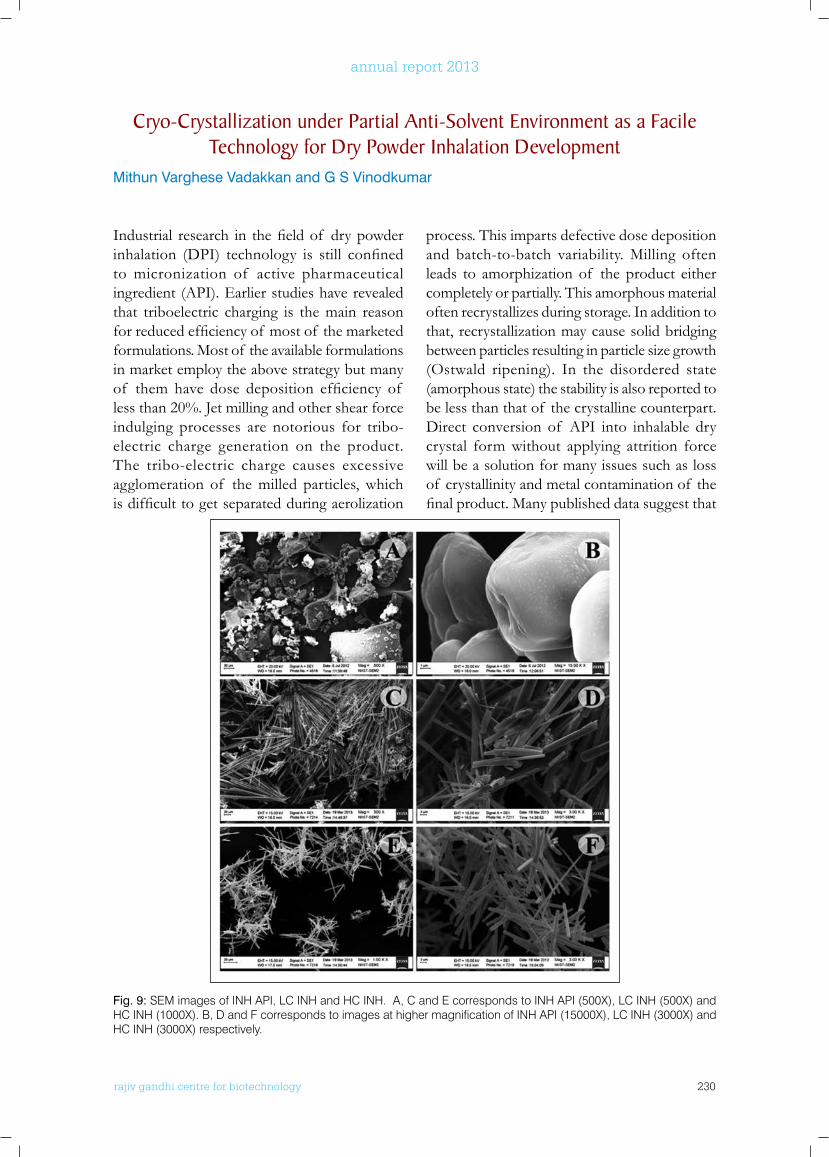
rajiv gandhi centre for biotechnology 230
annual report 2013
Industrial research in thefieldof drypowderinhalation (DPI) technology is still confinedto micronization of active pharmaceutical ingredient (API). Earlier studies have revealed that triboelectric charging is the main reason forreducedefficiencyof mostof themarketedformulations. Most of the available formulations in market employ the above strategy but many of them have dose deposition efficiency of less than 20%. Jet milling and other shear force indulging processes are notorious for tribo-electric charge generation on the product. The tribo-electric charge causes excessive agglomeration of the milled particles, which isdifficulttogetseparatedduringaerolization
process. This imparts defective dose deposition and batch-to-batch variability. Milling often leads to amorphization of the product either completely or partially. This amorphous material often recrystallizes during storage. In addition to that, recrystallization may cause solid bridging between particles resulting in particle size growth (Ostwald ripening). In the disordered state (amorphous state) the stability is also reported to be less than that of the crystalline counterpart. Direct conversion of API into inhalable dry crystal form without applying attrition force will be a solution for many issues such as loss of crystallinity and metal contamination of the finalproduct.Manypublisheddatasuggestthat
Cryo-Crystallization under Partial Anti-Solvent Environment as a facile Technology for Dry Powder Inhalation Development
Mithun Varghese Vadakkan and G S Vinodkumar
Fig. 9: SEM images of INH API, LC INH and HC INH. A, C and E corresponds to INH API (500X), LC INH (500X) and HC INH (1000X). B, D and F corresponds to images at higher magnification of INH API (15000X), LC INH (3000X) and HC INH (3000X) respectively.
rajiv gandhi centre for biotechnology 231
annual report 2013
Fig. 10: PXRD data of INH API, HC INH and LC INH
Fig. 11: Eight stage Anderson cascade impactor data of HC INH and LC INH
inhalational anti-TB therapy will be promising in the treatment of tuberculosis. Though Isoniazid is a potent anti-TB drug, recent studies show that the main reason for emergence of drug resistance is the defective delivery of drug to the target. Clinical trials have proved that, high dose of Isoniazid can cure even multi-drug resistant tuberculosis (MDR-TB). One of the major reasons for patient non-compliance to conventional treatment regime is the hepatic and neuro toxicity of prolonged high oral dose. The anti-TB drug, Isoniazid (INH) was converted to inhalable particle through cryo-crystallization under partial anti solvent environment. Crystals were dried using lyophilization technique and the powder was characterized through SEM and PXRD. To study the effect of concentration of drug solution (aqueous) in crystallization, two batches were prepared. Unlike the raw material used (Isoniazid API), crystals prepared from higher concentration (HC INH) were found to have uniform particle size distribution (Fig.9). The surface of the same found to have smooth topology compared to crystals prepared from low concentrated drug solution (LC INH). The PXRD studies showed that new polymorph had been evolved by the proposed technique (Fig. 10). Anderson cascade impactor data says, HC INH is a better system for DPI application (Fig. 11).
rajiv gandhi centre for biotechnology 232
annual report 2013
PublICATIonS
• Lekha Nair K, Sankar Jagadeeshan, S Asha Nair, Kumar GSV. Folic acid conjugated d-valerolactone-poly(ethyleneglycol) based triblock copolymer as a promising carrier for targeted doxorubicin delivery. PLOS ONE 2013;8 (8) e70697.
• Siyad MA, Kumar GSV. Synthetic evaluation of disulphide-bonded sarafotoxin on a poly(oxy ether) grafted dendrimeric poly(alkylamine) support for polymer assisted organic synthesis. Organic & Biomolecular Chemistry 2013;11 4860-4870.
• Mithun Varghese Vadakkan, K Annapoorna, KC Sivakumar, Sathish Mundayoor, Kumar GSV. Dry powder cationic lipopolymeric nanomicelle inhalation for targeted delivery of antitubercular drug to alveolar macrophage. International Journal of Nanomedicine 2013;8: 2871-2885.
• Siyad MA, Kumar GSV. PEGylated dendrimer polystyrene support: synthesis, characterisation and evaluation of biologically active peptides. Amino Acids 2013;44(3):947-959.
• S i y a d M A , Ku m a r G S V. S y n t h e s i s , Characterization, and Application of Bisphenol A Glycerolate Dimethacrylate Cross-Linked Polystyrene (PS-BGD): A Novel Support for Gel Phase Peptide Synthesis. Current Organic Synthesis. 2013;10 (2) 318-327.
ConfEREnCE PRESEnTATIonS
• Ashwani Kumar N, Nisha Asok Kumar, Asha.S.Nair and G.S. Vinod Kumar. “Controlled, pH responsive delivery of 5- Fluorouracil by a Polymeric Nanomaterial.” ICAFM-2014 (International Conference on Advanced Functional Materials), Trivandrum, 2014.
• Ashwani Kumar N, Nisha Asok Kumar, K.C. Sivakumar, Asha.S.Nair, and G.S. Vinod Kumar. “Controlled delivery of 5-Fluorouracil using a pH responsive self assembling peptide nanomaterial”. MEDCHEM-13 (Advances in
Anticancer drug discovery and development). Indian Institute of Technology, Madras, 2013.
• Ashwani Kumar.N, Nisha Asok Kumar, Asha.S.Nair and G.S. Vinod Kumar. Facile synthesis and biological evaluation of a polymeric nanogel for the pH responsive delivery of 5-Fluorouracil, National Seminar on Emerging Trends in Chemical Science, St Aloysius College, Mangalore; 2013.
• Ashwani Kumar.N, Nisha Asok Kumar, K.C. Sivakumar, Asha S. Nair and G S Vinod Kumar. Peptide based nanomaterials:- A promising tool for the controlled delivery of 5-Fluorouracil. International Conference on Recent Advances in Material Science and Technology (ICRAMST-13). National Institute for Technology Surathkal Karnataka; 2013.
• Ashwani Kumar. N, Nisha Asok Kumar, Asha S. Nair and G S Vinod Kumar. A novel pH responsive polymeric drug delivery system for colon cancer. 25th Kerala Science Congress. Techno Park, Trivandrum; 2013.
• Ashwani Kumar.N, Nisha Asok Kumar, K.C.Sivakumar, Asha.S.Nair and GS Vinod Kumar. Peptide based nanomaterials:-A promising tool for the controlled delivery of 5-Fluorouracil 5th International Conference on Current Trends in Drug Discovery and Research (CTDDR-2013).Central Drug research Institute, Lucknow; 2013.
• Mithun V. Varghese, Annapoorna K, Sivakumar K C, Satish Mundayoor and G S Vinod Kumar. “Dry powder inhaler formulation of an anti TB drug for treating pulmonary tuberculosis.” 25th Kerala science congress-2013. Technopark, Thiruvanathapuram. 2013.
• Mithun V Varghese, Annapoorna K, Sivakumar K C, Satish Mundayoor and G S Vinod Kumar. “Development of cationic amphiphilic lipopolymer as an efficient carrier for anti-TB dry powder inhalation formulation.” Current Trend in Drug Discovery Research (CTDDR) - 2013, Central drug research institute, Lucknow. 2013.
ExTRA MuRAl RESEARCH GRAnTS
Title Investigators Funding Agency Duration
A novel site specifically targeting nanoparticle based oral - drug and siRNA releasing polymer systems for colon cancer
Professor M.Radhakrishna Pillai (Project Coordinator)G.S. Vinod Kumar (PI)Hari Krishnan (Co-PI)Asha Nair.S (Co-PI)Bramanandam Manavathi (Co-PI)
Department of Biotechnology,Government of India
2010-13
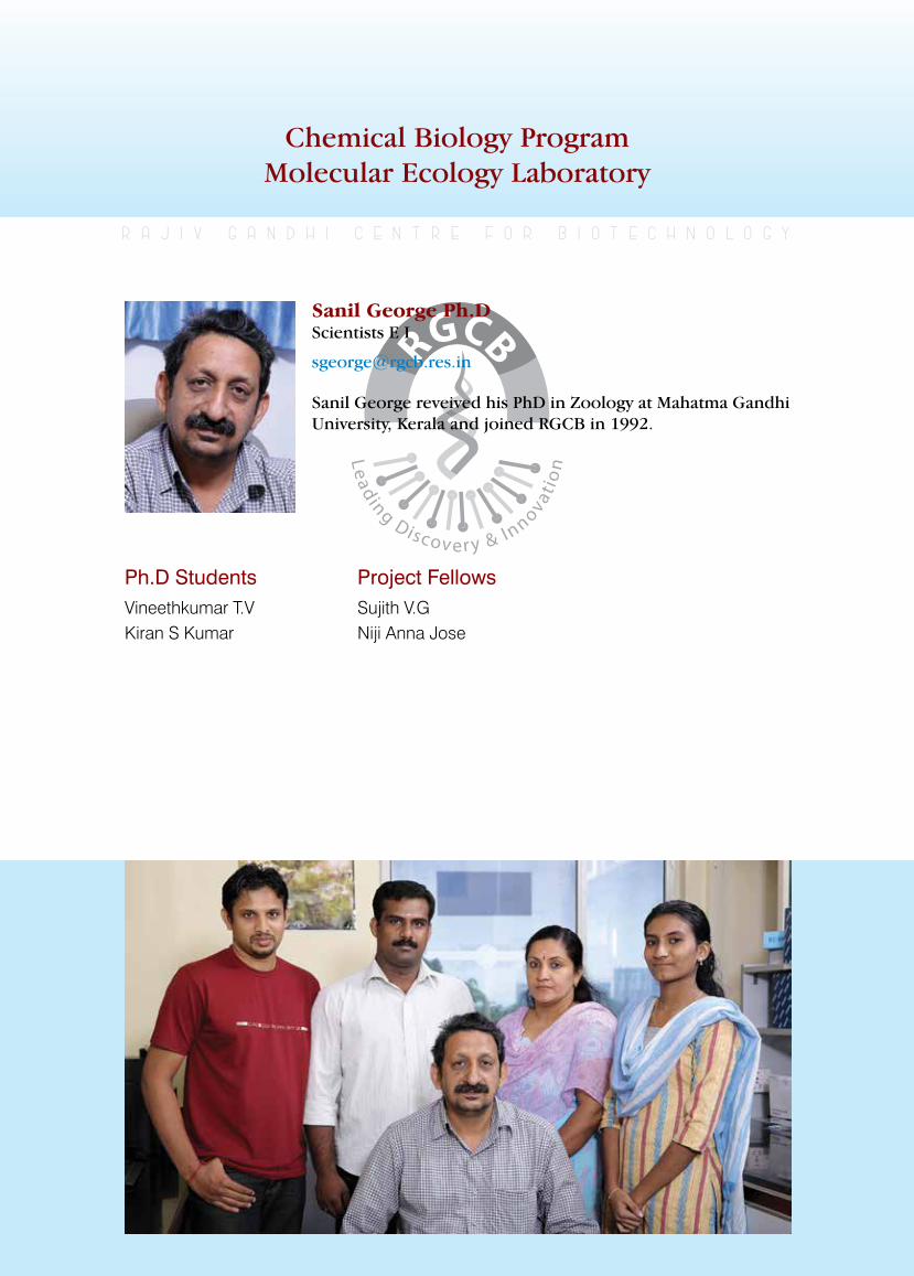
rajiv gandhi centre for biotechnology 233
annual report 2013
Chemical Biology ProgramMolecular Ecology Laboratory
Ph.D StudentsVineethkumar T.VKiran S Kumar
r a j i v g a n d h i c e n t r e f o r b i o t e c h n o l o g y
Sanil George reveived his PhD in Zoology at Mahatma Gandhi University, Kerala and joined RGCB in 1992.
Sanil George Ph.D Scientists E I
Project Fellows Sujith V.GNiji Anna Jose
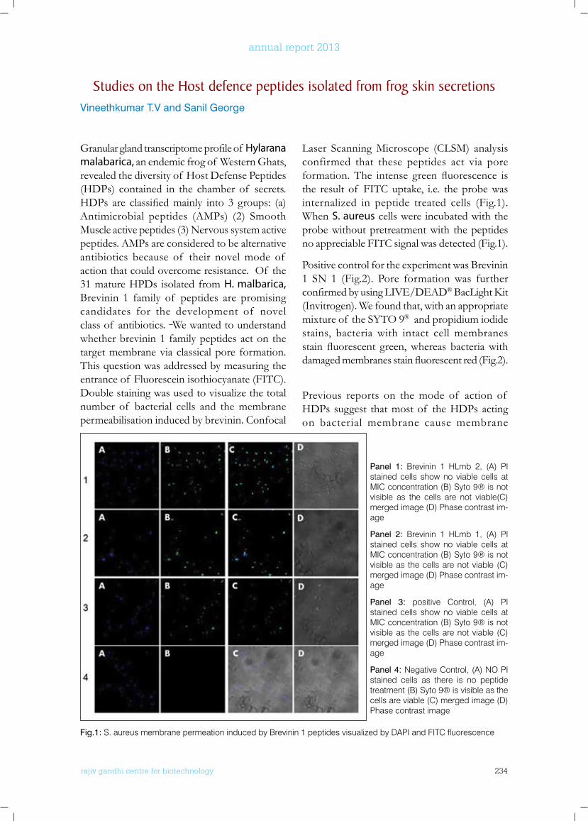
rajiv gandhi centre for biotechnology 234
annual report 2013
Granularglandtranscriptomeprofileof Hylarana malabarica, an endemic frog of Western Ghats, revealed the diversity of Host Defense Peptides (HDPs) contained in the chamber of secrets. HDPs are classifiedmainly into 3 groups: (a)Antimicrobial peptides (AMPs) (2) Smooth Muscle active peptides (3) Nervous system active peptides. AMPs are considered to be alternative antibiotics because of their novel mode of action that could overcome resistance. Of the 31 mature HPDs isolated from H. malbarica, Brevinin 1 family of peptides are promising candidates for the development of novel class of antibiotics. We wanted to understand whether brevinin 1 family peptides act on the target membrane via classical pore formation. This question was addressed by measuring the entrance of Fluorescein isothiocyanate (FITC). Double staining was used to visualize the total number of bacterial cells and the membrane permeabilisation induced by brevinin. Confocal
Laser Scanning Microscope (CLSM) analysis confirmed that these peptides act via pore formation.The intense greenfluorescence isthe result of FITC uptake, i.e. the probe was internalized in peptide treated cells (Fig.1). When S. aureus cells were incubated with the probe without pretreatment with the peptides no appreciable FITC signal was detected (Fig.1).
Positive control for the experiment was Brevinin 1 SN 1 (Fig.2). Pore formation was further confirmedbyusingLIVE/DEAD®BacLightKit(Invitrogen). We found that, with an appropriate mixture of the SYTO 9® and propidium iodide stains, bacteria with intact cell membranes stainfluorescent green,whereas bacteriawithdamagedmembranesstainfluorescentred(Fig.2).
Previous reports on the mode of action of HDPs suggest that most of the HDPs acting on bacterial membrane cause membrane
Panel 1: Brevinin 1 HLmb 2, (A) PI stained cells show no viable cells at MIC concentration (B) Syto 9® is not visible as the cells are not viable(C) merged image (D) Phase contrast im-age
Panel 2: Brevinin 1 HLmb 1, (A) PI stained cells show no viable cells at MIC concentration (B) Syto 9® is not visible as the cells are not viable (C) merged image (D) Phase contrast im-age
Panel 3: positive Control, (A) PI stained cells show no viable cells at MIC concentration (B) Syto 9® is not visible as the cells are not viable (C) merged image (D) Phase contrast im-age
Panel 4: Negative Control, (A) NO PI stained cells as there is no peptide treatment (B) Syto 9® is visible as the cells are viable (C) merged image (D) Phase contrast image
Fig.1: S. aureus membrane permeation induced by Brevinin 1 peptides visualized by DAPI and FITC fluorescence
Studies on the Host defence peptides isolated from frog skin secretionsVineethkumar T.V and Sanil George
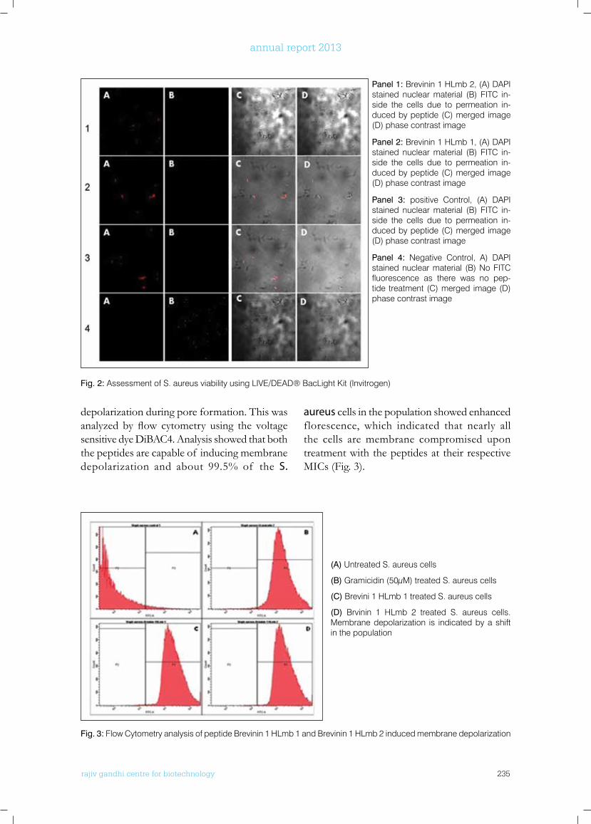
rajiv gandhi centre for biotechnology 235
annual report 2013
Panel 1: Brevinin 1 HLmb 2, (A) DAPI stained nuclear material (B) FITC in-side the cells due to permeation in-duced by peptide (C) merged image (D) phase contrast image
Panel 2: Brevinin 1 HLmb 1, (A) DAPI stained nuclear material (B) FITC in-side the cells due to permeation in-duced by peptide (C) merged image (D) phase contrast image
Panel 3: positive Control, (A) DAPI stained nuclear material (B) FITC in-side the cells due to permeation in-duced by peptide (C) merged image (D) phase contrast image
Panel 4: Negative Control, A) DAPI stained nuclear material (B) No FITC fluorescence as there was no pep-tide treatment (C) merged image (D) phase contrast image
Fig. 3: Flow Cytometry analysis of peptide Brevinin 1 HLmb 1 and Brevinin 1 HLmb 2 induced membrane depolarization
(A) Untreated S. aureus cells
(B) Gramicidin (50µM) treated S. aureus cells
(C) Brevini 1 HLmb 1 treated S. aureus cells
(D) Brvinin 1 HLmb 2 treated S. aureus cells. Membrane depolarization is indicated by a shift in the population
depolarization during pore formation. This was analyzedbyflow cytometry using the voltagesensitive dye DiBAC4. Analysis showed that both the peptides are capable of inducing membrane depolarization and about 99.5% of the S.
aureus cells in the population showed enhanced florescence, which indicated that nearly all the cells are membrane compromised upon treatment with the peptides at their respective MICs (Fig. 3).
Fig. 2: Assessment of S. aureus viability using LIVE/DEAD® BacLight Kit (Invitrogen)
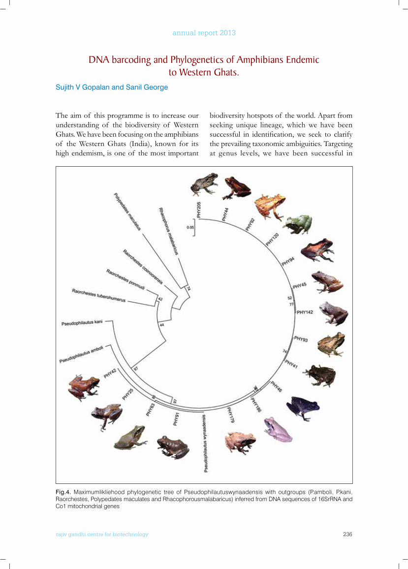
rajiv gandhi centre for biotechnology 236
annual report 2013
The aim of this programme is to increase our understanding of the biodiversity of Western Ghats. We have been focusing on the amphibians of the Western Ghats (India), known for its high endemism, is one of the most important
biodiversity hotspots of the world. Apart from seeking unique lineage, which we have been successful in identification,we seek to clarifythe prevailing taxonomic ambiguities. Targeting at genus levels, we have been successful in
DnA barcoding and Phylogenetics of Amphibians Endemic to western Ghats.
Sujith V Gopalan and Sanil George
Fig.4. Maximumlikliehood phylogenetic tree of Pseudophilautuswynaadensis with outgroups (P.amboli, P.kani, Raorchestes, Polypedates maculates and Rhacophorousmalabaricus) inferred from DNA sequences of 16SrRNA and Co1 mitochondrial genes
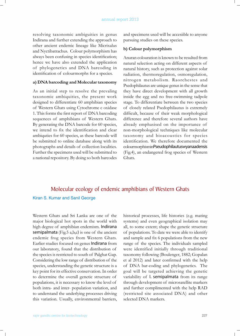
rajiv gandhi centre for biotechnology 237
annual report 2013
resolving taxonomic ambiguities in genus Indirana and further extending the approach to other ancient endemic lineage like Micrixalus and Nyctibatrachus. Colour polymorphism has alwaysbeenconfusinginspeciesidentification;hence we have also extended the application of phylogenetics and DNA barcoding in identificationof colourmorphsforaspecies.
a) DNA barcoding and Molecular taxonomy
As an initial step to resolve the prevaling taxonomic ambiquities, the present work designed to differentiate 60 amphibian species of Western Ghats using Cytochrome c oxidase 1.Thisformsthefirstreportof DNAbarcodingsequences of amphibians of Western Ghats. By generating the DNA barcode for 60 species, we intend to fix the identification and clearambiquties for 60 species, as these barcode will be submitted to online database along with its photographs and details of collection localities. Further the specimens used will be submitted to a national repository. By doing so both barcodes
and specimens used will be accesiible to anyone pursuing studies on these species.
b) Colour polymorphism
Anuran colouration is known to be resulted from natural selection acting on different aspects of natural history, such as protection against solar radiation, thermoregulation, osmoregulation, nitrogen metabolism. Raorchestes and Psedophilautus are unique genus in the sense that they have direct development with all growth inside the egg and no free-swimming tadpole stage. To differentiate between the two species of closely related Psedophilautus is extremely difficult,becauseof theirweakmorphologicaldifference and therefore several authors have already emphasised on the importance of non-morphological techniques like molecular taxonomy and bioacoustics for species identification.We therefore documented thecolourmorphism in Pseudophilautus wyanaadensis (Fig.4), an endangered frog species of Western Ghats.
Molecular ecology of endemic amphibians of western GhatsKiran S. Kumar and Sanil George
Western Ghats and Sri Lanka are one of the major biological hot spots in the world with high degree of amphibian endemism. Indirana semipalmata (Fig.5 a,b,c) is one of the ancient endemic frog species from Western Ghats. Earlier studies focused on genus Indirana from our laboratory, found that the distribution of the species is restricted to south of Palghat Gap. Considering the low range of distribution of the species, understanding the genetic structure is a key point for its effective conservation. In order to determine the overall genetic structure of populations, it is necessary to know the level of both intra- and inter- population variation, and to understand the underlying processes driving this variation. Usually, environmental barriers,
historical processes, life histories (e.g. mating systems) and even geographical isolation may all, to some extent; shape the genetic structure of populations. To date we were able to identify andsampleandfix6populationsfromthenewrange of the species. The individuals sampled were identified initially through traditional taxonomy following (Boulenger, 1882; Gopalan et al 2012) and later confirmedwith thehelpof DNA bar-coding and phylogenetics. The goal will be targeted achieving the genetic variability of I. semipalmata from its range through development of microsatellite markers and further complimented with the help RAD (restricted site associated DNA) and other selected DNA markers.
rajiv gandhi centre for biotechnology 238
annual report 2013
Mollusca represent one of few phyla that are routinely considered in marine biodiversity surveys and often considered as “indicator group” for rapid assessment of biodiversity. The 3 years multi-institutional project involving Rajiv Gandhi Centre for Biotechnology, Thiruvananthapuram, Department of Aquatic BiologyandFisheries,Universityof KeralaandSuganthi Devadason Marine Research Institute, Tuticorin, Tamil Nadu was implemented in September 2011 with the major objective to
prepare biodiversity database and DNA barcode data of commercially marine molluscs and to prepare a database on marine molluscs of India based on conventional and molecular taxonomy methods (using mitochondrial genes such as cytochrome oxidase subunit 1, cytochrome b, 12S and 16S genes). The project recorded many new sightings/new species as revealed by the phylogenetic analysis of CO1 gene (Figures 6a, b & c) generated in the present study.
Fig.5. Indiranasemipalmata female (A), Male (B) and tadpole (C)
biodiversity, DnA barcoding and Phylogeny of commercially valuable marine molluscs of India
Niji Anna Jose and Sanil George
Collaborators: Dr. BIjukumar A, Department of Aquatic Biology and Fisheries, University of Kerala, Dr.Patterson Edward, Suganthi Devadason Marine Research Institute, Tuticorin, India
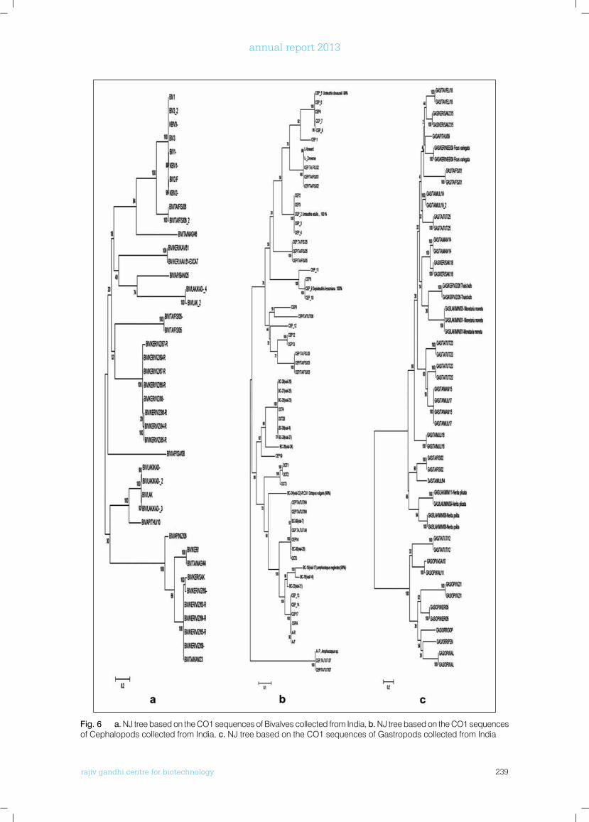
rajiv gandhi centre for biotechnology 239
annual report 2013
Fig. 6 a. NJ tree based on the CO1 sequences of Bivalves collected from India, b. NJ tree based on the CO1 sequences of Cephalopods collected from India, c. NJ tree based on the CO1 sequences of Gastropods collected from India
rajiv gandhi centre for biotechnology 240
annual report 2013
GEnbAnk SubMISSIonS
• KJ442579-KJ442611 (32 sequences)
• KJ631336-KJ631369 (33 sequences)
• KJ585580-KJ585665 (85 sequences)
• KJ561586-KJ561596 (10 sequences)
• KJ442612-KJ442649 (37 sequences)
• KJ442579-KJ442611 (32 sequences)
PublICATIonS
• Echi, P. C., Suresh, K. U., George S., Ratheesh, R. V., Vinitha, M. R., Ejere, V. C.1, Iyaji, F. O. and Nnamonu, E. I. 2013. Contribution towards the development of a DNA barcode reference library for West African mammals. African Journal of Biotechnology 12(48), 6704-6708.
ConfEREnCE PRESEnTATIonS
• Vineeth Kumar T V and Sanil George (2013). Antinociceptive and Antimicrobial peptides from the skin secretion of Endemic Frog of Western Ghats. International conference on ecosystem conservation, climate change and sustainable development (ECOCASD-2013), University of Kerala, Trivandrum, 3-5 October 2013.
• Vineeth Kumar T V and Sanil George (2013). Host defense Peptides from Skin Secretion of Endemic Frog of Western Ghats. International
Conference on Green Technology: ICGeT 2013, Sastra University, Thanjaore, Tamil Nadu. 26-27 July 2013.
• Suji th V Gopalan, Kiran S Kumar and Sanil George. One frog many suits. Poster presented at International Conference on Ecosystem conservation, Climate change and Sustainable Development (ECOCASD 2013). Thiruvananthapuram, Kerala,India.
• Sujith V Gopalan and Sanil George (2013). Reflections of Molecular Systematic Study on Amphibians of Western Ghats. Paper presented at International Conference on Ecosystem conservation, Climate change and Sustainable Development (ECOCASD 2013). Thiruvananthapuram, Kerala,India
AwARDS
• Vineeth Kumar T V and Sanil George (2013). International Conference on Ecosystem c o n s e r v a t i o n , C l i m a t e c h a n g e a n d Sustainable Development (ECOCASD 2013). Thiruvananthapuram, Kerala, India. (BEST PAPER AWARD)
• Sujith V Gopalan and Sanil George (2013) International Conference on Ecosystem c o n s e r v a t i o n , C l i m a t e c h a n g e a n d Sustainable Development (ECOCASD 2013). Thiruvananthapuram, Kerala, India. (BEST PAPER AWARD)
rajiv gandhi centre for biotechnology 241
annual report 2013
Chemical Biology ProgramEnvironmental Biology Laboratory
Ph.D StudentsArjun. J. K (UGC SRF)Aneesh. B (CSIR JRF)
r a j i v g a n d h i c e n t r e f o r b i o t e c h n o l o g y
Hari Krishnan has a Ph.D in Aquatic Biology from the University of Kerala and joined RGCB in 2001.
Hari Krishnan K Ph.D Scientists C
Project Fellow Kavitha. T
Project AssistantGeetha. S. L
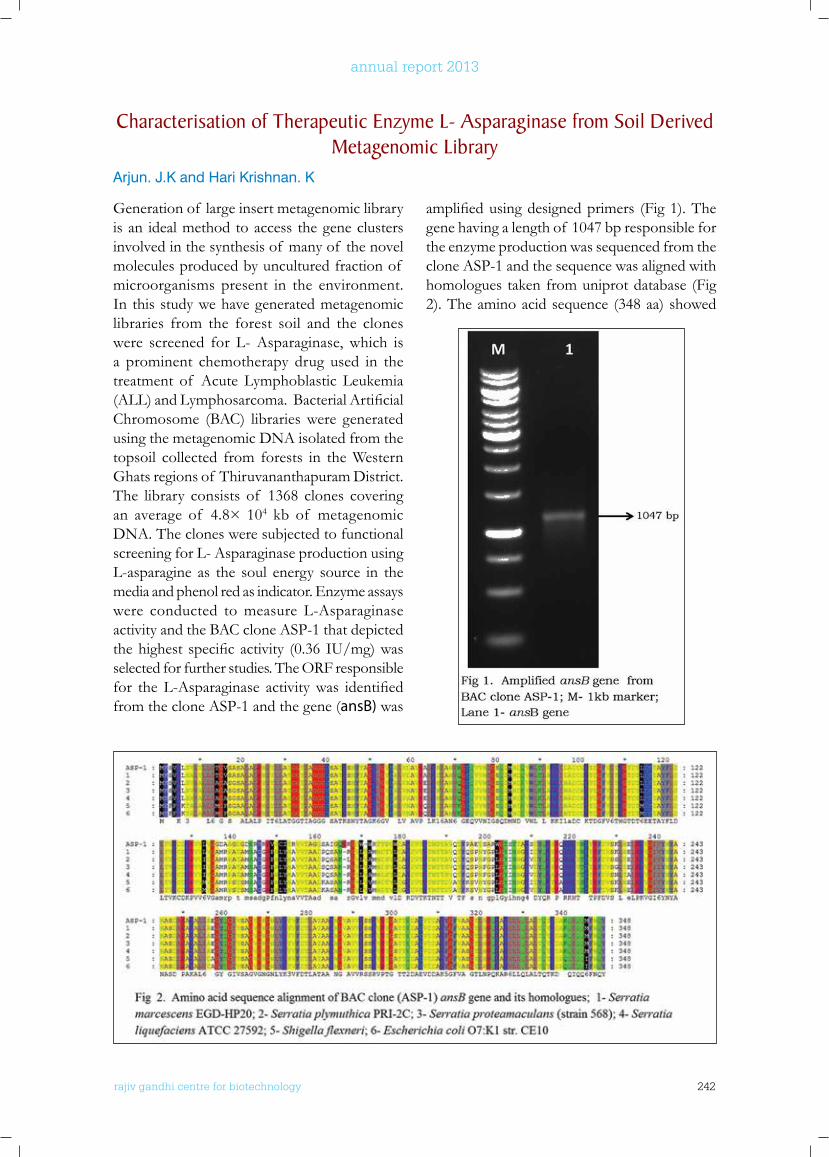
rajiv gandhi centre for biotechnology 242
annual report 2013
Generation of large insert metagenomic library is an ideal method to access the gene clusters involved in the synthesis of many of the novel molecules produced by uncultured fraction of microorganisms present in the environment. In this study we have generated metagenomic libraries from the forest soil and the clones were screened for L- Asparaginase, which is a prominent chemotherapy drug used in the treatment of Acute Lymphoblastic Leukemia (ALL)andLymphosarcoma.BacterialArtificialChromosome (BAC) libraries were generated using the metagenomic DNA isolated from the topsoil collected from forests in the Western Ghats regions of Thiruvananthapuram District. The library consists of 1368 clones covering an average of 4.8× 10⁴ kb of metagenomic DNA. The clones were subjected to functional screening for L- Asparaginase production using L-asparagine as the soul energy source in the media and phenol red as indicator. Enzyme assays were conducted to measure L-Asparaginase activity and the BAC clone ASP-1 that depicted thehighest specificactivity (0.36 IU/mg)wasselected for further studies. The ORF responsible for theL-Asparaginase activitywas identifiedfrom the clone ASP-1 and the gene (ansB) was
amplifiedusingdesignedprimers (Fig1).Thegene having a length of 1047 bp responsible for the enzyme production was sequenced from the clone ASP-1 and the sequence was aligned with homologues taken from uniprot database (Fig 2). The amino acid sequence (348 aa) showed
Characterisation of Therapeutic Enzyme l- Asparaginase from Soil Derived Metagenomic library
Arjun. J.K and Hari Krishnan. K
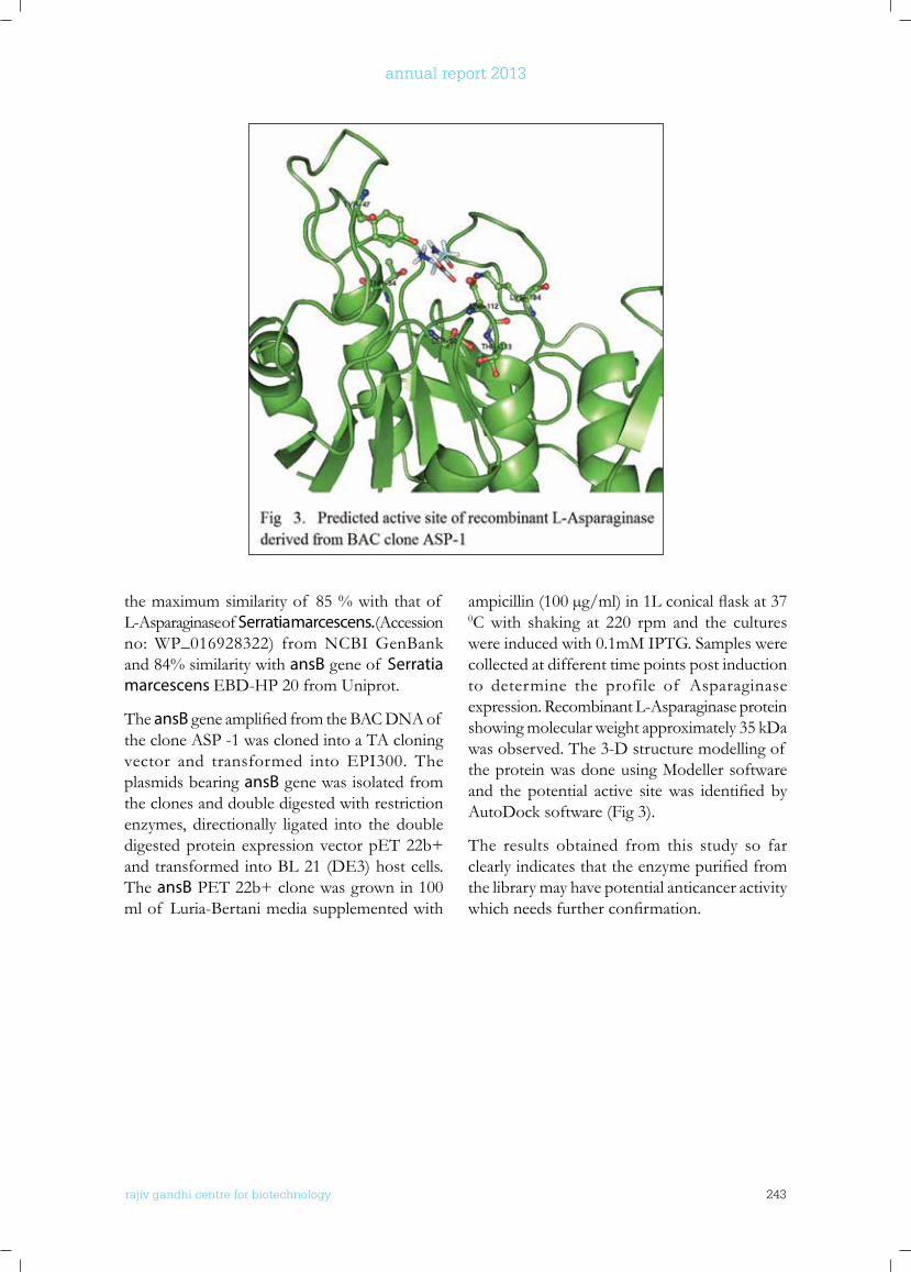
rajiv gandhi centre for biotechnology 243
annual report 2013
the maximum similarity of 85 % with that of L-Asparaginase of Serratia marcescens. (Accession no: WP_016928322) from NCBI GenBank and 84% similarity with ansB gene of Serratia marcescens EBD-HP 20 from Uniprot.
The ansBgeneamplifiedfromtheBACDNAof the clone ASP -1 was cloned into a TA cloning vector and transformed into EPI300. The plasmids bearing ansB gene was isolated from the clones and double digested with restriction enzymes, directionally ligated into the double digested protein expression vector pET 22b+ and transformed into BL 21 (DE3) host cells. The ansB PET 22b+ clone was grown in 100 ml of Luria-Bertani media supplemented with
ampicillin(100µg/ml)in1Lconicalflaskat370C with shaking at 220 rpm and the cultures were induced with 0.1mM IPTG. Samples were collected at different time points post induction to determine the profile of Asparaginase expression. Recombinant L-Asparaginase protein showing molecular weight approximately 35 kDa was observed. The 3-D structure modelling of the protein was done using Modeller software and thepotential active sitewas identifiedbyAutoDock software (Fig 3).
The results obtained from this study so far clearlyindicatesthattheenzymepurifiedfromthe library may have potential anticancer activity whichneedsfurtherconfirmation.
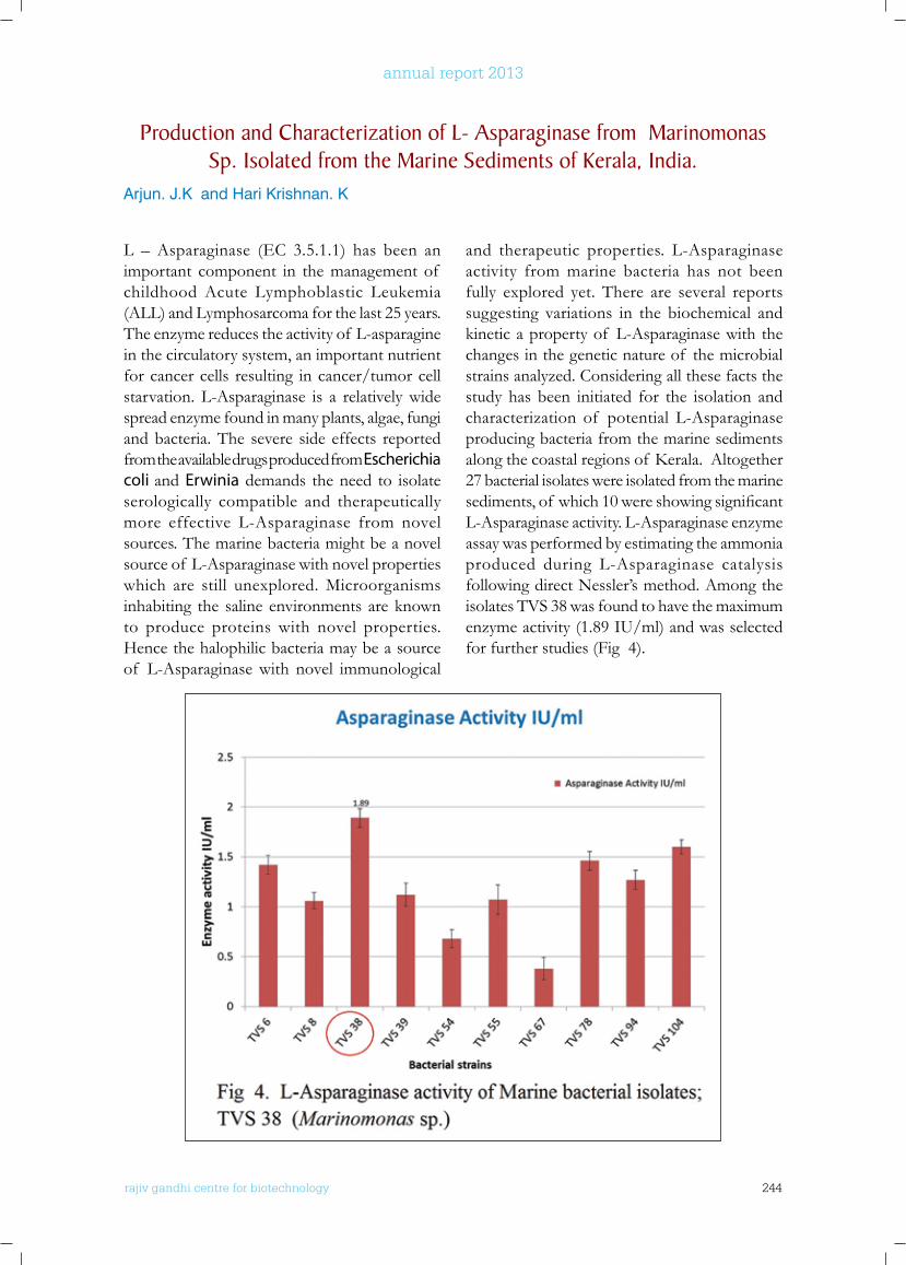
rajiv gandhi centre for biotechnology 244
annual report 2013
L – Asparaginase (EC 3.5.1.1) has been an important component in the management of childhood Acute Lymphoblastic Leukemia (ALL) and Lymphosarcoma for the last 25 years. The enzyme reduces the activity of L-asparagine in the circulatory system, an important nutrient for cancer cells resulting in cancer/tumor cell starvation. L-Asparaginase is a relatively wide spread enzyme found in many plants, algae, fungi and bacteria. The severe side effects reported from the available drugs produced from Escherichia coli and Erwinia demands the need to isolate serologically compatible and therapeutically more effective L-Asparaginase from novel sources. The marine bacteria might be a novel source of L-Asparaginase with novel properties which are still unexplored. Microorganisms inhabiting the saline environments are known to produce proteins with novel properties. Hence the halophilic bacteria may be a source of L-Asparaginase with novel immunological
and therapeutic properties. L-Asparaginase activity from marine bacteria has not been fully explored yet. There are several reports suggesting variations in the biochemical and kinetic a property of L-Asparaginase with the changes in the genetic nature of the microbial strains analyzed. Considering all these facts the study has been initiated for the isolation and characterization of potential L-Asparaginase producing bacteria from the marine sediments alongthecoastalregionsof Kerala.Altogether27 bacterial isolates were isolated from the marine sediments,of which10wereshowingsignificantL-Asparaginase activity. L-Asparaginase enzyme assay was performed by estimating the ammonia produced during L-Asparaginase catalysis following direct Nessler’s method. Among the isolates TVS 38 was found to have the maximum enzyme activity (1.89 IU/ml) and was selected for further studies (Fig 4).
Production and Characterization of l- Asparaginase from Marinomonas Sp. Isolated from the Marine Sediments of kerala, India.
Arjun. J.K and Hari Krishnan. K
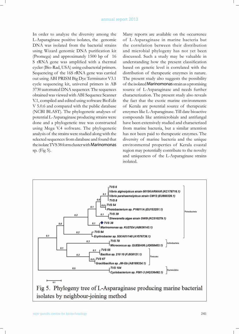
rajiv gandhi centre for biotechnology 245
annual report 2013
In order to analyze the diversity among the L-Asparaginase positive isolates, the genomic DNA was isolated from the bacterial strains usingWizard genomicDNApurification kit(Promega) and approximately 1500 bp of 16 S rRNA genewas amplifiedwith a thermalcycler (Bio-Rad, USA) using eubacterial primers. Sequencing of the 16S rRNA gene was carried out using ABI PRISM Big Dye Terminator V3.1 cycle sequencing kit, universal primers in AB 3730 automated DNA sequencer. The sequences obtained was viewed with ABI Sequence Scanner V.1, compiled and edited using software BioEdit V 5.0.6 and compared with the public database (NCBI BLAST). The phylogenetic analyses of potential L-Asparaginase producing strains were done and a phylogenetic tree was constructed using Mega V.4 software. The phylogenetic analysis of the strains were studied along with the selected sequences from database and found that the isolate TVS 38 form cluster with Marinomonas sp. (Fig 5).
Many reports are available on the occurrence of L-Asparaginase in marine bacteria but the correlation between their distribution and microbial phylogeny has not yet been discussed. Such a study may be valuable in understanding how the present classificationbased on genetic level is correlated with the distribution of therapeutic enzymes in nature. The present study also suggests the possibility of the isolated Marinomonas strain as a promising source of L-Asparaginase and needs further characterization. The present study also reveals the fact that the exotic marine environments of Kerala are potential source of therapeuticenzymes like L-Asparaginase. Till date bioactive compounds like antimicrobials and antifungal have been extensively studied and characterized from marine bacteria, but a similar attention has not been paid to therapeutic enzymes. The diversity of marine bacteria and the unique environmental properties of Kerala coastalregion may potentially contribute to the novelty and uniqueness of the L-Asparaginase strains isolated.
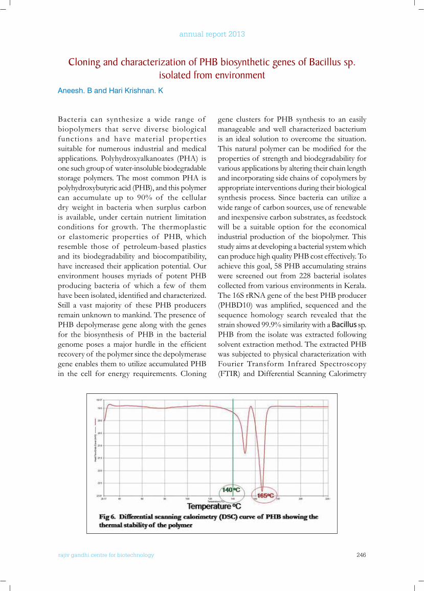
rajiv gandhi centre for biotechnology 246
annual report 2013
Bacteria can synthesize a wide range of biopolymers that serve diverse biological functions and have material properties suitable for numerous industrial and medical applications. Polyhydroxyalkanoates (PHA) is one such group of water-insoluble biodegradable storage polymers. The most common PHA is polyhydroxybutyric acid (PHB), and this polymer can accumulate up to 90% of the cellular dry weight in bacteria when surplus carbon is available, under certain nutrient limitation conditions for growth. The thermoplastic or elastomeric properties of PHB, which resemble those of petroleum-based plastics and its biodegradability and biocompatibility, have increased their application potential. Our environment houses myriads of potent PHB producing bacteria of which a few of them havebeenisolated,identifiedandcharacterized.Still a vast majority of these PHB producers remain unknown to mankind. The presence of PHB depolymerase gene along with the genes for the biosynthesis of PHB in the bacterial genomeposes amajor hurdle in the efficientrecovery of the polymer since the depolymerase gene enables them to utilize accumulated PHB in the cell for energy requirements. Cloning
Cloning and characterization of PHb biosynthetic genes of bacillus sp. isolated from environment
Aneesh. B and Hari Krishnan. K
gene clusters for PHB synthesis to an easily manageable and well characterized bacterium is an ideal solution to overcome the situation. Thisnaturalpolymercanbemodified for theproperties of strength and biodegradability for various applications by altering their chain length and incorporating side chains of copolymers by appropriate interventions during their biological synthesis process. Since bacteria can utilize a wide range of carbon sources, use of renewable and inexpensive carbon substrates, as feedstock will be a suitable option for the economical industrial production of the biopolymer. This study aims at developing a bacterial system which can produce high quality PHB cost effectively. To achieve this goal, 58 PHB accumulating strains were screened out from 228 bacterial isolates collectedfromvariousenvironmentsinKerala.The 16S rRNA gene of the best PHB producer (PHBD10)was amplified, sequenced and thesequence homology search revealed that the strain showed 99.9% similarity with a Bacillus sp. PHB from the isolate was extracted following solvent extraction method. The extracted PHB was subjected to physical characterization with Fourier Transform Infrared Spectroscopy (FTIR) and Differential Scanning Calorimetry
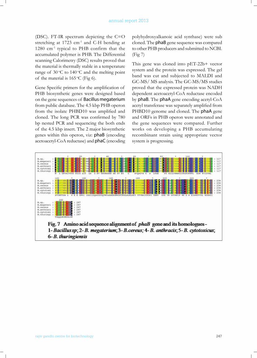
rajiv gandhi centre for biotechnology 247
annual report 2013
(DSC). FT-IR spectrum depicting the C=O stretching at 1723 cm-1 and C-H bending at 1280 cm-1 typical to PHB confirm that the accumulated polymer is PHB. The Differential scanning Calorimetry (DSC) results proved that the material is thermally stable in a temperature range of 30 oC to 140 oC and the melting point of the material is 165 oC (Fig 6).
GeneSpecificprimersfortheamplificationof PHB biosynthetic genes were designed based on the gene sequences of Bacillus megaterium from public database. The 4.5 kbp PHB operon from the isolate PHBD10was amplified andcloned.The longPCRwasconfirmedby780bp nested PCR and sequencing the both ends of the 4.5 kbp insert. The 2 major biosynthetic genes within this operon, viz: phaB (encoding acetoacetyl-CoA reductase) and phaC (encoding
polyhydroxyalkanoic acid synthase) were sub cloned. The phaB gene sequence was compared to other PHB producers and submitted to NCBI. (Fig 7)
This gene was cloned into pET-22b+ vector system and the protein was expressed. The gel band was cut and subjected to MALDI and GC-MS/ MS analysis. The GC-MS/MS studies proved that the expressed protein was NADH dependent acetoacetyl-CoA reductase encoded by phaB. The phaA gene encoding acetyl-CoA acetyltransferasewasseparatelyamplifiedfromPHBD10 genome and cloned. The phaA gene and ORFs in PHB operon were annotated and the gene sequences were compared. Further works on developing a PHB accumulating recombinant strain using appropriate vector system is progressing.
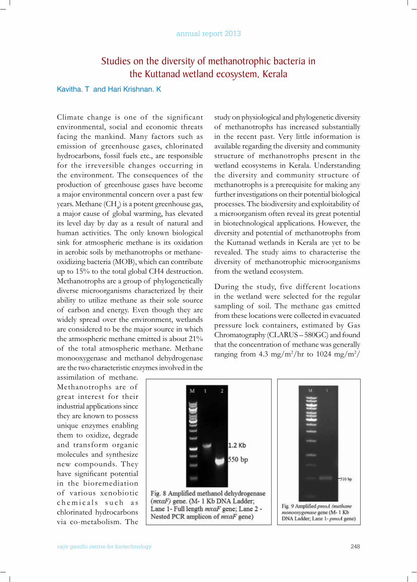
rajiv gandhi centre for biotechnology 248
annual report 2013
Climate change is one of the significant environmental, social and economic threats facing the mankind. Many factors such as emission of greenhouse gases, chlorinated hydrocarbons, fossil fuels etc., are responsible for the irreversible changes occurring in the environment. The consequences of the production of greenhouse gases have become a major environmental concern over a past few years. Methane (CH4) is a potent greenhouse gas, a major cause of global warming, has elevated its level day by day as a result of natural and human activities. The only known biological sink for atmospheric methane is its oxidation in aerobic soils by methanotrophs or methane-oxidizing bacteria (MOB), which can contribute up to 15% to the total global CH4 destruction. Methanotrophs are a group of phylogenetically diverse microorganisms characterized by their ability to utilize methane as their sole source of carbon and energy. Even though they are widely spread over the environment, wetlands are considered to be the major source in which the atmospheric methane emitted is about 21% of the total atmospheric methane. Methane monooxygenase and methanol dehydrogenase are the two characteristic enzymes involved in the assimilation of methane. Methanotrophs are of great interest for their industrial applications since they are known to possess unique enzymes enabling them to oxidize, degrade and transform organic molecules and synthesize new compounds. They have significant potentialin the bioremediation of various xenobiotic c h e m i c a l s s u c h a s chlorinated hydrocarbons via co-metabolism. The
study on physiological and phylogenetic diversity of methanotrophs has increased substantially in the recent past. Very little information is available regarding the diversity and community structure of methanotrophs present in the wetland ecosystems inKerala.Understandingthe diversity and community structure of methanotrophs is a prerequisite for making any further investigations on their potential biological processes. The biodiversity and exploitability of a microorganism often reveal its great potential in biotechnological applications. However, the diversity and potential of methanotrophs from theKuttanadwetlands inKeralaareyettoberevealed. The study aims to characterise the diversity of methanotrophic microorganisms from the wetland ecosystem.
During the study, five different locations in the wetland were selected for the regular sampling of soil. The methane gas emitted from these locations were collected in evacuated pressure lock containers, estimated by Gas Chromatography (CLARUS – 580GC) and found that the concentration of methane was generally ranging from 4.3 mg/m2/hr to 1024 mg/m2/
Studies on the diversity of methanotrophic bacteria in the kuttanad wetland ecosystem, kerala
Kavitha. T and Hari Krishnan. K
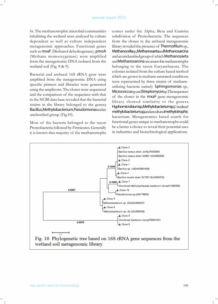
rajiv gandhi centre for biotechnology 249
annual report 2013
hr. The methanotrophic microbial communities inhabiting the wetland were analysed by culture dependent as well as culture independent metagenomic approaches. Functional genes such as mxaF (Methanol dehydrogenase), pmoA (Methane monooxygenase) were amplified form the metagenomic DNA isolated from the wetland soil (Fig. 8 & 9).
Bacterial and archaeal 16S rRNA gene were amplified from themetagenomicDNAusingspecific primers and librarieswere generatedusing the amplicons. The clones were sequenced and the comparison of the sequences with that in the NCBI data base revealed that the bacterial strains in the library belonged to the genera Bacillus, Methylobacterium, Pseudomonas and an unclassifiedgroup(Fig.10).
Most of the bacteria belonged to the taxon Proteobacteria followed by Firmicutes. Generally it is known that majority of the methanotrophs
comes under the Alpha, Beta and Gamma subdivision of Proteobacteria. The sequences from the clones in the archaeal metagenomic library revealed the presence of Thermofilum sp., Methanocella sp., Methanosaeta and Methanosarcina and an unclassifiedgroupof whichMethanosaeta and Methanosarcina are anaerobic methanotrophs belonging to the taxon Euryarchaeota. The colonies isolated from the culture based method which are grown in methane saturated conditions were represented by three strains of methane-utilizing bacteria namely Sphingomonas sp., Micrococcus sp. and Streptomyces sp. The sequences of the clones in the mxaF gene metagenomic library showed similarity to the genera Hyphomicrobium sp., Methylobacterium sp., Uncultued methylobacterium, sp, and uncultured methylotrophic bacterium. Metagenomics based search for functional genes unique to methanotrophs could be a better a choice to reveal their potential uses in industries and biotechnological applications.
rajiv gandhi centre for biotechnology 250
annual report 2013
AwARDS AnD HonouRS
• Hari Krishnan. K has been nominated as a member of the State Level Expert Appraisal Committee (SEAC), Kerala by the Ministry of Environment and Forest, Govt of India.
ConfEREnCE PRESEnTATIonS
• Arjun, J.K. and Hari Krishnan K (2013). “Production and Characterization of Therapeutic L-Asparaginase from a Marinomonas sp. isolated from Marine Sediments”. 54th Annual Conference of Association of Microbiology of India, International Conference on “Frontier Discoveries and Innovations in Microbiology and its Interdisciplinary Relevance dated 17-20 November 2013 at Rohtak, Haryana, India. (Poster presentation)
• Aneesh, B and Hari Krishnan K (2013). “Cloning and Characterization of NADPH-Dependant Acetoacetyl-CoA Dehydrogenase Gene (PhaB) from Bacillus megaterium Isolated from the Environment”. 54th Annual Conference of Association of Microbiology of India, International Conference on “Frontier Discoveries and Innovations in Microbiology and its Interdisciplinary Relevance dated 17-20 November 2013 at Rohtak, Haryana, India. (Poster presentation)
• Kavitha, T and Hari Krishnan. K (2013). A PCR Based detection of methanotrophic bacteria from environment. 54th Annual
Conference of Association of Microbiology of India, International Conference on “Frontier Discoveries and Innovations in Microbiology and its Interdisciplinary Relevance, 17-20 November 2013, Rohtak, Haryana, India. (Poster presentation)
GEnbAnk SubMISSIonS
• Aneesh B., Arjun J.K., Kavitha T. and Hari Krishnan K. Bacillus aryabhattai strain PHB10 acetoacetyl-CoA reductase (phaB)gene, complete cds (Acc. No.KF155284) and Bacillus megaterium strain PHB29 acetoacetyl-CoA reductase (phaB) gene, complete cds (Acc. No.KF155285) (2013)
• Arjun J.K., Aneesh B. and Hari Krishnan K. Uncultured Serratia sp. 16S ribosomal RNA gene, partial sequence (Acc. No. KF155286) (2013)
• Arjun J.K., Aneesh B., Kavitha T. and Hari Krishnan K. L-asparaginase producing halophilic bacteria isolated from marine sediments of Kerala. 16S ribosomal RNA gene sequences of 10 isolates (Acc. Nos. KF142387 to KF142396) (2013).
• Aneesh B., Arjun J.K., Kavitha T. and Hari Krishnan K. 16 S ribosomal RNA gene sequences of Bacillus aryabhattai strain PHB10 (Acc. No. KF056892 and Bacillus megaterium strain PHB29 (Acc. No. KF056893 ) (2013)
ExTRAMuRAl RESEARCH GRAnTS
Title Investigators Funding Agency Duration
Studies on the distribution and diversity of methanotrophic microbial communities, mitigating methane emission in Kuttanad wetland ecosystem, Kerala
Hari Krishnan. K (PI)G.S. Vinod Kumar (Co-PI)
Department of Environment and Climate Change, Government of Kerala
2013 - 16
Metagenomic analysis of gut microbiome from type 2 diabetic subjects in Kerala - a preliminary study
Hari Krishnan. K (PI)Abdul Jaleel. K.A (Co-PI)
Kerala State Council for Science, Technology & Environment
2014 - 17
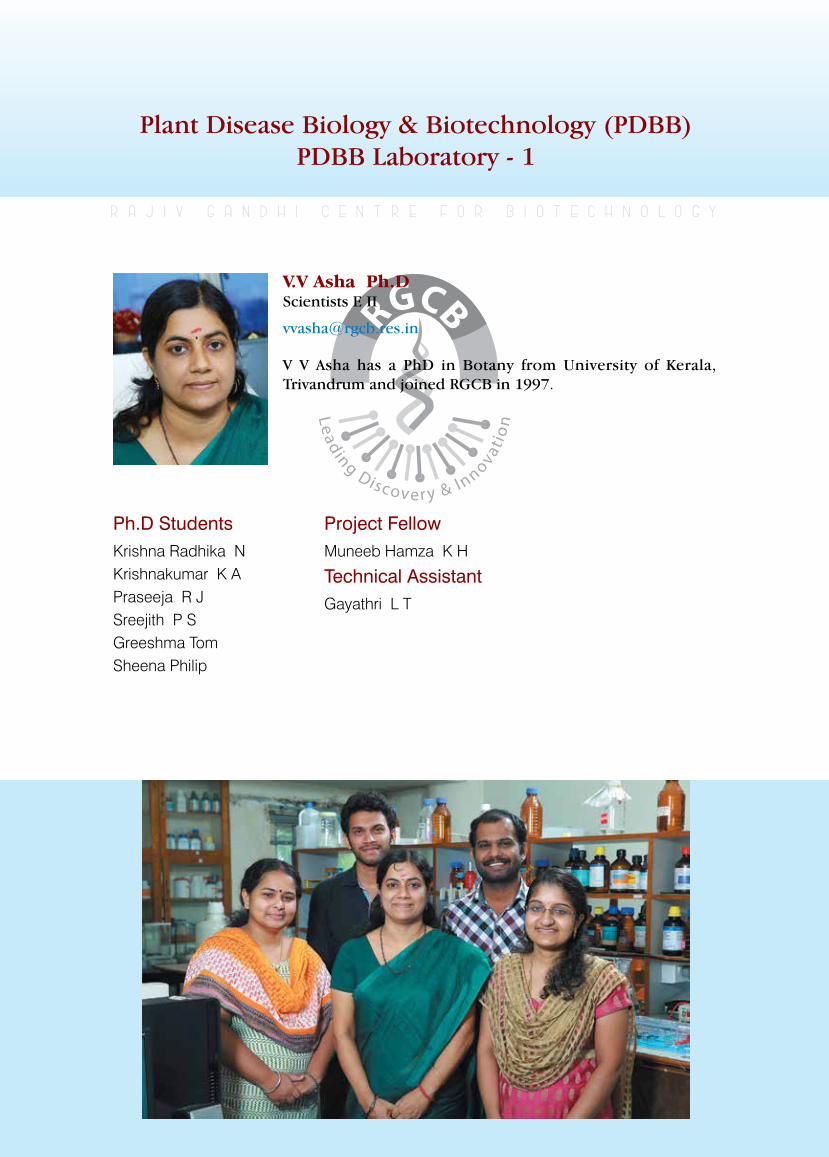
rajiv gandhi centre for biotechnology 251
annual report 2013
Plant Disease Biology & Biotechnology (PDBB) PDBB Laboratory - 1
Ph.D StudentsKrishna Radhika NKrishnakumar K APraseeja R JSreejith P SGreeshma TomSheena Philip
r a j i v g a n d h i c e n t r e f o r b i o t e c h n o l o g y
V V Asha has a PhD in Botany from University of Kerala, Trivandrum and joined RGCB in 1997.
V.V Asha Ph.D Scientists E II
Project Fellow Muneeb Hamza K H
Technical AssistantGayathri L T
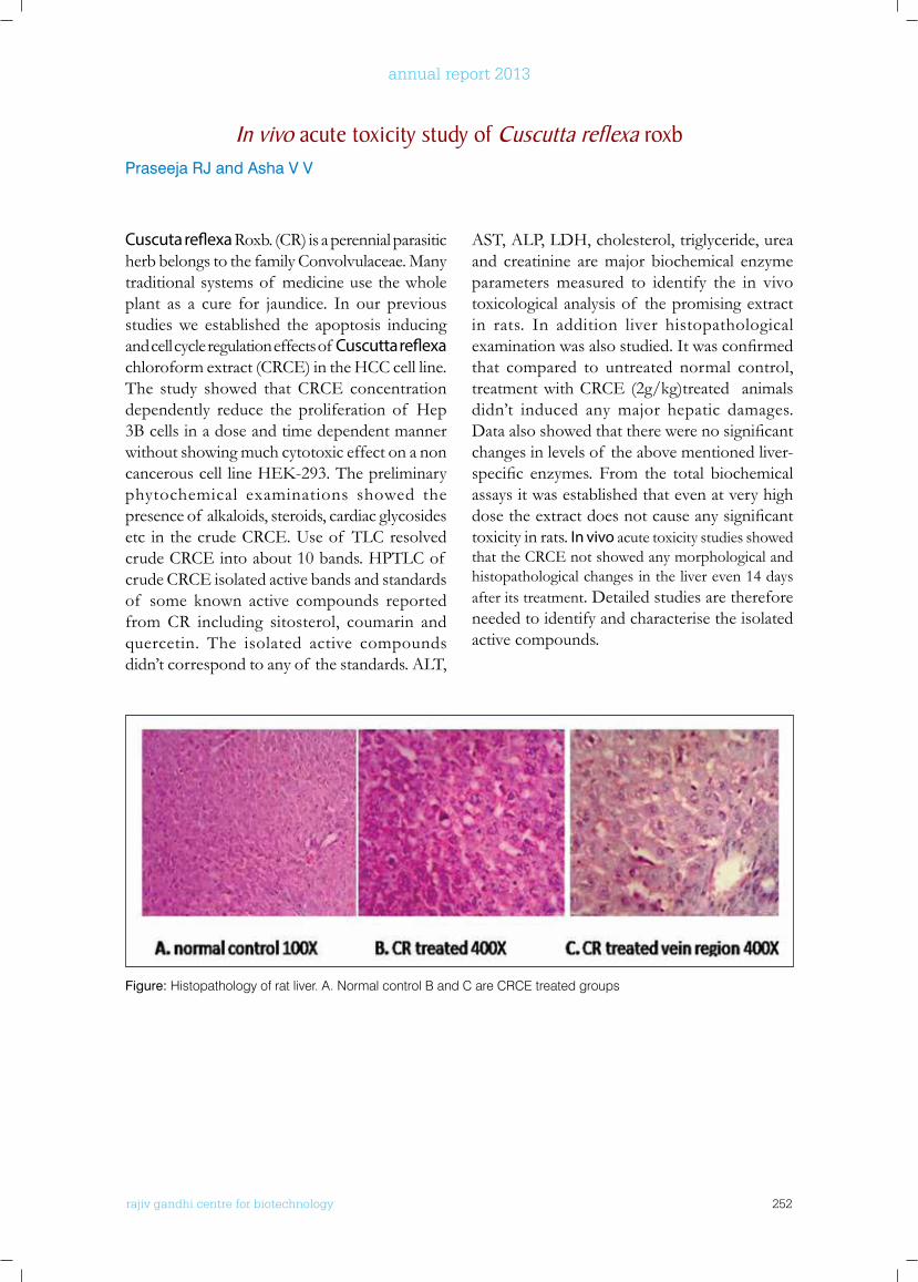
rajiv gandhi centre for biotechnology 252
annual report 2013
Cuscuta reflexa Roxb. (CR) is a perennial parasitic herb belongs to the family Convolvulaceae. Many traditional systems of medicine use the whole plant as a cure for jaundice. In our previous studies we established the apoptosis inducing and cell cycle regulation effects of Cuscutta reflexa chloroform extract (CRCE) in the HCC cell line. The study showed that CRCE concentration dependently reduce the proliferation of Hep 3B cells in a dose and time dependent manner without showing much cytotoxic effect on a non cancerouscelllineHEK-293.Thepreliminaryphytochemical examinations showed the presence of alkaloids, steroids, cardiac glycosides etc in the crude CRCE. Use of TLC resolved crude CRCE into about 10 bands. HPTLC of crude CRCE isolated active bands and standards of some known active compounds reported from CR including sitosterol, coumarin and quercetin. The isolated active compounds didn’t correspond to any of the standards. ALT,
AST, ALP, LDH, cholesterol, triglyceride, urea and creatinine are major biochemical enzyme parameters measured to identify the in vivo toxicological analysis of the promising extract in rats. In addition liver histopathological examinationwasalsostudied.Itwasconfirmedthat compared to untreated normal control, treatment with CRCE (2g/kg)treated animals didn’t induced any major hepatic damages. Dataalsoshowedthattherewerenosignificantchanges in levels of the above mentioned liver-specific enzymes. From the total biochemicalassays it was established that even at very high dosetheextractdoesnotcauseanysignificanttoxicity in rats. In vivo acute toxicity studies showed that the CRCE not showed any morphological and histopathological changes in the liver even 14 days after its treatment. Detailed studies are therefore needed to identify and characterise the isolated active compounds.
In vivo acute toxicity study of Cuscutta reflexa roxbPraseeja RJ and Asha V V
Figure: Histopathology of rat liver. A. Normal control B and C are CRCE treated groups
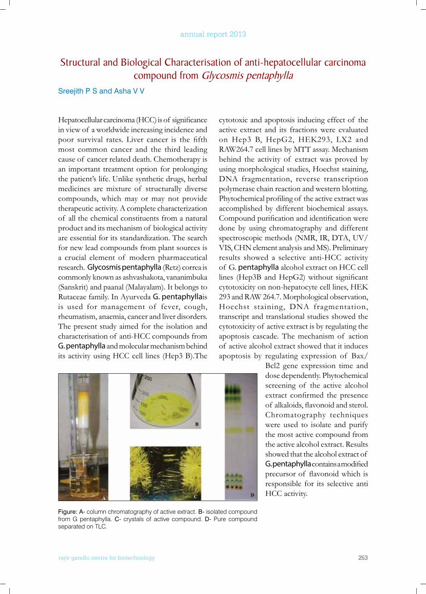
rajiv gandhi centre for biotechnology 253
annual report 2013
Hepatocellularcarcinoma(HCC)isof significancein view of a worldwide increasing incidence and poor survival rates. Liver cancer is the fifthmost common cancer and the third leading cause of cancer related death. Chemotherapy is an important treatment option for prolonging the patient’s life. Unlike synthetic drugs, herbal medicines are mixture of structurally diverse compounds, which may or may not provide therapeutic activity. A complete characterization of all the chemical constituents from a natural product and its mechanism of biological activity are essential for its standardization. The search for new lead compounds from plant sources is a crucial element of modern pharmaceutical research. Glycosmis pentaphylla (Retz) correa is commonly known as ashvashakota, vananimbuka (Sanskrit) and paanal (Malayalam). It belongs to Rutaceae family. In Ayurveda G. pentaphyllais is used for management of fever, cough, rheumatism, anaemia, cancer and liver disorders. The present study aimed for the isolation and characterisation of anti-HCC compounds from G. pentaphylla and molecular mechanism behind its activity using HCC cell lines (Hep3 B).The
cytotoxic and apoptosis inducing effect of the active extract and its fractions were evaluated on Hep3 B, HepG2, HEK293, LX2 andRAW264.7 cell lines by MTT assay. Mechanism behind the activity of extract was proved by using morphological studies, Hoechst staining, DNA fragmentation, reverse transcription polymerase chain reaction and western blotting. Phytochemicalprofilingof theactiveextractwasaccomplished by different biochemical assays. Compoundpurificationandidentificationweredone by using chromatography and different spectroscopic methods (NMR, IR, DTA, UV/VIS, CHN element analysis and MS). Preliminary results showed a selective anti-HCC activity of G. pentaphylla alcohol extract on HCC cell lines (Hep3BandHepG2)without significantcytotoxicityonnon-hepatocytecelllines,HEK293 and RAW 264.7. Morphological observation, Hoechst staining, DNA fragmentation, transcript and translational studies showed the cytotoxicity of active extract is by regulating the apoptosis cascade. The mechanism of action of active alcohol extract showed that it induces apoptosis by regulating expression of Bax/
Bcl2 gene expression time and dose dependently. Phytochemical screening of the active alcohol extract confirmed the presenceof alkaloids,flavonoidandsterol.Chromatography techniques were used to isolate and purify the most active compound from the active alcohol extract. Results showed that the alcohol extract of G. pentaphyllacontainsamodifiedprecursorof flavonoidwhichisresponsible for its selective anti HCC activity.
Structural and biological Characterisation of anti-hepatocellular carcinoma compound from Glycosmis pentaphylla
Sreejith P S and Asha V V
Figure: A- column chromatography of active extract. B- isolated compound from G pentaphylla. C- crystals of active compound. D- Pure compound separated on TLC.
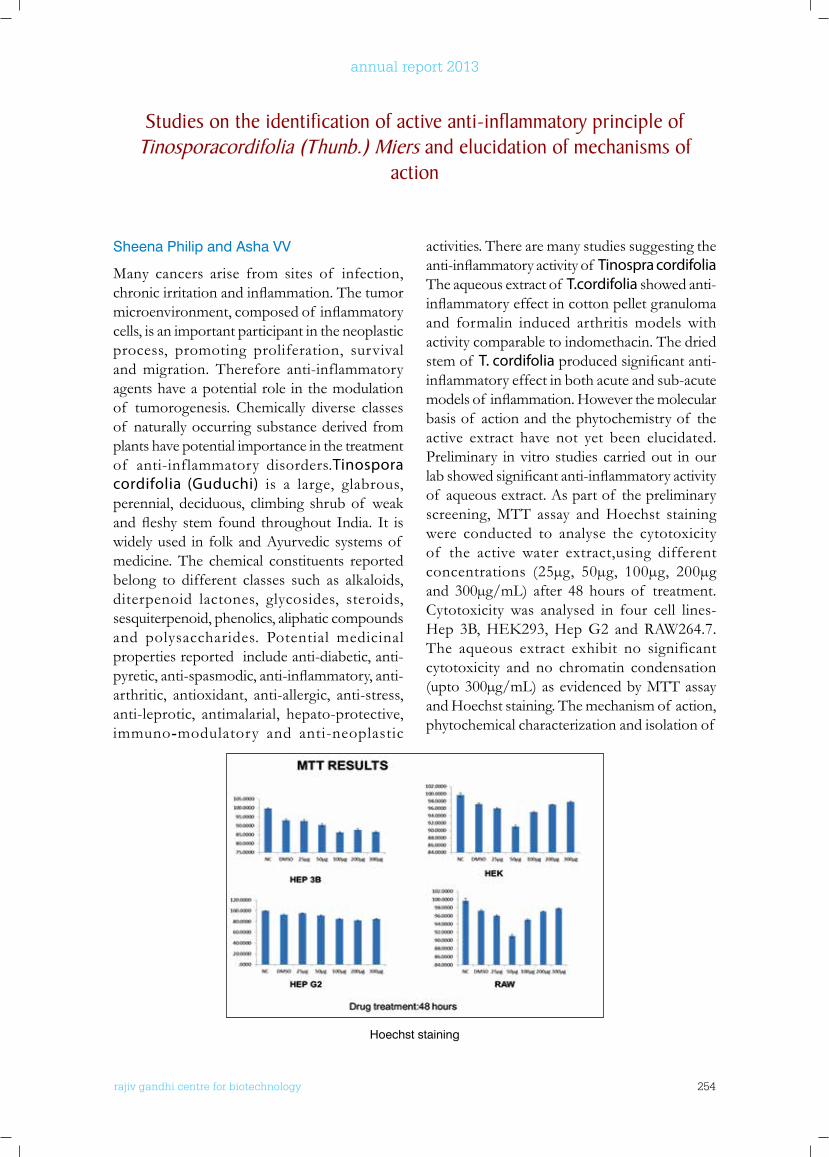
rajiv gandhi centre for biotechnology 254
annual report 2013
Sheena Philip and Asha VV
Many cancers arise from sites of infection, chronicirritationandinflammation.Thetumormicroenvironment,composedof inflammatorycells, is an important participant in the neoplastic process, promoting proliferation, survival and migration. Therefore anti-inflammatory agents have a potential role in the modulation of tumorogenesis. Chemically diverse classes of naturally occurring substance derived from plants have potential importance in the treatment of anti-inflammatory disorders.Tinospora cordifolia (Guduchi) is a large, glabrous, perennial, deciduous, climbing shrub of weak andfleshy stem found throughout India. It iswidely used in folk and Ayurvedic systems of medicine. The chemical constituents reported belong to different classes such as alkaloids, diterpenoid lactones, glycosides, steroids, sesquiterpenoid, phenolics, aliphatic compounds and polysaccharides. Potential medicinal properties reported include anti-diabetic, anti-pyretic,anti-spasmodic,anti-inflammatory,anti-arthritic, antioxidant, anti-allergic, anti-stress, anti-leprotic, antimalarial, hepato-protective, immuno-modulatory and anti-neoplastic
activities. There are many studies suggesting the anti-inflammatoryactivityof Tinospra cordifolia The aqueous extract of T.cordifolia showed anti-inflammatoryeffectincottonpelletgranulomaand formalin induced arthritis models with activity comparable to indomethacin. The dried stem of T. cordifoliaproducedsignificantanti-inflammatoryeffectinbothacuteandsub-acutemodelsof inflammation.Howeverthemolecularbasis of action and the phytochemistry of the active extract have not yet been elucidated. Preliminary in vitro studies carried out in our labshowedsignificantanti-inflammatoryactivityof aqueous extract. As part of the preliminary screening, MTT assay and Hoechst staining were conducted to analyse the cytotoxicity of the active water extract,using different concentrations (25µg, 50µg, 100µg, 200µg and 300µg/mL) after 48 hours of treatment. Cytotoxicity was analysed in four cell lines- Hep 3B,HEK293,HepG2 andRAW264.7.The aqueous extract exhibit no significant cytotoxicity and no chromatin condensation (upto 300µg/mL) as evidenced by MTT assay and Hoechst staining. The mechanism of action, phytochemical characterization and isolation of
Studies on the identification of active anti-inflammatory principle of Tinosporacordifolia (Thunb.) Miers and elucidation of mechanisms of
action
Hoechst staining
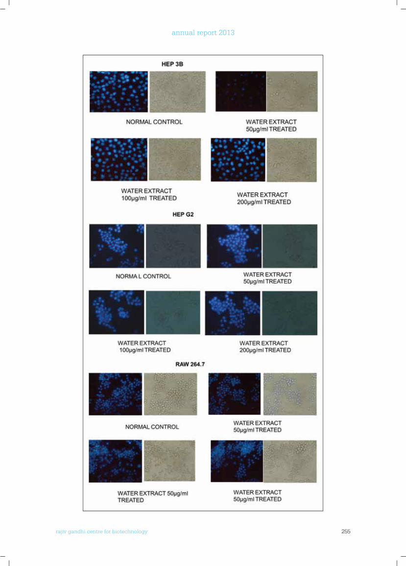
rajiv gandhi centre for biotechnology 255
annual report 2013
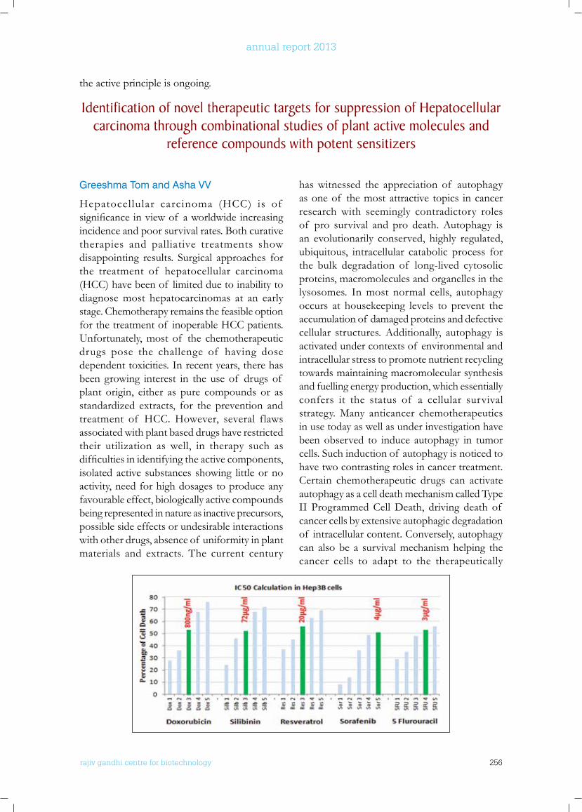
rajiv gandhi centre for biotechnology 256
annual report 2013
Greeshma Tom and Asha VV
Hepatocellular carcinoma (HCC) is of significanceinviewof aworldwideincreasingincidence and poor survival rates. Both curative therapies and palliative treatments show disappointing results. Surgical approaches for the treatment of hepatocellular carcinoma (HCC) have been of limited due to inability to diagnose most hepatocarcinomas at an early stage. Chemotherapy remains the feasible option for the treatment of inoperable HCC patients. Unfortunately, most of the chemotherapeutic drugs pose the challenge of having dose dependent toxicities. In recent years, there has been growing interest in the use of drugs of plant origin, either as pure compounds or as standardized extracts, for the prevention and treatment of HCC. However, several flaws associated with plant based drugs have restricted their utilization as well, in therapy such as difficultiesinidentifyingtheactivecomponents,isolated active substances showing little or no activity, need for high dosages to produce any favourable effect, biologically active compounds being represented in nature as inactive precursors, possible side effects or undesirable interactions with other drugs, absence of uniformity in plant materials and extracts. The current century
has witnessed the appreciation of autophagy as one of the most attractive topics in cancer research with seemingly contradictory roles of pro survival and pro death. Autophagy is an evolutionarily conserved, highly regulated, ubiquitous, intracellular catabolic process for the bulk degradation of long-lived cytosolic proteins, macromolecules and organelles in the lysosomes. In most normal cells, autophagy occurs at housekeeping levels to prevent the accumulation of damaged proteins and defective cellular structures. Additionally, autophagy is activated under contexts of environmental and intracellular stress to promote nutrient recycling towards maintaining macromolecular synthesis and fuelling energy production, which essentially confers it the status of a cellular survival strategy. Many anticancer chemotherapeutics in use today as well as under investigation have been observed to induce autophagy in tumor cells. Such induction of autophagy is noticed to have two contrasting roles in cancer treatment. Certain chemotherapeutic drugs can activate autophagy as a cell death mechanism called Type II Programmed Cell Death, driving death of cancer cells by extensive autophagic degradation of intracellular content. Conversely, autophagy can also be a survival mechanism helping the cancer cells to adapt to the therapeutically
the active principle is ongoing.
Identification of novel therapeutic targets for suppression of Hepatocellular carcinoma through combinational studies of plant active molecules and
reference compounds with potent sensitizers
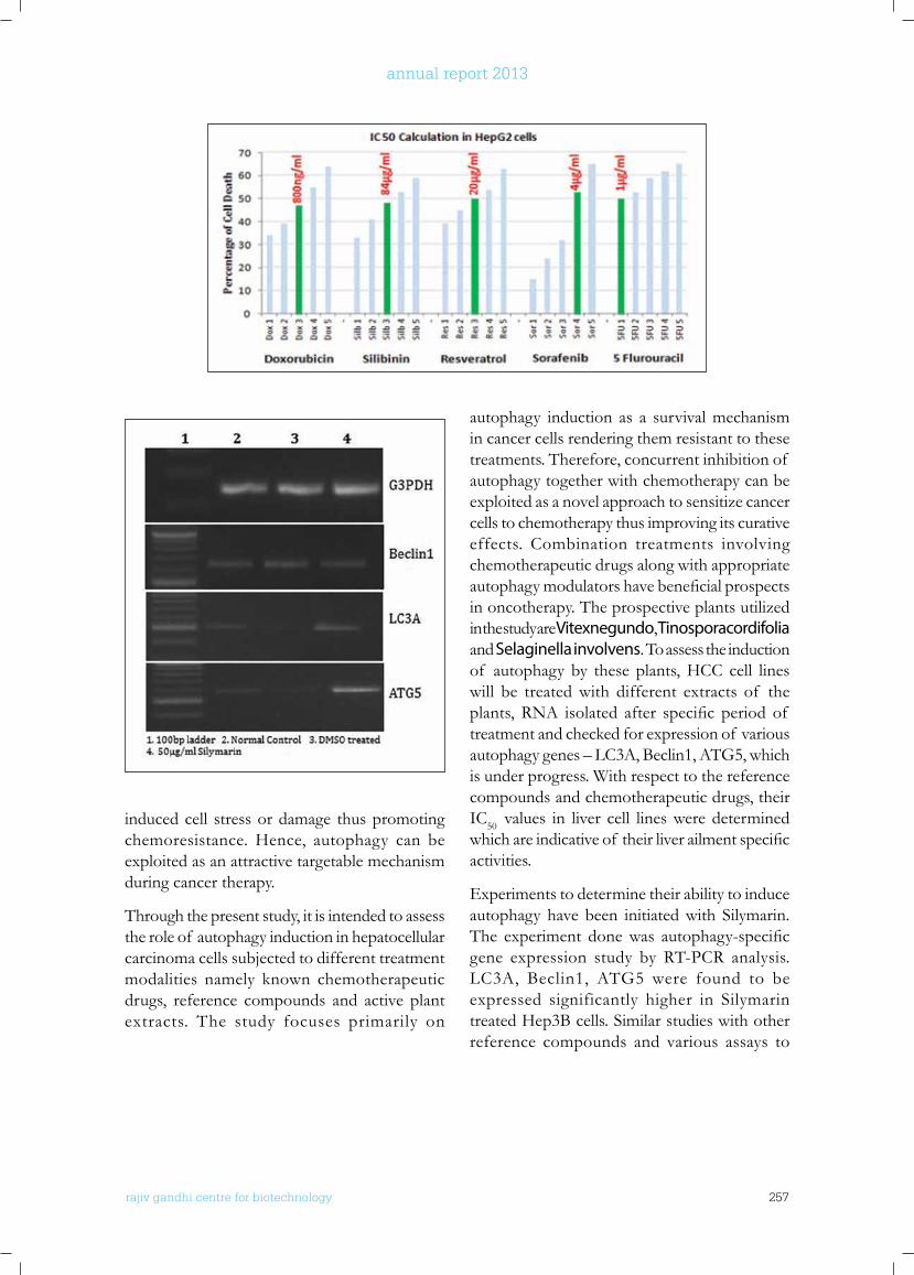
rajiv gandhi centre for biotechnology 257
annual report 2013
induced cell stress or damage thus promoting chemoresistance. Hence, autophagy can be exploited as an attractive targetable mechanism during cancer therapy.
Through the present study, it is intended to assess the role of autophagy induction in hepatocellular carcinoma cells subjected to different treatment modalities namely known chemotherapeutic drugs, reference compounds and active plant extracts. The study focuses primarily on
autophagy induction as a survival mechanism in cancer cells rendering them resistant to these treatments. Therefore, concurrent inhibition of autophagy together with chemotherapy can be exploited as a novel approach to sensitize cancer cells to chemotherapy thus improving its curative effects. Combination treatments involving chemotherapeutic drugs along with appropriate autophagymodulatorshavebeneficialprospectsin oncotherapy. The prospective plants utilized in the study are Vitexnegundo, Tinosporacordifolia and Selaginella involvens. To assess the induction of autophagy by these plants, HCC cell lines will be treated with different extracts of the plants,RNA isolated after specific period of treatment and checked for expression of various autophagy genes – LC3A, Beclin1, ATG5, which is under progress. With respect to the reference compounds and chemotherapeutic drugs, their IC50 values in liver cell lines were determined whichareindicativeof theirliverailmentspecificactivities.
Experiments to determine their ability to induce autophagy have been initiated with Silymarin. The experiment donewas autophagy-specificgene expression study by RT-PCR analysis. LC3A, Beclin1, ATG5 were found to be expressed significantly higher in Silymarin treated Hep3B cells. Similar studies with other reference compounds and various assays to
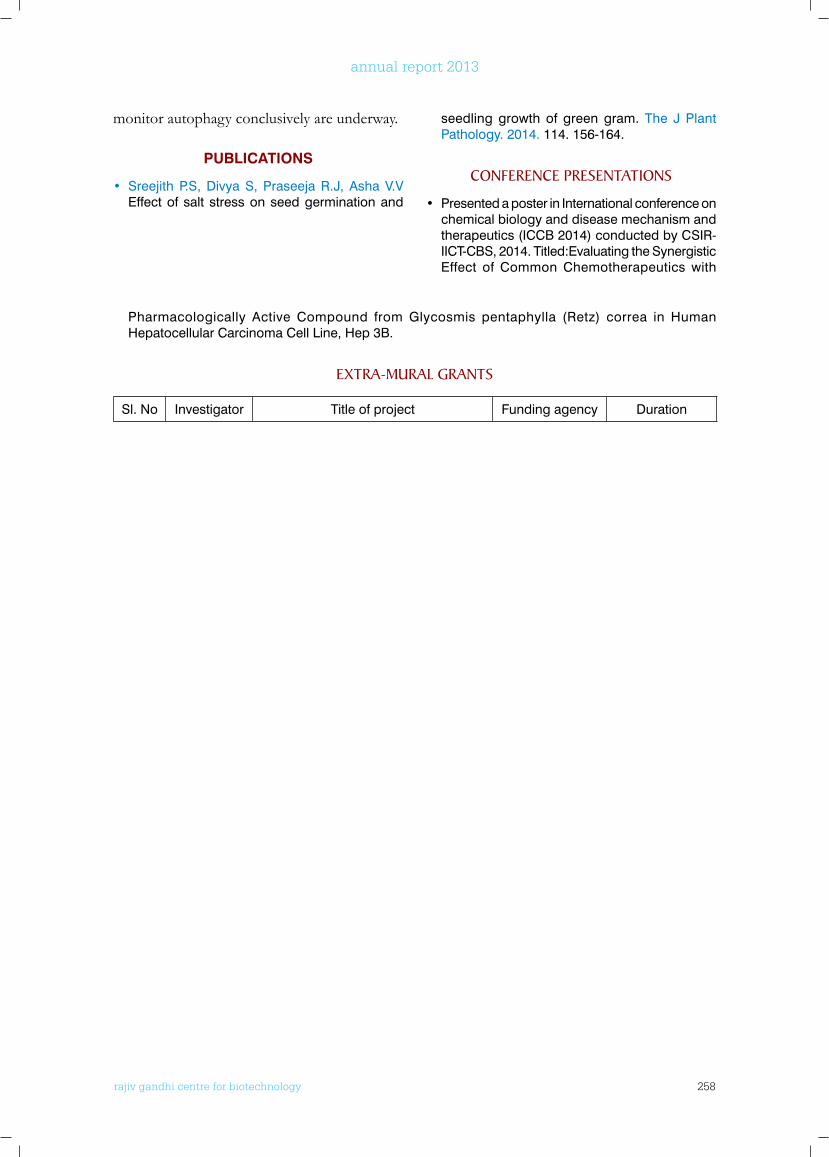
rajiv gandhi centre for biotechnology 258
annual report 2013
monitor autophagy conclusively are underway.
PUBLiCATioNS
• Sreejith P.S, Divya S, Praseeja R.J, Asha V.V Effect of salt stress on seed germination and
seedling growth of green gram. The J Plant Pathology. 2014. 114. 156-164.
ConfEREnCE PRESEnTATIonS
• Presented a poster in International conference on chemical biology and disease mechanism and therapeutics (ICCB 2014) conducted by CSIR-IICT-CBS, 2014. Titled:Evaluating the Synergistic Effect of Common Chemotherapeutics with
Pharmacologically Active Compound from Glycosmis pentaphylla (Retz) correa in Human Hepatocellular Carcinoma Cell Line, Hep 3B.
ExTRA-MuRAl GRAnTS
Sl. No Investigator Title of project Funding agency Duration
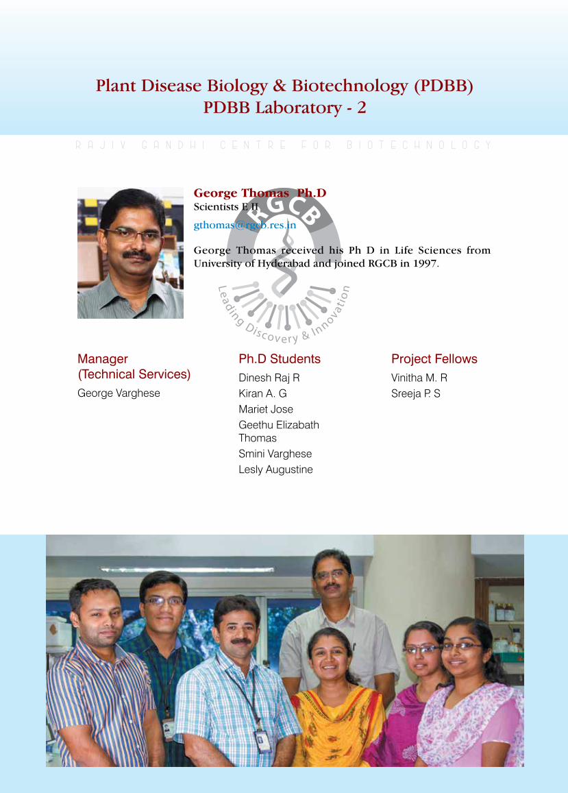
rajiv gandhi centre for biotechnology 259
annual report 2013
Plant Disease Biology & Biotechnology (PDBB) PDBB Laboratory - 2
Manager (Technical Services)George Varghese
r a j i v g a n d h i c e n t r e f o r b i o t e c h n o l o g y
George Thomas received his Ph D in Life Sciences from University of Hyderabad and joined RGCB in 1997.
George Thomas Ph.D Scientists E II
Ph.D StudentsDinesh Raj RKiran A. GMariet JoseGeethu Elizabath ThomasSmini VargheseLesly Augustine
Project FellowsVinitha M. RSreeja P. S
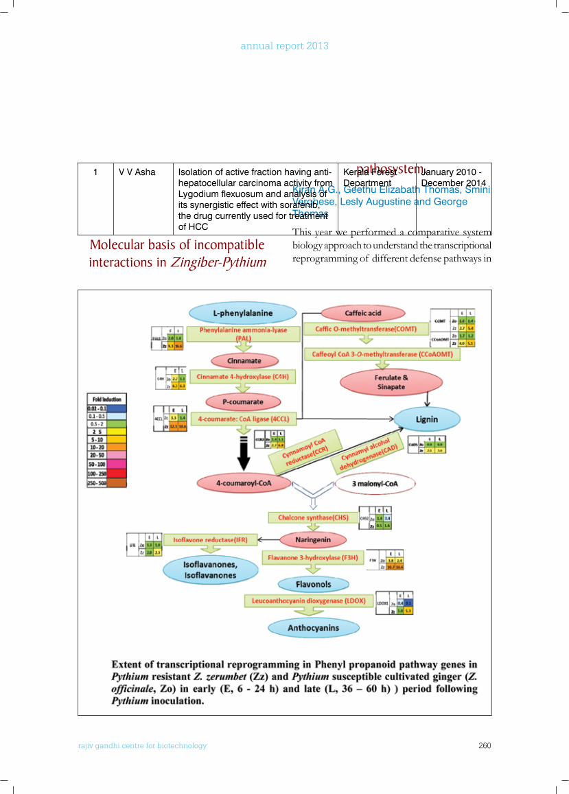
rajiv gandhi centre for biotechnology 260
annual report 2013
pathosystem.Kiran A.G., Geethu Elizabath Thomas, Smini Varghese, Lesly Augustine and George Thomas
This year we performed a comparative system biology approach to understand the transcriptional reprogramming of different defense pathways in
1 V V Asha Isolation of active fraction having anti-hepatocellular carcinoma activity from Lygodium flexuosum and analysis of its synergistic effect with sorafenib, the drug currently used for treatment of HCC
Kerala Forest Department
January 2010 - December 2014
Molecular basis of incompatible interactions in Zingiber-Pythium
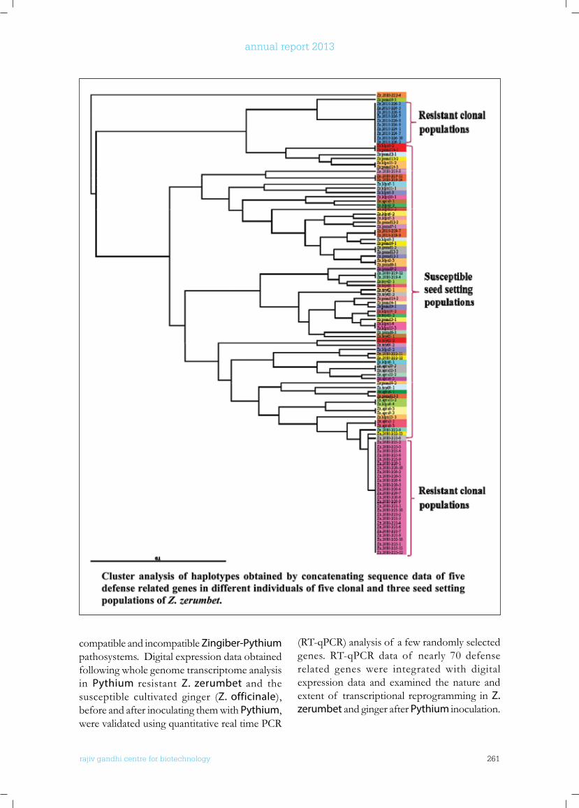
rajiv gandhi centre for biotechnology 261
annual report 2013
compatible and incompatible Zingiber-Pythium pathosystems. Digital expression data obtained following whole genome transcriptome analysis in Pythium resistant Z. zerumbet and the susceptible cultivated ginger (Z. officinale), before and after inoculating them with Pythium, were validated using quantitative real time PCR
(RT-qPCR) analysis of a few randomly selected genes. RT-qPCR data of nearly 70 defense related genes were integrated with digital expression data and examined the nature and extent of transcriptional reprogramming in Z. zerumbet and ginger after Pythium inoculation.
rajiv gandhi centre for biotechnology 262
annual report 2013
The analysis revealed induction of genes involved in the biosynthesis of major plant defense hormones; jasmonic acid, ethylene and salicylic acid in post inoculated Z zerumbet whereas in Z officinale only jasmonic acid and ethylene pathway genes were found upregulated. Salicylic acid responsive gene, PR1 (Pathogenesis related 1) was also found to be inducedsignificantlyintheresistantwildspecies.Reactive oxygen species (ROS) generating genes studied showed similar levels of induction in both the resistant and susceptible pathosystems. However genes responsible for ROS scavenging showed robust induction only in the resistant species. In hypersensitive response (HR) related genes,HSR203J andMAPKinases,MAPK3showed a robust induction in Z zerumbet. The results strongly suggest that the incompatible response of the wild species is HR mediated and involve salicylic acid signalling. The comparative transcript data also showed a critical role for phenylpropanoid pathway in governing Pythium resistance in Z. zerumbet. Genes involved in
biosynthesisof flavonoidsandligninssuchasphenylalanine ammonia lyase, cinnamoyl CoA reductase, caffeic acid o-methyltransferase, flavone3hydroxylase and leucoanthocyanidindioxygenase showed robust induction in Z zerumbet following Pythium inoculation. We were also interested in understanding the genetic basis of Pythium resistance in Z. zerumbet. Contrary to the expectations, earlier we found relatively higher levels of Pythium resistance in clonally propagated populations of Z. zerumbet and high susceptibility in seed setting populations. Theamplified fragment lengthpolymorphismanalysis yielded expected results with higher genetic diversity in seed setting populations and low diversity in clonal populations. This year we analyzed sequence variations at functional genes in multiple accessions of clonal and seed setting populations of Z. zerumbet. A total of 45 heterozygous positions (variable nucleotide sites between allele)were identified fromfivegenes covering a total length of 2295 bps. The heterozygosity pattern varied considerably in
seed setting populations, ranging from one to 30, whereas the heterozygosity panel was almost consistent within non-seed setting populations. Accessions with higher heterozygosity showed higher level of Pythium resistance while accessions with lower heterozygosity yielded higher susceptibility. Data indicate that the resistance in clonally propagated populations of Z. zerumbet may be due
to the retention of a set of useful heterozygous positions with adaptive value. Mixed breeding system together with heterozygosity thus provides adaptive value in Z. zerumbet.
Microsatellite assisted genetic resources characterization and
circumscription of the medicinal rice ‘njavara’
George Varghese, Dinesh Raj R., Mariet Jose and George Thomas.
The medicinal rice Njavara is traditionally considered as an endemic strain to South India, especially to the present dayKerala state. Itsgrains are used in Ayurveda treatments in Kerala since time immemorial.A cultivar by
this name is not known to occur in any other states in India and no rice cultivar is used for medicinal purposes as extensively as Njavara being used in Ayurveda. At the same time, Njavara is traditionally treated as a shashtika. Shashtika is referred in several Sanskrit Ayurveda texts written in Northern part of India from late BC to eighteenth century AD. According to Ayurveda texts, shashtika is a short duration cultivar (matures in 60 days), medicinally and nutritionally rich, best in vrihi type of rice and mitigates three doshas. At this juncture a few pertinent questions arise: ‘how can shashtika, which is referred by North Indian authors, be endemictoKerala?’and‘whyis Shashtika not seennowadaysinotherpartsof India?’Besidesshashtika, Ayurveda texts describe many other
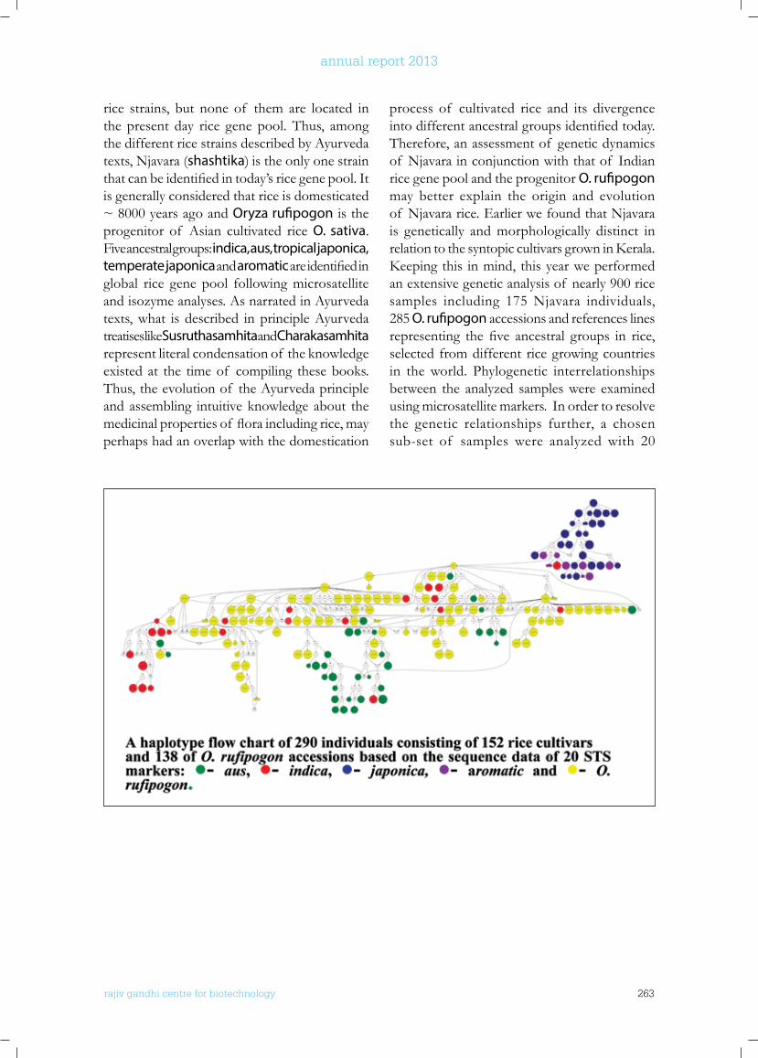
rajiv gandhi centre for biotechnology 263
annual report 2013
rice strains, but none of them are located in the present day rice gene pool. Thus, among the different rice strains described by Ayurveda texts, Njavara (shashtika) is the only one strain thatcanbeidentifiedintoday’sricegenepool.Itis generally considered that rice is domesticated ~ 8000 years ago and Oryza rufipogon is the progenitor of Asian cultivated rice O. sativa. Five ancestral groups: indica, aus, tropical japonica, temperate japonica and aromaticareidentifiedinglobal rice gene pool following microsatellite and isozyme analyses. As narrated in Ayurveda texts, what is described in principle Ayurveda treatises like Susruthasamhita and Charakasamhita represent literal condensation of the knowledge existed at the time of compiling these books. Thus, the evolution of the Ayurveda principle and assembling intuitive knowledge about the medicinalpropertiesof floraincludingrice,mayperhaps had an overlap with the domestication
process of cultivated rice and its divergence intodifferentancestralgroupsidentifiedtoday.Therefore, an assessment of genetic dynamics of Njavara in conjunction with that of Indian rice gene pool and the progenitor O. rufipogon may better explain the origin and evolution of Njavara rice. Earlier we found that Njavara is genetically and morphologically distinct in relationtothesyntopiccultivarsgrowninKerala.Keepingthis inmind,thisyearweperformedan extensive genetic analysis of nearly 900 rice samples including 175 Njavara individuals, 285 O. rufipogon accessions and references lines representing thefive ancestral groups in rice,selected from different rice growing countries in the world. Phylogenetic interrelationships between the analyzed samples were examined using microsatellite markers. In order to resolve the genetic relationships further, a chosen sub-set of samples were analyzed with 20
rajiv gandhi centre for biotechnology 264
annual report 2013
unlinked nuclear sequence tagged sites (STS), internal transcribed sequence (ITS) of nuclear ribosomal RNA genes and two loci of maternally inherited chloroplast genome. The analysis helped to gain deep insight into the origin and evolution of cultivate rice, including Njavara in India. The pattern of distribution of cultivars belonging to different ancestral groups was elucidated. Different O. rufipogon populations that progenitored the cultivated rice populations wereidentified.Thestudydetectedonlylimitedinter-group genetic admixing.
PublICATIonS
• Varghese G., Jose M., Dinesh Raj R., Bocianowski J., Thomas G. and Omanakumar N. 2014. Quantitative and molecular analyses reveal a
deep genetic divergence between the ancient medicinal rice (Oryza sativa) Njavara and syntopic traditional cultivars. Annals of Applied Biology 164: 95–106. DOI: 10.1111/aab.12083
• Vinitha M. R., Suresh Kumar U., Aishwarya K., Sabu M. and Thomas G. 2014. Prospects for discriminating Zingiberaceae species in India using DNA barcodes. Journal of Integrative Plant Biology. Doi: 10.1111/jipb.12189.
ConfEREnCE PRESEnTATIonS
• Kiran A.G., Augustine L., Smini V. and Thomas G. Incompatible response of Zingiber zerumbet (L) Smith. to the necrotrophic pathogen Pythium sp. involves hypersensitive response and salicylic acid signalling. Poster presented at the 10th International Congress of Plant Pathology (ICPP 2013), Bio-security, Food Safety and Plant
Pathology: The Role of Plant Pathology in a Globalized Economy, Beijing, China, 25-30 August 2013.
• Augustine L., Kiran A.G. and Thomas G. Differential defense response towards Pythium myriotylum in resistant and susceptible Zingiber spp. revealed by comparative whole genome RNA-Seq analysis. Poster presented at the 10th International Congress of Plant Pathology (ICPP 2013), Bio-security, Food Safety and Plant Pathology: The Role of Plant Pathology in a Globalized Economy, Beijing, China, 25-30 August 2013.
ExTRAMuRAl funDInG
Investigator Title Funding Agency Duration
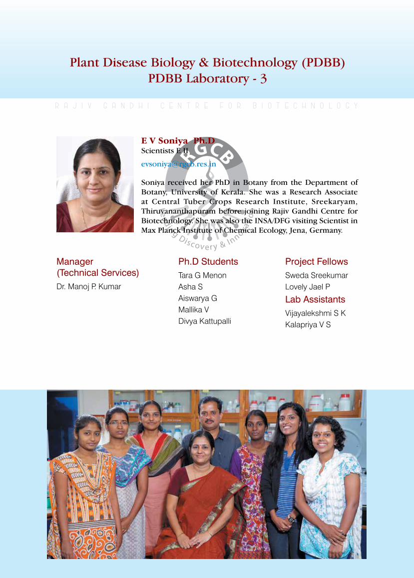
rajiv gandhi centre for biotechnology 265
annual report 2013
Plant Disease Biology & Biotechnology (PDBB) PDBB Laboratory - 3
Manager (Technical Services)Dr. Manoj P. Kumar
r a j i v g a n d h i c e n t r e f o r b i o t e c h n o l o g y
Soniya received her PhD in Botany from the Department of Botany, University of Kerala. She was a Research Associate at Central Tuber Crops Research Institute, Sreekaryam, Thiruvananthapuram before joining Rajiv Gandhi Centre for Biotechnology. She was also the INSA/DFG visiting Scientist in Max Planck Institute of Chemical Ecology, Jena, Germany.
E V Soniya Ph.D Scientists E II
Ph.D StudentsTara G MenonAsha SAiswarya GMallika VDivya Kattupalli
Project FellowsSweda SreekumarLovely Jael P
Lab AssistantsVijayalekshmi S KKalapriya V S
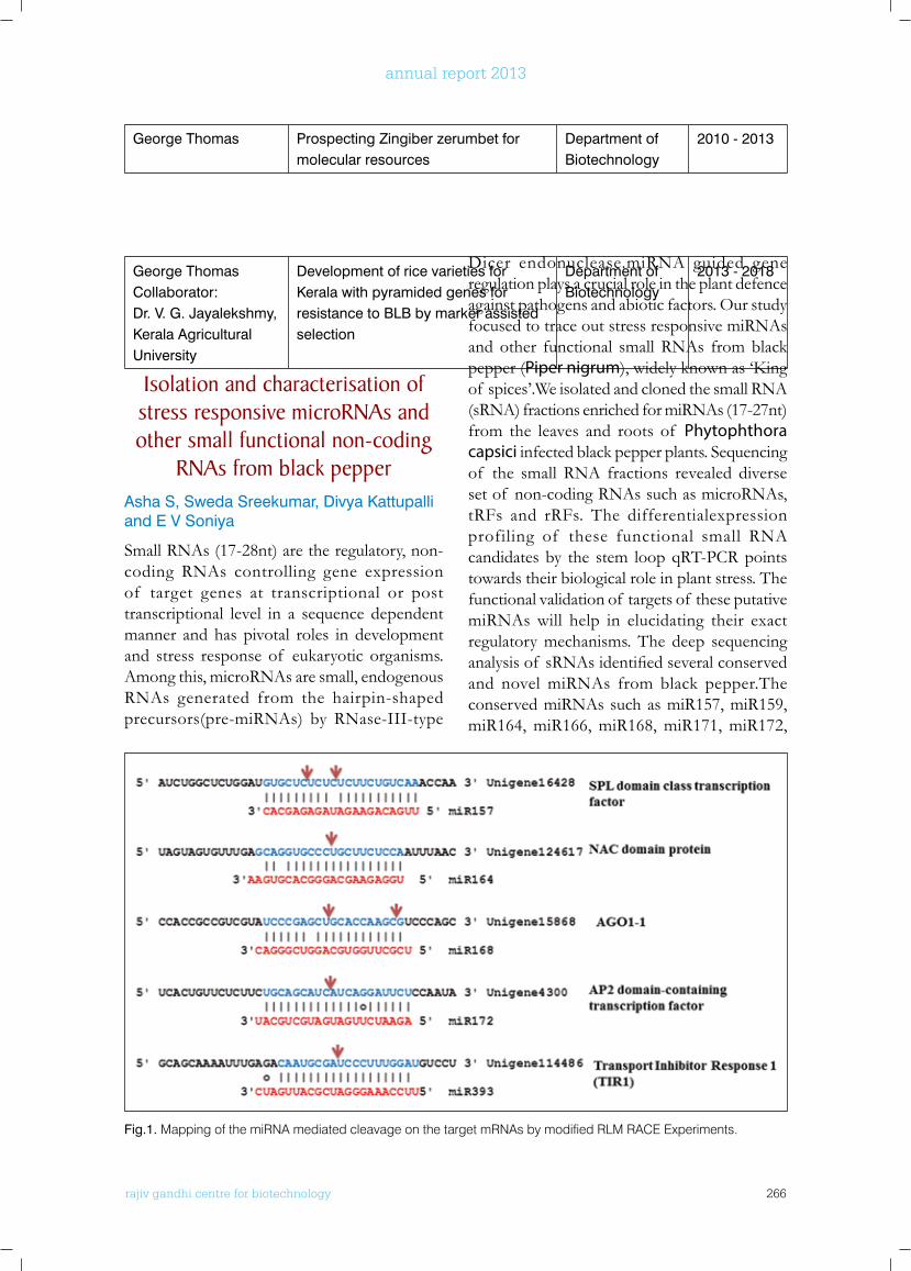
rajiv gandhi centre for biotechnology 266
annual report 2013
George ThomasCollaborator: Dr. V. G. Jayalekshmy, Kerala Agricultural University
Development of rice varieties for Kerala with pyramided genes for resistance to BLB by marker assisted selection
Department of Biotechnology
2013 - 2018
Isolation and characterisation of stress responsive microRnAs and other small functional non-coding
RnAs from black pepperAsha S, Sweda Sreekumar, Divya Kattupalli and E V Soniya
Small RNAs (17-28nt) are the regulatory, non-coding RNAs controlling gene expression of target genes at transcriptional or post transcriptional level in a sequence dependent manner and has pivotal roles in development and stress response of eukaryotic organisms. Among this, microRNAs are small, endogenous RNAs generated from the hairpin-shaped precursors(pre-miRNAs) by RNase-III-type
Dicer endonuclease.miRNA guided gene regulation plays a crucial role in the plant defence against pathogens and abiotic factors. Our study focused to trace out stress responsive miRNAs and other functional small RNAs from black pepper (Piper nigrum),widelyknownas‘Kingof spices’.We isolated and cloned the small RNA (sRNA) fractions enriched for miRNAs (17-27nt) from the leaves and roots of Phytophthora capsici infected black pepper plants. Sequencing of the small RNA fractions revealed diverse set of non-coding RNAs such as microRNAs, tRFs and rRFs. The differentialexpression profiling of these functional small RNA candidates by the stem loop qRT-PCR points towards their biological role in plant stress. The functional validation of targets of these putative miRNAs will help in elucidating their exact regulatory mechanisms. The deep sequencing analysisof sRNAsidentifiedseveralconservedand novel miRNAs from black pepper.The conserved miRNAs such as miR157, miR159, miR164, miR166, miR168, miR171, miR172,
George Thomas Prospecting Zingiber zerumbet for molecular resources
Department of Biotechnology
2010 - 2013
Fig.1. Mapping of the miRNA mediated cleavage on the target mRNAs by modified RLM RACE Experiments.
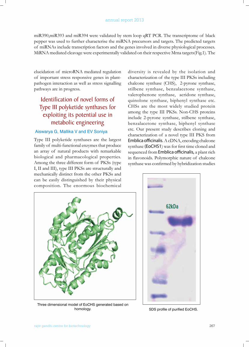
rajiv gandhi centre for biotechnology 267
annual report 2013
elucidation of microRNA mediated regulation of important stress responsive genes in plant-pathogen interaction as well as stress signalling pathways are in progress.
Identification of novel forms of Type III polyketide synthases for
exploiting its potential use in metabolic engineering
Aiswarya G, Mallika V and EV Soniya
Type III polyketide synthases are the largest family of multi-functional enzymes that produce an array of natural products with remarkable biological and pharmacological properties. Amongthethreedifferentformof PKSs(typeI,IIandIII),typeIIIPKSsarestructurallyandmechanicallydistinctfromtheotherPKSsandcan be easily distinguished by their physical composition. The enormous biochemical
diversity is revealed by the isolation and characterizationof thetypeIIIPKSsincludingchalcone synthase (CHS), 2-pyrone synthase, stilbene synthase, benzalacetone synthase, valerophenone synthase, acridone synthase, quinolone synthase, biphenyl synthase etc. CHSs are the most widely studied protein among the type IIIPKSs.Non-CHSproteinsinclude 2-pyrone synthase, stilbene synthase, benzalacetone synthase, biphenyl synthase etc. Our present study describes cloning and characterizationof anoveltypeIIIPKSfromEmblica officinalis. A cDNA, encoding chalcone synthase (EoCHS1)wasforfirsttimeclonedandsequenced from Emblica officinalis, a plant rich inflavonoids.Polymorphicnatureof chalconesynthasewasconfirmedbyhybridizationstudies
miR390,miR393 and miR394 were validated by stem loop qRT PCR. The transcriptome of black pepper was used to further characterise the miRNA precursors and targets. The predicted targets of miRNAs include transcription factors and the genes involved in diverse physiological processes.MiRNA mediated cleavage were experimentally validated on their respective Mrna targets(Fig.1). The
Three dimensional model of EoCHS generated based on homology. SDS profile of purified EoCHS.
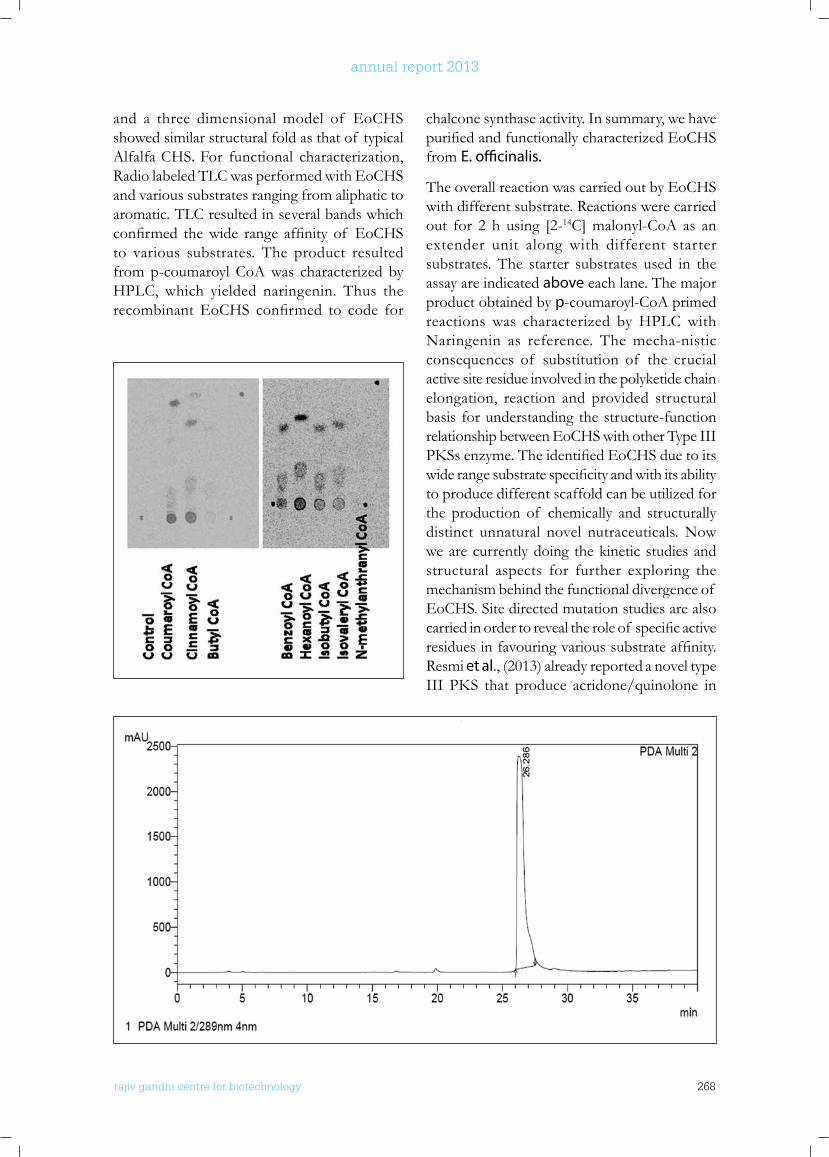
rajiv gandhi centre for biotechnology 268
annual report 2013
and a three dimensional model of EoCHS showed similar structural fold as that of typical Alfalfa CHS. For functional characterization, Radio labeled TLC was performed with EoCHS and various substrates ranging from aliphatic to aromatic. TLC resulted in several bands which confirmed thewide range affinity of EoCHSto various substrates. The product resulted from p-coumaroyl CoA was characterized by HPLC, which yielded naringenin. Thus the recombinantEoCHS confirmed to code for
chalcone synthase activity. In summary, we have purifiedandfunctionallycharacterizedEoCHSfrom E. officinalis.
The overall reaction was carried out by EoCHS with different substrate. Reactions were carried out for 2 h using [2-14C] malonyl-CoA as an extender unit along with different starter substrates. The starter substrates used in the assay are indicated above each lane. The major product obtained by p-coumaroyl-CoA primed reactions was characterized by HPLC with Naringenin as reference. The mecha-nistic consequences of substitution of the crucial active site residue involved in the polyketide chain elongation, reaction and provided structural basis for understanding the structure-function relationship between EoCHS with other Type III PKSsenzyme.TheidentifiedEoCHSduetoitswiderangesubstratespecificityandwithitsabilityto produce different scaffold can be utilized for the production of chemically and structurally distinct unnatural novel nutraceuticals. Now we are currently doing the kinetic studies and structural aspects for further exploring the mechanism behind the functional divergence of EoCHS. Site directed mutation studies are also carriedinordertorevealtheroleof specificactiveresiduesinfavouringvarioussubstrateaffinity.Resmi et al., (2013) already reported a novel type III PKS that produce acridone/quinolone in
rajiv gandhi centre for biotechnology 269
annual report 2013
catalytic and structural importance of active site residues, as predicted by our structural model, was investigated by performing site-directed mutagenesis. The modelling and mutagenesis studies provide an insight into the structural mechanism for the enzyme that could be used to generate pharmaceutically important products. Based on these studies, we are currently using computational and structural biology aspects to elucidate the structural mechanisms behind the functional behaviour of the system.
PublICATIonS
• Vivek PJ, Tuteja N, Soniya EV (2013). CDPK1 from ginger promotes salinity and drought stress tolerance without yield penalty by improving growth and photosynthesis in Nicotiana tabacum. PLoS One. Oct 23; 8 (10):e76392.
• Remakanthan, Tara G Menon and E V Soniya (2013). Somatic embryogenesis in banana (Musa acuminata AAA cv. Grand Naine): Effect of explant and culture conditions. In Vitro Cellular and Developmental Biology - Plant. DOI: 10.1007/s 11627-013-9546-4.
ConfEREnCE PRESEnTATIonS
• Asha S and E V Soniya. ‘Transcriptome-wide identification and characterization of Micro RNAs and their targets from black pepper ’. The Plant Genomics Congress Asia held at Kuala Lumpur, Malaysia on 24-25 February, 2014.
• Mallika V, Aiswarya G and EV Soniya “In silico characterization and molecular evolutionary analysis of Type III polyketide synthases from Zingiberaceae” Plant Genomics Congress Asia held in Kuala Lumpur, Malaysia. February 24-25, 2014.
• Asha S, Mallika V and EV Soniya “Identification of gene candidates in nitric oxide signalling pathways and their miRNA mediated regulation during Phytophthora capsici infection in black pepper (Outstanding Poster Award). ‘International Conference on Legacy of Nitric Oxide Discovery: Impact on Disease Biology’ held at Rajiv Gandhi Centre for Biotechnology, Thiruvananthapuram. November 5-6, 2013
• Aiswarya G, Lovely Jael P and EV Soniya “Role of nitric oxide in the acclimation of Type III Polyketide synthase gene in Zingiber officinale under wound stress”. ‘International Conference on Legacy of Nitric Oxide Discovery: Impact on Disease Biology’ held at Rajiv Gandhi Centre for
Biotechnology, Thiruvananthapuram. November 5-6, 2013.
AwARDS AnD HonouRS
• Asha S, Mallika V and E V Soniya “Identification of gene candidates in nitric oxide signalling pathways and their miRNA mediated regulation during Phytophthora capsici infection in black pepper (Outstanding Poster Award). ‘International Conference on Legacy of Nitric Oxide Discovery: Impact on Disease Biology’ held at Rajiv Gandhi Centre for Biotechnology, Thiruvananthapuram. November 5-6, 2013
Aegle marmelos (Rutaceae). The protein is named as quinolone synthase (QNS) and the reaction involves decarboxylative condensation of malonyl-CoA with N-methyl anthraniloyl-CoA to form an intermediate, which spontaneously
cyclise by amide formation to yield 4-hydroxy-2(1H)-quinolone. Kinetic analysis indicatedthatthecatalyticefficiencyof QNSproteintoaccept larger acyl-CoA substrate is several fold higher than that for smaller substrates. The
rajiv gandhi centre for biotechnology 270
annual report 2013
Plant Disease Biology & Biotechnology (PDBB) PDBB Laboratory - 4
Ph.D StudentsNisha NairJisha.SAnu KChidambareswaran M
r a j i v g a n d h i c e n t r e f o r b i o t e c h n o l o g y
Manjula is a Ph D in Botany from University of Kerala and joined RGCB in 2000.
Manjula S Ph.D Scientists E I
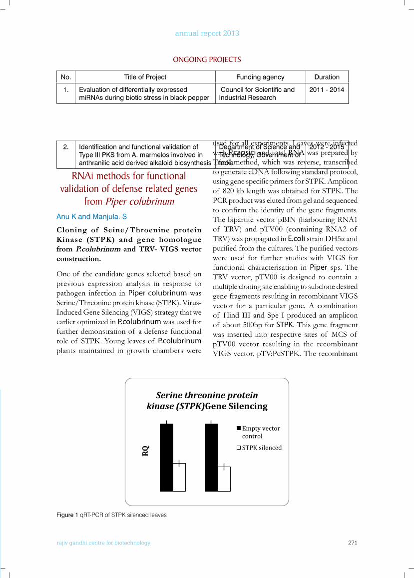
rajiv gandhi centre for biotechnology 271
annual report 2013
2. Identification and functional validation of Type III PKS from A. marmelos involved in anthranilic acid derived alkaloid biosynthesis
Department of Science and Technology, Government of India
2012 - 2015
RnAi methods for functional validation of defense related genes
from Piper colubrinumAnu K and Manjula. S
Cloning of Seine/Throenine protein Kinase (STPK) and gene homologue from P.colubrinum and TRV- VIGS vector construction.
One of the candidate genes selected based on previous expression analysis in response to pathogen infection in Piper colubrinum was Serine/Threonineproteinkinase(STPK).Virus-Induced Gene Silencing (VIGS) strategy that we earlier optimized in P.colubrinum was used for further demonstration of a defense functional roleof STPK.Youngleavesof P.colubrinum plants maintained in growth chambers were
used for all experiments. Leaves were infected with P.capsici and total RNA was prepared by Trizol method, which was reverse, transcribed to generate cDNA following standard protocol, usinggenespecificprimersforSTPK.Ampliconof 820kblengthwasobtainedforSTPK.ThePCR product was eluted from gel and sequenced toconfirmtheidentityof thegenefragments.The bipartite vector pBIN (harbouring RNA1 of TRV) and pTV00 (containing RNA2 of TRV) was propagated in E.colistrainDH5αandpurifiedfromthecultures.Thepurifiedvectorswere used for further studies with VIGS for functional characterisation in Piper sps. The TRV vector, pTV00 is designed to contain a multiple cloning site enabling to subclone desired gene fragments resulting in recombinant VIGS vector for a particular gene. A combination of Hind III and Spe I produced an amplicon of about 500bp for STPK. This gene fragment was inserted into respective sites of MCS of pTV00 vector resulting in the recombinant VIGSvector,pTV:PcSTPK.Therecombinant
onGoInG PRoJECTS
No. Title of Project Funding agency Duration
1. Evaluation of differentially expressed miRNAs during biotic stress in black pepper
Council for Scientific and Industrial Research
2011 - 2014
rajiv gandhi center forbiotechnology24
2013-14 Annual Report
Figure-2 RT-PCR analysis for confirming the movement of TRV viral particles in leaves of
P.colubrinum.
Figure-3 Pathogen infection studies
Empty vector control STPK down regulated
24 h 24h
RQ
Serine threonine protein kinase (STPK)Gene Silencing
Empty vector control
STPK silenced
M + 1 2 3 4 5 6 7 8 M – 100bp ladder
+ - positive control
1-8- RNA 2 amplification from infiltrated leaves after 30 days of infection
Figure 1 qRT-PCR of STPK silenced leaves
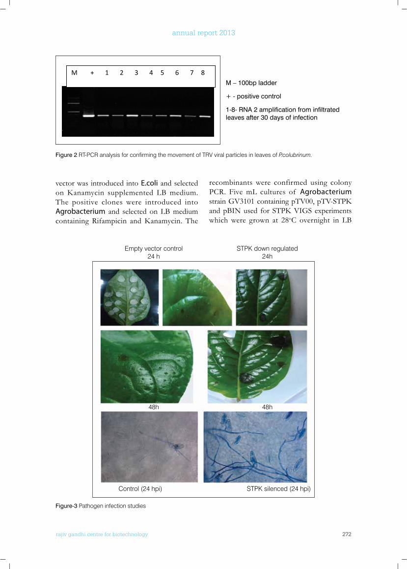
rajiv gandhi centre for biotechnology 272
annual report 2013
vector was introduced into E.coli and selected onKanamycin supplemented LBmedium.The positive clones were introduced into Agrobacterium and selected on LB medium containing Rifampicin andKanamycin. The
recombinants were confirmed using colony PCR. Five mL cultures of Agrobacterium strainGV3101containingpTV00,pTV-STPKandpBINused forSTPKVIGSexperimentswhich were grown at 28oC overnight in LB
rajiv gandhi center forbiotechnology24
2013-14 Annual Report
Figure-2 RT-PCR analysis for confirming the movement of TRV viral particles in leaves of
P.colubrinum.
Figure-3 Pathogen infection studies
Empty vector control STPK down regulated
24 h 24h
RQ
Serine threonine protein kinase (STPK)Gene Silencing
Empty vector control
STPK silenced
M + 1 2 3 4 5 6 7 8 M – 100bp ladder
+ - positive control
1-8- RNA 2 amplification from infiltrated leaves after 30 days of infection
M – 100bp ladder
+ - positive control
1-8- RNA 2 amplification from infiltrated leaves after 30 days of infection
Figure 2 RT-PCR analysis for confirming the movement of TRV viral particles in leaves of P.colubrinum.
Empty vector control STPK down regulated 24 h 24h
48h 48h
Figure-3 Pathogen infection studies
Control (24 hpi) STPK silenced (24 hpi)
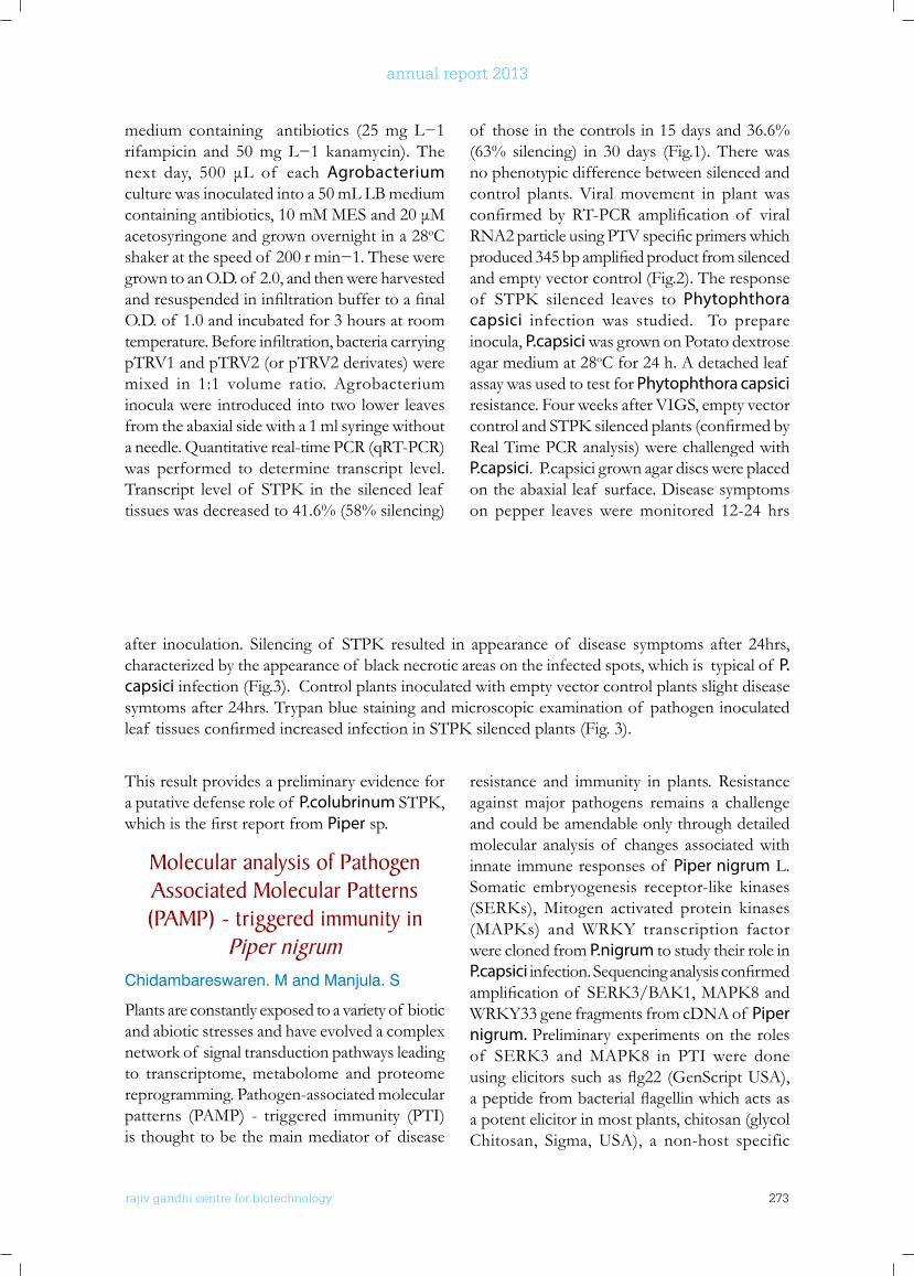
rajiv gandhi centre for biotechnology 273
annual report 2013
medium containing antibiotics (25mgL−1rifampicin and 50mgL−1 kanamycin). Thenext day, 500 μL of eachAgrobacterium culture was inoculated into a 50 mL LB medium containingantibiotics,10mMMESand20μMacetosyringone and grown overnight in a 28oC shakeratthespeedof 200rmin−1.Theseweregrown to an O.D. of 2.0, and then were harvested andresuspendedininfiltrationbuffertoafinalO.D. of 1.0 and incubated for 3 hours at room temperature.Beforeinfiltration,bacteriacarryingpTRV1 and pTRV2 (or pTRV2 derivates) were mixed in 1:1 volume ratio. Agrobacterium inocula were introduced into two lower leaves from the abaxial side with a 1 ml syringe without a needle. Quantitative real-time PCR (qRT-PCR) was performed to determine transcript level. Transcript levelof STPK in the silenced leaf tissues was decreased to 41.6% (58% silencing)
of those in the controls in 15 days and 36.6% (63% silencing) in 30 days (Fig.1). There was no phenotypic difference between silenced and control plants. Viral movement in plant was confirmed byRT-PCR amplification of viralRNA2particleusingPTVspecificprimerswhichproduced345bpamplifiedproductfromsilencedand empty vector control (Fig.2). The response of STPK silenced leaves toPhytophthora capsici infection was studied. To prepare inocula, P.capsici was grown on Potato dextrose agar medium at 28oC for 24 h. A detached leaf assay was used to test for Phytophthora capsici resistance. Four weeks after VIGS, empty vector controlandSTPKsilencedplants(confirmedbyReal Time PCR analysis) were challenged with P.capsici. P.capsici grown agar discs were placed on the abaxial leaf surface. Disease symptoms on pepper leaves were monitored 12-24 hrs
after inoculation. Silencing of STPK resulted in appearance of disease symptoms after 24hrs,characterized by the appearance of black necrotic areas on the infected spots, which is typical of P. capsici infection (Fig.3). Control plants inoculated with empty vector control plants slight disease symtoms after 24hrs. Trypan blue staining and microscopic examination of pathogen inoculated leaf tissuesconfirmedincreasedinfectioninSTPKsilencedplants(Fig.3).
This result provides a preliminary evidence for a putative defense role of P.colubrinumSTPK,whichisthefirstreportfromPiper sp.
Molecular analysis of Pathogen Associated Molecular Patterns (PAMP) - triggered immunity in
Piper nigrumChidambareswaren. M and Manjula. S
Plants are constantly exposed to a variety of biotic and abiotic stresses and have evolved a complex network of signal transduction pathways leading to transcriptome, metabolome and proteome reprogramming. Pathogen-associated molecular patterns (PAMP) - triggered immunity (PTI) is thought to be the main mediator of disease
resistance and immunity in plants. Resistance against major pathogens remains a challenge and could be amendable only through detailed molecular analysis of changes associated with innate immune responses of Piper nigrum L. Somatic embryogenesis receptor-like kinases (SERKs),Mitogen activated protein kinases(MAPKs) andWRKY transcription factorwere cloned from P.nigrum to study their role in P.capsiciinfection.Sequencinganalysisconfirmedamplificationof SERK3/BAK1,MAPK8andWRKY33genefragmentsfromcDNAof Piper nigrum. Preliminary experiments on the roles of SERK3 andMAPK8 in PTIwere doneusingelicitorssuchasflg22(GenScriptUSA),apeptidefrombacterialflagellinwhichactsasa potent elicitor in most plants, chitosan (glycol Chitosan, Sigma, USA), a non-host specific
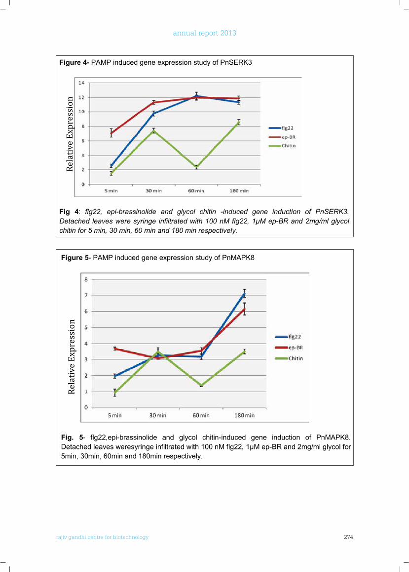
rajiv gandhi centre for biotechnology 274
annual report 2013
rajiv gandhi center forbiotechnology27
2013-14 Annual Report
Figure 4- PAMP induced gene expression study of PnSERK3
Fig 4: flg22, epi-brassinolide and glycol chitin -induced gene induction of PnSERK3. Detached leaves were syringe infiltrated with 100 nM flg22, 1μM ep-BR and 2mg/ml glycol chitin for 5 min, 30 min, 60 min and 180 min respectively.
Figure 5- PAMP induced gene expression study of PnMAPK8
Fig. 5- flg22,epi-brassinolide and glycol chitin-induced gene induction of PnMAPK8. Detached leaves weresyringe infiltrated with 100 nM flg22, 1μM ep-BR and 2mg/ml glycol for 5min, 30min, 60min and 180min respectively.
Rela
tive
Expr
essi
on
Rela
tive
Expr
essi
on

rajiv gandhi centre for biotechnology 275
annual report 2013
Laboratory Medicine & Molecular Diagostics (LMMD)
Senior Manager (Technical)Sanjai D
Post Doctoral FellowS. Dayakar
r a j i v g a n d h i c e n t r e f o r b i o t e c h n o l o g y
Radhakrishnan received his Ph.D from the University of Kerala working at the Regional Cancer Centre on Non Hodgkins Lymphoma.He subsequently served as senior faculty at various medical colleges including Manipal College of Medical Sciences, Oman University Medical College (Affiliated to the West Virginia University, USA), Sparten University, New York and the Atlantic University, New York. He joined RGCB in October 2011.
Radhakrishnan Nair Ph.D Scientist E I
ReceptionistV. Karthika
TechnologistsBinu Kumar DSreeja S Kannan T RJayalakshmi D
Project FellowAnjana S S
House KeepingSumaja V
RGCB Service Facilities
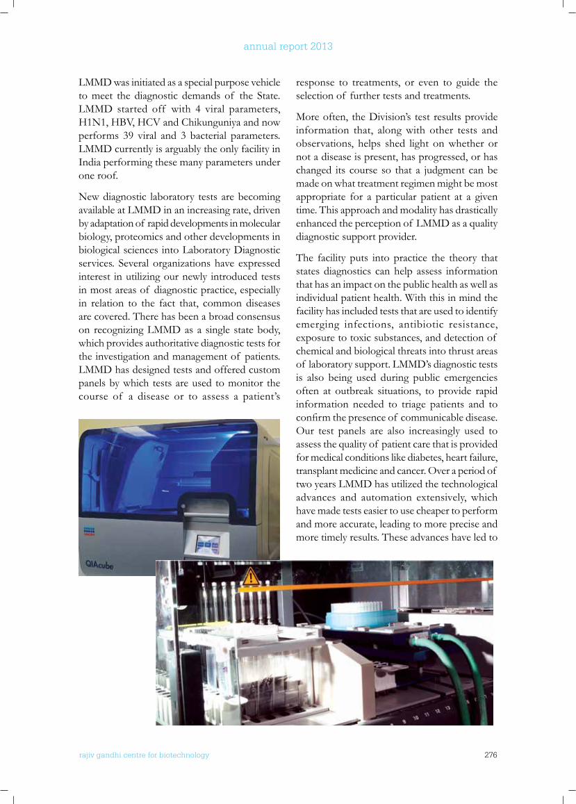
rajiv gandhi centre for biotechnology 276
annual report 2013
LMMD was initiated as a special purpose vehicle to meet the diagnostic demands of the State. LMMD started off with 4 viral parameters, H1N1, HBV, HCV and Chikunguniya and now performs 39 viral and 3 bacterial parameters. LMMD currently is arguably the only facility in India performing these many parameters under one roof.
New diagnostic laboratory tests are becoming available at LMMD in an increasing rate, driven by adaptation of rapid developments in molecular biology, proteomics and other developments in biological sciences into Laboratory Diagnostic services. Several organizations have expressed interest in utilizing our newly introduced tests in most areas of diagnostic practice, especially in relation to the fact that, common diseases are covered. There has been a broad consensus on recognizing LMMD as a single state body, which provides authoritative diagnostic tests for the investigation and management of patients. LMMD has designed tests and offered custom panels by which tests are used to monitor the course of a disease or to assess a patient’s
response to treatments, or even to guide the selection of further tests and treatments.
More often, the Division’s test results provide information that, along with other tests and observations, helps shed light on whether or not a disease is present, has progressed, or has changed its course so that a judgment can be made on what treatment regimen might be most appropriate for a particular patient at a given time. This approach and modality has drastically enhanced the perception of LMMD as a quality diagnostic support provider.
The facility puts into practice the theory that states diagnostics can help assess information that has an impact on the public health as well as individual patient health. With this in mind the facility has included tests that are used to identify emerging infections, antibiotic resistance, exposure to toxic substances, and detection of chemical and biological threats into thrust areas of laboratory support. LMMD’s diagnostic tests is also being used during public emergencies often at outbreak situations, to provide rapid information needed to triage patients and to confirmthepresenceof communicabledisease.Our test panels are also increasingly used to assess the quality of patient care that is provided for medical conditions like diabetes, heart failure, transplant medicine and cancer. Over a period of two years LMMD has utilized the technological advances and automation extensively, which have made tests easier to use cheaper to perform and more accurate, leading to more precise and more timely results. These advances have led to
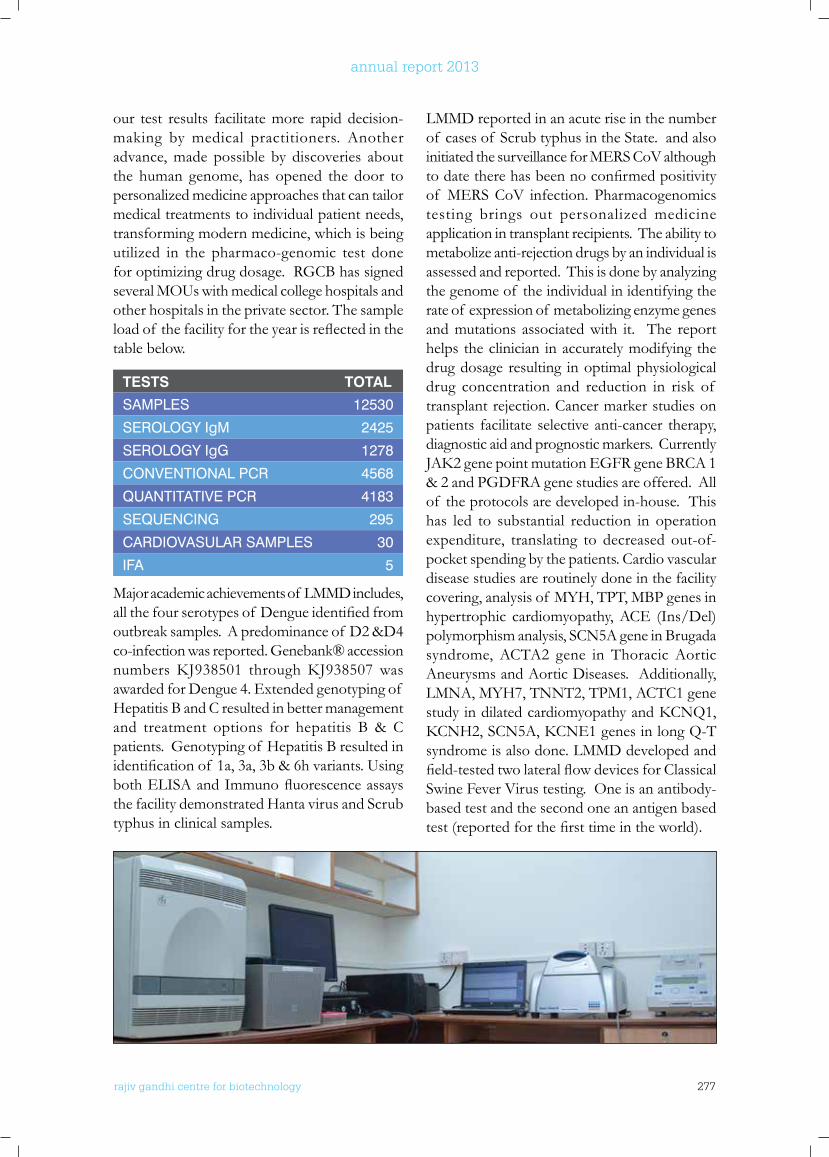
rajiv gandhi centre for biotechnology 277
annual report 2013
our test results facilitate more rapid decision-making by medical practitioners. Another advance, made possible by discoveries about the human genome, has opened the door to personalized medicine approaches that can tailor medical treatments to individual patient needs, transforming modern medicine, which is being utilized in the pharmaco-genomic test done for optimizing drug dosage. RGCB has signed several MOUs with medical college hospitals and other hospitals in the private sector. The sample loadof thefacilityfortheyearisreflectedinthetable below.
TeSTS ToTAL
SAMPLES 12530
SEROLOGY IgM 2425
SEROLOGY IgG 1278
CONVENTIONAL PCR 4568
QUANTITATIVE PCR 4183
SEQUENCING 295
CARDIOVASULAR SAMPLES 30
IFA 5
Major academic achievements of LMMD includes, allthefourserotypesof Dengueidentifiedfromoutbreak samples. A predominance of D2 &D4 co-infection was reported. Genebank® accession numbersKJ938501 throughKJ938507wasawarded for Dengue 4. Extended genotyping of Hepatitis B and C resulted in better management and treatment options for hepatitis B & C patients. Genotyping of Hepatitis B resulted in identificationof 1a,3a,3b&6hvariants.UsingbothELISAandImmunofluorescenceassaysthe facility demonstrated Hanta virus and Scrub typhus in clinical samples.
LMMD reported in an acute rise in the number of cases of Scrub typhus in the State. and also initiated the surveillance for MERS CoV although todatetherehasbeennoconfirmedpositivityof MERS CoV infection. Pharmacogenomics testing brings out personalized medicine application in transplant recipients. The ability to metabolize anti-rejection drugs by an individual is assessed and reported. This is done by analyzing the genome of the individual in identifying the rate of expression of metabolizing enzyme genes and mutations associated with it. The report helps the clinician in accurately modifying the drug dosage resulting in optimal physiological drug concentration and reduction in risk of transplant rejection. Cancer marker studies on patients facilitate selective anti-cancer therapy, diagnostic aid and prognostic markers. Currently JAK2genepointmutationEGFRgeneBRCA1& 2 and PGDFRA gene studies are offered. All of the protocols are developed in-house. This has led to substantial reduction in operation expenditure, translating to decreased out-of-pocket spending by the patients. Cardio vascular disease studies are routinely done in the facility covering, analysis of MYH, TPT, MBP genes in hypertrophic cardiomyopathy, ACE (Ins/Del) polymorphism analysis, SCN5A gene in Brugada syndrome, ACTA2 gene in Thoracic Aortic Aneurysms and Aortic Diseases. Additionally, LMNA, MYH7, TNNT2, TPM1, ACTC1 gene studyindilatedcardiomyopathyandKCNQ1,KCNH2,SCN5A,KCNE1genesinlongQ-Tsyndrome is also done. LMMD developed and field-testedtwolateralflowdevicesforClassicalSwine Fever Virus testing. One is an antibody-based test and the second one an antigen based test(reportedforthefirsttimeintheworld).
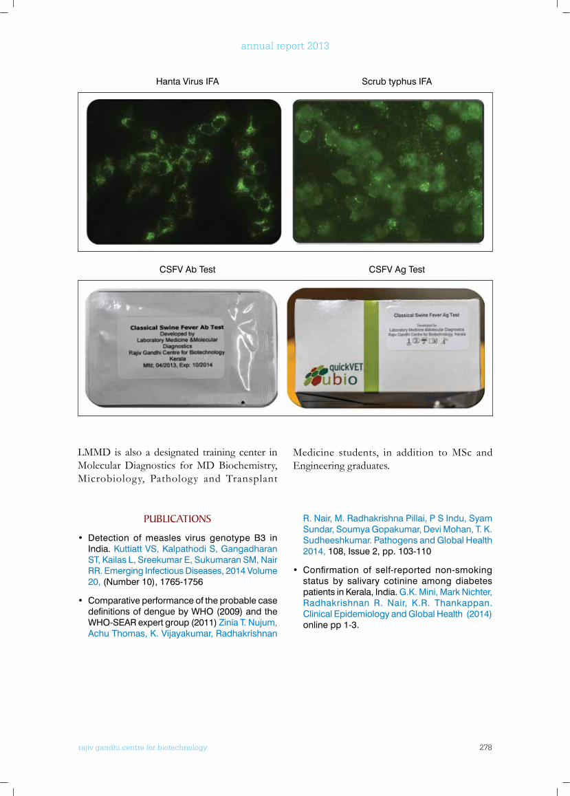
rajiv gandhi centre for biotechnology 278
annual report 2013
LMMD is also a designated training center in Molecular Diagnostics for MD Biochemistry, Microbiology, Pathology and Transplant
Medicine students, in addition to MSc and Engineering graduates.
Hanta Virus IFA Scrub typhus IFA
CSFV Ab Test CSFV Ag Test
PublICATIonS
• Detection of measles virus genotype B3 in India. Kuttiatt VS, Kalpathodi S, Gangadharan ST, Kailas L, Sreekumar E, Sukumaran SM, Nair RR. Emerging Infectious Diseases, 2014 Volume 20, (Number 10), 1765-1756
• Comparative performance of the probable case definitions of dengue by WHO (2009) and the WHO-SEAR expert group (2011) Zinia T. Nujum, Achu Thomas, K. Vijayakumar, Radhakrishnan
R. Nair, M. Radhakrishna Pillai, P S Indu, Syam Sundar, Soumya Gopakumar, Devi Mohan, T. K. Sudheeshkumar. Pathogens and Global Health 2014, 108, Issue 2, pp. 103-110
• Confirmation of self-reported non-smoking status by salivary cotinine among diabetes patients in Kerala, India. G.K. Mini, Mark Nichter, Radhakrishnan R. Nair, K.R. Thankappan. Clinical Epidemiology and Global Health (2014) online pp 1-3.
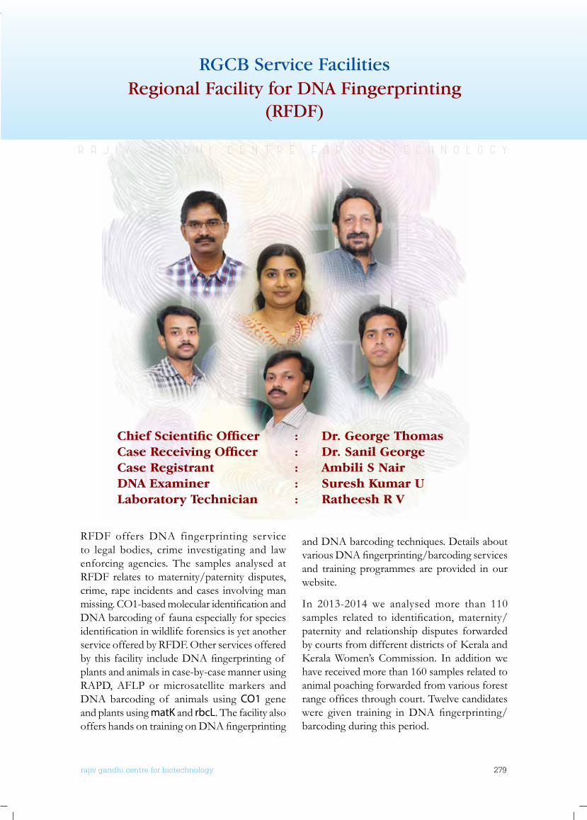
rajiv gandhi centre for biotechnology 279
annual report 2013
RFDF offers DNA fingerprinting service to legal bodies, crime investigating and law enforcing agencies. The samples analysed at RFDF relates to maternity/paternity disputes, crime, rape incidents and cases involving man missing.CO1-basedmolecularidentificationandDNA barcoding of fauna especially for species identificationinwildlifeforensicsisyetanotherservice offered by RFDF. Other services offered bythisfacility includeDNAfingerprintingof plants and animals in case-by-case manner using RAPD, AFLP or microsatellite markers and DNA barcoding of animals using CO1 gene and plants using matK and rbcL. The facility also offershandsontrainingonDNAfingerprinting
Regional Facility for DNA Fingerprinting(RFDF)
RGCB Service Facilities
Chief Scientific Officer : Dr. George ThomasCase Receiving Officer : Dr. Sanil GeorgeCase Registrant : Ambili S NairDNA Examiner : Suresh Kumar ULaboratory Technician : Ratheesh R V
and DNA barcoding techniques. Details about variousDNAfingerprinting/barcodingservicesand training programmes are provided in our website.
In 2013-2014 we analysed more than 110 samples related to identification,maternity/paternity and relationship disputes forwarded bycourtsfromdifferentdistrictsof KeralaandKeralaWomen’sCommission. In additionwehave received more than 160 samples related to animal poaching forwarded from various forest rangeofficesthroughcourt.Twelvecandidateswere given training inDNAfingerprinting/barcoding during this period.
r a j i v g a n d h i c e n t r e f o r b i o t e c h n o l o g y
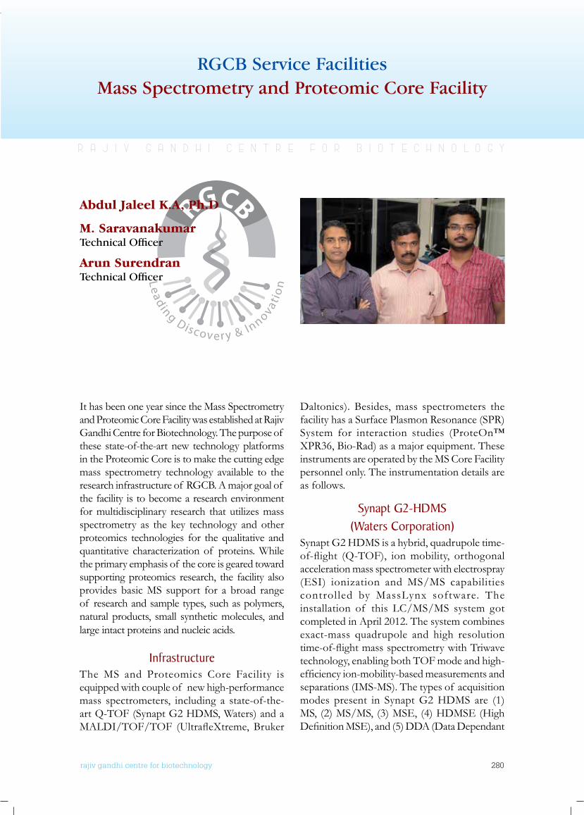
rajiv gandhi centre for biotechnology 280
annual report 2013
r a j i v g a n d h i c e n t r e f o r b i o t e c h n o l o g y
Abdul Jaleel K.A, Ph.D
M. SaravanakumarTechnical Officer
Arun SurendranTechnical Officer
Mass Spectrometry and Proteomic Core FacilityRGCB Service Facilities
It has been one year since the Mass Spectrometry and Proteomic Core Facility was established at Rajiv Gandhi Centre for Biotechnology. The purpose of these state-of-the-art new technology platforms in the Proteomic Core is to make the cutting edge mass spectrometry technology available to the research infrastructure of RGCB. A major goal of the facility is to become a research environment for multidisciplinary research that utilizes mass spectrometry as the key technology and other proteomics technologies for the qualitative and quantitative characterization of proteins. While the primary emphasis of the core is geared toward supporting proteomics research, the facility also provides basic MS support for a broad range of research and sample types, such as polymers, natural products, small synthetic molecules, and large intact proteins and nucleic acids.
InfrastructureThe MS and Proteomics Core Facility is equipped with couple of new high-performance mass spectrometers, including a state-of-the-art Q-TOF (Synapt G2 HDMS, Waters) and a MALDI/TOF/TOF (UltrafleXtreme,Bruker
Daltonics). Besides, mass spectrometers the facility has a Surface Plasmon Resonance (SPR) System for interaction studies (ProteOn™ XPR36, Bio-Rad) as a major equipment. These instruments are operated by the MS Core Facility personnel only. The instrumentation details are as follows.
Synapt G2-HDMS (waters Corporation)
Synapt G2 HDMS is a hybrid, quadrupole time-of-flight (Q-TOF), ionmobility, orthogonalacceleration mass spectrometer with electrospray (ESI) ionization and MS/MS capabilities controlled by MassLynx software. The installation of this LC/MS/MS system got completed in April 2012. The system combines exact-mass quadrupole and high resolution time-of-flightmassspectrometrywithTriwavetechnology, enabling both TOF mode and high-efficiencyion-mobility-basedmeasurementsandseparations (IMS-MS). The types of acquisition modes present in Synapt G2 HDMS are (1) MS, (2) MS/MS, (3) MSE, (4) HDMSE (High DefinitionMSE),and(5)DDA(DataDependant
rajiv gandhi centre for biotechnology 281
annual report 2013
Analysis) mode. In addition to the standard NanoLockSpray dual electrospray ion source, we also have a TRIZAIC UPLC source with nanoTile technology as the electrospary source for the Synapt G2. Both features LockSpray system for the optimized co-introduction of analyte and lock mass compounds directly into the ion source offering a superior alternative for the acquisition of exact mass data.
The Synapt G2 HDMS is equipped or supplemented with two nano LC systems. A 1D NanoACQUITY UPLC and the other a 2D NanoACQUITY UPLC, both from Waters Corporation. The nanoACQUITY Ultra Performance LC (UPLC) System is designed for nano-scale, capillary, and narrow-bore separations to attain the highest chromatographic resolution, sensitivity, and reproducibility. These systems provide the best technology for the separation and delivery of peptides to the MS for proteomics applications, for protein identificationandcharacterization.Thesystem’s10,000 psi operating pressure capability allows for superior high-peak capacity separations by operating longer columns packed with sub-2 micron particles. It is optimized for high-resolutionidentificationand2D-LCseparationsatprecisenano-flowrates.Thisinnovative2Dsystem effectively uses two-dimensional (2D) UPLC for better chromatographic resolution of complex proteomic samples by using a dual reversed-phase (RP) approach. The entire LC/MS/MS system is operated my MassLynx software and data analysis is performed by ProteinLynx Global Server (PLGS) version 2.5.3.
ultraflextreme (bruker Daltonics)In May 2012, a new MALDI/TOF/TOF mass spectrometer (UltrafleXtreme, Bruker Daltonics) was installed. The instrument has MS and MS/MS capability with high resolution and high mass accuracy. The instrument has laser-induced dissociation (LID) fragmentation for denovo peptide sequencing, top-down protein sequencing, and other applications, as well as high-energy CID fragmentation. The UltrafleXtreme is a high-performance instrument well suited for a wide range of applications, including polymers, proteins,
peptides, carbohydrates, and other biomolecules.
UltrafleXtremeissupplementedwithananoLC(EASY-nLC II, Bruker) and MALDI spotter (Proteineer fcII, Bruker). The EASY-nLC II is a nano-flowHPLC system tailored to therequirements of proteomics applications. This LC system is used to separate protein and peptide mixtures for MALDI MS. The system is used in combination with a MALDI spotter. Proteineer fc II is a MALDI spotter which enables automatic liquid handling for MALDI preparation of LC separated peptide fractions. It is designed to deposit fractions eluting from a NanoLC column (Easy-nLC II ) onto a MALDI plate, with the MALDI matrix automatically beingaddedforofflineMSandMS/MSanalysis.MALDI system is operated by Flex Control and Flex Analysis software, and the search engine Mascot is used for proteomics analysis.
Proteon™ xPR36 Surface Plasmon Resonance System (bio-Rad)
The ProteOn™ XPR36 protein interaction array system monitors in a label-free manner the interaction of biomolecules in real-time using surface plasmon resonance (SPR) technology. Interactions are monitored over time by detectingthebindingof ananalyteflowinginamicrofluidicchanneltoaligandimmobilizedon a sensor chip. This system generates a 6 x 6 interaction array for the simultaneous analysis of up to six ligands with up to six analytes. Interactions such as Protein- protein, Protein –peptide, Protein- small molecule, & Protein –DNAcanbecarriedoutusingspecificchips.The system applications are primarily analyte screening, kinetic analysis, equilibrium analysis, and concentration determination.
ServicesThe primary focus of the facility is to provide mass spectrometry services and consultation for the research and academic community at RGCB. The facility started accepting a broad range of research applications and sample types for analysis. The details of the services provided at present are given in the table below.
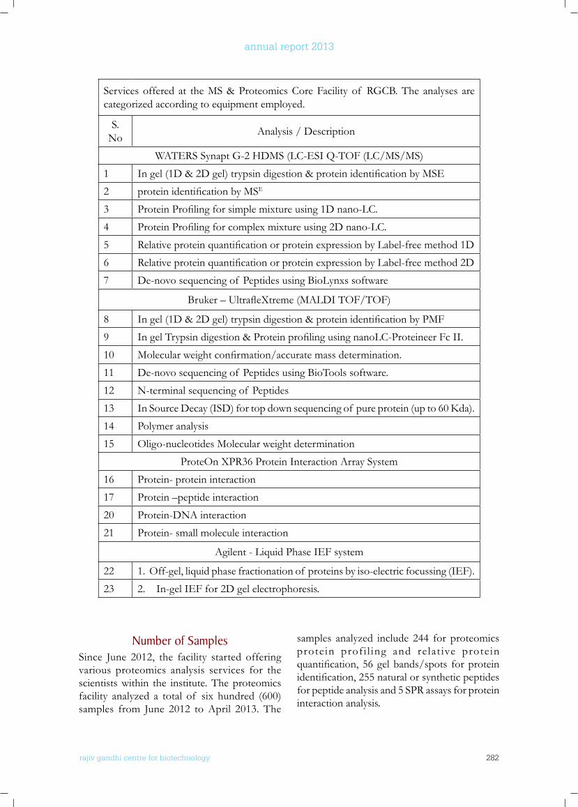
rajiv gandhi centre for biotechnology 282
annual report 2013
Services offered at the MS & Proteomics Core Facility of RGCB. The analyses are categorized according to equipment employed.
S. No Analysis / Description
WATERS Synapt G-2 HDMS (LC-ESI Q-TOF (LC/MS/MS)
1 Ingel(1D&2Dgel)trypsindigestion&proteinidentificationbyMSE
2 proteinidentificationbyMSE
3 ProteinProfilingforsimplemixtureusing1Dnano-LC.
4 ProteinProfilingforcomplexmixtureusing2Dnano-LC.
5 RelativeproteinquantificationorproteinexpressionbyLabel-freemethod1D
6 RelativeproteinquantificationorproteinexpressionbyLabel-freemethod2D
7 De-novo sequencing of Peptides using BioLynxs software
Bruker–UltrafleXtreme(MALDITOF/TOF)
8 Ingel(1D&2Dgel)trypsindigestion&proteinidentificationbyPMF
9 IngelTrypsindigestion&ProteinprofilingusingnanoLC-ProteineerFcII.
10 Molecularweightconfirmation/accuratemassdetermination.
11 De-novo sequencing of Peptides using BioTools software.
12 N-terminal sequencing of Peptides
13 InSourceDecay(ISD)fortopdownsequencingof pureprotein(upto60Kda).
14 Polymer analysis
15 Oligo-nucleotides Molecular weight determination
ProteOn XPR36 Protein Interaction Array System
16 Protein- protein interaction
17 Protein –peptide interaction
20 Protein-DNA interaction
21 Protein- small molecule interaction
Agilent - Liquid Phase IEF system
22 1. Off-gel, liquid phase fractionation of proteins by iso-electric focussing (IEF).
23 2. In-gel IEF for 2D gel electrophoresis.
number of SamplesSince June 2012, the facility started offering various proteomics analysis services for the scientists within the institute. The proteomics facility analyzed a total of six hundred (600) samples from June 2012 to April 2013. The
samples analyzed include 244 for proteomics protein profi l ing and relat ive protein quantification,56gelbands/spots forproteinidentification,255naturalorsyntheticpeptidesfor peptide analysis and 5 SPR assays for protein interaction analysis.
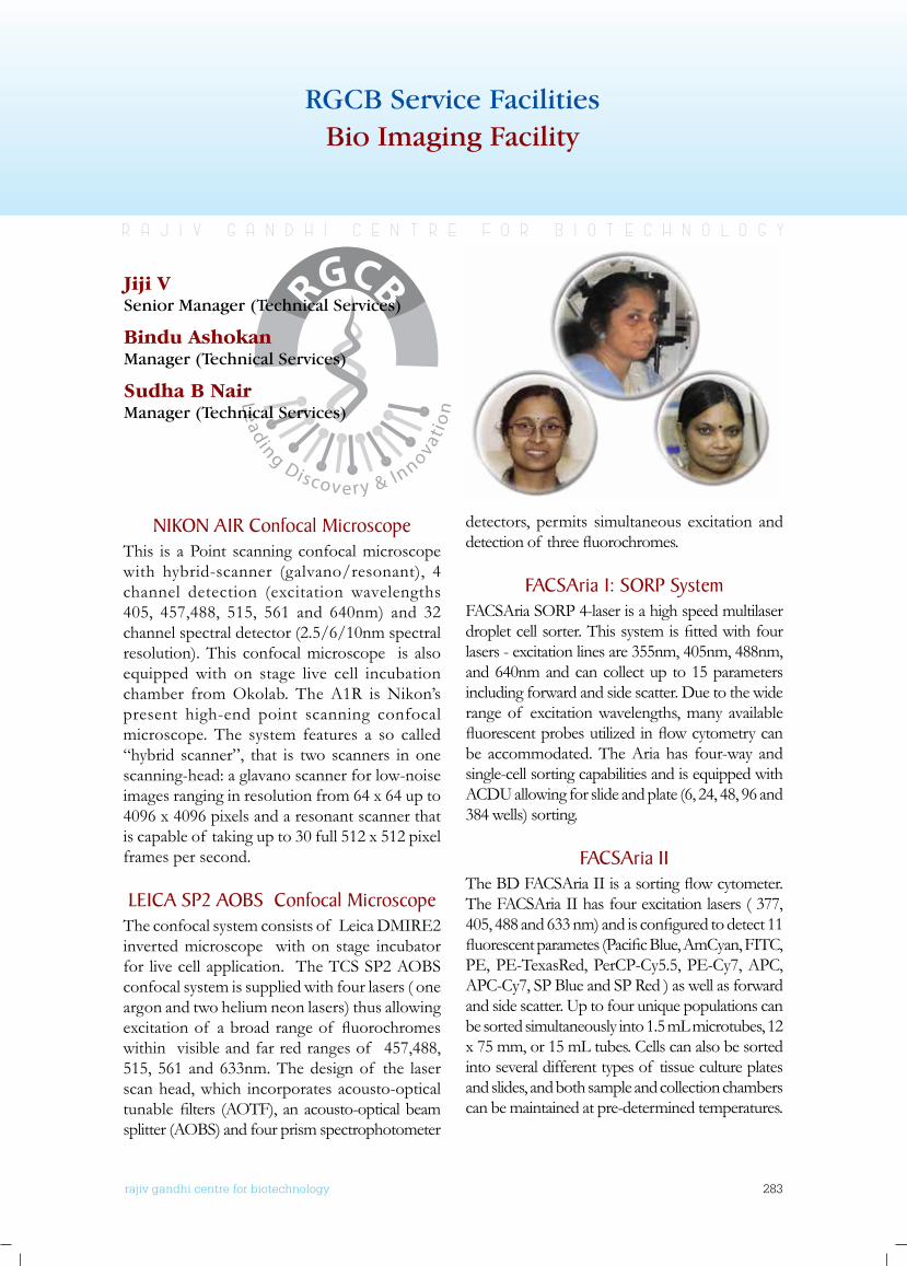
rajiv gandhi centre for biotechnology 283
annual report 2013
Bio Imaging FacilityRGCB Service Facilities
nIkon A1R Confocal Microscope This is a Point scanning confocal microscope with hybrid-scanner (galvano/resonant), 4 channel detection (excitation wavelengths 405, 457,488, 515, 561 and 640nm) and 32 channel spectral detector (2.5/6/10nm spectral resolution). This confocal microscope is also equipped with on stage live cell incubation chamber from Okolab. The A1R is Nikon’s present high-end point scanning confocal microscope. The system features a so called “hybrid scanner”, that is two scanners in one scanning-head: a glavano scanner for low-noise images ranging in resolution from 64 x 64 up to 4096 x 4096 pixels and a resonant scanner that is capable of taking up to 30 full 512 x 512 pixel frames per second.
lEICA SP2 AobS Confocal MicroscopeThe confocal system consists of Leica DMIRE2 inverted microscope with on stage incubator for live cell application. The TCS SP2 AOBS confocal system is supplied with four lasers ( one argon and two helium neon lasers) thus allowing excitationof abroadrangeof fluorochromeswithin visible and far red ranges of 457,488, 515, 561 and 633nm. The design of the laser scan head, which incorporates acousto-optical tunablefilters (AOTF),anacousto-opticalbeamsplitter (AOBS) and four prism spectrophotometer
detectors, permits simultaneous excitation and detectionof threefluorochromes.
fACSAria 1: SoRP SystemFACSAria SORP 4-laser is a high speed multilaser dropletcellsorter.Thissystemisfittedwithfourlasers - excitation lines are 355nm, 405nm, 488nm, and 640nm and can collect up to 15 parameters including forward and side scatter. Due to the wide range of excitation wavelengths, many available fluorescentprobesutilizedinflowcytometrycanbe accommodated. The Aria has four-way and single-cell sorting capabilities and is equipped with ACDU allowing for slide and plate (6, 24, 48, 96 and 384 wells) sorting.
fACSAria IITheBDFACSAriaIIisasortingflowcytometer.The FACSAria II has four excitation lasers ( 377, 405,488and633nm)andisconfiguredtodetect11fluorescentparametes(PacificBlue,AmCyan,FITC,PE, PE-TexasRed, PerCP-Cy5.5, PE-Cy7, APC, APC-Cy7, SP Blue and SP Red ) as well as forward and side scatter. Up to four unique populations can be sorted simultaneously into 1.5 mL microtubes, 12 x 75 mm, or 15 mL tubes. Cells can also be sorted into several different types of tissue culture plates and slides, and both sample and collection chambers can be maintained at pre-determined temperatures.
Jiji VSenior Manager (Technical Services)
Bindu AshokanManager (Technical Services)
Sudha B NairManager (Technical Services)
r a j i v g a n d h i c e n t r e f o r b i o t e c h n o l o g y
rajiv gandhi centre for biotechnology 284
annual report 2013
Instrumentation Engineering
r a j i v g a n d h i c e n t r e f o r b i o t e c h n o l o g y
Staff MembersRajasekharan KRahul C S NairSajan I XS Rajeev
Shaj Upendran General Manager
S Ajith KumarK Manoj KumarV Prem KumarV ShajiA S Vijayakumar
rajiv gandhi centre for biotechnology 285
annual report 2013
Rajiv Gandhi Centre for Biotechnology houses a large number of molecular biology and biotechnological research instruments. The Instrumentation Engineering Division is responsible for the installation, maintenance and repair of sophisticated research instruments in RGCB as well as the maintenance of Central instrumentation facility.Some of the sophisticated instruments installed during the year includeNext Generation Sequencing Systems (NGS) (Ion Proton Torrent System – Life Technologies & Ion Proton Personal Genomic Machine – Life Technologies ), GeneChip Scanner – Affymatrix, DNA sequencer (96 capillary)- Applied Biosystems (ABI), Nucleic Acid Extraction System - Beckman, Super Continnum White Laser Confocal Laser Scanning Microscope - Leica Microsystems , Stuctured Illumination Microscope with Confocal Laser Scanning System – Nikon, Invivo Animal Imager – Xenogen, FACS ARIA III Flow Cytometer - BD Biosciences, Fully Automated Liquid Handling System – Beckman Coulter, Non-Invasive Blood Pr. Monitor with ECG for animal facility – Iworks/IITC, Sample preparation System for NGS, Ion OneTouch – Life Technologies
The Division also maintains a well equipped engineering workshop with facilities required for the repair and calibration of the sophisticated instruments as its part. Repair up to the PCB level is done here reducing the downtime and repair costs. By attending to many essential repair works of instruments, dependence on expensive maintenance contracts with dealers has been reduced. The instrumentation Division also carriesoutdesign,modification,andfabricationof research instruments.
During the year, problems of Liquid Nitrogen Plant,FACS ARIA Flow Cytometer, Confocal Laser Scanning Microscope, Spectrophotometers, Ultra Centrifuges , High speed Centrifuges, Table Top Centrifuges, Gel Documentation systems, Transmission Electron Microscope, Upright and Inverted Microscopes , PCR machines, Electronic balances, Speed Vac Concentrator, CO2 Incubators, HPLC, Freeze Dryers ,
Microplate Washer etc,. have been repaired successfully.
The Instrumentation Division also maintains the Centralized Instrumentation facilities, Computers, PC based security surveillance system, Biometrics Time Attendance recorders, Conferencing facilities, Communication systems, Liquid Nitrogen Plant, Incinerator, Auditoriums, Convention Centre etc. It also carries out the supervision of 11KV electrical substation,340tonACplant,750KVA&1010KVADGsets. All minor electrical works required by the scientificstaff alsocarriesoutbytheinhouseengineering department.
Engineering division has been involved in setting up of new labs in BIO-INNOVTION CENTREatKazhakuttamKINFAcampus.Thisincludes the planning and design of Electrical works, Air-conditioning, furniture design etc.
Centralized Instrumentation facilityIn addition to the basic facilities available in all the research laboratories, we have a centralized core facility equipped with several minor and major equipments to cater the requirements of our research personnel. The following are the facilities available in the core facility:
SpectroscopyMALDI TOF/TOF Mass Spectrometer - BrukerBench top MALDI TOF Mass Spectrometer - ShimadzuSpectrophotometer - Perkin Elmer, Thermo, LabomedLuminescence Spectrometer - Perkin ElmerMultimode Plate Reader - TecanFTIR Spectrometer - Thermo
Genomics and ProteomicsNext Generation Sequencing Systems (NGS)Ion Proton Torrent System - Life TechnologiesIon Proton Personal Genomic Machine - Life TechnologiesGene Chip Scanner - Affymatrix
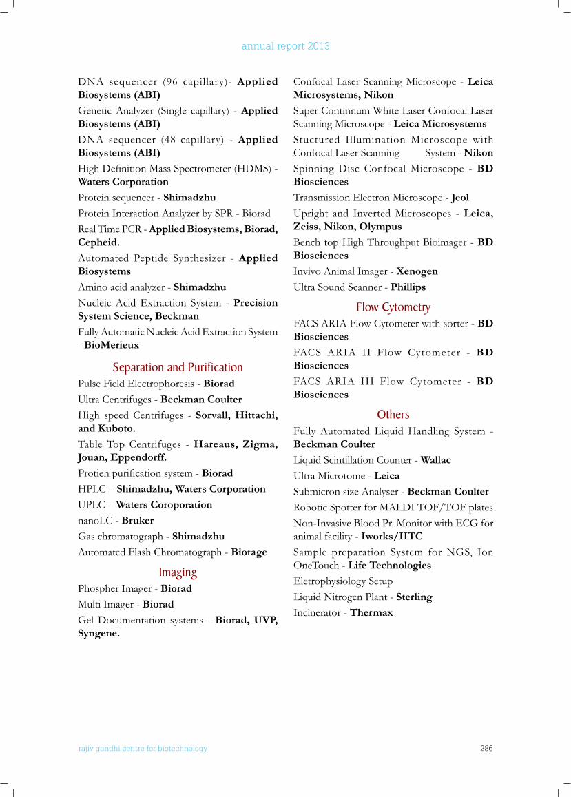
rajiv gandhi centre for biotechnology 286
annual report 2013
DNA sequencer (96 capillary)- Applied Biosystems (ABI)Genetic Analyzer (Single capillary) - Applied Biosystems (ABI)DNA sequencer (48 capillary) - Applied Biosystems (ABI)HighDefinitionMassSpectrometer(HDMS)-Waters CorporationProtein sequencer - ShimadzhuProtein Interaction Analyzer by SPR - BioradReal Time PCR - Applied Biosystems, Biorad, Cepheid.Automated Peptide Synthesizer - Applied BiosystemsAmino acid analyzer - ShimadzhuNucleic Acid Extraction System - Precision System Science, BeckmanFully Automatic Nucleic Acid Extraction System - BioMerieux
Separation and PurificationPulse Field Electrophoresis - BioradUltra Centrifuges - Beckman CoulterHigh speed Centrifuges - Sorvall, Hittachi, and Kuboto.Table Top Centrifuges - Hareaus, Zigma, Jouan, Eppendorff.Protienpurificationsystem- BioradHPLC – Shimadzhu, Waters CorporationUPLC – Waters CoroporationnanoLC - BrukerGas chromatograph - ShimadzhuAutomated Flash Chromatograph - Biotage
ImagingPhospher Imager - BioradMulti Imager - BioradGel Documentation systems - Biorad, UVP, Syngene.
Confocal Laser Scanning Microscope - Leica Microsystems, NikonSuper Continnum White Laser Confocal Laser Scanning Microscope - Leica MicrosystemsStuctured Illumination Microscope with Confocal Laser Scanning System - NikonSpinning Disc Confocal Microscope - BD BiosciencesTransmission Electron Microscope - JeolUpright and Inverted Microscopes - Leica, Zeiss, Nikon, OlympusBench top High Throughput Bioimager - BD BiosciencesInvivo Animal Imager - XenogenUltra Sound Scanner - Phillips
flow CytometryFACS ARIA Flow Cytometer with sorter - BD BiosciencesFACS ARIA II Flow Cytometer - BD BiosciencesFACS ARIA III Flow Cytometer - BD Biosciences
othersFully Automated Liquid Handling System - Beckman CoulterLiquid Scintillation Counter - WallacUltra Microtome - LeicaSubmicron size Analyser - Beckman CoulterRobotic Spotter for MALDI TOF/TOF platesNon-Invasive Blood Pr. Monitor with ECG for animal facility - Iworks/IITCSample preparation System for NGS, Ion OneTouch - Life TechnologiesEletrophysiology SetupLiquid Nitrogen Plant - SterlingIncinerator - Thermax
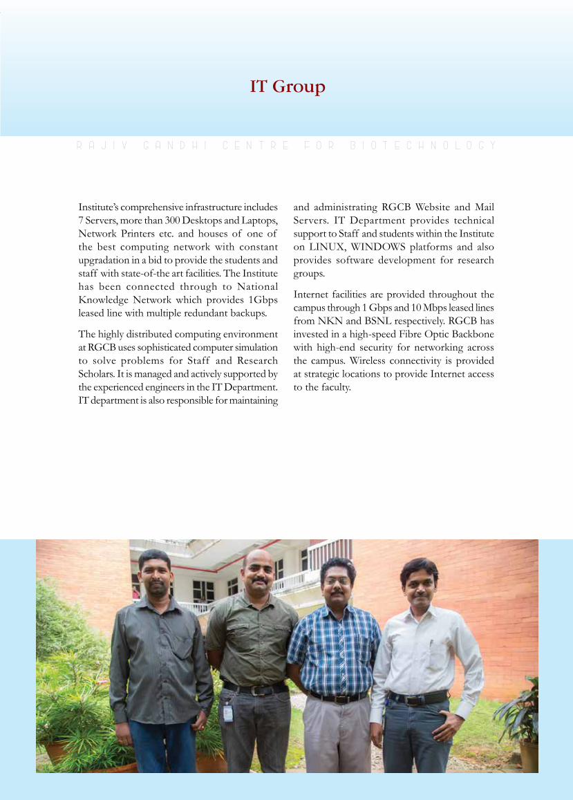
rajiv gandhi centre for biotechnology 287
annual report 2013
Institute’s comprehensive infrastructure includes 7 Servers, more than 300 Desktops and Laptops, Network Printers etc. and houses of one of the best computing network with constant upgradation in a bid to provide the students and staff with state-of-the art facilities. The Institute has been connected through to National KnowledgeNetworkwhich provides 1Gbpsleased line with multiple redundant backups.
The highly distributed computing environment at RGCB uses sophisticated computer simulation to solve problems for Staff and Research Scholars. It is managed and actively supported by the experienced engineers in the IT Department. IT department is also responsible for maintaining
and administrating RGCB Website and Mail Servers. IT Department provides technical support to Staff and students within the Institute on LINUX, WINDOWS platforms and also provides software development for research groups.
Internet facilities are provided throughout the campus through 1 Gbps and 10 Mbps leased lines fromNKNandBSNLrespectively.RGCBhasinvested in a high-speed Fibre Optic Backbone with high-end security for networking across the campus. Wireless connectivity is provided at strategic locations to provide Internet access to the faculty.
IT Group
r a j i v g a n d h i c e n t r e f o r b i o t e c h n o l o g y
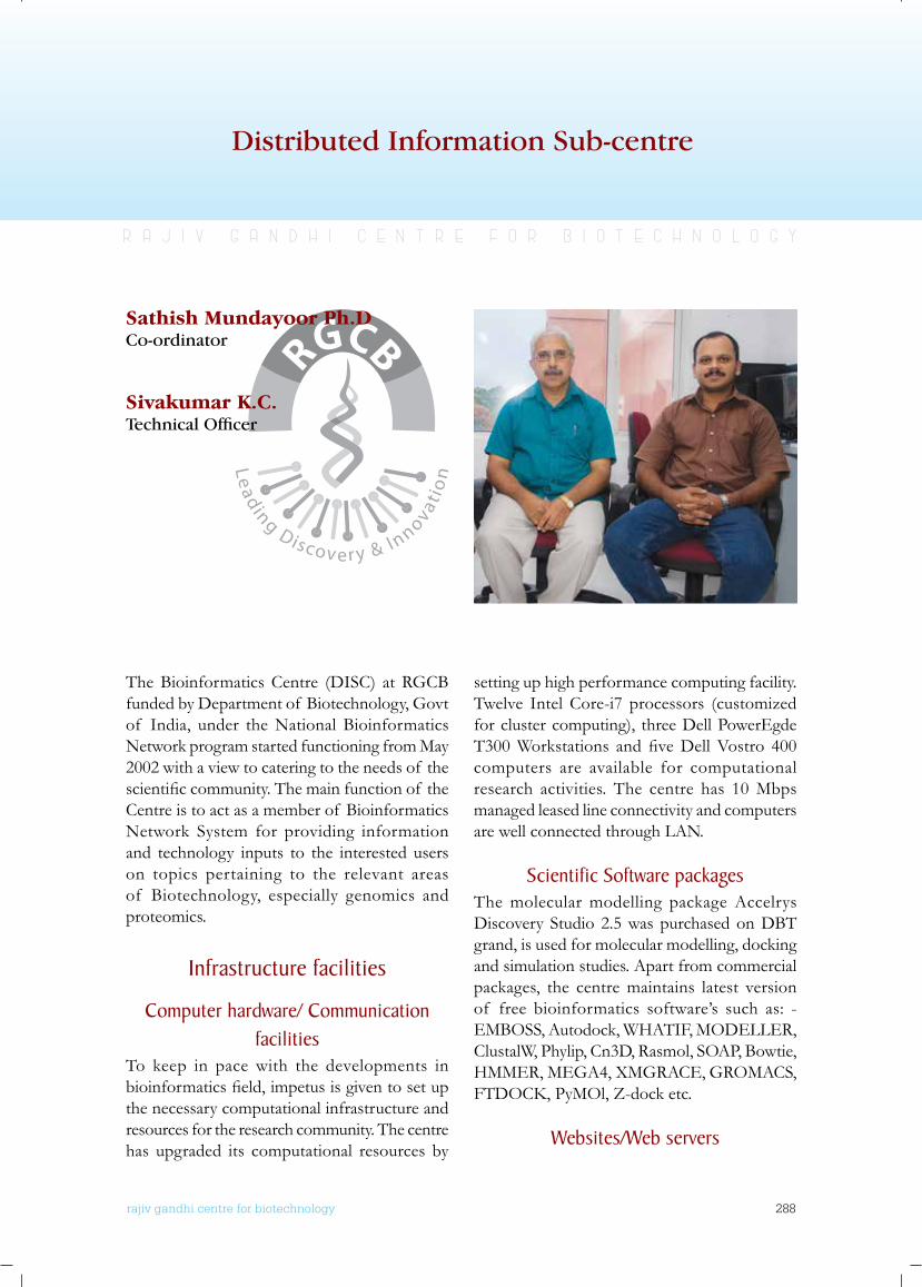
rajiv gandhi centre for biotechnology 288
annual report 2013
Distributed Information Sub-centre
r a j i v g a n d h i c e n t r e f o r b i o t e c h n o l o g y
Sathish Mundayoor Ph.DCo-ordinator
Sivakumar K.C.Technical Officer
The Bioinformatics Centre (DISC) at RGCB funded by Department of Biotechnology, Govt of India, under the National Bioinformatics Network program started functioning from May 2002 with a view to catering to the needs of the scientificcommunity.Themainfunctionof theCentre is to act as a member of Bioinformatics Network System for providing information and technology inputs to the interested users on topics pertaining to the relevant areas of Biotechnology, especially genomics and proteomics.
Infrastructure facilities
Computer hardware/ Communication facilities
To keep in pace with the developments in bioinformaticsfield,impetusisgiventosetupthe necessary computational infrastructure and resources for the research community. The centre has upgraded its computational resources by
setting up high performance computing facility. Twelve Intel Core-i7 processors (customized for cluster computing), three Dell PowerEgde T300Workstations andfiveDellVostro 400computers are available for computational research activities. The centre has 10 Mbps managed leased line connectivity and computers are well connected through LAN.
Scientific Software packagesThe molecular modelling package Accelrys Discovery Studio 2.5 was purchased on DBT grand, is used for molecular modelling, docking and simulation studies. Apart from commercial packages, the centre maintains latest version of free bioinformatics software’s such as: - EMBOSS, Autodock, WHATIF, MODELLER, ClustalW, Phylip, Cn3D, Rasmol, SOAP, Bowtie, HMMER, MEGA4, XMGRACE, GROMACS, FTDOCK,PyMOl,Z-docketc.
websites/web servers
rajiv gandhi centre for biotechnology 289
annual report 2013
The Centre is actively involved in creating software’s and integrated knowledgebases for those who are working in the areas of plant bioinformatics. Some of the databases and web servers developed at the centre are: PKSIIIexplorer[http://type3pks.in/tsvmpks3/],PKSIIIpred [http://type3pks.in/prediction/],TypeIII Polyketide Synthase Database [http://type3pks.in].
Services providedData retrieval, Next-Generation Sequencing analysis, Molecular modelling, Drug design and Virtual screening services are offered. This facility provides project work to students undergoing B.Tech/M.Tech/M.Phil/M.Sc courses from a large number of universities/institutes.
Major Activities Development of Centre to cater the needs for the researchers, students related to biotechnology, bioinformatics and computer technology. Initiated and enriched the research environment which resulted in:
• Publications(peerreviewed):03
• PosterPresentations:02
• Training:B.Tech:04,M.Sc:07,M.Tech:04,
• Developmentof databasearchivesand softwares.
• R&DactivitiesinBioinformatics
• Hypothesesgenerationandvalidationof possible outcomes
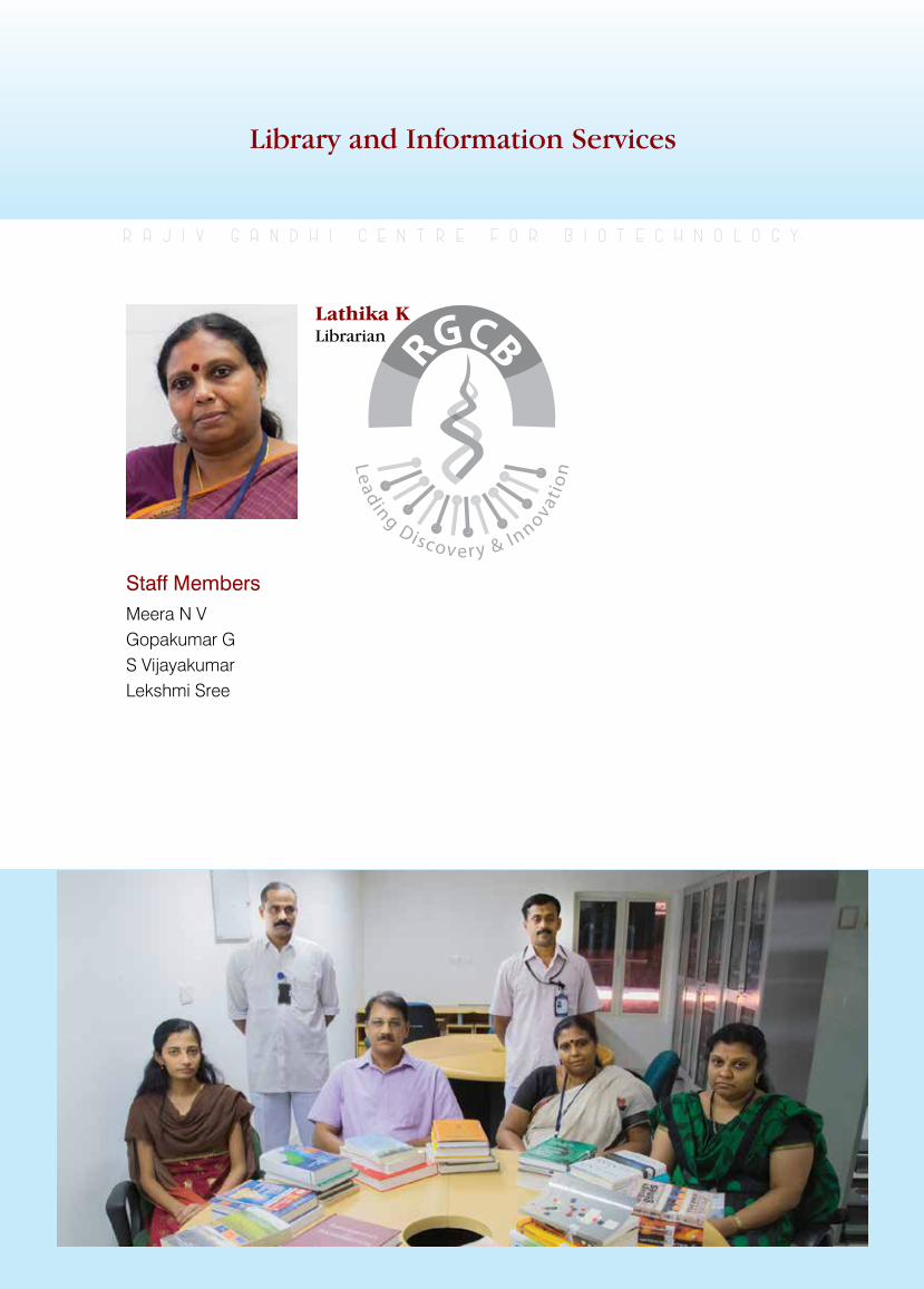
rajiv gandhi centre for biotechnology 290
annual report 2013
Library and Information Services
r a j i v g a n d h i c e n t r e f o r b i o t e c h n o l o g y
Staff MembersMeera N VGopakumar GS VijayakumarLekshmi Sree
Lathika K Librarian
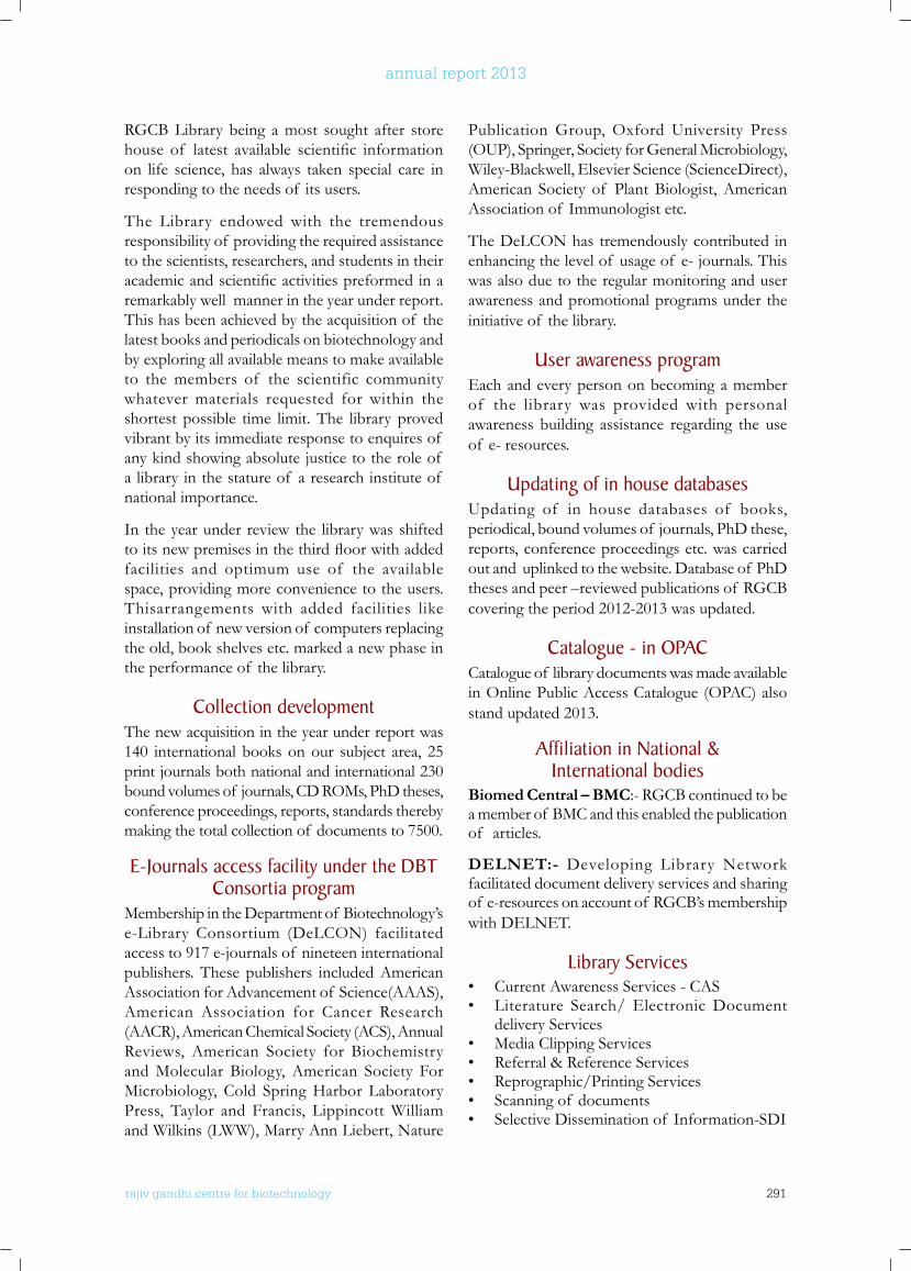
rajiv gandhi centre for biotechnology 291
annual report 2013
RGCB Library being a most sought after store house of latest available scientific informationon life science, has always taken special care in responding to the needs of its users.
The Library endowed with the tremendous responsibility of providing the required assistance to the scientists, researchers, and students in their academicandscientificactivitiespreformedinaremarkably well manner in the year under report. This has been achieved by the acquisition of the latest books and periodicals on biotechnology and by exploring all available means to make available to the members of the scientific community whatever materials requested for within the shortest possible time limit. The library proved vibrant by its immediate response to enquires of any kind showing absolute justice to the role of a library in the stature of a research institute of national importance.
In the year under review the library was shifted toitsnewpremisesinthethirdfloorwithaddedfacilities and optimum use of the available space, providing more convenience to the users.Thisarrangements with added facilities like installation of new version of computers replacing the old, book shelves etc. marked a new phase in the performance of the library.
Collection development The new acquisition in the year under report was 140 international books on our subject area, 25 print journals both national and international 230 bound volumes of journals, CD ROMs, PhD theses, conference proceedings, reports, standards thereby making the total collection of documents to 7500.
E-Journals access facility under the DbT Consortia program
Membership in the Department of Biotechnology’s e-Library Consortium (DeLCON) facilitated access to 917 e-journals of nineteen international publishers. These publishers included American Association for Advancement of Science(AAAS), American Association for Cancer Research (AACR), American Chemical Society (ACS), Annual Reviews, American Society for Biochemistry and Molecular Biology, American Society For Microbiology, Cold Spring Harbor Laboratory Press, Taylor and Francis, Lippincott William and Wilkins (LWW), Marry Ann Liebert, Nature
Publication Group, Oxford University Press (OUP), Springer, Society for General Microbiology, Wiley-Blackwell, Elsevier Science (ScienceDirect), American Society of Plant Biologist, American Association of Immunologist etc.
The DeLCON has tremendously contributed in enhancing the level of usage of e- journals. This was also due to the regular monitoring and user awareness and promotional programs under the initiative of the library.
user awareness program Each and every person on becoming a member of the library was provided with personal awareness building assistance regarding the use of e- resources.
updating of in house databases Updating of in house databases of books, periodical, bound volumes of journals, PhD these, reports, conference proceedings etc. was carried out and uplinked to the website. Database of PhD theses and peer –reviewed publications of RGCB covering the period 2012-2013 was updated.
Catalogue - in oPACCatalogue of library documents was made available in Online Public Access Catalogue (OPAC) also stand updated 2013.
Affiliation in national & International bodies
Biomed Central – BMC:- RGCB continued to be a member of BMC and this enabled the publication of articles.
DELNET:- Developing Library Network facilitated document delivery services and sharing of e-resources on account of RGCB’s membership with DELNET.
library Services• Current Awareness Services - CAS• Literature Search/ Electronic Document
delivery Services• Media Clipping Services• Referral & Reference Services • Reprographic/Printing Services• Scanning of documents• Selective Dissemination of Information-SDI
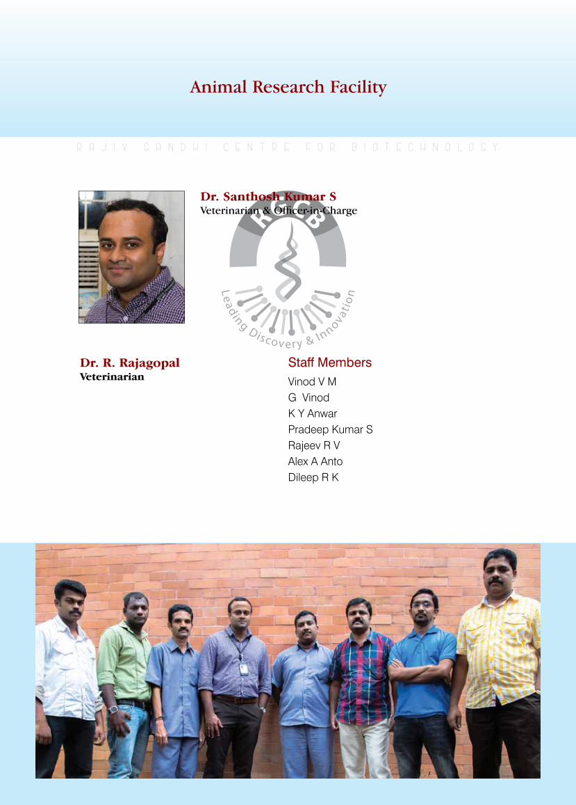
rajiv gandhi centre for biotechnology 292
annual report 2013
Animal Research Facility
r a j i v g a n d h i c e n t r e f o r b i o t e c h n o l o g y
Dr. Santhosh Kumar SVeterinarian & Officer-in-Charge
Dr. R. Rajagopal Veterinarian
Staff MembersVinod V MG VinodK Y AnwarPradeep Kumar SRajeev R VAlex A AntoDileep R K
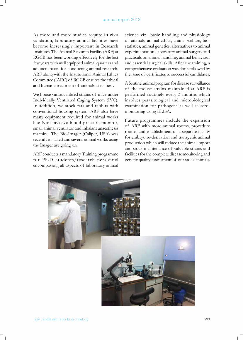
rajiv gandhi centre for biotechnology 293
annual report 2013
As more and more studies require in vivo validation, laboratory animal facilities have become increasingly important in Research Institutes. The Animal Research Facility (ARF) at RGCB has been working effectively for the last few years with well equipped animal quarters and adjunct spaces for conducting animal research. ARF along with the Institutional Animal Ethics Committee (IAEC) of RGCB ensures the ethical and humane treatment of animals at its best.
We house various inbred strains of mice under Individually Ventilated Caging System (IVC). In addition, we stock rats and rabbits with conventional housing system. ARF also have many equipment required for animal works like Non-invasive blood pressure monitor, small animal ventilator and inhalant anaesthesia machine. The Bio-Imager (Caliper, USA) was recently installed and several animal works using the Imager are going on.
ARF conducts a mandatory Training programme for Ph.D students/research personnel encompassing all aspects of laboratory animal
science viz., basic handling and physiology of animals, animal ethics, animal welfare, bio-statistics, animal genetics, alternatives to animal experimentation, laboratory animal surgery and practicals on animal handling, animal behaviour and essential surgical skills. After the training, a comprehensive evaluation was done followed by theissueof certificatestosuccessfulcandidates.
A Sentinel animal program for disease surveillance of the mouse strains maintained at ARF is performed routinely every 3 months which involves parasitological and microbiological examination for pathogens as well as sero-monitoring using ELISA.
Future programmes include the expansion of ARF with more animal rooms, procedure rooms, and establishment of a separate facility for embryo re-derivation and transgenic animal production which will reduce the animal import and stock maintenance of valuable strains and facilities for the complete disease monitoring and genetic quality assessment of our stock animals.
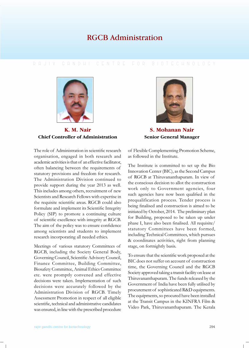
rajiv gandhi centre for biotechnology 294
annual report 2013
RGCB Administration
r a j i v g a n d h i c e n t r e f o r b i o t e c h n o l o g y
Theroleof Administrationinscientificresearchorganisation, engaged in both research and academic activities is that of an effective facilitator, often balancing between the requirements of statutory provisions and freedom for research. The Administration Division continued to provide support during the year 2013 as well. This includes among others, recruitment of new Scientists and Research Fellows with expertise in therequisitescientificareas.RGCBcouldalsoformulateandimplementitsScientificIntegrityPolicy (SIP) to promote a continuing culture of scientificexcellencewithintegrityatRGCB.Theaimof thepolicywastoensureconfidenceamong scientists and students to implement research incorporating all needed ethics.
Meetings of various statutory Committees of RGCB, including the Society General Body, GoverningCouncil,ScientificAdvisoryCouncil,Finance Committee, Building Committee, Biosafety Committee, Animal Ethics Committee etc. were promptly convened and effective decisions were taken. Implementation of such decisions were accurately followed by the Administration Division of RGCB. Timely Assessment Promotion in respect of all eligible scientific,technicalandadministrativecandidateswas ensured, in line with the prescribed procedure
of Flexible Complementing Promotion Scheme, as followed in the Institute.
The Institute is committed to set up the Bio Innovation Center (BIC), as the Second Campus of RGCB at Thiruvananthapuram. In view of the conscious decision to allot the construction work only to Government agencies, four such agencies havenowbeenqualified in theprequalification process. Tender process is beingfinalisedandconstructionisaimedtobeinitiated by October, 2014. The preliminary plan for Building, proposed to be taken up under phaseI,havealsobeenfinalised.Allrequisite/statutory Committees have been formed, including Technical Committees, which pursues & coordinates activities, right from planning stage, on fortnightly basis.
ToensurethatthescientificworkproposedattheBIC does not suffer on account of construction time, the Governing Council and the RGCB Society approved taking a transit facility on lease at Thiruvananthapuram. The funds released by the Government of India have been fully utilised by procurement of sophisticated R&D equipments. The equipments, so procured have been installed attheTransitCampusintheKINFRAFilm&VideoPark,Thiruvananthapuram.TheKerala
K. M. NairChief Controller of Administration
S. Mohanan NairSenior General Manager
rajiv gandhi centre for biotechnology 295
annual report 2013
BiotechParkownedbyKINFRAatKochihasbeen transferred to Rajiv Gandhi Centre for Biotechnologyforaninitialperiodof fiveyears,through an MOU entered into between RGCB andKINFRA.ThefacilitynamedBioNestwillserve as business development incubator for start up companies.
In view of budgetary constraints and austerity measures, the Finance & Accounts Division of RGCB had to strive hard for achieving its goal. The wide gap between projected budget estimate figures and actual amount allotted, could bebridged only through timely intervention of the Finance & Accounts Division. Prompt payment and accurate accounting was ensured by the Division, during this year as well. With the support of Project Management Division, Financial, Accounting as well as project monitoring activities could smoothly progress.
The ScientificAuditDivision of theC&AG,Bangalore had undertaken the audit of the activities of RGCB for the year 2011-12 & 2012-13 during 2013. The activities of the Institute wereauditedbyaScientificAuditteamandnoserious observations on functioning have been reported. There were no substantial objections and initial replies have been furnished by RGCB
to almost all the queries.
Safety and Security measures were reviewed by the Administration Division. An internal safety audit was conducted at the premises, in view of the present security scenario prevailing in the country and the delicate nature of R&D activities at the Institute. As a result, security cameras at sensitive areas will be installed within the RGCB Main Campus.
Welfare activities at the Institute were also accorded its prime importance during the year 2013. The health system could be improved by engagement of a dedicated female specialist AMO, considering the large number of women employees at the Institute. In addition, a specialistAMOinthefieldof Cardiologywashired on consultancy basis at the Institute. A limited reimbursement Medical Expenditure Scheme for Contract Staff was introduced to cater to the medical requirements of the contract staff. In association with an Insurance Company, a Medical Insurance Policy was also been framed to cater to the inpatient treatment of contract staff and their dependent family members. This policy has been successfully implemented and is regularly monitored.
1. Professor and Director Dr. M. Radhakrishna Pillai
2. Professor of Eminence Dr. C.C. Kartha
3. Scientist G Dr. Sathish Mundayoor
4. Scientist G Dr. G. Pradeep Kumar
5. Scientist F Dr. R.V. Omkumar
STAFF LIST
Scientific Cadre
6. Scientist F Dr. Malini Laloraya
7. Scientist F Dr. Moinak Banerjee
8. Scientist E II Dr. K. Santhosh Kumar
9. Scientist E II Dr. Ruby John Anto
10. Scientist E II Dr. George Thomas
rajiv gandhi centre for biotechnology 296
annual report 2013
11. Scientist E II Dr. R. Ajay Kumar
12. Scientist E II Dr. E.V. Soniya
13 Scientist E II Dr. Suparna Sengupta
14. Scientist E II Dr. T.R. Santhosh Kumar
15. Scientist E II Dr. Priya Srinivas
16. Scientist E II Dr. S. Asha Nair
17. Scientist E II Dr. Abdul Jaleel
18. Scientist E II Dr. V.V. Asha
19. Scientist E II Dr. Jackson James
20. Scientist E I Dr. Sabu Thomas
21. Scientist E I Dr. Radhakrishnan R Nair
22. Scientist E I Dr. Sanil George
23. Scientist E I Dr. S. Sreeja
24. Scientist E I Dr. E. Sreekumar
25. Scientist E I Dr. G.S. Vinod Kumar
26. Scientist E I Dr. S. Manjula
27. Scientist C Dr. K. Harikrishnan
28. Scientist C Dr. Rashmi Mishra
29. Scientist C Dr. K.B. Harikumar
30. Scientist C Dr. Debasree Dutta
31. Scientist C Dr. Rakesh Singh Laishram
32. Scientist C Dr. M. Maya Devi
33. Scientist C Dr. John Bernet Johnson
Technical Cadre
34. Senior General Manager S. Mohanan Nair
35. General Manager (Instrumentation Engineering) Shaj Upendran
36. Deputy General Manager (L & E A) R. Jayachandran Nair
37. Deputy Librarian K. Lathika
38. Senior Manager (Technical Services) V. Jiji
39. Senior Manager (Technical Services) George Varghese
40. Senior Manager (Technical Services) D. Sanjai
41. Manager (Technical Services) P. Manoj
rajiv gandhi centre for biotechnology 297
annual report 2013
42. Manager (Technical Services) K. Rajasekharan
43. Veterinarian & Animal House-in- Charge Dr. S. Santhosh Kumar
44. Manager (Technical Services) M. Saravana kumar
45. Manager (Technical Services) Indu Ramachandran
46. Manager (Technical Services) Laiza Paul
47. Manager (Technical Services) Sudha B.Nair
48. Manager (Technical Services) Bindu Asokan
49. Senior Technical Officer Ciji Varghese
50. Manager (Technical Services) Ambili S. Nair
51. Technical Officer K.C. Sivakumar
52. Senior Technical Officer Rahul C.S. Nair
53. Senior Technical Officer I.X. Sajan
54. Assistant Engineer C. Durga Prasad
55. Technical Officer Arun Surendran
56. Technical Assistant Group III K. Deepu
57. Deputy Engineer (Electrical) S. Ajith Kumar
58. Technical Assistant Group III Rintu T. Varghese
59. Technical Assistant Group I V. Amal
60. Technical Assistant Group I
R. Dileep Kumar
61. Technical Assistant Group.I Gr.II G. Johny
62. Technical Assistant Group.I Gr.I G. Sheela
63. Technical Assistant Group.I Gr.II V. R. Unnikrishnan
64. Technical Assistant Group.I Gr.II K. P. Antony
65. Technical Assistant Group.I Gr.II S. Edwin
66. Technical Assistant Group.I Gr.II G. Velthai
67. Technical Assistant Group.I Gr.I S. Vijayakumar
68. Technical Assistant Group.I Gr.II S. Rajeev
69. Technical Assistant Group.I Gr.II Biju S. Nair
70. Technical Assistant Group.I Gr.I S. Santhosh
71. Technical Assistant Group.I Gr.I N.V. Meera
72. Technical Assistant S. Aswani Kumar
73. Technical Assistant Reena Prasad
74. Helper G. Gopakumar
75. Helper J. Venugopalan
76. Helper K.A. Vinod Lal
77. Helper J. Jayanandan
78. Helper V. Sumaja

rajiv gandhi centre for biotechnology 298
annual report 2013
79. Chief Controller K.M. Nair
80. Registrar Grade II Dr. R. Ashok
81. Finance Officer M. Babu
82. Chief Manager (Purchase) Jeevan Chacko
83. Accounts Officer K.K. Jayasree
84. Administrative Officer S. Suthakumari
85. Senior Manager (Purchase) N. Jayakrishnan
86. Deputy Accounts Officer S. Usha Devi
87. Manager (Accounts & Audit) R. Kumar
88. Senior P S to Director U.S. Jayalakshmi
89. Senior P S to Director R. Priya
90. Assistant Administrative Officer Asha R. Nair
91. Private Secretary O. Girija Kumari
92. Management Assistant J. Preetha
93. Office Assistant K. Subash
94. Junior Office Assistant R. Anil Kumar
Support Staff
95. Driver Grade IV S. Harikumar
96. Senior Attendant T. Wilson
97. Attendant Grade - III B. Chandrika Devi
98. Attendant Grade III Thapasi Muthu
99. Attendant Grade II B. Usha
100. Attendant Grade III S. R. Vinod Kumar
101. Attendant Grade II R. Thankamany
102. Driver V. M. Manukumar
Administrative Cadre
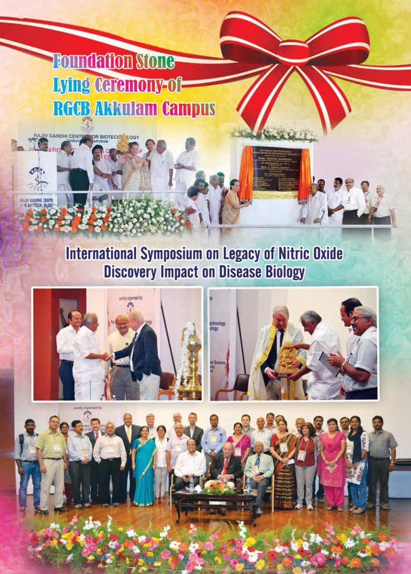
rajiv gandhi centre for biotechnology 299
annual report 2013
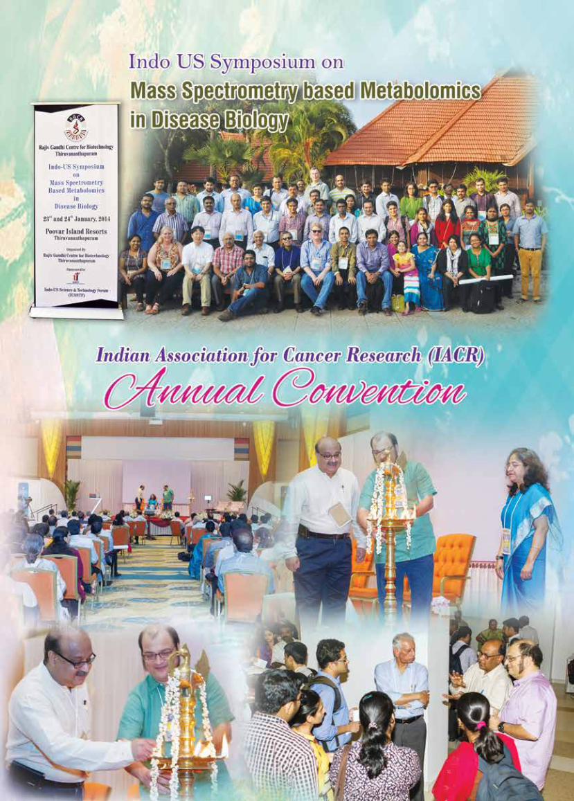
rajiv gandhi centre for biotechnology 300
annual report 2013

rajiv gandhi centre for biotechnology 301
annual report 2013
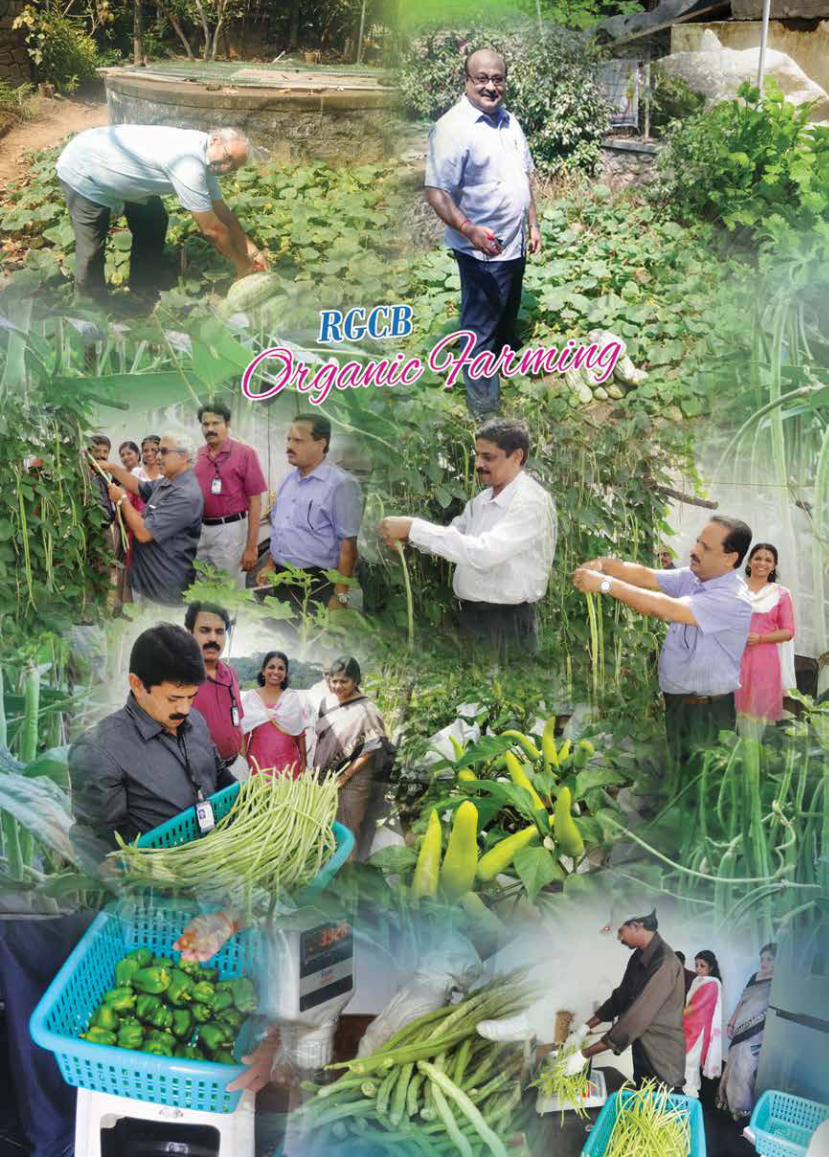
rajiv gandhi centre for biotechnology 302
annual report 2013
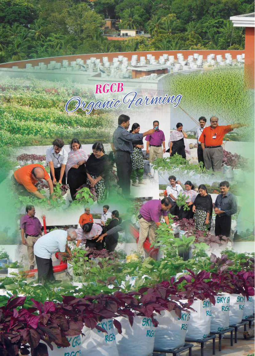
rajiv gandhi centre for biotechnology 303
annual report 2013

Layout & Printing: Vision Grafix | e-mail: [email protected]