ANIMAL CELL CULTURE - The American College
32
ANIMAL CELL CULTURE COURSE CODE : MIC 3635 COURSE TITLE: PLANT & ANIMAL CELL CULTURE COURSE TEACHER : D.RAMYA
-
Upload
khangminh22 -
Category
Documents
-
view
3 -
download
0
Transcript of ANIMAL CELL CULTURE - The American College

ANIMAL CELL CULTURE
COURSE CODE : MIC 3635
COURSE TITLE: PLANT & ANIMAL CELL CULTURE
COURSE TEACHER : D.RAMYA

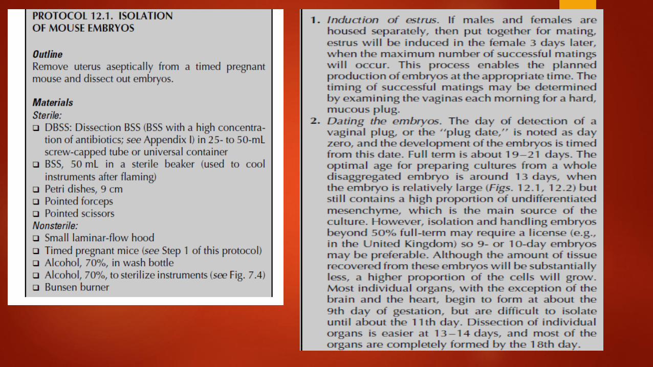
Primary culture. A culture started from cells, tissues, or organs taken directly from an organism, and before the first subculture. Substrate. The matrix or solid underlay upon which a monolayer culture grows. Passage. The transfer or subculture of cells from one culture vessel to another; usually, but not necessarily, involves the subdivision of a proliferating cell population, enabling the propagation of a cell line or cell strain. Passage number. The number of times a culture has been subcultured. Explant. A fragment of tissue transplanted from its original site and maintained in an artificial medium. Primary explant. A fragment of tissue removed from the organism and placed in culture in such as way as to promote its survival and the outgrowth of viable cells.


Primary Culture A primary culture is that stage of the culture after isolation of the cells but before the first subculture. There are four stages to consider:
(1) acquisition of the sample,
(2) isolation of the tissue,
(3) dissection and/or disaggregation, and
(4) culture after seeding into the culture vessel.
After isolation, a primary cell culture may be obtained either by allowing cells to migrate out from fragments of tissue adhering to a suitable substrate or by disaggregating the tissue mechanically or enzymatically to produce a suspension of cells, some of which will ultimately attach to the substrate. It appears to be essential for most normal untransformed cells, with the exception of hematopoietic cells, to attach to a flat surface in order to survive and proliferate with maximum efficiency

The enzymes used most frequently for tissue disaggregation are crude preparations of trypsin, collagenase, elastase, pronase, dispase, DNase, and hyaluronidase, alone or in various combinations, e.g., elastase and DNase for type II alveolar cell isolation. Although each tissue may require a different set of conditions, certain requirements are shared by most of them: (1) Fat and necrotic tissue are best removed during dissection. (2) The tissue should be chopped finely with sharp instruments to cause minimum damage. (3) Enzymes used for disaggregation should be removed subsequently by gentle centrifugation. (4) The concentration of cells in the primary culture should be much higher than that normally used for subculture, because the proportion of cells from the tissue that survives in primary culture may be quite low. (5) A rich medium, such as Ham’s F12, is preferable to a simple medium, such as Eagle’s MEM, and, if serum is required, fetal bovine often gives better survival than does calf or horse. Isolation of specific cell types will probably require selective media. (6) Embryonic tissue disaggregates more readily, yields more viable cells, and proliferates more rapidly in primary culture than does adult tissue.

ISOLATION OF THE TISSUE
Before attempting to work with human or animal tissue, make sure that your work fits within medical ethical rules or current legislation on experimentation with animals. For example, in the United Kingdom, the use of embryos or fetuses beyond 50% gestation or incubation is regulated under the Animal Experiments. Work with human biopsies or fetal material usually requires the consent of the local ethical committee and the patient and/or his or her relatives. Safety Note. Work with human tissue should be carried out at Containment Level 2 in a Class II biological safety Cabinet. Remove the tissue aseptically and transfer it to the tissue culture laboratory in dissection BSS (DBSS) or transport medium


Mouse Dissection. Stages in dissection of a pregnant mouse for the collection of embryos (a) Swabbing the abdomen. (b)(b), (c) Tearing the skin to expose the abdominal
wall. (d) Opening the abdomen. (e) Revealing the uterus in situ. (f) Removing the uterus. (g),(h) Dissecting the embryos from the uterus. (i) Removing the membranes. ( j) Removing the head (optional). (k) Chopping the embryos. (l) Transferring pieces to trypsinization flask (for warm trypsinization). (m) Transferring the pieces to a small Erlenmeyer flask (for cold trypsinization; (n) Flask on ice

Chick Embryo Chick embryos are easier to dissect, as they are larger than mouse embryos at the equivalent stage of development. Like mouse embryos, chick embryos are used to provide predominantly mesenchymal cell primary cultures for cell proliferation analysis, to provide feeder layers, and as a substrate for viral propagation. Because of their larger size, it is easier to dissect out individual organs to generate specific cell types, such as hepatocytes, cardiac muscle, and lungepithelium. As with mouse embryos, the use of chick embryos may be subject to animal legislation (e.g., in the United Kingdom) and working with embryos that are more than half-term may require a license


Removing a Chick Embryo from an Egg. Stages in the extraction of the whole chick embryo from an egg. (a) Swabbing the egg with alcohol. (b) Cracking the shell. (c) Peeling off the shell. (d) Peeling off the shell membrane. (e) Chorioallantoic membrane (CAM) and vasculature revealed. (f) Removing CAM with forceps. (g) Grasping the embryo round the neck. (h) Withdrawing the embryo from the egg. (i) Isolated 10-day embryo in Petri dish.


Primary culture broadly involves the culturing techniques carried following the isolation of the cells, but before the first subculture. Primary cultures are usually prepared from large tissue masses. Thus, these cultures may contain a variety of differentiated cells e.g. fibroblasts, lymphocytes, macrophages, epithelial cells. With the experiences of the personnel working in tissue culture laboratories, the following criteria/ characteristics are considered for efficient development of primary cultures: a. Embryonic tissues rather than adult tissues are preferred for primary cultures. This is due to the fact that the embryonic cells can be disaggregated easily and yield more viable cells, besides rapidly proliferating in vitro. b. The quantity of cells used in the primary culture should be higher since their survival rate is substantially lower (when compared to subcultures). c. The tissues should be processed with minimum damage to cells for use in primary culture. Further, the dead cells should be removed. d. Selection of an appropriate medium (preferably a nutrient rich one) is advisable. For the addition of serum, fetal bovine source is preferred rather than calf or horse serum. e. It is necessary to remove the enzymes used for disaggregation of cells by centrifugation.

Techniques for Primary Culture: Among the various techniques devised for the primary culture of isolated tissues, three techniques are most commonly used: 1. Mechanical disaggregation. 2. Enzymatic disaggregation. 3. Primary explant technique.

1. Mechanical Disaggregation: For the disaggregation of soft tissues (e.g. spleen, brain, embryonic liver, soft tumors), mechanical technique is usually employed. This technique basically involves careful chopping or slicing of tissue into pieces and collection of spill out cells. The cells can be collected by two ways: i. Pressing the tissue pieces through a series of sieves with a gradual reduction in the mesh size. ii. Forcing the tissue fragments through a syringe and needle. Although mechanical disaggregation involves the risk of cell damage, the procedure is less expensive, quick and simple. This technique is particularly useful when the availability of the tissue is in plenty, and the efficiency of the yield is not very crucial. It must however, be noted that the viability of cells obtained from mechanical techniques is much lower than the enzymatic technique. 2. Enzymatic Disaggregation: Enzymatic disaggregation is mostly used when high recovery of cells is required from a tissue. Disaggregation of embryonic tissues is more efficient with higher yield of cells by use of enzymes. This is due to the presence of less fibrous connective tissue and extracellular matrix. Enzymatic disaggregation can be carried out by using trypsin, collagenase or some other enzymes. Disaggregation by trypsin: The term trypsinization is commonly used for disaggregation of tissues by the enzyme, trypsin. Many workers prefer to use crude trypsin rather than pure trypsin for the following reasons:

Mechanical Disaggregation. (a) Scraping or ‘‘spillage’’. Cutting action, or abrasion of cut surface, releases cells. (b) Sieving. Forcing tissue through sieve with syringe piston. (c) Syringing. Drawing tissue into syringe through wide bore needle or canula and expressing. (d) Trituration by pipette. Pippetting tissue fragments up and down through wide bore pipette.

i. The crude trypsin is more effective due to the presence of other proteases ii. Cells can tolerate crude trypsin better. iii. The residual activity of crude trypsin can be easily neutralized by the serum of the culture media (when serum-free media are used, a trypsin inhibitor can be used for neutralization). Disaggregation of cells can also be carried out by using pure trypsin which is less toxic and more specific in its action. The desired tissue is chopped to 2-3 mm pieces and then subjected to disaggregation by trypsin. There are two techniques of trypsinization-warm trypsinization and cold trypsinization. Warm trypsinization This method is widely used for disaggregation of cells. The chopped tissue is washed with dissection basal salt solution (DBSS), and then transferred to a flask containing warm trypsin (37° C). The contents are stirred, and at an interval of every thirty minutes, the supernatant containing the dissociated cells can be collected. After removal of trypsin, the cells are dispersed in a suitable medium and preserved (by keeping the vial on ice). The process of addition of fresh trypsin (to the tissue pieces), incubation and collection of dissociated cells (at 30 minutes intervals) is carried out for about 4 hours. The disaggregated cells are pooled, counted, appropriately diluted and then incubated.

Warm Trypsin It is important to minimize the exposure of cells to active trypsin in order to preserve maximum viability. Hence, when whole tissue is being trypsinized at 37◦C, dissociated cells should be collected every half hour, and the trypsin should be removed by centrifugation and neutralized with serum in medium.

Warm Trypsin Disaggregation

Cold trypsinization This technique is more appropriately referred to as trypsinization with cold pre-exposure. The risk of damage to the cells by prolonged exposure to trypsin at 37°C (in warm trypsinization) can be minimized in this technique. After chopping and washing, the tissue pieces are kept in a vial (on ice) and soaked with cold trypsin for about 6-24 hours. The trypsin is removed and discarded. However, the tissue pieces contain residual trypsin. These tissue pieces in a medium are incubated at 37°C for 20-30 minutes. The cells get dispersed by repeated pi-pettings. The dissociated cells can be counted, appropriately diluted and then used. The cold trypsinization method usually results in a higher yield of viable cells with an improved survival of cells after 24 hours of incubation. This method does not involve stirring or centrifugation, and can be conveniently adopted in a laboratory. The major limitation of cold trypsinization is that it is not suitable for disaggregation of cells from large quantities of tissues. Limitations of trypsin disaggregation: Disaggregation by trypsin may damage some cells (e.g. epithelial cells) or it may be almost ineffective for certain tissues (e.g. fibrous connective tissue). Hence other enzymes are also in use for dissociation of cells.

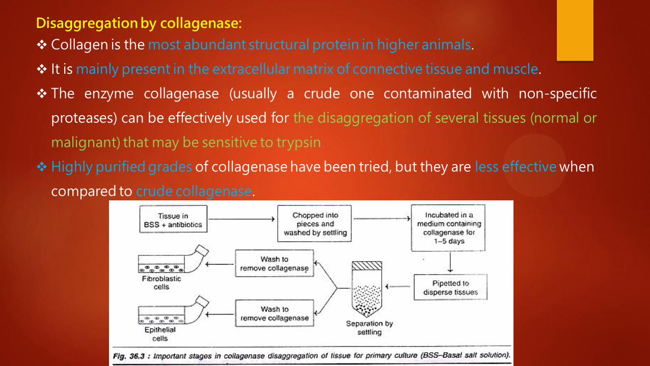
Disaggregation by collagenase:
Collagen is the most abundant structural protein in higher animals.
It is mainly present in the extracellular matrix of connective tissue and muscle.
The enzyme collagenase (usually a crude one contaminated with non-specific
proteases) can be effectively used for the disaggregation of several tissues (normal or
malignant) that may be sensitive to trypsin.
Highly purified grades of collagenase have been tried, but they are less effective when
compared to crude collagenase.

Tissue Disaggregation by Collagenase. (a) Schematic diagram of dissection
followed by disaggregation in collagenase.

The desired tissue suspended in basal salt solution, containing antibiotics is chopped into pieces. These pieces are washed by settling, and then suspended in a complete medium containing collagenase.
After incubating for 1-5 days, the tissue pieces are dispersed by pipetting. The clusters of cells are separated by settling. The epithelial cells and fibroblastic cells can be separated.
Collagenase disaggregation has been successfully used for human brain, lung and several other epithelial tissues, besides various human tumors, and other animal tissues.
Addition of another enzyme hyaluronidase (acts on carbohydrate residues on cell surfaces) promotes disaggregation.
Collagenase in combination with hyaluronidase is found to be very effective for dissociating rat or rabbit liver.
Use of other enzymes in disaggregation: Trypsin and collagenase are the most widely used enzymes for disaggregation. Certain bacterial proteases (e.g. pronase, dispase) have been used with limited
success. Besides hyaluronidase, neuraminidase is also used in conjunction with collagenase for effective degradation of cell surface carbohydrates.

The primary explant technique was, in fact the original method, developed by
Harrison in 1907. This technique has undergone several modifications, and is still in
use. The simplified procedure adopted for primary explant culture.
The primary explant technique is particularly useful for disaggregation of small
quantities of tissues (e.g. skin biopsies). The other two techniques mechanical or
enzymatic disaggregation however, are not suitable for small amounts of tissues, as
there is a risk of losing the cells.
primary explant technique


Attaching explants Both adherence and migration may be stimulated by placing a glass
coverslip on top of the explant, with the explant near the edge of the coverslip, or the plastic dish may be scratched through the explant to attach the tissue to the flask [Elliget & Lechner, 1992].
Attachment may also be promoted by treating the plastic with polylysine or fibronectin, extracellular matrix or feeder layers.
Historically, plasma clots have been used to promote attachment. Place a drop of plasma on the plastic surface, and embed the explant in it. This should induce the plasma to clot in a few minutes, whereupon medium can be added. Alternatively, purified fibrinogen and thrombin can be used [Nicosia & Ottinetti, 1990].
The limitation of explant technique It is the poor adhesiveness of certain tissues to the growth surface, and the selection of cells in
the outgrowth. It is however, observed that the primary explant technique can be used for a majority of embryonic cells e.g. fibroblasts, myoblasts, epithelial cells, glial cells.
Separation of Viable and Non-Viable Cells: It is a common practice to remove the nonviable cells while the primary culture is prepared from
the disaggregated cells. Sometimes, the non-viable cells from the primary cultures may be removed by centrifugation. The cells are mixed with ficoll and sodium metrizoate, and centrifuged. The dead cells form a
pellet at the bottom of the tube. Medical Ethics and Safety Measures in Culture Techniques: Since the culture techniques involve the use of animal or human tissues, it is absolutely necessary to follow several safety measures and medical ethics. In fact, in some countries there are established legislation/norms for selection and use of tissues in cultures. For example, in United Kingdom, Animal Experiments (Scientific Procedures) Act of 1986 is followed. Safety measures: Handling of human tissues is associated with a heavy risk of exposure for various infections. Therefore, it is absolutely necessary that the human materials are handled in a biohazard cabinet. The tissues should be screened for various infections such as hepatitis, tuberculosis, HIV, before their use. Further, the media and apparatus, after their use must be autoclaved or disinfected, so that the spread of infections is drastically reduced.

Cell Line After the first subculture, the primary culture becomes known as a cell line or sub-clone. Cell lines derived from primary cultures have a limited life span (i.e., they are finite; see below), and as they are passaged, cells with the highest growth capacity predominate, resulting in a degree of genotypic and phenotypic uniformity in the population. Cell Strain If a subpopulation of a cell line is positively selected from the culture by cloning or some other method, this cell line becomes a cell strain. A cell strain often acquires additional genetic changes subsequent to the initiation of the parent line.