Angiosperm ovules: diversity, development, evolution
26
Zurich Open Repository and Archive University of Zurich University Library Strickhofstrasse 39 CH-8057 Zurich www.zora.uzh.ch Year: 2011 Angiosperm ovules: diversity, development, evolution Endress, P K Abstract: In angiosperms the presence of an outer integument appears to be instrumental for ovule cur- vature, as indicated from studies on ovule diversity through the major clades of angiosperms, molecular developmental genetics in model species, abnormal ovules in a broad range of angiosperms, and com- parison with gymnosperms with curved ovules. Lobation of integuments is not an atavism indicating evolution from telomes, but simply a morphogenetic constraint from the necessity of closure of the mi- cropyle. Ovule shape is partly dependent on locule architecture, which is especially indicated by the occurrence of orthotropous ovules. Some ovule features are even more conservative than earlier assumed and thus of special interest in angiosperm macrosystematics. DOI: https://doi.org/10.1093/aob/mcr120 Posted at the Zurich Open Repository and Archive, University of Zurich ZORA URL: https://doi.org/10.5167/uzh-51422 Journal Article Published Version Originally published at: Endress, P K (2011). Angiosperm ovules: diversity, development, evolution. Annals of Botany, 107(9):1465- 1489. DOI: https://doi.org/10.1093/aob/mcr120
-
Upload
khangminh22 -
Category
Documents
-
view
8 -
download
0
Transcript of Angiosperm ovules: diversity, development, evolution
Zurich Open Repository andArchiveUniversity of ZurichUniversity LibraryStrickhofstrasse 39CH-8057 Zurichwww.zora.uzh.ch
Year: 2011
Angiosperm ovules: diversity, development, evolution
Endress, P K
Abstract: In angiosperms the presence of an outer integument appears to be instrumental for ovule cur-vature, as indicated from studies on ovule diversity through the major clades of angiosperms, moleculardevelopmental genetics in model species, abnormal ovules in a broad range of angiosperms, and com-parison with gymnosperms with curved ovules. Lobation of integuments is not an atavism indicatingevolution from telomes, but simply a morphogenetic constraint from the necessity of closure of the mi-cropyle. Ovule shape is partly dependent on locule architecture, which is especially indicated by theoccurrence of orthotropous ovules. Some ovule features are even more conservative than earlier assumedand thus of special interest in angiosperm macrosystematics.
DOI: https://doi.org/10.1093/aob/mcr120
Posted at the Zurich Open Repository and Archive, University of ZurichZORA URL: https://doi.org/10.5167/uzh-51422Journal ArticlePublished Version
Originally published at:Endress, P K (2011). Angiosperm ovules: diversity, development, evolution. Annals of Botany, 107(9):1465-1489.DOI: https://doi.org/10.1093/aob/mcr120
INVITED REVIEW: PART OF A SPECIAL ISSUE ON EVOLUTION AND DEVELOPMENT
Angiosperm ovules: diversity, development, evolution
Peter K. Endress*
Institute of Systematic Botany, University of Zurich, Zollikerstrasse 107, 8008 Zurich, Switzerland* E-mail [email protected]
Received: 2 March 2011 Returned for revision: 29 March 2011 Accepted: 11 April 2011 Published electronically: 23 May 2011
†Background Ovules as developmental precursors of seeds are organs of central importance in angiospermflowers and can be traced back in evolution to the earliest seed plants. Angiosperm ovules are diverse in theirposition in the ovary, nucellus thickness, number and thickness of integuments, degree and direction of curvature,and histological differentiations. There is a large body of literature on this diversity, and various views on itsevolution have been proposed over the course of time. Most recently evo–devo studies have been concentratedon molecular developmental genetics in ovules of model plants.† Scope The present review provides a synthetic treatment of several aspects of the sporophytic part of ovulediversity, development and evolution, based on extensive research on the vast original literature and on experi-ence from my own comparative studies in a broad range of angiosperm clades.†Conclusions In angiosperms the presence of an outer integument appears to be instrumental for ovule curvature,as indicated from studies on ovule diversity through the major clades of angiosperms, molecular developmentalgenetics in model species, abnormal ovules in a broad range of angiosperms, and comparison with gymnospermswith curved ovules. Lobation of integuments is not an atavism indicating evolution from telomes, but simply amorphogenetic constraint from the necessity of closure of the micropyle. Ovule shape is partly dependent onlocule architecture, which is especially indicated by the occurrence of orthotropous ovules. Some ovule featuresare even more conservative than earlier assumed and thus of special interest in angiosperm macrosystematics.
Key words: Angiosperms, development, diversity, evo–devo, evolution, integuments, macrosystematics,micropyle, nucellus, ovules, seed plants.
INTRODUCTION
Ovules, the developmental precursors of seeds, are the organsin angiosperm flowers that can be traced back farthest in time,back to early seed plants almost 400 million years ago. In spiteof their relatively stable basic structure, ovules have attained abroad diversity of forms. The early evolution of ovules inangiosperms has been much under discussion in comparativestructural studies and embryology on extant and fossil plants,and recently ovules became prominent in molecular develop-mental genetic studies.Thus, information on ovules relies on sources from different
fields, and a synthetic review needs to draw from all of them.There is a plethora of descriptive studies on embryology ofsingle angiosperm species, also including the sporophyticpart of the ovules, especially from Indian botanists in thetime between 1930 and 1980. Each by itself may not be ofspecial interest, but taken together they are a treasure troveof information on ovule diversity, the value of which continu-ously increases with each new study. Another field encom-passes studies on the development of ovules in modelspecies, especially Arabidopsis, from the past 20 years.There are a number of comparative studies and reviews inwhich the sporophytic part of ovules and its diversity was con-sidered (Brongniart, 1827; Mirbel, 1829; Agardh, 1858;Warming, 1878, 1913; van Tieghem, 1901; Schnarf, 1929,1931, 1933; Mauritzon, 1939b; Maheshwari, 1950, 1963;Johri, 1963, 1967, 1984; Kapil and Vasil, 1963; Puri, 1970;
Bouman, 1974, 1984a, b; Philipson, 1974, 1977; Hamann,1977; Yakovlev and Batygina, 1981–1990; Tobe, 1989;Dahlgren, 1991; Kapil and Bhatnagar, 1991; Johri et al.,1992; Rudall, 1997; Shamrov, 1998, 2002b, 2003, 2006;Rangan and Rangaswamy, 1999; Batygina, 2002). Currentlywe are able to discuss the diversity and evolution of ovulesbased on molecular phylogenetic results (APG, 2009). Inaddition, molecular developmental studies on ovules broughtto light new evolutionary facets over the past 20 years. Thisreview focuses mainly on (a) evolution of ovules withinangiosperms as seen in the current phylogenetic framework;(b) understanding of certain specific features of angiospermovules from patterns and trends in a broad range of angiospermovules; (c) evolution of angiosperm ovules fromgymnosperm ovules; and (d ) the role of ovules in angiospermmacrosystematics.In the studies from my laboratory, carpel and ovule diversity
was compared through all families of extant basal angios-perms, including the ANITA grade, magnoliids and the basalgrades of monocots and of eudicots (Endress, 1986; Endressand Igersheim 1997, 1999, 2000a, b; Endress et al., 2000;Igersheim and Endress, 1997, 1998; Igersheim et al., 2001),as well as several orders of rosids (Matthews and Endress,2002, 2004, 2005a, b, 2008, 2011; Endress and Matthews,2006; Bachelier and Endress, 2007, 2008, 2009). In addition,data on floral structure, including ovules, were compiledfrom .3300 original publications (see Endress, 2011).Although ovules have their own developmental dynamics,
# The Author 2011. Published by Oxford University Press on behalf of the Annals of Botany Company. All rights reserved.
For Permissions, please email: [email protected]
Annals of Botany 107: 1465–1489, 2011
doi:10.1093/aob/mcr120, available online at www.aob.oxfordjournals.org
at Univ
ersitaet Zuerich
. Philo
sophisch
es Sem
inar o
n N
ovem
ber 2
1, 2
011
http
://aob.o
xfo
rdjo
urn
als.org
/D
ow
nlo
aded
from
some structural properties of ovules, such as curvature andsymmetry, are dependent on their position in the ovary.Thus, ovule structure cannot be fully understood if the archi-tecture of their surroundings is not considered in thediscussion.
Most of the figures are original. Collections used for figuresare listed in the Appendix.
BASIC STRUCTURE AND DEVELOPMENT OFANGIOSPERM OVULES
Angiosperm ovules basically consist of a nucellus and twointeguments and may be sessile on the placenta or attachedto it by a stalk, the funiculus (survey by Bouman, 1984a).Most commonly a vascular bundle extends from the placentathrough the funiculus to the chalaza, i.e. the area right belowthe base of the nucellus where the integuments depart. Thefuniculus and the chalaza are intercalary structures and thusless well demarcated than the nucellus and integuments. Thenucellus represents the megasporangium, in which a meiocyteundergoes meiosis forming four megaspores, typically onlyone of which develops into an embryo sac representing themegagametophyte. The embryo sac contains basically fouror eight nuclei, organized into four or seven cells, dependingon whether there are two or three rounds of mitotic divisionsin the developing embryo sac (Maheshwari, 1950; Friedmanand Williams, 2003; Friedman, 2006). These cells are theegg cell, associated with two synergids, all three forming theegg apparatus, a large central cell with one or two nuclei,
and, if seven cells are present, three antipodals opposite theegg apparatus. The inner or both integuments form the micro-pyle, a narrow canal through which a pollen tube reaches thenucellus, grows into the nucellus and the embryo sac, andthere into one of the synergids. One of the two sperm cellsconveyed with the pollen tube fertilizes the egg cell resultingin the zygote, and the other fuses with the nucleus of thecentral cell (double fertilization), which then gives rise tothe endosperm. In typical embryo sacs with seven cells, thecentral cell contains two nuclei, which fuse into a diploidnucleus and the endosperm becomes triploid; this is the mostcommon type of embryo sac in angiosperms (Polygonumtype). In embryo sacs with four cells, the central cell hasonly one nucleus and the endosperm is diploid.Ovules begin development from the inner morphological
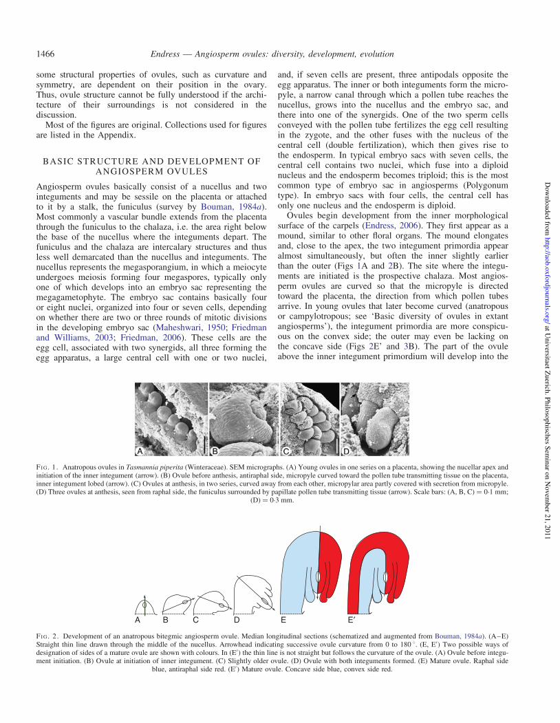
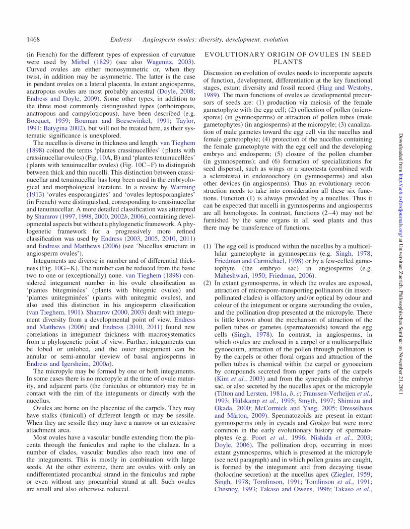
surface of the carpels (Endress, 2006). They first appear as amound, similar to other floral organs. The mound elongatesand, close to the apex, the two integument primordia appearalmost simultaneously, but often the inner slightly earlierthan the outer (Figs 1A and 2B). The site where the integu-ments are initiated is the prospective chalaza. Most angios-perm ovules are curved so that the micropyle is directedtoward the placenta, the direction from which pollen tubesarrive. In young ovules that later become curved (anatropousor campylotropous; see ‘Basic diversity of ovules in extantangiosperms’), the integument primordia are more conspicu-ous on the convex side; the outer may even be lacking onthe concave side (Figs 2E’ and 3B). The part of the ovuleabove the inner integument primordium will develop into the
A B C D E E¢
F IG . 2 . Development of an anatropous bitegmic angiosperm ovule. Median longitudinal sections (schematized and augmented from Bouman, 1984a). (A–E)Straight thin line drawn through the middle of the nucellus. Arrowhead indicating successive ovule curvature from 0 to 180 8. (E, E′) Two possible ways ofdesignation of sides of a mature ovule are shown with colours. In (E′) the thin line is not straight but follows the curvature of the ovule. (A) Ovule before integu-ment initiation. (B) Ovule at initiation of inner integument. (C) Slightly older ovule. (D) Ovule with both integuments formed. (E) Mature ovule. Raphal side
blue, antiraphal side red. (E′) Mature ovule. Concave side blue, convex side red.
F IG . 1 . Anatropous ovules in Tasmannia piperita (Winteraceae). SEM micrographs. (A) Young ovules in one series on a placenta, showing the nucellar apex andinitiation of the inner integument (arrow). (B) Ovule before anthesis, antiraphal side, micropyle curved toward the pollen tube transmitting tissue on the placenta,inner integument lobed (arrow). (C) Ovules at anthesis, in two series, curved away from each other, micropylar area partly covered with secretion from micropyle.(D) Three ovules at anthesis, seen from raphal side, the funiculus surrounded by papillate pollen tube transmitting tissue (arrow). Scale bars: (A, B, C) ¼ 0.1 mm;
(D) ¼ 0.3 mm.
Endress — Angiosperm ovules: diversity, development, evolution1466
at Univ
ersitaet Zuerich
. Philo
sophisch
es Sem
inar o
n N
ovem
ber 2
1, 2
011
http
://aob.o
xfo
rdjo
urn
als.org
/D
ow
nlo
aded
from

nucellus. Thus the nucellus is only delimited with the initiationof the integuments. If there are no integuments there is conse-quently no nucellus, and the ovule is morphologically undif-ferentiated. The inner integument always forms a tubularsheath around the nucellus. The outer integument is more vari-able. In orthotropous ovules it also forms a tubular sheath. Inanatropous or campylotropous ovules it either also forms atubular sheath (annular outer integument) or it is incompleteon the concave side (semi-annular, hood-shaped outer integu-ment). Whether it becomes annular or semi-annular dependson the speed of ovule curvature or the speed of progressionof the outer integument primordium from the convextowards the concave side. The greater the speed of ovule cur-vature and the lower the speed of progression of the outer inte-gument, the more the development is towards semi-annular.Basic developmental processes in ovules of the modelspecies Arabidopsis thaliana were described by Bowman(1993) and Schneitz et al. (1995). Curved ovules have araphe, a sometimes conspicuous area through which the vascu-lar bundle runs from the funiculus to the chalaza. It is notuseful to describe the raphe as a product of ‘congenitalfusion of the outer integument with the funiculus’ (Tiltonand Lersten, 1981a, p. 452). The raphe develops by the exten-sion of the ovule on one side beyond the funiculus and belowthe outer integument and is merely a developmentalby-product of ovule curvature (Fig. 2E).In terms of developmental genetics and the ‘ABC of floral
development’, an additional class D MADS-box gene wasassumed to determine ovule identity (Angenent et al., 1995;Colombo et al., 1995; Dreni et al., 2007). From subsequentstudies, ovule identity appears to be promoted by the sharedactivity of C and D class genes (Favaro et al., 2003;Pinyopich et al., 2003). The D gene lineage originated fromduplication of the C gene lineage; the C lineage may originallyhave operated in female organ identity (including ovules) and,following duplication, underwent sub-functionalization bywhich the D lineage specialized in ovule morphology(Kramer et al., 2004). A crucial event in ovule morphogenesisis integument initiation. As mentioned above, with integumentinitiation, the nucellus, chalaza and funiculus also becomedefined (Schneitz et al., 1995; Schneitz, 1999), and the gen-etics of this differentiation, in which NOZZLE plays an impor-tant role, was first studied in Arabidopsis (Schneitz et al.,
1997, 1998a; Balasubramaniam and Schneitz, 2000, 2002).So far, numerous genes have been recovered that are involvedin ovule development (Gasser et al., 1998; Schneitz et al.,1998a; Kelley and Gasser, 2009; Skinner and Gasser, 2009).This genetic diversity may reflect part of the morphologicaldiversity of angiosperm ovules.The putative evolutionary sequence of parts in ovules corre-
sponds to the developmental sequence (nucellus – innerintegument – outer integument – funiculus) and is reflectedby molecular genetics of development in Arabidopsis, whichshows that it is easier to affect the outer integument and funi-culus than the nucellus and inner integument (Schneitz et al.,1998b). As an analogy of this developmental sequence, instamen development the central part, the anther, also developsbefore the filament. Both ovules and stamens have in commonthat the part where meiosis takes place differentiates first. Itmay be assumed that this is functionally important becausedifferentiation of meiocytes involves a highly specialized sur-rounding tissue which, in turn, requires a relatively long timefor development. In contrast, the other (outer, basal) partshave a simpler structure and differentiate more rapidly.
BASIC DIVERSITY OF OVULES IN EXTANTANGIOSPERMS
Ovule diversity is expressed in several respects. The mainaspects of diversity are ovule size, degree of ovule curvature,nucellus thickness, integument number and thickness, for-mation of the micropyle, funiculus length, degree of vascular-ization of the ovule and diverse histological differentiation(e.g. hypostase, postament and endothecium).Ovules are around 0.5 mm long in many angiosperm clades
at the time of fertilization. In small-ovuled clades they areapprox. 0.15 mm long. Large ovules may reach .2 mm.Diversity of ovule size may be extensive even at the level oforders.Ovules can be straight or curved in various ways. Straight,
uncurved ovules (orthotropous, atropous; Fig. 3A) are radiallysymmetric (or disymmetric). In curved ovules the nucellus iseither straight (anatropous ovule, Fig. 3B) or it is also involvedin the curvature (campylotropous ovule, Fig. 3C). Ovules thatare only slightly curved are hemitropous (hemianatropous).The three terms ‘orthotrope’, ‘anatrope’ and ‘campulitrope’
F IG . 3 . Diversity in ovule curvature. Median longitudinal microtome sections. (A) Orthotropous ovule (Barclaya rotundifolia). (B) Anatropous ovule (Asiminatriloba). (C) Campylotropous ovule (Hypecoum pendulum). In the zig-zag micropyle (C) the part formed by the outer integument is marked with a green arrow,
the part formed by the inner integument with a red arrow. Scale bars: (A) ¼ 0.2 mm; (B, C) ¼ 0.1 mm.
Endress — Angiosperm ovules: diversity, development, evolution 1467
at Univ
ersitaet Zuerich
. Philo
sophisch
es Sem
inar o
n N
ovem
ber 2
1, 2
011
http
://aob.o
xfo
rdjo
urn
als.org
/D
ow
nlo
aded
from
(in French) for the different types of expression of curvaturewere used by Mirbel (1829) (see also Wagenitz, 2003).Curved ovules are either monosymmetric or, when theytwist, in addition may be asymmetric. The latter is the casein pendant ovules on a lateral placenta. In extant angiosperms,anatropous ovules are most probably ancestral (Doyle, 2008;Endress and Doyle, 2009). Some other types, in addition tothe three most commonly distinguished types (orthotropous,anatropous and campylotropous), have been described (e.g.Bocquet, 1959; Bouman and Boesewinkel, 1991; Taylor,1991; Batygina 2002), but will not be treated here, as their sys-tematic significance is unexplored.
The nucellus is diverse in thickness and length. van Tieghem(1898) coined the terms ‘plantes crassinucellees’ (plants withcrassinucellar ovules) (Fig. 10A, B) and ‘plantes tenuinucellees’(plants with tenuinucellar ovules) (Fig. 10C–F) to distinguishbetween thick and thin nucelli. This distinction between crassi-nucellar and tenuinucellar has long been used in the embryolo-gical and morphological literature. In a review by Warming(1913) ‘ovules eusporangiates’ and ‘ovules leptosporangiates’(in French) were distinguished, corresponding to crassinucellarand tenuinucellar. A more detailed classification was attemptedby Shamrov (1997, 1998, 2000, 2002b, 2006), containing devel-opmental aspects but without a phylogenetic framework. A phy-logenetic framework for a progressively more refinedclassification was used by Endress (2003, 2005, 2010, 2011)and Endress and Matthews (2006) (see ‘Nucellus structure inangiosperm ovules’).
Integuments are diverse in number and of differential thick-ness (Fig. 10G–K). The number can be reduced from the basictwo to one or (exceptionally) none. van Tieghem (1898) con-sidered integument number in his ovule classification as‘plantes bitegminees’ (plants with bitegmic ovules) and‘plantes unitegminees’ (plants with unitegmic ovules), andalso used this distinction in his angiosperm classification(van Tieghem, 1901). Shamrov (2000, 2003) dealt with integu-ment diversity from a developmental point of view. Endressand Matthews (2006) and Endress (2010, 2011) found newcorrelations in integument thickness with macrosystematicsfrom a phylogenetic point of view. Further, integuments canbe lobed or unlobed, and the outer integument can beannular or semi-annular (review of basal angiosperms inEndress and Igersheim, 2000a).
The micropyle may be formed by one or both integuments.In some cases there is no micropyle at the time of ovule matur-ity, and adjacent parts (the funiculus or obturator) may be incontact with the rim of the integuments or directly with thenucellus.
Ovules are borne on the placentae of the carpels. They mayhave stalks (funiculi) of different length or may be sessile.When they are sessile they may have a narrow or an extensiveattachment area.
Most ovules have a vascular bundle extending from the pla-centa through the funiculus and raphe to the chalaza. In anumber of clades, vascular bundles also reach into one ofthe integuments. This is mostly in combination with largeseeds. At the other extreme, there are ovules with only anundifferentiated procambial strand in the funiculus and rapheor even without any procambial strand at all. Such ovulesare small and also otherwise reduced.
EVOLUTIONARY ORIGIN OF OVULES IN SEEDPLANTS
Discussion on evolution of ovules needs to incorporate aspectsof function, development, differentiation at the key functionalstages, extant diversity and fossil record (Haig and Westoby,1989). The main functions of ovules as developmental precur-sors of seeds are: (1) production via meiosis of the femalegametophyte with the egg cell; (2) collection of pollen (micro-spores) (in gymnosperms) or attraction of pollen tubes (malegametophytes) (in angiosperms) at the micropyle; (3) canaliza-tion of male gametes toward the egg cell via the nucellus andfemale gametophyte; (4) protection of the nucellus containingthe female gametophyte with the egg cell and the developingembryo and endosperm; (5) closure of the pollen chamber(in gymnosperms); and (6) formation of specializations forseed dispersal, such as wings or a sarcotesta (combined witha sclerotesta) in endozoochory (in gymnosperms) and alsoother devices (in angiosperms). Thus an evolutionary recon-struction needs to take into consideration all these six func-tions. Function (1) is always provided by a nucellus. Thus itcan be expected that nucelli in gymnosperms and angiospermsare all homologous. In contrast, functions (2–4) may not befurnished by the same organs in all seed plants and thusthere may be transference of functions.
(1) The egg cell is produced within the nucellus by a multicel-lular gametophyte in gymnosperms (e.g. Singh, 1978;Friedman and Carmichael, 1998) or by a few-celled game-tophyte (the embryo sac) in angiosperms (e.g.Maheshwari, 1950; Friedman, 2006).
(2) In extant gymnosperms, in which the ovules are exposed,attraction of microspore-transporting pollinators (in insect-pollinated clades) is olfactory and/or optical by odour andcolour of the integument or organs surrounding the ovules,and the pollination drop presented at the micropyle. Thereis little known about the mechanism of attraction of thepollen tubes or gametes (spermatozoids) toward the eggcells (Singh, 1978). In contrast, in angiosperms, inwhich ovules are enclosed in a carpel or a multicarpellategynoecium, attraction of the pollen through pollinators isby the carpels or other floral organs and attraction of thepollen tubes is chemical within the carpel or gynoeciumby compounds secreted from upper parts of the carpels(Kim et al., 2003) and from the synergids of the embryosac, or also secreted by the nucellus apex or the micropyle(Tilton and Lersten, 1981a, b, c; Franssen-Verheijen et al.,1993; Hulskamp et al., 1995; Smyth, 1997; Shimizu andOkada, 2000; McCormick and Yang, 2005; Dresselhausand Marton, 2009). Spermatozoids are present in extantgymnosperms only in cycads and Ginkgo but were morecommon in the early evolutionary history of spermato-phytes (e.g. Poort et al., 1996; Nishida et al., 2003;Doyle, 2006). The pollination drop, occurring in mostextant gymnosperms, which is presented at the micropyle(see next paragraph) and in which pollen grains are caught,is formed by the integument and from decaying tissue(holocrine secretion) at the nucellus apex (Ziegler, 1959;Singh, 1978; Tomlinson, 1991; Tomlinson et al., 1991;Chesnoy, 1993; Takaso and Owens, 1996; Takaso et al.,
Endress — Angiosperm ovules: diversity, development, evolution1468
at Univ
ersitaet Zuerich
. Philo
sophisch
es Sem
inar o
n N
ovem
ber 2
1, 2
011
http
://aob.o
xfo
rdjo
urn
als.org
/D
ow
nlo
aded
from

1996; Stutzel and Rowekamp, 1997; Gelbart and vonAderkas, 2002; Wagner et al., 2007; Nepi et al., 2009).In extant gnetophytes, most of which are insect-pollinated,not only do the fertile ovules present pollination drops but,in addition, the male units are associated with pollen drop-producing sterile ovules (e.g. Endress, 1996). There islittle known on attraction by colour or scent in earlyseed plants, if there were animal pollinators at all. Infossils, such as Elkinsia or Lagenostoma (Lyginopteris)(Rothwell et al., 1989; Rothwell and Serbet, 1992;Taylor and Taylor, 1993), the organs surrounding ovulesand forming a cupule are spreading, and in Lagenostoma(Lyginopteris) from the Carboniferous they appear tohave had glands, which could have been protective orattractive. For paleozoic pteridosperms pollination dropswere inferred by Rothwell (1977).
(3) For canalization of male gametes a narrow tube is needed,the micropyle, which is formed by the single integumentin extant gymnosperms and, within the carpel in angios-perms, by one or two integuments. The evolution of theintegument in gymnosperms is unclear. An integumentmay have evolved several times (Li et al., 1997).Evolution from a group of branches of dichotomous branch-ing systems (telomes in the terminology of Zimmermann,1952) that became associated with megasporangia hasmost often been suggested as a first step (e.g. Andrews,1963; Smith, 1964; Long, 1967; Gillespie et al., 1981;Retallack and Dilcher, 1988; Rothwell and Scheckler,1988; Galtier and Rowe, 1989; Rothwell et al., 1989;Stewart and Rothwell, 1993; Hilton and Edwards, 1999;Kelley and Gasser, 2009), and such ovule precursorswithout a micropyle are called ‘pre-ovules’ (e.g. Stewartand Rothwell, 1993; Hilton and Edwards, 1996). Kenrickand Crane (1997) suggested a derivation of this megaspor-angium envelope from a group of sterile megasporangia. Insome cases, the integument may be derived from two units,depending on symmetry and the number of vascularbundles in fossils, such as in the early CarboniferousMitrospermum of cordaitean affinity (Long, 1977), theLate Carboniferous Stephanospermum (Drinnan et al.,1990) andCallospermarion of potential medullosan affinity(Eggert and Delevoryas, 1960), or the PermianChoanostoma of unknown affinity (Klavins et al., 2001).Such an envelope of several branches may function incatching microspores from the air by producing specificlocal airflows, if they were wind-pollinated, but not inexact canalization of microspores to the nucellus apex(Niklas, 1981a, b). Alternatively, it may be that the integu-ment developed from the outer wall of a differentiation ofthe ovule apex, the pollen chamber (including the lagenos-tome and salpinx) (Meeuse and Bouman, 1974), as inextinct early seed plants, which probably functioned incanalization (called a ‘pollen-receiving mechanism’ inTaylor et al., 2009). However, from descriptions it isoften not clear whether the pollen chamber and its wall inextinct seed plants is a structure at the morphologicallevel, i.e. by direct origin from the ovule apex, or merelyat the histological level, i.e. by differential decay of tissue(e.g. Long, 1960; Hilton and Bateman, 2006), similar tothe pollen chamber of some living gymnosperms (e.g.
Friedman, 1987; Douglas et al., 2007, for Ginkgo). Sucha hypothesis, derivation of the integument from the pollenchamber wall, would only make sense if the pollenchamber was a structure at the morphological level, aproblem not considered by Meeuse and Bouman (1974).
(4) Protection of the nucellus in gymnosperms is by the integu-ment. In early seed plants sterile branches could have func-tioned for protection (see preceding paragraph). In moreadvanced gymnosperms protection is more complex. InBennettitales the so-called interseminal scales could haveplayed this role (e.g. Crane, 1985; Stockey and Rothwell,2003; Crane and Herendeen, 2009; Rothwell et al., 2009).In Ephedra (Gnetales), it is a pair or a whorl of 3–4 fusedbracts (‘seed envelope’), which may be free in the upperpart (Rydin et al., 2010), perhaps also in the fossil gneto-phyte Siphonospermum (Rydin and Friis, 2010). Perhapsthe triangular seeds of Doylea (Stockey and Rothwell,2009) and Rugonella (Friis et al., 2009) and the quadrangu-lar seeds of Ephedrispermum, Buarcospermum,Lignierispermum and Lobospermum (Friis et al., 2009)also have a similar structure with an outer envelope ofthree or four units. Among gymnospermous ovules/seeds,such an outer envelope, commonly with layers of sclerifiedtissue, is known from Gnetales, Erdtmanithecales andBennettitales (Friis et al., 2007, 2009). Whether it is hom-ologous in all three orders has not been resolved. In angios-perms it is not only the integuments that protect the nucellusbut also the carpel or syncarpous gynoecium in which theovules are enclosed. In addition, angiosperms ancestrallyprobably have two integuments (e.g. Doyle and Endress,2000; Endress and Doyle, 2009).
(5) Protection of the young sporophyte in gymnosperms isprovided by closure (sealing) of the pollen chamber and/or integument (Takaso and Bouman, 1986; Serbet andRothwell, 1995). Some earlier information is summarizedin Singh (1978).
(6) The presence of wings and potential anemochory (in gym-nosperms) was described in seeds of the Late Devonian(Rowe, 1992, 1997) and Permian (Dilcher et al., 1997),and they occur in some extant conifers and in some gneto-phytes (Welwitschia, some Ephedra species). A sarcotestawas described for some Carboniferous (Taylor, 1965) andPermian seeds (Klavins et al., 2001; Hilton et al., 2003),and is present, at least in part, in allmajor extant gymnospermclades. In gymnosperm seeds, commonly also a sclerified(‘mechanical’) layer is developed. In angiosperms, the pos-ition of this layer is diverse but macrosystematically signifi-cant (inner or outer surface or central area of theintegument or of both integuments; Corner, 1976).A detailedhistorical survey on the use of terms for different layers wasgiven by Schmid (1986), and surveys on the diversity of seedcoats were given by Corner (1976) and Bouman (1984b).
SYMMETRY OF OVULES IN GYMNOSPERMSAND ANGIOSPERMS
Curvature directly affects the symmetry of ovules.Orthotropous ovules are radially symmetric or disymmetric,
Endress — Angiosperm ovules: diversity, development, evolution 1469
at Univ
ersitaet Zuerich
. Philo
sophisch
es Sem
inar o
n N
ovem
ber 2
1, 2
011
http
://aob.o
xfo
rdjo
urn
als.org
/D
ow
nlo
aded
from
whereas curved ovules are monosymmetric or asymmetric.However, curvature is not the only factor influencing ovulesymmetry.
In early spermatophytes the distinction between radiosper-mic (radially symmetric) and platyspermic (disymmetric)ovules/seeds (Rothwell, 1986; Stewart and Rothwell, 1993)appears to be phylogenetically important at the macro-level(Rothwell and Serbet, 1994; Doyle, 1996, 2006; Hilton andBateman, 2006). Both radiospermic and platyspermic seedsappear in the Devonian (Chaloner et al., 1977; Gillespieet al., 1981). In contrast, in angiosperms, radial and flattenedovules may occur in closely related groups, and flattenedovules may simply be understood by space constraints in theovary locule.
Angiosperm ovules are probably derived from radiospermicseeds among gymnosperms (Meyen, 1982). However, thisdoes not preclude that ancestral angiosperm ovules were ana-tropous and, thus, monosymmetric. Changes in the symmetryof ovules occurred multiple times in angiosperms and at differ-ent systematic levels. Araceae are an example in which thereare multiple changes even within a single family (French,1986; Mayo et al., 1997). Thus radial symmetry, disymmetryand monosymmetry in gymnosperms and angiosperms arenot directly related; their significance is not at the same levels.
EVOLUTION OF BITEGMY FROM UNITEGMYON THE WAY TO ANGIOSPERM EVOLUTION
The inner integument of angiosperms is probably homologousto the single integument in their gymnospermous ancestors(Reinheimer and Kellogg, 2009), and the outer integumentmay have been derived from the wall of a cupule and reductionof ovule number to one per cupule in angiosperm ancestors[e.g. Glossopteridales or Caytoniales (e.g. Gaussen, 1946;Stebbins, 1974, 1976; Doyle 1978, 1994, 2008)]. InCaytoniales the cupules are curved and could have given risein this way directly to an anatropous bitegmic ovule(Gaussen, 1946; Doyle, 1978, 2008). Curved cupules arealso known from some other fossil gymnosperms [Ktalenia,Umkomasia, Corystospermales; Petriellaea, Petriellaeales(e.g. Taylor and Archangelsky, 1985; Taylor et al., 1994,2006, 2009; Klavins et al., 2002; Frohlich, 2003; Taylor andTaylor, 2009)]. Curvature in these various gymnosperms andthe crown-group angiosperms could also have independentlyarisen several times.
There is little information on ovule evolution from the per-spective of molecular developmental genetics. In a study onthe interaction of NOZZLE and INNER NO OUTER, and thatof PHABULOSA and WUSCHEL, Sieber et al. (2004,p. 333) are ‘tempted to speculate that bitegmic ovules ofextant angiosperms might have been derived through the ‘split-ting’ of an integument in a unitegmic precursor’. They hesitateto acknowledge an interpretation of other authors, of the outerintegument being derived from a leaf simply because theYABBY gene INO is expressed on its outer side as otherYABBY genes are on the outer (abaxial) side of leaves. Itshould be taken into consideration that the activity of thesegenes may not be organ specific but pattern or symmetryspecific, i.e. promoting dorsiventrality (Eshed et al., 2001).However, this hypothesis of origin of the second integument
is of interest as it is at variance with the common paleobotani-cal hypothesis that the second (the outer integument) inangiosperms was co-opted from an already existing organ,such as the cupule (see ‘Evolutionary origin of ovules inseed plants’ point 4).
DEVELOPMENT OF CURVATURE IN OVULES
Ovule curvature is a predominant feature in angiosperms, incontrast to the commonly uncurved ovules in gymnosperms.Curvature ensures a position of the micropyle close to theattachment site of the ovule and thus close to the placentafor an easy uptake of pollen tubes (Figs 1A–D, 2A–E, E′).It was first discussed by Agardh (1858) that ovule curvatureis functionally important to take up pollen tubes by the micro-pyle. That the micropyle (called ‘mamelon d’impregnation’)plays a role for the development of the seed was alreadydescribed by Brongniart (1827) and Mirbel (1829), whoobserved the pollen tube (called ‘tube conducteur’ and‘filet’) from the style to the micropyle in someCucurbitaceae, but the exact function of pollen tubes wasnot recognized until the ground-breaking work of Hofmeister(1849).I contend that curvature and the advent of bitegmy are inti-
mately functionally connected and that the development of theouter integument is responsible for curvature. There is evi-dence from several sources: (1) differential thickness of theouter integument in curved and uncurved angiosperm ovules;(2) structure of the outer integument in abnormally uncurvedovules in angiosperms; (3) behaviour of ovule mutants inmodel organisms without normal curvature; and (4) develop-ment of ovules in the rare gymnosperms that have curvedovules.
(1) The outer integument is often thinner in orthotropousovules than in anatropous ovules, or is even lacking. Forinstance, it is only two cell layers thick in the orthotropousBarclaya of Nymphaeaceae, in contrast to more layers inthe other, anatropous Nymphaeaceae; only two in theorthotropous family Piperaceae and Hydnoraceae, in con-trast to more in most other magnoliids; and also onlytwo(to three) in the orthotropous Proteaceae andPlatanaceae, in contrast to the anatropousNelumbonaceae among Proteales; and even unitegmy inthe orthotropous Sabiaceae (Igersheim and Endress,1998; Endress and Igersheim, 1999). In ovules that havean early forming aril, full ovule curvature seems to beslightly hindered. Often such ovules are not fully anatro-pous but more or less hemianatropous. Examples areMyristicaceae (Endress, 1973; Igersheim and Endress,1997), Xanthorrhoeaceae (Steyn and Smith, 1998), andin Sapindaceae ovules are also sometimes hemianatropousbut later they become campylotropous without goingthrough an anatropous stage. In Mauloutchia(Myristicaceae), in which a well-developed aril islacking, the ovule appears to be more anatropous, or atleast the outer integument appears to be semi-annular(Sauquet et al., 2003).
(2) Abnormal orthotropous (or hemianatropous) ovules oftenoccur in plants that normally have anatropous ovules,
Endress — Angiosperm ovules: diversity, development, evolution1470
at Univ
ersitaet Zuerich
. Philo
sophisch
es Sem
inar o
n N
ovem
ber 2
1, 2
011
http
://aob.o
xfo
rdjo
urn
als.org
/D
ow
nlo
aded
from

especially in ovaries with numerous ovules. These arefound in various major clades of angiosperms.Interestingly, in these abnormal ovules that failed todevelop a normal curvature, the outer integument isoften reduced; it is shorter or completely lacking.Another concomitant trait is that the funiculus is oftenlonger than in the normal anatropous ovules. Suchovules were described and drawn in a number of publi-cations for single species. Both features together(reduced outer integument and long funiculus) were docu-mented for Takhtajania (Winteraceae, Fig. 4A; Endresset al., 2000), Butomus (Butomaceae, Fig. 4B; pers. obs.;Fernando and Cass, 1996), Burmannia (Burmanniaceae,one integument completely lacking; Rubsamen, 1986),Berberis (Berberidaceae; Schleiden, 1839; Mauritzon,1938), Caltha (Ranunculaceae, outer integument growingbackward; Kapil and Jalan, 1962), Holoptelea(Ulmaceae; Capoor, 1937), Parnassia (Parnassiaceae,one integument completely lacking; Saxena, 1964b),Dalechampia (Euphorbiaceae, outer integument growingbackward in one case; Singh and Pal, 1968),Podostemon (Podostemaceae, funiculus length not indi-cated; Hammond, 1937), Jussieua (Onagraceae; Khan,1942) and Foeniculum (Apiaceae, integument growingbackward; Gupta, 1964). In the following taxa, the outerintegument was not reduced, but the funiculus waslonger: Hitchenia (Zingiberaceae; Panchaksharappa,1962), Platystemon (Papaveraceae; Bocquet and Bersier,1960), Bergenia (Saxifragaceae; Saxena, 1969),Heuchera (Saxifragaceae; Mauritzon, 1933), Saxifraga(Saxifragaceae; Saxena, 1964a), Neptunia (Fabaceae;Singh and Shivapuri, 1935) and Rhodamnia (Myrtaceae;Mauritzon, 1939a). In the following taxa the outer integu-ment was misshapen, but the funiculus was not longer:Podophyllum emodi (Berberidaceae, Fig. 4C; pers. obs.)and Pterospermum (Malvaceae; Venkata Rao, 1954). Insome Myrtaceae the pluriovulate placenta regularly con-tains some reduced ovules (‘ovulodes’) (see also‘Direction of the ovule initiation squence in placentaewith numerous ovules’). In Angophora, ovulodes haveonly one integument (Prakash, 1969).
(3) The behaviour of mutants in the model species A. thalianastrongly supports the role of the outer integument in ovulecurvature. In ant (aintegumenta) (Elliott et al., 1996;Schneitz et al., 1997, 1998b; Skinner and Gasser, 2009)and hll (huellenlos) (Schneitz et al., 1997, 1998b) both inte-guments are lacking and the ovule is not curved, and the
same occurs in double, triple and quadruple mutants invol-ving ant, stk (seedstick), shatterproof1 (shp1) and shp2(shatterproof2) (Losa et al., 2010) and in triple mutantswith cna (corona), phb (phabulosa) and phv (phavoluta)(Kelley et al., 2009). In wus (wuschel) ovules both integu-ments are lacking and there is no normal curvature(Gross-Hardt et al., 2002). In ino (inner no outer), theouter integument is almost lacking (initiated but notfurther developed) but the inner is well developed, andthere is no curvature (Baker et al., 1997; Schneitz et al.,1997; Villanueva et al., 1999; Gallagher and Gasser,2008; Skinner and Gasser, 2009); the same was found inan ino mutant of the basal angiosperm Annona squamosa(Lora et al., 2011). In pfs2 ( pretty few seeds2) mutantswith the PFS2 transgene some ovules are normal;however, in some ovules the outer integument is reduced(shorter than the inner) and the ovule is only halfwaycurved (Park et al., 2004, 2005). In kan1 (kanadi1) andkan2 (kanadi2) double mutants the outer integumentremains short and the ovules are not curved (Eshed et al.,2001); the same is the case in kan1, kan2, kan3 triplemutants (McAbee et al., 2006) and in seu, cyp85A2-1double mutants (Nole-Wilson et al., 2010). In sin1 (shortinteguments 1) (Robinson-Beers et al., 1992; Baker et al.,1997) and sin2 mutants (Broadhvest et al., 2000) both inte-guments remain short and the ovules are only weaklycurved, and similarly in ag/AG stk shp1 shp2 mutants(Brambilla et al., 2007). In bel1 (bell) the two integumentsare not distinct, forming 2–4 irregular mounds, and there isno regular curvature (Robinson-Beers et al., 1992, Schneitzet al., 1997). Taking these results from several mutantstogether, there is a distinct pattern: the shorter the outer inte-gument, the less the ovule is curved. If the outer integumentis not formed at all, there is no curvature. For the formationof curvature, apparently an outer integument needs to bepresent and it must have a slant in order to develop asymme-trically from early on. A seemingly contradictory case wasdescribed and interpreted for the ovules of rice, inwhich cur-vature is said to be ‘associated closely with the extent ofinner integument growth’ (Yamaki et al., 2005, p. 408).However, these ovules are almost uncurved and not easilycompared with anatropous or campylotropous ovules.
(4) Among extant gymnosperms, Podocarpaceae are the onlygroup that shows prominent ovule curvature during devel-opment. Although Podocarpaceae do not have an outerintegument, they have a special structure, the ‘epimatium’.The epimatium looks like the hood-shaped outer
F IG . 4. Abnormal orthotropous ovules on a multiovulate placenta (asterisks). (A) Takhtajania perrieri. (B) Butomus umbellatus. (C) Podophyllum emodi.Abnormal hemitropous ovule with short outer integument marked with an arrow. Scale bars: (A) ¼ 0.15 mm; (B) ¼ 0.1 mm; (C) ¼ 0.5 mm.
Endress — Angiosperm ovules: diversity, development, evolution 1471
at Univ
ersitaet Zuerich
. Philo
sophisch
es Sem
inar o
n N
ovem
ber 2
1, 2
011
http
://aob.o
xfo
rdjo
urn
als.org
/D
ow
nlo
aded
from

integument of an anatropous ovule in angiosperms. Theepimatium is involved with curvature (‘anatropy’) of theovule (Doyle, 1945; Tomlinson et al., 1991; Tomlinson,1992; Del Fueyo, 1999; Mill et al., 2001; Tomlinson andTakaso, 2002). Thus it is in some way functionally analo-gous to an outer integument. However, morphologically itcorresponds to the ovuliferous scale in other conifers(Tomlinson, 1992). This difference in homology is alsoreflected in the precocious developmental appearance ofthe epimatium compared with the ovule (Tomlinson,1992). The function of this ‘anatropy’ in Podocarpaceaeis ‘pollen scavenging’: the pollination drop spreads inthe area around the micropyle, and pollen grains trappedin the pollination drop will then float into the micropylarcavity (Tomlinson, 1991; Tomlinson et al., 1991). Theepimatium is also involved in seed dispersal as itbecomes fleshy and brightly coloured.
DIVERSITY OF OVULE POSITION IN THEGYNOECIUM AND REPERCUSSIONS OF OVARY
ARCHITECTURE ON OVULE SHAPE
The position of the ovules in the gynoecium and ovary loculearchitecture have repercussions on details of ovule structure,especially ovule curvature and symmetry. Therefore, thisaspect is important to understand details of ovule shape(Endress, 1994a, 2008). In most locule architectures, anatro-pous or campylotropous ovules direct their micropyle closeto the placenta by their curvature, which facilitates pollentube growth from the carpels directly into the micropyles(Fig. 1). For this reason, curved ovules are so common inangiosperms and appear to be the basal state for extant angios-perms (see ‘Basic diversity of ovules in extant angiosperms’;Doyle, 2008; Endress and Doyle, 2009). Completely orthotro-pous ovules occur in four different placenta positions or loculearchitectures. (1) A single ovule on a basal placenta in anarrow locule (single ascidiate carpel or syncarpous gynoe-cium) (Figs 6A, 7). The micropyle is directed towards thestylar canal and is connected with it either through secretionor by contiguity. This is a relatively widespread situation ina number of unrelated angiosperm groups [e.g. Piperales ofmagnoliids, some Araceae of basal monocots, Juglandaceae,Myricaceae, and Urticaceae of rosids, Polygonaceae of theasterid alliance; Endress, 2008]. (2) Numerous ovules onlaminar-diffuse or protruding-diffuse placentae in a spaciouslocule (locule much larger than the ovules), filled withsecretion [Barclaya of the ANITA grade, Fig. 3A; Hydnora
of magnoliids; Acorus (Rudall and Furness, 1997; Buzgoand Endress, 2000); Pistia, Fig. 6D (Buzgo, 1994); andHydrocharis of basal monocots (Igersheim et al., 2001);Xiphidium coeruleum of core monocots (slightly curved),Figs 5B, 6E; Akebia of basal eudicots (slightly curved),Figs 5C, 6F (Endress and Igersheim, 1999); and Cytinus ofcore eudicots (Igersheim and Endress, 1998)]. (3) Severalovules on parietal placentae, the micropyles being contiguouswith an adjacent placenta, Fig. 6B [e.g. Houttuynia cordata ofPiperales, Fig. 5A (Endress, 1994b); Mayacaceae of monocots,Casearia of rosids (Endress, 2008); Scaphocalyx of rosids (vanHeel, 1973)]. (4) Ovules with a long, curved funiculus, whichdirects the micropyle to their own placenta [e.g. Helianthemumof core eudicots, Fig. 6C (Nandi, 1998)].If orthotropous ovules are present in narrow locules and not
in a central basal position, they cannot be completely radiallysymmetrical on architectural grounds, but are somewhatcurved at their base. This needs to be emphasized because
A B C
D E F
F IG . 6. Orthotropous ovules and conditions of ovary locule architecturesunder which they occur (schematic, only one integument is drawn in eachovule for simplicity; augmented and modified from Endress, 1994a). (A–C)Ovary or locules not filled with secretion. (A) Single ovule with basal placenta(LS gynoecium) (e.g. Piperaceae, Juglandaceae, Urticaceae). (B) Ovules onparietal placentae with the micropyle directed toward another placenta (TSovary) (e.g. Casearia, Salicaceae; Mayaca, Mayacaceae). (C) Ovules withlong funiculi curved to their own placenta (LS gynoecium) (e.g.Helianthemum, Cistaceae). (D–F) Ovary or locules filled with secretion(secretion shaded blue). (D) Basal diffuse placenta (LS gynoecium) (e.g.Pistia, Araceae). (E) Laminar-diffuse placenta (TS carpel/ovary) (e.g.Barclaya, Nymphaeaceae; Hydrocharis, Hydrocharitaceae; Akebia,Lardizabalaceae, shown, in the latter ovules slightly curved at anthesis). (F)Axile placenta (TS ovary) (e.g. Acorus, Acoraceae; Xiphidium,
Haemodoraceae).
F IG . 5 . Orthotropous ovules and ovary locule architecture (arrows point to micropyles). (A) Houttuynia cordata. (B) Xiphidium coeruleum. (C) Akebia quinata
(with secretory hairs and secretions between the ovules). Scale bars: (A) ¼ 0.2 mm; (B) ¼ 0.3 mm; (C) ¼ 0.1 mm.
Endress — Angiosperm ovules: diversity, development, evolution1472
at Univ
ersitaet Zuerich
. Philo
sophisch
es Sem
inar o
n N
ovem
ber 2
1, 2
011
http
://aob.o
xfo
rdjo
urn
als.org
/D
ow
nlo
aded
from

this is the case in some of the basal angiosperms, such asAmborella (Endress, 1986, 1994b), Chloranthaceae (Endress,1987, 1994b) and Ceratophyllum (Igersheim and Endress,1998). There has been debate in the literature as to whetherthese ovules are orthotropous or anatropous, without fully rea-lizing the problem of architectural constraint. The question isstill open in most cases of whether they are basically anatro-pous but could not complete the curvature because oflimited space in the locule or, vice versa, whether they arebasically orthotropous but were forced to make a slightcurve at the base because of spatial constraint. Thus the curva-ture may be considered to be a superimposed restriction. Thisquestion is especially interesting in Amborella, the sister of allother extant angiosperms, which has been described both asanatropous and as orthotropous (see, for example, Bailey andSwamy, 1948; Endress, 1986; Endress and Igersheim, 1997,2000b; Doyle and Endress, 2000; Tobe et al., 2000; Yamadaet al., 2001b; Endress and Doyle, 2009). Similar cases alsooccur in basal monocots (Potamogeton, Igersheim et al.,2001; Shamrov, 2006) and basal eudicots (Platanus, Endressand Igersheim, 1999). An example of nearly orthotropousovules due to developmental constraint on original anatropyoccurs in Avicennia (Acanthaceae; Borg and Schonenberger,2011).An especially obvious case of dependence of ovule shape on
locule architecture are ascending orthotropous ovules with themicropyle contiguous with the transition area of the stylarcanal into the locule. In some groups with this architectureone or both integuments elongate to keep pace with theelongation of the locule (Fig. 7A) [Elatostema andMyriocarpa of Urticaceae, Rosales (Fagerlind, 1944);Didymeles of Buxales (ovules hemitropous) (von Balthazaret al., 2003)]. In these Urticaceae the reverse situation alsooccurs: instead of an elongation of the integuments, hairsfrom the pollen tube transmitting tissue grow into the micro-pyle or onto the nucellus (Fig. 7C) (Fagerlind, 1944). Thuseither the integument(s) or the pollen tube transmitting tractgrows actively toward its functional counterpart. In some
Polygonaceae (Caryophyllales) with the same gynoeciumarchitecture, a third possibility occurs: growth of a nucellarbeak into the stylar canal (Fig. 7B) (Edman, 1929).
DIRECTION OF OVULE CURVATURE ANDCARPEL CURVATURE
Ovules are commonly formed at or close to the margin ofcarpels (in a marginal or sub-marginal position). To reachangiospermy, carpel margins curve inward so that the ventralcarpel surface and the ovules become enclosed. The directionof carpel curvature is always the same in angiosperms.However, this is not the case for the direction of ovule curva-ture. Although in most cases the direction of curvature in ana-tropous or campylotropous ovules is the same as that of carpelcurvature, there are other cases in which ovule curvature is inthe opposite direction. These opposite patterns were earlierdistinguished as apotropous and epitropous (introduced inthe Latin form by Agardh, 1858; used, for example, byEngler, 1931). They were defined in a complicated and imprac-tical way, as the relationship with carpel curvature was notconsidered. To make this connection and thus have a simplerdefinition, the terms ‘syntropous’ (Fig. 8A) and ‘antitropous’(Fig. 8B) were introduced (Endress, 1994a). Syntropousovules are common, e.g. in basal angiosperms (Endressand Igersheim, 2000a), whereas antitropous ovules are wellknown, e.g. from many Malpighiales (Sutter and Endress,1995; Merino Sutter et al., 2006; Matthews andEndress, 2008, 2011), from Anacardiaceae (Bachelier andEndress, 2007, 2009) and some Rosaceae (Juel, 1918).The predominant syntropous direction is optimal for ovules
arranged in two parallel series (the most common pattern inangiosperms) because it leaves enough space between theopposite ovules for curvature to bring the micropyle close tothe placenta and thus to form a direct passage for pollentubes. Pollen tubes grow from the placenta around the funicu-lus to the other side of the ovule where the micropyle is located(Fig. 1C, D). With the antitropous pattern this would not be the
i n
A B C
o
F IG . 7. Different patterns of contact between the ovule and pollen tube trans-mitting tissue of the stylar canal in gynoecia with a single orthotropous ovuleon a basal placenta (schematic, only one integument drawn in each ovule forsimplicity). (A) Integument(s) protruding into the stylar canal (i, e.g.Didymeles, Didymelaceae, schematized after von Balthazar et al., 2003;Elatostema, Urticaceae, schematized after Fagerlind, 1944). (B) Nucellus(nucellar beak, n) protruding into the stylar canal (e.g. Polygonum,Polygonaceae, schematized after Edman, 1929). (C) Carpel forming an obtura-
tor (o, e.g. Elatostema, Urticaceae, schematized after Fagerlind, 1944).
A B
F IG . 8. Different orientations of curved ovules with respect to carpel curva-ture (denoted by a red line). (A) Syntropous. Curvature of the ovule in the samedirection as the curvature of the carpel. (B) Antitropous. Curvature of the ovule
in the opposite direction to that of the carpel.
Endress — Angiosperm ovules: diversity, development, evolution 1473
at Univ
ersitaet Zuerich
. Philo
sophisch
es Sem
inar o
n N
ovem
ber 2
1, 2
011
http
://aob.o
xfo
rdjo
urn
als.org
/D
ow
nlo
aded
from

case; the antitropous pattern commonly occurs in carpels withonly one or two ovules. Associated with antitropous ovules isoften a special auxiliary structure, an obturator, which isnecessary for pollen tubes to bridge the gap between thepollen tube transmitting tract and the micropyle. For instance,in Malpighiales, many clades have antitropous ovules withobturators, and in clades with syntropous ovules within theorder obturators are lacking. As seen throughout the angios-perms, obturators may have different developmental origins.Often they are formed from the carpel flanks above theplacenta.
In multiovulate carpels or gynoecia, the curvature is syntro-pous. In linear (axile or parietal) placentae the longitudinalseries of ovules are curved away from each other (Fig. 1C).In median placentae the ovules are curved downwards (andalso outwards, if there are many). In laminar-diffuse placentaethey are curved downwards if in the ascidiate zone (Fig. 6F)and sideways if in the plicate zone. In free central placentae(which represent part of the ascidiate zone), the ovules arealso curved downwards.
DIRECTION OF THE OVULE INITIATIONSEQUENCE IN PLACENTAE WITH NUMEROUS
OVULES
In long, linear placentae or in diffuse placentae (with a numberof ovules side by side), there is often a gradient in ovule devel-opment. Payer (1857), based on .30 examples, reported threepatterns of ovule initiation: (1) basipetal in axile placentae(Fig. 9A); (2) acropetal in parietal placentae; and (3) bidirec-tional in placentae that are axile at the base and parietal ontop. A fourth type, intercalation of new ovules between olderones, was described later (Kaplan, 1968). Okamoto (1984)hypothesized that initiation begins closest to the site of theformer floral apex which, to some extent, conforms withPayer’s axile and parietal placentation initiation types.However, Okamoto (1984) considered only nine genera.Unfortunately, he did not take into account that axile placentaecan be present in both the symplicate and synascidiate zone ofthe ovary and that only in the latter would the site of theformer floral apex be on top of the placenta. Whereas this cor-relation between placenta form and direction of ovule develop-ment appears to be a trend in the material studied by Payer(1857), there are also cases in which parietal placentae showbasipetal ovule initiation (such as Dicentra and Mentzelia,Payer, 1857; and Berberidopsis, Ronse De Craene, 2004). Asexpected, the direction of initiation is also commonly basipetalin flowers with a free central placenta (e.g. Sundberg, 1982;Caris et al., 2000; Caris and Smets, 2004). Payer’s (1857)study is as yet the largest comparative study. A limitednumber of species was described by Sattler (1973), andmostly only single species by other authors. Thus theproblem of the direction of ovule initiation needs more criticalstudy.
The pattern of ovule initiation in multiovulate placentaeapparently depends on the direction of elongation/maturationof the ovary locules. In ovaries with basipetal ovule initiationthere is intercalary locule elongation mainly at the base. Thepattern of elongation and expansion is especially complex inOrchidaceae. Although ovule development was studied in a
number of species, generally the authors focused on singleovules and did not study the development of the placentaand the sequence of initiation of numerous ovules. The parietalplacenta may be multiply branched or crested, and ovuleinitiation begins in the centre of each branch or crest (Abe,1972; Yeung and Law, 1997; Tsai et al., 2008); the crestsmay be convoluted (Zhang and O’Neill, 1993). From myexperience, there is often a centrifugal direction of ovuleappearance on a placenta. The ovules in the centre of anextended placenta appear the most developed, whereas theovules at the periphery are less developed (Fig. 9D–F).However, whether this reflects the initiation sequence or apost-initiation delay of the peripheral ovules remains an openquestion. In some groups with linear placentae and acropetalsuccession of ovule development, the later formed ovulesmay remain small and be sterile (e.g. Altingiaceae, Fig. 9B;Hamamelidaceae, Fig. 9C; Anemoneae, Ren et al., 2010). Ingroups with diffuse placentae, the peripheral ovules may besterile, such as in some Myrtaceae (e.g. Eucalyptus, Davis,1968).
OVULE BASE AND VASCULARIZATION
Commonly, ovules have a short funiculus at the base, markingthe transition from the placenta. Long funiculi, as an extreme,are uncommon and scattered in angiosperms. They show someconcentration in Caryophyllales (notably in both the coreclade, e.g. Cactaceae, Leins and Schwitalla, 1985; and theextended clade, e.g. Plumbaginaceae, De Laet et al., 1995)and Brassicales (Brassicaceae, Shamrov, 2002a; andCaricaceae, Ronse Decraene and Smets, 1999). In basalangiosperms long funiculi are present, e.g. in Monodora(Annonaceae) (Igersheim and Endress, 1997), and in basalmonocots, e.g. in Hydrocleys (Limnocharitaceae) (Igersheimet al., 2001). As another extreme, sessile ovules with an exten-sive attachment area occur, e.g. among monocots in palms(Robertson, 1976) and among core eudicots in someMalpighiales (Irvingiaceae and Caryocaraceae, Matthews andEndress, 2011; and Achariaceae, van Heel, 1973).The chalaza also belongs to the ovule base, as it is located
below the nucellus and the integuments. In pachychalazalovules the chalaza is relatively long compared with the nucel-lus and integuments, and the embryo sac becomes partly‘inferior’ (e.g. Lauraceae; Endress, 1972). In perichalazalovules the chalaza is long only in the median symmetryplane of the ovule but shorter in the transverse areas (e.g.Austrobaileyaceae; Endress, 1980). Both pachychalazal andperichalazal ovules occur scattered in several angiospermgroups.Anatomically, ovules are connected with the placenta by a
vascular bundle, which commonly extends through the funicu-lus and raphe and ends in the chalaza. However, in many casesthe vascular bundle branches in the chalaza and branchesextend into the outer, inner or both integuments. In somecases, branching begins in the raphe, and such vascularbranches reach the outer integument by by-passing thechalaza. This is especially the case in ovules with an extensiveattachment area (Matthews and Endress, 2011), but not onlythere (Tokuoka and Tobe, 2003). Vascularized integumentsoccur especially in massive ovules and ovules that develop
Endress — Angiosperm ovules: diversity, development, evolution1474
at Univ
ersitaet Zuerich
. Philo
sophisch
es Sem
inar o
n N
ovem
ber 2
1, 2
011
http
://aob.o
xfo
rdjo
urn
als.org
/D
ow
nlo
aded
from
into large seeds (Kuhn, 1928; Corner, 1976; Bouman, 1984a).This may be seen in Araceae with vascularized and non-vascularized taxa (French, 1986) or Rhizophoraceae, wherethe large-seeded mangrove genera are more vascularizedthan non-mangrove genera (Matthews and Endress, 2011).Other clades with extensive vascularization in the integu-ment(s) are Fagales and Euphorbiaceae (e.g. Tokuoka andTobe, 2003). Among extant basal angiosperms (ANITAgrade) ovules with a vascular bundle in the (outer) integumentoccur in Trimeniaceae (Endress and Sampson, 1983). Asanother extreme, there are ovules without a vascular bundleor with only an undifferentiated procambial strand, e.g. inOrchidaceae (Asparagales, monocots; Shamrov, 2006), theparasitic Cytinaceae (Malvales, rosids; Teryokhin, 1981;
Shamrov, 2003, 2006) and some asterids, such as inLamiales and Gentianaceae (Gentianales; Shamrov, 1990,2003). Such ovules without differentiated vascular bundlesare generally restricted to groups with numerous small seeds(Endress, 2010, 2011).
NUCELLUS STRUCTURE IN ANGIOSPERMOVULES
Nucellus thickness (measured in the number of cell layersabove and around the meiocyte) greatly varies in angiosperms,but is relatively constant in larger clades. Nucellus structure isbest compared at the time of prophase of meiosis. At this stage,the tissues of the nucellus are still intact. Later, during meiosis
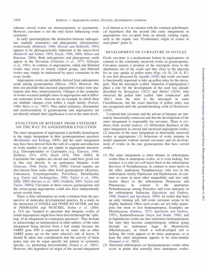
F IG . 9 . Gradients in ovule development on multiovulate placentae. (A–C) Acropetal and basipetal development of ovules on the placenta (and reduction of lastformed ovules in some cases). (A) Basipetal. Solanum sisymbrifolium. (B) Acropetal, upper ovules reduced. Liquidambar orientalis. (C) Acropetal, upper ovulesgreatly reduced. Corylopsis willmottiae. (D–F) Peripheral delay in development. (D) Passiflora holosericea. After ovule initiation. (E) Passiflora holosericea.
After integument initiation. (F) Nymphaea tetragona. Scale bars: (A, D, E) ¼ 0.05 mm; (B, F) ¼ 0.2 mm; (C) ¼ 0.1 mm.
A
G H I J K
B C D E F
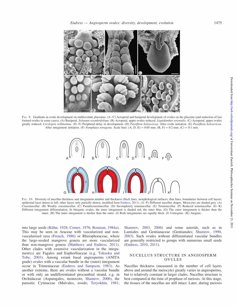
F IG . 10. Diversity of nucellus thickness and integument number and thickness (thick lines, morphological surfaces; thin lines, boundaries between cell layers;epidermal layer drawn in full, other layers only partially drawn; modified from Endress, 2011). (A–F) Different nucellus shapes. Meiocytes are shaded grey. (A)Crassinucellar. (B) Weakly crassinucellar. (C) Pseudocrassinucellar. (D) Incompletely tenuinucellar. (E) Tenuinucellar. (F) Reduced tenuinucellar. (G–K)Different integument differentiation. In bitegmic ovules, the inner integument is shaded red, the outer blue. (G) The outer integument is thicker than the
inner. (H) The inner integument is thicker than the outer. (I) Both integuments are equally thick. (J) Unitegmic. (K) Ategmic.
Endress — Angiosperm ovules: diversity, development, evolution 1475
at Univ
ersitaet Zuerich
. Philo
sophisch
es Sem
inar o
n N
ovem
ber 2
1, 2
011
http
://aob.o
xfo
rdjo
urn
als.org
/D
ow
nlo
aded
from

and embryo sac formation, tissue adjacent to the gametophyticparts is generally crushed and it becomes difficult to determinethe number of cell layers around the embryo sac. The classicaldistinction between crassinucellar (with one or more hypoder-mal cell layers between the meiocyte and nucellus apex) andtenuinucellar (with no hypodermal cell layer between themeiocyte and nucellus apex) has been modified into a finergrid of six types based on the recognition that they characterizelarger clades.
In surveys on floral diversity at the levels of eudicots(Endress, 2010) and angiosperms (Endress, 2011), the follow-ing ovule classification according to nucellus thickness wastentatively used: (a) crassinucellar (with more than one hypo-dermal cell layer between meiocyte and nucellus apex;Fig. 10A) (e.g. Cinnamomum, Lauraceae; Endress, 1972); (b)weakly crassinucellar (with just one hypodermal cell layerbetween the meiocyte and nucellus apex; Fig. 10B) (e.g.Dichelostemma, Asparagaceae, Berg, 1996); (c) pseudocrassi-nucellar (without a hypodermal cell layer between the meio-cyte and nucellus apex, but with periclinal cell divisions inthe epidermis of the nucellus apex; Fig. 10C) (e.g.Sagittaria, Alismataceae, Johri, 1935); (d ) incompletely tenui-nucellar (without a hypodermal cell layer between the meio-cyte and nucellus apex, but with hypodermal tissue at thenucellus flanks and/or below the meiocyte) (e.g. Nemophila,Boraginaceae; Berg, 2009; Fig. 10D); (e) tenuinucellar(without any hypodermal tissue in the nucellus; Fig. 10E)(e.g. Orphium, Gentianaceae; Hakki, 1997); and ( f ) reducedtenuinucellar (as in tenuinucellar but meiocyte partly extend-ing below the nucellus, thus with a partly ‘inferior’ position;Fig. 10F) (e.g. Phyllis, Rubiaceae; Fagerlind, 1936).
The terms in Endress (2010, 2011) had been used in partearlier by other authors, such as ‘pseudocrassinucellar’(Davis, 1966) and ‘reduced tenuinucellar’ (as ‘reduced vari-ation of tenuinucellate’) (Shamrov, 1998). Shamrov (1998)divised an elaborate classification with types and sub-types,primarily based on histogenesis, which is sensible. However,a drawback is that a type may change during developmentinto another: the ovules of Butomus are at first crassinucellateand then become medionucellate (Shamrov, 1998, p. 403). Apracticable typology should be based on a fixed developmentalstage. Also, it would be premature to make too elaborate atypology before its systematic relevance has been tested.
Other nucellus differentiations of systematic interest are anucellus cap and a nucellus beak. They are sometimes con-fused in the literature. A cap refers to the anatomical/histologi-cal structural level and a beak to the morphological level. Acap is formed by multiple periclinal divisions in the epidermisof the nucellus apex, sometimes, in addition, in the originallyhypodermal tissue, whereas a beak is an acuminate protrusionof the nucellar apex, which can grow partly or entirely throughthe micropyle (Merino Sutter et al., 2006). A beak is usuallyassociated with a cap, but not vice versa.
In thin ovules (tenuinucellar, incompletely tenuinucellar andweakly crassinucellar), often an endothelium is formed on theinside of the inner integument (Kapil and Tiwari, 1978;Endress, 2010, 2011). In such ovules, during embryo sac for-mation the nucellus dissolves around the embryo sac and theembryo sac becomes adjacent to and contiguous with theinside of the inner integument. The endothelium appears to
supply the embryo sac with certain substances. An endo-thelium is especially present in certain rosids and in asterids(see ‘Features of ovules and macrosystematics of angios-perms’). It is noteworthy that an endothelium is also presentin Lactoris, a magnoliid with exceptionally thin (incompletelytenuinucellar) ovules (Tobe et al., 1993) and in Canrightia, aLower Cretaceous magnoliid fossil (Friis and Pedersen, 2011).In both these magnoliids the endothelium appears to be persist-ent during seed development, in contrast to those core eudicotsin which it occurs.
INTEGUMENT THICKNESS
Integuments are two or more cell layers thick.Two-cell-layered integuments are developmentally derivedfrom the dermatogen (‘dermal integuments’, Bouman,1984a). Integuments of more than two cell layers are eitherderived from the dermatogen and become thicker later indevelopment by periclinal cell divisions in the epidermis orthey are derived from both dermatogen and sub-dermatogen(‘subdermal integuments’, Bouman, 1984a). Whether integu-ments are dermal or sub-dermal is correlated with their thick-ness at anthesis and later. It cannot be used for any deductionof homology (in contrast to Tilton and Lersten, 1981a).Integument thickness is a relatively stable character, and
therefore of interest at the macrosystematic level. This isespecially so for the relative thickness of outer and inner inte-gument (Fig. 10G–I; Endress and Matthews, 2006; Endress,2010, 2011), which are constrained by the subsequent differen-tiation of the seed coat (for systematic significance, see‘Features of ovules and macrosystematics of angiosperms’).In wild-type Arabidopsis the outer integument is regularlytwo cell layers thick and the inner three. However, in themutant ats (aberrant testa shape), which has an abnormalseed coat, the entire cover is only three cell layers thick(Leon-Kloosterziel et al., 1994).The inner integument appears to be constrained in thickness
by the outer integument. This can be deduced from two obser-vations. (1) If the inner integument is longer than the outer, itis considerably thicker in the micropyle where it is not sur-rounded by the outer. (2) In abnormal ovules with an exceed-ingly long inner integument forming the micropyle (in speciesin which the micropyle is normally formed by both integu-ments), the exposed rim of the inner integument becomesmuch thicker than in normal ovules (Eschscholzia; Sacharand Mohan Ram, 1958).
HOODED, SEMI-ANNULAR VS. CUP-SHAPED,ANNULAR OUTER INTEGUMENT
In curved (anatropous) ovules the outer integument is eitherhooded or cup-shaped, developmentally derived from a semi-annular or annular early stage, respectively (Yamada et al.,2001a). Hooded vs. cup-shaped outer integuments have beenbelieved to represent two fundamentally different organiz-ations by some authors (Kato and Imaichi, 1993). However,as it looks now, this difference is rather a consequence ofminor differences in the speed of developmental curvature ofthe ovule, and not a fundamental difference.
Endress — Angiosperm ovules: diversity, development, evolution1476
at Univ
ersitaet Zuerich
. Philo
sophisch
es Sem
inar o
n N
ovem
ber 2
1, 2
011
http
://aob.o
xfo
rdjo
urn
als.org
/D
ow
nlo
aded
from

It has been suggested that a hood-shaped (semi-annular)outer integument is primitive in angiosperms (Kato andImaichi, 1993; Matsui et al., 1993; Umeda et al., 1994;Imaichi et al., 1995; Yamada et al., 2003a, b). In our compara-tive study on carpels and ovules through all families of basalangiosperms (as mentioned in the Introduction), we found adiversity of anatropous ovules with semi-annular (hood-shaped) and annular (cup-shaped) outer integument. Oftenboth co-occur at low systematic levels. This indicates thatthere is no fundamental difference between the two forms. Ifanatropous ovules are primitive at the level of crown-groupangiosperms, which is likely (discussion in Endress andDoyle, 2009), this does not automatically mean that thehood shape is also primitive. The hood shape is probablyonly a consequence of the early developmental curvature.Thus it is only the propensity to form hood-shaped outer inte-guments that is primitive. For instance, the outer integument isnot semi-annular but annular in Illiciaceae, Canellaceae,Myristicaceae, Degeneriaceae and Himantandraceae(Igersheim and Endress, 1997). It has also been repeatedlyfound that ovules with both a semi-annular and an annularouter integument occur in the same family or even the samegenus or species (e.g. Calycanthus, Peumus, Siparuna;Endress and Igersheim, 1997; Nuphar, Nymphaea,Aristolochia, Thottea; Igersheim and Endress, 1998), indicat-ing that the two features are not of fundamental evolutionarydifference but may merely depend on subtle developmentaldifferences. The earlier the curvature begins, the more pro-nounced will the semi-annular form become. It may beassumed that if a certain threshold of retardation on one sideis surpassed, instead of a complete ring, a partial ring and acompensatory additional lobe are formed. Thus the additionallobe is probably not a fundamentally different part as assumedby Matsui et al. (1993) or Umeda et al. (1994). This interpret-ation is also supported by those species in which abnormalorthotropous ovules were found, which always had a cup-shaped (annular) outer integument, as opposed to the normalanatropous ovules (see ‘Development of curvature in ovules’).
MULTIPLE EVOLUTION OF UNITEGMY FROMBITEGMY WITHIN ANGIOSPERMS
If bitegmy was so important for curvature in angiosperms, whywas it possible that unitegmy (Fig. 10J) evolved secondarilywithin angiosperms several times, and yet in many cases theovules did not give up their curvature? In anatropous uniteg-mic ovules (as found in most asterids and some other eudi-cots), the single integument probably does not correspond toan outer or an inner integument but is an evolutionarilycomplex structure in which both participate, although theycan no longer be distinguished morphologically (as discussedby Bouman and Calis, 1977). This process of amalgamation ofthe two integuments is shown by those rare genera in whichboth bitegmic and unitegmic conditions are present [e.g.Impatiens (McAbee et al., 2005; Colombo et al., 2008,Kelley and Gasser, 2009) and Coriaria (Matthews andEndress, 2004)]. In contrast, there is some evidence that uni-tegmy in basal angiosperms evolved by reduction and loss ofthe outer integument (Igersheim and Endress, 1998).
Whether in orthotropous unitegmic ovules of core eudicotsthe only integument corresponds to the inner integument isunknown, but would be interesting to study (e.g. in Fagalesand Rosales). That in several cases unitegmy goes togetherwith orthotropy is plausible if the outer integument, which isresponsible for curvature, is reduced. This is likely to be thecase in (unitegmic) Peperomia, as in other (bitegmic)Piperaceae the outer integument is already shortened. InRafflesiaceae and Cytinaceae the outer integument is stillpresent but highly reduced, and Ceratophyllum has only oneintegument (Igersheim and Endress, 1998). Also in the (ortho-tropous) Urticaceae the outer integument is shortened(Fagerlind, 1944).
FURTHER REDUCTION OF INTEGUMENTS ANDENTIRE OVULES
Ovules also became reduced in other respects in some angios-perm clades. Integuments and then entire ovules were succes-sively reduced in the parasitic order Santalales (Fagerlind,1947, 1948; Shamrov et al., 2001, Brown et al., 2010;Endress, 2010, 2011). Brown et al. (2010) found inSantalales that lack differentiation into nucellus and integu-ments that integument-associated genes were expressed inthe periphery of the ovule. It is not necessary to assume con-genital fusion between the nucellus and integument(s). Underthe assumption of non-differentiation (i.e. lack of nucellus andinteguments) this evolutionary process can be seen as transfer-ence of function, in which the peripheral zone of the ovule thatis normally formed by the (outer) integument is now formedby the periphery of the undifferentiated ovule. Some myco-trophic Gentianaceae also evolved highly reduced ovuleswithout differentiation into nucellus and integument(ategmic) (Fig. 10K; Goebel, 1933; Bouman et al., 2002).
LOBATION OF INTEGUMENTS IN ANGIOSPERMOVULES
It has been argued that the lobes on the rim of the inner inte-gument in some basal angiosperms (Magnolia) (Figs 1B, 11E;Umeda et al., 1994; Herr, 1995), in some other angiosperms(van Heel, 1970, 1976) or in some mutants of Arabidopsis(Park et al., 2004) may represent remnants or atavisms ofancient telomic structures. In earlier publications we expresseddoubts concerning this interpretation (Endress and Igersheim,1997; Igersheim and Endress, 1997). We argued that if anannular young plant part that spans an opening of a certaindiameter in early development needs to close in later develop-ment, i.e. to form a closed pore, it can do this only by lobation(in the longitudinal direction) (Fig. 11C, D) or by irregularthickening, which also leads to a sort of lobation (in the trans-verse direction) (Fig. 13 in Igersheim and Endress, 1997), orby both processes in combination (Fig. 11A, B).From these principles of plant growth and development,
several hypotheses can be derived. These hypotheses can betested with the wide array of species studied in our laboratory,covering all families of basal angiosperms, including theANITA grade, magnoliids and the basal grades of monocotsand eudicots (Igersheim and Endress, 1997, 1998; Endressand Igersheim, 1998, 1999, 2000a; Igersheim et al., 2001).
Endress — Angiosperm ovules: diversity, development, evolution 1477
at Univ
ersitaet Zuerich
. Philo
sophisch
es Sem
inar o
n N
ovem
ber 2
1, 2
011
http
://aob.o
xfo
rdjo
urn
als.org
/D
ow
nlo
aded
from

According to our studies, lobed integuments are widespreadin basal angiosperms, not only in inner, but also in outer inte-guments. Of 131 taxa in which the lobation of the integumentswas studied [from all families of the ANITA grade (exceptHydatellaceae), magnoliids, basal eudicots and basal mono-cots] 124 have two integuments. Of these 124 taxa, in 57 theinner integument is lobed and in 40 the outer integument islobed. We tested the following three hypotheses with ourmaterial.
(1) The outer integument is relatively more often lobed thanthe inner if both integuments form the micropyle,because the circumference of the outer integument islarger than that of the inner. Result: of the 124 taxa withtwo integuments studied, in 34 taxa both integumentsform the micropyle. Of those, in 14 the inner integumentis lobed, in 13 the outer integument is lobed (38 % ofall). However, in the 85 taxa in which the micropyle isformed by the inner integument alone, the inner integu-ment is lobed in 40 taxa, but the outer only in 21 taxa(only 25 % of all). Thus hypothesis 1 receives somesupport.
(2) Ovules in which the integuments do not form a micropylemore often have unlobed integuments (inner integuments)than those that have a micropyle, because they do not haveto compensate for their initially wide circumference.Result: of the seven taxa studied without a micropyle(five bitegmic, two unitegmic), both integuments or theonly integument are lobed in only two (one bitegmic,
one unitegmic). The number of cases is too small butdoes not speak against the hypothesis.
(3) Integuments are more often lobed in ovules with thicknucelli than in ovules with thin nucelli, because theyneed to make a closure with an initially wider circumfer-ence. Result: two measurements were taken, always fromanthetic flowers: (a) nucelli were measured at the broadestdiameter at anthesis (ANITA grade, magnoliids, basalmonocots and basal eudicots); (b) nucelli were measuredat their base (only ANITA grade and magnoliids). Inboth cases the result was not clear. Ovules with broadnucelli did not show integument lobation more oftenthan ovules with narrow nucelli. Of course, to use thesize of mature nucelli is a very crude measure for a firstapproximation. For more useful results it would be impor-tant to measure the size of nucelli at a stage shortly afterintegument initiation. The size difference of nucellibetween these two stages is considerable, and thusmature nucelli may not be suitable for meaningful deduc-tions in this matter.
However, these preliminary results on basal angiosperms lendsome support to the view that lobes on the rim of integumentsin angiosperms are not remnants of ancient organs but merelythe result of a developmental necessity for the closure of themicropyle, as discussed above. The peculiar deep lobation ofthe single integument in Juglandaceae that puzzled van Heeland Bouman (1972) may have a different cause. The ovulehas an unusual position in the centre of the unilocular ovaryformed by two carpels. The two integument lobes develop inthe median symmetry plane of the carpels. There may betwo explanations that have not been discussed by von Heeland Bouman (1972): (1) the lobation originates by space con-straint in the slit-shaped two-carpellate locule; or (2) ovulegrowth is controlled by the two carpels, which is reflected inthe two-parted integument.
EVOLUTION OF ADDITIONAL ‘INTEGUMENTS’IN ANGIOSPERMS
Here and there in angiosperms there are ovules with a thirdenvelope, which is called a ‘third integument’ if similar tothe two normal integuments, or an ‘aril’, if it is more differentand especially if delayed in development and functional as anattractive organ in fruit. Such extra envelopes can be presentbetween the normal integuments, such as the third integumentin some Annonaceae (Christmann, 1989), but commonly theyappear on the outside of the two normal integuments, such asthe arils in Myristicaceae (Endress, 1973) and many othergroups. As they appear later than the normal integuments,their position is commonly distanced from the integumentsand closer to the funiculus. More often than previouslyassumed, there are ‘reduced arils’, the role of which isunknown in most cases. They appear as small mounds atanthesis and do not develop further [e.g. among basal angios-perms, in Nymphaeaceae (Igersheim and Endress, 1998); inSarcandra of Chloranthaceae (Endress and Igersheim, 1997)and Canellaceae (Igersheim and Endress, 1997); inRanunculales, in Nandina of Berberidaceae (Endress andIgersheim, 1997); and in Buxales, in Buxus and Notobuxus
A B
C D
E
F IG . 11. Two possibilities for morphological closure of a tubular structurethrough differential directional growth: irregular thickening or lobation. (A,B) End of a tube seen from above. (A) Tube open. (B) Tube partially closedby irregular thickening (arrows). (C, D) End of the tube seen from the side.(C) Tube open. (D) Tube partially closed by lobe formation (arrows). (E)Micropyle with lobed inner (i) and outer (o) integuments. Illicium floridanum
(from Igersheim and Endress, 1997, Fig. 75). Scale bar: (E) ¼ 0.1 mm.
Endress — Angiosperm ovules: diversity, development, evolution1478
at Univ
ersitaet Zuerich
. Philo
sophisch
es Sem
inar o
n N
ovem
ber 2
1, 2
011
http
://aob.o
xfo
rdjo
urn
als.org
/D
ow
nlo
aded
from

of Buxaceae (von Balthazar and Endress, 2002) andDidymelaceae (von Balthazar et al., 2003); in the latter twofamilies, this inconspicuous feature supports their relation-ship]. In Arabidopsis a small extra mound is formed inovules of ucn (unicorn) mutants, which could represent apartial supernumerary integument (Schneitz et al., 1997;Schneitz, 1998b). To what extent third integuments and arilsin angiosperms are homologous is uncertain.
MICROPYLE
The preponderance of narrow micropyles at the time aroundfertilization indicates that it is important for a successful func-tioning. In a few taxa a micropyle is not formed so that thenucellus apex is exposed. It is expected that in these taxa thearchitecture of the ovary is such that the exposed nucellustouches the funiculus, the placenta or the inner ovary wall.In this way a narrow gate above the nucellus apex may alsobe provided, e.g. in Cassytha (Lauraceae; Endress andIgersheim, 1997), Hernandia (Hernandiaceae; Heo and Tobe,1995), Quisqualis (Combretaceae; Fagerlind, 1941), variousEuphorbiaceae s.l. (Sutter and Endress, 1995; Merino Sutteret al., 2006), and Hiptage and Stigmatophyllon(Malpighiaceae; Rao, 1940). In the mentioned Hernandia,Euphorbiaceae s.l. and Malpighiaceae a nucellar beak is con-tiguous with the ovary roof.In bitegmic ovules micropyles are commonly formed by
both integuments (amphistomic) (Fig. 3C) or the inner alone(endostomic) (Fig. 3A, B), and only rarely by the outeralone (exostomic). If both integuments participate in micro-pyle formation, they may form a straight or undulating canalor, if the integuments are not aligned straight, a zig-zag-shapedcanal (‘zig-zag micropyle’, Fig. 3C).Micropyles may be open, forming an open canal (Fig. 3B),
or closed (Fig. 3A, C). In the former the micropyle may besealed by a secretion (e.g. Annona, Igersheim and Endress,1997; Ornithogalum, Tilton and Lersten, 1981a, b; Beta,Olesen and Bruun, 1990). In the latter a closed pollen tubetransmitting tract is formed by post-genital fusion(Helianthus, Yan et al., 1991). Thus this diversity is analogousto that of the carpels sealed by secretion or by post-genitalfusion (Endress and Igersheim, 2000a). However, details ofthe histology of mature micropyles have only rarely beenstudied.For some time, it had been suggested that a zig-zag micro-
pyle is a basal feature of angiosperm ovules. This would fit thehypothesis by Gaussen (1946) (see ‘Evolution of bitegmy fromunitegmy on the way to angiosperm evolution’) of a derivationof the angiosperm ovule from a cupule. In addition, a zig-zagmicropyle is pronouncedly differentiated in Dilleniaceae, afamily earlier thought to be basal in angiosperms (Stebbins,1974). However, since in the meantime the phylogenetic pos-ition of Dilleniaceae has been found not to be basal, thezig-zag micropyle can be considered to be merely a conse-quence of excessive elongation of the outer integument conco-mitant with excessive curvature of the ovule. (This view doesnot contradict the hypothesis by Gaussen, 1946.) The hypoth-esis that a zig-zag micropyle is not basal is corroborated fromtwo different sides, the systematic distribution and the strongassociation with camplyotropous ovules. (1) Zig-zag
micropyle and campylotropous ovules are mainly present inmore derived groups; this combination is lacking in theANITA grade, and in magnoliids it is restricted toCanellaceae (Igersheim and Endress, 1997); in Magnoliaceaezig-zag micropyles have been reported in anatropous ovules.(2) In campylotropus ovules the antiraphal peripheral side ofthe ovule develops comparatively strongly; an expression ofthis strong development is also that the outer integumentbecomes exceedingly long and may far overtop the innerone; thus a zig-zag micropyle is formed. The combination ofa zig-zag micropyle and campylotropy tends to be present insome Ranunculales (Papaveraceae, Berberidaceae, weakly inMenispermaceae; Endress and Igersheim, 1999) and in anumber of major sub-clades of core eudicots, such asvarious Dilleniaceae (Svedelius 1911; Swamy andPeriasamy, 1955; Rao, 1957; Sastri, 1958; Stebbins, 1974;Imaichi and Kato, 1996); Fabales (various Fabaceae, Rau,1951; Prakash and Chan, 1976; Lakshmi et al., 1987;Ashrafunnisa and Pullaiah, 1999); Oxalidales(Elaeocarpaceae: Aristotelia, Mauritzon, 1934); Myrtales(Lythraceae: Sonneratia, Venkateswarlu, 1937; Kamelina,1985; Melastomataceae: Rhexia, Etheridge and Herr, 1968;Monochaetum, Ziegler, 1925; Myrtaceae: Acca, Pescadoret al., 2009; Baeckea, Mauritzon, 1939a; Psidium,Narayanaswami and Roy, 1960); Crossosomatales(Crossosomataceae: Crossosoma, Kapil and Vani, 1963);Brassicales (various Brassicaceae; Hakki, 1974; Prasad,1974; Bouman, 1975; Febulans and Pullaiah, 1990;Beeckman et al., 2000; Capparaceae: Cadaba, Narayana,1965; Niehburia, Arunalakshmi, 1985; Resedaceae: Reseda,Chaban and Yakovlev, 1974); and Malvales (variousMalvaceae; Venkata Rao, 1954; Singh, 1967). However, inCaryophyllales, although the ovules are prominently campylo-tropous, there is no zig-zag micropyle; there the prominentinner integument forms the micropyle. There are also casesof anatropous ovules with zig-zag micropyle, but they aremore rare. Examples are in Oxalidales (Elaeocarpaceae:Elaeocarpus, Venkata Rao, 1953); Geraniales (Geraniaceae:Geranium, Boesewinkel and Been, 1979); and Sapindales(Rutaceae: Aegle, Johri and Ahuja, 1957; Poincirus,Boesewinkel, 1978).
PATHWAY OF POLLEN TUBES FROM CARPELSTO OVULES
It was mentioned above that pollen tubes are attracted to themicropyle by secretions of the micropyle, nucellus andembryo sac (Hulskamp et al., 1995). That the embryo sacplays an important role in this attraction has long beenknown (e.g. Jensen, 1974). In ovules with degeneratedembryo sacs the pollen tubes do not grow into the micropylebut down to the chalaza (Elodea; Ernst-Schwarzenbach,1945). On the other hand, ovules with more than oneembryo sac attract more pollen tubes (Persea; Sedgley,1976). In Arabidopsis mutants with delayed embryo sac matu-ration, pollen tubes lose their way on the funiculus just beforeentering the micropyle (Shimizu and Okada, 2000). However,in normal Arabidopsis ovules there is a mechanism to preventreception of more than one pollen tube (Shimizu and Okada,2000; Shimizu, 2002; Palanivelu et al., 2003).
Endress — Angiosperm ovules: diversity, development, evolution 1479
at Univ
ersitaet Zuerich
. Philo
sophisch
es Sem
inar o
n N
ovem
ber 2
1, 2
011
http
://aob.o
xfo
rdjo
urn
als.org
/D
ow
nlo
aded
from

In plant groups with delayed ovule development, in whichthe ovules are far from fully developed at the time of pollina-tion, pollen tubes do not enter the ovules via the micropyle.This was first described as chalazogamy in Casuarina(Treub, 1891), and later found in many other Fagales (Sogoet al., 2004; Sogo and Tobe, 2006a, c, 2008) and inGarryales (Eucommia, Sogo and Tobe, 2006b). A moreneutral term, non-porogamy, considers that the entrance ofthe pollen tube to the ovules is not always via the chalaza insuch cases. Luza and Polito (1991) showed that in Juglans, agenus with delayed ovule maturation and non-porogamyunder natural conditions, the pollen tubes grew through themicropyle in post-anthetic flowers experimentally pollinatedat the time of embryo sac maturity.
Immaturity of ovules at the time of pollination has also beenfound in other groups, but in such cases either non-porogamydoes not occur or the pathway of the pollen tubes has not beenstudied (survey in Sogo and Tobe, 2006a). Examples are inmagnoliids (Magnoliaceae; Igersheim and Endress, 1997),monocots (Orchidaceae; Yeung and Law, 1997), basal eudi-cots [Eupteleaceae (Endress, 1969), Circaeasteraceae, someRanunculaceae and Lardizabalaceae, (Endress and Igersheim,1999). Platanaceae, Myrothamnaceae (Endress andIgersheim, 1999) Buxaceae: Sarcococca, Pachysandra (vonBalthazar and Endress, 2002)], Saxifragales [Altingiaceae,Cercidiphyllaceae, Daphniphyllaceae (Endress andIgersheim, 1999), some Hamamelidaceae (Endress, 1967,1977)] and Sapindales [some Anacardiaceae (Bachelier andEndress, 2007)]. Mutants with precocious stigma development,such as ettin in Arabidopsis (Sessions, 1997), may give a clueto the easy and multiple evolution of this displacement ofstigma and ovule maturity.
FEATURES OF OVULES ANDMACROSYSTEMATICS OF ANGIOSPERMS
As angiosperm phylogeny becomes increasingly resolved, theevolution of ovules can be traced in ever more detail. Largeclades can be better characterized by their ovule features andwe learn more about evolutionary idiosyncrasies of ovules.This was still not possible or not attempted when the lastround of big reviews on embryological features was published(Johri et al., 1992; Batygina et al., 2002). First attempts in thenew era were made by Endress (2003, 2005, 2010, 2011) andEndress and Matthews (2006), and are briefly summarizedhere. The names used here for the major clades of angiospermsare those in APG (2009).
ANITA grade
Ovules are crassinucellar. Only in Hydatellaceae, highlyspecialized, miniaturized wetland plants, are they reduced topseudocrassinucellar or incompletely tenuinucellar (Hamann,1975; Rudall et al., 2008), and in the water plant familyCabombaceae they are weakly crassinucellar (Ramji andPadmanabhan, 1965; Igersheim and Endress, 1998). Theovules are predominantly anatropous, but they are almostorthotropous in Amborellaceae, Chloranthaceae andCeratophyllaceae (see above) (position of Chloranthaceaeand Ceratophyllaceae in the ANITA grade uncertain, see, for
example, Endress and Doyle, 2009) and completely orthotro-pous in Barclaya of Nymphaeaceae (Schneider, 1978;Igersheim and Endress, 1998). They are bitegmic, except forthe unitegmic Ceratophyllaceae, another reduced water plantfamily. The outer integument is predominantly more thantwo cell layers thick [only 2–3 in Cabombaceae (Endressand Igersheim, 1997, 2000a; Igersheim and Endress, 1997,1998) and two in Hydatellaceae (Rudall et al., 2007)Barclaya of Nymphaeaceae (Igersheim and Endress, 1998)and Ascarina of Chloranthaceae (Endress and Igersheim,1997)]. The inner integument is predominantly 2–3 celllayers thick. The micropyle is commonly formed by theinner integument. However, in Hydatellaceae, some derivedNymphaeaceae, in Trimeniaceae and in some Chloranthaceaeboth integuments participate in the formation of the micropyle(Endress and Igersheim, 1997; Igersheim and Endress, 1997,1998; Rudall et al., 2007).
Magnoliids
Crassinucellar ovules are by far predominant. They areincompletely tenuinucellar in a few Piperales, such asLactoridaceae, Hydnoraceae, and Houttuynia of Saururaceae(Tobe et al., 1993; Igersheim and Endress, 1998). Theovules are predominantly anatropous, but orthotropous inSaururaceae, Piperaceae and Hydnoraceae (Piperales)(Igersheim and Endress, 1998), and almost orthotropous inGomortegaceae (Endress and Igersheim, 1997). They arebitegmic, except for Siparunaceae (Laurales) (Endress, 1972;Renner et al., 1997), and Peperomia and Hydnoraceae(Piperales) (Igersheim and Endress, 1998). The outer integu-ment is more than two cell layers thick (Endress andIgersheim, 1997, 2000a; Igersheim and Endress, 1997; Heoet al., 1998), except for Piperales (Igersheim and Endress,1998). The inner integument is mostly 2–3 cell layers thick.The micropyle is predominantly formed by the inner integu-ment, but by both integuments in Magnoliaceae,Canellaceae, Gomortegaceae, some Calycanthaceae, a fewMonimiaceae and several Piperales (Endress and Igersheim,1997; Igersheim and Endress, 1997, 1998).
Monocots
Crassinucellar and weakly crassinucellar ovules arecommon, the latter here and there in most orders and promi-nent in Commelinales and Zingiberales of commelinids.Pseudocrassinucellar ovules appear to be more common inbasal groups (Acorales, some Alismatales) than in morederived groups (a few Asparagales and Poales) (Rudall,1997; Igersheim et al., 2001; Endress, 2011). Incompletelytenuinucellar ovules are widespread, with increased frequencyin Dioscoreales, Pandanales and Poales (see Endress, 2011).Tenuinucellar ovules are only known from Orchidaceae(Asparagales) and Triuridaceae (Pandanales): both familiesmycotrophic (see Endress, 2011). Reduced tenuinucellarovules appear to be absent. Ovules are predominantly anatro-pous, and only rarely campylotropous. However, they areorthotropous in Acorales, several Alismatales, someAsparagales, Commelinales and Poales (see Endress, 2011).Ovules are bitegmic (Bouman, 1984a), with a few exceptions
Endress — Angiosperm ovules: diversity, development, evolution1480
at Univ
ersitaet Zuerich
. Philo
sophisch
es Sem
inar o
n N
ovem
ber 2
1, 2
011
http
://aob.o
xfo
rdjo
urn
als.org
/D
ow
nlo
aded
from
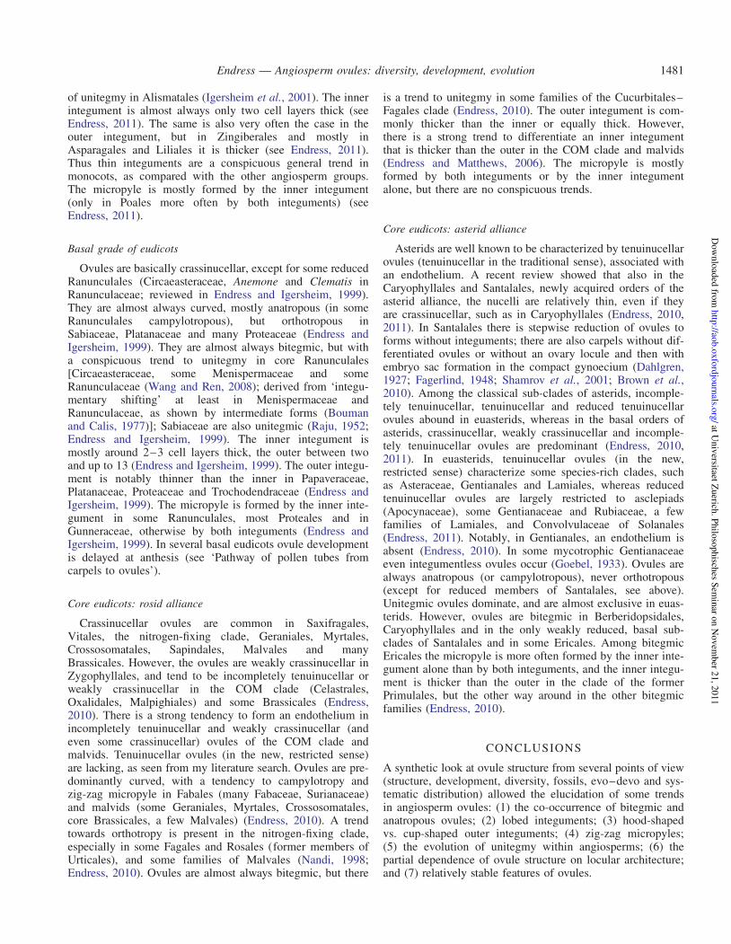
of unitegmy in Alismatales (Igersheim et al., 2001). The innerintegument is almost always only two cell layers thick (seeEndress, 2011). The same is also very often the case in theouter integument, but in Zingiberales and mostly inAsparagales and Liliales it is thicker (see Endress, 2011).Thus thin integuments are a conspicuous general trend inmonocots, as compared with the other angiosperm groups.The micropyle is mostly formed by the inner integument(only in Poales more often by both integuments) (seeEndress, 2011).
Basal grade of eudicots
Ovules are basically crassinucellar, except for some reducedRanunculales (Circaeasteraceae, Anemone and Clematis inRanunculaceae; reviewed in Endress and Igersheim, 1999).They are almost always curved, mostly anatropous (in someRanunculales campylotropous), but orthotropous inSabiaceae, Platanaceae and many Proteaceae (Endress andIgersheim, 1999). They are almost always bitegmic, but witha conspicuous trend to unitegmy in core Ranunculales[Circaeasteraceae, some Menispermaceae and someRanunculaceae (Wang and Ren, 2008); derived from ‘integu-mentary shifting’ at least in Menispermaceae andRanunculaceae, as shown by intermediate forms (Boumanand Calis, 1977)]; Sabiaceae are also unitegmic (Raju, 1952;Endress and Igersheim, 1999). The inner integument ismostly around 2–3 cell layers thick, the outer between twoand up to 13 (Endress and Igersheim, 1999). The outer integu-ment is notably thinner than the inner in Papaveraceae,Platanaceae, Proteaceae and Trochodendraceae (Endress andIgersheim, 1999). The micropyle is formed by the inner inte-gument in some Ranunculales, most Proteales and inGunneraceae, otherwise by both integuments (Endress andIgersheim, 1999). In several basal eudicots ovule developmentis delayed at anthesis (see ‘Pathway of pollen tubes fromcarpels to ovules’).
Core eudicots: rosid alliance
Crassinucellar ovules are common in Saxifragales,Vitales, the nitrogen-fixing clade, Geraniales, Myrtales,Crossosomatales, Sapindales, Malvales and manyBrassicales. However, the ovules are weakly crassinucellar inZygophyllales, and tend to be incompletely tenuinucellar orweakly crassinucellar in the COM clade (Celastrales,Oxalidales, Malpighiales) and some Brassicales (Endress,2010). There is a strong tendency to form an endothelium inincompletely tenuinucellar and weakly crassinucellar (andeven some crassinucellar) ovules of the COM clade andmalvids. Tenuinucellar ovules (in the new, restricted sense)are lacking, as seen from my literature search. Ovules are pre-dominantly curved, with a tendency to campylotropy andzig-zag micropyle in Fabales (many Fabaceae, Surianaceae)and malvids (some Geraniales, Myrtales, Crossosomatales,core Brassicales, a few Malvales) (Endress, 2010). A trendtowards orthotropy is present in the nitrogen-fixing clade,especially in some Fagales and Rosales (former members ofUrticales), and some families of Malvales (Nandi, 1998;Endress, 2010). Ovules are almost always bitegmic, but there
is a trend to unitegmy in some families of the Cucurbitales–Fagales clade (Endress, 2010). The outer integument is com-monly thicker than the inner or equally thick. However,there is a strong trend to differentiate an inner integumentthat is thicker than the outer in the COM clade and malvids(Endress and Matthews, 2006). The micropyle is mostlyformed by both integuments or by the inner integumentalone, but there are no conspicuous trends.
Core eudicots: asterid alliance
Asterids are well known to be characterized by tenuinucellarovules (tenuinucellar in the traditional sense), associated withan endothelium. A recent review showed that also in theCaryophyllales and Santalales, newly acquired orders of theasterid alliance, the nucelli are relatively thin, even if theyare crassinucellar, such as in Caryophyllales (Endress, 2010,2011). In Santalales there is stepwise reduction of ovules toforms without integuments; there are also carpels without dif-ferentiated ovules or without an ovary locule and then withembryo sac formation in the compact gynoecium (Dahlgren,1927; Fagerlind, 1948; Shamrov et al., 2001; Brown et al.,2010). Among the classical sub-clades of asterids, incomple-tely tenuinucellar, tenuinucellar and reduced tenuinucellarovules abound in euasterids, whereas in the basal orders ofasterids, crassinucellar, weakly crassinucellar and incomple-tely tenuinucellar ovules are predominant (Endress, 2010,2011). In euasterids, tenuinucellar ovules (in the new,restricted sense) characterize some species-rich clades, suchas Asteraceae, Gentianales and Lamiales, whereas reducedtenuinucellar ovules are largely restricted to asclepiads(Apocynaceae), some Gentianaceae and Rubiaceae, a fewfamilies of Lamiales, and Convolvulaceae of Solanales(Endress, 2011). Notably, in Gentianales, an endothelium isabsent (Endress, 2010). In some mycotrophic Gentianaceaeeven integumentless ovules occur (Goebel, 1933). Ovules arealways anatropous (or campylotropous), never orthotropous(except for reduced members of Santalales, see above).Unitegmic ovules dominate, and are almost exclusive in euas-terids. However, ovules are bitegmic in Berberidopsidales,Caryophyllales and in the only weakly reduced, basal sub-clades of Santalales and in some Ericales. Among bitegmicEricales the micropyle is more often formed by the inner inte-gument alone than by both integuments, and the inner integu-ment is thicker than the outer in the clade of the formerPrimulales, but the other way around in the other bitegmicfamilies (Endress, 2010).
CONCLUSIONS
A synthetic look at ovule structure from several points of view(structure, development, diversity, fossils, evo–devo and sys-tematic distribution) allowed the elucidation of some trendsin angiosperm ovules: (1) the co-occurrence of bitegmic andanatropous ovules; (2) lobed integuments; (3) hood-shapedvs. cup-shaped outer integuments; (4) zig-zag micropyles;(5) the evolution of unitegmy within angiosperms; (6) thepartial dependence of ovule structure on locular architecture;and (7) relatively stable features of ovules.
Endress — Angiosperm ovules: diversity, development, evolution 1481
at Univ
ersitaet Zuerich
. Philo
sophisch
es Sem
inar o
n N
ovem
ber 2
1, 2
011
http
://aob.o
xfo
rdjo
urn
als.org
/D
ow
nlo
aded
from

(1) The two most salient differences between ovules ofangiosperms and other seed plants are that they are basi-cally and predominantly anatropous and bitegmic (vs.orthotropous and unitegmic). The combination of thesetwo features is not a coincidence. Evidence from fourindependent sources indicates that curvature is function-ally dependent on bitegmy: (a) in orthotropous ovulesthe outer integument is often thin (only two cell layersthick), short or even absent; (b) in normally bitegmic, ana-tropous clades, abnormal orthotropous ovules mostly havea reduced outer integument; (c) in mutants of Arabidopsiswithout ovule curvature, the outer integument is mostlyreduced, with the same pattern arising with differentgenes and gene combinations; (d ) in Podocarpaceae, theonly extant gymnosperm clade with a conspicuous devel-opmental ovule curvature, an outer envelope (but not hom-ologous with the outer integument in angiosperms) alsoappears instrumental for ovule curvature.
(2) Lobation of inner (and outer) integuments in angiospermsdoes not represent remnants of telomes as in fossil gym-nosperms but is simply a morphogenetic necessity informing a narrow micropyle.
(3) Hood-shaped and cup-shaped outer integuments are not fun-damentally different in angiosperms. They co-occur withinclosely related clades. It is likely that the hood shape is justan extreme form resulting from rapid early curvature.
(4) Zig-zag micropyles in angiosperms are not basal, butrather the result of developmental overgrowth in ovuleswith excessive development of the antiraphal side. (a)Zig-zag micropyles are rare in basal angiosperms butmore common in more derived groups; and (b) zig-zagmicropyles and campylotropous ovules show a high corre-lation of occurrence. Campylotropy results from especiallystrong growth of the antiraphal side.
(5) The evolutionary pathway to unitegmy from bitegmy inangiosperms is not uniform: in unitegmic basal angios-perms and some derived groups it probably evolvedmainly by loss of the outer integument, often associatedwith orthotropous ovules, but in derived eudicots (asterids)it is mainly by incorporation of the outer into the innerintegument, associated with anatropous ovules.
(6) Ovule structure is partly shaped by locule architectureand also associated with the presence or absence of massivelocular secretions. This is especially pronounced in the distri-bution of orthotropous ovules through the angiosperms.
(7) A closer look at nucellus and integument thickness showsa clear association of some newly recognized types withlarger clades in angiosperms. It indicates that certainaspects of ovule diversity are relatively stable in evolution.However, stability has changed over time, e.g. ovule sym-metry has become less stable from gymnosperms toangiosperms, whereas ovule size has become more stablefrom basal to more advanced angiosperms.
OUTLOOK
Some new questions derived from the ‘Conclusions’ and unan-swered old questions are as follows. (a) How does the
interdependence of ovule shape and locule architecture workdevelopmentally in detail? (b) It has been shown that the thick-ness of the nucelli and the relative thickness of the two integu-ments are relatively stable at the macrosystematic level. How isthis stable thickness developmentally regulated? (c) The diver-sity of the micropyle differentiation (fusion or non-fusion)across angiosperms is little known. Is there a systematicpattern? (d ) There is no comparative study on the developmentof different ovule curvature patterns. How exactly do thesedifferent patterns come about in development? (e) The evol-ution of the outer integument in angiosperms is still uncertain.Does it correspond to a cupular wall or does it have a differentorigin? ( f ) How was the evolution of ancestral anatropy inangiosperms co-ordinated with the evolutionary advent ofthe closed carpel?
ACKNOWLEDGEMENTS
I thank George Schatz, Missouri Botanical Garden, andEdward Schneider, Botanical Garden, University ofCalifornia, Santa Barbara, for valuable plant material. AntonIgersheim and Rosemarie Siegrist are acknowledged formicrotome sections, Urs Jauch for support with the SEM,and Alex Bernhard for graphic work. Mary Endress isthanked for commenting on the manuscript. Two anonymousreviewers are acknowledged for their valuable suggestions.
LITERATURE CITED
Abe K. 1972. Contributions to the embryology of the family Orchidaceae. VII.A comparative study of the orchid embryo sac. Science Reports of the
Tohoku University, Series IV 36: 179–201.Agardh JG. 1858. Theoria systematis plantarum. Lund: Gleerup.Andrews HN. 1963. Early seed plants: recent fossil discoveries shed light on
the seed and on seed plant progenitors. Science 142: 925–931.Angenent GC, Franken J, Busscher M, et al. 1995.. A novel class of MADS
box genes is involved in ovule development in Petunia. Plant Cell 7:1569–1582.
APG (The Angiosperm Phylogeny Group). 2009. An update of theAngiosperm Phylogeny Group classification for the orders and familiesof flowering plants: APG III. Botanical Journal of the Linnean Society
161: 105–121.Arunalakshmi V. 1985. Embryological studies in Capparidaceae – life
history of Niebuhria apetala DC. Journal of the Indian Botanical
Society 64: 17–24.Ashrafunnisa [without first name], Pullaiah T. 1999. Embryology of
Teramnus labialis (Fabaceae). Phytomorphology 49: 197–202.Bachelier JB, Endress PK. 2007. Development of inflorescences, cupules,
and flowers in Amphipterygium, and comparison with Pistacia
(Anacardiaceae). International Journal of Plant Sciences 168:1237–1253.
Bachelier JB, Endress PK. 2008. Floral structure of Kirkia (Kirkiaceae) andits position in Sapindales. Annals of Botany 102: 539–550.
Bachelier JB, Endress PK. 2009. Comparative floral morphology andanatomy in Anacardiaceae and Burseraceae (Sapindales), with a specialfocus on gynoecium structure and evolution. Botanical Journal of theLinnean Society 159: 499–571.
Bailey IW, Swamy BGL. 1948. Amborella trichopoda Baill., a new morpho-logical type of vesselless dicotyledon. Journal of the Arnold Arboretum
29: 245–254.Baker SC, Robinson-Beers K, Villanueva JM, Gaiser JC, Gasser CS. 1997.
Interactions among genes regulating ovule development in Arabidopsis
thaliana. Genetics 145: 1109–1124.Balasubramanian S, Schneitz K. 2000. NOZZLE regulates proximal–distal
pattern formation, cell proliferation and early sporogenesis during ovuledevelopment in Arabidopsis thaliana. Development 127: 4227–4238.
Endress — Angiosperm ovules: diversity, development, evolution1482
at Univ
ersitaet Zuerich
. Philo
sophisch
es Sem
inar o
n N
ovem
ber 2
1, 2
011
http
://aob.o
xfo
rdjo
urn
als.org
/D
ow
nlo
aded
from

Balasubramanian S, Schneitz K. 2002. NOZZLE links proximal–distal andadaxial–abaxial pattern formation during ovule development in
Arabidopsis thaliana. Development 129: 4291–4300.von Balthazar M, Endress PK. 2002. Reproductive structures and systematics
of Buxaceae. Botanical Journal of the Linnean Society 140: 193–228.von Balthazar M, Schatz GE, Endress PK. 2003. Female flowers and inflor-
escences of Didymelaceae. Plant Systematics and Evolution 237:199–208.
Batygina TB. 2002. Embryology of flowering plants. Terminology and con-
cepts, Vol. 1. Generative organs of flower. Enfield, NH: SciencePublishers.
Beekman T, De Rycke R, Viane R, Inze D. 2000. Histological study of seedcoat development in Arabidopsis thaliana. Journal of Plant Research
113: 139–148.Berg RY. 1996. Development of ovule, embryo sac, and endosperm in
Dipterostemon and Dichelostemma (Alliaceae) relative to taxonomy.American Journal of Botany 83: 790–801.
Berg RY. 2009. Embryo sac, endosperm, and seed of Nemophila
(Boraginaceae) relative to taxonomy, with a remark on embryogeny inPholistoma. American Journal of Botany 96: 565–579.
Bocquet G. 1959. The campylotropous ovule. Phytomorphology 9: 222–227.Bocquet G, Bersier JD. 1960. La valeur systematique de l’ovule: developpe-
ments teratologiques. Archives des Sciences 13: 475–496.Boesewinkel FD. 1978. Development of ovule and testa in Rutaceae III. Some
representatives of the Aurantioideae. Acta Botanica Neerlandica 27:341–354.
Boesewinkel FD, Been W. 1979. Development of ovule and testa of Geraniumpratense L. and some other representatives of the Geraniaceae. Acta
Botanica Neerlandica 28: 335–348.Borg AJ, Schonenberger J. 2011. Comparative floral development and struc-
ture of the black mangrove genus Avicennia L. and related taxa in theAcanthaceae. International Journal of Plant Sciences 172: 330–344.
Bouman F. 1974. Developmental studies of the ovule, integuments and seed in
some angiosperms. PhD Thesis, University of Amsterdam. Naarden: Los.
Bouman F. 1975. Integument initiation and testa development in someCruciferae. Botanical Journal of the Linnean Society 70: 213–229.
Bouman F. 1984a. The ovule. In: Johri BM. ed. Embryology of angiosperms.Berlin: Springer, 123–157.
Bouman F. 1984b. The seed: structure. In: Johri BM. ed. Embryology of
angiosperms. Berlin: Springer, 567–610.
Bouman F, Boesewinkel FD. 1991. The campylotropous ovules and seeds,their structure and functions. Botanische Jahrbucher fur Systematik
113: 255–270.Bouman F, Calis JIM. 1977. Integumentary shifting – a third way to uni-
tegmy. Berichte der Deutschen Botanischen Gesellschaft 90: 15–28.Bouman F, Cobb L, Devente N, Goethals V, Maas PJM, Smets E. 2002.
The seeds of Gentianaceae. In: Struwe L, Albert VA. eds.Gentianaceae: systematics and natural history. Cambridge: CambridgeUniversity Press, 498–572.
Bowman JL. ed. 1993. Arabidopsis: an atlas of morphology and development.New York: Springer.
Brambilla V, Battaglia R, Colombo M, et al. 2007. Genetic and molecularinteractions between BELL1 and MADS box factors support ovule devel-opment in Arabidopsis. The Plant Cell 19: 2544–2556.
Broadhvest J, Baker SC, Gasser CS. 2000. SHORT INTEGUMENTS 2 pro-motes growth during Arabidopsis reproductive development. Genetics
155: 899–907.Brongniart AT. 1827. Memoire sur la generation et le developpement de
l’embryon dans les vegetaux phanerogames. Annales des Sciences
Naturelles 12: 14–53, 145–172, 225–298.Brown RH, Nickrent DL, Gasser CS. 2010. Expression of ovule and
integument-associated genes in reduced ovules of Santalales. Evolution
and Development 12: 231–240.Buzgo M. 1994. Inflorescence development of Pistia stratiotes (Araceae).
Botanische Jahrbucher fur Systematik 115: 557–570.Buzgo M, Endress PK. 2000. Floral structure and development of Acoraceae
and its systematic relationships with basal angiosperms. International
Journal of Plant Sciences 161: 23–41.Capoor SP. 1937. The life history of Holoptelea integrifolia Planch.
(Ulmaceae). Beihefte zum Botanischen Centralblatt A 57: 233–249.Caris PL, Smets EF. 2004. A floral ontogenetic study on the sister group
relationship between the genus Samolus (Primulaceae) and theTheophrastaceae. American Journal of Botany 91: 627–643.
Caris P, Ronse Decraene LP, Smets E, Clinckemaillie D. 2000. Floral devel-opment of three Maesa species, with special emphasis on the position of
the genus within Primulales. Annals of Botany 86: 87–97.Chaban IA, Yakovlev MS. 1974. The embryology of Reseda lutea
L. I. Megasporogenesis and development of embryo sac. BotaniceskijZhurnal (Moscow & Leningrad) 59: 24–37.
Chaloner WG, Hill AJ, Lacy WS. 1977. First Devonian platyspermic seed
and its implications in gymnosperm evolution. Nature 293: 462–464.Chesnoy L. 1993. Les secretions dans la pollinisation des gymnospermes.
Acta Botanica Gallica 140: 145–156.Christmann M. 1989. Die tritegmischen Annonaceen-Samen. Botanische
Jahrbucher fur Systematik 110: 433–439.Colombo L, Franken J, Koetje E, et al. 1995. The Petunia MADS box gene
FBP11 determines ovule identity. The Plant Cell 7: 1859–1868.Colombo L, Battaglia R, Kater MM. 2008. Arabidopsis ovule development
and its evolutionary conservation. Trends in Plant Science 13: 444–450.Corner EJH. 1976. The seeds of dicotyledons, 1, 2. Cambridge: Cambridge
University Press.
Crane PR. 1985. Phylogenetic analysis of seed plants and the origin ofangiosperms. Annals of the Missouri Botanical Garden 72: 716–793.
Crane PR, Herendeen PS. 2009. Bennettitales from the Grisethorpe Bed(Middle Jurassic) at Cayton Bay, Yorkshire, UK. American Journal of
Botany 96: 284–295.Dahlgren G. 1991. Steps toward a natural system of the dicotyledons: embry-
ological characters. Aliso 13: 107–165.Dahlgren KVO. 1927. Die Morphologie des Nuzellus mit besonderer
Berucksichtigung der deckzellosen Typen. Jahrbucher fur
Wissenschaftliche Botanik 67: 347–426.Davis GL. 1966. Systematic embryology of the angiosperms. New York:
Wiley.
Davis GL. 1968. Floral morphology and the development of gametophytes inEucalyptus melliodora A. Cunn. Australian Journal of Botany 16: 19–35.
De Laet J, Clinckemaillie D, Jansen S, Smets E. 1995. Floral ontogeny in
the Plumbaginaceae. Journal of Plant Research 108: 289–304.Del Fueyo GM. 1999. Cone and ovule development in the Podocarpus species
from Argentina. Phytomorphology 49: 49–60.Dilcher D, Mei M, Du M. 1997. A new winged seed from the Permian of
China. Review of Palaeobotany and Palynology 98: 247–256.Douglas AW, Stevenson DW, Little DP. 2007. Ovule development in Ginkgo
biloba L., with emphasis on the collar and nucellus. International Journal
of Plant Sciences 168: 1207–1236.Doyle J. 1945. Developmental lines in pollination mechanisms in the
Coniferales. Scientific Proceedings of the Royal Dublin Society 23:43–62.
Doyle JA. 1978. Origin of angiosperms. Annual Reviews in Ecology and
Systematics 9: 365–392.Doyle JA. 1994. Origin of the angiosperm flower: a phylogenetic perspective.
Plant Systematics and Evolution Supplement 8: 7–29.Doyle JA. 1996. Seed plant phylogeny and the relationships of Gnetales.
International Journal of Plant Sciences 157: S3–S39.Doyle JA. 2006. Seed ferns and the origin of angiosperms. Journal of the
Torrey Botanical Society 133: 169–209.Doyle JA. 2008. Integrating molecular phylogenetic and paleobotanical evi-
dence on origin of the flower. International Journal of Plant Sciences169: 816–843.
Doyle JA, Endress PK. 2000. Morphological phylogenetic analysis of basalangiosperms: comparison and combination with molecular data.International Journal of Plant Sciences 161: S121–S153.
Dreni L, Jacchia S, Fornara F, et al. 2007. The D-lineage MADS-box geneOsMADS13 controls ovule identity in rice. The Plant Journal 52:690–699.
Dresselhaus T, Marton ML. 2009. Micropylar pollen tube guidance andburst: adapted from defense mechanisms? Current Opinion in Plant
Biology 12: 773–780.Drinnan AN, Schramke JM, Crane PR. 1990. Stephanospermum konopeo-
nus (Langford) comb. nov.: a medullosan ovule from the Middle
Pennsylvanian Mazon Creek flora of northeastern Illinois, U.S.A.Botanical Gazette 151: 385–401.
Edman G. 1929. Zur Entwicklungsgeschichte der Gattung Oxyria Hill, nebstzytologischen, embryologischen und systematischen Bemerkungen ubereinige andere Polygonaceen. Acta Horti Bergiani 9: 165–291.
Eggert DA, Delevoryas T. 1960. Callospermarion – a new seed genus fromthe upper Pennsylvanian of Illinois. Phytomorphology 10: 131–138.
Endress — Angiosperm ovules: diversity, development, evolution 1483
at Univ
ersitaet Zuerich
. Philo
sophisch
es Sem
inar o
n N
ovem
ber 2
1, 2
011
http
://aob.o
xfo
rdjo
urn
als.org
/D
ow
nlo
aded
from

Elliott RC, Betzner AS, Huttner E, et al. 1996. AINTEGUMENTA, an
APETALA2-like gene of Arabidopsis with pleiotropic roles in ovule
development and floral organ growth. The Plant Cell 8: 155–168.
Endress PK. 1967. Systematische Studie uber die verwandtschaftlichen
Beziehungen zwischen den Hamamelidaceen und Betulaceen.
Botanische Jahrbucher fur Systematik 87: 431–525.
Endress PK. 1969. Gesichtspunkte zur systematischen Stellung der
Eupteleaceen (Magnoliales). Untersuchungen uber Bau und
Entwicklung der generativen Region bei Euptelea polyandra Sieb. et
Zucc. Berichte der Schweizerischen Botanischen Gesellschaft 79:229–278.
Endress PK. 1972. Zur vergleichenden Entwicklungsmorphologie,
Embryologie und Systematik bei Laurales. Botanische Jahrbucher fur
Systematik 92: 331–428.
Endress PK. 1973. Arils and aril-like structures in woody Ranales. New
Phytologist 72: 1159–1171.
Endress PK. 1977. Evolutionary trends in the Hamamelidales–Fagales group.
Plant Systematics and Evolution Supplement 1: 321–347.
Endress PK. 1980. The reproductive structures and systematic position of the
Austrobaileyaceae. Botanische Jahrbucher fur Systematik 101: 393–433.
Endress PK. 1986. Reproductive structures and phylogenetic significance of
extant primitive angiosperms. Plant Systematics and Evolution 152:1–28.
Endress PK. 1987. The Chloranthaceae: reproductive structures and phyloge-
netic position. Botanische Jahrbucher fur Systematik 109: 153–226.
Endress PK. 1994a. Diversity and evolutionary biology of tropical flowers.
Cambridge: Cambridge University Press.
Endress PK. 1994b. Floral structure and evolution of primitive angiosperms.
Plant Systematics and Evolution 192: 79–97.
Endress PK. 1996. Structure and function of female and bisexual organ com-
plexes in Gnetales. International Journal of Plant Sciences 157:S113–S125.
Endress PK. 2003. What should a ‘complete’ morphological phylogenetic
analysis entail? In: Stuessy TF, Horandl E, Mayer V. eds. Deep mor-
phology: towards a renaissance of morphology in plant systematics.
Regnum Vegetabile 141: 131–164. Ruggell: Gantner.
Endress PK. 2005. Links between embryology and evolutionary floral mor-
phology. Current Science 89: 749–754.
Endress PK. 2006. Angiosperm floral evolution: morphological developmen-
tal framework. Advances in Botanical Research 44: 1–61.
Endress PK. 2008. The whole and the parts: relationships between floral
architecture and floral organ shape, and their repercussions on the
interpretation of fragmentary floral fossils. Annals of the Missouri
Botanical Garden 95: 101–120.
Endress PK. 2010. Flower structure and trends of evolution in eudicots and
their major subclades. Annals of the Missouri Botanical Garden 97:541–583.
Endress PK. 2011. Evolutionary diversification of the flowers in angiosperms.
American Journal of Botany 98: 370–396.
Endress PK, Doyle JA. 2009. Reconstructing the ancestral flower and its
initial specializations. American Journal of Botany 96: 22–66.
Endress PK, Igersheim A. 1997. Gynoecium diversity and systematics of the
Laurales. Botanical Journal of the Linnean Society 125: 93–168.
Endress PK, Igersheim A. 1999. Gynoecium diversity and systematics of the
basal eudicots. Botanical Journal of the Linnean Society 130: 305–393.
Endress PK, Igersheim A. 2000a. Gynoecium structure and evolution in
basal angiosperms. International Journal of Plant Sciences 161:S211–S223.
Endress PK, Igersheim A. 2000b. The reproductive structures of the basal
angiosperm Amborella trichopoda (Amborellaceae). International
Journal of Plant Sciences 161: S237–S248.
Endress PK, Matthews ML. 2006. First steps towards a floral structural
characterization of the major rosid subclades. Plant Systematics and
Evolution 260: 223–251.
Endress PK, Sampson FB. 1983. Floral structure and relationships of the
Trimeniaceae (Laurales). Journal of the Arnold Arboretum 64: 447–473.
Endress PK, Igersheim A, Sampson FB, Schatz GE. 2000. Floral structureof Takhtajania and its systematic position in Winteraceae. Annals of the
Missouri Botanical Garden 87: 347–365.
Engler A. 1931. Ubersicht der in den Banden 19a, 19b und 19c bearbeiteten
Familien. In: Engler A, Prantl K. eds. Die naturlichen Pflanzenfamilien,
2nd edn. 19a. Leipzig: Engelmann, 7–10.
Ernst-Schwarzenbach M. 1945. Kreuzungsversuche an Hydrocharitaceen.Archiv der Julius Klaus-Stiftung 20: 22–41.
Eshed Y, Baum SF, Perea JV, Bowman JL. 2001. Establishment of polarityin lateral organs of plants. Current Biology 11: 1251–1260.
Etheridge AL, Herr JMJr.. 1968. The development of the ovule and mega-gametophyte in Rhexia virginica (Melastomataceae). Rhodora 69:163–178.
Fagerlind F. 1936. Embryologische Beobachtungen uber die Gattung Phyllis.Botaniska Notiser 1936: 577–584.
Fagerlind F. 1941. Der Bau der Samenanlage und des Makrogametophytenbei Quisqualis indica. Botaniska Notiser 1941: 216–222.
Fagerlind F. 1944. Die Samenbildung und die Zytologie bei agamosper-mischen und sexuellen Arten von Elatostema und einigen nahestehenden
Gattungen nebst Beleuchtung einiger damit zusammenhangenderProbleme. Kungliga Svenska Vetenskapsakademiens Handlingar, Ser. 321 (4): 1–130.
Fagerlind F. 1947. Gynoceummorphologische und embryologische Studien inder Familie Olacaceae. Botaniska Notiser 1947: 207–230.
Fagerlind F. 1948. Beitrage zur Kenntnis der Gynoceummorphologie undPhylogenie der Santalales-Familien. Svensk Botanisk Tidskrift 42:195–229.
Favaro R, Pinyopich A, Battaglia R, et al. 2003. MADS-box protein com-plexes control carpel and ovule development in Arabidopsis. The Plant
Cell 15: 2603–2611.Febulans GNV, Pullaiah T. 1990. Embryology of Schouwia (Brassicaceae).
Phytomorphology 40: 377–382.Fernando DD, Cass DD. 1996. Development and structure of ovule, embryo
sac, embryo, and endosperm in Butomus umbellatus (Butomaceae).International Journal of Plant Sciences 157: 269–279.
Franssen-Verheijen MAW, Willemse MTM. 1993. Micropylar exudate inGasteria (Aloaceae) and its possible function in pollen tube growth.American Journal of Botany 80: 253–262.
French JC. 1986. Ovule vasculature in Araceae. Botanical Gazette 147:478–495.
Friedman WE. 1987. Growth and development of the male gametophyte ofGinkgo biloba within the ovule (in vivo). American Journal of Botany
74: 1797–1815.Friedman WE. 2006. Embryological evidence for developmental lability
during early angiosperm evolution. Nature 441: 337–340.Friedman WE, Carmichael JS. 1998. Heterochrony and developmental inno-
vation: evolution of female gametophyte ontogeny in Gnetum, a highly
apomorphic seed plant. Evolution 52: 1016–1030.Friedman WE, Williams JH. 2003. Modularity in the angiosperm female
gametophyte and its bearing on the early evolution of flowering plants.Evolution 57: 216–230.
Friis EM, Pedersen KR. 2011. Canrightia resinifera gen. et sp. nov., a new
extinct angiosperm with Retimonocolpites-type pollen from the EarlyCretaceous of Portugal: missing link in the eumagnoliid tree? Grana
50: 3–29.Friis EM, Crane PR, Pedersen KR, et al. 2007. Phase-contrast X-ray micro-
tomography links Cretaceous seeds with Gnetales and Bennettitales.Nature 450: 549–552.
Friis EM, Pedersen KR, Crane PR. 2009. Early Cretaceous microfossilsfrom Portugal and Eastern North America related to the Bennettitales–Erdtmanithecales–Gnetales group. American Journal of Botany 96:252–283.
Frohlich MW. 2003. An evolutionary scenario for the origin of flowers.
Nature Reviews Genetics 4: 559–566.Galtier J, Rowe NP. 1989. A primitive seed-like structure and its implications
for early gymnosperm evolution. Nature 340: 225–227.Gallagher TL, Gasser CS. 2008. Independence and interaction of regions of
the INNER NO OUTER protein in growth control during ovule develop-
ment. Plant Physiology 147: 306–315.Gasser CS, Broadhvest J, Hauser BA. 1998. Genetic analysis of ovule devel-
opment. Annual Review of Plant Physiology and Plant Molecular Biology
49: 1–24.Gaussen H. 1946. Les Gymnospermes, actuelles et fossils. Travaux du
Laboratoire Forestier Toulouse, Tome II, Etudes Dendrologiques Sect.1, Vol. 1. Fasc. 3, Chap. 5: 1–26.
Gelbart G, von Aderkas P. 2002. Ovular secretions as part of pollinationmechanisms in conifers. Annals of Forest Science 59: 345–357.
Gillespie WH, Rothwell GW, Scheckler SE. 1981. The earliest seeds. Nature293: 462–464.
Endress — Angiosperm ovules: diversity, development, evolution1484
at Univ
ersitaet Zuerich
. Philo
sophisch
es Sem
inar o
n N
ovem
ber 2
1, 2
011
http
://aob.o
xfo
rdjo
urn
als.org
/D
ow
nlo
aded
from
Goebel K. 1933. Organographie der Pflanzen, insbesondere der
Archegoniaten und Samenpflanzen, 3rd edn. 3. Jena: Fischer.
Gross-Hardt R, Lenhard M, Laux T. 2002. WUSCHEL signalling functionsin interregional communication during Arabidopsis ovule development.
Genes and Development 16: 1129–1138.Gupta SC. 1964. The embryology of Coriandrum sativum L. and Foeniculum
vulgare Mill. Phytomorphology 14: 530–547.Haig D, Westoby M. 1989. Selective forces in the emergence of the seed
habit. Biological Journal of the Linnean Society 38: 215–238.Hakki MI. 1974. Embryologische und morphologische Beobachtungen an
Succowia balearica (L.) Medik. (Brassicaceae). Botanische Jahrbucher
fur Systematik 94: 360–382.Hakki MI. 1997. On floral morphology and embryology of Orphium frutes-
cens (L.) E. Meyer (Gentianaceae). Botanische Jahrbucher fur
Systematik 119: 337–383.Hamann U. 1975. Neue Untersuchungen zur Embryologie und Systematik der
Centrolepidaceae. Botanische Jahrbucher fur Systematik 96: 154–191.Hamann U. 1977. Uber Konvergenzen bei embryologischen Merkmalen der
Angiospermen. Berichte der Deutschen Botanischen Gesellschaft 90:369–384.
Hammond BL. 1937. Development of Podostemon ceratophyllum. Bulletin of
the Torrey Botanical Club 64: 17–36.van Heel WA. 1970. Distally lobed integuments in some angiosperm ovules.
Blumea 18: 67–70.van Heel WA. 1973. Flowers and fruits in Flacourtiaceae I. Scaphocalyx
spathacea Ridl. Blumea 21: 259–279.van Heel WA. 1976. Distally-lobed integuments in Exochorda, Juglans,
Leontice and Bongardia. Phytomorphology 26: 1–4.van Heel WA, Bouman F. 1972. Note on the early development of the integu-
ment in some Juglandaceae, together with some general questions on thestructure of angiosperm ovules. Blumea 20: 157–159.
Heo K, Tobe H. 1995. Embryology and relationships of Gyrocarpus andHernandia (Hernandiaceae). Journal of Plant Research 108: 327–341.
Heo K, Tobe H, van der Werff H. 1998. Embryology and relationships ofLauraceae (Laurales). Botanical Journal of the Linnean Society 126:295–322.
Herr JM Jr. 1995. The origin of the ovule. American Journal of Botany 82:547–564.
Hilton J, Bateman RM. 2006. Pteridosperms are the backbone of seed-plantphylogeny. Journal of the Torrey Botanical Society 133: 119–168.
Hilton J, Edwards D. 1996. A new Late Devonian acupulate preovule fromTaffs Well, South Wales. Review of Palaeobotany and Palynology 93:235–252.
Hilton J, Edwards D. 1999. New data on Xenotheca devonica (Arber andGoode), an enigmatic early seed plant cupule bearing preovules. In:
Kurmann MH, Hemsley AR. eds. The evolution of plant architecture.Kew: Royal Botanical Gardens, 75–90.
Hilton J, Wang S-J, Tian B. 2003. Reinvestigation of Cardiocarpus minor
(Wang) Li nomen nudum from the Lower Permian of China and its impli-cations for seed plant taxonomy, systematics and phylogeny. BotanicalJournal of the Linnean Society 141: 151–175.
Hofmeister W. 1849. Die Entstehung des Embryos der Phanerogamen.Leipzig: Hofmeister.
Hulskamp M, Schneitz K, Pruitt RE. 1995. Genetic evidence for a long-range activity that directs pollen tube guidance in Arabidopsis. The
Plant Cell 7: 57–64.Igersheim A, Endress PK. 1997. Gynoecium diversity and systematics of the
Magnoliales and winteroids. Botanical Journal of the Linnean Society
124: 213–271.Igersheim A, Endress PK. 1998. Gynoecium diversity and systematics of the
paleoherbs. Botanical Journal of the Linnean Society 127: 289–370.Igersheim A, Buzgo M, Endress PK. 2001. Gynoecium diversity and sys-
tematics of the basal monocots. Botanical Journal of the Linnean
Society 136: 1–65.Imaichi R, Kato M. 1996. A scanning electron microscopic study of ovule
development in Dillenia suffruticosa (Dilleniaceae). Phytomorphology
46: 45–51.Imaichi R, Kato M, Okada H. 1995. Morphology of the outer integument in
three primitive angiosperm families. Canadian Journal of Botany 73:1242–1249.
Jensen WA. 1974. Reproduction in flowering plants. In: Robards AW. ed.
Dynamic aspects of plant ultrastructure. London: McGraw-Hill,481–503.
Johri BM. 1935. Studies in the family Alismaceae. III. Sagittaria guyanensis
H.B.K. and S. latifolia Willd. Proceedings of the Indian Academy of
Sciences 2: 33–48.Johri BM. 1963. Embryology and taxonomy. In: Maheshwari P. ed. Recent
advances in the embryology of angiosperms. Delhi: InternationalSociety of Plant Morphologists, Department of Botany, University ofDelhi, 395–444.
Johri BM. ed 1967. Symposium on comparative embryology of angiosperms.New Delhi: Indian National Science Academy.
Johri BM. ed 1984. Embryology of angiosperms. Berlin: Springer.Johri BM, Ahuja MR. 1957. A contribution to the floral morphology and
embryology of Aegle marmelos Correa. Phytomorphology 7: 10–24.Johri BM, Ambegoakar KB, Srivastava PS. 1992. Comparative embryology
of angiosperms 1,2. Berlin: Springer.
Juel HO. 1918. Beitrage zur Blutenanatomie und zur Systematik derRosaceen. Kungliga Svenska Vetenskapsakademiens Handlinger 58 (5):1–81.
Kamelina OP. 1985. Sonneratiaceae. In: Batygina TB, Yakovlev MS. eds.Comparative embryology of flowering plants – Brunelliaceae–
Tremandraceae. Leningrad: Nauka, 88–93.Kapil RN, Bhatnagar AK. 1991. Embryological evidence in angiosperm
classification and phylogeny. Botanische Jahrbucher fur Systematik
113: 309–338.Kapil RN, Jalan S. 1962. Studies in the family Ranunculaceae: I. The embry-
ology of Caltha palustris L. In: Maheshwari P. ed. Plant embryology.A symposium. New Delhi: CSIR, 205–214.
Kapil RN, Tiwari SC. 1978. The integumentary tapetum. Botanical Review
44: 457–490.Kapil RN, Vani RS. 1963. Embryology and systematic position of
Crossosoma californicum Nutt. Current Science 32: 493–495.Kapil RN, Vasil IK. 1963. Ovule. In: Maheshwari P. ed. Recent advances in
the embryology of angiosperms. Delhi: International Society of Plant
Morphologists, 41–67.Kaplan DR. 1968. Histogenesis of the androecium and gynoecium in
Downingia bacigalupii. American Journal of Botany 55: 933–950.Kato M, Imaichi R. 1993. Morphology and origin of the outer integument.
Proceedings of the Japanese Society of Plant Taxonomists 9: 67–72.Kelley DR, Gasser CS. 2009. Ovule development: genetic trends and evol-
utionary considerations. Sexual Plant Reproduction 22: 229–234.Kelley DR, Skinner DJ, Gasser CS. 2009. Roles of polarity determinants in
ovule development. Plant Journal 57: 1054–1064.
Kenrick P, Crane PR. 1997. The origin and early diversification of land
plants. A cladistic study. Washington, DC: Smithsonian Institution Press.
Khan R. 1942. A contribution to the embryology of Jussieua repens Linn.Journal of the Indian Botanical Society 21: 267–282.
Kim S, Mollet J-C, Dong J, Zhang K, Park S-Y, Lord EM. 2003.Chemocyanin, a small basic protein from the lily stigma, inducespollen tube chemotropism. Proceedings of the National Academy of
Sciences, USA 100: 16125–16130.Klavins SD, Taylor EL, Krings M, Taylor TN. 2001. An unusual, structu-
rally preserved ovule from the Permian of Antarctica. Review of
Palaeobotany and Palynology 115: 107–117.Klavins SD, Taylor TN, Taylor EL. 2002. Anatomy of Umkomasia
(Corystospermales) from the Triassic of Antarctica. American Journal
of Botany 89: 664–676.Kramer EM, Jaramillo MA, Di Stilio VS. 2004. Patterns of gene duplication
and functional evolution during the diversification of the AGAMOUS sub-family of MADS box genes in angiosperms. Genetics 166: 1011–1023.
Kuhn G. 1928. Beitrage zur Kenntnis der intraseminalen Leitbundel bei den
Angiospermen. Botanische Jahrbucher fur Systematik 61: 325–379.Lakshmi PS, Kumari KN, Pullaiah T. 1987. Embryology of Macroptilium
(Fabaceae). Phytomorphology 37: 201–207.Leins P, Schwitalla S. 1985. Studien an Cactaceen-Bluten I. Einige
Bemerkungen zur Blutenentwicklung von Pereskia. Beitrage zur
Biologie der Pflanzen 60: 313–323.Leon-Kloosterziel KM, Keijzer CJ, Koorneef M. 1994. A seed shape
mutant of Arabidopsis that is affected in integument development. ThePlant Cell 6: 385–392.
Li C-S, Hilton J, Hemsley AR. 1997. Frasnian (Upper Devonian) evidence
for multiple origins of seed-like structures. Botanical Journal of the
Linnean Society 123: 133–146.Long AG. 1960. On the structure of Calymmatotheca kidstoni (emended) and
Genomosperma latens gen. et sp. nov. from the Calciferous Sandstone
Endress — Angiosperm ovules: diversity, development, evolution 1485
at Univ
ersitaet Zuerich
. Philo
sophisch
es Sem
inar o
n N
ovem
ber 2
1, 2
011
http
://aob.o
xfo
rdjo
urn
als.org
/D
ow
nlo
aded
from

Series of Berwickshire. Transactions of the Royal Society of Edinburgh
64: 29–48.
Long AG. 1967. Some Lower Carboniferous fructifications from
Berwickshire, together with a theoretical account of the evolution of
ovules, cupules, and carpels. Transactions of the Royal Society of
Edinburgh 66: 345–375.
Long AG. 1977. Observations on Carboniferous seeds of Mitrospermum,
Conostoma and Lagenostoma. Transactions of the Royal Society of
Edinburgh 70: 37–61.
Lora J, Hormaza JI, Herrero M, Gasser CS. 2011. Seedless fruits and the
disruption of a conserved genetic pathway in angiosperm ovule develop-
ment. Proceedings of the National Academy of Sciences, USA 108:5461–5465.
Losa A, Colombo M, Brambilla V, Colombo L. 2010. Genetic interaction
between AINTEGUMENTA (ANT) and the ovule identity genes
SEEDSTICK (STK), SHATTERPROOF1 (SHP1) and
SHATTERPROOF2 (SHP2). Sexual Plant Reproduction 23: 115–121.
Luza JG, Polito VS. 1991. Porogamy and chalazogamy in walnut (Juglans
regia L.). Botanical Gazette 152: 100–106.
Maheshwari P. 1950. An introduction to the embryology of angiosperms.
New York: McGraw-Hill.
Maheshwari P. 1963. Embryology in relation to taxonomy. In: Turrill WB. ed.
Vistas in botany IV. Oxford: Pergamon Press, 55–97.
Matsui M, Imaichi R, Kato M. 1993. Ovular development and morphology
in some Magnoliaceae species. Journal of Plant Research 106: 297–304.
Matthews ML, Endress PK. 2002. Comparative floral structure in Oxalidales
(Oxalidaceae, Connaraceae, Cephalotaceae, Brunelliaceae, Cunoniaceae,
Elaeocarpaceae, Tremandraceae). Botanical Journal of the Linnean
Society 140: 321–381.
Matthews ML, Endress PK. 2004. Comparative floral structure and systema-
tics in Cucurbitales (Corynocarpaceae, Coriariaceae, Datiscaceae,
Tetramelaceae, Begoniaceae, Cucurbitaceae, Anisophylleaceae).
Botanical Journal of the Linnean Society 145: 129–185.
Matthews ML, Endress PK. 2005a. Comparative floral structure and systema-
tics in Celastrales (Celastraceae, Parnassiaceae, Lepidobotryaceae).
Botanical Journal of the Linnean Society 149: 129–194.
Matthews ML, Endress PK. 2005b. Comparative floral structure and systema-
tics in Crossosomatales (Crossosomataceae, Stachyuraceae,
Staphyleaceae, Aphloiaceae, Geissolomataceae, Ixerbaceae,
Strasburgeriaceae). Botanical Journal of the Linnean Society 147: 1–46.
Matthews ML, Endress PK. 2008. Comparative floral structure and systema-
tics in Chrysobalanaceae s.l. (Chrysobalanaceae, Dichapetalaceae,
Euphroniaceae, Trigoniaceae; Malpighiales). Botanical Journal of the
Linnean Society 157: 249–309.
Matthews ML, Endress PK. 2011. Comparative floral structure and systema-
tics in Rhizophoraceae, Erythroxylaceae, and the potentially related
Ctenolophonaceae, Linaceae, Irvingiaceae, and Caryocaraceae
(Malpighiales). Botanical Journal of the Linnean Society(in press).
Mauritzon J. 1933. Studien uber die Embryologie der Familien Crassulaceae
und Saxifragaceae. Doctoral dissertation, University of Lund. Lund:
Ohlsson.
Mauritzon J. 1934. Zur Embryologie der Elaeocarpaceae. Arkiv for Botanik
26A (10): 1–8.
Mauritzon J. 1938. Zur Embryologie der Berberidaceen. Acta Horti
Gothoburgensis 11: 1–18.
Mauritzon J. 1939a. Contributions to the embryology of the orders Rosales
and Myrtales. Lunds Universitets Arsskrift, n.F., Avd. 2 35 (2): 1–121.
Mauritzon J. 1939b. Die Bedeutung der embryologischen Forschung fur das
naturliche System der Pflanzen. Lunds Universitets Arsskrift, n.F., Avd. 2
35(15): 1–70.
Mayo SJ, Bogner J, Boyce PC. 1997. The genera of Araceae. Kew: Royal
Botanic Gardens.
McAbee JM, Kuzoff RK, Gasser CS. 2005. Mechanisms of derived uni-
tegmy among Impatiens species. The Plant Cell 17: 1674–1684.
McAbee JM, Hill TA, Skinner DJ, et al. 2006. ABERRANT TESTA SHAPE
encodes a KANADI family member, linking polarity determination to
separation and growth of Arabidopsis ovule integuments. The Plant
Journal 46: 522–532.
McCormick S, Yang H. 2005. Is there more than one way to attract a pollen
tube? Trends in Plant Science 10: 260–263.
Meeuse ADJ, Bouman F. 1974. The inner integument – its probable origin
and homology. Acta Botanica Neerlandica 23: 237–249.
Merino Sutter D, Forster PI, Endress PK. 2006. Female flowers and sys-tematic position of Picrodendraceae (Euphorbiaceae s.l., Malpighiales).
Plant Systematics and Evolution 261: 187–215.Meyen SV. 1982. Gymnosperm fructifications and their evolution as evi-
denced by palaeobotany. Zhurnal Obshchei Biologii 43: 303–323.Mill RR, Moller M, Christie F, Glidewell SM, Masson D, Williamson B.
2001. Morphology, anatomy and ontogeny of female cones inAcmopyle pancheri (Brongn. & Gris) Pilg. (Podocarpaceae). Annals of
Botany 88: 55–67.Mirbel CFB. 1829. Nouvelles recherches sur la structure et les developpemens
de l’ovule vegetale. Annales des Sciences Naturelles 17: 302–318.Nandi OI. 1998. Ovule and seed anatomy of Cistaceae and related Malvanae.
Plant Systematics and Evolution 209: 239–264.Narayana HS. 1965. Studies in Capparidaceae. II. Floral morphology and
embryology of Cadaba indica Lamk. and Crataeva nurvala
Buch.-Ham. Phytomorphology 15: 158–175.Narayanaswami S, Roy SK. 1960. Embryology of the genus Psidium. Journal
of the Indian Botanical Society 39: 35–45.Nepi M, von Aderkas P, Wagner R, Mugnaini S, Coulter A, Pacini E. 2009.
Nectar and pollination drops: how different are they? Annals of Botany
104: 205–219.Niklas KJ. 1981a. Airflow patterns around some early seed plant ovules and
cupules: implications concerning efficiency in wind pollination.American Journal of Botany 68: 635–650.
Niklas KJ. 1981b. Simulated wind pollination and airflow around ovules ofsome early seed plants. Science 211: 275–277.
Nishida H, Pigg KB, Rigby JF. 2003. Swimming sperm in an extinctGondwanan plant. Nature 422: 396–397.
Nole-Wilson S, Rueschhoff EE, Bhatti H, Franks RG. 2010. Synergistic dis-ruptions in seuss cyp85A2 double mutants reveal a role for brassinolidesynthesis during gynoecium and ovule development. BMC Plant
Biology 10: 198. doi:10.1186/1471-2229-10-198.Okamoto M. 1984. Centrifugal ovule inception I. Sequence of ovule inception
in Silene cucubalus. Botanical Magazine Tokyo 97: 345–353.Olesen P, Bruun L. 1990. A structural investigation of the ovule in sugar beet,
Beta vulgaris: integuments and micropyle. Nordic Journal of Botany 9:499–506.
Palanivelu R, Brass L, Edlund AF, Preuss D. 2003. Pollen tube growth andguidance is regulated by POP2, an Arabidopsis gene that controls GABAlevels. Cell 114: 47–59.
Panchaksharappa MG. 1962. Embryological studies in some members of theZingiberaceae. In: Maheshwari P. ed. Plant embryology, a symposium.
New Delhi: Council of Scientific and Industrial Research, 224–238.Park SO, Hwang S, Hauser BA. 2004. The phenotype of Arabidopsis ovule
mutants mimics the morphology of primitive seed plants. Proceedings of
the Royal Society B: Biological Sciences 271: 311–316.Park SO, Zeng Z, Oppenheimer DG, Hauser BA. 2005. The PRETTY FEW
SEEDS2 gene encodes an Arabidopsis homeodomain protein that regu-lates ovule development. Development 132: 841–849.
Payer J-B. 1857. Traite d’organogenie comparee de la fleur. Paris: Masson.
Pescador R, Barbante-Kerbauy G, Constantino-Strassburg R, Kraus JE.2009. Structural aspects of the zygotic embryogenesis of Acca sellowiana
(O. Berg) Burret (Myrtaceae). Acta Botanica Brasilica 23: 136–144.Philipson WR. 1974. Ovular morphology and the major classification of the
dicotyledons. Botanical Journal of the Linnean Society 68: 89–108.Philipson WR. 1977. Ovular morphology and the classification of dicotyle-
dons. Plant Systematics and Evolution Supplement 1: 123–140.Pinyopich A, Ditta GS, Savidge B, et al. 2003. Assessing the redundancy of
MADS-box genes during carpel and ovule development. Nature 424:85–88.
Poort RJ, Visscher H, Dilcher DL. 1996. Zoidogamy [sic!] in fossil gymnos-perms: the centenary of a concept, with special reference to prepollen of
late Paleozoic conifers. Proceedings of the National Academy of Sciences,USA 93: 11713–11717.
Prakash N. 1969. A contribution to the life history of Angophora floribunda
(Sm.) Sweet (Myrtaceae). Australian Journal of Botany 17: 457–469.Prakash N, Chan YY. 1976. Embryology of Glycine max. Phytomorphology
26: 302–309.Prasad K. 1974. Studies in the Cruciferae. Gametophytes, structure and devel-
opment of seed in Eruca sativa Mill. Journal of the Indian Botanical
Society 53: 24–33.Puri V. 1970. The angiosperm ovule. Proceedings of the Indian Science
Congress, Section of Botany, 57: 1–36.
Endress — Angiosperm ovules: diversity, development, evolution1486
at Univ
ersitaet Zuerich
. Philo
sophisch
es Sem
inar o
n N
ovem
ber 2
1, 2
011
http
://aob.o
xfo
rdjo
urn
als.org
/D
ow
nlo
aded
from

Raju MVS. 1952. Embryology of Sabiaceae. Current Science 21: 107–108.
Ramji MV, Padmanabhan D. 1965. Developmental studies on Cabomba car-
oliniana Gray. I. Ovule and carpel. Proceedings of the Indian Academy of
Sciences B 62: 215–223.
Rangan TS, Rangaswamy NS. 1999. Nucellus – a unique embryologic
system. Phytomorphology 49: 337–376.
Rao AMS. 1940. Studies in the Malpighiaceae I. Embryo-sac development
and embryogeny in the genera Hiptage, Banisteria and
Stigmatophyllum. Journal of the Indian Botanical Society 18: 145–156.
Rao N. 1957. A contribution to the embryology of Dilleniaceae. Proceedings
of the Iowa Academy of Science 64: 172–176.
Rau MA. 1951. The endosperm in some of the Papilionaceae.
Phytomorphology 1: 153–158.
Reinheimer R, Kellogg EA. 2009. Evolution of AGL6-like MADS box genes
in grasses (Poaceae): ovule expression is ancient and palea expression is
new. The Plant Cell 21: 2591–2605.
Ren Y, Chang H-L, Endress PK. 2010. Floral development in Anemoneae
(Ranunculaceae). Botanical Journal of the Linnean Society 162: 77–100.
Renner SS, Schwarzbach AE, Lohmann L. 1997. Phylogenetic position and
floral function of Siparuna (Siparunaceae: Laurales). International
Journal of Plant Sciences 158: S89–S98.
Retallack GJ, Dilcher DL. 1988. Reconstructions of selected seed ferns.
Annals of the Missouri Botanical Garden 75: 1010–1057.
Robertson BL. 1976. Embryology of Jubaeopsis caffra Becc.:
2. Megasporangium, megasporogenesis and megagametogenesis.
Journal of South African Botany 42: 173–184.
Robinson-Beers K, Pruitt RE, Gasser CS. 1992. Ovule development in wild-
type Arabidopsis and two female-sterile mutants. The Plant Cell 4:1237–1249.
Ronse De Craene LP. 2004. Floral development of Berberidopsis corallina: a
crucial link in the evolution of flowers in the core eudicots. Annals of
Botany 94: 741–751.
Ronse Decraene LP, Smets EF. 1999. The floral development and anatomy of
Carica papaya (Caricaceae). Canadian Journal of Botany 77: 582–598.
Rothwell GW. 1977. Evidence for a pollination drop mechanism in Paleozoic
pteridosperms. Science 198: 1251–1252.
Rothwell GW. 1986. Classifying the earliest gymnosperms. In: Spicer RA,
Thomas BA. eds. Systematic and taxonomic approaches in palaeobotany.
Oxford: Clarendon Press, 137–162.
Rothwell GW, Scheckler SE. 1988. Biology of ancestral gymnosperms. In:
Beck CB. ed. Origin and evolution of gymnosperms. New York:
Columbia University Press, 85–134.
Rothwell GW, Serbet R. 1992. Pollination biology of Elkinsia polymorpha,
implications for the origin of the gymnosperms. Courier
Forschungs-Institut Senckenberg 147: 225–231.
Rothwell GW, Serbet R. 1994. Lignophyte phylogeny and the evolution of
spermatophytes: a numerical cladistic analysis. Systematic Botany 19:443–482.
Rothwell GW, Scheckler SE, Gillespie WH. 1989. Elkinsia gen. nov., a Late
Devonian gymnosperm with cupulate ovules. Botanical Gazette 150:170–189.
Rothwell GW, Crepet WL, Stockey RA. 2009. Is the anthophyte hypothesisalive and well? New evidence from the reproductive structures of
Bennettitales. American Journal of Botany 96: 296–322.
Rowe NP. 1992. Winged Late Devonian seeds. Nature 359: 682.
Rowe NP. 1997. Late Devonian winged preovules and their implications for
the adaptive radiation of early seed plants. Palaeontology 40: 575–595.
Rubsamen T. 1986. Morphologische, embryologische und systematische
Untersuchungen an Burmanniaceae und Corsiaceae (Mit Ausblick auf
die Orchidaceae-Apostasioideae). Dissertationes Botanicae 92: 1–310.
Rudall J. 1997. The nucellus and chalaza in monocotyledons: structure and
systematics. Botanical Review 63: 140–181.
Rudall PJ, Furness CA. 1997. Systematics of Acorus: ovule and anther.
International Journal of Plant Sciences 158: 640–651.
Rudall PJ, Remizowa MV, Beer AS, et al. 2008. Comparative ovule and
megagametophyte development in Hydatellaceae and water lilies reveal
a mosaic of features among the earliest angiosperms. Annals of Botany
101: 941–956.
Rydin C, Friis EM. 2010. A new Early Cretaceous relative of Gnetales:
Siphonospermum simplex gen. et sp. nov. from the Yixian Formation of
Northeast China. BMC Evolutionary Biology 10: 183. doi:10.1186/
1471-2148-10-183.
Rydin C, Khodabandeh A, Endress PK. 2010. The female reproductive unitof Ephedra (Gnetales): comparative morphology and evolutionary per-
spectives. Botanical Journal of the Linnean Society 163: 387–430.Sachar RC, Mohan Ram HY. 1958. The embryology of Eschscholzia califor-
nica Cham. Phytomorphology 8: 114–124.Sastri RLN. 1958. Floral morphology and embryology of some Dilleniaceae.
Botaniska Notiser 111: 495–511.Sattler R. 1973. Organogenesis of flowers. A photographic text-atlas. Toronto:
University of Toronto Press.
Sauquet H, Doyle JA, Scharaschkin T, et al. 2003. Phylogenetic analysis ofMagnoliales and Myristicaceae based on multiple data sets: implications
for character evolution. Botanical Journal of the Linnean Society 142:125–186.
Saxena NP. 1964a. Studies in the family Saxifragaceae. I. A contribution tothe morphology and embryology of Saxifraga diversifolia Wall.Proceedings of the Indian Academy of Sciences B 60: 38–51.
Saxena NP. 1964b. Studies in the family Saxifragaceae II. Development ofovule and megagametophyte in Parnassia nubicola Wall. Proceedingsof the Indian Academy of Sciences B 60: 196–202.
Saxena NP. 1969. Studies in the family Saxifragaceae IV. A contribution tothe embryology of Bergenia ciliata (Royle) Raizada. Proceedings of
the Indian Academy of Sciences B 70: 104–110.Schleiden MJ. 1839. Uber Bildung des Eichens und Entstehung des Embryo’s
bei den Phanerogamen. Nova Acta Leopoldina 19 (1): 27–58.
Schmid R. 1986. On Cornerian and other terminology of angiospermous andgymnospermous seed coats: historical perspective and terminological rec-ommendations. Taxon 35: 476–491.
Schnarf K. 1929. Embryologie der Angiospermen. Berlin: Borntraeger.
Schnarf K. 1931. Vergleichende Embryologie der Angiospermen. Berlin:Borntraeger.
Schnarf K. 1933. Die Bedeutung der embryologischen Forschung fur das nat-urliche System der Pflanzen. Biologia Generalis 9: 271–288.
Schneider EL. 1978. Morphological studies on the Nymphaeaceae. IX. Theseed of Barclaya longifolia Wall. Botanical Gazette 139: 223–230.
Schneitz K. 1999. The molecular and genetic control of ovule development.Current Opinion in Plant Biology 2: 13–17.
Schneitz K, Hulskamp M, Pruitt RE. 1995. Wild-type ovule development in
Arabidopsis thaliana: a light microscope study of cleared whole-mounttissue. The Plant Journal 7: 731–749.
Schneitz K, Hulskamp M, Kopczak SD, Pruitt RE. 1997. Dissection ofsexual organ ontogenesis: a genetic analysis of ovule development inArabidopsis thaliana. Development 124: 1367–1376.
Schneitz K, Balasubramanian S, Schiefthaler U. 1998a. Organogenesis inplants: the molecular and genetic control of ovule development. Trendsin Plant Science 3: 468–472.
Schneitz K, Baker SC, Gasser CS, Redweijk A. 1998b. Pattern formationand growth during floral organogenesis: HUELLENLOS andAINTEGUMENTA are required for the formation of the proximal region
of the ovule primordium in Arabidopsis thaliana. Development 125:2555–2563.
Sedgley M. 1976. Control by the embryo sac over pollen tube growth in thestyle of the avocado (Persea americana Mill.). New Phytologist 77:149–152.
Serbet R, Rothwell GW. 1995. Functional morphology and homologies ofgymnospermous ovules: evidence from a new species ofStephanospermum (Medullosales). Canadian Journal of Botany 73:650–661.
Sessions RA. 1997. Arabidopsis (Brassicaceae) flower development andgynoecium patterning in wild type and ettin mutants. American Journal
of Botany 84: 1179–1191.Shamrov II. 1990. The ovule of Gentiana cruciata (Gentianaceae): struc-
tural–functional aspects of development. Botanicheskij Zhurnal
(Moscow & Leningrad) 75: 1363–1379.Shamrov II. 1997. Nucellus typification and ovule classification. Bulletin of
the Polish Academy of Sciences, Biological Sciences 45: 65–74.Shamrov II. 1998. Ovule classification in flowering plants – new approaches
and concepts. Botanische Jahrbucher fur Systematik 120: 377–407.Shamrov II. 2000. The integument of flowering plants: developmental pat-
terns and evolutionary trends. Acta Biologica Cracoviensia, Series
Botanica, 42/2: 9–20.Shamrov II. 2002a. Ovule and seed morphogenesis in Capsella bursa-
pastoris (Brassicaceae) in connection with peculiar mode of endotheliumformation. Botanicheskij Zhurnal (St. Petersburg) 87 (2): 1–18.
Endress — Angiosperm ovules: diversity, development, evolution 1487
at Univ
ersitaet Zuerich
. Philo
sophisch
es Sem
inar o
n N
ovem
ber 2
1, 2
011
http
://aob.o
xfo
rdjo
urn
als.org
/D
ow
nlo
aded
from

Shamrov II. 2002b. Ovule nucellus: its origin, differentiation, structure and
functions. Botaniceskji Zhurnal (St. Petersburg) 87 (10): 1–30.
Shamrov II. 2003. The integument of flowering plants: origin, differentiation,
structure and functions. Botanicheskij Zhurnal (St. Petersburg) 88 (6):
1–30.
Shamrov II. 2006. Morphological nature of ovule and its evolutionary
lineages in flowering plants. Botanicheskij Zhurnal (St. Petersburg) 91:
1601–1635.
Shamrov II, Anisimova GM, Batygina TB, Sita GL. 2001. The types and
morphological evolution of the ovule in the order Santalales.
Botanicheskij Zhurnal (St. Petersburg) 86 (7): 1–14.
Shimizu KK. 2002. Ecology meets molecular genetics in Arabidopsis.
Population Ecology 44: 221–233.
Shimizu KK, Okada K. 2000. Attractive and repulsive interactions between
female and male gametophytes in Arabidopsis pollen tube guidance.
Development 127: 4511–4518.
Sieber P, Gheyselinck J, Gross-Hardt R, Laux T, Grossniklaus U, Schneitz
K. 2004. Pattern formation during early ovule development in
Arabidopsis thaliana. Developmental Biology 273: 321–334.
Singh B. 1967. The structure and development of Abelmoschus moschatus
Medic. seed. Phytomorphology 17: 282–299.
Singh B, Shivapuri TN. 1935. The gametophytes of Neptunia oleracea Lour.
Proceedings of the Indian Academy of Sciences B 1: 423–434.
Singh H. 1978. Embryology of gymnosperms. Berlin: Borntraeger.
Singh RP, Pal A. 1968. Structure and development of seeds in Euphorbiaceae:
Dalechampia roezliana Muell.-Arg. National Botanic Gardens Lucknow,
Technical Communication 1968: 65–74.
Skinner DJ, Gasser CS. 2009. Expression-based discovery of candidate ovule
development regulators through transcriptional profiling of ovule mutants.
BMC Plant Biology 9: 29. doi:10.1186/1471-2229-9-29.
Smith DL. 1964. The evolution of the ovule. Biological Review 39: 137–159.
Smyth DR. 1997. Plant development: attractive ovules. Current Biology 7:
R64–R66.
Sogo A, Tobe H. 2006a. Delayed fertilization and pollen-tube growth in pistils
of Fagus japonica (Fagaceae). American Journal of Botany 93:
1748–1756.
Sogo A, Tobe H. 2006b. Mode of pollen tube growth in pistils of Eucommia
ulmoides (Eucommiaceae, Garryales). International Journal of Plant
Sciences 167: 933–941.
Sogo A, Tobe H. 2006c. The evolution of fertilization modes independent of
the micropyle in Fagales and ‘pseudoporogamy’. Plant Systematics and
Evolution 259: 73–80.
Sogo A, Tobe H. 2008. Mode of pollen tube growth in pistils of Ticodendron
incognitum (Ticodendraceae, Fagales) and the evolution of chalazogamy.
Botanical Journal of the Linnean Society 157: 621–631.
Sogo A, Noguchi J, Jaffre T, Tobe H. 2004. Pollen-tube growth pattern and
chalazogamy in Casuarina equisetifolia (Casuarinaceae). Journal of
Plant Research 117: 37–46.
Stebbins GL. 1974. Flowering plants. Evolution above the species level.
Cambridge, MA: Belknap Press of Harvard University Press.
Stebbins GL. 1976. Seeds, seedlings and the origin of the angiosperms. In:
Beck CB. ed. Origin and early evolution of angiosperms. New York:
Columbia University Press, 300–311.
Stewart WN, Rothwell GW. 1993. Paleobotany and the evolution of plants,
2nd edn. Cambridge: Cambridge University Press.
Steyn EAM, Smith GF. 1998. Ovule orientation, curvature and internal struc-
ture in the Aloaceae. South African Journal of Botany 64: 192–197.
Stockey RA, Rothwell GW. 2003. Anatomically preserved Williamsonia
(Williamsoniaceae): evidence for bennettitalean reproduction in the
Late Cretaceous of Western North America. International Journal of
Plant Science 164: 251–262.
Stockey RA, Rothwell GW. 2009. Distinguishing angiophytes from the ear-
liest angiosperms: a Lower Cretaceous (Valanginian-Hauterivian) fruit-
like reproductive structure. American Journal of Botany 96: 323–335.
Stutzel T, Rowekamp I. 1997. Bestaubungsbiologie bei Nacktsamern. Der
Palmengarten 61(2): 100–109.
Sundberg MD. 1982. Floral ontogeny in Cyclamen persicum ‘F-1 Rosemunde
Rose’ (Primulaceae). American Journal of Botany 69: 380–388.
Sutter D, Endress PK. 1995. Aspects of gynoecium structure and macrosyste-
matics in Euphorbiaceae. Botanische Jahrbucher fur Systematik 116:
517–536.
Svedelius N. 1911. Om frobyggnaden hos slaktena Wormia och Dillenia. Ett
bidrag till Dilleniaceernas morfologi. Svensk Botanisk Tidskrift 5:152–173.
Swamy BGL, Periasamy K. 1955. Contributions to the embryology of
Acrotrema arnottianum. Phytomorphology 5: 301–314.
Takaso T, Bouman F. 1986. Ovule and seed ontogeny in Gnetum gnemon L.
Botanical Magazine Tokyo 99: 241–266.
Takaso T, Owens JN. 1996. Postpollination–prezygotic ovular secretions intothe micropylar canal in Pseudotsuga menziesii (Pinaceae). Journal of
Plant Research 109: 147–160.
Takaso T, von Aderkas P, Owens JN. 1996. Prefertilization events in ovules
of Pseudotsuga: ovular secretion and its influence on pollen tubes.
Canadian Journal of Botany 74: 1214–1219.
Taylor DW. 1991. Angiosperm ovules and carpels: their character and
polarities, distribution in basal clades, and structural evolution. Postilla
208: 1–40.
Taylor EL, Taylor TN. 2009. Seed ferns from the late Paleozoic and
Mesozoic: any angiosperm ancestors lurking there? American Journal
of Botany 96: 237–251.
Taylor EL, Taylor TN, Kerp H, Hermsen EJ. 2006. Mesozoic seed ferns:
old paradigms, new discoveries. Journal of the Torrey Botanical
Society 133: 62–82.
Taylor TN. 1965. Palaeozoic seed studies: a monograph of the American
species of Pachytesta. Palaeontographica B 117: 1–46.
Taylor TN, Archangelsky S. 1985. The Cretaceous pteridosperms Ruflorinia
and Ktalenia and implications on cupule and carpel evolution. American
Journal of Botany 72: 1842–1853.
Taylor TN, Taylor EL. 1993. The biology and evolution of fossil plants.
Englewood Cliffs, NJ: Prentice Hall.
Taylor TN, Del Fueyo GM, Taylor EL. 1994. Permineralized seed fern
cupules from the Triassic of Antarctica: implications for cupule and
carpel evolution. American Journal of Botany 81: 666–677.
Taylor TN, Taylor EL, Krings M. 2009. Palaeobotany. The biology and
evolution of fossil plants. Amsterdam: Academic Press.
Teryokhin ES. 1981. Rafflesiaceae. In: Yakovlev MS. ed. Comparative
embryology of flowering plants: Winteraceae-Junglandaceae.
Leningrad: Nauka, 96–100.
van Tieghem P. 1898. Structure de quelques ovules et parti qu’on peut tirer
pour ameliorer la classification. Journal de Botanique 12: 197–220.
van Tieghem P. 1901. L’oeuf des plantes considere comme base de leur
classification. Annales des Sciences Naturelles, Botanique, Ser. 8 14:213–390.
Tilton VR, Lersten NR. 1981a. Ovule development in Ornithogalum cauda-
tum (Liliaceae) with a review of selected papers on angiosperm
reproduction. I. Integuments, funiculus, and vascular tissue. New
Phytologist 88: 439–457.
Tilton VR, Lersten NR. 1981b. Ovule development in Ornithogalum cauda-
tum (Liliaceae) with a review of selected papers on angiosperm reproduc-
tion. III. Nucellus and megagametophyte. New Phytologist 88: 477–504.
Tilton VR, Lersten NR. 1981c. Ovule development in Ornithogalum cauda-
tum (Liliaceae) with a review of selected papers on angiosperm reproduc-
tion. IV. Egg apparatus structure and function. New Phytologist 88:505–531.
Tobe H. 1989. The embryology of angiosperms: its broad application to the
systematic and evolutionary study. Botanical Magazine Tokyo 102:351–367.
Tobe H, Jaffre T, Raven PH. 2000. Embryology of Amborella
(Amborellaceae): Descriptions and polarity of character states. Journal
of Plant Research 111: 271–280.
Tobe H, Stuessy TF, Raven PH, Oginuma K. 1993. Embryology and karyo-
morphology of Lactoridaceae. American Journal of Botany 80: 933–946.
Tokuoka T, Tobe T. 2003. Ovules and seeds in Acalyphoideae
(Euphorbiaceae): structure and systematic implications. Journal of Plant
Research 116: 355–380.
Tomlinson PB. 1991. Pollen scavenging. National Geographic Research and
Exploration 7: 188–195.
Tomlinson PB. 1992. Aspects of cone morphology and development in
Podocarpaceae (Coniferales). International Journal of Plant Sciences
153: 572–588.
Tomlinson PB, Takaso T. 2002. Seed cone structure in conifers in relation to
development and pollination: a biological approach. Canadian Journal of
Botany 80: 1250–1273.
Endress — Angiosperm ovules: diversity, development, evolution1488
at Univ
ersitaet Zuerich
. Philo
sophisch
es Sem
inar o
n N
ovem
ber 2
1, 2
011
http
://aob.o
xfo
rdjo
urn
als.org
/D
ow
nlo
aded
from
Tomlinson PB, Braggins JE, Rattenbury JA. 1991. Pollination drop inrelation to cone morphology in Podocarpaceae: a novel reproductivemechanism. American Journal of Botany 78: 1289–1303.
Treub M. 1891. Sur les Casuarinees et leur place dans le systeme naturel.Annales du Jardin Botanique de Buitenzorg 10: 145–231.
Tsai W-C, Hsiao Y-Y, Pan Z-J, Kuoh C-S, Chen W-H, Chen H-H. 2008.The role of ethylene in orchid ovule development. Plant Science 175:98–105.
Umeda A, Imaichi R, Kato M. 1994. Ovular development and morphology ofthe outer integument of Magnolia grandiflora (Magnoliaceae). AmericanJournal of Botany 81: 361–367.
Venkata Rao C. 1953. Floral anatomy and embryology of two species ofElaeocarpus. Journal of the Indian Botanical Society 32: 21–33.
Venkata Rao C. 1954. Embryological studies in Malvaceae. I. Developmentof gametophytes. Proceedings of the National Institute of Sciences of
India 20: 127–150.Venkateswarlu J. 1937. A contribution to the embryology of Sonneratiaceae.
Proceedings of the Indian Academy of Sciences B 5: 206–223.Villanueva JM, Broadhvest J, Hauser BA, Meister RJ, Schneitz K, Gasser
CS. 1999. INNER NO OUTER regulates abaxial–adaxial patterning inArabidopsis ovules. Genes and Development 13: 3160–3169.
Wagenitz G. 2003. Worterbuch der Botanik, 2nd edn. Heidelberg: Spektrum.Wagner RE, Mugnaini S, Sniezko R, et al. 2007. Proteomic evaluation of
gymnosperm pollination drop proteins indicates highly conserved andcomplex biological functions. Sexual Plant Reproduction 20: 181–189.
Wang Z-F, Ren Y. 2008. Ovule morphogenesis in Ranunculaceae and its sys-tematic significance. Annals of Botany 101: 447–462.
Warming E. 1878. De l’ovule Annales des Sciences Naturelles, Botanique,
Ser. 6 5: 177–266.Warming E. 1913. Observations sur la valeur systematique de l’ovule.
Mindeskrift Japetus Steenstrup, 1–45.Yakovlev MS, Batygina TB. eds 1981–1990. Comparative embryology of
flowering plants, 1–5. Leningrad: Nauka.Yamada T, Imaichi R, Kato M. 2001a. Developmental morphology of ovules
and seeds of Nymphaeales. American Journal of Botany 88: 963–974.Yamada T, Tobe H, Imaichi R, Kato M. 2001b. Developmental morphology
of the ovules of Amborella trichopoda (Amborellaceae) and Chloranthus
serratus (Chloranthaceae). Botanical Journal of the Linnean Society 137:277–290.
Yamada T, Imaichi R, Prakash N, Kato M. 2003a. Developmental mor-phology of ovules and seeds of Austrobaileyales. Australian Journal of
Botany 51: 555–564.Yamada T, Imaichi R, Kato M. 2003b. The outer integument and funicular
outgrowth complex in the ovule of Magnolia grandiflora (Magnoliaceae).Journal of Plant Research 116: 189–198.
Yamaki S, Satoh H, Nagato Y. 2005. Gypsy embryo specifies ovule curvatureby regulating ovule/integument development in rice. Planta 222:408–417.
Yan H, Yang H-Y, Jensen WA. 1991. Ultrastructure of the micropyle and itsrelationship to pollen tube growth and synergid degeneration in sunflower.Sexual Plant Reproduction 4: 166–175.
Yeung EC, Law SK. 1997. Ovule and megagametophyte development inorchids. In: Arditti J, Pridgeon AM. eds. Orchid biology: reviews and per-spectives, VII. Dordrecht: Kluwer, 31–73.
Zhang XS, O’Neill SD. 1993. Ovary and gametophyte development areco-ordinately regulated by auxin and ethylene following pollination.The Plant Cell 5: 403–418.
Ziegler A. 1925. Beitrage zur Kenntnis des Androeceums und derSamenentwicklung einiger Melastomataceen. Botanisches Archiv 9:398–467.
Ziegler H. 1959. Uber die Zusammensetzung des ‘Bestaubungstropfens’ undden Mechanismus seiner Sekretion. Planta 52: 587–599.
Zimmermann W. 1952. Main results of the ‘telome theory’. Paleobotanist 1:456–470.
APPENDIX
List of collections used for figures
Species Collection
Akebia quinata Decne.(Lardizabalaceae)
P. K. Endress 4625, Botanic Garden,University of Zurich.
Barclaya rotundifolia M. Hotta(Nymphaeaceae)
E. L. Schneider, s.n., 1992,Malaysia.
Asimina triloba (L.) Dunal(Annonaceae)
P. K. Endress 5160, Botanic Garden,University of Zurich.
Butomus umbellatus L.(Butomaceae)
P. K. Endress, s.n., s.d., BotanicGarden, University of Zurich.
Corylopsis willmottiae Rehder &E. H. Wilson (Hamamelidaceae)
P. K. Endress 3577, Botanic Garden,University of Zurich.
Hypecoum pendulum
L. (Papaveraceae)P. K. Endress 4488, Botanic Garden,University of Zurich.
Houttuynia cordata Thunb.(Saururaceae)
P. K. Endress 7259, Botanic Garden,University of Zurich.
Liquidambar orientalis Mill.(Altingiaceae)
P. K. Endress 2221, Rhodos, Greece.
Nymphaea tetragona Georgi(Nymphaeaceae)
P. K. Endress 4901, Botanic Garden,University of Zurich.
Passiflora holosericea Ruiz & Pav.ex Mast. (Passifloraceae)
P. K. Endress, s.n., 27 IX 1990,Botanic Garden, University ofZurich.
Podophyllum emodi Wall. exHook. f. & Thomson(Berberidaceae)
P. K. Endress 4606, Botanic Garden,University of Zurich.
Solanum sisymbrifolium Lam.(Solanaceae)
P. K. Endress 7281, Botanic Garden,University of Zurich.
Takhtajania perrieri (Capuron)M. Baranova & J.-F. Leroy(Winteraceae)
P. J. Rakotomalaza et al. 1342,Madagascar.
Tasmannia piperita Miers(Winteraceae)
P. K. Endress 4137, Papua NewGuinea.
Xiphidium coeruleum Aubl.(Haemodoraceae)
P. K. Endress 3764, Botanic Garden,University of Zurich.
Endress — Angiosperm ovules: diversity, development, evolution 1489
at Univ
ersitaet Zuerich
. Philo
sophisch
es Sem
inar o
n N
ovem
ber 2
1, 2
011
http
://aob.o
xfo
rdjo
urn
als.org
/D
ow
nlo
aded
from