and binuclear transition metal complexes with a hydrazone ...
13
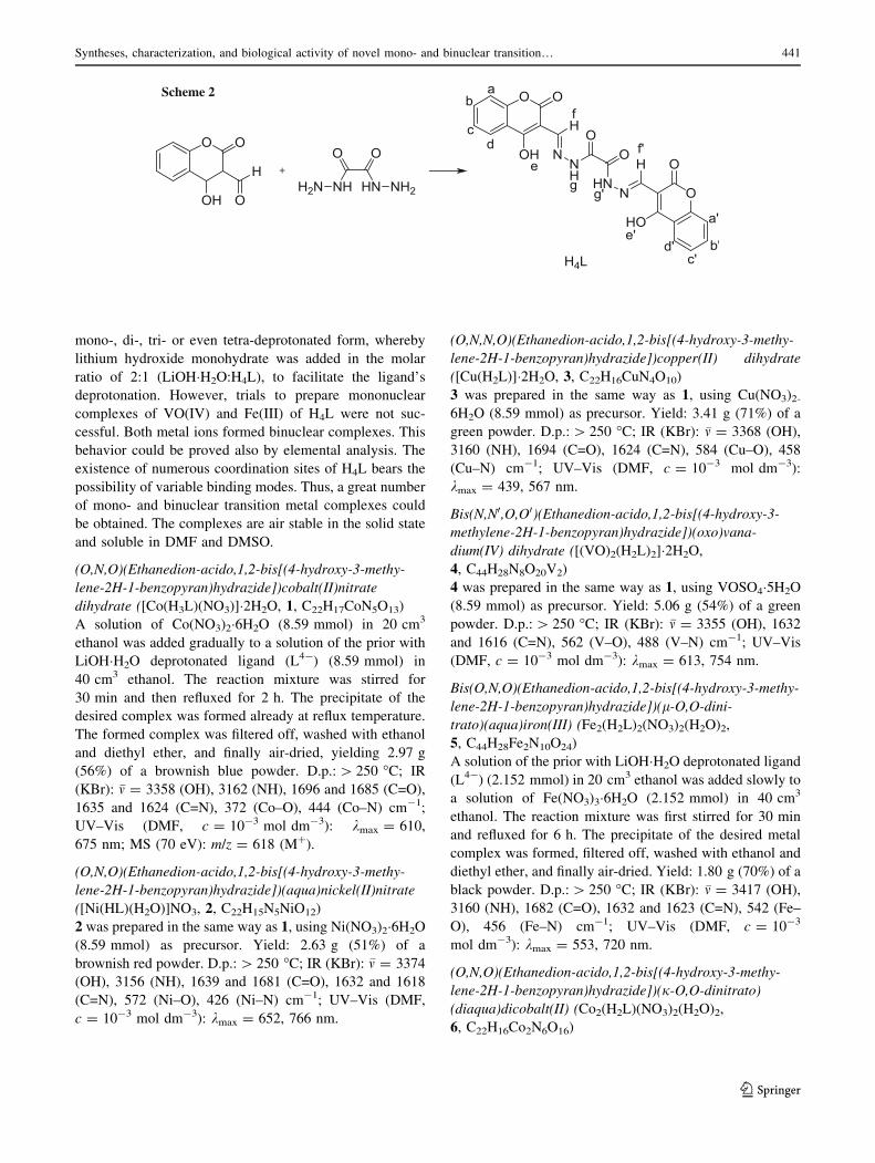
ORIGINAL PAPER Syntheses, characterization, and biological activity of novel mono- and binuclear transition metal complexes with a hydrazone Schiff base derived from a coumarin derivative and oxalyldihydrazine Esther Theresa Knittl 1 • Azza A. Abou-Hussein 2 • Wolfgang Linert 1 Received: 14 March 2017 / Accepted: 16 October 2017 / Published online: 27 November 2017 Ó The Author(s) 2017. This article is an open access publication Abstract A hydrazone Schiff base ligand was synthesized by the condensation of 3-formyl-4-hydroxycoumarin and oxalyldihydrazide in the molar ratio 2:1. The Schiff base ligand acts as a mono-, bi-, tri- or even tetradentate ligand with metal cations in the molar ratios 1:1 or 2:1 (M:L) to yield either mono- or binuclear complexes as keto or enol isomers, where M = Co(II), Ni(II), Cu(II), VO(IV), and Fe(III). The ligand and its metal complexes were charac- terized by elemental analyses, IR, 1 H NMR, mass, and UV–Vis spectroscopy. Furthermore, the magnetic moments were calculated from the measured electric conductivities of the complexes. According to the received data, the dihydrazone ligand contains one or two units of ONO domains and can bind to the metal ions via the azomethine nitrogen, the carbonyl oxygen atoms, and/or the phenolic oxygen atoms. Electronic spectra and the magnetic moments of all complexes show that the complexes’ geometries are either octahedral, tetrahedral, square planar, or square pyramidal. Cyclic voltammograms of the mononuclear Co(II) and Ni(II) complexes show quasi-re- versible peaks. Tests against two pathogenic bacteria as Gram-positive and Gram-negative bacteria for both, the Schiff base ligand and its metal complexes were carried out. In addition, also one kind of fungi was tested. The synthesized complexes demonstrate mild antibacterial and antifungal activities against these organisms. Graphical abstract Keywords 3-Formyl-4-hydroxycoumarin derivative Mono- and binuclear complexes Cyclic voltammetry Antibacterial and antifungal activity Introduction The chemistry of organic hydrazone compounds, which include –N–NH–CO– groups, takes the forefront position in the development of coordination chemistry of symmet- rical dihydrazone transition metal complexes, as they demonstrate versatility in their coordination, a tendency to show stereochemistry [1–5] due to higher coordination numbers, an ability to act in their neutral or deprotonated forms, as they behave as keto–enol tautomers, bearing unusual coordination numbers [2, 6–9] and flexibility in assuming different conformations. They are extensively used due to their promising applications in biomimetic catalytic reactions, analytical chemistry [10–12], polymer coating pigments, industrial fluorescent materials, Portland Electronic supplementary material The online version of this article (doi:10.1007/s00706-017-2075-9) contains supplementary material, which is available to authorized users. & Wolfgang Linert [email protected] 1 Institute of Applied Synthetic Chemistry, Vienna University of Technology, Getreidemarkt, 9/163-AC, 1060 Vienna, Austria 2 Faculty of Women for Arts, Science and Education, Ain Shams University, Heliopolis, Cairo, Egypt 123 Monatsh Chem (2018) 149:431–443 https://doi.org/10.1007/s00706-017-2075-9
-
Upload
khangminh22 -
Category
Documents
-
view
4 -
download
0
Transcript of and binuclear transition metal complexes with a hydrazone ...

ORIGINAL PAPER
Syntheses, characterization, and biological activity of novel mono-and binuclear transition metal complexes with a hydrazone Schiffbase derived from a coumarin derivative and oxalyldihydrazine
Esther Theresa Knittl1 • Azza A. Abou-Hussein2 • Wolfgang Linert1
Received: 14 March 2017 / Accepted: 16 October 2017 / Published online: 27 November 2017
� The Author(s) 2017. This article is an open access publication
Abstract A hydrazone Schiff base ligand was synthesized
by the condensation of 3-formyl-4-hydroxycoumarin and
oxalyldihydrazide in the molar ratio 2:1. The Schiff base
ligand acts as a mono-, bi-, tri- or even tetradentate ligand
with metal cations in the molar ratios 1:1 or 2:1 (M:L) to
yield either mono- or binuclear complexes as keto or enol
isomers, where M = Co(II), Ni(II), Cu(II), VO(IV), and
Fe(III). The ligand and its metal complexes were charac-
terized by elemental analyses, IR, 1H NMR, mass, and
UV–Vis spectroscopy. Furthermore, the magnetic moments
were calculated from the measured electric conductivities
of the complexes. According to the received data, the
dihydrazone ligand contains one or two units of ONO
domains and can bind to the metal ions via the azomethine
nitrogen, the carbonyl oxygen atoms, and/or the phenolic
oxygen atoms. Electronic spectra and the magnetic
moments of all complexes show that the complexes’
geometries are either octahedral, tetrahedral, square planar,
or square pyramidal. Cyclic voltammograms of the
mononuclear Co(II) and Ni(II) complexes show quasi-re-
versible peaks. Tests against two pathogenic bacteria as
Gram-positive and Gram-negative bacteria for both, the
Schiff base ligand and its metal complexes were carried
out. In addition, also one kind of fungi was tested. The
synthesized complexes demonstrate mild antibacterial and
antifungal activities against these organisms.
Graphical abstract
Keywords 3-Formyl-4-hydroxycoumarin derivative �Mono- and binuclear complexes � Cyclic voltammetry �Antibacterial and antifungal activity
Introduction
The chemistry of organic hydrazone compounds, which
include –N–NH–CO– groups, takes the forefront position
in the development of coordination chemistry of symmet-
rical dihydrazone transition metal complexes, as they
demonstrate versatility in their coordination, a tendency to
show stereochemistry [1–5] due to higher coordination
numbers, an ability to act in their neutral or deprotonated
forms, as they behave as keto–enol tautomers, bearing
unusual coordination numbers [2, 6–9] and flexibility in
assuming different conformations. They are extensively
used due to their promising applications in biomimetic
catalytic reactions, analytical chemistry [10–12], polymer
coating pigments, industrial fluorescent materials, Portland
Electronic supplementary material The online version of thisarticle (doi:10.1007/s00706-017-2075-9) contains supplementarymaterial, which is available to authorized users.
& Wolfgang Linert
1 Institute of Applied Synthetic Chemistry, Vienna University
of Technology, Getreidemarkt, 9/163-AC, 1060 Vienna,
Austria
2 Faculty of Women for Arts, Science and Education, Ain
Shams University, Heliopolis, Cairo, Egypt
123
Monatsh Chem (2018) 149:431–443
https://doi.org/10.1007/s00706-017-2075-9

cement, amphibolites, and granites [13]. They bear
remarkable antifungal, antibacterial, anticancer, antiviral,
and herbicidal applications [14–18] and anti-inflammatory
properties, exhibited by these compounds, which can be
attributed to their metal complexing abilities [19–21].
Based on their applicability in various fields, we are
extending this field by the synthesis of novel mono- and
binuclear hydrazone complexes.
The present study is an extension to our research work
for the formation of mono- and binuclear complexes for
coumarin derivatives, synthesized by the condensation of
4-hydroxycoumarin with oxalyldihydrazide in the molar
ratio of 1:2, to afford the corresponding hydrazine [22].
Mono- and binuclear complexes of Co(II), Ni(II), Cu(II),
VO(IV), and Fe(III) with the hydrazone Schiff base ligand
have been prepared. H4L acts as a bidentate, tridentate,
tetradentate, or hexadentate ligand in a mono- or binuclear
form. The bonding sites are the azomethine nitrogen, the
carbonyl oxygen in its keto or enol isomeric form, and the
phenolic oxygen. The ligand exhibits a tautomerization,
which can modulate the coordination to the metal in the
keto or enol form. The redox properties and the nature of
the electro-active species of the complexes have been
characterized to study the electrochemical behavior of
selected complexes by cyclic voltammetry [23]. It was
hoped that the bacterial activity of the present hard-soft
Schiff base ligand and its transition metal complexes show
increased activity against two pathogenic bacteria and
fungi. As Gram-positive bacteria, Staphylococcus aureus
was used, and as Gram-negative bacteria, Pseudomonas
fluorescens was taken. To investigate anti-fungi, Fusarium
oxysporum was used as an example.
Results and discussion
The data on the characterization of the synthesized ligand
and its transition metal complexes are reported in Table 1.
Characterization of the Schiff base H4L
H4L was analyzed by elemental analysis, IR, UV–Vis,
mass spectrometry, and NMR spectroscopy. The mass
spectrum and the schematic fragmentation of the ligand can
be seen in the supplementary materials S1.
The IR spectrum is consistent with the structure of H4L.
In fact, the ligand includes four acidic hydrogen atoms,
which can be released easily by adding a base. Thus, it can
act in its mono-, di-, tri- or even tetra-deprotonated form,
when binding to a metal center. Due to keto–enol tau-
tomerism, it can exist in both forms, or in a mixture of
both, since there is an amide group (–NH–C=O) in the
ligand structure. However, the IR spectrum indicates that
the ligand exists almost in its keto form in the solid state.
Table 1 Data on the characterization of the synthesized ligand and its transition metal complexes
Complex/ligand Electronic absorption bands nm-1 and assignment leff Ka Geometry M/g mol-1
Yield/%
Color
d–d transition Assignment
1 [Co(H3L)(NO3)]�2H2O 610 (0.074), 675 (0.054) 4A2(F) ? 4T1(P) 4.22 95 Tetrahedral 618.33 56 Brownishblue
2 [Ni(H3L)(H2O)]NO3 652 (0.045), 766 (0.052) 3A2(F)(m3) ? 3T1g(P) 2.92 91 Tetrahedral 600.08 51 Brownish red
3 [Cu(H2L)]�2H2O 439 (0.330), 567 (0.424) 2B1g ?2A1g, 2B1g ?
2Eg 1.97 – Square planner 559.93 71 Green
4 [(VO)2(H2L)2]�2H2O 613 (0.414), 754 (0.378) 1B2 ? 2E, 1B2 ? 2A1 1.81 – Squarepyramidal
1090.63 54 Green
5 Fe2(H2L)2(NO3)2(H2O)2 553 (0.021), 720 (0.027) Charge transfer from UVto Vis region
4.35 145 Octahedral 1192.44 70 Black
6 Co2(H2L)(NO3)2(H2O)2 575 (0.076), 660 (0.088) 4A2g(F) ? 4T1g (F)(m2)4A2g(P) ? 4T1g (P) (m3)
4.69 153 Octahedral 738.62 61 Brown
7 Ni2(H2L)(NO3)2(H2O)4 652 (0.053), 435 (0.076) 3A2g(F) ? 3T1g (F)(m2)3A2g(F) ? 3T1g (P) (m3)
3.29 158 Octahedral 773.81 56 Green
8 [Cu2(H2L)(H2O)4](NO3)2 610 (0.432), 455 (0.339) 2A1g ? 2B1g, 2E ? 2B1g 1.83 154 Squarepyramidal
783.52 62 Violet
9 [(VO)2(H2L)(SO4)]�3H2O 624 (0.532), 775 (0.388) 1B2 ? 2E, 1B2 ? 2A1 1.74 – Squarepyramidal
744.34 71 Green
10 Fe2(L)(H2O)4(NO3)2 520 (0.034), 765 (0.044) Charge transfer from UVto Vis region
4.87 162 Octahedral 766.10 53 Black
H4L – – – – – 462.37 71 Yellow
a Molar conductivities in Ohm-1 cm2 mol-1
432 E. T. Knittl et al.
123
The bands, which appeared at 1695 m(C=O), 3142–3180
m(NH) ? 1505 m(C–N) ? d(N–H), 1215 d(N–H),
743 cm-1 U(C=O) are characteristic for the amide group
and thus support the existence of the tautomeric keto form
of the ligand [24, 25]. It is worth to mention that the IR
spectrum of the ligand displays another band at 1723 cm-1,
which can be attributed to the presence of the lactone group
in the coumarin ring [28, 29]. The band observed at
1272 cm-1 is assigned to m(C–O–C) of the coumarin ring
[30], while the band at 1253 cm-1 is ascribed to the phe-
nolic C–O stretching vibrations [31], and another one in the
region from 1567 to 1483 cm-1 is assigned to the combi-
nation of m(C=C) of the aromatic ring system [32]. The
broad band in the region of 3142–3180 cm-1 in the free
ligand is assigned to the stretching vibration of the amide
group (NH).1H and 13C NMR spectra are given in the supplementary
material S2. In the 1H NMR spectrum of the ligand, a very
small signal could be observed at 13.36 ppm, which may
be attributed to the tautomerism of the ligand [26, 27]. The
broad signal at 12.43 ppm, H(e,e0), corresponds to the
hydroxyl groups. This signal disappears after the addition
of D2O, which indicates that these protons are acidic.
Characterization of the complexes of H4L: IR
spectroscopy
Infrared spectra of the complexes were recorded to gain
information about the binding modes of the ligand to the
corresponding metals. All spectra can be seen in the sup-
porting material S3. In fact, it could be observed in the IR
spectra that the ligand can be coordinated in its keto or enol
form. For the monometallic complexes [Co(H3L)(NO3)]-
2H2O (1) and [Ni(H3L)(H2O)]�NO3 (2), the great flexibility
of the ligand and, thus, the presence of numerous donor
sites make it difficult to establish the coordination types of
the final complexes; however, IR spectra and elemental
analysis suggest that at least one oxygen atom from one
phenolic site and its imine nitrogen, as well as one terminal
carbonyl group, are involved in the coordination. For the
complexes 1 and 2, the strong band ascribed to the vibra-
tion of the C=O bond of the hydrazone Schiff base (ca
1695 cm-1) appears at the same position in the spectra of
the complexes, but with a much lower intensity. In addi-
tion, a new band appears at 1685 and 1681 cm-1 in the
spectra of the complexes 1 and 2, respectively, which
suggests that the oxygen atom on one side of the ligand
coordinates to the metal atom, while the other C=O group
remains free. In a similar way, there are observed two
bands assigned to the stretching frequencies of the
azomethine groups at 1624 and 1618 cm-1, which could
give a hint that the azomethine group on one side of the
ligand coordinates to the metal, while the same group on
the other side stays uncoordinated [33]. One broad band,
which appears in the spectra of the ligand and the corre-
sponding metal complexes of Co(II) and Ni(II) in the range
between 3162 and 3156 cm-1, is assigned to the stretching
vibration of m(N–H). Finally, the IR spectra of the com-
plexes show two bands, which can be associated with the
stretching frequencies of the coordinated and uncoordi-
nated phenolic oxygens in the ranges between 1232–1237
and 1260–1267 cm-1, respectively. The observations
indicate that the Schiff base ligand acts as a mono-nega-
tive, tridentate ligand in the case of the synthesized Co(II)
and Ni(II) complexes, where one moiety of the Schiff base
is coordinated to the metal atoms by only one carbonyl,
azomethine, and phenolic oxygen group, forming six- and
five-membered chelate rings with the metal ions, forming
the complexes 1 and 2 [34]. In fact, this is not the case for
[Cu(H2L)]�2H2O (3), where both sides of the ligand par-
ticipate in the coordination to the metal ion, in spite of the
Schiff base being present in its keto form. The carbonyl
groups remain uncoordinated in this case [35]. The bands
in the IR spectra, assigned to m(N–H) and m(C=O), were
found nearly at the same position in the spectrum of the
complex and the ligand, indicating that the possibility of
the coordination of the ligand to the metal center by these
groups is quite low. On the other hand, the azomethine
group is shifted to lower frequencies, which support the
coordination by the two azomethine groups to the metal
ion. This is further consistent with the involvement of the
phenolic oxygen atoms, which also bind to the metal center
in the final complex. These facts are confirmed by ele-
mental analyses, physical, and spectroscopic
measurements.
For the macrocyclic complex [(VO)2(H2L)2]�2H2O (4),
the stretching vibrations of the C=O, as well as the N–H
bands, are absent in the spectrum of the complex. This
could be attributed to the enolization of both carbonyl
groups of the Schiff base during the formation of the
complex [36]. The appearance of a new C–O band in the
spectrum of the complex at 1257 cm-1 suggests that the
ligand coordinates by the two C–O groups of the oxalyl-
hydrazine moiety to the metal ion after the deprotonation
of this group [37]. The band assigned to C=N has been
shifted to lower frequencies in the spectrum of the complex
compared to the one of the free ligand, which suggests that
both azomethine nitrogens participate in the coordination.
Also, the spectrum of the complex shows an additional
C=N band at 1616 cm-1, which might be assigned to the
formation of two new C–N groups as a matter of the tau-
tomeric enolization [38]. Due to the fact that the stretching
(m 3355 cm-1) and bending (d 1360 cm-1) vibrations of
the hydroxyl groups of the coumarin rings did not change
their positions in both spectra, it indicates that these groups
do not take part in the coordination. In the case of
Syntheses, characterization, and biological activity of novel mono- and binuclear transition… 433
123
Fe2(H2L)2(NO3)2(H2O)2 (5), the stretching vibration
assigned to the carbonyl group of the hydrazone Schiff
base shows no significant changes, when the spectrum of
the complex is compared to the one of the free Schiff base
due to the presence of one free C=O group, which is not
involved in the coordination [39]. However, a new band
appeared in the spectrum of the complex between 855 and
842 cm-1, which might be attributed to the participation of
the C–O group in the complexation, which results in the
formation of the enol form of the amide groups (–N=C–
OH). Another evidence for the enolization is the presence
of the stretching frequencies of the azomethine groups
C=N in the spectrum of the metal complex. In fact, the m(–
C=N) bands were found in two different regions. Before, it
was observed at 1623 cm-1, which is lower than the m(–
C=N) of the free ligand at 1635 cm-1 as a result of the
coordination to the metal center. The band at 1270 cm-1,
which is ascribed to the phenolic C–O stretching vibration
in the ligand, is shifted to higher frequencies as a result of
the coordination to the metal ion and thus, was found at
1282 cm-1. It is obvious that the Schiff base behaves as a
twofold negative, tridentate ligand in the complex 5. The
coordination occurs via the phenolic oxygen, the azome-
thine group, and the enolic oxygen.
The IR spectra show that the hydrazone Schiff base
ligand behaves in its keto form in the binuclear complexes
Co2(H2L)(NO3)2(H2O)2 (6), Ni2(H2L)(NO3)2(H2O)4 (7),
[Cu2(H2L)(H2O)4](NO3)2 (8), and [(VO)2(H2L)(SO4)]-
3H2O (9), where the stretching frequency m(NH) appears at
the same range in the spectrum of the ligand H4L, com-
pared to the ones of the corresponding metal complexes,
which proves that this group is not participating in the
coordination. The frequency of the carbonyl oxygen in the
spectra is shifted to lower frequencies due to the coordi-
nation of this atom to the metal ion in the range between
1675 and 1680 cm-1, compared to the free ligand at
1695 cm-1. A shift in the same way can also be observed
for the azomethine group, which indicates that the nitro-
gens of these groups participate in the coordination [40].
Moreover, the band at 1000 cm-1, which is attributed to
the vibration m(N–N) of the ligand, is shifted to higher
wavenumbers (by ca. 10 cm-1) after the complexation
[41, 42]. The complexation through the phenolic oxygen
atoms, which takes place after the deprotonation of this
group, can be proved by the appearance of a band assigned
to m(C–O) at much higher frequencies (1274–1266 cm-1)
in the spectra of all complexes, compared to the spectrum
of the free ligand H4L (1253 cm-1). The shift to higher
wavenumbers is expected due to the increase of the double
bond character through the resonance in the chelate ring of
the free Schiff base [43, 44]. This observation has been
further supported by elemental analysis and molar con-
ductivity values, which indicate that there is a substantial
dissociation of the complexes in dimethyl formamide
(DMF) and supports the coordination of the ligand to the
metal ion in its deprotonated dianionic form. On the other
hand, the Schiff base ligand coordinates in its enol form in
the case of the binuclear complexes 4 and 5.
One of the most important features of the IR spectra of
some complexes is the possibility to prove the coordination
behavior of nitrate and sulfate anions. Strong absorption
bands are displayed by the IR spectra of the complexes,
which are consistent with monodentate, bidentate or ionic
nitrate vibrations. In the complexes 5 and 6, the nitrate
anions behave in a coordinated bidentate nature, where it
possesses three non-degenerated vibrational modes (ns, ms’,
and mas) in the ranges 1443–1468, 1266–1287, and
1033–1021 cm-1, respectively. The distance of about
200 cm-1 between the vibrations ns and ms’ confirms the
bidentate nature of the nitrate group. On the other hand, for
the complexes 1, 7, and 10, the nitrate ions are bound to the
metals in a monodentate way, corresponding to the C2v
symmetry with three non-degenerated modes of vibrations
(ms, ms’ and mas), which appear in the ranges of 1356–1372,
1245–1265, and 836–856 cm-1, respectively. For the
complexes 2 and 8, a strong band around 1380 cm-1 could
be observed and assigned to the stretching mode of ionic
nitrate [45]. The presence of nitrate in this complex is
shown by IR spectroscopy and elemental analysis. The
oxovanadium complexes 4 and 9 exhibit a strong band
around 985 and 980 cm-1, respectively. This reflects the
high p-band order of vanadium to the oxygen link of VO2?
[46, 47]. The appearance of new bands at 953 (v2), 1166
(v5), and 647 (v6) cm-1 in the VO2? complex 9 is an
evidence for the bidentate nature of the SO42- anion This
observation has been further supported by conductivity
measurements and elemental analysis. It is worth to men-
tion that the broad band, which is observed in the IR
spectra of the complexes in the range of 3355–4381 cm-1,
can be ascribed to the stretching vibrations of m(O–H) of
the phenolic group, crystalline or coordinated water
molecules associated to the complexes. New bands in the
spectra of all complexes, which are absent in the spectrum
of the free ligand and are located at 534–584 and
426–488 cm-1, can be assigned to m(M–O) and m(M–N),
respectively [48]. It is obvious that the binding sites of the
ligand to the metal ions are the azomethine nitrogen, and
the carbonyl oxygen in either the keto or the enol form and/
or the phenolic oxygen atoms. The nitrate, sulfate, and
chloride anions, as well as coordinated water molecules,
enable the satisfaction of the other coordination sites, to
complete the geometry of the complexes. It is of interest
that none of the carbonyl lactone groups are involved in the
coordination and thus, remain unchanged according to the
IR spectra [49, 50]. This observation is further supported
by the stretching vibration m(C–O–C), assigned to the
434 E. T. Knittl et al.
123
coumarin ring, which remains nearly unchanged after the
complexation to the metal ion and thus does not have an
influence on the coordination to the metal center [51].
Electronic spectra, magnetic moments, and molar
conductivity measurement
The electronic spectra of the ligand and its transition metal
complexes in a DMF solution (10-3 M) with their
assignments, magnetic moments, and molar conductivity
measurements are given in Table 1. The electronic spec-
trum of H4L shows absorption bands at 270 and 316 nm,
which are assigned to p–p* transitions within the coumarin
moiety [52]. The broad band at 365 nm is attributed to the
p–p* transitions of the C=N and C=O bonds in addition to
a broad band at 410 nm, attributed to the n–p* transition,
which is overlapping with the intramolecular CT-band of
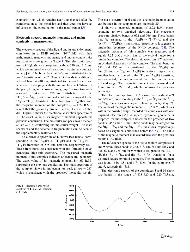
the phenyl ring to the azomethine group. 1 shows two well-
resolved peaks at 675 nm, attributed to the4T1(P) / 4A2(F) transition and at 610 nm, assigned to the4A2 ? 4T1(F) transition. These transitions, together with
the magnetic moment of the complex (l = 4.22 B.M.),
reveal that the geometry around the Co(II) ion is tetrahe-
dral. Figure 1 shows the electronic absorption spectrum of
1. The exact value of its magnetic moment supports the
previous conclusion. The molecular ion peak was observed
at m/z = 618, confirming the molecular weight. The mass
spectrum and the schematic fragmentation can be seen in
the supplementary materials S4.
The electronic spectrum of 6 shows two bands, corre-
sponding to the 4A2g(F) / 4T1g(F) and the 4T1g(P) /4T1g(F) transition at 575 and 660 nm, respectively [53].
These transitions are consistent with the formation of an
octahedral high-spin geometry. The measured magnetic
moment of this complex indicates an octahedral geometry.
The exact value of its magnetic moment is 4.69 B.M.,
supporting the previous conclusion. The mass spectrum of
the complex shows its molecular ion peak at m/z = 737,
which is consistent with the proposed molecular weight.
The mass spectrum of 6 and the schematic fragmentation
can be seen in the supplementary materials S5.
2 shows a magnetic moment of 2.92 B.M., corre-
sponding to two unpaired electrons. The electronic
spectrum displays bands at 652 and 766 nm. These bands
may be assigned to the 3A2(F) ? 3T2(F)(m1) and the3A2(F) ? 3T1(F)(m2) transitions, respectively. It suggests a
tetrahedral geometry of the Ni(II) complex [54]. The
magnetic moment of this complex was measured and
equals 3.32 B.M., which lies in the typical range for a
tetrahedral complex. The electronic spectrum of 7 indicates
an octahedral geometry of the complex. The main bands at
652 and 435 nm are attributed to the 3T1g(F) /3A2g(F) and the 3T1g / 3A2g(F) transitions, respectively.
Another band, attributed to the 3T1g / 3A2g(F) transition,
was expected, but not observed, as it lies in the near
infrared range. The magnetic moment was measured and
found to be 3.29 B.M., which confirms the previous
geometry.
The electronic spectrum of 3 shows two bands at 439
and 567 nm, corresponding to the 2B1g ?2Eg and the 2B1g
? 2A1g transitions in a square planar geometry (Fig. 2).
The value of the magnetic moment is 1.97 B.M., which lies
within the possible range, recorded for complexes with one
unpaired electron [55]. A square pyramidal geometry is
proposed for the complex 8 based on the presence of two
bands at 455 and 610 nm. These bands may be assigned to
the 2B1 / 2A1 and the 2B1 / 2E transitions, respectively,
based on assignments published before [56, 57]. The value
of the magnetic moment is in accordance with the previous
results (1.83 BM).
The reflectance spectra of the oxovanadium complexes 4
and 9 reveal three bands at 362, 613, and 754 nm for 7 and
436, 624, and 775 nm for 9, which is assigned to the 2B2 ?2E, the 2B2 ? 2B1, and the 2B2 ? 2A1 transitions for a
distorted square-pyramid geometry. The magnetic moment
was found to be 1.81 and 1.74 B.M. for the complexes 7
and 9, respectively [58].
The electronic spectra of the complexes 5 and 10 show
two bands in the range of 453–520 and 720–765 nm,
Fig. 1 Electronic absorption
spectrum of 1 in a DMF solution
(10-3 M)
Syntheses, characterization, and biological activity of novel mono- and binuclear transition… 435
123

respectively. The first band is assigned to charge transfer
transitions from the out-of-plane pp orbital of the phenolate
coumarin oxygen to the half-filled dx2–y2/dz2 orbital of the
high-spin Fe(III) [59]. Elemental analysis and the infrared
spectra give significant information about the coordination
mode of the nitrate anion as a bi- and monodentate anion
for the complexes 5 and 10, respectively. On the other
hand, the magnetic moments of the Fe(III) complexes 5
and 10 were measured and their values are 4.35 and 4.87
B.M., respectively, in accordance with a high-spin dis-
torted octahedral arrangement (t2g3 eg2).
The presented magnetic moments do not show any
metal–metal interactions in the bimetallic complexes. Very
weak antiferromagnetic interactions cannot be excluded,
for this low temperature (He-temperature) measurements
of the magnetic moment would be needed, which are out of
our reach. Otherwise, this would be speculation.
Molar conductivities of the complexes were recorded in
DMF (1.0 9 10-3 M). It has been reported that DMF is a
good donor solvent, since it can replace NO3- in metal
complexes [60]. Conductivity measurements indicate the
non-electrolytic nature of the complexes 3, 4, and 9.
Because the bi-coordinated binding of SO42- is usually
stronger, DMF will not replace SO42-. In other words, the
Lewis basicity (donor number) of DMF is not big enough
to replace SO42- in complex 9 [61]. Complexes 1 and 2
exhibit molar conductivities of 95 and 92 Ohm-1 -
cm2 mol-1, respectively, and can therefore be considered
as 1:1 electrolytes, since a maximum value about
100 Ohm-1 cm2 mol-1 is reported for 1:1 electrolytes in
DMF [60]. On the other hand, the complexes 5, 6, 7, 8, and
10 behave as 1:2 electrolytes.
ESR spectroscopy
X-band ESR spectra were recorded in the solid state for the
complexes 3 (Fig. 3) and 9 (Fig. 4) at 25 �C. The g-tensor
values of the Cu(II) complex can be used to derive the
ground state. The observed values, gk(2.136)[ g\
(2.053)[ ge(2.0023), indicate that the copper site has a
dx2-y2 orbital giving 2B1g as the ground state, which is
characteristic for a square planar or an octahedral geometry
[62]. The gk value is an important value, when it comes to
indicating the covalent or ionic character of the M–L bond.
Kivelson and Neiman reported for an ionic character of the
Fig. 2 Electronic absorption
spectrum of 3 in a DMF solution
(10-3 M)
Fig. 3 ESR spectrum of 3
Fig. 4 ESR spectrum of 9
436 E. T. Knittl et al.
123
bond gk[ 2.3 and for a covalent character gk\ 2.3. In the
present complex, gk is smaller than 2.3, which indicates a
covalent character of the Cu–L bond [63]. The axial
symmetry parameter G is smaller than four, which indi-
cates a considerable exchange interaction in the solid
complex [14]. If the value of G is less than 4, a consider-
able exchange interaction is noticed in the solid complex.
The calculated G value of the square planar complex 3
suggests a strong interaction between the Cu(II) centers
[64]. The X-band ESR spectrum of 9 gives a broad peak
without hyperfine coupling, where the g-tenser values are
gk(1.99) and g\ (1.97). The decrease of the g values
compared to the value of a free-electron (2.0023) could be
attributed to the ligand field strength. The shape of the
spectrum, as well as the g-tenser values agrees with a
square-pyramidal geometry for the vanadyl complex 9.
Thermal analysis
Thermal gravimetric analysis was carried out for selected
complexes 4, 5, and 10, to gain information concerning
their thermal stability and to decide, whether water mole-
cules are bound in the inner or outer coordination sphere of
the central metal ions [65]. The TGA curves showed that
the decomposition of the complexes proceeds in many
steps in the weight loss percentage. The first stage is the
weight loss, which corresponds to the loss of lattice or
coordinated water molecules. The second stage corre-
sponds to the loss of nitrate ions in the form of N2O5 gas,
followed by the organic ligand. At the end, metal oxide was
formed around the range from 700 to 1000 �C, where
residues are nearly close to the calculated values. In
Table 2, the thermal analysis is summarized. The degra-
dation and the schematic fragmentation pathways of the
complexes are shown in detail in the supplementary
material S6.
Cyclic voltammetry studies
The electrochemical properties of the metal complexes
have been studied, to determine the structural changes
accompanying with the electron transfer. The redox
behavior has been investigated for 1 and 2 in DMSO
(1.0 mM dm-3). The measurements were carried out in a
0.1 M tetrabutylammonium tetraflouroborate
(TBA ? BF4-) solution as a supporting electrolyte, using
platinum wires with a diameter of 0.5 mm as working and
counter electrodes, and Ag/AgCl as a reference electrode.
Ferrocene/ferrocenium (Fc/Fc?) was used as internal
standard for the assignment of the potential electrode
couple [41, 42]. The complexes 1 and 2 show electro-
chemical reversible steps (respective pseudo-reversible),
related to single-electron transfer processes [66, 67]. The
cyclic voltammogram of the Co(II) complex (Fig. 5, left)
refers to E1/2 = - 0.640 V. The Ni(II) complex (Fig. 5,
right) refers to E1/2 = - 0.591 V, both corresponding to
the M(II)/M(III) one-electron redox system. The ratio Ipa/
Ipc is close to unity.
From the interpretation of elemental analysis, magnetic
studies and spectral data, as well as the thermal analysis
and molar conductivity measurements, one can propose
tentative structures of the synthesized transition metal
complexes. Scheme 1 depicts the suggested structures for
the obtained metal complexes.
Biological studies
The Schiff base ligand H4L and its metal complexes were
examined according to antimicrobial activity against
Gram-positive bacteria (Staphylococcus aureus) and Gram-
negative bacteria (Pseudomonas fluorescens), as well as
one pathogenic fungus (Fusarium oxysporum). The results
of the biological studies of the ligand and its complexes are
given in Fig. 6 and Table 3. The data is compared to
standard antibiotics, chloramphenicol, as standard
Table 2 Results of the thermal analysis of complexes 4, 5, and 10
Complex Decomposition transitions M/g mol-1 Temperature/�C Weight loss/%
TGA DrTGA Found Calculated
4 (I) [(VO)2(H2L)2]�2H2O ? (- 2H2O) 1090.63 60–136 95 4.10 3.32
(II) [(VO)2(H2L)2] ? (- 2(H2L)) 136–668 326 80.30 79.93
(III) Complete decomposition 668–1000 630 17.81 16.76
5 (I) Fe2(H2L)2(NO3)2(H2O)2 ? (-2 H2O) 1192.44 142–254 123 3.58 3.02
(II) Fe2(H2L)2(NO3)2 ? (- N2O5) 254–381 310 9.32 9.05
(III) Fe2(H2L)2O ? (- 2(H2L)) 381–760 623 76.31 74.54
10 (I) Fe2(L)(H2O)4(NO3)2 ? (-4 H2O) 766.10 161–266 134 10.13 9.31
(II) Fe2(L)(NO3)2 ? (- N2O5) 266–393 216 15.82 14.10
(III) Fe2(L)O ? (- L) 393–745 526 51.60 55.61
Syntheses, characterization, and biological activity of novel mono- and binuclear transition… 437
123

reference for Gram-negative and cephalothin for Gram-
positive bacteria. Cycloheximide was used as antifungal
standard reference. The in vitro antibacterial and antifungal
activities demonstrate that the complexes have higher
antimicrobial activities compared to that of the ligand. This
behavior could explain that the chelation tends to make the
ligand act as a more powerful and potent antibacterial
agent. Tweedy’s theory [68] suggests that the chelation
reduces the polarity of the metal atom due to the partial
sharing of its positive charge with a donor group and thus
Fig. 5 Cyclic voltammograms of 1 (left), 2 (right)
Scheme 1
438 E. T. Knittl et al.
123

the possible p-electron delocalization over the whole che-
late ring is enabled [69]. Such a chelation could enhance
the lipophilic character of the central metal atom. Subse-
quently, this favors its permeation through the lipid layers
of the cell membrane and thus, bears the possibility to
block the metal binding site in the enzyme of a microor-
ganism [70]. The variety in the activity of different
complexes against various microorganisms depends either
on the cells, the impermeability of the microbes, or the
contrasts of ribosomes in microbial cells. There are other
factors, which increase the activity, like solubility, con-
ductivity, and bond length between the metal and the
ligand. The results also reveal that Cu(II), Fe(III), and
VO2? complexes display the highest (significant) inhibi-
tion against the growth of the selected bacteria and fungi.
On the other hand, Co(II) and Ni(II) complexes show
moderate activity. According to the data, there is an evi-
dence for the relationship of the structure of the complexes
and their activity, where the antimicrobial activity is
enhanced by binuclear complexes, rather than by acyclic
complexes, which reveals that these complexes are bio-
logically more efficient and thus, provide the possibility to
be useful as new drugs and discuss that the chemical
geometry of compounds is important to explain the bio-
logical activity of the complexes.
Fig. 6 Biological screening of the ligand and its complexes against Gram-positive bacteria, Gram-negative bacteria, and fungi
Table 3 Antimicrobial activity of the Schiff base ligand H4L and its metal complexes
Compound Mean of zone diameter/mm mg cm-3
Gram—positive bacteria Gram—negative bacteria Fungi
Staphylococcus aureus Pseudomonas phaseolicol Fusarium oxysporium
H4L 22 � 0:2 13 � 0:1 17 � 0:2
1 [Co(H3L)(NO3)]�2H2O 24 � 0:6 23 � 0:1 26 � 0:2
2 [Ni(H3L)(H2O)]NO3 28 � 0:2 28 � 0:3 25 � 0:2
3 [Cu(H2L)]�2H2O 36 � 0:3 30 � 0:2 31 � 0:3
4 [(VO)2(H2L)2]�2H2O 33 � 0:1 24 � 0:3 25 � 0:3
5 Fe2(H2L)2(NO3)2(H2O)2 37 � 0:4 26 � 0:1 31 � 0:2
6 Co2(H2L)(NO3)2(H2O)2 27 � 0:4 22 � 0:1 23 ± 0.2
7 Ni2(H2L)(NO3)2(H2O)4 20 � 0:6 20 � 0:1 29 � 0:3
8 [Cu2(H2L)(H2O)4](NO3)2 31 � 0:1 29 � 0:2 24 � 0:3
9 [(VO)2(H2L)(SO4)]�3H2O 30 � 0:1 22 ± 0.2 34 � 0:3
10 Fe2(L)(H2O)4(NO3)2 32 � 0:2 23 � 0:1 30 � 0:2
Antibiotic 42 36 40
Syntheses, characterization, and biological activity of novel mono- and binuclear transition… 439
123

Conclusion
The hydrazone Schiff base ligand acts as hexa-, penta-,
tetra- or monodentate ligand with metal cations to afford a
series of mono or binuclear complexes. It is known from IR
spectra that the chelation of all acyclic divalent metal ions
to the ligand occurs via the phenolic atoms of the coumarin
moiety, the ketonic oxygens, and the nitrogen atoms of the
azomethine groups, except for 4, 5, and 10, where the
ligand coordinates in its enol form. Nitrate, sulfate, and
chloride ions in addition to water molecules, which can be
crystalline or coordinated, satisfy the metal’s coordination
sites and thus, bear the possibility to complete the metal’s
geometry around its center. The spectroscopic studies and
the molar conductivity measurements of all the complexes
were used to examine the type of the coordination and the
metal’s geometry around its center. Mono- and binuclear
complexes exhibit either tetrahedral, square planar, square
pyramidal, or octahedral geometries. The synthesized
Schiff base and the corresponding metal complexes were
tested for the growth inhibitory activity against phy-
topathogenic bacteria and fungi. It is obvious that the metal
complexes are more toxic against bacteria and fungi in
comparison with their ligand. Cyclic voltammograms of
the complexes 1 and 2 show one-electron transfer pro-
cesses, indicating their electro-activity in solution.
Experimental
The nitrate salts of cobalt(II), nickel(II), iron(III), and
copper(II) were purchased from Merck or DBH, vanady-
l(IV) sulfate pentahydrate from Sigma Aldrich. Organic
solvents [absolute ethyl alcohol, methyl alcohol, acetone,
dimethylformamide (DMF), and dimethylsulfoxide
(DMSO)] were reagent grade and used without further
purification.
Microanalyses of carbon, hydrogen, and nitrogen were
carried out on a Perkin-Elmer 2400 Series II Analyzer.
Electronic spectra of the metal complexes in DMF were
carried out on a UV–Vis Perkin-Elmer Model Lamda 900.
NIR-, IR- and mid-range FT-IR spectra of the compounds
were recorded as KBr pellets within the range of
400–4000 cm-1, using a Perkin–Elmer 16PC FT-IR spec-
trometer. Far FT-IR spectra were recorded within the range
of 600–200 cm-1 on a Perkin–Elmer System 2000 spec-
trometer as polyethylene pellets. Analyses of the metals in
the complexes were carried out according to the standard
method [71]. Mass spectra were recorded on a Shimadzu-
GC–MS–QP mass spectrometer (model 1000 EX) using a
direct inlet system at 220 �C and 70 eV. NMR spectra of
the ligand were carried out in DMSO-d6 on a Bruker WP
200 SY spectrometer at room temperature using TMS as an
internal standard. Magnetic susceptibilities of the com-
plexes were measured at room temperature using a Johnson
Matthey, Alfa Products, model MKI magnetic suscepti-
bility balance. The effective magnetic moments were
calculated from the expression leff = 2.828 (vM.T)1/2
B.M., where vM is the molar susceptibility, corrected using
the Pascal’s constants for the diamagnetism of all atoms in
the compounds [72]. Melting points were measured using a
Stuart melting point instrument. ESR spectra of the copper
complexes were recorded on a Bruker BioSpin spectrom-
eter. The molar conductance of 1.0 9 10-3 M solution in
DMF was measured on a WTW.D8120 Weilheim L.F.42
conductivity meter. TGA curves were obtained using a
NETZSCH-Geratebau. Thermal gravimetric analysis
(TGA) was carried out in a range from room temperature
up to 1000 �C with a heating rate of 10 �C min-1 in a
nitrogen atmosphere. The cyclic voltammetry measure-
ments were carried out with a Potentiostat Wave Generator
(Oxford press), and equipped with a Phillips PM 8043 X–Y
recorder. The electrochemical cell assembly consists of
platinum wires of 0.5 mm diameter as working and counter
electrodes, and Ag/AgCl as a reference electrode.
Ethanedioic acid 1,2-bis[(4-hydroxy-3-methylene-2H-1-
benzopyran)hydrazide] (H4L, C22H14N4O8)
In a first step, 4-hydroxycoumarin was reacted with DMF
to obtain 3-formyl-4-hydroxycoumarin [73]. The final
Schiff base ligand H4L was then prepared by the addition
of a solution of oxalic dihydrazide (2.54 mmol) in 40 cm3
ethanol/H2O (70%/30%, v/v) to 3-formyl-4-hydroxy-
coumarin (5.05 mmol) in 40 cm3 ethanol. The reaction
mixture was refluxed for 3 h. After the solution was slowly
cooled to room temperature, yellow crystals were formed,
which were filtered off and washed with ethanol and die-
thyl ether, yielding 0.89 g (70.63%) of the title substance
(Scheme 2). M.p.: 174 �C; 1H NMR (200 MHz, DMSO-
d6): d = 12.43 (s, 2H, e, e0), 10.21 (s, 2H, g, g0), 8.32 (s,
2H, f, f0), 7.64 (t, 2H, c, c0), 7.43 (d, 2H, a, a0), 7.34 (d, 2H,
d, d0), 7.31 (t, 2H, b, b0) ppm; 13C NMR (50 MHz, DMSO-
d6): d = 165.69 (s), 161.97 (s), 153.56 (s), 151.12 (s),
149.03 (s), 132.71 (s), 123.94 (s), 123.24 (s), 116.40 (s),
115.84 (s), 91.05 (s) ppm; IR (KBr): �m = 3376 (OH),
3142–3180 (NH), 1695 (C=O), 1635 (C=N) cm-1; UV–Vis
(DMF, c = 10-3 mol dm-3): kmax = 470, 410, 365,
316 nm; MS (70 eV): m/z = 371 (M?).
Synthesis of the transition metal complexes
The coordination reactions of the Schiff base ligand H4L to
cobalt(II), nickel(II), copper(II), oxovanadium(IV), and
iron(III) were carried out in the molar ratios of 1:1 (li-
gand:metal), whereby mono- and binuclear transition metal
complexes were formed. The ligand itself could act in its
440 E. T. Knittl et al.
123
mono-, di-, tri- or even tetra-deprotonated form, whereby
lithium hydroxide monohydrate was added in the molar
ratio of 2:1 (LiOH�H2O:H4L), to facilitate the ligand’s
deprotonation. However, trials to prepare mononuclear
complexes of VO(IV) and Fe(III) of H4L were not suc-
cessful. Both metal ions formed binuclear complexes. This
behavior could be proved also by elemental analysis. The
existence of numerous coordination sites of H4L bears the
possibility of variable binding modes. Thus, a great number
of mono- and binuclear transition metal complexes could
be obtained. The complexes are air stable in the solid state
and soluble in DMF and DMSO.
(O,N,O)(Ethanedion-acido,1,2-bis[(4-hydroxy-3-methy-
lene-2H-1-benzopyran)hydrazide])cobalt(II)nitrate
dihydrate ([Co(H3L)(NO3)]�2H2O, 1, C22H17CoN5O13)
A solution of Co(NO3)2�6H2O (8.59 mmol) in 20 cm3
ethanol was added gradually to a solution of the prior with
LiOH�H2O deprotonated ligand (L4-) (8.59 mmol) in
40 cm3 ethanol. The reaction mixture was stirred for
30 min and then refluxed for 2 h. The precipitate of the
desired complex was formed already at reflux temperature.
The formed complex was filtered off, washed with ethanol
and diethyl ether, and finally air-dried, yielding 2.97 g
(56%) of a brownish blue powder. D.p.:[ 250 �C; IR
(KBr): �m = 3358 (OH), 3162 (NH), 1696 and 1685 (C=O),
1635 and 1624 (C=N), 372 (Co–O), 444 (Co–N) cm-1;
UV–Vis (DMF, c = 10-3 mol dm-3): kmax = 610,
675 nm; MS (70 eV): m/z = 618 (M?).
(O,N,O)(Ethanedion-acido,1,2-bis[(4-hydroxy-3-methy-
lene-2H-1-benzopyran)hydrazide])(aqua)nickel(II)nitrate
([Ni(HL)(H2O)]NO3, 2, C22H15N5NiO12)
2 was prepared in the same way as 1, using Ni(NO3)2�6H2O
(8.59 mmol) as precursor. Yield: 2.63 g (51%) of a
brownish red powder. D.p.:[ 250 �C; IR (KBr): �m = 3374
(OH), 3156 (NH), 1639 and 1681 (C=O), 1632 and 1618
(C=N), 572 (Ni–O), 426 (Ni–N) cm-1; UV–Vis (DMF,
c = 10-3 mol dm-3): kmax = 652, 766 nm.
(O,N,N,O)(Ethanedion-acido,1,2-bis[(4-hydroxy-3-methy-
lene-2H-1-benzopyran)hydrazide])copper(II) dihydrate
([Cu(H2L)]�2H2O, 3, C22H16CuN4O10)
3 was prepared in the same way as 1, using Cu(NO3)2-
6H2O (8.59 mmol) as precursor. Yield: 3.41 g (71%) of a
green powder. D.p.:[ 250 �C; IR (KBr): �m = 3368 (OH),
3160 (NH), 1694 (C=O), 1624 (C=N), 584 (Cu–O), 458
(Cu–N) cm-1; UV–Vis (DMF, c = 10-3 mol dm-3):
kmax = 439, 567 nm.
Bis(N,N0,O,O0)(Ethanedion-acido,1,2-bis[(4-hydroxy-3-
methylene-2H-1-benzopyran)hydrazide])(oxo)vana-
dium(IV) dihydrate ([(VO)2(H2L)2]�2H2O,
4, C44H28N8O20V2)
4 was prepared in the same way as 1, using VOSO4�5H2O
(8.59 mmol) as precursor. Yield: 5.06 g (54%) of a green
powder. D.p.:[ 250 �C; IR (KBr): �m = 3355 (OH), 1632
and 1616 (C=N), 562 (V–O), 488 (V–N) cm-1; UV–Vis
(DMF, c = 10-3 mol dm-3): kmax = 613, 754 nm.
Bis(O,N,O)(Ethanedion-acido,1,2-bis[(4-hydroxy-3-methy-
lene-2H-1-benzopyran)hydrazide])(l-O,O-dini-
trato)(aqua)iron(III) (Fe2(H2L)2(NO3)2(H2O)2,
5, C44H28Fe2N10O24)
A solution of the prior with LiOH�H2O deprotonated ligand
(L4-) (2.152 mmol) in 20 cm3 ethanol was added slowly to
a solution of Fe(NO3)3�6H2O (2.152 mmol) in 40 cm3
ethanol. The reaction mixture was first stirred for 30 min
and refluxed for 6 h. The precipitate of the desired metal
complex was formed, filtered off, washed with ethanol and
diethyl ether, and finally air-dried. Yield: 1.80 g (70%) of a
black powder. D.p.:[ 250 �C; IR (KBr): �m = 3417 (OH),
3160 (NH), 1682 (C=O), 1632 and 1623 (C=N), 542 (Fe–
O), 456 (Fe–N) cm-1; UV–Vis (DMF, c = 10-3
mol dm-3): kmax = 553, 720 nm.
(O,N,O)(Ethanedion-acido,1,2-bis[(4-hydroxy-3-methy-
lene-2H-1-benzopyran)hydrazide])(j-O,O-dinitrato)
(diaqua)dicobalt(II) (Co2(H2L)(NO3)2(H2O)2,
6, C22H16Co2N6O16)
Scheme 2
Syntheses, characterization, and biological activity of novel mono- and binuclear transition… 441
123

6 was prepared in the same way as 5, using Co(NO3)2-
6H2O (2.15 mmol) as precursor. Yield: 0.97 g (61%) of a
brown powder. D.p.:[ 250 �C; IR (KBr): �m = 3374 (OH),
3152 (NH), 1680 (C=O), 1615 (C=N), 544 (Co–O), 466
(Co–N) cm-1; UV–Vis (DMF, c = 10-3 mol dm-3):
kmax = 575, 660 nm; MS (70 eV): m/z = 737 (M?).
(O,N,O)(Ethanedion-acido,1,2-bis[(4-hydroxy-3-methy-
lene-2H-1-benzopyran)hydrazide])(dinitrato)(tetraaqua)-
dinickel(II) (Ni2(H2L)(NO3)2(H2O)4,
7, C22H20N6Ni2O18)
7 was prepared in the same way as 5, using Ni(NO3)2�6H2O
(2.15 mmol) as precursor. Yield: 0.93 g (56%) of a green
powder. D.p.:[ 250 �C; IR (KBr): �m = 3368 (OH), 3151
(NH), 1678 (C=O), 1621 (C=N), 562 (Ni–O), 458 (Ni–N)
cm-1; UV–Vis (DMF, c = 10-3 mol dm-3): kmax = 652,
435 nm.
(O,N,O)(Ethanedion-acido,1,2-bis[(4-hydroxy-3-methy-
lene-2H-1-benzopyran)hydrazide])(tetraaqua)dicopper(II)
nitrate ([Cu2(H2L)(H2O)4](NO3)2,
8, C22H20Cu2N6O18)
8 was prepared in the same way as 5, using Cu(NO3)2-
6H2O (2.15 mmol) as precursor. Yield: 1.05 g (62%) of a
violet powder. D.p.:[ 250 �C; IR (KBr): �m = 3481 (OH),
3170 (NH), 1663 (C=O), 1624 (C=N), 567 (Cu–O), 473
(Cu–N) cm-1; UV–Vis (DMF, c = 10-3 mol dm-3):
kmax = 610, 455 nm.
(N,N,O,O,O,O)(Ethanedion-acido,1,2-bis[(4-hydroxy-3-
methylene-2H-1-benzopyran)hydrazide])(j-O,O-sulfato)
(dioxo)divanadium(IV) trihydrate
([(VO)2(H2L)(SO4)]�3H2O, 9, C22H18N4O17SV2)
9 was prepared in the same way as 5, using VOSO4�5H2O
(2.15 mmol) as precursor. Yield: 1.14 g (71%) of a green
powder. D.p.:[ 250 �C; IR (KBr): �m = 3433 (OH), 3167
(NH), 1675 (C=O), 1615 (C=N), 582 (V–O), 486 (V–N)
cm-1; UV–Vis (DMF, c = 10-3 mol dm-3): kmax = 624,
775 nm.
(O,N,O)(Ethanedion-acido,1,2-bis[(4-hydroxy-3-methy-
lene-2H-1-benzopyran)hydrazide])(dinitrato)
(tetraaqua)diiron(III) (Fe2(L)(H2O)4(NO3)2,
10, C22H18Fe2N6O18)
A solution of H4L (1.076 mmol) in 20 cm3 ethanol was
added slowly to a solution of Fe(NO3)3�6H2O
(2.152 mmol) in 40 cm3 ethanol. The reaction mixture was
first stirred for 30 min and refluxed for 6 h. The precipitate
of the desired metal complex was formed, filtered off,
washed with ethanol and diethyl ether, and finally air-dried.
Yield: 0.87 g (53%) of a black powder. D.p.:[ 250 �C; IR
(KBr): �m = 33.82 (OH), 1622 (C=N), 564 (Fe–O), 467 (Fe–
N) cm-1; UV–Vis (DMF, c = 10-3 mol dm-3):
kmax = 520, 765 nm.
Biological studies
In vitro antibacterial activity studies were carried out using
the standardized disk-agar diffusion method [74] to
investigate the inhibitory effect of the synthesized ligand
and its complexes against Gram-positive bacteria, such as
S. aureus (ATCC25923), Gram-negative bacteria, such as
P. fluorescence (S97), and F. oxysporum as a fungus. The
antibiotic chloramphenicol was used as a standard refer-
ence in the case of Gram-negative bacteria, cephalothin for
Gram-positive bacteria, and cycloheximide as an antifungal
standard reference.
The tested compounds were dissolved in DMF, which
does not have an inhibition activity, to get concentrations
of 2 and 1 mg cm-3. The test was performed on potato
dextrose agar (PDA) as a medium, which contains an
infusion of 200 g potatoes, 6 g dextrose, and 15 g agar
[75, 76]. Uniform size filter paper disks (3 disks per
compound) were impregnated by an equal volume of the
specific concentrations of the dissolved compounds for the
test and carefully placed on the incubated agar surface.
After an incubation for 36 h at 27 �C in the case of bacteria
and for 48 h at 24 �C in the case of fungi, the inhibition of
the organisms, which was evidenced by clear zone sur-
round, was measured and used to calculate its mean of
inhibition zones. The inhibition zone diameter displayed
that the tested ligand and its complexes are active against
the used bacteria and fungus.
Acknowledgements Open access funding provided by TU Wien
(TUW).
Open Access This article is distributed under the terms of the
Creative Commons Attribution 4.0 International License (http://
creativecommons.org/licenses/by/4.0/), which permits unrestricted
use, distribution, and reproduction in any medium, provided you give
appropriate credit to the original author(s) and the source, provide a
link to the Creative Commons license, and indicate if changes were
made.
References
1. Antony A, Fasna F, Ajil PA, Varkey JT (2016) J Chem 5:37
2. Tajudeen SS, Kannappan G (2016) Indian J Adv Chem Sci 4:40
3. Back DF, Manzonia OG, Roman D, Ballin MA, Kober RPPC
(2014) Inorg Chim Acta 6:412
4. Singh DP, Sharma D, Parveen K (2014) J Chem 26:376
5. Sedaghat T, Aminian M, Azarkish M (2015) J Phosphorus Sulfur
Silicon Relat Elem 190:352
6. Gupta S, Kirillova MV, Guedes S, Fatima M, Pombeiro AJP
(2013) Appl Catal A Gen 82:460
7. Hosseini-Monfared H, Asghari-Lalami N, Pazio A, Wozniak K,
Janiak C (2013) Inorg Chim Acta 406:241
8. Tyagi S, Tyagi K, Yadav BP, Ansari MA (2012) Chem Sin 3:440
9. Kar NK, Singh MK, Lal RA (2012) Arab J Chem 5:67
442 E. T. Knittl et al.
123
10. Youssef HM, El-Gamil MM, Abdulhamed Y, Abu El-Reas GM
(2016) Int J Adv Res Biol Sci 3:13
11. El-Bahnasawy RM, El-Meleigy SE, El-Tawansi A (1994) Transit
Met Chem 19:270
12. Najlaa S, Al-Radadi Magda M, Elbeshlaw MA, Mohsen MM
(2016) Open J Inorg Chem 6:1
13. Zhang RH, Yan H, Wang HM, Chen YB, Zhu SJ (1998) Synth
React Inorg Met Org Chem 28:1381
14. Ferraroni M, Carta F, Scozzafava A, Supura CT (2016) J Med
Chem 59:462
15. Freires IA, Alencar SM, Rosalen PLA (2016) Eur J Med Chem
110:267
16. Mahammad M, Samira MI, Mina S, Maliheh S, Seyed ESE,
Alireza F, Abbas S (2016) J Iran Chem Soc 13:1139
17. Shan PM, Namsheer B, Jyoti H, Beena T, Mithun KS (2016) Eur
J Pharm Med Res 3:418
18. Sshwini MR, Vishwanathaswamy AHM (2016) Int J Pharm
Chem Anal 3:1
19. Baluja S, Chanda S, Moteriya P, Talaviya R (2016) Sky J
Microbiol Res 4:31
20. Chetan TP, Sangamesh AP, Shivakumar ST, Shivashankar MK,
Prema SB (2016) J Photochem Photobiol B Biol 157:1
21. Reddya MS, Prathimaa B, Saraswathib M, Babuc S, Saralad Y,
Reddy AV (2016) J Appl Pharm Sci 6:90
22. Abou-Hussein AA, Linert W (2015) Spectrochim Acta A 141:223
23. Abu-Hussein AA, Linert W (2009) Spectrochim Acta A 74:214
24. Back DF, de Oliveira GM, Roman D, Ballin MA, Kober R,
Piquini PC (2014) Inorg Chim Acta 412:6
25. Adel AA, Emara AAA, El-Sayed BA, El-Sayed AE (2008)
Spectrochim Acta A 69:757
26. Webster FX, Silverstein RM (1992) Spectrophotometric identi-
fication of organic compounds, 6th edn. Wiley, New York
27. Pretsch E, Seible J (1983) Tables of spectral data for structure
determination of organic compounds. Springer, Berlin
28. Girgaonkar MV, Shirodkar SG (2012) Res J Recent Sci 1:110
29. Rajeshirkea M, Shaha R, Yadavb P, Purohita NV (2012) Der
Pharm Sin 3:239
30. Bellamy LJ (1975) The infrared spectra of complex molecules,
3rd edn. Chapman and Hall, London
31. Alawandi NG, Kulkarni VM (2006) Indian J Chem 45:258
32. Creaven BS, Devereux M, Karcz D, Kellett A, McCann M, Noble
A, Walsh M (2009) J Inorg Biochem 103:1196
33. Abu-Hussen AA, Emara AAA (2004) J Coord Chem 57:973
34. Rathor N, Pandeya SC, Saxena GC (1998) Orient J Chem 14:231
35. Singh VP, Vinod P, Singh S, Singh DP, Tiwari K, Mishra M
(2014) J Mol Struct 1058:71
36. Ghosh T, Mondal B, Ghosh T, Sutradhar M, Mukherjee G, Drew
M (2007) Inorg Chim Acta 360:1753
37. Ngan NK, Lo KM, Wong CSR (2011) Polyhedron 30:2922
38. Singh VP, Singh DP (2013) Macromol Res 21:757
39. Gupta S, Kirillova MV, Guedes da Silva MF, Pombeiro AJL
(2013) Appl Catal A 82:460
40. Mosaa AI, Emarab AAA, Yousefa JM, Saddiqd AA (2011)
Spectrochim Acta A 81:35
41. Abu-Hussen AA, El-Metwally NM, Saad IM, El-Asmy AA
(2005) J Coord Chem 58:1735
42. El-Metwally NM, Abu-Hussen AA, Gaber IM, El- Asmy AA
(2006) Transit Met Chem 31:71
43. Emara AAA, Abu-Hussen AA (2006) Spectrochim Acta A
64:1010
44. Abou-Hussein AAA, Linert W (2012) Spectrochim Acta A
95:596
45. Nakamoto K (1997) Infrared and Raman spectra at inorganic and
coordination components, 2nd edn. Wiley, New York
46. Salavati-Niasari M, Sobhani A (2008) J Mol Catal A Chem
285:58
47. Zhang MX, You XZ (1996) Polyhedron 15:1793
48. Ferraro JR (1971) Low Frequency vibrations of inorganic and
coordination compounds, 2nd edn. Wiley, New York
49. Refat MS, El-Deen IM, Anwer ZM, El-Ghol S (2009) J Mol
Struct 920:149
50. Youssef NS, El-Zahany E, Ahmed M, El-Seidy A, Caselli A,
Cenini S (2009) J Mol Catal A Chem 308:159
51. Halli MB, Reddy PV, Sumathi RB, Basavaraja A (2012) Pharm
Chem 4:1214
52. Kapoor P, Fahmi N, Singh RV (2011) Spectrochim Acta A 83:74
53. Cotton FA, Wilkinson G (1986) Advanced inorganic chemistry, a
comprehensive text, 4th edn. Wiley, New York
54. Lever ABP (1968) Crystal field spectra, inorganic electronic
spectroscopy, 1st edn. Elsevier, Amsterdam
55. Nakamoto K, Mccarthy PJ (1968) Spectroscopy and structure of
metal chelate compounds. Wiley, New York
56. Williams DH, Fleming I (1989) Spectroscopic methods in
organic chemistry. McGraw-Hill, London
57. Bhattacharyy S, Kumar SB, Dutta SK, Tiekink ERT, Chaudhury
M (1996) Inorg Chem 35:1967
58. Satyanarayana DN (2001) Electronic absorption spectroscopy
and related techniques. University Press India Limited, New
Delhi
59. Emara AAA, Saleh AA, Adly OMZ (2007) Spectrochim Acta A
68:592
60. Geary WJ (1971) Coord Chem Rev 7:18
61. Camard A, Murata FI, Mereiter K, Fukuda Y, Linert W (2005)
Inorg Chim Acta 358:409
62. Kivelson D, Neiman R (1961) J Chem Phys 35:149
63. Hathway BT (1973) Struct Bond 14:60
64. Abu-Hussein AA (2006) J Coord Chem 59:157
65. Dodd JW, Tonge KH (1987) Thermal methods: analytical
chemistry by open learning. Wiley, New York
66. Sawant VA, Yamgar BA, Sawant SK, Chavan SS (2009) Spec-
trochim Acta A 74:1100
67. Kandaz M, Koca A, Ozkaya AR (2004) Polyhedr 23:1987
68. Tweedy BG (1964) Phytopathology 55:910
69. Jadon SCS, Gupta N, Singh RV (1995) Indian J Chem Sect A
34:733
70. Pelczar MJ, Chan ECS, Krieg NR (1998) Microbiology, 5th edn.
McGraw-Hill, New York
71. West TS (1969) Complexometry with EDTA and related
reagents, 3rd edn. DBH Ltd, Pools
72. Mabbs FE, Machin DT (1973) Magnetism and transition metal
complexes. Chapman and Hall, London, p 5
73. Moorty SM, Sundaramurthy V, Rao NVS (1973) Indian J Chem
Sect A 11:854
74. Bauer AW, Kirby WW, Sherris JC, Turck M (1966) Am J Clin
Pathol 45:493
75. Gross DC, De Vay SE (1977) Physiol Plant Pathol 11:13
76. Hewitt W, Vincent S (1989) Theory and application of micro-
biological assay. Academic Press, San Diego, p 320
Syntheses, characterization, and biological activity of novel mono- and binuclear transition… 443
123


![Mono and binuclear complexes involving [Pd(N,N-dimethylethylenediamine)(H2O)2], 4,4′-bipiperidine and DNA constituents](https://static.fdokumen.com/doc/165x107/631fd774d85b325bc2095ade/mono-and-binuclear-complexes-involving-pdnn-dimethylethylenediamineh2o2.jpg)