ANCYLOSTOMA DUODENALE - GMC Jammu
17
ANCYLOSTOMA DUODENALE • Dubini – 1843 HABITAT : S.I of man MORPHOLOGY : • Anterior end is curved – Hookworm Oral cavity – • Ventral surface – 4 teeth, • Dorsal surface – 2 teeth
-
Upload
khangminh22 -
Category
Documents
-
view
0 -
download
0
Transcript of ANCYLOSTOMA DUODENALE - GMC Jammu
ANCYLOSTOMA DUODENALE
• Dubini – 1843
HABITAT : S.I of man
MORPHOLOGY :
• Anterior end is curved – Hookworm
Oral cavity –
• Ventral surface – 4 teeth,
• Dorsal surface – 2 teeth
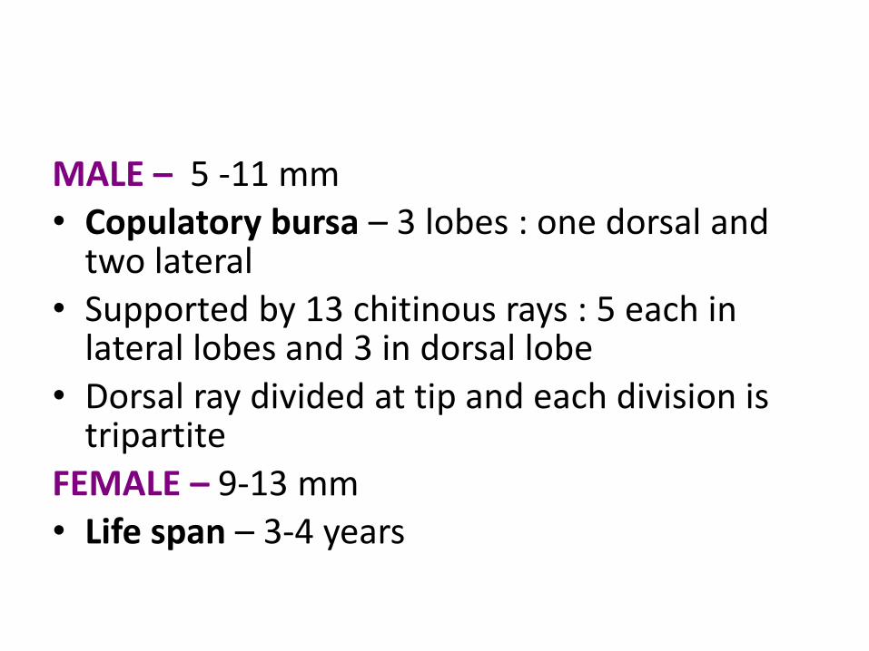
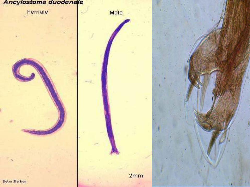
MALE – 5 -11 mm• Copulatory bursa – 3 lobes : one dorsal and
two lateral • Supported by 13 chitinous rays : 5 each in
lateral lobes and 3 in dorsal lobe
• Dorsal ray divided at tip and each division is tripartite
FEMALE – 9-13 mm• Life span – 3-4 years

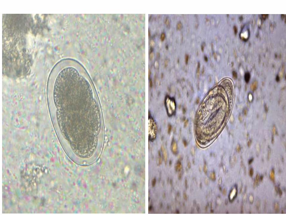
EGGS :
• 60 x 40 um in size
• Segmented ovum with four blastomeres
• Not bile stained
• Float in saturated salt solution
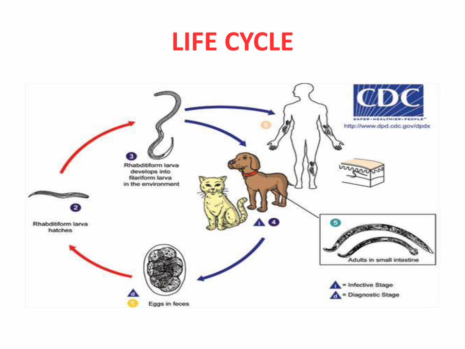
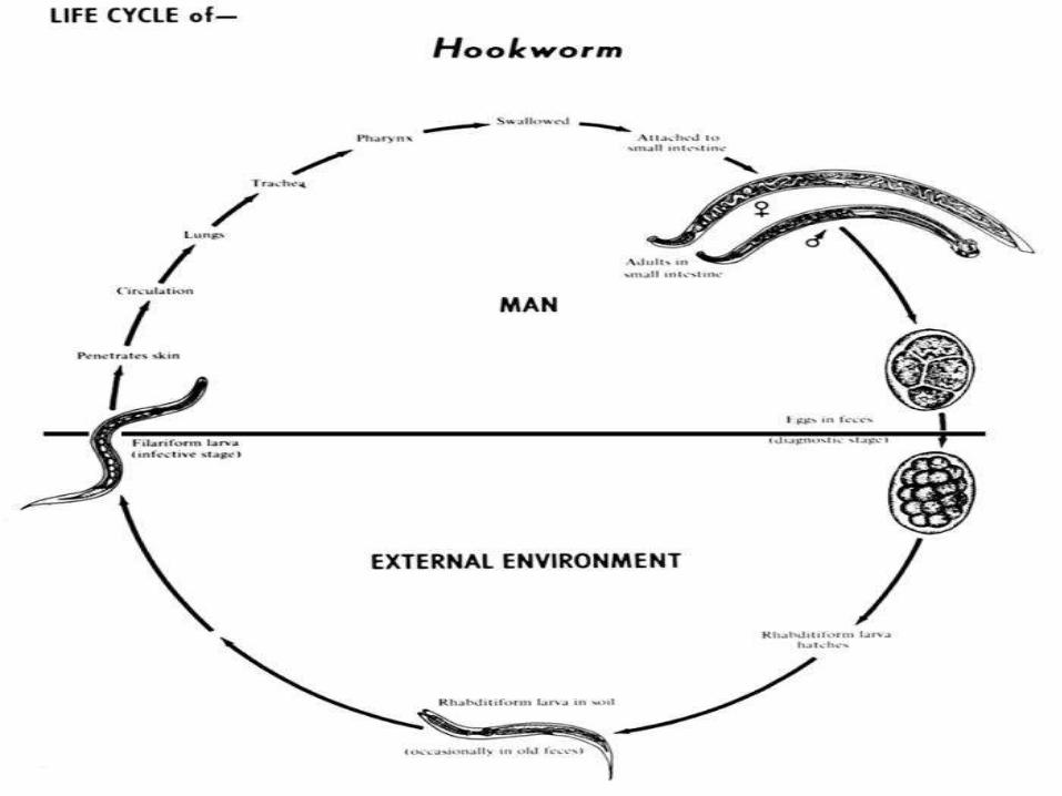
LIFE CYCLE
PATHOGENICITY
MIGRATING LARVAE :
Three types of lesions :
• Ancylostoma dermatits or Ground itch
• Pulmonary lesions
• Creeping eruption or Cutaneous larva migrans
ANCYLOSTOMA DERMATITS OR GROUND ITCH :
• Larvae enter the skin --- Dermatitis --- Itiching and burning --- erythema and oedema ---papular and vesicular eruptions
PULMONARY LESIONS :
• Bronchitis, Bronchopneumonia
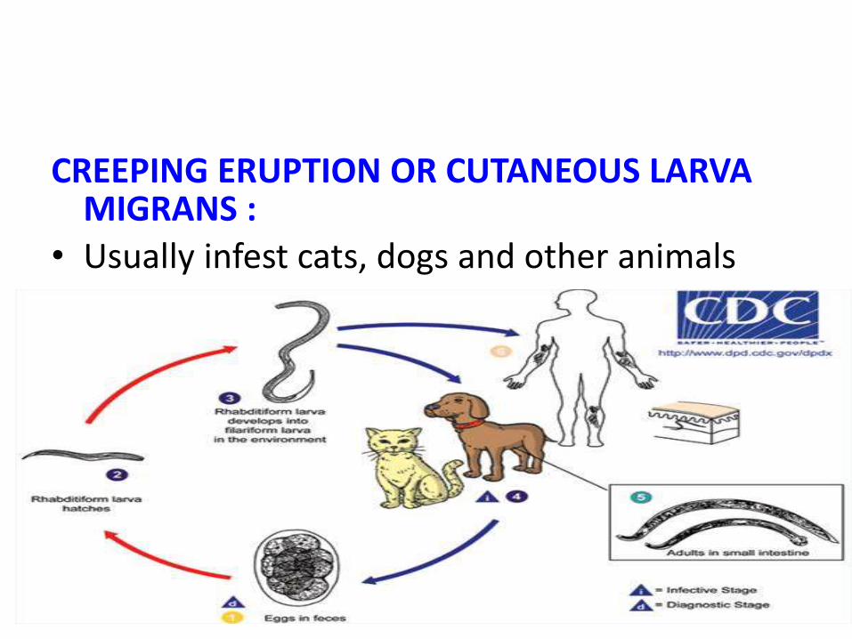
CREEPING ERUPTION OR CUTANEOUS LARVA MIGRANS :
• Usually infest cats, dogs and other animals
IN HUMAN – by walking barefoot
• Larvae are unable to penetrate the basement membrane
• Larvae migrate under the skin's surface –Creeping eruption
• Snake like tracks 2-3 mm wide
• Tracks advance a few mm to few cm daily
• Sites – feet, spaces b/w toes, hands, knees and buttocks
• Self limiting disease
• Humans are an accidental and dead-end host
• Thiabendazole, albendazole, mebendazole, ivermectin
IN ANIMALS –• Penetrate deeper layer of skin --- infect the
blood and lymphatic system --- in S.I they mature sexually and lay more eggs ----
• Ankylostoma brazilienses
• Ankylostoma caninum• Uncinaria stenocephala
• Bunostomum phlebotomum
ADULT WORM :• 0.2 – 0.03 ml blood daily• Contain anticoagulant activity• Microcytic, hypochormic type of iron
deficiency anaemia• Patient develops – epigastric pain, dyspepsia,
vomiting, diarrhoea, stool being reddish or black
• Skin becomes cold and dry• Oedema of feet and ankle
LABORATORY DIAGNOSIS
DIRECT METHODS :Microscopy :• Wet-mount • Faecal egg count – Adult female hookworms
produce 2,500-5,000 eggs/day• >2000 eggs/ml in women and > 5,ooo eggs/ml
in males ----> Anaemia• Aspiration of duodenal contents by Ryle's
tube• Adult worms in stool
INDIRECT METHODS :• PBF – Microcytic, hypochromic anaemia and
Eosinophilia• Stool examination – Occult blood, Charcot-
Leyden crystals
• NECATOR AMERICANUS• ANCYLOSTOMA BRAZILIENSE
• ANCYLOSTOMA CEYLANICUM