![Acer [Course Title] [Teacher's Name](https://static.fdokumen.com/doc/165x107/6320a62900d668140c0d1f09/acer-course-title-teachers-name.jpg)
Analysis of spatial and temporal dynamics of xylem refilling in Acer rubrum L. using magnetic...
8
ORIGINAL RESEARCH ARTICLE published: 22 July 2013 doi: 10.3389/fpls.2013.00265 Analysis of spatial and temporal dynamics of xylem refilling in Acer rubrum L. using magnetic resonance imaging Maciej A. Zwieniecki 1 *, Peter J. Melcher 2 and EricT. Ahrens 3 1 Department of Plant Sciences, University of California at Davis, Davis, CA, USA 2 Biology Department, Ithaca College, Ithaca, NY, USA 3 Department of Biological Sciences, Carnegie Mellon University, Pittsburgh, PA, USA Edited by: Abraham D. Stroock, Cornell University, USA Reviewed by: Jinkee Lee, Sungkyunkwan University, South Korea Abraham D. Stroock, Cornell University, USA *Correspondence: Maciej A. Zwieniecki, Department of Plant Sciences, University of California at Davis, PES 2316, One Shields Avenue, Davis, CA 95616, USA e-mail: [email protected] We report results of an analysis of embolism formation and subsequent refilling observed in stems of Acer rubrum L. using magnetic resonance imaging (MRI). MRI is one of the very few techniques that can provide direct non-destructive observations of the water content within opaque biological materials at a micrometer resolution. Thus, it has been used to determine temporal dynamics and water distributions within xylem tissue. In this study, we found good agreement between MRI measures of pixel brightness to assess xylem liquid water content and the percent loss in hydraulic conductivity (PLC) in response to water stress (P 50 values of 2.51 and 2.70 for MRI and PLC, respectively). These data provide strong support that pixel brightness is well correlated to PLC and can be used as a proxy of PLC even when single vessels cannot be resolved on the image. Pressure induced embolism in moderately stressed plants resulted in initial drop of pixel brightness. This drop was followed by brightness gain over 100 min following pressure application suggesting that plants can restore water content in stem after induced embolism. This recovery was limited only to current-year wood ring; older wood did not show signs of recovery within the length of experiment (16 h). In vivo MRI observations of the xylem of moderately stressed (∼−0.5 MPa) A. rubrum stems revealed evidence of a spontaneous embolism formation followed by rapid refilling (∼30 min). Spontaneous (not induced) embolism formation was observed only once, despite over 60 h of continuous MRI observations made on several plants. Thus this observation provide evidence for the presence of naturally occurring embolism-refilling cycle in A. rubrum, but it is impossible to infer any conclusions in relation to its frequency in nature. Keywords: embolism, xylem, MRI imaging, refilling, tension INTRODUCTION There is widespread agreement that negative hydrostatic pressures make water transport in the xylem intrinsically vulnerable to cavi- tation (Pickard, 1981; Tyree and Zimmermann, 2002). In order to maintain hydraulic capacity, plants must either minimize cavita- tion or restore conductivity in embolized conduits. The idea that embolized vessels might be returned to their functional state is not new, but it has generally been thought to be limited to situations in which the entire vascular system could be pressurized due to active solute transport by the roots (Fisher et al., 1997). However, more recent studies indicated that embolism removal may be pos- sible even when the majority of the water in the xylem remains under low, or moderate tensions (Salleo et al., 1996; Canny, 1997; McCully et al., 1998; Zwieniecki and Holbrook, 1998; McCully, 1999). This triggered a substantial effort to provide a conceptual framework and descriptions of important prerequisites that could explain how xylem refilling could occur in actively transpiring plants (Holbrook et al., 1999; Tyree et al., 1999; Salleo et al., 2009; Zwieniecki and Holbrook, 2009; Nardini et al., 2011; Secchi and Zwieniecki, 2011). Our current understanding of the spatial and temporal pat- terns of embolism formation and refilling relies heavily on measurements from destructive sampling techniques, such as mea- suring changes in stem hydraulic conductivity. However, there are several less invasive methods such as the use of a cryo-scanning electron microscope (cryo-SEM) that allows one to view the liq- uid (ice) content within the xylem of stems that were rapidly frozen in liquid nitrogen (Canny, 1997, 2001; McCully et al., 1998, 2000; Pate and Canny, 1999; Melcher et al., 2001). This cryo- SEM technique has helped to resolve some questions regarding the spatial distributions of embolism formation (Canny, 1997, 2001; McCully et al., 1998, 2000; Pate and Canny, 1999; Melcher et al., 2001). For example, they show that vessels tend to embolize in clusters, and that many embolized vessels had droplets of frozen water on their vessel walls. However, results from cryo- SEM studies were called into question because potential artifacts may arise during the freezing procedure (Cochard et al., 2000). A more recent study used high-resolution computed tomogra- phy to view in vivo water content in the stems on Vitis vinifera L. plants (Brodersen et al., 2010). Collected images showed not only the presence of water droplets on the walls of embolized vessels but also the dynamic changes in droplet size during refilling. These data provide strong support for the presence of refilling. www.frontiersin.org July 2013 | Volume 4 | Article 265 | 1
Transcript of Analysis of spatial and temporal dynamics of xylem refilling in Acer rubrum L. using magnetic...

“fpls-04-00265” — 2013/7/20 — 12:12 — page 1 — #1
ORIGINAL RESEARCH ARTICLEpublished: 22 July 2013
doi: 10.3389/fpls.2013.00265
Analysis of spatial and temporal dynamics of xylem refillingin Acer rubrum L. using magnetic resonance imagingMaciej A. Zwieniecki 1*, Peter J. Melcher 2 and EricT. Ahrens 3
1 Department of Plant Sciences, University of California at Davis, Davis, CA, USA2 Biology Department, Ithaca College, Ithaca, NY, USA3 Department of Biological Sciences, Carnegie Mellon University, Pittsburgh, PA, USA
Edited by:
Abraham D. Stroock, CornellUniversity, USA
Reviewed by:
Jinkee Lee, SungkyunkwanUniversity, South KoreaAbraham D. Stroock, CornellUniversity, USA
*Correspondence:
Maciej A. Zwieniecki, Department ofPlant Sciences, University ofCalifornia at Davis, PES 2316,One Shields Avenue, Davis,CA 95616, USAe-mail: [email protected]
We report results of an analysis of embolism formation and subsequent refilling observedin stems of Acer rubrum L. using magnetic resonance imaging (MRI). MRI is one of thevery few techniques that can provide direct non-destructive observations of the watercontent within opaque biological materials at a micrometer resolution. Thus, it has beenused to determine temporal dynamics and water distributions within xylem tissue. In thisstudy, we found good agreement between MRI measures of pixel brightness to assessxylem liquid water content and the percent loss in hydraulic conductivity (PLC) in responseto water stress (P50 values of 2.51 and 2.70 for MRI and PLC, respectively). These dataprovide strong support that pixel brightness is well correlated to PLC and can be used as aproxy of PLC even when single vessels cannot be resolved on the image. Pressure inducedembolism in moderately stressed plants resulted in initial drop of pixel brightness.This dropwas followed by brightness gain over 100 min following pressure application suggestingthat plants can restore water content in stem after induced embolism. This recovery waslimited only to current-year wood ring; older wood did not show signs of recovery within thelength of experiment (16 h). In vivo MRI observations of the xylem of moderately stressed(∼−0.5 MPa) A. rubrum stems revealed evidence of a spontaneous embolism formationfollowed by rapid refilling (∼30 min). Spontaneous (not induced) embolism formation wasobserved only once, despite over 60 h of continuous MRI observations made on severalplants. Thus this observation provide evidence for the presence of naturally occurringembolism-refilling cycle in A. rubrum, but it is impossible to infer any conclusions in relationto its frequency in nature.
Keywords: embolism, xylem, MRI imaging, refilling, tension
INTRODUCTIONThere is widespread agreement that negative hydrostatic pressuresmake water transport in the xylem intrinsically vulnerable to cavi-tation (Pickard, 1981; Tyree and Zimmermann, 2002). In order tomaintain hydraulic capacity, plants must either minimize cavita-tion or restore conductivity in embolized conduits. The idea thatembolized vessels might be returned to their functional state is notnew, but it has generally been thought to be limited to situationsin which the entire vascular system could be pressurized due toactive solute transport by the roots (Fisher et al., 1997). However,more recent studies indicated that embolism removal may be pos-sible even when the majority of the water in the xylem remainsunder low, or moderate tensions (Salleo et al., 1996; Canny, 1997;McCully et al., 1998; Zwieniecki and Holbrook, 1998; McCully,1999). This triggered a substantial effort to provide a conceptualframework and descriptions of important prerequisites that couldexplain how xylem refilling could occur in actively transpiringplants (Holbrook et al., 1999; Tyree et al., 1999; Salleo et al., 2009;Zwieniecki and Holbrook, 2009; Nardini et al., 2011; Secchi andZwieniecki, 2011).
Our current understanding of the spatial and temporal pat-terns of embolism formation and refilling relies heavily on
measurements from destructive sampling techniques, such as mea-suring changes in stem hydraulic conductivity. However, there areseveral less invasive methods such as the use of a cryo-scanningelectron microscope (cryo-SEM) that allows one to view the liq-uid (ice) content within the xylem of stems that were rapidlyfrozen in liquid nitrogen (Canny, 1997, 2001; McCully et al., 1998,2000; Pate and Canny, 1999; Melcher et al., 2001). This cryo-SEM technique has helped to resolve some questions regardingthe spatial distributions of embolism formation (Canny, 1997,2001; McCully et al., 1998, 2000; Pate and Canny, 1999; Melcheret al., 2001). For example, they show that vessels tend to embolizein clusters, and that many embolized vessels had droplets offrozen water on their vessel walls. However, results from cryo-SEM studies were called into question because potential artifactsmay arise during the freezing procedure (Cochard et al., 2000).A more recent study used high-resolution computed tomogra-phy to view in vivo water content in the stems on Vitis viniferaL. plants (Brodersen et al., 2010). Collected images showed notonly the presence of water droplets on the walls of embolizedvessels but also the dynamic changes in droplet size duringrefilling. These data provide strong support for the presence ofrefilling.
www.frontiersin.org July 2013 | Volume 4 | Article 265 | 1

“fpls-04-00265” — 2013/7/20 — 12:12 — page 2 — #2
Zwieniecki et al. Temporal dynamics of xylem refilling
Studies that have investigated the temporal dynamics of therefilling process using artificially induced embolism show thatrefilling was more or less completed within an hour after embolisminduction (Salleo et al., 1996; Zwieniecki et al., 2004; Secchi andZwieniecki, 2011). Similar findings come from observations ofnatural embolism in petioles of red maple (Acer rubrum L.) andtulip trees (Liriodendron tulipifera L.) using a double stainingmethod (Zwieniecki et al., 2000). However, reversal of embolismin vines (Vitis spp.) was observed to only occur when tran-spiration had been stopped (Zwieniecki et al., 2000; Holbrooket al., 2001). In addition, the temporal pattern of recovery fromembolism seems to be related to the level of plant water stress.For example, Laurus nobilis L. and A. negundo L. only refilledembolisms over prolonged recovery times of 24 h and onlywhen water stress levels were significantly reduced (Hacke andSperry, 2003). Secchi and Zwieniecki (2011) showed that therate of embolism recovery in poplar trees (Populus trichocarpaL.) was dependent on the level of water stress. Their studyshowed faster recovery (less than 2 h) in moderately stressedtrees compared to much longer recovery times (more than 20 h)in trees exposed to severe stress. The difference in recoveryrates was observed despite the fact that stem water potentialsincreased in both cases within 1 h (Secchi and Zwieniecki,2011).
Most of the evidence that demonstrates rapid refilling in plantsrelies on destructive sampling methods that could be prone tomethodological problems. The few in vivo observations usingmagnetic resonance imaging (MRI) and x-ray tomography showonly very slow recovery in species with large vessels: Vitis spp.(Holbrook et al., 2001; Brodersen et al., 2010) and Cucumis sativus(Scheenen et al., 2007). Thus, there is still a lack of in vivo evi-dence that would provide supporting evidence of the rapid ratesof the embolism-refilling cycles observed in species with smallvessels obtained using destructive sampling techniques. The goalof this short contribution is aimed specifically at addressing thisissue. We present results of direct observations of naturally occur-ring embolism/refilling cycle in stems of A. rubrum observed usingMRI.
MATERIALS AND METHODSStudy was conducted on A. rubrum plants either 2-year-old plantswith minimum 1 m long stem and branches collected from 20-year-old A. rubrum trees. For all of the MRI experiments, priorto placing a plant or a sample into the MRI magnet, a 15-mmdiameter surface coil radio frequency resonator was placed on thestem. Each plant was positioned in an 11.7 T, 89 mm vertical-bore,Bruker AVANCE micro-imaging system. The sample temperaturewas regulated at ∼25◦C by pumping air through the magnet bore.For image data collection, we used a T2/spin-density-weighted3D Fourier transform spin-echo sequence (T2W-3DFT) with arepetition time/echo time (TR/TE) = 980/45 ms. The T2W-3DFTdata provided good free water versus air contrast. Images wereacquired with a 256 × 128 × 128 matrix and then zero-filled to512 × 256 × 256 before Fourier transformation, yielding a finalisotropic resolution of approximately 50 μm. The imaging timewas approximately 90 s per image with 90 s resting time betweenimages.
To compare MRI analysis of xylem water content to stemhydraulic conductivity, we used ∼2-m-long leafy branches thatwere collected from seven trees (15–20 years old) growing in thefield at Harvard Forest. Several leaves on each branch were placedinto sealed plastic bags and covered in aluminum foil the eveningbefore collecting branches at predawn the next day. After excisingbranches in the air, they were allowed to continue to transpire (inthe shade) until the loss of water from the uncovered leaves reducedcovered leaf water potentials to values that were needed to gener-ate a vulnerability to embolism response curve. Covered, branchequilibrated leaf water potentials were measured using a pressurechamber system. The balancing pressure required to squeeze waterto the excised petiole surface was determined and used to estimatestem water potentials. Following dehydration, each branch waslabeled and was double bagged in large black plastic bags. Wetpaper towels lined the two-bag layers to reduce evaporation andto allow the branch water potentials to equilibrate within eachsample. These branches were then shipped from Harvard Forest,Petersham, MA to the MRI facility in Carnegie Mellon Universityin Pittsburgh, PA.
Prior to MRI measurements, leaf water potentials were re-measured using the same pressure chamber system to determineequilibrated water potentials of the branch samples. For eachsample, a long portion of the stem was excised under water firstand then two 5-cm-long stem segments were subsequently excisedunderwater from the current extension growth (number of sampletested 25). One of the excised stem samples was used to deter-mine the PLC using classical hydraulic pressure-flow methods.The other excised sample was used for the determination of thewater content using MRI. The MRI sample was tightly wrapped inparafilm to further reduce desiccation during the measurement.After MRI imaging was complete, a post-processing image regis-tration algorithm was applied to the data to correct for physicaltranslations of the stem in the image field of view over the measure-ment time (total successful measurements 20). Image brightnesswas adjusted for all images using two control glass tubes filled withDI H2O and 1:1 mixture of DI H2O and D2O (volumetric). Pixelbrightness ranged in images from black (0 value) to white (65525value), and these values corresponded to increasing concentrationof unbound water that was present in the voxel (volumetric pictureelement 50 μm × 50 μm × 1000 μm) and were used for anal-ysis of xylem water content (Matlab12, MathWorks, Inc., Natick,MA, USA).
To determine the potential for spontaneous embolism forma-tion in moderately stressed stems, undisturbed 3-year-old plantswere fitted through the magnet bore using the same strategyas described above. Each plant was left in the magnet for 10–15 h and images were taken every 3min (90 s signal collectiontime). Images were acquired using a multi-slice gradient-echosequence with TR/TE = 75/5 ms and a 512 × 256 × 256matrix size, in-plane resolution of 50 μm × 50 μm and 1 mmthick slices. Images were simultaneously collected from fiveslices separated by 2 mm distance and thus covering a totalof 15 mm of stem length. Data were analyzed using Matlab12 (MathWorks). During the 10- to 15-h observation period,plants were not subjected to any experimental treatments orany disturbance. They were maintained at an average leaf water
Frontiers in Plant Science | Plant Biophysics and Modeling July 2013 | Volume 4 | Article 265 | 2

“fpls-04-00265” — 2013/7/20 — 12:12 — page 3 — #3
Zwieniecki et al. Temporal dynamics of xylem refilling
potential of about −0.5 MPa. Total time of observation equaled60 h.
Long-term MRI observations were followed by an air-injectionexperiment to determine the temporal dynamics of artificiallyinduced embolism in intact plants. Prior to attaching a pressurecollar near the base of the main stem of each plant a small inci-sion was made to allow pressurized gas to penetrate the xylemof the plants during air-injection (Crombie et al., 1985). Eachof three plants was pressurized so that the pressure gradientacross the bordered pit membranes equaled 5.0 MPa (sum cov-ered leaf water potential and injection pressure). The pressurewas held for 2 min. while the plant was still in the MRI mag-net. MRI measurements were made during and after air-injectionto determine if A. rubrum could recover from artificially inducedembolism.
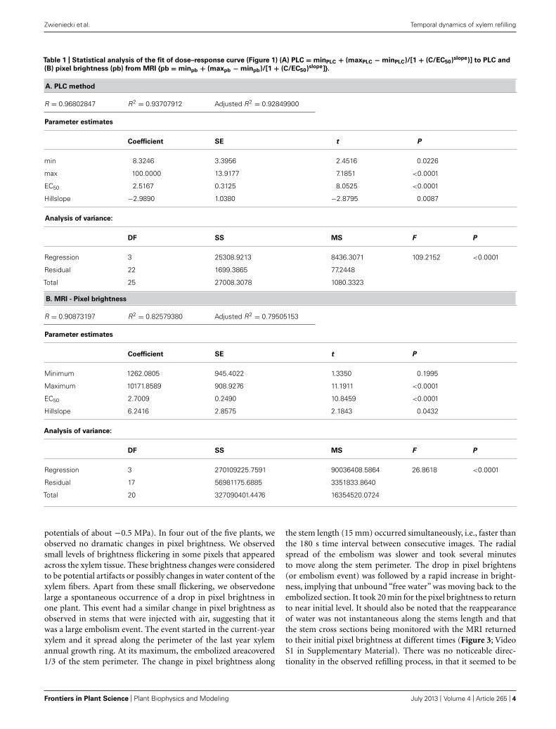
RESULTSComparative analysis of xylem water content from MRI imagesand stem hydraulic measurements were made on current-yearextension growth. Analysis was made on branches exposed tovarying levels of water stress to assess the relationship of pixelbrightness measured with MRI to changes in stem hydraulics. Pixelbrightness measured with MRI is related to the amount of freewater in the sample. In plant tissues, this would be the water thatcan freely move and is not bound within cellular walls. The gen-erated MRI-based “vulnerability” curve was found to be similarto the PLC curve measured using hydraulic methods (Figure 1).We found that stress of −2.51 MPa was required to reduce thexylem hydraulic conductance of the xylem of current-year exten-sion growth of A. rubrum plants by 50% (P50), determined fromhydraulic methods. The equivalent 50% loss of average pixelbrightness in MRI images was determined to be −2.70 MPa.We also observed similarities in the shape of the vulnerabilityto embolism curves obtained from both hydraulic methods andMRI image analysis and no statistical difference between estimatesof EC50 (Table 1). These results provided assurance that pixelbrightness was a good proxy for analysis of stem water contentand for estimating changes in stem hydraulic conductance due toembolism formation (Figure 1).
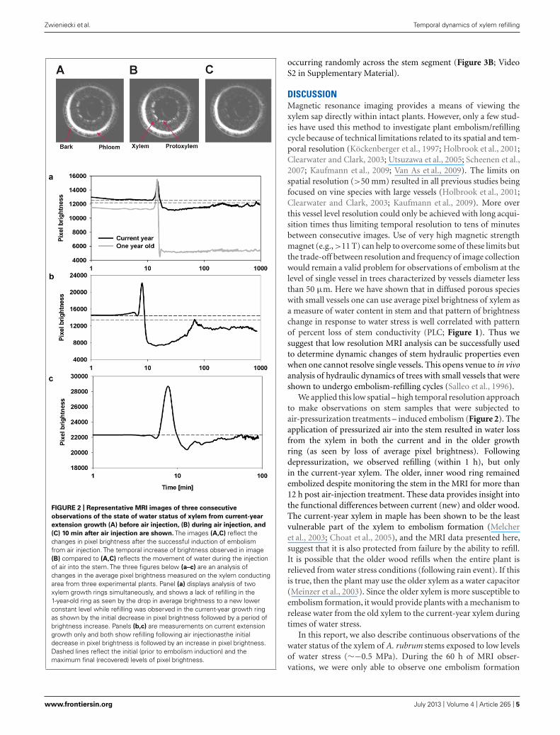
The temporal and spatial dynamics of embolism were mea-sured using MRI on intact, well hydrated A. rubrum plants thatwere exposed to air-pressurization treatments that created a 5.0-MPa pressure gradient across the xylem bordered pit membranes.Changes in the average pixel brightness of analyzed tissues wereused to assess changes in stem water content in two regions ofthe stems during these pressurization treatments: (1) current-year extension growth (one xylem ring) and (2) 1-year-old stems(two xylem rings). As expected, air injection treatments, thatcreated a 5.0-MPa gas/water interface pressure differential at thebordered pit level, resulted in the loss of pixel brightness andwas interpreted as a drop in the water content in the stem andformation of embolism. In 1-year-old stems, embolism formedin both the older (internal ring) and in the current-year xylem(outer ring). The loss of water content determined from decreasedpixel brightness in the older ring was found to be much morepronounced (Figure 2). We did not observe any signs of bright-ness recovery over a 10-h measurement period in the older ring.
FIGURE 1 | Leaf water potential, measured on equilibrated branches,
are plotted to PLC (A), and average pixel brightness (black = 0 to
white = 65525) determined from MRI analysis (B), is shown. Both datasets were fitted with a dose–response curve (solid line) in the form ofPLC = minPLC + (maxPLC − minPLC)/[1 + (C/EC50)slope], where minPLC isminimum PLC in non-stressed plants, maxPLC is 100%, EC50 represents50% loss of initial functionality [minPLC + (maxPLC − minPLC)/2], and slopeis the rateof PLC increase at EC50. There was no statistical differencebetween EC50 from two methods (PLC and MRI). The same function wasused for pixel brightness curve fitting except that minPLC and maxPLC weresubstituted with average pixel brightness at low and high ends of stemwater stress. The four MRI images shown are representative images thatwere used to create the MRI-vulnerability curve. Only pixel brightness datafrom the xylem conducting area was used to produce the curve. Allmeasurements were made on current-year extension growth.
However, the initial loss of water content in current-year xylemrecovered within 2 h from induction of embolism (Figure 2).In two other instances, only sections of the current extensiongrowth of the stem were observed with the MRI, and we foundthat the initial drop of water content due to air-injection inducedembolism was followed by recovery within a 1- to 2-h period(Figure 2). The spike in brightness during the air injection processreflects the movement of water in the xylem caused by the waterbeing replaced with the air that is being forced into the xylem(Figure 2).
The long-term MRI monitoring experiment was conducted onintact potted plants that were undisturbed for 10–15 h each. Thisexperiment was designed to determine if A. rubrum plants undergo“natural” spontaneous embolism formation within their xylem onplants exposed to moderate levels of water stress (xylem water
www.frontiersin.org July 2013 | Volume 4 | Article 265 | 3
“fpls-04-00265” — 2013/7/20 — 12:12 — page 4 — #4
Zwieniecki et al. Temporal dynamics of xylem refilling
Table 1 | Statistical analysis of the fit of dose–response curve (Figure 1) (A) PLC = minPLC + (maxPLC − minPLC)/[1 + (C/EC50)slope)] to PLC and
(B) pixel brightness (pb) from MRI {pb = minpb + (maxpb − minpb)/[1 + (C/EC50)slope]}.
A. PLC method
R = 0.96802847 R2 = 0.93707912 Adjusted R2 = 0.92849900
Parameter estimates
Coefficient SE t P
min 8.3246 3.3956 2.4516 0.0226
max 100.0000 13.9177 7.1851 <0.0001
EC50 2.5167 0.3125 8.0525 <0.0001
Hillslope −2.9890 1.0380 −2.8795 0.0087
Analysis of variance:
DF SS MS F P
Regression 3 25308.9213 8436.3071 109.2152 <0.0001
Residual 22 1699.3865 77.2448
Total 25 27008.3078 1080.3323
B. MRI - Pixel brightness
R = 0.90873197 R2 = 0.82579380 Adjusted R2 = 0.79505153
Parameter estimates
Coefficient SE t P
Minimum 1262.0805 945.4022 1.3350 0.1995
Maximum 10171.8589 908.9276 11.1911 <0.0001
EC50 2.7009 0.2490 10.8459 <0.0001
Hillslope 6.2416 2.8575 2.1843 0.0432
Analysis of variance:
DF SS MS F P
Regression 3 270109225.7591 90036408.5864 26.8618 <0.0001
Residual 17 56981175.6885 3351833.8640
Total 20 327090401.4476 16354520.0724
potentials of about −0.5 MPa). In four out of the five plants, weobserved no dramatic changes in pixel brightness. We observedsmall levels of brightness flickering in some pixels that appearedacross the xylem tissue. These brightness changes were consideredto be potential artifacts or possibly changes in water content of thexylem fibers. Apart from these small flickering, we observedonelarge a spontaneous occurrence of a drop in pixel brightness inone plant. This event had a similar change in pixel brightness asobserved in stems that were injected with air, suggesting that itwas a large embolism event. The event started in the current-yearxylem and it spread along the perimeter of the last year xylemannual growth ring. At its maximum, the embolized areacovered1/3 of the stem perimeter. The change in pixel brightness along
the stem length (15 mm) occurred simultaneously, i.e., faster thanthe 180 s time interval between consecutive images. The radialspread of the embolism was slower and took several minutesto move along the stem perimeter. The drop in pixel brightens(or embolism event) was followed by a rapid increase in bright-ness, implying that unbound “free water” was moving back to theembolized section. It took 20 min for the pixel brightness to returnto near initial level. It should also be noted that the reappearanceof water was not instantaneous along the stems length and thatthe stem cross sections being monitored with the MRI returnedto their initial pixel brightness at different times (Figure 3; VideoS1 in Supplementary Material). There was no noticeable direc-tionality in the observed refilling process, in that it seemed to be
Frontiers in Plant Science | Plant Biophysics and Modeling July 2013 | Volume 4 | Article 265 | 4
“fpls-04-00265” — 2013/7/20 — 12:12 — page 5 — #5
Zwieniecki et al. Temporal dynamics of xylem refilling
FIGURE 2 | Representative MRI images of three consecutive
observations of the state of water status of xylem from current-year
extension growth (A) before air injection, (B) during air injection, and
(C) 10 min after air injection are shown. The images (A,C) reflect thechanges in pixel brightness after the successful induction of embolismfrom air injection. The temporal increase of brightness observed in image(B) compared to (A,C) reflects the movement of water during the injectionof air into the stem. The three figures below (a–c) are an analysis ofchanges in the average pixel brightness measured on the xylem conductingarea from three experimental plants. Panel (a) displays analysis of twoxylem growth rings simultaneously, and shows a lack of refilling in the1-year-old ring as seen by the drop in average brightness to a new lowerconstant level while refilling was observed in the current-year growth ringas shown by the initial decrease in pixel brightness followed by a period ofbrightness increase. Panels (b,c) are measurements on current extensiongrowth only and both show refilling following air injectionasthe initialdecrease in pixel brightness is followed by an increase in pixel brightness.Dashed lines reflect the initial (prior to embolism induction) and themaximum final (recovered) levels of pixel brightness.
occurring randomly across the stem segment (Figure 3B; VideoS2 in Supplementary Material).
DISCUSSIONMagnetic resonance imaging provides a means of viewing thexylem sap directly within intact plants. However, only a few stud-ies have used this method to investigate plant embolism/refillingcycle because of technical limitations related to its spatial and tem-poral resolution (Köckenberger et al., 1997; Holbrook et al., 2001;Clearwater and Clark, 2003; Utsuzawa et al., 2005; Scheenen et al.,2007; Kaufmann et al., 2009; Van As et al., 2009). The limits onspatial resolution (>50 mm) resulted in all previous studies beingfocused on vine species with large vessels (Holbrook et al., 2001;Clearwater and Clark, 2003; Kaufmann et al., 2009). More overthis vessel level resolution could only be achieved with long acqui-sition times thus limiting temporal resolution to tens of minutesbetween consecutive images. Use of very high magnetic strengthmagnet (e.g., >11 T) can help to overcome some of these limits butthe trade-off between resolution and frequency of image collectionwould remain a valid problem for observations of embolism at thelevel of single vessel in trees characterized by vessels diameter lessthan 50 μm. Here we have shown that in diffused porous specieswith small vessels one can use average pixel brightness of xylem asa measure of water content in stem and that pattern of brightnesschange in response to water stress is well correlated with patternof percent loss of stem conductivity (PLC; Figure 1). Thus wesuggest that low resolution MRI analysis can be successfully usedto determine dynamic changes of stem hydraulic properties evenwhen one cannot resolve single vessels. This opens venue to in vivoanalysis of hydraulic dynamics of trees with small vessels that wereshown to undergo embolism-refilling cycles (Salleo et al., 1996).
We applied this low spatial – high temporal resolution approachto make observations on stem samples that were subjected toair-pressurization treatments – induced embolism (Figure 2). Theapplication of pressurized air into the stem resulted in water lossfrom the xylem in both the current and in the older growthring (as seen by loss of average pixel brightness). Followingdepressurization, we observed refilling (within 1 h), but onlyin the current-year xylem. The older, inner wood ring remainedembolized despite monitoring the stem in the MRI for more than12 h post air-injection treatment. These data provides insight intothe functional differences between current (new) and older wood.The current-year xylem in maple has been shown to be the leastvulnerable part of the xylem to embolism formation (Melcheret al., 2003; Choat et al., 2005), and the MRI data presented here,suggest that it is also protected from failure by the ability to refill.It is possible that the older wood refills when the entire plant isrelieved from water stress conditions (following rain event). If thisis true, then the plant may use the older xylem as a water capacitor(Meinzer et al., 2003). Since the older xylem is more susceptible toembolism formation, it would provide plants with a mechanism torelease water from the old xylem to the current-year xylem duringtimes of water stress.
In this report, we also describe continuous observations of thewater status of the xylem of A. rubrum stems exposed to low levelsof water stress (∼−0.5 MPa). During the 60 h of MRI obser-vations, we were only able to observe one embolism formation
www.frontiersin.org July 2013 | Volume 4 | Article 265 | 5

“fpls-04-00265” — 2013/7/20 — 12:12 — page 6 — #6
Zwieniecki et al. Temporal dynamics of xylem refilling
FIGURE 3 | Images of real-time MRI observations of a section of stem
from an intact A. rubrum plant exposed to moderate levels of water
stress. (A) Images show the development of a spontaneously occurringembolism (at 6 min) and its spread across the current growth ring (at 18 min)followed by an almost full water content recovery (at 30 min). (B) Three
dimensional analyses of embolism formation, its spatial spread, and recovery.The blue color denotes volumes with 90% loss of pixel brightness. Pleasenote that to improve embolus visibility, images were reoriented in respect tothe MRI images. Please consult the Supplementary Material online (Video S1and S2).
event. However, we feel confident that this one observation placedin the context of other available data (see Brodersen and McElrone,2013) provides strong support for in situ formation of embolism instems of moderately stressed plants. In addition, our data provideevidence of rapid xylem refilling as the loss of pixel brightness(formation of embolism) was followed by the restoration of pixelbrightness (refilling) to pre-embolism conditions in the affectedarea with 30 min. The spread of this naturally occurring embolismevent was limited only to current-year xylem. The circumferen-tial progress (several millimeters) occurred over several minutes(i.e., over several consecutive images) while vertical occurrence(3 cm distance) was instantaneous (i.e., within time needed tocollect signal for a single image). Analysis of refilling showed nodirectionality and water seemed to occur in many separated imagevoxels across entire embolized volume.
The unique in vivo time-lapse observation of embolism/refillingcycles using MRI highlights important considerations for our cur-rent understanding of xylem function, i.e., that cavitation might be
an everyday event in the stems of transpiring plants at a frequencythat is related to tension of sap in the xylem. As our data show,the restoration of water content in the affected stem occurredrelatively quickly, but one can expect that the effectiveness ofrefilling would decrease with increasing levels of water stress. Thiswould eventually lead to a situation when embolisms may not beremoved by refilling, and that embolisms may accumulate fasterthan they can be refilled, resulting in an increase of non-functionalconduits (Secchi and Zwieniecki, 2012). Thus, we can expect thatthe current level of embolisms in a stem is a product of the prob-ability of embolism occurrence (positively related to tension) andembolism-refilling rate which is inversely related to tension andthe ability of the plant to supply energy (Zwieniecki and Holbrook,2009; Nardini et al., 2011; Secchi and Zwieniecki, 2012). If this viewis correct, then it would be predicted that plants continuously gothrough embolism/refilling cycles in different parts of the stemand that the percent of measured embolisms reflects the currentbalance between these two processes.
Frontiers in Plant Science | Plant Biophysics and Modeling July 2013 | Volume 4 | Article 265 | 6

“fpls-04-00265” — 2013/7/20 — 12:12 — page 7 — #7
Zwieniecki et al. Temporal dynamics of xylem refilling
It is interesting to note that the rate of refilling in A. rubrum,from both spontaneous cavitation, and from the air-injectionmethod was fast (less than an hour). This stands in contrast to theobservation of refilling of embolized vessels in Vitis spp.(Brodersenet al., 2010) where refilling extended for several hours and in C.sativus which took 17–47 h (Scheenen et al., 2007). The discrep-ancy may be the result of the vessel volume differences betweenthe studied species. Maple vessels (this study) are general less than50 μm in diameter, while the grapevine vessels are often morethan 200 μm in diameter (Brodersen et al., 2010). This differenceresults in roughly 16 times larger volume of the grapevine ves-sels compared to maplevessels per length. Thus, if the living cellrefilling activity is not enhanced, then we might expect that thetime required to refill grapevine vessels will be 16 times longer(i.e., ∼8 h), which is very similar to the time reported by Broder-sen et al. (2010). The same might be true for the time discrepancyfrom this and the Scheenen et al. (2007) study as they were focus-ing only on the largest vessels in the cucumber stem (∼200 μm indiameter). It is also possible that difference in temporal dynam-ics of refilling reflect intrinsic physiological differences betweenherbaceous annual plants and woody perennials, where perennialsmay need mechanim for fast refilling to ensure long-term xylemfunctionality.
In conclusion, comparative analysis of the hydraulicdetermination of PLC and pixel brightness from MRI images inrelation to stem water potential showed a functional paralelismallowing for interpretation of MRI data in the context of PLCwithout need resolve single vessels. Further, this work provides
visual evidence that the embolism formation/refilling cycle existsin intact A. rubrum stems experiencing moderate levels of waterstress. The long-term observations using MRI of undisturbedstems of A. rubrum showed one such unambiguous event that incombination with other reports (Brodersen and McElrone, 2013)provide support for the existence of rapid refilling in moder-ately stressed plants. However, the functionality (restoration ofwater transport) of vessels refilled under tension still remainsunanswered.
ACKNOWLEDGMENTSThis work was supported by National Science Foundation Award#:IOS-0919729, Mellon University MRI internal grant and theMellon Foundation. The MRI studies were performed at thePittsburgh NMR Center for Biomedical Research funded by theNational Institute of Health (P41-EB001977).
SUPPLEMENTARY MATERIALThe Supplementary Material for this article can be found onlineat: http://www.frontiersin.org/Plant_Biophysics_and_Modeling/10.3389/fpls.2013.00265/abstract
Video 1 | A time lapse video of A. rubrum stem from an MRI observation.
The video shows a natural occurrence of embolism in an intact stem of a pottedtree. The embolism occurs in the current-year vascular ring, spreads and thendisappears as refilling takes place.
Video 2 | A 3D reconstruction of embolism dynamics in a stem of
A. rubrum from images collected during MRI observations. Five verticalobservation planes (1.5 mm thick) separated by 1.5 mm distances are shown.
REFERENCESBrodersen, C. R., and McElrone, A. J.
(2013). Maintenance of xylem net-work transport capacity: a reviewof embolism repair in vascularplants. Front. Plant Sci. 4:108. doi:10.3389/fpls.2013.00108
Brodersen, C. R., McElrone, A. J.,Choat, B., Matthews, M. A., andShackel, K. A. (2010). The dynam-ics of embolism repair in xylem:in vivo visualizations using high-resolution computed tomography.Plant Physiol. 154, 1088–1095. doi:10.1104/pp.110.162396
Canny, M. (1997). Vessel contents dur-ing transpiration – embolisms andrefilling. Am. J. Bot. 84, 1223. doi:10.2307/2446046
Canny, M. J. (2001). Embolisms andrefilling in the maize leaf lamina, andthe role of the protoxylem lacuna.Am. J. Bot. 88, 47–51. doi: 10.2307/2657125
Choat, B., Lahr, E. C., Melcher, P. J.,Zwieniecki, M. A., and Holbrook,N. M. (2005). The spatial patternof air seeding thresholds in maturesugar maple trees. Plant Cell Envi-ron. 28, 1082. doi: 10.1111/j.1365-3040.2005.01336.x
Clearwater, M. J., and Clark, C. J.(2003). In vivo magnetic resonance
imaging of xylem vessel contents inwoody lianas. Plant Cell Environ.26, 1205–1214. doi: 10.1046/j.1365-3040.2003.01042.x
Cochard, H., Bodet, C., Ameglio, T., andCruiziat, P. (2000). Cryo-scanningelectron microscopy observations ofvessel content during transpirationin walnut petioles. Facts or arti-facts. Plant Physiol. 124, 1191. doi:10.1104/pp.124.3.1191
Crombie, D. S., Milburn, J. A., andHipkins, M. F. (1985). Maximumsustainable xylem sap tensions inRhododendron and other species.Planta 163, 27–33. doi: 10.1007/BF00395893
Fisher, J. B., Angeles, A., Ewers, F. W.,and López-Portillo, J. (1997). Surveyof root pressure in tropical vines andwoody species. Int. J. Plant Sci. 158,44–50.
Hacke, U. G., and Sperry, J. S. (2003).Limits to xylem refilling under neg-ative pressure in Laurus nobilis andAcer negundo. Plant Cell Environ.26, 303–311. doi: 10.1046/j.1365-3040.2003.00962.x
Holbrook, N. M., Ahrens, E. T., Burns,M. J., and Zwieniecki, M. A. (2001).In vivo observation of cavitationand embolism repair using magneticresonance imaging. Plant Physiol.
126, 27–31. doi: 10.1104/pp.126.1.27
Holbrook, N. M., Zwienicki, M. A., andField, C. B. (1999). Embolism repairand xylem tension: do we need amiracle? Plant Physiol. 120, 7. doi:10.1104/pp.120.1.7
Kaufmann, I., Schulze-Till, T.,Schneider, H. U., Zimmermann, U.,Jakob, P., and Wegner, L. H. (2009).Functional repair of embolized ves-sels in maize roots after temporaldrought stress, as demonstratedby magnetic resonance imag-ing. New Phytol. 184, 245–256. doi: 10.1111/j.1469-8137.2009.02919.x
Köckenberger, W., Pope, J. M., Xia,Y., Jeffrey, K. R., Komor, E., andCallaghan, P. T. (1997). A non-invasive measurement of phloem andxylem water flow in castor beanseedlings by nuclear magnetic res-onance microimaging. Planta 201,53–63. doi: 10.1007/BF01258680
McCully, M. E. (1999). Root xylemembolisms and refilling. Relation towater potentials of soil, roots, andleaves, and osmotic potentials of rootxylem sap. Plant Physiol. 119, 1001–1008. doi: 10.1104/pp.119.3.1001
McCully, M. E., Huang, C. X.,and Ling, L. E. C. (1998). Daily
embolism and refilling of xylemvessels in the roots of field-grownmaize. New Phytol. 138, 327–342. doi: 10.1046/j.1469-8137.1998.00101.x
McCully, M. E., Shane, M. V. V., Baker,A. N., Huang, C. X., Ling, L. E., andCanny, M. J. (2000). The reliabilityof cryoSEM for the observation andquantification of xylem embolismsand quantitative analysis of xylemsap in situ. J. Microsc. 198, 24. doi:10.1046/j.1365-2818.2000.00679.x
Meinzer, F. C., James, S. A., Goldstein,G., and Woodruff, D. (2003). Whole-tree water transport scales withsapwood capacitance in tropical for-est canopy trees. Plant Cell Environ.26, 1147–1155. doi: 10.1046/j.1365-3040.2003.01039.x
Melcher, P. J., Goldstein, G., Meinzer, F.C.,Yount, D. E., Jones, T. J., Holbrook,N. M., et al. (2001). Water relationsof coastal and estuarine Rhizophoramangle: xylem pressure potential anddynamics of embolism formationand repair. Oecologia 126, 182–192.doi: 10.1007/s004420000519
Melcher, P. J., Zwieniecki, M. A., andHolbrook, N. M. (2003). Vulner-ability of xylem vessels to cavita-tion in sugar maple. Scaling fromindividual vessels to whole branches.
www.frontiersin.org July 2013 | Volume 4 | Article 265 | 7
“fpls-04-00265” — 2013/7/20 — 12:12 — page 8 — #8
Zwieniecki et al. Temporal dynamics of xylem refilling
Plant Physiol. 131, 1775–1780. doi:10.1104/pp.102.012856
Nardini, A., Lo Gullo, M. A., and Salleo,S. (2011). Refilling embolized xylemconduits: is it a matter of phloemunloading? Plant Sci. 180, 604–611. doi: 10.1016/j.plantsci.2010.12.011
Pate, J. S., and Canny, M. J. (1999).Quantification of vessel embolismsby direct observation: a compari-son of two methods. New Phytol.141, 33–43. doi: 10.1046/j.1469-8137.1999.00329.x
Pickard, W. F. (1981). The ascent ofsap in plants. Prog. Biophys. Mol.Biol. 37, 181–229. doi: 10.1016/0079-6107(82)90023-2
Salleo, S., Gullo, M. A. L., dePaoli, D., and Zippo, M. (1996).Xylem recovery from cavitation-induced embolism in young plantsof Laurus nobilis: a possible mecha-nism. New Phytol. 132, 47–56. doi:10.1111/j.1469-8137.1996.tb04507.x
Salleo, S., Trifilò, P., Esposito, S., Nar-dini, A., and Lo Gullo, M. A. (2009).Starch-to-sugar conversion in woodparenchyma of field-growing Laurusnobilis plants: a component of thesignal pathway for embolism repair?Funct. Plant Biol. 36, 815–825. doi:10.1071/FP09103
Scheenen, T. W. J., Vergeldt, F. J.,Heemskerk, A. M., and Van As,H. (2007). Intact plant magnetic
resonance imaging to study dynam-ics in long-distance sap flow andflow-conducting surface area. PlantPhysiol. 144, 1157–1165. doi:10.1104/pp.106.089250
Secchi, F., and Zwieniecki, M. A.(2011). Sensing embolism in xylemvessels: the role of sucrose as a trig-ger for refilling. Plant Cell Environ.34, 564–579. doi: 10.1111/j.1365-3040.2010.02259.x
Secchi, F., and Zwieniecki, M. A.(2012). Analysis of xylem sap fromfunctional (nonembolized) and non-functional (embolized) vessels ofpopulus nigra: chemistry of refill-ing. Plant Physiol. 160, 955–964. doi:10.1104/pp.112.200824
Tyree, M. T., Salleo, S., Nardini, A.,Assunta Lo Gullo, M., and Mosca,R. (1999). Refilling of embolized ves-sels in young stems of laurel. Do weneed a new paradigm? Plant Phys-iol. 120, 11–22. doi: 10.1104/pp.120.1.11
Tyree, M. T., and Zimmermann, M.H. (2002). Xylem Structure and theAscent of Sap, 2nd Edn. New York:Springer-Verlag.
Utsuzawa, S., Fukuda, K., and Sakaue,D. (2005). Use of magnetic reso-nance microscopy for the nonde-structive observation of xylem cav-itation caused by pine wilt disease.Phytopathology 95, 737–743. doi:10.1094/PHYTO-95-0737
Van As, H., Homan, N., Vergeldt, F.J., and Windt, C. W. (2009). “MRIof water transport in the soil-plant-atmosphere continuum,” in Mag-netic Resonance Microscopy: SpatiallyResolved NMR Techniques and Appli-cations. Wiley-VCH Verlag GmbHand Co. KGaA.
Zwieniecki, M. A., and Holbrook,N. M. (1998). Diurnal variationin xylem hydraulic conductivity inwhite ash (Fraxinus americana L.),red maple (Acer rubrum L.) and redspruce (Picea rubens Sarg.). PlantCell Environ. 21, 1173–1180. doi:10.1046/j.1365-3040.1998.00342.x
Zwieniecki, M. A., and Holbrook, N. M.(2009). Confronting Maxwell’sdemon: biophysics of xylemembolism repair. Trends Plant Sci.14, 530–534. doi: 10.1016/j.tplants.2009.07.002
Zwieniecki, M. A., Hutyra, L., Thomp-son, M. V., and Holbrook, N. M.(2000). Dynamic changes in petiolespecific conductivity in red maple(Acer rubrum L.), tulip tree (Lirioden-dron tulipifera L.) and northern foxgrape (Vitis labrusca L.). Plant CellEnviron. 23, 407. doi: 10.1046/j.1365-3040.2000.00554.x
Zwieniecki, M. A., Melcher, P. J.,Feild, T. S., and Holbrook, N. M.(2004). A potential role for xylem-phloem interactions in the hydraulicarchitecture of trees: effects of
phloem girdling on xylem hydraulicconductance. Tree Physiol. 24,911–917. doi: 10.1093/treephys/24.8.911
Conflict of Interest Statement: Theauthors declare that the research wasconducted in the absence of any com-mercial or financial relationships thatcould be construed as a potential con-flict of interest.
Received: 27 February 2013; accepted:02 July 2013; published online: 22 July2013.Citation: Zwieniecki MA, Melcher PJand Ahrens ET (2013) Analysis of spa-tial and temporal dynamics of xylemrefilling in Acer rubrum L. using mag-netic resonance imaging. Front. PlantSci. 4:265. doi: 10.3389/fpls.2013.00265This article was submitted to Fron-tiers in Plant Biophysics and Model-ing, a specialty of Frontiers in PlantScience.Copyright © 2013 Zwieniecki, Melcherand Ahrens. This is an open-access arti-cle distributed under the terms of theCreative Commons Attribution License,which permits use, distribution andreproduction in other forums, providedthe original authors and source arecredited and subject to any copyrightnotices concerning any third-party graph-ics etc.
Frontiers in Plant Science | Plant Biophysics and Modeling July 2013 | Volume 4 | Article 265 | 8