Natural Genetic Variation in Lycopene Epsilon Cyclase Tapped for Maize Biofortification
Upload
independentCategory
view
6download
0
Biophysical Journal Volume 72 June 1997 2470-2478
Analysis of FcERI-Mediated Mast Cell Stimulation bySurface-Carried Antigens
Reinhard Schweitzer-Stenner,* Idan Tamir,# and Israel Pecht#*Institut fur Experimentelle Physik, Universitat Bremen, 28334 Bremen, Germany, and #Department of Immunology,The Weizmann Institute of Science, Rehovot 76100, Israel
ABSTRACT Clustering of the type I receptor for IgE (FcaRI) on mast cells initiates a cascade of biochemical processes thatresult in secretion of inflammatory mediators. To determine the FcsRI proximity, cluster size, and mobility requirements forinitiating the FcaRI cascade, a novel experimental protocol has been developed in which mast cells are reacted with glasssurfaces carrying different densities of both antigen and bound IgE, and the cell's secretory response to these stimuli ismeasured. The results have been analyzed in terms of a model based on the following assumptions: 1) the glass surfaceantigen distribution and consequently that of the bound IgE are random; 2) FcsRl binding to these surface-bound IgEsimmobilizes the former and saturates the latter; 3) the cell surface is formally divided into small elements, which function as
a secretory stimulus unit when occupied by two or more immobilized IgE-FcsRI complexes; 4) alternatively, similar stimulatoryunits can be formed by binding of surface-carried IgE dimers to two FcsRI. This model yielded a satisfactory andself-consistent fitting of all of the different experimental data sets. Hence the present results establish the essential role ofFcsRI immobilization for initiating its signaling cascade. Moreover, it provides independent support for the notion that as fewas two FcsRls immobilized at van der Waals contact constitute an "elementary stimulatory unit" leading to mast cell (RBL-2H3line) secretory response.
INTRODUCTION
Aggregation of the type I Fcs-receptor (FcsRI) on mastcells and basophils initiates a cascade of biochemical pro-cesses resulting in the secretion of inflammatory mediators(Siraganian, 1988). This process is of intrinsic interest andprovides a useful paradigm for studying the mechanism ofmultichain immuno-recognition receptor (MIRR)-inducedsignaling (Ravetch and Kinet, 1991; Jorgensen et al., 1992;Schatz et al., 1992; Holowka and Baird, 1996). FcsRIaggregation may be accomplished by a variety of protocols,e.g., clustering of receptor-bound IgE by multivalent anti-gens or divalent haptens (Schweitzer-Stenner et al., 1987),covalent cross-linkers (Menon et al., 1984, 1986a), IgEspecific antibodies (Menon et al., 1986a,b), or directly, byFcsRI-specific polyclonal or monoclonal antibodies (mAb)(Basciano et al., 1986; Ortega et al., 1988). Despite numer-ous studies using the rat mucosal mast cell line RBL-2H3 asa model system, the structural and physical parametersgoverning the stimulatory efficiency of FcsRI clusters arenot fully resolved (Holowka and Baird, 1996). The diffi-culty in obtaining quantitative insights is due partly to thefact that the dose response of these cells strongly dependson the way the FcsRI clusters were formed and cannot besimply correlated with their number or size (Perelson andDeLisi, 1980; Fewtrell, 1985; Oliver et al., 1988; Ortega etal., 1988; Posner et al., 1995). Moreover, recent studies
Received for publication 2 December 1996 and in final form 14 March1997.Address reprint requests to Dr. Israel Pecht, Department of Immunology,The Weizmann Institute of Science, Rehovot 76180, Israel. Tel.: 972-8-9344020; Fax: 972-8-9465264; E-mail: [email protected] 1997 by the Biophysical Society0006-3495/97/06/2470/09 $2.00
indicate that the stimulatory efficiency of FcsRI clustersdepends on their lifetime (Schweitzer-Stenner et al., 1994)as well as their configuration (Ortega et al., 1988; Pecht etal., 1991). Further problems arise from desensitization pro-cesses that terminate the stimulatory signal of FcsRI clus-ters (Dembo et al., 1979; Fewtrell, 1985; Oliver et al., 1988;Weetal et al., 1993; Schweitzer-Stenner et al., 1994). Somestudies proposed that maintenance of FcsRI signaling re-quires continuous de novo formation of FcsRI clusters(Oliver et al., 1988; Posner et al., 1995). This notion wasmainly supported by the observation that the stimulus re-sulting from clustering IgE-carrying FcsRI by polyvalentantigens is abrogated by the subsequent addition of mono-valent haptens, whereas a considerable fraction of antigen isstill bound to the cell surface IgE (Oliver et al., 1988).
Recently we developed and employed an experimentalprotocol to study FcsRI density and motion requirementsfor initiating and sustaining a secretory response of mastcells (Tamir et al., 1996). Our protocol employed IgE boundeither to soluble photoactivatable antigens or to flat glasssurfaces onto which these antigens were covalently at-tached. RBL-2H3 cells were reacted with these reagents viatheir FcsRI, yielding a secretory response. Results of thesestudies unambiguously showed that no recruitment of newFcsRI into clusters is required for sustaining the secretoryresponse. Moreover, it was found that these cells degranu-late in response to their binding to matrix-immobilized IgE,provided that the IgE surface density is higher than 800-1000 Am-2. It was therefore concluded that sustainedFcsRI signaling requires solely that the average density ofimmobilized FcsRI exceed a distinct threshold value for aperiod of time that allows stimulus-secretion coupling totake place. Thus receptor clustering by soluble, polyvalent
2470

Schweitzer-Stenner et al.
antigens may be considered one specific stimulation proce-dure.
The present study is intended to assess how many immo-bilized FceRI constitute a stimulatory element, and withinwhat distance. To this end we have developed and employeda statistical model that allows a quantitative and self-con-sistent analysis of the RBL-2H3 cells' secretory dose re-sponse to surfaces carrying immobilized, randomly andnonrandomly distributed IgE. IgE immobilization wasachieved by binding DNP-specific IgEs to surfaces fullycovered by the antigen DNP1 1-BSA and confirmed byFRAP (fluorescence recovery after photobleaching) mea-surements. Nonrandom IgE distributions were produced byusing an IgE-specific monoclonal antibody (mAb) to dimer-ize them before binding to these surfaces (see below).Moreover, we also changed the surface distribution of IgEby altering the antigen density. In each case the cells'secretory dose response to their interaction with these sur-faces was measured as a function of IgE surface density. Allof the data were subjected to a global fitting procedurebased on a model that assumes that two Fc-RI immobilizedin close proximity serve as a triggering unit for the secretorystimulus of RBL-2H3 cells.
MATERIALS AND METHODS
Cell cultures
RBL-2H3 cells (Barsumian et al., 1981), originally obtained from Dr. R.Siraganian (NIH, Bethesda, MD), were grown in Dulbecco's minimumessential medium (DMEM) (Bio-Lab, Israel) supplemented with 10% fetalcalf serum (Gibco-BRL, USA), 2 mM glutamine, and antibiotics (Bio-Lab,Israel) in a humidified atmosphere with 7% CO2 at 370C. The adherentRBL-2H3 cells were harvested by incubation with 10 mM EDTA (inDMEM) for 15 min at room temperature.
Antibodies and soluble antigens
The mouse monoclonal 2,4-dinitrophenyl (DNP)-specific IgE clone A2(Rudolph et al., 1981) and the IgE-specific rat monoclonal antibody clone95.3 (Baniyash and Eshhar, 1984) were a kind gift of Dr. Z. Eshhar(Weizmann Institute of Science, Rehovot, Israel). The mAb A2 was puri-fied by affinity chromatography on DNP-Sepharose and centrifuged beforeits use at 100,000 X g for 1 h at 4°C to remove any oligomeric forms. Onlythe upper two-thirds of the supernatant were collected, kept at 4°C, andemployed within a week after centrifugation. The mAb 95.3 was usedwithout further purification. Bovine serum albumin (BSA) carrying anaverage of 11 DNP groups (DNP, I-BSA) was prepared by reacting 2,4-dinitrofluorobenzene with BSA as described previously (Eisen et al., 1959;Hardy, 1986). Iodination of DNP,I-BSA with 125I was performed on -100jig protein samples by the chloramine-T method (Hunter and Greenwood,1962). Specific activities in the range of 5-10 Ci/g protein were obtained.
Unless otherwise stated, all other reagents were purchased from Sigma.All organic solvents used were high-performance liquid chromatographygrade.
Preparation of antigen-carrying surfaces
Antigen-carrying glass surfaces were prepared essentially as describedearlier (Tamir et al., 1996). Briefly, glass coverslips were reacted with 1%
(w/w) 3-aminopropyltriethoxysilane (APTES) (Aldrich) by sonication in
dichloromethane, washed several times with the latter solvent, and air-dried. The resultant 3-aminopropylsilyl (APS)-carrying coverslips werefurther derivatized by glutaraldehyde (10mM in phosphate-buffered saline(PBS) for 1 h at room temperature), followed by reaction with 1-1000 nMDNP1 I-BSA overnight at 4°C. The remaining free aldehyde groups on theresultant surfaces were blocked by reaction with BSA (1 mg/ml in PBS) for3 h at room temperature. The amount of DNP, -BSA bound to the glasssurfaces was determined by performing the above reaction with 1251_labeled DNP1 -BSA. The obtained DNP,1-BSA surface density variedbetween experiments and hence was always determined in quadruplicate.The standard deviation of this density among different surfaces within asingle experiment was ±10%. Maximum surface coverage of -2.3 x 10⁴,Xm-2 was obtained by using 500 nM DNP,,-BSA in the above protocol.
Preparation of IgE complexes withantigen-carrying surfaces
Coverslips with DNP, -BSA covalently bound to the thin glass surfacewere placed in 24-well plates, and IgE binding was allowed to take placeat 220C overnight with 0. 1-1000 nM '25I-labeled IgE in PBS, followed byextensive washings with PBS. On average, the obtained IgE surface density(Clge) was related to the reacting IgE molar solution concentration by 0rIgE
3 x 10⁷ [IgE].The IgE surface density was determined separately for each set of
coverslips before the secretory response induced by the reaction of RBL-2H3 cells with these surfaces was measured (see below).
Unlike the experiments performed in our earlier study (Tamir et al.,1996), the present study did not employ photoactivable haptens as surface-carried epitopes. This is justified by the facts that 1) the secretory responseto coverslips carrying covalently bound IgE was found to be indistinguish-able from that induced by nonphotoactivated (and thus noncovalentlybound) hapten-IgE complexes and 2) IgE dissociation from DNP, ,-BSA-carrying surfaces is negligible on the time scale of the experiment.
For studies involving IgE dimerized by the specific mAb (clone 95.3),the former was either first diluted in Tyrode's buffer to the required finalconcentrations (0.1-100 nM) and then reacted with 10 nM of the mAb 95.3at 22°C for 2 h, or first reacted with 100 nM of the IgE-specific mAb andthen diluted. The dimeric IgE-containing solutions were next incubatedovernight with the antigen-carrying surfaces at 22°C, followed by exten-sive washings with PBS.
Secretion assays
Harvested RBL-2H3 cells were washed twice with Tyrode's buffer (10mM HEPES, pH 7.4, 130 mM NaCl, 5 mM KCI, 1.4 mM CaCl2, 1 mMMgCl2, 5.6 mM glucose, 0.1% BSA) and suspended in this buffer at 4 x10⁶ cells/ml. Next the suspended cells were allowed to adhere to IgE-carrying glass surfaces placed in 24-well plates (2 x 10⁶ cells/well) for 1 hat 10°C, followed by washing of the cell-carrying glass surfaces twice inTyrode's buffer to remove nonadherent cells. These glass coverslips werethen transferred to fresh wells and further incubated in 250 ,ul Tyrode'sbuffer per well at 37°C for 45 min to allow for secretion. After withdrawalof supematant samples for 3-hexosaminidase assay, the cell-carrying cov-erslips were transferred to Tyrode's buffer containing 1% Triton X-100 for1 h at room temperature to extract and determine the remaining ,3-hex-osaminidase activity. Next the glass surfaces were removed and taken todetermine their radioactivity with a gamma-counter. The 13-hexosamini-dase enzymatic activity in supernatant samples and in 1% Triton X-100 cellextract was assayed in triplicate, essentially as described earlier (Ortega-Soto and Pecht, 1988). The determined absorption was background cor-rected, and the net percentage secretory response was expressed as (100 xsecreted)/(secreted + extracted) and is presented in the dose-responsecurves with its standard error.
Mast Cell Stimulation 2471
Volume 72 June 1997
RESULTS AND ANALYSIS
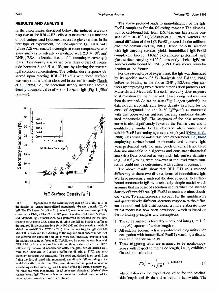
In the experiments described below, the induced secretoryresponse of the RBL-2H3 cells was measured as a functionof both antigen and IgE densities on the glass surface. In thefirst type of experiment, the DNP-specific IgE class mAb(clone A2) was reacted overnight at room temperature withglass surfaces covalently derivatized with 2.3 X 10⁴/4am2DNP1 -BSA molecules (i.e., a full monolayer coverage).IgE surface density was varied over three orders of magni-tude between 8 and 5 X 10³/4am2 by altering the reactantIgE solution concentration. The cellular dose response ob-served upon reacting RBL-2H3 cells with these surfaceswas very similar to that observed in our earlier study (Tamiret al., 1996), i.e., the secretion steeply increased above adensity threshold value of -8 X 10²/,4m2 IgE (Fig. 1,filledsymbols).
40
- 2e+52e+5 -
130 -0 100
CO) ~ 5e+4 -
g 25 - e+o
Q)20 -2-IgE Surface Density [,u-2]
o 15-
C) o -5
0-1 10 100 1000
IgE Surface Density [W-21FIGURE 1 Dependence of the secretory response of RBL-2H3 cells onthe density of surface-immobilized monomeric (0) and dimeric (l, C)IgE. The DNP-specific IgE mAb (clone A2) was bound to coverslips fullycoated with DNP, 1-BSA (2.3 X 10⁴ umM-2) as described under Materialsand Methods. IgE dimerization was performed in solution by the IgE-specific mAb clone 95.3, either by diluting the IgE in Tyrode's buffer tothe required final concentrations (0.1-100 nM) and then reacting it with 10nM of the mAb 95.3 at 22°C for 2 h (E), or first reacting the IgE with 100nM of this mAb and then diluting to the required final concentration (O).The dimeric IgE-containing solutions were next incubated overnight withthe antigen-carrying surfaces at 22°C, followed by extensive washings withPBS. RBL cells were allowed to settle on these surfaces for 1 h at 10°C,followed by removal of nonadherent cells. The glass surface-carried cellswere then incubated in Tyrode's buffer for 45 min at 37°C, and theirsecretory response was measured. The solid and dashed lines result fromfitting the data obtained with monomeric and dimeric IgE according to themodel described in the text. The inset shows the computed number ofstimulating surface units (nau) as a function of the IgE surface density o-IgEfor reactions with monomeric (solid line) and dimerized (dashed line)surface-bound IgE. The error bars represent the standard deviation -of thesecretory response determined in triplicate.
The above protocol leads to immobilization of the IgE-FcsRI complexes for the following reasons: The dissocia-tion of cell-bound IgE from DNP-haptens has a time con-stant of _10_102 s (Goldstein et al., 1989), whereas thelateral diffusion of free IgE-FcsRI proceeds in the millisec-ond time domain (DeLisi, 1981). Hence the cells' reactionwith IgE-carrying surfaces yields immobilized IgE-FceRIcomplexes. Indeed, FRAP experiments performed withglass surface carrying 103 fluorescently labeled IgE/pum2noncovalently bound to DNP1 -BSA have shown immobi-lization of the former.
For the second type of experiment, the IgE was dimerizedby its specific mAb (95.3) (Baniyash and Eshhar, 1984)before its binding to the above DNP1 -BSA-carrying sur-faces by employing two different dimerization protocols (cf.Materials and Methods). The cells' secretory dose responseto stimulation by the dimerized IgE-carrying surfaces wasthen determined. As can be seen (Fig. 1, open symbols), thedata exhibit a considerably lower density threshold for theonset of degranulation (-10-60 IgE/4am2) as comparedwith that observed on surfaces carrying randomly distrib-uted monomeric IgE. The steepness of the dose-responsecurve is also significantly lower in the former case and isqualitatively similar to that observed when conventionalsoluble FcsRI clustering agents are employed (Oliver et al.,1988). (It should be noted that both experiments, i.e., thoseemploying surface-bound monomeric and dimeric IgE,were performed with the same batch of cells. Hence thesedata are amenable to a rigorous and consistent theoreticalanalysis.) Data obtained at very high IgE surface densities(e.g., >103 ,Im-2), were however at the level where satu-ration could not be determined with sufficient accuracy.The above results show that RBL-2H3 cells respond
differently to these two distinct forms of immobilized IgE.We have previously analyzed the dose response to surface-bound monomeric IgE by a relatively simple model whichassumes that an onset of secretion occurs when the averagedensity of immobilized IgE-FcsRI exceeds a distinct thresh-old value. To simultaneously account for the qualitativelyand quantitatively different secretory response to the differ-ent immobilized IgE distributions, a more elaborate theo-retical model has now been developed, which is based onthe following principles and assumptions:
1. The cell's surface is formally subdivided into j (j = 1, 2,. . ., Np) squares of a side length sj.
2. All patches become active signal-transducing units uponoccupation with immobilized FcsRI exceeding a distinctthreshold density value 0.
3. These triggering units are assumed to be nonhomoge-neous with respect to their side length, i.e., sj exhibits aGaussian distribution.
P(sj) = ^1 e -(s--s-42/[2(&)2] (1)
where S denotes the expectation value for the patches'side length and 8s their distribution's half-width. The
2472 Biophysical Journal

Schweitzer-Stenner et al.
number of patches per cell with side lengths in theinterval s + As can be calculated by
2A r2 s AS)N(s) =2c s-erfs) -erf
s 8s (2)
where rcell (5 ,um) is the average radius of RBL-2H3cells (Oliver et al., 1988). AC is the average fraction ofthe cells' surface in contact with the coverslip.
4. Each IgE at the cell-glass contacts is occupied by a
FcsRI, so that the density of immobilized FceRIsmatches that of IgE on the glass surface.
5. The complete coverage of the coverslip with DNP1 -
BSA causes a random distribution of IgEs on it. Thenumber nc of FcsRI-IgE complexes within patches withside length s can therefore be expressed as a function ofthe IgE density at the glass surface by
nc = S2OIgE (3)where UIgE denotes the surface density of IgE in pum- .
The average number of active units can now be writtenas
O nc 0
nau= X N(s)(nc 1) dsn (4)
_00
The integral in Eq. 4 was truncated for practical reasons,
assuming s- 28s and s + 26s as lower and upper values,respectively. The integration was carried out numerically.
6. Following the concept of Dembo and co-workers(Dembo et al., 1979), the relationship between secretoryresponse R(nau) and the number of triggering units per
cell (nau) is described by a Hill function (Schweitzer-Stenner et al., 1994), namely,
1 )(Kstimnau)
where Kstim is defined as the efficiency of the stimulus-secretion coupling per secretion stimulatory unit, -q de-notes the maximum secretion at high naU values, and h isthe Hill coefficient.
The above model was now employed to fit the doseresponse to randomly distributed surface bound IgE (soliddata points in Fig. 1), using kstim, 71, h, s, and Ss as freeparameters. The threshold 0 was fixed to 2, i.e., only twoFcsRI-IgE complexes within the same patch were assumedto constitute a secretion stimulus unit. This fit yielded thesolid curve depicted in Fig. 1 for the dose response to therandomly distributed IgEs. The parameter values obtainedare listed in Table 1. The average side length of the trig-gering surface units was found to be 30 ± 10 nm, which isabout twice the estimated diameter of the FcERI-IgE com-
plex (i.e., 15 nm; Holowka et al., 1985; Zheng et al., 1992;
Kubitscheck et al., 1993). The fitting parameters thereforeindicate a requirement for closest (i.e., van der Waals)proximity of two immobilized FcERIs for forming a signal-transducing unit. (The variance of the side length is the fullhalf-width of the Gaussian distribution P(sj) described byEq. 1.)The above analysis only deals with the dose response to
randomly distributed surface-bound IgE. To also accountfor the secretion observed with coverslips carrying dimer-ized, i.e., nonrandomly distributed IgE (open symbols inFig. 1), we assume that each IgE dimer constitutes a secre-tion stimulus unit upon its binding to two FceRIs on thecells' surface. Hence the number of signaling units per cellis now given by
O'IgE Asnau 2
= (6)
where As is the total surface of the coverslip and ncell is thenumber of cells on the coverslip. The dose response is nowobtained by inserting Eq. 6 into Eq. 5.To fit the thus modified model to the dose-response
curve, we used the very same parameter values as obtainedabove from the analysis of the secretion induced by surfaceswith randomly distributed IgE (cf. Table 1). Hence weassume that IgE-FceRI dimers produced by clustering IgEwith mAb 95.3 exhibit the same triggering efficiency Kstimas two immobilized IgE-FcsRI in close proximity. Despitethese restrictions, a satisfactory fitting of the experimentaldata is obtained, as judged from the dashed line in Fig. 1.This strongly supports the validity of the model and theobtained parameter values.The reason for the observed distinct patterns of dose
response to monomeric and dimeric surface-bound IgE canbe understood as follows. At low UIgE two of the randomlydistributed IgE are unlikely to be in close proximity, so thatno stimulating surface units are created by IgE-FceRI bind-ing. The situation changes when 0TIgE exceeds a distinctthreshold. Above this value the number of doubly occupiedsurface units increases significantly. As a consequence, oneobserves a steep increase in secretion. The situation isqualitatively different when the cells react with surfacescarrying IgE dimers; because each of them creates a stim-ulating unit upon binding to two FceRIs, stimulation iscaused at very low 0-IgE. This is demonstrated by the inset inFig. 1, which displays the computed number of secretion-stimulating surface units as a function of IgE surface densityfor cells reacting with monomeric or dimeric IgE. Thesecurves are very similar to the corresponding experimentallyobserved dose-response curves.
TABLE I Parameters obtained from the fits to the dose-response curves in Figs. I and 2.
Experiment s (nm) &s (nm) Kstim AC 7)(%) 0
I 30 10 0.5 0.3 30 2II 50 15 0.5 0.3 35 2
Mast Cell Stimulation 2473

Volume 72 June 1997
The above results provide strong evidence that the stim-ulus induced upon immobilizing FcsRI by randomly dis-tributed or by dimerized surface-bound IgEs can be de-scribed in terms of the very same biophysical parameters(i.e., Kstim and -q). To further support this notion, we mea-sured the cells' secretory response to surfaces carryingdifferent densities of either antigen or monomeric IgE. Tothis end, we employed glass surfaces with three differentantigen surface densities, i.e., 0rAg = 308 ± 30, 1860 ± 130,and 2718 ± 570 ttm-2. The dose response was then mea-sured as a function of IgE surface density in the range of10-1000 p.m-2 for each of these glass surfaces. As shownin Fig. 2, the observed secretion decreased significantlywith decreasing 0rAg, whereas the onset of response oc-curred at essentially the same IgE density, although with adifferent slope.To analyze the above data, we adopted a model based on
the following general considerations. The concentration[Ck] of DNP1 -BSA bound to k IgE can be calculated byusing the mass-action law to yield
[Ck] = 2. (4 k + I)I[IgE]k[Ag]fwhere [Ag]f is the number of free epitopes on the glasssurface, [IgE] is the concentration of IgE in the solutionused to produce the examined surface, and K1 is the affinityof DNP binding to a Fab-combining site. The latter is aneffective parameter, because steric factors may affect thisvalue differently. [Ag]f depends on the total concentration ofsterically exposed haptens, and thus on the surface densityof the epitopes ([Ag]T), and can be calculated by utilizingthe law of mass conservation:
[Ag]f= ~~[A9]T 8
[Ag]f =1 + Em [2. (4 - k + 1)/k]4[IgE]k (8)
It should be noted that the simultaneous binding of a singleIgE to BSA with both of its Fab-binding sites (cf. Fig. 3) isnot accounted for by Eqs. 7 and 8. (Binding of a single IgEto two DNP-haptens on the same BSA-DNPI1 molecule isunlikely, because the minimum distance between its Fab-combining sites is - 13 nm (Schweitzer-Stenner et al.,1987), whereas the estimated size is ofBSA is - 12 nm.) Allspecies concentrations in these equations are thus far ex-pressed as molarity. To convert [Ck] to surface densities, weemploy
[Ck] L V(Ck = AAs
30 -
25 -
20 -
15 -
10 -
5-
0-
o 30-(DcO 25 -C0C. 20 -coa)
0LDa.)(c)
30(7)
10 100 1000
1860 [BSA/t2J
15
10
10 100 10)00
10 100 1000
IgE Surface Density [,-2]
FIGURE 2 Dependence of secretory response of RBL-2H3 cells on thedensities of both surface-carried antigen and immobilized monomeric IgE.IgE was bound to coverslips coated with the indicated surface densities ofDNP1 I-BSA, as described in Materials and Methods. RBL-2H3 cells werenext allowed to settle on these surfaces for 1 h at 10°C, followed byremoval of nonadherent cells. The glass surface-carried cells were thenincubated in Tyrode's buffer for 45 min at 37°C, and their secretoryresponse was measured and presented as a fraction of the total granularcontents. The solid lines result from fitting all data points according to themodel described in the text. The error bars represent the standard deviationof the secretory response determined in triplicate.
imity can produce a signaling unit upon reaction withFceRI. The fraction P(ovAg) of antigens fulfilling this re-quirement can be calculated by solving the integral
(9)
where L is Avogadro's constant, and V is the sample'sbathing buffer volume.We now divide the number of IgE-bound antigen into two
subsets. One contains all antigen molecules occupied by asingle IgE (ac.). At high antigen density, this subset con-stitutes a random distribution of IgEs. For low densities,however, only those IgEs bound to antigens in close prox-
rS
P(CrA?) = w(r) drJo
(lOa)
where
w(r) = 2lTrrYA. exp(-1Tr2o-Ag) (lOb)
and r is the distance between two antigens on the glasssurface. (Equation 10a,b can be derived by employing the
2474 Biophysical Journal

Schweitzer-Stenner et al.
-7) cn
*: DNP-groups
FIGURE 3 Schematic representation of IgE binding to the antigen(DNP1 -BSA) carried on a glass surface. It is assumed that only two DNPgroups per antigen molecule are accessible for IgE binding to BSA.Simultaneous binding of the two Fab combining sites of the same IgE toone DNP1 -BSA molecule is excluded for steric reasons.
procedure of Chandrasekhar (1943) for random distribu-tions of particles on a two-dimensional surface. The antigendistribution is thereby treated as an ideal gas, thus neglect-ing the excluded volume effect.) We assume that to stimu-late, two antigen-IgE-FcsRI complexes must fill the square
patches of a triggering unit, so that rs = s. After this isinserted into Eq. 10, a straightforward calculation yields
P((JAg) = 1 exp(-mrro-Ag) (11)
The secretory response to the subset C1 can be described byEqs. 2-7, substituting 0JIgE by (0c) P(oAg).The second subset comprises all antigen molecules car-
rying more than one IgE. We assumed therefore that irre-spective of the number k of IgEs bound to the same BSA-DNP11, steric hindrance prevents the simultaneous bindingof more than two of them to FcsRIs (m = 2). The contri-bution of the thus formed FcsRI dimers (n'u) to the numberof stimulatory units (nau) on the cells' surface can be writtenas
n = k(12)ncell
The equilibrium constant K1 for IgE binding to surface-bound DNP1 1-BSA was assumed to be equal to the solutionequilibrium constant of 107 M-1 (Schweitzer-Stenner et al.,1987).The above model was employed to simultaneously fit the
secretory dose-response curves depicted in Fig. 2, by allow-ing kstim, 7q, h, s, and Ss to be free parameters. This fittingprocedure yielded the solid curves shown in Fig. 2. Theparameter values are listed in Table 1.
It should be noted that for o-Ag values of 2718 and 1860Am-2 (middle and lower panels in Fig. 2), the parametervalues do not depend on the number m of sterically acces-
sible DNP epitopes, because the secretory response is de-termined by species of the subset C1. In contrast, C2 is ofrelevance for oAg = 308 ,tm-2 (Fig. 2, upper panel), andm ' 3 is now required to account for the maximum IgEsurface density still achieved in this experiment (i.e., 700,pm-2). However, the data do not allow us to determine theexact stoichiometry of the IgE-antigen reaction, because the
dose response is still insensitive to variations of k in theregion covered by the experimental data.To determine the uncertainty of the above parameters, we
have performed calculations with detuned parameters todetermine the extent to which their values can be variedwithout a deterioration of the fit's quality. This leads to anestimate of their statistical error, showing that h, s, and 6shave an uncertainty between 10% and 15%. In contrast,strong correlation effects (Grinvald and Steinberg, 1973)were observed for Kstim and -q, so that multiple pairs of theseparameters could be found that produce reasonable fits tothe same dose-response curves. This is mostly due to thefact that our dose-response data do not reach the region ofsaturation. Their relative error is eventually found to bebetween 30% and 40%.
It should be mentioned that some parameter values (i.e.,s, Ss, and, to a minor extent, 71) obtained from the second set(Fig. 2) of experiments are somewhat different from thoseemerging from the analysis of the first one (Fig. 1; cf. Table1). However, these differences are far from being significantand may well be explained by the fact that different cellbatches were used for these experiments.The above model is based on the assumption that all IgEs
at cell-glass contacts are saturated by surface-bound FceRI.This is supported by experiments (McCloskey and Poo,1986) in which cell-size phospholipid vesicles containingDNP derivatized lipids were allowed to react with RBL-2H3 cells carrying DNP-specific, fluorescently labeled IgEon their FcsRI. It was found that upon cell-vesicle contact,the IgE-FceRI complexes undergo a lateral redistribution toaccumulate at the site of cell-vesicle contacts. That can alsobe expected to occur upon cell binding to the antigen-carrying glass surfaces. This will provide access of theFcsRI to the surface carrying IgE. After this reaction hasreached equilibrium, however, the total concentration ofFcsRI does not significantly exceed that of immobilizedIgE-FceRI on the cells (cf. Tamir et al., 1996).
Finally, to check the influence of possible incompleteIgE-FcsRI saturation on the fitting parameters, we havefitted the data in Fig. 1, assuming that only 60% of thesurface-bound IgEs have reacted with FceRI. The fitobtained was still satisfactory and yielded s = 40 nm andKstim = 0.3. Thus the difference from the parameters ob-tained for complete saturation is insignificant and would notaffect the implications of our analysis.
DISCUSSION
The mechanism by which members of the MIRR familyinitiate the cascades that couple them to the respectivecellular response is still not clearly resolved. FcsRI has beenone of the more extensively studied paradigms for MIRRaction. Numerous studies have indicated that producing aneffective immunological stimulus in RBL-2H3 cells re-quires clustering of their FceRI into dimers or larger oli-gomers (Ortega et al., 1988; Holowka and Baird, 1996).
-o - o - <Mast Cell Stimulation 2475

Volume 72 June 1997
However, no knowledge about the requirement for FcsRIimmobilization is available. Earlier theoretical consider-ations have led DeLisi (1980) to suggest that bringing twoFceRI into close proximity for a time period longer than agiven threshold would allow appropriate downstream signaltransmission. Thus this time period has to be considerablylonger than the lifetime of FcsRI encounter complexes,which is determined by the receptors' lateral diffusion. Toexamine the above predictions, we have devised a newexperimental approach. It involves immobilization of theFceRI upon their reaction with DNP-specific IgE antibodiesbound to DNP, I-BSA-carrying glass surfaces (Tamir et al.,1996). The secretory response of the cells reacting withthese surfaces was measured as a function of IgE surfacedensity and found to exhibit a rather sharp onset of secretionabove a density threshold of -800 IgE/4tm2 (Fig. 1). Thisobservation indicates that a secretory stimulus productionrequires only the surface density of immobilized FcsRI toexceed a distinct threshold value at which a sufficient num-ber of them are in close proximity. Moreover, we havemeasured the cells' secretory response to surfaces that car-ried altered distributions of immobilized IgE. This wasattained either by IgE dimerization in solution before itssurface immobilization or by varying the immobilizing an-tigen density on the glass surface. Both of these protocolswere expected to change the probability of immobilizing therequired number of FcsRIs within signaling proximity andtherefore the onset of secretory response and the shape of itsdose dependence. This was indeed observed. First, the onsetof the secretion induced by glass surfaces with predimerizedIgE was observed at much lower IgE surface densities(10-60 IgE/,.m2), and the slope of the dose-response curveis less steep than that observed for surfaces carrying mono-meric IgEs (Fig. 1). Second, reducing the antigen surfacedensity significantly decreased secretion and the slope of itsinitial dose-response phase (Fig. 2).The implications of these results were examined by using
a novel theoretical model based on statistical consider-ations. The finding that this model consistently reproducesall experimentally observed dose-response data sets (Figs. 1and 2) supports the assumptions on which it is based,namely, that monomeric IgE are randomly distributed onthe glass surfaces, whereas predimerized IgE yields a non-random distribution. Furthermore, the IgE glass surfacedensity is the same as that of the immobilized IgE-FcsRIcomplexes at the glass-cell interface.To characterize the clusters that initiate the stimulus, the
cell's surface is formally divided into the smallest squarepatches that are capable of triggering secretion upon theiroccupancy by at least two immobilized FcsRI. These FcsRIclusters would then trigger secretion autonomously (Oliveret al., 1988). Using the patches' side length as a freeparameter, a remarkably good fit to the experimental datawas obtained. Hence, in spite of its simplicity, our modelfully accounts for observed variations in the dose responseto qualitative and quantitative changes in IgE distribution on
an elementary signaling unit requires only two FceRI insuch proximity, that they are accommodated into patches of30 10 nm side length, i.e., they are essentially in van derWaals contact.The model also accounts for results of earlier studies by
McConnel and associates (Cooper et al., 1981; Balakrishnanet al., 1982). They have investigated the interaction ofRBL-2H3 cells carrying DNP-specific IgE mAb with lipo-somes containing DNP-derivatized lipids. Different types ofliposomes were employed, i.e., "solid" and "liquid" ones
with lipid diffusion coefficients of 10-10 and 10-7 to 10-8cm2/s, respectively. Both were found to be capable of stim-ulating the secretion of RBL-2H3 cells, but the "solid"liposomes were up to threefold more effective. In view ofour results, one would indeed expect the "solid" liposomesto be effective in triggering secretion, because they are
likely to immobilize the FcsRI on the reacting cells' sur-
faces. Indeed, the dose responses to these liposomes are
very much like that shown in Fig. 1, inasmuch as they bothdisplay a steep increase above a certain threshold value ofhapten surface density (cf. Figure 3 in Balakrishnan et al.,1982), thus indicating that the employed protocol immobi-lizes a sufficient number of FccRI in close proximity. Therelatively low dose response to the "liquid" liposomes was
rationalized by invoking the accumulation of IgE-FcsRIcomplexes at cell-liposome contact regions that would in-deed significantly increase the effective FcsRI density.Such molecular crowding on membrane surfaces was shownby simulations (Minton, 1989; Saxton, 1989, 1992, 1993) toimpede lateral diffusion, so that the effective lifetime of a
given FcsRI pair may become sufficiently long to producea stimulus. Therefore, the model developed and employedhere also rationalizes the dose response to both "solid" and"liquid" hapten-carrying liposomes.
Ortega et al. (1988) have already shown that FcsRIdimerization by different specific mAbs caused effectiveRBL-2H3 degranulation. Moreover, the marked differencesin response to the FcsRI dimers produced by the threeemployed mAbs suggested that the triggering capacity ofFcsRI dimers depends on their intrinsic properties, e.g.,their lifetimes and/or the relative orientation of the receptorswithin a dimer (Ortega et al., 1991; Schweitzer-Stenner etal., 1994). The present study provides an independent quan-titative characterization of the smallest functional triggeringunits, i.e., two FcsRI occupying a patch with a side lengthof approximately 30 nm, which brings them into van derWaals contact. The major new insight gained by the currentprotocols is the decisive role that receptor immobilizationplays in initiating the signaling cascade. As the number ofFcsRI copies per cell of the RBL-2H3 line is high (3-6 X
105), the probability of immobilizing them at the requiredproximity is considerable, and hence the stimulus is pro-duced. For other multichain immunoreceptors that are
present at considerably lower density (e.g., T-cell recep-tors), stimulation indeed requires protocols that cause both
the glass surface. The result of the fit provides evidence that
2476 Biophysical Journal
clustering and immobilization (cf. also McKeithan, 1995).

Schweitzer-Stenner et al. Mast Cell Stimulation 2477
Why do the very early biochemical coupling processesset these requirements on a stimulatory FcsRI cluster? Theearliest known process in the RBL-2H3 cells is activation ofthe FcsRI-associated protein tyrosine kinase (PTK)-Lyn(Ravetch and Kinet, 1991; Eiseman and Bolen, 1992; Ad-amczewski et al., 1992; Adamczewski and Kinet, 1994) thatis bound with low affinity to the NH2 tail of the restingcell's FcsRI ,8-chain (Lin et al., 1996). Upon FcsRI clus-tering, Lyn undergoes activation and phosphorylates tyrosylresidues of the adjacent FcsRI's 13- and y-subunits on theirimmunoreceptor tyrosine activation motifs (ITAMs). Thephosphorylated y-chains then recruit the PTK Syk, whichupon binding to the phosphorylated tyrosyls of the ITAMs,also undergoes activation. This mechanism obviously re-quires that the clustered FcsRIs remain immobile at closeproximity for the time span required by these processes totake place.
Finally, it should be mentioned that physiologically, mastcell stimulation is attained under conditions very differentfrom those employed in most studies using the RBL-2H3line (including the present one). Whereas for the latter cells,the FcsRI are saturated by mAbs with a single bindingspecificity in the living animal, mast cells would carrypolyclonal IgEs with the whole repertoire of specificitiesand affinities present, few of which are the same. In con-trast, the situation is different for B- and T-cells, each ofwhich carries on its surfaces MIRRs with only one speci-ficity. Therefore the requirements for activating the lattercells are similar to those established for the FcsRI on mastcells, i.e., clustering and immobilization of their MIRRs.Hence the model developed in the present study and itsresults may be of wider applicability to other immunologi-cal systems.
This work was supported by a grant to I.P. from the Minerva Foundation,Munich, Germany. RS-S acknowledges financial support from the Gesell-schaft der Freunde des Weizmann Institutes, Bonn, Germany.
REFERENCES
Adamczewski, M., and J.-P. Kinet. 1994. The high-affinity receptor forimmunoglobulin E. Chem. Immunol. 59:173-190.
Adamczewski, M., R. Paolini, and J.-P. Kinet. 1992. Evidence for twodistinct phosphorylation pathways activated by high affinity immuno-globulin E receptors. J. Bio. Chem. 267:18126-18132.
Balakrishnanan, K., F. J. Hsu, A. D. Cooper, and H. M. McConnell. 1982.Lipid hapten containing membrane targets can trigger specific immuno-globulin E-dependent degranulation of rat basophil leukemia cells.J. Bio. Chem. 257:6427-6433.
Baniyash, M., and Z. Eshhar. 1984. Inhibition of IgE binding to mast cellsand basophils by monoclonal antibodies that inhibit IgE binding. Eur.J. Immunol. 14:799-807.
Barsumian, E. L., C. Isersky, M. G. Petrino, and R. P. Siraganian. 1981.IgE-induced histamine release from rat basophilic leukemia cell lines:isolation of releasing and nonreleasing clones. Eur. J. Immunol. 11:317-323.
Basciano, L. K., E. H. Berenstein, L. Kmak, and R. P. Siraganian. 1986.Monoclonal antibodies that inhibit IgE binding. J. Bio. Chem. 261:11823-11831.
Bolen, J. B. 1995. Protein tyrosine kinases in the initiation of antigenreceptor signaling. Curr. Opin. Immunol. 7:306-311.
Chandrasekhar, S. 1943. Stochastic problems in physics and astronomy.Rev. Mod. Phys. 15:1-89.
Cooper, A. D., K. Balakrishnan, and H. M. McConnell. 1981. Mobilehaptens in liposomes stimulate serotonin release by rat basophil leuke-mia cells in the presence of specific immunoglobulin E. J. Biol. Chem.256:9379-9381.
DeLisi, C. 1980. The biophysics of ligand-receptor interactions. Q. Rev.Biophys. 13:201-230.
DeLisi, C. 1981. The magnitude of signal amplification by ligand-inducedreceptor clustering. Nature. 289:322-323.
Dembo, M., B. Goldstein, A. K. Sobotka, and L. M. Lichtenstein. 1979.Degranulation of human basophils: differences between antigen- andanti-IgE-induced secretion. J. Immunol. 123:1864-1872.
Eiseman, E., and J. B. Bolen. 1992. Engagement of the high affinity IgEreceptor activates src protein-related tyrosine kinases. Nature. 355:78-80.
Eisen, H. N., M. Kern, W. T. Newton, and E. Helmreich. 1959. A study ofthe distribution of 2,4-dinitrobenzene sensitizers between isolated lymphnode cells and extracellular medium in relation to induction of contactskin sensitivity. J. Exp. Med. 110: 187-206.
Erickson, J. W., R. G. Posner, B. Goldstein, D. Holowka, and B. Baird.1991. Bivalent ligand dissociation kinetics from receptor-bound immu-noglobulin E: evidence for a time-dependent increase in ligand rebindingat the cell surface. Biochemistry. 30:2357-2363.
Fewtrell, C. 1985. Activation and desensitization of receptors for IgE ontumor basophils. In Calcium in Biological Systems. R. P. Ruben, G. B.Weiss, and J. W. Putney, Jr., editors. Plenum, New York. 129-136.
Fewtrell, C., and H. Metzger. 1980. Large oligomers of IgE are moreeffective than dimers in stimulating rat basophilic leukemia cells. J. Im-munol. 125:701-710.
Goldstein, B., R. G. Posner, D. C. Torney, J. Erickson, D. Holowka, and B.Baird. 1989. Competition between solution and cell surface receptors forligands. Dissociation of hapten bound to surface antibody in the presenceof solution antibody. Biophys. J. 56:955-966.
Grinvald, A., and I. Z. Steinberg. 1973. On the analysis of fluorescencedecay kinetics by the method of least square. Anal. Biochem. 59:583-598.
Hardy, R. 1986. In Handbook of Experimental Immunology. D. M. Weir,editor. Blackwell Scientific Publications, Boston. 405.
Holowka, D., and B. Baird. 1996. Antigen-mediated IgE receptor aggre-gation and signaling: a window on cell surface structure and dynamics.Annu. Rev. Biophys. Biomol. Struct. 25:79-112.
Holowka, D., D. H. Conrad, and B. Baird. 1985. Structural mapping ofmembrane-bound immunoglobulin E-receptor complexes: use of mono-clonal anti-IgE antibodies to probe the conformation of receptor boundIgE. Biochemistry. 24:6260-6267.
Hunter, M. W., and F. C. Greenwood. 1962. Preparation of iodine-131labeled human growth hormone of high specific activity. Nature. 194:4876-4882.
Kane, P. M., D. Holowka, and B. Baird. 1988. Cross-linking of IgE-receptor complexes by rigid bivalent antigens >200 A in length triggerscellular degranulation. J. Bio. Chem. 107:969-980.
Kubitscheck, U., R. Schweitzer-Stenner, D. J. Arndt-Jovin, T. M. Jovin,and I. Pecht. 1993. Distribution of type I Fcs-receptors on the surface ofmast cells probed by fluorescence resonance energy transfer. Biophys. J.64:110-120.
Jorgensen, J. L., P. R. Reay, E. W. Ehrich, and M. M. Davis. 1992.Molecular components of T-cell recognition. Annu. Rev. Immunol. 10:835-873.
Lin, S., C. Cicala, A. M. Scharenberg, and J.-P. Kinet. 1996. The FcsRIl3subunit functions as an amplifier of FceRIly-mediated cell activationsignals. Cell. 85:985-995.
McCloskey, M., and M. M. Poo. 1986. Contact-induced redistribution ofspecific membrane components: local accumulation and development ofadhesion. J. Cell. Biol. 102:2185-2196.
McKeithan, T. W. 1995. Kinetic proofreading in T-cell receptor signaltransduction. Proc. Natl. Acad. Sci. USA. 92:5042-5046.

2478 Biophysical Journal Volume 72 June 1997
Menon, A. K., D. Holowka, and B. Baird. 1984. Small oligomers ofimmunoglobulin E (IgE) cause large scale clustering of IgE receptors onthe surface of rat basophilic leukemia cells. J. Cell. Bio. 98:577-583.
Menon, A. K., D. Holowka, W. W. Webb, and B. Baird. 1986a. Clustering,mobility, and triggering activity of small oligomers of immunoglobulinE on rat basophilic leukemia cells. J. Cell Bio. 102:534-540.
Menon, A. K., D. Holowka, W. W. Webb, and B. Baird. 1986b. Cross-linking of receptor-bound IgE to aggregates larger than dimers leads torapid immobilization. J. Cell Biol. 102:541-550.
Minton, A. P. 1989. Lateral diffusion of membrane proteins in protein-richmembranes. A simple hard particle model for concentration dependenceof the two-dimensional diffusion coefficient. Biophys. J. 55:805-808.
Myers, J. N., D. Holowka, and B. Baird. 1992. Rotational motion ofmonomeric and dimeric immunoglobulin E-receptor complexes. Bio-chemistry. 31:567-575.
Oliver, J., J. C. Seagrave, R. F. Stump, J. R. Pfeiffer, and G. G. Deanin.1988. Signal transduction and cellular response in RBL-2H3 mast cells.Prog. Allergy. 42:185-245.
Ortega, E., R. Schweitzer-Stenner, and I. Pecht. 1988. Possible orienta-tional constraints determine signals induced by aggregation of IgEreceptors on mast cells. EMBO J. 7:4101-4109.
Ortega, E., R. Schweitzer-Stenner, and I. Pecht. 1991. Kinetics of ligandbinding to the type I Fcc receptor on mast cells. Biochemistry. 30:3473-3483.
Ortega-Soto, E., and I. Pecht. 1988. A monoclonal antibody that inhibitssecretion from rat basophilic leukemia cells and binds to a novel mem-brane component. J. Immunol. 141:4324-4332.
Pecht, I., E. Ortega, and T. M. Jovin. 1991. Rotational dynamics of theFceRI receptor on mast cells monitored by specific monoclonal anti-bodies and IgE. Biochemistry. 30:3450-3468.
Perelson, A. S., and C. DeLisi. 1980. Receptor clustering on a cell surface.I. Theory of receptor cross-linking by ligands bearing two chemicallyidentical functional groups. Math. Biosci. 48:71-110.
Posner, R. G., J. W. Erickson, D. Holowka, and B. Baird. 1991. Dissoci-ation kinetics of bivalent ligand-immunoglobulin E aggregates in solu-tion. Biochemistry. 30:2348-2356.
Posner, R. G., B. Lee, D. H. Conrad, D. Holowka, B. Baird, and B.Goldstein. 1992. Aggregation of IgE-receptor complexes on rat baso-philic leukemia cells does not change the intrinsic affinity but can alterthe kinetics of the ligand-IgE interaction. Biochemistry. 31:5350-5356.
Posner, R. G., K. Subramanian, B. Goldstein, J. Thomas, T. Feder, D.Holowka, and B. Baird. 1995. Simultaneous crosslinking by two non-triggering bivalent ligands causes synergistic signaling of IgE-FcCRIcomplexes. J. Immunol. 155:3601-3609.
Ravetch, J. V., and J.-P. Kinet. 1991. Fc receptors. Annu. Rev. Immunol.9:457-492.
Rudolph, A. K., P. D. Burrows, and M. R. Wahl. 1981. Thirteen hybrido-mas secreting hapten-specific immunoglobulin E from mice with Iga andIgb heavy chain haplotype. Eur. J. Immunol. 11:527-529.
Saxton, M. 1989. The spectrin network as a barrier to lateral diffusion inerythrocytes: a percolation analysis. Biophys. J. 55:21-28.
Saxton, M. 1992. Lateral diffusion and aggregation. A Monte Carlo study.Biophys. J. 61:119-128.
Saxton, M. 1993. Lateral diffusion in an archipelago: single particle dif-fusion. Biophys. J. 64:1766-1780.
Scharenberg, A., S. Li, B. Cuenod, H. Yamamura, and J.-P. Kinet. 1995.Reconstitution of interactions between tyrosine kinases and the highaffinity IgE receptor which are controlled by receptor clustering. EMBOJ. 14:3385-3394.
Schatz, D. G., M. A. Oettinger, and M. S. Schissel. 1992. Recombination:molecular biology and regulation. Annu. Rev. Immunol. 10:359-383.
Schweitzer-Stenner, R., A. Licht, I. Luischer, and I. Pecht. 1987. Oligomer-ization and ring closure of immunoglobulin E class antibodies by diva-lent haptens. Biochemistry. 26:3602-3612.
Schweitzer-Stenner, R., A. Licht, and I. Pecht. 1992. Dimerization kineticsof the IgE-class antibodies by divalent haptens. II. The interactionbetween intact IgE and haptens. Biophys. J. 63:563-568.
Schweitzer-Stenner, R., E. Ortega, and I. Pecht. 1994. Kinetics of FcsRIdimer formation by specific monoclonal antibodies on mast cells. Bio-chemistry. 33:8813-8825.
Siraganian, R. P. 1988. In Inflammation: Basic Principles and ClinicalCorrelates. J. I. Gallin, I. M. Goldstein, and R. Snyderman, editors.Raven Press, New York. 513-542.
Subramanian, K., D. Holowka, B. Baird, and B. Goldstein. 1996. The Fcsegment of IgE influences the kinetics of dissociation of a symmetricalbivalent ligand from cyclic dimeric complexes. Biochemistry. 35:5518-5527.
Tamir, I., R. Schweitzer-Stenner, and I. Pecht. 1996. Immobilization of thetype I receptor for IgE initiates signal transduction in mast cells. Bio-chemistry. 35:6872-6883.
Weetall, M., D. Holowka, and B. Baird. 1993. Heterologous desensitiza-tion of the high affinity receptor for IgE (FceRI) on RBL cells. J. Im-munol. 150:4072-4083.
Zheng, Y., B. Shopes, D. Holowka, and B. Baird. 1992. Conformations ofIgE bound to its receptor FcsRI and in solution. Biochemistry. 31:7446-7456.
Copyright © 2022 FDOKUMEN