Ana Sofia da Quinta e Costa Neves de Oliveira Morais
265
MECHANISMS OF ANTIFUNGAL RESISTANCE IN PATHOGENIC YEASTS: EVALUATION OF THE IN VITRO AND IN VIVO EXPRESSION Ana Sofia da Quinta e Costa Neves de Oliveira Morais Porto 2012
-
Upload
khangminh22 -
Category
Documents
-
view
1 -
download
0
Transcript of Ana Sofia da Quinta e Costa Neves de Oliveira Morais
MECHANISMS OF ANTIFUNGAL RESISTANCE IN PATHOGENIC YEASTS:
EVALUATION OF THE IN VITRO AND IN VIVO EXPRESSION
Ana Sofia da Quinta e Costa Neves de Oliveira Morais
Porto 2012


Dissertação de candidatura ao grau de Doutor em Biomedicina, apresentada
à Faculdade de Medicina da Universidade do Porto
Programa Doutoral em Biomedicina


O presente estudo decorreu no Serviço e Laboratório de Microbiologia da Faculdade de
Medicina da Universidade do Porto, Portugal.
Orientação
Professora Doutora Cidália Irene Azevedo Pina Vaz
Co-orientação
Professor Doutor Acácio Agostinho Gonçalves Rodrigues
Júri da Prova de Doutoramento em Biomedicina
Presidente
Reitor da Universidade do Porto
Vogais
Doutora Emília Canton Lacasa, Investigadora do Centro de Investigação do “Hospital
Universitari i Politècnic la Fe”, Valencia
Doutora Teresa Maria Fonseca Oliveira Gonçalves, Professora Auxiliar da Faculdade de
Medicina da Universidade de Coimbra
Doutor José António Martinez Souto de Oliveira, Professor Catedrático da Faculdade de
Ciências da Saúde da Universidade da Beira Interior
Doutor Daniel Filipe de Lima Moura, Professor Catedrático da Faculdade de Medicina da
Universidade do Porto
Doutora Cidália Irene Azevedo Pina Vaz, Professora Associada da Faculdade de Medicina
da Universidade do Porto
Doutora Isabel Alexandra Marcos Miranda, Investigadora da Faculdade de Medicina da
Universidade do Porto
Artigo 48, Parágrafo 31: “A Faculdade não responde pelas doutrinas expendidas na
Dissertação.” (Regulamento da Faculdade de Medicina da Universidade do Porto/ Decreto Lei nº
19337, de 29 de Janeiro de 1931)


Apoio financeiro da Fundação para a Ciência e a Tecnologia (FCT) do Ministério da Ciência,
Tecnologia e Ensino Superior (Bolsa de Doutoramento SFRH/ BD/ 27662/ 2006) .

À MINHA FAMÍLIA
"O valor das coisas não está no tempo que elas duram, mas na intensidade
com que acontecem. Por isso existem momentos inesquecíveis, coisas
inexplicáveis e pessoas incomparáveis."
Fernando Pessoa


À Professora Doutora Cidália Pina Vaz
Ao Professor Doutor Acácio Gonçalves Rodrigues
"A admiração é filha da ignorância, porque ninguém se admira senão das
coisas que ignora, principalmente se são grandes; e mãe da ciência, porque
admirados os homens das coisas que ignoram, inquirem e investigam as
causas delas até as alcançar, e isto é o que se chama ciência."
Padre António Vieira

Agradecimentos / Acknowledgments
"Enquanto os rios correrem para o mar, os montes fizerem sombra aos
vales e as estrelas fulgirem no firmamento, deve durar a
recordação do benefício recebido na mente do homem reconhecido."
Virgílio
Gostaria de expressar o meu mais sincero agradecimento a todos aqueles que me acompanharam durante este percurso e que contribuíram para a realização desta Tese:
Ao Professor Acácio Gonçalves Rodrigues, Diretor do Serviço e Laboratório de Microbiologia, por me ter dado a honra de co-orientar esta Tese. Obrigada pela confiança que em mim depositou, pela amizade que me dedica, e acima de tudo, por me ter ajudado a crescer como cientista.
À Professora Doutora Cidália Pina Vaz, agradeço a valiosa e sábia orientação deste trabalho. Obrigada por partilhar comigo os seus vastos conhecimentos científicos e pelas constantes palavras de incentivo e de carinho. Muito obrigada pela amizade com que me premeia e pelo privilégio que me concede em testemunhar o seu modo singular de fazer ciência.
Ao Professor Doutor Daniel Moura agradeço toda a disponibilidade e simpatia com que sempre me recebeu e acima de tudo, pelos preciosos conhecimentos que me transmitiu.
Ao Professor Doutor José Martinez de Oliveira, que acompanhou bem de perto o início do meu percurso microbiológico, agradeço o estímulo e a simpatia que sempre me dedicou.
Ao Dr. Filipe Sansonetty e ao Dr. Alexandre Salvador, a quem eu devo a minha formação na área da citometria de fluxo, obrigada pelos ensinamentos tão valiosos e pela amizade ao longo destes anos.
À Equipa de Investigação e Docente do Serviço de Microbiologia, da qual eu tenho a honra de fazer parte, e de onde nasceram verdadeiras amizades. Obrigada pelos vossos conselhos e opiniões que tanto enriqueceram esta Tese. Acima de tudo, obrigada por contribuírem para que o nosso Serviço seja uma referência na área da Microbiologia Clínica.
Aos antigos e atuais funcionários do Serviço e Laboratório de Microbiologia em especial à D. Emília de Magalhães, à D. Maria da Luz e ao Sr. João Teixeira, obrigada por todo o vosso apoio ao longo desta caminhada. À Isabel Santos, a “minha” Isabelita, por estar sempre pronta a ajudar e, acima de tudo, pela sua amizade que tanto me honra.
À Dra. Luísa Guardão agradeço os valiosos ensinamentos e toda a ajuda prestada na experimentação animal.
À Professora Doutora Paula Ludovico e à Dra. Belém Sampaio Marques por toda a ajuda dispensada e por tão bem me terem recebido na Escola das Ciências da Saúde, na Universidade do Minho.
Às minhas amigas de longa data, Janine e Isabel, pelo genuíno carinho e amizade partilhados.

Por fim gostaria de agradecer às pessoas que fazem parte da Minha Família, que tanto me ajudaram a chegar até aqui e, sem dúvida alguma, fizeram parte deste trabalho:
À Nandi, à Nunu e à Ritinha agradeço-vos do fundo do coração por todo o amor que me dedicam. Ao “Avô Chico” por estar sempre presente quando mais preciso.
À minha Terezinha, para mim uma referência pessoal e profissional. O meu mais sincero e profundo agradecimento por todas as palavras de ternura e incentivo. Obrigada, querida prima, por fazeres parte da minha vida.
À minha Mãe, por me amparar nas quedas, por fortalecer o meu ânimo com o seu amor, e acima de tudo por me ter ensinado a crescer. Obrigada Mamã.
Ao meu querido Irmão, por nos mantermos sempre unidos nas gargalhadas e no pranto. Obrigada Miga, pelo teu amor.
Ao meu querido Pai, com uma saudade sem fim. Obrigada Papá por me teres dado 29 anos de amor. Estarás sempre presente na minha vida.
À minha Avó Rosalina, que tanto amor semeou entre nós e sempre foi e será um modelo de vida para mim. Sei que estás sempre aqui, Vóvó.
Ao meu Avô Tini, por me fazer sentir sempre tão especial. Obrigada pela tua força e todo o teu amor incondicional.
Ao Tiago, meu marido e amigo, obrigada pelo teu amor e paciência e, particularmente, por teres um papel tão importante no maior projeto da minha vida: ser Mãe.
Aos meus grandes AMORES, os meus filhos, Afonso e Leonor, por tornarem a minha vida tão preciosa e, acima de tudo, por todos os dias me dizerem: Amo-te muito, Mamã.
Por fim, agradeço a Deus pela Fé que sempre me acompanha…

List of Publications
Ao Abrigo do Art. 8° do Decreto –Lei ° 388/ 70 fazem parte integrante desta dissertação
os seguintes trabalhos já publicados, ou em vias de publicação:
Manuscripts
I. Costa-de-Oliveira S, Sampaio-Marques B, Barbosa M, Ricardo E, Pina-Vaz C,
Ludovico P, Rodrigues AG. An Alternative Respiratory Pathway on Candida krusei:
Implications on Susceptibility Profile and oxidative stress. FEMS Yeast Res. 2012
Jan 23. doi: 10.1111/j.1567-1364.2012.00789.
II. Costa-de-Oliveira S, Miranda I, Silva R, Silva AP, Rocha R, Amorim A, Rodrigues
AG, Pina-Vaz C. FKS2 mutations associated with decreased echinocandin
susceptibility of Candida glabrata following anidulafungin therapy. Antimicrobial
Agents and Chemotherapy 2011; 55:1312-1314.
III. Costa-de-Oliveira S, Sousa I, Correia A, Sampaio P, Pais C, Rodrigues AG, Pina-Vaz
C. Genetic relatedness and antifungal susceptibility profile of Candida albicans
isolates from fungaemia patients. Medical Mycology 2011; 3:248-52.
IV. Costa de Oliveira S, Araujo R, Silva-Dias A, Pina Vaz C, Rodrigues AG. Propofol
lipidic infusion promotes resistance to antifungals by reducing drug input into the
fungal cell. BMC Microbiology 2008; 17:8-9.
V. Ricardo E, Silva AP, Gonçalves T, Costa-de-Oliveira S, Granato C, Martins J,
Rodrigues AG, Pina-Vaz C. Candida krusei reservoir in a neutropaenia unit:
molecular evidence of a foe? Clinical Microbiology and Infection 2011; 17:259-
263.
VI. Sampaio P, Santos M, Correia A, Amaral FE, Chavéz-Galarza J, Costa-de-Oliveira
S, Castro AG, Pedrosa J, Pais C. Virulence attenuation of Candida albicans genetic
variants isolated from a patient with a recurrent bloodstream infection. PLoS
One. 13;5(4):e10155, 2010.
VII. Cobrado L, Espinar MJ, Costa-de-Oliveira S, Silva AT, Pina-Vaz C, Rodrigues AG.
Colonization of central venous catheters in intensive care patients: a 1-year
survey in a Portuguese University Hospital. American Journal of Infection Control
2010, 38:83-4.
VIII. Ricardo E, Costa-de-Oliveira S, Dias AS, Guerra J, Rodrigues AG, Pina-Vaz C.
Ibuprofen reverts antifungal resistance on Candida albicans showing
overexpression of CDR genes. FEMS Yeast Research 2009; 9:618-25.
IX. Pinto e Silva AT, Costa-de-Oliveira S, Silva-Dias A, Pina-Vaz C, Rodrigues AG.
"Dynamics of in vitro acquisition of resistance by Candida parapsilosis to
different azoles. FEMS Yeast Research 2009; 9:626-33.
X. Araujo R, Costa-de-Oliveira S, Coutinho I, Rodrigues AG, Pina-Vaz C. Evaluating
the resistance to posaconazole by E-test and CLSI broth microdilution
methodologies of Candida spp. and pathogenic moulds. European Journal of
Clinical Microbiology and Infectious Diseases 2009; 28:1137-40.
XI. Araujo R, Carneiro A, Costa de Oliveira S, Pina Vaz C, Rodrigues AG, Guimarães,
JE. Fungal infections after haematology unit renovation: evidence of clinical,
environmental and economical impact. European Journal of Haematology 2008;
80: 436-43.
XII. Sofia Costa-de-Oliveira, Isabel Marcos Miranda, Ana Silva-Dias, Cidália Pina-Vaz,
Helder Pinheiro, Daniel Moura, Dominique Sanglard, Acácio G. Rodrigues.
Adrenaline stimulates efflux pumps activity, growth and mitochondrial
respiration in Candida albicans. (submitted)
XIII. Sofia Costa-de-Oliveira, Isabel Marcos Miranda, Elisabete Ricardo, Ana Silva-
Dias, Cidália Pina-Vaz, Acácio G. Rodrigues. In vivo synergistic effect between
ibuprofen and fluconazole in Candida albicans. (submitted).
XIV. Sofia Costa-de-Oliveira, Ana P. Silva, Isabel M. Miranda, Alexandre Salvador,
Maria M Azevedo, Carol A. Munro, Acácio G. Rodrigues, Cidália Pina-Vaz.
Determination of chitin content in fungal cell wall: an alternative flow cytometric
method. (submitted)
Abstracts
I. Costa-de-Oliveira S, Miranda IM, Ricardo E, Silva-Dias A, Rodrigues AG, Pina-Vaz
C. Effective reversion of fluconazole resistance by ibuprofen in an animal model.
Clin Microb Infect 2012. (in press)
II. Costa de Oliveira S, IM Miranda, A Silva-Dias, C. Pina Vaz, D. Moura, AG
Rodrigues. Adrenaline enhances yeast cell growth and ATP production through a
common target to mammalian cells. Mycoses 2011; 54:166.
III. Costa-de-Oliveira S, AP Silva, IM Miranda, A Salvador, MM Azevedo, CA Munro,
AG Rodrigues and C Pina-Vaz. Easy quantification of yeast chitin cell wall content
by flow cytometry. Mycoses 2011; 54:166.
IV. Silva A., Costa de Oliveira S., Miranda I, Pina Vaz C, Rodrigues AG. Fungaemia by
Candida parapsilosis: in vivo induction of azole resistance due to prolonged
therapeutic exposure. Clin Microbiol Infect. 2010; 16: S216.


List of abbreviations
ABC Adenosine triphosphate Binding Cassette
ADR Adrenaline
AIDS Acquired Immune Deficiency Syndrome
AMB lipo Amphtericin B Lipid Complex
AND Anidulafungin
AOX Alternative Oxidase
ARE Azole-Responsive enhancer
ARP Alternative Respiratory Pathway
CaR Candida albicans azole resistant induced strain
CaS Candida albicans azole susceptible
CDC Centre of Disease Control and Prevention
CDR Candida Drug Resistance
CFS Caspofungin
CFU Colony Forming Unit
CFW Calcofluor White
Chr5 Chromosome 5
CLSI Clinical Laboratory Standards Institute
CyA Clyclosporine A
Cyp Cyclophilin
DHR 123 Dihydrorhodamine 123
DMSO Dimethyl sulfoxide
DNA Deoxyribonucleic acid
dNTP Deoxyribonucleotide Triphosphate
DST Diploid Sequence Type
EC50 Half Maximal Effective Concentration
ECV Epidemiological Cutoff Value
ED50 Half Maximal effective Dose
FC Flow Cytometry
FIC Fractional Inhibitory Concentration
FIX Fractional Inhibitory Index
FK 506 Tacrolimus
FLC Fluconazole
GPCR G Protein-Coupled Receptor
GPI Glycosylphosphatidylinositol
GTP Guanosine Triphosphate
HOG High Osmolarity Glycerol Response
HS Hot Spot
HSP Heat Shock Protein

Ibu Ibuprofen
ICU Intensive Care Unit
ITC Itraconazole
LOH Loss of Heterozygosity
MAP Mitogen-Activated Protein
MCA Micafungin
MDR Multi Drug Resistance
MF Major Facilitator
MFS Major Facilitator Superfamily
MIC Minimal inhibitory Concentration
MLC Minimal Lethal Concentration
MLP Microsatellite Length Polymorphism
MLST Multilocus Sequence Typing
MTL Mating-Type Locus
NCCLS National Clinical Collaborative Laboratory Standards
NS Non-Susceptible
NSAID Non-Steroidal anti-inflammatory Drug
PAS Periodic Acid-Schiff
PBS Phosphate Buffer Saline
PCR Polymerase Chain Reaction
PDR Pleiotropic Drug Resistance
P-gp Permeability Glycoprotein
Phe Phenylalanine
PKC Protein Kinase C
Pro Proline
PSC Posaconazole
R Resistant
RAPD Randomly Amplified Polymorphic Deoxyribonucleic acid
REA Restriction Endonuclease analysis
Rh-6G Rhodamine 6G
RNA Ribonucleic Acid
ROS Reactive Oxygen Species
S Susceptible
S-DD Susceptible Dose Dependent
Ser Serine
SHAM Salicylhydroxamic Acid
SI Staining Index
VRC Voriconazole
YBC Yeast Biochemical Card
YPD Yeast Peptone Dextrose

List of Tables and Figures
Chapter I Introduction
Figure 1- Principal mechanisms of azole resistance by minimizing the impact of
the drug in the cell.
Figure 2- Mechanisms of echinocandin resistance and tolerance.
Figure 3- Risk factors that contribute to clinical resistance.
Chapter III Results
Part I. Genetic relatedness and antifungal susceptibility profile of Candida albicans
isolates from fungaemia patients
Table 1- Candida albicans isolates from blood cultures and from other body sites
obtained from 12 patients and respective multilocus genotyping results.
Figure 1- Schematic representation of patients clinical data and genetic
relatedness of C. albicans strains.
Part II. Determination of chitin content in fungal cell wall: an alternative flow cytometric
method
Table 1- In vitro antifungal susceptibility and paradoxical effect of caspofungin
(CFS) against Candida spp and Cryptococcus neoformans clinical isolates.
Figure 1- Cell wall chitin content of reference and chs3Δ/chs3Δ, pga62Δ/Δ and
pga31Δ/Δ strains.
Figure 2- Cell wall chitin content of Candida spp and Cryptococcus neoformans
clinical isolates in the absence and presence of caspofungin.
Part III. FKS2 mutations associated with decreased echinocandin susceptibility of
Candida glabrata following anidulafungin therapy
Table 1- Primers used for C. glabrata FKS1 and FKS2 HS1 amplification and
sequencing.
Table 2- In vitro antifungal susceptibility of successive Candida isolates to
amphotericin B (AmB), fluconazole (FLC), voriconazole (VRC), posaconazole (PSC),
caspofungin (CAS), anidulafungin (AND) and micafungin (MCA), determined
accordingly to the CLSI protocol.
Table 3- Mutations in HS1 of the FKS2 gene from C. glabrata clinical isolates.
Figure 1- Antifungal therapy administered to the patient.
Figure 2- Relative cell wall chitin content from susceptible (light grey) and non-
susceptible (dark grey) strains.
Figure 3- Random amplification of polymorphic DNA gel patterns of C. glabrata
isolates 7-1, 9-2, 5-1 and 8-1 obtained with primers OPE-18 and OPA-18.
Part IV. An alternative respiratory pathway on Candida krusei: implications on susceptibility and
oxidative stress response
Figure 1- Effect of KCN and SHAM upon oxygen consumption by Candida krusei
clinical strain (representative example).
Figure 2- Representative example of the presence of an AOX in Candida krusei.
Figure 3- Effect of fluconazole (FLC) and oxidative inductors (menadione – Men;
plumbagin – Plumb; hydrogen peroxide – H2O2) upon intracellular ROS
accumulation by a clinical Candida krusei and a negative control strain
Saccharomyces cerevisiae (S.c.), with or without the addition of SHAM.
Figure 4- Chronological life span of a C. krusei strain.
Part V. Propofol lipidic infusion promotes resistance to antifungals by reducing drug
input into the fungal cell
Table 1- Minimal fungicidal concentration (MFC) values of Candida strains to
AMB (amphotericin B), FLC (fluconazole), ITC (itraconazole), VRC (voriconazole)
and PSC (posaconazole), determined by CLSI protocols, in the absence and
presence of propofol infusion.
Figure 1- Flow cytometric histograms representing the emitted fluorescence after
90 minutes.
Figure 2- Effect of propofol up on [3H]-labelled itraconazole accumulation in
antifungal susceptible strain C. albicans ATCC 90028.
Part VI. Adrenaline stimulates efflux pumps activity, growth and mitochondrial
respiration in Candida albicans
Figure 1- Effect of adrenaline (mixed α1, α2, β1 and β2), noradrenaline (mixed α1,
α2 and β1) and isoprenaline (selective β1 and β2) upon FUN-1 staining.
Figure 2- Effect of adrenaline (adr) upon Rh-6G staining.
Figure 3- Effect of medetomidine (selective α2 agonist) and phenylephrine
(selective α1 agonist) upon FUN-1clinical isolate staining.
Figure 4- Effect of adrenaline on wild-type and CDR1 and CDR2 mutants strains.

Figure 5- Effect of adrenaline upon cell receptor deleted strains.
Figure 6- Immunodetection of Cdr1p in C. albicans strain SC5314.
Figure 7- Effect of adrenaline (Adr) upon growth rate of C. albicans strain SC5314.
Figure 8- Effect of adrenaline upon oxygen consumption by C. albicans SC5314
strain.
Part VII. In vivo synergistic effect between ibuprofen and fluconazole in Candida albicans
Table 1- Sequences of primers used in RT-PCR.
Table 2- Minimal inhibitory concentrations (MIC) and phenotypes of the Candida
albicans parental susceptible (CaS) and resistant (CaR) strain after exposure to
fluconazole (FLC) to FLC, voriconazole (VRC) and posaconazole (PSC) alone and in
combination with subinhibitory concentrations of ibuprofen.
Figure 1- In vivo antifungal synergistic effect between fluconazole and ibuprofen
against C. albicans systemic infection.
Figure 2- Effect of the combination of fluconazole plus ibuprofen on mice weight
loss during C. albicans systemic infection.
Figure 3- Representative example of kidney histology slides of PAS-stained
paraffin sections of kidneys recovered from mice infected with 5x10⁵ cells/0.1 ml
of C. albicans resistant (CaR) strain at day four post-infection.
Figure 4- C. albicans genes up-regulated and downregulated after fluconazole
(FLC) exposure (C. albicans resistant - CaRFLC) in vitro comparatively to gene
expression in unexposed cells (C. albicans susceptible parent strain-CaS) grouped
according to their biological processes.
Figure 5- C. albicans genes up-regulated and down regulated after fluconazole
(FLC) and ibuprofen exposure (CaRFLCIbu) comparatively to gene expression
found in unexposed cells (C. albicans susceptible parent strain-CaS) grouped
according to their biological processes.
Figure 6- Microarray analysis of the genes classically involved in antifungal
resistance of the resistant strain (CaRFLC) and of the resistant strain following
exposure to ibuprofen (CaRFLCIbu).
Figure 7- Quantitative real time PCR analysis of genes implicated in antifungal
resistance whose expression was found to be altered in microarray assay.


Content
Chapter I Introduction
Introduction ................................................................................................................................ 29
Epidemiology .............................................................................................................................. 31
Risk factors for candidaemia……………………….…………………………………………………………………………32
Antifungal agents: mechanisms of action ................................................................................... 35
Antifungal resistance mechanisms ............................................................................................. 37
Methods for assessing antifungal drug resistance……………………………………………………..…….…….43
Risk factors contributing to clinical resistance: patient versus yeast versus drugs………………….45
Strategies to defeat antifungal resistance……………………………………………………..……………….………48
Chapter II Aims
Aims of the study ........................................................................................................................ 53
Chapter III Results
Part I. Genetic relatedness and antifungal susceptibility profile of Candida albicans isolates
from fungaemia patients ............................................................................................................ 57
Background ............................................................................................................................. 57
Material and methods…………………..…………………………………………………………………………………. 58
Results………………………………………………………………………..………..…………………………………………. 59
Discussion……………………………………………………………………………………………………………..…………..64
Part II. Determination of chitin content in fungal cell wall: an alternative flow cytometric
method ....................................................................................................................................... 67
Background ............................................................................................................................. 67
Material and methods………………………………………………………………………………………………………. 68
Results and Discussion…………………………………………………………..…………………………………………. 69
Part III. FKS2 mutations associated with decreased echinocandin susceptibility of Candida
glabrata following anidulafungin therapy .................................................................................. 75
Background ............................................................................................................................. 75
Case report………………………………………………………………………………………………………………………. 76
Material and methods………………………………………………………………………………………………………. 77
Results…………………………………………………………………………………..…………………………………………. 81

Discussion……………………………………………………………………………………………………………..……………..86
Part IV. An alternative respiratory pathway on Candida krusei: implications on susceptibility
and oxidative stress response ..................................................................................................... 89
Background ............................................................................................................................. 89
Material and methods………………………………………………………………………………………………………. 90
Results……………………………………………………………………………………..………………………………………. 93
Discussion………………………………………………………………………………………………………………..………..98
Part V. Propofol lipidic infusion promotes resistance to antifungals by reducing drug input into
the fungal cell ............................................................................................................................ 101
Background ............................................................................................................................ 101
Material and methods………………………………………………………………………………………………….…. 102
Results…………………………………………….…………………………………..……………………..…………….……. 105
Discussion……………………………………………………………………………………………………………..….……..109
Part VI. Adrenaline stimulates efflux pumps activity, growth and mitochondrial
respiration in Candida albicans ................................................................................................. 113
Background ........................................................................................................................... 113
Material and methods……………………………………………………………………………………………………. 115
Results……………………………………………………………………………………..……………………………………. 118
Discussion………………………………………………………………………………………………………………………..126
Part VII. In vivo synergistic effect between ibuprofen and fluconazole in Candida albicans ...129
Background ........................................................................................................................... 129
Material and methods……………………………………………………………………………………………………. 130
Results……………………………………………………………………………………..……………………………………. 135
Discussion……………………………….……………………………………………………………………………..………..144
Chapter IV Conclusions and Future Perspectives
Conclusions ............................................................................................................................... 151
Future Perspectives ................................................................................................................... 155
Chapter V References
References ................................................................................................................................ 158
Chapter VI Summary - Resumo
Summary ................................................................................................................................... 185
Resumo ..................................................................................................................................... 189
Chapter VII Publications…………………………………………………………………………………………………….……193

CHAPTER I
Introduction


29 Introduction
Antifungal Drug resistance and tolerance in Candidaemia patients:
from bed side to bench
Introduction
Candida organisms coexists in humans as commensals without damage to the host,
colonizing several body locations like the skin, genital tract and gastro-intestinal tract [1]
However, as an opportunistic pathogen, whenever the immune status of the host or its
microbiota becomes disturbed, it can cause extensive mucosal colonization and disease [2,
3]. Candida infections represent an increasing challenge for clinicians. It may range
cutaneous or mucocutaneous infections to severe systemic infections. Formerly described
in HIV and immunocompromised neoplasic patients, it is now clear that Candida may cause
serious infections in non-immunocompromised, critically ill and surgical patients [4]. Along
the years, in parallel, the advance of medical procedures, the incidence of bloodstream
Candida infections increased as well as the associated mortality rate [5-7].
One of the main factors that contribute to the high mortality rate associated with Candida
bloodstream infections is the difficulty in diagnosis, due to the nonspecific clinical
symptoms of systemic fungal infection and the delayed laboratorial detection methods, as
well as the delay in initiation adequate antifungal therapy [8, 9]. Unlike antibacterial drugs,
the array of available antifungals is somewhat scarce. Azoles, polyenes and echinocandins
are the main antifungal classes, being the last nowadays considered first-line therapy in
many hospitals for the treatment of invasive candidiasis [10-12].
Antifungal prophylaxis with fluconazole seems to have favoured the increase of resistance
in yeasts. Despite its recent start of use, increasing reports describe the emergence of
echinocandin resistance during treatment, a fact that raises concerns about echinocandin-
resistant Candida spp [13-19]. The major mechanisms responsible for azole resistance are
the upregulation of multidrug efflux transporters which include the ATP-binding cassette
(ABC) transporter and the Major facilitators (MF), the alteration or overexpression of the

30
azole binding site, as well as mutations in the ergosterol pathway [20-29]. Regarding
echinocandins, the major mechanisms of resistance are specific mutations in FKS1 and FKS2
genes that encode essential components of the glucan synthesis enzyme complex [13, 15-
17, 25, 30, 31].
With the increase of clinical and/or microbiological resistance, antifungal susceptibility tests
play an ever-increasing role in the selection of antifungal drugs [32]. The approved Clinical
and Laboratory Standards Institute (CLSI) document M27-A2 2002 (formerly National
Clinical Collaborative Laboratory Standards - NCCLS) provides support for the
standardization of testing, but still has considerable limitations [33]. It does not provide
interpretative breakpoints for all antifungals; it raises problems of trailing endpoints, it is
very labour intensive and gives late results. For these reasons most of the clinical
laboratories do not follow such a procedure. Particularly in life-threatening situations like
fungal sepsis, in patients administered antifungal prophylaxis and in strains isolated from
patients who do not respond to treatment, antifungal susceptibility testing is of crucial
relevance and should be mandatory [34]. The in vivo conditions are significantly different of
in vitro, in particular, the microorganisms are often under the effect of both antifungal and
non antifungal drugs, as is the typical case of critical care patients. The role of concomitant
therapies in the promotion of antifungal resistance still remains unveiled.
Throughout this chapter, the main aspects of Candida infections such as epidemiology, risk
factors, pathogenesis and virulence attributes, antifungal mechanisms of action and
resistance will be addressed.

31 Introduction
Epidemiology
Candida are opportunistic fungi, nowadays often associated with fatal invasive infections.
During the last decade, Candida spp infections increased markedly [35-37]. Factors
contributing to this trend include a growing population of immunocompromised patients
with AIDS, but also of non-immunocompromised critically ill patients submitted to
aggressive and invasive therapy [4, 6, 7, 38].
Candida represents the third and the sixth cause of nosocomial bloodstream infections in
North America and European ICUs, respectively [39]. In this settings the most common
types of Candida infections are bloodstream infections, indwelling catheter-related
infections, intra-abdominal infections and urinary tract infections; they are associated with
a considerable increase in hospital costs and length of stay [6, 40-43].
The yeast most frequently isolated from ICU patients is C. albicans [4, 7, 41, 42, 44-46].
During the 90s novel developments in antifungal prophylaxis strategies in patients at risk,
particularly in transplant, hematologic and critically ill patients were made. This contributed
to a shift towards a greater involvement of non-Candida albicans Candida strains as a cause
of candidaemia [6, 7, 46-50]. The drug most frequently used for prophylaxis was
fluconazole, which favored the emergence of more resistant species like C. glabrata, C.
parapsilosis and C. krusei [39, 46, 48, 51, 52], as well as the emergence of new species
including Saccharomyces cerevisiae and Rhodotorula spp [53-56].
The isolated Candida species varies accordingly the clinical settings and the patient
population [6]. C. glabrata ranks as the second most frequent isolated species from cases of
candidemia in the United States [39, 44, 46, 48, 52, 57]. Bloodstream infections by C.
glabrata occur predominantly in patients with neoplasic disease and associate to high
mortality rates [6, 7 ,52]. C. parapsilosis stands out as the second most common species
isolated from blood in Latin American countries, Asia and Europe and occurs particularly in
neoplasic and neonates [6, 7, 41, 58]. Although less prevalent than the other Candida spp.,
C. krusei infections raise interest due to its intrinsic resistance to fluconazole and to the fact
32
that they are more prevalent in elderly patients and with hematologic malignancies [52, 59-
61].
A prospective, observational study was conducted at a large Portuguese University hospital,
aiming to evaluate the epidemiology of bloodstream fungal infection [7]. The incidence of
fungaemia and nosocomial fungaemia during the year of 2004 were 2.7 and 2 per 1000
hospital admissions, respectively [7]. Thirty-five percent of yeast isolates were C. albicans
followed by C. parapsilosis (25.6%). Mortality rate associated with fungemia was 39.3%; the
highest values were found in patients yielding C. glabrata (78%), C. tropicalis (53%) and C.
albicans (46%) infection [7]. Seventy-five per cent of the fungaemia episodes were
nosocomial, with 48% mortality [7]. The main risk factors for an unfavourable fungaemia
related outcome included concomitant therapy, the nosocomial origin of the infection and
ICU stay [7]. In this study a high percentage (15%) of antifungal resistance was observed;
81% of fungaemia episodes due to resistant strains had been submitted to antifungal
treatment (mostly with fluconazole) within the first episode of fungaemia (p=0.017) [7].
Attending to this picture, it was imperative to study the influence of the risk factors
involved in patient clinical resistance (unfavourable outcome) in order to manage the high
resistance found among us. This was the starting point of this thesis.
Risk factors for candidaemia
Colonization of the skin and mucous membranes and the alteration or disruption of natural
host barriers, like wounds, surgery and the insertion of indwelling intravascular catheters
are the main predisposing factors for Candida infections. Factors like broad-spectrum
antibiotherapy, abdominal surgery, presence of central venous catheter, administration of
parenteral nutrition and immunosuppressive therapy are the most important risk factors for
candidaemia especially in ICU [7, 42, 45]. Among all admissions to the hospital, patients
with underlying diseases such as hematologic malignancies or neutropenia, AIDS, extreme
ages and those submitted to gastrointestinal surgery, are under an increased risk of

33 Introduction
candidaemia [4, 7, 45, 47, 48]. In recent years a trend of increased candidaemia episodes in
non-immunosuppressed patients admitted at ICU was registered [45, 62-64]. Among such
patients, an important risk factor for the development of Candida bloodstream infection is
the prolonged stay in the ICU; risk exponentially increases after a length of stay for 7 to 10
days [42, 63, 65].
The ICU setting provides Candida the idyllic opportunity for development of infection and
subsequent transmission, attending to the fact that most patients are submitted to
mechanical ventilation or placed central venous catheters and surgical drainage devices.
Fungaemia by Candida spp in critical care patients is considered to have in most case an
endogenous origin from the gastrointestinal tract [66].
Surveillance strategies have been implemented in order to identify ICU patients at high risk
for candidaemia, who may benefit from antifungal prophylaxis or early empiric therapy [67-
69]. Ostrosky-Zeichner and co-workers have enrolled 2,890 patients who stayed for more
than 4 days in the ICU in order to create a rule that identifies patients at high risk for
invasive candidosis [69]. The clinical prediction rule for the early diagnosis of candidaemia in
ICU patients used the combination of the following risk factors: any systemic antibiotic or
presence of a central venous catheter and at least two of the following, total parenteral
nutrition, any dialysis, any major surgery, pancreatitis, any use of steroids, or of other
immunosuppressive agents [53]. In 2011 the prediction rule was improved and mechanical
ventilation was considered important: mechanical ventilation and central venous catheter
and broad spectrum antibiotics and one additional risk factor [70]. Hermsen et al recently
applied this prediction rule to 352 patients and concluded that it is most useful for
identifying patients who are not likely to develop invasive candidosis, potentially preventing
unnecessary antifungal use, thus optimizing patient ICU care and facilitating the design of
forthcoming antifungal clinical trials [71].
Understanding pathogen distribution and relatedness is essential for determining the
epidemiology of nosocomial infections. Establishing clonality of pathogens can aid in the
identification of the source (environmental or endogenous) of organisms and distinguish

34
relapse from reinfection. Many of the species that are hospital-acquired are also common
endogenous commensal organisms, and therefore it is important to be able to determine
whether the isolate recovered from a patient sample is a pathogenic strain a commensal or
a contaminant strain unlikely to be the source of the infection. Molecular typing is a
powerful tool in the armamentarium for combating the spread of infection in the hospital
environment and to discover the routes of microbial transmission.
Presently, Multilocus sequence typing (MLST) and microsatellite length polymorphism
(MLP) are considered the most discriminatory typing methods for C. albicans. MLST typing is
based on sequence analysis of DNA fragments from six housekeeping genes, ACC1, ADP1,
GLN4, RPN2, SYA1, and VPS13 [72, 73]. MLST is the typing method more frequently used,
mainly because it has a very high discriminatory ability, it has been optimized with a
consensus scheme, and is the only typing method that has a public database
(http://calbicans.mlst.- net/) where each diploid sequence type (DST) obtained can be
deposited and compared with others already available in the database [74]. MLP typing is
based on the PCR amplification of microsatellite sequences, defined as tandem repetitive
stretches of two to six nucleotides. The PCR fragments obtained after amplification with
primers flanking the microsatellite region differ in size according to the number of
repetitions of the microsatellite stretch. This technique has been used in several studies
addressing C. albicans genotyping [75-77]. Recently, the comparison between the ability of
MLP and MLST in C. albicans typing and grouping indicated that the two methods show
similar discriminatory abilities and a high correlation in the clustering of isolates [78].
Randomly amplified polymorphic DNA (RAPD) analysis is another robust typing tool,
showing a high degree of discrimination in studies involving nosocomial transmission and
microevolution, especially in C. glabrata infections [79]. Restriction endonuclease analysis
(REA) of the mitochondrial DNA has been described as a valuable tool for Candida spp.
characterization and has been recently used in order to discriminate between Candida
clinical isolates [61, 80, 81].

35 Introduction
Antifungal agents: mechanisms of action
The battery of clinical antifungal agents available is limited, in contrast to antibacterial
drugs. Limits arise from the number of drug targets in fungi, which are heavily focused in
the cell wall and plasma membrane. Nevertheless, pursuit for new cell targets, within the
genomic era, has increased exponentially. Throw this section the main antifungal agents
used for the treatment of candidaemia will be addressed.
Polyenes
The polyenes belong to a class of natural compounds with a heterocyclic amphipathic
molecule (one hydrophilic charged side of the molecule and one hydrophobic, uncharged
side). They target ergosterol in the fungal membrane by inserting into the lipid bilayers and
creating pores that disrupt plasma membrane integrity, allowing small molecules to diffuse
across the membrane resulting in cell death [82]. There are two main polyenes:
amphotericin B and nystatin. Amphotericin B is still considered the gold standard in the
treatment of most fungal infections, especially in severe invasive infections. However,
amphotericin is toxic to mammalian cells, particularly causing nephrotoxicity. To overcome
its toxicity a variety of reformulated versions have been introduced. Lipid formulations of
amphotericin B are better tolerated than amphotericin B deoxycolate [83]. Although having
a broad spectrum activity against most fungi, lipid formulations are very expensive, limiting
the use to second-line or salvage therapy.
Pyrimidine analogues
5-Fluorocytosine is the only representative of this class of antifungals. It acts through
conversion to 5-fluorouracil by a cytosine deaminase, which is the incorporated into DNA
and RNA, inhibiting cellular function and division [82]. Since most filamentous fungi lack
cytosine deaminase, the spectrum of flucytosine is restricted to pathogenic yeasts. 5-
fluorocytosine is used in combination with other antifungal agents namely amphotericin B,
rather than in monotherapy, because resistance develops at high frequency [82].

36
Triazoles
The triazoles are the largest class of antifungal drugs in clinical use and have been deployed
for approximately two decades. They are heterocyclic synthetic compounds that inhibit the
fungal cytochrome P450 14α-lanosterol demethylase, encoded by the ERG11 gene (also
known as CYP51) which catalyzes the late step of ergosterol biosynthesis. The drugs binds
through a nitrogen group in their five-membered azole ring to the heme group in the target
protein and block demethylation of the C-14 of lanosterol, leading to the substitution of
methylated sterols in the membrane. Inhibition of this enzyme results in decreased
membrane ergosterol content and accumulation of toxic methylated intermediates, with
resultant disruption of fungal cell membrane function, growth inhibition, and, in some
cases, cell death [20, 84, 85]. Triazole antifungal activity is generally fungistatic against
Candida spp., but fungicidal against Aspergillus.
The triazoles include fluconazole, itraconazole, voriconazole and posaconazole. Given its
excellent safety and low cost profile and the proven efficacy for the treatment of invasive
candidosis, fluconazole remains one of the most commonly used antifungal agents [86].
Voriconazole is a second generation triazole that is active against all Candida species and
has a broad spectrum of activity and, like itraconazole, is fungicidal against some isolates of
filamentous species [87]. Posaconazole differs in structure from the compact triazoles
(fluconazole and voriconazole) in part by its extended side chain (a feature held in common
with itraconazole); however it displays a dioxolane ring altered to a tetrahydrofluran [84,
88]. The structural differences between the azoles might seem small, but they dictate its
antifungal potency and spectrum, bioavailability, drug interaction and toxic potential.
Posaconazole is currently only available as oral suspension, and it must be taken with food
or a nutritional supplement, somewhat limiting its usefulness. The drug is well tolerated,
with an overall safety profile comparable to that of fluconazole [88].
Echinocandins
These compounds are fungicidal in vitro against yeasts. However they are not active against
Cryptococcus spp. Three agents are presently available for clinical use: caspofungin,

37 Introduction
micafungin and anidulafungin. They inhibit β-1, 3 glucan synthase, an enzyme complex that
is located in the plasma membrane of fungal cells [25, 31, 82, 89]. This enzyme has a
minimum of two subunits, Fks1, the catalytic subunit, and Rho, a GTP-binding protein that
regulate the activity of the glucan synthase [31]. They are responsible for the production of
β-1, 3 glucan which is essential for fungi as they represent one of the major components of
the fungal cell wall [31]. The safety profile of echinocandins is excellent, with few reported
adverse events and drug interactions. Despite considerably greater cost, echinocandins are
replacing fluconazole as the antifungal of choice in ICU setting [86].
Recent studies have shown that echinocandins are efficacious and safe, explaining why
these compounds are recommended as the first-line therapy for the treatment of
candidemia [90].
Antifungal resistance mechanisms
Patients under long term antifungal prophylaxis or antifungal treatment display favorable
conditions for the emergence of antifungal resistance [91].
Three types of antifungal resistance have been described: primary or intrinsic, previous to
antifungal exposure, secondary or acquired, and clinical resistance. Secondary or acquired
resistance develops following exposure to an antifungal agent and can be either reversible,
due to transient adaptation, or persistent as a result of one or several genetic alterations.
Clinical resistance relates to patient unfavorable outcome despite antifungal therapy and it
is most often to be due to primary or secondary yeast antifungal resistance mechanisms.
Factors related to clinical resistance will be focused latter.
Polyenes
Resistance to amphotericin B is quite rare and most often results from mutations in the
ERG3 gene (which encodes a C-5 sterol desaturase, an enzyme involved in ergosterol
biosynthesis) and lower the concentration of ergosterol in the fungal membrane [92].
Consequently the accumulation of an alternate sterol in the membrane occurs [92].

38
Resistance to amphotericin B may also be mediated by increased catalase activity, with
decreasing susceptibility to oxidative damage [93]. C. krusei, C. glabrata and C. lusitaniae
are less susceptible to amphotericin B [90].
Pyrimidine analogues
The use of flucytosine is nowadays very restricted due to the high prevalence of resistance
among clinical isolates and by the speed at which yeast isolates develop resistance under
treatment. Resistance of Candida clinical isolates correlates with mutations in the enzyme
uracil phosphoribosyltransferase (Fur1p) that turns unable the conversion of 5-fluorouracil
to 5-fluorouridine monophosphate [20]. The high incidence of 5-fluocytosine resistance
recommends its use only in combination with other antifungal drugs like amphotericin B,
especially in cryptococcosis [94].
Triazoles
The major mechanism responsible for high level of azole resistance is the overexpression of
cell membrane efflux pumps [95, 96]. Two classes of pumps are responsible for lowering the
accumulation of azoles inside the yeast cell by actively translocating compounds across cell
membrane: ABC pumps and the major facilitator (MF) transporters (figure 1) [1, 9, 21, 24,
27, 34, 45, 46, 61, 66, 97, 98].
The ABC pumps, also called ATP-binding cassette, use the hydrolysis of ATP as energy
source. They have low specificity since they accept as substrates azoles but also a wide
range of compounds [22]. The most frequently encountered triazole resistance mechanism
among clinical isolates is the upregulation or overexpression of mainly CDR1 and CDR2
genes [99-101]. Their expression is regulated by the zinc finger transcription factor Tac1,
which binds to the drug response element (DRE) found in their promoter [102]. CDR
expression is increased by gain-of-function mutations in Tac1p, with high level of
fluconazole resistance occurring when this mutation is coupled with loss of heterozygosity
[103]. Interestingly, TAC1 is located in the left arm of chromosome 5 (Chr5), the same
chromosome where mating-type-locus (MTL) is located [104]. C. albicans exhibits two MTL
alleles, MTLa and MTLα, and the loss of heterozigoty at MTL locus is frequently associated
39 Introduction
with homozygosity at the TAC1 and ERG11 loci. This homozygosity is described by some
authors to be related to antifungal resistance [23, 103, 105, 106]. However, others showed
that homozygosity at MTL is infrequent among clinical isolates and it does not influence
directly antifungal resistance [107-109]. Our findings suggest that homozygosity at MTL
locus is not frequent among clinical isolates despite the azole resistance pattern, the site of
infection or previous in vivo antifungal drug exposure (author unpublished data).
As with C. albicans, azole resistance in C. glabrata clinical isolates is associated with
increased expression of PDR ABC drug efflux pumps such as CgCdr1p and Cg Pdh1p, also
called CgCdr2p [97, 110]. Contrary to C. albicans, C. glabrata usually shows high MIC values
to azoles, especially fluconazole [111] and prophylaxis with azoles is the main factor
responsible for such fact [110]. C. krusei shows intrinsic fluconazole resistance, however is
susceptible to voriconazole and posaconazole [112, 113]. This innate resistance is due to
reduced susceptibility of the drug target Erg11p to azole antifungals [114, 115], however C.
krusei also possesses efflux pumps namely ABC1 and ABC2 [60, 116]. Voriconazole binds
more effectively to the cytochrome P450 isoenzyme in C. krusei than fluconazole, thus
resulting in higher rates of susceptibility [112, 113].
The second main class of multidrug transporters also involved in azole resistance is MF
class. MDR1 gene is involved specifically in resistance to fluconazole rather than other
azoles and uses the proton motive force of the membrane as an energy source [117, 118].
The multidrug resistant regulator, Mrr1, is the transcription factor that controls the
expression and is upregulated with MDR1 in drug resistant clinical isolates [27, 118]. The
gain-of-function in the transcription factor Mrr1p, followed by loss of heterozygosity,
represents the main cause of MDR1 overexpression in fluconazole resistant C. albicans
strains [26].

40
Figure 1. Principal mechanisms of azole resistance by minimizing the impact of the drug in
the cell. Upregulation of ABC transporter efflux pumps (ATP dependent) confers resistance
to azoles while a major facilitator (MF) transporter (Proton motive force dependent) confers
resistance only to fluconazole.
Another mechanism that operates in order to overcome the effect of the drug in the yeast
cell is the alteration of the target enzyme Erg11, where at least 12 mutations have been
associated with azole resistance, avoiding the binding of the drug to the target [119, 120].
Reduced affinity of Erg11p to azoles seems to be responsible for the intrinsic resistance to
fluconazole in C. krusei [114, 115]. Upregulation of ERG11 due to the amplification of the
copy number of the gene is another way used by the cell in order to overcome antifungal
action [121]. ERG11 overexpression can be achieved through mutations in the transcription
factor Upc2 [122]. This transcription factor binds to the azole-responsive enhancer element
(ARE) in the ERG11 promoter [123]. Upc2 also binds to two distinct regions on its own
promoter to autoregulate expression during azole exposure [28].
41 Introduction
Echinocandins
Echinocandin resistance in Candida spp. has been attributed to mutations in the FKS1 gene,
the catalytic subunit of β-(1, 3)-glucan synthase, and in a lesser extent in FKS2, resulting in
amino acid substitutions in conserved regions hot spot 1 (HS1) and hot spot 2 (HS2) (figure
2b) [11]. This mutations turn the mutant enzyme approximately 1,000-fold less sensitive to
the drug [31] (figure 2). Acquired mutations in FKS1 and FKS2 genes have been
predominantly found at position 645 (Serine), S645F (serine to phenylalanine), S645P
(serine to proline) and S645Y (serine to tyrosine), and have now been identified in a wide
range of Candida clinical isolates [31, 124]. Nevertheless the prevalence of Fks mutations in
geographically different clinical isolates remains low [18]. Hot spot mutations are more
likely to confer resistance to caspofungin than to anidulafungin or micafungin. Such fact
suggests that caspofungin could be less potent than the other two drugs [18, 125].
However, these differences in echinocandin potency are abolished in the presence of
human serum and therefore cross-resistance is likely to occur in vivo [14, 126]. Among
Candida, MIC values are higher for C. parapsilosis and C. guilliermondii than for C. albicans
isolates, although recent reports showed that MIC values are also higher for C. glabrata, C.
krusei and C. tropicalis [17, 19, 127]. C. parapsilosis exhibits a point mutation at amino acid
position 660 resulting in a proline to alanine substitution, which is thought to be responsible
for the intrinsically less susceptible profile to caspofungin [30]. C. guilliermondii displays
three amino acid polymorphisms in the first hot spot region in Fks1 and Fks2 [31].
Echinocandin treatment may trigger cell wall salvage mechanisms producing physiological
alterations that decrease the susceptibility to these antifungal agents [128]. The inhibition
of the β-(1, 3)-glucan synthesis leads to a compensatory increase in chitin synthesis (figure
2c) mediated by the PKC cell wall integrity MAP kinase, Ca2+
- calcineurin and High
Osmolarity Glycerol Response (HOG) signaling pathways [129]. This increase in chitin
content is responsible for the paradoxical growth or “eagle effect” and occurs most
frequently with caspofungin than with anidulafungin and micafungin [130-132]. There is
now evidence that the compensatory elevated chitin content is likely to occur also in vivo
[133].

42
Figure 2. Mechanisms of echinocandin resistance and tolerance. a) The disruption of (1,3)-β-D-
glucan by echinocandins causes a loss of cell wall integrity and severe stress to the fungal cell.
b) Mutations of (1,3)-β-D-glucan synthase confers resistance to echinocandins by minimizing
the impact of the drug in the cell. c) Another way to overcome echinocandin action is by
triggering a stress response through Rho1, a positive regulator of glucan synthase, which
activates PKC, calcineurin and HOG pathways, producing an upregulation of chitin synthesis.
a
b c

43 Introduction
Methods for assessing antifungal drug resistance
The emergence of resistant strains to antifungals renders imperative the routine evaluation
of the susceptibility pattern to antifungal agents, a test that is not routinely performed in
most laboratories.
The ideal method for susceptibility testing should include the following requisites: easy to
perform, fast, reproducible, cost effective and ability to detect fungal isolates that exhibit
decreased susceptibility profile or resistance to antifungals. Early detection of fungi in blood
or other specimens with a rapid assessment of drug susceptibility could improve the
survival of patients with invasive disease by accelerating the initiation of appropriate
antifungal treatment.
Laboratory assessment of antimicrobial susceptibility is often regarded as a prerequisite for
correct therapeutic management of infectious diseases. However antifungal susceptibility
tests are not always accurate predictors, since they not take into account the dynamic and
complex biology of fungi exposed to an antifungal in vivo. They are based on the
measurement of the reduction of microbial growth resulting from the exposure to an
inhibitor. Various testing procedures include broth microdilution, agar and disk diffusion
and Etest®. These techniques vary in cost, accordance between methods, reproducibility
and interpretation [134, 135].
Standardization of susceptibility testing methodology between laboratories is a basic
requirement to ensure compatibility of susceptibility data. The US National Committee for
Clinical and Laboratory Standards (NCCLS, now the CLSI – Clinical Laboratory Standard
Institute) [33] has developed standardized methods for susceptibility testing. The most
widely used quantitative susceptibility tests estimate the minimal inhibitory concentration
(MIC) of an antimicrobial. The ability to generate a MIC is of little value without the
corresponding ability to interpret its clinical meaning. Like in the case of antibacterial
agents, breakpoints can be established for antifungal agents based on a number of factors
including distribution curves of MIC values for wild-type populations of particular
organisms, as well as their pharmacokinetic and pharmacodynamics properties [136]. CLSI
resistance breakpoints are based on data relating treatment outcome to antifungal MIC
44
values, and indicate the MIC at which clinical responses showed a marked fall-off [137].
They do not serve as absolute predictors of clinical failure or success, since in vitro
resistance does not always result in clinical failure or an in vitro susceptibility profile not
always correlates to a favorable clinical outcome [34]. Furthermore MICs do not distinguish
cidal from static drug activity.
CLSI proposes a reference protocol as a basis for standardized antifungal susceptibility
testing regarding yeasts, the M27 A3 protocol. However this methodology still has
considerable limitations. Although M27 A3 protocol has now defined breakpoints for most
antifungals, including echinocandins, they lack information regarding posaconazole and
amphotericin B [33]. Also, when using azole susceptibility tests, the trailing growth
represents an additional problem for interpretation of susceptibility endpoints [134, 138].
Besides, this protocol is just based on growth assays, giving no information about the
possible mechanisms of resistance involved. Pfaller et al recently defined the
“epidemiological cutoff values” (ECVs) for the interpretations of in vitro susceptibility
testing results. The future application of ECVs will be important for the detection of
emergence of resistance to azoles and echinocandins and will also represent an important
step forward the development of improved species-specific clinical breakpoints [139-141].
Over the last years microbiology laboratories witnessed considerable changes, with the
development of more accurate techniques, not only in clinical routine but also in research.
Flow cytometry (FC) represents an efficient and fast approach for the analysis of cell
architecture and functional phenotypes, with considerable advantages over conventional
methods. FC has been recognized as a possible tool for antifungal susceptibility testing,
principally of Candida spp. [142-144]. By using suitable dyes it is possible to perform a rapid
detection of damaged fungi and to examine the nature of drug-induced damage to yeasts
[142-146]. This methodology allows the timely determination of susceptibility patterns with
excellent correlation with the CLSI reference susceptibility testing method [142-144].
Besides the antifungal susceptibility profile, flow cytometry can predict the mechanism of
resistance involved. Pina-Vaz et al. showed that flow cytometry analysis using FUN-1

45 Introduction
staining provides not only information regarding Candida metabolic activity but also
additional information about the mechanisms of azole resistance, being a good marker of
efflux activity [145, 147]. The measurement of intracellular accumulation of Rh-6G is
another helpful method to identify azole resistance due to efflux pumps using flow
cytometry [103, 148].
Recent years have witnessed the growth of molecular biology technology, ideally suited for
fungal identification and assessment of drug resistance mechanisms. Real-time PCR and
sequentiation techniques have been widely used for the quantification of gene expression
and search for transcriptional regulator mutations involved in the evolution of antifungal
drug resistance [13, 97, 101-103, 149, 150].
Within the microarray area, the transcriptome of the complex yeast network unveiled the
genetic mechanisms responsible for triazole and echinocandin resistance in Candida spp
[23, 60, 149, 151-153]. Besides transcriptome, protein microarray technology, allows the
assessment of protein-protein, protein-DNA, protein-small molecule interaction networks
as well as post-translational modification networks in a large-scale [154, 155]. Proteomics
analysis has also been used to study the adaptive response of C. albicans to azole, polyene
and echinocandin and to identify changes in protein abundance in matched sets of azole
susceptible and resistant clinical isolates of C. albicans [156-159].
All these methodologies provide an opportunity to develop molecular diagnostic platforms
suitable for rapid detection of primary and secondary antifungal drug resistance, as well as
novel tools for the discovery of new targets and therefore the design of new therapeutic
protocols.
Risk factors contributing to clinical resistance: patient versus yeast versus drugs
For the clinicians, the three main issues of concern about antifungal resistance are: how
commonly does it occurs, how easy it is to induce through inappropriate usage of antifungal
agents, and how often does it result in failure of treatment. Clinical resistance may be
defined as the persistence or progression of an infection despite appropriate antifungal
46
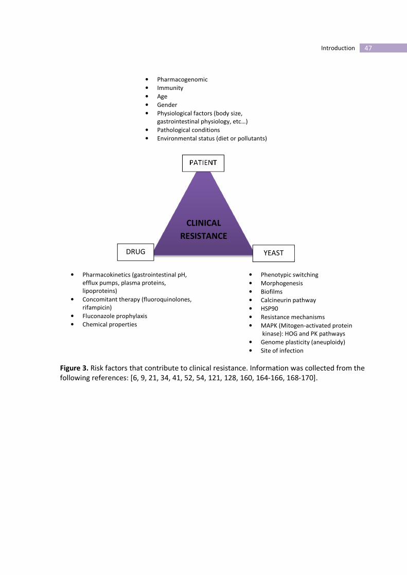
therapy and of an in vitro antifungal susceptibility profile. Many factors may contribute to
clinical resistance and to the discrepancy between the laboratory susceptibility pattern and
the clinical outcome (figure 3). The antifungal efficacy is soaked into a Bermuda triangle that
goes through patient, drug and yeast factors that ultimately are responsible for the clinical
outcome (figure 3).
Under stressing conditions like antifungal exposure, Candida cells may exploit several
cellular responses, such as development of mutations, overexpression of multidrug efflux
pumps, modulation of Ca2+
- calmodulin-calcineurin or the cAMP protein kinase A pathways
[160]. Given its clinical relevance, C. albicans has been the subject of extensive research in
order to unveil the mechanisms governing fungal virulence and drug resistance. The success
of this yeast as a pathogen depends largely on its ability to generate diversity not only at the
genetic level but also at the morphological and physiological level.
The mitochondrial respiratory pathway can regulate the metabolic behavior contributing to
fitness and flexibility of Candida strains in response to external challenges [161, 162]. The
existence of an alternative respiratory pathway is present in some Candida species,
especially C. albicans and is implicated in reduced susceptibility to azoles [161-163].
C. albicans is considered to be pleomorphic due to its ability to switch from yeast to hyphal
or pseudohyphal form [164]. Morphogenesis changes are coupled to biofilm formation,
which plays an important role in virulence. C. albicans can produce biofilms on medical
implants, like indwelling vascular catheters, and its formation acts as a physical barrier,
protecting the underlying cells of being exposed to antifungal drugs, hence lowering the
available drug concentration [165]. In addition, efflux pumps can be upregulated during
biofilm formation which may confer increases in resistance to azoles [166, 167].
47 Introduction
Figure 3. Risk factors that contribute to clinical resistance. Information was collected from the
following references: [6, 9, 21, 34, 41, 52, 54, 121, 128, 160, 164-166, 168-170].
CLINICAL
RESISTANCE
PATIENT
YEAST DRUG
• Pharmacogenomic
• Immunity
• Age
• Gender
• Physiological factors (body size,
gastrointestinal physiology, etc…)
• Pathological conditions
• Environmental status (diet or pollutants)
• Pharmacokinetics (gastrointestinal pH,
efflux pumps, plasma proteins,
lipoproteins)
• Concomitant therapy (fluoroquinolones,
rifampicin)
• Fluconazole prophylaxis
• Chemical properties
• Phenotypic switching
• Morphogenesis
• Biofilms
• Calcineurin pathway
• HSP90
• Resistance mechanisms
• MAPK (Mitogen-activated protein
kinase): HOG and PK pathways
• Genome plasticity (aneuploidy)
• Site of infection

48
Concomitant medications administered to patients, such as antibiotics, can influence the
pharmacodynamics of the antifungals. Fluoroquinolones antagonize fluconazole activity
against C. albicans strains [170], whether rifampicin can induce the expression of MDR1
pumps [168]. Nevertheless, the effect of other medications, some of them life-saving in
the case of critical care patients still remains to be elucidated.
The choice of an antifungal agent for the empirical treatment of Candida bloodstream
infections is a complicated task. Similarly to the findings with the extensive use of
antibiotics and the development of multiresistant bacterial pathogens, the selective
pressure due to the widespread use of fluconazole in prophylaxis, promoted a shift toward
non-albicans Candida species, like C. glabrata and C. krusei [171, 172].
Patient pharmacogenomics, which can influence drug absorption, distribution and
metabolism, its immunological status and the underlying disease are additional important
factors to be considered when managing individual patients [173].
Strategies to defeat antifungal resistance
The knowledge of the mechanism of antifungal resistance brought by the genomic era
supports the development of therapeutic strategies in order to bypass drug resistance. The
principal cell mechanism of antifungal resistance is the active transport of drugs out of the
cell by efflux pumps [24, 27, 98, 101], expressed not only by yeasts but also by humans cells
[21, 173]. The main strategy to reduce efflux impact involves the maintenance of a high
antifungal concentration inside the cell, at its site of action. The simplest approach would be
the use of antifungals that are not substrate of efflux pumps, like amphotericin B or
echinocandins, which given their hydrophobicity and size, do not interact with the efflux
pump [25, 174].
The second approach would be the development of inhibitors or chemosensitizers of efflux,
affecting the target, the activity, by blocking access to the binding site, or even the efflux
pump transcription.
49 Introduction
In humans, one of the factors that is responsible for the failure of cancer therapy are ATP-
dependent drug efflux pumps, such as P-glycoprotein (P-gp) [175]. P-gp substrates such as
FK506 [176] or cyclosporine A (CsA) [177] are immunosuppressors that are able to inhibit
efflux. They act similarly in C. albicans strains, inhibiting the calcineurin-mediated azole
tolerance by binding to small, abundant, conserved binding proteins called immunophilins.
CsA binds with cyclophilin A (Cyp1p) and FK506 with FKBP12, to form protein-drug
complexes that inhibit calcineurin [24, 169, 178]. By inhibiting calcineurin these compounds
act synergistically with azoles [169, 179, 180]. While FK506 and CsA chemosensitize C.
albicans cells to azoles, rending the azoles fungicidal, they are also immunosuppressive
drugs, which make it administration problematic in immunosuppressive candidosis patients.
Nevertheless, the inhibition of calcineurin-mediated azole tolerance is still a potential
therapeutic approach [181]. Non-immunosuppressive analogs could inhibit fungal
calcineurin by exploiting structural differences between the human and the fungal targets
[181].
Ibuprofen ([2-(4-isobutylphenyl)-propionic acid has been described to act synergistically
with pyrazinamide [182], fluconazole [101, 145, 183] and amphotericin B [184] in fungi. In C.
albicans expressing CDR efflux pumps, the presence of ibuprofen increased azole
intracellular accumulation, changing the resistant phenotype to susceptible [101, 145]. This
potent anti-inflammatory, non-steroidal drug might play important role in future
therapeutic strategies. However, its in vivo effect still remains unveiled.
Another helpful strategy would be the design of inhibitors that could act indirectly on efflux,
de-energizing the ATP or H+ dependent transporter, by lowering the cytoplasmic ATP
concentration or depleting the electrochemical potential of the plasma membrane,
respectively [22, 185]. However, by altering ATP and membrane potential, other cellular
metabolic activities could be compromised. Alternatively, the promotion of antifungal
uptake could also be a strategy to overcome antifungal resistance due to efflux.
Dubikovskaya et al. showed that the inclusion of multiple arginine residues (octaarginine

50
[R8]) in human anticancer drugs enhances the delivery to its intracellular targets [186], an
approach that has already been tried in yeasts [185].
The medical complexity of patients taken together with the intricate cellular mechanism
involved in drug resistance makes the pursuit of effective solutions mandatory.

CHAPTER II
Aims


53 Aims
Aims of the Study
This study has the following goals:
1. Characterization of yeasts isolates from fungaemia patients in order to assess: the
source of infection and modes of transmission; the genetic relatedness and the
antifungal susceptibility profile;
2. To evaluate the role of chitin in echinocandin resistance and to develop of a new
methodology for evaluating chitin content in the fungal cell wall;
3. To characterize the in vivo mechanisms enrolled in the induction of resistance
during echinocandin treatment;
4. To evaluate the role upon an alternative respiratory pathway in C. krusei as a
possible contribution to resistance to cell stresses;
5. To unveil the effect of concomitant therapies used in critical care patients upon
antifungal resistance or tolerance, like propofol or vasoactive amines, such as
adrenaline and noradrenaline;
6. To assess the in vivo reversion of azole resistance by ibuprofen, in an animal model
of systemic Candida infection.


CHAPTER III
Results


57 Results
Part I
Genetic relatedness and antifungal susceptibility profile of Candida albicans
isolates from fungaemia patients
Background
Candida infections have progressively emerged as major health care related invasive fungal
infections since the late 80s, mainly arising from an endogenous source, either digestive or
mucocutaneous. Commensalism, followed by colonization, usually precedes dissemination,
most frequently in patients with transient or permanent immunocompromised status, such
as transplant recipients, chemotherapy patients, underweight neonates and human
immunodeficiency virus-infected individuals. Significant morbidity and mortality rates have
been associated to bloodstream infections due to Candida spp [7, 187, 188].
Several polymorphic microsatellite loci have been identified in the genome of C. albicans
near EF3, CDC3 and HIS3 [75] or inside the coding regions of ERK1, 2NF1, CCN2, CPH2, and
EFG1 [189]. However the discriminatory power for each locus is relatively low. In order to
more rapidly obtain a higher discrimination, simultaneous amplification of sets of
microsatellite markers can be performed. A multiplex system with a high discriminatory
power was recently described and found to represent an efficient molecular tool for the
swift and accurate differentiation of C. albicans [76, 190].
During a twelve month period (2004) a prospective study addressing fungaemia was
conducted at Hospital de São João, a large university hospital located in the Northern region
of Portugal [7]. The epidemiological data analyzed included the department of admission,
underlying diseases and antimicrobial therapy, among others. Several yeast isolates from
blood cultures were collected and analyzed as well as fungal strains isolated from
surveillance cultures or from medical indwelling devices. These included isolates from
distinct biological sources for the same patient, such as urine and lower respiratory
58
secretions, and from central venous catheters. All episodes of recurrence were investigated
[7].
Our main purpose was to genotype all C. albicans isolates by using a multiplex PCR system
with four microsatellite loci. Ultimately, we aimed to determine the genetic relatedness
between simultaneous and/or recurrent isolates from the same patient, the source of
infection, the route of transmission, as well as the possibility of transmission among distinct
patients. Additionally, the susceptibility profile of all isolates was determined and analysed
in regard to antifungal therapy.
Materials and Methods
Patients clinical data
Thirty five C. albicans isolates from the blood and other biological sources were collected
from twelve patients with fungaemia. The data documented included patient gender and
age, department and date of admission, concomitant therapy (i.e. immunossupressors),
date and site of fungal isolation, and nosocomial origin of the fungaemia. According to the
Centre for Disease Control and Prevention (CDC), nosocomial fungaemia was identified
whenever a patient yielded at least one fungal positive blood culture following 48 hours of
hospital admission [191]. Antifungal therapy (prophylactic or therapeutic) and clinical
outcome (survival/death) were also documented. Fungaemia outcome was evaluated 30
days after the first fungaemia episode. Fungaemia-related death was defined as death
occurring within 30 days after the first fungal positive blood culture, with no signs of
intracerebral or gastrointestinal bleeding or pulmonary embolism. All Candida isolates were
frozen at –70ºC in Brain-Heart medium with 5% of glycerol (Difco) and subcultured twice in
agar Sabouraud (Difco) prior to experimental procedures.
C. albicans genotyping
Yeast strains were grown overnight in Sabouraud broth at 30ºC. A Zymolyase-based method
was used to extract DNA, as previously described [192]. To assess strain relatedness, all
isolates were genotyped using a microsatellite multiplex PCR assay with three markers (CAI,

59 Results
CAIII and CAVI) as described by Sampaio et al. [76] and a singleplex amplification reaction
assay using the microsatellite marker CEF3, according to procedures described by Bretagne
et al [75]. Following PCR amplification, a 1 to 2-μl aliquot of each sample was added to 15 μl
of formamide containing 0.4 μl of GeneScanTM
- 500 TAMRA size standards (Applied
Biosystems). Amplicons were denatured at 95°C for 5 min and immediately placed on ice.
Denatured samples were resolved by capillary electrophoresis in an ABI Prism 310 genetic
analyzer (Applied Biosystems). Determination of allele sizes was automatically performed
with GeneScan 3.7 analysis software. The alleles were designated according to the number
of repeated units for the CAI ((CAA)2CTG(CAA)n), CAIII ((GAA)n) and CAVI ((TAAA)n) markers
and by the number of nucleotides for the CEF3 ((TTTC)n(TTC)n) marker.
Antifungal susceptibility profile
Antifungal susceptibility testing of all isolates was performed according to the CLSI M27 A3
protocol in RPMI 1640 (Sigma) [33]. Minimal inhibitory concentration (MIC) for fluconazole,
voriconazole, caspofungin and amphotericin B were determined [33]. The yeast strains were
classified as susceptible (S), susceptible-dose dependent (S-DD) and resistant (R) to azoles
according to the breakpoints defined by CLSI [33]. Although definitive breakpoints have not
yet been established for amphotericin B, strains showing MIC ≤1 µg/ml were considered
susceptible [193]. For caspofungin, a MIC ≤2 μg/ml corresponded to S and >2 μg/ml
to non-
susceptible (NS) strains [33]. Strains displaying a MIC variation within one dilution were
considered as having a similar susceptibility pattern.
Results
The patients age ranged from 40 to 83 years old and 75% (9/12) were male. Data such as
department of admission, date of first isolation and strain origin are presented in table 1
and figure 1. All patients were iatrogenically immunosuppressed, and 75% died. All
fungaemia cases corresponded to episodes of nosocomial infections. The microsatellite
genotyping analysis is detailed in Table 1.
Amongst the 12 patients two or more isolates were obtained from blood and from

60
additional body locations. We considered isolates has been highly related when their
genotypes differed at a single locus only. In the large majority of patients, blood isolates
displayed the same multilocus genotype or a genotype that was highly similar to that of
isolates from other body locations. In one single case, patient 10, one isolate from urine
exhibited a completely distinct multilocus genotype from the blood isolate, even though
they were collected at the same time. Isolate 9.3 from urine displayed a genotype that
differed at two loci compared to the isolates obtained from the respiratory tract or blood
isolates. This isolate was moderately related to the other isolates from the same patient but
was highly related to isolates from patients 6, 7 and 8. In two patients, 11 and 12, minor
genetic differences were detected among blood isolates and isolates from other body
sources. In these patients, the isolates were highly related, suggestive of microevolution.
Consecutive blood isolates from the same patient exhibited the same microsatellite
genotype in 5 out of 6 patients. The exception was for patient 12 where a highly related
isolate was observed between two consecutive blood cultures. This was indicative of strain
maintenance regardless of antifungal treatment rather than re-infection by a distinct strain
(table 1). Isolates from patients 3, 4 and 5 showed identical or highly similar microsatellite
profiles (table 1), an interesting fact since these patients had been initially simultaneously
admitted in the same intensive care unit (ICU). Isolates from patients 6, 7 and 8 also
exhibited identical or highly similar genotypes (table 1) and were initially admitted to the
same ICU, although not concurrently. All isolates from patients 1 and 2, whom where in the
same ICU in the previous months, displayed the same genotype (table 1). Sharing of the
same strain among different patients is suggestive of hospital-acquired exogenous
nosocomial infection with hospital endemic strains.
The antifungal therapy timelines are detailed in figure 1. All doses were administered
according to the Clinical Practice Guidelines for the management of Candidosis [10]. Four
patients (1, 3, 5 and 9) were under antifungal prophylaxis with fluconazole for 2 or 3 weeks
prior to the first fungal isolation. Only patient 1, in which fluconazole therapy was replaced
by amphotericin B, had a favorable outcome. Two patients (patient 3 and 6), with end-stage

61 Results
neoplasic diseases, were infected with fluconazole-susceptible strains and died due to
fungaemia according to their medical records. In patients 3, 4 and 5 infected by the same
strain, MIC increases were observed. Isolates from patients 4 and 5 that were collected
several months after isolates from patient 3 were resistant to azoles (Table 1 and Figure 1).
Similarly, we observed azole cross-resistance in isolates from patient 8 that were collected
several months after isolates from patients 6 and 7. All of these isolates had identical
genotypes and probably represented the same strain (Table1 and Figure 1).
All of the isolates analyzed showed low MIC values to amphotericin B and caspofungin (≤1
µg/ml) (Table 1).

62
Table 1. Candida albicans isolates from blood cultures and from other body sites
obtained from 12 patients and respective multilocus genotyping results.
Strain replacement is highlighted in dark grey
Microevolutionary events are highlighted in light grey

63 Results
Figure 1. Schematic representation of patients clinical data and genetic relatedness of C. albicans
strains. The duration of hospitalization (from admission to discharge or death) of each patient is
represented by a thick line. Identical symbols represent isolates with the same or highly related
genotype.

64
Discussion
Nosocomial infections represent an important source of morbidity and mortality in hospital
settings [194]. The understanding of pathogen distribution and relatedness is critical for
both the epidemiological surveillance of health-care related infections and for the
conception of rational pathogen control policies [195]. Pathogen typing allows for the
determination of genetically and epidemiologically related isolates. The development and
implementation of new DNA based technologies and molecular analyses over the last 3
decades have led to considerable advances in microbial typing approaches [77, 196, 197].
The identification of a pathogen origin, endogenous versus exogenous, and the
characterization of consecutive infections by the same organism as relapse or re-infection
are critical [197]. Our group has previously developed a microsatellite multiplex PCR
strategy with high discriminatory power for typing Candida albicans [76]. This methodology
was found to efficiently discriminate C. albicans strains, as well as C. glabrata, C.
parapsilosis and C. tropicalis [77, 198, 199]. Maintenance and infection by the same strain
may indicate that the therapeutic regimen was unsuccessful and that alternative therapies
might be required. The results obtained here for successive blood isolates from the same
patient suggest the failure of antifungal therapy in these cases. Moreover, fungal strains
may invade the host from different body sites, and microevolution, due to strand slippage
during DNA replication in the microsatellite region or loss of heterozygosity (LOH), may
represent an adaptive fungal response to new environments [76, 196]. We detected
microevolution events in 3 patients (9, 11 and 12). C. albicans colonization and/or infection
of different body sites may represent a predisposing condition or an initial step towards the
subsequent fungaemia development [188, 200].
All the fungaemia cases considered herein were of nosocomial origin, according to the
accepted definition [191]. Our results clearly demonstrate that isolates displaying the same
or highly similar genotypes were obtained from patients who shared the same hospital
department of admission. This suggests hospital-acquired infections. These observations
indicate that the safety measures between patients most probably failed and those
65 Results
incidents of cross-infection were likely to have occurred.
In our study, fungaemia relapses were frequent, appeared to be caused by the same strain,
and were invariably associated to a poor therapeutic outcome. The extension of cross-
resistance to azoles detected among fungaemia patients may challenge the large empiric
use of fluconazole and how it can lead to the development of dramatic resistance. We
observed induction of azoles resistance in two groups of patients (patients 3, 4 and 5 and
patients 6, 7 and 8) and the increase in MIC values, over time, in similar strains from
different patients suggests that the strains were endemic to the hospital environment for at
least several months. This exemplifies the risk of selecting for strains with increased
antifungal resistance in the hospital environment.
The molecular identity of fungal isolates represents a key feature for comprehensible
therapeutic strategies in a near future; indeed, analysis of dominant genotypes in different
geographical regions, distinct clinical samples and distinct underlying diseases is now a
possibility. Molecular approaches are now available, allowing detailed comparisons
between C. albicans clinical strains and providing a clear definition of their genetic
relatedness.

67 Results
Part II
Determination of chitin content in fungal cell wall: an alternative flow
cytometric method
Background
Chitin is a β-1,4-homopolymer of N-acetylglucosamine that is synthesized by chitin synthase
enzymes [201]. This polysaccharide is present in most fungi and together with β-1,3-glucan,
plays a fundamental role in maintaining fungal cell integrity and conferring structural
rigidity during growth and morphogenesis [201-203]. Mutations in glucan synthase genes
reduce glucan levels in the cell wall while stimulating salvage pathways leading to increased
chitin synthesis. This pathway restores the strength of the cell wall matrix and prevents
antifungal action [204]. Since chitin is not present in human cells, inhibition of chitin
synthesis has been proposed as a potential, selective antifungal target.
The assessment of cell wall chitin content based upon glucosamine release through acid
hydrolysis has been used extensively; however this method is very laborious and time
consuming [128, 129, 205]. Epifluorescence microscopy has also been widely used to
quantify chitin levels in fungi stained by Calcofluor White (CFW), a specific chitin dye [128,
206-210]. However, with this approach, only a limited number of yeast cells can be analyzed
and the quantification of the fluorescence emitted cannot be performed accurately [128,
211, 212]. Flow cytometry represents an efficient and fast approach for the analysis of cell
architecture and functional phenotypes, with considerable advantages over conventional
methods [103, 145, 147, 213].
Here we describe a fast and reliable protocol to measure cell wall chitin content in yeasts
cells based upon flow cytometric analysis after CFW staining.
68
Material and methods
Strains
Twenty two Candida spp and 4 Cryptococcus neoformans clinical isolates with well
characterized susceptibility profiles to caspofungin (antifungal chosen as representative of
echinocandin class), were used in this study (detailed in Table 1). SC5314 [214] with wild
type chitin levels, chs3Δ/chs3Δ (Myco 3) [215], pga62Δ/Δ [216] and pga31Δ/Δ [216] were
used as control strains.
Measurement of cell wall chitin content
Wild type and mutant yeast cells were grown in YPD broth medium at 35ºC, 150 rpm, until
late logarithmic phase, and used to optimize flow cytometric protocol. A 106 yeast cells ml
-1
suspension in sterilize distilled water was stained with 0 (autofluorescence), 2.5, 6.25, 12.5
and 25 µg CFW ml-1 (Fluka, St. Louis, USA), a specific chitin dye (excitation at 365 nm and
emission at 430 nm), for 15 minutes at room temperature. In parallel, yeast cells were
treated with MIC values of caspofungin during 2 hours, and stained with CFW. The yeast
cells were washed twice and blue fluorescence (Pacific blue channel) emitted by 50000 cells
was quantified, using a BD FACSCanto™ II (Becton Dickinson, San Jose, CA, USA) flow
cytometer. BD FACSCanto™ II system consists of an excitation source with three lasers: blue
(488-nm, air-cooled, 20-mW solid state), red (633-nm, 17-mW HeNe), and violet (405-nm,
30-mW solid state). The mean intensity of fluorescence (obtained from three independent
experiments) emitted from stained (positive population) and non-stained (autofluorescence
or negative population) yeast cells was analyzed and processed with FACSDiva software
(version 6.1). In each experiment a staining index (SI) was calculated as follows: (mean
intensity of fluorescence of positive population – mean intensity of fluorescence of negative
population)/ 2 x standard deviation of the mean intensity of fluorescence of negative
population [217]. The chitin content of the 26 clinical isolates was assessed according to the
described protocol, after staining with 2.5 µg CFW ml-1
; the SI was calculated.

69 Results
Epifluorescence microscopy
In order to confirm flow cytometry results, epifluorescence microscopy analysis was
performed. Yeasts cells were grown and prepared as described for flow cytometric assays
and stained with 25 µg CFW ml-1
for 15 minutes. Following staining, 30 µl of the cell
suspension were placed on a glass slide and overlapped with vectashield fluorescence
mounting medium (Vector Laboratories, Peterborough, UK) and observed under an
epifluorescence microscope (400X) imager Z Apotome (Zeiss, Barcelona, Spain).
Paradoxical effect of caspofungin
The ability of the clinical isolates to grow in the presence of high caspofungin (Merck,
Rahway, NJ, USA) levels, termed paradoxical growth was tested over a range of
concentrations varying from 0.03 to 256 µg ml-1 and MICs were determined using
prominent inhibition as an endpoint corresponding to 50% (MIC50) [33, 218]. The
paradoxical effect was defined as a progressive increase in cell growth occurring at least
two drug dilutions above the MIC, following 48 hours incubation [218].
Data Analysis
The SI mean values displayed by the different isolates after CFW staining were compared
using the Student’s t-test. Significant effects were accepted at p<0.05. The SPSS Statistics
17.0 Software for Windows was used to perform the statistical analysis. All experiments
were performed in triplicate.
Results and Discussion
The cytometric protocol was optimized using four C. albicans strains with known differences
in chitin contents: chs3Δ/chs3Δ, pga31Δ/Δ, pga62Δ/Δ and the reference strain SC5314.
These strains are deleted in genes that are involved in chitin synthesis (chs3Δ/chs3Δ) or
encode GPI-proteins that are involved in cell wall biosynthesis or in cell wall salvage
pathways (pga31Δ/Δ and pga62Δ/Δ) [128, 216]. A range of CFW concentrations was tested
and 2.5 µg CFW ml-1
revealed to be the concentration to achieve the best resolution to
differentiate the chitin content of the four strains used as controls. The reference strain

70
SC5314 had significantly higher SI values (p<0.001) than strains chs3Δ/chs3Δ and pga31Δ/Δ
and had lower values when compared to the pga62Δ/Δ strain (figure 1). Flow cytometry
chitin measurements were concordant with the chitin levels determined by the
quantification of glucosamine released by acid hydrolysis, previously obtained by others
[128, 215, 216]. Caspofungin treatment of the reference strain SC5314 led to a significant
increase in chitin content (p<0.001), contrasting with the mutant strains where caspofungin
did not produce any effect (figure 1). The chitin levels obtained after caspofungin exposure
of the reference and chs3Δ/Δ strains are in agreement with those achieved by the classic
method performed by other authors [128].
Furthermore, results obtained by flow cytometry were consistent with epifluorescence
microscopy observations (data not shown). The flow cytometric protocol for chitin
quantification is considerably less laborious and more accurate in comparison with the
previously described methods since a large amount of cells (50000) are randomly evaluated,
without operator interference. Given the variation in yeast morphology such as cell shape
and size, different species may emit different levels of autofluorescence, which arises from
endogenous fluorophores. This autofluorescence emission analyzed under epifluorescence
microscopy or flow cytometry results in a background “noise” which may interfere with the
quantification of fluorescence emitted by stained cells [217]. To avoid autofluorescence
interference, especially when fluorescence emitted by cells from different species is
compared, normalization of data is mandatory [217]. This was achieved through the
calculation of a SI which provides sensitivity and reliability to the output data and enables
comparison of the fluorescence emitted by cells with distinct morphologies. With this
approach we can expect a possible normalization in intra and inter laboratory results.
The relationship between the CFW staining index and the caspofungin susceptibility
phenotype displayed by clinical strains is detailed in Table 1 and figure 2. Among the distinct
species included in this study, C. parapsilosis, C. tropicalis and C. albicans clinical isolates
showed a higher CFW staining index, and therefore a higher cell wall chitin content
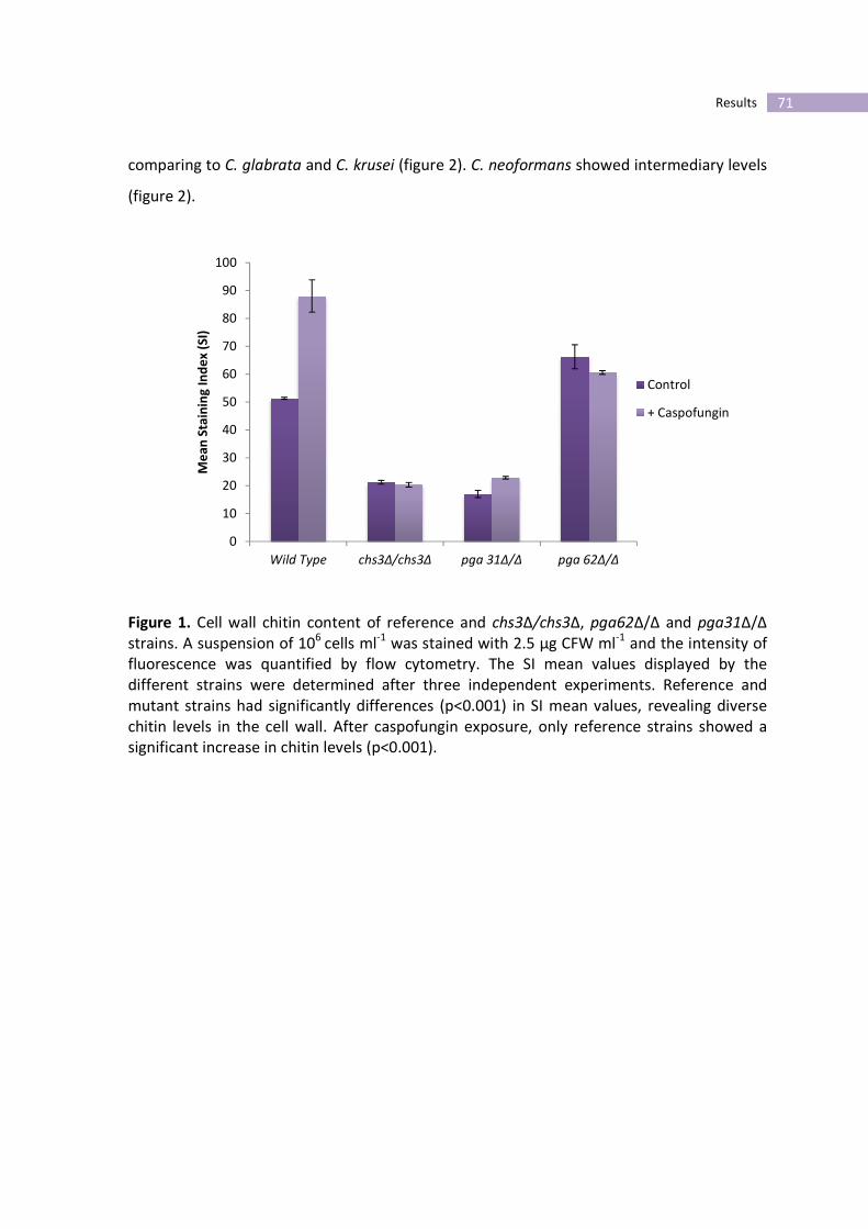
71 Results
0
10
20
30
40
50
60
70
80
90
100
Wild Type chs3Δ/chs3Δ pga 31Δ/Δ pga 62Δ/Δ
Me
an
Sta
inin
g I
nd
ex
(S
I)
Control
+ Caspofungin
comparing to C. glabrata and C. krusei (figure 2). C. neoformans showed intermediary levels
(figure 2).
Figure 1. Cell wall chitin content of reference and chs3Δ/chs3Δ, pga62Δ/Δ and pga31Δ/Δ
strains. A suspension of 106
cells ml-1
was stained with 2.5 µg CFW ml-1
and the intensity of
fluorescence was quantified by flow cytometry. The SI mean values displayed by the
different strains were determined after three independent experiments. Reference and
mutant strains had significantly differences (p<0.001) in SI mean values, revealing diverse
chitin levels in the cell wall. After caspofungin exposure, only reference strains showed a
significant increase in chitin levels (p<0.001).

72
Table 1. In vitro antifungal susceptibility and paradoxical effect of caspofungin (CFS)
against Candida spp and Cryptococcus neoformans clinical isolates. Minimal inhibitory
concentrations (MIC; µg ml-1
) were determined using prominent inhibition as an end
point, corresponding to 50% (MIC50), according to CLSI protocol.
Yeast Strain
code
Source CFS MIC50 (µg ml-1
)
/ Phenotype
Paradoxical growth
(mean values)
Start point/ end point
(µg ml-1
)
C. glabrata Cg1 Blood >32/ NS NF
Cg2 Blood 0.125/ S NF
Cg3 Peritoneal fluid 32/ NS NF
Cg4 Fecal 0.125/ S NF
Cg5 Peritoneal fluid 0.5/ S NF
Cg6 Blood 0.25/ S NF
C. parapsilosis Cp1 Peritoneal fluid 4/ NS NF
Cp2 Blood 2/ S NF
Cp3 Blood 4/ NS NF
Cp4 Blood 4/ NS 16/64
Cp5 Blood 0.5/ S NF
C. tropicalis Ct1 Blood 0.5/ S 8/16
Ct2 Peritoneal fluid 0.5/ S NF
Ct3 Pus 4/ NS 16
Ct4 Pus 4/ NS 16
C. krusei Ck1 Urine 1/ S NF
Ck2 Blood 1/ S NF
Ck3 Bronchial
secretions
1/ S NF
Ck4 Bronchial
secretions
1/ S NF
C. albicans Ca1 Blood 0.5/ S 16/32
Ca2 Blood 0.5/ S 16/32
Ca3 Blood 0.5/ S 16/32
C. neoformans Cn1 Blood 16/ NS NF
Cn2 Blood 16/ NS NF
Cn3 Blood 16/ NS NF
Cn4 Blood 32/ NS NF
NS non susceptible phenotype; S susceptible phenotype; NF not found

73 Results
Figure 2. Cell wall chitin content of Candida spp and Cryptococcus neoformans clinical
isolates in the absence and presence of caspofungin. Yeast cells were stained with 2.5 µg
CFW ml-1
and fluorescence emitted was quantified by flow cytometry. Higher staining index
(SI) values were observed in strains that showed a paradoxical growth (*) in the presence of
caspofungin. # shows significantly different chitin levels after caspofungin treatment.
Notably, several C. parapsilosis and C. tropicalis (Cp4, Ct1, Ct3 and Ct4) isolates showed a
significant increase in chitin level (p<0.001) in comparison with other isolates from the same
species (figure 2). Interestingly, these strains, along with all C. albicans isolates tested,
exhibited a paradoxical growth in the presence of high caspofungin concentrations. The
ability to grow at high caspofungin concentrations has been frequently described among C.
albicans, C. parapsilosis and C. tropicalis [218] and has been suggested to relate to a
compensatory increase in cell wall chitin [128, 130]. This salvage mechanism strengths cell
wall damaged by exposure to echinocandins. Chitin quantification by flow cytometry
revealed to be a highly sensitive method. It enabled the detection of different chitin levels,
which allowed us to validate the association between a higher amount of cell wall chitin and
paradoxical growth in the presence of caspofungin concentrations well above the MIC. Also,
when yeast cells were treated with caspofungin for two hours, a significant increase in

74
chitin levels were obtained, especially in strains that showed the paradoxical effect (Cp4,
Ct1, Ct3, Ct4, Ca1, Ca2 and Ca3) (figure 2). The finding that caspofungin treatment
stimulates chitin biosynthesis has also been described by other authors [130, 216, 218]. The
clinical relevance of this in vitro effect is yet uncertain. Although unrelated to resistance, the
paradoxical effect may represent a drug tolerance mechanism and an adaptive response to
the presence of caspofungin. In contrast, C. glabrata, C. krusei and Cryptococcus
neoformans strains showed an absence of paradoxical growth, displaying the lowest chitin
levels. Interestingly, the accuracy of this novel methodology can predict the occurrence of
this paradoxical effect within a few minutes, representing a valuable tool for detection of an
antifungal compensatory mechanism in the presence of high antifungal concentrations,
namely echinocandins.
The flow cytometry protocol described herein may constitute a useful tool to evaluate the
inhibition of chitin synthesis by new drugs presently under development.
75 Results
Part III
FKS2 mutations associated with decreased echinocandin susceptibility of
Candida glabrata following anidulafungin therapy
Background
Fungal pathogens have become progressively more responsible for human disease.
Although C. albicans is still the most frequent agent of fungaemia in U.S.A., the incidence of
candidosis by non-albicans species is increasing, with C. glabrata being the second causing
agent of fungaemia in U.S.A. and in some European countries [6, 48]. C. glabrata infections
represent a serious concern due to the inherent high mortality rate, to patient
comorbidities as well as to the predisposition to rapidly develop azoles resistance [7, 52,
59]. As such, there has been an urge to develop new therapeutic alternatives. A new class of
antifungal agents has arisen, including caspofungin, micafungin and more recently
anidulafungin, which inhibit the β (1, 3)-glucan synthase. This enzyme is responsible for
producing a key component of the fungal cell wall and is encoded by the FKS genes and
includes a regulatory subunit Rho-1p.
Due to their excellent clinical effectiveness and safety, these antifungals became the first-
line therapy in many hospitals for the treatment of invasive candidosis [10-12]. To date,
case reports describing primary or secondary echinocandin resistance in candidosis have
been rare, however only caspofungin has been widely used. Reduced susceptibility to
echinocandins has been linked to mutations in hot spot regions of FKS genes [31].
Specifically, mutations in FKS2 have been shown to be associated with echinocandin
resistance in C. glabrata acquired during caspofungin treatment [18, 125, 219]. Aside from
glucan synthesis, chitin synthesis plays a fundamental role in maintaining fungal cell
integrity during growth and morphogenesis. Previous studies have demonstrated a
76
significant increase in chitin content after exposure to different concentrations of
echinocandins [130, 150]. This suggests that increased amounts of chitin could be
responsible for reduced activity of echinocandins resulting as an escape mechanism to
these agents [128].
The aim of this study was to unveil the underlying resistance mechanisms developed during
prolonged anidulafungin treatment.
Herein, and for the first time, we describe a case of echinocandin resistance in a patient
with candidosis by C. glabrata following anidulafungin therapy. Interestingly, during the
hospital course two of the C. glabrata isolates turned out to be non susceptible to
echinocandins displaying different mechanisms of resistance, possibly responsible for the
patient’s unfavorable outcome.
Case Report
A 71-year-old female was admitted to the Intensive Care Unit (ICU) with a diagnosis of acute
pancreatitis developed after a laparoscopic cholecystectomy. The patient’s medical history
included arterial hypertension and dyslipidemia. An exploratory laparoscopic surgery was
performed at the second day of admission at the ICU. The surgical findings were peritoneal
necrosis, necrosis of the ileum and jejunum and pancreatic necrosis. During her hospital
stay the patient was administered several antibacterial drugs namely tazobactam,
piperacillin, meropenem, tigecycline and vancomycin. She had no previous antifungal
exposure. Her medical condition was further complicated by multiple Candida infections,
including disseminated candidosis and catheter related infections. Figure 1 summarizes the
Candida isolates, the antifungal therapy administered and subsequent clinical response.
Starting on day 6, she received a 200 mg load followed by 100mg/day of anidulafungin for
27 days. Cultures of multiple sites including blood, exudates, central venous catheter (cvc)
and urine were positive for C. albicans and C. glabrata. While C. albicans fungaemia cleared
upon 24 hours of anidulafungin therapy, C. glabrata persisted. At day 17 the patient
received noradrenaline support since no clinical improvement could be observed, and was

77 Results
subsequently submitted to a second exploratory laparoscopic surgery at day 30. The
procedure revealed extensive cytoesteatonecrosis. Amphotericin B was then initiated and
two days later the patient developed multiorgan failure. The patient died 34 days after
admission. No autopsy was performed.
Figure 1. Antifungal therapy administered to the patient. Days after admission and fungal
strains isolated from biological samples are also shown. AND, anidulafungin; AMB lipo,
amphotericin B lipid complex.
Material and methods
Strains
The strains isolated from different body sites throughout the course of infection (figure 1
and table 2) were characterized by Vitek YBC identification cards (BioMérieux, Paris,
France). For each laboratorial experiment, strains were subcultured twice on YPD agar (1%
yeast extract, 2% peptone, 2% glucose, and 2% agar) medium for 48 hours at 35°C.
Microdilution susceptibility testing
Assessment of the antifungal susceptibility profile of all clinical isolates was performed
according to the CLSI M27 A3 protocol in RPMI 1640 (Sigma) [33]. The minimal inhibitory
concentration (MIC) of fluconazole (Pfizer, Groton, CT, USA), voriconazole (Pfizer),
posaconazole (Shering-Plough, Kenilworth, NJ, USA), caspofungin (Merck, Rahway, NJ, USA),

78
anidulafungin (Pfizer), micafungin (Astellas Pharma US, Deerfield, IL) and amphotericin B
(Bristol-Myers Squibb New York, USA) was determined [33]. Strains were classified as
susceptible (S), susceptible-dose dependent (S-DD) or resistant (R) to azoles according to
breakpoints defined by CLSI [33]. For caspofungin, anidulafungin and micafungin, MIC ≤2
μg/ml were considered S and those >2 μg/ml
as non susceptible (NS) [33]. Although
definitive breakpoints have not yet been established for amphotericin B and posaconazole,
strains with MIC ≤1 µg/ml were considered susceptible [193].
Resistance stability
To assess resistance stability, the non susceptible isolates (7-1 and 9-2) were subcultured
daily in the absence of anidulafungin for 30 days. Each strain was cultured in 10 mL drug-
free RPMI 1640 at 35 °C, 150 rpm. The following day, aliquots were transferred into fresh
medium. At each subculture, a 1-mL aliquot of the yeast suspension was mixed with 0.5 mL
of 50% glycerol and frozen at −70 °C. Afterwards, the microdilution susceptibility testing of
each collected strain to anidulafungin was repeated as detailed above.
Measurement of cell wall chitin content by flow cytometry
C. glabrata cells were grown in YPD broth medium at 35ºC under agitation until late log
phase. A 106 yeast cells/ml suspension in distilled H2O was stained with 2.5 µg/ml Calcofluor
White (CFW) (Fluka), a specific chitin dye (excitation at 365 nm and emission at 430 nm), for
15 minutes at room temperature. Yeast cells were washed by centrifugation and the blue
fluorescence (Pacific blue channel) was quantified in a flow cytometer BD FACSCANTO II
(Becton Dickiinson, Madrid, Spain). The results were analyzed and processed with FACSDiva
software. We determined the mean intensity of fluorescence emitted from yeast cell
populations from three independent experiments.
Epifluorescence microscopy
In order to confirm flow cytometry assay results, C. glabrata cells were examined by
epifluorescence microscopy. Yeast cells were grown and prepared as described for flow
cytometry assays and stained with 25µg/ml CFW for 15 minutes. Upon staining, 30 µl of the
cell suspension were placed on a slide and overlapped with vectashield fluorescence

79 Results
mounting media (Vector Laboratories, Peterborough, UK). The stained suspensions were
observed under an epifluorescence microscopy (400X) imager Z Apotome (Zeiss).
C. glabrata DNA extraction
For total DNA extraction, C. glabrata cells were grown overnight at 30ºC in 10 ml YPD broth
at 180 rpm, collected by centrifugation and washed twice with sterile distilled water. Pellets
were resuspended in 400 µl lysis buffer (20mM Tris, 1 mM EDTA, 100mM NaCl, 2% Triton X-
100, 1% SDS) and 400 µl Phenol:Chloroform:Isoamyl alcohol 25:24:1; 0.3 g acid-washed
glass beads were added and samples were strongly vortexed for 10 min with 1 min intervals
on ice. Subsequently, 400 µl TE buffer (10mM Tris-HCl, 1mM EDTA, pH 8.0) were added and
samples centrifuged at 14000 rpm, for 5min, at 4ºC. The aqueous phase was removed and
an equivalent volume of Phenol: Chloroform: Isoamyl alcohol 25:24:1 was added. This
phenol:chloroform extraction step was repeated twice. Genomic DNA was precipitated by
1ml of 100% ice-cold ethanol. Samples were centrifuged at 14000rpm for 20 min (4ºC), the
supernatant was removed and the pellet washed twice with 70% ethanol. The pellet was
allowed to dry, ressuspended in 200 µl of TE buffer and treated with 20 µg of RNase
(Applichem, Darmstadt, Germany) by incubating at 37ºC up to 1h. For DNA final
precipitation, 20 µl of 4M ammonium acetate pH 4.8 (Sigma –Aldrich, Germany), and 600 µl
of ice-cold 100% ethanol (Applichem) were added and samples incubated overnight at -
20ºC. Upon washing the pellet with ice-cold 70% ethanol, DNA was ressuspended in TE
buffer to a final concentration of 2.0-2.5 µg/µl.
Random amplification of polymorphic DNA of all C. glabrata isolates
RAPD was performed according to methodology previously described by Bautista-Munoz et
al. [220]. Primer OPE-18 (5’ GGACTGCAGA 3’) and OPA (5’ AGCTGACCGT 3’) at a final
concentration of 0.4 µM were added to a PCR reaction mixture containing 1x Taq buffer, 10
ng of genomic DNA, 200 µM dNTPs, 1.5 mM MgCl2 and 1 U of Taq DNA polymerase
(Fermentas). PCR was carried out in a Realplex Mastercycler instrument (Eppendorf)
programmed as follows: an initial denaturation step of 2 min at 95°C followed by 38 cycles
of 1 minute at 95°C, 1 minute at 36ºC, 2 minutes at 72ºC and a final extension step of 10

80
minutes at 72ºC. The RAPD patterns were obtained through electrophoresis in 2% agarose
gel.
HS1 sequencing of FKS1 and FKS2 genes
Specific primers (table 1) were used for PCR amplification of HS1 of FKS1 and FKS2 genes.
PCRs were performed in a Realplex Mastercycler instrument (Eppendorf) according to the
amplification conditions: an initial denaturation step of 2 minutes at 96ºC followed by 30
cycles of 30 seconds at 96ºC, 30 seconds at 69.6ºC, 30 seconds at 72ºC and a final extension
step of 10 minutes at 72ºC. PCR products were treated with ExoSAP-IT (USB Corporation
OH, USA) and used as template for the sequencing reactions, performed with the Big Dye
Terminator Cycle Sequencing Ready Reaction Kit (Applied Biosystems). The DNA products
were purified with Sephadex G-50 Fine (GE Healthcare, UK) and sequenced in an ABI PRISM
3130 Genetic Analyzer (Applied Biosystems). The results were analyzed with the Sequencing
Analysis 5.2 software from Applied Biosystems.
Table 1. Primers used for C. glabrata FKS1 and FKS2 HS1 amplification and sequencing.
Primers Sequence (5’-3) Reference
FKS1 1HS1F2 CTTATGTTTGATTTTTGCA This study
FKS1 1HS1R CCTTCAATTTCAGATGGAACTTGATG [30]
FKS2-HS1F GTGCTCAACATTTATCTCGTAGG [219]
FKS2-HS1R CAGAATAGTGTGGAGTCAAGACG [219]

81 Results
Data Analysis
Fluorescence mean values displayed by the different strains after CFW staining were
compared using Student’s t-test. Significance was accepted at p<0.05. The SPSS Statistics
17.0 Software for Windows was used to perform the statistical analysis.
The coding sequences of the Candida glabrata FKS1 and FKS2 genes (GenBank accession:
XM_446406 and XM_448401) were aligned with those obtained from the clinical isolates.
Alignments were analyzed in BioEdit (http://www.mbio.ncsu.edu/BioEdit/bioedit.html).
Results
Antifungal susceptibility profile
Twenty one isolates were recovered from the patient’s blood and other biological samples
throughout anidulafungin treatment (table 2). The majority of such isolates corresponded
to C. glabrata (table 2). All strains were susceptible to echinocandins, except two strains
one recovered from the blood (7-1) and the other from the peritoneal fluid (9-2), the 7th
and
9th
isolates, respectively (table 2 and figure 1). Regarding azoles and amphotericin B no
resistance could be observed (table 2).
After subculturing the NS strains (7-1 and 9-2) in drug free RPMI medium during 30 days,
the MIC values for both strains remained 4µg/ml (non-reverting strains 7-1NR and 9-2NR),
confirming the stability of the non susceptible phenotype.
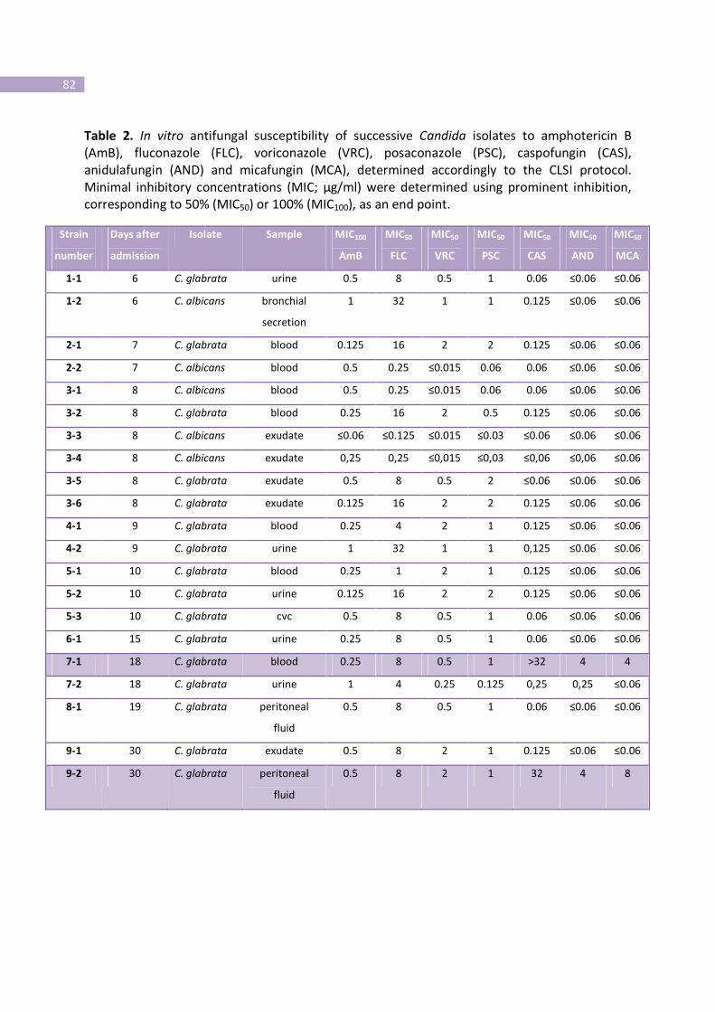
82
Table 2. In vitro antifungal susceptibility of successive Candida isolates to amphotericin B
(AmB), fluconazole (FLC), voriconazole (VRC), posaconazole (PSC), caspofungin (CAS),
anidulafungin (AND) and micafungin (MCA), determined accordingly to the CLSI protocol.
Minimal inhibitory concentrations (MIC; µg/ml) were determined using prominent inhibition,
corresponding to 50% (MIC50) or 100% (MIC100), as an end point.
Strain
number
Days after
admission
Isolate Sample MIC100
AmB
MIC50
FLC
MIC50
VRC
MIC50
PSC
MIC50
CAS
MIC50
AND
MIC50
MCA
1-1 6 C. glabrata urine 0.5 8 0.5 1 0.06 ≤0.06 ≤0.06
1-2 6 C. albicans bronchial
secretion
1 32 1 1 0.125 ≤0.06 ≤0.06
2-1 7 C. glabrata blood 0.125 16 2 2 0.125 ≤0.06 ≤0.06
2-2 7 C. albicans blood 0.5 0.25 ≤0.015 0.06 0.06 ≤0.06 ≤0.06
3-1 8 C. albicans blood 0.5 0.25 ≤0.015 0.06 0.06 ≤0.06 ≤0.06
3-2 8 C. glabrata blood 0.25 16 2 0.5 0.125 ≤0.06 ≤0.06
3-3 8 C. albicans exudate ≤0.06 ≤0.125 ≤0.015 ≤0.03 ≤0.06 ≤0.06 ≤0.06
3-4 8 C. albicans exudate 0,25 0,25 ≤0,015 ≤0,03 ≤0,06 ≤0,06 ≤0.06
3-5 8 C. glabrata exudate 0.5 8 0.5 2 ≤0.06 ≤0.06 ≤0.06
3-6 8 C. glabrata exudate 0.125 16 2 2 0.125 ≤0.06 ≤0.06
4-1 9 C. glabrata blood 0.25 4 2 1 0.125 ≤0.06 ≤0.06
4-2 9 C. glabrata urine 1 32 1 1 0,125 ≤0.06 ≤0.06
5-1 10 C. glabrata blood 0.25 1 2 1 0.125 ≤0.06 ≤0.06
5-2 10 C. glabrata urine 0.125 16 2 2 0.125 ≤0.06 ≤0.06
5-3 10 C. glabrata cvc 0.5 8 0.5 1 0.06 ≤0.06 ≤0.06
6-1 15 C. glabrata urine 0.25 8 0.5 1 0.06 ≤0.06 ≤0.06
7-1 18 C. glabrata blood 0.25 8 0.5 1 >32 4 4
7-2 18 C. glabrata urine 1 4 0.25 0.125 0,25 0,25 ≤0.06
8-1 19 C. glabrata peritoneal
fluid
0.5 8 0.5 1 0.06 ≤0.06 ≤0.06
9-1 30 C. glabrata exudate 0.5 8 2 1 0.125 ≤0.06 ≤0.06
9-2 30 C. glabrata peritoneal
fluid
0.5 8 2 1 32 4 8

83 Results
Measurement of chitin content
To evaluate whether anidulafungin (AND) resistance was related to an increase in chitin cell
wall content, the two susceptible (5-1 and 8-1), two non susceptible strains (7-1 and 9-2)
and the two non-reverting strains (7-1NR and 9-2NR) were compared regarding their chitin
content.
Flow cytometry was used to measure fungal chitin through assessment of fluorescence
emitted by Calcofluor White (CFW). CFW is a fluorescent dye that binds specifically to fungal
chitin and the resulting fluorescence intensity is considered proportional to chitin cell wall
content [209].
When compared to the other strains, only the non susceptible strain 7-1 displayed a
significant increase (p<0.0051) in the fluorescence mean value after CFW staining, indicative
of a higher chitin content (figure 2). This result is concurrent with epifluorescence
microscopy observations, showing strain 7-1 a higher staining intensity, with no
abnormalities in the distribution of chitin (blue fluorescence), compared to strain 9-2 (figure
2a and b, respectively).
Regarding the non-reverting strains (7-1NR and 9-2NR), after subculturing for 30 days in the
absence of antifungal the chitin content decreased when compared to strains 7-1 and 9-2.
Strain 7-1NR showed a significant lower chitin content than strain 7-1 (p=0.0106) (figure 2).
Genetic similarity between the different isolates recovered from the patient
Random amplification of polymorphic DNA (RAPD) was carried out in isolates from different
biological samples to determine their isogenicity. Using primer OPE-18 and OPA-18 [220] an
identical band profile was displayed by all isolates with exception of strain 9-2 recovered
from the peritoneal fluid (figure 3).

84
Figure 2. Relative cell wall chitin content from susceptible (light grey) and non susceptible
(dark grey) strains. A suspension of 106cells/ml was stained with 2.5 µg/ml Calcofluor White
(CFW) and the intensity of fluorescence was quantified by flow cytometry. The mean CFW
fluorescence was determined from three independent assays.
Figure 3. Random amplification of polymorphic DNA gel patterns of C. glabrata isolates 7-1,
9-2, 5-1 and 8-1 obtained with primers OPE-18 and OPA-18.

85 Results
FKS1 and FKS2 sequence analysis
To determine if anidulafungin resistance was associated with mutations in the target genes,
genomic DNA extraction of C. glabrata isolates was performed and the hot spot 1 (HS1)
regions of FKS1 and FKS2 genes were amplified and sequenced. No sequence alterations
were observed in HS1 of the FKS1 gene, however, several point mutations were found in
HS1 of the FKS2 gene, most of them corresponding to synonymous substitutions that did
not result in amino acid changes (table 3).
Furthermore, isolate 9-2, which was recovered following a long period of antifungal
treatment, displayed a C-T mutation at position 1987. This mutation leads to a substitution
of serine 663 by proline in HS1 of FKS2 (table 3). In the same HS, a deletion of 3 nucleotides
was found in the blood isolate 7-1, which results in the deletion of one of the two
consecutive phenylalanines in positions 658 and 659 (table 3). These results are consistent
with previous reports, describing increased resistance to echinocandins associated with the
mutations S663P and Phe659 deletion in the FKS2 gene [125].
Table 3. Mutations in HS1 of the FKS2 gene from C. glabrata clinical isolates. All point
mutations correspond to synonymous substitutions, except for the T to C substitution in
position 1987 which led to a replacement of serine 663 by proline. The CTT deletion (1977-
1979) results in the deletion of phenylalanine 659.
SNP A1956G C1959T 1977-9 CTT
deletion
T1987C T2046C T2119C C2191T
Codon AAA-AAG TAC-TAT TTCTTG-TT…G TCT-CCT TAT-TAC TTG-CTG CTA-TTA
AA Lys Tyr ∆Phe659 Ser663Pro Tyr Leu Leu
C. glabrata
isolates
All All 7-1 9-2 All All All
SNP – single nucleotide polymorphisms; AA - aminoacid

86
Discussion
Echinocandins are considered to be advantageous over other existing antifungals, mainly
azoles, due its clinical effectiveness and safety profile [25, 221]. While the three
echinocandins, caspofungin, micafungin and anidulafungin share the same target, in vivo
potency, mechanism of resistance, and spectrum, discrepancies in vitro have been observed
[125]. In the absence of serum, caspofungin seems to be less potent than the two others
drugs, however, such differences are minimized in the presence of 50% of serum and were
confirmed in in vivo models [125, 126, 222]. We report for the first time the in vivo
acquisition of echinocandin resistance following anidulafungin therapy in a patient with C.
glabrata invasive candidosis. Our study describes anidulafungin resistance acquisition
associated to an increase in caspofungin and micafungin MIC, suggesting the development
of cross-resistance between these three echinocandins. Although MIC values are usually
considered predictors of clinical responses to antimicrobial therapy in invasive fungal
infections, no strong correlation has actually been found between in vitro susceptibility test
results and clinical outcome [7, 223-226]. It is considered that high in vitro MIC values for
echinocandin drugs are correlated to clinical failure, however some reports describing high
MIC values have not been shown to be good predictors of treatment outcome [227, 228].
The authors justified it as the majority of the MIC values found were close to breakpoint
(2µg/ml) while the sporadic clinical failures were related to only strains displaying high MIC
values (>2µg/ml) [31].
Our clinical case however depicts a correlation between in vitro results and in vivo efficiency
of the drug. MIC values were well above the breakpoint in the blood (7-1) and peritoneal
fluid isolates (9-2) from a patient under anidulafungin therapy who developed clinical
failure. After RAPD analysis, C. glabrata isolates were shown to be isogenic, including the
non susceptible blood strain (7-1), except the last non susceptible C. glabrata isolate,
obtained from the patient’s peritoneal fluid (9-2) prior to her death. Considering surgical
procedures, the evidence of CVC infection and the long ICU stay (34 days), where

87 Results
nosocomial infections are likely to occur, it is plausible that this strain had an exogenous
source.
The acquisition of resistance to echinocandins in several Candida species has been
associated with amino acid substitutions in two highly conserved regions of Fks1p or Fks2p
[31]. Deletion of both alleles of the FKS1 gene is lethal in Candida spp. and point mutations
in FKS1 and FKS2 lead to reduced caspofungin susceptibility [11, 31, 125]. The highest
frequency of resistance-associated mutations is found within hot spot 1 [229]. HS1 is a
highly conserved region among the Fks family, hence amino acid changes in this region
implicate a modification in the presumed echinocandin target and thus a reduced
susceptibility phenotype. However, not all nonsynonymous mutations confer a similar
resistance phenotype. A weaker resistance phenotype (caspofungin MIC≤2µg/ml) has been
described for mutations occurring in the C-terminal of the HS1, whereas substitutions in the
Ser645 lead to a stronger phenotype, even in a heterozygous form [11]. In our study, we
identified this sort of mutation (C to T) leading to the Ser663Pro substitution in FKS2 HS1 of
the non susceptible strain isolated from the peritoneal fluid.
Within the same region, a deletion of 3 nucleotides encoding Phe in position 659 was
detected in the NS isolate 7-1 from blood. This deletion has previously been described as
responsible for echinocandin resistance [125]. Furthermore, this isolate displayed an
increase in cell wall chitin content compared to that of the other isogenic strains. The
significant increase in chitin content following in vitro echinocandins exposure has been
demonstrated by several investigators and suggested to be an escape or salvage pathway
mechanism to echinocandins [128, 130, 150]. Up-regulation of the cell wall integrity
pathway in C. albicans upon cell wall damage increases chitin content and has been
correlated to paradoxical attenuation of caspofungin activity at clinically relevant supra-MIC
concentrations [130]. Although this effect has not been observed in C. glabrata, some
studies have reported the incomplete killing of some isolates due to the increase of SLT2
and CHS3 expression [14, 150, 218, 230]. Chitin and β1, 3-glucan represents key structural
polysaccharides in fungal cell walls responsible for cell shape and structural rigidity [202].
Mutations in glucan synthase genes that reduce glucan levels in cell wall stimulate a salvage
88
pathway in which chitin synthesis is increased to restore the strength of the wall matrix
[204]. Previous clinical case reports did not assess the cell wall chitin content of the isolates
as a possible mechanism of resistance. In our study, a flow cytometry method was used to
quantify the cell wall chitin. To unveil whether resistance of the blood isolate 7-1 was due to
the Phe659 deletion or to the high chitin amount, daily subcultures in drug free medium
were performed during 30 days. The NS phenotype remained, however the chitin content
decreased. Attending to such results we can conclude that resistance of strain 7-1 was a
consequence of the deletion found in FKS2 gene. Nevertheless, the initial high chitin level in
strain 7-1 might indicate a form of drug tolerance and an adaptive response to the presence
of anidulafungin in vivo. Whilst the Ser663Pro substitution in FKS2 HS1 seems to be
exclusively responsible for the NS phenotype of the peritoneal fluid isolate. The chitin
content found in this isolate was similar to that found on susceptible strains (9-2).
Overall, our findings suggest that structural alterations in the HS1 of FKS2 molecule due to
the Ser663Pro substitution and a Phe659 deletion lead to a dramatic decrease in
echinocandin efficiency. Although just a few cases of echinocandin resistance development
have been so far reported, our case report emphasizes the crucial need of antifungal
susceptibility surveillance in patients under extended echinocandin therapy.

89 Results
Part IV
An Alternative Respiratory Pathway on Candida krusei: Implications on
Susceptibility Profile and oxidative stress response
Background
Over the past few decades authors have documented increases in the rate of candidaemia
by non- albicans species of Candida, such as C. krusei, especially in critically ill and
immunocompromised patients with hematologic malignancies [52].
Fluconazole is often used in the prophylaxis and treatment of candidaemia and is the first-
line therapy for this condition [10]. Among non-albicans species, C. krusei is the only that is
predictably fluconazole resistant [231]. Antifungal drug tolerance can also be modulated by
metabolic adaptability mechanisms, including alterations in the respiratory mitochondrial
pathway [161, 162] . This fact had been previously assessed in C. glabrata, C. albicans and C.
parapsilosis regarding its influence on fluconazole and caspofungin [161-163].
In eukaryotic organisms the energy necessary for growth, development, reproduction and
stress response is acquired through the ATP, synthesized during the mitochondrial
respiration, where cytochrome c oxidase acts as a terminal oxidase in the reception of
electrons and converting oxygen into water. Within the mitochondrial respiratory chain,
another route mediated by the alternative oxidase (AOX) (a mitochondrial enzyme), can be
found in plants, in certain protozoa and fungi [232-236]. This AOX is insensitive to
cytochrome pathway inhibitors, such as antimicin A or cyanide, but is specifically inhibited
by salicylhydroxamic acid (SHAM) and confers a cyanide-resistant respiration through an
alternative respiratory pathway (ARP) to such organisms [233]. The AOX is located on the
matrix side of the inner mitochondrial membrane and plays an important role in
susceptibility to azole antifungals in C. albicans [163]. The alternative respiratory chain can

90
be activated by stress situations like the presence of antifungals or oxidative inductors, thus
leading to drug tolerance and to the reduction of generation of intracellular reactive oxygen
species (ROS) [237].
In an attempt to explain the contribution of mitochondrial respiration in the intrinsic
fluconazole resistance displayed by C. krusei, we assessed the existence of an alternative
respiratory pathway and its influence upon fluconazole resistance and tolerance to
oxidative stress.
Material and Methods
Drugs and chemicals
Fluconazole was obtained from Pfizer (Groton, CT, USA) and stock solutions were prepared
according CLSI M27 A3 protocol and maintained at -70 ºC until use [238]. Stock solutions of
salicylhydroxamic acid (SHAM; 200 mM; Sigma-Aldrich, Germany) and potassium cyanide
(KCN; 1 M; Sigma) were prepared in DMSO (Sigma) and distilled water, respectively.
Hydrogen peroxide (H2O2; 30% v/v) was obtained from Merck. Plumbagin and menadione
were obtained from Sigma and stock solutions (100 mM) were prepared in 95% ethanol.
Dihydrorhodamine 123 (DHR123) was obtained from Molecular Probes (Molecular Probes,
Eugene, OR, USA).
Strains and culture conditions
A total of 25 Candida krusei (9 from respiratory secretions, 1 from blood, 11 vaginal, 3 faecal
and the type strain ATCC 6258 from the American Type Culture Collection), previously
identified using VITEK II system (BioMérieux, Paris, France), all resistant to fluconazole were
used in this study. Candida albicans SC5314 strain was used as positive control for the
presence of AOX specific protein. The strains were grown in Sabouraud dextrose broth
(Difco) at 30 ºC, in an orbital shaker at 150 rpm until late exponential growth phase (O.D.640
1.5). Debaromyces hansenii IGC2968 (LGC Standards S.L.U., Barcelona, Spain) was used as
the positive control for the presence of an alternative respiratory pathway, assessed by
91 Results
oxygen consumption. Saccharomyces cerevisiae BY4742 (EUROSCARF, Frankfurt, Germany)
was used throughout the study as the negative control for the presence of the alternative
oxidase assessed by oxygen consumption, immunoblotting and ROS production. These
control strains were grown in a mineral medium with vitamins and 2% (w/v) glucose until
stationary growth phase (O.D.640 2.5 and 3, respectively) [232].
Alternative respiratory pathway
Late exponential phase cultures were centrifuged (18 000g) for 4 minutes at 4 ºC and
washed twice with cold sterile water. An amount of 1.5 g (wet weight) pellet was
ressuspended in 100 ml of phosphate-buffered 50 mM, pH 6.0 with 0.1% cicloheximide
(Sigma, St. Louis, MO, USA) and 6 ml of suspension were incubated in a small reactor at 28
ºC. The O2 consumption was continuously measured with a Clark type electrode YSI model
5775 (YSI Incorporated, Yellow Springs, Ohio, USA) after the addition of 200 µl of glucose
1.55 M (Difco Laboratories, Detroit, MI, USA), 3.2 mM of KCN and/or 3.2 mM of SHAM
(Sigma-Aldrich, Germany). The presence of an alternative oxidase (AOX) was considered
whenever the oxygen consumption pattern was resistant to KCN but sensitive to SHAM.
Chequerboard microdilution assay
Checkerboard assays were performed with all C. krusei strains, in the presence of
fluconazole and SHAM, using the protocol described in the Clinical Microbiology Procedures
Handbook [239]. The concentration range used was 0.125 µg fluconazole ml
-1 to 64 µg
fluconazole ml-1
and 0.6 to 20 mM for SHAM. The minimal inhibitory concentrations (MIC)
of each compound were determined according to CLSI M27 A3 and S3 protocol in RPMI
1640 (Sigma) [238]. Fractional inhibitory concentration of fluconazole (FICA) was calculated
as the MIC of fluconazole in combination/MIC of fluconazole alone and FICB is the MIC of
SHAM in combination/MIC of SHAM alone. The fractional inhibitory index (FIX) was
calculated as follows: FIX= FICA + FICB. The interpretation of FIX was as recommended: ≤0.5,
synergistic effect; >0.5 to <4.0, no interaction; ≥4.0, antagonistic effect [239].

92
Assessment of intracellular ROS accumulation
Intracellular ROS accumulation was estimated in order to study the influence of an AOX
upon C. krusei oxidative stress tolerance. After being washed and ressuspended in PBS,
107cells ml
-1 were treated with ROS inductors such as H2O2 (0.4 mM), menadione (0.5 mM)
and plumbagin (0.003 mM) for 15, 30 and 60 minutes, at 35 ºC under 150 rpm. Yeast cells
were also treated with fluconazole and voriconazole at MIC concentrations for 1 hour. All
the treatments were repeated following the pre-incubation of yeast cells with 3.2 mM of
SHAM, an inhibitor of the AOX, during 30 minutes. Control cells were treated with a DMSO
concentration similar to that used in cell samples pre-treated with SHAM. The cells were
then collected by centrifugation and ressuspended in PBS. Free intracellular ROS were
detected with 15 µg dihydrorhodamine 123 ml-1
(Molecular Probes, Eugene, OR, USA) [240,
241]. Cells were incubated during 90 min at 30 ºC in the dark, washed in PBS and 5 µl were
placed in a glass slide and overlapped with vectashield fluorescence mounting media
(Vector Laboratories, Peterborough, UK). The stained suspensions were visualized under
epifluorescence microscopy (40X) (Olympus BX61). In each condition, a minimum of 500
cells from 3 different replicates were counted and the percentage of stained cells (cells
displaying ROS) was determined.
Chronological life span
Overnight cultures of C. krusei strains were incubated in YPD broth until stationary phase
(day 0 of chronological life span) [240]. Cultures were treated without (control) and with
3.2mM SHAM, 0.25mM and 0.5mM of menadione alone and in combination with 3.2mM of
SHAM. Survival was assessed daily by counting colony-forming units (CFUs) from cultures
aliquots in YPD agar plates beginning at day 0 (when viability was considered to be 100%).
Preparation of Mitochondrial extracts
Mitochondria extracts were isolated from S. cerevisiae, Candida albicans SC5314 and C.
krusei. Cells were grown to stationary phase, harvested and cell wall was digested with
zymolyase buffer [2 M Sorbitol-D, Phosphate buffer 1 M (pH 7.5), zymolyase 20.000 U, 125
mM β-mercaptoethanol, 0.5 M EDTA] at 30ºC for 1 hour. Protoplasts were disrupted with
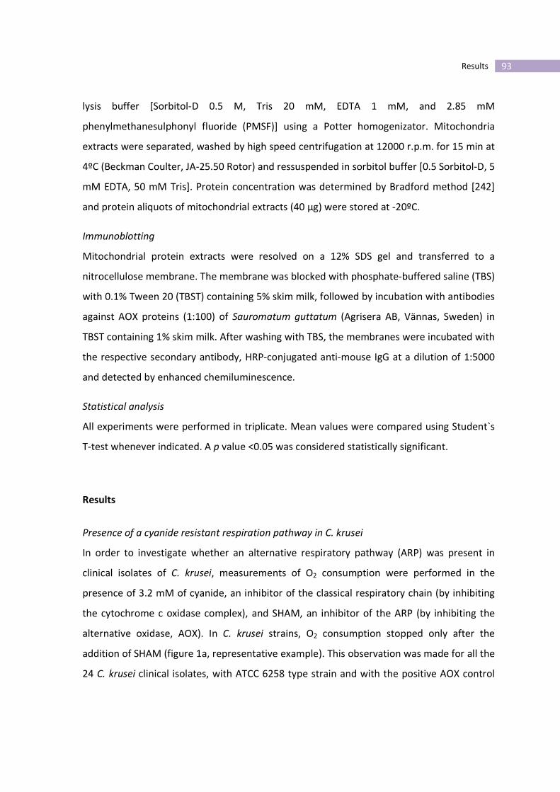
93 Results
lysis buffer [Sorbitol-D 0.5 M, Tris 20 mM, EDTA 1 mM, and 2.85 mM
phenylmethanesulphonyl fluoride (PMSF)] using a Potter homogenizator. Mitochondria
extracts were separated, washed by high speed centrifugation at 12000 r.p.m. for 15 min at
4ºC (Beckman Coulter, JA-25.50 Rotor) and ressuspended in sorbitol buffer [0.5 Sorbitol-D, 5
mM EDTA, 50 mM Tris]. Protein concentration was determined by Bradford method [242]
and protein aliquots of mitochondrial extracts (40 µg) were stored at -20ºC.
Immunoblotting
Mitochondrial protein extracts were resolved on a 12% SDS gel and transferred to a
nitrocellulose membrane. The membrane was blocked with phosphate-buffered saline (TBS)
with 0.1% Tween 20 (TBST) containing 5% skim milk, followed by incubation with antibodies
against AOX proteins (1:100) of Sauromatum guttatum (Agrisera AB, Vännas, Sweden) in
TBST containing 1% skim milk. After washing with TBS, the membranes were incubated with
the respective secondary antibody, HRP-conjugated anti-mouse IgG at a dilution of 1:5000
and detected by enhanced chemiluminescence.
Statistical analysis
All experiments were performed in triplicate. Mean values were compared using Student`s
T-test whenever indicated. A p value <0.05 was considered statistically significant.
Results
Presence of a cyanide resistant respiration pathway in C. krusei
In order to investigate whether an alternative respiratory pathway (ARP) was present in
clinical isolates of C. krusei, measurements of O2 consumption were performed in the
presence of 3.2 mM of cyanide, an inhibitor of the classical respiratory chain (by inhibiting
the cytochrome c oxidase complex), and SHAM, an inhibitor of the ARP (by inhibiting the
alternative oxidase, AOX). In C. krusei strains, O2 consumption stopped only after the
addition of SHAM (figure 1a, representative example). This observation was made for all the
24 C. krusei clinical isolates, with ATCC 6258 type strain and with the positive AOX control

94
strain Debaromyces hansenii 2968 (figure 1b). In Saccharomyces cerevisiae BY4742, the
negative AOX control strain, the oxygen consumption stopped soon after the addition of
cyanide (figure 1c). The results indicate that C. krusei cells have a cyanide resistant
respiration pathway promoted by the presence of an AOX.
AOX expression
Mitochondrial preparations were immunoblotted with a monoclonal antibody raised
against S. guttatum AOX, but also recognize C. albicans AOX [243]. In all C. krusei clinical
isolates mitochondria AOX was clearly detectable as well as in C. albicans SC5314 strain
(figure 2). Mitochondria isolated from the negative control strain, S. cerevisiae BY4742,
showed no reactivity with AOX antiserum (figure 2).
Fluconazole-SHAM combination did not influence fluconazole activity
In order to study the influence of the alternative respiratory pathway upon fluconazole
resistance by C. krusei, we explored the in vitro combination of fluconazole and SHAM using
the chequerboard methodology. SHAM produced no impairment of antifungal activity even
at a concentration of 10mM. The addition of SHAM did not change MIC values for
fluconazole, resulting in an indifferent effect in all C. krusei isolates.

95 Results
Figure 1. Effect of KCN and SHAM upon oxygen consumption by Candida krusei clinical
strain (representative example) (a), Debaryomyces hansenii (alternative oxidase positive
control strain) (b) and Saccharomyces cerevisiae (alternative oxidase negative control strain)
(c). Oxygen consumption was measured with an oxygen electrode at 28 ºC. Where indicated
(arrows), glucose (0.051 mM), KCN (3.2 mM) or SHAM (3.2 mM) were added.
96
Figure 2. Representative example of the presence of an AOX in Candida krusei. Immunoblot
analysis of AOX levels in 40 µg of mitochondrial extracts of Saccharomyces cerevisiae (S.c.),
Candida albicans SC5314 (C.a.) and Candida krusei (C.k.).
The presence of an alternative respiratory pathway in C. krusei relates to reduced ROS
accumulation
To assess the influence of the alternative respiratory pathway upon oxidative stress
response we measured intracellular ROS accumulation with and without SHAM, using
DHR123. This fluorochrome enters the yeast cell as a freely permeable dye, which is
converted to rhodamine 123 and subsequently localized in the mitochondria. The
conversion from the non-fluorescent to the fluorescent molecule is entirely dependent
upon the presence of oxidation products. After treatment with the ROS-inducing agents,
H2O2, plumbagin, menadione and with azoles, the percentage of stained cells (cells with
ROS accumulation) was calculated and compared with values displayed by non treated cells,
in the presence and absence of SHAM (3.2 mM). No significant differences regarding ROS
accumulation were observed with the AOX negative strain, S. cerevisiae BY4742, in the
presence or in the absence of SHAM (figure 3). In contrast, after treatment with ROS
inducing agents, C. krusei cells treated with SHAM produced significantly (p< 0.001) more
endogenous ROS than the cells with the unblocked alternative respiratory pathway (without
SHAM) (figure 3). Significant differences in ROS production (p= 0.008) were also accomplish
with fluconazole (figure 3). All C. krusei tested strains displayed similar results.
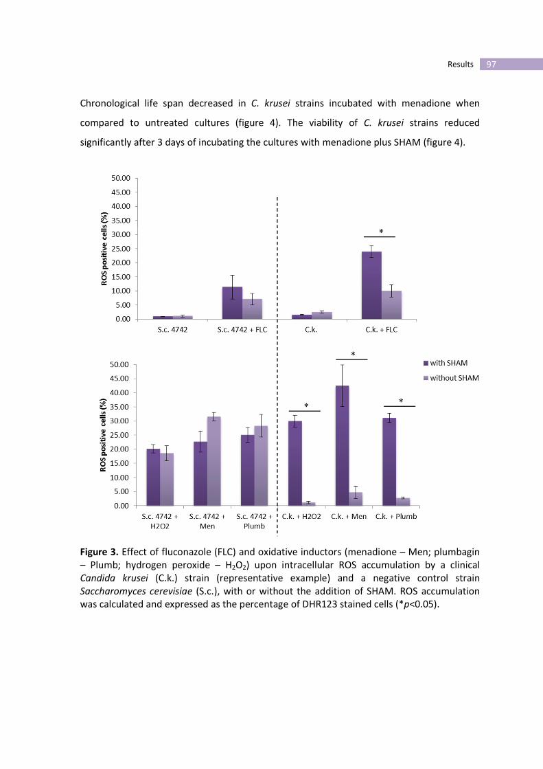
97 Results
Chronological life span decreased in C. krusei strains incubated with menadione when
compared to untreated cultures (figure 4). The viability of C. krusei strains reduced
significantly after 3 days of incubating the cultures with menadione plus SHAM (figure 4).
Figure 3. Effect of fluconazole (FLC) and oxidative inductors (menadione – Men; plumbagin
– Plumb; hydrogen peroxide – H2O2) upon intracellular ROS accumulation by a clinical
Candida krusei (C.k.) strain (representative example) and a negative control strain
Saccharomyces cerevisiae (S.c.), with or without the addition of SHAM. ROS accumulation
was calculated and expressed as the percentage of DHR123 stained cells (*p<0.05).
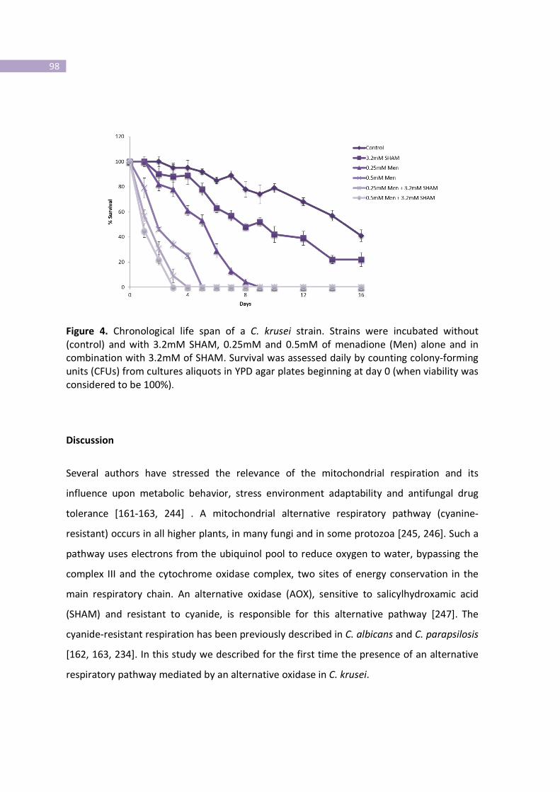
98
Figure 4. Chronological life span of a C. krusei strain. Strains were incubated without
(control) and with 3.2mM SHAM, 0.25mM and 0.5mM of menadione (Men) alone and in
combination with 3.2mM of SHAM. Survival was assessed daily by counting colony-forming
units (CFUs) from cultures aliquots in YPD agar plates beginning at day 0 (when viability was
considered to be 100%).
Discussion
Several authors have stressed the relevance of the mitochondrial respiration and its
influence upon metabolic behavior, stress environment adaptability and antifungal drug
tolerance [161-163, 244] . A mitochondrial alternative respiratory pathway (cyanine-
resistant) occurs in all higher plants, in many fungi and in some protozoa [245, 246]. Such a
pathway uses electrons from the ubiquinol pool to reduce oxygen to water, bypassing the
complex III and the cytochrome oxidase complex, two sites of energy conservation in the
main respiratory chain. An alternative oxidase (AOX), sensitive to salicylhydroxamic acid
(SHAM) and resistant to cyanide, is responsible for this alternative pathway [247]. The
cyanide-resistant respiration has been previously described in C. albicans and C. parapsilosis
[162, 163, 234]. In this study we described for the first time the presence of an alternative
respiratory pathway mediated by an alternative oxidase in C. krusei.
99 Results
The elucidation of the oxidative stress responses in yeast has considerable clinical interest,
as it is involved in invasion and colonization of host tissues by yeast pathogens as well as
during the defensive mechanisms triggered by phagocytes. All aerobic organisms inevitably
generate a range of ROS, including superoxide anion, hydrogen peroxide and hydroxyl
radical during oxygen metabolism. If not quickly and effectively eliminated from the cells,
ROS will trigger a large number of oxidative reactions in cellular systems that possibly lead
to cell death [248]. In the course of an in vivo infection, the formation of ROS and other
oxidants radicals by phagocytes play a crucial role in the intracellular destruction of the
pathogen [249]. ROS attack almost all essential cell components, including DNA, proteins
and lipids [250]. In a recent study, C. krusei appears to be resistant to ROS and to possess a
potent antioxidant system enabling deep systemic infections [251]. We decided to evaluate
the difference regarding oxidative stress response before and after the blockade of the
alternative respiratory pathway. It has been previously described that in C. albicans,
fluconazole is able to induce the production and accumulation of ROS [252]. In our study,
we showed that fluconazole induced a low percentage of ROS formation by C. krusei cells.
These results may suggest that the fungistatic mechanism of this azole is not based upon
ROS formation. However, when the AOX was inhibited by SHAM, an increase in the
intracellular ROS levels was evident. Attending to these facts, we can conclude that AOX
activity allows the yeast cells to reduce ROS accumulation when challenged by antifungals
like fluconazole, leading to drug tolerance, like in C. albicans [163]. According to several
authors, the AOX has a metabolic and antioxidant role and its presence may be considered a
potential virulence attribute of pathogenic fungi [163, 253, 254]. The importance of AOX
activity upon resistance to oxidative stress was evident when the oxidative stress inductors,
H2O2, menadione and plumbagin were assayed. After treatment with such compounds, ROS
accumulation was low. However, the scenario changed significantly when the AOX activity
was blocked by SHAM. To confirm our hypothesis that the presence of an alternative
respiratory pathway could protect C. krusei from oxidative stress, we assessed ROS
accumulation in the presence of fluconazole and the other oxidative stress inductors by the
negative control strain, S. cerevisiae (AOX-). The results obtained regarding ROS
100
accumulation, in the presence or absence of SHAM, were not significantly different. When
testing C. albicans AOX mutant strains, Yan and co-workers also obtained no significant
differences in the amount of ROS generation [163]. Regarding the chronological life span
assays we could conclude that the decreased viability of C. krusei strains in the presence of
menadione and with the AOX blocked is associated with an increased level of cell ROS.
Our results showed clearly that the inhibition of the expression of the AOX was associated
with intracellular ROS accumulation, unveiling its effect of the alternative respiratory
pathway on oxidative damage. Although fluconazole resistance was unrelated to the
presence of the ARP, we can consider that it confers antifungal tolerance, which may give
yeast cells enough time to develop long-term genetically stable resistance mechanisms [22].
The alternative respiratory pathway is a potential target that should be taken into account
considering the development of new therapeutic strategies in the case of C. krusei
infections. Attending to its selective effect, SHAM should be used in combination with
azoles, in order to reduce resistance due to oxidative stress and consequently the virulence
of C. krusei.

101 Results
Part V
Propofol lipidic infusion promotes resistance to antifungals by reducing drug
input into the fungal cell
Background
Discrepancies between in vivo and in vitro susceptibility to antifungals discourage
microbiologists and clinicians regarding the routine use of susceptibility testing methods.
Although in vitro resistance usually correlates with clinical resistance, high susceptibility in
vitro is not always related to clinical success. Ultimately, the mortality rates are
unacceptably high in patients treated with antifungals that showed high in vitro efficacy
[255].
Propofol is an intravenous hypnotic agent very popular for induction and maintenance of
general and intravenous anaesthesia. It is commonly administered in Intensive Care Units to
critically ill patients, often under mechanical ventilation, which represent a high risk group
for health care related infections. The use of propofol has been previously associated to an
increased risk for infection, although some controversy still remains [256, 257]. It was
proposed a low risk of contamination whenever providing standard hygienic precautions
[256, 258].
Nevertheless, other observations described the lipid emulsion of propofol as a good culture
medium to support the growth of Candida albicans and Escherichia coli [259, 260].
Additionally, other reports associated post-surgical infections with the extrinsically
contamination of propofol infusion [258, 261]. Propofol has also been shown to inhibit a
variety of functions of neutrophils in vitro, although such effect was not so evident in vivo
[262].

102
We have studied the effect of the infusion of propofol and its lipidic vehicle upon antifungal
susceptibility of Candida. A promotion of resistance due to a decreased input of the
antifungal drugs was found.
Materials and Methods
Strains
Twenty clinical strains of Candida spp. (5 C. albicans, 5 C. tropicalis, 5 C. glabrata and 5 C.
parapsilosis) were studied. C. albicans 95-190, resistant to azoles by overexpression of
efflux pumps genes (CDR1 and CDR2), was used during cytometric approach (strain kindly
gift by Prof. Theodore White). Until testing, yeasts and moulds were kept frozen in Brain-
Heart broth (Difco Laboratories, Detroit, MI, USA) with 5% glycerol. For each experiment,
the strains were subcultured twice on Sabouraud agar (Difco) at 35ºC, 48 hours for Candida.
Drugs and Chemicals
Propofol infusion Fresenius® (Kabi, France) at stock concentration of 1% was used. Propofol
vehicle (soya bean oil, egg lecithin, glycerol, sodium hydroxide and sterile water) was also
assayed. Fluconazole and voriconazole were obtained from Pfizer (Groton, CT, USA),
amphotericin B from Bristol-Myers Squibb (New York, USA), itraconazole from Janssen-Cilag
(Beerse, Belgium) and posaconazole from Shering-Plough (Kenilworth, NJ, USA). Antifungals
drugs were maintained in stock solution at -70ºC until use. [3H]-labelled itraconazole was
supplied by Janssen-Cilag. Sodium azide was purchased from Sigma (Sigma-Aldrich,
Germany).
Growth assays
After cultivation of Candida strains in Sabouraud agar medium (Difco, Detroit, MI, USA), a
5x106.ml
-1 blastoconidia suspension of Candida was prepared in phosphate buffer saline
(PBS) (Sigma) and 100µl were added in two parallel serial dilutions of propofol infusion
(stock solution at 1%) and its vehicle (both at 0, 1.25, 2.5 and 5 mg.ml-1
final concentrations)
in RPMI 1640 culture medium (Sigma), PBS and plain propofol infusion in a final volume of
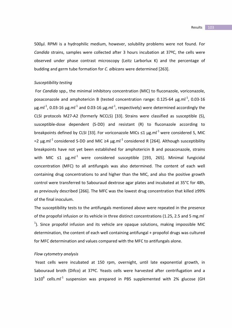
103 Results
500µl. RPMI is a hydrophilic medium, however, solubility problems were not found. For
Candida strains, samples were collected after 3 hours incubation at 37ºC, the cells were
observed under phase contrast microscopy (Leitz Larborlux K) and the percentage of
budding and germ tube formation for C. albicans were determined [263].
Susceptibility testing
For Candida spp., the minimal inhibitory concentration (MIC) to fluconazole, voriconazole,
posaconazole and amphotericin B (tested concentration range: 0.125-64 µg.ml-1
, 0.03-16
µg.ml-1
, 0.03-16 µg.ml-1
and 0.03-16 µg.ml-1
, respectively) were determined accordingly the
CLSI protocols M27-A2 (formerly NCCLS) [33]. Strains were classified as susceptible (S),
susceptible-dose dependent (S-DD) and resistant (R) to fluconazole according to
breakpoints defined by CLSI [33]. For voriconazole MICs ≤1 µg.ml
-1 were considered S, MIC
=2 µg.ml-1
considered S-DD and MIC ≥4 µg.ml-1
considered R [264]. Although susceptibility
breakpoints have not yet been established for amphotericin B and posaconazole, strains
with MIC ≤1 µg.ml-1
were considered susceptible [193, 265].
Minimal fungicidal
concentration (MFC) to all antifungals was also determined. The content of each well
containing drug concentrations to and higher than the MIC, and also the positive growth
control were transferred to Sabouraud dextrose agar plates and incubated at 35°C for 48h,
as previously described [266]. The MFC was the lowest drug concentration that killed ≥99%
of the final inoculum.
The susceptibility tests to the antifungals mentioned above were repeated in the presence
of the propofol infusion or its vehicle in three distinct concentrations (1.25, 2.5 and 5 mg.ml-
1). Since propofol infusion and its vehicle are opaque solutions, making impossible MIC
determination, the content of each well containing antifungal + propofol drugs was cultured
for MFC determination and values compared with the MFC to antifungals alone.
Flow cytometry analysis
Yeast cells were incubated at 150 rpm, overnight, until late exponential growth, in
Sabouraud broth (Difco) at 37ºC. Yeasts cells were harvested after centrifugation and a
1x10⁶ cells.ml
-1 suspension was prepared in PBS supplemented with 2% glucose (GH

104
solution) and later divided into aliquots of 1 ml. The cells were then incubated with
different concentrations of propofol infusion (0, 1.25, 2.5 and 5 mg.ml-1
) at 37ºC for 90
minutes and afterwards washed thrice, ressuspended in sterile water supplemented with
2% glucose and stained with 0.5 µM FUN1 (Molecular Probes, Europe BV, Leiden, Holland)
for 30 minutes at 37ºC. A Beckman Coulter XL-MCL flow cytometer (Beckman-Coulter Corp.,
Hialeah, FL, USA) equipped with a 15 nm argon laser was used. From each suspension
30000-50000 cells were analysed. The intensity of fluorescence emitted by cells treated
with propofol infusion was determined at FL2 (575 nm) and compared with non-treated
cells (control). In parallel experiments, yeast cells were treated with 0.1 mM sodium azide
during 30 minutes, prior to incubation with propofol, as previously described [145],, in
order to block efflux pumps; thereafter, the same flow cytometry analytical protocol was
used.
Intracellular accumulation of [3H]-labelled itraconazole
Candida cells were initially incubated under similar conditions as previously described for
flow cytometry assays. The cells were harvested by centrifugation at 5000 rpm for 10
minutes at 4ºC, washed thrice, ressuspended in PBS at a final concentration of 2.5x10⁸
cells.ml-1
and incubated with 0, 1.25 and 5 mg.ml-1
of propofol infusion at 37ºC, with
continuous shaking at 300 rpm for 30 minutes [145]. Parallel experiments were prepared,
but also involving a pre-incubation of the yeasts cells with sodium azide at 0.1 mM. The cells
were washed thrice and [3H]-labelled itraconazole was added to yeast suspensions at a final
concentration of 3 µM, as previously described [145]; the cells were incubated in glass vials
at 37ºC, with continuous shaking (300 rpm), during 1 hour and then harvested by
centrifugation at 5000 rpm for 10 min at 4ºC, washed thrice with 3ml of ice-cold PBS
containing 10 µM unlabelled itraconazole. The pellets were later ressuspended in 500 µl of
PBS and the radioactivity was determined, following the addition of a scintillation cocktail
(Optiphase “Hiphase3”, Perkin-Elmer), in a liquid scintillation counter (LKB Wallac, 1209
RackBeta).
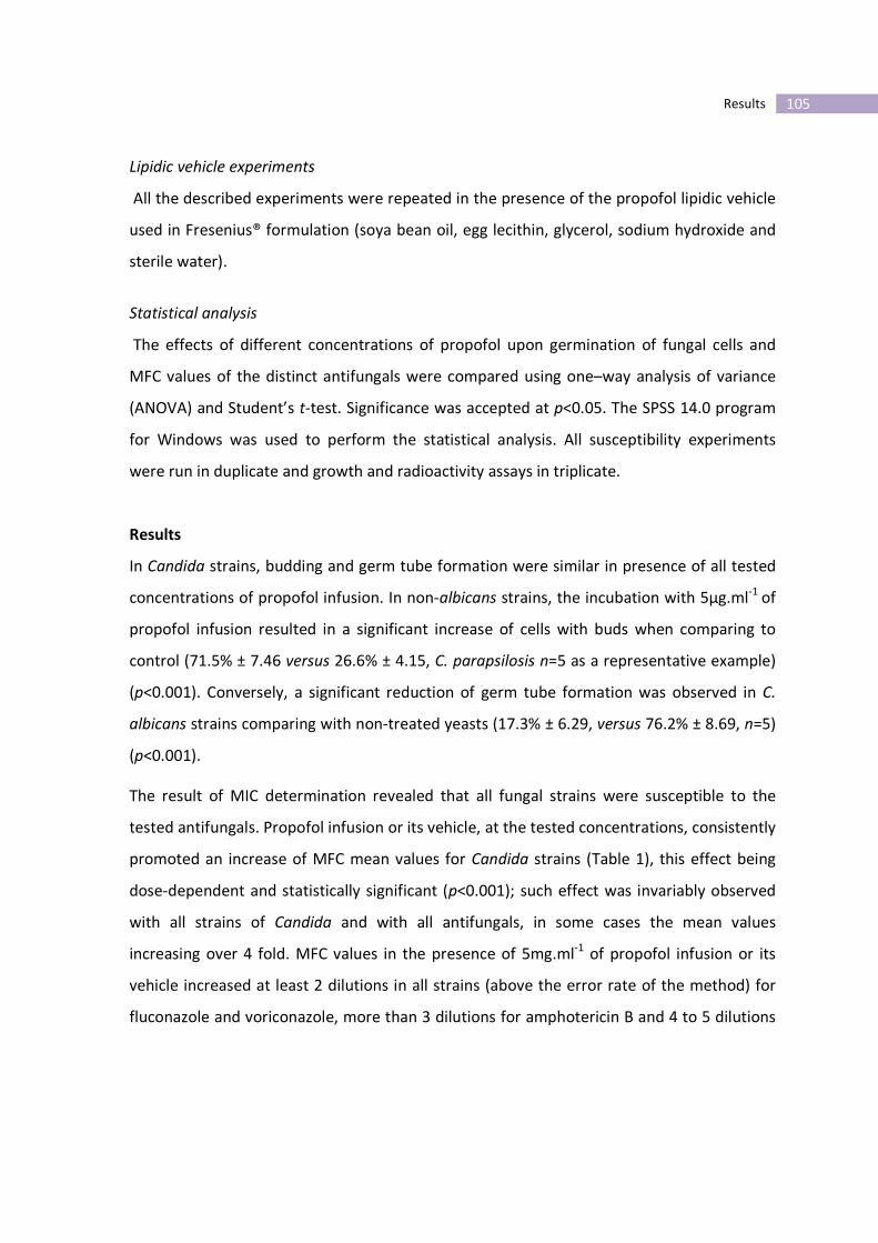
105 Results
Lipidic vehicle experiments
All the described experiments were repeated in the presence of the propofol lipidic vehicle
used in Fresenius® formulation (soya bean oil, egg lecithin, glycerol, sodium hydroxide and
sterile water).
Statistical analysis
The effects of different concentrations of propofol upon germination of fungal cells and
MFC values of the distinct antifungals were compared using one–way analysis of variance
(ANOVA) and Student’s t-test. Significance was accepted at p<0.05. The SPSS 14.0 program
for Windows was used to perform the statistical analysis. All susceptibility experiments
were run in duplicate and growth and radioactivity assays in triplicate.
Results
In Candida strains, budding and germ tube formation were similar in presence of all tested
concentrations of propofol infusion. In non-albicans strains, the incubation with 5µg.ml-1
of
propofol infusion resulted in a significant increase of cells with buds when comparing to
control (71.5% ± 7.46 versus 26.6% ± 4.15, C. parapsilosis n=5 as a representative example)
(p<0.001). Conversely, a significant reduction of germ tube formation was observed in C.
albicans strains comparing with non-treated yeasts (17.3% ± 6.29, versus 76.2% ± 8.69, n=5)
(p<0.001).
The result of MIC determination revealed that all fungal strains were susceptible to the
tested antifungals. Propofol infusion or its vehicle, at the tested concentrations, consistently
promoted an increase of MFC mean values for Candida strains (Table 1), this effect being
dose-dependent and statistically significant (p<0.001); such effect was invariably observed
with all strains of Candida and with all antifungals, in some cases the mean values
increasing over 4 fold. MFC values in the presence of 5mg.ml-1
of propofol infusion or its
vehicle increased at least 2 dilutions in all strains (above the error rate of the method) for
fluconazole and voriconazole, more than 3 dilutions for amphotericin B and 4 to 5 dilutions

106
for posaconazole and itraconazole. There was no large variability between the tested
strains.
Flow cytometry analysis of Candida blastoconidia resulted in 98% of cells stained with
FUN1, even after the incubation of sodium azide (figure 1b). However, after treatment with
propofol infusion and stained with FUN1, a non-stained sub-population of cells, similar to
autofluorescence, of around 15% was revealed soon after 90 minutes (figure 2c). This fact
was unrelated with the incubation with sodium azide at a concentration able to block the
efflux pumps (figure 1c). The azole resistant strain of C. albicans 95-190 (with
overexpression of efflux pumps genes), after treatment with propofol infusion and stained
with FUN1, revealed a non-stained sub-population, similar to other susceptible strains, even
after the blockade of the efflux pumps with sodium azide. This effect was similar in
presence of all the tested concentrations of propofol.
After 1 hour, the accumulation of [3H]-labelled itraconazole was detected in blastoconidia
cells (control cells) (figure 2). However, a decrease of 39% in intracellular [3H]-labelled
itraconazole was seen when yeast cells were incubated with propofol lipid infusion (figure
2). This effect was similar in presence of the different tested concentrations of propofol
lipidic infusion and did not increase following the pre-incubation with sodium azide (figure
2).
The results obtained in the different assays were similar when performed with propofol
lipidic vehicle.

107 Results
Table 1. Minimal fungicidal concentration (MFC) values of Candida strains to AMB
(amphotericin B), FLC (fluconazole), ITC (itraconazole), VRC (voriconazole) and PSC
(posaconazole), determined by CLSI protocols, in the absence and presence of propofol
infusion.
Strains (n)
MFC range µg.ml-1
Antifungals Propofol
(mg.ml-1
)
C. albicans (5) C. tropicalis (5) C. parapsilosis (5) C. glabrata (5)
AMB 0 0.25 0.25-1 0.06-0.125 0.25-0.5
1.25 0.5-1 1 2 2
2.5 0.5-2 1-2 2 2
5 2-4 2 2-8 4-8
FLC 0 8-16 4-8 8-16 16
1.25 16->64 >64 >64 >64
2.5 16->64 >64 >64 >64
5 16->64 >64 >64 >64
ITC 0 nd nd nd nd
1.25 nd nd nd nd
2.5 nd nd nd nd
5 nd nd nd nd
VRC 0 0.06-0.25 0.25-0.5 0.5-1 4-8
1.25 0.25->2 >2 >2 >8
2.5 0.5->2 >2 >2 >8
5 1->2 >2 >2 >8
PSC 0 0.06-0.125 0.03-0.125 0.06-0.125 1-2
1.25 1-2 >2 2 2
2.5 >2 >2 2 >2
5 >2 >2 >2 >2
nd not done
* The mean value for each strain was considered
108
0
1
2
3
4
5
1 2 3 control 5mg.ml -1 propofol azide+5mg.ml -1
Acc
um
ula
tio
n o
f [3
H]i
tra
con
azo
le
(nm
ol.
10
-8ce
lls)
Figure 1. Flow cytometric histograms representing the emitted fluorescence after 90
minutes by: a. non-stained yeast cells (autofluorescence); b. cells treated with sodium
azide and stained with FUN1; c. cells treated with sodium azide and 5 mg.ml-1
of propofol
infusion and stained with FUN1 (strain of Candida albicans shown as a representative
example).
Figure 2. Effect of propofol up on [3H]-labelled itraconazole accumulation in antifungal
susceptible strain C. albicans ATCC 90028. The accumulation of itraconazole was
measured in the absence and presence of 5 mg.ml-1
of propofol and after a prior
incubation with 0.1mM sodium azide. Dispersion bars relates to standard deviation.
a b c

109 Results
Discussion
Propofol administered in a lipid-based emulsion to patients has been shown to represent an
excellent vehicle for supporting the growth of various microorganisms [259, 260]. In the
present study we clearly showed that budding of Candida spp. was promoted when
incubated in the presence of propofol infusion. It is important to emphasize that the
propofol concentrations used in all assays are within the plasma levels often achieved in
clinical practice [267, 268].
Due to the opacity of propofol infusion or its lipidic vehicle it was not possible to determine
MIC values. As the conventional susceptibility phenotypes refer to MIC values, we could not
establish the corresponding phenotypes after incubation with propofol. In the case of
amphotericin B (a fungicidal to Candida) MFC, determined after culture in agar solid
medium, usually corresponds to MIC values; for the other antifungals, MFC values are
usually located within one or three dilutions above the MIC. Some C. albicans and C.
tropicalis showed trailing phenotype (about 2 dilutions) for azoles. This common effect may
influence the evaluation of MFC values. However, trailing is only azole dependent and was
not affected by the presence of propofol infusion. Nevertheless, we could observe a
significant promotion of the MFC value in presence of propofol infusion, in some cases up to
4 dilutions, suggesting that the strains became quite tolerant to the effect of all antifungals.
FUN1 is a fluorescent probe that is converted by metabolically active yeasts in
intracytoplasmic vacuolar structures [269]. We have used this probe to study susceptibility
of yeasts after incubation during one to two-hours with the antifungals; a higher intensity of
fluorescent in susceptible strains, decreasing in resistant strains was described [142, 144].
The decrease of the intensity of fluorescence could be explained by the presence of energy-
dependent efflux pumps, which were reverted with sodium azide or several modulators
[144, 269]. In the present study, the viable cells showed a decrease of the intensity of
fluorescence in the presence of propofol which was not however reverted with sodium
azide, even considering the resistant tested strain. Furthermore, cells were washed several
times before the addition of the fluorescent probe in order to avoid its binding to propofol
infusion. These results support that propofol infusion affected FUN1 access and/or

110
permeabilization, reducing the input of the fluorescent probe into the cell. We had also
previously shown that [3H]-labelled itraconazole accumulation could be a useful tool to
confirm the existence of efflux pumps in resistant Candida strains to azoles [145]. The
blockade of efflux pumps with sodium azide resulted in the reduction of input of [3H]-
labelled itraconazole in Candida cells, after incubation with propofol infusion. In fact,
propofol infusion was responsible for the difficult access of itraconazole to the fungal cell.
Attending to the facts that i) the increase of MFC values was similar with all antifungal
drugs, including the recently available posaconazole; ii) FUN1 could not stain a considerable
percentage of the yeast cells in presence of propofol infusion and iii) a lesser amount of
[3H]-labelled itraconazole was present within such cells, even after the addition of sodium
azide, we are forced to conclude that the promotion of antifungal resistance in presence of
propofol lipidic infusion results from a reduced access and/or permeabilization of antifungal
agents into the fungal cells. The same results were obtained when only the propofol vehicle
was used, thus we had concluded that the major responsible for such effect was the lipidic
nature of propofol infusion.
Several studies reported that the lipidic membrane of fungal cells plays an important role in
susceptibility to azoles [270, 271]. Fluctuations in the lipidic environment affects, not only
drug diffusion but also efflux pumps, coded specially by Cdr1 and Pdr5p, leading to
multidrug azole resistance [272, 273]. In our study, as the efflux pumps were blocked with
sodium azide, the increased MFC values probably resulted from poor diffusion of the
antifungal drugs caused by the lipidic vehicle layer or drops deposited around the cells. The
hypothesis that propofol could be binding the antifungal agent in culture medium reducing
the free number of molecules available for penetrating the cells was also raised. MFC
results were more evident when propofol was associated with itraconazole or posaconazole
(increase of 4 to 5 dilutions), both lipophilic drugs. However, we must consider that
different concentrations of propofol resulted in similar effects. The hypothesis that the
lipidic vehicle of propofol might be playing a role in sterol homeostasis and changing the
azole target, should be considered attending to other research [274]. Lipid uptake by the
fungal cell was described in the presence of the azoles following longer incubation time
111 Results
(overnight) [274]. Measuring sterols in azole-treated fungi in the presence or not of the
lipidic vehicle of propofol could be interesting however, the incubation time with propofol
on cytometric and radioactivity studies was very short to allow the incorporation of the lipid
in membrane.
The assays described in this manuscript provided an opportunity to describe the effect of
propofol infusion in antifungal drug resistance. We concluded that propofol infusion, due to
its lipidic vehicle reduced the access and/or permeabilization to Candida cells to main
antifungals administered to patients.
The described effect should raise the alert to a promoted risk of fungal infections in patients
receiving propofol infusions, resulting from the fact that fungal strains become increasingly
resistant to antifungals.


113 Results
Part VI
Adrenaline stimulates efflux pumps activity, growth and mitochondrial
respiration in Candida albicans
Background
Candida spp. are the most common agents of bloodstream fungal infections [6]. High
morbidity and mortality rates are often observed, mainly among intensive care units (ICU)
patients [7, 275].
Despite the in vitro susceptibility to antifungals, particularly to azoles, a poor clinical
outcome is often reported [6, 7, 48, 276], thus suggesting the development of in vivo
resistance. Candida may develop resistance mechanisms during medical therapy that
ultimately may lead to clinical failure [51]. However, discrepancies between in vitro
susceptibility and the patient outcome show that drug resistance is not the single factor for
the clinical results. The progression of Candida infections is often observed despite
aggressive antifungal treatment, especially among ICU patients [6, 7, 48]. Such patients are
invariably submitted to multiple lifesaving medications. In a recent study of patients
admitted in ICU with bloodstream Candida infections we have shown that those receiving
vasoactive amines showed a significantly higher risk of fungaemia-related poor outcome [7].
In humans, catecholamines target adrenoceptors which are a class of G protein-coupled
receptors (GPCRs). GPCRs are transmembrane spanning proteins that transduce an
extracellular signal (catecholamine binding) into an intracellular event (G-protein
activation). Although firstly detected in mammals it is now clear that these receptors
followed closely the evolutionary spectrum from archaebacteria to humans. GPCRs are
abundantly expressed in yeasts, showing an identical GTP binding site to human GPCRs
[277-281].
Considering these homologies between yeasts and human cells, we hypothesized that
adrenaline and other vasoactive amine administered to ICU patients might also act on
114
Candida. We aimed to clarify whether adrenaline, acting on C. albicans might promote
antifungal tolerance, resistance or both of clinical isolates to azoles, amphotericin B and
caspofngin. The results obtained underscore the role of non-antifungal drugs like
catecholamines, often prescribed in ICU patients, as promoters of antifungal tolerance or
resistance.
Material and Methods
Strains
The clinical isolates of Candida albicans (n=6) included in this study had been previously
collected from blood cultures of patients with fungaemia admitted at different Intensive
Care Units of Hospital São João, Porto, Portugal. All strains were susceptible to fluconazole,
voriconazole, posaconazole, amphotericin B and caspofungin, according to the Clinical
Laboratory Standards Institute (CLSI) M27-A3 protocol [282, 283]. C. albicans DSY 448
(cdr1Δ/Δ) [284], DSY 653 (cdr2Δ/Δ) [285], and DSY 654 (cdr1Δ/Δ cdr2Δ/Δ) [285] with
selective deletion of efflux genes were used (kindly gifted by Prof. D Sanglard). C. albicans
strains gpr1 Δ / Δ (LDR8-5 strain) and gpa2 Δ / Δ (NM8 strain) [277] (kindly provided by Prof.
Patrick Van Dijck), Δ ras1-2/ Δ ras1-3 (kindly provided by Prof. Gerald Fink) and ste2 Δ / Δ,
ste3 Δ / Δ and ste4 Δ/Δ [145, 279] (kindly provided by Prof. David Soll) were used and the
respective wild-type strains SC5314, P37005, P57072. Until testing, all strains were kept
frozen in yeast extract peptone dextrose agar (YPD) (Difco Laboratories, Detroit, MI, USA)
with 40% glycerol. For each experiment, the strains were subcultured twice on YPD medium
at 35°C for 48 hours and afterwards cultured in YPD broth at 35°C, under constant agitation.
Drugs and Chemicals
Fluconazole and voriconazole were obtained from Pfizer (Groton, CT, USA), posaconazole
from Shering-Plough (Kenilworth, NJ, USA), amphotericin B from Bristol-Myers Squibb (New
York, USA) and caspofungin from Merck (Rahway, NJ, USA). Antifungal drugs were prepared
following CLSI recommendations [282, 283] and maintained in stock solutions at -70°C until
use.

115 Results
The adrenoceptor agonists adrenaline (mixed α1, α2, β1 and β2-adrenoceptor agonist) (Braun
Medical, Barcarena, Portugal), noradrenaline (mixed α1, α2 and β1-agonist) (Sigma-Aldrich,
Germany), isoprenaline (selective β1 and β2-agonist) (Sigma), medetomidine (selective α2-
agonist) (Santa Cruz Biotechnology, Inc, Heidelberg, Germany) and phenylephrine (selective
α1-agonist) (Sigma) were used. Drugs were dissolved in distilled water and kept at -20₀C
until use.
Sodium azide was purchased from Sigma and dissolved in distilled water. The fluorescent
probes FUN-1 was acquired from Molecular Probes (Molecular Probes, Europe BV, Leiden,
Holand) and Rhodamine 6G (Rh-6G) from Sigma.
Antifungal susceptibility testing of the clinical isolates
Antifungal susceptibility testing of all clinical isolates was performed according to CLSI M27
A3 protocol [282, 283] in RPMI 1640 (Sigma). The minimal inhibitory concentration (MIC) of
fluconazole, voriconazole, posaconazole, caspofungin and amphotericin B was determined
[282, 283]. Strains were classified as susceptible (S), susceptible-dose dependent (S-DD) and
resistant (R) to azoles according to the breakpoints defined by CLSI [282, 283]. Although
definitive breakpoints have not yet been established for amphotericin B and posaconazole,
strains with MIC ≤1 µg/ml were considered susceptible [286]. For caspofungin a MIC ≤2
μg/ml was considered S and >2 μg/ml
non susceptible (NS) [282, 283]. MICs of adrenaline
and noradrenaline were also determined using the above mentioned CLSI protocol [282,
283] (tested concentration ranging from 5 to 545 µM).
The susceptibility tests with all the antifungals previously used were repeated in the
presence of adrenaline (5, 27, 54, 108, 136, 273, 545 µM) and noradrenaline (11, 27, 136,
545 µM). Synergistic studies were performed according to the checkerboard procedure, as
described in the Clinical Microbiology Procedures Handbook [239]. Fractional inhibitory
concentration of drug A (FICA) was calculated as the ratio of the MIC of drug A in
combination over the MIC of drug A alone. FICB was the ratio of the MIC of drug B in
combination over the MIC of drug B alone. The fractional inhibitory index (FIX) was
calculated as follows: FIX= FICA + FICB. The interpretation of FIX was as recommended: ≤0.5
synergistic effect; >0.5 to <4.0 indifferent effect; ≥4.0 antagonistic effect [239].

116
Efflux quantification by flow cytometry
a) Yeast cell culture
The yeast cells were incubated in YPD broth at 35⁰C until mid-exponential growth phase
(optical density at 600nm [OD600]= 0.4) and harvested by centrifugation. A suspension with
106cells/ml
was prepared in sterile distilled water supplemented with 2% glucose (Sigma).
b) FUN-1 and Rh-6G staining
Cells suspensions were incubated with 5, 27, 54, 108 µM of adrenaline for 90 minutes at
35ºC and stained with 0.5 µM of FUN-1 for 30 minutes, according to Pina-Vaz et al [145].
For Rh-6G staining, yeast suspensions were incubated with 5 µM Rh-6G for 30 minutes,
washed twice and ressuspended in cold PBS (Sigma) supplemented with 10 mM of sodium
azide. Yeast suspensions were kept in ice until cytometric analysis in a FACSCalibur flow
cytometer (BD, Bioscience, Sydney). From each suspension 30,000 cells were analyzed and
the intensity of fluorescence emitted by cells exposed to the different treatments was
determined without staining (autofluorescence – af) and after FUN-1 and Rh-6G staining at
FL2 (575 nm) and FL1 (525 nm), respectively. Results were compared with non-treated cells
(control).
Studies with adrenoceptor agonists
Parameters like incubation time and agonist concentration were optimized using
adrenaline. Adrenaline was tested at the concentrations of 5, 27, 54, 108, 136, 273 and 545
µM, for 30, 60 and 90 minutes.
To further characterize the adrenoceptor that mediates the effect of amines upon clinical
isolates, yeast suspensions were also incubated for 90 minutes, at 37 ⁰C with noradrenaline
(5, 27, 54, 108, 136, 273, 545 µM), isoprenaline (5, 27, 54, 108, 136, 273, 545 µM),
phenylephrine (5, 27, 54, 108, 136, 273, 545 µM, 1, 3 mM) and medetomidine (0.01, 0.1, 1,
10 µM) [287].
Effect of adrenaline upon Rh-6G staining of Candida CDR1 and/or CDR2 mutant strains
Mutant strains DSY 448, DSY 653, DSY 654 and the wild-type strain were analyzed by flow
cytometry following treatment increasing concentrations of adrenaline for 90 minutes and

117 Results
staining with Rh-6G (according to the protocol described above). The amount of
intracellular accumulation of Rh-6G was determined relatively to untreated cells (control)
and compared between the different mutant strains.
Effect of adrenaline upon membrane receptor deleted strains
C. albicans selectively deleted in one of membrane receptors or proteins, gpr1Δ/ Δ, gpa2Δ/
Δ [277], Δ ras1-2/ Δ ras1-3 [276, 278], ste2Δ/ Δ, ste3Δ/ Δ and ste4Δ/ Δ [145, 279], and its
respective wild-type strains, were incubated with 0, 5, 27, 54 and 108 µM of adrenaline for
90 min at 35ºC, stained with Rh-6G and analyzed according to the cytometric protocol
described above.
Effect of adrenaline upon Candida CDR1 and CDR2 gene expression
C. albicans cell extracts for immunoblotting, previously exposed to 54 µM of adrenaline for
30, 60 and 90 minutes, were prepared by an alkaline extraction procedure as described
previously [103]. Detection of Cdr1p and Cdr2p was performed as described previously
[103]. Signals were revealed by exposure to Kodak BioMax MR films (GE Healthcare).
Effect of adrenaline upon C. albicans SC5314 strain growth
Following overnight culture in YPD broth medium a yeast suspension was diluted to an
O.D.600 0.07 in 50 ml of YEPD broth and incubated at 35ºC under agitation (150 rpm). Cells
suspensions were exposed to the following conditions: no treatment (control), treatment
with FLC 1 µg/ml (sub-MIC concentration), treatment with 54 and 108 µM of adrenaline,
treatment with FLC 1 µg/ml plus 54 of adrenaline and treatment with FLC 1 µg/ml plus 108
µM of adrenaline. O.D.600 was registered repeatedly during 18 hours.
Effect of adrenaline upon oxygen consumption by C. albicans
Late log phase cultures of SC5314 strain were centrifuged (18000g) for 4 minutes at 4 ºC
and washed twice with cold sterile water. An amount of 1.5 g (wet weight) pellet was
ressuspended in 100 ml of phosphate-buffered 50 mM, pH 6.0 with 0.1% cicloheximide
(Sigma, St. Louis, MO, USA) and 6 ml of such suspension were incubated in a small reactor
at 28ºC and 200 µl of glucose 1.55 M (Difco Laboratories, Detroit, MI, USA) were added. The
O2 consumption was continuously measured during 90 minutes with a Clark type electrode

118
YSI model 5775 (YSI Incorporated, Yellow Springs, Ohio, USA) in the presence of 16 µg/ml
FLC, with 0 and 54 µM of adrenaline, and the oxygen uptake rate was determined.
Data presentation
All susceptibility assays were performed at least thrice. Mean values were compared using
Student`s T-test whenever indicated. A p value <0.05 was considered statistically significant.
The determination of FUN-1 and Rh-6G staining, oxygen consumption and strain growth
were made in triplicate and the respective standard deviations values determined. Potency
values were expressed as pEC50 values, which are the negative logarithms of the molar
concentration of the drug required to elicit half-maximal effect.
Results
Effect of adrenaline on antifungal susceptibility of C. albicans isolates – checkerboard results
Checkerboard assays were performed in order to evaluate the interactions between
adrenaline and antifungal drugs. Adrenaline, as well as noradrenaline, did not show
antifungal activity upon all isolates even at high concentrations (MIC values ≥ 545µM).
However, the median FIX for fluconazole combined with adrenaline or noradrenaline was 5
(range 2-17) (antagonistic effect). Although an antagonistic effect was obtained, the MIC
values did not reach a breakpoint, except for one clinical isolate (that changed to
susceptible –dose dependent, MIC=16 µg/ml). In contrast to fluconazole neither the
antifungal activity of amphotericin B nor caspofungin were changed by adrenaline.
Effect of adrenaline upon FUN-1 and Rh-6G staining
In order to elucidate the antagonistic effect of adrenaline on the antifungal action of
fluconazole, flow cytometric evaluations were performed using FUN-1. FUN-1 is a
fluorescent probe that is converted by metabolically active yeasts in intra-cytoplasm
vacuolar structures [269]. This probe has been previously used to study susceptibility of
yeasts to azoles [147, 213]. In susceptible strains a high intensity of fluorescence is
detected, while in resistant strains fluorescence decreases [147, 213]. This decrease in
fluorescence intensity is due to the activity of energy-dependent efflux pumps which

119 Results
actively export FUN-1 to the extracellular environment [145]. Furthermore, to confirm and
quantify the effect of vasoactive amines upon efflux, another distinct efflux pump substrate
was used (Rh-6G) [103]. Candida cells treated with adrenaline showed a dose-dependent
decrease of FUN-1 fluorescence intensity in comparison with non-treated cells (figure 1),
thus indicating an increase in the efflux activity with a pEC50= 5.81 ± 0.56. Although less
pronounced, a similar effect was obtained with noradrenaline (pEC50= 4.58 ± 0.22) (figure
1). Since isoprenaline at concentration up to 545 µM had no significant effect upon the
efflux pumps activity it was possible to establish a rank order of potency:
adrenaline>noradrenaline>>isoprenaline (figure 1). This rank order of potencies is achieved
whenever an α-adrenoceptor-mediated effect is involved [288, 289]. Accordingly, cells
treated with adrenaline showed a lower intensity of Rh-6G fluorescence than non-treated
cells (figure 2). These results indicate the inhibitory effect of adrenaline on the
accumulation of Rh-6G was concentration-dependent (figure 2). The same effect was
observed with all the tested clinical isolates and with SC5314 strain.
A concentration-dependent decrease of FUN-1 accumulation in all strains was observed
with medetomidine (a selective α2-adrenoceptor-agonist) with a pEC50= 7.83 ± 0.28 while
phenylephrine (a selective α1-adrenoceptor-agonist) in concentration up to 3 mM had no
effect upon FUN-1 staining (figure 3). This result suggests that the effect of adrenaline upon
C. albicans is mediated by a α2-adrenoceptor, as only with medetomidine cells were able to
reduce stain accumulation.

120
Figure 1. Effect of adrenaline (mixed α1, α2, β1 and β2), noradrenaline (mixed α1, α2 and β1)
and isoprenaline (selective β1 and β2) upon FUN-1 staining: a rank order of potencies was
found (adrenaline>noradrenaline>>isoprenaline) showing a probable alpha mediated effect
of adrenaline and noradrenaline on C. albicans clinical isolate (representative example).
Data from three independent experiments is presented as mean ± standard deviation and
shown as the percentage of FUN-1 accumulation relatively to untreated cells.
Figure 2. Effect of adrenaline (adr) upon Rh-6G staining: flow cytometric histogram
representing the emitted fluorescence (Fl1) by a clinical isolate after 90 minutes of
incubation with 0, 27 and 54 µM adr and stained with Rh-6G. A dose dependent reduction
of Rh-6G accumulation is observed, shown by a decrease in the intensity of Fl1
fluorescence.

121 Results
Figure 3. Effect of medetomidine (selective α2 agonist) and phenylephrine (selective α1
agonist) upon FUN-1clinical isolate staining. A dose dependent decrease of FUN-1 staining
was caused by medetomidine, while phenylephrine showed a minor effect. Data from three
independent experiments is presented as mean ± standard deviation and shown as the
percentage of FUN-1 accumulation relatively to untreated cells.
Effect of adrenaline upon Rh-6G staining in CDR1 and/or CDR2 mutant strains
The results described strongly suggest that adrenaline acts as a promoter of efflux pumps
activity. However to demonstrate the role of this effect in antifungal resistance, we
assessed the effect of adrenaline upon the efflux by strains with diminished capacity to
extrude since the genes encoding for the two most important efflux pumps had been
deleted. Thus, strains without CDR1 (DSY448), CDR2 (DSY653) genes, and also both CDR1
and CDR2 (DSY654) were used.
As expected, in the DSY654 (cdr2Δ/Δ cdr1Δ/Δ) strain the effect of adrenaline was null and
Rh-6G accumulated inside the cell. Conversely, in the parental strain a major reduction of
Rh-6G staining was found. Intermediate result were observed in the DSY653 (cdr2Δ/Δ) and
DSY448 (cdr1Δ/Δ) (figure 4). These results indicate that adrenaline acts at the CDR1 and
CDR2 efflux pumps, stimulating Rh-6G extrusion.

122
Figure 4. Effect of adrenaline on wild-type and CDR1 and CDR2 mutants strains. The
percentage of accumulation of Rh-6G was higher in DSY 654 (cdr1∆/∆ cdr2∆/∆) than DSY
653 (cdr2∆/∆) and DSY 448 (cdr1∆/∆). The wild-type strain C. albicans SC5314 showed a
dose dependent decrease in Rh-6G accumulation. No decrease of staining was observed in
DSY 654. All strains were treated with 0, 5, 27, 54, 108 µM of adrenaline for 90 minutes.
Data from three independent experiences is presented as mean ± standard deviation and
shown as the percentage of Rh-6G accumulation relatively to untreated cells.
Effect of adrenaline upon Rh-6G staining in cell receptor deleted strains
In order to identify and functionally characterize the binding target of adrenaline in C.
albicans, selectively deleted membrane receptors strains were used and assayed for Rh-6G
staining (figure 5). All strains showed a concentration- dependent decrease in the
percentage of stain accumulation (figure 5A and C), except for the ste3∆/∆ and gpa2 Δ / Δ
(NM8) strains in which treatment with adrenaline showed no effect (figure 5B and D,
respectively). The increase of staining reveals the lack of Rh-6G efflux, contrary to the wild-
type strain (P57072 and SC5314, respectively), especially at the higher adrenaline
concentration tested (108µM). These results strongly suggest that adrenaline might act in C.
albicans through the Ste3 mediated receptor and a Gpa alpha protein.
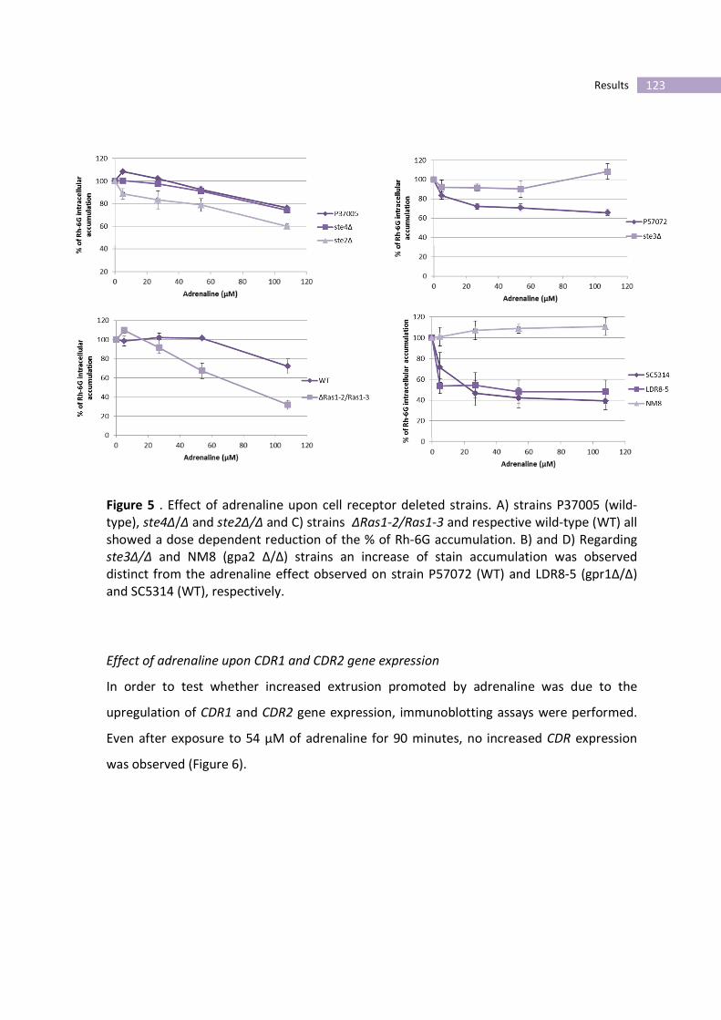
123 Results
Figure 5 . Effect of adrenaline upon cell receptor deleted strains. A) strains P37005 (wild-
type), ste4∆/∆ and ste2∆/∆ and C) strains ∆Ras1-2/Ras1-3 and respective wild-type (WT) all
showed a dose dependent reduction of the % of Rh-6G accumulation. B) and D) Regarding
ste3∆/∆ and NM8 (gpa2 ∆/∆) strains an increase of stain accumulation was observed
distinct from the adrenaline effect observed on strain P57072 (WT) and LDR8-5 (gpr1∆/∆)
and SC5314 (WT), respectively.
Effect of adrenaline upon CDR1 and CDR2 gene expression
In order to test whether increased extrusion promoted by adrenaline was due to the
upregulation of CDR1 and CDR2 gene expression, immunoblotting assays were performed.
Even after exposure to 54 µM of adrenaline for 90 minutes, no increased CDR expression
was observed (Figure 6).

124
Figure 6. Immunodetection of Cdr1p in C. albicans strain SC5314. Cell extracts previously
exposed (+) and not exposed (-) to adrenaline (54 µM) during 30, 60 and 90 minutes. No
differences were found regarding Cdr1p detection.
Effect of adrenaline upon growth and oxygen consumption of SC5314 strain
CDR efflux pump activity is ATP-dependent. Thus, if adrenaline enhances its activity, high
amounts of energy are required. The mitochondrial electron transport chain represents the
most important cell apparatus for production of cellular ATP. Therefore the effect of
adrenaline upon growth and oxygen consumption was investigated. We compared the
growth rate of SC5314 strain in the presence of fluconazole with and without adrenaline
(figure 7). Interestingly, a significant increase in the growth rate was observed after 14
hours incubation in the presence of adrenaline (figure 7).
Measurement of oxygen consumption in the presence of fluconazole and fluconazole plus
adrenaline were carried out with C. albicans, strain SC5314. Oxygen uptake rate was higher
in cells treated with adrenaline (p=0.016) (figure 8), indicating that adrenaline activates
energy production. In the yeast cell, this energy promptly available could be directed to a
response to the antifungal pressure and improve the fitness and stress response of the
fungal population.
Taken together, these results demonstrate that adrenaline improves the fitness of a
population under antifungal stress, activating energy production.

125 Results
Figure 7. Effect of adrenaline (Adr) upon growth rate of C. albicans strain SC5314. Higher
growth rates were observed whenever C. albicans was treated with Adr 108µM plus FLC
1µg/ml (Sub-MIC values). Significant differences (1µg/ml fluconazole versus 1µg/ml
fluconazole plus 54µM adrenaline: p=0.030 and 1µg/ml fluconazole versus 1µg/ml
fluconazole plus 108µM adrenaline: p=0.015) were observed following 14 hours incubation.
Data from three independent experiences is presented as means of optical density (O.D.) ±
standard deviation.
Figure 8. Effect of adrenaline upon oxygen consumption by C. albicans SC5314 strain. After
90 minutes in the presence of 16µg/ml fluconazole (FLC) or 16µg/ml fluconazole plus 54µM
adrenaline (adr) the oxygen uptake rate was determined. Oxygen uptake rate was higher in
cells treated with adrenaline (p=0.016). Oxygen consumption was measured by a Clark
electrode. Data from three independent experiences is presented as mean ± standard
deviation.
126
Discussion
It is well known that although in vitro microbial resistance usually correlates with clinical
resistance, susceptibility in vitro is not invariably related to clinical success [290]. In vivo
conditions do significantly differ from the in vitro ones, due to several factors, namely
biofilm formation, yeast interaction with other microorganisms, and also due to in vivo
exposure to non-antifungal drugs. Many drugs administered to critical care patients, like
albumin and propofol, have the potential to interfere with the antifungal activity, by
reducing the access, permeabilization or both of fungal cells [290-292]. In this study we
showed that catecholamines, such as adrenaline and noradrenaline, two drugs widely used
for life support, share the potential to interfere with antifungals namely by promoting
tolerance or resistance to azoles.
We have addressed the interaction between catecholamines and antifungals using the
checkerboard methodology and FUN-1 staining. Adrenaline and noradrenaline antagonized
the effect of fluconazole, resulting in an increased fluconazole MIC values. The antagonistic
effect observed between catecholamines and azoles, as well as the indifferent effect
regarding amphotericin B and caspofungin, suggested a possible interference of adrenaline
and noradrenaline on yeast cell efflux, the most common mechanism conferring resistance
to azoles. In order to provide a better insight of this effect, FUN-1 flow cytometric analyses
were performed. Besides information regarding metabolic activity FUN-1 also provides
additional information about the mechanisms of azole resistance, being a good marker of
efflux activity [145, 147]. The dose-dependent decrease of the fluorescence intensity of C.
albicans cells incubated with adrenaline in comparison to non-treated cells suggested an
increase of the efflux activity. With regard to the efflux of FUN-1 in presence of the other
adrenoceptor agonists (noradrenaline, isoprenaline), a rank order of potencies became
evident: adrenaline>noradrenaline>>isoprenaline. This ranking of potencies was very
important finding since it allowed to understand the specificity of the mechanism like in
mammalian cells [288]. In Human cells, whenever an α-adrenoceptor is involved, adrenaline
and noradrenaline have higher affinity than isoprenaline [288]. This approach helped to
typify the receptor involved in C. albicans. It suggests that adrenaline acts on the C. albicans
127 Results
strains through an α-adrenoceptor. Selective α-adrenoceptor agonists were also tested
(medetomidine α-2 agonist and phenylephrine α-1 agonist). The effect of medetomidine
upon FUN-1 staining was similar to that obtained with adrenaline and noradrenaline,
suggesting that the effect is mediated by an α-2 adrenoceptor [287]. Nevertheless more
agonists and antagonists should be tested in order to better characterize the type of
adrenoceptor involved in C. albicans cells. The results obtained with FUN-1 were confirmed
with a more specific substrate, Rh-6G [103, 148]. The measurement of intracellular
accumulation of Rh-6G is a useful method to identify azole resistance due to efflux pumps
[103, 148]. In all clinical isolates a dose-dependent reduction of the Rh-6G staining was
observed in the presence of adrenaline.
Yeasts cells have efflux mechanisms, like efflux pumps of the ABC transporter family (CDR1
and CDR2) that reduce drug accumulation inside the cell. After assessing cdr1∆/∆ and
cdr2∆/∆ strains, we could conclude that adrenaline promoted drug efflux, since the
percentage of accumulation of Rh-6G was significantly lower than that found in the parental
strain. Finally, adrenaline did not impair the staining of the double cdr1∆/∆ cdr2∆/∆ mutant.
Taking together, these results demonstrated that adrenaline promotes the activity of C.
albicans efflux pumps.
After uncovering the role of adrenaline as a contributor to antifungal resistance, we aimed
to identify C. albicans surface receptors mediating such effects based upon analogies with
mammalian cells. Several studies have reported the existence of GPCRs in yeasts, showing a
GTP-binding site similar to those in eukaryotic G-proteins [277-281]. They comprise a highly
diverse family of receptors which are involved in multiple cellular processes. In C. albicans,
some GPCRs have been identified, being mainly involved in morphogenesis (Gpr1, Gpa2 and
Ras) [277, 278] and mating (pheromone receptor- Ste2, Ste3 and Ste4) [279]. They mediate
environmental stimulus responses that result in changes in cell physiology, morphology and
adherence ability [293]. Considering the many homologies between yeasts and human cells,
we raised the hypothesis that catecholamines could act on Candida through receptors
similar to adrenoceptors that are present in eukaryotic cells. Adrenoceptors are GPCRs that
evoke cellular signaling mechanisms after binding to catecholamines [294].

128
In an attempt to unveil the possible adrenaline receptor the effect of adrenaline upon Rh-
6G staining was studied in GPCR-deleted C. albicans strains and compared with the
corresponding wild-type strains. We could conclude that adrenaline acts through a
pheromone receptor Ste3 and a Gpa alpha protein, since in strains ste3∆/∆ and gpa2 Δ / Δ
(NM8) adrenaline did not impair the Rh-6G staining.
Aiming at uncovering the mechanisms underlying the increased efflux, immunoblotting
assays were performed. The results revealed that adrenaline does not act as an inducer of
the expression of CDR1 and CDR2 genes. However, adrenaline stimulates ATP production,
and increases the activity of energy-dependent efflux pumps. In addition to increased efflux
activity, other mechanisms can allow the cell to overcome antifungal drug induced stress
[237, 295]. The mitochondrial electron transport chain has been implicated in drug
tolerance or resistance and it is a very important cell apparatus for production of ATP [296-
299].
In brief we found that adrenaline enhances drug efflux activity mediated by CDR1 and CDR2
pumps. This increase in efflux pump activity requires energy whose production is also
stimulated by adrenaline. Interestingly, despite the presence of fluconazole, adrenaline can
improve the fitness of fungal cells by stimulating energy production and fungal growth, thus
allowing them to overcome the stress caused by the antifungal drug.
Overall, it is plausible to postulate that adrenaline can trigger ATP production through a
GPCR by acting on the electron transport chain (via putatively a secondary messenger). In
turn, these intracellular downstream events can increase the activity of efflux ATP-
dependent pumps (CDR1 and CDR2) and yeast cell growth, promoting concomitantly the
tolerance or resistance to antifungal drugs.
Hospitals face at present an increasing emergence of resistant fungal pathogens. The
knowledge on underlying mechanisms of induction of drug resistance by non-antifungal
drugs like catecholamines will allow the development of new therapeutic strategies.

129 Results
Part VII
In vivo synergistic effect between ibuprofen and fluconazole in Candida
albicans
Background
Candida infections range from superficial mucocutaneous infections to life-threatening
invasive infections. Especially the last one represents an increasing challenge for clinicians,
with an attributable mortality around 40% [6, 7, 300]. The emergence and spread of
antifungal resistance is one of the main factors that are responsible for this trend. Among
azoles, fluconazole represented a landmark in the treatment of Candida bloodstream
infections. However, its extensive use both for prophylaxis and therapy resulted in the
emergence of fluconazole resistance [301-303].
The major mechanism responsible for high level azole resistance in Candida albicans clinical
isolates is the overexpression of plasma membrane efflux pumps, the ATP-Binding Cassette
(ABC) transporters, namely CDR pumps, or the major facilitator superfamily (MFS)
transporters, MDR pumps [24, 27, 98, 101].
The knowledge of the underlying resistance mechanisms is of crucial importance since it
could support approaches to achieve its reversion, thus leading to the development of new
therapeutic strategies. Our previous studies showed that antifungal resistance conferred by
increased efflux activity in Candida clinical strains could be reverted by ibuprofen [101,
145]. Ibuprofen ([2-(4-isobutylphenyl)-propionic acid]) is a non-steroidal anti-inflammatory
drug (NSAIDs) used for its antipyretic, analgesic, and anti-inflammatory effects. In humans,
it inhibits the synthesis and release of prostaglandins as mediators of inflammation.
Ibuprofen acts synergistically with pyrazinamide, an tuberculostatic drug [182], azoles [101,
145, 183] and amphotericin B [184]. Previously, we had showed that ibuprofen acts
synergistically with fluconazole, voriconazole and itraconazole, by inhibiting efflux pumps
130
mechanisms, thus promoting intracellular accumulation of [H3]- itraconazole in Candida
cells overexpressing CDR1 and CDR2 genes [101, 145].
Generally, azole exposure triggers several cellular and molecular mechanisms in Candida
albicans, including the overexpression of genes related with antifungal resistance, mainly
efflux pumps [118, 302, 304, 305]. With the development of new molecular biology
methods, mainly DNA microarray technology, it was possible to get insights about the
molecular basis involved in the evolution of antifungal resistance [23, 302, 306]. Herein we
aimed to study the in vivo synergistic effect between these two drugs using a C. albicans
systemic model of infection and to unveil the transcriptional profile involved in the
reversion of fluconazole resistance by ibuprofen.
With this study we clarified the ability of ibuprofen to revert azole resistance, as well as
elucidated about molecular changes associated with resistance reversion.
Material and methods
C. albicans strains and culture conditions
A Candida albicans blood stream isolate (CaS) susceptible to fluconazole, voriconazole and
posaconazole was used throughout this study. In order to induce a resistant phenotype, a
yeast suspension containing 106 cells in 10 ml of RPMI 1640 medium (RPMI 1640; Sigma, St.
Louis, MO USA) was repeatedly incubated with fluconazole (FLC; Pfizer, Groton, CT, USA) at
a final concentration of 16 µg/ml (therapeutic serum levels achieved during antifungal
therapy) in order to induce a resistant phenotype (CaR) [307, 308]. Yeast suspensions were
grown overnight at 35°C under agitation (150 rpm). Cultures were daily transferred to fresh
medium with and without the same antifungal concentration for a total of 60 days. Each
day 1ml aliquot from each subculture was mixed with 0.5ml of 50% glycerol and frozen at -
70°C for later use. The Minimal inhibitory concentration (MIC) value of azoles was
determined for the parent strain and the successive fluconazole exposed isolates.

131 Results
Microdilution antifungal susceptibility testing
Assessment of the antifungal susceptibility profile was performed according to the CLSI M27
A3 protocol in RPMI 1640 culture medium (Sigma)[33]. The minimal inhibitory
concentration (MIC) of FLC, voriconazole (VRC; Pfizer) and posaconazole (PSC; Schering-
Plough, NJ) was determined [33]. Strains were classified as susceptible (S), susceptible-dose
dependent (S-DD) or resistant (R) to azoles according to the breakpoints defined by CLSI
[33]. Although definitive breakpoints have not yet been established for posaconazole, a MIC
≤1 µg/ml was considered susceptible.
Effect of ibuprofen upon azole resistance
In order to study the effect of ibuprofen (Ibu; Sigma) on the resistant phenotype displayed
by CaR strain, MIC value of FLC, VOR and PSC was re-determined as described above in the
presence of 100 µg/ml of ibuprofen, a concentration previously described to impair azole
extrusion [101, 145].
In vivo effect of ibuprofen upon fluconazole resistance
All in vivo investigations were performed in the animal facility of the Cardiovascular
Research & Development Unit Faculty of Medicine, University of Porto, in accordance with
the European Directive 86/609, transposed to the Portuguese Law by DL 129/92 and by
Portaria 1005/92. A murine model of disseminated candidosis was used to study the effect
of ibuprofen upon fluconazole resistant phenotype. Female specific-pathogen-free BALB/c
mice (age, 6 to 8 weeks; weight, 17 to 20 g; Charles River Laboratories, Barcelona, Spain)
were housed in microisolator cages with five animals per group, having access to food and
water ad libitum. To induce disseminated infection mice were injected with 5x10⁵ cells of
the CaS or the CaR strain in 0.1ml of sterile saline via the lateral tail vein. The fluconazole
effective dose in reducing the pathological effects of i.v. C. albicans challenge relative to
untreated mice (control) was defined as the 50% effective dose (ED50) [309]. Therapy was
initiated 3 hours after yeast challenge and was administered intraperitoneally once a day
for a total of 4 days. Mice were treated with FLC (8 to 60 mg/kg of body weight/day), with
Ibu (10 or 20 mg/kg/day) [182, 310] or FLC (8 to 60 mg/kg of body weight/day) + Ibu (10 or

132
20 mg/kg/day). Mice weight was daily registered and at day 4 post-infection mice were
euthanized and the kidneys were aseptically removed. The right kidney from all mice was
homogenized in 5 ml of phosphate buffer saline (PBS; Sigma) and weighted. Serial dilutions
of the homogenate were plated onto YPD agar in triplicate and incubated at 30°C for 48
hours. The fungal burden was calculated as the number of colony forming units (CFU) per
gram of tissue. The kidney fungal isolates were frozen in YPD broth with 50% glicerol at -
70°C for later MIC azole determination according CLSI M27 A3 protocol [33]. For histological
studies the left kidneys were fixed in 10% phosphate-buffered formalin, embedded in
paraffin, sectioned, and stained with periodic acid-Schiff (PAS) stain. Slides were observed
under a Zeizz Axioskop 40 microscope and acquired with an Axiocam MRc5 Zeizz.
RNA extraction
Strains CaS and CaR were grown in 50-ml YPD broth at 180 rpm and 30°C until an optical
density at 600nm of approximately 0.8 was reached. CaR strain was grown in the presence
of 16 µg/ml of FLC (CaRFLC) and of 16 µg/ml of FLC plus 100 µg/ml of ibuprofen
(CaRFLCIbu). Total RNA was extracted using the hot acid phenol method, as described by
Köhrer & Domdey [311]. RNA concentration was assessed by Nanodrop ND-1000 and the
RNA quality and integrity levels were controlled by capillary electrophoresis in the Agilent
2100 Bioanalyzer (Agilent Technologies, Palo Alto, CA), according to the manufacturer’s
instruction. Only samples yielding a 28S rRNA/18S rRNA ratio ranging from 1.6 to 2.2, with a
“RNA Integrity Number” (RIN) higher than 7.0 and showing the absence of degradation
were used. RNA samples were stored at -70°C for later use.
Probe preparation and microarray hybridization
All the experiments were carried out using C. albicans microarrays manufactured by Agilent
Technologies. A RNA/primer mix containing 50 µg RNA, 1.25 µg Oligo dT12-18, (Invitrogen,
Carlsbad, CA) was incubated at 70°C for 10 minutes (min) and chilled on ice for 5 min. Then,
a labeling mixture containing dGAC – mix 0.1mM, dTTP 0.03mM, aa-dUTP 0.08 mM, first
strand buffer 1x, DTT 0.01M, Superscript II Reverse Transcriptase 400U (Invitrogen) was
added and the reaction was incubated at 42°C for 60min. Residual RNA template was

133 Results
degraded by hydrolysis. Briefly, the reaction mixture was incubated at 95°C for 2 min and
placed on ice immediately; 10 µl NaOH 1M (Sigma) and 10 µl EDTA 0.5M (Sigma) were
added to the reaction and incubated at 65°C for 15min. Afterwards, 25 µl HEPES buffer 1M
(pH 7.5, Sigma)were added. Samples were purified using the Microcon - 30 (Amicon
Microcon YM-30, Millipore) columns, according to the manufacturer’s instructions and were
coupled to Cy3 and Cy5 fluorophores. Before hybridization, free dyes were removed using
Chromaspin-30 (Clontech) columns, the efficiency of cDNA synthesis and dye incorporation
was measured spectrophotometrically in the Nanodrop ND-1000. For full slides, 200 ng of
Cy3/5 labeled sample were used. Hybridization was performed according to the labeling kit
Quick Amp Labeling for Two-Color Microarray-Based Gene Expression Analysis protocol
(from Agilent Technologies). A common reference design with dye-swap replicates was
used. Total RNA obtained from CaS strain was used as hybridized against samples studied.
Control of experimental background was performed through self-self hybridization; a total
of four experiments were made.
Image acquisition and data processing
The microarrays were scanned with an Agilent G2565BA microarray scanner and the
fluorescence intensities were obtained with Agilent Feature Extraction Software Protocol
GE2. Pre-processing of the data was performed using the Biometric Research Branch (BRB)-
ArrayTools v3.4.0 software.
Statistical analysis and functional annotation of the data
For data analysis the statistical software R and the package Limma from Bioconductor
(www.bioconductor.org) were used. Microarray normalization was performed using the
method loess and within arrays quantile [312]. After normalization, median gene expression
was determined for each ORF. Relative gene expression and statistically significant
differences found between the different samples were determined with the software
Limma package. Significant differences were encountered using the Empirical Bayes

134
statistical test [313]. Hierarchical clustering of the normalized and dye swap averaged
samples was performed using the method complete linkage.
Only probes with a fold change (FC) greater than 2 and adjusted p<0.05 were used in
analyses. Gene ontology identification was performed for significant differentially expressed
genes using the Web tool GoTermFinder available on the website of the Candida Genome
Database (http://www.candidagenome.org/cgi bin/GO/goTermFinder).
Quantitative RT-PCR
In order to confirm microarray data, cDNA was synthesized from 100ng in 20µl of total RNA
from CaS, CaRFLC and CaRFLCIbu strains using a Superscript III reverse transcriptase kit
(Invitrogen) according to manufacturer`s instructions. Quantitative real- time PCR was
performed using PerfeCTa SYBR green Fast Mix (Quanta Biosciences) on a Realplex
Mastercycler instrument (Eppendorf, Madrid, Spain) and according to the following
program: 1 minute hot start at 95°C, a 35-cycle program composed of 15-seconds (s)
denaturation step at 95°C, a 30-s annealing step at 60°C, and a 30-s extension step at 60°C,
ending by a 5-min final extension step at 60°C. The primers used were designed using
OligoExplorer program (listed in Table 1). ACT1 was used as the normalization gene.

135 Results
Table 1. Sequences of primers used in RT-PCR
Primer name Primer sequence
CDR11 - F 5´-GGTCACGAATCTACTTTGGAA-3´
CDR11 - R 5´-CGGGTCTCATAATGGCAT-3´
NAG3 - F 5´-CAAGGGGGTGGAAAAGAT-3´
NAG3 - R 5´-TCGTTGTAGTCAGTGTGTGG-3´
MDR1 - F 5´-CCCGAAAACCCTCAAAAT-3´
MDR1- R 5´-CGACTCTTCCAATACCAAAATC-3´
ERG1 - F 5´-GACGAGACCATTACTATCCCTT-3´
ERG1 - R 5´-TTACACCATCAACGGCAT-3´
ACT1 - F 5´-ATGGACGGTGAAGAAGTTG-3´
ACT1 - R 5´-CAAGAGATGGGAAAACAGC-3´
Results
In vitro induction of resistance
The repeated exposure of the susceptible strain (CaS) to therapeutic serum concentrations
of FLC the acquisition of an azole cross-resistant phenotype to FLC, VRC and PSC (table 2).
Table 2 resumes MIC values assessed following 60 days of incubation with FLC.
Effect of ibuprofen in azole susceptibility
The azole susceptibility pattern of the parental (CaS) and the azole resistant induced strain
in the absence and presence of ibuprofen is detailed in table 2. The MIC values decreased in
the presence of Ibu for all strains, the phenotype changing from R to S regarding all the
tested azoles (table 2); meanwhile, the MIC values remained unchanged regarding the
susceptible strain (table 2).

136
Table 2. Minimal inhibitory concentrations (MIC) and phenotypes of the Candida albicans
parental susceptible (CaS) and resistant (CaR) strain after exposure to fluconazole (FLC) to
FLC, voriconazole (VRC) and posaconazole (PSC) alone and in combination with
subinhibitory concentrations of ibuprofen (Ibu; 100 µg/ml). MICs were determined
according the CLSI M27-A3 protocol. Susceptible, susceptible dose dependent and resistant
phenotypes are represented as S, S-DD and R, respectively.
MIC (µg/ml)/ phenotype
Strains FLC FLC+Ibu VRC VRC+Ibu PSC PSC+Ibu
CaS 1/S 1/S 0.06/S 0.06/S 0.03/S 0.06/S
CaR >64/R 4/S >8/R 0.06/S >8/R 0.125/S
In vivo synergistic effect between FLC and Ibu
In order to investigate the potential clinical application resulting from the synergistic effect
between FLC and ibuprofen experiments were conducted in a murine candidosis model. The
fungal burden of CaS and CaR strains in mice kidneys were determined after treatment for 4
days with 8 to 60 mg/kg of body weight/day of FLC alone or combined with 10 or 20 mg/kg
of body weight/day of ibuprofen. On mice infected with the CaS strain and treated with 30
mg/kg of body weight/day (ED50) of FLC a significant reduction (p<0.001) of CFU/g of kidney
was found when compared with untreated mice (figure 1). On mice infected with the CaR
strain no significant reduction on fungal burden was achieved even when treated with 60
mg/kg of FLC. The group of mice infected with CaR strain treated or untreated with FLC
showed the highest weight loss during at the first and at the fourth day of infection (figure
2). However, when FLC was associated to the two concentrations of ibuprofen, a
significantly reduction of CaR fungal burden (p<0.001) was observed, especially in mice
treated with 30 mg/kg of FLC plus 20 mg/kg of ibuprofen (figure 1). These mice also showed
weight increase at the first and at the fourth day of infection (figure 2).

137 Results
Interestingly, the yeast isolates recovered from mice kidneys retained the susceptible and
the resistant phenotype as displayed previously to infection with CaS and CaR strains,
respectively. Azoles MIC values remained unchanged.
Figure 1. In vivo antifungal synergistic effect between fluconazole and ibuprofen against C.
albicans systemic infection. Mice infected with the parent susceptible strain (CaS) and the
induced resistant strain (CaR) were treated with 30mg/kg/day of fluconazole (ED50) (FLC),
and 10 or 20 mg/kg/day of ibuprofen (Ibu) intraperitoneally during 4 days. The right kidneys
(from 5 mice per group) were removed, homogenized, weighed, serially diluted and plated
onto YPD agar plates. Colony forming units (CFU) per gram of kidney were determined and
plotted as mean value and respective standards error.

138
Figure 2. Effect of the combination of fluconazole plus ibuprofen on mice weight loss during
C. albicans systemic infection. Mice infected with C. albicans susceptible (CaS) and the
resistant (CaR) strains and treated and untreated with 30mg/kg/day of fluconazole (FLC30)
and 10 or 20 mg/kg/day of ibuprofen (Ibu10 and Ibu20, respectively) intraperitoneally
during 4 days were daily weighted. Differences in weight loss between the first and the
fourth day of infection are plotted as mean values plus the respective standard errors.
Regarding the PAS stained histopathology sections of mice kidneys, susceptibility of mice to
infection with the resistant strain (CaR) was evident on untreated or FLC treated mice. At
day four post infection PAS staining of untreated mice infected with CaR strain revealed a
dramatic increase in fungal cell numbers, both blastoconidea and hypha morphotypes
(figure 3A) similarly to the findings in mice treated with 30 mg/kg of FLC (figure 3B, C, D and
F). In mice infected with CaR strain and treated with 30 mg/kg of FLC plus 20 mg/kg of
ibuprofen rare yeasts were found, all showing the blastoconidea form (figure 3 F, G). These
results were in agreement with CFU counts obtained in fungal burden assays.

139 Results
Figure 3. Representative example of kidney histology slides of PAS-stained paraffin sections
of kidneys recovered from mice infected with 5x10⁵ cells/0.1 ml of C. albicans resistant
(CaR) strain at day four post-infection, untreated (A; 40x amplification), treated with
30mg/kg/day of FLC (B, C, D, E; 10x, 20x, 40x and 20x, respectively) and treated with
30mg/kg/day of FLC plus 20 mg/kg/day of Ibu (F, G; 40x).
A
B C
D E
F G
140
Microarray transcriptional profile
Microarray assays allowed the evaluation of the expression of 6158 C. albicans genes,
however only the differentially expressed genes (p< 0.05) were used in analyses. The gene
expression of the induced fluconazole resistant strain pre-exposed to FLC (CaRFLC) was
compared with the parent susceptible CaS strain and 836 genes were differentially
expressed, of which 207 were upregulated and while 629 were downregulated. The
biological processes encoded by the majority of these genes are unknown (30% of
upregulated genes and 18% of down regulated genes) (figure 4). However up to 5% of the
upregulated genes are associated with transport processes and response to chemical
stimulus. A large number of such genes are involved in antifungal resistance mechanisms,
mainly CDR and sterol biosynthesis genes.
When compared with the parent strain, the presence of Ibu plus FLC (CaRFLCIbu) altered
the expression of 1517 (411 upregulated and 1106 down regulated) (figure 5). Ibuprofen
exposure upregulated genes related with transport, response to chemical stimulus and cell
wall/membrane organization processes (figure 5). A minority of such genes was related to
protein folding (1%) and transduction (1%) (figure 5). Genes involved in cell cycle (5%), RNA
(9%) and DNA (5%) metabolic process were differentially downregulated (figure 5).
Figure 6 describes the transcriptome analysis of the induced FLC resistant strain (CaRFLC)
and after exposure to fluconazole plus ibuprofen (CaRFLCIbu). The CaRFLC strain showed
overexpression of ERG251, CDR1, CDR4, CDR11 and UPC2 genes. However, in the presence
of fluconazole plus ibuprofen a significantly decrease of CDR11 and ERG251 gene expression
and of the transcription factor UPC2 was found (figure 6). MDR1, CDR4, NAG3 and NAG4
genes showed increased expression in the presence of fluconazole plus ibuprofen, resulting
in significant differences when compared to strain CaRFLC (figure 6). The microarray results
were confirmed with quantitative real-time PCR (figure 7). Significant different levels of
gene expressions was observed between CaRFLC and CaRFLCIbu strains (p<0.05).
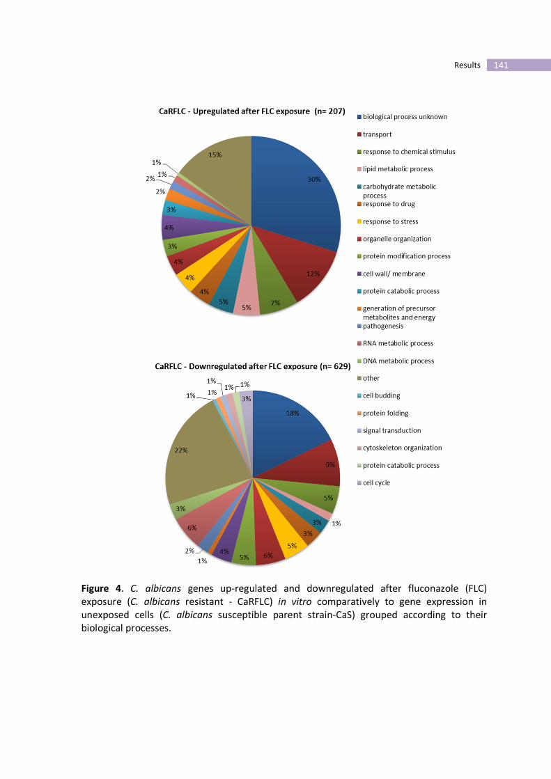
141 Results
Figure 4. C. albicans genes up-regulated and downregulated after fluconazole (FLC)
exposure (C. albicans resistant - CaRFLC) in vitro comparatively to gene expression in
unexposed cells (C. albicans susceptible parent strain-CaS) grouped according to their
biological processes.
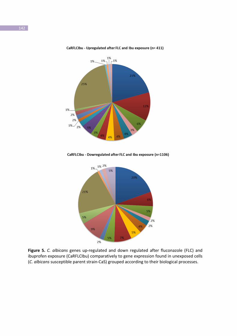
142
Figure 5. C. albicans genes up-regulated and down regulated after fluconazole (FLC) and
ibuprofen exposure (CaRFLCIbu) comparatively to gene expression found in unexposed cells
(C. albicans susceptible parent strain-CaS) grouped according to their biological processes.
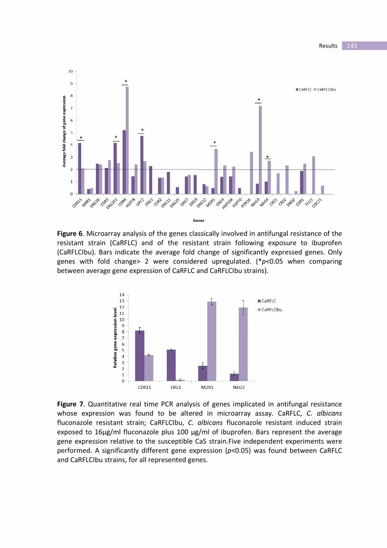
143 Results
Figure 6. Microarray analysis of the genes classically involved in antifungal resistance of the
resistant strain (CaRFLC) and of the resistant strain following exposure to ibuprofen
(CaRFLCIbu). Bars indicate the average fold change of significantly expressed genes. Only
genes with fold change> 2 were considered upregulated. (*p<0.05 when comparing
between average gene expression of CaRFLC and CaRFLCIbu strains).
Figure 7. Quantitative real time PCR analysis of genes implicated in antifungal resistance
whose expression was found to be altered in microarray assay. CaRFLC, C. albicans
fluconazole resistant strain; CaRFLCIbu, C. albicans fluconazole resistant induced strain
exposed to 16µg/ml fluconazole plus 100 µg/ml of ibuprofen. Bars represent the average
gene expression relative to the susceptible CaS strain.Five independent experiments were
performed. A significantly different gene expression (p<0.05) was found between CaRFLC
and CaRFLCIbu strains, for all represented genes.
144
Discussion
A high mortality rate is often observed in immunocompromised patients with systemic
fungal infections, despite being often prescribed long courses of antifungal therapy. In
addition, antifungal therapeutic options are limited, being the azoles the most commonly
used drugs [314]. The widespread use of fluconazole, especially for prophylaxis, has
selected Candida species with easily inducible resistance, such as C. glabrata [315] and C.
tropicalis [306]. The development of resistance by C. albicans after in vitro fluconazole
exposure has been widely demonstrated [302, 304, 305]. Aiming to reproduce in vivo
findings occurring during antifungal treatment, we daily exposed a susceptible C. albicans
strain to serum concentration levels of fluconazole. As expected fluconazole exposure
resulted in the development of cross resistant profiles to azoles.
The major mechanism described as responsible for azole resistance in clinical Candida
albicans isolates is overexpression of plasma membrane efflux pumps [22, 96, 117]. The use
of DNA microarray technology push forward the knowledge of the protagonists involved in
cellular processes [23, 96, 152, 153, 302, 304, 306, 315]. The transcriptional responses of C.
albicans after fluconazole exposure showed an increased expression of genes encoding CDR
efflux pumps as well as of genes involved in the ergosterol biosynthesis, as expected [96,
117, 302, 304, 305]. Similarly to C. albicans, species like C. glabrata, C. tropicalis and C.
parapsilosis also use efflux pumps as the main tool for the development of azole resistance
[153, 306, 315].
During the recent years several approaches have been used in order to overcome the
development of antifungal resistance. The inhibition of antifungal extrusion by blocking
specifically ABC efflux pumps is a possible way to impair multiple drug resistance. The use of
antifungals which are not substrates of efflux pumps, of pump blockers that inhibit the
antifungal extrusion (inhibiting H+ ATPase reducing the energy required for efflux activity) or
the increase of antifungal uptake rate in order to maintain a high intracellular concentration
of the drug are all strategies to achieve a high concentration of the antifungal compound at
its site of action [22]. The pursuit of knowledge of efflux pumps mechanism in Candida

145 Results
arises from the homology between yeasts and human cells. In eukaryotic neoplasic cells,
ATP-dependent drug efflux pumps, such as P-glycoprotein (P-gp) which is encoded by MDR1
gene, are important mediators of resistance, contributing to failure of cancer therapy. Over
the years different strategies were undertaken in order to overcome drug resistance by
efflux, including the development of agents able to modulate P-gp activity, such as pump
substrates like FK506 [176] or cyclosporine A [177], calcium channel blockers like verapamil
[316] or anti-malaric analogs [317].
Azza et al demonstrated that ibuprofen could inhibit methotrexate efflux transporters in
the human kidney [318]. A similar effect was described regarding FK506 (tacrolimus), a
potent immunossuppressor agent used for the prevention of allograft rejection and as a
calcineurin inhibitor, shows a synergistic effect when combined with antineoplastic agents
on tumor cells, decreasing or even suppressing multidrug resistance by competing with
cytotoxic drugs for the P-glycoprotein [319-321]. Thus, a similar approach could be applied
to Candida albicans cells, using non- antifungal drugs exhibiting a synergistic effect with
antifungals that could induce a reversion of resistance. In Candida, FK506 has shown to be a
potent inhibitor of the calcineurin pathway, rendering the normally fungistatic azole effect
to fungicidal [169, 179]. The calcineurin pathway has been shown to be critical for survival
and stress responses in several fungi, including the in vitro antifungal activity against
Saccharomyces cerevisiae, C. albicans and Cryptococcus neoformans [169, 322].
In the present study, a decreased of the MIC levels for the three azoles tested in the
presence of ibuprofen was registered in strain CaR, changing the phenotype from azole
resistant to a susceptible one. The synergism between posaconazole and ibuprofen was
hereby demonstrated for the first time.
The in vivo assays clearly demonstrated that ibuprofen acts synergistically with fluconazole
in mice infected with the CaR strain. A significantly reduction of CFU kidney counts were
obtained in mice treated with both drugs, as well as a recovery of mice weight. These
results were attested by the histological assays. A dramatic increase in fungal cell numbers
was observed in the kidneys of mice infected with CaR strain untreated or treated with

146
fluconazole. However, fluconazole in combination with ibuprofen promoted fluconazole
activity, histologically showing scarce fungal cells.
C. albicans cells recovered from mice treated with fluconazole plus ibuprofen still displayed
a resistant phenotype to fluconazole. Consequently, we can conclude that the presence of
ibuprofen is mandatory for the reversion of the azole resistance.
Trying to scrutinize the molecular base involved in the ability of ibuprofen to revert
antifungal resistance we used microarray analysis of CaRFLC comparing the transcriptional
profile to CaRFLCIbu strain. The CaRFLC strain revealed an overexpression of ERG and CDR
genes, as expected [302, 304, 305]. The presence of ibuprofen significantly increased the
expression of CDR and MDR efflux pumps (CDR4, MDR1, NAG3 and NAG4). Interestingly,
these findings may be seen as cellular salvage attempt mechanisms to overcome the
inhibitory capacity of ibuprofen. Yeasts cells have the capacity to trigger cellular salvage
pathways when facing stress conditions like in the presence of antifungals or other drugs
[22, 160, 237]. Similarly, regarding echinocandins, the ability to grow at high caspofungin
concentrations and has been suggested to relate to a compensatory increase in cell wall
chitin [128, 130]. This salvage mechanism strengths cell wall damaged by exposure to
echinocandins.
MRR1 and TAC1 are all transcription factors of MDR1 and CDR genes [96, 118, 152, 305,
323-325]. Despite the significative increase of MDR1 gene expression displayed by
CaRFLCIbu strain, MRR1 expression remained downregulated and no difference was seen
between exposed and non-exposed isolates to ibuprofen. On the other hand, the presence
of ibuprofen promoted the downregulation of CDR11, ERG251 and and the transcription
factor UPC2. In fact, these results strongly suggest that ibuprofen could act at the
transcriptional level. However, and due to the contradictory gene expression profiles found
and their respective transcription factors, this assumption still needs further investigation.
Overall, our results stress the fact that ibuprofen can inhibit efflux pumps, either by
blocking access to the binding site or by blocking the expression of the pumps. Besides,
since it is not immunosuppressive, this anti-inflammatory drug has advantages over FK506.

147 Results
Further studies are being addressed in order to uncover the main mechanism of ibuprofen
inside the yeast cell as well as to assess its influence in the dynamics of the induction of
antifungal resistance.
By allying anti-inflammatory and analgesic properties, ibuprofen in combination with
fluconazole could play a relevant role in a therapeutic strategy for severe fungal infections.


CHAPTER IV
Conclusions and
Future Perspectives

151 Conclusions and Future Perspectives
Conclusions
Over the last 30 years medical advances led to a significant increase of life-threatening
fungal infections. The incidence and mortality rates associated with invasive candidosis
have remained unchanged for more than a decade despite the advances in the field of
antifungal therapy. Such infections could be treated more effectively if faster and more
specific diagnostic and therapeutic approaches were available. Preventive safe strategies
targeting patients with a high-risk profile, the development of new diagnostic tools for early
identification of fungal species, including innate resistant species or those that are more
prone to develop multidrug resistance, especially in patients submitted to long term
therapy are of utmost importance. In conjunction, preventive attitudes should be
imperatively implemented in order to reduce the number of health care related infections.
The comprehension of the routes of transition is essential to overcome this objective.
Given the association between antifungal exposure and the development of resistance,
prophylaxis must be selectively restricted to high-risk patients. Apparently there is no class
of antifungal agents that is immune to the development of acquired resistance. It is
essential that laboratories start performing routinely in vitro susceptibility testing especially
in isolates from invasive infections, isolated from patients receiving antifungal prophylaxis
and in strains isolated from patients who do not respond to therapy.
Intricate signaling networks govern the development, morphogenetic transitions but also
the evolution of antifungal drug resistance in Candida. Cell stress may be caused by other
concomitant factors apart the presence of the antifungal drug. Medication often
administered to critical care patients may trigger a medley of escapes responses in order to
ensure survival which may also be responsible for the discrepancy between in vitro and in
vivo susceptibility profile. The knowledge of the mechanisms involved in antifungal
resistance will help to design effective measures to reverse it. Ibuprofen may represent a
hopeful compound in the reversion of azole resistance by efflux activity.

152
Regarding echinocandins, point mutations in FKS genes are responsible for the decreased
susceptibility; such findings stress the need for the development of novel molecules with
higher affinity to the target.
In brief, the research presented has led to the following findings:
Following the assessment of the genetic relatedness between simultaneous and/or
successive Candida albicans isolates from fungaemia patients and of the antifungal
susceptibility profile (detailed in Part I) the following conclusions were obtained:
• C. albicans colonization and/or infection of different body sites may represent a
predisposing condition or an initial step towards the subsequent fungaemia
development;
• The resistance of blood infection by successive isolates with similar genotypes suggests
the failure of antifungal therapy;
• Isolates displaying the same or similar genotypes were obtained from patients who
shared the same hospital department, suggesting the nosocomial origin of the infection
in such cases;
• The induction of azole resistance was observed in similar strains from different patients
admitted in distinct periods of time, suggesting that the strains were endemic to the
hospital environment;
• Hospital acquired bloodstream infections are linked with a higher risk of antifungal
resistance, thus needing close monitoring.
The novel flow cytometric protocol developed to measure yeast cell wall chitin (detailed in
Part II) helped to understand the effect of echinocandins on fungal cell wall:
• Flow cytometry protocol showed to be a simple and reliable assay to accurately
quantify cell wall chitin in yeast cells;

153 Conclusions and Future Perspectives
• No relationship between chitin content and the caspofungin susceptibility profile was
found;
• The accuracy of this novel simple and reliable methodology can predict the occurrence of
the paradoxical effect, representing a valuable tool for the detection of antifungal
compensatory mechanisms in the presence of high echinocandin concentrations.
In Part III the first case report describing acquisition of echinocandin resistance by C.
glabrata following anidulafungin treatment was addressed. The main conclusions were:
• The acquisition of resistance to anidulafungin was associated with an increase in
caspofungin and micafungin MIC value, suggesting the development of cross-resistance
between these three echinocandins drugs;
• No relation between chitin amount and antifungal resistance was established;
• Structural alterations in the HS1 of FKS2 molecule due to the Ser663Pro substitution and
a Phe659 deletion lead to a dramatic decrease of anidulafungin efficacy in vivo.
Studies related to the contribution of mitochondrial respiration in the intrinsic fluconazole
resistance displayed by C. krusei (Part IV) revealed that:
• An alternative respiratory pathway, cyanide-resistant, was described for the first time as
a characteristic of C. krusei species;
• Such alternative pathway due to an alternative oxidase is unrelated to fluconazole
resistance; nevertheless it protects C. krusei from oxidative stresses;
• The alternative respiratory pathway is a potential target that should be taken into
account considering the development of new therapeutic strategies in the case of C.
krusei infections.

154
The research described in Part V provided an opportunity to describe the effect of propofol
in antifungal drug resistance. The main conclusions were:
• The promotion of antifungal resistance in presence of propofol lipidic emulsion results
from a reduced access and/or permeabilization of the antifungal agents into the fungal
cells;
• The major responsible for such effect was the lipidic vehicle of propofol emulsion.
The assessment of the effect of adrenaline upon C. albicans (Part VI) has led to the following
conclusions:
• Adrenaline enhances drug efflux activity mediated by CDR1 and CDR2 pumps;
• Despite the presence of fluconazole, adrenaline promotes the fitness of fungal cells by
stimulating energy production and fungal growth, thus allowing the overcome of stress
caused by the antifungal drug;
• Adrenaline acts on C. albicans cells through a pheromone receptor Ste3 and a Gpa alpha
protein.
Part VII addressed the in vivo synergistic effect between ibuprofen and fluconazole. This
study provided the following conclusions:
• The in vivo assays clearly demonstrated that ibuprofen acts synergistically with
fluconazole in mice infected with a C. albicans resistant strain;
• The presence of ibuprofen is mandatory for the reversion of fluconazole resistance.
This research resulted in a more comprehensive understanding of the multiple mechanisms
that can influence antifungal resistance or tolerance. The deliverables of our studies include
a better awareness of the efforts that should be made for the implementation of routine

155 Conclusions and Future Perspectives
antifungal susceptibility testing and provide new hope for the prevention of azole antifungal
resistance.
Future Perspectives
During the development of this research several questions were raised and should be
addressed in future studies:
• The contribution of other concomitant medications, like antibacterial drugs, albumin
infusion and other catecholamines on the development of antifungal resistance or
tolerance;
• The knowledge of the underlying mechanisms of induction of drug resistance by non-
antifungal drugs like catecholamines, addressing in particular the identification and
characterization of other possible receptors that might be involved, as well as the
downstream events implicated;
• Since ibuprofen can act at the transcriptional level, the search for mutations in the
transcription factors that regulate azole resistance is mandatory. The effect of ibuprofen
in the calcineurin pathway will be studied, aiming also to assess its influence in the
dynamics of the induction of antifungal resistance, eventually preventing its emergence;
• The gene AOX should deserve further investigation; it may help to further decode the C.
krusei genome.


CHAPTER V
References

159 References
References
1. Calderone, R., ed. Candida and candidiasis. 2002, ASM Press: Washington DC.
2. Cannon, R.D., et al., Oral Candida: clearance, colonization, or candidiasis? Journal of
dental research, 1995. 74(5): p. 1152-61.
3. Casadevall, A. and L.A. Pirofski, Accidental virulence, cryptic pathogenesis, martians,
lost hosts, and the pathogenicity of environmental microbes. Eukaryotic cell, 2007.
6(12): p. 2169-74.
4. Eggimann, P., J. Garbino, and D. Pittet, Epidemiology of Candida species infections in
critically ill non-immunosuppressed patients. The Lancet infectious diseases, 2003.
3(11): p. 685-702.
5. Segal, B.H., et al., Prevention and early treatment of invasive fungal infection in
patients with cancer and neutropenia and in stem cell transplant recipients in the era
of newer broad-spectrum antifungal agents and diagnostic adjuncts. Clinical
infectious diseases : an official publication of the Infectious Diseases Society of
America, 2007. 44(3): p. 402-9.
6. Pfaller, M.A. and D.J. Diekema, Epidemiology of invasive candidiasis: a persistent
public health problem. Clinical microbiology reviews, 2007. 20(1): p. 133-63.
7. Costa-de-Oliveira, S., et al., A first Portuguese epidemiological survey of fungaemia in
a university hospital. European journal of clinical microbiology & infectious diseases :
official publication of the European Society of Clinical Microbiology, 2008. 27(5): p.
365-74.
8. Nolla-Salas, J., et al., Candidemia in non-neutropenic critically ill patients: analysis of
prognostic factors and assessment of systemic antifungal therapy. Study Group of
Fungal Infection in the ICU. Intensive care medicine, 1997. 23(1): p. 23-30.
9. Garey, K.W., et al., Time to initiation of fluconazole therapy impacts mortality in
patients with candidemia: a multi-institutional study. Clinical infectious diseases : an
official publication of the Infectious Diseases Society of America, 2006. 43(1): p. 25-
31.
10. Pappas, P.G., et al., Clinical practice guidelines for the management of candidiasis:
2009 update by the Infectious Diseases Society of America. Clinical infectious
diseases : an official publication of the Infectious Diseases Society of America, 2009.
48(5): p. 503-35.
11. Park, S., et al., Specific substitutions in the echinocandin target Fks1p account for
reduced susceptibility of rare laboratory and clinical Candida sp. isolates.
Antimicrobial agents and chemotherapy, 2005. 49(8): p. 3264-73.

160
12. Wiederhold, N.P. and R.E. Lewis, The echinocandin antifungals: an overview of the
pharmacology, spectrum and clinical efficacy. Expert opinion on investigational
drugs, 2003. 12(8): p. 1313-33.
13. Castanheira, M., et al., Low prevalence of fks1 hot spot 1 mutations in a worldwide
collection of Candida strains. Antimicrob Agents Chemother, 2010. 54(6): p. 2655-9.
14. Wiederhold, N.P., et al., In vivo efficacy of anidulafungin and caspofungin against
Candida glabrata and association with in vitro potency in the presence of sera.
Antimicrob Agents Chemother, 2007. 51(5): p. 1616-20.
15. Cleary, J.D., et al., Reduced Candida glabrata susceptibility secondary to an FKS1
mutation developed during candidemia treatment. Antimicrob Agents Chemother,
2008. 52(6): p. 2263-5.
16. Baixench, M.T., et al., Acquired resistance to echinocandins in Candida albicans: case
report and review. J Antimicrob Chemother, 2007. 59(6): p. 1076-83.
17. Garcia-Effron, G., et al., Caspofungin-resistant Candida tropicalis strains causing
breakthrough fungemia in patients at high risk for hematologic malignancies.
Antimicrob Agents Chemother, 2008. 52(11): p. 4181-3.
18. Katiyar, S., M. Pfaller, and T. Edlind, Candida albicans and Candida glabrata clinical
isolates exhibiting reduced echinocandin susceptibility. Antimicrob Agents
Chemother, 2006. 50(8): p. 2892-4.
19. Kahn, J.N., et al., Acquired echinocandin resistance in a Candida krusei isolate due to
modification of glucan synthase. Antimicrob Agents Chemother, 2007. 51(5): p.
1876-8.
20. Akins, R.A., An update on antifungal targets and mechanisms of resistance in
Candida albicans. Med Mycol, 2005. 43(4): p. 285-318.
21. Borst, P., et al., A family of drug transporters: the multidrug resistance-associated
proteins. J Natl Cancer Inst, 2000. 92(16): p. 1295-302.
22. Cannon, R.D., et al., Efflux-mediated antifungal drug resistance. Clinical microbiology
reviews, 2009. 22(2): p. 291-321, Table of Contents.
23. Coste, A., et al., Genotypic evolution of azole resistance mechanisms in sequential
Candida albicans isolates. Eukaryotic cell, 2007. 6(10): p. 1889-904.
24. Cowen, L.E., The evolution of fungal drug resistance: modulating the trajectory from
genotype to phenotype. Nat Rev Microbiol, 2008. 6(3): p. 187-98.
25. Denning, D.W., Echinocandin antifungal drugs. Lancet, 2003. 362(9390): p. 1142-51.
26. Dunkel, N., et al., Mutations in the multi-drug resistance regulator MRR1, followed
by loss of heterozygosity, are the main cause of MDR1 overexpression in fluconazole-
resistant Candida albicans strains. Mol Microbiol, 2008. 69(4): p. 827-40.
27. Morschhauser, J., Regulation of multidrug resistance in pathogenic fungi. Fungal
Genet Biol, 2010. 47(2): p. 94-106.
161 References
28. Hoot, S.J., et al., An A643V amino acid substitution in Upc2p contributes to azole
resistance in well-characterized clinical isolates of Candida albicans. Antimicrob
Agents Chemother, 2011. 55(2): p. 940-2.
29. Heilmann, C.J., et al., An A643T mutation in the transcription factor Upc2p causes
constitutive ERG11 upregulation and increased fluconazole resistance in Candida
albicans. Antimicrob Agents Chemother, 2010. 54(1): p. 353-9.
30. Garcia-Effron, G., et al., A naturally occurring proline-to-alanine amino acid change
in Fks1p in Candida parapsilosis, Candida orthopsilosis, and Candida metapsilosis
accounts for reduced echinocandin susceptibility. Antimicrob Agents Chemother,
2008. 52(7): p. 2305-12.
31. Perlin, D.S., Resistance to echinocandin-class antifungal drugs. Drug Resist Updat,
2007. 10(3): p. 121-30.
32. Hospenthal, D.R., C.K. Murray, and M.G. Rinaldi, The role of antifungal susceptibility
testing in the therapy of candidiasis. Diagnostic microbiology and infectious disease,
2004. 48(3): p. 153-60.
33. CLSI, Clinical Laboratory Standards Institute. Reference method for broth dilution
antifungal susceptibility testing of yeasts.2008, 3rd Edition: M27-A3. CLSI, Wayne,
PA, USA.
34. Rodriguez-Tudela, J.L., et al., Clinical relevance of resistance to antifungals.
International journal of antimicrobial agents, 2008. 32 Suppl 2: p. S111-3.
35. Richards, M.J., et al., Nosocomial infections in combined medical-surgical intensive
care units in the United States. Infection control and hospital epidemiology : the
official journal of the Society of Hospital Epidemiologists of America, 2000. 21(8): p.
510-5.
36. Rangel-Frausto, M.S., et al., National epidemiology of mycoses survey (NEMIS):
variations in rates of bloodstream infections due to Candida species in seven surgical
intensive care units and six neonatal intensive care units. Clinical infectious diseases :
an official publication of the Infectious Diseases Society of America, 1999. 29(2): p.
253-8.
37. Wisplinghoff, H., et al., Current trends in the epidemiology of nosocomial
bloodstream infections in patients with hematological malignancies and solid
neoplasms in hospitals in the United States. Clinical infectious diseases : an official
publication of the Infectious Diseases Society of America, 2003. 36(9): p. 1103-10.
38. Hajjeh, R.A., et al., Cryptococcosis: population-based multistate active surveillance
and risk factors in human immunodeficiency virus-infected persons. Cryptococcal
Active Surveillance Group. The Journal of infectious diseases, 1999. 179(2): p. 449-
54.
162
39. Lewis, R.E., Overview of the changing epidemiology of candidemia. Current medical
research and opinion, 2009. 25(7): p. 1732-40.
40. Pappas, P.G., et al., Guidelines for treatment of candidiasis. Clinical infectious
diseases : an official publication of the Infectious Diseases Society of America, 2004.
38(2): p. 161-89.
41. Eggimann, P., J. Garbino, and D. Pittet, Management of Candida species infections in
critically ill patients. The Lancet infectious diseases, 2003. 3(12): p. 772-85.
42. Dimopoulos, G., et al., Candidemia in immunocompromised and immunocompetent
critically ill patients: a prospective comparative study. European journal of clinical
microbiology & infectious diseases : official publication of the European Society of
Clinical Microbiology, 2007. 26(6): p. 377-84.
43. Cobrado, L., et al., Colonization of central venous catheters in intensive care patients:
a 1-year survey in a Portuguese University Hospital. American journal of infection
control, 2010. 38(1): p. 83-4.
44. Bassetti, M., et al., Epidemiological trends in nosocomial candidemia in intensive
care. BMC infectious diseases, 2006. 6: p. 21.
45. Blumberg, H.M., et al., Risk factors for candidal bloodstream infections in surgical
intensive care unit patients: the NEMIS prospective multicenter study. The National
Epidemiology of Mycosis Survey. Clinical infectious diseases : an official publication
of the Infectious Diseases Society of America, 2001. 33(2): p. 177-86.
46. Clark, T.A. and R.A. Hajjeh, Recent trends in the epidemiology of invasive mycoses.
Current opinion in infectious diseases, 2002. 15(6): p. 569-74.
47. Pappas, P.G., et al., A prospective observational study of candidemia: epidemiology,
therapy, and influences on mortality in hospitalized adult and pediatric patients.
Clinical infectious diseases : an official publication of the Infectious Diseases Society
of America, 2003. 37(5): p. 634-43.
48. Hajjeh, R.A., et al., Incidence of bloodstream infections due to Candida species and in
vitro susceptibilities of isolates collected from 1998 to 2000 in a population-based
active surveillance program. Journal of clinical microbiology, 2004. 42(4): p. 1519-27.
49. Pfaller, M.A., et al., Candida krusei, a multidrug-resistant opportunistic fungal
pathogen: geographic and temporal trends from the ARTEMIS DISK Antifungal
Surveillance Program, 2001 to 2005. Journal of clinical microbiology, 2008. 46(2): p.
515-21.
50. Bell, J.M., et al., Prevalence of extended spectrum beta-lactamase (ESBL)-producing
clinical isolates in the Asia-Pacific region and South Africa: regional results from
SENTRY Antimicrobial Surveillance Program (1998-99). Diagnostic microbiology and
infectious disease, 2002. 42(3): p. 193-8.

163 References
51. Sanglard, D. and F.C. Odds, Resistance of Candida species to antifungal agents:
molecular mechanisms and clinical consequences. The Lancet infectious diseases,
2002. 2(2): p. 73-85.
52. Hachem, R., et al., The changing epidemiology of invasive candidiasis: Candida
glabrata and Candida krusei as the leading causes of candidemia in hematologic
malignancy. Cancer, 2008. 112(11): p. 2493-9.
53. Miceli, M.H., J.A. Diaz, and S.A. Lee, Emerging opportunistic yeast infections. The
Lancet infectious diseases, 2011. 11(2): p. 142-51.
54. Goldani, L.Z., D.E. Craven, and A.M. Sugar, Central venous catheter infection with
Rhodotorula minuta in a patient with AIDS taking suppressive doses of fluconazole.
Journal of medical and veterinary mycology : bi-monthly publication of the
International Society for Human and Animal Mycology, 1995. 33(4): p. 267-70.
55. Olver, W.J., et al., Nosocomial transmission of Saccharomyces cerevisiae in bone
marrow transplant patients. The Journal of hospital infection, 2002. 52(4): p. 268-72.
56. Munoz, P., et al., Saccharomyces cerevisiae fungemia: an emerging infectious
disease. Clinical infectious diseases : an official publication of the Infectious Diseases
Society of America, 2005. 40(11): p. 1625-34.
57. Fidel, P.L., Jr., J.A. Vazquez, and J.D. Sobel, Candida glabrata: review of
epidemiology, pathogenesis, and clinical disease with comparison to C. albicans.
Clinical microbiology reviews, 1999. 12(1): p. 80-96.
58. Almirante, B., et al., Epidemiology, risk factors, and prognosis of Candida parapsilosis
bloodstream infections: case-control population-based surveillance study of patients
in Barcelona, Spain, from 2002 to 2003. Journal of clinical microbiology, 2006. 44(5):
p. 1681-5.
59. Pfaller, M.A. and D.J. Diekema, Role of sentinel surveillance of candidemia: trends in
species distribution and antifungal susceptibility. Journal of clinical microbiology,
2002. 40(10): p. 3551-7.
60. Lamping, E., et al., Abc1p is a multidrug efflux transporter that tips the balance in
favor of innate azole resistance in Candida krusei. Antimicrobial agents and
chemotherapy, 2009. 53(2): p. 354-69.
61. Ricardo, E., et al., Candida krusei reservoir in a neutropaenia unit: molecular
evidence of a foe? Clinical microbiology and infection : the official publication of the
European Society of Clinical Microbiology and Infectious Diseases, 2011. 17(2): p.
259-63.
62. Ostrosky-Zeichner, L., New approaches to the risk of Candida in the intensive care
unit. Current opinion in infectious diseases, 2003. 16(6): p. 533-7.
63. Ostrosky-Zeichner, L. and P.G. Pappas, Invasive candidiasis in the intensive care unit.
Critical care medicine, 2006. 34(3): p. 857-63.

164
64. Sobel, J.D. and J.H. Rex, Invasive candidiasis: turning risk into a practical prevention
policy? Clinical infectious diseases : an official publication of the Infectious Diseases
Society of America, 2001. 33(2): p. 187-90.
65. Wisplinghoff, H., et al., Nosocomial bloodstream infections in US hospitals: analysis
of 24,179 cases from a prospective nationwide surveillance study. Clinical infectious
diseases : an official publication of the Infectious Diseases Society of America, 2004.
39(3): p. 309-17.
66. Cole, G.T., A.A. Halawa, and E.J. Anaissie, The role of the gastrointestinal tract in
hematogenous candidiasis: from the laboratory to the bedside. Clinical infectious
diseases : an official publication of the Infectious Diseases Society of America, 1996.
22 Suppl 2: p. S73-88.
67. Leon, C., et al., A bedside scoring system ("Candida score") for early antifungal
treatment in nonneutropenic critically ill patients with Candida colonization. Critical
care medicine, 2006. 34(3): p. 730-7.
68. Paphitou, N.I., L. Ostrosky-Zeichner, and J.H. Rex, Rules for identifying patients at
increased risk for candidal infections in the surgical intensive care unit: approach to
developing practical criteria for systematic use in antifungal prophylaxis trials.
Medical mycology : official publication of the International Society for Human and
Animal Mycology, 2005. 43(3): p. 235-43.
69. Ostrosky-Zeichner, L., et al., Multicenter retrospective development and validation of
a clinical prediction rule for nosocomial invasive candidiasis in the intensive care
setting. European journal of clinical microbiology & infectious diseases : official
publication of the European Society of Clinical Microbiology, 2007. 26(4): p. 271-6.
70. Ostrosky-Zeichner, L., et al., Improvement of a clinical prediction rule for clinical
trials on prophylaxis for invasive candidiasis in the intensive care unit. Mycoses,
2011. 54(1): p. 46-51.
71. Hermsen, E.D., et al., Validation and comparison of clinical prediction rules for
invasive candidiasis in intensive care unit patients: a matched case-control study.
Critical care, 2011. 15(4): p. R198.
72. Bougnoux, M.E., et al., Collaborative consensus for optimized multilocus sequence
typing of Candida albicans. J Clin Microbiol, 2003. 41(11): p. 5265-6.
73. Tavanti, A., et al., Optimization and validation of multilocus sequence typing for
Candida albicans. J Clin Microbiol, 2003. 41(8): p. 3765-76.
74. Odds, F.C., et al., Molecular phylogenetics of Candida albicans. Eukaryot Cell, 2007.
6(6): p. 1041-52.
75. Bretagne, S., et al., Microsatellite polymorphism in the promoter sequence of the
elongation factor 3 gene of Candida albicans as the basis for a typing system. J Clin
Microbiol, 1997. 35(7): p. 1777-80.
165 References
76. Sampaio, P., et al., New microsatellite multiplex PCR for Candida albicans strain
typing reveals microevolutionary changes. J Clin Microbiol, 2005. 43(8): p. 3869-76.
77. Li, J., et al., Biased genotype distributions of Candida albicans strains associated with
vulvovaginal candidosis and candidal balanoposthitis in China. Clin Infect Dis, 2008.
47(9): p. 1119-25.
78. Garcia-Hermoso, D., et al., Comparison of microsatellite length polymorphism and
multilocus sequence typing for DNA-Based typing of Candida albicans. J Clin
Microbiol, 2007. 45(12): p. 3958-63.
79. Dodgson, A.R., et al., Multilocus sequence typing of Candida glabrata reveals
geographically enriched clades. J Clin Microbiol, 2003. 41(12): p. 5709-17.
80. Fujita, S. and T. Hashimoto, DNA fingerprinting patterns of Candida species using
HinfI endonuclease. Int J Syst Evol Microbiol, 2000. 50 Pt 3: p. 1381-9.
81. Sancak, B., et al., Comparison of PCR- and HinfI restriction endonuclease-based
methods for typing of Candida krusei isolates. J Clin Microbiol, 2004. 42(12): p. 5889-
91.
82. Peman, J., E. Canton, and A. Espinel-Ingroff, Antifungal drug resistance mechanisms.
Expert Rev Anti Infect Ther, 2009. 7(4): p. 453-60.
83. Wingard, J.R., et al., A randomized, double-blind comparative trial evaluating the
safety of liposomal amphotericin B versus amphotericin B lipid complex in the
empirical treatment of febrile neutropenia. L Amph/ABLC Collaborative Study Group.
Clin Infect Dis, 2000. 31(5): p. 1155-63.
84. Xiao, L., et al., Three-dimensional models of wild-type and mutated forms of
cytochrome P450 14alpha-sterol demethylases from Aspergillus fumigatus and
Candida albicans provide insights into posaconazole binding. Antimicrob Agents
Chemother, 2004. 48(2): p. 568-74.
85. Odds, F.C., A.J. Brown, and N.A. Gow, Antifungal agents: mechanisms of action.
Trends Microbiol, 2003. 11(6): p. 272-9.
86. Meyer, E., et al., Antifungal use in intensive care units. J Antimicrob Chemother,
2007. 60(3): p. 619-24.
87. Espinel-Ingroff, A., et al., Wild-type MIC distributions and epidemiological cutoff
values for the triazoles and six Aspergillus spp. for the CLSI broth microdilution
method (M38-A2 document). J Clin Microbiol, 2010. 48(9): p. 3251-7.
88. Nagappan, V. and S. Deresinski, Reviews of anti-infective agents: posaconazole: a
broad-spectrum triazole antifungal agent. Clin Infect Dis, 2007. 45(12): p. 1610-7.
89. Agarwal, A.K., et al., Genome-wide expression profiling of the response to polyene,
pyrimidine, azole, and echinocandin antifungal agents in Saccharomyces cerevisiae. J
Biol Chem, 2003. 278(37): p. 34998-5015.

166
90. Pappas, P.G., et al., Clinical practice guidelines for the management of candidiasis:
2009 update by the Infectious Diseases Society of America. Clin Infect Dis, 2009.
48(5): p. 503-35.
91. Brion, L.P., S.E. Uko, and D.L. Goldman, Risk of resistance associated with fluconazole
prophylaxis: systematic review. J Infect, 2007. 54(6): p. 521-9.
92. Kelly, S.L., et al., Resistance to fluconazole and cross-resistance to amphotericin B in
Candida albicans from AIDS patients caused by defective sterol delta5,6-
desaturation. FEBS Lett, 1997. 400(1): p. 80-2.
93. Sokol-Anderson, M.L., J. Brajtburg, and G. Medoff, Amphotericin B-induced oxidative
damage and killing of Candida albicans. J Infect Dis, 1986. 154(1): p. 76-83.
94. Saag, M.S., et al., Practice guidelines for the management of cryptococcal disease.
Infectious Diseases Society of America. Clin Infect Dis, 2000. 30(4): p. 710-8.
95. Holmes, A.R., et al., ABC transporter Cdr1p contributes more than Cdr2p does to
fluconazole efflux in fluconazole-resistant Candida albicans clinical isolates.
Antimicrob Agents Chemother, 2008. 52(11): p. 3851-62.
96. Rogers, P.D. and K.S. Barker, Genome-wide expression profile analysis reveals
coordinately regulated genes associated with stepwise acquisition of azole resistance
in Candida albicans clinical isolates. Antimicrob Agents Chemother, 2003. 47(4): p.
1220-7.
97. Sanglard, D., et al., The ATP binding cassette transporter gene CgCDR1 from Candida
glabrata is involved in the resistance of clinical isolates to azole antifungal agents.
Antimicrobial agents and chemotherapy, 1999. 43(11): p. 2753-65.
98. Sanglard, D., A. Coste, and S. Ferrari, Antifungal drug resistance mechanisms in
fungal pathogens from the perspective of transcriptional gene regulation. FEMS
yeast research, 2009. 9(7): p. 1029-50.
99. Franz, R., et al., Multiple molecular mechanisms contribute to a stepwise
development of fluconazole resistance in clinical Candida albicans strains.
Antimicrob Agents Chemother, 1998. 42(12): p. 3065-72.
100. Perea, S., et al., Prevalence of molecular mechanisms of resistance to azole
antifungal agents in Candida albicans strains displaying high-level fluconazole
resistance isolated from human immunodeficiency virus-infected patients.
Antimicrobial agents and chemotherapy, 2001. 45(10): p. 2676-84.
101. Ricardo, E., et al., Ibuprofen reverts antifungal resistance on Candida albicans
showing overexpression of CDR genes. FEMS yeast research, 2009. 9(4): p. 618-25.
102. Coste, A.T., et al., TAC1, transcriptional activator of CDR genes, is a new transcription
factor involved in the regulation of Candida albicans ABC transporters CDR1 and
CDR2. Eukaryotic cell, 2004. 3(6): p. 1639-52.
167 References
103. Coste, A., et al., A mutation in Tac1p, a transcription factor regulating CDR1 and
CDR2, is coupled with loss of heterozygosity at chromosome 5 to mediate antifungal
resistance in Candida albicans. Genetics, 2006. 172(4): p. 2139-56.
104. Hull, C.M. and A.D. Johnson, Identification of a mating type-like locus in the asexual
pathogenic yeast Candida albicans. Science, 1999. 285(5431): p. 1271-5.
105. Rustad, T.R., et al., Homozygosity at the Candida albicans MTL locus associated with
azole resistance. Microbiology, 2002. 148(Pt 4): p. 1061-72.
106. Selmecki, A., A. Forche, and J. Berman, Aneuploidy and isochromosome formation in
drug-resistant Candida albicans. Science, 2006. 313(5785): p. 367-70.
107. Legrand, M., et al., Homozygosity at the MTL locus in clinical strains of Candida
albicans: karyotypic rearrangements and tetraploid formation. Mol Microbiol, 2004.
52(5): p. 1451-62.
108. Lockhart, S.R., et al., In Candida albicans, white-opaque switchers are homozygous
for mating type. Genetics, 2002. 162(2): p. 737-45.
109. Pujol, C., et al., Drug resistance is not directly affected by mating type locus zygosity
in Candida albicans. Antimicrob Agents Chemother, 2003. 47(4): p. 1207-12.
110. Bennett, J.E., K. Izumikawa, and K.A. Marr, Mechanism of increased fluconazole
resistance in Candida glabrata during prophylaxis. Antimicrob Agents Chemother,
2004. 48(5): p. 1773-7.
111. Pfaller, M.A., et al., Candida bloodstream infections: comparison of species
distributions and antifungal resistance patterns in community-onset and nosocomial
isolates in the SENTRY Antimicrobial Surveillance Program, 2008-2009. Antimicrob
Agents Chemother, 2011. 55(2): p. 561-6.
112. Pfaller, M.A., et al., Selection of a surrogate agent (fluconazole or voriconazole) for
initial susceptibility testing of posaconazole against Candida spp.: results from a
global antifungal surveillance program. J Clin Microbiol, 2008. 46(2): p. 551-9.
113. Pfaller, M.A., et al., Candida krusei, a multidrug-resistant opportunistic fungal
pathogen: geographic and temporal trends from the ARTEMIS DISK Antifungal
Surveillance Program, 2001 to 2005. J Clin Microbiol, 2008. 46(2): p. 515-21.
114. Fukuoka, T., et al., Genetic basis for differential activities of fluconazole and
voriconazole against Candida krusei. Antimicrob Agents Chemother, 2003. 47(4): p.
1213-9.
115. Orozco, A.S., et al., Mechanism of fluconazole resistance in Candida krusei.
Antimicrob Agents Chemother, 1998. 42(10): p. 2645-9.
116. Katiyar, S.K. and T.D. Edlind, Identification and expression of multidrug resistance-
related ABC transporter genes in Candida krusei. Med Mycol, 2001. 39(1): p. 109-16.
117. White, T.C., et al., Resistance mechanisms in clinical isolates of Candida albicans.
Antimicrob Agents Chemother, 2002. 46(6): p. 1704-13.

168
118. Morschhauser, J., et al., The transcription factor Mrr1p controls expression of the
MDR1 efflux pump and mediates multidrug resistance in Candida albicans. PLoS
Pathog, 2007. 3(11): p. e164.
119. Sanglard, D., et al., Amino acid substitutions in the cytochrome P-450 lanosterol
14alpha-demethylase (CYP51A1) from azole-resistant Candida albicans clinical
isolates contribute to resistance to azole antifungal agents. Antimicrobial agents and
chemotherapy, 1998. 42(2): p. 241-53.
120. Sanglard, D., et al., Candida albicans mutations in the ergosterol biosynthetic
pathway and resistance to several antifungal agents. Antimicrobial agents and
chemotherapy, 2003. 47(8): p. 2404-12.
121. Selmecki, A., et al., An isochromosome confers drug resistance in vivo by
amplification of two genes, ERG11 and TAC1. Mol Microbiol, 2008. 68(3): p. 624-41.
122. Silver, P.M., B.G. Oliver, and T.C. White, Role of Candida albicans transcription factor
Upc2p in drug resistance and sterol metabolism. Eukaryot Cell, 2004. 3(6): p. 1391-7.
123. Oliver, B.G., et al., cis-Acting elements within the Candida albicans ERG11 promoter
mediate the azole response through transcription factor Upc2p. Eukaryot Cell, 2007.
6(12): p. 2231-9.
124. Garcia-Effron, G., et al., Novel FKS mutations associated with echinocandin resistance
in Candida species. Antimicrob Agents Chemother, 2010. 54(5): p. 2225-7.
125. Garcia-Effron, G., et al., Effect of Candida glabrata FKS1 and FKS2 mutations on
echinocandin sensitivity and kinetics of 1,3-beta-D-glucan synthase: implication for
the existing susceptibility breakpoint. Antimicrob Agents Chemother, 2009. 53(9): p.
3690-9.
126. Paderu, P., et al., Serum differentially alters the antifungal properties of
echinocandin drugs. Antimicrob Agents Chemother, 2007. 51(6): p. 2253-6.
127. Desnos-Ollivier, M., et al., Mutations in the fks1 gene in Candida albicans, C.
tropicalis, and C. krusei correlate with elevated caspofungin MICs uncovered in AM3
medium using the method of the European Committee on Antibiotic Susceptibility
Testing. Antimicrob Agents Chemother, 2008. 52(9): p. 3092-8.
128. Walker, L.A., et al., Stimulation of chitin synthesis rescues Candida albicans from
echinocandins. PLoS Pathog, 2008. 4(4): p. e1000040.
129. Munro, C.A., et al., The PKC, HOG and Ca2+ signalling pathways co-ordinately
regulate chitin synthesis in Candida albicans. Mol Microbiol, 2007. 63(5): p. 1399-
413.
130. Stevens, D.A., et al., Escape of Candida from caspofungin inhibition at concentrations
above the MIC (paradoxical effect) accomplished by increased cell wall chitin;
evidence for beta-1,6-glucan synthesis inhibition by caspofungin. Antimicrob Agents
Chemother, 2006. 50(9): p. 3160-1.
169 References
131. Jacobsen, M.D., J.A. Whyte, and F.C. Odds, Candida albicans and Candida
dubliniensis respond differently to echinocandin antifungal agents in vitro.
Antimicrob Agents Chemother, 2007. 51(5): p. 1882-4.
132. Fleischhacker, M., et al., Paradoxical growth effects of the echinocandins
caspofungin and micafungin, but not of anidulafungin, on clinical isolates of Candida
albicans and C. dubliniensis. Eur J Clin Microbiol Infect Dis, 2008. 27(2): p. 127-31.
133. Lee, K.K., et al., Elevated Cell Wall Chitin in Candida albicans Confers Echinocandin
Resistance In Vivo. Antimicrob Agents Chemother, 2012. 56(1): p. 208-17.
134. Rex, J.H., et al., Antifungal susceptibility testing: practical aspects and current
challenges. Clin Microbiol Rev, 2001. 14(4): p. 643-58, table of contents.
135. Araujo, R., et al., Evaluating the resistance to posaconazole by E-test and CLSI broth
microdilution methodologies of Candida spp. and pathogenic moulds. Eur J Clin
Microbiol Infect Dis, 2009. 28(9): p. 1137-40.
136. Johnson, E.M., Issues in antifungal susceptibility testing. J Antimicrob Chemother,
2008. 61 Suppl 1: p. i13-8.
137. Rex, J.H., et al., Development of interpretive breakpoints for antifungal susceptibility
testing: conceptual framework and analysis of in vitro-in vivo correlation data for
fluconazole, itraconazole, and candida infections. Subcommittee on Antifungal
Susceptibility Testing of the National Committee for Clinical Laboratory Standards.
Clin Infect Dis, 1997. 24(2): p. 235-47.
138. Marr, K.A., et al., The trailing end point phenotype in antifungal susceptibility testing
is pH dependent. Antimicrob Agents Chemother, 1999. 43(6): p. 1383-6.
139. Pfaller, M.A., et al., Wild-type MIC distributions and epidemiologic cutoff values for
fluconazole, posaconazole, and voriconazole when testing Cryptococcus neoformans
as determined by the CLSI broth microdilution method. Diagn Microbiol Infect Dis,
2011. 71(3): p. 252-9.
140. Pfaller, M.A., et al., Wild-type MIC distributions and epidemiological cutoff values for
posaconazole and voriconazole and Candida spp. as determined by 24-hour CLSI
broth microdilution. J Clin Microbiol, 2011. 49(2): p. 630-7.
141. Pfaller, M.A., et al., Wild-type MIC distributions and epidemiological cutoff values for
the echinocandins and Candida spp. J Clin Microbiol, 2010. 48(1): p. 52-6.
142. Pina-Vaz, C., et al., Comparison of two probes for testing susceptibilities of
pathogenic yeasts to voriconazole, itraconazole, and caspofungin by flow cytometry.
J Clin Microbiol, 2005. 43(9): p. 4674-9.
143. Pina-Vaz, C., et al., Susceptibility to fluconazole of Candida clinical isolates
determined by FUN-1 staining with flow cytometry and epifluorescence microscopy.
Journal of medical microbiology, 2001. 50(4): p. 375-82.

170
144. Pina-Vaz, C., et al., Cytometric approach for a rapid evaluation of susceptibility of
Candida strains to antifungals. Clin Microbiol Infect, 2001. 7(11): p. 609-18.
145. Pina-Vaz, C., et al., Potent synergic effect between ibuprofen and azoles on Candida
resulting from blockade of efflux pumps as determined by FUN-1 staining and flow
cytometry. The Journal of antimicrobial chemotherapy, 2005. 56(4): p. 678-85.
146. Davey, H.M., Life, death, and in-between: meanings and methods in microbiology.
Appl Environ Microbiol, 2011. 77(16): p. 5571-6.
147. Pina-Vaz, C., et al., Cytometric approach for a rapid evaluation of susceptibility of
Candida strains to antifungals. Clinical microbiology and infection : the official
publication of the European Society of Clinical Microbiology and Infectious Diseases,
2001. 7(11): p. 609-18.
148. Maesaki, S., et al., Rhodamine 6G efflux for the detection of CDR1-overexpressing
azole-resistant Candida albicans strains. The Journal of antimicrobial chemotherapy,
1999. 44(1): p. 27-31.
149. Walker, L.A., et al., Genome-wide analysis of Candida albicans gene expression
patterns during infection of the mammalian kidney. Fungal Genet Biol, 2009. 46(2):
p. 210-9.
150. Cota, J.M., et al., Increases in SLT2 expression and chitin content are associated with
incomplete killing of Candida glabrata by caspofungin. Antimicrob Agents
Chemother, 2008. 52(3): p. 1144-6.
151. Andes, D., et al., In vivo fluconazole pharmacodynamics and resistance development
in a previously susceptible Candida albicans population examined by microbiologic
and transcriptional profiling. Antimicrob Agents Chemother, 2006. 50(7): p. 2384-94.
152. Karababa, M., et al., Comparison of gene expression profiles of Candida albicans
azole-resistant clinical isolates and laboratory strains exposed to drugs inducing
multidrug transporters. Antimicrobial agents and chemotherapy, 2004. 48(8): p.
3064-79.
153. Silva, A.P., et al., Transcriptional profiling of azole-resistant Candida parapsilosis
strains. Antimicrob Agents Chemother, 2011. 55(7): p. 3546-56.
154. Doyle, S., Fungal proteomics: from identification to function. FEMS Microbiol Lett,
2011. 321(1): p. 1-9.
155. Chen, R. and M. Snyder, Yeast proteomics and protein microarrays. J Proteomics,
2010. 73(11): p. 2147-57.
156. Bruneau, J.M., et al., Drug induced proteome changes in Candida albicans:
comparison of the effect of beta(1,3) glucan synthase inhibitors and two triazoles,
fluconazole and itraconazole. Proteomics, 2003. 3(3): p. 325-36.

171 References
157. Kusch, H., et al., A proteomic approach to understanding the development of
multidrug-resistant Candida albicans strains. Mol Genet Genomics, 2004. 271(5): p.
554-65.
158. Hooshdaran, M.Z., et al., Proteomic analysis of azole resistance in Candida albicans
clinical isolates. Antimicrob Agents Chemother, 2004. 48(7): p. 2733-5.
159. Hoehamer, C.F., et al., Changes in the proteome of Candida albicans in response to
azole, polyene, and echinocandin antifungal agents. Antimicrob Agents Chemother,
2010. 54(5): p. 1655-64.
160. Cowen, L.E. and W.J. Steinbach, Stress, drugs, and evolution: the role of cellular
signaling in fungal drug resistance. Eukaryot Cell, 2008. 7(5): p. 747-64.
161. Brun, S., et al., Relationships between respiration and susceptibility to azole
antifungals in Candida glabrata. Antimicrob Agents Chemother, 2003. 47(3): p. 847-
53.
162. Chamilos, G., R.E. Lewis, and D.P. Kontoyiannis, Inhibition of Candida parapsilosis
mitochondrial respiratory pathways enhances susceptibility to caspofungin.
Antimicrob Agents Chemother, 2006. 50(2): p. 744-7.
163. Yan, L., et al., The alternative oxidase of Candida albicans causes reduced fluconazole
susceptibility. J Antimicrob Chemother, 2009. 64(4): p. 764-73.
164. Cutler, J.E., Putative virulence factors of Candida albicans. Annu Rev Microbiol, 1991.
45: p. 187-218.
165. d'Enfert, C., Hidden killers: persistence of opportunistic fungal pathogens in the
human host. Curr Opin Microbiol, 2009. 12(4): p. 358-64.
166. Andes, D., et al., Development and characterization of an in vivo central venous
catheter Candida albicans biofilm model. Infect Immun, 2004. 72(10): p. 6023-31.
167. Mukherjee, P.K., et al., Mechanism of fluconazole resistance in Candida albicans
biofilms: phase-specific role of efflux pumps and membrane sterols. Infect Immun,
2003. 71(8): p. 4333-40.
168. Vogel, M., et al., Rifampicin induces MDR1 expression in Candida albicans. J
Antimicrob Chemother, 2008. 61(3): p. 541-7.
169. Sanglard, D., et al., Calcineurin A of Candida albicans: involvement in antifungal
tolerance, cell morphogenesis and virulence. Mol Microbiol, 2003. 48(4): p. 959-76.
170. Stergiopoulou, T., et al., Comparative pharmacodynamic interaction analysis
between ciprofloxacin, moxifloxacin and levofloxacin and antifungal agents against
Candida albicans and Aspergillus fumigatus. J Antimicrob Chemother, 2009. 63(2): p.
343-8.
171. Munoz, P., et al., Candida krusei fungaemia: antifungal susceptibility and clinical
presentation of an uncommon entity during 15 years in a single general hospital. J
Antimicrob Chemother, 2005. 55(2): p. 188-93.

172
172. Ruan, S.Y., et al., Candida glabrata fungaemia in intensive care units. Clin Microbiol
Infect, 2008. 14(2): p. 136-40.
173. Meletiadis, J., S. Chanock, and T.J. Walsh, Human pharmacogenomic variations and
their implications for antifungal efficacy. Clin Microbiol Rev, 2006. 19(4): p. 763-87.
174. Maki, N., et al., Modulator-induced interference in functional cross talk between the
substrate and the ATP sites of human P-glycoprotein. Biochemistry, 2006. 45(8): p.
2739-51.
175. Modok, S., H.R. Mellor, and R. Callaghan, Modulation of multidrug resistance efflux
pump activity to overcome chemoresistance in cancer. Curr Opin Pharmacol, 2006.
6(4): p. 350-4.
176. Tsujimura, S., et al., Overcoming drug resistance induced by P-glycoprotein on
lymphocytes in patients with refractory rheumatoid arthritis. Ann Rheum Dis, 2008.
67(3): p. 380-8.
177. Twentyman, P.R., Cyclosporins as drug resistance modifiers. Biochem Pharmacol,
1992. 43(1): p. 109-17.
178. Kawamura, A. and M.S. Su, Interaction of FKBP12-FK506 with calcineurin A at the B
subunit-binding domain. J Biol Chem, 1995. 270(26): p. 15463-6.
179. Sun, S., et al., In vitro interactions between tacrolimus and azoles against Candida
albicans determined by different methods. Antimicrob Agents Chemother, 2008.
52(2): p. 409-17.
180. Marchetti, O., et al., Fluconazole plus cyclosporine: a fungicidal combination effective
against experimental endocarditis due to Candida albicans. Antimicrobial agents and
chemotherapy, 2000. 44(11): p. 2932-8.
181. Steinbach, W.J., et al., Harnessing calcineurin as a novel anti-infective agent against
invasive fungal infections. Nat Rev Microbiol, 2007. 5(6): p. 418-30.
182. Byrne, S.T., S.M. Denkin, and Y. Zhang, Aspirin and ibuprofen enhance pyrazinamide
treatment of murine tuberculosis. J Antimicrob Chemother, 2007. 59(2): p. 313-6.
183. Arai, R., T. Sugita, and A. Nishikawa, Reassessment of the in vitro synergistic effect of
fluconazole with the non-steroidal anti-inflammatory agent ibuprofen against
Candida albicans. Mycoses, 2005. 48(1): p. 38-41.
184. Venturini, T.P., et al., In vitro synergisms obtained by amphotericin B and
voriconazole associated with non-antifungal agents against Fusarium spp. Diagn
Microbiol Infect Dis, 2011. 71(2): p. 126-30.
185. Monk, B.C., et al., Surface-active fungicidal D-peptide inhibitors of the plasma
membrane proton pump that block azole resistance. Antimicrob Agents Chemother,
2005. 49(1): p. 57-70.
173 References
186. Dubikovskaya, E.A., et al., Overcoming multidrug resistance of small-molecule
therapeutics through conjugation with releasable octaarginine transporters. Proc
Natl Acad Sci U S A, 2008. 105(34): p. 12128-33.
187. Leroy, O., et al., Epidemiology, management, and risk factors for death of invasive
Candida infections in critical care: a multicenter, prospective, observational study in
France (2005-2006). Crit Care Med, 2009. 37(5): p. 1612-8.
188. Hsueh, P.R., et al., Consensus statement on the management of invasive candidiasis
in Intensive Care Units in the Asia-Pacific Region. Int J Antimicrob Agents, 2009.
34(3): p. 205-9.
189. Metzgar, D., et al., Random amplification of polymorphic DNA and microsatellite
genotyping of pre- and posttreatment isolates of Candida spp. from human
immunodeficiency virus-infected patients on different fluconazole regimens. J Clin
Microbiol, 1998. 36(8): p. 2308-13.
190. Sampaio, P., et al., Highly polymorphic microsatellite for identification of Candida
albicans strains. J Clin Microbiol, 2003. 41(2): p. 552-7.
191. Garner, J.S., et al., CDC definitions for nosocomial infections, 1988. Am J Infect
Control, 1988. 16(3): p. 128-40.
192. Kaiser, C., Michaelis, S., Mitchell, A., Methods in yeast genetics, in Cold Spring Harbor
Laboratory Press1994, Cold Spring Harbor, N.Y.
193. Sabatelli, F., et al., In vitro activities of posaconazole, fluconazole, itraconazole,
voriconazole, and amphotericin B against a large collection of clinically important
molds and yeasts. Antimicrob Agents Chemother, 2006. 50(6): p. 2009-15.
194. Gudlaugsson, O., et al., Attributable mortality of nosocomial candidemia, revisited.
Clin Infect Dis, 2003. 37(9): p. 1172-7.
195. Diekema, D.J., et al., Epidemiology of candidemia: 3-year results from the emerging
infections and the epidemiology of Iowa organisms study. Journal of clinical
microbiology, 2002. 40(4): p. 1298-302.
196. Shin, J.H., et al., Microevolution of Candida albicans strains during catheter-related
candidemia. J Clin Microbiol, 2004. 42(9): p. 4025-31.
197. Singh, A., et al., Application of molecular techniques to the study of hospital
infection. Clin Microbiol Rev, 2006. 19(3): p. 512-30.
198. Sabino, R., et al., New polymorphic microsatellite markers able to distinguish among
Candida parapsilosis sensu stricto isolates. J Clin Microbiol, 2010. 48(5): p. 1677-82.
199. Desnos-Ollivier, M., et al., Clonal population of flucytosine-resistant Candida
tropicalis from blood cultures, Paris, France. Emerg Infect Dis, 2008. 14(4): p. 557-65.
200. Miranda, L.N., et al., Candida colonisation as a source for candidaemia. J Hosp Infect,
2009. 72(1): p. 9-16.
201. Klis, F.M., Review: cell wall assembly in yeast. Yeast, 1994. 10(7): p. 851-69.
174
202. de Groot, P.W., et al., Proteomic analysis of Candida albicans cell walls reveals
covalently bound carbohydrate-active enzymes and adhesins. Eukaryot Cell, 2004.
3(4): p. 955-65.
203. Munro, C.A. and N.A. Gow, Chitin synthesis in human pathogenic fungi. Med Mycol,
2001. 39 Suppl 1: p. 41-53.
204. Smits, G.J., et al., Cell wall dynamics in yeast. Curr Opin Microbiol, 1999. 2(4): p. 348-
52.
205. Kapteyn, J.C., et al., The cell wall architecture of Candida albicans wild-type cells and
cell wall-defective mutants. Mol Microbiol, 2000. 35(3): p. 601-11.
206. Henry-Stanley, M.J., R.M. Garni, and C.L. Wells, Adaptation of FUN-1 and Calcofluor
white stains to assess the ability of viable and nonviable yeast to adhere to and be
internalized by cultured mammalian cells. J Microbiol Methods, 2004. 59(2): p. 289-
92.
207. Hoch, H.C., et al., Two new fluorescent dyes applicable for visualization of fungal cell
walls. Mycologia, 2005. 97(3): p. 580-8.
208. Lockhart, S.R., et al., Cell biology of mating in Candida albicans. Eukaryot Cell, 2003.
2(1): p. 49-61.
209. Monheit, J.E., D.F. Cowan, and D.G. Moore, Rapid detection of fungi in tissues using
calcofluor white and fluorescence microscopy. Arch Pathol Lab Med, 1984. 108(8): p.
616-8.
210. Lin, X. and J. Heitman, Chlamydospore formation during hyphal growth in
Cryptococcus neoformans. Eukaryot Cell, 2005. 4(10): p. 1746-54.
211. Baker, L.G., et al., Chitosan, the deacetylated form of chitin, is necessary for cell wall
integrity in Cryptococcus neoformans. Eukaryot Cell, 2007. 6(5): p. 855-67.
212. Cortes, J.C., et al., Localization of the (1,3)beta-D-glucan synthase catalytic subunit
homologue Bgs1p/Cps1p from fission yeast suggests that it is involved in septation,
polarized growth, mating, spore wall formation and spore germination. J Cell Sci,
2002. 115(Pt 21): p. 4081-96.
213. Pina-Vaz, C., et al., Comparison of two probes for testing susceptibilities of
pathogenic yeasts to voriconazole, itraconazole, and caspofungin by flow cytometry.
Journal of clinical microbiology, 2005. 43(9): p. 4674-9.
214. Fonzi, W.A. and M.Y. Irwin, Isogenic strain construction and gene mapping in
Candida albicans. Genetics, 1993. 134(3): p. 717-28.
215. Bulawa, C.E., et al., Attenuated virulence of chitin-deficient mutants of Candida
albicans. Proc Natl Acad Sci U S A, 1995. 92(23): p. 10570-4.
216. Plaine, A., et al., Functional analysis of Candida albicans GPI-anchored proteins: roles
in cell wall integrity and caspofungin sensitivity. Fungal Genet Biol, 2008. 45(10): p.
1404-14.

175 References
217. Maecker, H.T., et al., Selecting fluorochrome conjugates for maximum sensitivity.
Cytometry A, 2004. 62(2): p. 169-73.
218. Chamilos, G., et al., Paradoxical effect of Echinocandins across Candida species in
vitro: evidence for echinocandin-specific and candida species-related differences.
Antimicrob Agents Chemother, 2007. 51(6): p. 2257-9.
219. Thompson, G.R., 3rd, et al., Development of caspofungin resistance following
prolonged therapy for invasive candidiasis secondary to Candida glabrata infection.
Antimicrob Agents Chemother, 2008. 52(10): p. 3783-5.
220. Bautista-Munoz, C., et al., Identification of Candida spp. by randomly amplified
polymorphic DNA analysis and differentiation between Candida albicans and
Candida dubliniensis by direct PCR methods. J Clin Microbiol, 2003. 41(1): p. 414-20.
221. Pappas, P.G., et al., Micafungin versus caspofungin for treatment of candidemia and
other forms of invasive candidiasis. Clin Infect Dis, 2007. 45(7): p. 883-93.
222. Odabasi, Z., et al., Effects of serum on in vitro susceptibility testing of echinocandins.
Antimicrob Agents Chemother, 2007. 51(11): p. 4214-6.
223. Hernandez, S., et al., Caspofungin resistance in Candida albicans: correlating clinical
outcome with laboratory susceptibility testing of three isogenic isolates serially
obtained from a patient with progressive Candida esophagitis. Antimicrob Agents
Chemother, 2004. 48(4): p. 1382-3.
224. Krogh-Madsen, M., et al., Amphotericin B and caspofungin resistance in Candida
glabrata isolates recovered from a critically ill patient. Clin Infect Dis, 2006. 42(7): p.
938-44.
225. Laverdiere, M., et al., Progressive loss of echinocandin activity following prolonged
use for treatment of Candida albicans oesophagitis. J Antimicrob Chemother, 2006.
57(4): p. 705-8.
226. Rex, J.H. and M.A. Pfaller, Has antifungal susceptibility testing come of age? Clinical
infectious diseases : an official publication of the Infectious Diseases Society of
America, 2002. 35(8): p. 982-9.
227. Kartsonis, N., et al., Caspofungin susceptibility testing of isolates from patients with
esophageal candidiasis or invasive candidiasis: relationship of MIC to treatment
outcome. Antimicrob Agents Chemother, 2005. 49(9): p. 3616-23.
228. Pfaller, M.A., et al., Effectiveness of anidulafungin in eradicating Candida species in
invasive candidiasis. Antimicrob Agents Chemother, 2005. 49(11): p. 4795-7.
229. Balashov, S.V., S. Park, and D.S. Perlin, Assessing resistance to the echinocandin
antifungal drug caspofungin in Candida albicans by profiling mutations in FKS1.
Antimicrob Agents Chemother, 2006. 50(6): p. 2058-63.
230. Clancy, C.J., et al., Characterizing the effects of caspofungin on Candida albicans,
Candida parapsilosis, and Candida glabrata isolates by simultaneous time-kill and
176
postantifungal-effect experiments. Antimicrob Agents Chemother, 2006. 50(7): p.
2569-72.
231. Oxman, D.A., et al., Candidaemia associated with decreased in vitro fluconazole
susceptibility: is Candida speciation predictive of the susceptibility pattern? J
Antimicrob Chemother, 2010. 65(7): p. 1460-5.
232. Veiga, A., et al., Energy conversion coupled to cyanide-resistant respiration in the
yeasts Pichia membranifaciens and Debaryomyces hansenii. FEMS Yeast Res, 2003.
3(2): p. 141-8.
233. Moore, A.L. and J.N. Siedow, The regulation and nature of the cyanide-resistant
alternative oxidase of plant mitochondria. Biochim Biophys Acta, 1991. 1059(2): p.
121-40.
234. Helmerhorst, E.J., et al., Characterization of the mitochondrial respiratory pathways
in Candida albicans. Biochim Biophys Acta, 2002. 1556(1): p. 73-80.
235. Veiga, A., J.D. Arrabaca, and M.C. Loureiro-Dias, Stress situations induce cyanide-
resistant respiration in spoilage yeasts. J Appl Microbiol, 2003. 95(2): p. 364-71.
236. Helmerhorst, E.J., et al., The concomitant expression and availability of conventional
and alternative, cyanide-insensitive, respiratory pathways in Candida albicans.
Mitochondrion, 2005. 5(3): p. 200-11.
237. Cannon, R.D., et al., Candida albicans drug resistance another way to cope with
stress. Microbiology, 2007. 153(Pt 10): p. 3211-7.
238. CLSI, Reference method for broth dilution antifungal susceptibility testing of yeasts.
Approved Standard2008, 3rd Edition, Wayne, PA, USA.
239. Moody, J.A., Clinical microbiology procedures handbook, in Synergism testing. Broth
microdilution checkerboard and broth macrodilution methods
1991, American Society for Microbiology Washington, DC. p. 27.
240. Mesquita, A., et al., Caloric restriction or catalase inactivation extends yeast
chronological lifespan by inducing H2O2 and superoxide dismutase activity. Proc Natl
Acad Sci U S A, 2010. 107(34): p. 15123-8.
241. Almeida, B., et al., NO-mediated apoptosis in yeast. J Cell Sci, 2007. 120(Pt 18): p.
3279-88.
242. Kruger, N.J., The Bradford method for protein quantitation. Methods Mol Biol, 1994.
32: p. 9-15.
243. Huh, W., Kang SO, Molecular cloning and functional expression of alternative oxidase
from Candida albicans. Journal of Bacteriology, 1999. 181: p. 4.
244. Li D, C.H., Florentino A, Alex D, Sikorski P, Fonzi WA, Calderone R, Enzymatic
dysfunction of mitochondrial complex I of the Candida albicans goa1 mutant is
associated with increased reactive oxidants and cell death. Eukaryotic Cell, 2011. 5:
p. 10.

177 References
245. Kirimura, K., M. Yoda, and S. Usami, Cloning and expression of the cDNA encoding an
alternative oxidase gene from Aspergillus niger WU-2223L. Curr Genet, 1999. 34(6):
p. 472-7.
246. Johnson, C.H., et al., Characterization of an alternative oxidase activity of
Histoplasma capsulatum. Yeast, 2003. 20(5): p. 381-8.
247. McIntosh, L., Molecular biology of the alternative oxidase. Plant Physiol, 1994.
105(3): p. 781-6.
248. Raha, S. and B.H. Robinson, Mitochondria, oxygen free radicals, disease and ageing.
Trends Biochem Sci, 2000. 25(10): p. 502-8.
249. Murphy, J.W., Mechanisms of natural resistance to human pathogenic fungi. Annu
Rev Microbiol, 1991. 45: p. 509-38.
250. Moradas-Ferreira, P. and V. Costa, Adaptive response of the yeast Saccharomyces
cerevisiae to reactive oxygen species: defences, damage and death. Redox Rep,
2000. 5(5): p. 277-85.
251. Abegg, M.A., et al., Response to oxidative stress in eight pathogenic yeast species of
the genus Candida. Mycopathologia, 2010. 170(1): p. 11-20.
252. Kobayashi, D., et al., Endogenous reactive oxygen species is an important mediator
of miconazole antifungal effect. Antimicrob Agents Chemother, 2002. 46(10): p.
3113-7.
253. Vanlerberghe, G.C., C.A. Robson, and J.Y. Yip, Induction of mitochondrial alternative
oxidase in response to a cell signal pathway down-regulating the cytochrome
pathway prevents programmed cell death. Plant Physiol, 2002. 129(4): p. 1829-42.
254. Missall, T.A., J.K. Lodge, and J.E. McEwen, Mechanisms of resistance to oxidative and
nitrosative stress: implications for fungal survival in mammalian hosts. Eukaryot Cell,
2004. 3(4): p. 835-46.
255. White, T.C., K.A. Marr, and R.A. Bowden, Clinical, cellular, and molecular factors that
contribute to antifungal drug resistance. Clin Microbiol Rev, 1998. 11(2): p. 382-402.
256. Bach, A. and J. Motsch, Infectious risks associated with the use of propofol. Acta
Anaesthesiol Scand, 1996. 40(10): p. 1189-96.
257. Tallis, G.F., et al., Evidence of patient-to-patient transmission of hepatitis C virus
through contaminated intravenous anaesthetic ampoules. J Viral Hepat, 2003. 10(3):
p. 234-9.
258. McNeil, M.M., et al., Postsurgical Candida albicans infections associated with an
extrinsically contaminated intravenous anesthetic agent. J Clin Microbiol, 1999.
37(5): p. 1398-403.
259. Crowther, J., et al., Growth of microorganisms in propofol, thiopental, and a 1:1
mixture of propofol and thiopental. Anesth Analg, 1996. 82(3): p. 475-8.

178
260. Sosis, M.B., B. Braverman, and E. Villaflor, Propofol, but not thiopental, supports the
growth of Candida albicans. Anesth Analg, 1995. 81(1): p. 132-4.
261. Bennett, S.N., et al., Postoperative infections traced to contamination of an
intravenous anesthetic, propofol. N Engl J Med, 1995. 333(3): p. 147-54.
262. Frohlich, D., et al., Inhibition of the neutrophil oxidative response by propofol:
preserved in vivo function despite in vitro inhibition. Eur J Anaesthesiol, 2006. 23(11):
p. 948-53.
263. Rodrigues, A.A., et al., Inhibition of germ tube formation by Candida albicans by local
anesthetics: an effect related to ionic channel blockade. Curr Microbiol, 2000. 40(3):
p. 145-8.
264. Pfaller, M.A., et al., Correlation of MIC with outcome for Candida species tested
against voriconazole: analysis and proposal for interpretive breakpoints. J Clin
Microbiol, 2006. 44(3): p. 819-26.
265. Rex, J.H., et al., Detection of amphotericin B-resistant Candida isolates in a broth-
based system. Antimicrob Agents Chemother, 1995. 39(4): p. 906-9.
266. Canton, E., et al., Minimum fungicidal concentrations of amphotericin B for
bloodstream Candida species. Diagn Microbiol Infect Dis, 2003. 45(3): p. 203-6.
267. Kazama, T., et al., Optimal propofol plasma concentration during upper
gastrointestinal endoscopy in young, middle-aged, and elderly patients.
Anesthesiology, 2000. 93(3): p. 662-9.
268. Takizawa, D., et al., Plasma concentration for optimal sedation and total body
clearance of propofol in patients after esophagectomy. J Anesth, 2005. 19(1): p. 88-
90.
269. Millard, P.J., et al., Development of the FUN-1 family of fluorescent probes for
vacuole labeling and viability testing of yeasts. Applied and environmental
microbiology, 1997. 63(7): p. 2897-905.
270. Kohli, A., et al., In vitro low-level resistance to azoles in Candida albicans is
associated with changes in membrane lipid fluidity and asymmetry. Antimicrob
Agents Chemother, 2002. 46(4): p. 1046-52.
271. Loffler, J., et al., Phospholipid and sterol analysis of plasma membranes of azole-
resistant Candida albicans strains. FEMS Microbiol Lett, 2000. 185(1): p. 59-63.
272. Mukhopadhyay, K., A. Kohli, and R. Prasad, Drug susceptibilities of yeast cells are
affected by membrane lipid composition. Antimicrob Agents Chemother, 2002.
46(12): p. 3695-705.
273. Smriti, S.S. Krishnamurthy, and R. Prasad, Membrane fluidity affects functions of
Cdr1p, a multidrug ABC transporter of Candida albicans. FEMS Microbiol Lett, 1999.
173(2): p. 475-81.
179 References
274. Xiong, Q., et al., Cholesterol import by Aspergillus fumigatus and its influence on
antifungal potency of sterol biosynthesis inhibitors. Antimicrob Agents Chemother,
2005. 49(2): p. 518-24.
275. Leroy, O., et al., Epidemiology, management, and risk factors for death of invasive
Candida infections in critical care: a multicenter, prospective, observational study in
France (2005-2006). Critical care medicine, 2009. 37(5): p. 1612-8.
276. Colombo, A.L., et al., Epidemiology of candidemia in Brazil: a nationwide sentinel
surveillance of candidemia in eleven medical centers. Journal of clinical microbiology,
2006. 44(8): p. 2816-23.
277. Maidan, M.M., et al., The G protein-coupled receptor Gpr1 and the Galpha protein
Gpa2 act through the cAMP-protein kinase A pathway to induce morphogenesis in
Candida albicans. Molecular biology of the cell, 2005. 16(4): p. 1971-86.
278. Feng, Q., et al., Ras signaling is required for serum-induced hyphal differentiation in
Candida albicans. Journal of bacteriology, 1999. 181(20): p. 6339-46.
279. Yi, S., et al., The same receptor, G protein, and mitogen-activated protein kinase
pathway activate different downstream regulators in the alternative white and
opaque pheromone responses of Candida albicans. Molecular biology of the cell,
2008. 19(3): p. 957-70.
280. Kumamoto, C.A., Molecular mechanisms of mechanosensing and their roles in fungal
contact sensing. Nature reviews. Microbiology, 2008. 6(9): p. 667-73.
281. Paveto, C., L. Montero, and S. Passeron, Enzymatic and immunological detection of G
protein alpha-subunits in the pathogenic fungus Candida albicans. FEBS letters,
1992. 311(1): p. 51-4.
282. Reference method for broth dilution antifungal susceptibility testing of yeasts.
Approved Standard- 3th Edition ed. Vol. M27-A3. 2008, Wayne, PA, USA: Clinical
Laboratory Standards Institute (CLSI).
283. Reference method for broth dilution antifungal susceptibility testing of yeasts. 3th
ed.
Vol. Third Informational Supplement: M27-S3. 2008, Wayne, PA, USA: Clinical
Laboratory Standards Institute (CLSI).
284. Sanglard, D., et al., Susceptibilities of Candida albicans multidrug transporter
mutants to various antifungal agents and other metabolic inhibitors. Antimicrobial
agents and chemotherapy, 1996. 40(10): p. 2300-5.
285. Sanglard, D., et al., Cloning of Candida albicans genes conferring resistance to azole
antifungal agents: characterization of CDR2, a new multidrug ABC transporter gene.
Microbiology, 1997. 143 ( Pt 2): p. 405-16.
286. Sabatelli, F., et al., In vitro activities of posaconazole, fluconazole, itraconazole,
voriconazole, and amphotericin B against a large collection of clinically important
molds and yeasts. Antimicrobial agents and chemotherapy, 2006. 50(6): p. 2009-15.

180
287. Virtanen, R., et al., Characterization of the selectivity, specificity and potency of
medetomidine as an alpha 2-adrenoceptor agonist. Eur J Pharmacol, 1988. 150(1-2):
p. 9-14.
288. Bylund, D.B., Subtypes of alpha 2-adrenoceptors: pharmacological and molecular
biological evidence converge. Trends Pharmacol Sci, 1988. 9(10): p. 356-61.
289. Bylund, D.B., et al., International Union of Pharmacology nomenclature of
adrenoceptors. Pharmacol Rev, 1994. 46(2): p. 121-36.
290. Meletiadis, J., S. Chanock, and T.J. Walsh, Human pharmacogenomic variations and
their implications for antifungal efficacy. Clinical microbiology reviews, 2006. 19(4):
p. 763-87.
291. Costa-de-Oliveira, S., et al., Propofol lipidic infusion promotes resistance to
antifungals by reducing drug input into the fungal cell. BMC microbiology, 2008. 8: p.
9.
292. Rodrigues, A.G., R. Araujo, and C. Pina-Vaz, Human albumin promotes germination,
hyphal growth and antifungal resistance by Aspergillus fumigatus. Medical mycology
: official publication of the International Society for Human and Animal Mycology,
2005. 43(8): p. 711-7.
293. Biswas, S., P. Van Dijck, and A. Datta, Environmental sensing and signal transduction
pathways regulating morphopathogenic determinants of Candida albicans.
Microbiology and molecular biology reviews : MMBR, 2007. 71(2): p. 348-76.
294. Guimaraes, S. and D. Moura, Vascular adrenoceptors: an update. Pharmacological
reviews, 2001. 53(2): p. 319-56.
295. Cowen, L.E. and W.J. Steinbach, Stress, drugs, and evolution: the role of cellular
signaling in fungal drug resistance. Eukaryotic cell, 2008. 7(5): p. 747-64.
296. Brun, S., et al., Relationships between respiration and susceptibility to azole
antifungals in Candida glabrata. Antimicrobial agents and chemotherapy, 2003.
47(3): p. 847-53.
297. Yan, L., et al., The alternative oxidase of Candida albicans causes reduced fluconazole
susceptibility. The Journal of antimicrobial chemotherapy, 2009. 64(4): p. 764-73.
298. Milani, G., et al., Respiratory chain network in mitochondria of Candida parapsilosis:
ADP/O appraisal of the multiple electron pathways. FEBS letters, 2001. 508(2): p.
231-5.
299. Chamilos, G., et al., Paradoxical effect of Echinocandins across Candida species in
vitro: evidence for echinocandin-specific and candida species-related differences.
Antimicrobial agents and chemotherapy, 2007. 51(6): p. 2257-9.
300. Murray, C.K., et al., Incidence of systemic fungal infection and related mortality
following severe burns. Burns, 2008. 34(8): p. 1108-12.

181 References
301. Hof, H., Is there a serious risk of resistance development to azoles among fungi due
to the widespread use and long-term application of azole antifungals in medicine?
Drug Resist Updat, 2008. 11(1-2): p. 25-31.
302. Lepak, A., et al., Time course of microbiologic outcome and gene expression in
Candida albicans during and following in vitro and in vivo exposure to fluconazole.
Antimicrob Agents Chemother, 2006. 50(4): p. 1311-9.
303. Anderson, J.B., Evolution of antifungal-drug resistance: mechanisms and pathogen
fitness. Nat Rev Microbiol, 2005. 3(7): p. 547-56.
304. Liu, T.T., et al., Genome-wide expression profiling of the response to azole, polyene,
echinocandin, and pyrimidine antifungal agents in Candida albicans. Antimicrob
Agents Chemother, 2005. 49(6): p. 2226-36.
305. Marr, K.A., et al., Inducible azole resistance associated with a heterogeneous
phenotype in Candida albicans. Antimicrob Agents Chemother, 2001. 45(1): p. 52-9.
306. Barchiesi, F., et al., Experimental induction of fluconazole resistance in Candida
tropicalis ATCC 750. Antimicrobial agents and chemotherapy, 2000. 44(6): p. 1578-
84.
307. Azanza, J.R., E. Garcia-Quetglas, and B. Sadaba, [Pharmacology of azoles]. Rev
Iberoam Micol, 2007. 24(3): p. 223-7.
308. Pinto e Silva, A.T., et al., Dynamics of in vitro acquisition of resistance by Candida
parapsilosis to different azoles. FEMS Yeast Res, 2009. 9(4): p. 626-33.
309. MacCallum, D.M., et al., Genetic dissection of azole resistance mechanisms in
Candida albicans and their validation in a mouse model of disseminated infection.
Antimicrob Agents Chemother, 2010. 54(4): p. 1476-83.
310. Cherif, A., et al., Randomized pilot study comparing oral ibuprofen with intravenous
ibuprofen in very low birth weight infants with patent ductus arteriosus. Pediatrics,
2008. 122(6): p. e1256-61.
311. Kohrer, K. and H. Domdey, Preparation of high molecular weight RNA. Methods
Enzymol, 1991. 194: p. 398-405.
312. Zahurak, M., et al., Pre-processing Agilent microarray data. BMC Bioinformatics,
2007. 8: p. 142.
313. Smyth, G.K., Linear models and empirical bayes methods for assessing differential
expression in microarray experiments. Stat Appl Genet Mol Biol, 2004. 3: p. Article3.
314. Shao, P.L., L.M. Huang, and P.R. Hsueh, Recent advances and challenges in the
treatment of invasive fungal infections. Int J Antimicrob Agents, 2007. 30(6): p. 487-
95.
315. Borst, A., et al., Rapid acquisition of stable azole resistance by Candida glabrata
isolates obtained before the clinical introduction of fluconazole. Antimicrob Agents
Chemother, 2005. 49(2): p. 783-7.

182
316. Tsuruo, T., et al., Overcoming of vincristine resistance in P388 leukemia in vivo and in
vitro through enhanced cytotoxicity of vincristine and vinblastine by verapamil.
Cancer Res, 1981. 41(5): p. 1967-72.
317. Tsuruo, T., et al., Effects of quinidine and related compounds on cytotoxicity and
cellular accumulation of vincristine and adriamycin in drug-resistant tumor cells.
Cancer Res, 1984. 44(10): p. 4303-7.
318. El-Sheikh, A.A., et al., Interaction of nonsteroidal anti-inflammatory drugs with
multidrug resistance protein (MRP) 2/ABCC2- and MRP4/ABCC4-mediated
methotrexate transport. J Pharmacol Exp Ther, 2007. 320(1): p. 229-35.
319. Cardenas, M.E., et al., Antifungal activities of antineoplastic agents: Saccharomyces
cerevisiae as a model system to study drug action. Clin Microbiol Rev, 1999. 12(4): p.
583-611.
320. Liu, J., et al., Calcineurin is a common target of cyclophilin-cyclosporin A and FKBP-
FK506 complexes. Cell, 1991. 66(4): p. 807-15.
321. Arceci, R.J., K. Stieglitz, and B.E. Bierer, Immunosuppressants FK506 and rapamycin
function as reversal agents of the multidrug resistance phenotype. Blood, 1992.
80(6): p. 1528-36.
322. Odom, A., et al., Calcineurin is required for virulence of Cryptococcus neoformans.
EMBO J, 1997. 16(10): p. 2576-89.
323. Kolaczkowska, A., et al., Compensatory activation of the multidrug transporters
Pdr5p, Snq2p, and Yor1p by Pdr1p in Saccharomyces cerevisiae. FEBS Lett, 2008.
582(6): p. 977-83.
324. Sellam, A., F. Tebbji, and A. Nantel, Role of Ndt80p in sterol metabolism regulation
and azole resistance in Candida albicans. Eukaryot Cell, 2009. 8(8): p. 1174-83.
325. Dunkel, N., et al., A gain-of-function mutation in the transcription factor Upc2p
causes upregulation of ergosterol biosynthesis genes and increased fluconazole
resistance in a clinical Candida albicans isolate. Eukaryot Cell, 2008. 7(7): p. 1180-90.

CHAPTER VI
Summary - Resumo


185 Summary
Summary
Candida represents the most frequent isolated yeast from fungaemia patients. The
economic cost of bloodstream fungal infections and its associated mortality, especially in
debilitated patients, remains unacceptably high. Fungi are highly adaptable microorganisms,
developing resistance to antifungal drugs whenever under its pressure. There are multiple
mechanisms of antifungal resistance: formation of biofilms which diminish the accessibility
of the antifungal, selection of spontaneous mutations that increase expression or decreased
susceptibility of the target, altered chromosome stoichiometry and overexpression of
multidrug efflux pumps. Besides the ability to develop antifungal resistance, the facility to
adapt to different environmental niches in the human body contributes to clinical resistance
and a poor outcome.
The research plan of this work addressed the source of candidaemia and the factors that
contribute to antifungal resistance or tolerance, both in vitro and in vivo by pathogenic
Candida spp. Also, the assessment of in vivo effect of the non antifungal drug ibuprofen
that was previously shown to be able to revert the resistant phenotype in vitro, was also
pursued.
The genetic relatedness of C. albicans isolates from blood and other biological products
from fungaemia patients revealed that distinct patients were infected by the same strain at
different time periods, and an increase in antifungal resistance was observed over time for
some of these strains. These results were suggestive of an hospital-acquired exogenous
nature of the infection, by hospital endemic strains.
Fungi represent well characterized eukaryotic model systems for scientific and biomedical
research. The ability to develop resistance mechanisms in vivo is well documented among
patients under azole antifungal therapy. Echinocandins brought new hope for treatment of
candidaemia and nowadays are considered first line drugs. However, even with the newly
available anidulafungin, structural alterations in the HS1 of FKS2 molecule due to point
mutations can lead to a dramatic decrease in drug efficiency in vivo, in a patient with C.
glabrata. Even though only few reports described echinocandin decreased susceptibility in

186
vivo, ours was the first one addressing anidulafungin. In fact, Candida may exploit
compensatory cell wall chitin production following exposure to echinocandins in order to
overcome the decrease of β-glucan in the cell wall. This paradoxical effect can now be
predicted with a new reliable flow cytometric protocol using calcofluor white staining.
Metabolic adaptability mechanisms, including alterations in the respiratory mitochondrial
pathway, can also modulate antifungal resistance or tolerance to stress. Although unrelated
to intrinsic resistance to fluconazole, the hereby described alternative respiratory pathway
in C. krusei protects the yeast from oxidative stress.
Critical care patients are often administrated multiple medications including life support
therapy. This represents risk factors for increased clinical resistance and consequently to a
poor outcome. Propofol lipidic infusion, often used for sedation in mechanically ventilated
patients, reduced the access and/or permeabilization of antifungal agents into the fungal
cell. Vasoactive amines like adrenaline are lifesaving medications, often administered in
ICU. In humans, the adrenergic receptors, a class of G protein-coupled receptors (GPCRs),
represent catecholamine targets. Several studies have described the existence of G-protein
coupled receptors in yeasts, showing an identical GTP binding site to human GPCRs.
Considering the many homologies that have already been described between yeasts and
human cells, we raised the hypothesis that in Candida albicans adrenaline could act through
similar adrenergic receptors and influence antifungal resistance. Such studies showed that
adrenaline can improve the fitness of fungal cells by stimulating energy production and
fungal growth, thus allowing overcoming the stress caused by the antifungal drug by
increasing efflux activity.
The knowledge of the antifungal resistance mechanisms can allow the design of alternative
therapeutically options in order to modulate or revert the resistance. In a murine model of
C. albicans systemic infection, ibuprofen acted synergistically with fluconazole, by reducing
drastically the fungal burden in mice infected with a resistant C. albicans strain. By allying
anti-inflammatory and analgesic properties, ibuprofen in combination with fluconazole
might play a relevant role in a future therapeutic strategy for severe fungal infections.

187 Summary
This research results in a more comprehensive understanding of the multiple mechanisms
that can influence antifungal resistance or tolerance. The deliverables of our studies include
a better awareness of the efforts that should be made for the implementation of routine
antifungal susceptibility testing and provide new hope for the prevention of azole antifungal
resistance.

189 Sumário
Sumário
Candida é a levedura mais frequentemente isolada em doentes com fungemia. A
mortalidade associada às infeções fúngicas, especialmente em pacientes debilitados,
permanece inaceitavelmente alta. Os fungos são microrganismos altamente adaptáveis,
capazes de desenvolver resistência a fármacos antifúngicos sempre que estejam sob a sua
pressão de seleção. Existem vários mecanismos de resistência antifúngica: formação de
biofilmes que diminuem o acesso do antifúngico, aumento da expressão ou diminuição da
suscetibilidade do alvo, alteração da estequiometria cromossómica ou sobre-expressão de
transportadores de efluxo. Para além desta capacidade para desenvolver resistência
antifúngica, a Candida tem muita facilidade para se adaptar a diferentes nichos ambientais
no corpo humano, contribui para a resistência clínica e o mau prognóstico.
Este trabalho procurou por um lado esclarecer a origem das estirpes que provocam
candidemia e, por outro lado, caracterizar os fatores que podem contribuir para a
resistência ou tolerância in vitro e in vivo de espécies patogénicas de Candida aos
antifúngicos. Para além disso, avaliou-se o efeito in vivo de um fármaco não antifúngico
como o ibuprofeno, para o qual se demonstrou que ser capaz de reverter o fenótipo de
resistência in vitro ao fluconazole.
A identidade genética de C. albicans isolada em diferentes doentes a partir de sangue e de
outros produtos biológicos de doentes com fungemia revelaram que foram infetados pela
mesma estirpe em períodos de tempo distintos, observando-se também um aumento da
resistência antifúngica em algumas estirpes. Estes resultados foram sugestivos de uma
infeção de natureza exógena adquirida em ambiente hospitalar, por estirpes provavelmente
endémicas no hospital.
O desenvolvimento in vivo de resistência da Candida aos antifúngicos está bem
documentado em doentes sob tratamento com azoles antifúngicos. As equinocandinas
conduziram a uma nova esperança para o tratamento da candidemia e hoje em dia são
considerados fármacos de primeira linha. Contudo, mesmo com a recentemente disponível
anidulafungina, podem ocorrer alterações estruturais no HS1 da molécula FKS2, devido a
190
mutações pontuais, que podem levar a uma diminuição abrupta na eficiência do fármaco in
vivo. Estão descritos casos clínicos de diminuição da suscetibilidade das equinocandinas in
vivo. No entanto, não estava ainda descrita essa diminuição de suscetibilidade para a
anidulafungina. O caso clínico relatado neste trabalho mostra pela primeira vez essa
redução da suscetibilidade in vivo. Após a exposição às equinocandinas, a Candida produz
mais quitina, que compensa a diminuição de β-glucano na parede celular. Este efeito
paradoxal pode agora ser previsto com um protocolo de citometria de fluxo fiável, usando o
calcofluor como marcador.
Os mecanismos de adaptação metabólica, incluindo alterações na via respiratória
mitocondrial, podem também modular a resistência antifúngica ou atolerância ao stress.
Embora não relacionado com a resistência intrínseca ao fluconazole, a cadeia respiratória
alternativa da C. krusei protege a levedura do stress oxidativo.
Aos doentes em estado crítico são normalmente administradas variadas medicações de
suporte de vida, que podem ser um fator de risco para o desenvolvimento de resistência
clínica aos antifúngicos e consequentemente para um mau prognóstico. A infusão lipídica
de propofol, muitas vezes usada para sedação em doentes ventilados, reduz o acesso e/ou
permeabilização dos agentes antifúngicos à célula. As aminas vasoativas, tal como a
adrenalina, são medicamentos frequentemente administrados nas Unidades de Cuidados
Intensivos para estabilizar a função cardiocirculatória. Nos seres humanos, as catecolaminas
atuam em recetores adrenérgicos que pertencem à família dos recetores articulados a
proteínas G (GPCR). Os fungos são um modelo muito bem caracterizado para a investigação
científica e biomédica. Está descrita a existência destes recetores GPCR em leveduras com
um local de ligação ao GTP idêntico ao dos GPCR dos seres humanos. Considerando as
várias homologias entre leveduras e células humanas, postulámos a hipótese de que a
adrenalina poderia atuar na C. albicans ligando-se a GPCR semelhantes aos recetores
adrenérgicos humanos, influenciando assim a resistência aos antifúngicos. Estes estudos
revelaram que a adrenalina aumenta a produção de energia, o crescimento fúngico, e a
atividade dos transportadores de efluxo de antifúngicos azólicos.

191 Sumário
O conhecimento dos mecanismos de resistência antifúngica permitem desenhar
alternativas terapêuticas de forma a reverter a resistência. Num modelo animal de infeção
sistémica por C. albicans, o ibuprofeno atuou de forma sinérgica com o fluconazole,
reduzindo drasticamente a carga fúngica nos ratos infetados com uma estirpe resistente de
C. albicans. O ibuprofeno, bem conhecido como medicamento analgésico e anti-
inflamatório, poderia ainda desempenhar um papel relevante numa futura estratégia
terapêutica para reduzir a resistência ao fluconazole em doentes com infeções fúngicas
graves.
Esta dissertação resultou numa compreensão mais abrangente dos múltiplos mecanismos
que podem influenciar a resistência ou tolerância aos antifúngicos. Os resultados dos
nossos estudos incluem um melhor conhecimento dos esforços que devem ser tomados
para a aplicação na rotina dos testes de suscetibilidade aos antifúngicos e para dar uma
nova esperança na prevenção da resistência aos azoles antifúngicos.


CHAPTER VII
Publications

R E S EA RCH AR T I C L E
An alternative respiratory pathway on Candida krusei:implications on susceptibility profile and oxidative stress
Sofia Costa-de-Oliveira1,2, Belem Sampaio-Marques3,4, Matilde Barbosa3,4, Elisabete Ricardo1,2,Cidalia Pina-Vaz1,2,5, Paula Ludovico3,4 & Acacio G. Rodrigues1,2,6
1Department of Microbiology, Faculty of Medicine, University of Porto, Porto, Portugal; 2Cardiovascular Research & Development Unit, Faculty of
Medicine, University of Porto, Porto, Portugal; 3Life and Health Sciences Research Institute (ICVS), School of Health Sciences, University of Minho,
Braga, Portugal; 4ICVS/3B’s – PT Government Associate Laboratory, Braga/Guimaraes, Portugal; 5Department of Microbiology, Hospital S. Joao,
Porto, Portugal; and 6Burn Unit, Department of Plastic and Reconstructive Surgery, Hospital S. Joao, Porto, Portugal
Correspondence: Sofia Costa-de-Oliveira,
Department of Microbiology, Faculty of
Medicine, Porto University, Alameda
Professor Hernani Monteiro, 4200-319 Porto,
Portugal. Tel.: +351919709956; fax:
+351225513603; e-mail: [email protected]
Received 3 October 2011; revised 9 January
2012; accepted 13 January 2012.
Final version published online 8 March 2012.
DOI: 10.1111/j.1567-1364.2012.00789.x
Editor: Richard Calderone
Keywords
Candida krusei; alternative respiratory
pathway; antifungal resistance; oxidative
stress; alternative oxidase.
Abstract
Our aim was to detect the presence of an alternative oxidase (AOX) in Candi-
da krusei clinical strains and its influence on fluconazole susceptibility and in
reactive oxygen species (ROS) production. Candida krusei clinical isolates were
tested to evaluate the presence of AOX. Debaromyces hansenii 2968 (AOX posi-
tive) and Saccharomyces cerevisiae BY4742 (AOX negative) were used as control
strains. Measurements of oxygen consumption were performed in the presence
of 1 mM KCN, an inhibitor of the classical respiratory chain, and 5 mM sali-
cylhydroxamic acid (SHAM). AOX expression was monitored by Western blot-
ting using an AOX monoclonal antibody. Interactions between fluconazole and
SHAM were performed using checkerboard assay. ROS production was evalu-
ated in the presence of SHAM plus fluconazole, H2O2, menadione, or plumba-
gin. AOX was present in all C. krusei tested. The combination of fluconazole
with SHAM resulted in an indifferent effect. In the presence of SHAM, the
treatment with ROS inductors or fluconazole increased ROS production, except
in the AOX-negative strain. An alternative respiratory pathway resistant to cya-
nide is described for the first time as a characteristic of C. krusei species. This
AOX is unrelated to fluconazole resistance; however, it protects C. krusei from
oxidative stress.
Introduction
Over the past few decades, authors have documented
increases in the rate of candidemia by non-albicans spe-
cies of Candida, such as Candida krusei, especially in crit-
ically ill and immunocompromised patients with
hematologic malignancies (Hachem et al., 2008).
Fluconazole is often used in the prophylaxis and treat-
ment of candidemia and is the first-line therapy for this
condition (Pappas et al., 2009). Among non-albicans spe-
cies, C. krusei is the only species that is predictably fluco-
nazole resistant (Oxman et al., 2010). Antifungal drug
tolerance can also be modulated by metabolic adaptability
mechanisms, including alterations in the respiratory mito-
chondrial pathway (Chamilos et al., 2006; Brun et al.,
2003). This fact had been previously assessed in C. glab-
rata, C. albicans, and C. parapsilosis regarding its
influence on fluconazole and caspofungin (Brun et al.,
2003; Chamilos et al., 2006; Yan et al., 2009).
In eukaryotic organisms, the energy necessary for
growth, development, reproduction, and stress response is
acquired through the ATP synthesized during mitochon-
drial respiration, where cytochrome c oxidase acts as a
terminal oxidase in the reception of electrons and con-
verting oxygen into water. Within the mitochondrial
respiratory chain, another route mediated by the alterna-
tive oxidase (AOX; a mitochondrial enzyme) can be
found in plants, in certain protozoa and fungi (Moore &
Siedow, 1991; Helmerhorst et al., 2002, 2005; Veiga et al.,
2003a, b). This AOX is insensitive to cytochrome pathway
inhibitors, such as antimicin A or cyanide, but is specifi-
cally inhibited by salicylhydroxamic acid (SHAM) and
confers a cyanide-resistant respiration through an alterna-
tive respiratory pathway (ARP) to such organisms (Moore
FEMS Yeast Res 12 (2012) 423–429 ª 2012 Federation of European Microbiological SocietiesPublished by Blackwell Publishing Ltd. All rights reserved
YEA
ST R
ESEA
RC
H

& Siedow, 1991). The AOX is located on the matrix side
of the inner mitochondrial membrane and plays an
important role in susceptibility to azole antifungals in
C. albicans (Yan et al., 2009). The alternative respiratory
chain can be activated by stress situations like the pres-
ence of antifungals or oxidative inductors, thus leading to
drug tolerance and to the reduction in generation of
intracellular reactive oxygen species (ROS; Cannon et al.,
2007).
In an attempt to explain the contribution of mitochon-
drial respiration in the intrinsic fluconazole resistance dis-
played by C. krusei, we assessed the existence of an ARP
and its influence upon fluconazole resistance and toler-
ance to oxidative stress in C. krusei.
Materials and methods
Drugs and chemicals
Fluconazole was obtained from Pfizer (Groton, CT), and
stock solutions were prepared according to CLSI M27 A3
protocol and maintained at �70 °C until use (CLSI,
2008). Stock solutions of SHAM (200 mM; Sigma-
Aldrich, Germany) and potassium cyanide (KCN, 1 M;
Sigma) were prepared in DMSO (Sigma) and distilled
water, respectively. Hydrogen peroxide (H2O2; 30% v/v)
was obtained from Merck. Plumbagin and menadione
were obtained from Sigma, and stock solutions
(100 mM) were prepared in 95% ethanol. Dihydrorhod-
amine 123 (DHR123) was obtained from Molecular
Probes (Eugene, OR).
Strains and culture conditions
A total of 25 C. krusei (nine from respiratory secretions,
one from blood, 11 vaginal, three fecal, and the type
strain ATCC 6258 from the American Type Culture Col-
lection), previously identified using VITEK II system
(BioMerieux, Paris, France), all resistant to fluconazole
were used in this study. Candida albicans SC5314 strain
was used as positive control for the presence of AOX-spe-
cific protein. The strains were grown in Sabouraud dex-
trose broth (Difco) at 30 °C, in an orbital shaker at
150 r.p.m. until late exponential growth phase (OD640
1.5). Debaromyces hansenii IGC2968 (LGC Standards S.L.
U., Barcelona, Spain) was used as the positive control for
the presence of an ARP, assessed by oxygen consumption.
Saccharomyces cerevisiae BY4742 (EUROSCARF, Frank-
furt, Germany) was used throughout the study as the
negative control for the presence of the AOX assessed by
oxygen consumption, immunoblotting, and ROS produc-
tion. These control strains were grown in a mineral med-
ium with vitamins and 2% (w/v) glucose until stationary
growth phase (OD640 2.5 and 3, respectively; Veiga et al.,
2003a, b).
Alternative respiratory pathway
Late exponential phase cultures were centrifuged
(18 000 g) for 4 min at 4 °C and washed twice with cold
sterile water. An small amount (1.5 g wet weight) of pel-
let was resuspended in 100 mL of 50 mM phosphate-buf-
fered saline, pH 6.0 with 0.1% cicloheximide (Sigma, St.
Louis, MO), and 6 mL of suspension was incubated in a
small reactor at 28 °C. The O2 consumption was continu-
ously measured with a Clark-type electrode YSI model
5775 (YSI Incorporated, Yellow Springs, OH) after the
addition of 200 lL of 1.55 M glucose (Difco Laborato-
ries, Detroit, MI), 3.2 mM of KCN, and/or 3.2 mM of
SHAM (Sigma-Aldrich). The presence of an AOX was
considered whenever the oxygen consumption pattern
was resistant to KCN, but sensitive to SHAM.
Checkerboard microdilution assay
Checkerboard assays were performed with all C. krusei
strains, in the presence of fluconazole and SHAM, using
the protocol described in the Clinical Microbiology Pro-
cedures Handbook (Moody, 1991). The concentration
range used was 0.125–64 lg fluconazole mL�1 and 0.6–20 mM for SHAM. The minimal inhibitory concentra-
tions (MIC) of each compound were determined accord-
ing to CLSI M27 A3 and S3 protocol in RPMI 1640
(Sigma; CLSI, 2008). Fractional inhibitory concentration
of fluconazole (FICA) was calculated as the MIC of fluco-
nazole in combination/MIC of fluconazole alone, and
FICB is the MIC of SHAM in combination/MIC of
SHAM alone. The fractional inhibitory index (FIX) was
calculated as follows: FIX = FICA + FICB. The interpreta-
tion of FIX was as recommended: � 0.5, synergistic
effect; > 0.5 to < 4.0, no interaction; � 4.0, antagonistic
effect (Moody, 1991).
Assessment of intracellular ROS accumulation
Intracellular ROS accumulation was estimated to study
the influence of an AOX upon C. krusei oxidative stress
tolerance. After being washed and resuspended in PBS,
107 cells mL�1 were treated with ROS inductors such as
H2O2 (0.4 mM), menadione (0.5 mM), and plumbagin
(0.003 mM) for 15, 30, and 60 min, at 35 °C under
150 r.p.m. Yeast cells were also treated with fluconazole
and voriconazole at MIC concentrations for 1 h. All the
treatments were repeated following the preincubation of
yeast cells with 3.2 mM of SHAM, an inhibitor of the
AOX, during 30 min. Control cells were treated with a
ª 2012 Federation of European Microbiological Societies FEMS Yeast Res 12 (2012) 423–429Published by Blackwell Publishing Ltd. All rights reserved
424 S. Costa-de-Oliveira et al.

DMSO concentration similar to that used in cell samples
pretreated with SHAM. The cells were then collected by
centrifugation and resuspended in PBS. Free intracellular
ROS were detected with 15 lg dihydrorhodamine
123 mL�1 (Molecular Probes; Almeida et al., 2007; Mesq-
uita et al., 2010). Cells were incubated during 90 min at
30 °C in the dark, washed in PBS, and 5 lL was placed
in a glass slide and overlapped with vectashield fluores-
cence mounting media (Vector Laboratories, Peterbor-
ough, UK). The stained suspensions were visualized
under epifluorescence microscopy (409; Olympus BX61).
In each condition, a minimum of 500 cells from three
different replicates were counted, and the percentage of
stained cells (cells displaying ROS) was determined.
Chronological life span
Overnight cultures of C. krusei strains were incubated in
YPD broth until stationary phase (day 0 of chronological
life span; Mesquita et al., 2010). Cultures were treated
without (control) and with 3.2 mM SHAM, 0.25 mM,
and 0.5 mM of menadione alone, and in combination
with 3.2 mM of SHAM. Survival was assessed daily by
counting colony-forming units (CFUs) from cultures
aliquots in YPD agar plates beginning at day 0 (when via-
bility was considered to be 100%).
Preparation of mitochondrial extracts
Mitochondrial extracts were isolated from S. cerevisiae,
Candida albicans SC5314, and C. krusei. Cells were grown
to stationary phase, harvested, and cell wall was digested
with zymolyase buffer [2 M sorbitol-D, phosphate buffer
1 M (pH 7.5), zymolyase 20 000 U, 125 mM b-mercap-
toethanol, 0.5 M EDTA] at 30°C for 1 h. Protoplasts
were disrupted with lysis buffer [sorbitol-D 0.5 M, Tris
20 mM, EDTA 1 mM, and 2.85 mM phenylmethanesul-
phonyl fluoride (PMSF)] using a Potter homogenizator.
Mitochondrial extracts were separated, washed by high-
speed centrifugation at 16 420 g for 15 min at 4°C (Beck-
man Coulter, JA-25.50 Rotor), and resuspended in sorbi-
tol buffer (0.5 sorbitol-D, 5 mM EDTA, 50 mM Tris).
Protein concentration was determined by Bradford
method (Kruger, 1994), and protein aliquots of mito-
chondrial extracts (40 lg) were stored at �20°C.
Immunoblotting
Mitochondrial protein extracts were resolved on a 12%
SDS gel and transferred to a nitrocellulose membrane.
The membrane was blocked with phosphate-buffered sal-
ine (TBS) with 0.1% Tween 20 (TBST) containing 5%
skim milk, followed by incubation with antibodies against
AOX proteins (1 : 100) of Sauromatum guttatum (Agri-
sera AB, Vannas, Sweden) in TBST containing 1% skim
milk. After washing with TBS, the membranes were incu-
bated with the respective secondary antibody, HRP-conju-
gated anti-mouse IgG at a dilution of 1 : 5000, and
detected by enhanced chemiluminescence.
Statistical analysis
All experiments were performed in triplicate. Mean val-
ues were compared using Student’s t-test whenever indi-
cated. A P value < 0.05 was considered statistically
significant.
Results
Presence of a cyanide-resistant respiration
pathway in C. krusei
To investigate whether an ARP was present in clinical
isolates of C. krusei, measurements of O2 consumption
were performed in the presence of 3.2 mM of cyanide,
an inhibitor of the classical respiratory chain (by inhibit-
ing the cytochrome c oxidase complex), and SHAM, an
inhibitor of the ARP (by inhibiting the AOX). In C.
krusei strains, O2 consumption stopped only after the
addition of SHAM (Fig. 1a, representative example).
This observation was made for all the 24 C. krusei clini-
cal isolates, with ATCC 6258 type strain and with the
positive AOX control strain Debaromyces hansenii 2968
(Fig. 1b). In Saccharomyces cerevisiae BY4742, the nega-
tive AOX control strain, the oxygen consumption
stopped soon after the addition of cyanide (Fig. 1c).
The results indicate that C. krusei cells have a cyanide-
resistant respiration pathway promoted by the presence
of an AOX.
AOX expression
Mitochondrial preparations were immunoblotted with a
monoclonal antibody raised against S. guttatum AOX, but
also recognize C. albicans AOX (Huh & Kang, 1999). In
all C. krusei clinical isolates, mitochondrial AOX was
clearly detectable as well as in C. albicans SC5314 strain
(Fig. 2). Mitochondria isolated from the negative control
strain, S. cerevisiae BY4742, showed no reactivity with
AOX antiserum (Fig. 2).
Fluconazole–SHAM combination did not
influence fluconazole activity
To study the influence of the ARP upon fluconazole resis-
tance by C. krusei, we explored the in vitro combination
FEMS Yeast Res 12 (2012) 423–429 ª 2012 Federation of European Microbiological SocietiesPublished by Blackwell Publishing Ltd. All rights reserved
Alternative respiratory pathway on C. krusei 425

of fluconazole and SHAM using the checkerboard meth-
odology. SHAM produced no impairment of antifungal
activity even at a concentration of 10 mM. The addition
of SHAM did not change MIC values for fluconazole,
resulting in an indifferent effect in all C. krusei isolates.
The presence of an alternative respiratory
pathway in C. krusei relates to reduced ROS
accumulation
To assess the influence of the ARP upon oxidative stress
response, we measured intracellular ROS accumulation
with and without SHAM, using DHR123. This fluoro-
chrome enters the yeast cell as a freely permeable dye,
which is converted to rhodamine 123 and subsequently
localized in the mitochondria. The conversion from the
nonfluorescent to the fluorescent molecule is entirely
dependent upon the presence of oxidation products. After
treatment with the ROS-inducing agents, H2O2, plumba-
gin, menadione, and with azoles, the percentage of
stained cells (cells with ROS accumulation) was calculated
and compared with values displayed by nontreated cells,
in the presence and absence of SHAM (3.2 mM). No sig-
nificant differences regarding ROS accumulation were
observed with the AOX-negative strain, S. cerevisiae
BY4742, in the presence or in the absence of SHAM
(Fig. 3). In contrast, after treatment with ROS-inducing
agents, C. krusei cells treated with SHAM produced sig-
nificantly (P < 0.001) more endogenous ROS than the
cells with the unblocked ARP (without SHAM; Fig. 3).
Significant differences in ROS production (P = 0.008)
were also accomplished with fluconazole (Fig. 3). All C.
krusei-tested strains displayed similar results.
Chronological life span decreased in C. krusei strains
incubated with menadione when compared with
untreated cultures (Fig. 4). The viability of C. krusei
strains reduced significantly after 3 days of incubating the
cultures with menadione plus SHAM (Fig. 4).
Discussion
Several authors have stressed the relevance of the mito-
chondrial respiration and its influence upon metabolic
behavior, stress environment adaptability, and antifungal
drug tolerance (Brun et al., 2003; Chamilos et al., 2006;
Yan et al., 2009; Li et al., 2011). A mitochondrial ARP
[Oxy
gen]
(%sa
tura
tion
of m
ediu
m)
Time 1 minute
KCN
SHAM
Glucose
10%
satu
ratio
n
Oxy
gen]
(%sa
tura
tion
of m
ediu
m)
Time
KCN
SHAM
Glucose
1 minute
10%
satu
ratio
n
Oxy
gen]
(%sa
tura
tion
of m
ediu
m)
Time 1 minute
KCN
Glucose
10%
satu
ratio
n
(a)
(b)
(c)
Fig. 1. Effect of KCN and SHAM upon oxygen consumption by
Candida krusei clinical strain (representative example) (a),
Debaryomyces hansenii (AOX positive control strain) (b), and
Saccharomyces cerevisiae (AOX negative control strain) (c). Oxygen
consumption was measured with an oxygen electrode at 28 °C.
Where indicated (arrows), glucose (0.051 mM), KCN (3.2 mM), and
SHAM (3.2 mM) were added.
Fig. 2. Representative example of the presence of an AOX in
Candida krusei. Immunoblot analysis of AOX levels in 40 lg of
mitochondrial extracts of Saccharomyces cerevisiae (S.c.), Candida
albicans SC5314 (C.a.), and Candida krusei (C.k.).
ª 2012 Federation of European Microbiological Societies FEMS Yeast Res 12 (2012) 423–429Published by Blackwell Publishing Ltd. All rights reserved
426 S. Costa-de-Oliveira et al.

(cyanine-resistant) occurs in all higher plants, in many
fungi, and in some protozoa (Kirimura et al., 1999; John-
son et al., 2003). Such a pathway uses electrons from the
ubiquinol pool to reduce oxygen to water, bypassing the
complex III and the cytochrome oxidase complex, two
sites of energy conservation in the main respiratory chain.
An AOX, sensitive to SHAM and resistant to cyanide, is
responsible for this alternative pathway (McIntosh, 1994).
The cyanide-resistant respiration has been previously
described in C. albicans and C. parapsilosis (Helmerhorst
et al., 2002; Chamilos et al., 2006; Yan et al., 2009). In
this study, we described for the first time the presence of
an ARP mediated by an AOX in C. krusei.
The elucidation of the oxidative stress responses in
yeast has considerable clinical interest, as it is involved in
invasion and colonization of host tissues by yeast patho-
gens as well as during the defensive mechanisms triggered
by phagocytes. All aerobic organisms inevitably generate a
range of ROS, including superoxide anion, hydrogen per-
oxide, and hydroxyl radical during oxygen metabolism. If
not quickly and effectively eliminated from the cells, ROS
will trigger a large number of oxidative reactions in cellu-
lar systems that possibly lead to cell death (Raha &
Robinson, 2000). In the course of an in vivo infection,
the formation of ROS and other oxidants radicals by
phagocytes plays a crucial role in the intracellular destruc-
tion of the pathogen (Murphy, 1991). ROS attack almost
all essential cell components, including DNA, proteins,
and lipids (Moradas-Ferreira & Costa, 2000). In a recent
study, C. krusei appears to be resistant to ROS and to
possess a potent antioxidant system enabling deep sys-
temic infections (Abegg et al., 2010). We decided to eval-
uate the difference regarding oxidative stress response
before and after the blockade of the ARP. It has been pre-
viously described that in C. albicans, fluconazole is able
to induce the production and accumulation of ROS
(Kobayashi et al., 2002). In our study, we showed that
fluconazole induced a low percentage of ROS formation
by C. krusei cells. These results may suggest that the fun-
gistatic mechanism of this azole is not based upon ROS
formation. However, when the AOX was inhibited by
SHAM, an increase in the intracellular ROS levels was
evident. Attending to these facts, we can conclude that
AOX activity allows the yeast cells to reduce ROS accu-
mulation when challenged by antifungals like fluconazole,
leading to drug tolerance, like in C. albicans (Yan et al.,
2009). According to several authors, the AOX has a meta-
bolic and antioxidant role, and its presence may be con-
sidered a potential virulence attribute of pathogenic fungi
(Vanlerberghe et al., 2002; Missall et al., 2004; Yan et al.,
2009). The importance of AOX activity upon resistance
to oxidative stress was evident when the oxidative stress
inductors, H2O2, menadione, and plumbagin, were
assayed. After treatment with such compounds, ROS
accumulation was low. However, the scenario changed
significantly when the AOX activity was blocked by
0.005.00
10.0015.0020.0025.0030.0035.0040.0045.0050.00
S.c. 4742 S.c. 4742 + FLC C.k. C.k. + FLC
RO
S po
sitiv
e ce
lls (%
)
0.005.00
10.0015.0020.0025.0030.0035.0040.0045.0050.00
S.c. 4742 + H2O2
S.c. 4742 +men
S.c. 4742 +plumb
C.k. + H2O2 C.k. + men C.k. + plumb
RO
S po
sitiv
e ce
lls (%
)
With SHAM
Without SHAM
*
*
*
*
Fig. 3. Effect of fluconazole (FLC) and oxidative inductors
(menadione – Men; plumbagin – Plumb; hydrogen peroxide – H2O2)
upon intracellular ROS accumulation by a clinical Candida krusei (C.k.)
strain (representative example) and a negative control strain
Saccharomyces cerevisiae (S.c.), with or without the addition of
SHAM. ROS accumulation was calculated and expressed as the
percentage of DHR123-stained cells (*P < 0.05).
0
20
40
60
80
100
120
0 4 8 12 16
% su
rviv
al
Days
Control
3.2 mM SHAM
0.25 mM Men
0.5 mM Men
0.25 mM Men + 3.2 mM SHAM
0.5 mM Men + 3.2 mM SHAM
Fig. 4. Chronological life span of a Candida krusei strain. Strains
were incubated without (control) and with 3.2 mM SHAM, 0.25 mM,
and 0.5 mM of menadione (Men) alone and in combination with
3.2 mM of SHAM. Survival was assessed daily by counting colony-
forming units (CFUs) from cultures aliquots in YPD agar plates
beginning at day 0 (when viability was considered to be 100%).
FEMS Yeast Res 12 (2012) 423–429 ª 2012 Federation of European Microbiological SocietiesPublished by Blackwell Publishing Ltd. All rights reserved
Alternative respiratory pathway on C. krusei 427
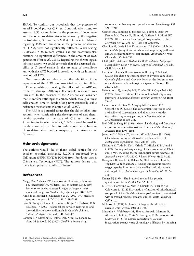
SHAM. To confirm our hypothesis that the presence of
an ARP could protect C. krusei from oxidative stress, we
assessed ROS accumulation in the presence of fluconazole
and the other oxidative stress inductors by the negative
control strain, S. cerevisiae (AOX-). The results obtained
regarding ROS accumulation, in the presence or absence
of SHAM, were not significantly different. When testing
C. albicans AOX mutant strains, Yan and coworkers also
obtained no significant differences in the amount of ROS
generation (Yan et al., 2009). Regarding the chronological
life span assays, we could conclude that the decreased via-
bility of C. krusei strains in the presence of menadione
and with the AOX blocked is associated with an increased
level of cell ROS.
Our results showed clearly that the inhibition of the
expression of the AOX was associated with intracellular
ROS accumulation, revealing the effect of the ARP on
oxidative damage. Although fluconazole resistance was
unrelated to the presence of the ARP, we can consider
that it confers antifungal tolerance, which may give yeast
cells enough time to develop long-term genetically stable
resistance mechanisms (Cannon et al., 2009).
The ARP is a potential target that should be taken into
account when considering the development of new thera-
peutic strategies in the case of C. krusei infections.
Attending to its selective effect, SHAM should be used in
combination with azoles, to reduce resistance because
of oxidative stress and consequently the virulence of
C. krusei.
Acknowledgements
The authors would like to thank Isabel Santos for the
excellent technical assistance. S.C.O. is supported by a
PhD grant (SFRH/BD/27662/2006) from Fundacao para a
Ciencia e a Tecnologia (FCT). The authors declare that
there is no potential conflict of interest.
References
Abegg MA, Alabarse PV, Casanova A, Hoscheid J, Salomon
TB, Hackenhaar FS, Medeiros TM & Benfato MS (2010)
Response to oxidative stress in eight pathogenic yeast
species of the genus Candida. Mycopathologia 170: 11–20.Almeida B, Buttner S, Ohlmeier S et al. (2007) NO-mediated
apoptosis in yeast. J Cell Sci 120: 3279–3288.Brun S, Aubry C, Lima O, Filmon R, Berges T, Chabasse D &
Bouchara JP (2003) Relationships between respiration and
susceptibility to azole antifungals in Candida glabrata.
Antimicrob Agents Chemother 47: 847–853.Cannon RD, Lamping E, Holmes AR, Niimi K, Tanabe K,
Niimi M & Monk BC (2007) Candida albicans drug
resistance another way to cope with stress. Microbiology 153:
3211–3217.Cannon RD, Lamping E, Holmes AR, Niimi K, Baret PV,
Keniya MV, Tanabe K, Niimi M, Goffeau A & Monk BC
(2009) Efflux-mediated antifungal drug resistance. Clin
Microbiol Rev 22: 291–321, Table of Contents.
Chamilos G, Lewis RE & Kontoyiannis DP (2006) Inhibition
of Candida parapsilosis mitochondrial respiratory pathways
enhances susceptibility to caspofungin. Antimicrob Agents
Chemother 50: 744–747.CLSI (2008) Reference Method for Broth Dilution Antifungal
Susceptibility Testing of Yeasts. Approved Standard, 3rd edn.
CLSI, Wayne, PA.
Hachem R, Hanna H, Kontoyiannis D, Jiang Y & Raad I
(2008) The changing epidemiology of invasive candidiasis:
Candida glabrata and Candida krusei as the leading causes
of candidemia in hematologic malignancy. Cancer 112:
2493–2499.Helmerhorst EJ, Murphy MP, Troxler RF & Oppenheim FG
(2002) Characterization of the mitochondrial respiratory
pathways in Candida albicans. Biochim Biophys Acta 1556:
73–80.Helmerhorst EJ, Stan M, Murphy MP, Sherman F &
Oppenheim FG (2005) The concomitant expression and
availability of conventional and alternative, cyanide-
insensitive, respiratory pathways in Candida albicans.
Mitochondrion 5: 200–211.Huh WK & Kang SO (1999) Molecular cloning and functional
expression of alternative oxidase from Candida albicans. J
Bacteriol 181: 4098–4102.Johnson CH, Prigge JT, Warren AD & McEwen JE (2003)
Characterization of an alternative oxidase activity of
Histoplasma capsulatum. Yeast 20: 381–388.Kirimura K, Yoda M, Ko I, Oshida Y, Miyake K & Usami S
(1999) Cloning and sequencing of the chromosomal DNA
and cDNA encoding the mitochondrial citrate synthase of
Aspergillus niger WU-2223L. J Biosci Bioeng 88: 237–243.Kobayashi D, Kondo K, Uehara N, Otokozawa S, Tsuji N,
Yagihashi A & Watanabe N (2002) Endogenous reactive
oxygen species is an important mediator of miconazole
antifungal effect. Antimicrob Agents Chemother 46: 3113–3117.
Kruger NJ (1994) The Bradford method for protein
quantitation. Methods Mol Biol 32: 9–15.Li D CH, Florentino A, Alex D, Sikorski P, Fonzi WA &
Calderone R (2011) Enzymatic dysfunction of mitochondrial
complex I of the Candida albicans goa1 mutant is associated
with increased reactive oxidants and cell death. Eukaryot
Cell 5: 10.
McIntosh L (1994) Molecular biology of the alternative
oxidase. Plant Physiol 105: 781–786.Mesquita A, Weinberger M, Silva A, Sampaio-Marques B,
Almeida B, Leao C, Costa V, Rodrigues F, Burhans WC &
Ludovico P (2010) Caloric restriction or catalase
inactivation extends yeast chronological lifespan by inducing
ª 2012 Federation of European Microbiological Societies FEMS Yeast Res 12 (2012) 423–429Published by Blackwell Publishing Ltd. All rights reserved
428 S. Costa-de-Oliveira et al.
H2O2 and superoxide dismutase activity. P Natl Acad Sci
USA 107: 15123–15128.Missall TA, Lodge JK & McEwen JE (2004) Mechanisms of
resistance to oxidative and nitrosative stress: implications
for fungal survival in mammalian hosts. Eukaryot Cell 3:
835–846.Moody JA (1991) Clinical Microbiology Procedures Handbook.
Synergism Testing. Broth Microdilution Checkerboard and
Broth Macrodilution Methods. American Society of
Microbiology, Washington, DC.
Moore AL & Siedow JN (1991) The regulation and nature of
the cyanide-resistant alternative oxidase of plant
mitochondria. Biochim Biophys Acta 1059: 121–140.Moradas-Ferreira P & Costa V (2000) Adaptive response of
the yeast Saccharomyces cerevisiae to reactive oxygen species:
defences, damage and death. Redox Rep 5: 277–285.Murphy JW (1991) Mechanisms of natural resistance to
human pathogenic fungi. Annu Rev Microbiol 45: 509–538.Oxman DA, Chow JK, Frendl G et al. (2010) Candidaemia
associated with decreased in vitro fluconazole susceptibility:
is Candida speciation predictive of the susceptibility pattern?
J Antimicrob Chemother 65: 1460–1465.Pappas PG, Kauffman CA, Andes D et al. (2009) Clinical
practice guidelines for the management of candidiasis: 2009
update by the Infectious Diseases Society of America. Clin
Infect Dis 48: 503–535.Raha S & Robinson BH (2000) Mitochondria, oxygen free
radicals, disease and ageing. Trends Biochem Sci 25: 502–508.
Vanlerberghe GC, Robson CA & Yip JY (2002) Induction of
mitochondrial alternative oxidase in response to a cell signal
pathway down-regulating the cytochrome pathway prevents
programmed cell death. Plant Physiol 129: 1829–1842.Veiga A, Arrabaca JD & Loureiro-Dias MC (2003a) Cyanide-
resistant respiration, a very frequent metabolic pathway in
yeasts. FEMS Yeast Res 3: 239–245.Veiga A, Arrabaca JD, Sansonetty F, Ludovico P, Corte-Real M
& Loureiro-Dias MC (2003b) Energy conversion coupled to
cyanide-resistant respiration in the yeasts Pichia
membranifaciens and Debaryomyces hansenii. FEMS
Yeast Res 3: 141–148.Yan L, Li M, Cao Y, Gao P, Wang Y & Jiang Y (2009) The
alternative oxidase of Candida albicans causes reduced
fluconazole susceptibility. J Antimicrob Chemother 64: 764–773.
FEMS Yeast Res 12 (2012) 423–429 ª 2012 Federation of European Microbiological SocietiesPublished by Blackwell Publishing Ltd. All rights reserved
Alternative respiratory pathway on C. krusei 429
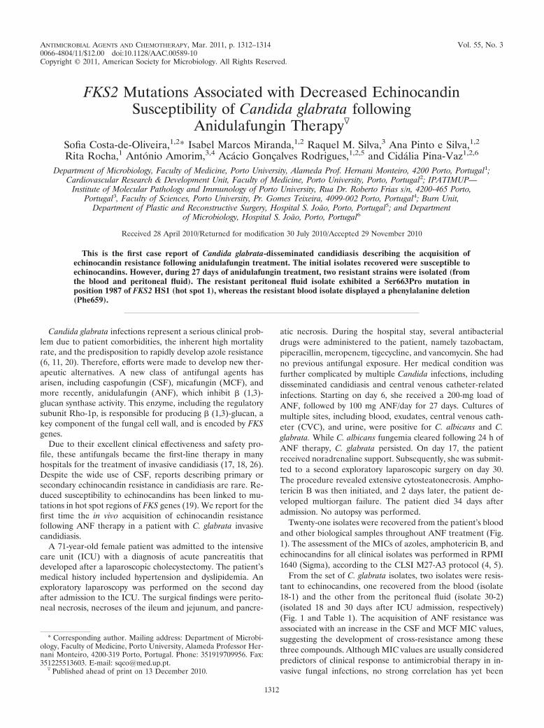
ANTIMICROBIAL AGENTS AND CHEMOTHERAPY, Mar. 2011, p. 1312–1314 Vol. 55, No. 30066-4804/11/$12.00 doi:10.1128/AAC.00589-10Copyright © 2011, American Society for Microbiology. All Rights Reserved.
FKS2 Mutations Associated with Decreased EchinocandinSusceptibility of Candida glabrata following
Anidulafungin Therapy�
Sofia Costa-de-Oliveira,1,2* Isabel Marcos Miranda,1,2 Raquel M. Silva,3 Ana Pinto e Silva,1,2
Rita Rocha,1 Antonio Amorim,3,4 Acacio Goncalves Rodrigues,1,2,5 and Cidalia Pina-Vaz1,2,6
Department of Microbiology, Faculty of Medicine, Porto University, Alameda Prof. Hernani Monteiro, 4200 Porto, Portugal1;Cardiovascular Research & Development Unit, Faculty of Medicine, Porto University, Porto, Portugal2; IPATIMUP—
Institute of Molecular Pathology and Immunology of Porto University, Rua Dr. Roberto Frias s/n, 4200-465 Porto,Portugal3, Faculty of Sciences, Porto University, Pr. Gomes Teixeira, 4099-002 Porto, Portugal4; Burn Unit,
Department of Plastic and Reconstructive Surgery, Hospital S. Joao, Porto, Portugal5; and Departmentof Microbiology, Hospital S. Joao, Porto, Portugal6
Received 28 April 2010/Returned for modification 30 July 2010/Accepted 29 November 2010
This is the first case report of Candida glabrata-disseminated candidiasis describing the acquisition ofechinocandin resistance following anidulafungin treatment. The initial isolates recovered were susceptible toechinocandins. However, during 27 days of anidulafungin treatment, two resistant strains were isolated (fromthe blood and peritoneal fluid). The resistant peritoneal fluid isolate exhibited a Ser663Pro mutation inposition 1987 of FKS2 HS1 (hot spot 1), whereas the resistant blood isolate displayed a phenylalanine deletion(Phe659).
Candida glabrata infections represent a serious clinical prob-lem due to patient comorbidities, the inherent high mortalityrate, and the predisposition to rapidly develop azole resistance(6, 11, 20). Therefore, efforts were made to develop new ther-apeutic alternatives. A new class of antifungal agents hasarisen, including caspofungin (CSF), micafungin (MCF), andmore recently, anidulafungin (ANF), which inhibit � (1,3)-glucan synthase activity. This enzyme, including the regulatorysubunit Rho-1p, is responsible for producing � (1,3)-glucan, akey component of the fungal cell wall, and is encoded by FKSgenes.
Due to their excellent clinical effectiveness and safety pro-file, these antifungals became the first-line therapy in manyhospitals for the treatment of invasive candidiasis (17, 18, 26).Despite the wide use of CSF, reports describing primary orsecondary echinocandin resistance in candidiasis are rare. Re-duced susceptibility to echinocandins has been linked to mu-tations in hot spot regions of FKS genes (19). We report for thefirst time the in vivo acquisition of echinocandin resistancefollowing ANF therapy in a patient with C. glabrata invasivecandidiasis.
A 71-year-old female patient was admitted to the intensivecare unit (ICU) with a diagnosis of acute pancreatitis thatdeveloped after a laparoscopic cholecystectomy. The patient’smedical history included hypertension and dyslipidemia. Anexploratory laparoscopy was performed on the second dayafter admission to the ICU. The surgical findings were perito-neal necrosis, necroses of the ileum and jejunum, and pancre-
atic necrosis. During the hospital stay, several antibacterialdrugs were administered to the patient, namely tazobactam,piperacillin, meropenem, tigecycline, and vancomycin. She hadno previous antifungal exposure. Her medical condition wasfurther complicated by multiple Candida infections, includingdisseminated candidiasis and central venous catheter-relatedinfections. Starting on day 6, she received a 200-mg load ofANF, followed by 100 mg ANF/day for 27 days. Cultures ofmultiple sites, including blood, exudates, central venous cath-eter (CVC), and urine, were positive for C. albicans and C.glabrata. While C. albicans fungemia cleared following 24 h ofANF therapy, C. glabrata persisted. On day 17, the patientreceived noradrenaline support. Subsequently, she was submit-ted to a second exploratory laparoscopic surgery on day 30.The procedure revealed extensive cytosteatonecrosis. Ampho-tericin B was then initiated, and 2 days later, the patient de-veloped multiorgan failure. The patient died 34 days afteradmission. No autopsy was performed.
Twenty-one isolates were recovered from the patient’s bloodand other biological samples throughout ANF treatment (Fig.1). The assessment of the MICs of azoles, amphotericin B, andechinocandins for all clinical isolates was performed in RPMI1640 (Sigma), according to the CLSI M27-A3 protocol (4, 5).
From the set of C. glabrata isolates, two isolates were resis-tant to echinocandins, one recovered from the blood (isolate18-1) and the other from the peritoneal fluid (isolate 30-2)(isolated 18 and 30 days after ICU admission, respectively)(Fig. 1 and Table 1). The acquisition of ANF resistance wasassociated with an increase in the CSF and MCF MIC values,suggesting the development of cross-resistance among thesethree compounds. Although MIC values are usually consideredpredictors of clinical response to antimicrobial therapy in in-vasive fungal infections, no strong correlation has yet been
* Corresponding author. Mailing address: Department of Microbi-ology, Faculty of Medicine, Porto University, Alameda Professor Her-nani Monteiro, 4200-319 Porto, Portugal. Phone: 351919709956. Fax:351225513603. E-mail: [email protected].
� Published ahead of print on 13 December 2010.
1312
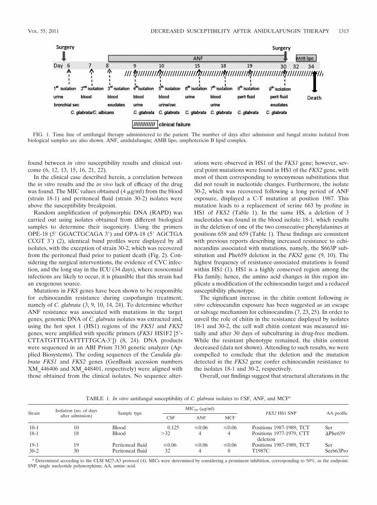
found between in vitro susceptibility results and clinical out-come (6, 12, 13, 15, 16, 21, 22).
In the clinical case described herein, a correlation betweenthe in vitro results and the in vivo lack of efficacy of the drugwas found. The MIC values obtained (4 �g/ml) from the blood(strain 18-1) and peritoneal fluid (strain 30-2) isolates wereabove the susceptibility breakpoint.
Random amplification of polymorphic DNA (RAPD) wascarried out using isolates obtained from different biologicalsamples to determine their isogenicity. Using the primersOPE-18 (5� GGACTGCAGA 3�) and OPA-18 (5� AGCTGACCGT 3�) (2), identical band profiles were displayed by allisolates, with the exception of strain 30-2, which was recoveredfrom the peritoneal fluid prior to patient death (Fig. 2). Con-sidering the surgical interventions, the evidence of CVC infec-tion, and the long stay in the ICU (34 days), where nosocomialinfections are likely to occur, it is plausible that this strain hadan exogenous source.
Mutations in FKS genes have been shown to be responsiblefor echinocandin resistance during caspofungin treatment,namely of C. glabrata (3, 9, 10, 14, 24). To determine whetherANF resistance was associated with mutations in the targetgenes, genomic DNA of C. glabrata isolates was extracted and,using the hot spot 1 (HS1) regions of the FKS1 and FKS2genes, were amplified with specific primers (FKS1 HS1F2 [5�-CTTATGTTTGATTTTTGCA-3�]) (8, 24). DNA productswere sequenced in an ABI Prism 3130 genetic analyzer (Ap-plied Biosystems). The coding sequences of the Candida gla-brata FKS1 and FKS2 genes (GenBank accession numbersXM_446406 and XM_448401, respectively) were aligned withthose obtained from the clinical isolates. No sequence alter-
ations were observed in HS1 of the FKS1 gene; however, sev-eral point mutations were found in HS1 of the FKS2 gene, withmost of them corresponding to synonymous substitutions thatdid not result in nucleotide changes. Furthermore, the isolate30-2, which was recovered following a long period of ANFexposure, displayed a C-T mutation at position 1987. Thismutation leads to a replacement of serine 663 by proline inHS1 of FKS2 (Table 1). In the same HS, a deletion of 3nucleotides was found in the blood isolate 18-1, which resultsin the deletion of one of the two consecutive phenylalanines atpositions 658 and 659 (Table 1). These findings are consistentwith previous reports describing increased resistance to echi-nocandins associated with mutations, namely, the S663P sub-stitution and Phe659 deletion in the FKS2 gene (9, 10). Thehighest frequency of resistance-associated mutations is foundwithin HS1 (1). HS1 is a highly conserved region among theFks family; hence, the amino acid changes in this region im-plicate a modification of the echinocandin target and a reducedsusceptibility phenotype.
The significant increase in the chitin content following invitro echinocandin exposure has been suggested as an escapeor salvage mechanism for echinocandins (7, 23, 25). In order tounveil the role of chitin in the resistance displayed by isolates18-1 and 30-2, the cell wall chitin content was measured ini-tially and after 30 days of subculturing in drug-free medium.While the resistant phenotype remained, the chitin contentdecreased (data not shown). Attending to such results, we werecompelled to conclude that the deletion and the mutationdetected in the FKS2 gene confer echinocandin resistance tothe isolates 18-1 and 30-2, respectively.
Overall, our findings suggest that structural alterations in the
FIG. 1. Time line of antifungal therapy administered to the patient. The number of days after admission and fungal strains isolated frombiological samples are also shown. ANF, anidulafungin; AMB lipo, amphotericin B lipid complex.
TABLE 1. In vitro antifungal susceptibility of C. glabrata isolates to CSF, ANF, and MCFa
Strain Isolation (no. of daysafter admission) Sample type
MIC50 (�g/ml)FKS2 HS1 SNP AA profile
CSF ANF MCF
10-1 10 Blood 0.125 �0.06 �0.06 Positions 1987-1989, TCT Ser18-1 18 Blood �32 4 4 Positions 1977-1979, CTT
deletion�Phe659
19-1 19 Peritoneal fluid �0.06 �0.06 �0.06 Positions 1987-1989, TCT Ser30-2 30 Peritoneal fluid 32 4 8 T1987C Ser663Pro
a Determined according to the CLSI M27-A3 protocol (4). MICs were determined by considering a prominent inhibition, corresponding to 50%, as the endpoint.SNP, single nucleotide polymorphism; AA, amino acid.
VOL. 55, 2011 DECREASED SUSCEPTIBILITY AFTER ANIDULAFUNGIN THERAPY 1313

HS1 of the FKS2 molecule due to the Ser663Pro substitutionand Phe659 deletion lead to a dramatic decrease in echinocan-din efficacy. Although just a few cases describing the develop-ment of echinocandin resistance have been reported so far, ourcase report emphasizes the crucial need for antifungal suscep-tibility surveillance in patients under extended echinocandintherapy.
S.C.-D.-O. and A.P.E.S. are supported by Ph.D. grants (SFRH/BD/27662/2006 and SFRH/BD/29540/2006, respectively) from the Funda-cao para a Ciencia e a Tecnologia (FCT). IPATIMUP is partiallysupported by the Programa Operacional Ciencia e Inovacao 2010(POCI 2010), Sixth Framework Programme (2002 to 2006). R.M.S.and I.M.M. are supported by FCT Ciencia 2007 and by FCT Ciencia2008, respectively, and cofinanced by the European Social Fund.
REFERENCES
1. Balashov, S. V., S. Park, and D. S. Perlin. 2006. Assessing resistance to theechinocandin antifungal drug caspofungin in Candida albicans by profilingmutations in FKS1. Antimicrob. Agents Chemother. 50:2058–2063.
2. Bautista-Munoz, C., X. M. Boldo, L. Villa-Tanaca, and C. Hernandez-Ro-dríguez. 2003. Identification of Candida spp. by randomly amplified poly-morphic DNA analysis and differentiation between Candida albicans andCandida dubliniensis by direct PCR methods. J. Clin. Microbiol. 41:414–420.
3. Castanheira, M., et al. 2010. Low prevalence of fks1 Hotspot 1 mutations ina worldwide collection of Candida spp. Antimicrob. Agents Chemother.54:2655–2659.
4. Clinical and Laboratory Standards Institute (CLSI). 2008. Referencemethod for broth dilution antifungal susceptibility testing of yeasts, 3rd ed.Approved standard M27-A3. CLSI, Wayne, PA.
5. Clinical and Laboratory Standards Institute (CLSI). 2008. Referencemethod for broth dilution antifungal susceptibility testing of yeasts; thirdinformational supplement. M27-S3. CLSI, Wayne, PA.
6. Costa-de-Oliveira, S., C. Pina-Vaz, D. Mendonca, and A. Goncalves Ro-drigues. 2008. A first Portuguese epidemiological survey of fungaemia in auniversity hospital. Eur. J. Clin. Microbiol. Infect. Dis. 27:365–374.
7. Cota, J. M., et al. 2008. Increases in SLT2 expression and chitin content areassociated with incomplete killing of Candida glabrata by caspofungin. An-timicrob. Agents Chemother. 52:1144–1146.
8. Garcia-Effron, G., S. K. Katiyar, S. Park, T. D. Edlind, and D. S. Perlin.2008. A naturally occurring proline-to-alanine amino acid change in Fks1p inCandida parapsilosis, Candida orthopsilosis, and Candida metapsilosis ac-counts for reduced echinocandin susceptibility. Antimicrob. Agents Che-mother. 52:2305–2312.
9. Garcia-Effron, G., S. Lee, S. Park, J. D. Cleary, and D. S. Perlin. 2009. Effectof Candida glabrata FKS1 and FKS2 mutations on echinocandin sensitivityand kinetics of 1,3-beta-D-glucan synthase: implication for the existing sus-ceptibility breakpoint. Antimicrob. Agents Chemother. 53:3690–3699.
10. Garcia-Effron, G., et al. 2010. Novel FKS mutations associated with echino-candin resistance in Candida species. Antimicrob. Agents Chemother. 54:2225–2227.
11. Hachem, R., H. Hanna, D. Kontoyiannis, Y. Jiang, and I. Raad. 2008. Thechanging epidemiology of invasive candidiasis: Candida glabrata and Can-dida krusei as the leading causes of candidemia in hematologic malignancy.Cancer 112:2493–2499.
12. Hernandez, S., et al. 2004. Caspofungin resistance in Candida albicans:correlating clinical outcome with laboratory susceptibility testing of threeisogenic isolates serially obtained from a patient with progressive Candidaesophagitis. Antimicrob. Agents Chemother. 48:1382–1383.
13. Kartsonis, N., et al. 2005. Caspofungin susceptibility testing of isolates frompatients with esophageal candidiasis or invasive candidiasis: relationship ofMIC to treatment outcome. Antimicrob. Agents Chemother. 49:3616–3623.
14. Katiyar, S., M. Pfaller, and T. Edlind. 2006. Candida albicans and Candidaglabrata clinical isolates exhibiting reduced echinocandin susceptibility. An-timicrob. Agents Chemother. 50:2892–2894.
15. Krogh-Madsen, M., M. C. Arendrup, L. Heslet, and J. D. Knudsen. 2006.Amphotericin B and caspofungin resistance in Candida glabrata isolatesrecovered from a critically ill patient. Clin. Infect. Dis. 42:938–944.
16. Laverdiere, M., et al. 2006. Progressive loss of echinocandin activity follow-ing prolonged use for treatment of Candida albicans oesophagitis. J. Anti-microb. Chemother. 57:705–708.
17. Pappas, P. G., et al. 2009. Clinical practice guidelines for the management ofcandidiasis: 2009 update by the Infectious Diseases Society of America. Clin.Infect. Dis. 48:503–535.
18. Park, S., et al. 2005. Specific substitutions in the echinocandin target Fks1paccount for reduced susceptibility of rare laboratory and clinical Candida sp.isolates. Antimicrob. Agents Chemother. 49:3264–3273.
19. Perlin, D. S. 2007. Resistance to echinocandin-class antifungal drugs. DrugResist. Updat. 10:121–130.
20. Pfaller, M. A., and D. J. Diekema. 2002. Role of sentinel surveillance ofcandidemia: trends in species distribution and antifungal susceptibility.J. Clin. Microbiol. 40:3551–3557.
21. Pfaller, M. A., et al. 2005. Effectiveness of anidulafungin in eradicatingCandida species in invasive candidiasis. Antimicrob. Agents Chemother.49:4795–4797.
22. Rex, J. H., and M. A. Pfaller. 2002. Has antifungal susceptibility testing comeof age? Clin. Infect. Dis. 35:982–989.
23. Stevens, D. A., M. Ichinomiya, Y. Koshi, and H. Horiuchi. 2006. Escape ofCandida from caspofungin inhibition at concentrations above the MIC (par-adoxical effect) accomplished by increased cell wall chitin; evidence forbeta-1,6-glucan synthesis inhibition by caspofungin. Antimicrob. AgentsChemother. 50:3160–3161.
24. Thompson, G. R., III, et al. 2008. Development of caspofungin resistancefollowing prolonged therapy for invasive candidiasis secondary to Candidaglabrata infection. Antimicrob. Agents Chemother. 52:3783–3785.
25. Walker, L. A., et al. 2008. Stimulation of chitin synthesis rescues Candidaalbicans from echinocandins. PLoS Pathog. 4:e1000040.
26. Wiederhold, N. P., and R. E. Lewis. 2003. The echinocandin antifungals: anoverview of the pharmacology, spectrum and clinical efficacy. Expert Opin.Investig. Drugs 12:1313–1333.
FIG. 2. Random amplification of polymorphic DNA gel patterns ofC. glabrata isolates 18-1, 30-2, 10-1, and 19-1, resistant and susceptibleto echinocandins, obtained with primers OPE-18 and OPA-18.
1314 COSTA-DE-OLIVEIRA ET AL. ANTIMICROB. AGENTS CHEMOTHER.
Received 24 March 2010; Receive
Accepted 26 July 2010
Correspondence: Sofi a Costa-de-O
Faculty of Medicine, University o
Monteiro, 4200-319 Porto, Portug
1225513603; E-mail: [email protected]
Genetic relatedness and antifungal susceptibility profi le of
Candida albicans isolates from fungaemia patients
SOFIA COSTA-DE-OLIVEIRA * , † , IN Ê S SOUSA ‡ , ALEXANDRA CORREIA ‡ , PAULA SAMPAIO ‡ , C É LIA PAIS ‡ ,
AC Á CIO GON Ç ALVES RODRIGUES * , † , § & CID Á LIA PINA-VAZ * , † ,#
* Department of Microbiology, Faculty of Medicine, University of Porto, Porto, † Cardiovascular Research & Development Unit,
Faculty of Medicine, University of Porto, Porto, ‡ Centre of Molecular and Environmental Biology (CBMA), Department of Biology
University of Minho, Braga, § Burn Unit, Department of Plastic and Reconstructive Surgery, Faculty of Medicine, University of Porto,
Porto, and # Department of Microbiology, Hospital S ã o Jo ã o, Porto, Portugal
© 2010 ISHAM
Medical Mycology Month 2010, Early Online, 1–5
Med
Myc
ol D
ownl
oade
d fr
om in
form
ahea
lthca
re.c
om b
y U
nive
rsid
ade
Do
Port
o on
09/
08/1
0Fo
r pe
rson
al u
se o
nly.
A prospective study to assess fungaemia was conducted for 12 months at a Portuguese University Hospital. A total of 35 Candida albicans isolates obtained from 12 patients with fungaemia were compared by a multiplex PCR system using four microsatellite loci. Blood isolates were evaluated against concomitant isolates from urine, lower respi-ratory secretions and central venous catheters, as well as with successive isolates recov-ered from recurrent episodes of fungaemia. The data analyzed included the department of admission, underlying diseases and antifungal therapy. The susceptibility phenotypes of all isolates to amphotericin B, fl uconazole, itraconazole, voriconazole and caspo-fungin were determined according to the CLSI M27-A3 protocol. We observed a high degree of similarity between successive blood isolates and between blood and concomi-tant isolates from other sites of the same patient. This is suggestive of the recurrence of fungaemia and was due to the same strain, possibly as a result of the failure of antifungal therapy. The genetic similarity observed between some strains isolated from different patients suggested the likelihood that they were hospital acquired. Distinct patients were infected by the same strain at different time periods, and an increase in antifungal resist-ance was observed over time for some of these strains. Hospital-acquired exogenous nosocomial infections can be associated with higher risks of antifungal resistance and need to be closely monitored. Particular attention should also be given to endogenous non-blood Candida isolates which can be critical in high risk patients, as they often can become invasive in immunodefi cient individuals.
Keywords microsatellite , Candida albicans , fungaemia , genetic relatedness , antifungals , infection
Introduction
Candida infections have progressively emerged as major
nosocomial invasive fungal diseases since the late 1980s,
mainly arising from an endogenous source, i.e., either diges-
tive or mucocutaneous. Commensalism, followed by colo-
nization, usually precedes dissemination, most frequently in
patients with transient or permanent immunocompromised
d in fi nal revised form 1 July 2010;
liveira, Department of Microbiology,
f Porto, Alameda Professor Hernani
al. Tel: � 35 1919709956; fax: � 35
.pt
status, such as transplant recipients, chemotherapy patients,
underweight neonates and those infected by the human
immunodefi ciency virus. Signifi cant morbidity and mortal-
ity rates have been associated with Candida spp. blood-
stream infections [1 – 3].
Several polymorphic microsatellite loci have been iden-
tifi ed in the genome of C. albicans near EF3 , CDC3 and
HIS3 [4] or inside the coding regions of ERK1 , 2NF1 ,
CCN2 , CPH2 , and EFG1 [5]. However the discriminatory
power for each locus is relatively low. In order to more
rapidly obtain a higher discrimination, simultaneous ampli-
fi cation of sets of microsatellite markers can be performed.
A multiplex system with a high discriminatory power was
recently described and found to represent an effi cient
DOI: 10.3109/13693786.2010.511633

2 Costa-de-Oliveira et al .
Med
Myc
ol D
ownl
oade
d fr
om in
form
ahea
lthca
re.c
om b
y U
nive
rsid
ade
Do
Port
o on
09/
08/1
0Fo
r pe
rson
al u
se o
nly.
molecular tool for the swift and accurate differentiation of
C. albicans [6,7] isolates.
During a 12-month period (2004) a prospective study
addressing fungaemia was conducted at Hospital de S ã o Jo ã o,
a large university hospital located in the Northern region of
Portugal [1]. The epidemiological data analyzed included,
among others, the department of admission, underlying dis-
eases and antimicrobial therapy. Several yeast isolates from
blood cultures were collected and studied, as well as fungi
recovered from surveillance cultures or from medical indwell-
ing devices. These included isolates from distinct biological
sources of the same patient, such as urine and lower respira-
tory secretions, and from central venous catheters. All
episodes of recurrence were investigated [1].
Our main purpose was to genotype all C. albicans iso-
lates by using a multiplex PCR system with four microsat-
ellite loci. Ultimately, we aimed to determine the genetic
relatedness between simultaneous and/or recurrent isolates
from the same patient, the source of infection, the route of
transmission, as well as the possibility of transmission
among distinct patients. Additionally, the susceptibility
profi les of all isolates were determined and evaluated with
respect to antifungal therapy.
Materials and methods
Patients ’ clinical data
Thirty-fi ve C. albicans isolates recovered from the blood
and other biological sources were obtained from 12 patients
with fungaemia. The data documented included patient
gender and age, department and date of admission, con-
comitant therapy (i.e., immunossuppressors), date and site
of fungal isolation, and nosocomial origin of the fungae-
mia. According to the Centre for Disease Control and Pre-
vention (CDC), nosocomial fungaemia is defi ned as having
occurred whenever at least one fungal positive blood cul-
ture is obtained within 48 h of the patient ’ s hospital admis-
sion [8]. Antifungal therapy (prophylactic or therapeutic)
and clinical outcome (survival/death) were also docu-
mented. Outcomes were evaluated 30 days after the fi rst
fungaemia episode, with related deaths being defi ned as
those that occurred within 30 days of the fi rst fungal posi-
tive blood culture, with no signs of intracerebral or gastro-
intestinal bleeding or pulmonary embolism. All Candida
isolates were frozen at – 70 ° C in brain-heart medium with
5% of glycerol (Difco) and subcultured twice on Sab-
ouraud agar (Difco) prior to experimental procedures.
C. albicans genotyping
Yeast strains were grown overnight in Sabouraud broth
at 30 ° C. A Zymolyase-based method was used to extract
DNA, as previously described [9]. To assess strain
relatedness, all isolates were genotyped using a micro-
satellite multiplex PCR assay with three markers (CAI,
CAIII and CAVI) as described by Sampaio et al . [7] and a
singleplex amplifi cation reaction assay using the microsat-
ellite marker CEF3, according to procedures described by
Bretagne et al . [4]. Following PCR amplifi cation, a 1 – 2- μ l
aliquot of each sample was added to 15 μ l of formamide
containing 0.4 μ l of GeneScan - 500 TAMRA size stan-
dards (Applied Biosystems). Amplicons were denatured at
95 ° C for 5 min and immediately placed on ice. Denatured
samples were resolved by capillary electrophoresis in an
ABI Prism 310 genetic analyzer (Applied Biosystems).
Determination of allele sizes was automatically performed
with GeneScan 3.7 analysis software. The alleles were des-
ignated according to the number of repeated units for the
CAI [(CAA) 2 CTG(CAA) n ], CAIII [(GAA) n ] and CAVI
((TAAA) n) markers and by the number of nucleotides for
the CEF3 [(TTTC) n (TTC) n] marker.
Antifungal susceptibility profi le
Antifungal susceptibility testing of all isolates was performed
according to the CLSI M27 A3 protocol in RPMI 1640
(Sigma) [10,11]. Minimal inhibitory concentration (MIC) for
fl uconazole, voriconazole, caspofungin and amphotericin B
were determined [10,11]. The yeast strains were classifi ed as
susceptible (S), susceptible-dose dependent (S-DD) and resis-
tant ® to azoles according to the breakpoints defi ned by CLSI
[10,11]. Although defi nitive breakpoints have not yet
been established for amphotericin B, strains showing MIC
� 1 μ g/ml were considered susceptible [12]. For caspofungin,
strains with an MIC � 2 μ g/ml corresponded to S and
� 2 μ g/ml to non-susceptible (NS) [10,11]. Strains display-
ing a MIC variation within one dilution were considered as
having a similar susceptibility pattern.
Results
The patients ’ age ranged from 40 – 83 years old and 75%
(9/12) were male. Data such as department of admission,
date of fi rst isolation and strain origin are presented in
Table 1 and Fig. 1. All patients were iatrogenically immu-
nosuppressed, and 75% died. All fungaemia cases corre-
sponded to episodes of nosocomial infections. The
microsatellite genotyping analysis is detailed in Table 1.
Among the 12 patients, two or more isolates were
obtained from blood and from additional body locations. We
considered isolates as being highly related when their geno-
types differed at only a single locus. In the large majority
of patients, blood isolates displayed the same multilocus
genotype or a genotype that was highly similar to that of
isolates from other body locations. In one single case, patient
10, one isolate from urine exhibited a completely distinct
© 2010 ISHAM, Early Online, 1–5

Med
Myc
ol D
ownl
oade
d fr
om in
form
ahea
lthca
re.c
om b
y U
nive
rsid
ade
Do
Port
o on
09/
08/1
0Fo
r pe
rson
al u
se o
nly.
multilocus genotype from the blood isolate, even though
they were collected at the same time. Isolate 9.3 from urine
displayed a genotype that differed at two loci compared to
the isolates obtained from the respiratory tract or blood iso-
lates. This isolate was moderately related to the other iso-
lates from the same patient but was highly related to isolates
from patients 6, 7 and 8. In two patients, 11 and 12, minor
genetic differences were detected among blood isolates and
those from other body sources. In these patients, the isolates
were highly related, suggestive of microevolution.
Consecutive blood isolates from the same patient exhibited
the same microsatellite genotype in 5 out of 6 patients. The
exception was patient 12 where a highly related isolate was
observed between two consecutive blood cultures. This
© 2010 ISHAM, Early Online, 1–5
was indicative of strain maintenance regardless of antifun-
gal treatment, rather than re-infection by a distinct strain
(Table 1). Isolates from patients 3, 4 and 5 showed identi-
cal or highly similar microsatellite profi les (Table 1), an
interesting fact since these patients had initially been
simultaneously admitted in the same intensive care unit
(ICU). Isolates from patients 6, 7 and 8 also exhibited iden-
tical or highly similar genotypes (Table 1) and were ini-
tially admitted to the same ICU, although not concurrently.
All isolates from patients 1 and 2, who where in the same
ICU in the previous months, displayed the same genotype
(Table 1). Sharing of the same strain among different
patients is suggestive of hospital-acquired exogenous
nosocomial infection with hospital endemic strains.
Genetic relatedness of C. albicans strains 3
Table 1 Candida albicans isolates from blood cultures and from other body sites obtained from 12 patients and respective multilocus genotyping
results.
Patient
Isolate
no. Source
Isolation
date(day/
month)
Microsatellite genotype MIC m g/ml
CAI CAIII CAVI CEF3 FLC ITC VRC AmB CAS
1 1.1 Blood 21/4 11-11 5-6 7-11 129-143 � 64 � 8 � 8 1 0.5
1.2 Bronchial washing
fl uid
22/4 11-11 5-6 7-11 129-143 � 64 � 8 � 8 0.5 1
2 2.1 Lower respiratory
secretions
15/4 11-11 5-6 7-11 129-143 � 64 4 � 8 0.5 0.5
2.2 Urine 15/4 11-11 5-6 7-11 129-143 � 64 4 � 8 0.5 0.5
2.3 Blood 16/4 11-11 5-6 7-11 129-143 � 64 4 � 8 0.5 0.5
3 3.1 Central venous
catheter
02/4 26-28 6-6 7-7 136-145 0.125 0.015 0.015 0.25 1
3.2 Blood 22/4 26-28 6-6 7-7 136-145 0.125 0.015 0.015 0.25 13.3 Urine 25/4 26-28 6-6 7-7 136-145 0.125 0.015 0.015 0.25 13.4 Blood 30/4 26-28 6-6 7-7 136-145 0.125 0.015 0.015 0.25 1
4 4.1 Blood 30 /8 26-26 6-6 7-7 136-145 32 � 8 � 8 0.5 0.25
4.2 Urine 30 /8 26-26 6-6 7-7 136-145 32 � 8 � 8 0.5 1
4.3 Blood 08 /9 26-26 6-6 7-7 136-145 32 � 8 � 8 0.5 1
5 5.1 Blood 20 /9 26-26 6-6 7-7 136-145 � 64 � 8 � 8 1 1
5.2 Lower respiratory
secretions
20 /9 26-26 6-6 7-7 136-145 � 64 � 8 � 8 1 1
6 6.1 Blood 14/4 25-25 6-7 9-14 131-131 0.25 0.015 0.015 0.25 0.256.2 Blood 26/4 25-25 6-7 9-14 131-131 0.25 0.5 0.03 0.5 0.5
7 7.1 Blood 11/7 21-25 6-7 9-14 131-131 0.5 0.03 0.015 0.25 0.257.2 Blood 19/7 21-25 6-7 9-14 131-131 0.5 0.03 0.015 0.25 0.5
8 8.1 Blood 22/10 21-25 6-7 9-14 131-131 � 64 � 8 � 8 0.25 1
8.2 Pleural fl uid 25/10 21-25 6-7 9-14 131-131 � 64 � 8 � 8 0.5 0.5
9 9.1 Blood 26/10 21-26 7-7 9-14 130-131 16 0.06 0.25 0.5 19.2 Blood 05/11 21-26 7-7 9-14 130-131 16 0.06 0.25 0.5 19.3 Urine 08/11 21-25 6-7 9-14 130-131 16 0.06 0.25 0.5 19.4 Lower respiratory
secretions
08/11 21-26 7-7 9-14 130-131 16 0.06 0.25 0.5 1
10 10.1 Blood 02/8 11-17 5-5 7-7 126-135 � 64 4 � 8 0.5 1
10.2 Urine 02/8 27-46 6-11 20-20 139-144 � 64 4 � 8 0.5 1
11 11.1 Blood 21/9 29-29 6-6 7-11 129-143 � 64 � 8 � 8 1 1
11.2 Urine 21/9 28-29 6-6 7-11 129-143 � 64 � 8 � 8 1 1
12 12.1 Catheter 18/5 18-34 6-7 40-40 137-139 � 64 2 � 8 0.5 1
12.2 Blood 31/7 18-34 6-7 42-42 137-139 � 64 4 � 8 0.5 1
12.3 Lower respiratory
secretions31/7 34-34 6-7 40-40 137-139 � 64 0.5 0.03 0.5 0.5
12.4 Blood 26/8 18-34 6-7 40-40 137-139 � 64 � 8 � 8 0.25 0.25
Strain replacement is highlighted in dark grey; microevolutionary events are highlighted in light grey.
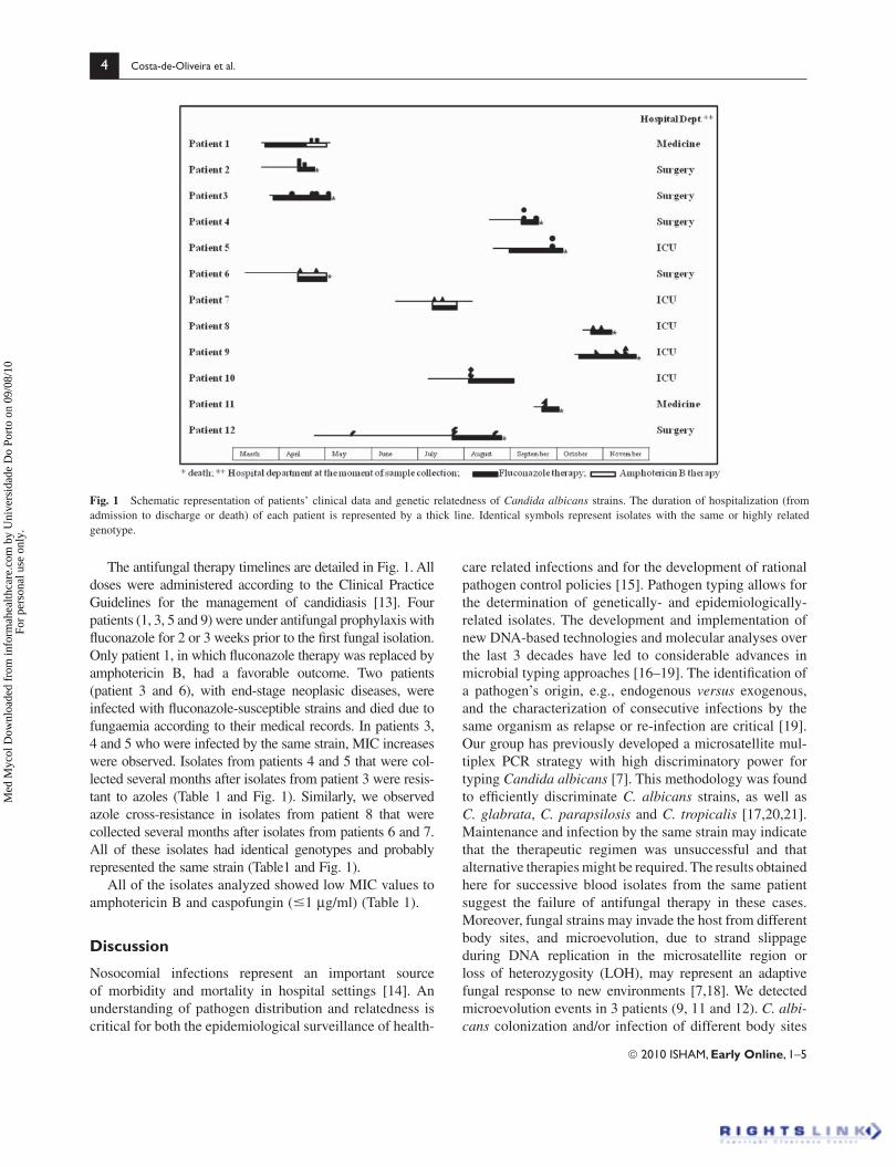
4 Costa-de-Oliveira et al .
Med
Myc
ol D
ownl
oade
d fr
om in
form
ahea
lthca
re.c
om b
y U
nive
rsid
ade
Do
Port
o on
09/
08/1
0Fo
r pe
rson
al u
se o
nly.
The antifungal therapy timelines are detailed in Fig. 1. All
doses were administered according to the Clinical Practice
Guidelines for the management of candidiasis [13]. Four
patients (1, 3, 5 and 9) were under antifungal prophylaxis with
fl uconazole for 2 or 3 weeks prior to the fi rst fungal isolation.
Only patient 1, in which fl uconazole therapy was replaced by
amphotericin B, had a favorable outcome. Two patients
(patient 3 and 6), with end-stage neoplasic diseases, were
infected with fl uconazole-susceptible strains and died due to
fungaemia according to their medical records. In patients 3,
4 and 5 who were infected by the same strain, MIC increases
were observed. Isolates from patients 4 and 5 that were col-
lected several months after isolates from patient 3 were resis-
tant to azoles (Table 1 and Fig. 1). Similarly, we observed
azole cross-resistance in isolates from patient 8 that were
collected several months after isolates from patients 6 and 7.
All of these isolates had identical genotypes and probably
represented the same strain (Table1 and Fig. 1).
All of the isolates analyzed showed low MIC values to
amphotericin B and caspofungin ( � 1 μ g/ml) (Table 1).
Discussion
Nosocomial infections represent an important source
of morbidity and mortality in hospital settings [14]. An
understanding of pathogen distribution and relatedness is
critical for both the epidemiological surveillance of health-
care related infections and for the development of rational
pathogen control policies [15]. Pathogen typing allows for
the determination of genetically- and epidemiologically-
related isolates. The development and implementation of
new DNA-based technologies and molecular analyses over
the last 3 decades have led to considerable advances in
microbial typing approaches [16 – 19]. The identifi cation of
a pathogen ’ s origin, e.g., endogenous versus exogenous,
and the characterization of consecutive infections by the
same organism as relapse or re-infection are critical [19].
Our group has previously developed a microsatellite mul-
tiplex PCR strategy with high discriminatory power for
typing Candida albicans [7]. This methodology was found
to effi ciently discriminate C. albicans strains, as well as
C. glabrata , C. parapsilosis and C. tropicalis [17,20,21].
Maintenance and infection by the same strain may indicate
that the therapeutic regimen was unsuccessful and that
alternative therapies might be required. The results obtained
here for successive blood isolates from the same patient
suggest the failure of antifungal therapy in these cases.
Moreover, fungal strains may invade the host from different
body sites, and microevolution, due to strand slippage
during DNA replication in the microsatellite region or
loss of heterozygosity (LOH), may represent an adaptive
fungal response to new environments [7,18]. We detected
microevolution events in 3 patients (9, 11 and 12). C. albi-cans colonization and/or infection of different body sites
Fig. 1 Schematic representation of patients ’ clinical data and genetic relatedness of Candida albicans strains. The duration of hospitalization (from
admission to discharge or death) of each patient is represented by a thick line. Identical symbols represent isolates with the same or highly related
genotype.
© 2010 ISHAM, Early Online, 1–5

Genetic relatedness of C. albicans strains 5
Med
Myc
ol D
ownl
oade
d fr
om in
form
ahea
lthca
re.c
om b
y U
nive
rsid
ade
Do
Port
o on
09/
08/1
0Fo
r pe
rson
al u
se o
nly.
may represent a predisposing condition or an initial step
towards subsequent development of fungaemia [3,22].
All the fungaemia cases considered in this study were
of nosocomial origin, according to the accepted defi nition
[8]. Our results clearly demonstrate that isolates displaying
the same or highly similar genotypes were obtained from
patients who shared the same hospital department of admis-
sion which clearly suggests hospital-acquired infections.
These observations indicate that the safety measures used
between patients most probably failed and that incidents
of cross-infection were likely to have occurred.
In our study, fungaemia relapses were frequent, appeared
to be caused by the same strain, and were invariably associ-
ated to a poor therapeutic outcome. The extension of cross-
resistance to azoles detected among fungaemia patients
may challenge the large empiric use of fl uconazole and how
it can lead to the development of dramatic resistance. We
observed induction of azoles resistance in two groups of
patients (patients 3, 4 and 5 and patients 6, 7 and 8) and the
increase in MIC values, over time, in similar strains from
different patients suggests that the strains were endemic to
the hospital environment for at least several months. This
exemplifi es the risk of selecting for strains with increased
antifungal resistance in the hospital environment.
The molecular identity of fungal isolates represents a
key feature for comprehensible therapeutic strategies in a
near future; indeed, analysis of dominant genotypes in
different geographical regions, distinct clinical samples
and separate underlying diseases is now a possibility.
Molecular approaches are now available, allowing detailed
comparisons between C. albicans clinical strains and pro-
viding a clear defi nition of their genetic relatedness.
Acknowledgements
S. Costa de Oliveira is supported by the SFRH/
BD/27662/2006 grant from Portuguese Science and Tech-
nology Foundation (FCT).
Declaration of interest: The authors report no confl icts of
interest. The authors alone are responsible for the content
and writing of the paper.
References
Costa-de-Oliveira S, Pina-Vaz C, Mendon ç a D, Gon ç alves Rodrigues 1
A. A fi rst Portuguese epidemiological survey of fungaemia in a uni-
versity hospital. Eur J Clin Microbiol Infect Dis 2008; 27 : 365 – 374.
Leroy O, Gangneux JP, Montravers P, 2 et al . Epidemiology, manage-
ment, and risk factors for death of invasive Candida infections in
critical care: a multicenter, prospective, observational study in France
(2005 – 2006). Crit Care Med 2009; 37 : 1612 – 1618.
This paper was fi rst published online on Early Online on 25 August
2010.
© 2010 ISHAM, Early Online, 1–5
Hsueh PR, Graybill JR, Playford EG, 3 et al . Consensus statement on
the management of invasive candidiasis in intensive care units in the
Asia-Pacifi c Region. Int J Antimicrob Agents 2009; 34 : 205 – 209.
Bretagne S, Costa J M, Besmond C, Carsique R, Calderone R. Mic-4
rosatellitepolymorphism in the promoter sequence of the elongation
factor 3 gene of Candida albicans as the basis for a typing system. J Clin Microbiol 1997; 35 : 1777 – 1780.
Metzgar D, Belkum van A, Field D, Haubrich R, Wills C. Random 5
amplifi cation of polymorphic DNA and microsatellite genotyping of
pre- and postreatment isolates of Candida spp. from human immuno-
defi ciency virus-infected patients on different fl uconazole regimens.
J Clin Microbiol 1998; 36 : 2308 – 2313.
Sampaio P, Gusm ã o L, Alves C, 6 et al . Highly polymorphic microsat-
ellite for identifi cation of Candida albicans strains. J Clin Microbiol 2003; 41 : 552 – 557.
Sampaio P, Gusm ã o L, Correia A, 7 et al . New microsatellite multiplex
PCR for Candida albicans strain typing reveals microevolution
changes. J Clin Microbiol 2005; 43 : 3869 – 3876.
Garner JS, Jarvis WR, Emori TG, Toran TC, Hughes JM. CDC defi nition 8
for nosocomial infections. Am J Infect Control 1988; 16 : 128 – 140.
Kaiser, C., Michaelis S, Mitchell A. 9 Methods in Yeast Genetics . Cold
Spring Harbor, NY: Cold Spring Harbor Laboratory Press, 1994.
Clinical Laboratory Standards Institute (CLSI). Reference method 10
for broth dilution antifungal susceptibility testing of yeasts. Approved
Standard – 3rd edition: M27-A3; CLSI, Wayne, PA, USA, 2008.
Clinical Laboratory Standards Institute (CLSI). Reference method for 11
broth dilution antifungal susceptibility testing of yeasts; Third Infor-
mational Supplement: M27-S3. CLSI, Wayne, PA, USA, 2008.
Sabatelli F, Patel R, Mann PA, 12 et al. In vitro activities of posaconazole,
fl uconazole, itraconazole, voriconazole, and amphotericin B against a
large collection of clinically important molds and yeasts. Antimicrob Agents Chemother 2006; 50 : 2009 – 2015.
Pappas PG, Kauffman CA, Andes D, 13 et al . Clinical practice guidelines
for the management of candidiasis: 2009 update by the Infectious Dis-
eases Society of America. Clin Infect Dis 2009; 48 : 503 – 535.
Gudlaugsson O, Gillespie S, Lee K, 14 et al . Attributable mortality of noso-
comial candidemia, revisited. Clin Infect Dis 2003; 37 : 1172 – 1177.
Diekema DJ, Messer SA, Brueggemann AB, 15 et al . Epidemiology of
candidaemia: 3-year results from the emerging infections and the
epidemiology of lowa organisms study. J Clin Microbiol 2002; 40 :
1298 – 1302.
Goering RV. Molecular strain typing for the clinical laboratory: cur-16
rent application and future direction. Clin Microbiol News 2000; 22 :
169 – 173.
Li J, Fan SR, Liu XP, 17 et al . Biased genotype distributions of Candida albicans strains associated with vulvovaginal candidosis and candidal
balanoposthitis in China. Clin Infect Dis 2008; 9 : 1119 – 1125.
Shin JH, Park MR, Song JW, 18 et al . Microevolution of Candida al-bicans strains during catheter-related candidemia. J Clin Microbiol 2004; 9 : 4025 – 4031.
Singh A, Goering RV, Simjee S, Foley SL, Zervos MJ. Application of 19
molecular techniques to the study of hospital infection. Clin Microbiol Rev 2006; 19 : 521 – 530.
Sabino R, Sampaio P, Rosado L, 20 et al . New polymorphic microsatel-
lite markers able to distinguish among Candida parapsilosis sensu
stricto isolates. J Clin Microbiol 2010; 5 : 1677 – 1682.
Desnos-Ollivier M, Bretagne S, Bern è de C, 21 et al . Clonal population
of fl ucytosine-resistant Candida tropicalis from blood cultures, Paris,
France. Emerg Infect Dis 2008; 4 : 557 – 565.
Miranda LN, van der Heijden IM, Costa SF, 22 et al . Candida colonisa-
tion as a source for candidaemia. J Hosp Infect 2009; 72 : 9 – 16.
BioMed CentralBMC Microbiology
ss
Open AcceResearch articlePropofol lipidic infusion promotes resistance to antifungals by reducing drug input into the fungal cellSofia Costa-de-Oliveira*1, Ricardo Araujo1, Ana Silva-Dias1, Cidália Pina-Vaz1,2 and Acácio Gonçalves Rodrigues1,3Address: 1Department of Microbiology, Faculty of Medicine, University of Porto, Porto, Portugal, 2Department of Microbiology, Hospital S. João, Porto, Portugal and 3Burn Unit, Department of Plastic and Reconstructive Surgery, Faculty of Medicine, University of Porto and Hospital S. João, Alameda Prof. Hernani Monteiro, 4200, Porto, Portugal
Email: Sofia Costa-de-Oliveira* - [email protected]; Ricardo Araujo - [email protected]; Ana Silva-Dias - [email protected]; Cidália Pina-Vaz - [email protected]; Acácio Gonçalves Rodrigues - [email protected]
* Corresponding author
AbstractBackground: The administration of non-antifungal drugs during patient hospitalization might beresponsible for discrepancies between in vitro and in vivo susceptibility to antifungals. Propofol isoften administered to intensive care units as a sedative.
The purpose of this study was to evaluate the effect of propofol lipidic infusion upon the growthand susceptibility profile of pathogenic fungi.
Candida and Aspergillus were studied regarding the ability to grow and its susceptibility profile toantifungals in the presence of propofol infusion (Fresenius®) (1.25, 2.5 and 5 mg.ml-1) and its lipidicvehicle. The intensity of fluorescence after staining with FUN1, in the presence and absence ofpropofol infusion, was determined by flow cytometry. Radioactivity assays were also performed inorder to quantify the input of [3H]- itraconazole into the fungal cell in the presence of propofol.Assays were repeated after addition of sodium azide, in order to block efflux pumps.
Results: Propofol infusion promoted budding of Candida and the germination of Aspergillus, latterforming a lipid layer around the hypha. An increase of minimal fungicidal concentrations regardingboth Candida and Aspergillus strains was found for all antifungals when incubated simultaneously withpropofol infusion. A decrease of the intensity of fluorescence of Candida cells was systematicallyobserved, as well as a significant reduced intracellular uptake of [3H] itraconazole in cells treatedwith propofol infusion, even after the blockade of efflux pumps. The results obtained when testingwith the lipid vehicle were similar.
Conclusion: Propofol infusion, due to its lipidic vehicle, increased the fungal germination andpromoted resistance to antifungals. This effect seems to be related to the reduced access and/orpermeabilization to fungal cells by antifungals.
Published: 17 January 2008
BMC Microbiology 2008, 8:9 doi:10.1186/1471-2180-8-9
Received: 20 July 2007Accepted: 17 January 2008
This article is available from: http://www.biomedcentral.com/1471-2180/8/9
© 2008 Costa-de-Oliveira et al; licensee BioMed Central Ltd. This is an Open Access article distributed under the terms of the Creative Commons Attribution License (http://creativecommons.org/licenses/by/2.0), which permits unrestricted use, distribution, and reproduction in any medium, provided the original work is properly cited.
Page 1 of 8(page number not for citation purposes)

BMC Microbiology 2008, 8:9 http://www.biomedcentral.com/1471-2180/8/9
BackgroundDiscrepancies between in vivo and in vitro susceptibility toantifungals discourage microbiologists and cliniciansregarding the routine use of susceptibility testing meth-ods. Although in vitro resistance usually correlates withclinical resistance, high susceptibility in vitro is not alwaysrelated to clinical success, particularly for Aspergillus spp.,most strains being susceptible in vitro [1]. Ultimately, themortality rates are unacceptably high in patients treatedwith antifungals that showed high in vitro efficacy [1,2].Propofol is an intravenous hypnotic agent very popularfor induction and maintenance of general and intrave-nous anaesthesia. It is commonly administered in Inten-sive Care Units to critically ill patients, often undermechanical ventilation, which represent a high risk groupfor health care related infections. The use of propofol hasbeen previously associated to an increased risk for infec-tion, although some controversy still remains [3,4]. It wasproposed a low risk of contamination whenever provid-ing standard hygienic precautions [3,5]. Nevertheless,other observations described the lipid emulsion of propo-fol as a good culture medium to support the growth ofCandida albicans and Escherichia coli [6,7]. Additionally,other reports associated post surgical infections with theextrinsically contamination of propofol infusion [5,8].Propofol has also been shown to inhibit a variety of func-tions of neutrophils in vitro, although such effect was notso evident in vivo [9].
We have studied the effect of the infusion of propofol andits lipidic vehicle upon antifungal susceptibility of Cand-ida and Aspergillus spp. A promotion of resistance due to adecreased input of the antifungal drugs was found.
ResultsIn Candida strains, budding and germ tube formationwere similar in presence of all tested concentrations ofpropofol infusion. In non-albicans strains, the incubationwith 5 μg.ml-1 of propofol infusion resulted in a signifi-cant increased of cells with buds when comparing to con-trol (71.5% ± 7.46 versus 26.6% ± 4.15, C. parapsilosis n =5 as a representative example) (p < 0.001). Conversely, asignificant reduction of germ tube formation wasobserved in C. albicans strains comparing with non-treated yeasts (17.3% ± 6.29, versus 76.2% ± 8.69, n = 5)(p < 0.001). Aspergillus conidia germinated in RPMI 1640culture medium after 4 to 6 hours, the hyphal form beingobtained following 10 to 12 hours. Propofol infusionsupported the germination of all Aspergillus strains, show-ing similar values to those obtained in plain RPMI 1640medium. A significantly higher percentage of germinationof A. fumigatus conidia was found in PBS plus propofoland plain propofol infusion in comparison with plainPBS (21% ± 3.6 versus 6% ± 1.6, n = 5) (p = 0.047) follow-ing 6 hours at 37°C. Longer incubation periods of conidia
with propofol infusion showed lipidic drops or a lipidiclayer around the hypha, in all assays and with all strains,which remained present following several washings steps(Figure 1).
The result of MIC determination revealed that all fungalstrains were susceptible to the tested antifungals. Propofolinfusion or its vehicle, at the tested concentrations, con-sistently promoted an increase of MFC mean values forCandida and Aspergillus strains(Table 1), this effect beingdose-dependent and statistically significant (p < 0.001);such effect was invariably observed with all strains of Can-dida and Aspergillus and with all antifungals, in some casesthe mean values increasing over 4 fold. MFC values in thepresence of 5 mg.ml-1 of propofol infusion or its vehicleincreased at least 2 dilutions in all strains (above the errorrate of the method) for fluconazole and voriconazole,more than 3 dilutions for amphotericin B and 4 to 5 dilu-tions for posaconazole and itraconazole. There was nolarge variability between the tested strains.
Flow cytometry analysis of Candida blastoconidia resultedin 98% of cells stained with FUN1, even after the incuba-tion of sodium azide (Figure 2b). However, after treat-ment with propofol infusion and stained with FUN1, anon-stained sub-population of cells, similar to autofluo-rescence, of around 15% was revealed soon after 90 min-utes (Figure 2c). This fact was unrelated with theincubation with sodium azide at a concentration able toblock the efflux pumps (Figure 2c). The azole resistantstrain of C. albicans 95–190 (with overexpression of effluxpumps genes), after treatment with propofol infusion andstained with FUN1, revealed a non-stained sub-popula-tion, similar to other susceptible strains, even after theblockade of the efflux pumps with sodium azide. Thiseffect was similar in presence of all the tested concentra-tions of propofol.
After 1 hour, the accumulation of [3H]-labelled itracona-zole was detected in blastoconidia cells (control cells)(Figure 3). However, a decrease of 39% in intracellular[3H]-labelled itraconazole was seen when yeast cells wereincubated with propofol lipid infusion (Figure 3). Thiseffect was similar in presence of the different tested con-centrations of propofol lipidic infusion and did notincrease following the pre-incubation with sodium azide(Figure 3).
The results obtained in the different assays were similarwhen performed with propofol lipidic vehicle.
DiscussionPropofol administered in a lipid-based emulsion topatients has been shown to represent an excellent vehiclefor supporting the growth of various microorganisms
Page 2 of 8(page number not for citation purposes)
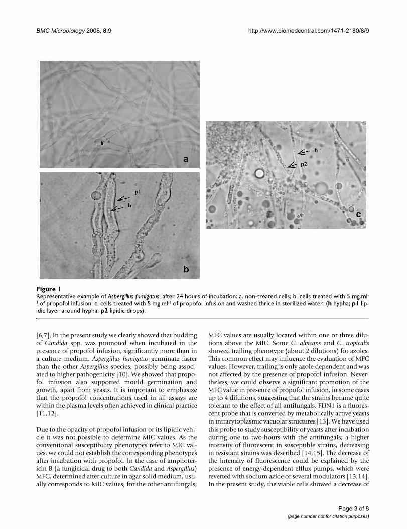
BMC Microbiology 2008, 8:9 http://www.biomedcentral.com/1471-2180/8/9
[6,7]. In the present study we clearly showed that buddingof Candida spp. was promoted when incubated in thepresence of propofol infusion, significantly more than ina culture medium. Aspergillus fumigatus germinate fasterthan the other Aspergillus species, possibly being associ-ated to higher pathogenicity [10]. We showed that propo-fol infusion also supported mould germination andgrowth, apart from yeasts. It is important to emphasizethat the propofol concentrations used in all assays arewithin the plasma levels often achieved in clinical practice[11,12].
Due to the opacity of propofol infusion or its lipidic vehi-cle it was not possible to determine MIC values. As theconventional susceptibility phenotypes refer to MIC val-ues, we could not establish the corresponding phenotypesafter incubation with propofol. In the case of amphoter-icin B (a fungicidal drug to both Candida and Aspergillus)MFC, determined after culture in agar solid medium, usu-ally corresponds to MIC values; for the other antifungals,
MFC values are usually located within one or three dilu-tions above the MIC. Some C. albicans and C. tropicalisshowed trailing phenotype (about 2 dilutions) for azoles.This common effect may influence the evaluation of MFCvalues. However, trailing is only azole dependent and wasnot affected by the presence of propofol infusion. Never-theless, we could observe a significant promotion of theMFC value in presence of propofol infusion, in some casesup to 4 dilutions, suggesting that the strains became quitetolerant to the effect of all antifungals. FUN1 is a fluores-cent probe that is converted by metabolically active yeastsin intracytoplasmic vacuolar structures [13]. We have usedthis probe to study susceptibility of yeasts after incubationduring one to two-hours with the antifungals; a higherintensity of fluorescent in susceptible strains, decreasingin resistant strains was described [14,15]. The decrease ofthe intensity of fluorescence could be explained by thepresence of energy-dependent efflux pumps, which werereverted with sodium azide or several modulators [13,14].In the present study, the viable cells showed a decrease of
Representative example of Aspergillus fumigatus, after 24 hours of incubationFigure 1Representative example of Aspergillus fumigatus, after 24 hours of incubation: a. non-treated cells; b. cells treated with 5 mg.ml-1 of propofol infusion; c. cells treated with 5 mg.ml-1 of propofol infusion and washed thrice in sterilized water. (h hypha; p1 lip-idic layer around hypha; p2 lipidic drops).
Page 3 of 8(page number not for citation purposes)
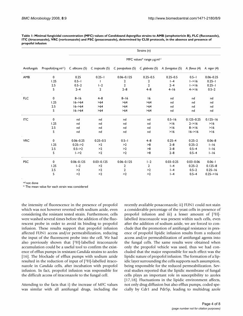
BMC Microbiology 2008, 8:9 http://www.biomedcentral.com/1471-2180/8/9
the intensity of fluorescence in the presence of propofolwhich was not however reverted with sodium azide, evenconsidering the resistant tested strain. Furthermore, cellswere washed several times before the addition of the fluo-rescent probe in order to avoid its binding to propofolinfusion. These results support that propofol infusionaffected FUN1 access and/or permeabilization, reducingthe input of the fluorescent probe into the cell. We hadalso previously shown that [3H]-labelled itraconazoleaccumulation could be a useful tool to confirm the exist-ence of efflux pumps in resistant Candida strains to azoles[16]. The blockade of efflux pumps with sodium azideresulted in the reduction of input of [3H]-labelled itraco-nazole in Candida cells, after incubation with propofolinfusion. In fact, propofol infusion was responsible forthe difficult access of itraconazole to the fungal cell.
Attending to the facts that i) the increase of MFC valueswas similar with all antifungal drugs, including the
recently available posaconazole; ii) FUN1 could not staina considerable percentage of the yeast cells in presence ofpropofol infusion and iii) a lesser amount of [3H]-labelled itraconazole was present within such cells, evenafter the addition of sodium azide, we are forced to con-clude that the promotion of antifungal resistance in pres-ence of propofol lipidic infusion results from a reducedaccess and/or permeabilization of antifungal agents intothe fungal cells. The same results were obtained whenonly the propofol vehicle was used, thus we had con-cluded that the major responsible for such effect was thelipidic nature of propofol infusion. The formation of a lip-idic layer surrounding the cells supports such assumption,being responsible for the reduced permeabilization. Sev-eral studies reported that the lipidic membrane of fungalcells plays an important role in susceptibility to azoles[17,18]. Fluctuations in the lipidic environment affects,not only drug diffusion but also efflux pumps, coded spe-cially by Cdr1 and Pdr5p, leading to multidrug azole
Table 1: Minimal fungicidal concentration (MFC) values of Candidaand Aspergillus strains to AMB (amphotericin B), FLC (fluconazole), ITC (itraconazole), VRC (voriconazole) and PSC (posaconazole), determined by CLSI protocols, in the absence and presence of propofol infusion
Strains (n)
MFC values* range μg.ml-1
Antifungals Propofol(mg.ml-1) C. albicans (5) C. tropicalis (5) C. parapsilosis (5) C. glabrata (5) A. fumigatus (5) A. flavus (4) A. niger (4)
AMB 0 0.25 0.25–1 0.06–0.125 0.25–0.5 0.25–0.5 0.5–1 0.06–0.251.25 0.5–1 1 2 2 1–4 1–>16 0.25–12.5 0.5–2 1–2 2 2 2–4 1–>16 0.25–15 2–4 2 2–8 4–8 4–16 4–>16 0.5–2
FLC 0 8–16 4–8 8–16 16 nd nd nd1.25 16–>64 >64 >64 >64 nd nd nd2.5 16–>64 >64 >64 >64 nd nd nd5 16–>64 >64 >64 >64 nd nd nd
ITC 0 nd nd nd nd 0.5–16 0.125–0.25 0.125–161.25 nd nd nd nd >16 2–>16 >162.5 nd nd nd nd >16 8–>16 >165 nd nd nd nd >16 16–>16 >16
VRC 0 0.06–0.25 0.25–0.5 0.5–1 4–8 0.25–4 0.25–2 0.06–81.25 0.25–>2 >2 >2 >8 2–8 0.25–2 1–162.5 0.5–>2 >2 >2 >8 2–8 0.5–4 1–165 1–>2 >2 >2 >8 2–8 0.5–4 1–16
PSC 0 0.06–0.125 0.03–0.125 0.06–0.125 1–2 0.03–0.25 0.03–0.06 0.06–11.25 1–2 >2 2 2 1–4 0.25–2 0.125–82.5 >2 >2 2 >2 1–4 0.5–2 0.25–165 >2 >2 >2 >2 1–4 0.5–4 0.25–>16
nd not done* The mean value for each strain was considered
Page 4 of 8(page number not for citation purposes)
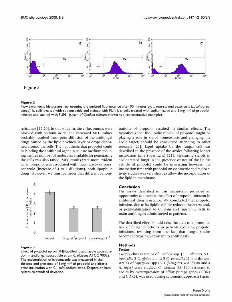
BMC Microbiology 2008, 8:9 http://www.biomedcentral.com/1471-2180/8/9
resistance [19,20]. In our study, as the efflux pumps wereblocked with sodium azide, the increased MFC valuesprobably resulted from poor diffusion of the antifungaldrugs caused by the lipidic vehicle layer or drops depos-ited around the cells. The hypothesis that propofol couldbe binding the antifungal agent in culture medium reduc-ing the free number of molecules available for penetratingthe cells was also raised. MFC results were more evidentwhen propofol was associated with itraconazole or posa-conazole (increase of 4 to 5 dilutions), both lipophilicdrugs. However, we must consider that different concen-
trations of propofol resulted in similar effects. Thehypothesis that the lipidic vehicle of propofol might beplaying a role in sterol homeostasis and changing theazole target, should be considered attending to otherresearch [21]. Lipid uptake by the fungal cell wasdescribed in the presence of the azoles following longerincubation time (overnight) [21]. Measuring sterols inazole-treated fungi in the presence or not of the lipidicvehicle of propofol could be interesting however, theincubation time with propofol on cytometric and radioac-tivity studies was very short to allow the incorporation ofthe lipid in membrane.
ConclusionThe assays described in this manuscript provided anopportunity to describe the effect of propofol infusion inantifungal drug resistance. We concluded that propofolinfusion, due to its lipidic vehicle reduced the access and/or permeabilization to Candida and Aspergillus cells tomain antifungals administered to patients.
The described effect should raise the alert to a promotedrisk of fungal infections in patients receiving propofolinfusions, resulting from the fact that fungal strainsbecome increasingly resistant to antifungals.
MethodsStrainsTwenty clinical strains of Candida spp. (5 C. albicans, 5 C.tropicalis, 5 C. glabrata and 5 C. parapsilosis) and thirteenstrains of Aspergillus spp.(5 A. fumigatus, 4 A. flavus and 4A. niger) were studied. C. albicans 95–190, resistant toazoles by overexpression of efflux pumps genes (CDR1and CDR2), was used during cytometric approach (strain
Effect of propofol up on [3H]-labelled itraconazole accumula-tion in antifungal susceptible strain C. albicans ATCC 90028Figure 3Effect of propofol up on [3H]-labelled itraconazole accumula-tion in antifungal susceptible strain C. albicans ATCC 90028. The accumulation of itraconazole was measured in the absence and presence of 5 mg.ml-1 of propofol and after a prior incubation with 0.1 mM sodium azide. Dispersion bars relates to standard deviation.
5
Acc
umul
atio
n of
[3 H
] it
raco
nazo
le
(nm
ol.1
0 -8
cell
s)
4
3
2
1
0-11 2 31
propofol azide g.ml+5m-
control 5mg.ml
Flow cytometric histograms representing the emitted fluorescence after 90 minutes byFigure 2Flow cytometric histograms representing the emitted fluorescence after 90 minutes by: a. non-stained yeast cells (autofluores-cence); b. cells treated with sodium azide and stained with FUN1; c. cells treated with sodium azide and 5 mg.ml-1 of propofol infusion and stained with FUN1 (strain of Candida albicans shown as a representative example).
Page 5 of 8(page number not for citation purposes)

BMC Microbiology 2008, 8:9 http://www.biomedcentral.com/1471-2180/8/9
kindly gift by Prof. Theodore White). Until testing, yeastsand moulds were kept frozen in Brain-Heart broth (DifcoLaboratories, Detroit, MI, USA) with 5% glycerol. For eachexperiment, the strains were subcultured twice on Sabour-aud agar (Difco) at 35°C, 48 hours for Candida and 7 daysfor Aspergillus.
Drugs and ChemicalsPropofol infusion Fresenius® (Kabi, France) at stock con-centration of 1% was used. Propofol vehicle (soya beanoil, egg lecithin, glycerol, sodium hydroxide and sterilewater) was also assayed. Fluconazole and voriconazolewere obtained from Pfizer (Groton, CT, USA), amphoter-icin B from Bristol-Myers Squibb (New York, USA), itraco-nazole from Janssen-Cilag (Beerse, Belgium) andposaconazole from Shering-Plough (Kenilworth, NJ,USA). Antifungals drugs were maintained in stock solu-tion at -70°C until use. [3H]-labelled itraconazole wassupplied by Janssen-Cilag. Sodium azide was purchasedfrom Sigma (Sigma-Aldrich, Germany).
Growth assaysAfter cultivation of Candida and Aspergillus strains in Sab-ouraud agar medium (Difco, Detroit, MI, USA), a 5 ×106.ml-1 blastoconidia or conidia suspension of Candidaand Aspergillus was prepared in phosphate buffer saline(PBS) (Sigma) and 100 μl were added in two parallelserial dilutions of propofol infusion (stock solution at1%) and its vehicle (both at 0, 1.25, 2.5 and 5 mg.ml-1
final concentrations) in RPMI 1640 culture medium(Sigma), PBS and plain propofol infusion in a final vol-ume of 500 μl. RPMI is a hydrophilic medium, however,solubility problems were not found. For Candida strains,samples were collected after 3 hours incubation at 37°C,the cells were observed under phase contrast microscopy(Leitz Larborlux K) and the percentage of budding andgerm tube formation (for C. albicans) were determined[22]. For Aspergillus, the percentage of conidial germina-tion was determined after incubation for 6, 12 and 24hours at 37°C [10].
Susceptibility testingFor Candida spp., the minimal inhibitory concentration(MIC) to fluconazole, voriconazole, posaconazole andamphotericin B (tested concentration range: 0.125–64μg.ml-1, 0.03–16 μg.ml-1, 0.03–16 μg.ml-1 and 0.03–16μg.ml-1, respectively) were determined accordingly theCLSI protocols M27-A2 (formerly NCCLS) [23]. Strainswere classified as susceptible (S), susceptible-dosedependent (S-DD) and resistant (R) to fluconazoleaccording to breakpoints defined by CLSI [23]. For vorico-nazole MICs ≤ 1 μg.ml-1 were considered S, MIC = 2 μg.ml-
1 considered S-DD and MIC ≥ 4 μg.ml-1 considered R [24].Although susceptibility breakpoints have not yet beenestablished for amphotericin B and posaconazole, strains
with MIC ≤ 1 μg.ml-1 were considered susceptible [25,26].Minimal fungicidal concentration (MFC) to all antifun-gals was also determined. The content of each well con-taining drug concentrations to and higher than the MIC,and also the positive growth control were transferred toSabouraud dextrose agar plates and incubated at 35°C for48 h, as previously described [27]. The MFC was the low-est drug concentration that killed ≥ 99% of the final inoc-ulum.
For Aspergillus spp., the CLSI protocol M 38-A was used todetermine MIC values of voriconazole, posaconazole,itraconazole and amphotericin B (all antifungals testedconcentration ranged 0.03–16 μg.ml-1) [28]. Although theunavailability of breakpoints for Aspergillus species, wefollowed several researchers that consider values of MIC ≤1 μg.ml-1 as susceptible [29,30]. MFC values were deter-mined, as previously described [31], and defined as thelowest drug concentration that showed either no growthor fewer than three colonies to obtain approximately 99 to99.5% killing activity.
The susceptibility tests to the antifungals mentionedabove were repeated in the presence of the propofol infu-sion or its vehicle in three distinct concentrations (1.25,2.5 and 5 mg.ml-1). Since propofol infusion and its vehi-cle are opaque solutions, making impossible MIC deter-mination, the content of each well containing antifungal+ propofol drugs was cultured for MFC determination andvalues compared with the MFC to antifungals alone.
Flow cytometry analysisYeast cells were incubated at 150 rpm, overnight, until lateexponential growth, in Sabouraud broth (Difco) at 37°C.Yeasts cells were harvested after centrifugation and a 1 ×10⁶ cells.ml-1 suspension was prepared in PBS supple-mented with 2% glucose (GH solution) and later dividedinto aliquots of 1 ml. The cells were then incubated withdifferent concentrations of propofol infusion (0, 1.25, 2.5and 5 mg.ml-1) at 37°C for 90 minutes and afterwardswashed thrice, ressuspended in sterile water supple-mented with 2% glucose and stained with 0.5 μM FUN1(Molecular Probes, Europe BV, Leiden, Holland) for 30minutes at 37°C. A Beckman Coulter XL-MCL flowcytometer (Beckman-Coulter Corp., Hialeah, FL, USA)equipped with a 15 nm argon laser was used. From eachsuspension 30000–50000 cells were analysed. The inten-sity of fluorescence emitted by cells treated with propofolinfusion was determined at FL2 (575 nm) and comparedwith non-treated cells (control). In parallel experiments,yeast cells were treated with 0.1 mM sodium azide during30 minutes, prior to incubation with propofol, as previ-ously described [16], in order to block efflux pumps;thereafter, the same flow cytometry analytical protocolwas used.
Page 6 of 8(page number not for citation purposes)

BMC Microbiology 2008, 8:9 http://www.biomedcentral.com/1471-2180/8/9
Intracellular accumulation of [3H]-labelled itraconazoleCandida cells were initially incubated under similar condi-tions as previously described for flow cytometry assays.The cells were harvested by centrifugation at 5000 rpm for10 minutes at 4°C, washed thrice, ressuspended in PBS ata final concentration of 2.5 × 10⁸ cells.ml-1 and incubatedwith 0, 1.25 and 5 mg.ml-1 of propofol infusion at 37°C,with continuous shaking at 300 rpm for 30 minutes [16].Parallel experiments were prepared, but also involving apre-incubation of the yeasts cells with sodium azide at 0.1mM. The cells were washed thrice and [3H]-labelled itra-conazole was added to yeast suspensions at a final con-centration of 3 μM, as previously described[16]; the cellswere incubated in glass vials at 37°C, with continuousshaking (300 rpm), during 1 hour and then harvested bycentrifugation at 5000 rpm for 10 min at 4°C, washedthrice with 3 ml of ice-cold PBS containing 10 μM unla-belled itraconazole. The pellets were later ressuspended in500 μl of PBS and the radioactivity was determined, fol-lowing the addition of a scintillation cocktail (Optiphase"Hiphase3", Perkin-Elmer), in a liquid scintillation coun-ter (LKB Wallac, 1209 RackBeta).
Lipidic vehicle experimentsAll the described experiments were repeated in the pres-ence of the propofol lipidic vehicle used in Fresenius® for-mulation (soya bean oil, egg lecithin, glycerol, sodiumhydroxide and sterile water).
Statistical analysisThe effects of different concentrations of propofol upongermination of fungal cells and MFC values of the distinctantifungals were compared using one-way analysis of var-iance (ANOVA) and Student's t-test. Significance wasaccepted at p < 0.05. The SPSS 14.0 program for Windowswas used to perform the statistical analysis. All susceptibil-ity experiments were run in duplicate and growth andradioactivity assays in triplicate.
Authors' contributionsSCO and RA designed the study, performed the cytometricand radioactivity assays and wrote the manuscript. ASDperformed growth and susceptibility assays. CPV and AGRalso helped in the design of the study and draft the man-uscript.
All authors read and approved the final manuscript.
AcknowledgementsThe authors would like to thank Maria da Luz Dias, Mariana Castro Lopes and Teresa Deuchande for the excellent technical assistance.
This research was supported by the project POCTI/SAU-ESP/61080/2004.
References1. Araujo R, Pina-Vaz C, Rodrigues AG: Susceptibility of environ-
mental versus clinical strains of pathogenic Aspergillus. Int JAntimicrob Agents 2007, 29:108-111.
2. White TC, Marr KA, Bowden RA: Clinical, cellular and molecu-lar factors that contribute to antifungal resistance. Clin Micro-biol Rev 1998, 11:382-402.
3. Bach A, Motsch J: Infectious risks associated with the use ofpropofol. Acta Anaesthesiol Scand 1996, 40:1189-1196.
4. Tallis J, Ryan GM, Lambert SB, Bowden DS, McCaw R, Birch CJ, Molo-ney M, Carnie JA, Locarnini SA, Rouch GJ, Catton MG: Evidence ofpatient-to-patient transmission of hepatitis C virus throughcontaminated intravenous anaesthetic ampoules. J Virol Hepat2003, 10:234-239.
5. McNeil MM, Lasker BA, Lott TJ, Jarvis WR: Postsurgical Candidaalbicans infections associated with an extrinsically contami-nated intravenous anesthetic agent. J Clin Microbiol 1999,37:1398-1403.
6. Crowther J, Hrazdil J, Jolly D, Galbraith JC, Greacen M, Grace M:Growth of microorganisms in propofol, thiopental, and a 1:1mixture of propofol and thiopental. Anesth Analg 1996,82:475-478.
7. Sosis M, Braverman B, Villaflor E: Propofol, but not thiopental,supports the growth of Candida albicans. Anesth Analg 1995,81:132-134.
8. Bennett SN, McNeil MM, Bland LA, Arduino MJ, Villarino E, PerrottaDM, Burwen DR, Welbel SF, Pegues DA, Stroud L, Zeitz PS, JarvisWR: Postoperative infections traced to contamination of anintravenous anaesthetic, propofol. N Engl J Med 1995,333:147-154.
9. Frohlich D, Trabold B, Rothe G, Hoerauf K, Wittmann S: Inhibitionof the neutrophil oxidative response by propofol: preservedin vivo function despite in vitro inhibition. Eur J Anaesthesiol2006, 23:948-953.
10. Araujo R, Rodrigues AG: Variability of germinative potentialamong pathogenic species of Aspergillus. J Clin Microbiol 2004,42:4335-4337.
11. Kazama T, Takeuchi K, Ikeda K, Ikeda T, Kikura M, Iida T, Suzuki S,Hanai H, Sato S: Optimal propofol plasma concentration dur-ing upper gastrointestinal endoscopy in young, middle-aged,and elderly patients. Anesthesiol 2000, 93:662-669.
12. Takizawa D, Sato E, Nishikawa K, Hinohara H, Hiraoka H, Saito S,Goto F, Kunimoto F: Plasma concentration for optimal seda-tion and total body clearance of propofol in patients afteresophagectomy. J Anesth 2005, 19:88-90.
13. Millard PJ, Roth BL, Thi H-PT, Yue ST, Haugland RP: Developmentof the FUN1 family of fluorescent probes for vacuole label-ling and viability testing of yeasts. Appl Environ Microbiol 1997,63:2897-2905.
14. Pina-Vaz C, Sansonetty F, Rodrigues AG, Costa-Oliveira S, Tavares C,Martinez-de-Oliveira J: Cytometric approach for a rapid evalu-ation of susceptibility of Candida strains to antifungals. ClinMicrobiol Infect 2001, 7:609-618.
15. Pina-Vaz C, Costa-de-Oliveira S, Rodrigues AG, Espinel-Ingroff A:Comparison of the two probes for testing susceptibilities ofpathogenic yeasts to voriconazole, itraconazole and caspo-fungine by flow cytometry. J Clin Microbiol 2005, 43:4674-4679.
16. Pina-Vaz C, Rodrigues AG, Costa-de-Oliveira S, Ricardo E, Mardh P-A: Potent synergic effect between ibuprofen and azoles onCandida resulting from blockade of efflux pumps as deter-mined by FUN1 staining and flow cytomety. J Antimicrob Chem-other 2005, 56:678-685.
17. Kohli A, Smriti A, Mukkopadhyay K, Rattan A, Prasad R: In vitro low-level resistance to azoles in Candida albicans is associatedwith changes in membrane lipid fluidity and asymmetry. Anti-microb Agents Chemother 2002, 46:1046-1052.
18. Loffler J, Einsele H, Hebart H, Schumacher U, Hrastnik C, Daum G:Phospolipid and sterol analysis of plasma membranes ofazole-resistant Candida albicans strains. FEMS Microbiol Lett2000, 185:59-63.
19. Mukhopadhyay K, Kohli A, Prasad R: Drug susceptibilities ofyeasts cells are affected by membrane lipid composition.Antimicrob Agents Chemother 2002, 46:3695-3705.
20. Smriti A, Krishnamurthy S, Prasad R: Membrane fluidity affectsfunctions of Cdr1p, a multidrug ABC transporter of Candidaalbicans. FEMS Microbiol Lett 1999, 173:475-478.
Page 7 of 8(page number not for citation purposes)
http://www.ncbi.nlm.nih.gov/entrez/query.fcgi?cmd=Retrieve&db=PubMed&dopt=Abstract&list_uids=9564569
http://www.ncbi.nlm.nih.gov/entrez/query.fcgi?cmd=Retrieve&db=PubMed&dopt=Abstract&list_uids=9564569
http://www.ncbi.nlm.nih.gov/entrez/query.fcgi?cmd=Retrieve&db=PubMed&dopt=Abstract&list_uids=8986181
http://www.ncbi.nlm.nih.gov/entrez/query.fcgi?cmd=Retrieve&db=PubMed&dopt=Abstract&list_uids=8986181
http://www.ncbi.nlm.nih.gov/entrez/query.fcgi?cmd=Retrieve&db=PubMed&dopt=Abstract&list_uids=8623946
http://www.ncbi.nlm.nih.gov/entrez/query.fcgi?cmd=Retrieve&db=PubMed&dopt=Abstract&list_uids=8623946
http://www.ncbi.nlm.nih.gov/entrez/query.fcgi?cmd=Retrieve&db=PubMed&dopt=Abstract&list_uids=8623946
http://www.ncbi.nlm.nih.gov/entrez/query.fcgi?cmd=Retrieve&db=PubMed&dopt=Abstract&list_uids=7598241
http://www.ncbi.nlm.nih.gov/entrez/query.fcgi?cmd=Retrieve&db=PubMed&dopt=Abstract&list_uids=7791816
http://www.ncbi.nlm.nih.gov/entrez/query.fcgi?cmd=Retrieve&db=PubMed&dopt=Abstract&list_uids=7791816
http://www.ncbi.nlm.nih.gov/entrez/query.fcgi?cmd=Retrieve&db=PubMed&dopt=Abstract&list_uids=9212436
http://www.ncbi.nlm.nih.gov/entrez/query.fcgi?cmd=Retrieve&db=PubMed&dopt=Abstract&list_uids=9212436
BMC Microbiology 2008, 8:9 http://www.biomedcentral.com/1471-2180/8/9
Publish with BioMed Central and every scientist can read your work free of charge
"BioMed Central will be the most significant development for disseminating the results of biomedical research in our lifetime."
Sir Paul Nurse, Cancer Research UK
Your research papers will be:
available free of charge to the entire biomedical community
peer reviewed and published immediately upon acceptance
cited in PubMed and archived on PubMed Central
yours — you keep the copyright
Submit your manuscript here:http://www.biomedcentral.com/info/publishing_adv.asp
BioMedcentral
21. Xiong Q, Hassan SA, Wilson WK, Han XY, May GS, Tarrand JJ, Mat-suda SPT: Cholesterol Import by Aspergillus fumigatus and ItsInfluence on Antifungal Potency of Sterol Biosynthesis Inhib-itors. Antimicrob Agents Chemother 2005, 49:518-524.
22. Rodrigues AG, Pina-Vaz C, Mardh PA, Martinez-de-Oliveira J, Freitas-da-Fonseca A: Inhibition of germ tube formation by Candidaalbicans by local anesthetics: an effect related to ionic chan-nel blockade. Curr Microbiol 2000, 40:145-148.
23. Clinical Laboratory Standards Institute (CLSI): Reference methodfor broth dilution antifungal susceptibility testing of yeasts.2nd edition. Approved standard M27-A2. CLSI, Wayne, PA, USA;2002.
24. Pfaller MA, Diekema DJ, Rex JH, Espinel-Ingroff A, Johnson EM, AndesD, Chaturvedi V, Ghannoum MA, Odds FC, Rinaldi MG, Sheehan DJ,Troke P, Walsh TJ, Warnock DW: Correlation of MIC with out-come for Candida species tested against voriconazole: analy-sis and proposal for interpretive breakpoints. J Clin Microbiol2006, 44:819-826.
25. Rex JH, Cooper CR, Merz WG Jr, Galgiani JN, Anaissie EJ: Detectionof amphotericin B-resistant Candida isolates in a broth-based system. Antimicrob Agents Chemother 1995, 39:906-909.
26. Sabatelli F, Patel R, Mann PA, Mendrick CA, Norris CC, Hare R, Loe-benberg D, Black TA, McNicholas PM: In vitro activities of Posa-conazole, Fluconazole, Itraconazole, Voriconazole, andAmphotericin B against a large collection of clinically impor-tant molds and yeasts. Antimicrob Agents Chemother 2006,50:2009-2015.
27. Cantón E, Pemán J, Viudes A, Quindós G, Gobernado M, Espinel-Ingroff A: Minimum fungicidal concentrations of amphotericinB for bloodstream Candida species. Diag Microbiol Infect Dis2003, 45:203-206.
28. Clinical Laboratory Standards Institute (CLSI): Reference methodfor broth dilution antifungals susceptibility testing of conid-ium-forming filamentous fungi. M38-A. CLSI, Wayne, PA, USA;2002.
29. Espinel-Ingroff A: In vitro antifungal activities of anidulafunginand micafungin, licensed agents and the investigational tria-zole posaconazole as determined by NCCLS methods for12,052 fungal isolates: review of the literature. Rev IberoamMicol 2003, 20:121-136.
30. Chamilos G, Kontoyiannis DP: Update on antifungal drug resist-ance mechanisms of Aspergillus fumigatus. Drug Resist Updat2005, 8:344-358.
31. Espinel-Ingroff A, Fothergill A, Peter J, Rinaldi MG, Walsh TJ: TestingConditions for Determination of Minimum Fungicidal Con-centrations of New and Established Antifungal Agents forAspergillus spp.: NCCLS Collaborative Study. J Clin Microbiol2002, 40:3204-3208.
Page 8 of 8(page number not for citation purposes)
http://www.ncbi.nlm.nih.gov/entrez/query.fcgi?cmd=Retrieve&db=PubMed&dopt=Abstract&list_uids=7785993

Candida krusei reservoir in a neutropaenia unit: molecular evidence of a
foe?
E. Ricardo1,2, A. P. Silva1,2, T. Goncalves3,4, S. Costa de Oliveira1,2, C. Granato5, J. Martins5, A. G. Rodrigues1,2,6 and
C. Pina-Vaz1,2,7
1) Department of Microbiology, Faculty of Medicine, University of Porto, Porto, 2) Cardiovascular Research & Development Unit, Faculty of Medicine,
University of Porto, Porto, 3) Center for Neuroscience and Cell Biology, University of Coimbra, Coimbra, 4) Institute of Microbiology, Faculty of Medicine,
University of Coimbra, Coimbra, 5) Haematology Department, Neutropenic unit, Hospital S. Joao, Porto, 6) Burn Unit, Department of Plastic and
Reconstructive Surgery, Hospital S. Joao, Porto and 7) Department of Microbiology, Hospital S. Joao, Porto, Portugal
Abstract
Candida krusei has been documented as an emerging pathogen causing nosocomial outbreaks. The consecutive isolation of C. krusei strains
in three patients admitted to the same hospital department within 2 months lead us to consider the possibility of an outbreak. Additionally,
C. krusei isolates were collected from the room surfaces, whereas another isolate had been recovered from the blood of one patient
2 years before. HinfI DNA restriction endonuclease-based analysis of all C. krusei isolates was performed and restriction profiles were com-
pared. Surprisingly, isolates from different patients were unrelated, whereas isolates from biological products of the same patient showed
indistinguishable HinfI restriction patterns and were similar to those obtained from the surrounding environment of the respective patients.
The study approach revealed the endogenous origin of the C. krusei infectious episodes observed and demonstrated that, subsequent to
colonizing a patient, C. krusei can be involved in infectious episodes distant in time. The hypothesis of an outbreak was excluded, although
we believe that the methodology employed in the present study represents a valuable tool for diagnostic and epidemiological surveys.
Keywords: Candida krusei, haematological patients, health care related infections, molecular typing, mt DNA Restriction, Endonuclease
Analysis, nosocomial outbreak
Original Submission: 5 January 2010; Revised Submission: 11 March 2010; Accepted: 11 March 2010
Editor: M. Cavling Arundrup
Article published online: 20 March 2010
Clin Microbiol Infect 2011; 17: 259–263
10.1111/j.1469-0691.2010.03223.x
Corresponding author: E. Ricardo, Department of Microbiology,
Faculty of Medicine, University of Porto, Alameda Professor Hernani
Monteiro, 4200-319 Porto, Portugal
E-mail: [email protected]
Introduction
In the last two decades, invasive fungal infections in hospital-
ized patients have increased significantly worldwide. Accord-
ing to data obtained from the USA and Europe, Candida
species represent, respectively, the fourth and sixth most
frequent cause of invasive healthcare-related infections [1,2],
accounting for 8–15% of all episodes of sepsis acquired in
hospital settings [3]. Inherent to these types of infection are
the extremely high morbidity and mortality rates, particularly
among immunocompromised patients [4,5].
Fluconazole is one of the antifungal agents mostly used in
both prophylactic and therapeutic protocols. Fluconazole
prophylaxis has been associated with a decrease in the prev-
alence of Candida species such as Candida tropicalis and
Candida albicans, and to an increase in that of Candida krusei
and Candida glabrata [3]. C. krusei presents intrinsic resistance
to fluconazole and, to some extent, reduced susceptibility to
amphotericin B [6].
Infectious outbreaks in hospitals, especially in intensive
care units, represent a serious health problem and are mainly
due to Candida lusitaniae [7], C. albicans [8], Candida parapsilo-
sis [9], and C. krusei [10,11]. Many factors may account for
their occurrence (e.g. barrier loss, lack of proper infection
control measures by health care workers when managing
patients, resistance to prescribed antifungal drugs, as well as
insufficient drug levels).
Molecular methods represent a powerful tool to clarify
transmission pathways in health care facilities (i.e. to investi-
gate the occurrence of possible outbreaks). Techniques such
as karyotyping, restriction fragment length polymorphism
analysis by pulsed-field gel electrophoresis, southern blot
ª2010 The Authors
Journal Compilation ª2010 European Society of Clinical Microbiology and Infectious Diseases
ORIGINAL ARTICLE MYCOLOGY

hybridization, PCR fingerprinting and randomly amplified
polymorphic DNA fingerprinting have been extensively used
for Candida typing [12–16]. Restriction endonuclease analysis
(REA) has been described in the last decade as a valuable tool
for Candida spp. characterization. REA of the mitochondrial
DNA (mtDNA) was first applied in the biotechnology industry
in order to characterize yeast strains used for wine fermenta-
tion [17,18] and, more recently, to discriminate between Can-
dida clinical strains [19–22]. The data obtained demonstrate
the relevance of using molecular genetic methods in many
different areas, including taxonomic, ecological and clinical
surveys.
Recently, we were challenged by a hypothetical outbreak
as a result of C. krusei in the neutropaenia unit of the Hae-
matology Department of Hospital S. Joao, Porto, Portugal.
Within a short period of time, several C. krusei isolates were
cultured from biological products of three patients. In addi-
tion, C. krusei was found in the surrounding environment of
the patients. All isolates were compared using mtDNA REA,
which is a convenient and powerful tool that allows valid
comparisons between isolates of the same yeast species.
Patients and Methods
Patients
Patient A was a 41-year-old male with acute lymphoblastic
leukaemia (T/NK) diagnosed in July 2006. In August 2008,
the patient was initially treated with amoxicillin and cipro-
floxacin because of undetermined fever. When symptoms
remained unchanged, a myelogram was performed; a first
relapse of the haematological disease was diagnosed. He was
admitted to the neutropaenia unit for salvage intensive che-
motherapy with fludarabine, idarubicin and ara-C, followed
by growth factor (G-CSF), administered through a central
venous catheter. Upon the onset of the aplastic period (28
August), he received antimicrobial treatment for 2 weeks
[ciprofloxacin, amoxicillin-clavulanic acid, imipenem, acyclovir
and fluconazole (200 mg/day)]. At the time of fungaemia
detection, he displayed the following haematologic parame-
ters: white blood cells 0.03 · 109 cells/L, haemoglobin level
8.5 g/dL and blood platelets 11 · 109 cells/L. As soon as
C. krusei was identified, the patient was started on caspofun-
gin for 12 days without improvement; the treatment was
then changed to amphotericin B.
This patient had previously developed a fungaemia episode
as a result of C. krusei during chemotherapy in 2006 and a
corresponding isolate had been stored at )70�C.
The patient remained in room number 1 from August to
October 2008.
Patient B was a 53-year-old male with non-Hodgkin’s lym-
phoma, who was admitted to room number 2 for 3 weeks in
September 2008. He was administered cyclophosphamide,
doxorubicin, vincristine, and prednisone, followed by G-CSF.
On 12 September 2008, he was started on caspofungin but,
as a result of sustained fever on day 5, treatment was chan-
ged to voriconazole, completing 14 days of antifungal ther-
apy. He never received fluconazole. At that fungaemia
episode, he displayed the following haematologic parameters:
white blood cells 2.41 · 109 cells/L, haemoglobin 9.3 g/dL
and blood platelets 73 · 109 cells/L.
Patient C was a 60-year-old male with acute myeloid leu-
kaemia secondary to myelodisplastic syndrome; he was
admitted to room number 2 for 3 weeks in October 2008,
subsequent to patient B. He received Ara-C, etoposide and
doxorubicin for 5 days, followed by another 5 days of Ara-C;
he was also prescribed amoxicillin, ciprofloxacin and allopu-
rinol. In addition, prophylactic treatment included imipenem,
vancomycin, fluconazole and acyclovir from 29 September
to 20 October 2008. On that latest date, he displayed the
following haematologic parameters: white blood cells
0.6 · 109 cells/L, haemoglobin 9.8 g/dL and blood platelets
24 · 109 cells/L.
Patient D served as a typing control; one single C. krusei
isolate was recovered from the patient’s bronchial secretions
in September 2008 upon his admission to the Internal Medi-
cine Department.
Clinical strains
Eighteen C. krusei isolates were collected from different clini-
cal specimens (blood, urine, stools and bronchial secretions)
of the above mentioned patients admitted to the neutropae-
nia unit within a 2-month period. Additionally, a previous
C. krusei isolate (2006) from a blood culture of patient A
was included in the study, as well as a C. krusei isolate
obtained from the control patient D. All isolates were iden-
tified using the automatic system Vitek2 YBC identification
cards (BioMerieux, Paris, France), stored at )70�C in Brain
Heart infusion (Merck KGaA, Darmstadt, Germany) with
10% glycerol and sub-cultured twice on Sabouraud agar
(Merck KGaA) to ensure purity prior to experimental
assays.
Environmental strains
Several environmental samples were collected from the
patients’ rooms (1 and 2) using Sabouraud agar contact
plates (Merck KGaA); two C. krusei isolates were cultured:
one from the bedside table of patient A (room 1) and
another from the bed of patient C (room 2). The isolates
were characterized and stored as described above for clinical
260 Clinical Microbiology and Infection, Volume 17 Number 2, February 2011 CMI
ª2010 The Authors
Journal Compilation ª2010 European Society of Clinical Microbiology and Infectious Diseases, CMI, 17, 259–263

samples. Air samples were collected by filtration with a
MAS-100 Eco instrument (Merck Eurolab, Dietlikon, Switzer-
land), containing Sabouraud agar plates (Merck KGaA); no
C. krusei isolates were obtained.
Antifungal susceptibility testing
Voriconazole (Pfizer, New York, NY, USA), posaconazole
(Schering-Plough; Kenilworth, NJ, USA), amphotericin B
(Bristol Meyers Squibb, New York, NY, USA), caspofungin
(Merck, Rahway, NJ, USA) and anidulafungin (Pfizer) stock
solutions were prepared according to CLSI protocols (M27-
A3) [23] and maintained at )70�C until use. Minimal inhibi-
tory concentration (MIC) of each antifungal drug was deter-
mined according to CLSI protocol M27-A3 [23].
Total genomic DNA extraction
Yeast cells were cultured overnight at 30�C in 10 mL of
YPD liquid medium, with continuous orbital shaking at
180 r.p.m., and subsequently collection by centrifugation.
Total DNA was extracted using phenol:chloroform:isoamyl
alcohol 25:24:1, precipitated using 100% ice-cold ethanol and
redissolved in 200 lL of TE buffer. The DNA was treated
with 20 lg of RNase (Applichem, Darmstadt, Germany),
incubated at 37�C for 30 min to 1 h. For final precipitation,
20 lL of 4 M ammonium acetate, pH 4.8 (Sigma-Aldrich,
Munich, Germany) and 600 lL of ice-cold 100% ethanol
(Applichem) were added and samples were incubated over-
night at )20�C. The DNA was re-dissolved in TE buffer 1x,
assessed in a biophotometer 6131 (Eppendorf, Hamburg,
Germany) and adjusted to a final concentration of 2.0–
2.5 lg/lL. To assay DNA integrity, approximately 3–5 lg
of DNA was run in agarose gel (1%, w/v) (Sigma-Aldrich)
in TBE buffer 1x and stained with ethidium bromide (0.5
mg/mL) (Applichem). DNA samples were stored at )20�Cfor subsequent use.
REA of mt DNA
For each sample, a reaction mixture was prepared containing
1x HinfI enzyme reaction buffer (Metabion, Planegg, Ger-
many), 1 lg/lL RNase, 0.5 U/lL HinfI restriction enzyme
(Metabion), approximately 25–30 lg of total DNA and
DNAse-RNAse free water up to 20 lL final volume; reaction
tubes were incubated overnight at 37�C. Restriction was
ended upon HinfI inactivation by incubating for 20 min at
80�C. The total reaction mixture was run on a 1% agarose gel
(20 cm · 24 cm) at 120 mV for 3–5 h, stained with ethidium
bromide (0.5 mg/mL) and the DNA visualized under UV light.
Restriction patterns were analyzed using the UVIDOC
12.6 software for Windows (Topac Inc., Cohasset, MA,
USA) and the resulting groups of strains were compared.
Results and Discussion
There have been an increasing numbers of reports describing
non-albicans Candida hospital outbreaks. Given that C. krusei is
not the main pathogen causing nosocomial infections, the
detection of simultaneous episodes at the neutropaenia unit of
our hospital lead us to consider the possibility of an outbreak.
The C. krusei isolates were all susceptible to all the anti-
fungals assayed (fluconazole was not tested because C. krusei
presents intrinsic resistance to this agent). Variations in the
antifungal MICs for different isolates were not significant.
The MICs of amphotericin B were from 0.06 to 1 mg/L; for
caspofungin from 0.125 to 1 mg/L; for anidulafungin 0.06 mg/
L; for voriconazole from 0.25 to 2 mg/L; and for posaconaz-
ole from 0.03 to 0.5 mg/L.
The routine biochemical identification protocols or anti-
fungal susceptibility profiles are usually not sufficient to
either corroborate or exclude an outbreak hypothesis. REA
for Candida species was first described by Scherer and Ste-
vens [24] who considered this method to be an extremely
valuable tool for epidemiological studies. Fujita et al.[21]
described HinfI restriction patterns as exhibiting a superior
discriminatory power among distinct Candida isolates com-
pared to patterns obtained with other enzymes such as
EcoRI or MspI. Additionally, Sancak et al.[22] established a
correspondence of almost 100% between the results
obtained with HinfI restriction endonuclease-based analysis
and PCR methodologies. In the present study, a total of 22
C. krusei isolates (20 clinical, two environmental) were ana-
lyzed using REA. A high number of C. krusei isolates was
obtained from different biological products of patient A in
distinct periods of time. All of them showed the same
restriction pattern, including the isolate recovered in 2006
from a blood culture (ABC1) (Fig. 1, lanes ABC1, ABC2 and
ACVCII), indicating that they are the same strain. Most cer-
tainly, this patient harbors a reservoir of C. krusei and was
colonized throughout a long period of time, as described
similarly for Acinetobacter [25] and Pseudomonas [26]. This is
the first report, to our knowledge, describing a long-lasting
colonization by C. krusei. These results have implications in
terms of prophylactic measures (i.e. fluconazole is not rec-
ommended in a patient with previous isolation of C. krusei).
Other antifungals, such voriconazole or amphotericin B, are
more likely to be efficient in these cases [27,28].
The C. krusei isolates from each patient yielded distinct HinfI
restriction patterns, suggesting that the isolates were different
strains (Fig. 1, lanes ABC2, BBS, CS), at the same time discarding
the hypothesis of an outbreak in the neutropaenia unit where
the patients were admitted. The C. krusei strain isolated from
CMI Ricardo et al. mtDNA molecular typing of C. krusei clinical isolates 261
ª2010 The Authors
Journal Compilation ª2010 European Society of Clinical Microbiology and Infectious Diseases, CMI, 17, 259–263

patient D showed a pattern distinct from those isolated from
the remaining patients (Fig. 1, lane DBS), as expected.
The two environmental C. krusei isolates collected from
the surfaces of the rooms where patient A (AE9) (room 1)
and patients B and C (CE) (room 2) had stayed were
different, as depicted in Fig. 1. This excludes the possibility
of different C. krusei strains being transmitted as a result of
patient handling by health care workers. However, both the
environmental and the clinical C. krusei isolates associated
with the same patient displayed an undistinguishable restric-
tion pattern, as shown in Fig. 1 (lanes ACVC11 and AE9 vs. CS
and CE), suggesting a putative environmental reservoir and
the possibility of subsequent transmission from it to other
patients. Our results emphasize the need to enhance preven-
tive infection control measures, both when handling the
patients directly and when cleaning patients¢ facilities, particu-
larly those admitting neutropenic patients. Indeed, a study by
Berrouane et al. [29] described the relatedness among C. kru-
sei clinical isolates obtained from different biological products
from both patients and health care workers.
As described in the present study, we were able to
exclude the hypothesis of an outbreak occurring in the neu-
tropaenia unit throughout the time period considered, sup-
porting the usefulness and suitability of the REA
methodology. Moreover, the present study provided very
useful information concerning C. krusei reservoirs existing in
patients and their surrounding environment.
Acknowledgements
The authors would like to thank I. Santos for the excellent
technical support. Part of the results were presented at the
19th European Congress of Clinical Microbiology and Infec-
tious Diseases held in Helsinki, Finland, 2009.
Transparency Declaration
The authors declare that there is no source of funding and
no potential conflicts of interest.
References
1. Bouza E, Perez-Molina J, Munoz P. Report of ESGNI-001 and ESGNI-
002 studies: bloodstream infections in Europe. Clin Microbiol Infect
1999; 5: 2S1–2S12.
2. Edmond MB, Wallace SE, McClish D. Nosocomial bloostream infec-
tions in the United States hospitals: a three-year analysis. Clin Infect
Dis 1999; 29: 239–244.
3. Tortorano AM, Kibbler C, Peman J et al. Candidaemia in Europe: epi-
demiology and resistance. Int J Antimicrob Agents 2006; 27: 359–366.
4. Costa-de-Oliveira S, Pina-Vaz C, Mendonca D et al. A first Portu-
guese epidemiological survey of fungaemia in a university hospital. Eur
J Clin Microbiol Infect Dis 2008; 27: 365–374.
5. Pfaller MA, Diekema DJ. Epidemiology of invasive candidiasis: a per-
sistent public health problem. Clin Microbiol Rev 2007; 20: 133–163.
6. Eggimann P, Garbino J, Pittet D. Management of Candida species
infections in critically ill patients. Lancet Infect Dis 2003; 3: 772–785.
7. Minari A, Hachem R, Raad I. Candida lusitaniae: a cause of break-
through fungemia in cancer patients. Clin Infect Dis 2001; 32: 186–
190.
8. Lasheras A, Rogues AM, Peyrere S et al. Candida albicans outbreak in
a neurosurgical intensive care unit. J Hosp Infect 2007; 65: 181–182.
9. Brillowska-Dabrowska A, Schon T, Pannanusorn S et al. A nosocomial
outbreak of Candida parapsilosis in southern Sweden verified by geno-
typing. Scand J Infect Dis 2009; 41: 135–142.
10. Hautala T, Ikaheimo I, Husu H et al. A cluster of Candida krusei infec-
tions in a haematological unit. BMC Infect Dis 2007; 7: 97.
11. Vos MC, Endtz HP, Horst-Kreft D et al. Candida krusei transmission
among hematology patients resolved by adapted antifungal prophy-
laxis and infection control measures. J Clin Microbiol 2006; 44: 1111–
1114.
12. Dıaz-Guerra TM, Martınez-Suarez JV, Laguna F et al. Comparison of
four molecular typing methods for evaluating genetic diversity among
Candida albicans isolates from human immunodeficiency virus-positive
patients with oral candidiasis. J Clin Microbiol 1997; 35: 856–861.
13. Liguori G, Di Onofrio V, Lucariello A et al. Oral candidiasis: a com-
parison between conventional methods and multiplex polymerase
M ABC1 ABC2 M ACVC11 AE9 DBS BBS CS M CE
FIG. 1. Restriction endonuclease patterns of HinfI-digested DNA
obtained after agarose gel electrophoresis. Each pattern corresponds
to Candida krusei isolates from patients A, B, C and D (upper case
letters), from different biological products (BC, blood cultures; CVC,
central venous catheter; BS, bronchial secretions; S, stools), and to
C. krusei isolates from the room environment of patients A and C
(AE and CE, respectively); M, molecular weight marker (1 kb DNA
ladder; Metabion).
262 Clinical Microbiology and Infection, Volume 17 Number 2, February 2011 CMI
ª2010 The Authors
Journal Compilation ª2010 European Society of Clinical Microbiology and Infectious Diseases, CMI, 17, 259–263
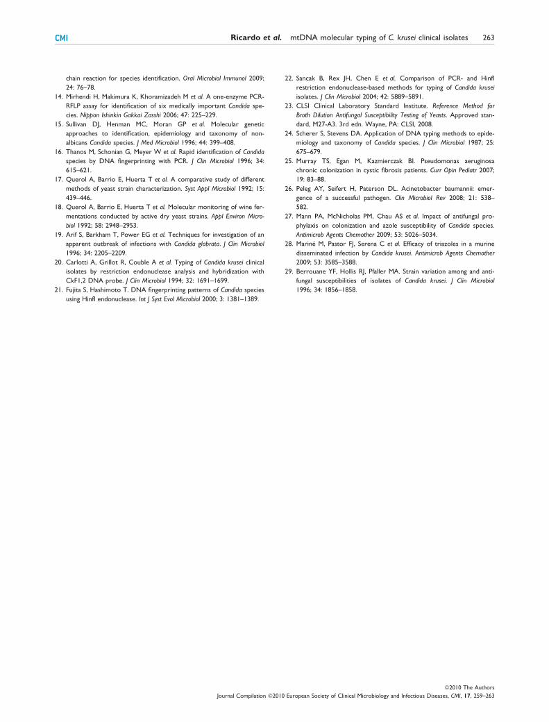
chain reaction for species identification. Oral Microbiol Immunol 2009;
24: 76–78.
14. Mirhendi H, Makimura K, Khoramizadeh M et al. A one-enzyme PCR-
RFLP assay for identification of six medically important Candida spe-
cies. Nippon Ishinkin Gakkai Zasshi 2006; 47: 225–229.
15. Sullivan DJ, Henman MC, Moran GP et al. Molecular genetic
approaches to identification, epidemiology and taxonomy of non-
albicans Candida species. J Med Microbiol 1996; 44: 399–408.
16. Thanos M, Schonian G, Meyer W et al. Rapid identification of Candida
species by DNA fingerprinting with PCR. J Clin Microbiol 1996; 34:
615–621.
17. Querol A, Barrio E, Huerta T et al. A comparative study of different
methods of yeast strain characterization. Syst Appl Microbiol 1992; 15:
439–446.
18. Querol A, Barrio E, Huerta T et al. Molecular monitoring of wine fer-
mentations conducted by active dry yeast strains. Appl Environ Micro-
biol 1992; 58: 2948–2953.
19. Arif S, Barkham T, Power EG et al. Techniques for investigation of an
apparent outbreak of infections with Candida glabrata. J Clin Microbiol
1996; 34: 2205–2209.
20. Carlotti A, Grillot R, Couble A et al. Typing of Candida krusei clinical
isolates by restriction endonuclease analysis and hybridization with
CkF1,2 DNA probe. J Clin Microbiol 1994; 32: 1691–1699.
21. Fujita S, Hashimoto T. DNA fingerprinting patterns of Candida species
using HinfI endonuclease. Int J Syst Evol Microbiol 2000; 3: 1381–1389.
22. Sancak B, Rex JH, Chen E et al. Comparison of PCR- and HinfI
restriction endonuclease-based methods for typing of Candida krusei
isolates. J Clin Microbiol 2004; 42: 5889–5891.
23. CLSI Clinical Laboratory Standard Institute. Reference Method for
Broth Dilution Antifungal Susceptibility Testing of Yeasts. Approved stan-
dard, M27-A3. 3rd edn. Wayne, PA: CLSI, 2008.
24. Scherer S, Stevens DA. Application of DNA typing methods to epide-
miology and taxonomy of Candida species. J Clin Microbiol 1987; 25:
675–679.
25. Murray TS, Egan M, Kazmierczak BI. Pseudomonas aeruginosa
chronic colonization in cystic fibrosis patients. Curr Opin Pediatr 2007;
19: 83–88.
26. Peleg AY, Seifert H, Paterson DL. Acinetobacter baumannii: emer-
gence of a successful pathogen. Clin Microbiol Rev 2008; 21: 538–
582.
27. Mann PA, McNicholas PM, Chau AS et al. Impact of antifungal pro-
phylaxis on colonization and azole susceptibility of Candida species.
Antimicrob Agents Chemother 2009; 53: 5026–5034.
28. Marine M, Pastor FJ, Serena C et al. Efficacy of triazoles in a murine
disseminated infection by Candida krusei. Antimicrob Agents Chemother
2009; 53: 3585–3588.
29. Berrouane YF, Hollis RJ, Pfaller MA. Strain variation among and anti-
fungal susceptibilities of isolates of Candida krusei. J Clin Microbiol
1996; 34: 1856–1858.
CMI Ricardo et al. mtDNA molecular typing of C. krusei clinical isolates 263
ª2010 The Authors
Journal Compilation ª2010 European Society of Clinical Microbiology and Infectious Diseases, CMI, 17, 259–263

Virulence Attenuation of Candida albicans GeneticVariants Isolated from a Patient with a RecurrentBloodstream InfectionPaula Sampaio1, Marlene Santos1, Alexandra Correia1, Fabio E. Amaral2, Julio Chavez-Galarza1,3, Sofia
Costa-de-Oliveira4, Antonio G. Castro2, Jorge Pedrosa2, Celia Pais1*
1 Centre of Molecular and Environmental Biology (CBMA), Department of Biology, University of Minho, Braga, Portugal, 2 Life and Health Sciences Research Institute
(ICVS), School of Health Sciences, University of Minho, Braga, Portugal, 3 Faculdad de Ciencias Biologicas, Universidad Nacional de Trujillo, Trujillo, Peru, 4 Department of
Microbiology, Porto Faculty of Medicine, University of Porto, Porto, Portugal
Abstract
The incidence of Candida albicans infections and the relapse episodes after antifungal treatment have increased in recentdecades. Recurrences are mainly due to the persistence of the original infecting strain that may present genetic andgenomic rearrangements during interaction with the host, reflecting strain adaptation. In this study, four isolates recoveredfrom a patient during recurrent candidemia episodes were genotyped by microsatellite length polymorphism (MLP) and bymultilocus sequence typing (MLST) and found to be genetic variants of the same strain. Using experimental mouseinfections, a progressive reduction in the virulence of the four isolates was observed, with the first two isolates more virulentthan the third and fourth. Additionally, in the mouse model, the first isolate resisted host control more efficiently, resultingin higher kidney fungal burdens and necrosis as compared to the third isolate. The resolution of inflammation was delayedin mice challenged with the first isolate and the message for IFN-c and TNF-a in the spleen was lower within the first fewhours post-infection. Original and recurrent isolates also displayed different phenotypes regarding activity of secretedenzymes and response to stress agents. Overall, the comparative analysis indicated that the virulence decrease of theseisolates was related to a lower ability to resist to the host anticandida effector mechanisms. We showed for the first timethat C. albicans genetic variants of the same strain, sequentially isolated from an immunocompromised patient, underwentadaptations in the human host that resulted in virulence attenuation when tested in mice.
Citation: Sampaio P, Santos M, Correia A, Amaral FE, Chavez-Galarza J, et al. (2010) Virulence Attenuation of Candida albicans Genetic Variants Isolated from aPatient with a Recurrent Bloodstream Infection. PLoS ONE 5(4): e10155. doi:10.1371/journal.pone.0010155
Editor: Deb Fox, The Research Institute for Children at Children’s Hospital New Orleans, United States of America
Received November 12, 2009; Accepted March 18, 2010; Published April 13, 2010
Copyright: � 2010 Sampaio et al. This is an open-access article distributed under the terms of the Creative Commons Attribution License, which permitsunrestricted use, distribution, and reproduction in any medium, provided the original author and source are credited.
Funding: This work was supported by Fundacao para a Ciencia e Tecnologia (FCT) through the project POCI/SAU-IMI/58014/2004 and by a Grant from the HealthServices of Fundacao Calouste Gulbenkian. Julio Chavez-Galarza was supported by an ALbAN scholarship (Nu E06M103915PE), and Alexandra Correia by SFRH/BD/31354/2006 fellowship. The funders had no role in study design, data collection and analysis, decision to publish, or preparation of the manuscript.
Competing Interests: The authors have declared that no competing interests exist.
* E-mail: [email protected]
Introduction
Candida albicans is a common colonizer of the human
gastrointestinal, respiratory, and reproductive tracts. However, in
immunocompromised patients, this species is one of the most
important opportunistic fungal pathogens, being responsible for
both superficial and systemic infections [1,2]. Despite the
prevalence of Candida in the hospital environment and the poor
outcome of this infection, the pathways involved in clearance of
mucocutaneous and systemic infections have not been fully defined
and the majority of the clinical studies focus on epidemiology,
diagnosis and therapeutic management [3].
Molecular epidemiology studies showed that C. albicans isolates
exhibit a high level of genetic diversity. Microsatellite length
polymorphism (MLP) and multilocus sequence typing (MLST)
have been used to discriminate C. albicans strains and to detect
small genetic changes or microvariations that may be indicative of
adaptability processes [4–7]. Typing of multiple C. albicans isolates
from the same patient obtained in longitudinal studies, or in
surveillance cultures from different anatomical sites, showed a
tendency towards the maintenance of the same strain during the
infection process [6,7]. This view of a monoclonal infecting
population has recently been extended by the demonstration of
colony-to-colony variation in C. albicans primary isolations in
samples from patients with vaginal and oral infections [8].
Nevertheless, the referred study also showed that strain variability
in primary cultures from established infections is much lower than
from healthy individuals, suggesting that the infecting population
results from the selective proliferation of one or more clones that
were present in the mixed commensal population before the
establishment of the infectious process. Observations on the
genetic and phenotypic variation in C. albicans populations showed
higher rates of chromosome-level genetic variations during passage
in the mouse relatively to in vitro growth [9], and in strains
isolated from the digestive tract of healthy individuals [10]. These
genomic alterations may be involved in the generation of new
variants within the population that contribute to the adaptation
during infection.
Host defense against systemic candidiasis relies mainly on the
ingestion and elimination of C. albicans by cells of the innate immune
system, in particular macrophages, monocytes, and neutrophils
[11–15]. Activation of leukocytes by C. albicans, triggers the release
PLoS ONE | www.plosone.org 1 April 2010 | Volume 5 | Issue 4 | e10155

of pro-inflammatory cytokines (Th1 and Th17 responses), such
as IFN-c, TNF-a, IL-1b, IL-6, and IL-17 that in turn activate
phagocyte effector functions to eliminate the invading yeast
[12,16–19]. In contrast, anti-inflammatory cytokines (Th2 response)
such as IL-4 and IL-10 have immunosuppressive effects. Thus, the
balance between pro- and anti-inflammatory cytokines is decisive
in determining whether the host defense system is overwhelmed
or able to eliminate the fungal pathogens [14,20–25]. Although
the status of the host immune system is the major factor balancing
the transition from commensalism to pathogenicity [26], C. albicans
expresses several virulence attributes that contribute for its successful
behavior, both as a commensal colonizer and as a pathogen [27].
One of its major virulence traits is the ability to reversibly switch
from unicellular budding cells to filamentous forms and the yeast
uses this attribute during an infection, not only to invade tissues, but
also to escape intracellular phagocyte death by inducing hyphal
growth inside the phagosome, resulting in the destruction of the
macrophage [28–30].
In the present work, we assessed the virulence of C. albicans
isolates from a patient with recurrent candidemia treated during a
period of four months with fluconazole. Typing of the isolates
determined that they were variants of the same strain and it was
observed that those genetic variants were progressively less virulent
to mice. With this study, we show for the first time that variants of
the same strain, recovered from a patient during recurrent
infections, differ considerably in terms of their capacity to produce
disease when tested in an immunocompetent host.
Results
Candida albicans isolates from a case of recurrentcandidemia are genetic variants of the same strain
C. albicans isolates used in this study were recovered from cases of
recurrent candidemia (Table 1). The four isolates analysed in more
detail were obtained from patient 1 and collected within a period of
four months. Isolate 124A was the first recovered and, despite the
patient’s treatment with fluconazole, three other isolates, 140, 140A,
and 144, were sequentially collected. All four isolates were found to
be resistant to fluconazole, presenting MIC values .64 mg/ml.
These isolates showed the same multilocus genotype by MLP,
except 140A, which presented a loss of heterozygosity (LOH) at CAI
microsatellite (Table 1). MLST analysis also showed that the isolates
were closely related, although presenting minor differences,
resulting in different diploid sequence types (DSTs). To gain a
better insight into the genetic proximity of these four isolates, a
similarity UPGMA dendrogram based on MLST data was
constructed. Strains isolated from other patients were also typed
and included in the Clustal analysis to generate a more robust tree.
This analysis showed that all isolates from patient 1 grouped closely,
within a p distance value lower than 0.02, and with a nodal support
value of 1 after 1000 bootstrap replications (Fig. 1), indicating that
the four isolates could be considered undistinguishable, or genetic
variants of the same strain.
Mouse virulence of the isolates decreased progressivelyIn view of the genetic similarity of the isolates, the question of
whether they also behaved identically regarding virulence towards
a healthy host was raised, and the mouse model of i.v.
disseminated candidiasis was used to assess virulence.
Survival analysis of mice inoculated with 26106 yeast cells
showed that the first isolates (124A and 140) were the most
virulent, while the last ones (140A and 144) were less virulent
(Fig. 2A). Comparing mice infected with the first isolate (124A)
with mice infected with the third (140A), or with the fourth (144),
the overall differences in survival were highly significant
(P = 0.0034 and P = 0.0002). In fact, mice infected with 124A,
140, 140A or 144 presented median survival times of 4.0, 4.5, 8.0,
and 13.5 days, respectively. Differences in virulence between
isolates 124A and 140A were confirmed using a lower inoculum
(Fig. 2B). When testing reference strain SC5314 with the same
inoculum, all mice succumbed during the first two days post-
infection, in accordance to what is described in the literature [31].
Table 1. Microsatellite genotypes and diploid sequence types (DSTs) obtained by MLP and MLST analysis of the clinical isolatesused in this study.
Patient Isolate Isolation Data Local of isolation MLP Genotypes MLST DST Clade
CAI (CAA/G)n CAVI (TAAA)n CEF3 (TTTC)n(TTC)n
1 DBC-124A 18-05-04 Catheter 18–34 12–12 137–139 1282 16
DBC-140 31-06-04 Blood 18–34 12–12 137–139 1283 16
DBC-140A 31-07-04 Bronchial secretions 34–34 12–12 137–139 1284 16
DBC-144 26-08-04 Blood 18–34 12–12 137–139 1285 16
2 DBC-154 20-09-04 Blood 26–26 7–7 135–146 1277 4
4 DBC-141 02-08-04 Blood 12–17 7–7 126–135 1278 8
5 DBC-165A 22-10-04 Pleural fluid 21–25 9–15 131–131 1279 11
6 DBC-155 21-09-04 Blood 29–29 7–11 129–143 1280 15
7 DBC-164 22-10-04 Blood 26–28 7–7 136–145 1281 4
8 DBC-7J - Vaginal 30–30 19–23 126–126 1286 5
DBC-8J - Vaginal 30–32 19–23 126–126 1287 1
DBC-9J - Vaginal 30–32 19–23 126–126 1288 1
9 DBC-22J - Vaginal 23–27 18–21 135–136 1289 S*
DBC-23J - Vaginal 23–27 21–21 135–136 1290 S*
The corresponding date and local of isolation, as well as clade assignment based on MSLT, are also shown.- data unknown; DST – diploid sequence type; S* - singleton.doi:10.1371/journal.pone.0010155.t001
C. albicans Isolates Virulence
PLoS ONE | www.plosone.org 2 April 2010 | Volume 5 | Issue 4 | e10155
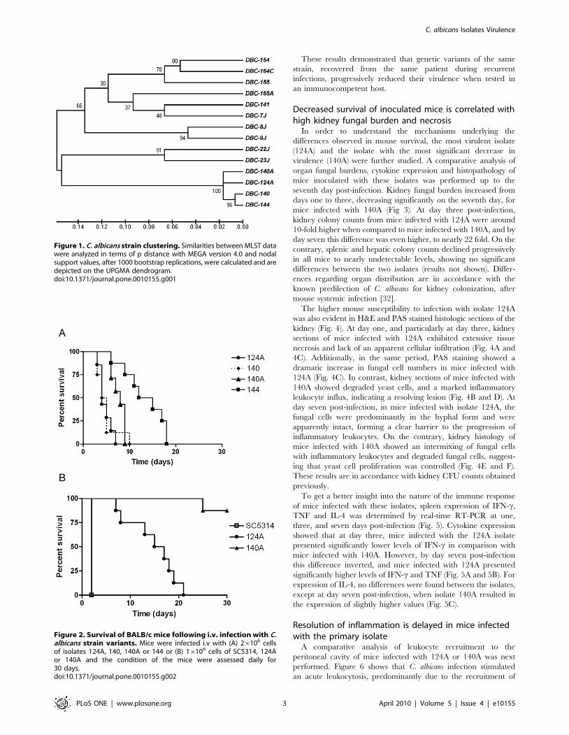
These results demonstrated that genetic variants of the same
strain, recovered from the same patient during recurrent
infections, progressively reduced their virulence when tested in
an immunocompetent host.
Decreased survival of inoculated mice is correlated withhigh kidney fungal burden and necrosis
In order to understand the mechanisms underlying the
differences observed in mouse survival, the most virulent isolate
(124A) and the isolate with the most significant decrease in
virulence (140A) were further studied. A comparative analysis of
organ fungal burdens, cytokine expression and histopathology of
mice inoculated with these isolates was performed up to the
seventh day post-infection. Kidney fungal burden increased from
days one to three, decreasing significantly on the seventh day, for
mice infected with 140A (Fig 3). At day three post-infection,
kidney colony counts from mice infected with 124A were around
10-fold higher when compared to mice infected with 140A, and by
day seven this difference was even higher, to nearly 22 fold. On the
contrary, splenic and hepatic colony counts declined progressively
in all mice to nearly undetectable levels, showing no significant
differences between the two isolates (results not shown). Differ-
ences regarding organ distribution are in accordance with the
known predilection of C. albicans for kidney colonization, after
mouse systemic infection [32].
The higher mouse susceptibility to infection with isolate 124A
was also evident in H&E and PAS stained histologic sections of the
kidney (Fig. 4). At day one, and particularly at day three, kidney
sections of mice infected with 124A exhibited extensive tissue
necrosis and lack of an apparent cellular infiltration (Fig. 4A and
4C). Additionally, in the same period, PAS staining showed a
dramatic increase in fungal cell numbers in mice infected with
124A (Fig. 4C). In contrast, kidney sections of mice infected with
140A showed degraded yeast cells, and a marked inflammatory
leukocyte influx, indicating a resolving lesion (Fig. 4B and D). At
day seven post-infection, in mice infected with isolate 124A, the
fungal cells were predominantly in the hyphal form and were
apparently intact, forming a clear barrier to the progression of
inflammatory leukocytes. On the contrary, kidney histology of
mice infected with 140A showed an intermixing of fungal cells
with inflammatory leukocytes and degraded fungal cells, suggest-
ing that yeast cell proliferation was controlled (Fig. 4E and F).
These results are in accordance with kidney CFU counts obtained
previously.
To get a better insight into the nature of the immune response
of mice infected with these isolates, spleen expression of IFN-c,
TNF and IL-4 was determined by real-time RT-PCR at one,
three, and seven days post-infection (Fig. 5). Cytokine expression
showed that at day three, mice infected with the 124A isolate
presented significantly lower levels of IFN-c in comparison with
mice infected with 140A. However, by day seven post-infection
this difference inverted, and mice infected with 124A presented
significantly higher levels of IFN-c and TNF (Fig. 5A and 5B). For
expression of IL-4, no differences were found between the isolates,
except at day seven post-infection, when isolate 140A resulted in
the expression of slightly higher values (Fig. 5C).
Resolution of inflammation is delayed in mice infectedwith the primary isolate
A comparative analysis of leukocyte recruitment to the
peritoneal cavity of mice infected with 124A or 140A was next
performed. Figure 6 shows that C. albicans infection stimulated
an acute leukocytosis, predominantly due to the recruitment of
Figure 2. Survival of BALB/c mice following i.v. infection with C.albicans strain variants. Mice were infected i.v with (A) 26106 cellsof isolates 124A, 140, 140A or 144 or (B) 16106 cells of SC5314, 124Aor 140A and the condition of the mice were assessed daily for30 days.doi:10.1371/journal.pone.0010155.g002
Figure 1. C. albicans strain clustering. Similarities between MLST datawere analyzed in terms of p distance with MEGA version 4.0 and nodalsupport values, after 1000 bootstrap replications, were calculated and aredepicted on the UPGMA dendrogram.doi:10.1371/journal.pone.0010155.g001
C. albicans Isolates Virulence
PLoS ONE | www.plosone.org 3 April 2010 | Volume 5 | Issue 4 | e10155

neutrophils, as previously described [33–35]. Counts of the
peritoneal exudate leukocytes showed that the number of
neutrophils increased more than 190 fold (P,0.001) in the
infection with isolate 124A, and 158 fold (P,0.001) following
infection with 140A. Using the set of resolution indices from
Bannenberg et al. [36], in the time interval between three h (Tmax)
and 20 h (T50), exudate PMNs decreased in number from
13.346106 (Ymax) to 6.76106 (R50), resulting in a resolution
interval (Ri) of 17 h (i.e., 3–20 h), in the infection with isolate
124A. For mice infected with 140A, the Ymax was much lower,
11.386106, and Tmax higher (eight hours), resulting in a resolution
index of 13 h (i.e., 8–21 h). The macrophage cell population
showed a similar kinetics in mice infected with isolates 124A and
140A (Fig. 6B). These results indicated that the two C. albicans
isolates raise similar patterns of leukocyte recruitment. However,
the resolution of inflammation is four hours delayed in mice
infected with the primary isolate, 124A.
Subsequent isolates induce reduced macrophage deathIsolates 124A and 140A were tested in vitro with a macrophage
cell line. The percentage of phagocytosis was approximately 11%
for isolate 124A and 17% for 140A, but this difference was not
statistically significant (P = 0.076). Phagocyte death, assessed by the
number of PI-positive phagocytes, showed that isolate 124A
induced death of about 50% of the macrophages after 4 h of co-
incubation (Fig. 7). On the contrary, 140A did not induce a
significant change in the percentage of macrophage death during
the same period. Differences in macrophage death induced by
both isolates were statistically significant after 2 h (P = 0.011) of co-
incubation, and continued after 3 h (P = 0.037) and 4 h
(P = 0.001).
Subsequent isolates have different phenotypiccharacteristics regarding activity of secreted enzymesand response to stress agents
Phenotypic characteristics known to contribute to C. albicans
pathogenicity, such as growth rate, response to stress and activity
of extracellular enzymes, were evaluated in the two clinical
isolates. No significant differences were observed regarding the
ability of the isolates to secrete aspartic proteases (Saps) or in their
growth rates at 26, 30 or 37uC in SD and YPD media (results not
shown). The extracellular in vitro phospholipase activity, deter-
mined as the Pz value, showed that isolate 124A presented a
higher activity than isolate 140A (Pz value of 0.5260.001 for 124A
and of 0.8660.042 for 140A, P,0.05).
The behaviour of both clinical isolates showed no significant
differences regarding growth in the presence of CaCl2, Caffeine,
MnSO4, SDS and ethanol, at all tested concentrations, as well as
on SD plates at pH 3.7, pH 5.5 or pH 8.0. Both isolates seemed to
be equally resistant to osmotic stress induced by NaCl (1M) and
sorbitol (1.2 M). However, in the presence of 20 mM acetic acid,
isolate 124A was more tolerant than isolate 140A (Fig. 8). The
response to 1.25 mM H2O2 oxidative stress of the two isolates
showed that 140A was significantly more sensitive to H2O2
induced death than isolate 124A, presenting a decrease in viability
of around 50% [124A 98% (623.3), 140A 52% (62.3), P,0.05].
Phenotypic characterisation showed that the genetic variants
behaved similarly although isolate 124A presented a higher
phospholipase activity and was more tolerant to acetic acid and
H2O2 than 140A.
Discussion
Infections due to C. albicans may result from the selective
proliferation of a single strain variant present in the commensal
population before invasive infection [7,8]. In patients with
recurrent infections, three basic scenarios were described: (i)
maintenance of that same strain, (ii) maintenance of that same
strain undergoing microevolution or microvariation, or (iii) strain
replacement [4,6,7,8]. Microvariations are relatively frequent and
may occur in response to changes within the host, reflecting strain
adaptation. Therefore, with the characterization of strains
sequentially isolated from patients with recurrent infections, it is
important to evaluate whether these adaptations have conse-
quences in host-pathogen interaction. These aspects are particu-
larly relevant when dealing with commensal organisms. It has long
been known that different C. albicans strains can exhibit varying
Figure 3. Kidney fungal burden. Groups of four mice infected i.v. with 106 C. albicans cells were killed at 1, 3 and 7 days after challenge. Organswere homogenized in HBSS and the suspension diluted and cultured on Sabouraud dextrose agar. Results are presented as log of colony-formingunits (CFUs). Statistically significant differences between results at each hour of infection as evaluated by Student’s t test are labeled with singleasterisk (P,0.05).doi:10.1371/journal.pone.0010155.g003
C. albicans Isolates Virulence
PLoS ONE | www.plosone.org 4 April 2010 | Volume 5 | Issue 4 | e10155

levels of virulence when tested both in vivo and in vitro models
[37–41]. However, to the best of our knowledge, no work has
characterized the virulence of isolates, and their genetic variants,
sequentially recovered from the same patient, as described in the
present study.
In this work, four sequential isolates from a patient were
genotyped by MLST and MLP and found to be closely related.
Cluster analysis, including other strains isolated in the same period
from other patients of the hospital, confirmed that the four isolates
were very close since they were the only ones to group within a
p distance value lower than 0.02. According to Odds et al. [7]
strains that group within this p distance value could be considered
undistinguishable or variants of the same strain. Interestingly,
these isolates showed a clear progressive decrease in virulence in
an i.v. mouse model of systemic infection. Since they were variants
of the same strain, we concluded that the differences in virulence
were not due to different genetic backgrounds of the isolates but to
strain adaptation to host changes during the recurrent infections.
One might doubt that these changes occurred in such a short time,
however recent studies showed that C. albicans isolates undergo
chromosomal and genetic alterations during a single passage in the
mouse [9]. Following these results, the original isolate, 124A, and
the first recurrent isolate to present a significant decrease in
virulence (140A) were selected in order to understand the
mechanisms underlying the observed differences in virulence.
Systemic infection by C. albicans is associated with the release of
proinflamatory cytokines, including TNF and IFN-c [42,43]. In
this study cytokine quantification showed that although at day
seven post-infection the levels of IFN-c and TNF increased in mice
infected with 124A, on day three the levels of IFN-c were lower,
comparing with mice infected with 140A. Moreover, cells from
isolate 124A developed long filaments inside the kidney, while cells
from 140A appeared as fragmented hyphae intermixed with the
inflammatory cells. These observations are in agreement with
Figure 4. Kidney histology. Representative micrographs of H&E/PAS-stained paraffin sections of kidneys recovered from mice infected with 106
yeast cells at days 1 (A and B), 3 (C and D) and 7 (E and F) days post-infection with isolates 124A and 140A.The bar 2100 mM.doi:10.1371/journal.pone.0010155.g004
C. albicans Isolates Virulence
PLoS ONE | www.plosone.org 5 April 2010 | Volume 5 | Issue 4 | e10155

previous reports, indicating that pro-inflammatory cytokines are
important for antifungal effector functions, particularly during the
early phase of the inflammatory response [12,33,34,44,45].
The differences observed in the murine virulence study could be
due to a differential recognition of the isolates by the host cells,
resulting in an impaired inflammatory cellular response, or to an
intrinsic higher resistance of isolate 124A to phagocyte killing.
The comparative analysis of leukocyte recruitment to the
peritoneal mouse cavity of mice infected with 124A or 140A
showed that even though resolution of inflammation was delayed
in mice infected with 124A, immunocompetent mice recognized
both isolates similarly, invoking an acute neutrophilia, as
previously described [35,46,47]. Thus, we tested the hypothesis
that the differences in virulence observed in infected mice could be
mainly due to an intrinsically higher resistance to phagocyte killing
of isolate 124A. One mechanism proposed for the opportunistic C.
albicans to resist phagocyte killing is by rapidly changing to a
filamentous form, allowing the fungal cells to resist ingestion or, if
internalized, kill the phagocyte to escape to the extracellular
environment [29,30]. This higher resistance was confirmed in vitro
upon co-incubation with J774 macrophages cell line. Both isolates
were equally recognized and phagocytosed, but 124A cells induced
a much higher macrophage death than 140A cells. The
observation that 124A resisted more efficiently to the presence of
H2O2, a compound present in the hostile environment of the
phagolysosome, and presented a higher activity of secreted
phospholipases, also favored its resistance to phagocyte induced
death.
Overall, this comparative analysis demonstrated that the
virulence decrease of isolate 140A was related to its lower ability
to resist to anticandida effector mechanisms, what explains the
lower kidney CFU’s, the absence of long filaments in kidney
histology and in vitro assays, and the faster spontaneous resolution
of acute inflammation. We believe that the four isolates from this
patient are genetic variants of a strain that, upon interaction with
the host, adapted to differences in the microenvironment. It is
likely that, as the patient became immunocompromised, the host
environment became less stressful, and adaptation resulted in a
progressive decrease in virulence that was evidenced when tested
in an immunocompetent host. Several works analyzing rates of
genetic and genomic alterations and their possible consequences to
microbe fitness propose that for opportunistic pathogens, such as
C. albicans, these alterations favor the commensal state rather than
the infectious [9,48,49]. Additionally, Cheng et al. [50] isolated a
C. albicans variant with attenuated virulence after passages through
mice, which could also be considered in agreement with the
commensal theory proposed.
This study is the first to show a decrease in virulence of genetic
variants of the same strain sequentially isolated from a human
patient, suggesting that C. albicans is able to adjust to the host,
favoring commensalism rather than increase of virulence. The
ability of C. albicans to adapt to and change its virulence in
immunocompromised hosts can be a strategy of this organism to
maintain its host alive and prolong its own survival.
Materials and Methods
Yeast strains and typingC. albicans clinical strains (14 isolates) used in this study were
collected from nine patients with recurrent infections attending the
same hospital (Table 1). The four isolates analysed in more detail
were from a patient with gastro-intestinal cancer who had been
under chemotherapy (patient 1). This patient was submitted to
surgical intervention and presented two sequential bloodstream
infections in a period of four months (Table 1). All the isolates and
the reference strain SC5314, were maintained on Sabouraud agar
plates at 4uC and cryopreserved in 30% glycerol (wt/wt) at
280uC.
Strain typing was performed by using microsatellite length
polymorphism (MLP) and multilocus sequence typing (MLST), the
more discriminatory typing methods for C. albicans. For MLP
analysis polymerase chain reaction (PCR) amplification with CAI,
CAVI, and CEF3 markers was performed as described by Sampaio
et al. [6] and by Bretagne et al. [51]. PCR products were run in an
ABI 310 Genetic Analyser (AB Applied Biosystems) and fragment
sizes were determined automatically using the GeneScan 3.7 analysis
software. MLST typing was based on sequence analysis of DNA
fragments from the six housekeeping C. albicans genes, ACC1, ADP1,
GLN4, RPN2, SYA1, and VPS13, as previously reported [52]. The
Figure 5. Real time PCR cytokine quantification. RNA wasextracted from spleen homogenates in HBSS of mice infected with106 yeast cells of 124A (%) or 140A (&) by using the Trizol method andmRNA levels of IFN-c (A), TNF-a (B) and IL-4 (C) quantified andexpressed as copies per HPRT gene. Statistical significance wascalculated by using Student’s t test and significant differences arelabeled with a single asterisk (P,0.05) or triple asterisks (P,0,0001).doi:10.1371/journal.pone.0010155.g005
C. albicans Isolates Virulence
PLoS ONE | www.plosone.org 6 April 2010 | Volume 5 | Issue 4 | e10155

diploid sequence types (DST) obtained were deposited in the C.
albicans MLST database (http://calbicans.mlst.net/).
Similarities between MLST sequence data were analyzed in
terms of p distance with MEGA version 4.0 [53], as described by
Odds et al. [7]. Nodal support, after 1000 bootstrap replications,
was also calculated and depicted in the UPGMA dendrogram.
Mice and C. albicans hematogenously disseminated
infection. Female BALB/c mice 6 to 8 weeks old were
obtained from Charles River (Barcelona, Spain) and kept under
specific pathogen-free conditions at the Animal Facility of Life and
Health Sciences Research Institute (Braga, Portugal). The present
study was conducted under the guidelines and approval of the
Research Ethics Committee of the same Institute.
To evaluate the virulence of the isolates mice were injected
intravenously (i.v.) in the lateral tail vein with 26106 cells of each
of the four isolates studied in more detail, in 0.5 ml PBS. For
preparation of inocula, cells unfrozen from the original stock were
grown in Winge medium (0.2% glucose and 0.3% of yeast extract)
at 26uC, to maintain the conidial morphology [54]. In each
experiment, all isolates were tested simultaneously and inocula
were confirmed by CFU counting of the suspensions used to infect
mice. Animal welfare was assessed twice daily during 30 days.
For assessment of organ fungal-burdens and cytokine quantifi-
cation mice were separated in groups, four mice in each cage, and
i.v. infected with a lower inoculum, 106 yeast cells. At days 1, 3,
and 7 post-infection, mice from a cage were sacrificed and their
Figure 6. Intraperitoneal inflammatory response to C. albicans strain variants. Kinetics of neutrophils (A) and macrophages (B) in theperitoneal cavity following i.p. infection of BALB/c mice with 107 cells from strains 124A (solid line) and 140A (dashed lines). Cells were recovered byperitoneal lavage, and counting of leukocytes was performed by flow citometry. Statistically significant differences between results at 3, 8, 24 and48 hours of infection, as evaluated by Student’s t test, are labeled with double asterisks (P,0.001).doi:10.1371/journal.pone.0010155.g006
C. albicans Isolates Virulence
PLoS ONE | www.plosone.org 7 April 2010 | Volume 5 | Issue 4 | e10155

kidneys, livers, and spleens aseptically processed. Organs were
homogenized in 2 ml of Hanks Balanced Salt Solution (HBSS)
from Invitrogen, diluted, and cultured on Sabouraud agar at
37uC. The results of organ fungal burden were expressed as log
CFU/ml of homogenate. Prior to processing, spleens were divided
in half to allow simultaneous analysis of fungal colony counts and
cytokine quantification.
Cytokine quantificationRNA was isolated from the spleen homogenate obtained
previously. Briefly, 200 ml of the organ homogenate were
centrifuged at 6000 rpm at 4uC and the pellet resuspended in
0.5 ml of Trizol reagent. After 5 minutes of incubation at room
temperature 0.1 ml of chloroform was added, tubes were agitated
and incubated on ice for 15 minutes. Samples were then
centrifuged at 12000 g for 15 minutes at 4uC and the aqueous
phase recovered. RNA was precipitated from the aqueous phase
by mixing with isopropyl alcohol, and samples centrifuged at
12000 g for 10 minutes at 4uC. RNA pellet was washed once with
0.8 ml of 70% ethanol and air-dried. RNA was resuspended in
10 ml of ultra-pure water, quantified in the NanoDrop 1000 R
Spectrophotometer (NanoDrop Technologies, Inc., Wilmington,
NC), and stored at 280uC at a concentration of 200 ng/ml.
Total RNA was reverse transcribed in a thermocycler My
Cycler Thermal Cycler (Bio-Rad, Hercules, CA) by using the
Superscript Kit II and Oligo dT (Invitrogen). The cDNA was
subjected to real-time RT-PCR reactions for quantification of
mRNA levels of TNF, IFN-c, IL-4, and the housekeeping gene
mHPRT by using the LightCycler (Roche, Basel, Switzerland),
and the LightCycler FastStart DNA Master Hybridization Probes
kit. Probes and primer sequences used to amplify the cDNA, as
well as the specific annealing temperatures are described in
Botelho et al. [55].
HistologyKidneys excised from infected mice were fixed in 10%
phosphate-buffered formalin, embedded in paraffin, sectioned,
and stained with periodic acid-Schiff (PAS) stain after hematox-
ylin-Eosin (H&E) staining, according to Kretschmar et al. [56].
Quantification of in vivo acute inflammatory responseTo quantify the cellular acute inflammatory response to isolates
124A or 140A, mice were intraperitoneally (i.p.) injected with 107
C. albicans cells and killed after 3, 8, 24 and 48 h [56]. The
inflammatory infiltrate was collected by lavage with ice-cold PBS
[57]. Quantification of leukocyte sub-populations in the peritoneal
lavage fluids was performed by flow cytometric analysis (FACScan)
based on the expression of F4/80, a marker associated with the
macrophage lineage [58], and GR1, a marker associated primarily
with the granulocyte lineage [59]. The following monoclonal
antibodies (mAbs) were used in the cytometric analysis (Becton-
Dickinson, San Jose, CA) using CELLQUEST software (Becton-
Dickinson): Phycoerythrin (PE) conjugated anti-mouse F4/80
antigen (clone BM8), and FITC anti-mouse Ly-6G and Ly-6C
(Gr-1) (RB6-8C5) (BD Pharmingen). To characterize the resolu-
tion of inflammation the following quantitative indices were used:
(i) the magnitude of PMN tissue infiltration (maximal PMN, Ymax);
(ii) the time interval when numbers of PMN reach Ymax within
exudates (Tmax); (iii) the time point (T50) when PMN numbers
reduce to 50% of Ymax (R50); and (iv) the resolution interval (Ri),
the time interval from the maximum PMN point (Ymax) to the
50% reduction point (R50) [i.e.T50-Tmax] [36].
Macrophage culture and phagocytosis assaysThe mouse macrophage-like cell line J774 (ATCC TIB-67) was
cultured at 37uC in 5% CO2 in Dulbecco’s Modified Eagle’s
medium (DMEM), supplemented with 10% heat-inactivated fetal
calf serum (FCS) (Valbiotech), 2 mM L-glutamine, 1 mM sodium
pyruvate, and 10 mM HEPES. Macrophages were plated at a
concentration of 56105 cells/ml into 24-well tissue culture plates
(Orange) containing a 13 mm diameter coverslip (Nunc) in each
well and incubated overnight in 5% CO2 at 37uC. C. albicans
isolates were grown overnight at 26uC in Winge medium,
Figure 7. In vitro C. albicans macrophage killing. Cells from themacrophage cell line J774 were incubated with C. albicans isolates 124Aor 140A cells in a ratio of 1:5 (E:T) and dead macrophages identifiedafter incubation with 1 mg/ml of propidium iodide under theflorescence microscope. Statistically significant differences betweenresults at each hour of co-infection, as evaluated by Student’s t test, arelabeled with single asterisk (P,0.05).doi:10.1371/journal.pone.0010155.g007
Figure 8. In vitro susceptibility assay. Growth of 124A and 140A yeast cells at 37uC for 48 h on YPD and YPD containing 20mM acetic acid, 0.3MCaCl2, 1M NaCl, 10mM MnSO4, and 12mM caffeine. Drop tests were performed by spotting 10 ml of 105, to 102 cells/ml dilutions.doi:10.1371/journal.pone.0010155.g008
C. albicans Isolates Virulence
PLoS ONE | www.plosone.org 8 April 2010 | Volume 5 | Issue 4 | e10155
recovered by centrifugation at 5000 rpm and washed twice in
sterile phosphate buffered saline (PBS).
Phagocytosis was assessed at a 5:1 C. albicans/macrophage ratio,
and the number of internalized C. albicans cells determined in a
phase-contrast microscope (Leica DMRB) after 30 minutes of co-
incubation [60,61]. Percentage of phagocytosis was determined as
the number of internalized cells/number macrophages 6100. At
least 300 cells were counted.
Macrophage death assessment was determined by incubating
macrophages and yeast cells, as previously described, and cells
stained with 1 mg/ml propidium iodide (PI) after 1, 2, 3, and 4 h of
incubation. Images were taken in ten independent fields using a
Leica DM5000B fluorescence microscope. Percentage of dead
phagocytes was determined as the number of PI positive
macrophages/number macrophages counted 6100 [62]. At least
300 cells were counted.
Phenotypic screening and susceptibility assaysFor the determination of growth rates, a pre-culture was
prepared incubating C. albicans isolates over night at 30uC in liquid
SD (0.17% of YNB, 0.5% of (NH4)2SO4, 2% of glucose) and YPD
(1% yeast extract, 2% peptone, 2% glucose) media and then a
dilution was prepared in fresh medium to start with a OD600 of
0.05. Growth rates were determined at 26uC, 30uC and 37uC by
measuring OD600 every hour until the culture reached stationary
phase.
Extracellular proteolytic activity (Saps) of the isolates was
assessed in BSA solid and liquid medium according to Monod el al.
[63]. C. albicans isolates were also screened for production of
extracellular phospholipase activity by growing them on egg yolk
agar and measuring the size of the zone of precipitation by the
method of Samaranayake et al. [64]. Phospholipase activity (Pz
value) was calculated as the ratio of the diameter of the colony and
the diameter of the colony plus that of the precipitation zone.
Since Saps and phospholipases are inducible enzymes these tests
were performed with freshly unfrozen cells from the original
stocks.
Strain sensitivity to osmotic, acidic and oxidative stress was
determined by incubating the yeast cells on SD agar plates
containing several stress conditions: 50 and 100 mg/ml SDS; 1 M
NaCl, 7% and 11% Ethanol; 1.2 M Sorbitol; 20 and 50 mM
Acetic Acid; 12 mM Caffeine; 0.3 M CaCl2; 10 mM MnSO4, 0.2
M LiCl; and also SD at pHs 3.75, 5.5 and 8. Cells grown overnight
in YPD medium were recovered by centrifugation at 5000 rpm
and washed in PBS. Drop tests were performed by spotting 10 ml
of 105 to 102 cells/ml dilutions onto the prepared plates that were
incubated at 26, 30 and 37uC, for 3 days. Susceptibility to H2O2
was assessed by incubating the yeast cells in a solution of 1.25 mM
H2O2, for 60 minutes at 30uC, and viability measured by CFU
counts on YPD D.
Statistical analysisUnless otherwise stated, results shown are from one experiment,
representative of three independent experiments. Statistical
significance of results was determined by unpaired Student t-test
and survival data were analyzed with the log-rank test, using the
GraphPad Prism 4 Software (GraphPad Software, Inc., La Jolla,
CA, USA). Results were considered statistically significant with
P values of less than 0.05.
Acknowledgments
We are indebted to Cidalia Pina-Vaz and Acacio G. Rodrigues from the
Department of Microbiology, Faculty of Medicine, University of Porto,
Portugal, for providing the strains used in this study and medical
information about the patient. We are thankful to Frank Odds and
Manuel Teixeira da Silva for critically reading the manuscript.
Author Contributions
Conceived and designed the experiments: PS AGC JP CP. Performed the
experiments: PS MS AC FEA SCdO. Analyzed the data: PS AC JCG CP.
Contributed reagents/materials/analysis tools: AGC JP CP. Wrote the
paper: PS AGC JP CP.
References
1. Pappas PG (2006) Invasive candidiasis. Infect Dis Clin North Am 20: 485–506.
2. Pelroth J, Choi B, Spellberg B (2007) Nosocomial fungal infections:epidemiology, diagnosis, and treatment. Medical Mycology 45: 321–346.
3. Enoch DA, Ludlam HA, Brown NM (2006) Invasive fungal infections: a reviewof epidemiology and management options. J Med Microbiol 55: 809–818.
4. Lockhart SR, Fritch JJ, Meier AS, Schroppel K, Srikantha T, et al. (1995)Colonizing populations of Candida albicans are clonal in origin but undergo
microevolution through C1 fragment reorganization as demonstrated by DNA
fingerprinting and C1 sequencing. J Clin Microbiol 33: 1501–9.
5. Shin JH, Park MR, Song JW, Shin DH, Jung SI, et al. (2004) Microevolution of
Candida albicans strains during catheter-related candidemia. J Clin Microbiol 42:4025–31.
6. Sampaio P, Gusmao L, Correia A, Alves C, Rodrigues AG, et al. (2005) Newmicrosatellite multiplex PCR for Candida albicans strain typing reveals
microevolutionary changes. J Clin Microbiol 43: 3869–3876.
7. Odds FC, Davidson AD, Jacobsen MD, Tavanti A, Whyte JA, et al. (2006)
Candida albicans strain maintenance, replacement, and microvariation demon-strated by multilocus sequence typing. J Clin Microbiol 44: 3647–3658.
8. Jacobsen MD, Duncan AD, Bain J, Johnson EM, Naglik JR, et al. (2008) MixedCandida albicans strain populations in colonized and infected mucosal tissues.
FEMS Yeast Res 8: 1334–1338.
9. Forche A, Magee PT, Selmecki A, Berman J, May G (2009) Evolution in Candida
albicans populations during a single passage through a mouse host. Genetics 182:799–7811.
10. Diogo D, Bouchier C, d’Enfert C, Bougnoux ME (2009) Loss of heterozygosityin commensal isolates of the asexual diploid yeast Candida albicans. Fungal Genet
Biol 46: 159–168.
11. Romani L (2000) Innate and adaptive immunity in Candida albicans infections and
saprophytism. J Leukoc Biol 68: 175–179.
12. Huang W, Na L, Fidel PL, Schwarzenberger P (2004) Requirement of
interleukin-17A for systemic anti-Candida albicans host defense in mice. J Infect
Dis 190: 624–631.
13. Fradin C, De Groot P, MacCallum D, Schaller M, Klis F, et al. (2005)Granulocytes govern the transcriptional response, morphology and proliferation
of Candida albicans in human blood. Mol Microbiol 56: 397–415.
14. Netea MG, Ferwerda G, van der Graaf CA, Van der Meer JW, Kullberg BJ
(2006) Recognition of fungal pathogens by toll-like receptors. Curr Pharm Des12: 4195–4201.
15. Richardson M, Rautemaa R (2009) How the host fights against Candida
infections. Front Biosci 14: 4363–4375.
16. Djeu JY (1990) Role of tumor necrosis factor and colony-stimulating factors in
phagocyte function against Candida albicans. Diagn Microbiol Infect Dis 13:
383–386.
17. Kullberg BJ, van ’t Wout JW, Hoogstraten C, van Furth R (1993) Recombinantinterferon-gamma enhances resistance to acute disseminated Candida albicans
infection in mice. J Infect Dis 168: 436–443.
18. Kaposzta R, Tree P, Marodi L, Gordon S (1998) Characteristics of invasive
candidiasis in gamma interferon- and interleukin-4-deficient mice: role ofmacrophages in host defense against Candida albicans. Infect Immun 66: 1708–1717.
19. Netea MG, van Tits LJ, Curfs JH, Amiot F, Meis JF, et al. (1999) Increased
susceptibility of TNF-alpha lymphotoxin-alpha double knockout mice to
systemic candidiasis through impaired recruitment of neutrophils and phago-cytosis of Candida albicans. J Immunol 163: 1498–1505.
20. Cenci E, Mencacci A, Spaccapelo R, Tonnetti R, Mosci P, et al. (1995) T helper
cell type 1 (Th1) and Th2-like responses are present in mice with gastric
candidiasis but protective immunity is associated with Th1 development. J InfectDis 171: 1279–1288.
21. Lavigne LM, Schopf LR, Chung CL, Maylor R, Sypek JP (1998) The role of
recombinant murine IL-12 and IFN-gamma in the pathogenesis of a murine
systemic Candida albicans infection. J Immunol 160: 284–292.
22. Leigh JE, Steele C, Wormley FL, Jr., Luo W, et al. (1998) Th1/th2 cytokineexpression in saliva of HIV-positive and HIV-negative individuals - a pilot study
in HIV-positive individuals with oropharyngeal candidiasis. J Acquir Immun
Defic Syndr Human Retrovirol 19: 373–380.
C. albicans Isolates Virulence
PLoS ONE | www.plosone.org 9 April 2010 | Volume 5 | Issue 4 | e10155

23. Romani L (1999) Immunity to Candida albicans: Th1, th2 cells and beyond. Curr
Opin Microbiol 2: 363–367.24. Leigh JE, Steele C, Wormley FL Jr, Fidel PL Jr (2002) Salivary cytokine
profiles in the immunocompetent individual with Candida-associated denture
stomatitis. Oral Microbiol Immunol 17: 311–314.25. Rozell B, Ljungdahl PO, Martınez P (2006) Host-pathogen interactions and the
pathological consequences of acute systemic Candida albicans infections in mice.Curr Drug Targets 7: 483–494.
26. Pirofski LA, Casadevall A (2009) Rethinking T cell immunity in oropharyngeal
candidiasis. J Exp Med 206: 269–273.27. Biswas S, Van Dijck P, Datta A (2007) Environmental sensing and signal
transduction pathways regulating morphopathogenic determinants of Candida
albicans. Microbiol Mol Biol Rev 71: 348–376.
28. Mansour MK, Levitz SM (2002) Interactions of fungi with phagocytes. CurrOpin Microbiol 5: 359–365.
29. Lorenz MC, Fink GR (2002) Life and death in a macrophage: role of the
glyoxylate cycle in virulence. Eukaryot Cell 1: 657–662.30. Lorenz MC, Bender JA, Fink GR (2004) Transcriptional response of Candida
albicans upon internalization by macrophages. Eukaryot Cell 3: 1076–87.31. Calera JA, Zhao XJ, De Bernardis F, Sheridan M, Calderone R (1999)
Avirulence of Candida albicans CaHK1 mutants in a murine model of
hematogenously disseminated candidiasis. Infect Immun 67: 4280–4284.32. Tavares D, Ferreira P, Arala-Chaves M (2003) Increased resistance in BALB/c
mice to reinfection with Candida albicans is due to immunoneutralization of avirulence-associated immunomodulatory protein. Microbiology 149: 333–339.
33. MacCallum DM, Odds FC (2005) Temporal events in the intravenous challengemodel for experimental Candida albicans infections in female mice. Mycoses 48:
151–161.
34. Murciano C, Villamon E, Gozalbo D, Roig P, O’Connor JE, et al. (2006) Toll-like receptor 4 defective mice carrying point or null mutations do not show
increased susceptibility to Candida albicans in a model of hematogenouslydisseminated infection. Med Mycol 44: 149–157.
35. Basu S, Hodgson G, Zhang HH, Katz M, Quilici C, et al. (2000) ‘‘Emergency’’
granulopoiesis in G-CSF-deficient mice in response to Candida albicans infection.Blood 95: 3725–3733.
36. Bannenberg GL, Chiang N, Ariel A, Arita M, Tjonahen E, et al. (2005)Molecular circuits of resolution: formation and actions of resolvins and
protectins. J Immunol 174: 4345–4355.37. Bartie KL, Williams DW, Wilson MJ, Potts AJ, Lewis MA (2004) Differential
invasion of Candida albicans isolates in an in vitro model of oral candidosis. Oral
Microbiol Immunol 19: 293–296.38. Tavanti A, Pardini G, Campa D, Davini P, Lupetti A, et al. (2004) Differential
expression of secretory aspartyl proteinase genes (SAP1-10) in oral Candida
albicans isolates with distinct karyotypes. J Clin Microbiol 42: 4726–34.
39. Hu Y, Farah CS, Ashman RB (2006) Isolates of Candida albicans that differ in
virulence for mice elicit strain-specific antibody-mediated protective responses.Microbes Infect 8: 612–620.
40. Thewes S, Moran GP, Magee BB, Schaller M, Sullivan DJ, et al. (2008)Phenotypic screening, transcriptional profiling, and comparative genomic
analysis of an invasive and non-invasive strain of Candida albicans. BMCMicrobiol 8: 187–203.
41. MacCallum DM, Castillo L, Nather K, Munro CA, Brown AJ, et al. (2009)
Property differences among the four major Candida albicans strain clades.Eukaryot Cell 8: 373–387.
42. Djeu JY, Blanchard DK, Halkias D, Friedman H (1986) Growth inhibition ofCandida albicans by human polymorphonuclear neutrophils: activation by
interferon-gamma and tumor necrosis factor. J Immunol 137: 2980–2984.
43. Mencacci A, Cenci E, Del Sero G, Fe d’Ostiani C, Mosci P, et al. (1998)Defective co-stimulation and impaired Th1 development in tumor necrosis
factor/lymphotoxin-alpha double-deficient mice infected with Candida albicans.Int Immunol 10: 37–48.
44. Romani L (2004) Immunity to fungal infections. Nat Rev Immunol 4: 1–23.
45. Schaller M, Boeld U, Oberbauer S, Hamm G, Hube B, Korting HC (2004)
Polymorphonuclear leukocytes (PMNs) induce protective Th1-type cytokineepithelial responses in an in vitro model of oral candidosis. Microbiology 150:
2807–2813.
46. Mullaly SC, Kubes P (2007) Mast cell-expressed complement receptor, not
TLR2, is the main detector of zymosan in peritonitis. Eur J Immunol 37:224–234.
47. Zhang HH, Basu S, Wu F, Begley CG, Saris CJ, et al. (2007) Macrophage-colony stimulating factor is required for the production of neutrophil-promoting
activity by mouse embryo fibroblasts deficient in G-CSF and GM-CSF. J Leukoc
Biol 82: 915–925.
48. Romani L, Bistoni F, Puccetti P (2003) Adaptation of Candida albicans to the host
environment: the role of morphogenesis in virulence and survival in mammalianhosts. Curr Opin Microbiol 6: 338–343.
49. Campos PR, Wahl LM (2009) The effects of population bottlenecks on clonalinterference, and the adaptation effective population size. Evolution 63:
950–958.
50. Cheng S, Clancy CJ, Zhang Z, Hao B, Wang W, et al. (2007) Uncoupling of
oxidative phosphorylation enables Candida albicans to resist killing by phagocytesand persist in tissue. Cell Microbiol 9: 492–501.
51. Bretagne S, Costa JM, Besmond C, Carsique RC, Calderone R (1997)Microsatellite polymorphism in the promoter sequence of the elongation factor 3
gene of Candida albicans as the basis for a typing system. J Clin Microbiol 35:
1777–1780.
52. Bougnoux ME, Tavanti A, Bouchier C, Gow NA, Magnier A, et al. (2003)
Collaborative consensus for optimized multilocus sequence typing of Candida
albicans. J Clin Microbiol 41: 5265–5266.
53. Tamura K, Dudley J, Nei M, Kumar S (2007) MEGA4: Molecular EvolutionaryGenetics Analysis (MEGA) software version 4.0. Mol Biol Evol 24: 1596–1599.
54. Bistoni F, Vecchiarelli A, Mazzolla R, Puccetti P, Marconi P, et al. (1985)Immunoadjuvant activity of amphotericin B as displayed in mice infected with
Candida albicans. Antimicrob Agents Chemother 27: 625–631.
55. Botelho AS, Teixeira L, Correia-da-Costa JM, Faustino AM, Castro AG, et al.
(2007) Neospora caninum: high susceptibility to the parasite in C57BL/10ScCrmice. Exp Parasitol 115: 68–75.
56. Kretschmar M, Hube B, Bertsch T, Sanglard D, Merker R, et al. (1999) Germ
tubes and proteinase activity contribute to virulence of Candida albicans in murineperitonitis. Infect Immun 67: 6637–6642.
57. Oliveira MS, Fraga AG, Torrado E, Castro AG, Pereira JP, et al. (2005)Infection with Mycobacterium ulcerans induces persistent inflammatory responses in
mice. Infect Immun 73: 6299–6310.
58. Austyn JM, Gordon S (1981) F4/80, a monoclonal antibody directed specifically
against the mouse macrophage. Eur J Immunol 11: 805–815.
59. Lagasse E, Weissman IL (1996) Flow cytometric identification of murine
neutrophils and monocytes. J Immunol Methods 197: 139–150.
60. Terron MP, Paredes SD, Barriga C, Ortega E, Rodrıguez AB (2004)
Comparative study of the heterophil phagocytic function in young and old ringdoves (Streptopelia risoria) and its relationship with melatonin levels. J Comp
Physiol B 174: 421–427.
61. Ortega E, Marchena JM, Garcıa JJ, Barriga C, Rodrıguez AB (2005)
Norepinephrine as mediator in the stimulation of phagocytosis induced by
moderate exercise. Eur J Appl Physiol 93: 714–718.
62. Shin YK, Kim KY, Paik YK (2005) Alterations of protein expression in
macrophages in response to Candida albicans infection. Mol Cells 20: 271–279.
63. Monod M, Hube B, Hess D, Sanglard D (1998) Differential regulation of SAP8
and SAP9, which encode two new members of the secreted aspartic proteinasefamily in Candida albicans. Microbiology 144: 2731–2737.
64. Samaranayake LP, Raeside JM, MacFarlane TW (1984) Factors affecting thephospholipase activity of Candida species in vitro. Sabouraudia 22: 201–207.
C. albicans Isolates Virulence
PLoS ONE | www.plosone.org 10 April 2010 | Volume 5 | Issue 4 | e10155
www.ajicjournal.orgVol. 38 No. 1
Letters to the Editor 83
The authors thank Dr. Carolyn Gould, Dr. Arjun Srinivasan, and Mary Andrus for theirinvaluable contributions to the revision of the NHSN UTI criteria.Disclaimer: The views expressed in this letter are those of the authors and do not neces-sarily represent the official position of the Centers for Disease Control and Prevention.
Katherine Allen-Bridson, RN, BSN, CICTeresa Horan, MPH
Centers for Disease Control and Prevention (CDC)
E-mail: [email protected]
References
1. Anderson DJ, Freeman J, Sexton DJ. Recent changes in the NHSN def-
inition for UTI: for better AND worse. Am J Infection Control 2010;38:
81-2.
2. McCarter YS, Burd EM, Hall GS, Zervos M. Laboratory diagnosis of urinary
tract infections. In: Sharp SE, editor. Cumulative techniques and procedures
in clinical microbiology 2C. Washington, DC: ASM Press; 2009. p. 3.
3. Boscia JA, Kobasa WD, Abrutyn E, Levison ME, Kaplan AM, Kaye D.
Lack of association between bacteriuria and symptoms in the elderly.
Am J Med 1986;81:979-82.
4. Pezzlo M, York M. Urine cultures. In: Isenberg H, editor. Clinical micro-
biology procedures handbook, 2nd ed. Washington, DC: ASM Press;
2004 3.12.2.
5. Muder RR, Brennen C, Rihs JD, Wagener MM, Obman A, Stout JE,
et al. Isolation of Staphylococcus aureus from the urinary tract: as-
sociation of isolation with symptomatic urinary tract infection and
subsequent staphylococcal bacteremia. Clin Infect Dis 2006;42:
46-50.
6. Ekkelenkamp MB, Verhoef J, Bonten MJ. Quantifying the relationship
between Staphylococcus aureus bacteremia and S aureus bacteriuria:
a retrospective analysis in a tertiary care hospital. Clin Infect Dis
2007;44:1457-9.
doi:10.1016/j.ajic.2009.08.002
Colonization of central venouscatheters in intensive carepatients: A 1-year survey in aPortuguese university hospital
To the Editor:Patients admitted at intensive care units (ICUs) often
require central venous catheterization. Catheter coloni-zation may lead to bacteremia and, eventually, end upas a catheter-related bloodstream infection.1 To reviewthe etiology and antimicrobial susceptibility pattern ofcentral venous catheter (CVC) isolates from ICU pa-tients, a retrospective study of the clinical database ofHospital of S. Joao, in Porto, was performed during a12-month period. All positive results from the cultureof distal tips of CVCs removed from ICU patients andantimicrobial susceptibility pattern of isolates werereviewed.
From 1482 cultures of CVC distal tips performedduring the study period, 647 positive results (43.7%)were found. As expected,2 coagulase-negative
staphylococci (54.7%), mainly Staphylococcus epider-midis (40.2%), were the most common isolates; S au-reus (12.8%), Enterobacteriaceae (8.6%), Candida spp(7.0%), Pseudomonas aeruginosa (6.2%), enterococci(4.8%) and Acinetobacter baumannii (2.8%) followedin the ranking. Of major concern was the resistancerate of S aureus to methicillin (80%), of Enterococcusfaecium to vancomycin (85.7%), of P aeruginosa tomeropenem (34.8%) and imipenem (65.2%) and of Abaumannii to both carbapenems (94.4%).3-7 Interest-ingly, no resistance to azole antifungals (fluconazole,itraconazole, voriconazole, posaconazole) was detectedamong the low number of tested Candida isolates(n 5 35), in accordance with previous findings.8 Thisfact might result from the limited exposure to antifun-gal agents in this hospital because no antifungal pro-phylactic treatment is routinely prescribed to suchpatients.
Globally, the etiology of CVC colonization in ICU pa-tients was similar to international available data. How-ever, higher levels of antimicrobial resistance werefound for S aureus, E faecium, P aeruginosa and A bau-mannii isolates. Routine surveillance for resistance pat-terns and the prospective evaluation of newtherapeutic protocols might be highly advisable insuch setting.
Luıs Cobrado, MD, PhD(c)
Department of Microbiology, Faculty of Medicine,University of Porto, Porto, PortugalMaria J. Espinar, MD, PhD(c)
Department of Microbiology, Faculty of Medicine,University of Porto, Porto, PortugalDepartment of Laboratorial Medicine, Hospital S.Joao, Porto, PortugalSofia Costa-de-Oliveira, PhD(c)
Department of Microbiology, Faculty of Medicine,University of Porto, Porto, PortugalAna T. Silva, PhD(c)
Department of Microbiology, Faculty of Medicine,University of Porto, Porto, PortugalCidalia Pina-Vaz, MD, PhD
Department of Microbiology, Faculty of Medicine,University of Porto, Porto, PortugalDepartment of Laboratorial Medicine, Hospital S.Joao, Porto, PortugalAcacio G. Rodrigues, MD, PhD
Department of Microbiology, Faculty of Medicine,University of Porto, Porto, PortugalBurn Unit, Department of Plastic and ReconstructiveSurgery, Hospital S. Joao, Porto, PortugalReferences
1. Polderman KH, Girbes AR. Central venous catheter use. Part 2: infectious
complications. Intensive Care Med 2002;28:18-28.
2. Mermel LA, Farr BM, Sherertz RJ, Raad II, O’Grady N, Harris JS, et al.
Guidelines for the management of intravascular catheter-related infec-
tions. Infectious Diseases Societyof America, American College of Critical
Care Medicine, Society for Healthcare Epidemiology of America. Clin
Infect Dis 2001;32:1249-72.

84 Letters to the Editor American Journal of Infection ControlFebruary 2010
3. Clark NM, Hershberger E, Zervosc MJ, Lynch JP III. Antimicrobial resis-
tance among gram-positive organisms in the intensive care unit. Curr
Opin Crit Care 2003;9:403-12.
4. Noble WC, Virani Z, Cree RG. Co-transfer of vancomycin and other re-
sistance genes from Enterococcus faecalis NCTC 12201 to Staphylococcus
aureus. FEMS Microbiol Lett 1992;72:195-8.
5. Jones RN, Pfaller MA. Bacterial resistance: a worldwide problem. Diagn
Microbiol Infect Dis 1998;31:379-88.
6. Clark NM, Patterson J, Lynch JP III. Antimicrobial resistance among
gram-negative organisms in the intensive care unit. Curr Opin Crit
Care 2003;9:413-23.
Erratum
At the request of Neghat Lakdawala, the journal wishelaundry at low temperature with CuWB50, a novel copp2009;37:478-83) by removing Ms. Lakdawala’s name fromThe authors have consented to this revision.
7. McGowan JE Jr. Resistance in nonfermenting gram-negative bacteria:
multidrug resistance to the maximum. Am J Med 2006;119(6 Suppl.
1):S29-36 S62–S70.
8. Pfaller MA, Jones RN, Doern GV, Fluit AC, Verhoef J, Sader HS, et al.
International surveillance of blood stream infections due to Candida
species in the European SENTRY Program: species distribution and an-
tifungal susceptibility including the investigational triazole and echino-
candin agents. SENTRY Participant Group (Europe). Diagn Microbiol
Infect Dis 1999;35:19-25.
doi:10.1016/j.ajic.2009.08.004
s to revise our February article, ‘‘Decontamination ofer-based biocidal compound,’’ (Am J Infect Controlthe section of the text that recognizes her assistance.

R E S E A R C H A R T I C L E
Ibuprofen reverts antifungal resistanceonCandidaalbicansshowingoverexpressionofCDRgenesElisabete Ricardo1,2, Sofia Costa-de-Oliveira1,2, Ana Silva Dias1, Jose Guerra1, Acacio GoncalvesRodrigues1,2 & Cidalia Pina-Vaz1,2,3
1Department of Microbiology, Faculty of Medicine, University of Porto, Porto, Portugal; 2Unit of Cardiovascular Diseases, Porto, Portugal; and3Department of Microbiology, Hospital S. Joao, Porto, Portugal
Correspondence: Elisabete Ricardo,
Department of Microbiology, Faculty of
Medicine, University of Porto, Alameda
Professor Hernani Monteiro, 4200-319 Porto,
Portugal. Tel./fax: 1351 225 513 662; e-mail:
Received 10 July 2008; revised 18 February
2009; accepted 21 February 2009.
First published online 30 March 2009.
DOI:10.1111/j.1567-1364.2009.00504.x
Editor: Richard Calderone
Keywords
Candida albicans; ibuprofen; azoles resistance
genes; antifungal resistance; real-time PCR.
Abstract
Several mechanisms may be associated with Candida albicans resistance to azoles.
Ibuprofen was described as being able to revert resistance related to efflux activity
in Candida. The aim of this study was to uncover the molecular base of antifungal
resistance in C. albicans clinical strains that could be reverted by ibuprofen. Sixty-
two clinical isolates and five control strains of C. albicans were studied: the azole
susceptibility phenotype was determined according to the Clinical Laboratory
for Standards Institute, M27-A2 protocol and minimal inhibitory concentration
values were recalculated with ibuprofen (100mg mL�1); synergistic studies between
fluconazole and FK506, a Cdr1p inhibitor, were performed using an agar disk
diffusion assay and were compared with ibuprofen results. Gene expression was
quantified by real-time PCR, with and without ibuprofen, regarding CDR1, CDR2,
MDR1, encoding for efflux pumps, and ERG11, encoding for azole target protein.
A correlation between susceptibility phenotype and resistance gene expression
profiles was determined. Ibuprofen and FK506 showed a clear synergistic effect
when combined with fluconazole. Resistant isolates reverting to susceptible
after incubation with ibuprofen showed CDR1 and CDR2 overexpression espe-
cially of the latter. Conversely, strains that did not revert displayed a remarkable
increase in ERG11 expression along with CDR genes. Ibuprofen did not alter
resistance gene expression significantly (P4 0.05), probably acting as a Cdrp
blocker.
Introduction
Candida spp., especially Candida albicans, are common
opportunistic yeasts causing mucocutaneous and systemic
infections with high morbidity and mortality. Such infec-
tions are often associated with immune disorders, endocrine
abnormalities and unrestricted use of large-spectrum anti-
microbials being treated with antifungals such as azoles, the
most widely used drugs (Carrillo-Munoz et al., 2006).
Several different mechanisms may confer antifungal re-
sistance in C. albicans clinical strains (White et al., 1998;
Sanglard & Odds, 2002); its molecular base is being docu-
mented gradually (de Micheli et al., 2002; Silver et al., 2004;
Harry et al., 2005; Coste et al., 2006, 2007; Pasrija et al.,
2007). A common mechanism described involves the reduc-
tion of the antifungal intracellular concentration due to
efflux pump overexpression encoded by CDR1, CDR2 and
MDR1 genes (Fling et al., 1991; Prasad et al., 1995; Sanglard
et al., 1997), the former two related to azole cross-resistance,
and the third associated with selective resistance to flucona-
zole. Cdr1p and Cdr2p are transmembrane proteins belong-
ing to the ABC family, ATP dependent; the substrates for
such proteins are not specific and include unrelated com-
pounds such as steroids and lipids and antifungals such as
azoles. However, its respective mode of action is not yet fully
understood. Mdr1p belongs to the Major Facilitator protein
family, which uses proton motive force to obtain energy. As
described previously, a deletion in any of these genes confers
hypersusceptibility to azoles (Coste et al., 2004). Another
resistance mechanism described involves point mutations in
the ERG11 gene leading to protein amino acidic sequence
alterations, thus decreasing azole affinity for its molecular
FEMS Yeast Res 9 (2009) 618–625c� 2009 Federation of European Microbiological SocietiesPublished by Blackwell Publishing Ltd. All rights reserved

target. The ERG11 gene encodes for lanosterol 14a-de-
methylase (Marichal et al., 1999), a cytochrome P450
enzyme involved in ergosterol biosynthesis, one of the most
abundant lipids in fungal cell membranes. An increase in its
expression might also result in protein overproduction that
may overcome the azole concentration and its effects.
Previous studies showed that antifungal resistance in
Candida spp. clinical strains could be reverted with mod-
ulators such as ibuprofen (Pina-Vaz et al., 2005). Ibuprofen
inhibited extrusion mechanisms, promoting the intracel-
lular accumulation of the fluorescent probe FUN-1 or
[H3] – itraconazole in blastoconidia, as confirmed by flow
cytometry and by cintilography, respectively (Pina-Vaz et al.,
2005). A similar effect was described with compounds such
as FK506 (tacrolimus), a potent immunosuppressor agent
and calcineurin inhibitor, showing a synergistic effect when
combined with antineoplastic agents on tumour cells,
decreasing or even suppressing drug resistance; it competes
with cytotoxic drugs for a binding site on P-glycoprotein,
being able to revert multidrug resistance by such cells
(Arceci et al., 1992). Reversion studies have also addressed
efflux-resistant C. albicans strains and FK506 (Marchetti
et al., 2000; Sun et al., 2008).
In order to characterize the resistant C. albicans strains
reverting with ibuprofen and to assess whether such a drug
might interfere with efflux pump activity, a molecular study
was performed in order to quantify CDR1, CDR2, MDR1
and ERG11 gene expression in the presence and absence of
ibuprofen. The elucidation of such mechanisms could help
in the development of new therapeutic strategies, aimed at
blockade of the efflux proteins and the subsequent reversion
of antifungal resistance, or to new approaches in drug
design, namely of compounds avoiding the extrusion of
antifungal drugs.
Materials and methods
Strains
Sixty-two C. albicans clinical strains (42 resistant, R and 20
susceptible, S to azoles), isolated from the respiratory tract,
vaginal fluid, urine and blood, were included. Five C.
albicans strains with known different susceptibility pheno-
types and with well-characterized gene expressions were also
included as controls: strains 95–142 and 95–190 (both with
CDR1 and CDR2 overexpression) and strain 12–99 (with
CDR1, CDR2, MDR1 and ERG11 overexpression), all three
R to azoles, and the S strain 2–76 (without gene over-
expression associated with resistance), kindly gifted by Dr
Theodore C. White, Seattle, WA (White et al., 2002); C.
albicans ATCC 90028 type strain, from American Type
Culture Collection, as recommended by the susceptibility
test reference protocol (CLSI, 2002). The strains were kept
at � 70 1C in brain/heart infusion (Difco) with 10% glycerol
and subcultured twice in Sabouraud agar (Difco), before
testing to assure purity of cultures.
Antifungals, drugs and stock solutionpreparation
Fluconazole (Pfizer) stock solution was prepared in sterile
distilled water at a final concentration of 640 mg mL�1;
itraconazole (Jansen) and voriconazole (Pfizer) were pre-
pared in dimethyl sulphoxide (DMSO) (Sigma) at a final
concentration of 1600 mg mL�1. FK506 (Sigma) and ibupro-
fen (Sigma) stock solutions were prepared in DMSO and
sterile distilled water, respectively, both at a final concentra-
tion of 1 mg mL�1. All stock solutions were stored at
� 70 1C, except for ibuprofen, which was kept at 4 1C until
use.
Phenotypical analysis
Broth microdilution susceptibility testing methodfor azoles with and without ibuprofen
Minimal inhibitory concentrations (MIC) of fluconazole
(Pfizer), itraconazole (Jansen) and voriconazole (Pfizer)
were determined according to the protocol established by
the Clinical Laboratory for Standards Institute (CLSI) (for-
merly NCCLS), M27-A2 (CLSI, 2002). Antifungal serial
dilutions were prepared from the respective stock solutions.
The visual readings were performed following 24 and 48 h of
incubation at 37 1C; MIC was defined as the lowest concen-
tration that produced a significant reduction (about 80%) in
cellular growth in comparison with control cells that were
not exposed to the antifungal, as recommended. The strains
were classified as S, R or susceptible-dose dependent (S-DD)
according to the CLSI protocol. For all strains, MIC values
of the tested antifungals were redetermined in the presence
of a subinhibitory concentration of ibuprofen (100mg mL�1)
(Pina-Vaz et al., 2005). Strains were clustered, according to
azole MIC values in the presence of ibuprofen, into three
distinct phenotypic groups: S, RR (Resistant Reverting) and
RNR (Resistant Non-Reverting).
Agar disk diffusion assays with FK506
Control strains 95–190 and 95–142, both R to fluconazole
but reverting to S when fluconazole was associated with
ibuprofen (RR), and two clinical strains, one RR and one
RNR strain (did not revert to S in the presence of ibuprofen),
were incubated overnight in YEPD liquid medium at 37 1C,
150 r.p.m. The inoculum was prepared after resuspending
the overnight growth culture in sterile distilled water at 0.5
McFarland density standard at 530 nm wavelength (Densi-
mat; Biomerieux, France), corresponding to a final
FEMS Yeast Res 9 (2009) 618–625 c� 2009 Federation of European Microbiological SocietiesPublished by Blackwell Publishing Ltd. All rights reserved
619Resistance by efflux overexpression is reverted by ibuprofen
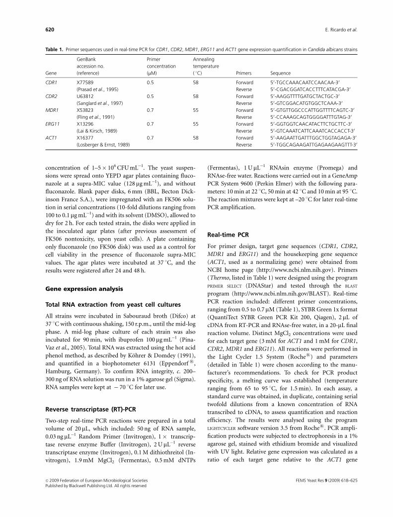
concentration of 1–5� 106 CFU mL�1. The yeast suspen-
sions were spread onto YEPD agar plates containing fluco-
nazole at a supra-MIC value (128mg mL�1), and without
fluconazole. Blank paper disks, 6 mm (BBL, Becton Dick-
inson France S.A.), were impregnated with an FK506 solu-
tion in serial concentrations (10-fold dilutions ranging from
100 to 0.1 mg mL�1) and with its solvent (DMSO), allowed to
dry for 2 h. For each tested strain, the disks were applied in
the inoculated agar plates (after previous assessment of
FK506 nontoxicity, upon yeast cells). A plate containing
only fluconazole (no FK506 disk) was used as a control for
cell viability in the presence of fluconazole supra-MIC
values. The agar plates were incubated at 37 1C, and the
results were registered after 24 and 48 h.
Gene expression analysis
Total RNA extraction from yeast cell cultures
All strains were incubated in Sabouraud broth (Difco) at
37 1C with continuous shaking, 150 r.p.m., until the mid-log
phase. A mid-log phase culture of each strain was also
incubated for 90 min, with ibuprofen 100 mg mL�1 (Pina-
Vaz et al., 2005). Total RNA was extracted using the hot acid
phenol method, as described by Kohrer & Domdey (1991),
and quantified in a biophotometer 6131 (Eppendorf s,
Hamburg, Germany). To confirm RNA integrity, c. 200–
300 ng of RNA solution was run in a 1% agarose gel (Sigma).
RNA samples were kept at � 70 1C for later use.
Reverse transcriptase (RT)-PCR
Two-step real-time PCR reactions were prepared in a total
volume of 20 mL, which included: 50 ng of RNA sample,
0.03 ng mL�1 Random Primer (Invitrogen), 1� transcrip-
tase reverse enzyme Buffer (Invitrogen), 2 U mL�1 reverse
transcriptase enzyme (Invitrogen), 0.1 M dithiothreitol (In-
vitrogen), 1.9 mM MgCl2 (Fermentas), 0.5 mM dNTPs
(Fermentas), 1 UmL�1 RNAsin enzyme (Promega) and
RNAse-free water. Reactions were carried out in a GeneAmp
PCR System 9600 (Perkin Elmer) with the following para-
meters: 10 min at 22 1C, 50 min at 42 1C and 10 min at 95 1C.
The reaction mixtures were kept at –20 1C for later real-time
PCR amplification.
Real-time PCR
For primer design, target gene sequences (CDR1, CDR2,
MDR1 and ERG11) and the housekeeping gene sequence
(ACT1, used as a normalizing gene) were obtained from
NCBI home page (http://www.ncbi.nlm.nih.gov). Primers
(Thermo, listed in Table 1) were designed using the program
PRIMER SELECT (DNAStar) and tested through the BLAST
program (http://www.ncbi.nlm.nih.gov/BLAST). Real-time
PCR reaction included: different primer concentrations,
ranging from 0.5 to 0.7mM (Table 1), SYBR Green 1x format
(QuantiTect SYBR Green PCR Kit 200, Qiagen), 2 mL of
cDNA from RT-PCR and RNAse-free water, in a 20-mL final
reaction volume. Distinct MgCl2 concentrations were used
for each target gene (3 mM for ACT1 and 1 mM for CDR1,
CDR2, MDR1 and ERG11). All reactions were performed in
the Light Cycler 1.5 System (Roches) and parameters
(detailed in Table 1) were chosen according to the manu-
facturer’s recommendations. To check for PCR product
specificity, a melting curve was established (temperature
ranging from 65 to 95 1C, for 1.5 min). In each assay, a
standard curve was obtained, in duplicate, containing serial
twofold dilutions from a known concentration of RNA
transcribed to cDNA, to assess quantification and reaction
efficiency. The results were analysed using the program
LIGHTCYCLER software version 3.5 from Roches. PCR ampli-
fication products were subjected to electrophoresis in a 1%
agarose gel, stained with ethidium bromide and visualized
with UV light. Relative gene expression was calculated as a
ratio of each target gene relative to the ACT1 gene
Table 1. Primer sequences used in real-time PCR for CDR1, CDR2, MDR1, ERG11 and ACT1 gene expression quantification in Candida albicans strains
Gene
GenBank
accession no.
(reference)
Primer
concentration
(mM)
Annealing
temperature
( 1C) Primers Sequence
CDR1 X77589 0.5 58 Forward 50-TGCCAAACAATCCAACAA-30
(Prasad et al., 1995) Reverse 50-CGACGGATCACCTTTCATACGA-30
CDR2 U63812 0.5 58 Forward 50-AAGGTTTTGATGCTACTGC-30
(Sanglard et al., 1997) Reverse 50-GTCGGACATGTGGCTCAAA-3 0
MDR1 X53823 0.7 55 Forward 50-GTGTTGGCCCATTGGTTTTCAGTC-30
(Fling et al., 1991) Reverse 50-CCAAAGCAGTGGGGATTTGTAG-3 0
ERG11 X13296 0.7 55 Forward 50-GGTGGTCAACATACTTCTGCTTC-30
(Lai & Kirsch, 1989) Reverse 50-GTCAAATCATTCAAATCACCACCT-30
ACT1 X16377 0.7 58 Forward 50-AAGAATTGATTTGGCTGGTAGAGA-30
(Losberger & Ernst, 1989) Reverse 50-TGGCAGAAGATTGAGAAGAAGTTT-30
FEMS Yeast Res 9 (2009) 618–625c� 2009 Federation of European Microbiological SocietiesPublished by Blackwell Publishing Ltd. All rights reserved
620 E. Ricardo et al.

concentration, for each of the three sets of strains studied (S,
RR and RNR).
Statistical analysis
Student’s t-test was used to compare the gene expression
among the different groups studied. A P-value o 0.05 was
considered significant.
Results
Azole susceptibility in the presence or absenceof ibuprofen
The results obtained with the control strains are detailed in
Table 2. Only the R strains 95–190 and 95–142, displaying
CDR overexpression, showed a reduction in azole MIC
values following incubation with ibuprofen; S strains and R
strains overexpressing all the screened resistance genes
(CDR, MDR1 and ERG11) showed minimal changes in azole
MIC values following incubation with ibuprofen.
The azole susceptibility pattern of the clinical isolates
determined in the presence or absence of ibuprofen is
detailed in Table 3. In the presence of ibuprofen, the MIC
values did not change in S strains, while most R strains,
which presented cross-resistance to all the assayed azoles,
reverted to S, therefore being designated as RR; one R strain
maintained the R profile while another became S-DD, being
designated as RNR.
Synergistic effect between fluconazole andFK506
For strains classified as RR, a synergistic effect between
fluconazole and FK506 was detected using the agar diffusion
method (Fig. 1); in control plates with no fluconazole added
to the medium, neither FK506 nor its solvent was able to
inhibit Candida growth (Fig. 1, left panel), which shows that
such a compound does not exhibit antifungal activity.
Regarding the control strain 95–142, a clear synergistic effect
was already observed at the lowest FK506 concentration
assayed (Fig. 1, right panel) while with the clinical strain the
same effect was observed only at higher concentrations
(right panel). Conversely, with RNR strains, complete growth
inhibition was not achieved under similar experimental
conditions (Fig. 1).
CDR1, CDR2, MDR1 and ERG11 gene expressionquantification
The results for the control strains were in agreement with
previously published data (White et al., 2002): S control
strains showed low expression for target genes while R
strains displayed an evident increase in CDR1 and CDR2
gene expression; strain 12–99 was the single strain
Table 2. Susceptibility profile in the presence and absence of ibuprofen and CDR1, CDR2, MDR1 and ERG11 basal gene expression, normalized with
ACT1 in Candida albicans control strains
Control strains
MIC Gene expression level
Fluconazole
(mg mL�1)
Fluconazole1
ibuprofen (mg mL�1) CDR1 CDR2 MDR1 ERG11
ATCC 90028 0.5 (S) 0.5 (S) 1.22� 0.07 0.18� 0.01 ND 0.93� 0.07
2–76 1 (S) 1 (S) 1.36� 0.09 1.07� 0.06 ND 0.44� 0.03
12–99 64 (R) 32 (S-DD) 37.37� 0.93 41.88� 1.23 1.169� 0.09 1.18� 0.11
95–190 128 (R) 1 (S) 88.35� 3.21 90.53� 2.43 0.04� 0.01 2.86� 0.13
95–142 128 (R) 1 (S) 4.19� 0.17 3.63� 0.17 0.02� 0.01 1.88� 0.09
S, susceptible; R, resistant; S-DD, susceptible-dose dependent.
ND, not detected (expression below detectable values).
Table 3. Fluconazole, voriconazole and itraconazole MIC for Candida albicans clinical strains (n = 62) and its respective susceptibility patterns
No. of strains/phenotype
MIC fluconazole MIC voriconazole MIC itraconazole MIC fluconazole1ibuprofen
20/S (0.25–8 mg mL�1) 20/S (0.015–0.25 mg mL�1) 20/S (0.125 mg mL�1) 20/S (0.25–8 mg mL�1
42/R (64–256 mg mL�1) 42/R (8–16mg mL�1) 42/R (4–8 mg mL�1) 40/S (0.5–8 mg mL�1); 1/S-DD and1/R (16–64 mg mL�1)
The MIC of fluconazole was determined in the presence of a subinhibitory concentration of ibuprofen (100 mg mL�1). The breakpoints considered for the
different azoles were: for fluconazole resistant (R) MICZ64 mg mL�1, susceptible-dose dependent (S-DD) MIC between 16–32 mg mL�1 and susceptible
(S) MIC � 8 mg mL�1; for voriconazole, R MICZ4 mg mL�1, S-DD MIC = 2 mg mL�1 and S MIC � 1 mg mL�1; for itraconazole, R MICZ1mg mL�1, S-DD
MIC between 0.25 and 0.5 mg mL�1, S MIC � 0.125 mg mL�1; n, number of strains.
FEMS Yeast Res 9 (2009) 618–625 c� 2009 Federation of European Microbiological SocietiesPublished by Blackwell Publishing Ltd. All rights reserved
621Resistance by efflux overexpression is reverted by ibuprofen
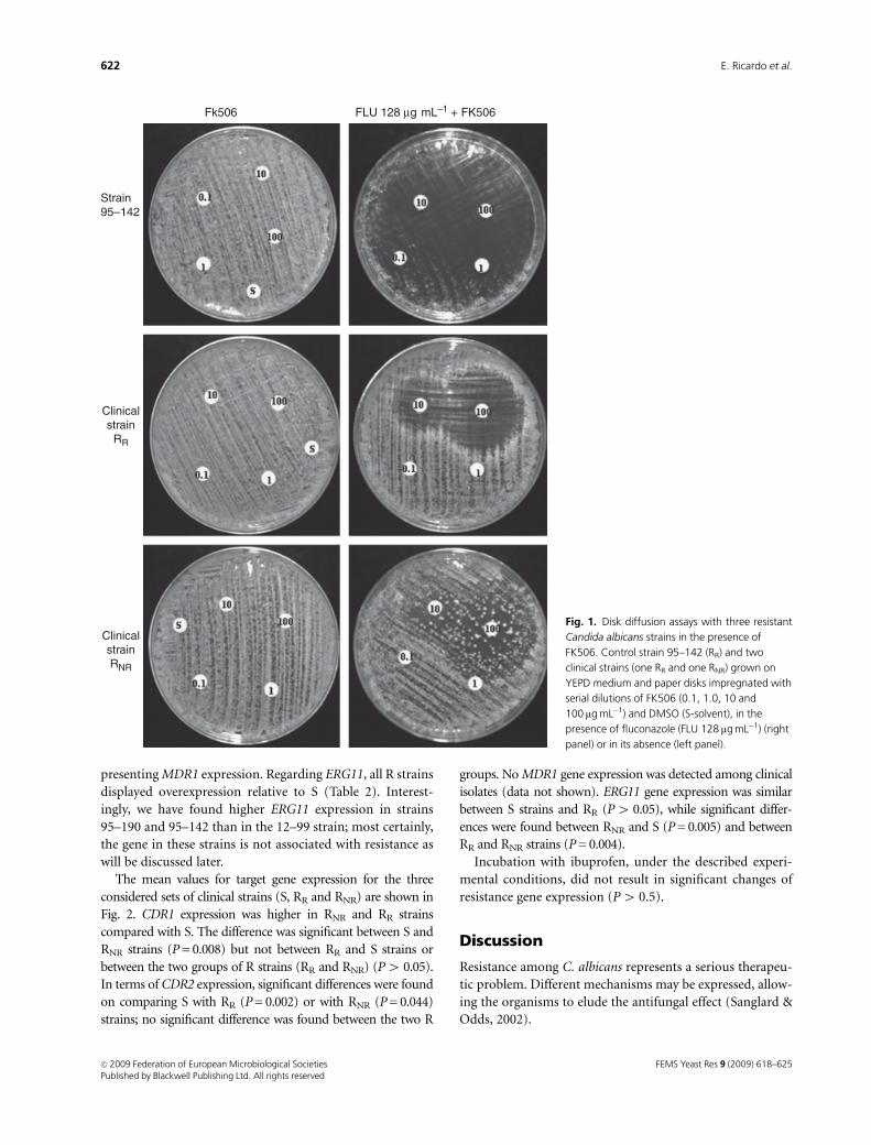
presenting MDR1 expression. Regarding ERG11, all R strains
displayed overexpression relative to S (Table 2). Interest-
ingly, we have found higher ERG11 expression in strains
95–190 and 95–142 than in the 12–99 strain; most certainly,
the gene in these strains is not associated with resistance as
will be discussed later.
The mean values for target gene expression for the three
considered sets of clinical strains (S, RR and RNR) are shown in
Fig. 2. CDR1 expression was higher in RNR and RR strains
compared with S. The difference was significant between S and
RNR strains (P = 0.008) but not between RR and S strains or
between the two groups of R strains (RR and RNR) (P4 0.05).
In terms of CDR2 expression, significant differences were found
on comparing S with RR (P = 0.002) or with RNR (P = 0.044)
strains; no significant difference was found between the two R
groups. No MDR1 gene expression was detected among clinical
isolates (data not shown). ERG11 gene expression was similar
between S strains and RR (P4 0.05), while significant differ-
ences were found between RNR and S (P = 0.005) and between
RR and RNR strains (P = 0.004).
Incubation with ibuprofen, under the described experi-
mental conditions, did not result in significant changes of
resistance gene expression (P4 0.5).
Discussion
Resistance among C. albicans represents a serious therapeu-
tic problem. Different mechanisms may be expressed, allow-
ing the organisms to elude the antifungal effect (Sanglard &
Odds, 2002).
Fk506 FLU 128 µg mL–1 + FK506
Strain95–142
Clinicalstrain
RR
ClinicalstrainRNR
Fig. 1. Disk diffusion assays with three resistant
Candida albicans strains in the presence of
FK506. Control strain 95–142 (RR) and two
clinical strains (one RR and one RNR) grown on
YEPD medium and paper disks impregnated with
serial dilutions of FK506 (0.1, 1.0, 10 and
100mg mL�1) and DMSO (S-solvent), in the
presence of fluconazole (FLU 128 mg mL�1) (right
panel) or in its absence (left panel).
FEMS Yeast Res 9 (2009) 618–625c� 2009 Federation of European Microbiological SocietiesPublished by Blackwell Publishing Ltd. All rights reserved
622 E. Ricardo et al.

We have studied a high number of C. albicans clinical
resistant strains regarding resistance target gene expression.
The CDR1 and CDR2 gene overexpression associated with
an efflux-related resistance mechanism appears to be quite
prevalent, which may indicate the ibuprofen treatment
strategy as quite promising.
Previous studies conducted by our team described ibu-
profen as a potential efflux blocker; FK506 was also
described as being able to revert C. albicans azole resistance
due to a synergistic effect with azoles (Maesaki et al., 1998;
Onyewu et al., 2003; Sun et al., 2008). This effect seems to
result from efflux pump blockade, especially of Cdr1p
(Schuetzer-Muehlbauer et al., 2003). To corroborate our
results with ibuprofen and its possible involvement in efflux
blockade, we performed disk diffusion assays with FK506 in
a small group of strains; a synergistic effect similar to
ibuprofen was obtained. Control strain 95–142 revealed
much lower CDR1 and CDR2 gene expression levels than
control strain 95–190 (Table 2), which could explain the
results obtained in the disk diffusion assays: in the former,
reversion of resistance occurred with all FK506 concentra-
tions tested, suggesting that efflux is much less efficient on
such a strain. On the other hand, in RR strains CDR1 and
CDR2, high gene expression levels were overcome in the
presence of FK506; ibuprofen yielded an effect similar to
FK506, eventually blocking efflux pumps. RNR strains might
express other distinct resistance mechanisms.
MDR1 gene expression was not detected among clinical R
strains (RR and RNR); this gene is invariably associated with
selective resistance to fluconazole (White et al., 2002) and
does not seem to be a common or a relevant antifungal
resistance mechanism among clinical isolates. Nevertheless,
two facts should be highlighted: the clinical isolates showed
azole cross-resistance, but not specific fluconazole resis-
tance, and strains 12–99, used as a positive control for
MDR1 quantification, displayed MDR1 overexpression
when compared with S control strains 2–76 (White et al.,
2002). It is possible that in such strains, Cdr1p and Cdr2p
activity overcome Mdr1p efflux activity; on the other hand,
CDR1 and CDR2 encoding mRNA has a longer half-life than
MDR1 encoding mRNA (Lyons & White, 2000), thus being
more easily detected in gene expression studies.
The ERG11 gene is not always associated with antifungal
resistance (White et al., 2002), thus being expressed at
relatively high levels in both S and R (RR and RNR) strains.
Apart from a high ERG11 gene expression, RNR strains also
revealed CDR gene overexpression, displaying most prob-
ably multiple resistance mechanisms. Ibuprofen was unable
to revert the resistance of such strains, which corroborates
our hypothesis of ibuprofen activity being related to efflux
pump activity inhibition. In accordance with this assump-
tion, control strain, 12–99, possessing multiple resistance
mechanisms including ERG11 gene overexpression asso-
ciated with resistance, revealed the same phenotypical and
genotypical behaviour as RNR strains (Redding et al., 1994;
White, 1997; White et al., 2002). In addition, the fact that
incubation with ibuprofen did not impair resistance gene
expression reinforces the concept of its direct effect on efflux
pumps, most probably inducing its physical blockade.
Although the clinical strains were phenotypically homo-
geneous within the same group, when target gene expression
levels were determined, a high intragroup variability in gene
expression profiles was observed, thus resulting in high SD
values. This variation might result from the fact that some
clinical isolates might have been previously exposed to
antifungals in vivo. Antifungal resistance acquisition over
time has already been described (Franz et al., 1998; Cowen
et al., 2000; Karababa et al., 2004) and what occurs at a
molecular level is being progressively elucidated (Silver
et al., 2004; Coste et al., 2007; Morschhauser et al., 2007).
In conclusion, clinical R strains revealing ERG11 over-
expression and eventually presenting point mutations will
not respond to efflux blockers, such as ibuprofen, even when
azole resistance also involves efflux mechanisms. Conversely,
whenever efflux genes are the most expressed resistance
genes, reversion by ibuprofen will be feasible and of con-
siderable future therapeutic interest. The uncovering of
molecular bases of Candida resistance mechanisms, apart
from representing a major challenge for molecular biolo-
gists, is of major interest for clinicians who seek new and
more targeted therapeutic strategies, aimed at the reversion
of resistance by clinical fungal pathogens such as C. albicans.
Acknowledgements
This work was supported by project POCI/SAU:IMI/61598/
2004, from the Science and Technology Fundantion (FCT).
We would like to thank Theodore White (Seattle, WA) for
0.00.51.01.52.02.53.03.5
4.0
5.04.5
Rel
ativ
e ge
ne e
xpre
ssio
n
S RR RNR
CDR1 CDR2 ERG11Gene expression profiles of C. albicans clinical strains
Fig. 2. Mean target gene expression levels of Candida albicans clinical
isolates. CDR1, CDR2, MDR1 and ERG11 gene expression levels were
quantified and normalized relative to the housekeeping gene, ACT1, in
clinical isolates grouped according to their susceptibility phenotype:
susceptible (S), resistant strains that reverted with ibuprofen (RR) and
resistant strains that did not revert with ibuprofen (RNR).
FEMS Yeast Res 9 (2009) 618–625 c� 2009 Federation of European Microbiological SocietiesPublished by Blackwell Publishing Ltd. All rights reserved
623Resistance by efflux overexpression is reverted by ibuprofen
kindly providing the control strains used in this study and
Isabel Miranda for critically reading the manuscript. We
would also like to thank Roberto Roncon-Albuquerque Jr
and Antonia Teles for the help in the real-time PCR assays.
References
Arceci RJ, Stieglitz K & Bierer BE (1992) Immunosuppressants
FK506 and rapamycin function as reversal agents of the
multidrug resistance phenotype. Blood 80: 1528–1536.
Carrillo-Munoz AJ, Giusano G, Ezkurra PA & Quindos G (2006)
Antifungal agents: mode of action in yeast cells. Rev Esp
Quimioterap 19: 130–139.
Clinical Laboratory Standards Institute (2002) Reference Method
for Broth Dilution Antifungal Susceptibility Testing of Yeasts.
Approved Standard. CLSI Document M27-A2. National
Committee for Clinical Laboratory Standards, Wayne, PA.
Coste A, Turner V, Ischer F, Morschhauser J, Forche A, Selmecki
A, Berman J, Bille J & Sanglard D (2006) A mutation in Tac1p,
a transcription factor regulating CDR1 and CDR2, is coupled
with loss of heterozygosity at chromosome 5 to mediate
antifungal resistance in Candida albicans. Genetics 172:
2139–2156.
Coste A, Selmecki A, Forche A, Diogo D, Bougnoux M-E,
d’Enfert C, Berman J & Sanglard D (2007) Genotypic
evolution of azole resistance mechanisms in sequential
Candida albicans isolates. Eukaryot Cell 6: 1889–1904.
Coste AT, Karababa M, Ischer F, Bille J & Sanglard D (2004)
TAC1, transcriptional activator of CDR genes, is a new
transcription factor involved in the regulation of Candida
albicans ABC transporters CDR1 and CDR2. Eukaryot Cell 3:
1639–1652.
Cowen LE, Sanglard D, Calabrese D, Sirjusingh C, Anderson JB &
Kohn LM (2000) Evolution of drug resistance in experimental
populations of Candida albicans. J Bacteriol 182: 1515–1522.
De Micheli M, Bille J, Schueller C & Sanglard D (2002) A
common drug-responsive element mediates the upregulation
of the Candida albicans ABC transporters CDR1 and CDR2,
two genes involved in antifungal drug resistance. Mol Microbiol
43: 1197–1214.
Fling ME, Kopf J, Tamarkin A, Gorman JA, Smith HA & Koltin Y
(1991) Analysis of a Candida albicans gene that encodes a
novel mechanism for resistance to benomyl and methotrexate.
Mol Gen Genet 227: 318–329.
Franz R, Kelly SL, Lamb DC, Kelly DE, Ruhnke M &
Morschhauser J (1998) Multiple molecular mechanisms
contribute to a stepwise development of fluconazole resistance
in clinical Candida albicans strains. Antimicrob Agents Ch 42:
3065–3072.
Harry JB, Oliver BG, Song JL, Silver PM, Little JT, Choiniere J &
White TC (2005) Drug-induced regulation of the MDR1
promoter in Candida albicans. Antimicrob Agents Ch 49:
2785–2792.
Karababa M, Coste AT, Rognon B, Bille J & Sanglard D (2004)
Comparison of gene expression profiles of Candida albicans
azole-resistant clinical isolates and laboratory strains exposed
to drugs inducing multidrug transporters. Antimicrob Agents
Ch 48: 3064–3079.
Kohrer K & Domdey H (1991) Preparation of high molecular
weight RNA. Method Enzymol 194: 398–405.
Lai MH & Kirsch DR (1989) Nucleotide sequence of cytochrome
P450 L1A1 (lanosterol 14 alpha-demethylase) from Candida
albicans. Nucleic Acids Res 17: 804.
Losberger C & Ernst JF (1989) Sequence of the Candida albicans
gene encoding actin. Nucleic Acids Res 17: 9488.
Lyons CN & White TC (2000) Transcriptional analyses of
antifungal drug resistance in Candida albicans. Antimicrob
Agents Ch 44: 2296–2303.
Maesaki S, Marichal P, Hossain MA, Sanglard D, Vanden Bossche
H & Kohno S (1998) Synergic effects of tactolimus and azole
antifungal agents against azole-resistant Candida albican
strains. J Antimicrob Chemoth 42: 747–753.
Marchetti O, Moreillon P, Glauser MP, Bille J & Sanglard D
(2000) Potent synergism of the combination of fluconazole
and cyclosporine in Candida albicans. Antimicrob Agents Ch
44: 2373–2381.
Marichal P, Koymans L, Willemsens S, Bellens D, Verhasselt P,
Luyten W, Borgers M, Ramaekers FC, Odds FC & Bossche HV
(1999) Contribution of mutations in the cytochrome P450
14a-demethylase (Erg11p, Cyp51p) to azole resistance in
Candida albicans. Microbiology 145: 2701–2713.
Morschhauser J, Barker KS, Liu TT, Blaß-Warmuth J, Homayouni
R & Rogers PD (2007) The transcription factor Mrr1p controls
expression of the MDR1 efflux pump and mediates multidrug
resistance in Candida albicans. PLoS Pathog 3: e164.
Onyewu C, Blankenship JR, Del Poeta M & Heitman J (2003)
Ergosterol biosynthesis inhibitors become fungicidal when
combined with calcineurin inhibitors against Candida
albicans, Candida glabrata, and Candida krusei. Antimicrob
Agents Ch 47: 956–964.
Pasrija R, Banerjee D & Prasad R (2007) Structure and function
analysis of CaMdr1p, a major facilitator superfamily
antifungal efflux transporter protein of Candida albicans:
identification of amino acid residues critical for drug/H1
transport. Eukaryot Cell 6: 443–453.
Pina-Vaz C, Rodrigues AG, Costa-de-Oliveira S, Ricardo E &
Mardh P-A (2005) Potent synergic effect between ibuprofen
and azoles on Candida resulting from blockade of efflux
pumps as determined by FUN-1 staining and flow cytometry. J
Antimicrob Chemoth 56: 678–685.
Prasad R, de Wergifosse P, Goffeau A & Balzi E (1995) Molecular
cloning and characterization of a novel gene of Candida
albicans, CDR1 conferring multiple resistance to drugs and
antifungals. Curr Genet 27: 320–329.
Redding S, Smith J, Farinacci G, Rinaldi M, Fothergill A, Rhine-
Chalberg J & Pfaller M (1994) Resistance of Candida albicans
to fluconazole during treatment of oropharyngeal candidiasis
in a patient with AIDS: documentation by in vitro
susceptibility testing and DNA subtype analysis. Clin Infect Dis
18: 240–242.
FEMS Yeast Res 9 (2009) 618–625c� 2009 Federation of European Microbiological SocietiesPublished by Blackwell Publishing Ltd. All rights reserved
624 E. Ricardo et al.

Sanglard D & Odds FC (2002) Resistance of Candida species to
antifungal agents: molecular mechanisms and clinical
consequences. Lancet Infect Dis 2: 73–85.
Sanglard D, Ischer F, Monod M & Bille J (1997) Cloning of
Candida albicans genes conferring resistance to azole
antifungal agents: characterization of CDR2, a new multidrug
ABC transporter gene. Microbiology 143: 405–416.
Schuetzer-Muehlbauer M, Willinger B, Egner R, Ecker G &
Kuchler K (2003) Reversal of antifungal resistance mediated by
ABC efflux pumps from Candida albicans functionally
expressed in yeast. Int J Antimicrob Ag 22: 291–300.
Silver PM, Oliver BG & White TC (2004) Role of Candida
albicans transcription factor Upc2p in drug resistance and
sterol metabolism. Eukaryot Cell 3: 1391–1397.
Sun S, Li Y, Guo Q, Shi C, Yu J & Ma L (2008) In vitro interactions
between Tacrolimus and Azoles against Candida albicans
determined by different methods. Antimicrob Agents Ch 52:
409–417.
White TC (1997) Increased mRNA levels of ERG16, CDR, and
MDR1 correlate with increases in azole resistance in Candida
albicans isolates from a patient infected with human
immunodeficiency virus. Antimicrob Agents Ch 41: 1482–1487.
White TC, Marr KA & Bowden RA (1998) Clinical, cellular, and
molecular factors that contribute to antifungal drug resistance.
Clin Microbiol Rev 11: 382–402.
White TC, Holleman S, Dy F, Mirels LF & Stevens DA (2002)
Resistance mechanisms in clinical isolates of Candida albicans.
Antimicrob Agents Ch 46: 1704–1713.
FEMS Yeast Res 9 (2009) 618–625 c� 2009 Federation of European Microbiological SocietiesPublished by Blackwell Publishing Ltd. All rights reserved
625Resistance by efflux overexpression is reverted by ibuprofen

R E S E A R C H A R T I C L E
Dynamicsof invitro acquisitionof resistancebyCandidaparapsilosis to di¡erent azolesAna Teresa Pinto e Silva1, Sofia Costa-de-Oliveira1,2, Ana Silva-Dias1, Cidalia Pina-Vaz1,2,3 & AcacioGoncalves Rodrigues1,2,4
1Department of Microbiology, Faculty of Medicine, University of Porto, Porto, Portugal; 2Cardiovascular Research and Development Unit, Porto,
Portugal; 3Department of Microbiology, Hospital S. Joao, Porto, Portugal; and 4Burns Unit, Department of Plastic and Reconstructive Surgery, Hospital S.
Joao, Porto, Portugal
Correspondence: Ana Teresa Pinto e Silva,
Department of Microbiology, Faculty of
Medicine, University of Porto, Alameda Prof.
Hernani Monteiro, 4200-319 Porto, Portugal.
Tel./fax: 135 122 551 3662; e-mail:
Received 13 October 2008; revised 13 February
2009; accepted 21 February 2009.
First published online 6 April 2009.
DOI:10.1111/j.1567-1364.2009.00508.x
Editor: Richard Calderone
Keywords
Candida parapsilosis; antifungal resistance;
azoles; inducible resistance.
Abstract
Candida parapsilosis is a common isolate from clinical fungal infectious episodes.
Resistance of C. parapsilosis to azoles has been increasingly reported. To analyse the
development of resistance in C. parapsilosis, four azole-susceptible clinical strains
and one American Type Culture Collection type strain were cultured in the
presence of fluconazole, voriconazole and posaconazole at different concentra-
tions. The isolates developed variable degrees of azole resistance according to the
antifungal used. Fluconazole was the fastest inducer while posaconazole was the
slowest. Fluconazole and voriconazole induced resistance to themselves and each
other, but not to posaconazole. Posaconazole induced resistance to all azoles.
Developed resistance was stable; it could be confirmed after 30 days of subculture
in drug-free medium. Azole-resistant isolates revealed a homogeneous population
structure; the role of azole transporter efflux pumps was minor after evaluation by
microdilution and cytometric assays with efflux pump blockers (verapamil,
ibuprofen and carbonyl cyanide 3-chloro-phenylhydrazone). We conclude that
the rapid development of azole resistance occurs by a mechanism that might
involve mutation of genes responsible for ergosterol biosynthesis pathway, stressed
by exposure to antifungals.
Introduction
During the last decade Candida species have emerged as
major opportunistic pathogens, mainly due to the increase
in the number of immunocompromised patients (Pfaller &
Diekema, 2007). Among Candida species causing nosoco-
mial infections, the opportunistic yeast Candida parapsilosis
is at present the second or third most common blood
culture fungal isolate in Europe, Canada and Latin America,
outranking Candida albicans in some European hospitals
(Pfaller & Diekema, 2007).
Although sometimes considered a member of the indi-
genous microbial population in healthy individuals,
C. parapsilosis can be also recovered from distinct environ-
mental sources and from the hands of health care workers,
thus suggesting its role as a potential route for nosocomial
transmission (Levin et al., 1998; Kojic & Darouiche, 2004).
Unfortunately, fungal pathogens acquire resistance to
azoles, particularly after prolonged exposure, as is the case
with its prophylactic overuse. The widespread use of anti-
fungal molecules, especially fluconazole, has selected Candi-
da species with easily inducible resistance, such as Candida
glabrata (Wingard et al., 1993) and Candida tropicalis
(Barchiesi et al., 2000) or species that show intrinsic
resistance, such as Candida krusei (Wingard et al., 1991).
Candida albicans resistance to fluconazole was promoted
following incubation with subinhibitory concentrations of
the drug (Marr et al., 2001). In the case of an antifungal drug
being more prone to induce in vitro resistance in compar-
ison with others, this might represent a hypothetical draw-
back for its use in vivo, at least for prophylactic treatment.
Three distinct mechanisms of azole resistance have been
described so far in C. albicans: (1) failure to accumulate the
drug intracellularly, which may be caused by the lack of drug
FEMS Yeast Res 9 (2009) 626–633c� 2009 Federation of European Microbiological SocietiesPublished by Blackwell Publishing Ltd. All rights reserved

penetration due to changes in membrane lipids or sterols
(Hitchcock et al., 1986) or (2) by active efflux of drugs
resulting from overexpression of genes CDR1, CDR2 and
MDR1 (Albertson et al., 1996; Sanglard et al., 1996; White,
1997; Marr et al., 1998); and (3) increased production of the
azole target enzyme and point mutations in genes that codify
for this enzyme, the products of which have reduced affinity to
azoles (Sanglard et al., 1998). Lanosterol-14a-demethylase is a
common target for all azole antifungals. This enzyme belongs
biochemically to the group of cytochrome P450 enzymes and
is involved in the synthesis of ergosterol, which is a major and
essential lipid constituent of the cell membrane of fungi
(Akins, 2005). All azoles, including posaconazole (Munayyer
et al., 2003; Hof, 2006), inhibit the production of ergosterol,
causing a depletion of this compound. Mutations in other
enzymes involved in the synthesis of ergosterol may also
contribute to azole resistance.
Although several resistance mechanisms may operate in
fungal pathogens, efflux-mediated drug tolerance is the
major factor responsible for clinical resistance (Kontoyian-
nis & Lewis, 2002; Sanglard & Odds, 2002; White et al.,
2002). Previous studies demonstrated that resistance related
to overexpression of efflux pumps can be reverted by
modulators, particularly verapamil or sex hormones (Ford
& Hait, 1990) and more recently by ibuprofen (Pina-Vaz
et al., 2005). Inhibition of the efflux pumps that are
H1-dependent in yeasts by carbonyl cyanide 3-chloro-
phenylhydrazone (CCCP) has also been demonstrated (Pru-
dencio et al., 2000; Guinea et al., 2006).
Surprisingly, very few data are yet available regarding the
mechanisms of azole resistance in C. parapsilosis.
The initial aim of our study was to assess and characterize
the in vitro induction of resistance by distinct azole anti-
fungals at different concentrations in four blood culture
isolates and one American Type Culture Collection (ATCC)
type strain of C. parapsilosis. Additional aims of this study
were to elucidate whether such induced resistance is stable
and to assess the involvement of efflux pumps.
Materials and methods
Strains
Four blood culture isolates of C. parapsilosis (BC014, BC011,
BC237 and BC190) isolated from patients admitted to
Hospital S. Joao in Porto, Portugal, were used in this study.
They were characterized by Vitek YBC identification cards
(BioMerieux, Paris, France). Until testing, the strains were
stored in Brain–Heart broth (Difco) with 5% glycerol at
� 70 1C. For each experiment, each strain was subcultured
twice on Sabouraud agar (Difco) at 35 1C for 48 h. Candida
parapsilosis type strain ATCC 22019 was included. To study
the role of efflux pumps on antifungal-induced resistance,
two C. albicans strains with well-characterized mechanisms
of resistance were also included as controls: C. albicans
strain 95-68, with overexpression of CDR1 and CDR2 genes
(ATP-dependent efflux pumps), and C. albicans strain 2-76,
showing lower levels of resistance gene expression (a kind
gift from Dr Ted White).
Chemicals and culture media
Standard powders of fluconazole (Pfizer, Groton, CT),
voriconazole (Pfizer, New York, NY), posaconazole (Scher-
ing-Plough, Kenilworth, NJ), amphotericin B (AMB; Bristol
Myers Squibb, New York, NY) and caspofungin (CAS;
Merck, Rahway, NJ) were obtained from the respective
manufacturers. A stock solution of fluconazole and CAS
were prepared in distilled water; voriconazole, posaconazole
and AMB were prepared with dimethyl sulfoxide. Antifungal
drugs were diluted afterwards with Roswell Park Memorial
Institute 1640 medium (RPMI 1640; Sigma, St. Louis, MO)
buffered to pH 7.0 with 0.165 M morpholine propanesulfo-
nic acid buffer (MOPS; Sigma) and stored at � 70 1C until
use. Verapamil, ibuprofen and CCCP were purchased from
Sigma. The culture medium used for induction assays of
resistance and evaluation of its stability was RPMI 1640 with
0.165 M MOPS, pH 7.0. Rhodamine 6G (Rh-6G) used in
flow cytometric assays was purchased from Sigma.
Strategy for induction of antifungal resistanceand for assessing its stability
A single, randomly selected colony from each C. parapsilosis
strain was incubated in 10 mL of RPMI 1640 overnight
in a rotating drum at 150 r.p.m., 35 1C. An aliquot of this
culture, containing 106 blastoconidia, was transferred to
different vials, each containing 10 mL of culture medium
with or without an antifungal drug (fluconazole, voricona-
zole or posaconazole), and incubated overnight as described
above. The following day, aliquots from each culture con-
taining 106 blastoconidia were again transferred into fresh
medium containing the same antifungal and reincubated as
described. Each day, for the 30 days of the assay, a 1-mL
aliquot from each subculture was mixed with 0.5 mL of 50%
glycerol and frozen at � 70 1C for later testing. In this type
of experiment two different approaches were taken: (1)
incubation for 30 days with constant concentrations of
fluconazole (16mg mL�1), voriconazole (2mg mL�1) or
posaconazole (1mg mL�1); these concentrations correspond
to therapeutic serum levels obtained during antifungal
treatment (Azanza et al., 2007) and (2) incubation with
fluconazole, voriconazole and posaconazole at twice the
concentration of the minimal inhibitory concentration
(MIC) value, for 2 weeks, and afterwards at four times the
concentration until the 30th day.
FEMS Yeast Res 9 (2009) 626–633 c� 2009 Federation of European Microbiological SocietiesPublished by Blackwell Publishing Ltd. All rights reserved
627In vitro acquisition of resistance by C. parapsilosis

To assess resistance stability, the resistant isolates
obtained were subcultured daily in the absence of the drug
for 30 days. A single colony from each isolate was incubated
in 10 mL drug-free RPMI 1640 at 35 1C, 150 r.p.m., The
following day, aliquots were transferred into fresh medium.
At each subculture, a 1-mL aliquot of the suspension was
mixed with 0.5 mL of 50% glycerol, and frozen at � 70 1C
for later testing.
Antifungal susceptibility testing
The MIC values of each antifungal drug were determined
according to the Clinical Laboratory Standards Institute
(CLSI, 2008) M27-A3 protocol. MIC was registered after 24
and 48 h. Interpretative criteria for fluconazole and voricona-
zole were those of the CLSI: for fluconazole: susceptible (S)-
MIC � 8mg mL�1, susceptible-dose dependent (S-DD)-MIC
16–32mg mL�1, and resistance (R)-MICZ64mg mL�1; for
voriconazole, S-MIC � 1mg mL�1 and R-MICZ4mg mL�1.
The interpretive criteria for CAS were those recently assigned
by the CLSI (June 2007): S � 2mg mL�1. Although suscept-
ibility breakpoints have not yet been established for posaco-
nazole and AMB, strains inhibited by � 1mg mL�1 of each
were considered to be susceptible (Pfaller et al., 2001, 2003).
Every 5 days of incubation, with or without antifungal, MIC
values were redetermined for the five antifungals tested.
Candida parapsilosis type strain ATCC 22019 was used in
each testing assay, as recommended.
Population analysis
This assay was performed as described previously by Marr
et al. (2001). A single colony of the initially susceptible and
of the final resistant isolates were cultured overnight in
YEPD broth (1% yeast extract, 2% peptone, 2% glucose) at
35 1C; a suspension containing 103 blastoconidia mL�1 was
prepared in RPMI 1640 and plated in Sabouraud agar with
and without fluconazole (1, 4, 16 and 64 mg mL�1), vorico-
nazole (0.125, 0.5, 2 and 8 mg mL�1) and posaconazole (0.25,
1, 4 and 16 mg mL�1). Growth was quantified after 48 h of
incubation at 35 1C. The number of colonies growing in the
presence of the drug at each concentration, relative to the
number growing in its absence, was calculated and plotted.
Study of efflux pumps
The role of efflux pumps in the resistance mechanism
developed was evaluated using two different approaches:
(1) Antifungal MIC values were determined according to
the CLSI M27-A3 protocol (CLSI, 2008) in the presence of
different drugs described as efflux blockers: verapamil
100mM, ibuprofen 100 mg L�1 (Pina-Vaz et al., 2005) and
CCCP 0.5mg mL�1 (Guinea et al., 2006).
(2) Flow cytometric analysis using Rh-6G, a specific substrate
of efflux pumps was performed as described previously
(Sanglard et al., 1999; Posteraro et al., 2003), to compare the
induced resistant strains with susceptible strains (incubated in
drug-free medium) and the induced resistant strains with and
without incubation for 90 min with verapamil 100mM, as
described by Pina-Vaz et al. (2005). Briefly, yeast cells were
grown to logarithmic phase in 5 mL of YEPD medium at
35 1C under constant agitation. Labelling of cells was con-
ducted in 1 mL of YEPD, containing 107 blastoconidia and
10mM Rh-6G for 30 min at 35 1C, after which the reaction was
stopped by placing the tubes on ice. The mixture was then
diluted 40-fold with cold sterile 0.1 M phosphate-buffered
saline (Sigma) at pH 7.0 and analysed using flow cytometry in
a FACSCalibur (BD Biosciences, Sydney, Australia) equipped
with a 15-nm argon laser. From each yeast suspension,
30 000–50 000 blastoconidia were analysed; cell fluorescence
was determined at FL1 (515 nm). All experiments were
performed in duplicate.
Results
Development of resistance
Table 1 shows the MIC values determined for fluconazole,
voriconazole and posaconazole before and after induction
with different azoles for all tested strains. After exposure to
therapeutic serum concentrations of the different azoles,
distinct patterns of resistance were observed. Following 15
days of antifungal exposure, only the fluconazole susceptibility
phenotype changed. This finding was observed with most
tested strains exposed to fluconazole and with a few strains
exposed to voriconazole; following posaconazole exposure,
only minor MIC variations were registered after this short
period of time. Fluconazole and voriconazole increased MIC
values to themselves and to each other, the susceptibility
phenotype changing to R after 30 days, but not to posacona-
zole, which maintained the S phenotype. Posaconazole in-
duced resistance to all azoles only at the very end of incubation
time. Therefore, the activity of fluconazole was the most
impaired and it was the fastest inducer of resistance.
In a different strategy to compare the ability of the same
azole drugs to induce resistance, incubation with MIC
values two- and fourfold that of the antifungals were
assayed. A representative example (type strain ATCC
22019) of the development of resistance in such instances is
detailed in Fig. 1. The lower concentration of fluconazole
was sufficient to induce resistance to itself and to voricon-
azole but not to posaconazole; the exposure of the yeast
cells to the lower concentration of voriconazole only chan-
ged the fluconazole phenotype (S strain turned to S-DD),
as only minor variations on MIC values were observed with
FEMS Yeast Res 9 (2009) 626–633c� 2009 Federation of European Microbiological SocietiesPublished by Blackwell Publishing Ltd. All rights reserved
628 A.T. Pinto e Silva et al.
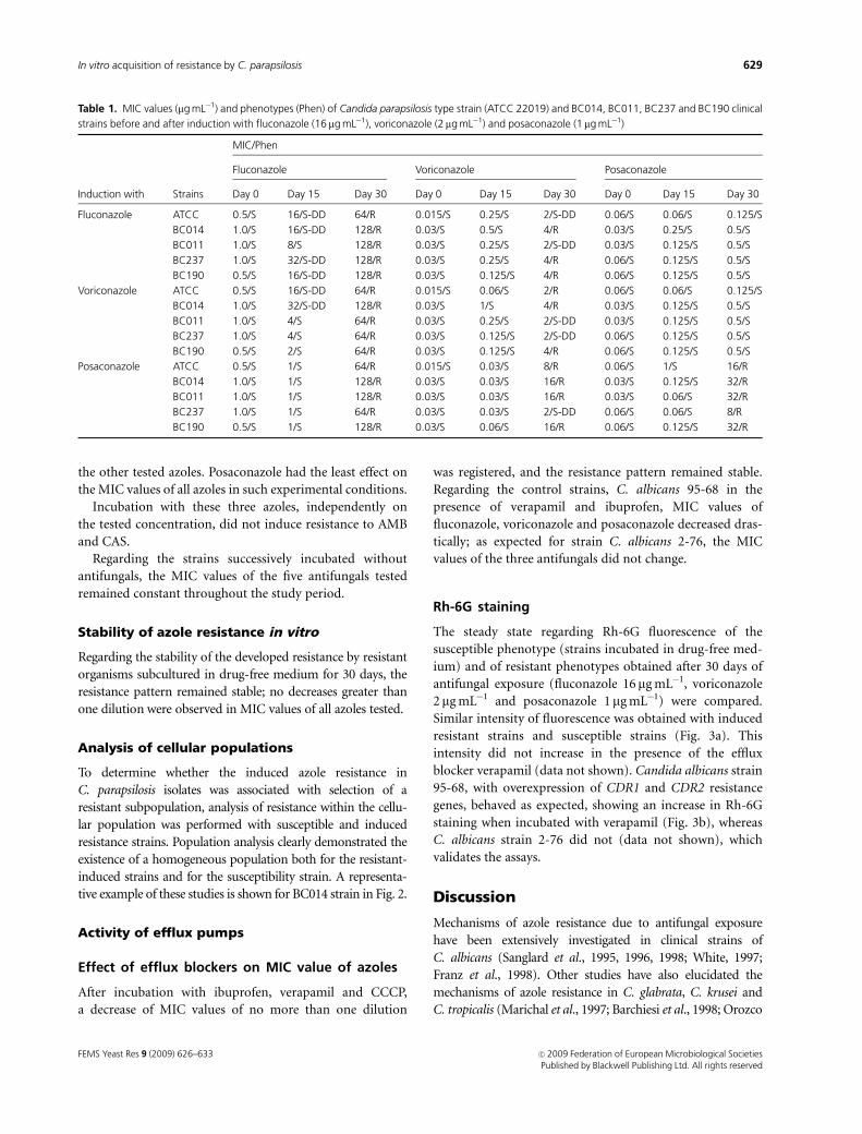
the other tested azoles. Posaconazole had the least effect on
the MIC values of all azoles in such experimental conditions.
Incubation with these three azoles, independently on
the tested concentration, did not induce resistance to AMB
and CAS.
Regarding the strains successively incubated without
antifungals, the MIC values of the five antifungals tested
remained constant throughout the study period.
Stability of azole resistance in vitro
Regarding the stability of the developed resistance by resistant
organisms subcultured in drug-free medium for 30 days, the
resistance pattern remained stable; no decreases greater than
one dilution were observed in MIC values of all azoles tested.
Analysis of cellular populations
To determine whether the induced azole resistance in
C. parapsilosis isolates was associated with selection of a
resistant subpopulation, analysis of resistance within the cellu-
lar population was performed with susceptible and induced
resistance strains. Population analysis clearly demonstrated the
existence of a homogeneous population both for the resistant-
induced strains and for the susceptibility strain. A representa-
tive example of these studies is shown for BC014 strain in Fig. 2.
Activity of efflux pumps
Effect of efflux blockers on MIC value of azoles
After incubation with ibuprofen, verapamil and CCCP,
a decrease of MIC values of no more than one dilution
was registered, and the resistance pattern remained stable.
Regarding the control strains, C. albicans 95-68 in the
presence of verapamil and ibuprofen, MIC values of
fluconazole, voriconazole and posaconazole decreased dras-
tically; as expected for strain C. albicans 2-76, the MIC
values of the three antifungals did not change.
Rh-6G staining
The steady state regarding Rh-6G fluorescence of the
susceptible phenotype (strains incubated in drug-free med-
ium) and of resistant phenotypes obtained after 30 days of
antifungal exposure (fluconazole 16 mg mL�1, voriconazole
2 mg mL�1 and posaconazole 1mg mL�1) were compared.
Similar intensity of fluorescence was obtained with induced
resistant strains and susceptible strains (Fig. 3a). This
intensity did not increase in the presence of the efflux
blocker verapamil (data not shown). Candida albicans strain
95-68, with overexpression of CDR1 and CDR2 resistance
genes, behaved as expected, showing an increase in Rh-6G
staining when incubated with verapamil (Fig. 3b), whereas
C. albicans strain 2-76 did not (data not shown), which
validates the assays.
Discussion
Mechanisms of azole resistance due to antifungal exposure
have been extensively investigated in clinical strains of
C. albicans (Sanglard et al., 1995, 1996, 1998; White, 1997;
Franz et al., 1998). Other studies have also elucidated the
mechanisms of azole resistance in C. glabrata, C. krusei and
C. tropicalis (Marichal et al., 1997; Barchiesi et al., 1998; Orozco
Table 1. MIC values (mg mL�1) and phenotypes (Phen) of Candida parapsilosis type strain (ATCC 22019) and BC014, BC011, BC237 and BC190 clinical
strains before and after induction with fluconazole (16 mg mL�1), voriconazole (2 mg mL�1) and posaconazole (1 mg mL�1)
Induction with Strains
MIC/Phen
Fluconazole Voriconazole Posaconazole
Day 0 Day 15 Day 30 Day 0 Day 15 Day 30 Day 0 Day 15 Day 30
Fluconazole ATCC 0.5/S 16/S-DD 64/R 0.015/S 0.25/S 2/S-DD 0.06/S 0.06/S 0.125/S
BC014 1.0/S 16/S-DD 128/R 0.03/S 0.5/S 4/R 0.03/S 0.25/S 0.5/S
BC011 1.0/S 8/S 128/R 0.03/S 0.25/S 2/S-DD 0.03/S 0.125/S 0.5/S
BC237 1.0/S 32/S-DD 128/R 0.03/S 0.25/S 4/R 0.06/S 0.125/S 0.5/S
BC190 0.5/S 16/S-DD 128/R 0.03/S 0.125/S 4/R 0.06/S 0.125/S 0.5/S
Voriconazole ATCC 0.5/S 16/S-DD 64/R 0.015/S 0.06/S 2/R 0.06/S 0.06/S 0.125/S
BC014 1.0/S 32/S-DD 128/R 0.03/S 1/S 4/R 0.03/S 0.125/S 0.5/S
BC011 1.0/S 4/S 64/R 0.03/S 0.25/S 2/S-DD 0.03/S 0.125/S 0.5/S
BC237 1.0/S 4/S 64/R 0.03/S 0.125/S 2/S-DD 0.06/S 0.125/S 0.5/S
BC190 0.5/S 2/S 64/R 0.03/S 0.125/S 4/R 0.06/S 0.125/S 0.5/S
Posaconazole ATCC 0.5/S 1/S 64/R 0.015/S 0.03/S 8/R 0.06/S 1/S 16/R
BC014 1.0/S 1/S 128/R 0.03/S 0.03/S 16/R 0.03/S 0.125/S 32/R
BC011 1.0/S 1/S 128/R 0.03/S 0.03/S 16/R 0.03/S 0.06/S 32/R
BC237 1.0/S 1/S 64/R 0.03/S 0.03/S 2/S-DD 0.06/S 0.06/S 8/R
BC190 0.5/S 1/S 128/R 0.03/S 0.06/S 16/R 0.06/S 0.125/S 32/R
FEMS Yeast Res 9 (2009) 626–633 c� 2009 Federation of European Microbiological SocietiesPublished by Blackwell Publishing Ltd. All rights reserved
629In vitro acquisition of resistance by C. parapsilosis

et al., 1998). However, very few data are yet available regarding
azole resistance in C. parapsilosis. This Candida species is an
opportunistic yeast responsible for nosocomial infections,
especially in immunocompromised or debilitated patients.
Resistance to fluconazole among Candida spp. has been
reported to emerge whenever this drug is used extensively
(Richardson, 2005). The results of our study stress several
relevant characteristics of the in vitro acquisition of resis-
tance by C. parapsilosis after prolonged exposure to anti-
fungals. When incubated with a therapeutic fluconazole
concentration (aiming to reproduce in vivo antifungal treat-
ment), azole resistance quickly developed; similar results
were obtained with a lower concentration of the same
antifungal, showing that of the three azoles tested, flucona-
zole is an easy and fast inducer of resistance. The two
different approaches used for induction of resistance corro-
borate this conclusion. This finding underlines the need
to review therapeutic protocols. Both voriconazole and
posaconazole induce resistance at concentrations of 2 and
1 mg mL�1, respectively, but at lower concentrations the
susceptibility pattern did not change drastically. As ex-
pected, incubation with any of the three azoles did not
induce resistance to AMB and CAS, as azole targets are
distinct and selective.
Our data strongly suggest that the dynamics of in vitro
development of azole resistance in C. parapsilosis is quite
different from that described for C. tropicalis and C. albicans
(Calvet et al., 1997; Barchiesi et al., 2000; Marr et al., 2001).
For C. albicans, the time required to develop fluconazole
resistance is longer and it could be fully reverted to baseline
when the antifungal exposure stopped. Candida albicans
isolates that became resistant showed an increase of mRNA
(a)
(b)
(c)
FLU
VOR POS
FLU
VOR POS
FLU
VOR
POS
10
MIC
val
ue (
µg m
L–1
)M
IC v
alue
(µg
mL
–1)
MIC
val
ue (
µg m
L–1
)
1
0.1
5 10 15
Days in culture
Days in culture
Days in culture
20 25 30
5 10 15 20 25 30
5 10 15 20 25 30
0.01
10
1
0.1
0.01
10
1
0.1
0.01
Fig. 1. Variation of fluconazole (FLU), voriconazole (VOR) and posaco-
nazole (POS) MIC values for Candida parapsilosis type strain (ATCC
22019) cultured in medium containing two- and fourfold MIC values of
FLU (a), VOR (b) and POS (c) for 30 days. , MIC of FLU; , MIC of
VOR; , MIC of POS.
(a)
(b)
(c)
100
10
1
100
10
1
100
10
1
0 0.125 0.5 2 8
0 0.25 1 4 16
0 1 4 16 64
BC014BC014FLUBC014VORBC014POS
Fig. 2. Population analysis (a representative example) of a susceptible
(BC014) and the respective resistant-induced strains following 30 days of
exposure to azoles (BC014 FLU, BC014 VOR and BC014 POS). The
percentage of resistant cells (y-axis, log scale) is defined as the ratio of
colonies with growth on plates containing the indicated concentration of
fluconazole (FLU) (a), voriconazole (VOR) (b) and posaconazole (POS) (c)
relative to growth in the absence of drug.
FEMS Yeast Res 9 (2009) 626–633c� 2009 Federation of European Microbiological SocietiesPublished by Blackwell Publishing Ltd. All rights reserved
630 A.T. Pinto e Silva et al.

specific for a CDR ATP-binding cassette transporter efflux
pump (Marr et al., 1998, 2001). The in vitro induction of
resistance in C. tropicalis has been documented with associated
increased expression of CDR1 and MDR1 genes (Barchiesi
et al., 2000), coding for a specific fluconazole transporter.
Distinct antifungal resistance mechanisms have been
described among Candida spp., active efflux being the most
relevant mechanism of azole resistance; its molecular base is
relatively well known for C. albicans (Sanglard & Odds,
2002). Overexpression of these efflux proteins confers
resistance to most azoles but in the same degree. Whereas
ketoconazole, fluconazole, itraconazole and voriconazole are
transported readily, posaconazole is transported to a much
lesser extent (Chau et al., 2004). Apparently, this compound
is not the ideal substrate for these transporters.
If the underlying resistance mechanism induced after
antifungal exposure were overexpression of efflux pumps,
we would expect that in the presence of both types of efflux
blockers MIC values would show a decrease and Rh-6G
staining in resistant cells exposed to antifungal would be
lower when compared with nonexposed cells. According to
these results, the efflux seems to play a minor role on the
resistance induced; however, the hypothesis of the existence
of specific C. parapsilosis efflux pumps blocked by different
modulators or with fluorescence substrate other than Rh-6G
cannot be excluded.
In this study, the hypothesis that induced resistance in
C. parapsilosis may be due to mutation in genes that codify
enzymes of the ergosterol biosynthesis pathway gains con-
siderable strength. This mutation is probably induced by the
antifungal and does not result from selection of an initial
resistant clone. This last hypothesis was not supported by
the results from our population studies, as both the suscep-
tible population (without antifungal exposure) as well as the
resistant population, obtained following induction, were
shown to be homogeneous.
The azoles bind in the vicinity of the haeme group of
lanosterol-14a-demethylase. Although there are silent
mutations scattered throughout the gene, at least a dozen
different mutations in the active region result in a lower
affinity for azoles, resulting in a greater influence on the
antimicrobial activity of fluconazole and voriconazole than
of posaconazole and itraconazole (Levin et al., 1998). More-
over, the latter two drugs are able to bind to an additional
domain of the enzyme by means of their long side chain (Li
et al., 2004; Akins, 2005), meaning that these two antifungals
can still inhibit the target at a point when fluconazole and
voriconazole are no longer active (Sabatelli et al., 2006).
Accordingly, such mutation should result in a lower impact
on the susceptibility profile to posaconazole than to fluco-
nazole or voriconazole. Because of the two different binding
sites of posaconazole, the occurrence of large-spectrum
cross-resistance is highly unlikely.
Different assumptions support the occurrence of drug-
induced mutation during the described assays: the rapidity
of development of the resistant phenotype; the fact that the
three azoles are affected, although to a different extent; the
insignificant role of efflux; and the stability of resistance
following prolonged absence of drug.
Spontaneous mutations are very infrequent, occurring at
frequencies of c. 10�6–10�8 per gene. The constancy of the
MIC values in time when strains were incubated in drug-free
medium strongly supports the assumption that the occur-
rence of spontaneous mutations is uncommon.
Diminished sensitivity of the target is probably the main
resistance mechanism to azoles in C. parapsilosis. Likewise,
phenotypically stable resistance to azole antifungals in
C. albicans can result from mutations in genes involved in the
ergosterol synthesis pathway (including the target enzyme
14-a-demethylase). Regarding C. parapsilosis, the ERG se-
quences of the biosynthetic pathway of ergosterol need to be
determined in resistant strains and compared with susceptible
strains to clarify whether mutations are associated with
azole resistance, as well as to assess changes in membrane
Fig. 3. Representative example of flow cytometric analysis of yeast
blastoconidia after Rh-6G staining, an efflux pump substrate. (a) Histo-
gram obtained by flow cytometric analysis at FL1 (515 nm) of BC014-
susceptible strain incubated in drug-free medium (A) and BC014-
resistant strain after incubation during 30 days with 16mg mL�1 of
fluconazole (B). (b) Histogram obtained by flow cytometric analysis at
FL1 (515 nm) of resistant Candida albicans 95-68 strain (overexpression
of genes that codify to efflux pumps) without (A) and with (B) the efflux
blocker verapamil. af, autofluorescence of yeast blastoconidia.
FEMS Yeast Res 9 (2009) 626–633 c� 2009 Federation of European Microbiological SocietiesPublished by Blackwell Publishing Ltd. All rights reserved
631In vitro acquisition of resistance by C. parapsilosis
lipid content. Studies involving a large number of isolates of
C. parapsilosis, aiming to characterize the cellular and mole-
cular factors conferring resistance are being pursued.
In summary, we described the rapid and stable develop-
ment of azole resistance in C. parapsilosis after growth in the
presence of azole antifungals, fluconazole being the fastest
inducer while posaconazole was the slowest.
References
Akins RA (2005) An update on antifungal targets and
mechanisms of resistance in Candida albicans. Med Mycol 43:
285–318.
Albertson GD, Niimi M, Cannon RD & Jenkinson HF (1996)
Multiple efflux mechanisms are involved in Candida albicans
fluconazole resistance. Antimicrob Agents Ch 40: 2835–2841.
Azanza JR, Garcıa-Quetglas E & Sadaba B (2007) Farmacologıa
de los azoles. Rev Iberoam Micol 24: 223–227.
Barchiesi F, Arzeni D, Del Prete MS et al. (1998) Fluconazole
susceptibility and strain variation of Candida albicans isolates
from HIV-infected patients with oropharyngeal candidosis.
J Antimicrob Chemoth 41: 541–548.
Barchiesi F, Calabrese D, Sanglard D, Falconi di Francesco L,
Caselli F, Giannini D, Giacometti A, Gavaudan S & Scalise G
(2000) Experimental induction of fluconazole resistance in
Candida tropicalis ATCC 750. Antimicrob Agents Ch 44:
1578–1584.
Calvet HM, Yeaman MR & Filler SG (1997) Reversible
fluconazole resistance in Candida albicans: a potential in vitro
model. Antimicrob Agents Ch 41: 535–539.
Chau AS, Mendrick CA, Sabatelli FJ, Loebenberg D &
McNicholas PM (2004) Application of real-time quantitative
PCR to molecular analysis of Candida albicans strains
exhibiting reduced susceptibility to azoles. Antimicrob Agents
Ch 48: 2124–2131.
Clinical and Laboratory Standards Institute (CLSI) (2008)
Reference Method for Broth Dilution Antifungal Susceptibility
Testing of Yeasts. Approved Standard M27-A3, 3rd edn. CLSI,
Wayne, PA.
Ford JM & Hait WN (1990) Pharmacology of drugs that alter
multidrug resistance in cancer. Pharmacol Rev 42: 155–199.
Franz R, Kelly SL, Lamb DC, Kelly DE, Ruhnke M &
Morschhauser J (1998) Multiple molecular mechanisms
contribute to a stepwise development of fluconazole resistance
in clinical Candida albicans strains. Antimicrob Agents Ch 42:
3065–3072.
Guinea J, Sanchez-Somolinos M, Cuevas O, Pelaez T & Bouza E
(2006) Fluconazole resistance mechanisms in Candida krusei:
the contribution of efflux-pumps. Med Mycol 44: 575–578.
Hitchcock CA, Barett-Bee K & Russel NJ (1986) The lipid
composition of azole-sensitive and azole-resistance strains of
Candida albicans. J Gen Microbiol 132: 2421–2431.
Hof H (2006) A new, broad-spectrum azole antifungal:
posaconazole-mechanisms of action and resistance, spectrum
of activity. Mycosis 49 (suppl 1): 1–5.
Kojic EM & Darouiche RO (2004) Candida infections of medical
devices. Clin Microbiol Rev 17: 255–267.
Kontoyiannis DP & Lewis RE (2002) Antifungal drug resistance
of pathogenic fungi. Lancet 359: 1135–1144.
Levin AS, Costa SF, Mussi NS et al. (1998) Candida parapsilosis
fungemia associated with implantable and semi-implantable
central venous catheters and the hands of healthcare workers.
Diagn Micr Infec Dis 30: 243–249.
Li X, Brown N, Chau AS et al. (2004) Changes in susceptibility to
posaconazole in clinical isolates of Candida albicans. J
Antimicrob Chemoth 53: 74–80.
Marichal P, Vanden Bossche H, Odds FC, Nobels G, Warnock
DW, Timmerman V, Van Broechoven C, Fay S & Mose-Larsen
P (1997) Molecular biological characterization of an azole-
resistant Candida glabrata isolate. Antimicrob Agents Ch 41:
2229–2237.
Marr KA, Lyons CN, Rustad T, Bowden RA & White TC (1998)
Rapid, transient, fluconazole resistance in Candida albicans is
associated with increased mRNA levels of CRD. Antimicrob
Agents Ch 42: 2584–2589.
Marr KA, Lyons CN, Ha K, Rustad TR & White TC (2001)
Inducible azole resistance associated with a heterogeneous
phenotype in Candida albicans. Antimicrob Agents Ch 45:
52–59.
Munayyer HK, Mann PA, Chau AS, Yarosh-Tomaine T, Greene
JR, Hare RS, Heimark L, Palermo RE, Loebenberg D &
McNicholas PM (2003) Antibiotic resistance among gram-
negative bacilli in US intensive care units: implications for
fluoroquinolone use. JAMA 289: 885–888.
Orozco AS, Higginbotham LM, Hitchcock CA, Parkinson T,
Falconer D, Ibrahim AS, Ghannoum MA & Filler SG (1998)
Mechanism of fluconazole resistance in Candida krusei.
Antimicrob Agents Ch 42: 2645–2649.
Pfaller MA & Diekema DJ (2007) Epidemiology of invasive
candidiasis: a persistent public health problem. Clin Microbiol
Rev 20: 133–163.
Pfaller MA, Diekema DJ, Jones RN, Sader HS, Fluit AC, Hollis RJ
& Messer SASENTRY Participant Group (2001) International
surveillance of bloodstream infections due to Candida species:
frequency of occurrence and in vitro susceptibilities to
fluconazole, ravuconazole, and voriconazole of isolates
collected from 1997 through 1999 in the SENTRY
antimicrobial surveillance program. J Clin Microbiol 39:
3254–3259.
Pfaller MA, Diekema DJ, Messer SA, Hollis RJ & Jones RN (2003)
In vitro activities of caspofungin compared with those of
fluconazole and itraconazole against 3,959 clinical isolates of
Candida spp., including 157 fluconazole-resistant isolates.
Antimicrob Agents Ch 47: 1068–1071.
Pina-Vaz C, Rodrigues AG, Costa-de-Oliveira S, Ricardo E &
Mardh PA (2005) Potent synergic effect between ibuprofen
and azoles on Candida resulting from blockade of efflux
pumps as determined by FUN-1 staining and flow cytometry.
J Clin Antimicrob Chemother 56: 678–685.
FEMS Yeast Res 9 (2009) 626–633c� 2009 Federation of European Microbiological SocietiesPublished by Blackwell Publishing Ltd. All rights reserved
632 A.T. Pinto e Silva et al.

Posteraro B, Sanguinetti M, Sanglard D, La Sorda M, Boccia S,
Romano L, Morace G & Fadda G (2003) Identification and
characterization of a Cryptococcus neoformans ATP binding
cassette (ABC) transporter-encoding gene, CnAFR1, involved
in the resistance to fluconazole. Mol Microbiol 47: 357–371.
Prudencio C, Sansonetty F, Sousa MJ, Corte-Real M & Leao C
(2000) Rapid detection of efflux pumps and their relation with
drug resistance in yeast cells. Cytometry 39: 26–35.
Richardson MD (2005) Changing patterns and trends in systemic
fungal infections. J Antimicrob Chemoth 39: 1–8.
Sabatelli F, Patel R, Mann PA, Mendrick CA, Norris CC, Hare R,
Loebenberg D, Black TA & McNicholas PM (2006) In vitro
activities of posaconazole, fluconazole, itraconazole,
voriconazole, and amphotericin B against a large collection of
clinically important molds and yeasts. Antimicrob Agents Ch
50: 2009–2015.
Sanglard D & Odds FC (2002) Resistance of Candida species to
antifungal agents: molecular mechanisms and clinical
consequences. Lancet Infect Dis 2: 73–85.
Sanglard D, Kuchler K, Isher F, Pagani JL, Monod M & Bille J
(1995) Mechanisms of resistance to azole antifungal in
Candida albicans isolates from AIDS patients involve specific
multidrug transporter mutants to various antifungal agents
and other metabolic inhibitors. Antimicrob Agents Ch 40:
2300–2305.
Sanglard D, Isher F, Monod M & Bille J (1996) Susceptibilities of
Candida albicans multidrug transporter mutants to various
antifungal agents and other metabolic inhibitors. Antimicrob
Agents Ch 40: 2300–2305.
Sanglard D, Ischer F, Koymans L & Bille J (1998) Amino
acid substitution in the cytochrome P-450 lanosterol
14-a-demethylase (CYP51A1) from azole-resistant
Candida albicans clinical isolates contribute to
resistance to azoles antifungal agents. Antimicrob Agents
Ch 42: 241–253.
Sanglard D, Ischer F, Calabrese D, Majcherczyk PA & Bille J
(1999) The ATP binding cassette transporter gene CgCDR1
from Candida glabrata is involved in the resistance of clinical
isolates to azole antifungal agents. Antimicrob Agents Ch 43:
2753–2765.
White TC (1997) Increased mRNA levels of ERG16, CDR, and
MDR1 correlate with the increase in azole resistance in
Candida albicans isolates from a human immunodeficiency
virus-infected patient. Antimicrob Agents Ch 41:
1482–1487.
White TC, Holleman S, Dy F, Mirels LF & Stevens DA (2002)
Resistance mechanisms in clinical isolates of Candida albicans.
Antimicrob Agents Ch 46: 1704–1713.
Wingard JR, Merz WG, Rinaldi MG, Johnson TR, Karp JE & Saral
R (1991) Increase in Candida krusei infection among patients
with bone marrow transplantation and neutropenia treated
prophylactically with fluconazole. New Engl J Med 325:
1274–1277.
Wingard JR, Merz WG, Rinaldi MG, Miller CB, Karp JE
& Saral R (1993) Association of Torulopsis glabrata
infections with fluconazole prophylaxis in neutropenic
bone marrow transplant patients. Antimicrob Agents Ch 37:
1847–1849.
FEMS Yeast Res 9 (2009) 626–633 c� 2009 Federation of European Microbiological SocietiesPublished by Blackwell Publishing Ltd. All rights reserved
633In vitro acquisition of resistance by C. parapsilosis

BRIEF REPORT
Evaluating the resistance to posaconazole by E-testand CLSI broth microdilution methodologies of Candida spp.and pathogenic moulds
R. Araujo & S. Costa-de-Oliveira & I. Coutinho &
A. G. Rodrigues & C. Pina-Vaz
Received: 10 December 2008 /Accepted: 23 March 2009 /Published online: 7 April 2009# Springer-Verlag 2009
Abstract E-test methodology was compared with Clinicaland Laboratory Standards Institute (CLSI) broth micro-dilution, particularly concerning the detection of resistanceto posaconazole among clinical fungal isolates. Thesusceptibility of a large set of fungal strains (n=300) wasevaluated following 24 and 48 h in two different culturemedia (RPMI 1640 and Sabouraud agar). Fungal strainswere highly susceptible to posaconazole; however, few lesssusceptible strains were found, mostly regarding Candidaalbicans, Candida glabrata, Acremonium sp., Cladospo-rium sp. and Scedosporium apiospermum. Broth micro-dilution and E-test methods provided similar results forposaconazole-susceptible strains, while the less susceptiblefungal strains (10.3% of the strains showed MIC ≥2 µg/mL)resulted in higher discrepancies between the two method-ologies, particularly concerning Candida spp. E-test sus-ceptibility values were critically affected by the pH of the
culture media. Sabouraud medium provided similar suscep-tibility results for moulds to those for RPMI, soon after24 h. Posaconazole resistance was rare in this study, butroutine susceptibility methods, such as the E-test, should beable to detect fungal strains with reduced susceptibility. E-test methodology still needs improvements to recogniseaccurately strains less susceptible to posaconazole.
Introduction
The E-test is an averagely priced susceptibility methodwidely used in clinical laboratories that is easy to performand does not require specialised equipment. However,recent conflictive results regarding E-test antifungal sus-ceptibility encouraged additional studies concerning thedetection of antifungal resistance [1]. The susceptibilityprofile of several clinical strains of Candida spp. andmoulds to posaconazole (POS) was determined by brothmicrodilution (BMD) and E-test methods. Different agarmedia (RPMI 1640 and Sabouraud) and pH conditionswere tested.
Materials and methods
Three hundred strains were studied, corresponding to 131Candida isolates (C. albicans, n=75; C. parapsilosis, n=13;C. tropicalis, n=12; C. glabrata, n=14; C. guilliermondii,n=7; C. krusei, n=7; and C. lusitaniae, n=3), 145Aspergillus isolates (A. fumigatus, n=49; A. flavus, n=25;A. niger, n=25; A. terreus, n=24; A. nidulans, n=14; and A.glaucus, n=8), and 24 additional mould isolates (Mucor sp.n=2; Rhizopus sp., n=2; Acremonium sp., n=3; Scedospo-rium apiospermum, n=3; Cladosporium sp., n=3; Tricho-
Eur J Clin Microbiol Infect Dis (2009) 28:1137–1140DOI 10.1007/s10096-009-0736-2
R. Araujo (*) : S. Costa-de-Oliveira : I. Coutinho :A. G. Rodrigues : C. Pina-VazDepartment of Microbiology, Faculty of Medicine,University of Porto,Alameda Prof. Hernani Monteiro,4200-319 Porto, Portugale-mail: [email protected]
R. AraujoIPATIMUP, Institute of Pathology and Molecular Immunology,University of Porto,Porto, Portugal
A. G. RodriguesBurn Unit, Department of Plastic and Reconstructive Surgery,Faculty of Medicine, Hospital S. Joao,Porto, Portugal
C. Pina-VazClinical Pathology Laboratory, Hospital S. João,Porto, Portugal

derma sp., n=3; Alternaria sp., n=2; and Trichophyton sp,.n=6). All strains were clinical isolates (Candida spp. fromblood and genital tract, and moulds from lower respiratorytract and surgical wounds) obtained from the Laboratory ofPathology, Hospital S. João, Porto, Portugal. The singleexception was A. glaucus, which was isolated from hospitalair samples. Yeasts were characterised by Vitek 2 (BioMér-ieux, Paris, France) and moulds by macro- and micromor-phology under light microscopy. Quality control strains C.parapsilosis ATCC 22019 and C. krusei ATCC 6258 wereincluded each time as controls; the minimal inhibitoryconcentration (MIC) of POS was within the recommendedvalues for both strains [2, 3]. POS powder and E-test® stripswere kindly provided by Schering-Plough Farma (Cacém,Portugal). The determination of BMD MIC was performedaccording to the M27-A3 and M38-A2 protocols stand-ardised by the Clinical and Laboratory Standards Institute
(CLSI) for yeast and moulds respectively [4, 5]. E-test (ABBiodisk, Solna, Sweden) susceptibility was performedaccording to the manufacturer’s instructions. The test stripswere evaluated in two culture media: Sabouraud agar with 2% glucose (Difco), pH of 6.0, and RPMI 1640 agar medium(Sigma) supplemented with 2% glucose, at pH values of 6.0and 7.0. Two distinct RPMI agar formulations werecompared: the first purchased from Izasa Portugal (Oeiras,Portugal) and the second prepared in our laboratory, asrecommended by the E-test instructions. All tests wereperformed in duplicate.
For both methods, MICs were read following 24 and48 h of incubation at 35ºC, with the exception ofzygomycetes (after 24 h) and S. apiospermum (after72 h). MICs obtained with both methods were consideredto be in agreement whenever falling within no more thantwo consecutive dilutions [6, 7]. Fungal strains were
Table 1 Agreement (%) between the Clinical and Laboratory Standards Institute (CLSI) broth microdilution and E-test® susceptibility methodsperformed with home-made RPMI 1640 agar culture medium (pH 7.0) and Sabouraud agar (pH 6.0), after incubation for 24 and 48 h
Strains (n) RPMI 1640 (24h) RPMI 1640 (48h) Sabouraud (24h) Sabouraud (48 h)
Susceptible strains a
Candida spp. (108) 93% 88% 83% 83%
Aspergillus spp. (145) 89% 93% 92% 83%
Other moulds b (16) 75% 80% 75% 54%
Less susceptible strains a
Candida spp. c (23) 27% 10% 25% 25%
Moulds d (8) 75% 75% 86% 100%
aBroth microdilution minimal inhibitory concentrations (MICs) were determined for zygomycetes at 24 h, for Scedosporium apiospermum at 72 hand for Candida spp. and Aspergillus spp. at 48 h (CLSI protocols M27-A3 and M38-A2)b Including Mucor sp. (n=2), Rhizopus sp. (n=2), S. apiospermum (n=1), Trichoderma sp. (n=3), Alternaria sp. (n=2) and Trichophyton sp. (n=6) strainsc Strains with MIC≥2 µg/mL to posaconazole determined according to the CLSI broth microdilution method, including C. albicans (n=12), C.parapsilosis (n=1), C. tropicalis (n=3) and C. glabrata (n=7) strainsd Strains with MIC≥2 µg/mL to posaconazole determined according to the CLSI broth microdilution method, including Acremonium sp. (n=3),Cladosporium sp. (n=3) and S. apiospermum (n=2) strains
Table 2 Errors (%) from the comparison of the CLSI broth microdilution and E-testa methodologies, for the determination of posaconazolesusceptibility
Strains (n) VME (24h) ME (24h) VME (48h) ME (48h)
Candida spp. b (131) 13% d 11% 10% d 12%
Aspergillus spp. b (145) – 2% – 4%
Other moulds b,c (24) – 17% – 29%
VME: very major errors; ME: major errorsa RPMI 1640 agar medium, after incubation for 24 and 48 hb Broth microdilution MICs were determined for zygomycetes at 24 h, for Scedosporium apiospermum at 72 h and for Candida spp. andAspergillus spp. at 48 h (CLSI protocols M27-A3 and M38-A2)c Including Mucor sp., Rhizopus sp., Acremonium sp., Scedosporium apiospermum, Cladosporium sp., Trichoderma sp., Alternaria sp. andTrichophyton sp. strainsd Including C. albicans, C. tropicalis and C. glabrata strains
1138 Eur J Clin Microbiol Infect Dis (2009) 28:1137–1140

classified according to the recently proposed in vitrobreakpoints (susceptible, MIC ≤1 µg/mL; intermediate,MIC of 2 µg/mL; and resistant, MIC ≥4 µg/mL) for yeast[8] and moulds [9]. In addition, discrepant results wereconsidered a ”very major error” whenever the strain wasclassified as susceptible by the E-test and resistant by theBMD (false susceptibility); a classification of resistance byE-test with a corresponding susceptible BMD pattern wasconsidered a “major error” (false resistance) [6, 10].
Results and discussion
Posaconazole exhibited excellent in vitro activity againstmost yeast and moulds, in agreement with previous reports[1, 11]. C. krusei, C. guilliermondii and C. parapsilosis,often resistant or less susceptible to antifungals likefluconazole and caspofungin [12, 13], were high susceptibleto POS. Few yeast strains were less susceptible to POS (MIC≥2 µg/mL), particularly C. albicans (12 strains), C. glabrata(7 strains), C. tropicalis (3 strains) and C. parapsilosis(1 strain). All Aspergillus strains showed MIC <1 µg/mL,inverse to other less susceptible moulds, like Acremoniumsp. (MIC >16 µg/mL), Cladosporium sp. (MIC >16 µg/mL),and S. apiospermum, Mucor sp. or Rhizopus sp. (with POSMICs ranging between 1 and 2 µg/mL).
The agreement between yeast BMD MIC results follow-ing 24 and 48 h was excellent (95%); thus, 48-h resultswere compared using E-test methodology. Two groups offungal strains with distinct susceptibility classification toPOS were defined according to BMD MIC values, asshown in Table 1. BMD and E-test methods providedsimilar results for yeast susceptible strains, while the lesssusceptible yeast strains provided high discrepancy betweenthe two methodologies. In fact, the E-test was unable todetect high POS MICs in 9 strains of C. albicans, 5 strainsof C. glabrata and 3 strains of C. tropicalis, after 48 h. Ourresults agreed with those of other studies mostly testingPOS-susceptible yeast strains [7, 11, 12, 14]. The overallcomparison of the BMD and E-test methods was lessapparent than previously reported because of the higherpercentage (18% versus less than 2% from previousstudies) of less susceptible yeast strains to POS includedin this study. The isolation of yeast isolates that are lesssusceptible or resistant to POS has not been frequentlyreported [1, 11], but the susceptibility methods usedroutinely, as is the case with the E-test, should be able todetect all fungal strains with reduced susceptibility. Around10% of E-test MICs for Candida spp. resulted in “verymajor errors” (Table 2), being highly problematic in clinicallaboratories employing a single methodology for suscepti-bility testing. Conversely, no “very major errors” werefound with moulds, similar to previous reports [10]. The
BMD and E-test methods provided similar results for allmoulds tested (Table 1).
The E-test had been previously tested in distinct culturemedia [14], but RPMI 1640 agar remains the recommendedmedium. Sabouraud medium might provide a better nutrientsupport for several pathogenic fungi [15] and in this study itprovided similar susceptibility results for moulds than RPMI1640 medium, soon after 24 h; inversely, it did not improveE-test results for yeasts (Table 1). The discrepancy weobserved between Sabouraud and RPMI 1640 culture mediacame from distinct pH conditions; in fact, E-test values weresimilar when Sabouraud and RPMI 1640 agar were equallybuffered to a pH of 6.0 (data not shown). Te Dorsthorst et al.[16] had previously shown that pH critically affects in vitroantifungal susceptibility. No differences were found whencomparing home-made and commercial RPMI agar media(data not shown). The occurrence of the phenomenon “novisible growth after 24 h”, incidentally mentioned byprevious studies regarding C. albicans [12], was moreregularly observed testing yeast susceptibility with “home-made” RPMI agar (the E-test was repeated in these cases).The employment of a commercial formulation of RPMIculture media with more standardised pH values may favourE-test determination and improve inter-laboratory agreement.
C. albicans and A. fumigatus remain the predominantyeast and mould species causing invasive infection.However, a shift towards non-albicans Candida, zygomy-cetes and Scedosporium species, which usually showreduced susceptibility to antifungals, was recently described[10, 11]. Additionally, the resistance of azoles has beenincreasingly reported [12, 13] and susceptibility methodsmust be prepared and should be able to detect it. E-testmethodology still needs improvements in order to betterallow the recognition of less susceptible yeast strains byantifungals. The employment of Sabouraud medium for E-test susceptibility testing of moulds may present consider-able advantages regarding early detection and may lead tomore availability in clinical laboratories.
Acknowledgements The authors are grateful to Maria Luz Dias forthe excellent technical assistance on susceptibility testing and to thetwo reviewers who made valuable suggestions that considerablyimproved the manuscript. RA, SCO and IC received grants fromFundação para Ciência e Tecnologia (FCT). IPATIMUP is partiallysupported by FCT, Programa Operacional Ciência e Inovação (POCI)and Programa Operacional do Potencial Humano (POPH – QREN).
References
1. Diekema DJ, Messer SA, Hollis RJ et al (2007) Evaluation ofEtest and disk diffusion methods compared with broth micro-dilution antifungal susceptibility testing of clinical isolates ofCandida spp. against posaconazole. J Clin Microbiol 45:1974–1977. doi:10.1128/JCM.02087-06
Eur J Clin Microbiol Infect Dis (2009) 28:1137–1140 1139

2. Barry AL, Pfaller MA, Brown SD et al (2000) Quality controllimits for broth microdilution susceptibility tests of ten antifungalagents. J Clin Microbiol 38:3457–3459
3. Chaturvedi V, Ramani R, Ghannoum MA et al (2008) Multi-laboratory testing of antifungal combinations against a qualitycontrol isolate of Candida krusei. Antimicrob Agents Chemother5:1500–1502. doi:10.1128/AAC.00574-07
4. Clinical Laboratory Standards Institute (2008) Reference methodfor broth dilution antifungal susceptibility testing of yeasts:approved standard, 3rd edn. M27-A3. CLSI, Wayne, PA
5. Clinical Laboratory Standards Institute (2008) Reference methodfor broth dilution antifungals susceptibility testing of conidium-forming filamentous fungi: approved standard, 2nd edn. M38-A2.CLSI, Wayne, PA
6. Guinea J, Peláez T, Alcalá L et al (2007) Correlation between theE-test and the CLSI —38 A microdilution method to determinethe activity of amphotericin B, voriconazole, and itraconazoleagainst clinical isolates of Aspergillus fumigatus. Diagn MicrobiolInfect Dis 57:273–276. doi:10.1016/j.diagmicrobio.2006.09.003
7. Sims CR, Paetznick VL, Rodriguez JR et al (2006) Correlationbetween microdilution, E-test, and disk diffusion methods forantifungal susceptibility testing of posaconazole against Candidaspp. J Clin Microbiol 44:2105–2108. doi:10.1128/JCM.02591-05
8. Pfaller MA, Messer SA, Boyken L et al (2008) Selection of asurrogate agent (fluconazole or voriconazole) for initial suscepti-bility testing of posaconazole against Candida spp.: results from aglobal antifungal surveillance program. J Clin Microbiol 46:551–559. doi:10.1128/JCM.01952-07
9. Espinel-Ingroff A, Arthington-Skaggs B, Iqbal N et al (2007)Multicenter evaluation of a new disk agar diffusion method forsusceptibility testing of filamentous fungi with voriconazole,posaconazole, itraconazole, amphotericin B, and caspofungin. JClin Microbiol 45:1811–1820. doi:10.1128/JCM.00134-07
10. Messer SA, Diekema DJ, Hollis RJ et al (2007) Evaluation of diskdiffusion and Etest compared to broth microdilution for antifungalsusceptibility testing of posaconazole against clinical isolates offilamentous fungi. J Clin Microbiol 45:1322–1324. doi:10.1128/JCM.02450-06
11. Espinel-Ingroff A (2006) Comparison of three commercial assaysand a modified disk diffusion assay with two broth microdilutionreference assays for testing zygomycetes, Aspergillus spp.,Candida spp., and Cryptococcus neoformans with posaconazoleand amphotericin B. J Clin Microbiol 43:3616–3622. doi:10.1128/JCM.01187-06
12. Alexander BD, Byrne TC, Smith KL et al (2007) Comparativeevaluation of Etest and sensititre yeastone panels against theClinical and Laboratory Standards Institute M27–A2 referencebroth microdilution method for testing Candida susceptibility toseven antifungal agents. J Clin Microbiol 45:698–706.doi:10.1128/JCM.01840-06
13. Costa-de-Oliveira S, Pina-Vaz C, Mendonça D et al (2008) A firstPortuguese epidemiological survey of fungaemia in a universityhospital. Eur J Clin Microbiol Infect Dis 27:365–374.doi:10.1007/s10096-007-0448-4
14. Pfaller MA, Messer SA, Mills K et al (2001) Evaluation of Etestmethod for determining posaconazole MICs for 314 clinicalisolates of Candida species. J Clin Microbiol 39:3952–3954.doi:10.1128/JCM.39.11.3952-3954.2001
15. Meletiadis J, Meis JF, Mouton JW et al (2001) Analysis of growthcharacteristics of filamentous fungi in different nutrient media. JClin Microbiol 39:478–484. doi:10.1128/JCM.39.2.478-484.2001
16. Te Dorsthorst DT, Mouton JW, van den Beukel CJ et al (2004)Effect of pH on the in vitro activities of amphotericin B,itraconazole, and flucytosine against Aspergillus isolates. Anti-microb Agents Chemother 48:3147–3150. doi:10.1128/AAC.48.8.3147-3150.2004
1140 Eur J Clin Microbiol Infect Dis (2009) 28:1137–1140
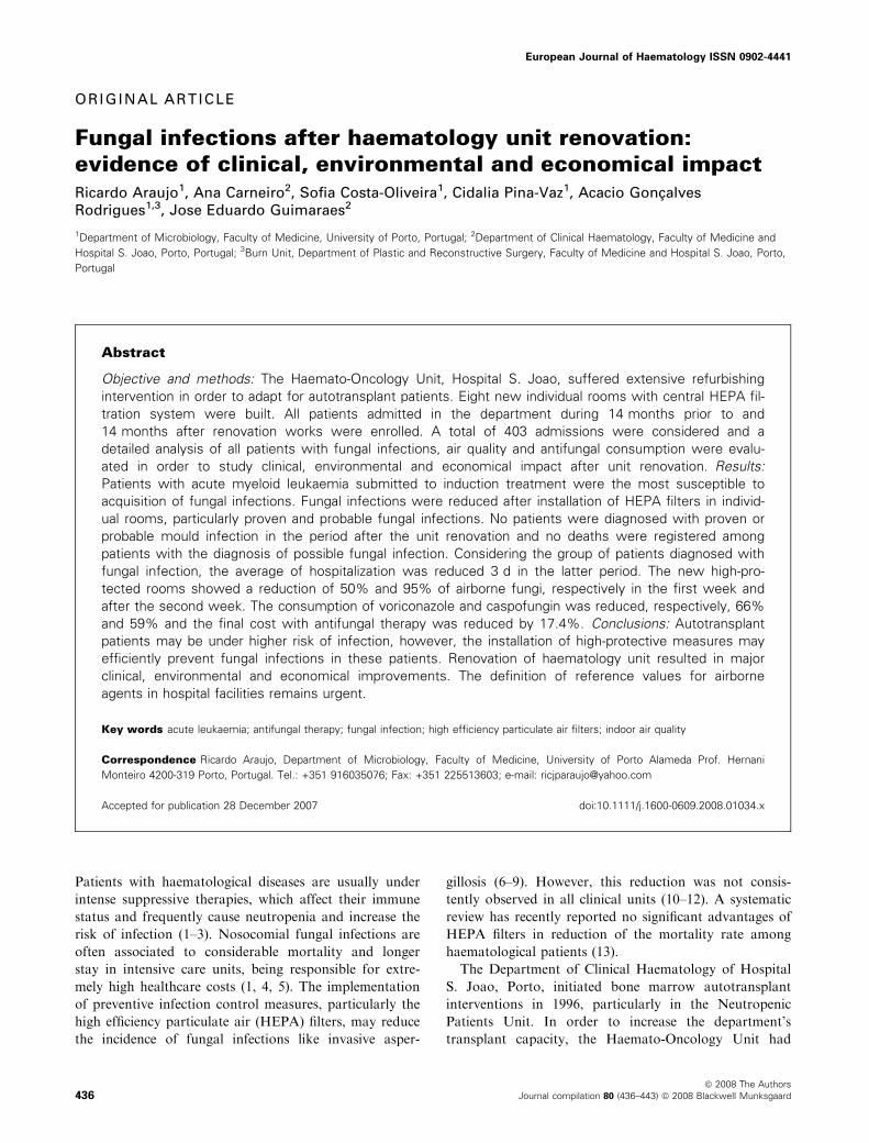
Fungal infections after haematology unit renovation:evidence of clinical, environmental and economical impactRicardo Araujo1, Ana Carneiro2, Sofia Costa-Oliveira1, Cidalia Pina-Vaz1, Acacio GoncalvesRodrigues1,3, Jose Eduardo Guimaraes2
1Department of Microbiology, Faculty of Medicine, University of Porto, Portugal; 2Department of Clinical Haematology, Faculty of Medicine and
Hospital S. Joao, Porto, Portugal; 3Burn Unit, Department of Plastic and Reconstructive Surgery, Faculty of Medicine and Hospital S. Joao, Porto,
Portugal
Patients with haematological diseases are usually under
intense suppressive therapies, which affect their immune
status and frequently cause neutropenia and increase the
risk of infection (1–3). Nosocomial fungal infections are
often associated to considerable mortality and longer
stay in intensive care units, being responsible for extre-
mely high healthcare costs (1, 4, 5). The implementation
of preventive infection control measures, particularly the
high efficiency particulate air (HEPA) filters, may reduce
the incidence of fungal infections like invasive asper-
gillosis (6–9). However, this reduction was not consis-
tently observed in all clinical units (10–12). A systematic
review has recently reported no significant advantages of
HEPA filters in reduction of the mortality rate among
haematological patients (13).
The Department of Clinical Haematology of Hospital
S. Joao, Porto, initiated bone marrow autotransplant
interventions in 1996, particularly in the Neutropenic
Patients Unit. In order to increase the department’s
transplant capacity, the Haemato-Oncology Unit had
Abstract
Objective and methods: The Haemato-Oncology Unit, Hospital S. Joao, suffered extensive refurbishing
intervention in order to adapt for autotransplant patients. Eight new individual rooms with central HEPA fil-
tration system were built. All patients admitted in the department during 14 months prior to and
14 months after renovation works were enrolled. A total of 403 admissions were considered and a
detailed analysis of all patients with fungal infections, air quality and antifungal consumption were evalu-
ated in order to study clinical, environmental and economical impact after unit renovation. Results:
Patients with acute myeloid leukaemia submitted to induction treatment were the most susceptible to
acquisition of fungal infections. Fungal infections were reduced after installation of HEPA filters in individ-
ual rooms, particularly proven and probable fungal infections. No patients were diagnosed with proven or
probable mould infection in the period after the unit renovation and no deaths were registered among
patients with the diagnosis of possible fungal infection. Considering the group of patients diagnosed with
fungal infection, the average of hospitalization was reduced 3 d in the latter period. The new high-pro-
tected rooms showed a reduction of 50% and 95% of airborne fungi, respectively in the first week and
after the second week. The consumption of voriconazole and caspofungin was reduced, respectively, 66%
and 59% and the final cost with antifungal therapy was reduced by 17.4%. Conclusions: Autotransplant
patients may be under higher risk of infection, however, the installation of high-protective measures may
efficiently prevent fungal infections in these patients. Renovation of haematology unit resulted in major
clinical, environmental and economical improvements. The definition of reference values for airborne
agents in hospital facilities remains urgent.
Key words acute leukaemia; antifungal therapy; fungal infection; high efficiency particulate air filters; indoor air quality
Correspondence Ricardo Araujo, Department of Microbiology, Faculty of Medicine, University of Porto Alameda Prof. Hernani
Monteiro 4200-319 Porto, Portugal. Tel.: +351 916035076; Fax: +351 225513603; e-mail: [email protected]
Accepted for publication 28 December 2007 doi:10.1111/j.1600-0609.2008.01034.x
ORIGINAL ARTICLE
European Journal of Haematology ISSN 0902-4441
436ª 2008 The Authors
Journal compilation 80 (436–443) ª 2008 Blackwell Munksgaard
undergone restructuring works to build eight isolation
rooms with HEPA filters and positive pressure central
system. Are these protective measures enough to prevent
fungal infections in patients admitted for autotransplant?
Were clinical and environmental variables improved and
patients admitted in a safer unit? The main objective of
the current study considered the detailed analysis of the
clinical data of all patients with fungal infections admit-
ted at Haemato-Oncology Unit, before and after renova-
tion works. We intent to verify if a more protected
environment may be correlated with clinical improve-
ments regarding fungal infection in risk patients admitted
in haematological units. The incidence of yeast and
mould infections in haematological patients, mortality
rate and related risk factors was evaluated. The assess-
ment of indoor air quality and antifungal consumption
were also considered, before and after unit renovation.
Methods
Unit conditions and study population
The renovation works and installation of central HEPA
filters at the Haemato-Oncology Unit started at Novem-
ber 2004, being the new rooms available for admission
of patients in June 2005. Renovation works occurred
into the Haemato-Oncology Unit separated from the
main hall of the unit by a barrier (door). No additional
measures were considered. The installed air filtration
HEPA system was based on a polyester pre-filter G4
(initial efficiency of 70%) replaced every 15 d, a polyes-
ter fine filter F8 (24 · 24 inches; flow of 4500 m3 h)1;
Luwa GmbH, Frankfurt, Germany) replaced every
6 months and the HEPA filter H13 of polyester and
fiberglass (glued type with a flow of 4200 m3 h)1; Camfil
Farr, Trosa, Sweden) replaced once a year. HEPA air
filtration system was turned on continuously 24 h per
day, being turn off only for filter replacement, and it
was adjusted to 15–20 air exchanges per hour. Addi-
tional protective conditions were added to the new
rooms like the existence of an anteroom with HEPA fil-
ters installed along the entrance to each room (during
door opening prevents air flow into patient room, also
preventing the access of non-authorized people), water
filtration and the systematic use of protective sterile
clothes (the unit staff or visitors must wear sterile gown,
shoe covers, head cap and surgical mask). The depart-
ment availability comprised 12 beds before renovation
works (one room each for six men and six women,
respectively, both without air filters or other protective
measure) and 14 beds after the unit renovation (two
rooms with three beds each, both without air filters or
other protective measure, plus the eight new individual
rooms equipped with HEPA filters and positive air
flow). Additional protective measures like extensive
hand disinfection were part of the staff routines.
Patients and unit staffs were continuously alerted to a
higher risk of infection during renovation works and
stressed to keep the wards maximally isolated from the
exterior air. During that period, visitors and unit staff
must nevertheless wear surgical facial mask into the
rooms with no protective measures, the doors and win-
dows were always kept closed, the patients only leaving
the room for clinical exams and to use toilet facilities.
In such cases, patients must wear N95 NIOSH facial
mask (Kimberly-Clark SL, Madrid, Spain), as recom-
mended (14). All patients admitted in the department
from April 2004 to July 2006 were monitored, corre-
sponding to the period before and during the works
(14 months) and the period of 14 months immediately
after the renovation works. The Haemato-Oncology
Unit admitted patients with all types of acute leukaemia
and the most severe cases of lymphoma ⁄myeloma malig-
nancies in the northern region of Portugal. Data regard-
ing haematological disease, admission diagnosis, length
of stay at hospital, duration of neutropenia, acquired
infections, therapeutic protocols and origin of infection
were registered.
Definition of hospital-acquired infection
The definition of invasive fungal infection was estab-
lished in three levels, proven, probable and possible,
according to previous consensual criteria (15). Hospital-
acquired yeast infections or nosocomial infections were
defined following the isolation and diagnosis of infec-
tions after the initial 48 h after admission in the haema-
tology unit, according to Centers for Disease Control
and Prevention (CDC) (16) and other reports (17). Pro-
longed fever corresponded to the persistence of fever for
more than 4 d and refractory to broad-spectrum antibac-
terial treatment, while prolonged neutropenia corre-
sponded to neutropenia longer than 10 d (15). The
incidence of fungal infections was determined considering
the number of admitted patients diagnosed with fungal
infection against the total number of admissions.
Antifungal consumption
The antifungal agents requested by the Haemato-Oncol-
ogy Unit during the 28 months surveillance period were
quantified and the consumption associated to amphoteri-
cin B, both deoxycholate and liposomal formulation,
fluconazole, voriconazole and ⁄or caspofungin was evalu-
ated. The same price was considered for each antifungal
formulation in the periods before and after renovation
works, in order to avoid false comparisons due to anti-
fungals price adjustments along the 28 months.
Araujo et al. Fungal infection after haematology renovation
ª 2008 The Authors
Journal compilation 80 (436–443) ª 2008 Blackwell Munksgaard 437

Environmental surveillance
The conventional volumetric air sampling method using
the Andersen one-stage sieve impactor was used for air
surveillance. This standard method is recommended to
collect and quantify airborne fungal species. Eighteen
impactor air samples with six different impact volumes
of air (14, 28, 56, 84, 140 and 280 L) were collected in
each room, at a flow of 28 L ⁄min, as recommended (18).
Larger air volumes were not collected due to dehydration
of agar culture medium for prolonged air exposure.
Impactor air samples were cultured using the DG18 med-
ium (19, 20) and incubated at 25�C and 37�C, during
10 d. Results were expressed as number of colony-form-
ing units per cubic meter (CFU ⁄m3) of analyzed air.
Regression through the origin was applied to the data
and the concentration of CFU in the analyzed air deter-
mined from the slope of the regression line. For each
room, the total number of colonies was plotted against
the volume of analyzed air. Identification of fungal colo-
nies was based upon macro and microscopic morphologi-
cal aspects, accordingly the standard mycological
methods (21). Sample collection was conducted in phases
of four consecutive weeks each (sampling phases per-
formed every 3 months), before and after the renovation
works.
Statistical analysis
SPSS 13.0 (SPSS Inc., Chicago, IL, USA) application
was used for data elaboration and analysis. Student’s t-
test for paired and two independent samples was used
during statistical analysis. The comparison of infection
incidence in the periods before and after renovation
works was performed determining z-values, the popula-
tion considered at normal distribution. Alpha was set to
0.05 and all reported P-values were two tailed.
Results
Haematological malignancies and fungal infections
A total of 403 admissions (198 before and 205 after the
renovation works), corresponding to 221 patients (119
women and 102 men), were made to the Haemato-Oncol-
ogy Unit during the 28 months surveillance period.
Patients’ age ranged between 15 and 86 years old (aver-
age of 54 years old). The haematological patients admit-
ted at the unit corresponded to 75 patients with acute
myeloid leukaemia (33.9%), 25 with acute lymphoblastic
leukaemia (11.3%), 4 with blast crisis of chronic myeloid
leukaemia (1.8%), 4 with chronic lymphoblastic leukae-
mia (1.8%), 5 with myelodysplastic syndrome (2.3%), 1
with plasma cell leukaemia (0.5%), 11 with Hodgkin
lymphoma (5.0%), 51 with non-Hodgkin lymphoma
(23.1%), 35 with multiple myeloma (15.8%) and 10 with
aplastic anaemia (4.5%). The admissions of both periods
are more detailed in Table 1.
The incidence of fungal infections in haematological
patients admitted at unit was 6.6% and 4.9%
(P = 0.001), respectively, in the periods before and after
the renovation works, corresponding to 0.33 patients per
100 d before works and 0.26 patients per 100 d after
installation of protective measures (Table 1). These fun-
gal infections also corresponded to 0.64 infections per
100 d of neutropenia before renovation and 0.49 infec-
tions per 100 d of neutropenia in the latter period.
Mould infections, proven and probable, were signifi-
cantly reduced after the renovation works (incidence of
1.5% and 0%, respectively, before and after renovation;
P < 0.001), as well as proven yeast infections (incidence
of 1.5% and 0%, respectively, before and after renova-
tion; P < 0.001). The details of the patients diagnosed
with proven and probable fungal infections are described
in Table 2. Patients 1 and 6 stayed at haematology unit
during the construction period, while the other four
patients were admitted and left the unit before this per-
iod. Pre-construction period showed more fungal infec-
tions compared to construction period (incidences of
9.5% vs. 3.7%) probably due to the decrease of admitted
patients for treatments causing longer expected neutrope-
nia. After the unit renovation, there were no proven or
probable fungal infections, being diagnosed 10 cases of
possible fungal infection (in the previous period of
14 months, seven cases were diagnosed, Table 1). Of the
previous 17 cases, three were PCR positive for Aspergil-
lus fumigatus. However, this test is not still accepted as
microbiological criteria. Nevertheless, it was especially
relevant to the fact that there were no deaths in patients
with the diagnosis of possible fungal infection in the lat-
ter period against three patients with poor outcome in
the first 14 months (Table 1). More severely ill patients
Table 1 Admissions, duration of neutropenia, unfavourable outcome
and fungal infections in haematological patients, before and after unit
renovation
Beforerenovation
Afterrenovation
Admissions 198 205
Non-neutropenia 51 43
Short neutropenia 68 74
Long neutropenia 79 88
Deaths 16 8
Fungal infections (FI) 13 10
Yeasts1 3 0
Moulds1 3 0
Possible FI 7 10
1Proven and probable infections.
Fungal infection after haematology renovation Araujo et al.
438ª 2008 The Authors
Journal compilation 80 (436–443) ª 2008 Blackwell Munksgaard

were admitted during the latter period in the new pro-
tected wards compared to the wards without HEPA fil-
ters. The 10 cases of possible fungal infection detected
after the renovation works corresponded to 7 patients
admitted in the new rooms with HEPA filters and 3
patients admitted in the other rooms without air filtra-
tion. Haematological patients with expected prolonged
neutropenia (more than 10 d) or submitted to autotrans-
plant were preferentially admitted in the new rooms. All
patients diagnosed with fungal infection stayed at least
3 wk into the haematology unit and were frequently neu-
tropenic for longer periods (the average was 23 d in both
studied phases), being simultaneously under administra-
tion of large spectrum antibiotics due to the systematic
isolation of other microbial agents from clinical samples
and ⁄or central venous catheters.Patients with acute myeloid leukaemia submitted to
the induction treatment were more susceptible to acquisi-
tion of fungal infections, both yeast and mould infec-
tions, compared with patients with other haematological
malignancies and ⁄or submitted to other treatments. A
single case of probable mould infection was detected in a
patient diagnosed with non-Hodgkin lymphoma (patient
6 shown in Table 2), being also found during the entire
study a single case of possible fungal infection in each
group of haematological patients admitted with acute
lymphoblastic leukaemia, myelodysplastic syndrome,
aplastic anaemia and non-Hodgkin lymphoma. Thirty-
eight autotransplants were successfully performed after
the renovation works; these patients were never diag-
nosed with a fungal infection. A. fumigatus was the most
frequent and detected mould during the 28 months sur-
veillance period, being registered a single case of possible
infection by Mucorales in a patient admitted before the
unit renovation. The three described yeast infections
were all caused by different organisms. Two cases were
classified as nosocomial yeast infections (patients 2 and
3; Table 2).
Economical impact
Considering the group of patients diagnosed with all fun-
gal infections, the average of hospitalization days was
reduced 3.4 d in the latter period. Patients with a proven
or probable fungal infection stayed from 21 to 75 d in
haematology unit (average of 41 hospitalization days).
Although there was also a reduction in the number of
patients diagnosed with a fungal infection, the adminis-
tration of some antifungal agents did not follow this ten-
dency. The consumption of voriconazole and
caspofungin in the Haemato-Oncology Unit was reduced
66% and 59%, respectively, in the period after the reno-
vation works, while increased the use of deoxycholate
amphotericin B, liposomal amphotericin B and fluconaz-
ole (Table 3). The final cost with antifungal therapy was
reduced by 17.4% (around €71 000) during the second
arm of the study. The usual choice for empiric antifungal
therapy of moulds was an amphotericin B formulation,
Table 2 Data from haematological patients diagnosed with proven and probable fungal infections (all patients presented fever for more than 4 d,
submitted to broad-spectrum antibiotics)
Neutropenia(days)
Haematologicaldisease Diagnosis of fungal infection Fungi detected Level1 Outcome
Patient 1 20 Acute myeloid
leukaemia
CT scan suggesting fungal
infection, negative antigen test,
positive biopsy to A. fumigatus
Aspergillus fumigatus Proven, deep
tissue
Alive
Patient 2 27 Acute myeloid
leukaemia
Positive cultures from blood and
bronchial secretions to
C. glabrata
Candida glabrata Proven, fungemia Died
Patient 3 22 Acute myeloid
leukaemia
Positive blood culture Saccharomyces
cerevisiae
Proven, fungemia Alive
Patient 4 20 Acute myeloid
leukaemia
Positive cultures from catheter
and blood
Candida parapsilosis Proven, fungemia Alive
Patient 5 27 Acute myeloid
leukaemia
Two CT scans suggesting fungal
infection, three positive cultures
to A. fumigatus, two positive
antigen tests, one positive PCR test
Aspergillus fumigatus Probable Died
Patient 6 6 Non-Hodgkin
lymphoma
Symptoms of lower respiratory
tract infection and pleural
infusion, two positive cultures
from bronchial secretions to
A. fumigatus
Aspergillus fumigatus Probable Alive
CT, Computerized tomography.1According to Ascioglu et al. (15).
Araujo et al. Fungal infection after haematology renovation
ª 2008 The Authors
Journal compilation 80 (436–443) ª 2008 Blackwell Munksgaard 439

being the second line therapy mainly an association of
drugs using voriconazole and ⁄or caspofungin. For yeast
infections, the first choice was regularly fluconazole and
the alternative caspofungin or liposomal amphotericin B.
Liposomal amphotericin B was administered at
3 mg kg day)1. The daily defined dose of the other anti-
fungals administered to patients was similar in both peri-
ods and established according to standard therapeutic
protocols.
Environmental surveillance
Higher values of A. fumigatus, Aspergillus flavus and
Aspergillus niger were detected in the cultures incubated
at 37�C, while the other fungal species were mostly
detected at 25�C. The air surveillance of the clinical
unit disclosed values of total fungi ranging from 22 to
278 CFU ⁄m3 (average of 131 CFU ⁄m3) in the rooms
without air filtration system along the 28 months sur-
veillance period. In the same wards, A. fumigatus ran-
ged between 0.7 and 19.3 CFU ⁄m3 (average of
8.5 CFU ⁄m3). The haematology main hall was not
monitored during the entire study, including the con-
struction period. Airborne quality was assessed exclu-
sively in patients’ wards throughout the study, being all
rooms considered. Staff and patients were continuously
alerted to keep wards maximally isolated from outdoor
air. No significant differences were found during the
renovation works considering airborne fungal levels into
such wards (P > 0.05). However, the levels of A. fu-
migatus increased from 8.5 to 12 CFU ⁄m3, during the
construction period. The new rooms with HEPA filters
showed a gradual improvement of the air quality since
the first week (Fig. 1). The total airborne fungi
decreased from 70 CFU ⁄m3 (reduction of 50%) in the
first week to less than 7 CFU ⁄m3 (reduction of 95%)
in the following weeks. Aspergillus fumigatus yielded
1.4 CFU ⁄m3 in the first week but it was not detected
thereafter. Similar corresponding airborne values were
detected in the entrance hall (also with HEPA filters)
that conducted to the patient protected rooms (data
not shown).
Discussion
Construction and renovation works are a well-known
risk factor for mould infections (6, 22, 23). Aspergillus
infections resulting from failure in air filtration systems
have been reported (24, 25). Other studies have reported
a reduction of Aspergillus infections after the installation
of HEPA filters and ⁄or by the existence of physical bar-
riers that limit the access of airborne conidia to the clin-
ical units (6–9). The installation of HEPA filters in the
individual rooms did not completely prevent fungal
infections in the haematological patients admitted in our
clinical unit, although its incidence was reduced by 25%.
HEPA filters had shown in other studies a reduction of
airborne fungi to less than 10 CFU ⁄m3 (26) and a
reduction of mould infections in haematological patients
(7). After unit renovation, the new rooms with HEPA
filters showed a large improvement of air quality com-
pared with the non-treated rooms, as well as a clinical
positive impact – no proven or probable fungal infec-
tions were registered in the latter period and no deaths
associated to possible fungal infections were reported,
even considering the 38 autotransplants performed in
the unit during this period. During the construction per-
iod, a small increase of airborne A. fumigatus was found
Table 3 Consumption of antifungal agents before and after renova-
tion of haematology unit (number of packs)
Antifungal and formulation Before AfterDifference(%)
Deoxycholate amphotericin B (50 mg) 580 921 +59
Liposomal amphotericin B (50 mg) 759 990 +30
Fluconazole (200 mg), capsules 691 3342 +484
Fluconazole (2 mg mL)1), intravenous
formulation (200 mL)
124 180 +45
Voriconazole (200 mg), capsules 454 330 )28
Voriconazole (200 mg), intravenous
formulation
138 7 )95
Caspofungin, intravenous formulation 382 158 )59
CF
U/m
3
0
5
10
60
80
100
A. fumigatusA. flavusA. nigerOther AspergillusYeastsOther fungi
No air treatedwards
Wards with HEPAfilters (1st wk)
Wards with HEPAfilters (2nd wkand wks after)
Figure 1 Colony-forming units per cubic meter (CFU ⁄ m3) of fungi
detected during air quality surveillance in haematology unit after reno-
vation works. Other fungi included mostly Penicillium sp. and rare iso-
lates of Mucor sp., Alternaria sp. and Scedosporium sp. Values of
Aspergillus fumigatus, Aspergillus flavus and Aspergillus niger were
detected in the cultures incubated at 37�C, while the other fungal spe-
cies were mostly detected at 25�C.
Fungal infection after haematology renovation Araujo et al.
440ª 2008 The Authors
Journal compilation 80 (436–443) ª 2008 Blackwell Munksgaard
but there was no significant clinical impact compared
with the previous months, conversely to what had been
shown in other locations (6, 22, 23). Even after the
installation of HEPA filters, it was possible to find
moulds into protected wards, particularly Penicillium sp.,
but these species are not commonly pathogenic, with the
exception of Penicillium marneffei. Aspergillus species
were rarely found, particularly A. fumigatus. Sherertz
et al. had reported no A. fumigatus infections in clinical
environments with less than 0.1 CFU ⁄m3 (27). However,
reference airborne fungal values are still not defined, as
well as it is not yet established the sampling air fre-
quency that should be performed in clinical units. Con-
firming previous reports (4), a higher number of fungal
infections were reported among patients with acute mye-
loid leukaemia. These patients were commonly associ-
ated to prolonged neutropenia and longer hospital stay.
Incidence of invasive aspergillosis had been previously
associated with an increasing number of hospital admis-
sions per year (5, 28). Possible confounders may be
detected in this study, namely regarding patients’ age,
patient’s genetic predisposition for infection, severity of
haematological malignancy and mortality rate of a unit
external group. All patients staying more than 24 h in
Haemato-Oncology Unit were included in this study (cri-
teria for admission were similar in both periods) and
previous mentioned factors occasionally may not be
equally distributed in both compared groups (before vs.
after unit renovation). However, the presented main
results and values were similar to previous reports and
focus different aspects, all indicative of clinical and envi-
ronmental improvements after installation of protective
measures.
The incidence of yeast infections was lower after
renovation works, both classified as nosocomial and
community-acquired infections. It is relevant that the
new eight rooms were individual rooms therefore reduc-
ing inter-patient, medical staff–patient and visitor–patient
contacts. Yeasts are usually present in the human inter-
nal milieu, being some nosocomial infections a clear
consequence of host neutropenia and further invasion by
the endogenous fungal agent.
The economical benefits of the haematology unit reno-
vation were shown by the reduction of the hospitaliza-
tion days in a particular group of patients –
hospitalization costs are commonly described the most
important healthcare costs (5, 29–32) – and the reduction
of the expenses with antifungal agents into the unit. The
larger consumption of both amphotericin B formulations
and fluconazole in the latter period was most probably
related to the antifungal prophylaxis administered to
patients with acute myeloid or lymphoblastic leukaemia
under longer neutropenia (more admissions in this per-
iod, as shown in Table 1). The administration of antifun-
gal prophylaxis was a clinician decision and it was
mostly employed in acute leukaemic patients under
longer neutropenia periods, being sometimes also related
to clinical history of invasive aspergillosis. Unit policy
regarding prophylaxis or treatment of fungal infections
was not modified during the studied period, fluconazole
for yeast and amphotericin B for mould infections.
In accordance with previous studies (4, 27, 33),
patients staying in hospital under prolonged neutropenia
(more than 10 d) and with longer stay than 3–4 wk are
more susceptible to develop fungal infections, usually
associated to the isolation of other microbial agents and
administration of a large spectrum of antibiotics.
Patients submitted to autotransplant may be under
higher risk of infection in haematological units, neverthe-
less, the installation of high-protective measures may effi-
ciently prevent fungal infections in these patients, as we
had shown in this study. It is urgent to understand the
presence and transmission of fungal agents in protected
clinical environments in order to improve health and
well-being of patients submitted to immunosuppressive
treatments and staying longer at hospitals.
Acknowledgements
The authors would like to thank Joao Paulo Cabral for
providing the Andersen one siege impactor throughout
the study, as well as all staff of the Department of Clini-
cal Haematology and Pharmacy, Hospital S. Joao who
kindly collaborated in this study. The authors also thank
both reviewers for their valuable suggestions that cer-
tainly improved the manuscript.
Conflict of interest
The authors declare no conflict of interest.
References
1. Groll AH, Shah PM, Mentzel C, Schneider M, Just-
Nuebling G, Huebner K. Trends in the postmortem
epidemiology of invasive fungal infections at a university
hospital. J Infect 1996;33:23–32.
2. Marr KA, Carter RA, Crippa F, Wald A, Corey L.
Epidemiology and outcome of mould infections in
hematopoietic stem cell transplant recipients. Clin Infect
Dis 2002;34:909–17.
3. Pagano L, Caira M, Candoni A, et al. The epidemiology
of fungal infections in patients with hematologic malig-
nancies: the SEIFEM-2004 study. Haematologica
2006;91:1068–75.
4. Vandewoude KH, Blot SI, Benoit D, Colardyn F,
Vogelaers D. Invasive aspergillosis in critically ill patients:
attributable mortality and excesses in length of ICU stay
and ventilator dependence. J Hosp Infect 2004;56:269–76.
Araujo et al. Fungal infection after haematology renovation
ª 2008 The Authors
Journal compilation 80 (436–443) ª 2008 Blackwell Munksgaard 441
5. Dasbach EJ, Davies GM, Teutsch SM. Burden of
aspergillosis-related hospitalizations in the United States.
Clin Infect Dis 2000;31:1524–8.
6. Oren I, Haddad N, Finkelstein R, Rowe JM. Invasive
pulmonary aspergillosis in neutropenic patients during
hospital construction: before and after chemoprophylaxis
and institution of HEPA filters. Am J Hematol
2001;66:257–62.
7. Alberti C, Bouakline A, Ribaud P, Lacroix C, Rousselot
P, Leblanc T, Derouin F; Aspergillus Study Group. Rela-
tionship between environmental fungal contamination and
the incidence of invasive aspergillosis in haematology
patients. J Hosp Infect 2001;48:198–206.
8. Hovi L, Saxen H, Saarinen-Pihkala UM, Vettenranta K,
Meri T, Richardson M. Prevention and monitoring of
invasive fungal infections in pediatric patients with cancer
and hematologic disorders. Pediatr Blood Cancer
2007;48:28–34.
9. Berthelot P, Loulergue P, Raberin H, Turco M, Mounier
C, Tran Manh Sung R, Lucht F, Pozzetto B, Guyotat
D. Efficacy of environmental measures to decrease the
risk of hospital-acquired aspergillosis in patients hospita-
lised in haematology wards. Clin Microbiol Infect
2006;12:738–44.
10. Hospenthal DR, Kwon-Chung KJ, Bennett JE. Concen-
trations of airborne Aspergillus compared to the incidence
of invasive aspergillosis: lack of correlation. Med Mycol
1998;36:165–8.
11. Mahieu LM, De Dooy JJ, Van Laer FA, Jansens H, Ieven
MM. A prospective study on factors influencing Aspergil-
lus spore load in the air during renovation works in a neo-
natal intensive care unit. J Hosp Infect 2000;45:191–7.
12. Cooper EE, O’Reilly MA, Guest DI, Dharmage SC. Influ-
ence of building construction work on Aspergillus infec-
tion in a hospital setting. Infect Control Hosp Epidemiol
2003;24:472–6.
13. Eckmanns T, Ruden H, Gastmeier P. The influence of
high-efficiency particulate air filtration on mortality and
fungal infection among highly immunosuppressed patients:
a systematic review. J Infect Dis 2006;193:1408–18.
14. Raad I, Hanna H, Osting C, Hachem R, Umphrey J,
Tarrand J, Kantarjian H, Bodey GP. Masking of
neutropenic patients on transport from hospital rooms is
associated with a decrease in nosocomial aspergillosis
during construction. Infect Control Hosp Epidemiol
2002;23:41–3.
15. Ascioglu S, Rex JH, de Pauw B, et al. Mycoses Study
Group of the National Institute of Allergy and Infectious
Diseases. Defining opportunistic invasive fungal infections
in immunocompromised patients with cancer and hemato-
poietic stem cell transplants: an international consensus.
Clin Infect Dis 2002; 34: 7–14.
16. Garner JS, Jarvis WR, Emori TG, Horan TC, Hughes JM.
CDC definitions for nosocomial infections. In: Olmsted
RN, ed. APIC Infection Control and Applied Epidemiology:
Principles and Practice. St. Louis: Mosby, 1996:1.
17. Jeyaratnam D, Edgeworth JD, French GL. Enhanced
surveillance of meticillin-resistant Staphylococcus aureus
bacteraemia in a London teaching hospital. J Hosp Infect
2006;63:365–73.
18. Andersen AA. New sampler for the collection, sizing and
enumeration of viable airborne particles. J Bacteriol
1958;76:471–84.
19. Hocking AD, Pitt JI. Dichloran–glycerol medium for
enumeration of xerophilic fungi from low-moisture foods.
Appl Environ Microbiol 1980;39:488–92.
20. Wu PC, Su HJJ, Ho HM. A comparison of sampling
media for environmental viable fungi collected in a
hospital environment. Environ Res 2000;82:253–7.
21. Larone DH. Medically important fungi: a guide to
identification, 3rd edn. Washington DC: American Society
for Microbiology, 2005.
22. De La Rosa GR, Champlin RE, Kontoyiannis DP. Risk
factors for the development of invasive fungal infections
in allogeneic blood and marrow transplant recipients.
Transpl Infect Dis 2002;4:3–9.
23. Vonberg RP, Gastmeier P. Nosocomial aspergillosis in
outbreak settings. J Hosp Infect 2006;63:246–54.
24. Lutz BD, Jin J, Rinaldi MG, Wickes BL, Huycke MM.
Outbreak of invasive Aspergillus infection in surgical
patients, associated with a contaminated air-handling
system. Clin Infect Dis 2003;37:786–93.
25. Munoz P, Guinea J, Pelaez T, Duran C, Blanco JL,
Bouza E. Nosocomial invasive aspergillosis in a heart
transplant patient acquired during a break in the
HEPA air filtration system. Transpl Infect Dis 2004;6:
50–4.
26. Araujo R, Cabral JP, Rodrigues AG. Air filtration systems
and restrictive access conditions improve indoor air quality
in clinical units. Penicillium as a general indicator of hospi-
tal indoor fungal levels. Am J Infect Control (in press).
DOI: 10.1016/j.ajic.2007.02.001.
27. Sherertz RJ, Belani A, Kramer BS, Elfenbein GJ, Weiner
RS, Sullivan ML, Thomas RG, Samsa GP. Impact of air
filtration on nosocomial Aspergillus infections. Unique risk
of bone marrow transplant recipients. Am J Med
1987;83:709–18.
28. Allam MF, Del Castillo AS, Diaz-Molina C, Navajas RF.
Invasive pulmonary aspergillosis: identification of risk
factors. Scand J Infect Dis 2002;34:819–22.
29. McGarry SA, Engemann JJ, Schmader K, Sexton DJ,
Kaye KS. Surgical-site infection due to Staphylococcus
aureus among elderly patients: mortality, duration of
hospitalization, and cost. Infect Control Hosp Epidemiol
2004;25:461–7.
30. Olaechea PM, Palomar M, Leon-Gil C, Alvarez-Lerma F,
Jorda R, Nolla-Salas J, Leon-Regidor MA; EPCAN
Study Group. Economic impact of Candida colonization
and Candida infection in the critically ill patient. Eur J
Clin Microbiol Infect Dis 2004;23:323–30.
31. Falagas ME, Apostolou KE, Pappas VD. Attributable
mortality of candidemia: a systematic review of matched
Fungal infection after haematology renovation Araujo et al.
442ª 2008 The Authors
Journal compilation 80 (436–443) ª 2008 Blackwell Munksgaard
cohort and case–control studies. Eur J Clin Microbiol
Infect Dis 2006;25:419–25.
32. Riedel A, Choe L, Inciardi J, Yuen C, Martin T,
Guglielmo BJ. Antifungal prophylaxis in chemotherapy-
associated neutropenia: a retrospective, observational
study. BMC Infect Dis 2007;7:70.
33. Marr KA, Carter RA, Boeckh M, Martin P, Corey L.
Invasive aspergillosis in allogeneic stem cell transplant
recipients: changes in epidemiology and risk factors. Blood
2002;100:4358–66.
Araujo et al. Fungal infection after haematology renovation
ª 2008 The Authors
Journal compilation 80 (436–443) ª 2008 Blackwell Munksgaard 443