An overview of signaling pathways regulating YAP/TAZ activity
16
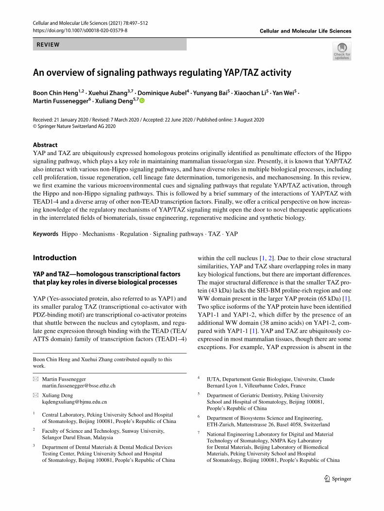
Vol.:(0123456789) 1 3 Cellular and Molecular Life Sciences (2021) 78:497–512 https://doi.org/10.1007/s00018-020-03579-8 REVIEW An overview of signaling pathways regulating YAP/TAZ activity Boon Chin Heng 1,2 · Xuehui Zhang 3,7 · Dominique Aubel 4 · Yunyang Bai 5 · Xiaochan Li 5 · Yan Wei 5 · Martin Fussenegger 6 · Xuliang Deng 5,7 Received: 21 January 2020 / Revised: 7 March 2020 / Accepted: 22 June 2020 / Published online: 3 August 2020 © Springer Nature Switzerland AG 2020 Abstract YAP and TAZ are ubiquitously expressed homologous proteins originally identified as penultimate effectors of the Hippo signaling pathway, which plays a key role in maintaining mammalian tissue/organ size. Presently, it is known that YAP/TAZ also interact with various non-Hippo signaling pathways, and have diverse roles in multiple biological processes, including cell proliferation, tissue regeneration, cell lineage fate determination, tumorigenesis, and mechanosensing. In this review, we first examine the various microenvironmental cues and signaling pathways that regulate YAP/TAZ activation, through the Hippo and non-Hippo signaling pathways. This is followed by a brief summary of the interactions of YAP/TAZ with TEAD1-4 and a diverse array of other non-TEAD transcription factors. Finally, we offer a critical perspective on how increas- ing knowledge of the regulatory mechanisms of YAP/TAZ signaling might open the door to novel therapeutic applications in the interrelated fields of biomaterials, tissue engineering, regenerative medicine and synthetic biology. Keywords Hippo · Mechanisms · Regulation · Signaling pathways · TAZ · YAP Introduction YAP and TAZ—homologous transcriptional factors that play key roles in diverse biological processes YAP (Yes-associated protein, also referred to as YAP1) and its smaller paralog TAZ (transcriptional co-activator with PDZ-binding motif) are transcriptional co-activator proteins that shuttle between the nucleus and cytoplasm, and regu- late gene expression through binding with the TEAD (TEA/ ATTS domain) family of transcription factors (TEAD1–4) within the cell nucleus [1, 2]. Due to their close structural similarities, YAP and TAZ share overlapping roles in many key biological functions, but there are important differences. The major structural difference is that the smaller TAZ pro- tein (43 kDa) lacks the SH3-BM proline-rich region and one WW domain present in the larger YAP protein (65 kDa) [1]. Two splice isoforms of the YAP protein have been identified YAP1-1 and YAP1-2, which differ by the presence of an additional WW domain (38 amino acids) on YAP1-2, com- pared with YAP1-1 [1]. YAP and TAZ are ubiquitously co- expressed in most mammalian tissues, though there are some exceptions. For example, YAP expression is absent in the Cellular and Molecular Life Sciences Boon Chin Heng and Xuehui Zhang contributed equally to this work. * Martin Fussenegger [email protected] * Xuliang Deng [email protected] 1 Central Laboratory, Peking University School and Hospital of Stomatology, Beijing 100081, People’s Republic of China 2 Faculty of Science and Technology, Sunway University, Selangor Darul Ehsan, Malaysia 3 Department of Dental Materials & Dental Medical Devices Testing Center, Peking University School and Hospital of Stomatology, Beijing 100081, People’s Republic of China 4 IUTA, Departement Genie Biologique, Universite, Claude Bernard Lyon 1, Villeurbanne Cedex, France 5 Department of Geriatric Dentistry, Peking University School and Hospital of Stomatology, Beijing 100081, People’s Republic of China 6 Department of Biosystems Science and Engineering, ETH-Zurich, Mattenstrasse 26, Basel 4058, Switzerland 7 National Engineering Laboratory for Digital and Material Technology of Stomatology, NMPA Key Laboratory for Dental Materials, Beijing Laboratory of Biomedical Materials, Peking University School and Hospital of Stomatology, Beijing 100081, People’s Republic of China
-
Upload
khangminh22 -
Category
Documents
-
view
2 -
download
0
Transcript of An overview of signaling pathways regulating YAP/TAZ activity
Vol.:(0123456789)1 3
Cellular and Molecular Life Sciences (2021) 78:497–512 https://doi.org/10.1007/s00018-020-03579-8
REVIEW
An overview of signaling pathways regulating YAP/TAZ activity
Boon Chin Heng1,2 · Xuehui Zhang3,7 · Dominique Aubel4 · Yunyang Bai5 · Xiaochan Li5 · Yan Wei5 · Martin Fussenegger6 · Xuliang Deng5,7
Received: 21 January 2020 / Revised: 7 March 2020 / Accepted: 22 June 2020 / Published online: 3 August 2020 © Springer Nature Switzerland AG 2020
AbstractYAP and TAZ are ubiquitously expressed homologous proteins originally identified as penultimate effectors of the Hippo signaling pathway, which plays a key role in maintaining mammalian tissue/organ size. Presently, it is known that YAP/TAZ also interact with various non-Hippo signaling pathways, and have diverse roles in multiple biological processes, including cell proliferation, tissue regeneration, cell lineage fate determination, tumorigenesis, and mechanosensing. In this review, we first examine the various microenvironmental cues and signaling pathways that regulate YAP/TAZ activation, through the Hippo and non-Hippo signaling pathways. This is followed by a brief summary of the interactions of YAP/TAZ with TEAD1-4 and a diverse array of other non-TEAD transcription factors. Finally, we offer a critical perspective on how increas-ing knowledge of the regulatory mechanisms of YAP/TAZ signaling might open the door to novel therapeutic applications in the interrelated fields of biomaterials, tissue engineering, regenerative medicine and synthetic biology.
Keywords Hippo · Mechanisms · Regulation · Signaling pathways · TAZ · YAP
Introduction
YAP and TAZ—homologous transcriptional factors that play key roles in diverse biological processes
YAP (Yes-associated protein, also referred to as YAP1) and its smaller paralog TAZ (transcriptional co-activator with PDZ-binding motif) are transcriptional co-activator proteins that shuttle between the nucleus and cytoplasm, and regu-late gene expression through binding with the TEAD (TEA/ATTS domain) family of transcription factors (TEAD1–4)
within the cell nucleus [1, 2]. Due to their close structural similarities, YAP and TAZ share overlapping roles in many key biological functions, but there are important differences. The major structural difference is that the smaller TAZ pro-tein (43 kDa) lacks the SH3-BM proline-rich region and one WW domain present in the larger YAP protein (65 kDa) [1]. Two splice isoforms of the YAP protein have been identified YAP1-1 and YAP1-2, which differ by the presence of an additional WW domain (38 amino acids) on YAP1-2, com-pared with YAP1-1 [1]. YAP and TAZ are ubiquitously co-expressed in most mammalian tissues, though there are some exceptions. For example, YAP expression is absent in the
Cellular and Molecular Life Sciences
Boon Chin Heng and Xuehui Zhang contributed equally to this work.
* Martin Fussenegger [email protected]
* Xuliang Deng [email protected]
1 Central Laboratory, Peking University School and Hospital of Stomatology, Beijing 100081, People’s Republic of China
2 Faculty of Science and Technology, Sunway University, Selangor Darul Ehsan, Malaysia
3 Department of Dental Materials & Dental Medical Devices Testing Center, Peking University School and Hospital of Stomatology, Beijing 100081, People’s Republic of China
4 IUTA, Departement Genie Biologique, Universite, Claude Bernard Lyon 1, Villeurbanne Cedex, France
5 Department of Geriatric Dentistry, Peking University School and Hospital of Stomatology, Beijing 100081, People’s Republic of China
6 Department of Biosystems Science and Engineering, ETH-Zurich, Mattenstrasse 26, Basel 4058, Switzerland
7 National Engineering Laboratory for Digital and Material Technology of Stomatology, NMPA Key Laboratory for Dental Materials, Beijing Laboratory of Biomedical Materials, Peking University School and Hospital of Stomatology, Beijing 100081, People’s Republic of China
498 B. C. Heng et al.
1 3
hippocampus and parathyroid gland [3], while the thymus and peripheral blood leukocytes lack TAZ expression [4].
Historically, YAP/TAZ were identified as the penulti-mate effectors of the Hippo signaling pathway, which plays a crucial role in regulating tissue/organ size [5]. In recent years, many other functions of YAP/TAZ have been found, in development, cell proliferation, tissue regeneration and cell lineage fate determination [6], as well as in mechano-sensing and mechanotransduction at cell-ECM/biomaterial interfaces [7]. These functions are all highly relevant to the fields of tissue engineering and regenerative medicine. Furthermore, YAP/TAZ also have roles in other biological processes and disease pathologies, most notably in tissue/organ homeostasis and cancer metastasis [8].
In this review, we will give an overview of the various microenvironmental cues and signaling pathways that reg-ulate YAP/TAZ activity, followed by a brief summary of YAP/TAZ interaction with TEAD and non-TEAD transcrip-tion factors. Then, we will present a critical perspective, suggesting how our increasing knowledge of YAP/TAZ sign-aling might advance therapeutic applications in the fields of biomaterials, tissue engineering, regenerative medicine and synthetic biology.
YAP/TAZ—key mediators of cell interaction with the microenvironment
YAP/TAZ mediate cellular responses to (i) biomechanical cues [9–17], (ii) extracellular ligands, such as growth factors and lipids [18–21], (iii) energy, osmotic and hypoxic stress [22–25], and (iv) inflammation and tissue injury [26–30].
Biomechanical cues refer to the mechanical forces gen-erated upon cell interaction with the extracellular matrix (ECM) or substrata, cell-to-cell contact, and liquid shear forces encountered by cells (best exemplified by the endothe-lial cells lining blood vessels). The responses to these cues mediated by YAP/TAZ in turn play key roles in orchestrating organ/tissue development and regulating homeostasis, and are therefore of interest in connection with the fabrication of improved biomimetic materials for tissue engineering applications.
Mechanosensing of ECM/substrata stiffness is mediated primarily by focal adhesions (FAs), which influence cell adhesion, spreading, and remodeling of the actin cytoskel-eton via RhoA activity [9–11]. The FAs in turn modulate Hippo-dependent and independent signaling pathways that control cell proliferation and differentiation through YAP/TAZ. Generally, with a stiffer substrate, there is increased cytosol to nuclear translocation of YAP/TAZ (Fig. 2), which can be attributed to increased number of FAs per cell, as well as increased tensile force on the stress fibers connecting the FAs that cause the cell to spread over a larger surface area [10, 11]. In the case of bone marrow-derived mesenchymal
stem cells (BMSCs), increased YAP/TAZ activation on a stiff substrate leads to enhancement of osteogenic differen-tiation [11].
Another biomechanical cue is cell density. With increas-ing cell density, there is increased cell-to-cell contact, and the cell shape changes from a more flattened to a more rounded geometry, with a consequent reduction in FAs, but an increase in the numbers of tight junctions (TJs) and adherens junctions (AJs) between adjacent cells [12, 13]. This inhibits cytosolic to nuclear translocation of YAP/TAZ through both the Hippo signaling pathway and Hippo-independent mechanisms. Generally, a decrease in YAP/TAZ nuclear-to-cytoplasmic ratio impedes cell prolifera-tion, resulting in the commonly observed phenomenon of contact inhibition of cell proliferation [12, 13].
Cellular response to shear stress is also mediated by YAP/TAZ [14–17]. Studies with a microfluidic perfusion device demonstrated that exposure to shear force enhances YAP translocation to the cell nucleus, which in turn promotes osteogenesis and inhibits adipogenesis in mesenchymal stem cells (MSCs), but initiates dedifferentiation of chondrocytes [14]. In the case of endothelial cells, laminar (unidirectional) shear stress, as is normally encountered in healthy blood vessels, suppresses YAP/TAZ activation, whereas oscilla-tory (disturbed or turbulent) shear stress, which is associated with choked blood vessels, enhances YAP/TAZ activation [15–17]. These findings have major implications for the pathogenesis of arteriosclerosis [15–17].
The signaling pathways of some cell-surface receptors of extracellular ligands, such as lipids, hormones and growth factors, can also activate YAP/TAZ through interactions with the Hippo signaling pathway (Fig. 1). These ligands include lysophosphatidic acid (LPA), sphingosine 1-phos-phate (S1P), glucagon and epinephrine, which act through G protein-coupled receptors (GPCRs) [18, 19], as well as the receptor tyrosine kinases (RTKs) for nerve growth factor (NGF) and epidermal growth factor (EGF) [20, 21].
Cellular responses to microenvironmental stress fac-tors have also been linked to YAP/TAZ via interaction of various cytosolic factors with the Hippo signaling pathway (Fig. 1). Energy stress induced by inhibitors of glucose or ATP metabolism decreases YAP/TAZ activation through the Hippo signaling pathway via AMPK (AMP-activated protein kinase) [22, 23]. It was reported that osmotic stress promotes YAP translocation to cell nuclei via Nemo-like kinase (NLK)-mediated phosphorylation of the Ser128 residue [24], while hypoxic stress activates YAP through the action of SIAH2 ubiquitin E3 on the Hippo signaling pathway [25].
Pro-inflammatory cytokines, such as prostaglandin E2 and tumor necrosis factor alpha (TNF-α), also activate YAP/TAZ, but the underlying mechanisms remain unclear [26, 27]. YAP/TAZ are also activated in response to liver, skin,
499An overview of signaling pathways regulating YAP/TAZ activity
1 3
myocardial and intestinal epithelium injury [28–30], and facilitate healing by enhancing cell proliferation. However, the underlying mechanisms through which YAP/TAZ are activated upon tissue injury are still largely uncharacterized.
Regulatory mechanisms of YAP/TAZ activity
Regulation of YAP/TAZ activity through the canonical Hippo signaling pathway
Core components of the canonical Hippo signaling pathway
The canonical Hippo signaling pathway is highly conserved in different species ranging from fruitflies to mammals [31],
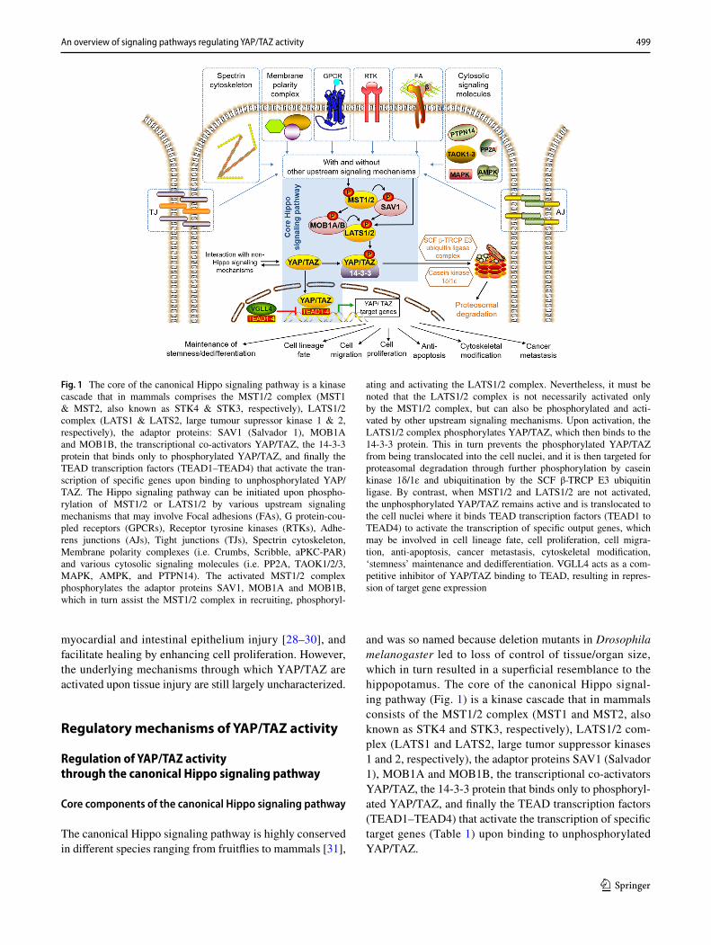
and was so named because deletion mutants in Drosophila melanogaster led to loss of control of tissue/organ size, which in turn resulted in a superficial resemblance to the hippopotamus. The core of the canonical Hippo signal-ing pathway (Fig. 1) is a kinase cascade that in mammals consists of the MST1/2 complex (MST1 and MST2, also known as STK4 and STK3, respectively), LATS1/2 com-plex (LATS1 and LATS2, large tumor suppressor kinases 1 and 2, respectively), the adaptor proteins SAV1 (Salvador 1), MOB1A and MOB1B, the transcriptional co-activators YAP/TAZ, the 14-3-3 protein that binds only to phosphoryl-ated YAP/TAZ, and finally the TEAD transcription factors (TEAD1–TEAD4) that activate the transcription of specific target genes (Table 1) upon binding to unphosphorylated YAP/TAZ.
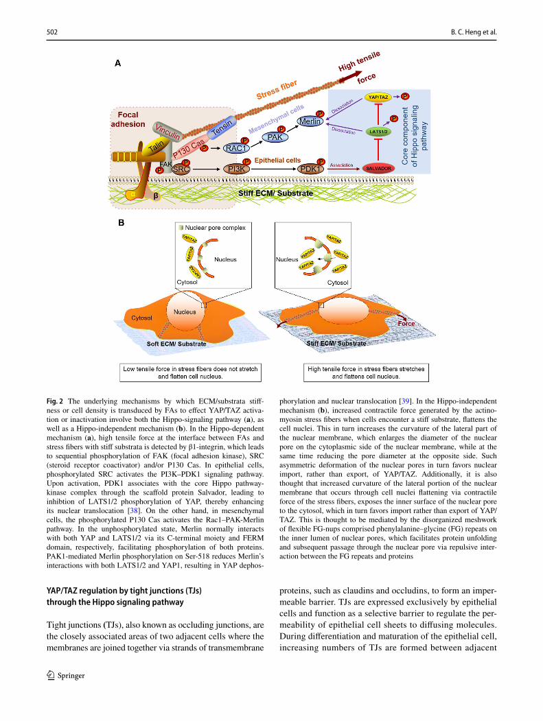
Fig. 1 The core of the canonical Hippo signaling pathway is a kinase cascade that in mammals comprises the MST1/2 complex (MST1 & MST2, also known as STK4 & STK3, respectively), LATS1/2 complex (LATS1 & LATS2, large tumour supressor kinase 1 & 2, respectively), the adaptor proteins: SAV1 (Salvador 1), MOB1A and MOB1B, the transcriptional co-activators YAP/TAZ, the 14-3-3 protein that binds only to phosphorylated YAP/TAZ, and finally the TEAD transcription factors (TEAD1–TEAD4) that activate the tran-scription of specific genes upon binding to unphosphorylated YAP/TAZ. The Hippo signaling pathway can be initiated upon phospho-rylation of MST1/2 or LATS1/2 by various upstream signaling mechanisms that may involve Focal adhesions (FAs), G protein-cou-pled receptors (GPCRs), Receptor tyrosine kinases (RTKs), Adhe-rens junctions (AJs), Tight junctions (TJs), Spectrin cytoskeleton, Membrane polarity complexes (i.e. Crumbs, Scribble, aPKC-PAR) and various cytosolic signaling molecules (i.e. PP2A, TAOK1/2/3, MAPK, AMPK, and PTPN14). The activated MST1/2 complex phosphorylates the adaptor proteins SAV1, MOB1A and MOB1B, which in turn assist the MST1/2 complex in recruiting, phosphoryl-
ating and activating the LATS1/2 complex. Nevertheless, it must be noted that the LATS1/2 complex is not necessarily activated only by the MST1/2 complex, but can also be phosphorylated and acti-vated by other upstream signaling mechanisms. Upon activation, the LATS1/2 complex phosphorylates YAP/TAZ, which then binds to the 14-3-3 protein. This in turn prevents the phosphorylated YAP/TAZ from being translocated into the cell nuclei, and it is then targeted for proteasomal degradation through further phosphorylation by casein kinase 1δ/1ε and ubiquitination by the SCF β-TRCP E3 ubiquitin ligase. By contrast, when MST1/2 and LATS1/2 are not activated, the unphosphorylated YAP/TAZ remains active and is translocated to the cell nuclei where it binds TEAD transcription factors (TEAD1 to TEAD4) to activate the transcription of specific output genes, which may be involved in cell lineage fate, cell proliferation, cell migra-tion, anti-apoptosis, cancer metastasis, cytoskeletal modification, ‘stemness’ maintenance and dedifferentiation. VGLL4 acts as a com-petitive inhibitor of YAP/TAZ binding to TEAD, resulting in repres-sion of target gene expression
500 B. C. Heng et al.
1 3
The Hippo signaling pathway is initiated upon activation (phosphorylation) of MST1/2 by various upstream signaling factors (Fig. 1). The activated MST1/2 complex phospho-rylates the adaptor proteins SAV1, MOB1A and MOB1B, which in turn assist the MST1/2 complex in recruiting, phos-phorylating and activating the LATS1/LATS2 complex. It should be noted that the LATS1/LATS2 complex is not necessarily activated only by the MST1/2 complex, but can also be phosphorylated and activated by a whole multitude of cytosolic factors that are implicated in other signaling pathways. Hence, there is a rather high degree of overlap with other signaling pathways.
Upon activation, the LATS1/LATS2 complex phospho-rylates YAP/TAZ, which then bind to the 14-3-3 protein. This in turn prevents the phosphorylated YAP/TAZ from being translocated to the cell nucleus, and they are then tar-geted for proteasomal degradation through further phospho-rylation by casein kinase 1δ/1ε and ubiquitination by the SCF β-TRCP E3 ubiquitin ligase complex [32]. In contrast, when MST1/2 and LATS1/2 are not activated, the unphos-phorylated YAP/TAZ are translocated to the cell nucleus, where they bind to TEAD transcription factors to activate the transcription of specific target genes (Table 1).
Hence, when the core Hippo signaling pathway is switched ‘ON’, YAP/TAZ are inactivated through phos-phorylation, and their translocation to the cell nucleus is blocked. On the other hand, when Hippo signaling is switched ‘OFF’, the non-phosphorylated YAP/TAZ can be translocated to the cell nucleus, where they bind to TEAD transcription factors and activate the transcription of specific
target genes (Table 1). Nevertheless, it is important to note that in reality, the Hippo signaling pathway does not behave digitally only in the ‘ON’ or ‘OFF’ mode. Depending on the relative activities of MST1/2, LATS1/2, and other cytosolic factors involved in the activation/deactivation of these two complexes (Fig. 1), YAP/TAZ localization may be partially cytoplasmic or partially nuclear. The situation is further complicated by the existence of competitive inhibitors of YAP/TAZ binding to TEAD, such as VGLL4 [33], which represses target gene expression. Hence, depending on the cell type and biological context, variations in the relative activity of the core Hippo signaling cascade and YAP/TAZ may arise from differing balance in the activity/or quan-tity of the various regulatory kinases, which can function in a complementary manner and which may substitute each other. This in turn would have critical implications for the practical implementation and data interpretation of YAP/TAZ assays.
YAP/TAZ regulation by focal adhesions through the Hippo signaling pathway
Focal adhesions (FAs) are integrin-containing macromo-lecular assemblies that form a mechanical linkage between the extracellular matrix and cytoskeleton via intracellular actin bundles (stress fibers) [7, 9]. As a consequence of their structural role, FAs are the primary conduits for cel-lular sensing of ECM/substrata stiffness [7, 9], in addition to being sensors of cell density [34]. At low cell density, cells tend to be more spread out on the substrata surface and have
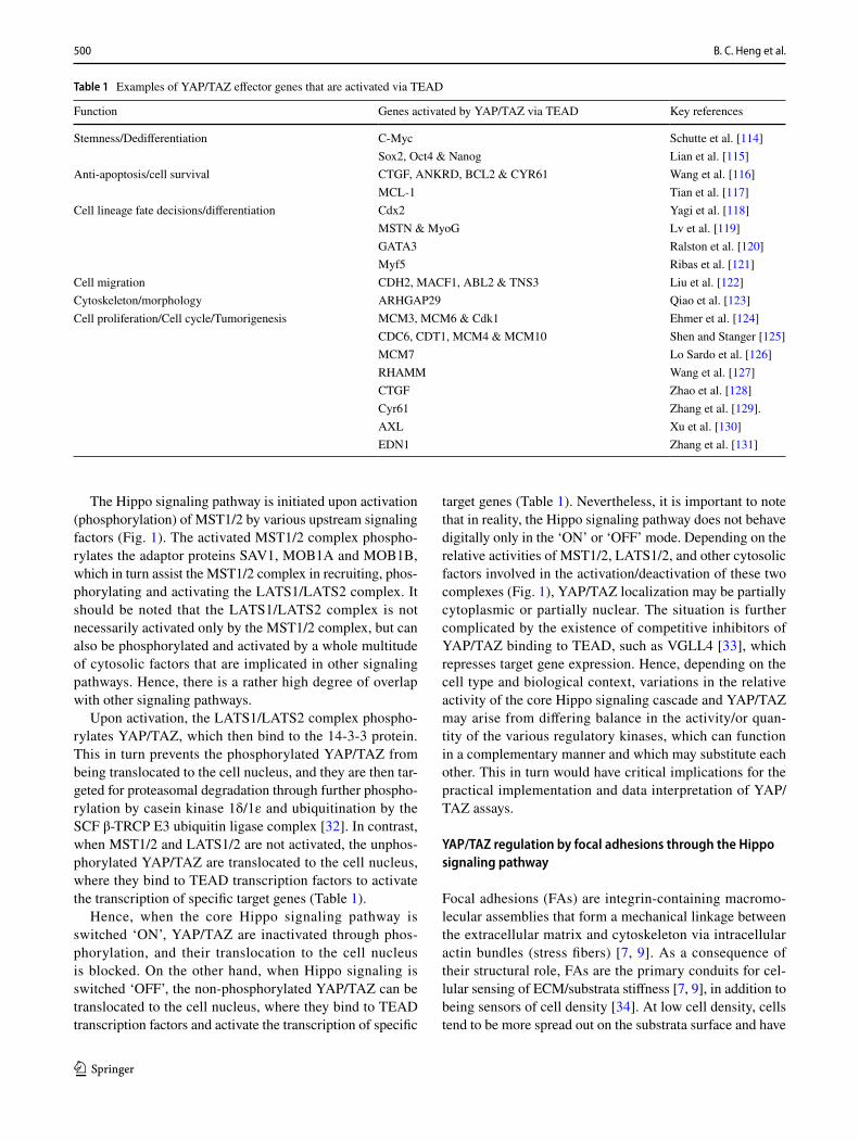
Table 1 Examples of YAP/TAZ effector genes that are activated via TEAD
Function Genes activated by YAP/TAZ via TEAD Key references
Stemness/Dedifferentiation C-Myc Schutte et al. [114]Sox2, Oct4 & Nanog Lian et al. [115]
Anti-apoptosis/cell survival CTGF, ANKRD, BCL2 & CYR61 Wang et al. [116]MCL-1 Tian et al. [117]
Cell lineage fate decisions/differentiation Cdx2 Yagi et al. [118]MSTN & MyoG Lv et al. [119]GATA3 Ralston et al. [120]Myf5 Ribas et al. [121]
Cell migration CDH2, MACF1, ABL2 & TNS3 Liu et al. [122]Cytoskeleton/morphology ARHGAP29 Qiao et al. [123]Cell proliferation/Cell cycle/Tumorigenesis MCM3, MCM6 & Cdk1 Ehmer et al. [124]
CDC6, CDT1, MCM4 & MCM10 Shen and Stanger [125]MCM7 Lo Sardo et al. [126]RHAMM Wang et al. [127]CTGF Zhao et al. [128]Cyr61 Zhang et al. [129].AXL Xu et al. [130]EDN1 Zhang et al. [131]
501An overview of signaling pathways regulating YAP/TAZ activity
1 3
higher numbers of FAs and associated stress fibers; whereas at high density, cells are more rounded and compact, with fewer FAs and associated stress fibers [34].
Different cell types, such as epithelial and mesenchymal cells, will exhibit different responses to stiff and soft sub-strates, as well as to high and low cell densities which can be attributed to varying combinations and quantities of TEAD transcription factors present in different cell types that can potentially be activated by YAP/TAZ. Additionally, complex interaction and cross-talk between the core Hippo signaling cascade (Fig. 1) with other signaling pathways in different cell types, will undoubtedly lead to variations in the signal-ing mechanisms by which ECM/substrata stiffness or cell density is transduced by FAs to modulate YAP/TAZ activity in different cell lineages.
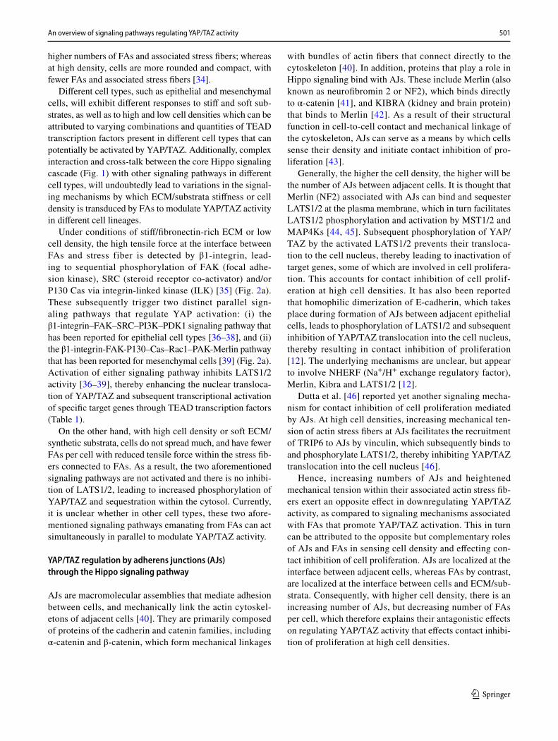
Under conditions of stiff/fibronectin-rich ECM or low cell density, the high tensile force at the interface between FAs and stress fiber is detected by β1-integrin, lead-ing to sequential phosphorylation of FAK (focal adhe-sion kinase), SRC (steroid receptor co-activator) and/or P130 Cas via integrin-linked kinase (ILK) [35] (Fig. 2a). These subsequently trigger two distinct parallel sign-aling pathways that regulate YAP activation: (i) the β1-integrin–FAK–SRC–PI3K–PDK1 signaling pathway that has been reported for epithelial cell types [36–38], and (ii) the β1-integrin-FAK-P130–Cas–Rac1–PAK-Merlin pathway that has been reported for mesenchymal cells [39] (Fig. 2a). Activation of either signaling pathway inhibits LATS1/2 activity [36–39], thereby enhancing the nuclear transloca-tion of YAP/TAZ and subsequent transcriptional activation of specific target genes through TEAD transcription factors (Table 1).
On the other hand, with high cell density or soft ECM/synthetic substrata, cells do not spread much, and have fewer FAs per cell with reduced tensile force within the stress fib-ers connected to FAs. As a result, the two aforementioned signaling pathways are not activated and there is no inhibi-tion of LATS1/2, leading to increased phosphorylation of YAP/TAZ and sequestration within the cytosol. Currently, it is unclear whether in other cell types, these two afore-mentioned signaling pathways emanating from FAs can act simultaneously in parallel to modulate YAP/TAZ activity.
YAP/TAZ regulation by adherens junctions (AJs) through the Hippo signaling pathway
AJs are macromolecular assemblies that mediate adhesion between cells, and mechanically link the actin cytoskel-etons of adjacent cells [40]. They are primarily composed of proteins of the cadherin and catenin families, including α-catenin and β-catenin, which form mechanical linkages
with bundles of actin fibers that connect directly to the cytoskeleton [40]. In addition, proteins that play a role in Hippo signaling bind with AJs. These include Merlin (also known as neurofibromin 2 or NF2), which binds directly to α-catenin [41], and KIBRA (kidney and brain protein) that binds to Merlin [42]. As a result of their structural function in cell-to-cell contact and mechanical linkage of the cytoskeleton, AJs can serve as a means by which cells sense their density and initiate contact inhibition of pro-liferation [43].
Generally, the higher the cell density, the higher will be the number of AJs between adjacent cells. It is thought that Merlin (NF2) associated with AJs can bind and sequester LATS1/2 at the plasma membrane, which in turn facilitates LATS1/2 phosphorylation and activation by MST1/2 and MAP4Ks [44, 45]. Subsequent phosphorylation of YAP/TAZ by the activated LATS1/2 prevents their transloca-tion to the cell nucleus, thereby leading to inactivation of target genes, some of which are involved in cell prolifera-tion. This accounts for contact inhibition of cell prolif-eration at high cell densities. It has also been reported that homophilic dimerization of E-cadherin, which takes place during formation of AJs between adjacent epithelial cells, leads to phosphorylation of LATS1/2 and subsequent inhibition of YAP/TAZ translocation into the cell nucleus, thereby resulting in contact inhibition of proliferation [12]. The underlying mechanisms are unclear, but appear to involve NHERF (Na+/H+ exchange regulatory factor), Merlin, Kibra and LATS1/2 [12].
Dutta et al. [46] reported yet another signaling mecha-nism for contact inhibition of cell proliferation mediated by AJs. At high cell densities, increasing mechanical ten-sion of actin stress fibers at AJs facilitates the recruitment of TRIP6 to AJs by vinculin, which subsequently binds to and phosphorylate LATS1/2, thereby inhibiting YAP/TAZ translocation into the cell nucleus [46].
Hence, increasing numbers of AJs and heightened mechanical tension within their associated actin stress fib-ers exert an opposite effect in downregulating YAP/TAZ activity, as compared to signaling mechanisms associated with FAs that promote YAP/TAZ activation. This in turn can be attributed to the opposite but complementary roles of AJs and FAs in sensing cell density and effecting con-tact inhibition of cell proliferation. AJs are localized at the interface between adjacent cells, whereas FAs by contrast, are localized at the interface between cells and ECM/sub-strata. Consequently, with higher cell density, there is an increasing number of AJs, but decreasing number of FAs per cell, which therefore explains their antagonistic effects on regulating YAP/TAZ activity that effects contact inhibi-tion of proliferation at high cell densities.
502 B. C. Heng et al.
1 3
YAP/TAZ regulation by tight junctions (TJs) through the Hippo signaling pathway
Tight junctions (TJs), also known as occluding junctions, are the closely associated areas of two adjacent cells where the membranes are joined together via strands of transmembrane
proteins, such as claudins and occludins, to form an imper-meable barrier. TJs are expressed exclusively by epithelial cells and function as a selective barrier to regulate the per-meability of epithelial cell sheets to diffusing molecules. During differentiation and maturation of the epithelial cell, increasing numbers of TJs are formed between adjacent
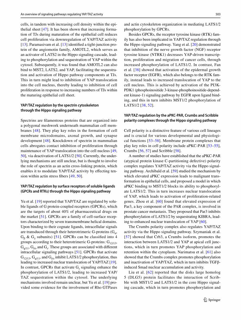
Fig. 2 The underlying mechanisms by which ECM/substrata stiff-ness or cell density is transduced by FAs to effect YAP/TAZ activa-tion or inactivation involve both the Hippo-signaling pathway (a), as well as a Hippo-independent mechanism (b). In the Hippo-dependent mechanism (a), high tensile force at the interface between FAs and stress fibers with stiff substrata is detected by β1-integrin, which leads to sequential phosphorylation of FAK (focal adhesion kinase), SRC (steroid receptor coactivator) and/or P130 Cas. In epithelial cells, phosphorylated SRC activates the PI3K–PDK1 signaling pathway. Upon activation, PDK1 associates with the core Hippo pathway-kinase complex through the scaffold protein Salvador, leading to inhibtion of LATS1/2 phosphorylation of YAP, thereby enhancing its nuclear translocation [38]. On the other hand, in mesenchymal cells, the phosphorylated P130 Cas activates the Rac1–PAK-Merlin pathway. In the unphosphorylated state, Merlin normally interacts with both YAP and LATS1/2 via its C-terminal moiety and FERM domain, respectively, facilitating phosphorylation of both proteins. PAK1-mediated Merlin phosphorylation on Ser-518 reduces Merlin’s interactions with both LATS1/2 and YAP1, resulting in YAP dephos-
phorylation and nuclear translocation [39]. In the Hippo-independent mechanism (b), increased contractile force generated by the actino-myosin stress fibers when cells encounter a stiff substrate, flattens the cell nuclei. This in turn increases the curvature of the lateral part of the nuclear membrane, which enlarges the diameter of the nuclear pore on the cytoplasmic side of the nuclear membrane, while at the same time reducing the pore diameter at the opposite side. Such asymmetric deformation of the nuclear pores in turn favors nuclear import, rather than export, of YAP/TAZ. Additionally, it is also thought that increased curvature of the lateral portion of the nuclear membrane that occurs through cell nuclei flattening via contractile force of the stress fibers, exposes the inner surface of the nuclear pore to the cytosol, which in turn favors import rather than export of YAP/TAZ. This is thought to be mediated by the disorganized meshwork of flexible FG-nups comprised phenylalanine–glycine (FG) repeats on the inner lumen of nuclear pores, which facilitates protein unfolding and subsequent passage through the nuclear pore via repulsive inter-action between the FG repeats and proteins
503An overview of signaling pathways regulating YAP/TAZ activity
1 3
cells, in tandem with increasing cell density within the epi-thelial sheet [47]. It has been shown that increasing forma-tion of TJs during maturation of the epithelial cell reduces cell proliferation via downregulation of YAP/TAZ activity [13]. Paramasivam et al. [13] identified a tight-junction pro-tein of the angiomotin family, AMOTL2, which serves as an activator of LATS2 in the Hippo signaling cascade, lead-ing to phosphorylation and sequestration of YAP within the cytosol. Subsequently, it was found that AMOTL2 can also bind to MST2, LATS2, and YAP, facilitating the sequestra-tion and activation of Hippo pathway components at TJs. This in turn might lead to inhibition of YAP translocation into the cell nucleus, thereby leading to inhibition of cell proliferation in response to increasing numbers of TJs within the maturing epithelial cell sheet.
YAP/TAZ regulation by the spectrin cytoskeleton through the Hippo signaling pathway
Spectrins are filamentous proteins that are organized into a polygonal meshwork underneath mammalian cell mem-branes [48]. They play key roles in the formation of cell membrane microdomains, axonal growth, and synapse development [48]. Knockdown of spectrin in mammalian cells abrogates contact inhibition of proliferation through maintenance of YAP translocation into the cell nucleus [49, 50], via deactivation of LATS1/2 [50]. Currently, the under-lying mechanisms are still unclear, but is thought to involve the role of spectrin as an actin cross-linking protein, which enables it to modulate YAP/TAZ activity by effecting ten-sion within actin stress fibers [49, 50].
YAP/TAZ regulation by surface receptors of soluble ligands (GPCRs and RTKs) through the Hippo signaling pathway
Yu et al. [19] reported that YAP/TAZ are regulated by solu-ble ligands of G protein-coupled receptors (GPCRs), which are the targets of about 40% of pharmaceutical drugs on the market [51]. GPCRs are a family of cell-surface recep-tors characterized by seven transmembrane helical domains. Upon binding to their cognate ligands, intracellular signals are transduced through their heterotrimeric G-proteins (Gα, Gβ & Gγ subunits) [51]. GPCRs can be classified into 4 groups according to their heterotrimeric G-proteins: G12/13, Gq/11, Gi/o and Gs. These groups are associated with different intracellular signaling pathways [51]. GPCRs that activate G12/13, Gq/11, and Gi/o inhibit LATS1/2 phosphorylation, thus leading to increased nuclear translocation of YAP/TAZ [19]. In contrast, GPCRs that activate Gs signaling enhance the phosphorylation of LATS1/2, leading to increased YAP/TAZ sequestration within the cytosol. The underlying mechanisms involved remain unclear, but Yu et al. [19] pro-vided some evidence for the involvement of Rho GTPases
and actin cytoskeleton organization in mediating LATS1/2 phosphorylation by GPCRs.
Besides GPCRs, the receptor tyrosine kinase (RTK) fam-ily has also been implicated in YAP/TAZ regulation through the Hippo signaling pathway. Yang et al. [20] demonstrated that inhibition of the nerve growth factor (NGF) receptor tyrosine kinase (NTRK1) decreases YAP-driven transcrip-tion, proliferation and migration of cancer cells, through increased phosphorylation of LATS1/2. In contrast, Fan et al. [38] showed that activation of the epidermal growth factor receptor (EGFR), which also belongs to the RTK fam-ily, instead leads to increased translocation of YAP to the cell nucleus. This is achieved by activation of the PI3K-PDK1 (phosphoinositide 3-kinase-phosphoinositide-depend-ent kinase-1) signaling pathway by EGFR upon ligand bind-ing, and this in turn inhibits MST1/2 phosphorylation of LATS1/2 [38, 52].
YAP/TAZ regulation by the aPKC‑PAR, Crumbs and Scribble polarity complexes through the Hippo signaling pathway
Cell polarity is a distinctive feature of various cell lineages and is crucial for various developmental and physiologi-cal functions [53–58]. Membrane protein complexes that play key roles in cell polarity include aPKC-PAR [53–55], Crumbs [56, 57] and Scribble [58].
A number of studies have established that the aPKC-PAR (atypical protein kinase C-partitioning defective) polarity complex regulates YAP/TAZ activity via the Hippo signal-ing pathway. Archibald et al. [59] studied the mechanism by which elevated aPKC expression leads to malignant trans-formation in epithelial cells, and proposed a model in which aPKC binding to MST1/2 blocks its ability to phosphoryl-ate LATS1/2. This in turn increases nuclear translocation of YAP, which leads to activation of proliferation-related genes. Zhou et al. [60] found that elevated expression of Par3, a key component of the PAR complex, is involved in prostate cancer metastasis. They proposed that Par3 inhibits phosphorylation of LATS1/2 by sequestrating KIBRA, lead-ing to enhanced nuclear translocation of YAP [60].
The Crumbs polarity complex also regulates YAP/TAZ activity via the Hippo signaling pathway. Szymaniak et al. [57] showed that Crb3, a Crumbs isoform, promotes the interaction between LATS1/2 and YAP at apical cell junc-tions, which in turn promotes YAP phosphorylation and retention within the cytoplasm. Narimatsu et al. [61] also showed that the Crumbs complex promotes phosphorylation and inactivation of YAP/TAZ, which in turn inhibits TGFβ-induced Smad nuclear accumulation and activity.
Liu et al. [62] reported that the disks large homolog 5 (DLG5) protein facilitates the interaction of Scrib-ble with MST1/2 and LATS1/2 in the core Hippo signal-ing cascade, which in turn promotes phosphorylation and
504 B. C. Heng et al.
1 3
inactivation of YAP, resulting in inhibition of breast cancer cell proliferation.
YAP/TAZ regulation by other cytosolic signaling molecules (PP2A, TAOK1/2/3, MAPK, AMPK, PTPN14) through the Hippo signaling pathway
YAP/TAZ are also regulated by other cytosolic signaling molecules that enable overlap and cross-talk between the Hippo signaling pathway and other signaling pathways. Protein phosphatase 2A (PP2A) is a ubiquitously expressed serine threonine phosphatase that has diverse roles in tumor suppression, apoptosis, cell proliferation and signal trans-duction. It acts by dephosphorylating various cytosolic sign-aling molecules, such as Akt, p53, c-Myc and β-catenin. Bae et al. [63] showed that PP2A regulates YAP/TAZ through dephosphorylation of MST1/2, and its activity may be inhib-ited by SAV1, which is a part of the core Hippo signaling cascade. Thousand and one kinases 1/2/3 (TAOK1/2/3), which have diverse functions in various signaling pathways relating to inflammation, microtubule dynamics, and stress response, also have a role in modulating YAP/TAZ activity. Boggiano et al. [64] showed that TAOK1/2/3 regulate YAP/TAZ activity by facilitating phosphorylation of MST1/2 within the Hippo signaling cascade. MAP kinase kinase kinase kinases (MAP4Ks) are a family of serine threonine kinases that regulate a diverse array of biological processes including cell survival, proliferation, motility and differen-tiation, and also regulate YAP through the Hippo signaling pathway [65]. Meng et al. [65] showed that several members of the MAP4K family act in parallel to MST1/2 to activate LATS1/2 within the Hippo pathway, leading to increased YAP phosphorylation and sequestration in the cytosol. Hence in this respect, MAP4Ks functionally overlap in part with MST1/2. Adenosine monophosphate kinase (AMPK) is a key regulator of cellular metabolism and a sensor of energy/nutrient stress. Mo et al. [23] showed that in the pres-ence of nutrient starvation or energy stress, AMPK activates LATS1/2, leading to YAP phosphorylation and inhibition of nuclear translocation. They further showed that AMPK can also phosphorylate YAP directly at the Ser 94 residue, which is essential for its binding to TEAD, thus inhibiting target gene activation by blocking YAP-TEAD interaction. A notable example is the inhibition of gene expression of glucose-transporter 3 (GLUT3), which is involved in glucose metabolism [66]. In addition to AJs and TJs, non-receptor tyrosine phosphatase 14 (PTPN14) also mediates contact inhibition of cell proliferation by regulating YAP/TAZ activ-ity through the core Hippo signaling pathway [67, 68]. At high cell densities, PTPN14 is abundantly expressed and forms a complex with KIBRA, which activates the core Hippo signaling cascade to phosphorylate YAP/TAZ via
MST1/2 and LATS1/2, and this in turn represses TEAD-specific genes involved in cell proliferation [67, 68].
Regulation of YAP/TAZ activity through Hippo‑independent mechanisms
Hippo‑independent regulation of YAP/TAZ through stress fibers connected to focal adhesions (FAs)
Besides FA-mediated mechanotransduction through the canonical Hippo signaling pathway (Fig. 1), there is also a Hippo-independent pathway mediated through contractile force generated by actinomyosin stress fibers, which directly connect FAs to the apical surface of the cell nuclei via the LINC (linker of the nucleoskeleton and cytoskeleton) com-plex of the nuclear envelope [69]. Elosegui-Artola et al. [69] proposed that increased contractile force generated by the actinomyosin stress fibers when cells encounter a stiff sub-strate flattens the cell nuclei (Fig. 2b). This in turn increases the curvature of the lateral part of the nuclear membrane, which induces asymmetric morphological changes to the openings of nuclear pores on both the cytoplasmic and nuclear side of the membrane. This in turn favors nuclear import, rather than export, of YAP/TAZ (Fig. 2b). It is hypothesized that morphological changes to the nuclear pore exposes the disorganized meshwork of flexible FG-nups comprising phenylalanine–glycine (FG) repeats [70] on the inner lumen of the nuclear pores to the cytosol, facilitat-ing protein unfolding and subsequent passage of YAP/TAZ through the nuclear pore via repulsive interaction between the FG repeats and the protein.
Hippo‑independent regulation of YAP/TAZ through adherens junctions (AJs) and tight junctions (TJs)
As previously discussed, AJs mechanically link the cytoskel-eton of adjacent cells, and serve as a means by which cells sense and react to their density via the Hippo-signaling path-way through the AJ-associated protein Merlin (also known as neurofibromin 2 or NF2). Although Merlin is closely associ-ated with the Hippo signaling pathway, it is not considered to be a central component of the core signaling Hippo signaling cascade (Fig. 1). Besides the mechanism described in 2.1.3, it was also reported that Merlin can act independently of the core Hippo signaling cascade to repress YAP/TAZ activation [71]. At high cell densities, increased tensile force exerted on the actomyosin-based stress fibers tethered at AJs induces dis-sociation of Merlin from AJs. Subsequently, the dissociated Merlin is translocated to the cell nucleus, where it directly binds and forms a complex with YAP. This Merlin-YAP com-plex is translocated from the cell nuclei to the cytosol via the nuclear export signals of Merlin, leading to repression of
505An overview of signaling pathways regulating YAP/TAZ activity
1 3
TEAD-specific genes involved in cell proliferation and thus, contact inhibition of proliferation.
As previously discussed, TJs regulate YAP activity through the Hippo signaling pathway via AMOTL2. On the other hand, Domínguez-Calderó [72] found that TJs can also regulate YAP activity through a Hippo-independent mechanism via the tight-junction protein zona occludens 2 (ZO-2), which binds to and sequesters YAP within the cytosol. Upon ZO-2 gene silenc-ing, hypertropy of renal cells was observed concurrently with increased YAP accumulation within the cell nuclei [72].
Hippo‑independent regulation of YAP/TAZ through the Wnt‑β‑catenin pathway
The regulation of β-catenin through a cytoplasmic destruc-tion complex forms the crux of the Wnt signaling cascade [73]. This β-catenin destruction complex is a multi-protein complex that includes Axin, adenomatous polyposis coli (APC), glycogen synthase kinase-3 (GSK3), and casein kinase 1 (CK1) [73]. When Wnt signaling is inactive, this destruction complex captures not only cytosolic β-catenin [73], but also cytosolic YAP [74], leading ultimately to degradation by the β-TrCP ubiquitin ligase. The activation of Wnt signaling inactivates the destruction complex and allows escape of both β-catenin [73] and YAP [74] from degradation, which in turn enables their translocation and accumulation in the cell nucleus.
Hippo‑independent regulation of YAP/TAZ through methylation and phosphorylation
SET7 (also known as SETD7) is a SET-domain-containing lysine methyltransferase that methylates and alters the func-tion of a variety of proteins. Oudhoff et al. [75] demonstrated that SET7 methylates the lysine 494 residue of YAP, which inhibits its translocation to the cell nucleus and results in its sequestration within the cytosol. Knockout of SET7 in mice resulted in a larger progenitor compartment in the intestine, concomitantly with increased expression of YAP target genes.
As previously discussed, PTPN14 forms a complex with KIBRA protein to regulate YAP/TAZ activity via the core Hippo signaling pathway. Liu et al. [76] showed that PTPN14 can also directly phosphorylate and inactivate YAP, through interaction between the WW domain of YAP and the PPxY domain of PTPN14. The presence of a YAP-PTPN14 complex was validated by co-immunoprecipitation [76].
Hippo‑independent regulation of YAP/TAZ through the Notch signaling pathway
The Notch signaling pathway is a major signaling path-way with diverse biological functions, which regulates
cell-to-cell communications in a juxtacrine manner, via interaction between Notch receptors (Notch 1 to 4 iso-forms) and their corresponding ligands (JAG1, JAG2, DLL1, DLL3, and DLL4) expressed on adjacent cells [77]. Notch receptor-ligand binding triggers proteolytic cleavage and subsequent release of the Notch intracellular domain (NICD), which translocates into the cell nucleus, where it binds and activates the transcription factor RBPJ (recombining binding protein suppressor of hairless), as well as the nuclear effector Mastermind-like (MAML), thereby initiating transcription of Notch target genes [77].
Although, it is well-known that there is much cross-talk between YAP/TAZ and the Notch signaling pathway [78], and numerous examples whereby these can work syner-gistically together to regulate the expression of various genes and hence biological processes [78], there have only been a few studies to date, which have reported upstream regulation of YAP/TAZ activity via the Notch signaling pathway [79–81]. In the study of Li et al. [79] on murine neural stem cells, it was shown by gain- and loss-of-func-tion experiments that the Notch signaling pathway exert positive upstream control of YAP activity, which in turn regulated the proliferation of these cells. Further inves-tigations revealed that the RBPJ transcription factor of the canonical Notch signaling pathway directly control transcription of the YAP1 protein by binding to its pro-moter sequence [79]. Although, Li et al. [79] observed that RBPJ could also bind to the promoter sequence of TEAD2, this by itself was insufficient to initiate transcription of TEAD2. Very similar results were reported by the study of Slemmons et al. [80] on human rhabdomyosarcoma cells, which also utilized gain- and loss-of-function experiments to demonstrate positive regulation of YAP activity by the Notch signaling pathway. Again, it was demonstrated that RBPJ can bind directly to the YAP promoter and acti-vate its transcription. Additionally, Slemmons et al. [80] showed that the Notch signaling pathway can also facilitate YAP nuclear translocation, but the underlying mechanism of how this occurs is currently unknown, and it is unclear whether this involves the core Hippo signaling cascade. Contradictory results were, however, reported by the study of Lu et al. [81] on murine hepatic stem cells. Although, RBPJ was again shown to bind directly to the YAP pro-moter, it repressed rather than activated transcription of the YAP protein [81]. This in turn led to Notch signal-ing promoting the differentiation of hepatic stem cells, while at the same time inhibiting their proliferation [81] by reducing YAP activity. Hence, it is likely that specific upstream regulation of YAP/TAZ activity by Notch signal-ing may vary according to the particular cell type, being dependent on complex interactions with other signaling processes present in different cell lineages.
506 B. C. Heng et al.
1 3
Hippo‑independent regulation of YAP/TAZ through the TGF‑β signaling pathway
Transforming growth factor beta (TGF-β) with five know isoforms (TGF-β1 to 5), together with other members of the TGF-β superfamily, i.e. bone morphogenetic proteins (BMPs), growth differentiation factors (GDFs), activin and inhibin, play key roles in numerous diverse biological pro-cesses, such as embryonic development, cell proliferation, differentiation and migration [82]. Nevertheless, the focus here will be solely on the TGF-β signaling pathway [83].
As in the case of the Notch signaling pathway, the TGF-ß signaling pathway is also known to have extensive cross-talk and interactions with YAP/TAZ, in the regulation of numerous genes and biological processes [84, 85]; but again, there are few studies that have reported upstream modulation of YAP/TAZ activity by the TGF-ß signaling pathway. It must be noted that to date, studies have reported that TGF-ß signaling can only exert upstream regulation of TAZ, but not YAP [86, 87], even though it is known that YAP can promote TGF-ß signaling by binding to and facilitating the nuclear translocation of SMAD2/3 [88]. For example, in the study of Miranda et al. [86] on rodent mesenchymal and epithelial cell lines, it was shown that TGF-ß signaling can induce only robust expression of TAZ, but not YAP. Further investigations revealed that the underlying mechanism was independent of SMAD2/3, and involved sequential activa-tion of p38 MAPK and its major downstream target MK2 by TGF-ß signaling, which in turn activates myocardin-related transcription factor (MTRF) that drives the TAZ promoter in a CC(A/T-rich)6GG (CArG) box-dependent manner and induced TAZ protein expression [86]. Additionally, Miranda et al. [86] also identified a positive feedback loop mecha-nism that amplified TGF-ß-induced TAZ expression. This involved potentiation of Nox4 expression by MTRF, with NOX4 in turn enhancing MTRF activation via phosphoryl-ation, thereby amplifying TGF-ß-induced TAZ expression [86]. The study of Wang et al. [87] on murine hepatic stellae cells uncovered another mechanism of upstream regulation of TAZ activity by the TGF-ß signaling pathway, at the post-translational level. Utilizing co-immunoprecipitation, immu-nofluorescence, and nuclear fractionation assays, Wang et al. [87] showed that TGF-β1 signaling promoted binding of SMAD2/3 and TAZ to the co-activator p300, leading to for-mation of a p300/SMAD2/3/TAZ heterocomplex that more readily translocate into the cell nucleus. Hence in this case, the p300 co-activator induced by TGF-β1 signaling, acted as a shuttle for the nuclear transport SMAD2/3 and TAZ, as both proteins lacked nuclear localization signals (NLS). Further investigations by Wang et al. [87] revealed that both the protein scaffolding function and acetyltransferase activ-ity of p300 were essential for its nuclear transport function, and that p300 played no role in YAP nuclear translocation.
Interaction of YAP/TAZ with TEAD transcription factors
As already noted, YAP/TAZ controls the expression of target genes primarily by acting as a co-activator of TEAD transcription factors. There are four isoforms of TEAD in mammals (TEAD1–4), all of which contain a highly conserved TEA DNA-binding domain (DBD) and a YAP-binding domain (YBD), separated by a proline-rich region (PRR) [89]. There is evidence that early mammalian devel-opment may be associated with distinct spatio-temporal expression patterns of TEAD1-4 [90]. The various target genes regulated by YAP/TAZ through TEAD1-4 have been linked to a diverse array of biological processes. These can be broadly classified into the following six categories [91]: (i) cell proliferation, cell cycle and tumorigenesis, (ii) cell migration, (iii) stemness/dedifferentiation, (iv) cell lineage fate determination and differentiation, (iv) cytoskeleton and cell morphology, and (vi) anti-apoptosis and cell survival (Table 1). The YAP/TAZ regulation of TEAD1-4-activated target genes appears to be precisely controlled in a tissue- and cell type-specific manner, pos-sibly through expression of specific TEAD isoforms in dif-ferent tissue/cell types [90]. Epigenetic mechanisms may also be involved, since there is evidence that YAP/TAZ activity can be mediated by various chromatin complexes, including the NCOA6 histone methyltransferase complex and SWI/SNF chromatin remodeling complex [92, 93]. Currently, the precise mechanisms by which YAP/TAZ mediates tissue/cell-type specific activation of target genes via TEAD1-4 remain unclear.
Some nuclear proteins can regulate YAP/TAZ activ-ity by modulating their binding interaction with TEAD transcription factors within the cell nucleus. Two promi-nent examples are p38 MAPK (mitogen-activated protein kinase) and VGLL4. In the presence of cellular stresses, such as hyperosmolarity, high cell density, and cell detach-ment, increased shuttling of TEAD from the cell nucleus to the cytosol takes place [94]. Subsequently, it was found that these cellular stresses activate p38 MAPK, which in turn binds to TEAD and drives the translocation of TEAD from the nucleus to the cytosol, thus repressing the expres-sion of YAP/TAZ target genes [94]. This translocation of TEAD does appear to involve p38 MAPK-mediated phos-phorylation of TEAD. However, p38 MAPK has no bind-ing interaction with YAP/TAZ [94]. VGLL4 blocks the YAP-TEAD interaction by competing directly with YAP for binding to TEAD [33, 95]. In addition, Lin et al. [96] found that p300-mediated acetylation of VGLL4 disrupts its interaction with TEAD1 in the neonatal heart. This results in increased expression of target genes activated by YAP–TEAD1 interaction, which promotes the growth of heart tissue. On the other hand, disruption of VGLL4
507An overview of signaling pathways regulating YAP/TAZ activity
1 3
acetylation led to suppression of YAP–TEAD binding interaction by VGLL4, slowing the growth of heart tissue [96].
Interaction of YAP/TAZ with non‑TEAD transcription factors
Although TEAD1–4 are the major transcription factors that interact with YAP/TAZ to regulate the expression of specific target genes in the Hippo signaling pathway, it is also known that YAP/TAZ can also bind to and mod-ulate the activity of various other non-TEAD transcrip-tion factors. These regulate a diverse array of biological processes, with YAP/TAZ demonstrating different effects on different cell types. Nevertheless, there are some com-monalities among different cell types, particularly in the case of major key signaling pathways. For example, it is well-known that YAP/TAZ binding to the transcription factor Smad2/3 mediate cross-talk between the Hippo and TGF-β signaling pathways in diverse cell types ranging from epithelial, mesenchymal, keratinocyte and colon can-cer cell lines [88, 97].
Very often, YAP and TAZ interaction with the same transcription factor, modulate different signaling pathways in different cell types, thus eliciting different biological effects, for example, in the case of RUNX2. Brusgard et al. [98] demonstrated that the binding interaction of RUNX2 with TAZ initiated ectodomain shedding of an oncogenic soluble E-cadherin fragment (sE-Cad), which worked syn-ergistically with human epidermal growth factor receptor-2 (HER2/ErbB2) to stimulate the growth of breast cancer cells; whereas, Lin et al. [99] reported that RUNX2 binding interaction with YAP can inhibit osteogenic differentiation of mesenchymal stem cells, and that this inhibition can be overcome through competitive binding of YAP with AP2a.
Many of the non-TEAD transcription factors that have binding interactions with YAP/TAZ are implicated in onco-genesis and cancer metastasis. For example, Tomlinson et al. [100] and Roperch et al. [101] established that apop-tosis can be regulated by the binding of YAP/TAZ with the p53-related proteins p63 and p73. Bora-Singhal et al. [102] showed that YAP can bind to and enhance OCT4 activity, which in turn upregulated SOX2 expression to promote self-renewal and vascular mimicry of stem-like cells from non-small cell lung cancer. Kuser-Abali et al. [103] identi-fied YAP as a physiological binding partner and positive regulator of androgen receptors in prostate cancer. Liu et al. [104] showed that the binding of YAP with PRDM4 induced leukocyte-specific integrin β2 (ITGB2) expression in can-cer cells, which in turn promoted cell invasion through the endothelium in a similar manner to leukocytes.
Conclusion
Recent years have seen an exponential growth of scientific knowledge on the intricate molecular mechanisms regu-lating YAP/TAZ and their associated signaling pathways. Despite commonalities in the various upstream and down-stream signaling pathways related to YAP/TAZ, it should be noted that the specific biological effects elicited by YAP/TAZ ultimately depend on the specific cell type and lineage in question, simply because different cell types and lineages have variable amounts and proportions of diverse signaling and effector molecules that interact with YAP/TAZ. For example, variable proportions of the TEAD 1 to 4 isoforms in different cell types lead to activation of different subsets of effector genes by YAP/TAZ. Moreo-ver, different cell types and lineages may have differing sensitivity to YAP/TAZ, so that what may be considered ‘low’ YAP/TAZ activity in one particular cell type or line-age, may in fact work as ‘high’ or ‘moderate’ activity in another cell type or lineage.
New insights into the regulatory mechanisms of YAP/TAZ can potentially lead to novel therapeutic applications. First, they may offer clues to solving one of the most dif-ficult and intractable challenges in tissue engineering and regenerative medicine, i.e., to quickly obtain sufficient numbers of tissue-specific adult stem cells or progenitors for transplantation therapy. Due to the key roles of YAP/TAZ in activating the proliferation and self-renewal of various adult stem cell lineages during the regeneration process, and in maintaining the ‘stemness’ of these cells [105, 106], it may be useful to employ high-throughput drug screening technology to screen various natural-prod-uct-derived or artificially synthesized small molecules for the capacity to activate proliferation or enhance ‘stemness’ of specific adult stem cell types via upregulation of YAP/TAZ activity (e.g., by utilizing fluorescent reporter genes). Such newly identified small molecule drugs might not only find application to facilitate the expansion of transplant-able adult stem/progenitor cells in vitro, but also might be delivered into the human body directly or via controlled release from biomedical implants to aid tissue regeneration via activation and mobilization of the body’s endogenous pool of adult stem cells.
Second, new knowledge on YAP/TAZ should help to improve the biomimetic properties and therapeutic effi-cacy of implant materials. There are already much data on how the biomechanical properties of substrata, such as stiffness [9–11] and topography (i.e. micro- and nano-patterning) [107–111], can modulate YAP/TAZ activity. Much research has been focused on the role of YAP/TAZ in the mechanosensing of substrata biomechanical prop-erties via both Hippo-dependent and Hippo-independent
508 B. C. Heng et al.
1 3
mechanisms. Increasing knowledge of how YAP/TAZ activity correlates with the healing and regeneration pro-cesses at specific tissue/organ sites, may enable us to fine tune the biomechanical properties of implant materials. For example by screening the YAP/TAZ activity of rel-evant cell lineages cultured on newly-developed bioma-terials in vitro.
Third, the rapidly progressing field of synthetic biology may open up intriguing new possibilities for applying our increasing knowledge of YAP/TAZ in hitherto unimagined ways. Synthetic biology is the science of creating and engi-neering artificial biological systems (often for therapeutic applications) through reassembling biological items with known functions (i.e. genes, proteins) in a systematic and rational manner [112, 113]. Because YAP/TAZ are key mediators of cellular interaction with the microenvironment, including (i) biomechanical cues [9–17], (ii) extracellular ligands, such as growth factors and lipids [18–21], (iii) energy, osmotic and hypoxic stress [22–25], and (iv) inflam-mation and tissue injury [26–30], it is possible that YAP/TAZ and related effector and signaling molecules could be integrated into complex synthetic gene networks that would activate the transcription of specific therapeutic transgenes in response to changes in YAP/TAZ activity [112, 113]. Alternatively, synthetic gene circuits might be designed to activate YAP/TAZ in response to specific stimuli, such as molecular markers of tissue injury or metabolic stress. We hope that the present review will inspire new advances in these and other areas.
Acknowledgments This work was supported by the National Key R&D Program of China (Grant No. 2018YFC1105303/04), National Natu-ral Science Foundation of China (Grant Nos. 51772006, 31670993, 51973004, 81991505), Beijing Municipal Science & Technol-ogy Commission Projects (Grant No. Z181100002018001), and Peking University Medicine Fund (Grant Nos. PKU2020LCXQ009, BMU2020PYB029).
References
1. Webb C, Upadhyay A, Giuntini F, Eggleston I, Furutani-Seiki M, Ishima R, Bagby S (2011) Structural features and ligand binding properties of tandem WW domains from YAP and TAZ, nuclear effectors of the Hippo pathway. Biochemistry 50(16):3300–3309
2. Lin KC, Park HW, Guan KL (2017) Regulation of the hippo pathway transcription factor TEAD. Trends Biochem Sci 42(11):862–872
3. Kanai F, Marignani PA, Sarbassova D et al (2000) TAZ: a novel transcriptional co-activator regulated by interactions with 14-3-3 and PDZ domain proteins. EMBO J 19:6778–6791
4. Uhlén M, Fagerberg L, Hallström BM, Lindskog C, Oksvold P, Mardinoglu A et al (2015) Tissue-based map of the human proteome. Science 347(6220):1260419
5. Piccolo S, Cordenonsi M, Dupont S (2013) Molecular pathways: YAP and TAZ take center stage in organ growth and tumorigen-esis. Clin Cancer Res 19(18):4925–4930
6. Varelas X (2014) The Hippo pathway effectors TAZ and YAP in development, homeostasis and disease. Development 141(8):1614–1626
7. Dupont S (2016) Role of YAP/TAZ in cell-matrix adhesion-mediated signalling and mechanotransduction. Exp Cell Res 343(1):42–53
8. Yu FX, Zhao B, Guan KL (2015) Hippo pathway in organ size control, tissue homeostasis, and cancer. Cell 163(4):811–828
9. Nardone G, Oliver-De La Cruz J, Vrbsky J, Martini C, Pribyl J, Skládal P, Pešl M, Caluori G, Pagliari S, Martino F, Maceckova Z, Hajduch M, Sanz-Garcia A, Pugno NM, Stokin GB, Forte G (2017) YAP regulates cell mechanics by controlling focal adhe-sion assembly. Nat Commun. 8:15321
10. Pardo-Pastor C, Rubio-Moscardo F, Vogel-González M, Serra SA, Afthinos A, Mrkonjic S, Destaing O, Abenza JF, Fernández-Fernández JM, Trepat X, Albiges-Rizo C, Konstantopoulos K, Valverde MA (2018) Piezo2 channel regulates RhoA and actin cytoskeleton to promote cell mechanobiological responses. Proc Natl Acad Sci USA 115(8):1925–1930
11. Dupont S, Morsut L, Aragona M, Enzo E, Giulitti S, Cordenonsi M, Zanconato F, Le Digabel J, Forcato M, Bicciato S, Elvassore N, Piccolo S (2011) Role of YAP/TAZ in mechanotransduction. Nature 474(7350):179–183
12. Kim NG, Koh E, Chen X, Gumbiner BM (2011) E-cadherin mediates contact inhibition of proliferation through Hippo signaling-pathway components. Proc Natl Acad Sci USA 108(29):11930–11935
13. Paramasivam M, Sarkeshik A, Yates JR 3rd, Fernandes MJ, McCollum D (2011) Angiomotin family proteins are novel acti-vators of the LATS2 kinase tumor suppressor. Mol Biol Cell 22(19):3725–3733
14. Zhong W, Tian K, Zheng X, Li L, Zhang W, Wang S, Qin J (2013) Mesenchymal stem cell and chondrocyte fates in a multi-shear microdevice are regulated by Yes-associated protein. Stem Cells Dev 22(14):2083–2093
15. Wang KC, Yeh YT, Nguyen P, Limqueco E, Lopez J, Thorossian S, Guan KL, Li YJ, Chien S (2016) Flow-dependent YAP/TAZ activities regulate endothelial phenotypes and atherosclerosis. Proc Natl Acad Sci USA 113(41):11525–11530
16. Wang L, Luo JY, Li B, Tian XY, Chen LJ, Huang Y, Liu J, Deng D, Lau CW, Wan S, Ai D, Mak KK, Tong KK, Kwan KM, Wang N, Chiu JJ, Zhu Y, Huang Y (2016) Integrin-YAP/TAZ-JNK cascade mediates atheroprotective effect of unidirectional shear flow. Nature 540(7634):579–582
17. Nakajima H, Yamamoto K, Agarwala S, Terai K, Fukui H, Fukuhara S, Ando K, Miyazaki T, Yokota Y, Schmelzer E, Belt-ing HG, Affolter M, Lecaudey V, Mochizuki N (2017) Flow-dependent endothelial YAP regulation contributes to vessel maintenance. Dev Cell 40(6):523–536
18. Cai H, Xu Y (2013) The role of LPA and YAP signaling in long-term migration of human ovarian cancer cells. Cell Commun Signal 11(1):31
19. Yu FX, Zhao B, Panupinthu N, Jewell JL, Lian I, Wang LH, Zhao J, Yuan H, Tumaneng K, Li H, Fu XD, Mills GB, Guan KL (2012) Regulation of the Hippo-YAP pathway by G-protein-coupled receptor signaling. Cell 150(4):780–791
20. Yang X, Shen H, Buckley B, Chen Y, Yang N, Mussell AL, Cher-nov M, Kobzik L, Frangou C, Han SX, Zhang J (2018) NTRK1 is a positive regulator of YAP oncogenic function. Oncogene. https ://doi.org/10.1038/s4138 8-018-0609-1
21. Chen J, Harris RC (2016) Interaction of the EGF receptor and the hippo pathway in the diabetic kidney. J Am Soc Nephrol 27(6):1689–1700
22. DeRan M, Yang J, Shen CH, Peters EC, Fitamant J, Chan P, Hsieh M, Zhu S, Asara JM, Zheng B, Bardeesy N, Liu J, Wu X (2014) Energy stress regulates hippo-YAP signaling involving
509An overview of signaling pathways regulating YAP/TAZ activity
1 3
AMPK-mediated regulation of angiomotin-like 1 protein. Cell Rep 9(2):495–503
23. Mo JS, Meng Z, Kim YC, Park HW, Hansen CG, Kim S, Lim DS, Guan KL (2015) Cellular energy stress induces AMPK-mediated regulation of YAP and the Hippo pathway. Nat Cell Biol 17(4):500–510
24. Moon S, Kim W, Kim S, Kim Y, Song Y, Bilousov O, Kim J, Lee T, Cha B, Kim M, Kim H, Katanaev VL, Jho EH (2017) Phosphorylation by NLK inhibits YAP-14-3-3-interactions and induces its nuclear localization. EMBO Rep 18(1):61–71
25. Ma B, Chen Y, Chen L, Cheng H, Mu C, Li J, Gao R, Zhou C, Cao L, Liu J, Zhu Y, Chen Q, Wu S (2015) Hypoxia regulates Hippo signalling through the SIAH2 ubiquitin E3 ligase. Nat Cell Biol 17(1):95–103
26. Kim HB, Kim M, Park YS, Park I, Kim T, Yang SY, Cho CJ, Hwang D, Jung JH, Markowitz SD, Hwang SW, Yang SK, Lim DS, Myung SJ (2017) Prostaglandin E2 activates YAP and a positive-signaling loop to promote colon regeneration after colitis but also carcinogenesis in mice. Gastroenterology 152(3):616–630
27. Choi HJ, Kim NE, Kim BM, Seo M, Heo JH (2018) TNF-α-induced YAP/TAZ activity mediates leukocyte-endothelial adhesion by regulating vcam1 expression in endothelial cells. Int J Mol Sci 19(11):3428
28. Tharehalli U, Svinarenko M, Kraus JM, Kühlwein SD, Szekely R, Kiesle U, Scheffold A, Barth TFE, Kleger A, Schirmbeck R, Kestler HA, Seufferlein T, Oswald F, Katz SF, Lechel A (2018) YAP activation drives liver regeneration after cholestatic dam-age induced by Rbpj deletion. Int J Mol Sci 19(12):3801
29. Flinn MA, Link BA, O’Meara CC (2019) Upstream regula-tion of the Hippo-Yap pathway in cardiomyocyte regeneration. Semin Cell Dev Biol 100:10–11
30. Gregorieff A, Liu Y, Inanlou MR, Khomchuk Y, Wrana JL (2015) Yap-dependent reprogramming of Lgr5(+) stem cells drives intestinal regeneration and cancer. Nature 526(7575):715–718
31. Kim W, Jho EH (2018) The history and regulatory mechanism of the Hippo pathway. BMB Rep. 51(3):106–118
32. Kim Y, Jho EH (2018) Regulation of the Hippo signaling path-way by ubiquitin modification. BMB Rep 51(3):143–150
33. Deng X, Fang L (2018) VGLL4 is a transcriptional cofactor act-ing as a novel tumor suppressor via interacting with TEADs. Am J Cancer Res 8(6):932–943
34. Dobrokhotov O, Samsonov M, Sokabe M, Hirata H (2018) Mechanoregulation and pathology of YAP/TAZ via Hippo and non-Hippo mechanisms. Clin Transl Med 7(1):23
35. Serrano I, McDonald PC, Lock F, Muller WJ, Dedhar S (2013) Inactivation of the Hippo tumour suppressor pathway by integ-rin-linked kinase. Nat Commun 4:2976
36. Kim NG, Gumbiner BM (2015) Adhesion to fibronectin regu-lates Hippo signaling via the FAK-Src-PI3K pathway. J Cell Biol 210(3):503–515
37. Elbediwy A, Vincent-Mistiaen ZI, Spencer-Dene B, Stone RK, Boeing S, Wculek SK, Cordero J, Tan EH, Ridgway R, Brunton VG, Sahai E, Gerhardt H, Behrens A, Malanchi I, Sansom OJ, Thompson BJ (2016) Integrin signalling regulates YAP and TAZ to control skin homeostasis. Development 143(10):1674–1687
38. Fan R, Kim NG, Gumbiner BM (2013) Regulation of Hippo path-way by mitogenic growth factors via phosphoinositide 3-kinase and phosphoinositide-dependent kinase-1. Proc Natl Acad Sci USA 110(7):2569–2574
39. Sabra H, Brunner M, Mandati V, Wehrle-Haller B, Lallemand D, Ribba AS, Chevalier G, Guardiola P, Block MR, Bouvard D (2017) β1 integrin-dependent Rac/group I PAK signaling medi-ates YAP activation of Yes-associated protein 1 (YAP1) via NF2/merlin. J Biol Chem 292(47):19179–19197
40. Ladoux B, Nelson WJ, Yan J, Mège RM (2015) The mecha-notransduction machinery at work at adherens junctions. Integr Biol (Camb) 7(10):1109–1119
41. Gladden AB, Hebert AM, Schneeberger EE, McClatchey AI (2010) The NF2 tumor suppressor, Merlin, regulates epidermal development through the establishment of a junctional polarity complex. Dev Cell 19(5):727–739
42. Yu J, Zheng Y, Dong J, Klusza S, Deng WM, Pan D (2010) Kibra functions as a tumor suppressor protein that regulates Hippo signaling in conjunction with Merlin and Expanded. Dev Cell 18(2):288–299
43. Gumbiner BM, Kim NG (2014) The Hippo-YAP signaling pathway and contact inhibition of growth. J Cell Sci 127(Pt 4):709–717
44. Yin F, Yu J, Zheng Y, Chen Q, Zhang N, Pan D (2013) Spa-tial organization of Hippo signaling at the plasma mem-brane mediated by the tumor suppressor Merlin/NF2. Cell 154(6):1342–1355
45. Meng Z, Moroishi T, Mottier-Pavie V, Plouffe SW, Hansen CG, Hong AW, Park HW, Mo JS, Lu W, Lu S, Flores F, Yu FX, Hal-der G, Guan KL (2015) MAP4K family kinases act in parallel to MST1/2 to activate LATS1/2 in the Hippo pathway. Nat Com-mun 5(6):8357
46. Dutta S, Mana-Capelli S, Paramasivam M, Dasgupta I, Cirka H, Billiar K, McCollum D (2018) TRIP6 inhibits Hippo signal-ing in response to tension at adherens junctions. EMBO Rep 19(2):337–350
47. Yi C, Troutman S, Fera D, Stemmer-Rachamimov A, Avila JL, Christian N, Persson NL, Shimono A, Speicher DW, Marmor-stein R, Holmgren L, Kissil JL (2011) A tight junction-associ-ated Merlin-angiomotin complex mediates Merlin’s regulation of mitogenic signaling and tumor suppressive functions. Cancer Cell 19(4):527–540
48. Liem RK (2016) Cytoskeletal integrators: the spectrin superfam-ily. Cold Spring Harb Perspect Biol 8(10):a018259
49. Wong KK, Li W, An Y, Duan Y, Li Z, Kang Y, Yan Y (2015) β-Spectrin regulates the hippo signaling pathway and modulates the basal actin network. J Biol Chem 290(10):6397–6407
50. Fletcher GC, Elbediwy A, Khanal I, Ribeiro PS, Tapon N, Thompson BJ (2015) The spectrin cytoskeleton regulates the Hippo signalling pathway. EMBO J 34(7):940–954
51. Heng BC, Aubel D, Fussenegger M (2013) An overview of the diverse roles of G-protein coupled receptors (GPCRs) in the pathophysiology of various human diseases. Biotechnol Adv 31(8):1676–1694
52. Xia H, Dai X, Yu H, Zhou S, Fan Z, Wei G, Tang Q, Gong Q, Bi F (2018) EGFR-PI3K-PDK1 pathway regulates YAP signaling in hepatocellular carcinoma: the mechanism and its implications in targeted therapy. Cell Death Dis 9(3):269
53. Hirate Y, Hirahara S, Inoue K, Kiyonari H, Niwa H, Sasaki H (2015) Par-aPKC-dependent and -independent mechanisms cooperatively control cell polarity, Hippo signaling, and cell positioning in 16-cell stage mouse embryos. Dev Growth Differ 57(8):544–556
54. Suzuki A, Yamanaka T, Hirose T, Manabe N, Mizuno K, Shimizu M, Akimoto K, Izumi Y, Ohnishi T, Ohno S (2001) Atypical protein kinase C is involved in the evolutionarily conserved par protein complex and plays a critical role in establishing epithelia-specific junctional structures. J Cell Biol 152(6):1183–1196
55. Vorhagen S, Niessen CM (2014) Mammalian aPKC/Par polarity complex mediated regulation of epithelial division orientation and cell fate. Exp Cell Res 328(2):296–302
56. Yin Y, Sheng J, Hu R, Yang Y, Qing S (2014) The expression and localization of Crb3 in developmental stages of the mice embryos and in different organs of 1-week-old female mice. Reprod Domest Anim 49(5):824–830
510 B. C. Heng et al.
1 3
57. Szymaniak AD, Mahoney JE, Cardoso WV, Varelas X (2015) Crumbs3-mediated polarity directs airway epithelial cell fate through the hippo pathway effector yap. Dev Cell 34(3):283–296
58. Yates LL, Schnatwinkel C, Hazelwood L, Chessum L, Paudyal A, Hilton H, Romero MR, Wilde J, Bogani D, Sanderson J, Form-stone C, Murdoch JN, Niswander LA, Greenfield A, Dean CH (2013) Scribble is required for normal epithelial cell-cell con-tacts and lumen morphogenesis in the mammalian lung. Dev Biol 373(2):267–280
59. Archibald A, Al-Masri M, Liew-Spilger A, McCaffrey L (2015) Atypical protein kinase C induces cell transformation by disrupt-ing Hippo/Yap signaling. Mol Biol Cell. 26(20):3578–3595
60. Zhou PJ, Xue W, Peng J, Wang Y, Wei L, Yang Z, Zhu HH, Fang YX, Gao WQ (2017) Elevated expression of Par3 promotes prostate cancer metastasis by forming a Par3/aPKC/KIBRA com-plex and inactivating the hippo pathway. J Exp Clin Cancer Res 36(1):139
61. Narimatsu M, Samavarchi-Tehrani P, Varelas X, Wrana JL (2015) Distinct polarity cues direct Taz/Yap and TGFβ receptor localiza-tion to differentially control TGFβ-induced Smad signaling. Dev Cell 32(5):652–656
62. Liu J, Li J, Li P, Wang Y, Liang Z, Jiang Y, Li J, Feng C, Wang R, Chen H, Zhou C, Zhang J, Yang J, Liu P (2017) Loss of DLG5 promotes breast cancer malignancy by inhibiting the Hippo sign-aling pathway. Sci Rep 7(7):42125
63. Bae SJ, Ni L, Osinski A, Tomchick DR, Brautigam CA, Luo X (2017) SAV1 promotes Hippo kinase activation through antago-nizing the PP2A phosphatase STRIPAK. Elife 6:e30278
64. Boggiano JC, Vanderzalm PJ, Fehon RG (2011) Tao-1 phospho-rylates Hippo/MST kinases to regulate the Hippo-Salvador-Warts tumor suppressor pathway. Dev Cell 21(5):888–895
65. Meng Z, Moroishi T, Mottier-Pavie V, Plouffe SW, Hansen CG, Hong AW, Park HW, Mo JS, Lu W, Lu S, Flores L, Yu FX, Hal-der G, Guan KL (2015) MAP4K family kinases act in parallel to MST1/2 to activate LATS1/2 in the Hippo pathway. Nat Com-mun 6:8357
66. Wang W, Xiao ZD, Li X, Aziz KE, Gan B, Johnson RL, Chen J (2015) AMPK modulates Hippo pathway activity to regulate energy homeostasis. Nat Cell Biol 17(4):490–499
67. Wang W, Huang J, Wang X, Yuan J, Li X, Feng L, Park JI, Chen J (2012) PTPN14 is required for the density-dependent control of YAP1. Genes Dev 26(17):1959–1971
68. Wilson KE, Li YW, Yang N, Shen H, Orillion AR, Zhang J (2014) PTPN14 forms a complex with Kibra and LATS1 pro-teins and negatively regulates the YAP oncogenic function. J Biol Chem 289(34):23693–23700
69. Elosegui-Artola A, Andreu I, Beedle AEM, Lezamiz A, Uroz M, Kosmalska AJ, Oria R, Kechagia JZ, Rico-Lastres P, Le Roux AL, Shanahan CM, Trepat X, Navajas D, Garcia-Manyes S, Roca-Cusachs P (2017) Force triggers YAP nuclear entry by regulating transport across nuclear pores. Cell 171(6):1397–1410
70. Frey S, Görlich D (2007) A saturated FG-repeat hydrogel can reproduce the permeability properties of nuclear pore complexes. Cell 130:512–523
71. Furukawa KT, Yamashita K, Sakurai N, Ohno S (2017) The Epithelial Circumferential Actin Belt Regulates YAP/TAZ through Nucleocytoplasmic Shuttling of Merlin. Cell Rep 20(6):1435–1447
72. Domínguez-Calderón A, Ávila-Flores A, Ponce A, López-Bayghen E, Calderón-Salinas JV, Luis Reyes J, Chávez-Mun-guía B, Segovia J, Angulo C, Ramírez L, Gallego-Gutiérrez H, Alarcón L, Martín-Tapia D, Bautista-García P, González-Mari-scal L (2016) ZO-2 silencing induces renal hypertrophy through a cell cycle mechanism and the activation of YAP and the mTOR pathway. Mol Biol Cell 27(10):1581–1595
73. Schaefer KN, Bonello TT, Zhang S, Williams CE, Roberts DM, McKay DJ, Peifer M (2018) Supramolecular assembly of the beta-catenin destruction complex and the effect of Wnt signal-ing on its localization, molecular size, and activity in vivo. PLoS Genet 14(4):e1007339
74. Azzolin L, Panciera T, Soligo S, Enzo E, Bicciato S, Dupont S, Bresolin S, Frasson C, Basso G, Guzzardo V, Fassina A, Cordenonsi M, Piccolo S (2014) YAP/TAZ incorporation in the β-catenin destruction complex orchestrates the Wnt response. Cell 158(1):157–170
75. Oudhoff MJ, Freeman SA, Couzens AL, Antignano F, Kuznet-sova E, Min PH, Northrop JP, Lehnertz B, Barsyte-Lovejoy D, Vedadi M, Arrowsmith CH, Nishina H, Gold MR, Rossi FM, Gingras AC, Zaph C (2013) Control of the hippo pathway by Set7-dependent methylation of Yap. Dev Cell 26(2):188–194
76. Liu X, Yang N, Figel SA, Wilson KE, Morrison CD, Gelman IH, Zhang J (2013) PTPN14 interacts with and negatively regulates the oncogenic function of YAP. Oncogene 32(10):1266–1273
77. Hozumi K (2020) Distinctive properties of the interac-tions between Notch and Notch ligands. Dev Growth Differ 62(1):49–58
78. Totaro A, Castellan M, Di Biagio D, Piccolo S (2018) Cross-talk between YAP/TAZ and notch signaling. Trends Cell Biol 28(7):560–573
79. Li Y, Hibbs MA, Gard AL, Shylo NA, Yun K (2012) Genome-wide analysis of N1ICD/RBPJ targets in vivo reveals direct tran-scriptional regulation of Wnt, SHH, and hippo pathway effectors by Notch1. Stem Cells 30(4):741–752
80. Slemmons KK, Crose LES, Riedel S, Sushnitha M, Belyea B, Linardic CM (2017) A novel Notch-YAP circuit drives stemness and tumorigenesis in embryonal rhabdomyosarcoma. Mol Cancer Res 15(12):1777–1791
81. Lu J, Zhou Y, Hu T, Zhang H, Shen M, Cheng P, Dai W, Wang F, Chen K, Zhang Y, Wang C, Li J, Zheng Y, Yang J, Zhu R, Wang J, Lu W, Zhang H, Wang J, Xia Y, De Assuncao TM, Jalan-Sakrikar N, Huebert RC, Bin Z, Guo C (2016) Notch signaling coordinates progenitor cell-mediated biliary regeneration follow-ing partial hepatectomy. Sci Rep 6:22754
82. Derynck R, Budi EH (2019) Specificity, versatility, and control of TGF-β family signaling. Sci Signal 12(570):5183
83. Chaikuad A, Bullock AN (2016) Structural basis of intracel-lular TGF-β signaling: receptors and smads. Cold Spring Harb Perspect Biol 8(11):a022111
84. Ben Mimoun S, Mauviel A (2018) Molecular mechanisms under-lying TGF-ß/Hippo signaling crosstalks—role of baso-apical epi-thelial cell polarity. Int J Biochem Cell Biol 98:75–81
85. Saito A, Nagase T (2015) Hippo and TGF-β interplay in the lung field. Am J Physiol Lung Cell Mol Physiol 309(8):L756–L767
86. Miranda MZ, Bialik JF, Speight P, Dan Q, Yeung T, Szászi K, Pedersen SF, Kapus A (2017) TGF-β1 regulates the expression and transcriptional activity of TAZ protein via a Smad3-inde-pendent, myocardin-related transcription factor-mediated mecha-nism. J Biol Chem 292(36):14902–14920
87. Wang Y, Tu K, Liu D, Guo L, Chen Y, Li Q, Maiers JL, Liu Z, Shah VH, Dou C, Tschumperlin D, Voneschen L, Yang R, Kang N (2019) p300 acetyltransferase is a cytoplasm-to-nucleus shuttle for SMAD2/3 and TAZ nuclear transport in transform-ing growth factor β-stimulated hepatic stellate cells. Hepatology 70(4):1409–1423
88. Pefani DE, Pankova D, Abraham AG, Grawenda AM, Vlahov N, Scrace S, O’Neill E (2016) TGF-β targets the hippo pathway scaffold RASSF1A to facilitate YAP/SMAD2 nuclear transloca-tion. Mol Cell 63(1):156–166
89. Holden JK, Cunningham CN (2018) Targeting the hippo pathway and cancer through the TEAD family of transcription factors. Cancers (Basel). 10(3):81
511An overview of signaling pathways regulating YAP/TAZ activity
1 3
90. Yasunami M, Suzuki K, Ohkubo H (1996) A novel family of TEA domain-containing transcription factors with distinct spa-tiotemporal expression patterns. Biochem Biophys Res Commun 228(2):365–370
91. Moya IM, Halder G (2018) Hippo-YAP/TAZ signalling in organ regeneration and regenerative medicine. Nat Rev Mol Cell Biol. https ://doi.org/10.1038/s4158 0-018-0086-y
92. Oh H, Slattery M, Ma L, White KP, Mann RS (2014) Irvine KD Yorkie promotes transcription by recruiting a histone meth-yltransferase complex. Cell Rep 8(2):449–459. https ://doi.org/10.1016/j.celre p.2014.06.017
93. Zhu Y, Li D, Wang Y, Pei C, Liu S, Zhang L, Yuan Z, Zhang P (2015) Brahma regulates the Hippo pathway activity through forming complex with Yki-Sd and regulating the transcription of Crumbs. Cell Signal 27(3):606–613
94. Lin KC, Moroishi T, Meng Z, Jeong HS, Plouffe SW, Sekido Y, Han J, Park HW, Guan KL (2017) Regulation of Hippo pathway transcription factor TEAD by p38 MAPK-induced cytoplasmic translocation. Nat Cell Biol 19(8):996–1002
95. Zhang W, Gao Y, Li P, Shi Z, Guo T, Li F, Han X, Feng Y, Zheng C, Wang Z, Li F, Chen H, Zhou Z, Zhang L, Ji H (2014) VGLL4 functions as a new tumor suppressor in lung cancer by negatively regulating the YAP-TEAD transcriptional complex. Cell Res 24(3):331–343
96. Lin Z, Guo H, Cao Y, Zohrabian S, Zhou P, Ma Q, VanDusen N, Guo Y, Zhang J, Stevens SM, Liang F, Quan Q, van Gorp PR, Li A, Dos Remedios C, He A, Bezzerides VJ, Pu WT (2016) Acety-lation of VGLL4 Regulates Hippo-YAP Signaling and Postnatal Cardiac Growth. Dev Cell 39(4):466–479
97. Grannas K, Arngården L, Lönn P, Mazurkiewicz M, Blokzijl A, Zieba A, Söderberg O (2015) Crosstalk between Hippo and TGFβ: subcellular Localization of YAP/TAZ/Smad complexes. J Mol Biol 427(21):3407–3415
98. Brusgard JL, Choe M, Chumsri S, Renoud K, MacKerell AD Jr, Sudol M, Passaniti A (2015) RUNX2 and TAZ-dependent signal-ing pathways regulate soluble E-Cadherin levels and tumorsphere formation in breast cancer cells. Oncotarget 6(29):28132–28150
99. Lin X, Yang H, Wang L, Li W, Diao S, Du J, Wang S, Dong R, Li J, Fan Z (2019) AP2a enhanced the osteogenic differentia-tion of mesenchymal stem cells by inhibiting the formation of YAP/RUNX2 complex and BARX1 transcription. Cell Prolif 52(1):e12522
100. Tomlinson V, Gudmundsdottir K, Luong P, Leung KY, Knebel A, Basu S (2010) JNK phosphorylates Yes-associated protein (YAP) to regulate apoptosis. Cell Death Dis 1:e29
101. Roperch JP, El Ouadrani K, Hendrix A, Emami S, De Wever O, Melino G, Gespach C (2008) Netrin-1 induces apoptosis in human cervical tumor cells via the TAp73alpha tumor suppres-sor. Cancer Res 68(20):8231–8239
102. Bora-Singhal N, Nguyen J, Schaal C, Perumal D, Singh S, Cop-pola D, Chellappan S (2015) YAP1 regulates OCT4 activity and SOX2 expression to facilitate self-renewal and vascular mimicry of stem-like cells. Stem Cells 33(6):1705–1718
103. Kuser-Abali G, Alptekin A, Lewis M, Garraway IP, Cinar B (2015) YAP1 and AR interactions contribute to the switch from androgen-dependent to castration-resistant growth in prostate cancer. Nat Commun 1(6):8126
104. Liu H, Dai X, Cao X, Yan H, Ji X, Zhang H, Shen S, Si Y, Zhang H, Chen J, Li L, Zhao JC, Yu J, Feng XH, Zhao B (2018) PRDM4 mediates YAP-induced cell invasion by activating leu-kocyte-specific integrin β2 expression. EMBO Rep 19(6):e45180
105. Panciera T, Azzolin L, Fujimura A, Di Biagio D, Frasson C, Bre-solin S, Soligo S, Basso G, Bicciato S, Rosato A, Cordenonsi M, Piccolo S (2016) Induction of expandable tissue-specific stem/progenitor cells through transient expression of YAP/TAZ. Cell Stem Cell 19(6):725–737
106. Ohgushi M, Minaguchi M, Sasai Y (2015) Rho-signaling-directed YAP/TAZ activity underlies the long-term survival and expansion of human embryonic stem cells. Cell Stem Cell 17(4):448–461
107. Yang W, Han W, He W, Li J, Wang J, Feng H, Qian Y (2016) Surface topography of hydroxyapatite promotes osteogenic dif-ferentiation of human bone marrow mesenchymal stem cells. Mater Sci Eng C Mater Biol Appl 60:45–53
108. Zhang Y, Gong H, Sun Y, Huang Y, Fan Y (2016) Enhanced osteogenic differentiation of MC3T3-E1 cells on grid-topo-graphic surface and evidence for involvement of YAP mediator. J Biomed Mater Res A. 104(5):1143–1152
109. Qian W, Gong L, Cui X, Zhang Z, Bajpai A, Liu C, Castillo AB, Teo JCM, Chen W (2017) Nanotopographic regulation of human mesenchymal stem cell osteogenesis. ACS Appl Mater Interfaces 9(48):41794–41806
110. Hwang JH, Lee DH, Byun MR, Kim AR, Kim KM, Park JI, Oh HT, Hwang ES, Lee KB, Hong JH (2017) Nanotopological plate stimulates osteogenic differentiation through TAZ activa-tion. Sci Rep 7(1):3632
111. Arslan E, Hatip Koc M, Uysal O, Dikecoglu B, Topal AE, Garifullin R, Ozkan AD, Dana A, Hermida-Merino D, Castel-letto V, Edwards-Gayle C, Baday S, Hamley I, Tekinay AB, Guler MO (2017) Supramolecular peptide nanofiber morphol-ogy affects mechanotransduction of stem cells. Biomacromol 18(10):3114–3130
112. Sedlmayer F, Aubel D, Fussenegger M (2018) Synthetic gene circuits for the detection, elimination and prevention of dis-ease. Nat Biomed Eng. 2(6):399–415
113. Tolle F, Stücheli P, Fussenegger M (2019) Genetic circuitry for personalized human cell therapy. Curr Opin Biotechnol 7(59):31–38
114. Schütte U, Bisht S, Heukamp LC, Kebschull M, Florin A, Haarmann J, Hoffmann P, Bendas G, Buettner R, Brossart P, Feldmann G (2014) Hippo signaling mediates proliferation, invasiveness, and metastatic potential of clear cell renal cell carcinoma. Transl Oncol 7(2):309–321
115. Lian I, Kim J, Okazawa H, Zhao J, Zhao B, Yu J, Chinnai-yan A, Israel MA, Goldstein LS, Abujarour R, Ding S, Guan KL (2010) The role of YAP transcription coactivator in regu-lating stem cell self-renewal and differentiation. Genes Dev 24(11):1106–1118
116. Wang S, Ma K, Chen L, Zhu H, Liang S, Liu M, Xu N (2016) TAZ promotes cell growth and inhibits Celastrol-induced cell apoptosis. Biosci Rep 36(5)
117. Tian T, Li A, Lu H, Luo R, Zhang M, Li Z (2015) TAZ promotes temozolomide resistance by upregulating MCL-1 in human gli-oma cells. Biochem Biophys Res Commun 463(4):638–643
118. Yagi R, Kohn MJ, Karavanova I, Kaneko KJ, Vullhorst D, DePamphilis ML, Buonanno A (2007) Transcription factor TEAD4 specifies the trophectoderm lineage at the beginning of mammalian development. Development 134(21):3827–3836
119. Lv XY, Sun W, Su R, Li D, Wang QZ, Musa HH, Chen L, Zhang YF, Wu WZ (2015) Correlation between sheep YAP1 temporal and spatial expression trends and MSTN and MyoG gene expres-sion. Genet Mol Res 14(2):3244–3256
120. Ralston A, Cox BJ, Nishioka N, Sasaki H, Chea E, Rugg-Gunn P, Guo G, Robson P, Draper JS, Rossant J (2010) Gata3 regulates trophoblast development downstream of Tead4 and in parallel to Cdx2. Development 137(3):395–403
121. Ribas R, Moncaut N, Siligan C, Taylor K, Cross JW, Rigby PW, Carvajal JJ (2011) Members of the TEAD family of transcrip-tion factors regulate the expression of Myf5 in ventral somitic compartments. Dev Biol 355(2):372–380
122. Liu X, Li H, Rajurkar M, Li Q, Cotton JL, Ou J, Zhu LJ, Goel HL, Mercurio AM, Park JS, Davis RJ, Mao J (2016) Tead
512 B. C. Heng et al.
1 3
and AP1 Coordinate Transcription and Motility. Cell Rep 14(5):1169–1180
123. Qiao Y, Chen J, Lim YB, Finch-Edmondson ML, Seshachalam VP, Qin L, Jiang T, Low BC, Singh H, Lim CT, Sudol M (2017) YAP regulates actin dynamics through ARHGAP29 and pro-motes metastasis. Cell Rep 19(8):1495–1502
124. Ehmer U, Zmoos AF, Auerbach RK, Vaka D, Butte AJ, Kay MA, Sage J (2014) Organ size control is dominant over Rb family inactivation to restrict proliferation in vivo. Cell Rep 8(2):371–381
125. Shen Z, Stanger BZ (2015) YAP regulates S-phase entry in endothelial cells. PLoS One 10(1):e0117522
126. Lo Sardo F, Forcato M, Sacconi A, Capaci V, Zanconato F, Di Agostino S, Del Sal G, Pandolfi PP, Strano S, Bicciato S, Blan-dino G (2017) MCM7 and its hosted miR-25, 93 and 106b cluster elicit YAP/TAZ oncogenic activity in lung cancer. Carcinogen-esis 38(1):64–75
127. Wang Z, Wu Y, Wang H, Zhang Y, Mei L, Fang X, Zhang X, Zhang F, Chen H, Liu Y, Jiang Y, Sun S, Zheng Y, Li N, Huang L (2014) Interplay of mevalonate and Hippo pathways regulates RHAMM transcription via YAP to modulate breast cancer cell motility. Proc Natl Acad Sci U S A 111(1):E89–E98
128. Zhao B, Ye X, Yu J, Li L, Li W, Li S, Yu J, Lin JD, Wang CY, Chinnaiyan AM, Lai ZC, Guan KL (2008) TEAD mediates YAP-dependent gene induction and growth control. Genes Dev 22(14):1962–1971
129. Zhang H, Pasolli HA, Fuchs E (2011) Yes-associated pro-tein (YAP) transcriptional coactivator functions in balancing growth and differentiation in skin. Proc Natl Acad Sci U S A 108(6):2270–2275
130. Xu MZ, Chan SW, Liu AM, Wong KF, Fan ST, Chen J, Poon RT, Zender L, Lowe SW, Hong W, Luk JM (2011) AXL receptor kinase is a mediator of YAP-dependent oncogenic functions in hepatocellular carcinoma. Oncogene 30(10):1229–1240
131. Zhang Y, Xia H, Ge X, Chen Q, Yuan D, Chen Q, Leng W, Chen L (2014) Tang Q1 Bi F. CD44 acts through RhoA to regulate YAP signaling. Cell Signal 26(11):2504–2513
Publisher’s Note Springer Nature remains neutral with regard to jurisdictional claims in published maps and institutional affiliations.


![Yap - Shelter for All - An Enabling or Empowering Strategy [1995]](https://static.fdokumen.com/doc/165x107/631a1dc720bd5bb1740c2432/yap-shelter-for-all-an-enabling-or-empowering-strategy-1995.jpg)