An integrated approach for prescribing fewer chest x-rays in the ICU
9
REVIEW Open Access An integrated approach for prescribing fewer chest x-rays in the ICU Vincent Ioos 1 , Arnaud Galbois 2,3,4 , Ludivine Chalumeau-Lemoine 5 , Bertrand Guidet 2,6,7 , Eric Maury 2,6,7 , Gilles Hejblum 6,7,8* Abstract Chest x-rays (CXRs) are the main imaging tool in intensive care units (ICUs). CXRs also are associated with concerns inherent to their use, considering both healthcare organization and patient perspectives. In recent years, several studies have focussed on the feasibility of lowering the number of bedside CXRs performed in the ICU. Such a decrease may result from two independent and complementary processes: a raw reduction of CXRs due to the elimination of unnecessary investigations, and replacement of the CXR by an alternative technique. The goal of this review is to outline emblematic examples corresponding to these two processes. The first part of the review concerns the accumulation of evidence-based data for abandoning daily routine CXRs in mechanically ventilated patients and adopting an on-demand prescription strategy. The second part of the review addresses the use of alternative techniques to CXRs. This part begins with the presentation of ultrasonography or capnography combined with epigastric auscultation for ensuring the correct position of enteral feeding tubes. Ultrasonography is then also presented as an alternative to CXR for diagnosing and monitoring pneumothoraces, as well as a valuable post-procedural technique after central venous catheter insertion. The combination of the emblematic examples presented in this review supports an integrated global approach for decreasing the number of CXRs ordered in the ICU. Introduction Among investigations performed daily in the Intensive Care Unit (ICU), bedside chest x-rays (CXRs) are com- pletely trivialized. However, such CXRs are sources of discomfort and irradiation for the patients, of disorgani- zation of the radiology department, and of potential risk of accidental removal of devices (catheters, tubes) and microbial dissemination, all resulting in additional cost for the community. In this context, it is essential to assess whether it is possible to reduce the number of CXRs performed during an ICU stay without impairing the quality of care. There is a great variability of prescription practices from one team to another, because the individual per- ception of practitioners about what is appropriate is based on personal experience or expert recommenda- tions. Indications for ordering CXRs in ICUs have been poorly studied in a systematic way. Apart from invasive procedures that are easier to study [1-3], research has mainly focussed on prescribing strategies (i.e., routine vs. on-demand) [4-12] more than on precise clinical contexts. A study that collected the opinions of 82 ICU physicians on CXR indications [8] illustrates the above-mentioned variable perceptions. The study proposed a questionnaire composed of 29 items relative to the placement of medical devices and their surveillance, as well as various clinical situations. The study was based on the Delphi method (anonymous and iterative collection of the answers with feedback of the collected answers at each iteration) and was designed to estimate the consensus on indications of CXR prescription in various clinical situations. Physicians’ opinions about the appropriateness of a systematic pre- scription of CXRs in the proposed situations were col- lected through a 1 to 9 scoring scale during iterative sequences of interrogation using a dedicated Web applica- tion. A strong consensus was observed–i.e., low variability of the answers together with a low or high median score– for 10 questions that represented widely accepted reason- able attitudes. The study evidenced the importance of the * Correspondence: [email protected] 6 UPMC Univ Paris 06, UMR_S 707, Paris F-75012, France. Full list of author information is available at the end of the article Ioos et al. Annals of Intensive Care 2011, 1:4 http://www.annalsofintensivecare.com/content/1/1/4 © 2011 Ioos et al; licensee Springer. This is an Open Access article distributed under the terms of the Creative Commons Attribution License (http://creativecommons.org/licenses/by/2.0), which permits unrestricted use, distribution, and reproduction in any medium, provided the original work is properly cited.
-
Upload
independent -
Category
Documents
-
view
1 -
download
0
Transcript of An integrated approach for prescribing fewer chest x-rays in the ICU

REVIEW Open Access
An integrated approach for prescribing fewerchest x-rays in the ICUVincent Ioos1, Arnaud Galbois2,3,4, Ludivine Chalumeau-Lemoine5, Bertrand Guidet2,6,7, Eric Maury2,6,7,Gilles Hejblum6,7,8*
Abstract
Chest x-rays (CXRs) are the main imaging tool in intensive care units (ICUs). CXRs also are associated with concernsinherent to their use, considering both healthcare organization and patient perspectives. In recent years, severalstudies have focussed on the feasibility of lowering the number of bedside CXRs performed in the ICU. Such adecrease may result from two independent and complementary processes: a raw reduction of CXRs due to theelimination of unnecessary investigations, and replacement of the CXR by an alternative technique. The goal of thisreview is to outline emblematic examples corresponding to these two processes. The first part of the reviewconcerns the accumulation of evidence-based data for abandoning daily routine CXRs in mechanically ventilatedpatients and adopting an on-demand prescription strategy. The second part of the review addresses the use ofalternative techniques to CXRs. This part begins with the presentation of ultrasonography or capnographycombined with epigastric auscultation for ensuring the correct position of enteral feeding tubes. Ultrasonographyis then also presented as an alternative to CXR for diagnosing and monitoring pneumothoraces, as well as avaluable post-procedural technique after central venous catheter insertion. The combination of the emblematicexamples presented in this review supports an integrated global approach for decreasing the number of CXRsordered in the ICU.
IntroductionAmong investigations performed daily in the IntensiveCare Unit (ICU), bedside chest x-rays (CXRs) are com-pletely trivialized. However, such CXRs are sources ofdiscomfort and irradiation for the patients, of disorgani-zation of the radiology department, and of potential riskof accidental removal of devices (catheters, tubes) andmicrobial dissemination, all resulting in additional costfor the community. In this context, it is essential toassess whether it is possible to reduce the number ofCXRs performed during an ICU stay without impairingthe quality of care.There is a great variability of prescription practices
from one team to another, because the individual per-ception of practitioners about what is appropriate isbased on personal experience or expert recommenda-tions. Indications for ordering CXRs in ICUs have beenpoorly studied in a systematic way. Apart from invasive
procedures that are easier to study [1-3], research hasmainly focussed on prescribing strategies (i.e., routinevs. on-demand) [4-12] more than on precise clinicalcontexts.A study that collected the opinions of 82 ICU physicians
on CXR indications [8] illustrates the above-mentionedvariable perceptions. The study proposed a questionnairecomposed of 29 items relative to the placement of medicaldevices and their surveillance, as well as various clinicalsituations. The study was based on the Delphi method(anonymous and iterative collection of the answers withfeedback of the collected answers at each iteration) andwas designed to estimate the consensus on indications ofCXR prescription in various clinical situations. Physicians’opinions about the appropriateness of a systematic pre-scription of CXRs in the proposed situations were col-lected through a 1 to 9 scoring scale during iterativesequences of interrogation using a dedicated Web applica-tion. A strong consensus was observed–i.e., low variabilityof the answers together with a low or high median score–for 10 questions that represented widely accepted reason-able attitudes. The study evidenced the importance of the
* Correspondence: [email protected] Univ Paris 06, UMR_S 707, Paris F-75012, France.Full list of author information is available at the end of the article
Ioos et al. Annals of Intensive Care 2011, 1:4http://www.annalsofintensivecare.com/content/1/1/4
© 2011 Ioos et al; licensee Springer. This is an Open Access article distributed under the terms of the Creative Commons AttributionLicense (http://creativecommons.org/licenses/by/2.0), which permits unrestricted use, distribution, and reproduction in any medium,provided the original work is properly cited.

clinical context in the decision of prescription and the dif-ficulty in making too general recommendations not takinginto account the heterogeneity of the clinical scenarios.The present article is not a systematic review but
was designed to outline the two complementary pro-cesses that should be considered for decreasing CXRordering. On the one hand, fewer CXRs may resultfrom the raw elimination of some investigations per-formed in patients, the objective being to merelyreduce the rate of unnecessary investigations. Becausemost articles on this topic concern the current debateof whether mechanically ventilated patients shouldreceive routine daily CXRs or on-demand CXRs, wewill focus on this particular question. On the otherhand, fewer CXRs may result from utilization of alter-native techniques in specific indications. We presentand discuss emblematic situations for which suchalternative techniques have been proposed. In thatregard, CT scans cannot be viewed as routine investi-gations and therefore will not be considered in thispresentation as an alternative to CXRs.
Reduction of the number of unnecessary CXRsordered in patients on mechanical ventilationThe American College of Radiology recommends rou-tine daily chest radiographs for mechanically ventilatedpatients, and use of additional CXRs if necessary [13].This strategy is controversial [5,8,11,12,14,15]; someauthors support it [7,16,17], whereas others advocateprescription of chest radiographs only when warrantedby the patient ’s clinical status [5,8,9,11,12,18]. Theabove-mentioned Delphi study revealed that physicians’opinions on the appropriateness of routine CXRs in allpatients on mechanical ventilation considerably varyfrom a physician to another [8].Routine CXRs theoretically have two main advantages.
First, some potentially life-threatening situations thatmight otherwise be missed could be discovered andtreated. Second, scheduling CXRs during morningrounds might be more efficient on a logistical point ofview. In contrast, the on-demand strategy might avoidunnecessary radiation exposure and provides substantialcost savings [19], but an increased number of CXRsmight be needed during the rest of the day to compen-sate for those not done in the morning.A recent meta-analysis selected eight studies that
compared on-demand and daily routine strategies,including a total of 7,078 patients [20]. No difference inICU mortality, ICU length of stay, and duration ofmechanical ventilation was found between the on-demand and daily routine groups, and the meta-analysishighly suggests abandoning routine CXRs. However,only two small-sized (n = 165 and n = 94) and single-center, randomized, controlled trials [5,11] were
included in this meta-analysis. As a consequence, thismeta-analysis lacks powerful enough evidence for totallyconvincing ICU physicians to abandon daily routineCXRs [21].Nevertheless, while this meta-analysis was in the pro-
cess of being published, the RARE study [22], based ona cluster-randomized crossover design and involving 849patients and 7,755 CXRs, compared routine and on-demand prescription strategies in ICU patients onmechanical ventilation. With the “routine strategy”,CXRs were performed daily in patients on mechanicalventilation, irrespective of their clinical status, during amorning round CXR session. With the “on-demandstrategy”, CXRs were performed in this morning roundsession if warranted by the clinical examination and theanalysis of biological parameters. Twenty-one ICUs(medical, surgical or medico-surgical) in 18 hospitals(teaching and nonteaching) were randomly assigned touse “routine” or “on-demand” strategy during the first oftwo treatment periods. All the ICUs used the alternativestrategy in the second period. The primary outcomemeasure was the mean number of CXRs per patient-dayof mechanical ventilation. Secondary outcome measureswere related to the quality and safety of care (days ofmechanical ventilation, ICU length of stay, and ICUmortality). Moreover, the number of unscheduled CXRsperformed was analyzed, as well as the diagnostic andtherapeutic impact of the CXRs performed within eachstrategy. The results of the study are summarized inFigure 1. During the study period, 424 patients had4,607 routine CXRs (mean per patient-day of mechani-cal ventilation 1.09; 95% confidence interval (CI, 1.05-1.14), and 425 had 3,148 on-demand CXRs (mean 0.75;95% CI, 0.67-0.83), which corresponded to a reductionof 32% (95% CI, 25-38) with the on-demand strategy (p< 0.0001). Duration of mechanical ventilation as well asICU length of stay and ICU mortality did not signifi-cantly differ between the two groups. The difference inthe total number of routine and on-demand CXRs wasnot significant when the analysis was restricted to CXRswith new findings that led or contributed to diagnosticprocedures or therapeutic interventions.Finally, there was no increase in the number of
unscheduled CXRs performed in the afternoon or in thenight in the on-demand strategy, and therefore no dis-ruption in the organisation of the medical imagingdepartment. This study strongly suggests that routinedaily CXRs in the ICU patient on mechanical ventilationshould be abandoned. The support for the on-demandrestrictive strategy is in line with previous studies thathad some methodology flaws [20]. The main limit to itsbroad application lies in the fact that French ICUs areclosed units and the results may not be applicable toopen ICUs, an organization model found in other
Ioos et al. Annals of Intensive Care 2011, 1:4http://www.annalsofintensivecare.com/content/1/1/4
Page 2 of 9

countries [23]. In that regard, it is worth mentioningthat the Haute Autorité de Santé (French HealthAuthority) currently does not recommend a daily rou-tine CXR in all mechanically ventilated patients but onlyin particular cases of such patients [24].
Alternatives to CXR when an imaging control isneededSome situations in ICU require an imaging controlusually relying on a CXR. In France, the Haute Autoritéde Santé indicates that, for instance, a control after pla-cement of a thoracic drain or patient’s intubation is anindication for a CXR [24]. However, in situations furtherdetailed, alternative techniques involving fewer disadvan-tages than CXR have been recently proposed. Someintensivists might be reluctant to avoid CXRs in these
situations because it might be a piece of evidence incase of litigation. However, if the findings issued fromthese well-assessed alternative techniques are appropri-ately documented in the patient’s chart, such a fearshould not be a bridle to their utilization. Moreover, ifthe alternative technique is ultrasonography, recordingor printing images is a basic functionality available inmost ultrasound scanners.
Alternatives to CXR for ensuring correct placement ofenteral feeding tubeThe collected opinions of ICU physicians on the appro-priateness of a systematic CXR after placement of anasogastric tube for enteral nutrition were highly vari-able [8]. However, ensuring correct enteral feeding tube(EFT) position is of paramount importance for patients
229 220
183 180
62 57 27 39
323 338
0
100
200
300
400
500
600
700
800
Routine strategy (n = 824 events on 728 CXRs)
On-demand strategy (n = 834 events on 729 CXRs)
Num
ber o
f eve
nts
Distribution of interventions in the CXRs that lead
to diagnostic or therapeutic interventions
Other Chest tube Antibiotic therapy Specimen collection for microbiological analysis Repositioning or removal of a medical device
0
5
10
15
20
25
30
35
Routine strategy (n = 131/424 patients)
On-demand strategy (n = 136/425 patients)
% M
orta
lity
of ic
lude
d pa
tient
s in
the
ICU
ICU mortality
0
5
10
15
Routine On-demand Length of mechanical ventilation
Routine On-demand Length of stay in the ICU
num
ber o
f day
s
Length of mechanical ventilation and length of stay in the ICU
0
0.1
0.2
0.3
0.4
0.5
0.6
0.7
0.8
0.9
1
1.1
1.2
Routine strategy On-demand strategy
CX
Rs
per p
atie
nt_d
ay
Total number of CXRs per patient-day
Morning Round
Morning Round
Unscheduled Unscheduled
Figure 1 Main results of the RARE study [22].
Ioos et al. Annals of Intensive Care 2011, 1:4http://www.annalsofintensivecare.com/content/1/1/4
Page 3 of 9

in the ICU. Accidental placement of EFT in the tracheo-bronchial tract can lead to potentially lethal complica-tions and tracheal intubation does not always preventthis misplacement [25]. When used alone, epigastricauscultation after air injection through the EFT is not areliable test for confirming the adequate placement ofEFT [26-28]. Some studies have suggested testing thepH of an aspirate obtained from the EFT to ensureproper placement, but this test can be inconclusive inpatients with small-bore EFT or those on acid suppres-sion therapies [26]. Therefore, most guidelines recom-mend confirmation of EFT placement with a CXRbefore starting enteral nutrition [28,29]. Nevertheless,two interesting alternatives to CXR might be considered:ultrasonography and capnography combined with epi-gastric auscultation.Bedside ultrasonography is a noninvasive procedure
increasingly used in ICU by nonradiologist physicianswho can obtain reliable results after a short training invarious organs exploration [30,31]. Within 5 minutes, a2- to 5-MHz probe-based ultrasonography was shownto allow the display of a small-bore EFT in the digestivetract with a sensitivity of 97% and to assess whether it isproperly placed in the stomach (Figure 2) [32]. If theEFT is not immediately visible by ultrasound, injectionof 5 ml of normal saline mixed with 5 ml of air into thetube increases the sensitivity. This radiation-free proce-dure is more rapid than conventional radiography andcan be taught to ICU physicians during a short training
period [32]. Radiography might be only reserved for therare cases of ultrasonography failures, due to gas inter-position, for example.Capnography often is used to assess expiratory CO2.
However, it is possible to connect the capnographydevice to the EFT via the tip of an endotracheal tubeand to assess the correct placement of the EFT by theabsence of CO2 detection. The EFT must be inserted toa depth of 30 cm from the nostril and should not getcoiled in the pharynx. When the EFT is accidentallyinserted into the respiratory tract, the capnograph dis-plays a normal capnogram, whereas when the EFT isinserted into the esophagus, the capnograph does notdisplay a CO2 waveform [33]. EFT permeability is essen-tial for CO2 detection. In our ICU, we ensure this per-meability by removing the guidewire, insufflating andthen exsufflating air with a 50-ml syringe, before con-necting the capnography device. We use a colorimetriccapnography device after a 30-cm insertion and then wecomplete the insertion until 50 cm from the nostril.Finally, to check that the EFT is not coiled in the eso-phagus after its complete insertion, nurses perform epi-gastric auscultation. Radiography is required only whenepigastric auscultation is inconclusive (10.1% of cases).This local protocol combining colorimetric capnographyand epigastric auscultation had a perfect specificity toconfirm correct EFT placement, improves nurse’s orga-nization of care, saves time, and decreases costs [34,35].Another advantage of this procedure is that the
Figure 2 Assessment of intragastric position of a small bore enteral feeding tube by ultrasonography [31]. The probe is placed in themiddle epigastric area and oriented toward the left upper abdominal quadrant to visualize the gastric area. The small bore feeding tube appearsas two parallel hyperechogenic lines.
Ioos et al. Annals of Intensive Care 2011, 1:4http://www.annalsofintensivecare.com/content/1/1/4
Page 4 of 9
accidental tracheobronchial insertion is detected after30-cm insertion. Therefore, the procedure also preventsall risks of pneumothorax or hydrothorax–rare butpotentially fatal complications of EFT misplacement notprevented by a postprocedural radiography.
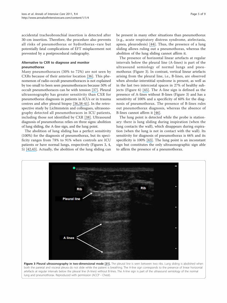
Alternative to CXR to diagnose and monitorpneumothoraxMany pneumothoraces (30% to 72%) are not seen byCXRs because of their anterior location [36]. This phe-nomenon of radio-occult pneumothoraces is not explainedby too small to been seen pneumothoraces because 50% ofoccult pneumothoraces can be with tension [37]. Pleuralultrasonography has greater sensitivity than CXR forpneumothorax diagnosis in patients in ICUs or in traumacentres and after pleural biopsy [36,38-41]. In the retro-spective study by Lichtenstein and colleagues, ultrasono-graphy detected all pneumothoraces in ICU patients,including those not identified by CXR [38]. Ultrasounddiagnosis of pneumothorax relies on three signs: abolitionof lung sliding, the A-line sign, and the lung point.The abolition of lung sliding has a perfect sensitivity
(100%) for the diagnosis of pneumothorax, but its speci-ficity ranges from 78% to 91% when controls are ICUpatients or have normal lungs, respectively (Figures 3, 4,5) [42,43]. Actually, the abolition of the lung sliding can
be present in many other situations than pneumothorax(e.g., acute respiratory distress syndrome, atelectasia,apnea, pleurodesis) [44]. Thus, the presence of a lungsliding allows ruling out a pneumothorax, whereas theabolition of the lung sliding cannot affirm it.The presence of horizontal linear artefacts at regular
intervals below the pleural line (A-lines) is part of theultrasound semiology of normal lungs and pneu-mothorax (Figure 3). In contrast, vertical linear artefactsarising from the pleural line, i.e., B-lines, are observedwhen alveolar-interstitial syndrome is present, as well asin the last two intercostal spaces in 27% of healthy sub-jects (Figure 6) [45]. The A-line sign is defined as thepresence of A-lines without B-lines (Figure 3) and has asensitivity of 100% and a specificity of 60% for the diag-nosis of pneumothorax. The presence of B-lines rulesout pneumothorax diagnosis, whereas the absence ofB-lines cannot affirm it [46].The lung point is detected while the probe is station-
ary: there is lung sliding during inspiration (when thelung contacts the wall), which disappears during expira-tion (when the lung is not in contact with the wall). Itssensitivity for diagnosis of pneumothorax is 66% and itsspecificity is 100% [43]. The lung point is an inconstantsign but constitutes the only ultrasonographic sign ableto affirm the presence of a pneumothorax.
Figure 3 Pleural ultrasonography in two-dimensional mode [31]. The pleural line is seen between two ribs. Lung sliding is abolished whenboth the parietal and visceral pleura do not slide while the patient is breathing. The A-line sign corresponds to the presence of linear horizontalartefacts at regular intervals below the pleural line (A-lines) without B-lines. The A-line sign is part of the ultrasound semiology of the normallung and pneumothorax. Reproduced with permission (ACCP - Chest).
Ioos et al. Annals of Intensive Care 2011, 1:4http://www.annalsofintensivecare.com/content/1/1/4
Page 5 of 9

Figure 4 Assessment of lung sliding on pleural ultrasonography in time-motion mode on a patient without pneumothorax [31]. Lungsliding generates a granular pattern under the pleural line. Subcutaneous tissue over the pleural line does not move while the patient isbreathing, generating horizontal lines. Reproduced with permission (ACCP - Chest).
Figure 5 Abolition of lung sliding on pleural ultrasonography in time-motion mode in a patient with pneumothorax [31]. While thepatient is breathing, the (normal) granular pattern under the pleural line is replaced by horizontal lines, indicating abolition of lung sliding.Reproduced with permission (ACCP - Chest).
Ioos et al. Annals of Intensive Care 2011, 1:4http://www.annalsofintensivecare.com/content/1/1/4
Page 6 of 9

In the Delphi study mentioned earlier, most ICU phy-sicians supported a daily routine CXR in patients with achest tube [8]. However, after drainage, ultrasonographyis better than CXR for detecting residual pneu-mothoraces, whereas 39% of them are not identified byCXR [31]. After drainage of primary spontaneous pneu-mothoraces, performance of ultrasonography is excellent[31]. After drainage of nonprimary spontaneous pneu-mothorax, the positive predictive value of ultrasonogra-phy was 100% in the presence of a lung point. However,it decreased to 90% in the absence of a lung point [31].Exclusive use of ultrasonography for follow-up of non-primary spontaneous pneumothorax seems possible, butthe physician must be aware that in the absence of lungpoint, diagnosis of pneumothorax should not be made ifother causes of lung sliding abolition have not beenruled out. We recommend performing a CT scan ifdoubt persists, especially if new chest tube insertion isunder consideration.These excellent performances make pleural ultraso-
nography more than an alternative to CXR and shouldbe considered as the “bedside gold standard” to diag-nose and monitor pneumothorax. Moreover, ultrasono-graphy gives faster results than CXR and is performedcompetently by naïve physicians after a brief trainingsession [31,47].
Alternative to CXR after central venous catheter insertionThe French ICU physicians who participated in theDelphi study agreed on the appropriateness of per-forming a CXR after central venous catheter (CVC)insertion in the superior vena cava system [8]. Aftercatheterization of the subclavian or internal jugularvein, CVC tip misplacement occurs in 5% to 6% andpneumothorax occurs in 1.5% to 3.1% and 0.1% to0.2%, respectively [48].Clinical evaluation of the patient to predict the
absence of complications after CVC insertions via thesubclavian vein or internal jugular vein was very accu-rate in Gray and colleagues’ study [49]. However, Glad-win and colleagues showed that the clinical impressionof the operator (based on the number of needle passes,difficulty establishing access, operator experience, pooranatomical landmarks, number of previous catheter pla-cements, resistance to wire or catheter advancement,resistance to aspiration of blood or flushing of thecatheter ports, sensations in the ear, chest, or arm, anddevelopment of signs or symptoms suggestive of pneu-mothorax) had a poor sensitivity (44%) and specificity(55%) for predicting a complication [50]. Gladwin andcolleagues concluded that postprocedural CXR remainsnecessary because clinical factors alone cannot reliablyidentify tip misplacement.
Figure 6 Detection of B-lines on pleural ultrasonography in two-dimensional mode. The presence of vertical linear artefacts arising fromthe pleural line (B-lines or comet-tail artefacts) rules out pneumothorax in this patient with interstitial syndrome. Reproduced with permission(ACCP - Chest).
Ioos et al. Annals of Intensive Care 2011, 1:4http://www.annalsofintensivecare.com/content/1/1/4
Page 7 of 9

Nevertheless, as mentioned, numerous pneumothoracescan be missed by bedside CXR, whereas ultrasonographyshowed excellent sensitivity and specificity for diagnosingpneumothorax within a few minutes. Postprocedural ultra-sonography and CXRs were compared after insertion of 85central venous catheters (70 subclavian and 15 internaljugular) [51]. Ultrasonic examination feasibility was 99.6%.Ten misplacements and one pneumothorax occurred.This pneumothorax and all misplacements except onewere diagnosed by ultrasound. Taking into considerationsigns of misplacement and pneumothorax, ultrasonicexamination did not give any false-positive results. More-over, ultrasound guidance increases the success rate ofCVC insertion, saves time, and decreases the complicationrate [52]. Considering these results, it appears logical touse the same ultrasonographic device to assess both theadequate position of the CVC and the absence of pneu-mothorax after the procedure. The only limit of ultrasono-graphy in this indication is the lack of visualization ofazygos, internal thoracic and cardiophrenic veins, and aninconstant visualization of the superior vena cava. Thus,ultrasonography could be proposed for assessing theabsence of misplacement and pneumothorax while limit-ing CXR requirement to incomplete ultrasonographicanalysis.
ConclusionsWe have shown that bedside CXR could be avoided inmany circumstances. This is true for most mechanicallyventilated patients and for ensuring proper placement ofdevices, such as feeding tubes and central venous cathe-ter. This restrictive policy for ordering bedside CXRrequires an assessment of the patient’s clinical status atleast once a day before ordering CXR. It means thatCXR should never replace clinical evaluation of thepatient but should be prescribed on the basis of clinicalsuspicion. As a consequence, the organization of theICU might have to be modified to allow the implemen-tation of such a prescribing strategy and the reductionof the number of CXRs ordered. Ultrasonography is avery good alternative to CXR. For example, ultrasono-graphy is more accurate than CXR for detecting pneu-mothorax. However, short training courses must beorganized to reach a basic level of competency for everyphysician working in ICU. A policy of reducing thenumber of CXRs has many advantages (comfort for thepatients, better organization of the radiology depart-ment, cost reduction) and should be widely implemen-ted in the ICU. The emblematic examples presented inthis review can be combined, and the global pictureissued from this review suggests adopting an integratedapproach for decreasing the number of CXR investiga-tions performed in the ICU.
Author details1Hôpital Delafontaine, Service de Réanimation Polyvalente, Saint-Denis F-93205, France. 2AP-HP, Hôpital Saint-Antoine, Service de RéanimationMédicale, Paris F-75012, France. 3UPMC Univ Paris 06, UMR_S 938, CdR Saint-Antoine, F-75005, Paris, France. 4INSERM, UMR_S 938, CdR Saint-Antoine, F-75012, Paris, France. 5Institut Gustave Roussy, Service de RéanimationMédico-Chirurgicale, F-94805, Villejuif, France. 6UPMC Univ Paris 06, UMR_S707, Paris F-75012, France. 7INSERM, U707, Paris F-75012, France. 8AP-HP,Hôpital Saint-Antoine, Unité de Santé Publique, Paris F-75012, France.
Authors’ contributionsVI, AG, LC-L, BG, EM and GH all participated in the design and in theredaction of the first draft of the article, corrected and approved the finalversion.
Competing interestsThe authors declare that they have no competing interests.
Received: 11 February 2011 Accepted: 21 March 2011Published: 21 March 2011
References1. Haddad SH, Aldawood AS, Arabi YM: The diagnostic yield and clinical
impact of a chest X-ray after percutaneous dilatational tracheostomy: aprospective cohort study. Anaesth Intensive Care 2007, 35:393-397.
2. Houghton D, Cohn S, Schell V, Cohn K, Varon A: Routine daily chestradiography in patients with pulmonary artery catheters. Am J Crit Care2002, 11:261-265.
3. Rao PS, Abid Q, Khan KJ, Meikle RJ, Natarajan KM, Morritt GN, Wallis J,Kendall SW: Evaluation of routine postoperative chest X-rays in themanagement of the cardiac surgical patient. Eur J Cardiothorac Surg 1997,12:724-729.
4. Bhagwanjee S, Muckart DJ: Routine daily chest radiography is notindicated for ventilated patients in a surgical ICU. Intensive Care Med1996, 22:1335-1338.
5. Clec’h C, Simon P, Hamdi A, Hamza L, Karoubi P, Fosse JP, Gonzalez F,Vincent F, Cohen Y: Are daily routine chest radiographs useful in criticallyill, mechanically ventilated patients? A randomized study. Intensive CareMed 2008, 34:264-270.
6. Graat ME, Kroner A, Spronk PE, Korevaar JC, Stoker J, Vroom MB, Schultz MJ:Elimination of daily routine chest radiographs in a mixed medical-surgical intensive care unit. Intensive Care Med 2007, 33:639-644.
7. Hall JB, White SR, Karrison T: Efficacy of daily routine chest radiographs inintubated, mechanically ventilated patients. Crit Care Med 1991,19:689-693.
8. Hejblum G, Ioos V, Vibert JF, Boelle PY, Chalumeau-Lemoine L, Chouaid C,Valleron AJ, Guidet B: A web-based Delphi study on the indications ofchest radiographs for patients in ICUs. Chest 2008, 133:1107-1112.
9. Hendrikse KA, Gratama JW, Hove W, Rommes JH, Schultz MJ, Spronk PE:Low value of routine chest radiographs in a mixed medical-surgical ICU.Chest 2007, 132:823-828.
10. Kappert U, Stehr SN, Matschke K: Effect of eliminating daily routine chestradiographs on on-demand radiograph practice in post-cardiothoracicsurgery patients. J Thorac Cardiovasc Surg 2008, 135:467-468, author reply 468.
11. Krivopal M, Shlobin OA, Schwartzstein RM: Utility of daily routine portablechest radiographs in mechanically ventilated patients in the medicalICU. Chest 2003, 123:1607-1614.
12. Silverstein DS, Livingston DH, Elcavage J, Kovar L, Kelly KM: The utility ofroutine daily chest radiography in the surgical intensive care unit. JTrauma 1993, 35:643-646.
13. Expert Panel on Thoracic Imaging of the American College of Radiology:Routine Chest Radiograph Appropriatenes Criteria. 2008 [http://www.acr.org/SecondaryMainMenuCategories/quality_safety/app_criteria/pdf/ExpertPanelonThoracicImaging.aspx].
14. Graat ME, Hendrikse KA, Spronk PE, Korevaar JC, Stoker J, Schultz MJ: Chestradiography practice in critically ill patients: a postal survey in theNetherlands. BMC Med Imaging 2006, 6:8.
15. Graat ME, Stoker J, Vroom MB, Schultz MJ: Can we abandon daily routinechest radiography in intensive care patients? J Intensive Care Med 2005,20:238-246.
Ioos et al. Annals of Intensive Care 2011, 1:4http://www.annalsofintensivecare.com/content/1/1/4
Page 8 of 9

16. Brainsky A, Fletcher RH, Glick HA, Lanken PN, Williams SV, Kundel HL:Routine portable chest radiographs in the medical intensive care unit:effects and costs. Crit Care Med 1997, 25:801-805.
17. Henschke CI, Yankelevitz DF, Wand A, Davis SD, Shiau M: Chestradiography in the ICU. Clin Imaging 1997, 21:90-103.
18. Graat ME, Choi G, Wolthuis EK, Korevaar JC, Spronk PE, Stoker J, Vroom MB,Schultz MJ: The clinical value of daily routine chest radiographs in amixed medical-surgical intensive care unit is low. Crit Care 2006, 10:R11.
19. Price MB, Grant MJ, Welkie K: Financial impact of elimination of routinechest radiographs in a pediatric intensive care unit. Crit Care Med 1999,27:1588-1593.
20. Oba Y, Zaza T: Abandoning daily routine chest radiography in theintensive care unit: meta-analysis. Radiology 2010, 255:386-395.
21. Hejblum G, Guidet B: Evidence-based data for abandoning unselectivedaily chest radiographs in intensive care units. Radiology 2010,256:1013-1014.
22. Hejblum G, Chalumeau-Lemoine L, Ioos V, Boelle PY, Salomon L, Simon T,Vibert JF, Guidet B: Comparison of routine and on-demand prescriptionof chest radiographs in mechanically ventilated adults: a multicentre,cluster-randomised, two-period crossover study. Lancet 2009,374:1687-1693.
23. Siegel MD, Rubinowitz AN: Routine daily vs on-demand chestradiographs in intensive care. Lancet 2009, 374:1656-1658.
24. Haute Autorité de Santé: Principales indications et « non indications » dela radiographie du thorax. 2009 [http://www.has-sante.fr/portail/jcms/c_755004/indications-et-non-indications-de-la-radiographie-du-thorax].
25. Woodall BH, Winfield DF, Bisset GS: Inadvertent tracheobronchialplacement of feeding tubes. Radiology 1987, 165:727-729.
26. Kunis K: Confirmation of nasogastric tube placement. Am J Crit Care 2007,16:19, author reply 19.
27. Stroud M, Duncan H, Nightingale J: Guidelines for enteral feeding in adulthospital patients. Gut 2003, 52(Suppl 7):vii1-vii12.
28. Metheny NA: Preventing respiratory complications of tube feedings:evidence-based practice. Am J Crit Care 2006, 15:360-369.
29. Jolliet P, Pichard C, Biolo G, Chiolero R, Grimble G, Leverve X, Nitenberg G,Novak I, Planas M, Preiser JC, Roth E, Schols AM, Wernerman J: Enteralnutrition in intensive care patients: a practical approach. Working Groupon Nutrition and Metabolism, ESICM. European Society of Intensive CareMedicine. Intensive Care Med 1998, 24:848-859.
30. Chalumeau-Lemoine L, Baudel JL, Das V, Arrive L, Noblinski B, Guidet B,Offenstadt G, Maury E: Results of short-term training of naive physiciansin focused general ultrasonography in an intensive-care unit. IntensiveCare Med 2009, 35:1767-1771.
31. Galbois A, Ait-Oufella H, Baudel JL, Kofman T, Bottero J, Viennot S, Rabate C,Jabbouri S, Bouzeman A, Guidet B, Offenstadt G, Maury E: Pleuralultrasound compared with chest radiographic detection ofpneumothorax resolution after drainage. Chest 2010, 138:648-655.
32. Vigneau C, Baudel JL, Guidet B, Offenstadt G, Maury E: Sonography as analternative to radiography for nasogastric feeding tube location. IntensiveCare Med 2005, 31:1570-1572.
33. Burns SM, Carpenter R, Truwit JD: Report on the development of aprocedure to prevent placement of feeding tubes into the lungs usingend-tidal CO2 measurements. Crit Care Med 2001, 29:936-939.
34. Galbois A, Vitry P, Ait-Oufella H, Baudel JL, Guidet B, Maury E, Offenstadt G:Colorimetric capnography, a new procedure to ensure correct feedingtube placement in the intensive care unit: An evaluation of a localprotocol. J Crit Care 2010.
35. Meyer P, Henry M, Maury E, Baudel JL, Guidet B, Offenstadt G: Colorimetriccapnography to ensure correct nasogastric tube position. J Crit Care2009, 24:231-235.
36. Soldati G, Testa A, Pignataro G, Portale G, Biasucci DG, Leone A, Silveri NG:The ultrasonographic deep sulcus sign in traumatic pneumothorax.Ultrasound Med Biol 2006, 32:1157-1163.
37. Tocino IM, Miller MH, Fairfax WR: Distribution of pneumothorax in thesupine and semirecumbent critically ill adult. AJR Am J Roentgenol 1985,144:901-905.
38. Lichtenstein DA, Meziere G, Lascols N, Biderman P, Courret JP, Gepner A,Goldstein I, Tenoudji-Cohen M: Ultrasound diagnosis of occultpneumothorax. Crit Care Med 2005, 33:1231-1238.
39. Soldati G, Testa A, Sher S, Pignataro G, La Sala M, Silveri NG: Occulttraumatic pneumothorax: diagnostic accuracy of lung ultrasonographyin the emergency department. Chest 2008, 133:204-211.
40. Chung MJ, Goo JM, Im JG, Cho JM, Cho SB, Kim SJ: Value of high-resolution ultrasound in detecting a pneumothorax. Eur Radiol 2005,15:930-935.
41. Sartori S, Tombesi P, Trevisani L, Nielsen I, Tassinari D, Abbasciano V:Accuracy of transthoracic sonography in detection of pneumothoraxafter sonographically guided lung biopsy: prospective comparison withchest radiography. AJR Am J Roentgenol 2007, 188:37-41.
42. Lichtenstein DA, Menu Y: A bedside ultrasound sign ruling outpneumothorax in the critically ill. Lung sliding. Chest 1995, 108:1345-1348.
43. Lichtenstein D, Meziere G, Biderman P, Gepner A: The “lung point": anultrasound sign specific to pneumothorax. Intensive Care Med 2000,26:1434-1440.
44. Lichtenstein DA, Lascols N, Prin S, Meziere G: The “lung pulse": an earlyultrasound sign of complete atelectasis. Intensive Care Med 2003,29:2187-2192.
45. Lichtenstein D, Meziere G, Biderman P, Gepner A, Barre O: The comet-tailartifact. An ultrasound sign of alveolar-interstitial syndrome. Am J RespirCrit Care Med 1997, 156:1640-1646.
46. Lichtenstein D, Meziere G, Biderman P, Gepner A: The comet-tail artifact:an ultrasound sign ruling out pneumothorax. Intensive Care Med 1999,25:383-388.
47. Noble VE, Lamhaut L, Capp R, Bosson N, Liteplo A, Marx JS, Carli P:Evaluation of a thoracic ultrasound training module for the detection ofpneumothorax and pulmonary edema by prehospital physician careproviders. BMC Med Educ 2009, 9:3.
48. McGee WT, Ackerman BL, Rouben LR, Prasad VM, Bandi V, Mallory DL:Accurate placement of central venous catheters: a prospective,randomized, multicenter trial. Crit Care Med 1993, 21:1118-1123.
49. Gray P, Sullivan G, Ostryzniuk P, McEwen TA, Rigby M, Roberts DE: Value ofpostprocedural chest radiographs in the adult intensive care unit. CritCare Med 1992, 20:1513-1518.
50. Gladwin MT, Slonim A, Landucci DL, Gutierrez DC, Cunnion RE: Cannulationof the internal jugular vein: is postprocedural chest radiography alwaysnecessary? Crit Care Med 1999, 27:1819-1823.
51. Maury E, Guglielminotti J, Alzieu M, Guidet B, Offenstadt G: Ultrasonicexamination: an alternative to chest radiography after central venouscatheter insertion? Am J Respir Crit Care Med 2001, 164:403-405.
52. Hind D, Calvert N, McWilliams R, Davidson A, Paisley S, Beverley C,Thomas S: Ultrasonic locating devices for central venous cannulation:meta-analysis. BMJ 2003, 327:361.
doi:10.1186/2110-5820-1-4Cite this article as: Ioos et al.: An integrated approach for prescribingfewer chest x-rays in the ICU. Annals of Intensive Care 2011 1:4.
Submit your manuscript to a journal and benefi t from:
7 Convenient online submission
7 Rigorous peer review
7 Immediate publication on acceptance
7 Open access: articles freely available online
7 High visibility within the fi eld
7 Retaining the copyright to your article
Submit your next manuscript at 7 springeropen.com
Ioos et al. Annals of Intensive Care 2011, 1:4http://www.annalsofintensivecare.com/content/1/1/4
Page 9 of 9