
An evaluation on vital organ donation, culture, altruism and informed consent
325
design by Almir Rizvanovic
Transcript of An evaluation on vital organ donation, culture, altruism and informed consent

design by Almir Rizvanovic
EDITORIAL BOARD
Editor-in-chief Mensura Kudumovic
Execute Editor Mostafa Nejati
Associate Editor Azra Kudumovic
Technical Editor Eldin Huremovic
Cover Design Almir Rizvanovic
Members
Paul Andrew Bourne (Jamaica) Xiuxiang Liu (China) Nicolas Zdanowicz (Belgique) Farah Mustafa (Pakistan) Yann Meunier (USA) Suresh Vatsyayann (New Zealand) Maizirwan Mel (Malaysia) Budimka Novakovic (Serbia) Diaa Eldin Abdel Hameed Mohamad (Egypt) Zmago Turk (Slovenia) Edvin Dervisevic (Slovenia) Chao Chen (Canada) Aleksandar Dzakula (Croatia) Farid Ljuca (Bosnia & Herzegovina) Sukrija Zvizdic (Bosnia & Herzegovina) Damir Marjanovic (Bosnia & Herzegovina) Bozo Banjanin (Bosnia & Herzegovina) Gordana Manic (Bosnia & Herzegovina) Address Sarajevo, Hamdije Kresevljakovica 7A
Editorial Board [email protected] http://www.healthmedjournal.com
Published by DRUNPP, Sarajevo
Volume 7 Number 4, 2013
ISSN 1840-2291
HealthMEDVolume 7 / Number 4 / 2013
Journal of Society for development in new net environment in B&H
Sadržaj / Table of Contents
HealthMED journal with impact factor indexed in: - Thomson Reuters ISI web of Science, - Science Citation Index-Expanded, - Scopus, - EBSCO Academic Search Premier, - EMBASE - Index Copernicus, - getCITED, and etc.
ELSEVIER
A quantitative evaluation of health care systemin US, China and Sweden .............................................. 1064Qixin Wang, Menghui Li, Hualong Zu, Mingyi Gao, Chenghua Cao, Li Charlie XiaAn analysis of 263 female patients exposedto physical violence that admitted to a hospitalemergency department in Turkey ............................... 1075Aydin Tekin, Umut Yucel Cavus, Sinan Yildirim, Fatih BuyukcamAnesthetic management of Eclampsia/hellp ............... 1079Feyzi Celik, M. Erdal Sak, Adnan Tufek, Abdulmenap GuzelThe influence of -330 IL-2 gene polymorphismand HLA-DRB1*1501 allele on age at onset inIranian multiple sclerosis patients ................................ 1084Arezou Sayad, Abdolamir Alameh, Aida SayadEffectiveness of a structered commercial dieting program on weight control ............................... 1089Memet Isik, Fatma Nihal Aksoy, Yasemin Cayir,Zeliha CanseverAdolescence and sexuality ............................................. 1094Jorge Andre Cartaxo Peixoto, Modesto Leite Rolim Neto, Luiz Carlos de AbreuAn evaluation on vital organ donation, culture,altruism and informed consent .................................... 1099Sukran SevimliCases with critical illness polineuromyopathy ............ 1105Sayin Refah, Isik Yasemin, Ugur Goktas, Kati IsmailMortality and related risk factors in patientsundergoing maintenance peritoneal dialysis ............... 1109Elham Yousefi-Abdolmaleki, Sepideh Seyfi, Mahboob Lesanpezeshki, Yahya SalehtabariClinical supervision and peer consultationfor Medical Faculty members ........................................1114Yesim Senol, Fatos B. Yildirim, Levent Sarikcioglu,Mualla AksuAnalysis of the dynamics of growth of permanentteeth in intraoral-prefunctional phase ..........................1119Ljiljana Kostadinovic, Mirjana Apostolovic,Branislava Stojkovic, Aleksandra Ignjatovic,Marija Igic, Olivera Trickovic Janjic,Dusan Surdilovic

Sadržaj / Table of Contents
Upper respiratory system infectionfrequency is affected by allergic symptomsand cigarette smoking .................................................... 1126Ugur Bilge, Ahmet KeskinEffect of the pedicle mucosal (muscle) flapof the lip for primary repair of contralateralintrocession ..................................................................... 1131Ying-li Li, Hui Chen, Jie Du, Yan CaoThe effect of adenotonsillectomy onpulmonary function tests ............................................... 1136Salim Yuce, Ismail Onder Uysal, Mehmet Bayram,Mansur Dogan, Isa Dongel, Cahit Polat, Ali KayaSelf-buttressing patch: Alternative techniquefor surgical closure of Ostium secundum atrialseptal defect .................................................................1140Faranak Kargar, Mathias H Aazami, Sevil AghapourThe susceptibility to staining of orthodonticelastic ligatures caused by food colorants – In vitro study ................................................................... 1145Konrad Malkiewicz, Barbara Pietrzak-BilinskaExpression, purification and bioactivityanalysis of the human antimicrobial peptideLL-37 in Escherichia coli ............................................... 1151Xiaoyan Wu, Yi Lu, Hong ZhaoInvestigation of free-living Amoebae andrespiratory bacterial pathogens in watersamples taken from recreational fountainsand ornamental pools in Ankara, Turkey .................... 1158Ece Erci, Elif Abay, Gulay Aral Akarsu,Zeynep Ceren KarahanMolecular typing of Staphylococcus aureusisolated from patients and healthy carrierson the basis of coagulase gene polymorphism ............. 1168Teena Dadgar, Ezzat Allah Ghaemi, Nima Bahador,Abbas Ali Imani FooladiAdvanced adenoid cystic carcinoma ofthe Parotid gland treated with surgery,chemotherapy and targeted therapy: A case report and literature review .............................. 1174Yu Zhang, Hongming PanConcomitance of acute pancreatitis andthrombocytopenia due to valproic acidintoxication: A rare case report .................................... 1178Mehmet Aslan, Lokman Soyoral, Ahmet Cumhur Dulger, Senar EbincExposure of students to passive inhalationof tobacco smoke ............................................................. 1182Agima Ljaljevic, Aleksandar Stijepcevic,Dragan CabarkapaLate presenters and significance ofscreening tests in early diagnosis ofHIV infection in Istanbul ............................................... 1187Hayat Kumbasar Karaosmanoglu, Ozlem Altuntas Aydin, Isık Somuncu Johansen, Ramazan Korkusuz, Ozcan Nazlican
Relationship between STAT3 signal pathwayand recurrent spontaneous abortion ............................ 1192Shu-hua Zhao, Fan Zhao, Xiu-yun Zhao, Man-hua CuiUse of intermittent hypobaric pressurein the treatment of inoperable peripheralarterial occlusive disease (PAOD) inelderly patients ................................................................ 1197Branko Milicevic, Gordana Devecerski, Jure Jelenc,Nikola RadakovicStyloid process elongation in end stage renaldisease patients with peritoneal dialysis: is there any role for ectopic calcification? .................... 1201Cumali Gokce, Elif Tarim Ertas, Yildiray Sisman,Murat Rifalioglu, Oktay Oymak, Cengiz UtasEfficacy of Linezolid on gram-positivebacterial infection in elderly patientsand the risk factors associated withthrombocytopenia .......................................................... 1207Liqing Bi, Jing Zhou, Ming Huang, Suming ZhouEfficacy and safety of use of the combinationof Ketamine and Propofol (“Ketofol”) inprocedural sedation and analgesia in children ........... 1215Milanka Tatic, Sanja Skeledzija-Miskovic,Aleksandar Komarcevic, Svetlana Bukarica,Aleksandra MarcikicaLaparoscopic versus open appendectomy ................... 1220Burhan Hakan Kanat, Ahmet Turkoglu, Mesut Yur,Mustafa Girgin, Mehmet Yasar AslanmirzaEffects of intermittent negative pressuretreatment on vascular and plateletcomponent of hemostasis system inpatients with lymphoedema of lower limbs ................. 1224Branko Milicevic, Gordana Devecerski, Nikola Radakovic, Jure JelencEthyl pyruvate attenuation of hepatocyteinjury by ATP-related mechanism underhypoxia/reoxygenation in vitro ..................................... 1226Zhe Luo, Yizhou He, Zhanggang Xue, Hao WangParameters influencing renal function inthe subject with metabolic syndrome ........................... 1235Danijela TasicAn unusual spinal mixed glioneuronaltumor with BRAFV600E mutation analysis:Case report and literature review ................................. 1239Yu Hu, Weiying Zhong, Haifeng Chen, Siqing HuangEvaluation of ST segment and T-wavechanges in the anterior, inferior andposterior derivations of patients withisolated acute occluded left circumflex artery ............. 1244Eftal Murat Bakirci, Mahmut Acikel, Enbiya Aksakal,Serdar Sevimli1, Husnu Degirmenci, Muhammed Hakan Tas, Selami Demirelli, Sinan Inci, Hakan Duman, Ibrahim Halil Tanboga

Sadržaj / Table of Contents
Endoscopic treatment of bile duct lithiasisafter Billroth-II gastroenterostomy .............................. 1251Guang-quan Zhang, Qi-yuan Lin, Man-xi He, Zhong LiaoEffects of autohemotherapy on hematologicalresponses in Wistar female ratsAutohemotherapy in rats ............................................... 1256Aline S. Ibanes, Myrian Cabral, Luiz Carlos de Abreu,Vitor E. Valenti, Thais M. Gascon, Ana P. F. Moreira,David Feder, Ligia A. Azzalis, Virginia B. C. Junqueira,Edimar C. Pereira, Sarah R. Marsicano, Fernando F. Perazzo, Fernando L. A. FonsecaSubglottic high frequency jet ventilation in management of bilateral vocal foldparalysis: a case report ................................................. 1262Nevena Kalezic, Dusanka Janjevic, Vladimir Dolinaj,Nebojsa Ladjevic, Dejan MarkovicEffect of yoga exercises on quality of lifeof postmenopausal women............................................. 1266Shabnam al-Sadat ShariatPanahi, EftekharalsadatHajikazemi, Soghra Nikpour, Somayeh Fghanipour,Agha Fatemeh HosseiniRole of Bradykinin in the cardioprotectiveeffects of Captopril and Angiotensin IIreceptor blockers (AT1, AT2) on Myocardialischemia–Reperfusion injury in rats ............................ 1274Seda Tasdemir, Hakan Parlakpinar, Ahmet AcetOrthodontic treatment of class II division1 malocclusion by Fränkel functionalregulator type Ic: Case report ....................................... 1281Zorana Stamenkovic, Nenad Nedeljkovic,Vladimir RisticComparison of post-operative analgesic efficacies of Tramadol and Bupivacaineinfiltration applied to multiple regionsduring laparoscopic cholecystectomy ........................... 1285Abdulkadir Iskender, Ismet Ozaydin, Gulbin Sezen, MehmetYasar, Yavuz Demiraran, Tuna Yilmaz, Zekeriya IlceMedical University’s strategic plan:Progress and related obstacles ...................................... 1290Masoumeh Hosseini, Saeed Asefzadeh, Mina Nejati,Tahere SadeghiThe prevalence of Parvovirus B19 in patientswith Sickle cell disease in southern Turkey ................. 1295Vicdan Koksaldı Motor, Omer Evirgen, Nizami Duran,Vefik Arıca, Melek Inci, M. Murat Celik,Fatmagul Basarslan, Yusuf OnlenDetecting and reporting child abuseby health workers in Serbia .......................................... 1300Miroslav Misic, Fadilj Eminovic, Lidija Milenovic,Snezana MirkovicDo serum Zinc levels and oxidative statuschange in familial Mediterranean feverpatients during attack and attack-free periods? ......... 1307Koksal Deveci, Soner Senel, Hulya Deveci,Enver Sancakdar, Abdulkadir Deniz, Ali Ugur Uslu,Huseyin Aydin
Cardiac disease prediction and rule extractwith data mining - Classification techniques ............... 1312Peyman Rezaei Hachesu, Maryam Ahmadi, Somayyeh Alizadeh, Farahnaz SadughiThe compare of effect of exhaustiveexercise on MDA, ADA, GSH and SODactivity in women ............................................................ 1323M. Emin Kafkas, Armagan Sahin Kafkas, Seyfi Savas,Aysun Bay KarabulutAssociation of PPARG gene polymorphism(Pro12Ala) with type 2 diabetes in theIranian population ......................................................... 1329Javad Mohammadi-Asl, Aniseh Soheil-Nezhad, Leila Saremi, Fakher RahimAnti-Toxoplasma antibody prevalence,primary infection rate, and risk factorsin a study of toxoplasmosis in 1.026pregnant women southeastern Turkey ......................... 1334Tuncay Celik, Beyza Doganay Erdogan, Semiha Aydin,Servet KolgelierPsychophysical status of human trafficking victims ........1341Zeljko Bjelajac, Zaklina Spalevic, Bozidar BanovicEfficacy of folk medicinal plant extractAnkaferd Blood Stopper on full-thicknessskin wound healing ......................................................... 1347Mehmet Tahir Gokdemir, Halil Kaya, Ozgur Sogut,Tuncer Demir, Sezen Kocaslan, Muazez CevikImpact of iron-folic acid supplementationon passive avoidance memory in adult maleWistar rats ....................................................................... 1354Khombi Shooshtari M , Moazedi A. A, Parham GhKnowledge, attitudes and practices ofparents regarding circumcision in Trabzon ................ 1361Ilknur Kahriman, Murat Topbas, Gamze CanUse of the QIDS-C16, QIDS-SR16 and the17-item Hamilton rating scale for depression(HAM-D17) in a Turkish college student sample ........ 1369Haluk Mergen, Ira H. Bernstein, Berna Erdogmus MergenThe prevalence of carotid artery calcificationor atherosclerotic plaque in end-stage renaldisease being treated with hemodialysis andperitoneal dialysis patients detected onpanoramic radiography .......................................................1376Cumali Gokce, Elif Tarim Ertas, Yildiray Sisman, Murat Sipahioglu, Mursel Davarci, Oktay Oymak, Cengis UtasInstructions for the authors ........................................... 1384

1064
HealthMED - Volume 7 / Number 4 / 2013
Journal of Society for development in new net environment in B&H
Abstract
This study is mainly aimed at evaluating the effectiveness of current health care systems of several representative countries and improving that of the US. To achieve these goals, a people-oriented non-linear evaluation model is designed. It comprises one major evaluation metric and four minor metrics. The major metric is constituted by combining possible factors that most significantly determine or affect the life expectancy of people in this country. The four minor metrics evaluate less important aspects of health care systems and are subordinate to the major one. The authors rank some of the health care systems in the world according to the major metric and detect problems in them with the help of minor ones. It is concluded that the health care system of Sweden scores higher than the US and China’s system scores lower than that of the US. Especially, the health care system of US lags behind a little bit compared with its economic power. At last, it is reasonable for the American government to optimize the arrangement of funding base on the result of goal programming model.
Key words: Health care system, evaluation.
Introduction
A satisfactory health care system of one country is supposed to provide its residents with effective health care, so that a majority of citizens can enjoy a security and high-quality life, with maximized so-cial equality and minimized total medical expendi-ture. The complexity of the health care systems ma-kes it difficult to evaluating the health care system by taking into account only a few factors.
The considerations of previous research tend to emphasize the financial efficiency of health care systems. Controlling of the cost has been reported as the key factor of this system [1,2]. However, health care system is slightly different from finan-cial system [3,4]. Medical care is a necessity rather than a commodity for citizens of a country. The quality of medical care of patients is much more important than controlling of cost in health care system [5]. In health care system, health insurance covers most large medical expenses, but there is no institution in a financial system would insuran-ce the high consumption. Thus, we developed a people-oriented comprehensive evaluation system and pay more attention to make sure quality of he-alth care of people. In this evaluation system, the quality of health care to the patient has a higher priority rather than the financial efficiency.
In addition, previous researchers fail to consider the relationship between health care system and medical research institution [1-3]. The health care system is heavily influenced by the development of biomedical research. The investment to the medi-cal research institution can improve the operational efficiency of the health care system, discover new drugs, revise the therapies, and improve the life qu-ality of patients. It is no doubt that increase of inves-tments on medical research has positive impact on health care system in developed country. However, for a developing country, it seems reasonable to pay more attention on other part of health care system such as development medical insurance system, bu-ilding new hospitals and medical education [6].
Several studies described the qualitative analysis of patient satisfactory of health care and
A quantitative evaluation of health care system in US, China and SwedenQixin Wang1, Menghui Li2, Hualong Zu3, Mingyi Gao4, Chenghua Cao5, Li Charlie Xia6
1 Department of Mathematics, University of Southern California, Los Angeles, United States of America,2 Department of Biomedical Engineering, Peking University, Beijing, PR China,3 Department of Electrical Engineering, University of Southern California, Los Angeles, United States of America,4 Department of Chemical Engineering and Materials, University of Southern California, Los Angeles, United States of America,5 School of Medicine, University of California Irvine, Los Angeles, United States of America,6 Department of Biological Sciences, University of Southern California, Los Angeles, United States of America.

HealthMED - Volume 7 / Number 4 / 2013
Journal of Society for development in new net environment in B&H 1065
government investment to medical system [7, 8]. However, since there is a complex non-linear relati-onship between increase of government investment and improvement patient satisfactory of health care system[9,10,11], it is hard to optimize the amount of investment to current health care system; a qu-antitative analysis model is highly desired. In this study, we are going to conduct a quantitative mo-del to evaluate the health care system of the United States, Sweden, and China. We will also develop a goal programming model to evaluate the best go-vernment funding allocation to health care system.
Definitions and Key Terms
A health care system is the organization and the method by which health care is provided.
The potential of health care (Phc) of a country shows the power of medical researches and devel-opment supported by the government at the pres-ent time. It is positively correlated with the size of medical research staff and the quantity of funding from the government.
The health of a citizen is perfectly ensured, if his/her income plus aid from the government (plus the financial compensation from medical in-surance system if he/she is covered by it) is large enough to prevent and/or cure diseases, ignoring the irreversible damage to health that is beyond the ability of current medical technology.
The quality index of life represents the relief from possible diseases or accidentally physical injury based on economic aid offered by medical insurance, and a certain quantity of medical re-sources provided by current health care system, as well as scientific potential of medicine realized by government-funding researches. This index can approximately imply the quality of life.
The life expectancy represents the average life span of a newborn and is an indicator of the over-all health of the population of a country.
Practical effect of medical resources is the quantity of all categories (medical doctors, nurses, beds) of medical aid that is practically distributed to each citizen in a country on average.
The medical care resources are divided into essential health care and complementary health care resources. Complementary care is the kind of services that offer holistic benefits that comple-
ment or enhance the health care received from the physicians or hospital, and essential health care embraces all the other kinds.
The matching degree of a health care system in a country measures whether the system is mas-sive enough to keep up with the development of the country. The health of a citizen is perfectly ensured, if his/her income plus aid from the gov-ernment (plus the financial compensation from medical insurance system if he/she is covered by it) is large enough to prevent and/or cure diseases, ignoring the irreversible damage to health that is beyond the ability of current medical technology.
The fairness index represents how well a health care system distributes its resources to everyone who needs it, both rich and poor, urban and rural residents.
The life index of a nation is a general and com-prehensive figure that describes how much life of high quality is enjoyed by all the citizens in one country. It is positively correlated with quality in-dex of life and average life expectancy.
Universal health care refers to delivery by a com-bination of public and private systems. In most cases, the law says that everyone must have access to health care. Germany and Sweden, for example, has uni-versal coverage, and social insurance plans cover the majority of people. Symbols are listed in Table 1.
AssumptionsPeople around the world have the same suscep-
tibility to diseases, whichever country they are in.- Medical personnel and scientific researchers
are all competent for their job.- The per capita GDP of one country can denote
how rich and developed the country is.- Every health care system possesses appro-
ximately equal ability of emergency management.
- Every type of disease occurs to people in all countries with the same possibility.
- If a resident is covered by the health care insurance, he/she will be able to afford his/her medical expenditure.
- The investment into scientific medical researches is always effectively used.
- The investment into science researches will pay off (be transformed into applied technology) 25 years later averagely.

1066
HealthMED - Volume 7 / Number 4 / 2013
Journal of Society for development in new net environment in B&H
Model Design
The Major Evaluation MetricGeneral Analysis Since the service object of the health care sys-
tem is people, we perceive that the evaluation met-ric should reflect how well the length and qual-ity of people’s life are guaranteed by the system through providing health care, which is repre-sented by a general concept called life index. Then we decompose life index into two parts that are mutually independent: quality index of life and life expectancy, which measure the quality and quan-tity of residents’ life, respectively. We whereafter keep breaking down quality index of life into con-crete concepts and simple factors. In so doing, the evaluation system model is concretized and op-erationalized, because: 1) life index is quantified and hence computable; 2) it is easier to search and identify associated sources of data.
As we attach great emphasis on the practical effectiveness of health care systems, the life index (Lindex) is the final metric that decides whether a health care system is good or not. According to our definition, we have:
index life lifeL Q E= × , ..................... (1)
whereQlife is standardized life quality index, and Elife
is the average life expectancy of the population in one country.
Elife of countries in the world, as a basic and use-ful kind of data, can be easily found from more than one reliable sources of information, but Qlife is com-paratively abstract and complicated to measure.
Since it is unreasonable to limit Elife to a fixed range, Lindex is not standardized here.
Obviously, a health care system can help pro-mote Qlife in many different ways, but we notice
Table 1. Symbols of evaluation modelSymbols Definitions & Descriptions
indexL Life index
lifeE Life expectancy
lifeQ Life quality index
mrP Practical effect of medical resources
eiP Perfect ensurence index
techP The current power of medical technology
hcP Potential of health care
eR Essential health care resources
cR Complementary health care resources
unD Unnecessary degree
neD Necessity degree
inN Number of residents who are covered by medical insurance
govk The proportion of government reimbursement in medical expense
,med iX One’s medical expenditure which is submitted to Poisson distribution
,inc iX one’s net income which is submitted to normal probability distribution
eE Average essential expenditure to maintain everyday life
,inc iX one’s income which is submitted to normal probability attribution
( )sN t Number of medical researchers
( )sM t Quantity of funding going to medical research
t Time delay
HealthMED - Volume 7 / Number 4 / 2013
Journal of Society for development in new net environment in B&H 1067
that almost every way is realized through one of the following three channels:
I every country organizes and provides health care resources to its citizens;
II medical insurance and government offer economic aid so that the patients have access to medical care service; and
III the government invests money into medical researches so that we will have more advanced medical technology that can cure the currently incurable diseases and prevent unpredictable diseases in the future.
Using three corresponding variables Pmr, Pei and Ptech to measure the effectiveness of the above three channels, we find that Qlife is positively cor-related with each one of them. Therefore, it is rea-sonable to define:
Qlife=(Pmr + Pei + Ptech)/kq , ................... (2)
wherePmr is the practical effect of medical resources,Pei is perfect ensurence index,Ptech is the current power of medical technology
(all of them are standardized indexes), and kq is a coefficient to standardize Qlife.
To get Qlife, we have to obtain the value of Pmr, Pei and Ptech one by one.
Quantify and Calculate Pmr Since the medical care resources are divided
into essential health care and complementary health care resources, Pmr should be broken down into two corresponding parts: the practical effect of essential health care and that of complementary health care. Thus, we get
3 3, ,
1 1, , , ,
e i c imr
i ie i e i c i c i
R RP
R k R k= =
= ++ +∏ ∏ ,......... (3)
Where 1R , 2R and 3R respectively refer to the number of medical doctors, nurses and the beds in hospitals,
Re is a standardized index that denote the es-sential health care resources,
Rc is a standardized index that denote the complementary health care resources,
ke,i and kc,i are empirical coefficients, and the re-lationship among them is expressed by
, , , ,e i e i c i c iR k R k× = ×
Note that:
when , 0e iR → , we have
,,
, , ,
1e ie i
e i e i e i
RR
R k k→
+,
which means the practical effect of medical resources is decided by the quantity of medical resources completely (directly proportional to it);
when ,e iR → ∞ , we get ,
, ,
1e i
e i e i
RR k
→+
,
which means excessive medical resources contrib-utes little and will cause a great waste;
we multiply the monomial
i
i i
RR k+
(i=1, 2, 3)
because the lack of any one of Ri will bring serious difficulty to any health care system.
Calculate Pei The population of one country can be divided
into two categories: those who are covered by medical care insurance (Nin) and those who are not (Nun). So the proportion of insured people (Pinsure) is given by
,in in
insurein un
N NPpopulation N N
= =+
............ (4)
The perfect ensurence index (Pei ) actually mea-sure how many people have their health well en-sured through either joining health care insurance or paying by their sufficient income. Thus, it is reasonable to finally define Pei as
1 ,ei uninsuretotal medical expenditure shortageP P
total medical expenditure= - ×
........................................ (5)
which in fact is

1068
HealthMED - Volume 7 / Number 4 / 2013
Journal of Society for development in new net environment in B&H
( )
( ){ }, , ,
, ,1
,1
,
1 ,
0 ,
, (1 )
(1 )1 (1 ) ,
(1 )
,
,
un
Au
A
M i inc i M i gov
N
med i gov inc i e Ai
ei insure n
med i govi
inc i e
i AI
i A
A X X X k
X k X E IP P
X k
X E
=
=
∈=
∉
= × - >
× - - - × = - - ×
× -
-
∑
∑
........................................ (6)
where kgov is the proportion of government reimburse-
ment in medical expense,Ee is the average essential expenditure to main-
tain everyday life,Xmed,i is the medical expenditure of someone in
the country, which is submitted to Poisson distri-bution, and
Xinc,i is the net income of someone in the coun-try which is submitted to normal distribution.
Calculate PtechThe current power of medical technology (Ptech)
can be well deduced by the potential of health care (Phc) years ago, which means Ptech can be estimat-ed as Phc with a time delay (t), because it takes a period of time to transfer scientific investment into scientific products. Some scholars believe that t=20-30 years and we make it 25 [6] years here.
Firstly, we calculate Phc based on its definition:
( )( )
( )( )
s shc
N s M s
N t M tP
k N t k M t= ×
+ + .............. (7)
whereNs(t) is the number of medical researchers, andMs(t) is the quantity of funding going to medical
research.Note that both of the two factors, medical re-
searchers and money, can enhance Phc, but exces-sive investment (medical researchers and money) gives only limited effect to Phc. This truth supports our idea to define Phc this way.
Secondly, we incorporate t into Phc to get Ptech
( )( )
( )( )
s stech
N s M s
N t M tP
k N t k M tt t
t t- -
= ×+ - + -
.......... (8)
Medical research plays an important role to im-prove the math expectation of residents’ life in future.
Elife=E0+ klt 0life lt hcE E k P= + × Phc ........................ (9)
where E0, klt are coefficients.With Eq (1), (2), (3), (4), (6), and (8), Lindex can
be expressed by a complicated equation which in-volves a series of variables.
Figure 1. The diagram showing the organization of evaluation of health care system
Subordinate Metrics Potential of Health Care (Phc )Eq(7) gives the expression of Phc which is actu-
ally a standardized index that predicts the power of medical technology (Ptech) in the future.
Matching DegreeTo get matching degree of each health care
system, two factors need to be taken into account: the how well residents’ health is ensured and how wealthy the country is. A rich country has the abi-lity to maintain a large scale of health care system that provide abandon health care resources, whi-le a developing country can only afford a smaller and cheaper one. This fact implies that it is harder for a developed country to maintain a matching health care system, because the country has to in-vest more (money, etc) into its health care system. Therefore, matching degree is given by
( ) ( )10
ln ln index
Matching Degreeper capita GDP L
=-
(10)
HealthMED - Volume 7 / Number 4 / 2013
Journal of Society for development in new net environment in B&H 1069
where Re is the essential health care resources, Rc is the complementary health care resources,
andPer capita GDP is per capital Gross Domestic
Product.
Fairness DegreeThe attribution of medical resources cannot po-
ssibly be absolutely fair. We tend to believe that wealthy people have more chance to accept medi-cal aid then the poor. Here we compare urban pe-ople with those living in rural areas by measuring the quantities of health care resources attributed to them respectively.
rural medical resoucesFairness Degreeurban medical resouces
= ..(11)
Luxury DegreeConsidering some parts of a health care sys-
tem may not play the most essential role or can-not bring immediate benefits to the residents, we define Complementary health care resources and potential of health care to be “unnecessary”, while essential health care resources and perfect en-surence index to be “necessary”. Thus, if we con-tinue to define unnecessity degree (Dun) and neces-sary degree (Dne) as:
Dun=Rc+Phc ............................ (12)
Dne= Re +Pei ........................... (13)
we will arrive at:
un
ne un
DLuxury Index
D D=
+ ........... (14)
Revise the spending plan for USAlthough the health care system of US ranked
considerably high in the world, is still far from ideal. In this part, we try to revise the previous model to optimize the health care system of US to give more detailed suggestions, as we realize that improving such a complicated system requi-res further investigation.
Suppose the government has already given it the funding shortage (the health care system of U.S. needs extra 300 billion dollars to push its
matching degree to as high as Sweden’s) and the-refore the total budget expands, how shall the he-alth care system spend the extra 300 billion dollars wisely? We argue that a wise spending plan should maximize the life index. As we have stated above, life index is positively correlated with quality in-dex of life and average life expectancy. However, life expectancy varies very slightly as time elapses, hence we prescribe that an ideal spending plan is the one that maximizes quality index of life. Now our aim is to revise the previous model to solve this non-linear programming problem.
We identify nine symbols representing nine major expenditures in Table 2:Table 2. Symbols representing major expenditures
Symbols Definition
, gov 1F Economic aid to patients
, gov 2F Salary of research staff
, gov 3F Funding to support medical researches
4gov, F Salary of medical doctors in essential medical source
5gov, F Salary of medical nurses in essential medical source
6gov, F Equipment in essential medical source
7gov, F Salary of medical doctors in complementary medical source
8gov, F Salary of medical nurses in complementary medical source
9gov, F Equipment in complementary medical source
The Objective function is
Max Lindex= (Pei + Pmr + Phc) x (E0+ klt 0life lt hcE E k P= + × Phc)/kq
...................................... (15)
subject to 9
,1
gov j totalj
F F=
=∑
where
1070
HealthMED - Volume 7 / Number 4 / 2013
Journal of Society for development in new net environment in B&H
,1
,2 ,3
,2 ,3
3, 3 , 6
1 , 3 ,1 ,1 , 6 ,1 ,
1 1
incomeei uninsure A
med gov
gov govhc
N salary gov M gov
gov i gov imr
i gov i e e gov i c c
FP P EIF F
F FP
k S F k FF F
PF n k F n k
+ +
= + +
= + - × -
= ×+ +
= ++ +∏
3
1 1
i=∏
The process of deduction shown as following:
, ,1
,1
, ,
,
, ,
,
( (1 ) )1
(1 )
(1 ) )1
(1 )
(1 )1
(1 )
Au
Au
n
M i gov F i Ai
ei uninsure n
M i govi
A Au M i gov Au F iei uninsure
Au M i gov
A M i gov F iei uninsure
M i gov
e
X k X IP P
X k
EI n EX k n EXP P
n EX k
EI EX k EXP P
EX k
P
=
=
× - - ×= - ×
× -
× × × - - × ⇒ = - ×× × -
× × - - ⇒ = - ×× -
⇒
∑
∑
, ,
,
,
,
(1 )1
(1 )
1 1(1 )
A M i gov F ii uninsure
M i gov
F iei uninsure A
M i gov
EI EX k EXP
EX k
EXP P EI
EX k
× × - - = - ×× -
⇒ = - × -
× -
,1
,1
,1
1 1(1 )
(1 )
( )
ei uninsure Agov
med
uninsure Aei uninsure A
gov
med
Au uninsure A medei uninsure A
Au med gov
ei uninsure A
P P EI
uP EIP P EI
n uP EI FP P EIn F F
P P EI
⇒ = - -
⇒ = - - × - ×
⇒ = - - × × -
⇒ = - +,1
,1
( )
( )
u uninsure A med
Au med gov
income uninsure A medei uninsure A
Au med gov
med Au
uP EI Fn F FF P EI FP P EIn F F
F n
⇒ = - +
,1
1 income uninsure Aei uninsure A
med gov
F P EIP P EIF F
= - +-
∴3 3
, ,
1 1, , , ,
, 3 , 6
3 3,1 ,1
, 3 , 61 1,1 ,1
,1 ,1
3 3, 3 , 6
1 1, 3 ,1 ,1 , 6 ,1 ,1
e i c imr
i ie i e i c i c i
gov i gov i
e cmr
gov i gov ii ie c
e c
gov i gov imr
i igov i e e gov i c c
R RP
R k R kF F
n nP F F
k kn n
F FP
F n k F n k
= =
+ +
+ += =
+ +
= =+ +
= ++ +
⇒ = ++ +
⇒ = ++ +
∏ ∏
∏ ∏
∏ ∏
,2
,3
,2 ,3
,2 ,3
,2 ,3
s shc
N s M s
gov
salary govhc
gov M govN
salary
gov govhc
N salary gov M gov
N MPk N k M
FS F
P F k FkSF F
Pk S F k F
= ×+ +
⇒ = ×++
⇒ = ×× + +
⇒( ) 0
,1
,2 ,3
,2 ,3
3 3, 3 , 6
1 1, 3 ,1 ,1 , 6 ,1 ,1
max ( )
1 1
index ei mr hc lt hc q
incomeei uninsure A
med gov
gov govhc
N salary gov M gov
gov i gov imr
i igov i e e gov i c c
L P P P E k P
FP P EIF F
F FP
k S F k FF F
PF n k F n k
k
+ +
= =+ +
= + + × + ×
= + - × -
= × + + = +
+ +∏ ∏
9
,1
gov j totalj
subject to F F=
=∑
Results and Discussion
Compare the Effectiveness of Health Care SystemsWe had the ability to express Qlife with variables
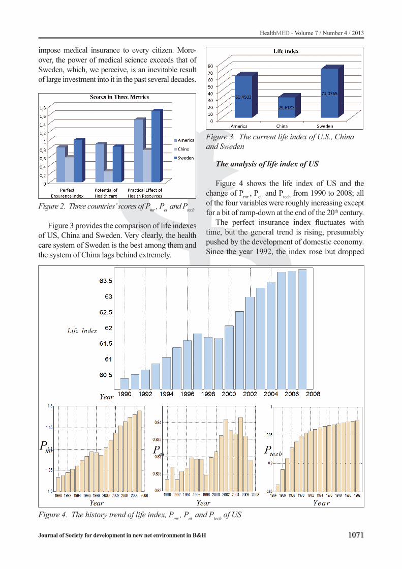
that are supported by sufficient data. Figure 2 shows the scores of America, China and Sweden in Pmr, Pei and Ptech. Note that the perfect ensurence index of Sweden is 1 (the largest possible value), because, the Sweden has a universal health care system that
HealthMED - Volume 7 / Number 4 / 2013
Journal of Society for development in new net environment in B&H 1071
impose medical insurance to every citizen. More-over, the power of medical science exceeds that of Sweden, which, we perceive, is an inevitable result of large investment into it in the past several decades.
Figure 2. Three countries’ scores of Pmr , Pei and Ptech
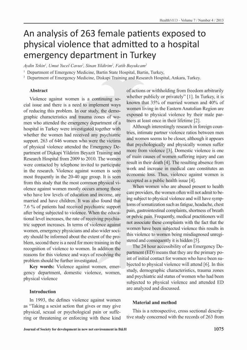
Figure 3 provides the comparison of life indexes of US, China and Sweden. Very clearly, the health care system of Sweden is the best among them and the system of China lags behind extremely.
Figure 3. The current life index of U.S., China and Sweden
The analysis of life index of US
Figure 4 shows the life index of US and the change of Pmr , Pei and Ptech from 1990 to 2008; all of the four variables were roughly increasing except for a bit of ramp-down at the end of the 20th century.
The perfect insurance index fluctuates with time, but the general trend is rising, presumably pushed by the development of domestic economy. Since the year 1992, the index rose but dropped
Figure 4. The history trend of life index, Pmr , Pei and Ptech of US

1072
HealthMED - Volume 7 / Number 4 / 2013
Journal of Society for development in new net environment in B&H
drastically in 1998, [14] which is a rather puzzling phenomenon. If we take a look back on history of America, we will find that in the 1993, President Clinton issued a new policy about medical insur-ance aiming at popularizing medical insurance so that every citizen is covered[16]; in 1998, [15] he declared that this policy was suddenly ceased be-cause of some reason. The data coincide with his-toric changes amazingly.
Figure 5. Revised spending plan of US by greedy algorithm
Analyses of Subordinate MetricsWith the most general metric life index, we are
able to evaluate and rank the health care systems in the world (e.g. the health care system of US is better than that of China but not as good as that of Sweden), but a considerable amount of informa-tion is lost or neglected at the same time, which will bring much difficulty in identifying exiting limitations and problems with these health care systems. In order to crack this, we pick out and re-arrange some factors to constitute new metrics as complementary metrics (potential of health care, matching degree, fairness degree, & luxury de-gree). With the help of these complementary me-trics, different aspects of one health care system can be evaluated and its limitations become de-tectable and predictable.
A low matching degree may suggest the ne-cessity of investing more money into the health care system so that its scale can be enlarged, whi-le a high one implies that the current health care system is massive enough considering the limited economy scale. Figure 7 implies that the health care system of U.S. should be stronger to match the massive scale of its economy [9]. If we want the matching degree of U.S. to be promoted to 1.66, the government must invest more money
into health care system [10]. The matching degree implies a slight lack of government investment into health care system [11].
Since it is difficult to obtain all the data to de-cide their precise quantities, we consider it to be feasible to substitute them with numbers of beds in hospitals in urban and rural areas [12, 13]. The information delivered by Figure 8 is clear: China did a very poor job in health care fairness while that of US could be better.
We consider luxury index to be tolerant of subtle conceptual ambiguity, because the slight lack of preciseness in defining concepts may wea-ken its competence in give an absolute evaluation, but still allows it to serve as a metric to compare different health care systems. (Note that the word ‘unnecessary’ doesn’t mean ‘redundant’.)
Figure 6. All the Subordinate metrics
It is never easy to give a complicated system properly and a precise evaluation [17], as the result is connected with multiple factors that are interwo-ven with each other [18]. However, if we establish a model that based on reasonable assumptions and tolerate a certain degree of ambiguity, satisfactory result could be achieved [19]. On the other hand, limitations of our model also mainly originate from the assumptions and ambiguity [20-23].
It is well admitted that few things are perfect in the world, whereas we never stop pursuing ideal health care systems, even though it takes a lot of money, manpower, time and energy to improve, because they are our safe guard that relieve our fear of diseases [24-31].

HealthMED - Volume 7 / Number 4 / 2013
Journal of Society for development in new net environment in B&H 1073
References
1. Perleth M, Jakubowski E, Busse R, What is ‘best prac-tice’ in health care? State of the art and perspectives in improving the effectiveness and efficiency of the Eu-ropean health care systems. Health Policy, 2001(56), 235-250
2. Ros CC, Groenewegen PP, Delnoij DM, All rights reserved, or can we just copy? Cost sharing arran-gements and characteristics of health care systems, Health Policy, 2000(52), 1-13
3. Abelson J, Miller FA, Giacomini M, What does it mean to trust a health system?: A qualitative study of Canadian health care values, Health Policy 2009(91), 63-70
4. Calnan M, Towards a conceptual framework of lay evaluation of health care, Social Science & Medicine, 1988(27), 927-933
5. Petersen I, Swartz L, Primary health care in the era of HIV/AIDS. Some implications for health systems reform, Social Science & Medicine, 2002(55), 1005-1013
6. Chang C, Zhang Y, Deng D, Xiao Y, A comprehensive evaluation model of health care system, Networking and Information Technology (ICNIT), 2010 Internati-onal Conference, 535 -539
7. K. Claxton The irrelevance of inference: a decision-making approach to the stochastic evaluation of he-alth care technologies, J Health Econ. 1999(18), PP. 341-64.
8. Yang T, Matthews SA, Understanding the non-sta-tionary associations between distrust of the health care system, health conditions, and self-rated health in the elderly: A geographically weighted regression approach, Health & Place, 2012(18), 576-585
9. Smith PC, Stepan A, Valdmanis V, Verheyen P, Princi-pal-agent problems in health care systems: an inter-national perspective, Health Policy. 1997(41), 37-60
10. Pons-Vigués M, Puigpinós-Riera R, Rodríguez D, Sanmamed M J., Pasarín MI, Pérez G, Borrell C, Casamitjana M, Benet J, Country of origin and pre-vention of breast cancer: Beliefs, knowledge and barriers, Health & Place, 2012(18), 1270-1281
11. Hollander MJ, Miller JA, Kadlec H, Evaluation of Healthcare Services: Asking the Right Questi-ons to Develop New Policy and Program-Relevant Knowledge for Decision-Making, Healthcare Quar-terly, 2010(4), 40-47
12. Oliveira MD et al, Modeling hospital costs to produ-ce evidence for policies that promote equity and effi-ciency, European Journal of Operational Research, 2008(16), 933-947
13. Rijsbergen MV et al. Managing the overflow of in-tensive care patients. European Journal of Operati-onal Research, 2008 (16), 988-1010
14. B.X. Qin, Bill Clinton’s health care reform. Ameri-can Research 1994, 7-8
15. Congressional Quarterly, Health Care’s Hour, 1993, 19-20
16. Congressional Quarterly, 1993, 2458-2459
17. Wang Q, Liu Y, Mo L, The evaluation and prediction of the effect of AIDS therapy, Proceeding of IEEE/ICME International Conference, 2007, 1591- 1596
18. Wang Q, Liu Y, Pan X, Atmosphere pollutants and mortality rate of respiratory diseases in Beijing, Sci-ence of the Total Environment, 2008(391), 143-148
19. Wang Q, Liu Y, Zhang B, Economic strategies in the issue of controlling AIDS, Proceeding of IEEE/ICME International Conference, 2007, 1601- 1608
20. Shmueli A, Israelis evaluate their health care system before and after the introduction of the national he-alth insurance law, Health Policy, 2003(63), 279-287
21. Kiil A, What characterises the privately insured in universal health care systems? A review of the empi-rical evidence, Health Policy, 2012(106), 60-75
22. Wensing M, Baker R, Szecsenyi J, Grol R, On behalf of the EUROPEP Group, Impact of national health care systems on patient evaluations of general prac-tice in Europe, Health Policy, 2004(68), 353-357
23. Gu Xing-Yuan, Tang Sheng-Lan, Reform of the Chinese health care financing system, Health Policy, 1995(32), 181-191
24. Yaesoubi R, Roberts SD, Payment contracts in a pre-ventive health care system: A perspective from Ope-rations Management, Journal of Health Economics, 2011(30), 1188-1196
25. Avgerinos ED, Koupidis SA, Filippou DK, Impact of the European Union enlargement on health pro-fessionals and health care systems, Health Policy, 2004(69), 403-408
26. Zu H, Wang Q, Dong M, Ma L, Yin L, Yang Y, Com-pressed Sensing Based Fixed-Point DCT Image En-coding, Advances in Computational Mathematics and its Applications, 2012(2), 237-240

1074
HealthMED - Volume 7 / Number 4 / 2013
Journal of Society for development in new net environment in B&H
27. Wang Q, Li M, Xia LC, Wen G, Zu H, Gao M(2013), Genetic Analysis about Differentiation of Helper T Lymphocytes, Genetics and Molecular Research, in press
28. Xia L, Zhou C, Phase transition in sequence unique reconstruction, Journal of Systems Science and Complexity 2007(20), 18-29
29. Zhang SW, Li YJ, Xia L, Pan Q, PPLook: an automa-ted data mining tool for protein-protein interaction, BMC bioinformatics 2011(11), 326
30. He PA, Xia L, Oligonucleotide profiling for discri-minating bacteria in bacterial communities, Com-binatorial Chemistry & High Throughput Screening 2007(10), 247-255
31. Steele JA, Countway PD, Xia L, et al., Marine bacte-rial, archaeal and protistan association networks re-veal ecological linkages, The ISME Journal 2011(5), 1414-1425.
Corresponding AuthorQixin Wang,Department of Mathematics, University of Southern California,Los Angeles,United States of America,E-mail: [email protected]
HealthMED - Volume 7 / Number 4 / 2013
Journal of Society for development in new net environment in B&H 1075
Abstract
Violence against women is a continuing so-cial issue and there is a need to implement ways of reducing this problem. In our study, the demo-graphic characteristics and trauma zones of wo-men who attended the emergency department of a hospital in Turkey were investigated together with whether the women had received any psychiatric support. 263 of 646 women who were the victims of physical violence attended the Emergency De-partment of Dişkapi Yildirim Beyazit Training and Research Hospital from 2009 to 2010. The women were contacted by telephone invited to participate in the research. Violence against women is seen most frequently in the 20-40 age group. It is seen from this study that the most common physical vi-olence against women mostly occurs among those who have low levels of education and income, are married and have children. It was also found that 7.6 % of patients had received psychiatric support after being subjected to violence. When the educa-tional level increases, the rate of receiving psychia-tric support increases. In terms of violence against women, emergency physicians and also wider soci-ety should be informed about the extent of the pro-blem, second there is a need for more training in the recognition of violence to women. In addition the reasons for this violence and ways of resolving the problem should be further investigated.
Key words: Violence against women, emer-gency department, domestic violence, women, physical violence
Introduction
In 1993, the defines violence against women as “Taking a sexist action that gives or may give physical, sexual or psychological pain or suffe-ring or threatening or enforcing with these kind
of actions or withholding from freedom arbitrarily whether publicly or privately” [1]. In Turkey, it is known that 35% of married women and 40% of women living in the Eastern Anatolian Region are exposed to physical violence by their male par-tners at least once in their lifetime [2].
Although interestingly research in foreign coun-tries, intimate partner violence ratios between men and women seems to be closer, although it appears that psychologically and physically women suffer more from violence [3]. Domestic violence is one of main causes of women suffering injury and can result in their death [4]. The resulting absence from work and increase in medical care constitutes an economic loss. Thus, violence against women is accepted as a public health issue [4].
When women who are abused present to health care providers, the women often will not admit to be-ing subject to physical violence and will have symp-toms of somatization such as fatigue, headache, chest pain, gastrointestinal complaints, shortness of breath or pelvic pain. Frequently, medical practitioners will not associate these complaints with the fact that the women have been subjected violence this results in this violence to women being misdiagnosed unregi-stered and consequently it is hidden [5].
The 24 hour accessibility of an Emergency De-partment (ED) means that they are the primary po-int of initial contact for women who have been su-bjected to physical violence will attend [6]. In this study, demographic characteristics, trauma zones and psychiatric aid status of women who had been subjected to physical violence and attended ED are analyzed and discussed.
Material and method
This is a retrospective, cross sectional descrip-tive study concerned with the records of 263 from
An analysis of 263 female patients exposed to physical violence that admitted to a hospital emergency department in Turkey Aydin Tekin1, Umut Yucel Cavus2, Sinan Yildirim2, Fatih Buyukcam2
1 Department of Emergency Medicine, Bartin State Hospital, Bartin, Turkey,2 Department of Emergency Medicine, Diskapi Training and Research Hospital, Ankara, Turkey.

1076
HealthMED - Volume 7 / Number 4 / 2013
Journal of Society for development in new net environment in B&H
a total of 646 women who were the victims of physical violence and attended the ED of Dişkapi Yildirim Beyazit Training and Research Hospital from 2009 to 2010. After an attempt to contact all 646 women by telephone 263 women over the age of 18 volunteered to participate in this research. Approval from the ethics committee was obtained.
The following characteristics were investiga-ted. The trauma regions and whether the women had received psychiatric help. The income levels of women were also analyzed by used the classi-fication of income levels distribution in Turkey. According to this classification, women were di-vided into the following 5 income groups; very low – low –moderate – good and very good [7].
The Statistical Package for Social Science (SPSS) 17.0 for Windows package program was used for data analysis. The continuous variables were; mean, median and standard deviation, the median and mode ordinal variables and the num-ber and nominal variables were expressed as per-centages. The continuous variables were evaluated using a histogram and a One-Sample Kolmogorov-Smirnov Test. P > 0.05 was considered as a normal distribution. The relationship between nominal va-riables was evaluated using a Pearson Chi–Square Test and Fisher’s Exact Test and p > 0.05 was con-sidered as meaningful.
Results
Patients were grouped according to their ages; 79 patients (30.1%) were under the age of 30, 85 (32.3%) were between the ages of 30-40 and 43 (16.3%) between the ages of 40-50 , and 39 (14.8%) 50 -60 years of age , and 17 (6.5%) were over the age of 60. The age distribution of the pa-tients is shown in Figure 1.
The mean age of the patients was 35 ± 13.3 and the median age 35 years (age range: 18-84). Of these patients, 148 (56.30%) were married, 63 (24.00%) unmarried, 52 (19.80%) were divorced.
Of the women subjected to violence 79 (30.00%) did not have children, 184 (70%) patients had one child or more. 89 (33.80%) women worked. 174 (66.20%) patients did not work. 79 (30.00%) pa-tients had no social security. The distribution rati-os of the women who had social security was: 115 (43.70%) patients were in the Social Insurance In-
stitution scheme (SSK), 11 (4.20%) were part of the Bag-Kur scheme, 25 (9:50%) had a green card (A type of social security for lower income levels of population in Turkey), 33 (12.50%) had standard state health care.
Figure 1. The age distribution of the women exposed to physical violence
91 (34.60%) of the women had a very low income, 108 (41.10%) were in the low income group, 48 (18:30%) had a moderate income, 16 (6.00%) patients had a good income. None of the families there was not in the group of very good income level. In terms of education levels, 21 (8.00%) women were illiterate, 52 (19.80%) wo-men had completed primary schools, 86 (32.70%) had completed secondary education, 82 (31.20%) had completed high school, 22 (8.40%) were uni-versity graduates. 217 of the women (82.50%) had been subjected to violence once, 46 (17:50%) had been exposed to violence more than once.
The perpetrator of the violence was as follows: 96 (36.50%) women had been attacked by the wife, 66 (25.10%) by their relatives, 60 (22.80%) by colleagues and 41 (15.60%) women had been su-bject to violence by an unknown person. According to the perpetrator of the violence, the distribution of educational status of patients shown in Figure 2.
The distribution of parts of the body of the wo-men received a physical assault most commonly isolated head and neck trauma (99 patients, 37.60 %) were present (Figure 3).
Only medical treatment was given the women for physical trauma, although 20 (7.6%) women had received psychiatric help after the assault. The educational background of these women was as follows; 5 (25%) had completed primary scho-

HealthMED - Volume 7 / Number 4 / 2013
Journal of Society for development in new net environment in B&H 1077
ol, 15 (75%) graduated from high school or uni-versity. The relationship between the educational status of the patients not wanting psychiatric assi-stance is shown in Figure 4.
Figure 2. Education levels of the women exposed to physical violence
Figure 3. The effected body region of the women exposed to physical violence
Figure 4. Psychiatric support condition according to education level
Discussion
Approximately one fifth of women attend EDs because of domestic violence. According to studies in Turkey , one in three women experience physical violence at the hands of their husband at least once in their lives [2]. The cause of the trauma to women presenting to the ED are often not attributed to do-
mestic violence by the medical staff. To achieve the correct diagnosis and offer appropriate treatment to women victims of physical violence, it is necessary to take a holistic approach considering not only the signs of physical trauma but also use the details from the forensic report and elicit information from the patient [8]. In addition to the physical damage of physical violence, it can increase the risk of other diseases in the long term. Even if women who have experienced physical violence often do not want to or cannot talk about it, every woman that presents with an injury to a medical institution should be considered as a victim of domestic violence until proven otherwise [6].
According to the results of 48 studies across the world; 10-69 % of women experienced violence by their spouses or partners [2]. In our study, 646 of 2459 patients were women who attended the ED because of physical violence . 56.3% of pati-ents who were subjected to violence were married and 64.9% of the women who were married had been beaten by her husband (Figure 2).
In studies shows that women’s educational level is not a factor that determines whether women are the victims of violence, but shows that the educated women are more successful for the break away from violence. In Turkey and almost all countries, women who have lower levels of education are exposed to more domestic violence [9]. 61.4% of the women in the current study only have primary or lower educa-tion levels while 39.6% of the women had gradua-ted from high school or university (Figure 2).
Women who are subject to violence can develop many mental disorders such as; post-traumatic stre-ss, depression, suicide attempts, depression, anxiety, mood and eating disorders, alcohol and drug de-pendence, antisocial personality disorders, psycho-sis and aggressive behavior towards the children [3, 10]. 50% of female psychiatric patients have a history of violence and 25% of suicide attempts in women there is a history of assault [11]. The fetus of pregnant women who victims of violence are also likely to be injured [3]. This damage may be happen if the woman has received a blow directly to the ab-dominal region, also may be happen indirectly with other body regions trauma [3]. In a meta-analysis study, women who are exposed to violence during pregnancy have a significantly higher probability of giving birth to low birth weight infants [12]. In
1078
HealthMED - Volume 7 / Number 4 / 2013
Journal of Society for development in new net environment in B&H
our study, 7.6% of the patients received psychiatric support. Those women with a higher level of edu-cation tend to receive psychiatric support more than the women with a lower level of education (Figure 4). In the literature, women exposed to violence had a low level of education and were in a low socio-economic group and it is interesting to note that they did have not any income and didn’t work [13-14]. In our study 199 of the women (75.70%) had a low and very low income, 64 (24.30%) had moderate or high income levels. Our findings were compatible with the literature.
The approach of health workers in relation to vi-olence against women, the diagnosis of violence, treatment and after care are very important. Ramsey et al stated that there was not enough evidence re-garding the efficacy of medical interventions in women victims of violence [15]. In our study, after the first examination none of the doctors referred patients for psychiatric assessment. This negligence can cause that the recurrence of violence and the same woman can attend the ED many times and each event can be worse than the previous one and a possibly reaching such an extreme situation that the woman may attempt suicide. In this context, in medical training programs, diagnosis, treatment and psychological support should not be neglected. Also the patient data must be collected in more de-tail. Furthermore, there is a need for an increased number of studies on this topic.
Conclusion
The staff of an ED are not only charged with provide medical intervention but they should also ensure that the women victims of physical violence are offered the appropriate psychiatric support. Re-porting of cases of violence against women, as well as judicial directions in this regard together with the creation of a database, women victims, institutions that provide support for women and that the women can be directed to receive psychological support, are as important as emergency medical assistance.
References
1. United Nations Declaration on the Elimination of Vi-olence Against Women. Available from: www.un.org/documents/ga/res/48/a48r104.htm.
2. Altinay, A.G. and Y. Arat, Türkiye’de kadina yönelik şiddet. 2007, İstanbul: Punto.
3. Wathen, C.N. and H.L. MacMillan, Interventions for violence against women: scientific review. JAMA, 2003. 289(5): p. 589-600.
4. Edwards, T.A., et al., Stages of change as a correla-te of mental health symptoms in abused, low-inco-me African American women. J Clin Psychol, 2006. 62(12): p. 1531-43.
5. Fincanci, Ş.K., Kadina Yönelik Şiddete Adli Tip Açi-sindan Yaklaşim, in Kadina Yönelik Şiddet ve Hekim Sempozyumu. 2003.
6. Akin, A., Kadina Yönelik Aile İçi Şiddetin Kadin Sağliğina Etkileri:Kadina Yönelik Aile İçi Şiddetle Mücadelede Sağlik Hizmetleri. 2008, TC Başbakanlik Kadinin Statüsü Genel Müdürlüğü Yayini: Ankara.
7. Aydin, K., Türkiye’de Kişisel Gelir Dağiliminin So-syoekonomik ve Demografik Belirleyicileri. Çalişma ve Toplum, 2012. 1.
8. Acil Serviste Kadina Yönelik Şiddetin Tani-Tedavi ve Yönlendirilmesi. 2th ed. 2009, Ankara: Türk Tabipleri Birliği Merkez Konseyi Kadin Hekim ve Kadin Sağliği Kolu.
9. Balci, Y.G. and U. Ayranci, Physical violence against women: evaluation of women assaulted by spouses. J Clin Forensic Med, 2005. 12(5): p. 258-63.
10. Kaya, M. and B. Kaya, Kadina Yönelik Şiddet; Pan-doranin Kirik Kutusu. Sağlik Toplum Siyaset, 2000. 3: p. 50-53.
11. Noel, N.L. and M. Yam, Domestic violence. The pre-gnant battered women. Nurs Clin North Am, 1992. 27(4): p. 871-84.
12. Murphy, C.C., et al., Abuse: a risk factor for low birth weight? A systematic review and meta-analysis. CMAJ, 2001. 164(11): p. 1567-72.
13. Günay, T., et al., İzmir’de bir gecekondu bölgesin-de kadina yönelik aile içi şiddet. Sağlik ve Toplum, 2006: p. 31-37.
14. Turhan, E., A. Güraksin, and T. İnandi, Erzurum’da kadina yönelik aile içi şiddet. Sağlik ve Toplum 2006. 16: p. 24-30.
15. Ramsay, J., et al., Should health professionals scre-en women for domestic violence? Systematic review. BMJ, 2002. 325(7359): p. 314.
Corresponding AuthorUmut Yucel Cavus,Diskapi Yildirim Beyazit Trainig and Research Hospital, Department of Emergency Medicine,Ankara,Turkey,E-mail: [email protected]

HealthMED - Volume 7 / Number 4 / 2013
Journal of Society for development in new net environment in B&H 1079
Abstract
Objective: Patient anesthesia management re-mains a matter of debate. Therefore, we aim to pre-sent the anesthesia practice on patients in our clinic.
Method: The medical records of 99 patients with the diagnosis of eclampsia that were admini-stered anesthesia due to caesarean section betwe-en the dates of September 2005 and September 2010 in Dicle University Hospital were retrospec-tively analyzed. The patients were classified into two groups: patients that were administered spinal anesthesia (group S), and patients that were admi-nistered general anesthesia (group G).
Results: The study included 38 patients that were administered spinal anesthesia and 61 pa-tients that were administered general anesthesia. HELLP syndrome coincided in 12 patients that were given spinal anesthesia and in 33 patients that were given general anesthesia. Spinal anesthesia was performed on patients that had low Hb, Htc, platelet counts and ALT levels preoperatively. The data showed that the general anesthesia group had more blood transfusions than the spinal anesthe-sia group (p=0.01). In group S, only one patient experienced a complication, whereas in group G, 15 patients experienced complications (p=0.004). In group S nine patients had transfusions, while in group G, 30 patients had transfusions (p=0.019). One patient in group S as well as four patients in group G died postoperatively.
Conclusion: Anesthesia management should be tailored to the patients’ condition and laboratory re-sults. Additionally, a regional anesthetic approach should be preferred unless there is a contraindication.
Key words: Eclampsia, spinal anesthesia, ge-neral anesthesia.
Introduction
Hypertensive disorders are seen in 3-8% of pregnancies and are one of the major causes of maternal mortality and morbidity (1,2). Eclamp-
sia is among the hypertensive disorders, and it is a life threatening condition, with an incidence that varies within a range of 1/3448 and 1/100. It typi-cally occurs during the third trimester, especially before 32nd gestational week (3). Major mater-nal complications including hemolysis-elevated liver enzymes and lower platelet count (HELLP) syndrome, placental abruption, disseminated in-travascular coagulation, pulmonary aspiration, pulmonary edema, acute renal failure and cardi-opulmonary arrest may occur in these patients as well (4, 5). Anesthesia management is a challenge for these patients; hemodynamic instability, men-tal status and coagulation dysfunction make an anesthetic decision controversial. Ideal anesthe-tic methods for these patients will not deteriorate organ function and will have a minimal effect on hemodynamic parameters (6). This study aims to compare general anesthesia (GA) and spinal ane-sthesia in patients diagnosed with eclampsia.
Method
After the consent of the Ethics Committee, the records of 99 patients’ that were diagnosed at Dicle University between September-2005 and Septem-ber-2010 were gathered. The exclusion criteria included hypertension and proteinuria diagnosed before the 20th gestational week; renal, hematolo-gic and cardiac diseases causing proteinuria, hyper-tension, multiparous women, and patients with he-matologic or other diseases with increased hepatic enzyme levels were also excluded. Patients were classified into two groups: patients that were admi-nistered general anesthesia (group G), and patients that were administered spinal anesthesia (group S).
The ages of the patients, gravid, parity, gestation-al week at admission, and length of stay were re-corded. Preoperative laboratory results, and postop-erative features and complications were also record-ed. Additional disorders (HELLP) and any drugs ad-ministered preoperatively were also recorded. The
Anesthetic management of Eclampsia/hellpFeyzi Celik1, M. Erdal Sak2, Adnan Tufek1, Abdulmenap Guzel1
1 Dicle University, Faculty of Medicine, Anesthesiology and Reanimation Department, Diyarbakir, Turkey,2 Dicle University, Faculty of Medicine, Obstetrics and Gynecology Department, Diyarbakir, Turkey.
1080
HealthMED - Volume 7 / Number 4 / 2013
Journal of Society for development in new net environment in B&H
postoperative lab values, vital signs, postoperative 1st and 5th minute APGAR scores of the fetus, and all blood and blood products that were given during the operation were noted as well. A 6 gram loading dose of magnesium sulfate was administered to all patients, followed by a 2 gram maintenance dose 20 minutes later. Postoperative complications and mor-tality rates were also recorded. Postoperative com-plications are listed in Table 4.
Statistical analysisStatistical analysis was performed by SPSS for
Windows 15.0 (SPSS Inc., Chicago, IL, USA). Data were presented as mean values ± standard deviation for continuous variables. P values of less than 0.05 were considered significant. Means and standard deviations (SD) were calculated for continuous variables and subject characteristics and demographics were analyzed descriptively. The normal distribution of the variables was ana-lyzed by the Kolmogorov–Smirnov test. The Chi- square test and the Student’s t test were used to evaluate associations between the categorical and continuous variables. All variables were included in the backward stepwise procedure.
Results
Group S contained 38 patients while Group G contained 61 patients. Groups were comparable in age, gravid, parity and gestational age at the time of admission. HELLP syndrome was demonstrat-ed in 12 patients in Group S and 33 patients in Group G (Table 1).
Group S had higher levels of Hb, Hct and ALT levels compared to Group G (p>0.01). Group S had a higher platelet count than Group G but it was not statistically significant. Other preopera-tive parameters were similar (Table 2).
APGAR scores of neonates were similar in the two groups at the 1st and 5th minutes. Arterial blo-od pressures (both systolic and diastolic) were also similar. Group G had more transfusions compared to Group S (p=0.01). In Group S, nine patients had transfusions of blood products, while 30 patients in Group G had transfusions (p=0.019). Moreover, total blood transfusion was higher in Group G (Ta-ble 3). Postoperatively, only one patient in Group S experienced a complication, whereas 15 patients
in Group G experienced complications (p=0.004). During the postoperative period, one patient died from group S; in group G, 4 patients died.
Discussion
Eclampsia is one of the leading causes of perina-tal mortality and morbidity in developing countries. Eclampsia causes significant physiologic changes during pregnancy. Caesarean section is required in 11-57% of these cases (7), however the patients are hemodynamically unstable and a possibility of multiple organ failure and insufficient intravascular volume is present; accordingly, the correct type of anesthesia to be utilized is still a matter of debate (6). Additionally, if HELLP syndrome is seen in pa-tients with eclampsia, the decision on which type of anesthesia to use becomes even more challenging (6,8). General anesthesia (GA) seems preferable among patients suffering from coagulation disor-ders and fetal distress. However, several studies have observed that GA may have several disadvan-tages. Laryngoscopy while under GA may trigger a hemodynamic response which may provoke in-tracerebral bleeding and pulmonary complications, in addition to possibly increasing the risk of pro-longed intubation and aspiration of gastric contents. General anesthesia may also delay recovery after anesthesia (6, 9, 10, 11).
The use of spinal anesthesia (SA) during coa-gulation disorders and thrombocytopenia is con-troversial. Severe hypotension may also result from this technique due to high motor block. Sen-sitivity against vasopressors used for hypotension can also increase with spinal anesthesia (12). Ne-vertheless, if no complication exists, these patients will benefit from regional anesthesia. Greater con-trol on hemodynamic stability may be provided by spinal anesthesia (13, 14, 15). Several studies have shown the safety of single dose spinal anesthesia (16, 17, 18, 19). In our study, patients without absolute contraindications for regional anesthe-sia benefited from SA, while patients with severe bleeding disorders benefited most from GA.
A review of the literature shows that APGAR scores were better in spinal anesthesia groups when compared to patients with general anesthe-sia (10, 20, 21, 22, 23). In our study, APGAR sco-res in both groups were comparable (Table 3).

HealthMED - Volume 7 / Number 4 / 2013
Journal of Society for development in new net environment in B&H 1081
Table 1. Demographic Data Group S (n=38) Group G (n=61) p
Age 31.63±6.81 31.82±7.46 0.900Gravida 3.84±3.15 3.59±3.08 0.696Parite 2.42±2.78 2.57±2.88 0.796Gestational Week of Hospitalization 32.84±3.68 33.39±2.82 0.404Total length of stay 6.63±2.99 6.64±3.79 0.991Eclampsia + HELLP n (%) 12(31.6%) 33(54.10%) 0.038
Values are given as mean ± standard deviation. n: number
Table 2. Preoperative Laboratory ValuesGroup S (n=38) Group G (n=61) p
Hct 35.37±7.79 32.44±6.30 0.043*Hb 12.16±2.60 10.85±2.30 0.011*Plt 166.87±93.32 131.31±115.18 0.112ALT 67.68±84.547 157.54±247.470 0.036*AST 132.27±180.920 204.80±256.241 0.135LDH 918.43±1074.639 1077.17±819.467 0.420Bilirubin 3.98±16.047 6.99±40.588 0.677Blood sugar 120.46±54.100 116.70±36.675 0.684Urea 27.41±14.532 36.68±35.003 0.130Creatinine 0.82±0.493 1.12±1.205 0.147Proteinuria 416.43±157.057 396.43±174.540 0.582Number of convulsions 1.61±0.887 1.85±0.980 0.209Mean systolic AT 163.16±9.893 164.59±12.052 0.540Mean diastolic AT 100.53±8.366 101.97±9.259 0.454
Values are given as mean ±standard deviation. n: number
Table 3. Postoperative Laboratory ValuesGroup S (n=38) Group G (n=61) p
Fetus 1. min APGAR 4.34±2.197 3.73±1.745 0.132Fetus 5. min APGAR 6.13±2.451 5.48±2.103 0.167Mean systolic AT 129.74±9.722 131.80±10.410 0.327Mean diastolic AT 81.32±6.646 82.62±7.938 0.399RBC Transfuse 1.71±1.113 2.78±2.225 0.233TDP Transfusion 2.00±0.00 3.24±2.625 0.431Platelet Suspension 2.00±0.00 2.57±1.718 0.766Platelet Apheresis Absent 1.62±1.193 absentTotal amount of Transfusion (U) 0.53±1.41 2.97±5.05 0.01*
Values are given as mean ±standard deviation. AT: Arterial tension U: Unit min: minute
Table 4. ComplicationsGroup S (n) Group G (n)
AKF 1 4Shortness of breath - 2Sepsis - 1AKF+ICB - 2Shortness of breath + ABY - 4Intra abdominal bleeding - 2
AKF: acute kidney failure ICB; intra cranial bleeding. n: number
1082
HealthMED - Volume 7 / Number 4 / 2013
Journal of Society for development in new net environment in B&H
General anesthesia may influence uterine vascu-lar resistance or perfusion pressures during caesa-rean sections thereby altering blood flow and indi-rectly altering uterine contractions; ultimately, this may lead to an increase in blood loss (24). Afolabi et al compared spinal and general anesthesia tech-niques in patients that underwent caesarean section and found that blood loss was lower in those that were given spinal anesthesia (25). Another study shows that general anesthesia is correlated with more postoperative blood loss and decreased Htc levels when compared to regional anesthesia in ca-esarean sections (26). Similarly in our study, Group G had a greater need for blood transfusions during the postoperative period (Table 3).
Patients with eclampsia may be faced with a number of complications ranging from intracranial bleeding, renal and pulmonary impairment to cardi-opulmonary arrest (4, 27). In a study conducted by Singh et al, they found that pregnant women with stable eclampsia and without major complications could use spinal anesthesia to avoid possible com-plications of general anesthesia (6). In our study, there were no significant intraoperative complicati-ons in either group, but Group G had more postope-rative complications.
Several studies have shown that 7-36% percent of eclampsia cases that occur in conjunction with HELLP syndrome may also have acute renal fa-ilure (28, 29, 30). In our study, 11.1% of patient had acute renal failure. One patient (2.6%) and 10 patients (16.4%) had acute renal failure in Groups S and G, respectively. We think that this may be because there are more patients with HELLP in the GA group (n=33; 54.10%) when compared to the SA group (n=12, 31.6%) (p=0.038).
Vigil De Garcia et al found that in developing countries the maternal mortality rate due to ec-lampsia is 9.4%. In our study, the maternal morta-lity rate was 5.05% (31).
Several limitations of the study include not ran-domizing patient selection, as well as not optimi-zing the anesthesia method individually.
Conclusion
GA should be avoided in eclamptic pregnant women according to their condition and lab re-sults. Spinal anesthesia may be a safer method in the absence of absolute contraindications to re-gional anesthesia.
References
1. Geographic variation in the incidence of hypertension in pregnancy. World Health Organization Internation-al Collaborative Study of Hypertensive Disorders in Pregnancy. Am J Obstet Gynecol 1988; 158: 80-83.
2. Roberts JM, Cooper DW. Pathogenesis and genetics of pre-eclampsia. Lancet 2001; 357: 53-56.
3. Gambling D. Principles and Practice: Hypertensive disorders: obstetric anesthesia. Philadelphia: Else-vier; 2004.
4. Barton JR, Sibai BM. Cerebral pathology in eclamp-sia. Clin Perinatol 1991; 18: 891-910.
5. Kaplan PW, Repke JT. Eclampsia. Neurol Clin 1994; 12: 565-582.
6. Singh R, Kumar N, Jain A, Chakraborty M. Spinal anesthesia for lower segment Cesarean section in pa-tients with stable eclampsia. Journal of Clinical Anes-thesia 2011; 23: 202–206.
7. Didley GA, Cotton DB, Phelan JP. Critical Care Obstetrics: Complications of PIH. 2nd ed. Oxford: Blackwell, 1991.
8. Graciaa PV, Silvab S, Montufara C, Carrolb I, Riosb SL. Anesthesia in pregnant women with HELLP syn-drome. International Journal of Gynecology & Ob-stetrics 2001; 74: 23-27.
9. Yuen TS, Kua JS, Tan IK. Spinal haematoma follow-ing epidural anaesthesia in a patient with eclampsia. Anaesthesia 1999; 54: 350-354.
10. Ramanathan J, Coleman P, Sibai B. Anesthetic modification for hemodynamic and neuroendocrine response to cesarean section delivery in severe pre-eclampsia. Anesth Analg 1991; 73: 772-779.
11. Lavies NG, Meiklejohn BH, May AE, Achola KJ, Fell D. Hypertensive and catecholamine response to tra-cheal intubation in patients with pregnancy-induced hypertension. Br J Anaesth 1989; 63: 429-434.
12. Talledo OE, Chesley LC, Zuspan FP. Renin-angio-tensin system in normal and toxemic pregnancies.

HealthMED - Volume 7 / Number 4 / 2013
Journal of Society for development in new net environment in B&H 1083
III. Differential sensitivity to angiotensin II and nor-epinephrine in toxemia of pregnancy. Am J Obstet Gynecol 1968; 100: 218-221.
13. Gogarten W. Preeclampsia and anaesthesia. Curr Opin Anaesthesiol 2009; 22: 347-351.
14. Öz H, Akkor A, Aykaç B, Sun S. Preeclampsia-eclampsia, anesthesia and intensive care.. Perinatal Journal 1993; 1: 50-54.
15. Mandal NG, Surapaneni S. Regional anaesthesia in pre-eclampsia: advantages and disadvantages. Drugs 2004; 64: 223-236.
16. Botfield C, Howell P. The use of spinal anaesthesia in severe preeclampsia. Fetal Maternal Med Rev 2001; 12: 67-79.
17. Visalyaputra S, Rodanant O, Somboonviboon W, Tantivitayatan K, Thienthong S, Saengchote W. Spi-nal versus epidural anesthesia for Cesarean delivery in severe preeclampsia: a prospective randomized, multicenter study. Anesth Analg 2005; 101: 862-868.
18. Dyer RA, Piercy JL, Reed AR. The role of the an-aesthetist in the management of the pre-eclamptic patient. Curr Opin Anaesthesiol 2007; 20: 168-174.
19. Santos AC. Spinal anesthesia in severely preeclamp-tic women: when is it safe? Anesthesiology 1999; 90: 1252-1254.
20. O’Brien JM, Shumate SA, Satchwell SL. Maternal benefit of corticosteroid therapy in patients with HELLP (hemolysis, elevated liver enzymes, and low platelet count) syndrome: Impact on the rate of re-gional anesthesia. Am J Obstet Gynecol 2002; 186: 507-522.
21. Moodley J, Jjuuko G, Rout C. Epidural compared with general anaesthesia for caesarean delivery in conscious women with eclampsia. BJOG 2001; 10: 378-382.
22. Okafor UV, Efetie ER, Igwe W, Okezie O. Anaes-thetic management of patients with pre-eclampsia/eclampsia and perinatal outcome. J Matern Fetal Neonatal Med 2009; 22: 688-692.
23. Krishnan L, Gunasekaran N, Bhaskaranand N. An-esthesia for cesarean section and immediate neona-tal outcome. Indian J Pediatr 1995; 62: 219-223.
24. Yalınkaya A, Güzel Aİ, Kangal K, Uysal E, Erdem S. Comparing the blood values of the patients operated by cesarean under spinal and general anesthesia. Perinatal Journal 2009; 17(2): 70-73.
25. Afolabi BB, Lesi FE, Merah NA.Regional versus general anaesthesia for caesarean section.Cochrane Database Syst Rev 2006;4: CD004350.
26. Lertakyamanee J, Chinachoti T, Tritrakarn T, Muangkasem J, Somboonnanonda A, Kolatat T.Comparison of general and regional anesthesia for cesarean section: success rate, blood loss and satisfaction from a randomized trial.J Med Assoc Thai 1999; 82: 672-680.
27. Kaplan PW, Repke JT. Eclampsia. Neurol Clin 1994; 12: 565-582.
28. Celik C, Gezginc K, Altintepe L, Tonbul HZ, Yaman ST, Akyurek C, Turk S: Results of the pregnancies with HELLP syndrome. Ren Fail 2003, 25: 613-618.
29. Cavkaytar S, Ugurlu EN, Karaer A, Tapisiz OL, Danisman N: Are clinical symptoms more predictive than laboratory parameters for adverse maternal outcome in HELLP syndrome? Acta Obstet Gynecol Scand 2007, 86: 648-651.
30. Gul A, Aslan H, Cebeci A, Polat I, Ulusoy S, Ceylan Y: Maternal and fetal outcomes in HELLP syndrome complicated with acute renal failure. Ren Fail 2004, 26: 557-562.
31. Gracia PV. Maternal deaths due to eclampsia and Hellp syndrome. Int J Gynaecol Obstet 2009; 104: 90–94.
CorrespondingAuthorFeyzi Celik,Dicle University,Faculty of Medicine,Anesthesiology and Reanimation Department,Diyarbakir,Turkey,E-mail: [email protected]

1084
HealthMED - Volume 7 / Number 4 / 2013
Journal of Society for development in new net environment in B&H
Abstract
Multiple sclerosis (MS) is a chronic autoimmu-ne, multifactorial and complex genetic disease due to demyelination of the central nervous system. It is believed that HLA and cytokines genes are involved in the pathogenesis of MS. Previously, we showed that HLA-DRB1*1501 allele and −330 T/T IL-2 genotype had most susceptibility effect on Iranian MS patients. In this study, we evaluated the influen-ce of susceptible DRB1*1501 allele and -330 IL-2 polymorphism on age at onset of MS. Our results indicated that individuals with T/T genotype (23.8±5 vs. 34.4±10 years, p<0.0001) and HLA-DRB1*1501 allele (21.3±3 vs. 26.6±3 years, p<0.0001) had signi-ficantly lower age at onset of disease than controls. The -330 T/T IL-2- HLA DRB1*1501 haplotype showed the highest significant association to young age at onset. The 330 T/T- IL-2- HLA DRB1*1501 haplotype had older age at onset of disease compare to 330 T/T IL-2- HLA DRB1*1501- haplotype, but it was not significant. However, we have provided evi-dence of an interaction between HLA-DRB1*1501 allele and the -330 T/T IL-2 genotype in age at onset of MS. But additional Studies on large sample size maybe discover the fact of these gene interactions in age of disease onset.
Key words: IL-2, polymorphism, HLA-DRB, age at onset, Multiple sclerosis.
Introduction
Multiple sclerosis (MS) is a chronic neuro-inflammatory and autoimmune disease due to demyelination of the central nervous system. It is believed to be initiated and mediated by autoreac-tive T cells directed against myelin antigens. Both genetic and environmental factors are contributed in disease risk (1). For a long time, the only HLA-
DRB1 region was known as susceptible genetic factor to MS, and allelic interactions of the HLA-DRB1 locus had been believed (2).
Interleukin 2 was identified as pro and anti in-flammatory factor .Interleukin 2 was initially found as an autocrine secretory product from activated T cells with growth factor properties. Later, it was fo-und that IL-2 elicited T-cell proliferation, survival and differentiation of effectors. The interleukin-2 gene was strong functional candidate which invol-ved in immune regulation and function. The main nonredundant role of IL-2 was the maintenance of peripheral T-cell tolerance; it had an important role in regulatory T-cell (Treg) homeostasis. The impa-irment of Treg cells was believed to be the cause of autoimmunity in the absence of IL-2 (3, 4).
Interleukin-2 (IL-2) is a cytokine involve in the function and regulation of immune system. IL-2 gene was located on chromosome 4q21. Many studies implicated that interleukin-2 (IL-2) played an effective role in the pathology of MS (5-11). Levels of IL-2 increased in the cerebrospinal fluid (CSF) and sera of MS patients (12).
Interleukin 2 was identified as pro and anti in-flammatory factor .Interleukin 2 was initially fo-und as an autocrine secretory product from acti-vated T cells with growth factor properties. Later, it was found that IL-2 elicited T-cell proliferation, survival and differentiation of effectors. The in-terleukin-2 gene was strong functional candidate which involved in immune regulation and func-tion. The main nonredundant role of IL-2 was the maintenance of peripheral T-cell tolerance; it had an important role in regulatory T-cell (Treg) homeostasis. The impairment of Treg cells was believed to be the cause of autoimmunity in the absence of IL-2 (3, 4). IL-2 gene was located on chromosome 4q21.
The influence of -330 IL-2 gene polymorphism and HLA-DRB1*1501 allele on age at onset in Iranian multiple sclerosis patientsArezou Sayad1, Abdolamir Alameh2, Aida Sayad2
1 Department of Medical Genetics, Faculty of Medical Science, Tarbiat Modares University, Tehran, Iran,2 Department of Biochemistry, Faculty of Medical Science, Tarbiat Modares University, Tehran, Iran.

HealthMED - Volume 7 / Number 4 / 2013
Journal of Society for development in new net environment in B&H 1085
John and colleagues identified -330 Interle-ukin-2 promoter single nucleotide polymorphism (13). Matesanz et al. showed that both of -330 G/T and T/T genotypes of IL-2 gene have an associati-on with susceptibility to secondary progressive (SP) course of MS (10). First study on Iranian populati-on identified that −330 G/G IL-2 genotype was si-gnificantly more frequent in MS patient group than controls (14). On the contrary, next study was found that there is not significantly difference between MS individuals and healthy controls (15). Our previous study indicated that −330 T/T genotype and HLA-DRB1*1501 allele were significantly more frequent in MS patients than control group (under publish).
Matesanz et al found that -330 IL-2 gene polymorphism did not effect on age at onset of MS disease (10). Several studies up to now showed that HLA-DRB1*15 was correlated with younger age at onset of disease and carriers of DR15 are susceptible to MS development at an earlier age than non carriers (16-20). In contrast, others did not confirm that HLADRB1*15 decreased age at onset of MS disease (21, 22).
In the present study, we assess effect of -330 IL-2 polymorphism genotype and HLA-DRB1*1501 allele on age at onset of MS disease.
Materials and Methods
Subject and control groupsOne hundred unrelated patients with relapsing
remitting MS were included in this study. They had mean standard deviation age of 27.09±5.917 years and age range of 20-42 years. Patients were selected from medical genetics department of Sa-rem women hospital and diagnosed by neurolo-gist according to the McDonald criteria (23). One hundred ethnically, age and sex matched healthy controls with no personal or family history of au-toimmune disorders was selected. Control group had mean age of 29.8.2±7.801 years and age range of 20-52 years. All individuals were informed of the study and gave their informed written consent.
Genotyping and data collectionDNA was extracted from peripheral blood sam-
ples by using salting out method (24). HLA-DRB genotyping was performed by HISTO TYPE SSP Low Resolution Kits (BAG Health care, Germany)
in accordance with the manufacturer’s recommen-dations. The sequencing of IL-2 gene containing -330 polymorphism was detected by performing RLFP-PCR according to Matesanz and colleagues (10). According to the result of HLA-DRB typing and -330 IL-2 genotyping, we could determine susceptible alleles and genotypes. Base on age at onset of each individual, the influence of these po-sitions on age of disease onset was analyzed.
Statistical MethodsTo evaluate the influence of susceptible
DRB1*1501 allele and -330 IL-2 polymorphism on age of disease onset, t-test and Mann-Whitney U test were performed. P value <0.05 was consid-ered as significant. SPSS 18v for windows soft-ware was used.
Result
Age at onset of MS in relation to susceptible HLA-DRB1*1501 allele and -330 IL-2 polymor-phism genotypes was calculated and their results were shown in Table 1 and 2. As it was demon-strated in table 1, patients with T/T genotype had significantly (p<0.0001) lower age at onset of dis-ease than controls (23.8±5 vs. 34.3±10 years). Car-riers of HLA-DR1*1501 allele was significantly (p<0.0001) associated with lower age at onset in MS individuals than control group (21.3±3 vs. 26.6±3 years) (Table 2). Age at onset of MS in re-lation to haplotype of HLA-DRB1*1501 and -330 IL-2 gene was shown in Tables 3 and 4. The carriers of -330T/T- DRB1*1501 had the youngest age at onset of disease significantly. Patients with -330T/T
- DRB1*1501 haplotype had younger age at onset of disease than carriers of -330T/T- - DRB1*1501 (20.4±0.5 vs. 34.5±0.7 years, p<0.002) (Table 3). Also, patients who carried -330T/T- DRB1*1501 were significantly younger in age at onset of disease than carriers of -330T/T- DRB1*1501- (20.4±0.5 years vs. 32.4±9 years, p<0.0001) (Table 4).Table 1. Age at onset of MS in relation to -330 IL-2 genotypesGenotype Patient N=100 Control N=100 P
GG 27.1±3.38 27.27±4.13 0.875GT 30.68±5.07 28.82±6.89 0.192TT 23.83±5.7 34.35±10.43 0.000
Independent two sample t test was used.

1086
HealthMED - Volume 7 / Number 4 / 2013
Journal of Society for development in new net environment in B&H
Table 2. Age at onset of MS in relation to HLA-DRB1HLA-DRB1 Patient N=100 Control N=100 P
1501+ 21.36±3.6 26.6±3.83 0.0001501- 29.54±4.9 30.15±8.05 0.55
Independent two sample t test was use.
Discussion
Multiple sclerosis is a chronic inflammatory and autoimmune disease. Most of genes which caused susceptibility to MS are involve in immune respon-se. In autoimmune disease such as MS, one of the most important genes is cytokine genes as IL-2. Many studies have shown that HLA genes, espe-cially HLA II, are the most important susceptible genes to MS. Previously, we studied association of HLA-DRB1 alleles and -330 IL-2 gene polymorp-hism to MS disease. We found that -330 T/T IL-2 genotype was the most frequent genotype among other genotypes and had significantly association to MS. Also it was shown that the HLA-DRB1*1501 allele had the most susceptibility effect on disease (under published). In this study, we analyzed the effect of HLA-DRB1*1501 allele and -330 IL-2 genotypes on age at onset of MS disease.
Matesanz et al found that -330 IL-2 gene polymorphism did not effect on age at onset of MS disease (10). One study on Norwegian and Swe-dish MS patients was shown HLA-DRB1*15 was
correlated with younger age at onset of disease (P: 0.009) (25). A correlation between HLA-DRB1*15 and younger age at onset of MS has been reported by several studies (16-20). In contrast, other studies did not confirm that HLA DRB1*15 decreased age at onset disease (21, 22).
Our results showed that HLA-DRB1*1501 alle-le and -330 T/T IL-2 genotype had significantly effect on decrease of age at onset of MS. Indivi-duals who carried HLA-DRB1*1501 allele or -330 T/T IL-2 genotype was significantly younger than non-carriers.
The -330 T/T IL-2- HLA DRB1*1501 haploty-pe showed the highest significant association to young age at onset of MS. Carriers of -330 T/T IL-2- HLA DRB1*1501 haplotype had 20.4±0.5 years mean age at onset of disease. When in this haploty-pe, susceptible -330 T/T IL-2 genotype was repla-ced by -330 T/T- genotype while carried suscepti-ble HLA DRB1*1501 allele, age at onset of MS significantly increased to 34.5±0.7 years (p: 0.002). This demonstrated the major effect of -330 T/T ge-notype on decreasing age at onset of MS. Also, if susceptible HLA DRB1*1501 allele of -330 T/T IL-2- HLA DRB1*1501 haplotype was replaced with HLA DRB1*1501- allele while carried suscep-tible -330 T/T genotype, mean age at onset of di-sease increased to 32.45±9.75 years (p: 0.0001). It showed main effect of HLA DRB1*1501 allele on decreasing age at onset of disease. This comparison
Table 3. Age at onset of MS in relation to -330 IL-2 genotype and HLA-DRB*1501 allele-330 IL-2 genotype
pTT- TT+
HLA-DRB1 allele1501+ 34.5±0.7 (2) 20.42±0.57 (28) 0.002a
1501- 29.92±4.66 (56) 28.64±6.28 (14) 0.394b
a Mann-Whitney U test and Randomization was used; b independent two sample t test was used.
Table 4. Age at onset of MS in relation to -330 IL-2 and HLA-DRB1 haplotypeHLA-DRB1 allele
p1501+ 1501-
-330 IL-2 genotypesGG NDa 27.1±3.38 (20) -GT 34.5±0.7 (2) 31.5±4.57 (36) 0.41b
TT 20.42±0.57 (28) 32.45±9.57(42) <0.0001c
a ND: it was not detected; b Mann-Whitney U test and Randomization was used; c independent two sample t test was used.

HealthMED - Volume 7 / Number 4 / 2013
Journal of Society for development in new net environment in B&H 1087
indicated important effect of both susceptible HLA DRB1*1501 allele and -330 T/T genotype on decre-asing age at onset of MS. Since the 330 T/T- IL-2- HLA DRB1*1501 haplotype had older age at onset of disease (not significantly) in comparing -330 T/T IL-2- HLA DRB1*1501- haplotype (34.5±0.7 vs. 32.45±9.57 years), -330 T/T genotype maybe have more important effect on decreasing age at onset of MS. As this difference was not significant, studies on large sample size maybe discover the fact.
In conclusion, our result reveals that the suscep-tible HLA-DRB1*1501 allele and -330 T/T IL-2 genotype had the major effect on age at onset of disease and result in younger age of MS onset . We have provided evidence of an interaction between HLA-DRB1*1501 allele and the -330 T/T IL-2 genotype in age at onset of MS. However, additi-onal studies on large patient collections are nece-ssary to find out these gene interactions in age of disease onset.
References
1. Oksenberg JR, Barcellos LF. Multiple sclerosis gene-tics: leaving no stone unturned. Genes Immun. 2005, 5: 375–387.
2. DeLuca GC, Ramagopalan SV, Herrera BM, Dyment DA, Lincoln MR, Montpetit A,. An extremes of outco-me strategy provides evidence that multiple sclerosis severity is determined by alleles at the HLA-DRB1 locus. Proc Natl Acad Sci U S A. 2007, 52: 20896–20901.
3. Malek TR, Bayer AL: Tolerance, not immunity, cru-cially depends on IL-2. Nat Rev mmunol. 2004, 4: 665–674.
4. Malek TR. The biology of interleukin-2. Annu Rev Immunol. 2008, 26: 453–479.
5. Lu CZ, Fredrikson S, Xiao BG, Link H. Interleukin-2 secreting cells in multiple sclerosis and controls. J Ne-urol Sci. 1993, 120: 99–106.
6. Gallo P, Piccinno M, Pagni S, Tavolato B. Interleu-kin-2 levels in serum and cerebrospinal fluid of multi-ple sclerosis patients. Ann Neurol. 1998, 24: 795–797.
7. Gallo P, Piccinno MG, Pagni S, Argentiero V, Gio-metto B. Immune activation in multiple sclerosis: stu-dy of IL-2, sIL-2R, and gamma-IFN levels in serum and cerebrospinal fluid. J Neurol Sci. 1989, 92: 9–15.
8. Trotter JL, Clifford DB, McInnis JE, Griffeth RC, Bruns KA. Correlation of immunological studies and disease progression in chronic progressive multiple sclerosis. Ann Neurol. 1989, 25: 172–178.
9. Reboul J, Mertens C, Levillayer F, Eichenbaum-Voli-ne S, Vilkoren T, Cournu I, et al. Cytokines in genetic susceptibility to multiple sclerosis: a candidate gene approach. French Multiple Sclerosis Genetics Group. Journal of Neuroimmunology. 2000, 102-107.
10. Matesanz F, Fedetz M, Collado-Romero M, Fer-nandez O, Guerrero M, Delgado MC, et al. Allelic expression and interleukin-2 polymorphisms in mul-tiple sclerosis. Journal of Neuroimmunology. 2001, 119- 101.
11. Matesanz F, Fedetz L, Leyva C, Delgado O, Fernán-dez A. Alcina, Effects of the multiple sclerosis asso-ciated -330 promoter polymorphism in IL2 allelic expression, J. Neuroimmunol. 2004, 148; 212–217.
12. Petitto JM, Streit WJ, Huang Z, Butfiloski E, Schiffe-nbauer J. Interleukin- gene deletion produces a robust reduction in susceptibility to experimental autoimmune encephalomyelitis in C57BL/6 mice. Neurosci Letter. 2000, 285; 66–70.
13. John S, Turner D, Donn R, Sinnott P, Worthington J, Ollier W.E.R., et al. Two novel biallelic polymor-phisms in the IL-2 gene, Eur. J. Immunogenet. 1998, 25; 419–420.
14. Amirzargar A, Khosravi F, Dianat S, Hushmand F, Maryousef P, Foroushani AR, et al. Profile of cytoki-ne gene polymorphisms in Iranian multiple sclerosis patients. Mult. Scler. 2007, 13; 253–255.
15. Shokrgozar MA, Sarial S, Amirzargar A, Shokri F, Rezaei N, Arjang Z, et al. IL-2, IFN-gamma, and IL-12 gene polymorphisms and susceptibility to multi-ple sclerosis. J. Clin. Immunol. 2009, 29; 747–751.
16. Masterman T, Ligers A, Olsson T, Andersson M, Olerup O, Hillert J. HLA-DR15 is associated with lower age at onset in multiple sclerosis. Annals of Neurology. 2000, 48; 211–219.
17. Celius EG, Harbo HF, Egeland T, Vartdal F, Vandvik B, Spurkiand A. Sex and age at diagnosis are corre-lated with the HLA-DR2, DQ6 haplotype in multi-ple sclerosis. Journal of the Neurological Sciences. 2000, 178; 132–135.
18. Hensiek AE, Sawcer SJ, Feakes R. HLA-DR 15 is associated with female sex and younger age at di-agnosis in multiple sclerosis. Journal of Neurology, Neurosurgery and Psychiatry. 2002; 72; 184–187.

1088
HealthMED - Volume 7 / Number 4 / 2013
Journal of Society for development in new net environment in B&H
19. Weatherby SJ, Thomson W, Pepper L. HLA-DRB1 and disease outcome in multiple sclerosis. Journal of Neurology. 2001, 248; 304–310.
20. Barcellos LF, Oksenberg JR, Begovich AB, HLA-DR2 dose effect on susceptibility to multiple sclero-sis and influence on disease course. American Jour-nal of Human Genetics. 2003, 72; 710–716.
21. Barcellos LF, Sawcer S, Ramsay PP. Heterogeneity at the HLA-DRB1 locus and risk for multiple sclerosis. Human Molecular Genetics. 2006, 15; 2813–2824.
22. Ballerini C, Guerini FR, Rombola G. HLA-multiple sclerosis association in continental Italy and corre-lation with disease prevalence in Europe. Journal of Neuroimmunology. 2004, 150; 178–185.
23. McDonald WI, Compston A, Edan G. Recommended diagnostic criteria for multiple sclerosis: guidelines from the International Panel on the diagnosis of multiple sclerosis. Ann Neurol. 2001, 50; 121– 7.
24. Miller SA, Dykes DD, Polesky HF. A simple salting out procedure for extracting DNA from human nucle-ated cells. Nucleic Acids Res. 1998, 16(3); 1215.
25. Smestada C, Brynedalb B, Jonasdottirc G, Lorentze-na AR, Mastermanb T, Akessonb A, et al. The impact of HLA-A and -DRB1 on age at onset, disease cour-se and severity in Scandinavian multiple sclerosis patients. European Journal of Neurology. 2007, 14; 835–840.
Corresponding AuthorAbdolamir Alameh,Department of Biochemistry,Faculty of Medical Sciences,Tarbiat Modares University,Tehran,Iran, E-mail: [email protected]

HealthMED - Volume 7 / Number 4 / 2013
Journal of Society for development in new net environment in B&H 1089
Abstract
Background and Aim: Overweight and obe-sity are seriously growing health problems. The goals of a successful weight loss and management program should be to reduce body weight, and to maintain a lower body weight over the long peri-od of time. The commercial dieting program is a structured healthy eating program, which consists of a 4 phase structured individual nutrition plan. Aim of this study was to assess effectiveness of the program on the weight control, and mainte-nance of weight loss achieved during dieting.
Methods: This is a retrospective cohort stu-dy, conducted on the participants of the program, followed between years of 2009-2011 in Turkey. The total number of the participants was 768. A phone call was made to all the participants. Totally 520 participants answered the call and 270 accep-ted to participate in the study. Ten questions were asked to all participants about their current weight, how many kilograms they lost during dieting, du-ration and level of adherence to the program, and their opinions about the program.
Results: Mean age of the participants was 43 ± 12.2 years. Baseline body mass index was 35.5 ± 6.8 kg/ m2; there was no difference between ma-les and females (p=0.528; t=-0.632). In contrast to males who lost an average of 14.2 ± 9.1 kg, fe-males lost 10.8 ± 6.4 kg (t= 2.9, p = 0.04). After the program, both males and females reduced the-ir body mass index significantly (p < 0.05). Two-third of the participants reported that the benefits of the program were worth the cost.
Conclusions: The commercial nutrition pro-gram is effective for weight reduction and for ma-intaining weight, easy to comply, and worth the cost. It is successful in achieving lifestyle changes.
Key words: Obesity, reduced daily caloric in-take, commercial nutrition program, weight loss.
Introduction
Overweight and obesity are seriously growing health problems, increasing the risk of death and a variety of chronic diseases. However, they are not receiving the attention they deserve from primary care physicians and health workers. Among the reasons cited for not treating obesity is the lack of competent knowledge for counseling (1).
The goals of a successful weight loss and ma-nagement program should be to reduce body weight and to maintain a lower body weight over a long period of time. A successful weight - loss program should have an initial goal of a 5 to 10% reduction in weight and maintain it at least for one year. A reasonable duration to achieve a 10% reduction in body weight is more than 6 months (1, 2).
Due to high public interest in weight loss pro-grams, different commercial nutrition programs have been developed. Although commercial weight loss programs provide treatment to millions of parti-cipants, accurate information about the effectivene-ss of these programs is rarely available (3). Nume-rous commercial weight loss programs demonstrate short-term success (4). Although many studies have been done related to weight loss programs, still the-re are controversies about which form of diet wo-uld be the best for treatment of overweight and obe-sity (5-8). Commercial weight loss programs have been recommended as the second step in a graded, 4-steep model for weight management (9). Some studies showed that in management of obesity, low carbohydrate and high protein diets would be more effective than low-calorie, low-fat diets (10, 11). On the contrary, some other studies did not support this view (12, 13). Some studies retrieve that effec-tiveness of weight loss programs may depend on the adherence and regular contact with the therapist not only the content and form of the diet (14, 15).
Aim of this study was to assess the effectivene-ss of the commercial nutrition program (CNP), a
Effectiveness of a structered commercial dieting program on weight controlMemet Isik1, Fatma Nihal Aksoy1, Yasemin Cayir1, Zeliha Cansever2
1 Ataturk University Medical Faculty Department of Family Medicine, Erzurum, Turkey2 Ataturk University Medical Faculty Department of Medical Education, Erzurum, Turkey

1090
HealthMED - Volume 7 / Number 4 / 2013
Journal of Society for development in new net environment in B&H
structured commercial healthy eating program on weight control, and maintenance of weight. The main focus of the study is to assess the effective-ness of the CNP program, which relates to how well a treatment works in practice, with BMI as the main outcome variable.
Material and Methods
Study Design and Sample SizeThis is a retrospective cohort study. This stu-
dy was conducted on the participants of CNP, followed between 2009 and 2011 in Turkey. Par-ticipants were recruited from existing records of one physician applying the program to his pati-ents. Total number of the participants was 768. Data was subtracted from patient records. A pho-ne call was made to participants to inquire about variables such as patient satisfaction; meanwhile informed consent was also taken. Totally data for 270 participants was available.
Study variables were amount of BMI change during dieting, duration of dieting, level of adhe-rence to the program, and participants’ opinions about the program. We also asked the reasons of giving up the program to the participants who dropped out of the program and their weight at the time of leaving. Out of the 270 participants, 215 replied the phone interview. Participants who were younger than 18 years old at the beginning of the program were excluded and in total of 195 par-ticipants (78 male, 40%; 117 female, 60%) were included in the analysis. BMI and weight change were the primary outcome measures.
BMI was recorded at three time points: Base-line BMI (BMI-1), at the time of joining the pro-gram; Discontinuation BMI (BMI-2), at the time when participants left the program; Current BMI (BMI-3) at the time of phone interview.
Basic principles of the CNP The CNP consists of a 4-phase structured in-
dividual nutrition plan. Trained counselor, who is a medical doctor, compiles medical history and collects blood samples. All results and data are gathered and analyzed, and individual nutrition plan is created. The CNP counselor guides and supports the participants throughout all stages of the program.
Phase 1 is a 2 day preparation and detoxifi-cation process. Phase 2 is a minimum of 14 day strict adjustment phase. All the participants will be required to eat 3 meals daily following 8 simple rules. Participants will have an option to remain on this phase until they have reached their desired desired body weight. Phase 3 is a more relaxed adjustment phase where participants begin to in-tegrate additional foods into their daily meal plan.
Phase 4 is the maintenance phase where parti-cipants continue to follow basic rules.
The Eight Basic Rules of CNP:1. Eat 3 meals per day2. A minimum of 5 hours between meals3. Meals must not exceed 60 minutes4. Begin each meal with protein5. Eat only 1 type of protein with each meal6. Finish dinner by 9 p.m.7. Drink at least 2 liters of water per day8. Eat one apple per day
Statistical AnalysesData was reported as numbers, percent, mean
and standard deviation. Continuous variables were expressed as mean ± standard deviation, and cate-gorical variables were expressed as frequencies and percentage. All analyses were carried out with the Statistical Package for Social Sciences version 20 (SPSS Inc., Chicago, IL, USA). Normality of vari-ables was evaluated by the Kolmogorov-Smirnov’s test. Student’s t test was used to compare BMI and weight differences; paired t test was used to com-pare the changes in BMI and weight. In order to compare BMI at the three time points, we performed repeated measures ANOVA with “duration of diet-ing” as co-factor. Pearson correlation analyze was used to determine the relation between lost weight and duration of dieting. Statistical significance level p was taken as less than 0.05.
Results
Mean age (± SD) of the participants was 43 ± 12.2 years. The oldest participant was 74 years old. BMI-1 was 35.5 ± 6.8 kg/m2; there was no difference between males and females (p=0.528; t=-0.632). Also there was no difference between the BMI of the male and female subjects at BMI-2 and BMI-3 measurements (p>0.05).

HealthMED - Volume 7 / Number 4 / 2013
Journal of Society for development in new net environment in B&H 1091
In contrast to males who lost an average of 14.2 ± 9.1 kg, females lost 10.8 ± 6.4 kg (t= 2.9, p = 0.04). After the program, both males and females reduced their initial body weight; their BMI re-duced significantly (t and p values 12.1; < 0.001 and 17.6; <0.001, respectively, Table 1).
The number of the participants who fully im-plemented the program was 142 (72%). Out of 195 participants, 75 (38.4%) were still continuing to the program, 120 discontinued the program due to different reasons. The reasons of quitting the program were: difficulty in adherence (16.4%), disregard the program (13.8%) personal work-load (10.3%), get bored with the program (8.7%), having some illnesses unrelated to the program (5.1%), reasons related to the CNP advisor (0.5%) and some other reasons (6.7%).
Only 15 (7.7%) participants tried another weight loss program after quitting CNP. Num-ber of the participants who reported the compli-ance with the diet as “very easy” was 47 (24.1%), as “easy” was 84 (43.1%), as “difficult” was 45 (23.1%), and as “very difficult” was 19 (9.7%).
Out of the participants, 157 (80.5%) reported that it was worth the money they paid for the CNP and 166 of them (85.1%) would recommend the program to others.
There was no significant difference between male and female subjects in view of adherence to the program (χ2=0.295; p=0.587). We found a strong correlation between the duration of diet-ing and amount of weight loss (Pearson r=0.350; p<0.001, Figure 1).
In order to compare BMI between male and fe-male subjects at the 3 measurement times, we per-formed repeated measures ANOVA by assuming duration of dieting as co-factor. We found that the BMI of both males and females at first measure-ment decreased significantly in the second mea-surements (F=124.6; p<0.001). However the dif-
ference was not statistically significant (F=2.067; p=0.130), in the subjects who discontinued diet-ing; while females kept their body weight; males began to gain some weight (Figure 2).
Figure 1. Correlation between weight reduction and duration of dieting
Figure 2. Change of BMI of the participants according to gender and time
Table 1. Descriptive features of the participantsMinimum Maximum Mean SD
Age (year) 18 74 43 12.2BMI-1 (kg/m2) 24.4 58.3 35.4 6.7BMI-2(kg/m2) 20.6 52.6 31.2 5.9BMI-3 (kg/m2) 20.6 153.8 32.5 10.7Duration of dieting (week) 1 24 5.9 5.8Weight loss (kg) 0 50 12.1 7.7

1092
HealthMED - Volume 7 / Number 4 / 2013
Journal of Society for development in new net environment in B&H
Discussion
Results of this study have shown that the CNP is effective in weight loss and preserving the lost weight. At the beginning of the program, there was no difference between BMI of male and fe-male participants.
It was an interesting finding that male partici-pants lost more weight than females. Male parti-cipants lost more weight when compared with fe-males, which was statistically significant. It can be attributed to males having more daily activity and work in jobs requiring more physical strength. This finding supports the literature suggesting that die-ting combined with increased daily physical activity is more effective in weight lost programs (16, 17).
Nearly 2/3rd of the participants reported that it was easy to implement the program. Generally, im-plementing dieting programs were reported as diffi-cult by the participants (18). Reasons for adherence and effectiveness of the program may be flexibi-lity of the CNP in choosing the diet content, and frequent supervision/consultation by the counselor, which is consistent with the literature (18).
Vast majority of the participants reported that the cost of the program was worth the money paid and they would recommend it to others. This is not the expected result from commercial dieting programs (19). It is also noteworthy that the cost - effectiveness of the CNP has a very important place in terms of providing patient satisfaction.
There was no difference between male and fe-males in view of compliance to the program. It is well known that expensive programs have high adherence rate. High rate of adherence may be attributed to high cost.
There was a positive correlation between im-plementation of the program and lost body weight. This result is expected from all dieting programs. Weight control programs should be implemented life-long rather than certain period of time and permanent life style changes that will last a lifeti-me should be recommended (1).
Although female participants maintained their weight after quitting implementation of the pro-gram, male participants tended to get some weight which was not statistically significant. This finding shows that female participants won permanent li-festyle changes and were able to maintain and im-
plement the changes in their daily life. Having no information about the occupation of participants may be a limitation of this study. However, in the Turkish culture, we expect majority of the women to be housewives. Maintaining their weight may be more difficult for men because they have a more outdoor lifestyle.
Conclusion
Weight gain was positively correlated with diffe-rent unhealthy eating styles and behaviors such as food craving, binge eating disorder and night eating syndrome (20). CNP is effective for weight reduc-tion and for maintaining lost weight. It is easy to implement and worth the cost. This nutritional pro-gram is successful in acquiring lifestyle changes. In order to be successful in weight control, reducing daily consumption of calories and increasing physi-cal activity are indispensable basic elements.
Given the apparent substantial, long-term success of CNP in weight reduction, perhaps gre-ater emphasis should be placed on prevention of obesity rather than its treatment.
Acknowledgement
The authors thank to Dr. Zekeriya Gur, Ista-nbul, who graciously shared valuable data of the-irs patients, and to the participants.
References
1. National Institutes of Health National Heart, Lung, and Blood Institute. North American Association for the Study of Obesity, The Practical Guide Identification, Evaluation, and Treatment of Overweight and Obesity in Adults NIH Publication Number 00-4084, (2000).
2. Andeerson GJ, Hensrud DD. Textbook of Family Practice. Philadelphia, PA Elsevier, 2011: 1169.
3. Furlow EA, Anderson JW. A systematic review of tar-geted outcomes associated with a medically supervi-sed commercial weight-loss program. Journal of the American Dietetic Association. 2009; 109(8): 1417-21. Epub 2009/07/28
4. Grodstein F, Levine R, Troy L, Spencer T, Colditz GA, Stampfer MJ. Three-year follow-up of participants in a commercial weight loss program. Can you keep it off? Arch Intern Med 1996; 156: 1302-1306.

HealthMED - Volume 7 / Number 4 / 2013
Journal of Society for development in new net environment in B&H 1093
5. Aston LM, Stokes CS, Jebb SA. No effect of a diet with a reduced glycaemic index on satiety, energy intake and body weight in overweight and obese women. Int J Obes (Lond) 2008; 32: 160-165.
6. Maki KC, Rains TM, Kaden VN, Raneri KR, David-son MH. Effects of a reduced-glycemic-load diet on body weight, body composition, and cardiovascular disease risk markers in overweight and obese adults. Am J Clin Nutr 2007; 85: 724-734.
7. Brunner EJ, Wunsch H, Marmot MG. What is an opti-mal diet? Relationship of macronutrient intake to obe-sity, glucose tolerance, lipoprotein cholesterol levels and the metabolic syndrome in the Whitehall II study. Int J Obes Relat Metab Disord 2001; 25: 45-53.
8. Stern L, Iqbal N, Seshadri P, Chicano KL, Daily DA, McGrory J, Williams M, et al. The effects of low-car-bohydrate versus conventional weight loss diets in severely obese adults: one-year follow-up of a ran-domized trial. Ann Intern Med 2004; 140: 778-785.
9. Khaodhiar L BG. Health benefits and risks of weight loss2001. In: Bjorntorp P, ed. International Textbook of Obesity. Chichester, England: John Wiley & Sons; 2001: 413-439.
10. Gardner CD, Kiazand A, Alhassan S, Kim S, Stafford RS, Balise RR, Kraemer HC, et al. Com-parison of the Atkins, Zone, Ornish, and LEARN diets for change in weight and related risk factors among overweight premenopausal women: the A TO Z Weight Loss Study: a randomized trial. JAMA 2007; 297: 969-977.
11. Samaha FF, Iqbal N, Seshadri P, Chicano KL, Daily DA, McGrory J, Williams T, et al. A low-carbohydra-te as compared with a low-fat diet in severe obesity. N Engl J Med 2003; 348: 2074-2081.
12. Das SK, Gilhooly CH, Golden JK, Pittas AG, Fuss PJ, Cheatham RA, Tyler S, et al. Long-term effects of 2 energy-restricted diets differing in glycemic load on dietary adherence, body composition, and meta-bolism in CALERIE: a 1-y randomized controlled trial. Am J Clin Nutr 2007; 85: 1023-1030.
13. Lecheminant JD, Gibson CA, Sullivan DK, Hall S, Washburn R, Vernon MC, Curry C, et al. Compari-son of a low carbohydrate and low fat diet for weight maintenance in overweight or obese adults enrolled in a clinical weight management program. Nutr J 2007; 6: 36.
14. Meffert C, Gerdes N. Program adherence and effec-tiveness of a commercial nutrition program: the me-tabolic balance study. J Nutr Metab 2010; 197-656.
15. Sacks FM, Bray GA, Carey VJ, Smith SR, Ryan DH, Anton SD, McManus K, et al. Comparison of weight-loss diets with different compositions of fat, protein, and carbohydrates. N Engl J Med 2009; 360: 859-873.
16. Akturk Z, Dagdeviren N, Enec FC, Ayemir İ, Tastan K. An Exercise Facility Connected to Family Practi-ce Offices as a Solution for Female Obesity. Turkiye Klinikleri J Cardiovasc Sci 2010; 22.
17. Vranesic Bender D, Krznaric Z. Nutritional and be-havioral modification therapies of obesity: facts and fiction. Dig Dis 2012; 30: 163-167.
18. Foster GD, Wyatt HR, Hill JO, McGuckin BG, Brill C, Mohammed BS, Szapary PO, et al. A randomized trial of a low-carbohydrate diet for obesity. N Engl J Med 2003; 348: 2082-2090.
19. Spielman AB, Kanders B, Kienholz M, Blackburn GL. The cost of losing: an analysis of commercial weight-loss programs in a metropolitan area. J Am Coll Nutr 1992; 11: 36-41.
20. Saka M, Türker PF, Bas M, Metin S, Yılmaz B, Köse-ler E. An Examination of Food Craving and Eating Behaviour with regard to Eating Disorders Among Adolescent. HealthMED 2012; 6(4): 1331-1340.
Corresponding AuthorMemet Isik,Department of Family Medicine,Ataturk University Medical Faculty,Erzurum,Turkey,E-mail: [email protected]

1094
HealthMED - Volume 7 / Number 4 / 2013
Journal of Society for development in new net environment in B&H
Abstract
Background: Adolescence is a period of transi-tion between puberty and adulthood. It is a situati-on in which the subject remakes his concepts about himself and that leads to abandon his infantile ima-ge and to project it in the future of his adult life.
Purpose: The purpose of this study was to explore the relationships among individual factors, parental factors and adolescent attitudes about sex.
Methods: MEDLINE, SCIELO and LILLACS were searched through July 2012 to identify rele-vant articles. Were considered eligible studies on teen and sexuality. Results: The complete hetero-sexual genital relationship occurs in adolescence and the central sexual fantasy that is formulated during childhood takes its final form. At the sam time , define the role of procreative, searching for the love object in the external world. Masturbati-on is a normal expression of infantile sexuality In search of genital definition, adolescents often go through periods of homosexuality.
Conclusion: In modern society, what have happened is the franchise of sexual practices and perversions in general. The adult created a climate for their children rather promiscuous than permi-ssive and that rather than allow them to serving their demands, have acted as an inhibiting factor , confusion and misuse of the original goals of the sexual instinct .
Key words: Adolescents, sexuality, masturba-tion, homossexuality.
Introduction
Adolescence is a crucial moment in the life of man and is the decisive step of a process of de-tachment. When the genital maturity stimulates to relate with the other sex, it is possible the con-summation of incest. At the same time, you define your role of procreative, escaping to the incest, the adolescent begins the search for the love object in the external world, which will be realized with the
discovery of your pair, if managed the internal de-tachment of the parents. It is a period of contradic-tions, ambivalent, painful, characterized by fights with the family and its enviroment.[1]
The sexual characters primary and secondary are in men and women in different ages. In girls, which are more early, the development of the bre-asts is the first sign of the initiation of sexual matu-ration. Soon after, they appear the pubic hair, axi-llary, and among these, the menarche. In the boy, on the contrary, the first sexual character secon-dary is the pubic hair, since it has already began to increase the size of genital organ. Only after they appear the axillary and finally the facial.
Freud, for tests on the sexual theory, says that “with the arrival of puberty, operating changes in-tended to give the sexual child life its final normal form. The sexual instinct was until then predomi-nantly self-erotic, now is a sexual object”. 2Ado-lescence is the time in which under the gaze of the other, the subject will have to take ownership of an image of the body transformed.
Methods
We performed a literature review, based on a manual lifting of books and indexed publications and access to databases: MEDLINE, SCIELO AND LILLACS, provided by EBSCO Publishing Green Initiative.
The descriptors used were: “adolescence, sexu-ality”. Most references date from the period 1987-2012.
The including criteria were: means of publica-tion (books, journals); publication language (eng-lish, french, portuguese); articles type (clinical trial, meta-analysis, randomized controlled-trial, review, systematic review); age (13-18 years); ref-erences that focus on the approach of adolescence and its development of normal sexuality. Studies addressing homossexuality, masturbation during adolescence and the dilemma of teens today. Were
Adolescence and sexualityJorge Andre Cartaxo Peixoto1, Modesto Leite Rolim Neto1, Luiz Carlos de Abreu2
1 Departament of Medicine, University of Federal do Ceara, Brazil,2 Postgraduation Program in Public Health, University of Sao Paulo, Brazil.

HealthMED - Volume 7 / Number 4 / 2013
Journal of Society for development in new net environment in B&H 1095
excluded studies that did not date from the men-tioned period.
A general reading was performed in order to obtain an overview of scientific issues involving bioethics and mental health.
Results
The process of sexual adolescent, whether or not there is sexual intercourse between them, you must combine in itself three qualities: it must be a similar, yet different sex; to be desired, it should attract by his body, regarded as an object; it can only be loved if you take the time of the parents as for recent of the words, that is, if it can sustain an ideal figure of the other sex, as the parents have incarnated, for a time, this other absolute.[3]
The adolescent identifies, in his own body, the partial objects attributes of the parent of the same sex, whether they are the breasts or the menstrual blood for the girl, the voice or facial hair for the boy.[4]
The desire of the adolescent is hesitant: di-rected toward the other sex, he finds the forbidden from Edipo complex; addressed to the similar, he is oriented to a bond more fraternal than sexual.[5] Love the similar is in the foreground. The friend-ship in relation with the other, the fact of being or not of the same sex, become secondary.[6,7]
For the girl, the puberty marks what can be viewed by others.[8] The image of the body is com-mitted to seeking a conformity to a model socially defined, found in magazines and in the figures of highlights female, as also with the demand of a confirmation by the other that the status of his body has changed. On the other hand, the men-arche is, for her and the other, the first sign of their access to genital life.[9]
For the boy, that the animate in relation with the other sex will be the voice and its placing the evidence: the act of counting advantages.[10]
The change of tone causes a voice more seri-ous, similar to the father in reality. Adolescence is certainly the time of a comparison and a con-frontation with the image of the parent of the same sex. There is for the adolescent a ownership of the gaze and the voice supposed of another, this look and this voice which comes from the mother in the experience of the stage of the mirror.[11]
The adolescent through his experiences like dating versus first relationships and love versus fraternal tie, tests his sexual identity.[12] Flirting is an exercise that will demonstrate the value of look and voice. Dating is where the sexual games like the kiss on the mouth and touch, make him real-ize the quality of objects and the body of the other and the own body for the other. The object of love for the adolescent is different, what counts is the loving state.[13]
Adolescent and MasturbationMasturbation is a normal expression of infan-
tile sexuality, although it can also occur in adults.[14,15] The masturbatory act carries , inside an erotic game, a ratio of two, in which the hand performs the actual level of the role of sex of the other. However, the ghost can repress the Other, may be confined to persons of the same sex, different or-gans and orifices of the genital organs, the partial objects, such as droppings of the body, or may ex-tend to animals or a world of inanimate and mys-terious objects.
Masturbation helps the teen to accept her sex and fight the tendency to consummate incest. This assumes a new meaning to defend the young of the incest, since incestuous ghosts can materialize because there was already maturing of the genital organ. During adolescence the central masturba-tion fantasy that is formulated during childhood takes its final form and paradoxically must now be directed outward for appropriate object find-ing and pair matching in the service of procreative aims. This is a step in adaptation that requires a further developmental landmark. The path toward airing these private fantasies is facilitated by chum ship relationships as a step toward further expo-sure to the social surround.
In normal adolescence, masturbation fulfills the function of the ego to help organize itself around the genital supremacy. If this materializes, there will be an elaboration of the conflict by the loss of the body and identity of children. Masturbation is a testament to the adolescent genital operation and a recognition of the instrument which will enable him to face the genital relationship.16 The appear-ance of semen in the boy and menstruation in girls is the starting point of a new stage in the posses-sion of the body. In the psychic plane determines

1096
HealthMED - Volume 7 / Number 4 / 2013
Journal of Society for development in new net environment in B&H
conflicts and strains that can lead to unconscious rejection of procreation, sterility or serious diffi-culties to assume a paternal or maternal role.
The onset of puberty intense masturbatory ac-tivity means a repeat of maniac denial of the first year of life. The teenager has fantasies about be-loved and fulfill these fantasies the same way that the game met during childhood: the need to pre-pare to have a sexual partner who is denied, what gets through masturbation.[16]
Teen and PerversionAs Freud said: “the sexual instincts gratify
themselves through the conservation of the ego in-stincts”.[2] The called polymorphous-perverse phase would begin with the birth of a subject and ended with his early adolescence, the production of final genitalization of their sexuality, which came to be proven and understood by young since puberty. There is a combination of sexual abstinence and losses of object, while appear frequent attempts to reestablish the polymorphous - perverse behavior, conduct that is rejected by adults.[17]\
The complete heterosexual genital relationship occurs in adolescence and is a much more com-mon phenomenon than is considered usual in the world of adults of different social classes who try to deny the genitality of the teenager.[18,19,20]
With the end of the oral bond with the mother, occurs the discovery and manipulation of the geni-tals and the fantasies of establishing a bond geni-tal. These bond genital fantasies happen with the characteristics of penetrating to male and penetrat-ed to female. Then are the fantasies of penetrating or being penetrated the model of bond that will remain throughout the life of the subject later , as an expression of masculine and feminine.[21,22] For this, the figures of the mother and father are fun-damental and essential. The absence or shortage of father figure will be what determines the fixation on the mother and, consequently, will also be the origin of homosexuality, both man and woman.
In search of genital definition, adolescents of-ten go through periods of homosexuality, which may be the expression of a projection of bisexual-ity lost and desired, in another individual of the same sex.[23,24] Should not scare anyone from ho-mosexuality fleeting situations that adolescents present.[25]
The lack of a father figure makes both the boy and the girl set to become mother.[26] The boy by not having a male figure with which to identify, for a deficit or absence of a father figure, try to look for that figure all his life (looking penis that gives power and masculinity). The girl set the relationship with the mother oral and skin contact repressing and de-nying the possibility of a relationship with a penis, even in the absence of their early object relations. The root of homosexuality is the fact that the father did not assume their roles or he is absent. Both the boy and the girl are going to homosexuality, because both are thus obliged to keep bisexuality as a de-fense against incest.[27] Recent research has suggest-ed that the sexual identity development of lesbian, gay, and bisexual (LGB) youths may not follow a single pattern, but may follow a variety of pathways.[28] Although some research documenting variability identity development exists, unclear are the poten-tial individual and social contexts that predict these different patterns, as well as the contexts that predict changes in identity integration over time.[29,30]
Conclusion
There is much talk in the fall of sexual taboos in modern society, when in fact what seems to have happened is the franchise of sexual practices and perversions in general, than the overcoming of prej-udices. The adult contemporary, in its desire to free themselves from the yoke of repression of sexual instincts enslaved to them all the time, created a cli-mate for their children rather promiscuous than per-missive and that rather than allow them to serving their demands, have paradoxically instinctive acted as an inhibiting factor, confusion and misuse of the original goals of the sexual instinct.
The release of sexual mores in our time and the degree of safety provided by the improvement of contraceptive methods allow the possibility of a con-temporary teenager significantly change their sexual behavior in relation to past generations. The media have arguably become the leading sex educator for children and teenagers.31 Considerable research now exists that attests to the ability of the media to influ-ence adolescents’ attitudes and beliefs about sex and sexuality. In addition, new research has found a sig-nificant link between exposure to sexual content in the media and earlier onset of sexual intercourse.[32,33]

HealthMED - Volume 7 / Number 4 / 2013
Journal of Society for development in new net environment in B&H 1097
The sexual dilemma of the young today is un-doubtedly based on the values of family crisis and bankruptcy of the contemporary institution of marriage as an instrument to ensure the stability of affective relationships and procreative function of the species.[34]
References
1. Aberastury A et. al – Adolescência. Porto Alegre, Ar-tes Médicas, 1990.
2. Freud S. (1905) – Três ensaios sobre a teoria sexual. Obras completas ( vol. VII) . Rio de Janeiro, Imago, 1987.
3. Aberastury A; Knobel M – Adolescência normal. Por-to Alegre, Artes Médicas, 1989.
4. Dolto F. – Psicanálise e pediatria. Rio de Janeiro, Guanabara, 1988.
5. Fenichel O. – Teoria psicanalítica das neuroses. São Paulo, Atheneu, 1997.
6. Allensworth-Davies D, Welles SL, Hellerstedt WL, Ross MW. - Body image, body satisfaction, and unsafe anal intercourse among men who have sex with men. J Sex Res. 2008 Jan-Mar; 45(1): 49-56.
7. Aylwin AS, Reddon JR, Burke AR. Sexual fantasies of adolescent male sex offenders in residential treat-ment: a descriptive study. Arch Sex Behav. 2005 Apr; 34(2): 231-9.
8. Kulik-Rechberger B. - Individual and environmental conditions influencing puberty in girls. Ginekol Pol. 2008 Oct; 79(10): 697-701.
9. Agampodi SB, Agampodi TC, Ukd P. - Adolescents perception of reproductive health care services in Sri Lanka. BMC Health Serv Res. 2008 May 3; 8: 98.
10. Rassial JJ. – O adolescente e o psicanalista. Rio de janeiro, Companhia de Freud, 1999.
11. Rassial JJ. – A passagem adolescente. Porto Alegre, Artes e Ofícios, 1999.
12. Thomas JJ. Adolescents’ conceptions of the influen-ce of romantic relationships on friendships. J Genet Psychol. 2012 Apr-Jun; 173(2): 198-207.
13. Ballester Arnal R, Gil Llario MD. - Sexuality in chil-dren 9-14 years old. Psicothema. 2006 Feb; 18(1): 25-30.
14. Shapiro T. - Masturbation, sexuality, and adaptati-on: normalization in adolescence. J Am Psychoanal Assoc. 2008 Mar; 56(1): 123-46.
15. Strachan E, Staples B. Masturbation. Pediatr Rev. 2012 Apr; 33(4): 190-1.
16. Gerressu M, Mercer CH, Graham CA, Wellings K, Johnson AM. - Prevalence of masturbation and associated factors in a British national probability survey. Arch Sex Behav. 2008 Apr; 37(2): 266-78.
17. Katz G, Lazcano-Ponce E. - Sexuality in subjects with intellectual disability: an educational interven-tion proposal for parents and counselors in develo-ping countries. Salud Publica Mex. 2008; 50 Suppl 2: s239-54.
18. Haydon AA, Herring AH, Prinstein MJ, Halpern CT. Beyond age at first sex: patterns of emerging sexu-al behavior in adolescence and young adulthood.J Adolesc Health. 2012 May; 50(5): 456-63. Epub 2011 Nov 17.
19. Heilborn ML, Cabral CS. - Sexual practices in youth: analysis of lifetime sexual trajectory and last sexual intercourse. Cad Saude Publica. 2006 Jul; 22(7): 1471-81.
20. Mungrue K. - Has early initiation of sexual activity in adolescence had an impact on teenage childbe-aring in Trinidad and Tobago? Int J Adolesc Med Health. 2008 Jul-Sep; 20(3): 255-60.
21. Bos HM, Sandfort TG, de Bruyn EH, Hakvoort EM. - Same-sex attraction, social relationships, psycho-social functioning, and school performance in early adolescence. Dev Psychol. 2008 Jan; 44(1): 59-68.
22. De Bruyn EH, Cillessen AH, Weisfeld GE. Dominan-ce-popularity status, behavior, and the emergence of sexual activity in young adolescents.Evol Psychol. 2012 Jun 21; 10(2): 296-319.
23. Macdougall J. – Em defesa de uma certa anorma-lidade, teoria e clínica psicanalítica. Porto Alegre, Artes Médicas, 1991.
24. Osório LC – Adolescente hoje. Porto Alegre, Artes Médicas, 1992.
25. Arreola S, Neilands T, Pollack L, Paul J, Catania J. - Childhood sexual experiences and adult health sequelae among gay and bisexual men: defining childhood sexual abuse. J Sex Res. 2008 Jul-Sep; 45(3): 246-52.
26. Fairtlough A. - Growing up with a lesbian or gay pa-rent: young people’s perspectives. Health Soc Care Community. 2008 Sep; 16(5): 521-8.
27. Rule NO, Ambady N, Adams RB, Macrae CN. - Accuracy and awareness in the perception and ca-tegorization of male sexual orientation. J Pers Soc Psychol. 2008 Nov; 95(5): 1019-28.

1098
HealthMED - Volume 7 / Number 4 / 2013
Journal of Society for development in new net environment in B&H
28. Worthington RL, Navarro RL, Savoy HB, Hampton D. - Development, reliability, and validity of the Measure of Sexual Identity Exploration and Commitment (MO-SIEC). Dev Psychol. 2008 Jan; 44(1): 22-33.
29. Coyne CA, D’Onofrio BM. Some (but not much) pro-gress toward understanding teenage childbearing: a review of research from the past decade. Adv Child Dev Behav. 2012; 42: 113-52.
30. Rosario M, Schrimshaw EW, Hunter J. - Predicting different patterns of sexual identity development over time among lesbian, gay, and bisexual youths: a clu-ster analytic approach. Am J Community Psychol. 2008 Dec; 42(3-4): 266-82.
31. O’Hara RE, Gibbons FX, Gerrard M, Li Z, Sargent JD. Greater Exposure to Sexual Content in Popular Movies Predicts Earlier Sexual Debut and Increased Sexual Risk Taking.Psychol Sci. 2012 Jul 18.
32. Chapman EN, Werner-Wilson RJ. - Does positive youth development predict adolescent attitudes about sexuality? Adolescence. 2008 Fall; 43(171): 505-23.
33. Strasburger VC. Adolescents, sex, and the media. Adolesc Med State Art Rev. 2012 Apr; 23(1): 15-33.
34. Savin-Williams RC, Ream GL. - Prevalence and stability of sexual orientation components during adolescence and young adulthood. Arch Sex Behav. 2007 Jun; 36(3): 385-94.
Corresponding AuthorJorge Andre Cartaxo Peixoto,Departament of Medicine, University of Federal do Ceara, Brazil,E-mail: [email protected]

HealthMED - Volume 7 / Number 4 / 2013
Journal of Society for development in new net environment in B&H 1099
Abstract
Altruism has a biological basis of concept, be-sides of that altruistic behaviors are shaped up by cultural components classifications of the commu-nities and empowers the relationship among the members of the community. Cultural components, individualism and collectivism definitely have a relationship with informed consent and autonomy which take part in medical ethics principles and this relationship must be evaluated in frame of organ donation. That evaluation informs us about what the community actually think about their cul-ture. Because culture includes abstract and conc-rete values depend on the society, person and the geographical location possibilities. Societies have their own cultural links and their own perception about life in terms of altruistic behaviors. Science and technology help these communities to keep in touch to each other by also affecting their ethics value. Along with the ethics principles that are constructed in an universal frame, cultural compo-nents of the societies are applied in different ways.
Key words: Altruism, culture, organ donation, autonomy, informed consent.
Introduction
Communities of Asia, especially Middle East and Europe-Western which can explicate east and west culture are different in terms of abstract and concrete cultures that are called as encounter cul-tures which mean that culture is a distinctive feature between communities. The cultural structure has different components, their differences especially begin with language structure and symbiotic as their structures and signs effect some phenomenon such as the way of thinking, creating new approach to events, defining the problem, critical thinking, superstitions, religion, interpretation of life and re-questing long-term healthy life.1,2 Knowing that, not only cultural approaches but also health policy and health issues play an important role on medicine.
This enables us to observe some of the altruistic be-haviors better. In this context, it is said that attitude and realization of the organ donation are invariably depend on cultural component. Cultural life has an impact on health and also it determines attitudes about health applications. Both of collectivism and individualism approaches are determined by their own comprehension of perception toward health and these approaches are steered by some of health applications include vital donor or cadaver4.
The first point to be indicated is the affect of the ecological conditions on culture. Researches show that a significant effect on communities is geogra-phy-ecological conditions-. Ecological conditions have an affect on abstract and concrete comple-ments of culture. The most significant difference between the continents is climate as it determines the cultural components. It know that priority of the community developed as communal life in Asia and Asian communities usually have the idea of collectivism, but in Europe, priority of the indi-vidual development-individualism- was accepted more important as a feature.3,4
Another point to be discussed is the language dif-ferences that form the altruistic applications. How-ever communities have their own tradition, cus-tom, symbolic thoughts, community dynamics and beliefs, it is the language and culture of language which actually create a pathway to the cultures such as English culture, Arabic culture, Turkish culture. Studies on relationship between individualism and collectivism with languages show that language is decision-maker factor4. In the world, languages are separated in two major ways: Agglutivenative and non agglutivenative. Both of them are content of languages’ grammatical structure which depends on morphology and these forms are of deeply im-portance on human development for the expression of oneself. These terms were introduced by Wilhem Humbolt in 1836.1 While the language of Asia or non-Western languages are generally agglutivena-tive, language of Western is nonagglutivenative.4
An evaluation on vital organ donation, culture, altruism and informed consent Sukran SevimliYuzuncu Yil University, Faculty of Medicine, Department of Medical History and Ethics, Van, Turkey

1100
HealthMED - Volume 7 / Number 4 / 2013
Journal of Society for development in new net environment in B&H
What is the difference between agglutivenative and nonagglutivenative languages in terms of compo-nent of culture? Firstly, agglutinative languages are generally spoken in Asia, and among non-Western communities which give more importance on col-lectivist life than individual life and it has been used since ancient time (Sumerian, Hurrian). Secondly, non-agglutinative languages are generally spoken by Western communities which have American or Anglo-Saxon culture that evoke individualism.3
Both of these language types include signs, vi-sual fields, patterns of symbolism, visual semiotics, and perception of life. However, without a single doubt, communities which use non-agglutivenative language are also very important as it is the core dis-cipline of individualism.3,4 It is important that oppo-site views and ideas enable a progression for human development since this progress provide different point of views to people. It can be said that essence of development is based on this dilemma. This fun-damental dilemma can be seen as not only language structures like agglutinative and non-agglutinative but also social development theories like individu-alism and collectivism terms which are used to de-scribe cultural differences in social behavior.
Individualism and collectivism are two com-ponents which complete each other like a DNA helix in a symbiotic relationship. They cannot be regarded as separate and independent components as they contribute to the development of the prog-ress of each other. So that it can be regarded as this symbiotic relationship belongs to human contrib-uting to the self-expression in two contradictory ways. So, people find an opportunity for develop-ment because comparing and contrast enable cre-ative and critical thinking. In this way, people can think of different possibilities and criticism, they have thought of new ways, experience in various ways, they make synthesis more easily for differ-ent usage of symbiotics and language.
This difference is of not only structure of ver-bal language but also nonverbal language which is actually body language that every community has its own style on verbal and nonverbal language.
Briefly, encounter culture can be defined as west and east culture which has various features, but the-re is same diversity in custom, tradition, community dynamics and beliefs. Along with that language’s grammatical structure plays an important role to
shape up human thoughts, attitudes, behaviors, applications, approach to events and so forth. The same features are also the crucial points of organ do-nation as organ donation includes behavior, attitude, thought, community dynamics, relationship, custom and tradition. This issue has different ways of sol-ving problems as legal and altruistic approach.6 In the light of the statements above, the frame of or-gan donation changes depending on communitarian comprehension and individual comprehension.
Altruism Altruism concept has been the subject of the
study of moral philosophers, sociologists and evo-lutionary biologists. Altruism is described in the free dictionary as; “Instinctive cooperative behavior that is detrimental or without reproductive benefit to the individual but that contributes to the survival of the group to which the individual belongs”. Altruism concept was clearly introduced as ethical doctrine by Auguste Comte (1798-1857). Comte defined altru-ism “a tendency or a desire to live for others. Comte described altruism as living for the sake of the oth-ers”.8 John Stuart Mill (1806-1873) described altru-ism as an impetus for action and difference between gradations of pleasure.9 On the other hand, famous sociologist Emile Durkheim (1858-1917) said that moral collectivism and altruism are perceived as or-ganic nature of society.10 Pitirim A. Sorokin (1889-1968) developed creative altruism and his foresight is that people who internalize high altruistic values strengthen adaptation and interaction of the societ-ies.11 Altruism is also defined in evolutionary biol-ogy, an organism behave altruistically to benefit from the other organisms, this benefit explains that altruistic behaviors depend on natural selection acts. This biological process probably leads people to altruistic behavioral development for the behalf of other organisms.12
Altruistic approaches create human behaviors such as family relationship and family’s or society’s expectations that are more important than person’s own goals or desires. In another words, altruism is highly accepted as an element of prosocial behavior. However it has been known that altruistic behaviors depend on person, group, and, community; causes and conclusions of culture which include various parameters emerge from the applications of these behaviors. These parameters are emerged by basic

HealthMED - Volume 7 / Number 4 / 2013
Journal of Society for development in new net environment in B&H 1101
components that include geography and human biol-ogy and their interaction to each other. We have the knowledge of organism processes which includes biological, physiologic and chemical, but it isn’t enough for understanding every mechanism about living organism including human. However, we can understand altruistic behaviors of human with ob-serving and evaluating some situations like process of organ transplantation procedure from vital donor to patient. In this context, in the case of organ dona-tion, donor is usually chosen among kin members in Turkey. There are two basic reasons for this dona-tion: the first one is biological coherence in terms of tissue and blood crossmatching. The second reason is cultural basis. This cultural behavior is shaped up by Turkish family members and leading authorities of society via custom, religion, rule of elders, tradi-tions that presented as his/her own culture. Especial-ly, societies of Asia-Middle East- implement their own life as the most extreme style which they easily sacrifice themselves for an holly aim or they are con-sidered as nationality, ethnicity, religion, and land.
Both of these approaches are sensible but prob-lem is about donor who gives its own organ to kin member without first questioning if the donor is tru-ly enthusiastic or not. Why organ donation emerges as a problem for donor in Turkey? Because, people in Turkey constitute a pride community which ap-pears as a perception of being accepted and ap-proved by the other members of the society.12 The outlook of the “others” is always more important than their own aspects- especially the thoughts of the family members. If a vital donor commits organ donation, the reason of this commitment will prob-ably be the desire to be accepted by vital donor’s own family members and to be applauded and/or honored in the community. But then the vital do-nor can regret for his own behavior. Therefore, the explanation of the process of organ donation is of crucial importance significant in terms of under-standing the fundamental dilemma of conscientious acceptance or the objection of the vital donor.
This contradiction arises because of two rea-sons. The first reason is collectivism. The accep-tance of being a vital donor with the intention of being accepted by the social community is actu-ally a requirement of the collectivist societies. In this respect, as Islam religion includes collectiv-ist approaches, this reason plays a crucial role
on this choice. According to collectivist cultural approach, sense of responsibility, shared values, social well-being, accomplishment of community, values of community are more significant than person or vice versa. Individual approaches in-clude more liberal feelings of individual freedom, individual independence, and individual develop-ment. Individuality takes place in center as a core on the contrary of community.
The second reason is being a pride community. Pride has two meanings as positive and negative. As for the first meaning, pride describes a feeling of happiness that comes from achieving something. For the other meaning, pride refers to feeling of exceedingly high self-regard which makes people dependent and people can be slave of pride merely. Pride is an important emotion that plays an impor-tant role on life of human and it begin from child-hood and ends with the death. Pride reinforces pro-saically behaviors such as altruism, adaptive behav-ior and achievement.13 Pride has positive and nega-tive value, high and low activation and its family, state or /and a group forms. Loss of the pride causes people to be aggressive and enact antisocial behav-iors since pride community members suffer from pride.14 Because worth concept neither belongs to person nor belongs to a community which has like fairly a symbiotic life. The behaviors of the person should be in harmony with the common acceptance of the society. Otherwise, person is isolated from the rest of the society and the family members as the common expectation is to develop an altruis-tic behavior in accordance with the necessity of the community as it will be stated below.
Altruistic Behaviors in Turkey Turkish people have altruistic behaviors which
have different behavioral styles and they can be eval-uated in terms of different characteristic approaches. This situation occurred because of different cultural components which were emerged by geographical positions. These cultural components determine two dimensions: individualism and collectivism.15 Both of them have abstract and concrete cultural compo-nents that include differences in attitudes, values, cognition, communication, behaviors, intention, prediction, approaches, attribution, socialization, self concepts, and practices.16 In this context, these behaviors are also of equal importance in history. As

1102
HealthMED - Volume 7 / Number 4 / 2013
Journal of Society for development in new net environment in B&H
its known, life began in Anatolia 600.000 years ago and what we know about Anatolia’s written history is that this history had a number of communities.17 Anatolia is bridge and a settlement and its com-munities are both individualist and collectivist. For this reason, Turkish people have some values from previous communities that are seen idioms, supersti-tions, music, fortunetelling, eating etc. In addition to this, Turkish people have culture of Seljuk and Ottoman communities and its own neighbors. This data means that Turkish people are exposed to both west and east culture. This interaction has existed for thousands of years. In this period, Turkey has not only effected its neighbor countries with its own cul-ture but also it has been effected by them.
Turkish people have collectivism and altruism, this situation is handled as a big problem by some of artist or writers like Sinan Çetin. Sinan Çetin who is a cinema master, said that “collectivist and altruistic culture are my pain” and he thinks that collectivism and altruism are big problems for Turkey and the world.18 Altruistic expectation of collectivism is forced by people who really perform unwilling acts. Besides of altruism, unwilling acts are supported by pride perception. Turkish people have honor cultu-re which is described in two components as inner and outer honor. Turkey perceives honor as it is in Mediterranean and South America. They give more importance outer pride than inner pride. Honor has different kinds: collective honor, feminine and masculine.13,14 But in Turkey honor has also another characteristic; honor culture is explained that the pride is perceived and objectified as a possession. Therefore, people don’t want to lose their pride, so they frequently try to do everything for outer honor. People who give more significance to outer honor than inner honor and it is a problem for individual and individual happiness, peaceful, development for oneself . Outer pride and altruism help behaviors to increase since people fell themselves to be forced for good behavior to the others like organ donation. However they also do misbehaviors such as cha-stity killings, misconduct and bribe and these mi-sbehaviors are seen more frequently in outer pride community.19 Because people think that the bad be-haviors are not considered as a problem such as bri-be and misconduct unless society is aware of one’s behaviors. Inner pride is as important as outer pride for building a healthy community.13 Individualism
is a basic element of the society, condition of the individual equally determines the condition of the society. These interactions caused a cultural contra-diction that while one side of Turkey is highly tend to the west, the other one is highly tend to the Midd-le- Eastern so there is a smooth difference between the parts of Turkey. Due to this difference, the soci-eties living in Anatolia have come across with some of cultural dilemmas. Some of people has an ‘asi-ret’ concept that population changes between 300- 10000 and they are all relatives. These asiret’s life mainly base on their asiret’s culture, people mostly consider views of their ‘asiret’ rather than their own, that is, individuals must fit into asiret rules. In this situation, their asiret’s views, approaches or prac-tices are more important than individual thought. However, west, south-north west cities highly have western culture and Turkish language is dominant here. Contrary to ‘asiret’ culture, family concept and individualism are accepted. Naturally, this situ-ation indicates the reason of the difference in the al-truistic behaviors among people in Turkey. On one hand, some people actualize the altruistic behaviors consciously but some people actualize altruistic be-havior so as not to be condemned, criticized. On the other hand, they actualize altruistic behaviors, es-pecially organ donation, to prevent the complaints about them and to be appreciated and to be honored by their relatives besides of their close and distant social environment. If a member of the ‘asiret’ or community doesn’t have precious pride - deny of organ donation like kidney or tissue etc.- the family or members of asiret’s pride also hurt and they are condemned by social environment.
Informed Consent and Organ DonationCulture causes comprehension and perception
of concepts like autonomy and informed consents. Informed consent is interested in autonomy of con-cept; the more individual autonomy actualizes, the more informed consent develops and takes place in the life of the individual. At this point, ‘deci-sion-making’ comes out as an important concept as it also recalls the concept of ‘destiny’. So that avoiding of the self responsibilities makes indivi-dual to transfer the responsibilities to the “others”.
In Turkey, the informed consent can be handled in that way: However patient rights has been accep-ted since 1998 in Turkey, a contradiction is said to be

HealthMED - Volume 7 / Number 4 / 2013
Journal of Society for development in new net environment in B&H 1103
existed about autonomy and informed consent based on the cultural components of the society. Medical doctors take the informed consent from the patient in the frame of autonomy but the decision is actually belong to the family or asiret members rather than the patient’s own decision. Health professionals take lesson about medical ethics concept and applicati-ons, besides legal regulation on patient rights call as Patient Rights Legislations”. Approaches that take into consideration of the patient autonomy and in-formed consent are explained in ‘Patient Rights Le-gislations’.22 Doctors consider the determined rules in the frame of ethic codes, principles and patient rights legislations, as well. Because most of the pa-tients are not equipped with enough psychological, social, and cultural knowledge as they are lack of the sense of individual independence, they are not able to have the ability of making a judgment about autonomy, decision making, and informed consent; that’s why this issue emerges as a problem. In this reason, instead of making their own decision, pati-ents or vital donors accept decision of their family members or asiret. Patients and vital donor actually don’t convey their own decision to the medical doc-tor. As a conclusion these people who have altrui-stic behavior are accepted, honored, and respected by social environment or family member; but as a conclusion of this attitude, their behavior turn into an obligation rather than their own intention. The doctors are aware of this situation, but unfortunately they have no other option but to accept their decisi-on because of cultural approaches. The mentioned problem increases especially in the east parts of Tur-key. This problem not only emerges in Turkey.
Middle-East communities give more impor-tance on justice than autonomy. Therefore, people consider firstly their rights- patient rights but the important point is that they do not perceive auto-nomy as individual rights - but in Turkey a mino-rity of groups take into consideration autonomy -informed consent- consciously.
Comprehension of the informed consent which takes place in the frame of patient rights changes depending on people of their connection with west or east culture. People connected with west cul-ture have core family concept and individualism. These people easily understand autonomy and in-formed consent, so they apply these disciplines in their life and in hospitals.
However, people connected with the east culture don’t comprehend individualism perfectly and don’t understand autonomy and informed consent. They often accept the idea and decisions of their family or asiret as their own. That’s why in the period of informed consent, asiret or family’s idea is accepted as the patient’s individual idea. In this context, appli-cation of this legal regulation is difficult in real terms because culture of people don’t include enough auto-nomy concept and practices like informed consent.
Briefly, as the members of these asirets and lar-ge families- are not conscious about the concept of autonomy, they do not give importance about a gro-up of people’s justice or right but they don’t give importance about individual right, autonomy. It is a big problem since it is not possible for a person to decide on one’s own thoughts in terms of autonomy. However doctors are aware of this realism, they are helpless about that dependent and collectivist attitu-des which do not include altruism concept.
Conclusion
Turkish people have more collectivist inter-dependence than individualist self-reliance. Both individualism and collectivism have a complex structure which includes a lot of factors and it is not possible to separate them. Altruism concept and altruistic behaviors, in the frame of organ donation and vital donor in Turkey, explain us individual and collectivist approaches and their applications. Altru-ism is not important only in communication or in re-lationship but also important for the development in health fields and technology that presents new hopes to people. Human factor is the most important ele-ment of these developments. Vital donor and organ donation have a very close relationship in terms of altruistic comprehension and perception which are, at same time, connected to the component of cul-ture and biology in the way of organ donation. In the context of collectivist communities such as Turkey, vital donor ,who has not enough idea and behavior of autonomy and informed consent, accept ideas or views of their social environment.
In this manner, organ donation and vital donor gain more importance in terms of autonomy and in-formed consent but components of culture affect the application of these concepts. The behavior of the vital donor, who commits donation with the purpose

1104
HealthMED - Volume 7 / Number 4 / 2013
Journal of Society for development in new net environment in B&H
of sacrificing his family members, is actually not an altruistic behavior on contrary to the popular belief of the community. In Turkey, organ donation prog-ress includes complex ethical issues such as auton-omy, decision making, donors self assessment and donors expectation. In order to create a balance be-tween individual and community, new regulations and education can be put into practice. Authorities take into consideration these problems and doctors have to pay attention on ethical concern and evalu-ate donors’ thoughts since they can actually be vul-nerable. In the long run, the most beneficial solution is explaining the importance of being an individual rather than a collectivist.
References
1. Humboldt W. On Language: On the Diversity of Human Language Construction and its Influence on the Mental Development of the Human Species. Ed. Michael Loson-sky.Trans. Peter Heath. Intro. Hans Aarsleff. Cambridge: CUP, 1988. Rpt. 1999. access date: 4 March 2012.
2. Moriarty SE. The Symbiotics of Semiotics and Visual Communication Journal of Visual Literacy. Spring 2002; 22: 19-28. http://www.ivla.org. access date: 5 March 2012.
3. Oyserman D, Lee WS S, Does Culture Influence What and How We Think? Effects of Priming Indi-vidualism and Collectivism Psychological Bulletin American Psychological Association 2008; 134: 311–342. DOI: 10.1037/0033-2909.134.2.311 ac-cess date: 7 March 2012.
4. Haspelmath M. An Empirical Test of The Agglutina-tion Hypothesis Max-Planck-Institut für evolutionäre Antropologie. Leipzig. http://www.eva.mpg.de/lingua/staff/haspelmath/ access date: 4 March 2012.
5. Toth A., “Are all Agglutinative Languages related to one Another, International Mikes. http://www.federa-tio.org/mikes_bibl.html access date: 5 March 2012.
6. Badhwar N. K. Altruism versus self-interest: Some-times False Dichotomy, In E. F. Paul, F. D. Miller, & J. Paul (Eds.), Altruism Cambridge UK: Cambridge University Press 1993; 90-117.
7. Mitteldorf, J.J. and Wilson, D.S. 2000. Population Viscosity and the Evolution of Altruism. J. Theor. Biol. 2004: 481-496. access date: 4 March 2012.
8. Comte A. System of Positive Polity, 1-4 London: Longmans, Green and Co. 1875-1877.
9. Mill S J, Utilitarianism, 1863. http://www.utilitarian-ism.com/mill1.htm. access date: 2 March 2012.
10. Dubeski N. “Durkheim’s Altruism as the Source of His Social Holism: A Discussion of the Vialibility of a Social Basis for Moral Principles” Electronic
Journal of Sociology, 2001. http://www.sociology.org/archive.html. web. access date: 4 March 2012.
11. Sorokin PA. Studies of the Harvard Research Cen-ter in Creative Altruism. B.V. Johnston. Ed. Pitirim A. Sorokin: On the Practice of Sociology Chicago: University of Chicago Press. 1948; pp. 305-316.
12. Wilson S.D. and Wilson O.E. Rethinking the Theo-retical Foundation of Sociobiology, The Quarterly Review of Biology, 2007; 82; No. 4. access date: 5 March 2012.
13. Tracy JL, Robins RW.(2004). Show Your Pride: Evi-dence for A Discrete Emotion Expression. PsycholS-ci, 2004; 15: 194–197. access date: 4 March 2012.
14. Öner-Özkan B., Gençöz T. (2001) The Importance of Investigation of Turkish Culture from the Point of View of Cultural Pride. Kriz Journal. 2001; 14-3: 19-25. access date: 3 March 2012.
15. Fiske A P. (2002). “Using Individualism and Collec-tivism to Compare Cultures—A Critique of the Va-lidity and Measurement of the Constructs: Comment on Oyserman et al. University of California, Los Angeles, Psychological Bulletin Copyright 2002 by the American Psychological Association, Inc. 128: 78–88. access date: 11 March 2012.
16. Páez D. Variation of Individualism and Collectiv-ism within and between 20 Countries A Typological Analysis Jean-Claude Deschamps Journal Of Cross-Cultural Psychology, 2005; 36(3): 321-339.
17. Akurgal E. Ancient Civilizations and Ruins of Tur-key, Net Publication, Turkey. 1969.
18. Soydan M. Political Identity and Cinema Langua-ge Relationship: Example of Sinan Cetin (Siyasal Kimlik ve Sinema Dili İliskisi: Sinan Çetin Örneği). Küresel İletisim Dergisi, Bahar 1. 2006, access date: 6 March 2012.
19. Coymak A. Isik R. Other East as a Social Represen-tation: Honor and Chastity Killing in Social Psycho-logical Paradigm. 2011.
20. http://qub.academia.edu/AhmetCoymak/Pa-pers/1001068/ Other East as a Social Representa-tion Honor and Chasity Killing in Social Psycholog-ical Paradigm (CRcsPP Working Paper.1001068) Retrieved (01.04.2012) access date: 9 March 2012.
21. Ministry of Health of Patients’ Rights Legislations. Turkey (1998) http://sbu.saglik.gov.tr/hastahaklari. access date: 4 March 2012.
Corresponding AuthorSukran Sevimli, Yuzuncu Yil University,Faculty of Medicine,Department of Medical History and Ethics,Van,Turkey,E-mail: [email protected]

HealthMED - Volume 7 / Number 4 / 2013
Journal of Society for development in new net environment in B&H 1105
Abstract
Background: One of the significant causes of difficult weaning of Intensive Care Unit patients from mechanical ventilation is critical illness neu-ropathy and critical illness myopathy. 50-70% of intensive care unit patients has critical illness neu-ropathy, 42% of them has critical illness myopathy. It is diagnosed by EMG, there is no specific method for treatment, it is more symptomatic. However, some medicines are reported to be beneficial such as Intra Venous Immune Globulin.
Methods: Some cases were observed retro-spectively in terms of mortality that patients diag-nosed with critical illness neuropathy and critical illness myopathy who were administered Intra Ve-nous Immune Globulin and not administered Intra Venous Immune Globulin.
Results: In our study 19 patients were included. Critical illness neuropathy was present in 18 out of 19 patients and one patient was critical illness my-opathy. Cases were separated in two groups that 6 of the critical illness neuropathy patients were administered Intra Venous Immune Globulin and 12 of the patients were not administered. The age range was 10-84. All patients receiving Intra Ve-nous Immune Globulin were successfully weaned from the mechanical ventilator while 5 of the pa-tients not receiving Intra Venous Immune Globu-lin could be weaned off, 7 patients were required ventilation and died.
Conclusions: It is surmised that critical illness neuropathy patients receiving IVIG are easier to be weaned from the mechanical ventilator as well as their mortality rate can be decreased. In addi-tion, an efficient rehabilitation programmer will have a positive effect upon the treatment proce-dure of the critical illness neuropathy/critical ill-ness myopathy patients.
Key words: Critical illness polyneuropathy, critical illness myopathy, electromyography.
Introduction
“Critical illness” frequently develops in pa-tients hospitalized in intensive care units as a complication of an illness, a trauma or a surgery. Muscle weakness might occur in those patients as a result of that critical illness affection. Some of the underlying reasons of muscle weakness ac-quired in Intensive Care Unit (ICU) patients might be Guillain-Barre Syndrome, Myasthenia Gravis, polyneuropathy.1 Polyneuropathy develops in 70-80% of the patients with sepsis and multiple organ dysfunction. In 50-70% of those polyneuropathy patients, critical illness neuropathy (CIP) occurs and 42% of them exhibits critical illness myop-athy (CIM). Pati et al.2 observed loss of muscle strength in 25-85% of ICU hospitalized patients who developed polyneuropathy. Hyperglycemia, corticosteroid and neuromuscular blocking agents are the major risk factors while multiple organ dysfunction and systemic inflammatory response syndrome (SIRS) and immobilization are the oth-er various risk factors for intensive care unit ac-quired weakness.1
In our study we will examine the results of intravenous immunoglobulin treatment admin-istered in CIP/CIM patients who are diagnosed through electromyography (EMG) and having dif-ficulty in weaning from the mechanical ventilator.
Material and Methods
After receiving the research ethics committee approval from Yuzuncu Yil University Faculty of Medicine; between January 2005 and December 2010, from 19 patients hospitalized in anesthesia ICU of our hospital were included in our study who were mechanical ventilation dependent and diagnosed with CIP/CIM through EMG. ICU pa-tients were examined in terms of their age, gender, CIP/CIM diagnosis, major risk factors, mortality and treatment alternatives.
Cases with critical illness polineuromyopathySayin Refah1, Isik Yasemin2, Ugur Goktas2, Kati Ismail2
1 Yuzuncu Yil University, Faculty of Medicine, Department of Neurology, Van, Turkey,2 Yuzuncu Yil University, Faculty of Medicine, Department of Anesthesiology and Intensive Care, Van, Turkey.

1106
HealthMED - Volume 7 / Number 4 / 2013
Journal of Society for development in new net environment in B&H
Statistical analysisDescriptive statistics for continuous variables of
the overemphasized features were expressed as ave-rage, standard deviation, minimum and maximum values while for categorical variables they were expressed as number and percentage. For compa-rison of groups in terms of continuous variables, Mann–Whitney U test was used. In determining the relationship between categorical variables khi-kare and likelihood ratio test were used. Statistical signi-ficance level was 5% in the calculations.
Results
Demographical data and duration of Intensive Care Unit hospitalization of patients were shown in Table 1. Routine intensive care monitorization, blood gas analysis of the patients and trail of we-aning from mechanic ventilator were performed. Blood glucose levels of the patients were checked in accordance with the intensive insulin treatment. After EMG 18 patients were diagnosed with CIP and 1 patient was CIM. 11 of the patients (57.9%) used corticosteroid due to major risk factors and 8 of the patients (42.1%) did not. IVIG treatment was administered to 6 of the patients (31.6%) while therapy was not given to 13 of the patients (65.4%). All of the patients given therapy achie-ved complete response on the treatment. During the IVIG treatment, 0.4gr/kg IVIG (Pentaglobulin 50 mL, Biotest Pharma, Dreieich/ Germany) was administered for 3 days. 7 of the patients with CIP were died, 11 of the patients were survived that 6 of 11 were taken therapy as 5 of 11 were not. Patient with CIM diagnosis was discharged from the hospital. No statistical significance was deter-mined between EMG result and gender (p=0.33), risk factor and gender (p=0.463), mortality and treatment (p=0.127) and risk factor and mortality.
Discussion
Progress in the treatment of critical patients especially who required mechanical ventilator in ICU, lengthened the duration of staying in the in-tensive care unit. It is reported that muscle weak-ness and paralysis are very common in ICU and cause CIP/CIM, consequently those two diseases are common determined.3 CIP/CIM complicates to wean the patients from mechanical ventilator in ICU as well as lengthens the duration of ICU and increases the mortality.
As it was the most remarkable finding identified by Bolton and his colleagues in 1984 that CIP pa-tients have difficulty in weaning from mechanical ventilator, flask and areflexia extremity are observed in the neurological examination.4 CIP is a distal axonopathy containing both sensation and motor nerves. Pati et al.1 classified polyneuropathy in 3 groups such as CIP, CIM and CIP/CIM (develop-ing together). In the pathological physiology, com-plications of sepsis and SIRS triggered by severe trauma and burns might cause impaired perfusion of peripheral nervous system. Also any increase in capillary permeability might cause the transmission of toxicants into peripheral nerve system. In addi-tion, it is indicated that hyperglycemia which oc-curs as a result of increased insulin resistance cause ischemia in nerve by raising the endoneural vascu-lar resistance and reduce the blood flow.5-6
Endoneural hyperkalaemia and/or membrane depolarization associated with hypoxia in motor axones might support the development of CIP.
CIM is a diffuse neuropathy effecting proximal as well as distal muscle groups. Muscle weakness is effective on leg proximal. It may proceed to fa-cial muscles and rarely seen involvement of extra ocular muscles. Deep tendon reflex is tending to be decreased. The most significant complication of CIM is the difficulty in weaning from the mechan-ical ventilation based upon diaphragmatic weak-
Table 1. Demographical data and duration of intensive care unit hospitalization of patientsAverage Minimum Maximum
Age (n:19) (years) 43.37 10 84Female (n:10) (years) 47.6 24 84Male (n: 9) (years) 38.67 10 63Number of days in intensive care 44.89 9 120Number of days on EMG 21.82 2 52

HealthMED - Volume 7 / Number 4 / 2013
Journal of Society for development in new net environment in B&H 1107
ness. Hypercatabolism due to sepsis and muscle protein catabolism due to substrate insufficiency develops in CIM.7 It is argued for the opinion that corticosteroids and neuromuscular blocker agents play significant role in the pathogenesis of thick filament myopathy.8
Chawla et al.9 reported that developing gener-alized muscle weakness, areflexia and difficulty weaning from the mechanical ventilation is a com-mon clinical presentation for patients particularly with critical illness such as Sepsis, Multiple Or-gan Failure and Hyperglycemia. Difficult wean-ing from the mechanical ventilation of our critical patients made us think the polyneuropathy. Deep tendon reflex of our patients was decreased. CIP is related with the use of high dosage non-depo-larized muscle blocking medicines and steroids.2
4 of 6 treated patients and 7 of 13 untreated pa-tients were given corticosteroid based upon major risk factors. In addition, differential diagnosis is substantial for prediction of diagnosis of the pa-tients remaining in intensive care unit for long pe-riods.10 We would like to emphasize that because of the identical examination findings of both CIP and CIM, EMG is a golden standard to distinguish those two diseases. Also, high Creatine Kinase level of 50% of patients with CIM which peaks in a few days of the onset illness, is a support-ing evidence for the diagnosis. In biopsy results, while atrophy particularly in type 2 muscle fibers and necrosis in muscle fibers was observed, any inflammatory cell was not found. Therefore we di-agnosed our patients by performing EMG. During the nerve conduction studies in EMG, low ampli-tude motor responses were taken as the sensory responses were found normal (sensory responses are decreased in CIP while it is normal in CIM).
Recently there is not any existing intervention or specific treatment reducing the effects CIP/CIM or preventing the beginning of CIP/CIM.11 Treat-ment is principally symptomatic and can be per-formed with the rehabilitation of the medical and surgical situations that cause the sepsis.
It has been indicated that early administration of IVIG in Multiple Organ Failure and gram nega-tive sepsis decrease severe complications. Wheth-er CIP is developed or not, multiple organ failure cases with sepsis 0.3 gr/kg intravenous of IVIG was administered for 3 days as soon as it was diag-
nosed, 3 cases were administered IVIG treatment after 24 hours and IVIG was not given to 4 sepsis and multiple organ failure cases developed CIP.12
Also publications indicating the administration of IVIG treatment are available.13 Other group with CIP was including 18 patients. IVIG was used for treatment of 6 of the patients; all patients had suc-cessful results from the treatment. Guarneri et al.10
reported that CIM was more prognostic than CIP. Only one of our patients was CIM and well pro-gnosis. However we cannot evaluate because of the insufficient number of our patients.
As a result; we surmised that CIP/CIM can de-velop in patients who are mechanical ventilation dependent in ICU; those patient should be diag-nosed by EMG and IVIG treatment in CIP patients affects the prognosis positively. However we would like to emphasize that number of patients was insufficient and we need multicenter studies.
Reference
1. Irwin Richard S, Rippe James M. Newly acquired we-akness in the intensive care unit: Critical illness myo-pathy and neuropathy. David A. Chad. Chapter 183. Intencive Care medicine. 6. edition. 2008; 2039-41.
2. Pati S, Goodfellow JA, Iyadurai S, Hilton-Jones D. Approach to critical illness polyneuropathy and myo-pathy. Postgrad Med J 2008; 84: 354-60.
3. Latronico N, Guarneri B. Critical illness myopathy and neuropathy. Minerva Anestesiol 2008; 74: 319-23.
4. Bolton CF, Gilbert JJ, Hahn AF, Sibbald WJ. Polyne-uropathy in critically ill patients. J Neurol Neurosurg Psychiatry 1984; 47: 1223-31.
5. Dyck PJ. Hypoxic neuropathy: Does hypoxia plays a role in diabetic neuropathy? The 1988 Wartenberg Lecture. Neurology 1989; 39: 111-8.
6. Hermans G, De Jonghe B, Bruyninckx F, Van den Berghe G. Interventions for preventing critical illness polyneuropathy and critical illness myopathy. Coch-rane Database Syst Rev. 2009; 21: 238-46.
7. Callahan LA, Supinski GS. Sepsis-induced myopathy. Crit Care Med. 2009; 37: 354-67.
8. Massa R, Carpenter S, Holland P, Karpati G. Loss and renewal of thick myofilaments in glucocortico-id-treated rat soleus after denervation and reinnerva-tion. Muscle Nerve. 1992; 15: 1290-8.

1108
HealthMED - Volume 7 / Number 4 / 2013
Journal of Society for development in new net environment in B&H
9. Chawla J, Gruener G. Management of critical illness polyneuropathy and myopathy. Neurol Clin. 2010; 28: 961-77.
10. Guarneri B, Bertolini G, Latronico N. Long-term outcome in patients with critical illness myopathy or neuropathy: The Italian multicentre CRIMYNE study. J Neurol Neurosurg Psychiatry. 2008; 79: 838-41.
11. Nicola D, Colin D.S. Critical illness polyneuromyo-pathy (CIPNM); rehabilitation during critical illne-ss. Therapeutic options in nursing to promote reco-very: A review of the literature. Intensive and Criti-cal Care Nursing, 2010; 26: 353-62.
12. Mohr M, Englisch L, Roth A, Burchardi H, Zielmann S.Intensive Care Med. Effects of early treatment with immunoglobulin on critical illness polyneuropathy following multiple organ failure and gram-negative sepsis. 1997; 23: 1144-9.
13. Zink W, Kollmar R, Schwab S. Critical illness polyneuropathy and myopathy in the intensive care unit. Nat Rev Neurol. 2009; 5: 372-9.
Corresponding AuthorYasemin Isik,Yuzuncu Yil University, Faculty of Medicine, Department of Anesthesiology and Intensive Care, Van, Turkey,E-mail: [email protected]

HealthMED - Volume 7 / Number 4 / 2013
Journal of Society for development in new net environment in B&H 1109
Abstract
Background and Objective: The mortality rates of patients with end-stage renal disease have signifi-cantly declined by dialysis. So the aim of this study was to determine the mortality rate and some hema-tological biomarkers peritoneal dialysis patients.
Material and Methods: In this longitudinal stu-dy we enrolled 57 patients with end stage renal di-sease that receiving maintenance peritoneal dialysis in one referral teaching hospital in Tehran, Iran, for six years. We measured some hematological bio-marker regularly and at the time of mortality.
Results: The mortality rate was 31.5% (18/57) and cardiovascular events were mortality cause in 12 patients. The mean of ESR, CRP and ferritin in patients with mortality were significantly higher than other patients (P<0.05) and the mean of al-bumin and hemoglobin in patients with mortality were significantly lower than other (P<0.05).
Conclusion: According to these results, cardi-ovascular disease screening and control of modi-fiable risk factors and prevention of hypoalbumi-nemia in these patients were suitable for decline of mortality.
Key words: End stage renal disease, mortality, peritoneal dialysis.
Introduction
Although continuous dialysis leads to reducti-on of mortality of patients suffering renal failure caused by uremia and its complications, but long term survival and life quality promotion is still an important goal. Type of renal disease and presence of other disease like HTN and situations as infec-tions, malnutrition, inflammatory circumstances and psychosocial disorders that happen in this gro-up of patients affect on long term survival and life quality in dialysis patients (1-3).
Studies show that the commonest reason of death in dialysis patients are cardiovascular disease, infec-tion and disruption of dialysis (4,5) and in more than 50% of cases, coronary artery disease is the reason of mortality in these patients (6). Evaluation of pati-ents specially assessment the risk factors of cardio-vascular events and continuous special care in these patients can be suitable (5,7,8), and finding biomar-kers that has predicting value for the risk of morta-lity can be very helpful. Studies has shown that the presence of inflammatory conditions and increased proinflammatory cytokines in dialysis patients indu-ced by infections related to dialysis, uremia, contact with surface of fistula and membranes and contact with solutions that are accompanied with increased of acute phase reactions (9,10) can accelerate athe-rosclerosis and be effective in cardiovascular events. There are some studies that mentioned the relati-onship between ESR, CRP, Ferritin and Albumin le-vel with cardiovascular events (11-16). High levels of phosphorus and Ca-P product is a very common disorder that progressively mentioned as a predic-ting factor of cardiovascular risks in different sur-veys (17). Inflammatory conditions and infections can lead to malnutrition in dialysis patients (17,18) and malnutrition is accompanied with higher morta-lity in these patients(19,20). The correlation betwe-en albumin, prealbumin, cholesterol level and meta-bolism of proteins and minerals are effective in life quality and long term survival (21,22).
There exist special factors in peritoneal dialysis rather than hemodialysis that cause exacerbation of inflammation in peritoneal dialysis (23,24) that include: Apparent and hidden episodes of peritonitis related to peritoneal dialysis catheter, permanent connection with dialysis solution that may include biologic adverse materials or endo-toxins (23,25) and loss of renal function and fluid overload (23). It has been shown that peritoneal dialysis is accompanied with higher cardiovascu-
Mortality and related risk factors in patients undergoing maintenance peritoneal dialysisElham Yousefi-Abdolmaleki1, Sepideh Seyfi2, Mahboob Lesanpezeshki2, Yahya Salehtabari3
1 Department of Internal Medicine, Mazandaran University of Medical Sciences, Sari, Iran,2 Department of Nephrology, Mazandaran University of Medical Sciences, Tehran, Iran,3 Mazandaran University of Medical Sciences, Sari, Iran.

1110
HealthMED - Volume 7 / Number 4 / 2013
Journal of Society for development in new net environment in B&H
lar risk rather than hemodialysis and the reason is unclear (26). Most studies have assessed the relati-onship of blood markers with the mortality risk in renal failure patients undergoing hemodialysis. In the present study we assessed the relationship of hematologic and biochemical index of blood with mortality in peritoneal dialysis patients.
Methods
The study was a longitudinal study and it has been done in all 57 End stage renal disease (ESRD) patients who underwent continuous Peri-toneal Dialysis in Imam Khomeini hospital in Teh-ran (a teaching referral hospital in capital of Iran) for six years. An informed consent was obtained from each patient. Patients’ demographic data in-cluding age, sex, etiology of ESRD, duration of the disease and co-morbidities was taken. The blood sample was taken at the first meeting and then monthly. In order to the data sheet, full blood count, Hg, Hct were measured with cell counter and ESR, CRP, ferritin, albumin, iron, TIBC, TG, Cholesterol, LDL, HDL, SGOT, SGPT, and ALP were measured with the auto analyzer Hitachi 902. The ranges of the parameters were defined in order to the handbook of dialysis (27).The results were analyzed with chi-square and T-test.
Results
Of total 57 recruited patients, 28 patients (49.1%) were male and 29 (50.9%) were female. The most common cause of renal failure was dia-betes (45.6%), and then hypertension (21.2%). Other causes of renal failure in our patients were glomerulonephritis, kidney stones, polycystic kidney disease, polyarteritis nodosa, SLE and ne-phrotic syndrome. In 9 patients the cause of renal failure was unknown. Time onset of peritoneal di-alysis among studied patients was at least 2 to 27 months during the study period. Mean age of the patients was 54.2 years with a range between 21 to 85 years. Range of patients’ BMI was at least 17.1 to maximum 35.1 with a mean of 24.7 respectively. The most common associated diseases among the patients with diabetes were hypertension and car-diovascular disease. Eighteen cases of the 57 pa-tients had been death during the study period and
cardiovascular disease was the cause of death in the 12 patients (66.7% of total deaths). Other causes of death were included CVA in two cases, septicemia in two, and one case in each suicidal attempted and hepatitis respectively. Evaluating of the two groups of expired patients and comparing the measured in-dices of them with patients in alive group (table 1), the mean ESR 96.48 mm/h was significantly higher in dead patients (P=0.000). ESR was abnormal in all dead patients and there was also a significant difference (P = 0.035) with the compared group (Odds ratio = 1.6, CI95%: 1.2-1.9). CRP was also significantly increased in dead patients with a mean of 30.9 gr/dl (P=0.000). The number of abnormal CRP among dead patients was also significantly (P = 0.041) higher than the other group (Odds ra-tio= 4.05, CI95%: 1-16.33). There were also dif-ferences in blood hemoglobin level, such that the hemoglobin level with a mean 9.5 gr/dl in dead pa-tients was significantly lower than the other group (P=0.000). Blood ferritin levels in this group was significantly higher than the average of 1045.9 of dead patients (P=0.002). Abnormal albumin level was significantly (P=0.002) higher in dead patients (Odds ratio=8.8, CI95%: 1.7-44.1). Mean serum al-bumin level was 2.9 gr/dl in dead group. WBC and PMN counts and mean serum levels of AST, ALT was higher and average HCT and cholesterol levels (177.7 mg/dl) was lower in dead group compared with the other group (187.05 mg/dl). But these dif-ferences were not statistically significant. No sig-nificant differences were observed in other investi-gated parameters related to causes of death among studied patients. Table 1. Comparison of inflammatory markers among the dead patients and the alive patients
P valueAlivepatients
Dead patients
Inflammatorymarkers
0.00032.296.4Mean of ESR0.03536%64%Abnormal ESR0.00011.330.9Mean of CRP0.04142%58%Abnormal CRP0.002395.21045.9Mean of Ferritin0.0023.92.9Mean of Albumin0.00011.49.5Mean of Hemoglobin

HealthMED - Volume 7 / Number 4 / 2013
Journal of Society for development in new net environment in B&H 1111
Discussion
Long-term survival and quality of life in pa-tients undergoing regular dialysis is not desirable. Of 57 patients, 18 patients had been death during the study period. Bloembergen et al. found that cardiovascular and infectious diseases are the most important causes of mortality in dialysis patients (28). Wallen was also outlined cardiac disease as leading causes of death in these patients (29). In our study, the most common cause of death was due to cardiac ischemia (6.66%), septicemia (81.1%) and CVA (11.1%), respectively. Mailloux explained that type of underlying kidney disease could be ef-fective on survival rate of dialysis patient (2). Ac-cording to this issue, chronic glomerulonephritis and polycystic kidney disease were associated with good survival and hypertension-induced nephropa-thy associated with moderate survival and diabetic nephropathy was expressed with poor survival. In the present study, the most common cause of renal failure was diabetes in 26 patients (6.45%) and hy-pertension in 12 cases (2.21%).
In a study by Kalantar-zadeh, increase in CRP and decrease in albumin were introduced as strong factors to estimate the survival of dialysis patients. Other factors such as serum ferritin and platelet, and then TIBC - Iron - WBC and ESR were considered as survival estimators (30). In our study, the num-ber of abnormal CRP as also the mean of CRP was significantly higher in dead patients. The number of abnormal serum albumin level was significantly higher in dead patients, but the mean of serum al-bumin group was low in dead patients group. In-flammatory processes are active in dialysis patients because of various causes. Sharma believes that decreasing in renal clearance of pre- inflammatory cytokines and bacteria endotoxin play an important role in inflammatory process (31). Another studies pointed to other influencing factors such as uremia, increased exposure to infectious agents, oxidative stress and risk of other diseases (32-34).
Pecoits and Yeun in their studies have noted to the Catheter-related infections, contact with the so-lution and reduced renal function as other causes of inflammation in patients undergoing peritoneal dialysis (23,25). Langlois introduced the process of inflammation as an accelerated factor of atheroscle-rosis and coronary artery disease in dialysis patients
(35). In several studies, high CRP level and other acute phase proteins were introduced as predictive risk factors for cardiovascular death (17,18,36,37). Also, chronic inflammatory conditions may lead to weight loss, malnutrition and even therapeutic re-sistance anemia (17,18,38). In our study, the mean levels of CRP in dead patients was significantly higher than in alive patients (P =0.000). Also, the number of abnormal CRP was significantly higher in dead patients. In addition to increasing ESR in inflammatory conditions, it is also useful to deter-mine the risk of morbidity and mortality particular-ly due to cardiovascular events and for detecting of metastatic malignancies (7,36). Stenvinkel and col-leagues expressed ESR as adjunct to CRP and other inflammatory factors is a risk factor for cardiovas-cular disease and malnutrition (39). In our study, the ESR was abnormal in all patients who died with the mean of 96.48 mm/h that was significantly higher than alive group (P=0.000). Abnormally high lev-els of ESR and CRP were in close association with mortality of patients in our study. Because the lead-ing cause of death in these patients was due to car-diovascular causes, it can be a reference to the role of the inflammatory processes in the acceleration of atherosclerosis and cardiovascular problems, which is similar to the results of other studies (12,17,18,).
Kalantar-zadeh and colleagues stated in a study that high levels of ferritin as an acute phase pro-teins is associated with increased risk of mortal-ity in dialysis patients (40). In our study, the mean serum ferritin level of dead patients was signifi-cantly higher than the mean serum ferritin level of alive patients (P=0.001). In dialysis patients, although the both existence of inflammatory con-dition and iron deficiency are influenced on serum ferritin level, it can be used as a valuable factor to evaluate the condition of these patients.
Albumin may also decrease in inflammatory conditions in addition to malnutrition states. De-filippi on hemodialysis patients (20) and Teehan (41) on patients underwent peritoneal dialysis showed an inverse relationship between serum al-bumin and mortality rate. In this study, the number of cases with abnormal serum albumin levels was significantly higher in dead patients group (P=0.03).
Anemia is existed in dialysis patients because of various causes, including decreased secretion of erythropoietin, chronic inflammatory conditions,

1112
HealthMED - Volume 7 / Number 4 / 2013
Journal of Society for development in new net environment in B&H
reduction in iron intake or loss of iron, which re-duces the oxygen carrying capacity of hemoglobin and cardiovascular vascular stress. In our study, the mean hemoglobin in dead patients was sig-nificantly higher than the alive group (p=0.000). Although WBC and PMN counts in dead patients and patients with peritonitis was higher than the other group but the difference was not significant.
Several studies showed that there was an in-verse relationship between the serum cholesterol level as an indicator of nutritional status and mor-tality (22,23). In the present study, the mean serum cholesterol level in dead patients was lower than the mean serum cholesterol in alive patients but this difference was not statistically significant.
Finally, according to these results, cardiovas-cular disease screening and control of modifiable risk factors and prevention of hypoalbuminemia in these patients were suitable for decline of mortality.
References
1. Mailloux LU, Napolitano B, Bellucci AG, Mossey RT, Vernace MA, Wilkes BM. The impact of co morbid risk factors at the start of dialysis upon the survival of ESRD patients. ASAIO J 1996; 42: 164-9.
2. Mailloux LU, Bellucci AG, Napolitano B, Mossey T, Wilkes BM, Bluestone PA. Survival estimates for 683 patients starting dialysis from 1970 through 1989: Identification of risk factors for survival. Clin Nephrol 1994; 42: 127-35.
3. Chung, SH, Lindholm, B, Lee, HB. Influence of initial nutritional status on continuous ambulatory peritoneal dialysis patient survival. Perit Dial Int 2000; 20: 19-26.
4. Argain H, Mozaffari S, Zadefatah Y, Rahbani M. Acu-te phase reactants in hemodialysis and renal tran-splantion. Transplant Proc 2002; 34: 2420-1.
5. Mallamaci F, Tripepi G, Cutrupi S, Malatino LS, Zoccali C. Prognostic value of combined use of bi-omarkers of inflammation, endothelial dysfunction and myocardiopathy in pation with ESRD. Kidney Int 2005; 67: 2330-7.
6. Venkatesan J, Henrich WL. Anemia, hypertension, and myocardial dysfunction in end-stage renal dise-ase. Semin Nephrol 1997; 17: 257-69.
7. Herzig KA, Purdie DM, Chang W, Brown AM, Hawley CM, Campbell SB, et al. Is C-reactive protein a useful predictor of outcome in peritoneal dialysis patients? J Am Soc Nephrol 2001; 12: 814-21.
8. Lacson E Jr, Levin NW. C-reactive protein and end-stage renal disease. Semin Dial 2004; 17: 438-48.
9. O’Seaghdha CM, Foley RN. Septicemia, access, car-diovascular disease, and death in dialysis patients. Perit Dial Int 2005; 25: 534-40.
10. Garg PP, Frick KD, Diener-West M, Powe NR. Effect of the ownership of dialysis facilities on pa-tients’ survival and referral for transplantation. N Engl J Med 1999; 341: 1653-60.
11. Horl WH, Cohen JJ, Harrington JT, Madias NE, Zu-sman CJ. Atherosclerosis and uremic retention solu-tes. Kidney Int 2004; 66: 1719-31.
12. Stenvinkel P, Pecoits filho R, Lindholm B. Coronary artery disease in end stage renal disease: No longer a simple plumbing problem. J Am Soc Nephrol 2003; 14: 1927-39.
13. Bro S, Bentzon JF, Falk E, Andersen CB, Olgaard K, Nielsen LB. Chronic renal failure accelerates athe-rogenesis in apolipoprotein e deficient mice. J Am Soc Nephrol 2003; 14: 2466-74.
14. Deicher R, Ziai F, Bieglmayer C, Schillinger M, Hörl WH. Low total vitamin C plasma level is a risk factor for cardiovascular morbidity and mortality in hemodialysis patients. J Am Soc Nephrol 2005; 16: 1811-8.
15. Boger CA, Gotz A, Stubanus M, Banas B, Deinzer M, Kruger B, et al. C-reactive protein as predictor of death in end-stage diabetic nephropathy: role of peripheral arterial disease. Kidney Int 2005; 68: 217-27.
16. Wong JS, Port FK, Hulbert Shearon TE, Carroll CE, Wolfe RA, Agodoa LY, ,et al. Survival advantage in Asian American end stage renal disease patients. Kidney Int 1999; 55: 2515-23.
17. Qureshi AR, Alvestrand A, Divino-Filho JC, Gu-tierrez A, Heimbürger O, Lindholm B, et al. In-flammation, malnutrition, and cardiac disease as predictors of mortality in hemodialysis patients. J Am Soc Nephrol 2002; 13: S28-36.
18. Kaysen GA, Dubin JA, Muller HG, Mitch WE, Rosa-les LM, Levin NW. Relationships among inflammati-on nutrition and physiologic mechanisms establishi-ng albumin levels in hemodialysis patients. Kidney Int 2002; 61: 2240-9.
19. Chertow GM, Goldstein-Fuchs DJ, Lazarus JM, Kaysen GA. Prealbumin, mortality, and cause-spe-cific hospitalization in hemodialysis patients. Kidney Int 2005; 68: 2794-800.

HealthMED - Volume 7 / Number 4 / 2013
Journal of Society for development in new net environment in B&H 1113
20. DeFilippi, C, Wasserman, S, Rosanio, S, Tiblier, E. Cardiac troponin T and C reactive protein for predic-ting prognosis, coronary atherosclerosis, and cardi-omyopathy in patients undergoing long term hemodi-alysis. JAMA 2003; 290: 353-9.
21. Block GA, Klassen PS, Lazarus JM, Ofsthun N, Lowrie EG, Chertow GM. Mineral metabolism, mortality, and morbidity in maintenance hemodi-alysis. J Am Soc Nephrol 2004; 15: 2208-18.
22. Kalantar-Zadeh K, Kopple JD. Relative contributi-ons of nutrition and inflammation to clinical outco-me in dialysis patients. Am J Kidney Dis. 2001; 38: 1343-50.
23. Pecoits-Filho R, Stenvinkel P, Wang AY, Heimbürger O, Lindholm B. Chronic inflammation in peritoneal dialysis: the search for the holy grail? Perit Dial Int 2004; 24: 327-39.
24. Avram MM, Fein PA, Rafiq MA, Schloth T, Chatto-padhyay J, Mittman N. Malnutrition and inflamma-tion as predictors of mortality in peritoneal dialysis patients. Kidney Int 2007; 70: S4-S7.
25. Yeun JY, Kaysen GA. Acute phase proteins and peri-toneal dialysate albumin loss are the main determi-nants of serum Albumin in peritoneal dialysis. Am J Kidney Dis 2000; 30: 923-7.
26. Pecoits-Filho R. Managing a peritoneal dialysis pa-tient with high risk for cardiovascular disease. Nep-hron Clin Pract 2010; 116: c283-8.
27. Daugirdas JT, Blake PG, Ing TS. Hand book of Di-alysis. 4th ed. Lippincott Williams & Wilkins. 2006. pp 5-12.
28. Bloembergen WE, Port FK, Mauger EA, Wolfe RA. Causes of death in dialysis patients: Racial and gen-der differences. J Am Soc Nephrol 1994; 5: 1231-42.
29. Wallen MD, Radhakrishnan J, Appel G, Hodgson ME, Pablos-Mendez A. An analysis of cardiac mor-tality in patients with new-onset end-stage renal disease in New York State. Clin Nephrol 2001; 55: 101-8.
30. Kalantar-Zadeh K. Inflammatory marker mania in chronic kidney disease: pentraxins at the crossroad of universal soldiers of inflammation. Clin J Am Soc Nephrol 2007; 2: 872-5.
31. Sharma R, Bolger AP, Li W, Davlouros PA, Volk HD, Poole-Wilson PA, et al. Elevated circulating levels of inflammatory cytokines and bacterial endotoxin in adults with congenital heart disease. Am J Cardiol. 2003; 92: 188-93.
32. Locatelli F, Canaud B, Eckardt KU, Stenvinkel P, Wanner C, Zoccali C. The importance of diabetic nephropathy in current nephrological practice. Nep-hrol Dial Transplant. 2003; 18: 1716-25.
33. Bergström J, Lindholm B, Lacson E Jr, Owen W Jr, Lowrie EG, Glassock RJ, et al. What are the causes and consequences of the chronic inflammatory state in chronic dialysis patients? Semin Dial 2000; 13: 163-75.
34. Panichi V, Migliori M, De Pietro S, Taccola D, Bi-anchi AM, Norpoth M, et al. C reactive protein in patients with chronic renal diseases. Ren Fail 2001; 23: 551-62.
35. Langlois M, Duprez D, Delanghe J, De Buyzere M, Clement DL. Serum vitamin C concentration is low in peripheral arterial disease and is associated with inflammation and severity of atherosclerosis. Circu-lation 2001; 103: 1863-8.
36. Fine A. Relevance of C-reactive protein levels in peri-toneal dialysis patients. Kidney Int 2002; 61: 615-20.
37. Busch M, Franke S, Müller A, Wolf M, Gerth J, Ott U, et al. Potential cardiovascular risk factors in chronic kidney disease: AGEs, total homocysteine and metabolites, and the C reactive protein. Kidney Int 2004; 66: 338-47.
38. Kaysen GA, Dubin JA, Müller HG, Rosales LM, Le-vin NW. The acute-phase response varies with time and predicts serum albumin levels in hemodialysis patients. The HEMO Study Group. Kidney Int. 2000; 58: 346-52.
39. Stenvinkel P, Barany P, Heimbürger O, Pecoits-Filho R, Lindholm B. Mortality, malnutrition, and atherosclerosis in ESRD: what is the role of interle-ukin-6? Kidney Int Suppl 2002; 80: 103-8.
40. Kalantar Zadeh K, Don BR, Rodriguez RA, Humphreys MH. Serum ferritin is a marker of mor-bidity and mortality in hemodialysis patients. Am J Kidney Dis 2001; 37: 564-72.
41. Teehan BP, Schleifer CR, Brown JM, Sigler MH, Ra-imondo J. Urea kinetic analysis and clinical outco-me on CAPD: A five year longitudinal study. Adv Pe-rit Dial 1990; 6: 181-5.
Corresponding Author Yahya Salehtabari,Mazandaran University of Medical Sciences,Sari,Iran,E-mail: [email protected]

1114
HealthMED - Volume 7 / Number 4 / 2013
Journal of Society for development in new net environment in B&H
Abstract
Background: Clinical supervision and peer consultation was implemented on faculty mem-bers by using an exemplary subject. The aim of this study is to put forward the effect of peer consultation and clinical supervision on the de-velopment of the educational competencies of fa-culty members.
Methods: Our study was made on students who attended the given lecture, one faculty mem-bers from department of anatomy.
Results: As a result, in-class performance of the faculty member was found very successful in general.
Conclusion: These findings reinforced clinical supervision and peer consultation, may be used to improve the quality of the in-class activities in me-dical schools.
Key words: Clinical supervision, peer consul-tation, faculty member, competence.
Introduction
Formative and summative evaluation is a natural part of professional life for teacher, occurs conti-nuously and emphasizes ongoing growth and de-velopment. The supervision is formative evaluation and foremost interested in improving teaching and increasing teacher’s personal development.1 Additi-onally, supervision has a key role in achieving and monitoring educational quality.2 Supervision and clinical supervision are interdependent. Clinical su-pervision refers to face to face contact with teacher with intent of improving instruction and increasing professional growth.1,3
Recently, major changes have taken place in educational strategies and practices. Some of the-se changes are problem-based education, skill de-velopment and implementation of faculty deve-lopment programs. Faculty development programs
are applied in many medical faculties throughout the world.4,5 Qualitative and quantitative feedbacks concerning these applications are received from the students and faculty members. In addition to feed-backs, studies point out to the usefulness of lesson observation in education and in-service training. Lesson observation is also beneficial in determi-ning the requirements of educators. In-class obser-vations help to determine the facts, problems and requirements that can not be revealed with other methods.7 The benefits from in-class observations lead to the development of clinical and peer evalu-ation methods. In the final quarter of the 20th centu-ry, educational supervision and clinical supervision have been added to the methods in educational sci-ences, which are used to evaluate the performance of the educators.1,8,9,10 Clinical supervision is a con-cept that has been accepted and used in the areas of nursing and psychology.11 However, to date, this approach has not been used in the evaluation of me-dical faculty members.
Clinical supervision in education is the sum of planned, co-operative activities to change behavi-or patterns. Sergiovanni and Staratt1 defined clini-cal supervision as refers to face-to-face encounters with teachers about teaching, usually in classro-oms, with the double-barreled intent of professio-nal development and improvement of instruction. According to them, clinical supervision is in the center and surrounded by educational supervision while general supervision is located in the perime-ter. Clinical supervision cycle is a whole made of the activities including pre-observation conferen-ce, observation phase, analysis, post-observation phase and re-planning.1, 8 The studies showed that clinical supervision resulted in changes in the be-havior patterns of the educators. This effect increa-ses cumulatively in concordance with the duration of clinical supervision. Similarly, the productivity of the educator also increases.12
Clinical supervision and peer consultation for Medical Faculty membersYesim Senol1, Fatos B. Yildirim2, Levent Sarikcioglu2, Mualla Aksu3
1 Department of Medical Education, Akdeniz University Faculty of Medicine, Antalya, Turkey,2 Department of Anatomy, Akdeniz University Faculty of Medicine, Antalya, Turkey,3 Department of Educational Sciences, Akdeniz University Faculty of Education, Antalya, Turkey.

HealthMED - Volume 7 / Number 4 / 2013
Journal of Society for development in new net environment in B&H 1115
In our medical faculty, 214 out of 278 fa-culty members have attended the faculty deve-lopment program since 2001.In the framework of this program, the faculty members enrolled in a 5-day-education program and were asked to give a 20-minute-presentation in the last day. The pre-sentations were recorded by video camera. After completion of this program, the educational effi-ciency of the faculty members was evaluated by student feedbacks. Changes in behavior did not observed in class by course team of the faculty de-velopment program. For that reason, we did not know completely how faculty members changed their lecture behavior or program (unpublished data). Therefore the main purpose of this case stu-dy is to put forward the effect of peer consultation and clinical supervision on the development of the educational competencies of faculty members by using an exemplary subject.
Methods
Our study was made on students who attended the given lecture, one faculty members from depart-ment of anatomy. The faculty member attended to the faculty development program in 2003. The peer consultant and the supervised faculty member were from the same department. The clinical supervisor, on the other hand, was a member of the course team given the faculty development program. The peer consultant evaluated the lesson in terms of knowl-edge while clinical supervisor’s evaluations for both content and educational techniques.
The 101 out of 143 students filled out the feed-back forms. 21 students did not filled out the feed-back form by different reasons, the rest filled out the form by giving totally 5 or 1. The feedback form consisted of 21 items (Table 1). A five-point scale was used in the evaluation (5 = perfect, 1 = insufficient). The received forms were evaluated and presented on tables (Tables 1-3).
Working process:1. The evaluation forms for the students, peer
consultant, and clinical supervisor were prepared by a faculty member from the faculty of education and a faculty member from the department of medical education.
2. Initially, the study team assembled and reached consensus on what would be done.
3. Before in-class observation, the equipment for video recording was installed in the classroom and preliminary information was given to the students.
4. Lecture was video-recorded without distracting the faculty member. Meanwhile, the clinical supervisor and peer consultant filled out the feedback forms during lecture.
5. The lesson objectives handed out to the students in the beginning of the lesson.
6. At the end of the lesson, evaluation form was distributed to the students and asked for filling out.
7. After the lecture, information regarding the application was obtained via the “in-depth personal interview form”.
8. One week later, the video recording was watched together with the faculty member. The feedbacks of the students, peer consultant and clinical supervisor were shared with him.
Results
The alpha reliability coefficient of the feedback form was found as 89.9%. Barttlett’s sample co-efficient was 0.84%. According to Barttlet’s co-efficient, the sample size was sufficient.
Distribution of the scores given by students is depicted on Table 1. The item, “influence to the le-sson subject” received the highest score. The majo-rity of the students concluded that the faculty mem-ber was perfect in all competency areas but three.
The scores given by the peer consultant and the clinical supervisor were classified into nine items that were commonly rated as “perfect” and “good” and five common items, which were jud-ged as requiring improvement.
The opinions of the faculty member about the application reflected through the in-depth personal interview form are presented below:
Before evaluation The supervised faculty member mentioned
that the application excited and motivated him to give his lectures in a better way. He stated that he wished that he could always give his lectures in such an atmosphere of evaluation and realize his weak areas.

1116
HealthMED - Volume 7 / Number 4 / 2013
Journal of Society for development in new net environment in B&H
Table 1. The distribution of the scores given by the students to the faculty member according to the rated itemsPerfect
%Good
%Neutral
%Acceptable
%Insufficient
%1. Making an appropriate introduction to the lesson 56.4 40.6 3.02. Making the students aware of the aim of the lesson 61.4 36.6 2.03. Making connections with past subjects 53.5 40.6 5.94. Ensuring the active participation of the students during
the lesson 19.8 50.5 15.8 10.9 3.0
5. Making use of multiple interactive techniques 54.5 36.6 5.0 1.0 3.06. Influence to the lesson subject 79.2 17.8 2.0 1.07. Using time efficiently and effectively 52.5 32.7 9.9 3.0 2.08. Using audiovisual tools 62.4 28.7 6.9 1.0 1.09. Not only relying on only audiovisual tools but also
communicating verbally during presentations 65.3 32.7 2.0
10. Relating the subject with life 66.3 26.7 5.9 1.011. Making connections between the subject and the pre-
clinical and clinical experiences 71.3 26.7 1.0 1.0
12. Lecturing according to a plan 62.4 28.7 7.9 1.013. Maintaining the interest and attention of the students 56.4 33.7 7.9 1.0 1.014. Stressing the crucial points 66.3 21.8 6,9 2.015. Giving the opportunity of taking notes to the listeners 19.8 39.6 28.7 5.9 5.916. Responding to the questions and contributions of the
students in an appropriate manner 44.6 44.6 5.0 4.0 2.0
17. Making eye contact with the students 50.5 39.6 5.9 1.0 3.018. Talking clearly and understandably 58.4 31.7 6.9 2.0 1.019. Adjusting tone of voice and talking speed according
to the class 50.5 31.7 14.9 3.0
20. Making use of appropriate gestures, facial expressions and body movements 74.3 21.8 1.0 1.0 2.0
21. Summarizing the subject at the end of the lesson 51.5 33.7 11.9 3.0
Table 2. The common areas that were found sufficient and requiring improvement by the peer consultant and clinical supervisor according to the rated items
Perfect-good Areas requiring improvementMaking an appropriate introduction to the lesson Ensuring the active participation of the students during the lessonMaking the students aware of the aim of the lesson Relating the subject with life
Influence to the lesson subject Making connections between the subject and the preclinical and clinical experiences
Using tine efficiently and effectively Maintaining the interest and attention of the studentsUsing audiovisual tools Giving the opportunity of taking notes to the listenersLecturing according to a plan Stressing the crucial pointsTalking clearly and understandablyMaking use of appropriate gestures, facial expres-sions and body movements

HealthMED - Volume 7 / Number 4 / 2013
Journal of Society for development in new net environment in B&H 1117
After evaluation The supervised faculty member’s answer to the
question “was it the result you expected?” was “It was a difficult lesson subject. I expected the stu-dents to state that I was going too fast. However, it was far better than I expected. They did not say anything like that”.
As for the question, “Did the video recording cause to a stress?”, his answer was “No, but actu-ally I feel an anxiety in the beginning. However, I think this anxiety was from “Will I be able to teach such a difficult subject in two hours?”. Then he added, “Clinical supervision and peer consul-tation were not sources of stress for me because residents occasionally join my classes”.
His answer to the question “Should this method be widely used in your faculty?” was “It should be applied … We attended the courses on faculty deve-lopment program. However, it was not followed by supervision and the course content may be forgotten after some time. With this application, the efficiency of the course can be supervised and monitored”.
Discussion
This case study aimed to put forward the effect of peer consultation and clinical supervision in the development of the educational competencies of faculty members. The data revealed that both of peer consultation and clinical supervision are im-portant for faculty member.
In the recent years, the importance of faculty de-velopment programs has been increased. Nearly a great number of medical faculties implement the-se programs into their curricula and evaluate them via student feedbacks. The student feedbacks are
received at the end of each lesson or at the end of the clerkship period. However, what is done in the classroom, the black box behind closed doors, and whether the things done in the classroom being consistent with the content of the program or not are unknown. On the other hand, clinical supervisi-on and peer consultation allow the evaluation of the teaching-learning activities of the faculty member in all aspects through in-class observation, student evaluation and video recording.8,1 Thus, it is obvio-us that the application will offer beneficial results especially in terms of educational quality.
A remarkable finding was that the scores given to the faculty member were identical. The in-class performance of the faculty member was found very successful in general. According to the data of the feedbacks it can be stated that the faculty member was very successful in the competency areas, which had been aimed to develop through the faculty de-velopment program. It was concluded that the mee-ting before lesson and the guidance provided by the peer consultant and clinical supervisor might have resulted to flourish of “good” and “perfect” scores in many competency areas. The competency are-as scored as “perfect” and “good” and “the aspects requiring improvement” were identical in the feed-backs of all evaluators, which is an indicator of the objectivity of our study. It was also found that the peer consultant and clinical supervisor had diffe-rent judgments about certain competency areas. For example, the peer consultant did not find the summary of the lesson satisfactory whereas the cli-nical supervisor stated that even the presence of a summary was enough by itself. This discrepancy might have resulted from the different expectations of the both evaluators.
Table 3. The differences between the evaluations of the peer consultant and the clinical supervisor in terms of the areas found sufficient and those requiring improvement
Perfect-good Areas requiring improvementMaking connections with past subjects ** Making connections with past subjects *Making use of multiple interactive techniques ** Making use of multiple interactive techniques *Responding to the questions and contributions of the students in an appropriate manner *
Responding to the questions and contributions of the stu-dents in an appropriate manner **
* Making eye contact with the students Making eye contact with the students **Adjusting tone of voice and talking speed according to the class *
Adjusting tone of voice and talking speed according to the class **
Summarizing the subject at the end of the lesson* Summarizing the subject at the end of the lesson ***:Clinical supervisor, **:Peer consultant
1118
HealthMED - Volume 7 / Number 4 / 2013
Journal of Society for development in new net environment in B&H
During in-depth personal interview, the fa-culty member mentioned that the application was exciting and arousing motivation for self-deve-lopment. Similar results had been reported in the previous studies (McMahon and Patton, 2000) performed expect for medical faculty. They su-ggested that their application were increased the self-confidence of their study group to try new ideas and techniques in education. 13 The results of the peer consultancy studies from the Univer-sity of Missouri 10 were in concordance with those of ours. Similarly, the faculty members included in their study concluded that the program was be-neficial for their educational competencies deve-lopment and they made changes in their teaching strategies by using the evaluations.
Although clinical supervision and peer consul-tation has many advantages, the main limitations of clinical supervision are its time-consuming na-ture and its long preparation phase.
Conclusion
This study was planned to apply on medical fa-culty members who had previously attended the faculty development program. It is concluded that clinical supervision and peer consultation may be used for improving the quality of the in-class ac-tivities in faculties of medicine. We suggest that supervision in medical education has a key role in achieving and monitoring educational quality. Additionally, in-class observation helps to deter-mine weakness and perfect point of faculty mem-bers and therefore result to improve quality of the education of the medical faculty.
Acknowledgements
This study was supported by Akdeniz Univer-sity Research Foundation.
References
1. Sergiovanni T, Starratt R. Supervision: A Redefinition (New York, McGraw- Hill) 2002.
2. Van Der Linde CH. Clinical supervision in teacher evaluation: a pivotal factor in the quality manage-ment of education. Education 1998; 2: 328-335.
3. Kilminster S, Cottrell D, Grant J, Jolly B. AMEE Guide No.27: Effective educational and clinical su-pervision, Medical Teacher 2007; 29: 2-19.
4. Garcia-Barbero M. Medical education in the light of the World Health Organization Health for All strategy and the European Union. Medical Education 1995; 29: 3-12.
5. Jones R, Higgs R, de Angelis C, Prideaux D. Chan-ging face of medical curricula. The Lancet 2001; 357: 699-703.
6. Walton H. Medical education worldwide. A global strategy for medical education: partners in reform. Medical Education 1993; 27: 394-398.
7. O’Sullivan M. (2004) The Usefulness of Lesson Ob-servation in a Primary Teachers’ INSET Programme in Namibia. Journal of Education for Teaching 2004; 30: 5-25.
8. Glickman CD, Gordon SP, Ross-Gordon JM. Su-pervision of Instruction: A Developmental Approach (Boston, Allyn & Bacon). 1990.
9. Goldhammer R, Anderson RH, Krajewski RJ. Clini-cal supervision: special methods fort he supervision of teachers (New York, Holt, Rinehart and Winston Inc). 1993.
10. Happner PP, Johnston JA. Peer Consultation: Fa-culty and Students Working Together to Improve Teaching. Journal of Counseling & Development 1994; 72: 492-499.
11. Falender CA, Shafranske EP. Clinical supervision: A competency-based approach (Washington, DC, American Psychological Assocciation) 2004.
12. Nolan J, Hawkes B, Francis P. (1993) Case studies: Windows onto clinical supervision. Educational Le-adership 1993; 51: 52-56.
13. McMahon M, Patton, W. (2000) Conversations on clinical supervision: benefits perceived by school counsellors. British Journal of Guidance and Coun-selling 2000; 28: 339-351.
Corresponding AuthorYesim Senol,Department of Medical Education, Akdeniz University Faculty of Medicine,Antalya,Turkey,E-mail: [email protected]

HealthMED - Volume 7 / Number 4 / 2013
Journal of Society for development in new net environment in B&H 1119
Abstract
Introduction: Knowing the eruption times and growth dynamics of permanent teeth in intraoral-prefunctional phase has a huge signifance on pre-ventive dentistry.This study aims to establish the dynamics of growth of permanent teeth in intrao-ral- prefunctional phase of growth.
Methods: The analysis enrolled 1324 exami-nees, aged 5 to 14 years. The levels of growth of observed permanent teeth, serving to monitor growth dynamics, were verified using numbers I-III: I-a clinically visible portion of dental crown – occlusal surface/incisal edge; II-occlusal surface/incisal edge at the height of the dental equator; III-occlusal surface/incisal edge reaching the occlusal plane. The obtained statistical data were statisti-cally processed using the probit analysis.
Results: The formation of permanent dentition begins with eruption of the first lower permament molar at the age of 5.59 years, and is completed upon reaching the occlusal plane of the second upper permanent molar at the age of 12.63 years. The duration of growth of permanent teeth in in-traoral-prefunctional phase ranges from 1.34 to 3.07 years for upper dentition, and 1.56 to 3.10 for lower dentition. The duration of growth of teeth from level I- II is two times shorter compared to the duration of growth from level II to the level of reaching the occlusal plane. The lowest values for the duration of growth are observed in second permanent molars, and the highest values are ob-served in first permanent molars.
Conclusion: The period from tooth eruption to the point of reaching the occlusal plane lasts long, depends on the tooth type, matches the time of posteruptive maturation, and represents the most significant period for the institution of preventive-prophylactic and interceptive measures.
Key words: Permanent teeth, tooth eruption, dental growth dynamics.
Introduction
Tooth growth is a complex and dynamic physi-ologic process, influenced by numerous factors in the fulfillment of its genetically predetermined role. It starts with the formation of dental crown and inception of dental root develoment, with various associated movements leading the tooth towards the mouth cavity. The appearance of teeth in the mouth cavity is termed tooth eruption, re-presenting the first, clinically observable segment of tooth growth (1). In further growth, the tooth enters the posteruptive phase, representing the in-traoral (prefunctional and functional) growth pha-se. The point of reaching the antagonist tooth mar-ks the completion of prefunctional and beginning of functional phase of growth, lasting as long as the tooth is in the mouth cavity (1,2).
Determination of the times of eruption and dyna-mics of growth of deciduous and permanent teeth in children has been the topic of study of numero-us authors (3-6). Tooth growth dynamics depends, above all, on the growth phase; in intraoral phase, it is closely associated with metabolic processes in the periodontal ligament (7). It is thought that the speed of tooth growth in intraoral prefunctional phase is at its highest immediately after eruption, being considerably lower in the phase of approxi-mation to antagonist teeth (8). Precise determinati-on of the time of eruption and growth dynamics of permanent teeth in intraoral – prefunctional phase is very important regarding the use of preventive measures and procedures, since teeth are most sen-sitive to caryogenic noxae in the period of 2-3 years after eruption – in the phase of posteruptive matura-tion. Moreover, tooth growth dynamics is essential
Analysis of the dynamics of growth of permanent teeth in intraoral-prefunctional phaseLjiljana Kostadinovic1, Mirjana Apostolovic1, Branislava Stojkovic2, Aleksandra Ignjatovic3, Marija Igic1, Olivera Trickovic Janjic1, Dusan Surdilovic1
1 Faculty of Medicine University of Nis, Dentistry Clinic Nis, Serbia,2 Faculty of Medicine University of Nis, Serbia,3 Faculty of Medicine University of Nis, Department of Medical Statistics and Informatics, Nis, Serbia.

1120
HealthMED - Volume 7 / Number 4 / 2013
Journal of Society for development in new net environment in B&H
in interceptive jaw orthopedics, clinical diagnosis and therapy, as well as in forensics.
This study aimed to establish the dynamics of growth of permanent teeth in intraoral – prefuncti-onal growth phase in examinees aged 5-14 years.
Methods
The study was performed as a part of the pro-ject titled „Oral health and required orthodontic treatment in pre-school and school children in the Municipality of Niš“, approved by the Ethics Committee of the Faculty of Medicine, University of Niš (No. 01-244-4). The study enrolled the pu-pils from three elementary schools in Niš, as well as the patients of pre-school and school age of the Dentistry Clinic in Niš, Department of Preventi-ve and Pediatric Dentistry. School principals and parents of the enrolled children gave their written informed consent for the children to participate in the study. The examinees were all healthy, without any diagnosed chronic, systemic diseases. All the examinees were of the Serbian ethnicity, born in the Municipality of Niš territory.
The study was undertaken by a team of resear-chers, specialists in preventive and pediatric denti-stry, with one specialist candidate, in the period Ja-nuary-July 2012. Before the study start, the resarch team members were instructed how to observe and register the data, with clinical demonstration, in or-der to reduce to the maximum extent the probability of error in data collection and registration.
The study enrolled 1392 examinees, but those on active orthodontic therapy (68 subjects) were deemed ineligible and were excluded from the study. The analysis thus involved 1324 examinees, 669 boys (50.53%) and 655 girls (49.47%), aged 5 to 14 years. The average number of examinees in each of the age groups was 132.4 ± 7.1.
Mouth cavity examination was performed in school dental clinics and dental clinic of the Depar-tment of Preventive and Pediatric Dentistry, Den-tistry Clinic in Niš, using mouth mirrors and den-tal picks, under artificial lighting. The following information were obtained: date of birth and exa-mination date, gender, condition of deciduous and permanent teeth, and the level of growth of per-manent teeth. The dynamics of growth of perma-nent teeth, in intraoral-prefunctional growth pha-
se, was evidenced using the levels of growth – a modified method by Ekstrand et al. (9). The levels of growth of permanent teeth were marked using the Roman numbers I-III: I (first level) – clinically visible part of dental crown – occlusal surface/incisal edge; II (second level) – occlusal surface/incisal edge at the level of dental equator; III (third level) – occlusal surface/incisal edge reaching the occlusal plane. On examination, all the permanent teeth were registered, regardless of the presence of pathologic changes (caries, caries complications, structural disorders).
Age of the examinees was obtained subtracting the date of birth from the examination date, and expressed decimally (one decimal unit represen-ting a tenth of the year). Statistical data processing was done using the program package SPSS 16.0. The information obtained by statistical analysis were presented as absolute numbers and as per-centages. Probit analysis for the 5th, 50th, and 95th percentile of age of the subjects related to all three levels of tooth growth was utilized. The median, i.e. the 50th percentile was equalized to arythme-tic mean, used for result interpretation. Extreme cases of early and late eruption of permanent teeth were excluded from our analysis. The time of eruption of permanent teeth was determined based on the level I (first level). Dynamics of growth of permanent teeth in intraoral-prefunctional phase was observed by the analysis of all three levels of growth, for each permanent tooth in the upper and lower jaw, and duration of growth between the le-vels was determined using the temporal difference between the levels.
Results
The results of this study showed that there was no statistically significant difference in the dyna-mics of growth of permanent teeth of the left and right side, in the upper and lower jaw (p< 0.05), so that the results of intraoral-prefunctional phase of growth were presented for the right side teeth in both jaws.
In the Tables 1, 2, and 3 we presented the re-sults of probit analysis of the dynamics of intra-oral-prefunctional phase of growth of permanent teeth, per each tooth, for the I, II, and III level of growth in the examined children.

HealthMED - Volume 7 / Number 4 / 2013
Journal of Society for development in new net environment in B&H 1121
Table 1. Dynamics of tooth growth in intraoral-prefunctional phase in children aged 5-14 years – level I *Tooth No. of teeth n 5% 50% 95%
Upper jaw
Central incisor 31 5.93 6.64 7.03Lateral incisor 72 5.97 6.82 7.60Canine 57 8.91 10.17 10.77First premolar 69 7.31 8.94 9.91Second premolar 39 7.60 9.88 10.31First molar 34 5.34 5.71 6.09Second molar 33 10.25 11.29 12.39
Lower jaw
Central incisor 31 5.40 5.68 6.02Lateralni incisor 44 5.87 6.67 7.22Canine 83 8.50 9.42 9.98First premolar 67 8.27 9.26 9.92Second premolar 47 7.35 9.23 9.89First molar 34 5.25 5.59 5.90Second molar 64 10.01 10.85 11.47
*Average duration of growth in years for level I
Table 2. Dynamics of tooth growth in intraoral-prefunctional phase in children aged 5-14 years – level II *Tooth No. of teeth n 5% 50% 95%
Upper jaw
Central incisor 116 6.13 7.05 7.73Lateralni incisor 173 6.64 7.32 8.58Canine 89 9.00 10.46 11.19First premolar 146 7.77 9.33 10.20Second premolar 67 8.35 10.19 10.76First molar 52 5.55 6.02 6.82Second molar 79 11.01 11.59 12.59
Lower jaw
Central incisor 98 5.51 6.15 6.73Lateralni incisor 87 6.24 7.08 7.73Canine 83 8.60 9.72 10.25First premolar 130 8.40 9.67 10.10Second premolar 83 8.63 9.81 10.52First molar 58 5.43 5.91 6.45Second molar 105 10.02 11.29 12.16
*Average duration of growth in years for level II
Table 3. Dynamics of tooth growth in intraoral-prefunctional phase in children aged 5-14 years – level III*Tooth No. of teeth n 5% 50% 95%
Upper jaw
Central incisor 1018 7.14 9.42 10.01Lateralni incisor 859 7.85 9.55 10.24Canine 435 10.20 12.02 12.82First premolar 564 9.56 11.10 12.39Second premolar 497 9.88 11.65 12.58First molar 1156 6.25 8.78 9.50Second molar 277 11.11 12.63 13.21
Lower jaw
Central incisor 1123 6.49 8.65 9.30Lateralni incisor 1016 7.29 9.47 10.15Canine 562 9.75 11.47 12.46First premolar 589 9.43 11.43 12.31Second premolar 492 9.81 11.68 12.61First molar 1173 6.15 8.69 9.22Second molar 329 10.50 12.41 13.07
*Average duration of growth in years for level III

1122
HealthMED - Volume 7 / Number 4 / 2013
Journal of Society for development in new net environment in B&H
The formation of permanent dentition starts with the eruption of first permanent molar at the age of 5.59 years, and ends upon reaching the occlusal plane of the second upper permanent mo-lar at 12.63 years of age (Tables I and III).
The results indicated earlier eruption of lower jaw teeth compared to the same teeth in the upper jaw, with the exception of first upper premolar which grew earlier than the same tooth in the lower jaw. A slight difference was identified in the time of eruption of first premolar, second premolar, and canines, in both upper and lower jaw (Table 1).
The duration of growth of permanent teeth in intraoral-prefunctional phase (levels I-III) ran-ged from 1.34 to 3.07 years for upper jaw teeth, and from 1.56 to 3.10 for their counterparts in the lower jaw. The duration of growth of lower teeth in this phase is longer compared to the duration observed for upper jaw teeth. In all examined per-manent teeth it was observed that the duration of growth from eruption (level I) to level II was two (or more) times shorter compared to the duration from level II to III, i.e. the point of reaching the occlusal plane. The lowest values of duration of tooth growth in intraoral-prefunctional phase were observed for upper and lower second permanent molars (1.34 and 1.56), and the highest in upper and lower first permanent molars (3.07 and 3.1) (Table 4, Graphs 1 and 2)
Graph 1. Duration of growth of upper teeth in intraoral-prefunctional phase (for 50%)
Graph 2. Duration of growth of lower teeth in intraoral-prefunctional phase (for 50%)
Full (completed) permanent dentition (level III) was observed at the age of 10 (4.47% of exa-minees), at the earliest; at the age of 14, full per-manent dentition was present in 89.15% of exami-
Table 4. Duration of growth of permanent teeth in intraoral-prefunctional phase – 50% of age of the examinees *
I-II II-III I-III
Upper jaw
Central incisor 0.41 2.37 2.78Lateralni incisor 0.50 2.23 2.73Canine 0.29 1.56 1.95First premolar 0.39 1.77 2.16Second premolar 0.31 1.46 1.77First molar 0.31 2.76 3.07Second molar 0.30 1.04 1.34
Lower jaw
Central incisor 0.47 2.50 2.97Lateralni incisor 0.41 2.39 2.80Canine 0.30 1.75 2.05First premolar 0.41 1.76 2.17Second premolar 0.58 1.87 2.45First molar 0.32 2.78 3.10Second molar 0.44 1.12 1.56
*Duration of growth of permanent teeth was presented in years

HealthMED - Volume 7 / Number 4 / 2013
Journal of Society for development in new net environment in B&H 1123
nees (Table 5). The remaining 10.85% of exami-nees, aged 14 years, had mixed dentition (with one or two decuduous teeth).Table 5. Presence of full permanent dentition by age groups of the examinees
Age group Full permanent dentition %10 years 4,47%11 years 14.10%12 years 47.95%13 years 87.41%14 years 89.15%
Discussion
In the literature, significant differences have been observed in the time of eruption of permanent teeth (level I), which could be explained, among other things, by different methodologic approaches and standards in their determination. Precise time of eruption and dynamics of growth of permanent teeth can be determined using the radiographic met-hod by Demirijen (10) or Nolla (11), but the use of these methods is mostly limited to the monito-ring of eruption times of a small number of teeth. Therefore, epidemiologic studies have been intro-duced, in which the observed permanent teeth were designated as „present“ or „absent“. This study is a cross-sectional attempt to establish the average duration of growth of permanent teeth in intraoral-prefunctional phase in children aged 5-14 years.
Analyzing the obtained results, it was esta-blished that the formation of permanent dentition started with the eruption of lower first permanent molar at 5.59 years of age, and central incisor at 5.68 years, and that full permanent dentition was present at 12.63 years, reaching the occlusal plane of the second upper permanent molar, so that the formation of permanent dentition lasted 7 years on the average. The data of Koch and Kreiborga (12) indicated that the formation of full permanent den-tition lasted 6 years on the average.
Studies have indicated earlier eruption of per-manent teeth of the lower jaw compared to their upper counterparts, as confirmed in our study as well, with the exception of first upper premolar, which grows earlier compared to the same lower jaw tooth. This could be explained by a high po-sition of upper canine, which does not disturb the growth of first premolar. In addition, a slight tem-
poral difference was observed between the erupti-on of first and second premolar and canines in the upper and lower jaw.
The dynamics of growth of permanent teeth in intraoral-prefunctional phase has demonstrated a high degree of variability, and depends on the tooth type, time of day, gender, hormonal status. Haavikko established the tooth eruption of 4 mm per 14 weeks in intraoral-prefunctional phase of growth (13).
From the clinical point of view, knowledge of the course and duration of tooth growth in intra-oral-prefunctional phase is of huge importance. This study showed that the duration of growth of permanent teeth in intraoral-prefunctional phase, from level I to III, ranged from 1.34 to 3.07 years for upper jaw dentition, and from 1.56 to 3.10 ye-ars for lower jaw dentition.
The lowest values of duration of tooth growth in intraoral-prefunctional phase were observed in upper and lower second permanent molars. The main reason for the finding perhaps should have been sought in the crown length of second perma-nent molar, being shorter than the crown of first permanent molar. Ekstrand et al. (9) pointed out a huge variation in eruption times of second perma-nent molars, in both girls (from 12 to 44 months) and boys (from 9 to 45 months).
First permanent molar is the tooth that most commonly grows as the first permanent tooth. However, viewed by levels, its gowth phases in intraoral-prefunctional phase are longer compa-red to other permanent teeth. Difficult growth of this tooth due to insufficiently developed jaws at that age, as well as frequent infections around the crown of a partially grown tooth, may explain this phenomenon. The study by Ekstrand et al. (9) de-monstrated that the duration of growth of first per-manent molars, from their eruption to the point of reaching the occlusal plane, ranged from 5 to 32 months in girls, and 7-28 months in boys. These authors found no statistically significant correla-tion between the time of eruption and duration of growth of first permanent molar.
It has been well established that the speed of growth is at its highest immediately after tooth eruption, slowing down as the tooth is getting clo-se to the occlusal plane (8) This study found very intense tooth growth from the eruption to level II, slowing down markedly as the tooth approaches

1124
HealthMED - Volume 7 / Number 4 / 2013
Journal of Society for development in new net environment in B&H
the occlusal plane, so that the duration of tooth growth from level II to level III was longer two times or more, compared to the growth duration from eruption (level I) to level II. This finding has very wide preventive and clinical implications.
In intraoral growth phase, tooth mineralization is not fully completed, since in the phase of poste-ruptive maturation, 10-20% of mineral content is being incorporated into a tooth. That is why teeth are most sensitive to caryogenic noxae in the period of 2-3 years after tooth eruption (14), and the above period correspond, in fact, to the period from tooth eruption to the moment of reaching the occlusal pla-ne. Carvalho et al. (15) established that dental bio-film accumulation on the surface of first permanent molars depended on the level of growth, functional use of the tooth in question, and its specific anato-mical traits. In the above study, the authors conclu-ded that biofilm accumulation was greater in teeth which did not reach the occlusal plane – being not yet fully functional. In our study, we found that the duration of this period depended on the tooth type, and that in upper jaw teeth this ranged from 1.34 to 3.07, and in lower jaw teeth from 1.56 to 3.10 years. Since it was established in this study that the formation of full permanent dentition lasted, on the average, 7 years, the conclusion could be drawn that this period was most significant regarding the institution of preventive measures and procedures, related primarily to the prevention of caries on all permanent teeth.
The study utilized a cross-sectional analysis of the dynamics of growth of permanent teeth in intraoral-prefunctional phase, and the obtained re-sults indicated that a longitudinal analysis (ongo-ing, involving the same examinees) could produce even more precise information.
Conclusion
A conclusion may be drawn that this cross-sec-tional study showed that the duration of growth of permanent teeth in intraoral-prefunctional phase, i.e. the period from tooth eruption to the moment of re-aching the occlusal plane lasted considerably long, the shortest period being for the upper second per-manent molar (1.34 years) and the longest for lower first permanent molar (3.10 years). This period matches the period of posteruptive dental maturati-
on; this is the most significant period for the institu-tion of preventive-prophylactic measures in the pre-servation of dental health, since the accumulation of dental biofilm is greater in teeth that did not reach the occlusal plane and its full function. Moreover, the period is of relevance in interceptive orthopedics and choice and planning of orthodontic therapy, in traumatology, clinical diagnosis, treatment of patho-logic conditions in young permanent teeth, as well as in forensic science and practice.
References
1. Avery JK, Chiego DJ. Essentials of Oral Histology and Embryology. A clinical approach. 3rd ed. Belgra-de: Data-status; 2011 (Serbian).
2. Wise GE, King GJ. Mechanisms of tooth eruption and orthodontic tooth movement. Journal of Dental Rese-arch 2008; 87(5):414-434.
3. Wedl JS, Danias S, Schmelzle R, Friedrich RE. Erup-tion times of permanent teeth in children and young adolescents in Athens (Greece). Clinical Oral Investi-getion 2005; 9:131-134.
4. Wedl JS, Schoder V, Blake FAS, Schmelzle R, Frie-drich RE. Eruption times of permanent teeth in teena-ge boys and girls in Izmir (Turkey). Journal of Clini-cal Forensic Medicine 2004; 11:299-302.
5. Rajić Z, Rajić-Meštrović S, Verzak Ž. Chronology, dynamics and period of permanent tooth eruption in Zagreb children (Part II). Collegium Antropologicum 2000; 24(1):137-148.
6. Rajić Z, Rajić-Meštrović S, Verzak Ž. Chronology, dynamics and period of permanent tooth eruption in Zagreb children (Part II). Collegium Antropologicum 2000; 24(1):137-148
7. Lee CF, Proffit WR. The daily rhythm of tooth erupti-on. American Journal of Orthodontics and Dentofaci-al Orthopedics 1995; 107(1):38-47
8. Beloica D i sar. Pediatric dentistry. Belgrade: Elit me-dica; 2002.
9. Ekstrand KR, Christiansen J, Christiansen MEC. Time and duration of eruption of first and sceond permanent molars: a longitudinal investigations. Comunity Denti-stry and Oral Epidemiology 2003, 31:344-50.
10. Demirjian A, Goldstein H, Tanner JM. A new system of dental age assessment. Hum Biol 1973; 45(2): 211−27.
HealthMED - Volume 7 / Number 4 / 2013
Journal of Society for development in new net environment in B&H 1125
11. Nolla CM. The development of the permanent teeth. J Dent Child 1960; 27: 254–66.
12. Koch G, Kreiborg S, Andreasen JO. Eruption and shedding of teeth. In: Koch G, Poulsen S, editors. Pediatric Dentistry: A Clinical Approach. 2nd ed. Oxford: WileyBlackwell; 2009. p. 197-199.
13. Haavikko K. The formation and the alveolar and clinical eruption of the permanent teeth: an ort-hopantomographic study. Proc Finnish Dent Soc 1970;69:93-98.
14. Vojinović J, Vojinović O, Milin J, Tatić E. Biologija zuba. Belgrade :Naučna knjiga; 1986.
15. Carvalho JC, Ekstrand KR, Thylstrup A: Den-tal plaque and caries on occlusal surfaces of first permanent molars in relation to stage of eruption.J Dent Res 1989; 68: 773–779.
Coresponding AuthorLjiljana Kostadinovic,Faculty of Medicine,University of Nis,Dentistry Clinic Nis, Nis,Serbia,E-mail: [email protected]
1126
HealthMED - Volume 7 / Number 4 / 2013
Journal of Society for development in new net environment in B&H
Abstract
Aim: The morbidity of infectious diseases due to smoking and allergic symptoms is not widely appreciated by physicians. The purpose of this stu-dy is to evaluate the relationship between upper respiratory tract infection frequency in a year pe-riod and allergic symptoms and cigarette smoking in a primary care population.
Method: ECRHS (European Community Res-piratory Health Survey) questionnaire was applied and cigarette smoking history was evaluated in a primary care population. Patients’ history of upper respiratory tract infection in a year was evaluated retrospectively. A median of upper respiratory infec-tion URI (frequency and risks of URI in a year) and Odd’s ratios were calculated statistically. All stati-stical analyzes were performed by using IBM SPSS 20 software. Binary Logistic Regression Analysis was performed and Odd’s Ratios were calculated for each group.
Results: Total of 357 adult patients (154 fema-le, 203 male, aged 44.9 ± 15,5 (median± std.dev) years) were included to study. The overall preva-lence of current asthma, asthma like symptoms and non-infectious rhinitis were 9.0%, 46.2% and 43.4%, respectively. We found a relationship between URI risk and cigarette smoking and asth-ma like symptoms. URI risk was found 2.86 (CI (1.78_4.59) p<0.001) fold more in smokers com-pared with non-smokers. Also URI risk was found 1.63 (CI (1,03_2.57) p=0.035) fold more in the patients with asthma like symptoms.
Discussion: Our findings support the evidence that asthma-like symptoms and cigarette smoking are the risk factors for upper respiratory infections. Physicians should pay attention to allergic symp-toms and cigarette smoking. These patients sho-uld be followed and appropriate treatments should be given. Cigarette cessation should be advised at every admittance.
Key words: Cigarette smoking, allergy, upper respiratory system infection.
Introduction
Allergic rhinitis (AR) is one of the most common diseases of the respiratory system. Although AR is not a life-threatening disease, because of its signi-ficant socio-economic impact on people who are actively working or attending school and its adver-se effects on quality of life, is considered to be a global health problem and is thought to affect 40% of world population (1,2). Allergic rhinitis both alo-ne and in association with asthma may adversely affect quality of life (3). Its economic impact cannot be estimated frequently, while effecting social life, sleep, school and work life (4). Although it is low, the prevalence of allergic rhinitis was significantly increased in the last 50 years (5). In a study of Euro-pean countries, the prevalence of allergic rhinitis is found to be between 17-29% in different countries (6). In a study conducted among university students in Turkey, asthma, wheezing and allergic rhinitis prevalence in males were, 2.1%, 6.9% and 12.7%; in females 2.5%, 7.2% and 14.5%, respectively (7). Despite the prevalence of AR, often become chro-nic due to inadequate treatment. Chronic nasal in-flammation and obstruction develops and leads to the upper and lower respiratory complications. AR is associated with asthma, sinusitis and otitis media with effusion (8). 40% of adults with chronic maxi-llary sinusitis have any kind of allergy. This ratio increases to 80% in bilateral maxillary sinusitis (9). Studies have shown that there is an increase in res-piratory tract infections in allergic cases (8,9,10).
Cigarette smoking causes premature death by leading to cancer, cardiovascular diseases and chro-nic obstructive pulmonary disease. Smoking is also a risk factor in the respiratory and other systemic infections (11). Increased infection risk due to smo-king is linked to suppression of respiratory defense
Upper respiratory system infection frequency is affected by allergic symptoms and cigarette smokingUgur Bilge1, Ahmet Keskin2
1 Eskisehir Osmangazi University, Faculty of Medicine, Department of Family Medicine, Turkey,2 Ankara Cankaya 4th Primary Care Unit, Turkey.

HealthMED - Volume 7 / Number 4 / 2013
Journal of Society for development in new net environment in B&H 1127
mechanisms, structural and immunological mecha-nisms (11,12). Infections caused by smoking are not considered sufficiently by clinicians (11). Smo-king is also associated with the presence of asthma and asthma like symptoms (13,14)
In this study, we aimed to reveal the frequency of smoking and allergic symptoms and the relati-onship between these and the incidence of upper respiratory infections during the last one year in patients presenting to primary care.
Materials and Methods
357 patients over 18 years who applied to Ankara Cankaya Family Health Center were included to this study. The patients were selected by simple random sampling method. A questionnaire consisting of two stages was performed to patients who were accept-ed to fill out the survey on a voluntary basis. In the first stage of the questionnaire, socio-demographic characteristics, smoking behavior of the patient were recorded and the number of treatment requir-ing previous upper respiratory tract infections (acute pharyngitis, acute sinusitis etc.) during the last one
year was asked retrospectively. In the second stage of the questionnaire, ECHRS (European Commu-nity Respiratory Health Survey) questionnaire was used to inquire asthma and asthma-like symptoms. Chi-square test was used to examine “the existence of relationship and univariate analysis” of the data obtained from the questionnaire. “Logistic regres-sion model” was applied for multivariate analyzes (14). All reference groups for categorical variables were determined, the “Odds Ratios (OR) and 95% confidence intervals (95% CI) were calculated. Me-dian value of the annual number of upper respira-tory infections was calculated as “2”, and ORs were calculated in 2 groups as 0-1/year and ≥2/year. All statistical analyzes were performed by using IBM SPSS 20 software package program.
Findings
A total of 357 patients (154 female, 203 male) were included to study. Mean age ± standard devi-ation was 44.9 ± 15.6 years. Prevalence was found as 9.0% for current asthma, 46.2% for asthma-like symptoms and 43.4% for non-infectious rhinitis.
Table 1. (European community respiratory health survey) ECHRS questionnaire form distribution of diagnoses
European Community Respiratory Health Survey (ECRHS) Questionnaire Questionnaire Diagnoses (n)
(%)
Current Asthma: Subjects saying “YES” to Q5 and Q7With Current Asthma (32/357)
9,0
Without Asthma (325/357) 91,0
Saying “YES” to Q1 and/or Q2 and/or Q3 and/or Q4With asthma-like symptoms (165/357)
46,2
Without asthma like symptom (192/357) 53,8
Cumulative Asthma: saying “YES” to Q6With cumulative asthma (39/357)
10,9
Without cumulative asthma (318/357) 89,1
Non-infectious rhinitis: saying “YES” to Q8With Non-infectious rhinitis (155/357)
43,4
Without Non-infectious rhinitis (202/357) 56,6
Pruritic dermatitis and/or eczema: saying “YES” to Q9With Pruritic dermatitis and/or eczema (125/357)
35
Without Pruritic dermatitis and/or eczema (232/357) 65

1128
HealthMED - Volume 7 / Number 4 / 2013
Journal of Society for development in new net environment in B&H
Other results of the questionnaire were given in Table 1. 121 patients participated in the study (33.9%) were regular smokers.
When asthma and asthma-like symptoms were evaluated for some risk factors; we found that presence of smoking and non-infectious rhini-tis were associated with risk of waking up with wheezing and cough.
Presence of allergic diseases in the family was found in association with increased waking up with wheezing, cough and shortness of breath. Statistical evaluation of some risk factors for asth-ma like symptoms is given in Table 2.
Among smoking, current asthma, asthma-like symptoms and non-infectious rhinitis, smoking
(p<0.001) and asthma-like symptoms (p<0.05) has a significant effect in risk of having 2 and more in-fection in a year (Table 3). When the other two vari-ables (current asthma and non-infectious rhinitis) are excluded smoking and asthma like symptoms have equally significant effect (Table 4).
Discussion
In parallel with the results of other studies, our study revealed that presence of allergic symptoms is a risk factor for upper respiratory infections (15,16). Also allergic rhinitis and chronic upper respiratory infections are associated with in-flammation and sometimes some symptoms may
Table 2. Logistic regression assessment of some risk factors of asthma-like symptoms
Age OR (CI)
Gender OR (CI)
Smoking OR (CI)
Non-infectious rhinitisOR (CI)
Family history OR (CI)
“Wheezing” 1.00(0.98-1.01)
0.92(0.59-1.44)
3.11(1.966-4.93)**
2.50(1.60-3.91)**
2.10(1.32-3.35)*
“Wheezing” with shortness of breath
1.01(0.99-1.03)
0.60(0.34-1.05)
1.45(0.83-2.51)
1.87(1.09-3.20)
2.06(1.19-3.58)
“Wheezing” without a cold 1.00(0.98-1.01)
0.93(0.55-1.57)
1.91(1.13-3.22)*
1.84(1.09-3.08)*
2.53(1.49-4.29)
p=0.001Waking up with tightness and pressure feeling
1.01(0.99-1.03)
1.11(0.62-1.99)
1.496(0.83-2.69)
1.20(0.67-2.14)
1.50(0.82-2.73)
Waking up with shortness of breath
1.03(1.01-1.05)*
0.97(0.49-1.88)
1.05(0.53-2.10)
1.20(0.62-2.32)
2.28(1.71-4.44)*
Waking up with cough 1.00(0.98-1.01)
0.71(0.45-1.12)
1.86(1.17-2.97)*
1.72(1.10-2.71)*
1.79(1.11-2.87)*
*p< 0.05**p<0.001OR: Odd’s ratio; %95 CI: %95 Confidential Interval
Table 3. Binary logistic regression assessment of risk of having 2 and more infection in a year and some risk factors
Odd’s Ratio %95 CI p- valueSmoking 2.86 1.78-4.59 <0.001Current asthma 1.26 0.58-2.76 0.54Asthma like symptom 1.63 1.03-2.57 0.035Non-infectious rhinitis 1.09 0.69-1.71 0.70
Table 4. Backward stepwise logistic regression assessment of risk of having 2 and more infection in a year and some risk factors
Odd’s ratio %95 CI P valueSmoking 2.80 1.75-4.49 <0.001Asthma like symptom 1.70 1.09-2.64 0.017
HealthMED - Volume 7 / Number 4 / 2013
Journal of Society for development in new net environment in B&H 1129
overlap. Presence of allergic symptoms is a facili-tating factor for infections. One of the hypotheses in this issue says that increased total and specific IgE and T-cell activation may play a role in the inflammation (17).
One of the most important findings of our stu-dy is association between the risks of having 2 and more respiratory infections and smoking. Smoking is seen as a major risk factor for respiratory and other systemic infections. Cigarette smoke consi-sts of more than 4.000 compounds, including more than 50 carcinogens. As a result cigarette smoking leads to chronic respiratory symptoms (18). Increa-sed frequency and severity of respiratory infections is seen in smokers. This is usually due to depressive effects of smoking on respiratory defense mecha-nism (12). There is an increased risk of infection in both active and passive smokers. Mortality and morbidity from infections caused by smoking are not considered enough by clinicians (11). The sen-sitivity of the smokers to infections is multifac-torial. The studies show that smoking causes pe-ri-bronchial inflammation and fibrosis, increased mucosal permeability and decreased muco-ciliary activity like structural changes leading to chan-ges in adherence and distribution of pathogens on respiratory epithelium (19,20). In addition, cell-mediated and humoral immune responses are also affected by cigarette smoking. Smoking causes decreased circulating immunoglobulin levels and antibody response and inhibits phagocytic activity. Smoking increases the number of respiratory tract infections caused by pneumococcus, legionella and viral pathogens. There is also an increased risk of respiratory tract infection for passive smokers (11). According to our results, there is strong association between asthma like symptoms and smoking, they tend to accompany each other.
In this context, with smoking there is also incre-ased risk of asthma-like symptoms. In one study cigarette smoking, have been associated with the onset of new asthma. According to the same study ex-smokers have increased risk for asthma when compared to non-smokers (13). In another stu-dy cigarette smoking was found associated with wheezing and asthma (14).
Except as in allergic responses, stimulation of airway hyper-responsiveness and remodeling, attenuation of allergic airway response by smo-
king may be effective in the formation of asthma-like symptoms (14, 21,22).
Smoking is considered to be one of the risk fac-tors associated with aggravated respiratory infecti-ons and asthma symptoms (23). According to stu-dies cigarette smoking has effects like inhibition of immune response against microbial infections and exacerbation of allergic responses on respiratory defense mechanisms (24,25). There are also studies showing that nicotine found in cigarette smoke has negative effects on the immune system (26,27).
In the family medicine system, which has high patient admittance frequency (28), doctors, in ge-neral, take smoking related cancer, atherosclerotic heart disease, chronic obstructive pulmonary dise-ase as a major health problem but ignore tobacco-related infections. In clinical practice smoking related infections are ignored and can be misdia-gnosed. In addition, the importance of recommen-ding patients to stop smoking in all visits must be emphasized (11). Furthermore, in recurrent respi-ratory infections as chronic rhinosinusitis, physici-ans must research for allergic symptoms and refer these patients for appropriate treatment. Therefo-re, it is important for physicians to inform patients for acute and chronic complication of smoking and presence of allergic symptoms, and also re-commend those smoking patients to quit smoking in every admittance.
References
1. Costa DJ, Bousquet PJ, Ryan D, Price D, Demoly P, Brozek J, Schünemann HJ, Bousquet J. Guidelines for allergic rhinitis need to be used in primary care. Prim Care Respir J. 2009 Dec; 18(4): 250-7.
2. Konno S, Hizawa N, Fukutomi Y, Taniguchi M, Kawa-gishi Y, Okada C et al.The prevalence of rhinitis and its association with smoking and obesity in a nati-onwide survey of Japanese adults. Allergy. 2012 May; 67(5): 653-60.
3. Leynaert B, Neukirch C, Liard R, Bousquet J, Neukir-ch F. Quality of life in allergic rhinitis and asthma. A population-based study of young adults. Am J Respir Crit Care Med. 2000 Oct; 162(4 Pt 1): 1391-6.
4. Bousquet J, Van Cauwenberge P,Khaltaev N. Aller-gic rhinitis and its impact on asthma. J Allergy Clin Immunol 2001; 108(Suppl. 5): S147–334.

1130
HealthMED - Volume 7 / Number 4 / 2013
Journal of Society for development in new net environment in B&H
5. Sears MR, Burrows B, Herbison GP, Holdaway MD, Flannery EM. Atopy in childhood: II. Relationship to airway responsiveness, hay fever and asthma. Clin Exp Allergy 1993; 23: 949–956.
6. Bauchau V, Durham SR. Prevalence and rate of di-agnosis of allergic rhinitis in Europe. Eur Respir J. 2004 Nov; 24(5): 758-64.
7. Elci MA, Elci OC, Odabasi A. Work related asth-ma-like symptoms among florists. Chest 2004; 125: 2336–2339
8. Skoner DP, Complications of allergic rhinitis. Allergy Clin Immunol Vol 105, Number 6, part: 2 p: 605-609
9. Slavin RG, Complications of allergic rhinitis: Impli-cations for sinusitis and asthma. Allergy Clin Immu-nol Vol 101, Number 2, part: 2 p: 357-360
10. Ansley PR, Howatson G, Tallent J et al.Prevalence of Allergy and Upper Respiratory Tract Symptoms in Runners of the London Marathon. Med Sci Sports Exerc. 2012 Jun; 44(6): 999-1004.
11. Arcavi L, Benowit NLzCigarette Smoking and Infec-tion. Arch Intern Med. 2004; 164: 2206-2216
12. Bulbul Y, Ozlu T. Akut Sigara Dumanı Maruziyeti-nin Bakteri Aderensine Etkisi. Solunum 1999 1: 72-76, 1999
13. Polosa R, Knoke JD, Russo CCigarette smoking is associated with a greater risk of incident asthma in allergic rhinitis. J Allergy Clin Immunol 2008; 121: 1428-34
14. Konno S, Hizawa N, Fukutomi YThe prevalence of rhinitis and its association with smoking and obesity in a nationwide survey of Japanese adults. Allergy 67 (2012) 653–660
15. Sacre Hazouri JA. Allergic rhinitis. Coexistent dise-ases and complications. A review and analysis]. Rev Alerg Mex. 2006 Jan-Feb; 53(1): 9-29.
16. Kvaerner KJ, Tambs K, Harris JR et al Otitis me-dia: relationship to tonsillitis, sinusitis and atopic diseases. Int J Pediatr Otorhinolaryngol. 1996 Apr; 35(2): 127-41.
17. Lee S, Kundaria S, Ferguson BJ. Practical clinical management strategies for the allergic patient with chronic rhinosinusitis. Curr Opin Otolaryngol Head Neck Surg 2012, 20: 179–187
18. Jaakkola MS. Environmental tobacco smoke and he-alth in the elderly. Eur Respir J 2002; 19: 172–181
19. Marcy TW, Merrill WW. Cigarette smoking and res-piratory tract infection. Clin Chest Med. 1987; 8: 381-391.
20. Richardson MA. Upper airway complications of ci-garette smoking. J Allergy Clin Immunol. 1988; 81: 1032-1035.
21. Moerloose KB, Pauwels RA, Joos GF.Short-term cigarette smoke exposure enhances allergic airway inflammation in mice. Am J Respir Crit Care Med 2005; 172: 168–172.
22. Botelho FM, Llop-Guevara A, Trimble NJ, Nikota JK, Bauer CMT, Lambert KN et al. Cigarette smoke differentially impacts eosinophilia and remodeling in a house dust mite asthma model. Am J Respir Cell Mol Biol 2011; 45: 753–760.
23. Nouri-Shirazi M, Guinet E. A possible mechanism linking cigarette smoke to higher incidence of res-piratory infection and asthma. Immunology Letters 103 (2006) 167–176
24. Sopori M. Effects of cigarette smoke on the immune system. Nat Rev Immunol 2002; 2: 372–7.
25. Holt PG, Keast D. Environmentally induced changes in immunological function: acute and chronic effects of inhalation of tobacco smoke and other atmosphe-ric contaminants in man and experimental animals. Bacteriol Rev 1977; 41: 205–16.
26. Nouri-Shirazi M, Guinet E. Evidence for the immu-nosuppressive role of nicotine on human dendritic cell functions. Immunology 2003; 109: 365–73.
27. Guinet E, Yoshida K, Nouri-Shirazi M. Nicotinic environment affects the differentiation and functio-nal maturation of monocytes derived dendritic cells (DCs). Immunol Lett 2004; 95: 45–55.
28. Ustu Y, Ugurlu M, Kasım I, Egici MT. Application Frequencies to Health Institutions after the Health Transformation Project. Konuralp Tıp Dergisi 2012; 4(2): 48-53
Corresponding AuthorUgur Bilge,Eskisehir Osmangazi University, Faculty of Medicine, Department of Family Medicine, Turkey,E-mail: [email protected]

HealthMED - Volume 7 / Number 4 / 2013
Journal of Society for development in new net environment in B&H 1131
Abstract
Objective: To observe the clinical effect of mucosal (muscle) pedicle flap of the lip for primary repair of contralateral introcession of soft tissues.
Methods: According to the defective degree of soft tissue of the lip, we designed mucosal (muscle) pedicle flap of 1/2 defective volume in the uninjured side. Based on the line of attachment of fraenum labiorum and vermilion tubercle, the muscle under the mucous membrane was separa-ted as the mucosal (muscle) pedicle flap. We made an incision whose area was smaller than the long axis of area of the bottom of the mucosal (muscle) pedicle flap in the defective side and the tissue of mucous membrane was separated. The lacou-na to contain the mucosal (muscle) pedicle flap was made by eye scissors. The tissue of mucous membrane at the junction of two incisions was se-parated to make a tunnel. The mucosal (muscle) pedicle flap was revolved and moved through the tunnel. This repaired the deformity of the lip.
Results: We carried out the operation in 6 cases. The operations utilized normal tissues of uninjured side of the lip. Incision trace was small and larva-ceous. Appearance of the lip was nearly normal and symmetrical to ensure a perfect repair effect.
Conclusion: The mucosal (muscle) pedicle flap of the lip is effective for primary repair of contralateral introcession and deformity, and is applicable in similar circumstances.
Key words: Lip, mucosal (muscle) flap, intro-cession, deformity, repair.
Introduction
There may be soft tissue introcession and de-formity on one side of the lip and asymmetry between the two sides caused by development, di-seases or operations and so on in labial part which affect the image and life of patients. Lips have
abundant blood circulation which includes well-known arteriae labiales and vena labialis. There are profuse capillary networks between vessels on two sides, forming the arciform vascular net. So, primary repairing contralateral soft tissue in-trocession and deformity by rotated transplanting mucosal (muscle) pedicle flap of uninjured side with the middle of lip as the axis is feasible. We have repaired 6 patients with labial introcession and deformity with labial mucosa (muscle) flap from uninjured side since 2008, acquiring satis-factory effect.
Clinical data
A total of 6 patients (2 males and 4 female) with ages ranging from 17 to 55 years (mean age 35 years) were included in our group. There were 3 patients with unilateral congenital malformati-on, 2 patients with introcession after lesion resec-tion and 1 patient with infectious introcession. The defective side of lip accounted for 1/3 to 3/4 of the uninjured side.
The anatomy of lipsThe lips have five-layered structure from the
outmost to the innermost: skin layer, superficial fascia layer, muscle layer, submucosa layer and mucosa layer. The surface structure of vermilion border is similar to skin and presents semitranspa-rent with no cuticle. There are abundant capillary networks under the papillary layer that is the main cause for the vermilion border showing vermilion. There is no submucosa layer in vermilion border which closely connects with the extrophia part on the edge of deep orbicularis oris. Orbicularis oris has two layers: the deep layer is sphincter, located in the submucosal layer, surrounding the mouth, and mainly origins from alveololabialis. The su-perficial layer extends to the opposite side from one side of skin and mucous and divides into the
Effect of the pedicle mucosal (muscle) flap of the lip for primary repair of contralateral introcession Ying-li Li1, Hui Chen1, Jie Du1, Yan Cao2
1 Department of Plastic Surgery, Tang du Hospital, The Fourth Military Medical University, Xi’an, China,2 Hami Clinic, Urumchi General Hospital, PLA Lanzhou Military Area Command, China.

1132
HealthMED - Volume 7 / Number 4 / 2013
Journal of Society for development in new net environment in B&H
upper and lower beams. The upper beam, nose beam, arises from zygoma, maxilla and nasal bone respectively, while the muscle fibers mainly come from major zygomatic muscle, minor zygomatic muscle, levator labii superioris, levator labii su-perioris alaeque nasi and nasal transverse muscle. The lower beam is also known as nasolabial beam and its muscle fibers mainly come from musculus caninus and quadrate muscle of lower lip. A part of muscle fibers of buccinator turn over outward along with mucosa, forming the vermilion border. The blood supply of lips is mainly provided by su-perior labial artery and inferior labial artery, which walk windingly between the muscle and mucosa adjacent to vermilion border. There are rich ca-pillary networks between the left and right sides of vessels to communicate with each other which form arcuate vascular network or vascular arca-des. The motor nerve of lip is mainly dominated by buccal branch and marginal mandibular branch of facial nerve. The sensory nerve of upper lip is dominated by the infraorbital nerve while the sen-sory nerve of lower lip by mental nerve[1, 2].
The design of mucosa (muscle) flapDetermine soft tissue thickness on both sides
of lips and design the spindle-shaped mucosa (muscle) flap with the volume as half as defective volume of the defective side on the uninjured side according to the labial introcession levels of the defective side. The design of length, width, height and thickness of mucosa (muscle) flap is calcu-lated on the basis of approximate rhombohedron. First, select 3 sites on both the defective and un-injured sides along the lip curve respectively with equidistant spacing between two sites. Measure lip thickness with pachymete and calculate aver-age thickness of lips and difference values (△h) respectively. Then, measure the length from the angle of lip to vermilion tubercle and calculate the approximate volumes and volume differences (△v) of lips by the length multiplied with thick-ness. Third, the depth extending to the deep face of incision on the underside of mucosa (muscle) flap of the uninjured side equals 1/2 of the difference of average thickness of lips between two sides (1/2△h), and the volume of mucosa (muscle) flap approximates 1/2△v. Forth, design the length and width of incision according to the basal area of
mucosa (muscle) flap of the uninjured side. Based on the length of horizontal incision on defective side: that the length of long axis of incision on un-derside of mucosa (muscle) flap of the uninjured side equals 1: 1.5~2 to design the incision length on defective side.
Surgical procedure 1% lidocaine was used for local infiltration ane-
sthesia. We cut mucosal and submucosal soft tissue with the line of labial frenum and vermilion tubercle as the pedicle and separated submucosal soft tissue with scissors. The volume of fusiform soft tissue separated was approximately 1/2 of defective volu-me of the defective side. We performed an incision which was slightly smaller than the long axis on the underside of mucosa (muscle) flap of the uni-njured side in the centre of defective side, and then slit it and separated toward the deep with scissors, avoiding arteriae labials during the process. Next, we separated soft tissue under the mucosa of junc-tion between the left and right incisions to make it a tunnel. We turned over the mucosa (muscle) flap which passed through the tunnel and entered in the separated lacouna on the defective side (Figure 1D) to repair tissue defects. After completely stopping bleed, we carried out apposition suture of muco-sa (muscle) flap and wound edge with nylon line of 3-0 and sutured mucosa with nylon line of 7-0 (Figure 1E). Layered suture was done at donor site carefully. You could refer to Figure 2: Schematic diagram of the surgery for primary repairing con-tralateral introcession and deformity with mucosal (muscle) pedicle flap of lip.
Results
All patients underwent conventional anti-in-fection measures after operation. Dressing change was performed at 24h after operation and stitches were removed at 7 days. The wound healed well with concealed and tiny scar. The labial defective side was plump in shape and it was approxima-tely symmetrical between two sides with satisfac-tory repaired effect. There were no deformation in neighboring parts and other complications and al-most no incision marks in an anterior view in 5 pa-tients who were followed up over than 6 months, achieving satisfactory appearance.

HealthMED - Volume 7 / Number 4 / 2013
Journal of Society for development in new net environment in B&H 1133
Typical caseA female patient aged 28 year-old suffered from
right congenital dysplasia of upper lip which had less mucosal and submucosal soft tissue than those of the opposite side with asymmetric two sides (Figure 1A). Surgical process: we turned over the upper lip upward and then designed mucosa (mus-cle) flap in the left with oval bottom (Figure 1B). Based on the line of attachment of fraenum labio-rum and vermilion tubercle, we cut mucosal and submucosal soft tissue and separated submucosal soft tissue with scissors which formed a fusiform mucosa (muscle) flap with the middle of lip as an axis of rotation. The diameter of submucosal soft tissue pedicle was about 3~5mm and the volume of fusiform soft tissue was approximate 1/2 of defective volume on the defective side. We per-
formed an incision which was slightly bigger than the long axis of fusiform mucosa (muscle) flap in the center of right mucosa and separated submu-cosal soft tissue of junction to make it a tunnel. Then we turned over mucosa (muscle) flap and made it pass through the tunnel and then repaired the right introcession and deformity (Figure 1C). The flap well survived after surgery with unobvi-ous scars, plum appearance and fine shape during the 6 months of follow-up (Figure 1F).
Discussion
The blood supply of labial mucosa (muscle) flap: There is abundant blood in lips where the ves-sels form networks and have sufficient blood sup-ply. Xia [3] reported that blood supply in upper lip
Figure 1. Surgical process for primary repairing contralateral introcession and deformity with mucosal (muscle) pedicle flap of the lips
Figure 2. Schematic diagram of the surgery for primary repairing contralateral introcession and defor-mity with mucosal (muscle) pedicle flap of lip. a. tunnel; b. soft tissue pedicle; c. mucosal (muscle) flap

1134
HealthMED - Volume 7 / Number 4 / 2013
Journal of Society for development in new net environment in B&H
mainly relied on superior labial artery which passed through the deep face of musculus orbicularis oris, walked windingly between muscles and mucosa near vermilion border and connected to contralat-eral superior labial artery forming a constant axial arterial arcades, i.e. arterial arcades of upper lip. The coarse branch of superior labial artery devel-ops dense submucosal vascular networks in submu-cosa of upper lip and the perforating branch which provides muscles and skin as well as mucous with their nutritional needs develops rich subdermal or intradermal vascular networks. Zhong[4] performed angiography and microscopic anatomy on new-born specimens and showed that, in addition to the superior labial artery and its branches, the blood-supply of upper lip also includs the vascular access which is connected by superior labial artery and the branches of lower naso-alar marginal artery, arteria nasalis posterior septi and anterior ethmoidal artery on upper lip. The results we studied on the vascular distribution patterns of lips are generally consistent with previous reports. We believe that transferring labial mucosa (muscle) flap with submucosal soft tissue pedicle at a certain diameter to repair is fea-sible and the blood circulation also can be guaran-teed. Furthermore, Millard [5] reported that although pedicle flaps only contained mucosa and a few muscles without well-know axis artery, mild blood circulation disorder at early stage could be well im-proved latterly. We also verified from the investiga-tion on clinical efficacy of 6 patients that repairing introcession with mucosal (muscle) pedicle flap of upper lip had well blood supply and were easier to survive without necrosis and other complications.
Another treatment method for local introces-sion of lip is fat filling which is suitable for de-formity and introcession caused by skin, muscle and other soft tissue. It usually absorbs fat from the abdomen. Tumescent fluid contained 0.0125% lidocaine is injected in abdominal subcutaneous tissue followed by absorbing fat by fine needle with a diameter of about 2.0mm. The fat absorbed is injected into labial part after the processing of sedimentation, filtration and so on to make the lip plump. The advantages of this method are smaller wound on lips and easier to be accepted by pa-tients, while the disadvantage is uncertain survival mount of fat graft, thence sometimes it needs for several fillings. Furthermore, most deformity and
introcession of lip accompany with scar adhesions of deep tissue or absolute reduction of the volume of mucosa and muscle tissue, so filling with au-tologous fat or other materials is difficult to reach ideal repair effect. That repair tissue defect of lower lip with mucosa (muscle) flap of upper lip or repair tissue defect of upper lip with mucosa (muscle) flap of lower lip can also acquire reason-able effect. However, this surgical method needs second surgery to cut off pedicle, which has long course of treatment and high cost as well as brings many inconveniences to patients’ life.
When designing mucosa (muscle) flap of lip, we should pay attention to accurately measure, only in this way, it can reach symmetry and harmony be-tween the uninjured side and defective side after operation. To ensure postoperative survival quality of labial mucosa (muscle) flap, we should oper-ate carefully during surgery to reduce unnecessary damage. The diameter of soft tissue muscle pedicle maintains around 3~5mm and the tunnel width is suitable to make mucosa (muscle) flap pass through smoothly. The pedicle of soft tissue flap accumu-lates in the middle of lips, which makes the vermil-ion tubercle more plum and have well form rather than affects the form of lip. That repairing contra-lateral introcession and deformity of lip with liable mucosa (muscle) flap on the uninjured side has reli-able postoperative effect and symmetrical as well as plum shape after surgery. The form and function of lips occupy a very important position in facial aesthetics, and its complex anatomical structure, physiological function and unique expression func-tion is the focus of emotional concentration and impulse only second to eyes on the face. The intro-cession or defect of lips may result in function dis-orders of oral cavity, such as salivation, influences on eating and pronunciation and so forth. The dam-age or deformity of lip shape will bring the defects of some physiological functions and psychological trauma, accordingly, the reshaping and repairing of lip is assigned more positive and substantial clini-cal and social significances. That repair contralat-eral introcession and deformity of lip with mucosa (muscle) pedicle flap fully utilizes normal tissue of uninjured side which has the totally same histology to that of the opposite side, resulting in approximate symmetry between two sides of lips and satisfac-tory shape. The surgery can be finished one time
HealthMED - Volume 7 / Number 4 / 2013
Journal of Society for development in new net environment in B&H 1135
that saves time and is convenient as well as reliable. The method is ideal for repairing the introcession and deformity of soft tissue of lips which deserves to be popularized.
References
1. Kane MA, Lorenc ZP, Lin X, Smith SR. Validation of a lip fullness scale for assessment of lip augmentation. Plast Reconstr Surg. 2012 May; 129(5): 822e-8e.
2. Dreyer LN, Brown GC.Oral manifestations of psoria-sis. Clinical presentation and management. N Y State Dent J. 2012; 78(3): 14-8.
3. Xia YC, Chen XX, Tian L, Xia JL. Relationship be-tween arterial configuration of the lips and blood supply of the labial flap. J Peking Univ (Health Sci), 2001; 33(1): 6-9
4. Zhong W, Zhang K, Wang F. Applied anatomical study of blood supply in human nose and lip. Zhonghua Kou Qiang Yi Xue Za Zhi. 2001; 36(2): 136-8.
5. Millard DR Jr, McLaughlin CA. Abbe flap on mucosal pedicle. Ann Plast Surg. 1979 Dec; 3(6): 544-8.
Corresponding AuthorHui Chen,Department of Plastic Surgery,Tang du Hospital, The Fourth Military Medical University, Xi’an,China,E-mail: [email protected]

1136
HealthMED - Volume 7 / Number 4 / 2013
Journal of Society for development in new net environment in B&H
Abstract
Background: Upper airway obstructions may lead to reversible pulmonary dysfunction in children.
Aim: The aim of the present study was to eva-luate the pulmonary function of the children with upper airway obstruction who underwent an ope-ration due to the presence of hypertrophic tonsils and/or adenoids.
Materials and Methods: Twenty-nine patients with upper airway obstruction aged between 4-16 years who were clinically diagnosed with tonsi-llar and/or adenoid hypertrophy and had the signs of upper airway obstruction and in whom surgery was performed. If suspicious or confirmed asthma history were present subject did not enrolled to the study.Spirometric evaluation was performed prior to surgery and one month after surgery.
Results: The mean age of the patients was 9.52±5.21 years and 14 (48.3%) of the patients were male and 15 (51.7%) were female. No clinical pathology was detected preoperatively regarding the cardiopulmonary system in patients. Results of preoperative and post operative spirometric pa-rameters were as follows respectively forced vital capacity (FVC) (liter); 1.80 ±1.00 and 2.01±0.99, p=0.007 FVC% (of predicted) 84±25 and 93±30, p=0.035, forced expiratory volume in one second (FEV1) (liter); 1.74; ±0.9 and 1.87±0.8, p=0.035, FEV1% (of predicted) 0.92±0.24 and 1.0±0.3, p=0.066, peak pxpiratory flow (liter/sec); 3.87±1.5 and 4.1±1.5, p=0.162.
Conclusion: Pulmonary system function di-sorders can be early detected by respiratory func-tion tests before the appearance of clinical signs in children having the signs of upper airway obstruc-tion. The mild signs of obstructive disorder deter-mined by the pulmonary function tests preoperati-vely returned to normal after adenotonsillectomy.
Key words: Adenoid, tonsillar hypertrophy, pul-monary function test, pulmonary system function.
Introduction
Hypertrophy of the adenoid tissue of the nasop-harynx is frequently encountered in children. Ob-structive adenoid hypertrophy leads to the com-plaints of hyponasal speech, breathing through the mouth and snoring (1, 2). Clinical data on these symptoms are obtained by questioning the patient (3). The diagnosis is confirmed by anterior and posterior rhinoscopy, direct nasopharyngoscopy, or digital examination. However, there are some difficulties in the physical examination of the chil-dren. Owing to the fact that digital examination is a difficult technique, many physicians accept that radiological evaluation of the adenoidal-nasop-haryngeal ratio (ANR) is an easy and non-invasive method in the assessment of adenoid size and in the decision of adenoidectomy (3, 4).
Hypertrophy of the tonsillar and adenoid tissue in children may occur due to physiological, allergic or infectious causes. Adenotonsillar hypertrophy occurs during early childhood, continues to incre-ase in size until puberty and after puberty, a slow atrophy and an involution begins (5). The growing adenoid may obstruct the nasopharyngeal airway, especially at nights. Alveolar hypoventilation may occur after prolonged and marked blockage of the nasopharyngeal airway, which may in turn result in pulmonary hypertension and cor pulmonale. Con-sequent to acute upper airway obstruction, hypoxia, cardiopulmonary arrest, and as a result, death may occur. Providing a sufficient airway opening allow the patients return to their normal conditions (6, 7).
Spirometric evaluation is the most basic method in the assessment of the respiratory functions. It is routinely used in the diagnosis of obstructive and
The effect of adenotonsillectomy on pulmonary function testsSalim Yuce1, Ismail Onder Uysal1, Mehmet Bayram2, Mansur Dogan1, Isa Dongel3, Cahit Polat4, Ali Kaya5
1 Department of Otolaryngology, Faculty of Medicine, Cumhuriyet University, Sivas, Turkey,2 Department of Chest Diseases, Sivas Numune Hospital, Sivas, Turkey,3 Department of Thoracic Surgery, Sivas Numune Hospital, Sivas, Turkey,4 Department of Otorhinolaryngology, Elazığ Research Hospital, Elazig, Turkey,5 Department of Pediatrics, Faculty of Medicine, Cumhuriyet University, Sivas, Turkey.

HealthMED - Volume 7 / Number 4 / 2013
Journal of Society for development in new net environment in B&H 1137
restrictive pulmonary diseases, in the assessment of disease severity and treatment response, and in the diagnosis, screening, and assessment of disability from occupational diseases. The clinical use of res-piratory function tests (RFTs) in which spirometer was used to measure the vital capacity was first uti-lized by Hutchinson in 1844. With this application, RFT was introduced into practical use and became one of the most important diagnostic tools in pul-monary diseases clinics. Moreover, RFTs have be-come a gold standard in the diagnosis of subclinical diseases, determination of the types of functional disorders, evaluation of the seriousness of the di-sease, and follow-up of treatment response. Data obtained from RFTs represent the anatomical or physiological borders. These borders are different in many diseases and usually non-specific. In non-pulmonary diseases, however, rather than assisting in the diagnosis of the primary diseases, RFTs are more useful in the determination of the functional disorders developing secondary to these primary di-seases. Spirometric measurement reflects the calm respiration, forced inspiration, forced expiration, and the duration, volume and flow values measured during deep and rapid respiration in a certain time of period (8).
In the present study, the effects of adenoton-sillectomy, which is frequently performed for otorhinolaryngologic diseases, on RFTs were in-vestigated.
Material and Method
In the present cross-sectional prospective stu-dy, 29 patients aged between 4-16 years who were clinically diagnosed with tonsillar and/or adeno-id hypertrophy and had the signs of upper airway obstruction and in whom surgery was planned by otorhinolaryngology clinic of Sivas state hospi-tal between January 2010 and March 2011 were enrolled. Anamnesis was obtained from all pati-ents, and they were questioned regarding nasal obstruction, oral breathing, and nasal and post-nasal discharge. Preoperative laboratory tests and chest radiographs were obtained. If the subject was excluded if he or she had a history of asthma broncheiectasis, tuberculosis or cyctic fibrosis.
The signs of upper airway obstruction were defined as difficulties in breathing even causing
awake from sleep, snoring, and difficulties in fee-ding. None of the patients had a history of known heart disease, recurrent cough, wheezing attacks, obstructive sleep apnea-hypopnea and/or hypo-ventilation, and none of the patients had an upper respiratory tract infection during the study period. All of the patients were examined preoperatively and postoperatively at month one by physical exa-mination, telecardiography, RFTs and complete blood count analysis. Furthermore, the preoperati-ve cardiac and pulmonary functions were assessed Spirometryt was performed at Sivas State Hospi-tal by the same technician at the same room tem-perature, and appropriate humidity and pressure. Among the spirometric parameters, forced expi-ratory volume in one second (FEV1), forced vital capacity (FVC), and peak expiratory flow (PEF) were recorded. The parameters were determined as appropriate percentiles according to the age, gender and weight. The values of FVC and FEV1 >80% were accepted as normal.
The statistical analyses of the data were perfor-med using Statistical Package for the Social Scien-ces version 14.0 (SPSS Inc., Chicago, IL, USA). In the evaluation of the data, paired T-test was used. The p value <0.05 was considered significant.
Results
The mean age of the 29 patients was 9.52±5.21 years (range 4-16). Of these patients, 14 (48.3%) were male. There was a significant difference re-garding the values of preoperative and post ope-rative FVC, FVC%, FEV1, and FEV1% (p<0.05 for all). No significant difference was present for values of preoperative and post operative FEV1% (of predicted), FEV1/FVC ratio and PEF. The comparison of the pre- and postoperative values of RFT parameters are presented in Table 1.
Discussion
Chronic tonsillitis, adenoid vegetation and tonsillar hypertrophy are very frequently obser-ved health problems in the population. It has been known for many years that the blood oxygen le-vels in individuals who breathe through the mouth are lower than that of those breathe nasally. More-over, the subjects who breathe through the mouth

1138
HealthMED - Volume 7 / Number 4 / 2013
Journal of Society for development in new net environment in B&H
are more tired, somnolent and forgetful. Oral bre-athing may lead to orthodontic problems as well. Some studies have reported a significant correlati-on between the radiological findings and adenoid weight (9-10). Thus, a lateral radiograph of the na-sopharynx should be performed in the detection of adenoid hypertrophy. In the evaluation of patients with upper airway obstruction for whom tonsillec-tomy and/or adenoidectomy has been planned, Spirometry has practical importance as it is easily applicable, non-invasive and reliable method.
Spirometric measurements are the most com-monly used RFTs and provide reliable informati-on regarding the respiratory functions (8). In the present study, the postoperative FVC and FEV1 were significantly higher. This can be attributed to the fact that adenotonsillectomy may increase the lung capacities and as well as the blood oxygen le-vels. Consequently, an improvement in the comp-laints of forgetfulness, fatigue and somnolence may be observed.
In childhood, especially between 2-5 years of age, tonsillar and adenoid hypertrophies are the most frequently encountered cause of upper airway obstruction (11, 12). The spirometry pro-vides the diagnosis, follow-up and effective treat-ment of respiratory system functions (13, 14). In the study of Maruzi et al. (14), it was reported that of the children with adenoid hypertrophy, 65.7% had pulmonary function disorders. In their study, Kavukcu et al. (14) evaluated 45 children with no significant signs of clinical or radiological obst-ructive manifestations undergoing adenotonsillec-tomy due to the frequent occurrence of adenoton-sillitis preoperatively and postoperatively at month one by RFTs. After the operation, the pre-existing mild signs of obstructive manifestations disappe-ared in 60% of the children, and the mean values
of RFT parameters (FVC, PEF, FEV1, forced ex-piratory flow between 25% and 75%) increased as well. In that particular study, RFTs were found to be effective to indicate the obstructive effects and surgical indications in adenotonsillar hypertrophy with no accompanying clinical and radiological signs of obstructive manifestations (14). Moreo-ver, they suggested that reflex airway obstructi-on would have been occurred during an attack of adenotonsillitis in those with prolonged recurrent adenotonsillitis (14).
Conclusion
In the present study, we demonstrated that ade-notonsillectomy provided an slight improvement in pulmonary functions at childhood.
Pulmonary function test may help to decide for adenotonsillectomy in patients that the clinician was suspicious about the operation.
References
1. Wiatrak, B.J., Woolley, A.L.: Pharyngitis and Adeno-tonsillar disease. Cummings CW, Fredrickson JM, Harker LA, Krause CT, Richardson MA, Schuller DE, editors. Pediatric Otolaryngology-Head and Neck Surgery. St Louis: 3rd. Mosby Year Book; 1998. p. 188-215.
2. Brodsky. L.: Tonsillitis, tonsillectomy and adenoidec-tomy. Bailey BJ, Johnson JT, Kohut RJ, Pillsbury III HC, Tardy ME, editors. Head and neck surgery-O-tolaryngology. Philadelphia: J.B. Lippincott; 1993, p.833-847.
3. Paradise, J.L., Bernard, B.S., Colborn, D.K., Janosky, J.E.: Assessment of adenoidal obstruction in children: clinical versus roentgenographic findings. Pediatrics 101 (6): 979-86, 1998.
Table 1. The comp arison of the pre- and postoperative values of respiratory function test parametersParameter Preoperative Postoperative P value
FVC(liter) 1.80 ±1.00 2.01±0.99 0.007FVC% (of predicted) 0.84±0.25 0.93±0.30 0.035FEV1 (liter) 1.74±0.92 1.87±0.84 0.035FEV1% (of predicted) 0.92±0.24 1.01±0.31 0.066FEV1/FVC 94.85±6.77 94.60±6.55 0.847PEF 3.87±1.52 4.10±1.56 0.162
Data are presented as mean±standard deviation. A p value <0.05 are considered significant. FVC, Forced vital capacity; FEV1, Forced expiratory volume in one second; PEF, peak expiratory flow.

HealthMED - Volume 7 / Number 4 / 2013
Journal of Society for development in new net environment in B&H 1139
4. Kemaloglu, Y.K., Goksu, N., Inal, E. Radiographic evaluation of children with nasopharyngeal obstructi-on due to the adenoid. Ann Otol Rhinol Laryngol 108 (1): 67-72, 1999.
5. Brown OE, Manning SC, Ridenour B. Cor pulmona-le secondary to tonsillar and adenoidal hypertrophy: management considerations. Int J Pediatr Otorhino-laryngol 1988; 16: 131-139.
6. Ballenger JJ. Orofarinks hastalıkları, In: Otorinola-ringoloji Baş ve boyun Cerrahisi (15th ed). Edited by Ballenger JJ, Snow JB. ŞenocakD, çeviri editörü. İs-tanbul, 1990, s: 236-244.
7. McGowan FX, Kenna MA, Fleming JA, O’Connor T. Adenotonsillectomy for upper airway obstruction car-ries increased risk in children with a history of prema-turity. Pediatr Pulmonol 1992; 13: 222-226.
8. Chopp GL. Clinics in Chest Medicine; Pulmonary function testing WB Saunders Company Philadelphia, 2001.
9. Jeans, W.D., Fernando, D.C., Maw, A.R.: How should adenoidal enlargement be measured? A radiological study based on interobserver agreement. Clin Radiol. 1981; 32 (3): 337-40.
10. Hibbert, J., Whitehouse, G.H.: The assessment of adenoidal size by radiological means. Clin Oto-laryngol 3 (1): 43-7, 1978.
11. Kenna MA. Tonsils and Adenoids. In: Behrman RE, Kliegman RM, Jenson HB, (Eds). Nelson Texbook of Pediatrics. 16th edition. Philadelphia, WB Saun-ders, 2000; 1267-1268.
12. Rosen CL, Haddad GG. Obstructive Sleep Apnea and Hypoventilation in Children. In: Behrman RE, Kliegman RM, Jenson HB, (Eds). Nelson Texbook of Pediatrics. 16th edition. Philadelphia, WB Saun-ders, 2000; 1268-1271.
13. Maurizi M, Paludetti G, Todisco T, Dottorini M, Grassi V. Pulmonary function studies in adenoid hypertrophy. Int J Pediatr Otorhinolaryngol 1980; 2: 243-250.
14. Kavukcu S, Coskun S, Cevik N, Kuscu B, Akkoclu A. The importance of pulmonary function tests in ade-notonsillectomy indications. Indian J Pediatr 1993; 60: 249- 255.
Corresponding AuthorSalim Yuce,Department of Otolaryngology, Faculty of Medicine,Cumhuriyet University, Sivas, Turkey,E-mail: [email protected]

1140
HealthMED - Volume 7 / Number 4 / 2013
Journal of Society for development in new net environment in B&H
Abstract
Background: Ostium secondum atrial septal defects (ASDs) with large diameters, inadequate rim of tissue surrounding the defect or neighbour-ing on the adjacent critical structures have unsuita-ble anatomy for device closure and would increase the likelihood of residual shunt postoperatively. To surgically secure a synthetic patch more effi-ciently, herein is a report of an alternative tech-nique devised to doubly buttress the defect rims.
Methods: Forty-eight patients with secundum type atrial septal defect who had been discarded for devise closure were enrolled in the current stu-dy. Technique: A Dacron patch 10% larger than the actual defect size prepared and tailored up and down-wards from its 3 and 9 o’clock to create four attached strips hinged on its 6 and 12 o’clock. The patch was sat in place using two 4/0 poly propyl-ene mattress sutures on its superior and inferior poles. Each quadrant was sewn by stitching the Dacron strip, the defect rim, and the patch.
Results: 32 patients were operated using the present technique. All patients survived the op-eration. Follow up duration was six months. The postoperative Doppler echocardiography ruled out any residual shunt immediately after the sur-gery and one and six months later.
Conclusion: Closing various types of ASD us-ing synthetic self-buttressing patch is a safe and reproducible technique facing various difficult anatomical variants of ASD.
Key words: Atrial septal defect ІІ, surgical closure, self buttressing.
Introduction
Since 1952 when Bailey (1) performed the first successful surgical closure of atrial septal defect, various surgical techniques have been developed to
repair ASDs. Since transcatheter methods are used extensively in recent years (2-7), the referred ca-ses for surgery have more unsuitable anatomy than before (the large diameter, inadequate rim of tissue surrounding the defect or neighbouring on the adja-cent critical structures), therefore in such situations surgeons need more secure suturing techniques with the least risk of damage to critical adjacent structu-res and at the same time the lowest residual shunt.
In this study we tried to reinforce the suture-line using a buttressing patch. Using this novel technique a synthetic patch is secured more effi-ciently on the defect rims.
Methods
Patient selectionThe need to repair ASDΙΙ defects were decided
based on clinical and echocardiographic examinati-on, including transthoracic measurement of the ASD size and its location, the content and hemodynamic impact of the left-to-right shunt, and the presen-ce and degree of pulmonary arterial hypertension. Inclusion criteria in our study were the presence of secundum type of ASD, clinically significant shunt, determined when the ratio of pulmonary to systemic blood flow (Qp: Qs) is at least 1.5: 1, or minimal shunt in association with symptoms such as arrhythmias, transient ischemic attacks or right ventricular volume overload and patients who were excluded from transcatheter correction( patients with a defect of 28 mm or more in transthoracic echocardiographic, those with inadequate tissue rim surrounding the defect (less than 5 mm), or neighbouring on the adjacent critical structures (a distance of <5 mm from the coronary sinus, atrio-ventricular valves and right upper pulmonary vein)
After being approved by the Ethics Committee of our research center, all the patients who had been
Self-buttressing patch: Alternative technique for surgical closure of Ostium secundum atrial septal defect Faranak Kargar, Mathias H Aazami, Sevil Aghapour
Rajaie Cardiovascular Medical and Research Center, Tehran University of Medical Sciences, Tehran, Iran

HealthMED - Volume 7 / Number 4 / 2013
Journal of Society for development in new net environment in B&H 1141
considered inappropriate for device closure during 12 months in Shaheed Rajai Cardiovascular Me-dical and Research Center were recruited. From among them, forty-eight patients met the inclusion criteria and were enrolled. A single surgeon opera-ted all the patients in the same hospital. A written informed consent was obtained from all the patients.
TechniqueSurgical access was achieved through a standard
median sternotomy. Following systemic heparin-ization, cardiopulmonary bypass was initiated with aorto–bicaval cannulation. The aorta is then cross-clamped and cold crystalloid cardioplegia was used for myocardial protection. A trap-door opening is made into the right atrium and the remnant tissues of fossa ovalis trimmed away (Figure 1).
Figure 1. Atrial septal defect. Surgical field image
An oval-shaped Dacron patch made from Bard-Debakey Woven Fabric (C.R. Bard, Inc., Haever-hill, MA, U.S.A.Tempe, AZ, USA) 10% larger than the actual defect size was prepared.
The patch was tailored up and down-wards from its 3 and 9 o-clock section to create four at-tached strips hinged on its 6 and 12o-clock section (Figure 2).
Figure 2. Four attached strips hinged on patch
The patch was then located in place using two 4/0 poly propylen mattress sutures on its superior and inferior poles (Figure 3).
Figure 3. Sutures on superior and inferior poles of the patch. A. Surgical field image, B. The patch were sutured from superior and inferior poles C. Graphic design representing the technique.
Each quadrant was sewn by stitching the Da-cron strip, the defect rim, and the patch; doubly buttressing the defect rims (Figures 4, 5).
Figure 4. Suturing each quadrant by stitching the Dacron strip, the defect rim, and the patch
Figure 5. Final outcome, repaired defect
Follow upClinical examination, electrocardiography, chest
radiography and color Doppler transthoracic echocar-diography were performed for all the patients prior to discharge. The patients were monitored for possible recurrence of the shunt by echocardiography within one and six months after the operation.
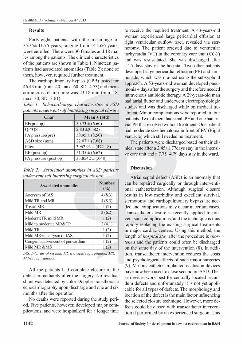
1142
HealthMED - Volume 7 / Number 4 / 2013
Journal of Society for development in new net environment in B&H
Results
Forty-eight patients with the mean age of 35.35± 11.76 years, ranging from 14 to56 years, were enrolled. There were 30 females and 18 ma-les among the patients. The clinical characteristics of the patients are shown in Table 1. Nineteen pa-tients had associated anomalies (Table 2); none of them, however, required further treatment.
The cardiopulmonary bypass (CPB) lasted for 46.43 min (min=40, max=60, SD=4.73) and mean aortic cross-clamp time was 23.18 min (min=18, max=30, SD=3.61). Table 1. Echocardiologic characteristics of ASD patients underwent self buttressing surgical closure
Char Mean ± (Std)EF(pre op) 50.73 ± (6.60)QP/QS 2.83 ±(0 .82)PA pressure(pre) 38.95 ± (8.50)ASD size (mm) 22.07 ± (7.68)Flow 3962.93 ± (472.18)EF (post op) 51.35 ± (6.62)PA pressure (post op) 33.8542 ± (.048)
Table 2. Associated anomalies in ASD patients underwent self buttresing surgical closure
Associated anomalies Number (%)
Anerysm of IAS 4 (8.3)Mild TR and MR 4 (8.3)Trivial MR 1 (2)Mild MR 3 (6.2)ModerateTR mild MR 1 (2)Mild to moderate MR&TR 2 (4.1)Mild TR 1 (2)Mild MR+aneurysm of IAS 1 (2)Congenitalabsencent of pericardium 1 (2)Mild MR &MS 1 (2)
IAS: Inter atrial septum, TR: tricuspid regurgitation, MR: Mitral regurgitation
All the patients had complete closure of the defect immediately after the surgery. No residual shunt was detected by color Doppler transthorasic echocardiography upon discharge and one and six months after the operation.
No deaths were reported during the study peri-od. Five patients, however, developed major com-plications, and were hospitalized for a longer time
to receive the required treatment. A 43-years-old woman experienced large pericardial effusion at right ventricular outflow tract, revealed via ster-notomy. The patient arrested due to ventricular tachycardia (VT) in the coronary care unit (CCU) and was resuscitated. She was discharged after a 25-days stay in the hospital. Two other patients developed large pericardial effusion (PE) and tam-ponade, which was drained using the subxyphoid approach. A 53-years-old woman developed pneu-monia 4 days after the surgery and therefore needed intravenous antibiotic therapy. A 29-years-old man had atrial flutter and underwent electrophysiologic studies and was discharged while on medical tre-atment. Minor complications were reported in four patients. Two of them had small PE and one had tri-vial PE that resolved without treatment. One patient had moderate size hematoma in front of RV (Right ventricle) which still needed no treatment.
The patients were discharged based on their cli-nical state after a 2.45±1.77days stay in the intensi-ve care unit and a 7.75±4.79 days stay in the ward.
Discussion
Atrial septal defect (ASD) is an anomaly that can be repaired surgically or through interventi-onal catheterization. Although surgical closure results in low morbidity and excellent survival, sternotomy and cardiopulmonary bypass are nee-ded and complications may occur in certain cases. Transcatheter closure is recently applied to pre-vent such complications; and the technique is thus rapidly replacing the existing surgical treatments in major cardiac centers. Using this method, the length of hospital stay after the procedure is shor-tened and the patients could often be discharged on the same day of the intervention (8). In addi-tion, transcatheter intervention reduces the costs and psychological effects of such major surgeries (9). Various catheter-implanted occlusion devices have now been used to close secundum ASD. The-se devices work best for centrally located secun-dum defects and unfortunately it is not yet appli-cable for all types of defects. The morphology and location of the defect is the main factor influencing the selected closure technique. However, more de-fects could be closed with transcatheter interven-tion if performed by an experienced surgeon. This

HealthMED - Volume 7 / Number 4 / 2013
Journal of Society for development in new net environment in B&H 1143
comes while the technique is not applicable for large defects, with an adequate rim of tissue surro-unding the defect, or neighbouring on the adjacent critical structures. Therefore open heart surgical treatment in such cases is yet inevitable. On the other hand, we encounter more cases with difficult anatomy than before.
Technique: Anesthesia procedure, sternotomy and commencement of CPB in our series were done according the standard method in conventio-nal technique of ASD repair. The mean CPB time and mean aortic cross-clamp time was respecti-vely 46.43 and 23.18 min in our study which are comparable with previous studies (10). Since most of adverse effects occur due to open heart surgery and CPB, therefore we expect no significant diffe-rence in complications between our series and conventional technique.
Pastorek et al reported the presence of residual shunt in 7.8% of their patients but did not find any significant difference between primary suturing and the use of Dacron patch (11). Du, et al in a study to compare the outcome of surgical and transcatheter closure, reported no large or moderate residual shunt in the surgical group. Mean ASD size was 14.2 ± 6.3 millimetre in their study (8). Our study similarly showed no residual shunts in either of cases despite the fact that our patients had larger ASDs and unsu-itable anatomy. In accordance with previous publi-cations, no deaths were reported during our study (8, 11). Gala et al, however, reported a single case of death secondary to sepsis (0.4% total mortality rate) (11). The complication rate in the Gala study was higher than other studies as only 21.2% of the children and 17.5% of the adults in that study expe-rienced an operation with no complications. Du et al reported major complications in 5.2% of the ca-ses that were mainly the result of large pericardial effusion with tamponade and minor complication in 17.8% of the cases in the surgical group. Pastorek et al reported major complications in 8.1% of the ca-ses, and minor complications in 13.3% of the cases; all of them resolved without any intervention. 1.7% of them developed streptococcus viridans endocar-ditis that was treated with intravenous antibiotics. In our study, 4 patients (8.3 %) experienced major complications that led to prolonged hospital stay and need for further examination and treatment. Minor morbidities developed in 5 (10.4 %) pati-
ents. Two patients had small PE and one had tri-vial PE that resolved without surgical intervention. One patient had moderate size hematoma in front of RV which needed no treatment. A 29-year-old man (2%) had atrial flutter which was treated medically. No patient in our study had atrial arrhythmia before surgery. In a study by Gatzoulis, 5 patients (2.9%) developed atrial flutter or fibrillation in a short peri-od after the surgery, while none of the studied 174 patients had atrial arrhythmia before the operation (12). Gatzoulis found a direct relation between the age of the patients and the development of atrial ar-rhythmias; this comes while atrial flutter in our stu-dy only occurred in one young patient.
Limitations
We had a limited number of cases and we did not have a control group.
Conclusions
This experience showed that the present met-hod might be used efficiently for ASD repair es-pecially in patients with difficult anatomical ASD variants.
Authors’ contributions
FK, suggested the idea, and prepared the pa-tients and participate in preparing manuscript, MHA prepared the patients and participate in pre-paring manuscript, SA gathered data, prepared the firs draft, revised the final article.
Acknowledgements
We thank Dr Hooman Bakhshande and Dr Mo-hsen Rezaei Hemami for their help in this study.

1144
HealthMED - Volume 7 / Number 4 / 2013
Journal of Society for development in new net environment in B&H
References
1. Bailey C, Nichols H, Bolton H, Jamison W, Gomez-Almeida M. Surgical treatment of forty-six interatrial septal defects by atrio-septo-pexy. Annals of Surgery. 1954; 140(6): 805.
2. Masura J, Gavora P, Formanek A, Hijazi ZM. Tran-scatheter closure of secundum atrial septal defects using the new self-centering Amplatzer septal occlu-der: initial human experience. Catheterization and cardiovascular diagnosis. 1997; 42(4): 388-93.
3. Carminati M, Chessa M, Butera G, Bini RM, Giusti S, Festa P, et al. Transcatheter Closure of Atrial Sep-tal Defects with the STARFlex Device: Early Results and Follow-Up. Journal of interventional cardiology. 2001; 14(3): 319-24.
4. Rao PS, Sideris EB, Haddad J, Rey C, Hausdorf G, Wil-son AD, et al. Transcatheter occlusion of patent ductus arteriosus with adjustable buttoned device. Initial clini-cal experience. Circulation. 1993; 88(3): 1119-26.
5. Das GS, Voss G, Jarvis G, Wyche K, Gunther R, Wil-son RF. Experimental atrial septal defect closure with a new, transcatheter, self-centering device. Circulati-on. 1993; 88(4): 1754-64.
6. Rome J, Keane J, Perry S, Spevak P, Lock J. Dou-ble-umbrella closure of atrial defects. Initial clinical applications. Circulation. 1990; 82(3): 751-8.
7. Pedra C, Pihkala J, Lee K, Boutin C, Nykanen D, McLaughlin P, et al. Transcatheter closure of atrial septal defects using the Cardio-Seal implant. Heart. 2000; 84(3): 320-6.
8. Du ZD, Hijazi ZM, Kleinman CS, Silverman NH, Larntz K. Comparison between transcatheter and surgical closure of secundum atrial septal defect in children and adults: results of a multicenter nonran-domized trial. Journal of the American College of Cardiology. 2002; 39(11): 1836.
9. Hughes M, Maskell G, Goh T, Wilkinson J. Prospec-tive comparison of costs and short term health outco-mes of surgical versus device closure of atrial septal defect in children. Heart. 2002; 88(1): 67-70.
10. Galal M, Wobst A, Halees Z, Hatle L, Schmaltz A, Khougeer F, et al. Peri-operative complications following surgical closure of atrial septal defect type II in 232 patients—a baseline study. European heart journal. 1994; 15(10): 1381-4.
11. Pastorek JS, Allen HD, Davis JT. Current outcomes of surgical closure of secundum atrial septal defect. The American journal of cardiology. 1994; 74(1): 75-7.
12. Gatzoulis MA, Freeman MA, Siu SC, Webb GD, Harris L. Atrial arrhythmia after surgical closure of atrial septal defects in adults. New England Journal of Medicine. 1999; 340(11): 839-46.
Corresponding AuthorSevil Aghapour,Rajaie Cardiovascular Medical and ResearchCenter,Tehran University of Medical Sciences,Tehran,Iran,E-mail: [email protected]
HealthMED - Volume 7 / Number 4 / 2013
Journal of Society for development in new net environment in B&H 1145
Abstract
Introduction: For adolescent patients face ae-sthetic plays an important role during orthodon-tic treatment. This population presents a higher expectations regarding the appearance of elements of fixed appliances including elastic ligatures.
Objectives: The aim of the study was an asse-ssment if food dyes affect the color of orthodontic elastic ligatures.
Material and methods: The elastics of four brands: Ortho Organizers, Dentaurum, Ortho Technology and Dentech Corp. were investigated. The 40 samples of the each producer were divided according to the four colors; transparent, white, yellow and silver. The ligatures were stored in so-lution of food colorant for twenty four hours. The color change for each sample was analyzed by the use of Spectroshade (MHT) spectrophotometer. The complete color change (∆E) of ligatures was analyzed statistically on the level of p=0,05.
Results: The greatest color change (∆E) was observed for Ortho Organizers elastics at the mean level of 16,20 points and the lowest for Ortho Tech-nology elastics at the mean level of 12,19 points. No significant differences among the manufactu-rers were observed. The significant p≤0,05 color changes were observed in relation to original color of elastics. The highest color change was observed for white at the mean level of 21,24 points and the lowest for silver products at the level of 4,91 points.
Conclusions: The susceptibility to discolo-ration of elastic ligatures does not depend on producer’s brand.
The level of discoloration of orthodontic elastic by food dyes is strongly related to the original co-lor of the material.
Key words: Elastic ligatures, discoloration, orthodontic treatment.
Introduction
Orthodontic treatment with fixed appliances is becoming increasingly common. Corrections of occlusion are performed not only in children and adolescents; also adults choose this form of treatment more and more frequently. For patients who are socially and professionally active, face aesthetic plays an important role also during ortho-dontic treatment. For this reason there are higher expectations regarding the appearance of elements of fixed appliances and their color, which should match natural dentition. In order to fulfill these requirements, producers of dental materials have expanded their offer with elastic orthodontic brac-kets, whose color is more similar to natural denti-tion in contrast with traditional metal brackets. On the market there are also orthodontic arch wires coated with plastics, whose color is a better match for hard tissues of teeth. The purpose of all this is to decrease the visibility of orthodontic appliances within patient’s oral cavity.
Treatment with orthodontic appliances takes on average about 24 months, and its extent depends, among other things, on: the degree of occlusal dis-orders, patient’s cooperation and applied method of treatment. Lingual techniques fulfill the aesthetic criteria regarding the appearance of dentition du-ring orthodontic treatment, however they have limi-tations resulting from a lower effectiveness of such appliances, harder working conditions for dentists, extended time or increased cost of treatment1.
Most dentists using fixed appliances in their practice, implement labial techniques, which means that elements of appliances, i.e. brackets and arch wires are positioned on labial surfaces of teeth. In order to maintain the arch wire in a bracket’s groo-ve, clinicists may apply steel wire ligatures 0.008 or 0.014 inches in diameter2, elastic ligatures or incre-asingly more popular self-ligating systems.
Elastic ligatures, which are commonly used in ort-hodontic treatment with fixed appliances, are made
The susceptibility to staining of orthodontic elastic ligatures caused by food colorants – In vitro studyKonrad Malkiewicz, Barbara Pietrzak-Bilinska
Department of Orthodontics, Medical University of Warsaw, Warsaw, Poland

1146
HealthMED - Volume 7 / Number 4 / 2013
Journal of Society for development in new net environment in B&H
of elastic polymers usually based on polyurethane. Their exact composition is kept secret by manu-facturers. The advantages of elastic ligatures inclu-de: ease of fixing and removing from brackets, no irritation of oral mucosal membrane, availability in a large selection of colors. Among the disadvantages are: favoring of dental plaque accumulation, often insufficient maintenance of the arch in the bracket’s groove, adsorption of water resulting in change of their physical properties within the oral cavity. Des-pite the fact that elastic ligatures are not perfect ma-terials, they ensure ease of application, which is why they are preferred by dentists. Young patients readily choose colored elastic ligatures, enabling to indivi-dualize the appearance of an orthodontic application thanks to their diverse colors. Adult patients prefer transparent or lightly colored elastomers3.
Clinical observations indicate that elastomers used in orthodontics do not preserve their origi-nal color in clinical conditions. In the water envi-ronment of the oral cavity they are exposed to temperature changes within the range from around 0 to 70 degrees Celsius 4. Food colorants which are present in meals and drinks deposit on ela-stic ligature surfaces and are absorbed inside the materials due to their porous structure. The color change of orthodontic elastomers in oral cavity is particularly visible in case of transparent and light materials. Therefore it seems important to deter-mine, which elastic ligatures discolor to a higher degree than others, in order to treat patients with materials characterized by relatively stable color.
The discoloration of orthodontic ligatures in clinical conditions during one-month period is presented at Figure 1.
Figure 1. The discoloration of orthodontic ligatu-res in clinical conditions during one-month period
Objectives
The aim of the study was to determine the im-pact of food dyes on color change of elastic liga-tures in laboratory conditions, depending on the manufacturer and the initial color of elastomers.
Materials and methods
The study included elastic ligatures from four manufacturers: Ortho Organizers (USA), Denta-urum (Germany), Ortho Technology (USA) and Dentech Corp. (South Korea). Materials from each of the above mentioned producers were used to prepare 40 samples 3x3 mm, which were then divided into 4 groups of 10 samples each accor-ding to the color of materials offered as: transpa-rent, white, yellow and silver. The initial color of each sample was determined by colorime-tric analysis with the use of Spectroshade dental spectrophotometer (MHT, Italy) shown in Figure 2. The 160 studied samples were placed in 5 ml individual silicon containers. Five samples from each group (producer/color) were kept in coffee solution or red wine solution for 24 hours. The samples were stored in an incubator at 37ºC. Af-ter incubation the studied materials were removed from containers with solutions of food colorants and rinsed under running water for one minute. Then the elastomers were dried and subjected to
Figure 2. The Spectroshade - dental spectropho-tometer

HealthMED - Volume 7 / Number 4 / 2013
Journal of Society for development in new net environment in B&H 1147
another colorimetric analysis by spectrophotome-ter. The numeric L*a*b* scale was used to assess the brightness change (∆L*), green/red saturation (∆a*), blue/yellow saturation (∆b*), and a total color change of samples (∆E*). The color change was assessed with regard to elastomer’s producer, initial color, and applied food colorant. A statistical analysis was performed at the significance level of p≤0.05, with the use of Statictica 6.0 package, as well as the following tests: LSD (least significant differences), Bonferroni and Tukey.
Results
Mean change in brightness of ligatures exposed to food colorants (∆L*)Mean decrease in brightness of ligatures expo-
sed to coffee or wine was 5.98 points (p≤0.05). The highest decrease in brightness of 8.88 points on average was observed in elastomers produced by Ortho Organizers, whereas the lowest decrease of 4.28 points on average was observed in products by Dentaurum. The differences were not statistically significant with reference to ligatures’ producer.
Storage in coffee solution caused a decrea-se in brightness of studied ligatures by 3.92 po-ints on average, whereas wine caused a signifi-cantly higher (p≤0.05) mean decrease of ligature brightness by 8.04 points.
The most significant (p≤0.05) decrease in brightness was observed in white ligatures (-11.62 points), whereas in case of silver ligatures the brightness increased by 0.64 points. Transparent ligatures changed their brightness by -6.84 points, yellow ligatures by -7.50 points. The difference between the above mentioned color groups was not statistically significant.
Mean change in saturation of red in ligatures exposed to food dyes (∆a*)Mean increase in red saturation of ligatures su-
bjected to colorimetric tests before and after incu-bation in food dye solutions was 1.54 points and was not statistically significant.
The highest discoloration within this color range was in products made by Ortho Organi-zers (increase by 2.60 points), whereas the lowest was in elastomers produced by Ortho Technology (increase by 0.72 points). The observed changes
within the red color were not statistically signifi-cant depending on manufacturer.
Wine caused a significantly (p≤0.05) higher increase in red saturation (by 2.39 points) than coffee (by 0.56 points).
The highest (by 4.64 points) increase in red sa-turation was observed in white ligatures, whereas in silver elastomers a decrease in red intensity was observed by 0.46 points. The difference between individual color groups was statistically signifi-cant (p≤0.05).
Mean change in saturation of yellow in ligatures exposed to food dyes (∆b*)Mean increase in yellow saturation of elastic
ligatures after incubation in coffee solution or in wine was 6.35 points (p≤0.05).
The highest change within this color range was observed in products made by Dentech (mean increase by 6.47 points), whereas the lowest was in ligatures produced by Dentaurum (mean incre-ase by 3.57 points). No statistically significant difference was observed between products of par-ticular manufacturers.
A significantly higher (p≤0.05) change in yellow saturation was caused by coffee (mean increase by 10.61 points) than by wine (mean increase by 0.43 points).
The highest susceptibility to color change wit-hin the yellow color range was observed in tran-sparent ligatures (mean increase by 15.38 points), the lowest susceptibility was observed in silver ligatures (mean increase by 2.34 points).
Mean global change in color of ligatures exposed to food dyes (∆E*)Mean global change in color of elastic ligatures
exposed to food colorants in the conditions of this study was 13.22 and was statistically significant at the level of p≤0.05.
The color of elastomers produced by Ortho Or-ganizers changed on average by 16.20 points, pro-ducts of Dentaurum indicated a change of 10.66 points, ligatures from Dentech changed by 13.17 points, and from Ortho Technology by 12.19 po-ints. During the analysis of mean color change at p≤0.05 significance level, no significant differen-ces in susceptibility to discoloration were obser-ved in products of individual manufacturers.
1148
HealthMED - Volume 7 / Number 4 / 2013
Journal of Society for development in new net environment in B&H
Also, no significant difference was observed between applied food colorants with regard to the-ir impact on color change of studied elastomers. Coffee changed the color of ligatures on average by 14.41 points, whereas wine by 12.03 points.
A significant (p≤0.05) change in susceptibility to discoloration was observed with reference to the initial color of elastic ligatures. The highest le-vel of color change was observed in white elasto-mers, whose discoloration was on average 20.70 points, whereas the lowest level was noted in the case of silver ligatures, whose color change equa-led 4.91 points. Transparent ligatures changed co-lor to almost the same degree as white ones (chan-ge by 17.93 points). No differences were observed between the mentioned groups in susceptibility to discoloration caused by food dyes. Yellow ligatu-res changed color on average by 11.19 points and their susceptibility to discoloration was at a midd-le level (p≤0.05) with reference to white/transpa-rent and silver elastomers.
Mean color changes of elastic ligatures with re-ference to the manufacturer, applied food dye and ligature color are presented in Table 1.
Discussion
Colorimetric assessment performed with the use of digital optical devices enables an objective and repetitive analysis of color change of dental materials both in oral cavity environment and in
laboratory conditions5,6,7. The method of spectral assessment, which was applied in this study, allows a numeric determination of color change in dental materials through ascribing certain nume-ric values to color sensation which is individual for each observer. Color assessment by means of attributed numeric values within a defined scale enables a precise calculation of its changes resul-ting from environmental conditions.
The analysis of the results in this study indi-cated that ligatures’ susceptibility to discoloration has no significant dependence on their manufactu-rer. For clinicists it means freedom in the choice of elastomer provider in the context of color stability.
Red wine caused a significantly (p≤0.05) higher changes in brightness and saturation of the red color in incubated elastic ligatures compared to coffee. However, coffee solution changed the color of studied elastomers within the yellow ran-ge to a higher degree than wine. The differences in the impact of food dyes on the red and yellow co-lors seem justified by the colors of drinks applied in the study. However, it should be noted that both liquids used in the experiment caused a compa-rable global color change of ligatures on average by 15.08 points for coffee solution and by 12.56 points for red wine.
Color changes of dental materials applied in oral cavity are commonly recognized as noti-ceable if their values exceed 3 points8,9,10 in the L*a*b* scale. The results of this study indicate
Table 1. Mean color change of elastic ligatures in relation to producer, kind of used food colorant and basic color of the elastics
Mean color change of elastic ligatures in relation to producer, kind of used food colorant and basic color of the elastics
∆L ∆a ∆b ∆Eavg std avg std avg std avg std
total -5,98 5,82 1,47 2,80 5,52 11,24 13,22 7,82Ortho Organizers -8,88 6,20 2,60 3,65 5,43 12,86 16,20 8,12Dentaurum -4,28 4,84 0,79 2,50 3,57 10,11 10,66 6,86Ortho Technology -5,19 3,40 0,72 1,89 6,12 11,73 12,19 8,18Dentech -5,14 7,06 1,61 2,48 6,47 9,99 13,17 7,11cafe -3,92 4,14 0,56 2,64 10,61 11,71 14,41 8,98wine -8,04 6,53 2,39 2,67 0,43 8,06 12,03 6,29transparent -6,84 3,97 0,14 2,80 15,38 7,10 17,93 5,90white -11,62 4,37 4,64 2,75 13,99 9,31 20,70 5,88yellow -7,50 3,41 2,37 1,22 -7,52 4,82 11,19 5,41silver 0,64 3,94 -0,46 0,90 2,34 2,28 4,91 1,75

HealthMED - Volume 7 / Number 4 / 2013
Journal of Society for development in new net environment in B&H 1149
that applied food colorants caused color changes in elastomers to the degree which greatly exceeds the limits of aesthetic acceptability. In the experi-mental conditions a mean color change of more than 3 points in the L*a*b* scale was observed regardless of the initial color of elastomers. Howe-ver, it should be noted that white and transparent ligatures discolored about 4 times more intensi-vely than silver ones. It seems that this informati-on should encourage orthodontists to apply elastic ligatures with initially darker and more intensive colors in patients whose diet includes large quan-tities of stimulants, in order to avoid a significant discoloration of elastomers during treatment.
A high susceptibility of elastic ligatures to color change resulting from contact with dyes included in drinks and food was observed by Ardeshna i Vaidyanathan11. The authors analyzed elastic liga-tures’ color change with the use of a Minolta co-lorimeter, and indicated that transparent elastomers in contact with food dyes change color to a signifi-cantly higher degree than initially dark materials. Observations of the authors from the USA quoted above concur with the results obtained in this study.
Color changes of transparent elastic ligatures in experimental conditions were also observed by Kim and Lee 12. The Korean authors stored 3 diffe-rent kinds of transparent elastomers in water solu-tion of ethanol, and water solution of methylene blue for a period from 1 to 120 hours. Depending on ligature type, incubation time and environment, they observed color changes of elastomers in the range from 0.6 to 30 points in the L*a*b* scale. The colorimetric analysis was performed with the use of a Nikon D50 digital camera and Adobe Pho-toshop (ver. 7.0) software package. Unfortunately, the methodology of colorimetric assessment used by the authors Kim and Lee12, as well as solutions in which they kept elastomers were different from methodology and solutions applied in this rese-arch. Therefore any comparison between the two studies is impossible.
Available literature reports about color chan-ge of various types of dental materials caused by food dyes present in oral cavity environment, and about a correlation between the type of dye and the degree of material’s discoloration. Most frequ-ently they refer to composite materials used for fillings and for making orthodontic brackets13-17.
There are few reports about the degree of disco-loration in elastomers used during orthodontic tre-atment. We are aware that elastic ligatures change color in the environment of the oral cavity, which is confirmed by observations by both dentists and patients, however our knowledge on the subject is more intuitive than based on proofs. Research on susceptibility of orthodontic elastomers to color change, which is performed by means of objecti-ve methods of colorimetric assessment, will allow clinicists to choose materials fulfilling aesthetic demands of patients not only at the moment of application, but also during treatment. Although in vitro studies do not reflect the complexity of oral cavity’s environment, they significantly help in assessment of dental materials.
Conclusions
In the conditions of the present study, the de-gree of elastic ligatures’ color change caused by the presence of food dyes depends on the initial color of assessed elastomer.
The method of colorimetric assessment by spectral analysis enables an objective assessment of dental materials’ color change
References
1. Russel JS. Current products and practice aesthetic orthodontic brackets. J Orthod. 2005; 32: 146-163.
2. Elegdak – Turk S, Ozkalayci N, Isci D, Turk T. Color preferences of patients receiving elastic ligatures. Eur J Dent. 2010; 4: 171-174.
3. Taloumis LJ, Smith TM, Hondrum SO, Lorton L. Force decay and deformation of orthodontic elasto-meric ligatures. Am J Orthod Dentofacial Orthop. 1997; 111(1): 1-11.
4. Barclay CW, Spence D, Laird WRE. Intra-oral tem-peratures during function. J Oral Rehab. 2005; 12(32): 886-94.
5. Cal E, Güneri P, Kose T. Comparison of digital and spectrophotometric measurements of colour shade guides. J Oral Rehab. 2006; 33: 221-228.
6. Karamouzos A, Papadopoulos MA, Kolokhitkas G, Athanasiou AE. Precision of on vivo spectrophotome-tric colour evaluation of natural teeth. J Oral Rehab. 2007; 34: 613-621.

1150
HealthMED - Volume 7 / Number 4 / 2013
Journal of Society for development in new net environment in B&H
7. Braun A, Jepsen S, Krause F. Spectrophotometric and visual evaluation of vital tooth bleaching employing different carbamide peroxide concentrations. Dent Mater. 2007; 23: 165-169.
8. Stober T, Gilde H, Lenz P. Color stability of highly filled composite resin materials for facings. Dent Ma-ter. 2001; 17: 87-94.
9. Ruyter IE, Nilner K, Moller B. Color stability of den-tal composite resin materials for crown and bridge veneers. Dent Mater. 1987; 3: 246-251.
10. Johnson WM, Kao EC. Assessment of appearance match by visual observation and clinical colourime-try. J Dental Res. 1989; 68: 819-22.
11. Ardeshna AP, Vaidyanathan TK. Colour changes of orthodontic elastomeric module material exposed to in vitro dietary media. J Orthod. 2009; 36(3): 17-85.
12. Kim SH, Lee YK. Measurement of discolouration of orthodontic elastomeric module with a digital came-ra. Eur J Orthod. 2009; 31: 556-562.
13. Lee Y-K. Changes in the reflected and transmitted co-lor of esthetic brackets after thermal cycling. Am J Orthod Dentofac Orthop. 2008; 133: 641.e1-641.e6.
14. Guler AU, Yilmaz F, Kulunk T, Guler E, Kurt S. Effects of different drinks on stainability of resin composite provisional restorative materials. J Pro-sthet Dent. 2005; 94: 118-124.
15. Patel SB, Gordan VV, Barrett AA, Shen Ch. The effect of surface finishing and storage solutions on the color stability of resin-based composites. J Am Dent Assoc. 2004; 35: 587-594.
16. Faltermeier A, Behr M, Müssig D. In vitro color sta-bility of aesthetic brackets. Eur J Orthod. 2007; 29: 354-358.
17. Eldiwany M, Friedl KH, Powers J M. Color stabi-lity of light-cured and post-cured composites. Am J Dent. 1995; 8: 179-181.
Corresponding AuthorKonrad Malkiewicz,Department of Orthodontics,Medical University of Warsaw,Warszawa,Poland,E-mail: [email protected]

HealthMED - Volume 7 / Number 4 / 2013
Journal of Society for development in new net environment in B&H 1151
Abstract
The cathelicidin derived LL-37 has a broad spectrum of antimicrobial and immunomodulato-ry activities. Here, a DNA sequence encoding the peptide which was fused to the well characterized 384bp fusion partner was cloned into the pUC18 vector and transformed into Escherichia coli. JM109. After high density fermentation of engi-neering bacteria, cell disruption, fusion protein isolation and precise chemical cleavage, HPLC purification and lyophilization, the target peptide LL-37 was purified. The antimicrobial bioactivity of LL-37 in vitro was evaluated by bacteria (Esch-erichia coli., Streptococcus Group B and staphylo-coccus aureus) incubation and showed high abil-ity in sterilization. Acute toxicity test showed high safety. Together, our analysis indicated that LL-37 is an effective and safe antimicrobial peptide, which can be successfully produced at high levels in the Escherichia coli. All these showed the LL-37 acts as a good potential antimicrobial candidate for clinical applications.
Key words: LL-37, antimicrobial bioactivity, peptide.
Introduction
The increasing resistance of microorganisms toward common antibiotics that have provided safety for the last half a century has become a growing threat for the public health. Antimicrobi-al peptides(AMPs) play important roles in innate immune defense.[ 1]More than 1400 AMPs have been identified from the organisms during the past 30 years, among which more than 200 were obtained from frogs skin.[2, 3] As a potential thera-peutic agent, antimicrobial peptides have received increased attention in recent years.[ 4]
LL-37 is the C-terminal part of the only human cathelicidin identified to date called human cationic antimicrobial protein (hCAP18), which is mainly expressed by neutrophils and epithelial cells.[ 5] LL-37,a 4.5 kDa, amphipathic α-helical peptide,[ 6] is a multifunctional host defense molecule that may mediate various host responses, including bacte-ricidal action, epithelial cell activation, epithelial wound regeneration and activation of chemokine secretion[5, 7, 8]. Various mechanisms have been pro-posed for the way involved in its bacterial killing. These included the formation of phase separation due to specific peptide–lipid interactions[9, 10], dis-crete channels that dissipate ion gradients across the membrane[9, 11, 12], detergent-like solubilization of the membrane[13] and disturbance of the lipid bilayer as a result of a carpet or a toroidal-type action[14, 15]. The bacterial membrane is one of the key compo-nents for the cell, and the development of bacterial resistance to a membrane-active peptide appears to be very rare[16], therefore AMPs are potentially an effective way to solve the drug-resistance problem of bacteria. LL-37 can also prevent immunostimu-latory effects of bacterial wall molecules such as li-popolysaccharide and reduce its lethality in murine models of endotoxemia. Additional reported activi-ties include inhibition of neutrophil apoptosis, NK cell antitumor function and stimulation of angio-genesis, cytokine release (e.g. IL-8), inducing mast cells and monocytes, enableing TLR3 to respond to viral dsRNA and stimulate angiogenesis both in vi-tro and in vivo.[4, 7, 17-19]
LL-37 is the only human antimicrobial peptide in the cathelicidin was family and it shows a broad spectrum of antimicrobial activities. It is of interest to develop this peptide for pharmaceutical applica-tions. Obtained by a solid phase chemical synthesis, LL-37 should be very expensive. On the other hand,
Expression, purification and bioactivity analysis of the human antimicrobial peptide LL-37 in Escherichia coliXiaoyan Wu1, Yi Lu1, Hong Zhao2
1 Department of Geriatrics, Huashan Hospital, Fudan University, Shanghai, China,2 Shanghai Peptide Bio-Technologies, Ltd, Shanghai, China.

1152
HealthMED - Volume 7 / Number 4 / 2013
Journal of Society for development in new net environment in B&H
recombinant methods permit the production of pep-tides and proteins in microorganisms. Recombinant expression of LL-37 may be cost-effective. Howev-er, high-level expression of small peptides with an-timicrobial activity is a challenge.[17,20] Escherichia coli (E. coli) is the most commonly used host cell because of easy culture, fast growth, and well-estab-lished alternative expression systems. There are two major problems in the expression of antimicrobial peptides: the expressed peptides are toxic to bacte-ria and the high content of positive-charged amino acid residues makes them very sensitive to protease, which are abundant in E.coli. cells[21]. Furthermore the potential intrinsic antimicrobial activity of an-timicrobial peptides to the host bacteria becomes a question during heterologous expression and then restrain the fermentation.[18, 19] Our strategy is to con-nect the peptide sequence to well characterized fu-sion partners(molecular weight: 16kDa, designed by our laboratory). After specifically cleaving the fusion protein by cyanogen bromide[22, 23], high yield of recombinant LL-37 was generated. The bioactiv-ity of purified recombinant LL-37 to the common bacteria (E.Coli, Streptococcus Group B and staphy-lococcus aureus) was evaluated in vitro.
Materials and methods
The sequence of LL-37The sequence(GenBank Accession No.
Z38026) was as follow, and the C-terminus of the peptide was serine-OH. Leu-Leu-Gly-Asp-Phe-Phe-Arg-Lys-Ser-Lys-Glu-Lys-Ile-Gly-Lys-Glu-Phe-Lys-Arg-Ile-Val-Gln-Arg-Ile-Lys-Asp-Phe-Leu-Arg-Asn-Leu-Val-Pro-Arg-Thr-Glu-Ser-OH
Strains and plasmidsThe E. coli strain DH5α (Invitrogen, USA) was
used as host strain for expression of LL-37. The E. coli strain JM109 (Invitrogen, USA) was used for expression of fusion protein. The plasmid pUC18 (Invitrogen, USA) was used to prepare the gene LL-37 and express the synthesized gene. The well characterized fusion partner was a 128 amino ac-ids peptide (molecular weight: 16kDa) designed by our laboratory. Freezer stocks for long-term storage of expression strains are made by adding 0.1ml of 100% (w/w) glycerol to 1 ml of culture in log phase or grown to saturation in non-inducing
media, mixing well and placing at -70 °C. Sub-cultures for working stocks are made by scraping up a small amount of frozen culture with a sterile plastic pipettor tip without melting the rest of the stock and inoculating into non-inducing media. After growth to saturation, working stocks are typically stable for weeks in refrigerator.
Cyanogen bromide, chemical reagents and Growth mediaThe cyanogen bromide was white crystals
(97%purity, Sigma). The culture media LB was prepared as described[24].Yeast extract and poly-peptone were obtained from OXOID. All other chemical reagents were made in China with the highest purity of commercially available.
Construction of the expression vector The DNA sequence expressing LL-37 was de-
signed with BamHⅠand a cyanogen bromide site (Methionine) at the N-terminus and the termination codon of E. coli and SalⅠsite at the C-terminus. The oligonucleotide designed was prepared with five PCR cycles by using the following primers:
Primer 1 ccaaatGGATCCTGCATGCTGCTGGG; Primer 2 CTTTTACGGAAGAAATCACCCAGCAGCATGC; Primer 3 TGATTTCTTCCGTAAAAGCAAAGAAAAAATCGGTAAAGAA; Primer 4 CTGAACGATACGTTTGAATTCTTTACCGATTTTTTCTTTG
Primer 5 TTCAAACGTATCGTTCAGCGTATCAAAGATTTCCTGCGTA
Primer 6 CGGTACGCGGAACCAGGTTACGCAGGAAATCTTTGATACG
Primer 7 ACCTGGTTCCGCGTACCGAAAGCTAGgtcgacccaaat
Primer 8 ATTTGGGTCGACCTAGCTTT
All primers were synthesized by ShineGene (Shanghai, PR China). The purified PCR product was digested with Bam H I and Sal I and ligated into the plasmid pUC18- FP plasmid(the recombi-nant plasmid of pUC18 and a fusion partner owned by our laboratory). The resultant recombinant plasmid is referred to M-LL-37/pUC18-FP. plas-mid M-LL-37/pUC18-FP was transfor med into E.coli DH5α. The positive clone harboring the re-

HealthMED - Volume 7 / Number 4 / 2013
Journal of Society for development in new net environment in B&H 1153
combination plasmid was confirmed by PCR. The gene encoding LL-37 was confirmed with DNA sequencing by ShineGene. The recombinant plas-mids confirmed were ligated and transformed into E. coli strain JM109 for protein expression.
Expression of LL-37 fusion protein and SDS-PAGE analysisThe transformant was inoculated to LB me-
dium containing 100μg/ml ampicillin. Cells grew at 37 °C with continuous shaking until the optical density at OD600 was 0.9.Then IPTG (isopro-pyl-beta-D-thiogalactopyranoside) was added to a final concentration of 1mM to initiate the fu-sion protein expression. Cells grew at 37 °C with continuous shaking for another 4h in the conical flasks before harvest. Before induction and after induction at 37 °C for 4 h in the conical flask, the bacteria were collected, lysed, and then boiled in SDS-PAGE sample buffer. The resulting products were separated by 15% SDS-PAGE.
Purification of the fusion proteinAfter induction at 37 °C for 4h in the conical
flasks, cells were centrifuged for 30min at 5000 rpm. The supernatant was removed and the pallet containing cells were stored at -70 °C overnight. Then, these cells were resuspended in 8M urea and disrupted by homogenizer(IKA T25 Digital Ultra – Turrax, Germany) at 15000 rpm. After centrifuging at 15000rpm for 30min at 4 °C, the soluble fusion protein was in the supernatant. Then the supernatant was diluted at a ratio 1: 25 by dropwise addition to the refolding buffer (IM Tris-HCl, pH7.3, 0.25M sucrose, 2Mm Ethylenediaminetetraacetic acid)(EDTA) under permanent stirring, and finally kept at 20 °C for 24 h. After centrifuging at 15000rpm for 30min at 4 °C, washed five times with 1M Urea followed by 24hr lyophilization, the purified fusion protein was obtained.
Chemical proteolytic cleavage of the fusion protein by cyanogen bromideLyophilized fusion protein(30mg) was dissolved
in 30ml 80% formic acid under continuous stirring and flushed with N2 while incubated in room tem-perature for 20hr. Solutions of CNBr were prepared freshly prior to each experiment by dissolving the appropriate amount of solid in acetonitrile to a fi-
nal concentration of 0.5g/ml. After solubilization, the fusion protein was incubated with 30mg CNBr (white crystals, a molar ratio of 100: 1 with respect to the Methionine content per molecule) at room temperature under darkness for 24 hr under con-tinuous stirring in the chemical fume hood and fol-lowed by addition of 30 ml double distilled water to terminate the reaction. After dialysis(MW 2kDa) in double distilled water overnight for desalting, the solution was lyophilization.
Purification of peptide LL-37 by HPLC and lyophilizationThe recombinant peptide LL-37 was further
purified by RP-HPLC 1100 series(Agilent, USA) equipped with a C18 column (φ30 X 300mm, Agilent,USA). Processing at a linear gradient of 80 to 20% acetonitrile, containing 0.1% trifluoro-acetic acid at 30 ml/min for 30min was performed. Each peak at 220nm was analyzed by small peptide electrophoresis. The samples of target peak were collected. After lyophilization, the recombinant peptide LL-37 was obtained and was identified by electrospray ionization mass spectrometry (ESI-MS)( LCQ Deka XP Plus, Thermo Fenian, USA).
Antimicrobal bioactivity of LL-37 in vitroE.Coli. Streptococcus Group B and staphylo-
coccus aureus were separated from clinic(Huashan hospital, Fudan University, Shanghai, China)(all multiple drug-resistant bacteria).The control bacteria(about 109/ml) had been in pre-mixed 37 °C saline water 3hr. The samples were LL-37 50μg/ml mixed with the bacteria in pre-mixed 37 °C saline water 3hr. Then both groups were ap-plied to plates. The plates underwent 37 °C oven overnight and then count the bacterial colony. Bacterio-percentage was calculated and graphed.
Acute toxicity test The animal experiment was performed accord-
ing to the instructions of “The Animal Care and Welfare Committee of the Peking Union Medical College (Beijing, China)”. Kungming mice (male) 8-week-old, around 25 g (SLAC laboratory animal, Shanghai, China) were used. The animals were housed in an air-conditioned room at 22 ± 2°C with a 12h light/12h dark cycle. Drinking water and a standard rodent maintenance diet were supplied.

1154
HealthMED - Volume 7 / Number 4 / 2013
Journal of Society for development in new net environment in B&H
Mice were then injected intraperitoneally with LL-37 via three different concentration group (0.1mg, 0.2mg and 1mg) or saline. Each group includes 3 mice. The living percentage was calculated.
Results
The design principle of recombinant LL-37As there is no Methionine in the peptide sequence
of LL-37, we designed the cyanogen bromide site (-Methionine -) at the N-terminus of LL-37 to con-nect to the fusion partner owned by our lab.
Construction of LL-37 geneTo avoid limiting the supply of the correspond-
ing charged tRNA in the host cell, the gene was designed using the preferential codons of E. coli. The BamH I site at N-terminus was introduced for subsequent cloning. The termination codon at C-terminus was used for convenience of purifica-tion. The Sal I site at C-terminus was introduced for subsequent cloning. The constructed plasmid pUC18-FP/M--LL-37 was confirmed by PCR and DNA sequencing. Here was the DNA sequence of the gene encoding LL-37:
g a a t t c A G A T C T T G C A T G C T -G C T G G G T G A T T T C T T C C G T A -A A A G C A A A G A A A A A AT C G G T A -AAGAATTCAAACGTATCGTTCAGCGTAT-CAAAGATTTCCTGCGTAACCTGGTTCCGC-GTACCGAAAGCTGCATGGGATCCTAGgtcgac
Finally, the confirmed plasmids were used for the transformation of E. coli strain JM109.
Expression, cleavage and purification of recombinant LL-37 in shake flasksThe expression efficiency of plasmids was
tested at a small scale using the transformed E. coli strain JM109 cell. Lysates of the inducted and non-inducted bacteria were analyzed by 15%SDS-PAGE (Figure 2). High expression was achieved in all cases and most of the fusion protein was solu-ble in 8M urea. Expression was carried out in 12 shaking flask cultures yielding 18g of wet bacterial biomass/L broth. The frozen cells were then used for extraction with 8M urea and the soluble inclu-sion bodies purified with refolding buffer. After centrifuged 30min at 15000rpm at 4 °C, washed five times with 1M Urea and then lyophilization,
the purified fusion protein was obtained. Lyophi-lized fusion protein(30mg) was dissolved in 30ml 80% formic acid and flushed with N2 for 20hr. So-lutions of CNBr were prepared freshly by dissolv-ing the appropriate amount of solid in acetonitrile to a final concentration of 5M. After solubilization, the fusion protein was incubated with 30mg CNBr at room temperature under darkness for 24 hr under continuous stirring and followed by addition of 30 ml double distilled water to terminate the reaction. After dialysis(MW 2kDa) against double distilled water overnight for desalting, the solution was ly-ophilization under vacuum. Then, the powder was further purified by RP-HPLC. After lyophilization, LL-37 was obtained. The purified LL-37 was iden-tified by electrospray ionization mass spectrometry (ESI-MS)( LCQ Deka XP Plus, Thermo Fenian, USA).The peptide peak reflected a mean molecular mass of Da which was consistent with the calcu-lated molecular weight(MW: 4492.7 Da). The pu-rity of the peptides was > 95%, and the peptides (8-10 mg peptide per litre) in bacterial culture were obtained (peptide concentration depends on the fer-mentation conditions).
Figure 1. Schematic of pUC/LL-37 construction and transformation into E.Coli JM109
Figure 2. 4, centrifuged deposit after cell dis-ruption; 5,supernatant after refolding; 6,last 1M urea wash; 7,blank; 8,target peptide after purifi-cation; 9,target peptide after purification
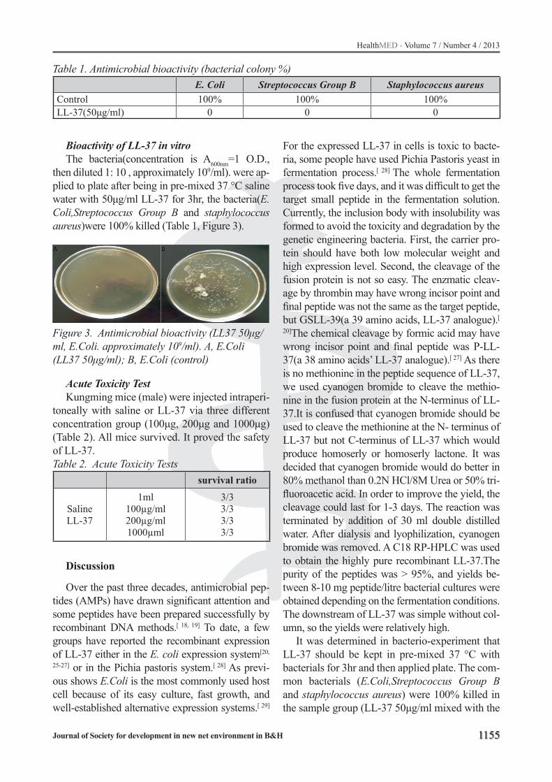
HealthMED - Volume 7 / Number 4 / 2013
Journal of Society for development in new net environment in B&H 1155
Bioactivity of LL-37 in vitroThe bacteria(concentration is A600nm=1 O.D.,
then diluted 1: 10 , approximately 109/ml). were ap-plied to plate after being in pre-mixed 37 °C saline water with 50μg/ml LL-37 for 3hr, the bacteria(E.Coli,Streptococcus Group B and staphylococcus aureus)were 100% killed (Table 1, Figure 3).
Figure 3. Antimicrobial bioactivity (LL37 50μg/ml, E.Coli. approximately 109/ml). A, E.Coli (LL37 50μg/ml); B, E.Coli (control)
Acute Toxicity TestKungming mice (male) were injected intraperi-
toneally with saline or LL-37 via three different concentration group (100μg, 200μg and 1000μg) (Table 2). All mice survived. It proved the safety of LL-37. Table 2. Acute Toxicity Tests
survival ratio
SalineLL-37
1ml100µg/ml200µg/ml1000µml
3/33/33/33/3
Discussion
Over the past three decades, antimicrobial pep-tides (AMPs) have drawn significant attention and some peptides have been prepared successfully by recombinant DNA methods.[ 18, 19] To date, a few groups have reported the recombinant expression of LL-37 either in the E. coli expression system[20,
25-27] or in the Pichia pastoris system.[ 28] As previ-ous shows E.Coli is the most commonly used host cell because of its easy culture, fast growth, and well-established alternative expression systems.[ 29]
For the expressed LL-37 in cells is toxic to bacte-ria, some people have used Pichia Pastoris yeast in fermentation process.[ 28] The whole fermentation process took five days, and it was difficult to get the target small peptide in the fermentation solution. Currently, the inclusion body with insolubility was formed to avoid the toxicity and degradation by the genetic engineering bacteria. First, the carrier pro-tein should have both low molecular weight and high expression level. Second, the cleavage of the fusion protein is not so easy. The enzmatic cleav-age by thrombin may have wrong incisor point and final peptide was not the same as the target peptide, but GSLL-39(a 39 amino acids, LL-37 analogue).[ 20]The chemical cleavage by formic acid may have wrong incisor point and final peptide was P-LL-37(a 38 amino acids’ LL-37 analogue).[ 27] As there is no methionine in the peptide sequence of LL-37, we used cyanogen bromide to cleave the methio-nine in the fusion protein at the N-terminus of LL-37.It is confused that cyanogen bromide should be used to cleave the methionine at the N- terminus of LL-37 but not C-terminus of LL-37 which would produce homoserly or homoserly lactone. It was decided that cyanogen bromide would do better in 80% methanol than 0.2N HCl/8M Urea or 50% tri-fluoroacetic acid. In order to improve the yield, the cleavage could last for 1-3 days. The reaction was terminated by addition of 30 ml double distilled water. After dialysis and lyophilization, cyanogen bromide was removed. A C18 RP-HPLC was used to obtain the highly pure recombinant LL-37.The purity of the peptides was > 95%, and yields be-tween 8-10 mg peptide/litre bacterial cultures were obtained depending on the fermentation conditions. The downstream of LL-37 was simple without col-umn, so the yields were relatively high.
It was determined in bacterio-experiment that LL-37 should be kept in pre-mixed 37 °C with bacterials for 3hr and then applied plate. The com-mon bacterials (E.Coli,Streptococcus Group B and staphylococcus aureus) were 100% killed in the sample group (LL-37 50μg/ml mixed with the
Table 1. Antimicrobial bioactivity (bacterial colony %)E. Coli Streptococcus Group B Staphylococcus aureus
Control 100% 100% 100%LL-37(50μg/ml) 0 0 0

1156
HealthMED - Volume 7 / Number 4 / 2013
Journal of Society for development in new net environment in B&H
bacteria). Most serious infection in clinic are com-bined or multiple-drug resistant bacteria. To be a superspectrum germicidal agent, LL-37 is suit-able. The final metabolic products of LL-37 are amino acids. Acute toxicity tests proved the safety of LL-37. So, LL-37 may be a potential agent in the infection treatment.
Whether human antimicrobial peptide LL-37 can be produced by genetic engineering at a large scale is the key point to be the substance for clini-cal application.
To increase the expression and avoid cytotoxic effects of antimicrobial peptide LL-37 in the E. coli host strain, target peptides were usually fused to cellulose-binding domains or GST,[ 26] result-ing in low yield of target peptide and complicated downstream purification. If the copies of LL-37 gene were linked in tandem with methionine, only 1/n of tandem copies were target peptide while (n-1)/n were peptidyl homoserine lactone in C-ter-minal after chemical cleavage by cyanogen bro-mide. Antimicrobial peptide LL-37 was fused to a MW16kD carrier protein owned by our laboratory which was neutral efficient system for expression and purification in the E. coli host strain. The yield of target peptide was high because of low Molecu-lar Weight of carrier protein. After LL-37 was usu-ally fused to the carrier protein and expressed in E. coli., inclusion bodies were formed without cy-totoxic effects to host cells. So the carrier protein may work without neutralization of the positive charges of LL-37 by fusing to an acidic peptide to avoid the lethal effect of the expressed antimicro-bial peptide on the host cells.[ 19]
pUC18 was usually applied in cloning vectors but seldom acted as expression vector. pUC18 was applied to be the expression vector in the E. coli host strain to meet the scale-up production accord-ing to our experience.[ 30]
The solubility of the fusion protein was im-portant in chemical efficient cleavage of cyano-gen bromide. The reaction was ordinary in 70% formic acid solution or 0.2N HCl/6M guanidine hydrochloride.[ 23] But the soluble inclusion bodies were better digested with 5M CNBr in acetonitrile in 80% formic acid solution. The reaction may last 1 to 3 days for better yield.
In conclusion, we have developed a useful bac-terial expression system to generate biologically
active LL-37. This expression system with an cy-anogen bromide cleavage site is attractive to be harnessed at a large scale.
In the near future, the availability of purified LL-37 could enable the clinical, structural, bio-chemical, and biological investigations.
Acknowledgements
The study was supported by the grant from Foundation of Science and Technology Commit-tee of Shanghai (No.10431904000). We thank Dr. Tieying Gong and Dr. Tianzi Huang for their valu-able comments.
References
1. Hurtado P, Peh CA. LL-37 promotes rapid sens-ing of CpG oligodeoxynucleotides by B lymphocytes and plasmacytoid dendritic cells. J Immunol 2010; 184(3): 1425-35.
2. Zasloff M. Magainins, a class of antimicrobial pep-tides from Xenopus skin: isolation, characterization of two active forms, and partial cDNA sequence of a precursor. Proc Natl Acad Sci U S A 1987; 84(15): 5449-53.
3. Zasloff M. Antimicrobial peptides of multicellular or-ganisms. Nature 2002; 415(6870): 389-95.
4. Saleem M, Nazir M, Ali MS, Hussain H, Lee YS, Riaz N, et al. Antimicrobial natural products: an update on future antibiotic drug candidates. Nat Prod Rep 2010; 27(2): 238-54.
5. Bucki R, Leszczyńska K, Namiot A, Sokołowski W. Cathelicidin LL-37: a multitask antimicrobial pep-tide. Arch Immunol Ther Exp (Warsz) 2010; 58(1): 15-25.
6. Bals R, Wilson JM. Cathelicidins--a family of multi-functional antimicrobial peptides. Cell Mol Life Sci 2003; 60(4): 711-20.
7. Arrighi RB, Nakamura C, Miyake J, Hurd H, Burgess JG. Design and activity of antimicrobial peptides against sporogonic-stage parasites causing murine malarias. Antimicrob Agents Chemother 2002; 46(7): 2104-10.
8. Mendez-Samperio P. The human cathelicidin hCAP18/LL-37: a multifunctional peptide involved in myco-bacterial infections. Peptides 2010; 31(9): 1791-8.

HealthMED - Volume 7 / Number 4 / 2013
Journal of Society for development in new net environment in B&H 1157
9. Münster C, Spaar A, Bechinger B, Salditt T. Magainin 2 in phospholipid bilayers: peptide orientation and lipid chain ordering studied by X-ray diffraction. Bio-chim Biophys Acta 2002; 1562(1-2): 37-44.
10. Dathe M, Schumann M, Wieprecht T, et al. Peptide helicity and membrane surface charge modulate the balance of electrostatic and hydrophobic interacti-ons with lipid bilayers and biological membranes. Biochemistry 1996; 35(38): 12612-22.
11. Matsuzaki K. Magainins as paradigm for the mode of action of pore forming polypeptides. Biochim Bio-phys Acta 1998; 1376(3): 391-400.
12. Ladokhin AS, Selsted ME, White SH. Sizing mem-brane pores in lipid vesicles by leakage of co-encap-sulated markers: pore formation by melittin. Biophys J 1997; 72(4): 1762-6.
13. Ladokhin AS, White SH. ‘Detergent-like’ permeabi-lization of anionic lipid vesicles by melittin. Biochim Biophys Acta 2001; 1514(2): 253-60.
14. Chen Z, Wang D, Cong Y, et al. Recombinant an-timicrobial peptide hPAB-beta expressed in Pichia pastoris, a potential agent active against methicil-lin-resistant Staphylococcus aureus. Appl Microbiol Biotechnol 2011; 89(2): 281-91.
15. Ramamoorthy A, Thennarasu S, Lee DK, Tan A, Maloy L. Solid-state NMR investigation of the mem-brane-disrupting mechanism of antimicrobial pep-tides MSI-78 and MSI-594 derived from magainin 2 and melittin. Biophys J 2006; 91(1): 206-16.
16. Hancock RE. Concerns regarding resistance to self-proteins. Microbiology 2003; 149(Pt 12): 3343-4; discussion 3344-5.
17. Büchau AS, Morizane S, Trowbridge J, Schauber J, Kotol P, Bui JD, et al. The host defense peptide cathe-licidin is required for NK cell-mediated suppression of tumor growth. J Immunol 2010; 184(1): 369-78.
18. Lee JH, Kim JH, Hwang SW, Lee WJ, Yoon HK, Lee HS, et al. High-level expression of antimicrobial peptide mediated by a fusion partner reinforcing formation of inclusion bodies. Biochem Biophys Res Commun 2000; 277(3): 575-80.
19. Lee JH, Minn I, Park CB, Kim SC. Acidic peptide-mediated expression of the antimicrobial peptide bu-forin II as tandem repeats in Escherichia coli. Pro-tein Expr Purif 1998; 12(1): 53-60.
20. Yang YH, Zheng GG, Li G, Zhang XJ, Cao ZY, et al. Expression of bioactive recombinant GSLL-39, a vari-ant of human antimicrobial peptide LL-37, in Esch-erichia coli. Protein Expr Purif 2004; 37(1): 229-35.
21. Thomas-Virnig CL, Centanni JM, Johnston CE, He LK, Schlosser SJ, Van Winkle KF, et al. Inhibition of multidrug-resistant Acinetobacter baumannii by nonviral expression of hCAP-18 in a bioengineered human skin tissue. Mol Ther 2009; 17(3): 562-9.
22. Sánchez D, Moussaoui M, Carreras E, Torrent M, Nogués V, Boix E. Mapping the eosinophil cationic protein antimicrobial activity by chemical and enzy-matic cleavage. Biochimie 2011; 93(2): 331-8.
23. Andreev YA, Kozlov SA, Vassilevski AA, Grishin EV. Cyanogen bromide cleavage of proteins in salt and buffer solutions. Anal Biochem 2010; 407(1): 144-6.
24. Sambrook J, Russell DW. Molecular Cloning: A Laboratory Manual. third ed 2001. 1595-1597.
25. Ramos R, Domingues L, Gama M. Escherichia coli expression and purification of LL37 fused to a family III carbohydrate-binding module from Clostridium thermocellum. Protein Expr Purif 2010; 71(1): 1-7.
26. Moon JY, Henzler-Wildman KA, Ramamoorthy A. Expression and purification of a recombinant LL-37 from Escherichia coli. Biochim Biophys Acta 2006; 1758(9): 1351-8.
27. Li Y, Li X, Wang G. Cloning, expression, isotope labeling, and purification of human antimicrobial peptide LL-37 in Escherichia coli for NMR studies. Protein Expr Purif 2006; 47(2): 498-505.
28. Hong IP, Lee SJ, Kim YS, Choi SG. Recombinant ex-pression of human cathelicidin (hCAP18/LL-37) in Pichia pastoris. Biotechnol Lett 2007; 29(1): 73-8.
29. Huang L, Wang J, Zhong Z, Peng L, Chen H, Xu Z, et al. Production of bioactive human beta-defensin-3 in Escherichia coli by soluble fusion expression. Bio-technol Lett 2006; 28(9): 627-32.
30. Wu X, Lu Y, Zhao H, Hu R. EW, a recombinant ana-logue of Exendin-4 expressed in Escherichia coli. Sci Res Essays 2011; 6(14): 2941-2949.
Corresponding AuthorYi Lu,Department of Geriatrics, Huashan Hospital, Fudan University, Shanghai, China,E-mail: [email protected]

1158
HealthMED - Volume 7 / Number 4 / 2013
Journal of Society for development in new net environment in B&H
Abstract
Introduction: Free-living amoebae (FLA), be-sides being agents of human infections, may also serve as hosts for other microorganisms including respiratory bacterial pathogens (RBPs). Recre-ational fountains and ornamental pools (RF-OPs) spread aerosolized water droplets in the air which can easily be inhaled.
Objective: The aim of this study was to in-vestigate the presence of FLA and RBPs in water samples taken from RF-OPs and to determine if RBPs existed in FLA that were cultured from the initial samples.
Methods: Ninety-eight water samples collected during 2010 and 2011 summer months were cul-tured xenically by using non-nutrient agar for FLA. DNA was extracted from both water samples and FLA-positive culture material by a standard phe-nol-chloroform extraction method. FLA identifica-tion was performed by multiplex-PCR. Presence of respiratory bacterial pathogens (S. pneumonia, H. influenzae, C. pneumoniae, L. pneumophila, B. per-tussis, M. pneumoniae) and Mycobacterium spp. in water samples and the cultured FLA were investi-gated by a commercial multiplex-PCR and a Myco-bacterium specific PCR, respectively.
Results: FLA were detected in 39 (39.8%) of the total water samples by culture, 32 of which were also identified by PCR. Nine (23.1%) of these cultured FLA were also RBP-positive. FLA and RBPs were identified by PCR in 78 (79.6%) and 32 (32.7%) of water samples, respectively.
Conclusion: The FLA and RBPs can consti-tute biofilms on water surface and the possibil-ity of RBPs also living as endosymbionts should
be considered. In either situation, RF-OPs can be sources of FLA and RBPs, thus may pose a public health risk.
Key words: Free-living amoebae, respiratory bacterial pathogens, pool, fountain, water.
Introduction
Free-living amoebae (FLA) such as Acantham-oeba, Naegleria, Balamuthia, Sappinia, Vahlkamp-fia and Hartmannella species are ubiquitous organ-isms found in various environmental reservoirs such as air, soil, sand, dust, fresh and salty waters, as well as man-made water systems like recreation-al and domestic water sources, cooling towers, air-conditioning units, humidifiers, tap water, or drink-ing water production plants (1-6). Among these FLA, Naegleria fowleri, Acanthamoeba spp. and Balamuthia mandillaris are pathogenic to humans causing central nervous system, cornea, lung, and skin infections in susceptible individuals (6).
FLA trophozoites feed on bacteria including RBPs, some of which are able to survive phago-cytosis (amoeba-resisting bacteria, ARB), and thus use the FLA as hosts (7). Besides being etiological agents of human infections, FLA serve not only as reservoirs but also as amplification and transport vehicles for water-based pathogenic ARB such as Legionella spp., Mycobacterium avium, and Ch-lamydia pneumoniae (7-10). Some of the advanta-ges FLA offer ARB are: supplying critical nutrients and growth factors; protecting against biological, chemical and physical inhibitors especially in the cystic form; helping to survive and spread in water and environment; and increasing the virulence, in-fectivity and resistance of bacteria (1, 5, 9-14). FLA
Investigation of free-living Amoebae and respiratory bacterial pathogens in water samples taken from recreational fountains and ornamental pools in Ankara, TurkeyEce Erci1, Elif Abay1, Gulay Aral Akarsu2, Zeynep Ceren Karahan3
1 Ankara University Faculty of Medicine, Ankara, Turkey,2 Division of Medical Parasitology, Ankara University Faculty of Medicine, Ankara, Turkey,3 Department of Medical Microbiology, Ankara University Faculty of Medicine, Ankara, Turkey.

HealthMED - Volume 7 / Number 4 / 2013
Journal of Society for development in new net environment in B&H 1159
protect its hidden resistant microorganism from the first line of human defenses and the strategies de-veloped by ARB for surviving inside the amoeba help them to survive in human macrophages. As a result, the FLA/ARB couple synergically produce pathogens to which humans are frequently expo-sed, that are highly resistant to decontamination, and able to resist destruction by human macropha-ges (7). Human infection can occur via inhalation of aerosolized water containing free bacteria and/or FLA containing infective ARB (10).
Recreational fountains and ornamental pools (RF-OPs) affect people by spreading aerosols which can be easily inhaled especially in spring and summer months. Aerosols allow the transmission of possibly existing FLA and bacteria over consid-erable distances (15). The aim of this study was to investigate the existence of FLA and respiratory bacterial pathogens (RBPs) separately or as endo-symbionts in RF-OP water that may pose a poten-tial health risk for humans via respiratory route.
Methods
Water SamplesNinety-eight water samples were collected
from different outdoor (n=91) and indoor (n=7) RF-OPs located at different regions of Ankara city center, Turkey, during the summer months (Ju-ne-August) of 2010 and 2011. The source of the water was municipal water system. The one water sample per RF-OP were collected from the surface of the water in 50 ml sterile centrifuge tubes, and transported to the laboratory. These samples were used for both FLA culture and total DNA extracti-on for PCR amplification.
FLA cultureFLA were cultured xenically by using non-nu-
trient agar. For this purpose, water samples (50 ml) were centrifuged at 250xg for 10 min. The super-natant was pipetted out and discarded leaving 1 ml of the sediment in the tube. This sediment was re-suspended by pipetting and used for FLA culture and DNA extraction. FLA culture was performed by placing one drop of suspension in the center of a non-nutrient agar plate that had been streaked with Escherichia coli (ATCC 25922) in an X con-figuration, which promoted the multiplication and
accumulation of amoeba in a defined area along the line of bacteria. The plate was sealed with pa-rafilm and incubated at room temperature. The plates were examined at x400 magnification with an inverted microscope for the presence of amoe-bae daily for one week (16). For DNA extraction, the grown FLA were harvested in 1 ml sterile di-stilled water by scraping the surface of the agar.
DNA extractionDNA was extracted both from the initial water
samples and cultured FLA by a previously publis-hed extraction method with additional phenol-chloroform extraction steps (17). The additional steps were as follows: Following the inactivati-on of Proteinase K, the sample was mixed with 1:1 phenol-chloroform-Isoamyl alcohol solution (25:24:1), vortexed for 10 min, chilled on ice for 10 min, and centrifuged at 14.000xg for five min at +4oC. The upper phase was collected into a new tube, 1:1 chloroform was added, and the same steps were applied as mentioned above. 500 µl of 95% ethanol was added to the upper phase which was collected to a new tube. Following vortexing and centrifugation at 14.000xg for five min at 4oC, the pellet was washed with 70% ethanol. Superna-tant was removed, the pellet was air-dried, and the extracted DNA was re-suspended in 200 µl sterile distilled water. DNA samples were stored at 4oC until PCR amplifications were performed.
PCR Amplification of FLAFLA identification was done by the PCR pro-
tocol described by Tsvetkova et al. (6). This PCR provided identification of Hartmannella vermifor-mis (800 bp), Naegleria fowleri (900 bp), Vanella spp./ Vahlkampfia ovis (950 bp), Acanthamoeba castellanii/ polyphaga/ lenticulata/ hatchetti (1080 bp), A. comandoni (1350 bp), and A. astronyxis (1500 bp) according to the amplified product size.
PCR Amplification of RBPsThe presence of RBPs (Streptococcus pneumo-
niae, Haemophilus influenzae, Chlamydia pneumo-niae, Legionella pneumophila, Bordetella pertu-ssis, and Mycoplasma pneumoniae) in initial water and culture-positive FLA samples were investiga-ted by using a multiplex-PCR kit (Seeplex Pneu-mobacter Detection Kit, Seegene, Korea) following

1160
HealthMED - Volume 7 / Number 4 / 2013
Journal of Society for development in new net environment in B&H
manufacturer’s instructions. Mycobacterium genus specific PCR was performed by using hsp65 gene directed primers as described previously (18). All PCR products were analysed by agarose gel electrophoresis and stained with ethidium bromide.
Results
FLA CultureMicroscopic evaluation of the cultures on non-
nutrient agar plates showed that 39 (39.8 %) out of 98 water samples yielded amoeba growth, thus they were considered as culture-positive.
FLA-PCR of Culture-Grown AmoebaIn 32 (82.1%) of the 39 culture-positive water
samples, FLA could be identified by PCR. H. ver-miformis was detected in 29 (74.4%), N. fowleri in 6 (15.4%), Acanthamoeba spp. in 4 (10.3%), and Vanella spp./V. ovis in 2 (5.1%) of the FLA-PCR positive culture samples. Eight of these samples contained more than one amoeba species: H. vermi-formis + N. fowleri (n=3), H. vermiformis + Acan-thamoeba spp. (n=3), H. vermiformis + Vanella spp./V. ovis (n=1), H. vermiformis + N. fowleri + Vanella spp./V. ovis (n=1). In seven (17.9%) of the 39 culture positive material, FLA-PCR was nega-tive although amoebae were observed microscopi-cally, suggesting that they may contain FLA species for which PCR was not directed for detection.
FLA-PCR of Water SamplesThe evaluation of water samples (n=98) re-
vealed 78 (79.6%) positive samples. 76 (77.6%) were positive for H. vermiformis, 19 (19.4%) for Acanthamoeba spp., 7 (7.1%) for N. fowleri and 2 (2%) for Vanella spp./V. ovis. In 25 of the wa-ter samples, more than one amoeba species were detected: H. vermiformis + Acanthamoeba spp. (n=18), H. vermiformis + N. fowleri (n=5), H. ver-miformis + Vanella spp./V. ovis (n=1), H. vermifor-mis + N. fowleri + Vanella spp./V. ovis (n=1) (Figure 1). Twenty (20.4%) water samples were FLA-PCR negative. Eight (8.2%) of these samples were cul-ture-positive, though only one yielded H. vermifor-mis after performing FLA-PCR of culture samples.
Figure 1. Representative FLA-PCR products. M represents 100 bp molecular size marker (Mass Ruler, Fermentas, Lithuania). 1: Acanthamoeba spp. positive sample, 2-5, 8,9: H. vermiformis positive samples, 6,7: H. vermiformis+ Acantha-moeba spp. positive samples, 10: H. vermifor-mis+ N. fowleri positive sample
RBP-PCR of Culture-Grown AmoebaeRBPs were found in nine (23.1%) of the 39
culture material. S. pneumoniae was detected in five, Mycobacterium spp. in four, C. pneumoniae in two, and B. pertussis in one. In three cultures, more than one RBP existed: S. pneumoniae + My-cobacterium spp. (n=2), and S. pneumoniae + C. pneumonia (n=1). The amoebae coexisting with these RBPs are shown in Table 1.
RBP-PCR of Water SamplesOf the water samples evaluated, 32 (32.7%)
were positive for RBPs. S. pneumoniae DNA was detected in 25 (78.1%), Mycobacterium spp. in 10 (31.3%), H. influenzae in six (18.8%), C. pneumoniae in two (6.3%), and B. pertussis in one (3.1%) water sample. Twelve water samples contained more than one RBP: S. pneumoniae + Mycobacterium spp (n=5), S. pneumoniae+H. influenzae (n=4), S. pneumoniae+B. pertussis (n=1), S. pneumoniae+C. pneumoniae (n=1), and C. pneumoniae+Mycobacterium spp. (n=1) (Figure 2). Only three of the RBP-positive water samples were FLA-negative both by culture and PCR. In six RBP-positive water samples there were culture-grown amoebae, but they could not be identified by FLA-PCR, suggesting that they may belong to other genera than the studied ones. The amoebae detected in the remaining 23 RBP-positive samples are shown in Table 1.

HealthMED - Volume 7 / Number 4 / 2013
Journal of Society for development in new net environment in B&H 1161
Table 1. Co-existening amoeba and RBP in culture and/or water samples Water
SampleNo
FLACulture
PCR Results of Culture Material PCR Results of Water Samples
FLA-PCR RBP-PCR FLA-PCR RBP-PCR
2 (+) N. fowleri S. pneumoniae,Mycobacterium spp. N. fowleri (-)
15 (+) (-) (-) (-) S. pneumoniaeH. influenzae
18 (+) (-) (-) (-) H. influenzae20 (-) (-) NP* H. vermiformis S. pneumoniae
21 (-) (-) NP H. vermiformisAcanthamoeba spp. S. pneumoniae
22 (+) H. vermiformis S. pneumoniae H. vermiformis (-)24 (+) H. vermiformis S. pneumoniae H. vermiformis (-)26 (+) H. vermiformis Mycobacterium spp. H. vermiformis (-)28 (+) (-) (-) (-) S. pneumoniae31 (+) H. vermiformis (-) H. vermiformis S. pneumoniae36 (-) (-) NP H. vermiformis S. pneumoniae38 (+) H. vermiformis (-) H. vermiformis S. pneumoniae
43 (+) H. vermiformis S. pneumoniaeMycobacterium spp. H. vermiformis (-)
44 (+) (-) (-) (-) S. pneumoniae45 (+) (-) (-) (-) S. pneumoniae51 (+) (-) (-) (-) S. pneumoniae
H. influenzae
52 (+) N. fowleri B. pertussis H. vermiformisN. fowleri (-)
54 (+) H. vermiformis (-) H. vermiformis S. pneumoniae
55 (+) H. vermiformis (-) H. vermiformis S. pneumoniaeMycobacterium spp.
57 (-) (-) NP H. vermiformis Mycobacterium spp.
59 (+) H. vermiformis (-) H. vermiformis S. pneumoniaeMycobacterium spp.
61 (-) (-) NP H. vermiformis Mycobacterium spp.
65 (+) H. vermiformis S. pneumoniaeC. pneumoniae H. vermiformis (-)
70 (+) H. vermiformis (-) H. vermiformis Mycobacterium spp.
72 (+) H. vermiformis N. fowleri Mycobacterium spp. H. vermiformis
N. fowleri (-)
73 (-) (-) NP H. vermiformisAcanthamoeba spp.
S. pneumoniaeMycobacterium spp.
74 (+) H. vermiformis (-) H. vermiformis Mycobacterium spp.
75 (-) (-) NP H. vermiformis S. pneumoniaeB. pertussis
77 (-) (-) NP H. vermiformis S. pneumoniae
80 (-) (-) NP H. vermiformisAcanthamoeba spp.
S. pneumoniaeMycobacterium spp.
86 (-) (-) NP H. vermiformisAcanthamoeba spp.
C. pneumoniaeMycobacterium spp.

1162
HealthMED - Volume 7 / Number 4 / 2013
Journal of Society for development in new net environment in B&H
Figure 2. Representative RBP-PCR products. M represents molecular size marker corresponding to the given pathogens on each side. IC repre-sents internal control. 1: C. pneumoniae positive sample, 2, 5,6,8,9: S. pneumoniae positive sam-ples. 3: S. pneumoniae + H.influenzae positive sample, 4: B. pertussis positive sample, 7: S. pne-umoniae + B. pertussis positive sample
Discussion
Ankara is the capital city of Turkey, with about 4.750.000 inhabitants. During the past ten years, RF-OPs have been increasingly constructed for esthetic purposes in the open-air areas such as parks, divided highways, traffic junctions and also in indoor areas like shopping malls located at An-kara city center. These RF-OPs are filled from the city water network, but water is recycled throu-ghout spring and summer months. These RF-OPs spread out aerosols, which come into contact with people even with summer breeze.
In low nutrient environments such as freshwater, the association of bacteria with FLA is an adapti-ve survival mechanism of ARB that promotes their persistence and dissemination. ARB infected FLA genera identified by laboratory research to date are; Acanthamoeba, Dictyostelium, Echinamoeba, Hartmannella, Naegleria and Vahlkampfia (10). In Bulgaria, 61.1% of the environmental soil and water sources were reported to contain FLA (6).
The frequency of FLA in various environmental sources was reported to be between 17.2-31% in Turkey by culture. They were identified as Hart-mannella spp., Naegleria spp., Acanthamoeba spp., and Vahlkampfiidae by PCR (19, 20). In our study 86 (87.8%) of the water samples obtained from RF-OPs were found to contain FLA by culture (n=12) or PCR alone (n=47), or by both (n=27).
Direct PCR evaluation of water samples yield-ed twice as much amoebae than culture (79.6% vs 39.8%) in our study. Culture-based methods can un-derestimate both FLA diversity and density in water (10). Culturing water is a laborious procedure, and it is often difficult to achieve appropriate enrichment. Moreover, FLA concentrations in water may be too low for cultural detection but still be high enough to cause infection. Therefore the use of highly-sen-sitive and specific molecular detection methods based on the detection of specific segments on the pathogens’s genome are needed for the evaluation of water-borne pathogens (21). Presence of micro-organisms in water samples can be directly demon-strated by PCR without prior culture isolation (22). On the other hand, PCR can only detect the targeted genomes, thus any FLA which is not covered for de-tection can not be identified by that specific PCR. In our study, all but one (H. vermiformis) of the cultu-red amoebae could also be identified by direct PCR of the water samples. For one culture whose PCR yielded the presence of N. fowleri, PCR of the water sample detected both N. fowleri and H. vermiformis. On the other hand, we could not identify amoebae from seven (7.2%) culture-positive water samples. These FLA may belong to other genera such as Sap-pinia, Dictyostelium or Echinamoeba which were out of the range of the PCR method used.
91 (-) (-) NP H. vermiformisAcanthamoeba spp. S. pneumoniae
92 (-) (-) NP H. vermiformisAcanthamoeba spp. S. pneumoniae
93 (-) (-) NP H. vermiformis S. pneumoniae
95 (-) (-) NP H. vermiformis S. pneumoniaeMycobacterium spp.
97 (-) (-) NP H. vermiformis S. pneumoniaeH. influenzae
98 (+) H. vermiformis C. pneumoniae H. vermiformis S. pneumoniaeC. pneumoniae
*NP: Not performed; (-): negative, (+): positive
HealthMED - Volume 7 / Number 4 / 2013
Journal of Society for development in new net environment in B&H 1163
The most frequently encountered FLA in tap water are; Acanthamoeba, Hartmannella, Vah-lkampfia, and Vanella (10). Corsaro et al. have isolated Naegleria spp. (44.4%), Acanthamoeba spp. (25%), and H. vermiformis (13.8%) from the samples obtained from water treatment plants in Spain (2). FLA density and diversity in tap water show seasonal variation, and increases during the summer months due to warming of water (10, 15). Acanthamoeba is more prevalent in spring, while Vahlkampfia and Naegleria are frequently dete-cted in autumn (9). In our study, water samples were collected during the summer months, and the most frequently encountered FLA were H. vermi-formis (78.6%), followed by Acanthamoeba spp. (19.4%), N. fowleri (7.1%), and Vanella spp./V. ovis (2%). Similarly, in the study of Tsvetkova et al., Acanthamoeba and Hartmannella, but not Naegleria, were found to be abundant in water and soil samples from Bulgaria (6).
Bacterial endosymbionts of FLA were micros-copically observed nearly 30 years ago, but due to the inability to culture these bacteria outside their eukaryotic hosts, their identification and analysis delayed until the introduction of molecular tech-niques. Using PCR for the investigation of FLA and RBPs in water samples may have some drawbacks, despite its speed, ease to perform, high sensitivity, and specificity. Prolonged exposure to water may lead the bacterial pathogens to enter a viable, but non-culturable state, and these bacteria may not be detected by culture, although they retain their infe-ctive potential. Using PCR, these bacteria can be detected, but PCR cannot distinguish between vi-able and non-viable organisms. The standardizati-on and validation of protocols is very important for the implementation of molecular techniques either in the environmental or clinical field. Direct PCR amplification of some bacterial pathogens from wa-ter samples may be difficult due to the presence of low numbers of bacteria in environmental sources, and the presence of inhibitors in the samples may lead to false-negative results (21).
Bacterial endosymbionts belong to five linea-ges: (i) the Alphaproteobacteria, (ii) the Betapro-teobacteria, (iii) the Gamaproteobacteria, (iv) the Bacteroidetes, and (v) the Chlamydiales (4).
Legionella pneumophila is the first human pat-hogen shown to multiply and persist in amoebae
(23). FLA are necessary for the multiplication of Legionella in water, and humans are probably infected by inhaling the amoeba filled with Le-gionella (23, 24). Growth in amoebae has been shown to enhance the entry, intracellular survi-val and replication of Legionella pneumophila in macrophages. Legionella grown in amoeba has higher virulence and infectivity (25). L. pneumop-hila can infect and multiply within Hartmannella, Acanthamoeba, Vahlkampfia, Echinamoeba and Naegleria spp (1, 26). We didn’t detect L. pneu-mophila in either cultured FLA or water samples. This finding is consistent with the study of Pagnier et al. who also could not detect any Legionella in the environmental water samples (27).
C. pneumoniae is the second example of respira-tory pathogens which can use the amoebae as hosts (12, 28). C. pneumoniae also benefits from the amoebal adaptation process for infection of animal cells and macrophages (4, 12). In humans, mixed infections due to Acanthamoeba and Chlamydia have been reported (29). We detected the presence of C. pneumoniae in three water samples by either culture (n=1) or PCR (n=1), or both (n=1). All three contained H. vermiformis, and one was also positi-ve for Acanthamoeba spp. (Table 1).
Mycobacterium species are widely distributed in the environment. They can infect and multiply in FLA, and use them as training grounds to adapt and survive within human macrophages (2, 5). To date, many mycobacterial species such as M. tuberculo-sis, M. bovis and BCG strains, M. avium complex (MAC), M. marinum, M. ulcerans, M. simiae, M. habane, M. smegmatis, M. fortuitum, M. phlei, M. gordonae, M. kansasii, and M. xenopi are shown to survive within FLA (7, 27, 30-32). Besides, MAC, M. marinum, M. kansasii, M. scrofulaceum, M. chelonae, M. xenopi, M. abscessus, M. bolettii, M. massiliense and M. fortuitum are shown to be the free living mycobacterial species associated with contaminated water supplies (30, 31). In our study, four of the FLA-positive culture samples, and ten of the water samples containing FLA were also positi-ve for Mycobacterium spp (Table 1).
Legionella, Chlamydia and Mycobacterium species are among the most frequently investigat-ed RBPs capable to survive in FLA. With amoebal co-culture, Corsaro et al. have isolated Legionella spp., Chlamydia-like bacteria., and rapid growing

1164
HealthMED - Volume 7 / Number 4 / 2013
Journal of Society for development in new net environment in B&H
Mycobacterium spp. in 29.6%, 10.4%, and 19.2% of the water treatment plant samples, respectively (2). Thomas et al. investigated hospital network for the presence of FLA and bacterial endosym-bionts by amoeba co-culture (5). They isolated H. vermiformis and A. polyphaga in 6.5% and 0.5% of the samples, respectively. The samples contai-ning bacterial isolates (45.5%) revealed Mycobac-terium spp. and Legionella spp. but they couldn’t isolate any Chlamydia-related organisms. Althou-gh we identified the presence of Mycobacterium spp. in 10.3%, and C. pneumoniae in 3.1% of the water samples, we did not detect L. pneumophila (Table 1). C. pneumoniae has not been detected from natural or artificial water sources before. Our findings may have resulted from a cross-reaction between Chlamydia and Chlamydia-like bacteria.
In the present study, S. pneumoniae DNA was present in 25 (25.5%) water samples, 17 of whi-ch were also containing FLA-DNA (Table 1). We detected S. pneumoniae DNA in one N. fowleri and four H. vermiformis positive culture samples. S. pneumoniae was reported to be very common in ground and sea water (33), although only one study reported S. pneumoniae as an ARB until to-day (3). Our findings may support the evidence that S. pneumoniae can either be found as endosy-mbionts of FLA or free-living forms in water.
One interesting finding of our study which nee-ds further verification was the determination of B. pertussis in two samples; one, N. fowleri positive culture sample and the other, an initial water sample simultaneously harboring S. pneumoniae, H. vermi-formis and Acanthamoeba spp. Bordetella spp. be-long to the family Alcaligenaceae which also com-prises the genera Alcaligenes and Achromobacter (34, 35). B. pertussis is an obligate pathogen for hu-mans, whereas Achromobacter and Alcaligenes are environmental species. B. bronchiseptica, B. petrii, and B. ansorpii are shown to live environmental-ly (35, 36). B. petrii can cause human infections, and has a 97.9-98.6% sequence similarity with the members of the genus Bordetella (35). Achromoba-cter xylosoxidans and Alcaligenes species can in-fect FLA (13, 27, 37). The Seeplex Pneumobacter Detection kit used to detect RBPs in this study is a highly sensitive, and specific kit validated for res-piratory pathogens. İn the kit’s manual, only B. pa-rapertussis and B. bronchiseptica are excluded for
cross-reaction with B. pertussis. Thus, its detection may also reflect either a cross-reaction with other species, or human contamination of water samples. Whether it may be an endosymbiont of FLA or not, needs further confirmation.
The interactions between FLA and ARB are generally species-specific (4, 10), but coinfection of amoebae by more than one bacterial species may occur (2). In the culture material, we detected the presence of two bacterial species (S. pneumo-niae and Mycobacterium spp) along with H. ver-miformis (n=1) and N. fowleri (n=1). Seven wa-ter samples with H. vermiformis also yielded two bacteria: S. pneumoniae and Mycobacterium spp. (n=4), S. pneumoniae and B. pertussis (n=1), S. pneumoniae and H. influenzae (n=1), and C. pneu-moniae and Mycobacterium spp. (n=1).
The presence of bacterial DNA along with FLA-DNA in water samples suggest that either these bacteria are endosymbionts of FLA, or they live together in biofilms, most of which are com-posed of multiple eukaryotic and prokaryotic spe-cies (1, 26). Amoebae establish and stimulate the productivity of biofilms by adhering to surfaces, secreting metabolic substances, and enhancing ba-cterial productivity (38).
As the water samples in this study were taken from RF-OPs located in the crowded parks and shopping centers at the city center, they could be contamina-ted by human excretions containing RBPs. Survival time of certain RBPs in the external environment are; 244.3 days for M. tuberculosis, 28.6 days for S. pneumoniae, 11.6 days for B. pertussis and 1.3 days for H. influenzae. There is insufficient infor-mation about the survival of C. pneumoniae in the external environment (39). H. influenzae is a human respiratory pathogen, which has not been previous-ly shown to infect amoebae, but is hypothesized to produce biofilms (40). Its presence, as well as other RBPs in RF-OP water samples may also indicate contamination from human respiratory excretions. As PCR cannot discriminate between dead and alive bacteria we cannot speculate on the infectivity of the pathogens, but due to their ability to survive in the environment for a matter of days, their presence in RF-OPs may pose a public health threat.
In our study, 32.6% and 16.3% of 86 FLA containing water also contained S. pneumoniae, and Mycobacterium spp., respectively. Of the 12
HealthMED - Volume 7 / Number 4 / 2013
Journal of Society for development in new net environment in B&H 1165
FLA-negative water samples, only three (25%) contained RBPs (S. pneumoniae and/or H. influ-enzae). The difference between the presence of RBP in water samples with or without amoebae was not statistically significant (p>0.05). None of the FLA-negative water samples contained Myco-bacterium spp. Some studies reported that there was no difference between presence of Mycobac-terium in water samples with and without amoeba (41), while other studies showed that FLA conta-ining water samples contained statistically signi-ficantly more Mycobacterium spp. (2, 42). These differences were attributed to the presence of bi-ofilms, population density, complexity of ecosys-tems, biocide treatments, and sampling bias (2).
Although we have no proof of RBPs being en-dosymbionts of FLA in water samples in this study, their coexistence makes it a possibility. These RBPs are among the leading causes of community acquired pneumoniae in both adults and children (43, 44). The presence of FLA and RBPs in RF-OPs may pose a public health problem especially for the ones with underlying predisposing factors.
Acknowledgement
This study was supported by a grant provided by the Scientific Research Projects of Ankara Uni-versity (Project No: 12O3330001).
References
1. Barker J, Brown, M.R.W, 1994. Trojan horses of the microbial World: protozoa and the survival of bacte-rial pathogens in the environment. Microbiology 140, 1253-1259.
2. Corsaro D., Pages, G.S., Catalan, V., Loret, J-F., Greub, G., 2010. Biodiversity of amoeba and amoe-ba-associated bacteria in water treatment plants. Int. J. Hyg. Environ. Health 213, 158-166.
3. Evstigneeva A., Raoult, D., Karpachevskiy, L., La Scola, B., 2009. Amoeba co-culture of soil specimens recovered 33 different bacteria, including four new species and Streptococcus pneumoniae. Microbiology 155, 657-664.
4. Molmeret M., Horn, M., Wagner, M., Santic, M., Kwa-ik, Y.A., 2005. Amoebae as training grounds for intra-cellular bacterial pathogens. Appl. Environ. Microbi-ol. 71, 20-28.
5. Thomas V., Herrera-Rimann, K., Blanc, D.S., Greub, G., 2006. Biodiversity of amoebae and amoeba-resis-ting bacteria in hospital water network. Appl. Envi-ron. Microbiol. 72, 2428-2438.
6. Tsvetkova N., Schild, M., Panaiotov, S., Kurdo-va-Mintcheva, R., Gottstein, B., Walochnik, J., Aspö-ck, H., Lucas, M.S., Müller, N., 2004. The identifica-tion of free-living environmental isolates of amoebae from Bulgaria. Parasitol. Res. 92, 405-413.
7. Greub G., Raoult, D., 2004. Microorganisms resis-tant to free-living amoebae. Clin. Microbiol. Rev. 17, 413-433.
8. Fritsche T.R., Horn, M., Wagner, M., Herwig, R.P., Schleifer, K.H., Gautom, R.K., 2000. Phylogenetic diversity among geographically dispersed Chlaym-diales endosymbionts recovered from clinical and environmental isolates of Acanthamoeba spp. Appl. Environ. Microbiol. 66, 2613-2619.
9. Marciano-Cabral F., Jamerson, M., Kaneshiro, E.S., 2010. Free-living amoebae, Legionella and Mycoba-cterium in tap water supplied by a municipal drinking water utility in the USA. J. Water Health 8, 71-82.
10. Thomas J.M., Ashbolt, N.J., 2011. Do free-living amoebae in treated drinking water systems present an emerging health risk? Environ. Sci. Technol. 45, 860-869.
11. Aksozek A., McClellan, K., Kniederkorn, J.Y.H., Ali-zadeh, H., 2002. Resistance of Acanthamoeba cas-tellanii cysts to physical, chemical and radiological conditions. J. Parasitol. 88, 621-623.

1166
HealthMED - Volume 7 / Number 4 / 2013
Journal of Society for development in new net environment in B&H
12. Essig A., Heinemann, M., Simnacher, U., Marre, R., 1997. Infection of Acanthamoeba castellanii by Ch-lamydia pneumoniae. Appl. Environ. Microbiol. 63, 1396-1399.
13. Greub G., La Scola, B., Raoult, D., 2004. Amoebae-re-sisting bacteria isolated from human nasal swabs by amoebal coculture. Emerg. Infect. Dis. 10, 470-477.
14. Tomov A., Kassovsky, V., Chorbadjiska, L., Tsvetko-va, E., Tasnev, N., Vancheva, Z., 1982. Lytic activity of Bdellovibrio bacteriovorous against bacteria of teh family Legionellaceae. Zentralb. Bakteriol. Mik-robiol. Hyg. Ser. A 252, 96-100.
15. Wotton R.S., Preston, T.M., 2005. Surface films: Are-as of water bodies that are often overlooked. BioS-cience 55, 137-145.
16. Newsome A.L., Scott, T.M., Benson, R.F., Fields, B.S., 1998. Isolation of an amoeba naturally har-bouring a distinctive Legionella species. Appl. Envi-ron. Microbiol. 64, 1688-1693.
17. Da Silva A., Schwartz, D.A., Visvesvara, G.S., de Moura, H., Slemenda, S.B., Pieniazek, N.J., 1996. Sensitive PCR diagnosis of infections by Enterocyto-zoon bieneusi (Microsporidia) using primers based on the region coding for small-subunit rRNA. J. Clin. Microbiol. 34, 986-987.
18. Telenti A., Marchesi, F., Balz, M., Bally, F., Böttger, E.C., Bodmer, T., 1993. Rapid identification of my-cobacteria to the species level by polymerase chain reaction and restriction enzyme analysis. J. Clin. Microbiol. 31, 175-178.
19. Burak D.M., Zeybek Z., 2011. Investigation of Legi-onella pneumophila and free-living amoebas in the domestic hot water systems in İstanbul. Turk. J. Biol. 3, 679-685.
20. Saygı G., Akın, Z., 2000. Sivas’ta toprak ve termal su örneklerinden Acanthamoeba ve Naegleria türlerinin soyutulması. Turkiye Parazitol. Derg. 24, 237-242.
21. Girones R., Ferrus, M.A., Alonso, J.L., Rodrigu-ez-Manzano, J., Calgua, B., Correa, A.A., Hundesa, A., Carratala, A., Bofill-Mas, S., 2010. Molecular detection of pathogens in water-The pros and cons of molecular techniques. Water Res. 44, 4325-4339.
22. Kilvington S., Beeching, J., 1995. Development of a PCR for identification of Naegleria fowleri from the environment. Appl. Environ. Microbiol. 61, 3764-3767.
23. Rowbotham T.J., 1986. Current views on the relati-onships between amoebae, Legionella, and man. Isr. J. Med. Sci. 22, 678-689.
24. Murga R., Forster, T.S., Brown, E., Pruckler, J.M., Fields, B.S., Donlan, R.M., 2001. Role of biofilms in the survival of Legionella pneumophila in a model potable-water system. Microbiol. 147, 3121-3126.
25. Cirillo J.D., Cirillo, S.L.G., Yan, L., Bermudez, L.E., Falkow, S., Tompkins, L.S., 1999. Intracellular growth in Acanthamoeba castellanii affects monocyte entry mechanisms and enhances virulence of Legionella pneumophila. Infect. Immun. 67, 4427-4434.
26. Snelling W.J., Moore, J.E., McKenna, J.P., Lecky, D.M., Dooley, J.S., 2006. Bacterial-protozoa inter-actions; an update on the role these phenomena play towards human illness. Microbes Infect. 8, 578-587.
27. Pagnier I., Raoult, D., La Scola, B., 2008. Isolation and identification of amoeba-resisting bacteria from water in human environment by using an Acantha-moeba polyphaga co-culture procedure. Environ. Microbiol. 10, 1135-1144.
28. Amann R., Springer, N., Schönhuber, W., Ludwig, W., Schmid, E.N., Müller K.D., Michel, R., 1997. Obli-gate intracellular bacterial parasites of Acantha-moeba related to Chlamydia species. Appl. Environ. Microbiol. 63: 115-121.
29. Reddy V.M., Pepose, J.S., Lubniewski, A.J., Gans, L.A., Smith, M.E., 1991. Concurrent Chlamydial and Acanthamoeba keratoconjunctivitis. Am. J. Ophtal-mol. 112, 466-468.
30. Cirillo J.D., Falkow, S., Tompkins, L.S., Bermudez, L.E., 1997. Interaction of Mycobacterium avium with environmental amoebae enhances virulence. Infect. Immun. 65, 3759-3767.
31. Medie F.M., Salah, I.B., Henrissat, B., Raoult, D., Drancourt, M., 2011. Mycobacterium tuberculosis complex mycobacteria as amoeba-resistant organis-ms. PLoS One 6, e20499.
32. Salah I.B., Ghigo, E., Drancourt, M., 2009. Free-living amoebae, a training field for macrophage resistance of mycobacteria. Clin. Microbiol. Infect. 15, 894-905.
33. Harakeh S., Yassine, H., El-Fadel, M., 2006. Anti-microbial-resistance of Streptococcus pneumoniae isolated from the Lebanese environment. Mar. Envi-ron. Res. 62,181-193.
34. Gerlach G., von Wintzingerode, F., Middendorf, B., Gross, R., 2001. Evolutionary trends in the genus Bordetella. Microbes Infect. 3, 61-72.
35. Von Wintzingerode F., Schattke, A., Siddiqui, R.A., Rösick, U., Göbel, U.B., Gross R., 2001. Bordetella petrii sp. nov., isolated from an anaerobic bioreac-
HealthMED - Volume 7 / Number 4 / 2013
Journal of Society for development in new net environment in B&H 1167
tor, and emended description of the genus Bordetel-la. Int. J. Syst. Evol. Microbiol. 51, 1257-1265.
36. Bastian F, Alabouvette, C., Saiz-Jimenez, C., 2009. Bacteria and free-living amoeba in the Lascaux cave. Res. Microbiol. 160, 38-41.
37. Tyndall R.L., Ironside, K.S., Little, C.D., Katz, D.S., Kennedy, J.R., 1991. Free-living amoeba used to isolate consortia capable of degrading trichlorethy-lene. Appl. Biochem. Biotechnol. 28/29, 917-925.
38. Hoffmann R., Michel, R., 2001. Distribution of free-living amoebae (FLA) during preparation and supply of drinking water. Int. J. Hyg. Environ. He-alth 203, 215-219.
39. Moxon E.R., Sweetman, W.A., Deadman, M.E., Fer-guson, D.J.P., Hood, D.W., 2008. Haemophilus in-fluenzae biofilms: hypothesis or fact? Trends Micro-biol. 16, 95-100.
40. Walther B.A., Ewald, P.W., 2004. Pathogen survival in the external environment and the evolution of vi-rulence. Biol. Rev. 79, 849-869.
41. Eddyani M., DeJonckheere, M., Dunez, J.F., Suyker-buyk, L., Leirs, P., Portaels, F., 2008. Occurence of free-living amoebae in Southern Benin communiti-es of low and high endemicity of Buruli ulcer. Appl. Environ. Microbiol. 74, 6547-6553.
42. Berk S.G., Gunderson, J.H., Newsome, A.L., Farone, A.L., Hayes, B.J., Redding, K.S., Uddin, N., Williams, E.L., Johnson, R.A., Farsia, M., Reid, A., Skimmyhorn, J., Farone, M.B., 2006. Occurrence of infected amoe-bae in cooling towers compared with natural aquatic environments: Implications for emerging pathogens. Environ. Sci. Technol. 40, 7440-7444.
43. Woodhead M., 2002. Community acquired pneumo-niae in Europe: causative pathogens and resistance patterns. Eur. Respir. J. 20 Suppl 36, 20s-27s.
44. Harris M., Clark, J., Coote, N., Fletcher, P., Harn-den, A., McKean, M., Thomson, A., on behalf of the British Thorasic Society Standards of Core Com-mittee, 2011. British Thorasic Society guidelines for the management of community acquired pneumonia in children: Update. Thorax 66, ii1-23.
Corresponding AuthorGulay Aral Akarsu,Ankara University Faculty of Medicine,Division of Medical Parasitology, Ankara,Turkey,E-mail: [email protected]

1168
HealthMED - Volume 7 / Number 4 / 2013
Journal of Society for development in new net environment in B&H
Abstract
Coagulase gene, an important characteristic of Staphylococcus aureus, has polymorphic repe-at region that can be used for typing of S.aureus isolates. Determination of the Coa gene variation among S.aureus isolated from healthy carriers with clinical isolates, and MRSA with MSSA strains was the objective of this research. In this study Coa gene variation among 170 isolates of S. aureus; 95(56%) cases from patients and75 (44%) cases from healthy carriers were studied by PCR RFLP method. For this purpose the repeated units enco-ding hyper variable regions of S.aureus coagulase gene were amplified by the PCR method, followed by AluI restriction enzyme digestion of PCR pro-duct. The isolates revealed 7 types of Coa gene products between 570-1200 ±20 bp and 17 distinct RFLP Patterns were obtained with AluI digests of PCR products. Majority of isolates belong to the band class 800±20 bp and PCR-RFLP pattern P8 with one restriction site (405-324bp). Some PCR-RFLP patterns had specific for S.aureus isolated from carriers (p3,p7,p15 types), did not observe in patients, The differences in types is significant between two groups of carriers and patients (P <0.05) but counter to this difference is not statisti-cally meaningful among MRSA and MSSA strains. The method investigated in this study, with high discriminatory index value (D: 0.9), can be useful, rapid and efficient for typing of S.aureus isolated from clinical samples or healthy carriers. This typing procedure could be used to analyze large number of strains within a short period of time and thus useful for epidemiological investigation.
Key words: Coagulase gene, polymorphism, Staphylococcus aureus.
Introduction
Staphylococcus aureus is a common pathogen associated with serious community and hospital acquired diseases and has for a long time been con-sidered as a major problem of Public Health (1). The drug resistant strains are arising rapidly and thus ma-king the treatment difficult (2). Rapid identification of infected patients and interruption of strain tran-smission is very crucial in controlling the spread of infection. Rapid and accurate typing of S. aureus is important to understand the transmission of this in-fections organism (3). Staphylocoagulase (SC) that causes coagulation of plasma is one of the extracel-lular virulence factors produced by S. aureus strains, and is regarded as a marker for discriminating S. au-reus from other less pathogenic staphylococci called as coagulase-negative staphylococci. SC binds to prothrombin and the complex of SC and prothrom-bin induces plasma coagulation by converting fibri-nogen in to fibrin. Variation in SC have been noticed as the differences in the antigenicity (4).
The coagulase gene consists of three distinct re-gions: (i) the N-terminus containing the prothrom-bin-binding site (ii) a central region which is highly conserved, and (iii) a C-terminal region composed of 81-bp tandem repeat units, which differ among S. aureus isolates, both in their number of tandem re-peat and in the location of AluI restriction sites (5). It is known that phenotypically and genotypically different many strains of S. aureus exist; however, there is not enough information about distribution of the types of the pathogen in our geographic loca-tions, Gorgan, Northern of Iran. In this research, the diversity of Coa gene in S. aureus isolated from pa-tients and healthy carriers in this region was estab-lished and their diversity among MRSA and MSSA isolates were compared.
Molecular typing of Staphylococcus aureus isolated from patients and healthy carriers on the basis of coagulase gene polymorphismTeena Dadgar1, Ezzat Allah Ghaemi2, Nima Bahador1, Abbas Ali Imani Fooladi3
1 Department of Microbiology, Science and Research Branch , Islamic Azad University, Fars, Iran,2 Infectious Diseases Research Center, Golestan University of Medical Sciences, Gorgan, Iran,3 Applied Microbiology Research Center, Baqiyatallah University Medical of Sciences, Tehran, Iran.

HealthMED - Volume 7 / Number 4 / 2013
Journal of Society for development in new net environment in B&H 1169
Material and Methods
In this study 170 S. aureus strains were used; 95clinical isolates were collected from different clinical specimens, such as pus, blood, sputum, urine and wound of patients and 75 cases were ob-tained from health care workers in 3 educational hospitals in Gorgan, north of Iran during 2010-2011. The primary diagnosis of S. aureus were based on bacterial growth on Manitol Salt Agar media, Gram Staining, Catalase, slide or tube co-agulase and Dnase tests (6). It confirmed by specif-ic glutamate synthetase gene primer and Sensitiv-ity of resistance of S. aureus isolates to methicillin was determined on the base of presence of mecA gene, using specific primers were shown in table 1 (7, 8 and 9). According to this method we found that from 170 S. aureus isolates in different clinical specimens and health care workers, 51(30%) and 119 (70%) were MRSA and MSSA, respectively.
Polymerase chain reaction (PCR)The DNA extraction was done using the Lyso-
staphin, Phenol-chloroform(10) and PCR ampli-fication was performed using DNA and primers of 3-end hyper variable region containing 81bp tandems repeats. The sequence of primer used for amplification was shown in table in table 1(11). PCR conditions used were as follows:
94°C for 2 min, followed by 30 cycles of 94°Cfor 30 seconds, 55°C for 2 min and 72°C for4 min. Followed by final extension of 72°C for 7 min. PCR products were electrophoresis on 1.5% agarose gel (12).
Restriction enzyme digestion of Coa ampliconWe used AluI restriction endonuclease, which
recognizes AT/GC sequences, for RFLP typing. Depending on the number of 81bp repeats a strain Analysis of restriction fragment length polymor-phism of PCR products performed with AluI (Fer-
mentas, India). Amplicons (10μl) were digested over night with 2μl of AluI, 2μl of enzyme buffer and 18μl nuclease free water. Digested products were electrophoresed in agarose gel 2% according to standard procedures (11, 13). In this study COL strain was used as a positive and distilled water as negative control. Data were entered in SPSS soft-ware version 16 and analyzed with chi square tests and P<0.05 was considered as significant.
Determination of numerical index of discriminationThe discriminatory power of coagulase gene
PCR-RFLP was determined according to the numer-ical index described by Hunter and Gaston (1998) (14). The D-value indicates the probability that two unrelated isolates sampled from the test population will be placed in to different typing group’s (12). The following formula was used as Eq1 (14):
......... Eq1
Result
Amplification of coagulase geneThe PCR amplification of coagulase gene of
170 S. aureus isolates distinguished 7 PCR prod-ucts of approximately 570-1200±20 bp (Table2 and Figure1). Pattern Analysis, it was showed that the type 800±20 bp (42.9%), 730±20 bp (28.2%) were the most common types. The distribution of different Coa genotypes in MRSA and MSSA was similar and there wasn’t significant differ-ence among them. The most Common Coa types in S. aureus isolated from patients were 730±20 (37.9%) and 800±20 bp (37.9%) but in healthy carrier isolates was 800±20 (49.3%), 730±20 and 650±20 bp (16%), this difference was statistically meaningful (P <0.05), (Table 2).
Table 1. The genes and related primers used in this studyReferenceAmplicon sizePrimersGene
(6)bp 108F 5-CGTAATGAGATTTCAGTAGATAATACAACA-3R 5-AATCTTTGTCGGTACACGATATTCTTCACG-3sa442
(7,8)bp 533F 5-AAAATCGATGGTAAAGGTTGGC-3R 5-AGTTCTGCAGTACCGGATTTGC-3mec A
(11)bp 600-1000F 5-GCAGACCAAGATTCAACAAGR 5-AAAGAAAACCACTCACATCACoa

1170
HealthMED - Volume 7 / Number 4 / 2013
Journal of Society for development in new net environment in B&H
The variability of amplification products re-flects the variation in the Coa gene lengths of S. aureus isolates. The differences in average len-gth of Coa gene were not significant between two groups of patients (791bp) and carriers (788bp). Also we found that no discriminative bands or banding patterns were obtained that can distin-guish between healthy carrier and patients.
The numerical index of discrimination based on PCR product sizes was 0.70, which is less for typing based on PCR product size. PCR amplifica-tion of the Coa gene which clearly emphasizes the use of molecular methods in detecting S. aureus.
Figure 1. Type of Coa gene frequency
AluI Restriction enzyme digestionRestriction digestion of the PCR product with
AluI gave one to three bands in each isolates and their sizes varied from 81- 972 bp (Table 3), it means our S. aureus isolates Coa gene have maxi-mum 2 enzyme restriction sites, but in 40 cases PCR products did not show any restriction cut with AluI; most of them were observed in MSSA stains (30%) and healthy carriers (32%) (Table 3). The dif-ferences in types is significant between two group of carriers and patients (P<0.05)but this difference is not statistically meaningful among MRSA and
MSSA strains. 17 distinct PCR-RFLP patterns were observed among the 170 isolates examined after AluI digestion, the pattern 324-405 bp (28.2%) was found to be dominant type (Table 3). Amplicons of 570, 730, 900 and 1200±20 bp produced only one pattern of p10, p8, p11, p2 respectively, Amplicons of 970±20bp produced three patterns of p15, p6, p3, Amplicons of 650±20 bp produced 4 patterns of p17, p13, p9, p5 and amplicons of 800±20bp pro-duced 6 PCR RFLP patterns of p16, p14, p12, p7, p4 and p1 (Table 4). In this study we realized that 16 types (from 17 types) were presented in healthy carrier isolates but only 13 types were found in pa-tients isolates. Types p17, p15, p7, p3 were not in healthy carrier isolates. In the other hand, the va-riety of patterns in MSSA strains were more than MRSA strains and the Coa types p9, p11, p17 were found only in MSSA strains (Table 3). We couldn’t found any relation between special PCR RFLP Coa types and the type of infection and all Coa gene types were present in different source of S. aureus in patients. Noticeable calculation of the discrimi-natory power of the AluI typing method yield high power (D: 0.90) and can be interpreted with con-fidence is desirable.
Discussion
Molecular strain typing of microorganisms is now recognized as an essential component of in-fection control program (15). Meanwhile the co-agulase gene amplification has been considered a simple and accurate method for typing S. aureus. This method is found to be technically simple with a good reproducibility and discriminatory power. Comparisons between large numbers of bacterial strains can be made within a short time; present
Table 2. Size and type of Coa bands frequency in Healthy Carriers, patients and MRSA, MSSA isolates
TotalMRSAMSSAPatientsHealthyCarriers
Size of Coa gene (bp)
Band Classof Coa gene
5(2.9%)3(5.9%)2(1.7%)3(3.2%)2(2.7%)570±20 117(10%)3(5.9%)14(11.8%)5(5.3%)12(16%) 650±202
48 (28.2%)17(33.3%)31(26.1%)37(37.9%)12(16%)730±20 373(42.9%)19(37.3%)54(45.4%)36(37.9%)37(49.3%)800±20 4
3(1.8%)0(0%)3(2.5%)2(2.1%)1(1.3%)900±20 520(11.8s %)6(11.8%)14(11.8%)10(10.5%)10(13.3%)970±20 6
4(2.4%)3(5.9%)1(.8%)3(3.2%)1(1.3%)s1200±20 70.10.10.010.01P value

HealthMED - Volume 7 / Number 4 / 2013
Journal of Society for development in new net environment in B&H 1171
method is suitable for general clinical setting in the hospital and for epidemiological investiga-tions of S. aureus (11). Some researchers carri-ed out similar studies on Coa gene products and RFLP patterns using AluI for S .aureuas isolates and demonstrated the utility of RFLP typing in
epidemiological investigation of S. aureus infec-tion in hospital (4, 5, 11, 16, 17, 18and19). In our study among 170 S. aureus isolates distinguished 7 PCR products of approximately 570-1200±20 bp. Since, the numerical index of discrimination based on PCR product sizes were 0.70, which is less for
Table 3. Band patterns by AluI Restriction enzyme digestion of PCR-ProductsNumber and Percent of isolates
Band Pattern – AluI (approx bp)
Pattern Code TotalMRSAMSSAPatientsHealthy
carrier8(4.7%)4(7.8%)4(3.4%)3(3.2%)5(6.7%)405-324-81P14(2.4%)3(5.9%)1(0.8%)(3.2%)31(1.3%)405-324-486P22(1.2%)1(2%)1(0.8%)02(2.7%)405-324-243P3
23(13.5%)7(13.7%)16(13.4%)(11.6%)1112(16%)405-243-162P45(2.9%)2(3.9%)3(2.5%)3(3.2%)2(2.7%)405-162-81P5
13(7.6%)4(7.8%)9(7.6%)(10.5%)103(4%)486-324-162P62(1.2%)1(2%)1(0.8%)02(2.7%)486-243-81P7
48(28.2%)17(33.3%)31(26.1%)36(37.9%)12(16%)405-324P86(3.5%)06(5%)1(1.1%)5(6.7%)405-243P95(2.9%)3(5.9%)2(1.7%)3(3.2%)2(2.7%)405-162P103(1.8%)03 (2.5%)2(2.1%)1(1.3%)486-405P115(2.9%)3(5.9%)2(1.7%)3(3.2%)2(2.7%)486-324P123(1.8%)1(2%)2(1.7%)1(1.1%)2(2.7%)486-162P133(1.8%)1(2%)2(1.7%)3(3.2%)0648-162P145(2.9%)1(2%)4(3.4%)05(6.7%)972P15
32(18.8%)3(5.9%)29(24.4%)16(16.8%)16(21.3%)810P163(1.8%)03(2.5%)03(4%)648P17
0.10.10.0050.005P value
Table 4. Distribution of AluI PCR RFLP patterns among Coa gene bandsNumberAluI Pattern codeSize of Coa bandBand Class of Coa gene
32P16
20 ± 8004
3P145P122P723P48P13P17
20 ±6502 3P136P95P55P15
970 ± 206 13P62P34P21200 ± 2073P11900 ± 20548P8730 ± 2035P10570 ± 201

1172
HealthMED - Volume 7 / Number 4 / 2013
Journal of Society for development in new net environment in B&H
typing based on PCR product size, can’t be used alone. Also we found that no discriminative bands or specific type for diagnosis of healthy carrier or patient. This point was observed in MSSA and MRSA strains. These PCR products were further subjected to RFLP analysis with AluI digestion. The only remarkable item observation is that the Coa gene type II (650±20 bp) is significantly pres-ent in S. aureus isolated from healthy carriers more than the patients, also it was observed that the type III Coa gene (730±20) in strains isolated from pa-tients in particular are more than carriers. Also 170 S. aureus strains were sub typed by PCR-RFLP patterns, as a whole the results obtained from this study indicated that 17 different types were pres-ent in our investigation. Types 8-14 showing two bands RFLP pattern (with one restriction site) ac-counted for 74 (43.5%) of total population; types 1-7 with three bands RFLP pattern (with 2 different restriction sites) 56(33. %); types 15-17 with sin-gle band RFLP patterns accounted for 41(23.5%) probably indicates the absence of AluI restriction sites amongst these isolates. These results explain a considerable heterogeneity in the Coa gene of the strains isolated in Gorgan, north of Iran. With this method can clearly classify S.aureus types consist-ing of either patients or healthy carrier and there is statistically meaningful difference between the PCR-RFLP pattern among S.aureus isolated from patients or healthy carrier (P< 0.05).
In a similar study Goh et al. (1992) classified 19 distinct groups out of 69 S. aureus isolates on the basis of coagulase PCR amplified gene prod-ucts and unique AluI RFLP profiles (20). Stud-ies by Tiwari et al. (2008) and Himabindu et al. (2009) from Indian indicated greater variability of RFLP models of Coa gene (33 and 31types) S. au-reus isolated from patients (11, 13). This variation is relevant to the bacterial pool, hard management and environmental conditions in each geographi-cal region. It was also concluded coagulase geno-type 4 and PCR-RFLP pattern 8 were predominant in patients and healthy carriers. Band class 2 and its AluI subtypes of the already mention, it beca-me clear that: subtypes 17 and 9 significantly were higher in carrier isolates than patients and none of them were not existed in MRSA strains. Conside-ring type17 and type9 in class 2, raises the possi-bility that some S. aureus isolates are despite the
host’s body but they can’t intervene in the disease or eliminated by the body quickly. Result stable 4showsthattype4 Coa is probably the oldest type which is available in this area, because this lat-ter type is divided 6differenttyping, but the type 3 which is the second most common type in the area. This is a newer type in evolution and all isolates of this type has one subtype. Our result showed that type distribution in carriers and MSSA isolates were more than other groups.
Conclusion
This method proved to be useful, rapid and efficient for typing S. aureus strains isolated from clinical samples. There with this method could clearly classify S. aureus types consisting of either patients or healthy carrier. The results of this study showed that Coa marker and AluI subtypes could also be used to determine the evolutionary pattern of S. aureus. However, it should be noted that the carboxyl end of the coagulase encoded by this re-petitive region did not appear to be required for its prothrombin–activating function. Also the in-formation generated in this study could be useful to develop an efficient control measure.
Acknowledgment
The authors would like to thank the Infectious Diseases Research Center, Golestan University of Medical Sciences.
References
1. Pesavento G, Ducci B, Comodo N, Nostro AL. Anti-microbial resistance profile of Staphylococcus aureus isolated from raw meat: a research for methicillin re-sistant Staphylococcus aureus (MRSA). Food Control 2007; 18: 196–200.
2. Skov R, Smyth R, Larsen AR, Bolmstrom A, Karlsson A. Phenotypic detection of Methicillin Resistance in staphylococcus aureus by disk diffusion testing and Etest on Muller Hinton agar . Journal of clinical microbiology 2006; 44: 4395-4399.
3. Tenover FC, Arbeit R, Archer G, Biddle J, Byrne S. Comparison of traditional and molecular methods of typing isolates of Staphylococcus aureus. Journal of clinical microbiology 1994; 32: 407-415.
HealthMED - Volume 7 / Number 4 / 2013
Journal of Society for development in new net environment in B&H 1173
4. Watanabe S, Ito T, Sasaki T, Li S, Uchiyama I, Kishi K, Kikuchi K, Skov RL, Hiramatso K. Genetic diversity of Staphylocoagulase Genes(Coa):Insight in to the Evo-lution of Variable Chromosomal Virulence Factors in Staphylococcus aureus . Ploseone 2009; 4(5): 1-11.
5. Janwithayanuchit I, Ngam-Ululert S, Paungmoung P, Rangsipanurant W. Epidemiological study of Methi-cilin – Resistant Staphylococcus aureus by coagulase gene polymorphism. Science Asia 2006; 32: 127-132.
6. Rahimi Alang S, Amini A, Cheraghali F, Tabbaraei A, Ghaemi EA. The Frequency of MRSA carriers in Health care workers in Gorgan, North of Iran. He-althMED 2011; 5(6): 1885-1890.
7. Samadi N, Alvandi M, Fazeli MR, Azizi E, Mehrgan H, Naseri M. PCR-based detection of low levels of Staphylococcus aureus contamination in pharmace-utical preparations. Journal of Biological Sciences 2007; 7(2): 359–363.
8. Louie L, Good Fellow J, Mathieu P, Glatt A, Louie M, Simor AE. Rapid detection of Methicillin-Resistant staphylococci from blood culture bottles by using a multiplex PCR assay. Journal of Clinical Microbio-logy 2002; 40(8): 2786–2790.
9. Louie L, Matsumura SO, Choi E, Louie M, Simor AE. Evaluation of three rapid methods for detecti-on of Methicillin Resistance in Staphylococcus au-reus. Journal of Clinical Microbiology 2000; 38(6): 2170–2173.
10. Vaez H, Tabaraei A, Moradi A, Ghaemi EA. Evalua-tion of methicillin resistance Staphylococcus aureus isolated from patients in Golestan province-North of Iran. African Journal of Microbiology Research 2011; 5(4): pp. 432-436,
11. Himabindu M, Sugapthamiriya Muthamilselvan D, Verma S. Molecular analysis of coagulase gene polymorphism in clinical isolates of Methicillin Resistant staphylococcus aureus by restriction fragment length polymorphism based genotyping. American Journal Of Infectious Diseases 2009; 5(2): 170-176.
12. Ram Undo O, Dighton M, Capstick J, Gerraty N. Molecular typing of staphylococcus aureus of bovi-ne origin by polymorphisms of the coagulase gene. Vet Microbiol 1999; 66: 275-284.
13. Tiwari HK, Sapkota D, Gaur A, Mathuria JP, Singh A, Sen MR. Molecular typing of clinical staphylo-coccus aureus isolates from Northern India using coagulase gene PCR-RFLP. Southeast Asian J Trop Med Public Health 2008; 39: 467-473.
14. Hunter PR, Gaston MA. Numerical index of the dis-criminatory ability of typing systems: an application of Simpsons index of diversity. journal of clinical microbiology 1988; 26: 2465-2466.
15. Laplana LM, Cepero MA, Ruiz J, Zolezzi PC, Calvo MA, Erazo MC. Molecular typing of Staphylococcus aureus clinical isolates by pulsed-field gel electropho-resis, Staphylococcal cassette chromosome mec type determination and dissemination of antibiotic resistan-ce genes. Int J Antimicrob Agents 2007; 30: 505-513.
16. Hookey JV, Richardson JF, Cookson BD. Molecular typing of Staphylococcus aureus based on PCR re-striction fragment length polymorphism and DNA sequence analysis of the coagulase gene. Journal of clinical microbiology 1998; 36: 1083-1089.
17. Shopsin B, Gomez M, Montgomery S, Smith Waddin-gton D, Kreiswirth N, Riehmann M. Use of coagula-se gene (Coa) Repeat Region Nucleotide Sequence for typing of Methicillin–Resistant Staphylococcus aureus Strains. journal of clinical microbiology 2000; 38(9): 3556-3563.
18. Udo EE, AL-Sweih N, Mohana Krishnan S, West PWJ. Antibacterial resistance and molecular typing of Met-hicillin-Resistan Staphylococcus aureus in a Kuwaiti general hospital. Med PrincPract 2006; 15: 39-45.
19. Sanjiv K, Kataria AK, Sharma R, Singh G. Epide-miological typing of Staphylococcus aureus by DNA Restriction Fragment Length Polymorphism. Vet. arhiv 2008; 78(1): 31-38.
20. Goh SH, Byrne SK, Zhang JL, Chow AW. Molecu-lar Typing of Staphylococcus aureus on the Basis of coagulase Gene Polymorphism. Journal of clinical microbiology 1992; 30(7): 1642-1645.
Corresponding AuthorTeena Dadgar,Department of Microbiology, Science and research Branch, Islamic Azad University,Fars, Iran,E-mail: [email protected]

1174
HealthMED - Volume 7 / Number 4 / 2013
Journal of Society for development in new net environment in B&H
Abstract
Adenoid cystic carcinoma (ACC) is a type of highly aggressive malignant tumor which often occurs in the salivary glands. Recurrence of the tu-mor will generally be accompanied by metastases to major organs such as the lungs. Chemotherapy or local radiotherapy is often used as the main type of treatment for advanced ACC. In recent years, targeted therapy has emerged as a promising tre-atment for this disease, utilizing medication that can effectively inhibit the proliferation, infiltration and metastasis of the tumor by blocking abnormal signal transduction pathways in cells. In this pa-per, we review a case study of the treatment for advanced ACC of the parotid gland and suggest an alternative treatment plan based on these results as well as from reviews of recent literature.
Key words: Adenoid cystic carcinoma, parotid gland, targeted therapy, epidermal growth factor receptor, tumor angiogenesis.
Introduction
Adenoid cystic carcinoma (ACC) is a type of malignant tumor that typically occurs in the head and neck, particularly in the salivary glands. This type of carcinoma is highly aggressive and spreads along neurovascular bundles, thereby giving can-cer cells access to distant organs such as the lungs, liver, brain or bones via bloodstream. The most common symptoms of ACC are the presence of slow-growing masses and pain (H Spiro R, 1997). At present, the standard treatment for ACC is sur-gical resection followed by adjuvant radiotherapy. For recurrence or metastasis of advanced ACC of the salivary gland, current effective treatments are lacking and the efficacy of chemotherapy molecu-lar targeted therapy has not been confirmed (Kha-fif A et al, 2005; Laurie SA et al, 2011).
In this paper, we present the detailed clinical data of one case of ACC of the parotid gland. Re-currence was present post-surgery and the patient showed no response to combination chemothe-rapy. Targeted therapy with epidermal growth factor receptor (EGFR) tyrosine kinase inhibitor (TKI) and anti-angiogenesis played a palliative role. Symptomatic and supportive treatments were also provided. The clinical data of this case indi-cate that with effective treatments, patients with advanced ACC can achieve longer survival and higher quality of life for a certain period.
Case report
The patient was a 46 year-old male who under-went surgery for a malignant parotid tumor at Zhe-jiang Provincial People’s Hospital in December 2006. The tumor was first discovered as a mass in the left parotid gland. The postoperative pathologi-cal analysis revealed “Malignant tumor of the left parotid gland, with the size of 1.2 cm × 1 cm × 0.8 cm; Adenoid cystic carcinoma of the left parotid gland; Nerve invasion exists, no vessel invasion, the surrounding parotid gland tissue has been infiltrated by the tumor cells, and no lymph node involve-ment.” In February 2008, the patient was diagnosed with multiple pulmonary metastases from adenoid cystic carcinoma at Zhejiang Cancer Hospital and was given two cycles of docetaxel (DTX), cisplatin (DDP), 5-Fluorouracil (5-FU)-based chemotherapy and 6 cycles of chemotherapy with Gemcitabine and Nedaplatin. The lung masses noted on the chest radiograph did not significantly reduce after chemo-therapy. The patient experienced worsening tachy-pnea and dull pain in the chest and back. Between May 2009 and February 2010, the patient received targeted therapy by daily oral administration of 0.25 g gefitinib tablets. The follow-up examination
Advanced adenoid cystic carcinoma of the Parotid gland treated with surgery, chemotherapy and targeted therapy: A case report and literature reviewYu Zhang1,2, Hongming Pan1
1 Department of Oncology, Sir Run Run Shaw Hospital, Zhejiang University School of Medicine, Hangzhou, China, 2 Department of Oncology, Zhejiang Hospital, Hangzhou, China.

HealthMED - Volume 7 / Number 4 / 2013
Journal of Society for development in new net environment in B&H 1175
at 3 months after the start of this targeted therapy showed that the pulmonary lesions remained stable (Figure 1). During the gefitinib administration pe-riod, the symptoms, including tachypnea and cough, were relieved and the pulmonary lesions were stable. In February 2010, the patient suffered from worsening chest pain accompanied by tachypnea and leg swelling. Results of a CT scan demonstrated significant progression of the pulmonary lesions. Therefore, gefitinib was withdrawn. Following this, an alternative antitumor medicine sunitinib was ad-ministered orally beginning in February 2010. Dur-ing this treatment period, symptoms like tachypnea, fatigue and dyspnea were slightly relieved, but the patient’s blood pressure was significantly increased. The highest recorded blood pressure was 200/110 mmHg. Therefore, the combination treatment with losartan potassium and hydrochlorothiazide tablets and nifedipine extended-release tablets was provid-ed to control blood pressure. The patient experienced a gradual worsening of the disease and needed con-tinuous oxygen inhalation. At this point, the fentan-yl transdermal system (skin patches) was used for
cancer-related pain relief. In July 2010, the patient died due to multiple organ failure as a result of dis-ease progression. Before the introduction of targeted therapy, detection of the mutations in the tyrosine kinase (TK) domain of the epidermal growth factor receptor (EGFR) gene showed an EGFR exon 19 deletion (2235-2250Del). The serum level of vascu-lar endothelial growth factor (VEGF) suggested that VEGF expression was low (251.18pg/ml).
Discussion
Currently, a standard treatment regimen for progressive advanced adenoid cystic carcinoma (ACC) has not been established. At present, sys-temic chemotherapy treatment and targeted therapy are used as alternative methods to treat ACC. How-ever, since ACC is a neoplasm with a low malig-nant potential, there has not been an agreement as to whether patients with ACC benefit from chemo-therapy. The literature shows varying degrees of ef-ficacy of chemotherapy with different drugs or dif-ferent combinations to treat ACC. Anthracyclines
Figure 1. The chest CT scan and radiograph of the patient with advanced ACC. A. Chest CT scan performed in May 2009, prior to gefitinib administration, showing multiple pulmonary metastases. B. Chest CT scan in December 2009, at 7 months after the start of gefitinib administration. C. Chest CT scan in February 2010, at 10 months after the start of gefitinib administration. D. Chest CT scan in April 2010, at 2 months after the start of sunitinib administration. E. Chest radiograph in July 2010, at 5 months after the start of sunitinib administration, showing progression of the pulmonary lesions

1176
HealthMED - Volume 7 / Number 4 / 2013
Journal of Society for development in new net environment in B&H
including cisplatin and 5-FU rank among the most effective antitumor drugs, with the efficacy rate of combination therapies vary from 25% to 30%( Ross PJ et al,2009). Till et al. (Till BG et al,2009)reported that paclitaxel (Taxol) was effective in the treatment of 2 consecutive patients suffering from ACC with pulmonary metastasis. Some research-ers have also reported that gemcitabine monother-apy was well tolerated by patients with ACC, but the efficacy was not significant ( van Herpen CM et al,2008). Overall, there is a lot of variation re-garding the efficacy of different drug combinations and therapies to treat ACC and currently there is no standard procedure or treatment plan.
One study demonstrated that overexpression of EGFR and ErbB2 is to some extent associated with the aggressiveness and poor prognosis of ACC ( Bell D et al,2010). Due to this association, some researchers recommended that EGFR and ErbB2 be used as specific molecular targets. When EGFR and ErbB2 overexpression is inhibited by administration of drugs, the growth and metastasis of ACC will be also inhibited.
In some patients with non-small cell lung cancer (NSCLC), mutations have been found in the tyro-sine kinase (TK) domain of the epidermal growth factor receptor (EGFR) gene. Somatic mutations in the EGFR kinase domain are found in NSCLC, particularly in lung adenocarcinomas and are as-sociated with sensitivity to TKI (point mutations G719A/C in exon 18, L858R and L861Q in exon 21 and in-frame deletions at position 745 of exon 19). This suggests that EGFR tyrosine kinase in-hibitors (EGFR-TKI) may significantly improve treatment efficacy in these patients. However, some researchers have reported that the frequency of EG-FR-TK mutations is low in patients with ACC. In one study, allele-specific PCR was used to detect the mutations in EGFR exons19-21 in 25 malignant salivary tumor samples and found that mutations in the TK domain of the EGFR gene were not com-mon. Only two samples showed exon 19 deletions (c.2235_2249del15), one of which was an ACC sample. No mutations at other genetic loci were de-tected in that study( Dahse R et a, 2008).
The effectiveness of EGFR-TKI in the treat-ment of NSCLC can be attributed to the activating mutations at corresponding genetic loci, while the mechanism of action for the parotid gland tumor
is quite different. In one study, a total of 65 parotid gland tumor samples with confirmed histopatho-logical types were used for the detection of com-mon EGFR mutations. Comprehensive sequence analysis indicated that the frequency of EGFR gene mutations was low. EGFR exon 19 muta-tions were only detected in one mucoepidermoid carcinoma of the parotid gland samples and one ACC of the parotid gland samples, while no other EGFR-related mutations were detected( Dahse R et a,2009). In this study, genetic mutation detec-tion of the tumor samples also demonstrated that EGFR exon 19 mutations occurred. In this case, the patient received gefitinib, an EGFR-TKI, for one year, at which point the pulmonary lesions and symptoms of tachypnea and cough were stable. During this treatment period, the patient achieved stable disease status in an efficacy assessment.
In gastrointestinal stromal tumors (GIST), a mu-tated form of KIT protein is generally expressed. KIT is a c-kit pro-oncogene protein and is a trans-membrane receptor tyrosine kinase (RTK), playing a role in the control of cell growth, differentiation and migration during tumor development. Re-searchers have reported that the expression rate of KIT protein reached up to 89% in ACC. Imatinib mesylate can inhibit c-kit RTK activity and has been considered a promising drug in the treatment of ACC. Alcedo et al. reported two cases of ACC that were sensitive to imatinib therapy, however, two subsequent Phase II clinical trials on the effect of imatinib in the management of advanced ACC demonstrated no efficacy of imatinib on the pro-gression of the tumor. Therefore, further studies are needed to determine if imatinib is an appropriate pharmacological option for the treatment of ACC( Alcedo JC et al,2004).
Tumor angiogenesis is associated with the growth, invasion and metastasis of tumors. Many studies have reported that vascular endothelial growth fac-tor (VEGF) plays an important role in the invasion and metastasis of tumors. The association between VEGF expression and tumor angiogenesis has been clearly defined. Vascular endothelial growth factor can increase microvascular permeability, stimulate the division and proliferation of endothelial cell of different sources, promote angiogenesis, and induce endothelial cell migration( Younes MN et al,2006). Adenoid cystic carcinoma is considered a slow-grow-

HealthMED - Volume 7 / Number 4 / 2013
Journal of Society for development in new net environment in B&H 1177
ing tumor which tends to recur locally and spread along neurovascular bundles with a significant pro-pensity for distant metastasis. Sunitinib is a VEGF inhibitor with multiple targets that has been shown to be an active agent for the treatment of multiple tumors including kidney cancer and GIST( Demetri GD et al,2006; Motzer RJ et al,2006). During treat-ment of ACC, sunitinib exhibited antitumor activity to some extent in this case. Reports have shown that low levels of serum VEGF would contribute to the antitumor activity of sunitinib( Porta C et al,2010). In this case, in order to predict the treatment outcome, serum levels of VEGF were detected before admin-istration, with low levels of serum VEGF suggesting the possibility of treatment efficacy.
For advanced ACC of the parotid gland with pulmonary metastasis after surgical management and either chemotherapy or radiotherapy, there are few options for further treatment. In addition to the metastasis-related targets of the tumor cells such as EGFR, c-kit and VEGF as the potential targets for therapy, other signal pathway targets on the genetic mutations present in advanced ACC could serve as possible promising targets for ACC therapy in the future.
The use improved therapeutic targeting agents in sequential or combined treatment paradigms would enhance tumor control and overall survival. This would not only improve the patient outcome, but would make the treatment more efficient for all stakeholders involved in the patients care. Based on the successful outcomes in this case, we feel that identifying prediction of efficacy before treatment, including sequential treatment with EGFR-TKI and the use of multi-targeting of anti-angiogenic agents will enhance the treatment of advanced AAC.
References
1. H Spiro R. Distant metastasis in adenoid cystic car-cinoma of salivary origin. The American journal of surgery. 1997; 174: 495-498.
2. Khafif A, Anavi Y, Haviv J et al. Adenoid cystic car-cinoma of the salivary glands: a 20-year review with long-term follow-up. Ear Nose Throat J. 2005; 84: 662, 664-667.
3. Laurie SA, Ho AL, Fury MG et al. Systemic therapy in the management of metastatic or locally recurrent adenoid cystic carcinoma of the salivary glands: a systematic review. Lancet Oncol. 2011; 12: 815-824.
4. Ross PJ, Teoh EM, A’Hern R P et al. Epirubicin, cis-platin and protracted venous infusion 5-Fluorouracil chemotherapy for advanced salivary adenoid cystic car-cinoma. Clin Oncol (R Coll Radiol). 2009; 21: 311-314.
5. Till BG, Martins RG. Response to paclitaxel in ad-enoid cystic carcinoma of the salivary glands. Head Neck. 2009; 30: 810-814.
6. Van Herpen CM, Locati LD, Buter J et al. Phase II study on gemcitabine in recurrent and/or metastat-ic adenoid cystic carcinoma of the head and neck (EORTC 24982). Eur J Cancer. 2008; 44: 2542-2545.
7. Bell D, Roberts D, Kies M et al. Cell type-dependent biomarker expression in adenoid cystic carcinoma: biologic and therapeutic implications. Cancer. 2010; 116: 5749-5756.
8. Dahse R, Kosmehl H. Detection of drug-sensitizing EGFR exon 19 deletion mutations in salivary gland carcinoma. Br J Cancer. 2008; 99: 90-92.
9. Dahse R, Driemel O, Schwarz S et al. Epidermal growth factor receptor kinase domain mutations are rare in salivary gland carcinomas. Br J Cancer. 2009; 100: 623-625.
10. Alcedo JC, Fabrega JM, Arosemena JR et al. Imati-nib mesylate as treatment for adenoid cystic carcino-ma of the salivary glands: report of two successfully treated cases. Head Neck. 2004; 26: 829-831.
11. Younes MN, Park YW, Yazici YD et al. Concomitant inhibition of epidermal growth factor and vascular endothelial growth factor receptor tyrosine kinases reduces growth and metastasis of human salivary ad-enoid cystic carcinoma in an orthotopic nude mouse model. Mol Cancer Ther. 2006; 5: 2696-2705.
12. Demetri GD, van Oosterom AT, Garrett CR et al. Efficacy and safety of sunitinib in patients with ad-vanced gastroint. 2006; 368: 1329-1338.
13. Motzer RJ, Michaelson MD, Redman BG et al. Acti-vity of SU11248, a multitargeted inhibitor of vascular endothelial growth factor receptor and platelet-deri-ved growth factor receptor, in patients with metastatic renal cell carcinoma. J Clin Oncol. 2006; 24: 16-24.
14. Porta C, Paglino C, De Amici M et al. Predictive value of baseline serum vascular endothelial growth factor and neutrophil gelatinase-associated lipo-calin in advanced kidney cancer patients receiving sunitinib. Kidney Int. 2010; 77: 809-815.
Corresponding AuthorHongming Pan,Department of Oncology, Sir Run Run Shaw Hospital, Zhejiang University School of Medicine,Hangzhou,China,E-mail: [email protected]

1178
HealthMED - Volume 7 / Number 4 / 2013
Journal of Society for development in new net environment in B&H
Abstract
Background: Acute intoxication of VPA, whi-ch is a antiepileptic drug, may be a life-threatening condition that can result in progressive coma, acute pancreatitis, pancytopenia caused by bone marrow depression. Although a few cases have been repor-ted about thrombocytopenia relation with VPA in-toxication, there are no case report about coexisten-ce of acute pancreatitis and thrombocytopenia.
Case report: We report a 24 year-old female patient with acute pancreatitis and thrombocyto-penia induced by VPA overdose who presented with coma. Serum valproate concentration was 701 µg/ml. The laboratory abnormalities showed thrombocytopenia and increased amylase. Hyper-hydration and alkalinization for enhanced renal clearance were started. To control cerebral edema, she was given carnitine 60 mg/kg. Then, hemodi-alysis was performed to the patients. The patient’s clinical and laboratory findings completely reco-vered at follow-up.
Conclusions: Pancreatitis is a serious compli-cation of VPA that should be suspected in a pa-tients using VPA, presenting with coma.
Key words: Valproic acid, intoxication, th-rombocytopenia, acute pancreatitis, hemodialysis, carnitine.
Introduction
Valproic acid (VPA), a antiepileptic drug, is increasingly used in bipolar affective disorders, schizoaffective disorders, schizophrenia and mig-raine prophylaxis (1). It is usually well tolerated. However, VPA intoxication with suicide attempt is a relatively serious clinical problems that can result in progressive coma, respiratory failure, renal failu-re, acute pancreatitis, hepatotoxicity, pancytopenia
caused by bone marrow depression, hemodynamic instability and death (2). Toxic effects of VPA are usually associated with daily doses above 1800 mg and blood levels above 100 µg/ml (3).
VPA, which is metabolised in the liver, is excre-ted by the kidneys. Therapeutic serum levels of VPA is 50 to 100 µg/ml . It has been shown that VPA has a protein binding of 90%–95% at therapeutic levels (4). Thus, at the therapeutic serum levels, drug re-moval obtained through hemodialysis is negligible. However, it has been suggested that hemodialysis can be effective as protein binding may become saturated at high VPA levels. Indeed, some studies mention successful treatment with hemodialysis (5). In VPA intoxication with suicide attempt, the pre-ferred treatment is carnitine supplementation (6), gastric lavage, administration of laxatives and mul-tiple-dose activated charcoal (7), hyperhydration, forced diuresis and extracorporeal removal (5).
Only a few cases have been reported about acu-te pancreatitis association with VPA intoxication in a few adults and in some children (8-10). Most of the cases report previously described presented with acute pancreatitis in the initial stage. However, the clinical course of our patient, was different from those previously described, suggesting the presence of coma related to VPA. In addition, to our know-ledge, only a few cases have been reported about thrombocytopenia associated with VPA (11,12). However, there are no case report about coexistence of acute pancreatitis and thrombocytopenia due to VPA intoxication in the medical literature.
Therefore, we want to case report a 24 year-old female patient with acute pancreatitis along with thrombocytopenia induced by VPA overdose who presented with coma to emergency department of our hospital. The patient was successfully treated with hemodialysis.
Concomitance of acute pancreatitis and thrombocytopenia due to valproic acid intoxication: A rare case reportMehmet Aslan1, Lokman Soyoral2, Ahmet Cumhur Dulger3, Senar Ebinc1
1 Yuzuncu Yil University, Medical Faculty, Department of Internal Medicine, Van, Turkey,2 Regional Educational and Research Hospital, Department of Anesthesiology and Reanimation, Van, Turkey,3 Yuzuncu Yil University, Medical Faculty, Department of Gastroenterology, Van, Turkey.

HealthMED - Volume 7 / Number 4 / 2013
Journal of Society for development in new net environment in B&H 1179
Case report
A 24-year-old female patient with a history of epilepsy was admitted to the emergency room de-partment of our hospital 3 hr after ingestion 20000 mg enteric-coated VPA tablets. She had sore thro-at, fever and headache 3 days before admission. She had a history of well controlled epilepsy, whi-ch was treated with 1 g of VPA once daily.
On pyhsical examination, she was stuporous (Glasgow coma score 3) with isocoric pupils and a sluggish light reflex. Immediately after admission, she was intubated for airway protection and con-nected to mechanical ventilation. At that time, the patient’s blood pressure was 120/70 mm Hg, pul-se rate 78/min, respiratory rate 26 per minute, and body temperature 36.8°C. A Levin tube was inser-ted, activated charcoal to reduce absorbsion and to intercept the enterohepatic cycle was administered at a dose of 1 g/kg. The patient was transferred to the intensive care unit for further treatment.
Arterial blood gases in room air showed pH: 7.19, PaCO2: 23.0 mm Hg, PaO2: 76 mm Hg, base excess: 19 mmol/L, HCO3: 8.2 mmol/L, and SpO2: 92.0%. Her blood ammonia level was 30.92 μg/ml on admission (normal range: 20–85 μg/dl). The serum VPA concentration was 701 µg/ml on admission (therapeutic range 50 to 100 µg/ml).
The laboratory investigations showed a he-moglobin of 14.2 mmol/l (normal: 8.5–11.0 mmol/l), white blood cell count 21.5x10³ (neut-rophil 83.8%) (normal: 3.5–11.0x10⁹/l) and th-rombocytes 100x109/l (normal: 150–400x109/l). Serum amylase level was 286 U/l (28–100 U/l). The serum lipase level was 22.6 U/l. The C-reacti-ve protein level was 12.9 mg/dl. Prothrombin time was 16.5 seconds.
Blood urea levels (73.9 mg/dL; normal ran-ge: 0-50 mg/dL), creatinine (2.41 mg/dL; normal range: 0.7-1.3 mg/dL), aspartate aminotransfe-rase (86,9 I/U; normal range: 0-31 I/U), alanine aminotransferase (55,3 I/U; normal range: 0-41 I/U), sodium (152 mmol/L; normal range 136-145 mmol/L) levels were increased. Serum glucose, total bilirubin, total protein, albümin, potasium and phosphorus levels were normal ranges.
An abdominal ultrasound showed an enlarged pancreas with a homogeneous aspect. She had no history of pancreatitis. Based on clinical presen-
tation, abdominal ultrasound and elevated serum lipase, the diagnosis pancreatitis was made.
Hyperhydration and alkalinization for enhan-ced renal clearance were started. To control ce-rebral edema, she was given L-carnitine 60 mg/kg per day intravenously in 3 doses and a lactulose enema every 3 hr. Then, hemodialysis was started to the patients.
Hemodialysis was started 3 hr after admission and was performed for 4 hr. During this hemo-dialysis session, the blood flow rate was 300 ml/min, and dialysate flow rate was 500 ml/min. Nine hours after admission, her mental status improved to drowsy. After one session of hemodialysis, her serum VPA level (reference range, 50-100 μg/mL), which was 701 μg/mL on admission, had fallen to 398 μg/mL. After a second round of hemodialysis on the second day, her serum VPA level was 84.1 μg/mL. On the third day of hospitalization, her se-rum VPA level was 38.9 μg/mL.
Clearance of VPA with hemodialysis is more effective than with hemoperfusion, but hemoper-fusion is generally better tolerated in hemodyna-mically unstable patients (13). However, the he-moperfusion cartridge is not readily available in our institution, so we applied hemodialysis.
The patients was comatose for 2 days and extu-bated in 3th day. The patient’s clinical and labora-tory findings began to improve at follow-up. The patient’s general medical condition had improved by the 5th day of hospitalization. After normali-zation of blood tests for acute pancreatitis, the pa-tient had a full clinical recovery at follow-up. In addition, the patient remained hemodynamically stable. The patient was discharged from the in-ternal medicine service without sequelae 10 days after admission.
The study protocol was carried out in accor-dance with the Helsinki Declaration as revised in 1989. The patient was informed about the study protocol and the written consent was obtained from patients.
Discussion
This is the first reported case coexistence of acute pancreatitis and thrombocytopenia after se-vere VPA intoxication, which fully recovered after normalization of VPA levels.

1180
HealthMED - Volume 7 / Number 4 / 2013
Journal of Society for development in new net environment in B&H
VPA is commonly prescribed as an anticonvul-sive. Also, VPA has widely been used in the treat-ment of bipolar affective disorders, schizoaffective disorders, schizophrenia, and migraine prophylaxis. The drug is well tolerated by most people. The most common side effects associated with VPA therapy are transient nausea, vomiting, abdominal cramps, and diarrhea. Acute VPA intoxication is a life-th-reatening clinical problem. More serious adver-se reactions can occur in multiple organ systems, including the liver, kidneys, brain, heart and bone marrow. Common adverse reactions include leu-copenia or thrombocytopenia caused by bone mar-row depression (14). To our knowledge, only a few cases have been reported about thrombocytopenia associated with VPA (11, 12). In an study, Thana-coody (11) reported the case of thrombocytopenia in a 76-year-old woman patients receiving chro-nic VPA. In addition, Auinger et al. (12) reported the case of pancytopenia associated with the VPA overdose in a 19-year-old man patient. Hovewer, in these case report, there was no evidence of acute pancreatitis associated with the VPA intoxication. In our patient, thrombocytopenia along with acute pancreatitis associated with the VPA overdose was observed. The patient’s thrombocyt count began to improve at follow-up.
Drug-induced disorders may be observed in any organ such as liver and pancreas. Acute pancreati-tis has been related to a number of drugs, including VPA. To our knowledge, only a few cases have been reported about acute pancreatitis associati-on with VPA intoxication in the literature (10-12). Most patients treated with VPA had normal or sli-ghtly elevated serum amylase values (8). Serum li-pase is mainly secreted by the pancreas. It has been considered serum lipase may to be more specific for pancreatitis than amylase and remains elevated for a longer period (15). Routine measurements of serum amylase and lipase during VPA use are not recommended (8). Our patients had clinical findin-gs of acute pancreatitis induced by VPA overdose who presented with coma. We detected increased serum amylase levels along with increased serum lipase levels in our patients. After normalization of blood tests for acute pancreatitis, the patient had a full clinical recovery at follow-up.
The link between administering VPA and panc-reatitis has been well documented (10-12). Howe-
ver, the mechanism of pancreatitis associated with VPA therapy is not clearly understood. One pos-sible cause is that the drug has a direct toxic effect due to the free radicals (8). Recent studies deal with pancreatitis have focused on the role of oxidative stress as an etiological mechanism . Moreover, it was recently shown that free radical-mediated ca-pillary endothelial injury contributes to the early stage of acute pancreatitis. It has been suggested that the depletion of carnitine due to VPA may also have played an important part in pancreatic injury . Drugs clearly can be a source of such oxidative stress. VPA specifically has been suggested to re-duce free radical scavenger enzyme activity (16). Besides, acute pancreatitis induced by VPA may be due to some form of an idiosyncratic complication. In addition, it was shown that the protein binding of VPA may alter due to the patient’s age, dose of treatment and concomitant medication (5,8).
Management of acute VPA intoxication is mainly supportive care and close attention to the airway. There is no specific antidote or guidelines for managing VPA toxicity. Some reports suggest the efficacy of L-carnitine, but the studies are in-sufficient to make strong conclusions. L-Carnitine is recommended for the hyperammonemia caused by VPA toxicity. Besides, it has been suggested that carnitine supplementation could hasten reso-lution of coma, prevent development of hepatic dysfunction (17). Various extracorporeal tech-niques for managing VPA toxicity have been des-cribed, but none has prevailed as standard therapy . At higher concentrations, protein-binding sites become saturated and there is more free VPA ava-ilable for removal by extracorporeal means (18). Some studies have shown succesful extracorpore-al removal of VPA by hemodialysis (5). Minville et al. (19) reported a case of severe VPA overdose in a 36-year-old man. They suggested that hemo-dialysis and L-carnitine therapy were decreased the high serum VPA concentration. Hyperhydrati-on and alkalinization for enhanced renal clearance were started to our patients. To control cerebral edema, she was given carnitine 60 mg/kg. Finally, hemodialysis was performed to the patients.
Symptoms of VPA intoxication are related to the VPA plasma concentration. Plasma concentrations of less than 450 mg/l produce limited toxicity, such as mild central nervous system depression, with be-

HealthMED - Volume 7 / Number 4 / 2013
Journal of Society for development in new net environment in B&H 1181
nign outcomes. Severe intoxication (concentration >850 mg/l) is at greater risk of serious or life-threa-tening clinical effects such as respiratory depressi-on, hypotension, metabolic acidosis, and also coma (8). In addition, cerebral edema can develop in pati-ents after 48–72 hr ingestion.
The patient’s VPA serum level was 701 μg/ml on admission and dropped to 38,9 μg/ml the following day. In our patient, there were no factors involved in the onset of pancreatitis other than the administration of VPA. The patient’s clinical and laboratory findings began to improve at follow-up. The patient’s general medical condition had im-proved by the 5th day of hospitalization. Then, the patient was discharged from the hospital without complication 10 days after admission.
Acute pancreatitis is a serious complication of VPA therapy that should be suspected in a patients using VPA, presenting with coma. In addition, hemodialysis in VPA intoxication a sensible the-rapeutic option with increasing efficiency when plasma concentration is high.
References
1. Garnier R, Boudignat O, Fournier PE. Valproate poi-soning. Lancet 1982; 2: 97.
2. Peces R, Fernandez EJ, Sanchez RJ, et al. Hemoper-fusion in the treatment of acute valproic acid intoxica-tion. Nefrologia 2007; 27(3): 370-3.
3. Tank JE, Palmer BF. Simultaneous “in series” he-modialysis and hemoperfusion in the management of valproic acid overdose. Am J Kidney Dis 1993; 22: 341-4.
4. Pinkston R, Walker LA. Multiorgan system failure caused by valproic acid toxicity. Am J Emerg Med 1997; 15: 504-6.
5. Franssen EJ, van Essen GG, Portman AT, et al. Valproic acid toxicokinetics: serial hemodialysis and hemoperfusion. Ther Drug Monit 1999; 21(3): 289-92.
6. Murakami K, Sugimoto T, Woo M, et al. Effect of L-carnitine supplementatin on acute valproate intoxica-tion. Epilepsia 1996; 37: 687–9.
7. Farrar HC, Herold DA, Reed MD. Acute valproic acid intoxication: enhanced drug clearance with oral-activated charcoal. Crit Care Med 1993; 21: 299–301
8. Pellock JM, Wilder BJ, Deaton R, et al. Acute pancre-atitis coincident with valproate use: A critical review. Epilepsia 2002; 43: 1421-4.
9. Norgaard M, Jacobsen J, Ratanajamit C, et al. Valpro-ic acid and risk of acute pancreatitis: A population-based case-control study. Am J Ther 2006; 13: 113-7.
10. Taira N, Nishi H, Mano M, et al. Pancreatitis indu-ced by valproic acid: report of a case. Surg Today. 2001; 31(11): 1027-31.
11. Thanacoody HK. Chronic valproic acid intoxica-tion: reversal by naloxone. Emerg Med J. 2007; 24(9): 677-8.
12. Auinger K, Müller V, Rudiger A, et al. Valproic acid intoxication imitating brain death. Am J Emerg Med. 2009 ; 27(9): 1177.e5-6.
13. Hicks LK, McFarlane PA. Valproic acid overdose and haemodialysis. Nephrol Dial Transplant 2001; 16: 1483-1486.
14. Smith FR, Boots M. Sodium valproate and bone marrow suppression. Ann Neurol 1980; 8: 197-9.
15. Grauso-Eby NL, Goldfarb O, Feldman-Winter LB, et al. Acute pancreatitis in children from Valproic acid: case series and review. Pediatr Neurol 2003; 28(2): 145–8.
16. Uden S, Acheson DW, Reeves J, et al. Antioxidants, enzyme induction, and chronic pancreatitis: a reappraisal following studies in patients on anticon-vulsants. Eur J Clin Nutr 1988; 42(7): 561-9
17. Ishikura H, Matsuo N, Matsubara M, et al. Valproic acid overdose and L-carnitine therapy. J Anal Toxi-col 1996; 20: 55-8.
18. Al Aly Z, Yalamonchilli P, Gonzales E. Extracorpo-real management of valproic acid toxicity: A case report and review of the literature. Semin Dialysis. 2005; 18: 62–66.
19. Minville V, Roche Tissot C, Samii K. Haemodialysis, L-carnitine therapy and valproic acid overdose. Ann Fr Anesth Reanim 2004; 23: 357-60.
Corresponding AuthorLokman Soyoral,Regional Educational and Research Hospital,Department of Anesthesiology and Reanimation,Van, Turkey,E-mail: [email protected]

1182
HealthMED - Volume 7 / Number 4 / 2013
Journal of Society for development in new net environment in B&H
Abstract
Introduction: The aim of this study was to de-termine the impact of exposure to passive smoke on the students of medical branch faculties as well as the differences in exposure between students smokers and non-smokers.
Method: The study was carried out by the met-hodology of the World Health Organization, Centers for Disease Control in Atlanta and The American Public Health Association (CPHA) who designed a special questionnaire to survey the prevalence of smoking among medical branch students of re-lated faculties, i.e. Faculty of Medicine, Dentistry, Nursing and Pharmacy (Global Health Professions Student Survey - GHPS). The obtained results of the survey were analyzed using statistical methods: descriptive and nonparametric analysis method for testing the significance of differences.
Results: There is a statistically significant difference in exposure to tobacco smoke betwe-en smokers and nonsmokers in their own home, due to other people’s smoking. Smokers are si-gnificantly more exposed to the impact of passive smoking in their homes. The exposure to the influ-ence of tobacco smoke in students of faculties of medical branches who are non-smokers is higher outdoors than indoors. In smokers, the exposure outside the home is less frequent than at home and at the same time there was a statistically signi-ficant difference in the exposure to the influence of tobacco smoke between smokers and nonsmokers. Those who smoked were statistically more likely to respond that a policy of prohibiting tobacco use at their faculties does not comply or is com-pletely absent. In addition, the respondents were generally more likely to respond that the policy of prohibiting the use of tobacco products is not being applied or is absent in comparison to those who answered that the banning policy is being im-plemented on their faculties.
Conclusions: Students smokers are significantly more exposed to the impact of passive smoking at home and outside. Most students said that at their fa-culties the policy of restriction of the use of tobacco products does not apply or is completely absent.
Key words: Medical branch faculty students, passive smoking, health risks.
Introduction
The risk of passive smoking is a very often ne-glected determinant of health, although the harmful effects of tobacco on the body were proved many ti-mes by a large number of research scientists in their researches(1). The association between involuntary inhalation of cigarettes and increased number of va-rious respiratory and other diseases, either in chil-dren or in adults, is now clearly established (2). The new study puts passive smokers at more than dou-ble risk of developing coronary heart disease than earlier estimates, which were based only on studi-es done on non-smokers living with a partner who smokes. Very frequent, but also very severe con-sequences of passive smoking are various malignant diseases, which often end in death or severe compli-cations. The negative impact of passive smoking on health is especially recognized in young people (3). When the air is tainted with cigarette smoke, young, developing lungs receive a higher concentration of inhaled toxins than do older lungs (4). Negative effect on the organism is associated with active, but significantly with passive smoking (5).
Psycho-socio-cultural specifics of the students’ population (stress during the preparation of exa-minations, psycho-emotional problems, separation from home, the desire for group affiliation and for confirmation, the existing cultural models) are the reasons for ‘taking risks’ such as smoking tobacco (6). However, the target group - medical students were made the focus of this survey because they constitute a group of young adults which has shown
Exposure of students to passive inhalation of tobacco smokeAgima Ljaljevic1, Aleksandar Stijepcevic2, Dragan Cabarkapa2
1 Institute for Public Health, Montenegro,2 Health Center Kotor, Montenegro.

HealthMED - Volume 7 / Number 4 / 2013
Journal of Society for development in new net environment in B&H 1183
an increasing prevalence for smoking in the West, and they are also potential health professionals and their attitudes and views could affect future policies . Awareness and knowledge of students at medical branch faculties about the harmful effects of to-bacco smoke should be determining their behavior. It is quite understandable that a population group of medical branch students avoids smoking as a form of conduct, and residence in the company of smo-kers as well, in order to avoid the negative effects of passive tobacco smoke consumption. The behavior, knowledge and skills of medical branch students should represent an example of healthy behavior (7).
Health Professionals are in an excellent posi-tion that allows them to have a prominent role on tobacco control (8). They are in a position to par-ticipate in the processes of disease prevention of smoking, treatment of the disease, rehab and tre-atment of complications caused by smoking, and they are able to work on increasing the levels of knowledge about the negative effects of tobacco on health through all levels of health care . They can intervene in order to prevent the consumption of tobacco and demand reduction measures con-cerning tobacco dependence and cessation as they reach a high percentage of the population through education, communication, informational campa-igns that raise awareness regarding effects of to-bacco on health. They have the opportunity to help people change their behavior and can give advice, guidance and answers to questions related to the consequences of tobacco use.
Another very important measure in restricting the use of tobacco products is their price as well, because the use of tobacco products correlates si-gnificantly with their availability. Apart from that, the number of cases of lung cancers, chronic ob-structive pulmonary diseases and myocardial in-farction are increasing since the sale of cigarettes is rising (9).The aim of this study was to determi-ne the effects of exposure of students at medical branch faculties to secondhand tobacco smoke, as well as the differences in exposure between stu-dents smokers and non-smokers. In addition, the aim was to determine the policy of the faculties, which determines the possibilities of avoiding the exposure of students to secondhand tobacco smo-ke effects on the university they attend.
Working Method
The survey was conducted in accordance with the methodology of World Health Organization, Center for Disease Control in Atlanta and The Ca-nadian Public Health Association (CPHA) who de-signed the special questionnaire to survey the pre-valence of smoking among the students at medical branch faculties (Faculty of Medicine, Dentistry, Pharmacy and Nursing (Global Health Professions Student Survey - GHPS). The questionnaire was answered anonymously, with no identification data about examinees, and answers were entered on a special form. In Montenegro the survey was con-ducted between October 2010 and March 2011.
The research sample included all students from all health care faculties (Medical, Nursing, Denti-stry and Pharmacy faculty). Since the lectures are mandatory the survey was done in class by years of study and at faculties. Faculty response rate was 100%, while the student response rate was 97%, be-cause some students from a variety of reasons, were absent from classes in the days when the research was done.
Pilot survey of group that consisted of 20 me-dical students was conducted before the complete survey started. It was conducted in order to test the time necessary to enter answers and the accuracy of answers. Testing and pretesting were supervi-sed by specially educated coordinators from the Institute of Public Health.
The obtained results of survey were analyzed using statistical methods: descriptive and nonpa-rametric analysis method for testing significance of differences, used to examine the significance of differences in attitudes and behavior generally among the examined students smokers and non-smokers, and their exposure to secondhand smoke.
Results
From a total of 822 examined students, one third of them said that during the last seven days not once were they exposed to tobacco smoke in their own home due to people smoking in their environment. One fifth of the total number of su-bjects were exposed to this influence for one to two days, and nearly a third of respondents were exposed to tobacco smoke during all seven days,

1184
HealthMED - Volume 7 / Number 4 / 2013
Journal of Society for development in new net environment in B&H
due to smoking of other people in their envi-ronment (Graphic1).
Graphic 1. The exposure to the effects of tobacco smoke in their own home due to other people smoking
From the following table 1 it can be concluded that there was a statistically significant difference (X 2 = 92 547, p <0.001) in exposure of smokers and non-smokers to tobacco smoke in their own homes, because other people are smoking. Smo-kers were significantly more exposed to passive smoking at home than non-smokers.
In places outside their own home one third of respondents were exposed to tobacco smoke, se-ven days a week. Exposure to passive inhalation of tobacco smoke outside their own homes lasted one to two days a week in more than one fifth of respondents, as well as those who were exposed for three to four days, while the exposure to tobacco smoke for five to seven days, in places outside the-ir own home, was recorded in the lowest number of respondents (11.1%) (Graphic 2).
Graphic 2. Exposure to tobacco smoke outside home
Table 1. Subject’s exposure to the influence of tobacco smoke at their own homes Number of days of the last week when subjects were
around smokers
Subjects non smokers Subjects smokers All subjects
No. % No. % No. %0 days 256 98,5% 4 1,5% 260 100,0%1-2 days 165 97,1% 5 2,9% 170 100,0%3-4 days 84 97,7% 2 2,3% 86 100,0%5-7 days 58 90.6% 6 9,4% 64 100,0%All seven days 185 76,4% 57 23,6% 242 100,0%Total 748 91,0% 74 9,0% 822 100,0%Significance of the difference Value df Asymp. Sig. (2-sided)Person Chi-Square 92,547a 4 ,000
Table 2. The exposure of subjects to the effects of tobacco smoke outside their own homeThe number of days of the last week during which the subjects were around smokers in their
own homes
Subjects non smokers Subjects smokers All subjects
No. % No. % No. %
0 days 121 99,2% 1 0,8% 122 100,0%1-2 days 182 97,8% 4 2,2% 186 100,0%3-4 days 158 95,8% 7 4,2% 165 100,0%5-7 days 84 92,3% 7 7,7% 91 100,0%All seven days 204 78,8% 55 21,2% 259 100,0%Total 748 91,0% 74 9,0% 822 100,0%Significance of the difference Value df Asymp. Sig. (2-sided)Person Chi-Square 72,601a 4 ,000

HealthMED - Volume 7 / Number 4 / 2013
Journal of Society for development in new net environment in B&H 1185
The exposure to the influence of tobacco smoke in students of medical faculties that are non-smo-kers is higher outside the home than at home (Table 2). For smokers, the exposure outside the home is somewhat rarer than at home and at the same time there was a statistically significant difference in the exposure to tobacco smoke between smokers and non-smokers (X2 = 72.601, p <0.001).
A third of respondents stated that the official policy of prohibiting the use of tobacco products is being implemented in their higher education insti-tution (faculty), just over half of them considered that this policy does not apply in their institution, while about 15% of respondents stated that on the-ir faculty an official policy of prohibiting the use of tobacco products does not exist (Graphic 3).
Graphic 3. The application of the policy of pro-hibition of smoking at the faculties
Those who smoked have significantly more of-ten responded that a policy of prohibiting the use of tobacco products at their faculties is not being res-pected, or does not exist at all (X 2 = 6644, p <0.05). All respondents were more likely to respond that the policy of prohibiting the use of tobacco products is not being implemented at their faculties or that such a policy does not exist at all (Table 3).
Discussion
Health Professionals are in an excellent posi-tion that allows them to have a prominent role on tobacco control (10). They can intervene to pre-vent and demand reduction measures concerning tobacco dependence and cessation, as well as the effects of exposure to tobacco smoke. They have the opportunity to help people change their beha-vior and can give advice, guidance and answers to questions related to the consequences of tobacco use. It is therefore particularly important that he-alth workers are non-smokers.
The research has shown that the exposure to the effects of passive smoking is evident in students at medical branch faculties to significantly higher level among those who themselves are smokers. Approximately 35% of all respondents were expo-sed to the effects of tobacco smoke every day, which is slightly less than the students of these faculties in Saudi Arabia, where the prevalence was 38.2% of the total number of students tested (11). Every fourth respondent in Montenegro who does not smoke and more than three-quarters of those who smoked were exposed to tobacco smo-ke every day at their homes. The exposure to to-bacco smoke for students non smokers is bigger outside the home than at home, while in smokers this exposure is less frequent. Similar results were obtained in the researches conducted by the Center for Chronic Disease Prevention and Health Pro-motion and the CDC (Centers for Disease Con-trol and Prevention) among high school students. Even though exposure has gone down over the last decade, 22.8% of students who did not smoke reported that they had breathed in environmental tobacco smoke during the previous seven days - 75.3% of smoking students had done so too (12).
Table 3. Ban on use of tobacco products at the facultiesThe application of official policy of prohibiting the use of tobacco
products at the faculty
Subjects non smokers Subjects smokers All subjects
No. % No. % No. %Yes, it is being implemented 244 87,5% 35 12,5% 279 100,0%No, it is not implemented 391 93,18% 29 6,9% 420 100,0%The university does not have an official policy 113 91,9,8% 10 9,1% 123 100,0%
Total 748 91,0% 74 9,0% 822 100,0%Significance of the difference Value df Asymp. Sig. (2-sided)Pearson Chi-Square 6,644a 2 ,036
1186
HealthMED - Volume 7 / Number 4 / 2013
Journal of Society for development in new net environment in B&H
The causes of exposure to tobacco smoke in students are often related to smoking status of the-ir parents and friends (13). It is therefore under-standable why students smokers are more expo-sed to tobacco smoke at home and outside, since they usually reside in the company of smokers. It is evident that there is an association between smoking children and their parents, which is also the result of the research carried out in Croatia. In fact, every second smoking student has a parent smoker, while among students non-smokers only every third student has a parent who smokes (14).
The restriction of use of tobacco products influ-ences the prevalence of smoking in the population. It is significant to point out that two thirds of tested students in Montenegro stated that the policy of prohibiting the use of tobacco products is not be-ing applied at their faculties or does not exist at all. The research done on similar populations in Serbia showed that a small part of the surveyed students saw the importance of the prohibition of adver-tising tobacco products for the prevalence of use of tobacco products. This fact is one of the most powerful weapons of the tobacco industry that is being used to recruit 2.5 million of new smokers every year, most of whom are young people (15).
References
1. Biasco F, Hartnett JP. Colleges students’ attitudes toward smoking. College Student Journal, 2002; 36: 442-447
2. Bravo MJ. Exposure to environmental tobacco smoke at home increases the need for medical attention for respiratory diseases in childhood. An Pediatr (Barc) 2007; 66: 475-80.
3. Omair A, Kazmi T, Alam SE . Smoking prevalence and awareness about tobacco related diseases among me-dical students of Ziauddin Medical University. J Pak Med Assoc 2002; 52: 389-92
4. Moreira MA, Moraes MR, Silva DG, Pinheiro TF, Vasconcelos Jr HM, Maia LF, et al. Comparative stu-dy of respiratory symptoms and lung function altera-tions in patients with chronic obstructive pulmonary disease related to the exposure to wood and tobacco smoke. J Bras Pneumol 2008; 34: 667-74.
5. Zil-a-Rubab, Ata-ur-Rahman M. Passive smoking status of students and employees of a private medical university. Pak J Med Sci 2007; 23: 425-8.
6. Kear ME. Psychosocial determinants of cigarette smoking among college students. Community Health Nurs 2002; 19(4): 245-57.
7. Khan FM, Husain SJ, Laeeq A, Awais A, Hussain SF, Khan JA. Smoking prevalence, knowledge and atti-tudes among medical students in Karachi, Pakistan. East Mediterr Health J 2005; 11: 952-8.
8. World Health Organization. Tobacco Free Initiative (TFI) Website. (Online) (Cited 2009 Oct). Available from URL: http://www.who.int/tobacco/.
9. Zil-a-Rubab, Ata-ur-Rahman M. Passive smoking status of students and employees of a private medical university. Pak J Med Sci 2007; 23: 425-8.
10. World Health Organization. Tobacco Free Initiative (TFI) Website. (Online) (Cited 2009 Oct). Available from URL: http://www.who.int/tobacco/. Pattenden S, Antova T, Neuberger M, Nikiforov B, De Sario M, Grize L, et al.
11. Yousef A. Al-Turki, Smoking habits among medical students in Central Saudi Arabia. Saudi Med J 2006; Vol. 27(5): 700-703.
12. Teen Secondhand Smoke Exposure Down, But Not Enough http://www.medicalnewstoday.com/articles/ 241228.php Article Date: 06 Feb 2012 - 9:00 PST
13. Parental smoking and children’s respiratory health: independent effects of prenatal and postnatal expo-sure. Tob Control 2006; 15: 294-301.
14. Sović S, Musil V, Majer M, Božikov J, Jureša V. Učestalost pušenja u studenata medicine // Hrvatski kongres preventivne medicine i unaprjeđenja zdrav-lja s međunarodnim sudjelovanjem Knjiga Sažetaka/ Šogorić, Selma; Štimac, Danijela (ur.). Zagreb, 2010; 130-131.
15. Mackay J, Eriksen M. The Tobacco atlas. WHO; Ge-neva 27, Switzerland, 2002; 30-32.
Corresponding AuthorAgima Ljaljevic,Institute for Public Health,Montenegro,E-mail: [email protected], [email protected]

HealthMED - Volume 7 / Number 4 / 2013
Journal of Society for development in new net environment in B&H 1187
Abstract
Introduction: Delayed diagnosis of HIV infec-tion is a common problem leading to an increase both in HIV related morbidity and mortality, and rate of HIV transmission. Patients presenting with a CD4 lymphocyte count below 350/mm³ or pre-senting with an AIDS defining event are defined as late presenters. HIV/AIDS patients can be diagno-sed earlier through screening tests. The scope of this study was to determine the frequency of late HIV presenters and the potential of screening tests for an early diagnosis of HIV infection in Istanbul.
Method: Demographic characteristics, stage of presentations stratified by reasons for HIV testing at the time of diagnosis were analyzed retrospec-tively at the outpatient clinic of Department of Infectious Diseases and Clinical Microbiology at Haseki Research and Training Hospital between January 2006 and March 2011.
Results: In total, 209 individuals were tested HIV positive, 172 (82%) were males and a mean age was 37 years (range 20-78 years). Overall, 137 (66%) were late presenters and 58% of these were presented with advanced HIV. As 76 (37%) patients presented with a clinical manifestation, the diagnosis of HIV was made based on screening te-sts in 133 individuals. 90.3% of none-late presen-ter, 67.2% of late presenters, and 30.3% of patients with advanced HIV were diagnosed by screening test (p<0.001).
Discussion: The fraction of late presenters was high. A substantial number of asymptomatic HIV positive individuals were identified by screening test, while the diagnosis of late presentation of the disease was overall mostly based on clinical symptoms.
Key words: Early diagnosis, HIV, screening tests.
Introduction
Early diagnosis and treatment is of great im-portance in HIV infection. After the highly active antiretroviral therapy (HAART) has been in use, its enormous impact on clinical improvement of late diagnosed patients was observed [1,2].
It has been demonstrated that starting HAART in asymptomatic period and in patients with CD4+ lymphocyte counts > 200/mm³ results in a decrea-se in morbidity and mortality [2,3,4]. However, in Europe still one-third of HIV infected individuals are not diagnosed until later stages of the disease, and this is an important public health issue, which indicates that new strategies for prevention and control of HIV/AIDS should be developed [5,6,7].
The incidence of HIV/AIDS in Turkey is estimated to be low but the exact number is not known. The notification rate has increased during the last decade. According to Ministry of Health, in 2000 there were 157 new cases, but in 2011 the number increased to 699. However, the offi-cial estimates are not reflecting the real size of the problem due to poor reporting systems, and lack of focus on this area from society and the autho-rities. We recently described the epidemiological characteristics of our HIV cohort and poor level of education was remarkable feature [22].
Routine testing for HIV is essential to diagno-se and control the infection in the asymptomatic early stage. The Centers for Disease Control and Prevention recommends opt-out testing in health care settings where the prevalence of undiagnosed infection is 0.1% or higher [8].
In this trial, we aimed to investigate the pro-portion of late presentation of HIV infected indi-viduals and potential of screening tests in the early diagnosis.
Late presenters and significance of screening tests in early diagnosis of HIV infection in IstanbulHayat Kumbasar Karaosmanoglu1, Ozlem Altuntas Aydin1, Isık Somuncu Johansen2, Ramazan Korkusuz1, Ozcan Nazlican1
1 Haseki Training and Research Hospital, Infectious Diseases and Clinical Microbiology, Istanbul, Turkey,2 Odense University Hospital, Depatment of Infectious Diseases, Odense, Denmark.
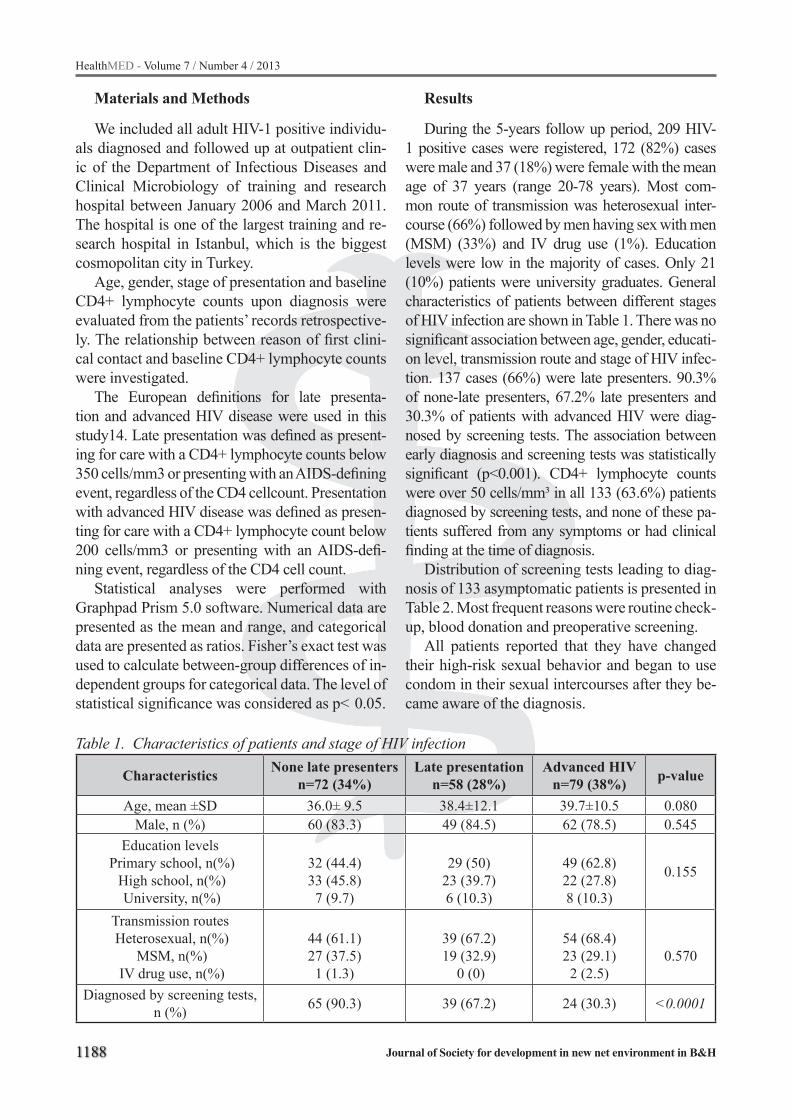
1188
HealthMED - Volume 7 / Number 4 / 2013
Journal of Society for development in new net environment in B&H
Materials and Methods
We included all adult HIV-1 positive individu-als diagnosed and followed up at outpatient clin-ic of the Department of Infectious Diseases and Clinical Microbiology of training and research hospital between January 2006 and March 2011. The hospital is one of the largest training and re-search hospital in Istanbul, which is the biggest cosmopolitan city in Turkey.
Age, gender, stage of presentation and baseline CD4+ lymphocyte counts upon diagnosis were evaluated from the patients’ records retrospective-ly. The relationship between reason of first clini-cal contact and baseline CD4+ lymphocyte counts were investigated.
The European definitions for late presenta-tion and advanced HIV disease were used in this study14. Late presentation was defined as present-ing for care with a CD4+ lymphocyte counts below 350 cells/mm3 or presenting with an AIDS-defining event, regardless of the CD4 cellcount. Presentation with advanced HIV disease was defined as presen-ting for care with a CD4+ lymphocyte count below 200 cells/mm3 or presenting with an AIDS-defi-ning event, regardless of the CD4 cell count.
Statistical analyses were performed with Graphpad Prism 5.0 software. Numerical data are presented as the mean and range, and categorical data are presented as ratios. Fisher’s exact test was used to calculate between-group differences of in-dependent groups for categorical data. The level of statistical significance was considered as p< 0.05.
Results
During the 5-years follow up period, 209 HIV-1 positive cases were registered, 172 (82%) cases were male and 37 (18%) were female with the mean age of 37 years (range 20-78 years). Most com-mon route of transmission was heterosexual inter-course (66%) followed by men having sex with men (MSM) (33%) and IV drug use (1%). Education levels were low in the majority of cases. Only 21 (10%) patients were university graduates. General characteristics of patients between different stages of HIV infection are shown in Table 1. There was no significant association between age, gender, educati-on level, transmission route and stage of HIV infec-tion. 137 cases (66%) were late presenters. 90.3% of none-late presenters, 67.2% late presenters and 30.3% of patients with advanced HIV were diag-nosed by screening tests. The association between early diagnosis and screening tests was statistically significant (p<0.001). CD4+ lymphocyte counts were over 50 cells/mm³ in all 133 (63.6%) patients diagnosed by screening tests, and none of these pa-tients suffered from any symptoms or had clinical finding at the time of diagnosis.
Distribution of screening tests leading to diag-nosis of 133 asymptomatic patients is presented in Table 2. Most frequent reasons were routine check-up, blood donation and preoperative screening.
All patients reported that they have changed their high-risk sexual behavior and began to use condom in their sexual intercourses after they be-came aware of the diagnosis.
Table 1. Characteristics of patients and stage of HIV infection
Characteristics None late presentersn=72 (34%)
Late presentationn=58 (28%)
Advanced HIVn=79 (38%) p-value
Age, mean ±SD 36.0± 9.5 38.4±12.1 39.7±10.5 0.080Male, n (%) 60 (83.3) 49 (84.5) 62 (78.5) 0.545
Education levels Primary school, n(%)
High school, n(%) University, n(%)
32 (44.4)33 (45.8)7 (9.7)
29 (50)23 (39.7)6 (10.3)
49 (62.8)22 (27.8)8 (10.3)
0.155
Transmission routes Heterosexual, n(%)
MSM, n(%) IV drug use, n(%)
44 (61.1)27 (37.5)1 (1.3)
39 (67.2)19 (32.9)
0 (0)
54 (68.4)23 (29.1)2 (2.5)
0.570
Diagnosed by screening tests, n (%) 65 (90.3) 39 (67.2) 24 (30.3) <0.0001

HealthMED - Volume 7 / Number 4 / 2013
Journal of Society for development in new net environment in B&H 1189
Baseline CD4+ lymphocyte counts of all pa-tients were grouped as <200/mm³, 200-350/mm³, 350-500/mm³ and >500/mm³. The proportions of reasons to HIV-1 test stratified by CD4+ lympho-cyte counts are presented in Figure 1.
Figure 1. Relationship between reasons of the HIV test and CD4+ lymphocyte numbers of cases
CD4+ lymphocyte counts were below 350 cells/mm³ in 137 patients at the time of the di-agnosis and HAART started immediately. CD4+ lymphocyte counts in 54% of patients were <200 cells/mm³ and they received prophylactic trime-thoprim-sulphametoxazol
(TMP/SMX) in addition to the treatment. Fur-thermore, 16% of patients had CD4+ count <50 cells/mm³ and received prophylactic clarithromy-cine as well.
Discussion
HIV/AIDS disease progresses asymptom-atically averagely 8-10 years after the infection [9,10]. Since the individuals who are unaware of their infection, can continue their risky behaviors,
the treatment is delayed and all of these contribute into the spread of HIV.
Delayed diagnosis in HIV/AIDS disease is di-rectly related to the disease spread, and Marks et al estimated 3.5 folds higher HIV transmission rate from a HIV positive unaware group then HIV positive aware group [11]. Marks et al. reported in their meta-analysis study that HIV positive in-dividuals decreased their high risk sexual behav-iors prominently after the diagnosis [12]. In our study, all patients reported that they have changed their high risk sexual behaviors after the diagnosis of HIV/AIDS. Late diagnosed patients are under great risk in clinical progression, their responses to the treatment are lower and development of op-portunistic infections are higher in these patients, when compared to the early diagnosed ones [2,14].
Approximately 25% of HIV mortality has been detected to be related to delayed diagnosis (or ad-vance disease [2,15,16]. Treatments of late diag-nosed individuals are generally more complex and, depending on this, have higher treatment costs [17]. 66% of our population was late presenters which is considerably higher compared to 49% late presen-ters in the European Union and European Econo-mic area in 2010 [23]. There is a need for focus on population based HIV education programmes and screening in Istanbul. There is free access to HIV care in Turkey. All late presenters received antiret-roviral treatment, and both with prophylactic agents and directed at concomitant diseases.
Late presentation of HIV infection has serious health consequences for the individual and poses a high financial burden on society. Krentz and Gill found significantly higher costs for late present-ers, especially inpatient costs, during the first year after accessing care, and that costs remained signif-
Table 2. Number of patients diagnosed with HIV-1 by screening test and the reasons for screeningScreening tests Number ( n=133) %
Check-up 29 21.8Blood donation 27 20.3Preoperative 26 19.5After suspected sexual intercourse 22 16.5Job application 12 9Marriage 12 9Military service 3 2.2Pregnancy 2 1.5

1190
HealthMED - Volume 7 / Number 4 / 2013
Journal of Society for development in new net environment in B&H
icantly higher (>50% higher) during subsequent years for HIV-infected patients who present with a CD4 count <350 cells/mm3 compared to early presenters [21].
HIV screening tests are an important strategy to decrease incidence of HIV infection among sexu-ally active adolescents. WHO stated in the progress report in 2011 that testing and counseling for HIV and sexually transmitted infections was one of the key approaches for prevention. In 2006, routine HIV infection screening in all individuals between ages 13-64 years, who have given consents, was placed within the CDC recommendations [18]. The Society for Adolescent Medicine also recommends routine screening tests in sexually active adoles-cents, especially those who live in regions with high HIV prevalence [19]. Moreover, American College of Obstetricians and Gynecologists recom-mends routine screening in sexually active females above 19 years of age [20]. In Turkey, compulsory screening tests are performed in special conditions like marriage procedures, job applications and for the military service. Furthermore, all blood do-nors are screened for HIV infection. Occasionally HIV tests may be performed in people, who have applied to a healthcare unit or in patients, who are admitted to hospital without informing them and re-ceiving consents. Although the ethical background of these situations are debatable, these may have positive impacts on patients since diagnosis at an early phase can have positive impacts on the dis-ease progression. Nevertheless, if individuals are informed about the importance of early diagnosis for themselves and the society, then it is a fact that these tests will be performed on voluntary basis rather than a compulsory one.
In conclusion, performing screening tests and supporting individuals with high risks to have HIV tests voluntarily can increase the numbers of early diagnosed patients. Thus, spread of the dis-ease and treatment costs can be decreased by an important extent.
References
1. Palella F, Delaney K, Moorman A, Loveless MO, Fuhrer J, Satten GA, et al. Declining morbidity and mortality among patients with advanced human im-munodeficiency virus infection. N Engl J Med 1998; 338(13): 853-60.
2. Palella FJ, Deloria-Knoll M, Chimel JS, Moorman AC, Wood KC, Greenberg AE, et al. Survival benefits of initiating antiretroviral therapy in HIV-infected persons in different CD4+ cell strata. Ann Intern Med 2003; 138(8): 620-6.
3. Sabin C, Smith C, Gumley H, Murphy G, Lamphe FC, Phillips AN, et al. Late presenters in the era of highly active antiretroviral therapy: uptake of and responses to antiretroviral therapy. AIDS 2004; 18(16): 2145-51.
4. Centers for Disease Control and Prevention (CDC). Late versus early testing of-16 sites, United States, 2000-2003. MMWR Morb Mortal Wkly Rep 2003; 52: 581-86.
5. Fisher M. Late diagnosis of HIV infection: major con-sequences and missed opportunities. Curr Opin Infect Dis 2008; 21: 1–3.
6. Adler A, Mounier-Jack S, Coker RJ. Late diagnosis of HIV in Europe: definitional and public health challen-ges. AIDS Care 2009; 21: 284–293.
7. Antinori A, Coenen T, Costagiola D, Dedes N, Ellef-son M, Gatell J, et al.Late presentation of HIV infecti-on: a consensus definition.
8. Centers for Disease Control and Prevention (CDC). Person Tested for HIV—United States, 2006. MMWR Morb Mortal Wkly Rep 2008 Aug 8; 57(31): 845-49.
9. Centers for Disease Control and Prevention (CDC). Projections of the number of persons diagnosed with AIDS and the number of immunosupressed HIV-infected persons-United States, 1992-1994.MMWR Morb Mortal Wkly Rep 1992; 41(RR-18): 1-29.
10. Rutherford GW, Lifson AR, Hessol NA, Darrow WW, O’Malley PM, Buchbinder SP, et al. Course of HIV-1 infection in a cohort of homosexual and bi-sexual men: An 11 year follow up study. BMJ 1990; 301(6762): 1183-88.
11. Marks G, Crepaz N, Janssen RS. Estimating sexual transmission of HIV from persons aware and un-aware they are infected with the virus in the USA. AIDS 2006; 20(10): 1447-50.
12. Marks G, Crepaz N, Senterfitt JW, Janssen RS. Me-ta-analysis of high-risk sexual behavior in persons

HealthMED - Volume 7 / Number 4 / 2013
Journal of Society for development in new net environment in B&H 1191
aware in persons aware and unaware they are in-fected with HIV in the United States: Implications for HIV prevention programs. J Acquir Immune Def-ic Syndr 2005; 39(4): 446-53.
13. Centers for Disease Control and Prevention. HIV/AIDS Surveillance Report, 2005. Vol. 17. Rev ed. Atlanta: U.S Department of Health and Human Ser-vices, Centers for Disease Control and Prevention; 2007: 5-9.
14. Antinori A, Coenen T, Costagiola D, Dedes N, Ellef-son M, Gatell J. Late presentation of HIV infection: a consensus definition. HIV Med 2011; 12: 61-4
15. Losina E, Figueroa P, Duncan J, Divi N, Wolf LL, Hirschhorn LR, et al. HIV morbidity and mortality in Jamaica: analysis of national surveillance data, 1993--2005. Int J Infect Dis 2008; 12(2): 132-38.
16. Chadborn TR, Delpech VC, Sabin CA, Sinka K, Ev-ans BG. The late diagnosis and consequent short-term mortality of HIV-infected heterosexuals (Eng-land and Wales, 2000-2004). AIDS 2006; 20(18): 2371-79.
17. Krentz HB, Auld MC, Gill MJ. The high cost of med-ical care for patients who present late (CD4 <200 cell/microL) with HIV infection. HIV Med 2004; 5(2): 93-98.
18. Centers for Disease Control and Prevention. Revised recommedations for HIV testing of adults, adoles-cents, and pregnant women in health care settings. MMWR Morb Mortal Wkly Rep 2006; 55: 1-17.
19. D’Angelo LJ, Samples C, Rogers AS, Peralta L, Friedman L. HIV infection and AIDS in adolescents: an update of the position of the Society for Adoles-cent Medicine. J Adolesc Health 2006; 38(1): 88-91.
20. ACOG Commitee Opinion. Routine human immu-nodeficiency virus screening. Obstet Gynecol 2008; 112(2 Pt 1): 401-3.
21. Krentz HB, Gill MJ. The Direct Medical Costs of Late Presentation (<350/mm3) of HIV Infection over a 15-Year Period. AIDS research and tre-atment 2012; 2012: 757135.
22. Karaosmanoglu HK, Aydin OA, Nazlican O. Profile of HIV/AIDS patients in a tertiary hospital in Ista-nbul, Turkey. HIV Clin Trials. 2011; 12(2): 104-8
23. Likatavicius G, van de Laar MJ. HIV infection and AIDS in the European Union and European Eco-nomic Area, 2010 Euro Surveill. 2011; 16(48): pii=20030. Available online: http: //www.eurosur-veillance.org/ViewArticle.aspx?ArticleId=20030
Corresponding AuthorHayat Kumbasar Karaosmanoglu,Haseki Training and Research Hospital, Infectıous Diseases and Clinical Microbiology,Istanbul,Turkey,E-mail: [email protected]
1192
HealthMED - Volume 7 / Number 4 / 2013
Journal of Society for development in new net environment in B&H
Abstract
It has been considered that STAT3 plays an important role in early embryonic development, but the relationship between STAT3 and recurrent spontaneous abortion and its possible mechanisms have not been fully elucidated. In the present stu-dy, we evaluated the expressions of STAT3 signal pathway associated proteins in the allogeneic mo-use models of low abortions (CBA/J*Balb/c) and abortion prone (CBA/J*DBA/2J). Results showed, in contrast to the control group, the IL-6 was de-creased and STAT3 signal pathway associated pro-teins were down-regulated in the abortion-prone mating on gestation day 7.5. Our results suggest that STAT3 signal pathway may be associated with recurrent abortion. Decreased IL-6 may block the STAT3 activation and thus lead to abortion.
Key words: Recurrent spontaneous abortion, STAT3, signaling pathway, relationship.
Introduction
Recurrent spontaneous abortion is a common complication of pregnancy and also one of the re-fractory diseases(1). It has been ascribed to a vari-ety of causal factors. Although many studies have been carried to investigate the recurrent spontaneo-us abortion, the pathogenesis of recurrent sponta-neous abortion is still largely unknown, which poses difficulty to its treatment (2). The pathway involving STAT transcription factor was initially discovered by researchers studying the transcrip-tional activation of genes through cytokines and growth factors in mammalian cells. It soon beco-mes clear that this pathway is conserved in many species and plays a central role in the regulation of numerous biological processes (3-4). STAT3, one of the members of the STAT family, is a key me-diator in growth regulation and its crucial effects have also been observed in many tissues. There is evidence showing that STAT3 is important for
the early embryonic development. Therefore, we speculate that STAT3 signaling pathway may be related to the recurrent spontaneous abortion.
STAT3 has been shown to be activated by va-rious cytokines and growth factors especially the interleukin-6 (IL-6) (5). IL-6 is a multifunctional cytokine regulating immune response, hematopoi-esis, and acute phase reaction (6). IL-6 exhibits its biological function through its receptor. The com-plex of IL-6 and soluble IL-6R can generate the IL-6-mediated signal transduction. Furthermore, the binding of IL-6 to its receptor triggers the associati-on of IL-6R with a second membrane glycoprotein with the molecular weight of 130 kD (gpl30).
STATs are potential transcription factors involved in the normal cellular response to the stimulation of cytokines or growth factors. They are activated by the members of Janus kinase (JAK). p-STAT, the ac-tive form of STAT, forms hetero and/or homodimers, then translocate to the nucleus and bind the conser-ved DNA sites such as the high affinity sis-inducible element, which, together with other transcription factors, regulate the expression of specific effector genes (7-9). STAT3 is one of the members of STAT family. Gene deletion assay, microarray, proteomics and chromatin immunoprecipitation assay have re-vealed STAT3 related target genes with a broad ran-ge of functions. Vascular endothelial growth factor (VEGF) is an endothelial cell specific mitogen and is a disulphide-linked homodimer of 34~42 kD that shares structural homology with the platelet derived growth factor (PDGF)-family (10). It can regula-te the proliferation, differentiation and survival of endothelial cells, act as a chemoattractant for mo-nocytes, mast cells and pericytes and enhance the vascular permeability. VEGF is considered as one of the major modulators of angiogenesis in a variety of physiological processes, including wound healing and embryonic development, and plays a key role in the pathogenesis of diseases including diabetic reti-nopathy, psoriasis and tumor growth.
Relationship between STAT3 signal pathway and recurrent spontaneous abortionShu-hua Zhao, Fan Zhao, Xiu-yun Zhao, Man-hua Cui
Department of Obstetrics and Gynecology, Second Hospital, Jilin University, Changchun, China

HealthMED - Volume 7 / Number 4 / 2013
Journal of Society for development in new net environment in B&H 1193
Survivin is a member of the inhibitor of apop-tosis (IAP) family containing a single baculovirus IAP repeat (BIR) domain and an extended -COOH terminal α-helical coiled coil (11). Survivin can regulate both cell proliferation and apoptosis. Ini-tially, survivin expression was thought to be con-fined to the developing fetus, as it is largely unde-tectable in normal adult tissues with the exception of placenta, testis and thymus. However, survivin is also expressed in the growth hormone-stimula-ted hematopoetic progenitor cells as well as the proliferating blood vessels (12). Moreover, survi-vin expression is also found in the chromaffin cells and renal tubule epithelial cells (13). At present, it has been considered to increase the angiogenesis and carcinogenesis (14, 15).
In the present study we explored the relationship of STAT3 signal pathway and the recurrent sponta-neous abortion. The CBA/J * BALB/c mice served as controls and CBA/J * DBA/2 mice with abortion prone as the experimental group were investigated. On the gestation day 7.5, serum IL-6 was determi-ned by ELISA. The expressions of IL-6R, GP130, STAT3, P-STAT3, VEGF and survivin were evalu-ated by western blot assay and immunohistoche-mistry. Results suggest that STAT3 signal pathway may be involved in the pregnancy loss.
Materials and methods
AnimalsMice were purchased from the Institute of Ex-
perimental Animals of the Chinese Academy of Medical Sciences and housed in an animal facility with a 12-h light/dark cycle. Animal care and ex-perimental procedures were followed according to institutional guidelines and conformed to require-ments of the state authority for animal research. CBA/J females were caged with DBA/2J or Balb/c males overnight and examined by vaginal smear in the morning every day. The presence of plug de-termined the first day of pregnancy. At this time, mice were segregated and pregnant females were sacrificed on the gestation day 7.5. Uteri and pla-centa were removed and cut into pieces. A fraction of tissues were used for immunochemistry and the remaining tissues were used for western blot assay.
Detection of serum IL-6 by ELISA After preparation and dilution, standard wells,
testing sample wells and bank wells were set. Di-luted standard 50µl was added to the standard well; Sample dilution 25µl was added to the testing sam-ple well, then testing sample 25µl was added. Noth-ing was added in blank well. Taking blank well as zero, the optical densit(OD) was measured at 450 nm after adding stop solution and within 15 min.
Western blot assayTissues were lysed with the Total Protein Ex-
traction Kit for 30 min at 4℃. After clarifica-tion, equal amounts of protein were separated by electrophoresis on a SDS-polyacrylamide gel and transferred onto a PVDF membrane using a Bio-Rad semidry transfer cell. The membrane was blocked in 5% non-fat milk containing 0.1% Tween 20 at room temperature for 1 h. The mem-brane was then incubated with corresponding primary antibodies at 4℃ overnight followed by washing and incubation with horseradish perox-idase-conjugated anti-rabbit secondary antibody (1: 5000). β-actin served as an internal reference. After washes, visualization was performed. The optical density (OD) of each band was quantified using GIS gel analysis software.
Immunohistochemistry Cryostat sections (10 mm) were treated with
3% H2O2 for 25 min to inactivate the endogenous peroxidase. After incubation, sections were rinsed in PBS thrice for 5 min each. Sections were then incubated with normal goat serum (50 μl/section) for 30 min. The monoclonal rabbit anti-mouse an-tibodies (50 μl/section) were applied to treat these sections for 1 h. Then, sections were rinsed in PBS thrice for 5 min each followed by incubation with peroxidase-conjugated goat anti-rabbit secondary antibody for 30 min. After rinsing in PBS thrice for 5 min each, the visualization was done by in-cubating sections with diaminobenzidine (DAB), followed by counterstaining with Mayer’s hema-toxylin. Slides were examined under a light micro-scope (Zeiss Axioscope). Analysis was performed using a digital image analysis system.

1194
HealthMED - Volume 7 / Number 4 / 2013
Journal of Society for development in new net environment in B&H
Results
Results from ELISAAccording to the concentration of the stan-
dards and the corresponding OD values, the lin-ear regression equation of standard curve was y=1244.1x-57.548, R2=0.9962. The IL-6 was then calculated. The IL-6 was 2.41 ± 72.820 ng/ml in the abortion group and 2.24 ± 62.01ng/ml in the control group, showing statistically significant dif-ference (P<0.01).
Results from Western blot assayThe expressions of STAT3 signal pathway as-
sociated proteins including GP130, STAT3, P-STAT3, survivin and VEGF were determined by western blot assay. Because the gestational day 7.5 is crucial with respect to the decidualisation, we focused on this particular day. Actin expression served as an internal control. Results showed that the CBA/J *DBA/2J pregnant females exhibited the significant down-regulation of these proteins as compared to low abortion mating (Figure 1).
Figure 1. Protein expressions of GP130, STAT3, P-STAT3, Survivin and VEGF by western blot assay (**P< 0.01 and *P < 0.05, n=6)
Results from immunohistochemistryThe STAT3 signal pathway associated proteins
were mainly expressed in the cytoplasm except for the p-STAT3. Immunohistochemistry revealed higher expression of those proteins in the CBA/J *Balb/c mice than in the CBA/J*DBA/2J mice (Figure 2).
Figure 2. Protein expression of GP130, STAT3, P-STAT3, VEGF and Survivin by immunohi-stochemistry (×400)
Discussion
The mechanisms underlying the gestation fail-ure are not fully elucidated, and now attributed to some immunological factors. We studied the expressions of STAT3 signal pathway associated proteins in the allogeneic mouse models with low abortions (CBA/J*Balb/c) and abortion prone (CBA/J*DBA/2J). Our results showed that there

HealthMED - Volume 7 / Number 4 / 2013
Journal of Society for development in new net environment in B&H 1195
were significant differences in the expressions of these proteins in the decidua between two groups, implying that there is a close relationship between STAT3 signaling pathway and recurrent spontane-ous abortion.
In this study, the blood was collected and se-rum IL-6 detected by ELISA on the pregnant day 7.5. Results showed that the IL-6 level was lower than that in the control group (P<0.01). Thus, we speculate that IL-6 plays an important role in the early embryonic stage and the low serum IL-6 level may be involved in the pathogenesis of re-current spontaneous abortion.
IL-6 exerts the biological function through its re-ceptor system. The complex of IL-6 and soluble IL-6R can generate the IL-6-mediated signal transduc-tion. Furthermore, the binding of IL-6 to its recep-tor triggers the association of IL-6R with a second membrane glycoprotein, gp130. High expression of GP130 mRNA has been found in the endometrium of pregnancy (16). In our study, results showed the GP130 was down-regulated in abortion pregnan-cies, suggesting a role of GP130 in the maintenance of mouse pregnancy. On the other hand, low IL-6 serum level influences the association of GP130 with the complex of IL-6 and IL-6R.
STAT3 is the center of the signalling pathway. Thus, the role of STAT-3 and p-STAT3 was inves-tigated in the pregnancy. Our results showed that both proteins were down-regulated in the abortion matings, CBA/J*DBA/2J. We speculate that the STAT-3 activity is involved in the maintenance of pregnancy. Decidualization was a prevented rea-son for down-regulation of STAT3. Furthermore, the embryos and the interaction contact with ma-ternal tissues were also blocked.
Two effector proteins, STAT3, VEGF and Sur-vivin, were determined in the present study. The expressions of both proteins showed similar find-ings to previous studies. VEGF is one of the ma-jor modulators of angiogenesis. Low VEGF level may pose difficulty in the decidual angiogenesis, and thus embryo is lost due to lacking of blood supply. Survivin regulates both cell proliferation and apoptosis. Down-regulation of Survivin leads to the apoptosis of decidual cells, and thus the em-bryos lose the support and fall off the endometrium. These findings suggest the expressions of these pro-teins are necessary to maintain the pregnancy.
The binding of IL-6 to its receptor triggers the as-sociation of IL-6R with the gpl30. STAT3 is activat-ed by the members of the JAK family of tyrosine ki-nases complexed with the cytoplasmicdomain of li-gand-bound cytokine receptors by GP130. p-STAT3 can form hetero and/or homodimers, then translocate to the nucleus and bind the conserved DNA sites, which together with other transcription factors regu-late the expressions of VEGF and Survivin. Due to the down-regulation of serum IL-6, STAT3 signaling pathway is inhibited. The development of embryo is stopped or even lost. In conclusion, our data points that IL-6/GP130/STAT3 signaling pathway plays an important role in the embryonic development. Reg-ulating these proteins may improve the survival of embryos, which may be a novel strategy for the treat-ment of recurrent spontaneous abortion.
References
1. Abou-Nassar K, Karsh J, Giulivi A and Allan D. Successful prevention of thrombotic thrombocytope-nic purpura (TTP) relapse using monthly prophylactic plasma exchanges throughout pregnancy in a patient with systemic lupus erythematosus and a prior history of refractory TTP and recurrent fetal loss. Transfus Apher Sci. 2010; 43(1): 29-31
2. Venkatesh S, Thilagavathi J, Kumar K, Deka D, Talwar P, Dada R. Cytogenetic, Y chromosome microdeletion, sperm chromatin and oxidative stress analysis in male partners of couples experiencing re-current spontaneous abortions. Arch Gynecol Obstet. 2011; 284(6): 1577-84
3. Wittig I and Groner B. Signal transducer and activa-tor of transcription 5 (STAT5), a crucial regulator of immune and cancer cells. Curr Drug Targets Immune Endocr Metabol Disord 2005; 5: 449-63
4. Valentino L and Pierre J. JAK/STAT signal transducti-on: regulators and implication in hematological mali-gnancies. Biochem Pharmacol 2006; 71: 713–21
5. Liao W, Yu C, Wen J, et al. Adiponectin induces interle-ukin-6 production and activates STAT3 in adult mouse cardiac fibroblasts. Biol Cell 2009; 101(5): 263-72
6. Kishimoto T. IL-6: from its discovery to clinical appli-cations. Int Immunol 2010; 22(5): 347-52
7. Schaefer TS, Sanders LK and Nathans D. Coopera-tive transcriptional activity of Jun and Stat3 beta, a short form of Stat3. Proc Natl Acad Sci U S A. 1995; 92: 9097-101

1196
HealthMED - Volume 7 / Number 4 / 2013
Journal of Society for development in new net environment in B&H
8. Wagner BJ, Hayes TE, Hoban CJ and Cochran BH. The SIF binding element confers sis/PDGF inducibi-lity onto the c-fos promoter. EMBO J1990; 9: 4477-84
9. Zhang X, Wrzeszczynska MH, Horvath CM and Dar-nell JE: Interacting regions in Stat3 and c-Jun that participate in cooperative transcriptional activation. Mol Cell Biol1999; 19: 7138-46
10. Conn G, Bayne ML, Sodermann DD et al. Amino acid and cDNA sequences of a vascular endothelial cell mitogen that is homologous to platelet-dervived growth factor. Proc Natl Acad Sci USA 1990; 87: 26 28-32
11. Altieri DC: Molecular circuits of apoptosis regulati-on and cell division control: The survivin paradigm. J Cell Biochem 2004; 92: 656-663
12. Ambrosini G, Adida C and Altieri DC. A novel an-ti-apoptosis gene, Survivin, expressed in cancer and lymphoma. Nat Med 1997; 3: 917–921
13. Koch CA, Vortmeyer AO, Diallo R, et al. Survivin: a novel neuroendocrine marker for pheochromocyto-ma. Eur J Endocrinol 2002; 146: 381-388
14. Koch CA, Vortmeyer AO, Diallo R, et al. Survivin: a novel neuroendocrine marker for pheochromocyto-ma.Eur Endocrinol 2002; 146: 381-388
15. Graaf AO, de Witte T and Jansen JH. Inhibitor of apoptosis proteins: new therapeutic targets in hema-tological cancer?. Leukemia2004; 18: 1751-1759
16. Lam SP, Luk JM, Man K, et al. Activation of in-terleukin-6-induced glycoprotein 130/signal tran-sducer and activator of transcription 3 pathway in mesenchymal stem cells enhances hepatic differen-tiation, proliferation, and liver regeneration. Liver Transpl 2010; 16: 1195-206
Corresponding AuthorMan-Hua Cui,Department of Obstetrics and Gynecology,Second Hospital, Jilin University,Changchun,China,E-mail: [email protected]
HealthMED - Volume 7 / Number 4 / 2013
Journal of Society for development in new net environment in B&H 1197
Abstract
Objective: The goal of this study was to assess the value of intermittent hypobaric pressure (the Greensac® kit) in the treatment of patients with inoperable peripheral arterial occlusive disease (PAOD).
Methods: The prospective study included 68 patients with pathology of lower extremities. Effects of the Greensac® kit on the claudicating distance prolongation and/or wound healing in critical leg ischemia were analyzed. Results were analyzed using standard statistical methods.
Results: The obtained data showed a signifi-cant claudicating distance prolongation in patients with arteriosclerosis lesions predominantly in ear-ly stages of the disease. In the group with critical leg ischemia, the claudicating distance improve-ment was much lower, compared to the group of patients with an early stage of the disease.
Conclusion: Treatment using the Greensac® kit may decrease the claudicating pain more in the earlier phases of the disease. In order to assess the value of the Greensac® kit in the treatment of is-chemic foot ulcers in critical leg ischemia, a grea-ter number of patients are necessary.
Key words: Arterial occlusive disease, the-rapy, leg, blood supply, aged.
Introduction
It is believed that over 5% of adult population today suffers from some kind of peripheral arterial occlusive disease (PAOD) of lower extremities. A majority of these patients are inoperable; either they need no surgery, or due to advanced disease, adequ-ate revascularization cannot be performed (1, 2).
The incidence of PAOD tends to shift toward the younger age, mainly because of the prevalence of risk factors (smoking, dyslipidemia, obesity), physical inactivity, inadequate diet and so on. That is why it is reasonable to expect a significant increase in the number of patients with PAOD in the future, and therefore a rise in the cost of tre-atment, so each new method that may be used in its treatment deserves attention of clinicians (2).
In recent years, a new type of conservative tre-atment, by intermittent hypobaric pressure, has appeared in our environment, and literature data show that it significantly improves circulation and increases lymphatic drainage (3–8). Its basic physiopathology mechanism includes increasing blood and lymph movement in the subcutaneous tissues and manipulation with this relatively small quantity of fluids. That is why circulation through the tissues is better and peripheral oxygenation is improved. Improvement of peripheral perfusion may lead to a reduction of symptoms and helps wound healing (4, 9-11). This study was designed to investigate the value of intermittent hypobaric pressure in the treatment of patients with PAOD of the lower extremities. The objective of this paper was to analyze effects of intermittent hypobaric pressure on the following:
- Elimination of claudication symptoms during a 6-minute walk on flat ground;
- Indicators of peripheral circulation improvement - ankle-brachial index (ABI) and skin temperature;
- Pain in patients with critical limb ischemia; - Wound healing in patients with critical limb
ischemia.
Use of intermittent hypobaric pressure in the treatment of inoperable peripheral arterial occlusive disease (PAOD) in elderly patientsBranko Milicevic1, Gordana Devecerski2, Jure Jelenc3, Nikola Radakovic4
1 Institute for Workers’ Health Protection Novi Sad, Novi Sad, Serbia,2 Clinic of Medical Rehabilitation, Clinical Center of Vojvodina, Novi Sad, Serbia,3 Iskra Medical, Ljubljana, Slovenia,4 Center for Integrative Medicine, Novi Sad, Serbia.
1198
HealthMED - Volume 7 / Number 4 / 2013
Journal of Society for development in new net environment in B&H
Methods
The study included patients aged 65 and over with clinical symptoms of PAOD: 68 patients had a verified PAOD, but were inoperable (for general and/or local reasons) and could not undergo endo-vascular revascularization.
The patients were submitted to intermittent hypobaric pressure using the Greensack® kit (manu-factured by “Iskra-Medical” from Slovenia) during 2 sessions, each of 10 treatments lasting 20 minutes, 3 times a week. There was a pause of 14 – 21 days between sessions. All patients underwent a standard diagnostic algorithm, which included clinical exa-minations, noninvasive diagnosis, and invasive arte-riography. Angiography was not performed in pati-ents with stage II of the disease (Fontaine staging).
Inclusion criteria are provided in Table 1. Table 1. Inclusion criteria
Inclusion criteria1. Age 65 and over
2. Symptoms of claudication and/or trophic foot ischemia
3.
Established PAOD with contraindicated re-vascularization: · Morbus cruris· Subcritical ischemia (Fontaine stage IIa)· Comorbidity (recent myocardial infarc-
tion, chronic renal failure, malignant dis-eases, cerebrovascular insult
All patients filled out a form with information related to PAOD and possibly existing critical foot ischemia. Important data measured the claudica-tion distance before and after treatment and one month after therapy, as well as ulcer surface be-fore treatment and one month after therapy. The subjective feeling of pain was also evaluated on a scale of 1 to 10 (after the abovementioned time intervals). These parameters were gathered and correlated with factors and comorbidity. Two gro-ups of patients were formed:
Group A included 36 patients with moderate symptoms and subcritical ischemia. These pati-ents were not referred for surgery due to the cli-nical stage of the disease (IIa stage after Fontai-ne), previous insult with paralysis, or malignant diseases. The claudication distance was measured before and after treatment with result correlation.
Group B included 32 patients with critical limb ischemia. These patients presented with advanced crural lesions, with trophic changes and/or pain at rest. Foot wound healing was also measured. In patients with toe gangrene, the speed of recovery and demarcation were measured, and in those with trophic ulcers, the ulcer surface reduction was me-asured in relation to the time of healing. The wo-und healing progress was evaluated from 1 to 5 on each weekly control, and results were summarized at the end of the treatment.
The treatment outcome was evaluated after the second treatment and one month later. All data were statistically analyzed using standard statistical methods and the statistical program SPSS 7.0. The gathered data are presented in tables and figures.
Results
Group AThis group included 36 patients with symptoms
of subcritical ischemia and claudication during a 6-minute walk. One month after treatment, 14 pa-tients reported that they were claudication free.Table 2. Claudication before and after therapy
Group A Before After T-test resultsNumber of pts 36 22 p<0.05
Group B This group included patients with critical limb
ischemia and they were evaluated related to their subjective feeling of pain in the legs (from 1 to 10) and volume of wound healing during the exami-nation period – before and after therapy and one month after treatment.
The group included 32 patients with pain at rest and trophic changes, ischemic ulcers, dry toe or toes gangrene and/or “wet” gangrene. The first group of data was related to the subjective feeling of pain at rest in patients with critical limb ische-mia. A significant improvement and pain relief was reported by 23 patients (71.9%) in the initial phase, after the second treatment. Out of this num-ber, reduced symptoms remained after one month in 9 patients (28.1%), indicating relatively short-term effects of the therapy. Regarding wound he-aling, fast effects occurred in 14 patients (43.8%), whereas delayed improvement occurred in 17 pa-tients (53.1%) that are presented in Graph 1.

HealthMED - Volume 7 / Number 4 / 2013
Journal of Society for development in new net environment in B&H 1199
Graph 1. Treatment outcome related to pain re-lief and wound healing progress
Discussion
The ideal course of treatment for patients with PAOD is revascularization. However, feasibility of revascularization depends on the general condi-tion and operability of patients, as well as existen-ce of comorbid factors such as advanced cardiac insufficiency, recent acute myocardial infarction, chronic renal failure, recent cerebrovascular in-sult, advanced malignant disease, condition after cerebrovascular accident with advanced organic psychosyndrome. Also, patients with an ischemic or paralyzed limb, characteristic for the state after insult with contractures which cannot be correc-ted, monoplegia, hemiplegia, paraplegia, demen-tia and so on, are not feasible for revascularization.
Conservative therapy of symptomatic occlusi-ve disease is still insufficient and reserved mostly for patients who may not undergo some form of revascularization, but a significant number of pa-tients with PAOD are inoperable for various re-asons. In such cases treatment with intermittent hypobaric pressure, like Greensack® is available.
Effects of Greensack® kit in Group A (36 pa-tients with moderate symptoms and subcritical lower limb ischemia) was satisfactory, with a sta-tistically significant decrease of patients with cla-udication symptoms.
Group B included 32 patients with critical limb ischemia. In this group, intermittent hypobaric pressure was used for pain relief and improvement of wound healing.
Pain is a subjective category and its interpre-tation depends on objective circumstances (stage of ischemia), but also on subjective conditions. In 71.9% of patients, Greensack® caused initial relief
of pain at rest. In the further course of therapy, re-duction of its effects on pain occurred, and 28.1% of patients experienced satisfactory effects of the-rapy. An explanation for this could be sought in a more favorable effect of therapy in the treatment of diabetic neuropathy due to evacuation of xyli-tol and acid products in the skin and subcutaneous tissue that occurs when affecting fluids in the feet and lower leg (7-11).
Wound healing was significantly affected by the fact that this group of patients mostly presen-ted with absolute prevalence of diabetic etiology (diabetes mellitus type IIb was registered in 27 out of 32 patients) and with accompanying crural dia-betic arteriopathy and low systolic pressure in the ankle. This indicates to a small amount of blood flow and fluid retention in the lower leg. Diabetic arteriopathy is characterized by extreme rigidity and calcification of blood vessels as well as micro-angiopathy. Under these conditions, the effect of nutritive flow is restricted so that the exchange of fluids through the skin is also limited. According to the findings of this study, 43.8% of patients re-ported a remarkable immediate effect on wound healing, while a delayed effect occurred in 53.1% of patients. Given that these patients were expo-sed to other conservative methods of treatment as well, the findings point to the synergy of the therapy using intermittent hypobaric pressure with other methods of treatment. Taking into conside-ration the total number of patients, we believe that a larger sample is necessary to obtain relevant conclusions. This analysis requires a special inve-stigation (3, 4, 9).
Conclusion
Based on the obtained findings, the following conclusions can be made:
1. Intermittent hypobaric pressure therapy using the Greensack® kit leads to significant reduction of the claudication symptoms.
2. Improved wound healing in patients with critical limb ischemia occurs after the end of treatment, but due to a relatively small number of subjects, it cannot be fully contributed to Greensack® therapy.
3. Pain relief is more pronounced immediately after completion of treatment, but after

1200
HealthMED - Volume 7 / Number 4 / 2013
Journal of Society for development in new net environment in B&H
a month as well; more than a quarter of patients feels significantly less pain after therapy.
Our general conclusion is that the Greensack®
kit deserves its place in the treatment of inoperable peripheral arterial disease, but for final evaluation in patients with critical limb ischemia, further stu-dies are necessary with more patients and a greater number of tested parameters.
Acknowledgment
Work was partially funded by the European Regional Development Fund (Biomedical Engi-neering Competence Center, Slovenia).
References
1. Hirsh AT, Criqui MH, Treat-Jacobson D. Peripheral arterial disease detection, awareness, and treatment in primary care.JAMA 2001; 286: 1317-24
2. Damjanović Ž. Fizikalna terapija periferne oblitera-tivne arteriosklerotične bolesti. Timočki Medicinski Glasnik. 2004; (29)2: 97-102
3. Himmelstrup H, Himmelstrup B, Mehlsen J, Trap-Jensen J.: Effects of vacusac in intermittent claudica-tion: a controlled cross-over study. Clin Physiol. 1991 May; 11(3): 263-9.
4. Bostrom K, Fors B.: “What I do is not dangerous and no hocus pocus”. Vacusac therapist fights for recogni-tion. Interview by Elisabet Forslind. Vardfacket. 1997 Dec 8; 21(11): 16-9.
5. Mehlsen J, Himmelstrup H, Himmelstrup B, Winther K, Trap-Jensen J.: Beneficial effects of intermittent suction and pressure treatment in intermittent claudication. Angiology. 1993 Jan; 44(1): 16-20.
6. Himmelstrup H, Himmelstrup B, Mehlsen J, Bonde J, Trap-Jensen J. The effect of natural medicine and vacuum therapy (Vacusac) in patients with stable in-termittent claudication Ugeskr Laeger. 1987 Mar 23; 149(13): 845-8. Danish.
7. Delis KT, Nicolaides AN, Wolfe JH, Stansby G.: Im-proving walking ability and ankle brachial pressure indices in symptomatic peripheral vascular disease with intermittent pneumatic foot compression: a pro-spective controlled study with one-year follow-up.J Vasc Surg. 2000 Apr; 31(4): 650-61
8. Mehlsen J, Himmelstrup H, Himmelstrup B, Winther Hansen KF. Positive effect of intermittent overpres-sure and underpressure (Vacusac) in intermittent claudication Ugeskr Laeger. 1994 Jan 10; 156(2): 169-71
9. Ramaswami G, D’Ayala M, Hollier LH, Deutsch R, McElhinney AJ.: Rapid foot and calf compression in-creases walking distance in patients with intermittent claudication: results of a randomized study.J Vasc Surg. 2005 May; 41(5): 794-801.
10. Devečerski G, Teofilovski M, Miličević B, Novaković B, Popović V, Radaković N. Uticaj terapije in-termitentnog negativnog pritiska na poboljšanje mirkocirkulacije u bolesnika sa dijabetesom – prikaz slučaja. Med Pregl 2006; 59 (Suppl): 63-5.
11. Devečerski G, Teofilovski M, Miličević B, Konstantinović Lj, Lazović M, Radaković N, Mujović N, Krstin A, Jelenc J. Effects of Greensack® on low-er limb functional status. Europa Medicophysica 2006; 42 (Suppl): 145-6.
Corresponding AuthorBranko Milicevic,Institute for Workers’ Health Protection,Novi Sad,Serbia,E-mail: [email protected]

HealthMED - Volume 7 / Number 4 / 2013
Journal of Society for development in new net environment in B&H 1201
Abstract
Objectives: The aim of this study is to deter-mine the presence of styloid process elongation (SPE) detected on panoramic radiographs in end-stage renal disease (ESRD) patients with peritone-al dialysis (PD) and to investigate the role of Ca and P metabolism (ectopic calcification) for SPE by evaluating the PD period and the correlation between the styloid process length and this period.
Study design: One hundred and ten patients with ESRD and PD were enrolled in the present study. The PRs of 5 patients who have questiona-ble SP and the other 5 patients who have co-dis-orders (3 thyroid gland and 2 parathyroid gland operations) were excluded. Therefore, 100 ESRD patients with PD were included.
Results: Eleven (11%) subjects demonstrated SPE at least one side. The PD period was not sta-tistically different between the patients with SPE and the patients without SPE. There was not a si-gnificant correlation between the PD period and styloid process length in these patients.
Conclusions: According to our knowledge, this is the first study investigating the role of ec-topic calcification in the elongation of styloid pro-cess in patients with ESRD. In conclusion, instead of the high prevalence, ectopic calcification or the abnormality in Ca and P metabolism may not have a role in this elongation.
Key words: Ectopic calcification, styloid pro-cess elongation, end-stage renal disease, panora-mic radiography.
Introduction
The styloid process (SP) is a cylindrical, long cartilaginous bone located on the temporal bone. There are many vessels such as carotid arteries and nerves adjacent to the SP.[1-3] The normal len-gth of the SP is approximately 20-30 mm.[4-9] The styloid process elongation (SPE) can be assumed if either the SP or the adjacent stylohyoid ligament ossification shows an overall length in excess of 30 mm.[4,6,8-17] SPE resulted in facial and neck pain is known as Eagle’s syndrome.[6,7,11,13,18-20] More uncommonly, symptoms such as dysphagia, tinni-tus, and otalgia may occur in patients with this syndrome. All these symptoms may be due to SPE causing the compression of the nerves such as glossopharyngeal, vagus, etc., carotid arteries compression and other reasons. Therefore, it may also cause stroke due to this compression.[4,21,22]
The exact cause of the elongated SP due to cal-cified and ossified bone and ligament is not clear. It was suggested that local chronic irritations, sur-gical trauma, endocrine disorders in female at me-nopause, persistence of mesenchymal elements, growth of the osseous tissue and mechanical stress or trauma during development of SP could result in calcified hyperplasia of the SP.[2,4,13,23,24]
Extraskeletal (ectopic) calcification (deposition calcium phosphate crystals) or ossification (true bone formation) may have a role for the elonga-tion of SP. Ectopic calcification (EC) in nonosse-ous soft tissue may be due to three mechanisms; metastatic calcification due to disorders causing abnormal serum Ca and phosphate P levels, dys-trophic calcification due to mineral deposition into metabolically impaired or dead tissue despite
Styloid process elongation in end stage renal disease patients with peritoneal dialysis: is there any role for ectopic calcification?Cumali Gokce1, Elif Tarim Ertas2, Yildiray Sisman3, Murat Rifalioglu4, Oktay Oymak5, Cengiz Utas5
1 Department of Endocrinology and Metabolism, School of Medicine, Mustafa Kemal University, Hatay, Turkey,2 Department of Maxillofacial Radiollogy, Faculty of Dentistry, Izmir Katip Celebi University, Izmir, Turkey,3 Department of Oral Diagnosis and Radiology, Faculty of Dentistry, Erciyes University, Kayseri, Turkey,4 Department of Urology, School of Medicine, Mustafa Kemal University, Hatay, Turkey,5 Department of Nephrology, School of Medicine, Erciyes University, Kayseri, Turkey.

1202
HealthMED - Volume 7 / Number 4 / 2013
Journal of Society for development in new net environment in B&H
normal serum levels of Ca and P, and ectopic os-sification. In patients with end-stage renal disease (ESRD) have risks for EC or ossification due to disorders (renal failure, dialysis, secondary hyper-parathyroidism) causing metastatic calcification.[25] The increase of iPTH over the recommended levels, associated with hypercalcemia and hyperp-hosphataemia, can induce extraosseous calcificati-ons and cardiovascular events. [26, 27] Extraosseous, especially vascular calcifications are an important complication for the patients treated with PD.[28,29]
In the present study, our aim is to investigate the prevalence of SPE- the role of EC-in ESRD pa-tients with peritoneal dialysis (PD) and to analyze the correlation between SP length and PD period. According to the literature, this the first study in-vestigating the role of Ca and P metabolism in the elongation of SP.
Materials and methods
One hundred and ten ESRD patients with PD are investigated for SPE by panoramic radiography (PR). All the PRs were taken at the Erciyes Univer-sity Faculty of Dentistry, Department of Oral Diag-nosis and Radiology. The PRs of the 110 ESRD pa-tients with PD had originally been taken for routine examination. It was not necessary to seek ethical approval as the PRs were essential for the routine clinical evaluation of the patients. The PRs who have questionable SP were excluded in the pres-ent study. Patients had disorders causing dystro-phic calcification (scleroderma, dermatomyositis, systemic lupus erythematosis, trauma-induced) and ectopic ossification (post surgery, burns, neurologic injury, myositis ossificans) were not included. A detailed differential diagnosis was done for the dis-orders causing–metastatic calcification-abnormal serum Ca and P levels. Except renal failure, disease causing metastatic calcification such as sarcoidosis, tumoral calcinosis, primary hyperthyroidism, milk alkali syndrome etc. were not enrolled.[25] PD peri-ods of all the patients were noted.
All the panoramic radiographs were done and evaluated in the same fashion (Orthopanto-mography® OP100, Tuusula, Finland). The PRs were processed in relation to the manufacturer’s recommendations in an automatic film proces-sor. For mineralized stylohyoid complexes and
lengths of bilateral SPs were evaluated by using the measurement method of Jung et al’study.[8] In brief, the measurements were taken on the tem-poral bone’s frontal side. A thin transparent line is usually imagined between the SP shadows and the tympanic bone in this area on the PRs. This transparent line corresponds to the cleft between the SP and the temporal bone’s tympanic plate [8,30] (Figure 1 A,B,C). The tip of the SP is its bony end including calcified parts of the ligament. All the PRs were viewed in subdued ambient light using transmitted light from a standard viewbox. The lengths of the SPE variants were measured using a true-toscale radiometric ruler (magnification fac-tor: 1.4). The radiographs were investigated and the measurements were performed by the same author (Y.S). To check the intraobserver varia-tions, measurements were repeated after some weeks on a subset of 50 PRs. The length of SP and/or stylohyoid ligament, which are longer than 30 mm were considered to be SPE.[4,6,8-13]
The observed results were analyzed with SPSS 10.0 (Statistical package for social science Inc., Chicago, Illinois, USA). t-test analysis was used to compare PD periods in patients with SPE and without SPE. A correlation analysis was performed between the SP length and PD period. P values less than 0.05 were accepted as statistically significant.
Results
One hundred and ten patients with ESRD and PD were enrolled in the present study. The PRs of 5 patients who have questionable SP and the other 5 patients who have co-disorders (3 thyroid gland and 2 parathyroid gland operations) were exclud-ed. Therefore, 100 ESRD patients with PD were included. The mean age of these patients was 46.4 ± 13.4 years. On all the 100 PRs, the length of the SP could be measured. No significant difference in mean age between the males (48.7 ± 12.2 years) and the females (44.1 ± 14.3 years) was detected.
Eleven (11%) subjects demonstrated SPE at least one side. This abnormality (elongation) was present in both genders. The majority of these abnormalities 8 (8%) were unilateral with 6 (6%) occurring on the right and 2 (2%) on the left side. The remainder (n = 3) were bilateral. There was a greater tendency for the abnormality in male population (n = 9).

HealthMED - Volume 7 / Number 4 / 2013
Journal of Society for development in new net environment in B&H 1203
Figure 1. (A,B,C). The styloid process length was measured by using anatomical landmarks on the panoramic radiographs in patients with end-stage renal disease. (Based on Jung et al.[8] and Sisman et al.[17])
The mean PD period and SP length of these pa-tients were 4.0 ± 3.3 years (range: 1-17 years) and 21.5 ± 8.1 mm (range: 7-47 mm), respectively. No significant difference in mean age between males (48.7 ± 12.2 years) and females (44.1 ± 14.3 years) was detected. There was not a statistical difference for the PD period between the patients with SPE (5.2 ± 3.4 years) and the patients without SPE (3.8 ± 3.3 years) (Table 1). There was also not a sig-nificant correlation between the PD period and SP length in these patients (Figure 2). Table 1. PD periods in ESRD patients
Patients PD period (years)ESRD patients with SPE 5.2±3.4 ESRD patients without SPE 3.8±3.3 p >0.05

1204
HealthMED - Volume 7 / Number 4 / 2013
Journal of Society for development in new net environment in B&H
Figure 2. A correlation graphic between the PD period and SP length in patients with ESRD
Discussion
The elongated SP and structural changes in sty-lohyoid ligament with its clinical symptoms were first described by Eagle. So, it is also known as the Eagle’s syndrome.[18,31] The symptoms and signs with this syndrome are due to the anatomic rela-tionship between the SP and its surrounding struc-tures.[1,32] The symptoms due to this syndrome can be confused with some disorders including a wide variety of facial neuralgias, oral, dental and, tem-poromandibular diseases. Therefore, a detailed differential diagnosis for SPE should be done.[20]
Although there are many suggested hypotheses, the definite etiology of calcified and ossified SPE is unknown.[2,4,13,23,24] However, according to our knowledge, there is no study investigating the role of Ca and P metabolism in patients with elongated SP. EC-metastatic calcification- in nonosseous soft tissue due to abnormal serum Ca and P lev-els (abnormality in Ca and P metabolism) is very common in patients with ESRD.[25] Therefore, this disease is a good model for the investigation of the EC in development of SPE.
There are limited Turkish reports investigating the SPE prevalence in normal population. Ilguy et al evaluated the PRs of 860 subjects in terms of SPE. Of these patients, 32 patients (3.7%) had SPEs.[6] In another study, the SPE prevalence on PRs was investigated for the 900 adult patients with dental problems and the prevalence was
1.1%.[33] In our previous study, based on 689 PRs consecutively retrieved from the archival records of our faculty, we aimed to investigate the prev-alence of SPE by PR and to analyze this preva-lence in relation to gender and age subgroups in a Turkish population living in the same region. The mean age of these 698 patients (285 male; 413 fe-male) was 34.9±14.1 years and fifty four (7.7%) subjects demonstrated SPE at least one side.[14]
As in our study, the length of SP and/or stylohy-oid ligament, which are longer than 30 mm were considered to be SPE in these Turkish reports. According to our knowledge, the present study is the first report investigating the SPE prevalence on PRs in ESRD patients with PD. In the present study, the prevalence was 11% in these patients and the findings were relatively high comparing to our previous results if we consider as a control cohort. This high prevalence in comparison to the other Turkish reports may be due to underlying disorder, but the sample of the study may not be enough for this comment.
The extent of EC increases with the dialysis peri-od and age.[34] EC due to the abnormality in calcium and phosphate metabolism which is related to the duration of dialysis is also very important for this calcification and it is very common in patients with ESRD.[25, 34] This is the first study evaluating the du-ration of the peritoneal dialysis between the patients with SPEs and the patients without SPE and analyz-ing the correlation between SPE and PD period in terms of EC. It was found that the PD period was not significantly different in the patients with SPEs than in the patients without SPEs. Also, there was not a significant correlation between the SP length and the PD period in the patients with ESRD.
As a result, SPEs found as incidental findings on standard PRs may be important clinically in not only patients with ESRD, but also normal popula-tion. Instead of many hypotheses and studies, the exact etiology of elongated SP and the role of ecto-pic calcification are unknown. SPEs in the present study were detected by PRs in 11% of the ESRD patients with PD. The PD period of the patients with SPEs was not significantly different than the patients without SPEs. Also, there was not a signif-icant correlation between the length of SP and PD period in these patients. According to our knowl-edge, this is the first study done in ESRD patients

HealthMED - Volume 7 / Number 4 / 2013
Journal of Society for development in new net environment in B&H 1205
with PD. We also firstly investigated the role of Ca and P metabolism in the development of the SPEs. In conclusion, instead of the high prevalence, ec-topic calcification or the abnormality in Ca and P metabolism may not have a role in the elongation of SP. However, further studies and large samples are needed to clarify the etiology of this disorder.
References
1. Gözil R, Yener N, Calgüner E, Araç M, Tunç E, Bah-celioğlu M. Morphological characteristics of styloid process evaluated by computerized axial tomography. Ann Anat. 2001; 183: 527-35.
2. Krennmair G, Piehslinger E. Variants of ossification in the stylohyoid chain. Cranio. 2003; 21: 31-7.
3. Camarda AJ, Deschamps C, Forest D. II. Stylohyoid chain ossification: a discussion of etiology. Oral Surg Oral Med Oral Pathol. 1989; 67: 515-20.
4. Prasad KC, Kamath MP, Reddy KJ, Raju K, Agarwal S. Elongated styloid process (Eagle’s syndrome): a cli-nical study. J Oral Maxillofac Surg. 2002; 60: 171-5.
5. Monsour PA, Young WG. Variability of the styloid pro-cess and stylohyoid ligament in panoramic radiograp-hs. Oral Surg Oral Med Oral Pathol. 1986; 61: 522-6.
6. Ilgüy M, Ilgüy D, Güler N, Bayirli G. Incidence of the type and calcification patterns in patients with elonga-ted styloid process. J Int Med Res. 2005; 33: 96-102.
7. Kursoglu P, Unalan F, Erdem T. Radiological evalua-tion of the styloid process in young adults resident in Turkey’s Yeditepe University faculty of dentistry. Oral Surg Oral Med Oral Pathol Oral Radiol Oral Endod. 2005; 100: 491-4.
8. Jung T, Tschernitschek H, Hippen H, Schneider B, Bor-chers L. Elongated styloid process: when is it really elongated? Dentomaxillofac Radiol. 2004; 33: 119-24.
9. Zaki HS, Greco CM, Rudy TE, Kubinski JA. Elon-gated styloid process in a temporomandibular di-sorder sample: prevalence and treatment outcome. J Prosthet Dent. 1996; 75: 399-405.
10. Ferreira de Albuquerque R Jr, Müller K, Hotta TH, Gonçalves M. Temporomandibular disorder or Eag-le’s syndrome? A clinical report. J Prosthet Dent. 2003; 90: 317-20.
11. Lee S, Hillel A. Three-dimensional computed tomog-raphy imaging of Eagle’s syndrome. Am J Otolaryn-gol. 2004; 25: 109.
12. Keur JJ, Campbell JP, McCarthy JF, Ralph WJ. The clinical significance of the elongated styloid process. Oral Surg Oral Med Oral Pathol. 1986; 61: 399-404.
13. Murtagh RD, Caracciolo JT, Fernandez G. CT fin-dings associated with Eagle syndrome. Am J Neuro-radiol. 2001; 22: 401-1402.
14. Gokce C, Sisman Y, Tarim Ertas E, Akgunlu F, Ozturk A. Prevalence of styloid process elongation on pa-noramic radiography in the Turkey population from Cappodocia region. Eur J Dent. 2008; 2: 18-22.
15. Gokce C, Sisman Y, Sipahioglu M. Styloid process elongation or Eagle’s syndrome: is there any role for ectopic calcification? Eur J Dent. 2008; 2: 224-8.
16. Sisman Y, Gokce C, Sipahioglu M. Bilateral elon-gated styloid process in an end-stage renal disease patient with peritoneal dialysis: is there any role for ectopic calcification? Eur J Dent. 2009; 3: 155-7.
17. Sisman Y, Gokce C, Tarim Ertas E, Sipahioglu M, Akgunlu F. Investigation of elongated styloid pro-cess prevalence in patients with torus palatinus. Clin Oral Investig. 2009; 13: 269-72.
18. Eagle WW. Elongated styloid process. Report of two cases. Arch Otolaryngol. 1937; 25: 548-87.
19. Cernea CR, Hojaij FC, De Carlucci D Jr, Plopper C, Vanderley F, Guerreiro CA., et al. First-bite sy-ndrome after resection of the styloid process. Laryn-goscope. 2007; 117: 181-2.
20. Aral IL, Karaca I, Güngör N. Eagle’s syndrome masquerading as pain of dental origin. Case report. Aust Dent J. 1997; 42: 18-9.
21. Chuang WC, Short JH, McKinney AM, Anker L, Knoll B, McKinney ZJ. Reversible left hemispheric ischemia secondary to carotid compression in Eagle syndrome: surgical and CT angiographic correlati-on. Am J Neuroradiol. 2007; 28: 143-5.
22. Strauss M, Zohar Y, Laurian N. Elongated styloid process syndrome: intraoral versus external appro-ach for styloid surgery. Laryngoscope. 1985; 95: 976-9.
23. Balbuena L Jr, Hayes D, Ramirez SG, Johnson R. Eagle’s syndrome (elongated styloid process). South Med J. 1997; 90: 331-4.
24. Fini G, Gasparini G, Filippini F, Becelli R, Mar-cotullio D. The long styloid process syndrome or Eagle’s syndrome. J Craniomaxillofac Surg. 2000; 28: 123-7.

1206
HealthMED - Volume 7 / Number 4 / 2013
Journal of Society for development in new net environment in B&H
25. Favus MJ, Vokes TJ. Paget disease and other dysplasias of bone. In: Jameson JL, ed. Harrison’s endocrinology. USA: McGraw-Hill Companies, 2006: 485–98.
26. Rusu CC, Moldovan D, Valea A, Parvu L, Kacso I, Bondor C, Patiu IM, Racasan S, Gherman-Caprioara M. The calcium phosphorus product is a better indi-cator for survival than immunoreactive Parathormo-ne in chronic hemodialysis patients with renal failure. Possible role of serum albumin level. Acta Endocrino-logica (Buc). 2009; vol. V, no. 3, p. 349-60.
27. Kacso I, Rusu A, Racasan S, Patiu I. M., Orasan R. , Rogojan A, Georgescu C, Airizer M, Moldovan D,G-herman-Caprioara M. Calcific uremic arteriolopathy related to hyperparathyroidism secondary to chronic renal failure. A case-control study. Acta Endocrino-logica (Buc) 2008; vol. IV, no. 4, p. 391 – 400.
28. Moldovan D, Rusu C, Patiu I, Racasan S, Orasan R, Kacso I, Brumboiu I, Bondor C, Gherman-Caprioa-ra M. Could the serum parathormone be a predictive marker for peripheral vascular calcifications in ch-ronic dialysis patients? Experience of a single Cen-ter in Transylvania. Acta Endocrinologica (Buc). 2010; vol. VI, no. 1; 43-55.
29. Mircescu G. Oxidative stress of chronic kidney dise-ase. Acta Endocrinologica (Buc) 2008; vol. IV, no. 4, p. 433 – 46.
30. Okabe S, Morimoto Y, Ansai T, Yamada K, Tanaka T, Awano S, et al.. Clinical significance and variation of the advanced calcified stylohyoid complex detec-ted by panoramic radiographs among 80-year-old subjects. Dentomaxillofac Radiol. 2006; 35: 191-9.
31. Eagle WW. Elongated styloid process, further ob-servations and a new syndrome. Arch Otolaryngol. 1948; 47: 630-40.
32. Bafaqeeh SA. Eagle syndrome: classic and carotid artery types. J Otolaryngol. 2000; 29: 88-94.
33. Erol B. Radiological assessment of elongated styloid process and ossified stylohyoid ligament. Journal of Marmara University Dental Faculty. 1996; 2: 554-6.
34. Guérin AP, London GM, Marchais SJ, Metivier F. Arterial stiffening and vascular calcifications in end-stage renal disease. Nephrol Dial Transplant. 2000; 15: 1014-21.
Corresponding AuthorCumali Gokce,Department of Endocrinology and Metabolism,Mustafa Kemal University, Hatay,Turkey,E-mail: [email protected]

HealthMED - Volume 7 / Number 4 / 2013
Journal of Society for development in new net environment in B&H 1207
Abstract
Background: Linezolid is active against drug-resistant gram-positive bacteria. However, the ef-ficacy and safety of linezolid in the treatment of gram-positive bacterial infection in the elderly have not been well characterized.
Methods: This was a retrospective analysis of 50 elderly patients who were treated with intra-venous linezolid for gram-positive bacterial infec-tion. Clinical data and bacteriological responses were assessed. Risk factors associated with throm-bocytopenia in elderly patients were analyzed.
Results: The overall clinical cure rate of line-zolid was 74%, and the bacteriological eradication rate was 69%. Thrombocytopenia occurred in 24 patients, and thrombocytopenia was associated with both the duration of treatment (P = 0.005) and the baseline platelet count (P = 0.042). Based on a logistic regression analysis, the baseline platelet count <200×109/L (OR = 0.244; 95% CI=0.068- 0.874; P =0.030) was identified as the only sig-nificant risk factor for linezolid-associated throm-bocytopenia in elderly patients. The mean platelet count decreased significantly from the 7th day of treatment, and decreased to the lowest value 1-2 days after the end of therapy.
Conclusions: Linezolid is effective and safe for the elderly with gram-positive bacterial infec-tions. Adverse effects such as thrombocytopenia are of greater concern. Platelet counts should be monitored in patients who are treated with linezol-id and that measures should be taken in advance to avoid hemorrhagic tendencies.
Key words: Linezolid, risk faktors, thrombo-cytopenia
Introduction
Gram-positive bacteria are important pathogens in community and nosocomial infections. In re-cent years, antibiotic-resistant gram-positive bac-teria have caused a variety of diseases and deaths, among which methicillin-resistant staphylococcus aureus (MRSA) is a major pathogen [1]. Line-zolid was the first oxazolidinone to be developed and approved for clinical use in the USA in April 2000, and it is active against drug resistant gram-positive bacteria [2]. Cross-resistance between linezolid and other protein synthesis inhibitors is really uncommon, and linezolid seldom induces drug resistance of bacteria in vitro [3,4]. There-fore, linezolid is predominantly used in clinical treatment of bacterial infection. However, linezol-id has been associated with hematologic adverse effects such as thrombocytopenia [5]. At present, there are few studies that reported the efficacy and safety of linezolid in the elderly. The purpose of this study was to evaluate the efficacy of linezolid in the treatment of the elderly with gram-positive bacterial infection and to investigate the risk fac-tors associated with the development of thrombo-cytopenia in these patients.
Methods
Patients and study designThis was a retrospective study that enrolled 50
patients with a mean age of 81 years (range, 60–96 years) who were treated with linezolid at 600 mg IV BID (q12h) between Jan 2008 and Oct 2010 at Jiangsu Province Hospital in Nanjing, China. Data were extracted from the electronic medical records obtained from the central database at the hospital. These patients were treated with linezol-id as part of their primary antibiotic management of a suspected or proven Gram-positive infection.
Efficacy of Linezolid on gram-positive bacterial infection in elderly patients and the risk factors associated with thrombocytopenia Liqing Bi, Jing Zhou, Ming Huang, Suming Zhou
Department of Geriatric ICU, The First Affiliated Hospital of Nanjing Medical University, Nanjing, China.

1208
HealthMED - Volume 7 / Number 4 / 2013
Journal of Society for development in new net environment in B&H
The medical records of the study population were analyzed retrospectively. For each patient the following data were collected: demographics; length of hospital stay; intensive care unit (ICU) admission; type of infection and microbiological data; co-morbidities; previous and concomitant an-timicrobial treatments; duration of linezolid thera-py; outcome of linezolid treatment. The following laboratory findings before, during and after treat-ment were collected: hematologic properties (white blood cell count, hemoglobin, platelet count); rou-tine biochemical tests; C-reactive protein (CRP); hepatic and renal function. The results of bacterial culture, smear, susceptibility tests and correlative imaging examinations were collected as well.
Evaluation of the outcomesAccording to the response to treatment, patients
were classified as cured, failed or indeterminate. Clinical cure was defined as the resolution of the baseline signs and symptoms of infection with im-provement or lack of progression of radiographic findings. Failure was defined as the persistence or progression of the signs and symptoms of infec-tion after at least 7 days of therapy, administration of a potentially effective other antibiotics during the treatment because of lack of efficacy, or the ab-sence of clinical assessments at the end of therapy. Indeterminate was defined as the inability to as-sign classification to one of these two categories.
Microbiological outcomes were classified as eradication, presumed eradication, persistence, eradication with reinfection. Microbiological re-sponse rate was defined as the number of patients with eradication or presumed eradication divided by the total number of patients in the analysis.
Analysis of hematologic properties Hematologic properties (white blood cell
count, hemoglobin, platelet count) before, du-ring and after the treatment were extracted from electronic medical records. All the data were sta-tistically analyzed.
Risk factors for thrombocytopeniaThrombocytopenia was defined a decrease in
platelet count of ≥25% and a final count of <100 × 109/L. To investigate the risk factors associated with the development of thrombocytopenia in el-
derly patients who received linezolid therapy, the patients were divided into 2 groups according to whether thrombocytopenia occurred, and the fol-lowing clinical characteristics were collected and compared in the thrombocytopenia group and the non-thrombocytopenia group: age, sex, length of hospital stay, baseline alanine aminotransferase (ALT) and creatinine clearance rate(CCr), base-line hemoglobin(HGB) concentration and platelet count, duration of linezolid therapy and lactic acid dehydrogenase(LDH) before the treatment.
Statistical analysisResults were expressed as mean values (±SD)
unless otherwise specified. The unpaired Student’s t test was used to analyze continuous data, and ei-ther X2 analysis or Fisher’s exact test was used to analyze categorical data. Risk factors associated with thrombocytopenia were identified via logis-tic regression analysis. Statistical analyses were performed with SPSS for Windows, version 13.0 (SPSS). A p value of <0.05 was considered to be statistically significant.
Results
Patient demographic and clinical characteristicsIn total, 50 patients (36 men and 14 women,
age≥60 years, mean age 81±10 years) were in-cluded in the study. Linezolid at the dose of 600 mg was administered by IV infusion q12h. Mean duration of treatment was 13±2 days (range 6-21 days), average hospital stay was 49±26 days. The clinical characteristics were shown in Table 1.
Mean planet count, hemoglobin concentration, liver and renal function and creatinine clearance rate of all patients were analyzed (Table 2). Based on t-test, a significant difference was observed only in the platelet count (P=0.000), whereas hemoglo-bin concentration, liver and renal function showed no significant differences before and after the treat-ment. In the 17 patients with renal dysfunction, re-nal function did not get worse after linezolid was used, and renal function of some patients improved with the improvement of their primary diseases.
A microbiologically documented diagnosis was made in 26 patients (52%). The most com-monly isolated pathogen was Staphylococcus au-

HealthMED - Volume 7 / Number 4 / 2013
Journal of Society for development in new net environment in B&H 1209
reus (34%, of which 88% were MRSA), followed by Staphylococcus epidermidis (28%, of which 64% were MRSE), Enterococcus faecium (10%), Enterococcus avium (8%), Staphylococcus lugdu-nensis (4%), Enterococcus faecalis (4%), Staph-ylococcus haemolyticus (4%), Staphylococcus capitis (4%), Staphylococcus hominis (2%), Staphylococcus sciuri (2%). All isolates were susceptible to linezolid.
Clinical and microbiological outcomesOut of the 50 treated patients, 37 patients (74%)
were considered to be cured. 5 patients were dead because of severe physical illnesses accompanied by respiratory and circulation failure. In the cured patients,the mean time to a decrease in tempera-ture was 6±2 days, and the mean time to a decrease in white blood cell counts was 5±2 days. The bac-teriological eradication rate was 69% (18/26).
Analysis of hematologic properties Thrombocytopenia occurred in 24 patients, in-
dicating the incidence of 48% (24/50). The platelet counts in all the patients in the thrombocytopenia group decreased since the beginning of linezolid therapy and decreased maximally at about day 15, then began to increase gradually (Figure 1). Among them, 4 patients accepted platelet trans-fusion because of the lower platelet counts and hemorrhagic tendencies, which happened on day 13, day 14, day 15 and day 17 of treatment, re-spectively. The platelet counts of most patients increased to near normal values 7 days after the end of therapy. The patterns of the changes in the platelet counts were demonstrated in Figure 2 of the patients who were treated with linezolid for 14 days, excluding those who accepted platelet trans-fusion. Simultaneously, the variations in white blood cell count and hemoglobin concentration in these patients were recorded (Figure 3).
Table 1. Clinical characteristics of the study populationClinical condition n percentage %
Types of infectionPneumonia 33 66Pneumonia concomitant with bloodstream infection 11 22Pneumonia concomitant with urinary infection 1 2Bloodstream infection 4 8Empyema 1 2Characteristics associated with infectionMechanical ventilation 30 60Hematological disease 2 4Renal inadequacy 17 34Age >70 years old 43 86Length of stay >30 days 38 76Poor malnourished condition 24 48Vessel in deep vein and urinary canal remaining 37 74ICU admission 43 86
Table 2. Comparison of laboratory findings in the study population before and after treatmentIndices Mean value before treatment Mean value after treatment P
PLT(×109/L) 239±114 146±87 0.000HGB(g/L) 102±18 94±16 0.052ALT(U/L) 32±34 24±13 0.303AST(U/L) 40±37 40±25 0.984Cr(μmol/L) 122±84 130±135 0.780CCr(ml/min) 54±40 50±31 0.668
PLT, platelet; HGB, hemoglobin; ALT, alanine aminotransferase; AST, aspartate aminotransferase; Cr, creatine; CCr, cre-atinine clearance.

1210
HealthMED - Volume 7 / Number 4 / 2013
Journal of Society for development in new net environment in B&H
Figure 1. Platelet count (open circles,×109/L) in patients of the thrombocytopenia group
Figure 2. Mean platelet count in patients of the thrombocytopenia groupPlanet count was expressed as (x 109/L, n=18) #P<0.05, * P<0.01, **P<0.01 compared with mean PLT count before treatment Δ P<0.01 compared with mean PLT count(d15)
Figure 3. Mean white blood cell count (×109/L, n=18) and hemoglobin concentration (g/L, n=18) in patients of the thrombocytopenia group#P<0.01 compared with mean hemoglobin concentration before treatment
Further statistical analysis showed that the mean platelet count of the patients was as follows: 210±114×109/L (before treatment), 131±83×109/L
(d7), 102±55×109/L (d9), 60±30×109/L (d14), 55±24×109/L (d15, 1 day after the end of thera-py), and 118±62×109/L (d21, 7 days after the end of therapy). We found a significant difference in platelet count between the time before treatment and 7 days after the therapy (d7, P=0.048). The difference became more significant at the end of therapy (d14, P=0.000), and the lowest mean platelet count were observed 1 day after the end of therapy (d15, P=0.000). On the other hand, the mean white blood cell count and hemoglobin concentration both decreased with the duration of linezolid therapy, and the hemoglobin concentra-tion was significantly higher at the end of treat-ment than before the treatment (P=0.009).
In addition, we observed that in half of the 24 patients who developed thrombocytopenia, the platelet count decreased to below normal level when linezolid was used for one week (Figure 4).
Figure 4. The population distribution classified by platelet counts in patients of thrombocytopenia group during treatment
Risk analysis of thrombocytopeniaTo investigate the risk factors associated with
the development of thrombocytopenia in elderly patients who received linezolid therapy, the char-acteristics of patients were compared between those with and those without thrombocytopenia. As shown in Table 3, the mean (SD) treatment du-ration of linezolid was significantly longer in pa-tients with thrombocytopenia than in those with-out thrombocytopenia (P=0.005). In addition, the mean baseline platelet count was significantly lower in patients with thrombocytopenia than in those without thrombocytopenia (P = 0.042). Oth-er risk factors such as age, sex, length of hospital stay, baseline alanine aminotransferase, creatinine

HealthMED - Volume 7 / Number 4 / 2013
Journal of Society for development in new net environment in B&H 1211
clearancerate,hemoglobin concentration, lactic acid dehydrogenase were not significantly different between the 2 groups. However, based on a logis-tic regression analysis, the baseline platelet count <200×109/L (OR = 0.244; 95% CI=0.068- 0.874; P =0.030) was a significant risk for linezolid-associ-ated thrombocytopenia in elderly patients (Table 4).
Discussion
Gram-positive bacteria, particularly multidrug-resistant Staphylococcus aureus, increasingly be-come the common causes of nosocomial and com-
munity-acquired infection [6]. In USA,the rate of MRSA infection increased to 50%-60% according to data from the National Nosocomial Infections Sur-veillance System of the Centers for Disease Control and Prevention [7]. Similarily, bacterial resistance monitoring in 9 major hospitals in China in 2006 showed that MRSA accounted for 58. 4% of infec-tion by Staphylococcus aureus [8]. In addition, the appearance of vancomycin resistant enterococcus (VRE), vancomycin intermediate S aureus (VISA) and vancomycin resistant S aureus (VRSA) brings difficulties to curing infectious diseases. Consis-tent with previous data, in this study we found that
Table 3. Comparison of patient characteristics between those with thrombocytopenia and those without thrombocytopenia who received intravenous linezolid therapy
Patients WithThrombocytopenia
(n=24)
Patients WithoutThrombocytopenia
(n=26)P
Age(years)
83±9 80±11 0.344
Sex(no)MaleFemale
177
197 0.861
Length of stay(days)
47±29 51±32 0.658
Baseline ALT(U/L)
44±42 23±14 0.159
Baseline CCr(ml/min)
46±36 60±43 0.290
Baseline HGB (g/L)
104±21 99±16 0.358
Baseline PLT (×109/L)
204±118 272±101 0.042
Treatment duration(days)
14±2 12±2 0.005
Baseline LDH (U/L)
289±182 272±168 0.815
Table 4. Risk factors associated with the development of thrombocytopenia in elderly patients who re-ceived intravenous linezolid therapy (n = 50)
OR 95% CI PAge(>80 years) 1.091 0.300- 3.960 0.895ALT(>50U/L) 0.420 0.079- 2.218 0.307CCr(<30μmol/L) 1.064 0.220- 5.148 0.938HGB(<90g/L) 1.444 0.309-6.734 0.640PLT(<200×109/L) 0.244 0.068- 0.874 0.030Treatment duration(>14 days) 0.299 0.022-4.128 0.368
OR=odds ratio

1212
HealthMED - Volume 7 / Number 4 / 2013
Journal of Society for development in new net environment in B&H
Staphylococci including MRSA and MRSE was the major pathogens isolated in the patients.
Infection by drug resistant gram-positive bacte-ria such as MRSA could increase mortality rates and hospital charges and prolong the length of hospital stay. In 2003,Cosgrove et al. comprehensively analyzed previous studies and concluded that the fatality rate of MRSA infection was 20 times that of MSSA infection [9]. Glycopeptide antibiotics are thought to be the drugs of choice in the treatment of MRSA infection, but failures in the treatment of pulmonary infections have occurred recently. The main causes of these failures are the poor lung tissue penetration of glycopeptide antibiotics and the drift of minimal inhibitory concentrations. In addition, glycopeptide antibiotics have a narrow therapeutic range and require therapeutic drug monitoring, par-ticularly in patients with renal dysfunction [10]. Po-tential adverse effects of vancomycin include renal toxicity and ‘red-man’ syndrome.
As the first oxazolidinone to be developed and approved for clinical use, linezolid blocks the first step of protein synthesis-initiation, unlike most other protein synthesis inhibitors which inhibit elongation. Due to this unique mechanism of ac-tion, cross-resistance between linezolid and other protein synthesis inhibitors is rare [11]. With high penetration in tissues and fast efficacy for pul-monary infections, linezolid has become the rec-ommended choice of treatment for vancomycin-resistant enterococcus infections, hospital- and community-acquired pneumonia, tuberculosis and meningitis [12,13]. A two double-blind stud-ies of patients with MRSA nosocomial pneumonia showed that initial therapy with linezolid was as-sociated with significantly better survival and clin-ical cure rates than therapy with vancomycin [14]. Similar clinical trial in China confirmed that the efficacy rate of linezolid was 78.6% in pneumo-nia patients [15]. At present, the use of linezolid in the elderly remains limited and is mainly based on incidental case reports, with not enough informa-tion regarding its efficacy and tolerability in old patients. In the present study, the clinical data con-cerning the use of linezolid in 50 elderly patients were retrospectively analyzed. We found that line-zolid was effective in the treatment of the elderly with gram-positive infection and the total clinical efficacy was 74%, similar to previous reports. In
addition, the effective cases included not only bacterial culture positive patients but also smear positive patients, implying that linezolid can be used empirically in the following conditions: lack of bacterial culture; infection not controlled after initial antibiotic therapy; pathogenic bacteria in nubibus. Linezolid could be especially useful as appropriate empiric therapy for elderly patients with pulmonary gram-positive bacterial infection.
In our study, the main adverse reaction throm-bocytopenia was reversible, in agreement with previous study [16]. However, the incidence of thrombocytopenia was 48%, higher than what reported previously [17-19]. The possible reason was that the selected patients in our study were the elderly, and the hematogenesis function of the elderly might be lower than in younger patients.
To investigate the risk factors associated with the development of thrombocytopenia, the pa-tients were divided into 2 groups based on the occurrence of thrombocytopenia. In the thrombo-cytopenia group, we found that the platelet count decreased significantly on the 7th day of treatment, and decreased to the lowest value 1-2 days after the end of therapy, then increased significantly 1 week later. In addition, thrombocytopenia was often ac-companied by anemia. The pattern of changes in platelet count we observed is useful for monitoring of platelet counts when patients are treated with linezolid. The mechanisms underlying linezolid induced hematologic toxicity remain to be clari-fied, although bone marrow suppression may be involved. In our study, we observed the decrease of hemoglobin and white blood cell counts accom-panying the thrombocytopenia, but we could not confirm the existence of bone marrow suppres-sion due to the complicated influencing factors and the deficiency of the marrow smears. Based on a univariate analysis, we found that the mean treatment duration of linezolid was significantly longer and the mean baseline platelet count was significantly lower in patients with thrombocy-topenia than in those without thrombocytopenia. However, based on a logistic regression analysis, only the baseline platelet count <200×109/L was a significant risk for linezolid-associated thrombo-cytopenia in elderly patients. It has been reported that thrombocytopenia occurred when the time of treatment exceeded 14 days,implying that this

HealthMED - Volume 7 / Number 4 / 2013
Journal of Society for development in new net environment in B&H 1213
side effect was associated with the accumulation of drug or metabolite [20]. In the present study, the duration of linezolid treatment (>14 days) was not a significant risk for thrombocytopenia based on multivariate analysis. The difference in the patient population and relatively short duration may explain the inconsistent result. Because the activity of LDH in blood serum usually increased when platelet and red blood cells were destroyed, we collected the data of LDH levels before and after treatment and did not find evidence for the destruction of platelets. In addition, by statistical analyses, we found that thrombocytopenia was not associated with age, sex, length of stay, liver and renal function before treatment or baseline hemo-globin concentration, although it was reported that the incidence of linezolid-associated thrombocy-topenia was higher in patients with renal dysfunc-tion than in those with normal renal function [21]. In our study, we did not find correlated factors ex-cept for the pretreatment PLT count and the dura-tion of treatment. Future studies involving larger samples may help identify and confirm the corre-lated risk factors. During treatment with linezolid, we found that although platelet counts decreased below normal level, hemorrhagic tendency did not always occur, and platelet counts could increase gradually after the end of therapy. This proved that thrombocytopenia was a reversible adverse effect although platelet transfusion is necessary when hemorrhagic symptoms appear.
In conclusion,linezolid is effective and safe for the elderly with gram-positive bacterial infec-tion. Linezolid causes minimal damage to the liver and renal function of the aged, but adverse effects such as thrombocytopenia should be monitored. Thrombocytopenia was associated with baseline PLT count and duration of treatment, and the base-line platelet count <200×109/L was a significant risk for linezolid-associated thrombocytopenia in elderly patients. These findings suggest that plate-let counts should be monitored in patients who are treated with linezolid and that measures should be taken in advance to avoid hemorrhagic tendencies.
Acknowledgements
The study was supported by the Funds of Na-tional Major Scientific and Technological Special Project for “Significant New Drugs Development” under Contract No.2011ZX09302-003-02 and Ji-angsu Province Major Scientific and Technological Special Project under Contract No. BM2011017.
References
1. Backx M, Healy B: Serious staphylococcal infections. Clin Med 2008; 8(5): 535-8.
2. Taylor JJ, Wilson JW, Estes LL: Linezolid and seroto-nergic drug interactions: a retrospective survey. Clin Infect Dis 2006; 43(2): 180-7.
3. Diekema DJ, Jones RN: Oxazolidinone antibiotics. Lancet 2001; 358(9297): 1975-82.
4. Rodriguez JC, Ethlvarez JM, Escribano I, Royo G: In vitro activity of linezolid against Staphylococcus au-reus: a population study. Chemotherapy 2005; 51(2-3): 86-8.
5. Hiraki Y, Tsuji Y, Hiraike M, Misumi N, Matsumoto K, Morita K, Kamimura H, Karube Y. Correlation be-tween serum linezolid concentration and the develop-ment of thrombocytopenia. Scand J Infect Dis. 2012; 44(1): 60-4.
6. Cercenado E, Garcia-Garrote F, Bouza E: In vitro activity of linezolid against multiply resistant Gram-positive clinical isolates. J Antimicrob Chemother 2001; 47(1): 77-81.
7. Klevens RM, Edwards JR, Tenover FC, McDonald LC, Horan T, Gaynes R: Changes in the epidemiol-ogy of methicillin-resistant Staphylococcus aureus in intensive care units in US hospitals, 1992-2003. Clin Infect Dis 2006; 42(3): 389-91.
8. WangFu: CHINET 2006 surveillance of bacterial re-sistance in China. Chinese Journal of Infection and Chemotherapy 2008; 8 (1) : 1-9.
9. Cosgrove SE, Carmeli Y: The impact of antimicrobial resistance on health and economic outcomes. Clin In-fect Dis 2003; 36(11): 1433-7.
10. Goldstein FW, Kitzis MD: Vancomycin-resistant Staphylococcus aureus: no apocalypse now. Clin Microbiol Infect 2003; 9(8): 761-5.
11. Honeybourne D, Tobin C, Jevons G, Andrews J, Wise R: Intrapulmonary penetration of linezolid. J Antimicrob Chemother 2003; 51(6): 1431-4.

1214
HealthMED - Volume 7 / Number 4 / 2013
Journal of Society for development in new net environment in B&H
12. Sipahi OR, Bardak S, Turhan T, Arda B, Pullukcu H, Ruksen M, et al: Linezolid in the treatment of meth-icillin-resistant staphylococcal post-neurosurgical meningitis: A series of 17 cases. Scand J Infect Dis. 2011 Jun 15. [Epub ahead of print]
13. Kjöllerström P, Brito MJ, Gouveia C, Ferreira G, Varandas L. Linezolid in the treatment of multidrug-resistant/extensively drug-resistant tuberculosis in paediatric patients: experience of a paediatric in-fectious diseases unit. Scand J Infect Dis. 2011 Jul; 43(6-7): 556-9
14. Wunderink RG, Rello J, Cammarata SK, Croos-Dabrera RV, Kollef MH: Linezolid vs vancomycin: analysis of two double-blind studies of patients with methicillin-resistant Staphylococcus aureus nosoco-mial pneumonia. Chest 2003; 124(5): 1789-97.
15. Lin DF, Wu JF, Zhang YY: A randomized,double-blinded,controlled,multicenter clinical trial of line-zolid versus vancomycin in the treatment of gram positive bacterial infection. Chinese Journal of In-fection and Chemotherapy 2009; 9(1): 10-17
16. Bishop E, Melvani S, Howden BP, Charles PG, Grayson ML: Good clinical outcomes but high rates of adverse reactions during linezolid therapy for serious infections: a proposed protocol for monitor-ing therapy in complex patients. Antimicrob Agents Chemother 2006; 50(4): 1599-602.
17. French G. Safety and tolerability of linezolid. J Anti-microb Chemother.2003; 51(Suppl 2): ii45–ii53.
18. Lin YH, Wu VC, Tsai IJ, et al. High frequency of line-zolid-associated thrombocytopenia among patients with renal insufficiency. Int J Antimi-crob Agents. 2006; 28: 345–351.
19. Rao N, Hamilton CW. Efficacy and safety of linezolid for Gram-positive orthopedic infections: A prospec-tive case series. Diagn Microbiol Infect Dis. 2007; 59: 173–179
20. Gerson SL, Kaplan SL, Bruss JB, et al. Hematologic effects of linezolid: Summary of clinical experien-ce.Antimicrob Agents Chemother. 2002; 46: 2723–2726.
21. Soriano A, Ortega M, Garcia S, Penarroja G, Bove A, Marcos M: Comparative study of the effects of pyridoxine, rifampin, and renal function on hemato-logical adverse events induced by linezolid. Antimi-crob Agents Chemother 2007; 51(7): 2559-63.
Corresponding AuthorSuming Zhou,Department of Geriatric ICU, The First Affiliated Hospital of Nanjing Medical University,Nanjing,China, E-mail: [email protected]

HealthMED - Volume 7 / Number 4 / 2013
Journal of Society for development in new net environment in B&H 1215
Abstract
Objective: This study evaluated the doses, effec-tiveness, recovery time and safety of intravenous Ketamine-Propofol („Ketofol“) combination for procedural sedation and analgesia (PSA) in children.
Methods: Prospective data were collected in the period of one year in the trauma receiving hos-pital. Patients receiving intravenously1: 1 mixture Ketofol (10mg/ml Ketamine and 10mg/ml Pro-pofol) a single-syringe.
Results: Ketofol was performed in 31 patients, with a median age 10,4 years. Mean values of Ke-tofol doses was 0.645 mg / kg. Apnea which occu-rred only in one patient (3.2%). Alpha
Other side effects were not observed. Median recovery time was 7,84min (range 4 to 20 minutes).
Conclusion: “Ketofol” procedural sedation and analgesia is highly effective. Ketofol achiev-ing adequate sedation, analgesia, respiratory and hemodynamic stability and faster awakening and postoperative recovery of patients.
Key words: Ketamine, Propofol, analgesia
Introduction
Procedural sedation (PS) is a technique that pro-vides a safe sedation and analgesia. This technique involves an adequate level of sedation, eliminati-on of analgesia and fear, amnesia, controlling the level of consciousness, and maintaining the stabi-lity of the respiratory and cardiovascular system. The ideal pharmacological agent for this type of procedure should be easy to use, with fast onset and cessation of action, safe for all age categories and that is economically acceptable. It is clear that it is difficult to expect to find a single drug that meets all the requirements, so the combination of two ore more drugs is often used, mainly in vario-
us doses, as well as in different combinations and means of application. The use of combination of Ketamine and Propofol, under the popular name “Ketofol” is quite popular in procedural sedation, especially in emergency departments (1,2).
Ketamine is a derivative, actually a precursor of phenylcyclidin that is due to the effects that is po-sesses described as a “dissociative anesthetic”. It is the blockator of NMDA (N methyl D aspartate) receptors and leads to analgesia and amnesia. After the application of this anesthetic the swallowing reflex and spontaneous respiration are preserved, depending on the dose, with the stimulation of the cardiovascular system (sympatomimetic effect), and preservation of muscle tone. The use of this anesthetic as monoanestetic, is limited by the lon-ger awakening, agitation, unpleasant dreams, nau-sea and vomiting, especially in adults.
Propofol is nonopioid, nonbarbiturate, seda-tive-hypnotic agent with rapid dissolution of ac-tion and short action and rapid awakening. The side effects of propofol are bradycardia and car-diovascular and respiratory depression, which are dose dependent. It has antiemetic and amnestic but no analgesic effect. Its use and application locally, even leads to pain because it is liposolu-bile. Use of Ketamine and Propofol in combina-tion is documented in a small number of clinical studies, including the most common gynecologi-cal, ophthalmic, cardiovascular and hematological procedures, both for adults and children. Given the physical and chemical compatibility of these two agents, which is very important, their application is possible both individually and in a single syrin-ge, protected from light (3,4). The name “Ketofol” is settled for the combination of these anesthetics in a single syringe, it has a proven efficacy, both in operating rooms, ambulatory surgery, and in the emergency department, which is of a great impor-
Efficacy and safety of use of the combination of Ketamine and Propofol (“Ketofol”) in procedural sedation and analgesia in childrenMilanka Tatic, Sanja Skeledzija-Miskovic, Aleksandar Komarcevic, Svetlana Bukarica, Aleksandra Marcikica
Institute for Health Protection of Children and Youth of Vojvodina, Novi Sad, Serbia,Clinic for Child Surgery, Novi Sad, Serbia.

1216
HealthMED - Volume 7 / Number 4 / 2013
Journal of Society for development in new net environment in B&H
tance. Most often these agents are combined in a 1: 1 ratio, 10mg/ml and 10mg/ml Ketamine with Propofol, in a syringe 10 or 20 ml. This results in the concentration of 5mg/ml of each anesthetic. Application of Ketofol is described in a variety of procedures: heart catheterization (5,6), MRI (7,8), endoscopic procedures of GIT (9), burns dressing changes (10), orthopedic fracture (11,12,13), pro-cedures in oncology (14,15,16,17,18) and proce-dural sedation in some cases of regional or local anesthesia (19,20). It is usually administered at in-tervals of several minutes to achieve a satisfactory level of sedation and analgesia, the stability of the cardiovascular and respiratory systems, rapid awa-kening and safety and patient satisfaction. This is a relatively new possibility of application of these anesthetics in procedural sedation and analgesia.
Methodology
Prospective study, which included pediatric patients aged from 1 to 17 years in the period of one year on the Clinic for paediatric surgery at the Institute of Healthcare of Children and Youth from Vojvodina in Novi Sad. From the study were excluded patients with a history of sensitization or allergic reaction to Propofol, Ketamine, eggs, soy products, patients in whom there is hemodynamic instability, data on seizures, increased intracrani-al or intraocular pressure and patients with head injuries. All patients included in the study were classified by the ASA I and ASA II.
Procedural sedation and analgesia was carried out with a previously inserted peripheral venous line, complete equipment, and with obligatory standard monitoring of the cardiovascular and res-piratory system. Patients were premedicated intra-venously (IV) with Midazolam or with intramus-cular (IM) Midazolam and Atropine. Ketofol was prepared in a syringe of 10 or 20 ml, in a ratio of Ketamine: Propofol of 1: 1, 10mg/ml of Propofol and 10mg/ml Ketamine.
Procedural sedation was achieved with injec-tion of drugs intravenously for about one minu-te, and then repeated in boluses of Ketofol every 3-5min. If the intervention lasted longer and the procedure was painful, opioid analgesics were applied, usually Fentanyl. The dosage of Keto-fol was adjusted individually, depending on the
type of premedication of the patient, duration of the procedure and the application of analgesics during the intervention. All patients breathed spontaneously during the procedure and received supplemental oxygen supply, through face ma-sks. Recovery was monitored under the survei-llance of trained staff in the recovery room. We monitored the following parameters: age, body weight, diagnosis and types of surgical proce-dures, drug doses, duration of procedure, a pe-riod of awakening, vital signs and side effects. Monitoring of vital parameters during procedural sedation was continuous, and all the complications were recorded. The following complications were documented: drop in SaO2 desaturation, apnea, hypotension, nausea and vomiting and anxiety.
Desaturation is a condition of hypoxemia, and it is a decrease in saturation below 90% or 10% of the initial value in less than 30 seconds. Hypotension is defined as a decrease in systolic blood pressure valu-es of TA in more than 20% compared with baseline.
Results
In one year, in the Department of Pediatric Surgery in Novi Sad, a total of 31 patients were analyzed, aged from 1 to 17 years, male and female, ASA classification I and II. There were 10 female (32.3%) and 21 males (67.7%). In the ASA I classi-fication were 27 patients (87.1%), and 4 patients of ASA II (12.9%). Age of patients ranged from 1 to 17, the mean value was 10.4 years (Table 1).Table 1. The caracteristics of patients
N Min.(yrs)
Max. (yrs) Mean
Age 31 1,67 17,00 10,43Sex % M 21 67,7 F 10 32,3ASA I 27 87,1ASA II 4 12,9
A total of 7 patients (22.6%) who received Keto-fol during the intervention, received also opiate anal-gesia (Fentanyl). Mean values of Ketofol doses was 0.645 mg / kg. There was no significant hemodyna-mic instability or other complications, except for apnea, which occurred only in one patient (3.2%), and it was transient with no significant desaturation.
HealthMED - Volume 7 / Number 4 / 2013
Journal of Society for development in new net environment in B&H 1217
Other side effects were not observed. Median recovery time was 7,84min (range 4 to 20 minu-tes). The most common diagnoses are shown in Table 2. In our study, most interventions were rela-ted to changing of dressing in patients with burns, and other interventions were: revision of wounds, orthopedic interventions, scleroembolisation and drainage (Table 3).Table 2. DiagnosesAbscessus, Phlegmona 3 9,7Amputatio traumatica 1 3,2Combustio 16 51,6Conquasatio manus 3 9,7Decolment 2 6,5Epiphyseolisis femoris 1 3,2Fractura radii 1 3,2Osteoepiphiseolysis 1 3,2Lymphangioma 1 3,2VLC 2 6,5TOTAL 31 100%
Table 3. InterventionsDrainage 1 3,2Extensio 2 6,5Incisio 1 3,2Repositio 1 3,2Revisio vulneris 3 9,7Scleroembolisatio 1 3,2Dressing burns 22 71,0TOTAL 31 100%
Table 4. Duration of intervention and awakeningMinimum
(min)Max(min)
Mean (min)
Duration 15 75 35Awakening 4 20 7,84
Discussion
The clinical experience in application of Ke-tamine and Propofol as a single drug during pro-cedural sedation indicated a possibility for their adequate use in “one syringe” in a 1: 1 ratio, which is discussed in a great number of clinical studi-es (3, 4,12). In this way effective deep sedation and analgesia is achieved, with a short recovery time and awakening, and a small incidence of side effects and complications, and significant respira-tory and hemodynamic stability of patients.
There are not many studies on this subject, es-pecially in the pediatric age group. Most often it comes to analyzing a small number of patients for various procedures, from diagnostic to therapeu-tic procedures, especially in pediatric hematology (14). A large clinical trial in adults was published by Andolfatto, with a total of 728 patients in the emergency department for a period of 4-5 years (4). A small number of patients was documented in literature, simlarily to our study. Perhaps one reason is that the use of drug combinations such as Ketofol is relatively new as an idea and is still little literature data of the results on a great num-ber of patients.
A different ratio of Ketamine and Propofol (0.5 mg / kg ketamine and 1.0 mg / kg Propofol) is also described. Use of Ketamine and Propofol in com-bination requires a lower dose of each drug, when compared to the dose when they are used as a sin-gle agent. Moreover, in this way many adverse events are avoided or reduced to a minimum. In a study in which different concentrations of Ketofol was used (ratio Propofol: Ketamine, 1: 1, 2: 1, 3: 1), indicated that the ratio of 1: 1 drug leads to the lowest rate of respiratory depression, to the most adequate hemodynamic stability and to signifi-cantly shorter duration of sedation.
Erden has compared Propofol 0.5 mg / kg Ke-tamine 0.5 mg / kg compared to Propofol 0.5 mg / kg Ketamine 0.25 mg / kg (ratio 2: 1) and found that this combination of drugs showed no signifi-cant difference between the two groups in relation to hemodynamic parameters, oxygen saturation and adverse events (21). The combination of the-se two drugs is also interesting because of their different mechanisms of action and duration. The analgesic effect of Ketamine in subdissociative doses is well known, and on the other a sedative effect of Propofol is significant, which is directly dependent on the dose.
All these elements indicate that in the use of the-se two drugs in combination provides significant analgesia and hemodynamic stability which is pri-marily due to Ketamine, and that lower initial doses of Propofol significantly contribute to reduced inci-dence of hypoxia, apnea, and postoperative nausea. In our study, Ketamine and Propofol ratio was 1: 1, and the observed side effects were: hypotension, bradycardia, vomiting or laryngospasm.

1218
HealthMED - Volume 7 / Number 4 / 2013
Journal of Society for development in new net environment in B&H
Hypoxia was avoided by using a facial mask oxygenation, although respiratory depression and apnea were recorded in only one patient (3.2%). Roback notes in his study a respiratory depression in 19% of the cases of procedural sedation, but for the combination of Fentanyl and Midazolam (22), and Green descried cases of apnea and hypoxia in cases where the drug was administered intraveno-usly too rapidly. Muscle rigidity, which is one of the side effects of Ketamine, has been avoided just by using Ketofol (23, 24).
In our study, most interventions were related to changing of dressing in patients with burns, with the additional application of opioids (Fentanyl). Of the total number of patients, 7 (22.6%), recei-ved additional opioid analgesia. Other interventi-ons were: revision of wounds, orthopedic inter-ventions, scleroembolisation and drainage.
A large number of clinicians give opioids be-fore induction with Propofol, because of the pain caused by Propofol during its application. This certainly adds to appearance of respiratory depre-ssion. However, the combination of drugs in the form of Ketofol painful application or the pain is kept to a minimum due to the significant analgesic effects of Ketamine. (25). Ketofol has a signifi-cantly smaller emetic effect and emergence agita-tion when compared with Ketamine, especially in adults, so its use is limited in this age group (12).
The median time for emergence after Ketofol is significantly shorter than that of Ketamine, but it is longer than Propofol (8, 24). Data on the median time of awakening from Ketamine varies: from 32-103 min. (17, 18, 22, 25). In procedures in which Propofol was used median emergence time ranged from 8-93 minutes (26, 27, 28, 29, 30). The average time of awakening in our study was 7.8 minutes.
Awakening in the period of time up to 20 min was present in all patients. In a study concerning the use of the Ketofol during orthopedic interven-tions, 90% of patients had an awakening in time up to 20 min, and the others were awake in 30 mi-nutes. Mean dose of drug was 0.8 mg / kg, and in 2 patients (2.2%) hypoxia was recorded (3). Proba-bly a smaller dose per kg (0.645 mg) is the reason why the patients awakening time is shorter, and apnea and hypoxia is less common. To confirm this assertion it is certainly necessary to conduct a study on a much larger number of patients.
Conclusion
Drugs which are used in anaesthesia, are often combined in order to favor their preferred thera-peutic effects and side effects and complications reduce to a minimum. The combination of Ke-tamine and Propofol, “Ketofol” favores sedative and antiemetic effects of Propofol with the analge-sic effect and simpatomimetic effect of Ketamine, achieving adequate sedation, analgesia, respira-tory and hemodynamic stability and faster awake-ning and postoperative recovery of patients.
References
1. Sanjay Arora. Combining ketamine and Propofol („Ketofol“) for Emergency Department Procedural Sedation and Analgesia: A Review. Western Journal of Emergency Medicine, Vol IX, No 1: 2008: 20-23.
2. E. V. Willman, G. Andolfatto. A prospective Evalua-tion of „Ketofol“ (Ketamine/ Propofol Combination) for Procedural sedation and Analgesia in the Emer-gency Department. Annals of Emergency Medicine, 2007. Vol 49, No1: 23-30.
3. Gary Andolfatto, Elaine Willman. A Prospective Case series of Pediatric Procedural sedation and Analgesia in the Emergency department using Single-syringe Ke-tamine-Propofol Combination (Ketofol). Acad Emerg Medicine, 2010, Vol 17, No 2. 194-201. www.aemj.org
4. Gary Andolfatto, Elaine Willman. A Prospective Case series of Single syringe Ketamine-Propofol (Ketofol) for Emergency Department Procedural sedation and Analgesia in Adults. Acad Emerg Medicine 2011, Vol 18, No3. 237-245. www.aemj.org
5. Akin A, Esmaoglu A, Guler G, Demircioglu R, Narin N, Boyaci A. Propofol and Propofol-Ketamine in pe-diatric patients undergoing cardiac catheterization. Pediatr Cardiol. 2005; 26: 553-7.
6. Kogan A, Efrat R, Katz J, Vidne B.A. Propofol-Ke-tamine mixture for anesthesia in pediatric patients undergoing cardiac catheterization. J. Cardiothorac Vasc. Anesth. 2003; 17: 691-3.
7. Tomatir E, Atalay H, Gurses E, Erbay H, Bozkurt P. Effects of low dose ketamine before induction on propofol anesthesia for pediatric magnetic resonance imaging. Pediatr Anaesth 2004; 14: 845-50.
8. MachataAm, Willschke H, Kabon B, Kettner Sc, Mar-hofer P. Propofol-based sedation regimen for infants and children undergoing ambulatory magnetic reso-nance imaging. Br J Anaesth 2008; 101: 239-43.

HealthMED - Volume 7 / Number 4 / 2013
Journal of Society for development in new net environment in B&H 1219
9. Tosun Z, Aksu R, Guler G.et al. Propofol-ketamine vs propofol-fentanyl for sedation during pediatric upper gastrointestinal endoscopy. Pediatr Anaesth 2007; 17: 983-8.
10. Tosun Z, Esmaoglu A, Coruh A. Propofol-ketamine vs propofol-fentanyl combination for deep sedation and analgesia in pediatric patients undergoing burn dressing changes. Pediatr Anaesth 2008; 18: 43-7.
11. Sharieff GQ, Trocinski DR, Kanegaye JT, Fisher B, Harley JR. Ketamine-propofol combination sedati-on for fracture reduction in the pediatric emergency department. Pediatr Emerg Care 2007, 23: 881-4.
12. Saeed E. Ketofol infusion as a Procedural sedation and Analgesia Modality for Minor Orthopedic Sur-geries: Evaluation of Dose-Outcome Relation. Ain Shams Journal of Anesthesiology 2011; 4: 63-74.
13. Roback MG, Wathen JE, Mackenzie T, Bajaj L. A randomized contolled trial of i.v. versus i.m keta-mine for sedation of pediatric patients receiving emergency department orthopedic procedures. Ann Emerg Med 2006; 48: 605-12.
14. Paulo Sergio Lucas da Silva,V. E de Aguiar, D.R. Waisberg, R.M.A. Passos and M.V. Flor Park. Use of Ketofol for procedural sedation and analgesia in children with hematological diseases. Pediatrics in-ternational, 2011, 53: 62-67.
15. Chiaretti A, Ruggiero A, Barone G, et al. Propofol-alfentanil and propofol-ketamine procedural sedati-on in children with acute lymphoblastic leukaemia: safety, efficacy and their correlation with pain ne-uromediator expression. Eur J Cancer Care (Engl) 2009; 21: 212.20.
16. Aouad MT, Moussa AR, Dagher CM et al. Addition of ketamine to propofol for initiation of procedural anaesthesia in children reduces propofol consumpti-on and preserves hemodynamic stability. Acta Anae-sthesiol Scand 2008; 52: 561-5.
17. Evans D, Turnham L, Barbour K et al.Intravenous ketamine sedation for painful oncology procedures.Paediatr Anaesth 2005; 15: 131-8.
18. Mason KP, Padua H, Fontaine PJ, Zurakowski D. Radiologist-supervised ketamine sedation for solid organ biopsies in children and adolescents. AJR Am J Roentgenol 2009; 192: 1261-5.
19. Santiveri X, Molto L, Rodriguez C, Sandin F, Vila-plana J, Castillo J. Sedation and analgesia with pro-pofol plus low-dose ketamine for retrobulbar block. Rev Esp Anestesiol Reanim 2006; 53: 545-9.
20. Singh R, Batra YK, Bharti N, Panda NB. Compa-rasion of propofol versus propofol-ketamine combi-nation for sedation during spinal anesthesia in chil-
dren: randomized clinical trial of efficacy and safety. Paediatr Anesthesia 2010; 20: 439-44.
21. Erden IA, Pamuk AG, Akinci SB, Koseoglu A, Aypar U. Comparasion of two ketamine-propofol dosing re-giments for sedation during interventional radiology procedures. Minerva Anestesiol 2010; 76: 260-5.
22. Roback MG, Wathen JE, Bajaj L et al. Adverse events associated with procedural sedation and analgesia in a pediatric emergency department: a comparasion of common parenteral drugs. Acad Emerg Med.2005; 12: 508-513
23. Green SM, Ketamine sedation for pediatric procedu-res: part 2, review and implications. Ann Emerg Med 1990; 19: 1033-1046.
24. Hasan RA, Reddy R. Sedation with propofol for flexi-bile bronchoscopy in children. Pediatr Pulmonol 2009; 44: 373-8.
25. Mc Queen A, Wright RO, Kido MM, Kaye E, Krauss B. Procedural sedation and analgesia outcomes in children after discharge from the emergency depar-tment: Ketamine versus fentanyl/midazolam. Ann Emerg Med.2009; 54: 191-7
26. Zed PJ, Abu-Laban RB, Chan WW, Harrison DW. Efficacy, safety and patient satisfaction of propofol for procedural sedation and analgesia in the emer-gency department: a prospective study. Can J Emerg Med 2007; 9: 421-7.
27. Bell A, Treston G, Cardwell R, Schabort WJ, Chand D. Optimisation of propofol dose shortens procedu-ral sedation time, prevents resedation and removes the requirement for post-procedure physiologic mo-nitoring. Emerg Med Australas. 2007; 19: 411-7.
28. Bassett KE, Anderson JL, Pribble CG, Guenther. Propofol for procedural sedation in children in the emergency department. Ann. Emerg. Med. 2003; 42: 773-82.
29. M.Daabiss, M. Elsherbiny, R. Alotibi. Assessment of diferent concentration of Ketofol in procedural ope-ration. BJMP 2009; 2: 27-31.
30. H.Aboeldahab, R.Samir, H.Hosny, A.Omar. Compa-rative study between Propofol, Ketamine and their combination (Ketofol) as an induction agent. Egyp-tian Journal of Anaesthesia 2011; 27: 145-150.
Corresponding AuthorMilanka R. Tatic,Institute for Health Protection of Children and Youth of Vojvodina,Novi Sad,Serbia,E-mail: [email protected]

1220
HealthMED - Volume 7 / Number 4 / 2013
Journal of Society for development in new net environment in B&H
Abstract
Background and Aims: Our aim in this study is retrospective evaluation of patients with a dia-gnosis of acute appendicitis that underwent lapa-roscopic or open appendectomy and determination of the significance of laparoscopic appendectomy.
Methods: In this study, the patients’ data with a diagnosis of acute appendicitis that were operated by same surgeon in same centers were retrospecti-vely analyzed between March 2010 and September 2011. Forty-two laparoscopic, 41 open appendecto-mies were performed. Both groups were compared with respect to the use of drains, surgical time, hos-pital stays and complication rates.
Results: In terms of patient characteristics between the two groups did not differ statistically. Operative times, complication rates, hospital stay and the use of drains were similar in both groups.
Conclusion: Laparoscopic appendectomy co-uld not be the quality of gold standard as Laparos-copic cholecystectomy and Nissen fundoplication recently. However, suspected cases should be done by laparoscopy for diagnosis and treatment. Dia-gnostic laparoscopy is especially may be preferred because of good vision providing of the abdomen and utility of an emergency diagnosis in pathology.
Key words: Acute appendicitis, laparoscopic appendectomy, treatment method, hospital stay, postoperative complications, operative time.
Introduction
Acute appendicitis is the most common cause of acute abdomen in all age groups and appen-dectomy is the most common emergent operati-ve procedure performed worldwide (1). Since the first description of open appendectomy (OA) by McBurney, it has been the gold standard for the treatment of acute appendicitis (2).
Although appendectomy is the first surgical procedure for most surgeons, its diagnosis may be difficult for even experienced surgeons. In today’s modern surgery, the treatment of many diseases are remodelling with rapid advance of the medical technology. One of the most important advances in the medical technology is laparoscopic surgery (3). Laparoscopic appendectomy was first described in 1983 by a German gynecologist, Kurt Semm. Des-pite the many studies done about laparoscopic ap-pendectomy, it is not yet considered as a gold stan-dard (4,5). In this study, we aimed to review our experience with appendicitis to compare OA and LA techniques.
Methods
We retrospectively reviewed 83 patients who underwent appendectomy for acute appendicitis from March 2010 to September 2011. The pati-ents were divided into two consequtive groups as open appendectomy and laparoscopic appendec-tomy. [Group I (n=42) LA and Group II (n=41) OA] All these patients were under single consul-tant and surgery was performed by same consul-tant surgeon. Data regarding age, gender, duration of operation, usage of a drain, perforation of the apendix, hospital stay, and postoperative compli-cations were recorded.
The diagnosis of appendicitis was made preop-eratively on the basis of clinical signs, physical ex-amination findings, serum levels of leukocyte and radiological findings. Patients diagnosed as appen-dicitis were operated within 12 hours. The choice of operative procedure as open or laparoscopic was related to the patient and surgeon’s preference. Op-eration time was defined as the the time between induction of anesthesia and extubation. All patients received a standard perioperative antibiotic regimen of intravenous second-generation cephalosporin.
Laparoscopic versus open appendectomyBurhan Hakan Kanat1, Ahmet Turkoglu2, Mesut Yur3, Mustafa Girgin4, Mehmet Yasar Aslanmirza1
1 Deprtment of General Surgery, Elazig Training and Research Hospital, Elazig, Turkey,2 Department of General Surgery, Faculty of Medicine, Dicle University, Diyarbakir, Turkey,3 Department of General Surgery, Adiyaman State Hospital, Adiyaman, Turkey,4 Department of General Surgery, Faculty of Medicine, Firat University, Elazig, Turkey.

HealthMED - Volume 7 / Number 4 / 2013
Journal of Society for development in new net environment in B&H 1221
Laparoscopy was converted to open appendec-tomy in the case of technical difficulties, uncertain anatomy or bleeding. For the laparoscopic ap-proach, after a nasogastric tube and bladder cath-eter insertion, pneumoperitoneum was achieved with veress needle and a 10 mm trocar was placed to umbilicus. Two additional ports 5mm and 10mm were placed in the left iliac fossa and hypo-gastrium just below pubic hair line respectively. A 10 mm bipolar vessel sealer (Liga-Sure TM Valley lab, Tyco, USA) was used to divide the appendix and mesoappendix. The specimens were retrieved through the 10 mm port in the left iliac fossa. If appendix was expected not to fit into the port, then a part of a surgical glove or a retrieval bag was used. The base of the appendix was ligated with 2-0 silk. Before completion of the operation, the abdominal cavity was thoroughly inspected in or-der to exclude other intraabdominal or pelvic pa-thology. In the open approach, we used a tradition-al oblique or transverse incision over McBurney’s point. A surgical drain was inserted in the perfo-rated and suspected cases. All specimens were sent for histopathologic investigation. In the post-operative period, patients with complete nutrition, normal physical examination and vital findings were discharged without a problem. While in the perforated cases daily wound care was performed with oral antibiotics, in other cases patients were checked when skin sutures were removed or pa-tient have a complaint.
Statistical analysis was performed using Med-Calc Software for Windows (MedCalc, Mariaker-ke, Belgium). In case of normal distribution t-test, in case on abnormal distribution Mann-Whitney U tests were performed. Compliance of data with normal distribution was assessed by Kolmogorov-Smirnov test. The groups were compared by using
Chi-square test and Fisher’s exact test for categori-cal variables. The P value of <0.05 was considered as significant.
Results
Mean age of patients was 33,97±16,00 (16-77) years in group I and 35,17±17,31 (16-87) years in group II (p = 0,7449). There were 21 males and 21 females in group I with male/female ratio (1: 1); 19 males and 22 females (male/female ratio 0.86: 1). The mean operative time of 41,86±19,57 (20-121) minutes for the laparoscopic group was similiar with the mean operative time of 41,19±8,96 (28-75) min-utes for open appendectomy (p=0,5448) (Table 1).
Laparoscopic cholecystectomy was performed simultaneously in 2 (4.76 %) patients due to symp-tomatic cholelithiasis in group I. Conversion to open surgery was needed for 3 (7.2 %) patients. Ap-pendectomy didn’t performed in 1 (2.38%) patient in whom uterus was hyperemic and edematous in exploration. The median postoperative hospital stay was 1 day (1-20 days) in LA compared with 2 days (1-12 days) in OA, which was statistically not sig-nificant (p=0.1662). A drain was used in 7 (16.7 %) patients in group 1 and 6 patients in group 2 (14.6 %) (p=0,9623). In exploration, perforation was ob-served in 6 (14.6 %) patients in group 1 and 7 (17.1 %) patients in group 2 (p=0.9623).
There was no mortality in the study. Postopera-tive complications were compared and there was no statistically significant difference between the two groups. A total of 2 (4.8%) complications oc-curred in the group I, while 5 (12,2%) complica-tions occurred in the group II (p=0,15). In group I, wound site infection developed in one patient and intra-abdominal abscess in the other. This latter patient was treated with ultrasound guided drain-
Table 1. Demographic and Medical CharacteristicsGroup 1 (laparoscopic group) Grup 2 (open group) P*
Mean age (year) 33.97±16.00 (16-77) 35.17±17.31 (16-87) 0.7449
Gender 21 (50 %) male21 (50 %) female
19 (46.3 %) male22 (53.7 %) female 0.9094
Mean operation time (minute) 41.86±19.57 (20-121) 41,19±8,96 (28-75) 0.5448Complication 2 patients (4.76 %) 5 patients (12.2 %) 0.15Median hospital stay 1 day (1-20) 2 days (1-12) 0.1662Drain usage 7 patients (16.67 %) 6 patients (14,63 %) 0.9623Perforation 6 patients (14.63 %) 7 patients (16.67 %) 0.9623

1222
HealthMED - Volume 7 / Number 4 / 2013
Journal of Society for development in new net environment in B&H
age and antibiotics. In the open group, complica-tion occured in 5 (12.2%) patients. Pre-peritoneal hematoma developed in one patient. Ultrasonog-raphy-guided drainage was performed but as it re-lapsed for the third time, the patient necesseriated laparotomy. Subhepatic abscess and wound infec-tion developed in one patient who responded con-servative treatment. The third patient who devel-oped early ileus responded to stopping oral intake and nasogastric decompression. The complication of the last two patients was wound infection which healed with conservative treatment (Table 2). Table 2. The distribution of complications accor-ding to the groups
Complication Group I Group IIWound infection 1 (2.38 %) 3 (7.31 %)Intra-abdominal abscess 1 (2.38 %) 1 (2.43 %)Ileus - 1 (2.43 %)Preperitoneal hematom - 1 (2.43 %)
(Note: Wound infection and intra-abdominal abscess was developed in one same patient in group II)
Discussion
Appendetomy is the most common cause of emergency abdominal surgery. Open appendec-tomy is applied as a gold standard in many clinics. Considered its easy applicability and results, lapa-roscopic appendectomy is a reliable method alter-native to open appendectomy. On the other hand, laparoscopy is also a diagnostic tool alongside to therapeutic procedure in the patients with acute abdominal pain and suspected clinic (6).
Laparoscopy has been the subject of many dis-cussions since the first day being used. Many studi-es suggest that diagnostic laparoscopy significantly reduces the negative appendectomy rate, especially in reproductive-age women. The highest negative appendectomy rate is in young adulth women. The rate of normal appendices unnecessarily removed varies between 32 to 45 % in appendectomies of the women aged between 15-45 years. One of the most important advantage of laparoscopic appendec-tomy is possibility of abdominal exploration. The diagnosis and treatment of some diseases conside-red in the differential diagnosis of acute appendici-tis in woman may be possible with laparoscopy. On the other hand, when a surgeon encounters a nor-mal appendix during open appendectomy, it may
be difficult to search for other pathologies from the same incission. (7,8). Therefore, fertile women sus-pected with appendicitis have the most probability of benefiting from diagnostic laparoscopy. In this study, the patient whose operation was terminated without appendectomy was a 22 years old female. The patient was diagnosed as pelvic inflammatory disease and thus laparoscopy prevented unnece-ssary appendectomy.
It is another controversial issue whether to re-move the normal appendix or not. We often remo-ve laparoscopically normal appearing appendices in our clinic. In this retrospective review, only one patient with normal appendix did not undergo ap-pendectomy after laparoscopic exploration. In this point, one can ask why this number is so low. We think that it is because most surgeons prefer to apply appendectomy for even normal appearing appendices of patients with clinical signs of acute abdomen. When we asked whether they prefer to carry out appendectomy to all patients uderwent Mcburney incission or not, 4 of 5 surgeons in our clinic replied that they prerefered to carry out ap-pendectomy even to patients with normal appearing appendicitis. Likewise, 3 of 5 surgeons stated that they prefered to remove normal appearing appe-dices by laparoscopic exploration in patients whit unclear picture. According to studies pathologic findings were revealed by hystopathologic exami-nation in 19 % of normal-appearing appendices (9). The probability of endoluminal appendicitis is the main justification for the surgeons who prefer to remove the normal appearing appendix. On the other hand, other surgeons suggest to remove the normal appearing appendix to avoid future confusi-on as to whether it was removed or for prevention of future appendicitis. But still leaving the normal appendix in situ when another cause for the pain was found in laparoscopic exploration seems to be a safe approach (10).
The long duration of surgery is a disadvantage of laparoscopic appendectomy. The learning curve is generally thought to be responsible for that. In a meta-analysis, while the duration of open surgery was 25 to 87 minutes, laparoscopic surgery took 35 to 102 minutes (11). In the same study, the durati-on of operation is shorter for open appendectomies than laparoscopic appendectomies in all analyzed studies. In addition, studies in which the average
HealthMED - Volume 7 / Number 4 / 2013
Journal of Society for development in new net environment in B&H 1223
operative time was longer than one hour were older in terms of publication date. In our study, the mean operative times for both groups did not contradict with the literature. The duration of open surgery was close to that of laparoscopic surgery. This may be because our hospital is a training clinic and open cases are performed by junior surgeons accompani-ed by former surgeons. On the other hand, Tzovaras et all (12) suggested an alternative explanation for the longer duration of surgery in the laparoscopic group; it could be because of the relatively high number of converted cases. In this study, there was no statistically significant difference in operative time between the open and laparoscopic groups af-ter exclusion of the converted cases.
Consequently, in our opinion, laparoscopic ap-pendectomy can not be a gold standart in the tre-atment of acute appendicitis as laparoscopic cho-lecystectomy in cholelithiasis and laparoscopic Nissen funduplication in gastroesophageal reflux disease. It may be useful in diagnosis and treatment of suspected cases. Otherwise, because there is no apparent advantage or disadvantage of laparoscopic appendectomy, the surgeon will decide the appro-priate method according to his or her experience.
References
1. Addiss DG, Shaffer N, Fowler BS, Tauxe RV. The epi-demiology of appendicitis and appendectomy in the United States. Am J Epidemiol. 1990; 132: 910–25.
2. McBurney C. The incision made in the abdominal wall in case of appendicitis with a description of a new method of operating. Ann Surg. 1894; 20: 38–43.
3. Yong JL, Law WL, Lo CY, Lam CM. A comparative study of routine laparoscopic versus open appendec-tomy. Apr-Jun, 2006; 10: 188-92.
4. Semm K. Endoscopic appendectomy. Endoscopy, 1983; 15: 59-64.
5. Song JY, Yordan E, Rotman C. Incidental appendec-tomy during endoscopic surgery. 2009 Jul-Sep; 13: 376-83.
6. Frazee RC, Roberts JW, Symmonds RE, Snyder SK, Hendricks JC, Smith RW, Custer MD, Harrison JB. A prospective randomized trial comparing open versus laparoscopic appendectomy. Ann Surg. 1994 ; 219: 725-8.
7. Snyder TE, Selanders JR. Incidental Appendectomy--Yes or No? A Retrospective Case Study and Review of the Literature. Infectious Diseases in Obstetrics and Gynecology 1998; 6: 30-37.
8. Flum DR, Koepsell T. The clinical and economic correlates of misdiagnosed appendicitis. Arch Surg. 2002; 137: 799-804.
9. Grabham JA, Sutton C, Nicholson ML. A case for the removal of the ‘normal’ appendix at laparoscopy for suspected acute appendicitis. Ann R Coll Surg Engl 1999; 81: 279-280.
10. Turner EJH, Lightwood R. Management of the ‘Nor-mal’ Appendix during Laparoscopy for Right Iliac Fossa Pain. gement of the ‘Normal’ Appendix during Laparoscopy for Right Iliac Fossa.
11. Golub R, Siddiqui F, Pohl D. Laparoscopic versus open appendectomy: A metaanalysis. J Am Coll Surg 1998; 186(5): 545-553.
12. Tzovaras G, Baloyiannis I, Kouritas V, Symeonidis D, Spyridakis M, Poultsidi A, Tepetes K, Zacharoulis D. Laparoscopic versus open appendectomy in men: A prospective randomized trial. Surg Endosc 2010 Dec; 24(12): 2987-92.
Corresponding AuthorBurhan Hakan Kanat,Department of General Surgery,Elazig Training and Research Hospital,Elazig,Turkey,E-mail: [email protected]

1224
HealthMED - Volume 7 / Number 4 / 2013
Journal of Society for development in new net environment in B&H
Abstract
Objective: After discovering the fact that applying intermittent hipobaric pressure causes a long-time improvement of microcirculation, we in-vestigated the functionality of the hemostatic system in patients with lymphoedema of lower limbs.
Matherials and methods: The total of 86 pa-tients with lymphoedema have been treated with intermittent hipobaric pressure using hypobaric sack “Greensac” manufactured by “Iskra-Medi-cal” from Slovenia. The functionality of the he-mostatic system was tested by using a number of laboratory tests which included vascular and pla-telet components of the hemostatic system.
Results: On the basis of the gathered results, the degree of the platelet reactivity was moderately increased-with the addition of ADP 80%, adrena-line 72% and collagen 78%. However, two weeks after the treatment had finished, a significant nor-malization in the platelet reactivity degree-bellow the upper limit of the physiological range of 65%.
Conclusion: Based on the data we acquired, we can conclude that the treatments with hipoba-ric intermittent pressure had a positive impact on the degree of thrombocytic reactivity. Although the group of patients we examined is not large, these starting results give us the hope that, combi-ned with hipobaric treatments, the dosage of antit-hrombocytic drugs could be lowered.
Key words: Intermittent pneumatic compressi-on devices, microcirculation, platelet aggregation.
Introduction
Lymphoedema emerge as a consequent of ba-lance disorder between utilization and draining ca-pacity of lymphatic system. Slower blood circula-
tion and hypoxia do compromise functionality of hemostasis mechanism which becomes more sui-table for creation of thrombosis masses and addi-tionally pressurize blood circulation. Based on earlier reports of favorable effects of intermittent negative pressure treatment (INP) on blood circu-lation [1-2] and our own researches [3-6] we assu-med that INP treatment can also improve functio-nality of hemostasis system. In this article we put focus on vascular and platelet component through determination of aggregability of platelets.
Materials and methods
Patients with lymphedema, 86 of them, average age of 59.4 years were submitted to INP treatment in hypobaric sack produced by »Iskra-Medical« from Slovenia, model „Greensack®“. The following pro-tocol was used: 10 treatments in hypobaric sack, 3 times per week, 20 minute each treatment. Before treatment, right after treatment and one month after treatment, aggregability of platelets test and hemo-stasis system screening were performed together with CBC and CRP. Aggregability of platelets test was performed with addition of ADP, Adrenaline and Collagen, and was examined with two-channel aggregometer device Chrono-log (USA) with rea-gents produced by Chrono-par (USA). As antago-nists were used Adrenaline in concentration of 10 mmol/L, ADP in concentration of 10 mmol/L and Collagen in concentration of 2 mg/mL. Results were processed with statistical software SPSS 7.0.
Results
Typical lab tests results were in referent values scope. Degree of platelets reactivity measured with aggregometer before treatment was slightly enhan-
Effects of intermittent negative pressure treatment on vascular and platelet component of hemostasis system in patients with lymphoedema of lower limbsBranko Milicevic1, Gordana Devecerski2, Nikola Radakovic3, Jure Jelenc4
1 Institute for the Health Protection of Workers Novi Sad, Serbia,2 Clinical center of Vojvodina, Novi Sad, Serbia,3 Practicioner office CIM, Novi Sad, Serbia,4 Iskra-Medical, Ljubljana, Slovenia.

HealthMED - Volume 7 / Number 4 / 2013
Journal of Society for development in new net environment in B&H 1225
ced: with addition of ADP 80±4,7%, Adrenaline 72±5,2% and Collagen 78±4,9%. Right after tre-atment result was almost unmodified: with addition of ADP 79±4,8%, Adrenaline 74±5,0% and Co-llagen 77±4,9%. However, one month after com-pletion of treatment degree of platelet aggregability was below upper limit of physiological scope of 65%: with addition of ADP 64±4,4%, Adrenaline 60±4,6% and Collagen 63±4,7%. (Chart 1)
Chart 1. Percent of platelet reactivity with additi-on of ADP, adrenaline and collagenADP = Adenosine diphosphate; ADRN = Adrenaline; COLL = Collagen; *=SignificanceNumber of patient’s = n = 86
Discussion and conclusion
We can conclude that use of hypobaric sack has had positive effect in relation of platelets reacti-vity degree causing reduction of thrombosis po-tential of activated platelets. We can assume that hypobaric therapy with its effect on endothelial cell can cause greater disengagement of tissue plasminogen activator and shift dynamic balance of hemostasis system in direction of greater fibri-nolysys activity, which is a status of lower risk for origination of thrombosis complications. We conclude that use of intermittent negative pressu-re treatment with vacuum sack significantly and long-lasting decreases degree of platelet reactivity in patients with lymphoedema. On this way a risk of origination of thrombus and occlusion of blood vessel of diseased limbs is reduced and also a pre-served and improved permability of blood vessels has positive effect on final results of treatment.
Acknowledgment
Work was partially funded by the European Regional Development Fund (Biomedical Engi-neering Competence Center, Slovenia)
References
1. Himmelstrup H, Himmelstrup B, Mehlsen J, Trap-Jensen J. Effects of vacusac in intermittent claudica-tion: a controlled cross-over study. Clin Physiol 1991 May 11: 3 263-9
2. Mehlsen J, Himmelstrup H, Himmelstrup B, Winther Hansen KF. Positive effect of intermittent overpressure and underpressure (Vacusac) in intermittent claudica-tion. Ugeskr Laeger 1994 Jan 10 156: 2 169-71
3. Devecerski G, Teofilovski M, Milicevic B, Novakovic B, Popovic V, Radakovic N. Effects of intermittent negative pressure therapy on improvement of micro-circulation in patients with diabetes - Case study. Med Pregl 2006; 59 (Suppl): 63-5.
4. Devecerski G, Teofilovski M, Milicevic B, Konstanti-novic Lj, Lazović M, Radaković N, Mujovic N, Krstin A, Jelenc J. Effects of Greensack® on lower limb functional status. Europa Medicophysica 2006; 42 (Suppl): 145-6.
5. Devecerski G, Teofilovski M, Milicevic B, Radakovic N, Lazović M. Improvement of lower limb joint mo-bility using Greensac® in a diabetes patient – case report. Abstract Book 6th Mediterranean Congress of PRM, Algarve, Portugal, 2006; P079
6. Popovic V, Devecerski G, Radakovic N, Milicevic B, Jelenc J. Use value of Greensac® device in treat-ment of non-operable periphery arterial obliterative disease in older patients. Med Pregl 2007; 60 (Suppl 1): 49-53
Corresponding AuthorBranko Milicevic,Institute for the Health Protection of Workers,Novi Sad,Serbia,E-mail: [email protected]
1226
HealthMED - Volume 7 / Number 4 / 2013
Journal of Society for development in new net environment in B&H
Abstract
Background: The present study aims to inves-tigate the effect of ethyl pyruvate (EP) on L-02 he-patocytes under hypoxia/reoxygenation condition in vitro and to elucidate the underlying mechanism.
Methods: L-02 hepatocytes under hypoxia/reoxygenation condition were generated as an in vitro injure model by using a Billups–Rothen-berg device. Protective reagents, such as Lactate Ringer’s (LR group), ethyl pyruvate (EP group), and solvent control (Con group) were added to the media when the cells are under hypoxia con-dition. After reoxygenation, the levels of Serum Aspartate Aminotransferase (AST) and Alanine Aminotransferase (ALT) were measured to deter-mine the degrees of cell damage. Cell apoptosis and necrosis were measured using the Hoechs 33342 Propidium Iodide double staining, flow cytometry, and terminal deoxynucleotidyl trans-ferase-mediated deoxyuridine triphosphate-biotin nick end-labeling methods. Moreover, the hepa-tocyte expression levels of Caspase9, Caspase3, and Poly(ADP-ribose) polymerase (PARP) were measured using Western blot analysis. Further-more, the Adenosine Triphosphate (ATP) levels in hepatocytes were measured using the luciferase luminescence method.
Results: The concentrations of AST and ALT in the EP group were significantly lower than those in the Con and LR groups (p < 0.05). The percent-age of viable cells in the EP group was the high-est among the three groups (p < 0.05), whereas the percentage of necrotic cells in this group was the lowest (p < 0.05). Moreover, the percentage of apoptotic cells in the EP group was the highest compared with the Con and LR groups. Western blot analysis also showed that the cleaved Cas-pase9, Caspase3, and PARP protein levels in the
EP group were higher than those in the Con and LR groups. The concentration of ATP in the EP group was considerably higher than those in the Con and LR groups (3.476 µmol/g ± 0.5 µmol/g vs. 1.78 µmol/g ± 0.3 µmol/g and 1.73 µmol/g ± 0.4 µmol/g, p < 0.05).
Conclusions: In the proposed in vitro model, we validated the protective function of EP for he-patic L02 cells under hypoxia/reoxygenation. The protective effect of EP may be attributed to the ac-tivation of the switch from necrosis to apoptosis and to the upregulation of intracellular ATP levels.
Key words: Hypoxia/reoxygenation; ethyl py-ruvate; hepatocyte; apoptosis; necrosis
Introduction
Liver ischemia-reperfusion injury (IRI) is a common pathophysiological and life-threatening condition for seriously ill patients, such as those undergoing liver surgery to reduce bleeding in a partial local block, those who have undergone transplantation surgery or tissue resections, and those under conditions of hemorrhagic shock. IRI causes severe liver damage, including hepatocyte swelling, vacuolization, endothelial cell injury, neutrophil infiltration, and eventually, liver apop-tosis and necrosis (1-4). The prevention and tre-atment of liver IRI has long been considered an important clinical issue. Previous studies have shown that the progression of IRI might be invol-ved in the activation of Kupffer and endothelial cells; enrichment of oxygen free radicals; calci-um overload; inflammatory mediators, such as doctrine; and mitochondrial dysfunction (5-12). Some experimental interventions for IRI preven-tion have been reported, including the utilization of free radical scavengers, heme oxygenase and
Ethyl pyruvate attenuation of hepatocyte injury by ATP-related mechanism under hypoxia/reoxygenation in vitroZhe Luo, Yizhou He, Zhanggang Xue, Hao Wang
Department of Anesthesiology and Surgical Intensive Care Unit, Fudan University, Zhongshan hospital, Shanghai, China.
HealthMED - Volume 7 / Number 4 / 2013
Journal of Society for development in new net environment in B&H 1227
heat shock protein, and ischemia pre/post-condi-tioning. However, efficient cellular protective me-dications for clinical prevention remain lacking, and the underlying mechanisms of IRI have never been precisely and clearly investigated.
Pyruvate, an important energy metabolite du-ring glucose metabolism, is the end-product of glycolysis and is the starting substrate of the citric acid cycle (tricarboxylic acid cycle, TCA). The α-keto acid structure of pyruvate can neutralize intracellular oxygen free radicals [reactive oxygen species (ROS)] derived from H2O2, and the OH– effect (13, 14). However, sodium pyruvate solu-tion is extremely unstable and is generated via pyruvate (parapyruvate), a TCA reaction inhibitor, thus hindering its clinical application(15). The esterification of pyruvate, called ethyl pyruvate (EP), has a similar but significantly more stabili-zed antioxidant effect compared with the pyruvate solution. Thus, EP has been widely investigated as a cell protective substance.
EP can protect various types of cells after such injuries as hemorrhagic shock (16, 17), sepsis (18, 19), burns (20), and acute pancreatitis (21, 22). EP has also been demonstrated to have anti-in-flammatory and anti-oxidative properties in vivo and in vitro (23-26). The potential therapeutic role of EP in ischemic-induced hepatic damage has not yet been addressed. Therefore, EP is considered to be a very promising cell protective substance for clinical applications. In a number of Western countries, EP has been used in pre-clinical studi-es. However, a number of studies were unable to verify the protective effect of EP. For instance, in a clinical phase II study, EP did not show a clear protective effect for myocardial cells in patients who have undergone cardiopulmonary bypass surgeries (27). Kenneth et al. reported that the mechanism of the inconsistently protective effect of EP in clinical applications remains to be clarifi-ed, given that its clinically protective effect varies dramatically when given at different timings (28).
Recent studies suggest that EP has a protecti-ve effect on organ IRI, as well as in cells cultu-red in vitro. However, the underlying mechanism remains unclear. Thus, elucidating the mechanism of the protective role of EP in IRI would supply guidance for future preclinical or clinical studies on EP. In this study, we used L-02 liver cells under
hypoxia/reoxygenation as an in vitro injury model to investigate the protective effect of EP against hepatic IRI. We aim to investigate the protective effects of EP on ischemic-induced hepatic damage and to identify its underlying mechanisms.
Materials and Methods
MaterialsRPMI-1640 medium was purchased from
GibcoBRL Inc. USA (Cat. No. 12430-054). A to-tal of two-thirds volume of the glucose medium was diluted with glucose-free medium four times, configured as low-glucose medium (< 1 mM). Fe-tal calf serum (FCS) was purchased from Hyclone Inc. USA (Cat. No. SH30070.03). Trypsin (0.25% Trypsin- Ethylene diaminete traacetic acid) was purchased from GibcoBRL Inc. USA (Cat. No. 25200-056). HMGB1, as well as Caspase3, 9, and Poly(ADP-ribose) polymerase (PARP) antibodi-es were purchased from Chemicon Corporation. EP was purchased from Sigma, St. Louis, MO. Hoechst 33342 was purchased from Invitrogen Corporation. A total of 10 mg Hoechst 33342 was diluted in 2 mL phosphate buffered saline solution, followed by filtration using a disposable filter. The aliquots were then stored at 4 °C. The Billups–Rothenberg hypoxia device (Del Mar, San Diego, CA), “95% N2 + 5% CO2” mixed gases, and “95% O2 + 5% CO2” mixed gas were purchased from the Shanghai Institute of Organic Chemistry. The ter-minal deoxynucleotidyl transferase (TdT)-media-ted deoxyuridine triphosphate (dUTP)-biotin nick end-labeling (TUNEL) kit S7100 was purcha-sed from Chemicon, USA. Nuclear-specific dye 4’-6-Diamidino-2-phenylindole (DAPI) and pro-pidium iodide (PI) were from Sigma. The Adeno-sine Triphosphate (ATP) detection kit and Alanine Aminotransferase (ALT)/Aspartate Aminotransfe-rase (AST) assay kit were purchased from Nanjing Jiancheng Biological Engineering Institute.
Cell culture and treatmentHuman hepatic L02 cells were provided by the
Institute of Liver Cancer, Fudan University. L02 cells were cultured at 37 °C with 5% CO2 in an RPMI-1640 medium containing 10% FCS.
Hypoxia/reoxygenation model: The media were replaced by a serum-free and low-glucose RPMI-
1228
HealthMED - Volume 7 / Number 4 / 2013
Journal of Society for development in new net environment in B&H
1640 medium, and the cells were placed in the Billups–Rothenberg hypoxia device (Del Mar, San Diego, CA). The “95% N2 + 5% CO2” gas mixture was inputted into the device. After 5 min, the input and output pipes were sealed, and the oxygen con-centration inside the device was less than 0.5%. Af-ter 60 min of hypoxia treatment at 37 °C, the media were replaced by normal RPMI-1640 medium with serum and glucose, and the “95% O2 + 5% CO2” gas mixture was inputted into the device at 5 L/min for 60 min as reoxygenation treatment.
Groups: The Lactate Ringer’s (LR), EP, and sol-vent control (Con) groups were set as triplicates.
Con group: The cell medium was replaced by serum-free RPMI-1640 medium with low gluco-se, followed by 60 min hypoxia, then by normal RPMI-1640 medium with serum and glucose, followed by 60 min reoxygenation.
LR group: The cell medium was replaced by serum-free RPMI-1640 medium with low glucose and LR at equivalent EP volume, followed by 60 min hypoxia, then by normal RPMI-1640 medi-um with serum and glucose, followed by 60 min reoxygenation.
EP group: The cell medium was replaced by serum-free RPMI-1640 medium with low gluco-se and EP at a final concentration of 2 mmol/L, followed by 60 min hypoxia, then by normal RPMI-1640 medium with serum and glucose, followed by 60 min reoxygenation.
After hypoxia/reoxygenation, the cells were used for a series of assays.
Measurement of ALT and AST concentrationsA total of 1 mL cell-conditioned medium was
centrifuged at 1000 rpm for 10 min, and 50 μL su-pernatant was used to measure the serum ALT and AST concentrations by using a HITACHI 7020 automated analyzer.
Hoechs 33342 Propidium Iodide (HO/PI) stainingHO/PI double fluorescence labeling was used
to stain apoptosis and necrosis cells. Cells from different groups were collected, and 20 μL Hoechst 33342 dye (1 mg/mL) was added to the cells at a final concentration of 10 μg/mL, followed by 10 min reaction at 37 °C. Then, 20 μL PI staining so-
lution (1 mg/mL) was added at the final concen-tration of 10 μg/mL, followed by 20 min reaction at 4 °C. Cell suspension was dropped on the glass slide, and the spreading cells were observed and photographed under the fluorescence microscope. Hoechst 33342 was excited by krypton UV laser at a 352 nm wavelength, and the emission wavel-ength was from 400 nm to 500 nm, thus resulting in blue fluorescence. PI with argon ion laser in-duced fluorescence at an excitation wavelength of 488 nm, and the emission wave is over 630 nm, thus resulting in red fluorescence. The Fluorescent microscope (SMZ800) was a product of the Japa-nese Nikon Company, whereas the Fluorescent microscope (BX-40) was a product of the Japane-se Olympus Company.
TUNEL assay of apoptotic cellsApoptosis was detected using DNA in situ
TUNEL staining according to the manufacturer’s protocol (Roche Co Ltd). In brief, adherent L02 cell smears and cytospin were prepared, fixed, and then permeated. After equilibration, the cells were end labeled with digoxigenin-11-dUTP by TdT enzyme in a buffer for 1 h at 37 °C in a humi-difying chamber. After treatment with stop-wash buffer, the slides were incubated with DAPI for 30 min and then observed under a fluorescence microscope. The apoptotic cell population was co-unted in 10 random fields (100×).
Western blot analysis of Casapase3, Caspase9, and PARP protein expressionIn brief, the cells were lysed in 1× sodium
dodecyl sulfate (SDS) lysis buffer (50 mM Tris–HCl pH 8.0, 150 mM NaCl, 1% SDS, 100 μg/mL phenylmethylsulfonyl fluoride). Protein concen-tration was determined by a BCA kit (Bio-colour, Shanghai, China). Equal amounts of protein were separated by SDS- PolyAcrylamide Gel Electrop-horesis and then transferred to NC membranes (CNI, Canada). Membranes were then blocked in Tris-buffered saline containing 0.1% (v/v) Tween 20% and 5% (w/v) nonfat dried milk, after which they were incubated for 2 h with the primary an-tibody at 1:500 in blocking buffer, and then for 1 h with horseradish peroxidase-conjugated secon-dary antibody at 1:2000. The blots were develo-ped using enhanced chemiluminescence and then
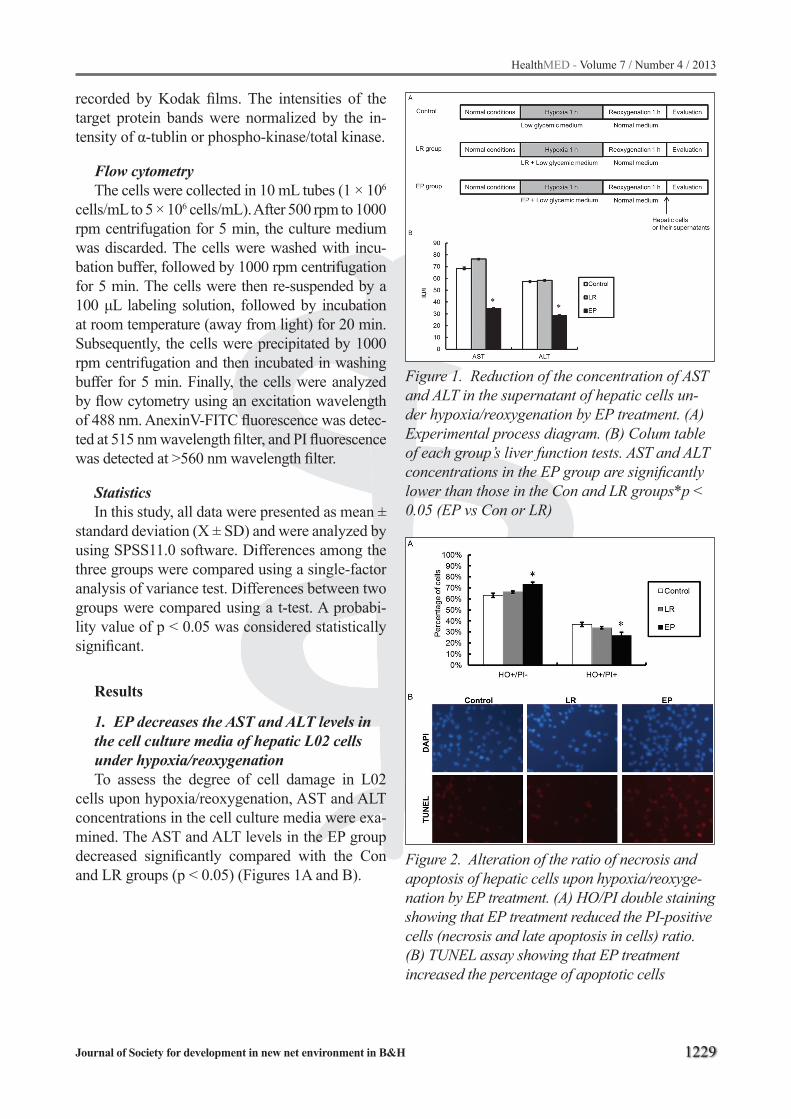
HealthMED - Volume 7 / Number 4 / 2013
Journal of Society for development in new net environment in B&H 1229
recorded by Kodak films. The intensities of the target protein bands were normalized by the in-tensity of α-tublin or phospho-kinase/total kinase.
Flow cytometryThe cells were collected in 10 mL tubes (1 × 10⁶
cells/mL to 5 × 10⁶ cells/mL). After 500 rpm to 1000 rpm centrifugation for 5 min, the culture medium was discarded. The cells were washed with incu-bation buffer, followed by 1000 rpm centrifugation for 5 min. The cells were then re-suspended by a 100 μL labeling solution, followed by incubation at room temperature (away from light) for 20 min. Subsequently, the cells were precipitated by 1000 rpm centrifugation and then incubated in washing buffer for 5 min. Finally, the cells were analyzed by flow cytometry using an excitation wavelength of 488 nm. AnexinV-FITC fluorescence was detec-ted at 515 nm wavelength filter, and PI fluorescence was detected at >560 nm wavelength filter.
StatisticsIn this study, all data were presented as mean ±
standard deviation (X ± SD) and were analyzed by using SPSS11.0 software. Differences among the three groups were compared using a single-factor analysis of variance test. Differences between two groups were compared using a t-test. A probabi-lity value of p < 0.05 was considered statistically significant.
Results
1. EP decreases the AST and ALT levels in the cell culture media of hepatic L02 cells under hypoxia/reoxygenationTo assess the degree of cell damage in L02
cells upon hypoxia/reoxygenation, AST and ALT concentrations in the cell culture media were exa-mined. The AST and ALT levels in the EP group decreased significantly compared with the Con and LR groups (p < 0.05) (Figures 1A and B).
Figure 1. Reduction of the concentration of AST and ALT in the supernatant of hepatic cells un-der hypoxia/reoxygenation by EP treatment. (A) Experimental process diagram. (B) Colum table of each group’s liver function tests. AST and ALT concentrations in the EP group are significantly lower than those in the Con and LR groups*p < 0.05 (EP vs Con or LR)
Figure 2. Alteration of the ratio of necrosis and apoptosis of hepatic cells upon hypoxia/reoxyge-nation by EP treatment. (A) HO/PI double staining showing that EP treatment reduced the PI-positive cells (necrosis and late apoptosis in cells) ratio. (B) TUNEL assay showing that EP treatment increased the percentage of apoptotic cells
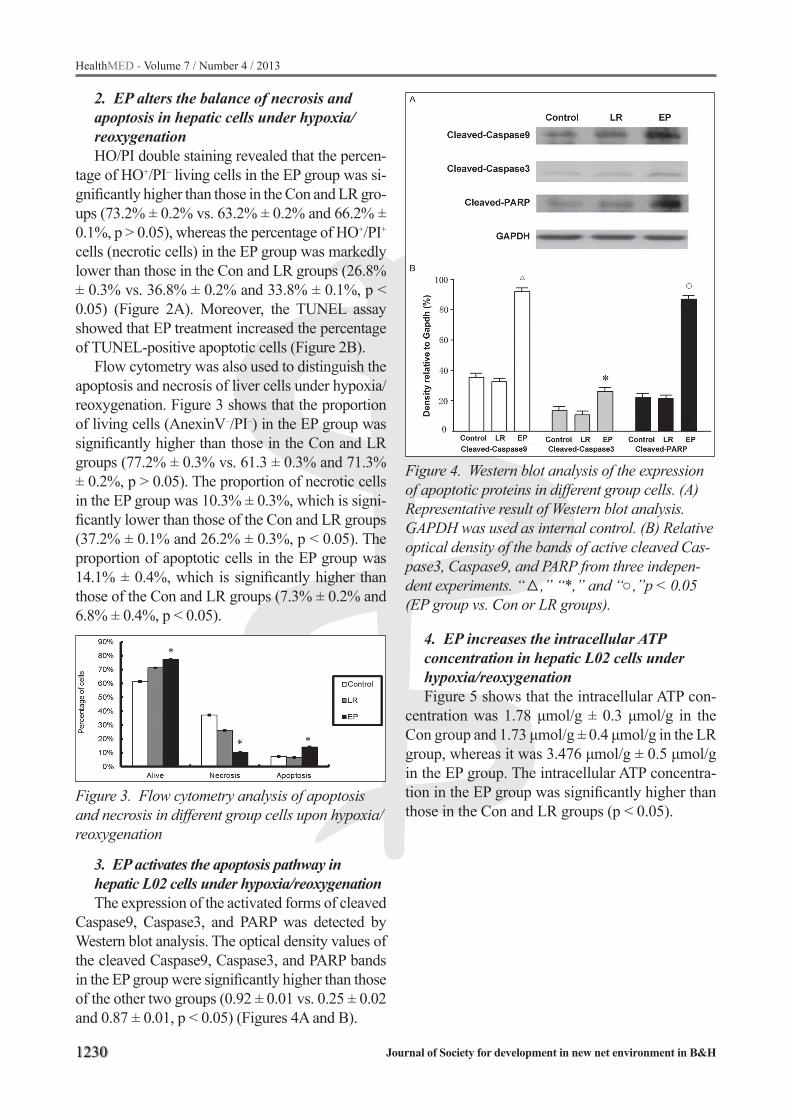
1230
HealthMED - Volume 7 / Number 4 / 2013
Journal of Society for development in new net environment in B&H
2. EP alters the balance of necrosis and apoptosis in hepatic cells under hypoxia/reoxygenationHO/PI double staining revealed that the percen-
tage of HO+/PI– living cells in the EP group was si-gnificantly higher than those in the Con and LR gro-ups (73.2% ± 0.2% vs. 63.2% ± 0.2% and 66.2% ± 0.1%, p > 0.05), whereas the percentage of HO+/PI+ cells (necrotic cells) in the EP group was markedly lower than those in the Con and LR groups (26.8% ± 0.3% vs. 36.8% ± 0.2% and 33.8% ± 0.1%, p < 0.05) (Figure 2A). Moreover, the TUNEL assay showed that EP treatment increased the percentage of TUNEL-positive apoptotic cells (Figure 2B).
Flow cytometry was also used to distinguish the apoptosis and necrosis of liver cells under hypoxia/reoxygenation. Figure 3 shows that the proportion of living cells (AnexinV–/PI–) in the EP group was significantly higher than those in the Con and LR groups (77.2% ± 0.3% vs. 61.3 ± 0.3% and 71.3% ± 0.2%, p > 0.05). The proportion of necrotic cells in the EP group was 10.3% ± 0.3%, which is signi-ficantly lower than those of the Con and LR groups (37.2% ± 0.1% and 26.2% ± 0.3%, p < 0.05). The proportion of apoptotic cells in the EP group was 14.1% ± 0.4%, which is significantly higher than those of the Con and LR groups (7.3% ± 0.2% and 6.8% ± 0.4%, p < 0.05).
Figure 3. Flow cytometry analysis of apoptosis and necrosis in different group cells upon hypoxia/reoxygenation
3. EP activates the apoptosis pathway in hepatic L02 cells under hypoxia/reoxygenationThe expression of the activated forms of cleaved
Caspase9, Caspase3, and PARP was detected by Western blot analysis. The optical density values of the cleaved Caspase9, Caspase3, and PARP bands in the EP group were significantly higher than those of the other two groups (0.92 ± 0.01 vs. 0.25 ± 0.02 and 0.87 ± 0.01, p < 0.05) (Figures 4A and B).
Figure 4. Western blot analysis of the expression of apoptotic proteins in different group cells. (A) Representative result of Western blot analysis. GAPDH was used as internal control. (B) Relative optical density of the bands of active cleaved Cas-pase3, Caspase9, and PARP from three indepen-dent experiments. “△,” “*,” and “○,”p < 0.05 (EP group vs. Con or LR groups).
4. EP increases the intracellular ATP concentration in hepatic L02 cells under hypoxia/reoxygenationFigure 5 shows that the intracellular ATP con-
centration was 1.78 μmol/g ± 0.3 μmol/g in the Con group and 1.73 μmol/g ± 0.4 μmol/g in the LR group, whereas it was 3.476 μmol/g ± 0.5 μmol/g in the EP group. The intracellular ATP concentra-tion in the EP group was significantly higher than those in the Con and LR groups (p < 0.05).
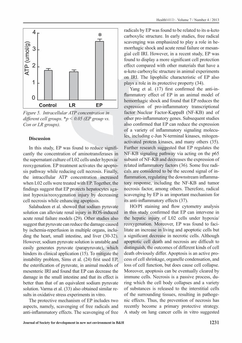
HealthMED - Volume 7 / Number 4 / 2013
Journal of Society for development in new net environment in B&H 1231
Figure 5. Intracellular ATP concentration in different cell groups. *p < 0.05 (EP group vs. Con or LR groups).
Discussion
In this study, EP was found to reduce signifi-cantly the concentration of aminotransferases in the supernatant culture of L02 cells under hypoxia/reoxygenation. EP treatment activates the apopto-sis pathway while reducing cell necrosis. Finally, the intracellular ATP concentration increased when L02 cells were treated with EP. Together, the findings suggest that EP protects hepatocytes aga-inst hypoxia/reoxygenation injury by decreasing cell necrosis while enhancing apoptosis.
Salahudeen et al. showed that sodium pyruvate solution can alleviate renal injury in ROS-induced acute renal failure models (29). Other studies also suggest that pyruvate can reduce the damage caused by ischemia-reperfusion in multiple organs, inclu-ding the heart, small intestine, and liver (30-32). However, sodium pyruvate solution is unstable and easily generates pyruvate (parapyruvate), which hinders its clinical application (15). To mitigate the instability problem, Sims et al. (24) first used EP, the esterification of pyruvate, in animal models of mesenteric IRI and found that EP can decrease the damage in the small intestine and that its effect is better than that of an equivalent sodium pyruvate solution. Varma et al. (33) also obtained similar re-sults in oxidative stress experiments in vitro.
The protective mechanism of EP includes two aspects, namely, scavenging of free radicals and anti-inflammatory effects. The scavenging of free
radicals by EP was found to be related to its α-keto carboxylic structure. In early studies, free radical scavenging was emphasized to play a role in he-morrhagic shock and acute renal failure or mesan-gial cell IRI. However, in a recent study, EP was found to display a more significant cell protection effect compared with other materials that have a α-keto carboxylic structure in animal experiments on IRI. The lipophilic characteristic of EP also plays a role in its protective property (34).
Yang et al. (17) first confirmed the anti-in-flammatory effect of EP in an animal model of hemorrhagic shock and found that EP reduces the expression of pro-inflammatory transcriptional factor Nuclear Factor-KappaB (NF-KB) and of other pro-inflammatory genes. Subsequent studies also confirmed that EP can reduce the expression of a variety of inflammatory signaling molecu-les, including c-Jun N-terminal kinases, mitogen-activated protein kinases, and many others (35). Further research suggested that EP regulates the NF-KB signaling pathway via acting on the p65 subunit of NF-KB and decreases the expression of related inflammatory factors (36). Some free radi-cals are considered to be the second signal of in-flammation, regulating the downstream inflamma-tory response, including the NF-KB and tumor necrosis factor, among others. Therefore, radical scavenging by EP is an important mechanism for its anti-inflammatory effects (37).
HO/PI staining and flow cytometry analysis in this study confirmed that EP can intervene in the hepatic injury of L02 cells under hypoxia/reoxygenation. Moreover, EP was found to faci-litate an increase in living and apoptotic cells but a significant decrease in necrotic cells. Although apoptotic cell death and necrosis are difficult to distinguish, the outcomes of different kinds of cell death obviously differ. Apoptosis is an active pro-cess of cell shrinkage, organelle condensation, and loss of cell function, but does cause cell collapse. Moreover, apoptosis can be eventually cleared by immune cells. Necrosis is a passive process, du-ring which the cell body collapses and a variety of substances is released to the interstitial cells of the surrounding tissues, resulting in pathoge-nic effects. Thus, the prevention of necrosis has recently become a primary protective strategy. A study on lung cancer cells in vitro suggested

1232
HealthMED - Volume 7 / Number 4 / 2013
Journal of Society for development in new net environment in B&H
that EP can reduce cell necrosis and stimulate the apoptosis pathway under glucose-free conditions. Although apoptosis was increased, the surroun-ding cell tissue was less stimulated (38), a finding that is consistent with our results.
By using electronic microscopy imaging tech-niques, the liver cells in each group were found to have different degrees of mitochondrial swelling and deformation, suggesting that mitochondrial permeability transition (MPT) occurred. Accor-ding to the “necrosis/apoptosis” theory, MPT de-termines the occurrence of cell death after injury, whereas the intracellular ATP levels decide which kind of death happens (39). When the normal level of intracellular ATP reaches 10% to 15%, it can activate the apoptotic pathway via Caspase9 and Caspase3 (9). A number of studies suggested that the use of fructose to increase intracellular ATP levels during hepatic IRI can reduce cell death but can increase apoptosis (40, 41). ATP provides the necessary energy for cell apoptosis (42, 43). If ATP was absent, the apoptosis pathway will be inhibited, thus making the cell necrotic (44, 45). The phenomenon of ATP-dependent cell death/apoptosis was also confirmed in an experiment in-volving overloading calcium ionophore and ace-taminophen poisoning (41, 46). An animal expe-riment on hemorrhagic shock suggested that early recovery can provide sufficient ATP, thus promo-ting the damaged cells to apoptosis and reducing the number of necrotic cells (47).
In summary, EP was found to protect L02 cells against hypoxia/reoxygenation injury in vitro by decreasing the number of necrotic cells while incre-asing the number of apoptotic cells. On one hand, EP increased the intracellular ATP levels possibly via “apoptosis/necrosis common pathway”-activa-ted apoptosis. On the other hand, EP reduced the necrotic cells, thereby reducing the expression of endogenous material injury to play a protective role.
Acknowledgement
The study was supported by the Research Fund of Shanghai Municipal Health Bureau, China (Grant No. 2009017)
Reference
1. Tsung A, Kaizu T, Nakao A, et al: Ethyl pyruvate ame-liorates liver ischemia-reperfusion injury by decrea-sing hepatic necrosis and apoptosis. Transplantation 79: 196-204, 2005.
2. Fondevila C, Busuttil RW and Kupiec-Weglinski JW: Hepatic ischemia/reperfusion injury--a fresh look. Exp Mol Pathol 74: 86-93, 2003.
3. Olthoff KM: Molecular pathways of regeneration and repair after liver transplantation. World J Surg 26: 831-837, 2002.
4. Selzner N, Rudiger H, Graf R and Clavien PA: Pro-tective strategies against ischemic injury of the liver. Gastroenterology 125: 917-936, 2003.
5. Clavien PA, Rudiger HA and Selzner M: Mechanism of hepatocyte death after ischemia: apoptosis versus necrosis. Hepatology 33: 1555-1557, 2001.
6. Fan C, Zwacka RM and Engelhardt JF: Therapeutic approaches for ischemia/reperfusion injury in the li-ver. J Mol Med (Berl) 77: 577-592, 1999.
7. Gujral JS, Bucci TJ, Farhood A and Jaeschke H: Mechanism of cell death during warm hepatic ische-mia-reperfusion in rats: apoptosis or necrosis? Hepa-tology 33: 397-405, 2001.
8. Jaeschke H: Molecular mechanisms of hepatic is-chemia-reperfusion injury and preconditioning. Am J Physiol Gastrointest Liver Physiol 284: G15-26, 2003.
9. Jaeschke H and Lemasters JJ: Apoptosis versus on-cotic necrosis in hepatic ischemia/reperfusion injury. Gastroenterology 125: 1246-1257, 2003.
10. Zhang JX, Jones DV and Clemens MG: Effect of ac-tivation on neutrophil-induced hepatic microvascular injury in isolated rat liver. Shock 1: 273-278, 1994.
11. Zhou W, Zhang Y, Hosch MS, Lang A, Zwacka RM and Engelhardt JF: Subcellular site of superoxide dismutase expression differentially controls AP-1 activity and injury in mouse liver following ische-mia/reperfusion. Hepatology 33: 902-914, 2001.
12. Kohli V, Selzner M, Madden JF, Bentley RC and Clavien PA: Endothelial cell and hepatocyte deaths occur by apoptosis after ischemia-reperfusion injury in the rat liver. Transplantation 67: 1099-1105, 1999.
13. O’Donnell-Tormey J, Nathan CF, Lanks K, DeBoer CJ and de la Harpe J: Secretion of pyruvate. An an-tioxidant defense of mammalian cells. J Exp Med 165: 500-514, 1987.
HealthMED - Volume 7 / Number 4 / 2013
Journal of Society for development in new net environment in B&H 1233
14. Brand KA and Hermfisse U: Aerobic glycolysis by proliferating cells: a protective strategy against re-active oxygen species. FASEB J 11: 388-395, 1997.
15. Fink MP: Ethyl pyruvate. Curr Opin Anaesthesiol 21: 160-167, 2008.
16. Tawadrous ZS, Delude RL and Fink MP: Resusci-tation from hemorrhagic shock with Ringer’s ethyl pyruvate solution improves survival and ameliorates intestinal mucosal hyperpermeability in rats. Shock 17: 473-477, 2002.
17. Yang R, Gallo DJ, Baust JJ, et al: Ethyl pyruvate modulates inflammatory gene expression in mice su-bjected to hemorrhagic shock. Am J Physiol Gastro-intest Liver Physiol 283: G212-221, 2002.
18. Venkataraman R, Kellum JA, Song M and Fink MP: Resuscitation with Ringer’s ethyl pyruvate solution prolongs survival and modulates plasma cytokine and nitrite/nitrate concentrations in a rat model of lipopolysaccharide-induced shock. Shock 18: 507-512, 2002.
19. Ulloa L, Ochani M, Yang H, et al: Ethyl pyruvate prevents lethality in mice with established lethal sep-sis and systemic inflammation. Proc Natl Acad Sci U S A 99: 12351-12356, 2002.
20. Dong YQ, Yao YM, Wei P, et al: [Effects of ethyl pyru-vate on cell-mediated immune function in rats with delayed resuscitation after burn injury]. Zhongguo Wei Zhong Bing Ji Jiu Yi Xue 17: 12-15, 2005.
21. Yang R, Uchiyama T, Alber SM, et al: Ethyl pyruvate ameliorates distant organ injury in a murine model of acute necrotizing pancreatitis. Crit Care Med 32: 1453-1459, 2004.
22. Cheng BQ, Liu CT, Li WJ, et al: Ethyl pyruvate im-proves survival and ameliorates distant organ injury in rats with severe acute pancreatitis. Pancreas 35: 256-261, 2007.
23. Uchiyama T, Delude RL and Fink MP: Dose-depen-dent effects of ethyl pyruvate in mice subjected to mesenteric ischemia and reperfusion. Intensive Care Med 29: 2050-2058, 2003.
24. Sims CA, Wattanasirichaigoon S, Menconi MJ, Ajami AM and Fink MP: Ringer’s ethyl pyruvate solution ameliorates ischemia/reperfusion-induced intestinal mucosal injury in rats. Crit Care Med 29: 1513-1518, 2001.
25. Epperly M, Jin S, Nie S, et al: Ethyl pyruvate, a po-tentially effective mitigator of damage after total-body irradiation. Radiat Res 168: 552-559, 2007.
26. Shen H, Hu X, Liu C, et al: Ethyl pyruvate protects against hypoxic-ischemic brain injury via anti-cell death and anti-inflammatory mechanisms. Neurobi-ol Dis 37: 711-722, 2010.
27. Bennett-Guerrero E, Swaminathan M, Grigore AM, et al: A phase II multicenter double-blind placebo-controlled study of ethyl pyruvate in high-risk pati-ents undergoing cardiac surgery with cardiopulmo-nary bypass. J Cardiothorac Vasc Anesth 23: 324-329, 2009.
28. Kao KK and Fink MP: The biochemical basis for the anti-inflammatory and cytoprotective actions of ethyl pyruvate and related compounds. Biochem Pharmacol 80: 151-159, 2010.
29. Salahudeen AK, Clark EC and Nath KA: Hydrogen peroxide-induced renal injury. A protective role for pyruvate in vitro and in vivo. J Clin Invest 88: 1886-1893, 1991.
30. Bunger R, Mallet RT and Hartman DA: Pyruvate-enhanced phosphorylation potential and inotropism in normoxic and postischemic isolated working he-art. Near-complete prevention of reperfusion con-tractile failure. Eur J Biochem 180: 221-233, 1989.
31. Matthews BD and Williams GB: Initial experience with the advanced breast biopsy instrumentation system. Am J Surg 177: 97-101, 1999.
32. Sileri P, Schena S, Morini S, et al: Pyruvate inhibits hepatic ischemia-reperfusion injury in rats. Tran-splantation 72: 27-30, 2001.
33. Varma SD, Devamanoharan PS and Ali AH: Preven-tion of intracellular oxidative stress to lens by pyru-vate and its ester. Free Radic Res 28: 131-135, 1998.
34. Cruz RJ, Jr., Harada T, Sasatomi E and Fink MP: Effects of ethyl pyruvate and other alpha-keto car-boxylic acid derivatives in a rat model of multivisce-ral ischemia and reperfusion. J Surg Res 165: 151-157, 2011.
35. Mollen KP, McCloskey CA, Tanaka H, et al: Hypoxia activates c-Jun N-terminal kinase via Rac1-depen-dent reactive oxygen species production in hepa-tocytes. Shock 28: 270-277, 2007.
36. Johansson AS and Palmblad J: Ethyl pyruvate mo-dulates adhesive and secretory reactions in human lung epithelial cells. Life Sci 84: 805-809, 2009.
37. Sappington PL, Fink ME, Yang R, Delude RL and Fink MP: Ethyl pyruvate provides durable protec-tion against inflammation-induced intestinal epithe-lial barrier dysfunction. Shock 20: 521-528, 2003.
1234
HealthMED - Volume 7 / Number 4 / 2013
Journal of Society for development in new net environment in B&H
38. Lim SC, Choi JE, Kim CH, et al: Ethyl pyruvate in-duces necrosis-to-apoptosis switch and inhibits high mobility group box protein 1 release in A549 lung ade-nocarcinoma cells. Int J Mol Med 20: 187-192, 2007.
39. Kim JS, He L and Lemasters JJ: Mitochondrial per-meability transition: a common pathway to necrosis and apoptosis. Biochem Biophys Res Commun 304: 463-470, 2003.
40. Kim JS, Qian T and Lemasters JJ: Mitochondrial permeability transition in the switch from necrotic to apoptotic cell death in ischemic rat hepatocytes. Gastroenterology 124: 494-503, 2003.
41. Kon K, Kim JS, Jaeschke H and Lemasters JJ: Mi-tochondrial permeability transition in acetami-nophen-induced necrosis and apoptosis of cultured mouse hepatocytes. Hepatology 40: 1170-1179, 2004.
42. Eguchi Y, Shimizu S and Tsujimoto Y: Intracellular ATP levels determine cell death fate by apoptosis or necrosis. Cancer Res 57: 1835-1840, 1997.
43. Genini D, Budihardjo I, Plunkett W, et al: Nucleo-tide requirements for the in vitro activation of the apoptosis protein-activating factor-1-mediated cas-pase pathway. J Biol Chem 275: 29-34, 2000.
44. Richter C, Schweizer M, Cossarizza A and Frances-chi C: Control of apoptosis by the cellular ATP level. FEBS Lett 378: 107-110, 1996.
45. Leist M, Single B, Castoldi AF, Kuhnle S and Nico-tera P: Intracellular adenosine triphosphate (ATP) concentration: a switch in the decision between apop-tosis and necrosis. J Exp Med 185: 1481-1486, 1997.
46. Mertz RJ, Worley JF, Spencer B, Johnson JH and Dukes ID: Activation of stimulus-secretion coupling in pancreatic beta-cells by specific products of gluco-se metabolism. Evidence for privileged signaling by glycolysis. J Biol Chem 271: 4838-4845, 1996.
47. Paxian M, Bauer I, Rensing H, et al: Recovery of hepatocellular ATP and “pericentral apoptosis” af-ter hemorrhage and resuscitation. FASEB J 17: 993-1002, 2003.
Corresponding AuthorHao Wang,Department of Anesthesiology and Surgical Intensive Care Unit,Fudan University,Zhongshan hospital,Shanghai,China,E-mail: [email protected]

HealthMED - Volume 7 / Number 4 / 2013
Journal of Society for development in new net environment in B&H 1235
Abstract
Introduction: The criteria for the definition of the metabolic syndrome are different. The metabo-lic syndrome is described as the risk factor which can accelerate the development and progression of the damaged kidney.
The objective of the study is to assessment of parameters affecting renal function in subject with metabolic syndrome.
Material and Methods: From 46 analyzed pa-tients in kidney diseases and the metabolic syn-drome defined by the criteria of AHA/NHLBI, NCEP/ATP and the criteria of IDF. The renal func-tion has been defined by the help of the MDRD formula and the patients have been divided into two groups with GFR < 59.9ml/min/1.73m² and GFR > 60ml/min/1.73m².
Results: Out of 46 examined patients with the presence of the metabolic syndrome by the cri-teria of AHA/NHLBI is at 37%, NCEP/ATPIII at 32.6% and IDF is at 39.1% of the patients. In the group of the patients defined by the criteria of IDF is 27.8 of the patient has GFR < 59.9ml/min/1.73m², the values of uric acid (males> 430μmol/L and females>360μmol/L) was found at 23.5%, creatinine (males>106.1μmol/L and females>97.2μmol/L) was found at 27.8 at female and 28.6% male patients. The logistic regression analysis showed that the older age, female gender and larger waist circumference has a statistically significant influence (CI 95%, p<0.001) on the de-crease of the renal function.
Conclusion: The research has shown that the older age, the female gender and the larger wa-ist size have a significant influence at the patients with the metabolic syndrome.
Key words: The metabolic syndrome, renal function.
Introduction
The mechanism of appearing of the metabolic syndrome is complex and still it is not known and the components of the metabolic syndrome are gro-uped in the basic risk factors for the cardiovascular disease. The manifestations of the kidneys in the metabolic syndrome are the micro-albuminuria, the reduction in the glomerular filtration rate (GFR) (1). Different criteria for the diagnosis of the me-tabolic syndrome have been suggested so far. So, in 2001 year, The National Cholesterol Education Program-Adult Treatment Panel III (NCEP-ATPI-II) gave the suggestion for the diagnosis of the me-tabolic syndrome that has been widely used. Accor-ding to them, the diagnosis based on the existence of three or more of the suggested criteria is given: abdominal obesity defined by the waist circum-ference, at men >102cm and at women >88cm, triglycerides>150mg/dl (1.7mmol/L), the concen-tration of HDL<40mg/dl (1.03mmol/L at men) and at women <50mg/dl (1.29mmol/L), blood pre-ssure > 130/85mmHg and fasting plasma glucose > 100mg/dl (>5.6mmol/L) (2). Unlike them, The National Heart, Lung and Blood Institute and The American Heart Association (NHLBI/AHA) su-ggest the lower limit values for the abdominal obe-sity and fasting plasma glucose at men and they suggest that the diagnosis can have minimally two criteria of the ones suggested (3). Then, The Inter-national Diabetes Federation (IDF) a new definition of the metabolic syndrome which includes a cen-tral obesity plus two or more factors: triglycerides > 150mg/dl (1.7mmol/L), the lower values of HDL cholesterol < 40mg/dl (1.03mmol/L), at men < 50mg/dl (1.29mmol/L), and at women systolic blo-od pressure>130mmHg or diastolic blood pressure > 85mmHg and higher value of the fasting plasma glucose > 100mg/dl (5.6mmol/L) or the previously diagnosed diabetes type 2 with the waist size which characterizes a central obesity is at European men > 94cm an European women > 80cm, and at Asian
Parameters influencing renal function in the subject with metabolic syndromeDanijela TasicClinical Centre Nis, The Clinic of Nephrology, Nis, Serbia

1236
HealthMED - Volume 7 / Number 4 / 2013
Journal of Society for development in new net environment in B&H
men > 90cm and Asian women > 80cm (4). The ba-sic mechanism of the pathological and pathophysi-ological changes in a kidney and its relation to a the metabolic syndrome hasn’t been still clarified but it involves the insulin resistance, inflammation, the endothelial dysfunction, the oxidative stress, the al-tered hemodynamics in the kidney, the activation of the rennin angiotensin aldosterone system and the sympathetic nervous system (5,6).
The aim of the study is to assessment of pa-rameters affecting renal function in subject with metabolic syndrome.
The patients and the methods
From 46 analyzed patients in kidney diseases and the metabolic syndrome defined by the crite-ria of AHA/NHLBI, NCEP/ATP and the criteria of IDF. {The working diagnosis of the World He-alth Organization (WHO) hasn’t been taken into account nor the diagnosis of the American Asso-ciation of the Clinical Endocrinologists (AACE) due to micro-albuminuria which is not available in everyday work.} The renal function has been defi-ned by the help of the MDRD formula and the pati-ents have been divided into two groups with GFR < 59.9ml/min/1.73m² and GFR > 60ml/min/1.73m², the limit value for serum creatinine have been taken for males Cr > 106.1μmol/L and for females Cr > 97.2μmol/L and uric acid for males > 430μmol/L and for females > 360μmol/L.
The Results
In the Table 1, the general demographic charac-teristics of both groups of patients with the metabo-lic syndrome have been shown. It has been noted that the patients with GFR>59ml/min/1.73m² are older and have larger body mass index (BMI), but without any statistical significance and there is sta-tistical difference between the values of uric acid and the systolic blood pressure parameters between the gropus. The statistical significance in the preva-lence of the metabolic syndrome relating different criteria hasn’t been recorded although there is the greatest difference between the criterion of NCEP/ATPIII 32.6% and IDF 39.1% (Figure 1).
Figure 1. Presence of MS according different criteria
The metabolic syndrome has been defined at 27.8% persons with the glomerular filtration (GFR< 59.9ml/min/1.73m²), the uric acid with the defined criteria larger than the limit value appea-red at 23.5%, creatinine at 27.8% of women and at 28.6% of men (Figure 2).
Table 1. General characteristics of the patients according to value of GFRGFR>60ml/min/1.73m2 GFR<59.9ml/min/1.73m2 p
Age (years) 41.89 ± 14.30 61.10 ± 10.70 0.0000Weist (cm) 98.03 ± 10.33 99.55 ± 10.31 0.6820Plasma glucose levels (mmol/l) 5.10 ± 0.81 5.13 ± 0.59 0.9000BMI (kg/m2) 25.13 ± 4.25 26.33 ± 4.37 0.4330Smoking (years) 7.64 ± 11.26 7.40 ± 10.60 0.9520HOL (mmol/l) 5.61 ± 1.55 5.45 ± 1.06 0.7750TG (mmol/l) 2.04 ± 1.16 2.28 ± 1.46 0.5930HDL (mmol/l) 1.30 ± 0.36 1.12 ± 0.25 0.2280LDL (mmol/l) 3.35 ± 1.44 3.29 ± 0.74 0.9110Urates (mmol/l) 300.52 ± 85.07 402.71 ± 131.38 0.0140SBP (mm Hg) 129.03 ± 15.98 144.50 ± 16.41 0.0100DBP (mm Hg) 75.56 ± 8.18 81.00 ± 8.10 0.0690PP (mm Hg) 53.47 ± 13.62 63.50 ± 13.13 0.0440
BMI - Body Mass Index (kg/m2), HOL − Holesterol (mmol/l), TG − Trigliceride (mmol/l), HDL − High-Density Lipopro-tein (mmol/l), LDL − Low-Density Lipoprotein (mmol/l), SBP − Systolic Blood Pressure (mm Hg), DBP − Diastolic Blood Pressure (mm Hg), PP − Pulse Pressure (mm Hg)

HealthMED - Volume 7 / Number 4 / 2013
Journal of Society for development in new net environment in B&H 1237
Figure 2. Parameters influencing renal function in the subject with MS (according IDF criteria of MS)
The logistic regression analysis has shown that the patients of older age, larger wais size and of female gender are under a significant risk of ha-ving a kidney damage in the metabolic syndrome (p<0.001) (Table 2).Table 2. Results of the multiple regression analysis
B95% CI for B
pLower Upper
(Constant) 101.83 48.74 154.93 <0.001Age (years) -1.07 -1.44 -0.71 <0.001Waist (cm) 0.98 0.49 1.48 <0.001Sex -23.20 -34.51 -11.89 <0.001
Discussion
The relation between the metabolic syndrome and the renal insufficiency have been examined by the numerous studies (7-11). In the HANES III stu-dy, 4.6% patients older than 20 years have a chronic renal insufficiency while 23.7% has the metabolic syndrome. In our study, the chronic renal insuffici-ency risk at the patient with the metabolic syndrome has been examined. The patients with the decreased renal function were, in average, 61.1±10.7 years, the body mass index 26.33±4.37kg/m². According to HANES III study by the diagnostic criteria of NCEP-ATPIII definition, 34.5% patients 33.7% at men and 35.4% at women had the prevalence of the metabolic syndrome, and according to IDF criteria, 39% of the patients (39.9% at men and 38.1% at women). According to our study 39.1% of the pati-ents have been diagnosed by the criteria of IDF for the metabolic syndrome and 32.6% by the criteria of NCEP/ATPIII. According to HANES III study, the greatest difference has been found in the preva-lence at the males of Mexican origin whereby the prevalence in the older age group is 40.3% by the criteria of NCEP-ATPIII and if the definition IDF is
used, then it is 50.6%. Our research has shown that the prevalence at male persons with the metabolic syndrome and the decreased renal function by the IDF criteria, for the metabolic syndrome is 28.6% at men and 27.8% at women. For example, Rashidi A at al. have shown that there is a 88 % high risk for the development of the renal insufficiency at the patients with the metabolic syndrome. Our results show that, at the patients with the metabolic syn-drome diagnosed by the criteria of IDF, 27.8% have the low estimated glomerular filtration rate and 23.5% have elevated uric acid by the standard crite-ria (12). Then, Kurella M at al. claim that when the patients with hypertension are left out, the patients with the metabolic syndrome have a significantly higher blood pressure. Our results show that all the patients with the metabolic syndrome have a high blood pressure but the high blood pressure values appear at the patients with a lowered renal function (systolic blood pressure (SBP) 144.5±16.41mmHg, diastolic blood pressure (DBP) 81.0±8.1mmHg, pulse pressure (PP) 63.5±13.13mmHg) but without any statistical significance in the relation to the gro-up with normal renal function (13). Kitiyakara C at al. suggest that the renal insufficiency risk is in the relation to the criteria of the metabolic syndrome diagnosis (14), which is indirectly confirmed by our research as well.
Conclusion
Our research has shown that, at the patients with the defined metabolic syndrome, the older age, the waist size and the female gender a significant influ-ence on the decrease of the renal function.

1238
HealthMED - Volume 7 / Number 4 / 2013
Journal of Society for development in new net environment in B&H
References
1. Ritz E. Metabolic syndrome: an emerging threat to renal function. Clin J Am Soc Nephrol 2007; 5(2): 869-71.
2. Expert Panel on Detection, Evaluation, and Treat-ment of High Blood Cholesterol in Adult. 3rd Report of the NCEP ATP III. JAMA 2001; 285(19): 2486-97.
3. Grundy SM, Cleeman JI, Daniels SR, Donato KA, Eckel RH, Franklin BA et al. Diagnosis and manage-ment of the metabolic syndrome. An NHLBI/AHA Re-port. Circulation 2005; 112(17): 285-90.
4. IDF Epidemiology Task Force Consensus Group. Lancet 2005; 366: 1059-62.
5. Ritz E. Metabolic syndrome and kidney disease. Blood Purif 2008; 26: 59-62.
6. Locatelli F, Pozzoni P, del Vecchio L. Renal manife-stations inthe metabolic syndrome. J Am Soc Nephrol 2006; 17: S81-S85.
7. Thomas G, Sehgal AR, Kashyap SR, Srinivas TR, Kir-wan JP, Navaneethan SD. Metabolic syndrome and kidney disease: a systematic review and meta-analy-sis. Clin J Am Soc Nephrol 2011; 6(10): 2364-73.
8. Tuttle KR. Renal manifestation of the metabolic syn-drome. Nephrol Dial Transplant 2005; 20(5): 861-64.
9. Alexander MP, Patel TV, Farag YMK, Florez A, Rennke HG, Singh AK. Kidney pathological changes in metabolic syndrome: a cross-sectional study. Am J Kidney Dis 2009; 5(53): 751-59.
10. Cirillo P, Sato W, Reungjui S, Heinig M, Gersch M, Sautin Y et al. Uric acid, the metabolic syndrome, and renal disease. J Am Soc Nephrol 2006; 17(12 Suppl 3): S 165-8.
11. Tasic D, Najman S. Certain experimental models in biomedical research of hypertension. Facta Univer-sitatis, Medicine and Biology 2008; 15(3): 81-84.
12. Rashidi A, Ghanbarian A, Azizi F. Are patients who have metabolic syndrome without diabetes at risk for developing chronic kidney disease? evidence based on data from a large cohort screening population. Clin J Am Soc Nephrol 2007: 2(5): 976-83.
13. Kurella M, Lo JC, Chertow GM. Metabolic syndro-me and the risk for chronic kidney disease among nondiabetic adults. J Am Soc Nephrol 2005; 16(7): 2134-40.
14. Kitiyakara C, Yamwong S, Cheepudomwit S, Dom-rongkitchaiporn S, Unkurapinun N, Pakpeankit-vatana V at al. The metabolic syndrome and chronic kidney deisease in a Southeast Asian cohort. Kidney Int 2007; 71(7): 693-700.
Corresponding AuthorDanijela Tasic,The Clinic of Nephrology, Clinical Centre Nis, Nis,Serbia,E-mail: [email protected]

HealthMED - Volume 7 / Number 4 / 2013
Journal of Society for development in new net environment in B&H 1239
Abstract
Mixed glioneuronal tumor (GNT) is a hetero-geneous group of primary central nervous system (CNS) neoplasm originating from neoplastic cells that can differentiate along both glial and neuronal cell lines. There were only 12 unusual spinal mixed GNTs except well-defined ganglioglioma repor-ted in the literature. Herein, we report one more unusual spinal mixed GNT in a 22-year-old female with signs of sensor-motor deficits. The lesion re-sembled pilocytic astrocytoma (PA) and presented no sign of morphologic neuronal rosette. Deoxyri-bonucleic acid (DNA) sequencing for BRAFV600E mutational hot-spot was firstly performed in these unusual spinal mixed GNTs. However, this muta-tion was not identified in our case, which might be useful to differentiate extra-cerebellar PA.
Key words: BRAFV600E mutation, Leptome-ningeal dissemination, mixed glioneuronal tumor, Spinal cord.
Introduction
Mixed GNTs are rare primary CNS neoplasms containing a variable extent of neuronal and gli-al elements. So far, only 12 spinal mixed GNTs except gangliogliomas have been reported in the literature.1-4 BRAFV600E mutation, characterized by a substitution of valine by glutamic acid at position 600, accounts for approximately 90% of BRAF mutations in human cancers. Analyze the molecular genetics of these tumors may provide novel insight in differential diagnosis, treatment and prognosis. Herein, we report a spinal mixed GNT with BRAFV600E mutation analysis and also review the related literature.
Case report
A previously healthy 22-year-old female was admitted with a two-year history of back pain. Neu-rological examination revealed motor weakness of 4/5 on the right side and 2/5 on the left side and sen-sory loss below T6 segment. Bilateral Hoffmann and Babinski signs were also positive. Spinal ma-gnetic resonance imaging (MRI) revealed a slightly enhanced intramedullary lesion in T6-7 region with obvious syringomyelia (Figure 1). A thoracic lami-nectomy and myelotomy were performed and the lesion was nearly totally removed. No evidence of tumor recurrence and leptomeningeal metastases was noted at 10 months postoperatively.
a)
An unusual spinal mixed glioneuronal tumor with BRAFV600E mutation analysis: Case report and literature reviewYu Hu, Weiying Zhong, Haifeng Chen, Siqing Huang
Department of Neurosurgery, West China Hospital, Sichuan University, Chengdu, Sichuan Province, China

1240
HealthMED - Volume 7 / Number 4 / 2013
Journal of Society for development in new net environment in B&H
b)
c)
Figure 1. Preoperative spinal MRI scan: (a) The T1-weighted image showing an isointense mass in T6-7 region with slightly enhanced after Gadoli-nium administration (c); (b) Sagittal T2-weighted image demonstrating a heterogeneous hyperin-tense neoplasm in the thoracic spinal cord with obvious syrinx and thinner of the spinal cord
The lesion was light-red, fish-like and well-cir-cumscribed on macroscopic observation. Histolo-gical examination revealed the lesion containing both glial and neuronal elements. The glial element was mainly composed of bipolar cells with bland, oval to elongated nuclei and piloid (hair-like) pro-cesses, reminiscent of PA (Figure 2a). Meanwhile, a number of oligodendrocyte-like cells with small, prominent, dark round nuclei and perinuclear halos were likewise noted. The neuronal element com-posed of scattered neuron-like cells with abundant eosinophilic cytoplasm, large nuclei and prominent nucleoli (Figure 2b). Rosenthal fibers (RFs) and eosinophilic granular bodies (EGBs) were absent. Glomeruliod vessels, mitoses and necroses were not observed. Immunohistochemically, the astro-cytic element showed positivity for glial fibrillary acidic protein (GFAP), S-100 protein, and Olig2 (Figure 2c). Neuron-like cells were synaptophysin and neurofilament positive, and the Ki67 labeling index was less than 1% (Figure 2d, 2e).
a)

HealthMED - Volume 7 / Number 4 / 2013
Journal of Society for development in new net environment in B&H 1241
b)
c)
d)
e)
Figure 2. Histopathological features of the mixed GNT: (a) The lesion is revealing bipolar cells with bland, oval to elongated nuclei and piloid (hair-like) processes, reminiscent of PA (hematoxylin and eosin, ×100); (b) Tumor cells are exhibiting multinucleate or “wreath-like” nuclei. Mitoses, significant nuclear pleomorphi-sm, vascular proliferation and areas of necrosis are absent (hematoxylin and eosin, ×400); (c) The astrocytic element is displaying immunore-activity for GFAP, (d) and rare neuron-like cells are showing synaptophysin positive (immunohi-stochemistry, ×400); (e) the Ki67 labeling index is less than 1% (immunohistochemistry, ×400)
Informed consent for analysis of the BRAFV600E mutation was obtained from the patient and his family, and this study was also approved by our institutional ethical committee. The DNA sample was extracted from formalin fixed and paraffin embedded tissues. DNA sequencing of hotspot co-don 600 (exon 15) of the BRAF gene for mutation was performed as previously described.5 Our case was negative for a mutation at this codon.
Discussion
Spinal mixed GNTs are uncommon entity that displays GFAP-positive glial and synaptophysin-positive neuronal elements. They are tentatively classified into three groups: Spinal ganglioglio-mas, spinal glioneuronal tumors and other unc-lassified.6 Spinal gangliogliomas, the same as their brain counterpart, are well documented and will not be discussed in this article. Most of spi-nal mixed GNTs are glioneuronal tumors (75%), whereas unclassified ones only account for about 25% of these tumors. The former usually exhibits well-formed or less structured neuronal rosettes in glial background while the latter presents no sign of morphologic neuronal rosette. They are frequ-ently located in the thoracic region, followed by cervicothoracic and cervical region of the spinal cord. The radiological feature of spinal mixed GNTs is usually iso/hypointense on T1-, iso/hype-rintense on T2-weighted images with heteroge-neous enhancement. Calcification and edema are generally absent while extensive syringomyelia is commonly noticed.1-2,4,7,8

1242
HealthMED - Volume 7 / Number 4 / 2013
Journal of Society for development in new net environment in B&H
Pathologically, spinal GNTs exhibit benign hi-stological features like rosette-forming glioneuronal tumor of the fourth ventricle suggesting that it may represent a distinctive clinical-pathological entity different from their cerebral counterparts.2,6 Howe-ver, Scholz et al. reported a spinal GNT with diffu-se infiltrating astrocytic component.8 Therefore, further more cases with genetic analysis and clinical follow-up are needed to consider these lesions as a distinct new entity. In our case, the lesion was initi-ally considered as a PA, but the presence of scattered synaptophysin-positive tumor cells is distinguished from PA. There are four spinal mixed GNTs resem-bled PA, and all of them have neuronal rosettes. No sign of neuronal rosette and lack of anaplastic fe-atures make our case an extremely rare low-grade mixed glioneuronal tumor of the spinal cord.
Spinal mixed GNTs are thought to originate from a primitive neuroectodermal stem cell that able to differentiate along both glial and neuronal cell lines.8 Cytogenetic analyses are only performed in three spinal mixed GNTs, which revealed a va-riable extent of the chromosomal deletion of 1p.3,8 Loss of 1p may serve as common genetic alterati-ons affected multipotent precursors and have a role in tumorigenesis of these tumors.3,8 The molecular genetics of these tumors, however, has never been investigated to date. BRAF is a partner of the RAF family of serine/threonine kinases, and is the prin-cipal downstream target of Ras in the pro-oncoge-nic MAPK pathway. In animal study, the activated BRAF kinase domain can induce genesis of PA, and concomitant BRAFV600E mutation with homozygous deletions of cyclin-dependent kinase inhibitor 2A can result in malignant gliomas.9 Most spinal mixed GNTs show PA-like histology, raising the possibility that these tumors may not be a specific tumor entity, but are a variant of PA. Our result consistent with other recent large-scale mutation analysis of CNS tumors, suggesting that BRAFV600E mutation is not a frequent event in GNTs and these tumors may repre-sent a distinct entity with molecular features differs from PA.5 Higher mutation frequencies in extra-ce-rebellar PA (20%) than cerebellar PA (2%) implicate that BRAFV600E mutations may serve as a valuable diagnostic marker for differentiating PA from mixed GNTs in the spinal cord.5 However, further large-scale analysis should address whether BRAFV600E mutation may happen in GNTs.
Though these tumors reveal benign histopatho-logical features, the long-term outcome is relati-vely poor. Surgical resection is the best treatment, and gross total resection usually result in a favo-rable outcome.2,7 However, aggressive total resec-tion may result in a serious neurological deficits, therefore, adjuvant therapy including radiation and chemotherapy should be considered for tho-se residual or recurrent ones, which might lead to some degree of clinical remission.3 Duration of follow-up range from 9 to 57 months (average: 18.7 months), and leptomeningeal dissemination is usually presented (68%) and affect patients’ long-term survival.1,4,7,8 Relatively adverse outco-mes suggest that long-term follow-up is advisab.
Reference
1. Harris BT, Horoupian DS. Spinal cord glioneuronal tumor with “rosetted” neuropil islands and meninge-al dissemination: a case report. Acta Neuropathologi-ca 2000; 100: 575-9.
2. Anan M, Inoue R, Ishii K, Abe T, Fujiki M, Kobayashi H, Goya T, Nakazato Y. A rosette-forming glioneuro-nal tumor of the spinal cord: the first case of a ro-sette-forming glioneuronal tumor originating from the spinal cord. Human pathology 2009; 40: 898-901.
3. Fraum TJ, Barak S, Pack S, Lonser RR, Fine HA, Quezado M, Iwamoto FM. Spinal cord glioneuronal tumor with neuropil-like islands with 1p/19q deletion in an adult with low-grade cerebral oligodendroglio-ma. Journal of neuro-oncology 2011; 107: 421-6.
4. Ruppert B, Welsh CT, Hannah J, Giglio P, Rumboldt Z, Johnson I, Fortney J, Jenrette JM, Patel S, Scheithau-er BW. Glioneuronal tumor with neuropil-like islands of the spinal cord with diffuse leptomeningeal neu-raxis dissemination. Journal of neuro-oncology 2011; 104: 529-33.
5. Schindler G, Capper D, Meyer J, Janzarik W, Omran H, Herold-Mende C, Schmieder K, Wesseling P, Mawrin C, Hasselblatt M. Analysis of BRAF V600E mutation in 1,320 nervous system tumors reveals high mutation frequencies in pleomorphic xanthoa-strocytoma, ganglioglioma and extra-cerebellar pi-locytic astrocytoma. Acta Neuropathologica 2011; 121: 397-405.
6. Raghavan R. Pediatric Mixed Glioneuronal Tumors in the Spinal Cord. In Hayat MA, editor. Tumors of the Central Nervous System. Netherlands : Springer Netherlands; 2012. P. 7-17.

HealthMED - Volume 7 / Number 4 / 2013
Journal of Society for development in new net environment in B&H 1243
7. Syed S, Rajaram V, Leonard JR, Perry A, Raghavan R. Mixed glioneuronal tumors of the spinal cord in two children. Acta Neuropathologica 2006; 111: 53-5.
8. Scholz M, Hoischen A, Radlwimmer B, Weber RG, Harders A, Reifenberger G, Riemenschneider MJ. Rosetted glioneuronal tumor of the spine with overtly anaplastic histological features. Acta Neuropatholo-gica 2009; 117: 591-3.
9. Huillard E, Hashizume R, Phillips JJ, Griveau A, Ihrie RA, Aoki Y, Nicolaides T, Perry A, Waldman T, Mc-Mahon M. Cooperative interactions of BRAFV600E kinase and CDKN2A locus deficiency in pediatric ma-lignant astrocytoma as a basis for rational therapy. Proceedings of the National Academy of Sciences 2012; 109: 8710-5.
Corresponding AuthorSiqing Huang,Department of Neurosurgery, West China Hospital,Sichuan University, Chengdu,China,E-mail: [email protected]

1244
HealthMED - Volume 7 / Number 4 / 2013
Journal of Society for development in new net environment in B&H
Abstract
Background: The aim of this study was to in-vestigate the possible ECG changes in the isolat-ed acute LCx occlusion and the relation between these changes and the site of the occlusion.
Methods: This retrospective study included 175 patients, who were appropriate for the inclu-sion and exclusion criteria, among 21855 patients who underwent coronary angiography. LCx was divided into two parts as central and peripheral.
Results: 75 patients (42.9%) had ST-elevation myocardial infarction (STEMI); 77 patients (44%) had non-ST-elevation myocardial infarction; and 23 patients (13.1%) had unstable angina pectoris. It was observed that inferior acute myocardial infarc-tion (AMI) was often seen in the peripheral occlu-sion while inferoposterolateral AMI and anterolat-eral AMI were often seen in the central occlusion (46.5%, 40.6%, 12,5% respectively and p<0.05). The extent and frequency of the ST-segment dep-ression in the precordial V1 and V2 derivations were significantly higher in the patients with the central occlusion (p<0.05). In the STEMI subgroup, the extent and frequency of the ST-segment depres-sion in the V1-V3 derivations and the ST-segment elevation in the V6, V8 and V9 derivations were significantly higher in the patients with the central occlusion than the patients with the peripheral LCx occlusion (p<0.05).
Conclusion: The determination of the type and the extent of ST segment changes in the records of ECG of the patients with ACS may help to es-timate the presence and site of the acute isolated LCx occlusion.
Key words: Isolated acute circumflex occlu-sion, ST segment change, T wave change.
Introduction
The classical myocardial infarction findings may not be seen in the electrocardiography (ECG) records of most of the patients with the acute coro-nary syndrome (ACS) caused by the left circum-flex artery (LCx) occlusion, which results with de-lay of the necessary treatment and greater affected myocardial area.1 The ECG has been used for de-cades as a reliable and inexpensive tool to diag-nose ACS. However, LCx occlusions are not often recognized. A 12-lead ECG detects acute occlu-sions of the left anterior descending artery (LAD) and right coronary artery (RCA) in 70% to 92% of cases; however, the sensitivity for acute LCx oc-clusion ranges from 32% to 48%, likely because of its posterolateral location.2, 3 The standard ECG may miss more than 50% of the patients with LCx occlusion if evaluated only for ST-segment eleva-tion.4 Therefore, more detailed evaluation for other potential changes due to LCx occlusion is neces-sary if suspected. In the literature, there is limited data showing the frequency and extent of the ST segment deviation and T-wave changes on ECG and their relation to occlusion site in patients with isolated LCx occlusion. 5-10 We aimed to investiga-te possible ECG changes in the isolated LCx occ-lusion and the relation between these changes and the site of the occlusion.
Methods
Study population: This study was carried out retrospectively in our catheterization laboratory by evaluating the files of 21855 patients who un-derwent selective coronary angiography between February 2002 and September 2010. The flow
Evaluation of ST segment and T-wave changes in the anterior, inferior and posterior derivations of patients with isolated acute occluded left circumflex arteryEftal Murat Bakirci1, Mahmut Acikel1, Enbiya Aksakal1, Serdar Sevimli1, Husnu Degirmenci1, Muhammed Hakan Tas2, Selami Demirelli1, Sinan Inci1, Hakan Duman1, Ibrahim Halil Tanboga1
1 Department of Cardiology, Faculty of Medicine, Atatürk University, Erzurum, Turkey,2 Department of Cardiology, Erzurum Regional Education and Research Hospital Erzurum, Turkey.

HealthMED - Volume 7 / Number 4 / 2013
Journal of Society for development in new net environment in B&H 1245
chart about the study design is showed in figure 1. As a result, the study population was composed of 175 patients with ACS (with TIMI-0 and TIMI-I coronary blood flow) who admitted in the first 24 hours after beginning of the symptoms, based on the ECG records and the clinical and laboratory data provided by the patient file. Our study was approved by the local ethics committee.
Figure 1. The flow chart about the study design. CABG: coronary artery bypass graft, ECG: ele-ctrocardiography, LAD: left anterior descending artery, LCx: left circumflex artery, RCA: right coronary artery, TIMI: thrombolysis in myocardial infarction
Angiographic analysis: Coronary angiograms were assessed by 2 experienced cardiologists who were blinded to all clinical and electrocar-diographic data. Total occlusion was defined as the observation of TIMI-0 or TIMI-1 flow on se-lective coronary angiography. TIMI-0 flow was accepted as a complete occlusion and TIMI-1 flow was accepted as the functional occlusion.11 LCx was divided into 5 segments as the proximal circumflex, the obtuse marginal branch, the dis-tal circumflex, the posterolateral branch and the posterior descending branch. When the left co-ronary artery gives off three branches, the inter-mediary branch was accepted as the 6th segment of LCx. Proximal LCx was defined as the part from the ostium of the LCx to the obtuse marginal branch takeoff and distal LCx was defined as the part from the obtuse marginal branch to the ori-gin of the posterior descending branch (Figure 2). The occlusion in LCx was defined with regard to its location as central (proximal segment, obtuse
marginal branch and intermediary branch) or pe-ripheral (distal segment, posterolateral branch and the posterior descending branch).5
Figure 2. Cx map used in our study. PC: Proximal circumflex, DC: Distal circumflex, OM: Obtuse marginal branch, PL: Posterolateral branch, PD: Posterior descending branch, IM: Intermediary arter and LAD: Left anterior descending artery
Electrocardiographic analysis: The standart 12-lead ECG and the posterior ECG were assessed by 2 experienced cardiologists who were blinded to all clinical and angiographic data. The line between T and P wave was accepted as isoelectric line; and the interval between J point and the be-ginning of T wave was defined as ST segment on ECG.12 J point was used for determining the extent of ST changes; the pathological Q wave, “ST ele-vation” and “ST depression” was defined with re-gard to the criteria stated in ESC/ACC. The loca-lization of acute myocardial infarction (AMI) was determined according to ST-segment deviation was observed in at least two consecutive corres-ponding leads.13 While D2-D3-aVF derivations were a measure for inferior AMI, ST-segment de-pression associated with high and sharp T waves in V1-2 and R/S>1 in V1 and V2 was used as a criteria for the diagnosis of true posterior AMI. Inferior AMI with posterior AMI was accepted as

1246
HealthMED - Volume 7 / Number 4 / 2013
Journal of Society for development in new net environment in B&H
Inferoposterior AMI.12, 14 ST elevation in D1, aVL, V5 and V6 derivations for Anterolateral AMI 5, ST elevation in D2-D3-aVF and V5-V6 derivations for inferolateral AMI, ST elevation in D2-D3-aVF and V5-V6 derivations and ST depression in V1-V4 derivations (V1>V4) for inferoposterolateral AMI were used as diagnostic ECG criterias.15
Statistical AnalysisThe statistical evaluation was made by SPSS
11.5 computer program. Quantitative variables were showed as arithmetic mean± standard de-viation while qualitative variables were frequency and percentage. The comparison of two different parametric groups was carried out by “Student’s t test”. “Chi-square or Fisher’s exact test” (two-way) was used for comparing the qualitative va-riables. p<0.05 was considered significant.
Results
Clinical presentation: The mean age of the study population was 58.9 (range 32 to 83) years. 138 patients (78.9%) were male and 37 patients (21.1%) were female. DM was present in 30 pa-tients (17.1%), hypertension in 56 (32.0%), dysli-pidemia in 32 (18.3%), positive family history in 11 (6.3%), and smoking in 64 (36.6%). 75 (42.9%) patients had ST-elevation myocardial infarction (STEMI), 77 (44%) had non-ST-elevation myo-cardial infarction (NSTEMI), while 23 (13.1%) patients had the diagnosis of unstable angina pe-ctoris (UAP). 58 of the patients (33.1%) were in the central occlusion group, while 117 patients (66.9%) were assessed in the peripheral occlusion group. No significant difference was observed between the central and the peripheral occlusion groups in terms of the clinical diagnosis and the demographic data of the cases (Table 1). Of the patients with STEMI, 32.0% had inferior AMI, 13.3% had inferolateral AMI, 26.7% had infero-posterolateral AMI, 20.0% had inferoposterior AMI and 8.0% had anterolateral AMI. 32 (42.7%) patients from the patient subgroup with STEMI were in the central group while the rest 43 (57.3%) patients were in the peripheral occlusion group. It was observed that inferior AMI was more often in peripheral LCx occlusion while inferoposterolat-eral AMI and anterolateral AMI were more often
in central LCx occlusion (46.5%, 40.6%, 12,5% respectively and p<0.05). Table 1. The demographic characteristics with re-gard to site of left circumflex artery (LCx) occlusion
Central LCx
(n=58)
Peripheral LCx
(n=117)p value
Age 60,4±10,7 58,1±10,8 0,18Male 46(79.3) 92(78.6) 0.91Diabetes(%) 6(10.3) 24(20.5) 0.09Hypertension(%) 14(24.1) 42(35.9) 0.11Smoking(%) 27(46.6) 37(31.6) 0.054Family history(%) 5(8.6) 6(5.1) 0.37Dyslipidemia(%) 11(19.0) 21(17.9) 0.87
Electrocardiography: It was detected that there was a significant difference in the only V1 and V2 derivations of the cases in terms of ST segment changes (Table 2). The frequency and the extent of ST segment depression in V1-V2 derivations were significantly higher in the patients with cen-tral LCx occlusion than in the patients with pe-ripheral LCx occlusion. In the STEMI subgroup, significant difference was found only in V1-V3, V6 and V8, V9 derivations in terms of ST segment changes (Table 3). The frequency and extent of the ST segment depression in precordial V1-V3 deri-vations were considerably higher in the patients with the central LCx occlusion than the patients with the peripheral LCx occlusion. The frequency and extent of the ST elevation in V6 and the pos-terior V8, V9 derivations were again considerably higher in the patients with the central LCx occlu-sion than in the patients with the peripheral LCx occlusion. The highest frequency and the amount of precordial ST segment depression were in V2 derivation, and the least amount was in V6. It was observed that the frequency and extent of the pre-cordial ST segment depression was decreasing from V2 to V6 excluding V1. Table 4 shows the sensitivity, specificity, positive predictive value (PPV) and negative predictive value (NPV) with regard to the estimation of proximal LCx occlu-sion of ≥1mm ST depression in V1-V3 derivation and ≥1mm ST elevation in V9 derivation in the patient subgroup with STEMI. No significant dif-ference regarding electrocardiographic changes in the patients with NSTEMI and UAP was detected between the groups. Also, T wave changes in an-

HealthMED - Volume 7 / Number 4 / 2013
Journal of Society for development in new net environment in B&H 1247
terior, inferior, posterior and the right derivations was evaluated and no significant difference was found between the groups.
Discussion
This is a study investigate the relation between the site of occlusion in the isolated LCx acute oc-clusion and the ST segment and T wave changes in anterior, inferior, and posterior derivations with-out being influenced by other possible effects on ECG. In this study, we also identified the clinical,
demographic and the angiographic characteristics of the isolated LCx occlusion on a larger popula-tion than the previous studies.
The Clinical, Demographic and the Angiograph-ic Characteristics: The obtuse marginal branch of LCx supplies the posterolateral myocardium, the proximal LCx supplies both posteroinferior and the posterolateral myocardium and the distal LCx supplies the posteroinferior myocardium. It was re-ported in the previous studies that the more frequent inferior wall hypokinesis and the more frequent ab-normalities in the inferior derivations on ECG were
Table 2. The relation between ST-segment deviation and site of left circumflex artery occlusionCentral LCx
(n=58)Peripheral LCx
(n=117) p value
≥1mm ST depression V1(%) 23(39.7) 28(23.9) 0.031 V2(%) 24(41.4) 29(24.8) 0.025V1 ST depression(mm) -0.58±0.91 -0.35±0.59 0.044 V2 ST depression(mm) -0.93±1.46 -0.55±0.97 0.045
Table 3. The relation between ST-segment deviation and site of left circumflex artery occlusion in the STEMI subgroup
Central LCx(n=32)
Peripheral LCx(n=43) p value
≥1mm ST depressionV1(%) 28(87.5) 22(51.2) 0.001V2(%) 28(87.5) 23(53.5) 0.002V3(%) 26(81.3) 21(48.8) 0.004≥1mm ST elevation V6(%) 21(65.6) 15(34.9) 0.021V8(%) 17(53.1) 13(30.2) 0.045V9(%) 16(50.0) 9(20.9) 0.008V1 ST depression(mm) 1,33±0,82 -0,73±0,70 0,001V2 ST depression(mm) -2,07±1,42 -1,17±1,14 0,003V3 ST depression(mm) -1,58±1,37 -0,90±1,01 0,017V6 ST elevation(mm) 1,00±1,21 0,44±1,12 0,043V8 ST elevation(mm) 0,62±0,66 0,35±0,52 0,046V9 ST elevation(mm) 0,53±0,57 0,23±0,48 0,016
Table 4. The sensitivity, specificity, positive and negative predictive value of different derivations for the prediction of the central circumflex artery occlusion in the STEMI subgroup
Sensitivity Specificity PPV NPV≥1mm ST depressionV1(%) 87,50 48,84 56,00 84,00V2(%) 87,50 46,51 54,90 83,33V3(%) 81,25 51,16 55,32 78,57V9 ≥1mm ST elevation 50,00 79,07 64,00 68,00
Abbreviations: PPV = positive predictive value, NPV = negative predictive value.

1248
HealthMED - Volume 7 / Number 4 / 2013
Journal of Society for development in new net environment in B&H
seen in the patients with the distal LCx occlusion than the patients who had AMI due to the proximal LCx occlusion.5, 16 In our study, it was detected that the inferior AMI was more often seen in the periph-eral LCx occlusion while the inferoposterolateral AMI and anterolateral AMI were more often in the central LCx occlusion.
From et al 1 have found that the incidence of isolated LCx occlusion in patients with AMI was 19.5%, and from the patients with the isolated LCx occlusion 127 had STEMI and 166 had NSTEMI. Berry et al2 performed surface and intracoronary ECG during percutaneous transluminal coronary angioplasty. In 19 patients with balloon inflation of the LCx, all of whom had intracoronary lead ST-segment elevation, only 32% manifested ST-segment elevation on the surface ECG, 42% had ST-segment depression, and 47% had no ST-seg-ment deviation. Gibson et al 17 described the an-giographic characteristics of the patients with the isolated precordial ST segment depression from whom had non-ST segment acute coronary syn-drome; and detected that LCx was the responsible in almost half of the 314 patients (%48.2) who had AMI due to an occluded vessel. Our study has shown smilar findings with these previous stud-ies. The protective effect of collateral circulation doesn’t explain the more frequent NSTEMI due to the LCx occlusion as it doesn’t have good collater-al flow. These findings can be especially explained by the fact that the spatial resolution of 12-lead ECG is limited in terms of determining the pos-terior and lateral wall infarction of the left ven-tricule1 The use of the posterior ECG derivations increases the sensitivity of ECG in diagnosis.18
The isolated LCx disease is not often in an-giography. Dunn et al 5 defined the isolated LCx disease as the ≥%70 reduction in vessel lumen di-ameter and determined isolated LCx disease in 94 from 3900 patients (2.4%) who underwent angiog-raphy. From et al 1 detected that the responsible le-sion in the coronary angiography of 1500 patients with the diagnosis of AMI was the most common in RCA (44.7%); the second most common loca-tion was LAD (35.8%) while culprit LCx lesions were less frequently seen (19.5%). We detected in our study that from 21855 patients who underwent coronary angiography, the isolated stenosis more than 50% was present in 922 patients (4.2%); and
339 ones (1.5%) had total occlusion. The propor-tion of 175 patients included in the study to whole population was 0.80%, to the ones diagnosed with coronary artery disease was 1.29%, to the isolated LCx disease was 18.9%, and to the patients with the isolated LCx occlusion was 51.6%. Thereby, we have demonstrated the prevalence of the iso-lated LCx disease and total occlusion among a larger number of patients than the previously re-ported data. It must be especially pointed out that the low frequency of LCx occlusion in the patients with AMI may be due to the fact that LCx and its branches are less prone to plaque rupture because of their geometry in comparison with the other vessels, and that less angiography is referred as a result of ignoring the ischemic cases occurred due to LCx occlusion.
Dunn et al 5 detected that the isolated coronary artery disease was encountered more often in dis-tal LCx. Stadius et al 19 determined that 80% of the LCx lesions were in the main portion and 20% was in the obtuse marginal branch. As to our study, from 175 patients included 58 ones (33.1%) were in the central group and the rest 117 (66.9%) were in the peripheral group. From the patient subgroup with STEMI, 32 patients (42.7%) were in the central group and 43 ones (57.3%) were in the group with the peripheral occlusion. We found in our study that the occlusion was more often seen in the localiza-tion of peripheral zone. The occlusion zones refer to the locations more prone to atherosclerotic disease in the patients with the isolated LCx.
Electrocardiographic Changes: Although there are many studies defining different electrocardio-graphic criteria to determine the artery related to the infarction in the patients with the acute infe-rior AMI, and to determine proximal and distal RCA occlusion in the literature 9,10,20,21, the studies researching the electrocardiographic differences between the individual LCx occlusion zones are limited. Mikuriya et al 6 detected that the extent of ST segment depression in V2 derivation in the patients with the acute inferoposterior AMI, and the total of ST deviation in DII-V2 and aVF-V2 derivations are significantly higher in the proximal LCx occlusions in proportion to the distal LCx oc-clusions. Jim et al 7 detected that in the patients with the acute inferoposterior AMI the frequency of ≥1mm ST segment depression in V1-V4 deri-
HealthMED - Volume 7 / Number 4 / 2013
Journal of Society for development in new net environment in B&H 1249
vations, ≥1mm ST elevation in DI an V6 deriva-tions, and ≥1mm ST segment depression in V4R derivation are significantly higher in the proximal LCx occlusions in proportion to the distal LCx oc-clusion. In our study, while significant difference in terms of ST segment changes was seen in only V1 and V2 derivations of the cases according to the location of total occlusion in the LCx, signifi-cant differences between the groups in terms of ST segment changes were detected in V1-V3, V6 and V8, V9 in the patient subgroup with STEMI. ST segment depression detected in precordial der-ivations shows the ischemia on the left ventricular anterior wall. At the same time, it can be seen as the reciprocal change due to the ischemia on the posterior wall.22, 23 The fact that ST segment de-pression in precordial derivations and ST eleva-tion in the posterior V8, V9 was more apparent in the central LCx occlusions is due to the more intense exposure of the posterior wall to ischemia in this group in proportion to the peripheral LCx occlusions.
There are several important limitations in our study. The type of the study was retrospective and there are wide range of anatomical variations of LCx. The factors such as LCx dominance and the obtuse marginal branch size which may dif-fer among patients may lead to the mistakes in the evaluation of ECG changes.
Conclusion
The determination of the type and the extent of ST segment changes in the records of ECG of the patients with ACS may help to estimate the pres-ence and site of the acute isolated LCx occlusion.
References
1. From AM, Best PJ, Lennon RJ, et al. Acute myocar-dial infarction due to left circumflex artery occlusion and significance of ST-segment elevation. Am J Cardi-ol 2010; 106: 1081-1085.
2. Berry C, Zalewski A, Kovach R, et al. Surface electro-cardiogram in the detection of transmural myocardial ischemia during coronary artery occlusion. Am J Car-diol 1989; 63: 21–26.
3. Huey BL, Beller GA, Kaiser DL, et al. A comprehen-sive analysis of myocardial infarction due to left cir-cumflex artery occlusion: comparison with infarction due to right coronary artery and left ante-rior descen-ding artery occlusion. J Am Coll Cardiol 1988; 12: 1156 –1166.
4. Krishnaswamy A, Lincoff AM, Menon V. Magnitude and consequences of missing the acute infarct-related circumflex artery. Am Heart J 2009; 158: 706 –712.
5. Dunn RF, Newman HN, Bernstein L et al. The clinical features of isolated left circumflex coronary artery di-sease. Circulation 1984; 69: 477-484.
6. Mikuriya Y, Tatsukawa Y, Tamura A, et al. Electrocar-diographic diagnosis of left circumflex artery occlusion and the occlusion site. Intern Med 1996; 35: 261-265.
7. Jim MH, Ho HH, Siu CW, et al. Value of ST-segment depression in lead V4R in predicting proximal against distal left circumflex artery occlusion in acute infero-posterior myocardial infarction. Clin Cardiol 2007; 30: 36-41.
8. Zhan ZQ, Wang W, Dang SY, et al. Electrocardio-graphic characteristics in angiographically documen-ted occlusion of the dominant left circumflex artery with acute inferior myocardial infarction: limitations of ST elevation III/II ratio and ST deviation in lateral limb leads. J Electrocardiol 2009; 42: 432-439.
9. Chia BL, Yip JW, Tan HC, et al. Usefulness of ST eleva-tion II/III ratio and ST deviation in lead I for identifying the culprit artery in inferior wall myocardial acute myocardial infarction. Am J Cardiol 2000; 86: 341–343.
10. Herz I, Assali A, Alder Y, et al. New electrocardi-ographic criteria for predicting either the right or left circumflex artery as a culprit coronary artery in inferior wall acute myocardial infarction. Am J Car-diol 1997; 80: 1343–1345.
11. Pompa JJ. Coronary Arteriography and Intravascu-ler Imaging. In Braunwald E (eds): Heart Disease: A Textbook of Cardiovascular Medicine. WB Saun-ders Company, Philadelphia 2008, pp . 489-492.
1250
HealthMED - Volume 7 / Number 4 / 2013
Journal of Society for development in new net environment in B&H
12. David M. Mirvis and Ary L. Goldberger. Elektrocar-diography. In Braunwald E (eds): Heart Disease: A Textbook of Cardiovascular Medicine. WB Saunders Company, Philadelphia 2008, pp.148-195.
13. Thygesen K, Alpert JS, White HD. Universal Defi-nition of Myocardial Infarction. J Am Coll Cardiol 2007; 50: 2173-2195.
14. Peterson ED, Hathaway WR, Zabel KM, et al. Pro-gnostic significance of precordial ST segment depre-ssion during inferior myocardial infarction in the thrombolytic era: results in 16,521 patients. J Am Coll Cardiol 1996; 28: 305-312.
15. Correale E, Battista R, Martone A, et al. Electro-cardiographic patterns in acute inferior myocardial infarction with and without right ventricle involve-ment: classification, diagnostic and prognostic va-lue, masking effect. Clin Cardiol 1999; 22: 37-44.
16. Sheehan FH. Left ventricular dysfunction in acute myocardial infarction due to isolated left circumflex coronary artery stenosis. Am J Cardiol 1989; 64: 440-447.
17. Gibson C, Pride Y, Mohanayelu S, et al. Angiographic and clinical outcomes among patients with acute co-ronary syndrome presenting with isolated anterior ST-segment depressions. Circulation 2008; 118: 654.
18. Schmitt C, Lehmann G, Schmieder S, et al. Diagnosis of acute myocardial infraction in angiographically documented occluded infarct vessel: limitations of ST segment elevation in standard and extended ECG leads. Chest 2001; 120: 1540-1546.
19. Stadius ML, Maynard C, Fritz JK, et al. Coronary anatomy and left ventircular function in the first 12 hours of acute myocardial infarction: The western Washington randomized intracoronary streptokina-se trial. Circulation 1985; 72: 292-301.
20. Misumi I, Shono H, Nakao K, et al. Electrocardi-ographic discrimination of infarct-related artery between left circumflex and right coronary artery: Comparison of ST elevation between leads II and III. J Cardiol 2003; 41: 271 – 276.
21. Fiol M, Carrillo A, Cygankiewicz I, et al. New criteria based on ST changes in 12-lead surface ECG to detect proximal vs. distal right coronary artery occlusion in case of acute inferoposterior myocardial infarction. Ann Noninvasi ve Electrocardiol 2004; 9: 383 – 388.
22. Nishian K, Nomoto Y, Naruse H, et al. Precordial ST-segment depression in acute inferior myocardial infarction: The importance of posterolateral wall in-farction. J Cardiol 1989; 19: 413–424.
23. Sigiura T, Nagahama Y, Takehana K, et al. Progno-stic significance of precoridal ST-segment changes in acute inferior wall myocardial infarction. Chest 1997; 111: 1039-1044.
Corresponding AuthorEftal Murat Bakirci,Department of Cardiology,Faculty of Medicine,Ataturk University,Erzurum,Turkey,E-mail: [email protected]

HealthMED - Volume 7 / Number 4 / 2013
Journal of Society for development in new net environment in B&H 1251
Abstract
Objective: To evaluate endoscopic retrograde cholangiography (ERCP) and ensuing treatment for bile duct lithiasis after Billroth-II gastroente-rostomy.
Methods: ERCP was performed in 137 pati-ents with bile duct lithiasis after Billroth-II gastro-enterostomy. Of the 137 patients, 34 also received endoscopic sphincterotomy (EST) and 4 received endoscopic papillary balloon dilation (EPBD).
Results: ERCP was successfully performed in 109 (79.6%) out of 137. Bile duct stones were successfully removed based on EST and EPBD in 35 patients. Digestive duct bleeding was observed in 4 patients. There were no serious complications in patients receiving endoscopic treatments.
Conclusion: The successful rate of EST is high in patients with bile duct lithiasis after Billroth-II gastroenterostomy. Endoscopic treatment or cho-langioduodenostomy has good therapeutic effects.
Key words: Bile duct lithiasis, Billroth-II ga-stroenterostomy, endoscopic sphincterotomy, tre-atment.
Introduction
It is common in clinical practice that the patients after subtotal gastrectomy with Billroth-II gastroje-junostomy suffer from calculus of bile duct later [1]. Because of the changes in anatomy and special di-agnosis and treatment, it is insufficient to treat and operate relying solely on the findings of ultrasound, computerized tomography (CT), magnetic reso-nance imaging (MRI). However, it may have diffi-culties in endoscopic retrograde cholangiopancre-atography (ERCP). We have performed ERCP on 4151 patients between May 1984 and Jun 2012. Among those, there are 137 (3.30%) patients with Billroth-II gastroenterostomy. Now, the following is the experience on diagnosis and treatment.
Patients and methods
General dataA total of 76 males and 61 females were enrolled
with ages ranging from 34 to 74 years (mean age 47.5 years). Clinical manifestations: there were 83 patients who suffered from right upper quadrant pain with chills, fever, jaundice and other cholan-gitis symptoms, 31 patients with recurrent right upper quadrant pain, 23 patients with obstructive jaundice. The finding of ultrasound and CT showed calculus of bile duct in all cases. All the patients had the history of Billroth-II subtotal gastrectomy, including 23 cases with gastrojejunostomy in front of the colon with afferent jejunal loop of 25~40cm (average 32.4 cm), 114 patients with gastrojeju-nostomy behind of the colon with efferent jejunal loop of 5~8cm (average 6.4cm), 97 cases with the proximal end of jejunum suturing to greater ga-stric curvature and 40 cases with the proximal end of jejunum lesser gastric curvature suturing to les-ser gastric curvature. There were 29 patients with Billroth-II surgery morethan 30 years, 76 patients more than 20 years, 27 patients more than 10 years and 5 patients less than 10 years.
Diagnosis and treatment by ERCPThe instrument used in the ERCP examina-
tions was side-viewing duodenofiberscope (model JF, Olympus). The patients were given the same premedication as that of normal ERCP and were placed in the left lateral decubitus position. The du-odenofiberscope was advanced to stomach through oral cavity and esophagus, observing whether there were lesions in remnant stomach and stoma, then entered into duodenum retrograde and gradually along cavity through stoma and afferent jejunal loop, finding the papilla near the closed duodenal stump. Firstly, we performed ERCP examination with catheter through papilla. The instant we deter-mined bile duct stones, we performed endoscopic sphincterotomy (EST) or Endoscopic papillary bal-
Endoscopic treatment of bile duct lithiasis after Billroth-II gastroenterostomyGuang-quan Zhang, Qi-yuan Lin, Man-xi He, Zhong Liao
Department of General Surgery, Sixth People’s Hospital, Chengdu, China

1252
HealthMED - Volume 7 / Number 4 / 2013
Journal of Society for development in new net environment in B&H
loon dilatation (EPBD) examinations and then per-formed endoscopic stone extraction technique and Endoscopic nasobiliary catheter drainage (ENBD) based on practical conditions.
Results
Examination results Among 137 patients who received ERCP, 109
cases were successful (79.6%) and 28 patients failed (20.4%). The reasons for failure: endoscopic length was not enough in 20 patients because of the gastro-jejunostomy in front of the colon, endoscope didn’t transit Treitz ligament in 5 patients and examina-tions were suspended in 3 patients due to violent reaction. In the 109 patients with successful ERCP, there were 36 patients with simple common bile duct stones, 23 patients with calculi of intrahepatic duct, 17 patients with calculi both in intrahepatic duct and extrahepatic duct. 85 patients showed gall-bladder visualization and calculi were identified in 23 patients; among those, pyema and delay empty-ing appeared in 61 patients. Only 16 cases suffered from moderate fever after ERCP examination.
Treatment results38 cases were treated with endoscope includ-
ing 34 patients who were performed on EST and stone extraction and the numbers of successful cases were 31 (91.1%). Among those, 6 cases were performed on stone extraction by emergency ERCP and 4 cases were successful both in EPBD and stone extraction. 4 cases suffered from mas-sive gastrointestinal tract hemorrhage after thera-peutic endoscopy and were cured by non-surgical treatment. Cholangiography showed all calculi were out of papillae at 1 week after stone exac-tion followed by ENBD in 11 patients. No gastro-intestinal perforation and dead cases in our group. There were no complications during follow-up of 1 to 19 years in 35 of 38 patients except that bile duct stones reoccurred in 12 cases.
Discussion
The normal anatomy of upper gastrointestinal tract has been changed after subtotal gastrectomy with Billroth-II gastrojejunostomy. Although endo-scopic technicians have done considerable explo-
ration with the success rate of ERCP around 80% in the past 30 years [2], post-Billroth II gastrectomy ERCP and EST are still regarded as the most com-plex and difficult endoscopic techniques. However, conducting ERCP when performing therapeutic endoscopy after Billroth II gastrectomy is safety and feasible [3]; the success rate of post-Billroth II gastrectomy ERCP cannulation is 55% ~88% while the success rate of the therapeutic endoscopy is 66%~100% [4]. Park [5] reported all EST performed on 10 patients after the success of ERCP were suc-cessful (100%). Yin et al. [6] said that, 21 cases in 25 patients who were performed on ERCP were suc-cessful (84.0%) and 21 patients who were treated by EST+ENBD were successful (100%). Jia et al. [7] successfully cut off and exacted stones with needle-knife guided by the plastic stent which was inserted in after successful intubation in 2 patients. Huang et al. [8] performed stone extraction on 2 pa-tients and stent insertion on 1 patient after EPBD with front-viewing endoscope. Zhang et al. [9] re-ported 22 patients acquired good effects. The au-thor has further investigated the operation of EST technology based on the success of post-Billroth II gastrectomy ERCP for many years [10].The expe-riences in the encountering problems and how to conquer difficulties are following.
Determine the afferent and efferent ends of gastrointestinal stoma correctly: 1)the operation methods of Billroth-II gastrojejunostomy are di-vided into the proximal end of jejunum suturing to greater gastric curvature and the proximal end of jejunum suturing to lesser gastric curvature. Gen-erally, patients are placed in left lateral decubitus position when endoscope is operated. If the proxi-mal end of jejunum is sutured to lesser gastric cur-vature, the opening above the stoma is the afferent jejunal loop and the opening below is efferent je-junal loop in the remnant stomach under the visual field of endoscope, which are opposite when the proximal end of jejunum is sutured to greater gas-tric curvature, i.e. efferent jejunal loop lies above the opening while afferent jejunal loop lies below the opening. We can immediately judge accord-ing to pervious surgical records of patients before examination, however, it is difficult to obtain such information in practical clinical work. Because the proximal end of jejunum suturing to greater gas-tric curvature is regarded as antegrade peristalsis
HealthMED - Volume 7 / Number 4 / 2013
Journal of Society for development in new net environment in B&H 1253
in Gastrointestinal Operative Surgery, most of sur-geon trend to choose this method [11]. That is worth referring by endoscopic technician. 2) the author usually uses two methods to advance endoscope. First, observe two openings of gastroenterostomy for several minutes, the place where the bile and bubble flow is the opening of afferent jejunal loop. But this condition is not too much, only 21cases in our group. Second, the endoscope is advanced along two openings respectively, but when the duodenal stump is not seen until all the endoscope run out, return to stoma and enter in another open-ing where is the afferent jejunal loop. The charac-ter of efferent jejunal loop under endoscope: the enteric cavity is clean so the endoscope would not be blocked when advancing. The character of af-ferent jejunal loop: bile and gas bubbles are com-mon in enteric cavity and the color of the intestinal mucosa is slightly yellow; if the endoscope is eas-ily to be blocked when being advanced, especially when suddenly be blocked with difficulty in see-ing the front enteric cavity, the endoscope is likely in afferent jejunal loop at the moment.
Side-viewing duodenoscope through the open-ing of afferent jejunal loop: It is difficult to enter in the efferent jejunal loop for side-viewing duo-denoscope, which is the same to passing through the pylorus; the essential for endoscope passing through the opening of afferent jejunal loop is “the meaning of sun goes down”. At this time, it is com-paratively smooth for the proximal end of jejunum suturing to greater gastric curvature while it is more difficult for the proximal end of jejunum su-turing to lesser gastric curvature, which is because the opening of afferent jejunal loop is higher and often shows acute angle. If change patients’ posi-tions or repeatedly adopt hooking-pulling method, it may help endoscope enter in the afferent jejunal loop. Bergman et al. [12] reported that it was easy to pass through the opening of efferent jejunal loop for front-viewing duodenoscope which has clear visual field and is easier to reach duodenal papilla than side-viewing duodenoscope, increas-ing the success rate of ERCP; the drawbacks of front-viewing duodenoscope are there is no lift-ing device and the size of biopsy hole is small, so therapeutic operations such as EST are more difficult. Recently, an increasing number of for-eign scholars recommend adopting side-viewing
duodenoscope for therapeutic ERCP following Billroth II gastrectomy [13].
Endoscopy pass through the Treitz ligament: Treitz ligament is the extension of peritoneum of duodenojejunal flexure which is located in front of spine and comparatively fixed, toward the begin-ning part of jejunum and spine. Billroth-II gastro-jejunostomy changes the physiological direction of Treitz ligament, and also changes the degree of curvature of duodenojejunal flexure, which direct-ly influences the complexity for endoscope pass-ing through Treitz ligament; that is the jejunum curvature generated by afferent jejunal loop sutur-ing to greater gastric curvature is different from that generated by afferent jejunal loop suturing to lesser gastric curvature [14]. Jejunum curvature of Treitz ligament significantly blocks the endoscope, which is similar to colonoscope passing through splenic flexure or hepatic flexure. At this time, the operation methods are different from that of colonoscope when passing through splenic flexure or hepatic flexure, i.e. do not blindly advance en-doscope without seeing the enteric cavity, lift the front-end of endoscope slowly to search enteric cavity in the anterior wall and advance endoscope when the side view field of enteric cavity sliding toward the intestinal mucosa in anterior wall. If the enteric cavity can’t be seen, adopt hooking-pulling method, i.e. lift upward large knob to hook intestinal canal and draw back endoscope aided with ventral massage shot or position adjusting for several times, the intestinal canal can be seen and the endoscope can pass through Treitz ligament.
Search for duodenal papilla: When the endo-scope is smoothly advanced through the stoma in jejunum and it completely enter in enteric cav-ity, but the papilla can’t be found, the endoscope may lies in efferent jejunal loop. At this time, we should withdraw the endoscope to stoma and ad-vance it from another opening. If the endoscope is completely enter in enteric cavity without seeing papilla, the reasons may be: first, afferent jejunal loop in front of the colon is too long; second, there is side-to-side anastomosis between efferent and afferent jejunal loops, which can make endoscope lose direction. We found the previous surgical record of the patient who is failing in our group after endoscopy showing side-to-side anatomosis between and afferent jejunal loops. Post-Billroth

1254
HealthMED - Volume 7 / Number 4 / 2013
Journal of Society for development in new net environment in B&H
II gastrectomy duodenal stump presents blind-end whose mucosa is smooth and flat without mucosal folds. Papilla usually lies in 3~5cm of the closed stump and the characteristics are that papilla is lo-cated on the wall opposite to visual field of endo-scope, its opening direction is on the top of visual field and the ring-shaped intestinal mucosa on the site of papilla uplift sits on the bottom of visual field. Because gastric fluid and food don’t pass through duodenum which has been closed after Billroth II gastrectomy, duodenal lumen becomes narrow and papilla becomes smaller with covert opening, the papillae in some patients seem stiff.
Overcome the bottleneck of insufficient length of endoscope: That insufficient length of endo-scope in patients with Billroth II gastrojejunos-tomy often confuses operators. The length of afferent jejunal loop in patients with gastrojeju-nostomy in front of the colon ranges from 25 to 40cm while the length of endoscope is sufficient in patients with gastrojejunostomy behind of the colon. Among 28 patients who are failing in op-erations in our study, 20 cases have surgical his-tories of gastrojejunostomy in front of colon. So, the success rate of ERCP and EST in patients with Billroth II gastrojejunostomy in front of the co-lon is very low, only 3 cases were successful in our group (13.0%). Be cautious in choosing en-doscopy for treating and diagnosing patients with biliopancreatic disease if they have such surgical approaches. Accordingly, we suggest surgeons choose anastomosis behind the colon when per-forming Billroth II gastrojejunostomy to increase success rate with endoscope in future. Sometimes, the length of endoscope is insufficient even if in patients with gastrojejunostomy behind the colon, which can be partly conquered with the methods of hooking-pulling and encasing endoscope into jejunum; if duodenal stump still can’t be seen when all the endoscope is advanced into enteric cavity, we should cease continuing to operate to avoid complications due to long time operation.
(6) Intubate through papilla by opposite phase of side-viewing endoscope: The endoscope is ret-rograde inserted through reconstructed digestive tract, so the papilla and its opening direction faced in the visual field of endoscope are opposite to those of common endoscope; however, the side-viewing duodenoscope is designed to be inserted
upward through the opening of inferior papilla. But, post-Billroth II gastrojejunostomy papilla and the opening direction are opposite to the vi-sual field of endoscope and intubation direction, which lead to difficult intubations. The author adopt four methods to make endoscope close to papilla: 1 change patients’ position, such as Left lateral position, semiprone position and prone po-sition; 2 turn operators’ body greatly to coordinate with the adjustment of knobs; 3 assistants massage the lens through abdomen; 4 put the top end of catheter on the opening of papilla which is extend-ed, adjust knobs and change the directions of pa-tients and operators. The catheter is easily inserted into pancreatic duct rather than bible duct. To vi-sualize bile duct, the endoscope should be inserted upward in line with opening direction of bile duct in principle. We can make the campiscope surface close to intubation tube or visualize bile duct with the kissing-style method.
Cut-off papilla and remove stones: The posi-tion of papilla and the opening in visual field of endoscope are opposite to normal position due to the endoscope is conversely inserted the closed duodenal stump, plus the closed stump limits up-ward movement, the difficulty of operation will be increased. We take common bow-shaped knife for example which has few opportunities in endo-scopic sphincterotomy, only 2 cases in our study. Other operations mainly depend on needle-knife, because needle-knife is more convenient to cut off the sphincter with endoscope at 5-6 o’clock position. Bow-shaped knife is difficult to adjust the endoscope to 5-6 o’clock position in patients with Billroth II gastrojejunostomy. Sometimes, we combine needle-knife with bow-shaped knife in some patients. The author choose three meth-ods on the basis of practical situations of the duo-denal stump and papilla: (1) Antegrade incision. It is suitable for the patients with obvious papilla openings. Aim the anterior wall of papilla open-ing with needleeknife which then is slid upward (the direction of bile duct) stably and accurately to cut off the papilla for about 1cm once. Dolay et al. [15] suggested that the operation is safer when the incision length of EST in post- Billroth II gas-trojejunostomy patients is 0.5~1.0cm, and don’t make it more than 1.5cm. (2) Retrograde incision. It is suitable for patients with concealed papilla.
HealthMED - Volume 7 / Number 4 / 2013
Journal of Society for development in new net environment in B&H 1255
Stay the needle-knife directly on the papilla sur-face of junction between papilla and duodenal mucosa and slide it downward (the direction of papilla opening) stably and accurately to cut all the papilla once. (3) Fenestration. It is suitable for patients with deformation of papilla eminence and other structures. The needle-knife faces the most obvious of eminence and cuts the anterior wall of papilla for 2~3cm to open a small window. After that we verify the directions of papilla and open-ing through the small window with catheter. At this time, the papilla below the window (opening direction of papilla) is easily cut with bow-shaped knife while it is more safe to cut the papilla above the window (direction of bile duct) with needle-knife. We eliminate stones with stone blanket after EST and EPBD. The operation of endoscopic na-sobiliary drainage (ENBD) is similar to common endoscopic therapy, while the only difference is that we draw back endoscope when withdrawing the calculi rather than advance endoscope.
The calculi after Billroth II gastrectomy mainly are bile pigment calculi with few cholesterol gall-stones. With the development of endoscope and min-imally invasive techniques, the therapeutic effects of stone extraction by stone basket on simple bile duct stones following EST or EPBD are given fully affir-mation. However, there is a certain difficulty in post-Billroth II gastrectomy therapeutic endoscopy, the success rate of treatment based on ERCP depends on the anatomical relationship between the papilla and the sphincter of Oddi and also the experience and proficient technology of operators.
References
1. Peng GF, Song Y, Liang YC. Biliary tract stones oc-cnrred after subtotal gastrectomy: a report of 36 cases. Chin J Gen Surg 2003; 12(2): 158-159.
2. Zhang ST, Ji M, Yu ZL. Standadization of endoscopic retrograde cholangiopancreatography. Chin J Dig Endosc 2009; 26(7): 337-338.
3. Swarnkar K, Stamatkis JD, Young WT. Diagnostic and therapeutic endospic retrograde cholangiopan-creatcography after Billroth II gastrectomy-safe pro-vision in a district general hospital. Ann R Coll Surg Engl 2005; 87(5): 274-276.
4. Reviakinvi Cotton PB. Endoscopic retrograde pancre-aticochangiography and papillosphincterotomy after
resection of the stomach by the Billroth II method. Sov Med 1986; 31(1): 111-115.
5. Park CH, Lee WS, Joo YE, et al. Cap-assisted ERCP in patients with a Billroth II gastrectomy. Gastrointest Endosc 2007; 66(3): 612-615.
6. Yin P, Ma EW, Bao WM, et al. Duodenoscopic tre-atment of biliary tract diseases in patients after ga-strectomy and Billroth II gastrojejunostomy. Chin J Hepatob Surg 2005; 11(9): 609-611.
7. Jia GF, Song LS, Wang ML. Application of precut sphincterotomy in difficult common bile duct cannulati-on of ERCP. Chin J Diges Endosc 2007; 24(3): 209-211.
8. Huang YH, Che ZP, Sun CY. Double-channel gastros-copy for biliary tract diseases after Billroth-II gastro-enterostomy. Chin J Dig Endosc 2003; 20(3): 187-189.
9. Zhang BY, Tian FZ, Tang LJ. Endoscopic sphinctero-papillotomy after Billroth-II gastroenterostomy. Chin J Dig Endosc 2003; 20(1): 46-47.
10. Zhang GQ, Yang HX, Cheng TX. Billroth II subtotal gastrectomy after endoscopic retrograde cholangi-ography. Chin J Gen Surg 1994; 3(4): 223-226.
11. Zhang QY, Qian L. Abdominal Surgery[M]. Beijing: People’s Medical Publishing House, 2006: 175.
12. Bergman JJ,van Berkel AM, Bruno MJ,et al. A ran-domized trial of endoscopic balloon dilation and endoscopic sphincterotomy for removal of bile duet stones in patients with a prior Billroth II gastrecto-my. Gastrointest Endosc 2001; 53(1): 19-26
13. Kasapidis P. Cannulating the papilla from the re-verse position. Therapeutic ERCP in patients with Billroth II gastrectomy. Ann Gastroenterol 2006; 19(2): 121-124.
14. Zhu SP, Zhong XY, Li MQ. Significance of Treitz li-gament for Billroth II subtotal gastrectomy. J Ab-dom Surg 2002; 15(4): 226-227.
15. Dolay K, Soylu A. Easy sphincterotomy in patients with Billroth II gastrectomy: A new technique. Turk J Gastroenterol 2008; 19(2): 109-113.
Corresponding AuthorQi-yuan Lin,Department of General Surgery,People s Hospital, Chengdu,ChinaE-mail: [email protected]
1256
HealthMED - Volume 7 / Number 4 / 2013
Journal of Society for development in new net environment in B&H
Abstract
Introduction: Autohemotherapy is a type of treatment that have acquired an opposite role and have presented its efficiency strived by the medi-cal community for many reasons. In this study we aimed to evaluate the effects of authohaemothe-rapy on hematological response.
Method: We used Wistar female rats (300g). The study consisted in a control group and a treatment group, blood samples were collected at the first day and at the eighth day after the application. In the both groups we collected 300 μl of blood from each rat through a syringe with a previously prepared so-lution of 30 μl of sodium citrate 3.2%. In the auto-hemotherapy group the blood sample was immedia-tely injected in the quadriceps muscle on the back of the thigh hind limb. Rats from the control group did not receive intramuscular blood application. The cellular count was done through flow cytometry and the samples were dosed for immunoglobulin.
Results: In the both groups we observed incre-ased production of erytrocites, hemoglobin and platelet (p<0.05). However, there was reduction of basophil in the control group and reduction of lymphocyte, monocyte and neutrophil in the both groups. No effects were observed in IgA, IgG and IgM levels.
Conclusion: Autohemotherapy did not influen-ce hematological responses in Wistar female rats.
Key words: Therapy; Blood; Cytokines, Che-motactic; Hemoglobins; Blood platelets.
Introduction
The literature investigated some components of the blood as adjunctive therapy in some treatments [1-4]. The procedure in which the blood is removed and the same blood is administrated through intra muscular or subcutaneous injection is known as au-tohemotherapy. This procedure may still be accom-plished with the application of whole blood [5].
The degradation products of erythrocytes are erythropoiesis [6] and immune [7] stimulators. It was observed an increase in 10% or more in the number of platelet-forming cells in the blood and organs from 15 to 30 minutes after application of this procedure [8]. It was also observed an incre-ase of antibodies responsible for neutralizing the cytotoxic products of cell degradation [5].
The immune stimulation caused by the blood was also observed in allergic diseases [7]. A po-sitive clinical effect was obtained in patients with bronchial asthma induced by infection. The pro-duction of antibodies against antigens in the lung tissue was suppressed [9].
Autohemotherapy was applied in patients with systemic lupus erythematosus resistant to costicoe-steroids. The patients presented clinical remission with decreased activity of the DNase enzyme, the same effect of immunosuppressors treatment [10]. Triquet et al performed autohemotherapy in HIV-negative patients presenting chronic ulcers in the lower limbs. The blood collected from the patient was heparinized and applied to the wound. The result was the ease of removal of the fibro-necrotic tissue and faster formation of granulation tissue on the wound [11].
Effects of autohemotherapy on hematological responses in Wistar female ratsAutohemotherapy in ratsAline S. Ibanes1, Myrian Cabral2, Luiz Carlos de Abreu2, Vitor E. Valenti2, 3, Thais M. Gáscon1, Ana P. F. Moreira1, David Feder2, Ligia A. Azzalis4, Virginia B. C. Junqueira4, Edimar C. Pereira3, Sarah R. Marsicano1, Fernando F. Perazzo4, Fernando L. A. Fonseca1, 4
1 Departamento de Clinica Medica, Faculdade de Medicina do ABC. Santo Andre, Brazil,2 Departamento de Morfologia e Fisiologia, Faculdade de Medicina do ABC. Santo Andre, Brazil,3 Departamento de Fonoaudiologia, Faculdade de Filosofia e Ciencias, Universidade Estadual Paulista, UNESP. Marilia, SP, Brazil,4 Departamento de Ciencias Biologicas, Universidade Federal de Sao Paulo, Diadema, SP, Brazil.

HealthMED - Volume 7 / Number 4 / 2013
Journal of Society for development in new net environment in B&H 1257
One simple report of clinical outcomes is not enough to establish the actual effects of this tech-nique and there are no clinical studies that have sufficient data to discard the side effects or com-plications of this procedure.
In 2007 the National Agency of Sanitary Sur-veillance (ANVISA) through the Management of Blood and Blood components (GGSTO) banned this procedure in Brazil. Autohemotherapy is inclu-ded in the item V, Article 2 of Decree 77.052/76, and it constitutes a sanitary infraction, subject to the penalties provided in Section XXIX of Article 10 of Law no. 6437, 20 August 1977 [12]. One of the arguments for the prohibition contained in the note was the absence of scientific evidence and indexed reports to prove the efficacy and safety of this procedure. In fact, the literature has not been able to conclude whether autohemotherapy may cause immediate or delayed adverse reactions. In addition, it is not confirmed yet the physiological responses of female subjects treated with autohe-motherapy. Therefore, we aimed to evaluate the effects of autohemotherapy on hematological res-ponses in female rats.
Method
AnimalsThe experiments were performed in Wistar fe-
male rats (n=15, 300 g). Rats were housed indivi-dually in plastic cages under standard laboratory conditions. We divided the animals into two gro-ups: Control (n=5) and autohemotherapy (n=10) groups. They were kept under a 12‐h light/dark cycle and had free access to food and water. Hou-sing conditions and experimental procedures were approved by our institution’s Animal Ethics Com-mittee (protocol number 009/2008). Efforts were made to minimize the number of animals used.
Protocol ProceduresBlood samples was collected through the tail
vein with a needle injection of 1 ml with a needle (35x0.7 mm) with a previously prepared solution of sodium citrate 3, 2%, looking for a 1: 10 ratio of the volume of anticoagulant in the volume of blood collected. The animals were anesthetized in order to avoid stress during the procedure using a combined solution of 1ml of ketamine (100mg/
ml), 0.5 ml of xylazine (20mg/ml) and 8.5 ml of saline 0.9%. The volume of blood collected from each animal did not exceed 0.05 ml/kg or 7.5% of blood volume from each animal.
In the autohemotherapy group we collected 300 μl of blood from each rat through a syringe with a previously prepared solution of 30 μl of so-dium citrate 3.2%. The volume was immediately injected in the quadriceps muscle on the back of the thigh hind limb. The application of blood was considered the autohemotherapy treatment, this procedure was performed once in each animal.
In the control group we collected 300 μl of blo-od from each rat, however, we did not apply the intramuscular injection.
We collected blood samples for laboratory analysis equivalent to 7.5% of volume of each ani-mal before the treatment and on the 8th day after the treatment, respecting the period of one week for hematopoietic recovery.
Blood samples were analyzed through comple-te blood count and it was measured immunoglo-bulin (Ig) G, Ig A and Ig M.
Cells were counted with the flow cytometer (ABX Pentra 60) and blades were also performed using the technique of blood smear and subsequ-ent staining with Leishman (eosinophilic stain ba-sophilic) for the qualitative and quantitative morp-hological evaluation of peripheral blood cells.
After the complete blood count, the samples were subjected to centrifugation in order to deter-mine the concentrations of immunoglobulins IgG, IgA and IgM through radial immunodiffusion. After inoculation of the plasma above the plates, halos were measured for 72 h incubation at 25 ° C.
Statistical AnalysisTo verify the normality of the distributions we
applied the Kolmorogov-Smirnov normality test. We applied the parametric paired Student T test to compare the variables between before and after the treatment, because all distributions were pa-rametric. Differences were considered significant when the probability of a Type I error was less than 5% (p < 0.05). We used the statistical packa-ge GraphPad Prisma®.
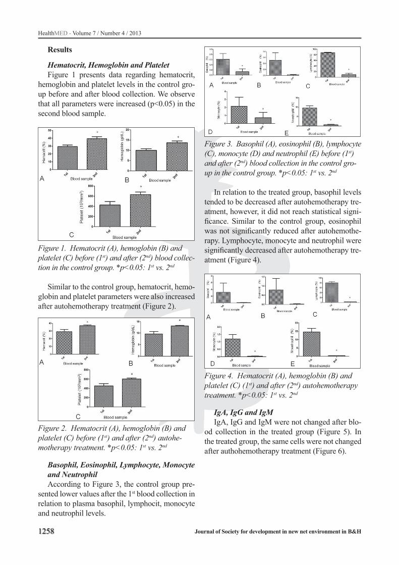
1258
HealthMED - Volume 7 / Number 4 / 2013
Journal of Society for development in new net environment in B&H
Results
Hematocrit, Hemoglobin and Platelet Figure 1 presents data regarding hematocrit,
hemoglobin and platelet levels in the control gro-up before and after blood collection. We observe that all parameters were increased (p<0.05) in the second blood sample.
Figure 1. Hematocrit (A), hemoglobin (B) and platelet (C) before (1st) and after (2nd) blood collec-tion in the control group. *p<0.05: 1st vs. 2nd
Similar to the control group, hematocrit, hemo-globin and platelet parameters were also increased after autohemotherapy treatment (Figure 2).
Figure 2. Hematocrit (A), hemoglobin (B) and platelet (C) before (1st) and after (2nd) autohe-motherapy treatment. *p<0.05: 1st vs. 2nd
Basophil, Eosinophil, Lymphocyte, Monocyte and NeutrophilAccording to Figure 3, the control group pre-
sented lower values after the 1st blood collection in relation to plasma basophil, lymphocit, monocyte and neutrophil levels.
Figure 3. Basophil (A), eosinophil (B), lymphocyte (C), monocyte (D) and neutrophil (E) before (1st) and after (2nd) blood collection in the control gro-up in the control group. *p<0.05: 1st vs. 2nd
In relation to the treated group, basophil levels tended to be decreased after autohemotherapy tre-atment, however, it did not reach statistical signi-ficance. Similar to the control group, eosinophil was not significantly reduced after autohemothe-rapy. Lymphocyte, monocyte and neutrophil were significantly decreased after autohemotherapy tre-atment (Figure 4).
Figure 4. Hematocrit (A), hemoglobin (B) and platelet (C) (1st) and after (2nd) autohemotherapy treatment. *p<0.05: 1st vs. 2nd
IgA, IgG and IgMIgA, IgG and IgM were not changed after blo-
od collection in the treated group (Figure 5). In the treated group, the same cells were not changed after authohemotherapy treatment (Figure 6).

HealthMED - Volume 7 / Number 4 / 2013
Journal of Society for development in new net environment in B&H 1259
Figure 5. IgA (A), IgG (B) and IgM (C) before (1st) and after (2nd) blood collection in the control group in the control group
Figure 6. IgA (A), IgG (B) and IgM (C) (1st) and after (2nd) autohemotherapy treatment
Discussion
This investigation was undertaken to evaluate the effects of autohemotherapy on hematological responses in female rats. As a main finding, there was no difference between the control and treated groups regarding hematocrit, hemoglobin, plate-let, basophil, eosinophil, lymphocyte, monocyte, neutrophil, IgA, IgG and IgM. We suggest that the treatment presented the same effects of the control group. Blood removal is responsible for inducing blood cell production, since blood loss lead to de-creased tissue oxygenation and it is a stimulus for erythropoiesis [13]. This mechanism also occu-rred with a small volume of blood applied, such as the procedure used in our study.
In relation to the leukocyte count, we observed no difference between the autohemotherapy and control groups. Our finding indicates that blood withdrawal reduced the production of leucocyte.
Moreover, a previous investigation concluded that ozonated autohemotherapy in a dose of 50 mg/mL does not present significant influence on natural killer cell function in hemodialyzed patients [14]. No differentiation was made between the types of lymphocytes, we suggest future studies to perform this procedure. To the best of our knowledge, no previous study specifically examined the inducti-on of leukocyte cell by cell. Thus, there is no gue-sswork in the literature regarding the pathophysi-ology of this mechanism.
A possible explanation for the phagocyte beha-vior found in our study is that intramuscular appli-cation of blood as well as blood withdrawal cause a local inflammatory process by stimulating pha-gocytosis through tissue macrophages [15]. Tissue macrophages present antigens to T lymphocytes that are activated and are involved in the proce-ss of B cells activation that initiate the process of clonal division with the production of antibodies and globulins [16]. Neutrophils are recruited from peripheral blood after inflammatory mediators se-creted by damaged tissues [15]. The result of the process is a decreased number of lymphocytes in-duced by local stimulation, which is responsible for the response modulation and is detected in pe-ripheral blood. Since neutrophils and macropha-ges recruited into the tissues phagocytize the inju-red tissues and blood cells inserted in place with cleaning function until the damaged cells and ti-ssues are properly removed [15], it is observed a lower removal to the lymphatic system are poorly detected in peripheral blood.
Bocci et al [17] published the first study to show that autohaemotherapy may activate an immuno-logical marker in normal subjects without procu-ring any toxic effects. However, according to our data, in the comparison between control and au-tohemotherapy groups regarding immunoglobu-lins, there was no difference. We noted that in the both groups immunoglobulins production was not changed. It indicates that autohemotherapy does not induce the production of acute-phase immu-noglobulin . Furthermore, induced desensitization plays an important part in the mechanism of action of autohaemotherapy. The administration of large doses of er ythrocytes or erythrolysate results in immunosuppression [5]. Nevertheless, according to Klemparskaya et al [5], autohaemotherapy does

1260
HealthMED - Volume 7 / Number 4 / 2013
Journal of Society for development in new net environment in B&H
not cause side effects and is feasible both on an in-and out-patient basis.
In this study, the maximum amount of blood that could be injected into the muscles of the animals was collected. This volume of blood did not affect blood oxygenation because the animals did not present symptoms related to this mechanism such as cyanosis and reduced blood hemoglobin levels. However, although this volume is not dangerous to blood oxygenation, it may have been responsible for the induction of hematopoietic responses observed in the autohemotherapy and in the control groups.
Based on our findings, Wistar female rats tre-ated with autohemotherapy presented the same responses described in previous studies such as induction of erytropoiesis [5]. Also, similar res-ponses were observed related to Ig M production. Nonetheless, we found no reference regarding this mechanism in female rats. Observing the princi-ples of erythropoiesis induction, this mechanism occurs when there is any condition that decreases tissue oxygenation such as low blood volume, ane-mia, low hemoglobin levels, reduced blood flow, low atmospheric oxygen or lung disease [18]. In these cases, renal hypoxia stimulates the produc-tion of erythropoietin inducing factor from bone marrow to produce erytrocites [19]. Removal of 5 ml of blood during autohemotherapy performed in humans does not induce renal hypoxia [20].
We observed that autohemotherapy and blood removal increased blood platelet levels. Howe-ver, a previous study reported opposite findings in humans [21]. Tylick et al evaluated the effects of ozonated autohaemotherapy on the platelet function in chronically haemodialysed patients with peripheral arterial disease. They found that autohemotherapy with ozone concentration of 50 microg/ml and citrate as an anticoagulant does not induce platelet aggregation.
Our data demonstrated that autohemotherapy presented the same effects of no treatment regar-ding hematological responses. This data does not provide evidence enough to implement changes to human treatment. We suggest further studies to justify the application of this technique in humans for treatment or cure of any disease.
These data provide useful information given that currently many types of animals are widely studied in order to develop new therapies for the
prevention of several cardiovascular disorders in humans [22-25].
Conclusion
Autohemotherapy did not influence hematolo-gical responses in Wistar female rats.
Acknowledgement
This study received finantial support from FA-PESP and NEPAS.
References
1. Bucioli SA, Abreu LC, Valenti VE, Vannucchi H. Vita-min e and carnitine suplementation effects on blood glucose levels in young rats submitted to exhaustive exercise stress. HealthMED J 2012; 6: 136-139.
2. Centofanti G, Breda JR, Valenti VE, Abreu LC, Fujiki EN, Audi SG, Pereira VX, Correa JA. Vascular access for hemodialysis: an experience report. HealthMED J 2011; 5: 1959-1962.
3. Moafi A, Rahgozar S, Hajian M, Ghias M, Ghorbani N, Hassanzadeh A. The Effects of Supplemental Iron on Educational Achievements of Students with “Iron Deficiency without Anemia”: A Randomized, Double-Blind, Placebo-Controlled Trial. HealthMED J 2012; 6: 2047-51.
4. Zheng Z, Zhan J, Zhang Y, Wei Y, Wang M, Zheng Z, Peng J. Serum fgl2 levels elevated in patients with acute coronary syndrome (ACS). HealthMED J 2012; 6: 2062-65.
5. Klemparskaya NN, Shalnova GA, Ulanova AM, Kuz-mina TD, Chuhrov AD. Immunomodulating effect of autohemotherapy (a literature review). J Hyg Epide-miol Microbiol Immunol 1986; 30: 331-6.
6. Mikhailov SN, Novikov NM. Patologicheskaya foziol-giya i eksperimentalnaya terapiya. Eksperimental’naya Terapiya 1981; 5: 62.
7. Klemparskaya NN, Shalnova GA. Normal autoanti-bodies as radioprotective factors. Atomizdat 1978; 3: 134-9.
8. Klemparskaya NN. Zhurnal mikrobiologii, epidemi-ologii i immunologii. Eksperimental’naya Terapiya 1973; 9: 47.
9. Gurgenidze GV, Kilasoniya LO. Terapevticheskii ark-hiv. Eksperimental’naya Terapiya 1971; 12: 83.
HealthMED - Volume 7 / Number 4 / 2013
Journal of Society for development in new net environment in B&H 1261
10. Domz CA, Fay KJ, Hoag CL. Hemotherapy in Suspected Dermatomyositis. Calif Med 1957; 87: 108–111.
11. Ruffiex TB, Mainetti P, Solomon C, Saudat D. To-pical Haemotherapy for Leg Ulcers. Dermatology 1994; 189: 418-420
12. Sangue, Tecidos e Órgãos; Nota Técnica nº 1 de 13 de abril de 2007 - 18h50 Auto-Hemoterapia, disponível em: http: //www.anvisa.gov.br/sangue/informes/01_130407.htm - acessado em 2 setembro de 2008
13. Jagannath VA, Fedorowicz Z, Al Hajeri A, Hu N, Sharma A. Hematopoietic stem cell transplantati-on for people with ß-thalassaemia major. Cochrane Database Syst Rev. 2011; 10: CD008708.
14. Biedunkiewicz B, Tylicki L, Rachon D, Hak L, Nieweglowski T, Chamienia A, Debska-Slizien A, Mysliwska J, Rutkowski B. Natural killer cell ac-tivity unaffected by ozonated autohemotherapy in patients with end-stage renal disease on mainte-nance renal replacement therapy. Int J Artif Organs 2004; 27: 766-71.
15. Skyler JS. Immune intervention for type 1 diabetes mellitus. Int J Clin Pract Suppl 2011; 2: 61-70.
16. Pammi M, Haque KN. Oral immunoglobulin for the treatment of rotavirus diarrhea in low birth weight infants. Cochrane Database Syst Rev 2011; 10: CD003742.
17. Bocci V, Luzzi E, Corradeschi F, Paulesu L. Studies on the biological effects of ozone: 5. Evaluation of immunological parameters and tolerability in nor-mal volunteers receiving ambulatory autohaemothe-rapy. Biotherapy 1993; 7: 83-90.
18. Okebe JU, Yahav D, Shbita R, Paul M. Oral iron supplements for children in malaria-endemic areas. Cochrane Database Syst Rev 2011; 10: CD006589.
19. Lee GR, Bithell TC, Foerster J, Lukens JN. Erithro-poiesis In: Wintrobe’s Clinical Hematology. ed. 9 Lea & Febiger, Pennsylvania – E.U.A., 1993, vol 1. Part 3, section 2: 169-267.
20. Deen PM, Robben JH. Succinate receptors in the kidney. J Am Soc Nephrol 2011; 22: 1416-22.
21. Tylicki L, Lizakowski S, Biedunkiewicz B, Skibowska A, Nieweglowski T, Chamienia A, Debska-Slizien A, Rutkowski B: Platelet function unaffected by ozonated autohaemotherapy in chronically haemodialysed pa-tients. Blood Coagul Fibrinolysis 2004, 15: 619-22.
22. Valenti VE, Ferreira C, Meneghini A, Ferreira M, Murad N, Ferreira Filho C, Correa JA, Abreu LC, Colombari E. Evaluation of baroreflex function in young spontaneously hypertensive rats. Arq Bras Cardiol 2009; 92: 205-15.
23. Cisternas JR, Valenti VE, Alves TB, Ferreira C, Pe-tenusso M, Breda JR, Pires AC, Tassi N, de Abreu LC. Cardiac baroreflex is already blunted in eight weeks old spontaneously hypertensive rats. Int Arch Med 2010; 3: 2.
24. Valenti VE, Imaizumi C, de Abreu LC, Colombari E, Sato MA, Ferreira C. Intra-strain variations of ba-roreflex sensitivity in young Wistar-Kyoto rats. Clin Invest Med 2009; 32: E251.
25. Valenti VE, De Abreu LC, Colombari E, Sato MA, Ferreira C. The variability of baroreflex sensitivity in juvenile, spontanebously hypertensive rats. Car-diovasc J Afr 2011; 22: 14-7.
Corresponding AuthorFernando Luiz Affonso Fonseca,Departamento de Clinica Medica,Faculdade de Medicina do ABC,Santo Andre,Brazil,E-mail: [email protected]

1262
HealthMED - Volume 7 / Number 4 / 2013
Journal of Society for development in new net environment in B&H
Abstract
We present the use of subglottic high frequ-ency jet ventilation (HFJV) in treatment of bila-teral vocal fold palsy in 36-year-old female who underwent thyroidectomy on the previous day. Af-ter intravenous anesthesia induction, endotracheal tube was exchanged with jet catheter inserted thro-ugh the vocal cords into the trachea. The surgical procedure was performed endoscopically using HFJV and the special endo-extralaryngeal suture technique by Lichtenberger. At the end of proce-dure, jet catheter was exchanged with LMA until spontaneous breathing was established. There was no hypoxia or barotrauma to the lungs during the surgery. The postoperative course was uneventful.
Key words: High frequency jet ventilation, Thyroid gland, Postoperative Complications, Airway Obstruction, Vocal Fold Paralysis.
Introduction
Lesion of recurrent laryngeal nerves as con-sequence of thyroid surgery, results in bilateral vocal fold paralysis (BVFP) and respiratory ob-struction [1,2].
The initial treatment involves obtaining an adequate airway with endo-extralaryngeal latero-fixating operations undergoing general anesthesia [3]. This revision surgery of bilateral vocal fold palsy has been conducted in suspension laryngos-cope with subglottic high frequency jet ventilati-on (HFJV). Applying these operative techniques satisfactory breathing can be achieved without tracheotomy [4-6].
Endolaryngeal microsurgery is challenging for the anesthesiologist and the laryngologist since they share access to the larynx [7]. The good exposure of the larynx, immobility of the vocal cords, satisfac-
tory ventilation and oxygenation are crucial. High frequency jet ventilation as ventilator approach in laryngeal microsurgery provides application of small tidal volume at high rates, followed by passi-ve expiration [8,9]. Subglottic HFJV with narrow tracheal catheters is described for surgery of the en-dolarynx as a safe and less obstructive ventilation technique [10,11]. However, the technical skills and equipment are the reasons for HFJV limited use.
We report a patient with BVFP undergoing en-do-extralaryngeal laterofixating operation without tracheotomy with subglottic HFJV on first day af-ter total thyroidectomy.
Case report
A 36-year old female (BMI 24 kg/m2) was ad-mitted from outside hospital after total thyroidec-tomy, performed on the previous day. Postoperati-vely, patient developed severe inspiratory stridor, worsening progressively with decreased arteri-al oxygen saturation (from 100% to 82 %) after tracheal tube removal. After mask ventilation with 100 % oxygen, flexible laryngoscopy revealed paradoxical behavior of the vocal folds that cau-sed near total obstruction of the larynx on forced inspiration. Trachea was reintubated over flexible laryngoscope. Chest radiograph, electrocardio-graphy (ECG) and laboratory findings including potassium, calcium and glucose level were nor-mal. Due to the logistic problem, laryngologist and equipment were unavailable; patient was tran-sferred on the following day to our institution.
Premedication consisted of intravenous (IV) atropine 1 mg and dexamethason 10 mg. Monito-ring included noninvasive blood pressure, ECG, capnography, pulse oximetry and entropy. Ane-sthesia was induced with propofol 2.5 mg/kg IV
Subglottic high frequency jet ventilation in management of bilateral vocal fold paralysis: A case report Nevena Kalezic1,3, Dusanka Janjevic2, Vladimir Dolinaj2, Nebojsa Ladjevic1,3, Dejan Markovic1,3
1 Department of Anesthesiology, Clinical Centre of Serbia, Belgrade, Serbia,2 Department of Anesthesiology, Ear, Nose and Throat Clinic, Clinical Centre Vojvodina, Novi Sad, Serbia,3 School of Medicine, University of Belgrade, Belgrade, Serbia.

HealthMED - Volume 7 / Number 4 / 2013
Journal of Society for development in new net environment in B&H 1263
followed by remifentanil infusion (0.2-0.5 μg/kg/min) (ARGUS Medical, Switzerland) and rocu-ronium (0.5 mg/kg) (ARGUS Medical, Switzer-land). Maintenance anesthesia was achieved with an infusion of propofol and remifentanil. Patient was ventilated with 100% oxygen during induc-tion. After two minutes, when satisfactory level of anesthesia was achieved and justified by en-tropy (RE/SE 35/36) and the loss of eyelash re-flex, tracheal tube was exchanged with jet catheter (Dual Lumen Jet Ventilation Catheter acc. Biro Acutronic Medical System, Switzerland) under laryngoscopy with Miller 3 blade. The jet cathe-ter was advanced by direct laryngoscope and po-sitioned in the posterior commissure. Subglottic HFJV was applied with automatic commercial Mistral jet ventilator (Acutronic Medical System, Switzerland) attached to the jet catheter.
The initial ventilator settings were: FiO2 0.6 respiratory frequencies 120 cycles/min, 40% in-spiration duration, and driving pressure 1.2 bar. After initiation HFJV was monitored by bilateral chest movements and auscultation of the lungs. Muscle relaxation was maintained based on train-of-four (TOF) stimulation of the ulnar nerve.
The surgical procedure consisted of right endo-extralaryngeal laterofixation. Duration of surgery was 30 minutes. At the end of the surgical proce-dure, propofol and remifentanil infusions were sto-pped. When TOF of 0.9 was achieved block was reversed with IV neostigmine (2.5 mg) and atropine (1mg). Ketorolac 30 mg IV was given for postope-rative pain treatment. No laryngeal edema was noti-ced on repeated postoperative endoscopic laryngeal examination. Jet catheter was removed and LMA (LMA-ClassicTM, The Laryngeal Mask Company Limited, Nicosia, Cyprus) was used to secure a supraglottic airway until the patients regained pro-tective reflexes and spontaneous breathing (patients lungs were ventilated by mask until awakening).
Postoperatively, the patient denied procedural recall, and expressed a high degree of satisfacti-on. Clinical signs, spirometry and flow/volume curve verified absence of airway obstruction. Complications resulting from ventilation with subglottic HFJV such as barotrauma or subcuta-neous emphysema, seeding of blood into trache-obronchial tree were not observed in patient. Wit-hin 8 days patient was discharged home.
Discussion
We presented the use of subglottic HFJV du-ring the revision surgery of BVFP in order to esta-blish the best surgical field and safe approach to the airway on previously intubated trachea.
Laryngeal obstruction due to BVFP may cause dyspnea, hoarseness and dysphagia. If left untre-ated, patients may become tracheotomy depended [3,5,6]. In this patient immediate airway obstruc-tion due to the bilateral recurrent laryngeal nerve damage and inability to phonate required tracheal reintubation.
a)
b)Figure 1. Views of a jet catheter in the glottis providing unobstructed access to the BVFP befo-re (a) and after (b) endo-extralaryngeal fixation procedure
Our decision to extubate trachea and apply HFJV was based on previous date about easy in-tubation and HFJV suitability for this procedure. Suspension microlaryngeal surgery requires good exposure of the larynx, continuous control of the

1264
HealthMED - Volume 7 / Number 4 / 2013
Journal of Society for development in new net environment in B&H
airway patency and immobility of the vocal cords. Even a small endotracheal tube impedes access and visibility of the operative field, especially posterior part of the glottis [10,12,13]. A good access to the surgical field due to small diameter of the jet cathe-ter is main advantage of subglottic jet ventilation [11]. Narrow jet catheters are preferable to provide space for observation and surgical manipulation, and permit a sufficient outflow of respiratory gases.
This is particularly important in patients with difficult or partially occluded airways with po-int out on its use in the setting of bilateral vocal cord immobility. Although certain vibration of the vocal folds may occur, the rigid laryngoscope remains unmoved and the operating field is quiet [10,14-16]. The jet catheter tip position above ca-rina enables a limited entrainment of ambient air only, HFJV creates a constant PEEP, which pre-vents bronchial and alveolar collapse and increase functional residual capacity [17]. The most serio-us complication of HFJV is lung barotraumas that may occur to an obstructed airway. This can hap-pen in case of accidental displacement of the jet catheter into a bronchus, obstruction of the larynx by instruments or inadvertent closure of the glottis when muscular relaxation is diminished. There-fore, it is essential to monitor airway pressure to avoid barotraumas [18,19].
Dexamethason was given for prevention of laryngeal edema as the possible complication of surgery [1]. Use of remifentanil with propofol for TIVA is safe and the patient is protected from de-layed respiratory depression [20].
Although subglottic HFJV is suitable to ma-intain adequate ventilation throughout the pro-cedure, we decided to insert LMA at the end of the procedure. The plan was based on endoscopic examination and decision that trachea should be extubated at the end of the procedure if adequa-te postoperative airway is achieved. This enables good airway control, smooth and safe tracheal extubation in patient who experienced previous emergency reintubation. Moreover LMA provides adequate views of vocal cords and facilitates repe-ated endoscopic exam [15,16,21,22].
In conclusion, this case demonstrates that subglottic HFJV is safe and effective in maintaining gas exchange, providing optimal visibility of laryn-geal structures, offering adequate space for surgical
manipulation without tracheotomy. Overall, combi-nation of HFJV and conventional ventilation with LMA seems uniquely suited for this specific use, jet prospective comparison of anesthetic techniques is needed to establish superiorly.
References
1. Gardner GM, Benniger MS. Vocal fold paralysis. In: Rubin JS, Sataloff RT, Korovin GS (Eds). Diagnosis and treatment of voice disorders. San Diego: Plural Publishing, Inc. 2006; 471-93.
2. Goncalves Filho J, Kowalski LP. Surgical complicati-ons after thyroid surgery performed in a cancer hos-pital. Otolaryngol Head Neck Surg 2005; 132: 490-4.
3. Jori J, Rovo L, Czigner J. Vocal cord laterofixation as early treatment for acute bilateral abductor paralysis after thyroid surgery. Eur Arch Otorhinolaryngol 1998; 255: 375–8.
4. Shvero J, Koren R, Stern Y, Segal K, Feinmesser R, Hadar T. Laser posterior ventriculocordectomy with partial arytenoidectomy for the treatment of bilate-ral vocal fold immobility. J Laryngol Otol 2003; 117: 540-3.
5. Lichtenberger G. Reversible lateralization of the pa-ralyzed vocal cord without tracheostomy. Ann Otol Rhinol Laryngol 2002; 111: 21-6.
6. Leitersdorfer S, Lichtenberger G, Bihari A, Kovacs I. Evaluation of the lung function test in reversible glottis-dilating operations. Eur Arch Otorhinolaryn-gol 2005; 262: 289-93.
7. Rezaie-Majd A, Bigenzahn W, Denk DM, Burian M, Kornfehl J, Grasl MCh, Ihra G, Aloy A. Superimposed high-frequency jet ventilation (SHFJV) for endosco-pic laryngotracheal surgery in more than 1500 pati-ents. Br J Anaesth 2006; 96: 650-9.
8. Ihra G, Gockner G, Kashanipour A, Aloy A. High-frequency jet ventilation in European and North Ame-rican institutions: developments and clinical practice. Eur J Anaesthesiol. 2000; 17(7): 418-30.
9. Bacher A, Pichler K, Aloy A. Supraglottic combined frequency jet ventilation versus subglottic monofrequ-ent jet ventilation in patients undergoing microlaryn-geal surgery. Anesth Analg 2000; 90: 460-5.
10. Davies JM, Hillel AD, Maronian NC, Posner Lk. The Hunsaker Mon-Jet tube with jet ventilation is effective for microlaryngeal surgery. Can J Anaesth 2009; 56: 284-90.

HealthMED - Volume 7 / Number 4 / 2013
Journal of Society for development in new net environment in B&H 1265
11. Bourgain JL, Chollet M, Fischler M, Gueret G, Mayne A. Guideline for the use of jet ventilation du-ring ENT and oral surgery. Ann Fr anaesth Reanim 2010; 29(10): 720-7.
12. Bacher A, Lang T, Weber J, Aloy A. Respiratory effi-ciancy of subglottic low-frequency, subglottic com-bined-frequency, and supraglottic combined-frequ-ency jet ventilation during microlaryngeal surgery. Anesth Analg 2000; 91: 1506-12.
13. Bourgain JL, Desruennes E, Fischler M, Ravussin P. Transtracheal high frequency jet ventilation for endoscopic airway surgery: a multicentre study. Br J Anaesth 2001; 87(6): 870-5 .
14. Barakate M, Maver E, Wotherspoon G, Havas T. Anaesthesia for microlaryngeal and laser laryngeal surgery: impact of subglottic jet ventilation. J Laryn-gol Otol. 2010; 124(6): 641-5.
15. Rubin JS, Patel A, Lennox P. Subglottic jet ventilati-on for suspension microlaryngoscopy. J Voice 2005; 19: 146-50.
16. Patel A, Rubin JS. The difficult airway: the use of subglottic jet ventilation for laryngeal surgery. Lo-goped Phoniatr Vocol 2008; 33: 22-4.
17. Mausser G, Schellauf A, Scherubl M, Arrer A. Schwarz G. Experimental model of laryngotracheal stenosis in infants: effect of different high-frequency jet ventilation patterns on pulmonary parameters. Paediatr Anaesth 2011; 21: 894-9.
18. Jaquet Y, Monnier P, Van Melle G, Ravussin P, Spahn DR, Chollet-Rivier M. Complications of diffe-rent ventilation strategies in endoscopic laryngeal surgery: a 10 – year review. Anesthesiology 2006; 104: 52-9.
19. Ihra GC, Heid A, Pernerstorfer Th. Airway steno-sis-related increase of pulmonary pressure during high-frequency jet ventilation depends on injector’s position. Anesth Analg 2009; 109: 461-5.
20. Mertens MJ, Engbers FH, Burm AG, Vuyk J. Predic-tive performance of computer-controlled infusion of remifentanil during propofol/remifentanil anaesthe-sia. Br J Anaesth 2003; 90: 132-41.
21. Brimacombe JR, Berry AM, White PF. The laryngeal mask airway: limitations and controversies. Int Ane-sthesiol Clin 1998; 36: 155-82.
22. Brimacombe J, Sher M, Laing D, Berry A. The laryngeal mask airway: a new technique for fiberop-tic guided vocal cord biopsy. J Clin Anaesth 1996; 8: 273-5.
Corresponding AuthorDusanka Janjevic,Department of Anesthesiology,Ear, Nose and Throat Clinic,Clinical Centre Vojvodina,Novi Sad,Serbia,E-mail: [email protected]
1266
HealthMED - Volume 7 / Number 4 / 2013
Journal of Society for development in new net environment in B&H
Abstract
Background: As a physiological event, meno-pause has complications which influence women’s quality of life. Several measures such as hormone therapy and non-pharmacological therapies are recommended to reduce menopausal symptoms and improve quality of life (QoL) in this period. One of the non pharmacological methods is com-plementary medicine i.e. yoga. The present study aimed to determine the impact of yoga exercises on the quality of life of postmenopausal women.
Methods: This was a randomized clinical trial in which 136 postmenopausal women aged 45-60 were divided into experimental and control grou-ps through random allocation method based on the study environment. The experimental group parti-cipated in basic Hatha Yoga training classes (twice a week and 90 minutes for each session); a combi-nation of Asana, Pranayama and Shavasana. Data collection tools included a Menopause-specific Qu-ality of Life (MENOQOL) developed by Hilditch et al. The quality of life of the subjects was evalu-ated before and two months after the intervention (in vasomotor, psychosocial, physical and sexual dimensions). Data were analyzed through SPSS Software version 16 through the statistical methods of Mann-Whitney and independent t-test (P < 0.05).
Results: The subjects were similar in terms of individual variables before the intervention. Mean QoL score before the intervention in the experi-mental and control groups was 103.69 (33.27) and 100.28 (36.27) respectively which did not have a significant difference. Two months after the in-tervention, this reached 84.75 (32.10) and 105.73 (37.22) respectively for the experimental and con-
trol groups. Independent t-test showed improve-ment of the QoL score in the experimental group (P = 0.002). The results of statistical tests showed that the yoga exercises were significant in three vasomotor (P = 0.039), psychosocial (P = 0.009) and physical (P = 0.006) dimensions.
Conclusion: Considering that a large number of women are reaching menopausal age, under-standing the effect of this stage of life on mental health and QoL in this population and using thera-peutic strategies such as physical activities for the-ir sense of well-being should be taken into account as an important health goal.
Key words: Yoga, quality of life, menopause.
Introduction
Menopause is the cessation of menstruation resulting from loss of ovarian follicular functi-on which is a recognized physiological event in women’s lives. In addition to hormonal changes particularly reduction of estrogen levels with bio-logical, psychological and social changes (1), this physiological process is associated with complicati-ons such as sleep disorder, night sweats, headaches, hot flashes, vaginal dryness, anger and depression, increase in cholesterol levels and cardiovascular di-seases (2) which influence women’s quality of life (QoL) (3-5). Due to an increase in life expectancy in the world and Iran, it is expected that women spend 20-30 years of their life in postmenopausal period during which they are faced with many problems, symptoms and complications (6) and has signifi-cant consequences for families and society arising from physical, mental, social and economic issues (7). 75 to 80% of women experience menopause-
Effect of yoga exercises on quality of life of postmenopausal womenShabnam al-Sadat ShariatPanahi1, Eftekharalsadat Hajikazemi2, Soghra Nikpour3, Somayeh Fghanipour2, Agha Fatemeh Hosseini4
1 Faculity of Nursing and Midvifery Tehran University of Medical Sciences, Tehran, Iran,2 Center for Nursing Care Research, Tehran University of Medical Sciences, Tehran, Iran,3 Center for Nursing Care Research and Faculity of Nursing and Midvifery, Tehran University of Medical Sciences, Tehran, Iran,4 School of Health Management & Medical Information Science, Tehran University of Medical Sciences, Tehran, Iran.

HealthMED - Volume 7 / Number 4 / 2013
Journal of Society for development in new net environment in B&H 1267
related problems and complications(8) approxima-tely 50% of them have these symptoms averagely four years after complete cessation of menstruation (9) and 10 to 30% of them have these symptoms their entire life so that 60% of women are seeking a solution and treatment for such problems (10). Different actions such as hormone therapy, physi-cal activity and exercises can be applied to decrea-se menopausal symptoms (11). Although hormone therapy is a selective treatment for reduction of me-nopausal symptoms (12), its harmful side effects in long-term use, including increased risk of cataract (13), endometrial and breast cancer, coronary artery disease, MI and thromboembolism have been pro-posed in different studies (14-17). However, Blake has announced that there is no evidence to confirm the side-effects of hormone therapy (18). Nonethe-less, the concerns about safety of hormone therapy, incentive in the health lifestyle and behavior chan-ges and alternative and complementary approaches during middle ages have been increased (14).
One of the types of complementary medicine is yoga. It is considered to be an exercise for the mind, body and spirit that can significantly redu-ce stress and anxiety (19), depression (20) and problems related to musculoskeletal disorders such as osteoarthritis, carpal tunnel syndrome, hyperkyphosis, backache and of motor skills and physiological variables such as hypertension, he-art rate and body weight (21). QoL improvement and sense of mental well-being have been reported in yoga (22). Furthermore, in many studies yoga has been reported to be effective in reduction of menopausal symptoms (23-26). In addition to the mentioned advantages of yoga, it has some other benefits in terms of cost, efficacy, non-invasive-ness, minimum risk compared with side-effects of drugs, and physical fitness that can improve QoL (20). On the other hand, the effect of exercises and physical activity has been proposed on mental and physical health; so that a systematic review of 14 studies has shown a positive relationship between physical activity and health related quality of life (27). Moreover, it has been suggested that women who exercise regularly experience less vasomotor symptoms, negative mood, decreased sexual desi-re and a higher feeling of well-being (28). Other researchers have also proposed that sports activiti-es are related to quality of life in postmenopausal
women (29) and have paid attention to exercise and lifestyle change as important factors reducing menopause symptoms (30).
Despite findings proposing that exercise can be an effective treatment for menopausal problems, Borrelli reported that there are very limited evi-dences to prove positive impacts of exercise on symptoms and problems experienced during pos-tmenopausal period (12). Therefore, the present study aimed to determine the impact of yoga exer-cises on quality of life of postmenopausal women in order to improve their physical and mental he-alth and promote their QoL in this period. Con-sidering the mentioned cases, the question of the present study was “what is the impact of imple-menting two months of yoga exercises on quality of life of postmenopausal women?”
Materials and methods
Experimental DesignThis was a randomized experimental trial with a
control group. The researchers evaluated the impact of yoga exercises on four dimensions of QoL in postmenopausal women by inserting the indepen-dent variable (yoga exercises) and also considering a control group. Thus, 136 postmenopausal women aged 45-60 participated in 8 weeks of basic Hatha yoga; a combination of Asana (physical exercise), Pranayama (breathing exercises) and Shavasana (relaxation training). Randomization was based on the study environment and conducted through selecting 5 districts from the 22 geographical dis-tricts of Tehran municipality by the use of a ran-dom number table and all the health centers in the mentioned districts were identified (totally; n = 29). Thereafter, each of the health centers was given a special number and four health centers were ran-domly selected through the random number table. After random selection of the four health centers, in order to prevent pollution of information among the experimental and control groups, the subjects of the experimental and control groups were selected from separate health centers; thus, two health cen-ters were randomly allocated to the experimental and control groups. In addition, in order to prevent from pollution of the information, the two health centers which were almost in a geographic district were considered as the experimental group.

1268
HealthMED - Volume 7 / Number 4 / 2013
Journal of Society for development in new net environment in B&H
SubjectsThe eligible subjects with inclusion criteria were
selected through continuous method from the se-lected health centers. The inclusion criteria of this study were: Married menopausal Iranian women aged 45-60, at least a year and a maximum of four years had passed from their menopause, with com-mon symptom of menopause and lowest literacy, BMI between 20-30, their menopause happened naturally not due to surgery, no regular physical ac-tivity currently, no history of HRT in past 6 months and no known physical (hypertension, diabetes, hypothyroidism and hyperthyroidism) and mental illness. The exclusion criteria of this study were: Fail to attend yoga classes as four sessions, the oc-currence of any crisis during the two months of the study, use of hormonal medications and entire in exercise program during the research period and lack of willingness to continue in the study.
InstrumentationThis study was approved by the Research
Deputy of Tehran University of Medical Scienc-es. Informed written consent was obtained from all the study subjects before starting the study. All participants are first accepted the questionnaire. The questionnaire consisted of two parts; the first part included the basic information such as age, education, occupation status, the number of chil-dren, menopausal age and the time elapsed from their menopause. The second part also included 31 questions related to the QoL assessment based on Menopause-specific Quality of Life (MENO-QOL).This questionnaire has been developed by the Department of Family and Community Medi-cine, University of Toronto, Canada in 1992(31) . It was used in Iran after cultural adaptation with Iranian the community; so that after determining the validity of this questionnaire by Golyan et al. in Iran, its reliability was calculated with the cor-relation of 0.95(32) . Its reliability in the present study was also obtained (r = 0.91). The questions with 8-degree Likert scale were scored based on how much the mentioned symptoms have been problematic during the past month. Scoring was implemented through the following method: an-swer “No” scored 1 and answer “Yes” scored 0. In choices identifying the intensity of the symptoms the scores were: zero equals 2 scores, one equals
3 scores, two equal 4 scores, three equal 5 sco-res, four equal 6 scores, five equal 7 scores and six equal 8 scores. As a result, the total score of QoL in this tool was maximum 248 and minimum 31 scores. In order to evaluate QoL of postmenopau-sal women in vasomotor, psychosocial, physical and sexual dimensions, the mean score of each dimension was calculated and compared; thus, mean score in each dimension was calculated to be 1-8. It was a self-report questionnaire.
Training ProgramThe study subjects in the experimental group
participated in yoga exercises for eight weeks (2 months), twice a week and each session for 90 minu-tes. The exercises were presented by the experienced instructor under the supervision of Yoga Association and also had the certificate of the provided trainings. The conditions and positions effective on relief of menopausal symptoms were extracted from Hatha yoga focusing on deep relaxation techniques. The exercises consisted of stretching and endurance in standing, sitting and sleeping, and breathing positi-on and breathing exercises with each position. The-se types of exercises have been selected based on the recommendation of current articles on women with menopausal symptoms.The sessions consisted of checking the presence of women, controlling the exercises in the previous sessions, warm-up tech-niques and breathing, yoga Asana gestures and ex-ercises, and eventually relaxation techniques. Fur-thermore, the experimental group was recommend-ed to exercise for 15 minutes at home based on the provided pamphlet including joint rotation exercises of wrist, elbow, shoulders and abdominal breathing due to the importance and ease of implementing these exercises. Moreover, at the beginning of each session, their feedback has been analyzed concern-ing the home exercises recommended to them.
The samples of the exercises during the ses-sions were: corpse pose (5-7 min), Pawan Mukta Asana (wind relieving pose) for hip and feet such as ankle rotation, knee crank, cycling, leg rotation, raised legs pose (20-25 min), backward bending movements and poses such as half locust pose, snake pose, Sphinx asana, Cobra pose (10-15 min totally), rotating poses such as sleeping abdominal stretching, leg log pose (2-3 min), Pawan Mukta Asana for hands such as wrist bending, elbow

HealthMED - Volume 7 / Number 4 / 2013
Journal of Society for development in new net environment in B&H 1269
bending, shoulder socket pose, and stretching pos-es in this area such as hand raising pose, bow and arrow pose (30-35 min), rotational viewing (2-3 min) and asana corpse poses (5-10 min).
Menopause-specific Quality of Life (MENO-QOL) was repeated two times for both groups; one before and another at the end of the interven-tion (end of week 8). It should also be noted that in order to complete the questionnaires at the end of week 8 for the control group, all the coordination were conducted through the phone. No interven-tion was conducted for the control group; there-fore, the control group was given a yoga training book as a gift to show appreciation and to enhance their knowledge on yoga so that they can partici-pate in yoga classes if willing to do so.
Statistical analysisData were analyzed through Software SPSS
version 16. In order to describe the data from the central and dispersion parameters for inferential analysis of research findings, first we used Kol-mogorov-Smirnov test to determine the normal data.The homogeneity of demographic and base-line data between the experimental and control groups was examined through chi-square, t and fisher’s exact tests. Then, in order to determine the effect of independent variable on the experimen-tal and control groups, we used statistical normal distribution of non-parametric Mann-Whitney and independent t-test at significance level of P < 0.05.
Results
Out of 136 participants, 13 subjects were exclu-ded from the study due to reasons including unfore-seen events for families (7 subjects), caring for dise-ased husband (2 subjects), unwillingness to partici-pate in a yoga class (1 subject), and failure to attend more than 4 sessions of the classes (3 subjects). In total 123 subjects (62 subjects in the experimental and 61 subjects in the control group) continued the study (Figure 1). Table 1 illustrates the statistical in-dicators related to mean age and menopausal age and the highest frequency percentage of individual characteristics.There was no significant difference between the experimental and control groups in mean score of QoL and its dimensions (vasomo-tor, psychosocial, physical and sexual dimensions) before the intervention (Table 2).Mean changes of the QoL score in three vasomotor, psychosocial and physical dimensions showed a significant reduction two months after the intervention, compared with the pre-intervention phase, between the two groups. However, this difference was not seen in the sexual dimension (Table 3).
Discussion
The findings of the study showed no statistically significant difference between the experimental and control groups in terms of age, education, number of children, menopausal age and years passed from
Figure 1. Random selection of a municipality district

1270
HealthMED - Volume 7 / Number 4 / 2013
Journal of Society for development in new net environment in B&H
menopause. In other words, there was no difference between the two groups in terms of the above fac-tors which can affect QoL of in this period.
The results showed that yoga exercises had a significant impact on reduction of vasomotor symp-toms (P = 0.039). The results of this part of the study
were in accordance with the results of Chattha in India(24), Elavsky (33) and Cohen in the U.S. (34). Cohen et al. believed menopause-related hot flashes were due to decreased estrogen level, which causes changes in sympathetic nervous system neurotrans-mitter level and they also believed that reduction of
Table 1. Statistical indicators related to individual characteristics of the study subjectsCharacteristics Experimental group Control group Total P value
Age, year (mean, SD)(frequency percentage)
53.04 ± 2.98 48.5%
53.64 ± 3.49 47.1% 47.8% P = 0.281
Menopausal age (mean, SD)(frequency percentage)
50.35 ± 2.61 73.5%
5.73 ± 3.0466.2% 69.9% P = 0.786
Education at primary or high school (frequency percentage) 64.7% 66.2% 65.4% P = 0.188
Occupational status housekeeper, (frequency percentage) 80.9% 86.8% 83.8% P = 0.352
Number of children, 3 or less (frequency percentage) 58.8% 58.8% 58.8% P = 0.295
Years passed from menopause, 3-4 years (frequency percentage) 63.2% 67.6% 65.4% P = 0.589
Table 2. Total mean score of vasomotor, psychosocial, physical and sexual dimensions in QoL of women in both experimental and control groups before the intervention
GroupsDescriptive indicesDimension (before)
Experimentaln = 68
Controln = 68 Independent t or
*Mann-Whitney testMean SD Mean SD
Vasomotor 9.66 3.90 9.14 5.60 P = 0.536t = 0.621
Psychosocial 30.58 13.33 28.54 13.18 P = 0.370t = 0.699
Physical 48.76 16.72 47.39 18.10 P = 0.648t = 0.5457
Sexual 14.47 7.31 15.19 7.65 P = 0.689t = 0.512
Total score of QoL 103.69 33.27 100.28 36.27 P = 0.569Z* = -0.738
Table 3. Total mean score of vasomotor, psychosocial, physical and sexual dimensions in QoL of women in both experimental and control groups 8 weeks after the intervention
GroupsDescriptive indicesDimension (After)
Experimentaln = 62
Control n = 61 Independent t or
Mann-Whitney* test Mean SD Mean SD
`Vasomotor 7.08 3.80 9.59 5.97 P = 0.039Z* = -2.065
Psychosocial 24.25 11.49 30.34 13.80 P = 0.009t = -2.659
Physical 39.51 18.35 48.91 18.90 P = 0.006t = -2.798
Sexual 13.90 6.58 16.18 7.37 P = 0.073t = -1.807
Total score of QoL 84.75 32.10 105.73 37.22 P = 0.002t = -3.237

HealthMED - Volume 7 / Number 4 / 2013
Journal of Society for development in new net environment in B&H 1271
hot flashes has been due to decreased sympathetic tone following implementation of yoga. The above mentioned results were not in accordance with the results of Arian (11) and Shaanabi in Iran (35). The reason for inconsistency of the results might be due to use of educational method, type of presented ex-ercises and their intensity. Thus, physical activity with appropriate intensity can be effective on neural transmitters influencing the central body tempera-ture regulators (36).
Comparing the mean difference of scores in psy-chosocial dimension in the two groups two months after the intervention showed a statistically signifi-cant difference (P = 0.009). Therefore, yoga exer-cises improved psychosocial dimension in the ex-perimental group. The above finding was in accor-dance with the results of Chattha et al. in India (24) based on improvement of most mental symptoms of postmenopausal women following yoga exercis-es. Sunsern also suggested that exercise and regular physical activity postpone age-related changes and improve mental status of postmenopausal women (37). Mechanism of increasing the level of anxiety, depression and menopausal syndrome is through sympathetic stimulation leading to increased levels of cortisol and catecholamine via the adrenal-pitu-itary-hypothalamus pathway. Thus, yoga exercises through electrophysiological and neurohormonal changes lead to decreased sympathetic stimula-tion and consequently improvement of depression, anxiety and psychosocial symptoms of menopause (24). Considering the above findings concerning physical activity and relaxation exercises such as yoga which can have positive effects on psychoso-cial aspects of menopause including depression and anxiety, the role of factors such as lifestyle, body self-image, interpersonal relations, role of women and other socio-cultural factors on psychosocial as-pects of menopause should not be ignored. Perhaps, the inconsistency of the results of the present study with the study of Cohen et al. regarding lack of ef-fect of yoga on psychosocial aspects of QoL could be due to the above mentioned factors.
It was also seen that yoga exercises had a signifi-cant impact on improvement of physical symptoms in postmenopausal women (P = 0.006). The present study was in accordance with the studies of Booth La Force in the U.S. (38), Forouhari in Iran (39), Arian in Iran(11) and Mc Andrew et al. in the U.S.
(40). The results of this part of study were not in ac-cordance with the results of Chattha et al. in India (24) regarding the intra-group comparison of impact of yoga on physical symptoms of menopause. A reason for this inconsistency with the present study might be due to selection of the control group. Sim-ilar to the results of Li et al. who announced that moderate physical activity can have a protective ef-fect against physical symptoms of menopause (41), so that the control group in the mentioned study did a series of simple and regular physical exercises; therefore insignificant statistical difference between the two groups has been due to implementation of exercise activities in both groups.
In terms of sexual dimension, mean score in the experimental group after 8 weeks of intervention de-creased; while in comparison with mean difference of sexual score two months after the intervention, there was no significant difference between the two groups (P = 0.073). Improvement of sexual symp-toms in postmenopausal women who participated in physical activity might be due to increased muscle tone and also increased perceived physical attrac-tiveness following participation in the exercises. Probably, insignificance of difference between the two groups in sexual dimension could be due to socio-cultural issues affecting the attitude of women toward sexual symptoms, which was in accordance with the results of Shaabani (35). Arian also con-firmed this subject in Iranian postmenopausal wom-en. Thus, in his conclusion he stated that decreased sexual desires and motivations in menopausal wom-en is a multidimensional matter influenced by physi-ologic, cultural and social factors (11). Mc Andrew also did not mention any improvement in sexual di-mension of menopausal women following physical activity with appropriate intensity (40). In general, reviewing the results showed that exercise programs in the present study could improve the QoL of post-menopausal women (P = 0.002). The results of the present study were in accordance with the results of Mastrangelo et al. in the U.S. (28), Elsvasky and Mc Auley in the U.S. (33) and Vaez Mosavi in Iran (42).
Conclusion
In this study, QoL in three dimensions (vasomo-tor, psychosocial and physical dimensions) signifi-cantly changed after implementation of yoga exer-

1272
HealthMED - Volume 7 / Number 4 / 2013
Journal of Society for development in new net environment in B&H
cises. Therefore, yoga exercises can be considered as an effective and acceptable intervention in order to reduce symptoms and problems of this period, improve physical and mental health, and ultimately promote quality of life in postmenopausal women. On the other hand, considering that a large num-ber of women are reaching menopause age, under-standing the effect of this stage of life on mental health and QoL in this population and using thera-peutic strategies such as physical activities for their sense of well-being should be taken into account as an important health goal.
Acknowledgment
This project was approved by the Research Deputy of Tehran University of Medical Sciences (grant No: 15200) and conducted with the colla-boration of Yoga Association. Hereby, the resear-chers declare their appreciation. Furthermore, this study was registered in Iran Clinical Trial Regi-stration Center (No. 201206059944N1)
References1. Zapantis G, Santoro N. The menopausal transition:
characteristics and management. Best Practice & Re-search Clinical Endocrinology & Metabolism. 2003; 17(1): 33-52.
2. Williams RE, Levine KB, Kalilani L, Lewis J, Clark RV. Menopause-specific questionnaire assessment in US po-pulation-based study shows negative impact on health-related quality of life. Maturitas. 2009; 62(2): 153-9.
3. Fallahzadeh H. Quality of life after the menopause in Iran: a population study. Quality of Life Research. 2010; 19(6): 813-9.
4. Takamatsu K, Makita K, Nozawa S. Study of psycho-social factors in japanese patients suffering from me-nopausal disorders. Journal of Obstetrics and Gynae-cology Research. 2004; 30(4): 309-15.
5. Blumel J, Castelo-Branco C, Binfa L, Gramegna G, Tac-la X, Aracena B, et al. Quality of life after the menopau-se: a population study. Maturitas. 2000; 34(1): 17-23.
6. Rapkin AJ. Vasomotor symptoms in menopause: physiologic condition and central nervous system approaches to treatment. American journal of obste-trics and gynecology. 2007; 196(2): 97-106.
7. Kjerulff KH, Frick KD, Rhoades JA, Hollenbeak CS. The cost of being a woman: a national study of health care utilization and expenditures for female-specific conditions. Women’s Health Issues. 2007; 17(1): 13-21.
8. MacLennan AH. Evidence-based review of therapies at the menopause. International Journal of Evidence-Based Healthcare. 2009; 7(2): 112-23.
9. Politi MC, Schleinitz MD, Col NF. Revisiting the du-ration of vasomotor symptoms of menopause: a meta-analysis. Journal of general internal medicine. 2008; 23(9): 1507-13.
10. Williams RE, Kalilani L, DiBenedetti DB, Zhou X, Fehnel SE, Clark RV. Healthcare seeking and tre-atment for menopausal symptoms in the United Sta-tes. Maturitas. 2007; 58(4): 348-58.
11. Arian S. The effect of educational program on meno-pausal women. (Dessertation ) Iran Tehran Tarbiat Modarress University. 2001.
12. Borrelli F, Ernst E. Alternative and complemen-tary therapies for the menopause. Maturitas. 2010; 66(4): 333-43.
13. Lindblad BE, Håkansson N, Philipson B, Wolk A. Hormone Replacement Therapy in Relation to Risk of Cataract Extraction: A Prospective Study of Wo-men. Ophthalmology. 2010; 117(3): 424-30.
14. Umland EM. Treatment strategies for reducing the burden of menopause-associated vasomotor symp-toms. Journal of managed care pharmacy: JMCP. 2008; 14(3 Suppl): 14.
15. Hersh AL, Stefanick ML, Stafford RS. National use of postmenopausal hormone therapy. JAMA: the journal of the American Medical Association. 2004; 291(1): 47-53.
16. Faber A, Bouvy ML, Loskamp L, Van De Berg PB, Egberts TCG, De Jong-van den Berg LTW. Dramatic change in prescribing of hormone replacement the-rapy in the Netherlands after publication of the Milli-on Women Study: a follow-up study. British journal of clinical pharmacology. 2005; 60(6): 641-7.
17. Lawton B, Rose S, McLeod D, Dowell A. Changes in use of hormone replacement therapy after the report from the Women’s Health Initiative: cross sectional survey of users. BMJ. 2003; 327(7419): 845-6.
18. Blake J. Menopause: evidence-based practice. Best Practice & Research Clinical Obstetrics & Gynae-cology. 2006; 20(6): 799-839.
19. Michalsen A, Grossman2CDE P, Acil1BDF A, Langhorst1AE J, Lüdtke3ACD R, Esch4DE T, et al. Rapid stress reduction and anxiolysis among dis-tressed women as a consequence of a three-month intensive yoga program. Med Sci Monit. 2005; 11(12): 561.
20. Da Silva TL, Ravindran LN, Ravindran AV. Yoga in the treatment of mood and anxiety disorders: A re-view. Asian Journal of Psychiatry. 2009; 2(1): 6-16.

HealthMED - Volume 7 / Number 4 / 2013
Journal of Society for development in new net environment in B&H 1273
21. Cowen VS, Adams TB. Physical and perceptual be-nefits of yoga asana practice: results of a pilot stu-dy. Journal of Bodywork and Movement Therapies. 2005; 9(3): 211-9.
22. Damodaran A, Malathi A, Patil N, Shah N, Marat-he S. Therapeutic potential of yoga practices in mo-difying cardiovascular risk profile in middle aged men and women. Journal of Association of Physici-ans of India. 2002; 50(MAY): 633-40.
23. Chiesa A, Serretti A. Mindfulness-based stress re-duction for stress management in healthy people: a review and meta-analysis. The Journal of Alter-native and Complementary Medicine. 2009; 15(5): 593-600.
24. Chattha R, Raghuram N, Venkatram P, Hongasan-dra NR. Treating the climacteric symptoms in In-dian women with an integrated approach to yoga therapy: a randomized control study. Menopause. 2008; 15(5): 862.
25. Khalsa SBS. Treatment of chronic insomnia with yoga: a preliminary study with sleep–wake diaries. Applied psychophysiology and biofeedback. 2004; 29(4): 269-78.
26. Innes KE, Selfe TK, Vishnu A. Mind-body therapies for menopausal symptoms: A systematic review. Ma-turitas. 2010; 66(2): 135-49.
27. Bize R, Johnson JA, Plotnikoff RC. Physical activity level and health-related quality of life in the gene-ral adult population: a systematic review. Preventive medicine. 2007; 45(6): 401-15.
28. Mastrangelo MA, Galantino ML, House L. Effects of yoga on quality of life and flexibility in menopausal women: a case series. Explore: The Journal of Sci-ence and Healing. 2007; 3(1): 42-5.
29. Abedzade M, Taebi M, Saberi F. Quality of life re-lated factors in postmenopausal women residing in Kashan. The Persian Gulf Biomedical Research In-stitiute. 2009; 12: 81-8.
30. Jahanfar S, Ramezani F, Sadathasemi M. Early symptoms of menopause in women in Tehran. Jour-nal of Reproductive. 2002: 31-40.
31. Hilditch JR, Lewis J, Peter A, van Maris B, Ross A, Franssen E, et al. A menopause-specific quality of life questionnaire: Development and psychometric properties. Maturitas. 2008; 61(1): 107-21.
32. Golyan Tehrani S, Mir Mohammad A, Mahmoudi M, Khaledian z. Study of quality of life and its patterns in different stages of menopause for women in Te-hran,. Institute for Humanities and Cultural Studies, Tehran University of Medical Sciences and Health Services. 2002; 8: 33-41.
33. Elavsky S, McAuley E. Physical activity and mental health outcomes during menopause: a randomized controlled trial. Annals of Behavioral Medicine. 2007; 33(2): 132-42.
34. Cohen BE, Kanaya AM, Macer JL, Shen H, Chang A, Grady D. Feasibility and acceptability of resto-rative yoga for treatment of hot flushes: a pilot trial. Maturitas. 2007; 56(2): 198-204.
35. Shaabani Bahar G, Nazem F, PourAghaie Z. Special training program on quality of life of postmenopau-sal women with non-athlete. Journal of Research in the Humanities. 2006; 12: 123-33.
36. Ivarsson T, Spetz AC, Hammar M. Physical exercise and vasomotor symptoms in postmenopausal wo-men. Maturitas. 1998; 29(2): 139-46.
37. Sunsern R. Effects of exercise on stress in Thai pos-tmenopausal women. Health Care for Women Inter-national. 2002; 23(8): 924-32.
38. Booth-LaForce C, Thurston RC, Taylor MR. A pi-lot study of a Hatha yoga treatment for menopausal symptoms. Maturitas. 2007; 57(3): 286-95.
39. Forouhari S, Safari Rad M, Moattari M, Mohit M, Ghaem H. The effect of education on quality of life in menopausal women referring to Shiraz Motahhari clinic in 2004. Journal of Birjand University of Me-dical Sciences. 2009; 16: 39-45.
40. McAndrew LM, Napolitano MA, Albrecht A, Farrell NC, Marcus BH, Whiteley JA. When, why and for whom there is a relationship between physical ac-tivity and menopause symptoms. Maturitas. 2009; 64(2): 119-25.
41. Li S, Holm K, Gulanick M, Lanuza D, Penckofer S. The relationship between physical activity and peri-menopause. Health Care for Women International. 1999; 20(2): 163-78.
42. Vaez Mosavi M. Comparison of individual and collective quality of life for athletes. Journal of Sports Sciences. 2002; 3: 83-93.
Corresponding AuthorEftekharalsadat Hajikazemi,Center for Nursing Care Research,Tehran University of Medical Sciences,Tehran,Iran,E-mail: [email protected]
1274
HealthMED - Volume 7 / Number 4 / 2013
Journal of Society for development in new net environment in B&H
Abstract
Background: Kinins protect against ischemia–reperfusion (IR) injury. Local release of bradyki-nin (BK) is increased during ischemia. The aim of this study was to examine the role of bradykinin in the cardioprotective effects of ACE inhibitor cap-topril, AT1 and AT2 receptor blockers, losartan, and PD123319, on IR-induced myocardial infarct size in rats.
Material and Methods: To induce necrosis, a branch of the descending left coronary artery was occluded for 30 minutes, followed by 2 hours re-perfusion. Captopril (3mg/kg), losartan (2mg/kg), and PD123319 (20µg/kg/min) were given I.V. 10 min before ischemia, BK (40 µg/kg) was given I.V. 1 min before ischemia and continued during ische-mia, and HOE 140 (BK B2 receptor blocker) was given by I.V. bolus injection 15 min before coronary occlusion. Infarction was measured by triphenyl te-trazolium staining. The volumes of necrosis and to-tal heart zone were determined by planimetry.
Results: Necrosis/Total heart zone ratios were significantly reduced in BK (44±2%, p<0.05), cap-topril (40±2%, p<0.05), losartan (42±1%, p<0.05), and PD123319+BK (43±2%, p<0.05) groups in comparison with the control (55±4%). Necrosis/Total heart zone ratios were non-significantly de-creased in captopril+HOE140 (48±2%) in com-parison with captopril (40±2), and (49±1%) in Losartan+HOE140 in comparison with losartan (42±1%).
Conclusion: These results supported the pro-tective role of BK in myocardial IR injury. The non-significant decrease in the protective effects of captopril and losartan against IR injury by HOE140 suggests there may be routes other than BK in the protective effects of these drugs.
Key words: Bradykinin, Captopril, Losartan, PD123319, Myocardial ischemia–reperfusion.
Introduction
Ischemic heart disease is the most important cause of morbidity and mortality in humans (1). Myocardial ischemia may be caused by therapeutic and physiological interventions such as atheroscle-rosis, thromboembolism, percutaneous translumi-nal coronary angioplasty, coronary artery bypa-ss, and transplantation (2,3). The mechanism of myocardial injury related to ischemic–reperfusion (IR) is not yet fully understood. Over-production of free radicals and intracellular Ca+2 ion imbalance, the roles of the renin-angiotensin system (RAS), neutrophils, platelets, and complementary system were shown to have a role in reperfusion injury (1).
RAS controls cardiovascular, renal, and adre-nal functions via body fluid, electrolyte balance, and arterial pressure. Angiotensin II (Ang II), as a main mediator of RAS, causes vasoconstriction, release of aldosterone and vasopressin, sodium and fluid retention, and sympathetic activation. Angiotensin converting enzyme (ACE) inhibitors impede the breakdown of bradykinin (BK), which is a vasodilatory peptide, and are important agents in the treatment of congestive heart failure and hypertension, while the vasoconstriction inhibits the formation of the peptide Ang II (4). In addition to these features, ACE inhibitors have been shown to be “heart savers” by reducing post-ischemic enzymes, norepinephrine (5), functional and me-tabolic injury (6,7), and reperfusion arrhythymias (8,9) in myocardial IR animal models.
The effects of Ang II are known to occur via Ang type I (AT1) and Ang type II (AT2) recep-tor subtypes (10). Some studies have shown that these two receptor subtypes have opposite effects to each other (11-13). Very little is known about the functions of AT2 receptors, although it has a vasoconstrictive effect (14), while AT1 receptor is responsible for almost all of the well-known
Role of Bradykinin in the cardioprotective effects of Captopril and Angiotensin II receptor blockers (AT1, AT2) on Myocardial ischemia–Reperfusion injury in ratsSeda Tasdemir, Hakan Parlakpinar, Ahmet Acet
Department of Pharmacology, Faculty of Medicine, Inonu University, Malatya, Turkey
HealthMED - Volume 7 / Number 4 / 2013
Journal of Society for development in new net environment in B&H 1275
cardiovascular and renal effects of Ang II (15). It was suggested that these effects of AT2 occur through nitric oxide and BK. Although the functi-ons of AT2 receptors in myocardium are not very well defined, because cardiac AT2 receptors in rats and humans are upregulated in some pathological conditions such as myocardial infarction, it was shown that AT2 receptors might play an important role in tissue remodeling (16,17).
Like RAS, the Kallikrein-kinin system (KKS) plays an important role in cardiovascular and renal functions and the regulation of blood pressure (18). All components of KKS are localized to heart and vascular tissues. Kinins are released during ische-mia and cause beneficial cardiac effects (19,20). Kinins act through two type receptors, type 1 (B1) and type 2 (B2) receptors. The beneficial effects of kinins in the heart could be counteracted by ki-nin B2-receptor antagonist HOE140 (21). Kinins with wide-spectrum activity are vasodilators and support natriuresis and diuresis, and also play a protective role in IR injury by decreasing post-is-chemic leukocyte adhesion, tissue injury, and dis-ruption of the microvascular barrier (22).
The aim of this study was to investigate the role of BK in the effects of captopril, an ACE inhibitor, AT1 selective receptor blocker, losartan, and AT2 selective receptor blocker “PD123319” on both hemodynamic and infarct size in myocardial IR.
Materials and Methods
Experimental animalsThe study used a group of 64 male Wistar albi-
no rats, (weight 250–300 g) obtained from the Re-search Center for Experimental Animals at Inonu University. Rats were housed in special cages un-der standard conditions (ventilated, constant-tem-perature rooms with 12 hours daylight, 12 hours dark) and provided 8-mm standard rat pellet feed.
DrugsThe following drugs were used in this study:
Captopril (Sigma, USA; CAS number: [62571-86-2]; 3 mg/kg), losartan (Merck, USA; L-158086-005H067; 2 mg/kg), PD123319 (Sigma, USA P-186; 20 µg/kg/minute), BK (Sigma, USA B-3259; 40 µg/kg), HOE140(Sigma, USA H-157; 100 µg/kg).
Myocardial IRThe animals were administered 1.2–1.4 g/kg
urethane intraperitoneally for anesthesia. Tracheal and jugular vein cannulations were performed for artificial respiration and drug administrations, re-spectively. Blood pressure, heart rate, and ECG were recorded via a cannula transducer (Harvard model, 50-8952) and a recorder (Harvard Univer-sal Pen-recorder) placed to the carotid.
After making a 1–1.5 cm incision in the left side of the chest, toracotomy was performed, pass-ing through subcutaneous and thoracal muscles, and cutting the fourth rib 2 mm to the left side of the sternum. As the thorax was opened, positively-pressured respiration was initiated (1.5ml/100 g and 60 atm/min room-air). Thus, the ventilation de-vice (Harvard Animal Rodent Ventilator) was used to counteract the resulting loss of negative pressure in the interior, to maintain respiration, and preserve PCO2, PO2, and Ph at normal values.
The heart was freed after smoothly opening the pericardium. The heart was then externalized by applying pressure to the right side of the chest. A 6/0 silk suture with 10 mm, round-tipped needle was quickly passed under the left main coronary artery with slightly incorporated myocardial tis-sue. The heart was replaced and the animal was monitored for approximately 20 minutes, until sta-bilization. In cases of arrhythmia or a decrease in the mean blood pressure below pre-occlusion (70 mm Hg, according to Lambeth Convention evalu-ation criteria) (23), subjects were excluded from the study. The ends of the placed 6/0 silk sutures were passed through a small plastic tube (diameter 1 mm, length 1 cm). After the 20-minute period of stabilization, the string passed under the vessel was constricted via the plastic tube and a clamp, causing ischemia (occlusion), and reperfusion was achieved with release of the string. Thirty-minute ischemia and 120-min reperfusion were performed, according to previous protocols mea-suring necrosis area (24). The experiment was ter-minated via carotid arterial bleeding of the animal.
GroupsAnimals were randomly assigned to eight
groups, each comprising eight individuals.1. Control group,2. BK group,
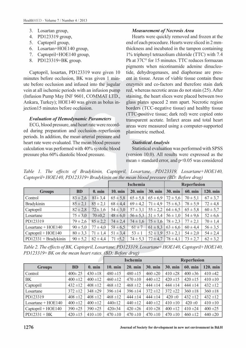
1276
HealthMED - Volume 7 / Number 4 / 2013
Journal of Society for development in new net environment in B&H
3. Losartan group,4. PD123319 group,5. Captopril group,6. Losartan+HOE140 group,7. Captopril+HOE140 group,8. PD123319+BK group.
Captopril, losartan, PD123319 were given 10 minutes before occlusion, BK was given 1 min-ute before occlusion and infused into the jugular vein at all ischemic periods with an infusion pump (Infusion Pump May INF 9601, COMMAT LTD., Ankara, Turkey); HOE140 was given as bolus in-jection15 minutes before occlusion.
Evaluation of Hemodynamic ParametersECG, blood pressure, and heart rate were record-
ed during preparation and occlusion–reperfusion periods. In addition, the mean arterial pressure and heart rate were evaluated. The mean blood pressure calculation was performed with 40% systolic blood pressure plus 60% diastolic blood pressure.
Measurement of Necrosis AreaHearts were quickly removed and frozen at the
end of each procedure. Hearts were sliced in 2 mm-thickness and incubated in the tampon containing 1% triphenyl tetrazolium chloride (TTC) with 7.4 Ph at 37CO for 15 minutes. TTC reduces formazan pigments when nicotinamide adenine dinucleo-tide, dehydrogenases, and diaphorase are pres-ent in tissue. Areas of viable tissue contain these enzymes and co-factors and therefore stain dark red, whereas necrotic areas do not stain (25). After staining, the heart slices were placed between two glass plates spaced 2 mm apart. Necrotic region borders (TCC-negative tissue) and healthy tissue (TTC-positive tissue; dark red) were copied onto transparent acetate. Infarct areas and total heart areas were measured using a computer-supported planimetric method.
Statistical AnalysisStatistical evaluation was performed with SPSS
(version 10.0). All results were expressed as the mean ± standard error, and p<0.05 was considered
Table 1. The effects of Bradykinin, Captopril, Losartane, PD123319, Losartane+HOE140, Captopril+HOE140, PD123319+Bradykinin on the mean blood pressure (BD: Before drug)
Ischemia ReperfusionGroups BD 0. min 10. min 20. min 30. min 30. min 60. min 120. min
Control 83 ± 2,6 81± 3,4 65 ± 5,8 65 ± 5,8 65 ± 6,9 72 ± 5,6 70 ± 5,1 67 ± 3,7Bradykinin 85 ± 2,1 85 ± 2,1 68 ± 4,4 69 ± 4,2 71 ± 4,9 75 ± 6,3 78 ± 5,9 72 ± 4,8Captopril 82 ± 2,8 72 ± 1,6 54 ± 3,0 57 ± 3,1 55 ± 2,2 64 ± 6,5 65 ± 5,8 60 ± 5,7Losartane 75 ± 3,0 70 ±0,2 48 ± 6,0 56 ± 5,3 51 ± 5,4 56 ± 1,0 54 ± 9,6 52 ± 6,6PD123319 79 ± 2,6 85 ± 2,2 74 ± 2,4 74 ± 1,6 75 ± 1,6 78 ± 2,3 77 ± 2,1 70 ± 1,4Losartane + HOE140 90 ± 5,0 77 ± 4,0 58 ± 6,5 61 ± 7 61 ± 8,3 63 ± 6,6 60 ± 4,4 56 ± 3,5Captopril + HOE140 80 ± 3,3 71 ± 1,4 51 ± 3,4 53 ± 1 52 ± 1,9 53 ± 2,1 54 ± 2,0 54 ± 2,4PD12331 + Bradykinin 90 ± 5,2 82 ± 4,4 71 ±5,2 74 ± 5,3 77 ± 4,7 78 ± 4,1 73 ± 2,7 62 ± 3,2
Table 2. The effects of BK, Captopril, Losartane, PD123319, Losartane+HOE140, Captopril+HOE140, PD123319+BK on the mean heart rates. (BD: Before drug)
Ischemia ReperfusionGroups BD 0. min 10. min 20. min 30. min 30. min 60. min 120. min
Control 400± 25 430 ±18 480 ±15 480 ±15 460 ±20 410 ±28 400 ±36 410 ±42BK 400 ±12 400 ±12 460 ±12 470 ±10 440 ±12 420 ±15 420 ±15 410 ±10Captopril 432 ±12 408 ±12 468 ±12 468 ±12 444 ±14 444 ±14 444 ±14 432 ±12Losartane 372 ±12 348 ±29 396 ±14 396 ±14 372 ±12 372 ±22 360 ±18 360 ±18PD123319 408 ±12 408 ±12 468 ±12 444 ±14 444 ±14 420 ±0 432 ±12 432 ±12Losartane + HOE140 400 ±12 400 ±12 440±12 440 ±12 440 ±12 410 ±10 420 ±0 410 ±10Captopril + HOE140 390 ±25 390 ±25 420±34 420 ±26 410 ±28 400 ±12 410 ±24 400 ±25PD12331 + BK 420 ±15 410 ±10 470 ±10 470 ±10 470 ±10 470 ±10 460 ±12 440 ±20
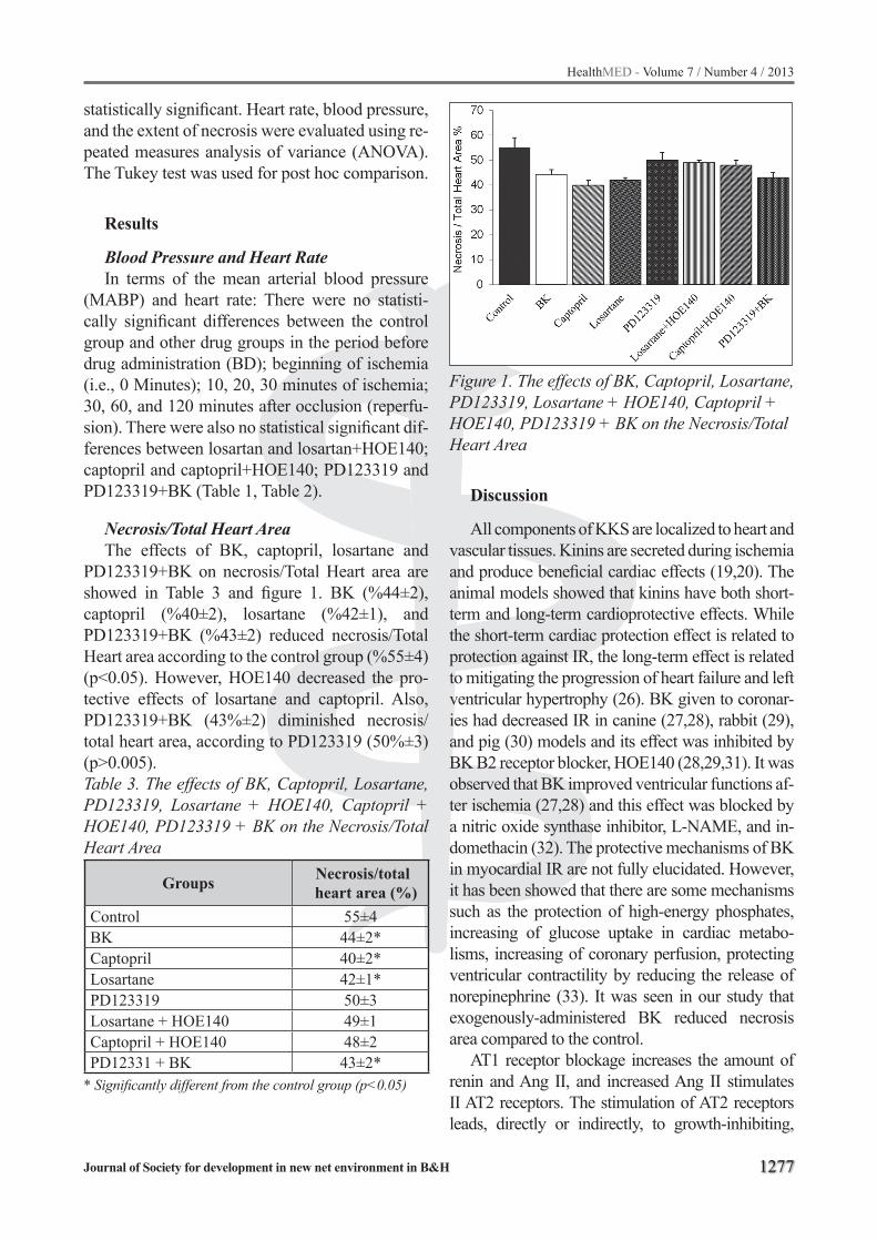
HealthMED - Volume 7 / Number 4 / 2013
Journal of Society for development in new net environment in B&H 1277
statistically significant. Heart rate, blood pressure, and the extent of necrosis were evaluated using re-peated measures analysis of variance (ANOVA). The Tukey test was used for post hoc comparison.
Results
Blood Pressure and Heart RateIn terms of the mean arterial blood pressure
(MABP) and heart rate: There were no statisti-cally significant differences between the control group and other drug groups in the period before drug administration (BD); beginning of ischemia (i.e., 0 Minutes); 10, 20, 30 minutes of ischemia; 30, 60, and 120 minutes after occlusion (reperfu-sion). There were also no statistical significant dif-ferences between losartan and losartan+HOE140; captopril and captopril+HOE140; PD123319 and PD123319+BK (Table 1, Table 2).
Necrosis/Total Heart AreaThe effects of BK, captopril, losartane and
PD123319+BK on necrosis/Total Heart area are showed in Table 3 and figure 1. BK (%44±2), captopril (%40±2), losartane (%42±1), and PD123319+BK (%43±2) reduced necrosis/Total Heart area according to the control group (%55±4) (p<0.05). However, HOE140 decreased the pro-tective effects of losartane and captopril. Also, PD123319+BK (43%±2) diminished necrosis/total heart area, according to PD123319 (50%±3) (p˃0.005).Table 3. The effects of BK, Captopril, Losartane, PD123319, Losartane + HOE140, Captopril + HOE140, PD123319 + BK on the Necrosis/Total Heart Area
Groups Necrosis/total heart area (%)
Control 55±4BK 44±2*Captopril 40±2*Losartane 42±1*PD123319 50±3Losartane + HOE140 49±1Captopril + HOE140 48±2PD12331 + BK 43±2*
* Significantly different from the control group (p<0.05)
Figure 1. The effects of BK, Captopril, Losartane, PD123319, Losartane + HOE140, Captopril + HOE140, PD123319 + BK on the Necrosis/Total Heart Area
Discussion
All components of KKS are localized to heart and vascular tissues. Kinins are secreted during ischemia and produce beneficial cardiac effects (19,20). The animal models showed that kinins have both short-term and long-term cardioprotective effects. While the short-term cardiac protection effect is related to protection against IR, the long-term effect is related to mitigating the progression of heart failure and left ventricular hypertrophy (26). BK given to coronar-ies had decreased IR in canine (27,28), rabbit (29), and pig (30) models and its effect was inhibited by BK B2 receptor blocker, HOE140 (28,29,31). It was observed that BK improved ventricular functions af-ter ischemia (27,28) and this effect was blocked by a nitric oxide synthase inhibitor, L-NAME, and in-domethacin (32). The protective mechanisms of BK in myocardial IR are not fully elucidated. However, it has been showed that there are some mechanisms such as the protection of high-energy phosphates, increasing of glucose uptake in cardiac metabo-lisms, increasing of coronary perfusion, protecting ventricular contractility by reducing the release of norepinephrine (33). It was seen in our study that exogenously-administered BK reduced necrosis area compared to the control.
AT1 receptor blockage increases the amount of renin and Ang II, and increased Ang II stimulates II AT2 receptors. The stimulation of AT2 receptors leads, directly or indirectly, to growth-inhibiting,

1278
HealthMED - Volume 7 / Number 4 / 2013
Journal of Society for development in new net environment in B&H
anti-hypertrophic and proapoptotic effects through the stimulation of autocoids. When AT1 antagonists and AT2 antagonists combined in IR models, it was observed that the beneficial effects of AT1 antago-nists were negated (34). In the present study, it was found that BK was effective in the role of losartan in decreasing necrosis/total heart area. This effect of losartan was reduced when combined with HOE140.
In the present study, PD123319, an AT2 blocker, was found to reduce necrosis area, relative to the control, although this was not significant, which is consistent with the literature. Ang II interacts with AT1 receptors when AT2 receptors are blocked by PD123319. However, it has been shown that AT1 receptor has BK-forming effect. But, this effect is not as far as AT2 receptors (35). When PD123319 was administered alone, Ang II maintained its effect with AT1 receptor, and stimulated BK by this recep-tor might have a minor beneficial effect on necrosis. In addition, some studies reported that PD123319 reduced apoptosis stimulated by Ang II (36).
Losartan and captopril may prevent harmful ef-fects of RAS by inhibiting binding of Ang II to AT1 receptors and suppressing the formation of Ang II, respectively. ACE inhibitors also prevent the destruction of BK. This effect may play a role on limiting the IR-induced damage in myocardium by captopril. As ACE is effective in the breakdown of BK, a vasodilatory peptide, and kallidin so ACE in-hibition raises the level of kinin peptides, which has a vasodilatory effect (37, 38). Birincioglu et al (39) associated the therapeutic effect of captopril in IR injury to stimulation of prostaglandin synthesis and prevention of BK breakdown. In our study, while captopril decreased necrosis area, as captopril com-bined with HOE 140, its protection effect was not fully removed. This suggests that pathways other than BK may be involved in the protective effect of captopril. Birincioglu et al. reported that captopril stimulates synthesis of prostaglandin and its struc-ture containing sulfhydryl group, which provides an antioxidant properties (40).
This study has shown that both ACE inhibition and AT1 receptor blockage and, indirectly, AT2 re-ceptor, have protective effects on necrosis area re-sulting from myocardial IR. However, BK plays an important role among the possible mechanisms of this cardioprotective effect, but it appears that this is not the only effective pathway in this process.
Acknowledgement
This project was supported by a financial grant from the Scientific Research Fund of Inonu Uni-versity, Turkey.
References
1. Moens AL, Claeys M, Timmermans JP, Vrints CJ. Myocardial ischemia / reperfusion - injury, a clinical view on a complex pathophysiological process. Inter-national Journal of Cardiology 2005; 100: 179– 90
2. Lai ZF. The relationship between intracellular chlo-ride concentration and ischemia reperfusion-induced arrhythmias in myocardial cells. Zhongguo Yi Xue Ke Xue Yuan Xue Bao. 2002; 24(2): 190-6.
3. Venardos KM, Perkins A, Headrick J, Kaye DM. Myocardial ischemia-reperfusion injury, antioxidant enzyme systems, and selenium: a review. Curr Med Chem 2007; 14(14): 1539-49.
4. Tom B, Dendorfer A, AH Jan Danser. Bradykinin, an-giotensin-(1–7), and ACE inhibitors: how do they in-teract? Journal of Biochemistry & Cell Biology 2003; 35: 792–801
5. Friedrich B, Walter RK. Postischemic antiarrhytmic effects of angiotensin-converting enzyme inhibitors. Circulation 1996; 94: 1752-61.
6. Kawabata H, Ryomoto T, Ishikawa K. Cardioprotecti-on with angiotensin converting enzyme inhibitor and angiotensin II type 1 receptor antagonist is not abo-lished by nitric oxide synthase inhibitor in ischemia-reperfused rabbit hearts. Hypertens Res. 2001 Jul; 24(4): 403-9
7. Toblli JE, Cao G, Derosa G, Di Gennaro F, Forcada P. Angiotensin-converting enzyme inhibition and an-giogenesis in myocardium of obese Zucker rats. Am J Hypertens. 2004 Feb; 17(2): 172-80.
8. Jurkovicova O, Cagan S. Reperfusion arrhythmias. Bratisl Lek Listy 1998; 99(3-4): 162-71.
9. Zhu B, Sun Y, Sievers RE, Browne AE, Pulukurthy S, Sudhir K, Lee RJ, Chou TM, Chatterjee K, Parmley WW. Comparative effects of pretreatment with cap-topril and losartan on cardiovascular protection in a rat model of ischemia-reperfusion. J Am Coll Cardiol 2000; 35(3): 787-95.
10. De Gasparo M, Catt KJ, Inagami T, Wright W, Un-ger TH. International union of pharmacology. XXIII. The angiotensin II receptors. Pharmacol Rev. 2000; 52: 415–72.

HealthMED - Volume 7 / Number 4 / 2013
Journal of Society for development in new net environment in B&H 1279
11. Jones A, Dhamrait SS, Payne JR, Hawe E, Li P, Toor IS, Luong L, Wootton PT, Miller GJ, Humphries SE, Montgomery HE. Genetic Variants of Angiotensin II Receptors and Cardiovascular Risk in Hypertensi-on. Hypertension 2003; 42: 500-6.
12. Said AbdAlla, Heinz Lother, Ahmed M. Abdel-tawab, Ursula Quitterer. The Angiotensin II AT2 Receptor Is an AT1 Receptor Antagonist. The Journal Of Biolo-gical Chemistry 2001; 276: 39721-26.
13. Lan Wu, Masaru I, Hironori N, Rui C, Jun , Masahi-ro A, Marc De G, Masatsugu H. Effect of Angioten-sin II Type 1 Receptor Blockade on Cardiac Remo-deling in Angiotensin II Type 2 Receptor Null Mice. Arterioscler Thromb Vasc Biol. 2002; 22: 49-54
14. Johren O, Dendorfer A, Dominiak P. Cardiovascular and renal function of angiotensin II type-2 receptors Cardiovascular Research 2004; 62: 460– 7.
15. Horiuchi M, Akishita M, Dzau VJ. Recent progress in angiotensin II type 2 receptor research in the car-diovascular system. Brief Review. 1999; 33: 613-21.
16. Nio Y, Matsubara H, Murasawa S, Kanasaki M, Ina-da M. Regulation of gene transcription of angioten-sin II receptor subtypes in myocardial infarction. J Clin Invest 1995; 95: 46 – 54.
17. Wharton J, Morgan K, Rutherford RA, Catravas JD, Chester A, Whitehead BF, De Leval MR, Yacoub MH, Polak JM. Differential distribution of angioten-sin AT2 receptors in the normal and failing human heart. J Pharmacol Exp Ther 1998; 284: 323– 36.
18. Yan Tan,. Hutchison FN, Jaffa AA. Mechanisms of angiotensin II-induced expression of B2 kinin re-ceptors Am J Physiol Heart Circ Physiol 2004; 286: 926–32.
19. Sharma JN. Does the Kinin System Mediate in Car-diovascular Abnormalities? An Overview. Journal of Clinical Pharmacology, 2003; 43: 1187-95.
20. Sharma JN. Role of tissue kallikrein-kininogen-kinin pathways in the cardiovascular system Arch Med Res. 2006 Apr; 37(3): 299-306.
21. Liu X, Lukasova M, Zubakova R, Lewicka S, Hilgen-feldt U. A kallidin-like peptide is a protective cardiac kinin, released by ischaemic preconditioning of rat heart. Br J Pharmacol. (2005); 146: 952–57.
22. Campbell DJ. The kallikrein–kinin system in hu-mans. Clinical and Experimental Pharmacology and Physiology. 2001; 28: 1060–65.
23. Walker MJ, Curtis MJ, Hearse DJ, Campbell RW, Janse MJ, Yellon DM, Cobbe SM, Coker SJ, Har-ness JB, Harron DW. The Lambeth Conventions: guidelines for the study of arrhythmias in ischemia infarction, and reperfusion. Cardiovasc Res 1988; 22 (7): 447-55.
24. Ozer MK, Sahna E, Birincioglu M, Acet A. Effects of captopril and losartan on myocardial ischemia-re-perfusion induced arrhythmias and necrosis in rats. Pharmacol Res. 2002 ; 45(4): 257-63.
25. Fishbein MC, Meerbaum S, Rit J, Lando U, Kan-matsuse K, Merciier JC, Corday E, Ganz W. Early phase acute myocardial infarct size quantification: Validation of the triphenyltetrazorium chloride ti-ssue enzyme staining technique. Am Heart J 1981; 101: 593-600.
26. Jorma O. Kokkonen, Ken A. Lindstedt, Antti Kuop-pala, Petri T. Kovanen. Kinin-Degrading Pathways in the Human Heart Trends Cardiovasc Med 2000; 10: 42–5
27. Linz W and Scholkens BA. Role of bradykinin in the cardiac effects of angiotensin-converting enzyme in-hibitors. J. Cardiovasc. Pharmacol. 1992; 20: 81–90.
28. Martorana PA, Kettenbach B, Breipohl G, Linz W and Scholkens BA. Reduction of infarct size by local angiotensinconverting enzyme inhibition is abolis-hed by a bradykinin antagonist. Eur. J. Pharmacol. 1990: 182: 395–6.
29. Hartman JC, Wall TM, Hullinger TG and Shebuski RJ. Reduction of myocardial infarct size in rabbits by ramiprilat: reversal by the bradykinin antago-nist HOE 140. J. Cardiovasc. Pharmacol. 1993; 21: 996–1003..
30. Tio RA, Tobe TJ M, Bel KJ, Langen CD J DE, Gilst WH VAN and Wesseling H. Beneficial effects of bradykinin on porcine ischemic myocardium. Basic Res. Cardiol. 1991; 86: 107–16.
31. Kanner J, Harel S And Granit R. Nitric oxide as an an-tioxidant. Arch. Biochem. Biophys. 1991; 289: 130–6.
32. Zhu P, Zaugg CE, Simper D, Hornstein P, Allegrini PR, Buser PT. Bradykinin improves postischemic re-covery in the rat heart: role of high energy phospha-tes, nitric oxide, and prostacyclin. Cardiovasc. Res. 1995; 29: 658–63.
33. Schriefer JA, Broudy EP, Hassen AH. Inhibitors of bradykinin-inactivating enzyme decrease myocar-dial ischemia/reperfusion injury following 3 and 7 days of reperfusion. The Journal of Pharmacology and Experimental Therapeutics 2001; 298: 970-5.

1280
HealthMED - Volume 7 / Number 4 / 2013
Journal of Society for development in new net environment in B&H
34. Jalowy A, Schulz R, Dorge H, Behrends M, Heusch G. Infarct size reduction by AT1 receptor blockade through a signal cascade of AT2 receptor activation. 1998 Nov 15; 32(6): 1787-96.
35. Seyedi N, Xu XB, Nasjletti A, Hinzte TH. Coronary kinin generation mediates nitric oxide release af-ter angiotensin receptor stimulation. Hypertension 1995; 26: 164-70
36. Goldenberg I, Grossman E, Jacobson KA, Shneyvays V, Shainberg A. Angiotensin II-induced apoptosis in rat cardiomyocyte culture: a possible role of AT1 and AT2 receptors. J Hypertens. 2001; 19(9): 1681-9.
37. Brown NJ, Vaughan DE. Angiotensin-Converting Enzyme Inhibitors. Circulation 1998; 97(14): 1411-20.
38. Woo KS, Nicholls MG. High prevalence of persistent cough with angiotensin converting enzyme inhibitors in Chinese. Br J Clin Pharmacol 1995; 40: 141-4
39. Birincioglu M Olmez E, Aksoy T, Acet A. The role of prostaglandin synthesis stimulation in the pro-tective effects of captopril on ischemia-reperfusion arrhytmias in rats in vivo. Pharmacol Res 1997; 36: 299-304.
40. Birincioglu M, Aksoy T, Olmez E, Acet A. Protective effects of ACE inhibitors on ischemia-reperfusion-induced arrhytmias in rats: is this effect related to the free radical scavenging action of these drugs? Free radic Res 1997; 27: 389-96.
Corresponding authorSeda Tasdemir,Inonu University,Medical Faculty,Pharmacology Department,Malatya,Turkey,E-mail: [email protected]

HealthMED - Volume 7 / Number 4 / 2013
Journal of Society for development in new net environment in B&H 1281
Abstract
Introduction: Skeletal Class II is one of the mainly malocclusion in modern populations. Pa-tients with this type of malocclusion could be tre-ated in early mixed dentition by Fränkel`s functi-onal regulator.
Case report: In this case was shown treatment by Fränkel`s functional regulator Ic (FR-Ic) in boy, age 8 years and 3 months, with malocclusi-on Class II division 1. Whole treatment lasted 22 months, with permanently wearing of appliance and regular orthodontic control. Age of patient has enabled to use modification of craniofacial growth and growth stimulation of the lower jaw during puberty. On lateral cephalograms changes were monitored and analysed.
Conclusion: FR-Ic has caused significant changes on skeletal and dental structures, stimula-ting growth of the lower jaw and causing retrusion of the upper and protrusion of the lower incisors. It caused significant improvement in facial aesthe-tics and corrections on the soft-tissue profile. Also, functions of swallowing and articulation of dental vocals were corrected.
Key words: Malocclusion Class II Division 1, functional appliances, growth modification, Frän-kel functional regulator, cephalometric changes, early mixed dentition.
Introduction
Skeletal Class II is malocclusion of sagittal di-rection caused by unbalanced anteroposterior posi-tion and development of the upper and lower jaw with respect to anterior cranial base. It manifested by increasing values of angle ANB (over 4°). Accor-ding to some studies the cause of this malocclusion is maxilarry prognatism in 56.3% cases, mandibu-lar retrognatism in 27% cases and combination of
them in 16.7% cases1. Of the total number of ma-locclusion Class II accounts for 27%, according to Angle. The most important characteristics of Class II division 1 malocclusion are: bimaxillary retro-gnatism ( more visible on the lower jaw), protrusion of upper incisors, narrow upper jaw, facial growth with backward rotation, mostly2. Mandibular defi-ciency can be absolute, with smaller lower jaw in all dimensions, and relative, with posterior position of the lower jaw. Facial profile has increased con-vexity, overjet is enlarged, mandibular corpus and ramus are shorter3,4. Use of functional appliances in treatment of Class II primarily based on stimu-lation growth of the lower jaw. The best treatment effect is possible in early mixed dentition, during the period of intense growth, also with continuo-usly wearing of appliances. On that way we have adequate force and response in orofacial muscles5.
Fränkel`s functional regulator is distinctive in that is only mounted tissue functional appliance, which can effectively corrected morphological (skeletal and dental) and functional irregulariti-es6,7. Operates on the principle of directional pre-ssure applications at the contact appliance and the skeletal and dental structures, elimination of the pressure surrounding perioral musculature and causing tensile stress in the area of mucosal for-nix, where exist bone apposition8-10.
This appliance is distinctive in that it can be used very early, immediately after eruption of in-cisors and first permanent molars, in early mixed dentition. On that way we create conditions for early corrections of skeletal deviations and pre-vent the occurrence of later developmental ano-malies. Anomalies are inevitable if we delayed beginning of orthodontic treatment. In this age patients are motivated for cooperate, appliance is comfortable, easy for wearing, adaptation phase is short, and there is no other unwanted side effect regarding the use of this appliance11-13.
Orthodontic treatment of class II division 1 malocclusion by Fränkel functional regulator type Ic: Case reportZorana Stamenkovic, Nenad Nedeljkovic, Vladimir RisticClinic of Orthodontics, School of Dentistry, University of Belgrade, Belgrade, Serbia.

1282
HealthMED - Volume 7 / Number 4 / 2013
Journal of Society for development in new net environment in B&H
Case report
In this case was treated male patient in age of 8 years and 3 months with diagnosis malocclusion Class II division 1, caused by manibular retrogna-tism. Intraoral examination indicated occlusion in whole Class II with extremely increased overjet of 12mm, overbite of 7mm, protrusion of upper inci-sors with injection of the upper lip between upper and lower incisors. Functional examination indica-ted existing of infantile swallowing and interdental sigmatism. Extraoral examination from the front indicated shorter lower third of face, emphasissed mentolabial sulcus and potentially competent lips. Extraoral examination in profile indicated grave convexity with prominent position of the upper lip and distal position of chin. Before orthodontic tre-atment study cast analysis, orthopantomograph and lateral cephalogram have been done. Skeletal ma-turity was determinated by level of development of cervical vertebrae on lateral cephalogram14,15. Patient was in CvS1 stage, before puberty growth acceleration. Parameters which were analysed on lateral cephalogram suggested skeletal distal bite caused by mandibular retrognatism, horizontal fa-cial growth, protrusion of the upper and retrusion of the lower incisors, decreased vertical interjaw (B) angle, anteinclination of both jaw and shorter manidubular corpus. After diagnostic procedures, bearing in mind patients calendar age and skeletal maturity, Fränkel’s functional regulator type Ic (FR-Ic) was chosen as a method of treatment (Figure 1.). Structural bite was determined in relation Class I on the posterior teeth, without activation in vertical direction, on the level of physiological stagnancy. On that way avoided the excessive tension of neuro-muscular structures. Based on structural bite and la-ter corrections on the study casts FR-Ic was created in laboratory. Components of this appliance are: 2 posterior buccal shields, 2 pelotas for lower lip, lin-gual shield with pins for protrusion, labial arch, pa-latal arch with anchorage on first permanent molars, loops for canines in upper jaw and screw. Patient re-ceived FR-Ic and instructions on wearing appliances during the whole day, except during meals. Regular orthodontics controls were carried out at intervals of 4 to 6 weeks. At the time when overjet was decre-ased on 7mm, after 9 months of treatment, patient started to turn the screw every 10 days.
Table 1. Skeletal and dental changes during tre-atment with FR-Ic
Parameter Beforetreatment
Aftertreatment
Angle SNA 80° 79°Angle SNB 74° 76°Angle ANB 6° 3°Angle I/SpP 60° 67°Angle i/MP 83° 80°Angle SpP/MP (B) 19° 23°Distance Cmand 69mm 73mm
Angle SNA – sagital position of upper jaw to anterior cranial base, angle SNB – sagital position of lower jaw to anterior cranial base, angle ANB – sagital interjaw angle, angle I/SpP – inclination of upper incisors, angle i/MP – inclination of lower incisors, angle SpP/MP (B) – vertical interjaw an-gle and distance Cmand – length of mandibular corpus
Orthodontic procedure lasted for 22 months. After that period patient had stabile occlusion in Class I, with significant decrease of overjet and overbite (Figure 2.). After treatmen control stu-dy casts and lateral cephalogram have been done. Patient started wearing appliance only during the night, as a retention. On lateral cephalogram af-ter treatment was observed anterior movement of lower jaw with increasing length of mandibular corpus and correction of sagittal interjaw relati-onship from Class II to Class I (Figure 3.). Upper incisors were remarkably retruded, vertical inter-jaw angle was increased, without changes in faci-al growth (table 1). Functional examination after treatment indicated correct articulation of dental vocals (s, z, c) and transition from infantile to ma-ture swallowing. Extraoral examination indica-ted significant improvement in facial aesthetics, decrease of profile convexity and prominence of the upper lip, balanced relationship between three parts of face in vertical direction, competent lips and correctly expressed mentolabial sulcus.
Figure 1. (a and b) Fränkel Appliance

HealthMED - Volume 7 / Number 4 / 2013
Journal of Society for development in new net environment in B&H 1283
A – before treatment B – after treatment
Figure 2. Patient with Class II, division 1. ma-locclusion before A(a-g) and after B(a-g) tre-atment by Fränkel I-c appliance
Figure 3. Lateral cephalogram a) before and b) after the treatment
Discussion
According to age when patient wanted ortho-dontic treatment and existing orthodontic diagnosis, using of FR-Ic was method of choise, which pro-vides stabile therapeutic result. During orthodontic treatment sagittal interjaw relationship was signifi-cantly corrected, due to accelerating the growth of the lower jaw and increase in total length of man-dibular corpus15-19. Stimulation of growth on the lower jaw occurs due to activity of condilar carti-lage, associated with dental displacement during the treatment20,21. The most significant dental chan-ges are retrusion of upper and protrusion of lower incisors, with correction of overjet22,23. Treatment with FR-Ic provides much better facial aesthetics, harmonic profile, correct position of lip in relation to aesthetic line and significant decrease of profile convexity22,24,25. Appliance causes significant chan-ges in activity of orofacial and masticatory muscles. Muscles show increased activity, which contributes to correction of orofacial disfunctions. Ba favour of pulling force and tension in muscles generates an overall increase in growth of lower jaw26.
Conclusion
FR-Ic is very efficient appliance for correction of mandibular retrognatism in early mixed denti-tion. FR-Ic induce significant changes on skeletal and dental structures, as advancement method of execution of orofacial functions and improvement of facial aesthetics, with stabile treatment result.

1284
HealthMED - Volume 7 / Number 4 / 2013
Journal of Society for development in new net environment in B&H
References
1. Rosenblum RE. Class II malocclusion: Mandibu-lar retrusion or maxillary protrusion? Angle Orthod 1995; 1: 49-62.
2. McNamara JA. Components of Class II malocclusion in children 8-10 years of age. Angle Orthod 2002; 51: 177-202.
3. Rakosi T, Jonas I, Graber TM. Orthodontic diagnosis. George Thieme Verlag: Stutgart – New York, 1993.
4. Pancherz H, Zieber K, Hoyer B. Cephalometric cha-racteristics of Class II division 1 and Class II division 2 malocclusions: a comparative study in children. An-gle Orthod 1997; 67: 111-120.
5. Proffit WR, Ackerman JL. A systematic approach to orthodontic diagnosis and treatment planning. In Graber TM, Vanarsdall RL (eds): Current orthodontic concepts and techniques, ed 3, St Louis, Mosby, 2000.
6. Frankel R. The theoretical concept underlying the tre-atment with the functional correctors. Trans Eur Ort-hod Soc 1966; 42: 233-254.
7. Frankel R, Frankel Ch. Orofacial Orthopaedics with the Function Regulator. Karger, Basel – Munchen –Paris – London – New York – New Delhi – Singapore – Tokyo – Sydney, 1989.
8. Braun S, Diers NR, Engel G, Wojtkiewicz P, Ewing SK. The effect of Fränkel II and modified Twin blok appliances on the “C“-axis: The growth vector of the dentomaxillary complex. Angle Orthod 2004; 74: 749-753.
9. Barton S, Cook PA. Predicting functional applian-ce treatment outcome in Class II malocclusions – a rewiew. Am J Orthod Dentofacial Orthop 1997; 112: 282-86.
10. Basciftci FA, Uysal T, Buyukrkmen A, Sari Z. The effects of activator treatment on the craniofacial structures of Class II division 1 patients. Eur J Ort-hod 2003; 25: 87-93.
11. Almeida MR, Henriques JFC, Almeida RR, Almeida-Pedrin RR, Ursi W. Treatment effects produced by the Bionator appliance. Comparison with an untre-ated Class II sample. Eur J Orthod 2004; 26: 65-72.
12. Bishara SE. Textbook of orthodontics. W. B. Saun-ders; Philadelphia, 2001.
13. Proffit WR, Fields HW, Sarver DM. Contemporary Orthodontics, 4th ed St. Louis, Mo: Mosby O’Relly T, Yanneillo G. Mandibular growth changes and matu-ration of cervical vertebrae – A longitudinal cephalo-metric study. Angle Orthod 1988; 4: 179-184.
14. Ball G, Woodside D, Tompson B, Hunter WS, Po-sluns J. Relationship between cervical vertebral ma-turation and mandibular growth. Am J Orthod Dent Orthop 2011; 139 (5): 455-461.
15. McNamara JA, Huge SA. The Frankel appliance (FR-2) model preparation and appliance construc-tion. Am J Orthod 1981; 80: 478-495.
16. Almeida MR, Henriques JFC and Ursi W. Compara-tive study of the Frankel (FR-2) and bionator appli-ances in the treatment of Class II malocclusion. Am J Orthod Dentofacial Orthop 2002; 121: 458-466.
17. Chadwick SM, Aird C, Taylor S, Bearn DR. Functi-onal regulator treatment of Class II division 1 ma-locclusions. Eur J Orthod 2001; 23: 495-505.
18. Creekmore TD, Radney LJ. Frankel appliance the-rapy: orthopedic or orthodontic? Am J Orthod 1983; 83: 89-108.
19. Petrovic AG, Stutzmann JJ, Lavergne JM, Shaye R. Is it possible to modulate the growth of the human mandible with a functional appliance? Bilt UOJ 1988; XXI (1): 15-20.
20. Thieme KM, Nägerl H, Hahn W, Ihlow D, Kubein D. Variations in cyclic mandibular movements during tre-atment of Class II malocclusions with removable func-tional appliances. Eur J Orthod 2011: 33(6): 628-635.
21. Stamenkovic Z. Clinical effects of Fränkel’s functio-nal regulators in treatment of distal and mesial bite. PhD thesis. Belgrade, 2011. (Serbian)
22. Janson GRP, Alegria Toruno JL, Rodrigez Martins D, Henriques JFC, De Freitas MR. Class II tre-atment effects of the Fränkel appliance. Eur J Ort-hod 2003; 25: 301-09.
23. Battagel JM. The relationship between hard and soft tissue changes following treatment og Class II division 1 malocclusions using Edgewise and Fränkel applian-ce techniques, Eur J Orthod 1990; 12: 154-165.
24. Carlos Flores-Mir and Paul WM. A systematic re-view of cephalometric facial soft tissue changes with the Activator and Bionator appliances in Class II division 1 subjects. Eur J Ortho. 2006; 28: 586-593.
25. Yamin-Lacouture C, Woodside DG, Sectakof PA, Se-ssle BJ. The action of three types of functional appli-ances on the activity of the masticatory muscles. Am J Orthod Dentofacial Orthop 1997; 112: 560-572.
Corresponding AuthorZorana Stamenkovic,Clinic of Orthodontics,School of Dentistry, University of Belgrade, Belgrade, Serbia,E-mail: [email protected]

HealthMED - Volume 7 / Number 4 / 2013
Journal of Society for development in new net environment in B&H 1285
Abstract
Purpose: The purpose of this study was to evaluate the analgesic efficacy of tramadol and bupivacaine during multi-regional application.
Methods: Ninety patients who had laparoscop-ic cholecystectomy were included in the study. Pa-tients in group 1 received 100 mg of tramadol was diluted with 50 mL of physiological serum and patients in group 2 received 0.125% 50 mL bupi-vacaine. 10 mL of each solution was infiltrated in gall bladder, right subdiaphragmatic and visceral peritoneal surface compartments and also 5 mL of each solution was infiltrated to the each trocar entry site; patients in group 3 received 1 mg/kg IV tramadol 30 minutes before the end of surgery. During the first postoperative 24 hours, pain scale, time of initial analgesic need, total analgesic dose and side effects were recorded.
Results: When pain scores from patients were compared, the mean scores from the 1st hour was 5.16 in Group 1, 5, 90 in Group 2, 5.73 in Group 3, being significantly lower for Group 1 (p<0.05). Pain scores from other time points were compared and their values were determined to be similar. Adverse effects were similar among the groups.
Conclusion: For the first postoperative hour, tramadol infiltration was especially more effective compared to bupivacaine, and neither drugs had any side effects. Additionally, in group 3 about 20 % of had shoulder pain.
Key words: Laparoscopic cholecystectomy, multi-regional application, Tramadol, Bupivacaine.
Introduction
Laparoscopy is currently the standard surgi-cal procedure for cholecystectomy. The main ad-
vantage of laparoscopic cholecystectomy (LC) is less postoperative pain. However, the severity of postoperative abdominal and shoulder pain is still a significant problem for LC. The intensity of pain after LC peaks within 4–8 h after surgery (1,2). Therefore, LC is not performed as a day-care sur-gical procedure in a remarkable number of pa-tients. The underlying mechanisms that contribute to postoperative pain are tissue damage resulting from surgical incisions, removal of the gallblad-der from the liver bed, stretching of nerve endings and irritation of the diaphragm by carbon dioxide (3,4). Moreover, the consent of central sensitiza-tion increases postoperative perceived pain in the area of tissue damage and in the surrounding areas as well(5). Intraoperative blockade of the periph-eral nerves directly before commencing LC is a promising strategy to decrease postoperative pain and prevent central sensitization. Bupivacaine as a long acting local anesthetic was investigated to decrease postoperative pain as well as central sensitization through infiltration into both surgical incisions and intraperitoneally (6).
Tramadol is a centrally-acting μ- receptor ag-onist cyclohexanol derivative. It is reported that tramadol provides a powerful analgesic effect and has no side effects relating to other opioid recep-tors. In addition to the well known central effects, recent studies have shown that tramadol also had local analgesic efficacy (2,7, 8).
The objective of this randomized prospective trial was to compare the efficacy of three methods, namely tramadol infiltration into surgical sites, bu-pivacaine infiltration into surgical sites and intra-venous tramadol application in terms of relieving postoperative pain in patients with LC.
Comparison of post-operative analgesic efficacies of Tramadol and Bupivacaine infiltration applied to multiple regions during laparoscopic cholecystectomyAbdulkadir Iskender1, Ismet Ozaydin2, Gulbin Sezen1, Mehmet Yasar2, Yavuz Demiraran1, Tuna Yilmaz1, Zekeriya Ilce3
1 Departments of Anesthesiology and Reanimation, Duzce University, Duzce School of Medicine, Turkey,2 Departments of General Surgery, Duzce University, Duzce School of Medicine, Turkey,3 Departments of Pediatric Surgery, Duzce University, Duzce School of Medicine, Turkey.

1286
HealthMED - Volume 7 / Number 4 / 2013
Journal of Society for development in new net environment in B&H
Methods
This study was planned as a prospective, ran-domized-double blind study. Before starting the study, Local Ethical Committee approval and in-formed consent from all patients were obtained Ninety patients aged between 30-60 who were ASA (American Society of Anesthesiologists) I-II, and had laparoscopic cholecystectomy due to gall bladder stones were included in the study. The patients were randomized into three groups each containing 30 subjects initially. We excluded the patients who had Endoscopic Retrograde Cholan-giopancreatography (ERCP) and/or papillotomy in the past 3 months or those who were pregnant, lac-tating, having previous abdominal surgery, renal or hepatic diseases, drug or alcohol dependency and socially enable to participate in addition to ones who refused to participate. Furthermore, patients whose surgeries were not completed laparoscopi-cally or had acute cholecistitis, history of chronic pain, allergy to bupivacaine and tramadol were not included in the study. According to CONSORT guidelines, recruitment, enrollment, retention of subjects are summarized in Figure 1. For post-oper-ative pain evaluation, pain intensity postoperatively was evaluated with a 10 cm Visual Analog Scale (VAS; 0-10 cm) in which 0 represented no pain and 10 represented the most severe pain.
Figure 1. CONSORT flowchart summarizing en-rollment and retention in the study protocol
Patients in group 1 received 100 mg of trama-dol diluted with 50 mL of physiological saline and
patients in group 2 received 0.125% 50 mL bupiva-caine. In group 1 and group 2, the gall bladder, right subdiaphragmatic and visceral peritoneal surface compartments were infiltrated with 10 mL of solu-tion and each trocar entry sites were infiltrated with 5 mL of solution. Patients in group 3 received 1 mg/kg IV tramadol 30 minutes before the end of surgery. Before going into the operation room the patients were pre-medicated with a dose of0,01mg/kg mid-azolam intravenously. General anesthesia protocol was performed for all patients. For induction, 2 mg/kg propofol, 0.1 mg/kg vecuronium bromide and 1 mcg/kg fentanyl were administered (additional an-algesic dose was not required during surgery). Dur-ing maintenance, vecuronium bromide, fentanyl, nitrous oxide/oxygen at 3/3 liter ratio and 1 mini-mum alveolar concentration (MAC) sevoflurane (concentration 2%) were used. During the surgery, electrocardiography (ECG), end-tidal carbon diox-ide (ETCO2) values, oxygen saturations (SPO2), average artery pressures, pulse rates, tidal volumes, minute volumes, respiration rates, airway pressures and MAC sevoflurane values were observed.
Four port techniques were used in all opera-tions. Pneumoperitoneum was created by insert-ing a Verres needle and insufflation of CO2 into the abdomen. During the surgery, internal CO2 levels were kept at 10-12 mmHg. Once the gall bladder removed, it was possible to spray the med-ication by entering inside the abdomen through the subcostal trocar using an endoscopic aspirator. Postoperatively, 100 mg tramadol IV was given to patients who were above VAS 5.
During the postoperative 24 hours, time of ini-tial need for analgesic, total analgesic dose and side-effects (nausea, vomiting, headache etc.) were recorded.
Statistical AnalysisBased on the previous studies and clinical ex-
periences, a difference of 0.5 } 0.6 was accepted to be significant. Each group should have 29 pa-tients depending on the power ratio and type 1 er-ror of the test as 80% and 5%, respectively. PASW (ver18) programme was used for all analyses. One-Way Analysis of Variance (ANOVA) was used for differences among the groups with regard to continuous demographic variables which are normally distributed.
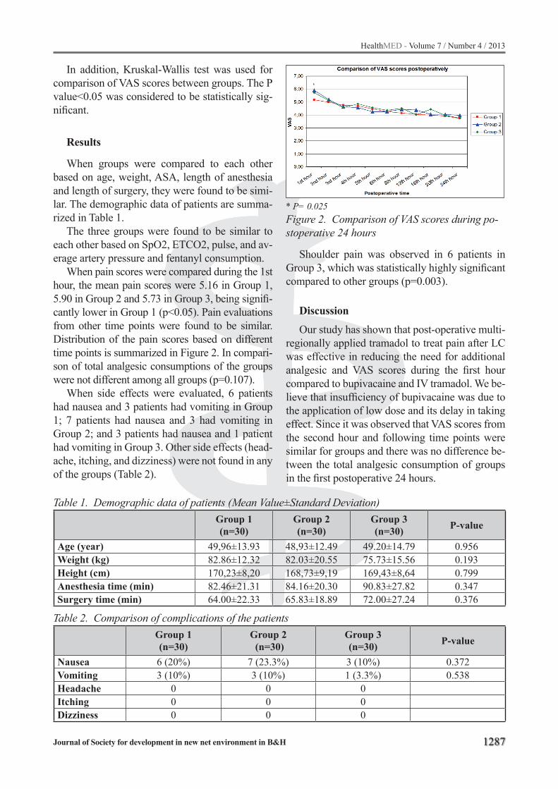
HealthMED - Volume 7 / Number 4 / 2013
Journal of Society for development in new net environment in B&H 1287
In addition, Kruskal-Wallis test was used for comparison of VAS scores between groups. The P value<0.05 was considered to be statistically sig-nificant.
Results
When groups were compared to each other based on age, weight, ASA, length of anesthesia and length of surgery, they were found to be simi-lar. The demographic data of patients are summa-rized in Table 1.
The three groups were found to be similar to each other based on SpO2, ETCO2, pulse, and av-erage artery pressure and fentanyl consumption.
When pain scores were compared during the 1st hour, the mean pain scores were 5.16 in Group 1, 5.90 in Group 2 and 5.73 in Group 3, being signifi-cantly lower in Group 1 (p<0.05). Pain evaluations from other time points were found to be similar. Distribution of the pain scores based on different time points is summarized in Figure 2. In compari-son of total analgesic consumptions of the groups were not different among all groups (p=0.107).
When side effects were evaluated, 6 patients had nausea and 3 patients had vomiting in Group 1; 7 patients had nausea and 3 had vomiting in Group 2; and 3 patients had nausea and 1 patient had vomiting in Group 3. Other side effects (head-ache, itching, and dizziness) were not found in any of the groups (Table 2).
* P= 0.025Figure 2. Comparison of VAS scores during po-stoperative 24 hours
Shoulder pain was observed in 6 patients in Group 3, which was statistically highly significant compared to other groups (p=0.003).
Discussion
Our study has shown that post-operative multi-regionally applied tramadol to treat pain after LC was effective in reducing the need for additional analgesic and VAS scores during the first hour compared to bupivacaine and IV tramadol. We be-lieve that insufficiency of bupivacaine was due to the application of low dose and its delay in taking effect. Since it was observed that VAS scores from the second hour and following time points were similar for groups and there was no difference be-tween the total analgesic consumption of groups in the first postoperative 24 hours.
Table 1. Demographic data of patients (Mean Value±Standard Deviation)
Group 1(n=30)
Group 2(n=30)
Group 3(n=30) P-value
Age (year) 49,96±13.93 48,93±12.49 49.20±14.79 0.956 Weight (kg) 82.86±12.32 82.03±20.55 75.73±15.56 0.193 Height (cm) 170,23±8,20 168,73±9,19 169,43±8,64 0.799Anesthesia time (min) 82.46±21.31 84.16±20.30 90.83±27.82 0.347 Surgery time (min) 64.00±22.33 65.83±18.89 72.00±27.24 0.376
Table 2. Comparison of complications of the patients
Group 1(n=30)
Group 2 (n=30)
Group 3(n=30) P-value
Nausea 6 (20%) 7 (23.3%) 3 (10%) 0.372 Vomiting 3 (10%) 3 (10%) 1 (3.3%) 0.538 Headache 0 0 0 Itching 0 0 0 Dizziness 0 0 0

1288
HealthMED - Volume 7 / Number 4 / 2013
Journal of Society for development in new net environment in B&H
Kucuk et al. applied bupivacaine100 mg (21 mL) + 1/200000 adrenaline, ropivacaine 100 mg (21 mL) + 1/200000 adrenaline and ropivacaine 150 mg (21 mL) + 1/200000 adrenaline to intra-peritoneal and wound sites, and determined that the analgesic need during the initial four hours was significantly reduced in the group that was given 150 mg of ropivacaine. In our study, we found a decrease in the VAS scores in intraperitoneal tram-adol group which was confined to first postopera-tive hour. We believe that this was because of not using adrenaline to extend the effective time for our drugs and administration of a high dose as in ropivacaine group in the study of Kucuk et al (9).
Despite these studies, there are other studies which show bupivacaine to be an ineffective anal-gesic. In one study, it was shown that bupivacaine administered in high doses intraperitoneally is not effective as an analgesic(10).
Papadima et al, administered 0.5% levobupiva-caine before extubation and continued infusion 8 hours postoperatively by placing a subdiaphrag-matic drain and determined that analgesic con-sumption was reduced by 50% in PCU and by 80% during the initial 24 hours compared to the control group(11). Mraovic et al, administered 15ml 0.5% bupivacaine infusion during the first postoperative 8 hours and showed that the anal-gesic need is much less. In this study, high VAS scores in the bupivacaine group may be due to the low dose of the drug and the type of infusion(12).
Shoulder pain is seen at 66% of patients af-ter laparoscopic surgery. The importance of pain is lesser in the first 24 hours, but it becomes in-creasingly the main complaint after the second day 1. It is assumed to be the result of peritoneal stretching, diaphragmatic irritation, traumatic nerve traction, and phrenic nerve stimulation due to the release of inflammatory mediators in(13). In studies trying to reduce the shoulder pain, the duration of pneumoperitoneum, the amount of gas insufflated, peak intra-abdominal pressure, gas temperature, intraperitoneal injection of nitrous oxide, local anesthetics and the use of saline has been questioned(1,14). In our study, intraperito-neal administration of the groups (intraperitoneal bupivacaine and tramadol), shoulder pain was not observed. Also, it is concluded that application of local anesthetic drugs intraperitoneally into the
subdiaphragmatic area prevented phrenic nerve excitation by creating irrigation and anesthetic ef-fect and by this way shoulder pain was not seen.
In addition to its known central effects, recent studies showed that tramadol has also local anal-gesic effects(2,8,9). Golubovic et al, administered intraperitoneal saline, bupivacaine and tramadol + bupivacaine and demonstrated that the analgesic needs of the latter two groups decreased consider-ably compared to the saline group during the ini-tial postoperative hours(15).
Memis et al, showed similar results in their study where they administered tramadol intraperi-toneally following total hysterectomies(16). In contrast to these studies, Akinci et al, has shown that intraperitoneally administered tramadol was effective during the initial postoperative 15 min-utes; however tramadol given intravenously was more effective at all postoperative time points(8). In our study, especially during the first postopera-tive hour, it was observed that the analgesia was more effective in tramadol group compared to bu-pivacaine group and shoulder pain was observed in the IV tramadol group. Mean VAS scores in tramadol group in the first postoperative hour was lower than in Group 2 and 3 and it was statistically significant (p<005). Therefore, it can be concluded that the analgesia was more effective in the intra-peritoneal tramadol group. However, in compari-son of total analgesic consumptions of the groups were not different among all groups (p=0.107).
In conclusion, pain increases mortality and morbidity during LC operations, thus it is an im-portant symptom that should not be ignored. For this reason, in addition to precautions taken during operation, analgesic applied to multiple regions can reduce shoulder and visceral pain especially during the initial postoperative hours.

HealthMED - Volume 7 / Number 4 / 2013
Journal of Society for development in new net environment in B&H 1289
References
1. Joris J, Thiry E, Paris P, Weerts J, Lamy M. Pain af-ter laparoscopic cholecystectomy: characteristics and effect of intraperitoneal bupivacaine. Anesth Analg 1995; 8: 379-84.
2. Bisgaard T, Kehlet H, Rosenberg J. Pain and con-valescence after laparoscopic holecystectomy. Eur J Surg. 2001; 167: 84-96.
3. Lindgren L, Koivusalo AM, Kellokumpu I. Conven-tional pneumoperitoneum compared with abdominal wall lift for laparoscopic cholecystectomy. Br J Anae-sth 1995; 75: 567–572.
4. Lindgren L. Pain after laparoscopic cholecystectomy. Do we do our best? Acta Anaesthesiol Scand 1997; 41: 191–192.
5. Kehlet H, Jensen TS,Woolf CJ. Persistent postsurgical pain: risk factors and prevention. Lancet 2006; 367: 1618–1625.
6. Moiniche S, Jorgensen H, Wetterslev J, Dahl JB. Local anesthetic infiltration for postoperative pain relief after laparoscopy: a qualitative and quantitative systematic review of intraperitoneal, port-site infiltration and me-sosalpinx block. Anesth Analg. 2000; 90: 899-912.
7. Grond S, Meuser T, Zech D, Hennig U, Lehmann KA. Analgesic efficacy and safety of tramadol enantiomers in comparison with the racemate: a randomised, do-uble-blind study with gynaecological patients using intravenous patient-controlled analgesia. Pain. 1995; 62: 313-20.
8. Robaux S, Blunt C, Viel E, Cuvillon P, Nouguier P, Dautel G, Boileau S, Girard F, Bouaziz H. Trama-dol added to 1.5% mepivacaine for axillary brachial plexus block improves postoperative analgesia dose-dependently. Anesth Analg. 2004; 98: 1172-7.
9. Kucuk C, Kadiogullari N, Canoler O, Savli S A. pla-cebo-controlled comparison of bupivacaine and ro-pivacaine instillation for preventing postoperative pain after laparoscopic cholecystectomy. Surg Today. 2007; 37: 396-400.
10. Raetzell M, Maier C, Schroder D, Wulf H. Intraperi-toneal application of bupivacaine during laparosco-pic cholecystectomy--risk or benefit? Anesth Analg. 1995; 81: 967-72.
11. Papadima A, Lagoudianakis EE, Antonakis P, Filis K, Makri I, Markogiannakis H, Katergiannakis V, Ma-nouras A. Repeated intraperitoneal instillation of levo-bupivacaine for the management of pain after laparos-copic cholecystectomy. Surgery. 2009; 146: 475-82.
12. Mraović B, Jurisić T, Kogler-Majeric V, Sustic A. In-traperitoneal bupivacaine for analgesia after lapa-roscopic cholecystectomy. Acta Anaesthesiol Scand. 1997; 41: 193- 6.
13. Atak I, Ozbagriacik M, Akinci OF, Bildik N, Subasi IE, Ozdemir M, Ayta NI. Active gas aspiration to re-duce pain after laparoscopic cholecystectomy. Surg Laparosc Endosc Percutan Tech 2011; 21: 98–100.
14. Mouton WG, Bessell JR, Otten KT, Maddern GJ. Pain after laparoscopy. Surg Endosc. 1999; 13: 445–448.
15. Golubović S, Golubović V, Cindrić-Stancin M, Tokmadzić VS Intraperitoneal analgesia for laparos-copic cholecystectomy: bupivacaine versus bupivaca-ine with tramadol. Coll Antropol. 2009; 33: 299-302.
16. Memis D, Turan A, Karamanlioglu B. Intraperitone-al tramadol and buvacaine in totalabdominal hyste-rectomy. Eur J Anaesthesiol. 2005; 22: 804-5.
Corresponding AuthorAbdulkadir Iskender, Departments of Anesthesiology and Reanimation, Duzce University,Duzce School of Medicine,Duzce,Turkey,E-mail: [email protected]

1290
HealthMED - Volume 7 / Number 4 / 2013
Journal of Society for development in new net environment in B&H
Abstract
Background: The survival of health organi-zations even Medical Universities with great mi-ssion are in close connection with application of strategic planning.
Objective: To assess the progress and related obstacles of the University strategic plan and its effective factors
Methods: This study was conducted within the Medical University’s divisions during 2006-2010. The progress reporting forms and priority settin-gs were formulated through team discussions and participation of the stakeholders.
Findings: Results showed that The most im-portant obstacles to achieve both curative and educational goals were recognized as shortage of fiscal resources, lack of clear internal and external communication, and poor orientation of the per-sonnel which caused the ascendant trend of achie-ving defined goals of University that 12% of them were shown to have been unattained at the end of the study period. In fact Systematic design of stra-tegic committee meetings to engage those invol-ved in planning and assessments of their related educational need were the lost circles of university strategic plan chain.
Conclusion: It is concluded that improving personnel’s perception of strategic plan and the-ir sense of responsibility will develop its process. Current study also showed, due to their severe in-fluence on the implementation stage, clear internal and external communication, more partnership plans and continuous review of structures can be determinant.
Key words: Planning, strategic planning, he-alth care system.
Introduction
One of the most outstanding properties of de-veloped countries is paying attention to knowledge management as one of unavoidable necessities of improvement, development and social-economic growth. Herein, what has been discussed as the most important management function of each country in any level, is to plan and endeavor to gain anticipa-ted goals in it. So in the recent decays, administra-tive manager’s attention is attracted to long term planning and especially strategic ones. In fact or-ganizations ought to have an strategic thought, con-vert their discoveries and understandings to effec-tive strategies to adapt with variable environments and finally prepare the essential infrastructures to execute them since strategic planning is a process to access the organization’s goals in a competitive ,dynamic and varying environment which is desi-gned through resource allocation(1). In fact the or-ganizations that provide strategic planning can get to their goals with a higher probability than those who don’t have this planning (2). In other words it defines the future navigation by helping the organi-zations to select the presidencies and takes today’s decision makings under their future aftereffects to provide bases of improvement of the organization’s functioning.(3) A look to salient achievements of so-cieties along different periods and preliminary study of health market’s circumstances and the internal and external environment fluctuations which rules on it, also shows that strategic planning maintain he-alth organization’s durability as other organizations.
1970’s strategic planning in health care was formed to design a new structure and development of healthcare services in response to population growth. By inventing payment system in 1980, strategic planning turned to a necessity for health care organizations to be able to respond the incre-
Medical University’s strategic plan: Progress and related obstaclesMasoumeh Hosseini1, Saeed Asefzadeh2, Mina Nejati2, Tahere Sadeghi3
1 Department of Health Management and Economics, School of Public Health Tehran University of Medical Sciences, Tehran, Iran,2 Qazvin Research Center for Social Determinant of Health Qazvin University of Medical Sciences, Qazvin, Iran,3 Tehran University of Medical Sciences, Tehran, Iran.

HealthMED - Volume 7 / Number 4 / 2013
Journal of Society for development in new net environment in B&H 1291
asing requirements in competing environments (4,5). Organized healthcare needed more ampli-fication in most fields during 80 and 90 decays .healthcare cost continuously increased more than gross domestic product and competition intensifi-ed in the market. Today hospitals and other health organizations utilize strategic planning as a useful tool to assess the substitution solutions and help to prepare them for the future. Managers of health field need to understand the process of strategic planning, its goals, advantages, challenges, and key factors to assess its progression (6).
Material and methods
This descriptive-analytical study was con-ducted within the 14 divisions affiliated to one of the High ranked Medical Universities to assess the progress of university’s strategic plan and ob-stacles prevented its development between2006 - 2010 for the first time in Iran. The evaluation method and the progress reporting forms were formulated through team discussions and partici-pation of the stakeholders in the collective inter-views. It took several sessions for the strategic committee to analyze the information on forms completed by the participants to set the obstacle priorities annually and recognize efficacious fac-tors. More over the process of goal achievement was assessed and classified in 4 subgroups: unat-tained, completely achieved (100%), between 50 to 90% and <50% achieved. While, delayed goals have been distinguished. Finally the obstacle’s influences on achieving defined goals formulated through collective interviews and the data were summarized and categorized by Spss 16.
Findings
The frequency of goal achievement in differ-ent divisions affiliated to the University is shown at tables and figures. Based on the gathered data, there was an ascendant trend on the completely achievement (100%) of goals until 2009, while it decreased (34.5 %) in 2010. On the other hand, the most unattained goals have been seen (12%) in the same year, as shown in the first figure. Moreover, subsequences of goal attainment assessed during the years surveyed and the data showed that in
the first years of planning most of the goals have gained on time while in the last year (2010), there was a salient down fall and only 32% of the goals had been achieved (figure 3). Nevertheless the University have been had the best rate of 50-90% goal achievement in 2010(figure2). According to the findings, strategic planning had been faced with some obstacles and problems such as short-age of fiscal resources (44%), unclear internal and external communications (29%), manpower and their motivational problems (19%), administrative problems and personnel’s Involvement to their routine activities (8%) totally. The findings sug-gest that besides the appearance of new problems like personnel’s involvement to their routine ac-tivities, the priorities of the old obstacles has been changed completely in the last year of the study and fiscal resource’s shortage has gained the 6th place (9%) (table2). So a salient down fall should be mentioned parallel with the emerged barriers and the priority setting has been done through team discussions. Table 1. Trend of University’s goal achievement
tota
l
Total goals percentage of Goalachievement
YearAchieved
zero
0 - 5
0
50 -
99
100
dela
yed
timel
y
100 28 72 7 10 34 49 2007100 31 69 4 11 32 53 2008100 25 75 5 8 30 57 2009100 68 32 12 16 37.5 34.5 2010
Table 2. Priorities of obstacles that strategic plan has been faced to during the 4 years of assessment
Degree of priority
related obstacles 2007
2008
2009
2010
Shortage of fiscal resources and budjet 1 1 1 6Unclear internal communications 2 5 3 3Unclear external communications 4 3 2 5Shortage of manpower and their motivational problems 3 2 4 4
Administrative problems 5 4 5 2Involvement to the Routine activities - - - 1

1292
HealthMED - Volume 7 / Number 4 / 2013
Journal of Society for development in new net environment in B&H
Figure 1. Percentage of goal achievement 2007 - 2010
Figure 2. Goals achievement’s percentage( > 50% and < 50% )2007 - 2010
Figure 3. Percentage of goal achievement’s re-tardation 2007 - 2010
Conclusion
Based on the medical universities scientific map’s vision which is “ training the best graduates, achieving health indexes as like as possible and having sufficient share in production, scientific and technological development until 2025” ele-mentary policies are made to orient the university toward its mission and final goals. University’s strategies and operational actions have been pre-dicted also to define strategic plan. On the other hand, each strategic plan will be reformed if the related forms and reports review perpetually to all available obstacles be revealed and removed after necessary analysis. So Qazvin university’s stra-tegic plan has been assessed during 2006 – 2010 to find whether related division’s activities follow mission and policies of the university or not, if not what was the reason. As findings suggest at the end of the 2006 which has been the first year of planning, about half of the defined goals com-pletely attained (49%), while for the next years it was more than that (respectively 53% and 57%). But at the end of 2010 this trend has been chan-ged and it descent to 37.5% because of the new obstacles which are ignored mostly. In fact the collective interviews revealed and prioritized the existing snags and problems University strategic planning has been faced to, shortage of fiscal re-sources categorized as the 6th priority in the last year of study while it has been the first one betwe-en 2006 to 2009 which demonstrate despite do-minating fiscal obstacles, improving of strategic performance have been affected by non- finan-cial factors which are in close relationship with the involved people perception. As the studies of Rudd JM, Greenly AT about “strategic planning and performance” (2008) showed, organization’s financial, structural and technical performance are strongly affected by strategic plan’s appli-cation which would have improve both financi-al and nonfinancial aspects of the organization (7, 8). Meanwhile, strategic committees survey showed that besides the old obstacles some new ones appeared like administrative problems and poor orientation of personnel which have influ-enced the strategic progression in 2010. So clear internal and external communication, partnership planning and structures review would have a se-

HealthMED - Volume 7 / Number 4 / 2013
Journal of Society for development in new net environment in B&H 1293
rious impact on personnel’s motivation through Improving their perception of strategic planning and their responsibilities. On the other hand Ne-cessity of improving knowledge level, acquain-tance and proficiency of targeting revealed when some of the routine activities had been defined as the strategic goals. Tohidi (2010) also has empha-sized on his research about “strategic planning of Iran’s educational institutes” that participant’s active role and educational standard’s improve-ment influence the effectiveness and efficiency of strategic planning tools strongly (9). Vafaeezadeh also has researched the effective factors play an important role in strategic plan of Iran’s Marine Industry (2000). According to her findings origi-nal source of each strategic plan is logical thought which force managers and workforces to respect the standards and follow them strongly (10). So Qazvin University’s strategic planning committee can improve the quality of strategic targeting by providing sufficient educational courses about strategic planning and logical thought.
As findings showed, more than 50% of goals have been achieved without delay (in 11 divisi-ons). It demonstrates that the University’s strate-gic plan has been implemented timely based on the schedules. Gordon (2000) investigated the University of Sydney’s educational situation thro-ugh the SWOT analysis technique. He has also confirmed that strategic planning would improve the quality of educational services and activity’s schedule (11). So, it is expected to Qazvin medical university eliminate related obstacles and improve goal achievement of 14th affiliated divisions as like as possible to have the best performance based on its scientific map. But the most important factors which have been recognized for improving the qu-ality of strategic management in all educational, research and curative deputies of universities can be listed as below:
- Continuous review of strategic plan, Feed-back results to relevant units and pursue reforms
- Implementation of strategic plans with state law
- The new management approaches such as knowledge management
- Promotion of capacity building and planning skills
- Systematic design of a strategic committee meetings To engage those involved in planning
- Training courses to improve necessary skills
Finally, Models of strategic planning for educa-tional institutions such as universities and educa-tional need assessment for preparing staff to parti-cipate in the strategic planning process should be studied more in the future. The relationship exists between management styles and effectiveness of the strategic plan also should be considered as an important factor especially in universities which senior managers have a severe impact on research and educational performance that most be impro-ved through the qualified strategic plan.
Acknowledgement
This study was funded by Qazvin University of Medical sciences (QUMSc), and the authors thank all the committees that participated in the assessment sessions.
References
1. Mc Namara C. strategic planning in nonprofit or for profit organization(2000), Available at: http://www.mapn.p.org/library/plan-dec/str-plan
2. Asefzadeh S, Rezapour A. Health planning. 1 st ed. Qazvin: Qazvin University of Medical Sciences; 2007; 164 [In Persian]
3. Tabibi SJ, Maleki M, Delgoshai B. Sterategic Planning. 1st ed. Tehran: Ministery of Health and Medical Education; 2004; 90 [In Persian]
4. Greenley, G.E., Hooley, G.J., Broderick, A.J. and Rudd, J.M. “Strategic Planning Differences Among Different Multiple Stakeholder Orientation Profiles”, Journal of Strategic Marketing, (2004); Vol. 12, No. 3: pp 163-182.
5. Lombardi DJ. Health care management. London: John wiley & son Inc; 2007. 60-75
6. Visseosy J. Health operations management. London: routledge; 2005; 39- 51
7. Rudd JM, Greenley AT. Strategic planning and per-formance: Extending the debate. Journal of Business Research 2008 Feb; 61(2): 99-10

1294
HealthMED - Volume 7 / Number 4 / 2013
Journal of Society for development in new net environment in B&H
8. Vila J, Canales JI. Can Strategic Planning Make Strategy More Relevant and Build Commitment Over Time. The case of RACC. Long Range Planning 2008 Jun; 41(3): 273
9. Hamid Tohidi, Aida Jafari, Aslan Azimi. Strategic planning in Iranian educational organization.proce-dia social and Behavioral sciences 2 (2010) 3904-3908
10. Vafaeezadeh M,R. effective factore of strategic planning in Iran’s marine industry. Tehran Univer-sity of medical sciences. (1998).
11. Gordon J, Hazlett C & et all. Strategic planning in medical education: enhancing the learning envi-ronment for students in clinical settings. Med Educ. 2000; 34(10): 841-50. Available at: www.ncbi.nlm.nih.go
Corresponding AuthorMina Nejati,Qazvin Research Center for Social Determinant of Health,Qazvin University of Medical Sciences, Qazvin, Iran,E-mail: [email protected]
HealthMED - Volume 7 / Number 4 / 2013
Journal of Society for development in new net environment in B&H 1295
Abstract
Objective: Human parvovirus B19 (HPV B19) has a remarkable tropism for erythroid progenitor cells and this infection leads to important morbid-ity in patients with sickle cell disease. The virus can induce transient aplastic crisis. The patients with sickle cell disease are great importance to ep-idemiological surveillance programs and disease prevention strategies. The aim of the present study was to investigate the prevalence of HPV B19 in patients with sickle cell disease by using PCR and ELISA techniques in southern Turkey.
Materials and Methods: All the serum sam-ples were collected from 113 patients with sickle cell disease and 64 healthy subjects. Antibodies to HPV B19 were measured by ELISA method. Vi-ral DNA was extracted by using a Real-time PCR LightCycler Parvovirus B19 Quantification kit.
Results: Of the total participants, 46 (40.7%) of patients and 18 (28.1%) of control group were found to IgG positive (P = 0.094), whereas 13 (11.5%) of patients and 9 (14.1%) of control group were found to IgM positive (P = 0.620). HPV B19 DNA were detected in 20 (17.7%) of patients and 9 (14.1%) of control group (P = 0.530). Good de-gree correlation was found when the comparison B19 DNA and specific IgM antibodies (r=0.666, P˂0.001). Transient aplastic crisis was found in 47.8% of patients with HPV B19 infection.
Conclusions: The data in the present study document that HPV B19 is a common pathogen for both children and adults with sickle cell dis-ease and healthy persons in Turkey.
Key words: Human parvovirus B19, Sickle cell disease, aplasia, prevalence.
Introduction
Human parvovirus B19 (HPV B19), which was identified in by Cossart et al., is a small, single-stranded, nonenveloped DNA virus and a member of the Erythrovirus genus (1, 2). Infections with this virus are occur worldwide and can result in a wide range of clinical findings (3, 4). Manife-stations of parvovirus B19 infection changes from asymptomatic, nonspecific, cold like symptoms or subclinical infection for most people (5, 6). On the other hand, clinical presentations associated with the infection contain erythema infectiosum, ar-thropathy, transient aplastic crisis (TAC), chronic red cell aplasia, and hydrops fetalis (6).
The virus has a remarkable tropism for erythroid progenitor cells and thus can cause eventually trig-gering a transient arrest in erythropoiesis (7). Pa-tients with chronic hemolytic disorders, including sickle cell disease (SCD), thalassemia, hereditary spherocytosis, red cell enzymopathies such as pyru-vate kinase deficiency, and autoimmune hemolytic anemia are at risk of violent clinical illness (8-12). The virus induces a cessation of erythropoiesis and acute erythroblastopenia. This condition is often called as transient aplastic crisis (13, 14).
HPV B19 infection leads to important morbi-dity in patients with SCD (9, 15). The patients are pretty much infectious during aplastic crisis and usually become highly viremic. They should be isolated to prevent contamination of the virus (13).
The patients with sickle cell disease are should be followed closely terms of HPV B19 infection. For this reason, it is great importance to epidemio-logical surveillance programs and disease preven-tion strategies. The aim of the present study was to investigate the prevalence of HPV B19 in patients
The prevalence of Parvovirus B19 in patients with Sickle cell disease in southern TurkeyVicdan Koksaldı Motor1, Omer Evirgen1, Nizami Duran2, Vefik Arıca3, Melek Inci2, M. Murat Celik4, Fatmagul Basarslan3, Yusuf Onlen1
1 Department of Infectious Disease and Clinical Microbiology, Mustafa Kemal University Medical School, Hatay, Turkey,2 Department of Microbiology, Mustafa Kemal University Medical School, Hatay, Turkey, 3 Department of Pediatrics, Mustafa Kemal University Medical School, Hatay, Turkey,4 Department of Internal Medicine, Mustafa Kemal University Medical School, Hatay, Turkey.

1296
HealthMED - Volume 7 / Number 4 / 2013
Journal of Society for development in new net environment in B&H
with sickle cell disease by using polymerase chain reaction (PCR) and enzyme linked immunosorbent assay (ELISA) techniques in southern Turkey.
Materials and methods
Serum samples were collected from 113 patients with SCD and 64 healthy subjects were taken as controls. All the serum samples were selected from Mustafa Kemal University Medical School internal medicine and pediatrics polyclinics. The patients (58 males and 55 females) with SCD were recruited ages ranging from 2 to 51 years (mean ± SD: 18.4 ± 9.1 years). The control subjects (29 males and 35 females) were recruited with ages ranging from 1 to 41 years (mean± SD: 17.9 ± 10.4 years).
The serum levels of anti HPV B19 IgG and IgM were measured by ELISA method according to instructions of the manufacturer (NovaTec Im-mundiagnostica GmbH®, Germany).
Extraction of DNAViral DNA was extracted from 200 μl of sam-
ple by using a Real-time PCR LightCycler Parvo-virus B19 Quantification kit (Roche®, Germany) according to instructions of the manufacturer. Nu-cleic acids were eluted from the filter column with 50 μl of nuclease distilled water. Isolated DNA samples were stored at -80°C until further use.
Primer and probesPCR primers and a TaqMan probe were used to
amplification of 123 bp region of NS1 gene in the study. Primary sequences of the primers and probe were as follows:
NS1-F (Forward): 5’-GAAGACTGGATGAT-GACAGATCCA-3’,
NS1-R (Reverse): 5’-TGCTGTTTTT-GTTCTTGCTAGAGTAA-3’.
NS1-P (Probe): FAM-AATGATGGCT-CAAACCGGAGGAGA-BHQ1.
The fluorogenic probe was 5’ labeled with the 6-carboxyfluorescein (FAM) and 3’ labeled with the Black Hole Quenchers (BHQ1).
Preparation of standard plasmid DNANS1 region was amplified in a 25 μl reaction
mixture containing 3 μl deoxynucleoside triphos-phates (dNTP), 16 μl bidistilled sterile water, 2.5
μl buffer, 1 μl of each primer (NS1-F and NS1-R), 1 μl of BQ, and 0.5 μl of DNA polymerase. Ampli-fication reactions were performed with 1 cycle at 94°C for 5 minute followed by 30 cycles of 94°C for 30 seconds, 58 °C for 45 seconds, and 72 °C for 30 seconds. A final extension 7 minute was added after the last cycle. This PCR product is produced by inserts to a vector (pMD18-T) in the host bacte-rium. It is purified recombinant plasmid by using a commercial kit from this bacterium. These products were stored at -20 ° C for later use.
Real-time PCRNS1 gene was amplified with a thermocycler in
a 25 μl reaction mixture containing 2.5 μl PCR buf-fer, 3.5 μl dNTP, 3 μl MgCl2, 1 μl of each primer (NS1-F and NS1-R), 1 μl recombinant plazmid, 0.5 μl probe (PPV-P), 0.5 μl hotstart Taq DNA polimer-ase, and 12 μl bidistilled sterile water. PCR amplifi-cation reactions of NS1 gene were performed with 1 cycle at 95°C for 30 seconds followed by 40 cycles of 95°C for 10 seconds, 58 °C for 20 seconds, and a final extension at 72°C for 20 seconds. The sta-tistical significance of observed differences was as-sessed by means of the chi-square test for categori-cal variables; Mann-Whitney U test for continuous variables; nonparametric correlation (Spearman’s twotail) for association. All tests were performed us-ing SPSS (version 15.0, USA). P less than 0.05 were considered to be statistically significant.
Results
The serological and PCR data of the both groups tested are shown in Table 1. Forty six (40.7%) of patients with sickle cell disease and 18 (28.1%) of control group were found to have detectable levels of anti parvovirus B19 IgG (χ2 = 2.8, d.f. = 1, P = 0.094). Thirteen (11.5%) of patients with the sickle cell disease and 9 (14.1%) of control group were found to have detectable levels of parvovirus B19 IgM (χ2 = 0.246, d.f. = 1, P = 0.620). HPV B19 DNA were detected in 20 (17.7%) of patients with the sickle cell disease and 9 (14.1%) of control group (χ2 = 0.394, d.f. = 1, P = 0.530). Good degree cor-relation was found when the comparison B19 DNA and specific IgM antibodies (r=0.666, P˂0.001).
Confirmation of HPV B19 infection was found in 23 (20.4%) of 113 patients by showing parvovi-
HealthMED - Volume 7 / Number 4 / 2013
Journal of Society for development in new net environment in B&H 1297
Table 1. Serological and PCR data of the patients with sickle cell disease and control group
Group tested B19-IgM antibodies positive n (%)
B19-IgG antibodies positive n (%)
B19 DNA n (%)
Sickle cell anemia n=113 13 (11.5) 46 (40.7) 20 (17.7)
Controln=64 9 (14.1) 18 (28.1) 9 (14.1)
Table 2. Results of B19 DNA and specific IgM antibodies detected in serum samples of patients with B19 infection according to clinical manifestations
Clinical manifestations Only B19 DNA + B19 DNA +IgM +
B19 DNA +IgG + Only IgM +
Not clinical signs 1 4 4 2Transient aplastic crisis - 6 4 -İnfection - - - 1Transient aplastic crisis + İnfection 1 - - -Total 2 10 8 3
Table 3. Prevalence of HPV B19 IgG antibodies in patients with SCD and control group according to age groups
AgePatients with sickle cell disease for IgG positive Control group for IgG positive
n Number % n Number %0-10 20 2 10 19 3 15.811-20 51 28 54.9 17 5 29.421-30 31 12 38.7 20 8 4031-40 9 4 44.4 7 2 28.641-51 2 0 0 1 0 0Total 113 46 40.7 64 18 28.1
Table 4. The comparison pediatric and adult patients and control group according to serology and PCR
Age
Patients with sickle cell disease Control group PIgM
n (%)
IgGn
(%)
B19 DNAn (%)
IgMn
(%)
IgGn
(%)
B19 DNAn (%)
IgM IgG B19 DNA
Pediatric group (0-16)58 patient, 30 control 7 (12.1) 20 (34.5) 10 (17.2) 8 (26.7) 7 (23.3) 9 (30) 0.086 0.285 0.170
Adult group (≥ 17)55 patient, 34 control 6 (10.9) 26 (47.3) 10 (18.2) 1
(2.9) 11 (32.4) 0 (0) 0.177 0.168 0.009
Total 13 46 20 9 18 9 0.620 0.094 0.530
rus B19 DNA and/or specific IgM antibody. Two patients only viral DNA could be detected, as 10 patients were tested positive for both parvovirus B19 DNA and specific IgM. Eight patients were found positive for both viral DNA and specific IgG. Other 3 patients, only specific IgM could be detected. Results of B19 DNA and specific IgM antibodies detected in serum samples of patients with HPV B19 infection according to clinical manifestations are summarized in Table 2.
Prevalence of HPV B19 IgG antibodies in pa-tients with sickle cell disease and control group according to age groups are shown in Table 3. The rate of positive identification of HPV B19 IgG an-tibody in young patients with the sickle cell disease was higher than other groups. The comparison pediatric and adult groups are shown in Table 4. There was statistical difference between the adult patients with sickle cell disease and adult control group in terms of HPV B19 DNA (P=0.009).

1298
HealthMED - Volume 7 / Number 4 / 2013
Journal of Society for development in new net environment in B&H
Discussion
SCD is common illness in Southern Turkey. The incidence of SCD is 10.0% in the Cukurova region of Southern Turkey (16, 17). HPV B19 infection can leads to repress erythropoiesis and acute erythroblastopenia in these group patients. The patients usually extremely viremic and risk of virus transmission is increased (18). Tight follow-ing of these patients with SCD terms of HPV B19 infection are necessary for the formulation of epi-demiological surveillance and disease prevention. This is the first study evaluating the prevalence of HPV B19 in patients with SCD in Turkey.
Infections with HPV B19 are occurring world-wide and situations of infection have been reported in all seasons. Seroprevalence increases with age and more than 70% of the adult population is sero-positive. Children are the master resource of con-tamination and outbreaks can persist for months in schools and child care centers (19). In a study among Korean plasmapheresis donors, it was re-ported that prevalence of IgG antibodies to HPV B19 is 59.38% (20). In the other study, it was shown that anti HPV B19 IgG antibodies are 32.1% in pa-tients with chronic hemolytic anemia is majority of the SCD (21). Smith-Whitley et al found the serol-ogy of HPV B19 IgG 29.5% in children with SCD while the same group of patients, Zimmerman et al that 53% (15, 22). In another study, it was reported that HPV B19 IgG antibodies were detected 56.5% of the sickle cell anemia, 15.2% of the control pa-tients. There was statistical difference between the sickle cell anemia and control patients in terms of HPV B19 IgG seropositivity (18). In the present study, there was found to have detectable HPV B19 IgG 40.7% of patients with the SCD and 28.1% of control group. There was no statistical difference between the patients and control subjects in terms of HPV B19 IgG seropositivity.
In a study of 12 years in Ireland, it was reported that prevalence of IgM antibodies to HPV B19 is 4.5% (23). In the other report, it was shown that HPV B19 IgM in 4 of 19 patients with TAC (21). Us et al found 12.6% were positive for specific IgM in patients with hematological disorders such as acute and chronic leukemia, malignant lympho-ma, myelodysplastic syndrome, and multiple my-eloma. But in this patient group, 70.9% were re-
ceived multiple blood transfusions or blood prod-ucts (24). In the present study, there was found to have detectable HPV B19 IgM 11.5% of patients with SCD and 14.1% of control group. Also 17.7% of patients with SCD and 14.1% of control group were found to contain HPV B19 DNA. Although 2 patients only viral DNA and other 3 patients only specific IgM could be detected, good degree correlation was found when the comparison B19 DNA and specific IgM antibodies.
It was discovered in 1981 that HPV B19 caused TAC in children with sickle-cell anemia and it is found from further studies that nearly 70% of HPV B19 infections in TAC [19]. In the present study, TAC was found in 11 (47.8%) of 23 patients with HPV B19 infection by showing HPV B19 DNA and/or specific IgM antibody. In a study, the op-posite anti B19 IgM could be detected in only 4 (21.1%) of 19 patients with TAC and none of them had HPV B19 DNA (21).
The data in the present study document that HPV B19 is a common pathogen for both children and adults with SCD and healthy persons in Tur-key. Although seroprevalence increases with age, it could not be shown in this study in full. The rea-son for this may be less the number of cases stud-ied according to age groups.
In conclusion, there was no statistical difference between the patients and control subjects in terms of HPV B19 IgG and IgM seropositivity and HPV B19 DNA. But while the comparison pediatric and adult groups; there was statistical difference be-tween the adult patients with SCD and adult control group in terms of HPV B19 DNA. Well degree cor-relation was found when the comparison HPV B19 DNA and specific IgM antibodies. TAC was found in 47.8% of patients with HPV B19 infection. This is the first study evaluating the prevalence of HPV B19 in patients with SCD in Turkey.
References
1. Cossart YE, Field AM, Cant B, Widdows D. Par-vovirus-like particles in human sera. Lancet 1975; 1(7898): 72-73.
2. International Committee on Taxonomy of Viruses (ICTV). Virus taxonomy: 2009 release. Virology divi-sion, IUMS. Available at http://www.ictvonline.org

HealthMED - Volume 7 / Number 4 / 2013
Journal of Society for development in new net environment in B&H 1299
3. Katta R. Parvovirus B19: a review. Dermatol Clin 2002; 20: 333-342.
4. Plummer FA, Hammond GW, Forward K, et al. An erythema infectiosum-like illness caused by human parvovirus infection. N Engl J Med 1985; 313: 74-79.
5. Brown KE. Human Parvoviruses, Including Parvo-virus B19 and Human Bocavirus. In: Mandell GL, Bennet JE, Dolin R, (eds). Principles and Practice of Infectious Diseases, 7th edn. Churchill Livingstone, Philadelphia, 2010.
6. Heegaard ED, Brown KE. Human parvovirus B19. Clin Microbiol Rev 2002; 15: 485-505.
7. Ozawa K, Kurtzman G, Young N. Replication of the B19 parvovirus in human bone marrow cell cultures. Science 1986; 233: 883-886.
8. Pattison JR, Jones SE, Hodgson J, et al. Parvovirus infections and hypoplastic crisis in sickle-cell anae-mia. Lancet 1981; 1(8221): 664-665.
9. Serjeant BE, Hambleton IR, Kerr S, Kilty CG, Ser-jeant GR. Haematological response to parvovirus B19 infection in homozygous sickle-cell disease. Lan-cet 2001; 358: 1779-1780.
10. Lefrère JJ, Couroucé AM, Bertrand Y, Girot R, So-ulier JP. Human parvovirus and aplastic crisis in chronic hemolytic anemias: a study of 24 observati-ons. Am J Hematol 1986; 23: 271-275.
11. Davidson RJ, Brown T, Wiseman D. Human parvo-virus infection and aplastic crisis in hereditary sphe-rocytosis. J Infect 1984; 9: 298-300.
12. Young NS. Hematologic manifestations and diagno-sis of parvovirus B19 infections. Clin Adv Hematol Oncol 2006; 4: 908-910.
13. Servey JT, Reamy BV, Hodge J. Clinical presentati-ons of parvovirus B19 infection. Am Fam Physician 2007; 75: 373-376.
14. Chorba T, Coccia P, Holman RC, et al. The role of parvovirus B19 in aplastic crisis and erythema infectiosum (fifth disease). J Infect Dis 1986; 154: 383-393.
15. Smith-Whitley K, Zhao H, Hodinka RL, et al. Epide-miology of human parvovirus B19 in children with sickle cell disease. Blood 2004; 103: 422-427.
16. Guler E, Garipardic M, Dalkiran T, Davutoglu M. Premarital screening test results for β-thalassemia and sickle cell anemia trait in east Mediterranean region of Turkey. Pediatr Hematol Oncol 2010; 27: 608-613.
17. Cürük MA, Zeren F, Genç A, Ozavci-Aygün S, Kilinç Y, Aksoy K. Prenatal diagnosis of sickle cell anemia and beta-thalassemia in southern Turkey. Hemoglo-bin 2008; 32: 525-530.
18. Regaya F, Oussaief L, Bejaoui M, Karoui M, Zili M, Khelifa R. Parvovirus B19 infection in Tunisian pa-tients with sickle-cell anemia and acute erythroblas-topenia. BMC Infect Dis 2007; 7: 123.
19. Corcoran A, Doyle S. Advances in the biology, diag-nosis and host-pathogen interactions of parvovirus B19. J Med Microbiol 2004; 53: 459-475.
20. Oh DJ, Lee YL, Kang JW, Kwon SY, Cho NS. Inves-tigation of the prevalence of human parvovirus B19 DNA in Korean plasmapheresis donors. Korean J Lab Med 2010; 30: 58-64.
21. Sant’Anna AL, Garcia Rde C, Marzoche M, et al. Study of chronic hemolytic anaemia patients in Rio de Janeiro: prevalence of anti-human parvovirus B19 IgG antibodies and the development aplastic crises. Rev Inst Med Trop S Paulo 2002; 44: 187-190.
22. Zimmerman SA, Davis JS, Schultz WH, Ware RE. Subclinical parvovirus B19 infection in children with sickle cell anemia. J Pediatr Hematol Oncol 2003; 25: 387-389.
23. Nicolay N, Cotter S. Clinical and epidemiological aspects of parvovirus B19 infections in Ireland, January 1996-June 2008. Euro Surveill 2009; 14: 19249.
24. Us T, Ozune L, Kasifoglu N, Akgun Y. The investi-gation of parvovirus B19 infection in patients with haematological disorders by using PCR and ELISA techniques. Braz J Infect Dis 2007; 11: 327-330.
Corresponding AuthorVicdan Koksaldi Motor,Department of Infectious Disease and Clinical Microbiology,Mustafa Kemal University Medical School,Hatay,Turkey,E-mail: [email protected]

1300
HealthMED - Volume 7 / Number 4 / 2013
Journal of Society for development in new net environment in B&H
Abstract
Introduction: The detecting and reporting ca-ses of child abuse in Serbia today is closer deter-mined by Special protocol provided by the special health care system for protection of children from abuse and neglecting, by Ministry of Health of the Republic of Serbia. This protocol was published in April 2009. However, it should be noted that this is yet an unexplored area in Serbia.
Aim: The aim of this study was to collect know-ledge about detection and willingness to report child abuse, as well as how well health workers and whether they are educated at all for these procedures.
Methods: In this phenomenological study a quantitative method was used and as a technique for data collection a survey was used. As a rese-arch instrument a questionnaire with mostly clo-sed questions was used. The collected data were analyzed quantitatively by the methods of descrip-tive statistics (analysis), while the answers to the open questions were analyzed qualitatively. The sample included doctors and nurses of both sexes from the two largest, and by organization similar child health institutions in Serbia: from the Uni-versity Children Hospital (UDC) and the Institute for Health Protection of Mother and Child “Dr Vu-kan Cupic” (IMD). The study was conducted on a sample of 135 health professionals from different disciplines (specialization) of pediatric health care institutions previously mentioned.
Results: The study identified that only 20% of health workers recognized and reported cases of child abuse, and that out of the total number of health workers who had reported child abuse case, 69.4% were trained for it.
Conclusion: The research data indicate that only one fifth of health care workers who reported child abuse case, revealed them only by recogni-
zing the symptoms of abuse. On the other hand, out of the total number of health workers who have reported child abuse cases, most of them were trained for it.
Key words: Detection, reporting, child abuse, health care workers.
Introduction
Health workers at any level of health care play an important role in the protection, primarily in the detection and reporting of child abuse [1,2]. Patro-nage nurse during home visits can detect cases of physical child abuse. They are a vital link between the child at risk or already abused child and the access to services and resources for protection from abuse [3]. Health workers from the emer-gency services are often the first medical exper-ts who interact with the child that comes to the examination. Therefore, the observation of nurses in the waiting room can play an important role in detecting physical abuse [4]. Missed opportunity to recognize the abuse victim means that the pro-per diagnosis has not been made and that the child will return to the environment in which the abuse was made. Since the recognition (detection) of the abuse is one of the skills of health workers, many articles on child abuse intended for health profe-ssionals (Spencer, 2002; Joughin, 2003; Mulryan et al., 2004) have been published. The aforementi-oned sources are educational, informative for he-alth workers how to recognize (detect) symptoms and signs of all kinds of abuse, including emotio-nal abuse [5]. Contact and intervention of health professionals with children and their parents can minimize the trauma after sexual abuse [6].
The empathy of health workers for the problems of abused children in Serbia begins to increase du-ring the 20-ies in twentieth century. The reasons for
Detecting and reporting child abuse by health workers in SerbiaMiroslav Misic¹, Fadilj Eminovic2, Lidija Milenovic3, Snezana Mirkovic4
1 High Medical School of Professional Studies, Zemun, Belgrade, Serbia,2 Faculty for Special Education and Rehabilitation, Belgrade, Serbia,3 High Medical School of Professional Studies’’Milutin Milanković’’, Belgrade, Serbia,4 Emergency Services, Health Center, Krusevac, Serbia.

HealthMED - Volume 7 / Number 4 / 2013
Journal of Society for development in new net environment in B&H 1301
this are numerous, and among them is the imme-diate encounter of a large number of health workers with children who have been exposed to violence in the war zone. Another very important reason is the activities of nongovernmental organizations that during the years of war and crisis strengthened [7].
Today the relationship of health workers and abused children in Serbia is closely determined by Special protocol provided by the special health care system for protection of children from abuse and neglecting, by Ministry of Health of the Re-public of Serbia. This protocol was published in April 2009, and it was intended for health wor-kers and health associates engaged in health care of children. The general goal of this Protocol is to protect children from all kinds of abuse [8].
By this Special protocol of the health care system for child protection from abuse and ne-glecting the health workers and health associates at all levels should be more included. Health wor-kers are among the first where a child or a child’s family members ask for help when a child is inju-red. Therefore, health workers are in a unique po-sition to detect and report cases of child abuse [9].
Detection of cases of child abuse cases is the first and very important step for the further course and outcome of the health of the child. The role of health workers in detecting cases of abuse is great because practically all children come into contact with health workers. Health workers at all level of health care have an important role in detecting cases of abuse. Detection of child abuse can happen in two ways: by identifying violations or changes in child`s be-havior, or by confession of the child, which can be directly (when abused children confide in health worker) and indirectly (when someone else con-fides in health worker and provides information about child abuse) [9].
Reporting cases of child abuse is the most im-portant step in the process of protecting children from abuse. Beside the ethical, health workers have also the legal right and obligation (Article 253 of the Code of Criminal Procedure) to report child abuse case. Failure to report cases of abuse is punishable. The detected case of child abuse the health worker reports to the police and the com-petent social work center. A child whose life or health are at risk should not be left to the person suspected of being an abuser or conceals the abu-
ser, but must be hospitalized until the competent authorities (police and experts from the Center for Social Work) arrive [9].
The research conducted in Serbia, which dealt with child abuse was oriented to the: extent, cau-ses and manifestations of child abuse (Stojakovic, 1984, Svenson, 2007), the visibility of violence against children and the role of the individual ser-vices in the treatment of abused children (Milosav-ljevic, 1998), knowledge about the doctor’s attitu-des towards their own contribution to protecting children from abuse and position on mandatory reporting of abuse (Engineering, 2000), long-term consequences that violence has on victims of vio-lence (Nikolic-Future, 2002), the level of protec-tion of abused children and social work centers in the detection, assessment and treatment (Zegarac, 2004). Until this very day Serbia lacks in experi-enced research on detection and reporting of cases child abuse by health care workers [8]. This fact influenced the goal of our research.
Aim of paper
The aim of this study was to collect knowledge about detection and willingness to report child abuse, as well as education of health professionals for these procedures.
Methods
This phenomenological study used a quantita-tive method and a survey as a technique for data collection. As a research instrument a question-naire with mostly closed questions was used. The collected data were analyzed quantitatively by the methods of descriptive statistics (analysis), while the answers to open questions were analyzed qua-litatively. Some open questions were closed after the research, that is, the responses to the questions were grouped in categories.
The statistical methods applied in this study are:- Descriptive analysis, which involves the
application of statistical methods that deal with collection, assorting and analysis of the statistical data. Its aim is to describe the measured phenomenon.
- Chi-Squared test, which task was to establish a statistically significant difference

1302
HealthMED - Volume 7 / Number 4 / 2013
Journal of Society for development in new net environment in B&H
or relationship between two or more categorical variables that had normal levels of measurement.
- Data processing was performed by the computer statistical program PASW 18.0.
The sample included doctors and nurses of both sexes from the two largest, and by organization si-milar child health institutions in Serbia, from the University Children Hospital (UDC) and the Insti-tute for Health Protection of Mother and Child “Dr Vukan Cupic” (IMD). The sample included a total of 94 doctors from the UDC and the IMD (which accounted for 51% of permanent staff physicians) and a total of 41 nurses from the emergency servi-ces including radiology technicians from the UDC and the IMD (which constitute 82% of total nurses employed in the aforementioned services). Total of 135 health professionals from different disci-plines (specialization) of pediatric health care in-stitutions mentioned. Collection of empirical data on the UDC and the IMD was conducted in the period from 8 April 2010 to 12 May 2010.
Results
Ways in which health workers detected reported cases of child abuseResearch data indicate that most respondents,
that is 20% of them (27) that have reported cases of child abuse detected them by recognizing the symptoms of abuse. Only 2.2% of respondents (3) used as a way of discovering that the heard from a person escorting child that he/she was abused. There were no respondents who have used as a way of discovering the child`s confession about abuse. The results also revealed that there were respondents who had reported cases of child abuse detecting them by combination of two or all three ways of detecting cases of child abuse. The highest percentage of respondents 11.1% (15) detected ca-ses combining all three modes of detection. Then, 4.4% (6) in detection combined the recognized symptoms with an information obtained by the child`s companion. It was 2.9% of respondents (4) that combined the recognized symptoms and the confession of the child. The least, that is 1.5% of respondents (2) in detection combined the story told by the child and his/hers companion.
It is important to note that 57.7% (78) of res-pondents did not discover cases of child abuse.
The relation of detection modes in cases of child abuse was analyzed in accordance to occu-pation of the respondent. This comparison reve-als that over 25.5% of physicians (24) than nurses 7.3% (3) reported cases of child abuse detected by recognizing the symptoms of abuse. The abo-ve-mentioned reveals also that more physicians 14.9% (14) than nurses 2.4% (1) that had reported cases of child abuse revealed them in combination of all three modes of detection.
The relation between detecting modes of child abuse was analyzed, according to whether res-pondents were systematically trained to protect abused children. This comparison revealed the following data:
- in the group with the most respondents who, as a mode of detecting child abuse, used recognizing symptoms, 11.8% (16) of respondents were educated versus 8.2% (11) of respondents who were not educated,
- in the group of subjects who used combination of all three detection modes, 7.4% (10) of respondents were educated versus 3.7% (5) of the respondents who were not educated.
- in the group which as a detection mode used a statement of the child`s companion, there were less, that is 1.5% (2) respondents who were educated versus 3% (4) respondents who were not educated,
- in the group which as a detection mode used a statement of the child`s companion, there were more, that is 1.5% (2) educated respondents in relation to 0.7% (1) non-educated respondents,
- in the group which as a detection mode used a child`s statement there were more, that is 2.1% (3) educated respondents in relation to 0.7% (1) non-educated respondents,
- in the group which as a detection mode used both statement from a child and from a companion there were 1.5% (2) educated respondents while there were no non-educated respondents,
- there were no respondents who have used the child’s statement as a detection mode.

HealthMED - Volume 7 / Number 4 / 2013
Journal of Society for development in new net environment in B&H 1303
Experience of health workers who have reported cases of child abuseBy researching we have come to the informa-
tion and experiences of health workers who have reported suspected child abuse. Less, that is 57 respondents (42.2%) reported the suspicion about child abuse, compared to 78 subjects (57.8%) who did not report this suspicion.
The relation between occupation and the fact whether the respondents reported suspected child abuse was analyzed. Based on the results (χ² = 8.918, df = 1, p = 0.002), significant statistical differences occur. It is obvious that more doctors (51.1%) than medical technicians and nurses report (22%) sus-pected child abuse. Then, the relation between the facility where respondents work and gender of the respondents and the fact whether the respondents re-ported suspected child abuse was analyzed. Based on the results, none of the cases marked statistically significant differences. The relation of respondents who reported suspected child abuse and the mode by which they detected reported cases was analyzed. Based on the results (χ² = 130.953, df = 2, p = 0.000), significant statistical differences occur. Based on these results it is obvious that a small number, that is
27 respondents (20%) detected cases of child abuse by recognizing, and then reporting them.
A statistically significant difference was ob-served in relation between subjects who detected and reported cases of abused children compared to how many of these subjects were educated and trained for protection of abused children. Table 1, reveals that among respondents who reported cases of child abuse were more, that is 69.4% of those who have had training and education for protection of abused children, versus 26.7% of respondents who were not trained.
Most of the respondents, precisely 72 (54.1%) answered the question “Have you or your collea-gues had a bad experience while reporting child abuse cases?” that they were not familiar with such cases. Then, 48 respondents (36.1%) answered that they or their colleagues had a bad experience in these cases. The least, that is, 13 subjects (9.8%) responded that neither they nor their colleagues had a bad experience in the mentioned cases. This que-stion was not answered by two respondents (1.5%).
The relation between the fact whether the res-pondents reported cases of abused children and the answers to the question of whether the respondents
Table 1. Respondents who discovered and reported cases of abused children according to how they were trained for and protection of abused children
Have you reported cases of child abuse?Total
yes not
Is he/she education
by
he/she education by
Count 34 15 49% 69.4% 30.6% 100.0%
had no education
Count 23 63 86% 26.7% 73.3% 100.0%
Total Count 57 78 135% 42.2% 57.8% 100.0%
χ²=23,268; df = 1; p = 0,000
Table 2. Report cases of abused children and the answer to the question of whether the respondents or their colleagues had a bad experience when logging
Did they have a bad experienceTotal
there were no adverse experiences negative experiences
Are the reported
cases
yes Count 45 12 57% 78.9% 21.1% 100.0%
not Count 75 1 76% 98.7% 1.3% 100.0%
Total Count 120 13 135% 90.2% 9.8% 100.0%
χ²=14,387; df = 1; p = 0,000
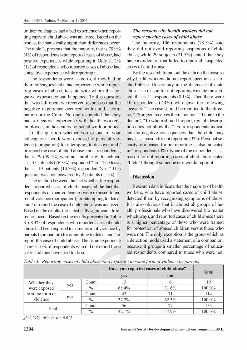
1304
HealthMED - Volume 7 / Number 4 / 2013
Journal of Society for development in new net environment in B&H
or their colleagues had a bad experience when repor-ting cases of child abuse was analyzed. Based on the results, the statistically significant differences occur. The table 2, presents that the majority, that is 78.9% (45) of respondents who reported cases of abuse, had positive experiences while reporting it. Only 21.2% (12) of respondents who reported cases of abuse had a negative experience while reporting it.
The respondents were asked to, if they had or their colleagues had a bad experience while repor-ting cases of abuse, to state with whom this ne-gative experience had happened. To this question that was left open, we received responses that the negative experience occurred with child`s com-panion or the Court. No one responded that they had a negative experience with health workers, employees in the centers for social work or police.
To the question whether you or one of your colleagues at work were exposed to parental vio-lence (companion) for attempting to discover and / or report the case of child abuse, most respondents, that is 79 (59.4%) were not familiar with such ca-ses; 35 subjects (26.3%) responded “no.” The least, that is, 19 patients (14.3%) responded “yes.” This question was not answered by 2 patients (1.5%).
The relation between the fact whether the respon-dents reported cases of child abuse and the fact that respondents or their colleagues were exposed to pa-rental violence (companion) for attempting to detect and / or report the case of child abuse was analyzed. Based on the results, the statistically significant diffe-rences occur. Based on the results presented in Table 3, 68.4% of respondents who reported cases of child abuse had been exposed to some form of violence by parents (companion) for attempting to detect and / or report the case of child abuse. The same experience share 31.6% of respondents who did not report these cases and they have tried to do so.
The reasons why health workers did not report specific cases of child abuseThe majority, 106 respondents (78.5%) said
they did not avoid reporting suspicions of child abuse, while 29 subjects (21.5%) stated that they have avoided, or that failed to report all suspected cases of child abuse.
By the research found out the data on the reasons why health workers did not report specific cases of child abuse. Uncertainty in the diagnosis of child abuse as a reason for not reporting was the most ci-ted, that is 11 respondents (8.1%). Then there were 10 respondents (7.4%) who gave the following answers: “The case should be reported to the direc-tor,” “Surgeon receives them, not me”, “I note to the doctor”, “To whom should I report, my job descrip-tion does not allow that”. Four respondents indica-ted the negative consequences that the child may have as a reason for not reporting (3%). Personal se-curity as a reason for not reporting is also indicated in 4 respondents (3%). None of the respondents as a reason for not reporting cases of child abuse stated “I felt / I thought someone else would report it”.
Discussion
Research data indicate that the majority of health workers, who have reported cases of child abuse, detected them by recognizing symptoms of abuse. It is also obvious that in almost all groups of he-alth professionals who have discovered (no matter which way), and reported cases of child abuse there is a higher percentage of those who were trained for protection of abused children versus those who were not. The only exception is the group which as a detection mode used a statement of a companion, because it groups a smaller percentage of educa-ted respondents compared to those who were not.
Тable 3. Reporting cases of child abuse and exposure to some form of violence by parentsHave you reported cases of child abuse?
Totalyes not
Whether they were exposed
to some form of violence
yes Count 13 6 19% 68.4% 31.6% 100.0%
not Count 43 71 114% 37.7% 62.3% 100.0%
Total Count 56 77 133% 42.1% 57.9% 100.0%
χ²=6,297; df = 1; p = 0,012
HealthMED - Volume 7 / Number 4 / 2013
Journal of Society for development in new net environment in B&H 1305
In practice, none of the respondents only used the child’s statement (confession of a child) that was abused as a way to detect reported cases of child abuse. Based on this analysis we conclude that the detection of cases of child abuse is an important factor in training health professionals in that field.
By researching we have come to realize that almost half of health workers reported suspected child abuse. However, based on the analysis it is obvious that a small number of health workers dis-covered cases of child abuse by recognition after which the report followed.
Based on the analysis that included half of the respondents we can conclude that gender of the respondent had no influence on the work with abused children (reporting suspected abuse). The same result was revealed by some similar resear-ches in the world [10, 11]. On the other hand, we have found that reporting of cases of child abuse depends on, that is - it is correlated with education of health workers in this field. Our results show that a high percentage of health workers who have reported cases of abuse were trained for protection of abused children.
This research showed that a high percentage of health workers who have reported cases of child abuse had a positive experience while reporting it. Only one fifth of health workers had negative expe-riences when reporting such cases, particularly with child`s companion/parent or the Court. These re-sults seem encouraging, if we take into account that the detection and reporting of cases of child abuse by health workers depends on previous experience in the detection and reporting of such cases.
Among respondents who were exposed to some kind of violence by parents (companion) for attempting to detect and report cases of abuse, there were more than twice of those who reported case of abuse than those who did not.
Based on the results of research we found that about a fifth of health workers avoided to report sus-picions of child abuse, that is did not report all sus-pected cases of child abuse. Research by Deleron-de conducted in 2000 in America showed data that 53% of doctors and 58% of nurses did not report all suspected cases of child abuse [12]. This means that we received data that are not in accordance with results of similar studies, that is, in our study, the reporting rate is higher than in other countries.
Most health workers as a reason for not repor-ting alleged the uncertainty of the diagnosis of child abuse. By comparison study that was conducted in Salzburg 1985 revealed data that 38% of doctors did not want to report a case of child abuse before being sure in the diagnosis [13]. This means that the data we have received are similar to this research study, with the exception of Austria, where the re-porting rate is four times higher than in our country.
Conclusion
The research data indicate that only one fifth of health care workers who reported child abu-se case, revealed them only by recognizing the symptoms of abuse. Even some studies [12, 14, 15] in the world have shown that a small number of health employees detects and reports cases of child abuse to the child protection services. On the other hand, out of the total number of health wor-kers who have reported child abuse cases, most of them were trained for it. Our results support the results of research [16] which found that the he-alth workers trained theoretically and practically for the protection of abused children successfully discovered and reported cases of abused children in relation to health care workers who werre not trained in this area. Then, studies conducted in the United Kingdom [17, 18] have shown that edu-cational programs can overcome barriers related to the detection and reporting of cases of abused children. This knowledge suggests that the current training of health workers about protection of mo-lested children would be more useful for reporting than for detecting cases of child abuse.

1306
HealthMED - Volume 7 / Number 4 / 2013
Journal of Society for development in new net environment in B&H
References
1. Vines S, Williams-Burgess C. Effects of a community health nursing Parent-Baby (Ad) Venture Program on depression and other selected maternal-child he-alth outcomes. Public Health Nursing, 1994; 11(3): 188-195.
2. Olds D, Robinson J, O’Brien R. and others, “Home Visiting by Paraprofessionals and Nurses: A Ran-domized, Controlled Trial,” Pediatrics 100, 2002; 486-496.
3. Adams BL. Assessment of child abuse risk factors by advanced practice nurses. Pediatr Nurs, 2005; 31: 498–502.
4. Powell C. Protecting children in the accident and emergency department. Accident and Emegency Nu-rsing, 1997; 5: 76-80
5. Keane C, Champan R. Evaluating nurses knowledge and skills in the detection of child abuse in the Emer-gency Department. International Emergenci Nursing, 2008; 16: 5-13
6. Мulvhill D. Nursing care of children after a traumatic incident. Comprehensive Pediatric Nrsing, 2007; 30: 15-28
7. Ишпановић-Радојковић В. Одакле смо пошли, докле смо стигли и куда идемо у заштити деце од злостављања и занемаривања. У Срна, Ј. уредник (2001) Од групе до тима. Београд: Центар за брак и породицу. 2001.
8. Мишић М. Злостављање деце и здравствени радници. Београд: Факултет за специјалну едукацију и рехабилитацију (магистарска теза). 2011.
9. Special protocol of health care system to protect chil-dren from abuse and neglecting (2009) Belgrade: Mi-nistry of Health, Republic of Serbia.
10. Ashton V. The effect of personal characteristics on reporting child malltreatment. Child Abuse & Ne-glect, 2004; 28: 985-997
11. Срна Ј. Од групе до тима. Београд: Центар за брак и породицу. 2001.
12. Vieth V. A Call to End Child abuse in the Uni-ted States Within 120 Years.in Gafner, R. (2006) Journal of Aggression, Maltreatment & Trauma. Binghamton: The Haworth Maltretment & Trauma Press. 2006.
13. Милосављевић М. Насиље над децом. Београд: Факултет политичких наука. 1998
14. Feng JY, Levine M. Factors associated with nurses’ intention to report child abuse: a national survey of Taiwanese nurses. Child Abuse and neglecht, 2005; 29: 783-789
15. Garrusi B, Safizadeh H, Bahramnejad B. Physici-ans’ Perception Regarding Child Maltreatment In Iran (IR) . The Internet Journal of Health. 2007; Volume 6 Number 2
16. Palusci V, McHugh M. Interdisciplinary training in the evaluation of child sexual abuse. Child Abuse & Neglect, 1995; 19(9): 1031-1038
17. Benger JR, McCabe SE. Burns and scalds in pre-school children attending accident and emergency: accident or abuse? Emergency Medicine Journal, 2001; 18: 172-174
18. Benger JR, Pearse AV. Simple intervention to im-prove detetcion of child abuse in emergency depar-tmens. British Medical Journal 324, 2002; 780-782
Corresponding AuthorFadilj Eminovic,Faculty for Special Education and Rehabilitation, Belgrade,Serbia,E-mail: [email protected]

HealthMED - Volume 7 / Number 4 / 2013
Journal of Society for development in new net environment in B&H 1307
Abstract
Objective: Familial Mediterranean fever (FMF) is a chronic autoinflammatory disease, and thus the-re may be an abnormality in the oxidative stress le-vel of FMF patients. We aimed to determine Zinc (Zn) and oxidative stress levels in patients with familial Mediterranean fever (FMF) and compare these characteristics with those in healthy controls.
Method: Fifty patients with FMF during the attack period (AP) and 38 patients with FMF during attack-free periods (AFP) and 30 healthy controls were enrolled in the study. We determi-ned oxidative status, including total antioxidant capacity (TAC), total oxidant status (TOS) and calculation of oxidative stress index (OSI), in the sera of FMF patients and healthy controls.
Results: Levels of Zn and TAC were found to be significantly lower (p <0.001) in both the serum and whole blood of the FMF-AP group and FMF-AFP group when compared with the HC group. The Zn and TAC levels in the FMF-AP group were found to be markedly lower (p<0.001) compared with the FMF-AFP group. Levels of TOS and OSI were found to be significantly higher (p <0.001) in both the serum and whole blood of FMF-AP group and FMF-AFP group compared with the HC group. The TOS levels and OSI values were found to be significantly higher (P <0.001) in the FMF-AP gro-up compared to the FMF-AFP group. In the FMF patient group, a significant positive correlation was observed between serum Zn levels and TAC.
Conclusion: Our study demonstrated that Zn levels were reduced in serum and oxidative stress was increased during AP in patients with FMF.
Key words: Familial Mediterranean fever, Zinc, oxidative stress, antioxidant status, attack.
Introduction
Familial Mediterranean feve (FMF) is an auto-somal recessive disorder characterized by recurrent acute attacks of fever and inflammatory reactions of the serosal membranes. The most common clinical findings are peritonitis, articular complaints, and pleurisy accompanied by fever. Several non-speci-fic immunological abnormalities and elevations in acute phase reactant levels have been observed du-ring FMF attacks, but these usually return to normal in attack-free periods (AFP).1-3
Zn (Zn) is an intracellular signaling molecu-le and it plays an important role in cell-mediated immune functions and inproecting against oxida-tive stress. Zn is also an anti-inflammatory agent.4 These unique properties mean that Zn may have si-gnificant therapeutic benefits in several diseases in humans, where concurrent Zn deficiency may com-plicate clinical features, adversely affect immuno-logical status, increase oxidative stress, and increa-se the generation of inflammatory cytokines.5 Oxi-dative stress and chronic inflammation may play important causative roles in many chronic diseases. Zn protects the cell from oxidation damage by free radicals. This may be due to several factors: its by stabilizing the cell membrane structure, maintai-ning an adequate level of metallothioneins (which are free radical scavengers), acting as an essential component of superoxide dismutase (SOD), acting as a protective agent for thiols, and in preventing the interaction between chemical groups with iron
Do serum Zinc levels and oxidative status change in familial Mediterranean fever patients during attack and attack-free periods?Koksal Deveci1 , Soner Senel2, Hulya Deveci3, Enver Sancakdar1, Abdulkadir Deniz4, Ali Ugur Uslu5, Huseyin Aydin1
1 Departments of Biochemistry, Cumhuriyet University School of Medicine, Sivas, Turkey,2 Departments of Rheumatology, Erciyes University School of Medicine, Kayseri, Turkey,3 Departments of Physical Medicine and Rehabilitation, Sivas State Hospital, Sivas, Turkey,4 Family Health, Family Health Goverment Clinic, Sivas, Turkey,5 Departments of Internal Medicine, Cumhuriyet University School of Medicine, Sivas, Turkey.
1308
HealthMED - Volume 7 / Number 4 / 2013
Journal of Society for development in new net environment in B&H
to form free radicals, as well as by acting as an in-hibitor of nicotinamide adenine dinucleotide phos-phate oxidase (effective scavenger of radicals).6-8
FMF is a chronic autoinflammatory disease, and persistent respiratory burst caused by activated neutrophils may generate reactive oxygen species (ROS) in patients with this disease. Alteration in the oxidant-antioxidant profile is known to occur in rheumatic diseases. There have been studies in the literature on the possible role of FR/ROS in the pathogenesis of some rheumatologic conditi-ons such as rheumatoid arthritis (RA), Behcet’s disease, systemic lupus erythematosus etc.9-13 We aimed to determine Zn and oxidative stress levels in patients with FMF and compare these characte-ristics with those in healthy controls.
Materials and methods
The study was conducted in the Faculty of Medi-cine of Cumhuriyet University, Departments of In-ternal Medicine-Rheumatology and Department of Biochemistry between December 2010- September 2011. The study protocol was approved by the Eth-ics Committee of the University. Written informed consent was obtained from all patients and controls.
The study groups consisted of 38 FMF patients in the attack-free period (AFP) (mean age: 29.9 ± 12.4 years; male/ female: 12/26), and 50 patients with FMF in the attack period (AP) (mean age: 32.8 ± 13.6 years; male/female: 21/29). The diag-nosis of FMF was established according to the Tell-Hashomer criteria.14 The disease durations were 5.5 ± 3.5 years and 5.6 ± 3.9 years for FMF-AFP pa-tients and FMF-AP patients, respectively. Age and sex distributions and disease durations were similar in both groups. All patients were being treated with colchicine at the time. All FMF patients and age/sex matched healthy controls (HC) were in the age range of 20 to 40 years. The HC group consisted of 30 individuals including 12 males and 18 females with no history of other potential health problems. Exclusion criteria were as follows: presence of systemic diseases, including chronic renal failure, diabetes mellitus, ischemic heart disease, and ma-lignancy; trauma; heavy exercise; and use of drugs with potential effects on biochemical parameters.
In total, an 8 ml sample of venous blood was tak-en in the morning before breakfast from each patient
who applied during AP or AFP to the Rheumatology Clinic. The blood samples of patients during FMF attack were obtained after 24-96 hours from acute attack initiation. Venous blood was taken from the HC, in the same way as in the FMF patients. The serum samples were obtained by centrifuging blood samples at 3,000 rpm for 15 min at 4°C. Afterwards serum samples were stored at -70°C until analysis.
High sensitivity C-Reactive Protein (hs-CRP), erythrocyte sedimentation rate (ESR), fibrinogen, albumin and white blood cell (WBC) count were measured on the same day as the venous blood samples were taken. Serum hs-CRP level was de-termined by the nephelometric method (Beckman Array 360 Protein System, Minnesota, Brea, CA, USA). The ESR was measured by the Westergreen method, and ESR was recorded within one hour. Fi-brinogen levels were measured by the clotting time method (Beckman Coulter, Inc., Fullerton, CA, USA), and leukocytes were determined with an automatic hematology analyzer (Beckman Coulter, Inc., Fullerton, CA, USA). Albumin was measured on a Synchron® LX 20 analyzer (Beckman Coul-ter, 95942 Villepinte–Roissy-CDG, France). Assay of serum Zn concentration was performed by direct colorimetric determination (Sentinel Diagnostics, Milani Italy). The total antioxidative capacity of plasma (TAC) levels and the total oxidative stress (TOS) were determined with a spectrophotometric kit (Rel Assay Diagnostics, Gaziantep, Turkey). The levels of TAC and TOS were assayed in an autoanalyzer (Beckman Coulter LX 20, Inc., Ful-lerton, CA, USA). The ratio of TOS to TAC was ac-cepted as the oxidative stress index (OSI). For cal-culation, the resulting unit to TAC was converted to mmol/l, and the OSI value was calculated accord-ing to the following formula;15 OSI (arbitrary unit) = TOS (µmol H2O2 Eq/l)/TAC (µmol trolox Eq/l)
Statistical analyses were performed with the Statistical Package for the Social Sciences (SPSS) 15.0 Package (SPSS Inc., Chicago, IL, USA). Values were expressed as means ± SD. The sig-nificance of the mean differences between groups was assessed by Student’s t-test. Differences were assessed by Chi-squared test for categorical vari-ables. Relationships between variables were tested using Pearson’s correlation analysis. P values of less than 0.05 were regarded as significant.

HealthMED - Volume 7 / Number 4 / 2013
Journal of Society for development in new net environment in B&H 1309
Results
Demographic characteristics and acute-phase re-actants values of the study groups are presented in Table 1. The mean age of FMF subjects in the FMF-AP and FMF-AFP groups were 32.86 ± 13.67 and 29.94 ± 12.45 years respectively while the mean age of those in the HC group was 30.2 ± 8.9 years. The disease durations were 5.5±3.5 and 5.6±3.9 years for the FMF-AFP and FMF-AP groups respectively. The groups were similar in terms of age and gender. Serum ESR levels were 14.48 ± 27.00 and 26.83 ± 27.75 mm/hr for FMF patients in AFP and FMF patients during acute attack. Serum hs-CRP levels were 3.12 ± 2.62 and 38.05 ± 43.93 mg/L for FMF patients in the attack-free period and FMF patients during acute attack (normal range for hs-CRP: 0-6). The ESR, hs-CRP and fibrinogen levels were sig-nificantly higher in the FMF-AP group than in the FMF-AFP and HC groups (p<0.001).
The serum Zn levels and TAC, TOS, and OSI values of the study groups are presented in Table 2. The Zn and TAC levels were found to be sig-nificantly lower (P <0.001) in the FMF-AP and FMF-AFP groups compared to the HC group. The
TOS levels and OSI values were found to be sig-nificantly higher (P <0.001) in the FMF-AP and FMF-AFP groups compared to the HC group. The Zn levels were found to be significantly lower (P <0.001) in the FMF-AP group compared to the FMF-AFP group. However, the TAC values were not fount to be significantly different (P > 0.05) in the FMF-AP group compared to the FMF-AFP group. The TOS levels and OSI values were found to be significantly higher (P <0.001) in the FMF-AP group compared to the FMF-AFP group.
For the FMF patient group, significant positive correlations were observed between serum Zn lev-els and TAC and albumin levels (r = 0.486, p<0.001 and r = 0.397, p<0.001, respectively). Zn was ob-served to be negatively correlated with TOS, OSI, and hs-CRP level (r = - 0.312, p < 0.001; r = - 0.345, p<0.001 and r = - 0.185, p<0.05, respectively).
Discussion
In order to investigate the antioxidant status in FMF disease, we measured TAC, TOS and OSI. The serum TAC levels were significantly lower in patients with FMF compared to healthy controls;
Table 1. Demographic characteristics and acute-phase reactants of the groups. Results are expressed as mean ± SD of study groups
FMF-AP groupn=50
FMF-AFP groupn=38
HC groupn=30
Sex (male/female) 21/29 12/26 12/18Age (years) 32.8 ± 13.6 29.9 ± 12.4 30.2 ± 8.9Disease duration (years) 5.6 ± 3.9 5.5 ± 3.5 -ESR (mm/hr)* 26.8 ± 27.7 a,b 14.4 ± 27.0 13.4 ± 22.6hs-CRP (mg/dl) 38.0 ± 43.9 a,b 3.1 ± 2.6 2.8 ± 2.1Fibrinogen (mg/dl) 324.7 ± 72.5 a,b 262.7 ± 45.5 282.7 ± 33.7Leukocytes (/mm3) 8746.0 ± 3170.7 7726.4 ± 4104.2 7604 ± 2845.2Albumin (g/dl) 4.0 ± 0.6 a,b 4.4 ± 0.4a 4.7 ± 0.3a Significantly different from HC at p<0.001 level,bSignificantly different from FMF-AFP at p<0.001 level
Table 2. Serum Zn levels and TAC, TOS, OSI values in different groups. Results are expressed as mean ± SD FMF-AP group
n=50FMF-AFP group
n=38HC group
n=30Zn (µg/dl) 58.26 ± 15.64 a,b 68.78 ± 13.58 a 95.20 ± 17.61TAC 1.22 ± 0.24 a 1.17 ± 0.21 a 1.54 ± 0.18TOS 17.46 ± 13.87 a,b 9.43 ± 5.79 a 3.34 ± 1.18OSI 1.56 ± 1.45 a,b 0.83 ± 0.52 a 0.21 ± 0.08a Significantly different from HC at p<0.001 level,bSignificantly different from FMF-AFP at p<0.001 level

1310
HealthMED - Volume 7 / Number 4 / 2013
Journal of Society for development in new net environment in B&H
also, the serum TOS levels and OSI values were significantly higher than those of the healthy con-trols. This confirms other reports of fluctuated or unchanged oxidant/antioxidant status in FMF dis-ease.16,17 In the literature there is evidence of in-creased oxidative stress in FMF patients both in re-mission and attack periods.18-20 In the present study we observed increased oxidative stress in FMF pa-tients both in remission and in attack periods.
In order to investigate Zn deficiency in FMF disease, we measured serum Zn levels. We found that FMF patients had significantly lower serum Zn levels than healthy controls. There are conflict-ing data in the literature about a lower serum Zn in inflammatory diseases such as Behçet’s disease, RA and SLE.21-23 Low serum Zn in FMF disease is probably due to increased consumption of Zn because the antioxidant activity of Zn may act as a free radical scavenger and/or decreased serum albumin levels and/or malnutrition.24,25
In the present work, in a comparison of findin-gs in FMF patients during attack and remission periods, the TOS level and OSI were found to be significantly increased and showed no significant differences in TAC level in the attack period. Simi-lar to our findings, Ediz et.al.16 reported that FMF patients had significantly higher GSH, MDA and PC levels and no significant differences were found in activities of CATand GSH-Px in the acute attack period compared to the findings in the AFP. Guzel et. al.17 reported that the T-SH level was found to be significantly increased and CuZn SOD activity significantly decreased in the attack period. The re-sults of previous studies about oxidative stress in other inflammatory rheumatic diseases, such as RA and Behçet’s disease, are in goodagreement with our findings for FMF. Noyan et al.26 reported that MDA levels were significantly higher in active pe-riod patients compared Behçet’s disease patients in the inactive period. In another study, a comparison of RA patients grouped according to disease acti-vity revealed enhanced lipid, protein, DNA oxidati-on status and exhaustion of the antioxidant defence system in those with greater disease activity.27 In the present study we observed increased oxidative stress in FMF patients in attack periods.
In a comparison of the findings in FMF pati-ents during attack and remission periods, the Zn level was found to be significantly decreased in
attack periods. In the present study, a correlation between serum Zn levels and oxidative stress pa-rameters and hs-CRP levels was observed. The low level of Zn in patients with AP-FMF was probably due to the increase in inflammation like that seen in Behçet’s Disease (21). In particular, increased expressions of intracellular metallo-thi-oneins following oxidative damage can sequester plasma Zn28, and up-regulation of Zn-importing proteins by pro-inflammatory cytokines can also reduce the level of plasma Zn.29 A negative corre-lation was observed between Zn levels and TOS, OSI and hs-CRP levels. A positive correlation ob-served between Zn levels and TAC and albumin levels. Guo et al.30 reported that the Cu/Zn ratio was strongly correlated with oxidative stress and inflammation in patients on peritoneal dialysis.
In recent years, there has been great interest in the role of oxidative stress in the pathogenesis of inflammatory disease such as FMF, RA, Behçet’s and Crohn’s disease.21,27,30 In many diseases con-current Zn deficiency may complicate clinical features, adversely affect immunological status, increase oxidative stress and increase the generati-on of inflammatory cytokines.4 Consequently, it is important that the status of Zn and oxidative stress is assessed and that Zn deficiency and oxidative stress are corrected in these inflammatory disea-ses. We conclude that plasma Zn, TAC, TOS le-vels and OSI values may be favorable markers of oxidative stress, inflammation, and immune status in FMF patients. Further studies are needed to de-termine the preventive and therapeutic effect of Zn and antioxidant supplementation in FMF patients.
Acknowledgements
The Research Foundation Council of Cumhu-riyet University partly supported this study (Pro-ject No. T-478). The authors declare no other fi-nancial interests relevant to the present study.
References
1. Samuels J, Aksentijevich I, Torosyan Y, Centola M, Deng Z, Sood R, et al. Familial Mediterranean fever at the millennium. Clinical spectrum, ancient muta-tions, and a survey of 100 American referrals to the National Institutes of Health. Medicine (Baltimore). 1998; 77: 268–97.

HealthMED - Volume 7 / Number 4 / 2013
Journal of Society for development in new net environment in B&H 1311
2. Chae JJ, Aksentijevich I, Kastner DL. Advances in the understanding of familial Mediterranean fever and possibilities for targeted therapy. Br J Haematol. 2009; 146: 467-78.
3. Korkmaz C, Ozdogan H, Kasapcapur O, Yazici H. Acute phase response in familial Mediterranean fever. Ann Rheum Dis, 2002; 61: 79–81.
4. Ananda S. Prasad. Impact of the discovery of human Zn deficiency on health. Journal of the American College of Nutrition, 2009; 28: 257–265.
5. Beck FWJ, Prasad AS, Kaplan J, Fitzgerald JT, Brewer GJ. Changes in cytokines production and T cell subpopulations in experimentally induced Zn-de-ficient humans. Am J Physiol, 1997; 272: 1002–1007.
6. Ananda S. Prasad. Zn: role in immunity, oxidative stress and chronic inflammation. Curr Opin Clin Nutr Metab Care, 2009; 12: 646–652
7. Davis SR, McMahon RJ, Cousins RJ. Metallothionein knockout and transgenic mice exhibit altered intesti-nal processing of Zn with uniform Zn-dependent Zn transporter-1expression. J Nutr 1998; 128: 825–831
8. Tapiero H, Tew KD. Trace elements in human physio-logy and pathology: Zn and metallothioneins. Biomed Pharmacother, 2003; 57: 399–411
9. Sohar E, Gafni J, Pras M, Heller H. Familial Medi-terranean fever. A survey of 470 cases and review of the literature. Am J Med, 1967; 43: 227–53.
10. Halliwell B, Gutteridge JMC. Free radicals in biology and medicine. 4th ed. Oxford: Clarendon Press; 2007.
11. Rahman K. Studies on free radicals, antioxidants, and cofactors. Clin Interv Aging, 2007; 2: 219–236.
12. Ben-Chetrit E, Touitou I. Familial Mediterranean fe-ver in the world. Arthritis Rheum, 2009; 61: 1447-53.
13. Tunca M, Akar S, Onen F. Familial Mediterranean fever (FMF) in Turkey: results of a nationwide multi-center study. Medicine (Baltimore), 2005; 84: 1–11.
14. Livneh A, Langevitz P, Zemer D, Zaks N, Kees S, Lidar T, et al. Criteria for the diagnosis of familial Medi-terranean fever. Arthritis Rheum, 1997; 40: 1879–85.
15. Erel O. A new automated colorimetric method for measuring total oxidant status. Clinical Biochemi-stry, 2005; 38(12): 1103–1111.
16. Ediz L, Ozkol H, Tekeoglu I, Tuluce Y, Gulcu E, Koyuncu I. Increased oxidative stress in patients with familial Mediterranean fever during attack pe-riod. African Health Sciences, 2011; (S1): 6 – 13
17. Guzel S, Andican G, Seven A, Aslan M, Bolayirli M, Guzel EC, Hamuryudan V. Acute phase response and oxidative stress status in familial Mediterranean fever (FMF). Mod Rheumatol 2011. DOI 10.1007/s10165-011-0517-5
18. Kirkali G, Tunca M, Genc S, Jaruga P, Dizdaroglu M. Oxidative DNA damage in polymorphonuclear leukocytes of patients with familial Mediterranean fever. Free Radic Biol Med, 2008; 44: 386-93.
19. Karaguezyan KG, Haroutjunian VM, Mamiconyan RS, Hakobian GS, Nazaretian EE, Hovsepyan LM, et al. Evidence of oxidative stress in erythrocyte phospholipid composition in the pathogenesis of familial Mediterranean fever (periodical disease). Clin Pathol, 1996; 49: 453-5.
20. Sarkisian T, Emerit I, Arutyunyan R, Levy A, Cer-njavski L, Filipe P. Familial Mediterranean fever: clastogenic plasma factors correlated with increa-sed O2(-) production by neutrophils. Hum Genet, 1997; 101: 238-42.
21. Najim RA, Sharquie KE, Abu-Raghif AR. Oxidative stress in patients with Behcet’s disease: I correlati-on with severity and clinical parameters. Journal of Dermatology, 2007; 34: 308–14.
22. Serce AF, Yilma MI. Trace elements and antioxidant enzymes in Behçet’s disease. Rheumatol Int, 2002; 2: 93-96.
23. Ala S, Shokrzadeh M, Pur Shoja AM, Saeedi Saravi SS. Zn and copper plasma concentrations in rheu-matoid arthritis patients from a selected population in Iran. Pak J Biol Sci. 2009; 15: 1041-4.
24. Maureen MB. Zn deficiency and child development. Am J Clin Nutr, 1998; 68: 464-9.
25. Michael H. Human Zn deficiency. J Nutr, 2000; 130: 1344S-1349S.
26. Noyan T, Sahin I, Sekeroglu R, Dulger H. The serum vitamin C levels in Behcet’s disease. Yonesi Med J 2003; 44: 771–778.
27. Seven A, Guzel S, Aslan M, Hamuryudan V. Lipid, protein, DNA oxidation and antioxidant status in rhe-umatoid arthritis. Clin Biochem. 2008; 41: 538–43.
28. Liuzzi JP, Lichten LA, Rivera S, Blanchard RK, Ayde-mir TB, Knutson MD, et al. Interleukin-6 regulates the Zn transporter Zip14 in liver and contributes to the hypoZnemia of the acute-phase response. Proc Natl Acad Sci USA, 2005; 102: 6843–8.
29. Haase H, Rink L. The immune system and the impact of Zn during aging. Immun Ageing, 2009; 6: 9.
30. Guo CH, Chen PC, Yeh MS, Hsiung DY, Wang CL. Cu/Zn ratios are associated with nutritional status, oxidative stress, inflammation, and immune abnor-malities in patients on peritoneal dialysis. Clin Bio-chem, 2011; 44: 275-80
31. Kruidenier L, Kuiper I, Lamers CB, Verspaget HW. In-testinal oxidative damage in inflammatory bowel dise-ase. Semi quantification, localization and association with mucosal antioxidants. J Path, 2003; 201: 28–36.
Corresponding AuthorKoksal Deveci,Department of Biochemistry, Cumhuriyet University School of Medicine,Sivas,Turkey,E-mail: [email protected]

1312
HealthMED - Volume 7 / Number 4 / 2013
Journal of Society for development in new net environment in B&H
Abstract
Objective: In this study data mining classifi-cation techniques Decision Trees, Artificial neural networks (ANNs), and Support Vector Machine (SVM) are analyzed on Coronary artery Disea-se (CAD) dataset. Finding interesting rules from a coronary artery disease data set is one of the objectives of this topic.
Background: CAD is one of the major causes of disability in adults and is one of the main causes of death in the developed countries that result in se-veral illnesses, disability, and death as well. There is a wealth of data available within the healthcare systems. The aim of this research is to find useful information from coronary artery disease data set.
Methods and material: The data we study was collected from patients with coronary heart disease. 4948 patients (from 2005 to 2010) who had suffe-red from coronary artery disease were included in the analysis. The technique used in this paper is classification with three techniques namely, Deci-sion Trees, SVM and Neural Network. Coronary artery disease is the target variable and 24 inputs or predictor variables are used for the classificati-on. Performance of these techniques is compared through sensitivity, specificity, and accuracy. In our study results were achieved using average value of tenfold cross-validation for each algorithm.
Results: The mean age of the 2064 patients was 58.22±13.006 SD (values between15-94) and 54-64 years old was the most. The overall accu-racy of the SVM was 96.41% in the training set and 81.5% in the validation set. The most signifi-cant factor influencing CAD is chest pain.
Discussion: All three algorithms have the abi-lity to predict coronary artery disease with vario-us degrees of confidence. SVM algorithm is the most useful for evaluation and prediction of CAD patients with a non-CAD. Elderly males (age>53) have high probability to be diagnosed with CAD.
Conclusion: Classification efficiency of diffe-rent algorithms was evaluated. Although extrac-ting rules could aid to early prediction of disease, further investigation with more features and larger data sets is still required.
Key words: Data mining, classification, coro-nary artery disease, rule extract, knowledge disco-very, prediction.
Introduction
The term heart disease used to describe a num-ber of illnesses that affect the circulatory system, which including heart and blood vessels(1). Heart disease was the major cause of deaths in the diffe-rent countries including Iran. Coronary heart disea-ses and Cardiovascular disease are some categories of heart diseases (2). Cardiovascular disease (CVD) results in several illness, disability, and death. IT is one of the most important causes of death in the world especially in developed countries (3-5). The World Health Organization has estimated that 12 million deaths occur worldwide, every year due to the cardiovascular diseases (3). According to the Iranian Ministry of Health, 39.9 percent causes of deaths are due to coronary heart disease, which is considered the most common cause of death in the country so that occur one of every three deaths is due to cardiovascular disease (6). The cost of coro-nary artery disease in one year has imposed to the society has been estimated more than three hundred billion Rials ($30 million) (7).
Prevention and treatment of heart disease are becoming an essential and increasing global con-cern (3, 8). Dramatic increase in CVD and their complications, effects and high costs that enter on society, caused the medical community following plans for further evaluation, prevention, early de-tection and effective treatment. In the last decades, an impressive data amount about the cardiovascular factors and diseases have been stored in very large
Cardiac diseases prediction and rule extract with data mining - Classification techniquesPeyman Rezaei Hachesu1, Maryam Ahmadi2, Somayyeh Alizadeh3, Farahnaz Sadughi2
1 Tabriz university of medical sciences, Paramedical faculty, Health information technology department, Iran2 Tehran University of Medical Sciences, School of Health Management and Information Sciences, Tehran, Iran,2 Khajeh Nasir Toosi University Technology, Industrial Engineering Faculty, Tehran, Iran.
HealthMED - Volume 7 / Number 4 / 2013
Journal of Society for development in new net environment in B&H 1313
databases. Many extensive epidemiological studi-es have been performed with the intention to detect factors that increase the risk of this cardiovascular disease (9).
Medical data mining has great potential for exploring the hidden patterns in the data sets of the medical domain. These patterns can be utili-zed for clinical diagnosis(2). Most of the time, the clinical decisions are made by physician’s expe-rience. All doctors are not experienced or expert. Systems with diagnosis support for unskilled and experienced doctors would be a guideline for cli-nical decision making(10). The aim of this study was to compare different data mining methods to evaluate the relationships between coronary artery disease variables and find the most important risk factors in coronary artery diseases.
Introduction to data mining Today’s, we see abundant data collected about
the different disease with various intentions. During the last decade, a large volume of data and informa-tion have been stored electronically in databases. The results of this accumulation and integrating of data is that the organizations in data-rich and in the knowledge creation derived from them have beco-me powerless. Integrating data in a wide spectrum rapidly have expanded and its rate is rapidly increa-sing, while practical use of the data resources are li-mited (11). The volume of medical data which stored electronically is growing every day but huge collec-tions of these raw data by themselves are worthless which they should be analyzed and converted into information and knowledge. Enormous rapid growth of databases almost in every area of human efforts has created great demand for new powerful tools for turning data into useful, task-oriented knowledge. In efforts to satisfy this need, researchers have explored ideas and methods developed in machine learning, pattern recognition, statistical data analysis, data vi-sualization, neural nets, etc. These efforts have emer-ged a new research area, often called data mining and knowledge discovery (12).
Finding undiscovered information and useful patterns in a database is often referred to as data mining (3). The process of knowledge discovery in databases (KDD) consists of sequenced steps, including problem understanding, data understan-ding and preparation, data mining, result interpreta-
tion and evaluation, and finally the use of induced knowledge (9). The basic step of knowledge extract is converting data into knowledge to help in decisi-on making, called data mining(4). Data mining has a wide use in the healthcare domain such as diagno-ses and patient management (13). Due to the wide availability of huge amounts of data and the immi-nent need for turning such data into useful infor-mation and knowledge, data mining has attracted a great deal of attention in the information industry (14). Extracting effective rules from a database is a beneficial but a challenging effort which would help physicians administer the therapeutic process (15). Relationships, rules and essential information about or from the data cannot be easily extracted because of database size and more features. Data mining is focused on discovering these rules and re-lationships (16). Important variables and significant rules identification also classification techniques are used in this study.
Data Mining AlgorithmsThere are various data mining techniques ava-
ilable with their suitability in health care doma-in. DM techniques have its own advantages and disadvantages when applied to a variety of pro-blems. Several DM algorithms such as classifica-tion which performed successfully in the medical field are used in this research. Classification is one of the important techniques of data mining. Assign a class to find previously unseen records as accu-rately as possible is the aim of classification (17). We used some of the most common predictive data mining methods (18) for our goals.
Artificial Neural Networks (19) Duo to their predictive good performance,
are most popular in various area of medicine(20, 21) and lead to appropriate decisions (22). ANN consists of many connected processing elements including multiple input nodes and weighted in-terconnections. “Connections may be from input to hidden/output layer (feed-forward) or in both directions (feedback)”(23).
Support Vector Machines (SVM) A category of classification which has been
paid attention to in recent years is called SVM. It’s a new method for the classification of both linear

1314
HealthMED - Volume 7 / Number 4 / 2013
Journal of Society for development in new net environment in B&H
and nonlinear data (24) and in terms of predictive accuracy is a powerful algorithm(18). In fact, SVM is a linear learning machine constructed through an algorithm that uses an optimization criterion (25).
Decision TreeIt is a graphic representation of obtained
knowledge in the form of a tree where each non leaf node denotes a test on an attribute and each branch indicates an output of a test (29). Due to nodes and branches organized hierarchically, they are easy to construct, to interpret. They are reliable and have a better accuracy in clinical decision ma-king (22, 30). Besides this structure, it produces an exclusive rule set. C5 and CART decision tree are most current decision tree algorithms.
Related researchAutomated diagnosis of CAD based on decision
support system (DSS) was done by Tsipouras et al. Four-stage methodology (fuzzy model and optimi-zation, decision tree induction and crisp model for-mulation) was proposed for the automated creation of the DSS. Of 199 subjects, 89 were normal and CAD was present in the other 110 subjects and 19 features selected. Results show gender is the most important feature in the produced set of rules and optimized fuzzy model DSS improves by 15% in terms of accuracy the results of the crisp rule-based classifier and finally the proposed DSS is auto-matically generated using the proposed data-driven methodology; only an initial annotated data set is needed in order to create a DSS for a specific do-main of application (31).
A model Intelligent Heart Disease Prediction System (IHDPS) built with the aid of data min-ing techniques like Decision Trees, Naïve Bayes and Neural Network was proposed by Sellappan Palaniappan et al. IHDPS was capable of answer-ing queries that the conventional decision support systems were not able to. It facilitated the establish-ment of vital knowledge, e.g. patterns, relationships amid medical factors connected with heart disease. IHDPS subsists well-being web-based, user-friend-ly, scalable, reliable and expandable (32).
Karaolis et al developed a data mining system based on decision trees for the assessment of CHD related risk factors targeting in the reduction of CHD events. The events investigated through this
study were: MI, PCI, and CABG. Three classifi-cation models were developed based on decision trees for classifying these patients (5).
Ordonez using association rules and the C4.5 decision tree algorithm for the prediction of cardiac disease based on 25 medical attributes documented that decision trees are not as powerful as association rules to exploit a set of numeric attributes manually binned and categorical attributes and several related target attributes. Constrained association rules can be an alternative to other statistical and machine learn-ing techniques applied in medical problems (33).
Diagnosis of Coronary Heart disease (CHD) using Bayesian networks is performed by Twardy et al. Bayesian networks (BNs) and their applica-tion to Medical domains were investigated. They evaluated and compared these BNs, comparing them to each other and to a suite of other machine learning methods (34).
Logistic regression with backward stepwise elimination has been used to develop a predictive model for coronary artery disease in patients pre-senting to a chest pain unit with a normal or non-diagnostic ECG. Variables independently associ-ated with the presence of coronary diseases were typical pain, aspirin use, diabetes and age with odds ratios of 1.9, 3.2, 1.7 and 2.1 respectively. A risk score obtained by combining these 4 factors was related to the occurrence of a clinical event during the patient’s stay in the chest pain unit, to coronary artery disease prevalence (which varied from 3.9% in those with a score of 0 to 66.7% in those with a score of 4), to all-cause mortality, and to the development of acute coronary syndrome during the 6-month follow up period(35).
Artificial Neural network methods based on presenting clinical data have been applied in the diagnosis of myocardial infarction. Heir use was purported to avoid the delay in treatment associated with current practice of assessing serial ECG and biomedical data. Neural network derived model were shown to be no more effective than appro-priately optimized logistic regression models(36).In many study artificial neural networks have been used to predict cardiovascular diseases (36-42).
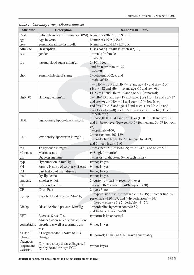
HealthMED - Volume 7 / Number 4 / 2013
Journal of Society for development in new net environment in B&H 1315
Table 1. Coronary Artery Disease data setAttribute Description Range Mean ± Stdv
P rate Pulse rate in beats per minute (BPM) Numerical(30-150) 75.9±10.2age Age in years Numerical(15-94) 58±3creat Serum Kreatinine in mg/dL Numerical(0.2-11.6) 1.2±0.55Attribute Description Class code (1=calss1; 2= class2, …)sex gender 1= male; 0=female
fbs Fasting blood sugar in mg/dl1=70-100; 2=101-126; and 3= more than>= 127
chol Serum cholesterol in mg1=<= 200; 2=between200-239; and3= above240
Hgb(50) Hemoglobin gm/ml
1= ( Hb >= 13.5 and Hb <= 18 and age>17 and sex=1) or ( Hb >= 12 and Hb <= 16 and age>17 and sex=0) or ( Hb >= 11 and Hb <= 16 and age < 17 )= normal; 2=( Hb< 13.5 and age>17 and sex=1) or ( Hb < 12 and age>17 and sex=0) or ( Hb <= 11 and age < 17 )= low level; and 3= ( Hb >18 and age>17 and sex=1) or ( Hb > 16 and age>17 and sex=0) or ( Hb > 16 and age < 17 )= high level
HDL high-density lipoprotein in mg/dL
1= best>=60; 2= poor(HDL <= 40 and sex=1) or (HDL <= 50 and sex=0); and 3= better level (between 40-59 for men and 50-59 for wom-en)
LDL low-density lipoprotein in mg/dL
1= optimal<=100; 2= near optimal100-129; 3= border line high130-159; 4= high160-189; and 5= very high>=190
trig Triglyceride in mg/dl 1=less than 150; 2=150-199; 3= 200-499; and 4= >= 500Marital s Marital status 0=Single 1=marrieddm Diabetes mellitus 1= history of diabetes; 0= no such historyhyp Hypertension in mmHg 0= no; 1= yesFH Family History of coronary disease 0= no; 1= yesPH Past history of heart disease 0= no; 1= yesdisld Dyslipidemia 0= no; 1= yessmoking Smoker or not 2=current 3= past 4= recent 5= neverEF Ejection fraction 1=good(50-75) 2=fair(30-49) 3=poor(<30)CP Chest Pain 2= yes; 3=no
Sys bp Systolic blood pressure Mm/Hg 1=hypotension =<90; 2=desirable =90-119; 3=border line hy-pertension =120-139; and 4=hypertension >=140
Dia bp Diastolic blood pressure Mm/Hg1= hypotension <60=; 2=desirable =61-79; 3=border line hypertension =80-89; and 4= hypertension >=90
EET Exercise Stress Test 0= normal; 1= abnormal
comorbidityAbsence or presence of one or more disorders as well as a primary dis-ease
0= no; 1= yes
ST and T Change
ST segment and T wave of ECG changes 0= normal; 1= having ST-T wave abnormality
Diagnosis(dependent variable)
Coronary artery disease diagnosed by physicians through ECG 0= no; 1=yes
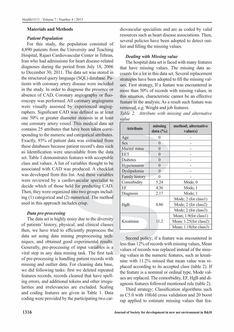
1316
HealthMED - Volume 7 / Number 4 / 2013
Journal of Society for development in new net environment in B&H
Materials and Methods
Patient PopulationFor this study, the population consisted of
4,890 patients from the University and Teaching Hospital, Rajaei Cardiovascular Center in Tehran, Iran who had admissions for heart disease-related diagnoses during the period from July 18, 2006 to December 30, 2011. The data set was stored in the structured query language (SQL) database. Pa-tients with coronary artery disease were included in the study. In order to diagnose the presence or absence of CAD, Coronary angiography or fluo-roscopy was performed. All coronary angiograms were visually assessed by experienced angiog-raphers. Significant CAD was defined as at least one 50% or greater diameter stenosis in at least one coronary artery vessel. This medical data set contains 25 attributes that have been taken corre-sponding to the numeric and categorical attributes. Exactly, 93% of patient data was extracted from these databases because patient record’s data such as Identification were unavailable from the data set. Table 1 demonstrates features with acceptable class and values. A list of variables thought to be associated with CAD was produced. A checklist was developed from this list. And these variables were reviewed by a cardiovascular specialist to decide which of those held for predicting CAD. Then, they were organized into two groups includ-ing (1) categorical and (2) numerical. The method used in this approach includes crisp.
Data pre-processingThe data set is highly noisy due to the diversity
of patients’ history, physical, and clinical classes then, we have tried to efficiently preprocess the data set using data mining preprocessing tech-niques, and obtained good experimental results. Generally, pre-processing of input variables is a vital step in any data mining task. The first task of pre-processing is handling patient records with missing and outlier data. For cleaning data base, we did following tasks: first we deleted repeated features records, records cleaned that have spell-ing errors, and additional tokens and other irregu-larities and irrelevancies are excluded. Scaling and coding features are given in Table 1. Data coding were provided by the participating two car-
diovascular specialists and are as coded by valid resources such as heart disease associations. Then, several policies have been adopted to detect out-lier and filling the missing values.
Dealing with Missing valueThe hospital data set is faced with many features
that have missing values. The missing data ac-counts for a lot in this data set. Several replacement strategies have been adopted to fill the missing val-ues; First strategy, If a feature was encountered in more than 50% of records with missing values, in this situation, characteristic cannot be an effective feature in the analysis; As a result such feature was removed, e.g. Weight and job features.Table 2. Attribute with missing and alternative value
Attribute missing data (%)
method; alternative value(s)
Age 0 -Sex 0 -Marital status 0 -EET 0 -Diabetes 0 -Hypertension 0 -Dyslipidemia 0 -Family history 0 -Comorbidity 5.24 Mode; 0EF 4.36 Mode; 1Diagnosis 2.17 Mode; 1
HgB 8.86Mode; 2 (for class1) Mode; 2 (for class2) Mode; 2 (for class3)
Kreatinine 11.2Mean; 1.9(for class1)Mean; 1.25(for class2) Mean; 1.18(for class3)
Second policy; if a feature was encountered in less than 12% of records with missing values, Mean values of records was replaced instead of the miss-ing values in the numeric features, such as kreati-nine with 11.2% missed that mean value was re-placed according to its accepted class (table 2). If the feature is a nominal or ordinal type, Mode val-ues are replaced. The comorbidity, EF, HgB and di-agnosis features followed mentioned rule (table 2).
Third strategy; Classification algorithms such as C5.0 with 10fold cross validation and 20 boost rap applied to estimate missing values that fea-
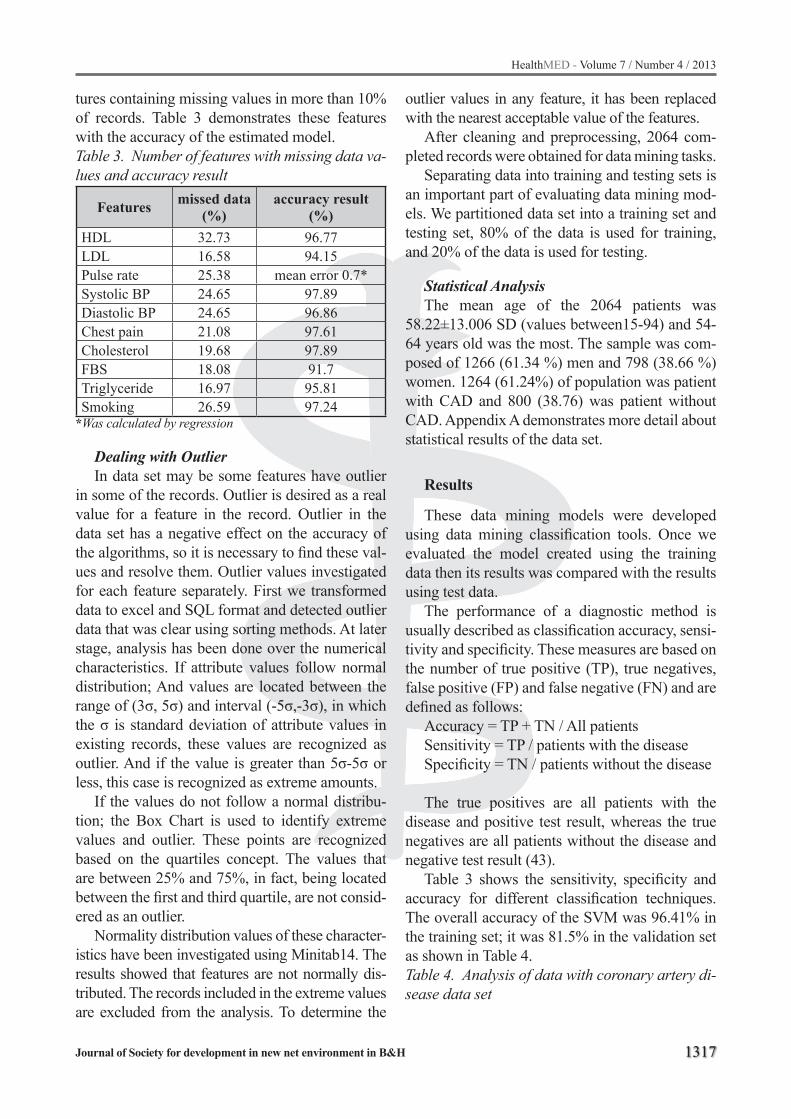
HealthMED - Volume 7 / Number 4 / 2013
Journal of Society for development in new net environment in B&H 1317
tures containing missing values in more than 10% of records. Table 3 demonstrates these features with the accuracy of the estimated model. Table 3. Number of features with missing data va-lues and accuracy result
Features missed data (%)
accuracy result (%)
HDL 32.73 96.77LDL 16.58 94.15Pulse rate 25.38 mean error 0.7*Systolic BP 24.65 97.89Diastolic BP 24.65 96.86Chest pain 21.08 97.61Cholesterol 19.68 97.89FBS 18.08 91.7Triglyceride 16.97 95.81Smoking 26.59 97.24
*Was calculated by regression
Dealing with OutlierIn data set may be some features have outlier
in some of the records. Outlier is desired as a real value for a feature in the record. Outlier in the data set has a negative effect on the accuracy of the algorithms, so it is necessary to find these val-ues and resolve them. Outlier values investigated for each feature separately. First we transformed data to excel and SQL format and detected outlier data that was clear using sorting methods. At later stage, analysis has been done over the numerical characteristics. If attribute values follow normal distribution; And values are located between the range of (3σ, 5σ) and interval (-5σ,-3σ), in which the σ is standard deviation of attribute values in existing records, these values are recognized as outlier. And if the value is greater than 5σ-5σ or less, this case is recognized as extreme amounts.
If the values do not follow a normal distribu-tion; the Box Chart is used to identify extreme values and outlier. These points are recognized based on the quartiles concept. The values that are between 25% and 75%, in fact, being located between the first and third quartile, are not consid-ered as an outlier.
Normality distribution values of these character-istics have been investigated using Minitab14. The results showed that features are not normally dis-tributed. The records included in the extreme values are excluded from the analysis. To determine the
outlier values in any feature, it has been replaced with the nearest acceptable value of the features.
After cleaning and preprocessing, 2064 com-pleted records were obtained for data mining tasks.
Separating data into training and testing sets is an important part of evaluating data mining mod-els. We partitioned data set into a training set and testing set, 80% of the data is used for training, and 20% of the data is used for testing.
Statistical AnalysisThe mean age of the 2064 patients was
58.22±13.006 SD (values between15-94) and 54-64 years old was the most. The sample was com-posed of 1266 (61.34 %) men and 798 (38.66 %) women. 1264 (61.24%) of population was patient with CAD and 800 (38.76) was patient without CAD. Appendix A demonstrates more detail about statistical results of the data set.
Results
These data mining models were developed using data mining classification tools. Once we evaluated the model created using the training data then its results was compared with the results using test data.
The performance of a diagnostic method is usually described as classification accuracy, sensi-tivity and specificity. These measures are based on the number of true positive (TP), true negatives, false positive (FP) and false negative (FN) and are defined as follows:
Accuracy = TP + TN / All patientsSensitivity = TP / patients with the diseaseSpecificity = TN / patients without the disease
The true positives are all patients with the disease and positive test result, whereas the true negatives are all patients without the disease and negative test result (43).
Table 3 shows the sensitivity, specificity and accuracy for different classification techniques. The overall accuracy of the SVM was 96.41% in the training set; it was 81.5% in the validation set as shown in Table 4.Table 4. Analysis of data with coronary artery di-sease data set

1318
HealthMED - Volume 7 / Number 4 / 2013
Journal of Society for development in new net environment in B&H
Algorithm Accuracy %
Specificity %
Sensitivity %
DecisionTree (C5.0) 84.54 65 97
C&RT 70.36 53 82QUEST 67.92 52 78CHAID 70.24 64 71NeuralNetwork 68.05 65 72
SVM 96.41 95 96Designed algorithm 95.92 93 98
Table 5. Variable importance for classification of the CADAlgorithms Important factors
C&RT tree
1-chest pain 2- past history 3-comorbid-ity 4-hypertension 5- age 6-sex 7-EF 8- pulse rate 9- systolic blood pressure 10- LDL 11- triglyceride 12-Hgb
QUEST
1-Chest pain 2- comorbidity 3-age 4-EF 5-kreatinine 6-pulse rate 7- HgB 8- sys-tolic blood pressure 9- FBS 10-dyslipid-emia
CHAID
1-chest pain 2- past history 3-EF 4-co-morbidity 5- age 6- sex 7-ST8- diabetes 9-dyslipidemia 10-hyperten-sion 11- cholesterol
C5.0
1-chest pain 2- age 3- past history 4-co-morbidity 5-FBS 6-EF 7- triglyceride 8-hgb 9- family history 10- LDL 11-ST 12-sex
SVM
1-Chest pain 2- past history 3- FBS 4-EF 5-HB 6- marital status 7- cholesterol 8-HDL 9-diabetes 10- diastolic BP 11- smoking 12- sex
Neural net-work
1-Chest pain 2- comorbidity 3-age 4- smoking 5- FBS 6- EF 7- triglyceride8- HgB 9- dyslipidemia 10- past history 11- ST 12-Sex
The most significant factor influencing CAD that obtained from all algorithms is chest pain and was included in all induced techniques. As shown in table 4, other important risk factors are comor-bidity, pass history, hypertension, age, sex, smok-ing and ejection fraction.
Based on the decision tree model, sample rules were extracted from this predictive model and confirmed by two cerebrovascular doctors. How-
ever, more investigation with more features and larger data sets is still required.
Diagnosis classification rules are interpreted in table 5. Rules are presented in two parts: 1) those which express the absence of CAD disease (with 5 rules) and 2) those that shows there exists CAD disease (with 4 rules). The most significant or in-teresting rules are set in order by number.Table 6. Selected Rules by cardiovascular Speci-alistsRule No. Rules for CAD subjects
1. if Chest Pain = 2.000 and comorbidity = 1.000 then 1.000
2.if age>53 and Chest Pain=2.000 and CEF=1.000 and CST=1.000 and sex=1.00 then 1.000
3. if Pulse Rate <= 88 and CST = 1.000 then 1.000
4.if age > 53 and Family History = 0.000 and Chest Pain = 2.000 and CHB = 2.000 then 1.000
5.if age > 70 and Pass History = 1.000 and Smoking = 2.000 and Pulse Rate <= 100 then 1.000
Rule No. Rules for healthy subjects
1.
if Hypertension = 0.000 and comorbid-ity = 0.000 and Cholesterol = 2.000and CEF = 1.000 and CST = 0.000 then 0.000
2.if age > 42 and age <= 53 and Smok-ing = 5.000 and comorbidity = 0.000 and CLDL = 1.000 then 0.000
3.if sex = 1.000 and age <= 44 and comor-bidity = 0.000 and Cholesterol = 1.000and Diastolic B.P = 2.000 then 0.000
4.
if Hypertension = 0.000 and Pulse Rate <= 88 and comorbidity = 0.000 and Cholesterol = 1.000 and HDL = 3.000 and ST = 0.000 then 0.000
Discussion
There are different data mining techniques that can be used for the prediction of cardiovascular disease. Using data mining in the analysis of medi-cal data is a good opportunity to investigate the re-lationships between variables. All three algorithms have the ability to predict coronary artery disease with various degrees of confidence. The present

HealthMED - Volume 7 / Number 4 / 2013
Journal of Society for development in new net environment in B&H 1319
study demonstrates that the SVM algorithm is the most useful for evaluation and prediction of CAD patients with a non-CAD. Similar study confirms this finding (44). This algorithm can be adopted as a model, which helps the physicians in evaluat-ing CAD. On the other hand, we can apply this model to pre-diagnose the possibilities of a CAD if a person has risk factors. Despite the simplicity of decision tree, results with acceptable accuracy presented in the field of data mining in relation to coronary artery disease and came out to be second best. The neural network did not correctly predict CAD subjects with disease diagnosis or healthy. The same study confirms this subject (45).
There have been many studies of risk-factors in cardiac disease (5, 19, 45-48). Most were derived from the UCI date set and may not necessarily ap-ply to local or regional practice. Many risk factors have been associated with coronary artery disease. Although different risk factors were obtained from the algorithms investigated, chest pain and past history were the major influence factors. In other words CP variable has the most effect in CAD pres-ence or absence. Some of the features identified as important in other multivariate analysis, such as age and sex. The absence of comorbidity, hypertension (45) and smoking too is supported by other studies.
Application of data mining in analyzing the medical data is a good method for considering the existing relationships between the variables. Ac-curate data, suitable preprocessing and suitable data mining technique will offer reasonable results in medical data mining (46).
From the obtained results, it is clear that the elderly males (age>53) have a high probability to be diagnosed with CAD. It is consistent with Tsipouras study(31). Another important risk factor for CAD is the smoking consumption. It has also been demonstrated that smoking has on effect on the CAD prediction in men; 61% of nonsmoker men did not suffer from CAD (105/171) and 64% of smokers had CAD (469/730).
The absence of hypertension, moderate choles-terol, not smoking and appropriate levels of LDL and HDL were very important factors that had a ma-jor role in staying subjects healthy. So controlling and bringing down the blood pressure, appropriately and relatively low levels of LDL, non-smoking and tobacco consumption and regular daily activities can
help to reduce the amount of risk for coronary heart disease. In investigating the results, it is interesting to note that patients having chest pain were assumed to have as CAD subjects in all algorithms. This rep-resents the highly important influence of chest pain to early diagnosis of cardiac patients. However, As it was shown, It is noteworthy that the the absence of chest pain cannot be an indicative of the health of coronary arteries. Obviously, in the medical field the diagnoses are basically dependent on physician’s experience; hence some extracted rules may be not accepted easily. However, by taking the knowledge of medical treatment, more effective knowledge and rules can be obtained (15).
Conclusion
In this paper, we have presented several algo-rithms for coronary artery disease diagnosis and prediction using data mining techniques. Firstly, we have extracted significant rules from the coro-nary artery disease data set for the efficient predic-tion of disease based on the sensitivity and accu-racy indicators.
As we mentioned earlier, the coronary artery diseases database consisting of 2064 cases has been used in this study. For predicting CAD, 25 attri-butes are listed, which included their demographic data, clinical results, and diagnoses. Three classifi-cation efficiency indicators commonly used in the medical field, sensitivity, specificity and accuracy, were employed in this study to evaluate the effi-ciency of classification models. We think extracted rules could aid, as useful knowledge for physicians, to early prediction of disease and consequently de-crease CAD morbidity and perhaps, mortality. If risk factor such as Hypertension, EF, LDL, Choles-terol, smoking and HDL was controlled, CAD risk of a subject may be reduced significantly.
Limitation
Few medical databases are made available to researchers. Medical data are generally not struc-tured and it is spread in various locations. This is a reason confirms the fact that multiple scaled medical databases need much effort during earlier phases of the KDD process. In these circumstanc-es, some DM techniques manifest difficulties in

1320
HealthMED - Volume 7 / Number 4 / 2013
Journal of Society for development in new net environment in B&H
data handling or the results generate poor perfor-mances in one or more applied techniques (49). It should be noted that the results of our study based on a partly small population and its generalization to the larger population need to more studies and large samples with more variables.
Acknowledgments
This study was part of a PhD. Thesis supported by Tehran University of Medical Sciences (TUMS), School of Health Management and Information Science. We express our gratitude for the Dr. Firuzi Rahim for their skillful and invaluable assistances. The authors would like to thank Dr. Gholpira and Dr. Moghaddan and other staff members of the In-formation tech unit of Rajaei cardiovascular center belong to Tehran University of medical science for allowing us to use the data and their assistance in retrieving data used in this study.
Reference
1. Rajkumar1 A, Reena GS. Diagnosis Of Heart Disease Using Datamining Algorithm Global Journal of Com-puter Science and Technology 2010; 10(10): 39-43.
2. Soni J, Ansari U, Sharma D. Predictive Data Mining for Medical Diagnosis: An Overview of Heart Dise-ase Prediction. International Journal of Computer Applications2011; 17(8): 43-8.
3. Jilani TA, Yasin H, Yasin M, Ardil C. Acute Coronary Syndrome Prediction Using Data Mining Techniques- An Application. International Journal of Computatio-nal Intelligence2009; 5(4).
4. Patil BS, Kumaraswamy YS. Intelligent and Effective Heart Attack Prediction System Using Data Mining and Artificial Neural Network. European Journal of Scientific Research2009; 31(4): 642-56.
5. Karaolis MA, Moutiris JA, Hadjipanayi D, Pattichis CS. Assessment of the Risk Factors of Coronary Heart Events Based on Data Mining With Decision Trees. IEEE Transactions on information technology in bio-medicine 2010; 14(3).
6. Cardiovascular disease is the most common cause of death in the country. Tehran: Ministry of Health and Medical Education; 2009; Available from: http: //www.behdasht.gov.ir/index.aspx?siteid=1&pageid=24979&newsview=12090.
7. Soltanian AR, Mahjub H, gudarzi S, Nabipur I, Jamali M. Five year survival rate in patients with acute myocar-dial infraction in Bushehr. Scientifin journal of Hama-dan university of medical science2007; 16(3): 33-7.
8. Limbu YR, AMaskey, Bahadur M, Malla R, Sharma D, Shrestha NK. A study on cardiovascular disease pattern of admitted cases in newly emerged national heart centre. Journal of Nepal Medical Association2002; 41: 284-8.
9. Gamberger D, Lavrac N, Krstacic G. Active subgroup mining: a case study in coronary heart disease risk group detection. Artificial Intelligence in Medicine 2003; 28(1): 27-57.
10. Anbarsi.M, Aanupria.E., N.CH.S.N.iyengra. Enhan-ced Prediction of Heart Disease with Feature Subset Selection using Genetic Algorithm. International Journal of Engineering Science and Technology 2010; 2(10): 5370-6.
11. Shahrabi J, Shakurniaz V. data mining in Sql Server. first ed. Tehran: AmirKabir University brabch; 2009.
12. S. Sumathi, Sivanandam SN. Introduction to Data Mining and its Applications. In: Kacprzyk J, editor. Studies in Computational Intelligence, Volume 29: Springer; 2006.
13. Liu P, Lei L, Yin J, Zhang W. Healthcare data mi-ning: predicting inpatient length of stay. 3rd Interna-tional IEEE Conference Intelligent Systems, Septem-ber 2006; 2006. p. 261-6.
14. Han J, Kamber M. Data mining: concepts and tech-niques: Morgan Kaufmann; 2006.
15. Hara A, Ichimura T, Yoshida K. Discovering multi-ple diagnostic rules from coronary heart disease da-tabase using automatically defined groups. Journal of Intelligent Manufacturing2005; 16(6): 645-61.
16. Markuzon N, Welsch R, Flietstra BC. A data mining approach for acoustic diagnosis of cardiopulmo-nary disease. The Department of the Air Force: Ma-ssachusetts Institute of Technology; 2008.
17. Kaur H, Wasan S. Empirical Study On Applications Of Data Mining Techniques In Healthcare. Journal of Computer Science2006; 2(2): 194-200.
18. Bellazzi R, Zupan B. Predictive data mining in clinical medicine: current issues and guidelines. international journal of medical informatics2008; 77(2): 81-97.
19. Wilson P, D’Agostino R, Levy D, Belanger A, Sil-bershatz H, Kannel W. Prediction of coronary he-art disease using risk factor categories. Circulation 1998; 97(18): 1837.
20. Schwarzer G, Vach W, Schumacher M, Universitet S. On the misuses of artificial neural networks for prognostic and diagnostic classification in oncology. Statistics in medicine2000; 19(4): 541-61.
21. Rani KU. Analysis of Heart Diseases Dataset using Neural Network Approach. Arxiv preprint arXiv: 111026262011.
22. Kaur H, Wasan SK. Empirical study on applications of data mining techniques in healthcare. Journal of Computer Science2006; 2(2): 194-200.
HealthMED - Volume 7 / Number 4 / 2013
Journal of Society for development in new net environment in B&H 1321
23. Chang KC, Tseng MC, Weng HH, Lin YH, Liou CW, Tan TY. Prediction of length of stay of first-ever is-chemic stroke. Stroke2002; 33(11): 2670-4.
24. Kamath C. Scientific data mining: a practical per-spective: Siam; 2009.
25. Wang J. Encyclopedia of data warehousing and mi-ning: Idea Group Reference; 2006.
26. Sumathi S, Sivanandam S. Introduction to data mi-ning and its applications: Springer-Verlag New York Inc; 2006.
27. Berner ES. Clinical decision support systems: the-ory and practice: Springer Verlag; 2007.
28. Witten IH, Frank E, Hall MA. Data Mining: Practi-cal machine learning tools and techniques: Morgan Kaufmann; 2011.
29. Kajabadi A, Saraee MH, Asgari S, editors. Data mi-ning cardiovascular risk factors2009: IEEE.
30. Sitar-Taut DA, Sitar-Taut AV. Overview on How Data Mining Tools May Support Cardiovascular Disease Prediction. Journal of Applied Computer Science 2010; 4.
31. Tsipouras MG, Exarchos TP, Fotiadis DI, and, et, al. Automated Diagnosis of Coronary Artery Disease Based on Data Mining and Fuzzy Modeling. IEEE Transactions on information technology in biomedi-cine2008; 12(4): 447-58.
32. Palaniappan S, Awang R. Intelligent heart disea-se prediction system using data mining techniques. IJCSNS2008; 8(8): 343.
33. Ordonez C. Comparing association rules and deci-sion trees for disease prediction. ACM; 2006. p. 24.
34. Twardy C, Nicholson A, Korb K, McNeil J. Data mining cardiovascular bayesian networks. Monash University, School of Computer Science & Software Engineering, Melbourne2004; 165.
35. Martínez-Sellés M, Ortiz J, Estévez Á, Andueza J, de Miguel J, Bueno H. A new risk score for patients with a normal or non-diagnostic ECG admitted to a chest pain unit. Revista Espanola de Cardiolo-gia2005; 58(7): 782-8.
36. Harrison RF, Kennedy RL. Artificial Neural Network Models for Prediction of Acute Coronary Syndromes Using Clinical Data From the Time of Presentation. Annals of Emergency Medicine2005; 46(5): 431-9.
37. Baxt W. Use of an artificial neural network for data analysis in clinical decision-making: the diagnosis of acute coronary occlusion. Neural Computation 1990; 2(4): 480-9.
38. Das R, Turkoglu I, Sengur A. Diagnosis of valvular heart disease through neural networks ensembles. Computer Methods and Programs in Biomedici-ne2009; 93(2): 185-91.
39. Das R, Turkoglu I, Sengur A. Effective diagnosis of he-art disease through neural networks ensembles. Expert Systems with Applications2009; 36(4): 7675-80.
40. Lapuerta P, Azen SP, LaBree L. Use of neural networks in predicting the risk of coronary artery disease. Com-puters and biomedical research1995; 28(1): 38-52.
41. Shen Z, Clarke M, Jones R, Alberti T. A new neural network structure for detection of coronary heart disea-se. Neural Computing & Applications 1995; 3(3): 171-7.
42. Turkoglu I, Arslan A, Ilkay E. An intelligent system for diagnosis of the heart valve diseases with wave-let packet neural networks. Computers in biology and medicine2003; 33(4): 319-31.
43. Anamika G, Naveen K, Vasudha B. Analysis of Medi-cal Data using Data Mining and Formal Concept Analysis. World Academy of Science, Engineering and Technology2005; 11: 61-4.
44. Kumari M, Godara S. Comparative Study of Data Mining Classification Methods in Cardiovascular Disease Prediction. International Journal of Com-puter Science and Technology2011; 2(2): 304-8.
45. Catherine F. Risk factors for coronary artery disease and the use of neural networks to predict the pres-ence or absence of high blood pressure. BMC Genet-ics 2003; 4: 1-6.
46. Kajabadi A, Saraee MH, Asgari S. Data mining car-diovascular risk factors. Application of Information and Communication Technologies; Baku: IEEE; 2009. p. 1-5.
47. Beaglehole R, Magnus P. The search for new risk factors for coronary heart disease: occupational therapy for epidemiologists? International journal of epidemiology2002; 31(6): 1117-22.
48. Ellen P. application of data mining techniques in the prediction of coronary artery disease: use of anes-thesia time series and patient risk factor data Aus-tralia: Queesland university of technology; 2009.
49. Andrei D, Viviana A. Overview on How Data Mining Tools May Support Cardiovascular Disease predic-tion. Journal of Applied Computer Science & Math-ematics: 2010; 8 (4).
50. Lundin M, Lundin J, Burke H, Toikkanen S, Pylk-konen L, Joensuu H. Artificial neural networks ap-plied to survival prediction in breast cancer. Oncol-ogy2000; 57(4): 281-6.
Corresponding AuthorAhmadi Maryam,Tehran University of Medical Sciences,School of Health Management and Information Sciences, Tehran,Iran,E-mail: [email protected] [email protected]

1322
HealthMED - Volume 7 / Number 4 / 2013
Journal of Society for development in new net environment in B&H
Appendix A. Statistical result of data set features
variable value Proportion %
Feature count with diagnosis class (0,1) variable value Proportion
%Feature count with diagnosis class (0,1)
Sex 01
38.6661.34
354= 0, 44= 1446= 0, 820= 1 HDL
123
2.4781.6415.89
18=0, 33=1649=0, 1036= 1133= 0, 195= 1
Marital status 01
8.6291.38
72= 0, 106= 1728= 0, 1158= 1 FBS
123
42.3431.0626.6
346= 0, 528= 1254= 0, 387= 1200= 0, 349= 1
Pass history 01
72.3827.62
518= 0, 976= 1288= 0, 570= 1 Systolic BP
1234
.3426.0758.9114.68
4=0, 3= 1242=0, 296= 1426=0, 790= 1128=0, 175=1
Diabetes 01
71.5128.49
597= 0, 879= 1203= 0, 385= 1 Diastolic BP
1234
.8237.9453.257.99
10=0, 7= 1323=0, 460= 1396=0, 703= 171=0, 94= 1
Hypertension 01
55.2344.77
441= 0, 699= 1565= 0, 924= 1 EF
123
47.9641.7110.31
413=0, 577= 1278= 0, 574= 1100=0, 113= 1
Family history 01
89.0510.95
723= 0, 1115= 177= 0, 149= 1 ST change 0
164.4335.56
563= 0, 767= 1237= 0, 497= 1
Dyslipidemia 01
58.8241.18
511= 0, 703= 1289= 0, 561= 1 HgB
123
25.2974.42.29
232= 0, 290= 1563=0, 973= 1
-
Smoking
2345
33.1419.675.1442.05
26=0, 422= 1139= 0, 267= 131= 0, 75= 1
368= 0, 500= 1
Triglyceride
1234
63.7117.1518.27.87
526= 0, 789= 1132= 0, 222= 1137=0, 240= 1
5=0, 13= 1
Chest pain 23
74.9525.05
495= 0, 4052= 1305= 0, 212= 1 LDL
12345
64.7818.99.744.81.79
534= 0, 803= 1150= 0, 240=173=0, 128= 130= 0, 69= 113= 0, 24= 1
Comorbidity 01
58.1941.81
540= 0, 661= 1
260= 0, 603= 1Cholesterol
123
79.4613.527.03
647= 0, 993= 1106= 0, 173= 147= 0, 98= 1
EET 01
96.613.39
772= 0, 1222= 128= 0, 42= 1

HealthMED - Volume 7 / Number 4 / 2013
Journal of Society for development in new net environment in B&H 1323
Abstract
The purpose of this study was to determine the changes of Malondialdehyde (MDA), Glutathione (GSH), Adenosine Deaminase (ADA) and Supe-roxide Dismutase (SOD) levels with pre and post acute exercise in obese middle-aged women (body mass index, BMI ≥30.0). Twenty four middle-aged women participated in this study. Subjects were di-vided as obese and non-obese group. The descripti-ve statistics of the participants were computed and the significance between the pre and post exercise levels of MDA, GSH, ADA and SOD values was determined. The results showed that obesity and acute exercise causation oxidative stress in obese women. Especially in obese women the increased levels of MDA and ADA levels may pose serious health problems. Obesity breaks down the anti oxi-dant defense mechanism of the body by effecting the GSH and SOD activities. That is why sudden and exhaustive exercises in obese people may have adverse effects on health.
Key words: Adenosine deaminaze, acute exer-cise, obese women, anti-oxidant.
Introduction
Obesity is gradually becoming a major heath concern all over the world. Obesity is an indepen-dent atherosclerosis risk factor [1]. The anomalies in body weight, body fat percentage and body mass index (BMI) cause difficulty in physical activity during exercise and increase the amount of oxygen consumed [2,3]. During a strenuous exercise the rate of metabolism increases almost 100 times of the resting condition depending upon intensity of the muscle activity [4,5]. The increased amount of oxygen increases the amount of superoxide anion formed in mitochondria. This anion forms reactive oxygen species known as free radicals. Free radi-
cals may causation oxidative stress depending upon intensity and the duration of exercise [6-8] It is tho-ught that lipid peroxidation occurs if amount of free radicals surpasses the amount of anti oxidants res-ponsible for immunity of cell [9]. Malondialdehyde (MDA), an important lipid peroxidation product which occurs as a result of oxidative injury is used as indicator of oxidative stress [5]. Oxidative stress is known to cause many diseases as well as DNA and cell damage [10-13].
Regular moderate exercise has been proposed due to health benefits, including an increase of maximum oxygen consumption and decreased risk of obesity, cardiovascular disease and metabolic syndrome. However, acute exercise increases oxi-dative stress because, during exercise, increased strain of transporting oxygen to active muscles may damage cellular components. Especially, oxidative stress by induced exercise in obese higher than non-obese following exercise [14]. Numerous reports have documented increases in by-products of lipid peroxidation, especially malondialdehyde (MDA) following exercise. Saiki et al. reported that obese individuals have greater oxidative stress following acute treadmill exercise than non-obese counterpar-ts. Santos-Silva reported that resting MDA levels were higher in trained adolescent swimmers com-pared to control group. However, not all studies have shown increases in MDA levels especially in response to exercise training. Miyazaki et al. [15] reduction of indices of oxidative stress following strenuous endurance training. These beneficial effects in response to exercise training imply a link between oxidative stress and increased antioxidant defense. Increased levels of blood antioxidant acti-vity at rest were found in trained subjects. In addi-tion, this antioxidant activity was further increased following exercise training.
Physical exercise has been associated with a re-
The compare of effect of exhaustive exercise on MDA, ADA, GSH and SOD activity in womenM. Emin Kafkas1, Armagan Sahin Kafkas2, Seyfi Savas3, Aysun Bay Karabulut4
1 Inonu University, School of Physical Education and sports, Malatya, Turkey,2 Erciyes University, Health Sciences Institute, Kayseri, Turkey,3 Gazi University, School of Physical Education and sports, Ankara, Turkey,4 Inonu University, Department of Biochemistry, Malatya, Turkey.
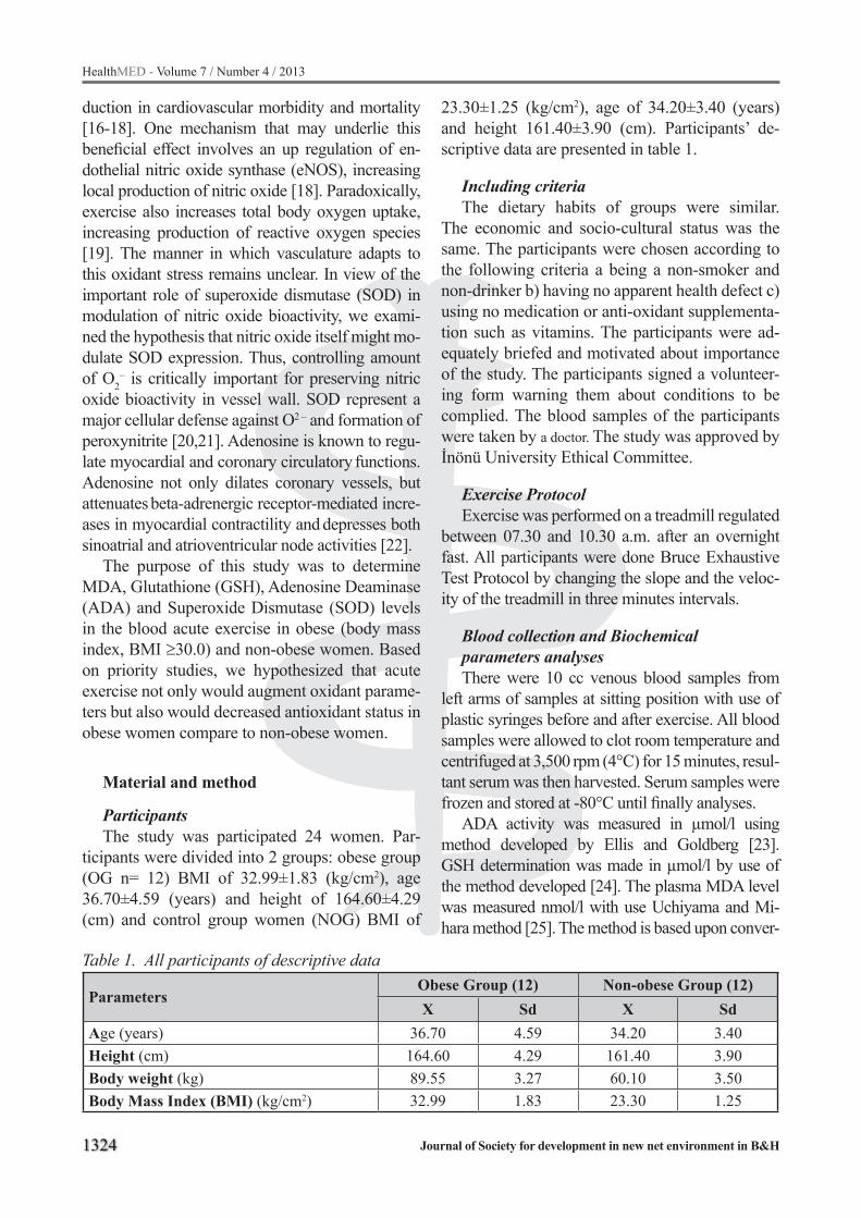
1324
HealthMED - Volume 7 / Number 4 / 2013
Journal of Society for development in new net environment in B&H
duction in cardiovascular morbidity and mortality [16-18]. One mechanism that may underlie this beneficial effect involves an up regulation of en-dothelial nitric oxide synthase (eNOS), increasing local production of nitric oxide [18]. Paradoxically, exercise also increases total body oxygen uptake, increasing production of reactive oxygen species [19]. The manner in which vasculature adapts to this oxidant stress remains unclear. In view of the important role of superoxide dismutase (SOD) in modulation of nitric oxide bioactivity, we exami-ned the hypothesis that nitric oxide itself might mo-dulate SOD expression. Thus, controlling amount of O2
– is critically important for preserving nitric oxide bioactivity in vessel wall. SOD represent a major cellular defense against O2 – and formation of peroxynitrite [20,21]. Adenosine is known to regu-late myocardial and coronary circulatory functions. Adenosine not only dilates coronary vessels, but attenuates beta-adrenergic receptor-mediated incre-ases in myocardial contractility and depresses both sinoatrial and atrioventricular node activities [22].
The purpose of this study was to determine MDA, Glutathione (GSH), Adenosine Deaminase (ADA) and Superoxide Dismutase (SOD) levels in the blood acute exercise in obese (body mass index, BMI ≥30.0) and non-obese women. Based on priority studies, we hypothesized that acute exercise not only would augment oxidant parame-ters but also would decreased antioxidant status in obese women compare to non-obese women.
Material and method
ParticipantsThe study was participated 24 women. Par-
ticipants were divided into 2 groups: obese group (OG n= 12) BMI of 32.99±1.83 (kg/cm2), age 36.70±4.59 (years) and height of 164.60±4.29 (cm) and control group women (NOG) BMI of
23.30±1.25 (kg/cm2), age of 34.20±3.40 (years) and height 161.40±3.90 (cm). Participants’ de-scriptive data are presented in table 1.
Including criteria The dietary habits of groups were similar.
The economic and socio-cultural status was the same. The participants were chosen according to the following criteria a being a non-smoker and non-drinker b) having no apparent health defect c) using no medication or anti-oxidant supplementa-tion such as vitamins. The participants were ad-equately briefed and motivated about importance of the study. The participants signed a volunteer-ing form warning them about conditions to be complied. The blood samples of the participants were taken by a doctor. The study was approved by İnönü University Ethical Committee.
Exercise Protocol Exercise was performed on a treadmill regulated
between 07.30 and 10.30 a.m. after an overnight fast. All participants were done Bruce Exhaustive Test Protocol by changing the slope and the veloc-ity of the treadmill in three minutes intervals.
Blood collection and Biochemical parameters analyses There were 10 cc venous blood samples from
left arms of samples at sitting position with use of plastic syringes before and after exercise. All blood samples were allowed to clot room temperature and centrifuged at 3,500 rpm (4°C) for 15 minutes, resul-tant serum was then harvested. Serum samples were frozen and stored at -80°C until finally analyses.
ADA activity was measured in µmol/l using method developed by Ellis and Goldberg [23]. GSH determination was made in µmol/l by use of the method developed [24]. The plasma MDA level was measured nmol/l with use Uchiyama and Mi-hara method [25]. The method is based upon conver-
Table 1. All participants of descriptive data
ParametersObese Group (12) Non-obese Group (12)X Sd X Sd
Age (years) 36.70 4.59 34.20 3.40Height (cm) 164.60 4.29 161.40 3.90Body weight (kg) 89.55 3.27 60.10 3.50Body Mass Index (BMI) (kg/cm2) 32.99 1.83 23.30 1.25

HealthMED - Volume 7 / Number 4 / 2013
Journal of Society for development in new net environment in B&H 1325
sion of superoxide radicals, formed by total (Cu–Zn and Mn) SOD (EC 1.15.1.1) via xantin oxidase, into H2O2 which reduces the nitrolue tetrazolium salt into a blue colored formazan which gives a maximum ab-sorbance at 560 nm [26]. The data were given in u/l.
Statistical analyzeBefore statistical analyses were done, the nor-
mality of distribution of data was evaluated by kolmogorof-smirnov test. Whole values were in-dicated as mean ± Standart Deviation (Sd). Bio-chemical parameters were compared difference between pre and post exercise using paired sample t-test. All data were measured by use SPSS 17.0 version for Windows and p≤0.05 was accepted as level of statistically significant.
Results
In this study the descriptive statistics of some of physical and physiological parameters of par-ticipants were computed and significance between pre and post exercise levels of MDA, GSH, ADA and SOD values was determined
For the differences between MDA, GSH, ADA
and SOD values of OG before and after acute ex-ercise are shown with paired t-test in Table 2. It was found that difference between MDA, GSH and ADA values pre-test and post-test signifi-cantly different (p<.05). Furthermore, there was no significant in serum SOD enzyme activity pre- and post-test in obese women (p>.05).
The values of MDA, ADA, GSH and SOD enzyme activity pre- and post-test on does not affect acute exercise in non-obese group (p>.05).
Discussions
In this study were investigated the effects of acute exercise on oxidant and antioxidant status in obese and non-obese women. The result of this study in-dicated that MDA concentrations significantly in-creased immediately after acute exercise in obese women. Whereas, non-obese women MDA con-centrations not significant. It is known that MDA causes oxidative stress and increases lipid peroxi-dation depending on the obesity that increased lipid peroxidation has been reported in obese individu-als following acute submaximal [27-29] and maxi-mal exercise [30,31]. When the oxygen consump-
Table 2. Analyses of pre and post exercise MDA, GSH, ADA and SOD levels of obese groupParameters N X Sd X1-X2 % p
MDA Pre-test (X1) 12 19.63 2.93 15.20Post-test (X2) 12 22.61 3.51 .029*
GSH Pre-test (X1) 12 31.38 1.33 4.63Post-test (X2) 12 29.93 1.48 .042*
ADA Pre-test (X1) 12 15.98 4.63 16.35Post-test (X2) 12 18.58 5.17 .013*
SOD Pre-test (X1) 12 1.61 .31 0.25Post-test (X2) 12 1.59 .44 .894(p<0.05)
Table 3. Analyses of pre and post test MDA, GSH, ADA and SOD levels of non-obese groupParameters N X Sd X1-X2 % p
MDAPre-test (X1) 12 18.98 3.27
8.6Post-test (X2) 12 20.61 3.48 .165
GSHPre-test (X1) 12 27.38 1.98
0.1Post-test (X2) 12 26.93 2.11 .843
ADAPre-test (X1) 12 16.38 4.79
0.9Post-test (X2) 12 17.88 4.85 .582
SODPre-test (X1) 12 1.54 .45
0.2Post-test (X2) 12 1.50 .43 .774
(p<0.05)

1326
HealthMED - Volume 7 / Number 4 / 2013
Journal of Society for development in new net environment in B&H
tion is low superoxide radicals and its derivatives are eliminated by anti oxidant immunity reaction. However, in case of excessive oxygen consumption defensive mechanism may not cope with increased free radical concentration which may result in cell damage [12,13]. Some of studies claim that acute exercise has no effect on serum MDA concentra-tion value [32,33] and the acute exercise has no ef-fect on MDA concentrations and oxidant conditions of obese women [34]. The data obtained in this study contradict with literature results. Özçelik et al. emphasize that acute exercises have a significant effect on MDA values [35]. It was also stated that obesity increases oxidative stress which shows par-allelism with this study [36].
GSH, is an anti oxidant molecule in mammalians which provides protection against oxidative stress. The GSH value was reported to increase as a result of acute exercise in literature, because this seems to be one of most reliable shows exercise-induced oxi-dant production [7,37,38]. Revan et al. [39] noted a decrease in serum blood GSH after acute exercise. Di-Renzo et al. [2] reported a significant decrease in blood decreased GSH level immediately after acute exercise in obese women [2]. The findings of the present study indicate that after acute exercise significant decrease in obese women.
The studies in literature state that SOD value does not change with acute exercises [40,41]. The-se results show a good accordance with our data. However, it was indicated that acute exercise cau-ses a significant increase in SOD values which con-tradicts with results obtained in this study [7,42].
Our study revealed that acute exercises incre-ased ADA values in obese women ADA increa-ses activity of rate determining enzyme purine nucleotide cyclus and decreases activity of ade-nosine. This change contributes the increase in post exercise activity adenosine activity of obese women. Adenosine was reported to have a cardio protective effect [43]. According to the literature survey there are no studies where ADA activity and oxidative stress were investigated together. In this context the ADA activity is very important to show the effect of exercise in future studies.
Conclusion
In conclusion acute exercise may causation
oxidative stress more in obese women than non-obese women. Especially in obese women incre-ased levels of MDA and ADA levels may pose serious health problems. The obesity breaks down anti oxidant defense mechanism of body by effec-ting GSH activities. That is why sudden and exha-ustive exercises in obese people may have adverse effects on health. Therefore, obese women should be done rather than acute exercise as much as po-ssible regular exercise. However there are further studies with larger groups needed to clarify this.
References
1. Hen K, Bogdanski P, Szulinska M, Jablecka A, Pupek-Musialik D (2010) Influence of regular physical activ-ity on oxidative stress in women with simple obesity. Pol Merkur Lekarski 28(166): 284-88
2. Di-Renzo L, Galvano F, Orlandi C, Bianchi A, Di-Giacomo C, La-Fauci L, Acquaviva R, De-Lorenzo A (2010) Oxidative stress in normal-weight obese syn-drome. Obesity 18(11): 2125-30
3. Karaouzene N, Merzouk H, Aribi M, Merzouk SA, Ber-rouiguet AY, Tessier C, Narce M (2011) Effects of the as-sociation of aging and obesity on lipids, lipoproteins and oxidative stress biomarkers: A comparison of older with young men. Nutr Metab Cardiovasc Dis 21: 792-799
4. Urso ML, Clarkson PM (2003) Oxidative stress, ex-ercise and antioxidant supplementation. Toxicology 189: 41-54
5. Karacan S, Çetin F, Çolakoğlu FF (2010) The effect of calisthenic exercise bout on oxidant and anti-oxidant status in middle aged an postmenopausal women. Iso-kinetics and Exercise Science 18: 39-44
6. Naziroğlu M, Kilinç F, Uğuz AC, Celik O, Bal R, But-terworth PJ, Baydar ML (2010) Oral vitamin C and E combination modulates blood lipid peroxidation and antioxidant vitamin levels in maximal exercising bas-ketball players. Cell Biochem Funct 28(4): 300-05.
7. Aguilo A, Tauler P, Fuantespina E, Tur JA. Cordova A, Pons A (2005) Antioxidant response to oxidative stress induced by exhaustive exercise. Physiol Behav 84: 1-7
8. Sirmali M, Uz E, Sirmali R, Kilbaş A, Yılmaz HR, Altuntaş I, Naziroğlu M, Delibaş N, Vural H (2007) Protective effects of erdosteine and vitamins C and E combination on ischemia-reperfusion induced lung oxidative stress and plasma copper and zinc levels in a rat hind limb model. Biol Trace Elem Res 118(1): 43-52

HealthMED - Volume 7 / Number 4 / 2013
Journal of Society for development in new net environment in B&H 1327
9. Silva LA, Pinho CA, Silveira PC, Tuon T, De Souza CT, Dal-Pizzol F, Pinho RA (2009) Vitamin E supple-mentation decrease muscular and oxidative damage but not inflammatory response induced by eccentric contraction. J Physiol Sci 60: 51-57
10. Fisher-Wellman K, Bell HK, Bloomer RJ (2009) Oxi-dative stress and antioxidant defense mechanisms lin-ked to exercise during cardiopulmonary and metabo-lic disorders. Oxid Med Cell Long 2(1): 43-51
11. Antoncic-Svetina M, Sentija D, Cipak A, Milicic D, Meinitzer A, Tatzber F, Andrisic L, Zelzer S, Zarko-vic N (2010) Ergometry induces systemic oxidative stress in healty human subjects. Tohoku J Exp Med 221(1): 43-8
12. Elahi MM, Kong YX, Matata BM (2009) Oxidative stress as a mediator of cardiovascular disease. Oxid Med Cell Longev 2(5): 259-69
13. Essick EE, Sam F (2010) Oxidative stress and au-tophagy in cardiac disease neurological disorders, aging and cancer. Oxid Med Cell Longev 3(3): 167-77
14. Vincent HK, Innes KE, Vincent KR (2007) Oxidative stress and potential interventions to reduce oxidati-ve stress in overweight and obesity. Diabetes Obes Metab 9(6): 813-39
15. Miyazaki H, Oh-ishi S, Ookawara T, Kizaki T, Toshi-nai K, Ha S, Haga S, Ji LL, Ohnı H (2001) Strenuous endurance training in human reduces oxidative stre-ss following exhausting exercise. Eur J Appl Physiol 84: 1-6
16. Spector SL. Surette ME (2003) Diet and asthma: Has the role of dietary lipids been overlooked in the managemen of asthma? Ann Allergy Asthma Immu-nol 90(4): 371-377
17. Ellwood P, Asher MI, Björkstén B, Burr M, Pearce N. and Robertson CF (2001) Diet and asthma, aller-gic rhino conjunctivitis and atopic eczema symptom prevalence: An ecological analysis of the Interna-tional Study of Asthma and Allergies in Childhood (ISAAC) data. Eur Respir J 17: 436-443
18. Cheng J, Peng G, Zhang Q. Deng H (2002) Preli-minary clinical study on the correlation between allergic rhinitis and food factors. Journal of Clinica Otorhinolaryngology 16: 393-396
19. Allen S, Britton JR. Leonardi-Bee JA (2009) Asso-ciation between antioxidant vitamins and asthma outcome measures: Systematic review and meta-analysis. Thorax 64(7): 610-912
20. De Batlle J, Garcia-Aymerich J, Barraza-Villarreal
A. and Antó JM, RomieuI I (2008) Mediterranean diet is associated with reduced asthma and rhinitis in Mexican children. Allergy 63(10): 1310-1316
21. Woods RK, Thien FC. Abramson MJ (2003) Dietary marine fatty acids (fish oil) for asthma in adults and children. Cochrane Database Syst Rev 3: CD001283
22. Kaur B, Rowe BH. Arnold E (2009) Vitamin C supplementation for asthma. Cochrane Database Systematic Reviews 21(1): CD000993
23. Ellis G, Goldberg DM (1970) A reduced nicotinamide adenine dinucleotide-linked kinetic assay for adenosi-ne deaminase activity. J Lab Clin Med 76(3): 507-17
24. Fairbanks V, Klee GG (1986) Biochemical aspects of hematology. In: N. W. Tietz Editor Ttextbook of clinical chemistry. W. B. Saunders, Philadelphiz
25. Uchiyama M, Mihara M (1978) Determination of malonaldehyde precursor in tissue by thiobarbituric acid test. Anal Biochem (34): 271-8
26. Sun Y, Larry W, Oberley W, Ving U (1988) A simple method for clinical assay of superoxide dismutase. Clin Chem 34: 497-500
27. Vincent HK, Morgan JW, Vincent KR (2004) Obesity exacerbates oxidative stress levels after acute exer-cise. Med Sci Sports Exerc 36: 772-9
28. Vincent HK, Bourguignon CM, Vincent KR, Weltman AL, Bryant M, Taylor AG (2006) Antioxidant supple-mentation lowers exercise-induced oxidative stress in young overweight adults. Obesity 14(12): 2224-35
29. Lwow F, Dunajska K, Tworowska U, Jedrzejuk D, Laczmanski L, Milewicz A, Szmigiero L (2007) Post-exercise oxidative stress and obesity in postmeno-pausal women: the role of beta3-adrenergic receptor polymorphism. Gynecol Endocrinol 23(10): 597-603
30. Vincent HK, Vincent KR, Bourguignon C, Braith RW (2005) Obesity and postexercise oxidative stress in older women. Med Sci Sports Exerc 37(2): 213-9
31. Vincent HK, Bourguignon C, Vincent KR (2006) Re-sistance training lowers exercise-induced oxidative stress and homocysteine levels in overweight and obese older adults. Obesity 14(11): 1921-30
32. Bloomer JR, Cole JB (2009) Relationship between blood lactate and oxidative stress biomarkers fol-lowing acute exercise. Open Sports Medicine Jour-nal 3: 44-48
33. Munoz-Marin D, Olcina G, Tımon R, Robles RC, Caballero MJ, Maynar M (2010) Effect of different
1328
HealthMED - Volume 7 / Number 4 / 2013
Journal of Society for development in new net environment in B&H
exercise intensities on oxidative stress markers and antioxidant response in trained cyclists. J Sports Med Phys Fitness 50(1): 93-8
34. Melton CE, Tucker PS, Fisher-Wellman KH, Schil-ing BK, Bloomer RJ (2009) Acute exercise does not attenuate postprandial oxidative stress in prediabet-ic women. Phys Sportsmed 37(1): 27-36
35. Özçelik O, Karataş F (2008) Effects of Incremental Exercise Test on Serum Malondialdehyid and Vitamin A E C Levels in Obese Subjects. Fırat Univ J Health Sci 22(6): 337-341
36. Codoner-Franch P, Tavarez-Alonso S, Murria-Estal R, Tortajada-Girbes M, Simo-Jorda R, Alonso-Igle-sias E (2012) Elevated advanced oxidation protein (AOPPs) indicate metabolic risk in severely obese children. Nutr Metab Cardiovasc Dis 22(3): 237-43
37. Sen CK (2001) Update on thiol status and supplements in physical exercise. Can J Appl Physiol 26: 4-12
38. Deminice R, Sicchieri T, Payao PO, Jardao AA (2010) Blood and salivary oxidative stress biomark-ers following an acute session of resistance exercise in humans. Int J Sports Med 31(9): 599-603
39. Revan S, Balci SS, Pepe H, Kurtoğlu F, Erol AE, Akkuş H (2010) Short duration exhaustive running exercise does not modify lipid hydroperoxide glutat-hione peroxidase and catalase. J Sports Med Phys Fitness 50(2): 235-40
40. Skarpanska-Stejnborn A, Basta P, Pilaczynska-Szczesnizak L, Horoszkiewicz-Hassan M (2010) Black grape extract supplementation attenuates blo-od oxidative stress in response to acute exercise. Biol Sport 27(1): 41-46.
41. McClean MC, McNeilly A, Trinick T, Duly Ellie, Murphy HM, McLaughlin AJ, Burge G, Davison WG (2009) Acute exercise and impaired glucose tolerance: Effects on glycaemic control, oxidative stress and arterial stiffness. Med Sci Sports Exerc 41(5): 36-41
42. Pinho RA, Silva LA, Pinho CA, Scheffer DL, Souza CT, Benetti M, Carvalho T, Dal-Pizzal F (2010) Oxi-dative stress and inflammatory parameters after an ironman race. Clin J Sport Med 20(4): 306-11.
43. Kinugawa T, Fujita M, Ogino K, Kato M, Osaki S, Igawa O, Shigemasa C, Hisatome I, Kitakaze M (2006) Catabolism of adenine nucleotidesfavors adenosine production following exercise in patients with chronic heart failure. J Card Fail 12(9): 720-5
Corresponding AuthorM. Emin Kafkas,Inonu University,School of Physical Education and Sports,Malatya,Turkey,E-mail: [email protected]

HealthMED - Volume 7 / Number 4 / 2013
Journal of Society for development in new net environment in B&H 1329
Abstract
Purpose: The present study aimed to investiga-te the relationship between Pro12Ala polymorphi-sm and T2DM occurrence in Iranian population.
Methods: Totally, 80 cases and 80 healthy matched controls were included in the study. PCR-RFLP technique was applied to investigate the polymorphism. Data were analyzed by χ2 test using SPSS software.
Results: The G allele (Ala 12) may has a modest and protective association with the development of T2DM in Iranian population (OR=2.117; 95% CI: 1.016-4.455; P= 0.022) while, Pro allele may consider as a risk factor to make individual prone to T2DM (OR=0.472; 95% CI: 0.224-0.984; P= 0.022). There was a 1.9-fold increase in the risk of T2DM associated with the CC allele (adjusted OR: 1.94, 95% CL 0.92–4.01). In contrast, there was no association between the CG polymorphi-sm and the risk of T2DM (adjusted OR: 0.51, 95% CL 0.24– 1.03)
Conclusions: The PPARG gene polymorphism has a modest association with the development of T2DM in Iranian patients.
Key words: PPARG, Pro12Ala polymorphism, PCR-RFLP technique, Type 2 diabetes.
Introduction
Diabetes is the most prevalent non-infectious disorder of endocrine glands with an increasing rate of incidence [1]. According to the internatio-nal health organization’s report, frequency of dia-betic patients is expected to rise up to 333 million people in 2025 [2].
Middle Asia has been regarded as the epidemic center of Type 2 diabetes mellitus (T2DM) [3, 4]. T2DM is the most common metabolic disorder
with complicate interplays between genetic and environmental factors. The disorder is the end-stage of chronic and progressive deficiency ow-ing to insulin resistance, functional decrease of pancreatic β cells and the increased production of glucose by the liver [5,6]. Diabetic high blood sugar can negatively affect small arteries (such as neuropathy, nephropathy and retinopathy) and large arteries (such as cardiovascular disease) and may bring about outcomes including kidney insuf-ficiency, blindness and non-thrombosed amputate of body limbs [7]. Over 1.5 million diabetic pa-tients live in Iran [8].
Due to the high mortality and morbidity rate of diabetes and the resultant shorter life expectancy in diabetes patients [9], knowledge about the in-cidence and affecting factors are remarkable. Al-though some genetic and environmental factors involved in the risk of developing diabetes have been identified with certainty [10], there is still conflicting data on the roles of other factors.
The peroxisome-proliferator-activated receptor γ gene (PPARG) is a member of the nuclear hor-mone receptor superfamily [11]. This transcription regulator PPARγ2 isoform (PPARγ2) is expressed selectively and at higher level in adipose tissue where it modulates the expression of target genes implicated in adipocyte differentiation and gluco-se homeostasis [12]. Therefore, the gene is regar-ded as a major candidate for such complex phe-notypes as T2DM or obesity. However, evidence in this regard is contradictory [10,13,14].
A polymorphism involving C to G substituti-on at nucleotide 34 in the PPARγ2 isoform which causes proline to alanine in position 12 of the PPARγ2 protein (Pro12Ala) [15] was selected sin-ce it has been associated with a reduced risk of type 2 diabetes mellitus development [16].
Association of PPARG gene polymorphism (Pro12Ala) with type 2 diabetes in the Iranian population Javad Mohammadi-Asl1, 2, Aniseh Soheil-Nezhad1 , Leila Saremi3, Fakher Rahim4
1 Department of Biology, Arsanjan Branch, Islamic Azad University, Arsanjan, Iran,2 Department of Medical Genetics, School of Medicine, Ahvaz Jundishapur University of Medical Sciences, Ahvaz, Iran,3 Department of Biology, Science and Research Branch, Islamic Azad University, Tehran, Iran,4 Toxicology Research Center, Ahvaz Jundishapur University of Medical Sciences, Ahvaz, Iran.

1330
HealthMED - Volume 7 / Number 4 / 2013
Journal of Society for development in new net environment in B&H
We launched the present study for first time on an Iranian population to investigate the relation-ship between Pro12Ala polymorphism and T2DM occurrence.
Method and materials
SubjectsTotally, 80 T2DM patients selected based on
their blood sugar data (over 200 mg/ml) (mean age±SD, 53.83±7.54, 25 male and 55 female) re-ferred to Golestan hospital, Ahvaz, Iran were re-cruited in this study. A total of 80 age, sex and eth-nic origin matched unrelated healthy volunteers were sampled.
Having being explained the purpose and meth-od of the study, all the subjects signed the written consent form. This study was approved by the eth-ics committee of the School of Medicine, Ahvaz Jundishapur University of Medical Sciences.
GenotypingBlood samples were taken from both groups in
EDTA tubes. DNA isolation was performed by Ge-nomic DNA extraction kit (Cat. No. K-3032, Bio-neer co., Ltd. Korea). Genotyping was performed by the PCR-RFLP method: DNA was amplified by polymerase chain reaction (PCR) using a pair of specific primers: forward primer TGTCTT-GACTCATGGGTGTATTC Reverse primer: ATCAGTGAAGGAACCGCTTT designed by using Primer3Plus website (http://www.bioinfor-matics.nl/cgi-bin/primer3plus/primer3plus.cgi), generated a 185 bp fragment. PCR reactions were carried out in a thermocycler instrument (Applied Biosystems 2720) under the following conditions: 95 oC for 5 min, 35 cycles including denaturing at 95 oC for 30 sec, annealing for 30 sec at 55.2 oC and extension at 72 oC for 20 sec. A final extension was followed at 72 oC for 5 min. PCR products were digested by BslI restriction enzyme which identifies and cuts the restriction site found in the product from C allele.
RFLP products were loaded on 8% polyacryla-mide gel electrophoresis (PAGE) and the sizes of DNA bands were distinguished using 100 bp lad-der (GeneOn GmbH, Germany). The 185bp PCR product creates162 and 23bp DNA fragments after restriction by BslI enzyme (60 minute at 37 oC).
Individuals with G/G genotype should only show the 185bp band, while C/C and C/G genotypes should show 23/162bp and 185 and 23/162bp bands, respectively. Some of the genotyping was confirmed by direct DNA sequencing.
Statistical analysisThe resulted date was analyzed by using SPSS
statistical software (SPSS Inc., Chicago, IL, USA). Deviations from Hardy-Weinberg equilibri-um were considered using χ2 statistics. Allele and genotype frequencies were compared using χ2 and Fisher exact tests. Univariate analyses were first performed to calculate the crude odds ratios (OR) and their 95% confidence limits (CL) for the ca-ses vs. control and various genotypes. Odds ratios and 95% CL adjusted for age, were then estimated using logistic regression analysis as an estimate of the relative risk and strength of association. For the logistic regression analysis the dichotomous response variable for the PPARG genotype was regressed against case status and age (continuous).
P value of less than 0.05 was considered stati-stically significant.
Results
Table 1 summarizes the demographics of the study population with regard to age and gender. The male/female ratio in the case and control gro-ups was not equal (case 25/ 55, control 35/45). The mean ages of patients and controls (±SD) were 53.8 ± 7.54 and 51.2 ± 10.06 years, respec-tively. However, no statistically significant diffe-rence was observed in mean ages. There were a statistically significant higher number of controls under the age of 45 years compared to cases. The genotype distributions of patients and control su-bjects were within the values expected from Har-dy-Weinberg equilibrium for each SNP. We ob-served three bands 185bp,162bp and 23bp in two groups of patients and controls (Figure1).
We found a statistically significant difference between patients and controls in the frequencies of Ala 12 allele which revealed protective effect aga-inst T2DM (OR=0.472; 95% CI: 0.224-0.984; P= 0.022) whereas, Pro allele plays as a risk factor to make individual prone to T2DM (OR=2.117; 95% CI: 1.016-4.455; P= 0.022). Also, we found signi-
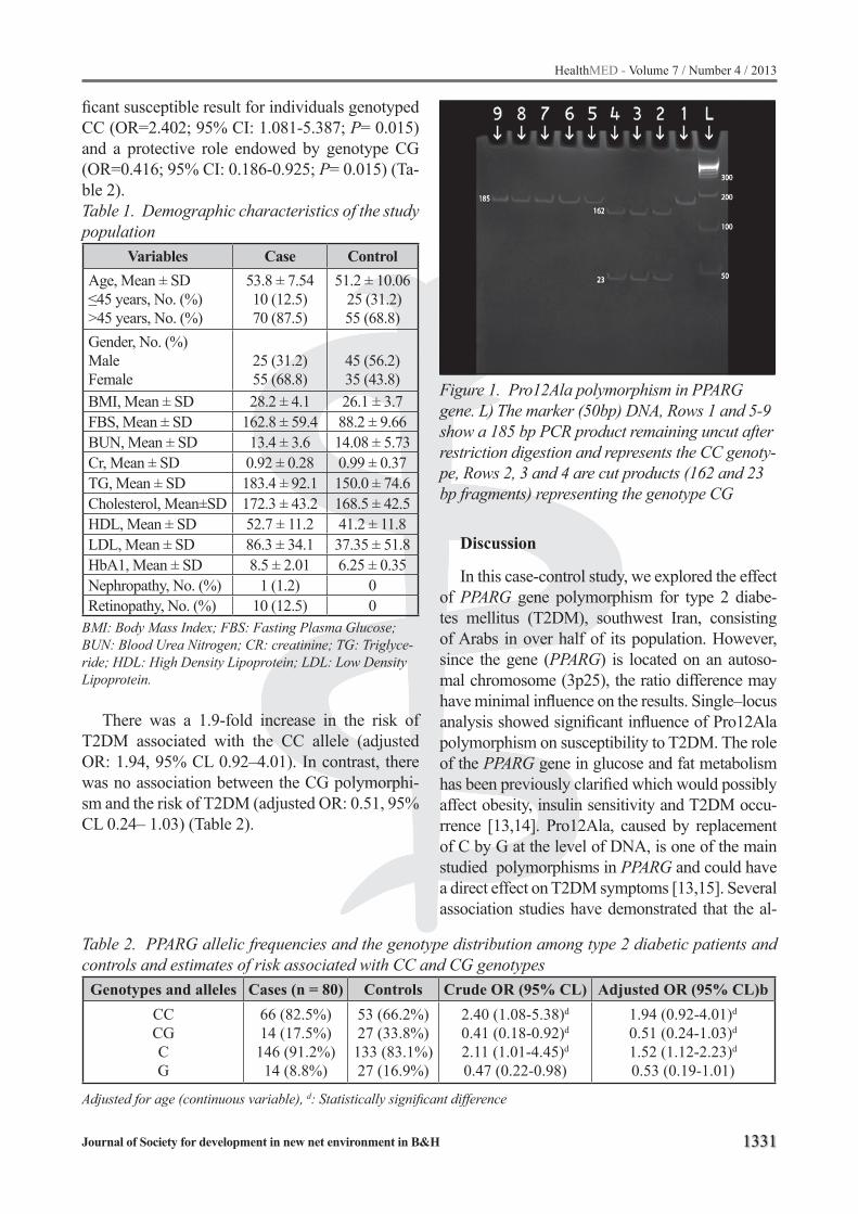
HealthMED - Volume 7 / Number 4 / 2013
Journal of Society for development in new net environment in B&H 1331
ficant susceptible result for individuals genotyped CC (OR=2.402; 95% CI: 1.081-5.387; P= 0.015) and a protective role endowed by genotype CG (OR=0.416; 95% CI: 0.186-0.925; P= 0.015) (Ta-ble 2).Table 1. Demographic characteristics of the study population
Variables Case ControlAge, Mean ± SD≤45 years, No. (%)>45 years, No. (%)
53.8 ± 7.5410 (12.5)70 (87.5)
51.2 ± 10.06 25 (31.2)55 (68.8)
Gender, No. (%)MaleFemale
25 (31.2)55 (68.8)
45 (56.2)35 (43.8)
BMI, Mean ± SD 28.2 ± 4.1 26.1 ± 3.7FBS, Mean ± SD 162.8 ± 59.4 88.2 ± 9.66BUN, Mean ± SD 13.4 ± 3.6 14.08 ± 5.73Cr, Mean ± SD 0.92 ± 0.28 0.99 ± 0.37TG, Mean ± SD 183.4 ± 92.1 150.0 ± 74.6Cholesterol, Mean±SD 172.3 ± 43.2 168.5 ± 42.5HDL, Mean ± SD 52.7 ± 11.2 41.2 ± 11.8LDL, Mean ± SD 86.3 ± 34.1 37.35 ± 51.8HbA1, Mean ± SD 8.5 ± 2.01 6.25 ± 0.35Nephropathy, No. (%) 1 (1.2) 0Retinopathy, No. (%) 10 (12.5) 0
BMI: Body Mass Index; FBS: Fasting Plasma Glucose; BUN: Blood Urea Nitrogen; CR: creatinine; TG: Triglyce-ride; HDL: High Density Lipoprotein; LDL: Low Density Lipoprotein.
There was a 1.9-fold increase in the risk of T2DM associated with the CC allele (adjusted OR: 1.94, 95% CL 0.92–4.01). In contrast, there was no association between the CG polymorphi-sm and the risk of T2DM (adjusted OR: 0.51, 95% CL 0.24– 1.03) (Table 2).
Figure 1. Pro12Ala polymorphism in PPARG gene. L) The marker (50bp) DNA, Rows 1 and 5-9 show a 185 bp PCR product remaining uncut after restriction digestion and represents the CC genoty-pe, Rows 2, 3 and 4 are cut products (162 and 23 bp fragments) representing the genotype CG
Discussion
In this case-control study, we explored the effect of PPARG gene polymorphism for type 2 diabe-tes mellitus (T2DM), southwest Iran, consisting of Arabs in over half of its population. However, since the gene (PPARG) is located on an autoso-mal chromosome (3p25), the ratio difference may have minimal influence on the results. Single–locus analysis showed significant influence of Pro12Ala polymorphism on susceptibility to T2DM. The role of the PPARG gene in glucose and fat metabolism has been previously clarified which would possibly affect obesity, insulin sensitivity and T2DM occu-rrence [13,14]. Pro12Ala, caused by replacement of C by G at the level of DNA, is one of the main studied polymorphisms in PPARG and could have a direct effect on T2DM symptoms [13,15]. Several association studies have demonstrated that the al-
Table 2. PPARG allelic frequencies and the genotype distribution among type 2 diabetic patients and controls and estimates of risk associated with CC and CG genotypes
Genotypes and alleles Cases (n = 80) Controls Crude OR (95% CL) Adjusted OR (95% CL)bCCCGCG
66 (82.5%)14 (17.5%)146 (91.2%)14 (8.8%)
53 (66.2%)27 (33.8%)133 (83.1%)27 (16.9%)
2.40 (1.08-5.38)d
0.41 (0.18-0.92)d
2.11 (1.01-4.45)d
0.47 (0.22-0.98)
1.94 (0.92-4.01)d
0.51 (0.24-1.03)d
1.52 (1.12-2.23)d
0.53 (0.19-1.01)
Adjusted for age (continuous variable), d: Statistically significant difference

1332
HealthMED - Volume 7 / Number 4 / 2013
Journal of Society for development in new net environment in B&H
lele is negatively associated with the development of T2DM, suggesting a protective role for it. In con-trast, other investigators have failed to confirm any independent role for the PPARG association with T2DM and have rather suggested its interaction with other genes [10].
Similar results which suggest a susceptibility role for Pro have been observed in Japanese [17], Caucasian American[18], Finnish[19] and Danish populations [20]. Conversely, a deleterious effect of Ala 12 allele has been shown in Canadian Oji-Cree individuals [21], Germans [22,23], French [24], Polish [25], Korean [26] or Pima Indians [27].
Many investigators have put forth the idea that this polymorphism, which is within the domain enhancing ligand independent activation, reduces the risk of T2DM through producing a PPARγ2 protein in adiposities which is less efficient in transcription stimulation of PPARγ2 target [28]. This would likely lead to increased suppression of lipolysis by insulin and the resultant decrease in free fatty acid secretion that could in turn cause a greater insulin sensitivity [28,29]. In summary, this study suggested that PPARG gene is likely to be related to the development of T2DM in an Iranian population. However, more studies with more SNPs in the gene, enough sample size and on different ethnic groups are required for clarifi-cation of the issue. Functional studies on PPARG action will be needed to obtain a deeper insight into the mechanism (s) through which its common variant exert its role in T2DM susceptibility
Acknowledgments
This research was funded by Ahvaz Jundis-hapur University of Medical Sciences.
References
1. Qiao, Q., Hu, G., Tuomilehto, J., Nakagami, T., Bal-kau, B., Borch-Johnsen, K., Ramachandran, A., Mo-han, V., Iyer, S.R., Tominaga, M., Kiyohara, Y., Kato, I., Okubo, K., Nagai, M., Shibazaki, S., Yang, Z., Tong, Z., Fan, Q., Wang, B., Chew, S.K., Tan, B.Y., Heng, D., Emmanuel, S., Tajima, N., Iwamoto, Y., Snehalatha, C., Vijay, V., Kapur, A., Dong, Y., Nan, H., Gao, W., Shi, H., Fu, F.: Age- and sex-specific prevalence of diabetes and impaired glucose regulation in 11 Asian cohorts. Diabetes Care 26(6), 1770-1780 (2003).
2. Simpson, R.W., Shaw, J.E., Zimmet, P.Z.: The pre-vention of type 2 diabetes--lifestyle change or phar-macotherapy? A challenge for the 21st century. Diabetes Res Clin Pract 59(3), 165-180 (2003). doi:S0168822702002759 [pii]
3. King, H., Aubert, R.E., Herman, W.H.: Global burden of diabetes, 1995-2025: prevalence, numerical esti-mates, and projections. Diabetes Care 21(9), 1414-1431 (1998).
4. Amos, A.F., McCarty, D.J., Zimmet, P.: The rising glo-bal burden of diabetes and its complications: estima-tes and projections to the year 2010. Diabet Med 14 Suppl 5, S1-85 (1997).
5. Bloomgarden, Z.T.: Insulin resistance: causes and consequences. Int Rev Neurobiol 65, 1-24 (2005). doi:S0074-7742(04)65001-X [pii] 10.1016/S0074-7742(04)65001-X
6. Hegele, R.A., Pollex, R.L.: Genetic and physiological insights into the metabolic syndrome. Am J Physiol Regul Integr Comp Physiol 289(3), R663-669 (2005). doi:00275.2005 [pii] 10.1152/ajpregu.00275.2005
7. Camacho, P., Pitale, S., Abraira, C.: Beneficial and detrimental effects of intensive glycaemic control, with emphasis on type 2 diabetes mellitus. Drugs Aging 17(6), 463-476 (2000).
8. Azizi, F.: Diabetes in the Islamic Republic of Iran. IDF bulletin 41, 38-39 (1996).
9. Morgan, C.L., Currie, C.J., Peters, J.R.: Relationship between diabetes and mortality: a population study using record linkage. Diabetes Care 23(8), 1103-1107 (2000).
10. Wang, F., Han, X.Y., Ren, Q., Zhang, X.Y., Han, L.C., Luo, Y.Y., Zhou, X.H., Ji, L.N.: Effect of genetic va-riants in KCNJ11, ABCC8, PPARG and HNF4A loci on the susceptibility of type 2 diabetes in Chinese Han population. Chin Med J (Engl) 122(20), 2477-2482 (2009).
11. Spiegelman, B.M.: PPAR-gamma: adipogenic regu-lator and thiazolidinedione receptor. Diabetes 47(4), 507-514 (1998).
12. Latruffe, N., Vamecq, J.: Peroxisome proliferators and peroxisome proliferator activated receptors (PPARs) as regulators of lipid metabolism. Biochimie 79(2-3), 81-94 (1997). doi:S0300-9084(97)81496-4 [pii]
13. Celi, F.S., Shuldiner, A.R.: The role of peroxisome proliferator-activated receptor gamma in diabetes and obesity. Curr Diab Rep 2(2), 179-185 (2002).
14. Yang, J., Zhang, D., Li, J., Zhang, X., Fan, F., Guan, Y.: Role of PPARgamma in renoprotection in Type 2 diabetes: molecular mechanisms and therapeu-tic potential. Clin Sci (Lond) 116(1), 17-26 (2009). doi:CS20070462 [pii] 10.1042/CS20070462

HealthMED - Volume 7 / Number 4 / 2013
Journal of Society for development in new net environment in B&H 1333
15. Yen, C.J., Beamer, B.A., Negri, C., Silver, K., Brown, K.A., Yarnall, D.P., Burns, D.K., Roth, J., Shuldiner, A.R.: Molecular scanning of the human peroxiso-me proliferator activated receptor gamma (hPPAR gamma) gene in diabetic Caucasians: identification of a Pro12Ala PPAR gamma 2 missense mutation. Bi-ochemical and biophysical research communications 241(2), 270-274 (1997).
16. Altshuler, D., Hirschhorn, J.N., Klannemark, M., Lindgren, C.M., Vohl, M.C., Nemesh, J., Lane, C.R., Schaffner, S.F., Bolk, S., Brewer, C., Tuomi, T., Ga-udet, D., Hudson, T.J., Daly, M., Groop, L., Lander, E.S.: The common PPARgamma Pro12Ala polymor-phism is associated with decreased risk of type 2 dia-betes. Nat Genet 26(1), 76-80 (2000).
17. Mori, H., Ikegami, H., Kawaguchi, Y., Seino, S., Yokoi, N., Takeda, J., Inoue, I., Seino, Y., Yasuda, K., Hana-fusa, T., Yamagata, K., Awata, T., Kadowaki, T., Hara, K., Yamada, N., Gotoda, T., Iwasaki, N., Iwamoto, Y., Sanke, T., Nanjo, K., Oka, Y., Matsutani, A., Maeda, E., Kasuga, M.: The Pro12 -->Ala substitution in PPAR-gamma is associated with resistance to development of diabetes in the general population: possible involve-ment in impairment of insulin secretion in individuals with type 2 diabetes. Diabetes 50(4), 891-894 (2001).
18. Li, W.D., Lee, J.H., Price, R.A.: The peroxisome pro-liferator-activated receptor gamma 2 Pro12Ala mu-tation is associated with early onset extreme obesity and reduced fasting glucose. Mol Genet Metab 70(2), 159-161 (2000). doi:10.1006/mgme.2000.2999 S1096-7192(00)92999-8 [pii]
19. Douglas, J.A., Erdos, M.R., Watanabe, R.M., Bra-un, A., Johnston, C.L., Oeth, P., Mohlke, K.L., Valle, T.T., Ehnholm, C., Buchanan, T.A., Bergman, R.N., Collins, F.S., Boehnke, M., Tuomilehto, J.: The pe-roxisome proliferator-activated receptor-gamma2 Pro12A1a variant: association with type 2 diabetes and trait differences. Diabetes 50(4), 886-890 (2001).
20. Poulsen, P., Andersen, G., Fenger, M., Hansen, T., Echwald, S.M., Volund, A., Beck-Nielsen, H., Peder-sen, O., Vaag, A.: Impact of two common polymorp-hisms in the PPARgamma gene on glucose toleran-ce and plasma insulin profiles in monozygotic and dizygotic twins: thrifty genotype, thrifty phenotype, or both? Diabetes 52(1), 194-198 (2003).
21. Hegele, R.A., Cao, H., Harris, S.B., Zinman, B., Hanley, A.J., Anderson, C.M.: Peroxisome prolife-rator-activated receptor-gamma2 P12A and type 2 diabetes in Canadian Oji-Cree. J Clin Endocrinol Metab 85(5), 2014-2019 (2000).
22. Ringel, J., Engeli, S., Distler, A., Sharma, A.M.: Pro-12Ala missense mutation of the peroxisome prolife-rator activated receptor gamma and diabetes melli-tus. Biochem Biophys Res Commun 254(2), 450-453 (1999). doi:S0006-291X(98)99962-4 [pii] 10.1006/bbrc. 1998.9962
23. Zietz, B., Barth, N., Spiegel, D., Schmitz, G., Scholmerich, J., Schaffler, A.: Pro12Ala polymor-phism in the peroxisome proliferator-activated re-ceptor-gamma2 (PPARgamma2) is associated with higher levels of total cholesterol and LDL-choleste-rol in male caucasian type 2 diabetes patients. Exp Clin Endocrinol Diabetes 110(2), 60-66 (2002). doi:10.1055/s-2002-23487
24. Clement, K., Hercberg, S., Passinge, B., Galan, P., Varroud-Vial, M., Shuldiner, A.R., Beamer, B.A., Charpentier, G., Guy-Grand, B., Froguel, P., Vais-se, C.: The Pro115Gln and Pro12Ala PPAR gamma gene mutations in obesity and type 2 diabetes. Int J Obes Relat Metab Disord 24(3), 391-393 (2000).
25. Malecki, M.T., Frey, J., Klupa, T., Skupien, J., Walus, M., Mlynarski, W., Sieradzki, J.: The Pro12Ala polymorphism of PPARgamma2 gene and suscepti-bility to type 2 diabetes mellitus in a Polish popula-tion. Diabetes Res Clin Pract 62(2), 105-111 (2003). doi:S0168822703001645 [pii]
26. Oh, E.Y., Min, K.M., Chung, J.H., Min, Y.K., Lee, M.S., Kim, K.W., Lee, M.K.: Significance of Pro12Ala mutation in peroxisome proliferator-activated recep-tor-gamma2 in Korean diabetic and obese subjects. J Clin Endocrinol Metab 85(5), 1801-1804 (2000).
27. Muller, Y.L., Bogardus, C., Beamer, B.A., Shuldiner, A.R., Baier, L.J.: A functional variant in the peroxi-some proliferator-activated receptor gamma2 pro-moter is associated with predictors of obesity and type 2 diabetes in Pima Indians. Diabetes 52(7), 1864-1871 (2003).
28. Deeb, S.S., Fajas, L., Nemoto, M., Pihlajamaki, J., Mykkanen, L., Kuusisto, J., Laakso, M., Fujimoto, W., Auwerx, J.: A Pro12Ala substitution in PPAR-gamma2 associated with decreased receptor acti-vity, lower body mass index and improved insulin sensitivity. Nat Genet 20(3), 284-287 (1998).
29. Li, D., Kang, Q., Wang, D.M.: Constitutive coacti-vator of peroxisome proliferator-activated receptor (PPARgamma), a novel coactivator of PPARgamma that promotes adipogenesis. Molecular endocrino-logy (Baltimore, Md 21(10), 2320-2333 (2007).
Corresponding AuthorAniseh Soheil-Nezhad,Department of Biology, Islamic Azad University,Arsanjan,Iran,E-mail: [email protected]

1334
HealthMED - Volume 7 / Number 4 / 2013
Journal of Society for development in new net environment in B&H
Abstract
Purpose: In this study it is aimed to determine seropositivity of Toxoplasma gondii (T. gondii) in pregnant women who were recruited to Adiyaman Obstetric and Children Hospital and identify the risk factors related to it.
Materials and Methods: Between October 2010 and October 2011, 1026 pregnant women were examined prospectively for toxoplasmosis. After a questionnaire applied to the pregnant women, anti-Toxoplasma IgG and IgM antibodies were investi-gated with ELISA. Univariate and multiple logistic regression analyses were used to determine the risk factors related to Toxoplasma gondii.
Results: Seroprevalences of IgG and IgM aga-inst T. gondii were 35.5% and 3.2% respectively. The mean age of the women was 28±5 years. The mean of gestation week was 10±7 (from 2 to 37). We found that the risk of T. gondii was 2.02 times higher between ages 33-40 than that of 17-24, after adjusting for education level, drinking water, ea-ting raw meat and current cat ownership. Among the environmental variables, the effect of drinking water (adjusted OR=2.40, 95% CI=1.25-4.60, p<0.001), eating raw meat (adjusted OR=2.27, 95% CI=1.67-3.09, p<0.001) and current cat ownership (adjusted OR=5.92, 95% CI=3.50-10.04, p<0.001) on positivity of Toxoplasma IgG antibody was fo-und to be statistically significant. Regarding to the results of multiple logistic regression analyses, no significant relationship were observed between anti-Toxoplasma IgG antibodies and residential address, monthly household income, gestational age, milk consumption and touching raw meat.
Conclusion: Childbearing age women in Adiyaman province living under poor socioeco-nomic conditions and education level are at risk for acquiring infection with T. gondii. We suggest pregnant women who live in Adiyaman where general network water is mostly used (94.4%) to drink boiled water, not to consume raw meat and keep away from cats.
Key words: Pregnancy, Anti-Toxoplasma anti-bodies, risk factors, Adiyaman.
Introduction
Toxoplasmosis is a worldwide zoonotic disease caused by Toxoplasma gondii (T. gondii), which infects humans and most warm-blooded animals. Felids are the definitive host of this protozoan pa-rasite being the only species able to excrete sporu-lated oocysts into the environment (1). Oocysts, re-markably stable environmentally, are transmitted to other hosts through inadvertent ingestion. Humans acquire T. gondii through ingestion of undercooked meat, contact with feline faeces and rarely through drinking contaminated water or through transplan-tation of a contaminated organ (2,3). However, wit-hin immunocompetent humans, 90% of T. gondii infections are asymptomatic. Vertical transmission of toxoplasmosis from an acutely infected pregnant woman could cause serious damage and malforma-tion. Toxoplasmosis during pregnancy can cause congenital infection and manifest as mental retarda-tion and blindness in the infant. Other findings are diminished visual acuity and neurological sequelae such as hydrocephalus, calcification in the brain, paresis, epilepsy and schizophrenia (4,5).
Anti-Toxoplasma antibody prevalence, primary infection rate, and risk factors in a study of toxoplasmosis in 1.026 pregnant women southeastern TurkeyTuncay Celik1, Beyza Doganay Erdogan2, Semiha Aydin3, Servet Kolgelier4
1 Adiyaman University, School of Health, Adiyaman, Turkey,2 Ankara University School of Medicine, Department of Biostatistics, Ankara, Turkey,3 Istanbul University Florenece Nightingale School of Nursing, Istanbul, Turkey,4 Adiyaman University, Faculty of Medicine, Department of Infectious Disease, Adiyaman, Turkey.

HealthMED - Volume 7 / Number 4 / 2013
Journal of Society for development in new net environment in B&H 1335
Early diagnosis of the mother, the fetus and the newborn is necessary to prevent human toxopla-smosis. Monitoring the immune response by measu-ring the titer and affinity of specific antibodies ma-kes a major contribution to diagnosis of congenital toxoplasmosis and evaluation of time of infection (6). The serological screening of pregnant women for toxoplasmosis and the follow-up until delivery are not routine procedures in Turkey. In a few studies performed in our country, seroprevalence of T. gon-dii infection in women at childbearing age was found to be between 19.2% and 85%; and it is estimated that incidence of congenital toxoplasmosis is 0.1% (7). The results of some studies indicated that the factors associated with increased risk were ingestion of raw or undercooked meat, use of kitchen knives that had not been sufficiently washed, and ingestion of unwashed raw vegetables or fruits. A recent case-control study from Europe examined the risk factors that predisposed pregnant woman to infection with T. gondii. In this study, approximately 30% to 63% of the infections were due to exposure to inadequately cooked or cured meat, which was interpreted to be the main risk factor for pregnant women in Europe. The association might be due to oocyst contaminati-on by dirty production techniques or to confounding by other lifestyle (for example, eating undercooked organically produced meat) (1,8).
Detailed knowledge on the prevalence and risk factors of infection with T. gondii are required to design appropriate prevention measures against infection during pregnancy and congenital tran-smission. In the present study, we therefore inve-stigated seroprevalence of infection with T. gondii and associated risk factors in a large number of pregnant women attending a public tertiary care obstetric hospital in Adiyaman.
Materials and Methods
Study design and populationAdiyaman has a semi-arid climate. Summers are
very hot and very dry. Temperatures reach 45°C at the height of summer for the majority of the time. Winters are cool/cold and snowy. The average annual precipitation is 400-800mm. The population of the central district of the province was 170.075 including counties and villages. However due to re-cent migration, mostly caused by the displaced vil-
lagers due to the Atatürk dam (part of southeastern Anatolian project, called GAP), the population is estimated to be over 590,000. The economical level of the population is very low and the cat population is high, they stroll freely around the community.
A total of 1026 pregnant women were included in the study, between (17-40 years). Inclusion cri-teria for the study were attendance for delivery at the Obstetrics and Children’s Hospital, residency in Adiyaman or in the rural region surrounding the city, between October 2010 and October 2011. Ex-clusion criteria from data were absence of a ques-tionnaire, lack of a serum samples, or an insufficient quantity of serum. To determine the seroprevalence of infection with T. gondii venous blood was ob-tained from study participants at the Hospital.
QuestionnairesA short questionnaire interview for pregnant
women was carried out to obtain information as-sociated with T. gondii infection. There were ques-tions eliciting socio-demographic data including age, education level, residential address (urban, rural), monthly wage, gestational age and environ-mental factors; drinking water, eating raw meat, milk consumption (yes or no) touching raw meat and current cat ownership. Additional information about ocular and neurological damage in children was collected.
Serologic analysis for Toxoplasma The sera were sent to the serology and Microbi-
ology laboratory for investigation of the anti-Toxo-plasma gondii IgG, and IgM antibodies by the EIA (Cobas Core, Roche, Germany and ETI-TOXOK-A, DiaSorin, Germany) technique. In the evaluation of the results, as recommended by the manufactur-er, for anti-Toxoplasma antibodies <1 IU/mL was considered non-reactive, ≥1-<30 IU/mL indetermi-nate, ≥30 IU/mL and reactive. Samples with con-centrations ≥30 IU/mL are considered positive for IgG antibodies to T. gondii and indicate either acute and or latent infection. For all samples with con-centrations ≥30 IU/mL a Toxoplasma IgM test was performed to exclude early Toxoplasma infection. Women with positive IgG titers but negative IgM titers were considered latently infected. Women with positive IgG and IgM titers were considered to have a possible recent infection.

1336
HealthMED - Volume 7 / Number 4 / 2013
Journal of Society for development in new net environment in B&H
Statistical AnalysisStatistical analyses were performed by using
SPSS 11.5 for Windows package program. Con-tinuous data were summarized as mean±standard deviation, whereas frequency (percentage) was used for categorical data. Univariate logistic re-gression analysis was used to evaluate the associa-tion between IgG and socio-demographic factors and environmental factors as univariately. Also, multiple logistic regression was performed to de-termine the risk factors on IgG positivity. Forward elimination (likelihood ratio) method was used for the selection of variables. The entry and removal
criteria used were p values of 0.05 for entry and p values of 0.10 for variable removal in multiple lo-gistic regression analysis. Odds ratio (OR) and its 95% confidence interval (CI) were given. p<0.05 was considered as statistically significant.
EthicsThe study was performed in accordance with
the Helsinki Declaration was approved by the lo-cal ethics committee of Adiyaman University. All participants were informed about the study, and informed written consent was obtained from all study participants
Table 1. Univariate logistic regression of socioeconomic factors associated with infection with Toxopla-sma gondii in 997 pregnant women
Socio-demographic factors Toxoplasma IgG positivity OR(95%CI) p
Age17-24 (n=305) 100 (32.8%)
<0.00125-32 (n=509) 161 (31.6%) 0.95(0.70-1.29)33-40 (n=183) 92 (50.3%) 2.07(1.42-3.02)
Education level
Illiteracy (n=93) 37 (39.8%)
0.007Primary school (n=517) 189 (36.6%) 0.87(0.55-1.37)Secondary school (n=133) 29 (21.8%) 0.42(0.24-0.76)High school (n=180) 65 (36.1%) 0.86(0.51-1.43)University (n=74) 33 (44.6%) 1.22(0.66-2.26)
Residential address Urban (n=736) 248 (33.7%) 0.058Rural (n=261) 105 (40.2%) 1.32(0.99-1.77)
Monthly household income, MW
250TL (n=345) 105 (30.4%)0.057375-750TL (n=413) 156 (37.8%) 1.39(1.02-1.88)
≥750TL (n=239) 92 (38.5%) 1.43(1.01-2.02)
Gestational age1st trimester (n=723) 241 (33.3%)
0.0722nd trimester (n=234) 94 (40.2%) 1.34(0.99-1.82)3rd trimester (n=40) 18 (45.0%) 1.64(0.86-3.11)
OR: Odds Ratio, CI: Confidence Interval, * MW = minimum wage of 250TL = U.S. $180
Table 2. Univariate Logistic Regression of eating and drinking habits associated with infection with Toxoplasma gondii in 997 pregnant women
Environmental factors Toxoplasma IgG positivity OR(95%CI) p
Drinking water General network (n=942) 327 (34.7%) 0.061Bottled (n=55) 26 (47.3%) 1.69(0.98-2.91)
Eating raw meat No (n=354) 95 (26.8%) <0.001Yes (n=643) 258 (40.1%) 1.83(1.38-2.43)
Milk consumption No (n=336) 122 (36.3%) 0.671Yes (n=661) 231 (34.9%) 0.94(0.72-1.24)
Touching raw meat No (n=539) 178 (33.0%) 0.088Yes (n=458) 175 (38.2%) 1.25(0.97-1.63)
Cat ownership No (n=918) 299 (32.6%) <0.001Yes (n=79) 54 (68.4%) 4.47(2.73-7.33)OR: Odds Ratio, CI: Confidence Interval

HealthMED - Volume 7 / Number 4 / 2013
Journal of Society for development in new net environment in B&H 1337
Results
A total of 1,300 pregnant women were enrol-led, and data of 1026 women (from age 17, to 40) were analyzed. The datasets of 29 women were excluded because of absence of a questionnaire, an incomplete questionnaire, lack of serum samp-les, or insufficient quantity of serum. The mean age of the women was 28±5 years. The mean of gestation week was 10 ±7 (from 2 to 37). The se-ropositivity of Toxoplasma specific IgG was 388 (35.5%) among pregnant women (chronic phase). 36 (3.2%) pregnant women were IgM positive (acute phase). 602 (61.3%) had no detectable IgM or IgG against. While there were no statisti-cally significant relationship between residential address, monthly household income, gestational age and Toxoplasma seropositivity (p>0.05), age and education level were found to be significant (p<0.001 and p=0.007, respectively) in univari-ate analyses (Table 1). Among the environmen-tal variables, only the relation between drinking water (general network or bottled) (p=0.061) and current cat ownership (p<0.001) with positivity of Toxoplasma IgG antibody was found to be statisti-cally significant (Table 2).
After univariate analyses, the multiple logistic regression was performed in order to determine the risk factors on IgG positivity. Among the so-cio-demographic and environmental factors age, education level, drinking water, eating raw meat and current cat ownership were found to be statis-tically significant in multivariate analyses (Table 3). Being a cat owner increases the risk of IgG positivity almost 6 times after adjusted for age,
education level, drinking water (bottled or gen-eral network) and eating raw meat. Also, in the questionnaire, pregnant women were asked if their children had neurological disturbances and visual defects. Of the 1026 pregnant women, 756 had children and of their children four was found to have Down syndrome, four epilepsy, six visual defects and five mental retardations. It was estab-lished that the mother of the child with visual de-fect was Toxoplasma IgG antibody.
Discussion
Infection with T. gondii has a high prevalence in the Southeastern Anatolia Region of Turkey. Whereas acute infection in most persons is asymp-tomatic or mild, infection during pregnancy may cause transmission to the fetus with severe signs and symptoms (9). The infection is commonly ac-quired by the oral route and thus represents a pre-ventable infection. However, to develop appropri-ate prevention programs, detailed knowledge on prevalence and risk factors is needed.
Toxoplasmosis is a common disease in our country as all over the world. As the incidence of this infection varies largely with geographic loca-tion and nutritional habits, it is important to de-termine regional prevalence. Seroprevalence of T.gondii infection range between 15%-77% in dif-ferent countries (10).
In the present study, we screened more than 1026 pregnant women attending a tertiary care obstetric hospital in Adiyaman. The seroposi-tivity of Toxoplasma specific IgM and IgG was 3.2%, 35.5%, respectively. It was found that Toxo-
Table 3. Results of multiple logistic regression analysis of socio-demographic and environmental fac-tors with Toxoplasma gondii in 997 pregnant women
Socio-demographic and environmental factors Adjusted OR (95% CI) p
Age 25-32 vs. 17-24 0.83(0.60-1.17) <0.00133-40 vs. 17-24 2.02(1.33-3.06)
Education Level
Primary school vs. Illiteracy 1.18(0.73-1.91)
0.002Secondary school vs. Illiteracy 0.50(0.26-0.94)High school vs. Illiteracy 1.13(0.75-2.30)University vs. Illiteracy 1.11(0.75-2.30)
Drinking Water Bottled vs. General network 2.40(1.25-4.60) 0.008Eating raw meat Yes vs. No 2.27(1.67-3.09) <0.001Cat ownership Yes vs. No 5.92(3.50-10.04) <0.001
Adjusted OR: Odds Ratio adjusted for age, education level, drinking water, eating raw meat and cat ownership, CI: Con-fidence Interval

1338
HealthMED - Volume 7 / Number 4 / 2013
Journal of Society for development in new net environment in B&H
plasma seropositivity was higher in the pregnant women between the ages of, 33-40 (50.3%) and it was found to be significant. the seropositivity of Toxoplasma specific IgG was 33.3%, 40.2%, and 45.0% in pregnant women at the first, second and third trimester respectively. It was found that se-ropositivity of T. gondii infection increases during pregnancy. The increase in prevalence with age is consistent with results of previous studies (11-13).
Previously, seroprevalence in pregnant women was investigated in Adiyaman, Urfa and Malatya (Provinces which share borders with Adiyaman) and found to be 48.4%, 60.4% and 37.6%, respectively (14,15). It was found to be quite higher in Adiya-man in the study of Kolgelier et al. compared to ours (16). It is thought that this difference may arise from the age difference between patient populations.
It is well documented that lamb and goat meat are sources of T. gondii. Finding beef as a source of infection is unexplained because T. gondii has never been isolated from edible beef in Europe or North America (17). Bradyzoites can be found in up to 8% of beef, 20% of pork, and 20% of lamb (18). In Turkey, beef and lamb are commonly used, especially mixed together. Pork is never used due to religious ban. Southeastern regions of Turkey, a special food prepared with raw meat and called raw meatball is consumed more than in other regions. Although this food is prepared without meat when it is served in restaurants, raw meat is used when it is prepared at home. Meatball made of raw meat is consumed commonly in Adiyaman and Urfa pro-vinces. While the most consumed meat is lamb in Urfa, beef is the most consumed meat in Adiyaman. We suppose that Urfa has higher Toxoplasma IgG seropositivity compared with Adiyaman provinces.
Several authors have emphasized the influen-ce of urban versus rural settings in toxoplasmosis (19). No relation was found residential address, monthly household income and Toxoplasma in-fectious. The fact that significant increase did not occur in Toxoplasma prevalence in pregnant wo-men in urban areas may be attributed to the high number of street cats in urban areas as in rural areas. It was found that Toxoplasma seropositivity was higher in pregnant women graduated from university (44.6%) while the lowest Toxoplasma seropositivity frequency was observed in in pre-gnant women graduated from secondary school.
Direct contact with a cat was associated with T. gondii seropositivity in the univariate analysis in accordance with other studies. Cats often spread oocysts outside the home, and feral cats may be responsible for much of the environmental conta-mination with oocysts because sporulated oocycts maintain infectivity for a long time in water or soil (20-22). Surprisingly, significant relation was fo-und between having cat at home (OR=5.92) and Toxoplasma seropositivity.
Most people (94.4%) use general network wa-ter, followed by bottled (5.5%). Relation was found between using drinking water and toxoplasmo-sis seropositivity (OR=2.40, 95%CI=1.25-4.60, p<0.001). Interestingly, We found that the risk of T. gondii was 2.40 times higher between users general network. It is thought that tap water is contaminated with oocysts and plays a role in transmission.
T. gondii tachyzoites have been isolated from goats’ milk and cows’ colostrums. The connection between the infection and tasting meat dishes du-ring cooking, as well as consuming unpasteurized milk and its products, was demonstrated. T. gondii tachyzoites were isolated from cow milk and udders (23-26). In our study, there were no statistically si-gnificant relationships between milk consumption.
A newborn exposed to T. gondii in utero may de-velop congenital toxoplasmosis with major ocular and neurological consequences (5). In the questi-onnaire, pregnant women were asked whether their children had any neurological or ocular disturban-ces. Of their children four was found to have Down syndrome, four epilepsy, six visual defects and five mental retardations. Since we couldn’t reach clinical and serologic data of pregnant women and children, we can not claim that diseases found in children emerge from Toxoplasma infection. It shouldn’t be ignored that diseases found in children could be rela-ted to seropositivity of T. gondii in pregnant women.
Due to its long term complications and the fact that T. gondii is omnipresent, epidemiologi-cal studies on its seropositivity help shape health policies in individual countries. The aim of this is to collectively evaluate available epidemiological data on the worldwide Toxoplasma seropositivity, particularly focusing on pregnant women of child-bearing age (15-45 years). To our knowledge, this study is the first to implicate the risk factors as a source of T. gondii infection in this area.

HealthMED - Volume 7 / Number 4 / 2013
Journal of Society for development in new net environment in B&H 1339
In our province where the information on the diseases is scant, women become pregnant at young ages and the number of children is high, especially pregnant women should be informed on the disease and ways of protection should be explained. Women in childbearing age in Adiya-man, where the frequency of consumption of raw meat is high, should be monitored for T. gondii in their first antenatal examination. The majority of the people use drinking water from the munici-pality as drinking water. It has been established that drinking water is contaminated by oocysts. It should be stressed that, in this region in which in-frastructure has not been completed yet, pregnant women may run the risk of the contamination of tap water with oocysts.
Acknowledgements
We would like to thank to the obstetrics consul-tants working in obstetrics and pediatric hospital and personnel’s working in microbiology labora-tory who examined their serology. The authors de-clare that there is no conflict of interests
References
1. Dubey JP. Toxoplasmosis a waterborne zoonosis. Vet Parasitol. 2004; 126(1-2): 57-72.
2. Tenter AM, Heckerroth AR, Weiss LM. Toxoplasma gondii: from animals to human. Int J Parasitol. 2000; 31(2): 217-20.
3. Hill D, Dubley JP Toxoplasma gondii: transmis-sion, diagnosis and prevention. Clin Microbiol Infect. 2002; 8(10): 634-40.
4. Akyar I. Seroprevalence and Coinfections of Toxo-plasma gondii in Childbearing Age Women in Turkey. Iranian J Publ Health.2011;40(1):63-67.
5. Henriquez SA, Brett R, Alexander J, Pratt J, Roberts CW. Neuropsychiatric disease and Toxoplasma gon-dii infection. Neuroimmunomodulation. 2009; 16(2): 122-33.
6. Macones GA, McNamara J, Wallenstein M, Squires K. ‘Congenital toxoplasmosis’ by Berrébi et al. Am J Obstet Gynecol. 2010; 203(6): e1-3.
7. Ertug S, Okyay P, Turkmen M, Yuksel H. Serop-revalence and risk factors for Toxoplasma infection among pregnant women in Aydin province, Turkey. BMC Public Health. 2005; 5:66. doi:10.1186/1471-2458-5-66.
8. Cook AJ, Gilbert RE, Buffolano W, Zufferey J, Petersen E, Jenum PA., et al. Sources of Toxoplasma infection in pregnant women: European multicentre case-con-trol study. European Research Network on Congenital Toxoplasmosis. BMJ. 2000; 321(7254): 142-147.
9. Montoya JG, Liesenfeld O Toxoplasmosis. Lancet. 2004; 363(9425): 1965-76.
10. Jones JL, Kruszon-Moran D, Wilson M, McQuillan G, Navin T, McAuley JB. Toxoplasma gondii infecti-on in the United States: seroprevalence and risk fac-tors. Am J Epidemiol. 2001; 154(4): 357-65.
11. Sroka S, Bartelheimer N, Winter A, Heukelbach J, Ariza L, Ribeiro H.,et al. Prevalence and risk factors of toxoplasmosis among pregnant women in For-taleza, Northeastern Brazil. Am J Trop Med Hyg. 2010; 83(3): 528-33
12. Rey LC, Ramalho IL. Seroprevalence of toxoplasmo-sis in Fortaleza, Ceara, Brazil. Rev Inst Med Trop Sao Paulo. 1999; 41:171-174 .
13. Varella IS, Wagner MB, Darela AC, Nunes LM, Muller RW. Seroprevalence of toxoplasmosis in pre-gnant women. J Pediatr. 2003; 79: 69-74.
1340
HealthMED - Volume 7 / Number 4 / 2013
Journal of Society for development in new net environment in B&H
14. Harma M, Gungen N and Demir N. Toxoplasmosis in pregnant women in Sanliurfa, Southeastern Anto-lia City, Turkey. J Egypt Soc Parasitol. 2004; 34(2): 519-25.
15. Kafkasli A, Uryan D, Buhur A, Koroglu M, Durmaz R Toxoplasma gondii screening amoung pregnant women in an outpatient clinic. Turkish Journal of Perinatolog. 1996; 4: 94-96.
16. Kolgelier S, Demiraslan H, Katas B, Guler D Se-roprevalence of Toxoplasma gondii in pregnant wo-men. Dicle Med J. 2009;36:170-172.
17. Dubey JP. Sources of Toxoplasma gondii infection in pregnancy. Until rates of congenital toxoplasmo-sis fall, control measures are essential. BMJ. 2000; 321(7254): 127-8.
18. Beazley DM and Egerman RS. Toxoplasmosis. Se-min Perinatol.1998; 22(4): 332-8.
19. Dubey JP, Miller NL, Frenkel JK. Toxoplasma gon-dii life cycle in cats. J Am Vet Med Assoc. 1970; 157(11): 1767-1770.
20. Ambroise-Thomas P, Petersen E. Congenital Toxoplasmosis:Scientific Background, Clinical Ma-nagement and Control. Paris: Springer-Verlag. 2000
21. Kapperud G, Jenum PA, Stray-Pedersen B, Melby KK, Eskild A. Eng J Risk factors for Toxoplasma gondii infection in pregnancy. Results of a prospec-tive case-control study in Norway. Am. J. Epidemiol. 1996; 144(4): 405-412.
22. Kravetz JD, Federman DG. Toxoplasmosis in pre-gnancy. Am J Med. 2005; 118(3): 212-6.
23. Dubey JP. Toxoplasmosis in sheep, goats, pigs and cattle. In: Dubey J, Beattie C eds. Toxoplasmosis in animals and man. Boca Raton, Florida, CRC Press. 1998; pp:61 114.
24. Bonametti AM, Passos JN, Koga da Silva EM, Ma-cedo ZS. Probable transmission of acute toxopla-smosis through breast feeding. J Trop Pediatr. 1997; 43(2): 116.
25. Dubey JP, Thulliez P. Persistence of tissue cysts in edible tissues of cattle fed Toxoplasma gondii oo-cysts. Am J Vet Res. 1993; 54(2): 270-3.
26. Jones JL, Dubey JP. Foodborne toxoplasmosis. Clin Infect Dis. 2012; 55(6): 845-51.
Corresponding AuthorTuncay Celik,School of Health, Adiyaman University, Adiyaman,Turkey,E-mail: [email protected]

HealthMED - Volume 7 / Number 4 / 2013
Journal of Society for development in new net environment in B&H 1341
Abstract
Human trafficking causes a number of conse-quences. Among them especially significant are grave consequences for the physical and mental health of victims. Victims are traumatized, physi-cally and mentally worn out, exposed to HIV in-fections, venereal diseases, with severe consequ-ences resulting from long-term abuse and torture. The level of physical and mental damage is so high. In this paper we analyzed some of the most common consequence sof human trafficking that harm mental and physical health of victims.
Key words: Human trafficking, victims of traf-ficking, HIV infections, venereal diseases, mental and physical health of human trafficking victims.
Introduction
Human trafficking is a global phenomenon that intensively affects states in economic and political transition and post-conflict stress. It is a phenom-enon with deep social and economic implications. Victims of human trafficking can be everyone: women, men, girls, and boys. Data associated to this phenomenon are alarming. Even though research and estimations differ, it can be stated with certain-ty that on a yearly basis, hundreds of thousands of human beings are trafficked globally. The forms of exploitation used to accumulate large profits mutu-ally differ, but they all imply violent exploitation and abuse of human beings and their rights.
There is a wide range of potential forms of ex-ploitation, from sexual, labor exploitation, crimi-nal involvement, arrangement of marriages, organ donation, to forced military service. Exploitation may undertake different forms, such as: prostitu-tion and other forms of sexual exploitation, force labor, slavery or actions similar to slavery and re-moval of organs [1].
Human trafficking represents gross violation of human rights, and it should therefore be recog-
nized as the most extreme form of human rights violation [2]. Victims are denied the right to life, work, education, dignity, safety, equality, freedom of movement, right to health, etc [3].
Female victims of human trafficking during sexual exploitation are not able to perform normal health protection measures, so the prevalence of HIV/AIDS has the greatest percent, which denies the right to life and health, as well as the concept of human dignity, in contrast to international norms and set civilization standards [4,5].
In most of the cases, the level of physical and mental harm caused to victims is so serious and long-lasting that it is often not possible to medi-cally re-balance and enable victims to reach satis-factory level of physical and mental health [6,7].
Method
From the period of March 2002. until December 2011., ASTRA SOS telephone service in Republic Serbia has identified 391 victims of trafficking [8].
When the countries of destination for trafficking victims in 2011. year, the most common destinati-on is, as in previous years, Serbia. Final destinati-on of trafficking victims are recruited for the pur-pose of labor exploitation were Germany, Russia and Chechnya.In individual cases, the destinations were Italy, Slovenia and Bosnia. Of the 40 identified victims in 2011. year, 17 were from Serbia and the Serbian territory are exploited. Serbia is a country of origin and destination for nine out of ten children who were identified in 2011. year. Internal trade was in 2011. year represented 42.5% of identified cases of trafficking (2010-51%, 2009-42%, 2008-25%).
The majority oftrafficking victims in ASTRA study were vulnerable to trafficking because they suffered from maltreatment and tragic home lives. They also had problems in theirfamily dysfunctions as the one source of creating vulne-rability in children who fall under the influence of
Psychophysical status of human trafficking victimsZeljko Bjelajac1, Zaklina Spalevic2, Bozidar Banovic3
1 Faculty of Law for Economy and Justice, University Business Academy, Novi Sad, Serbia,2 Faculty of Entrepreneurial Business, University Union Nikola Tesla, Belgrade, Serbia,3 Faculty of Law, University of Kragujevac, Serbia.

1342
HealthMED - Volume 7 / Number 4 / 2013
Journal of Society for development in new net environment in B&H
traffickers. In this study we also noticed that the standard dysfunctional family patterns are present in this group.
We did not investigate other strong influences one can usually find in the literature, such as in-dividual and peer factors, along with larger envi-ronmental factors often presented that play a si-gnificant role in child trafficking.
Our intention was not to advocate that those most probable to trafficking victims may be those that are poor, neglected, and abused.
We come to conclusion that family dynamics and socioeconomic status are important risk fac-tors, but we recognize that those interviewed for the study were youth that had been arrested and incarcerated. We admit that numerous risk factors create vulnerability, not just poor children who come from substance abusing and neglectful or abusive parents.
Vulnerability can be also found and created in children who come from wealthy, two-parent, see-mingly healthy familie.
Results
Among them are 299 persons had citizenship of the Republic of Serbia, while three people have dual citizenship. Of the total number of identifi-
ed victims, 144 people are children (36.83%). In 2011. year, 40 people were identified as victims of trafficking, and another 10 (25%) were children. Among the 40 newly identified victims, 13 people were male and all were of legal age when recruited and when mining began in the trafficking chain, while all the children identified in the previous year, and uposlednje three years were female.
Sexual exploitation is the most common form of exploitation to which victims were exposed (23). Labor exploitation have been exposed to all the males identified in this period (13), while in one case were present both types of exploitation. Through the forced marriage of two people being exploited, and one person was forced to commit crimes. Victims were most often drawn to traffic-king through business deals (14).
Recruitment was conducted by Macro (4), by the very people close - close and extended family, Jack (6) or a person the victim knew a relatively shallow (8). In two cases the trade were foreign nationals (Bosnia and Herzegovina and Russia), while other victims, 38 of them (95%) were from Serbia. Hu-man trafficking is sanctioned by the Criminal Code Articles 184, 388 and 389, on the basis of which is described territorial extent of the problem.
Table 1. Territorial distribution of criminal charges in relation to criminal offenses and perpetrators in Serbia (PD- Police Department, CCRS- The Criminal Code of Republic Serbia, *- Directorate of Immi-gration, **- Directorate for Public Order) [8]
Number of criminal charges in relation to criminal offenses Number of perpetratorsDepartment of the Interior RS
Article No. 388. CCRS
Article No. 389. CCRS
Article No. 184. CCRS
Article No. 388. CCRS
Article No. 389. CCRS
Article No. 184. CCRS
PD Belgrade* 7 1 5 20 5 7PD Belgrade** 1 0 12 1 0 14PD Pirot / / / / / /PD Kragujevac 1 0 2 1 / 3PD Nis / / 1 / / 2PD Novi Pazar 1 / / 2 / /PD Novi Sad 15 0 3 32 / 3PD Uzice / / / / / /PD Smederevo / / / / / /PD Leskovac / / / / / /PD Vranje 4 / / 8 / /PD Pancevo 1 / / 3 / /PD Zrenjanin 2 / / 6 / /
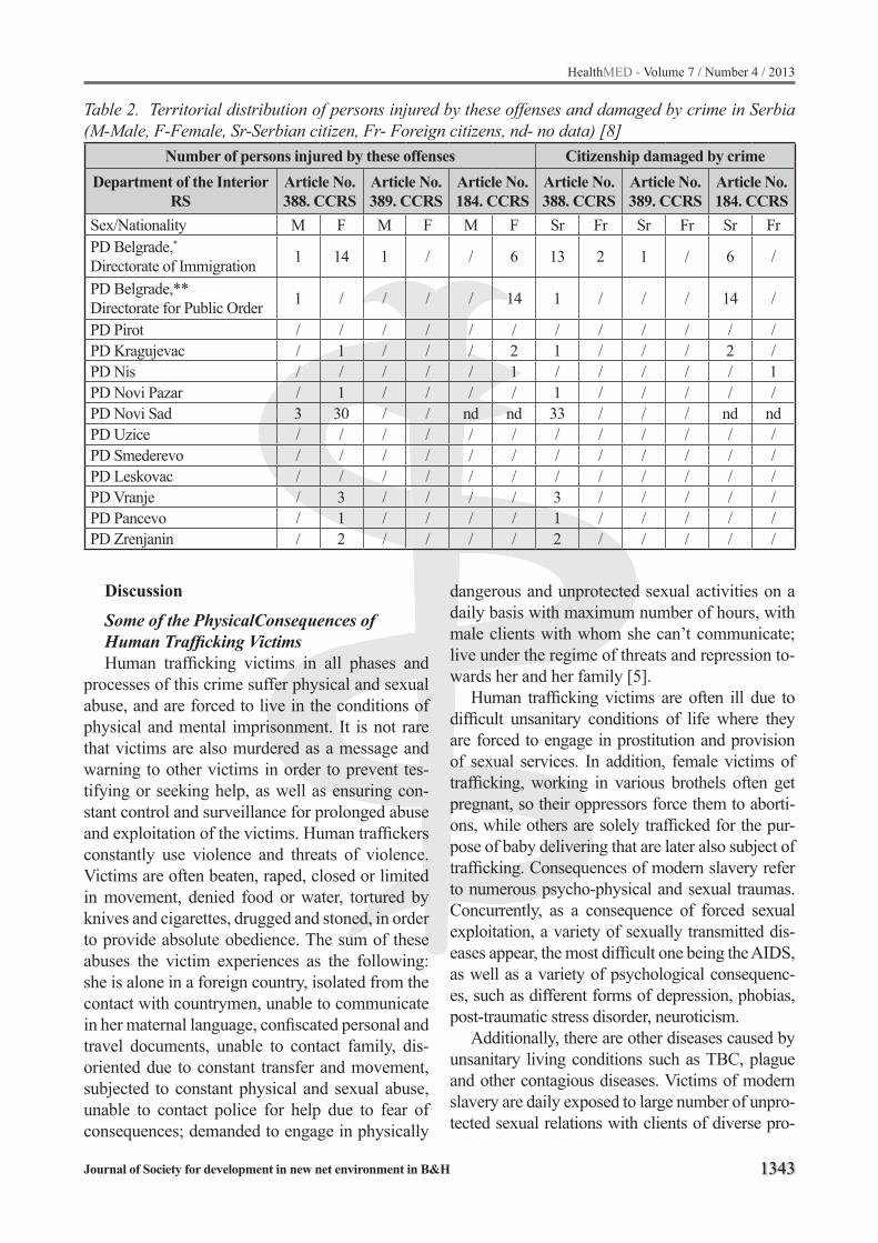
HealthMED - Volume 7 / Number 4 / 2013
Journal of Society for development in new net environment in B&H 1343
Discussion
Some of the PhysicalConsequences of Human Trafficking VictimsHuman trafficking victims in all phases and
processes of this crime suffer physical and sexual abuse, and are forced to live in the conditions of physical and mental imprisonment. It is not rare that victims are also murdered as a message and warning to other victims in order to prevent tes-tifying or seeking help, as well as ensuring con-stant control and surveillance for prolonged abuse and exploitation of the victims. Human traffickers constantly use violence and threats of violence. Victims are often beaten, raped, closed or limited in movement, denied food or water, tortured by knives and cigarettes, drugged and stoned, in order to provide absolute obedience. The sum of these abuses the victim experiences as the following: she is alone in a foreign country, isolated from the contact with countrymen, unable to communicate in her maternal language, confiscated personal and travel documents, unable to contact family, dis-oriented due to constant transfer and movement, subjected to constant physical and sexual abuse, unable to contact police for help due to fear of consequences; demanded to engage in physically
dangerous and unprotected sexual activities on a daily basis with maximum number of hours, with male clients with whom she can’t communicate; live under the regime of threats and repression to-wards her and her family [5].
Human trafficking victims are often ill due to difficult unsanitary conditions of life where they are forced to engage in prostitution and provision of sexual services. In addition, female victims of trafficking, working in various brothels often get pregnant, so their oppressors force them to aborti-ons, while others are solely trafficked for the pur-pose of baby delivering that are later also subject of trafficking. Consequences of modern slavery refer to numerous psycho-physical and sexual traumas. Concurrently, as a consequence of forced sexual exploitation, a variety of sexually transmitted dis-eases appear, the most difficult one being the AIDS, as well as a variety of psychological consequenc-es, such as different forms of depression, phobias, post-traumatic stress disorder, neuroticism.
Additionally, there are other diseases caused by unsanitary living conditions such as ТВС, plague and other contagious diseases. Victims of modern slavery are daily exposed to large number of unpro-tected sexual relations with clients of diverse pro-
Table 2. Territorial distribution of persons injured by these offenses and damaged by crime in Serbia (M-Male, F-Female, Sr-Serbian citizen, Fr- Foreign citizens, nd- no data) [8]
Number of persons injured by these offenses Citizenship damaged by crimeDepartment of the Interior
RSArticle No. 388. CCRS
Article No. 389. CCRS
Article No. 184. CCRS
Article No. 388. CCRS
Article No. 389. CCRS
Article No. 184. CCRS
Sex/Nationality M F M F M F Sr Fr Sr Fr Sr FrPD Belgrade,*Directorate of Immigration 1 14 1 / / 6 13 2 1 / 6 /
PD Belgrade,**Directorate for Public Order 1 / / / / 14 1 / / / 14 /
PD Pirot / / / / / / / / / / / /PD Kragujevac / 1 / / / 2 1 / / / 2 /PD Nis / / / / / 1 / / / / / 1PD Novi Pazar / 1 / / / / 1 / / / / /PD Novi Sad 3 30 / / nd nd 33 / / / nd ndPD Uzice / / / / / / / / / / / /PD Smederevo / / / / / / / / / / / /PD Leskovac / / / / / / / / / / / /PD Vranje / 3 / / / / 3 / / / / /PD Pancevo / 1 / / / / 1 / / / / /PD Zrenjanin / 2 / / / / 2 / / / / /

1344
HealthMED - Volume 7 / Number 4 / 2013
Journal of Society for development in new net environment in B&H
files and health status. Evaluations of the magnitude of the problem of AIDS presence in girls and wom-en victims of human trafficking and estimations of their access to effective treatment, protection and support, during rescue or imprisonment, could im-prove the programs focused on victims’ aid [6].
Alarming data indicate that there is a trend of significant decrease of the average age of the vic-tims, based on new perceptions and requests of the clients who believe that sexual relation with children minimizes the risk of sexually transmit-ted diseases. It is commonly known that any form of prostitution represents a source of spreading various, even deadly diseases. It should be empha-sized that prostitutes transmit diseases even when they are tested for them. Tests for HIV infection and other infections cannot detect the disease once it is captured, even though causes of the disease exist, live, and multiply in that person, and eas-ily pass to another person during sexual contact, which subsequently transmits them to others, who may also transmit to others, and so on. Each min-ute inside the mouth, a million new lymphocytes appear, while in case of the presence of an infec-tion, this number reaches ten million. In case indi-vidual is infected, lymphocytes contain multiple AIDS viruses. Often dentist are the first ones to suspect that a person suffers from AIDS once they see fertilization and mouth bleeding, white mate-rial and similar changes. Even a healthy individual almost always has small wounds in mouth after tooth brushing, rotten teeth, gingivitis, orsharp food, through which viruses easily enter. Today we know that an individual in blood and secretion-shas diverse amount of viruses in different phases of disease. There is certainly a difference in types of kisses. However, from the abovementioned, it is clear that the possibility of getting infected by certain kisses cannot be disregarded [5].
Condyloma (genital warts) are a disease caused by the ΗΡV virus i.e. Humani Papilloma Virus. ΗΡV virus causes cervical cancer in women and treatment must be initiated straight after detection. The HPV virus has around 100 variations and not all of them cause condyloma, but the most preva-lent types do. Only 4 types of virus cause cervi-cal cancer, while over 30 causecondyloma. The most prevalent types of virus cause condyloma as a consequence, or the danger of cervical cancer. A
number of documents confirm that the use of bar-rier contraception methods like condoms does not prevent spreading the HPV (Consensus Statement on Cervical Cancer of the US national institute for health in 1996). This institute, the National Institu-te of Allergy and Infectious Diseases and the De-partment of Health and Human Services of the US government, in the document Workshop Summary in 2000 indicate that there are no clinical evidence that condom would be a safe means of protection from diseases of Chlamydia (Chlamydia Tracho-matis), herpes, or syphilis. Chlamydia causes uter-us infections, which ultimately results in infurtil-ity, and in case infected woman is pregnant, child often dies even before birth.
„Human trafficking victim, an Ukrainian Ole-na Popik, died from AIDS in a hospital in Mostar. She was illegally in Serbia in the beginning of the 2004. The doctor, who performed autopsy, con-cluded that she was infected by AIDS and a num-ber of other dangerous diseases including hepa-titis C, miliary tuberculosis, syphilis and others. Throughout the last year, the Ukrainian represent-ed an extreme epidemiological risk for everyone with whom she had any contact, and particularly those with whom she had sexual contact” [5].
SomeofthePsychologicalConsequences of Human Trafficking VictimsHuman trafficking victims enter the circle of
terror and cruelty, due to extremely brutal psycho-physical treatment performed by the human traf-fickers. As a result of such exposure, the victims suffer from chronic trauma that tends to be long-term. Physical exhauster, confusion, phobias, ob-sessive-compulsive ideation, disorientation, amne-sia, strong emotions, grief and inability to commu-nicate represent determining psychophysical traits of an individual who has been liberated from the ‘claws’ of human traffickers during the first contact.
Post-traumatic stress disorder is the one of the common consequences of trauma [9]. Common symptoms of the post-traumatic stress disorder in-volve: episodes of repeated experience of trauma in imposed memories (flashbacks–Sudden revival of memories of past events), dreams or nightmares that appear on a persisting base of ’numbness’ (Re-duced emotional response associated with exposu-re to trauma), disregard of environment, emotio-

HealthMED - Volume 7 / Number 4 / 2013
Journal of Society for development in new net environment in B&H 1345
nal flatness (Persistent depressive mood of minor extent or in case the degree of emotional response is significantly reduced but is not lacking.), isola-tion from others, anhedonia (Inability to enjoy, loss of interest for pleasurable activities), avoidance of activities and situation that remind trauma. Rarely, dramatic attacks of fear, panic, or aggression may occur as stimulus causing rapid memory and/or re-vival of trauma and primary reactions to it [10].
Basic PTSD symptoms become chronic in case recovery does not provide favorable outcome. The most common symptoms refer to: intrusion, avoidance and hyper-sensitivity. Intrusion implies symptoms as flashbacks. Avoidance in case of vic-tims of violence often causes disengagement from certain forms of social contacts avoiding situations that remind traumatic event. Hyper-sensitivity is the third key element of psycho-trauma or PTSD, and involves strong feelings of fear, isolation, rage, anger, excitement, insomnia and lack of con-centration. Children as victims of human traffick-ing are often submitted to sexual abuse in family, and subsequently in the community, and traffic for the purpose of sexual exploitation represents the third step in the circle of violence that these chil-dren are exposed to. Likewise, they are often sub-stance and alcohol dependent. Additionally, they exhibit significant behavioral disorders, while due to the long-term repetitive sexual trauma, they are unable to develop mature defense mechanisms, but commonly exhibit serious psychological dis-turbances associated with mental disorders. Due to the long-term exposure to psychological, physi-cal, sexual and economic abuse and variety of manipulations by adults, these children, compared to other victims of sexual trauma, mainly demon-strate loss of trust in others, further complicating the process of the establishment of trust and safety with professional and assistants.
Intensity and range of sexual, physical and psychological violence that children victims of human trafficking suffer satisfy two basic criteria for diagnosing psychological trauma and its con-sequences. That involves exposure to threat to life and repeated sexual abuse during youth. Exposure to psychological trauma in these cases causes seri-ous consequences in terms of physiological func-tioning of the organism, cognitive (thoughts and comprehension) and emotional behavior [11].
Conclusions
Human trafficking victims are exposed to nu-merous risks that may directly or indirectly impact their health. The risks may result from physical, psychological abuse (exploitation), enforced alco-hol and drug abuse, social boundaries and manip-ulation, economic exploitation, legal insecurity re-lated to victims’ labor exploitation and inadequate working conditions and marginalization. Each of these risks involves heavy consequences to hu-man trafficking victims’ health [12]. It should be noted that the victims are almost never exposed to only one of the mentioned risks; rather they are often interrelated and multiplied. Impact of the aforementioned risks on health depends on their duration and intensity, as well as personal abil-ity to tackle these problems. For instance, risk of physical abuse may result in violent taking of life (murder), self-harm (acute and chronic physical injuries) in the form of bruises, scratches, abra-sions, head injuries, damages in muscles, nerves, senses, and all these consequences may result in minor or severe forms of disability.
Sexual abuse may result in damages of repro-ductive and sexual health, urinary infections, inabil-ity of birth, amenorrhea, dysmenorrhea, acute and chronic pain during sexual contact, occurrence of the cancer of uterus, incontinence, unwanted preg-nancy, impact of insecure abortions (septic shock), etc. Psycho-physical risks and consequences of the victims, among other things demand interventions of medical institutions, doctors of different special-izations and other medical staff, whose main goal is to evaluate health state, health support, and subse-quently planning and realization of the program of therapy for chronic diseases and injuries. Data on victims’ psycho-physical status are confidential and cannot be publicly exposed. Medical staff is only authorized to share information on victims’ psycho-physical status, based on informed consent, to other professionals who provide direct assistance to hu-man trafficking victims [13].
Exceptions are provided in Article 20. Codex of Professional Ethics. That is, doctor is acquitted of professional confidentiality or dismissed from the obligation of professional silence in case of client’s informed consent or for his/her benefit, his family or society, or this has been decided in

1346
HealthMED - Volume 7 / Number 4 / 2013
Journal of Society for development in new net environment in B&H
accordance with law. In case of clients consent, doctor will evaluate which data should be kept confidential if their release could harm the client. In case of court order, the doctor needs to inform the client, except if otherwise provided by the court decision. In most cases, victims require medical assistance, to resolve acute health issues, such as injuries, diseases and abortions resulting from hu-man trafficking, treatment of sexually transmitted diseases and substance addiction. Concurrently, the need for psychological support and assistance is considered crucial. Mental state examination and psychological treatment are performed on the basis of needs’ hierarchy. Primary, organic health is addressed, then physical safety and daily rou-tine, stabilization of physiological functions of sleep and alimentation, establishment of a trust-ing relationship between the victim of human traf-ficking, professionals and assistants, and finally in-depth psychological evaluation. Each victim and children in particular, has specific health and psychological status, which requires individual-ized treatment.
References
1. UN Protocol to prevent, suppress and punish traffic-king in persons, especially women and children, supplementing the United Nations convention aga-inst transnational organized crime. /2000/, New York, United Nations. Available:www.uncjin.org/Docu-ments/Conventions/dcatoc/ final_documents_2/con-vention_%20traff_eng, 10.06.2012.
2. Vertamatti M. A. F., de Abreu L. C., Otsuka F. C., da Costa P. R. F., Ferreira J. D., Tavares C., Santos M. E., Barbosa C. P., Factors Associated to Time of Arrival at the Health Service after Sexual Violence, HealthMed Journal, ISSN1840-2291, Vol. 6. No. 1., 2012.
3. Wheaton E. M., Schauer E. J., Galli T. V.,Economics of Human Trafficking, International Migration, ISSN 0020-7985, Vol. 48., No. 5., pp. 114-141, 2010.
4. Moossy R., Sex Trafficking: Identifying Cases and Victims, National Institute of Justice Journal, U.S. Department of Justice, Office of Justice Programs, ISSN: 1067-7453, Iss. 262, pp. 1-10, 2010.
5. Rahman M. A., Human Trafficking in the era of Glo-balization: The case of Trafficking in the Global Mar-ket Economy, Transcience Journal, ISSN2191-1150, Vol 2., No1., 2011.
6. Kara S., Designing More Effective Laws Against Hu-man, Northwestern Journal of International Human Rights, ISSN 1549- 828X, Vol. 9, No. 2 p. 123-147, 2011.
7. Williamson S., Prior M., Domestic Minor Sex Traffic-king: A Network of Underground Players in the Midwest,Journal of Child & Adolescent Trauma, Taylor & Francais Group, ISSN: 1936-1521, Vol. 2, Iss. 1, pp. 1-16, 2009.
8. Trafficking in Serbia, Report for the Period 2000-2010, ASTRA - Action against Trafficking in Human Beings - Belgrade, ISBN:978-86-84889-14-2, pp. 364-381.
9. The UN manual on justice for victims, Available:www.uncj in .org/Standards/9857854 .pdf ,pp .7-8 , 10.06.2012.
10. The ICD-10 classification of mental and behavioural disorders, World Health Organization, Geneva, 1992.
11. Zimmerman C, Yun K, Shvab I, Watts C, Trappo-lin L, Treppete M, et al. The health risks and con-sequences of trafficking in woman and adolescent - Findings from a European study. London, London School of Hygiene and Tropical medicine, 2003; Available:www.lshtm.ac.uk/php/ghd/docs/traffic-kingfinal.pdf, 10.06.2012.
12. Alempijevic Dj., Jecmenica D., Pavlekic S, Savić S, Aleksandric B., Forensic medical examination of victims of trafficking in human beings, Torture, 2007, vol. 17/2.
13. Banovic B., Bjelajac Z., Traumatic experiences, psychophysical consequences and needs ofhuman trafficking victims, Vojnosanitetski Pregled: Mili-tary Medical and Pharmaceutical Journal of Serbia, ISSN: 0042-8450, Vol. 69, No. 1, pp. 94-97, 2012.
Corresponding AuthorZaklina Spalevic,Faculty of Entrepreneurial Business, University Union Nikola Tesla, Belgrade, Serbia, E-mail: [email protected]

HealthMED - Volume 7 / Number 4 / 2013
Journal of Society for development in new net environment in B&H 1347
Abstract
Objective: Several haemostatic agents are available for clinical use. Ankaferd Blood Stopper (ABS), a mixture of five medicinal plant extracts, has been used historically as a haemostatic agent. The aim of this in vivo study was to investigate the effects of ABS on early full-thickness skin wound healing using a rat dorsal defect model.
Methods: Eighteen male albino wistar rats were used in this study and were divided into two groups: ABS group (Group 1) and the shame group (Group 2). After deep anesthesia with ketamine and xilo-sin, standard full-thickness skin excision wounds (1,5cm diameter and 2 mm deep) were created on the dorsal area of the all animals. The tampon form of the ABS was locally delivered at the wound site every day with the 2.5 X 7 cm3 mL. The shame group underwent a daily tampon form of saline. The wound closure percentage is measured on 0, 3 and 14 days. The wound tisue samples were collected from the dorsal area on 14 day and were examined histopathologically for inflamatosis, granulocytosis and Fibrosis. The anatomical measurement and his-topathological results were analyzed statistically by the Mann-Whitney U test. The Statistical Package for the Social Sciences (SPSS) version 11.5 was used for the data analysis. The analyses were con-ducted using a 0.05 confidence level. Results: Ana-tomically, the measurement of the wound closure percentage were significantly different on 3rd and 14th day between the ABS and the shame group (p < 0.001, p= 0.031 respectively). Histopathological-ly, also significant differences were found in both groups in terms of granulocytosis (p = 0.019) at the 14 the day. Decreased inflammation and increased fibrosis were found in the ABS group, but there were no statistically significant differences from the shame group ( p = 0.065, p = 0.088; respectively).
Conclusions: This study revealed that ABS product can provide an efficacious to accelerate wound healing. Further in vitro and in vivo stu-dies are necessary for evaluating the benefits and possible adverse effects of the application of ABS product on wound healing.
Key words: Ankaferd Blood Stopper, wound healing, full-thickness skin excision.
Introduction
Wound is defined as disruption of cellular, anatomical, and functional continuity of a living tissue. It may be produced by physical, chemical, thermal, microbial, or immunological insult to the tissue (1). Wound healing is the interaction of a complex cascade of cellular and biochemical ac-tions leading to the restoration of structural and functional integrity with regain of strength of in-jured tissues (2). In mammals, wound healing is a result of three overlapping processes: hemostasis and inflammation, granulation tissue formation, re-epithelialization and remodelling, which enab-le wound closure and the restoration of a functio-nal barrier (3). Wound healing normally develops without complications, but the resolution of inf-lammation is critical. Indeed, several conditions like ageing, obesity and many other disorders can compromise the normal resolution of the inflam-mation and resulted in chronic wound and ulcer development, leading to tissue damage (4).
Literally millions of traumatic wounds are tre-ated each year in emergency departments. Wound management is an area of still ongoing research, much of which supports traditionally performed techniques, however some of which also is still trying to resolve controversial topics. Plants have the immense potential for the management and treatment of wounds. A large number of plants are
Efficacy of folk medicinal plant extract Ankaferd Blood Stopper on full-thickness skin wound healingMehmet Tahir Gokdemir1, Halil Kaya1, Ozgur Sogut1, Tuncer Demir2, Sezen Kocaslan3, Muazez Cevik4
1 Harran University, School of Medicine, Department of Emergency Medicine, Sanliurfa, Turkey,2 Gaziantep University, School of Medicine, Department of Physiology, Gaziantep, Turkey,3 Harran University, School of Medicine, Department of Pathology, Sanliurfa, Turkey,4 Harran University, School of Medicine, Department of Pediatric Surgery, Sanliurfa, Turkey.

1348
HealthMED - Volume 7 / Number 4 / 2013
Journal of Society for development in new net environment in B&H
used in many countries for the treatment of wounds and burns (5-9). Ankaferd Blood Stopper® (ABS; Ankaferd Health Products Ltd., Istanbul, Turkey) is a traditional folk medicinal plant extract product that has been approved in the management of ex-ternal hemorrhage and dental surgery bleedings in Anatolia. ABS comprises a standardized mixture of the plants Thymus vulgaris, Glycyrrhiza glabra, Vitis vinifera, Alpinia officinarum and Urtica dio-ica. Several studies have shown that each of these plants has some effect on the endothelium, blood cells, angiogenesis, cellular proliferation, vascular dynamics and cell mediators (10-15).
Several animal studies of different experimen-tal models including incisional or excisional tra-umas have revealed its efficacy in stopping the bleeding in vivo (16-18). However, few study has been investigated on the anti-inflammatory ac-tivities of Ankaferd Blood Stopper® (19). To our knowledge previosly no study has reperted on skin wound healing activities of ABS. The aim of this in vivo study was to evaluate the effects of An-kaferd Blood Stopper® on early wound healing using a rat dorsal full-thicness skin wound model.
Methods
Ankaferd Blood StopperAnkaferd Blood Stopper (Trend Teknoloji Ilac
AS, Istanbul, Turkey) is a licensed pharmaceutical plant extract that is applied directly to injured skin and mucosa as a liquid (solution) or spray or in a dressing (tampon) (20). It produces active hemo-stasis for the management of external, postsurgical, and dental hemorrhage (21). ABS contains a stan-dardized mixture of 5 medicinal plant extracts. The active ingredients in the tampon form of ABS ( 2.5 X 7 cm3 mL) are as follows: 0.18 mg of dried root of Urtica dioica, 0.24 mg of dried leaf of Vitis vi-nifera, 0.27 mg of dried leaf of Glycyrrhiza glabra, 0.21 mg of dried leaf of Alpinia officinarum, and 0.15 mg of dried leaf of Thymus vulgaris (22). This study used the tampon form of ABS supplied in 2.5 cm x 7 cm – 3 ml to the full-thickness skin wound healing in animal model.
Animals Eighteen male rats weighing between 180 and
200 g were used in this study. The animals were
purchased locally from the Research Institute of Physiology (Gaziantep, Turkey). Twenty wistar albino rats were maintained in the Physiology De-partment animal house at a constant temperature of 22 ± 4°C with a 12-hour light/dark cycle and fed Standard pellet chow and water ad libitum. The animals of each group were sacrificed with an overdose of pentobarbital (40 mg / kg) on the 14th day after surgery. The experimental protocol was in accordance with the European Community Council Directive of November 24, 1986 (86/609/EEC), and was approved by the Harran University Medical School Ethics Committee.
Experimental Design The rats randomly assigned to two groups of
9 animals each. Group 1, study group, with full skin-thickness excisions, treated daily with tam-pon form of ABS. Group 2, shame, daily irriga-ted with saline and closed with a tampon. Prior to surgery, the animals were anesthetized with a 0.7 mL intramuscularly injection of a solution conta-ining xylazine hydrochloride (Rompun®, Bayer, Leverkusen, Germany) and ketamine hydrochlo-ride (Ketalar; Pfizer, New York, NY USA) at 1 / 0.5 proportion, 0.1 mL / 100 g body weight. Sur-gery was performed under sterile conditions. After surgery, dressing (tampon) form of the ABS® was applied to Group 1 (study group) at the first day, and this application was repeated everyday, whi-le Group 2 received no treatment and served as the shame group. No postoperative complications were noticed during the postsurgical course. All animals survived throughout the study period.
Macroscopic skin wound-healing assay A standardized ellipsoid round full thickne-
ss sckin tissue exicision (epidermis, dermis, and hypodermis) defect (1.5 cm in diameter, 1 mm in depth) was created on the dorsal area of rats (Figu-re 1A). The wound closure rats were measured on the day 3, and day 14 by the tape measure.
Tissue preparation and histopathological examinationOn day 14 the rats were anesthetized and the
part of the wounds were taken from animals for histopathological examination. The wound spe-cimens at 14 day postwounding including full

HealthMED - Volume 7 / Number 4 / 2013
Journal of Society for development in new net environment in B&H 1349
thickness skin layers (epidermis, dermis, and hy-podermis) were fixed in 10 % neutral buffered for-malin overnight at 4 °C and processed according to the routine light microscope tissue processing methods. The specimens were embedded in para-fin and routine hematoxylin and eosin (H&E) sta-ining and Masson Trichrome ( MT) staining were performed. The sections were examined with light microscope under 100 x magnification (Olympus BX51 TF, Tokyo, Japan).
A histomorphological review was performed by a single blinded pathologist to evaluate the pre-sence of inflamatosis, granulocytosis and fibrosis. The scores for inflamatosis, granulocytosis and fi-brosis were determined by counting the associated cells and their ratio to the total cell count in a stan-dardized area at 100 x magnification ( Figure 2A, 2B, 2C, 2D,2E) . As shown in table 1, the ratio of cells between 0-25% was scored as none, 25-50 % as slight (+), 50- 75 % as moderate (++), and 75-100 % as advanced (+++).
Figure 2. Photomicrograph of full thickness wo-unds in rats at 14th day (magnification, 100x). Shame group: advanced presence of inflamma-tion stained with H&E (2A), slight granulation and fibrosis with MT (2B). ABS group: slight in-flammation, moderate granulation and moderate fibrosis stained with H&E (2C), and Moderate granulation, moderate fibrosis with MT (2D)
The wound specimens at 14 day postwounding including full thickness skin layers (epidermis,
Figure 1. Wound healing effect in rats. A standardized ellipsoid round full thickness sckin tissue exicision defect (1.5 cm in diameter, 1 mm in depth) were created on the dorsal area of rats ( 1A) at the 0 day. ABS significantly contributed to the wound closure percentage compared with the control group at 3th (1B: ABS group, 1C: shame group) and 14th (1D: ABS group,1E:shame group) postwounding day

1350
HealthMED - Volume 7 / Number 4 / 2013
Journal of Society for development in new net environment in B&H
dermis, and hypodermis) also were observed un-der Olympus DP71 image analyzing system. 5 µm tissue sections with H&E and MT staining were examined and photographed.
Data analysisStatistical analyses were carried out using the
SPSS statistical package, version 11.5 (SPSS, Inc, an IBM Company, Chicago, Illinois) for Win-dows. The data on percentage anti-inflammatory and wound healing was compared among groups using the Mann- Whitney U test. The mean and standard deviation or median, minimum-maxi-mum data were calculated for each group. All data are expressed as the mean and 95 % confidence intervals. The values of p ≤ 0.05 were considered statistically significant.
Results
Anatomically wound of the all animals were measured on 3rd and 14th days (Table 1 ). On the 3rd day, median of the wound diameters were 1 ± 0.15 ( 0.9 - 1.2) in the ABS group and 1.4 ± 0.1 (1.3 - 1.4) in the shame group. Median of the wo-und closure also were significantly higher in the ABS group on the 14th day. Comparisons between
the ABS and shame groups indicate a significant variability in the wound closure on the 3rd and 14th days ( p < 0.001, p = 0.031 respectively). The appearance of the repaired wounds sites is shown in Figure 1B,1C, 1D and 1E.
As shown in the table 2, in the study group, 89 % of the specimens show moderate granulositosis, while in the shame moderate granulositosis was observed in 33.3 % of the specimens. Statistical-ly significant differences were found between the ABS and shame groups as for the granulositosis (p<0.019). In the ABS group, 33.3 % of the spe-cimens was show moderate inflamatosis and 44 % moderate fibrosis, while in the shame group moderate inflamatosis was observed in 56 % and moderate fibrosis in 11.1 % of the specimens, res-pectively (Figure 2A,2B,2C and 2E). Decreased inflammation and increased fibrosis were found in the ABS group, but there were no statistical-ly significant differences from the shame group (p=0.065, p=0.088; respectively).
Discussion
In this study, we tested the effects of ABS tam-pon form on the full tickness skin wound healing in a rat model. According to our findings, fibrosis,
Table 1. The percentage of inflamatosis, granulocytosis and fibrosis between the ABS* and shame group
Characteristic ABS group [n (%)]
Shame group[n (%)]
Statistical significance [p]¥
Inflamatosis None Slight(+)Moderate(++)Advanced(+++)
33.333.3 33.3
0
033.356
11.1
0.065
GranulocytosisNoneSlight(+)Moderate(++)advanced(+++)
0 11 89 0
066.733.3
0
0.019
FibrosisNoneSlight(+)Moderate(++)advanced(+++)
055440
11.178.811.1
0
0.088
*Trademark: Ankaferd Health Products Ltd., Istanbul, Turkey.¥From independent samples t-test. Values are Median ± IR (min - max). The ratio of cells between 0-25% was scored as none, 25-50 % as slight (+), 50- 75 % as moderate(++), and 75-100 % as advanced (+++).
HealthMED - Volume 7 / Number 4 / 2013
Journal of Society for development in new net environment in B&H 1351
granulositosis and wound contraction percentage were increased, while inflammation decreased.
Each ingredient of ABS mixture has specific cha-racteristics. T. vulgaris has been shown to exhibit varying levels of anti-oxidant activity, which may help to prevent in vivo oxidative damage, such as lipid peroxidation, associated with atherosclerosis (12). Inoculation experiments on detached leaves of V. vinifera exhibited enhanced resistance towar-ds pathogens (10,11). A. officinarum inhibits nitric oxide production in lipopolysaccharide activated mouse peritoneal macrophages (13). G. Glabra in-hibits angiogenesis, decreases vascular endothelial growth factor production and cytokineinduced ne-ovascularization (14). U. dioica can produce hypo-tensive responses through a vasorelaxation effect mediated by the release of endothelial nitric oxide and the opening of potassium channels, and throu-gh a negative inotropic action (15).
Generally wound-healing agents have the pro-perties to enhance the deposition of collagen con-tent, which provides strength to the tissues and forms cross-linkages between collagen fibers (23). The wound healing of the skin defect was determi-ned by the percentage of wound surface covered by regenerating epidermis. The wound closer percenta-ge of the ABS treated group was found to be which was significantly higher than that of shame group of animals on 14th post wounding day. These wound closure rates made a good match to the results of wound scratch assay in vitro and demonstrated the
accelerating effect of ABS on the wound healing. It was seen that the wounds treated by ABS recovered much more quickly with better skin appearance.
During the proliferative phase, a granulation ti-ssue (new stroma) is formed. Fibroblasts constitu-te the predominant cell type in granulation tissue. They start to proliferate and produce matrix compo-nents (hyaluronan, fibronectin, proteoglycans and type I and III procollagen), which are then depo-sited locally (24). The histopathological results of the our study showed that the wound treated with ABS showed more intense granulositosis which is associated with new tisue formation activity of the components of the ABS. This indicates a positive role of the extract on the cutaneous wounds healing process. In addition, the ABS treated group showed increased fibrosis rate. But occurrence of fibrosis was statistically similar in both groups.
Wound healing is a physiological process ne-cessary for repair and regeneration of injured skin tissue. This process greatly depends on the crosstalk between inflammation, oxidative stress and angiogenesis (25). Inflammatory cells appear very rapidly in a wound after injury, as reported by Kagawa and collaborators (26). In their stu-dy, isler et al showed that ABS decreased the in-flammation and necrosis process and increased the new bone formation in early bone healing period without causing any foreign body reaction in a rat model (19). Accordingly, in our study decreased inflammation was found in the ABS treated group,
Table 2. Excision wound closure rate between the ABS* and shame group
Characteristics Day 3 wound diameters [cm]
Day 14 wound diameters [cm]
No. of Animals ABS Shame ABS Shame1 1.1 1.3 0.11 0.162 1.0 1.3 0.15 0.243 0.9 1.4 0.12 0.214 0.9 1.4 0.20 0.215 1.2 1.3 0.05 0.206 1.2 1.4 0.20 0.227 1.0 1.4 0.22 0.108 1.0 1.4 0.16 0.409 1.1 1.3 0.11 0.30
¥Median (Min-Max) 1±0.15( 0.9-1.2) 1.4±0.1(1.3-1.4) 0.15±0.09(0.05-0.22) 0.21±0.09(0.10-0.40)p < 0.001 0.031
*Trademark: Ankaferd Health Products Ltd., Istanbul, Turkey.¥ From independent samples t-test. Values are Median ± IR (min-max)

1352
HealthMED - Volume 7 / Number 4 / 2013
Journal of Society for development in new net environment in B&H
which is probably related to the antiinflammatory activity of some components of the haemostatic agent. Also we did not see any adverse effects during the observation and could not make any conclusion on wound healing.
Conclusion
This experimental study revealed that ABS product can provide an efficacious to accelerate wound healing. It is decreases the inflammati-on and increase the granulositosis and fibrosis in early full ticknes wound healing period. Further in vitro and in vivo studies are necessary to evalua-ting the benefits and possible adverse effects of the application of ABS on wound healing.
Authors’ contributions
MTG: Protocol, development and manuscript writing, HK, OS and SK: Data Collection & data entry, TD and MC: Statistical Support and data analysis. All authors read and approved the final manuscript.
References
1. Jalalpure SS, Agrawal N, Patil MB, ChimkodeR, Tri-pathi A:Antimicrobial and wound healing activities of leaves of Alternanthera sessilis Linn. International Journal of Green Pharmacy 2008, 2: 141–144.
2. Martin P: Wound healing-aiming for perfect skin re-generation. Science 1997, 276: 75–81.
3. Tan W, Bailey AP, Shparago M, Busby B, Covington J, Johnson JW, et al.: Chronic alcohol consumption stimulates VEGF expression, tumor angiogenesis and progression of melanoma in mice. Cancer Biol Ther 2007,6: 1211-1217.
4. Pond CM: Paracrine interactions of mammalian adi-pose tissue. J Exp Zool A Comp Exp Biol 2003,295: 99-110.
5. Nayak BS,. Pinto Pereira LM: Catharanthus roseus flower extract has wound-healing activity in Sprague Dawley rats. BMC Complementary and Alternative Medicine 2006, 6: 41.
6. Parente LM, Lino Junior Rde S, Tresvenzol LM, Vi-naud MC, de Paula JR, Paulo NM: Wound Healing and Anti-Inflammatory Effect in AnimalModels of Ca-lendula officinalis L. Growing in Brazil. Evidence-Ba-sed Complementary and Alternative Medicine 2012, 2012:375671. doi:10.1155/2012/375671
7. Kosger HH, Ozturk M, Sokmen A, Bulut E, Ay S: Wo-und healing effects of Arnebia densiflora root extracts on rat palatal mucosa. European Journal of Dentistry 2009, 83: 96–99.
8. Garg VK, Paliwal SK: Wound healing activity of etha-nolic and aqueous extract of Ficus benghalensis. J Adv Pharm Technol Res 2011, 2:110-114.
9. Nayak S, Nalabothu P, Sandiford S, Bhogadi V, Ado-gwa A: Evaluation of wound healing activity of Alla-manda cathartica. L. and Laurus nobilis. L. extracts on rats. BMC Complementary and Alternative Medi-cine 2006, 6: 12.
10. Barka EA, Belarbi A, Hachet C, Nowak J. Audran JC: Enhancement of in vitro growth and resistance to gray mould of Vitis vinifera co-cultured with plant growth-promoting rhizobacteria. FEMS Microbiol Lett 2000, 186: 91-95.
11. Barka EA, Gognies S, Nowak J, Audran JC, Belarbi A: Inhibitory effect of endophyte bacteria on Botrytis cinerea and its influence to promote the grapevine growth. Biol Control 2002, 24: 135-142.

HealthMED - Volume 7 / Number 4 / 2013
Journal of Society for development in new net environment in B&H 1353
12. Lee SJ, Umano K, Shibamoto T, Lee KG: Identifi-cation of volatile components in basil (Ocimum ba-silicum L.) and thyme leaves (Thymus vulgaris L.) and their antioxidant properties. Food Chem 2007, 91:131-7.
13. Matsuda H, Ando S, Kato T, Morikawa T, Yoshikawa M: Inhibitors from the rhizomes of Alpinia officina-rum on production of nitric oxide in lipopolysaccha-ride-activated macrophages and the structural requ-irements of diarylheptanoids for the activity. Bioorg Med Chem 2006,14:138-142.
14. Sheela ML, Ramakrishna MK, Salimath BP: Angio-genic and proliferative effects of the cytokine VEGF in Ehrlich ascites tumor cells is inhibited by Glycyrr-hiza glabra. Int Immunopharmacol 2006,6: 494-498.
15. Testai L, Chericoni S, Calderone V, Nencioni G, Nie-ri P, Morelli I, et al.: Cardiovascular effects of Urti-ca dioica L. (Urticaceae) roots extracts: in vitro and in vivo pharmacological studies. J Ethnopharmacol 2002, 81: 105-109.
16. Bilgili H, Kosar A, Kurt M, Onal IK, Goker H, Cap-tug O, et al.: Hemostatic Efficacy of Ankaferd Blood Stoper in a Swine Bleeding Model. Medical Princi-ples and Practice 2009, 18: 165-169.
17. Huri E, Akgül T, Ayyildiz A, Ustün H, Germiyanoğlu C: Hemostatic Role of a Folkloric Medicinal Plant Extract in a Rat Partial Nephrectomy Model: Con-trolled Experimental Trial. Journal of Urology 2009,181: 2349-2354.
18. Ismail Iynen, Ozgur Sogut, Rustu Kose: The Efficacy of Ankaferd Blood Stopper in Heparin-Induced He-mostatic Abnormality in a Rat Epistaxis Model. Oto-laryngology –Head and Neck Surgery 2011; 145: 840-844.
19. Isler SC, Demircan S, Cakarer S, Cebi Z, Keskin C, Soluk M, et al.: Effects of folk medicinal plant extract Ankaferd Blood Stopper® on early bone healing. J Appl Oral Sci 2010, 18:409-414.
20. Kurtaran H, Ark N, Ugur KS, Sert H, Ozboduroglu AA, Kosar A, et al: Effects of a topical hemostatic agent on an epistaxis model in rabbits. Current The-rapeutic Research 2010; 71: 105-110.
21. Singer AJ, McClain SA, Katz A: A porcine epistaxis model: hemostatic effects of octyl mcyanoacrylate. Otolaryngol Head Neck Surg 2004,130: 553-557.
22. Goker H, Haznedaroglu IC, Ercetin S, Kirazlı S,Akman U,Ozturk Y, et al.: Haemostatic actions of the folkloric medicinal plant extract, Ankaferd blood stopper. J Int Med Res 2008, 36: 163-170.
23. Gupta N, Jain UK: Investigation of wound healing activity of methanolic extract of stem bark of mimu-sops elengi linn. Afr J Tradit Complement Altern Med 2011, 8: 98-103.
24. Robson MC, Steed DL, Franz MG: Wound healing: biologic features and approaches to maximize hea-ling trajectories. Curr Probl Surg 2001, 38: 72–140.
25. Soares R, Azevedo I: Inhibition of S1P by polyphe-nols prevents inflammation and angiogenesis: NFkappaB, a downstream effector? Free Radic Biol Med 2007, 42: 311.
26. Kagawa S, Matsuo A, Yagi Y, Ikematsu K, Tsuda R, Nakasono I: The time-course analysis of gene expre-ssion during wound healing in mouse skin. Leg Med (Tokyo) 2009, 11: 70-75.
Corresponding AuthorMehmet Tahir Gokdemir,Harran University,School of Medicine, Department of Emergency Medicine, Sanliurfa, Turkey,E-mail: [email protected]

1354
HealthMED - Volume 7 / Number 4 / 2013
Journal of Society for development in new net environment in B&H
Abstract
Background: Iron imbalance in the brain, in-cluding excess accumulation and deficiency, is as-sociated with neurological disease and cognitive dysfunction. On the other hand, folic acid is a water-soluble B-vitamin that may have roles in prevention of central nervous system development, mood disor-ders, dementia and Alzheimer’s disease. So, in this study we assessed the effects of co-administration of iron and folic acid supplementation on passive avoidance memory in adult male Wistar rats.
Methods: Animals were divided into four groups randomly with 8 in each: 1) Fe30: Rats that received iron 30 mg/kg/day via gavage for one week, 2) FA15: Rats that received folic acid 15 mg/kg/day single daily intra peritoneal for one week, 3) Fe+FA: rats that received coadministration iron 30mg/kg/day and folic acid 15 mg/kg/day for one week. 4) Sham (saline & sorbitol): received co-administra-tion folate and iron vehicles for one week then all groups were tested in shuttle box for memory.
Results: Data showed that co-administration Fe and FA (Fe+FA) significantly improved short term (P<0.001) and long term (P<0.05) memory with compare to each iron or folic acid group alone.
Conclusion: It appears that iron-folic acid sup-plementation co-administration had better effect than iron supplementation alone.
Key words: Folic acid, iron, avoidance mem-ory, rat.
Introduction
Iron is an essential trace metal in the human diet due to its obligate role in a number of metabolic processes (1). It is essential for nearly all living or-ganisms as it is involved in fundamental processes such as transport and exchange of oxygen, enzyme action, and DNA, RNA and protein syntheses. The brain metabolism depends on its balanced iron
concentrations in various its regions (2, 3). In the brain, iron is an important co-factor for the genera-tion of dopamine and several cellular and intracel-lular processes e.g. tyrosine hydroxylase (3). Iron is found predominantly in oligodendrocytes in the brain and required for myelin production (4) as well as in the production of several neurotransmit-ters such as norepinephrine and serotonin and gen-eration of GABAergic activity (5). Iron imbalances in the brain, including excess accumulation and de-ficiency, are associated with neurological disease and dysfunction; yet, their origins are poorly un-derstood (6). It is now generally accepted that iron accumulation in selective regions of the brain may acts as free radicals, thereby possessing implication for the etiology of neurodegenerative disorders (7). Although excess Iron appears to be one of the main factors in the metal induced neurodegeneration. Evidence suggests that brain iron deficiency at any time in life may disrupt metabolic processes and subsequently change the cognitive and behavior functions (8) On the other hand, however, total Fe losses vary from 0.17 to 1.05 mg/day, according to age and gender, a process resulting mainly from elimination of intestinal epithelial death cells. For this reason the losses necessarily must be compen-sated by nutritional supplementation (9). Even so, until 1974 little or no attention was paid to brain iron metabolism and brain function. Since then there has been an active interest in brain iron metabolism, not only as a consequence of its deficiency with an effect on cognitive processes, but also the role of excess brain iron accumulation and its involvement in neurodegeneration and progressive neurodegen-erative diseases such as Parkinson’s disease, Al-zheimer’s disease, Huntington chorea, and Haller Voren Spatz disease (10). In contrast to many of the dietary supplements now flooding the shelves of grocery stores and pharmacies, research on di-etary folate and its links to human disease began
Impact of Iron-folic acid supplementation on passive avoidance memory in adult male Wistar ratsKhombi Shooshtari M 1, Moazedi A. A1, Parham Gh2
1 Department of Biology, Faculty of Sciences, Shahid Chamran University, Ahvaz, Iran,2 Department of Statistics, Faculty of Mathematic, Shahid Chamran University, Ahvaz, Iran.

HealthMED - Volume 7 / Number 4 / 2013
Journal of Society for development in new net environment in B&H 1355
decades ago, well prior to the dietary supplement craze that has recently descended upon us (11). Fo-lic acid (folate) is a water-soluble B-vitamin which present in food such as dried beans, peas, lentils, oranges, whole-wheat products, liver, asparagus, beet, broccoli, Brussels sprouts, and spinach (12). Folate participates in the transfer of 1-carbon unit (such as methyl, methylen, and formyl groups) to the essential substrates involved in the synthesis of DNA, RNA, and proteins. Folic acid plays a role in the methylation of homocysteine providing the methyl group for the conversion of methionine to s-adenosyl methionine (13, 14). An increase in ho-mocysteine (Hcy) levels is a major consequence of folate deficiency that may have adverse effects on multiple organ systems during aging Recent stud-ies of cell culture and animal models of neurode-generative disorders have provided evidence that folate deficiency and elevated Hcy levels can in-deed neurons vulnerable to dysfunction and death. Exposure of cultured rat hippocampal neuron to folate medium and Hcy promotes apoptosis and increase the vulnerability of the neurons to being killed by amyloid β-peptide, a protein believed to be responsible for nerve cell death in Alzheimer’s disease (14) There is evidence to implicate Hcy in increased oxidative stress, DNA damage, the trig-gering of apoptosis and excitotoxicity, all impor-tant mechanisms in neurodegeneration (15). Dur-ing the past decade data have accumulated that support roles for folate and Hcy in modifying risk of Alzheimer’s and Parkinson’s diseases (16, 17). Some studies showed that, the B vitamins, specifi-cally folate, have been implicated in neurological disorders, including those associated with cogni-tive disorders (18). More recently, folate deficien-cy may also contribute to the declines in cognitive and other neurological functions that occur during normal aging (19). Conversely, high folate intake was associated with lower risk of Alzheimer’s dis-eases (20). The adverse effects of folate deficiency and elevated Hcy levels on the developing brain have been well documented. But, relatively little research has investigated the effects of impact iron-folic acid supplementation on cognitive function in normal aging (21). So, purpose of this study was to assess the potential effects of impact of iron- folic acid supplementation (IFA) on passive avoidance memory in adult subjects.
Materials and Methods
Animals: Forty adult male albino rats of Wis-tar strain (250±20g, 3-4 months) were used in this study that obtained from Ahvaz Jundishapur Uni-versity of Medical Sciences (AJUMS) laboratory animal center. Animals were housed in standard cages under controlled room temperature (20±2 ◦C), humidity (55-60%) and light exposure con-ditions 12:12 h light–dark cycle (lighted on 07:00 am). All experiments carried out during the light phase of the cycle (8:00 am to 6:00 pm). Access to food and water were ad libitum except during the experiments. Animal handling and experimen-tal procedures performed under observance of the University and Institutional legislation, controlled by the Local Ethics Committee for the Purpose of Control and Supervision of Experiments on Labo-ratory Animals. All efforts were made to minimize animal suffering, to reduce the number of animals used. Prior to the onset of behavioral testing, all rats were gentle handled for 5 minutes daily. Animals were divided randomly into four groups, consist-ing of 8 animals in each: 1) Fe30; rats that received iron 30 mg/kg/day via gavage for one week (5) , 2) FA15; rats that received folic acid 15 mg/kg/day single daily intra peritoneal for one week (22) , 3) Fe+FA; rats that received coadministration iron 30mg/kg/day and folic acid 15 mg/kg/day, rats re-cived folic acid two hours after recived iron supple-mentation for one week, 4) Sham; received coad-ministration of vehicle of folic acid (normal saline) and vehicle of iron supplementation (%5 sorbitol in water), rats recived normal saline two hours after received %5 sorbitol in water for one week.
Then, all groups were trained for passive avoi-dance learning and short- and long-term memories with using two-way shuttle box apparatus.
Passive avoidance task: The apparatus used for evaluation the passive avoidance task was two-way shuttle box (Borj Sanaat Co. Iran), which consisted of two adjacent Plexiglas compartments of identical dimensions (27×14.5×14 cm). For the experimental procedure, on the first day (adapta-tion) each rat was allowed a 3 minutes adaptation period prior and free access to either the light or dark compartment of the box to avoidance training and after being placed in a shuttle-box. Following this adaptation period, on the second day (training

1356
HealthMED - Volume 7 / Number 4 / 2013
Journal of Society for development in new net environment in B&H
phase) rats were placed into the illuminated com-partment and 30 seconds later the sliding door was raised. Upon entering the dark compartment the door was closed and a 1.5 mA constant-current shock was applied for 3 seconds. After 20 seconds the rat was removed from the dark compartment and placed into home cage. In order to test short-term and long term memories, 48 hours and 30 days after receiving foot shock, the rats were pla-ced in illuminated chamber and 30 seconds later the sliding door was raised and the latency of ente-ring the dark compartment (step-through latency) and the time spent there during 5 min were recor-ded again. The maximum time that considered in this procedure were 300 seconds (24, 25).
Statistics: Data were expressed as mean ±S.E.M. of values for memory test. Statistical analysis was performed by one-way ANOVA followed by LSD post hoc test. A P-value less than 0.05 were assu-med to denote a significant difference and levels of significance are indicated by symbols: * for diffe-rence between groups vs. control and # for differen-ce between treated groups with iron group.
Result
Analyze data in step-through latencies 48 hour after training between FA15, Fe30, Fe+FA and sham groups showed that there was significant (P<0.001) difference between rats received iron supplementation and Fe+FA groups (Fig. 1) and so, 30 days after training, in step-through latencies there was significant (P<0.05) difference between rats which were received Fe+FA and iron group (Fig. 2). On the other hand, statistically analysis in the time spent in the dark chamber 48 hour af-ter training, there was no significant difference between Fe+FA and iron groups in this step (Fig. 3). Also 30 days after training, in this step there was no significant difference between any of gro-ups (Fig. 4). Then, our results showed that Fe+FA supplementation improved short-term and long-term memory with compare to iron supplemen-tation alone but there was no difference between Fe+FA supplementation group and folic acid gro-up with 15mg/kg/day dose.
Figure 1. Comparing groups on step-through latency 48 h after training, (***P<0.001, n=8), (Veh: Sham treated, FA15: Folic acid 15mg/kg/day, Fe30: iron 30mg/kg/day, Fe+FA: iron 30mg/kg/day+ Folic acid 15mg/kg/day)
Figure 2. Comparing groups on step-through la-tency 30 days (long-term memory) after training (*P<0.05, ***P<0.001, n=8), (Veh: Sham trea-ted, FA15: FA15: Folic acid 15mg/kg/day, Fe30: iron 30mg/kg/day, Fe+FA: iron 30mg/kg/day+ Folic acid 15mg/kg/day)

HealthMED - Volume 7 / Number 4 / 2013
Journal of Society for development in new net environment in B&H 1357
Figure 3. Comparing groups on time spent in the dark chamber 48 h after training, (⃰ ⃰ ⃰ P<0.001, n=8), (Veh: Sham treated, FA15: Folic acid 15mg/kg/day, Fe30: iron 30mg/kg/day, Fe+FA: iron 30mg/kg/day+ Folic acid 15mg/kg/day)
Figure 4. Comparing groups on time spent in the dark chamber 30 days after training (n=8), (Veh: Sham treated, FA15: Folic acid 15mg/kg/day, Fe30: iron 30mg/kg/day, Fe+FA: iron 30mg/kg/day+ Folic acid 15mg/kg/day)
Discussion
The present study has investigated possibility that Fe+FA administration could increase passi-ve avoidance memory comparative with iron ad-ministration alone. In a previous reports we had demonstrated that iron supplementation whit 30
mg/kg/day or folic acid whit 15 mg/kg/day incre-ased passive avoidance memory in separately. But in this study we assessed the potential effects of Fe+FA supplementation on passive avoidance memory in adult subjects. Passive avoidance con-dition was assessed in particular since the relati-on between this behavior and Fe+FA in adult is not well documented in the literature. The data shown here demonstrated for the first time that IFA supplementation significantly improved short term (P<0.001) and long term (P<0.05) memory compare to iron group. The prevalence of iron, folic acid and vitamin B12 deficiencies and their role in the pathophysiology of recurrent aphthous stomatitis is not well known, although several re-ports have considered their importance and rele-vance (26). In the brain, sufficient iron supply is critical to a wide variety of biochemical pathways, ranging from basic cellular metabolism to ca-techolamine synthesis and myelination. Iron is also involved in the production of reactive oxygen species and can be toxic in excess. Thus, both iron overload and deficiency must be avoided, yet im-balances do occur in some individuals for reasons that are yet unknown. Iron overload in relevant brain regions is linked to Parkinson’s disease and Alzheimer’s disease, while iron deficiency is asso-ciated with restless legs syndrome and attention deficit hyperactivity disorder. Iron deficiency also leads to specific cognitive impairment and emo-tional effects, which vary across developmental stages (6). However, it has been proposed that iron accumulation in the brain mediates oxidative damage, and neuronal death associated with neu-rodegenerative disorders (27). It is interesting to note, however, after a long period of understan-ding role of folic acid in the pathogenesis of me-galoblastic anemia, increase attentions were given to folic acid in the last decade due to its role in the prevention of the development of various disea-ses, including neural tube defect, atherosclerosis heart disease, and cancer. Despite their important role in cognitive function, the value of B vitamin supplementation is unknown. The evidence does not yet provide adequate evidence of an effect of vitamin B6 or B12 or folic acid supplementation, alone or in combination, on cognitive function te-sting in people with either normal or impaired co-gnitive function (28). B vitamins such as thiamin,

1358
HealthMED - Volume 7 / Number 4 / 2013
Journal of Society for development in new net environment in B&H
riboflavin, vitamin B6, and folate are known to be essential for the maintenance of normal metabolic functions in the brain (29). Among Other B Vita-mins, Folte has received much attention recently as its low serum level is found to be closely asso-ciated with structural and functional abnormalities in the brain. Low serum folate levels have been related to atrophy of the cerebral cortex, dementia cerebrovascular diseases and to specific domains of cognitive functioning such as episodic recall and recognition (29, 30). In addition to this, fola-te supplementation has shown a positive effect on cognitive functions and memory deficits (16, 31). Folate, along with vitamin B12, is known to be im-portant for cognitive function (29). The mechani-sms by which chronic folate deficiency adversely affects CNS function are incompletely understo-od. Folic acid plays an essential role in one-carbon metabolism: it is required both in the remethyla-tion of homocysteine to methionine and in the synthesis of S-adenosyl-methionine, the principal biological methyl donor in numerous methylation reactions. Reduced DNA methylation during fo-late deficiency results in altered gene expression and thereby may disrupt genome integrity Dietary folate also has a major impact on homocysteine levels, which may exert direct neurotoxic and pro-oxidative actions with an inverse relationship between plasma folate and homocysteine concen-trations (32). One of the most recent reviews on folic acid clearly states its importance in neurop-sychiatric disorders. Dietary folate is required for the normal development of the nervous system, playing important roles in regulating neurogenesis and programmed cell deathRecent epidemiologi-cal and experimental studies have linked folate deficiency and resultant increased homocysteine levels with several neurodegenerative conditions, including stroke, AD, and Parkinson’s disease. Folate deficiency sensitizes mice to dopaminergic neurodegeneration and motor dysfunction cau-sed by neurotoxin MPTP. Additional experiments indicate that this effect of folate deficiency may be mediated by homocysteine. These findings su-ggest that folate deficiency and hyperhomocyste-inemia might be risk factors for Parkinson’s dise-ase (33). But Literature relating to functional be-nefits of Fe+FA interventions (such as cognitive abilities) among young adolescents is limited (34).
Sen and kanani was assess impact of daily Fe+FA supplementation on cognition of underprivileged primary schoolgirls in vardodara that those ei-vidence indicated that a higher uptake of iron is needed to lead to cognitive improvement; and the encouraging finding is that twice IFA was consi-stently comparable to daily Fe+FA in this regard. This finding has important program implications as twice weekly Fe+FA supplement will cost less and be more feasible to deliver to beneficiary girls than daily Fe+FA. As regards the absorption of iron and reduction of anemia from a physiological perspective (34). Studies reviewed by Hallberg on daily as well as intermittent Fe+FA supplementa-tion reported that six times more iron was absor-bed when Fe+FA was given daily than when given weekly and concluded that there was no mucosal block during oral iron therapy in humans. Howe-ver, he further stated that, if relatively high iron doses were given for a long time to subjects with low grade anemia, then all the doses of iron, all dosage schedules, and all iron preparations will give a similar Hb response. Thus, in the long run, intermittent iron-folate therapy will perhaps have a satisfactory impact on reducing anemia at lower cost and greater compliance (35.)
Conclusion
However, from our study and other literature reviewed it appears that iron or folate supplemen-tation could improve memory significantly, but co-administration of iron with folate (Fe+FA) was more effective. The effect of co-administration of iron with folate (Fe+FA) hadn’t any difference with folate alone.
Acknowledgments
This study is partly supported by Department of Biology, Shahid Chamran University, Ahvaz, Iran. Also we thank Dr. Alireza Sarkaki for revi-sing the draft of manuscript and confirmed final version and selection the journal to publish.

HealthMED - Volume 7 / Number 4 / 2013
Journal of Society for development in new net environment in B&H 1359
References
1. Sharp P, Srai SK. Molecular mechanisms involved in intestinal iron absorption. Word Journal Gastroente-rology. 2007; 13(35): 4716-4724.
2. Maaroufi K, Ammari M, Jeljeli M, et al. Impairment of emotional behavior and spatial learning in adult wistar rats by ferrous sulfate. Physiolo & Behav. 2009 96: 343-349.
3. Meinecke Ch, Morawski M, Reinert T, et al. Cellular distribution and localisation of iron in adult rat brain (Substantia nigra). Nucl. Instr. and Meth inPhys. Res. B. 2006; 249: 688-691.
4. Takeda AT amanoHb T, Tochigi M. Zinc homeosta-sis in the hypocampus of zinc-dependent young adult rats. Neurochem Int. 2005; 46: 221-225.
5. Moazedi AA, khombi shooshtari M, Parham GA. Dose dependent effects of iron supplementation on short-term and long-term memory in adult male wistar rats. J. Biol. Sci 2010; 10: 648-652.
6. Jellen LC, Beard JL, Jones BC. Systems genetics analysis of iron regulation in the brain. Biochimie. 2009; 91: 1255-1259.
7. De Lima M.M, Pieta Dias C, Presti Torres J, Dornelles A, Garsia VA, Scalco FS, et al.Reversion of age-rela-ted recognition memory impairment by iron chelation in rats. Neurobiol. Agi. 2008; 29:1052-1059.
8. Murray-Kolb LE, Beard JL. Iron treatment norma-lizes cognitive function in young woman. American Journal of Clinical Nutrition. 2007; 85(3): 778-787.
9. Otero GA, Pliego-River FB, Porcayo-Mercado R. Working Memory impairment and recovery in iron deficient children. Clinical Neurophysiology. 2008; 119: 1739-1746.
10. Youdim MBH. Brain iron deficiency and excess; cognitive impairment and neurodegeneration with involvement of striatum and hippocampus. Neuro-toxicity Research. 2008; 14(1): 45-56.
11. Mattson MP, Kruman I, Duan W. Folic acid and ho-mocysteine in age-related disease. Aging Research Reviews. 2002; 1: 95-111.
12. Meshkin B, Blum K. Folate nutrigenetics: A conver-gence of dietary folate metabolism, folic acid supple-mentation, and folate antagonist pharmacogenetics. Drug Metab Lett. 2007; 1:55-60.
13. Brocardo PS, Budni J, Kaster MP, Santos AR, Ro-driques AL. Folic acid administration produces an
antidepressant-like effect in mice: Evidence for the involvement of the serotonergic and noradrenergic systems. Neuropharmaco. 2008; 54: 464-473.
14. Gregory S, Kelly ND. Folates: Supplemental forms and therapeutic application. Altern Med Rev. 1998; 3(3): 208-220.
15. Sachdev PS. Homocysteine and brain atrophy. Prog Neuro-Psychopharmacol & Bio psychiatry. 2005; 29: 1152-1161.
16. Durga J, Van Boxtel MP, Schouten EG, Kok FJ, Ka-tan MB, Verhoef P. Effect of 3-year folic acid supple-mentation on cognitive function in older adults in the FACIT trial: a randomized, Double blind, controlled trial. Lancet. 2007; 369:208-216.
17. Chen H, zhang SM, Schwarzschild MA, Hernan MA, Logroscino G, Willett WC, et al. Folate intake and risk of Parkinson’s disease. Am. J. Epidemiol. 2004; 160(4): 368-375.
18. Lalonde R, Joyal CC, Botez MI. Effects of folic acid and folinic acid on cognitive and motor behaviors in 20-month-old rats. Pharmacol Biochem and Behav. 1993; 44: 703-707.
19. Mattson MP, Shea TB. folate and homocysteine me-tabolism in neuoral plasticity and neurodegenerati-ve disorders. Trand Neurosci. 2003; 26: 137-146.
20. Martignoni E, Tassorelli C, Nappi G, Zangaglia R, Pacchetti C, Blandini F. Homocysteine and Parkinson’s disease: A dangerous liaison? J Neurol Sci. 2007; 257: 31-37.
21. Wahlin TBR., Wahlin BW., Backman L., The influen-ce of serum vitamin b12 and folate status on cogni-tive functioning in very old age. Biological Psycho-logy. 2001; 56:247-265
22. Khombi Shooshtari M, Moazedi AA, Parham GA. Memory and motor coordination improvement by folic acid supplementation in healthy adult male rats. Iranian Journal of Basic Medical Science. 2012 (in press).
23. Moazedi AA, Ehsani Vostacolaee S, Chinipardaz R. Effect of oral aluminum chloride administration during lactation on short and long-term memory of their offspring. Bio. Sci. 2008; 4: 676-722.
24. Hugh E, Criswell& George R B. Similar effects of ethanol and flumazenil on acquisition of a shuttle-box avoidance response during withdrawal from chronic ethanol treatment. Br. J. Pharmacol. 1993; 110: 753-760.

1360
HealthMED - Volume 7 / Number 4 / 2013
Journal of Society for development in new net environment in B&H
25. Takeda A, T amanoH, Tochigi M. Zinc homeostasis in the hypocampus of zinc-dependent young adult rats. Neurochem Int. 2005; 46: 221-225.
26. Piskin S, Sayan C, Durukan N, Senol M. Serum iron, ferritin, folic acid , and vitamin B12 levels in recurrent aphthous stomatitis. European Academy of Depar-tment Dermatology and Venerology. 2002; 16, 66-67.
27. De Lima MNM, Presta-Torrest J, Caldana F, Grazzi-otin M, Scalco FS, Guimaraes MR. Desferoxamine reverse neonatal iron-induced recognition memory impairment in rats. European Journal of Pharmaco-logy. 2007; 570: 111-114.
28. Balk EM, Raman G, Tatsioni A, Chung M, Lau J, Ro-senberg IH. Vitamin B6, B12, and folic acid supple-mentation and cognitive function. Arch Intern Med. 2007;167:21-30.Epub 2007/06/17.
29. Chang N, Kim EJ, Kim KNHyesook Kim H, Kim SY, Jeong BS. Nutrition Research and Practice .2009; 3(1), 43-48.
30. Hassing L, Wahlin A, Winblad B, Backman L. Further Evidence on effects of vitamin B12 and fola-te levels on episodic memory functioning: a popula-tion- based study of healthy very old adults. Society of Biological Psychiatry. 1999; 45: 1472-14.
31. Tettamanti M, Garri MT, Nobili A, Riva E, Lucca U. Low folate and the risk of cognitive and functional deficits in the very old: The monzino 80-plus study. J Am Coll Nutr. 2006; 25(6): 502-508.
32. Kronenberg G, Harms C, Sobo RW, Cardozo-Pe-laez F, Linhart H, WinterB, et al. Folate deficiency induces neurodegeneration and brain dysfunction in mice lacking uracil DNA glycosylase. The Journal of Neuroscience. 2008 ; 28(28):7219 –7230.
33. Moretti R, Torre P, Antonello RM, Cattruzza T, Cazza-to G. Vitamin B12 and folate depletion in cognition: A Review. Neurology India. 2004; 52: 310-318.
34. Sen A, Kanani J. Imoact of iron-folic acid supple-mentation on cognitive abilities of school girls in va-dodara. Indian Pediatrics.2009; 46:137-143.
35. Hallberg Z. Combating iron deficiency: daily admi-nistration of iron is far superior to weekly admini-stration. Am J Clin Nutr. 1998; 68: 213-217.
Corresponding AuthorMaryam Khombi Shooshtari, Department of Biology, School of Sciences, Shahid Chamran University, Ahvaz, Iran, E-mail: [email protected]

HealthMED - Volume 7 / Number 4 / 2013
Journal of Society for development in new net environment in B&H 1361
Abstract
Background: Circumcision is very widespre-ad in Turkey, and male children are mainly cir-cumcised for religious reasons, since the practice is widely in both Islam and Judaism, at ages and under circumstances that vary according to custom.
Objective: This study was intended to determi-ne the circumcision status of primary school chil-dren in the provincial capital of Trabzon, Turkey, as well as parents’ knowledge, attitudes and practices.
Methods: The subjects of this descriptive stu-dy consisted of male students progressing to the 4th and 5th years of primary schools in Trabzon. Parents of students at 10 primary schools thought to be representative of the 76 primary schools in the provincial capital and chosen on the basis of settlement location and socioeconomic levels con-stituted the study sample. Data were evaluated on the basis of questionnaires completed by 843 par-ticipating families.
Results: The families taking part had a to-tal 1424 male children, of whom 1333 (93.6%) had been circumcised. These children had most commonly (37.4%) been circumcised at 13-35 months. In terms of age at circumcision, 28.8% of families said they had their children circumcised because they had reached an appropriate age and 21.0% because of existing health problems such as urinary tract infection, phimosis and hernia. Although only 3 (0.3%) families said that circum-cision was unnecessary, these had also had their sons circumcised. Families believing in the need for circumcision most frequently cited religious reasons (59.1%), followed by circumcision per-mitting greater male reproductive organ hygiene (43.0%), it being beneficial in general health terms (3.8%), traditional reasons (2.4%) and assisting sexual development (1.1%).
Conclusions: All families had their children circumcised, the most common reasons being re-
ligious ones, as well as considering the medical benefits of circumcision.
Key words: Circumcision, attitudes, traditi-ons, ethic, child.
Introduction
Circumcision is one of the earliest known surgi-cal procedures, the history of which goes back some 4000 years. It is widely practiced in many countri-es, especially in the Muslim world and the United States of America1-5. Half of all males in the USA and Canada are circumcised, and one in six of all males in the world6. It is widespread in Africa, the Middle East and Australia, less common in China, Japan, Taiwan and other Far Eastern countries such as North Korea that share the same cultural values, less common again in India and Europe, and com-paratively rare in New Guinea and South Ameri-ca3,7. Circumcision is very common in Turkey, with a prevalence approaching 100% 3, 8.
While circumcision is practiced for religious reasons in Muslim and Jewish societies, in Euro-pean countries it is performed for the treatment of conditions such as phimosis, paraphymosis and recurrent balanitis. Newborn babies are widely circumcised in the USA and Canada because of its protective effect against penis and ureteral car-cinoma, urinary tract infection and diseases origi-nating from sexual relations. In Korea, it is widely performed at primary school age and in early ado-lescence, for non-religious reasons 2, 7. In Turkey, children are circumcised for religious reasons, at ages and under conditions that vary according to custom. Although there have been many studies regarding the medical benefits of circumcision, it is essentially a social phenomenon and to a large extent based on religious foundations3.
Although circumcision appears to be a simple surgical procedure, it is essential that it be per-formed by specially trained doctors and assistant
Knowledge, attitudes and practices of parents regarding circumcision in TrabzonIlknur Kahriman1, Murat Topbas2, Gamze Can2
1 Karadeniz Technical University Faculty of Health Science, Trabzon, Turkey,2 Karadeniz Technical University Faculty of Medicine Department of Public Health, Trabzon, Turkey.

1362
HealthMED - Volume 7 / Number 4 / 2013
Journal of Society for development in new net environment in B&H
health personnel, with a good knowledge of and experience of applying basic principles of surgery. Circumcision is a surgical intervention, and as with all such procedures there is a risk of complications. Incidences of 0.2% to 5% for complications such as infection in the wound site, hemorrhage and gan-grene have been reported, and as some of these are irreversible they can result in serious problems3-5,8,9. In ethical terms, the medical practitioner must re-ceive permission from one of the parents in order to perform circumcision10,11. The medical world has for a long time used this protocol as the basis for receiving authorization for non-therapeutic circum-cisions. Yet there is article in law to support this. Circumcision, whether of a newborn or not, is a procedure that is neither diagnostic nor therapeu-tic12, 13. Therefore, a non-therapeutic intervention is a contravention of the law. No parent can give permission for a contravention of the law. Suppo-sed “parental permission” cannot justify a non-the-rapeutic or non-diagnostic intervention. Moreover, it is unclear how a non-therapeutic procedure can be in a child’s interests. A British court has issued a ruling on this subject10. British Medical Council rules declare that a non-therapeutic procedure, or one whose benefits are uncertain, can only be per-formed through a judicial ruling13.
Article 24.3 of the Declaration of Children’s Rights refers to circumcision as traditional pro-cedures harmful to children’s health. Therefore, child circumcision consistently violates various human rights laws and needs to be examined un-der the heading of an unethical medical procedure.
In the absence of any health problem, it is gene-rally families in Turkish society that decide when a child should be circumcised. Circumcision is therefore influenced by parental beliefs and attitu-des. There have been few studies in Turkey exami-ning family attitudes and behavior. This study was therefore intended to determine the circumcision status of primary school students in the provincial capital of Trabzon and also parental knowledge, attitudes and practices regarding it.
Subjects and methods
This descriptive study was performed between October 2007 and December 2008. The study population consisted of boys progressing to the
4th and 5th years in primary schools in the pro-vincial capital of Trabzon. The number of male students in these years in the 76 primary schools concerned is 10,040. The number of students to be included in the study was calculated using the formula n= Z2
1-α /2 (p (1-p)/d2. Estimating that 75% (p) of the boys in this age group would have been circumcised, an appropriate figure of 800 was determined, with a 95% Confidence Interval and 0.3 deviation. Firstly, a list of all the classes in the schools in the provincial capital was established. Sampling was performed among parents of stu-dents at 10 primary schools randomly selected on the basis of being considered representative of the province and of settlement locations and socioeco-nomic levels. Although we calculated that at least 800 families would be required, we planned to administer questionnaires to 950 families, on the basis that some would probably decline to partici-pate. Questionnaires were collected from school administrations at the end of the study, and 843 families had finally completed them. Data were evaluated on the basis of these 843 questionnaires.
Date was collected by questionnaire. This form was drawn up using the format employed by Oh et al. and Lee et al. 2, 7. Questionnaires were forward-ed to the principals of the schools included in the sampling. The study was described to the teachers of the classes chosen by the schools, and the ques-tionnaires were forwarded to student’s families by those teachers. The requisite written permission, No. 39386 dated 15 October, 2007, for the study to be performed with primary school students was obtained from the Trabzon Governor’s Office Pro-vincial Education Directorate.
Results
Eight hundred forty-three families participated; 57.5% of the parents completing the questionnaire were mothers. Mothers’ average age was 36.6±5.6, and that of fathers 41.0±5.7. Of the mothers re-sponding to the questionnaire, 38.6% were pri-mary school graduates and 72.0% housewives, while 33.0% of fathers were university graduates and 33.7% small traders. Of the families, 45.1% had one male child, and 43.3% had 2. The total number of male children of the families participat-ing was 1424, of which 1333 (93.6%) had been

HealthMED - Volume 7 / Number 4 / 2013
Journal of Society for development in new net environment in B&H 1363
circumcised. The great majority of these (37.4%) had been circumcised at 13-35 months (Table 1).
When families were asked the reason for the age at which they had their children circumcised, one was cited in the case of 1036 of the total 1333
subjects circumcised; 28.8% of families stated that the age was appropriate and 21.0% said they had their children circumcised because of health problems such as urinary tract infection, phimosis and hernia (Table 2).
Table 1. Various Characteristics of the Parents Participating and Distribution of Children by Age at Circumcision
No. %*Mother’s educational level (n=843)
Illiterate 16 1.9Literate 17 2.0Primary school 325 38.6Middle school 77 9.1High school 227 26.9University 181 21.5
Father’s educational level (n=843)Illiterate 2 0.2Literate 5 0,6Primary school 202 24.0Middle school 95 11.3High school 261 31.0University 278 33.0
Mother’s occupation (n=843)Housewife 607 72.0Civil servant 94 11.2Health personnel 49 5.8Self-employed/small trader 43 5.1Worker 39 4.6Retired 11 1.3
Father’s occupation (n=843)Self-employed/small trader 360 42.7Civil servant 209 24.8Worker 195 23.2Health personnel 34 4.0Retired 33 3.9Unemployed 12 1.4
Total Monthly Family Income (TL) (n=843)<500 111 13.2500-999 188 22.31000-1499 187 22.21500-1999 106 12.6≥2000 251 29.8
Ages at circumcision (n=1333)≤ 12 months 116 8.713-35 months 498 37.436-71 months 480 36.0≥ 72 months 239 17.9
*Percentages calculated on the basis of “n” figures.
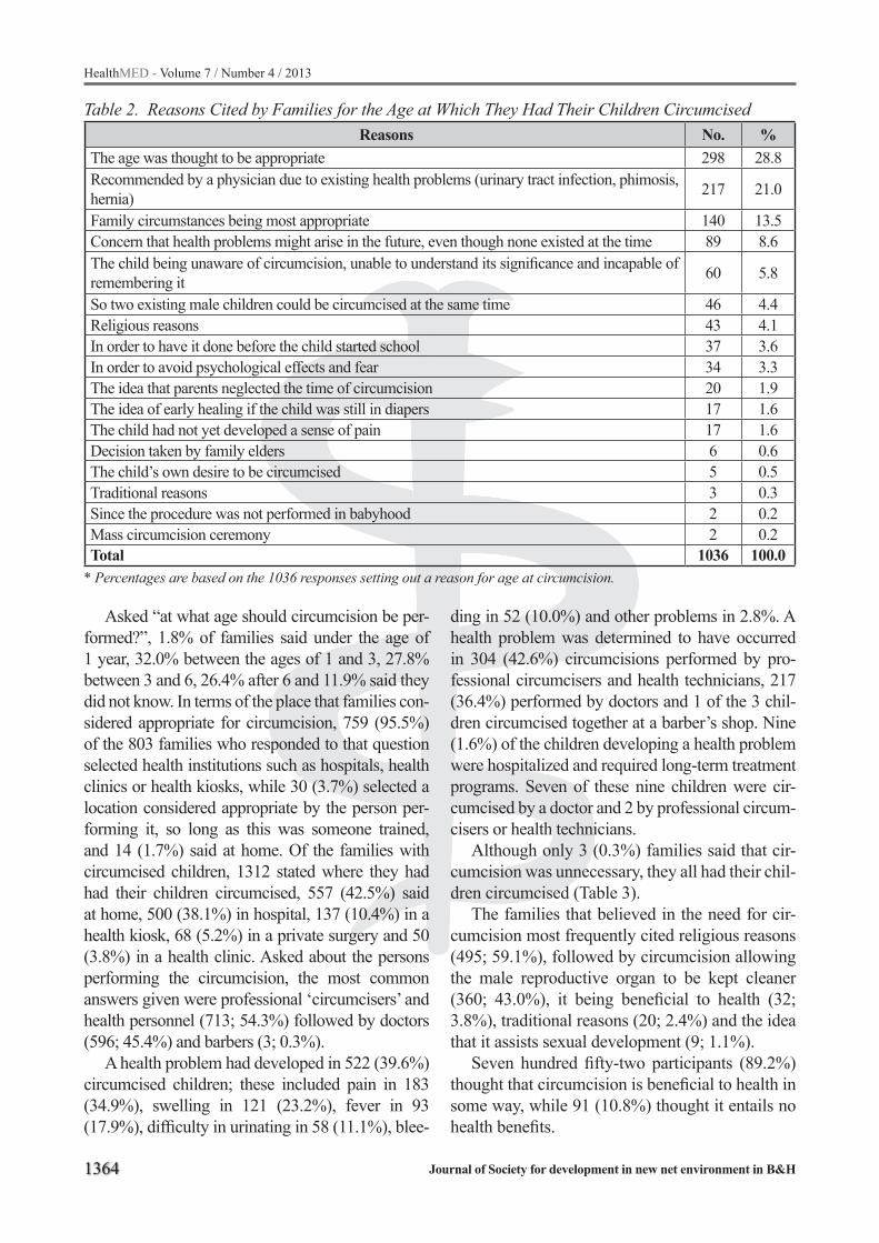
1364
HealthMED - Volume 7 / Number 4 / 2013
Journal of Society for development in new net environment in B&H
Asked “at what age should circumcision be per-formed?”, 1.8% of families said under the age of 1 year, 32.0% between the ages of 1 and 3, 27.8% between 3 and 6, 26.4% after 6 and 11.9% said they did not know. In terms of the place that families con-sidered appropriate for circumcision, 759 (95.5%) of the 803 families who responded to that question selected health institutions such as hospitals, health clinics or health kiosks, while 30 (3.7%) selected a location considered appropriate by the person per-forming it, so long as this was someone trained, and 14 (1.7%) said at home. Of the families with circumcised children, 1312 stated where they had had their children circumcised, 557 (42.5%) said at home, 500 (38.1%) in hospital, 137 (10.4%) in a health kiosk, 68 (5.2%) in a private surgery and 50 (3.8%) in a health clinic. Asked about the persons performing the circumcision, the most common answers given were professional ‘circumcisers’ and health personnel (713; 54.3%) followed by doctors (596; 45.4%) and barbers (3; 0.3%).
A health problem had developed in 522 (39.6%) circumcised children; these included pain in 183 (34.9%), swelling in 121 (23.2%), fever in 93 (17.9%), difficulty in urinating in 58 (11.1%), blee-
ding in 52 (10.0%) and other problems in 2.8%. A health problem was determined to have occurred in 304 (42.6%) circumcisions performed by pro-fessional circumcisers and health technicians, 217 (36.4%) performed by doctors and 1 of the 3 chil-dren circumcised together at a barber’s shop. Nine (1.6%) of the children developing a health problem were hospitalized and required long-term treatment programs. Seven of these nine children were cir-cumcised by a doctor and 2 by professional circum-cisers or health technicians.
Although only 3 (0.3%) families said that cir-cumcision was unnecessary, they all had their chil-dren circumcised (Table 3).
The families that believed in the need for cir-cumcision most frequently cited religious reasons (495; 59.1%), followed by circumcision allowing the male reproductive organ to be kept cleaner (360; 43.0%), it being beneficial to health (32; 3.8%), traditional reasons (20; 2.4%) and the idea that it assists sexual development (9; 1.1%).
Seven hundred fifty-two participants (89.2%) thought that circumcision is beneficial to health in some way, while 91 (10.8%) thought it entails no health benefits.
Table 2. Reasons Cited by Families for the Age at Which They Had Their Children Circumcised Reasons No. %
The age was thought to be appropriate 298 28.8Recommended by a physician due to existing health problems (urinary tract infection, phimosis, hernia) 217 21.0
Family circumstances being most appropriate 140 13.5Concern that health problems might arise in the future, even though none existed at the time 89 8.6The child being unaware of circumcision, unable to understand its significance and incapable of remembering it 60 5.8
So two existing male children could be circumcised at the same time 46 4.4Religious reasons 43 4.1In order to have it done before the child started school 37 3.6In order to avoid psychological effects and fear 34 3.3The idea that parents neglected the time of circumcision 20 1.9The idea of early healing if the child was still in diapers 17 1.6The child had not yet developed a sense of pain 17 1.6Decision taken by family elders 6 0.6The child’s own desire to be circumcised 5 0.5Traditional reasons 3 0.3Since the procedure was not performed in babyhood 2 0.2Mass circumcision ceremony 2 0.2Total 1036 100.0
* Percentages are based on the 1036 responses setting out a reason for age at circumcision.

HealthMED - Volume 7 / Number 4 / 2013
Journal of Society for development in new net environment in B&H 1365
When asked about the benefits of circumcision, families stated that it prevents bladder and kidney infections, as well as cervical cancer and genital infections in women (Table 4).
Discussion
Circumcision in Turkey is performed more for religious and traditional reasons than for its medi-cal benefits, generally between the ages of 5 and 7, after children have acquired the ability to reason. The reason for this is that in Turkish tradition, cir-cumcision occupies an important place in the lives of the mother, father and child, that families cele-brate the occasion together with their children and want their children to remember it in later life3,14,15.
The question by whom, where and when healthy and problem-free circumcision is to be performed is of considerable importance3,9.
The reasons behind circumcision may vary from one society to another. Religious and traditional reasons are the most important in Turkey, as well as others such as avoiding medical problems, cel-ebrating the event and it being a step toward man-hood8,16,17. In Korean society, by contrast, there is a common idea that it will enhance hygiene and future sexual functions, and also assist the child’s sexual development and endow him with a larger penis. The level of religious reasons is very low (<0.5%) 2,
7. In our study, religious reasons were the most im-portant factors behind circumcision (59.1%).
While there are traditional, religious and medi-cal aspects to circumcision, there is also a very serious anti-circumcision movement today. This movement, first initiated by atheists, then support-ed by Christians and more recently also by a group of Jews, initially sought to base its objections on scientific grounds. They then attempted to broad-en the movement by raising various ethical objec-tions. The movement even objects to circumcision by trying to undermine the religious grounds on which it is based, claiming that in the event of the child converting to a different faith or believing in no religion at all in the future, being circumcised might eliminate individuals’ right to freely choose their own religion17. In one study performed in Korea, 2.1% of participants said they regarded cir-cumcision as unnecessary7. In our study, 3 families (0.3%) stated that circumcision was unnecessary, though they still had their children circumcised. This does, however, show that there are opponents of circumcision even in Turkey.
Are there any health benefits to circumcision even though it is performed for religious and tra-ditional reasons? Oh et al. reported that families believed it prevents genital infections (80.6%), cervical cancer (64.7%) and urinary tract (62.7%) and kidney infections (53.4%) that might develop in the future7. Şahin et al. also reported that fami-lies believed that circumcision protects against infections8. In our study, the majority of families believed that circumcision is necessary and that
Table 3. Families’ Opinions Regarding Whether Circumcision Is Necessary Question
No. %Whether circumcision is necessary (n=828) Definitely necessary 795 96.0 Maybe necessary 19 2.3 Don’t know 7 0.9 May not be essential 4 0.5 Definitely unnecessary 3 0.3
Table 4. Families’ opinions regarding the benefits of circumcision
Belief in the benefits of circumcisionYes No Don’t know
No. % No. % No. %Prevents penile cancer 536 63.6 169 20.0 138 16.4Prevents urinary tract and kidney infections 628 74.5 88 10.4 126 14.9Circumcision prevents cervical cancer in women 410 48.6 236 28.0 196 23.3Circumcision prevents genital infections in women 465 55.2 189 22.4 189 22.4

1366
HealthMED - Volume 7 / Number 4 / 2013
Journal of Society for development in new net environment in B&H
it prevents urinary tract and kidney infections (87.6%) and penile cancer (76.0%). These results show that medical benefits are also considered among the factors making circumcision necessary.
The debate over the best age for circumcision continues, although avoiding circumcision as much as possible between the ages of 3 and 6 is recom-mended in order to avoid negativities arising from castration complex. Circumcision can be performed before these ages, during which psychological in-jury may develop, or after 9, 18. Since urinary system infection is more commonly seen in the first year of life, and particularly in the first 6 months, circumci-sion as early as possible is recommended, especial-ly in neonates with urinary system abnormalities, since circumcision in the newborn period prevents urinary system infections. It has also been empha-sized that circumcision in this period produces bet-ter esthetic results3, 8, 19-21.
In a study performed in South Korea, Oh et al. reported that children were circumcised more in earlier adolescence (» 11 years) than neonatally7.
In our study, a not insignificant number of fam-ilies (36.0%), a level comparable to that cited in other research, had their children circumcised at 36-71 months, outside the ideal ages (Table 5).
Very few children born in hospital in Turkey are circumcised immediately after birth, and gen-erally at the age of 5-7 3. In our study, only 8.7% of children were circumcised before the age of 1, and neonatal circumcision was determined to be very rare indeed. The families in our study stated that they had not considered having newborn children circumcised (65.4%). Among the reasons for not considering circumcision in the neonate period was that it was risky because the organ concerned was very small and that the baby would experi-ence pain. This suggests that families are unaware of the advantages posed by early circumcision.
The difference among the justifications for age at circumcision in different societies is striking. In Oh et al.’s study, the period they preferred and when families had their children circumcised, el-ementary school or slightly earlier, was described by families as when children would feel the least pain7. In studies in Turkey, families have empha-sized that children younger than 6 months heal faster, that they think that children younger than 2 do not feel pain and that children circumcised
at 6 and over “will not be afraid” as they now re-gard themselves as men3,16. In this study, 28.7% of families described the ages they had their children circumcised as “the right age,” while 21.0% stated that they had had to have their children circum-cised for medical reasons.
Circumcision is not just the removal of a piece of skin from the penis. Medical problems that may be encountered are sometimes irreversible and have a negative impact on the individual’s life, and even that of his family. For that reason, circumci-sion, a surgical procedure, must be performed un-der hygienic conditions and by expert physicians. In Turkey, circumcision is sometimes performed by people such as barbers or butchers, especially in rural areas, as well as by the health officials known as ‘circumcisers.’ In their study, Sivaslı et al. reported that 74.5% of children had been oper-ated on by such a ‘circumciser,” and Şahin et al. cited a figure of 13.3% 3,8. Even in our study, per-formed in the provincial capital, 54.3% of children had been circumcised by such officials.
In addition, the level of circumcisions at health institutions in Turkey is also low. For instance, in the study by Sivaslı et al. 17.3% of circumcisions were performed in hospitals and 8.2% in clinics3.In the present study, 42.5% of circumcisions were performed at home and 38.1% in hospital.
The most common complications tend to be minor and treatable: pain, bleeding, swelling or inadequate skin removal, excess bleeding and amputation of the glans penis, the penile shaft and the glans, infection, urinary retention, mea-tal ulcer, meatal stenosis, fistulas, loss of peni-le sensitivity, sexual dysfunction and edema of the glans penis22-24. A health problem developed in 39.6% of children circumcised, 9 (1.6%) of whom were hospitalized and had to be enrolled in long-term treatment programs. Circumcision is a small operation, but must be regarded as an important one in terms of outcome, and the need for it to be performed under hygienic condi-tions by physicians with expertise on the subject should not be forgotten.

HealthMED - Volume 7 / Number 4 / 2013
Journal of Society for development in new net environment in B&H 1367
Conclusion
The limitation of this study, the study is done in primary schools in urban area, but not includ-ed rural area. We established that all families had their children circumcised, that the main reason cited was religious and that the medical benefits of circumcision were also considered. Families were not sensitive to the issue of the age at which circumcision should be performed. Yet circumci-sion is the first and a serious procedure impinging on the body and sexuality of a male child. Society perceives circumcision as a ritual. There is also a need for social education programs stressing the medical importance of the procedure, which also affects the male child’s later life in terms of prob-lems that are or may be encountered.
Acknowledgment
We are profoundly grateful to the Trabzon Provincial Education Directorate for permission to conduct this research, to the directors, deputy directors and teachers at the Özel Neşem, Özel Candan, Erdoğdu, Bener Cordan, İskenderpaşa, 24 Şubat, İMKB, Ata, Gazi Paşa and Ata Koleji primary schools for their help and support, and to the student families that agreed to participate.
Table 5. Frequencies of age at circumcision according to various studies in the literature
Study authors Year of study Place Age/age group Incidence (%)
Şahin et al. 2003 Ankara
<1 year 13-35 months
3-6 years >6 years
14.87.735.941.6
Top et al. 2008 Giresun/Turkey<3 years 3-6 years>6 years
13.542.244.3
Sivaslı et al. 2003 Gaziantep/Turkey
<1 month2-3 months4-6 months7-12 months
1-2 years2-6 years7-13 years
9.68.05.635.213.421.36.9
Sang et al. 2003 Busan/ Korea
7 years 8 years9 years10 years11 years12 years
18.723.432.741.949.964.8
Kahriman et al. 2010, present study Trabzon/Tur-key
≤ 12 months13-35 months36-71 months≥ 72 months
8.737.436.017.9

1368
HealthMED - Volume 7 / Number 4 / 2013
Journal of Society for development in new net environment in B&H
References
1. Atikeler MK, Geçit İ, Bodakçi MN, Ergin E. Compli-cations of the circumcision performed within and out-side the hospital. Fırat University Medical Journal of Health Sciences.2001; 15: 477-479.
2. Lee SD, Park E, Choe BM. Parental concerns on the circumcision for elementary school boys. A Question-naire Study J Korean Med Sci. 2003; 18: 73-9.
3. Sivaslı E, Bozkurt Aİ, Ceylan H, Çoşkun Y. Knowl-edge, attitude and behaveior of parents regarding cir-cumcision in Gaziantep. Journal of Pediatrics. 2003; 46: 114-118.
4. Yazıcı M, Etensel B, Gürsoy H. Complications of cir-cumcision. Journal of Adnan Menderes University Medical Faculty. 2003; 4: 5-7.
5. Ceylan H, Topçu K, Özokutan BH at al. Circumci-sion and complications. Ege Pediatri Bulletin. 2004; 11: 1-5.
6. Dunsmuir WD and Gordon EM. The history of cir-cumcision. BJU International. 1999; 83: 1-12.
7. Oh SJ, Kim KD, Kim KM, Kim KS, Kim KK, Kim JS et al. Knowledge and attitudes of Koreans towards their sons’ circumcision: a nationwide questionnaire study. BJU International. 2002; 89: 426-32.
8. Şahin F, Beyazova U, Aktürk A. Attitudes and practices regarding circumcision in Turkey. Child: Care, Health and Development. 2003; 29: 275-280.
9. Balkan E. & Kılıç N. Circumcision and complica-tions. Güncel Pediatri. 2005; 3: 22-23.
10. Committee on Medical Ethics The law and ethics of male circumcision - guidance for doctors. British Medical Association, London: 2003.
11. College of Physicians and Surgeons of British Co-lumbia Policy Manual: Infant Male Circumcision. Vancouver, BC: College of Physicians and Surgeons of British Columbia: 2004.
12. American Academy of Pediatrics Committee on Bio-ethics. Informed consent, parental permission, and assent in pediatric practice. Pediatrics. 1995; 95: 314-317.
13. Seeking Patients Consent: The Ethical Consider-ations (1998) General Medical Council, London.
14. Tekgül S. Circumcision. Çocuk Sağlığı ve Hastalıkları Dergisi. 2000; 43: 297-302.
15. Yurdakök M. Kutsal Kitaplarda Çocuk Hekimliği Bilgileri. Alp Ofset Matbaacılık Makine San. ve Tic. Ltd Şti, Ankara 2001. 65-92.
16. Top FÜ, Üstüner F, Esüntimur Y, Uykan U, Pe-kdemir EA. The Knowledge, Behaviour and Attitu-de About of Circumcision in Families in Giresun. Çocuk Dergisi 2008; 8: 166-171.
17. Aksoy Ş. Sünnet, Sünnet mi? Medimagazin. 2010; 11: 470, 4.
18. Öztürk MO, Uluşahin A. Ruh Sağlığı ve Bozuklukları 11. Baskı, Ankara 2008.
19. Wiwell TE, Tencer HL, Welch CA, Chamberlain JL. Circumcision in children beyond the neonatal pe-riod. Pediatrics 1993; 92: 791-3.
20. Schoen EJ, Wiswell TE, Moses S. New policy on circumcision – cause for concern. Pediatrics. 2000; 105: 620–623.
21. Lerman SE, Liao JC. Neonatal circumcision. Ped Clin North Am, 2001; 48: 1539-1557.
22. Weiss HA, Larke N, Halperin D, Schenker I. Comp-lications of circumcision in male neonates, infants and children: a systematic review. BMC Urology. 2010; 10: 1-13.
23. Ademuyiwa O, Bode CO. Complications of neonatal circumcision: Avoiding common pitfalls in a com-mon procedure. African Journal of Paediatric Sur-gery, 2009; 6: 134-136.
24. Söylemez H, Burcu B. Habits and results of circum-cısion in Turkey. The New Journal of Urology. 2009; 5: 13-18.
Corresponding AuthorIlknur Kahriman,Karadeniz Technical University,Faculty of Health Science,Trabzon,Turkey,E-mail: [email protected]

HealthMED - Volume 7 / Number 4 / 2013
Journal of Society for development in new net environment in B&H 1369
Abstract
Objective: To evaluate the psychometric pro-perties and relations among the Quick Inventory of Depressive Symptomatology (QIDS-C16), clinical version, the Quick Inventory of Depressive Symp-tomatology, self-report version (QIDS-SR16), and the 17-item version of the Hamilton Rating Scale for Depression (HAM-D17) in a Turkish student sample
Materials and methods: Slightly modified ver-sions of the three scales were administered to 114 outpatients at the Uludağ University campus-based family health center. SAS, MPlus, Multilog, and SS-IRT were used to provide descriptive statistics, classical psychometric analyses, including factor analysis, test informations, and equatings of the three scale scores.
Results: The internal consistencies (Cronbach’s a) values of the QIDS-C16, QIDS-SR16, and HAM-D17 were 0.81, 0.80, and 0.89. Both versions of the QIDS were unidimensional, but the HAM-D17 required two dimensions according to a parallel analysis criterion. The pattern of item-total corre-lations for the two versions of the QIDS was si-milar. The correlation between the QIDS-C16 and QIDS-SR16 was 0.70. Similarly, the correlation between the QIDS-C16 and HAM-D17 was 0.79, and the correlation between the QIDS-SR16 and the HAM-D17 was 0.59. These respective corre-lations increased to 0.87, 0.94 and 0.70 when di-sattenuated (corrected for unreliability). Scores on the three measures were also equated.
Discussion: All three measures have good psychometric properties and convergent validity. In particular, this extends the utility of the QIDS-SR16, the object of a previous study. One impor-tant limitation in the present case is the small sample size (N = 114) Use of the QIDS-SR16 is
recommended when a self-reported instrument is appropriate even though this measure has slightly less in common with the QIDS-C16 and HAM-D17 when clinical judgment is possible.
Key words: Major depressive episode, scree-ning scale, validity, reliability.
Introduction
In a previous study, Mergen, Bernstein, Tavli et al. (1) compared the self-report version of the Quick Inventory of Depressive Symptomatology, in a Tur-kish student sample (QIDS-SR16-T) to (a) the Ame-rican version (QIDS-SR16-US) and (b) the Turkish version of the Beck Depression Inventory (BDI-II-T). Turkish data were obtained from 626 outpatients at the Uludağ University campus-based family he-alth center, and American data were obtained from 584 respondents at an American University as admi-nistered to a class in introductory psychology.
Internal consistencies (Cronbach’s a) of the two versions of the QIDS were in the .70s and were higher for the longer BDI-II-T at .89. However, the two versions of the QIDS were both unidimensional whereas the BDI-II-T was not. Item-total correlati-ons of the two versions of the QIDS were similar, and the correlation between the QIDS-SR16-T and BDI-II-T was .90 when disattenuated (corrected for unreliability). Multiple group item response theory analysis (2) analysis indicated that the two versions of the QIDS had different intercepts but the same fac-tor loadings. The difference in intercepts was not sur-prising given that participant selection in the Turkish sample favored those with depressive pathology.
It was concluded that the psychometric proper-ties of the QIDS-SR16-T and its convergent validity with the BDI-II-T is at least satisfactory for use when a self-reported instrument is appropriate.
Use of the QIDS-C16, QIDS-SR16 and the 17-item Hamilton rating scale for depression (HAM-D17) in a Turkish college student sample Haluk Mergen1, Ira H. Bernstein2, Berna Erdogmus Mergen3
1 Uludag University Family Health Center, Bursa, Turkey,2 University of Texas Southwestern Medical Center, Dallas, United States of America,3 Esentepe Family Health Center, Bursa, Turkey.

1370
HealthMED - Volume 7 / Number 4 / 2013
Journal of Society for development in new net environment in B&H
This study compared the clinical version of the QIDS (QIDS-C16), the QIDS-SR16, and the 17-item version of the Hamilton Rating Scale for De-pression (HAM-D17) in a Turkish student sample. Since all data were gathered in a Turkish sample, “-T” in the abbreviations of the three tests is re-dundant and will not be used.
Methods
The three scales, i.e., the QIDS-C16, the QIDS-SR16, and HAM-D17, were administered to 114 outpatients who participated at the Uludağ Uni-versity campus-based family health. As previou-sly noted (1), the QIDS-C16 and QIDS-SR16 were obtained from the www.ids-qids.org website (3). A few minor changes were made to accommodate actual Turkish usage, e.g., references to weight in pounds were replaced by weight in kilograms. The HAM-D17 was as originally published, in contrast to variants with more or fewer items. The transla-tions are available from the first author.
The sample consisted of students, who presen-ted consecutively to the outpatient clinic of the university. The study conformed to the Helsinki declaration requirements and had approval from the University ethics committee. The students gave informed consent and voluntarily participa-ted in the study. They completed the QIDS-SR16 and HAM-D17 in 7-10 minutes.
Statistical AnalysesDescriptive analyses, classical test theory (CTT),
exploratory factor analysis and IRT analyses were all employed. However, only the test information functions and test-equation results of the latter will be presented because the limited sample size makes the item-specific results somewhat unstable. The classical test theory analyses generated item me-ans, item standard deviations, item-total correlati-ons (rit), scale means, and scale standard deviations. This analysis also included intercorrelating the sca-le scores. The dimensionality of the three tests was evaluated using exploratory factor analysis. Parallel analysis (4, 5, 6, 7) was used to decide upon the number of factors (dimensionality). The version used in this study consisted of generating a series of 50 matrices of random normal deviates. Each ma-trix had 9 columns and 114 rows for the two versi-
ons of the QIDS and 17 columns and 114 rows for the HAM-D17. Thus, the number of columns equa-led the number of items on the actual test and the number of rows equaled the number of participants. Each of the resulting matrices was then subjected to principal component analysis and the eigenvalues averaged within each of the three sets. A test may be considered unidimensional if (a) its first eigen-value exceeds its simulated counterpart and (b) all of its subsequent obtained eigenvalues are smaller than the corresponding simulated eigenvalues. All of these analyses used SAS 9.2.
The test information function derived from the Samejima (8,9) IRT model describes the sensitivity of the latent variable, depression in this case (sym-bolized q and measured in standard deviation units) to changes in score. In essence it is a reliability-like measure, but one that is a function of q rather than a constant for all test scores. Finally, test equating involves finding scores on two or more tests with equivalent or nearly equivalent scores on q. Thus, a score of 3 on test A and a score of 5 on test B are considered equated if they produce similar values of θ. In the present case, this means that a patient getting a score of 3 on test A is as depressed as a patient who gets a score of 5 on test B. This equa-ting employed the SS-IRT program developed by Orlando, Sherbourne, and Thissen (10).
Results
Demographic resultsTable 1 contains the relevant demographic infor-
mation. Although patients with a family history of depression did not score significantly different from those without a family history on the two versions of the QIDS, they did score lower on the HAM-D17. The respective means were 13.3 and 8.5, t(112) = 2.39, p < .02. Not surprisingly, patients on antide-pressant medication scored higher on all three tests than those not on antidepressant medications. The respective means for the QIDS-C16, QIDS-SR16, and HAM-D17 were 9.6, 13.3, and 20.3 for the for-mer group and 5.8, 7.9, and 8.2 for the latter group, t(112) = 2.93, 2.70, and 4.71, p< .01.
The QIDS-SR16 scores were higher in the present study than in that reported previously by Mergen, Bernstein, Tavli et al. (1). The present and earlier means were 8.2 and 6.9, t(738) = 2.46, p < .02. Be-

HealthMED - Volume 7 / Number 4 / 2013
Journal of Society for development in new net environment in B&H 1371
cause the present sample was slightly more variable than the earlier sample (s = 5.2 vs. 4.9), Cronbach’s α was also slightly larger in this sample (.80 vs.78)
None of the remaining group differences (age, city vs. non-city residence, gender, income, or number of siblings approached significant.Table 1. Demographic Characteristics of Sample
Item Est.Mean Age 20.6St. Dev. Age 3.9Income: <500 TL* (%) 33Income: 501-1000 TL (%) 37Income: 1001-2000 TL (%) 22Income: >2000 TL (%) 8No. Siblings = 0 (%) 6No. Siblings = 1 (%) 29No. Siblings = 2 (%) 34No. Siblings = 3 (%) 13No. Siblings = 4+ (%) 18City Dwelling (%) 81Family History of Depression (%) 17Female Gender (%) 44On Antidepressants (%) 9
TL= Turkish money unit (1$=1.77 TL on 4/12/2012)
CTT Analysis Table 2 contains the relevant CTT results. The
correlation between the QIDS-C16 and QIDS-SR16 was .70; the correlation between the QIDS-C16 and HAM-D17 was .79, and the correlation be-tween the QIDS-SR16 and HAM-D17 was .59. Cor-recting for attenuation (unreliability) by dividing these respective correlations by the square root of the product of the coefficients a yields respective estimates of the error-free correlations of .87, .94, and .70. In other words, the two clinical versions correlated more highly with one another than did either with the self-report even though one of the clinical versions was identical in content to the self-report version.
Exploratory Factor Analysis (Dimensionality)Both versions of the QIDS were unidimen-
sional by the parallel analysis criterion. Specifi-cally, the first eigenvalues of the QIDS-C16 were 3.82 and 1.13 and the QIDS-SR16 were 3.71 and 1.04. In contrast, the first two simulated eigenval-ues were 1.44 and 1.29. This first factor accounted for 42.5% and 41.2% of the variances in the re-spective measures. However, the HAM-D17 con-
Table 2. Item means, item standard deviations, item-total correlations (rit), scale means, scale standard deviations, and Cronbach’s a
Domain QIDS-C16 QIDS-SR16 HAM-D17
(Item) Mean S. D. rit Mean S. D. rit Mean S. D. rit
1 1.39 .93 .40 1.80 .96 .41 .69 .83 .712 .65 .75 .65 .74 .85 .59 .33 .67 .623 .70 .81 .23 1.01 .97 .47 .10 .38 .244 .85 .76 .62 1.08 .89 .67 1.03 1.01 .665 .40 .70 .60 .80 1.21 .35 .38 .77 .256 .12 .36 .33 .20 .57 .50 .73 1.06 .457 .43 .65 .55 .61 .91 .53 .76 .97 .538 .75 .59 .61 .67 .76 .63 .51 .90 .579 .83 .77 .65 1.25 1.15 .42 .93 .98 .5510 .54 .73 .5311 .17 .42 .4312 1.01 1.03 .7013 .93 .93 .6814 .15 .50 .3615 .18 .47 .6216 .39 .77 .5317 .47 .81 .64
Scale 6.12 4.05 8.35 6.24 9.30 8.19 a .81 .80 .89
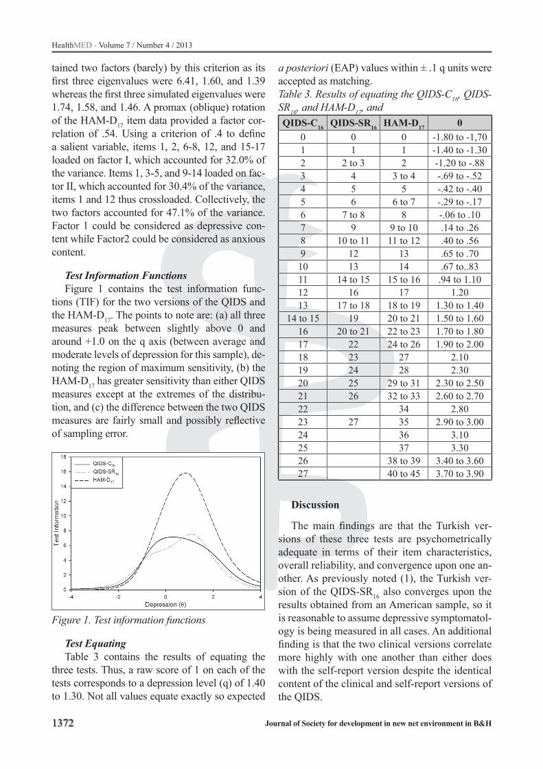
1372
HealthMED - Volume 7 / Number 4 / 2013
Journal of Society for development in new net environment in B&H
tained two factors (barely) by this criterion as its first three eigenvalues were 6.41, 1.60, and 1.39 whereas the first three simulated eigenvalues were 1.74, 1.58, and 1.46. A promax (oblique) rotation of the HAM-D17 item data provided a factor cor-relation of .54. Using a criterion of .4 to define a salient variable, items 1, 2, 6-8, 12, and 15-17 loaded on factor I, which accounted for 32.0% of the variance. Items 1, 3-5, and 9-14 loaded on fac-tor II, which accounted for 30.4% of the variance, items 1 and 12 thus crossloaded. Collectively, the two factors accounted for 47.1% of the variance. Factor 1 could be considered as depressive con-tent while Factor2 could be considered as anxious content.
Test Information FunctionsFigure 1 contains the test information func-
tions (TIF) for the two versions of the QIDS and the HAM-D17. The points to note are: (a) all three measures peak between slightly above 0 and around +1.0 on the q axis (between average and moderate levels of depression for this sample), de-noting the region of maximum sensitivity, (b) the HAM-D17 has greater sensitivity than either QIDS measures except at the extremes of the distribu-tion, and (c) the difference between the two QIDS measures are fairly small and possibly reflective of sampling error.
Figure 1. Test information functions
Test EquatingTable 3 contains the results of equating the
three tests. Thus, a raw score of 1 on each of the tests corresponds to a depression level (q) of 1.40 to 1.30. Not all values equate exactly so expected
a posteriori (EAP) values within ± .1 q units were accepted as matching.Table 3. Results of equating the QIDS-C16, QIDS-SR16, and HAM-D17, and QIDS-C16 QIDS-SR16 HAM-D17 θ
0 0 0 -1.80 to -1,701 1 1 -1.40 to -1.302 2 to 3 2 -1.20 to -.883 4 3 to 4 -.69 to -.524 5 5 -.42 to -.405 6 6 to 7 -.29 to -.176 7 to 8 8 -.06 to .107 9 9 to 10 .14 to .268 10 to 11 11 to 12 .40 to .569 12 13 .65 to .7010 13 14 .67 to..8311 14 to 15 15 to 16 .94 to 1.1012 16 17 1.2013 17 to 18 18 to 19 1.30 to 1.40
14 to 15 19 20 to 21 1.50 to 1.6016 20 to 21 22 to 23 1.70 to 1.8017 22 24 to 26 1.90 to 2.0018 23 27 2.1019 24 28 2.3020 25 29 to 31 2.30 to 2.5021 26 32 to 33 2.60 to 2.7022 34 2.8023 27 35 2.90 to 3.0024 36 3.1025 37 3.3026 38 to 39 3.40 to 3.6027 40 to 45 3.70 to 3.90
Discussion
The main findings are that the Turkish ver-sions of these three tests are psychometrically adequate in terms of their item characteristics, overall reliability, and convergence upon one an-other. As previously noted (1), the Turkish ver-sion of the QIDS-SR16 also converges upon the results obtained from an American sample, so it is reasonable to assume depressive symptomatol-ogy is being measured in all cases. An additional finding is that the two clinical versions correlate more highly with one another than either does with the self-report version despite the identical content of the clinical and self-report versions of the QIDS.

HealthMED - Volume 7 / Number 4 / 2013
Journal of Society for development in new net environment in B&H 1373
The HAM-D17 is slightly more sensitive to dif-ferences in depression than either version of the QIDS, and all three measures are most sensitive to somewhat above average levels of depression (roughly the 60th to the 85th percentile) within the present group. However, the greater sensitivity of the HAM-D17 is somewhat offset by its multidi-mensionality, i.e., its apparent tendency to con-found depressive and anxious symptomatology, certainly a familiar problem in the psychiatric lit-erature. This confounding seems important given the different medications most appropriate for the two conditions. One can of course use only the Hamilton’s depression items, but that will tend to reduce its sensitivity. As was noted in our previous study, the QIDS-SR16 seems appropriate for the evaluation of depressive symptomatology when-ever a self-descriptive measure is appropriate. One benefit of our approach is that we have furnished a QIDS-HAM-D equating for users of one test who are relatively unfamiliar with the alternative(s).
Rush et al. (11) compared the 30 item Inven-tory of depressive symptomatology (IDS-SR30), the QIDS-SR16, the QIDS-C16, the HAM-D17, and two of the latter’s variants differing as to number of items (HAM-D21 and HAM-D24) in a sample of 596 patients being treated for depression. The in-ternal consistency Cronbach’s α of the QIDS-SR16 was 0.86, and the HAM-D17 was 0 .83. Thus, their QIDS-SR16 was marginally higher than the present and their HAM-D17 was marginally lower.
Rush et al. (12) is similar to the present study in that they administered the QIDS-C16, QIDS-SR16, and HAM-D17, using a sample of 582 patients. The internal consistency (Cronbach’s α) of the three scales were respectively 0.87, 0.87 and 0.89 in their study as opposed to 0.81, 0.80, 0.89 in this study. Again, these differences are marginal.
Brown et al. (13) administered the QIDS-SR16, IDS-SR30, HAM-D17 and Mini Asthma Quality of Life Questionnaire to 73 asthmatic patients at treatment exit. Their primary interest was in the apparent co-occurrence of asthma and depression. The internal consistency Cronbach’s α values of QIDS-SR16 and HAM-D17 were both .87 and their respective standard deviations were 4.8 and 4.7. In our study, the internal consistency Cronbach’s α values of QIDS-SR16 and HAM-D17 were respec-tively .80 and .89. In addition, the standard devia-
tions were 6.24 and 6.19. The slight differences in coefficient a are thus not readily explained by differences in variability, as they often can be.
The marginal differences in reliability among the studies, including the present may reflect two things beyond simple random error. First, great-er reliabilities tend to be associated with greater variabilities. For example, the variabilities of the QIDS-C16 and HAM-D17 (but not the QIDS-SR16) were greater in Rush et al. (2006) than in this study (12). Reliability, being a ratio of true variance to total variance, increases as total increases since the remaining component, error, is, by definition, in-dependent of true variance and remains constant. As a result, increasing total variance normally also increases true variance by that same amount (14). Second, the data in some studies, e.g., Rush et al. (12) were obtained from an exit interview follow-ing prior assessments. We typically find that reli-abilities increase with number of assessments as respondents learn to treat items as a group rather than as separate items.
This paper adds to the number of scales that measure depressive symptomatology available in Turkish, which have at least some supporting psychometric data. These data could be found in the book of Aydemir & Köroğlu(15). The others include Akdemir, Örsel, Dağ et al. (16) who first adapted the HAM-D17, which is a clinical scale. They noted a Cronbach’s α of .75 and a test-retest (stability) reliability of 0.85. The scale correlated .48 with a translation of the Beck Depression in-ventory. In turn, Hisli (17) adapted the Beck De-pression Inventory, a self-report scale. The study reported a Cronbach’s α was .80. and a correlation of .50 with MMPI scale 2 (depression).
Özer, Demir, Tuğal, et al. (18) adapted the Montgomery-Asberg depression rating scale (MADRS), a clinical scale.. They reported pairwise interrater reliabilities among four psychi-atrists/raters ranging from .66 to .86. Cronbach’s α ranged from .74 to .84, and the correlations be-tween this scale and the Beck Depression Inven-tory scores ranged from .64 to .68.
Tatar and Saltukoğlu (19) adapted the Center for Epidemiological Studies Depression Scale (CES-D), a self-report scale. Its Cronbach’s α was .89. The test-retest correlation was .69. It corre-lated .87 with the Beck Depression Inventory.

1374
HealthMED - Volume 7 / Number 4 / 2013
Journal of Society for development in new net environment in B&H
Finally, Aydemir, Güven, Küey, et al. (20) adapted the Hospital Anxiety and Depression Scale, which is a self-report scale. Cronbach’s α was .78 for the depression sub-scale and it corre-lated .72 with the Beck Depression Inventory.
Limitations
One major limitation of the study is the rela-tively small number of observations, especially as compared to its predecessor. As before, the study sample consists of university students seen at an outpatient clinical setting, so it might not apply it to a more general Turkish population. A third im-portant limitation is that we did not have repeated test data to evaluate stability and sensitivity to change of the Turkish QIDS-SR16, which would be necessary if the scale were to monitor therapeu-tic effects. This was not the goal of this study nor its predecessor. Finally, the degree to which the test equating will be stable depends upon the rel-evant sample size. Ours can only provide highly tentative estimates until a much larger sample can be obtained.
References
1. Mergen H, Bernstein IH, Tavli V, Ongel K, Tavlı T, Tan Ş. Comparative Validity and Reliability Study of The QIDS-SR16 in Turkish and American College Stu-dent Samples. Klinik Psikofarmakoloji Bülteni 2011; 21(4): 289-301.
2. De Ayala, RJ. The theory and practice of item respon-se theory. New York: The Guilford Press, 2009.
3. IDS/QIDS Instruments in English and Multiple Trans-lations. www.ids-qids.org accessed on 4/12/2012.
4. Horn JL. An empirical comparison of various met-hods for estimating common factor scores. Educati-onal and Psychological Measurement 1976; 25: 313-322.
5. Humphreys LG, Ilgen D. Note on a criterion for the number of common factors. Educational and Psycho-logical Measurement 1969; 29: 571-578.
6. Humphreys LG, Montanelli RGJr. An investigation of the parallel analysis criterion for determining the number of common factors. Multivariate Behavioral Research 1975; 10: 193-206.
7. Montanelli RGJr, Humphreys LG. Latent roots of ran-dom data correlation matrices with squared multiple correlations on the diagonal: a Monte Carlo study. Psychometrika 1976; 41: 341-348.
8. Samejima, F. Estimation of latent ability using a res-ponse pattern of graded scores. Psychological Mono-graphs 1969; 4: 2.
9. Samejima, F. Graded response model. In: van Linden, W. and Hambleton, R.K. (Eds.), Handbook of Modern Item Response Theory. Springer-Verlag, New York, 1997. p.85-100.
10. Orlando, M., Sherbourne, C.D. and Thissen, D. Summed-score linking using item response theory: application to depression measurement. Psychologi-cal. Assessessment 2000; 12, 354-359.
11. Rush AJ, Trivedi MH, İbrahim HM, Carmody TJ, Ar-now B, Klein DN et al..The 16-Item Quick Inventory of Depressive Symptomatology (QIDS), Clinician Rating (QIDS-C), and Self-Report (QIDS-SR): A Psy-chometric Evaluation in Patients with Chronic Major Depression. BioIogical Psychiatry 2003; 54: 573-583
12. Rush AJ, Bernstein IH, Trivedi MH et al. An evalu-ation of the quick inventory of depressive symptom-atology and the Hamilton rating scale for depres-sion: a sequenced treatment alternatives to relieve depression trial report. Biological Psychiatry 2006; 59(6): 493-501.
13. Brown ES, Murray M, Carmody TJ et al. The Quick Inventory of Depressive Symptomatology-Self-report: a psychometric evaluation in patients with asthma and major depressive disorder. Annals of Allergy, Asthma, and Immunology 2008; 100(5): 433-8.
14. Nunnally, JC, Bernstein IH.. Psychometric Theory. 3rd ed. New York: McGraw-Hill, 1994.
15. Aydemir Ö, Köroğlu E. Psikiyatride Kullanılan Klinik Ölçekler. Ankara: HYB Yayıncılık, 2012.
16. Akdemir A, Örsel S, Dağ İ, Türkçapar H, İşcan N, Özbay H. Hamilton Depresyon Değerlendirme Öl-çeği (HDDÖ)’nin Geçerliği, Güvenilirliği ve Klinik-te Kullanımı. Psikiyatri Psikoloji Psikofarmakoloji Dergisi 1996; 4(4): 251-259.
17. Hisli N. Beck Depresyon Envanterinin Üniversite Öğrencileri İçin Geçerliliği, Güvenilirliği. Psikoloji Dergisi 1989; 7: 3-13.
18. Özer S, Demir B, Tuğal Ö, Kabakçı E, Yazıcı MK. Montgomery-Asberg Depresyon Değerlendirme Ölçeği: Değerlendiriciler Arası Güvenilirlik ve Geçerlilik Çalışması. Türk Psikiyatri Dergisi 2001; 12(3): 185-194.

HealthMED - Volume 7 / Number 4 / 2013
Journal of Society for development in new net environment in B&H 1375
19. Tatar A, Saltukoglu G. CES-Depresyon Ölçeği’nin Doğrulayıcı Faktör Analizi ve Madde Cevap Kuramı kullanımı ile Türkçe’ye uyarlanması ve Psikometrik Özelliklerinin İncelenmesi. Klinik Psikofarmaloji Bülteni 2010; 20: 213-222.
20. Aydemir Ö, Güvenir T, Küey L, Kültür S. Hastane Anksyete ve Depresyon Ölçeği Türkçe Formunun Geçerlilik ve Güvenilirliği. Türk Psikiyatri Dergisi 1997; 8(4): 280-287.
Corresponding AuthorHaluk Mergen,Uludag University Family Health Center,Bursa,Turkey,E-mail: [email protected]
1376
HealthMED - Volume 7 / Number 4 / 2013
Journal of Society for development in new net environment in B&H
Abstract
Objective. The aim of this study is to deter-mine the presence of carotid artery calcifications (CACs) detected on panoramic radiographs (PR) in end-stage renal disease patients (ESRD) with hemo and peritoneal dialysis and to analyze the relationship between this calcification prevalence and dialysis period.
Design. Subjects and Methods. A random sample of 170 panoramic radiographs belonged to ESRD patients were investigated.
Results. Of the 155 ESRD patients included in the data analysis, 41 (26 %) were detected as having CACs on PRs (29 PD &12 HD patients). There was a statistically significant difference in terms of mean age between the patients with CACs and those without CACs. A statistical diffe-rence was found in PD and HD period between the patients with CACs and those without CACs.
Conclusions. Suggesting the results of the stu-dy the incidental finding of CACs in the area of the carotid arteries on PRs should be followed up with carotid screening due to detect asymptomatic patients in terms of f stroke. Therefore, it could give us the life-saving information.
Key words: panoramic radiograph, carotid ar-tery calcification, end-stage renal disease
Introduction
Atherosclerosis is a chronic and progressive in-flammatory disease, which takes place in the intima-media layer of the arterial wall. Many general and
systemic factors are revealed to play an important role in the etiology of the process, including age, blood pressure, diabetes, hyperlipidemia, smoking, and radiotherapy (1, 2). In the general population, atherosclerotic disorder is known as an important etiology of morbidity and mortality (3). The risk is also known to increase in patients with chronic diseases. End-stage renal disease (ESRD) patients have a much higher prevalence of atherosclerosis in comparison to the normal population (4, 5).
Cardiovascular disease and stroke are known to be the leading cause of mortality in ESRD pa-tients. These patients need dialysis and have a ten- to twenty-fold risk in comparison to the age and sex of matched normal subjects in terms of having atherosclerotic heart and vascular disease (6, 7). Vascular calcifications are commonly seen in these patients due to ESRD (8). Although the etiology of vascular calcification in patients with ESRD is not clear, the process is most probably multi-factorial. In some reports, older age and a longer time on dialysis have been revealed as the major risk factors in vascular calcification (7, 9, 10). In addition, the process results in increased stiffness of the elastic-type, capacitive, large arte-ries, such as common carotid and the aorta artery (11). Arterial stenosis due to carotid atheromas is one of the most important risk factors leading to stroke (3). Approximately 80% of all strokes are ischemic due to atherosclerotic disorder in the ca-rotid bifurcation region (12-14).
Panoramic radiographs (PRs), which commonly display the existence of calcifications in the region of the 2nd, 3rd, and 4th cervical vertebrae, are gene-
The prevalence of carotid artery calcification or atherosclerotic plaque in end-stage renal disease being treated with hemodialysis and peritoneal dialysis patients detected on panoramic radiographyCumali Gokce1, Elif Tarim Ertas2, Yildiray Sisman3, Murat Sipahioglu4, Mursel Davarci5, Oktay Oymak4, Cengis Utas4
1 Department of Endocrinology and Metabolism, Mustafa Kemal University, Hatay, Turkey,2 Department of Oral and Maxillofacial Radiology, Faculty of Dentistry, Izmir Katip Celebi University, Izmir, Turkey,3 Department of Oral and Maxillofacial Radiology Faculty of Dentistry, Erciyes University, Kayseri, Turkey,4 Department of Nephrology, School of Medicine, Erciyes University, Kayseri, Turkey,5 Department of Urology, Mustafa Kemal University, Hatay, Turkey.

HealthMED - Volume 7 / Number 4 / 2013
Journal of Society for development in new net environment in B&H 1377
rally necessary during routine dental examination (15-18). Early in the 1980s, Friedlander and Lande first reported the incidence of carotid artery calcifi-cation (CAC) on PRs, and they suggested that the-se radiographs were able to play an important role in the early diagnosis of CAC, which are known to cause more serious stroke and heart disease (19, 20).
Many reports have investigated this further to confirm the ability of PRs to detect CACs in the carotid bifurcation region through correlation with medical history, Doppler spectral analysis, or cer-vical spine radiographs (20-24). In these studies, the overall prevalence rate of CAC was revealed to range from 2% to 11% of the adult population, and the incidence increases remarkably in samples with specific chronic diseases (23, 25-28). In our previous study, which was first performed with peritoneal dialysis (PD) patients, the incidence of CAC was found to be 27.3%, and this was the highest reported prevalence in the literature (19).
The aim of the current study is to investigate the prevalence of CAC by using standard PRs in ESRD patients (the largest sample), who need di-alysis, in order to evaluate the predictive value of the radiographs in the diagnosis of atherosclerotic heart and vascular disease.
Material and methods
PatientsFor the study, patients with ESRD being tre-
ated with PD and HD were referred for a dental examination, and the PRs taken for routine radi-ographic examination were investigated for the presence of CAC. All the PRs were performed at the Department of Oral Diagnosis and Radiology, Erciyes University Faculty of Dentistry. Since the radiographs had originally been taken for routine dental examination and not for the investigation of CAC, it was not necessary to seek ethical approval for the study.
PR processing and evaluationAll radiographs were obtained by the same
radiology technician using either a conventional (Orthopantomograph® OP 100, Instrumentarium Corp., Tuusula, Finland) or digital (Orthopanto-mography® OP 200D, Instrumentarium Corp., Tuusula, Finland) device. Conventional radio-
graphs were processed in an automatic film proce-ssor according to the manufacturer’s recommen-dations, and digital radiographs were printed with a laser printer at the same magnification factor (1: 1.3). Radiographs were viewed on a standard viewbox in subdued ambient light.
PRs were evaluated by the same authors (ETE & YS), and any radiographs that showed questi-onable evidence of CAC were excluded from the study. Radiographs were also excluded if they did not include vertebrae C3 and C4, or if the pati-ent had moved during the radiograph exposure. Each PR was viewed in subdued ambient light using transmitted light from a standard viewbox. The presence of a radiopaque nodular mass (or masses) adjacent to the cervical vertebrae at or below the intervertebral space between C3 and C4 was diagnosed as a CAC. Unilateral and bi-lateral CACs on the radiographs were recorded separately. For a differential diagnosis of CACs, other cervical calcifications, such as calcified tri-ticeous cartilage, calcified thyroid cartilage, hyo-id bone, and submandibular salivary gland sialo-liths, were excluded according to Carter’s study (26). To check intra-observer variation, all of the PRs were re-evaluated by the same authors (ETE & YS) after one month. The dialysis period of all the patients was recorded. The patients diagnosed with CACs in the PRs were contacted by phone for further Doppler ultrasound confirmation. Of the 14 patients, four cases had passed away due to stroke. The other patients with CACs diagno-sed by PRs were referred to the Erciyes University School of Medicine’s Radiology Department for color Doppler ultrasonography (CDUS) examina-tion to determine the location and extent of calci-fication. The presence of CAC was confirmed by an ultrasound examination. Figure 1 shows the PR of a PD patient displaying calcifications on both cervical regions. Figure 2a demonstrates a color Doppler image of the right, while Figure 2b shows the left internal carotid artery of the same patient in Figure 1, but revealing the extent of the CACs. Figure 3 shows the PR of a HD patient displaying calcifications on both cervical regions. Figure 4a demonstrates color Doppler images of the right, and Figure 4b shows the left internal carotid ar-tery of the same patient in Figure 3, but reveals the extent of the CACs.

1378
HealthMED - Volume 7 / Number 4 / 2013
Journal of Society for development in new net environment in B&H
Figure 1. Shows the PR of a PD patient dis-playing calcifications on both cervical regions
Figure 2a. Demonstrates color doppler image of the right
Figure 2b. Shows the left internal carotid artery of the same patient in Figure 1 showing the extent of CACs
Figure 3. Shows the PR of a HD patient dis-playing calcifications on both cervical regions
Figure 4a. Demonstrates Color Doppler images of the right
Figure 4b. Shows the left internal carotid artery of the same patient in Figure 3 showing the extent of CACs
Statistical analysisStatistical evaluation of the data was carried
out using the Statistical Package for Social Sci-ences (SPSS® version 13.0; SPSS Inc., Chicago,

HealthMED - Volume 7 / Number 4 / 2013
Journal of Society for development in new net environment in B&H 1379
IL, USA). The differences between the categorical variables were analyzed using chi-square, Fisher’s exact test, the Mann-Whitney U test and the Spe-arman correlation test. P-values < 0.05 were con-sidered to be statistically significant.
Results
A total of 170 patients with ESRD being trea-ted with PD and HD were included in the study. The PRs of 15 patients who had questionable CACs were excluded; hence the PRs of 155 ESRD pati-ents with PD (116) and HD (39) were included in the data analysis. This population consisted of 85 males and 70 females, with an overall mean age of 44.4 ± 14.2 years. ESRD patients being treated with PD included 58 males and 58 females. The mean age was 44.9 ± 13.7 years. The ESRD population being treated with HD comprised 27 males and 12 females. The mean age was 42.8 ± 15.5 years.
The mean age of ESRD patients was 44.4 ± 14.2 years, and there was no statistically significant difference in respect of mean age between males and females (P = 0.526). Of the 155 ESRD pati-ents included in the data analysis, 41 (26 %) had CACs on the PRs, and of those, 17 (11%) were male and 24 (15%) were female. There was a signi-ficant difference in terms of mean age between the patients with CACs (49.9 ± 15.1 years) and those without CACs (42.5 ± 13.4 years) (P = 0.004). Cal-cifications were unilateral in 31 (76%) and bilateral in 10 (24%) patients with CACs. A total of 51 CAC cases were therefore detected in the 41 patients and, of these, 12 (29%) were located on the right side and 19 (46%) were located on the left side.
For patients being treated with PD, the mean age of patients was 44.9 ± 13.7 years, and there was no statistically significance in the mean age between males and females (P = 0.134, P > 0.05). Of the 116 PD patients who had the data analysis, 29 (25%) were detected as having CACs on PRs, and of these, there were 12 (10%) males and 17 (15%) females.
The mean age was statistically different between the patients with CACs (50.5 ± 13.2 years) and tho-se without CACs (43.1 ± 13.6 years) (P = 0.012). Calcifications were unilateral in 22 (76%) patients and bilateral in 7 (24 %) patients. A total of 36 CAC cases were therefore detected in the 29 patients and, of these, 6 (21%) were located on the right side and 16 (55%) were located on the left side.
For patients being treated with HD, the mean age of the patients was 42.8 ± 15.5 years, and the-re was no significant difference in respect of the mean age between males and females (P = 0.338). Of the 39 HD patients who had the data analysis, 12 (31%) were detected as having CACs on PRs, and of these, there were 5 (13%) males and 7 (18%) females. There was a statistical difference between the mean age of the patients with CACs (48.4 ± 19.5 years) and those without CACs (40.4 ± 13.1 years) (P = 0.140). Calcifications were uni-lateral in 9 (75%) patients and bilateral in 3 (25%) patients. A total of 18 CAC cases were therefore detected in the 12 patients, and of these, 6 (50%) were located on the right side and 3 (25%) were located on the left side.
There was not a statistically significant diffe-rence between PD and HD patients in terms of CAC distribution (P = 0.120), and Table 1 shows the distribution of CAC localization according to dialysis methods. The difference between CAC percentages in PD and HD patients was not signi-ficant (P = 0.480). Figure 5 shows the distribution of CAC prevalence in the investigated cohort.
The mean dialysis period for all ESRD patients (both PD and HD) was 4.3 ± 3.5 years (range 1 – 15 years). There was no significant difference between the dialysis periods of males and fema-les (P = 0.411). There was, however, a statistical difference in terms of the dialysis periods between the patients with CACs (5.9 ± 4.0 years) and those without CACs (3.6 ± 3.0 years) (P = 0.0001). The mean age and dialysis period of ESRD patients with CACs are shown in Table 2.
Table 1. Shows the distribution of CAC localization according to dialysis methodsAll ESRD (n=155) PD (n=116) HD (n=39) P (HD vs PD)
Right Left Bilateral Right Left Bilateral Right Left Bilateral Fischer’s Exact Test12
(29 %)19
(46 %)10
(24 %)6
(21 %)16
(55 %)7
( 24%)6
(50 %)3
(25 %)3
(25 %) 0,120

1380
HealthMED - Volume 7 / Number 4 / 2013
Journal of Society for development in new net environment in B&H
Figure 5. Shows the distribution of CAC preva-lence in the investigated cohort
The mean dialysis period for PD patients was 5.4 ± 4.4 years (range 1 – 14 years). No significant difference in the dialysis period was detected betwe-en males and females (P = 0.472). There was, howe-ver, a statistical difference in terms of the PD period between the patients with CACs (5.1 ± 3.2 years) and those without CACs (3.5 ± 2.9 years) (P = 0.014). The distribution of the PD period in CAC-positive patients according to sex is shown in Table 2.
The mean dialysis period for HD patients was 5.4 ± 4.4 years (range 1 – 15 years). No significant difference in the dialysis period was found between males and females (P = 0.237). There was, however, a statistically significant difference in the HD period between the patients with CACs (8.1 ± 5.2 years) and those without CACs (4.2 ± 3.4 years) (P = 0.009). The distribution of the HD period in CAC-positive patients according to sex is shown in Table 2. In the evaluation of CAC presence in PD or HD patients, there was no significant difference between the types of dialysis methods (n = 12 HD patients with CACs; n = 29 PD patients with CAC) (P = 0.48).
Discussion
Most patients with ESRD need some form of dialysis during their lifetime (29). Therefore, whi-
le the activators of calcification are increased, the inhibitors of calcification are reduced in patients with ESRD, and these cause metastatic vascular calcification as an important etiology of vascular injury (30). In a previous study, there was a very high prevalence of cardiovascular calcification in ESRD patients receiving long-term PD. These re-sults reveal that the presence of vascular calcifi-cation may have prognostic implications for PD patients (8). It was reported that the presence of these calcifications in ESRD patients causes incre-ased stiffness of the large, capacitive, elastic-ty-pe arteries, such as the aorta and common carotid artery (31). Several observational cohort studies have demonstrated the prognostic importance of cardiovascular calcification in ESRD patients (8). Furthermore, in a follow-up study of 57 HD pa-tients, all of the patients had an increased calcifi-cation score during the 1 – 2 years following the study (32). In addition, a significant relationship was reported between the extent of coronary ar-tery disease and the extent of atherosclerosis in the thoracic aorta and carotid arteries (33).
PRs are used routinely in the evaluation of pa-tients with dental problems. Since, calcification is used as a clinical indicator of atherosclerosis (34), according to Friedlander and Cohen (35), the in-cidental finding of CAC on PRs is an important marker for vascular events. Because of other radi-opacities, such as anatomical entities (hyoid bone, epiglottis, thyroid cartilage, the stylohyoid, and the stylomandibular ligament) and pathological enti-ties (submandibular gland sialolith, phleboliths, tonsillolith, calcified acne, and calcified lymph nodes) in this area (36), advanced diagnostic exa-minations, such as ultrasonography, magnetic re-sonance imaging (MRI), or angiography might be required to confirm the presence and extent of vascular calcification (37, 38). Although, PRs are not as useful as ultrasonography, MRI, or angio-
Table 2. The mean age, gender and dialysis vintage (years) according to CAC positive and negative groups are shown for all ESRD, HD and PD patients.
All ESRD (n=155) PD (n=116) HD (n=39) CAC + CAC - p CAC + CAC - P CAC + CAC - P
Age (years) 49.9± 15.1 42.5±13.4 0.004 50.5±13.2 43.1±13.6 0.012 48.4±19.5 40.4±13.1 0.140Male ( n %) 17 (11%) 68 (44%) <0,001 12 (10%) 46 (40%) <0,001 5 (13%) 22 (56%) 0.001
Dialysis vintage(years) 5.9± 4.0 3.6± 3.0 <0,001 5.1±3.2 3.5±2.9 0.014 8.1±5.2 4.2±3.4 0.009
HealthMED - Volume 7 / Number 4 / 2013
Journal of Society for development in new net environment in B&H 1381
graphy for detecting atherosclerotic plaque in the carotid arteries, and especially the stenosis of the vessels (3), because it is cheap and non-invasive in comparison to other imaging methods, PRs taken as part of a routine dental examination should be carefully examined for evidence of CAC. Denti-sts should therefore refer patients with suspected CAC for further medical examination (39).
In the literature, there are many articles on in-cidental CAC detected by PRs in other populati-ons, and the reported prevalence of CAC on PRs ranges between 2 – 5% in the dental outpatient population (26, 27, 38, 40). Notwithstanding, pa-tients with a history of certain medical problems, including a history of cerebrovascular accidents (41), dilated cardiomyopathy (42), type II diabetes mellitus (36), elevated cholesterol levels (43), and renal disease (4, 19), have a higher prevalence of calcification detected in PRs due to the underlying disorders causing atherosclerosis.
The present study showed an increased preva-lence of CAC in the majority of ESRD patients. These findings are in agreement with the claim, which is accepted by many authors, and there is consistency between an acceleration of both vas-cular calcification and atherosclerosis in ESRD patients (44, 45). In the current study, the presence of CAC was found in 26% of the ESRD patients. Kansu et al., using the PRs of 69 adult patients with renal disease (35 renal transplant recipients and 34 HD patients), found a similarly high pre-valence of CAC in 15.7% of renal transplants pa-tients and 17.6% in patients with HD (4). An even higher prevalence of 27.3% was reported in our previous study, which evaluated the prevalence of CAC in the PRs of PD patients (19).
In the literature, although there is a greater male preponderance of calcification compared to fema-les (4, 46), in both our previous and current studies, females had a higher CAC prevalence compared to males (19). However, the present study had the largest sample for investigating CAC prevalence in ESRD patients being treated with HD and PD. In addition, it is the only study comparing the commo-nalities and the efficacy of the two dialysis methods in these patients in terms of CAC presence and the dialysis period. Therefore, the detection of CAC in patients with ESRD is very important for the deter-mination of atherosclerosis in relation to coronary
heart disease and stroke. Mortality and morbidity in these patients are generally due to these disorders.
The extent of arterial calcification increases with the dialysis period and age (31). Ectopic (metasta-tic) calcification due to an abnormality in calcium and phosphate metabolism, which is related to the duration of dialysis, is also very important for arte-rial calcification, and it is very common in patients with ESRD (4, 31). In this study, the relation betwe-en the presence of CAC and the dialysis periods of all ESRD patients was investigated. It was found that there is a statistically significant difference in the PD period between patients with CACs and those without CACs (P = 0.014). Furthermore, the HD period was higher in patients with CACs than in patients without CACs (P = 0.009). As expec-ted, there was statistical difference in the mean age between the patients with CACs and those without CACs (P = 0.012) for PD patients. In addition, a significant difference in mean age between patients with CACs and those without CACs (P = 0.140) was observed for HD patients. However, no signi-ficant difference between the presence of CAC and the type of treatment was found when comparing the dialysis methods (P = 0.48).
Conclusion
PR is an indispensable diagnostic tool routinely used in dental examinations, and clinicians usually focus on the teeth and jaws only. But the same radi-ographs may be used for other purposes, such as de-tecting CAC. Carotid artery calcifications found as incidental findings in routine PRs may be important prognostic markers for future coronary artery dise-ase, strokes, and death in patients with ESRD. The current study comprised the largest sample inve-stigating CAC prevalence in ESRD patients being treated with HD and PD. Also, it is the only study comparing the commonalities and the efficacy of the two dialysis methods in these patients in terms of CAC presence and the dialysis period. Therefo-re, the detection of CAC in patients with ESRD is very important for the determination of atheroscle-rosis in respect of coronary heart disease and stroke. Mortality and morbidity in these patients are gene-rally due to these disorders. We believe that denti-sts caring for patients with dental problems should carefully evaluate their PRs for evidence of CAC,

1382
HealthMED - Volume 7 / Number 4 / 2013
Journal of Society for development in new net environment in B&H
and refer them for further medical evaluation sin-ce detecting asymptomatic patients at risk of stroke could impart life-saving information.
References
1. Friedlander AH, Garrett NR, Chin EE, Baker JD. Ul-trasonographic confirmation of carotid artery athe-romas diagnosed via panoramic radiography. J Am Dent Assoc. 2005 May; 136(5): 635-40; quiz 82-3.
2. Nadareishvili ZG, Rothwell PM, Beletsky V, Pagni-ello A, Norris JW. Long-term risk of stroke and other vascular events in patients with asymptomatic carotid artery stenosis. Arch Neurol. 2002 Jul; 59(7): 1162-6.
3. Madden RP, Hodges JS, Salmen CW, Rindal DB, Tu-nio J, Michalowicz BS, et al. Utility of panoramic ra-diographs in detecting cervical calcified carotid athe-roma. Oral Surg Oral Med Oral Pathol Oral Radiol Endod. 2007 Apr; 103(4): 543-8.
4. Kansu O, Ozbek M, Avcu N, Genctoy G, Kansu H, Tur-gan C. The prevalence of carotid artery calcification on the panoramic radiographs of patients with renal di-sease. Dentomaxillofac Radiol. 2005 Jan; 34(1): 16-9.
5. London GM, Marty C, Marchais SJ, Guerin AP, Me-tivier F, de Vernejoul MC. Arterial calcifications and bone histomorphometry in end-stage renal disease. J Am Soc Nephrol. 2004 Jul; 15(7): 1943-51.
6. Cheung AK, Sarnak MJ, Yan G, Dwyer JT, Heyka RJ, Rocco MV, et al. Atherosclerotic cardiovascular dise-ase risks in chronic hemodialysis patients. Kidney Int. 2000 Jul; 58(1): 353-62.
7. Moe SM, O’Neill KD, Reslerova M, Fineberg N, Per-sohn S, Meyer CA. Natural history of vascular calci-fication in dialysis and transplant patients. Nephrol Dial Transplant. 2004 Sep; 19(9): 2387-93.
8. Wang AY. Vascular and other tissue calcification in peritoneal dialysis patients. Perit Dial Int. 2009 Feb; 29 Suppl 2: S9-S14.
9. London GM, Guerin AP, Marchais SJ, Metivier F, Pannier B, Adda H. Arterial media calcification in end-stage renal disease: impact on all-cause and car-diovascular mortality. Nephrol Dial Transplant. 2003 Sep; 18(9): 1731-40.
10. Moe SM, O’Neill KD, Fineberg N, Persohn S, Ahmed S, Garrett P, et al. Assessment of vascular calcifica-tion in ESRD patients using spiral CT. Nephrol Dial Transplant. 2003 Jun; 18(6): 1152-8.
11. Friedlander AH, Garrett NR, Norman DC. The pre-valence of calcified carotid artery atheromas on the panoramic radiographs of patients with type 2 dia-betes mellitus. J Am Dent Assoc. 2002 Nov; 133(11): 1516-23.
12. Tarim Ertas E ME, Sisman Y, Sahman H, Etoz M, Sekerci AE. . Incidental Findings Of Carotid Artery
Stenosis Detected By Calcifications On Panoramic Radiographs: Report Of Three Cases. Oral Radio-logy. 2010; 26(2): 116-21.
13. Almog DM, Tsimidis K, Moss ME, Gottlieb RH, Car-ter LC. Evaluation of a training program for detec-tion of carotid artery calcifications on panoramic radiographs. Oral Surg Oral Med Oral Pathol Oral Radiol Endod. 2000 Jul; 90(1): 111-7.
14. Anderson CS, Jamrozik KD, Burvill PW, Chakera TM, Johnson GA, Stewart-Wynne EG. Determining the incidence of different subtypes of stroke: results from the Perth Community Stroke Study, 1989-1990. Med J Aust. 1993 Jan 18; 158(2): 85-9.
15. Sisman Y, Gokce C, Sipahioglu M. Bilateral Elongated Styloid Process in an End-stage Renal Disease Patient with Peritoneal Dialysis: Is there Any Role for Ectopic Calcification? Eur J Dent. 2009 Apr; 3(2): 155-7.
16. Sisman Y, Ertas ET, Gokce C, Menku A, Ulker M, Akgunlu F. The Prevalence of Carotid Artery Calcifi-cation on the Panoramic Radiographs in Cappadocia RegionPopulation. Eur J Dent. 2007 Jul; 1(3): 132-8.
17. Romano-Sousa CM, Krejci L, Medeiros FM, Graci-osa-Filho RG, Martins MF, Guedes VN, et al. Dia-gnostic agreement between panoramic radiographs and color Doppler images of carotid atheroma. J Appl Oral Sci. 2009 Jan-Feb; 17(1): 45-8.
18. Lewis DA, Brooks SL. Cartoid artery calcification in a general dental population: a retrospective study of panoramic radiographs. Gen Dent. 1999 Jan-Feb; 47(1): 98-103.
19. Gokce C, Sisman Y, Sipahioglu M, Ertas ET, Akgunlu F, Unal A, et al. The prevalence of carotid artery cal-cification on the panoramic radiographs of end-sta-ge renal disease patients with peritoneal dialysis: do incidental findings provide life-saving information? J Int Med Res. 2008 Jan-Feb; 36(1): 47-53.
20. Cohen SN, Friedlander AH, Jolly DA, Date L. Carotid calcification on panoramic radiographs: an important marker for vascular risk. Oral Surg Oral Med Oral Pathol Oral Radiol Endod. 2002 Oct; 94(4): 510-4.
21. Yoon SJ, Yoon W, Kim OS, Lee JS, Kang BC. Dia-gnostic accuracy of panoramic radiography in the detection of calcified carotid artery. Dentomaxillo-fac Radiol. 2008 Feb; 37(2): 104-8.
22. Carter LC, Haller AD, Nadarajah V, Calamel AD, Aguirre A. Use of panoramic radiography among an ambulatory dental population to detect patients at risk of stroke. J Am Dent Assoc. 1997 Jul; 128(7): 977-84.
23. Friedlander AH, Baker JD. Panoramic radio-graphy: an aid in detecting patients at risk of cere-brovascular accident. J Am Dent Assoc. 1994 Dec; 125(12): 1598-603.
24. Friedlander AH, Friedlander IK. Identification of stroke prone patients by panoramic radiography. Aust Dent J. 1998 Feb; 43(1): 51-4.

HealthMED - Volume 7 / Number 4 / 2013
Journal of Society for development in new net environment in B&H 1383
25. Friedlander AH, Lande A. Panoramic radiographic identification of carotid arterial plaques. Oral Surg Oral Med Oral Pathol. 1981 Jul; 52(1): 102-4.
26. Carter LC. Discrimination between calcified triti-ceous cartilage and calcified carotid atheroma on panoramic radiography. Oral Surg Oral Med Oral Pathol Oral Radiol Endod. 2000 Jul; 90(1): 108-10.
27. Pornprasertsuk-Damrongsri S, Thanakun S. Caro-tid artery calcification detected on panoramic radi-ographs in a group of Thai population. Oral Surg Oral Med Oral Pathol Oral Radiol Endod. 2006 Jan; 101(1): 110-5.
28. Haller AD, Calamel AD, Carter LC. Association of calcified carotid atheromas on panoramic radi-ographs with risk factors with stroke. J Dent Res. 1997; 76: 249.
29. Noshad H, Sadreddini S, Nezami N, Salekzamani Y, Ardalan MR. Comparison of outcome and quality of life: haemodialysis versus peritoneal dialysis pati-ents. Singapore Med J. 2009 Feb; 50(2): 185-92.
30. Schiffrin EL, Lipman ML, Mann JF. Chronic kidney disease: effects on the cardiovascular system. Circu-lation. 2007 Jul 3; 116(1): 85-97.
31. Guerin AP, London GM, Marchais SJ, Metivier F. Arterial stiffening and vascular calcifications in end-stage renal disease. Nephrol Dial Transplant. 2000 Jul; 15(7): 1014-21.
32. Braun J, Oldendorf M, Moshage W, Heidler R, Zeitler E, Luft FC. Electron beam computed tomo-graphy in the evaluation of cardiac calcification in chronic dialysis patients. Am J Kidney Dis. 1996 Mar; 27(3): 394-401.
33. Rohani M, Jogestrand T, Ekberg M, van der Linden J, Kallner G, Jussila R, et al. Interrelation between the extent of atherosclerosis in the thoracic aorta, carotid intima-media thickness and the extent of co-ronary artery disease. Atherosclerosis. 2005 Apr; 179(2): 311-6.
34. Abedin M, Tintut Y, Demer LL. Vascular calcificati-on: mechanisms and clinical ramifications. Arteri-oscler Thromb Vasc Biol. 2004 Jul; 24(7): 1161-70.
35. Friedlander AH, Cohen SN. Panoramic radio-graphic atheromas portend adverse vascular events. Oral Surg Oral Med Oral Pathol Oral Radiol En-dod. 2007 Jun; 103(6): 830-5.
36. Friedlander AH, Maeder LA. The prevalence of cal-cified carotid artery atheromas on the panoramic radiographs of patients with type 2 diabetes melli-tus. Oral Surg Oral Med Oral Pathol Oral Radiol Endod. 2000 Apr; 89(4): 420-4.
37. Almog DM, Horev T, Illig KA, Green RM, Carter LC. Correlating carotid artery stenosis detected by panoramic radiography with clinically relevant ca-rotid artery stenosis determined by duplex ultraso-
und. Oral Surg Oral Med Oral Pathol Oral Radiol Endod. 2002 Dec; 94(6): 768-73.
38. Ohba T, Takata Y, Ansai T, Morimoto Y, Tanaka T, Kito S, et al. Evaluation of calcified carotid ar-tery atheromas detected by panoramic radiograph among 80-year-olds. Oral Surg Oral Med Oral Pat-hol Oral Radiol Endod. 2003 Nov; 96(5): 647-50.
39. Ertas ET, Sisman Y. Detection of incidental carotid artery calcifications during dental examinations: Panoramic radiography as an important aid in den-tistry. Oral Surg Oral Med Oral Pathol Oral Radiol Endod. Jun 8.
40. Tamura T, Inui M, Nakase M, Nakamura S, Okumu-ra K, Tagawa T. Clinicostatistical study of carotid calcification on panoramic radiographs. Oral Dis. 2005 Sep; 11(5): 314-7.
41. Friedlander AH, Manesh F, Wasterlain CG. Preva-lence of detectable carotid artery calcifications on panoramic radiographs of recent stroke victims. Oral Surg Oral Med Oral Pathol. 1994 Jun; 77(6): 669-73.
42. Sung EC, Friedlander AH, Kobashigawa JA. The prevalence of calcified carotid atheromas on the pa-noramic radiographs of patients with dilated cardi-omyopathy. Oral Surg Oral Med Oral Pathol Oral Radiol Endod. 2004 Mar; 97(3): 404-7.
43. Friedlander AH. Identification of stroke-prone pati-ents by panoramic and cervical spine radiography. Dentomaxillofac Radiol. 1995 Aug; 24(3): 160-4.
44. Krasniak A, Drozdz M, Pasowicz M, Chmiel G, Michalek M, Szumilak D, et al. Factors involved in vascular calcification and atherosclerosis in main-tenance haemodialysis patients. Nephrol Dial Tran-splant. 2007 Feb; 22(2): 515-21.
45. Amann K, Gross ML, Ritz E. Pathophysiology un-derlying accelerated atherogenesis in renal disease: closing in on the target. J Am Soc Nephrol. 2004 Jun; 15(6): 1664-6.
46. Johansson EP, Ahlqvist J, Garoff M, Karp K, Jagha-gen EL, Wester P. Ultrasound screening for asymp-tomatic carotid stenosis in subjects with calcificati-ons in the area of the carotid arteries on panoramic radiographs: a cross-sectional study. BMC Cardio-vasc Disord.11: 44.
Corresponding AuthorCumali Gokce, Department of Endocrinology and Metabolism,Medical School,Mustafa Kemal University,Hatay,Turkey,E-mail: [email protected]
1384
HealthMED - Volume 7 / Number 4 / 2013
Journal of Society for development in new net environment in B&H
Abstract
In this paper the instructions for preparing came-ra ready paper for the Journal are given. The reco-mmended, but not limited text processor is Microsoft Word. Insert an abstract of 50-100 words, giving a brief account of the most relevant aspects of the pa-per. It is recommended to use up to 5 key words.
Key words: Camera ready paper, Journal.
Introduction
In order to effect high quality of Papers, the aut-hors are requested to follow instructions given in this sample paper. Regular length of the papers is 5 to 12 pages. Articles must be proofread by an expert native speaker of English language. Can’t be accep-ted articles with grammatical and spelling errors.
Instructions for the authors
Times New Roman 12 points font should be used for normal text. Manuscript have to be prepared in a two column separated by 5 mm. The margins for A4 (210×297 mm2) paper are given in Table 1.Table 1. Page layout description
Paper size A4Top margin 20 mm
Bottom margin 20 mmLeft margin 20 mm
Right margin 18 mmColumn Spacing 5 mm
Regular paper may be divided in a number of sections. Section titles (including references and acknowledge-ment) should be typed using 12 pt fonts with bold option. For numbering use Times New Roman number. Sections can be split in su-bsection, which should be typed 12 pt Italic option.
Figures should be one column wide. If it is impossi-ble to place figure in one column, two column wide figures is allowed. Each figure must have a caption under the figure. Figures must be a resolution of 300 DPI, saved in TIFF format, width 10 cm min. For the figure captions 12 pt Italic font should be used. (1)
Figure 1. Text here
Conclusion
Be brief and give most important conclusion from your paper. Do not use equations and figures here.
Acknowledgements (If any)
These and the Reference headings are in bold but have no numbers.
References1. Sakane T, Takeno M, Suzuki N, Inaba G. Behcet’s di-
sease. N Engl J Med 1999; 341: 1284–1291.2. Stewart SM, Lam TH, Beston CL, et al. A Prospective
Analysis of Stress and Academic Performance in the first two years of Medical School. Med Educ 1999; 33(4): 243- 50.
Corresponding Author Name Surname, Institution, City, Country, E-mail:
Preparing Article for HealthMED JournalFirst Author1, Second Author2, Third Author3
1 First affiliation, Address, City, Country,2 Second affiliation, Address, City, Country,3 Third affiliation, Address, City, Country.
Instructions for the authorsAll papers need to be sent to e-mail: [email protected]