An efficient particle bombardment system for the genetic transformation of asparagus ( Asparagus...
6
Plant Cell Reports (1997) 16:255-260 PlantCell Reports © Springer-Verlag 1997 An efficient particle bombardment system for the genetic transformation of asparagus (Asparagus officinalis L.) Jose Luis Cabrera-Ponce, Liliana L6pez, Naeyra Assad-Gareia, Consuelo Medina-Arevalo, Ana Maria Bailey, and Luis Herrera-Estrella Departamento de Ingenieria Gen&ica de Plantas, Centro De Investigacion y de Estudios Avanzados del IPN, Unidad Irapuato, Apartado Postal 629, 36500 Irapuato, Guanajuato, Mexico Received 24 April 1996/Revised version received 2 July 1996 - Communicated by I. K. Vasil ABSTRACT The microprojectile bombardment method was used to transfer DNA into embryogenic callus of asparagus (Asparagus officcinalis L.) and to produce stably transformed asparagus plants. Embryogenic callus, derived from UC 157 and UC72 asparagus cultivars, was bombarded with tungsten particles coated with plasmid DNA that contained genes encoding hygromycin phosphotransferase, phosphinothricin acetyl transferase and 13-glucuronidase. Putatively transformed calli were identified from the bombarded tissue after 4 months selection on 25 mg/L hygromycin B plus 4 mg/L phosphinothricin (PPT). By selecting embryogenic callus on hygromycin plus PPT the overall transformation and selection efficiencies were substantially improved over selection with hygromycin or PPT alone, where no transgenic clones were recovered. The transgenic nature of the selected material was demonstrated by GUS histochemical assays and Southern blot hybridization analysis. Transgenic asparagus plants were found to withstand the prescribed levels of the PPT-based herbicide BASTA TM for weed control. Abbreviations: GUS, 13 -glucuronidase; HPT, hygromycin phosphotransferase; bar; phosphinothricin acetyl transferase gene; PPT, phosphophinothricin; NAA, naphthalene acetic acid; 2iP; 2-isopentenyl adenine INTRODUCTION Asparagus (Asparagus officinalis L., Liliaceae) is a monocotyledonous, herbaceous perennial that can be grown on the same land for 20 to 30 years. The plant is valued for its succulent fleshy shoots (spears),which appear after a prolonged winter rest period. In recent years asparagus production has declined in many countries because of diseases and weed problems in fields cultivated for many years, and a reduction in the availability of labor necessary to harvest the crop (Cantaluppi et al. 1993). In M6xico, several viral and fungal diseases significantly reduce the Correspondence to: L. Herrera-Estrella productivity of asparagus. Asparagus viruses I and II, tobacco streak virus, Fusarium oxysporum, F. moniliforme and Puccinia asparagi have been identified as the majol pathogens (Rafael Rivera and Ana Ma. Bailey, personal communication). The use of genetic engineering techniques to produce varieties of asparagus with viral, fungal and/or weed resistance will be of significant value in asparagus production. Transgenic asparagus plants have been obtained by using the natural gene tranfer system of Agrobacterium tumefaciens (Bytebier et al. 1987, Delbreil et al. 1993), and by electroporation of protoplasts (Mukhopadhyay et al. 1994). However the efficiency and reproducibility of these methods is low and not useful for practical applications. The biolistic procedure has proven suitable for the stable transformation of important crops such as maize, tobacco, rice, poplar, beans, papaya, wheat, cotton, soybean, barley, rye, turfgrass and alfalfa (Gordon-Kamm et al. 1990; Finer and McMullen 1990; Tomes et al. 1990; Christou et al. 1991; Mc Cown et al. 1991; Cao et al. 1992i Serres et al. 1992; Fitch et al. 1992; Vasil et al. 1993; Russell et al. 1993, McCabe et al. 1993, Barcelo et al. 1994; Jahne et al. 1994; Castillo et al. 1994; Hartman et al. 1994, Pereira et al. 1995). We describe the production of transgenic herbicide-resistant asparagus plants, employing somatic embryos as targets for particle bombardment-mediated transformation. This protocol was found to be more efficient than other methods used previously, and suitable for the genetic transformation of at least two commercial lines of asparagus. MATERIALS AND METHODS Induction of somatic embryos The asparagus genotypes used in this study were UC72 and UC157. Two hundred seeds of each cultivar were surface sterilized with 0.02% w/v mercuric chloride for 20 minutes, rinsed several times with sterile distilled water, and germinated at 26°C under a 12 hour photoperiod (50 gmoles.m-2.s -1) on Murashige and Skoog medium (MS, Murashige and Skoog, 1962) without growth regulators and containing 1% sucrose and 2.5 g/L gelrite at pH 5.8. Stem segments of about 0.7 cm. in length were dissected
-
Upload
independent -
Category
Documents
-
view
3 -
download
0
Transcript of An efficient particle bombardment system for the genetic transformation of asparagus ( Asparagus...
Plant Cell Reports (1997) 16:255-260 PlantCell Reports © Springer-Verlag 1997
An efficient particle bombardment system for the genetic transformation of asparagus (Asparagus officinalis L.)
Jose Luis Cabrera-Ponce, Liliana L6pez, Naeyra Assad-Gareia, Consuelo Medina-Arevalo, Ana Maria Bailey, and Luis Herrera-Estrella
Departamento de Ingenieria Gen&ica de Plantas, Centro De Investigacion y de Estudios Avanzados del IPN, Unidad Irapuato, Apartado Postal 629, 36500 Irapuato, Guanajuato, Mexico
Received 24 April 1996/Revised version received 2 July 1996 - Communicated by I. K. Vasil
ABSTRACT
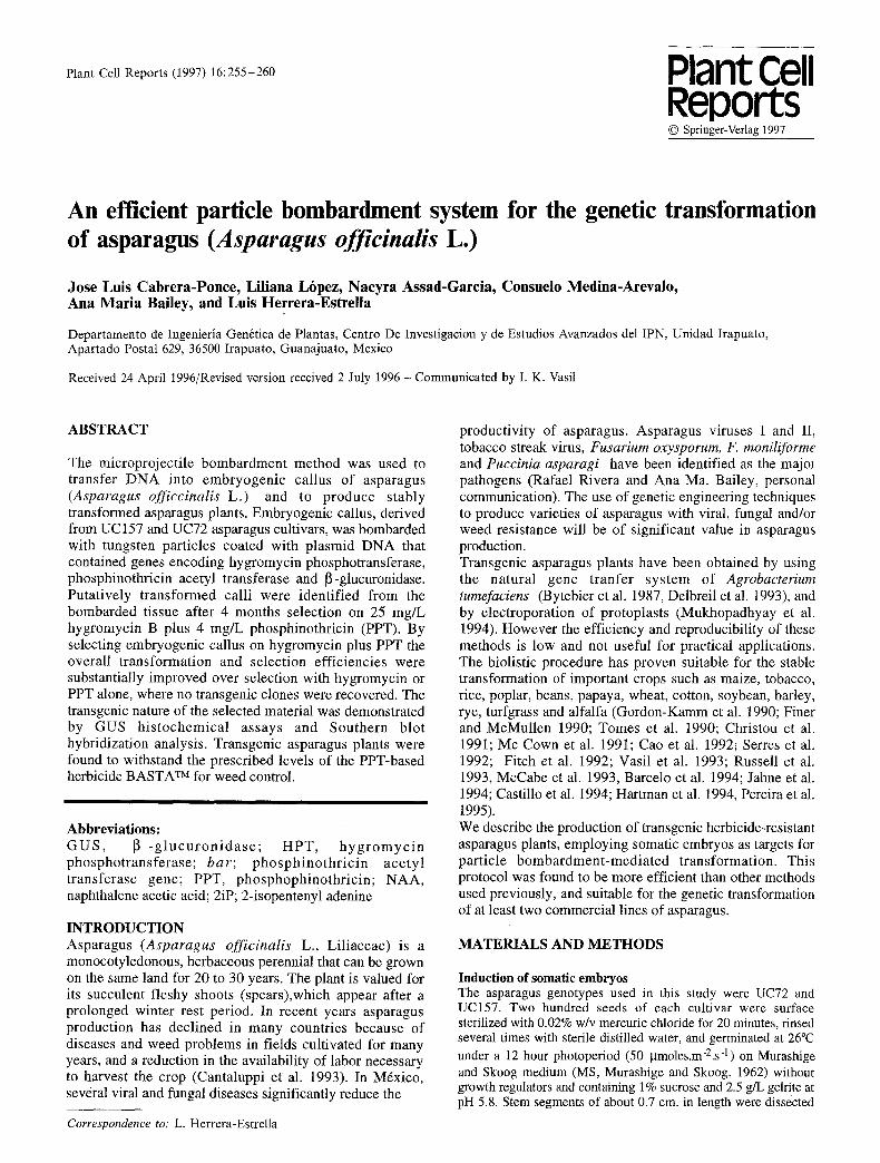
The microprojectile bombardment method was used to transfer DNA into embryogenic callus of asparagus (Asparagus officcinalis L.) and to produce stably transformed asparagus plants. Embryogenic callus, derived from UC 157 and UC72 asparagus cultivars, was bombarded with tungsten particles coated with plasmid DNA that contained genes encoding hygromycin phosphotransferase, phosphinothricin acetyl transferase and 13-glucuronidase. Putatively transformed calli were identified from the bombarded tissue after 4 months selection on 25 mg/L hygromycin B plus 4 mg/L phosphinothricin (PPT). By selecting embryogenic callus on hygromycin plus PPT the overall transformation and selection efficiencies were substantially improved over selection with hygromycin or PPT alone, where no transgenic clones were recovered. The transgenic nature of the selected material was demonstrated by GUS histochemical assays and Southern blot hybridization analysis. Transgenic asparagus plants were found to withstand the prescribed levels of the PPT-based herbicide BASTA T M for weed control.
Abbreviations: G U S , 13 - g l u c u r o n i d a s e ; HPT, h y g r o m y c i n phosphotransferase; bar; phosphinothr ic in acetyl transferase gene; PPT, phosphophinothricin; NAA, naphthalene acetic acid; 2iP; 2-isopentenyl adenine
INTRODUCTION Asparagus (Asparagus officinalis L., Liliaceae) is a monocotyledonous, herbaceous perennial that can be grown on the same land for 20 to 30 years. The plant is valued for its succulent fleshy shoots (spears),which appear after a prolonged winter rest period. In recent years asparagus production has declined in many countries because of diseases and weed problems in fields cultivated for many years, and a reduction in the availability of labor necessary to harvest the crop (Cantaluppi et al. 1993). In M6xico, several viral and fungal diseases significantly reduce the
Correspondence to: L. Herrera-Estrella
productivity of asparagus. Asparagus viruses I and II, tobacco streak virus, Fusarium oxysporum, F. moniliforme and Puccinia asparagi have been identified as the majol pathogens (Rafael Rivera and Ana Ma. Bailey, personal communication). The use of genetic engineering techniques to produce varieties of asparagus with viral, fungal and/or weed resistance will be of significant value in asparagus production. Transgenic asparagus plants have been obtained by using the natural gene tranfer system of Agrobacterium tumefaciens (Bytebier et al. 1987, Delbreil et al. 1993), and by electroporation of protoplasts (Mukhopadhyay et al. 1994). However the efficiency and reproducibility of these methods is low and not useful for practical applications. The biolistic procedure has proven suitable for the stable transformation of important crops such as maize, tobacco, rice, poplar, beans, papaya, wheat, cotton, soybean, barley, rye, turfgrass and alfalfa (Gordon-Kamm et al. 1990; Finer and McMullen 1990; Tomes et al. 1990; Christou et al. 1991; Mc Cown et al. 1991; Cao et al. 1992i Serres et al. 1992; Fitch et al. 1992; Vasil et al. 1993; Russell et al. 1993, McCabe et al. 1993, Barcelo et al. 1994; Jahne et al. 1994; Castillo et al. 1994; Hartman et al. 1994, Pereira et al. 1995). We describe the production of transgenic herbicide-resistant asparagus plants, employing somatic embryos as targets for particle bombardment-mediated transformation. This protocol was found to be more efficient than other methods used previously, and suitable for the genetic transformation of at least two commercial lines of asparagus.
MATERIALS AND METHODS
Induction of somatic embryos The asparagus genotypes used in this study were UC72 and UC157. Two hundred seeds of each cultivar were surface sterilized with 0.02% w/v mercuric chloride for 20 minutes, rinsed several times with sterile distilled water, and germinated at 26°C under a 12 hour photoperiod (50 gmoles.m-2.s -1) on Murashige and Skoog medium (MS, Murashige and Skoog, 1962) without growth regulators and containing 1% sucrose and 2.5 g/L gelrite at pH 5.8. Stem segments of about 0.7 cm. in length were dissected
256
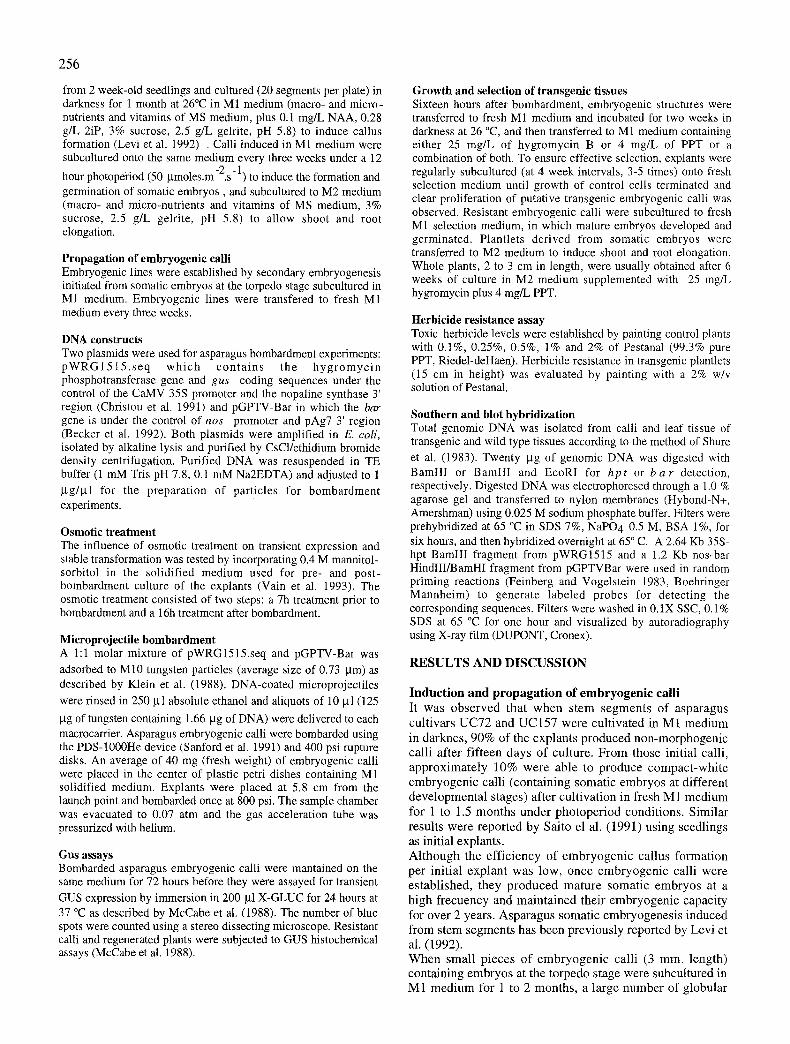
from 2 week-old seedlings and cultured (20 segments per plate) in darkness for 1 month at 26°C in M1 medium (macro- and micro- nutrients and vitamins of MS medium, plus 0.1 mg/L NAA, 0.28 g/L 2iP, 3% sucrose, 2.5 g/L gelrite, pH 5.8) to induce callus formation (Levi et al. 1992) . Calli induced in M1 medium were subcultured onto the same medium every three weeks under a 12
hour photopei-iod (50 I.tmoles.m-2.s-1) to induce the formation and germination of somatic embryos, and subcultured to M2 medium (macro- and micro-nutrients and vitamins of MS medium, 3% sucrose, 2.5 g/L gelrite, pH 5.8) to allow shoot and root elongation.
Propagation of embryogenic calli Embryogenic lines were established by secondary embryogenesis initiated from somatic embryos at the torpedo stage subcultured in M1 medium. Embryogenic lines were transfered to fresh M1 medium every three weeks.
DNA constructs Two plasmids were used for asparagus bombardment experiments: pWRG1515 . seq which conta ins the hyg romyc in phosphotransferase gene and gus coding sequences under the control of the CaMV 35S promoter and the nopaline synthase 3' region (Christou et al. 1991) and pGPTV-Bar in which the bar gene is under the control of nos promoter and pAg7 3' region (Becker et al. 1992). Both plasmids were amplified in E. coli, isolated by alkaline lysis and purified by CsC1/ethidium bromide density centrifugation. Purified DNA was resuspended in TE buffer (1 mM Tris pH 7.8, 0.1 mM Na2EDTA) and adjusted to 1 ~tg/~l for the preparation of particles for bombardment experiments.
Osmotic treatment The influence of osmotic treatment on transient expression and stable transformation was tested by incorporating 0.4 M mannitol- sorbitol in the solidified medium used for pre- and post- bombardment culture of the explants (Vain et al. 1993). The osmotic treatment consisted of two steps: a 7h treatment prior to bombardment and a 16h treatment after bombardment.
Microprojectile bombardment A 1:1 molar mixture of pWRG1515.seq and pGPTV-Bar was adsorbed to M10 tungsten particles (average size of 0.73 gm) as described by Klein et al. (1988). DNA-coated microprojectiles
were rinsed in 250 gl absolute ethanol and aliquots of 10 gl (125
I.tg of tungsten containing 1.66 ~tg of DNA) were delivered to each macrocarrier. Asparagus embryogenic calli were bombarded using the PDS-1000He device (Sanford et al. 1991) and 400 psi rupture disks. An average of 40 mg (fresh weight) of embryogenic calli were placed in the center of plastic petri dishes containing M1 solidified medium. Explants were placed at 5.8 cm from the launch point and bombarded once at 800 psi. The sample chamber was evacuated to 0.07 atm and the gas acceleration tube was pressurized with helium.
Gus assays Bombarded asparagus embryogenic calli were mantained on the same medium for 72 hours before they were assayed for transient GUS expression by immersion in 200 gl X-GLUC for 24 hours at 37 °C as described by McCabe et al. (1988). The number of blue spots were counted using a stereo dissecting microscope. Resistant calli and regenerated plants were subjected to GUS histochemical assays (McCabe et al. 1988).
Growth and selection of transgenic tissues Sixteen hours after bombardment, embryogenic structures were transferred to fresh M1 medium and incubated for two weeks in darkness at 26 °C, and then transferred to M1 medium containing either 25 mg/L of hygromycin B or 4 mg/L of PPT or a combination of both. To ensure effective selection, explants were regularly subcultured (at 4 week intervals, 3-5 times) onto fresh selection medium until growth of control cells terminated and clear proliferation of putative transgenic embryogenic calli was observed. Resistant embryogenic calli were subcultured to fresh M1 selection medium, in which mature embryos developed and germinated. Plantlets derived from somatic embryos were transferred to M2 medium to induce shoot and root elongation. Whole plants, 2 to 3 cm in length, were usually obtained after 6 weeks of culture in M2 medium supplemented with 25 mg/L hygromycin plus 4 mg/L PPT.
Herbicide resistance assay Toxic herbicide levels were established by painting control plants with 0.1%, 0.25%, 0.5%, 1% and 2% of Pestanal (99.3% pure PPT, Riedel-deHaen). Herbicide resistance in transgenic plantlets (15 cm in height) was evaluated by painting with a 2% w/v solution of Pestanal.
Southern and blot hybridization Total genomic DNA was isolated from calli and leaf tissue of transgenic and wild type tissues according to the method of Shure et al. (1983). Twenty ~tg of genomic DNA was digested with BamHI or BamHI and EcoRI for hpt or b a r detection, respectively. Digested DNA was electrophoresed through a 1.0 % agarose gel and transferred to nylon membranes (Hybond-N+, Amershman) using 0.025 M sodium phosphate buffer. Filters were prehybridized at 65 °C in SDS 7%, NaPO 4 0.5 M, BSA 1%, for six hours, and then hybridized overnight at 65 ° C. A 2.64 Kb 35S- hpt BamHI fragment from pWRG1515 and a 1.2 Kb nos-bar HindlII/BarnHI fragment from pGPTVBar were used in random priming reactions (Feinberg and Vogelstein 1983, Boehringer Mannheim) to generate labeled probes for detecting the corresponding sequences. Filters were washed in 0.1X SSC, 0.1% SDS at 65 °C for one hour and visualized by autoradiography using X-ray film (DUPONT, Cronex).
RESULTS AND D I S C U S S I O N
Induction and propagation of embryogenic calli It was observed that when stem segments of asparagus cultivars UC72 and UC157 were cultivated in M1 medium in darkncs, 90% of the cxplants produced non-morphogenic calli after fifteen days of culture. F rom those initial calli, approximate ly 10% were able to produce compact-whi te cmbryogcnic calli (containing somatic embryos at different developmental stages) after cultivation in fresh M 1 medium for 1 to 1.5 months under photoper iod conditions. Similar results were reported by Saito el al. (1991) using seedlings as initial explants. Al though the efficiency of embryogenic callus formation per initial explant was low, once cmbryogenic calli were established, they produced mature somatic embryos at a high frccuency and maintained their cmbryogenic capacity for over 2 years. Asparagus somatic cmbryogenesis induced from stem segments has been previously reported by Levi et ai. (1992). When small pieces of cmbryogen ic calli (3 ram. length) containing embryos at the torpedo stagc were subcultured in M1 medium for 1 to 2 months, a large number of globular

structures were produced by intensive secondary embryogenesis ( G - t y p e calli; Fig. 1A). When these ernbryogenic calli were mantained for an additional month on the same dish, a high percentage of the globular structures developed into somatic embryos at torpedo stage (T-type calli; Figure 1B). The transfer of calli containing torpedo stage embryos to fresh M1 medium induced a second round of secondary embrogenesis, from which embryos at different developmental stages could be easily separated (Fig. 1C). Mature somatic embryos consisted of a cylindrical cotyledon opposite to the root tip and exhibited the typical morphology of Liliaceae (Monocotyledonae) embryos (Esau 1953), similar to those previously described by Delbreil et al. (1994). Plant regeneration from somatic embryos at the torpedo stage was observed after 1 month of growth in M2 hormone free-medium (Fig. 1D). The efficiency of production of plantlets was variable depending upon the developmental stage of somatic embryos transfered to regeneration media, being 25%, 33% and 52% for somatic embryos (torpedo stage) of 2.5, 4.5 and 6.0 mm in length, respectively. The high germination capacity of completely mature embryos may be potentially useful for the production of artificial seeds.
Figure 1: Steps in the regeneration of asparagus plants from embryogenic calli derived from stem segments. A: Ernbryogenic callus containing globular structures, induced from stem segments of asparagus after 1 month of incubation (G-type callus), x4. B : Embryogenic callus with torpedo stage embryos after 2 months of incubation in M1 medium (T-type callus), x4. C : Developmental stages of somatic embryos present in calli derived from torpedo-stage somatic embryos shown in B, in which secondary somatic embryogenesis takes place, x5.5. D : Plant regeneration from a torpedo-stage somatic embryo after 1 month of incubation in regeneration medium, x5.
Selection of transgenic clones To establish the optimal particle bombardment conditions, embryogenic calli of asparagus vats. UC72 and UC157 were bombarded with particles coated with pWRG1515 using different conditions and scored for transient GUS expressing foci (data not shown). The following bombardment conditions produced the highest number of GUS expressing foci and were later used for the production of stably transformed lines: distance between disk and macrocarrier 1.2 cm, macrocarrier and stoping screen 1.2 cm, stoping screen and biological sample 5.8 cm; helium pressure 800 psi and vacuum 0.07 atm. conditions.
257
To develop a selection medium that effectively arrested the proliferation of non-transformed tissue, the growth of control calli was tested in M 1 medium containing different concentra t ions of kanamycin , hygromycin and phosphinothricin (PPT). Selection on medium containing kanamycin (up to 500 mg/L), hygromycin (up to 150 mg/L) or PPT (up to 20 mg/L) alone was not effective, since control calli were able to maintain a slow but consistent growth under these conditions. Effective selection was only obtained when both hygromycin B (25mg/1) and PPT (4 mg/1) were included in the selection medium (data not shown). To obtain stably transformed lines, T- and G-type calli were bombarded using a 1:1 molar mixture of pWRG1515 and pGPTV-Bar. Bombarded tissues were subcultured monthly to fresh selective M1 medium. After 3 months of culture under selection, resistant embryogenic calli could be visually selected and removed for further growth (Fig. 2A). In contrast to non-transformed tissue which turned brown after one month of culture in selective media, reststant embryogenic calli were compact, white-green and fast growing. In initial experiments, ten hygromycin/PPT resistant clones were obtained from three independent experiments using G embryogenic calli as a target tissue. The average efficiency was approximately 2 transgenic clones per g fresh weight bombarded callus (Table 1). However, since the efficiency of genetic transformation was low using G-type calli and no transgenic clones were obtained using T-type calli, which more efficiently produce secondary embryos, we decided to test whether an osmotic treatment would improve the efficiency of asparagus transformation. With this aim, the target tissue was pre-cultured 7 h before and 16 h after bombardment on M1 medium supplemented with 0.2 M mannitol and 0.2 M sorbitol (Vain et al. 1993)• Osmotic treatment of embryogenic asparagus explants produced a high and consistent transient GUS expression, that was superior to that obtained without osmotic treatment (data not shown). Using this osmotic treatment, 50 hygromycin/PPT resistant clones were obtained from two different bombardment experiments using T-type calli as target tissue. The average eff iciency of genetic transformation was approximately 20 transgenic clones per g of fresh weight (Table 3) or 0.7 clones per bombarded petri-dish containing 40 mg of embryogenic callus.
Table 1. Effect of osmotic treatment on stable transformation of asparagus using torpedo and globular structures as target tissues.
3xperiment Bombarded Bombarded Osmnticum No. resistant No. regenerating number tissue plates* clones, GUS+ clones
1 T-type 36 None 0 0
2 T-type 34 None 0 0
3 T-type 36 Yes 28 25
4 T-type 34 Yes 22 18
5 G-type 36 None 3 1
6 G-type 34 None 3 2
7 G-type 36 Yes 8 2
8 G-type 34 Yes 7 3
* 40 mg of target tissue was used per plate
258
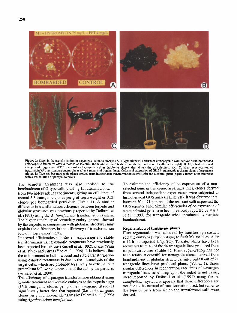
Figure 2: Steps in the transformation of asparagus somatic embryos.A : Hygromicin/PPT resistant embryogenic calli derived from bombarded embryogenic structures after 4 months of selection (bombarded tissue is shown on the left and control calli on the right). B : GUS histochemical analysis of hygromicin/PPT resistant embryogenic callus (globular stage) after 4 months of selection. 7X. C: Plant regeneration of hygromicin/PPT resistant asparagus plants after 8 months of bombardment (left), and expression of GUS in transgenic resistant plants of asparagus (right). D: Two nos-bar transgenic plants derived from independent transformation events (left) and a control plant (right) 1 month after treatment with a 1% solution of phosphinothricin.
The osmotic treatment was also applied to the bombardment of G-type calli, yielding 15 resistant clones from two independent experiments, giving an efficiency of around 5.3 transgenic clones per g of fresh weight or 0.21 clones per bombarded petri-dish (Table 1). A similar difference in transformation efficiency between torpedo and globular structures was previously reported by Delbreil et al. (1993) using the A. tumefaciens transformation system. The higher capability of secondary embryogenesis showed by the torpedo, in comparison with globular, structures may explain the differences in the efficiency of transformation found in these experiments. Improved efficiencies of transient expression and stable transformation using osmotic treatments have previously been reported for tobacco (Russell et al. 1992), maize (Vain et al. 1993) and citrus (Yao et al. 1996). It is believed that the enhancement in both transient and stable transformation using osmotic treatments is due to the plasmolysis of the target cells, which are probably less likely to extrude their protoplasm following penetration of the cell by the particles (Armaleo et al. 1990). The efficiency of asparagus transformation obtained using osmotic treatment and somatic embryos at the torpedo stage (15.6 transgenic clones per g of embryogenic tissue) is significantly better than that reported (0.6 to 4 transgenic clones per g of embryogenic tissue) by Delbreil et al. (1993) using Agrobacterium tumefaciens.
To estimate the efficiency of co-expression of a non- selected gene in transgenic asparagus lines, clones derived from several independent experiments were subjected to histochemical GUS analysis (Fig. 2B). It was observed that between 50 to 71 percent of the resistant calli expressed the GUS reporter gene. Similar efficiencies of co-expression of a non-selected gene have been previously reported by Vasil et al. (1993) for transgenic wheat produced by particle bombardment.
Regeneration of transgenie plants Plant regeneration was achieved by transferring resistant somatic embryos (torpedo stage) to fresh M 1 medium under a 12 h photoperiod (Fig. 2C). To date, plants have been recovered from 43 of the 50 transgenic lines produced from torpedo structures (Table 1). Plant regeneratio n has not been totally successful for transgenic clones derived from bombardment of globular structures, since only 8 out of 21 transgenic lines have produced plants (Tables 1). Since similar differences in regeneration capacities of asparagus transgenic lines, depending upon the initial target tissue, were reported by Delbreil et al. (1994) using the A. tumefaciens system, it appears that these differences are not due to the method of transformation used, but rather to the type of cells from which the transformed calli were derived.
259
After one month of culture in M1 media, plantlets were subcultured to selective M2 medium to allow root proliferation. All transgenic plants derived from GUS positive calli were found to express significant levels of GUS activity (Fig. 2C). A large number of plants (more than 40) can easily be obtained from clones derived from torpedo structures. Plants of about 10 cm length and a profuse root system were better established in soil than smaller plants. The overall time from bombardment to recovery of transgenic plants (15 to 20 cm) established in green house conditions, takes between 7 to 9 months.
Herbicide assay Functional expression of the bar gene in asparagus plants was assessed by localized application of purified PPT to transgenic and control plants. Control plants were found to be very sensitive to the application of PPT, since treatment with a 0.1% w/v solution_caused total necrosis 10 days after application. In contrast, no visible damage was observed in the plants derived from the 5 lines harboring the nos-bar chimeric gene which were tested, even two months after treatment with concentrations of up to 1% w/v of PPT (Fig. 2 D). The transgenic asparagus lines tested were able to withstand the prescribed application of PPT for weed control (0.5 to 1%) and therefore have potential for commercial plantations.
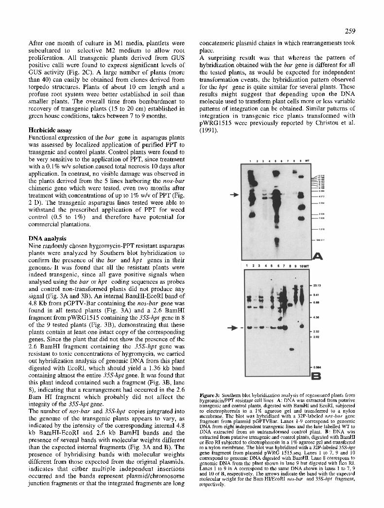
DNA analysis Nine randomly chosen hygromycin-PPT resistant asparagus plants were analyzed by Southern blot hybridization to confirm the presence of the bar and hpt genes in their genome~: It was found that all the resistant plants were indeed transgenic, since all gave positive signals when analysed using the bar or hpt coding sequences as probes and control non-transformed plants did not produce any signal (Fig. 3A and 3B). An internal BamHI-EcoRI band of 4.8 Kb from pGPTV-Bar containing the nos-bar gene was found in all tested plants (Fig. 3A) and a 2.6 BamHI fragment from pWRG1515 containing the 35S-hpt gene in 8 of the 9 tested plants (Fig. 3B), demonstrating that these plants contain at least one intact copy of the corresponding genes. Since the plant that did not show the presence of the 2.6 BamHI fragment containing the 35S-hpt gene was resistant to toxic concentrations of hygromycin, we carried out hybridization analysis of genomic DNA from this plant digested with EcoRI, which should yield a 1.36 kb band containing almost the entire 35S-hpt gene. It was found that this plant indeed contained such a fragment (Fig. 3B, lane 8), indicating that a rearrangement had occurred in the 2.6 Barn HI fragment which probably did not affect the integrity of the 35S-hpt gene. The number of nos-bar and 35S-hpt copies integrated into the genome of the transgenic plants appears to vary, as indicated by the intensity of the corresponding internal 4.8 kb BamHI-EcoRI and 2.6 kb BamHI bands and the presence of several bands with molecular weights different than the expected internal fragments (Fig. 3A and B). The presence of hybridising bands with molecular weights different from those expected from the original plasmids, indicates that either multiple independent insertions occurred and the bands represent plasmid/chromosome junction fragments or that the integrated fragments are long
concatemeric plasmid chains in which rearrangements took place. A surprising result was that whereas the pattern of hybridization obtained with the bar gene is different for all the tested plants, as would be expected for independent transformation events, the hybridization pattern observed for the hpt gene is quite similar for several plants. These results might suggest that depending upon the DNA molecule used to transform plant cells more or less variable patterns of integration can be obtained. Similar patterns of integration in transgenic rice plants transformed with pWRG1515 were previously reported by Christou et al. (1991).
1 2 3 4 S S 7 8 5 ~
1 2 3 4 IS 6 ? 8 9 tOWT
Figure 3: Southern blot hybridization analysis of regenerated plants from hygromicin/PPT resistant celt lines. A: DNA was extracted from putative transgenic and control plants, digested with BamHI and EcoRI, subjected to electrophoresis in a 1% agarose gel and transferred to a nylon membrane. The blot was hybridized with a 32P-labeled nos-bar gene fragment from plasmid pGPTVBar. Lanes 1-9 correspond to genomic DNA from eight independent transgenic lines and the lane labelled WT to DNA extracted from an untransformed control plant. B: DNA was extracted from putative transgenic and control plants, digested with BamHI or Eco RI subjected to electrophoresis in a 1% agarose gel and transferred to a nylon membrane. The blot was hybridized with a 32P-labeled 35S-hpt gene fragment from plasmid pWRG 1515.seq. Lanes 1 to 7, 9 and 10 correspond to genomic DNA digested with BamHI. Lane 8 correspons to genomic DNA from the plant shown in lane 9 but digested with Eco RI. Lanes 1 to 9 in A correspond to the same DNA shown in lanes 1 to 7, 9 and 10 of B, respectively. The arrows indicate the band with the expected molecular weight for the Barn HI/EcoRI nos-bar and 35S-hpt fragment, respectively.
260
Acknowledgments We thank Azucena Mendoza, Mireya Sanchez, June Simpson and Gilberto Mozqueda for ideas and facilities for Southern blot analysis; Rosa Maria Rangel and Luis Cuahutemoc Navarro for helpful discussions about somatic embryogenesis, and Antonio Cisneros and Martha Martinez for the photographic work. This project was supported in part by a grant from the Irapuato asparagus growers association.
References Armaleo D, Ye GN, Klein TM, Shark KB, Sanford JC, Johnston SA (1990) Curt Gen 17: 97-103. Barcelo P, Hagel C, Becker D, Martin A, L6rz H (1994) Plant J 5(4): 583-592. Becker D, Kemper E, Schell J, Masterson R (1992) Plant Mol Biol 20:1195-1197. Bytebier B, Deboeck F, De Greve H, Van Montagu M, Hernalsteens JP (1987) Proc. Natl Acad Sci USA 84: 5345-5349. Cao J, Duan X, McElroy D, Wu R (1992) Plant Cell Rep 11:586- 591. Cantaluppi CJ, Precheur RJ (1993) The Ohio State University Extension. Bulletin 826. Castillo AM, Vasil V, Vasil IK (1994) Bio/technology 12: 1366- 1371. Christou P, Ford TL, Kofron M (1991) Bio/technology 9: 957-962. Delbreil B, Guerche P, Jullien M (1993) Plant Cell Rep 12:129- 132. Delbreil B, Jullien M (1994) Plant Cell Rep 13: 372-376. Esau K (1953) In: Esan K (ed) Plant Anatomy, J Wiley & Sons Inc, New York, Chapman & Hall LTD, London, pp 597-705. Feinberg AP, Vogelstein B (1983) Anal Biochem 132: 6-13. Finer JJ, McMullen MD (1990) Plant Cell Rep 8: 586-589. Fitch MM, Manshardt RM, Gonsalves D, Slightom JL, Sanford JC (1992) Bio/technology 10: 1466-1472. Gordon-Kamm WJ, Spencer TM, Mangano ML, Adams TR, Daines RJ, Start WG, O'Brien JV, Chambers SA, Adams Jr WR, Willets NG, Rice TB, Mackey C J, Krueger RW, Kausch AP, Lemaux PG (1990) Plant Cell 2: 603-618. Hartman CL, Lee L, Day PR, Turner NE (1994) Bio/technology 12: 919-923. J~ihne A, Becker D, Brettschneider R, L6rz H (1994) Theor Appl Genet 89: 525-533. Klein TM, Fromm M, Weissinger A, Tomes D, Schaff S, Sletten M, Sanford J (1988) Proc Natl Acad Sci 85: 4305-4309. Levi A, Sink KC (1992) Plant Cell, Tissue and Organ Culture 31: 115-122. McCabe DE, Swain WF, Martinell BJ, Christou P (1988) Bio/Technology 6: 923-926. McCabe DE, Martinell BJ (1993) Bio/technoiogy 11: 596-598. McCown BH, McCabe DE, Russell DR, Robison DJ, Barton KA, Raffa KF (1991) Plant Cell Rep 9: 590-594. Mnkhopadhyay S, Desjardins Y (1994) Plant Cell Rep 13: 421- 424. Murashige T, Skoog F(1962) Physiol Plant 15: 473-497. Pereira LF, Erickson L (1995) Plant Cell Rep 14: 290-293. Russell JA, Roy MK, Sanford JC (1992) In Vitro Cell Dev Biol. 28P:97-105. Russell DR, Wallace KM, Bathe JH, Martinell B J, McCabe DE (1993) Plant Cell Rep 12: 165-169. Saito T, Nishizawa S, Nishimura S (1991) Plant Cell Rep 10: 230- 234. Sanford JC, Devit MJ, Rusell JA, Smith FD, Harpending PR, Roy MK, Johnston SA (1991) Technique 3: 1.3-16. Serres R, Stang E (1992) J. Amer Soc Hort Sci 117: 174-180. Southern EM (1975) J Mol Biol 98: 503-517. Shure M, Wessler S, Federoff N (1983) Cell 35: 225-233.
Tomes DT, Weissinger AK, Ross M, Higgins R, Drummond B J, Schaaf S, Malone-Schoneberg J, Staebel M, Flynn P, Anderson J, Howard J (1990) Plant Mol Biol 15: 809-819. Vasil V, Srivatara V, Castillo AM, Fromm ME, Vasil IK (1993) Bio/technology 11: 1553-1558. Vain P, McMullen MD, Finer JJ (1993) Plant Cell Rep 12: 84-88. Yao JL, Wu JH, Gleave AP, Morris BAM (1996) Plant Sci 113: 175-183.