Alpha-2 adrenergic challenge with guanfacine one month after mild traumatic brain injury: Altered...
18
Alpha-2 Adrenergic Challenge with Guanfacine One Month after Mild Traumatic Brain Injury: Altered Working Memory and BOLD Response Thomas W. McAllister, MD 1 , Brenna C. McDonald, PsyD 1,2 , Laura A. Flashman, PhD 1 , Richard B. Ferrell, MD 1 , Tor D. Tosteson, ScD 3 , Norman N. Yanofsky, MD 4 , Margaret R. Grove, MS 1 , and Andrew J. Saykin, PsyD 1,2 1 Section of Neuropsychiatry, Department of Psychiatry, Dartmouth Medical School, Lebanon, NH 2 Center for Neuroimaging, Department of Radiology and Imaging Sciences, Indiana University School of Medicine, Indianapolis, IN 3 Department of Community and Family Medicine, Biostatistics, Dartmouth Medical School 4 Section of Emergency Medicine, Dartmouth Medical School Abstract Alterations in working memory (WM) are common after traumatic brain injury (TBI). Frontal catecholaminergic systems, including the alpha-2 adrenergic system, modulate WM function and may be affected in TBI. We hypothesized that administration of an alpha-2 adrenergic agonist might improve WM after mild TBI (MTBI). Thirteen individuals with MTBI one month after injury and 14 healthy controls (HC) were challenged with guanfacine and placebo prior to administration of a verbal WM functional MRI task. Guanfacine was associated with improved WM performance in the MTBI but not the HC group. On guanfacine the MTBI group showed increased activation within a WM task-specific region of interest. Findings are consistent with the hypothesis that alterations in WM after MTBI may be improved with the alpha-2 agonist guanfacine. Keywords traumatic brain injury; alpha-2 adrenergic agonist; working memory; functional MRI; guanfacine Introduction Initial and persistent cognitive deficits are the most common complaints after traumatic brain injury (TBI) (Whyte et al. 2000) and are the major hindrance to return to baseline function (Cicerone et al. 2000). There are several predictable areas of cognitive impairment © 2011 Elsevier B.V. All rights reserved Correspondence: Thomas W. McAllister, M.D., Dartmouth-Hitchcock Medical Center, One Medical Center Drive, Lebanon, NH 03756 phone: (603) 650-5824 fax: (603) 650-5842 [email protected]. Publisher's Disclaimer: This is a PDF file of an unedited manuscript that has been accepted for publication. As a service to our customers we are providing this early version of the manuscript. The manuscript will undergo copyediting, typesetting, and review of the resulting proof before it is published in its final citable form. Please note that during the production process errors may be discovered which could affect the content, and all legal disclaimers that apply to the journal pertain. Location of Work: Section of Neuropsychiatry and Brain Imaging Laboratory, Departments of Psychiatry, Radiology, Community and Family Medicine (Biostatistics), Emergency Medicine, Dartmouth Medical School, Lebanon, NH NIH Public Access Author Manuscript Int J Psychophysiol. Author manuscript; available in PMC 2012 October 1. Published in final edited form as: Int J Psychophysiol. 2011 October ; 82(1): 107–114. doi:10.1016/j.ijpsycho.2011.06.022. NIH-PA Author Manuscript NIH-PA Author Manuscript NIH-PA Author Manuscript
Transcript of Alpha-2 adrenergic challenge with guanfacine one month after mild traumatic brain injury: Altered...

Alpha-2 Adrenergic Challenge with Guanfacine One Month afterMild Traumatic Brain Injury: Altered Working Memory and BOLDResponse
Thomas W. McAllister, MD1, Brenna C. McDonald, PsyD1,2, Laura A. Flashman, PhD1,Richard B. Ferrell, MD1, Tor D. Tosteson, ScD3, Norman N. Yanofsky, MD4, Margaret R.Grove, MS1, and Andrew J. Saykin, PsyD1,2
1Section of Neuropsychiatry, Department of Psychiatry, Dartmouth Medical School, Lebanon, NH2Center for Neuroimaging, Department of Radiology and Imaging Sciences, Indiana UniversitySchool of Medicine, Indianapolis, IN3Department of Community and Family Medicine, Biostatistics, Dartmouth Medical School4Section of Emergency Medicine, Dartmouth Medical School
AbstractAlterations in working memory (WM) are common after traumatic brain injury (TBI). Frontalcatecholaminergic systems, including the alpha-2 adrenergic system, modulate WM function andmay be affected in TBI. We hypothesized that administration of an alpha-2 adrenergic agonistmight improve WM after mild TBI (MTBI). Thirteen individuals with MTBI one month afterinjury and 14 healthy controls (HC) were challenged with guanfacine and placebo prior toadministration of a verbal WM functional MRI task. Guanfacine was associated with improvedWM performance in the MTBI but not the HC group. On guanfacine the MTBI group showedincreased activation within a WM task-specific region of interest. Findings are consistent with thehypothesis that alterations in WM after MTBI may be improved with the alpha-2 agonistguanfacine.
Keywordstraumatic brain injury; alpha-2 adrenergic agonist; working memory; functional MRI; guanfacine
IntroductionInitial and persistent cognitive deficits are the most common complaints after traumaticbrain injury (TBI) (Whyte et al. 2000) and are the major hindrance to return to baselinefunction (Cicerone et al. 2000). There are several predictable areas of cognitive impairment
© 2011 Elsevier B.V. All rights reservedCorrespondence: Thomas W. McAllister, M.D., Dartmouth-Hitchcock Medical Center, One Medical Center Drive, Lebanon, NH03756 phone: (603) 650-5824 fax: (603) 650-5842 [email protected]'s Disclaimer: This is a PDF file of an unedited manuscript that has been accepted for publication. As a service to ourcustomers we are providing this early version of the manuscript. The manuscript will undergo copyediting, typesetting, and review ofthe resulting proof before it is published in its final citable form. Please note that during the production process errors may bediscovered which could affect the content, and all legal disclaimers that apply to the journal pertain.Location of Work: Section of Neuropsychiatry and Brain Imaging Laboratory, Departments of Psychiatry, Radiology, Community andFamily Medicine (Biostatistics), Emergency Medicine, Dartmouth Medical School, Lebanon, NH
NIH Public AccessAuthor ManuscriptInt J Psychophysiol. Author manuscript; available in PMC 2012 October 1.
Published in final edited form as:Int J Psychophysiol. 2011 October ; 82(1): 107–114. doi:10.1016/j.ijpsycho.2011.06.022.
NIH
-PA Author Manuscript
NIH
-PA Author Manuscript
NIH
-PA Author Manuscript

after TBI (Binder et al. 1997; Levin 1998; Levin et al. 1987; McMillan 1997), includingworking memory (WM), which is the ability to hold information in mind and to manipulatethat material in light of incoming information. We previously used functional MRI (fMRI)to demonstrate that one month after mild TBI (MTBI) performance on a moderate difficultyWM task was associated with significantly greater cerebral activation (“compensatoryactivation”) and increased memory complaints despite WM task performance similar to thatseen in controls (McAllister et al. 1999). We subsequently showed that MTBI patients wereunable to further increase brain activation with a higher WM processing load (McAllister etal. 2001). We interpreted these results as evidence for abnormalities in the activation andallocation of WM processing resources after MTBI. In essence, patients were “workingharder” to achieve the same results with a moderately difficult task, and thus had feweradditional processing resources to recruit during the more difficult task condition. Othergroups have subsequently shown similar alterations in cerebral activation following MTBI(Chen et al. 2004; Perlstein et al. 2004).
Elucidation of the neural mechanisms underlying these observations might informpharmacological strategies for treatment of WM complaints associated with TBI. Asignificant body of work suggests that catecholaminergic mechanisms play important rolesin WM function, especially in the prefrontal cortex (PFC; see (Arnsten et al. 1998)), andthere is evidence that TBI alters catecholamine tone and regulation after TBI (see McAllisteret al 2004; Kobori et al. 2010 for reviews). Such abnormalities may play a role in alterationsin activation and allocation of WM processing resources after MTBI. Although difficult totest this hypothesis directly, it is possible to use pharmacological challenges to probewhether individuals with MTBI differ in their response to catecholaminergic agents. Alteredbehavioral (cognitive) and neurophysiological (task-related cerebral activation) responses toa catecholamine agonist would provide indirect support for this hypothesis.
The catecholamine systems are complex, however, and different catecholamine agonists canhave different effects on cognitive function. For example, in monkeys and rats localizedprefrontal depletion of catecholamines (dopamine and norepinephrine) caused by infusion of6-hydroxydopamine as well as global catecholamine depletion with agents such as reserpinelead to impaired performance on spatial WM tasks similar to that seen with ablation ofneural tissue in the PFC (Brozoski et al. 1979; Cai et al. 1993). Normal aging in theseanimals results in lower levels of both dopamine (DA) and norepinephrine (NE) and isassociated with declining performance on WM tasks (Arnsten 1998; Bartus et al. 1978;Luine et al. 1990), as is the use of alpha-2A (A2A) antagonists (Steere and Arnsten 1997;Tanila et al. 1996).
The role of the adrenergic system in WM varies as a function of receptor type, brain region,dose, and task difficulty. For example, in rodents and primates, blockade of the A2A systemwith infusion of A2A antagonists produces spatial WM impairment (Steere and Arnsten1997; Tanila et al. 1996), whereas alpha-1 and beta adrenergic antagonists do not (Arnstenand Li, 2005). WM performance deficits brought about by normal aging, chemicaldepletion, or infusion of A2A antagonists can be reversed by A2A agonists (see Arnsten etal. 1988 for review; Arnsten and Goldman-Rakic 1985; Carlson et al. 1992; Franowicz andArnsten 1998; Rama et al. 1996). However, A1A agonists can impair WM function,suggesting that different adrenergic receptors have opposing effects on cognitive function(Arnsten 1998; Birnbaum et al. 1999). In humans the A2A agonist guanfacine seemsparticularly effective at enhancing WM performance with a relatively low side effect profile(Arnsten et al. 1998; Arnsten 1998; Rama et al. 1996; Sirvio et al. 1991; Steere and Arnsten1997). Jakala et al. (Jakala et al. 1999; Jakala et al. 1999) administered three different dosesof clonidine (also an A2A agonist) or two doses of guanfacine to healthy controls using adouble-blind, placebo-controlled design. Both low and high dose clonidine were associated
McAllister et al. Page 2
Int J Psychophysiol. Author manuscript; available in PMC 2012 October 1.
NIH
-PA Author Manuscript
NIH
-PA Author Manuscript
NIH
-PA Author Manuscript

with poorer performance on WM, and showed little effect on the other tasks. At the higherdose (29mcg/kg), guanfacine was associated with significant improvement on WM, theTower of London task, and a verbal paired associates learning task. The researchersinterpreted these results as consistent with guanfacine-enhanced frontal functioning in bothspatial WM and planning. Taken together, these studies suggest that A2A effects maydepend on individual characteristics of the agonist used and the dose, as well as the nature ofthe task tested. There are no studies of the ability of A2A agonists to enhance WM inindividuals with TBI; however, there is evidence from other clinical populations thataugmentation of the A2A system can improve aspects of WM and other cognitive functions(Arnsten et al. 1996; Hunt et al. 1995); see also Arnsten (1998) and McAllister and Arnsten(2008) for thorough discussion.
The mechanism by which A2A receptor stimulation results in improved WM function is notclear. A2A receptors are located both pre-synaptically on noradrenergic neuronal terminals,and post-synaptically on dendritic spines, as well as sites removed from traditional synapses.Presynaptically, A2A stimulation results in inhibition of NE release; post-synaptically A2Astimulation appears to result in increased signal to noise ratio, apparently the opposite ofwhat is seen with alpha-1 receptor stimulation (Arnsten 1998). Adrenergic neuronal cellbodies arise in the brain stem within the locus coeruleus (LC) and project throughout mostof the CNS. Thus LC activity is capable of altering a wide array of CNS functions.However, there are particularly strong reciprocal connections between PFC and LC,suggesting that adrenergic tone in one area can affect adrenergic tone in the other (Arnsten1998). It is relevant that guanfacine is ten times weaker than clonidine in assays of pre-synaptic function, and this may in part explain why clonidine is more sedating thanguanfacine (clonidine also likely has more potent effects on A2B receptors in the thalamus,which have powerful effects on arousal). However, the majority of A2A receptors in thebrain are actually localized post-synaptic to NE neurons, and these are of particularrelevance to WM.
The above literature suggests that the adrenergic system plays an important but complex rolein the modulation of WM that is receptor, brain region, dose, and task specific.Enhancement of A2A system functioning can improve WM through stimulation of post-synaptic receptors in the PFC, which is a critical component of WM circuitry. However,depending on the A2A agonist used, there can be opposing effects. For example, agents thatstimulate pre-synaptic A2A receptors in LC (e.g., clonidine) can result in reduced adrenergictone in PFC and reduced arousal, both of which could impair WM function. Alternatively,excessive stimulation or conditions of stress (including perhaps arousal and stress associatedwith performance of difficult cognitive tasks), will impair WM and other cognitive functions(Arnsten and Li 2005). Guanfacine, because of its lower propensity for pre-synaptic A2Areceptors (and hence less inhibition of LC firing) is ideally positioned to be of selectivebenefit to WM.
In this study we tested the hypothesis that problems in activation and allocation of WMresources in PFC after MTBI are related to dysregulation of prefrontal A2A post-synapticreceptors. We therefore hypothesized that stimulation of these receptors with a moderatedose of guanfacine would selectively improve WM performance and normalize taskassociated cerebral activation in WM circuitry relative to noninjured controls.
MethodsThis was a prospective, placebo-controlled, double-blind, crossover study of consecutivepatients with MTBI referred to Dartmouth-Hitchcock Medical Center (DHMC), a Level 1Trauma Center.
McAllister et al. Page 3
Int J Psychophysiol. Author manuscript; available in PMC 2012 October 1.
NIH
-PA Author Manuscript
NIH
-PA Author Manuscript
NIH
-PA Author Manuscript

ParticipantsParticipants were diagnosed with MTBI by a Neuropsychiatrist (TM) using the AmericanCongress of Rehabilitation Medicine criteria for mild brain injury (Kay et al. 1993).Exclusion criteria included a history of other neurologic disorders, significant systemicmedical illness, or current DSM-IV Axis I diagnosis of psychiatric illness as assessed by theStructured Clinical Interview for DSM-IV (First et al. 1997). Healthy controls (HC) wererecruited through community advertisements and were screened for neurologic, medical, orany past or current psychiatric illness. After complete description of the study to theparticipants, written informed consent was obtained. The study protocol and the informedconsent document were approved by the Dartmouth College Committee for the Protection ofHuman Subjects.
ProceduresParticipants were studied on two occasions approximately one week apart. The sequence ofguanfacine and placebo administration was randomized and balanced across groups. Theorder of neuropsychological and fMRI task administration was also balanced, using alternateforms when available.
Study Protocol—Approximately two and a half hours after oral drug (2.0 mg guanfacine)or placebo ingestion, participants were positioned in the MRI scanner using laser alignmentbeams and a non-magnetic deformable foam head holder to stabilize head position. Stimulifor the cognitive tasks were programmed in Presentation (Neurobehavioral Systems, Inc.,Albany, CA) and presented visually through an MRI compatible goggle system (ResonanceTechnology, Van Nuys, CA).
fMRI Scan Procedure—All scans were acquired using the same GE Horizon 1.5T LXscanner. A gradient echo, echo-planar sequence was used to provide whole brain coverage:TR=2500 ms, TE=40 ms, FOV=24 cm, NEX=1, 29 interleaved 5 mm thick sagittal sliceswith no skip, yielding a 64×64 matrix with 3.75 mm2 in-plane resolution. Initial volumesprior to spin saturation were discarded.
WM Task—As in our previous studies (McAllister et al. 2006; McAllister et al. 1999;McAllister et al. 2001), a visually presented verbal “N-back” task was used to test WM.During scanning participants viewed a string of consonant letters (except L, W, and Y)presented at a rate of one every three seconds, in a four-condition, blocked design.Conditions were 0-, 1-, 2-, and 3-back. For each consonant seen, participants used a buttonpress device (Photon Control Inc., Burnaby, BC) to signify whether the current letter was amatch (i.e., was the same as the designated target or the letter presented 1, 2, or 3 back in thesequence, depending on the condition instructions) or was a non-match. Each task conditionwas presented in 27-second epochs preceded by three seconds of instruction (e.g., “thematch is D” or “the match is one back”). The four experimental conditions were eachpresented three times in pseudo-random order for a total of 12 task blocks. Participantsrehearsed a practice version of the task on a laptop prior to scanning to ensurecomprehension of task demands.
Clinical Assessment—After the scan session, participants completed aneuropsychological test battery that assessed level of estimated general intellectualfunctioning (WRAT-3, Reading subtest (Wilkinson 1993); WAIS-III, Block Design subtest(The Psychological Corporation 1997)), verbal memory (California Verbal Learning Test(Delis et al. 1987) or California Verbal Learning Test-II (Delis et al. 2000)), psychomotorspeed (WAIS-III, Digit Symbol-Coding subtest (The Psychological Corporation 1997)) andWM, executive, and attentional functioning (Trail Making Test, Parts A and B (Reitan and
McAllister et al. Page 4
Int J Psychophysiol. Author manuscript; available in PMC 2012 October 1.
NIH
-PA Author Manuscript
NIH
-PA Author Manuscript
NIH
-PA Author Manuscript

Wolfson 1993) or Delis-Kaplan Executive Function System (D-KEFS), Trail Making TestConditions 2 and 4 (Delis et al. 2001)); D-KEFS Color-Word Interference Test (Delis et al.2001); Controlled Oral Word Association Test (Lezak 1995; Spreen and Strauss 1998);Paced Auditory Serial Addition Test (PASAT) (Gronwall 1977); Gordon ContinuousPerformance Test (Gordon et al. 1986)).
Statistical AnalysisCognitive Measures—WM in-scanner performance data and neuropsychologicalvariables for both groups were compared using a repeated measures ANCOVA taking intoaccount drug condition (guanfacine vs. placebo), diagnosis (MTBI vs. HC), age, education,and order of drug administration (drug/placebo vs. placebo/drug). For neuropsychologicaltest data, raw or standard scores were used as indicated, with the exception of Trail Making.Because the version of the test changed during the protocol, raw scores for Trails A and Band D-KEFS conditions 2 and 4 were z-transformed using the HC group means in theplacebo condition, with z-scores utilized for group comparisons. All models were fit usingPROC MIXED in SAS V9, specifying a random subject effect for the repeated measurescovariance structure.
fMRI Analyses—Spatial realignment using a six parameter model was performed on allraw scan data to remove any minor (subvoxel) motion-related signal change. All volumesfor each subject were normalized into standardized Montreal Neurological Institute atlasspace using SPM5 (Wellcome Department of Cognitive Neurology, University College,London). During spatial normalization all scans were resampled to 2 mm3 isotropic voxels.Spatial smoothing to a full width half maximum of 8 mm was performed prior to statisticalanalysis. fMRI analyses included statistical parametric mapping, on a voxel-by-voxel basis,using a general linear model approach (Friston et al. 1995) as implemented in SPM5.Smoothed normalized scans for all subjects were entered into the model, and contrastimages comparing pairs of the WM processing load conditions (1-back > 0-back, 2-back >0-back, 3-back > 0-back) were created for each subject. These contrast images were thenused for the second level multi-subject/between-group random effects analyses. The randomeffects procedure performs a mixed model analysis to account for both random effects (scan)and fixed effects (task condition) (Holmes and Friston 1998).
Random effects analyses were conducted using ANCOVA to construct contrast maps ofvoxels in which brain activation differed between group and drug conditions (full factorialmodel in SPM5). Comparisons were conducted within an omnibus group- (two independentlevels: MTBI, HC) by-drug (two non-independent levels: guanfacine, placebo) ANCOVA,covarying for age, education, and order of drug/placebo administration. The design matrixtherefore included both conditions for both groups, accounting for the repeated measuresnature of the drug factor (i.e., the matrix included four columns, one for each group onplacebo and guanfacine). The critical significance threshold (Pcrit) was set to 0.01. Given theconstraints of the sample size, only clusters of activated voxels with a whole-brain searchcluster-level Puncorrected < 0.05 are discussed. For illustrative purposes, figures and tablesalso include results at a Pcrit of 0.05 where noted. Within the omnibus SPM5 design matrixbetween-group comparisons were conducted using weighted contrast vectors. For example,pair-wise comparisons of brain activation on guanfacine (MTBI vs. HC) were conducted byentering values of 1 and -1 in the appropriate columns in the matrix. In this mannerdetermination of regions where HC showed greater activation than MTBI on guanfacinewould be identified by entering 1 in the HC guanfacine column and -1 in the MTBIguanfacine column.
McAllister et al. Page 5
Int J Psychophysiol. Author manuscript; available in PMC 2012 October 1.
NIH
-PA Author Manuscript
NIH
-PA Author Manuscript
NIH
-PA Author Manuscript

ResultsDemographics
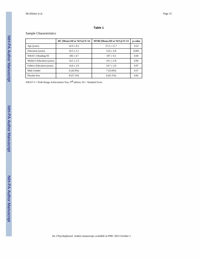
Thirteen subjects with MTBI and 14 HC completed the protocol (see Table 1). There wereno significant group differences in WRAT-3 Reading scores, an estimate of baseline verbalintellectual ability. The groups did differ with respect to years of education, largely becauseseveral of the MTBI subjects were students at the time of study participation. Parentaleducation did not differ between groups. MTBI participants were studied a mean of 37.2days after injury (±17.6,) and had an average Glasgow Coma Scale score of 13.9 (±2.1). Allparticipants were injured either by falls or in motor vehicle accidents, and none of the MTBIparticipants had focal injuries visible on computerized axial tomography (CT) scans done atthe time of injury. The protocol was well tolerated by both groups.
Cognitive PerformanceThere was a main effect of drug order in our initial modeling of cognitive outcomes (betterperformance on the second assessment), probably because the protocol called for repeatedcognitive assessments over a short interval and both test performance and imaging resultsmay be vulnerable to the influence of practice and exposure effects. Even when alternateforms of a task are given, there can be improvement in performance related to improvedstrategy/efficiency in task approach. Thus we included order (drug or placebo first) as acovariate in subsequent analyses. Although the two groups did not differ significantly withrespect to age, the literature supports a relationship between age and performance across avariety of cognitive measures, including the age range spanned by our participants. Inaddition, there is evidence that age affects task-related blood oxygen level dependent(BOLD) response. Thus subsequent analyses reported used age, education and order of drugadministration as covariates.
N-Back Performance—The MTBI and HC groups showed similar performance on the N-back task. Both groups showed the expected WM load effect on performance, regardless ofdrug condition (see Table 2), with percentage of correct responses (adjusted for guessing)decreasing with increasing WM load requirements. The HC group generally did worse onguanfacine relative to placebo, although this was not statistically significant. The MTBIgroup showed a statistically significant improvement on the moderate WM load (2-back)condition on guanfacine relative to placebo (p=0.018), and there was a significant drug-by-diagnosis interaction (p=0.007) for the 2-back as well as a trend for the mean back (p=0.07),with the MTBI group demonstrating improved performance on guanfacine relative toplacebo and the HC group showing a decline in performance on the drug.
Out of Scanner Cognitive Tasks—Overall, the MTBI group and the HC group did notdiffer on out of scanner cognitive test performance, with the exception of the fastestcondition of the PASAT (PASAT D), where HC demonstrated better performance than theMTBI group. There was a variable effect of guanfacine on performance in a small numberof out of scanner cognitive tests, with a positive main effect of drug on the D-KEFS Color-Word Interference Test Word Reading Trial (p=0.007), but a negative main effect on CPTsimple reaction time number correct (p=0.036); and CPT vigilance reaction time (p=0.009);as well as Trails 2/A (p=0.02).
fMRIBoth diagnostic groups showed the expected WM activation pattern on both drugs (Figure1), including bilateral frontal, parietal, and cerebellar circuitry. This pattern of activation isconsistent with previous results with the N-back task (McAllister et al. 1999; McAllister etal. 2001). As noted above, the two groups showed a significant diagnosis-by-drug
McAllister et al. Page 6
Int J Psychophysiol. Author manuscript; available in PMC 2012 October 1.
NIH
-PA Author Manuscript
NIH
-PA Author Manuscript
NIH
-PA Author Manuscript

interaction in task performance at the moderate (2-back) WM load. All WM conditions wereexamined, but similar to the findings for task performance, cerebral activation differenceswere most marked for the 2-back condition and are displayed in Figures 1 and 2, and Table3. Visual inspection of these maps indicates a prominent increase in right frontal activationin the MTBI group on guanfacine relative to placebo (Figure 1C compared to 1D). Within-group statistical comparisons are shown in Figure 2. Brain regions with significantactivation in both groups on both drugs are displayed in red, while drug contrasts of interestare overlaid in blue (e.g., regions where HC showed greater activation on guanfacine thanplacebo are shown in blue in Figure 2A). Figure 2 again demonstrates increased right frontalactivation on guanfacine relative to placebo in the MTBI group. In contrast, the HC groupshows increased activation outside typical WM circuitry on guanfacine relative to placebo.This pattern is similar to that of the MTBI group on placebo, where greater activation is alsonoted outside WM circuitry. This pattern was also evident on the 3-back condition (notshown).
DiscussionThese results indicate that individuals with MTBI have altered cognitive and cerebralactivation patterns in response to the A2A receptor agonist guanfacine relative to controls,consistent with the main study hypothesis. Of particular interest is that the observed effectwas largely specific to WM task performance. On guanfacine relative to placebo, the MTBIgroup showed increased activation in WM circuitry, particularly in the right prefrontal area,in and around the middle frontal gyrus. This change in activation was associated withimproved performance on the 2-back WM task. The HC group showed no increase inactivation in WM circuitry on guanfacine relative to placebo, but did show increasedactivation in regions not typically involved in WM and in particular N-back performance.This pattern was associated with generally poorer task performance. Although guanfacine isgenerally less sedating than other alpha-2 agonists such as clonidine, it may be that thedecrements observed on some timed tests (CPT measures and Trails 2/A) may be related todecreased arousal associated with the drug, perhaps through pre-synaptic effects onnoradrenergic neurons in the LC.
The observation that increased N-back performance was associated with increased activationin PFC, particularly in regions involved with WM processing, is of interest. The A2Asystem is complex and A2A agonist effects are regionally specific. At the level of centraladrenergic tone, stimulation of pre-synaptic A2A receptors on noradrenergic neurons in theLC can reduce activity of these neurons. However, the majority of A2A receptors are foundpost-synaptic to noradrenergic neurons (Arnsten and Li 2005), particularly on the dendriticspines of pyramidal neurons in the superficial layers of the PFC, where they are positionedto play an important role in the cortical-cortical networks that subserve WM (Arnsten and Li2005; McAllister and Arnsten 2008). Here the A2A receptors are co-localized withhyperpolarisation-activated cyclic nucleotide-gated (HCN) cation channels (Barth et al.2008; Ramos et al. 2006). Stimulation of the A2A receptors (with subsequent reduction inproduction of cyclic AMP) “closes” the HCN channels, making them less leaky andincreasing pyramidal cell excitability (Ramos et al. 2006). This in turn enhances localconnectivity and is associated with reduced distractibility and enhanced WM (Wang et al.2007). Additional work has suggested that TBI is associated with a hypercatecholaminergicstate, at least in part related to increased tyrosine hydroxylase expression and activity withsubsequent increased catecholamine synthesis (Kobori and Dash 2006). Thus it has beensuggested that A2A agonists such as guanfacine might enhance WM pre-synaptically byreducing overall catecholamine release at the level of the LC, while also working post-synaptically through enhancing cortical to cortical networks in the PFC (McAllister andArnsten 2008). In an animal model, guanfacine was found to increase regional cerebral
McAllister et al. Page 7
Int J Psychophysiol. Author manuscript; available in PMC 2012 October 1.
NIH
-PA Author Manuscript
NIH
-PA Author Manuscript
NIH
-PA Author Manuscript

blood flow in monkeys performing a WM task (Avery et al. 2000). More recently Clerkin etal. (2009) found that guanfacine (1.0 mg) significantly reduced reaction time in a warningsignal paradigm, and this was associated with increased activation in the dorsolateral PFC aswell as related frontal subcortical circuitry. Our results suggest enhanced WM associatedwith guanfacine in the MTBI group as well as increased PFC activation. Although we didnot find improved performance on timed tests, this could be related to the higher guanfacinedose used in our study.
The pattern of greater activation outside typical WM circuitry in the MTBI group on placeboand the HC group on guanfacine accompanied by poorer task performance also meritscomment. As shown in Figure 2A and 2D, activation in precentral and superior and middletemporal gyri was apparent in these contrasts. These regions are typically deactivated duringWM processing, with prior research in healthy subjects and other clinical populationsrelating poorer WM task performance to a failure to deactivate such regions (in addition tofailure to activate typical WM circuitry) (Anticevic et al. 2010; Crossley et al. 2009; Nejadet al. 2011; Tomasi et al. 2006). The current finding of relatively poorer task performanceaccompanied by greater activation (or failure to deactivate) outside of expected WMcircuitry may offer further evidence that successful WM performance relies on region-specific alterations in brain activation, which can be differentially disrupted bypharmacological challenge in clinical populations relative to healthy controls.
Several limitations should be considered in the interpretation of these findings. We did notinclude MTBI participants with significant medical and psychiatric disorders; thereforethese results may not generalize to all individuals with MTBI. This MTBI group had verymild injuries and the results may not apply to those with moderate and severe injuries. It isalso important to point out that the regions of increased activity evident on guanfacine donot necessarily indicate the exact locations of the adrenergic effect. Although BOLD fMRIactivation in these areas indicates local increased cerebral blood flow and metabolic activity,guanfacine administration could indirectly modulate this activity through striatal or otherinterconnected subcortical or cortical sites. This is a relatively small sample size and thusshould be considered a preliminary finding. However, the fact that we are able to detect aspecific effect on the hypothesized cognitive domain (WM) and that this effect is associatedwith increased activation in WM circuitry, even in this sample size, is of interest andsuggests that psychopharmacological probes in association with fMRI may be a promisingway to test hypotheses about the neural mechanisms of cognitive complaints and deficitsafter MTBI.
These results are consistent with the hypothesis that MTBI is associated with subtledysregulation of frontal alpha-2 adrenergic systems in the first 4–6 weeks after injury, andthat augmentation strategies with alpha-2 agonists might improve WM function. However,the majority of A2A receptors in the brain are actually localized post-synaptic to NEneurons, particularly in the dendritic spines of prefrontal neurons critical to cortical-corticalnetworks involved in WM (Arnsten and Li 2005; Ramos et al. 2006) Further studies areneeded to clarify the effects that different dosing strategies, severity of injury, and the injuryto treatment interval may all have on outcome.
AcknowledgmentsThis research was supported in part by NIDRR Grants H133G70031 & H133000136 and NIH Grant R01NS40472-01. The authors would like to thank Brian Greenlee and Kaloyan Tanev, Neuropsychiatry Fellows, JamesFord and Heather Pixley, Department of Psychiatry, and Robert Ferranti, Alice Davison, Shreve Soule and RobertShaffer, Department of Radiology, for their assistance on this project.
McAllister et al. Page 8
Int J Psychophysiol. Author manuscript; available in PMC 2012 October 1.
NIH
-PA Author Manuscript
NIH
-PA Author Manuscript
NIH
-PA Author Manuscript

ReferencesAnticevic A, Repovs G, Shulman GL, Barch DM. When less is more: TPJ and default network
deactivation during encoding predicts working memory performance. NeuroImage. 2010; 49(3):2638–2648. [PubMed: 19913622]
Arnsten A, Li B-M. Neurobiology of executive functions: catecholamine influences on prefrontalcortical functions. Biological Psychiatry. 2005; 57(11):1377–1384. [PubMed: 15950011]
Arnsten AF, Cai JX, Goldman-Rakic PS. The alpha-2 adrenergic agonist guanfacine improves memoryin aged monkeys without sedative or hypotensive side effects: evidence for alpha-2 receptorsubtypes. Journal of Neuroscience. 1988; 8(11):4287–98. [PubMed: 2903226]
Arnsten AF, Goldman-Rakic PS. Alpha 2-adrenergic mechanisms in prefrontal cortex associated withcognitive decline in aged nonhuman primates. Science. 1985; 230(4731):1273–6. [PubMed:2999977]
Arnsten AF, Steere JC, Jentsch DJ, Li BM. Noradrenergic influences on prefrontal cortical cognitivefunction: opposing actions at postjunctional alpha 1 versus alpha 2-adrenergic receptors. Advancesin Pharmacology (New York). 1998; 42:764–7.
Arnsten AFT. Catecholamine modulation of prefrontal cortical cognitive function. Trends in CognitiveSciences. 1998; 2(11):436–447. [PubMed: 21227275]
Arnsten AFT, Steere JC, Hunt RD. The contribution of alpha-2 noradrenergic mechanisms toprefrontal cortical cognitive function: potential significance to Attention Deficit HyperactivityDisorder. Archives of General Psychiatry. 1996; 53:448–455. [PubMed: 8624188]
Avery RA, Franowicz JS, Studholme C, van Dyck CH, Arnsten AF. The alpha-2A-adrenoceptoragonist, guanfacine, increases regional cerebral blood flow in dorsolateral prefrontal cortex ofmonkeys performing a spatial working memory task. Neuropsychopharmacology. 2000; 23(3):240–9. [PubMed: 10942848]
Barth A, Vizi E, Zelles T, Lendvai B. Alpha2-adrenergic receptors modify dendritic spike generationvia HCN channels in the prefrontal cortex. J Neurophysiol. 2008; 99:394–401. [PubMed:18003878]
Bartus RT, Fleming D, Johnson HR. Aging in the rhesus monkey: debilitation effects on short-termmemory. Journal of Gerontology. 1978; 33:858–871. [PubMed: 106081]
Binder LM, Rohling ML, Larrabee J. A review of mild head trauma. Part I: Meta-analytic review ofneuropsychological studies. Journal of Clinical and Experimental Neuropsychology. 1997; 19(3):421–431. [PubMed: 9268816]
Birnbaum S, Gobeske KT, Auerbach J, Taylor JR, Arnsten AF. A role for norepinephrine in stress-induced cognitive deficits: alpha-1-adrenoceptor mediation in the prefrontal cortex. BiologicalPsychiatry. 1999; 46(9):1266–1274. [PubMed: 10560032]
Brozoski T, Brown RM, Rosvold HE, Goldman PS. Cognitive deficit caused by regional depletion ofdopamine in prefrontal cortex of rhesus monkey. Science. 1979; 205:929–931. [PubMed: 112679]
Cai JX, Xu L, Hu X. Reserpine impairs spatial working-memory performance in monkeys: Reversalby the alpha-2 adrenergic agonist clonidine. Brain Research. 1993; 614:191–196. [PubMed:8102313]
Carlson S, Tanila H, Rama P, Mecke E, Pertovaara A. Effects of medetomidine, an alpha-2adrenoceptor agonist and atipamezole, an alpha-2 antagonist, on spatial memory performance inadult and aged rats. Behav Neural Biol. 1992; 58(2):113–9. [PubMed: 1360798]
Chen K, Reiman EM, Alexander GE, Bandy D, Renaut R, Crum WR, Fox NC, Rossor MN. Anautomated algorithm for the computation of brain volume change from sequential MRIs using aniterative principal component analysis and its evaluation for the assessment of whole-brain atrophyrates in patients with probable Alzheimer's disease. Neuroimage. 2004; 22(1):134–43. [PubMed:15110003]
Cicerone K, Dahlberg C, Kalmar K, Langenbahn DM, Malec J, Bergquist TF, Felicetti T, Giacino JT,Harley JP, Harrington DE, Herzog J, Kneipp S, Laatsch L, Morse PA. Evidence-based cognitiverehabilitation: Recommendations for clinical practice. Archives of Physical Medicine andRehabilitation. 2000; 81:1596–1615. [PubMed: 11128897]
McAllister et al. Page 9
Int J Psychophysiol. Author manuscript; available in PMC 2012 October 1.
NIH
-PA Author Manuscript
NIH
-PA Author Manuscript
NIH
-PA Author Manuscript

Clerkin S, Schulz K, Halperin J, Newcorn J, Ivanov I, Tang C, Fan J. Guanfacine Potentiates theActivation of Prefrontal Cortex Evoked by Warning Signals. Biol Psychiatry. 2009; 66:307–312.[PubMed: 19520360]
Crossley NA, Mechelli A, Fusar-Poli P, Broome MR, Matthiasson PJ, L.C. Bramon EV, L. WilliamsSCR, McGuire PK. Superior temporal lobe dysfunction and frontotemporal dysconnectivity insubjects at risk of psychosis and in first-episode psychosis. Human Brain Mapping. 2009; 30(12):4129–4137. [PubMed: 19530219]
Delis, DC.; Kramer, JH.; Kaplan, E.; Ober, BA. California Verbal Learning Test: Adult version. ThePsychological Corporation; New York: 1987.
Delis, DC.; Kramer, JH.; Kaplan, E.; Ober, BA. California Verbal Learning Test-Second Edition:Adult Version Manual. The Psychological Corporation; San Antonio, TX: 2000.
Delis, DC.; Kaplan, E.; Kramer, JF. Delis-Kaplan Executive Function System. The PsychologicalCorporation; San Antonio, TX: 2001.
First, MB.; Spitzer, RL.; Gibbon, M.; Williams, JBW. Structured Clinical Interview for DSM-IV AxisI Disorders. American Psychiatric Press; Washington, D.C.: 1997.
Franowicz JS, Arnsten AF. The alpha-2a noradrenergic agonist, guanfacine, improves delayedresponse performance in young adult rhesus monkeys. Psychopharmacology. 1998; 136(1):8–14.[PubMed: 9537677]
Friston KJ, Holmes AP, Worsley KJ, Poline JP, Frith CD, Frackowiak RSJ. Statistical parametric mapsin functional imaging: A general linear approach. Human Brain Mapping. 1995; 2:189–210.
Gordon, M.; McClure, FD.; Aylward, GP. Gordon Diagnostic System: Instruction Manual andInterpretive Guide. Gordon Systems, Inc.; DeWitt, NY: 1986.
Gronwall D. Paced Auditory Serial Addition Task: A measure of recovery from concussion.Perceptual Motor Skills. 1977; 44:367–373.
Holmes A, Friston K. Generalisability, random effects and population inference. Neuroimage. 1998;7:S754.
Hunt RD, Arnsten AFT, Asbell MD. An open trial of guanfacine in the treatment of Attention DeficitHyperactivity Disorder. Journal of the American Academy of Child and Adolescent Psychiatry.1995; 34:50–54. [PubMed: 7860456]
Jakala P, Riekkinen M, Sirvio J, Koivisto E, Kejonen K, Vanhanen M, Riekkinen P Jr. Guanfacine, butnot clonidine, improves planning and working memory performance in humans.Neuropsychopharmacology. 1999; 20(5):460–70. [PubMed: 10192826]
Jakala P, Sirvio J, Riekkinen M, Koivisto E, Kejonen K, Vanhanen M, Riekkinen P Jr. Guanfacine andclonidine, alpha 2-agonists, improve paired associates learning, but not delayed matching tosample, in humans. Neuropsychopharmacology. 1999; 20(2):119–30. [PubMed: 9885792]
Kay T, Harrington DE, Adams R, Anderson T, Berrol S, Cicerone K, Dahlberg C, Gerber D, Goka R,Harley P, Hilt J, Horn L, Lehmkuhl D, Malec J. Definition of Mild Traumatic Brain Injury.Journal of Head Trauma Rehabilitation. 1993; 8(3):86–87.
Kobori N, Dash PK. Reversal of brain injury-induced prefrontal glutamic acid decarboxylaseexpression and working memory deficits by D1 receptor antagonism. Journal of Neuroscience.2006; 26(16):4236–46. [PubMed: 16624944]
Kobori, N.; Hu, B.; Dash, PK. Altered adrenergic receptor signaling following brain injury contriburesto dysfunction of the prefrontal cortex. 2010. Neuroscience electronically publishedhttp://dx.doi.org/10.1016/j.neurosceince.2010.10.048
Levin HS. Cognitive function outcomes after traumatic brain injury. Current Opinion in Neurology.1998; 11:643–646. [PubMed: 9870131]
Levin HS, Mattis S, Ruff RM, Eisenberg HM, Marshall LF, Tabaddor K, High WM Jr. FrankowskiRF. Neurobehavioral outcome following minor head injury: A three-center study. Journal ofNeurosurgery. 1987; 66(2):234–243. [PubMed: 3806205]
Lezak, MD. Neuropsychological Assessment. Oxford University Press; New York: 1995.Luine V, Bowling D, Hearns M. Spatial memory deficits in aged rats: Contributions of monoaminergic
systems. Brain Researsh. 1990; 537:271–278.
McAllister et al. Page 10
Int J Psychophysiol. Author manuscript; available in PMC 2012 October 1.
NIH
-PA Author Manuscript
NIH
-PA Author Manuscript
NIH
-PA Author Manuscript

McAllister, T.; Arnsten, A. Pharmacologic Approaches to Cognitive Rehabilitation. In: Stuss, D.;Winocur, G.; Robertson, I., editors. Cognitive Neurorehabilitation. Cambridge University Press;Boston: 2008. p. 298-320.
McAllister TW, Flashman LA, McDonald BC, Saykin AJ, McAllister TW, Flashman LA, McDonaldBC, Saykin AJ. Mechanisms of working memory dysfunction after mild and moderate TBI:evidence from functional MRI and neurogenetics. Journal of Neurotrauma. 2006; 23(10):1450–67.[PubMed: 17020482]
McAllister TW, Flashman LA, Sparling MB, Saykin AJ. Working memory deficits after mildtraumatic brain injury: Catecholaminergic mechanisms and prospects for catecholaminergictreatment-a review. Brain Injury. 2004; 18(4):331–350. [PubMed: 14742148]
McAllister TW, Saykin AJ, Flashman LA, Sparling MB, Johnson SC, Guerin SJ, Mamourian A,Weaver J, Yanofsky N. Brain activation during working memory 1 month after mild traumaticbrain injury: A functional MRI study. Neurology. 1999; 53:1300–1308. [PubMed: 10522888]
McAllister TW, Sparling MB, Flashman LA, Guerin SJ, Mamourian AC, Saykin AJ. Differentialworking memory load effects after mild traumatic brain injury. Neuroimage. 2001; 14(5):1004–1012. [PubMed: 11697932]
McMillan TM. Minor head injury. Current Opinion in Neurology. 1997; 10(6):479–83. [PubMed:9425562]
Nejad AB, Ebdrup BH, Siebner HR, Rasmussen H, Aggernææs B, Glenthøøj BY, Baaréé WFC.Impaired temporoparietal deactivation with working memory load in antipsychotic-naïïve patientswith first-episode schizophrenia. World Journal of Biological Psychiatry. 2011; 12(4):271–281.[PubMed: 21375473]
Perlstein WM, Cole MA, Demery JA, Seignourel PJ, Dixit NK, Larson MJ, Briggs RW. Parametricmanipulation of working memory load in traumatic brain injury: Behavioral and neural correlates.Journal of the International Neuropsychological Society. 2004; 10:724–741. [PubMed: 15327720]
Rama P, Linnankoski I, Tanila H, Pertovaara A, Carlson S. Medetomidine, atipamezole andguanfacine in delayed response performance of aged monkeys. Pharmacology, Biochemistry &Behavior. 1996; 55(3):415–22.
Ramos B, Stark D, Verduzco L, van Dyck C, Arnsten A. Alpha-2A-Adrenoceptor stimulationimproves prefrontal cortical regulation of behavior through inhibition of cAMP signaling in aginganimals. Learning and Memory. 2006; 13(6):770–6. [PubMed: 17101879]
Reitan, RM.; Wolfson, D. The Halstead-Reitan neuropsychological test battery: Theory and clinicalinterpretation. Neuropsychology Press; Tucson, AZ: 1993.
Sirvio J, Riekkinen P Jr. Vajanto I, Koivisto E, Riekkinen PJ. The effects of guanfacine, alpha-2agonist, on the performance of young and aged rats in spatial navigation task. Behav Neural Biol.1991; 56(1):101–7. [PubMed: 1651076]
Spreen, O.; Strauss, E. A Compendium of Neuropsychological Tests(eds). New York; Oxford UP:1998. Controlled Oral Word Association; p. 447-467.
Steere JC, Arnsten AF. The alpha-2A noradrenergic receptor agonist guanfacine improves visualobject discrimination reversal performance in aged rhesus monkeys. Behavioral Neuroscience.1997; 111(5):883–91. [PubMed: 9383511]
Tanila H, Rama P, Carlson S. The effects of prefrontal intracortical microinjections of an alpha-2agonist, alpha-2 antagonist and lidocaine on the delayed alternation performance of aged rats.Brain Res Bull. 1996; 40(2):117–9. [PubMed: 8724429]
The Psychological Corporation. WAIS-III Wechsler Adult Intelligence Scale-Third Edition WMS-IIIWechsler Memory Scale-Third Edition Technical Manual. The Psychological Corporation; SanAntonio, TX: 1997.
Tomasi D, Ernst T, Caparelli EC, Chang L. Common deactivation patterns during working memoryand visual attention tasks: An intra-subject fMRI study at 4 Tesla. Human Brain Mapping. 2006;27(8):694–705. [PubMed: 16404736]
Wang M, Ramos B, Paspalas C, Shu Y, Simen A, Duque A, et al. Alpha2A-adrenoceptors strengthenworking memory networks by inhibiting cAMP-HCN channel signaling in prefrontal cortex. Cell.2007; 129:397–410. [PubMed: 17448997]
McAllister et al. Page 11
Int J Psychophysiol. Author manuscript; available in PMC 2012 October 1.
NIH
-PA Author Manuscript
NIH
-PA Author Manuscript
NIH
-PA Author Manuscript

Whyte J, Schuster K, Polansky M, Adams J, Coslett HB. Frequency and duration of inattentivebehavior after traumatic brain injury: effects of distraction, task and practice. Journal of theInternational Neuropsychological Society. 2000; 6(1):1–11. [PubMed: 10761362]
Wilkinson, GS. The Wide Range Achievement Test (WRAT3): Administration Manual. Wide Range,Inc.; Wilmington, DE: 1993.
McAllister et al. Page 12
Int J Psychophysiol. Author manuscript; available in PMC 2012 October 1.
NIH
-PA Author Manuscript
NIH
-PA Author Manuscript
NIH
-PA Author Manuscript

Figure 1.Group Activation Patterns during 2-Back in Response to Pharmacological ChallengeActivation maps (SPM5) for each group (MTBI and HC) under each drug condition(placebo or guanfacine). Note increased right frontal activation in the MTBI group onguanfacine when compared to placebo (1C and 1D).
McAllister et al. Page 13
Int J Psychophysiol. Author manuscript; available in PMC 2012 October 1.
NIH
-PA Author Manuscript
NIH
-PA Author Manuscript
NIH
-PA Author Manuscript

Figure 2.Within-Group Differences in Brain Activation during 2-Back in Response toPharmacological ChallengeThe combined SPM5 activation map for the 2-back > 0-back contrast for both groups(MTBI and HC) in both drug conditions (placebo and guanfacine) is displayed in red. Notethe bilateral frontal, parietal, and cerebellar network typically activated during the N-backtask. Group contrasts of interest (discussed further in text) are overlaid in blue, using theMRIcroGL software package.
McAllister et al. Page 14
Int J Psychophysiol. Author manuscript; available in PMC 2012 October 1.
NIH
-PA Author Manuscript
NIH
-PA Author Manuscript
NIH
-PA Author Manuscript
NIH
-PA Author Manuscript
NIH
-PA Author Manuscript
NIH
-PA Author Manuscript
McAllister et al. Page 15
Table 1
Sample Characteristics
HC [Mean±SD or N(%)] N=14 MTBI [Mean±SD or N(%)] N=13 p-value
Age (years) 32.0 ± 8.5 27.2 ± 11.7 0.23
Education (years) 16.5 ± 2.1 13.6 ± 2.8 0.005
WRAT-3 Reading SS 106 ± 4.7 107 ± 9.3 0.69
Mother's Education (years) 14.2 ± 2.5 14.1 ± 2.8 0.90
Father's Education (years) 14.6 ± 2.9 14.7 ± 3.0 0.97
Male Gender 6 (42.9%) 7 (53.8%) 0.57
Placebo first 8 (57.1%) 8 (61.5%) 0.82
WRAT-3 = Wide Range Achievement Test, 3rd edition; SS = Standard Score
Int J Psychophysiol. Author manuscript; available in PMC 2012 October 1.

NIH
-PA Author Manuscript
NIH
-PA Author Manuscript
NIH
-PA Author Manuscript
McAllister et al. Page 16
Tabl
e 2
N-B
ack
Perf
orm
ance
HC
(N=
14)
MT
BI (
N=
13)
HC
vs.
MT
BI p
-va
lues
Ove
rall
p-va
lues
N-B
ack
Proc
essi
ng L
oad
Plac
ebo
Mea
n(SE
)G
uanf
acin
e M
ean(
SE)
P-va
lue
Plac
ebo
Mea
n(SE
)G
uanf
acin
e M
ean(
SE)
P-va
lue
Plac
ebo
Dru
gD
iagn
osis
Dru
gIn
tera
ctio
n
Cor
rect
ed 1
-Bac
k86
.7(4
.1)
90.0
(4.4
)0.
5384
.7(4
.7)
89.2
(4.1
)0.
430.
970.
920.
970.
320.
88
Cor
rect
ed 2
-Bac
k79
.9(6
.0)
70.7
(6.6
)0.
1376
.9(6
.0)
88.8
(3.1
)0.
018
0.28
0.18
0.88
0.46
0.00
7
Cor
rect
ed 3
-Bac
k62
.3(6
.4)
55.9
(6.4
)0.
3673
.4(5
.5)
68.5
(7.1
)0.
480.
300.
240.
200.
260.
89
Mea
n-B
ack
76.3
(4.0
)72
.2(3
.8)
0.19
78.3
(4.2
)82
.2(4
.1)
0.19
0.99
0.22
0.51
0.97
0.07
Int J Psychophysiol. Author manuscript; available in PMC 2012 October 1.

NIH
-PA Author Manuscript
NIH
-PA Author Manuscript
NIH
-PA Author Manuscript
McAllister et al. Page 17
Tabl
e 3
Reg
ions
of S
igni
fican
t Act
ivat
ion
Diff
eren
ces f
or 2
-bac
k >
0-ba
ck C
ontra
st a
s a F
unct
ion
of D
iagn
osis
and
Dru
g C
ondi
tion
P cri
t < .0
1P c
rit <
.05
MN
Ico
ordi
nate
s(x
y z
)
Clu
ster
ext
ent (
k)C
lust
er-le
vel P
corr
ecte
dC
lust
er-le
vel P
unco
rrec
ted
Clu
ster
ext
ent (
k)C
lust
er-le
vel P
corr
ecte
dC
lust
er-le
vel P
unco
rrec
ted
TR
egio
n de
scri
ptio
n(fo
r cl
uste
r pe
ak)
HC
Pla
cebo
> G
uanf
acin
e
−44
24
−20
134
.965
.039
1440
.143
.001
3.77
Left
supe
rior t
empo
ral
gyru
s
34 −
66 4
617
7.8
22.0
2061
2.9
52.0
213.
54R
ight
supe
rior p
arie
tal
lobu
le
12 −
50 6
212
44.2
54.0
023.
50R
ight
pre
cune
us
14 1
2 0
632
.939
.019
3.29
Rig
ht c
auda
te
−24
−64
−8
609
.954
.021
3.26
Left
fusi
form
gyr
us
HC
Gua
nfac
ine
> P
lace
bo
52 −
14 6
015
0.9
26.0
3047
5.9
96.0
383.
60R
ight
pos
tcen
tral g
yrus
−26
−44
70
160
.892
.026
3.43
Left
post
cent
ral g
yrus
−40
−28
50
1135
.345
.003
3.64
Left
post
cent
ral g
yrus
(sam
e cl
uste
r as
prev
ious
line
)
42 −
44 −
1442
1.9
99.0
494.
05R
ight
fusi
form
gyr
us
−42
−52
617
88.0
52.0
003.
77Le
ft su
perio
r tem
pora
lgy
rus
46 1
8 −
3241
9.9
99.0
493.
27R
ight
supe
rior
tem
pora
l gyr
us
28 −
16 5
094
6.5
62.0
063.
08R
ight
pre
cent
ral g
yrus
MTB
I Pla
cebo
> G
uanf
acin
e
52 −
58 4
287
.324
.005
810
.747
.009
4.46
Rig
ht su
perio
rte
mpo
ral g
yrus
56 1
0 −
617
2.8
44.0
2293
1.5
82.0
064.
22R
ight
supe
rior
tem
pora
l gyr
us
−8
−78
042
4.0
76.0
0147
31.0
00.0
003.
82Le
ft lin
gual
gyr
us
8 −
64 6
185
.785
.018
3.74
Rig
ht c
uneu
s (sa
me
clus
ter a
s pre
viou
slin
e)
Int J Psychophysiol. Author manuscript; available in PMC 2012 October 1.

NIH
-PA Author Manuscript
NIH
-PA Author Manuscript
NIH
-PA Author Manuscript
McAllister et al. Page 18
P cri
t < .0
1P c
rit <
.05
MN
Ico
ordi
nate
s(x
y z
)
Clu
ster
ext
ent (
k)C
lust
er-le
vel P
corr
ecte
dC
lust
er-le
vel P
unco
rrec
ted
Clu
ster
ext
ent (
k)C
lust
er-le
vel P
corr
ecte
dC
lust
er-le
vel P
unco
rrec
ted
TR
egio
n de
scri
ptio
n(fo
r cl
uste
r pe
ak)
−32
−80
−14
179
.813
.020
3.73
Left
infe
rior o
ccip
ital
gyru
s (sa
me
clus
ter a
spr
evio
us li
ne)
−42
−50
−34
131
.970
.041
3.49
Left
cere
bellu
m (s
ame
clus
ter a
s pre
viou
slin
e)
18 −
58 −
1413
0.9
72.0
423.
28R
ight
cer
ebel
lum
(sam
e cl
uste
r as
prev
ious
line
)
22 −
76 4
451
4.9
91.0
322.
85R
ight
pre
cune
us
4 40
−16
442
.998
.044
2.82
Rig
ht m
edia
l fro
ntal
gyru
s
MTB
I Gua
nfac
ine
> P
lace
bo
−48
−50
36
471
.997
.039
3.87
Left
infe
rior p
arie
tal
lobu
le
42 −
6 30
1372
.175
.001
3.62
Rig
ht p
rece
ntra
l gyr
us
26 −
36 5
849
6.9
94.0
343.
37R
ight
pos
tcen
tral g
yrus
−4
20 1
686
0.6
79.0
083.
31Le
ft an
terio
r cin
gula
tegy
rus
−8
−34
68
1362
.180
.001
3.21
Left
para
cent
ral l
obul
e
54 3
2 30
439
.999
.045
2.79
Rig
ht m
iddl
e fr
onta
lgy
rus
Int J Psychophysiol. Author manuscript; available in PMC 2012 October 1.