Age-related changes in renal expression of oxidant and antioxidant enzymes and oxidative stress...
7
Age-related changes in renal expression of oxidant and antioxidant enzymes and oxidative stress markers in male SHR and WKY rats Sónia Simão, Pedro Gomes 1 , Vanda Pinto, Elisabete Silva, João S. Amaral, Bruno Igreja 2 , Joana Afonso, Maria Paula Serrão, Maria João Pinho, Patrício Soares-da-Silva ⁎ Institute of Pharmacology and Therapeutics, Faculty of Medicine, University of Porto, Alameda Prof. Hernâni Monteiro, 4200-319 Porto, Portugal abstract article info Article history: Received 1 October 2010 Received in revised form 18 January 2011 Accepted 1 February 2011 Available online 18 February 2011 Section Editor: Christiaan Leeuwenburgh Keywords: Aging Hypertension Oxidative stress Kidney Obesity Oxidative stress has been hypothesized to play a role in aging and age-related disorders, such as hypertension. This study compared levels of oxidative stress and renal expression of oxidant and antioxidant enzymes in male normotensive Wistar Kyoto (WKY) and spontaneously hypertensive rats (SHR) at different ages (3 and 12 months). In the renal cortex of 3-month old SHR increases in hydrogen peroxide (H 2 O 2 ) were accompanied by augmented expression of NADPH oxidase subunit Nox4 and decreased expression of antioxidant enzymes SOD1 and SOD3. A further increase in renal H 2 O 2 production and urinary TBARS was observed in 12-month old WKY and SHR as compared with 3-month old rats. Similarly, expressions of NADPH oxidase subunit p22 phox , SOD2 and SOD3 were markedly elevated with age in both strains. When compared with age-matched WKY, catalase expression was increased in 3-month old SHR, but unchanged in 12-month old SHR. Body weight increased with aging in both rat strains, but this increase was more pronounced in WKY. In conclusion, renal oxidative stress in 12-month old SHR is an exaggeration of the process already observed in the 3-month old SHR, whereas the occurrence of obesity in 12-month old normotensive rats may partially be responsible for the age-related increase in oxidative stress. © 2011 Published by Elsevier Inc. 1. Introduction Aging is a degenerative process affecting all living organisms. Among the many theories that have been put forward to explain the aging process, the free radical theory has received widespread attention in recent years. This theory postulates that endogenous oxygen radicals cause a pattern of cumulative damage that is responsible for the functional deterioration associated with aging (Harman, 1956). The current view is that oxidative stress, resulting from excessive production of reactive oxygen species (ROS) and/or inadequate antioxidant defense mechanisms, may be mechanistically linked to some aspects of aging and a multitude of age-associated disorders (Finkel and Holbrook, 2000; Kregel and Zhang, 2007). ROS can either be generated exogenously or produced intracellularly from several different sources. Enzymatic systems contributing to oxidative stress include, among others, the superoxide-generating enzyme, nicotinamide adenine dinucleotide phosphate (NADPH) oxidase (Griendling et al., 2000; Li et al., 2001). However, under physiological conditions, the burden of ROS production is largely neutralized by an intricate antioxidant defense system that includes low molecular weight antioxidants like glutathione, vitamins E, C and A, the enzymatic scavengers superoxide dismutase (SOD), catalase and glutathione peroxidase (GPx) (Finkel and Holbrook, 2000; Kregel and Zhang, 2007). The spontaneously hypertensive rat (SHR) is a genetic model of naturally developing hypertension that appears to be similar in many aspects to human essential hypertension (Trippodo and Frohlich, 1981). It is becoming increasingly recognized that hypertension in SHR is associated with enhanced oxidative stress (Touyz, 2004; Wilcox, 2002). This assumption results from multiple lines of evidence, including measurements of oxidative stress markers in plasma, urine and tissue or protein levels and activity of oxidant and antioxidant enzymes in the kidney and vascular tissues of SHR in comparison with their genetic normotensive controls, the Wistar Kyoto (WKY) rats (Biswas et al., 2008; Kerr et al., 1999; Schnackenberg and Wilcox, 1999; Ulker et al., 2003; Zhan et al., 2004a). Nevertheless, experimental studies have provided conflicting results when the aforementioned parameters were Experimental Gerontology 46 (2011) 468–474 Abbreviations: DTT, dithiothreitol; EDTA, ethylenediamine tetraacetic acid; GAPDH, glyceraldehyde-3-phosphate dehydrogenase; GPx, glutathione peroxidase; HEPES, 4- (2-hydroxyethyl)-1-piperazineethanesulfonic acid; LDH, lactate dehydrogenase; NADPH, nicotinamide adenine dinucleotide phosphate, reduced form; Nox, NADPH oxidase; ROS, reactive oxygen species; SDS-PAGE, sodium dodecyl sulfate polyacryl- amide gel electrophoresis; SHR, spontaneously hypertensive rat; SOD, superoxide dismutase; TBS, tris buffer saline; TBST, TBS tween 20; WKY, Wistar Kyoto. ⁎ Corresponding author. Tel.: +351 225 513 642; fax: +351 225 513 643. E-mail address: [email protected] (P. Soares-da-Silva). 1 Present address: Department of Biochemistry, Faculty of Medicine, University of Porto, 4200-319 Porto, Portugal. 2 Present address: Department of Research and Development, BIAL —Portela & C a , S.A., S. Mamede do Coronado, 4745-457, Portugal. 0531-5565/$ – see front matter © 2011 Published by Elsevier Inc. doi:10.1016/j.exger.2011.02.003 Contents lists available at ScienceDirect Experimental Gerontology journal homepage: www.elsevier.com/locate/expgero
-
Upload
uan-angola -
Category
Documents
-
view
3 -
download
0
Transcript of Age-related changes in renal expression of oxidant and antioxidant enzymes and oxidative stress...

Experimental Gerontology 46 (2011) 468–474
Contents lists available at ScienceDirect
Experimental Gerontology
j ourna l homepage: www.e lsev ie r.com/ locate /expgero
Age-related changes in renal expression of oxidant and antioxidant enzymes andoxidative stress markers in male SHR and WKY rats
Sónia Simão, Pedro Gomes 1, Vanda Pinto, Elisabete Silva, João S. Amaral, Bruno Igreja 2, Joana Afonso,Maria Paula Serrão, Maria João Pinho, Patrício Soares-da-Silva ⁎Institute of Pharmacology and Therapeutics, Faculty of Medicine, University of Porto, Alameda Prof. Hernâni Monteiro, 4200-319 Porto, Portugal
Abbreviations: DTT, dithiothreitol; EDTA, ethylenediaglyceraldehyde-3-phosphate dehydrogenase; GPx, glut(2-hydroxyethyl)-1-piperazineethanesulfonic acid; LNADPH, nicotinamide adenine dinucleotide phosphateoxidase; ROS, reactive oxygen species; SDS-PAGE, sodiamide gel electrophoresis; SHR, spontaneously hyperdismutase; TBS, tris buffer saline; TBST, TBS tween 20;⁎ Corresponding author. Tel.: +351 225 513 642; fax
E-mail address: [email protected] (P. Soares-da-Silva).1 Present address: Department of Biochemistry, Facu
Porto, 4200-319 Porto, Portugal.2 Present address: Department of Research and Dev
S.A., S. Mamede do Coronado, 4745-457, Portugal.
0531-5565/$ – see front matter © 2011 Published by Edoi:10.1016/j.exger.2011.02.003
a b s t r a c t
a r t i c l e i n f oArticle history:Received 1 October 2010Received in revised form 18 January 2011Accepted 1 February 2011Available online 18 February 2011
Section Editor: Christiaan Leeuwenburgh
Keywords:AgingHypertensionOxidative stressKidneyObesity
Oxidative stress has been hypothesized to play a role in aging and age-related disorders, such as hypertension.This study compared levels of oxidative stress and renal expression of oxidant and antioxidant enzymes inmale normotensive Wistar Kyoto (WKY) and spontaneously hypertensive rats (SHR) at different ages (3 and12 months). In the renal cortex of 3-month old SHR increases in hydrogen peroxide (H2O2) were accompaniedby augmented expression of NADPH oxidase subunit Nox4 and decreased expression of antioxidant enzymesSOD1 and SOD3. A further increase in renal H2O2 production and urinary TBARS was observed in 12-monthold WKY and SHR as compared with 3-month old rats. Similarly, expressions of NADPH oxidasesubunit p22phox, SOD2 and SOD3 were markedly elevated with age in both strains. When compared withage-matched WKY, catalase expression was increased in 3-month old SHR, but unchanged in 12-month oldSHR. Body weight increased with aging in both rat strains, but this increase was more pronounced in WKY. Inconclusion, renal oxidative stress in 12-month old SHR is an exaggeration of the process already observed inthe 3-month old SHR, whereas the occurrence of obesity in 12-month old normotensive rats may partially beresponsible for the age-related increase in oxidative stress.
mine tetraacetic acid; GAPDH,athione peroxidase; HEPES, 4-DH, lactate dehydrogenase;, reduced form; Nox, NADPHum dodecyl sulfate polyacryl-tensive rat; SOD, superoxideWKY, Wistar Kyoto.: +351 225 513 643.
lty of Medicine, University of
elopment, BIAL—Portela & Ca,
lsevier Inc.
© 2011 Published by Elsevier Inc.
1. Introduction
Aging is a degenerative process affecting all living organisms.Among the many theories that have been put forward to explain theaging process, the free radical theory has received widespreadattention in recent years. This theory postulates that endogenousoxygen radicals cause a pattern of cumulative damage that isresponsible for the functional deterioration associated with aging(Harman, 1956). The current view is that oxidative stress, resultingfrom excessive production of reactive oxygen species (ROS) and/orinadequate antioxidant defense mechanisms, may be mechanisticallylinked to some aspects of aging and a multitude of age-associated
disorders (Finkel and Holbrook, 2000; Kregel and Zhang, 2007). ROScan either be generated exogenously or produced intracellularly fromseveral different sources. Enzymatic systems contributing to oxidativestress include, among others, the superoxide-generating enzyme,nicotinamide adenine dinucleotide phosphate (NADPH) oxidase(Griendling et al., 2000; Li et al., 2001). However, under physiologicalconditions, the burden of ROS production is largely neutralized by anintricate antioxidant defense system that includes low molecularweight antioxidants like glutathione, vitamins E, C and A, theenzymatic scavengers superoxide dismutase (SOD), catalase andglutathione peroxidase (GPx) (Finkel and Holbrook, 2000; Kregel andZhang, 2007).
The spontaneously hypertensive rat (SHR) is a genetic model ofnaturally developing hypertension that appears to be similar in manyaspects to human essential hypertension (Trippodo and Frohlich, 1981).It is becoming increasingly recognized that hypertension in SHR isassociatedwith enhanced oxidative stress (Touyz, 2004;Wilcox, 2002).This assumption results from multiple lines of evidence, includingmeasurements of oxidative stressmarkers in plasma, urine and tissue orprotein levels and activity of oxidant and antioxidant enzymes in thekidney and vascular tissues of SHR in comparison with their geneticnormotensive controls, the Wistar Kyoto (WKY) rats (Biswas et al.,2008; Kerr et al., 1999; Schnackenberg and Wilcox, 1999; Ulker et al.,2003; Zhan et al., 2004a). Nevertheless, experimental studies haveprovided conflicting resultswhen the aforementioned parameterswere

469S. Simão et al. / Experimental Gerontology 46 (2011) 468–474
analyzed. For example, Zhan et al. (2004a) reported a marked increasein immunodetectable p22phox subunit of NADPH oxidase in the renalcortex of SHR comparedwith that in theWKY; by contrast, Chabrashviliet al. (2002) reported that there was no detectable difference forp22phox. On the other hand, data on the expression of oxidant andantioxidant enzymes in the aged SHR kidney are scarce.
The aim of the present study was, therefore, to assess oxidativestress and renal expression of oxidant and antioxidant enzymes inSHR and WKY and whether or not the expression of these enzymesmay change in the kidney during aging.
2. Materials and methods
2.1. Animal preparation and experimental design
Five-week old male Wistar Kyoto (WKY) and spontaneouslyhypertensive rats (SHR) were obtained from Harlan-InterfaunaIbérica (Barcelona, Spain). Animals were housed under controlledconditions (12 h light/dark cycle and room temperature at 22±2 °C)and had free access to tap water and standard rat chow (PANLAB,Barcelona, Spain). The animals were carefully maintained andmonitored until 12 months of age. Blood pressure (systolic anddiastolic) and heart rate were measured in conscious animals using aphotoelectric tail–cuff detector (LE 5000, Letica, Barcelona, Spain). Aminimum of 5 measures were made each time and the mean valueswere used for further calculations. Both body weight and bloodpressure measurements were performed at 4 to 6 week intervals.
2.2. Metabolic study
Two days before the start of experiments, 3- and 12-month old ratswere placed in metabolic cages (Tecniplast, Buguggiate, Italy) for a 24 hurine collection. The urine sampleswere collected in sterilized vials thatwere stored at −80 °C until assayed. After completion of this protocol,rats were anesthetized with sodium pentobarbital (60 mg/kg, i.p.). Theanimals were then sacrificed by exsanguination using cardiac punctureand the blood collected into tubes containing K3 EDTA for laterdetermination of plasma biochemical parameters. Before excising thekidneys, the right ventricle of the heart was perfused with ice-coldsaline (0.9% NaCl) to remove all blood from the kidneys. The kidneyswere then excised, weighed, decapsulated, and the renal cortex andmedulla rapidly separated by fine dissection. Tissue pieces wereimmediately frozen in liquid nitrogen and stored at−80 °C until use.
2.3. Plasma and urine biochemistry
The quantification of sodium was performed by an ion-selectiveelectrode. The analysis of non-fasting plasma creatinine, urinarycreatinine, urinary proteins and serum lactate dehydrogenase (LDH)was performed on the Cobas Mira Plus automated analyzer (RocheDiagnostics, Germany) using standardized procedures (ABXDiagnosticsfor Cobas Mira, Switzerland). Creatinine clearance was calculated using24-h urine creatinine excretion in absolute values (ml/min).
2.4. Quantification of adiposity levels
Adiposity of all rats was evaluated by the Lee index, which is wellcorrelated with the percentage of body mass (Li et al., 1998; Ricciet al., 2006). The Lee index is calculated through the cubic root of bodyweight (g) divided by the naso-anal length (mm) times 104.
2.5. H2O2 production by renal cortex
Fresh renal cortex was cut into square pieces and incubated at 37 °Cin Krebs–HEPES buffer (in mM: NaCl 118, KCl 4.5, CaCl2 2.5, MgCl2 1.20,K2HPO4 1.2, NaHCO3 25.0, Na–HEPES 25.0, and glucose 5; pH 7.4) for
90min. The supernatant was then used in the Amplex Red HydrogenPeroxideAssay kit (Molecular Probes Inc., Eugene, OR) in order to detectH2O2 released from the tissue, as previously described (Furukawa et al.,2004). Amplex Red is a fluorogenic substrate with very low backgroundfluorescence that reactswithH2O2with a 1:1 stoichiometry toproduceahighly fluorescent reagent. Fluorescence intensity was measured in amultiplate reader (Spectromax Gemini; Molecular Devices, Sunnyvale,CA) at an excitation wavelength of 530 nm and emission wavelength of590 nm at room temperature. After subtracting background fluorescence,the concentration of H2O2 (in nmol/mg tissue) was calculated using aresorufin–H2O2 standard calibration curve generated from experimentsusing H2O2 and Amplex Red.
2.6. Measurement of TBARS
As a marker of lipid peroxidation, we measured thiobarbituric acidreactive substances (TBARS) according to the method of Ohkawa et al.(1979), with some modifications. Briefly, urine samples werecombined with 8.1% SDS for 10 min. Equal volumes of 28% TCA and0.6% TBAwere added and heated at 95 °C for 1 h. After cooling at roomtemperature, a mixture of chloroform/methanol (2:1) was added andcentrifuged at 5000 rpm for 10 min. Supernatant absorbance wasmeasured at 532 nm. A calibration curve was prepared withmalondialdehyde (MDA) as a standard and results were expressedas μmol MDA/24 h urine volume. All samples gave results which werewithin the linear range of the MDA standard curve.
2.7. Western blotting
Renal cortices were lysed in RIPA buffer containing 150 mM NaCl,50 mM Tris–HCl, pH 7.4, 5 mM EDTA, 1% Triton X-100, 0.5% sodiumdeoxycholate, 0.1% SDS, 100 μg/ml PMSF, 2 μg/ml leupeptin and 2 μg/mlaprotinin. Protein concentrationwas determined using a protein assay kit(Bio-Rad Laboratories, Hercules, CA), with bovine serum albumin asstandard. Homogenates were boiled in 2x sample buffer (62.5 mM Tris–HCl pH 6.8, 2% SDS, 10% glycerol, 2% 50 mM DTT, 0.1% w/v bromophenolblue) at 95 °C for 5 min. Samples containing 25–100 μg of protein wereseparated by SDS-PAGE with 10% polyacrylamide gel and then electro-blotted onto nitrocellulose membranes (Bio-Rad Laboratories, Hercules,CA). Blots were blocked for 1 h with 5% non-fat dry milk in TBS at roomtemperature with constant shaking. Blots were then incubated withantibodies goat polyclonal anti-Nox4 (1:400, Santa Cruz Biotechnology,Santa Cruz, CA); rabbit polyclonal anti-p22phox (1:800, Santa CruzBiotechnology); rabbit polyclonal anti-SOD1 (1:2000, Santa Cruz Biotech-nology); goat polyclonal anti-SOD2 (1:100, Santa Cruz Biotechnology);goat polyclonal anti-SOD3 (1:500, Santa Cruz Biotechnology); rabbitpolyclonal anti-catalase (1:2000, Calbiochem, Nottingham, UK); mousemonoclonal anti-glutathione peroxidase (1:1000, Calbiochem) andmouse monoclonal anti-GAPDH (1:60,000, Santa Cruz Biotechnology) in5% non-fat dry milk in TBS-T overnight at 4 °C. The immunoblots weresubsequently washed and incubatedwith fluorescently labeled goat anti-rabbit (1:20,000; IRDyeTM 800, Rockland, Gilbertsville, PA), fluorescentlylabeled donkey anti-goat (1:20,000; IRDyeTM 800, Rockland), or thefluorescently labeled goat anti-mouse secondary antibody (1:20,000;AlexaFluor 680, Molecular Probes, Paisley, UK) for 60 min at roomtemperature and were protected from light. The membranes werewashed and imaged by scanning at both 700 and 800 nm, with anOdyssey Infrared Imaging System (LI-COR Biosciences, Lincoln, NE).
2.8. Drugs
All chemicals were obtained from Sigma (St. Louis, MO) unlessotherwise stated.

200
250 # *WKY
SHR
*
A
470 S. Simão et al. / Experimental Gerontology 46 (2011) 468–474
2.9. Data analysis
Arithmetic means are given with standard error of the mean(SEM). Statistical analysis was performed by one-way analysis ofvariance (ANOVA) followed by Newman–Keuls test. A P value lessthan 0.05 was assumed to denote a significant difference.
3-months 12-months0
50
100
150
SB
P (
mm
Hg)
3-months 12-months0
50
100
150
200
250WKY
SHR# *
*
DB
P (
mm
Hg)
400
500 WKY
SHR
* *
s)
B
C
3. Results
3.1. General data
Body weight increased with age in both rat strains, but thisincrement wasmore pronounced inWKY than in SHR (70% versus 40%increase) (Table 1). In addition, body weight inWKYwas significantlyhigher than in age-matched SHR (Table 1). Tibia length is an index ofgrowth that remains constant after maturity. As shown in Table 1,tibia length was slightly higher in 12-month old rats, indicating that3-month old rats were reaching full body growth. In contrast, theincrease in body weight/tibia length ratio in WKY (58%) was greaterthan in SHR (18%), suggesting that aged WKY accumulate fat mass.Additionally, the obesity Lee index was augmented in 12-monthWKYrats when compared with 3-month WKY rats, whereas no differenceswere observed in the SHR rats during aging. Fractional urinaryexcretion of sodium (FE Na+) and creatinine clearance wereconsidered as markers of renal function. In WKY, FE Na+ andcreatinine clearance were unaffected by age. FE Na+ in 12-monthSHR was lower than in 3-month SHR and 12-month WKY (Table 1).On the other hand, creatinine clearance was slightly higher in12-month SHR than in 3-month SHR (Table 1). We have alsoevaluated urinary protein excretion and serum lactate dehydrogenase(LDH) levels as markers of renal injury. Urinary protein excretion wasnot affected by age in WKY. By contrast, urinary protein excretion in12-month SHR was higher than in 3-month SHR. Urinary proteinexcretion in both SHR groups was also significantly higher than inage-matched WKY (Table 1). Following a similar pattern, serum LDHlevels were higher in SHR than in age-matched WKY rats and noage-related changes were observed, although statistical significancewas not reached (Table 1).
0
100
200
300
Hea
rt R
ate
(bea
ts/
3.2. Blood pressure data
As expected, the systolic anddiastolic bloodpressures (SBPandDBP)determined by the tail–cuff method were significantly higher in both3- and 12-month old SHR than in age-matched WKY (Fig. 1A and 1B).Moreover, a significant increase in both SBP and DBP was observed in12-month versus 3-month SHR (Fig. 1A and 1B). Heart rate in SHR washigher than in WKY (Fig. 1 C).
Table 1Physiological parameters in 3- and 12-month old WKY and SHR.
WKY SHR
Parameter 3-months 12-months 3-months 12-months
Body weight (g) 332±9 565±10a 272±5 380±5a,b
Tibia length (cm) 4.00±0.09 4.30±0.02 3.30±0.01 3.90±0.03Body weight/tibialength (g/cm)
83.0±2.9 131.4±2.5a 82.4±1.4 97.4±0.9a,b
Lee index 297.7±3.1 308.2±1.8a 301.8±3.0 303.4±1.7FE Na+ (%) 0.36±0.04 0.40±0.03 0.37±0.04 0.20±0.04a,b
Creatinine clearance(ml/min)
2.6 ±0.3 3.4±0.3 1.9±0.2 2.7 ±0.2a
Proteinuria (mg/24 h) 14.0±0.8 13.9±1.2 26.0±1.6b 34.0±3.3a,b
LDH (U/L) 248±72 337±44 436±70 410±93
Data are means±SEM of 5–11 rats per group.a Pb0.05 compared with corresponding 3-months old rats.b Pb0.05 compared with corresponding WKY rats.
3-months 12-months
Fig. 1. Changes in systolic (A), diastolic blood pressure (B), and heart rate (C) of 3-and12-month old WKY and SHR. Each bar represents the mean±SEM of 6–12 rats.Significantly different from corresponding WKY values (* Pb0.05) and significantlydifferent from values in 3-month old rats (# Pb0.05) using the Newman–Keuls test.
3.3. Markers of oxidative stress
To evaluate oxidative stress during aging in both WKY and SHR, wemeasured the production of hydrogen peroxide (H2O2), a hazardousROS against tissues and cells, in renal cortex tissue samples, andassessed lipid peroxidation through the quantification of TBARS in urinesamples. H2O2 production by the renal cortex of 3-month SHR wasincreased in comparison with age-matched WKY (Fig. 2A). In contrast,no changes in urinary TBARS were detected in this age group (Fig. 2B).H2O2 production in the renal cortex increasedmarkedly (4–5 fold)with

3-months 12-months0.00
0.02
0.04
0.06WKY
SHR
*
# #
3-months 12-months0.00
0.05
0.10
0.15
0.20 WKYSHR
##
A
B
Ren
al c
orte
x H
2O2
prod
uctio
n(n
mol
/mg
tissu
e)U
rine
TB
AR
S c
onte
nt(µ
mol
es M
DA
/24h
)
Fig. 2. Production of H2O2 by renal cortex of 3- and 12-month old WKY and SHR (A).Urinary levels of lipid peroxidation (TBARS) in 3- and 12-month old WKY and SHR (B).Each bar represents the mean±SEM of 3–7 rats. Significantly different from values in3-month old WKY (* Pb0.05) and significantly different from corresponding values in3-month old rats (# Pb0.05) using the Newman–Keuls test.
Fig. 3. Expression of the NADPH oxidase protein subunits p22phox (A) and Nox4 (B) inthe renal cortex of 3- and 12-month oldWKY and SHR. Representative immunoblots aredepicted on top of the bar graphs. Values are normalized to the level of GAPDHexpression in each condition and expressed as % of 3-month old WKY rats. Each barrepresents the mean±SEM of 4 independent immunoblots. Significantly different fromvalues in 3-month old WKY (* Pb0.05) and significantly different from correspondingvalues in 3-month old rats (# Pb0.05) using the Newman–Keuls test.
471S. Simão et al. / Experimental Gerontology 46 (2011) 468–474
age in both WKY and SHR, though no significant differences wereobserved between rat strains of the same age group (Fig. 2A). Similar tothe results shown for oxidant production, a significant increase (2 fold)was observed in urinary TBARS in 12-month WKY and SHR whencompared with 3-month rats (Fig. 2B).
3.4. Pro-oxidant enzymes
Because the NADPH oxidases are predominant sources of ROSleading to oxidative stress (Griendling et al., 2000; Li et al., 2001), thelevels of two major subunits of NADPH, Nox4 and p22phox, wereevaluated by Western blotting in the renal cortex of 3- and 12-monthold WKY and SHR. The abundance of Nox4 in 3-month SHR, as well asin 12-month WKY and 12-month SHR, was higher than in 3-monthWKY (Fig. 3B). No significant differences in p22phox proteinexpression were found between age-matched groups of WKY andSHR strains. However, the abundance of p22phox in both 12-month oldWKY and SHRwas higher than in 3-month oldWKY and SHR (Fig. 3A).
3.5. Antioxidant enzymes
Protein expression of superoxide dismutase isoforms (SOD1, SOD2and SOD3) was determined in renal cortex of 3- and 12-month WKYand SHR (Fig. 4). Expression of SOD1 in 3-month SHRwas significantlylower than in age-matched WKY (Fig. 4A). In 12-month SHR theexpression of SOD1 was higher than in 3-month SHR, though nodifferences were detected between aged WKY and SHR (Fig. 4A). The
abundance of SOD2 did not differ in 3-month WKY and SHR. Incontrast, the abundance of SOD2 in 12-month WKY and SHR washigher than in the 3-month rats (Fig. 4B). SOD3 expression levels in 3-months SHR were significantly lower than in 3-month WKY. In 12-month WKY and SHR the abundance of SOD3 was higher than in the3-month rats (Fig. 4 C). In addition, expression of catalase and GPx,the enzymes responsible for H2O2 catabolism, were also evaluated inthe renal cortex of all groups of animals (Fig. 5). The abundance ofcatalase in 3-month SHR, as well as in 12-month WKY and SHR, washigher than in 3-month WKY (Fig. 5A). By contrast, GPx expressionremained unchanged in all groups (Fig. 5B).
4. Discussion
The main findings of this study were that both SHR and WKYexhibited increased levels of oxidative stress in renal tissue with ageand this was accompanied by similar expression profile of oxidant andantioxidant enzymes.We hypothesize that the findings reported here,which apparently conflict with the current view that hypertension is astate of oxidative stress, might arise from the fact that normotensiveWKY developed obesity with aging. Common mechanisms leading tooxidative stress may, therefore, underlie hypertension and obesity.
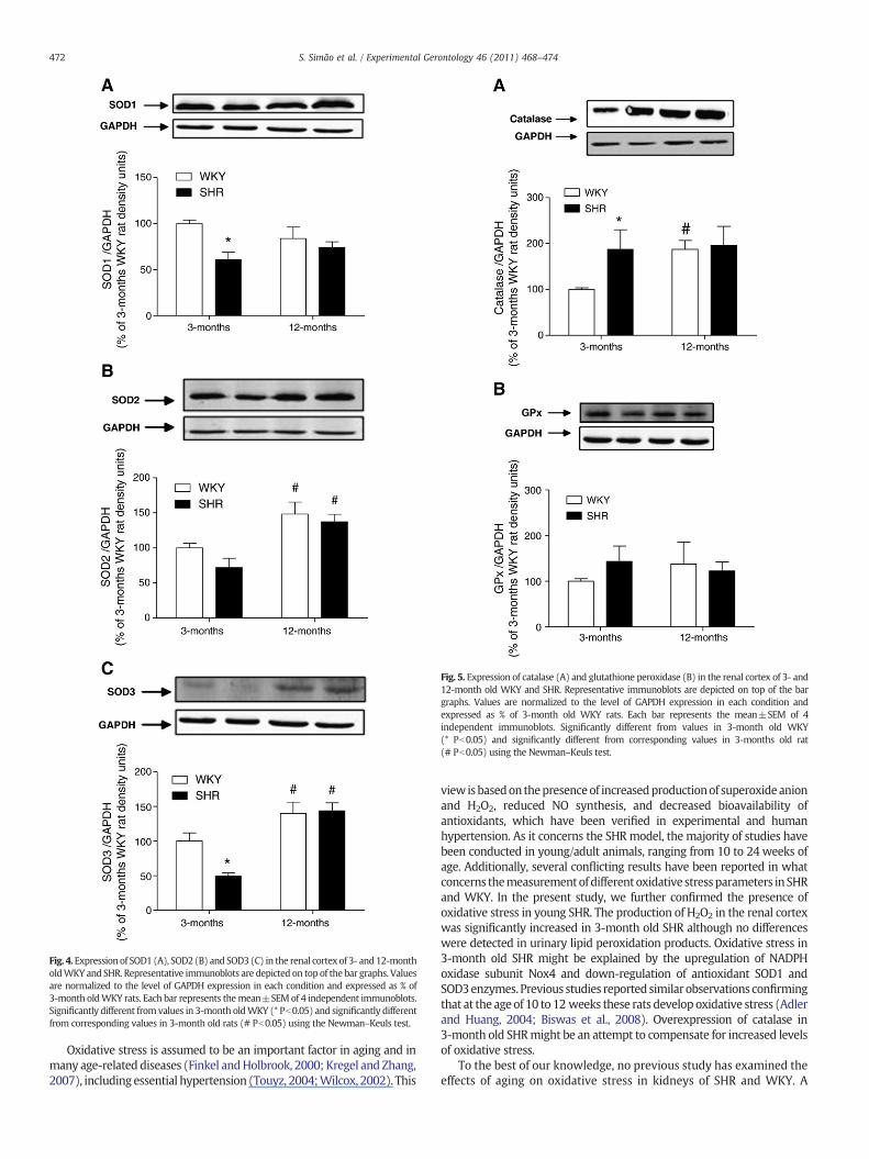
Fig. 4.Expression of SOD1 (A), SOD2 (B) and SOD3 (C) in the renal cortex of 3- and12-montholdWKY and SHR. Representative immunoblots are depicted on top of the bar graphs. Valuesare normalized to the level of GAPDH expression in each condition and expressed as % of3-month oldWKY rats. Each bar represents themean±SEMof 4 independent immunoblots.Significantly different from values in 3-month oldWKY (* Pb0.05) and significantly differentfrom corresponding values in 3-month old rats (# Pb0.05) using the Newman–Keuls test.
Fig. 5. Expression of catalase (A) and glutathione peroxidase (B) in the renal cortex of 3- and12-month old WKY and SHR. Representative immunoblots are depicted on top of the bargraphs. Values are normalized to the level of GAPDH expression in each condition andexpressed as % of 3-month old WKY rats. Each bar represents the mean±SEM of 4independent immunoblots. Significantly different from values in 3-month old WKY(* Pb0.05) and significantly different from corresponding values in 3-months old rat(# Pb0.05) using the Newman–Keuls test.
472 S. Simão et al. / Experimental Gerontology 46 (2011) 468–474
Oxidative stress is assumed to be an important factor in aging and inmany age-related diseases (Finkel andHolbrook, 2000; Kregel and Zhang,2007), including essential hypertension (Touyz, 2004;Wilcox, 2002). This
view isbasedon thepresenceof increasedproductionof superoxideanionand H2O2, reduced NO synthesis, and decreased bioavailability ofantioxidants, which have been verified in experimental and humanhypertension. As it concerns the SHRmodel, the majority of studies havebeen conducted in young/adult animals, ranging from 10 to 24 weeks ofage. Additionally, several conflicting results have been reported in whatconcerns themeasurementofdifferentoxidative stressparameters inSHRand WKY. In the present study, we further confirmed the presence ofoxidative stress in young SHR. The production of H2O2 in the renal cortexwas significantly increased in 3-month old SHR although no differenceswere detected in urinary lipid peroxidation products. Oxidative stress in3-month old SHR might be explained by the upregulation of NADPHoxidase subunit Nox4 and down-regulation of antioxidant SOD1 andSOD3enzymes. Previous studies reported similar observations confirmingthat at the age of 10 to 12 weeks these rats develop oxidative stress (Adlerand Huang, 2004; Biswas et al., 2008). Overexpression of catalase in3-month old SHRmight be an attempt to compensate for increased levelsof oxidative stress.
To the best of our knowledge, no previous study has examined theeffects of aging on oxidative stress in kidneys of SHR and WKY. A

473S. Simão et al. / Experimental Gerontology 46 (2011) 468–474
recent work evaluated oxidant handling and energy metabolism inchronic kidney disease using 3- and 21–24-month old WKY, SHR andWistar rats, although with a different approach regarding oxidativestress measurement. In this study, the expression of hemeoxygenase-1 was used as a renal marker of oxidative stress. The authors observedan increased expression of hemeoxygenase-1 in old Wistar and SHRrats as compared with the young rats (Percy et al., 2009).Furthermore, in the study of Alvarez et al., comparisons of oxidativestress levels were made during aging in hearts of Wistar and SHR rats.These authors reported increased levels of oxidative stress in agedSHR (Alvarez et al., 2008).
In the present study, we found a significant increase in H2O2
production by the renal cortex of 12-month old SHR. Moreover, theserats also exhibited a significant increase in urinary TBARS, a frequentlyused index of cell lipid peroxidation. Several reports have shown anage-associated increase in the concentrations of lipid peroxidationproducts (Pratico, 2002). Unexpectedly, WKY at the age of 12 monthsdisplayed similar oxidative stress levels as the age-matched SHR. It issuggested that the increased levels of H2O2 found in the renal tissue ofaged SHRandWKYmay be, at least in part, responsible for the increasedurinary lipid peroxides in these rats. Altogether, the results presentedhere indicate that aged rats have increased levels of oxidative stresscompared with young ones. In contrast, recent work demonstrated anincrease in plasma (Zhan et al., 2004a) and urinary (Suzuki et al., 2008)H2O2 levels in 22/24-week-old SHR compared with age-matchedWKY.Furthermore, the study of de Cavanagh et al. (2006) reports an increasein renal mitochondrial H2O2 production in 8-month old SHR comparedwith age-matchedWKY. The discrepancy between our study and thosementioned before in terms of H2O2 production might be explained bythe marked increase in bodyweight of agedWKY, even after correctionfor tibia length, suggesting the accumulation of fat in these rats. In fact,numerous reports have shown that oxidative stress is increased inanimal models of obesity and that the adipose tissue is a majorcontributor to ROS production (Davi et al., 2002; Furukawa et al., 2004;Urakawa et al., 2003). Several studies demonstrated that dietaryrestriction is an efficient approach to confront age-related oxidativedamage indifferent rat tissues, decreasingROSandF2-isoprostane levelsas well as the activities of proinflammatory transcription factors (Junget al., 2009; Opalach et al., 2010; Ward et al., 2005). It would be ofinterest, in future studies, to determine the importance of diet intakeupon oxidative stress parameters in the aged kidney of SHR and WKY.
The NADPH oxidase complex has been described as the mainsource of ROS in the kidney, although other ROS-generating enzymesare also present in this organ (Paravicini and Touyz, 2008; Touyz,2004). The NAPDH oxidase catalyzes the conversion of molecularoxygen into superoxide anion and is composed by p22phox and Nox2/gp91phox membrane subunits and the cytosolic proteins p40phox,p47phox, p67phox and Rac (Bedard and Krause, 2007; Paravicini andTouyz, 2008). Nox4 is the most highly expressed NADPH oxidase inthe kidney and does not require cytosolic subunits for its activation(Bedard and Krause, 2007). Oxidative stress in aged SHR and WKYwas accompanied by increases in the abundance of p22phox and Nox4proteins, which provides additional support for increased ROSproduction in the kidney of aged rats.
The primary lines of defense preventing biological macromoleculesfrom ROS attack are the antioxidant enzymes. SODs are an ubiquitousfamily of enzymes that catalyze the conversion of superoxide anion intoH2O2. In the subsequent step of the detoxifying cascade,H2O2 previouslyproduced is converted to water and molecular oxygen by catalase orGPx, which uses reduced glutathione as the hydrogen donor. Increasedlevels of ROS in cells and tissues may act as a signal to enhance theactivity and expression of antioxidant enzymes. According to thishypothesis, an increase in antioxidant enzymes activity and/orexpression with age would be expected, this being an adaptation tohelp cells and tissues protect fromoxidative stress. Here,we reported anincreased renal expression of SOD2, SOD3 and catalase in both aged SHR
and WKY. This finding may occur in response to the increased H2O2
production in aged rats. In addition, the increased levels of H2O2 presentin these rats might modulate mRNA levels of antioxidant enzymesthrough activation of redox-sensitive transcription factors such as AP-1and NF-κB, which can lead to the overexpression of these enzymes inaged rats. In contrast, expression of GPx remained unaltered duringaging in both SHR and WKY rats, suggesting that this enzyme may bedysregulateddespite thepresence of oxidative stress. Somestudies haveshowndecreases in activity and/or expressionof catalase andGPx,whileother studies showed the opposite trend. Fortepiani and Reckelhoff(2005) reported a decrease in the expression of both catalase andGPx in17/19-week old SHR. In contrast, Zhan et al. (2004b) reported increasesin the expression of catalase and GPx in the renal cortex of 24-week oldSHR without changes in enzymatic activity. In the study of Zhan et al.(2004b), and similar to the present work, a significant increase in bodyweight ofWKYwas reported. The cause of the discrepancy between theresults of Fortepiani and Reckelhoff (2005), Zhan et al., (2004b) and thepresent study remains at present unclear. It is possible that it restssimply on the fact that animals of different ages and bodyweights wereused and this is an important aspect to consider. Recent studies carriedout in WKY and Fischer-344 rats (21 to 24 months old) also describedincreases in the activity of major antioxidant enzymes with age (Judgeet al., 2005; Lambertucci et al., 2007).
In conclusion, aged SHR and WKY display increased levels ofoxidative stress in renal tissue with similar expression profile ofoxidant and antioxidant enzymes, suggesting the existence ofcommon mechanisms in hypertension and obesity leading tooxidative stress. It is suggested that oxidative stress levels in agedWKY might arise from increased fat mass accumulation which isoperating as a confounding factor.
References
Adler, S., Huang, H., 2004. Oxidant stress in kidneys of spontaneously hypertensive ratsinvolves both oxidase overexpression and loss of extracellular superoxidedismutase. Am J Physiol Renal Physiol 287, F907–913.
Alvarez, M.C., Caldiz, C., Fantinelli, J.C., Garciarena, C.D., Console, G.M., Chiappe deCingolani, G.E., Mosca, S.M., 2008. Is cardiac hypertrophy in spontaneouslyhypertensive rats the cause or the consequence of oxidative stress? HypertensRes 31, 1465–1476.
Bedard, K., Krause, K.H., 2007. The NOX family of ROS-generating NADPH oxidases:physiology and pathophysiology. Physiol Rev 87, 245–313.
Biswas, S.K., Peixoto, E.B., Souza, D.S., de Faria, J.B., 2008. Hypertension increases pro-oxidantgeneration and decreases antioxidant defense in the kidney in early diabetes. Am JNephrol 28, 133–142.
Chabrashvili, T., Tojo, A., Onozato, M.L., Kitiyakara, C., Quinn, M.T., Fujita, T., Welch, W.J.,Wilcox, C.S., 2002. Expression and cellular localization of classic NADPH oxidasesubunits in the spontaneously hypertensive rat kidney. Hypertension 39, 269–274.
Davi, G., Guagnano, M.T., Ciabattoni, G., Basili, S., Falco, A., Marinopiccoli, M., Nutini, M.,Sensi, S., Patrono, C., 2002. Platelet activation in obese women: role of inflammationand oxidant stress. JAMA 288, 2008–2014.
de Cavanagh, E.M., Toblli, J.E., Ferder, L., Piotrkowski, B., Stella, I., Inserra, F., 2006. Renalmitochondrial dysfunction in spontaneously hypertensive rats is attenuated by losartanbut not by amlodipine. Am J Physiol Regul Integr Comp Physiol 290, R1616–1625.
Finkel, T., Holbrook, N.J., 2000. Oxidants, oxidative stress and the biology of ageing.Nature 408, 239–247.
Fortepiani, L.A., Reckelhoff, J.F., 2005. Increasing oxidative stress with molsidomineincreases blood pressure in genetically hypertensive rats but not normotensivecontrols. Am J Physiol Regul Integr Comp Physiol 289, R763–770.
Furukawa, S., Fujita, T., Shimabukuro,M., Iwaki,M., Yamada, Y.,Nakajima,Y., Nakayama,O.,Makishima,M.,Matsuda,M., Shimomura, I., 2004. Increased oxidative stress in obesityand its impact on metabolic syndrome. J Clin Invest 114, 1752–1761.
Griendling, K.K., Sorescu, D., Ushio-Fukai, M., 2000. NAD(P)H oxidase: role incardiovascular biology and disease. Circ Res 86, 494–501.
Harman, D., 1956. Aging: a theory based on free radical and radiation chemistry. JGerontol 11, 298–300.
Judge, S., Jang, Y.M., Smith, A., Hagen, T., Leeuwenburgh, C., 2005. Age-associatedincreases in oxidative stress and antioxidant enzyme activities in cardiacinterfibrillar mitochondria: implications for the mitochondrial theory of aging.FASEB J 19, 419–421.
Jung, K.J., Lee, E.K., Kim, J.Y., Zou, Y., Sung, B., Heo, H.S., Kim, M.K., Lee, J., Kim, N.D., Yu, B.P.,Chung, H.Y., 2009. Effect of short term calorie restriction on pro-inflammatory NF-kBand AP-1 in aged rat kidney. Inflamm Res 58, 143–150.
Kerr, S., Brosnan, M.J., McIntyre, M., Reid, J.L., Dominiczak, A.F., Hamilton, C.A., 1999.Superoxide anion production is increased in a model of genetic hypertension: roleof the endothelium. Hypertension 33, 1353–1358.

474 S. Simão et al. / Experimental Gerontology 46 (2011) 468–474
Kregel, K.C., Zhang, H.J., 2007. An integrated view of oxidative stress in aging: basicmechanisms, functional effects, and pathological considerations. Am J Physiol RegulIntegr Comp Physiol 292, R18–36.
Lambertucci, R.H., Levada-Pires, A.C., Rossoni, L.V., Curi, R., Pithon-Curi, T.C., 2007.Effects of aerobic exercise training on antioxidant enzyme activities and mRNAlevels in soleus muscle from young and aged rats. Mech Ageing Dev 128, 267–275.
Li, H., Matheny, M., Tumer, N., Scarpace, P.J., 1998. Aging and fasting regulation of leptinand hypothalamic neuropeptide Y gene expression. Am J Physiol 275, E405–411.
Li, W.G., Miller Jr., F.J., Zhang, H.J., Spitz, D.R., Oberley, L.W., Weintraub, N.L., 2001. H(2)O(2)-induced O(2) production by a non-phagocytic NAD(P)H oxidase causesoxidant injury. J Biol Chem 276, 29251–29256.
Ohkawa, H., Ohishi, N., Yagi, K., 1979. Assay for lipid peroxides in animal tissues bythiobarbituric acid reaction. Anal Biochem 95, 351–358.
Opalach, K., Rangaraju, S., Madorsky, I., Leeuwenburgh, C., Notterpek, L., 2010. Lifelongcalorie restriction alleviates age-related oxidative damage in peripheral nerves.Rejuvenation Res 13, 65–74.
Paravicini, T.M., Touyz, R.M., 2008. NADPH oxidases, reactive oxygen species, andhypertension: clinical implications and therapeutic possibilities. Diabetes Care 31(Suppl 2), S170–180.
Percy, C.J., Brown, L., Power, D.A., Johnson, D.W., Gobe, G.C., 2009. Obesity andhypertension have differing oxidant handling molecular pathways in age-relatedchronic kidney disease. Mech Ageing Dev 130, 129–138.
Pratico, D., 2002. Lipid peroxidation and the aging process. Sci Aging Knowledge Environ2002, re5.
Ricci, E., Smallwood, S., Chouabe, C., Mertani, H.C., Raccurt, M., Morel, G., Bonvallet, R.,2006. Electrophysiological characterization of left ventricular myocytes from obeseSprague–Dawley rat. Obesity (Silver Spring) 14, 778–786.
Schnackenberg, C.G., Wilcox, C.S., 1999. Two-week administration of tempol attenuatesboth hypertension and renal excretion of 8-Iso prostaglandin F2alpha. Hypertension33, 424–428.
Suzuki, A., Fujii, A., Jokura, H., Tokimitsu, I., Hase, T., Saito, I., 2008. Hydroxyhydroquinoneinterferes with the chlorogenic acid-induced restoration of endothelial function inspontaneously hypertensive rats. Am J Hypertens 21, 23–27.
Touyz, R.M., 2004. Reactive oxygen species, vascular oxidative stress, and redox signalingin hypertension: what is the clinical significance? Hypertension 44, 248–252.
Trippodo, N.C., Frohlich, E.D., 1981. Similarities of genetic (spontaneous) hypertension.Man and rat. Circ Res 48, 309–319.
Ulker, S., McMaster, D., McKeown, P.P., Bayraktutan, U., 2003. Impaired activities ofantioxidant enzymes elicit endothelial dysfunction in spontaneous hypertensiverats despite enhanced vascular nitric oxide generation. Cardiovasc Res 59, 488–500.
Urakawa, H., Katsuki, A., Sumida, Y., Gabazza, E.C., Murashima, S., Morioka, K.,Maruyama, N., Kitagawa, N., Tanaka, T., Hori, Y., Nakatani, K., Yano, Y., Adachi, Y.,2003. Oxidative stress is associated with adiposity and insulin resistance in men. JClin Endocrinol Metab 88, 4673–4676.
Ward, W.F., Qi, W., Van Remmen, H., Zackert, W.E., Roberts 2nd, L.J., Richardson, A.,2005. Effects of age and caloric restriction on lipid peroxidation: measurement ofoxidative stress by F2-isoprostane levels. J Gerontol A Biol Sci Med Sci 60, 847–851.
Wilcox, C.S., 2002. Reactive oxygen species: roles in blood pressure and kidneyfunction. Curr Hypertens Rep 4, 160–166.
Zhan, C.D., Sindhu, R.K., Vaziri, N.D., 2004a. Up-regulation of kidney NAD(P)H oxidase andcalcineurin in SHR: reversal by lifelong antioxidant supplementation. Kidney Int 65,219–227.
Zhan, C.D., Sindhu, R.K., Pang, J., Ehdaie, A., Vaziri, N.D., 2004b. Superoxide dismutase,catalase and glutathione peroxidase in the spontaneously hypertensive rat kidney:effect of antioxidant-rich diet. J Hypertens 22, 2025–2033.