AG-512-Botany-science-of-plant-growth ... - University of Idaho
385
TABLE OF CONTENTS AG. 512 – BOTANY/SCIENCE OF PLANT GROWTH AND DEVELOPMENT 512-A ...............................The Organisms 512-B................................Cells: Structure, Functions and Division 512-C................................Plant Processes 512-D ...............................Nonvascular and Vascular Plants 512-E ................................Vegetative Plant Parts 512-F ................................Reproductive Plant Parts 512-G ...............................Vegetative Plant Growth 512-H ...............................Reproductive Plant Growth 512-I .................................Scientific Method Term Project and Matrix
-
Upload
khangminh22 -
Category
Documents
-
view
0 -
download
0
Transcript of AG-512-Botany-science-of-plant-growth ... - University of Idaho

TABLE OF CONTENTS AG. 512 – BOTANY/SCIENCE OF PLANT GROWTH AND DEVELOPMENT
512-A ...............................The Organisms
512-B................................Cells: Structure, Functions and Division 512-C................................Plant Processes 512-D ...............................Nonvascular and Vascular Plants 512-E ................................Vegetative Plant Parts 512-F ................................Reproductive Plant Parts 512-G ...............................Vegetative Plant Growth 512-H ...............................Reproductive Plant Growth 512-I .................................Scientific Method Term Project and Matrix

AG 512
BOTANY/SCIENCE
OF PLANT
GROWTH AND DEVELOPMENT
FOR
IDAHO
SECONDARY AGRICULTURE INSTRUCTORS
Developed and written by:
Cathy Tesnohlidek Mosman
Provided through a grant from the
Idaho State Division of Vocational Education
1991
Administered through the
Department of Agricultural and Extension Education
University of Idaho
By
Douglas A. Pals, Project Director

FOREWORD The Agricultural Science and Technology Curriculum Guides are the product of
many years of careful planning and development. In 1987, an Agricultural Education Technical Committee was assembled to determine the competencies necessary to prepare students for careers in agriculture. In 1989, a committee of secondary agriculture instructors, state supervisory staff and University of Idaho Agricultural and Extension Education faculty arranged the competencies into an outline of courses appropriate for secondary agriculture programs in Idaho. These curriculum guides have been written to provide the secondary agriculture instructor with up-to-date instructional materials to be used in developing lessons for the student interested in pursuing a career in agriculture.
The arrangement of the guide follows the courses outlined in the Agricultural Science and Technology Curriculum Outline - The Guide to the 90's (Vo. Ed. #240) published in 1989. The format used in this guide was adapted from the curriculum guides developed for Idaho secondary agriculture instructors during the period of 1981-1985.
The original Idaho Agricultural Curriculum Guides used in the development of these materials were:
1981 - Livestock Production 1981 - Agricultural Mechanics 1982 - Farm Business Management 1985 - Crop and Soil Science Many individuals made the original guides possible. The format used was adapted
from curriculum developed by the Curriculum and Instructional Materials Center of the Oklahoma State Department of Vocational and Technical Education. Selected information and many of the transparency masters used in the guides were provided by the Vocational Instructional Services, Texas A & M University. Additional information and transparency masters were provided by the Department of Agricultural Communications and Education, College of Agriculture, University of Illinois and the Agricultural Education Program, Department of Applied Behavioral Sciences, University of California, Davis.
Laboratory exercises incorporated into the units of instruction were used from the
Holt, Rinehart and Winston, Inc. book, Modern Biology, Biology Investigations and the Scott, Foresman, and Company Lab Manual for Biology. Credit appears on the first page of the materials used from these two sources.

Without the following individuals' dedication and commitment, this project would not have been completed.
Project staff Cathy Tesnohlidek Mosman, Curriculum Writer Donna Wommack, Curriculum Typist and Editor Molly Parrish, Curriculum Typist Douglas A. Pals, Project Director State Division of Vocational Education Trudy Anderson, Administrator DeVere Burton, State Supervisor, Agricultural Education Michael Rush, Director, Research Donald Eshelby, Director, Program Services Agricultural and Extension Education Department Faculty - Dr. Lou Riesenberg, Dr. John Mundt, Dr. Richard Ledington (affiliate), and Laurie Lancaster Typists - Marilyn Crumley, Eadie Samagaio, Terry Olson, Becky McMillan, Debby McMillan, Sue McMurray and Rebecca Jones Technical Assistance Agricultural Communications Katie Old, Graphic Artist Jerry Adams, Production Supervisor

USE OF THIS PUBLICATION Introduction
This material must be taught. It does not replace the teacher, nor the teacher's
expertise. The teacher needs to adapt the material to the local area and individual students. The teacher must also provide the necessary motivating techniques to help the students learn the material.
The pages in the guide are color coded to assist in identifying and locating the
desired pages. The colors used are:
Table of Contents Ivory Semester Course Title Page Green Foreword Yellow Use of Publication Salmon Divider Page Between Units Tan Refer to Another Unit Page Grey Unit Objectives/Specific Competencies White Suggested Activities Blue Information Sheets White Transparency Masters White Assignment Sheets White Answers to Assignment Sheets Gold Instructors Notes for Laboratory Exercises Blue Laboratory Exercises White Answers to Laboratory Exercises Gold Unit Test White Answers to Test Gold
Instructional Units
These units are not geared to a particular age level and must be adapted for the
students with whom they are used. Units include objectives and competencies, suggested activities for the instructor and students, information sheet, transparency masters, assignment sheets, laboratory exercises, instructor notes for laboratory exercises, answers to assignment sheets and laboratory exercises, test and answers to test. Units are planned for more than one lesson or class period.
The teacher should carefully study each instructional unit to determine:
A. The appropriateness of the material for the age level B. The amount of material that can be covered during a class period C. Additional objectives and/or assignments, which could be developed

D. The skills that must be demonstrated 1. Supplies needed 2. Equipment needed 3. Amount of practice needed 4. Amount of class time needed for demonstrations E. Supplementary materials, such as pamphlets, filmstrips and slides that must
be ordered F. Resource people who must be contacted Objectives and Competencies Each unit of instruction is based on stated objectives. These objectives state the goals of the unit, thus providing a sense of direction and accomplishment for the student. The objectives are stated in two forms: unit objectives, stating the subject matter to be covered in a unit of instruction; and specific objectives, stating the student performances necessary to reach the unit objective. Since the objectives of the unit provide direction for the teaching-learning process, it is important for the teacher and students to have a common understanding of the intent of the objectives. A limited number of performance terms have been used in the objectives for this curriculum to assist in promoting the effectiveness of the communication among all individuals using the materials. Following is a list of performance terms and their synonyms that may have been used in this material: Name Identify State a Rule Apply a Rule Label Select Calculate List in writing Mark List orally Point out Letter Pick out Record Choose Repeat Locate Give Match Describe Order Distinguish Define Arrange Discriminate Discuss in writing Sequence Discuss orally List in order Interpret Classify Tell how Divide Tell what Isolate Explain Sort

Construct Demonstrate Draw Transcribe Show your work Replace Make Reduce Show procedure Turn on/off Build Increase Perform an experiment (Dis) assemble Design Figure Perform the steps (Dis) connect Formulate Conduct Operate Reproduce Compare Remove Reading of the objectives by the student should be followed by a class discussion to answer any questions concerning performance requirements for each instructional unit. Teachers should feel free to add objectives, which will fit the material to the needs of the students and community. When a teacher adds objectives, he/she should remember to supply the needed information, assignment sheets and/or laboratory exercises and criterion tests. Suggested Activities Each unit of instruction has a suggested activities sheet outlining steps to follow in accomplishing specific objectives. Duties of the instructor will vary according to the particular unit. However, for best use of the material they should include the following: provide students with objective sheet, information sheet, assignment sheets, and laboratory exercises; preview filmstrips, make transparencies, and arrange for resource materials and people; discuss unit and specific objectives and information sheet; give test. Teachers are encouraged to use any additional instructional activities and teaching methods to aid students in accomplishing the objectives. Information Sheet The information sheet provides content essential for meeting the cognitive (knowledge) requirements of the unit. The teacher will find that the information sheet serves as an excellent guide for presenting the background knowledge necessary to develop the skills specified in the unit objective. Students should read the information sheet before the information is discussed in class. Students may take additional notes on the information sheet. Transparency Masters Transparency masters provide information in a special way. The students may see as well as hear the material being presented, thus reinforcing the learning process. Transparencies may present new information or they may reinforce information presented in the information sheet. They are particularly effective when identification is necessary.

Transparencies should be made and placed in the notebook where they will be immediately available for use. Transparencies direct the class's attention to the topic of discussion. They should be left on the screen only when topics shown are under discussion. (NOTE: Stand away from the overhead projector when discussing transparency material. The noise of the projector may cause the teacher to speak too loudly.)
Assignment Sheets Assignment sheets give direction to study and furnish practice for paper and pencil activities to develop the knowledge which is a necessary prerequisite to skill development. These may be given to the student for completion in class or used for homework assignments. Answer sheets are provided which may be used by the student and/or teacher for checking student progress. Laboratory Exercises Laboratory exercises are found in selected units. The laboratory exercises include both science and agricultural mechanics activities. The science laboratory exercises often have instructions to the instructor prior to the actual laboratory. Procedures outlined in the laboratory exercise for agricultural mechanics give direction to the skill being taught and allow both student and teacher to check student program toward the accomplishment of the skill. Test and Evaluation Paper-pencil and performance tests have been constructed to measure student achievement of each objective listed in the unit of instruction. Individual test items may be pulled out and used as a short test to determine student achievement of a particular objective. This kind of testing may be used as a daily quiz and can help the teacher spot difficulties being encountered by students in their efforts to accomplish the unit objective. Test items for objectives added by the teachers should be constructed and added to the test.
Test Answers Test answers are provided for each unit. These may be used by the teacher and/or student for checking student achievement of the objectives. Care of Materials The cost of reproduction of this guide prohibits the replacement of these materials. Therefore, please be extremely careful in handling originals. Make the necessary copies of the information sheets, transparencies, assignments and tests and replace originals in the curriculum guide notebook. Take extra care in keeping originals clear for future reproduction.

512A- 1
THE ORGANISMS
AG 512 - A
UNIT OBJECTIVE
After completion of this unit, students should be able to define terms related to the organisms and discuss the various methods of classifying plants. Students should also be able to discuss the differences between plants and animals and the scientific names. This knowledge will be demonstrated by completing the unit test with a minimum of 85 percent accuracy.
SPECIFIC OBJECTIVES AND COMPETENCIES
After completion of this unit, the student should be able to:
1. Match terms related to the organisms to the correct definitions.
2. List the seven categories of the Linnaeus classification system in order from largest to smallest.
3. List and define the five kingdoms in the classification system.
4. List three traits that help place an organism into a kingdom.
5. Match the phylums of the plant kingdom to their characteristics.
6. Discuss six differences between plants and animals.
7. Discuss six ways to classify plants besides the Linnaeus classification system.
8. Discuss scientific names and their importance.
9. Become familiar with laboratory skills and equipment.
10. Examine cells from the five kingdoms.
11. Classify organisms.

512A- 2
THE ORGANISMS
AG 512 - A
SUGGESTED ACTIVITIES
I. Suggested activities for instructor
A. Order materials to supplement unit.
1. Films
a. Classification, VHS video; examines the various organisms and how they are classified into distinct animal and plant groups; available from Vocational Education Productions, California Polytechnic State University, San Luis Obispo, California 93407 (1-800-235-4146); approximate cost $99.95; order no. 6-083-103J.
B. Make transparencies and necessary copies of materials.
C. Provide students with objective sheet.
D. Provide students with information sheets and laboratory exercises.
E. Discuss unit and specific objectives.
F. Discuss information sheet.
G. Demonstrate and discuss procedures outlined in laboratory exercises.
H. Review and give test.
I. Reteach and retest if necessary.
II. Instructional materials
A. Objective sheet
B. Suggested activities
C. Information sheet
D. Transparency masters
1. TM 1--Linnaeus' Classification of Plants
2. TM 2--Kingdoms in the Linnaeus Classification System
3. TM 3--Phylums of the Plant Kingdom
4. TM 4--Plant and Animal Differences
5. TM 5--Classifying Plants According to Growth Habits

512A- 3
6. TM 6--Classifying Plants According to Flower Parts
7. TM 7--Classifying Plants According to Roots and Leaves
8. TM 8--Classifying Plants According to Agricultural Use
9. TM 9--Scientific Names
E. Instructor notes for laboratory exercises
F. Laboratory exercises
1. LE 1--General Laboratory Procedures, Equipment and Report Writing
2. LE 2--Examining Cells From the Five Kingdoms
3. LE 3--Classifying Organisms
G. Answers to laboratory exercises
H. Test
I. Answers to test
III. Unit references
A. Agricultural Education Curriculum, College of Agriculture, University of Illinois, Urbana, Illinois, 1989.
B. Hartmann, Hudson T., et al., Plant Science - Growth, Development, and
Utilization of Cultivated Plants, 2nd edition, Prentice-Hall, Inc., Englewood Cliffs, New Jersey 07632, 1988.
C. Otto, James H., Towle, Albert, Modern Biology, Holt, Rinehart and Winston,
New York, 1985. D. Slesnick, Irwin L., Balzer, LeVon, McCormack, Alan J., Newton, David E.,
Rasmussen, Frederick A., Biology, Scott, Foresman and Company, Glenview, Illinois, 1985.

512A- 4
THE ORGANISMS
AG 512 - A
INFORMATION SHEET
I. Terms and definitions
A. Binomial nomenclature--A system invented by Carolus Linnaeus for classifying organisms. Each organism is assigned a two-word Latin name
(Note: First word represents the genus; second word is descriptive.)
B. Prokaryote--Cell type that has a nucleus without a membrane around it. The nuclear material floats freely within the cell
C. Eukaryote--Cell type that has an organized nucleus surrounded by a membrane
D. Adaptation--A characteristic which enables the organism to survive in its environment
E. Autotrophs--Organisms that manufacture organic nutrients from inorganic raw
materials F. Biogenesis--The theory that all living things come only from preexisting living
things G. Biome--Large, easily differentiated community unit arising as a result of
complex interactions of climate, other physical factors and biotic factors H. Colony--Association of unicellular or multicellular organisms of the same
species I. Community--An assemblage of populations that live in a defined habitat. The
organisms constituting the community interact in various ways with one another J. "Consumer" organisms--Those elements of an ecosystem that eat other plants or
animals K. Ecology--The study of the interrelations between living things and their
environment L. Ecosystem--All of the organisms of a given area
M. Epigenesis--The theory that development proceeds from a structureless cell by the successive formation and addition of new parts which do not preexist in the fertilized egg
N. Fossils--Any remains of an organism that have been preserved in the earth's
crust

512A- 5
O. Genus--Taxonomic classification in which closely related species are grouped together
P. Herbivore--A plant-eating animal
Q. Heterotrophs--Organisms which cannot synthesize their own food from inorganic materials
R. Phenotype--The visible expression of the hereditary constitution of an organism
S. Phylogeny--The evolutionary history of a group of organisms
T. Polymorphism--Occurrence of several distinct phenotypes in a population
U. Population--The group of individuals of a given species inhabiting a specified geographic area
V. Species--The unit of taxonomic classification, a population of similar
individuals, alike in their structural and functional characteristics
W. Taxonomy--The science of naming, describing and classifying organisms
X. Tissue--Specialized cells which together perform certain special functions
II. Classification system--Largest to smallest (Transparency 1)
A. Kingdom
B. Phylum
C. Class
D. Order
E. Family
F. Genus
G. Species
III. Five kingdoms (Transparency 2)
A. Animal kingdom (Animalia)
1. Eukaryotic cells
2. Multicellular organisms
3. Move about to obtain food
4. Digest food inside body

512A- 6
B. Plant kingdom (Plantae)
1. Eukaryotic cells
2. Multicellular organisms
3. Produce own food
4. Cannot move about
C. Fungi kingdom
1. Eukaryotic cells
2. Mostly multicellular organisms
3. Do not move about
4. Obtain food by absorbing it from living or dead organisms
D. Monera kingdom
1. Prokaryotic cells
2. Mostly one-celled organisms
3. Produce own food or obtain it from outside source
E. Protista kingdom
1. Eukaryotic cells
2. Many are one-celled
3. Produce own food or obtain it from outside source
IV. Traits that help place organism into kingdom
A. Kind of cells in organism: prokaryote or eukaryote
B. How organism obtains its food
C. How organism reproduces and develops
V. Phylums of the plant kingdom (Transparency 3)
A. Bryophyta
1. Multicellular, small green plants living on land (usually in moist situations) without true roots or flowers
2. Lack vascular tissues

512A- 7
3. Alternation of generations with the gametophyte, the dominant generation
4. Motile sperm
5. Liverworts, hornworts, mosses
B. Lycophyta
1. Vascular plants
2. Sporophyte dominant
3. Trailing or erect with simple leaves
4. Spores produced in a strobilus
5. Gametophyte underground
6. Club mosses
C. Sphenophyta
1. Vascular plants
2. Sporophyte dominant
3. Stems: hollow, jointed, contain silica
4. Leaves: tiny scales, in whorls at stem joints
5. Fertile stems with strobili
6. Horsetails
D. Pterophyta
1. Vascular plants
2. Sporophyte generation dominant with leafy fronds
3. Creeping rhizome
4. Gametophyte, a free-living prothallus
5. Motile sperm
6. Ferns and tree ferns

512A- 8
E. Cycadophyta
1. Palmlike plants
2. Male and female cones on different trees
3. Cycads, sago palms
F. Ginkgophyta
1. One living species
2. Pollen produced in conelike structure
3. Seeds naked
4. Ginko biloba
G. Gnetophyta
1. Specialized gymnosperms
2. Desert species with xylem resembling that in angiosperms
3. Few species
4. Welwitschia, ephedra, gnetum
H. Coniferophyta
1. Gymnosperms with sex organs in cones
2. Leaves in the form of needles
3. Most are evergreen
4. Pines, spruces, firs, larches, yews
I. Anthophyta
1. Includes all the flowering plants
2. Sex organs in whorls in flowers
3. Seeds enclosed in ovary that ripens into a fruit
4. Grasses, maples, elms

512A- 9
VI. Differences between plants and animals (Transparency 4)
A. Mobility
1. Animals are mobile
2. Most plants are stationary
B. Food
1. Plants manufacture their own food
2. Animals must rely on plants or other animals for their food
C. Cell walls
1. Cell walls of plants are rigid and usually made of cellulose
2. Animals don't have rigid cell walls or flaccid cell membranes
D. Cellulose
1. Synthesized by plants
2. Not synthesized by animals
E. Growth
1. Animal growth is limited
2. Many plants have unlimited growth and an indefinite number of parts
F. Number of parts
1. Animals have definite number of parts, such as eyes, ears, legs, etc.
2. The number of parts (such as leaves, stems, buds and flowers) of the same kind of plant usually varies from plant to plant
VII. Other methods of classifying plants (Transparencies 5, 6, 7, 8)
A. Growth habits (Transparency 5)
1. Annuals
2. Winter annuals
3. Biennials
4. Perennials

512A- 10
B. Types of flowers (Transparency 6)
1. Complete flower
a. Sepals
b. Petals
c. Stamens
d. Pistil
2. Incomplete flowers--One of the four principal parts is missing
3. Perfect flowers--Both the stamens and pistil are present
4. Imperfect flower--Lack either the stamens or pistil
C. Location of flowers
1. Dioecious--Flowers bearing stamens (male pollen) and those bearing pistils (female eggs) produced on separate plants
2. Monoecious--Flowers containing stamens and those containing pistils
are produced in different places on the same plant
D. Root structure (Transparency 7)
1. Fibrous root plants--Root systems are very branched and finely divided
2. Tap root plants--One major root with few lateral root hairs attached to it
E. Seed leaves
1. Dicot--Plant with two seed leaves in each of its seeds
2. Monocot--Plant with one seed leaf in each seed
F. Agricultural use of crop (Transparency 8)
1. Cereals or grain crops
2. Oil seed crops
3. Forage and pasture crops
4. Root and tuber crops
5. Fiber crops
6. Sugar crops
7. Specialty crops (for example: hops)

512A- 11
VIII. Scientific names (Transparency 9)
A. Definition
1. Each plant has a two-word (binomial) name--always in Latin
2. First name refers to the plant's genus
3. Second name refers to the plant's species
B. Importance--It is universal--the scientific name is the same regardless of the location or the language of the people
C. Examples
Common Name Scientific Name
Alfalfa Purple flowered Medicago sativa Variegated Medicago falcata media Yellow flowered Medicago falcata
Barley Six-row Hordeum vulgare Two-row Hordeum distichum
Barnyardgrass Echinochloa crusgalli
Bean Kidney Phaseolus vulgaris Pinto Phaeolus spp.
Black Medic Medicago lupulina
Black Nightshade Solanum nigrum
Bluegrass Annual Poa annua Canada Poa compressa Kentucky Poa pratensis
Bromegrass Downy Bromus tectorum Smooth Bromus inermis
Burdock Arctium minus
Clover Alsike Trifolium hybridum Red Trifolium pratense White Trifolium repens
Cocklebur Xanthium pensylvanicum

512A- 12
Common Name Scientific Name
Corn Dent Zea mays indentata Pop Zea mays everta Sweet Zea mays saccharata
Crabgrass Digitaria spp.
Dandelion Taraxacum officinale
Death Camas Cammassia spp.
Dodder, Field Cuscuta pentagona
Fescue Red Festuca rubra Tall Festuca arundinacea
Field Bindweed Convolvulus arvensis
Halogeton Halogeton glomeratus
Hemlock, Poison Conium maculatum
Hound's Tongue Cynoglossum officinale
Lambsquarter Chenopodium album
Larkspur Delphinium spp.
Lupine Lupinus spp.
Mayweed Anthemis cotula
Nutsedge Purple Cyperus rotundus Yellow Cyperus esculentus
Oats Common Avena sativa Wild, Common Avena fatua
Orchard grass Dactylis glomerata
Pea Field Pisum arvense Field, Austrian Winter Pisum arvense Garden Pisum sativum
Pigweed Amaranthus spp.
Plantain, Broadleaf Plantago major

512A- 13
Common Name Scientific Name
Plantain, Buckhorn Plantago lanceolata
Poison Hemlock Conium maculatum
Poison Ivy Rhus radicans
Poison Oak Rhus toxicondendron
Prickly Pear Opuntia spp.
Puncturevine, Spiny Tribulus terrestris
Purslane Portulaca oleracea
Quackgrass Agropyron repens
Rape Brassica napus
Russian Thistle Salsola kali var. tenuifolia
Rye Secale cereale
Shepherd's Purse Capsella bursa-pastoris
Sowthistle Annual Sonchus oleraceous Perennial Sonchus arvensis
St. Johnswort Hypericum perforatum
Sunflower Helianthus annuus
Thistle Bull Cirsium vulgare Canada Cirsium arvense
Timothy Phleum pratense
Vetch Common Vicia sativa Hairy Vicia villosa
Water Hemlock Cicuta maculata
Wheat Club Triticum compactum Common Triticum vulgare

512A- 14
Common Name Scientific Name
Wheatgrass Crested Agropyron desertorum Western Agropyron smithii
Wild Buckwheat Polygonum convolvulus
Yarrow Achillea millefolium

512A- 15
TM 1

512A- 16
TM 2

512A- 17
TM 3

512A- 18
TM 4

512A- 19
TM 5

512A- 20
TM 6

512A- 21
TM 7

512A- 22
TM 8

512A- 23
TM 9

512A- 24
THE ORGANISMS
AG 512 - A
INSTRUCTOR NOTES FOR LABORATORY EXERCISES
Lab #2 Background: The separate procedures do not have to be completed in any specific order. You may wish to set up five lab stations and divide the class into five groups. Have each group start at a different lab station doing a different procedure. This will minimize the amount of time that the students will need to wait for microscopes and prepared slides. Solution preparation: The following general instructions apply for the preparation of most solutions: Solvents should be added to solutes. Use distilled water, not tap water, for all reagents. When preparing an acid or base solution, slowly add the acid or base to the water. Never add water to a concentrated acid or base. To make percentage solutions measure 1 ml of solute per percentage. Add the solute to enough solvent to make 100 ml of solution. When dissolving a solid in water, measure 1 g of solute per percentage and mix the solute with enough water to make 100 ml of the solution. Iodine solution (also available ready-made) Dissolve 5.0 g of potassium iodide [KI] and 1.5 g of iodine crystals in 500 ml of distilled water. Store in brown bottle or other glass container that shields the liquid from light. CAUTION: Iodine dust and vapors are toxic and irritating. Avoid body contact and inhalation of fumes. Should body contact occur, flush immediately with water. (Quantity needed: 500 ml) Methyl cellulose solution (also available ready-made) Dissolve 2 g of methyl cellulose in 38 ml of distilled water. Store in refrigerator. (Quantity needed: 40 ml) Methyl blue stain (also available ready-made) Dissolve 0.75 g of methylene blue in 50 ml of 95% ethyl alcohol. Dilute 5 ml of the alcohol and methylene blue solution with 45 ml of distilled water. This diluted solution is the stain. Bottle and store the remaining methylene blue and alcohol solution. CAUTION: Ethyl alcohol is flammable. It is also irritating to the eyes. Flush spills with water. Do not ingest ethyl alcohol. (Quantity needed: 50 ml) Materials: Prepared slides could include: Animal cells: human and frog blood cells, skeletal and cardiac muscle, nerve cells and epithelial cells Plant cells: cross sections of leaves, stems and roots Fungal cells: Rhizopus, Lycogala and mushroom cross sections Protist cells: paramecia, diatoms, amebas and Volvox

512A- 25
Moneran cells: bacteria types and cyanobacteria, such as Anabaena Part I: Step 1: The tongue cells that students will observe are epithelial cells. Part III: To make yeast suspension, dissolve 0.1 g of yeast in 75 ml of warm (37oC) water. Add 2-5 g of sugar. Part IV: Step 3. Cilia of paramecia are best seen under dim light or under a phase-contrast microscope Part V: You may suggest that students work on Table I as they do the laboratory. Students may need to use a textbook to complete Table I. Lab #3 Inform the student that the construction and use of a classification key may be compared with solving a mystery or going on a treasure hunt, where each bit of information leads to another piece of information. Impress on them that one wrong choice somewhere along the way can cause them to take a wrong turn and end up in the wrong place with the wrong answer!

512A- 26
THE ORGANISMS
AG 512 - A
LABORATORY EXERCISE #1--GENERAL LABORATORY PROCEDURES, EQUIPMENT AND REPORT WRITING
Name _____________________________________ Score __________________________________
Part I: General Laboratory Procedures The following is a list of general laboratory procedures. You will be required to write at least ten of these on a quiz.
1. Never "horse around" in the laboratory.
2. Never play with laboratory equipment or materials.
3. Always follow instructions and wait until you are told to begin before starting any investigation.
4. Never carry out unassigned experiments.
5. Never eat or taste anything in the laboratory. This includes food, drinks and gum, as well as chemicals found in the laboratory.
6. Wash your hands after every experiment.
7. Keep all books and other nonessential items away from the work area.
8. Keep your work area clean. Dispose of waste materials in appropriate containers.
9. Turn off any gas jets or any electrically operated equipment when you have completed the laboratory investigation.
10. Report all injuries or accidents to your teacher immediately.
11. Never use broken or cracked glassware.
12. Always wear shoes in the laboratory. Sandals are not suggested.
13. Tie back long hair and restrict any loose clothing.
14. Wear safety goggles, laboratory aprons and gloves when instructed to do so.

512A- 27
Part II: Laboratory Equipment Various types of laboratory equipment are identified and illustrated on the following page. Ask your teacher to show you examples of each. You will be required to identify all the illustrations on a quiz.

512A- 28
Part III: How to Write a Laboratory Report The following information explains how to write laboratory reports. You will be asked to outline and explain these procedures on a quiz. There are two different types of laboratory reports that you may be asked to write. The first is a report of a laboratory investigation in which the results and your interpretation of the results are the most important items required by your teacher. This type of investigation is usually found in a laboratory manual, where the procedure is already outlined for you. Such reports would contain the following parts. Title This is the name of the laboratory investigation you are doing. In an investigation from a
laboratory manual, the title will be the same as the title of the investigation. Hypothesis The hypothesis is what you think will happen during the investigation. It is often posed
as an "If...then" statement. For example: If sulfuric acid is added to sugar, then the sugar will be broken down into its chemical components.
Materials This is a list of all the equipment and other supplies you will need to complete the
investigation. In investigations taken from a laboratory manual, the materials are generally listed for you.
Procedure The procedure is a step-by-step explanation of exactly what you did in the investigation.
Investigations from laboratory manuals will have the procedure carefully written out for you, all you need to do is to read it very carefully. Often, in laboratory manuals, there will be questions in the procedure section that will help you understand what is happening in the investigation.
Data Your data is what you have observed. It is often recorded in the form of tables, graphs
and drawings. Analyses and This is the most important and difficult part of the investigation. It explains Conclusions what you have learned. You should include everything you have learned; you should
explain any errors you made in the investigation; and you should evaluate your hypothesis. Keep in mind that not all hypotheses will be correct. That is normal. You just need to explain why things did not work out the way you thought they would. In laboratory manual investigations, there will be questions to guide you in analyzing your data. You should use these questions as a basis for your conclusions.
In some cases, you might be required to do an independent project. You may design your own investigation for a science fair project, or your teacher may have you design an investigation to perform in class. The report for this type of investigation should include two sections not included in the previous type of report. In order for a laboratory report on an independently designed experiment to be complete, you must now include an introduction and a reference section. They should be included in your report in the following order: Title

512A- 29
Introduction The introduction should include a clear, simple statement of your purpose. In addition,
the introduction should include a discussion of the important ideas that led you to design and perform the experiment. For example, you could include such things as why you are doing this investigation, what is interesting about the topic to be investigated, and what information you have already gathered about the topic. In order to prepare a good introduction, you will need to do library research on the topic. Be sure to use proper citation methods when you use ideas from any reference source.
Hypothesis
Materials
Procedure
Data
Analyses and Conclusions References List all the reference materials used to originate and to complete the project. Be sure to
use complete citations, including author, title, date of publication and place of publication. Your teacher will give you the format preferred for the type of investigation you are doing.
Remember that a good laboratory report takes time. Do not wait until the night before the report is due to begin work on it. Part IV: Quiz
1. List ten general laboratory procedures.
a. _____________________________________________________________________________
b. ____________________________________________________________________________
c. _____________________________________________________________________________
d. ____________________________________________________________________________
e. _____________________________________________________________________________
f. _____________________________________________________________________________
g. ____________________________________________________________________________
h. ____________________________________________________________________________
i. _____________________________________________________________________________
j. _____________________________________________________________________________

512A- 30
2. Identify the following types of laboratory equipment.

512A- 31
3. Outline and explain the procedures used in writing the two different types of laboratory reports.
______________________________________________________________________________
______________________________________________________________________________
______________________________________________________________________________
______________________________________________________________________________
______________________________________________________________________________
______________________________________________________________________________
______________________________________________________________________________
______________________________________________________________________________
______________________________________________________________________________
______________________________________________________________________________
______________________________________________________________________________
______________________________________________________________________________

512A- 32
THE ORGANISMS
AG 512 - A
LABORATORY EXERCISE #2--EXAMINING CELLS FROM THE FIVE KINGDOMS
Name _____________________________________ Score __________________________________
Slesnick, Irwin L., Biology Laboratory Manual, Scott, Foresman and Company, 1985. Reprinted by permission of Scott, Foresman and Company.
Introduction A plant such as the one in the drawing above looks and behaves very differently from insects that might feed on it and from fungi that might grow on its roots. Likewise, different types of single-celled organisms, such as amebas and bacteria, vary in appearance. Differences in the cells of the organisms ultimately account for these variations. As the functional units of life, however, all cells have common characteristics. For example, every cell is made mainly of cytoplasm enclosed in some sort of membrane. All cells, at some point, also contain genetic material that directs the way the cell functions. In this laboratory you will examine cells representing organisms from each of the five kingdoms. You will observe similarities and differences in cell structure and function. Materials needed: 5 microscope slides Yeast suspension Medicine dropper Paramecium culture Tap water Nostoc or Oscillatoria culture 5 coverslips Methylene blue stain Toothpick Iodine solution Paper towels Methyl cellulose solution Forceps Prepared slides of animal Compound microscope cells, plant cells, fungal cells, Leaf protist cells and moneran cells Part I: Animal Cells 1. Prepare a wet mount slide of tongue cells using the following directions. Place a drop of water on
the center of a clean slide. Use a clean toothpick to gently scrape the top surface of your tongue. Mix the tongue scrapings from the toothpick with the water on the slide. Gently lower a coverslip in place over the tongue cells and water mixture.

512A- 33
2. To make certain cell structures visible, stain the tongue cells with methylene blue stain by adding one drop of stain along one side of the coverslip. On the opposite side of the coverslip, place a small piece of paper towel, as shown in a. The paper towel draws the stain under the coverslip and across the slide.
3. Observe the stained tongue cells using the low power objective of your microscope. Estimate the
length of a tongue cell, and record this figure in Table I in Part IV of this lab. Describe the general shape of the tongue cells in the space provided in the table. Also use the table to check off the cell structures that you observe.
4. Switch to high power, and bring the tongue cells into focus. CAUTION: Whenever you use a
high power objective, very carefully lower the objective or raise the stage until the objective barely touches the slide. Then, look through the eyepiece and focus by slowly raising the objective or lowering the stage. Focusing this way will prevent damage to the lens and slide. Look for cell structures unobservable under low power. Check off these structures in the table.
5. Draw several tongue cells in the circle below. Label all the structures you observed.
6. Remove the slide of your tongue cells from the stage. Obtain prepared slides of animal cells, and
examine them under low and high power. In your table list the types of animal cells that you examined. Check off the cell structures you were able to observe in each cell.
7. In the circles below, draw the animal cells that you examined as they appeared under high power.
Record the cell type on the line below the circle. Label the cell structures that you observed.

512A- 34
Part II: Plant Cells 1. Fold the leaf in half so that the underside of the leaf is on the outside, as shown in b. Use your
forceps to pull a thin layer of tissue from the underside of the leaf.
2. Make a wet mount of the leaf tissue, and stain the plant cells with iodine solution, as in step 2 of
Part I. 3. As in Part I, view the plant cells under low power. Estimate the length of a plant cell, and record
your estimate in the table. Use the space provided in the table to describe the shape of the plant cell, and check off the cell structures that you observe in the plant cell.
4. Switch to high power, and adjust the focus on the microscope. As always, turn the adjustment
knobs slowly to avoid damaging the slide and the objective. In the table check off additional cell structures that were unobservable under low power. Try to observe the different kinds of plastids in the plant cell.
5. Draw plant cells in the circle below, labeling all the cell structures you observed.
6. Remove your wet mount of the plant cells from your microscope. Obtain and examine prepared
slides of plant cells. View these slides under low and high power. In the spaces provided in the table, list the plant cells that you examined. Check off cell structures that you observe.
7. In the circles above, draw the plant cells that you observed as they appeared under high power.
Label the cell parts, as you did in Part I.

512A- 35
Part III: Fungal Cells 1. Put one drop of yeast suspension in the center of a clean slide. Add a coverslip. Stain the yeast
cells with methylene blue stain, using the same method you used in Step 2 of Part I. 2. Observe the yeast cells under low power. Estimate the length of a yeast cell, and record your
measurement in Table I. Also describe the shape of the cells in the space provided. 3. Examine the yeast cells under high power. As before, use the table to check off cell structures that
you observe. 4. In the circle below, make a labeled drawing of yeast cells as they appear under high power.
5. Obtain prepared slides of fungal cells, and examine them under low and high power. Describe the
general shapes of the fungal cells in the space in the table, and check off the structures you observe.
6. In the circles below, draw and label the cells you observed, as they appear under high power.
Part IV: Protist Cells 1. Make a wet mount slide of Paramecium cells by placing a drop of Paramecium culture on the
center of a clean microscope slide. Add a drop of methyl cellulose. This material thickens the liquid, slowing the motion of the paramecia for easier viewing. Add a coverslip.
2. Examine the paramecia under low power. Locate one Paramecium that is swimming slowly
enough for you to estimate its length. Record your estimate in the space provided in Table I. 3. Switch to high power, and observe the Paramecium. Look for the following structures: food
vacuole, contractile vacuole and cilia. Use the table to check off the cell structures that you observe. Look for nuclei of different sizes. How many nuclei do you observe?

512A- 36
a) ____________________________________________________________________________ 4. In one of the circles below, draw and label a Paramecium cell as it appears under high power. 5. Remove the Paramecium slide, and examine prepared slides of other protists. Record the cell
structures that you observe when viewing the different cells under high power, by checking the appropriate boxes in the table.
6. In the spaces below, draw and label the protist cells, as before.
Part V: Moneran Cells 1. Remove several drops of Nostoc or Oscillatoria culture from a culture tube. Place one drop of the
culture on a clean slide. Add a coverslip. 2. Examine the moneran cells under low power. Estimate the length of one cell, and record this
measurement in Table I, as before. Look for a slimy substance that covers the outside of the cells. This substance may help the cells stick together to form long strands of organisms that you observe.
3. View the moneran cells under high power, checking off the cell structures you observe. Which
structures observable in other cells, are absent in moneran cells? (a) ____________________________________________________________________________ 4. In the circle below, draw and label a moneran cell under high power. Write the cell type on the
line below the circle.
5. Obtain prepared slides of other moneran cells, and view these under low and high power. Record
the cell structures that you observe by checking off the appropriate boxes in the table.

512A- 37
6. Make labeled drawings of these moneran cells in the circles below.
7. When you complete Parts I-IV, remove the coverslips from your wet mount slides. Put the
coverslips in containers provided by your teacher. Wash your slides under running tap water. Dry them with paper towels, or allow them to air dry.

512A- 38

512A- 39
Part VI: Analysis 1. Using your laboratory data, list the cell structures that are common to all cells from the five
kingdoms.
______________________________________________________________________________
2. Can individual cell size alone be used to determine the kingdom to which a cell belongs? Explain.
______________________________________________________________________________
______________________________________________________________________________
3. Use your data from Table I and your textbook to summarize the features that differentiate the cells of one kingdom from the cells of other kingdoms. List these structures in Table II below.
512A- 40
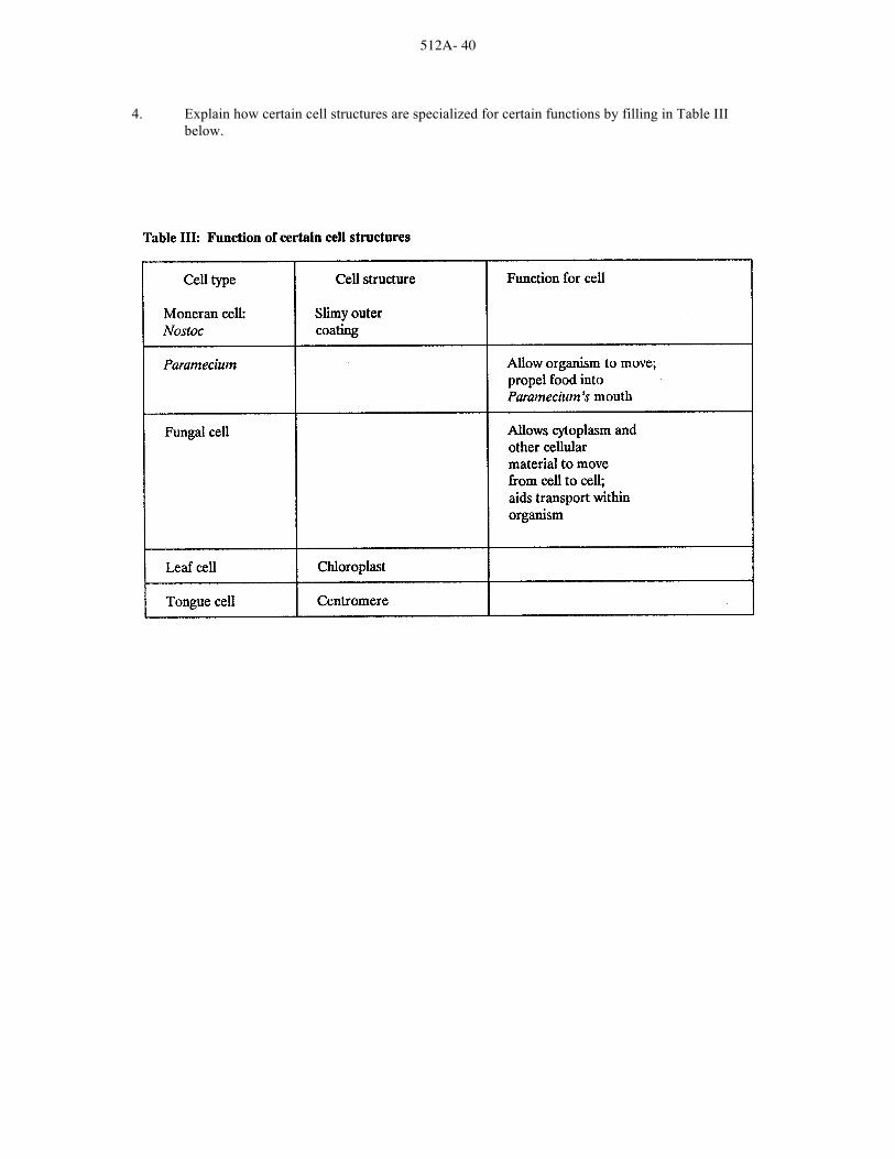
4. Explain how certain cell structures are specialized for certain functions by filling in Table III below.

512A- 41
THE ORGANISMS
AG 512 - A
LABORATORY EXERCISE #3--CLASSIFYING ORGANISMS Name _____________________________________ Score __________________________________
Selection from Modern Biology, Biology Investigations, Teacher's Edition, by James H. Otto, Albert Towle, W. David Otto, and Myra E. Madnick. Copyright 1977 by Holt, Rinehart and Winston, Inc. Printed by permission of the publisher. Part I: A Study of Classification
The classification categories in biology are: kingdom, phylum, class, order, family, genus and species. A system of classification may be applied to any number of objects. a. Examine the figures below and list some of the characteristics that you observe. ________________
______________________________________________________________________________
b. If considered in biological terms, what classification category would each individual figure represent? _____________________________________________________________________
c. What classification category would the entire group of figures represent? _____________________
Cut the figures apart. Be sure that the identifying number stays with the figure. Assemble the figures into two groups based on a common characteristic. For instance, put all figures with curved lines into one group. The second group, then, will be figures with straight lines.

512A- 42
d. By thus separating the figures into two smaller groups, what classification category has been achieved? ______________________________________________________________________
You should now have in the straight line group 12 straight-line figures: 1 rectangle and 1 triangle with lines projecting from them and 10 others being shaded or unshaded triangles, squares or rectangles. The group of figures with curved lines, representing the other phylum, will not be used further in this part of the investigation. Using the characteristic of lines projecting from the figures, divide the 12 figures into two groups.
e. In this division, what classification category has been achieved?
______________________________________________________________________________
f. What characterizes the remaining 10 figures? ___________________________________________
______________________________________________________________________________
______________________________________________________________________________
Separate the shaded figures from the unshaded figures. (Save the shaded figures for later use.)
g. What classification category has been achieved? _________________________________________
h. What characterizes the remaining 6 figures? ____________________________________________
______________________________________________________________________________
Separate the triangles from the other 4 figures. (Save the triangles for later use.)
i. What classification category does each group represent?
______________________________________________________________________________
The remaining 4 figures can be divided into two smaller groups on the basis of being squares or rectangles. Make the separation and save the rectangles. j. What classification category is represented by the group of squares and the group of rectangles? ___
______________________________________________________________________________
The group of squares should now have in it a large square and a small square. Make the final separation on the basis of the size of the squares. k. What classification category do you now have? _________________________________________
In this classification, the genus category contains but two distinct species. In biological classifications of organisms, a genus contains several related but distinct species. l. How are the figures (species) related? _________________________________________________

512A- 43
m. How are the figures different? _______________________________________________________
______________________________________________________________________________
Part II: Completing a Key to Straight-Line Figures Classification keys are usually based on pairs of opposing statements. Each pair of statements is increasingly specific in describing the item to be identified. Using the characteristics observed in Part I, fill in the blanks of the key with the characteristic needed to complete each pair of statements. The number in the column at the right refers you to the next pair of statements. When you come to "Fig.#_____," insert the number of the figure being described. KEY TO KINGDOM OF FIGURES
1a. All figures have curved lines ......................................................... Curved figures
1b. All figures have ____________ lines ............................................ 2
2a. All figures have projecting lines ................................................... 3
2b. All figures have ______ projecting lines ...................................... 4
3a. Figure is a triangle with lines ........................................................ Fig.# _______________
3b. Figure is a ______________ with lines ........................................ Fig.# _______________
4a. Figures are shaded ......................................................................... 5
4b. Figures are not _______________ ................................................ 8
5a. Figures are triangles ...................................................................... 6
5b. Figures are ___________ or ____________ ................................. 7
6a. Figure is ____________ triangle ................................................... Fig.# ______________
6b. Figure is _____________ triangles ............................................... Fig.# ______________
7a. Figure is a _____________ ........................................................... Fig.# ______________
7b. Figure is a ______________ ......................................................... Fig.# ______________
8a. Figures are triangles ...................................................................... 9
8b. Figures are ___________ or ____________ ................................. 10
9a. Figure is __________ triangle ....................................................... Fig.# ______________
9b. Figure is ___________ triangles ................................................... Fig.# ______________
10a. Figures are squares ........................................................................ 11
10b. Figures are ___________________ .............................................. 12

512A- 44
11a. _______________ square ............................................................. Fig.# _____________
11b. _______________ square ............................................................. Fig.# _____________
12a. Figure is _______________ rectangle .......................................... Fig.# _____________
12b. Figure is ________________ rectangle ........................................ Fig.# _____________
After completing the key, blacken the number of each figure and write the number on the back. Mix the figures and use the key to identify each of the 12 figures by number (species). If you can correctly identify each figure by number, you have accurately completed the key. Part III: Using a Classification Key to Identify Certain Species of Fish Study the terms defined below. All of these refer to structures of fish.
TERMS REFERRING TO THE STRUCTURE OF FISH barbel--a fleshy projection from the lips or head FINS adipose--a small fin on the top mid-line of the body near the tail fin anal--a fin along the lower mid-line of the body near the tail fin caudal--tail fin dorsal--the fin or fins along the top mid-line of the body pectoral--the paired fins nearest the head, corresponding to front legs or arms pelvic--the paired fins nearest the tail, corresponding to hind legs scales--overlapping growths of the skin Closely examine one of the drawings of a fish shown on the next page. Read both statements listed under number 1 in the classification key. One of these statements should describe the fish you have chosen; the other should not. Refer to the number after the statement that fits your fish and look for that number in the key. Again select the statement that describes the fish you picked. Continue through the key until you come to a name after one statement. This should be the name of the fish you picked. Practice using the key to identify several of the fish shown. Example: Suppose you want to find the name of fish number 2. Look at the classification key. Note that each numbered item presents two possibilities. We see that our fish has no scales, or at least we cannot see any. So we choose item 1b. This refers us to number 12. So we go down the page to number 12. Our fish is not elongated or snakelike (item 12b), so we go to number 13 of the key. The fish we are classifying has barbels growing from its lips and the top of its head (item 13a), so we go to number 14 of the key. Since our fish has a caudal fin that is rounded, and a blunt head, we see that it is the Bullhead Cathead Catfish (also known as horn pout in some parts of the country.)

512A- 45

512A- 46
CLASSIFICATION KEY TO CERTAIN FISH 1a. Body noticeably covered with scales ................................................................................................. 2 1b. Scales not covering body or too small to be seen ............................................................................ 12 2a. Dorsal fin single ................................................................................................................................. 3 2b. Dorsal fins two or more, joined or separated ..................................................................................... 6 3a. Body more than four times as long as broad (top to bottom); front edge of dorsal fin far back on body; mouth large, hinge back of eye .................................................................................. 4 3b. Body less than four times as long as broad; front edge of dorsal fin about midway between head
and tail; mouth not large, hinge in front of eye ................................................................................. 5 4a. Dark lines forming netted design on body; fins not spotted ................................................... Pickerel 4b. Body covered with yellow spots; fins spotted ............................................................... Northern pike 5a. Mouth turned downward; barbels absent; dorsal fin not elongated ................................ White sucker 5b. Mouth not turned downward; barbels present; dorsal fin elongated ........................................... Carp 6a. Two dorsal fins separated, the anterial spiny and the posterior soft .................................................. 7 6b. Two dorsal fins united, forming an anterior spiny portion and a posterior soft portion .................... 8 7a. Top of head concave, forming a hump in front of dorsal fin; dark vertical bars on body ................................................................................................................... Yellow perch 7b. Top of head not concave, body sloping to dorsal fin and not forming a hump; dark blotches on body .......................................................................................................... Wall-eyed pike 8a. Body more than three times as long as broad .................................................................................... 9 8b. Body less than three times as long as broad .................................................................................... 10 9a. Hinge of jaws behind the eye; notch between spiny and soft dorsal fin deep and nearly separating into two fins ................................................................ Large-mouth black bass 9b. Hinge of jaws below the eye; notch between spiny and soft dorsal fin not nearly separating into two fins .................................................................................. Small-mouth black bass 10a. Mouth large, hinge below or behind eye ......................................................................................... 11 10b. Mouth small, hinge in front of eye ......................................................................................... Bluegill 11a. Five to seven spines in dorsal fin; dark spots forming broad vertical bars
on sides .......................................................................................................................... White crappie 11b. Ten or more spines in dorsal fins; sides flecked with dark spots ........................ Rock bass (Redeye) 12a. Body much elongated and snakelike; dorsal, caudal and anal fins continuous ............................ Eel 12b. Body not elongated and snakelike; dorsal, caudal and anal fins separate; adipose fin present ........................................................................................................................................ 13 13a. Barbels growing from lips and top of head; head large and broad .................................................. 14 13b. Barbels lacking; head not large and broad ....................................................................................... 16 14a. Caudal fin deeply forked; head tapering .......................................................................................... 15 14b. Caudal fin rounded or slightly indented but not forked; head blunt .......................... Bullhead catfish 15a. Dorsal fin rounded at top; body silvery, speckled with black markings .................... Channel catfish 15b. Dorsal fin long and pointed at top; body bluish-gray without speckles ........................... Blue catfish 16a. Caudal fin deeply forked; back not mottled and with few spots ................................ Atlantic salmon 16b. Caudal fin square or slightly indented; back mottled or spotted ..................................................... 17 17a. Back and caudal fin spotted; broad horizontal band along sides .................................. Rainbow trout 17b. Back mottled with dark lines; caudal fin not spotted; fins edged with white .................... Brook trout

512A- 47
Part IV: Summary a. Based on what you have learned in this investigation, discuss how classification is a useful tool
for a biologist. __________________________________________________________________
______________________________________________________________________________
______________________________________________________________________________
______________________________________________________________________________
Fill in the blanks:
b. A group of closely related species is a _______________________________________________
c. A subdivision of a family is a ______________________________________________________
d. The largest of the classification categories is the _______________________________________
e. The most specific of the classification groupings is the __________________________________
f. A group of closely related classes is a _______________________________________________
g. The subdivision of an order is the ___________________________________________________
h. A ___________________ is composed of several closely related orders.
Part V: Investigations On Your Own
Select commonly seen groups of related objects (automobiles, canned goods, etc.) and classify them into the major classification categories. Construct a key to their identification. Try your classification key with some individuals in your class to see how well it works.

512A- 48
THE ORGANISMS
AG 512 - A
ANSWER SHEET TO LABORATORY EXERCISES Lab #1 Part IV 1. Answer should include ten of the following:
Never "horse around" in the laboratory. Never play with laboratory equipment or materials. Always follow instructions and wait until you are told to begin before starting any
investigation. Never carry out unassigned experiments. Never eat or taste anything in the laboratory. This includes food, drinks and gum, as well as
chemicals found in the laboratory. Wash your hands after every experiment. Keep all books and other nonessential items away from the work area. Keep your work area clean. Dispose of waste materials in appropriate containers. Turn off any gas jets or any electrically operated equipment when you have completed the
laboratory investigation. Report all injuries or accidents to your teacher immediately. Never use broken or cracked glassware. Always wear shoes in the laboratory. Sandals are not suggested. Tie back long hair and restrict any loose clothing. Wear safety goggles, laboratory aprons and gloves when instructed to do so.
2. a. Microscope slide b. Coverslip c. Petri dish d. Erlenmeyer flask e. Florence flask f. Funnel g. Graduate h. Test tube i. Test tube rack j. Test tube holder k. Pipette l. Striker m. Scalpel n. Medicine dropper o. Inoculating loop p. Forceps
3. Answer should include the following information:
Laboratory Investigation Report: Title This is the name of the laboratory investigation you are doing. In an
investigation from a laboratory manual, the title will be the same as the title of the investigation.
Hypothesis The hypothesis is what you think will happen during the investigation. It is often posed as an "If...then" statement. For example: If sulfuric acid is added to sugar, then the sugar will be broken down into its chemical components.
Materials This is a list of all the equipment and other supplies you will need to complete the investigation. In investigations taken from a laboratory manual, the materials are generally listed for you.
Procedure The procedure is a step-by-step explanation of exactly what you did in the investigation. Investigations from laboratory manuals will have the

512A- 49
procedure carefully written out for you, all you need to do is to read it very carefully. Often, in laboratory manuals, there will be questions in the procedure section that will help you understand what is happening in the investigation.
Data Your data is what you have observed. It is often recorded in the form of tables, graphs and drawings.
Analyses and This is the most important and difficult part of the investigation. It explains Conclusions what you have learned. You should include everything you have learned; you
should explain any errors you made in the investigation; and you should evaluate your hypothesis. Keep in mind that not all hypotheses will be correct. That is normal. You just need to explain why things did not work out the way you thought they would. In laboratory manual investigations, there will be questions to guide you in analyzing your data. You should use these questions as a basis for your conclusions.
Independent Project Report Title Introduction The introduction should include a clear, simple statement of your purpose. In
addition, the introduction should include a discussion of the important ideas that led you to design and perform the experiment. For example, you could include such things as why you are doing this investigation, what is interesting about the topic to be investigated, and what information you have already gathered about the topic. In order to prepare a good introduction, you will need to do library research on the topic. Be sure to use proper citation methods when you use ideas from any reference source.
Hypothesis Materials Procedure Data Analyses and Conclusions References List all the reference materials used to originate and to complete the project. Be
sure to use complete citations, including author, title, date of publication and place of publication. Your teacher will give you the format preferred for the type of investigation you are doing.
Lab #2
Part I:
Step 5:
Step 7: Drawings will vary depending on slides available.

512A- 50
Part II:
Step 5:
Step 7: Drawings will vary depending on slides available.
Part III:
Step 4:
Step 6: Drawings will vary depending on slides available.
Part IV:
3. a. Students should see several
Step 4:
Step 6: Drawings will vary depending on slides available.
Part V:
3. b. Nuclei

512A- 51
Step 4:
Step 6: Drawing will vary on slides available.
Table I:
Animal cells: Generally smaller than most other cells and irregular in shape. Animal cells lack cell walls and plastids. Some may have small vacuoles (vesicles) and cilia.
Plant cells: Large, generally box-shaped cells with cell walls, larger vacuoles and plastids. Fungal cells: Single or multicellular organisms of variable size and shape. Lack plastids but may have
vacuoles. Protist cells: Single cells of variable size and shape. Some may have cell walls, chloroplasts, cilia and
more than one nucleus. Moneran cells: Single cells of variable size and shape with cell wall. Lack nuclear membrane. Some
may have bacterial flagellum. Part VI:
1. Cell membrane, genetic material, cytoplasm
2. No. Cells within the same kingdom vary in size; cells in different kingdoms are often similar in size.
3. Table II: Cell features of each kingdom

512A- 52
4. Table III: Function of certain cell structures
Lab #3
Part I:
a. Figures with straight lines, curved lines; some triangles, squares or rectangles; lines projecting from them; single or double figures; shaded or unshaded
b. Species c. The kingdom of figures d. Phylum e. Class f. Shaded or unshaded squares, rectangles, single or double triangles g. Order h. Triangles or squares and rectangles i. Family j. Genus k. Species l. Both figures (species) are squares. m. One figure is larger than the other.
Part II:
1b. straight 2b. no 3a. 19 3b. rectangle, 9 4b. shaded 5b. squares, rectangles 6a. one, 15 6b. two, 6 7a. square, 13 7b. rectangle, 3 8b. squares, rectangles 9a. one, 2 9b. two, 12 10b. rectangles

512A- 53
11a. Large, 20 11b. Small, 5 12a. large, 11 12b. small, 14
Part III:
1. Atlantic salmon 2. Bullhead catfish 3. Rock bass (Redeye) 4. Carp 5. Small-mouth black bass 6. Eel 7. Bluegill 8. Brook trout 9. Yellow perch 10. White crappie 11. Channel catfish 12. Northern pike 13. Large-mouth black bass 14. Rainbow trout 15. White sucker 16. Wall-eyed pike 17. Blue catfish 18. Pickerel
Part IV:
a. The classifying of organisms enables biologists to organize and by grouping living organisms according to characteristics shared by the organisms, biologists are able to observe natural relationships and study characteristics of the group as a whole.
b. Genus c. Genus d. Kingdom e. Species f. Phylum g. Family h. Class

512A- 54
THE ORGANISMS
AG 512 - A
UNIT TEST Name _____________________________________ Score __________________________________
1. Match the terms on the right with the correct definitions by placing the appropriate numbers in the blanks provided.
_____a. Cell type that has a nucleus without a membrane 1. Binomial
around it. The nuclear material floats freely nomenclature within the cell
2. Prokaryote _____b. The group of individuals of a given species inhabiting
a specified geographic area 3. Eukaryote
4. Adaptation _____c. Organisms that manufacture organic nutrients from
inorganic raw materials 5. Autotrophs
_____d. Association of unicellular or multicellular organisms 6. Biogenesis of the same species
7. Biome _____e. Occurrence of several distinct phenotypes in a population
8. Colony _____f. An assemblage of populations that live in a defined habitat
and interact in various ways with one another 9. Community
_____g. Those elements of an ecosystem that eat other plants 10. "Consumer" or animals organisms
_____h. Organisms which cannot synthesize their own food from 11. Ecology inorganic materials
12. Ecosystem _____i. The theory that all living things come only from preexisting
living things 13. Epigenesis
_____j. Large, easily differentiated community unit arising 14. Fossils as a result of complex interactions of climate, other physical factors and biotic factors 15. Genus
_____k. A system invented by Carolus Linnaeus for classifying 16. Herbivore organisms; each organism is assigned a two-word Latin name 17. Heterotrophs
_____l. The theory that development proceeds from a structureless 18. Phenotype cell by the successive formation and addition of new parts which do not preexist in the fertilized egg 19. Phylogeny

512A- 55
_____m. Taxonomic classification in which closely related species 20. Polymorphism are grouped together
21. Population _____n. A characteristic which enables the organism to survive in
its environment 22. Species
_____o. Cell type that has an organized nucleus surrounded by a 23. Taxonomy membrane
24. Tissue _____p. Any remains of an organism that have been preserved in
the earth's crust
_____q. The study of the interrelations between living things and their environment
_____r. The evolutionary history of a group of organisms
_____s. The visible expression of the hereditary constitution of an organism
_____t. A plant-eating animal
_____u. The unit of taxonomic classification, a population of similar individuals, alike in their structural and functional characteristics
_____v. The science of naming, describing and classifying organisms
_____w. All of the organisms of a given area
_____x. Specialized cells which together perform certain special functions
2. List the seven categories of the Linnaeus classification system in order from largest to smallest.
a. _____________________________________________________________________________
b. ____________________________________________________________________________
c. _____________________________________________________________________________
d. ____________________________________________________________________________
e. _____________________________________________________________________________
f. _____________________________________________________________________________
g. ____________________________________________________________________________

512A- 56
3. List and define the five kingdoms in the classification system.
a. _____________________________________________________________________________
______________________________________________________________________________
______________________________________________________________________________
b. ____________________________________________________________________________
______________________________________________________________________________
______________________________________________________________________________
c. _____________________________________________________________________________
______________________________________________________________________________
______________________________________________________________________________
d. ____________________________________________________________________________
______________________________________________________________________________
______________________________________________________________________________
e. _____________________________________________________________________________
______________________________________________________________________________
______________________________________________________________________________
4. List three traits that help place an organism into a kingdom.
a. _____________________________________________________________________________
b. ____________________________________________________________________________
c. _____________________________________________________________________________

512A- 57
5. Match the phylums of the plant kingdom to their characteristics below. Write the correct numbers in the blanks.
1. Bryophyta 4. Pterophyta 7. Gnetophyta 2. Lycophyta 5. Cycadophyta 8. Coniferophyta 3. Sphenophyta 6. Ginkgophyta 9. Anthophyta
_____a. Vascular plants; Sporophyte dominant; Trailing or erect with simple leaves; Spores produced in a strobilus; Gametophyte underground; Club mosses
_____b. Palmlike plants; Male and female cones on different trees; Cycads, sago palms
_____c. Gymnosperms with sex organs in cones; Leaves in the form of needles; Most are evergreen; Pines, spruces, firs, larches, yews
_____d. Multicellular, small green plants living on land (usually in moist situations) without
true roots or flowers; Lack vascular tissues; Alternation of generations with the gametophyte the dominant generation; Motile sperm; Liverworts, hornworts, mosses
_____e. Includes all the flowering plants; Sex organs in whorls in flowers; Seeds enclosed in ovary that ripens into a fruit; Grasses, maples, elms _____f. Vascular plants; Sporophyte generation dominant with leafy fronds; Creeping rhizome;
Gametophyte, a free-living prothallus; Motile sperm; Ferns and tree ferns _____g. Specialized gymnosperms; Desert species with xylem resembling that in angiosperms;
Few species; Welwitschia, ephedra, gnetum _____h. Vascular plants; Sporophyte dominant; Stems: hollow, jointed, contain silica; Leaves:
tiny scales in whorls at stem joints; Fertile stems with strobili; Horsetails _____i. One living species; Pollen produced in conelike structure; Seeds naked; Ginko biloba
6. Discuss six differences between plants and animals.
______________________________________________________________________________
______________________________________________________________________________
______________________________________________________________________________
______________________________________________________________________________
______________________________________________________________________________
______________________________________________________________________________
______________________________________________________________________________
______________________________________________________________________________

512A- 58
7. Discuss six ways to classify plants besides the Linnaeus classification system.
______________________________________________________________________________
______________________________________________________________________________
______________________________________________________________________________
______________________________________________________________________________
______________________________________________________________________________
______________________________________________________________________________
______________________________________________________________________________
______________________________________________________________________________
______________________________________________________________________________
8. Discuss scientific names and their importance.
______________________________________________________________________________
______________________________________________________________________________
______________________________________________________________________________
______________________________________________________________________________
______________________________________________________________________________

512A- 59
THE ORGANISMS
AG 512 - A
ANSWERS TO TEST 1. a. 2 i. 6 q. 11
b. 21 j. 7 r. 19 c. 5 k. 1 s. 18 d. 8 l. 13 t. 16 e. 20 m. 15 u. 22 f. 9 n. 4 v. 23 g. 10 o. 3 w. 12 h. 17 p. 14 x. 24
2. (in order) Kingdom; Phylum; Class; Order; Family; Genus; Species
3. a. Animal kingdom (Animalia): Eukaryotic cells; Multicellular organisms; Move about to obtain food; Digest food inside body
b. Plant kingdom (Plantae): Eukaryotic cells; Multicellular organisms; Produce own food; Cannot move about
c. Fungi kingdom: Eukaryotic cells; Mostly multicellular organisms; Do not move about; Obtain food by absorbing it from living or dead organisms
d. Monera kingdom: Prokaryotic cells; Mostly one-celled organisms; Produce own food or obtain it from outside source
e. Protista kingdom: Eukaryotic cells; Many are one-celled; Produce own food or obtain it from outside source
4. Kind of cells in organism: prokaryote or eukaryote; How organism obtains its food; How
organism reproduces and develops 5. a. 2 f. 4
b. 5 g. 7 c. 8 h. 3 d. 1 i. 6 e. 9
6. Mobility: Animals are mobile; Most plants are stationary Food: Plants manufacture their own food; Animals must rely on plants or other animals for their food Cell walls: Cell walls of plants are rigid and usually made of cellulose; Animals don't have rigid cell walls or flaccid cell membranes Cellulose: Synthesized by plants; Not synthesized by animals Growth: Animal growth is limited; Many plants have unlimited growth and an indefinite number of parts Number of parts: Animals have definite number of parts, such as eyes, ears, legs, etc.; The number of parts (such as leaves, stems, buds and flowers) of the same kind of plant usually varies from plant to plant

512A- 60
7. Growth habits: Annuals; Winter annuals; Biennials; Perennials Types of flowers: Complete flower; Incomplete flowers--One of the four principal parts is missing; Perfect flowers--Both the stamens and pistil are present; Imperfect flower--Lack either the stamens or pistil Location of flowers: Dioecious--Flowers bearing stamens (male pollen) and those bearing pistils (female eggs) produced on separate plants; Monoecious--Flowers containing stamens and those containing pistils are produced in different places on the same plant Root structure: Fibrous root plants--Root systems are very branched and finely divided; Tap root plants--One major root with few lateral root hairs attached to it Seed leaves: Dicot--Plant with two seed leaves in each of its seeds; Monocot--Plant with one seed leaf in each seed Agricultural use of crop: Cereals or grain crops; Oil seed crops; Forage and pasture crops; Root and tuber crops; Fiber crops; Sugar crops; Specialty crops (for example: hops)
8. Definition: Each plant has a two-word (binomial) name--always in Latin; First name refers to the
plant's genus; Second name refers to the plant's species
Importance--It is universal--scientific name is the same regardless of the location or the language of the people

512B - 1
CELLS: STRUCTURE, FUNCTIONS AND DIVISION
AG 512 - B
UNIT OBJECTIVE After completion of this unit, students should be able to match terms and definitions and state the ideas of the cell theory. Students should also be able to label the parts of a plant cell, identify cell processes and the stages of mitosis and meiosis. This knowledge will be demonstrated by completion of laboratory exercises and a unit test with a minimum of 85 percent accuracy .
SPECIFIC OBJECTIVES AND COMPETENCIES
After completion of this unit, the student should be able to:
1. Match terms associated with cells to the correct definition.
2. State the four basic ideas of the cell theory.
3. List the three ways that cells can differ from one another.
4. Label the parts of a plant cell.
5. Match the parts of a plant cell to the correct functions and/or descriptions of each.
6. Describe the differences between plant and animal cells.
7. Describe protoplasm.
8. Describe the importance of energy to the functioning of the cell and where the energy is found within the cell.
9. Identify the cell processes for the given descriptions.
10. Describe the relationship of genes and chromosomes to cell division.
11. Identify the correct stages of mitosis when given a description of each.
12. Describe cytokinesis in plant and animal cells.
13. Identify the correct stages of meiosis when given a description of each.
14. Define the two basic types of plant tissue.
15. Identify the correct types of meristematic and permanent tissues when given a description of each.
16. Identify and describe cells.
17. Study cell parts.
18. Identify differences between plant and animal cells.

512B - 2
19. Observe the phases of mitosis and meiosis.
20. Compare mitosis in plant and animal cells.

512B - 3
CELLS: STRUCTURE, FUNCTIONS AND DIVISION
AG 512 - B
SUGGESTED ACTIVITIES
I. Suggested activities for instructor
A. Order materials to supplement unit.
1. Films
a. The Cell, VHS video; describes plant and animal cells and the four hases of mitosis; available from Vocational Education Productions, California Polytechnic State University, San Luis Obispo, California 93407 (1-800-235-4146); approximate cost $99.95; order no. 6-083-112J.
B. Make transparencies and necessary copies of materials.
C. Provide students with objective sheet.
D. Provide students with information sheets and laboratory exercises.
E. Discuss unit and specific objectives.
F. Discuss information sheets.
G. Demonstrate and discuss procedures outlined in laboratory exercises.
H. Review and give test.
I. Reteach and retest if necessary.
II. Instructional materials
A. Objective sheet
B. Suggested activities
C. Information sheet
D. Transparency masters
1. TM 1--Plant Cell
2. TM 2--Cell Wall
3. TM 3--Phases of Mitosis
4. TM 4--Chromatid Formation
5. TM 5--Cytokinesis

512B - 4
6. TM 6--Plant Meristems
7. TM 7--Epidermal Cells
8. TM 8--Parenchyma Tissue
9. TM 9--Collenchyma Tissue
10. TM 10--Sclerenchyma Tissue
11. TM 11--Phloem Tissue
E. Instructor notes for laboratory exercises
F. Laboratory exercises
1. LE 1--What Are Cells?
2. LE 2--Studying Cell Parts
3. LE 3--Animal and Plant Cell Differences
4. LE 4--Mitosis and Meiosis
5. LE 5--Comparing Mitosis in Plant and Animal Cells
G. Answers to laboratory exercises
H. Test
I. Answers to test
III. Unit references
A. Agricultural Education Curriculum, College of Agriculture, University of Illinois, Urbana, Illinois, 1989.
B. Delorit, R. J., et al., Crop Production, 5th edition, Prentice-Hall, Inc.,
Englewood Cliffs, New Jersey, 1984. C. Hartmann, Hudson T., et al., Plant Science: Growth, Development and
Utilization of Cultivated Plants, 2nd edition, Prentice-Hall, Inc., Englewood Cliffs, New Jersey, 1988.
D. Idaho Crop and Soil Curriculum Guide, Idaho State Board for Vocational
Education, 1985. E. Janick, Jules, et al., Plant Science, 2nd edition, W.H. Freeman and Co., San
Francisco, California, 1974. F. Model Agricultural Core Curriculum, California State Department of Education,
University of California, Davis, California, August, 1989.

512B - 5
G. Otto, James H., Towle, Albert, Modern Biology, Holt, Rinehart and Winston, New York, 1985.
H. Slesnick, Irwin L., et al., Biology, Scott, Foresman and Company, Glenview,
Illinois, 1985.

512B - 6
CELLS: STRUCTURE, FUNCTIONS AND DIVISION
AG 512 - B
INFORMATION SHEET
I. Terms and definitions
A. Adenosine triphosphate (ATP)--A chemical compound produced in the mitochondrion; stores energy that is used to carry out cellular functions
B. Cell--The basic structural and functional unit of living organisms
C. Cellulose--A complex, insoluble carbohydrate that constitutes the principal component of plant cell walls
D. Cell wall--The relatively rigid boundary of plant cells
E. Chlorenchyma--Tissue composed of parenchyma cells that contain chloroplasts
F. Chlorophyll--Green pigments essential to photosynthesis
G. Chloroplast--An organelle containing chlorophyll
H. Chromatid--One of the two strands of chromosome; they are united by a centromere
I. Chromatin--A readily staining complex of DNA and proteins found in
chromosomes J. Chromoplast--A plastid containing pigments other than chlorophyll (the
pigments are usually yellow to orange) K. Chromosome--Molecules of DNA wrapped around proteins, which are found in
the nucleus; control cell functions and the inheritance of traits L. Cytokinesis--Cell division
M. Cytoplasm--The protoplasm of the cell exclusive of the nucleus
N. Diploid--Having two sets of chromosomes in each cell; the 2n chromosome number characteristic of the sporophyte generation
O. DNA (deoxyribonucleic acid)--Carries genetic information in cells
P. Endoplasmic reticulum--A complex system of interlinked double membrane channels; parts of it are lined with ribosomes
Q. Enzymes--Numerous complex proteins that speed up chemical reactions in
living things without being used up in the reaction (act as catalysts) R. Eukaryotic cells--Cells having distinct membrane-bound organelles

512B - 7
S. Golgi body--An organelle consisting of disc-shaped, often branching, hollow tubules that function in accumulating and packaging substances used in the synthesis of materials by the cell
T. Haploid--Having one set of chromosomes per cell (n chromosomes)
U. Meiosis--The process of two successive nuclear divisions through which segregation of genes occurs and a single diploid cell (2n) becomes four haploid (n) cells
V. Metabolism--The use of all interrelated chemical processes occurring in a living
organism W. Mitochondrion--An organelle containing enzymes that function in the Krebs
cycle and the electron transport chain of aerobic respiration X. Mitosis--Nuclear division during which the chromatids of the chromosomes
separate and two genetically identical daughter nuclei are produced Y. Mutation--An inheritable change in a gene or chromosome
Z. Nucleolus--A somewhat spherical body within a nucleus; contains RNA and protein
AA. Nucleus--The organelle of a living cell that contains chromosomes and is
essential to the regulation and control of all the cell's functions BB. Organelle--A membrane-bound structure in the cell's cytoplasm
CC. Parenchyma--Thin-walled cells varying in size, shape and function; the most common type of plant cell
DD. Plasma membrane--The outer boundary of the protoplasm of a cell (also called
cell membrane, particularly in animal cells) EE. Plasmodesma--Minute strands of cytoplasm that extend between adjacent cells
through pores in the walls FF. Plastid--An organelle associated primarily with the storage or manufacture of
carbohydrates GG. Prokaryotic--Having a cell or cells that lack a distinct nucleus and other
membrane-bound organelles (for example: bacteria) HH. Protoplasm--The living substance of a cell
II. Rhizome--Granular particles each composed of two subunits consisting of RNA and proteins; they lack membranes and are very numerous in living cells
JJ. RNA (ribonucleic acid)--An important cellular substance that occurs in three
forms, all involved in protein synthesis

512B - 8
KK. Stroma--A colorless fluid substance constituting the bulk of the volume of a chloroplast or other plastid; contains enzymes that play a key role in photosynthetic reactions in chloroplasts
LL. Turgor pressure--Pressure within a cell resulting from water uptake
MM. Vacuolar membrane--A membrane between the cytoplasm and a vacuole of a cell
NN. Vacuole--A pocket of cell sap that is separated from the cytoplasm of a cell by a
membrane; also, food storage or contractile pockets within the cytoplasm of unicellular organisms
II. Cell theory
A. All organisms are made of one or more cells
B. Cells are alike in their structure and composition
C. All cells carry out similar functions that keep them alive
D. New cells arise only from old cells, usually by dividing into two equal parts at regular intervals
III. How cells differ from each other
A. Size
B. Shape
C. Organization
IV. Parts of a plant cell (Transpareny 1)

512B - 9
V. Functions of the parts of the cell
A. Nucleus
1. Contains the hereditary information that directs all cell activity
2. Contains the nucleolus
B. Nucleolus
1. Produces RNA (ribonucleic acid)
2. Assembles subunits of ribosomes
C. Plasma membrane
1. Lipid bilayer surrounding the cytoplasm
2. Maintains surface area for selective cell absorption and secretion
3. Generates energy
D. Cell wall (Transparency 2)
1. Protects protoplast
2. Provides external structure
3. May provide strong support for the plant
E. Middle lamella
1. Intercellular layer lying between cells
2. Contains compounds that hold adjacent cell walls together
F. Cytoplasm
1. Viscous fluid composed of matrix proteins
2. Assists in transport of substances within the cell
G. Endoplasmic reticulum
1. Network of membranes found within the cytoplasm
2. Involved in storing and transporting protein and chemical products
H. Ribosomes
1. Small, discrete structures found in the cytoplasm
2. Manufacture cell proteins

512B - 10
I. Mitochondrion (plural--mitochondria)
1. Small organelles with inner platelike folds
2. Serves as powerhouse for cell--releases body heat and energy
a. Principal respiration sites
b. Provide heat and energy for cell functions
3. Produces ATP (Adenosine triphosphate) in which energy for cell activities is stored
4. Involved in protein synthesis
J. Golgi body
1. Flat, disc-shaped organelles that appear in groups
2. Packages and distributes proteins for storage within cell and transport out of cell
K. Plastids--Large organelles in the cytoplasm
1. Leucoplasts
a. Colorless plastids
b. Storage bodies for oil, starch and proteins
2. Chromoplasts
a. Colored plastids
b. Chloroplasts
(1) Green chromoplasts
(2) Contain chlorophyll that manufactures the food in plants
L. Vacuole
1. Cavity containing cell sap, a watery fluid which contains dissolved substances
2. Serves as a storage reserve for water, salt and toxic products
3. Supports cell wall of plant cells through internal pressure

512B - 11
M. Vacuolar membrane (tonoplast)
1. Membrane surrounding the vacuole
2. Regulates ion flow within the cell
3. Maintains cell turgor
N. Plasmodesma (plural--plasmodesmata)
1. Connection of thin strands of cytoplasm between the interiors of cells
2. Assist in mineral element and food material movement between cells
VI. Differences between plant and animal cells
A. Plant cells
1. Cell wall
a. Made of cellulose
b. Gives support and shape
2. Plastids
a. Leucoplasts
(1) Colorless structures where glucose is changed into starch
(2) Storage for starch, lipids or proteins
b. Chromoplasts
(1) Manufacture and store pigments
(2) Give fruits, vegetables and leaves their bright color
c. Chloroplasts
(1) Contain green chlorophyll pigment
(2) Site of photosynthesis (food production) in the plant cell
3. No microtubules or centrioles

512B - 12
B. Animal cells
1. Microtubules give the cell its shape
2. Centrioles
a. Located near nucleus
b. Function in cell division for reproduction
3. No plastids or cell wall
VII. Protoplasm--The living parts of the cells
A. Complex, semi-liquid substance responsible for carrying out the life processes
B. No definite composition
1. Differs in living things
2. Not even the same in all parts of one individual
3. Composition is constantly changing
a. Made up mostly of water (85-90%)
b. Many substances dissolved or suspended in the water
(1) Organic substances including proteins, fats and carbohydrates
(2) Inorganic substances--Mineral elements taken up
from the soil through the roots
c. Organized into cells
VIII. Energy and the cell
A. All processes (or functions) that take place in living things need energy
B. The energy comes from chemical reactions that take place in the cell (respiration and photosynthesis)
1. Chemical activities in one part of the cell are dependent on reactions
taking place in another part of the cell
2. The reactions are all related to functions of the cell
C. ATP
1. Produced in mitochondrion
2. Stores energy for cell activities

512B - 13
IX. Cell processes
A. Nutrition--Cells need molecules for energy and building materials. Cells either form their own food molecules or take them in from the environment
B. Digestion--Cells break down most foods into simple forms in order to use them.
These reactions are sped up by certain enzymes in the cell C. Absorption--Water, food molecules, ions and other necessary materials from the
environment are taken in by the cell D. Biosynthesis--Organic substances (including carbohydrates, fats and proteins)
are manufactured by cells and organized into their own cell material. This process is necessary for growth and for enzyme production to control cell activity
E. Cellular respiration--Energy necessary to all cell activities is released from food
molecules; usually oxygen is used and carbon dioxide is a waste product F. Excretion--Waste materials from cell activities are passed from the cell to the
environment G. Secretion--Molecules of substances (such as vitamins and hormones) are
synthesized by certain cells and passed out of the cell (these substances affect activities of other cells)
H. Response--A change in cell activities in response to stimuli from the
environment such as heat, pressure, light or chemicals
I. Reproduction--Cell division
1. Complex organism--Results in larger number of cells in the organism
2. One-celled organisms--Produces more organisms
X. Relationship of genes and chromosomes to cell division
A. Genes are segments of DNA that control each hereditary trait (heredity is the passing of traits from parent to offspring)
B. The chromosomes carry the genes
C. Because chromosomes divide equally during cell division, each daughter cell inherits a complete set of genes

512B - 14
XI. Mitosis (Transparency 3)
A. Interphase
1. "Resting period" of mitosis--Period between cell division
2. Chromatin
a. Form of genetic information during interphase
b. Complex of DNA and protein
c. Spread throughout the nucleus
3. DNA in the chromatin replicates itself before cell division begins, but the replicated DNA molecules don't separate yet

512B - 15
B. Prophase (Transparency 4)
1. Chromatin threads gradually shorten and thicken and become visible as chromosomes
a. Each chromosome consists of two identical parts called
chromatids b. Chromatids are held together at a single point called the
centromere
2. Spindle formed from protein fibers
a. Poles--Ends of the spindles
b. Equator--Middle of the spindle
3. Nuclear membrane disappears
C. Metaphase
1. Chromatid pairs line up along the equator
2. Spindle fibers attach to the centromere of each chromatid
D. Anaphase
1. Chromatids separate
a. Move to opposite poles
b. Chromatids referred to as chromosomes after separating
2. Same number of chromosomes at each pole as in the parent cell
E. Telophase
1. Spindle breaks down
2. Nuclear membrane forms around each set of chromosomes
3. Cytokinesis (Transparency 5)
a. Process by which cytoplasm divides to form two cells
b. Plant cells--Cell plate forms between the two daughter cells
c. Animal cells--Cell membrane pinches inward and forms a groove, which deepens until the two cells are separated
4. Two new, identical cells

512B - 16
XII. Meiosis
A. Cell division of sex cells which reduces the chromosome number in eggs and sperm (also called reduction division)
B. Purpose is to form gametes with the haploid number of chromosomes
1. Diploid number = 2n
2. Haploid number = 1n or n
C. Only germ cells can produce sex cells
1. Germ cells--Diploid cells found in the organism's sex cells
2. A germ cell produces four sex cells with a monoploid number of chromosomes (1n) during meiosis
D. Two divisions
1. First meiotic division separates the homologous chromatids in each tetrad
2. Second meiotic division separates the two chromatids of each
chromatid pair XIII. First stage of meiosis
A. Interphase I
1. Genetic material in form of chromatin
2. DNA replicates itself
B. Prophase I
1. Chromatin threads shorten and thicken
2. Chromosomes become visible
a. Each chromosome is made up of two identical chromatids joined at their centromere
b. Homologous chromosomes begin to move toward each other
c. Tetrad--The arrangement formed when homologous chromosomes move close to each other and there is a total of four chromatids
3. Tetrads move toward the middle of the cell
C. Metaphase I--Tetrads line up along the cell's equator

512B - 17
D. Anaphase I
1. Homologous chromosomes of the tetrad separate and move to opposite poles
2. The chromatids of individual chromosomes don't separate yet
E. Telophase I
1. Cytoplasm divides
2. First stage of meiosis is complete
3. Two haploid daughter cells have been produced
a. Each daughter cell contains only one chromosome of each pair that was in the parent cell
b. Total number of chromosomes in each daughter cell equals
half the number of chromosomes in the original parent cell XIV. Second stage of meiosis
A. Prophase II
1. Chromatids become shorter and thicker
2. Spindle begins to form
B. Metaphase II
1. Chromosomes line up along the cell equator
2. Spindle fibers attach to the centromere of each chromatid
C. Anaphase II--Chromatids of each chromosome separate and migrate to opposite poles
D. Telophase II
1. Nuclear membrane forms around each set of chromatids
2. Chromatids now called chromosomes since they are no longer joined
3. Cytoplasm divides
4. Four haploid cells form with a different chromosome make-up than the parent cell
XV. Plant tissues
A. Large groups of organized cells of similar structure that perform a collective function

512B - 18
B. Basic types
1. Meristem (meristematic tissue)
a. Comprised of actively dividing cells that develop and differentiate into other tissues and organs
b. Cells have thin walls and dense protoplast
2. Permanent
a. Develops from the meristems
b. Non-dividing differentiated cells
XVI. Meristematic tissue (Transparency 6)
A. Apical meristems
1. Shoot meristems
a. Found in the tops of the shoots
b. Responsible for producing new buds and leaves in a uniform pattern at the end of the stem and laterally along stems
2. Root meristems
a. Growing points for the root system
b. Found at the various ends of the roots
B. Lateral meristems
1. Account for girth and growth of woody stems
2. Composed of cellulose and pectin
3. Provide mechanical support for plant
4. Vascular cambium--Produces new xylem and phloem
5. Cork cambium--Produces bark (the protective covering of old stems and roots)
6. Number of growth rings indicates tree's age
512B - 19
C. Intercalary meristems
1. Active tissues that have been separated from the shoot terminal meristem by regions of more mature or developed tissue
2. Found near the nodes of grasses
3. Reason for continuous growth after mowing grasses
XVII. Permanent tissues
A. Simple tissue--Uniform, composed of only one type of cell
1. Epidermis tissue (Transparency 7)
a. Single, exterior layer of cells that protects stems, leaves, flowers and roots
b. Outside surface of epidermal cells usually covered with cutin--
a waxy substance that reduces water loss
2. Parenchyma tissue (Transparency 8)
a. Living, thin-walled cells with large vacuoles and many flattened sides
b. Most common and abundant plant tissue making up the fleshy
part of the organism and functioning in food and water
3. Collenchyma tissue (Transparency 9)
a. Elongated cells with unevenly thickened primary walls
b. Gives support to young stems, petioles and leaf veins
4. Schlerenchyma tissue (Transparency 10)
a. Thick-walled cells
b. Common in stems and bark
c. Found as stone cells in pear fruits and walnut shells
d. Nonliving when mature
5. Cork tissue
a. Bark of maturing stems, tree trunks and potato skins
b. Cell walls are waterproofed with suberin (waxy material)
c. Die soon, but retain shape

512B - 20
B. Complex tissue--Composed of combinations of simple and specialized cells and tissues
1. Xylem
a. Constitutes the majority of wood
b. Principal conductor of water and dissolved nutrients
2. Phloem (Transparency 11)
a. Main conducting tissue for dissolved food material
b. Basically composed of cells called sieve elements arranged into sieve tubes

512B - 21

512B - 22
TM 2

512B - 23
TM 3

512B - 24
TM 4

512B - 25
TM 5

512B - 26
TM 6

512B - 27
TM 7

512B - 28

512B - 29

512B - 30
TM 10

512B - 31
TM 11

512B - 32
CELLS: STRUCTURE, FUNCTIONS AND DIVISION
AG 512 - B
INSTRUCTOR NOTES FOR LABORATORY EXERCISES Lab #1 Point out to students that the cell theory was not generally accepted in Hooke's time. Cork cells are excellent for use in observing the cell wall structure. Ask students to think about whether cork cells are living or nonliving. Students may have to make several attempts before slicing the cork thin enough for observation. It is easier to use large corks when cutting. Part I: Step g: It is important that students understand that the cork cells are not living and therefore are lacking
cellular structures. Part II: Caution students to avoid using too much water in the preparation of the slide. The drop of water should come to the edge of the cover glass. Step d: Point out to students that iodine will enable them to see the parts of the cell more clearly. Part III: Point out to students that the chromosomes are only visible when the cell is dividing. Lab #2 Sugar helps prevent the exploding of the nuclei and chloroplasts. Make a .58 M sucrose solution as directed below. Buffering this solution will also prevent the explosion of the cell parts. To buffer the solution add 0.1 g of potassium bisulphate (KH2PO4). The pH should be about 5.7. Solution preparation: The following general instructions apply for the preparation of most solutions: Solvents should be added to solutes. Use distilled water, not tap water, for all reagents. When preparing an acid or base solution, slowly add the acid or base to the water. Never add water to a concentrated acid or base. To make percentage solutions measure 1 ml of solute per percentage. Add the solute to enough solvent to make 100 ml of solution. When dissolving a solid in water, measure 1 g of solute per percentage and mix the solute with enough water to make 100 ml of the solution. Iodine solution (also available ready-made) Dissolve 5.0 g of potassium iodide [KI] and 1.5 g of iodine crystals in 500 ml of distilled water. Store in brown bottle or other glass container that shields the liquid from light. CAUTION: Iodine dust and
512B - 33
vapors are toxic and irritating. Avoid body contact and inhalation of fumes. Should body contact occur, flush immediately with water. Sucrose solution 0.58 M: Put 99.5 g of sucrose in a flask. Add enough distilled water to make exactly 500 ml of
solution. Stir until sucrose is dissolved, heating if necessary. Refrigerate. Quantity is enough for 50 students.
Part I: You may wish to prepare the pea mixture ahead of time and give 30 to 50 ml to each student for filtration. If time and availability of centrifuge are limited, prepare the filtrate and centrifuge it ahead of time for the students. The layers will remain separated and intact for over 24 hours. (Longer if refrigerated.) Supervise the students' placement of test tubes in the centrifuge so that the centrifuge is balanced. Lab #3: Students will specifically observe the cell walls of plant cells and the plasma membranes of animal cells. They will also observe the food-producing organelles of plants--the chloroplasts. Part I: Point out to students that such movement (cyclosis) often requires observing one cell for several minutes. On diagram: Students can stain the Elodea with iodine and observe one of the spike cells. The nucleus should become more clearly defined with iodine stain. Part II: Human cheek cells are excellent for the observation of cell membranes as well as cytoplasm. Part III: On diagram: Stress to students that although they appear different, both cork and cheek cells are the basic units of life. Lab #4 The principle of mitosis is the same for both plant and animal cells. Mitosis insures genetic continuity and identity of the daughter cells. Part I: Point out to the students that interphase was once considered a "resting period", but this is not accurate because of the activity in this phase.

512B - 34
Part II: The differences between plant and animal mitosis are only those of detail. In diagrams showing stages of mitosis, have students mark those phases where there are definite differences in the details of mitosis from that of the plant cells. Part III: The diagram provided of egg and sperm formation emphasizes the reduction of the chromosomes to half their original number. Part IV: Sperm formation (spermatogenesis) is identical, except that the primary spermatocyte divides to form two secondary spermatocytes. Thus, no polar bodies are formed and four sperms result. Lab #5 Part I: Step 3: Mitosis is an uninterrupted process in nature.

512B - 35
CELLS: STRUCTURE, FUNCTIONS AND DIVISION
AG 512 - B
LABORATORY EXERCISE #1--WHAT ARE CELLS? Name _____________________________________ Score __________________________________
Selection from Modern Biology, Biology Investigations, Teacher's Edition, by James H. Otto, Albert Towle, W. David Otto, and Myra E. Madnick. Copyright 1977 by Holt, Rinehart and Winston, Inc. Reprinted by permission of the publisher. Materials needed Microscope Razor blade Slides Onion Cover glasses Scalpel Forceps Iodine stain Bottle cork Part I: Observing Cork Cells More than 300 years have passed since Robert Hooke first described cork cells in his book Micrographia. In this investigation, you will repeat Hooke's early experiment with cork cells. Carefully shave a very thin section from a bottle cork with a razor blade. Prepare a wet mount slide of the cork slide. Examine the specimen under low power, studying it in different positions. In the space provided, draw a sketch of what you observe.
Now examine the specimen under high power. Draw the cells as you see them under high power.

512B - 36
a. How would you describe the units that compose the cork? ________________________________
______________________________________________________________________________
b. Are these units of similar shape? ____________________________________________________
c. Are they of similar size? __________________________________________________________
d. Are they filled with any material? ___________________________________________________
e. If so, explain what that content appears to be. __________________________________________
______________________________________________________________________________
f. Are there spaces between the cells? __________________________________________________
______________________________________________________________________________
g. Do you think that these cells are alive? _______________________________________________
Part II: Onion Cells
The epidermis of the onion is ideal for cell study because it is composed of a single layer of cells. As you study these cells, you are looking into functioning units of living material. Cut an onion lengthwise. Remove a thick scale and peel the delicate, transparent tissue from the inner surface. Cut a square of the tissue and mount it on a slide in a drop of water. (Note: Avoid wrinkling the tissue.) Add a cover glass. Examine the living cells under low power. a. What is the shape of the cells? ______________________________________________________
b. Are they similar in shape? _________________________________________________________
c. What color is the living cytoplasm? _________________________________________________
Carefully raise one side of the cover glass and add a drop of iodine stain.
d. What effect does iodine have on the cells? ____________________________________________
______________________________________________________________________________
Select one cell that shows the contents clearly. Move it to the center of the microscopic field. Using high power, examine all the parts of the cell. e. What is the appearance of the cytoplasm? _____________________________________________
f. What is the appearance of the nuclei? ________________________________________________
______________________________________________________________________________
g. Are the nuclei always in the same position in the cell?
______________________________________________________________________________

512B - 37
h. Does the onion epidermal cell have depth? ____________________________________________
i. Explain your answer. _____________________________________________________________
______________________________________________________________________________
Draw the onion cells under high power.
Part III: Summary
a. What are the units of cork seen under the microscope? __________________________________
______________________________________________________________________________
b. How did the cork units differ from those of the onion epidermis? __________________________
______________________________________________________________________________
______________________________________________________________________________
c. Why is an iodine stain used in this investigation? _______________________________________
______________________________________________________________________________
d. Identify and give the function of the nucleus. __________________________________________
______________________________________________________________________________
Part IV: Investigations On Your Own
1. Observe many different types of nonliving and living cells. Compare your findings to the cork and onion cells that you observed in this investigation. Draw sketches of the cells and their organelles.
2. It is possible to observe the mitochondria of some cells under the light microscope. Cut a strip of
celery stalk containing "strings". Place this strip, with the inner surface up, in a 5% sucrose solution. Cut a thin strip from between the "strings". Observe the mitochondria. If you add a few drops of 0.001% Janus Green B solution, the mitochondria will stain a blue color. However, this color will quickly fade because of enzyme action.

512B - 38
CELLS: STRUCTURE, FUNCTIONS AND DIVISION
AG 512 - B
LABORATORY EXERCISE #2--STUDYING CELL PARTS Name _____________________________________ Score __________________________________
Slesnick, Irwin L., Biology Laboratory Manual, Scott, Foresman and Company, 1985. Reprinted by permission of Scott, Foresman and Company. Introduction One way scientists study the insides of cells is by breaking cells apart and spinning them in an ultracentrifuge. The ultracentrifuge spins test tubes containing cellular materials at very high speeds. The rapid spinning breaks the cell walls and causes the heaviest cell parts to sink to the bottom of the test tube. Then, these cell parts can be removed for further study. Spinning the remaining material allows additional cell parts to be isolated for study. Though you probably do not have access to an ultracentrifuge, you can isolate and study some cell parts by spinning cellular material in a centrifuge. The centrifuge works on the same principle as the ultracentrifuge, but the centrifuge spins at lower speeds. In this laboratory exercise you will use a centrifuge to isolate parts of plant cells. Materials needed 100 ml 0.58 M sucrose solution Centrifuge 50 ml fresh, green peas 5 microscope slides Blender Toothpick Cheesecloth square, 12 cm x 12 cm 2 ml iodine solution 250-ml beaker 5 coverslips Rubber band Compound microscope Stirring rod 4 disposable Pasteur pipettes Centrifuge tube Colored pencils Part I: Procedure 1. Pour 100 ml of sucrose solution into a blender. Add about 50 ml of peas. Securely cover the
blender with its fitted lid. Blend the mixture at highest speed for three minutes. The blending of this mixture will break the cell walls and release cell parts into the sucrose solution.
2. Loosely stretch a piece of cheesecloth over a beaker. Secure the cheesecloth with a rubber band.
Pour the blended pea and sucrose mixture through the cheesecloth into the beaker, as shown in a on the following page. The liquid that passes through the cheesecloth is called the filtrate. The solid material that collects on top of the cheesecloth is called residue. If the cheesecloth becomes clogged and no longer allows liquid to pass through it, remove the rubber band, and fold the corners of the cheesecloth, as shown in b. Then, gently squeeze the pea and sucrose mixture so that more filtrate drips into the beaker.

512B - 39
3. Stir the filtrate with a clean stirring rod. Fill a centrifuge tube three-quarters full of filtrate. Insert
your tube and another student's tube, equally full of filtrate, into the holders opposite each other in the centrifuge. This placement balances the centrifuge and allows the centrifuge to spin evenly. Spin the centrifuge at the highest speed possible for ten minutes.
4. While your filtrate is spinning, make a wet mount slide of a small sample of residue, and stain the
sample with iodine. CAUTION: Avoid getting iodine on your hands. Iodine can stain your hands and clothes and is poisonous if ingested.
5. View the stained residue under a microscope at low and high power. A blue-black color indicates
the presence of starch. In the table below record if starch was present in the sediment. Sketch and label cell parts you recognize in the space provided in the Cell Parts Table.
6. After ten minutes, stop the centrifuge, and remove your centrifuge tube. The tube should contain
four distinct layers of material. Observe these layers, and use colored pencils to draw them in o, below. Number the layers from top to bottom.
7. Use a pipette to carefully remove several drops of material from the lightest material at the top of
the centrifuge tube. Place a drop of this material on a clean microscope slide. Stain this material with odine, and add a coverslip.
8. Observe the stained material under low and high power. Record the results of the starch test in the
table. Sketch what you see in the space in the table. 9. Repeat steps 7 and 8 for the other three layers.

512B - 40
Part II: Analysis
1. Complete the right half of the Cells Parts Table.
2. What does the iodine test indicate about the functions of certain cell parts? ____________________
______________________________________________________________________________
3. Which plant cell parts were not separated using this technique? Give reasons why you might not have been able to see these cell parts.
______________________________________________________________________________
______________________________________________________________________________
______________________________________________________________________________
4. Rank the cell parts you observed in order of density from least dense to most dense. Explain how you knew the relative density of the cell parts.
______________________________________________________________________________
______________________________________________________________________________
______________________________________________________________________________

512B - 41
CELLS: STRUCTURE, FUNCTIONS AND DIVISION
AG 512 - B
LABORATORY EXERCISE #3--ANIMAL AND PLANT CELL DIFFERENCES Name _____________________________________ Score __________________________________
Selection from Modern Biology, Biology Investigations, Teacher's Edition, by James H. Otto, Albert Towle, W. David Otto, and Myra E. Madnick. Copyright 1977 by Holt, Rinehart and Winston, Inc. Reprinted by permission of the publisher. Materials needed Elodea leaves (Anacharis) Colored pencils Microscope Human cheek cells Slides Toothpick (flat type) Cover glasses Methylene blue Medicine dropper Part I: Cells of a Leaf Although most cells of plants and animals are similar in structure, there are a few major differences. In this investigation, you will observe these differences under the microscope. Prepare a wet mount of an Elodea leaf. The whole leaf should be used. Examine the leaf under the low power of the microscope. Then select a portion of the leaf where the cells are particularly distinct. Center this portion in the microscope field. Bring it into focus under high power. Use the fine adjustment to observe the cells at various depths. a. In which layer are the widest cells located? ______________________________________________
______________________________________________________________________________
Observe the small, oval, green bodies that appear in the cells. These are the chloroplasts.
b. Are any of the chloroplasts moving? ____________________________________________________
c. If you see movement, are all the chloroplasts moving in the same direction? ____________________
d. Are they all moving at the same speed? _________________________________________________
e. Can you observe any structures for movement? ___________________________________________
f. Explain how the chloroplasts move. ____________________________________________________
______________________________________________________________________________

512B - 42
Draw some cells of an Elodea leaf. Use arrows to indicate the direction of chloroplast movement. Label your drawing, indicating the cell wall, chloroplasts, cytoplasm and nucleus.
Part II: Human Epithelial Cells In this part, you will examine the cell structure of human epithelial (cheek) cells, and you will note the absence of the cell wall that was present in the elodea cells. Gently scrape the inside of your cheek with a clean toothpick. Prepare a wet mount of the material that you have scraped from your cheek. Add a drop of methylene blue and a cover glass. Examine the cells under low power of the microscope. Switch to high power. Carefully look for the outer edge of the cytoplasm. a. How does it compare with the outer edge of the elodea cells? _____________________________
b. What is this outer edge called? _____________________________________________________
c. Describe the shape of the cheek cells. ________________________________________________
d. In what ways do the cheek cells differ from the elodea cells? _____________________________
______________________________________________________________________________
______________________________________________________________________________
e. Why did you use methylene blue in this investigation? __________________________________
______________________________________________________________________________
f. Describe the appearance of the cytoplasm. ____________________________________________
______________________________________________________________________________
In the space provided, draw a single cheek cell (high power) and label the plasma membrane, cytoplasm, and nucleus.

512B - 43
Part III: Summary
a. In what ways do elodea cells differ from human cheek cells? _____________________________
______________________________________________________________________________
b. What is the function of chloroplasts? ________________________________________________
______________________________________________________________________________
c. Why are chloroplasts green in color? ________________________________________________
______________________________________________________________________________
d. What is the outer covering of a cheek cell called? ______________________________________
e. Do cheek cells contain chloroplasts? _________________________________________________
f. Are both plants and animals composed of cells? ________________________________________
Explain your answer based on observations of elodea and cheek cells.
______________________________________________________________________________
______________________________________________________________________________
Part IV: Investigations On Your Own
1. You can investigate many types of plant cells and identify the cell walls as well as the organelles. You may want to include potato cells, tomato pulp cells, and beet cells in your investigation.
2. There are many interesting investigations that one can do with human cells. Some skin taken from
under the fingernails can be studied. These cells can be compared with those from the cheek. Identify the structures that you observe.

512B - 44
CELLS: STRUCTURE, FUNCTIONS AND DIVISION
AG 512 - B
LABORATORY EXERCISE #4--MITOSIS AND MEIOSIS
Name _____________________________________ Score __________________________________
Selection from Modern Biology, Biology Investigations, Teacher's Edition, by James H. Otto, Albert Towle, W. David Otto, and Myra E. Madnick. Copyright 1977 by Holt, Rinehart and Winston, Inc. Reprinted by permission of the publisher. Materials needed Prepared slides of onion root tip Microscope Prepared slide of whitefish blastula Colored pencils Biology textbook Part I: Mitosis in Plant Cells In this part, you will observe the phases of cell division known as mitosis. The genetic materials are replicated and distributed through the process of mitosis. Observe the stages of mitosis by examining the cells of an onion root tip with the microscope. The phases of mitosis are: prophase, metaphase, anaphase and telophase. Interphase is the phase when a cell is preparing for mitosis. Observe the onion root tip under low power of your microscope. Locate an area of the root tip where mitotic changes can be observed. These changes are best observed in the region between the tip of the root and where the cells are beginning to elongate. Turn to high power to observe the cells more closely. How many stages can you locate? Refer to a biology text to help you identify the phases. a. What role does the spindle play in the dividing cell? ____________________________________
______________________________________________________________________________
b. Where does the cellulose wall form in the mother cell? __________________________________
______________________________________________________________________________
c. What is its function? _____________________________________________________________
______________________________________________________________________________

512B - 45
d. How can you differentiate prophase from metaphase? ___________________________________
______________________________________________________________________________
______________________________________________________________________________
______________________________________________________________________________
e. What occurs during interphase? ______________________________________________________
______________________________________________________________________________
In the diagram, label each of the phases of mitosis. Also identify the: nucleus, nucleolus, spindle, chromatid, cell plate.
Part II: Mitosis in Animal Cells In this part, you will observe the mitotic phases in animal cells. You will observe certain structures that were not present during mitosis in plant cells. Locate as many phases of mitosis as you can in the prepared slide of the whitefish blastula.
a. How do the poles of the spindle differ from those of the onion root tip? ______________________
______________________________________________________________________________
b. Compare the separation of daughter cells with that of the plant cell wall. ______________________
______________________________________________________________________________
c. Are there any structures in the animal cell that were not present in the plant cell? _______________
d. Are there any mitotic structures present in both the plant and animal cells? ____________________
______________________________________________________________________________

512B - 46
In the space provided, draw an animal cell in each stage of mitosis. Identify structures that differ from plant cell mitosis.
Part III: Chromosome Changes During Meiosis Meiosis is a process that occurs only in the reproductive cells. This process allows for the reduction in chromosome number during the spermatogenesis and oogenesis. Simplified diagrams of egg and sperm formation are given. Use these diagrams to indicate changes that occur during meiosis. Refer to a biology text to help you identify the stages.

512B - 47
Draw 2 pairs of chromosomes (use a different color for each) in the first stage of egg formation, the primary oocyte. Mark one chromosome of each pair A and one B. a. What happens in the second cell during oogenesis? _____________________________________
______________________________________________________________________________
Show this in the space provided.
b. What are the chromosomes called following this division? _______________________________
______________________________________________________________________________
The chromosomes form pairs, separate, and move toward opposite poles. The primary oocyte divides, forming a secondary oocyte and the first polar body. Follow these steps in the diagram. Identify the chromosomes as A and B and use colored pencils to illustrate changes. c. What has occurred during this phase? ________________________________________________
______________________________________________________________________________
d. What happens when the chromatids separate during the division of the secondary oocyte? ______
______________________________________________________________________________
e. What is the chromosome number of the ootid and second polar body? ______________________
f. What happens to the ootid? ________________________________________________________
______________________________________________________________________________
Fill in the chromosomes of the egg cell and the 3 polar bodies.
g. What is the function of the polar bodies in reproduction?
______________________________________________________________________________
______________________________________________________________________________
Show the chromosome changes for sperm formation just as you did in egg formation.

512B - 48
Part IV: Summary
a. List the stages of mitosis. __________________________________________________________
______________________________________________________________________________
b. What is the significance of the mitotic process? ________________________________________
______________________________________________________________________________
c. How does mitosis in plant cells differ from mitosis in animal cells? ________________________
______________________________________________________________________________
______________________________________________________________________________
d. What structures are present in both plant and animal cells? _______________________________
______________________________________________________________________________
e. In what cells does meiosis occur? ___________________________________________________
f. How would you define meiosis? ____________________________________________________
______________________________________________________________________________
g. What is the significance of meiosis? _________________________________________________
______________________________________________________________________________
______________________________________________________________________________
h. During which stages of the reproductive process does meiosis occur? _______________________
______________________________________________________________________________
i. What would occur if there were no reduction of chromosome number? _____________________
______________________________________________________________________________

512B - 49
Part V: Investigations On Your Own 1. Examine different types of animal and plant cells and observe the phases of mitosis. Many
prepared slides are available for this type of observation. 2. What happens when the sperm and egg cells produced in oogenesis and spermatogenesis meet?
The union of the two cells is called fertilization. Illustrate the chromosome makeup of these cells by using colored pencils to fill in the diagram provided. When the zygote (fertilized egg) divides, body cells are formed. Fill in the chromosomes of the body cells.

512B - 50
CELLS: STRUCTURE, FUNCTIONS AND DIVISION
AG 512 - B
LABORATORY EXERCISE #5--COMPARING MITOSIS IN PLANT AND ANIMAL CELLS
Name _____________________________________ Score __________________________________
Slesnick, Irwin L., Biology Laboratory Manual, Scott, Foresman and Company, 1985. Reprinted by permission of Scott, Foresman and Company. Introduction Reproduction of body cells of plants and animals is accomplished by mitosis. Mitosis includes two main processes: the division of the cell nucleus and the division of the cytoplasm. In this laboratory you will study prepared slides of the onion root tip and early whitefish embryo. These slides were selected because they show the rapidly dividing cells in the various stages of mitosis. While mitosis in plant and animal cells is similar, there are some significant differences for you to discover. Materials needed Compound microscope Prepared slide of an onion root tip Prepared slide of a whitefish blastula Part I: Procedure 1. Examine an onion root tip under low power. Notice the region of the root tip immediately above
the protective root cap and below the elongated cells as shown in a. This region consists of rapidly dividing cells that give rise to new root cells. Now, survey the cells of the root tip under high power. How do the dividing cells differ in appearance?
a. _____________________________________________________________________________

512B - 51
2. Examine a slide of the whitefish blastula. The blastula is an early stage of an embryo. Normally blastulas are hollow spheres. The blastulas on your prepared slide consist of a slice of the sphere. This cross section enables you to see individual cells better. Cells lacking visible chromosomes are parts of cells from which the nuclei were sliced when the blastula was sliced in cross-section. Survey the cells of the whitefish blastula under high power. How does the size of the whitefish blastula chromosomes compare with the size of the onion root tip chromosomes?
b. ____________________________________________________________________________
3. Mitosis is part of the life cycle of a cell that occurs when the cell's nuclear material divides. These are the clearly distinct phases or stages in mitosis: Prophase, Metaphase, Anaphase and Telophase. Follow the directions below as you search for each phase of mitosis in both the onion root tip and the whitefish blastula.
4. Cells in Interphase can be distinguished from other phases by the appearance of DNA as a
granular-looking mass, the clear presence of the dark nucleolus and the nuclear membrane. Find cells in Interphase on your slides. Label: chromatin, nuclear membrane, cell membrane, and cell wall.
5. Early and late Prophase stages can readily be seen. In prophase, DNA coils tightly to form
scattered chromosomes, the nuclear membrane dissolves and spindle fibers begin to appear. Find prophase stages in onion and whitefish cells. Sketch examples of early and late prophase in the blank cells. Label: nuclear membrane and chromatid.

512B - 52
6. In Metaphase the short, thick chromosomes line up at the equator of the cell. Each chromosome is made up of two chromatids. A spindle fiber attaches to the centromere of each chromatid. Find metaphase in onion and whitefish cells. Draw an example of metaphase in the blank plant and animal cells. Label: equator, chromatid and spindle fiber.
7. Anaphase is characterized by spindle fibers pulling apart the paired chromatids by their
centromeres. Once separated, the chromatids are called chromosomes. Notice the "V" shape of the chromosomes as they appear to be pulled from the equator to the pole. Find anaphase stages in onion and whitefish cells. Draw an example of metaphase in the blank plant and animal cell. Label: equator, pole chromatid, spindle fiber and centromere. How many of the onion's 16 chromosomes are you able to count?
c. _____________________________________________________________________________
8. During Telophase the chromosomes begin to uncoil into chromatin, the nuclear membrane
reappears and the spindle fibers slowly dissolve. In plant cells the division of the entire cell begins with the formation of a cell plate. In animal cells the division of the entire cell or cytokinesis is seen as an ever tightening constriction of the cell membrane, known as the cell furrow. Find telophase stages in onion and whitefish cells. Draw an example of the telophase stage in both plant and animal cells. Label: cell plate or cell furrow, chromatid and nuclear membrane.

512B - 53
9. Offspring cells form at the very end of telophase. At this stage the cells are less mature than the original parent cell. Find this cell stage on your onion and whitefish slides. Draw the offspring cells of the plant and animal cells in the circles. How does the size of offspring cells compare with the size of parent cells?
d. ____________________________________________________________________________
10. Return equipment to their appropriate places.
Part II: Analysis
1. What difference(s) did you observe between mitosis in animal cells and plant cells?
______________________________________________________________________________
______________________________________________________________________________
2. The scientist who first termed cell reproduction "mitosis" took the name from the Greek words meaning "thread condition". What do you think inspired the scientist to use this term?
______________________________________________________________________________
______________________________________________________________________________

512B - 54
3. The pictures below represent the stages of mitosis. However, the pictures are out of sequence. Beginning with late (mature) Interphase (#1), number the pictures in their proper sequence. Beneath the numbers write the name of each stage.
4. Is the cell drawn in #3 a plant or an animal? Explain.
______________________________________________________________________________
______________________________________________________________________________
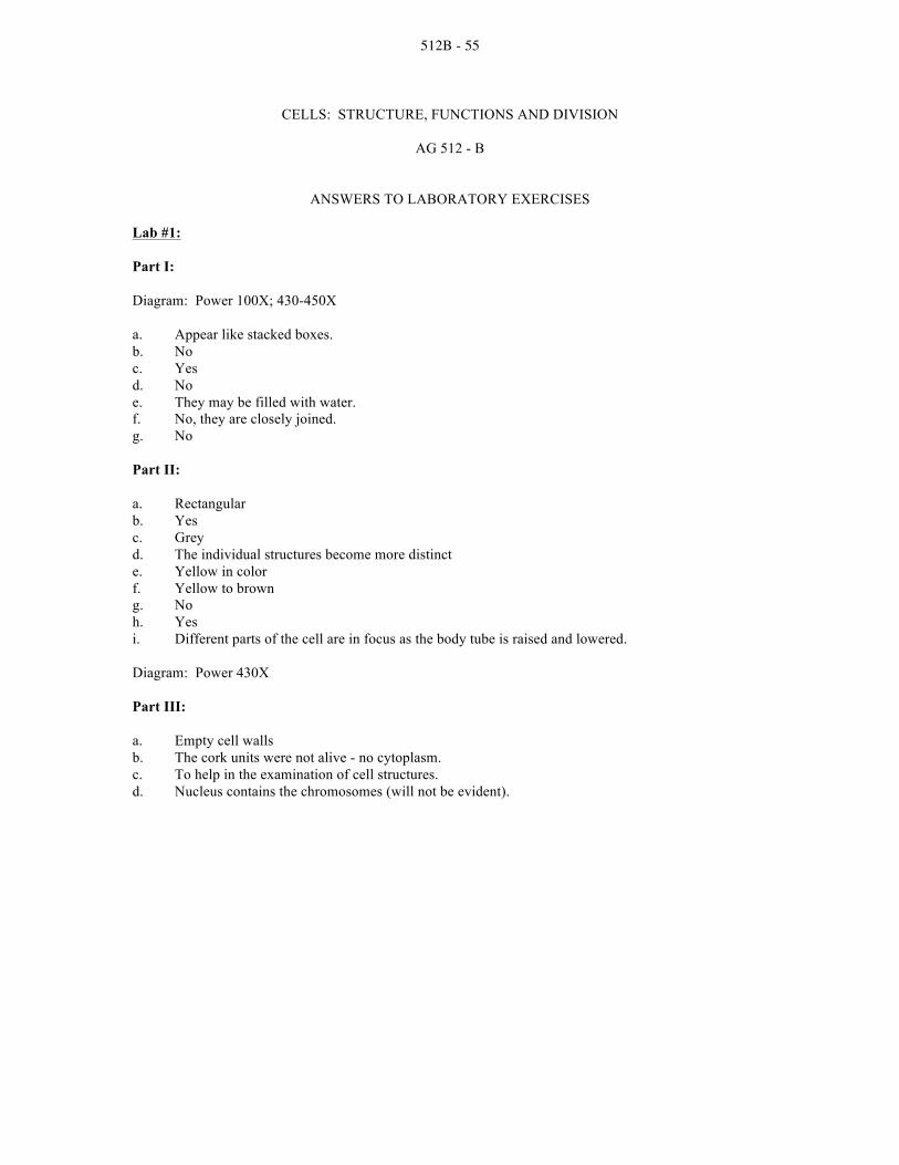
512B - 55
CELLS: STRUCTURE, FUNCTIONS AND DIVISION
AG 512 - B
ANSWERS TO LABORATORY EXERCISES Lab #1:
Part I:
Diagram: Power 100X; 430-450X
a. Appear like stacked boxes. b. No c. Yes d. No e. They may be filled with water. f. No, they are closely joined. g. No
Part II:
a. Rectangular b. Yes c. Grey d. The individual structures become more distinct e. Yellow in color f. Yellow to brown g. No h. Yes i. Different parts of the cell are in focus as the body tube is raised and lowered.
Diagram: Power 430X
Part III:
a. Empty cell walls b. The cork units were not alive - no cytoplasm. c. To help in the examination of cell structures. d. Nucleus contains the chromosomes (will not be evident).

512B - 56
Lab #2
Part II:
1. Table--Cell Parts
2. The presence of starch indicates that the cell part functions to store food.
3. Cell membranes, endoplasmic reticulum, Golgi apparatus, mitochondria, microtubules, microfilaments, ribosomes and nucleoli remained unobservable. They were broken apart or are too small to see with compound microscope.
4. Cell wall fragments; chloroplasts; nuclei; leucoplasts. The cell parts settle according to their
density after being centrifuged. Least dense materials are at the top. Lab #3
Part I: a. The inner layers b. Most likely c. Yes d. No e. No f. They are carried along in the circulating cytoplasm.
Part II: a. It appears to be thinner and less rigid. b. Plasma membrane c. Broad and flat d. Cheek cells tend to be less uniform in shape because of the plasma membrane, rather than the rigid surface of the cell wall. e. It makes cell structures more distinct. f. Grainy and dotted

512B - 57
Part III:
a. Elodea cells have rigid cell walls and chloroplasts. Cheek cells have thin cell membranes. b. Production of food c. They contain the pigment chlorophyll. d. Cell membrane e. No, they are animal cells. f. Yes. They are both made up of structural units called cells.
Lab #4
Part I:
a. The chromosomes migrate along the spindles. b. Across the center or the equatorial plate c. To separate the 2 daughter cells d. Prophase: formation of asters, disintegration of nucleolus, moving of chromatids to the equator. Metaphase: chromatids line up at equator. e. A period of growth and other activities (not part of cell division)
Part II:
a. A centriole is present at the poles of the spindle in the animal cell. b. Animal cells pinch in two; plant cells form a cell wall between daughter cells. c. Centrioles d. Chromosomes and spindle fibers
Part III:
a. The chromosomes replicate, but do not separate. b. Tetrads c. The chromatids form pairs and separate from the tetrad. d. Reduction-division occurs. e. Haploid (n) chromosome number f. Matures into an egg g. They have no function and will gradually disintegrate.

512B - 58
Part IV:
a. Prophase, metaphase, anaphase and telophase b. Insures genetic continuity c. Centrioles are observed in animal cells; cell plate in plant cells d. Chromosomes and spindle fibers e. Reproductive cells f. Reduction of chromosome number during spermatogenesis and oogenesis. g. Chromosome number is halved so that full complement can recur at fertilization. h. Spermatogenesis and oogenesis i. At fertilization there would be twice as many chromosomes in the zygote.
Lab #5
Part I:
a. Some cells have a distinct nucleus while others do not. b. The whitefish blastula chromosomes are smaller.

512B - 59
d. They are about half the size of parent cells
Part II:
1. A cell plate only forms during telophase in plant cells 2. The chromosomes within the cell look like threads. 3. 4 6 1
Anaphase Offspring cells Interphase
2 5 3 Prophase Telophase Metaphase
4. Plant cell, because it has a cell wall

512B - 60
CELLS: STRUCTURE, FUNCTIONS AND DIVISION
AG 512 - B
UNIT TEST Name _____________________________________ Score __________________________________
1. Match terms associated with cells to the correct definition. Write the correct numbers in the blanks.
1. ATP 15. DNA 28. Organelle 2. Cell 16. Endoplasmic 29. Parenchyma 3. Cellulose reticulum 30. Plasma membrane 4. Cell wall 17. Enzymes 31. Plasmodesma 5. Chlorenchyma 18. Eukaryotic cells 32. Plastid 6. Chlorophyll 19. Golgi body 33. Prokaryotic 7. Chloroplast 20. Haploid 34. Protoplasm 8. Chromatid 21. Meiosis 35. Rhizome 9. Chromatin 22. Metabolism 36. RNA 10. Chromoplast 23. Mitochondrion 37. Stroma 11. Chromosome 24. Mitosis 38. Turgor pressure 12. Cytokinesis 25. Mutation 39. Vacuolar membrane 13. Cytoplasm 26. Nucleolus 40. Vacuole 14. Diploid 27. Nucleus
_____a. A plastid containing pigments other than chlorophyll (the pigments are usually yellow to orange)
_____b. The organelle of a living cell that contains chromosomes and is essential to the
regulation and control of all the cell's functions _____c. A chemical compound produced in the mitochondrion; stores energy that is used to
carry out cellular functions _____d. A colorless fluid substance constituting the bulk of the volume of a chloroplast or other
plastid; contains enzymes that play a key role in photosynthetic reactions in chloroplasts
_____e. Having two sets of chromosomes in each cell; the 2n chromosome number
characteristic of the sporophyte generation _____f. An organelle containing enzymes that function in the Krebs cycle and the electron
transport chain of aerobic respiration _____g. Tissue composed of parenchyma cells that contain chloroplasts
_____h. Having a cell or cells that lack a distinct nucleus and other membrane-bound organelles
_____i. The basic structural and functional unit of living organisms
_____j. The outer boundary of the protoplasma of a cell

512B - 61
_____k. Cells having distinct membrane-bound organelles
_____l. The protoplasm of the cell exclusive of the nucleus
_____m. The process of two successive nuclear divisions through which segregation of genes occurs and a single diploid cell (2n) becomes four haploid (n) cells
_____n. Granular particles each composed of two subunits consisting of RNA and proteins;
they lack membranes and are very numerous in living cells _____o. An inheritable change in a gene or chromosome
_____p. The relatively rigid boundary of plant cells
_____q. Pressure within a cell resulting from water uptake
_____r. A complex system of interlinked double membrane channels; parts of it are lined with ribosomes
_____s. A somewhat spherical body within a nucleus; contains RNA and protein
_____t. The use of all interrelated chemical processes occurring in a living organism
_____u. A readily staining complex of DNA and proteins found in chromosomes
_____v. A pocket of cell sap that is separated from the cytoplasm of a cell by a membrane; also, food storage or contractile pockets within the cytoplasm of unicellular organisms
_____w. Molecules of DNA wrapped around proteins, which are found in the nucleus;
control cell functions and the inheritance of traits _____x. Nuclear division during which the chromatids of the chromosomes separate and two
genetically identical daughter nuclei are produced _____y. Carries genetic information in cells
_____z. Green pigments essential to photosynthesis
_____aa. A membrane-bound structure in the cell's cytoplasm
_____bb. An important cellular substance that occurs in three forms, all involved in protein synthesis
_____cc. A complex, insoluble carbohydrate that constitutes the principal component of plant
cell walls _____dd. Cell division
_____ee. An organelle consisting of disc-shaped, often branching hollow tubules that function in accumulating and packaging substances used in the synthesis of materials by the cell

512B - 62
_____ff. Thin-walled cells varying in size, shape and function; the most common type of plant cell
_____gg. A membrane between the cytoplasm and a vacuole of a cell
_____hh. One of the two strands of chromosome; they are united by a centromere
_____ii. Numerous complex proteins that speed up chemical reactions in living things without being used up in the reaction (act as catalysts)
_____jj. An organelle containing chlorophyll
_____kk. Having one set of chromosomes per cell (n chromosomes)
_____ll. Minute strands of cytoplasm that extend between adjacent cells through pores in the walls
_____mm. An organelle associated primarily with the storage or manufacture of carbohydrates
_____nn. The living substance of a cell
2. State the four basic ideas of the cell theory.
a. _____________________________________________________________________________
______________________________________________________________________________
b. ____________________________________________________________________________
______________________________________________________________________________
c. _____________________________________________________________________________
______________________________________________________________________________
d. ____________________________________________________________________________
______________________________________________________________________________
3. List the three ways that cells can differ from one another.
a. _____________________________________________________________________________
b. ____________________________________________________________________________
c. _____________________________________________________________________________

512B - 63
4. Label the parts of a plant cell.
a. ________________________ i _____________________________
b. ________________________ j. _____________________________
c. ________________________ k. ____________________________
d. ________________________ l. _____________________________
e. ________________________ m. ____________________________
f. ________________________ n. ____________________________
g. ________________________ o. ____________________________
h. ________________________ p. ____________________________

512B - 64
5. Match the parts of a plant cell to the correct functions and/or descriptions of each. Write the correct number in the blank provided. 1. Nucleus 6. Cytoplasm 11. Leucoplasts 2. Nucleolus 7. Ribosomes 12. Chromoplasts 3. Plasma membrane 8. Endoplasmic reticulum 13. Vacuole 4. Cell wall 9. Mitochondrion 14. Vacuolar membrane 5. Middle lamella 10. Golgi body 15. Plasmodesma _____a. Regulates ion flow within the cell; maintains cell turgor _____b. Flat, disc-shaped organelles that appear in groups; packages and distributes proteins
for storage within cell and transport out of cell _____c. Contains the heredity information that directs all cell activity; contains the nucleolus _____d. Lipid bilayer surrounding the cytoplasm; maintains surface area for selective cell
absorption and secretion; generates energy _____e. Viscous fluid composed of matrix proteins; assists in transport of substances within
the cell _____f. Connection of thin strands of cytoplasm between the interiors of cells; assist in __
mineral element and food material movement between cells _____g. Small organelles with inner platelike folds; serves as powerhouse for cell--releases
body heat; produces ATP in which energy for cell activities is stored; involved in protein synthesis
_____h. Colored plastids; contain chlorophyll that manufactures the food in plants _____i. Produces RNA; assembles subunits of ribosomes _____j. Cavity containing cell sap; serves as a storage reserve for water, salt and toxic __
products; supports cell wall of plant cells through internal pressure _____k. Network of membranes found within the cytoplasm; involved in storing and ____
transporting protein and chemical products _____l. Protects protoplast; provides external structure; may provide strong support for the
plant _____m. Colorless plastids; storage bodies for oil, starch and proteins _____n. Intercellular layer lying between cells; contains compounds that hold adjacent cells
walls together _____o. Small, discrete structures found in the cytoplasm; manufacture cell proteins

512B - 65
6. Describe the differences between plant and animal cells. Plant cells ______________________________________________________________________ ______________________________________________________________________________ ______________________________________________________________________________ ______________________________________________________________________________ ______________________________________________________________________________ ______________________________________________________________________________ ______________________________________________________________________________ Animal cells ____________________________________________________________________ ______________________________________________________________________________ ______________________________________________________________________________ ______________________________________________________________________________ ______________________________________________________________________________ ______________________________________________________________________________ 7. Describe protoplasm. ______________________________________________________________________________ ______________________________________________________________________________ ______________________________________________________________________________ ______________________________________________________________________________ ______________________________________________________________________________ ______________________________________________________________________________

512B - 66
8. Describe the importance of energy to the functioning of the cell and where the energy is found within the cell. ______________________________________________________________________________ ______________________________________________________________________________ ______________________________________________________________________________ ______________________________________________________________________________ ______________________________________________________________________________ ______________________________________________________________________________ ______________________________________________________________________________ 9. Identify the cell processes for the given descriptions. Write the number of the correct process in the blank. 1. Nutrition 4. Biosynthesis 7. Secretion 2. Digestion 5. Cellular respiration 8. Response 3. Absorption 6. Excretion 9. Reproduction
_____a. Organic substances are manufactured by cells and organized into their own cell material; this process is necessary for growth and for enzyme production to control cell activity
_____b. Cell division
_____c. Cells need molecules for energy and building materials; cells either form their own food molecules or take them in from the environment
_____d. Cells break down most foods into simple forms in order to use them; these reactions
are sped up by certain enzymes in the cell _____e. Waste materials from cell activities are passed from the cell to the environment
_____f. A change in cell activities in response to stimuli from the environment such as heat, pressure, light or chemicals
_____g. Water, food molecules, ions and other necessary materials from the environment are
taken in by the cell _____h. Molecules of substances are synthesized by certain cells and passed out of the cell
_____i. Energy necessary to all cell activities is released from food molecules; usually oxygen is used and carbon dioxide is a waste product

512B - 67
10. Describe the relationship of genes and chromosomes to cell division. a. _____________________________________________________________________________ ______________________________________________________________________________ b. ____________________________________________________________________________ ______________________________________________________________________________ c. _____________________________________________________________________________ ______________________________________________________________________________ 11. Identify the correct stages of mitosis. Write the name of the correct phase in the blank. Answers may be used more than once. __________________________ a. Chromatids separate and move to opposite poles __________________________ b. Period between cell division __________________________ c. Chromatid pairs line up along the equator __________________________ d. Cytokinesis occurs __________________________ e. Chromatin threads gradually shorten and thicken
and become visible as chromosomes
__________________________ f. Spindle fibers attach to the centromere of each chromatid
__________________________ g. Spindle breaks down 12. Describe cytokinesis in plant and animal cells. ______________________________________________________________________________ ______________________________________________________________________________ ______________________________________________________________________________ ______________________________________________________________________________ 13. Identify the correct stages of meiosis. Write the name of the correct phase in the blank. Answers may be used more than once. ____________________________ a. Homologous chromosomes begin to move toward
each other ____________________________ b. DNA replicates itself ____________________________ c. Chromatids of each chromosome separate and
migrate to opposite poles

512B - 68
____________________________ d. Spindle begins to form ____________________________ e. Tetrads move toward middle of cell ____________________________ f. First stage of meiosis is complete ____________________________ g. Tetrads line up along cell's equator ____________________________ h. Chromatids now called chromosomes since they are
no longer joined ____________________________ i. Chromosomes line up along the cell equator 14. Define the two basic types of plant tissue. a. Meristem ___________________________________________________________________ __________________________________________________________________________ __________________________________________________________________________ __________________________________________________________________________ b. Permanent __________________________________________________________________ __________________________________________________________________________ __________________________________________________________________________ __________________________________________________________________________ 15. Identify the types of meristematic and permanent tissues. Write the correct name of the tissue in the blank. ____________________________ a. Constitutes the majority of wood; principal conductor
of water and dissolved minerals ____________________________ b. Account for girth and growth of woody stems;
composed of cellulose and pectin; provide mechanical support for plant
____________________________ c. Found in the tops of the shoots; responsible for
producing new buds and leaves in a uniform pattern at the end of the stem and laterally along stems
____________________________ d. Elongated cells with unevenly thickened primary
walls; gives support to young stems, petioles and leaf veins
____________________________ e. Bark of maturing stems tree trunks and potato skins;
cell walls are waterproofed with suberin (waxy material); die soon, but retain shape
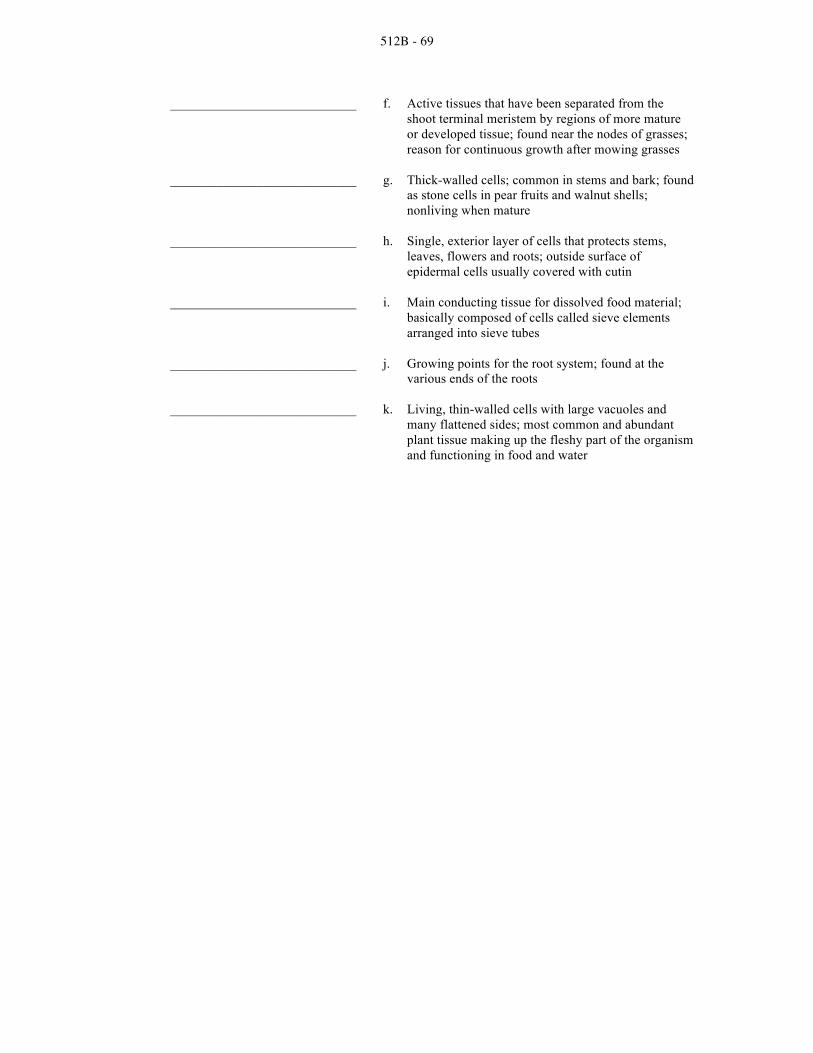
512B - 69
____________________________ f. Active tissues that have been separated from the shoot terminal meristem by regions of more mature or developed tissue; found near the nodes of grasses; reason for continuous growth after mowing grasses
____________________________ g. Thick-walled cells; common in stems and bark; found
as stone cells in pear fruits and walnut shells; nonliving when mature
____________________________ h. Single, exterior layer of cells that protects stems,
leaves, flowers and roots; outside surface of epidermal cells usually covered with cutin
____________________________ i. Main conducting tissue for dissolved food material;
basically composed of cells called sieve elements arranged into sieve tubes
____________________________ j. Growing points for the root system; found at the
various ends of the roots ____________________________ k. Living, thin-walled cells with large vacuoles and
many flattened sides; most common and abundant plant tissue making up the fleshy part of the organism and functioning in food and water

512B - 70
CELLS: STRUCTURE, FUNCTIONS AND DIVISION
AG 512 - B
ANSWERS TO TEST 1. a. 10 o. 25 bb. 36 b. 27 p. 4 cc. 3 c. 1 q. 38 dd. 12 d. 37 r. 16 ee. 19 e. 14 s. 26 ff. 29 f. 23 t. 22 gg. 39 g. 5 u. 9 hh. 8 h. 33 v. 40 ii. 17 i. 2 w. 11 jj. 7 j. 30 x. 24 kk. 20 k. 18 y. 15 ll. 31 l. 13 z. 6 mm. 32 m. 21 aa. 28 nn. 34 n. 35 2. All organisms are made of one or more cells; Cells are alike in their structure and composition; All
cells carry out similar functions that keep them alive; New cells arise only from old cells, usually by dividing into two equal parts at regular intervals
3. Size; Shape; Organization 4. a. plasma membrane b. ribosome cluster c. cytoplasm d. plasmodesma e. cell wall f. nucleus g. vacuole h. nucleolus i. vacuolar membrane (tonoplast) j. plasma membrane k. mitochondrion l. golgi body m. chloroplast n. middle lamella o. rough endoplasmic reticulum p. smooth endoplasmic reticulum 5. a. 14 f. 15 k. 8 b. 10 g. 9 l. 4 c. 1 h. 12 m. 11 d. 3 i. 2 n. 5 e. 6 j. 13 o. 7

512B - 71
6. Plant cells have a cell wall: Made of cellulose; Gives support and shape; Plastids--Leucoplasts: Colorless structures where glucose is changed into starch; Storage for starch, lipids or proteins; Chromoplasts: Manufacture and store pigments; Give fruits, vegetables and leaves their bright color; Chloroplasts: Contain green chlorophyll pigment; Site of photosynthesis (food production) in the plant cell
Animal cells: Microtubules give the cell its shape; Centrioles: Located near nucleus; Function in cell division for reproduction
7. The living parts of the cells: Complex, semi-liquid substance responsible for carrying out the life
processes; No definite composition; Differs in living things; Not even the same in all parts of one individual; Composition is constantly changing; Made up mostly of water; Many substances dissolved or suspended in the water; Organized into cells
8. All processes (or functions) that take place in living things need energy; The energy comes from
chemical reactions that take place in the cell; Chemical activities in one part of the cell are dependent on reactions taking place in another part of the cell; The reactions are all related to functions of the cell; ATP: Produced in mitochondrion; Stores energy for cell activities
9. a. 4 d. 2 g. 3 b. 9 e. 6 h. 7 c. 1 f. 8 i. 5 10. Genes are segments of DNA that control each hereditary trait; The chromosomes carry the genes;
Because chromosomes divide equally during cell division, each daughter cell inherits a complete set of genes
11. a. Anaphase e. Prophase b. Interphase f. Metaphase c. Metaphase g. Telophase d. Telophase 12. Process by which cytoplasm divides to form two cells; Plant cells--Cell plate forms between the
two daughter cells; Animal cells--Cell membrane pinches inward and forms a groove, which deepens until the two cells are separated; Two new, identical cells
13. a. Prophase I f. Telophase I b. Interphase I g. Metaphase I c. Anaphase II h. Telophase II d. Prophase II i. Metaphase II e. Prophase I 14. Meristem (meristematic tissue): Comprised of actively dividing cells that develop and
differentiate into other tissues and organs; Cells have thin walls and dense protoplast Permanent: Develops from the meristems; Non-dividing differentiated cells 15. a. Xylem g. Schlerenchyma tissue b. Lateral meristems h. Epidermis tissue c. Shoot meristems i. Phloem d. Collenchyma tissue j. Root meristems e. Cork tissue k. Parenchyma tissue f. Intercalary meristems

512C - 1
PLANT PROCESSES
AG 512 - C
UNIT OBJECTIVE After completion of this unit, students should be able to list the important plant processes. Students should be able to illustrate the process of photosynthesis and select from a list factors that effect photosynthetic rate. Students should be able to illustrate the process of respiration and identify processes as being characteristic of respiration or photosynthesis. Students should also be able to briefly explain the processes of absorption and transpiration. This knowledge will be demonstrated by completion of an assignment sheet, laboratory exercises and a unit test with a minimum of 85 percent accuracy.
SPECIFIC OBJECTIVES AND COMPETENCIES
After completion of this unit, the student should be able to:
1. Match terms associated with basic plant processes to the correct definition.
2. Name the four important plant processes in food manufacture and growth.
3. Explain why photosynthesis is an important process.
4. Explain the process of photosynthesis.
5. List five factors that affect photosynthetic rate.
6. Explain the process of respiration.
7. Classify characteristics as that of photosynthesis or respiration.
8. Match ways nutrients and water are absorbed by the plant to the correct definition.
9. Explain the process of absorption by plant roots.
10. Explain the process of transpiration.
11. List five factors affecting water loss by transpiration.
12. Explain osmosis.
13. Measure loss from transpiration.
14. Measure rates of photosynthesis in different environments.
15. Study water movement through plants.

512C - 2
PLANT PROCESSES
AG 512 - C
SUGGESTED ACTIVITIES
I. Suggested activities for instructor
A. Order materials to supplement unit.
1. Films, slideshows, etc.
a. Photosynthesis and Respiration, VHS video; available from Vocational Education Productions, California Polytechnic State University, San Luis Obispo, California 93407 (1-800-235-4146); approximate cost $99.95; order no. 6-083-107J.
b. Plant Movement and Transport, VHS video; discusses the
xylem and phloem, osmosis and transpiration; available from Vocational Education Productions, California Polytechnic State University, San Luis Obispo, California 93407 (1-800-235-4146); approximate cost $99.95; order no. 6-083-108J.
c. Principles of Plant Growth, 24 color slide set; discusses
photosynthesis, plant tissues, mineral and nutrient uptake, plant cell anatomy and root system; available from Vocational Education Productions, California Polytechnic State University, San Luis Obispo, California 93407 (1-800-235-4146); approximate cost $32.95; order no. 1-580-603J.
B. Make transparencies and necessary copies of material.
C. Provide students with objective sheet and discuss.
D. Provide students with information and assignment sheets and laboratory exercises.
E. Discuss unit and specific objectives.
F. Discuss information and assignment sheets.
G. Demonstrate and discuss laboratory exercises.
H. Review and give test.
I. Reteach and retest if necessary.
II. Instructional materials
A. Objective sheet
B. Suggested activities

512C - 3
C. Information sheet
D. Transparency masters
1. TM 1--Important Plant Processes
2. TM 2--Importance of Photosynthesis
3. TM 3--Photosynthesis
4. TM 4--Photosynthesis and Respiration in Relation to Dry Weight
5. TM 5--Absorption
6. TM 6--Transpiration
7. TM 7--Root Hairs, Soil Particles and Moisture
8. TM 8--How a Water Solution From the Soil Moves Within a Root
9. TM 9--Osmosis
E. Assignment sheet
1. AS 1--Light in Relation to Increase in Dry Weight
F. Instructor notes for laboratory exercises
G. Laboratory exercises
1. LE 1--Measuring Loss From Transpiration
2. LE 2--Measuring Rates of Photosynthesis in Different Environments
3. LE 3--Water Movement Through Plants
H. Answers to laboratory exercises
I. Test
J. Answers to test
III. Unit references
A. Delorit, R.J., et al., Crop Production, 5th edition, Prentice-Hall, Inc., Englewood Cliffs, New Jersey, 1984.
B. Fridline, C.R., Plant Growth and Development, Ohio State University, Ohio
Agricultural Education Curriculum Materials Service, Columbus, Ohio, 1980. C. Hartmann, Hudson, T., et al., Plant Science - Growth, Development, and
Utilization of Cultivated Plants, 2nd edition, Prentice-Hall, Inc., Englewood Cliffs, New Jersey, 1988.

512C - 4
D. Janick, J., et al., Plant Science, 2nd edition, W.H. Freeman and Co., San Francisco, California, 1974.
E. Otto, James H., Towle, Albert, Modern Biology, Holt, Rinehart and Winston,
Publishers, New York, 1985. F. Raven, P.H., et al., Biology of Plants, 3rd edition, Worth Publishers, Inc., 1981.
G. Slesnick, Irwin L., et al., Biology, Scott, Foresman and Company, Glenview, Illinois, 1985.

512C - 5
PLANT PROCESSES
AG 512 - C
INFORMATION SHEET I. Terms and definitions
A. Photosynthesis--The process of making sugars in green plants from water and carbon dioxide in the presence of sunlight B. Respiration--The process of using the stored foods of a plant in which energy is
obtained or released C. Transpiration--The evaporation of water vapor from the stoma on the leaves of a
plant D. Absorption--The taking in of water and mineral nutrients through the roots of a
plant E. Stoma--A small opening in the epidermis of leaves and stems through which
gases pass F. Mesophyll--The photosynthetic tissue of a leaf; located between the layers of
epidermis G. Chlorophyll--The green pigment of plant cells; necessary for photosynthesis
H. Chloroplast--A cellular organelle in which chlorophyll is contained; site of photosynthesis
I. Phloem--Food conducting tissue of plants
J. Root hairs--Outgrowths of the epidermal cells of the root; greatly increase absorption area of the root system
K. Permeable membrane--A membrane through which liquid substances may
diffuse, such as the plasma membrane of root hair cells
L. Epidermis--The outermost layer of cells of the leaf and of young stems and roots
M. Cortex--Cells of a stem or root bound externally by the epidermis and internally by the vascular system
N. Xylem--Tissue through which most of the water and minerals of a plant are
conducted II. Important plant processes in food manufacture and growth (Transparency 1)
A. Photosynthesis
B. Respiration

512C - 6
C. Transpiration
D. Absorption
III. Reasons photosynthesis is the most important process in the world (Transparency 2)
A. Plants produce food by photosynthesis
B. Plants produce food used directly by man
C. Plants produce food used indirectly by man through meat and milk produced by livestock
(Note: Green plants, through the process of photosynthesis, are the basic factories of the world, on which all life is dependent. A corn plant produces about 5 grams of sugar during 14 hours of sunlight. During a 100-day period, a 20-acre field of corn with 20,000 plants per acre could produce two and one-half tons of sugar. An estimated 150 billion tons of sugar are produced by plants each year by photosynthesis. This would be a pile of sugar 40 miles square at the base and 2 miles high at the peak.)
IV. Process of photosynthesis (Transparency 3)
A. Carbon dioxide (CO2) enters the leaf from the surrounding air through the stoma (Note: After CO2 enters the stoma, it enters the intercellular spaces of the mesophyll tissue. Here it comes in contact with the wet walls of the mesophyll cells. The CO2 dissolves in the water of the mesophyll cells.)
B. Water moves from the soil into the root, stems and leaves through the xylem
tissue
C. The molecules of water (H2O) and carbon dioxide (CO2) are synthesized (put together) in the chlorophyll of a plant with energy from sunlight
D. The end result is the formation of sugar which is transported by the phloem
tissue to the part of the plant where it is used
(Note: The process can be illustrated by the chemical equation which is written: 6 parts carbon dioxide (6 CO2) + 6 parts water (6 H2O) + 672 K. cal of radiant energy (sunlight) in the presence of chlorophyll of plants = sugar (C6H12O6) retained by the plant + oxygen (6 O2) given off into the atmosphere.)
V. Factors that effect photosynthetic rate
A. Water supply
B. Temperature
C. Light quality
D. Light intensity

512C - 7
E. Deficiency of certain plant nutrients
VI. Process of respiration (Note: As with all living things, plants require energy to carry out their growth and development process. This energy comes from a very complex process called respiration. In a sense, respiration in plants is the reverse of photosynthesis as sugar is broken down to produce energy.)
A. Sugar is broken down to produce energy for essential plant functions
B. Respiration consumes oxygen (O2) and glucose (C6H12O6)
C. Respiration gives off carbon dioxide (CO2) and water (H20)
(Note: The process can be illustrated by the chemical equation which is written: Sugar (C6H12O6) + 6 parts oxygen (6 O2) = 6 parts carbon dioxide (6 CO2) + 6 parts water (6 H2O).)
VII. Relationship between photosynthesis and respiration (Transparency 4)
Photosynthesis Respiration
1. A building process (+) 1. A destruction process (-)
2. Sugars manufactured 2. Sugars consumed
3. CO2 is consumed 3. CO2 is given off
4. Oxygen is given off 4. Oxygen is consumed
5. Requires light 5. Goes on day and night
6. Only takes place in cells 6. Carried on in all cells containing chlorophyll
7. Sugar (C6H12O6) is 7. Energy produced for plant
the end product functions is end product
(Note: A green plant grown in the dark loses in weight because its stored foods are respired and nothing is added through photosynthesis.)
VIII. Absorption of nutrients and water
A. Diffusion--Movement of gases or liquid from an area of high concentration to an area of low concentration
B. Osmosis--Movement of a substance from a place of greater concentration
through a semi-permeable membrane to a place of lesser concentration
C. Non-root feeding (leaf feeding)--Nutrients and water entering the plant through leaves and stems

512C - 8
IX. The process of absorption (Transparency 5)
A. The soil solution enters the root hairs by the process of osmosis
(Note: The soil solution is composed of water and minerals in solution.)
B. After the soil solution is absorbed by the root hairs, it moves through the epidermal cells, cortex and phloem to the xylem
C. The xylem conducts the solution to other parts of the plant
X. The process of transpiration (Transparencies 6, 7, 8)
A. Water enters plant through root hairs
B. Water passes to xylem and up the stem to the leaves
C. A small amount of water is used in photosynthesis
D. The remainder is lost by transpiration
(Note: Water loss occurs mainly through the stoma on the leaves. When stoma are open, water vapor, which is low in carbon dioxide, escapes from the leaf and is replaced by dry air higher in carbon dioxide.)
XI. Factors affecting water loss by transpiration
A. Climatic conditions
B. The number of stoma on the leaves
C. Availability of soil moisture to the plant
D. General plant structure
(Note: Some plants have a waxy covering called a cuticle which inhibits evaporation of water. Some plants have stoma only on lower side of leaf where temperatures are cooler.)
E. Soil fertility
(Note: Transpiration is greater for plants in fertile soils than poor soils; therefore the plant uses more water on fertile soils than poor soils.)
XII. Osmosis--The diffusion of water through a selectively permeable membrane from an area
of greater concentration of water to an area of lesser concentration (Transparency 9)

512C - 9
TM 1

512C - 10
TM 2

512C - 11
TM 3

512C - 12
TM 4

512C - 13
TM 5

512C - 14
TM 6

512C - 15
TM 7

512C - 16
TM 8

512C - 17
TM 9

512C - 18
PLANT PROCESSES
AG 512 - C
ASSIGNMENT SHEET #1--LIGHT IN RELATION TO INCREASE IN DRY WEIGHT
Name _____________________________________ Score __________________________________
Part I: Three hundred bean seeds were divided into three lots of 100 each. One lot of 100 seeds was used to measure the percent moisture in the seeds, from which it was possible to calculate the dry weight of the other two lots. A second lot of seeds was sown in sand in a suitable box and kept in a dark room. The third lot was likewise sown in sand in a suitable box and kept under a daily cycle of 15 hours of light and 9 hours of dark. These two lots of seeds were kept at a temperature of 75oF. Four weeks after planting, each lot of resulting seedlings was harvested and the dry weight of each was measured. These resulting data are tabulated.
Environmental Condition
Calculated Dry Weight of Seeds
Planted
Dry Weight of Seedlings at End of Four
Weeks
Difference Between Dry Weight of Seed - lings and Dry Weight
of Seeds Planted
Seedlings in light 57.9 g 82.2 g +23.4 g
Seedlings in dark 57.9 g 23.4 -34.5 g
Part II: a. What one environmental factor was varied in this experiment? b. How do you account for the increase in dry weight of the seedlings in light as compared with the dry weight of the seeds which were planted in darkness?

512C - 19
c. Why do the seedlings that developed in the dark not gain in dry weight? d. Why do the seedlings that developed in the dark actually decrease in dry weight? e. Is light necessary for the utilization of food in the growth of plants?
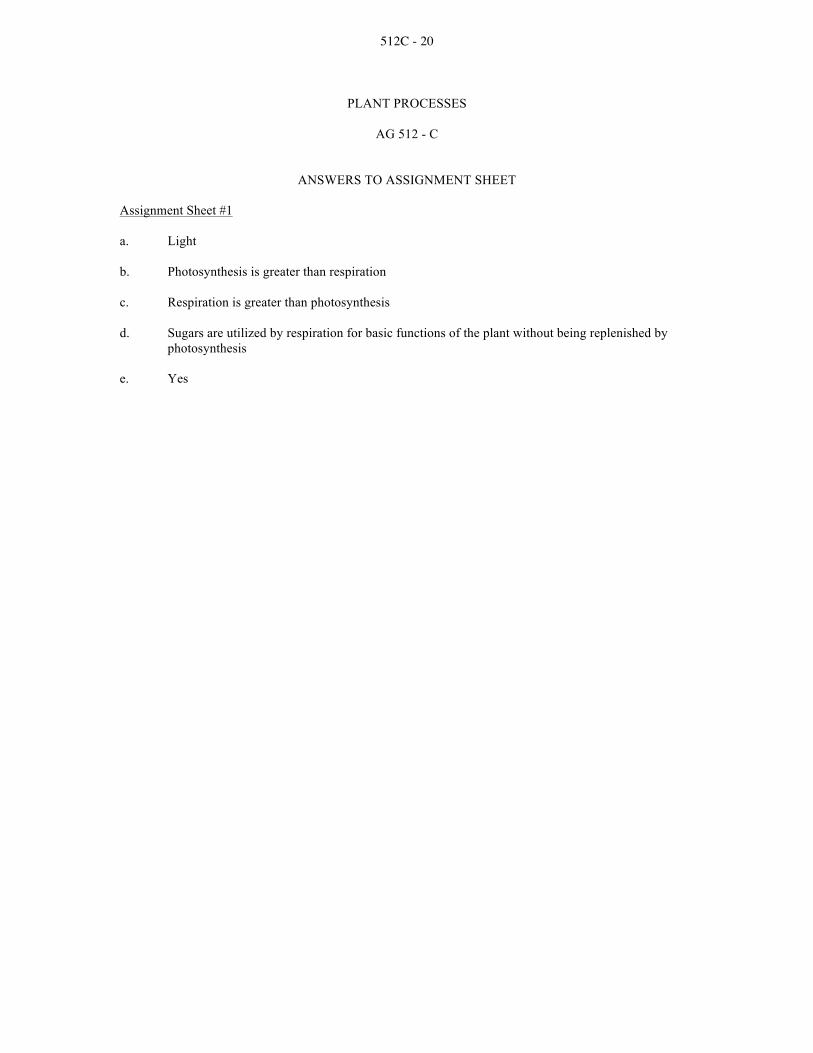
512C - 20
PLANT PROCESSES
AG 512 - C
ANSWERS TO ASSIGNMENT SHEET Assignment Sheet #1
a. Light
b. Photosynthesis is greater than respiration
c. Respiration is greater than photosynthesis
d. Sugars are utilized by respiration for basic functions of the plant without being replenished by photosynthesis
e. Yes

512C - 21
PLANT PROCESSES
AG 512 - C
INSTRUCTOR NOTES FOR LABORATORY EXERCISES
Lab #2 Make sure the Elodea plant is very healthy by exposing it to several hours of bright light a few days before students perform this laboratory. Students can set up Part II while they are waiting to measure oxygen production for plants in Part I. Part I: Step 2: Warn students to use caution when using sharp instruments. Step 3: Add about 1/2 teaspoon of sodium bicarbonate to each test tube. Step 4: Provide a fluorescent light for growing plants for the bright light environment. Step 5: In choosing these light environments, try to control for temperature.
Place test-tube racks in appropriate areas around the room in which the students can place their labeled test tubes in Step 5 of Part I.
Step 6: Remind students that plants are adapted to a variety of conditions. For some plants adapted to shade, long exposure to bright light may harm the plant. In the short run, bright light will increase the rate of photosynthesis of almost any plant.
Part II:
You may reuse Elodea sprigs from Part I in Part II.
Table I: More bubbles will be released in the bright light.
Table II: More bubbles will be released in the warm temperature than the hot, cool or freezing temperatures.
Part III: Extensions: Ask students to design an experiment to find out how the color of light affects the rate of photosynthesis. Lab #3
Part I:
Freshly removed leaves provide the best observation.
Part II:
Demonstrate the pairing of the cobalt paper on the leaves.
Part III:
Celery, freshly removed from the stalk, could be used as a substitute for the coleus.

512C - 22
PLANT PROCESSES
AG 512 - C
LABORATORY EXERCISE #1--MEASURING LOSS FROM TRANSPIRATION Name _____________________________________ Score __________________________________
I. Materials needed
A. A growing plant in a pot, bucket or other container or a plant growing in a convenient location
B. Plastic bag or sheet of clear plastic that will cover the plant or a branch of the plant
containing 10 to 12 leaves
C. String, rubber bands or other suitable tie materials
D. Stake to support the weight of the plastic
E. Plastic straw or other suitable tubing for a drain
F. Measuring cup or beaker
II. Procedure (Figure 1)
A. Be sure plant has been watered
B. Cover the entire vegetative portion of the plant or a section with plastic material
C. Locate tubing at bottom of plastic covering to serve as a water drain
D. Place plant in sunny location for an entire day; measure the water collected; empty container
E. Measure water collected during dark period of the day
F. Compare differences in the amount of water collected

512C - 23
III. Questions
a. Which period produced the most water in the container? Why?
b. Does temperature have an effect on transpiration rates? Does light? Air humidity?

512C - 24
PLANT PROCESSES
AG 512 - C
LABORATORY EXERCISE #2--MEASURING RATES OF PHOTOSYNTHESIS IN DIFFERENT ENVIRONMENTS
Name _____________________________________ Score __________________________________
Slesnick, Irwin L., Biology Laboratory Manual, Scott, Foresman and Company, 1985. Reprinted by permission of Scott, Foresman and Company. Introduction Plants grow in many different environments. Dandelions grow in cracks in a sidewalk; pine trees grow high on mountainsides; water lilies grow from shallow lake bottoms. Though plants flourish in vastly different environments, most plants have one thing in common: they need light to survive. Unlike certain animal species, no species of green plant can survive for long in total darkness. To make glucose, plants capture light energy in the process of photosynthesis. In this laboratory you will observe how both light intensity and temperature affect the rate of photosynthesis of an Elodea plant. Materials needed Metric ruler Thermometer Scissors or scalpel 4 300-ml beakers Clock or timepiece Elodea sprigs Sodium bicarbonate Tap water Spoon 4 test tubes Masking tape 4 glass stirring rods, each about 5 cm long Marking pen Ice Part I: Comparing Rates of Photosynthesis Under Different Light Conditions 1. Place four test tubes in a test-tube rack. Fill each test tube to within 4 cm of the rim with aquarium
water.

512C - 25
2. Cut four healthy sprigs from an Elodea plant. Each sprig should be approximately 8 cm long. Use sharp scissors or a scalpel to make a clean diagonal cut across the bottom of each sprig.
3. Obtain four glass rods. Wrap each Elodea sprig around a glass rod. Place each glass rod with an
Elodea sprig in a test tube, as shown in a. The cut end of the Elodea should be in the bottom of the test tube. Add about 1 g of sodium bicarbonate to each test tube. Sodium bicarbonate supplies carbon dioxide needed for certain reactions in photosynthesis.
4. Make masking tape labels for each test tube. On one label, write "bright light"; on a second label,
write "daylight"; on a third, write "shade"; on the fourth, write "darkness". These are the four different light environments for this part of the laboratory.
5. Place each test tube in the type of light indicated on its label. Make sure that the temperature in
each different light environment varies no more than 5oC. On a separate piece of paper, copy Table I below.
6. After 15 minutes have passed, observe the bubbles rising from the cut ends of each Elodea plant.
These bubbles are oxygen gas given off as a by-product of photosynthesis. What is the relationship between the number of bubbles given off and the rate of photosynthesis?
(a) ____________________________________________________________________________
7. Count the number of bubbles given off by each plant in three minutes. Record your results in your
Table I. Compute the average number of bubbles given off by each sprig in one minute. Record these averages in Table I.
Part II: Comparing Rates of Photosynthesis Under Different Temperature Conditions 1. Repeat steps 1-3 of Part I. 2. Prepare masking tape labels for your test tubes. Label one tube "0oC"; a second, label "15oC";
label a third test tube "30oC"; label the fourth "45oC". These are the four different temperature environments for this part of the laboratory. On a separate piece of paper, copy Table II.
3. Obtain four beakers. Fill one beaker three-quarters full of ice. Half fill the other beakers with
cool, warm and hot water respectively. Measure the temperature of each water bath. Add warmer or cooler water to each to bring the temperature of the cool water bath to 15oC, the warm water bath to 30oC, and the hot water bath to 45oC. In each water bath, place the test tube with the corresponding label.
4. Count and record the number of bubbles released during three minutes from the cut ends of each
Elodea sprig. Compute the average number of bubbles released by each sprig in one minute, and record these averages in Table II.
5. Clean up your work area and put away your equipment.

512C - 26
Part III: Analysis
1. Using your results from Part I as evidence, how does light intensity affect the rate of photosynthesis of an Elodea plant?
______________________________________________________________________________
______________________________________________________________________________
2. How can you explain the effects of light intensity on photosynthesis? Review the photosynthesis reactions to formulate your answer.
_____________________________________________________________________________
_____________________________________________________________________________
3. How does temperature affect the rate of photosynthesis in an Elodea? Explain using the results from Part II.
_____________________________________________________________________________
_____________________________________________________________________________

512C - 27
PLANT PROCESSES
AG 512 - C
LABORATORY EXERCISE #3--WATER MOVEMENT THROUGH PLANTS Name _____________________________________ Score __________________________________
Selection from Modern Biology, Biology Investigations, Teacher's Edition, by James H. Otto, Albert Towle, W. David Otto, and Myra E. Madnick. Copyright 1977 by Holt, Rinehart and Winston, Inc. Reprinted by permission of the publisher. Materials needed Tulip, Trandescantia or Geranium leaves 2 sandwich bags 6 coleus or periwinkle cuttings, 15-20 cm in length Cobalt paper 1 healthy geranium plant Paper clips Slide, cover glass Cellophane tape Microscope 250-ml beaker Paper toweling Eosin powder or red ink 10% salt solution Single-edged razor blade Part I: The Structure of the Stomata In addition to providing for the movement of gases in and out of a leaf, the stomata have the function of regulating the movement of water through the plant. Remove a small piece of the lower epidermis from a tulip or geranium leaf by tearing with the blade, twisting as you tear. The epidermis appears as a thin, transparent skin. Prepare a wet mount of a piece of the epidermis. Be careful not to wrinkle the tissue. Examine the tissue under low power. Note the shape and arrangement of the epidermal cells and the number and arrangement of the stomata (singular stoma). a. Are chloroplasts present in the epidermal cells? ________________________________________ Locate the bean-shaped guard cells on either side of the stoma. Study the walls of the guard cells under low power.
b. What variation in thickness can be observed in the inner and outer walls?
______________________________________________________________________________
c. What structure can you observe in the guard cells?
______________________________________________________________________________
The stoma is a pore opening into the air spaces of the leaf. Only a small percentage of the water absorbed by a plant is used in photosynthesis and other plant processes. The remainder is lost from the tissues of the leaf through the stomata. The guard cells regulate the size of the stomata by changing shape.

512C - 28
d. Why is this important to the leaf and the plant?
______________________________________________________________________________
To observe the opening and the closing of the stoma, prepare a fresh wet mount of the lower epidermis and examine under low power. e. Are the stomata open? ____________________________________________________________
______________________________________________________________________________
Remove the water from the preparation by placing paper toweling at the left edge of the cover glass to soak up the water. Add a 10% salt solution to the right side of the cover glass. Observe quickly. f. Describe what happened to the guard cells and the stoma.
______________________________________________________________________________
______________________________________________________________________________
g. How can you explain the changes that occurred?
______________________________________________________________________________
______________________________________________________________________________
h. Explain what natural activity of a leaf might cause the stomata to close.
______________________________________________________________________________
______________________________________________________________________________
______________________________________________________________________________
______________________________________________________________________________
In the space provided, draw the lower leaf epidermis. Label: stoma, guard cells, chloroplasts, epidermal cells.

512C - 29
Part II: Transpiration
Transpiration is essentially the loss of water from a plant through evaporation from leaf tissues.
Remove a leaf from a vigorously growing geranium plant. Fasten a strip of blue cobalt paper to the lower surface and upper surface by clipping the paper at both ends to the leaf. Do not damage the leaf unnecessarily. Blue cobalt paper turns pink in the presence of moisture. Place the leaf in a small plastic sandwich bag and seal it with tape. Place a strip of cobalt paper in a bag by itself and seal it. a. What is the purpose of this second bag?
______________________________________________________________________________
Observe the leaf after 10 minutes.
b. What is the color of the cobalt paper on the upper surface?
______________________________________________________________________________
c. The color on the lower surface? _____________________________________________________
d. What is the color of the cobalt paper in the empty bag?
______________________________________________________________________________
e. How do you account for the difference in the color of the cobalt paper?
______________________________________________________________________________
______________________________________________________________________________
f. Why is it necessary to seal the bag?
______________________________________________________________________________
______________________________________________________________________________
g. Are the leaves wet? ______________________________________________________________
h. From what part of the leaf did the moisture come?
______________________________________________________________________________
i. Why is a control bag needed?
______________________________________________________________________________
______________________________________________________________________________
j. Originally, how did the water get to the leaf?
______________________________________________________________________________
______________________________________________________________________________

512C - 30
Part III: The Path of Rising Water in a Stem
In the examination of the structure of roots and stems, it may be observed that certain tissues are responsible for the conduction of substances up and down the stem. a. Which tissue is responsible for the conduction of water?
______________________________________________________________________________
Fill a 250-ml flask about one third full of water which as been colored with powdered eosin or red ink. Place a freshly cut stem of coleus in the flask. Leave the stem in the solution at least 30 minutes. Remove the stem and split it lengthwise with a razor blade. b. What evidence do you see of the rise of dye in the stem?
______________________________________________________________________________
c. In the stem, locate where the water seems to have risen.
______________________________________________________________________________
______________________________________________________________________________
Make a cross section of a lower portion of the stem and prepare a wet mount. Examine the stem under low power of the microscope. d. In what tissue of the stem is there evidence that water has risen?
______________________________________________________________________________
Part IV: Summary
a. On the basis of what you have learned about transpiration, account for the fact that leaves often wilt on a hot summer day, but return to normal at night.
______________________________________________________________________________
______________________________________________________________________________
______________________________________________________________________________
b. List as many factors as you can think of that might affect the loss of water from a plant.
______________________________________________________________________________
______________________________________________________________________________

512C - 31
The following events trace the movement of water from the soil, through a plant to the atmosphere. Using your knowledge of plant structure, unscramble the pathway and write the events in order of occurrence in the space provided. The first and last are done for you. WATER
*enters the petiole of the leaf 1. water in the soil
*moves across the cortex of the root 2. ____________________________
*absorbed by mesophyll cells 3. ____________________________
*enters vascular cylinder 4. ____________________________
*enters veins of leaf 5. ____________________________
*absorbed by root hairs 6. ____________________________
*leaves leaf through stomata 7. ____________________________
*moves into xylem cells of stem 8. ____________________________
*evaporates into air spaces of leaf 9. ____________________________
*moves up xylem of stem 10. ____________________________
11. ____________________________
12. water escapes to atmosphere

512C - 32
PLANT PROCESSES
AG 512 - C
ANSWERS TO LABORATORY EXERCISES Lab #1
Part III:
a. Most water into container during the daylight period
Why? Stomata are open during the day, allowing water to escape
b. Temperature - as temperature increases, transpiration increases
Light - light increases temperature, which increases transpiration
Air humidity - as humidity increases, it lowers transpiration
Lab #2
Part I:
(a) The more bubbles, the faster the rate of photosynthesis.
Part III:
1. The stronger the light, the more oxygen was released by the plant. This indicates photosynthesis was occurring at a fast rate.
2. The more light energy, the more chlorophyll can undergo chemical changes that result in the
splitting of water into hydrogen and oxygen. Thus, the more light energy, the more oxygen produced during photosynthesis.
3. More oxygen bubbles were released from the plant in the 30oC environment. Thus, a warm
temperature best promotes photosynthesis. Lab #3
Part I:
a. No b. The inner wall is considerably thicker than the outer wall. c. Chloroplasts d. This regulates water loss. e. If the plant has been exposed to bright light, the stomata will be open. f. The guard cells changed shape, resulting in the closing of the stomata. g. The salt concentration outside the cell caused the guard cells to lose water, and the stomata were
closed. h. In the synthesis of carbohydrates, carbon dioxide is used up and the acid content of the guard cell
decreases. More starch is converted to sugar; water diffuses into the guard cells and the stomata close.

512C - 33
Drawings will vary. Complete to instructor's satisfaction.
Part II:
a. To serve as a control b. Less blue, slightly pink c. Pink d. Blue e. The papers attached to the leaves respond to moisture transpiration from the leaves and turned
pink. Those by themselves remained blue. f. To insure that the only moisture affecting the cobalt paper is coming from the leaf. g. No h. From the air spaces and through the stomata i. To demonstrate that the water came only from the leaf. j. It was carried to the leaf through the vascular tissue of the roots and stem and of the leaf itself.
Part III:
a. Xylem b. The water colored red by the dye has moved up the stem and the stem tissues appear red. c. Toward the interior where the xylem tissue is located. d. Xylem
Part IV:
a. The atmosphere becomes cooler and absorption makes up the water deficiency. The stomata remain closed for longer periods of time at night, thus reducing the evaporation rate.
b. Air currents, humidity, temperature, light, soil factors, available water
2. Absorbed by root hairs 3. Moves across the cortex of the root 4. Enters vascular cylinder 5. Moves into xylem cells of stem 6. Moves up xylem of stem 7. Enters the petiole of the leaf 8. Enters veins of leaf 9. Absorbed by mesophyll cells 10. Evaporates into air spaces of leaf 11. Leaves leaf through stomata

512C - 34
PLANT PROCESSES
AG 512 - C
UNIT TEST Name _____________________________________ Score __________________________________
1. Match terms associated with basic plant processes to the correct definitions. Write the correct numbers in the blanks.
1. Photosynthesis 6. Mesophyll 11. Permeable membrane 2. Respiration 7. Chlorophyll 12. Epidermis 3. Transpiration 8. Chloroplast 13. Cortex 4. Absorption 9. Phloem 14. Xylem 5. Stoma 10. Root hairs
_____a. A small opening in the epidermis of leaves and stems through which gases pass
_____b. The green pigment of plant cells; necessary for photosynthesis
_____c. The process of making sugars in green plants from water and carbon dioxide in the presence of sunlight
_____d. Food conducting tissue of plants
_____e. The outermost layer of cells of the leaf and of young stems and roots
_____f. The evaporation of water vapor from the stoma on the leaves of a plant
_____g. A cellular organelle in which chlorophyll is contained; site of photosynthesis
_____h. Tissue through which most of the water and minerals of a plant are conducted
_____i. The photosynthetic tissue of a leaf; located between the layers of epidermis
_____j. The process of using the stored foods of a plant in which energy is obtained or released _____k. A membrane through which liquid substances may diffuse, such as the plasma
membrane of root hair cells
_____l. The taking in of water and mineral nutrients through the roots of a plant
_____m. Cells of a stem or root bound externally by the epidermis and internally by the vascular system
____n. Outgrowths of the epidermal cells of the root; greatly increase absorption area of the
root system

512C - 35
2. Name the four important plant processes in food manufacture and growth.
a. _____________________________________________________________________________
b. ____________________________________________________________________________
c. _____________________________________________________________________________
d. ____________________________________________________________________________
3. Explain why photosynthesis is an important process.
a. _____________________________________________________________________________
______________________________________________________________________________
b. ____________________________________________________________________________
______________________________________________________________________________
c. _____________________________________________________________________________
______________________________________________________________________________
4. Explain the process of photosynthesis.
______________________________________________________________________________
______________________________________________________________________________
______________________________________________________________________________
______________________________________________________________________________
______________________________________________________________________________
______________________________________________________________________________
______________________________________________________________________________
______________________________________________________________________________
5. List five factors that effect photosynthetic rate.
a. _____________________________________________________________________________
b. ____________________________________________________________________________
c. _____________________________________________________________________________
d. ____________________________________________________________________________
e. _____________________________________________________________________________

512C - 36
6. Explain the process of respiration.
______________________________________________________________________________
______________________________________________________________________________
______________________________________________________________________________
______________________________________________________________________________
______________________________________________________________________________
7. Classify the following characteristics as that of photosynthesis (X) or respiration (O). Write the correct letter in the blank before each statement.
_____a. Sugar is the end product
_____b. Carbon dioxide is given off
_____c. Requires light
_____d. A destruction process
_____e. Goes on day and night
_____f. A building process
_____g. Only takes place in cells containing chlorophyll
_____h. Carried on in all cells
_____i. Oxygen is given off
_____j. Sugars consumed
8. Match ways nutrients and water are absorbed by the plant to the correct definition. Write the correct numbers in the blanks.
_____a. Nutrients and water entering the plant 1. Diffusion
through leaves and stems 2. Osmosis
_____b. Movement of gases or liquid from an area of high concentration to an area of low 3. Non-root feeding concentration
_____c. Movement of a substance from a place of greater concentration through a semi-permeable membrane to a place of lesser concentration

512C - 37
9. Explain the process of absorption by plant roots.
______________________________________________________________________________
______________________________________________________________________________
______________________________________________________________________________
______________________________________________________________________________
______________________________________________________________________________
______________________________________________________________________________
10. Explain the process of transpiration.
______________________________________________________________________________
______________________________________________________________________________
______________________________________________________________________________
______________________________________________________________________________
______________________________________________________________________________
______________________________________________________________________________
11. List five factors affecting water loss by transpiration.
a. _____________________________________________________________________________
b. ____________________________________________________________________________
c. _____________________________________________________________________________
d. ____________________________________________________________________________
e. _____________________________________________________________________________
12. Explain osmosis.
______________________________________________________________________________
______________________________________________________________________________
______________________________________________________________________________

512C - 38
PLANT PROCESSES
AG 512 - C
ANSWERS TO TEST 1. a. 5 f. 3 k. 11
b. 7 g. 8 l. 4 c. 1 h. 14 m. 13 d. 9 i. 6 n. 10 e. 12 j. 2
2. Photosynthesis; Respiration; Transpiration; Absorption
3. Plants produce food by photosynthesis; Plants produce food used directly by man; Plants produce food used indirectly by man through meat and milk produced by livestock
4. Answer could include the following information:
Carbon dioxide (CO2) enters the leaf from the surrounding air through the stoma; Water moves from the soil into the root, stems and leaves through the xylem tissue; The molecules of water (H2O) and carbon dioxide (CO2) are synthesized (put together) in the chlorophyll of a plant with energy from sunlight; The end result is the formation of sugar which is transported by the phloem tissue to the part of the plant where it is used
5. Water supply; Temperature; Light quality; Light intensity; Deficiency of certain plant nutrients
6. Answer could include the following information:
Sugar is broken down to produce energy for essential plant functions; Respiration consumes oxygen (O2) and glucose (C6H12O6); Respiration gives off carbon dioxide (CO2) and water (H20)
7. a. X e. O i. X b. O f. X j. O c. X g. X d. O h. O
8. a. 3 b. 1 c. 2
9. Answer could include the following information:
The soil solution enters the root hairs by the process of osmosis; After the soil solution is absorbed by the root hairs, it moves through the epidermal cells, cortex and phloem to the xylem; The xylem conducts the solution to other parts of the plant
10. Answer could include the following information:
Water enters plant through root hairs; Water passes to xylem and up the stem to the leaves; A small amount of water is used in photosynthesis; The remainder is lost by transpiration

512C - 39
11. Climatic conditions; The number of stoma on the leaves; Availability of soil moisture to the plant; General plant structure; Soil fertility
12. The diffusion of water through a selectively permeable membrane from an area of greater
concentration of water to an area of lesser concentration

512D - 1
NONVASCULAR AND VASCULAR PLANTS
AG 512 - D
UNIT OBJECTIVE
After completion of this unit, students should be able to describe nonvascular and vascular plants and the major phyla of nonvascular plants. Students should also be able to list and describe seed-bearing vascular plants, gymnosperms and angiosperms. This knowledge will be demonstrated by completion of a unit test with a minimum of 85 percent accuracy.
SPECIFIC OBJECTIVES AND COMPETENCIES
After completion of this unit, the student should be able to:
1. Describe nonvascular plants.
2. Identify and describe the major phyla of nonvascular plants.
3. Discuss the importance of nonvascular plants.
4. Describe vascular plants.
5. List and describe the functions of the two types of vascular tissue.
6. Discuss the advantages of vascular plants compared to nonvascular plants.
7. List the three seedless vascular plants.
8. List and describe the two types of seed-bearing vascular plants.
9. List and describe the three types of gymnosperms.
10. Define cotyledon.
11. List, describe and give four examples of the two classes of angiosperms.
12. Outline alternation of generations.
13. Study the life cycle of a bryophyte using moss.
14. Examine the differences between monocot stems and dicot stems.

512D - 2
NONVASCULAR AND VASCULAR PLANTS
AG 512 - D
SUGGESTED ACTIVITIES I. Suggested activities for instructor
A. Make transparencies and necessary copies of materials.
B. Provide students with objective sheet.
C. Provide students with information sheets and laboratory exercises.
D. Discuss unit and specific objectives.
E. Discuss information sheets and laboratory exercises.
F. Demonstrate and discuss procedures outlined in laboratory exercises.
G. Review and give test.
H. Reteach and retest if necessary.
II. Instructional materials
A. Objective sheet
B. Suggested activities
C. Information sheet
D. Transparency masters
1. TM 1--Bryophytes
2. TM 2--Importance of Nonvascular Plants
3. TM 3--Seedless Vascular Plants
4. TM 4--Seed-bearing Vascular Plants
5. TM 5--Comparison of Monocots and Dicots
6. TM 6--Alternation of Generations
E. Instructor notes for laboratory exercises
F. Laboratory exercises
1. LE 1--Studying the Life Cycle of a Bryophyte - the Moss
2. LE 2--How Monocot Stems Differ From Dicot Stems

512D - 3
G. Answers to laboratory exercises
H. Test
I. Answers to test
III. Unit references
A. Agricultural Education Curriculum, College of Agriculture, University of Illinois, Urbana, Illinois, 1989.
B. Delorit, R.J., et al., Crop Production, 5th edition, Prentice-Hall, Inc., Englewood
Cliffs, New Jersey, 1984.
C. Hartmann, Hudson T., et al., Plant Science: Growth, Development and Utilization of Cultivated Plants, 2nd edition, Prentice-Hall, Inc., Englewood Cliffs, New Jersey, 1988.
D. Janick, J., et al., Plant Science, 2nd edition, W.H. Freeman and Co., San
Francisco, California, 1974.
E. Otto, James H., Towle, Albert, Modern Biology, Holt, Rinehart and Winston, New York, 1985.
F. Slesnick, Irwin L., et al., Biology, Scott, Foresman and Company, Glenview,
Illinois, 1985.

512D - 4
NONVASCULAR AND VASCULAR PLANTS
AG 512 - D
INFORMATION SHEET
I. Nonvascular plants--Primitive land plants that lack true roots, stems and leaves. They are good examples of plants that did not make a complete transition from water to land
II. Major phyla of nonvascular plants
A. Bryophytes (Transparency 1)
1. Mosses
2. Liverworts
3. Hornworts
B. Characteristics
1. Usually grow in moist, shady areas such as marshes, bogs or forest floors
2. Obtain water and dissolved minerals by absorbing the water around
them
III. Importance of nonvascular plants (Transparency 2)
A. Produce food and oxygen for other organisms to use
B. Break down rocks to form soils where other plants can grow
C. Remains of mosses enrich the soil
D. Help prevent soil erosion and flooding by absorbing large amounts of water
E. Several mosses are sensitive to sulfur dioxide and can be used as bio-indicators of pollution
IV. Vascular plants--Plants with vascular tissue, which is a system of tubes through which
water, minerals and sugar are carried. Includes seedless vascular plants and seed-bearing vascular plants
V. Vascular tissue
A. Xylem--Conducts water and minerals from the roots to the stems and leaves
B. Phloem--Conducts sugar from the leaves (where it is produced during photosynthesis) to the rest of the plant
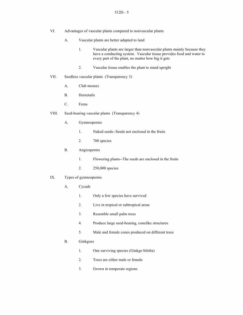
512D - 5
VI. Advantages of vascular plants compared to nonvascular plants
A. Vascular plants are better adapted to land
1. Vascular plants are larger than nonvascular plants mainly because they have a conducting system. Vascular tissue provides food and water to every part of the plant, no matter how big it gets
2. Vascular tissue enables the plant to stand upright
VII. Seedless vascular plants (Transparency 3)
A. Club mosses
B. Horsetails
C. Ferns
VIII. Seed-bearing vascular plants (Transparency 4)
A. Gymnosperms
1. Naked seeds--Seeds not enclosed in the fruits
2. 700 species
B. Angiosperms
1. Flowering plants--The seeds are enclosed in the fruits
2. 250,000 species
IX. Types of gymnosperms
A. Cycads
1. Only a few species have survived
2. Live in tropical or subtropical areas
3. Resemble small palm trees
4. Produce large seed-bearing, conelike structures
5. Male and female cones produced on different trees
B. Ginkgoes
1. One surviving species (Ginkgo biloba)
2. Trees are either male or female
3. Grown in temperate regions

512D - 6
4. Large, deciduous trees (shed leaves in fall)
5. Resistant to damage by pollution and insects
C. Conifers
1. Leaves
a. Needles
b. Small, compact with thick covering to prevent water loss
2. Most are evergreen and can carry on photosynthesis year around
3. Sex organs in cones--Most have both sexes on same tree
a. Male--Small, fleshy; form in groups
b. Female--Become large and woody
X. Cotyledon--Seed leaf; a leaflike structure of the embryo of the plant that often stores food for the new plant to use during germination
XI. Classes of angiosperms (Transparency 5)
A. Monocots
1. Characteristics
a. One cotyledon in each seed
b. Petals or flowers in threes or multiples of three
c. Veins in leaves are parallel
d. Stems have scattered vascular bundles
e. Fibrous roots
2. Examples
a. Wheat
b. Corn
c. Rice
d. Onions
e. Grasses

512D - 7
B. Dicots
1. Characteristics
a. Two cotyledons in each seed
b. Petals or flowers in fours or fives (or multiples of four or five)
c. Veins in leaf form network pattern
d. Stems have vascular bundles arranged in a ring
e. Usually have one large, main root (taproot)
2. Examples
a. Oak, elm and maple trees
b. Beans
c. Beets
d. Lettuce
e. Tomatoes
XII. Alternation of generations (Transparency 6)
A. Plants are made of haploid (n) cells in one generation and diploid (2n) in the next generation
B. Sporophyte
1. Diploid generation
2. Plants produce spores
a. Spore--Haploid reproductive cell produced by meiosis
b. Spore can germinate and develop into haploid plant (new gametophyte)
C. Gametophyte
1. Haploid generation
2. Plants produce gametes
a. Haploid gametes produced by mitosis
b. Male gamete fertilizes female gamete to produce diploid sporophyte

512D - 8
TM 1

512D - 9
TM 2

512D - 10
TM 3

512D - 11
TM 4

512D - 12
TM 5

512D - 13
TM 6

512D - 14
NONVASCULAR AND VASCULAR PLANTS
AG 512 - D
INSTRUCTOR NOTES FOR LABORATORY EXERCISES Lab #1 The first land plant probably appeared in the Ordovician period, though no fossil record of these plants exists from that time. Emphasize that although bryophytes are primitive evolutionarily, they are highly successful. Over 24 thousand species of mosses exist. This is more plant species than any other plant group except angiosperms. Good moss species to study include Polytrichum or Mnium. Live species of mosses and prepared slides of various structures are available from science supply companies. Part I : Step 1: Some mosses have water-conducting tissues. Also, some moss species have leaves covered with
cutin to prevent desiccation. Step 3: A structure called the "foot" attaches the sporophyte to the gametophyte in mosses. Step 4: Gametophyte tips will have several archegonia or antheridia. Step 6: The protonema resembles some filamentous algae. Tell students the proto means "original" in
Latin; nema means "thread" in Latin. Part III: Encourage students to examine gemma cups of Marchantia liverworts. These structures produce gemmae, for asexual reproduction. Accept student charts that demonstrate an understanding of alternation of generations of liverworts. Students should include drawings of the structures they observe. Lab #2 Part I: Have available several examples of herbaceous dicot stems for students to observe. It would be helpful to give a brief explanation on the fundamental differences between herbaceous and woody dicots. Large sections cut from fireplace-size logs can be used. The sections should be approximately 6 cm in thickness and can be cut in the wood shop. Oak logs make outstanding specimens although other species are suitable. You may wish to demonstrate and explain the difference between heartwood and sapwood, the vessels and how they determine whether the wood is diffuse or ring porous.

512D - 15
Part II: Make available several dried corn stalks for students to observe. Have on hand several other specimens of monocot stems to allow students to observe these macroscopically.

512D - 16
NONVASCULAR AND VASCULAR PLANTS
AG 512 - D
LABORATORY EXERCISE #1--STUDYING THE LIFE CYCLE OF A BRYOPHYTE--THE MOSS
Name _____________________________________ Score __________________________________
Slesnick, Irwin L., Biology Laboratory Manual, Scott, Foresman and Company, 1985. Reprinted by permission of Scott, Foresman and Company. Introduction: Land plants may have evolved from green algae that floated in the early Paleozoic seas. The first land plants were probably upright coastal plants, similar in appearance to seaweeds. Lacking a vascular system, these plants could grow only where water was continually available. Water diffused into the primitive land plant's tissues much as seawater diffuses through the cell walls of algae. As with algae, water also carried the male gamete to the female gamete, aiding reproduction. Similar as the first land plants were to water-dwelling algae, these nonvascular plants also differed from their algal ancestors. Special structures evolved that anchored the plants to the ground. In addition, the sporophyte and gametophyte generations show variation in structure and function in all species of land plants. (In many algae species the sporophyte and gametophyte look identical.) Furthermore, unlike algae, land plants have one generation that is dependent on the other generation. In all bryophytes, which include the moss you will examine in this laboratory, the sporophyte generation is dependent on the gametophyte generation. In this laboratory you will study the life cycle and adaptations of moss. Most scientists think that bryophytes, such as mosses, share common features with the first plants that colonized land. Materials needed Moss plant, living or preserved specimen 4 coverslips Hand lens or stereoscopic microscope Compound microscope Forceps Prepared slides of cross sections of an 3 microscope slides antheridium, archegonium and Scalpel or razor blade protonema of a moss Part I: Procedure 1. Obtain a sample of a moss plant. Observe the green carpetlike part of the plant. The thick, green
structures make up the gametophyte generation. Use a hand lens or a stereoscopic microscope to examine the gametophyte. Find the rhizoids that extend into the soil to anchor the moss. Unlike the roots of trees, shrubs and flowering plants, rhizoids lack vascular tissue. Observe the stemlike shaft that supports the plant. Again, this stemlike structure differs from the true stems in that it lacks vascular tissue. The leaflike structures of mosses also lack vascular tissue. Use your forceps to remove one of these leaflike structures. Make a wet mount slide, and observe the structure under low power of a compound microscope. Find chloroplasts in the cells. How many cell layers make up the leaflike structure?
(a) ____________________________________________________________________________
2. Find the brownish stalk growing from the tip of the gametophyte. The capsule at the tip of the stalk produces spores. Thus, the capsule and the brownish stalk that supports the capsule make up

512D - 17
the sporophyte generation. Notice the protective cap that covers the capsule. The sporophyte is attached to the top of the gametophyte. Remove the sporophyte from the gametophyte. Save the sporophyte for further study in step 4. Label the parts of the gametophyte and sporophyte shown in a.
3. The gametophyte's reproductive structures are located at the very tip of the gametophyte. Male
and female reproductive structures are borne on separate moss plants. Examine prepared slides of longitudinal cross sections of male and female reproductive structures, or make your own slides of these structures from a living moss gametophyte. As shown in b, cut the top centimeter off the green shafts of two gametophyte moss plants. Place the moss cuttings on a slide. Cut off and discard the bottom leafy half of each cutting, as shown in c. Add two drops of water to each moss tip. Place a second slide on top of the tips. Using d as your guide, gently but firmly press down on the top slide so the moss tips are slightly crushed. Remove the top slide. Place a coverslip over each moss tip.

512D - 18
4. Examine the tips of the moss gametophytes under low and high power. Male reproductive structures - antheridia - are oblong ovals that protect developing sperm cells, as shown in e. Female reproductive structures - archegonia - consist of two columns of cells that surround and protect the egg cell, as shown in f. Identify the reproductive structures on your slide. Make sure to observe both antheridia and archegonia. You may need to examine slides of your classmates to observe both structures.
5. Monoploid gametophytes produce gametes. Sperm cells are produced in the antheridia, and egg
cells are produced in the archegonia. Sperm cells swim in a film of water from an antheridium of the male plant to archegonium of a female plant. The sperm and egg cell unite in the archegonium, forming a diploid zygote that becomes the stalklike sporophyte. The sporophyte derives nutrients and protection from the gametophyte. When mature, the sporophyte produces monoploid spores. Make a wet mount of the spore capsule you removed in step 2. Crush the spore case in the same manner that you crushed the moss tips in step 3. Crushing the capsule will release the spores. Examine the spores under low and high power. Draw and label the spores in the circle below.
6. The moss spores are dispersed by wind. When a spore lands in a suitable place, it will absorb water and germinate, producing a green filament called the protonema. The protonema is the immature gametophyte moss plant. Examine a slide of a moss protonema under low and high power. Look for such structures as rhizoids, chloroplasts, buds and young shoots. Buds sprout on the protonema. They become shoots that develop into the mature gametophyte moss plant you observed in steps 1 and 2. Label the protonema in g.

512D - 19
7. Review the life cycle of mosses by labeling h. Be sure to include whether the structures you refer to are monoploid or diploid. Wash your slides and coverslips, and clean up your work area.
Part II: Analysis
1. List two reasons why mosses need to live in a wet environment.
______________________________________________________________________________
______________________________________________________________________________
2. How does the life cycle of the moss you examined clearly illustrate alternation of generations?
______________________________________________________________________________
______________________________________________________________________________
3. How is the sporophyte generation of the moss plant dependent on the gametophyte generation?
______________________________________________________________________________
______________________________________________________________________________

512D - 20
4. Name three structural adaptions of bryophytes to life on land.
______________________________________________________________________________
______________________________________________________________________________
______________________________________________________________________________
5. The picture below shows a filamentous green alga with horizontal and upright portions. Scientists
think that the first land plants may have evolved from an ancient alga, similar in appearance to the alga in the picture. Which moss form most resembles this alga? Describe the similarities between the two organisms.
______________________________________________________________________________
______________________________________________________________________________
6. Name two features of the alga in the picture that might have been adaptive to life on land.
______________________________________________________________________________
______________________________________________________________________________
Part III: Going Further
Examine a common liverwort such as Marchantia. Use reference books to identify the gametophyte, including its sexual and asexual reproductive structures. Examine slides of the sexual reproductive structures and the sporophyte. Make a chart showing the liverwort's life cycle.

512D - 21
NONVASCULAR AND VASCULAR PLANTS
AG 512 - D
LABORATORY EXERCISE #2--HOW MONOCOT STEMS DIFFER FROM DICOT STEMS
Name _____________________________________ Score __________________________________
Selection from Modern Biology, Biology Investigations, Teacher's Edition, by James H. Otto, Albert Towle, W. David Otto, and Myra E. Madnick. Copyright 1977 by Holt, Rinehart and Winston, Inc. Reprinted by permission of the publisher. Materials needed Cross section of a woody dicot stem, 10-15 years old or older Prepared slide of: herbaceous monocot stem (Zea mays) Textbook or charts Microscope Part I: Microscopic Examination of a Woody Dicot Stem Dicot stems can be both herbaceous and woody. Herbaceous dicot stems usually live for only a single growing season. When compared to a year old woody stem, close similarities may be observed in the tissues which compose the stem. In this part, you will examine only a woody dicot stem. Examine the cross section of a woody stem. You commonly hear the terms bark and wood.
a. Where is the bark located? _________________________________________________________
b. Where is the wood in relation to the bark? ____________________________________________
c. What tissue occupies the center of the stem? __________________________________________
d. Summarize the tissues that can be observed in a cross section of a woody stem. _______________
_____________________________________________________________________________________
Bark and wood are both composed of specialized tissues which can only be observed with a microscope. Without the microscope, it can be seen that bark is divided into the outer bark and inner bark. The outer bark is composed of cork tissue.
e. What are some functions of the cork? ________________________________________________
f. What tissue composes the inner bark? ________________________________________________
g. What is the function of the phloem? _________________________________________________
h. Although you are unable to see it, what layer of cells separates the bark from the wood? ________
______________________________________________________________________________

512D - 22
i. What is the function of the vascular cambium? _________________________________________
______________________________________________________________________________
j. What tissue composes wood? ______________________________________________________
k. Estimate the amount of wood in proportion to the amount of bark.
______________________________________________________________________________
______________________________________________________________________________
l. What evidence is there that the stem has lived for more than a single growing season? _________
______________________________________________________________________________
m. What are these rings commonly called? ______________________________________________
n. Are all of the rings of equal thickness? _______________________________________________
o. Account for your answer. _________________________________________________________
______________________________________________________________________________
______________________________________________________________________________
p. What is the function of the xylem? __________________________________________________
______________________________________________________________________________
In the chart below, summarize your observations of the woody dicot stem. Give the function of the tissue where it applies. Location Function
1. Outer covering
2. Arrangement of vascular tissue
3. Location of phloem
4. Location of xylem
5. Location of vascular cambium
6. Location of pith

512D - 23
On the figure of the cross section of a woody stem, label: cork tissue, phloem, bark, vascular cambium, xylem tissue, wood, pith, annual ring.
Part II: Examination of a Monocot Stem
Examine the prepared slide of a cross section of the monocot stem with your microscope under low power. The outer layer of cells is the epidermis. a. Describe the appearance of these cells. _______________________________________________
______________________________________________________________________________
Note that just under the epidermis are additional thick-walled cells. These cells, along with those of the epidermis, compose the rind of the mature stem. b. What tissue occupies most of the stem? ______________________________________________
c. Describe the cells which compose this tissue. __________________________________________
______________________________________________________________________________
Look for the fibrovascular bundles. Count the bundles in an estimated quarter of the stem.
d. How many do you find? __________________________________________________________
e. Where in the stem are they most numerous? ___________________________________________
______________________________________________________________________________
f. Of what significance is this observation? _____________________________________________
______________________________________________________________________________
Select a fibrovascular bundle toward the center of the stem. Examine it under high power. Note that the bundle has the appearance of a face with distinct facial regions. Large xylem vessels are found in the position of the "eyes" and "nose" of the face. The phloem occupies the position of the forehead. Locate and distinguish the sieve tubes and companion cells which compose the phloem.

512D - 24
g. Is a vascular cambium present? _____________________________________________________
h. What effect does its presence or absence have on a monocot stem? _________________________
______________________________________________________________________________
______________________________________________________________________________
Locate the thick-walled sclerenchyma fibers surrounding the bundle.
i. What function might they serve? ____________________________________________________
The position of the "mouth" is an irregular intercellular space. j. What can you observe to confirm that it is a space and not a large cell? _____________________
______________________________________________________________________________
k. Suggest how this space might be formed. _____________________________________________
______________________________________________________________________________
On the general view of the corn stem, label: epidermis, rind, pith, fibrovascular bundle. On the figure of the fibrovascular bundle and surrounding tissue, label: xylem vessels, phloem, intercellular space, pith, sclerenchyma fibers.
Part III: Summary In the chart on the following page, summarize the differences and similarities between monocot and dicot stems you have observed and studied.

512D - 25
Characteristic Dicot Monocot
1. Herbaceous or woody
2. Type of vascular tissue present
3. Arrangement of vascular tissue
4. Presence of vascular cambium
5. Location of pith
6. Means of support
7. Probable life duration of stem
Part IV: Investigations On Your Own 1. Obtain a prepared slide of a 3-year dicot stem (Tilia) and examine it under the microscope. You
should observe that the tissues in a woody stem are much more complex than what you observe with the naked eye. Consult a biology or botany textbook for descriptions of the cells which compose the outer and inner bark and the xylem. Make a detailed sketch of a pie-shaped section and label the cells and tissues which you observe.
2. Examine a prepared slide of an herbaceous dicot stem (Helianthus) and locate the tissues studied
in the woody dicot stem. Note the similarities and differences. Make a detailed sketch of a pie-shaped section and label the cells and tissues you observe.
3. Make a collection of cross sections of woody stems 3-4 cm in thickness and 5-8 cm in diameter.
Identify each with its scientific and family name. The sanding and sealing of the cross sections will help to preserve them for future use.

512D - 26
NONVASCULAR AND VASCULAR PLANTS
AG 512 - D
ANSWERS TO LABORATORY EXERCISES Lab #1
Part I:
a. One or two layers
5.

512D - 27
Part II: 1. These bryophytes have no vascular system, so water must diffuse through plant surfaces. Male
gametes are carried to egg cells via a film of water. 2. Mosses have a visible sporophyte (diploid) generation that grows from the (monoploid) female
gametophyte. 3. The sporophyte contains no chloroplasts. It is dependent on the gametophyte for nutrients. 4. Rhizoids anchor the plant to the soil; sporophytes are adapted for wind dispersal of spores;
photosynthesizing structures have a broadened surface for exposure to light; archegonia and antheridia protect developing gametes, preventing desiccation.
5. The protonema. Both are filamentous, with upright and horizontal portions. Both have chloroplasts.
6. Horizontal filaments could help anchor the plant. Upright portions help expose the chloroplasts to light and disperse reproductive cells.
Lab #2: Part I: a. On the outside of the stem. b. To the inside of the bark. c. Pith d. Bark, wood and pith. e. Cork resists the passage of water and gases from stem tissues, offers protection against disease and
provides insulation. f. Phloem g. To conduct food materials up and down the stem. h. The vascular cambium. i. The cells of the cambium divide to produce phloem cells and xylem cells. j. Xylem k. Answer will vary depending on the age of the stem. l. The wood is produced in rings. Each ring represents a year's growth. m. Annual rings. n. No o. Environmental factors affect the amount of wood produced each season by the cambium. p. Conduction and support.

512D - 28
Location Function
1. Outer covering bark prevents gases and water loss, protection
2. Arrangement of vascular tissue
concentric rings -------------------------
3. Location of phloem inner bark conduction of food materials up and down stem, some support
4. Location of xylem interior, woody portion of stem
conduction of water and minerals support
5. Location of vascular cambium
between xylem and phloem
divides to form new phloem and xylem cells
6. Location of pith center of stem storage
Part II: a. The cells are thick walled. b. Pith c. Thin-walled, large cells. d. Answers will vary. e. Toward the outside of the stem. f. The bundles toward the outside give strength to the stem. g. No h. Monocots can grow in diameter only until their cells have reached a maximum size. i. Support j. The cells surrounding the space have their own cell walls. k. As the bundle matures, the cells that were once in contact are pulled apart from each other.

512D - 29
Summary:
Characteristic Dicot Monocot
1. Herbaceous or woody herbaceous or woody herbaceous
2. Type of vascular tissue present
xylem and phloem xylem and phloem
3. Arrangement of vascular tissue
concentric rings scattered bundles
4. Presence of vascular cambium
present not present
5. Location of pith center of stem most areas of stem
6. Means of support primarily woody tissue rind, and distribution of vascular bundles
7. Probable life duration of stem
herbaceous - a single growing season; woody - many years
with exceptions, only one growing season

512D - 30
NONVASCULAR AND VASCULAR PLANTS
AG 512 - D
UNIT TEST Name _____________________________________ Score __________________________________
1. Describe nonvascular plants.
______________________________________________________________________________
______________________________________________________________________________
______________________________________________________________________________
2. Identify and describe the major phyla of nonvascular plants.
______________________________________________________________________________
______________________________________________________________________________
______________________________________________________________________________
______________________________________________________________________________
______________________________________________________________________________
3. Discuss the importance of nonvascular plants.
a. _____________________________________________________________________________
b. ____________________________________________________________________________
c. _____________________________________________________________________________
d. ____________________________________________________________________________
e. _____________________________________________________________________________
4. Describe vascular plants.
______________________________________________________________________________
______________________________________________________________________________
5. List and describe the functions of the two types of vascular tissue.
a. _____________________________________________________________________________
______________________________________________________________________________
______________________________________________________________________________

512D - 31
b. ____________________________________________________________________________
______________________________________________________________________________
6. Discuss the advantages of vascular plants compared to nonvascular plants.
______________________________________________________________________________
______________________________________________________________________________
______________________________________________________________________________
______________________________________________________________________________
______________________________________________________________________________
______________________________________________________________________________
7. List the three seedless vascular plants.
a. _____________________________________________________________________________
b. ____________________________________________________________________________
c. _____________________________________________________________________________
8. List and describe the two types of seed-bearing vascular plants.
a. _____________________________________________________________________________
______________________________________________________________________________
b. ____________________________________________________________________________
______________________________________________________________________________
9. List and describe the three types of gymnosperms.
a. _____________________________________________________________________________
______________________________________________________________________________
______________________________________________________________________________
______________________________________________________________________________
b. ____________________________________________________________________________
______________________________________________________________________________
______________________________________________________________________________
______________________________________________________________________________

512D - 32
c. _____________________________________________________________________________
______________________________________________________________________________
______________________________________________________________________________
______________________________________________________________________________
10. Define cotyledon.
______________________________________________________________________________
______________________________________________________________________________
______________________________________________________________________________
______________________________________________________________________________
11. List, describe and give four examples of the two classes of angiosperms.
a. _____________________________________________________________________________
______________________________________________________________________________
______________________________________________________________________________
Examples ______________________________________________________________________
______________________________________________________________________________
b. ____________________________________________________________________________
______________________________________________________________________________
______________________________________________________________________________
Examples ______________________________________________________________________
______________________________________________________________________________
12. Outline alternation of generations.
a. _____________________________________________________________________________
______________________________________________________________________________
b. ____________________________________________________________________________
______________________________________________________________________________
c. _____________________________________________________________________________
______________________________________________________________________________

512D - 33
NONVASCULAR AND VASCULAR PLANTS
AG 512 - D
ANSWERS TO TEST 1. Primitive land plants that lack true roots, stems and leaves. They are good examples of plants that
did not make a complete transition from water to land 2. Bryophytes--Mosses, liverworts, hornworts; Characteristics--Usually grow in moist, shady areas
such as marshes, bogs or forest floors; Obtain water and dissolved minerals by absorbing the water around them
3. Produce food and oxygen for other organisms to use; Break down rocks to form soils where other
plants can grow; Remains of mosses enrich the soil; Help prevent soil erosion and flooding by absorbing large amounts of water; Several mosses are sensitive to sulfur dioxide and can be used as bio-indicators of pollution
4. Plants with vascular tissue, which is a system of tubes through which water, minerals and sugar
are carried. Includes seedless vascular plants and seed-bearing vascular plants 5. Xylem--Conducts water and minerals from the roots to the stems and leaves; Phloem--Conducts
sugar from the leaves (where it is produced during photosynthesis) to the rest of the plant 6. Vascular plants are better adapted to land; Vascular plants are larger than nonvascular plants
mainly because they have a conducting system; Vascular tissue provides food and water to every part of the plant, no matter how big it gets; Vascular tissue enables the plant to stand upright
7. Club mosses; Horsetails; Ferns
8. Gymnosperms: Naked seeds--Seeds not enclosed in the fruits; 700 species; Angiosperms: Flowering plants--The seeds are enclosed in the fruits; 250,000 species
9. Cycads: Only a few species have survived; Live in tropical or subtropical areas; Resemble small
palm trees; Produce large seed-bearing, conelike structures; Male and female cones produced on different trees Ginkgoes: One surviving species (Ginkgo biloba); Trees are either male or female; Grown in temperate regions; Large, deciduous trees (shed leaves in fall); Resistant to damage by pollution and insects Conifers: Leaves are needles; Small, compact with thick covering to prevent water loss; Most are evergreen and can carry on photosynthesis year around; Sex organs in cones--Most have both sexes on same tree
10. Seed leaf; a leaflike structure of the embryo of the plant that often stores food for the new plant to
use during germination 11. Monocots: One cotyledon in each seed; Petals or flowers in threes or multiples of three; Veins in
leaves are parallel; Stems have scattered vascular bundles; Fibrous roots; Examples could include four of the following: wheat, corn, rice, onions, grasses

512D - 34
Dicots: Two cotyledons in each seed; Petals or flowers in fours or fives (or multiples of four or five); Veins in leaf form network pattern; Stems have vascular bundles arranged in a ring; Usually have one large, main root (taproot); Examples could include four of the following: oak, elm and maple trees, beans, beets, lettuce, tomatoes
12. Plants are made of haploid (n) cells in one generation and diploid (2n) in the next generation;
Sporophyte: Diploid generation; Plants produce spores; Spore--Haploid reproductive cell produced by meiosis; Spore can germinate and develop into haploid plant (new gametophyte); Gametophyte: Haploid generation; Plants produce gametes; Haploid gametes produced by mitosis; Male gamete fertilizes female gamete to produce diploid sporophyte

512E - 1
VEGETATIVE PLANT PARTS
AG 512 - E
UNIT OBJECTIVE After completion this unit, students should be able to match terms and definitions and identify the parts of a plant. Students should also be able to list functions of plant parts and name the root systems. This knowledge will be demonstrated by completion of the unit test with a minimum of 85 percent accuracy.
SPECIFIC OBJECTIVES AND COMPETENCIES
After completion of this unit, the student should be able to:
1. Match terms associated with plant growth and development to the correct definitions.
2. Label a drawing showing the four primary parts of a plant.
3. List functions of plant parts.
4. Label a drawing showing the parts of a simple leaf.
5. Label a drawing showing the parts of a compound leaf.
6. Identify the types of compound leaves.
7. Identify three types of leaf arrangements.
8. Identify four types of leaf veination.
9. Identify three types of leaf margins.
10. Identify four types of leaf attachments to the stem.
11. Label a drawing showing the parts of a plant stem.
12. Match stem modifications to the correct descriptions.
13. Name two types of root systems.
14. Examine the structure of leaves.
15. Study the relationship of the leaf structure to its function.
16. Examine roots and stems.
17. Observe root growth.

512E - 2
VEGETATIVE PLANT PARTS
AG 512 - E
SUGGESTED ACTIVITIES
I. Suggested activities for instructor
A. Order materials to supplement unit.
1. Literature
a. Agronomy Curriculum Materials Packet, 232 pages; available from IAVIM, 208 Davidson Hall, Iowa State University, Ames, Iowa 50011; approximate cost $10.00, order no. 214.
b. Crop Production, 15 transparency masters; available from
IAVIM, 208 Davidson Hall, Iowa State University, Ames, Iowa 50011; approximate cost $2.25, order no. 517.
2. Filmstrips, slideshows, etc.
a. Agronomy, computer program; available from IAVIM, 208 Davidson Hall, Iowa State University, Ames, Iowa 50011; approximate cost $15.00, order no. 902.
B. Make transparencies and necessary copies of materials.
C. Provide students with objective sheet.
D. Provide students with information sheets and laboratory exercises.
E. Discuss unit and specific objectives.
F. Discuss information sheets.
G. Demonstrate and discuss procedures outlined in laboratory exercises.
H. Review and give test.
I. Reteach and retest if necessary.
II. Instructional materials
A. Objective sheet
B. Suggested activities
C. Information sheets

512E - 3
D. Transparency masters
1. TM 1--Primary Parts of a Plant
2. TM 2--Functions of Leaves, Stems, Roots and Flowers
3. TM 3--Parts of a Simple Leaf
4. TM 4--Parts of a Compound Leaf
5. TM 5--Types of Compound Leaves
6. TM 6--Types of Leaf Arrangement
7. TM 7--Types of Leaf Veination
8. TM 8--Types of Leaf Margins
9. TM 9--Types of Leaf Attachments
10. TM 10--Parts of the Stem
11. TM 11--Above Ground Stem Modifications
12. TM 12--Below Ground Stem Modifications
13. TM 13--Types of Root Systems
E. Instructor notes for laboratory exercises
F. Laboratory exercises
1. LE 1--Examining the Structure of Leaves
2. LE 2--Relationship of Leaf Structure to Function
3. LE 3--Examining Roots and Stems
4. LE 4--Root Growth
G. Answers to laboratory exercises
H. Test
I. Answers to test
III. Unit references
A. Agricultural Education Curriculum, College of Agriculture, University of Illinois, Urbana, Illinois, 1989.
B. Delorit, R. J., et al., Crop Production, 5th edition, Prentice-Hall, Inc.,
Englewood Cliffs, New Jersey, 1984.

512E - 4
C. Hartmann, Hudson T., et al., Plant Science: Growth, Development and Utilization of Cultivated Plants, 2nd edition, Prentice-Hall, Inc., Englewood Cliffs, New Jersey, 1988.
D. Idaho Crop and Soil Curriculum Guide, Idaho State Board for Vocational
Education, 1985. E. Janick, J. et al., Plant Science, 2nd edition, W.H. Freeman and Co., San
Francisco, California, 1974. F. Model Agricultural Core Curriculum, California State Department of Education,
University of California, Davis, California, August, 1989. G. Otto, James H., Towle, Albert, Modern Biology, Holt, Rinehart and Winston,
New York, 1985. H. Slesnick, Irwin L., et al., Biology, Scott, Foresman and Company, Glenview,
Illinois, 1985.

512E - 5
VEGETATIVE PLANT PARTS
AG 512 - E
INFORMATION SHEET
I. Terms and definitions
A. Node--The part of a stem where a leaf is attached
B. Internode--The part of stem between two nodes
C. Bud--An embryonic shoot of a plant
D. Leaf scar--A scar left on the stem when a leaf falls
E. Vascular bundle scar--A spot within a leaf scar left by the vascular bundles when a leaf falls
F. Monocot--Plant having one seed leaf (cotyledon) as in cereals and corn
G. Dicot--Plant having two seed leaves (cotyledons) as in beans and peas
H. Vascular bundle--A strand of tissue containing xylem and phloem enclosed by a sheath of cells
I. Xylem--Vascular tissue that transports water and minerals from the root system
to the leaves J. Phloem--Vascular tissue that conducts food from the leaves to regions of growth
or storage
K. Pistil--Seed bearing organ of a flower, composed of the ovary, style and stigma
L. Stamen--Part of the flower producing the pollen; composed of the filament and anther
M. Fertilization--Union of the male (pollen) nucleus with the female (egg) cell
N. Pollination--Transfer of pollen from the anther to the stigma
O. Embryo--The young plantlet within the seed; the germ
P. Radicle--The embryonic root
Q. Hypocotyl--The part of an embryo between the cotyledons and the radicle
R. Epicotyl--The part of the embryo above the cotyledons and below the next leaves

512E - 6
II. Primary parts of a plant (Transparency 1)
A. Roots
B. Stem
C. Leaves
D. Flowers
III. Functions of plant parts (Transparency 2)
A. Roots
1. Absorb water and nutrients
(Note: Most of the absorption takes place through the root hairs. The rate at which water is absorbed depends on: (1) the rate at which water is lost from leaves (transpiration), (2) the amount of water in the soil, and (3) the amount of root surface in contact with soil particles.)
2. Anchor and support plant
(Note: The root must anchor the plant to the extent that wind, etc. cannot knock it down.)
3. Store food
(Note: Some plants store foods they have manufactured in the roots. Examples are radishes, carrots, sweet potatoes and sugarbeets.)
B. Stem
1. Supports leaves, flowers, fruit and seeds
2. Conducts water, nutrients and food
(Note: The stem conducts water and minerals in solution from the root system through the xylem tissue to the leaves. It also conducts food made in the leaves through the phloem tissue to the parts of the plant where it is growing or food is being stored.)
3. Stores food
(Note: Examples of plants that store food in the stem include potatoes and asparagus.)

512E - 7
C. Leaves
1. Manufacture food for the plant
(Note: Photosynthesis is the process by which leaves make food from carbon dioxide and water in the presence of sunlight.)
2. Necessary for transpiration
3. Store food
(Note: Examples of plants that store food in the leaves include lettuce, cabbage, celery, rhubarb and onions.)
D. Flowers
1. Serve as site of reproduction
2. Store food
(Note: Examples of plants that store food in flowers include grains, fruits, nuts, berries, broccoli and cauliflower.)
IV. Parts of a simple leaf (Transparency 3)
A. Blade
B. Veins
C. Petiole
D. Stipules
V. Parts of a compound leaf (Transparency 4)
A. Leaflet
B. Veins
C. Petiolule
D. Rachis
E. Petiole
F. Stipules
VI. Types of compound leaves (Transparency 5)
A. Pinnate
B. Bipinnate
C. Palmate

512E - 8
D. Trifoliate
VII. Types of leaf arrangement (Transparency 6)
A. Alternate
B. Opposite
C. Whorled
VIII. Types of leaf veination (Transparency 7)
A. Parallel
B. Pinnate
C. Palmate
D. Netted
IX. Types of leaf margins (Transparency 8)
A. Entire--No teeth or lobes on margin
B. Serrate--Toothed with teeth directed forward
C. Incised--Toothed with sinuses deeper than teeth
D. Lobed--Margin cut in about one-half way to midrib; sinuses and tips of segments rounded
X. Types of leaf attachment to the stem (Transparency 9)
A. Petiolate--Leaf attached by a petiole
B. Sessile--Leaf without a petiole; blade attached to stem
C. Clasping--Sessile leaf with the lower edges of the blade partly surrounding the stem
D. Decurrent--Point of attachment extends downward on the stem
XI. Parts of the stem (Transparency 10)
A. Node
B. Internode
C. Terminal bud
D. Lateral bud
E. Leaf scar

512E - 9
F. Vascular bundle scar
XII. Types of stem modifications (Transparencies 11, 12)
A. Above ground (Transparency 11)
1. Crown--Appears just above or just below the ground; corn and small grains are characteristic of this
2. Stolon--Runners that grow along top of soil surface; may take root at
nodes on runner; white clover is an example 3. Spur--Modified stem growth that appears laterally on branches of fruit
trees and bears fruit
B. Below ground (Transparency 12)
1. Rhizomes--Grow horizontally below soil surface; contain nodes from which new plants originate; examples are canadian thistle, field bindweed and quackgrass
2. Tubers--Enlarged fleshy parts of a rhizome; an example is the potato
3. Corms--Fleshy, short underground stems containing very few buds; an example is timothy
4. Bulb--Short, disc-shaped stem surrounded by leaf-like scales; an
example is gladiolus
XIII. Types of root systems (Transparency 13)
A. Tap root system
(Note: In this system, one root is larger than the rest. Examples include alfalfa, sugarbeets, beans, carrots and radishes.)
B. Fibrous root system
(Note: In this system, all roots are approximately the same size. Examples include all the grasses and cereal grains.)

512E - 10
TM 1

512E - 11
TM 2

512E - 12
TM 3
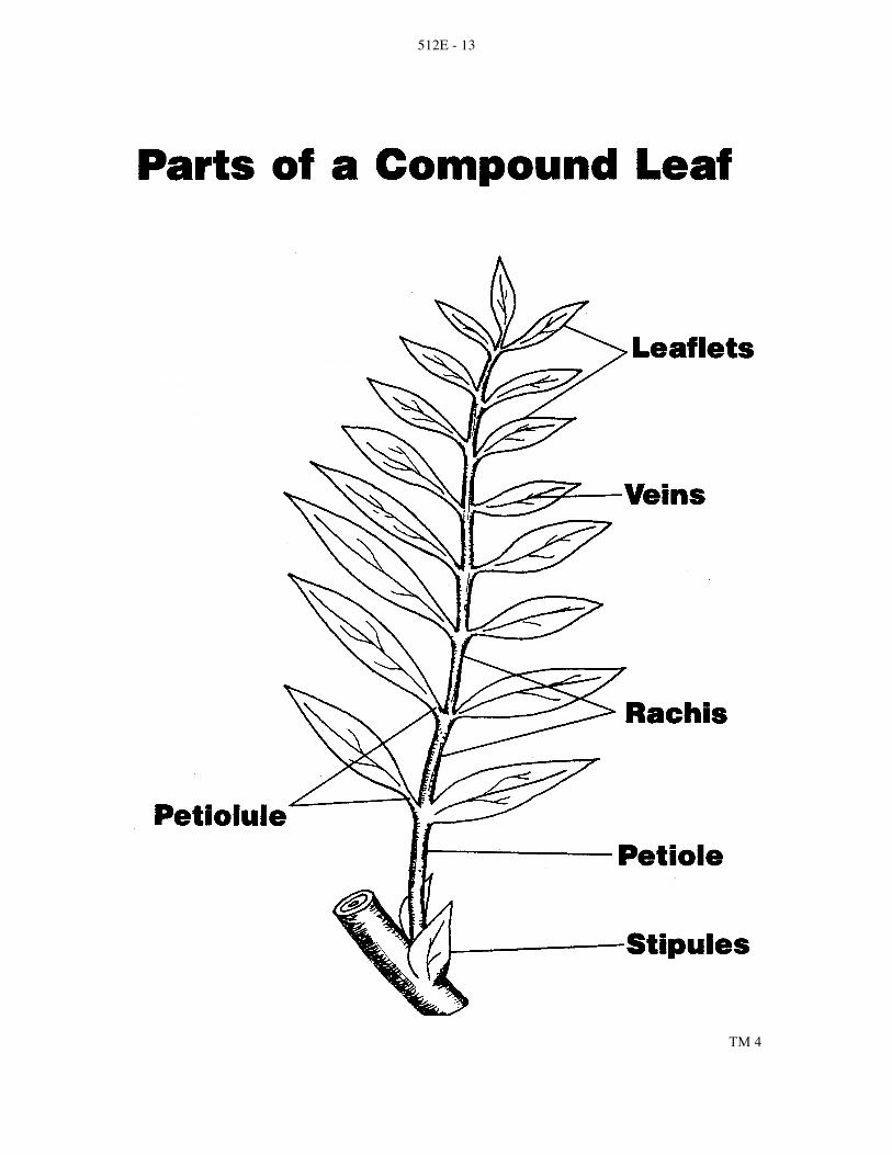
512E - 13
TM 4

512E - 14
TM 5

512E - 15
TM 6

512E - 16
TM 7

512E - 17
TM 8

512E - 18
TM 9

512E - 19
TM 10

512E - 20
TM 11

512E - 21
TM 12

512E - 22
TM 13

512E - 23
VEGETATIVE PLANT PARTS
AG 512 - E
INSTRUCTOR NOTES FOR LABORATORY EXERCISES Lab #1
Monocot leaves include corn, scallions, leeks, orchids and lilies.
Dicot leaves include elm, maple, oak, spinach, Geranium and other houseplants.
Modified leaves include Jade plant leaf, cactus spine and pitcher plant.
Scalelike leaves include juniper, arborvitae and cypress.
Needlelike leaves include pine, fir and spruce.
Prepared slides of leaves include privet (Ligustrum) leaf or lilac (dicots) or lily leaf, milo leaf, bluegrass leaf or beach grass leaf (monocots). Part I:
Step 1: Many monocots have no petiole; some common monocots, such as lilies, have a petiole
Step 3: Give each leaf an identification number. Ask the students to copy the number of each leaf they choose in the space next to step 1. In this way, you can check the students' answers to the questions in the laboratory text
Part II:
Step 1: Remind students to focus carefully to avoid damage to prepared slide or microscope Table II: Color and recognizability of chloroplasts depend on how the leaf tissues have been stained
Guide students to notice that the stomata are more numerous in the lower epidermis. Tell students if they orient their slides so that the upper epidermis is at the top of their microscope fields, they can see the leaf layers in the order they are listed in Table II
Diagram c: Students can switch to high power to examine the cuticle and epidermis more closely Allow students to examine prepared slides of other types of leaves. On a separate piece of paper suggest that the students make a labeled drawing of each type of leaf they observe. Note the tissues and structures that all leaves have in common. Identify the tissues and structures that help adapt a plant to its particular environment. Slides might include some of those listed in the materials section or a variety of slides listed in biological supply catalogs. Part III:
Collect leaves of trees and plants native to your area. Then, give students "mystery" leaves. Tell students to examine each leaf, using Table I to identify whether it is a monocot, dicot or conifer. Then, use a field guide to identify the species of plant the leaf came from.

512E - 24
Extension: Encourage students to make their own cross sections of different kinds of leaves. To make a leaf more rigid for easier cutting, students can place the leaf between two halves of a carrot and tie the carrot halves together with a string. Soak the carrot in water for 20 minutes. Warn the students to use caution when cutting their leaf cross sections. Cut very thin sections of each soaked leaf, and place the leaf sections in water to prevent them from curling. Use forceps to select the thinnest leaf sections. Make wet mount slides of these sections, and examine them under low power. Ask students to sketch each and label each cross section. Lab #3
You will need to provide dormant twigs 3 years old or older. Collect these twigs from fallen wood if possible. To sprout seeds, place them on moist filter paper in covered dishes for 30-40 hours. Lawn-grass seedlings are also excellent for viewing root hairs; they require 10-14 days for germination. Part I:
Remind students of the differences between taproots and fibrous roots. Remind them that the root they are looking at is a dicot root, thus it may be a taproot. Extension: Compare fibrous roots and taproots of different plants.
Step 3: If photosynthesis has not been allowed to occur, such cortex cells will not have starch stored in them.
Some slides may show roots developing from the pericycle of the main root. Suggest that students compare their slides to those of other students to note how root branches develop.
Part II:
Point out to students that the twig is an example of a dicot stem.
Step 1: Students may need to use hand lenses to see bundle scars and lenticels.
Step 2: Examine the meristem of the shoot apex under a microscope. Guide students to notice the dividing cells.
If prepared slides of longitudinal and transverse cross sections of woody stems are available, allow students to examine them and compare them to their twigs. Review the function of parenchyma cells with the students.
Part III:
Number 4: The region of the root that contains the root hair does not move in order to grow. If this region did move, as the root tip does, the delicate root hairs would be stripped from its surface.
Part IV:
Students should be able to recognize all structures identified in Part I.

512E - 25
Lab #4
Begin this lab, as indicated, by planting the seeds a week in advance. The roots should emerge within 4-5 days and be ready for examination soon thereafter.
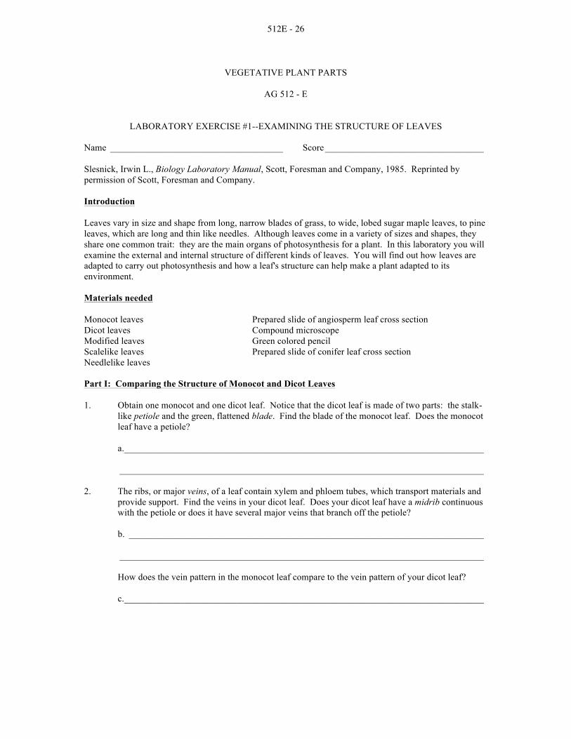
512E - 26
VEGETATIVE PLANT PARTS
AG 512 - E
LABORATORY EXERCISE #1--EXAMINING THE STRUCTURE OF LEAVES Name _____________________________________ Score __________________________________
Slesnick, Irwin L., Biology Laboratory Manual, Scott, Foresman and Company, 1985. Reprinted by permission of Scott, Foresman and Company. Introduction
Leaves vary in size and shape from long, narrow blades of grass, to wide, lobed sugar maple leaves, to pine leaves, which are long and thin like needles. Although leaves come in a variety of sizes and shapes, they share one common trait: they are the main organs of photosynthesis for a plant. In this laboratory you will examine the external and internal structure of different kinds of leaves. You will find out how leaves are adapted to carry out photosynthesis and how a leaf's structure can help make a plant adapted to its environment. Materials needed
Monocot leaves Prepared slide of angiosperm leaf cross section Dicot leaves Compound microscope Modified leaves Green colored pencil Scalelike leaves Prepared slide of conifer leaf cross section Needlelike leaves Part I: Comparing the Structure of Monocot and Dicot Leaves
1. Obtain one monocot and one dicot leaf. Notice that the dicot leaf is made of two parts: the stalk-like petiole and the green, flattened blade. Find the blade of the monocot leaf. Does the monocot leaf have a petiole?
a. _____________________________________________________________________________
______________________________________________________________________________
2. The ribs, or major veins, of a leaf contain xylem and phloem tubes, which transport materials and provide support. Find the veins in your dicot leaf. Does your dicot leaf have a midrib continuous with the petiole or does it have several major veins that branch off the petiole?
b. ____________________________________________________________________________
______________________________________________________________________________
How does the vein pattern in the monocot leaf compare to the vein pattern of your dicot leaf?
c. _____________________________________________________________________________

512E - 27
3. To review the structures that make up monocot and dicot leaves, label the structures in a and b below.
4. Obtain samples of several different kinds of leaves. Compare your leaves to the leaf pictures in
Table I. Examine different kinds of simple monocot and dicot leaves. Identify pinnate and palmate compound leaves. Notice that compound leaves are composed of leaflets that resemble simple leaves. Observe the structure of several kinds of modified leaves. Examine examples of scalelike and needlelike leaves.

512E - 28
Part II: Examining the Internal Structure of Leaves
1. Obtain a prepared slide of a cross section of an angiosperm leaf. Examine the slide under low power of a compound microscope. Identify the structures listed in Table II.
TABLE II. Leaf structures
2. Review the internal structure of a leaf by labeling the tissues in c. Draw in the chloroplasts with a
green pencil.
c. Leaf cross section
Structure Description Structure Description
Upper Cuticle
Thin, clear waxy coating that helps prevent water loss from the leaf surface
Veins Fibrovascular bundles scattered throughout the mesophyll; contain xylem and phloem tubes, which transport substances and support the leaf
Upper epidermis
A single layer of brick shaped cells with few openings to the surface
Lower Epidermis
Located below the spongy layer, a single layer of cells similar to cells that make up the upper
Mesophyll Leaf layers between the upper and lower epidermis where most photosynthesis occurs
Stomata Openings in the epidermis that allow for gas exchange with the environment
Palisade layer
Long, narrow cells located just below the epidermis; chloroplasts near the edges of the cells
Guard Cells Pair of sausage – shaped cells that surround the stomata and contain chloroplasts
Spongy layer
Irregularly shaped, loosely packed cells below the palisade layer; air spaces between these cells allow for transfer of gases
Lower Cuticle Identical in structure and function to the upper cuticle

512E - 29
3. Obtain a prepared slide of a cross section of a conifer leaf, such as a pine needle. Identify the structures listed in Table II. Notice that the stomata are sunk below the leaf surface. Are the stomata concentrated on one part of the epidermis as they are on angiosperm leaves?
d. ____________________________________________________________________________
Locate the hypodermis, one or more layers of thick-walled cells directly below the epidermis. The hypodermis helps prevent evaporation of water from the mesophyll. Notice the resin ducts found just inside the hypodermis. Resin is released from the needle when a leaf is injured. Where are vascular bundles located in pine needles?
e. _____________________________________________________________________________
The vascular bundles are surrounded by photosynthetic parenchyma cells and nonliving conduction cells similar to those found in the xylem. Together these cells make up transfusion tissue. The endodermis separates the transfusion tissue from the surrounding mesophyll.
4. Review the structure of a conifer leaf by labeling d. Draw in chloroplasts with a green pencil.
Part III: Analysis
1. Study the kinds of leaves shown below. On the line below each drawing, identify the leaf as a monocot, dicot or conifer. Then, name the structures that allowed you to classify the leaf as you did. If the leaf is a dicot, further identify it as simple, pinnate or palmate. If the leaf is a conifer, further identify it as needlelike or scalelike.

512E - 30
2. Fill in Table III to describe the functions of tissue that make up a leaf.
Table III. Structure of leaf tissue
Tissue Function Tissue Function
Cuticle Transport substances to and from leaf; support leaf tissue
Palisade layer Stomata
Loosely packed cells with air spaces that allow for the transfer of gases
Sausage shaped cells that control the size of the stomata
3. How does the long, narrow shape of cells in the palisade layer adapt the cells to their major
function? How does cyclosis, the movement of cytoplasm, in these cells further contribute to this function?
______________________________________________________________________________
______________________________________________________________________________
______________________________________________________________________________
4. The needlelike leaves of conifers help adapt these plants to environments that contain little available water. Name four ways conifer leaves are adapted to conserve water.
______________________________________________________________________________
______________________________________________________________________________
______________________________________________________________________________
5. A water lily is adapted to life in a freshwater environment. How is the location of the stomata on the upper epidermis an adaptation to life in this aquatic environment?
______________________________________________________________________________
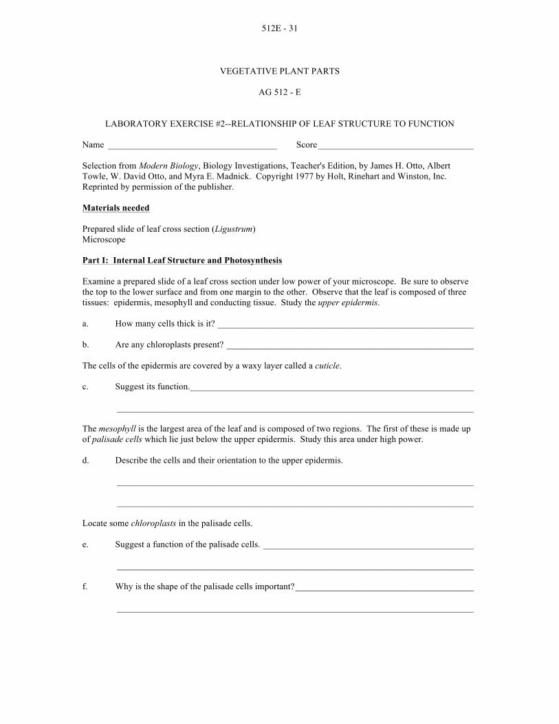
512E - 31
VEGETATIVE PLANT PARTS
AG 512 - E
LABORATORY EXERCISE #2--RELATIONSHIP OF LEAF STRUCTURE TO FUNCTION Name _____________________________________ Score __________________________________
Selection from Modern Biology, Biology Investigations, Teacher's Edition, by James H. Otto, Albert Towle, W. David Otto, and Myra E. Madnick. Copyright 1977 by Holt, Rinehart and Winston, Inc. Reprinted by permission of the publisher. Materials needed
Prepared slide of leaf cross section (Ligustrum) Microscope
Part I: Internal Leaf Structure and Photosynthesis
Examine a prepared slide of a leaf cross section under low power of your microscope. Be sure to observe the top to the lower surface and from one margin to the other. Observe that the leaf is composed of three tissues: epidermis, mesophyll and conducting tissue. Study the upper epidermis. a. How many cells thick is it? ________________________________________________________
b. Are any chloroplasts present? ______________________________________________________
The cells of the epidermis are covered by a waxy layer called a cuticle.
c. Suggest its function. ______________________________________________________________
______________________________________________________________________________
The mesophyll is the largest area of the leaf and is composed of two regions. The first of these is made up of palisade cells which lie just below the upper epidermis. Study this area under high power. d. Describe the cells and their orientation to the upper epidermis.
______________________________________________________________________________
______________________________________________________________________________
Locate some chloroplasts in the palisade cells.
e. Suggest a function of the palisade cells. ______________________________________________
______________________________________________________________________________
f. Why is the shape of the palisade cells important? _______________________________________
______________________________________________________________________________

512E - 32
Study the second region of the mesophyll. Locate the spongy layer of cells below the palisade cells.
g. Of the two, which layer is more compact? ____________________________________________
h. Are chloroplasts as numerous in the spongy cells as they are in the palisade cells?
______________________________________________________________________________
i. Account for any difference you find. _________________________________________________
______________________________________________________________________________
Note the numerous spaces among the spongy cells. These are the air spaces.
j. On the basis of their relationship to other tissues in the leaf, what do you think their function is?
______________________________________________________________________________
______________________________________________________________________________
The spongy layer is penetrated by numerous veins. Move the slide until you are able to locate a vein.
k. How can you tell a vein from other structures in a leaf? __________________________________
______________________________________________________________________________
______________________________________________________________________________
Examine the vein closely. Locate empty cells with thick walls in the upper parts of the section. These are the xylem cells. l. Suggest two functions of the xylem cells. _____________________________________________
______________________________________________________________________________
The thin-walled cells that form a cluster below the xylem cells are the phloem cells.
m. What is the function of the phloem cells? _____________________________________________
______________________________________________________________________________
Find a small vein in your section near the leaf margin.
n. What kind of cell composes the small vein? ___________________________________________
Examine the lower epidermis.
o. How many cell layers compose it? __________________________________________________
Closely examine the lower epidermis. Try to find tiny pores with small, rounded cells on either side. The pores are the stomata and the rounded cells are the guard cells.

512E - 33
p. Determine the relationship between the stomata and the air spaces of the spongy tissue. __________
______________________________________________________________________________
q. Suggest a function of the stomata. ____________________________________________________
______________________________________________________________________________
______________________________________________________________________________
On the following diagram, label: cuticle, upper epidermis, palisade cells, chloroplasts, spongy cells, vein, xylem cells, phloem cells, lower epidermis, air space, stoma, guard cells.
Part II: Summary
a. Write a brief paragraph discussing how the internal structure of a leaf adapts it to the process of
photosynthesis and the movement of water through the plant. _____________________________
______________________________________________________________________________
______________________________________________________________________________
______________________________________________________________________________
______________________________________________________________________________
______________________________________________________________________________
______________________________________________________________________________
______________________________________________________________________________
______________________________________________________________________________

512E - 34
Review what you have learned by filling in the blanks of the following statements.
b. The layer of cells which lacks chloroplasts is the _______________________________________ .
c. The ____________________________________________________ is the largest area of a leaf.
d. The ________________________ layer is composed of cells which are oriented at right angles to the epidermis.
e. The presence of _______________ is typical of the spongy layer.
f. The ________________________ is a waxy layer which prevents the loss of water from leaf tissues.
g. ____________________________ are composed of tissues which carry materials to and from leaf
tissues. h. Pores found on the underside of leaves are known as __________________________________
i. Conducting tissues in a leaf are ________________________ and _______________________
j. The only cells that contain chloroplasts in the epidermis are _____________________________
k. The three basic tissues of a leaf are _____________________, __________________________
and _________________________
Part III: Investigations On Your Own
Select fresh leaves (tulip, geranium or Tradescantia are good). Remove a small area of the lower epidermis by tearing through the blade, twisting slightly as you tear. The epidermis will appear as a thin, transparent skin. Using a razor blade, cut off a small portion of this skin. Mount it in a drop of water and add a cover glass. Examine it under low power. Compare the epidermal cells, guard cells and stomata of several kinds of leaves. Make drawings of each. Label the drawings where appropriate.

512E - 35
VEGETATIVE PLANT PARTS
AG 512 - E
LABORATORY EXERCISE #3--EXAMINING ROOTS AND STEMS Name _____________________________________ Score __________________________________
Slesnick, Irwin L., Biology Laboratory Manual, Scott, Foresman and Company, 1985. Reprinted by permission of Scott, Foresman and Company.
Look at any plant: a tree growing in a field; grass covering a lawn; a rosebush blooming in a garden. You are really seeing only half of the plant. The roots underground make up the other half of the plant. Roots, such as those in the pictures above, perform several important functions for a plant. They anchor the plant in the soil, absorb water necessary for life and growth, and store food and transport it to the rest of the plant. Locate the stems in the plant pictures above. Plant stems are often among the most conspicuous parts of a plant. Aside from supporting the plant, the stem also transports substances to and from the leaves, stores food, and in some plants, manufactures food. In this laboratory you will examine the tissues that make up roots and stems. By studying root and stem tissues you will learn how the structures in roots and stems contribute to the healthy functioning of the plant. Materials needed: Sprouted radish seed Colored pencils Hand lens or stereoscopic microscope Prepared slides of longitudinal and transverse Metric ruler cross sections of a root tip, root and Compound microscope herbaceous monocot and dicot stems Twig Scalpel

512E - 36
Part I: Examining Roots 1. Use a hand lens or stereoscopic microscopic to examine the sprout of a radish seed. Notice the
region on which root hairs first begin to grow. Locate the longest root hairs. On the cross section in a, draw the root hairs showing where they first appear and where the longest root hairs are located. You may wish to add a scale in mm next to your drawing to indicate the length of the region where root hairs appear.
2. Obtain a prepared slide of a longitudinal cross section of a root tip, and examine it under low and
high power. Locate root hairs growing off the side of the root tip. Find the three regions of root growth: the region of cell division, the region of elongation and the region of maturation. The root cap is made of larger cells at the very tip of the root. The region of cell division, above the root cap, is made of smaller, dividing cells called the apical meristem. Just above the apical meristem, find the lengthening cells that make up the region of elongation. Above this region, in the region of maturation, cells begin to differentiate into specialized tissues. In this region you will begin to notice root hairs. Identify and label the regions in a, above.
3. Examine a prepared slide of a transverse cross section of the mature region of a root. Find the
single layer of epidermal cells that protects the root surface. Locate the parenchyma cells that make up the cortex. These cells may have been stained with iodine, which turns blue in the presence of starch stored in the cortex cells. Notice that cells are very loosely packed to allow water absorbed by root hair to flow to the inner tissues of the roots. The endodermis, surrounding the vascular cylinder, is the next layer in from the cortex. The thick, waxy cell walls of endodermal cells control the passage of dissolved materials into the vascular tissues. Thus, the endodermis prevents the entrance of harmful substances that might be absorbed by the root hairs and dispersed to the rest of the plant. Find the vascular tissues that form a column called the vascular cylinder at the center of the root. Locate the pericycle layer, just inside the endodermis. Root branches form from pericycle tissue. Inside the pericycle, xylem is arranged in the form of a "+." Note the thick walls of the xylem cells. The phloem cells are in circular patches between the arms of the plus-shaped xylem columns. Compare the thickness of the phloem cell walls to that of the xylem cell walls. Label the structures of the root in b on the following page.

512E - 37
Part II: Examining Stems 1. Obtain a twig from a woody plant. Note that almost all woody plants are dicots. Observe the
places where a leaf was attached to the twig. These marks are called leaf scars. Notice the tiny row of dots, arranged in a "v" on the leaf scar. These bundle scars indicate where the xylem and phloem entered the leaf stalk. Find the tiny holes in the surface of the bark. These structures--the lenticels--allow water vapor and other gases to be exchanged through the stem. Examine the terminal bud, at the end of the twig. The length of a growing twig is determined by how fast the terminal bud grows. Examine the bud scales that cover a bud. The scales fall off after they have served their protective function, leaving concentric scars around the twig. Find these budscale scars that mark the end of the twig's yearly growing season. Notice the lateral buds on the side of the twig. Growth of these buds results in new branches, leaves, and flowers. Label the structures shown on diagram c.

512E - 38
2. Use a scalpel to make a longitudinal cut through the terminal bud. Examine the cut bud with a hand lens. Notice the tiny growing leaves and the green tissue in the lower central portion of the bud. This tissue--the shoot apex--is meristem tissue responsible for the growth in height of the stem.
3. Make a transverse cross section of your twig with a scalpel. CAUTION: Use the sharp blade of
your cutting instrument carefully to avoid injury. Always cut away from yourself. Use a hand lens to locate the large cells of the light-colored pith at the center of the stem. Rings of xylem make up the wood, the bulk of the stem. Xylem cells that make up spring wood are large and have thin walls. Summer wood is made of smaller, thick-walled xylem cells. The darker-looking summer wood makes a band that contrasts with the lighter spring wood. The contrast in the color of the bands of spring and summer wood can help you determine the age of the twig. Together these bands of different colored xylem cells make up an annual ring. Locate the thin layer of vascular cambium surrounding the outermost xylem cells. The inner layers of cambium become new xylem cells and the outer layers become new phloem cells. Tightly packed phloem cells are arranged in half-moon shapes outside the vascular cambium. Larger parenchyma cells make up the cortex. These cells store starch for the plant. The outermost layers are made of thick, tough, waterproof cells, called cork, that protect the inner tissues. You may notice vascular rays, channels that conduct materials between the different tissues. Label the tissues and structures in d:
4. Nonwoody--or herbaceous--stems are present in both monocots and dicots. Obtain prepared
slides of transverse cross sections of herbaceous monocot and dicot stems. Examine these slides under low power. Locate the epidermis, the outermost layer of the stem tissue. Draw the epidermis on the stem outlines, e and f, on the following page. Notice the vascular bundles contain phloem cells, which may be stained green, and xylem cells, which may be stained red. In dicots, a thin layer of vascular cambium may be located between the xylem and phloem. Compare the arrangement of vascular bundles in the monocot and dicot stems. Sketch the pattern of vascular bundles in the outlines of the monocot and dicot stem. Observe the large food-storing parenchyma cells around the vascular bundles. In the dicot stem these cells make up the cortex to the outside of the vascular bundles; the region of parenchyma cells to the inside of the vascular bundles is called pith. In a monocot stem parenchyma cells are referred to as pith in all regions of the stem. Draw the regions of parenchyma cells, and label the regions in both the monocot and dicot stems. Find the green-colored chloroplasts, and sketch these in your stem drawings.

512E - 39
Part III: Analysis
1. Complete the table below by checking those tissues that are present in a root, woody dicot stem, herbaceous monocot stem and herbaceous dicot stem. Also, give the function of each tissue.
Table. Structures of roots and stems

512E - 40
2. Name four tissues common to both roots and stems. What are the main functions that these tissues serve? ______________________________________________________________________________
______________________________________________________________________________
______________________________________________________________________________
3. Name one tissue unique to roots and one tissue unique to stems.
Roots: _________________________________________________________________________
Stems: ________________________________________________________________________
4. Give a reason why root hairs only appear above a certain point on a root. What does the location of root hairs on the root tell you about the way that the root grows? ______________________________________________________________________________
______________________________________________________________________________
5. How does the pattern of growth of woody dicot stems enable you to determine the age of the stem? ______________________________________________________________________________
______________________________________________________________________________
6. Describe the major difference between the structure of monocot and dicot stems. How does this difference affect the growth of the stem? ______________________________________________________________________________
______________________________________________________________________________
7. Imagine you find a scar on the side of a maple tree 2 m above the ground. If the tree grows 4 m in the next 10 years, at what height would you then find the scar? What does this tell you about the way the tree grows?
______________________________________________________________________________
Part IV: Going Further Make your own transverse and longitudinal cross sections of a root, such as a radish sprout or a carrot. Cut very thin slices of the root so that light will be able to pass through the slices when you look at them under a microscope. Stain the root slices with iodine. CAUTION: Avoid getting iodine on your hands. It stains and is poisonous if ingested. Cut away from yourself. Locate the different kinds of cells and tissues that make up the root.

512E - 41
VEGETATIVE PLANT PARTS
AG 512 - E
LABORATORY EXERCISE #4--ROOT GROWTH Name _____________________________________ Score __________________________________
Selection from Modern Biology, Biology Investigations, Teacher's Edition, by James H. Otto, Albert Towle, W. David Otto, and Myra E. Madnick. Copyright 1977 by Holt, Rinehart and Winston, Inc. Reprinted by permission of the publisher. Materials needed:
Radish, bean, pea or corn seeds Dissecting microscope Petri dishes Prepared slide: Filter paper Longitudinal section of root tip (Allium) Hand lens Colored pencils Microscope Dissecting needles
Part I: Origin of the Root System
A Week in Advance
To study the origin of roots, it is necessary to begin with a germinating seed from which the first root of a plant emerges. To observe the emergence of the primary root, trim a piece of filter paper to fit snugly in the bottom of a Petri dish. Flood the dish with water and drain off the excess. Lay 3 or 4 radish seeds at equal distances from each other on the filter paper and set aside until the roots have developed to a length of at least 2 cm. Repeat the procedure with other available seeds to note any differences in the primary root development. When the roots have developed, remove the cover from the dish and examine the seedlings with a hand lens. Locate the primary root. a. Describe its structure. ____________________________________________________________
______________________________________________________________________________
b. Where are the secondary roots developing? ___________________________________________
__________________________________________ . The fuzzy outgrowths are root hairs.
c. Locate and describe their growth. ___________________________________________________
______________________________________________________________________________
______________________________________________________________________________

512E - 42
d. What function do root hairs perform? ________________________________________________
______________________________________________________________________________
______________________________________________________________________________
e. Describe the relationship of the base of the shoot and the base of the root. ___________________
______________________________________________________________________________
On the figure, label: primary root, secondary root, root hair, seed coat, shoot.
Part II: How is Growth Accomplished in Root Tip? Remove a germinated seed from the Petri dish prepared in Part I. Cut a section of the portion of the root bearing root hairs and place this in a drop of water on a slide. Examine under a dissecting microscope and carefully, with dissecting needles, remove a portion of the tissue bearing the root hairs. In order to see the detail of the cells, add a drop of iodine to the preparation and examine it under low power. Observe the root hairs. a. Are the root hairs composed of cells? ________________________________________________
b. Explain your answer. _____________________________________________________________
______________________________________________________________________________
c. From what cells do the root hairs project? _____________________________________________
______________________________________________________________________________
d. Suggest how the root hairs absorb water. _____________________________________________
______________________________________________________________________________

512E - 43
Examine a prepared slide of a longitudinal section of the young root tip under low power. Use a biology or botany textbook or charts to locate the various regions of the root tip. Move the slide and examine all areas. e. Are root hairs present? ____________________________________________________________
f. If not, explain their absence. _______________________________________________________
______________________________________________________________________________
______________________________________________________________________________
Locate the root cap at the tip.
g. What function does it serve? _______________________________________________________
______________________________________________________________________________
Cells on the surface of the root cap are worn off as it pushes through the soil.
h. Why doesn't the root tip cap disappear entirely in time? __________________________________
______________________________________________________________________________
______________________________________________________________________________
i. Where are the smallest cells of the root tip located? _____________________________________
______________________________________________________________________________
Examine these cells closely.
j. What important activity is carried on in this region? ____________________________________
______________________________________________________________________________
k. Why is this activity important to the root? ____________________________________________
______________________________________________________________________________
Move the slide from the tip toward the older regions. l. What noticeable changes occur in the size of the cells? __________________________________
______________________________________________________________________________
m. What term applies to this region of the root? __________________________________________
______________________________________________________________________________
n. Why is the activity of this region important to the root? __________________________________
______________________________________________________________________________

512E - 44
o. Why are the regions of the root not clearly defined? _____________________________________
______________________________________________________________________________
______________________________________________________________________________
p. From what region do the root hairs originate? __________________________________________
______________________________________________________________________________
This is the region from which root hairs develop.
In the outline, locate with brackets the root cap, meristematic region, elongation region, maturation region. Accurately draw several rows of cells in each region.
Part III: Summary
a. On the basis of your observations of the root tip above, explain how the roots grow longer.
______________________________________________________________________________
______________________________________________________________________________
______________________________________________________________________________
______________________________________________________________________________

512E - 45
Test what you have learned by matching the function of the tissue in the left column with the tissue in the right column. Place the number of the tissue in the space before the function.
_____b. produces new root cells 1. maturation region
_____c. function in the absorption of water 2. epidermis
_____d. protects the growing root tip 3. root hairs
_____e. activity in this region serves 4. elongation region to push a root tip through the soil
5. root cap _____f. region producing root hairs
6. meristematic region Part IV: Investigations On Your Own
1. The secondary growth of a root may be studied through the examination of a carrot or similar root. Consult a biology or botany textbook to determine what becomes of the primary tissues as the root increases in diameter. Make drawings of the longitudinal and cross sections and label the tissues you observe.
2. Make a collection of root modifications as they are found on different plants. (for example:
adventitious roots of corn, English Ivy, tap roots of Dandelion, and turnip, etc.) Give the name of the plant and tell how the modification serves the plant.

512E - 46
VEGETATIVE PLANT PARTS
AG 512 - E
ANSWERS TO LABORATORY EXERCISES Lab #1
Part I:
1. a. Answers will depend on leaf examined.
2. b. Answers will depend on leaf examined. c. Monocot has parallel veins; dicot has branching veins.
3.
Part II: 2.

512E - 47
3. d. No, they are spaced evenly on epidermal surfaces. e. In the center of the leaf.
4.
Part III: 1. Corn leaf: Monocot; Parallel veins; no petiole
Balsam fir: Conifer; Needlelike leaves Sumac: Dicot; Branching veins; Pinnate
2. Table III. Structure of leaf tissue
Tissue Function Tissue Function
Cuticle Prevents water loss from leaf surface
Veins Transport substances to and from leaf; support leaf structure
Palisade layer
Contains long, narrow cells with chloroplasts that are major site of photosynthesis
Stomata Openings in epidermis that allow for gas exchange with the environment
Spongy layer
Loosely packed wells with air spaces that allow for the transfer of gases
Guard Cells
Sausage – shaped cells that control the size of a stoma
3. Chloroplasts move from upper layers of cells where maximum exposure to sunlight occurs to
lower layers where gases from photosynthesis and respiration are exchanged. Shape and cyclosis allow for efficient exposure to both sunlight and air spaces.
4. The limited surface area exposed to direct sunlight due to the needle's shape; the cuticle covering,
sunken stomata and the hypodermis all prevent evaporation of water. 5. Stomata location allows excess water that might diffuse into the leaf to evaporate.
Lab #2
Part I:
a. One b. No c. Prevent evaporation of water from tissues d. Thin-walled, numerous chloroplasts, lie at right angles to upper epidermis

512E - 48
e. Primary location for photosynthesis f. Permits greatest transmission of light throughout the cell g. Palisade h. No i. Spongy cells do not receive as much light as the palisade cells j. They allow gases to diffuse through the internal portion of the leaf k. Veins are composed of thick-walled cells and the cells are found in clusters l. Conduction of water and minerals, and support m. Conduction of food n. Xylem o. One p. They open into the air spaces q. Exchange of gases (carbon dioxide for photosynthesis and oxygen for respiration); regulation of
loss of water vapor to outside air
Part II: a. The leaf is a broad, flat, green structure permitting the maximum exposure of this photosynthetic
structure to light. The palisade cells compose the upper layer of mesophyll and contain numerous chloroplasts. The upper epidermis (and lower epidermis) contains no chloroplasts. This allows light to penetrate to the palisade cells where the process of photosynthesis is primarily carried out. The stomata and guard cells allow for the passage of water, evaporated from the surface of the spongy cells, to escape from the air space to the atmosphere. This allows for the movement of water through the plant.
b. Epidermis c. Mesophyll d. Palisade e. Air spaces f. Cuticle g. Veins h Stomata i. Xylem and phloem j. Guard cells k. Epidermis, mesophyll, conducting tissue

512E - 49
Lab #3
Part I:
Step 2:
Part II:
Step 3:

512E - 50
Part II:
Step 1:
Step 3:
Step 4:

512E - 51
Part III:
Table: Structures of roots and stems

512E - 52
2. Meristem contributes to growth; cortex stores food and provides support; phloem transports materials between leaves and roots and provides support; xylem transports water from roots to leaves and provides support.
3. Students may mention the root cap, endodermis or pericycle as unique root structures. Unique stem structures include bud scales, lenticels, cork or vascular cambium of woody stems or the chloroplasts of herbaceous stems.
4. Root hairs are cells that have differentiated; thus, they only appear in the region of maturation. Their location indicates that growth occurs in the tip.
5. Wood formed in the spring is lighter in color than wood formed in the summer. The bands of different colored wood form a ring each year.
6. Monocot has scattered vascular bundles; dicot has bundles in a ring. Structure and arrangement of bundles in dicots allows more than one season of growth.
7. The scar would remain at 2 m because growth in the stem occurs at the tip.
Lab #4
Part I:
a. It appears white and is long and tapering. Delicate strands project from the upper portion. b. From the upper regions of the root c. The root hairs are located a short distance from the tip of the root and appear as many numerous
hairs. d. They absorb water and increase the absorptive surface of the root. e. The developing root supplies the developing shoot with necessary water and minerals.
Part II:
a. No b. They appear cellular, but are actually long projections from a single epidermal cell. c. Epidermal cells d. Water is absorbed by osmosis. e. Probably not f. Being delicate structures, they were probably lost in the process of making the slide preparation. g. It protects the meristematic region where new root cells are being produced. h. New cells are formed from the meristematic region to replace the cells that are worn off. i. Meristematic region j. Production of new root cells by mitosis. k. Without new cells, the root could not grow. l. They lengthen. m. Elongation region

512E - 53
n. As the cells elongate, the root grows and is pushed through the soil. o. Cells are produced at different rates by the meristematic region and, therefore, elongate and
mature at different rates. p. Maturation region
Part III:
a. New cells produced by the meristematic region become pushed back to where they elongate. The process of elongation serves to push the root tip through the soil whereupon growth in length is achieved. Following elongation, the cells mature into tissues dependent on their location in the root tip.
b. 1 c. 3 d. 5 e. 4 f. 2

512E - 54
VEGETATIVE PLANT PARTS
AG 512 - E
UNIT TEST Name _____________________________________ Score __________________________________
1. Match terms associated with plant growth and development to the correct definitions. Write the correct numbers in the blanks.
1. Node 7. Dicot 13. Fertilization 2. Internode 8. Vascular bundle 14. Pollination 3. Bud 9. Xylem 15. Embryo 4. Leaf scar 10. Phloem 16. Radicle 5. Vascular bundle scar 11. Pistil 17. Hypocotyl 6. Monocot 12. Stamen 18. Epicotyl
_____a. Union of the male (pollen) nucleus with the female (egg) cell
_____b. Plant having two seed leaves
_____c. The part of a stem between two nodes
_____d. Vascular tissue that transports water and minerals from the root system to the leaves
_____e. The embryonic root
_____f. Seed bearing organ of a flower; composed of ovary, style and stigma
_____g. Plant having one seed leaf
_____h. The part of a stem where a leaf is attached
_____i. An embryonic shoot of a plant
_____j. The part of the embryo above the cotyledons and below the next leaves
_____k. A scar left on the stem when a leaf falls
_____l. Transfer of pollen from the anther to the stigma
_____m. The young plantlet within the seed; the germ
_____n. The part of an embryo between the cotyledons and the radicle
_____o. Part of the flower producing the pollen; composed of the filament and anther
_____p. A strand of tissue containing xylem and phloem enclosed by a sheath of cells
_____q. A spot within a leaf scar left by the vascular bundles when a leaf falls
_____r. Vascular tissue that conducts food from the leaves to regions of growth or storage

512E - 55
2. Label the primary parts of a plant. Write the correct names in the blanks.
3. List the functions of the following plant parts.
a. Roots ______________________________________________________________________
b. Stems _____________________________________________________________________
c. Leaves _____________________________________________________________________
d. Flowers ____________________________________________________________________
4. Labels the parts of a simple leaf. Write the correct names in the blanks.
5. Labels the parts of a compound leaf. Write the correct names in the blanks.

512E - 56
6. Identify types of compound leaves. Write the correct names in the blanks.
7. Identify types of leaf arrangement. Write the correct names in the blanks.
8. Identify types of leaf veination. Write the correct names in the blanks.

512E - 57
9. Identify types of leaf margins. Write the correct names in the blanks.
10. Identify types of leaf attachment to the stem. Write the correct names in the blanks.
11. Label the parts of a stem. Write the correct names in the blanks.

512E - 58
12. Match stem modifications to the correct description. Write the correct numbers in the blanks.
1. Crown 5. Tuber 2. Stolon 6. Corm 3. Spur 7. Bulb 4. Rhizome
_____a. Enlarged fleshy part found at the tip of a rhizome; potato
_____b. Appears laterally on branches of fruit trees and bears fruit; apple
_____c. Short, disc-shaped stem surrounded by leaf-like scales; onion
_____d. Flesh, short underground stem with very few buds; gladiolus
_____e. Runners that grow along top of soil surface; strawberry
_____f. Underground stems that grow horizontally below soil surface; quackgrass
_____g. Appears just above or just below ground level from which modified stems grow; small grains
13. Name two types of root systems.
a. _____________________________________________________________________________
b. ____________________________________________________________________________

512E - 59
VEGETATIVE PLANT PARTS
AG 512 - E
ANSWERS TO TEST 1. a. 13 f. 11 k. 4 p. 8
b. 7 g. 6 l. 14 q. 5 c. 2 h. 1 m. 15 r. 10 d. 9 i. 3 n. 17 e. 16 j. 18 o. 12
2. a. Roots b. Stem c. Leaves d. Flowers
3. Roots: Absorb water and nutrients; Anchor and support plants; Store food Stem: Supports leaves, flowers, fruit and seeds; Conducts water, nutrients and food; Stores food Leaves: Manufacture food for the plant; Necessary for transpiration; Store food Flowers: Serve as site of reproduction; Store food
4. a. Stipules b. Petiole c. Veins d. Blade
5. a. Leaflets d. Petiolule b. Veins e. Petiole c. Rachis f. Stipules
6. a. Pinnate b. Bipinnate c. Palmate d. Trifoliate
7. a. Alternate b. Opposite c. Whorled
8. a. Parallel b. Pinnate c. Netted d. Palmate
9. a. Entire b. Serrate c. Incised d. Lobed
10. a. Petiolate b. Sessile c. Clasping d. Decurrent
11. a. Node d. Lateral bud b. Internode e. Leaf scar c. Terminal bud f. Vascular bundle scar
12. a. 5 d. 6 g. 1 b. 3 e. 2 c. 7 f. 4
13. a. Tap root system b. Fibrous root system

512F - 1
REPRODUCTIVE PLANT PARTS
AG 512 - F
UNIT OBJECTIVE
After completion of this unit, students should be able to list in order the life cycle of a flowering plant, label a drawing showing the parts of a complete flower and the parts of a monocot and dicot seed, and define a fruit. Students should also be able to match the types of fruits to their correct descriptions. This knowledge will be demonstrated by completion of a unit test with a minimum of 85 percent accuracy.
SPECIFIC OBJECTIVES AND COMPETENCIES
After completion of this unit, the student should be able to:
1. List in order the life cycle of a flower plant.
2. Label a drawing showing the parts of a complete flower.
3. Describe the parts and/or functions of the flower parts.
4. Match the type of flower to the correct description.
5. Define fruit.
6. List the three layers of the pericarp.
7. Match the types of fruits to their correct descriptions.
8. Label a drawing showing the parts of a monocot and dicot seed.
9. Match functions to the correct seed parts.
10. Observe the structure and function of flowers.
11. Study flower functions in reproduction.
12. Observe and classify fruits.
13. Examine the development of seed parts into young plants.

512F - 2
REPRODUCTIVE PLANT PARTS
AG 512 - F
SUGGESTED ACTIVITIES
I. Suggested activities for instructor
A. Order materials to supplement unit.
1. Literature
a. Agronomy Curriculum Materials Packet, 232 pages; available from IAVIM, 208 Davidson Hall, Iowa State University, Ames, Iowa 50011; approximate cost $10.00, order no. 214.
b. Crop Production, 15 transparency masters; available from
IAVIM, 208 Davidson Hall, Iowa State University, Ames, Iowa 50011; approximate cost $2.25, order no. 517.
2. Filmstrips, slideshows, etc.
a. Agronomy, computer program; available from IAVIM, 208 Davidson Hall, Iowa State University, Ames, Iowa 50011; approximate cost $15.00, order no. 902.
B. Make transparencies and necessary copies of materials.
C. Provide students with objective sheet.
D. Provide students with information sheets and laboratory exercises.
E. Discuss unit and specific objectives.
F. Discuss information sheet.
G. Demonstrate and discuss procedures outlined in laboratory exercises.
H. Review and give test.
I. Reteach and retest, if necessary.
II. Instructional materials
A. Objective sheet
B. Suggested activities
C. Information sheet

512F - 3
D. Transparency masters
1. TM 1--The Life Cycle of a Flowering Plant
2. TM 2--Parts of a Complete Flower
3. TM 3--A Corn Grain and Its Parts
4. TM 4--A Bean Seed and Its Parts
E. Laboratory exercises
1. LE 1--Observing the Structure and Function of Flowers
2. LE 2--Flower Functions in Reproduction
3. LE 3--Observing and Classifying Fruits
4. LE 4--Development of Seed Parts into Young Plants
F. Test
G. Answers to test
III. Unit references
A. Delorit, R.J., et al., Crop Production, 5th edition, Prentice-Hall, Inc., Englewood Cliffs, New Jersey, 1984.
B. Hudson, T., et al., Plant Science: Growth, Development and Utilization of
Cultivated Plants, 2nd edition, Prentice-Hall, Inc., Englewood Cliffs, New Jersey, 1988.
C. Idaho Crop and Soil Science Curriculum Guide, Idaho State Board for
Vocational Education, Boise, Idaho, 1985. D. Janick, J., et al., Plant Science, 2nd edition, W.H. Freeman and Co., San
Francisco, California, 1974. E. Otto, James H., and Towle, Albert, Modern Biology, Holt, Rinehart and
Winston, New York, 1985. F. Slesnick, Irwin L., et al., Biology, Scott, Foresman and Company, Glenview,
Illinois, 1985.

512F - 4
REPRODUCTIVE PLANT PARTS
AG 512 - F
INFORMATION SHEET
I. The life cycle of a flowering plant (Transparency 1)
A. Seed germination and seedling growth
B. Vegetative growth
C. Flower formation
D. Pollination
E. Fertilization
F. Seed development
II. Parts of the complete flower (Transparency 2)
A. Pistil--Female part where egg cell originates
1. Stigma--Upper part of pistil that catches pollen
2. Style--Supports stigma
3. Ovary--Produces ovules which develop into seeds
B. Stamen--Male part of flower
1. Filament--Supports anther
2. Anther--Bears the pollen
C. Accessory organs
1. Corolla--Petals of the flower
2. Calyx--Sepals of the flower
3. Pedicel--Stalk of an individual flower
III. Types of flowers
A. Complete--Has stamens, pistils, petals and sepals on same flower; common to dicots
B. Incomplete--Has stamens and pistils, but no petals or sepals; common to
monocots C. Perfect flower--Has both stamens and pistils on the same flower

512F - 5
D. Imperfect flower--Has either stamens or pistils, but not both on the same flower
E. Staminate--Has only male flower parts
F. Pistillate--Has only female flower parts
G. Monoecious--Staminate and pistillate flowers found on the same plant
(Examples: corn, cucumbers, squash, melons and pumpkins)
H. Dioecious--Staminate and pistillate flowers found on separate plants
(Examples: holly, date, palm, spinach and asparagus)
IV. Fruit--An enlarged and ripened ovary enclosing one or more seeds, which are ripened ovules (some fruits are seedless, such as seedless grapes)
V. Layers of the pericarp (ovary wall)
A. Exocarp
B. Mesocarp
C. Endocarp
VI. Types of fruits
A. Fleshy fruits
1. Pome--Outer, fleshy layer developed from calyx and receptacle; ovary forms a leathery core containing seeds (apple, pear)
2. Drupe--Ripened ovary becomes two-layered (outer layer fleshy, inner
layer hard) forming a pit, enclosing seed (cherry, peach, plum) 3. Berry--Ovary is fleshy and usually juicy; contains several seeds
(tomato, grape, cucumber) 4. Aggregate fruit--Several pistils in single flower form compound fruit
(strawberry, raspberry, blackberry) 5. Multiple fruit--Several clustered flowers form compound fruit
(pineapple, fig)
B. Dry fruits that open when ripe
1. Pod--Thin ovary wall; single-chambered, contains several seeds, splits
along line when ripe (pea, bean, milkweed) 2. Capsule--Several chambers and seeds in ovary; splits open when
mature (iris, lily, cotton, poppy)

512F - 6
C. Dry fruits that remain closed when ripe
1. Nut--Ovary wall is hard; encloses one seed (oak, acorn)
2. Achene--Ovary wall isn't fastened to seed (dandelion, sunflower)
3. Grain--Ovary wall is thin and fastened securely to a seed (corn, wheat, oats)
4. Samara (winged fruit)--Wing attached to the ovary wall (maple, elm,
ash)
II. Parts of the seed (Transparencies 3, 4)
A. Monocot (Transparency 3)
1. Epicotyl
2. Hypocotyl
3. Radicle
4. Cotyledon
5. Coleoptile
6. Endosperm
7. Seed coat
B. Dicot (Transparency 4)
1. Epicotyl
2. Hypocotyl
3. Radicle
4. Cotyledons
5. Seed coat
VIII. Functions of seed parts
A. Seed coat--Protects the seed against injury and dehydration
B. Endosperm--Stores food
C. Coleoptile--Protects the epicotyl and leaves as they emerge from the ground
D. Cotyledons--Seed leaves; absorb endosperm and serve as food reservoirs
E. Radicle--Embryonic root

512F - 7
F. Hypocotyl--Part of the embryonic shoot below the cotyledon attachment point
G. Epicotyl--Part of the embryonic shoot above the point of attachment of the cotyledons

512F - 8
TM 1

512F - 9
TM 2

512F - 10
TM 3

512F - 11
TM 4

512F - 12
REPRODUCTIVE PLANT PARTS
AG 512 - F
INSTRUCTOR NOTES FOR LABORATORY EXERCISES
Lab #1
Complete flowers such as the snapdragon, tulip or lily are preferred for Parts I-III, but show students examples of pistillate, staminate, regular, irregular and composite flowers. Solution preparation
The following general instructions apply for the preparation of most solutions: Solvents should be added to solutes. Use distilled water, not tap water, for all reagents. When preparing an acid or base solution, slowly add the acid or base to the water. Never add water to a concentrated acid or base. To make percentage solutions measure 1 ml of solute per percentage. Add the solute to enough solvent to make 100 ml of solution. When dissolving a solid in water, measure 1 g of solute per percentage and mix the solute with enough water to make 100 ml of the solution. Methylene blue stain
Dissolve 0.75 g of methylene blue in 50 ml of 95% ethyl alcohol. Dilute 5 ml of the alcohol and methylene blue solution with 45 ml of distilled water. This diluted solution is the stain. Bottle and store the remaining methylene blue and alcohol solution. CAUTION: Ethyl alcohol is flammable. It is also irritating to the eyes. Flush spills with water. Do not ingest ethyl alcohol. 10% Sucrose solution
Dissolve 15 g of sucrose in 135 ml of distilled water. Refrigerate.
Part I:
You may want to begin Part II early in the lab to allow time for completion.
Part III: Staining helps to emphasize the nuclei. If the ovules are not flattened or well stained, have students repeat the slide preparation. Part V:
Ray flowers: sepals, petals Tube flowers: sepals, petals, stamen, carpel, ovary, ovule Ray and tube flowers have different flower parts and the ray flowers have colorful, showy petals.
Sunflowers are composed of different types of flowers so they are composites.

512F - 13
Lab #2
Flowers as suggested are often donated upon request by funeral homes.
Part II:
Stamens may be collected from any species of the lily family and frozen until ready for use.
Part III:
Pistils may be collected from any species of the lily family after summer blooming and preserved for future use by freezing them or placing them in a preservative. Part IV:
Pollen germination is often dependent on the concentration of the sugar. You may have to vary the concentration of the 10% solution suggested. Before releasing students to do the hanging-drop preparation, it is best to demonstrate it first.
Lab #3
Suggested fruits: Pear-pome; Apple-pome; Strawberry-aggregate; Raspberry-aggregate; Pineapple-multiple; Tomato-berry; Orange-berry; Milkweed-capsule; Maple-samara; Sunflower-achene; Kernel of corn-grain; Pecan-nut; Acorn-nut; Olive-drupe; Pea pod-legume; Coconut-drupe; Cucumber-berry Have at least one fruit from each group. If the suggested fruits are not available, substitute with another fruit from the same group. Part I:
Change questions for fruit if necessary.
Before class, set up 10-15 stations. Each should have a fruit (cross section whenever possible). Try to show as much of the fruit as possible and include any remaining flower parts. Lab #4
Be sure the seeds you are using have been packaged for the current growing season. Seeds older than a year may not germinate.

512F - 14
REPRODUCTIVE PLANT PARTS
AG 512 - F
LABORATORY EXERCISE #1--OBSERVING THE STRUCTURE AND FUNCTION OF FLOWERS
Name _____________________________________ Score __________________________________
Slesnick, Irwin L., Biology Laboratory Manual, Scott, Foresman and Company, 1985. Reprinted by permission of Scott, Foresman and Company. Introduction
In all flowering plants--or angiosperms--the flower is a highly refined organ that is specialized for sexual reproduction. The outer structures, the sepals and petals, are actually modified leaves that protect the reproductive structures. Each of the remaining parts plays a specific role in the actual seed formation. In this laboratory you will examine each of the flower structures and see how they are modified for their role in sexual reproduction. Materials needed
Assorted fresh flowers Forceps Stereoscopic microscope or hand lens Dissecting needle Compound microscope 10% Sucrose solution Coverslips Scalpel or single-edged razor blade 0.01% Methylene blue solution Lab or facial tissue Clean sheet of unlined paper Tape
Part I: Macroscopic Study of the Flower
1. Obtain all the materials listed above and bring them to your work area.
2. Examine the outer structure of your flower. The sepals, which are modified leaves, form the outermost circle--or whorl. Collectively the sepals form the calyx. Find the place where the calyx attaches to the base of the flower. This is the receptacle. The petals are found just inside the sepals and the whorl of petals is called the corolla. Label the receptacle, sepal and petal on a.

512F - 15
3. Monocots are flowers whose parts occur in threes or multiples of threes. Dicots are flowers whose parts occur in fours or fives or multiples thereof. Is the flower you are observing a monocot or a dicot?
(a) ____________________________________________________________________________
4. Gently remove the sepals and the petals. Tape the sepals along the bottom of a clean sheet of paper. Then tape the petals in a row above the sepals. What do you notice about the number of sepals and petals?
(b) ____________________________________________________________________________
Part II: The Male Reproductive Structures
1. Inside the corolla is a circle of stamens. These are the male reproductive organs, each consisting of an anther at the tip supported by a tubelike filament. Pollen grains found inside the anther are the male gametophytes. Label the anther, filament and pollen grain on b. Carefully remove the stamens and tape them in a row above the petals.
2. The anthers contain the pollen sacs. The pollen grains are formed from microspores in the pollen
sacs. To examine the pollen grains more closely, add some pollen grains to a drop of water on a microscope slide. Add a coverslip and examine the pollen grains under high power of your microscope. The small dotlike structures you see are the pollen grains. Make a sketch of a few of the pollen grains in c.

512F - 16
3. Sprinkle some pollen from your flower onto a drop of sucrose solution on a microscope slide. Add a coverslip and examine at high power at five-minute intervals for 30-60 minutes. The narrow thread-like structures you see growing are the pollen tubes. Locate and label the tube nucleus at the tip of the pollen tube in d on the previous page and the two sperm nuclei close behind. After 30 minutes make a sketch of the pollen tube growth. Describe the pollen grains and the growth of the pollen tubes.
(c) ____________________________________________________________________________
______________________________________________________________________________
______________________________________________________________________________
Part III: The Female Reproductive Structures
1. The female reproductive organ--or carpel--is located in the center of the flower. The top portion of the carpel is the stigma. The stigma is usually sticky and is where the pollen grains collect. The style is the stalk-like structure that supports the stigma. The enlarged structure at the base of the carpel is the ovary. Ovules within the ovary produce the female gametophytes. Label the carpel, stigma, style and ovary on e.
2. With a scalpel or razor blade, carefully remove the carpel by cutting just beneath the ovary.
CAUTION: Use the sharp blade of your cutting instrument carefully to avoid injury. Always cut away from yourself. Then, make a cross section of the ovary as shown in f on the next page. Tape one half of the cross section on the paper above your drawing of the pollen grains. Secure one half of the cross section with your forceps. Then, using your scalpel, cut a thin slice from the section. Make a wet mount and examine under low power. Find the white spherical ovules that are attached to the ovary wall by a tiny stalk, the funiculus. The ovules develop into hollow compartments called locules. The outer layers of the ovule surround the embryo sac. The embryo sac is the female gametophyte and this is where the egg is located. Label the ovule, funiculus, locule and the embryo sac on e.

512F - 17
3. Using the cross section of the ovary, carefully separate a few ovules. Place the ovules in a drop of water on a clean microscope slide and add one drop of methylene blue stain. Place a coverslip on top. Fold a piece of tissue and place it on top of the coverslip. Gently press down to crush the ovules. Under low power examine the slide and locate the stained nuclei inside the embryo sacs. Make a sketch of the nuclei in g.
Part IV: Analysis
1. In what structure are the male gametophytes found?
______________________________________________________________________________
2. In what structure are the female gametophytes found?
______________________________________________________________________________
3. Where is the stigma located on the flower and how does this aid in pollination?
______________________________________________________________________________
______________________________________________________________________________
4. Describe the process of fertilization in angiosperms. Name each of the structures involved.
______________________________________________________________________________
______________________________________________________________________________
______________________________________________________________________________
______________________________________________________________________________

512F - 18
Part V: Going Further
Obtain a composite flower from your teacher. Notice that there appear to be two kinds of petals. These are actually flowers. The flowers of the outer row--the ray flowers--have showy petals. The flowers in the center are called the tube flowers. Carefully remove one of the ray flowers at its base. Using your scalpel or razor blade make a longitudinal cut down the center of the flower. Examine one half of the ray flower with your hand lens. List the flower parts present in the ray flower. Carefully remove one of the tube flowers beneath the base. Examine one half of the flower with your hand lens. List the flower parts present in the tube flower. What are the differences between the ray and tube flowers? Sunflowers are an example of a composite flower. The name comes from the arrangement of the flowers. Explain why sunflowers are composite flowers.

512F - 19
REPRODUCTIVE PLANT PARTS
AG 512 - F
LABORATORY EXERCISE #2--FLOWER FUNCTIONS IN REPRODUCTION Name _____________________________________ Score __________________________________
Selection from Modern Biology, Biology Investigations, Teacher's Edition, by James H. Otto, Albert Towle, W. David Otto, and Myra E. Madnick. Copyright 1977 by Holt, Rinehart and Winston, Inc. Reprinted by permission of the publisher. Materials needed
Gladiolus flower (tulip, lily or snapdragons will suffice) Collection of anthers from a Single-edged razor blade variety of flowers Microscope Depression slides Prepared slide of cross section of Lily anther slide Toothpick Cover slip Dissecting needle Hand lens or stereoscopic microscope Petroleum jelly Dropper 10% sucrose solution Forceps Distilled water
Part I: What Are the Parts of a Flower?
Flowers must develop before there can be fruits and seeds. Seeds are contained in the fruits which develop after pollination and fertilization. As you read this description of a flower, label the parts given in italics on the figure 512F-21. Examine a complete flower and note that it has 4 kinds of floral parts. These parts are arranged in circles or whorls. The parts are supported on a stalk, the pedicel. The parts are attached to the swollen tip of the pedicel known as the receptacle. The outermost circle of parts is the sepals which may be green. In some species, the sepals appear as petals. Collectively the sepals make up the calyx which serves to protect young flower parts in the bud stage. a. Describe the sepals and their number. ________________________________________________
______________________________________________________________________________
The petals are within the calyx and collectively known as the corolla.
b. Describe the number and appearance of the petals. ______________________________________
______________________________________________________________________________
______________________________________________________________________________
The male and female organs make up the remaining circles of flower parts.
c. Why are they known as essential parts? ______________________________________________
______________________________________________________________________________
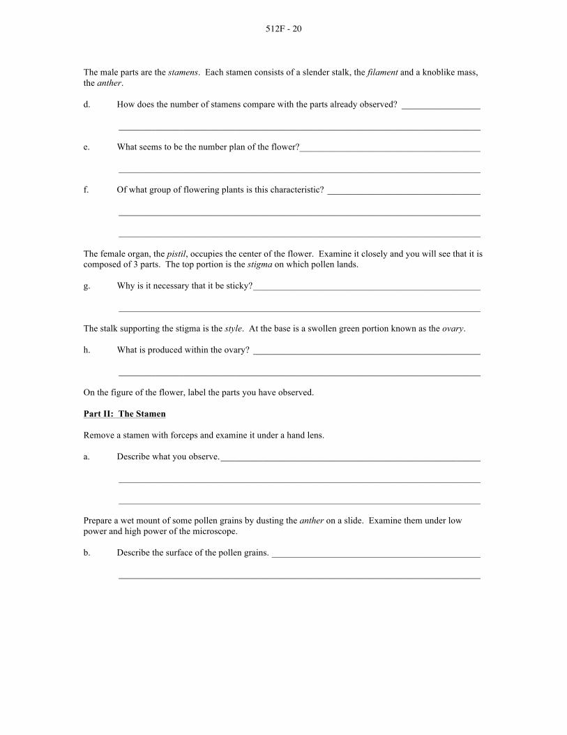
512F - 20
The male parts are the stamens. Each stamen consists of a slender stalk, the filament and a knoblike mass, the anther. d. How does the number of stamens compare with the parts already observed? _________________
______________________________________________________________________________
e. What seems to be the number plan of the flower? _______________________________________
______________________________________________________________________________
f. Of what group of flowering plants is this characteristic? _________________________________
______________________________________________________________________________
______________________________________________________________________________
The female organ, the pistil, occupies the center of the flower. Examine it closely and you will see that it is composed of 3 parts. The top portion is the stigma on which pollen lands. g. Why is it necessary that it be sticky? _________________________________________________
______________________________________________________________________________
The stalk supporting the stigma is the style. At the base is a swollen green portion known as the ovary.
h. What is produced within the ovary? _________________________________________________
______________________________________________________________________________
On the figure of the flower, label the parts you have observed.
Part II: The Stamen
Remove a stamen with forceps and examine it under a hand lens.
a. Describe what you observe. ________________________________________________________
______________________________________________________________________________
______________________________________________________________________________
Prepare a wet mount of some pollen grains by dusting the anther on a slide. Examine them under low power and high power of the microscope. b. Describe the surface of the pollen grains. _____________________________________________
______________________________________________________________________________

512F - 21
Examine a prepared slide of the cross section of a lily anther under low power.
c. How many cavities or pollen sacs are seen? ___________________________________________
d. When the anther matures, how is pollen released? ______________________________________
______________________________________________________________________________
e. What becomes of pollen as it is released? _____________________________________________
______________________________________________________________________________
______________________________________________________________________________

512F - 22
The pollen grains contain the male sex cells produced as a result of meiosis from special cells within the anther. On the cross section of the anther on the previous page, label: wall of anther, pollen sac, pollen grains. Part III: Structure and Function of the Pistil
Carefully remove the pistil from the flower and examine it closely under a stereoscopic microscope.
a. In relation to pollination, suggest a reason for the stigma being supported as it is by the style.
______________________________________________________________________________
______________________________________________________________________________
Using a sharp, single-edged razor blade, make a wet mount of a cross section of the ovary. Examine it under a stereoscopic microscope. Notice that the ovary is divided into sections known as carpels. Each carpel contains several ovules. b. How do the ovules appear? ________________________________________________________
______________________________________________________________________________
The ovules extend into a cavity known as a locule.
c. How many cavities or carpels make up the ovary? ______________________________________
______________________________________________________________________________
d. Explain the significance of the number. _____________________________________________
______________________________________________________________________________
______________________________________________________________________________
Each ovule contains an egg which is not visible. Observe that an ovule is attached to the ovary wall through a tiny stalk. e. Why must the ovules be attached as they are? _________________________________________
______________________________________________________________________________
______________________________________________________________________________
On the cross section of the ovary on the previous page, label: ovary wall, carpel, ovule, locule.
Part IV: Observing Germinating Pollen Grains
Once pollen of a particular species has landed on the stigma of the same or closely related species, a pollen tube will begin to germinate. This phenomenon can be observed under laboratory conditions.

512F - 23
Obtain a dropper of a 10% sucrose solution from a prepared stock solution. Place a drop of the solution in the center of a cover glass. Transfer some pollen grains to the drop with a dissecting needle or small brush. Use a toothpick to apply a thin ring of petroleum jelly around the depression. The slide should be deep enough to prevent the drop from touching. Turn the slide over and line up the depression with the drop of sucrose solution with the pollen. Gently allow the slide to come in contact with the cover glass. Turn the slide upright and examine under low power of the microscope. Pollen tubes should emerge within 20 minutes. Certain pollen requires one or two days to produce pollen tubes. Periodically check the preparation to determine the extent of germination.
a. What is the function of the pollen tube? ______________________________________________
______________________________________________________________________________
______________________________________________________________________________
b. Through what structures does it grow? _______________________________________________
______________________________________________________________________________
c. Toward what structure does it grow? _________________________________________________
Examine a pollen tube to see if you can find the 2 sperm nuclei.
d. What becomes of these 2 nuclei? ____________________________________________________
______________________________________________________________________________
In the space below, draw the pollen grain at the start of the observation and several stages in the growth of the pollen tube. Label: pollen tube, sperm nuclei.

512F - 24
Part V: Summary
In the following chart, briefly relate how each flower part contributes to the function of reproduction.
Floral Part Function
Sepal
Petal
Filament
Anther
Stigma
Style
Ovary
Part VI: Investigations On Your Own
Collect several flowers of varying structure and examine them to determine differences and similarities. Summarize in a chart the number of each of the floral parts and any unusual features that may be observed. Determine whether they are monocots or dicots.

512F - 25
REPRODUCTIVE PLANT PARTS
AG 512 - F
LABORATORY EXERCISE #3--OBSERVING AND CLASSIFYING FRUITS Name _____________________________________ Score __________________________________
Slesnick, Irwin L., Biology Laboratory Manual, Scott, Foresman and Company, 1985. Reprinted by permission of Scott, Foresman and Company. Introduction
After fertilization occurs in an angiosperm, the ovules develop into the seeds. The ovary that surrounds the seed or seeds develops into the fruit. The wall of the ovary develops three distinct layers. The outer layer--or exocarp--is made up of a single layer of epidermal cells. The middle layer--or mesocarp--is where food is stored for the fruit. The mesocarp is usually many cell layers thick. The inner layer--or endocarp--can vary in both thickness and texture. In some of the fruits the endocarp is soft and fleshy while in others it is hard and dry. The endocarp is one of the characteristics you will want to look for in the fruits you examine. In this laboratory you will observe and classify different fruits using a classification key. In a classification key each category is a collective group that contains one or more of the groups in the next level down. (Note: In this lab you may find fruits that you have previously called vegetables. A fruit is a ripened ovary that contains a seed or seeds. Squash, cucumbers, kernels of corn and bean pods are all fruits.) Materials needed
Hand lens Various fruits
Part I: Procedure
1. Study the cross section of the fruit shown in a. Notice the role each part of the ovary wall plays in the formation of the fruit. The thickness and texture of these parts will vary with each fruit that you examine. Label the exocarp, mesocarp, and endocarp in b and c on the following page.

512F - 26
2. Study the classification key on this page and notice how each of the categories is broken down at each level of the key.
3. At each station answer the questions and make a sketch of the sample. Label the fruit and seed on
your sketch. Use the key to classify the type of fruit.

512F - 27
2. Indehiscent--The mature fruit does not split open
a. Achene--The single seed is attached to the ovary wall at only one point and has a hard outer coat
b. Grain--The single seed is completely attached to the ovary wall
c. Samara--A prominent wing-like structure extends from the ovary wall and contains one or two seeds
d. Nut--The single seed is surrounded by a tough, thick ovary wall
II. Aggregate--The fruit develops from many small ovaries within a single flower
III. Multiple--The fruit develops from the ovaries of many flowers clustered together

512F - 28
SKETCH
PEA POD
Type of fruit: k. __________________________
7. Along how many lines does the fruit split open?
l. __________________________
8. Is this a dry or fleshy fruit?
m. _________________________
SUNFLOWER
Type of fruit: n. __________________________
9. Is the seed completely attached to the ovary
wall? o. __________________________
10. Which part of the fruit is eaten?
p. __________________________
TOMATO
Type of fruit: q. __________________________
11. How many locules are in this sample?
r. __________________________
MILKWEED
Type of fruit: s. __________________________
12. Is this fruit dehiscent or indehiscent?
t. __________________________
13. How many seamlines are there?
u. __________________________

512F - 29
SKETCH ACORN
Type of fruit: v. __________________________
14. How many seeds are there?
w. __________________________
15. Describe the ovary wall.
x. __________________________
COCONUT
Type of fruit: y. __________________________
16. Is this a dry or fleshy fruit?
z. __________________________
17. What parts of the ovary wall develop into the
husk? aa. _________________________
PINEAPPLE
Type of fruit: bb. _________________________
18. How many flowers are in this section of fruit?
cc. _________________________
KERNEL OF CORN
Type of fruit: dd. _________________________
19. Can you separate the ovary wall from the seed?
ee. _________________________
20. Is this a fleshy or dry fruit?
ff. __________________________

512F - 30
SKETCH ORANGE
Type of fruit: gg. _________________________
21. What part of the fruit is edible?
hh. _________________________
22. What parts of the fruit are discarded?
ii. __________________________
Part II: Analysis
1. Describe the formation of the fruit and seed in a simple fruit. List the flower parts that are involved in the formation of each.
______________________________________________________________________________
______________________________________________________________________________
______________________________________________________________________________
2. List four fruits where the seed is eaten and the fruit is discarded.
______________________________________________________________________________
3. How can you distinguish between the fruit and other parts of the flower?
______________________________________________________________________________
4. How does an aggregate fruit differ from a multiple fruit?
______________________________________________________________________________
______________________________________________________________________________
5. How is an aggregate fruit similar to a multiple fruit?
______________________________________________________________________________

512F - 31
REPRODUCTIVE PLANT PARTS
AG 512 - F
LABORATORY EXERCISE #4--DEVELOPMENT OF SEED PARTS INTO YOUNG PLANTS
Name _____________________________________ Score __________________________________
Selection from Modern Biology, Biology Investigations, Teacher's Edition, by James H. Otto, Albert Towle, W. David Otto, and Myra E. Madnick. Copyright 1977 by Holt, Rinehart and Winston, Inc. Reprinted by permission of the publisher. Materials needed
Dry and soaked lima beans 250-ml beaker Dried ear of corn Paper toweling Individual grains of corn Blotter or filter paper Knife or single-edged razor blade Cotton Hand lens Colored pencils Iodine solution
Part I: A Dicot Seed--The Bean
The seed is a matured ovule and the final product of angiosperm reproduction. The new plant is provided with stored food and special coverings. Under the proper conditions vegetative growth begins. This is known as seed germination. Obtain one dry lima bean and one that has been soaked overnight. Examine the dry seed and note its external markings. Locate a scarlike structure, the hilum. a. What does it represent? ___________________________________________________________
______________________________________________________________________________
Locate the micropyle, a tiny opening close to the hilum.
b. What is the significance of the micropyle? ____________________________________________
______________________________________________________________________________
c. Would you expect all seeds to have a hilum and a micropyle? _____________________________
Explain your answer. _____________________________________________________________
______________________________________________________________________________
______________________________________________________________________________
Examine a dry seed which has been soaked overnight. Compare this seed to a dry seed.
d. What changes have occurred? ______________________________________________________
______________________________________________________________________________

512F - 32
e. Offer an explanation for what you observe. ___________________________________________
______________________________________________________________________________
Remove the thin outer seed coat, the testa.
f. Describe the cotyledons which are now visible. ________________________________________
______________________________________________________________________________
g. What is their function? ____________________________________________________________
Separate the cotyledons allowing the embryo plant to remain attached to one of them. The epicotyl, often called the plumule, consists of two, tiny leaves which enclose the terminal bud of the future plant. Below the epicotyl is the hypocotyl, the embryonic stem. Locate the radicle at the base of the hypocotyl. The radicle is the embryonic root. Add a drop of iodine to the testa, cotyledon, epicotyl and hypocotyl. Remember that starch turns purple or blue-black in the presence of iodine. h. Which contains the greatest amount of starch? _________________________________________
______________________________________________________________________________
i. Suggest an explanation for what you have observed. ____________________________________
______________________________________________________________________________
______________________________________________________________________________
On the figure of the external view of the bean, label: hilum, micropyle. On the figure of the internal view, label: cotyledons, epicotyl, hypocotyl, radicle.
Part II: A Monocot Seed--Corn Grain
Examine an ear of corn.
a. Is this the product of a single flower or a group of flowers? _______________________________
Explain your answer. _____________________________________________________________
______________________________________________________________________________

512F - 33
Remove a single grain. Locate the silk scar as a projection near the top of the grain.
b. Account for the location of the silk scar. ______________________________________________
_____________________________________________________________________________________
A corn silk represents a greatly elongated style ending in the stigma. It is attached to an individual ovary.
c. If an ear of corn had 250 grains, how many corn silks would there have been? ________________
Explain your answer. ____________________________________________________________________
_____________________________________________________________________________________
d. Would you expect of find a hilum and micropyle in the corn grain? ________________________
Explain what you are able to locate. ________________________________________________________
_____________________________________________________________________________________
_____________________________________________________________________________________
Locate the prominent dent on one side of the grain marking the location of the cotyledon and the embryo plant. In corn, the point of attachment corresponds to the stalk of the bean's flower. It is the pathway through which the grain receives nourishment. On the figure of the external view, label: point of attachment, silk scar. Position a soaked kernel "dent" side up. Using a sharp razor blade, cut lengthwise at right angles to the broadside of the grain. Observe the embryo and its parts in longitudinal view. The outer covering is the ovary wall. The lower portion contains the embryo and cotyledon. The upper part of the embryo is the epicotyl sheath, directly below is the hypocotyl. The cotyledon is attached to the epicotyl and hypocotyl. The bulk of the grain is endosperm tissue which supplies food to the embryo plant. Add a drop of iodine to the endosperm. e. What color appears? _____________________________________________________________
f. In what form is food stored in the corn grain? __________________________________________
On the figure of the internal view, label: embryo, cotyledon, epicotyl sheath, hypocotyl, endosperm.

512F - 34
Part III: From Seed to Seedling
Prepare a germination jar by cutting a piece of blotter paper to line a 250-ml beaker. Tightly pack cotton on the inside to give support to the blotter. Place several bean seeds and corn grains in a row between the blotter and the glass about one half the distance from the top of the beaker. Moisten the cotton so that it is damp and avoid excess water. Put the beaker in a warm location. Allow the seeds to germinate until the young seedling plants are well formed. Observe the plants daily and make the following observations. Bean Seeds: a. What embryonic structure emerges from the seed coat?
b. Why is this important to the seedling? _______________________________________________
______________________________________________________________________________
Observe the growth of the hypocotyl.
c. How does it appear? ______________________________________________________________
d. Of what advantage could this be to a seedling growing in the soil? _________________________
______________________________________________________________________________
______________________________________________________________________________
e. Describe the position of the cotyledons. ______________________________________________
______________________________________________________________________________
f. As germination progresses, what becomes of the cotyledons? _____________________________
______________________________________________________________________________
Study the drawings representing stages in the germination of a bean seed. Use colored pencils to indicate each part of the embryo in the earliest stage. With the same color, shade in those structures in later stages.

512F - 35
Corn Grain: Observe a germinated corn grain. Note the direction of development of the emerging root and shoot. a. How are you able to distinguish each? _______________________________________________
______________________________________________________________________________
b. What type of tropism does each exhibit? ______________________________________________
______________________________________________________________________________
Examine a seedling that has "emerged" above ground level. Look for a colorless structure known as the epicotyl sheath, which surrounds and encloses the developing shoot. A similar structure is at the root tip. c. What function would these structures have for the developing seedling? _____________________
______________________________________________________________________________
d. What becomes of the epicotyl sheath as the foliage develops? _____________________________
______________________________________________________________________________
Below, use colored pencils to indicate each part of the embryo in the earliest stage. Use the same color for each structure in later stages.
Part IV: Summary Review what you have learned about seed structure and germination by filling in the blanks in the following statements. The answers are given at the right. a. The ___________________of the seed becomes the first true leaves cotyledons
of the newly emerged dicot plant. epicotyl sheath
b. The radicle of the seed becomes the __________________________ of the new seedling. epicotyl
c. The ______________________of a dicot seed supply food to radicle the developing embryo.
hypocotyl

512F - 36
d. The _______________________in the bean marks the point at which the ovule was attached to the fruit. silk scar
e. The _______________________of the corn grain contains starch. hilum
f. The point at which the pollen tube entered the embryo sac is point of attachment marked by the ___________________________.
micropyle g. The arching over of an emerging bean plant serves for protection
of delicate tissues. In a corn seedling this function is served primary root by the _________________________.
endosperm h. The _____________________________of a corn grain is likened to
the pedicel on the ovary of a bean plant.
Part V: Investigations On Your Own
Seed viability is the capability of seeds to germinate. Select 100 seeds of several species to test for their viability. Wet a piece of muslin or burlap and lay it out. Place 100 seeds of the same species in well spaced rows on the wet cloth. Wet another piece of cloth and lay it over the seeds carefully. Roll the two pieces together, loosely. This device in known as a "rag-doll tester." Prepare such a device for each species of seed. Keep the seeds moist for several days to a week. Check regularly to see if the seeds have germinated. When germination has occurred, unroll the cloths and count the number of germinated seeds. Summarize your results in a bar graph indicating percent of seeds germinated for each species. Discuss why some seeds were unable to germinate and differences you observed when compared to the predicted viability of the seeds.

512F - 37
REPRODUCTIVE PLANT PARTS
AG 512 - F
ANSWERS TO LABORATORY EXERCISES Lab #1
Part I:
(a) Answers will vary with sample. (b) They are the same.
Part II:
(c) The pollen tube begins to grow and at the tip is the tube nucleus. Behind the tube nucleus are the
two sperm cells moving down the tube. Part III:

512F - 38
Part IV:
1. The pollen grains. 2. The embryo sac. 3. The sticky surface of the stigma is located at the tip of the carpel to collect pollen. 4. Pollen grains, released from the anthers, are carried to the stigma. A germinating pollen grain
sends a pollen tube through the carpel to the embryo sac. When the tube nucleus reaches the embryo sac, the tube opens releasing the two sperm nuclei into the embryo sac. One sperm nucleus unites with the egg and the other with the two polar nuclei.
Lab #2
Part I:
a. The sepals may be green or colored and be 3, 4 or 5 in number. b. The petals may be white or have color. They are 3, 4 or 5 multiples thereof depending on the
specimen used. c. These parts are necessary to carry out reproductive processes and form the seeds. d. The number of stamens should be equal to or a multiple of the number of parts already observed. e. Answers will vary depending on the specimen. f. If the number plan is 3, the flower is a monocot; if the number plan is 4 or 5, the flower is a dicot. g. To hold the pollen which lands on it h. The future seeds Part II:
a. The stamen consists of several long, saclike structures which may have granular structures or pollen grains adhering to them.
b. They may have a smooth surface or have a textured surface. c. Usually four d. The pollen sacs rupture, releasing the mature pollen grains. e. Some will land on the stigma of the female organ, most will be wasted as it is carried away by
wind currents, etc. Part III:
a. To elevate it to wind currents or insects carrying pollen b. Small, white, round masses of tissue. c. Answers will vary: Three, four or five depending on the specimen. d. The number should be the same as or a multiple of the number of the flower parts. e. This allows the pollen tube to grow through the tissues of the pistil to accomplish fertilization of
the egg within the ovule.

512F - 39
Part IV:
a. To deliver a sperm cell nucleus to the egg contained within the ovule so that fertilization may take place.
b. Stigma, style and ovary wall. c. The ovule d. One fertilizes the egg, the other fertilizes the polar nuclei.
Drawings will vary
Part V:
Floral Part Function
Sepal Protects the young and delicate flower parts in bud stage, supports other flower parts
Petal Attracts insects for pollination; protects inner flower parts
Filament Supports and elevates the anther to wind currents; supplies nutrients to anther
Anther Produces pollen grains which contain male gametes
Stigma The uppermost part of the pistil provides sticky surface on which pollen lands and adheres
Style Supports and elevates the stigma to wind carrying pollen or to receive pollen from the anther
Ovary Contains the egg-producing ovule which, when fertilized, becomes the embryonic plant; develops into the fruit
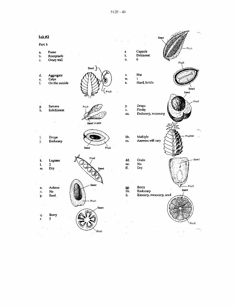
512F - 40

512F - 41
Part II:
1. After fertilization, the ovary wall develops three distinct layers that surround the ovule. The ovule develops into the seed. The fruit is the ripened ovary.
2. Sunflowers, beans, peas, peanut 3. The fruit has one or more ripened ovaries containing a seed or seeds. 4. Aggregate fruits develop from many ovaries within a single flower while multiple fruits develop
from many flowers. 5. They both develop specialized tissue in addition to the ovary.
Lab #4
Part I:
a. The point where the seed was attached to the wall of the pod (ovary). b. It is the opening in the ovule through which the pollen tube grew to deliver the sperm cell to the
egg. c. Yes. They must first be fertilized and then receive nourishment from the parent plant. d. The seed has expanded and the seed coat is wrinkled. e. Water has been absorbed. f. The cotyledons are fleshy. g. Food storage. h. The cotyledons. i. The starch will be digested by enzymes to supply glucose as food for the growing plant.
Part II:
a. A group of flowers. Each grain of corn contains a seed which is surrounded by a seed coat, the ovary wall of an individual flower.
b. It marks the point where the silk was attached to the ovule. c. 250. For each corn grain to have matured, there must have been a silk through which the tube cell
grew to accomplish fertilization. d. Yes. They are present but not plainly visible since they are covered by a three-layered fruit coat.
They are located within the point of attachment e. Purple or blue-black. f. Starch.

512F - 42
Part III:
a. The radicle (root). b. Further development of the radicle into the primary root serves to establish a means of absorbing
water. c. It is arched or bent over. d. It would serve to break the soil and prevent damage to the delicate leaves of the epicotyl. e. They are attached to the hypocotyl. f. As the plant becomes photosynthetically independent, they wither and fall off.
Corn Grain:
a. The shoot is growing upward and the somewhat longer root is growing downward. b. The shoot exhibits negative geotropism and the root positive geotropism. c. They protect the meristematic regions as growth occurs. d. It disintegrates.

512F - 43
Part IV:
a. epicotyl b. primary root c. cotyledons d. hilum e. endosperm f. micropyle g. epicotyl sheath h. point of attachment

512F - 44
REPRODUCTIVE PLANT PARTS
AG 512 - F
UNIT TEST Name _____________________________________ Score __________________________________
1. List in order the life cycle of a flower plant.
a. _____________________________________________________________________________
b. ____________________________________________________________________________
c. _____________________________________________________________________________
d. ____________________________________________________________________________
e. _____________________________________________________________________________
f. _____________________________________________________________________________
2. Label the parts of a complete flower. Write the correct names in the blanks provided.
3. Describe the parts and/or functions of the flower parts.
a. Filament ___________________________________________________________________
b. Corolla ____________________________________________________________________
c. Stigma _____________________________________________________________________
d. Anther _____________________________________________________________________

512F - 45
e. Style ______________________________________________________________________
f. Calyx ______________________________________________________________________
g. Ovary _____________________________________________________________________
h. Pedicil _____________________________________________________________________
i. Stamen ____________________________________________________________________
j. Pistil ______________________________________________________________________
4. Match the type of flower to the correct description. Write the correct numbers in the blanks.
_____a. Has only male flower parts 1. Complete
_____b. Has stamens and pistils, but no petals or sepals; 2. Incomplete common to monocots
3. Perfect _____c. Staminate and pistillate flowers found on the same
plant; corn 4. Imperfect
_____d. Has both stamens and pistils on the same flower 5. Staminate
_____e. Has only female flower parts 6. Pistillate
_____f. Staminate and pistillate flowers found on 7. Monoecious separate plants; spinach
8. Dioecious _____g. Has stamens, pistils, petals and sepals on the
same flower; common to dicots
_____h. Has either stamens or pistils, but not both on the same flower
5. Define fruit.
______________________________________________________________________________
______________________________________________________________________________
______________________________________________________________________________
6. List the three layers of the pericarp.
a. _____________________________________________________________________________
b. ____________________________________________________________________________
c. _____________________________________________________________________________

512F - 46
7. Match the types of fruits to their correct descriptions. Write the number of the correct answer in the blank.
_____a. Ovary wall is thin and fastened securely to a seed 1. Drupe
_____b. Ripened ovary becomes two-layered (outer layer 2. Aggregate fleshy, inner layer hard) forming a pit, enclosing seed 3. Pod
_____c. Several clustered flowers form compound fruit 4. Nut
_____d. Ovary wall is hard; encloses one seed 5. Pome
_____e. Ovary is fleshy and usually juicy; contains 6. Capsule several seeds
7. Multiple fruit _____f. Thin ovary wall; single-chambered; contains
several seeds; splits along line when ripe 8. Berry
_____g. Winged fruit; wing attached to the ovary wall 9. Achene
_____h. Several chambers and seeds in ovary; splits open 10. Grain when mature
11. Samara _____i. Several pistils in single flower form compound fruit
_____j. Outer, fleshy layer developed from calyx and receptacle; ovary forms a leathery core containing seeds
_____k. Ovary wall isn't fastened to seeds
8. Label the parts of a monocot and a dicot seed. Write the correct names in the blanks.

512F - 47
9. Match the functions to the correct seed parts. Write the number of the correct answer in the blank.
_____a. Embryonic root 1. Seed coat
_____b. Stores food 2. Endosperm
_____c. Part of the embryonic shoot above the point of 3. Coleoptile attachment of the cotyledons
4. Cotyledons _____d. Protects the seed against injury and dehydration
5. Radicle _____e. Seed leaves; absorb endosperm and serve as food
reservoirs 6. Hypocotyl
_____f. Protects the epicotyl and leaves as they emerge 7. Epicotyl from the ground
_____g. Part of the embryonic shoot below the cotyledon attachment point

512F - 48
REPRODUCTIVE PLANT PARTS
AG 512 - F
ANSWERS TO TEST 1. (in order) Seed germination and seedling growth; Vegetative growth; Flower formation;
Pollination; Fertilization; Seed development 2. a. Stigma e. Petal
b. Anther f. Style c. Filament g. Ovary d. Sepal
3. a. Supports anther b. Petals of the flower c. Upper part of pistil that catches pollen d. Bears the pollen e. Supports stigma f. Sepals of the flower g. Produces ovules which develop into seeds h. Stalk of an individual flower i. Male part of the flower j. Female part where egg cell originates
4. a. 5 d. 3 g. 1 b. 2 e. 6 h. 4 c. 7 f. 8
5. An enlarged and ripened ovary enclosing one or more seeds, which are ripened ovules (some fruits may be seedless)
6. Exocarp; Mesocarp; Endocarp
7. a. 10 e. 8 i. 2 b. 1 f. 3 j. 5 c. 7 g. 11 k. 9 d. 4 h. 6
8. a. Epicotyl g. Seed coat b. Hypocotyl h. Epicotyl c. Radicle i. Hypocotyl d. Cotyledon j. Radicle e. Coleoptile k. Cotyledons f. Endosperm l. Seed coat
9. a. 5 e. 4 b. 2 f. 3 c. 7 g. 6 d. 1

512G - 1
VEGETATIVE PLANT GROWTH
AG 512 - G
UNIT OBJECTIVE After completion of this unit, students should be able to list the stages of plant growth and development and the conditions affecting the vegetative growth of plants. Students should also be able to list nutrient elements in plant nutrition and deficiency symptoms. This knowledge will be demonstrated by completion of the unit test with a minimum of 85 percent accuracy.
SPECIFIC OBJECTIVES AND COMPETENCIES
After completion of this unit, the student should be able to:
1. Name the stages of plant growth and development.
2. List and discuss the three conditions affecting the vegetative growth of plants.
3. List the three primary nutrient elements in plant nutrition and two deficiency symptoms of each.
4. List the three secondary nutrient elements in plant nutrition and two deficiency symptoms
of each.
5. List three micronutrient elements in plant nutrition and one deficiency symptom of each.
6. Explain the relationships between reproductive and vegetative plant growth.
7. Name the three vegetative growth stages of small grains.
8. Name the four vegetative growth stages of corn.
9. Observe and measure the growth of plants.

512G - 2
VEGETATIVE PLANT GROWTH
AG 512 - G
SUGGESTED ACTIVITIES
I. Suggested activities for instructor
A. Order materials to supplement unit.
1. Literature
a. Agronomy Curriculum Materials Packet, 232 pages; available from IAVIM, 208 Davidson Hall, Iowa State University, Ames, Iowa 50011; approximate cost $10.00, order no. 214.
b. Crop Production, 15 transparency masters; available from
IAVIM, 208 Davidson Hall, Iowa State University, Ames, Iowa 50011; approximate cost $2.25, order no. 517.
2. Filmstrips, slideshows, etc.
a. Agronomy, computer program; available from IAVIM, 208
Davidson Hall, Iowa State University, Ames, Iowa 50011; approximate cost $15.00, order no. 902.
b. Essential Plant Nutrients, color slide set discussing nutrient
deficiencies and prevention, nutrients and nitrogen cycle and how nutrients are absorbed; available from Vocational Education Productions, California Polytechnic State University, San Luis Obispo, California 93407 (1-800-235-4146); approximate cost $76.95; order no. 1-580-604J.
c. Plant Nutrition, VHS video; describes the structures and their
functions that enable a plant to make food; also shows how the xylem functions; available from Vocational Education Productions, California Polytechnic State University, San Luis Obispo, California 93407 (1-800-235-4146); approximate cost $99.95; order no. 6-083-109J.
B. Make transparencies and necessary copies of materials.
C. Provide students with objective sheet.
D. Provide students with information sheet.
E. Discuss unit and specific objectives.
F. Discuss information sheet.
G. Review and give test.
H. Reteach and retest if necessary.

512G - 3
II. Instructional materials
A. Objective sheet
B. Suggested activities
C. Information sheet
D. Transparency masters
1. TM 1--Conditions Affecting the Vegetative Growth of Crop Plants
2. TM 2--Comparison of Utilization of Sunlight by Crop Plants
3. TM 3--Plant Growth Variance With Temperature Change
4. TM 4--Rate of Photosynthesis and Respiration As Affected By Temperature
5. TM 5--Approximate Pounds of Water Required to Produce One Pound
of Dry Matter
6. TM 6--Vegetative Growth Stages of Wheat
7. TM 7--Vegetative Growth Stages of Corn
E. Instructor notes for laboratory exercise
F. Laboratory exercise
1. LE 1--Plant Growth
G. Answers to laboratory exercise
H. Test
I. Answers to test
III. Unit references
A. Crop and Soil Science Curriculum Guide, Idaho State Board for Vocational Education, Boise, Idaho, 1985.
B. Delorit, R.J., et al., Crop Production, 5th edition, Prentice-Hall, Inc., Englewood
Cliffs, New Jersey, 1988. C. Hudson, H.T., et al., Plant Science: Growth, Development, and Utilization of
Cultivated Plants, 2nd edition, Prentice-Hall, Inc., Englewood Cliffs, New Jersey, 1988.
D. Janick, J., et al., Plant Science, 2nd edition, W.H. Freeman and Company, San
Francisco, California, 1974.

512G - 4
E. Otto, James H., and Towle, Albert, Modern Biology, Holt, Rinehart and Winston, New York, 1985.
F. Slesnick, Irwin L., et al., Biology, Scott, Foresman and Company, Glenview,
Illinois, 1985.

512G - 5
VEGETATIVE PLANT GROWTH
AG 512 - G
INFORMATION SHEET
I. Stages of plant growth and development
A. Seed germination and seedling growth
B. Vegetative
C. Reproduction
II. Conditions affecting the vegetative growth of crop plants (Transparency 1)
A. Climate (Transparency 2)
1. Sunlight
(Note: Sunlight is the energy source for photosynthesis. More efficient use of sunlight by a crop plant will result in higher yields if other factors are not limiting.)
2. Temperature (Transparencies 3, 4)
(Note: The temperature of both air and soil affects the rates at which the different plant processes take place. Air temperature affects the rate of photosynthesis, respiration and transpiration. Soil temperature has an effect on respiration and absorption by the roots.)
3. Water (Transparency 5)
(Note: Water can be a severe limiting factor in the growth of crop plants. The availability of water, either by precipitation or irrigation, influences crop yield more than any other factor. Water is a requirement for food manufacture, a solvent for mineral nutrients and a part of the transpiration process.)
B. Soil features
1. Nutrient availability
2. Moisture storage
3. Soil compaction
a. Reduced water infiltration
b. Reduced root penetration
4. Amount of erosion

512G - 6
C. Crop pests
1. Disease
2. Insects
3. Weeds
III. Primary nutrient elements and deficiency symptoms
A. Nitrogen
1. Stunted growth
2. Delayed maturity
3. Light green leaves
4. Yellow or dead lower leaves
B. Phosphorus
1. Purple-colored leaves, stems and branches
2. Reduced yields of seeds and fruits
3. Stunted growth
C. Potassium
1. Reduced yields
2. Mottled, spotted or curled older leaves
3. Marginal burning of leaves
4. Weak root system
5. Weak stalks
IV. Secondary nutrient elements and deficiency symptoms
A. Calcium
1. Deformed terminal leaves
2. Reduced root growth
3. Failure of terminal bud to grow
4. Plant may turn black
5. May have dead spots in midrib

512G - 7
B. Magnesium
1. Chlorosis affects the older leaves first, and later the younger ones
(Note: Chlorosis is a condition in plants relating to the failure of chlorophyll to develop. Chlorotic leaves range from light green through yellow to almost white.)
2. Leaves may droop
C. Sulfur
1. Light green leaves
2. Reduced growth
3. Yellowing of leaves
4. Weak stems
V. Micronutrient elements and deficiency symptoms
A. Boron
1. Terminal buds die
2. Lateral branches begin to grow, then lateral buds die
3. Branches form rosettes
4. Leaves thicken, curl and become brittle
5. Terminal leaf buds die
6. Chlorotic leaves
7. Stunted growth
8. Terminal leaves die
B. Copper
1. Terminal leaf buds die
2. Chlorotic leaves
3. Stunted growth
4. Terminal leaves die

512G - 8
C. Chlorine
1. Plants wilt
2. Chlorotic leaves
3. Some leaf necrosis (death associated with discoloration and
dehydration)
4. Bronzing in leaves
D. Iron
1. Paling or yellowing of leaves
2. Chlorosis between veins at first
3. Grasses develop alternate rows of yellowing and green stripes in leaves
E. Manganese
1. Chlorotic mottling
2. Leaves later become white
F. Molybdenum
1. Plants may become nitrogen deficient
2. Pale green, rolled or cupped leaves, with yellow spots
3. Leaves of crucifers become narrow
4. Cereal glumes don't fill out
G. Zinc
1. Abnormal roots
2. Mottled, bronzed or rosetted leaves
3. Chlorotic leaves
VI. Relationships between reproductive and vegetative plant growth
A. Reproductive plant growth may depend on vegetative plant growth for food (photosynthesis)
B. In many species, the apical meristems above ground produce vegetative growth
during the early part of the growing season and later switch to reproductive plant growth (typical in monocots such as corn, forage grasses and small grains)

512G - 9
C. In some plants, vegetative growth and reproductive growth occur at the same time
VII. Vegetative growth stages of small grains (Transparency 6)
A. Tillering
B. Jointing
C. Boot
VIII. Vegetative growth stages of corn (Transparency 7)
A. Two-leaf stage
B. Six-leaf stage
C. Ten-leaf stage
D. Fourteen-leaf stage

512G - 10
TM 1

512G - 11
TM 2

512G - 12
TM 3

512G - 13
TM 4

512G - 14
TM 5

512G - 15
TM 6

512G - 16
TM 7

512G - 17
VEGETATIVE PLANT GROWTH
AG 512 - G
INSTRUCTOR NOTES FOR LABORATORY EXERCISE Lab #1
Part I:
For each student, plant 2 bean seeds in an individual container 2 - 3 weeks in advance. Place the containers under a good light source so that the plants grow normally. Part II:
Demonstrate the marking of the leaves with India ink prior to the students' marking of the leaves.
Review the metric scale of measurements to insure proper measurements by the students.

512G - 18
VEGETATIVE PLANT GROWTH
AG 512 - G
LABORATORY EXERCISE #1--PLANT GROWTH Name _____________________________________ Score __________________________________
Selection from Modern Biology, Biology Investigations, Teacher's Edition, by James H. Otto, Albert Towle, W. David Otto, and Myra E. Madnick. Copyright 1977 by Holt, Rinehart and Winston, Inc. Reprinted by permission of the publisher. Materials needed
1 vigorously growing bean plant in an individual container 2 bean seeds 1 flower pot (7 to 10 cm) or suitable container Mixture of sand and loam (1:1) Metric ruler (divided into millimeters) Thread India ink
Part I: Observing Vegetative Organs
Obtain a bean plant growing in an individual container. The plant should be about 15-18 cm tall. Turn over the container to empty the entire mass of soil and roots. Wash the soil from the roots to expose the root system. You are now able to observe all of the vegetative organs of a seed plant. a. What parts of the plant can you observe? _____________________________________________
______________________________________________________________________________
b. What does it mean for an organ to be vegetative? _______________________________________
______________________________________________________________________________
c. Examine the root system closely, and describe what you see. _____________________________
______________________________________________________________________________
d. What characteristic may be observed which indicates the anchoring function of the roots in the
soil? __________________________________________________________________________
______________________________________________________________________________
e. What are some other functions of roots? ______________________________________________
______________________________________________________________________________

512G - 19
Examine the stem.
f. What obvious plant organ is attached to the stem? ______________________________________
______________________________________________________________________________
g. On the basis of your answer to (f), what is one of the functions of the stem? __________________
______________________________________________________________________________
h. What color is the stem? ___________________________________________________________
i. What pigment is present? __________________________________________________________
j. Name another function that may be carried out in the stem. _______________________________
______________________________________________________________________________
k. How do water and minerals get to the leaves from the roots? ______________________________
______________________________________________________________________________
l. How do materials move from the leaves to the roots? ____________________________________
______________________________________________________________________________
m. On the basis of your answers in (k) and (l), what function is being performed? ________________
___________________________________ Like the root, the stem often functions in the
storage of food. Examine the leaves.
n. What color are they? _____________________________________________________________
o. What is the principal function of the leaves? ___________________________________________
In the space provided, sketch the entire plant. Include the details of the branching pattern of the roots, and the shape and venation of the leaves. Label: roots, stem and leaf. Summarize the functions of each vegetative organ.

512G - 20
Part II: How Fast Do Plant Parts Grow? Plant a bean seed in a pot containing equal parts of sand and loam. Plant it just below the soil surface. Water the soil well and pour off the excess. Place the pot in a light source and keep the soil moist. Once the true leaves of the plant are formed, your observations and determination of the rate of growth may begin. To observe where the leaf expands in its growth, mark a small leaf in the following manner: Draw a piece of thread (15-20 cm) tightly between the forefinger and thumb of each hand. Have your partner moisten the thread with the applicator from an India ink bottle. Carefully place the moistened thread on the leaf to make a straight line across the leaf. Repeat the procedure and make the next line approximately 3 mm from the first. Continue until the leaf is marked as shown in the figure below. In the same manner, mark the stem from the soil surface to the tip of the stem.
a. What will the markings help you to observe? __________________________________________
______________________________________________________________________________
As the leaf expands, record your observations in a series of drawings by accurately representing the regions of expansion.

512G - 21
b. How will you be able to determine where the leaf and stem grew? _________________________
______________________________________________________________________________
______________________________________________________________________________
Use a metric ruler to obtain actual growth measurements. Measure in millimeters the length of the stem from the soil level to the growing tip. Record the measurement in the table below. Count and record the number of leaves. Determine the surface area of each leaf by multiplying the length of the leaf (base to tip) by the average width (measure in three places) of the blade. Record the total surface area of all leaves. Repeat the measurement at 2-3 day intervals for a period of two weeks. Record all data in the table. After the last measurements are recorded, carefully remove the plant from the soil. Wash the soil from the roots. Measure each root and record the total length of the root system. Base your answers on the markings and the measurements taken.
c. Was the rate of growth uniform in the stems and leaves during the growth period?
______________________________________________________________________________
Date of Measurement
Interval Between Measurement
Length of Stem
Number of Leaves
Total Leaf Area
Total Length of Roots (Last Measurements)
d. If not, when is the rate of growth most rapid? __________________________________________
______________________________________________________________________________
______________________________________________________________________________
e. Compare the total length of the stem with that of the root. ________________________________
______________________________________________________________________________
f. Did the leaf blades continue growth at a uniform rate? ___________________________________
g. If not, what variation occurred? _____________________________________________________
______________________________________________________________________________

512G - 22
h. Where were new leaves produced? __________________________________________________
i. How does the area of the plant above and below the ground compare? ______________________
______________________________________________________________________________
Part III: Summary
a. Using a suitable scale of numerical value, prepare a graph with separate lines for length of stem, number of leaves and total leaf area. Explain any observable relationship.
______________________________________________________________________________
______________________________________________________________________________
Days
In the space provided, indicate which vegetative organ(s) of the plant perform the function indicated.
b. ____________________________________________ conduct water and minerals to upper plant parts.
c. ____________________________________________ are the principal organs of photosynthesis.
d. ____________________________________________ serve to anchor the plant.
e. ____________________________________________ produce leaves.
f. ____________________________________________ store food substances.
g. ____________________________________________ exchange gases between the plant and the atmosphere.
h. ____________________________________________ absorb water and minerals.

512G - 23
i. _____________________________________________ have a secondary function of photosynthesis.
j. _____________________________________________ conduct water and minerals up and down the plant.
k. _____________________________________________ display leaves to light.
l. _____________________________________________ function in the process of transpiration.
Part IV: Investigations On Your Own
Select various growth media such as sand, vermiculite, heavy clay soil, etc. Observe how the type of soil influences seedling growth. The procedure as presented in Part 2 should be followed. To observe the effects of soil nutrients, you may wish to use laboratory prepared nutrient solutions or those prepared commercially in addition to selected soil types.

512G - 24
VEGETATIVE PLANT GROWTH
AG 512 - G
ANSWERS TO LABORATORY EXERCISE
Lab #1
Part I:
a. Roots, stems and leaves b. A vegetative organ performs all the processes necessary for life except the formation of seeds. c. The root system is highly branched and is lacking any color. d. The root system is highly branched and spreading. e. Absorption of water and minerals, conduction of water and minerals to other plant parts, food
storage f. Leaves g. Produce and display leaves to light h. Green i. Chlorophyll j. Photosynthesis k. They are carried in the stem. l. They are carried in the stem. m. Conduction n. Green o. Photosynthesis
Part II:
a. Where growth occurs in the leaves and stem
b. The squares on the leaf will change shape indicating that growth has occurred. The markings on the stem will become further apart indicating growth has taken place in that region of the stem.
c. No
Answers will vary on the chart
d. Growth should proceed more rapidly during the first half of the growth period and then slow down.
e. Answers will vary
f. No
g. The squares formed by the lines on the leaf have changed shape. This indicates that the leaf grew more from the center regions of the leaf.
h. At the sides of the shoot apex
i. The area is approximately the same.

512G - 25
Part III:
a. Generally, the three curves will follow the same pattern. As the stem length increases, more leaves are produced and the total surface area of the leaves will also increase.
b. Roots c. Leaves d. Roots e. Stems f. Roots and stems g. Leaves h. Roots i. Stems j. Stems k. Stems l. Leaves

512G - 26
VEGETATIVE PLANT GROWTH
AG 512 - G
UNIT TEST Name _____________________________________ Score __________________________________
1. Name the three stages of plant growth and development.
a. _____________________________________________________________________________
b. ____________________________________________________________________________
c. _____________________________________________________________________________
2. List and discuss the three conditions affecting the vegetative growth of plants.
a. _____________________________________________________________________________
______________________________________________________________________________
b. ____________________________________________________________________________
______________________________________________________________________________
c. _____________________________________________________________________________
______________________________________________________________________________
3. List the three primary nutrient elements in plant nutrition and two deficiency symptoms of each.
a. Nutrient ________________________________________________________________
Deficiency symptoms 1. ________________________________________________
2. ________________________________________________
b. Nutrient ________________________________________________________________
Deficiency symptoms 1. ________________________________________________
2. ________________________________________________
c. Nutrient ________________________________________________________________
Deficiency symptoms 1. ________________________________________________
2. ________________________________________________

512G - 27
4. List the three secondary nutrient elements in plant nutrition and two deficiency symptoms of each.
a. Nutrient ________________________________________________________________
Deficiency symptoms 1. ________________________________________________
2. ________________________________________________
b. Nutrient ________________________________________________________________
Deficiency symptoms 1. ________________________________________________
2. ________________________________________________
c. Nutrient ________________________________________________________________
Deficiency symptoms 1. ________________________________________________
2. ________________________________________________
5. List three micronutrient elements in plant nutrition and one deficiency symptom of each.
a. Nutrient ________________________________________________________________
Deficiency symptom ______________________________________________________
b. Nutrient ________________________________________________________________
Deficiency symptom ______________________________________________________
c. Nutrient ________________________________________________________________
Deficiency symptom ______________________________________________________
6. Explain the relationships between reproductive and vegetative plant growth.
______________________________________________________________________________
______________________________________________________________________________
______________________________________________________________________________
______________________________________________________________________________
______________________________________________________________________________
______________________________________________________________________________
______________________________________________________________________________
______________________________________________________________________________

512G - 28
7. Name the three vegetative growth stages of small grains.
a. _____________________________________________________________________________
b. ____________________________________________________________________________
c. _____________________________________________________________________________
8. Name the four vegetative growth stages of corn.
a. _____________________________________________________________________________
b. ____________________________________________________________________________
c. _____________________________________________________________________________
d. ____________________________________________________________________________

512G - 29
VEGETATIVE PLANT GROWTH
AG 512 - G
ANSWERS TO TEST 1. Seed germination and seedling growth; Vegetative; Reproduction
2. Climate: Sunlight; Temperature; Water Soil features: Nutrient availability; Moisture storage; Soil compaction; Reduced water infiltration; Reduced root penetration; Amount of erosion Crop pests: Disease; Insects; Weeds
3. Answer should include two of the following deficiency symptoms for each nutrient:
Nitrogen: Stunted growth; Delayed maturity; Light green leaves; Yellow or dead lower leaves Phosphorus: Purple-colored leaves, stems and branches; Reduced yields of seeds and fruits; Stunted growth Potassium: Reduced yields; Mottled, spotted or curled older leaves; Marginal burning of leaves; Weak root system; Weak stalks
4. Answer should include two of the following deficiency symptoms for each nutrient:
Calcium: Deformed terminal leaves; Reduced root growth; Failure of terminal bud to grow; Plant may turn black; May have dead spots in midrib Magnesium: Chlorosis affects the older leaves first, and later the younger ones; Leaves may droop Sulfur: Light green leaves; Reduced growth; Yellowing of leaves; Weak stems
5. Answer should include three of the following micronutrient elements and one deficiency for each:
Boron: Terminal buds die; Lateral branches begin to grow, then lateral buds die; Branches form rosettes; Leaves thicken, curl and become brittle; Terminal leaf buds die; Chlorotic leaves; Stunted growth; Terminal leaves die Copper: Terminal leaf buds die; Chlorotic leaves; Stunted growth; Terminal leaves die Chlorine: Plants wilt; Chlorotic leaves; Some leaf necrosis (death associated with discoloration and dehydration); Bronzing in leaves Iron: Paling or yellowing of leaves; Chlorosis between veins at first; Grasses develop alternate rows of yellowing and green stripes in leaves Manganese: Chlorotic mottling; Leaves later become white Molybdenum: Plants may become nitrogen deficient; Pale green, rolled or cupped leaves, with yellow spots; Leaves of crucifers become narrow; Cereal glumes don't fill out Zinc: Abnormal roots; Mottled, bronzed or rosetted leaves; Chlorotic leaves
6. Reproductive plant growth may depend on vegetative plant growth for food (photosynthesis); In many species, the apical meristems above ground produce vegetative growth during the early part of the growing season and later switch to reproductive plant growth (typical in monocots such as corn, forage grasses and small grains); In some plants, vegetative growth and reproductive growth occur at the same time
7. Tillering; Jointing; Boot
8. Two-leaf stage; Six-leaf stage; Ten-leaf stage; Fourteen-leaf stage

512H - 1
REPRODUCTIVE PLANT GROWTH
AG 512 - H
UNIT OBJECTIVE
After completion of this unit, students should be able to define sexual and asexual reproduction in plants. Students should be able to describe asexual reproduction methods and list the reproductive growth phases. Students should also be able to explain the process of fertilization and discuss seed germination. This knowledge will be demonstrated by completion of the laboratory exercise and unit test with a minimum of 85 percent accuracy.
SPECIFIC OBJECTIVES AND COMPETENCIES
After completion of this unit, the student should be able to:
1. Describe sexual and asexual reproduction in plants.
2. List and describe the five methods of asexual reproduction.
3. List the seven phases of reproductive growth.
4. Match the type of pollination to the correct description.
5. Name three ways pollen is moved.
6. Explain the process of fertilization in plants.
7. List and briefly discuss the three essential parts of the seed.
8. Describe the stages of seed germination of a monocot.
9. Describe the stages of seed germination of a dicot.
10. List the three requirements for good seed germination.
11. List ten factors that cause poor seed germination.
12. Study plant reproduction without seeds.
13. Grow a bean plant.

512H - 2
REPRODUCTIVE PLANT GROWTH
AG 512 - H
SUGGESTED ACTIVITIES
I. Suggested activities for instructor
A. Order materials to supplement unit.
1. Literature
a. Agronomy Curriculum Materials Packet, 232 pages; available from IAVIM, 208 Davidson Hall, Iowa State University, Ames, Iowa 50011; approximate cost $10.00, order no. 214.
b. Crop Production, 15 transparency masters; available from
IAVIM, 208 Davidson Hall, Iowa State University, Ames, Iowa 50011; approximate cost $2.25, order no. 517.
2. Filmstrips, slideshows, etc.
a. Agronomy, computer program; available from IAVIM, 208 Davidson Hall, Iowa State University, Ames, Iowa 50011; approximate cost $15.00, order no. 902.
b. Budding and Grafting, sound filmstrip; available from
Vocational Education Productions, California Polytechnic State University, San Luis Obispo, California 93407 (1-800-235-4146); approximate cost $44.95; order no. 1-206-230J.
c. Plant Propagation, Volume I, VHS video; describes four
methods of propagation: by seed, by division, by bulbs and tubers, and by micropropagation; available from Vocational Education Productions, California Polytechnic State University, San Luis Obispo, California 93407 (1-800-235-4146); approximate cost $75; order no. 6-044-101J.
d. Plant Propagation, Volume II, VHS video; describes
propagation by cuttings, layering and by grafting and budding; available from Vocational Education Productions, California Polytechnic State University, San Luis Obispo, California 93407 (1-800-235-4146); approximate cost $75; order no. 6-044-102J.
e. Plant Reproduction, VHS video; illustrates male and female
flower parts and how fertilization takes place; available from Vocational Education Productions, California Polytechnic State University, San Luis Obispo, California 93407 (1-800-235-4146); approximate cost $99.95; order no. 6-083-110J.

512H - 3
f. Romancing the Seed, 55 minute VHS video; covers the production, care and planting of seeds; available from Vocational Education Productions, California Polytechnic State University, San Luis Obispo, California 93407 (1-800-235-4146); approximate cost $29.95; order no. 6-072-101J.
B. Make transparencies and necessary copies of materials.
C. Provide students with information sheets and laboratory exercises.
D. Discuss unit and specific objectives.
E. Discuss information sheets.
F. Demonstrate and discuss procedures outlined in laboratory exercises.
G. Review and give test.
H. Reteach and retest if necessary.
II. Instructional materials
A. Objective sheet
B. Suggested activities
C. Information sheet
D. Transparency masters
1. TM 1--Reproductive Growth Phases
2. TM 2--Self-Pollination and Cross-Pollination
3. TM 3--Stages in Germination and Emergence of Corn
4. TM 4--Stages in Germination and Emergence of a Bean Seed
E. Instructor notes for laboratory exercises
F. Laboratory exercises
1. LE 1--Plant Reproduction Without Seeds
2. LE 2--Growing a Bean Plant
G. Answers to laboratory exercises
H. Test
I. Answers to test

512H - 4
III. Unit references
A. Agricultural Education Curriculum, College of Agriculture, University of Illinois, Urbana, Illinois, 1989.
B. Delorit, R. J., et al., Crop Production, 5th edition, Prentice-Hall, Inc.,
Englewood Cliffs, New Jersey, 1984. C. Hartmann, Hudson T., et al., Plant Science: Growth, Development and
Utilization of Cultivated Plants, 2nd edition, Prentice-Hall, Inc., Englewood Cliffs, New Jersey, 1988.
D. Idaho Crop and Soil Curriculum Guide, Idaho State Board for Vocational
Education, 1985. E. Janick, Jules, et al., Plant Science, 2nd edition, W.H. Freeman and Co., San
Francisco, California, 1974. F. Model Agricultural Core Curriculum, California State Department of Education,
University of California, Davis, California, August 1989. G. Otto, James H., Towle, Albert, Modern Biology, Holt, Rinehart and Winston,
New York, 1985. H. Slesnick, Irwin L., et al., Biology, Scott, Foresman and Company, Glenview,
Illinois, 1985.

512H - 5
REPRODUCTIVE PLANT GROWTH
AG 512 - H
INFORMATION SHEET
I. Sexual reproduction in plants
A. Reproduction by seed
1. Involves the combination of two different sets of genes to create offspring with a new genetic makeup
2. Often the most efficient and economical method for reproducing annual
bedding plants and some biennials and perennials 3. The function of the seed is to produce a new plant
a. A seed is produced by the combination of nuclear material in the process of fertilization
b. Results in zygote formation
B. Sexual reproduction usually used for annuals and on plants which grow quickly from seed and produce a plant similar to the parents
II. Asexual reproduction
A. Reproduction by vegetative propagation
1. Uses plant parts such as leaves, roots and stems to start new plants
2. No new genetic material introduced--the offspring will be identical to parents
B. Methods
1. Cuttings
a. Stem cuttings using a tip (straight) cutting
b. Leaf cuttings using a leaf section, leaf petiole or by cutting the veins
c. Root cuttings using a cutting of the root and planting it
2. Layering--Rooting a stem at the node
(Note: Grape layering to replace a vine or strawberries' natural runners are examples of layering.)
3. Separation--Removing corms or bulbets from the parent bulb plant (for
example: an iris)

512H - 6
4. Division--The removal of new shoots with some root from below
(Note: This is used on dahlias.)
5. Grafting--Involves the transfer of wood with buds from one plant and matching up its cambium layer to another plant. The ends then grow together, resulting in a plant having desirable qualities of both parent plants
III. Phases of reproductive growth (Transparency 1)
A. Flower induction and initiation
B. Flower differentiation and development
C. Pollination
D. Fertilization
E. Fruit set and seed formation
F. Growth and maturation of fruit and seed
G. Fruit senescence
IV. Types of pollination (Transparency 2)
A. Self-pollination--Transfer of pollen from the anthers to the stigma of the same flower on the same plant
B. Cross-pollination--Transfer of pollen from the anthers of one plant to the
stigmas of another plant
(Note: Cross-pollination usually requires an insect or bee to transfer the pollen from one plant to the other.)
V. Pollen is moved by
A. Gravity
B. Wind
C. Insects
D. Birds
E. Man
VI. Fertilization--After a pollen grain alights on the surface of the stigma, it forms a pollen tube. The pollen tube grows down the style to the ovary. It penetrates the ovary and the male cell unites with the ovule. This is called fertilization, the union of the male and female cells. The result is a zygote. Cell division takes place and the zygote becomes the embryo of the seed

512H - 7
VII. Parts of the seed
A. Embryo--Develops into the new plant
B. Food storage material--Nourishes the embryonic plant
1. Endosperm tissue
2. Fleshy cotyledons (a part of the embryo)
C. Seed coverings--Usually the two seed coats (may include other parts of the ovary wall)
VIII. Stages of germination (Transparencies 3, 4)
A. Monocot (corn, small grains) (Transparency 3)
1. Absorption of water and oxygen into the seed
2. The seed coat ruptures and the primary root (radicle) begins to grow downward
3. The epicotyl elongates, the coleoptile piercing the soil as it grows
upward
(Note: The leaves of the coleoptile are rolled into tight pointed buds.)
4. The coleoptile emerges
(Note: When the coleoptile emerges, the first node on the stem is still underground. It is from this node that the secondary root system develops.)
5. The coleoptile unfolds
(Note: When the leaves of a seedling emerge above the soil surface and unfold, the plant is then capable of manufacturing its own food.)
B. Dicot (beans, peas) (Transparency 4)
1. Absorption of water and oxygen into the seed
2. The seed coat ruptures and the primary root (radicle) begins to grow downward
3. The hypocotyl curves into a loop and pushes through the soil, pulling
the cotyledons toward the soil surface
4. Emergence of seedling occurs
(Note: The curve in the hypocotyl straightens out immediately after emergence so the plant will stand correctly.)

512H - 8
5. The cotyledons spread apart and the stem tip is exposed to air and sunlight (Note: When the first pair of leaves has emerged, the plant is then capable of manufacturing its own food.)
IX. Requirements for good seed germination
A. Proper temperature
(Note: This requirement varies for different crops. Cereals will show some germination at 32oF, while corn will not show any germination until 48oF.)
B. Sufficient moisture
(Note: This requirement varies for different crops. Cereals will germinate when their moisture content is about 50%. Soybeans will not germinate until their moisture content is about 75%. The range is 26% to 75% for most agronomic crops.)
C. Ample supply of oxygen
(Note: Germination will not occur if oxygen is not available for crops like small grains and peas. Rice seed can germinate in the absence of oxygen.)
X. Factors that cause poor seed germination
A. Mechanical injury to seed (cracked grain)
B. Disease
C. Storage conditions
(Note: Temperature and humidity are important considerations for storage of crop seeds.)
D. Age of seed
(Note: Germination percentages will decrease as the age of the seed increases.)
E. Soil temperature too cold
F. Hard seed coat
(Note: Some plants (hard-seeded legumes) produce seeds with a hard seed coat. The seed coat will not allow moisture and oxygen to enter the seed and bring about germination.)
G. Soil moisture insufficient
H. Planting too deep

512H - 9
I. Chemical damage
(Note: Reduced germination percentages may result if seeds come in contact with chemicals such as fertilizers.)
J. Crusting of soil

512H - 10
TM 1

512H - 11
TM 2

512H - 12
TM 3

512H - 13
TM 4

512H - 14
REPRODUCTIVE PLANT GROWTH
AG 512 - H
INSTRUCTOR NOTES FOR LABORATORY EXERCISES
Lab #1
Mosses and ferns may be collected during the spring and summer and may be kept in terraria, dried and pressed until needed for study. Refer to unit AG 512-D--Nonvascular and Vascular Plants for information about alternation of generations.
Part II:
You may have the students remove several sporangia and prepare a wet mount. When examined under low power of the microscope, the spores should be visible within the sporangia.

512H - 15
REPRODUCTIVE PLANT GROWTH
AG 512 - H
LABORATORY EXERCISE #1--PLANT REPRODUCTION WITHOUT SEEDS
Name _____________________________________ Score __________________________________
Selection from Modern Biology, Biology Investigations, Teacher's Edition, by James H. Otto, Albert Towle, W. David Otto, and Myra E. Madnick. Copyright 1977 by Holt, Rinehart and Winston, Inc. Reprinted by permission of the publisher. Materials needed
Moss plants with female sex organs Microscope Moss plants with male sex organs Slide, cover glass Fresh clump of living moss plants Dissecting needle Dissecting microscope Fresh fern frond bearing sori Textbook or charts Hand lens
Part I: How Do Moss Plants Reproduce?
Moss plants may be found growing in dense clumps on the forest floor or compact mats on a fallen log. A clump of moss plants is composed of many gametophyte plants growing close to each other for support. At certain times of the year, the sporophytes may be seen growing from certain plants. Examine a small portion of a clump of moss, using a hand lens. Remove a single gametophyte and examine it closely under a dissecting microscope. The rootlike structures at the base of the leafy stem are rhizoids which function to absorb water and minerals. Examine a leafy shoot. a. Of what adaptive value is the arrangement of the leaves on the stem axis? _________________
_____________________________________________________________________________
Gamete-producing structures, archegonia and antheridia, are located at the tips of the leafy stems in a cluster of leaves. b. What gamete is produced by the archegonia? _________________________________________
c. by the antheridia? _______________________________________________________________
These gamete-producing structures may be observed by squeezing them from the tip of the stem. Roll the tip of the stem between the thumb and the forefinger as you bring the tip in contact with a large drop of water on a slide. Use a dissecting needle to remove the fragments of the tip. Some of these pieces will be either antheridia or archegonia depending on the sex of the plants used. Use pictures in a biology or botany textbook or charts as reference. Repeat the procedure for a plant of the opposite sex. Observe the slide under the low power of the microscope. d. Describe the structure of an antheridium. ____________________________________________
_____________________________________________________________________________

512H - 16
e. Describe the structure of an archegonium. ____________________________________________
______________________________________________________________________________
______________________________________________________________________________
f. How does a sperm reach an egg for fertilization? _______________________________________
______________________________________________________________________________
g. Explain why mosses are not found growing in locations having little or no moisture. ______________________________________________________________________________
h. What cell is produced when the sperm fertilizes the egg? _________________________________
i. Where is the zygote formed? _______________________________________________________
Examine a gametophyte bearing a stalk and capsule.
j. Why is it known as the sporophyte generation? ________________________________________
______________________________________________________________________________
k. From what cell did the sporophyte develop? ___________________________________________
l. Explain why the sporophyte is present only on certain gametophyte plants. __________________
______________________________________________________________________________
Locate the capsule at the tip of the seta. Examine the capsule using a hand lens. Determine if a tiny lid, the operculum, is present. As spores mature within the capsule, the operculum will fall away. Under favorable conditions a spore germinates into a threadlike structure, the protonema. The protonema eventually develops into a mature gametophyte. m. What have you observed about the sporophyte that indicates it is nutritionally dependent on
the gametophyte? _______________________________________________________________
______________________________________________________________________________
______________________________________________________________________________
n. Explain how moss plants exhibit alternation of generations. ______________________________________________________________________________
______________________________________________________________________________
______________________________________________________________________________

512H - 17
Examine the stages in the life cycle of the moss plant. Label the following structures: rhizoids, archegonia, antheridia, egg, sperm, capsule, operculum, spores, protonema, young gametophyte. Also indicate the gametophyte generation and the sporophyte generation.
Part II: Alternation of Generations in Ferns
Examine the living fern frond or study the figure in the life cycle. The familiar fern plant is the sporophyte generation. Unlike the mosses, it is free living and independent. The fronds (leaves) of the fern grow from a horizontal, underground stem, the rhizome. The fronds first emerge from the ground as fiddleheads. Each consists of a stalk and a blade divided into leaflets or pinnae. When certain of the fronds mature, small dots known as sori (singular--sorus) are produced on the lower surface. A sorus contains a cluster of spore producing structures called sporangia. a. Compare the sporophytes of the moss and fern. ________________________________________
______________________________________________________________________________
______________________________________________________________________________
b. With what structure of a moss does a fern sorus compare? ________________________________
______________________________________________________________________________
Examine a single pinna with sori under a dissecting microscope.
c. Describe the number and position of the sori. __________________________________________
______________________________________________________________________________

512H - 18
When the spores are released and land in a favorable location, they germinate and develop into gametophytes. The gametophyte is called a prothallus. The prothallus measures about 1 cm in diameter. The prothallus produces antheridia and archegonia which produce sperms and eggs. d. How is fertilization accomplished in ferns? ___________________________________________
______________________________________________________________________________
e. How does the size and structure of the prothallus make this possible? _______________________
______________________________________________________________________________
______________________________________________________________________________
The cell formed upon fertilization is the zygote.
f. What generation does the zygote develop into? ________________________________________
On the diagram of the fern life cycle, label: frond, pinna, egg, spores, sporangium, sperm, prothallus, young sporophyte, antheridium, archegonium, sorus. Also indicate the sporophyte generation and the gametophyte generation.
Part III: Summary a. From what you have observed and studied, relate how mosses and ferns reproduce without
seeds. _________________________________________________________________________
______________________________________________________________________________
______________________________________________________________________________
______________________________________________________________________________

512H - 19
b. Summarize your observations of mosses and ferns by filling in the blanks of the following chart.
Mosses Ferns
1. Produces Eggs
2. Produces Sperm
3. Sporophyte independent (Yes/No)
4. Structure producing spores
5. Spores germinate into:
6. Inconspicuous germination
7. Conspicious germination
8. Medium for fertilizer
9. Agent for spore disposal
10. First cell of sporophyte
11. First cell of gametophyte
12. General habitat
Part IV: Investigations On Your Own
1. To become acquainted with the mosses in your region and to determine their various habitats, make a collection of mosses. Look in any environment where there is moisture and reduced light. Collect shoots bearing stalks and capsules. Place the specimens in different envelopes and record the location, date and habitat in your notebook. Use identification keys to trace your specimens to their particular genus. Leaf and capsule structure are used for identification purposes. The characteristic size and shape of spores are often used for identification.
2. Make a collection of fern fronds. Before beginning your collection, you should become familiar
with the characteristics used in their identification. These include: general form and structure; distribution of the sori, the sporangia and the indusia; and the form of the frond. Collect a portion of a frond and put it in a book or magazine. Assign a collection number to the specimen and record this number in your field notebook along with other data such as the location, date and habitat. When you return to the laboratory, identify your specimens. The fronds can be pressed and saved for later study by drying them between several layers of newspaper.

512H - 20
REPRODUCTIVE PLANT GROWTH
AG 512 - H
LABORATORY EXERCISE #2--GROWING A BEAN PLANT Name _____________________________________ Score __________________________________
Introduction
Beans for cross-pollination are raised inside greenhouses to avoid unfavorable environmental conditions and accidental cross-pollination. Remember the flowers need to be protected in order to accomplish cross-pollination. So a growth chamber, greenhouse or a shelter must be constructed first. Part I: Planting the Bean Seeds
1. The beans may first be treated with a suitable fungicide before being planted.
2. Well-drained and fertile soil, such as a mixture of loam, peat and sand in a 7,3,2 soil mixture with a pH of 6 to 6.5, is needed. You can mix this soil yourself and autoclave it to rid it of pathogens or you can buy it in bulk from your local nursery.
3. Seeds can be planted directly into (15-20 cm) clay or plastic pots. Or they can be germinated in a
planting medium such as Perlite and then transplanted into the pots that contain the 7,3,2 soil mixture.
Part II: Preparing the Pots and Planting
1. Fill the pots to an inch below the rim with 7,3,2 soil mixture and wet it down thoroughly. Peat is very hard to get moist if it is dry and moist soil is needed for the seeds to germinate.
2. Plant one to two plants into the pots. Use your first and middle fingers to poke out the holes in the
middle of the pot. Seeds should be planted at a half an inch deep. 3. After planting the seeds, go back and water the pot thoroughly. Beans are very sensitive to water
logged soil so this will be the only watering for the next three days. Part III: Care of the New Plant
1. Two to three applications of a complete nutrient solution (N, P, K, and Ca, Zn, Bo, Mg, and Mo) is needed throughout the growing season.
2. Bean plants do not react well to the water-logged soil or insufficient water. So a regular watering
program must be developed to suit the needs of your particular growth chamber. 3. Splashing the leaves of the bean plants must be avoided when watering the plants because this
helps to spread pathogens.

512H - 21
Part IV: Temperature Considerations
1. The optimum daytime temperatures should be between 20-25oC ideally and the night temperatures should be around 5oC cooler than that.
a. Temperatures below 15oC cause the plant to fail to grow and to flower abnormally. The
results are poor and small flowers for cross-pollination.
b. Temperatures above 30oC can result in poor growth and reproduction. The high temperatures and photoperiod can react together and produce very spindly plants. If this is mixed with moisture stress the plants can lose their flowers and pods are aborted.
Part V: Disease and Pests
1. It is extremely important that the bean plants are both disease and pest free.
a. Different chemicals should be used periodically to prevent resistance to a chemical. Because the leaves are very sensitive, high concentrations of pesticides should not be used.
b. Pests that may be troublesome are the following: White fly, Spider mites, Aphids and
Leaf miner.

512H - 22
REPRODUCTIVE PLANT GROWTH
AG 512 - H
ANSWERS TO LABORATORY EXERCISES
Lab #1
Part I:
a. The leaves are arranged in whorls around the axis allowing the greatest possible exposure of leaves to light.
b. egg c. sperm d. It is a saclike structure supported on a short stalk. e. It is bottle-shaped and consists of a slender neck and a swollen base. f. It swims to the egg. g. An abundant supply of moisture is necessary for the sperm to swim to the egg. h. zygote i. within the archegonium j. In this generation, a moss plant produces spores. k. zygote l. Fertilization may not have been accomplished in all gametophytes containing an archegonium. m. When mature, it lacks chlorophyll and it is structurally attached to the gametophyte. n. The union of an egg and sperm produced by gametophyte plants gives rise to a zygote which
develops into a spore-producing plant. Spores produced by the sporophyte germinate and grow into a new gametophyte and the cycle is repeated.
Part II:
a. The fern sporophyte is much larger and grows independently of the gametophyte. b. the capsule of the moss sporophyte c. Answers will vary depending on the species of fern being used. d. The sperms swim to the egg in a film of water. e. It is flat and grows close to the soil, and its size requires that only small amounts of moisture be
present for fertilization. f. the sporophyte

512H - 23
Part III: a. Reproduction is dependent on the production of egg and sperm in the gametophyte plant. These
form the zygote upon fertilization. The zygote develops into the sporophyte. The spores produced by both plants germinate and develop into gametophytes. There is no seed production.
b.
Mosses Ferns
1. Produces Eggs archegonium archegonium
2. Produces Sperm antheridium antheridium
3. Sporophyte independent (Yes/No) no yes
4. Structure producing spores capsule sporangium
5. Spores germinate into: protonema prothallus
6. Inconspicuous germination sporophyte gametophyte
7. Conspicious germination gametophyte sporophyte
8. Medium for fertilizer water water
9. Agent for spore disposal wind wind
10. First cell of sporophyte zygote zygote
11. First cell of gametophyte spore spore
12. General habitat damp, shaded environments
damp, shaded environments

512H - 24
REPRODUCTIVE PLANT GROWTH
AG 512 - H
UNIT TEST Name _____________________________________ Score __________________________________
1. Describe sexual and asexual reproduction in plants.
Sexual ________________________________________________________________________
______________________________________________________________________________
______________________________________________________________________________
______________________________________________________________________________
Asexual _______________________________________________________________________
______________________________________________________________________________
______________________________________________________________________________
______________________________________________________________________________
2. List and describe the five methods of asexual reproduction.
a. _____________________________________________________________________________
______________________________________________________________________________
______________________________________________________________________________
______________________________________________________________________________
b. ____________________________________________________________________________
______________________________________________________________________________
______________________________________________________________________________
______________________________________________________________________________
c. _____________________________________________________________________________
______________________________________________________________________________
______________________________________________________________________________
______________________________________________________________________________

512H - 25
d. ____________________________________________________________________________
______________________________________________________________________________
______________________________________________________________________________
______________________________________________________________________________
e. _____________________________________________________________________________
______________________________________________________________________________
______________________________________________________________________________
______________________________________________________________________________
3. List the seven phases of reproductive growth.
a. _____________________________________________________________________________
b. ____________________________________________________________________________
c. _____________________________________________________________________________
d. ____________________________________________________________________________
e. _____________________________________________________________________________
f. _____________________________________________________________________________
g. ____________________________________________________________________________
4. Match the types of pollination to the correct description. Write the correct numbers in the blanks.
_____a. Transfer of pollen from the anthers to 1. Self-pollination the stigma of the same flower on the same plant 2. Cross-pollination
_____b. Transfer of pollen from the anthers of one plant to the stigmas of another plant
5. Name three ways pollen is moved.
a. _____________________________________________________________________________
b. ____________________________________________________________________________
c. _____________________________________________________________________________

512H - 26
6. Explain the process of fertilization in plants.
______________________________________________________________________________
______________________________________________________________________________
______________________________________________________________________________
______________________________________________________________________________
______________________________________________________________________________
______________________________________________________________________________
______________________________________________________________________________
______________________________________________________________________________
7. List and briefly discuss the three essential parts of the seed.
______________________________________________________________________________
______________________________________________________________________________
______________________________________________________________________________
______________________________________________________________________________
______________________________________________________________________________
______________________________________________________________________________
8. Describe the stages of seed germination of a monocot.
______________________________________________________________________________
______________________________________________________________________________
______________________________________________________________________________
______________________________________________________________________________
______________________________________________________________________________
______________________________________________________________________________
______________________________________________________________________________
______________________________________________________________________________
______________________________________________________________________________
______________________________________________________________________________

512H - 27
9. Describe the stages of seed germination of a dicot.
______________________________________________________________________________
______________________________________________________________________________
______________________________________________________________________________
______________________________________________________________________________
______________________________________________________________________________
______________________________________________________________________________
______________________________________________________________________________
______________________________________________________________________________
______________________________________________________________________________
______________________________________________________________________________
______________________________________________________________________________
______________________________________________________________________________
______________________________________________________________________________
______________________________________________________________________________
10. Name three requirements for good seed germination.
a. _____________________________________________________________________________
b. ____________________________________________________________________________
c. _____________________________________________________________________________
11. List ten factors that cause poor seed germination.
a. _____________________________________________________________________________
b. ____________________________________________________________________________
c. _____________________________________________________________________________
d. ____________________________________________________________________________
e. _____________________________________________________________________________
f. _____________________________________________________________________________
g. ____________________________________________________________________________

512H - 28
h. ____________________________________________________________________________
i. _____________________________________________________________________________
j. _____________________________________________________________________________
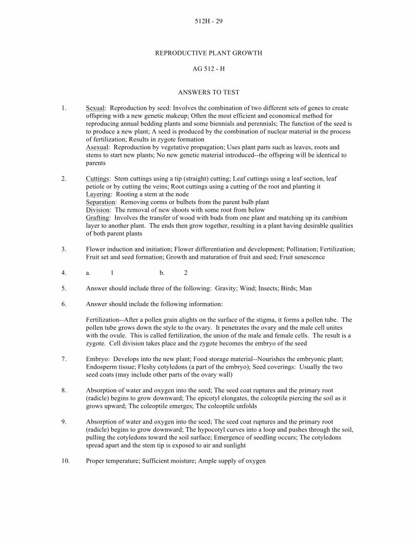
512H - 29
REPRODUCTIVE PLANT GROWTH
AG 512 - H
ANSWERS TO TEST 1. Sexual: Reproduction by seed: Involves the combination of two different sets of genes to create
offspring with a new genetic makeup; Often the most efficient and economical method for reproducing annual bedding plants and some biennials and perennials; The function of the seed is to produce a new plant; A seed is produced by the combination of nuclear material in the process of fertilization; Results in zygote formation Asexual: Reproduction by vegetative propagation; Uses plant parts such as leaves, roots and stems to start new plants; No new genetic material introduced--the offspring will be identical to parents
2. Cuttings: Stem cuttings using a tip (straight) cutting; Leaf cuttings using a leaf section, leaf
petiole or by cutting the veins; Root cuttings using a cutting of the root and planting it Layering: Rooting a stem at the node Separation: Removing corms or bulbets from the parent bulb plant Division: The removal of new shoots with some root from below Grafting: Involves the transfer of wood with buds from one plant and matching up its cambium layer to another plant. The ends then grow together, resulting in a plant having desirable qualities of both parent plants
3. Flower induction and initiation; Flower differentiation and development; Pollination; Fertilization;
Fruit set and seed formation; Growth and maturation of fruit and seed; Fruit senescence 4. a. 1 b. 2
5. Answer should include three of the following: Gravity; Wind; Insects; Birds; Man
6. Answer should include the following information:
Fertilization--After a pollen grain alights on the surface of the stigma, it forms a pollen tube. The pollen tube grows down the style to the ovary. It penetrates the ovary and the male cell unites with the ovule. This is called fertilization, the union of the male and female cells. The result is a zygote. Cell division takes place and the zygote becomes the embryo of the seed
7. Embryo: Develops into the new plant; Food storage material--Nourishes the embryonic plant;
Endosperm tissue; Fleshy cotyledons (a part of the embryo); Seed coverings: Usually the two seed coats (may include other parts of the ovary wall)
8. Absorption of water and oxygen into the seed; The seed coat ruptures and the primary root
(radicle) begins to grow downward; The epicotyl elongates, the coleoptile piercing the soil as it grows upward; The coleoptile emerges; The coleoptile unfolds
9. Absorption of water and oxygen into the seed; The seed coat ruptures and the primary root
(radicle) begins to grow downward; The hypocotyl curves into a loop and pushes through the soil, pulling the cotyledons toward the soil surface; Emergence of seedling occurs; The cotyledons spread apart and the stem tip is exposed to air and sunlight
10. Proper temperature; Sufficient moisture; Ample supply of oxygen

512H - 30
11. Mechanical injury to seed (cracked grain); Disease; Storage conditions; Age of seed; Soil temperature too cold; Hard seed coat; Soil moisture insufficient; Planting too deep; Chemical damage; Crusting of soil

512 I - 1
AG. 512 Botany / Science of Plant Growth and Development I. Scientific Method Term Project Based on: Idaho Science Content Guide and Framework. Grades 9 - 12.
Standard I. Habits of the Mind. Goal A. Science Processes. Goal. Document the theories of plant growth and development through time. Objectives. All students will: • Design an experiment using all elements of the scientific method to track the evolution of
plant life. • Write a clear, step-by-step methodology of the project data collection procedures. • Collect data by investigating the evolutionary history of plants through theory and supportive
literature. • Describe expected and actual results of the data collection, indicating major developments /
changes / differences in a plant’s evolution. • Predict future plant growth developments from time-line study results. • Analyze data by graphing a flow chart. • Explain results using a computer-generated presentation program or designing an interactive
display. • Identify and criticize any elements of research found which seemed faulty, incomplete, or
misleading. • Connect data directly to the resolution of the problem statement. • Discriminate between chance events and cause-effect events in plant evolution. • Formulate alternative conclusions or solutions to the problem statement based on evidence. Progress Indicator. All students will: • Chart a timeline of the vegetative and reproductive evolution of an agricultural crop or a
floricultural crop. • Produce a computer-generated presentations program or an interactive display. • Document the scientific methods of the study:
♦ State the Problem: Write a statement describing the problem researched. The problem may be written as a question; i.e. “What is the developmental future of the (genus species)?” or, “How does the past predict the future of (genus species)?”
♦ Gather Information. Investigate the evolutionary history of the study plants. Investigate the theories behind plant evolution and genetics. Document the literature surveyed.
♦ Form an Hypothesis. Generate an educated guess or idea of the results of the study based on the problem statement.

512 I - 2
♦ Collect the Data through Experimentation. Create a flow chart tracking the data gathered. Show points of evolution indicating major developments, changes or differences.
♦ Analyze Data and Form a Conclusion. The chart must support the conclusion. ♦ Report Results. Report the results in the form of a presentation. Explain how the
plants evolved. Use support information gathered. ♦ Propose a Theory. Propose an explanation for the results of the research, or . . . ♦ Identify Variables for Further Research. Isolate individual characteristics of the
study for further experiments. For instance, based on the results of this research, what would happen to the study plant’s growth pattern if the climate changed due to global warming?

512 I - 3
SCIENTIFIC METHOD MATRIX
AG. 512 BOTANY / SCIENCE OF PLANT GROWTH AND DEVELOPMENT
ACTIVITIES MEETING GOALS PER STANDARD
IDAHO K-‐12 SCIENCE CONTENT GUIDE AND FRAMEWORK
Standard I. Habits of the Mind Goal A. Science Processes The Organisms
General Laboratory Procedures, Equipment and Report Writing
X
Examining Cells from the Five Kingdoms X Classifying Organisms X
Cells: Structure, Functions, and Division What Are Cells? X Studying Cell Parts X Animal and Plant Cell Differences X
Nonvascular and Vascular Plants Studying the Life Cycle of a Bryophyte - The Moss
X
How Monocot Stems Differ from Dicot Stems
X
Vegetative Plant Parts Examining the Structure of Leaves X Relationship of Leaf Structure to Function X Examining Roots and Stems X Root Growth X
Reproductive Plant Parts Observing the Structure and Function of Flowers
X
Observing and Classifying Fruits X Development of Seed Parts into Young Plants
X
Vegetative Plant Growth Plant Growth X
Reproductive Plant Growth Growing a Bean Plant X

512 I - 4
Standard II. Science Themes Goal A. Change and Constancy Plant Processes
Light in Relation to Increase in Dry Weight X Water Movement Through Plants X
Standard II. Science Themes Goal B. Systems and Interactions Cells: Structure, Functions, and Division
Mitosis and Meiosis X Comparing Mitosis in Plant and Animal Cells
X
Plant Processes Measuring Loss from Transpiration X Measuring rates of Photosynthesis in Different Environments
X
Reproductive Plant Parts Flower Functions in Reproduction X
Reproductive Plant Growth Plant Reproduction Without Seeds X

512 I - 5
AGRICULTURAL SCIENCE AND TECHNOLOGY CURRICULUM SCIENTIFIC METHOD MATRIX
AG. 512 BOTANY / SCIENCE OF PLANT GROWTH AND DEVELOPMENT
IDAHO K-‐12 SCIENCE CONTENT GUIDE AND FRAMEWORK
Key - X
Standard I.
Habits of the Mind
Standard II.
Science Themes
Standard III.
Nature of Science
Section / Activity
Goal A. Science
Processes
Goal B. Values
Goal A.
Change and Constancy
Goal B.
Systems and Interactions
Goal C.
Models, Scale, and Structure
Goal A.
Science and Technology in Society
Goal B.
History and Cultural
Perspective
The Organisms General Laboratory Procedures, Equipment and Report Writing
X
Examining Cells from the Five Kingdoms
X
Classifying Organisms
X
Cells: Structure, Functions, and Division What Are Cells?
X
Studying Cell Parts
X
Animal and Plant Cell Differences
X
Mitosis and Meiosis
X
Comparing Mitosis in Plant and Animal Cells
X

512 I - 6
Key - X
Standard I.
Habits of the Mind
Standard II.
Science Themes
Standard III.
Nature of Science
Section / Activity
Goal A. Science
Processes
Goal B. Values
Goal A.
Change and Constancy
Goal B.
Systems and Interactions
Goal C.
Models, Scale, and Structure
Goal A.
Science and Technology in Society
Goal B.
History and Cultural
Perspective
Plant Processes Light in Relation to Increase in Dry Weight
X
Measuring Loss from Transpiration
X
Measuring Rates of Photosynthesis in Different Environments
X
Water Movement through Plants
X
Nonvascular and Vascular Plants Studying the Life Cycle of a Bryophyte - The Moss
X
How Monocot Stems Differ from Dicot Stems
X
Vegetative Plant Parts Examining the Structure of Leaves
X
Relationship of Leaf Structure to Function
X
Examining Roots and Stems
X
Root Growth X
512 I - 7
Key ~X
Standard I.
Habits of the Mind
Standard II.
Science Themes
Standard III.
Nature of Science
Section / Activity
Goal A. Science
Processes
Goal B. Values
Goal A.
Change and Constancy
Goal B.
Systems and Interactions
Goal C.
Models, Scale, and Structure
Goal A.
Science and Technology in Society
Goal B.
History and Cultural
Perspective
Reproductive Plant Parts Observing the Structure and Function of Flowers
X
Flower Functions in Reproduction
X
Observing and Classifying Fruits
X
Development of Seed Parts into Young Plants
X
Vegetative Plant Growth Plant Growth X
Reproductive Plant Growth Plant Reproduction Without Seeds
X
Growing a Bean Plant
X