
Il miracolo X greco di S. Giorgio (de libo) e la città di Vieste
Upload
khangminh22Category
view
1download
0
ADOLESCENT VARICOCELE
Giorgio BozziniAcademic Division of Urology,
IRCCS Policlinico San Donato,
University of Milan
H.Dep: prof. L. Carmignani
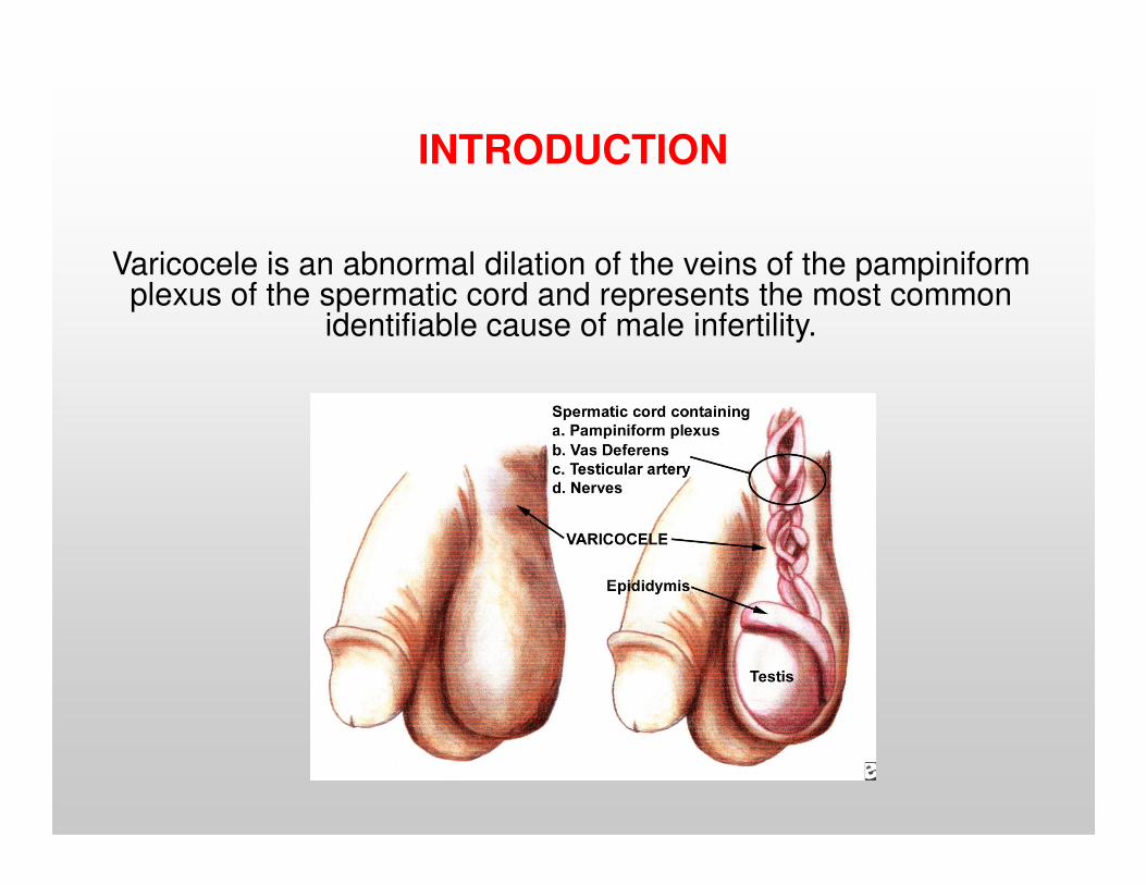
INTRODUCTION
Varicocele is an abnormal dilation of the veins of the pampiniform plexus of the spermatic cord and represents the most common
identifiable cause of male infertility.
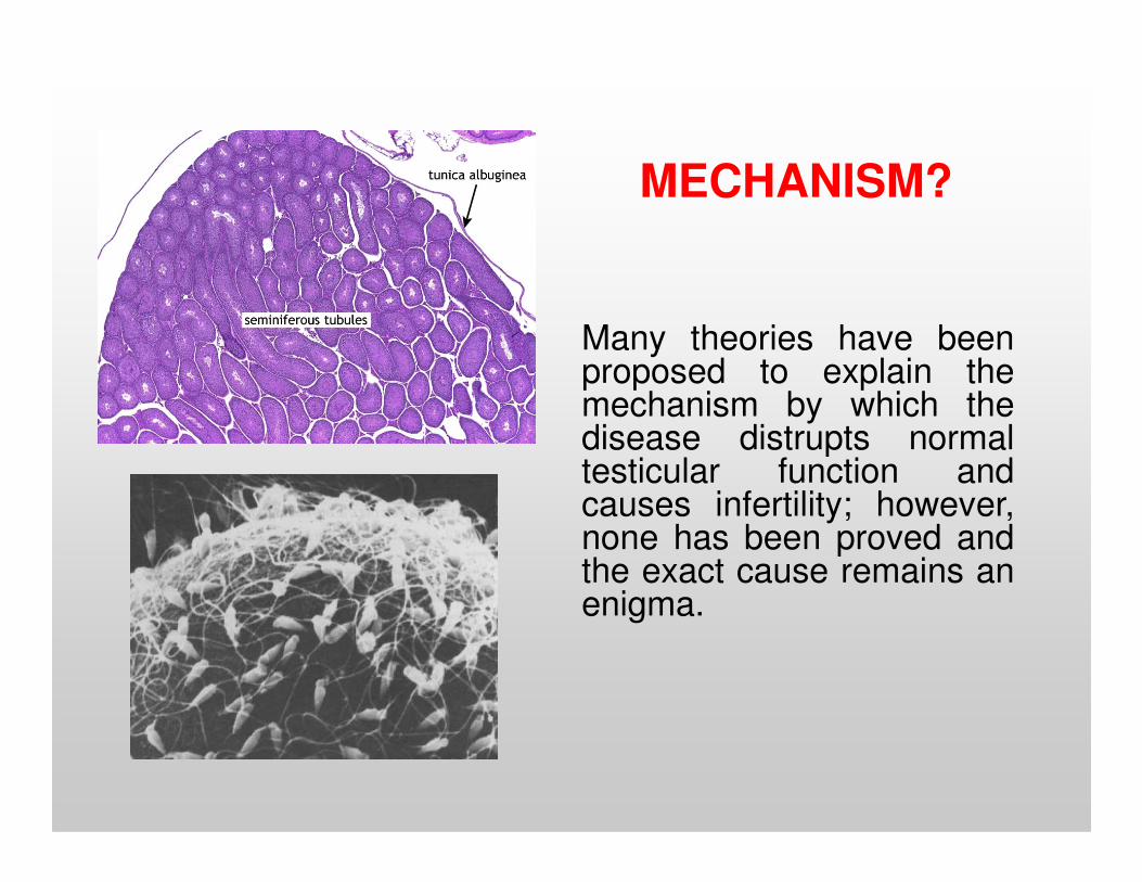
Many theories have beenproposed to explain themechanism by which thedisease distrupts normal
MECHANISM?
disease distrupts normaltesticular function andcauses infertility; however,none has been proved andthe exact cause remains anenigma.

Interest has focused on adolescents with a varicocele because
varicocelectomy in the
Interest on Adolescents
varicocelectomy in the adolescent population has
been proposed as a therapeutic intervention both to preserve fertility
and to preserve testicular growth
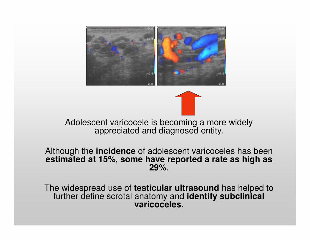
Adolescent varicocele is becoming a more widely Adolescent varicocele is becoming a more widely appreciated and diagnosed entity.
Although the incidence of adolescent varicoceles has been estimated at 15%, some have reported a rate as high as
29%.
The widespread use of testicular ultrasound has helped to further define scrotal anatomy and identify subclinical
varicoceles.
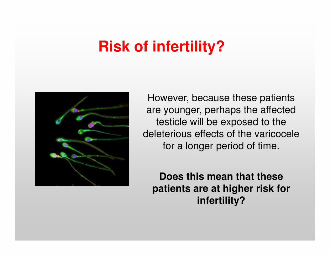
However, because these patients
are younger, perhaps the affected
testicle will be exposed to the
Risk of infertility?
deleterious effects of the varicocele
for a longer period of time.
Does this mean that these patients are at higher risk for
infertility?
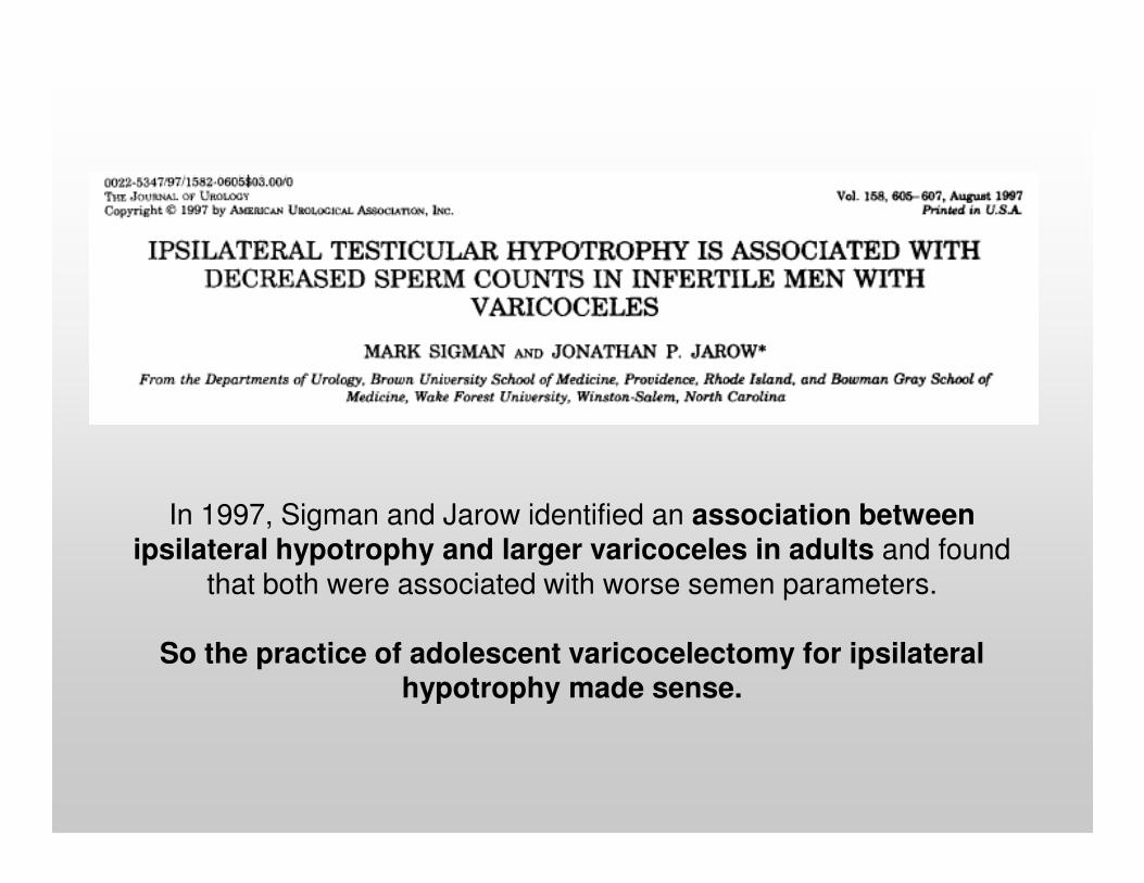
In 1997, Sigman and Jarow identified an association between ipsilateral hypotrophy and larger varicoceles in adults and found
that both were associated with worse semen parameters.
So the practice of adolescent varicocelectomy for ipsilateral hypotrophy made sense.
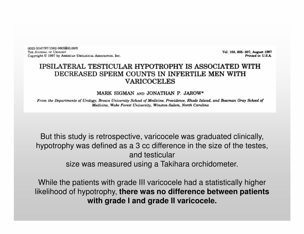
But this study is retrospective, varicocele was graduated clinically, hypotrophy was defined as a 3 cc difference in the size of the testes,
and testicularsize was measured using a Takihara orchidometer.
While the patients with grade III varicocele had a statistically higher likelihood of hypotrophy, there was no difference between patients
with grade I and grade II varicocele.
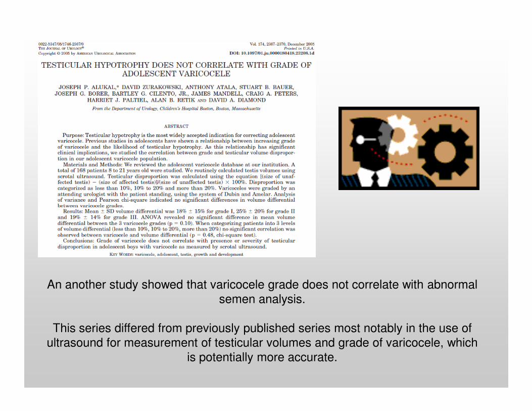
An another study showed that varicocele grade does not correlate with abnormal
semen analysis.
This series differed from previously published series most notably in the use of
ultrasound for measurement of testicular volumes and grade of varicocele, which
is potentially more accurate.
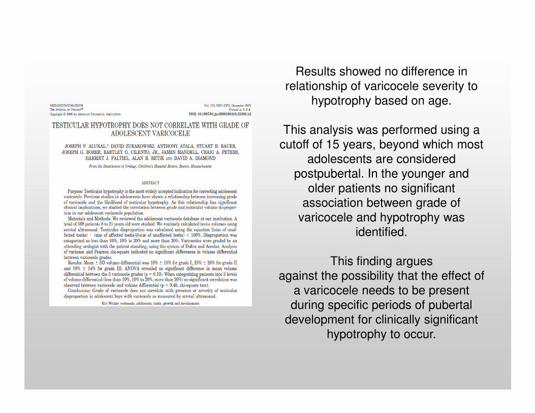
Results showed no difference in
relationship of varicocele severity to
hypotrophy based on age.
This analysis was performed using a
cutoff of 15 years, beyond which most
adolescents are considered
postpubertal. In the younger and
older patients no significant
association between grade ofassociation between grade of
varicocele and hypotrophy was
identified.
This finding argues
against the possibility that the effect of
a varicocele needs to be present
during specific periods of pubertal
development for clinically significant
hypotrophy to occur.
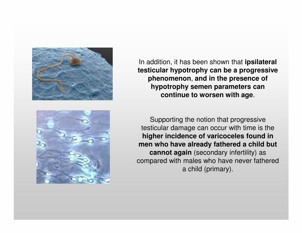
In addition, it has been shown that ipsilateral testicular hypotrophy can be a progressive
phenomenon, and in the presence of hypotrophy semen parameters can
continue to worsen with age.
Supporting the notion that progressive Supporting the notion that progressive
testicular damage can occur with time is the
higher incidence of varicoceles found in men who have already fathered a child but
cannot again (secondary infertility) as
compared with males who have never fathered
a child (primary).
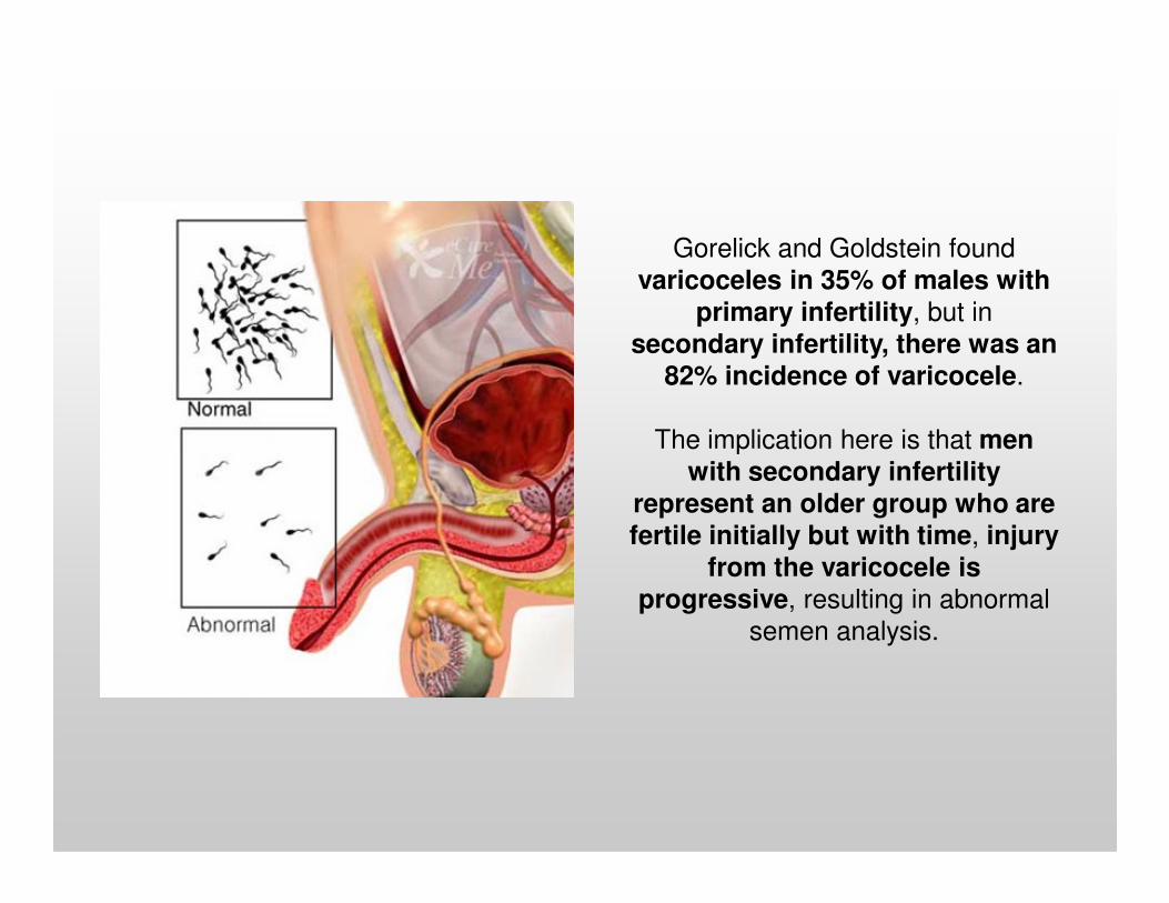
Gorelick and Goldstein found
varicoceles in 35% of males with primary infertility, but in
secondary infertility, there was an 82% incidence of varicocele.
The implication here is that men The implication here is that men with secondary infertility
represent an older group who are fertile initially but with time, injury
from the varicocele is progressive, resulting in abnormal
semen analysis.
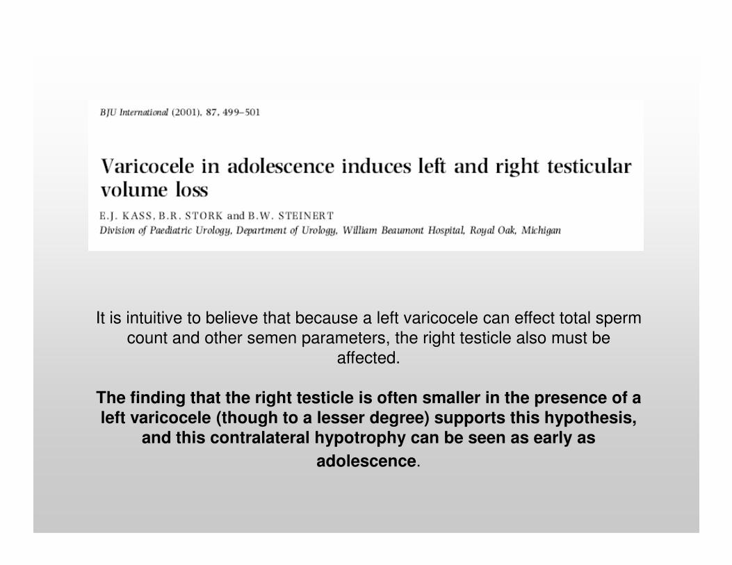
It is intuitive to believe that because a left varicocele can effect total sperm
count and other semen parameters, the right testicle also must be
affected.
The finding that the right testicle is often smaller in the presence of a left varicocele (though to a lesser degree) supports this hypothesis,
and this contralateral hypotrophy can be seen as early as
adolescence.
They found that the presence of a grade I varicocele appears to have no
effect on normal testicular growth. On the other hand, patients with a
grade II varicocele were found to be at risk of left testicular volume loss
and patients with grade III varicoceles were at risk of bilateral testicular
volume loss.
In this study varicocele was graduated clinically, and testicular size was
measured using a standard orchidometer.
ETIOLOGY and EPIDEMIOLOGY
Varicoceles are almost exclusivelyseen in pubertal or postpubertalpatients.
They are rarely found in patientsyounger than 10 years, and theyounger than 10 years, and theincidence reaches 16% betweenages 10 and 19 years.
Other studies have noted that theincidence varies from 9% to 29%between ages 10 and 17 years,with the incidence in adults beingapproximately 15%.
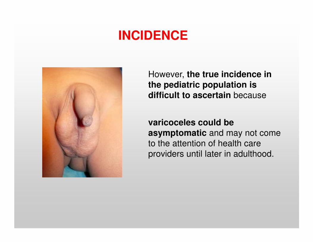
However, the true incidence in the pediatric population is difficult to ascertain because
varicoceles could be
INCIDENCE
varicoceles could be asymptomatic and may not come
to the attention of health care
providers until later in adulthood.
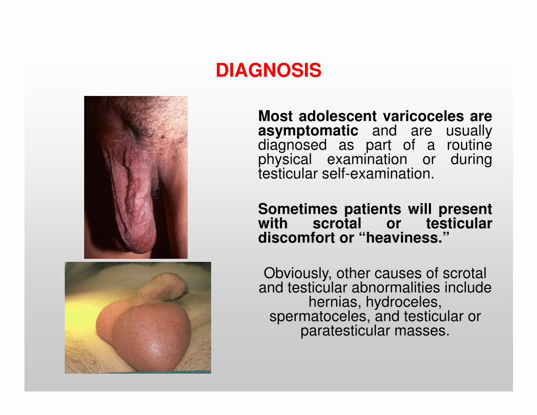
DIAGNOSIS
Most adolescent varicoceles areasymptomatic and are usuallydiagnosed as part of a routinephysical examination or duringtesticular self-examination.
Sometimes patients will presentSometimes patients will presentwith scrotal or testiculardiscomfort or “heaviness.”
Obviously, other causes of scrotal and testicular abnormalities include
hernias, hydroceles, spermatoceles, and testicular or
paratesticular masses.
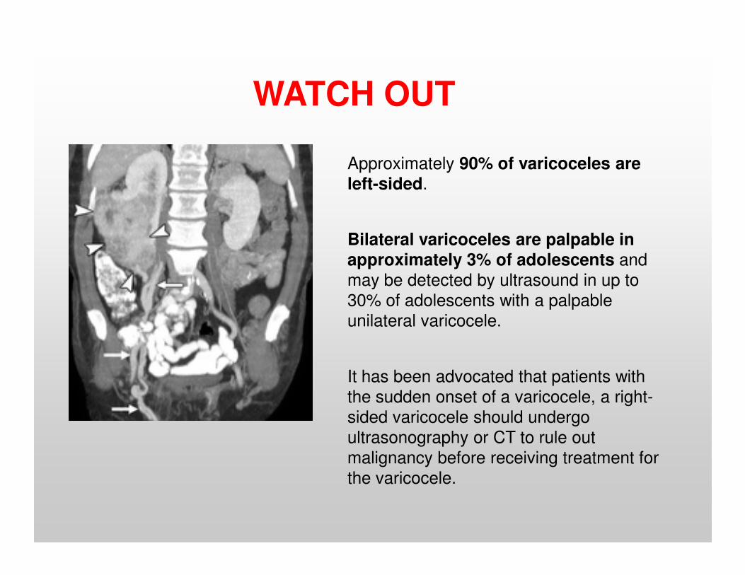
Approximately 90% of varicoceles are left-sided.
Bilateral varicoceles are palpable in approximately 3% of adolescents and
may be detected by ultrasound in up to
WATCH OUT
may be detected by ultrasound in up to
30% of adolescents with a palpable
unilateral varicocele.
It has been advocated that patients with
the sudden onset of a varicocele, a right-
sided varicocele should undergo
ultrasonography or CT to rule out
malignancy before receiving treatment for
the varicocele.
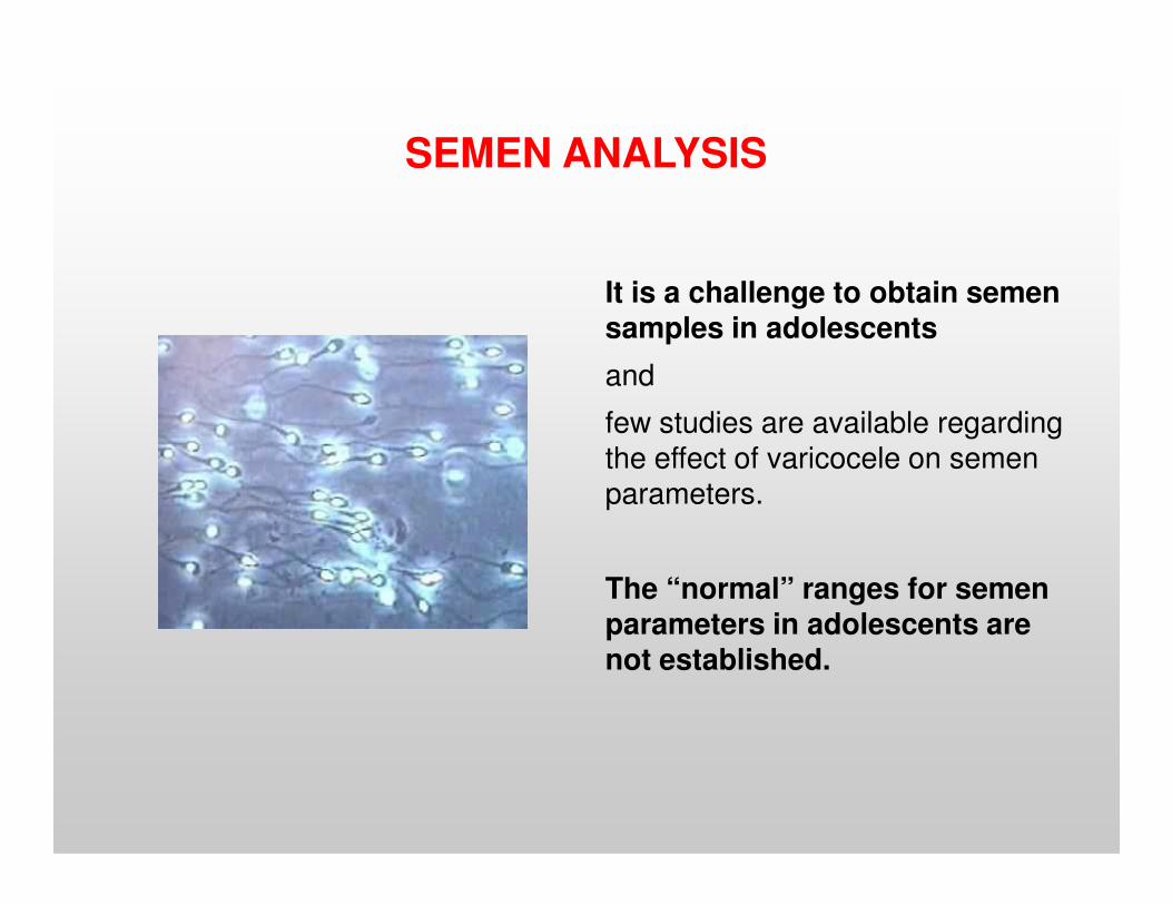
SEMEN ANALYSIS
It is a challenge to obtain semen samples in adolescents
and
few studies are available regarding the effect of varicocele on semen the effect of varicocele on semen parameters.
The “normal” ranges for semen parameters in adolescents are not established.
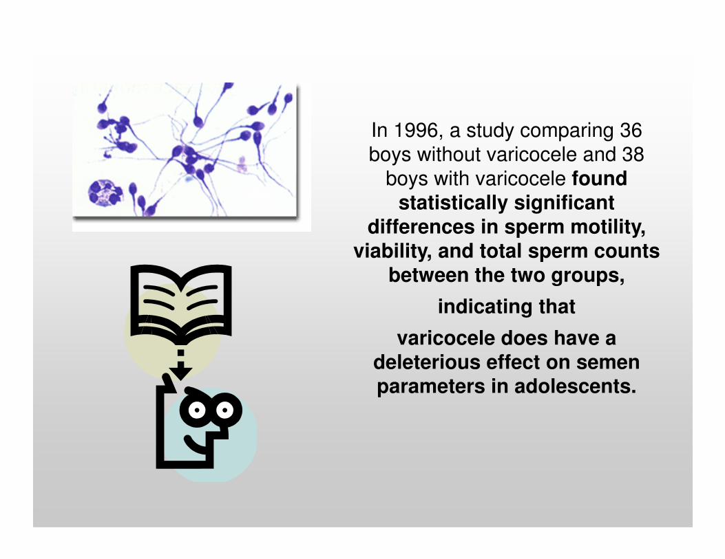
In 1996, a study comparing 36
boys without varicocele and 38
boys with varicocele found statistically significant
differences in sperm motility, viability, and total sperm counts
between the two groups,between the two groups,
indicating that
varicocele does have a deleterious effect on semen parameters in adolescents.
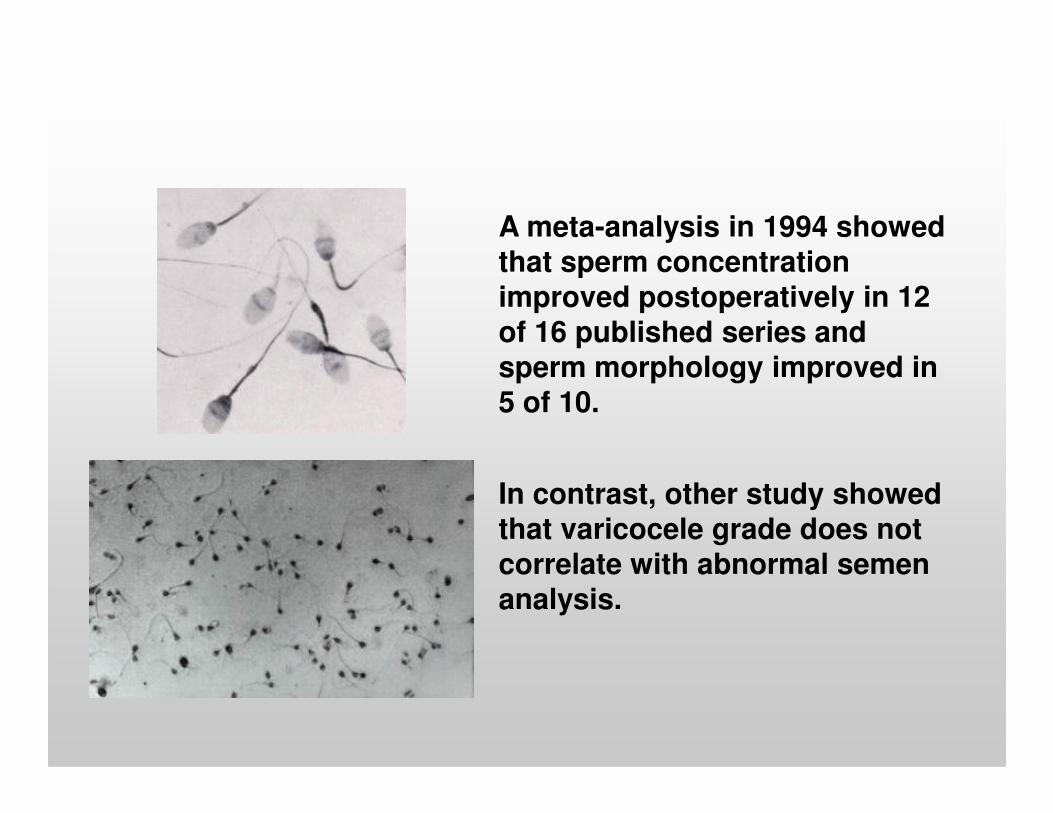
A meta-analysis in 1994 showed that sperm concentration improved postoperatively in 12 of 16 published series and sperm morphology improved in 5 of 10.5 of 10.
In contrast, other study showed that varicocele grade does not correlate with abnormal semen analysis.
Sonographically determined testicular differentials greater than 10% between normal and affected testes correlate with
decreased total motile sperm count and may serve to identify adolescents with unilateral varicocele who are at greatest risk
for future infertility.
This study was appropriately confined to Tanner stage V patients.
There are some limitations: of the boys 61% submitted 1 semen specimen, there
is not a control group for comparison and the number of Patients (57 pts) available for study was modest.
In another study of 214 patients with varicoceles, Zampieri et
al. found no differences in semen parameters when comparing men with or without testicular hypotrophy.
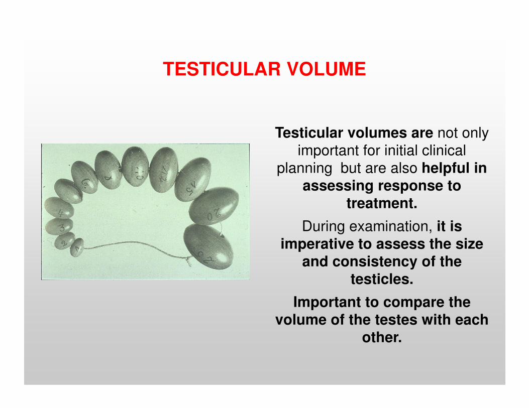
TESTICULAR VOLUME
Testicular volumes are not only
important for initial clinical
planning but are also helpful in assessing response to
treatment. treatment.
During examination, it is imperative to assess the size
and consistency of the testicles.
Important to compare the volume of the testes with each
other.
TREATMENT
The treatment of the adolescent varicocele raises several interesting
clinical and ethical dilemmas.
In adults, it is well established that
patients with varicoceles exhibit
abnormalities in their semen
parameters and abnormal testicular
function that may ultimately affect
fertility potential.
On semen analysis, adults with varicoceles are known to have
increased number of pathologic sperm forms, decreased sperm motility, and
decreased sperm density.
In presence of a clinically detectable varicocele associated with an abnormal semen analysis in an infertile couple is an appropriate indication for treatment.
If we treat all adolescents with varicoceles,
are we potentially decreasing the time period that the affected testicle is exposed to the deleterious conditions created by a
varicocele?
So, where does this leave the adolescent varicocele patient who presumably is
several years away from needing to worry several years away from needing to worry about fertility potential?
Interestingly, the incidence of varicoceles is much higher than that of
male factor infertility.
Furthermore, the finding that only 15% to 20% of men with varicoceles seek 20% of men with varicoceles seek
treatment for fertility implies that most men with varicoceles either choose not to seek treatment or are fertile.
Perhaps the main difference is that the adolescent testicle may recover from
damage more readily than the adult testicle. damage more readily than the adult testicle.
Adolescent testicles after varicocele repair almost always exhibit “catch-up” growth.
But, it is not known whether varicocele in adolescence impairs fertility or whether
surgery restores fertility.
The evaluation and choice of treatment for the adolescent varicocelepatients is based not on objective fertility criteria (paternity)
Buton indirect evidence that there may be compromise to testicular
function or spermatogenesis and thus eventual fertility.
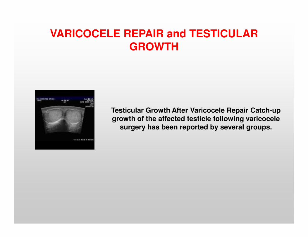
VARICOCELE REPAIR and TESTICULAR GROWTH
Testicular Growth After Varicocele Repair Catch-up growth of the affected testicle following varicocele growth of the affected testicle following varicocele
surgery has been reported by several groups.
Studies suggest that varicocele repair in older adolescents significantly
increases sperm values, especially motility and total motile sperm count
Adding to the controversy in this patient population is the report by
some of testicular growth in conservatively
managed patients with varicoceles
SURGICAL TREATMENT
Current recommendations for adolescent varicocele repair are based on the findings of persistently impaired testicular growth.
The main indication for surgery is testicular hypotrophy ( > 2
mL or > 20%) on the affected side.mL or > 20%) on the affected side.
As growth can vary between the testes, the difference in size should
be confirmed by two measurements made 12 months apart.
Relative indications for varicocele repair are:
- a soft testis,
- bilateral grade 3 varicocele with no testicular hypotrophy,
- presence of varicocele in a solitary testis,- presence of varicocele in a solitary testis,
- poor semen analysis in a Tanner V adolescent (preferably
confirmed on two specimens),
- the rare conditions of pain or intratesticular varicocele.
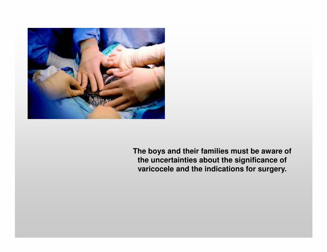
The boys and their families must be aware ofthe uncertainties about the significance ofvaricocele and the indications for surgery.
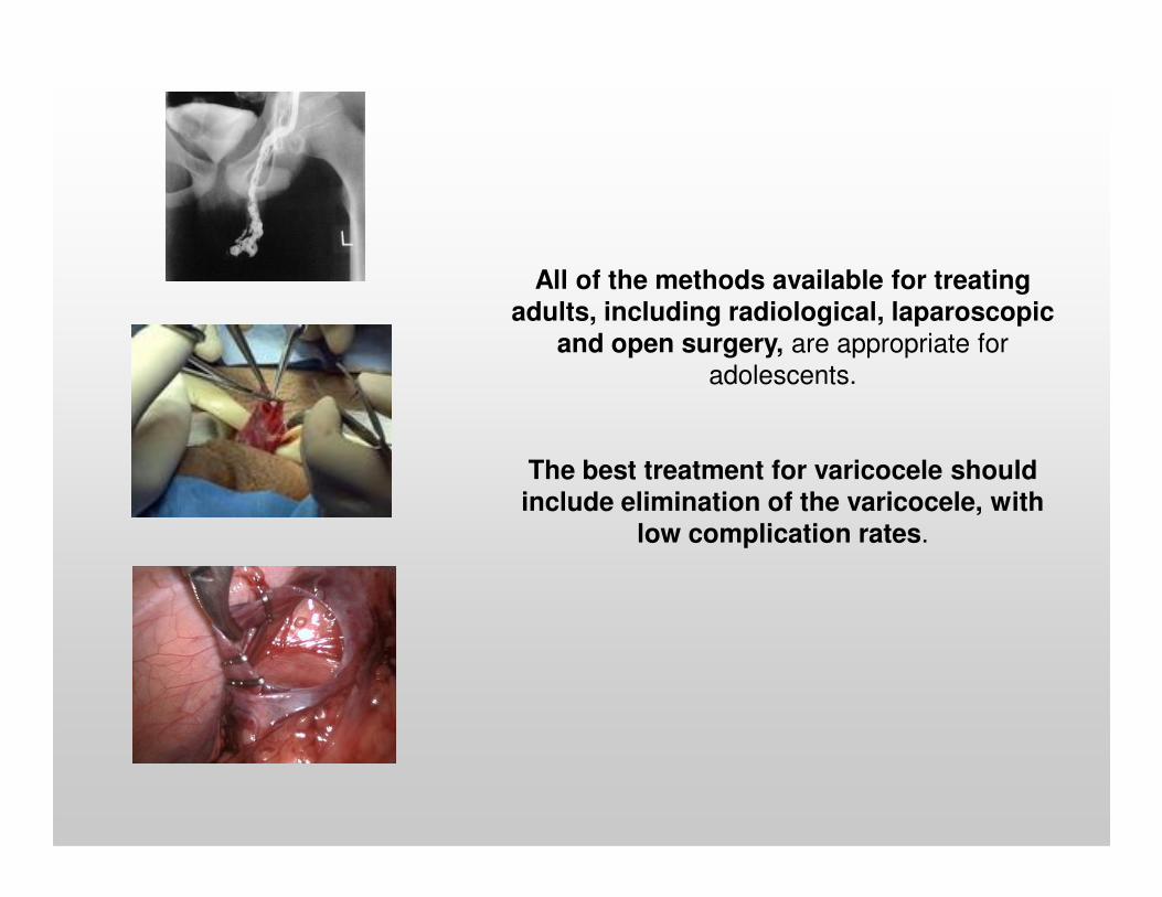
All of the methods available for treating adults, including radiological, laparoscopic
and open surgery, are appropriate for
adolescents.
The best treatment for varicocele should include elimination of the varicocele, with
low complication rates.
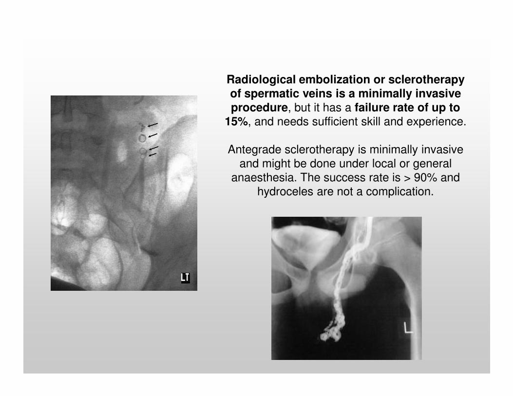
Radiological embolization or sclerotherapy of spermatic veins is a minimally invasive procedure, but it has a failure rate of up to
15%, and needs sufficient skill and experience.
Antegrade sclerotherapy is minimally invasive
and might be done under local or general
anaesthesia. The success rate is > 90% and
hydroceles are not a complication.
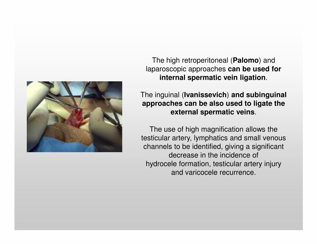
The high retroperitoneal (Palomo) and
laparoscopic approaches can be used for internal spermatic vein ligation.
The inguinal (Ivanissevich) and subinguinal approaches can be also used to ligate the
external spermatic veins.
The use of high magnification allows the
testicular artery, lymphatics and small venous
channels to be identified, giving a significant
decrease in the incidence of
hydrocele formation, testicular artery injury
and varicocele recurrence.
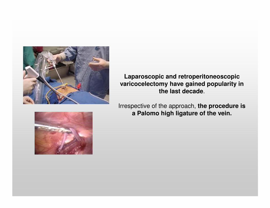
Laparoscopic and retroperitoneoscopic varicocelectomy have gained popularity in
the last decade.
Irrespective of the approach, the procedure is Irrespective of the approach, the procedure is a Palomo high ligature of the vein.
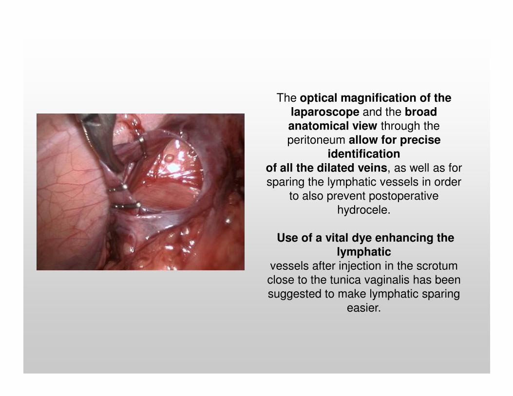
The optical magnification of the laparoscope and the broad anatomical view through the
peritoneum allow for precise identification
of all the dilated veins, as well as for
sparing the lymphatic vessels in order
to also prevent postoperative to also prevent postoperative
hydrocele.
Use of a vital dye enhancing the lymphatic
vessels after injection in the scrotum
close to the tunica vaginalis has been
suggested to make lymphatic sparing
easier.
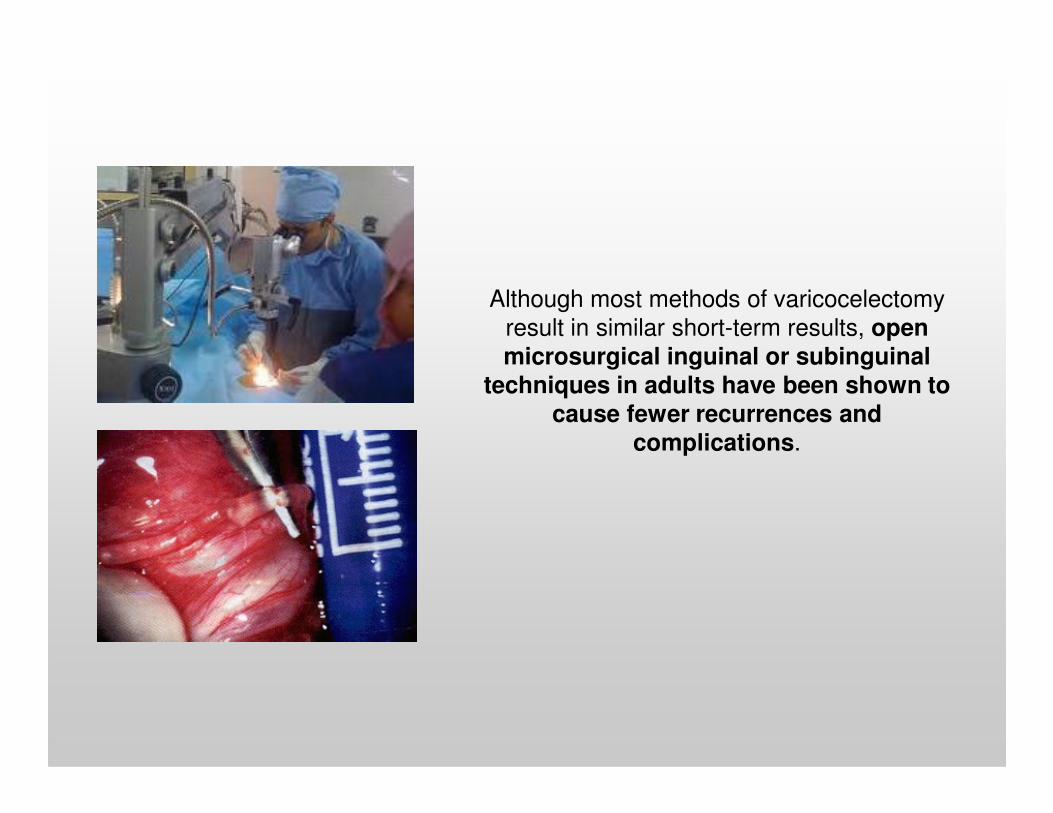
Although most methods of varicocelectomy
result in similar short-term results, openmicrosurgical inguinal or subinguinal
techniques in adults have been shown tocause fewer recurrences and cause fewer recurrences and
complications.
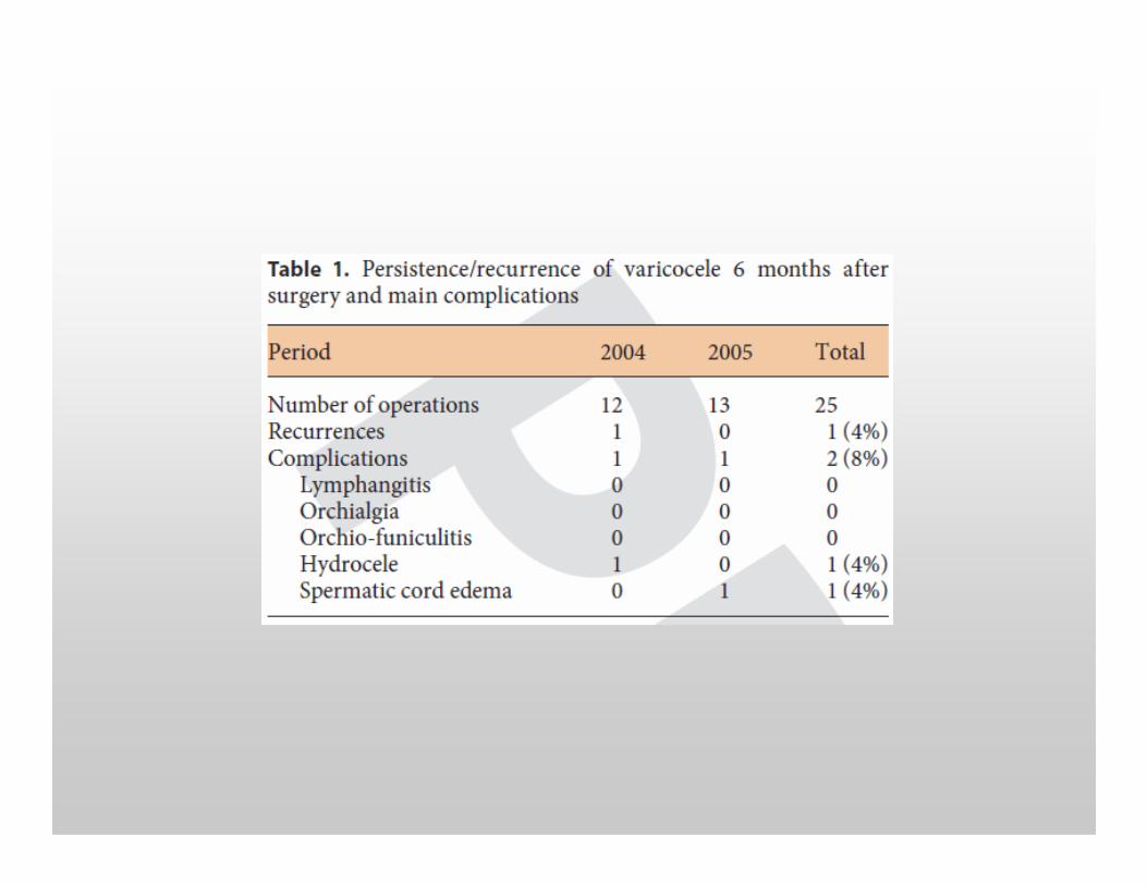
Complications after surgery vary with surgical techniques.
Recurrences after varicocele repair are reported in 0–16.6%,
varying with the technique used.
COMPLICATIONS
Hydrocele in adolescents is a potential problem, with an incidence of 0–24%.
The low, selective venous approaches using magnification to
preserve the lymphatics reduce this risk.
Other complications include wound infection, testicular atrophy and ilio-inguinal nerve damage, but the incidence is unrecorded.
Testicular atrophy would be a devastating complication.
COMPLICATIONS
None of the major series report this complication, but personal
communication suggests that it can occur.
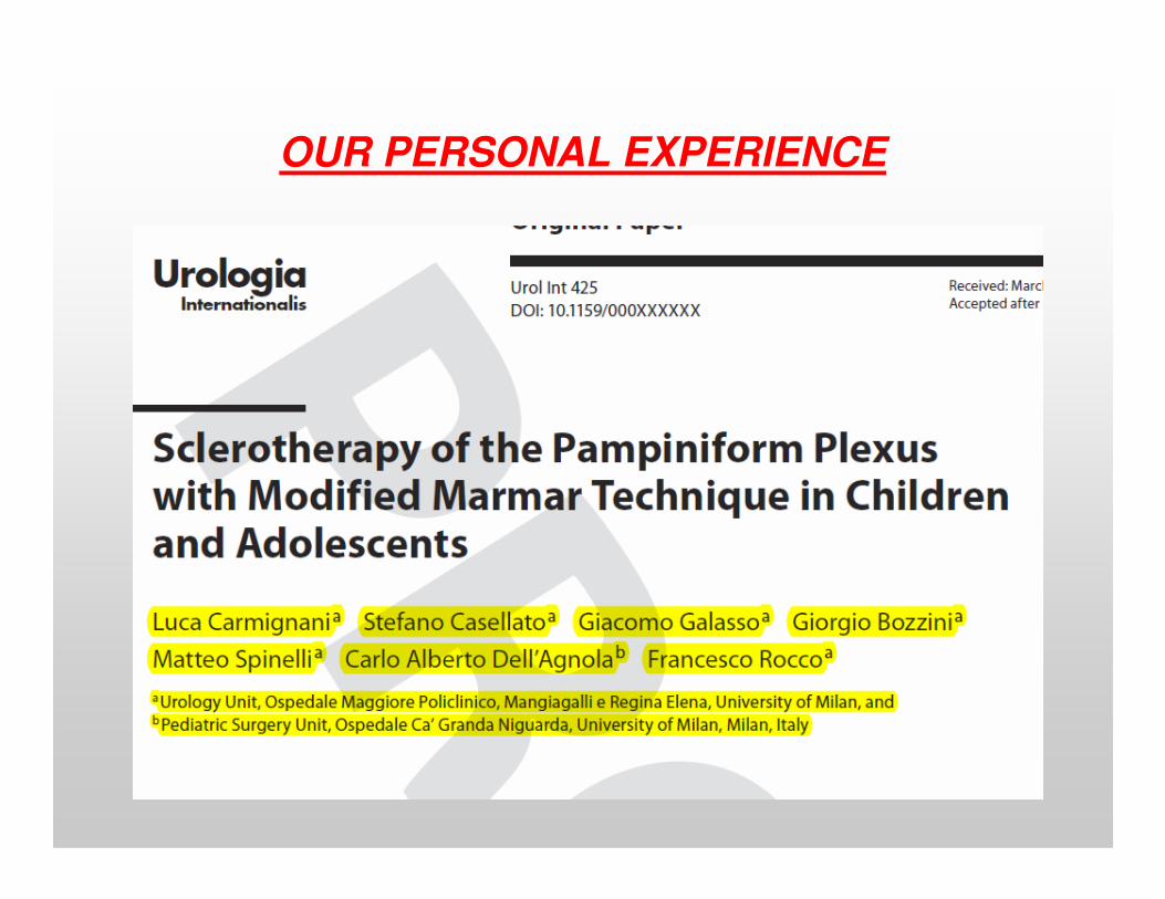
OUR PERSONAL EXPERIENCE
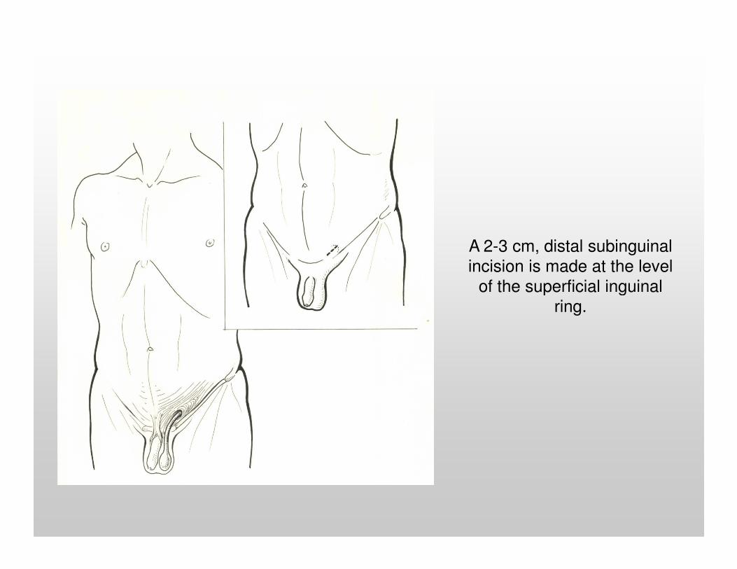
A 2-3 cm, distal subinguinal
incision is made at the level
of the superficial inguinal of the superficial inguinal
ring.
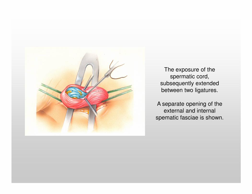
The exposure of the
spermatic cord,
subsequently extended
between two ligatures.
A separate opening of the
external and internal
spematic fasciae is shown.
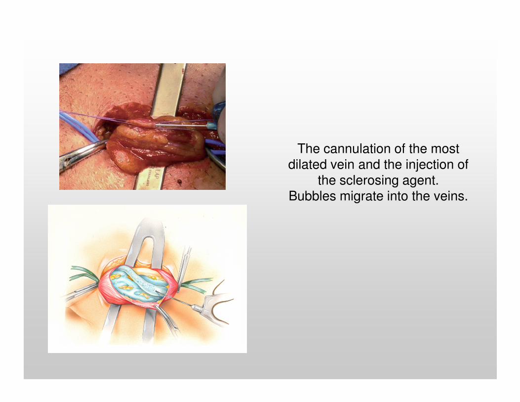
The cannulation of the most dilated vein and the injection of
the sclerosing agent. Bubbles migrate into the veins.Bubbles migrate into the veins.

Thank You
Copyright © 2022 FDOKUMEN