Addressing Global Ruminant Agricultural ... - Digital CSIC
33
REVIEW published: 25 September 2018 doi: 10.3389/fmicb.2018.02161 Frontiers in Microbiology | www.frontiersin.org 1 September 2018 | Volume 9 | Article 2161 Edited by: Zhongtang Yu, The Ohio State University, United States Reviewed by: Shengguo Zhao, Institute of Animal Sciences, Chinese Academy of Agricultural Sciences, China Suzanne Lynn Ishaq, University of Oregon, United States *Correspondence: Sharon A. Huws [email protected] Specialty section: This article was submitted to Microbial Symbioses, a section of the journal Frontiers in Microbiology Received: 31 May 2018 Accepted: 23 August 2018 Published: 25 September 2018 Citation: Huws SA, Creevey CJ, Oyama LB, Mizrahi I, Denman SE, Popova M, Muñoz-Tamayo R, Forano E, Waters SM, Hess M, Tapio I, Smidt H, Krizsan SJ, Yáñez-Ruiz DR, Belanche A, Guan L, Gruninger RJ, McAllister TA, Newbold CJ, Roehe R, Dewhurst RJ, Snelling TJ, Watson M, Suen G, Hart EH, Kingston-Smith AH, Scollan ND, do Prado RM, Pilau EJ, Mantovani HC, Attwood GT, Edwards JE, McEwan NR, Morrisson S, Mayorga OL, Elliott C and Morgavi DP (2018) Addressing Global Ruminant Agricultural Challenges Through Understanding the Rumen Microbiome: Past, Present, and Future. Front. Microbiol. 9:2161. doi: 10.3389/fmicb.2018.02161 Addressing Global Ruminant Agricultural Challenges Through Understanding the Rumen Microbiome: Past, Present, and Future Sharon A. Huws 1 *, Christopher J. Creevey 1 , Linda B. Oyama 1 , Itzhak Mizrahi 2 , Stuart E. Denman 3 , Milka Popova 4 , Rafael Muñoz-Tamayo 5 , Evelyne Forano 6 , Sinead M. Waters 7 , Matthias Hess 8 , Ilma Tapio 9 , Hauke Smidt 10 , Sophie J. Krizsan 11 , David R. Yáñez-Ruiz 12 , Alejandro Belanche 12 , Leluo Guan 13 , Robert J. Gruninger 14 , Tim A. McAllister 14 , C. Jamie Newbold 15 , Rainer Roehe 15 , Richard J. Dewhurst 15 , Tim J. Snelling 16 , Mick Watson 17 , Garret Suen 18 , Elizabeth H. Hart 19 , Alison H. Kingston-Smith 19 , Nigel D. Scollan 1 , Rodolpho M. do Prado 20 , Eduardo J. Pilau 20 , Hilario C. Mantovani 21 , Graeme T. Attwood 22 , Joan E. Edwards 23 , Neil R. McEwan 24 , Steven Morrisson 25 , Olga L. Mayorga 26 , Christopher Elliott 1 and Diego P. Morgavi 4 1 Institute for Global Food Security, Queen’s University of Belfast, Belfast, United Kingdom, 2 Department of Life Sciences and the National Institute for Biotechnology in the Negev, Ben Gurion University of the Negev, Beer Sheva, Israel, 3 Commonwealth Scientific and Industrial Research Organisation Agriculture and Food, Queensland Bioscience Precinct, St Lucia, QLD, Australia, 4 Institute National de la Recherche Agronomique, UMR1213 Herbivores, Clermont Université, VetAgro Sup, UMR Herbivores, Clermont-Ferrand, France, 5 UMR Modélisation Systémique Appliquée aux Ruminants, INRA, AgroParisTech, Université Paris-Saclay, Paris, France, 6 UMR 454 MEDIS, INRA, Université Clermont Auvergne, Clermont-Ferrand, France, 7 Animal and Bioscience Research Department, Animal and Grassland Research and Innovation Centre, Grange, Ireland, 8 College of Agricultural and Environmental Sciences, University of California, Davis, Davis, CA, United States, 9 Natural Resources Institute Finland, Jokioinen, Finland, 10 Department of Agrotechnology and Food Sciences, Wageningen, Netherlands, 11 Department of Agricultural Research for Northern Sweden, Swedish University of Agricultural Sciences, Umeå, Sweden, 12 Estacion Experimental del Zaidin, Consejo Superior de Investigaciones Cientificas, Granada, Spain, 13 Department of Agricultural, Food and Nutritional Science, University of Alberta, Edmonton, AB, Canada, 14 Lethbridge Research Centre, Agriculture and Agri-Food Canada, Lethbridge, AB, Canada, 15 Scotland’s Rural College, Edinburgh, United Kingdom, 16 The Rowett Institute, University of Aberdeen, Aberdeen, United Kingdom, 17 The Roslin Institute and the Royal (Dick) School of Veterinary Studies (R(D)SVS), University of Edinburgh, Edinburgh, United Kingdom, 18 Department of Bacteriology, University of Wisconsin-Madison, Madison, WI, United States, 19 Institute of Biological, Environmental and Rural Sciences, Aberystwyth University, Aberystwyth, United Kingdom, 20 Laboratório de Biomoléculas e Espectrometria de Massas-Labiomass, Departamento de Química, Universidade Estadual de Maringá, Maringá, Brazil, 21 Department of Microbiology, Universidade Federal de Viçosa, Viçosa, Brazil, 22 AgResearch Limited, Grasslands Research Centre, Palmerston North, New Zealand, 23 Laboratory of Microbiology, Wageningen University & Research, Wageningen, Netherlands, 24 School of Pharmacy and Life Sciences, Robert Gordon University, Aberdeen, United Kingdom, 25 Sustainable Livestock, Agri-Food and Bio-Sciences Institute, Hillsborough, United Kingdom, 26 Colombian Agricultural Research Corporation, Mosquera, Colombia The rumen is a complex ecosystem composed of anaerobic bacteria, protozoa, fungi, methanogenic archaea and phages. These microbes interact closely to breakdown plant material that cannot be digested by humans, whilst providing metabolic energy to the host and, in the case of archaea, producing methane. Consequently, ruminants produce meat and milk, which are rich in high-quality protein, vitamins and minerals, and therefore contribute to food security. As the world population is predicted to
-
Upload
khangminh22 -
Category
Documents
-
view
3 -
download
0
Transcript of Addressing Global Ruminant Agricultural ... - Digital CSIC
REVIEWpublished: 25 September 2018doi: 10.3389/fmicb.2018.02161
Frontiers in Microbiology | www.frontiersin.org 1 September 2018 | Volume 9 | Article 2161
Edited by:
Zhongtang Yu,
The Ohio State University,
United States
Reviewed by:
Shengguo Zhao,
Institute of Animal Sciences, Chinese
Academy of Agricultural Sciences,
China
Suzanne Lynn Ishaq,
University of Oregon, United States
*Correspondence:
Sharon A. Huws
Specialty section:
This article was submitted to
Microbial Symbioses,
a section of the journal
Frontiers in Microbiology
Received: 31 May 2018
Accepted: 23 August 2018
Published: 25 September 2018
Citation:
Huws SA, Creevey CJ, Oyama LB,
Mizrahi I, Denman SE, Popova M,
Muñoz-Tamayo R, Forano E,
Waters SM, Hess M, Tapio I, Smidt H,
Krizsan SJ, Yáñez-Ruiz DR,
Belanche A, Guan L, Gruninger RJ,
McAllister TA, Newbold CJ, Roehe R,
Dewhurst RJ, Snelling TJ, Watson M,
Suen G, Hart EH, Kingston-Smith AH,
Scollan ND, do Prado RM, Pilau EJ,
Mantovani HC, Attwood GT,
Edwards JE, McEwan NR,
Morrisson S, Mayorga OL, Elliott C
and Morgavi DP (2018) Addressing
Global Ruminant Agricultural
Challenges Through Understanding
the Rumen Microbiome: Past,
Present, and Future.
Front. Microbiol. 9:2161.
doi: 10.3389/fmicb.2018.02161
Addressing Global RuminantAgricultural Challenges ThroughUnderstanding the RumenMicrobiome: Past, Present, andFutureSharon A. Huws 1*, Christopher J. Creevey 1, Linda B. Oyama 1, Itzhak Mizrahi 2,
Stuart E. Denman 3, Milka Popova 4, Rafael Muñoz-Tamayo 5, Evelyne Forano 6,
Sinead M. Waters 7, Matthias Hess 8, Ilma Tapio 9, Hauke Smidt 10, Sophie J. Krizsan 11,
David R. Yáñez-Ruiz 12, Alejandro Belanche 12, Leluo Guan 13, Robert J. Gruninger 14,
Tim A. McAllister 14, C. Jamie Newbold 15, Rainer Roehe 15, Richard J. Dewhurst 15,
Tim J. Snelling 16, Mick Watson 17, Garret Suen 18, Elizabeth H. Hart 19,
Alison H. Kingston-Smith 19, Nigel D. Scollan 1, Rodolpho M. do Prado 20, Eduardo J. Pilau 20,
Hilario C. Mantovani 21, Graeme T. Attwood 22, Joan E. Edwards 23, Neil R. McEwan 24,
Steven Morrisson 25, Olga L. Mayorga 26, Christopher Elliott 1 and Diego P. Morgavi 4
1 Institute for Global Food Security, Queen’s University of Belfast, Belfast, United Kingdom, 2Department of Life Sciences and
the National Institute for Biotechnology in the Negev, Ben Gurion University of the Negev, Beer Sheva, Israel,3Commonwealth Scientific and Industrial Research Organisation Agriculture and Food, Queensland Bioscience Precinct, St
Lucia, QLD, Australia, 4 Institute National de la Recherche Agronomique, UMR1213 Herbivores, Clermont Université, VetAgro
Sup, UMR Herbivores, Clermont-Ferrand, France, 5UMR Modélisation Systémique Appliquée aux Ruminants, INRA,
AgroParisTech, Université Paris-Saclay, Paris, France, 6UMR 454 MEDIS, INRA, Université Clermont Auvergne,
Clermont-Ferrand, France, 7 Animal and Bioscience Research Department, Animal and Grassland Research and Innovation
Centre, Grange, Ireland, 8College of Agricultural and Environmental Sciences, University of California, Davis, Davis, CA,
United States, 9Natural Resources Institute Finland, Jokioinen, Finland, 10Department of Agrotechnology and Food Sciences,
Wageningen, Netherlands, 11Department of Agricultural Research for Northern Sweden, Swedish University of Agricultural
Sciences, Umeå, Sweden, 12 Estacion Experimental del Zaidin, Consejo Superior de Investigaciones Cientificas, Granada,
Spain, 13Department of Agricultural, Food and Nutritional Science, University of Alberta, Edmonton, AB, Canada,14 Lethbridge Research Centre, Agriculture and Agri-Food Canada, Lethbridge, AB, Canada, 15 Scotland’s Rural College,
Edinburgh, United Kingdom, 16 The Rowett Institute, University of Aberdeen, Aberdeen, United Kingdom, 17 The Roslin
Institute and the Royal (Dick) School of Veterinary Studies (R(D)SVS), University of Edinburgh, Edinburgh, United Kingdom,18Department of Bacteriology, University of Wisconsin-Madison, Madison, WI, United States, 19 Institute of Biological,
Environmental and Rural Sciences, Aberystwyth University, Aberystwyth, United Kingdom, 20 Laboratório de Biomoléculas e
Espectrometria de Massas-Labiomass, Departamento de Química, Universidade Estadual de Maringá, Maringá, Brazil,21Department of Microbiology, Universidade Federal de Viçosa, Viçosa, Brazil, 22 AgResearch Limited, Grasslands Research
Centre, Palmerston North, New Zealand, 23 Laboratory of Microbiology, Wageningen University & Research, Wageningen,
Netherlands, 24 School of Pharmacy and Life Sciences, Robert Gordon University, Aberdeen, United Kingdom, 25 Sustainable
Livestock, Agri-Food and Bio-Sciences Institute, Hillsborough, United Kingdom, 26Colombian Agricultural Research
Corporation, Mosquera, Colombia
The rumen is a complex ecosystem composed of anaerobic bacteria, protozoa, fungi,
methanogenic archaea and phages. These microbes interact closely to breakdown
plant material that cannot be digested by humans, whilst providing metabolic energy
to the host and, in the case of archaea, producing methane. Consequently, ruminants
produce meat and milk, which are rich in high-quality protein, vitamins and minerals,
and therefore contribute to food security. As the world population is predicted to
Huws et al. Understanding the Rumen Microbiome
reach approximately 9.7 billion by 2050, an increase in ruminant production to satisfy
global protein demand is necessary, despite limited land availability, and whilst ensuring
environmental impact is minimized. Although challenging, these goals can be met,
but depend on our understanding of the rumen microbiome. Attempts to manipulate
the rumen microbiome to benefit global agricultural challenges have been ongoing for
decades with limited success, mostly due to the lack of a detailed understanding of this
microbiome and our limited ability to culture most of these microbes outside the rumen.
The potential to manipulate the rumen microbiome and meet global livestock challenges
through animal breeding and introduction of dietary interventions during early life have
recently emerged as promising new technologies. Our inability to phenotype ruminants
in a high-throughput manner has also hampered progress, although the recent increase
in “omic” data may allow further development of mathematical models and rumen
microbial gene biomarkers as proxies. Advances in computational tools, high-throughput
sequencing technologies and cultivation-independent “omics” approaches continue to
revolutionize our understanding of the rumen microbiome. This will ultimately provide the
knowledge framework needed to solve current and future ruminant livestock challenges.
Keywords: rumen, microbiome, host, diet, production, methane, omics
GLOBAL AGRICULTURAL CHALLENGES
There are currently 7.5 billion humans on the planet, andthe world Hunger Map estimates that 795 million people(over 10%) do not have access to sufficient food (WFP,2015). Whilst some models predict the world population topeak at 9.7 billion in 2050, others estimate a populationof 11.2 billion in 2100 (United Nations, 2015). To meetan increasing demand for food, the Food and AgricultureOrganization of the United Nations (FAO) predicts that totalagricultural production (including crops and animals) willneed to be 60% higher than in 2005. With animal proteindemand rising at a proportionally faster rate, estimates suggestthat global meat and milk production will have to increaseby 76 and 63%, respectively (Alexandratos and Bruinsma,2012).
This extensive population growth, coupled with an increasedconsumption of ruminant products by developing countries,will add to the strain on the availability of safe and nutritiousruminant products. Due to land constraints, the numberof pastured ruminants cannot increase and therefore effortsshould be directed toward increasing production efficiency.Indeed, efficient utilization of feed by the rumen microbiomeresults in enhanced nutrient availability to the host, and thusimproved production efficiency is central to ensuring foodsecurity. Feed for ruminants typically accounts for 60–70%of total expenditure in beef production (Karisa et al., 2014;Fouhse et al., 2017), whilst requiring substantial land massfor plant growth. Residual feed intake (RFI), which is thedifference between the predicted (based on energy demands)and actual intake, has been proposed as a more meaningfulmeasure for calculating feed efficiency (Berry and Crowley,2012; Shabat et al., 2016). RFI values of 1.45 (high RFI)and −1.64 kg/day (low RFI) have been noted for crossbred
steers (with 0 being the expected and values <0 inferringthat the animal has greater feed efficiency than expected),resulting in high RFI animals requiring approximately 1,000 kgmore feed/annum than low RFI animals to achieve thesame production parameters (Fouhse et al., 2017). Therefore,understanding the underlying mechanisms for RFI, particularlywith respect to the involvement of the rumen microbiome,could aid efficiency and sustainability of ruminant production(Mizrahi, 2011).
Ruminant livestock production has been estimated to beresponsible for approximately 14% of anthropogenic methane,a potent greenhouse gas (GHG), released annually into theatmosphere due to the activity of rumen methanogens (Gerberet al., 2013). The released methane, produced by rumenmethanogens, is a major problem for the environment, butalso a great concern to livestock production as around 2–8% of the dietary energy can be lost to methane (CH4)production (IPCC, 2006); values as high as 12% have beenreported for low quality feeds (Johnson and Johnson, 1995).Nonetheless, reductions in methane emissions do not alwaysresult in a redirection of energy, leading to enhanced animalproduction. For example, 3-nitrooxypropanol (3-NOP) has beenshown to reduce methane emissions by up to 30% (Hristovet al., 2015; Jayanegara et al., 2018). However, a meta-analysisof all available animal data following supplementation with3-NOP only shows modest increases in animal production,possibly due to decreased volatile fatty acid (VFA) producedfrom breakdown of cellulose and increased H2 production;both processes requiring energy input (Jayanegara et al.,2018).
The rumen microbiome is also pivotal to nitrogen (N)use efficiency due to its role in proteolysis and catabolismof amino acids, resulting in microbial N, which contributes60–90% of protein absorbed at the duodenum (Wallace
Frontiers in Microbiology | www.frontiersin.org 2 September 2018 | Volume 9 | Article 2161
Huws et al. Understanding the Rumen Microbiome
et al., 1997). Ruminant N use efficiency also needs to beimproved to optimize production and reduce the environmentalfootprint of the industry as ruminants excrete approximately70% of ingested N (Macrae and Ulyatt, 1974; Dewhurstet al., 1996; Edwards et al., 2008; Kingston-Smith et al.,2008, 2010). Once in soil, a portion of the N can beconverted by bacteria into N2O, a GHG with a 298-foldgreater global warming potential than CO2 (Hristov et al.,2013).
In summary, the rumen microbiome is central to addressingthe grand challenges facing agriculture globally. A betterunderstanding of the roles played by the constituent microbes iscentral to the development of advanced methods to manipulatethe rumen microbiome in a manner that improves ruminantproduction whilst reducing environmental impact (Yáñez-Ruizet al., 2015).
THE RUMEN MICROBIOME
The rumen is a complex, dynamic ecosystem composed ofmainlyanaerobic bacteria, protozoa, anaerobic fungi, methanogenicarchaea and phages. These microbes interact with each otherand have a symbiotic relationship with the host, providingenergy from the breakdown of plant cell wall carbohydratesthat are largely inedible by humans (Mizrahi, 2013). Recently,it has also been hypothesized that these microbes displayniche specialization in terms of nutrient utilization and theyalso engineer the rumen ecosystem in terms of subsequentmicrobial colonization and nutrient utilization (Pereira andBerry, 2017; Shaani et al., 2018). As a consequence of theirhighly evolved rumen microbiome, ruminants provide human-edible nutritious foods derived from marginal land, withoutcompeting with food crop production (Kingston-Smith et al.,2010).
Rumen BacteriaThe seminal work of Robert Hungate, the father of rumenmicrobiology, resulted in many of the culture technologiesfor anaerobic bacteria that are still widely used throughoutthe world (Hungate, 1966). These cultivation techniquesenabled researchers to show that the rumen bacteria arethe most abundant and diverse group of microorganisms inthe rumen ecosystem. As a whole, they possess a multitudeof enzymatic activities (i.e., amylases, cellulases, proteases,lipases) that carry out digestion of starch, plant cell walls,proteins and lipids in the rumen. Whilst there have beensignificant technological advancements during the lastdecade, the function of the rumen bacteria and theirinteractions with other members of the rumen microbiomeis still poorly understood and consequently there areonly a few examples where direct manipulation of thecomposition of this community has generated beneficialoutcomes.
One of these successes relates to Leucaena leucocephala,whichis a leguminous plant, that is high in protein and used asa ruminant feed in tropical countries. Nonetheless, the plantalso produces toxins, causing salivation, live weight losses and
generally poor animal performance. L. leucocephala containsthe toxin mimosine which is converted in the rumen to 4-hydroxy-4(H)-pyridone (DHP), an effective goitrogen (Wallace,2008) The rumen microbiomes of Hawaiian goats were shownto be tolerant to L. leucocephala (Jones and Megarrity, 1986)and further investigations revealed that these goats possessed abacterium, Synergistes jonesii which was capable of degradingDHP. This is a unique example whereby understanding therole of the rumen bacteria transformed livestock nutrition,as S. jonesii is now used as an inoculum in many tropicalcountries as means of counteracting DHP toxicity (Wallace,2008).
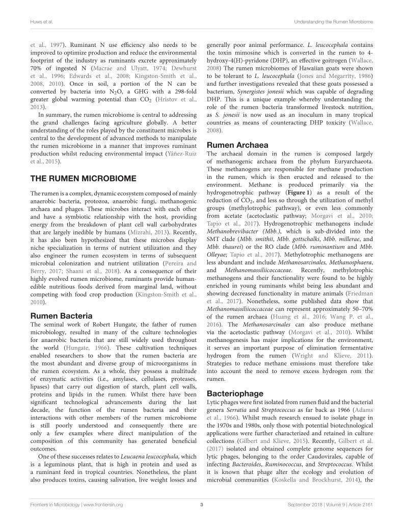
Rumen ArchaeaThe archaeal domain in the rumen is composed largelyof methanogenic archaea from the phylum Euryarchaeota.These methanogens are responsible for methane productionin the rumen, which is then eructed and released to theenvironment. Methane is produced primarily via thehydrogenotrophic pathway (Figure 1) as a result of thereduction of CO2, and less so through the utilization of methylgroups (methylotrophic pathway), or even less commonlyfrom acetate (acetoclastic pathway; Morgavi et al., 2010;Tapio et al., 2017). Hydrogenotrophic methanogens includeMethanobrevibacter (Mbb.), which is sub-divided into theSMT clade (Mbb. smithii, Mbb. gottschalki, Mbb. millerae, andMbb. thaurei) or the RO clade (Mbb. ruminantium and Mbb.Olleyae; Tapio et al., 2017). Methylotrophic methanogens areless abundant and include Methanosarcinales, Methanosphaera,and Methanomassiliicoccaceae. Recently, methylotrophicmethanogens and their functionality were found to be highlyenriched in young ruminants whilst being less abundant andshowing decreased functionality in mature animals (Friedmanet al., 2017). Nonetheless, some published data show thatMethanomassiliicoccaceae can represent approximately 50–70%of the rumen archaea (Huang et al., 2016; Wang P. et al.,2016). The Methanosarcinales can also produce methanevia the acetoclastic pathway (Morgavi et al., 2010). Whilstmethanogenesis has major implications for the environment,it serves an important purpose of elimination fermentativehydrogen from the rumen (Wright and Klieve, 2011).Strategies to reduce methane emissions must therefore takeinto account the need to remove excess hydrogen rom therumen.
BacteriophageLytic phages were first isolated from rumen fluid and the bacterialgenera Serratia and Streptococcus as far back as 1966 (Adamset al., 1966). Whilst much research ensued to isolate phage inthe 1970s and 1980s, only those with potential biotechnologicalapplications were further characterized and retained in culturecollections (Gilbert and Klieve, 2015). Recently, Gilbert et al.(2017) isolated and obtained complete genome sequences forlytic phages, belonging to the order Caudovirales, capable ofinfecting Bacteroides, Ruminococcus, and Streptococcus. Whilstit is known that phage alter the ecology and evolution ofmicrobial communities (Koskella and Brockhurst, 2014), the
Frontiers in Microbiology | www.frontiersin.org 3 September 2018 | Volume 9 | Article 2161
Huws et al. Understanding the Rumen Microbiome
FIGURE 1 | The hydrogenotrophic methane production pathway including
enzyme classifications (EC) for enzyme involved in the process. Reproduced
from Shi et al. (2014).
effects of phage on the rumen microbiome remains to bedetermined.
Rumen ProtozoaWhilst the rumen bacteria are the most numerate, the rumenprotozoa represent a large proportion of the microbial biomasswithin the rumen (approximately 20% and up to 50% in someconditions) due to their cell volume. Rumen protozoa werefirst described by Gruby and Delafond in 1843 (Gruby andDelafond, 1843) and, along with fungi, make up the rumeneukaryote members of the microbiota (Williams and Coleman,1997; Newbold et al., 2015). Ciliates dominate in the rumen,with flagellates such asTrichomonas sp.,Monocecromonas sp. andChilomastix sp. occasionally seen, but in much lower densities(Williams and Coleman, 1997). Ruminants commonly harbordistinct protozoal populations from birth, with only minorchanges in diversity throughout life, although the abundances ofspecies fluctuate with changes in diet (Williams and Coleman,1997). For example, Dastrychia and Entodinium were shown tobe the predominant genera in rumen fluid taken from dairycows and Dastrychia has been shown to be more predominantin the rumen fluid taken from cows fed corn stover as compared
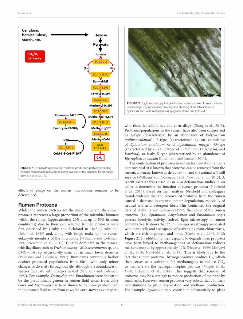
FIGURE 2 | Light microscopy image of rumen contents taken from a ruminant
possessing B-type protozoal diversity and showing close interactions of
Epidinium spp. with fresh perennial ryegrass. Scale bar: 200µM.
with those fed alfalfa hay and corn silage (Zhang et al., 2015).Protozoal populations in the rumen have also been categorizedas A-type (characterized by an abundance of Polyplastronmultivesiculatum), B-type (characterized by an abundanceof Epidinium caudatum or Eudiplodinium maggii), O-type(characterized by an abundance of Entodinum, Dasytrycha, andIsotricha), or lastly K-type (characterized by an abundance ofElytroplastron bubali; Kittelmann and Janssen, 2011).
The contribution of protozoa to rumen fermentation remainscontroversial. It is known that protozoa can be removed from therumen, a process known as defaunation, and the animal will stillsurvive (Williams and Coleman, 1992; Newbold et al., 2015). Arecent meta-analysis used 23 in vivo defaunation studies in aneffort to determine the function of rumen protozoa (Newboldet al., 2015). Based on their analysis, Newbold and colleaguesfound evidence that the removal of protozoa from the rumencaused a decrease in organic matter degradation, especially ofneutral and acid detergent fiber. This confirmed the originaldata of Williams and Coleman (1992) that some of the rumenprotozoa (i.e., Epidinium, Polyplastron and Entodinium spp.)possess fibrolytic activity. Indeed, light microscopy of rumencontents clearly shows that Epidinium spp. are strongly associatedwith plant cells and are capable of scavenging plant chloroplasts,which are rich in protein and lipids (Huws et al., 2009, 2012;Figure 2). In addition to their capacity to degrade fiber, protozoahave been linked to methanogenesis as defaunation reducesmethane output by approximately 11% (Hegarty, 1999; Morgaviet al., 2010; Newbold et al., 2015). This is likely due to thefact that rumen protozoal hydrogenosomes produce H2, whichthen serves as a substrate for methanogens to reduce CO2
to methane via the hydrogenotrophic pathway (Vogels et al.,1980; Belanche et al., 2014). This suggests that removal ofprotozoa may be a strategy to reduce production of methane byruminants. However, rumen protozoa vary substantially in theircontributions to plant degradation and methane production.For example, Epidinium spp. contribute substantially to plant
Frontiers in Microbiology | www.frontiersin.org 4 September 2018 | Volume 9 | Article 2161
Huws et al. Understanding the Rumen Microbiome
degradation (Huws et al., 2009) and generally holotrichs supportmethanogens and methanogenesis (Belanche et al., 2014). As aconsequence, a strategy which eliminates all protozoa may notbe the best approach, nonetheless, elimination of a sub-group ofprotozoa is a major challenge which currently is technologicallychallenging.
Rumen FungiThe flagellated zoospores of anaerobic fungi(Neocallimastigomycetes) were first observed in the early1900’s. However, it was not until the 1970’s that their trueidentity was confirmed (Orpin, 1974, 1977a). To date, nineanaerobic fungal genera have been characterized with manyother uncultivated taxa known to exist (Koetschan et al.,2014; Edwards et al., 2017; Paul et al., 2018). Paul et al. (2018)attempted to get consensus on the diversity of anaerobic fungiinhabiting the guts of herbivores and concluded that amongthe cultured genera, Piromyces was the most represented withBuwchfawromyces being the least represented in sequence dataobtained from the Genbank database. Paul et al. (2018) alsosuggest that possibly another 25 new genera exist in the gutsof herbivores, which remain uncharacterized. Irrespective,anaerobic fungi are among the most potent fiber-degradingorganisms in the known biological world, primarily due to theirefficient and extensive set of enzymes for the degradation of plantstructural polymers (Solomon et al., 2016). Furthermore, theirrhizoids have the ability to physically penetrate plant structuralbarriers (Orpin, 1977a,b). The latter ability benefits other rumenmicrobes by increasing the plant cell surface area available forcolonization. Rumen fungi also possess amylolytic (Gordon andPhillips, 1998) and proteolytic activity (Gruninger et al., 2014).
The activity of anaerobic fungi is enhanced by methanogenicarchaea (Cheng et al., 2009), which are known to physicallyattach to anaerobic fungal biomass. Anaerobic fungi are clearlybeneficial, and have been shown to improve feed intake,feed digestibility, feed efficiency, daily weight gain and milkproduction (Lee et al., 2000; Dey et al., 2004; Paul et al., 2004;Tripathi et al., 2007; Saxena et al., 2010; Puniya et al., 2015).Chitin measurements (Rezaeian et al., 2004) and rRNA transcriptabundance (Elekwachi et al., 2017) indicate that anaerobicfungi represent 10–20% of the rumen microbiome. However,like protozoa, they are not routinely studied despite suitablecultivation independent tools being available (Edwards et al.,2017).
Despite the importance of the rumen eukaryotes, ourunderstanding of their function is far less than that of rumenbacteria. Beyond the study of their fiber degrading enzymes,much of the activity and metabolism of anaerobic fungi remainsunknown, particularly due to the limited annotation of themultiple genome sequences and transcriptomes now available(Edwards et al., 2017). As with protozoa, key challenges includetheir cultivation, lack of genomic information, and lack ofconsensus on best practices to analyse sequence data (Ishaq et al.,2017). Thus, there are still many challenges which need to beovercome to enable a comprehensive understanding of the rumenmicrobiome as a whole.
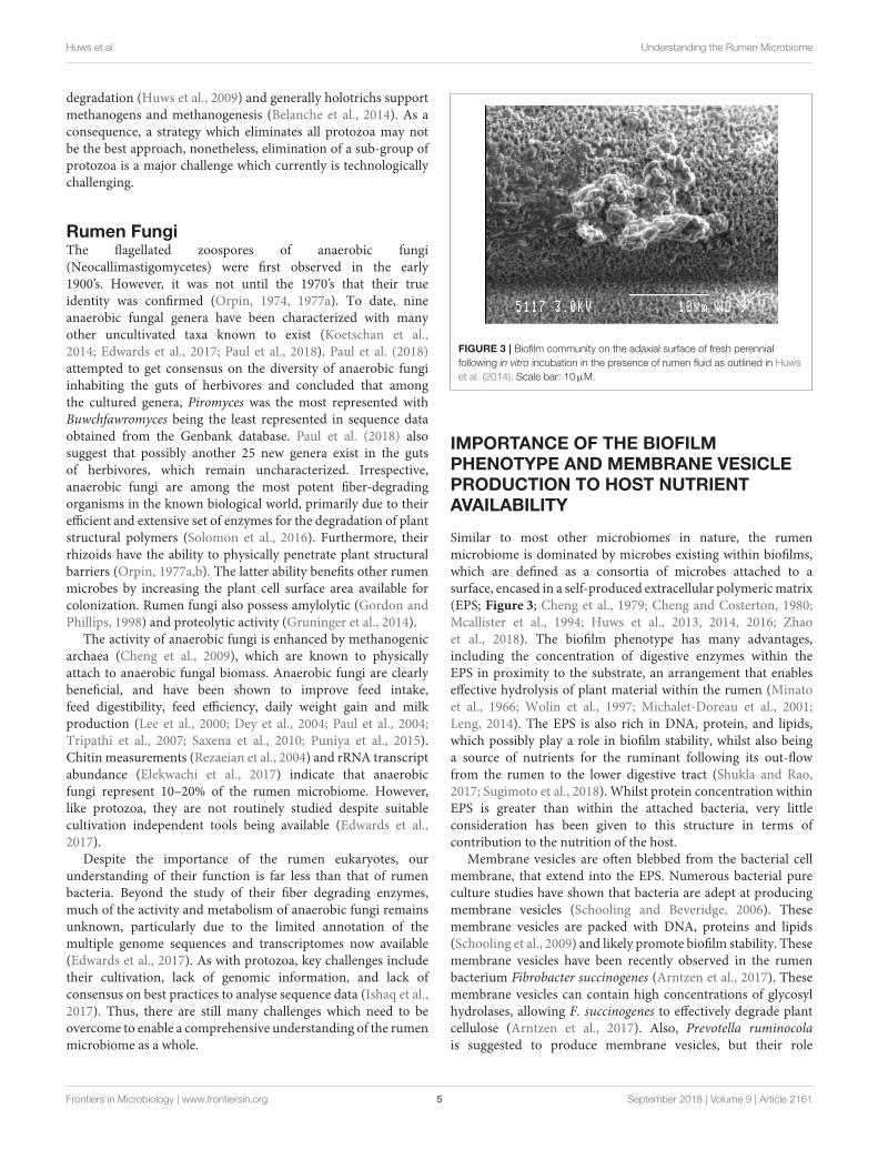
FIGURE 3 | Biofilm community on the adaxial surface of fresh perennial
following in vitro incubation in the presence of rumen fluid as outlined in Huws
et al. (2014). Scale bar: 10µM.
IMPORTANCE OF THE BIOFILMPHENOTYPE AND MEMBRANE VESICLEPRODUCTION TO HOST NUTRIENTAVAILABILITY
Similar to most other microbiomes in nature, the rumenmicrobiome is dominated by microbes existing within biofilms,which are defined as a consortia of microbes attached to asurface, encased in a self-produced extracellular polymericmatrix(EPS; Figure 3; Cheng et al., 1979; Cheng and Costerton, 1980;Mcallister et al., 1994; Huws et al., 2013, 2014, 2016; Zhaoet al., 2018). The biofilm phenotype has many advantages,including the concentration of digestive enzymes within theEPS in proximity to the substrate, an arrangement that enableseffective hydrolysis of plant material within the rumen (Minatoet al., 1966; Wolin et al., 1997; Michalet-Doreau et al., 2001;Leng, 2014). The EPS is also rich in DNA, protein, and lipids,which possibly play a role in biofilm stability, whilst also beinga source of nutrients for the ruminant following its out-flowfrom the rumen to the lower digestive tract (Shukla and Rao,2017; Sugimoto et al., 2018). Whilst protein concentration withinEPS is greater than within the attached bacteria, very littleconsideration has been given to this structure in terms ofcontribution to the nutrition of the host.
Membrane vesicles are often blebbed from the bacterial cellmembrane, that extend into the EPS. Numerous bacterial pureculture studies have shown that bacteria are adept at producingmembrane vesicles (Schooling and Beveridge, 2006). Thesemembrane vesicles are packed with DNA, proteins and lipids(Schooling et al., 2009) and likely promote biofilm stability. Thesemembrane vesicles have been recently observed in the rumenbacterium Fibrobacter succinogenes (Arntzen et al., 2017). Thesemembrane vesicles can contain high concentrations of glycosylhydrolases, allowing F. succinogenes to effectively degrade plantcellulose (Arntzen et al., 2017). Also, Prevotella ruminocolais suggested to produce membrane vesicles, but their role
Frontiers in Microbiology | www.frontiersin.org 5 September 2018 | Volume 9 | Article 2161
Huws et al. Understanding the Rumen Microbiome
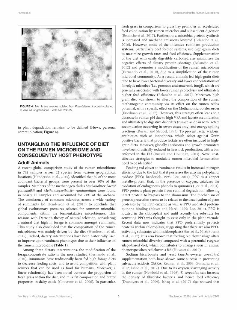
FIGURE 4 | Membrane vesicles isolated from Prevotella ruminocola incubated
in vitro in Hungate tubes. Scale bar: 200 nM.
in plant degradation remains to be defined (Huws, personalcommunication; Figure 4).
UNTANGLING THE INFLUENCE OF DIETON THE RUMEN MICROBIOME ANDCONSEQUENTLY HOST PHENOTYPE
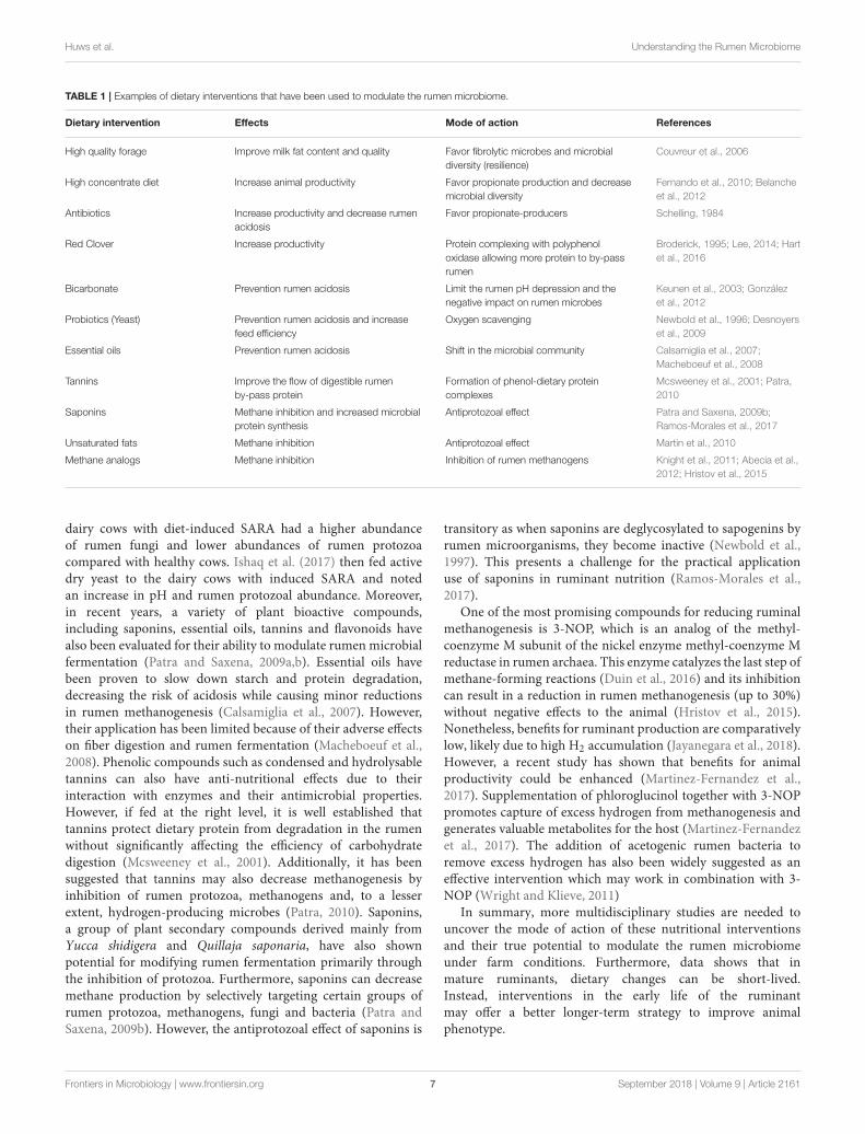
Adult AnimalsA recent global comparison study of the rumen microbiomein 742 samples across 32 species from various geographicallocations (Henderson et al., 2015), identified that 30 of the mostabundant bacterial groups were present in over 90% of thesamples. Members of the methanogen cladesMethanobrevibactergottschalkii and Methanobrevibacter ruminantium were foundin nearly all samples and accounted for 74% of the archaea.The consistency of common microbes across a wide varietyof ruminants led Henderson et al. (2015) to conclude thatglobal evolutionary pressures selected for common microbialcomponents within the fermentatative microbiomes. Thisreasons with Darwin’s theory of natural selection, consideringa natural diet high in forage is common amongst ruminants.This study also concluded that the composition of the rumenmicrobiome was mainly driven by the diet (Henderson et al.,2015). Indeed, dietary interventions have been historically usedto improve upon ruminant phenotypes due to their influence onthe rumen microbiome (Table 1).
Among these dietary interventions, the modification of theforage:concentrate ratio is the most studied (Fernando et al.,2010). Ruminants have traditionally been fed high forage dietsto decrease feeding costs, and to avoid competition with plantsources that can be used as food for humans. Moreover, alinear relationship has been noted between the proportion offresh grass within the diet, and milk fat composition and butterproperties in dairy cattle (Couvreur et al., 2006). In particular,
fresh grass in comparison to grass hay promotes an acceleratedfeed colonization by rumen microbes and subsequent digestion(Belanche et al., 2017). Furthermore, microbial protein synthesisis increased and methane emissions lowered (Belanche et al.,2016). However, most of the intensive ruminant productionsystems, particularly beef feedlot systems, use high-grain dietsto maximize growth rates and feed efficiency. Supplementationof the diet with easily digestible carbohydrates minimizes thenegative effects of dietary protein shortage (Belanche et al.,2012) and promotes a modification of the rumen microbiome(Fernando et al., 2010), due to a simplification of the rumenmicrobial community. As a result, animals fed high-grain dietstend to have lower bacterial diversity and lower concentrations offibrolytic microbes (i.e., protozoa and anaerobic fungi), which aregenerally associated with lower rumen proteolysis and ultimatelyhigher feed efficiency (Belanche et al., 2012). Moreover, highgrain diet was shown to affect the composition of the rumenmethanogenic community via its effect on the rumen redoxpotential, with a specific effect on the Methanomicrobiales order(Friedman et al., 2017). However, this strategy often leads to adecrease in rumen pH due to high VFA and lactate accumulationand ultimately to digestive disorders (rumen acidosis with lactateaccumulation occurring in severe cases only) and energy spillingreactions (Russell and Strobel, 1993). To prevent lactic acidosis,antibiotics such as ionophores, which select against Grampositive bacteria that produce lactate are often included in high-grain diets. However, globally antibiotics and growth promotershave been drastically reduced in livestock production, with a banenforced in the EU (Russell and Houlihan, 2003). Novel cost-effective strategies to modulate rumen microbial fermentationneed to be identified.
Feeding red clover to ruminants results in increased nitrogenefficiency due to the fact that it possesses the enzyme polyphenoloxidase (PPO; Broderick, 1995; Lee, 2014). PPO is a coppermetallo-protein that, in the presence of oxygen, catalyzes theoxidation of endogenous phenols to quinones (Lee et al., 2004).PPO protects plant protein from ruminal degradation, allowingintact protein to by-pass to the abomasum. The mechanism ofprotein protection seems to be related to the deactivation of plantproteases by the PPO enzyme as well as PPO mediated protein-quinone binding (Mayer and Harel, 1979; Lee, 2014). PPO islocated in the chloroplast and until recently the substrate foractivating PPO was thought to exist only in the plant vacuole.Recent data now indicates that PPO preferentially protectsproteins within chloroplasts, suggesting that there are also PPO-activating substrates within chloroplasts (Hart et al., 2016; Boeckxet al., 2017). It is also known that feeding red clover silage altersrumen microbial diversity compared with a perennial ryegrasssilage-based diet, which contributes to changes seen in animalphenotype when red clover is fed (Huws et al., 2010).
Sodium bicarbonate and yeast (Saccharomyces cerevisiae)supplementation both have shown some success in preventingsub-acute acidosis (SARA; Keunen et al., 2003; González et al.,2012; Ishaq et al., 2017). Due to its oxygen scavenging activityin the rumen (Newbold et al., 1996), S. cerevisiae can increasethe density of fibrolytic bacteria and hence feed efficiency(Desnoyers et al., 2009). Ishaq et al. (2017) also showed that
Frontiers in Microbiology | www.frontiersin.org 6 September 2018 | Volume 9 | Article 2161
Huws et al. Understanding the Rumen Microbiome
TABLE 1 | Examples of dietary interventions that have been used to modulate the rumen microbiome.
Dietary intervention Effects Mode of action References
High quality forage Improve milk fat content and quality Favor fibrolytic microbes and microbial
diversity (resilience)
Couvreur et al., 2006
High concentrate diet Increase animal productivity Favor propionate production and decrease
microbial diversity
Fernando et al., 2010; Belanche
et al., 2012
Antibiotics Increase productivity and decrease rumen
acidosis
Favor propionate-producers Schelling, 1984
Red Clover Increase productivity Protein complexing with polyphenol
oxidase allowing more protein to by-pass
rumen
Broderick, 1995; Lee, 2014; Hart
et al., 2016
Bicarbonate Prevention rumen acidosis Limit the rumen pH depression and the
negative impact on rumen microbes
Keunen et al., 2003; González
et al., 2012
Probiotics (Yeast) Prevention rumen acidosis and increase
feed efficiency
Oxygen scavenging Newbold et al., 1996; Desnoyers
et al., 2009
Essential oils Prevention rumen acidosis Shift in the microbial community Calsamiglia et al., 2007;
Macheboeuf et al., 2008
Tannins Improve the flow of digestible rumen
by-pass protein
Formation of phenol-dietary protein
complexes
Mcsweeney et al., 2001; Patra,
2010
Saponins Methane inhibition and increased microbial
protein synthesis
Antiprotozoal effect Patra and Saxena, 2009b;
Ramos-Morales et al., 2017
Unsaturated fats Methane inhibition Antiprotozoal effect Martin et al., 2010
Methane analogs Methane inhibition Inhibition of rumen methanogens Knight et al., 2011; Abecia et al.,
2012; Hristov et al., 2015
dairy cows with diet-induced SARA had a higher abundanceof rumen fungi and lower abundances of rumen protozoacompared with healthy cows. Ishaq et al. (2017) then fed activedry yeast to the dairy cows with induced SARA and notedan increase in pH and rumen protozoal abundance. Moreover,in recent years, a variety of plant bioactive compounds,including saponins, essential oils, tannins and flavonoids havealso been evaluated for their ability to modulate rumen microbialfermentation (Patra and Saxena, 2009a,b). Essential oils havebeen proven to slow down starch and protein degradation,decreasing the risk of acidosis while causing minor reductionsin rumen methanogenesis (Calsamiglia et al., 2007). However,their application has been limited because of their adverse effectson fiber digestion and rumen fermentation (Macheboeuf et al.,2008). Phenolic compounds such as condensed and hydrolysabletannins can also have anti-nutritional effects due to theirinteraction with enzymes and their antimicrobial properties.However, if fed at the right level, it is well established thattannins protect dietary protein from degradation in the rumenwithout significantly affecting the efficiency of carbohydratedigestion (Mcsweeney et al., 2001). Additionally, it has beensuggested that tannins may also decrease methanogenesis byinhibition of rumen protozoa, methanogens and, to a lesserextent, hydrogen-producing microbes (Patra, 2010). Saponins,a group of plant secondary compounds derived mainly fromYucca shidigera and Quillaja saponaria, have also shownpotential for modifying rumen fermentation primarily throughthe inhibition of protozoa. Furthermore, saponins can decreasemethane production by selectively targeting certain groups ofrumen protozoa, methanogens, fungi and bacteria (Patra andSaxena, 2009b). However, the antiprotozoal effect of saponins is
transitory as when saponins are deglycosylated to sapogenins byrumen microorganisms, they become inactive (Newbold et al.,1997). This presents a challenge for the practical applicationuse of saponins in ruminant nutrition (Ramos-Morales et al.,2017).
One of the most promising compounds for reducing ruminalmethanogenesis is 3-NOP, which is an analog of the methyl-coenzyme M subunit of the nickel enzyme methyl-coenzyme Mreductase in rumen archaea. This enzyme catalyzes the last step ofmethane-forming reactions (Duin et al., 2016) and its inhibitioncan result in a reduction in rumen methanogenesis (up to 30%)without negative effects to the animal (Hristov et al., 2015).Nonetheless, benefits for ruminant production are comparativelylow, likely due to high H2 accumulation (Jayanegara et al., 2018).However, a recent study has shown that benefits for animalproductivity could be enhanced (Martinez-Fernandez et al.,2017). Supplementation of phloroglucinol together with 3-NOPpromotes capture of excess hydrogen from methanogenesis andgenerates valuable metabolites for the host (Martinez-Fernandezet al., 2017). The addition of acetogenic rumen bacteria toremove excess hydrogen has also been widely suggested as aneffective intervention which may work in combination with 3-NOP (Wright and Klieve, 2011)
In summary, more multidisciplinary studies are needed touncover the mode of action of these nutritional interventionsand their true potential to modulate the rumen microbiomeunder farm conditions. Furthermore, data shows that inmature ruminants, dietary changes can be short-lived.Instead, interventions in the early life of the ruminantmay offer a better longer-term strategy to improve animalphenotype.
Frontiers in Microbiology | www.frontiersin.org 7 September 2018 | Volume 9 | Article 2161
Huws et al. Understanding the Rumen Microbiome
Early-LifeIn contrast to the developed rumen, where a stable and resilientmicrobial community is established, during the development ofthe rumen after birth a succession of different microbial groupscolonize and start occupying the different ecological niches. Theinstability occurring during this period potentially allows formanipulation to assemble a specific community composition thatpersist later in life for better health and productivity within agiven production system (Yáñez-Ruiz et al., 2015).
At birth, ruminants display a non-developed reticulo-rumen.Until the system is fully matured, they function as monogastrics,whereby the milk fed is not digested in the rumen but flowsto the abomasum via an esophageal groove (Church, 1988).Colonization of the developing rumen begins immediately afterbirth and progresses through the first few months of life until astable community establishes (Jami et al., 2013). The dynamics ofthe gut microbial community establishment in young ruminantsoccurs in three successive steps (Rey et al., 2014; Abecia et al.,2017): (i) initial colonization (0–2 days post-partum) originatedfrom a combination of sources such as microbiota of mother’svagina, skin, colostrum and microbes within the environment(Van Nimwegen et al., 2011; Yeoman et al., 2018); (ii) transitionalstage (3–15 days) during the transition from colostrum tomilk, and iii) maturation stage in which solid feed intakeprogressively increases and the distribution of main bacterialphyla and other microbial groups is comparable to that inadult animals. It is important to note that although the rumenmicrobiome establishes before intake of solid feeds, the type offeed consumed plays a significant role in shaping the establishedrumen microbiome. Hence, the early phases of solid feed intakerepresents a window of opportunity to modulate the compositionof the initial colonizers of the different ecological niches in therumen according to dietary and management strategies (Yáñez-Ruiz et al., 2015). Indeed, the use of probiotics, such as lacticacid bacteria, in early life to mitigate incidence of digestive andrespiratory diseases has shown promise (Timmerman et al., 2005;Signorini et al., 2012). Yáñez-Ruiz et al. (2010) also reportedthat feeding forage vs. concentrate around weaning modifiesthe bacterial population colonizing the rumen of lambs andthat the effect persists over 4 months. It is also known thatfeeding concentrate in early life stimulates the developmentof the epithelium, while feeding high fiber diets can stimulatedevelopment of rumen muscularization and volume (Zitnanet al., 1998). Nonetheless, little is known regarding the impactof management practices, such as milk intake, delayed weaningetc. on early-life programming of the rumen microbiome and itsimplications for ruminant productivity.
Another factor that promotes differences in rumencolonization is the presence of the dam and the associatedincrease in the availability of microorganisms in theenvironment. This can allow earlier (and different) inoculationof microbes in the digestive tract of naturally raised newbornsas compared to those fed milk replacer and kept in isolation(Abecia et al., 2017). Direct contact with the mother offers aconstant source of microbes through the mouth, feces, skin andmilk (Yeoman et al., 2018), sources that are not available forcalves raised in isolation on milk replacer. This explains the
greater number of Operational Taxonomic Units (OTUs) andbacterial diversity observed in naturally reared calves. Anotherdistinctive feature between natural and artificial rearing systemsis the near absence of protozoa in the rumen of artificially rearedcalves, as protozoa can only be inoculated in the rumen by directcontact with the dam or other mature animals through saliva(Abecia et al., 2014). A relatively recent study by Ishaq et al.(2015) showed that exposure of neonate lambs to the dam for 1week followed by subsequent separation was enough to ensurethe establishment of a stable rumen protozoal population fortheir lifetime.
Nutritional interventions in early-life may include (i) thedirect inoculation of specific microorganisms or (ii) the useof additives that prevent or facilitate the colonization of somemicrobial groups. Feeding live microorganisms to ruminants isnot a novel concept and extensive work has been published onthe use of “direct-fedmicrobials” (DFM;Martin andNisbet, 1992;Jeyanathan et al., 2014). The effect of supplementing S. cerevisiaeon rumen development and growth performance in neonataldairy calves has also been evaluated (Lesmeister et al., 2004).Although yeast cultures are widely used in ruminant nutrition,the concept of applying them in the diet of pre-ruminantsdeserves further assessment, especially in terms of their long termeffects on the microbiome (Alugongo et al., 2017). A differentapproach that uses compounds to inhibit the establishment ofcertain microbial groups or favor the development of others isalso now starting to attract attention. It has been shown thatapplication of bromochloromethane (BCM) to young goat kidsmodified archaeal colonization of the rumen, and was linked toa reduction in methane emission of around 50%, with the effectspersisting for 3 months after weaning (Abecia et al., 2013, 2014).
Despite some promising results from early-life dietaryinterventions, the ecological dynamics underpinning themicrobial colonization, the most effective window of time forintervention and the long-term implications have yet to beidentified.
UNTANGLING THE INFLUENCE OF HOSTGENOMICS ON THE RUMENMICROBIOME AND CONSEQUENTLYHOST PHENOTYPE
Consistent with human twin heritability studies (Goodrich et al.,2016), it is reasonable to hypothesize that animals possessingsimilar genomes should have more similar rumen microbiomes.Evidence of the influence of the host on the rumen microbiomewas first postulated by Weimer et al. (2010) who found thatafter near total exchange of the rumen contents betweencows, individuals restored their bacterial composition back topre-exchange conditions, which also returned rumen pH andvolatile fatty acid (VFA) concentration to pre-exchange values.Furthermore, in another near-total rumen content exchangebetween high- and low-efficiency Holstein cows, Weimer et al.(2017) demonstrated the hosts ability to return the rumenbacterial community to the original status, whilst linking therumen microbiome to milk production efficiency.
Frontiers in Microbiology | www.frontiersin.org 8 September 2018 | Volume 9 | Article 2161
Huws et al. Understanding the Rumen Microbiome
Whilst Henderson et al. (2015) postulated that diet wasthe main driver for rumen microbiome composition, they alsoidentified some differences in the relative abundance of certainbacterial populations across ruminant species. Similarly, whenthe microbiome of water buffalo (Bubalus bubalis) and Jerseycows were compared under comparable feeding conditionsvariations in bacterial, protozoa and methanogen populationswere found between the two species (Iqbal et al., 2018),suggesting that the rumen microbiome is controlled, to a certainextent, by the genetics of the host. In a beef cattle experiment,Roehe et al. (2016) ranked beef sire progeny groups based onrelative archaeal abundance and reported that group rankingremained consistent overall and within diet, suggesting thatarchaeal abundance in ruminal digesta is also, in part under hostgenetic control. Using sire progeny groups in dairy cattle, furtherevidence of genetic control was documented by the discoverythat 22 bacterial OTUs, exhibited a heritability estimate of 0.7or greater in dairy cattle (Sasson et al., 2017). In addition, theseheritable OTUs were found to be correlated with traits such asDMI (dry matter intake) and RFI. Pinares-Patiño et al. (2011)and Pinares-Patiño et al. (2013) demonstrated that methaneproduction is also regulated by host genetics in sheep and thatselection of low methane emitting animals by genotyping ispossible.
Nonetheless, De Mulder et al. (2018), stated that thedifferences in rumen microbiome composition may be due toother factors other than host genomics, including early life eventsand the fact that some breeds of cattle, such as Belgian Bluecattle, have a higher rate of cesarean section birth. The hostimmune system also likely plays an influential role on the rumenmicrobiome. For example, secretory immunoglobulin A (SIgA),which favors commensal bacteria in the gut (Gutzeit et al.,2014), has been shown to coat rumen bacteria (Fouhse et al.,2017) and control the host’s recognition of certain microbialspecies. In addition, the rumen epithelium plays an importantrole in both nutrient uptake and immunity. The physiology ofthe rumen has also been highlighted as a potential factor thatinfluences the rumen microbiome. For example, differences inrumen and camelid foregut volume, physiology as well as feedingfrequencies, was suggested as a reason for the proportionallyhigher abundance of unclassified Veillonellaceae in camelids,deer and sheep compared to cattle (Henderson et al., 2015).In addition, methane yield is associated with retention time inthe rumen (Pinares-Patiño et al., 2003) correlating increasedpassage rate in the rumen with reduced methane yield. Janssen(2010) provides a thorough review of these studies which inessence show that increased passage rate leads to less feedbeing fermented in the rumen and subsequently less substrateis available for methanogenesis (Tapio et al., 2017). It has alsobeen demonstrated that both a shorter rumen retention timeand a smaller rumen result in reduced methane yield (Goopyet al., 2014). Additionally, variation in the rumination behaviorof animals can influence particle retention time (Mcsweeneyet al., 1989). Therefore, genetic influence of the host on rumenpassage rate is likely to be one host factor that influences therumen microbiome, but other factors should also be considered(Pinares-Patiño et al., 2013).
Whilst there is increased evidence that host genetics has aninfluential role on themicrobial population residing in the rumen(Tapio et al., 2017), our current understanding of the extent ofthis influence and the underlying mechanisms (Sasson et al.,2017) remains incomplete, although a region on chromosome 6was recently associated with Actinobacteria, Euryarchaeota, andFibrobacteres densities (Golder et al., 2018).
CONTRIBUTIONS OF THE LOWERGASTROINTESTINAL TRACTMICROBIOMES TO RUMINANTPHENOTYPE
Typically, scientists have focussed their attention onunderstanding the rumen in order to deliver upon globallivestock challenges. However, the lower gastrointestinal (GI)tract microbiomes also play an important role, particularly inearly life (Meale et al., 2017). The microbial composition ofthe post-ruminal gastrointestinal tract is shaped by pH, gutmotility, redox potential, and host secretions present in differentcompartments of the digestive tract. Most microbes flowingfrom the rumen into the abomasum are lysed by the low pHand enzymatic activity within the organ. As a consequence ofthe harsh environmental conditions prevailing in the abomasumand at the beginning of the small intestine, microbial numbersand diversity plummet by several orders of magnitude in theabomasum, duodenum and jejunum as compared to the rumen(Frey et al., 2010; He et al., 2018; Yeoman et al., 2018). From theileum onwards, including caecum, colon and feces, favorablefermentation conditions are present again and microbial densityand phylogenetic diversity increase to a level comparable to thatof the rumen (Frey et al., 2010; De Oliveira et al., 2013; Popovaet al., 2017; He et al., 2018; Yeoman et al., 2018).
The post-ruminal microbial community is composedpredominantly of bacteria, but methanogenic archaea andanaerobic fungi have been described (Davies et al., 1993),although the later phylogentic group has not been targetedintensively with high-throughput sequencing techniques. Thereare significant difference in the microbial community assemblagedepending on the region of the GI tract (i.e., rumen vs. post-rumen; Mao et al., 2015; Bergmann, 2017; Zeng et al., 2017;Yeoman et al., 2018), and the post rumen microbiota differfurther between the small (duodenum, jejunum, and ileum)and the large (cecum, colon, and rectum) intestine (Mao et al.,2015; Bergmann, 2017; Wang et al., 2017; Yeoman et al., 2018).In general terms, compared to the rumen, the proportion ofBacteroidetes decrease and that of Firmicutes and Proteobacteriaincrease. Prevotella, Bacteroides, Ruminococcus, Treponema, andDesulfovibrio genera were detected in all segments of the GI tractof ruminant animals, while Fibrobacter was only present in theforegut (Zeng et al., 2017). Prevotella, Bacteroides, Ruminococcus,Faecalibacterium, Roseburia and Clostridium are consistentlyidentified in fecal samples from ruminants and are consideredpart of the core microbiota (Dowd et al., 2008; Durso et al.,2012). As for the rumen, the rectal microbiota shows important
Frontiers in Microbiology | www.frontiersin.org 9 September 2018 | Volume 9 | Article 2161
Huws et al. Understanding the Rumen Microbiome
inter-individual variation (Durso et al., 2010) and are affected bydiet (Shanks et al., 2011).
The mucosa-associated microbial community is also animportant modulator of immunological function and health(Malmuthuge et al., 2015). Mucosa-associated communitiesdiffer from those associated with luminal contents; and alsovary among intestinal regions (Malmuthuge et al., 2014; Maoet al., 2015; Yeoman et al., 2018). Potential pathogens such asEscherichia, Shigella, Salmonella and Treponema spp. are mostfrequently found in the mucosa-associated bacterial microbiota(Mao et al., 2015; Song et al., 2018). Recently, differences inboth the mucosa-associated microbiota of the rectoanal junctionand fecal microbiota of cattle have been shown to influence theshedding of the human pathogen Escherichia coli O157 in cattlefeces (Stenkamp-Strahm et al., 2018; Wang et al., 2018).
The role of the intestinal microbiota in feed degradationappears to be less important than that of the rumen (Al-Masaudiet al., 2017). Its main function has been suggested to be relatedto animal health and cross-talk interaction with the animalhost (Lyte et al., 2018), although work in this area is only inits nascent phase and these aspects need further investigation.Notwithstanding, it is highlighted that feces and samples fromthe intestines cannot be used as proxies of rumen function ona microbiome biomarker level (Tapio et al., 2016). Nonetheless,concentrations of the compound archaeol in feces has beenshown to correlate with methane emissions in cattle (Mccartneyet al., 2014).
DEVELOPING MICROBIOMEBIOMARKERS FOR PREDICTION OFRUMINANT PHENOTYPE
The sheer size of the rumen (12–15% of body mass) andconnectedness with the vascular, respiratory and immunesystems mean that it is well-placed to both affect, and be affectedby, animal function. There is a growing number of exampleswhere the interaction between host and intestinal microbialmetabolism can be used to explain, or act as a biomarker for,complex traits such as nutrient efficiency, responses to stressorssuch as disease and adverse environments, as well as to predictanimal behavior.
Nucleic acids have long been used as biomarkers forrumen microbial processes. Early work focussed on rumenmicrobial protein synthesis and RNA (Mcallan and Smith,1969), while purine bases (Zinn and Owens, 1986) were alsoused as biomarkers for microbial (protein) synthesis in studieswith intestinally cannulated animals. More recent attemptsto develop non-invasive biomarker approaches to estimatemicrobial protein synthesis have used urinary metabolitesderived from microbial purines (allantoin and uric acid; Chenet al., 1990). Recent advances in analytical technologies andbioinformatics have now greatly expanded our capacity toinvestigate the role of the rumen and its microbiome in complextraits by studying the composition of microbial DNA and RNA(metataxonomics, metagenomics and metatranscriptomics), aswell as microbial metabolites in blood or urine (metabolomics).
In terms of metataxonomics, microbial correlations to feedefficiency and/or methane production in ruminants, using rRNAgenes or the Methyl coenzyme M reductase (mcrA) gene inmethanogens are difficult to interpret, due to the confoundingfactors such as animal type, feed, and rumen sample processingand analysis (see Metataxonomy section). Recent studies suggestthat methanogen diversity, and not density, is critically importantto methane output, with more diversity being associated withhigher emissions (Janssen and Kirs, 2008; Carberry et al.,2014). However, most studies involve a small number ofanimals, making it difficult to clearly confirm the link betweenmethanogen diversity and methane emissions (Morgavi et al.,2010). When investigating the rumen bacterial associations withmethane production, density of Sharpea has been shown to besignificantly lower in low methane emitting animals (Kamkeet al., 2016). Positive correlations between Eubacterium sp.and reduced feed efficiency were also reported by Hernandez-Sanabria et al. (2012). Jami et al. (2014) also reported a positivecorrelation between RFI and the uncultured rumen bacteriumRF39, whereas Shabat et al. (2016), suggested that an increasein the acrylate pathway coded by Megasphaera elsdenii andCoprococcus catus in the rumen may increase feed efficiencyand reduce methane. It has been suggested also that the ratioof bacteria:archaea reflects methane output from the animalwith positive correlations reported in a few studies (Wallaceet al., 2014; Auffret et al., 2017b), but results are not consistent(Tapio et al., 2017). Recent data also suggest that the rumenmicrobiome of feed efficient ruminants is less diverse than theirinefficient counterparts (Shabat et al., 2016; Li and Guan, 2017).The microbial diversity within the rumen offers the animalresilience from dietary related perturbations, such as acidosis.Therefore, care must be taken to ensure that breeding forincreased feed efficiency in ruminants does not negatively impactresilience of the microbiome and increase the susceptibility ofthe host to digestive diseases. Irrespective, metataxonomic datais highly variable due largely to the differences in techniquesemployed across published datasets (see Metataxonomy section)and animal variation. As such the use of gene biomarkersusing metagenomics and/or metranscriptomic approaches maybe more useful given that rumen microbes possess genes codingfor a high level of functional redundancy (Edwards et al., 2008;Weimer, 2015).
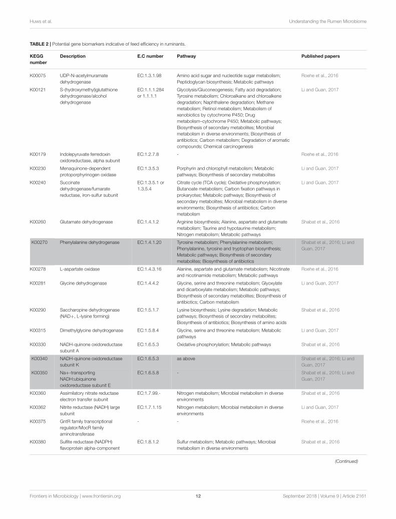
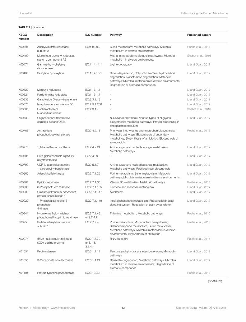
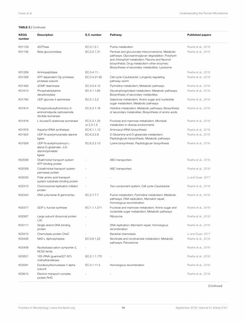
Recent work using metagenomics and/ormetatranscriptomics has confirmed significant relationshipsbetween the abundances of key rumen microbial genes and feedefficiency (Roehe et al., 2016; Shabat et al., 2016; Li and Guan,2017) and/or methane production (Roehe et al., 2016). Dueto the vastness of these datasets it is difficult to compare andinvestigate whether studies commonly find consensus genesthat would serve as good global biomarkers in their correlationstudies. Microbial gene correlations with RFI, data from Shabatet al. (2016) and Li and Guan (2017) showed some consensusas both showed that genes involved in amino acid metabolismwere less abundant in feed efficient animals (Table 2). Thesedata corresponds with observations that feed efficient animalsexcrete less urinary ammonia suggesting better rumen nitrogenuse efficiency (Bach et al., 2005; Broderick and Reynal, 2009).
Frontiers in Microbiology | www.frontiersin.org 10 September 2018 | Volume 9 | Article 2161
Huws et al. Understanding the Rumen Microbiome
Consensus of other genes across the three published datasetswere not found (Table 3). Likewise, genes correlating to methaneemissions show very little consensus amongst the five papersinvestigated. Nevertheless, in four datasets the methyl coenzymereductase enzyme, which is involved in the last step of thehydrogenotrophic methane pathway (Figure 1), showed themost correlation to methane. The lack of consensus acrossexperiments, whilst perhaps being biologically correct, likelyalso reflects the challenges associated with comparing of datasetsfrom different animals, variation in diet, as well as differences insampling method, sample preparations and data interpretation.Clearly more comparative large datasets are required to developmicrobiome based biomarkers for estimation of RFI andmethaneoutput. Alongside this is the need to obtain samples that arerepresentative of the rumen microbiome in a non-invasivemanner. Recently it was suggested that the oral microbiome ofruminants reflects the microbial diversity seen in the rumen(Tapio et al., 2016), raising the possibility that buccal swabbingmay be used as a proxy for rumen samples.
PROSPECTS FOR ENHANCING RUMENMICROBIOME UNDERSTANDING ANDANIMAL PHENOTYPE PREDICTIONS VIAMATHEMATICAL MODELING
Mathematical models can be used to integrate our understandingof feed, intake, digestion and passage rates on the resultingenergy available to the microbiome and ultimately the host.The development of rumen models has been deployed mainlyvia the consolidation of four model structures (Molly, Karoline,Cornell, and Dijkstra models) that have been improved over theyears to enhance the understanding of rumen function (Millset al., 2014; Huhtanen et al., 2015; Van Amburgh et al., 2015;Gregorini et al., 2016). These models represent relevant aspectsthat determine the nutritional and emission responses for a givendiet but do not attempt to provide a detailed description of themicrobiota or its function (Ellis et al., 2008). This gap between theavailable omics data of the rumen microbiome and the modelsneeds to be bridged to improve our understanding of rumenfunction (Bannink et al., 2016; Muñoz-Tamayo et al., 2016). Tomake these model applications possible, rumen modeling shouldembrace the framework of genome-scale metabolic models(GEMs). The basis of a GEM is the stoichiometry matrix thatlinks metabolites and biochemical reactions that the microbeis able to perform as a result of its genetic potential. Thestoichiometry matrix is organism-specific and results from agenome-scale network reconstruction obtained by a protocolthat includes functional genome annotation, curation of a draftreconstruction of metabolic reactions and finally translation ofthe reconstructed network into a computational model (GEM).The full process capitalizes on high-throughput network-wideand bibliomic data (Feist et al., 2009), and on dedicated software(Henry et al., 2010; Aite et al., 2018). The construction of arumen microbiome GEM will need to address central questionsthat remain to be elucidated due to the early stage of microbialcommunity modeling (Zengler and Palsson, 2012). One of these
key questions is howmicrobial species, their metabolic networks,and interspecies interactions should be represented (Biggs et al.,2015). Once this question is elucidated, a plethora of constraint-based reconstruction and analysis (COBRA) methods can bedeployed to investigate genotype–phenotype relationships (Lewiset al., 2012).
The COBRA methods rely on the principle thatmicrobial metabolism is bound by constraints that includethermodynamics, substrate and enzyme availability. Thesemethods mainly operate under steady-state. The most popularCOBRAmethod is flux balance analysis (FBA; Varma et al., 1993;Varma and Palsson, 1994), which looks at finding the networkreaction fluxes that optimize a regulatory condition (e.g.,microbial growth). Overall, COBRA approaches provide rationaltools for metabolic engineering. The number of applications isbroad and includes the development of tools for (i) studyinginteractions among different microbial groups, i.e., protozoa,fungi, archaea, bacteria and viruses or bacteriophages, (ii)developing selective cultivation strategies for as yet unculturedrumen microbes (Pope et al., 2011), (iii) designing methanemitigation strategies by exploiting the metabolic networksof genome-sequenced rumen archaea (Leahy et al., 2010;Pope et al., 2011), and (iv) developing prediction tools thatexploit microbiome biomarkers for fiber hydrolysis (Daiet al., 2015; Comtet-Marre et al., 2017, 2018) and methaneproduction (Popova et al., 2013; Shi et al., 2014; Auffret et al.,2017b).
Clearly rumen GEMs must be further integrated into wholerumen digestion models to provide a system-level pictureof the dynamic interplay between the diet, the animal hostand the rumen microbiota. Central to this task and for thedevelopment of novel strategies to enhance ruminant productionand reduce environmental impact is the need for data sharingand collaboration. The co-authors of this paper are all membersof the Global Research Alliance’s Rumen Microbial GenomicsNetwork, which is set up to allow global collaborations anddata sharing for this very purpose. This integration task isfar from trivial due to multiple time scales, among otheraspects such as parameter identifiability (Muñoz-Tamayo et al.,2018). Moreover, since COBRA approaches mainly operatesat steady-state, dynamic frameworks (Mahadevan et al., 2002;Baroukh et al., 2014) will need to be adapted to accountfor the dynamic fluctuations within the rumen environment.A great challenge is to deploy different model structures,capitalizing on “omics” data, and responding to differentgoals varying from supporting livestock management within aprecision farming context to guiding microbial programmingstrategies.
TECHNOLOGICAL ADVANCES TOFURTHER OUR UNDERSTANDING OF THERUMEN MICROBIOME
Genomics/CulturomicsLarge culture collections are incredibly powerful as the organismsin the collection can be studied both in vitro and in vivo.
Frontiers in Microbiology | www.frontiersin.org 11 September 2018 | Volume 9 | Article 2161
Huws et al. Understanding the Rumen Microbiome
TABLE 2 | Potential gene biomarkers indicative of feed efficiency in ruminants.
KEGG
number
Description E.C number Pathway Published papers
K00075 UDP-N-acetylmuramate
dehydrogenase
EC:1.3.1.98 Amino acid sugar and nucleotide sugar metabolism;
Peptidoglycan biosynthesis; Metabolic pathways
Roehe et al., 2016
K00121 S-(hydroxymethyl)glutathione
dehydrogenase/alcohol
dehydrogenase
EC:1.1.1.284
or 1.1.1.1
Glycolysis/Gluconeogenesis; Fatty acid degradation;
Tyrosine metabolism; Chloroalkane and chloroalkene
degradation; Naphthalene degradation; Methane
metabolism; Retinol metabolism; Metabolism of
xenobiotics by cytochrome P450; Drug
metabolism–cytochrome P450; Metabolic pathways;
Biosynthesis of secondary metabolites; Microbial
metabolism in diverse environments; Biosynthesis of
antibiotics; Carbon metabolism; Degradation of aromatic
compounds; Chemical carcinogenesis
Li and Guan, 2017
K00179 Indolepyruvate ferredoxin
oxidoreductase, alpha subunit
EC:1.2.7.8 - Roehe et al., 2016
K00230 Menaquinone-dependent
protoporphyrinogen oxidase
EC:1.3.5.3 Porphyrin and chlorophyll metabolism; Metabolic
pathways; Biosynthesis of secondary metabolites
Li and Guan, 2017
K00240 Succinate
dehydrogenase/fumarate
reductase, iron-sulfur subunit
EC:1.3.5.1 or
1.3.5.4
Citrate cycle (TCA cycle); Oxidative phosphorylation;
Butanoate metabolism; Carbon fixation pathways in
prokaryotes; Metabolic pathways; Biosynthesis of
secondary metabolites; Microbial metabolism in diverse
environments; Biosynthesis of antibiotics; Carbon
metabolism
Li and Guan, 2017
K00260 Glutamate dehydrogenase EC:1.4.1.2 Arginine biosynthesis; Alanine, aspartate and glutamate
metabolism; Taurine and hypotaurine metabolism;
Nitrogen metabolism; Metabolic pathways
Shabat et al., 2016
K00270 Phenylalanine dehydrogenase EC:1.4.1.20 Tyrosine metabolism; Phenylalanine metabolism;
Phenylalanine, tyrosine and tryptophan biosynthesis;
Metabolic pathways; Biosynthesis of secondary
metabolites; Biosynthesis of antibiotics
Shabat et al., 2016; Li and
Guan, 2017
K00278 L-aspartate oxidase EC:1.4.3.16 Alanine, aspartate and glutamate metabolism; Nicotinate
and nicotinamide metabolism; Metabolic pathways
Roehe et al., 2016
K00281 Glycine dehydrogenase EC:1.4.4.2 Glycine, serine and threonine metabolism; Glyoxylate
and dicarboxylate metabolism; Metabolic pathways;
Biosynthesis of secondary metabolites; Biosynthesis of
antibiotics; Carbon metabolism
Li and Guan, 2017
K00290 Saccharopine dehydrogenase
(NAD+, L-lysine forming)
EC:1.5.1.7 Lysine biosynthesis; Lysine degradation; Metabolic
pathways; Biosynthesis of secondary metabolites;
Biosynthesis of antibiotics; Biosynthesis of amino acids
Shabat et al., 2016
K00315 Dimethylglycine dehydrogenase EC:1.5.8.4 Glycine, serine and threonine metabolism; Metabolic
pathways
Li and Guan, 2017
K00330 NADH-quinone oxidoreductase
subunit A
EC:1.6.5.3 Oxidative phosphorylation; Metabolic pathways Shabat et al., 2016
K00340 NADH-quinone oxidoreductase
subunit K
EC:1.6.5.3 as above Shabat et al., 2016; Li and
Guan, 2017
K00350 Na+-transporting
NADH:ubiquinone
oxidoreductase subunit E
EC:1.6.5.8 - Shabat et al., 2016; Li and
Guan, 2017
K00360 Assimilatory nitrate reductase
electron transfer subunit
EC:1.7.99.- Nitrogen metabolism; Microbial metabolism in diverse
environments
Shabat et al., 2016
K00362 Nitrite reductase (NADH) large
subunit
EC:1.7.1.15 Nitrogen metabolism; Microbial metabolism in diverse
environments
Li and Guan, 2017
K00375 GntR family transcriptional
regulator/MocR family
aminotransferase
- - Roehe et al., 2016
K00380 Sulfite reductase (NADPH)
flavoprotein alpha-component
EC:1.8.1.2 Sulfur metabolism; Metabolic pathways; Microbial
metabolism in diverse environments
Shabat et al., 2016
(Continued)
Frontiers in Microbiology | www.frontiersin.org 12 September 2018 | Volume 9 | Article 2161
Huws et al. Understanding the Rumen Microbiome
TABLE 2 | Continued
KEGG
number
Description E.C number Pathway Published papers
K00394 Adenylylsulfate reductase,
subunit A
EC:1.8.99.2 Sulfur metabolism; Metabolic pathways; Microbial
metabolism in diverse environments
Roehe et al., 2016
K00400 Methyl coenzyme M reductase
system, component A2
- Methane metabolism; Metabolic pathways; Microbial
metabolism in diverse environments
Shabat et al., 2016
K00471 Gamma-butyrobetaine
dioxygenase
EC:1.14.11.1 Lysine degradation Li and Guan, 2017
K00480 Salicylate hydroxylase EC:1.14.13.1 Dioxin degradation; Polycyclic aromatic hydrocarbon
degradation; Naphthalene degradation; Metabolic
pathways; Microbial metabolism in diverse environments;
Degradation of aromatic compounds
Li and Guan, 2017
K00520 Mercuric reductase EC:1.16.1.1 - Li and Guan, 2017
K00521 Ferric-chelate reductase EC:1.16.1.7 - Li and Guan, 2017
K00633 Galactoside O-acetyltransferase EC:2.3.1.18 - Li and Guan, 2017
K00670 N-alpha-acetyltransferase 30 EC:2.3.1.256 - Li and Guan, 2017
K00680 Uncharacterized
N-acetyltransferase
EC:2.3.1.- - Shabat et al., 2016
K00730 Oligosaccharyl transferase
complex subunit OST4
- N-Glycan biosynthesis; Various types of N-glycan
biosynthesis; Metabolic pathways; Protein processing in
endoplasmic reticulum
Li and Guan, 2017
K00766 Anthranilate
phosphoribosyltransferase
EC:2.4.2.18 Phenylalanine, tyrosine and tryptophan biosynthesis;
Metabolic pathways; Biosynthesis of secondary
metabolites; Biosynthesis of antibiotics; Biosynthesis of
amino acids
Roehe et al., 2016
K00770 1,4-beta-D-xylan synthase EC:2.4.2.24 Amino sugar and nucleotide sugar metabolism;
Metabolic pathways
Li and Guan, 2017
K00785 Beta-galactosamide-alpha-2,3-
sialyltransferase
EC:2.4.99.- - Li and Guan, 2017
K00790 UDP-N-acetylglucosamine
1-carboxyvinyltransferase
EC:2.5.1.7 Amino sugar and nucleotide sugar metabolism;
Metabolic pathways; Peptidoglycan biosynthesis
Li and Guan, 2017
K00860 Adenylylsulfate kinase EC:2.7.1.25 Purine metabolism; Sulfur metabolism; Metabolic
pathways; Microbial metabolism in diverse environments
Li and Guan, 2017
K00868 Pyridoxine kinase EC:2.7.1.35 Vitamin B6 metabolism; Metabolic pathways Roehe et al., 2016
K00900 6-Phosphofructo-2-kinase EC:2.7.1.105 Fructose and mannose metabolism Li and Guan, 2017
K00908 Calcium/calmodulin-dependent
protein kinase kinase 1
EC:2.7.11.17 Alcoholism Li and Guan, 2017
K00920 1-Phosphatidylinositol-5-
phosphate
4-kinase
EC:2.7.1.149 Inositol phosphate metabolism; Phosphatidylinositol
signaling system; Regulation of actin cytoskeleton
Li and Guan, 2017
K00941 Hydroxymethylpyrimidine/
phosphomethylpyrimidine kinase
EC:2.7.1.49
or 2.7.4.7
Thiamine metabolism; Metabolic pathways Roehe et al., 2016
K00956 Sulfate adenylyltransferase
subunit 1
EC:2.7.7.4 Purine metabolism; Monobactam biosynthesis;
Selenocompound metabolism; Sulfur metabolism;
Metabolic pathways; Microbial metabolism in diverse
environments; Biosynthesis of antibiotics
Roehe et al., 2016
K00974 tRNA nucleotidyltransferase
(CCA-adding enzyme)
EC:2.7.7.72
or 3.1.3.-
3.1.4.-
RNA transport Roehe et al., 2016
K01051 Pectinesterase EC:3.1.1.11 Pentose and glucuronate interconversions; Metabolic
pathways
Li and Guan, 2017
K01055 3-Oxoadipate enol-lactonase EC:3.1.1.24 Benzoate degradation; Metabolic pathways; Microbial
metabolism in diverse environments; Degradation of
aromatic compounds
Li and Guan, 2017
K01104 Protein-tyrorsine phosphatase EC:3.1.3.48 - Roehe et al., 2016
(Continued)
Frontiers in Microbiology | www.frontiersin.org 13 September 2018 | Volume 9 | Article 2161
Huws et al. Understanding the Rumen Microbiome
TABLE 2 | Continued
KEGG
number
Description E.C number Pathway Published papers
K01129 dGTPase EC:3.1.5.1 Purine metabolism Roehe et al., 2016
K01195 Beta-glucuronidase EC:3.2.1.31 Pentose and glucuronate interconversions; Metabolic
pathways; Glycosaminoglycan degradation; Porphyrin
and chlorophyll metabolism; Flavone and flavonol
biosynthesis; Drug metabolism–other enzymes;
Biosynthesis of secondary metabolites; Lysosome
Roehe et al., 2016
K01269 Aminopeptidase EC:3.4.11.- - Roehe et al., 2016
K01358 ATP-dependent Clp protease,
protease subunit
EC:3.4.21.92 Cell cycle–Caulobacter; Longevity regulating
pathway–worm
Roehe et al., 2016
K01493 dCMP deaminase EC:3.5.4.12 Pyrimidine metabolism; Metabolic pathways Roehe et al., 2016
K01613 Phosphatidylserine
decarboxylase
EC:4.1.1.65 Glycerophospholipid metabolism; Metabolic pathways;
Biosynthesis of secondary metabolites
Roehe et al., 2016
K01784 UDP-glucose 4-epimerase EC:5.1.3.2 Galactose metabolism; Amino sugar and nucleotide
sugar metabolism; Metabolic pathways
Roehe et al., 2016
K01814 Phosphoribosylformimino-5-
aminoimidazole carboxamide
ribotide isomerase
EC:5.3.1.16 Histidine metabolism; Metabolic pathways; Biosynthesis
of secondary metabolites; Biosynthesis of amino acids
Roehe et al., 2016
K01818 L-fucose/D-arabinose isomerase EC:5.3.1.25
or 5.3.1.3
Fructose and mannose metabolism; Microbial
metabolism in diverse environments
Roehe et al., 2016
K01876 Aspartyl-tRNA synthetase EC:6.1.1.12 Aminoacyl-tRNA biosynthesis Roehe et al., 2016
K01924 UDP-N-acetylmuramate–alanine
ligase
EC:6.3.2.8 D-Glutamine and D-glutamate metabolism;
Peptidoglycan biosynthesis; Metabolic pathways
Roehe et al., 2016
K01928 UDP-N-acetylmuramoyl-L-
alanyl-D-glutamate−2,6-
diaminopimelate
ligase
EC:6.3.2.13 Lysine biosynthesis; Peptidoglycan biosynthesis Roehe et al., 2016
K02006 Obalt/nickel transport system
ATP-binding protein
- ABC transporters Roehe et al., 2016
K02008 Cobalt/nickel transport system
permease protein
- ABC transporters Roehe et al., 2016
K02030 Polar amino acid transport
system substrate-binding protein
- - Li and Guan, 2017
K02313 Chromosomal replication initiator
protein
- Two-component system; Cell cycle–Caulobacter Roehe et al., 2016
K02343 DNA polymerae III gamma/tau EC:2.7.7.7 Purine metabolism; Pyrimidine metabolism; Metabolic
pathways; DNA replication; Mismatch repair;
Homologous recombination
Roehe et al., 2016
K02377 GDP-L-fucose synthase EC:1.1.1.271 Fructose and mannose metabolism; Amino sugar and
nucleotide sugar metabolism; Metabolic pathways
Roehe et al., 2016
K02907 Large subunit ribosomal protein
L30
- Ribosome Roehe et al., 2016
K03111 Single-strand DNA binding
protein
- DNA replication; Mismatch repair; Homologous
recombination
Roehe et al., 2016
K03410 Chemotaxis protein CheC - Bacterial chemotaxis Li and Guan, 2017
K03426 NAD+ diphosphatase EC:3.6.1.22 Nicotinate and nicotinamide metabolism; Metabolic
pathways; Peroxisome
Roehe et al., 2016
K03458 Nucleobase:cation symporter-2,
NCS2 family
- - Roehe et al., 2016
K03501 16S rRNA (guanine527-N7)-
methyltransferase
EC:2.1.1.170 - Roehe et al., 2016
K03581 Exodeoxyribonuclease V alpha
subunit
EC:3.1.11.5 Homologous recombination Roehe et al., 2016
K03615 Electron transport complex
protein RnfC
- - Roehe et al., 2016
(Continued)
Frontiers in Microbiology | www.frontiersin.org 14 September 2018 | Volume 9 | Article 2161
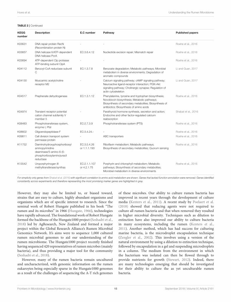
Huws et al. Understanding the Rumen Microbiome
TABLE 2 | Continued
KEGG
number
Description E.C number Pathway Published papers
K03631 DNA repair protein RecN
(Recombination protein N)
- - Roehe et al., 2016
K03657 DNA helicase II/ATP-dependent
DNA helicase PcrA
EC:3.6.4.12 Nucleotide excision repair; Mismatch repair Roehe et al., 2016
K03694 ATP-dependent Clp protease
ATP-binding subunit ClpA
- - Roehe et al., 2016
K04112 Benzoyl-CoA reductase subunit
C
EC:1.3.7.8 Benzoate degradation; Metabolic pathways; Microbial
metabolism in diverse environments; Degradation of
aromatic compounds
Li and Guan, 2017
K04130 Muscarinic acetylcholine
receptor M2
- Calcium signaling pathway; cAMP signaling pathway;
Neuroactive ligand-receptor interaction; PI3K-Akt
signaling pathway; Cholinergic synapse; Regulation of
actin cytoskeleton
Li and Guan, 2017
K04517 Prephenate dehydrogenase EC:1.3.1.12 Phenylalanine, tyrosine and tryptophan biosynthesis;
Novobiocin biosynthesis; Metabolic pathways;
Biosynthesis of secondary metabolites; Biosynthesis of
antibiotics; Biosynthesis of amino acids
Roehe et al., 2016
K04974 Transient receptor potential
cation channel subfamily V
member 5
- Parathyroid hormone synthesis, secretion and action;
Endocrine and other factor-regulated calcium
reabsorption
Shabat et al., 2016
K08483 Phosphotransferase system,
enzyme I, PtsI
EC:2.7.3.9 Phosphotransferase system (PTS) Roehe et al., 2016
K08602 Oligoendopeptidase F EC:3.4.24.- - Roehe et al., 2016
K09811 Cell division transport system
permease protein
- ABC transporters Roehe et al., 2016
K11752 Diaminohydroxyphosphoribosyl
aminopyrimidine
deaminase/5-amino-6-(5-
phosphoribosylamino)uracil
reductase
EC:3.5.4.26
or 1.1.1.193
Riboflavin metabolism; Metabolic pathways;
Biosynthesis of secondary metabolites; Quorum sensing
Roehe et al., 2016
K13542 Uroporphyrinogen III
methyltransferase/synthas
EC:2.1.1.107
or 4.2.1.75
Porphyrin and chlorophyll metabolism; Metabolic
pathways; Biosynthesis of secondary metabolites;
Microbial metabolism in diverse environments
Roehe et al., 2016
For simplicity only genes from Shabat et al. (2016) with significant correlation to amino acid metabolism are shown. Genes that lacked function annotation were removed. Genes identified
consistently across experiments and therefore representing the most promising marker genes are highlighted in gray.
However, they may also be limited to, or biased toward,strains that are easy to culture, highly abundant organisms andorganisms which are of specific interest to research. Since theseminal work of Robert Hungate published in his book “Therumen and its microbes” in 1966 (Hungate, 1966), technologieshave rapidly advanced. The foundational work of Robert Hungateformed the backbone of the Hungate1000 project (Seshadri et al.,2018) led by AgResearch, New Zealand and formed a majorproject within the Global Research Alliance’s Rumen MicrobialGenomics Network. It’s aims were to sequence 1,000 culturedrumen microbial genomes to aid our understanding of therumen microbiome. The Hungate1000 project recently finishedhaving sequenced 420 representatives of rumenmicrobes (mainlybacteria), and thus providing a major tool for the community(Seshadri et al., 2018).
However, many of the rumen bacteria remain unculturedand uncharacterized, with genomic information on the rumeneukaroytes being especially sparse in the Hungate1000 genomesas a result of the challenges of sequencing the A-T rich genomes
of these microbes. Our ability to culture rumen bacteria hasimproved in recent years through the development of culturemedia (Kenters et al., 2011). A recent study by Poelaert et al.(2018) showed that reducing agents were not required toculture all rumen bacteria and that when removed they resultedin higher microbial diversity. Techniques such as dilution toextinction have also improved our ability to culture bacteriain many ecosystems, including the rumen (Kenters et al.,2011). Another method, which has had success for culturingmarine bacteria, is the microdroplet encapsulation technique(Zengler et al., 2002). This involves using a version of thenatural environment by using a dilution to extinction technique,followed by encapsulation in a gel and suspending microdropletsin a column. The medium from the environment in whichthe bacterium was isolated can then be flowed through toprovide nutrients for growth (Stewart, 2012). Indeed, thereare many technologies emerging that should be investigatedfor their ability to culture the as yet unculturable rumenbacteria.
Frontiers in Microbiology | www.frontiersin.org 15 September 2018 | Volume 9 | Article 2161
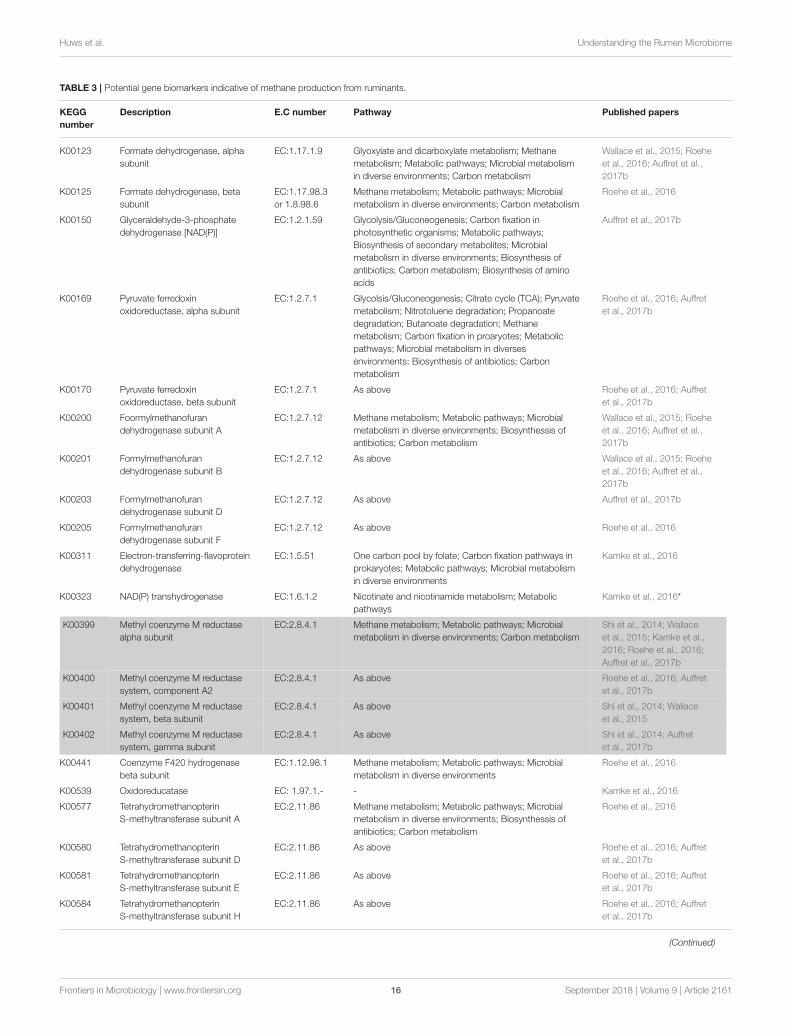
Huws et al. Understanding the Rumen Microbiome
TABLE 3 | Potential gene biomarkers indicative of methane production from ruminants.
KEGG
number
Description E.C number Pathway Published papers
K00123 Formate dehydrogenase, alpha
subunit
EC:1.17.1.9 Glyoxylate and dicarboxylate metabolism; Methane
metabolism; Metabolic pathways; Microbial metabolism
in diverse environments; Carbon metabolism
Wallace et al., 2015; Roehe
et al., 2016; Auffret et al.,
2017b
K00125 Formate dehydrogenase, beta
subunit
EC:1.17.98.3
or 1.8.98.6
Methane metabolism; Metabolic pathways; Microbial
metabolism in diverse environments; Carbon metabolism
Roehe et al., 2016
K00150 Glyceraldehyde-3-phosphate
dehydrogenase [NAD(P)]
EC:1.2.1.59 Glycolysis/Gluconeogenesis; Carbon fixation in
photosynthetic organisms; Metabolic pathways;
Biosynthesis of secondary metabolites; Microbial
metabolism in diverse environments; Biosynthesis of
antibiotics; Carbon metabolism; Biosynthesis of amino
acids
Auffret et al., 2017b
K00169 Pyruvate ferredoxin
oxidoreductase, alpha subunit
EC:1.2.7.1 Glycolsis/Gluconeogenesis; Citrate cycle (TCA); Pyruvate
metabolism; Nitrotoluene degradation; Propanoate
degradation; Butanoate degradation; Methane
metabolism; Carbon fixation in proaryotes; Metabolic
pathways; Microbial metabolism in diverses
environments; Biosynthesis of antibiotics; Carbon
metabolism
Roehe et al., 2016; Auffret
et al., 2017b
K00170 Pyruvate ferredoxin
oxidoreductase, beta subunit
EC:1.2.7.1 As above Roehe et al., 2016; Auffret
et al., 2017b
K00200 Foormylmethanofuran
dehydrogenase subunit A
EC:1.2.7.12 Methane metabolism; Metabolic pathways; Microbial
metabolism in diverse environments; Biosynthessis of
antibiotics; Carbon metabolism
Wallace et al., 2015; Roehe
et al., 2016; Auffret et al.,
2017b
K00201 Formylmethanofuran
dehydrogenase subunit B
EC:1.2.7.12 As above Wallace et al., 2015; Roehe
et al., 2016; Auffret et al.,
2017b
K00203 Formylmethanofuran
dehydrogenase subunit D
EC:1.2.7.12 As above Auffret et al., 2017b
K00205 Formylmethanofuran
dehydrogenase subunit F
EC:1.2.7.12 As above Roehe et al., 2016
K00311 Electron-transferring-flavoprotein
dehydrogenase
EC:1.5.51 One carbon pool by folate; Carbon fixation pathways in
prokaryotes; Metabolic pathways; Microbial metabolism
in diverse environments
Kamke et al., 2016
K00323 NAD(P) transhydrogenase EC:1.6.1.2 Nicotinate and nicotinamide metabolism; Metabolic
pathways
Kamke et al., 2016*
K00399 Methyl coenzyme M reductase
alpha subunit
EC:2.8.4.1 Methane metabolism; Metabolic pathways; Microbial
metabolism in diverse environments; Carbon metabolism
Shi et al., 2014; Wallace
et al., 2015; Kamke et al.,
2016; Roehe et al., 2016;
Auffret et al., 2017b
K00400 Methyl coenzyme M reductase
system, component A2
EC:2.8.4.1 As above Roehe et al., 2016; Auffret
et al., 2017b
K00401 Methyl coenzyme M reductase
system, beta subunit
EC:2.8.4.1 As above Shi et al., 2014; Wallace
et al., 2015
K00402 Methyl coenzyme M reductase
system, gamma subunit
EC:2.8.4.1 As above Shi et al., 2014; Auffret
et al., 2017b
K00441 Coenzyme F420 hydrogenase
beta subunit
EC:1.12.98.1 Methane metabolism; Metabolic pathways; Microbial
metabolism in diverse environments
Roehe et al., 2016
K00539 Oxidoreducatase EC: 1.97.1.- - Kamke et al., 2016
K00577 Tetrahydromethanopterin
S-methyltransferase subunit A
EC:2.11.86 Methane metabolism; Metabolic pathways; Microbial
metabolism in diverse environments; Biosynthessis of
antibiotics; Carbon metabolism
Roehe et al., 2016
K00580 Tetrahydromethanopterin
S-methyltransferase subunit D
EC:2.11.86 As above Roehe et al., 2016; Auffret
et al., 2017b
K00581 Tetrahydromethanopterin
S-methyltransferase subunit E
EC:2.11.86 As above Roehe et al., 2016; Auffret
et al., 2017b
K00584 Tetrahydromethanopterin
S-methyltransferase subunit H
EC:2.11.86 As above Roehe et al., 2016; Auffret
et al., 2017b
(Continued)
Frontiers in Microbiology | www.frontiersin.org 16 September 2018 | Volume 9 | Article 2161
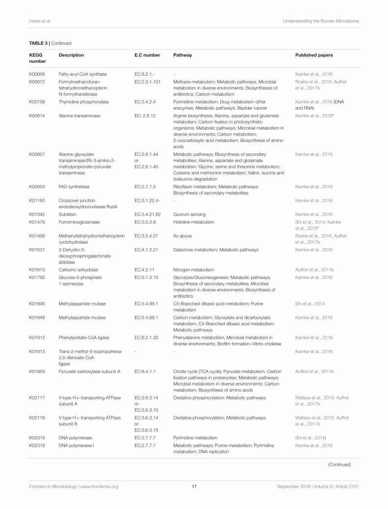
Huws et al. Understanding the Rumen Microbiome
TABLE 3 | Continued
KEGG
number
Description E.C number Pathway Published papers
K00666 Fatty-acyl-CoA synthase EC:6.2.1.- - Kamke et al., 2016
K00672 Formylmethanofuran-
tetrahydromethanopterin
N-formyltransferase
EC:2.3.1.101 Methane metabolism; Metabolic pathways; Microbial
metabolism in diverse environments; Biosynthessis of
antibiotics; Carbon metabolism
Roehe et al., 2016; Auffret
et al., 2017b
K00758 Thymidine phosphorylase EC:2.4.2.4 Pyrimidine metabolism; Drug metabolism–other
enzuymes; Metabolic pathways; Bladder cancer
Kamke et al., 2016 (DNA
and RNA)
K00814 Alanine transaminase EC: 2.6.12 Arginie biosynthesis; Alanine, aspartate and glutamate
metabolism; Carbon fixation in photosynthetic
organisms; Metabolic pathways; Microbial metabolism in
diverse environments; Carbon metabolism;
2-oxocarboxylic acid metabolism; Biosynthesis of amino
acids
Kamke et al., 2016*
K00827 Alanine-glyoxylate
transaminase/(R)-3-amino-2-
methylpropionate-pyruvate
transaminase
EC:2.6.1.44
or
EC:2.6.1.40
Metabolic pathways; Biosynthesis of secondary
metabolites; Alanine, aspartate and glutamate
metabolism; Glycine, serine and threonine metabolism;
Cysteine and methionine metabolism; Valine, leucine and
isoleucine degradation
Kamke et al., 2016
K00953 FAD synthetase EC:2.7.7.2 Riboflavin metabolism; Metabolic pathways;
Biosynthesis of secondary metabolites
Kamke et al., 2016
K01160 Crossover junction
endodeoxyribonuclease RusA
EC:3.1.22.4- - Kamke et al., 2016
K01342 Subtilisin EC:3.4.21.62 Quorum sensing Kamke et al., 2016
K01479 Formiminoglutamase EC:3.5.3.8 Histidine metabolism Shi et al., 2014; Kamke
et al., 2016*
K01499 Methenyltetrahydromethanopterin
cyclohydrolase
EC:3.5.4.27 As above Roehe et al., 2016; Auffret
et al., 2017b
K01631 2-Dehydro-3-
deoxyphosphogalactonate
aldolase
EC:4.1.2.21 Galactose metabolism; Metabolic pathways Kamke et al., 2016
K01673 Carbonic anhydrase EC:4.2.11 Nitrogen metabolism Auffret et al., 2017b
K01792 Glucose-6-phosphate
1-epimerase
EC:5.1.3.15 Glycolysis/Gluconeogenesis; Metabolic pathways;
Biosynthesis of secondary metabolites; Microbial
metabolism in diverse environments; Biosynthesis of
antibiotics
Kamke et al., 2016
K01846 Methylaspartate mutase EC:5.4.99.1 C5-Branched dibasic acid metabolism; Purine
metabolism
Shi et al., 2014
K01846 Methylaspartate mutase EC:5.4.99.1 Carbon metabolism; Glyoxylate and dicarboxylate
metabolism; C5-Branched dibasic acid metabolism;
Metabolic pathways
Kamke et al., 2016
K01912 Phenylacetate-CoA ligase EC:6.2.1.30 Phenylalanine metabolism; Microbial metabolism in
diverse environments; Biofilm formation–Vibrio cholerae
Kamke et al., 2016
K01913 Trans-2-methyl-5-isopropylhexa-
2,5-dienoate-CoA
ligase
- - Kamke et al., 2016
K01959 Pyruvate carboxylase subunit A EC:6.4.1.1 Citrate cycle (TCA cycle); Pyruvate metabolism; Carbon
fixation pathways in prokaryotes; Metabolic pathways;
Microbial metabolism in diverse environments; Carbon
metabolism; Biosynthesis of amino acids
Auffret et al., 2017b
K02117 V-type H+-transporting ATPase
subunit A
EC:3.6.3.14
or
EC:3.6.3.15
Oxidative phosphorylation; Metabolic pathways Wallace et al., 2015; Auffret
et al., 2017b
K02118 V-type H+-transporting ATPase
subunit B
EC:3.6.3.14
or
EC:3.6.3.15
Oxidative phosphorylation; Metabolic pathways Wallace et al., 2015; Auffret
et al., 2017b
K02319 DNA polymerase EC:2.7.7.7 Pyrimidine metabolism Shi et al., 2014)
K02319 DNA polymerase I EC:2.7.7.7 Metabolic pathways; Purine metabolism; Pyrimidine
metabolism; DNA replication
Kamke et al., 2016
(Continued)
Frontiers in Microbiology | www.frontiersin.org 17 September 2018 | Volume 9 | Article 2161
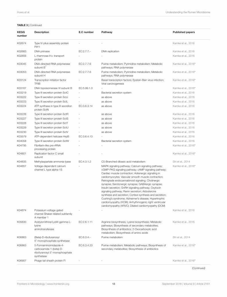
Huws et al. Understanding the Rumen Microbiome
TABLE 3 | Continued
KEGG
number
Description E.C number Pathway Published papers
K02674 Type IV pilus assembly protein
PilY1
- - Kamke et al., 2016
K02683 DNA primase EC:2.7.7.- DNA replication Kamke et al., 2016
K02856 L-rhamnose-H+ transport
protein
- - Kamke et al., 2016
K03045 DNA-directed RNA polymerase
subunit B′′
EC:2.7.7.6 Purine metabolism; Pyrimidine metabolism; Metabolic
pathways; RNA polymerase
Kamke et al., 2016*
K03053 DNA-directed RNA polymerase
subunit H
EC:2.7.7.6 Purine metabolism; Pyrimidine metabolism; Metabolic
pathways; RNA polymerase
Kamke et al., 2016*
K03124 Transcription initiation factor
TFIIB
- Basal transcription factors; Epstein-Barr virus infection;
Viral carcinogenesis
Kamke et al., 2016*
K03167 DNA topoisomerase VI subunit B EC:5.99.1.3 - Kamke et al., 2016*
K03219 Type III secretion protein SctC - Bacterial secretion system Kamke et al., 2016
K03222 Type III secretion protein SctJ - as above Kamke et al., 2016
K03223 Type III secretion protein SctL - as above Kamke et al., 2016
K03224 ATP synthase in type III secretion
protein SctN
EC:3.6.3.14 as above Kamke et al., 2016
K03226 Type III secretion protein SctR - as above Kamke et al., 2016
K03227 Type III secretion protein SctS - as above Kamke et al., 2016
K03228 Type III secretion protein SctT - as above Kamke et al., 2016
K03229 Type III secretion protein SctU - as above Kamke et al., 2016
K03230 Type III secretion protein SctV - as above Kamke et al., 2016
K03579 ATP-dependent helicase HrpB EC:3.6.4.13 - Kamke et al., 2016
K04058 Type III secretion protein SctW - Bacterial secretion system Kamke et al., 2016
K04795 Fibrillarin-like pre-rRNA
processing protein
- - Kamke et al., 2016*
K04801 Replication factor C small
subunit
- - Kamke et al., 2016*
K04835 Methylaspartate ammonia-lyase EC:4.3.1.2 C5-Branched dibasic acid metabolism Shi et al., 2014
K04857 Voltage-dependent calcium
channel L type alpha-1S
- MAPK signaling pathway; Calcium signaling pathway;
cGMP-PKG signaling pathway; cAMP signaling pathway;
Cardiac muscle contraction; Adrenergic signaling in
cardiomyocytes; Vascular smooth muscle contraction;
Retrograde endocannabinoid signaling; Cholinergic
synapse; Serotonergic synapse; GABAergic synapse;
Insulin secretion; GnRH signaling pathway; Oxytocin
signaling pathway; Renin secretion; Aldosterone
synthesis and secretion; Cortisol synthesis and secretion;
Cushing’s syndrome; Alzheimer’s disease; Hypertrophic
cardiomyopathy (HCM); Arrhythmogenic right ventricular
cardiomyopathy (ARVC); Dilated cardiomyopathy (DCM)
Kamke et al., 2016*
K04874 Potassium voltage-gated
channel Shaker-related subfamily
A member 1
- - Kamke et al., 2016
K05830 Acetylornithine/LysW-gamma-L-
lysine
aminotransferase
EC:2.6.1.11 Arginine biosynthesis; Lysine biosynthesis; Metabolic
pathways; Biosynthesis of secondary metabolites;
Biosynthesis of antibiotics; 2-Oxocarboxylic acid
metabolism; Biosynthesis of amino acids
Kamke et al., 2016
K06863 (Beta)-D-ribofuranosyl
5′-monophosphate synthetase
EC:6.3.4.- Purine metabolism Shi et al., 2014
K06863 5-Formaminoimidazole-4-
carboxamide-1-(beta)-D-
ribofuranosyl 5′-monophosphate
synthetase
EC:6.3.4.23 Purine metabolism; Metabolic pathways; Biosynthesis of
secondary metabolites; Biosynthesis of antibiotics
Kamke et al., 2016*
K06907 Phage tail sheath protein FI - - Kamke et al., 2016*
(Continued)
Frontiers in Microbiology | www.frontiersin.org 18 September 2018 | Volume 9 | Article 2161
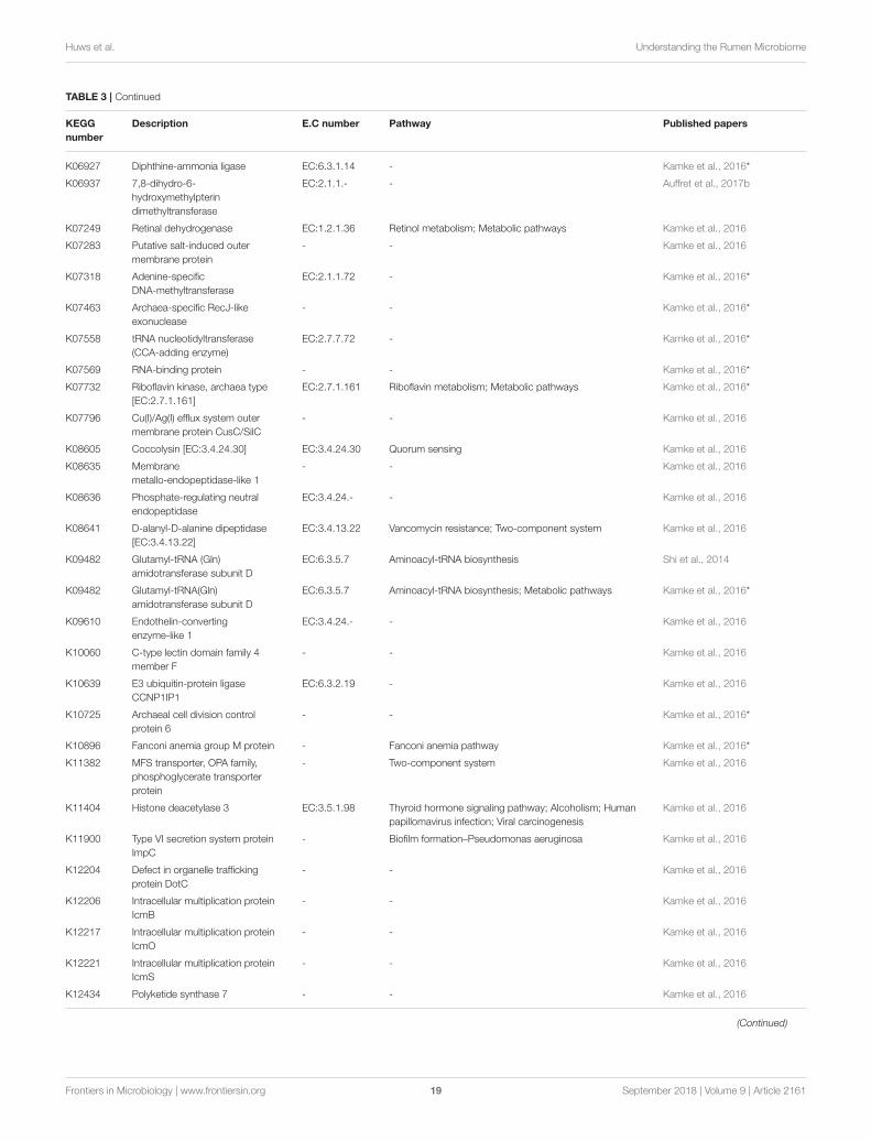
Huws et al. Understanding the Rumen Microbiome
TABLE 3 | Continued
KEGG
number
Description E.C number Pathway Published papers
K06927 Diphthine-ammonia ligase EC:6.3.1.14 - Kamke et al., 2016*
K06937 7,8-dihydro-6-
hydroxymethylpterin
dimethyltransferase
EC:2.1.1.- - Auffret et al., 2017b
K07249 Retinal dehydrogenase EC:1.2.1.36 Retinol metabolism; Metabolic pathways Kamke et al., 2016
K07283 Putative salt-induced outer
membrane protein
- - Kamke et al., 2016
K07318 Adenine-specific
DNA-methyltransferase
EC:2.1.1.72 - Kamke et al., 2016*
K07463 Archaea-specific RecJ-like
exonuclease
- - Kamke et al., 2016*
K07558 tRNA nucleotidyltransferase
(CCA-adding enzyme)
EC:2.7.7.72 - Kamke et al., 2016*
K07569 RNA-binding protein - - Kamke et al., 2016*
K07732 Riboflavin kinase, archaea type
[EC:2.7.1.161]
EC:2.7.1.161 Riboflavin metabolism; Metabolic pathways Kamke et al., 2016*
K07796 Cu(I)/Ag(I) efflux system outer
membrane protein CusC/SilC
- - Kamke et al., 2016
K08605 Coccolysin [EC:3.4.24.30] EC:3.4.24.30 Quorum sensing Kamke et al., 2016
K08635 Membrane
metallo-endopeptidase-like 1
- - Kamke et al., 2016
K08636 Phosphate-regulating neutral
endopeptidase
EC:3.4.24.- - Kamke et al., 2016
K08641 D-alanyl-D-alanine dipeptidase
[EC:3.4.13.22]
EC:3.4.13.22 Vancomycin resistance; Two-component system Kamke et al., 2016
K09482 Glutamyl-tRNA (Gln)
amidotransferase subunit D
EC:6.3.5.7 Aminoacyl-tRNA biosynthesis Shi et al., 2014
K09482 Glutamyl-tRNA(Gln)
amidotransferase subunit D
EC:6.3.5.7 Aminoacyl-tRNA biosynthesis; Metabolic pathways Kamke et al., 2016*
K09610 Endothelin-converting
enzyme-like 1
EC:3.4.24.- - Kamke et al., 2016
K10060 C-type lectin domain family 4
member F
- - Kamke et al., 2016
K10639 E3 ubiquitin-protein ligase
CCNP1IP1
EC:6.3.2.19 - Kamke et al., 2016
K10725 Archaeal cell division control
protein 6
- - Kamke et al., 2016*
K10896 Fanconi anemia group M protein - Fanconi anemia pathway Kamke et al., 2016*
K11382 MFS transporter, OPA family,
phosphoglycerate transporter
protein
- Two-component system Kamke et al., 2016
K11404 Histone deacetylase 3 EC:3.5.1.98 Thyroid hormone signaling pathway; Alcoholism; Human
papillomavirus infection; Viral carcinogenesis
Kamke et al., 2016
K11900 Type VI secretion system protein
ImpC
- Biofilm formation–Pseudomonas aeruginosa Kamke et al., 2016
K12204 Defect in organelle trafficking
protein DotC
- - Kamke et al., 2016
K12206 Intracellular multiplication protein
IcmB
- - Kamke et al., 2016
K12217 Intracellular multiplication protein
IcmO
- - Kamke et al., 2016
K12221 Intracellular multiplication protein
IcmS
- - Kamke et al., 2016
K12434 Polyketide synthase 7 - - Kamke et al., 2016
(Continued)
Frontiers in Microbiology | www.frontiersin.org 19 September 2018 | Volume 9 | Article 2161
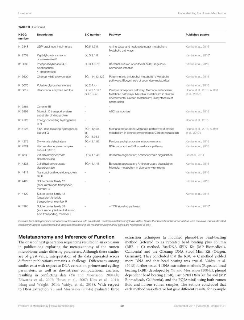
Huws et al. Understanding the Rumen Microbiome
TABLE 3 | Continued
KEGG
number
Description E.C number Pathway Published papers
K12448 UDP-arabinose 4-epimerase EC:5.1.3.5 Amino sugar and nucleotide sugar metabolism;
Metabolic pathways
Kamke et al., 2016
K12739 Peptidyl-prolyl cis-trans
isomerase-like 6
EC:5.2.1.8 - Kamke et al., 2016*
K13085 Phosphatidylinositol-4,5-
bisphosphate
4-phosphatase
EC:3.1.3.78 Bacterial invasion of epithelial cells; Shigellosis;
Salmonella infection
Kamke et al., 2016
K13600 Chlorophyllide a oxygenase EC:1.14.13.122 Porphyrin and chlorophyll metabolism; Metabolic
pathways; Biosynthesis of secondary metabolites
Kamke et al., 2016
K13670 Putative glycosyltransferase EC:2.4.-.- - Kamke et al., 2016
K13812 Bifunctional enzyme Fae/Hps EC:4.2.1.147
or 4.1.2.43
Pentose phosphate pathway; Methane metabolism;
Metabolic pathways; Microbial metabolism in diverse
environments; Carbon metabolism; Biosynthessis of
amino acids
Roehe et al., 2016; Auffret
et al., 2017b
K13886 Coronin-1B - -
K13893 Microcin C transport system
substrate-binding protein
- ABC transporters Kamke et al., 2016
K14123 Energy-converting hydrogenase
B N
- - Roehe et al., 2016
K14128 F420-non-reducing hydrogenase
subunit G
EC:1.12.99.-
or
EC:1.8.98.5
Methane metabolism; Metabolic pathways; Microbial
metabolism in diverse environments; Carbon metabolism
Roehe et al., 2016; Auffret
et al., 2017b
K14275 D-xylonate dehydratase EC:4.2.1.82 Pentose and glucuronate interconversions Kamke et al., 2016
K14324 Histone deacetylase complex
subunit SAP18
- RNA transport; mRNA surveillance pathway Kamke et al., 2016
K14333 2,3-dihydroxybenzoate
decarboxylase
EC:4.1.1.46 Benzoate degradation; Aminobenzoate degradation Shi et al., 2014
K14333 2,3-dihydroxybenzoate
decarboxylase
EC:4.1.1.46 Benzoate degradation; Aminobenzoate degradation;
Microbial metabolism in diverse environments
Kamke et al., 2016
K14414 Transcriptional regulatory protein
RtcR
- - Kamke et al., 2016
K14426 Solute carrier family 12
(sodium/chloride transporter),
member 3
- - Kamke et al., 2016
K14429 Solute carrier family 12
(potassium/chloride
transporters), member 9
- - Kamke et al., 2016
K14995 Solute carrier family 38
(sodium-coupled neutral amino
acid transporter), member 9
- mTOR signaling pathway Kamke et al., 2016*
Data are from metagenomics sequences unless marked with an asterisk. *Indicates metatranscriptomic datas. Genes that lacked functional annotation were removed. Genes identified
consistently across experiments and therefore representing the most promising marker genes are highlighted in gray.
Metataxonomy and Inference of FunctionThe onset of next generation sequencing resulted in an explosionin publications exploring the metataxonomy of the rumenmicrobiome under differing parameters. Although these studiesare of great value, interpretation of the data generated acrossdifferent publications remains a challenge. Differences amongstudies exist with respect to DNA extraction, primers and cyclingparameters, as well as downstream computational analysis,resulting in conflicting data (Yu and Morrisson, 2004a,b;Edwards et al., 2007; Huws et al., 2007; Kim et al., 2011;Ishaq and Wright, 2014; Vaidya et al., 2018). With respectto DNA extraction Yu and Morrisson (2004a) evaluated three
extraction techniques (a modified phenol-free bead-beatingmethod (referred to as repeated bead beating plus column(RBB + C) method, FastDNA SPIN Kit (MP Biomedicals,California) and the QIAamp DNA Stool Mini Kit (Qiagen,Germany). They concluded that the RBC + C method yieldedmore DNA and that bead beating was crucial. Vaidya et al.(2018) further tested 4 DNA extraction methods (Repeated beadbeating (RBB) developed by Yu and Morrisson (2004a), phenoldependent bead beating (PBB), Fast SPIN DNA kit for soil (MPBiomedicals, California), and the PQIAmini) using both rumenfluid and fibrous rumen samples. The authors concluded thateach method was effective but gave different results, for example
Frontiers in Microbiology | www.frontiersin.org 20 September 2018 | Volume 9 | Article 2161
Huws et al. Understanding the Rumen Microbiome
PBB extracted DNA extracted resulted in higher abundancesof Ruminococcaceae compared with abundances obtained usingthe FDSS method, whereas abundances of Fibrobacteraceae waslower compared with the RBB method. They conclude thateach method has advantages and disadvantages which needto be considered based on sample type, but bead beating iscritical. Further downstream many authors have investigated theimportance of primer choice for metataxonomic investigationsof the rumen microbiome (Yu and Morrisson, 2004b; Edwardset al., 2007; Huws et al., 2007; Kim et al., 2011; Ishaq andWright, 2014). Yu and Morrisson (2004b) investigated primerchoice in terms of diversity observed on denaturing gradientgel electrophoresis (DGGE) gels and concluded that primerstargeting the V3 region were the best. Huws et al. (2007) andEdwards et al. (2007), however showed that the V3 primers werenon-specific to bacteria and could amplify plant chloroplasticDNA as well as archaeal 16S rDNA and protozoal 18S rDNAsequences. The amplification of plant chloroplast sequences isa substantial issue for samples taken from animals fed freshforage (these are often assigned taxonomically as cyanobacteria),with an abundance of intact chloroplast DNA being presentin the rumen, and often masks microbial sequences (Edwardset al., 2007). This is the case when investigated using DGGEand NGS based sequencing (Huws, personal communication).Nonetheless, if animals are not fed fresh forage and primerswhich are more broad are required to cover a greater proportionof the microbes as a whole is required then the V3 regionis perhaps a justifiable choice. However, Huws et al. (2007)concluded that for specific bacterial 16S rRNA V6-V8 primerswere more appropriate. Edwards et al. (2007) also developeda primer pair based on the V6-V8 region which reduce theamplification of chloroplastic DNA. Nonetheless, the ampliconsize obtained using the Edwards et al. (2017) primers areoften too large for effective sequencing, therefore the reverseprimer has been changed to enable avoidance of chloroplastidentification and production of a smaller amplicon for NGSsequencing (Belanche et al., 2017). The annealing temperaturesand number of cycles used for PCR are also clearly going to biasresults somewhat. Nonetheless, using a basic set of standardizedprotocols may not be possible, due to the differing hypothesesand the complex nature of the ecosystem (i.e., host animal),however ensuring data accuracy by using internal standardsrepresents one approach to ensure that comparisons amongdatasets are valid. Pollock et al. (2018) attempted to describethe guidelines and consensus best practices for metataxonomicstudies and concluded that bead beating is critical for DNAextraction as is the use of internal standards for metataxonomicstudies amongst other recommendations. Also the constructionof rumen microbiome databases to aid accurate taxonomicalassignment, such as RIM-DB (for methanogens; Seedorf et al.,2014), the ureC database (ureolytic bacteria; Jin et al., 2017), andAF-RefSeq (anaerobic fungi; Paul et al., 2018) drastically improveour ability to monitor rumen microbial diversity.
Irrespective, metataxonomic rDNA data have providedinsights into the composition of the rumen microbiome underdiffering parameters, but these techniques are limited in termsof providing insight into microbial function. Nonetheless, due
to their low cost, these techniques are the most publishedand will continue to be important in microbiome research forthe near future. Software to predict microbial function frommetataxonomic data, such as PICRUSt (Langille et al., 2013),has been applied to many different ecosystems, including therumen. Although this approach saves on the cost associatedwithmore thorough and accurate shotgunmetagenomic analysis,it has limitations in accurately represent microbiome function(Wilkinson et al., 2018). The accuracy of PICRUSt prediction,originally intended for humanmicrobiota data, has recently beentested for the rumen microbiome using datasets with 16S rDNAdata and accompanying metagenomics or metatranscriptomicdatasets (Wilkinson et al., 2018). The data shows poor correlationof predicted function with the actual function seen within themetagenomics/metatranscriptomic datasets (Wilkinson et al.,2018). Wilkinson et al. (2018), developed CowPI an improved16S rDNA inference platform for the rumen which is based onPICRUSt but uses the Hungate1000 genomes as the searchablegenomes (http://www.cowpi.org/). Other platforms allowinginference of function from 16S rDNA data have also beendeveloped, such as Tax4Fun (Aßhauer et al., 2015) and PanFP(Jun et al., 2015), and have been proposed to provide moreaccurate functional annotations than PICRUSt (Koo et al., 2017).However, the ability of these programs to predict the functionof the rumen microbiome has not been investigated. Regardless,inferring metabolic function from phylogenetic data allows thescientific community to obtain retrospective value from thesedatasets in order to understand the rumen microbiome in lightof global agricultural challenges.
MetagenomicsThe benefits of metagenomics include the ability to assemblewhole- and fragmented-genomes, predict genes, map enzymesand pathways, discover new enzymes and pathways, and quantifythe abundance of functional genomic elements across andbetween samples. Shotgun metagenomics was first applied tothe rumen in order to discover novel biomass degradingenzymes from switchgrass-associated microbes (Hess et al.,2011). Subsequently, metagenomics has been used to study manyaspects of rumen microbiology, including methane emissionsin cattle (Wallace et al., 2015) and sheep (Shi et al., 2014),biomarkers to predict ruminal methanogenesis (Auffret et al.,2017b), the effect of feed-conversion-ratio, and breed and hostgenetics on the composition of the rumen microbiome (Roeheet al., 2016), nutrient acquisition (Mayorga et al., 2016; Rubinoet al., 2017), and effects of diet (Auffret et al., 2017a,b), andinvestigate impact of feed additives (Thomas et al., 2017) onthe abundance of antimicrobial-resistance genes. The rumen alsoremains a source of valuable bioactives for the biotechnologyindustry, and metagenomics is a key tool for such bioprospecting(Oyama et al., 2017; Roumpeka et al., 2017). More recently,metagenomic sequences have also resulted in an enhancedunderstanding of niche specialization within rumen bacteria(Rubino et al., 2017). Rubino et al. (2017) showed that, withinmetagenome sequences from 14 silage-fed cows, that the genusPrevotella possessed higher levels of glycosyl hydrolase (GH)isoforms relating specifically to the degradation of hemicellulose,
Frontiers in Microbiology | www.frontiersin.org 21 September 2018 | Volume 9 | Article 2161
Huws et al. Understanding the Rumen Microbiome
whilst Clostridium contained higher levels of GH isoforms forenzymes specifically involved in cellulose degradation. Their datasuggests that isoform diversity maintains selective advantage andniche specialization within these genera.
Binning Genomes From MetagenomesAnother major advancement in understanding the capacity ofrumen bacteria has been our increased ability to bin genomesfrom metagenomes. Assembly binning refers to the constructionof complete or near complete microbial genomes directly frommetagenomic sequencing data, and was first achieved by Tysonet al. (2004) from an acidophilic biofilm. Hess et al. (2011)were the first to apply this to ruminants, assembling 15 draftmicrobial genomes from the switchgrass associated microbiomeof cattle. Subsequently, Svartstrom et al. (2017) assembled 99microbial genomes from the moose rumen, Stewart et al. (2018)assembled 913 microbial genomes from the rumen of cattle, andParks et al. (2017) assembled over 8,000 novel microbial genomesfrom 1,500 public datasets, some of which originated fromthe rumen. Traditional metagenomic binning takes an in silicoapproach whereby metagenomic assembled contigs are clusteredby base-composition and abundance across multiple datasets—the hypothesis being that contigs from the same organism willfollow a very similar abundance profile across multiple samples,and will have a roughly similar base composition. The successof such binning procedures is validated by investigating thenumber of single-copy core-genome genes within each bin, asimplemented by software such as CheckM (Parks et al., 2015).More recently, physical methods of metagenomic binning suchas the use of Hi-C have been published (Beitel et al., 2014).In Hi-C experiments, parts of the chromosome that are incontact with one another inside the cell are cross-linked usingformaldehyde; cells are then lysed, the DNA is fragmented usinga restriction enzyme, followed by random ligation, amplificationand sequencing. Each pair of paired-end reads therefore comesfrom two separate fragments of the same original chromosome,and that information can be used to collate assembled contigsinto genomes. Hi-C binning has been used effectively on humanfeces (Press et al., 2017) as well as in ruminants (Stewart et al.,2018).
MetatranscriptomicsMetatranscriptomics involves the profiling of community-wideexpressed genes (mRNA), and is often termed RNA-seq. Whilstmetagenomics allows us to evaluate diversity and the potentialfunctional capacity of a microbiome, metatranscriptomesprovide insight into the actual function of microbiomes via geneexpression. Due to the abundance of rRNA, metatranscriptomicsrequires either very deep sequencing to obtain sufficient mRNAsequences coupled with computational binning of the rRNAgenes (these can also be useful for metataxonomics) or useof kits to deplete rRNA pre-sequencing. Deep sequencing isof course expensive and the kits used to remove rRNA formetranscriptomics of the rumen microbiome, have varyingdegrees of success (Huws personal communication). Thesekits are also bespoke for the removal of bacterial or eukaryoticrRNA, and thus for the complex rumen microbiome, a variety of
kits are required to remove prokaryotic and eukaryotic rRNA.This is both costly and laborious, with the time required likelyresulting in partial RNA degradation, which will ultimately biasdown-stream analysis. Nonetheless Comtet-Marre et al. (2017)developed a bespoke rRNA kit which was effective in removingrumen microbial rRNA, providing a potential solution for futureexperiments.
Despite these developments, it has also been shown thatthe correlation between mRNA and protein levels can be weakand variable, possibly due to post-transcriptional modifications(Greenbaum et al., 2003; Csárdi et al., 2015). Ribosomeprofiling (riboseq) has been developed as a direct methodto quantify and characterize translation (Ingolia et al., 2009).Riboseq takes advantage of the fact that during translation, theribosome protects around 30 nucleotides of the mRNA fromnuclease activity. High-throughput sequencing of these ribosomeprotected fragments offers a precise record of the number andlocation of the ribosomes at the time translation ceases. Mappingthe position of the ribosome-protected fragments is indicativeof the translated regions within the transcriptome. Nonetheless,whilst the use of this technique on pure cultures has beeneffective, the development of the technique (MetaRibo-Seq) ata metatranscriptomic level is in its infancy and still requiresvalidation for the rumen micobiome.
MetaproteomicsMetaproteomics falls between the established DNA and RNAsequencing and metabolomics procedures as an approach tocharacterize the functional activity of the microbial community.While still an emerging technology, the concept was introducedby Wilmes and Bond (2004), who used 2D PAGE methods toseparate and identify proteins from a complex sample extractedfrom waste water treatment. As a concept, it has some theoreticaladvantages over RNA sequencing methods in that the half-lifeof proteins can be significantly longer than RNA transcripts.Therefore, if the data represent a “snapshot” of microbial activityat a single time point, then identification of the proteins arguablywill provide a more accurate picture than sequencing mRNA.Moreover, as individual proteins can be identified by their aminoacid sequence, function can still be linked to taxa using proteinsequence alignment tools e.g., UniPept (Mesuere et al., 2018).
The 2D PAGE methodology involves using a pH gradientfirstly to separate proteins in one dimension based ontheir isoelectric point. The proteins are then subject to gelelectrophoresis to separate them by size. This results in a spotpattern representing the metaproteome. Individual spots canthen be excised, digested and the resulting peptides identifiedusing mass spectrometry. This method has been used to identifyproteins in waste water samples (Abram et al., 2009) soils,sediments (Benndorf et al., 2009; Chourey et al., 2010), therhizosphere (Wu et al., 2011) and human feces (Klaassens et al.,2007). However, when applied to rumen digesta samples thisapproach revealed a major shortcoming (Snelling and Wallace,2017), as the rumen contains high levels of plant secondarycompounds, such as tannins and other phenolics that complexwith the proteins, and interfere with protein extraction andpurification (Snelling and Wallace, 2017). Snelling and Wallace
Frontiers in Microbiology | www.frontiersin.org 22 September 2018 | Volume 9 | Article 2161
Huws et al. Understanding the Rumen Microbiome
(2017) reported that repeated wash steps and microfiltrationwere not effective in removing the contaminants, which preventaccurate protein quantitation and obscure spot patterns in gels.Humic acid is also highly abundant in soil and feces, which causesimilar implications for recovery for good metaproteomic datain these systems. One possible solution to this problem is to useacid precipitation to separate peptides (Qian and Hettich, 2017).The authors concluded that sample quality was a key factor, withbest results obtained from fresh digesta or samples with highmicrobial protein content relative to contaminants. Despite theselimitations, Snelling and Wallace (2017) did identify proteinsdirectly associated with the functional activity of the rumenmicrobial community from 2D PAGE spots. Abundant structuralproteins were identified including actin, alpha and beta tubulin,and axonemal isoforms dynein light chain, which are all involvedin the locomotion of ciliates. Among prokaryotic proteins wereenzymes from the Phyla Firmicutes and Bacteroidetes involvedin central metabolism. Archaeal proteins were also found insurprisingly high abundance considering the relatively smallproportion of the microbial community that this group occupiesin the rumen. This may be a reflection of the persistence ofarchaeal proteins after the original transcripts are degraded.These archaeal proteins, were identified as key enzymes involvedin the synthesis of methane. This finding was particularly relevantto the efforts to understand the mechanisms behind methaneproduction.
As a consequence of these technical challenges, very fewstudies have attempted to characterize the rumen microbialcommunity using metaproteomics. However, in recent years,the development of next generation mass spectrometers andaccompanying software have provided the means to identifyproteins en masse in an approach to analogous to shotgunDNA sequencing. Using this technique Deusch and Seifert(2015) described over 2,000 proteins associated with the rumenmicrobial community. Research recently conducted by Hartet al. (2018) explored the potential to develop a metaproteomicapproach to analyse the rumen that allowed comparison of datawith meta-transcriptomic information. Although in its infancy,the meta-proteomic methodology did allow for the identificationof members of the protein families that were associated with thetranscriptome of the rumen microbiome (Hart and Kingston-Smith, Personal Communication). The development of softwarefor meta-proteome analysis such as Meta-proteome Analyzer(Muth et al., 2015) can also aid in the analysis and interpretationof meta-protein data. As it stands the method shows greatpotential and is a complement to other omics technologies todetermine the functionality of the rumen microbiome.
MetabolomicsLike the field of proteomics, the study of metabolomics withinthe rumen is also in its infancy. Metabolomics can be definedas the comprehensive (qualitative and quantitative) analysis ofmetabolites by gathering as much metabolic information aspossible from an organism or biological system (Yi et al., 2016).Metabolomics focuses mainly on low molecular mass molecules(<1,000 Da), which can be related to the functional status ofthe organism (Bundy et al., 2009). The main challenges for
analyzing these metabolites include their chemical complexityand heterogeneity. Sample preparation for metabolomics canbe as simple as a liquid-liquid extraction procedure, but usingassertive methods is key to ensure effective metabolite extraction(Patejko et al., 2017). Various analytical instruments can beused for metabolomics, differing mainly on sensitivity andcoverage. To date, the majority of rumen metabolome studieshave used liquid chromatography-mass spectrometry (LC-MS),gas chromatography-mass spectrometry (GC-MS) and nuclearmagnetic resonance (NMR), with the latter the most used dueto reliability and absolute quantification (Goldansaz et al., 2017).There is growing interest for LC-MS due to recent advancesin instrument sensitivity, high processing capacity, data analysisand the development of data repositories where the communitycan curate large data sets have brought interest to tandemmass spectrometry (MS). Furthermore, with the introductionof the online tool Global Natural Products Social MolecularNetworking, a crowd-sourced knowledge repository and analysisinfrastructure, wherebyMS/MS spectra can be clustered based onspectral similarity data, and greatly improving data interpretation(Wang M. et al., 2016).
Usually, two approaches can be used for metabolicinvestigations: targeted and untargeted analysis. The targetedanalysis focuses on examination of a group of knownmetabolites,usually for hypothesis-driven studies (Patti et al., 2012), whereasuntargeted analysis evaluates large numbers of compounds.One of the main advantages of using an untargeted approach isthe prospection of novel compounds and metabolic pathways(Patti et al., 2012). To date, most studies regarding the rumenmetabolome used the targeted approach and had an interestin the effect of diets on the rumen metabolome (Ametaj et al.,2010; Saleem et al., 2012, 2013; Zhao et al., 2014; Zhang et al.,2015; Mao et al., 2016, 2017; Zhang R. et al., 2017; Do Pradoet al., 2018; O’callaghan et al., 2018). Using a combination ofseveral metabolomics platforms, the pioneer study by Saleemet al. (2013) demonstrated that the rumen metabolome is notas simple as previously anticipated. Indeed, 246 compoundswere reported as part of the rumen metabolome, including:phospholipids, inorganic ions, gases, amino acids, dicarboxylicacids, fatty acids, volatile fatty acids, glycerides, carbohydrateand cholesterol esters (Saleem et al., 2013). As part of this studySaleem et al. (2013) also set up a searchable rumen metabolomedatabase to improve metabolite assignments in the rumen(www.rumendb.ca). Recent studies have mainly explored howrumen metabolites are affected by different levels of roughagesand concentrate. For example, there is alteration to organicacids, amino acids, amines, sugars and nucleosides/nucleotideswhen cows were fed diets low on concentrate (40%, DM basis)compared to high concentrate (70%, DM basis; Zhang R. et al.,2017). In another study using dairy cows fed diets varying inroughage to concentrate ratio (80:20, 60:40, 40:60, and 20:80)ruminal amino acids, lipids, organic acids, and carbohydrateswere affected (Zhang J. et al., 2017).
Metabolites of microbial origin are also precursors ofruminant products (e.g., meat and milk), which might suggestbiochemical insights into the role played by rumen-dietinteractions (Saleem et al., 2013; Sun et al., 2015). Also,
Frontiers in Microbiology | www.frontiersin.org 23 September 2018 | Volume 9 | Article 2161
Huws et al. Understanding the Rumen Microbiome
metabolites such as phosphatidylcholine have been suggested asbiomarkers for protozoa abundance (Saleem et al., 2012). Finally,the rumen fluid metabolome can be used to identify potentialdifferences in rumen function. Recently, Artegoitia et al. (2017)reported 33 metabolites differing in cattle having high or lowaverage daily gain. Kingston-Smith et al. (2013), using FourierTransform Infra-Red (FT-IR) spectroscopy-based metaboliteprofiling, also showed that it was possible to discriminatedifferences in the rumen plant-microbe interactome when threedifferent cultivars of perennial ryegrass were used as substrate.This suggests that FT-IR could be used as an approach toimprove forages for livestock production (Kingston-Smith et al.,2013). However, there are few studies to date combining bothsequencing and metabolomics to provide a more comprehensiveanalysis of the rumen system that would contribute informationregarding metabolic expression of the genetic potential of themicrobiota (Mao et al., 2016; Zhang J. et al., 2017; O’callaghanet al., 2018).
The rumen environment is composed of a myriad ofmolecules of microbial, plant and animal origins. Currently,studies have focused on characterizing how the ruminalenvironment is affected by diet. There is an opportunity to extendthis to explore microbial interactions and how ruminal microbescope with stressors. Concentrating on exploring the ecologicalfoundations of the rumen microbiota might deliver an improvedcomprehension of the rumen, novel compounds and unexploredpathways.
Data Integration for EnhancedUnderstanding of the Rumen MicrobiomeEach of the previously described technological approachesprovide novel and fascinating insights into aspects of thestructure, function and/or activity of the rumen microbiome.However, if these different types of information were tobe integrated into a model that describes microbial activityat all levels recorded, our understanding would be furtherenhanced. At the simplest level we can use the centraldogma of molecular biology that describes the manner inwhich genetic information is transcribed and translated toproteins (DNA => RNA => Protein) to allow the “functionalpotential” in whole sequenced genomes from cultured rumenmicrobes, metagenomically assembled genomes (MAGs) or denovo assembled metagenomes to be linked to the activelytranscribed genes captured using whole transcriptomes fromcultured microbes, or meta-transcriptomic data from rumensamples. This level of data integration has provided some novelinsights missed by either approach alone, such as by Shi et al.(2014) who demonstrated that differences inmethane productionwere not due to differences in the abundances of the genesresponsible, but rather a result of their differential expression.It would not have been possible to make this distinctionwithout integration of both types of data. Similarly, thecomparison of proteomic data to transcriptomic or genomic datafrom the same samples can allow better protein identification(using tools such as MASCOT) and provide results that
TABLE 4 | Outstanding questions.
• What role do the rumen eukaryotome play in animal phenotype?
• What role do phages play in shaping the rumen microbiome?
• How can we harvest the ability of the rumen microbes to produce biofilms and
membrane vesicles to address global livestock challenges?
• Are early life dietary interventions effective in enhancing animal phenotype in the
long-term?
• What role do the lower GI tract microbiomes potentially play in the animal
phenotype?
• Can we use host genomics and develop ruminant breeding programs to
beneficially manipulate the rumen microbiome to enhance animal phenotype?
• How effective would a strategy involving enhanced ruminant genomics coupled
with effective early life dietary management of the animal be in enhancing animal
phenotype?
• Is mathematical modeling a feasible accurate approach to predicting ruminant
feed efficiency and methane output?
• Can we develop gene based biomarkers to predict feed efficiency and/or
methane output from ruminants?
• How resilient are ruminants with an improved phenotype to perturbations e.g.,
acidosis? Given that preliminary data shows that these animals have a less
diverse rumen microbiome?
• How can we ensure comparability of data generated globally to form a
consensus on best practices for achieving the global livestock challenges?
are more relevant to the rumen environment (Hart et al.,2018).
This, however, is only scratching the surface of what ispossible. For example, online databases, such as the KyotoEncyclopedia of Genes and Genomes (KEGG; Kanehisa et al.,2017) and BioCyc (Caspi et al., 2016), which provide collectionsof characterized metabolic pathways and software tools forexploring them, have been used to provide a better understandingof rumen microbial activity in a variety of contexts (Hesset al., 2011; Shi et al., 2014; Rubino et al., 2017; Seshadriet al., 2018). However, many of these databases are targetedtoward human pathogens or model aerobic organisms, suchas Escherichia coli, and do not necessarily reflect the functionsimportant to the facultative anaerobic lifestyle of rumenmicrobes. With nearly 500 genomes from rumen organismscurrently available, an opportunity exists to generate rumen-specific databases of known and predicted metabolic functions,similar to INTERMINE (Kalderimis et al., 2014) although thiswill likely require a parallel concerted effort to generate betterphenotypic information for these cultured organisms in vitro andin vivo.
The ultimate goal is to be able to combine information fromall types of “omics” data, and provide a true “systems” overview ofthe rumenmicrobiome. Progress toward achieving this continuesfor a few other model microbiomes, where the goal has beento identify interactions in order to interpret biological data andultimately model in silico how microbial communities behave(Bittner et al., 2010; Faust and Raes, 2012). This is generallyaccomplished through three tasks: firstly by identifying the“scaffold” of interactions between organisms (Faust and Raes,2012; Friedman and Alm, 2012); secondly, decomposing theseinteractions into “important components” (Lee and Tzou, 2009);and thirdly, carrying out cellular-systems modeling and analysis(Nobu et al., 2015; Mcgeachie et al., 2016). It is easy to see how
Frontiers in Microbiology | www.frontiersin.org 24 September 2018 | Volume 9 | Article 2161
Huws et al. Understanding the Rumen Microbiome
such an approach could also be integrated with the mathematicalmodels of the rumen environment described earlier.
Network-based approaches are likely to provide the besthope of integrating all this information by allowing a completeoverview of the rumen microbiome as they are capable ofcapturing the fundamental properties of the microbial ecosystemincluding taxonomy (Barberan et al., 2012), functional similarity(Martiny et al., 2015), metabolic processes (Shlomi et al.,2008), co-expression (Zhang and Horvath, 2005) and gene-sharing (Smillie et al., 2011). Network-based approaches haveproven successful for generating insight into the functional andgenetic potential of microbial communities, bringing clarity tothe complex history of microbes, providing tools that allowanalyses of mosaic sequences (Adai et al., 2004), and identifygenomes harboring sequences of multiple origins (Lima-Mendezet al., 2008; Fondi and Fani, 2010; Kloesges et al., 2011;Halary et al., 2013). Network-based approaches also providea framework where genetic diversity can be compared andquantified, even when analyzing highly divergent sequences(Bhattacharya et al., 2013). We are only in the early stages ofapplying these approaches to the rumen microbiome, and whilenetwork scaffolds for specific animal studies have been generated(Roehe et al., 2016), large-scale, integrative models have yet to bedeveloped.
CONCLUSIONS
The terminology of microbiome is often misused within therumen context as there is a tendency to focus on the study of therumen bacteria alone, without consideration of the eukaryotes.This undoubtedly limits our ability to understand the rumenmicrobiome, as these groups of microbes interact with eachother, consequently affecting production and the environmentalimpact of ruminants (Table 4). Also, little emphasis is given tounderstanding ecological interactions, niche specialization andconsequences of the biofilm phenotype and membrane vesicleson animal phenotype. An increased fundamental understandingof the rumen microbiome as well as that of the lower GItract of ruminants is essential to develop novel approaches forimproving livestock production and reducing environmentalimpact. Irrespective, much emphasis has been given to usingplant/additive/supplement strategies to manipulate the rumenmicrobiome in a manner that improves efficiency of animalproduction. Although, these have had limited success whenapplied to the mature animal, recent data show that ruminantbreeding programs coupled with a defined dietary managementprotocols from birth may be the best ways to achieve lifelong
animal phenotype benefits with limited financial and labor
input. Further research is required to assess the role of thehost genome on the rumen microbiome and animal phenotypeon a global geographic scale (Table 4). Likewise, applicationof these innovative techniques to early life ruminant nutritionis in its infancy, and a global effort is required to define thebest practices in early life which will differ based on geography(Table 4). Improving our ability to measure phenotype usingrumen microbiome gene based biomarkers may also allow highthroughput phenotype predictions (Table 4). Irrespective, theruminant sector has made major strides to improving animalphenotype, through understanding the rumen microbiome, andfurther advances will no doubt be accomplished, especially giventhe exponential advances in “omic” technologies.
AUTHOR CONTRIBUTIONS
All authors listed have made a substantial, direct, and intellectualcontribution to the work and approved it for publication.
ACKNOWLEDGMENTS
SH, DM, MP, RM-T, SW, IT, HS, JE, SK, GA, and CCacknowledge the support of ERA-net gas co-fund for funding(Project name: RumenPredict). SH, HM and CC acknowledgesupport from BBSRC (BBL/L026716/1 and BBL/L026716/2)and a British Council Newton Institutional Links funding(Grant 172629373). IM acknowledges funding from theEuropean Research Council under the European Union’sHorizon 2020 research and innovation program (Grant 640384).JE acknowledges funding from an EU H2020 Marie CurieFellowship (706899). CC, AK-S, and EH were supported bythe Biotechnology and Biological Sciences Research Council(Grants BBS/OS/GC/000011B and BBS/E/W/0012843D). CNand OM acknowledge the support of the British CouncilNewton Institutional Links funding (Grant 216425215). SRUCreceives financial support from the Scottish Government’s Ruraland Environment Science and Analytical Services Division(RESAS). RD and RR acknowledge financial support from theBiotechnology and Biological Sciences Research Council (BBSRCBB/N01720X/1). DY-R and AB acknowledge funding fromMINECO, Spain (Grant AGL2017-86938-R). GS acknowledgesfunding from the U.S. Department of Agriculture NationalInstitute of Food and Agriculture foundational (Grant 2015-67015-23246). EP acknowledges funding from CNPq (Grant401590/2014-3). All authors are also members of the GlobalResearch Alliance Rumen Microbial Genomics network.
REFERENCES
Abecia, L., Jimenez, E., Martinez-Fernandez, G., Martin-Garcia, A. I., Ramos-
Morales, E., Pinloche, E., et al. (2017). Natural and artificial feeding
management before weaning promote different rumen microbial colonization
but not differences in gene expression levels at the rumen epithelium of
newborn goats. PLoS ONE 12:e0182235. doi: 10.1371/journal.pone.0182235
Abecia, L., Martín-García, A. I., Martínez, G., Newbold, C. J., and Yáñez-Ruiz, D.
R. (2013). Nutritional intervention in early life to manipulate rumen microbial
colonization and methane output by kid goats postweaning1. J. Anim. Sci. 91,
4832–4840. doi: 10.1017/S1751731113001699
Abecia, L., Ramos-Morales, E., Martínez-Fernandez, G., Arco, A., Martín-
García, A. I., Newbold, C. J., et al. (2014). Feeding management in early
life influences microbial colonisation and fermentation in the rumen of
Frontiers in Microbiology | www.frontiersin.org 25 September 2018 | Volume 9 | Article 2161
Huws et al. Understanding the Rumen Microbiome
newborn goat kids. Anim. Prod. Sci. 54, 1449–1454. doi: 10.1071/AN
14337
Abecia, L., Toral, P. G., Martin-Garcia, A. I., Martinez, G., Tomkins, N. W.,
Molina-Alcaide, E., et al. (2012). Effect of bromochloromethane on methane
emission, rumen fermentation pattern, milk yield, and fatty acid profile in
lactating dairy goats. J. Dairy Sci. 95, 2027–2036. doi: 10.3168/jds.2011-4831
Abram, F., Gunnigle, E., and O’flaherty, V. (2009). Optimisation of protein
extraction and 2-DE for metaproteomics of microbial communities from
anaerobic wastewater treatment biofilms. Electrophoresis 30, 4149–4151.
doi: 10.1002/elps.200900474
Adai, A. T., Date, S. V., Wieland, S., and Marcotte, E. M. (2004). LGL: creating a
map of protein function with an algorithm for visualizing very large biological
networks. J. Mol. Biol. 340, 179–190. doi: 10.1016/j.jmb.2004.04.047
Adams, J. C., Gazaway, J. A., Brailsford, M. D., Hartman, P. A., and Jacobson, N.
L. (1966). Isolation of bacteriophages from the bovine rumen. Experientia 22,
717–718. doi: 10.1007/BF01901335
Aite, M., Chevallier, M., Frioux, C., Trottier, C., Got, J., Cortés, M. P.,
et al. (2018). Traceability, reproducibility and wiki-exploration for “à-la-
carte” reconstructions of genome-scale metabolic models. Plos. Comput. Biol.
14:e1006146. doi: 10.1371/journal.pcbi.1006146
Alexandratos, N., and Bruinsma, J. (2012).World Agriculture Towards 2030/2050:
The 2012 Revision, FAO, Food and Agriculture Organization of the United
Nations.
Al-Masaudi, S., El Kaoutari, A., Drula, E., Al-Mehdar, H., Redwan, E. M.,
Lombard, V., et al. (2017). A metagenomics investigation of carbohydrate-
active enzymes along the gastrointestinal tract of Saudi sheep. Front. Microbiol.
8:666. doi: 10.3389/fmicb.2017.00666
Alugongo, G. M., Xiao, J., Wu, Z., Li, S., Wang, Y., and Cao, Z. (2017). Review:
utilization of yeast of Saccharomyces cerevisiae origin in artificially raised
calves. J. Anim. Sci. Biotechnol. 8:34. doi: 10.1186/s40104-017-0165-5
Ametaj, B. N., Zebeli, Q., Saleem, F., Psychogios, N., Lewis, M. J., Dunn, S. M., et al.
(2010). Metabolomics reveals unhealthy alterations in rumen metabolism with
increased proportion of cereal grain in the diet of dairy cows. Metabolism 6,
583–594. doi: 10.1007/s11306-010-0227-6
Arntzen, M. Ø., Várnai, A., Mackie, R. I., Eljsink, V. G. H., and Pope, P. B. (2017).
Outer membrane vesicles from Fibrobacter succinogenes S85 contain an array of
carbohydrate-active enzymes with versatile polysaccharide-degrading capacity.
Environ. Microbiol. 19, 2701–2714. doi: 10.1111/1462-2920.13770
Artegoitia, V. M., Foote, A. P., Lewis, R. M., and Freetly, H. C. (2017). Rumen fluid
metabolomics analysis associated with feed efficiency on crossbred steers. Sci.
Rep. 7:2864. doi: 10.1038/s41598-017-02856-0
Aßhauer, K. P., Wemheuer, B., Daniel, R., and Meinicke, P. (2015). Tax4Fun:
predicting functional profiles from metagenomic 16S rRNA data.
Bioinformatics 31, 2882–2884. doi: 10.1093/bioinformatics/btv287
Auffret, M. D., Dewhurst, R. J., Duthie, C. A., Rooke, J. A., John Wallace, R.,
Freeman, T. C., et al. (2017a). The rumen microbiome as a reservoir of
antimicrobial resistance and pathogenicity genes is directly affected by diet in
beef cattle.Microbiome 5:159. doi: 10.1186/s40168-017-0378-z
Auffret, M. D., Stewart, R., Dewhurst, R. J., Duthie, C. A., Rooke, J. A., Wallace,
R. J., et al. (2017b). Identification, comparison, and validation of robust rumen
microbial biomarkers for methane emissions using diverse Bos Taurus breeds
and basal diets. Front. Microbiol. 8:2642. doi: 10.3389/fmicb.2017.02642
Bach, A., Calsamiglia, S., and Stern, M. D. (2005). Nitrogen metabolism
in the rumen. J. Dairy Sci. 88, E9–E21. doi: 10.3168/jds.S0022-0302(05)
73133-7
Bannink, A., Van Lingen, H. J., Ellis, J. L., France, J., and Dijkstra, J. (2016).
The contribution of mathematical modeling to understanding dynamic aspects
of rumen metabolism. Front. Microbiol. 7:1820. doi: 10.3389/fmicb.2016.
01820
Barberan, A., Bates, S. T., Casamayor, E. O., and Fierer, N. (2012). Using network
analysis to explore co-occurrence patterns in soil microbial communities. ISME
J. 6, 343–351. doi: 10.1038/ismej.2011.119
Baroukh, C., Muñoz-Tamayo, R., Steyer, J.-P., and Bernard, O., (2014). DRUM:
A new framework for metabolic modeling under non-balanced growth.
Application to the carbon metabolism of unicellular microalgae. PLoS ONE
9:e104499. doi: 10.1371/journal.pone.0104499
Beitel, C. W., Froenicke, L., Lang, J. M., Korf, I. F., Michelmore, R. W.,
Eisen, J. A., et al. (2014). Strain- and plasmid-level deconvolution of a
synthetic metagenome by sequencing proximity ligation products. PeerJ 2:e415.
doi: 10.7717/peerj.415
Belanche, A., De La Fuente, G., and Newbold, C. J. (2014). Study of methanogen
communities associated with different rumen protozoal populations. FEMS
Microbiol. Ecol. 90, 663–677. doi: 10.1111/1574-6941.12423
Belanche, A., Doreau, M., Edwards, J. E., Moorby, J. M., Pinloche, E., and Newbold,
C. J. (2012). Shifts in the rumenmicrobiota due to the type of carbohydrate and
level of protein ingested by dairy cattle are associated with changes in rumen
fermentation. J. Nutr. 142, 1684–1692. doi: 10.3945/jn.112.159574
Belanche, A., Kingston-Smith, A., and Newbold, C. J. (2016). An integrated multi-
omics approach reveals the effects of supplementing grass or grass hay with
vitamin E on the rumen microbiome and its function. Front. Microbiol. 7:905.
doi: 10.3389/fmicb.2016.00905
Belanche, A., Newbold, C. J., Lin, W., Stevens, P. R., and Kingston-Smith, A.
H. (2017). A systems biology approach reveals differences in the dynamics
of colonization and degradation of grass vs. hay by rumen microbes
with minor effects of vitamin E supplementation. Front. Microbiol. 8:1456.
doi: 10.3389/fmicb.2017.01456
Benndorf, D., Vogt, C., Jehmlich, N., Schmidt, Y., Thomas, H., Woffendin,
G., et al. (2009). Improving protein extraction and separation methods for
investigating the metaproteome of anaerobic benzene communities within
sediments. Biodegradation 20, 737–750. doi: 10.1007/s10532-009-9261-3
Bergmann, G. T. (2017). Microbial community composition along the
digestive tract in forage- and grain-fed bison. BMC Vet. Rese. 13:253.
doi: 10.1186/s12917-017-1161-x.
Berry, D. P., and Crowley, J. J. (2012). Residual intake and body weight gain:
a new measure of efficiency in growing cattle. J. Anim. Sci. 90, 109–115.
doi: 10.2527/jas.2011-4245
Bhattacharya, D., Price, D. C., Bicep, C., Bapteste, E., Sarwade, M., Rajah, V. D.,
et al. (2013). Identification of a marine cyanophage in a protist single-cell
metagenome assembly. J. Phycol. 49, 207–212. doi: 10.1111/jpy.12028
Biggs, M. B., Medlock, G. L., Kolling, G. L., and Papin, J. A., (2015). Metabolic
network modeling of microbial communities. Wiley interdisciplinary reviews.
Sys. Biol. Med. 7, 317–334. doi: 10.1002/wsbm.1308.
Bittner, L., Halary, S., Payri, C., Cruaud, C., De Reviers, B., Lopez, P., et al. (2010).
Some considerations for analyzing biodiversity using integrative metagenomics
and gene networks. Biol. Direct. 5:47. doi: 10.1186/1745-6150-5-47
Boeckx, T., Winters, A., Webb, K. J., and Kingston-Smith, A. H. (2017). Detection
of potential chloroplastic substrates for polyphenol oxidase suggests a role in
undamaged leaves. Front. Plant. Sci. 8:237. doi: 10.3389/fpls.2017.00237.
Broderick, G. A. (1995). Desirable characteristics of forage legumes for improving
protein utilization in ruminants. J. Anim. Sci. 73, 2760–2773.
Broderick, G. A., and Reynal, S. M., (2009). Effect of source of rumen-degraded
protein on production and ruminal metabolism in lactating dairy cows. J. Dairy
Sci. 92, 2822–2834. doi: 10.3168/jds.2008-1865
Bundy, J. G., Davey, M. P., and Viant, M. R. (2009). Environmental
metabolomics: a critical review and future perspectives. Metabolism 5:3.
doi: 10.1007/s11306-008-0152-0
Calsamiglia, S., Busquet, M., Cardozo, P., Castillejos, L., and Ferret, A. (2007).
Invited review: essential oils as modifiers of rumen microbial fermentation. J.
Dairy Sci. 90, 2580–2595. doi: 10.3168/jds.2006-644
Carberry, C. A., Waters, S. M., Kenny, D. A., and Creevey, C.J. (2014).
Rumen methanogenic genotypes differ in abundance according to host
residual feed intake phenotype and diet type. Environ. Microbiol. 80:2039.
doi: 10.1128/AEM.03131-13
Caspi, R., Billington, R., Ferrer, L., Foerster, H., Fulcher, C. A., Keseler, I. M.,
et al. (2016). The MetaCyc database of metabolic pathways and enzymes
and the BioCyc collection of pathway/genome databases. Nuc. Acid. Res. 44,
D471–D480. doi: 10.1093/nar/gkr1014
Chen, X. B., Hovell, F. D., and Orskov, E. R. (1990). Excretion of purine derivatives
by ruminants: recycling of allantoin into the rumen via saliva and its fate in the
gut. Br. J. Nutr. 63, 197–205. doi: 10.1079/BJN19900107
Cheng, K.-J., and Costerton, J. W. (1980). “Adherent rumen bacteria—their role in
the digestion of plant material, urea and epithelial cells,” in Digestive Physiology
and Metabolism in Ruminants: Proceedings of the 5th International Symposium
on Ruminant Physiology, held at Clermont—Ferrand, on 3rd−7th September,
1979. eds Y. Ruckebusch, and P. Thivend (Dordrecht: Springer Netherlands),
227–250.
Frontiers in Microbiology | www.frontiersin.org 26 September 2018 | Volume 9 | Article 2161
Huws et al. Understanding the Rumen Microbiome
Cheng, K. J., Mccowan, R. P., and Costerton, J. W. (1979). Adherent epithelial
bacteria in ruminants and their roles in digestive tract function. Am. J. Clin.
Nutr. 32, 139–148. doi: 10.1093/ajcn/32.1.139
Cheng, Y. F., Mao, S. Y., Liu, J. X., and Zhu, W. Y. (2009). Molecular diversity
analysis of rumen methanogenic archaea from goat in eastern China by DGGE
methods using different primer pairs. Lett. Appl. Microbiol. 48, 585–592.
doi: 10.1111/j.1472-765X.2009.02583.x
Chourey, K., Jansson, J., Verberkmoes, N., Shah, M., Chavarria, K. L., Tom,
L. M., et al. (2010). Direct cellular lysis/protein extraction protocol for soil
metaproteomics. J. Proteome Res. 9, 6615–6622. doi: 10.1021/pr100787q
Church, D. C. (1988). The Ruminant Animal, Digestive Physiology and Nutrition.
Englewood Cliff, NJ: Prentice Hill Inc.
Comtet-Marre, S., Chaucheyras-Durand, F., Bouzid, O., Mosoni, P., Bayat, A.
R., Peyret, P., et al. (2018). FibroChip, a functional DNA microarray to
monitor cellulolytic and hemicellulolytic activities of rumen microbiota. Front.
Microbiol. 9:215. doi: 10.3389/fmicb.2018.00215
Comtet-Marre, S., Parisot, N., Lepercq, P., Chaucheyras-Durand, F., Mosoni, P.,
Peyretaillade, E., et al. (2017). Metatranscriptomics reveals the active bacterial
and eukaryotic fibrolytic communities in the rumen of dairy cow fed a mixed
diet. Front. Microbiol. 8:67. doi: 10.3389/fmicb.2017.00067
Couvreur, S., Hurtaud, C., Lopez, C., Delaby, L., and Peyraud, J.-L. (2006). The
linear relationship between the proportion of fresh grass in the cow diet, milk
fatty acid composition, and butter properties. J. Dairy Sci. 89, 1956–1969.
doi: 10.3168/jds.S0022-0302(06)72263-9
Csárdi, G., Franks, A., Choi, D. S., Airoldi, E. M., and Drummond, D. A. (2015).
Accounting for experimental noise reveals that mRNA levels, amplified by post-
transcriptional processes, largely determine steady-state protein levels in yeast.
PLoS Genet. 11:e1005206. doi: 10.1371/journal.pgen.1005206
Dai, X., Tian, Y., Li, J., Su, X., Wang, X., Zhao, S., et al. (2015).
Metatranscriptomic analyses of plant cell wall polysaccharide degradation by
microorganisms in the cow rumen. Appl. Environ. Microbiol. 81, 1375–1386.
doi: 10.1128/AEM.03682-14
Davies, D. R., Theodorou, M. K., Lawrence, M. I. G., and Trinci, A. P. J. (1993).
Distribution of anaerobic fungi in the digestive tract of cattle and their survival
in faeces. J. Gen. Microbiol. 139, 1395–1400. doi: 10.1099/00221287-139-6-1395
De Mulder, T., Peiren, N., Vandaele, L., Ruttink, T., De Campeneere, S., Van De
Wiele, T., et al. (2018). Impact of breed on the rumen microbial community
composition and methane emission of Holstein Friesian and Belgian Blue
heifers. Livestock Sci. 207, 38–44. doi: 10.1016/j.livsci.2017.11.009
De Oliveira, M. N. V., Jewell, K. A., Freitas, F. S., Benjamin, L. A., Tótola,
M. R., Borges, A. C., et al. (2013). Characterizing the microbiota across the
gastrointestinal tract of a Brazilian Nelore steer. Vet. Microbiol. 164, 307–314.
doi: 10.1016/j.vetmic.2013.02.013
Desnoyers, M., Giger-Reverdin, S., Bertin, G., Duvaux-Ponter, C., and Sauvant,
D. (2009). Meta-analysis of the influence of Saccharomyces cerevisiae
supplementation on ruminal parameters and milk production of ruminants.
J. Dairy Sci. 92, 1620–1632. doi: 10.3168/jds.2008-1414
Deusch, S., and Seifert, J. (2015). Catching the tip of the iceberg–evaluation
of sample preparation protocols for metaproteomic studies of the rumen
microbiota. Proteomics 15, 3590–3595. doi: 10.1002/pmic.201400556
Dewhurst, R. J., Mitton, A. M., Offer, N. W., and Thomas, C. (1996). Effects of
the composition of grass silages on milk production and nitrogen utilization by
dairy cows. Anim. Sci. 62, 25–34. doi: 10.1017/S1357729800014272
Dey, A., Sehgal, J. P., Puniya, A. K., and Singh, K. (2004). Influence of an anaerobic
fungal culture (Orpinomyces sp.) administration on growth rate, ruminal
fermentation and nutrient digestion in calves. Asian-Australas. J. Anim. Sci. 17,
820–824. doi: 10.5713/ajas.2004.820
Do Prado, R. M., Porto, C., Nunes, E., De Aguiar, C. L., and Pilau, E. J. (2018).
Metabolomics and agriculture: what can be done?mSystems 3, e00156–e00117.
doi: 10.1128/mSystems.00156-17
Dowd, S. E., Callaway, T. R., Wolcott, R. D., Sun, Y., Mckeehan, T., Hagevoort, R.
G., et al. (2008). Evaluation of the bacterial diversity in the feces of cattle using
16S rDNA bacterial tag-encoded FLX amplicon pyrosequencing (bTEFAP).
BMCMicrobiol. 8:125. doi: 10.1186/1471-2180-8-125
Duin, E. C., Wagner, T., Shima, S., Prakash, D., Cronin, B., Yáñez-Ruiz, D. R.,
et al. (2016). Mode of action uncovered for the specific reduction of methane
emissions from ruminants by the small molecule 3-nitrooxypropanol. Proc.
Nat. Acad. Sci. U.S.A. 113, 6172–6177. doi: 10.1073/pnas.1600298113
Durso, L., Harhay, G., Smith, T., Bono, J., Desantis, T., Harhay, D., et al. (2010).
Animal-to-animal variation in fecal microbial diversity among beef cattle.Appl.
Environ. Microbiol. 76, 4858–4920. doi: 10.1128/AEM.00207-10
Durso, L., Wells, J., Harhay, G., Rice, W., Kuehn, L., Bono, J., et al. (2012).
Comparison of bacterial communities in faeces of beef cattle fed diets
containing corn and wet distillers’ grain with solubles. Lett. Appl.Microbiol. 55,
109–114. doi: 10.1111/j.1472-765X.2012.03265.x
Edwards, J. E., Forster, R. J., Callaghan, T. M., Dollhofer, V., Dagar, S. S., Cheng,
Y., et al. (2017). PCR and omics based techniques to study the diversity, ecology
and biology of anaerobic fungi: Insights, challenges and opportunities. Front.
Microbiol. 8:1657. doi: 10.3389/fmicb.2017.01657
Edwards, J. E., Huws, S. A., Kim, E. J., and Kingston-Smith, A. H.
(2007). Characterization of the dynamics of initial bacterial colonization of
nonconserved forage in the bovine rumen. FEMS Microbiol. Ecol. 62, 323–335.
doi: 10.1111/j.1574-6941.2007.00392.x
Edwards, J. E., Huws, S. A., Kim, E. J., Lee, M. R., Kingston-Smith, A.
H., and Scollan, N. D. (2008). Advances in microbial ecosystem concepts
and their consequences for ruminant agriculture. Animal 2, 653–660.
doi: 10.1017/S1751731108002164
Elekwachi, C. O., Wang, Z., Wu, X., Rabee, A., and Forster, R. J. (2017). Total
rRNA-seq analysis gives insight into bacterial, fungal, protozoal and archaeal
communities in the rumen using an optimized RNA isolation method. Front.
Microbiol. 8:1814. doi: 10.3389/fmicb.2017.01814
Ellis, J. L., Dijkstra, J., Kebreab, E., Bannink, A., Odongo, N. E., Mcbride, B.
W., et al. (2008). Aspects of rumen microbiology central to mechanistic
modelling of methane production in cattle. J. Agri. Sci. 146, 213–233.
doi: 10.1017/S0021859608007752
Faust, K., and Raes, J. (2012). Microbial interactions: from networks to models.
Nat. Rev. Microbiol. 10, 538–550. doi: 10.1038/nrmicro2832
Feist, A. M., Herrgård, M. J., Thiele, I., Reed, J. L., and Palsson, B. Ø. (2009).
Reconstruction of biochemical networks in microbial organisms. Nat. Rev.
Microbiol. 7, 129–143. doi: 10.1038/nrmicro1949
Fernando, S. C., Purvis, H. T., Najar, F. Z., Sukharnikov, L. O., Krehbiel, C. R.,
Nagaraja, T. G., et al. (2010). Rumen microbial population dynamics during
adaptation to a high-grain diet. Appl. Environ. Microbiol. 76, 7482–7490.
doi: 10.1128/AEM.00388-10
Fondi, M., and Fani, R. (2010). The horizontal flow of the plasmid resistome: clues
from inter-generic similarity networks. Environ. Microbiol. 12, 3228–3242.
doi: 10.1111/j.1462-2920.2010.02295.x
Fouhse, J. M., Smiegielski, L., Tuplin, M., Guan, L. L., and Willing, B. P. (2017).
Host immune selection of rumen bacteria through salivary secretory IgA. Front.
Microbiol. 8:848. doi: 10.3389/fmicb.2017.00848
Frey, J. C., Pell, A. N., Berthiaume, R., Lapierre, H., Lee, S., Ha, J. K., et al. (2010).
Comparative studies of microbial populations in the rumen, duodenum,
ileum and faeces of lactating dairy cows. J. Appl. Microbiol. 108, 1982–1993.
doi: 10.1111/j.1365-2672.2009.04602.x
Friedman, J., and Alm, E. J. (2012). Inferring correlation networks from genomic
survey data. PLoS Comput. Biol. 8:e1002687. doi: 10.1371/journal.pcbi.10
02687
Friedman, N., Elie, J., and Itzhak, M. (2017). Compositional and functional
dynamics of the bovine rumen methanogenic community across
different developmental stages. Environ. Microbiol. 19, 3365–3373.
doi: 10.1111/1462-2920.13846
Gerber, P. J., Steinfeld, H., Henderson, B., Mottet, A., Opio, C., Dijkman, J., et al.
(2013). Tackling Climate Change Through Livestock – A Global Assessment
of Emissions and Mitigation Opportunities. Rome: Food and Agriculture
Organization of the United Nations (FAO).
Gilbert, R. A., Kelly, W. J., Altermann, E., Leahy, S. C., Minchin, C., Ouwerkerk,
D., et al. (2017). Toward understanding phage: host interactions in the rumen;
complete genome sequences of lytic phages infecting rumen bacteria. Front.
Microbiol. 8:2340. doi: 10.3389/fmicb.2017.02340
Gilbert, R. A., and Klieve, A. V. (2015). “Ruminal viruses (bacteriophages,
archaeaphages),” in Rumen Microbiology: From Evolution to Revolution. eds
A. K. Puniya, R. Singh, and D. N. Kamra (New Delhi: Springer India),
121–141.
Goldansaz, S. A., Guo, A. C., Sajed, T., Steele, M. A., Plastow, G. S., andWishart, D.
S. (2017). Livestock metabolomics and the livestock metabolome: A systematic
review. PLoS ONE 12:e0177675. doi: 10.1371/journal.pone.0177675
Frontiers in Microbiology | www.frontiersin.org 27 September 2018 | Volume 9 | Article 2161
Huws et al. Understanding the Rumen Microbiome
Golder, H. M., Thomson, J. M., Denman, S. E., Mcsweeney, C. S., and Lean, I.
J. (2018). Genetic markers are associated with the ruminal microbiome and
metabolome in grain and sugar challenged dairy heifers. Front. Genet. 9:62.
doi: 10.3389/fgene.2018.00062
González, L., Manteca, X., Calsamiglia, S., Schwartzkopf-Genswein, K., and Ferret,
A. (2012). Ruminal acidosis in feedlot cattle: Interplay between feed ingredients,
rumen function and feeding behavior (a review). Anim. Feed. Sci. Technol. 172,
66–79. doi: 10.1016/j.anifeedsci.2011.12.009
Goodrich, J. K., Davenport, E. R., Waters, J. L., Clark, A. G., and Ley, R. E. (2016).
Cross-species comparisons of host genetic associations with the microbiome.
Science 352, 532–535. doi: 10.1126/science.aad9379
Goopy, J. P., Donaldson, A., Hegarty, R., Vercoe, P. E., Haynes, F., Barnett, M.,
et al. (2014). Low-methane yield sheep have smaller rumens and shorter rumen
retention time. Brit. J. Nutr. 111, 578–585. doi: 10.1017/S0007114513002936
Gordon, G. L., and Phillips, M. W. (1998). The role of anaerobic gut fungi in
ruminants. Nutr. Res. Rev. 11, 133–168. doi: 10.1079/NRR19980009
Greenbaum, D., Colangelo, C., Williams, K., and Gerstein, M. (2003). Comparing
protein abundance and mRNA expression levels on a genomic scale. Genome.
Biol. 4:117. doi: 10.1186/gb-2003-4-9-117
Gregorini, P., Beukes, P. C., Dalley, D., and Romera, A. J. (2016). Development
of an improved representation of the rumen igesta outflow in a mechanistic
an dynamic model of a dairy cow, Molly. Ecol. Model. 313, 293–306.
doi: 10.1016/j.ecolmodel.2015.06.042.
Gruby, D., and Delafond, H. M. O. (1843). Recherches ser des animalcules se
de veloppant en grand nombre dans l’estomac et dans les intestins, pedant la
digestion des animaux herbivores et carnivores. Compt. Rend. Acad. Sci. 17,
1304–1308.
Gruninger, R. J., Puniya, A. K., Callaghan, T. M., Edwards, J. E., Youssef, N., Dagar,
S. S., et al. (2014). Anaerobic fungi (phylum Neocallimastigomycota): advances
in understanding their taxonomy, life cycle, ecology, role and biotechnological
potential. FEMS Microbiol. Ecol. 90, 1–17. doi: 10.1111/1574-6941.12383
Gutzeit, C., Magri, G., and Cerutti, A. (2014). Intestinal IgA production and its role
in host-microbe interaction. Immun. Rev. 260, 76–85. doi: 10.1111/imr.12189
Halary, S., Mcinerney, J. O., Lopez, P., and Bapteste, E. (2013). EGN: a wizard for
construction of gene and genome similarity networks. BMC Evol. Biol. 13:146.
doi: 10.1186/1471-2148-13-146
Hart, E. H., Creevey, C. J., Hitch, T., and Kingston-Smith, A. H. (2018). Meta-
proteomics of rumen microbiota indicates niche compartmentalisation and
functional dominance in a limited number of metabolic pathways between
abundant bacteria. Sci. Rep. 8:10504. doi: 10.1038/s41598-018-28827-7
Hart, E. H., Onime, L. A., Davies, T. E., Morphew, R. M., and Kingston-Smith, A.
H. (2016). The effects of PPO activity on the proteome of ingested red clover
and implications for improving the nutrition of grazing cattle. J. Proteomics
141, 67–76. doi: 10.1016/j.jprot.2016.04.023
He, J., Yi, L., Hai, L., Ming, L., Gao, W., and Ji, R. (2018). Characterizing the
bacterial microbiota in different gastrointestinal tract segments of the Bactrian
camel. Sci. Rep. 8:654. doi: 10.1038/s41598-017-18298-7
Hegarty, R. S. (1999). Reducing rumen methane emissions through elimination of
rumen protozoa. Aus. J. Agri. Res. 50, 1321–1328. doi: 10.1071/AR99008.
Henderson, G., Cox, F., Ganesh, S., Jonker, A., Young, W., Abecia, L. et al. (2015).
Rumen microbial community composition varies with diet and host, but a
core microbiome is found across a wide geographical range. Sci. Rep. 5:14567.
doi: 10.1038/srep14567
Henry, C. S., Dejongh, M., Best, A. A., Frybarger, P. M., Linsay, B., and Stevens, R.
L. (2010). High-throughput generation, optimization and analysis of genome-
scale metabolic models. Nat. Biotech. 28, 977–U922. doi: 10.1038/nbt.1672
Hernandez-Sanabria, E., Goonewardene, L. A., Wang, Z., Durunna, O. N., Moore,
S. S., and Guan, L. L. (2012). Impact of feed efficiency and diet on adaptive
variations in the bacterial community in the rumen fluid of cattle. Appl.
Environ. Microbiol. 78, 1203–1214. doi: 10.1128/AEM.05114-11
Hess, M., Sczyrba, A., Egan, R., Kim, T. W., Chokhawala, H., Schroth, G., et al.
(2011). Metagenomic discovery of biomass-degrading genes and genomes from
cow rumen. Science 331, 463–467. doi: 10.1126/science.1200387
Hristov, A. N., Oh, J., Giallongo, F., Frederick, T. W., Harper, M. T., Weeks, H.
L., et al. (2015). An inhibitor persistently decreased enteric methane emission
from dairy cows with no negative effect on milk production. Proc. Nat. Acad.
Sci.U.S.A. 112, 10663–10668. doi: 10.1073/pnas.1504124112
Hristov, A. N., Oh, J., Lee, C., Meinen, R., Montes, F., Ott, T., et al. (2013).
Mitigation of greenhouse gas emissions in livestock production. A review of
options for non-CO2 emissions. eds P. J. Gerber, B. Henderson, and H. P. S.
Makkar. Rome: FAO Animal production and Health Paper No 177; FAO. 226.
Huang, X. D., Martinez-Fernandez, G., Padmanabha, J., Long, R., Denman, S.
E., and Mcsweeney, C. S. (2016). Methanogen diversity in indigenous and
introduced ruminant species on the Tibetan plateau. Archaea 2016:5916067.
doi: 10.1155/2016/5916067
Huhtanen, P., Cabezas-Garcia, E. H., Utsumi, S., and Zimmerman, S. (2015).
Nordic dairy cow model Karoline in predicting methane emissions: 1.
Model description and sensitivity analysis. Livestock Sci. 178, 71–80.
doi: 10.1016/j.livsci.2015.05.009
Hungate, R. E. (1966). The Rumen and its Microbes. New York, NY: Academic
Press.
Huws, S. A., Edwards, J. E., Creevey, C. J., Rees Stevens, P., Lin, W., Girdwod,
S. E., et al. (2016). Temporal dynamics of metabolically active rumen
bacteria colonizing fresh perennial ryegrass. FEMS Microbiol. Ecol. 92:fiv137.
doi: 10.1093/femsec/fiv137
Huws, S. A., Edwards, J. E., Kim, E. J., and Scollan, N. D. (2007). Specificity
and sensitivity of eubacterial primers utilized for molecular profiling of
bacteria within complex microbial ecosystems. J. Microbiol. Meth. 70, 565–569.
doi: 10.1016/j.mimet.2007.06.013
Huws, S. A., Kim, E. J., Kingston-Smith, A. H., Lee, M. R., Muetzel, S. M.,
Cookson, A. R., et al. (2009). Rumen protozoa are rich in polyunsaturated fatty
acids due to the ingestion of chloroplasts. FEMS Microbiol. Ecol. 69, 461–471.
doi: 10.1111/j.1574-6941.2009.00717.x
Huws, S. A., Lee, M. R., Kingston-Smith, A. H., Kim, E. J., Scott, M. B., Tweed, J.
K., et al. (2012). Ruminal protozoal contribution to the duodenal flow of fatty
acids following feeding of steers on forages differing in chloroplast content. Brit.
J. Nutr. 108, 2207–2214. doi: 10.1017/S0007114512000335
Huws, S. A., Lee, M. R., Muetzel, S. M., Scott, M. B., Wallace, R. J., and
Scollan, N. D. (2010). Forage type and fish oil cause shifts in rumen bacterial
diversity. FEMS Microbiol. Ecol. 73, 396–407. doi: 10.1111/j.1574-6941.2010.
00892.x
Huws, S. A., Mayorga, O. L., Theodorou, M. K., Kim, E. J., Cookson, A., Newbold,
J. C., et al. (2014). Differential colonization of plant parts by the rumen
microbiota is likely to be due to different forage chemistries. J. Microb. Biochem.
Technol. 6, 80–86. doi: 10.4172/1948-5948.1000126
Huws, S. A., Mayorga, O. L., Theodorou, M. K., Onime, L. A., Kim, E. J., Cookson,
A. H., et al. (2013). Successional colonization of perennial ryegrass by rumen
bacteria. Lett. Appl. Microbiol. 56, 186–196. doi: 10.1111/lam.12033
Ingolia, N. T., Ghaemmaghami, S., Newman, J. R. S., and Weissman, J. S. (2009).
Genome-Wide Analysis in vivo of translation with nucleotide resolution using
ribosome profiling. Science 324, 218–223. doi: 10.1126/science.1168978
IPCC (Ed.) (2006). Guidelines for National Greenhouse Inventories, vol 4.
Agriculture, forestry and other land use. Japan, Institute for Global
Environmental Strategies (IGES)
Iqbal, M. W., Zhang, Q., Yang, Y., Li, L., Zou, C., Huang, C., et al.
(2018). Comparative study of rumen fermentation and microbial
community differences between water buffalo and Jersey cows
under similar feeding conditions. J. Appl. Anim. Res. 46, 740–748.
doi: 10.1080/09712119.2017.1394859
Ishaq, S. L., AlZahal, O., Wlker, N., and McBride, B. (2017). An investigation into
rumen fungal and protozoal diversity in the three rumen fractions, during high
fiber or grain-induced sub-acute acidosis conditions, with or without active dry
yeast supplementation. Front. Microbiol. 8:1943. doi: 10.3389/fmicb.2017.01943
Ishaq, S. L., Kim, C. J., Reis, D., and Wright, A. D. (2015). Fibrolytic
bacteria isolated from the rumen of North Amerian moose (Alces alces)
and their use as a probiotic in neonatal lambs. PLoS ONE 10:e0144804.
doi: 10.1371/journal.pone.0144804
Ishaq, S. L., and Wright, A. D. (2014). Design and validation of four new
primers for next-generation sequencing to target the 18S rRNA genes of
gastrointestinal ciliate protozoa. Appl Environ. Microbiol. 80, 5515–5521.
doi: 10.1128/AEM.01644-14
Jami, E., Israel, A., Kotser, A., and Mizrahi, I. (2013). Exploring the bovine
rumen bacterial community from birth to adulthood. ISMEJ. 7:1069.
doi: 10.1038/ismej.2013.2
Frontiers in Microbiology | www.frontiersin.org 28 September 2018 | Volume 9 | Article 2161
Huws et al. Understanding the Rumen Microbiome
Jami, E., White, B. A., and Mizrahi, I. (2014). Potential role of the bovine rumen
microbiome in modulating milk composition and feed efficiency. PLoS ONE
9:e85423. doi: 10.1371/journal.pone.0085423
Janssen, P. H. (2010). Influence of hydrogen on rumen methane formation
and fermentation balances through microbial growth kinetics and
fermentation thermodynamics. Anim. Feed. Sci. Technol. 160, 1–22.
doi: 10.1016/j.anifeedsci.2010.07.002
Janssen, P. H., and Kirs, M. (2008). Structure of the archaeal community of the
rumen. Appl. Environ. Microbiol. 74, 3619–2066. doi: 10.1128/AEM.02812-07
Jayanegara, A., Sarwono, K. A., Kondo, M., Matsui, H., Ridla, M., Laconi, E. B.,
et al. (2018). Use of 3-nitrooxypropanol as feed additive for mitigating enteric
methane emissions from ruminants: a meta-analysis. Ital. J. Anim. Sci. 17,
650–656. doi: 10.1080/1828051X.2017.1404945
Jeyanathan, J., Martin, C., and Morgavi, D. P. (2014). The use of direct-fed
microbials for mitigation of ruminant methane emissions: a review. Animal 8,
250–261. doi: 10.1017/S1751731113002085
Jin, D., Zhao, S., Zheng, N., Bu, D., Beckers, Y., Denman, S. E., et al. (2017).
Differences in ureolytic bacterial composition between the rumen digesta
and rumen wall based on ureC gene classification. Front. Microbiol. 8:385.
doi: 10.3389/fmicb.2017.00385
Johnson, K. A., and Johnson, D. E. (1995). Methane emissions from cattle. J. Anim.
Sci. 73, 2483–2492. doi: 10.2527/1995.7382483x
Jones, R.J., and Megarrity, R. G. (1986). Successful transfer of DHP-degrading
bacteria from Hawaiian goats to Australian ruminants to overcome toxicity of
Leucaena. Aus. Vet. J. 63, 259–262. doi: 10.1111/j.1751-0813.1986.tb02990.x
Jun, S.-R., Robeson, M. S., Hauser, L. J., Schadt, C. W., and Gorin, A. A. (2015).
PanFP: pangenome-based functional profiles for microbial communities. BMC
Res. Notes. 8:479. doi: 10.1186/s13104-015-1462-8
Kalderimis, A., Lyne, R., Butano, D., Contrino, S., Lyne, M., Heimbach, J., et al.
(2014). InterMine: extensive web services for modern biology. Nuc. Acid. Res.
42, W468–472. doi: 10.1093/nar/gku301
Kamke, J., Kittelmann, S., Soni, P., Li, Y., Tavendale, M., Ganesh, S., et al.
(2016). Rumen metagenome and metatranscriptome analyses of low methane
yield sheep reveals a Sharpea-enriched microbiome characterised by lactic
acid formation and utilisation. Microbiome 4:56. doi: 10.1186/s40168-016-
0201-2
Kanehisa, M., Furumichi, M., Tanabe, M., Sato, Y., and Morishima, K. (2017).
KEGG: new perspectives on genomes, pathways, diseases and drugs. Nuc. Acid.
Res. 45, D353–D361. doi: 10.1093/nar/gkw1092
Karisa, B., Moore, S., and Plastow, G. (2014). Analysis of biological networks and
biological pathways associated with residual feed intake in beef cattle. Anim.
Sci. J. 85, 374–387. doi: 10.1111/asj.12159
Kenters, N., Henderson, G., Jeyanathan, J., Kittelmann, S., and Janssen, P. H.
(2011). Isolation of previously uncultured rumen bacteria by dilution to
extinction using a new liquid culture medium. J. Microbiol. Meth. 84, 52–60.
doi: 10.1016/j.mimet.2010.10.011
Keunen, J., Plaizier, J., Kyriazakis, I., Duffield, T., Widowski, T., Lindinger,
M., et al. (2003). Effects of subacute ruminal acidosis on free-choice intake
of sodium bicarbonate in lactating dairy cows. J. Dairy. Sci. 86, 954–957.
doi: 10.3168/jds.S0022-0302(03)73678-9
Kim, M., Morrison, M., and Yu, Z. (2011). Evaluation of dfferent partial 16S rRNA
gene sequence regions for phylogenetic analysis of microbiomes. J. Microbiol.
Meth. 84, 81–87. doi: 10.1016/j.mimet.2010.10.020
Kingston-Smith, A. H., Davies, T. E., Edwards, J. E., and Theodorou, M. K. (2008).
From plants to animals; the role of plant cell death in ruminant herbivores. J.
Exp. Bot. 59, 521–532. doi: 10.1093/jxb/erm326
Kingston-Smith, A. H., Davies, T. E., Rees Stevens, P., and Mur, L. A. J.
(2013). Comparative metabolite fingerprinting of the rumen system during
colonisation of three forage grass (Lolium perenne L.) varieties. PLoS ONE
8:e82801. doi: 10.1371/journal.pone.0082801
Kingston-Smith, A. H., Edwards, J. E., Huws, S. A., Kim, E. J., and
Abberton, M. (2010). Plant-based strategies towards minimising ‘livestock’s
long shadow’. Proc. Nutr. Soc. 69, 613–620. doi: 10.1017/S0029665110
001953
Kittelmann, S., and Janssen, P. H. (2011). Characterization of rumen ciliate
community composition in domestic sheep, deer, and cattle, feeding on varying
diets, by means of PCR-DGGE and clone libraries. FEMS Microbiol. Ecol. 75,
468–481. doi: 10.1111/j.1574-6941.2010.01022.x
Klaassens, E. S., De Vos, W. M., and Vaughan, E. E. (2007). Metaproteomics
approach to study the functionality of the microbiota in the human
infant gastrointestinal tract. Appl. Environ. Microbiol. 73, 1388–1392.
doi: 10.1128/AEM.01921-06
Kloesges, T., Popa, O., Martin,W., and Dagan, T. (2011). Networks of gene sharing
among 329 proteobacterial genomes reveal differences in lateral gene transfer
frequency at different phylogenetic depths. Mol. Biol. Evol. 28, 1057–1074.
doi: 10.1093/molbev/msq297
Knight, T., Ronimus, R., Dey, D., Tootill, C., Naylor, G., Evans, P., et al. (2011).
Chloroform decreases rumen methanogenesis and methanogen populations
without altering rumen function in cattle. Anim. Feed Sci. Technol. 166,
101–112. doi: 10.1016/j.anifeedsci.2011.04.059
Koetschan, C., Kittelmann, S., Lu, J., Al-Halbouni, D., Jarvis, G. N., Muller, T.,
et al. (2014). Internal transcribed spacer 1 secondary structure analysis reveals
a common core throughout the anaerobic fungi (Neocallimastigomycota). PLoS
ONE 9:e91928. doi: 10.1371/journal.pone.0091928
Koo, H., Mojib, N., Hakim, J. A., Hawes, I., Tanabe, Y., Andersen, D. T., et al.
(2017). Microbial communities and their predicted metabolic functions in
growth laminae of a unique large conical mat from Lake Untersee, East
Antarctica. Front. Microbiol. 8:1347. doi: 10.3389/fmicb.2017.01347
Koskella, B., and Brockhurst, M. A. (2014). Bacteria-phage coevolution as a driver
of ecological and evolutionary processes in microbial communities. FEMS
Microbiol. Rev. 38, 916–931. doi: 10.1111/1574-6976.12072
Langille, M. G., Zaneveld, J., Caporaso, J. G., Mcdonald, D., Knights, D., Reyes,
J. A., et al. (2013). Predictive functional profiling of microbial communities
using 16S rRNA marker gene sequences. Nat. Biotechnol. 31, 814–821.
doi: 10.1038/nbt.2676
Leahy, S. C., Kelly, W. J., Altermann, E., Ronimus, R. S., Yeoman, C. J.,
Pacheco, D. M., et al. (2010). The genome sequence of the rumen
methanogen Methanobrevibacter ruminantium reveals new possibilities
for controlling ruminant methane emissions. PLoS ONE 5:e8926.
doi: 10.1371/journal.pone.0008926
Lee, M. R. F. (2014). Forage polyphenol oxidase and ruminant livestock nutrition.
Front. Plant Sci. 5:694. doi: 10.3389/fpls.2014.00694
Lee, M. R. F., Winters, A. L., Scollan, N. D., Dewhurst, R. J., Theodorou, M. K., and
Minchin, F. R. (2004). Plant-mediated lipolysis and proteolysis in red clover
with different polyphenol oxidase activities. J. Sci. Food Agri. 84, 1639–1645.
doi: 10.1002/jsfa.1854
Lee, S. S., Ha, J. K., and Cheng, K. J. (2000). Influence of an anaerobic fungal culture
administration on in vivo ruminal fermentation and nutrient digestion. Anim.
Feed Sci. Technol. 88, 201–217. doi: 10.1016/S0377-8401(00)00216-9
Lee, W. P., and Tzou, W. S. (2009). Computational methods for discovering
gene networks from expression data. Brief. Bioinform. 10, 408–423.
doi: 10.1093/bib/bbp028
Leng, R. A. (2014). Interactions between microbial consortia in biofilms: a
paradigm shift in rumen microbial ecology and enteric methane mitigation.
Anim. Prod. Sci. 54, 519–543. doi: 10.1071/AN13381
Lesmeister, K. E., Heinrichs, A. J., and Gabler, M. T. (2004). Effects of supplemental
yeast (Saccharomyces cerevisiae) culture on rumen development, growth
characteristics, and blood parameters in neonatal dairy calves. J. Dairy Sci. 87,
1832–1839. doi: 10.3168/jds.S0022-0302(04)73340-8
Lewis, N. E., Nagarajan, H., and Palsson, B. O. (2012). Constraining the metabolic
genotype-phenotype relationship using a phylogeny of in silico methods. Nat.
Rev. Microbiol. 10, 291–305. doi: 10.1038/nrmicro2737
Li, F., and Guan, L. L. (2017). Metatranscriptomic profiling reveals linkages
between the active rumen microbiome and feed efficiency in beef cattle. Appl.
Environ. Microbiol. 83:9. doi: 10.1128/AEM.00061-17
Lima-Mendez, G., Van Helden, J., Toussaint, A., and Leplae, R. (2008). Reticulate
representation of evolutionary and functional relationships between phage
genomes.Mol. Biol. Evol. 25, 762–777. doi: 10.1093/molbev/msn023
Lyte, M., Villageli,ú, D. N., Crooker, B. A., and Brown, D. R. (2018). Symposium
review: microbial endocrinology—why the integration of microbes, epithelial
cells, and neurochemical signals in the digestive tract matters to ruminant
health. J. Dairy Sci. 101, 5619–5628. doi: 10.3168/jds.2017-13589
Macheboeuf, D., Morgavi, D., Papon, Y., Mousset, J.-L., and Arturo-Schaan, M.
(2008). Dose–response effects of essential oils on in vitro fermentation activity
of the rumen microbial population. Anim. Feed Sci. Technol. 145, 335–350.
doi: 10.1016/j.anifeedsci.2007.05.044
Frontiers in Microbiology | www.frontiersin.org 29 September 2018 | Volume 9 | Article 2161
Huws et al. Understanding the Rumen Microbiome
Macrae, J. C., and Ulyatt, M. J. (1974). Quantitative digestion of fresh herbage by
sheep: II. The sites of digestion of some nitrogenous constituents. J. Agri. Sci.
82, 309–319. doi: 10.1017/S0021859600059207
Mahadevan, R., Edwards, J. S., and Doyle, F. J. (2002). Dynamic flux balance
analysis of diauxic growth in Escherichia coli. Biophys. J. 83, 1331–1340.
doi: 10.1016/S0006-3495(02)73903-9
Malmuthuge, N., Griebel, P. J., and Guan, L. L. (2014). Taxonomic identification
of commensal bacteria associated with the mucosa and digesta throughout
the gastrointestinal tracts of preweaned calves. Appl. Environ. Microbiol. 80,
2021–2028. doi: 10.1128/AEM.03864-13
Malmuthuge, N., Griebel, P. J., and Guan, L. L. (2015). The gut microbiome and its
potential role in the development and function of newborn calf gastrointestinal
tract. Front. Vet. Sci. 2:36. doi: 10.3389/fvets.2015.00036
Mao, S., Wenjie, H., Junhua, L., Ruiyang, Z., and Weiyun, Z. (2017). In
vitro effects of sodium bicarbonate buffer on rumen fermentation, levels of
lipopolysaccharide and biogenic amine, and composition of rumenmicrobiota.
J. Sci. Food. Agri. 97, 1276–1285. doi: 10.1002/jsfa.7861
Mao, S., Zhang, M., Liu, J., and Zhu, W. (2015). Characterising the bacterial
microbiota across the gastrointestinal tracts of dairy cattle: membership and
potential function. Sci. Rep. 5:16116. doi: 10.1038/srep16116
Mao, S. Y., Wen-Jie, H., and Wei-Yun, Z. (2016). Microbiome–metabolome
analysis reveals unhealthy alterations in the composition and metabolism of
ruminal microbiota with increasing dietary grain in a goat model. Environ.
Microbiol. 18, 525–541. doi: 10.1111/1462-2920.12724
Martin, C., Morgavi, D., and Doreau, M. (2010). Methane mitigation
in ruminants: from microbe to the farm scale. Animal 4, 351–365.
doi: 10.1017/S1751731109990620
Martin, S. A., and Nisbet, D. J. (1992). Effect of direct-fed microbials
on rumen microbial fermentation. J. Dairy Sci. 75, 1736–1744.
doi: 10.3168/jds.S0022-0302(92)77932-6
Martinez-Fernandez, G., Denman, S. E., Cheung, J., and Mcsweeney, C. S. (2017).
Phloroglucinol degradation in the rumen promotes the capture of excess
hydrogen generated from methanogenesis inhibition. Front. Microbiol. 8:1871.
doi: 10.3389/fmicb.2017.01871
Martiny, J. B., Jones, S. E., Lennon, J. T., and Martiny, A. C. (2015).
Microbiomes in light of traits: a phylogenetic perspective. Science 350:6261.
doi: 10.1126/science.aac9323
Mayer, A. M., and Harel, E. (1979). Polyphenol oxidases in plants. Phytochem. 18,
193–215. doi: 10.1016/0031-9422(79)80057-6
Mayorga, O. L., Kingston-Smith, A. H., Kim, E. J., Allison, G. G., Wilkinson,
T. J., Hegarty, M. J., et al. (2016). Temporal metagenomic and metabolomic
characterization of fresh perennial ryegrass degradation by rumen bacteria.
Front. Microbiol. 7:1854. doi: 10.3389/fmicb.2016.01854
Mcallan, A. B., and Smith, R. H. (1969). Nucleic acid metabolism in the
ruminant: determination of nucleic acids in digesta. Brit. J. Nutr. 23, 671–682.
doi: 10.1079/BJN19690075
Mcallister, T. A., Bae, H. D., Jones, G. A., and Cheng, K. J. (1994). Microbial
attachment and feed digestion in the rumen. J. Anim. Sci. 72, 3004–3018.
Mccartney, C. A., Dewhurst, R. J., and Bull, I. D. (2014). Changes in the
ratio of tetraether to diether lipids in cattle feces in response to altered
dietary ratio of grass silage and concentrates. J. Anim. Sci. 92, 4095–4098.
doi: 10.2527/jas.2014-7929
Mcgeachie, M. J., Sordillo, J. E., Gibson, T., Weinstock, G. M., Liu, Y. Y., Gold,
D. R., et al. (2016). Longitudinal prediction of the infant gut microbiome with
dynamic bayesian networks. Sci. Rep. 6:20359. doi: 10.1038/srep20359
Mcsweeney, C. S., Kennedy, P. M., and John, A. (1989). Reticulo-ruminal motility
in cattle (Bos indicus) and water buffaloes (Bubalus bubalis) fed a low quality
roughage diet. Comp. Biochem. Physiol. A. Comp. Physiol. 94, 635–638.
Mcsweeney, C. S., Palmer, B., Mcneill, D. M., and Krause, D. O. (2001). Microbial
interactions with tannins: nutritional consequences for ruminants. Anim. Feed
Sci. Technol. 91, 83–93. doi: 10.1016/S0377-8401(01)00232-2
Meale, S. J., Li, S. C., Azevedo, P., Derakhshani, H., Devries, T. J., Plaizier, J. C., et al.
(2017). Weaning age influences the severity of gastrointestinal microbiome
shifts in dairy calves. Sci. Rep. 7:198. doi: 10.1038/s41598-017-00223-7
Mesuere, B., Van Der Jeugt, F., Willems, T., Naessens, T., Devreese, B., Martens,
L., et al. (2018). High-throughput metaproteomics data analysis with Unipept:
a tutorial. J. Proteomics 171, 11–22. doi: 10.1016/j.jprot.2017.05.022
Michalet-Doreau, B., Fernandez, I., Peyron, C., Millet, L., and Fonty, G. (2001).
Fibrolytic activities and cellulolytic bacterial community structure in the
solid and liquid phases of rumen contents. Reprod. Nutr. Dev. 41, 187–194.
doi: 10.1051/rnd:2001122
Mills, J. A., Crompton, L. A., Ellis, J. L., Dijkstra, J., Bannink, A., Hook, S., et al.
(2014). A dynamic mechanistic model of lactic acid metabolism in the rumen.
J. Dairy Sci. 97, 2398–2414. doi: 10.3168/jds.2013-7582
Minato, H., Endo, A., Ootomo, Y., and Uemura, T. (1966). Ecological treatise
on the rumen fermentation II. The amylolytic and cellulolytic activities of
the fractionated bacterial portions attached to the rumen solids. J. Gen. Appl.
Microbiol. 12, 53–69.
Mizrahi, I. (2011). “The role of the rumen microbiota in determining the feed
efficiency of dairy cows,” in Beneficial Microorganisms in Multicellular Life
Forms. eds E. Rosenberg, and U. Gophna (Berlin; Heidelberg: Springer Science
& Business Media), 203–210.
Mizrahi, I. (2013). “Rumen Symbioses,” in The Prokaryotes: Prokaryotic Biology
and Symbiotic Associations. eds Rosenberg, E., DeLong, E. F., Lory, S.,
Stackebrandt, E., and Thompson, F, (Berlin, Heidelberg: Springer Berlin
Heidelberg), 533–544.
Morgavi, D. P., Forano, E., Martin, C., and Newbold, C. J. (2010). Microbial
ecosystem and methanogenesis in ruminants. Animal 4, 1024–1036.
doi: 10.1017/S1751731110000546
Muñoz-Tamayo, R., Giger-Reverdin, S., and Sauvant, D. (2016). Mechanistic
modelling of in vitro fermentation by rumen microbiota. Anim. Feed. Sci.
Technol. 220, 1–21. doi: 10.1016/j.anifeedsci.2016.07.005
Muñoz-Tamayo, R., Puillet, L., Daniel, J. B., Sauvant, D., Martin, O., Taghipoor,
M., et al. (2018). Review: To be or not to be an identifiable model. Is
this a relevant question in animal science modelling? Animal 12, 701–712.
doi: 10.1017/S1751731117002774
Muth, T., Behne, A., Heyer, R., Kohrs, F., Benndorf, D., Hoffmann, M., et al.
(2015). The metaproteomeanalyzer: a powerful open-Source software suite
for metaproteomics data analysis and interpretation. J. Proteome. Res. 14,
1557–1565. doi: 10.1021/pr501246w
Newbold, C., El Hassan, S., Wang, J., Ortega, M., andWallace, R. (1997). Influence
of foliage from African multipurpose trees on activity of rumen protozoa and
bacteria. Brit. J. Nutr. 78, 237–249.
Newbold, C. J., De La Fuente, G., Belanche, A., Ramos-Morales, E., and Mcewan,
N. R. (2015). The role of ciliate protozoa in the rumen. Front. Microbiol. 6:1313.
doi: 10.3389/fmicb.2015.01313
Newbold, C. J., Wallace, R., and Mcintosh, F. (1996). Mode of action of the yeast
Saccharomyces cerevisiae as a feed additive for ruminants. Brit. J. Nutr. 76,
249–226. doi: 10.1079/BJN19960029
Nobu, M. K., Narihiro, T., Rinke, C., Kamagata, Y., Tringe, S. G., Woyke,
T., et al. (2015). Microbial dark matter ecogenomics reveals complex
synergistic networks in a methanogenic bioreactor. ISME J. 9, 1710–1722.
doi: 10.1038/ismej.2014.256
O’callaghan, T., Vázquez-Fresno, R., Serra-Cayuela, A., Dong, E., Mandal, R.,
Hennessy, D., et al. (2018). Pasture feeding changes the bovine rumen and milk
metabolome.Metabolites. 8:27. doi: 10.3390/metabo8020027
Orpin, C. G. (1974). The rumen flagellate Callimastix frontalis: does sequestration
occur? J. Gen. Microbiol. 84, 395–398.
Orpin, C. G. (1977a). The rumen flagellate Piromonas communis: its life-history
and invasion of plant material in the rumen. J Gen Microbiol. 99, 107–117.
Orpin, C. G. (1977b). Invasion of plant tissue in the rumen by the flagellate
Neocallimastix frontalis. J Gen Microbiol. 98, 423–430.
Oyama, L. B., Girdwood, S. E., Cookson, A. R., Fernandez-Fuentes, N., Prive,
F., Vallin, H. E., et al. (2017). The rumen microbiome: an underexplored
resource for novel antimicrobial discovery. NPJ Biofilms Microbiomes 3;33.
doi: 10.1038/s41522-017-0042-1
Parks, D. H., Imelfort, M., Skennerton, C. T., Hugenholtz, P., and Tyson, G.
W. (2015). CheckM: assessing the quality of microbial genomes recovered
from isolates, single cells, and metagenomes. Genome Res. 25, 1043–1055.
doi: 10.1101/gr.186072.114
Parks, D. H., Rinke, C., Chuvochina, M., Chaumeil, P. A., Woodcroft, B. J.,
Evans, P. N., et al. (2017). Recovery of nearly 8,000 metagenome-assembled
genomes substantially expands the tree of life. Nat. Microbiol. 2, 1533–1542.
doi: 10.1038/s41564-017-0012-7
Frontiers in Microbiology | www.frontiersin.org 30 September 2018 | Volume 9 | Article 2161
Huws et al. Understanding the Rumen Microbiome
Patejko, M., Jacyna, J., and Markuszewski, M. J. (2017). Sample preparation
procedures utilized in microbial metabolomics: an overview. J.
Chromatogr. B. Analyt. Technol. Biomed. Life. Sci. 1043, 150–157.
doi: 10.1016/j.jchromb.2016.09.029
Patra, A. K. (2010). Meta-analyses of effects of phytochemicals on digestibility and
rumen fermentation characteristics associated withmethanogenesis. J. Sci. Food
Agri. 90, 2700–2708. doi: 10.1002/jsfa.4143
Patra, A. K., and Saxena, J. (2009a). Dietary phytochemicals as rumen modifiers:
a review of the effects on microbial populations. Anton. Van Leeuwen. 96,
363–375. doi: 10.1007/s10482-009-9364-1
Patra, A. K., and Saxena, J. (2009b). The effect and mode of action of saponins
on the microbial populations and fermentation in the rumen and ruminant
production. Nutr. Res. Rev. 22, 204–219. doi: 10.1017/S0954422409990163
Patti, G. J., Yanes, O., and Siuzdak, G. (2012).Metabolomics: the apogee of the omic
triology. Nat. Rev. Mol. Cell Biol. 13, 263–269. doi: 10.1038/nrm3314
Paul, S. S., Bu, D., Xu, J., Hyde, K. D., and Yu, Z. (2018). A phylogenetic census of
global diversity of gut anaerobic fungi and a new taxonomic framework. Fung.
Divers. 89, 253–266. doi: 10.107/s13225-018-0396-6
Paul, S. S., Kamra, D. N., Sastry, V. R. B., Sahu, N. P., and Agarwal, N. (2004).
Effect of administration of an anaerobic gut fungus isolated from wild blue bull
(Boselaphus tragocamelus) to buffaloes (Bubalus bubalis) on in vivo ruminal
fermentation and digestion of nutrients. Anim. Feed Sci. Technol. 115, 143–157.
doi: 10.1016/j.anifeedsci.2004.01.010
Pereira, F. C., and Berry, D. (2017). Microbial nutrient niches in the gut. Environ.
Microbiol. 19, 1366–1378. doi: 10.1111/1462-2920.13659
Pinares-Patiño, C. S., Ebrahimi, S. H., McEwan, J. C., Dodds, K. G., Clark, H.,
and Luo, D. (2011). Is rumen retention time implicated in sheep differences
in methane emission? Proc. N. Z. Soc. Anim. Product. 71, Invercargill 219–222.
Pinares-Patiño, C. S., Hickey, S. M., Young, E. A., Dodds, K. G., Maclean, S.,
Molano, G., et al. (2013). Heritability estimates of methane emissions from
sheep. Animal 7, 316–321. doi: 10.1017/S1751731113000864
Pinares-Patiño, C. S., Ulyatt, M. J., Lassey, K. R., Barry, T. N., and Holmes, C. W.
(2003). Rumen function and digestion parameters associated with differences
between sheep in methane emissions when fed chaffed lucerne hay. J. Agri. Sci.
140, 205–214. doi: 10.1017/S0021859603003046
Poelaert, C., Nollevaux, G., Boudry, C., Taminiau, B., Nezer, C., Daube, G., et al.
(2018). Reducing agent can be omitted in the incubation medium of the
batch in vitro fermentation model of the pig intestines. Animal 12, 1154–1164.
doi: 10.1017/S1751731117002749
Pollock, J., Glendinning, L., Wisedchanwet, T., and Watson, M. (2018). The
madness of microbiome: attempting to find consensus ‘Best practice’ for
16S microbiome studies. Appl. Environ. Microbiol. 84, e02627–e02617.
doi: 10.1128/AEM.02627-17
Pope, P. B., Smith, W., Denman, S. E., Tringe, S. G., Barry, K., Hugenholtz,
P., et al. (2011). Isolation of Succinivibrionaceae implicated in low methane
emissions from Tammar wallabies. Science 333:646. doi: 10.1126/science.12
05760
Popova, M., Mcgovern, E., Mccabe, M. S., Martin, C., Doreau, M., Arbre, M.,
et al. (2017). The structural and functional capacity of ruminal and cecal
microbiota in growing cattle was unaffected by dietary supplementation
of linseed oil and nitrate. Front. Microbiol. 8:937. doi: 10.3389/fmicb.2017.
00937
Popova, M., Morgavi, D. P., and Martin, C. (2013). Methanogens and
methanogenesis in the rumens and ceca of lambs fed two different
high-grain-content diets. Appl. Environ. Microbiol. 79, 1777–1786.
doi: 10.1128/AEM.03115-12
Press, M. O., Wiser, A. H., Kronenberg, Z. N., Langford, K. W., Shakya, M., Lo,
C.-C., et al. (2017). Hi-C deconvolution of a human gut microbiome yields
high-quality draft genomes and reveals plasmid-genome interactions. bioRxiv.
doi: 10.1101/198713
Puniya, A. K., Salem, A. Z. M., Kumar, S., Dagar, S. S., Griffith, G. W., Puniya,
M., et al. (2015). Role of live microbial feed supplements with reference to
anaerobic fungi in ruminant productivity: a review. J. Integr. Agri. 14, 550–560.
doi: 10.1016/S2095-3119(14)60837-6
Qian, C., and Hettich, R. L. (2017). Optimized extraction method
to remove humic acid interferences from soil samples prior to
microbial proteome measurements. J. Proteome Res. 16, 2537–2546.
doi: 10.1021/acs.jproteome.7b00103
Ramos-Morales, E., De La Fuente, G., Duval, S., Wehrli, C., Bouillon, M.,
Lahmann,M., et al. (2017). Antiprotozoal effect of saponins in the rumen can be
enhanced by chemical modifications in their structure. Front. Microbiol. 8:399.
doi: 10.3389/fmicb.2017.00399
Rey, M., Enjalbert, F., Combes, S., Cauquil, L., Bouchez, O., and Montelis,
V. (2014). Establishment of ruminal bacterial community in dairy calves
from birth to weaning is sequential. J. Appl. Microbiol. 116, 245–257.
doi: 10.1111/jam.12405
Rezaeian, M., Beakes, G. W., and Parker, D. S. (2004). Distribution and estimation
of anaerobic zoosporic fungi along the digestive tracts of sheep.Mycol. Res. 108,
1227–1233.
Roehe, R., Dewhurst, R. J., Duthie, C. A., Rooke, J. A., Mckain, N., Ross, D.
W., et al. (2016). Bovine host genetic variation influences rumen microbial
methane production with best selection criterion for lowmethane emitting and
efficiently feed converting hosts based on metagenomic gene aqbundance. PloS
Genet. 12:20. doi: 10.1371/journal.pgen.1005846
Roumpeka, D. D., Wallace, R. J., Escalettes, F., Fotheringham, I., and Watson,
M. (2017). A Review of bioinformatics tools for bio-prospecting from
metagenomic sequence data. Front. Genet. 8:23. doi: 10.3389/fgene.2017.00023
Rubino, F., Carberry, C., Waters, S. M., Kenny, D., Mccabe, M. S., and
Creevey, C. J. (2017). Divergent functional isoforms drive niche specialisation
for nutrient acquisition and use in rumen microbiome. ISME J. 11:1510.
doi: 10.1038/ismej.2016.172
Russell, J. B., and Houlihan, A. J. (2003). Ionophore resistance of ruminal bacteria
and its potential impact on human health. FEMS Microbiol. Rev. 27, 65–74.
doi: 10.1016/S0168-6445(03)00019-6
Russell, J. B., and Strobel, H. J. (1993). “Microbial energetics,” in Quantitative
Aspects of Ruminant Digestion and Metabolism, eds J. M. Fobes, and J. France
(Wallingford: CAB International).
Saleem, F., Ametaj, B. N., Bouatra, S., Mandal, R., Zebeli, Q., Dunn, S. M., et al.
(2012). Ametabolomics approach to uncover the effects of grain diets on rumen
health in dairy cows. J. Dairy Sci. 95, 6606–6623. doi: 10.3168/jds.2012-5403
Saleem, F., Bouatra, S., Guo, A. C., Psychogios, N., Mandal, R., Dunn, S. M.,
et al. (2013). The bovine ruminal fluid metabolome. Metabolomics 9, 360–378.
doi: 10.1007/s11306-012-0458-9
Sasson, G., Ben-Shabat, S. K., Seroussi, E., Doron-Faigenboim, A., Shterzer, N.,
Yaacoby, S., et al. (2017). Heritable bovine rumen bacteria are phylogenetically
related and correlated with the cow’s capacity to harvest energy from it’s feed.
Mbio 8:12. doi: 10.1128/mBio.00703-17
Saxena, S., Sehgal, J. P., Puniya, A. K., and Singh, K. (2010). Effect of administration
of rumen fungi on production performance of lactating buffaloes. Benef.
Microbes 1, 183–188. doi: 10.3920/BM2009.0018
Schelling, G. T. (1984). Monensin mode of action in the rumen. J. Anim. Sci. 58,
1518–1527.
Schooling, S. R., and Beveridge, T. J. (2006). Membrane vesicles: an overlooked
component of the matrices of biofilms. J. Bacteriol. 188, 5945–5957.
doi: 10.1128/JB.00257-06
Schooling, S. R., Hubley, A., and Beveridge, T. J. (2009). Interactions of
DNA with biofilm-derived membrane vesicles. J. Bacteriol. 191, 4097–4102.
doi: 10.1128/JB.00717-08
Seedorf, H., Kittelmann, S., Henerson, G., and Janssen, P. H. (2014). RIM-DB:
a taxonomic framework for community structure analysis of methanogenic
archaea from the rumen and other intestinal environments. Peer J. 2:e494.
doi: 10.7717/peerj.494
Seshadri, R., Leahy, S. C., Attwood, G. T., Teh, K. H., Lambie, S. C., Cookson, A.
L., et al. (2018). Cultivation and sequencing of rumen microbiome members
from the Hungate1000 Collection. Nat. Biotech. 36:359. doi: 10.1038/nbt.
4110
Shaani, Y., Zehavi, T., Eyal, S., Miron, J., and Mizrahi, I. (2018). Microbiome
niche modification drives diurnal rumen community assembly, overpowering
individual variability and diet effects. ISME J. doi: 10.1038/s41396-018-0203-0.
[Epub ahead of print].
Shabat, S. K., Sasson, G., Doron-Faigenboim, A., Durman, T., Yaacoby, S.,
Berg Miller, M. E., et al. (2016). Specific microbiome-dependent mechanisms
underlie the energy harvest efficiency of ruminants. ISME J. 10, 2958–2972.
doi: 10.1038/ismej.2016.62
Shanks, O. C., Kelty, C. A., Archibeque, S., Jenkins, M., Newton, R. J., Mclellan,
S. L., et al. (2011). Community structures of fecal bacteria in cattle from
Frontiers in Microbiology | www.frontiersin.org 31 September 2018 | Volume 9 | Article 2161
Huws et al. Understanding the Rumen Microbiome
different animal feeding operations. Appl. Environ. Microbiol. 77, 2992–3001.
doi: 10.1128/AEM.02988-10
Shi, W., Moon, C. D., Leahy, S. C., Kang, D., Froula, J., Kittelmann, S., et al. (2014).
Methane yield phenotypes linked to differential gene expression in the sheep
rumen microbiome. Genome Res. 24, 1517–1525. doi: 10.1101/gr.168245.113
Shlomi, T., Cabili, M. N., Herrgard, M. J., Palsson, B. O., and Ruppin, E.
(2008). Network-based prediction of human tissue-specific metabolism. Nat.
Biotechnol. 26, 1003–1010. doi: 10.1038/nbt.1487
Shukla, S. K., and Rao, T. S. (2017). Staphylococcus aureus biofilm removal by
targeting biofilm-associated extracellular proteins. Indian J. Med. Res. 146,
S1–S8. doi: 10.4103/ijmr.IJMR_410_15
Signorini, M. L., Soto, L. P., Zbrun, M. V., Sequeira, G. J., Rosmini, M. R., and
Frizzo, L. S. (2012). Impact of probiotic administration on the health and fecal
microbiota of young calves: a meta-analysis of randomized controlled trials of
lactic acid bacteria. Res. Vet. Sci. 93, 250–258. doi: 10.1016/j.rvsc.2011.05.001
Smillie, C. S., Smith, M. B., Friedman, J., Cordero, O. X., David, L. A., and Alm,
E. J. (2011). Ecology drives a global network of gene exchange connecting the
human microbiome. Nature 480, 241–244. doi: 10.1038/nature10571
Snelling, T. J., and Wallace, R. J. (2017). The rumen microbial metaproteome as
revealed by SDS-PAGE. BMCMicrobiol. 17:9. doi: 10.1186/s12866-016-0917-y
Solomon, K. V., Haitjema, C. H., Henske, J. K., Gilmore, S. P., Borges-Rivera,
D., Lipzen, A., et al. (2016). Early-branching gut fungi possess a large,
comprehensive array of biomass-degrading enzymes. Science 351, 1192–1195.
doi: 10.1126/science.aad1431
Song, Y., Malmuthuge, N., Steele, M. A., and Guan, L. L. (2018). Shift
of hindgut microbiota and microbial short chain fatty acids profiles in
dairy calves from birth to pre-weaning. FEMS Microbiol. Ecol. 94:fix179.
doi: 10.1093/femsec/fix179
Stenkamp-Strahm, C., Mcconnel, C., Magzamen, S., Abdo, Z., and Reynolds,
S. (2018). Associations between Escherichia coli O157 shedding and
the faecal microbiota of dairy cows. J. Appl. Microbiol. 124, 881–898.
doi: 10.1111/jam.13679
Stewart, E. J. (2012). Growing Unculturable Bacteria. J. Bacteriol. 194, 4151–4160.
doi: 10.1128/JB.00345-12
Stewart, R. D., Auffret, M. D., Warr, A., Wiser, A. H., Press, M. O.,
Langford, K. W., et al. (2018). Assembly of 913 microbial genomes
from metagenomic sequencing of the cow rumen. Nat. Comm. 9:870.
doi: 10.1038/s41467-018-03317-6
Sugimoto, S., Sato, F., Miyakawa, R., Chiba, A., Onodera, S., Hori, S., et al. (2018).
Broad impact of extracellular DNA on biofilm formation by clinically isolated
Methicillin-resistant and -sensitive strains of Staphylococcus aureus. Sci. Rep.
8:2254. doi: 10.1038/s41598-018-20485-z
Sun, H.-Z., Wang, D.-M., Wang, B., Wang, J.-K., Liu, H.-Y., Guan, L. L.,
et al. (2015). Metabolomics of four biofluids from dairy cows: potential
biomarkers for milk production and quality. J. Proteome Res. 14, 1287–1298.
doi: 10.1021/pr501305g
Svartstrom, O., Alneberg, J., Terrapon, N., Lombard, V., De Bruijn, I., Malmsten, J.,
et al. (2017). Ninety-nine de novo assembled genomes from the moose (Alces
alces) rumen microbiome provide new insights into microbial plant biomass
degradation. ISME J. 11, 2538–2551. doi: 10.1038/ismej.2017.108
Tapio, I., Shingfield, K. J., Mckain, N., Bonin, A., Fischer, D., Bayat, A.
R., et al. (2016). Oral Samples as non-invasive proxies for assessing the
composition of the rumen microbial community. PLoS ONE 11:e0151220.
doi: 10.1371/journal.pone.0151220
Tapio, I., Snelling, T. J., Strozzi, F., and Wallace, R. J. (2017). The ruminal
microbiome associated with methane emissions from ruminant livestock. J.
Anim. Sci. Biotechnol. 8:11. doi: 10.1186/s40104-017-0141-0
Thomas, M., Webb, M., Ghimire, S., Blair, A., Olson, K., Fenske, G. J., et al.
(2017). Metagenomic characterization of the effect of feed additives on the
gut microbiome and antibiotic resistome of feedlot cattle. Sci. Rep. 7:12257.
doi: 10.1038/s41598-017-12481-6
Timmerman, H. M., Mulder, L., Everts, H., van Espen, D. C., van der Wal,
E., Klaassen, G., et al. (2005). Health and growth of veal calves fed
milk replacers with or without probiotics. J. Dairy Sci. 88, 2154–2165.
doi: 10.3168/jds.S0022-0302(05)72891-5
Tripathi, V. K., Sehgal, J. P., Puniya, A. K., and Singh, K. (2007). Effect of
administration of anaerobic fungi isolated from cattle and wild blue bull
(Boselaphus tragocamelus) on growth rate and fibre utilization in buffalo calves.
Arch. Anim. Nutr. 61, 416–423. doi: 10.1080/17450390701556759
Tyson, G. W., Chapman, J., Hugenholtz, P., Allen, E. E., Ram, R. J.,
Richardson, P. M., et al. (2004). Community structure and metabolism through
reconstruction ofmicrobial genomes from the environment.Nature 428, 37–43.
doi: 10.1038/nature02340
United Nations (2015). World Population Prospects. Available online at: http://
www.un.org/en/development/desa/publications/world-population-prospects-
2015-revision.html
Vaidya, J. D., van den Bogert, B., Edwards, J. E., Boekhorst, J., van Gastelen, S.,
Saccenti, E., et al. (2018). The effect of DNA extraction methods on observed
microbial communities from fibrous and liquid rumen fractions of dairy cows.
Front. Microbiol. 9:92. doi: 10.3389/fmicb.2018.00092
Van Amburgh, M. E., Collao-Saenz, E. A., Higgs, R. J., Ross, D. A., Recktenwald, E.
B., Raffrenato, E., et al. (2015). The cornell net carbohydrate and protein system:
updates to the model and evaluation of version 6.5. J. Dairy Sci. 98, 6361–6380.
doi: 10.3168/jds.2015-9378
Van Nimwegen, F. A., Penders, J., Stobberingh, E. E., Postma, D. S., Koppelman,
G. H., Kerkhof, M., et al. (2011). Mode and place of delivery, gastrointestinal
microbiota, and their influence on asthma and atopy. J. Allergy Clin. Immunol.
128, 948–955.e943. doi: 10.1016/j.jaci.2011.07.027
Varma, A., Boesch, B. W., and Palsson, B. O. (1993). Stoichiometric interpretation
of Escherichia coli glucose catabolism under various oxygenation rates. Appl.
Environ. Microbiol. 59, 2465–2473.
Varma, A., and Palsson, B. O. (1994). Stoichiometric flux balance models
quantitatively predict growth and metabolic by-product secretion in wild-type
Escherichia coli W3110. Appl. Environ. Microbiol. 60, 3724–3731.
Vogels, G. D., Hoppe, W. F., and Stumm, C. K. (1980). Association of
methanogenic bacteria with rumen ciliates. Appl. Environ. Microbiol. 40,
608–612.
Wallace, R. J. (2008). Gut microbiology – broad genetic diversity, yet
specific metabolic niches. Animal 2, 661–668. doi: 10.1017/S17517311080
01687
Wallace, R. J., Onodera, R., and Cotta, M. A. (1997). “Metabolism of nitrogen-
containing compounds,” in The rumen microbial ecosystem, eds Hobson, P.N.,
and Stewart, C. S (New York, NY: Chapman & Hall), 283–328.
Wallace, R. J., Rooke, J. A., Duthie, C.-A., Hyslop, J. J., Ross, D. W., Mckain,
N., et al. (2014). Archaeal abundance in post-mortem ruminal digesta
may help predict methane emissions from beef cattle. Sci. Rep. 4:5892.
doi: 10.1038/srep05892
Wallace, R. J., Rooke, J. A., Mckain, N., Duthie, C.-A., Hyslop, J. J., Ross, D. W.,
et al. (2015). The rumen microbial metagenome associated with high methane
production in cattle. BMC Genomics. 16:839. doi: 10.1186/s12864-015-2032-0
Wang, J., Fan, H., Han, Y., Zhao, J. Z., and Zhou, Z. J. (2017). Characterization
of the microbial communities along the gastrointestinal tract of sheep
by 454 pyrosequencing analysis. Asian-Aus. J. Anim. Sci. 30, 100–110.
doi: 10.5713/ajas.16.0166
Wang, M., Carver, J. J., Phelan, V. V., Sanchez, L. M., Garg, N., Peng, Y., et al.
(2016). Sharing and community curation of mass spectrometry data with
global natural products social molecular networking. Nat. Biotechnol. 34:828.
doi: 10.1038/nbt.3597
Wang, O., Mcallister, T. A., Plastow, G., Stanford, K., Selinger, B., and Guan, L.
L. (2018). Interactions of the hindgut mucosa-associated microbiome with its
host regulate shedding of Escherichia coli O157:H7 by cattle. Appl. Environ.
Microbiol. 84:e01738–17. doi: 10.1128/AEM.01738-17
Wang, P., Zhao, S.,Wang, X., Zhang, Y., Zheng, N., and Wang, J. (2016). Ruminal
methanogen community in dairy cows fed agricultural residues of corn
stover, rapeseed, and cottonseed meals. J. Agric. Food Chem. 64, 5439–5445.
doi: 10.1021/acs.jafc.6b00708
Weimer, P. J. (2015). Redundancy, resilience, and host specificity of the ruminal
microbiota: implications for engineering improved ruminal fermentations.
Front. Microbiol. 6:296. doi: 10.3389/fmicb.2015.00296
Weimer, P. J., Cox, M. S., Vieira De Paula, T., Lin, M., Hall, M. B., and Suen,
G. (2017). Transient changes in milk production efficiency and bacterial
community composition resulting from near-total exchange of ruminal
contents between high- and low-efficiency Holstein cows. J. Dairy Sci. 100,
7165–7182. doi: 10.3168/jds.2017-12746
Frontiers in Microbiology | www.frontiersin.org 32 September 2018 | Volume 9 | Article 2161
Huws et al. Understanding the Rumen Microbiome
Weimer, P. J., Stevenson, D. M., Mantovani, H. C., and Man, S. L. (2010).
Host specificity of the ruminal bacterial community in the dairy cow
following near-total exchange of ruminal contents. J. Dairy Sci. 93, 5902–5912.
doi: 10.3168/jds.2010-3500
WFP (2015).World Food Programme Hunger Map. Rome, Italy.
Wilkinson, T. J., Huws, S. A., Edwards, J. E., Kingston-Smith, A. H., Siu-
Ting, K., Hughes, M., et al. (2018). CowPI: a rumen microbiome focussed
version of the PICRUSt functional inference software. Front. Microbiol. 9:1095.
doi: 10.3389/fmicb.2018.01095
Williams, A. G., and Coleman, G. S. (1992). The Rumen Protozoa. New York, NY:
Springer-Verlag.
Williams, A. G., and Coleman, G. S. (1997). “The rumen protozoa,” in The Rumen
Microbial Ecosystem, eds P. N. Hobson, and C. S. Stewart (Dordrecht: Springer).
Wilmes, P., and Bond, P. L. (2004). The application of two-dimensional
polyacrylamide gel electrophoresis and downstream analyses to a mixed
community of prokaryotic microorganisms. Environ. Microbiol. 6, 911–920.
doi: 10.1111/j.1462-2920.2004.00687.x
Wolin, M. J., Miller, T. L., and Stewart, C. S. (1997). “Microbe-microbe
interactions,” in The Rumen Microbial Ecosystem, eds P. N. Hobson, and C. S.
Stewart (Dordrecht: Springer), 467–491.
Wright, A-D.G, and Klieve, A. V. (2011). Does the complexity of the rumen
microbial ecology preclude methane mitigation. Anim. Feed Sci. Technol.
166–167, 248–253. doi: 10.1016/j.anifeedsci.2011.04.015
Wu, L., Wang, H., Zhang, Z., Lin, R., Zhang, Z., and Lin, W. (2011). Comparative
metaproteomic analysis on consecutively rehmannia glutinosa-monocultured
rhizosphere soil. PLoS ONE. 6:e20611. doi: 10.1371/journal.pone.0020611
Yáñez-Ruiz, D. R., Abecia, L., and Newbold, C. J. (2015). Manipulating rumen
microbiome and fermentation through interventions during early life: a review.
Front. Microbiol. 6:1133. doi: 10.3389/fmicb.2015.01133
Yáñez-Ruiz, D. R., Macías, B., Pinloche, E., and Newbold, C. J. (2010). The
persistence of bacterial and methanogenic archaeal communities residing
in the rumen of young lambs. FEMS Microbiol. Ecol. 72, 272–278.
doi: 10.1111/j.1574-6941.2010.00852.x
Yeoman, C. J., Ishaq, S. L., Bichi, E., Olivo, S. K., Lowe, J., and Aldridge,
B. M. (2018). Biogeographical differences in the influence of maternal
microbial sources on the early successional development of the bovine neonatal
gastrointestinal tract. Sci. Rep. 8:3197. doi: 10.1038/s41598-018-21440-8
Yi, L., Shi, S., Wang, Y., Huang, W., Xia, Z.-A., Xing, Z., et al. (2016). Serum
metabolic profiling reveals altered metabolic pathways in patients with post-
traumatic cognitive impairments. Sci. Rep. 6:21320. doi: 10.1038/srep21320
Yu, Z, and Morrisson, M. (2004a). Comparisons of different hypervariable
regions of rrs genes for use in fingerprinting of microbial communities by
PCR-denaturing gradient gel electrophoresis. Appl. Environ. Microbiol. 70,
4800–4806. doi: 10.1128/AEM.70.8.4800-4806.2004
Yu, Z., and Morrisson, M. (2004b). Improved extraction of PCR-quality
community DNA from digesta and fecal samples. Biotechniques 36, 808–812.
doi: 10.2144/04365ST04
Zeng, Y., Zeng, D., Ni, X., Zhu, H., Jian, P., Zhou, Y., et al. (2017). Microbial
community compositions in the gastrointestinal tract of Chinese Mongolian
sheep using IlluminaMiSeq sequencing revealed highmicrobial diversity.AMB
Express. 7:75. doi: 10.1186/s13568-017-0378-1
Zengler, K., and Palsson, B. O. (2012). A road map for the development
of community systems (CoSy) biology. Nat. Rev. Microbiol. 10:366.
doi: 10.1038/nrmicro2763
Zengler, K., Toledo, G., Rappe,M., Elkins, J., Mathur, E. J., Short, J. M., et al. (2002).
Cultivating the uncultured. Proc. Natl. Acad. Sci. U.S.A. 99, 15681–15686.
doi: 10.1073/pnas.252630999
Zhang, B., and Horvath, S. (2005). A general framework for weighted
gene co-expression network analysis. Stat. Appl. Genet. Mol. Biol. 4:17.
doi: 10.2202/1544-6115.1128
Zhang, J., Shi, H., Wang, Y., Li, S., Cao, Z., Ji, S., et al. (2017). Effect of dietary
forage to concentrate ratios on dynamic profile changes and interactions
of ruminal microbiota and metabolites in Holstein heifers. Front. Microbiol.
8:2206. doi: 10.3389/fmicb.2017.02206
Zhang, J., Zhao, S., Zhang, Sun, P., Bu, D., and Wang, J. (2015). New primers
targetting full-length ciliate 18S rRNA genes and evaluation of dietary effect
on rumen ciliate diversity in dairy cows. Curr. Microbiol. 71, 650–657.
doi: 10.1007/s00284-015-0898-3
Zhang, R., Zhu, W., Jiang, L., and Mao, S. (2017). Comparative metabolome
analysis of ruminal changes in Holstein dairy cows fed low- or high-concentrate
diets.Metabolomics 13:74. doi: 10.1007/s11306-017-1204-0
Zhao, S., Li, G., Zheng, N., Wang, J., and Yu, Z. (2018). Steam explosion
enhances digestibility and fermentation of corn stover by facilitating
ruminal microbial colonization. Bioresour. Technol. 253, 244–251.
doi: 10.1016/j.biortech.2018.01.024
Zhao, S., Zhao, J., Bu, D., Sun, P., Wang, J., and Dong, Z. (2014). Metabolomics
analysis reveals large effect of roughage tpes on rumen microbial metabolic
profilein dairy cows. Lett. Appl. Microbiol. 59, 79–85. doi: 10.1111/lam.
12247
Zinn, R. A., and Owens, F. N. (1986). A rapid procedure for purine measurement
and its use for estimating net ruminal protein synthesis. Can. J. Anim. Sci. 66,
157–166. doi: 10.4141/cjas86-017
Zitnan, R., Voigt, J., Schonhusen, U., Wegner, J., Kokardova, M., Hagemeister,
H., et al. (1998). Influence of dietary concentrate to forage ratio on
the development of rumen mucosa in calves. Arch. Tierernahr. 51,
279–291.
Conflict of Interest Statement: The authors declare that the research was
conducted in the absence of any commercial or financial relationships that could
be construed as a potential conflict of interest.
Copyright © 2018 Huws, Creevey, Oyama, Mizrahi, Denman, Popova, Muñoz-
Tamayo, Forano, Waters, Hess, Tapio, Smidt, Krizsan, Yáñez-Ruiz, Belanche,
Guan, Gruninger, McAllister, Newbold, Roehe, Dewhurst, Snelling, Watson, Suen,
Hart, Kingston-Smith, Scollan, do Prado, Pilau, Mantovani, Attwood, Edwards,
McEwan, Morrisson, Mayorga, Elliott and Morgavi. This is an open-access article
distributed under the terms of the Creative Commons Attribution License (CC BY).
The use, distribution or reproduction in other forums is permitted, provided the
original author(s) and the copyright owner(s) are credited and that the original
publication in this journal is cited, in accordance with accepted academic practice.
No use, distribution or reproduction is permitted which does not comply with these
terms.
Frontiers in Microbiology | www.frontiersin.org 33 September 2018 | Volume 9 | Article 2161