ADAM and ADAMTS family proteins and their role in the colorectal cancer etiopathogenesis
12
BMB Reports *Corresponding author. Tel: +48717841567; Fax: +48717840063; E-mail: [email protected] http://dx.doi.org/10.5483/BMBRep.2013.46.3.176 Received 23 August 2012, Revised 27 September 2012, Accepted 25 October 2012 Keywords: ADAM, ADAMTS, Apoptosis, Cell proliferation, Colorectal cancer ISSN: 1976-6696 (print edition) / 1976-670X (electronic edition) Copyright ⓒ 2013 by the The Korean Society for Biochemistry and Molecular Biology This is an open-access article distributed under the terms of the Creative Commons Attribution Non-Commercial License (http://creativecommons.org/li- censes/by-nc/3.0) which permits unrestricted non-commercial use, distribution, and reproduction in any medium, provided the original work is properly cited. ADAM and ADAMTS family proteins and their role in the colorectal cancer etiopathogenesis Leszczynski Przemyslaw 1,2, * , Hendrich Andrzej Boguslaw 2 , Szmida Elzbieta 1 & Sasiadek Maria Malgorzata 1 1 Department of Genetics, Wroclaw Medical University, 2 Department of Biology and Medical Parasitology, Wroclaw Medical University, Wroclaw, Poland The ADAM and ADAMTS families, also called adamalysins belong to an important group of extracellular matrix proteins. The ADAMs family belong to both the transmembrane and secreted proteins, while ADAMTS family only contains secreted forms. Adamalysins play an important role in the cell phenotype regulation via their activities in signaling pathways, cell adhesion and migration. The human proteome contains 21 ADAM, and 19 ADAMTS proteins, which are involved in extracellular matrix remodeling, shedding of various substrates such as: adhesion ligands, growth factors, their receptors and diverse cytokines. Recent studies provide evidence that adamalysins play a crucial role in colorectal cancer (CRC) etiopathogenesis. It seems possible that adamalysins might be used as CRC prediction markers or potential pharmaceutical targets. [BMB Reports 2013; 46(3): 139-150] INTRODUCTION Cancers, after the disorders of the cardiovascular system, are the second leading cause of mortality and therefore are also called one of the twenty-first century epidemics. Permanent growth in the number of cases prompts the scientists to deep- en their knowledge of the etiology, diagnosis and cancer treatment. The mechanism of tumor formation is sophisticated and closely linked to the accumulation of genetic changes, whose etiology is not completely known. The tumor development is mainly related to the abnormal regulation of tumor cell proc- esses such as cell cycle control, apoptosis, angiogenesis and remodeling of extracellular matrix (ECM) and finally the meta- stasis, which is associated with these processes. Until now all research performed to clarify the basic mechanisms, causes and course of carcinogenesis, give as many answers as new questions (1). Two leading theories are currently applied to ex- plain the basics of the cancerogenesis: clonal theory and stem cells theory. Despite the differences between these theories, according to both the trigger for the process of carcinogenesis is largely associated with the accumulation of the genetic changes such as aberrations, point mutations and/or epigenetic changes (changing CpG island methylation status and acetyla- tion of histone proteins) (2). The diversity of current molecular biology research tools al- lows us study not only the role of individual genes involved in carcinogenesis, but also whole families of genes. Usage of a broad-spectrum of methods and holistic approach in the analy- sis of data highlighted the significant role of zinc-dependent proteases (metzincins) (3), including matrix metalloproteinases (MMPs - matrix metalloproteinases) in the pathogenesis of can- cer (4). Metzincins consist of a large heterogeneous superfamily of proteolytic proteins present in the extracellular matrix. These proteins can be classified taking into account various criteria such as mechanism of carried catalytic reaction, substrate pref- erences, resultant products and structural homology. The struc- tural criterion has been applied for the purposes of this article (5, 6). The major structural homology which was found in all proteins of this superfamily is highly conservative motif HEXXHXXGXXH present within the active site of these pro- teins (3, 7). Majority of differences between zinc-dependent proteases is associated with the occurrence of additional do- mains within the C-terminus of these proteins. Those C-termi- nus variants are linked to MMP’s location and their function performed within the cells and tissues. Described more than half century ago, MMPs were the first known zinc-dependent proteases family (8). They were followed by ADAM (a-disin- tergin and metalloproteinase), and ADAMTS (a-disintergin and metalloproteinase with thrombospondin motifs) (9) families; other closely related protease families are also serralysins and astacins (3, 10). Recent studies indicate the important roles of the ADAM and ADAMTS metalloproteinases family in the tu- mor formation and development (11). Contributed Mini Review
Transcript of ADAM and ADAMTS family proteins and their role in the colorectal cancer etiopathogenesis
BMB Reports
*Corresponding author. Tel: +48717841567; Fax: +48717840063; E-mail: [email protected]://dx.doi.org/10.5483/BMBRep.2013.46.3.176
Received 23 August 2012, Revised 27 September 2012, Accepted 25 October 2012
Keywords: ADAM, ADAMTS, Apoptosis, Cell proliferation, Colorectal cancer
ISSN: 1976-6696 (print edition) / 1976-670X (electronic edition)Copyright ⓒ 2013 by the The Korean Society for Biochemistry and Molecular Biology
This is an open-access article distributed under the terms of the Creative Commons Attribution Non-Commercial License (http://creativecommons.org/li-censes/by-nc/3.0) which permits unrestricted non-commercial use, distribution, and reproduction in any medium, provided the original work is properly cited.
ADAM and ADAMTS family proteins and their role in the colorectal cancer etiopathogenesisLeszczynski Przemyslaw1,2,*, Hendrich Andrzej Boguslaw2, Szmida Elzbieta1 & Sasiadek Maria Malgorzata1
1Department of Genetics, Wroclaw Medical University, 2Department of Biology and Medical Parasitology, Wroclaw Medical University, Wroclaw, Poland
The ADAM and ADAMTS families, also called adamalysins belong to an important group of extracellular matrix proteins. The ADAMs family belong to both the transmembrane and secreted proteins, while ADAMTS family only contains secreted forms. Adamalysins play an important role in the cell phenotype regulation via their activities in signaling pathways, cell adhesion and migration. The human proteome contains 21 ADAM, and 19 ADAMTS proteins, which are involved in extracellular matrix remodeling, shedding of various substrates such as: adhesion ligands, growth factors, their receptors and diverse cytokines. Recent studies provide evidence that adamalysins play a crucial role in colorectal cancer (CRC) etiopathogenesis. It seems possible that adamalysins might be used as CRC prediction markers or potential pharmaceutical targets. [BMB Reports 2013; 46(3): 139-150]
INTRODUCTION
Cancers, after the disorders of the cardiovascular system, are the second leading cause of mortality and therefore are also called one of the twenty-first century epidemics. Permanent growth in the number of cases prompts the scientists to deep-en their knowledge of the etiology, diagnosis and cancer treatment. The mechanism of tumor formation is sophisticated and closely linked to the accumulation of genetic changes, whose etiology is not completely known. The tumor development is mainly related to the abnormal regulation of tumor cell proc-esses such as cell cycle control, apoptosis, angiogenesis and remodeling of extracellular matrix (ECM) and finally the meta-stasis, which is associated with these processes. Until now all research performed to clarify the basic mechanisms, causes
and course of carcinogenesis, give as many answers as new questions (1). Two leading theories are currently applied to ex-plain the basics of the cancerogenesis: clonal theory and stem cells theory. Despite the differences between these theories, according to both the trigger for the process of carcinogenesis is largely associated with the accumulation of the genetic changes such as aberrations, point mutations and/or epigenetic changes (changing CpG island methylation status and acetyla-tion of histone proteins) (2). The diversity of current molecular biology research tools al-lows us study not only the role of individual genes involved in carcinogenesis, but also whole families of genes. Usage of a broad-spectrum of methods and holistic approach in the analy-sis of data highlighted the significant role of zinc-dependent proteases (metzincins) (3), including matrix metalloproteinases (MMPs - matrix metalloproteinases) in the pathogenesis of can-cer (4). Metzincins consist of a large heterogeneous superfamily of proteolytic proteins present in the extracellular matrix. These proteins can be classified taking into account various criteria such as mechanism of carried catalytic reaction, substrate pref-erences, resultant products and structural homology. The struc-tural criterion has been applied for the purposes of this article (5, 6). The major structural homology which was found in all proteins of this superfamily is highly conservative motif HEXXHXXGXXH present within the active site of these pro-teins (3, 7). Majority of differences between zinc-dependent proteases is associated with the occurrence of additional do-mains within the C-terminus of these proteins. Those C-termi-nus variants are linked to MMP’s location and their function performed within the cells and tissues. Described more than half century ago, MMPs were the first known zinc-dependent proteases family (8). They were followed by ADAM (a-disin-tergin and metalloproteinase), and ADAMTS (a-disintergin and metalloproteinase with thrombospondin motifs) (9) families; other closely related protease families are also serralysins and astacins (3, 10). Recent studies indicate the important roles of the ADAM and ADAMTS metalloproteinases family in the tu-mor formation and development (11).
Contributed Mini Review
ADAM and ADAMTS family proteins and their role in the colorectal cancer etiopathogenesisLeszczynski Przemyslaw, et al.
140 BMB Reports http://bmbreports.org
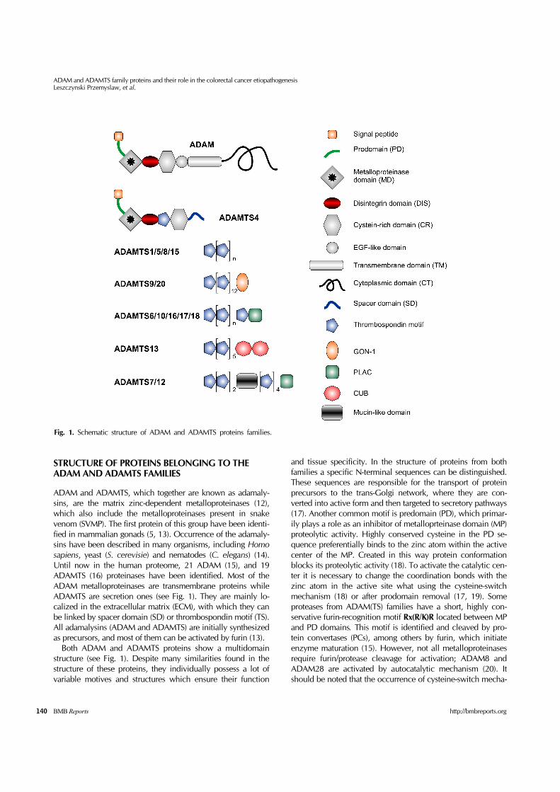
Fig. 1. Schematic structure of ADAM and ADAMTS proteins families.
STRUCTURE OF PROTEINS BELONGING TO THE ADAM AND ADAMTS FAMILIES
ADAM and ADAMTS, which together are known as adamaly-sins, are the matrix zinc-dependent metalloproteinases (12), which also include the metalloproteinases present in snake venom (SVMP). The first protein of this group have been identi-fied in mammalian gonads (5, 13). Occurrence of the adamaly-sins have been described in many organisms, including Homo sapiens, yeast (S. cerevisie) and nematodes (C. elegans) (14). Until now in the human proteome, 21 ADAM (15), and 19 ADAMTS (16) proteinases have been identified. Most of the ADAM metalloproteinases are transmembrane proteins while ADAMTS are secretion ones (see Fig. 1). They are mainly lo-calized in the extracellular matrix (ECM), with which they can be linked by spacer domain (SD) or thrombospondin motif (TS). All adamalysins (ADAM and ADAMTS) are initially synthesized as precursors, and most of them can be activated by furin (13). Both ADAM and ADAMTS proteins show a multidomain structure (see Fig. 1). Despite many similarities found in the structure of these proteins, they individually possess a lot of variable motives and structures which ensure their function
and tissue specificity. In the structure of proteins from both families a specific N-terminal sequences can be distinguished. These sequences are responsible for the transport of protein precursors to the trans-Golgi network, where they are con-verted into active form and then targeted to secretory pathways (17). Another common motif is predomain (PD), which primar-ily plays a role as an inhibitor of metalloprteinase domain (MP) proteolytic activity. Highly conserved cysteine in the PD se-quence preferentially binds to the zinc atom within the active center of the MP. Created in this way protein conformation blocks its proteolytic activity (18). To activate the catalytic cen-ter it is necessary to change the coordination bonds with the zinc atom in the active site what using the cysteine-switch mechanism (18) or after prodomain removal (17, 19). Some proteases from ADAM(TS) families have a short, highly con-servative furin-recognition motif Rx(R/K)R located between MP and PD domains. This motif is identified and cleaved by pro-tein convertases (PCs), among others by furin, which initiate enzyme maturation (15). However, not all metalloproteinases require furin/protease cleavage for activation; ADAM8 and ADAM28 are activated by autocatalytic mechanism (20). It should be noted that the occurrence of cysteine-switch mecha-

ADAM and ADAMTS family proteins and their role in the colorectal cancer etiopathogenesis Leszczynski Przemyslaw, et al.
141http://bmbreports.org BMB Reports
nism has been proven only for the ADAM family. Other role of the PD domain is that it probably also participates in the cor-rect folding of adamalysin (21). Metalloproteinase domain is another common motif in structure of proteins belonging to ADAM(TS) families. In its ac-tive site MP domain contains, as mentioned earlier, highly conservative motif, HEXXHXXGXXH, which is responsible for proteolytic activity. This motif contains three histidines sur-rounded by methionines in so-called methionine loop. Such zinc binding motif is characteristic for zinc-dependent metal-loproteinases of the reprolysin family. In addition to zinc atom MP domain active site, possess, also coordinatively bound wa-ter molecule – crucial for hydrolysis of proteins (3, 7). Disintegrin domain (DIS) is about 90aa long protein frag-ment which is also characteristic for the adamalysin’s structure. Disintegrin itself is a protein that inhibits platelet aggregation. Interaction between disintegrin and integrin (22) prevents binding plates with their natural ligands such as fibrinogen. Adamalysin’s DIS structure is homologous to the similar domain in the SVMP, where it determines the high tox-icity and is probably the cause of hemorrhage (23). DIS do-main, in both ADAM and ADAMTS, plays a crucial role in substrate recognition and it confers protease activity. It has been proven that some ADAM family proteins use the DIS do-main to specifically connect with integrins such as: α2β1, α4β1, α4β7, α5β1, α6β1, α6β4, α9β1, α2β1, αVβ3, αVβ5. This may point to the important role of those integrins in cell adhesion and cell-cell interactions (24). Cysteine-rich domain (CR) is the least common motif that could be found in the structure of ADAM and ADAMTS proteins. The function of this domain is not fully known. It was observed that the CR domain of ADAM13 regulates the pro-teolytic activity; it might have a similar functions in all adamalysins. In ADAM8 the CR domain together with PD is involved in autocatalytic activation of the metalloproteinase (20). Furthermore, it was proved that ADAM’s PD could bind to normal and tumor cells (25). It is likely that this binding is mediated by fibronectin (26). In the ADAM’s proteins structure, next to the CR a EGF-like domain can be distinguished. This is a rather conservative do-main, containing ten cysteines in its sequence. Until now the role of this domain remains unclear. Recent studies indicate that EGF-like domain might mediate the multimerisation of ADAM17 in cell membrane, which in turn may play a critical role in protease activation. It is worth to emphasize however, that this phenomenon has been proven for ADAM17 only (27). As it was mentioned above most of ADAM proteinases are cell membrane integral proteins and therefore they possess the transmembrane domain (TM), located behind EGF-like domain. The last, C-terminal fragment of ADAM proteins, lo-cated in the intracellular space is called a cytoplasmic tail (CT). This domain is playing a key role in intracellular signaling. Analytical structure of this protein fragment in differ-ent members of ADAM family shows that this it is presumably
the most variable protein part, both in size and in aminoacid sequence. Despite this, it was found that PXXP motifs present within the CT sequence allow to bind proteins containing SH3 domain (28). CT domain includes also several potential phos-phorylation sites (29). Cytoplasmic tail shows also regulatory properties, among others it can regulate the rate of MAPK cas-cade activity (30). All together, EGF-like, TM and CT domain, form a carboxylic region of the ADAM metalloproteinase family. ADAMTSs C-terminal region contains the TS, CR, and SD domains. In contrast to the previously described ADAM’s structures, the thrombospondin type 1-like repeat (TS) next to DIS domain occurs in ADAMTS structure. TS shows a high homology to the region I of thrombospondin 1 and 2 (31). This region allows ADAMTS to bind to the extracellular matrix (32) and it can probably be also involved in apoptosis and angiogenesis, which can be concluded from its homology to thrombospondin 1 and 2 (33). Different members of the ADAMTS family possess variable number of thrombospondin type 1-like motif repeats in their structures. This number varies between one, in ADAMTS4, and fifteen in ADAMTS20 and ADAMTS9 (15). Another structural diversity difference between the ADAM and ADAMTS proteins is spacer region, which is found only in ADAMTS. This region is located after CR domain and it is re-sponsible for the interaction of enzyme with substrate (34). Within the ADAMTS’ C-terminus region four additional do-mains were described; three of them (GON-1, CUB, PLAC, Mucin-like) are located after the last repetition of the TS do-main, only the mucin-like domain occurs between repetitions of the TS domain. These domains are unique and occur only in some proteases of ADAMTS family. GON-1 domain, found in ADAMTS9 and ADAMTS20, shows high homology to a GON-1 from C. elegans metalloproteinase, where it is neces-sary for distal tip cells migration during the gonads formation. Within its sequence, GON-1 contains ten conservative cysteines. The function of this domain in the human ADAMTS9 and ADAMTS20 has not yet been clarified (31, 35). The PLAC (protease and lacunin) domain occurs in the se-quences of several proteins from the ADAMTS family (6, 7, 10, 12, 16, 17, 18 and 19) (36). This domain was originally de-scribed in lacunin, which is hightly expressed in the ECM dur-ing Manduca sexta embryogenesis (37). In its sequence, PLAC domain has six cysteines which resemble an arrangement known from some PCs (38). ADAMTS13 is the only protein in this family with two CUB (Complement-Uegf-BMP-1) domains located in its C-terminus (39). This domain has also been found in proteins such as spermathesine and TSG-6 (tumor ne-crosis factor-stimulated gene-6) where it is probably involved in proteine-proteine interactions (40). The last of the additional domains present in ADAMTS is mucin-like domain. This do-main is found in ADAMTS7 and ADAMTS12 and is located between the third and fourth repetition of the TS. Mucin-like domain is a region rich in serine, proline, and threonine and undergoes o-glycosylation. It is suggested to be involved in
ADAM and ADAMTS family proteins and their role in the colorectal cancer etiopathogenesisLeszczynski Przemyslaw, et al.
142 BMB Reports http://bmbreports.org
metalloproteinase-substrate interaction (38).
ADAM AND ADAMTS: BIOLOGICAL FUNCTIONS AND THEIR PERTURBATIONS
Ever since the primary studies carried on adamalysins the ex-pression of these proteins in various tissues has been demonstrated. For some of them, like ADAMTS1, expression was traceable in almost all tissues (36, 41). On the other hand some of ADAM(TS) are tissue-specific, such as ADAMTS17 whose expression was observed exclusively in the ovary (42). Some disorders in collagen-rich tissues are connected with ada-malysins’ dysfunction. For example, mutations in ADAMTS10 have been characterized as a Weill-Marchesani syndrome background, whereas ADAMTS2 mutations are recognized as a background of Ehlers-Danlos syndrome type VII C (12). Among the two metalloproteinase families described in this article, the ADAM’s function has been first discovered and characterized; these proteins are also likely to be evolutio-narily older (15). The first ADAM-related reports have been as-sociated with the studies of such sperm proteins, as fertillin, as-sociated with the processes of spermatogenesis and sperm-egg fusion. Studies performed on mice showed that the Adam1 (fertillin-α) and Adam2 (fertillin-β) proteins are involved in spermatogenesis and presumably might play a crucial in this process (43). This finding, however, could not be directly transferred to humans because ADAM1 appeared to be a pseudogene. Adam2 and Adam1 present on the surface of mice spermatozoids are cleaved between MP and DIS do-mains (43). Retained DIS domain binds the α6β1-integrin on the surface of the egg and thus allows sperm-egg fusion. It is not known whether Adam1 and Adam2 are the only adamaly-sins involved in this process, since a similar feature is also postulated for ADAM3. Studies of this case are difficult be-cause silencing of Adam2 and Adam3 also induces down-reg-ulation of other adamalysins including Adam5-7, which also are involved in spermatogenesis, and result in dysfunction of sperm (44, 45). It is worth to note that humans’ ADAM3, sim-ilarly to ADAM1, proved to be a pseudognes (46), which again leads to the conclusion that often results of certain studies can-not be generalized to other species. Nineteen of the so far de-scribed ADAM’s family proteins (1-7, 14, 16, 18-21, 24-26, 30, 32, 34) are involved in spermatogenesis and in fertilization (12, 47); whereas ADAM8, -9, -10, -12, -15 and -17 may be in-volved in embryo implantation in the uterus (48). The above mentioned experimental data clearly demonstrates the im-portant role played by ADAM proteins in fertilization and in embryonic development. In addition, the expression of differ-ent members of the ADAM family has been demonstrated in many tissues or organs, where they play certain roles in proper functioning. These organs include: heart (ADAM9, 17, 19) (49), kidney (ADAM19), lung (ADAM33), teeth (ADAM28) and pancreas (ADAM9, 10, 17) (14). Research conducted on mice showed that Adam19 mutations are lethal, which proves the
key role of this protein in organs’ formation. Homozygous Adam19-/- mouse embryos died due to abnormalities of the heart and others cardiovascular system disorders. The functions of some of the ADAM and ADAMTS proteins have not yet been carefully examined – below we describe the examples whose function is at least partially identified and the pathological function or expression changes were found in col-orectal cancer (CRC) cell lines or tissues.
ADAM9 (MDC9, meltrin γ)The first reports on ADAM9 protein (50) initiated many studies on its function, location and development in normal and pathological tissues. Like most adamlysins, ADAM9 is synthe-sized as a precursor and is directed to posttranslational proc-essing in trans-Golgi. During this process metalloproteinase’s precursor is cleaved, by furin or other PCs, between PD and MP domains (51). In the same study it was shown that ADAM9 is catalytically active and in vitro it cleaves the insulin β-chain. On the other study the α-secretase activity of ADAM9 was postulated because its shortened form ADAM9s (lacking TD and cytoplasmic domain) expressed in COS cells cleaved proa-myloid protein APP (52). In several in vivo studies the follow-ing substrates for ADAM9 were found: FGFR2iiib, TNF (53) and fibronectin (54). Within a CT domain, ADAM9 has a po-tential SH3 binding site but, so far, its function has not been demonstrated in vivo (15, 51). Adam9 also showed high ex-pression in mouse embryo; those data suggests its role in em-bryogenesis and embryo implantation (48). ADAM9 is the only protein of this family, which has been shown to interact with integrin α2β1 and αVβ5 (24).
ADAM10 (MADM, kuz)In early studies ADAM10, by analogy to ADAM2, was sup-posed to be associated with spermatogenesis (55). Now these assumptions generally seem not to be valid although, along with other proteins including the aforementioned ADAM9, ADAM10 is also involved in embryo implantation (48). The structure of this protein is not typical because it does not con-tain EGF-like domain, while other elements typical for ADAM family are preserved. Like most proteins of the ADAM family, ADAM10 manifests its proteolytic activity. Its substrate range is broad and contains the six EGFR ligands, TNF, epireguline, HB-EGF and EGF (56). Similarly to ADAM9, ADAM10 cleaves APP; this evidence suggests that these proteins might influence the formation of amyloid inclusions and Alzheimer's disease development (57). ADAM10 together with ADAM17 are in-volved in cleavage of transmembrane protein Klotho (KL), which is involved in aging process. Additionally, cleavage of KL by ADAM10 is stimulated by insulin (58). The last on the list but probably the most important function of ADAM10 is its involvement in Regulated Intramembrane Proteolysis (RIP), which is part of the Notch/Delta signaling pathway. There exists some evidence that ADAM10 plays cru-cial role in cleavage of Notch membrane receptor (59).
ADAM and ADAMTS family proteins and their role in the colorectal cancer etiopathogenesis Leszczynski Przemyslaw, et al.
143http://bmbreports.org BMB Reports
Adam10 silencing is lethal in the early stage of mouse embryo-genesis and produces high neurological damage in aborted embryos (60). So far no protein-protein interactions between ADAM10 and integrins, were demonstrated (14).
ADAM12 (meltrin-α)ADAM12 is a protein which can be found in a human organ-ism in the two following forms: a long form (ADAM12-L) and short (ADAM12-S). Presence of those two versions is a result of an alternative splicing. ADAM12-L is a transmembrane pro-tein, while ADAM12-S is the secretion one (61). Both splicing versions have gelatinase activity and their substrates include mainly ECM proteins such as collagen type IV and fibronectin (62). Early studies on ADAM12 gene have shown its ex-pression in muscles and emphasized its role in muscle tissue formation via its influence on mioblast fusion (63). However, no change was found within the muscle tissue of a mouse after silencing the Adam12 expression, moreover, 30% of mice off-spring with Adam12-/- double knockout has died off within the first week of birth (64). Further research performed on mice suffering from muscular dystrophy showed that an over-expression of Adam12 decreases the disease’s symptoms in the early age of the mouse (65). Such results might suggest that ADAM12 has no influence on embryogenesis. Like other pro-teins from this family ADAM12 interacts with integrins: an in-teraction between this protein and integrins like α9β1 i α5β1was found (24).
ADAM17 (TACE, TNF-convertase)ADAM17 is the second ADAM family member which, next to ADAM10, does not contain a EGF-like domain in its structure. The proteolytic activity was also shown for this protein and one of its first discovered substrates were pro-TNF i TNF-α (66). As in ADAM10, the substrate specificity of ADAM17 was revealed for six EGF-like growing factors (56). Because of the similarity in the substrate specificity of ADAM17 to ADAM10, it may be assumed that ADAM17 can also take part in Notch signaling pathway as it was exhibited via in vitro. Yet, there was no change observed within this pathway in a mouse with Adam17-/- double gene knockout (67). ADAM17 shows also some characteristics of α-secretase which is to cut APP, thus suggesting its relation with the occurrence of Alzheimer’s dis-ease (57). The main activity of ADAM17 in in vitro studies is the TNFα conversion. It is proved that, proper function of ADAM17 is crucial for the transformation of inactive pro-TNFα into its active form and it is also crucial for the proper function of EGFR (68). The proteolytic activity of ADAM17 is regulated by the mechanism of oxidative stress (69).
ADAM23The structure of this protein is typical for the (ADAMs’ family) but it does not show proteolytic activity and, within MP do-main of this protein, a high-conservative zinc-binding motif was not detected. The first reports of ADAM23 were connected
with its high expression in the brain tissue (70). It can take part in a proper nervous system development via interaction with αVβ3 integrin (71). Research made on mice with Adam23-/- gene double knockout revealed increased embryonic mortality and many cases of ataxia in individuals born alive (72).
ADAM29ADAM29 does not show any proteolytic activity and, like ADAM23, it does not contain a high-conservative zinc-binding motif (73). Gene ADAM29 is located in chromosome 4 and its greatest expression is detected in testis (74). According to the conducted research, a human ADAM29 is found in three splic-ing forms containing 820 aa, 786 aa and 767 aa. Structural dif-ferences between particular isoforms are located within the CT domain (75). Results of RT-PCR experiments performed using the material taken from patients with chronic lymphocytic leu-kemia (CLL) show a higher ADAM29 expression in these patients. Some authors suggest, that the level of this gene ex-pression may have a significant prognostic value for patients with CLL (76). Recent studies found an increased number of mutations within ADAM29 gene among patients with melanoma. In addition, it was proved that mutated ADAM29 protein possess lower collagen adhesion, which may influence increased frequency and speed of metastasis formation (77).
ADAMTS1 (METH-1)ADAMTS1 is one of the most important and best described protein in the whole ADAMTS family; the studies on its struc-ture and function started in 1997 (78). It is confirmed that its spacer-domain has a strong affinity with ECM and the TS do-main shows high affinity with heparin (32). It is also found that ADAMTS1 has some proteolytic activity and shows a certain substrate specificity among others with aggrecan, although the places of cleavages made by ADAMTS1 vary from other ag-grecans (79). At the beginning of research, the inhibition of an-giogenesis was specified as a basic function of ADAMTS1 (41). It was proved that this protein silences the proliferation signal caused by VEGF and FGF2 via the inhibition of VEGFR2 phos-phorylation (80). This is not the only pathway of action of this protein on neovascularisation because due to its proteolytic activity ADAMTS1 can dissociate from thrombospondin type I and II a short polypetide with antiangiogenic activity a (81). These data may suggest that ADAMTS1 has a strong influence on angiogenesis, and as a result, on the speed of cancer development. It was also proved that ADAMTS1 possesses a metastasis promotion and tumor progression activity (82). Possible cause of such phenomenon could be shedding off amphiregulin and HB-EGF (83).
ADAMTS9In human organism, the ADAMTS9 protein occurs in two se-cretion forms differing in number of TS motifs. The so-called long form of ADAMTS9 has got 14 repetitions of TS motif while the so-called short form has got only three such repeti-
ADAM and ADAMTS family proteins and their role in the colorectal cancer etiopathogenesisLeszczynski Przemyslaw, et al.
144 BMB Reports http://bmbreports.org
tions in its structure (35). Additionally, there is an untypical gon-1-like motif C-terminus of ADAMTS9. The first studies on ADAMTS9 metalloproteinase were published at the beginning of this century and resulted in finding a high gene homology to gon-1 metalloproteinase in C. elegans. It is stated that, like gon-1, ADAMTS9 could take part in gametogenesis (84), how-ever, this result was not confirmed in studies on mammals. The ADAMTS9 protein shows proteolytic activity and is also classified as aggrecanase and versicanase (35). In the case of mice, Adamts9 gene exhibits high expression during embryo-genesis. Mouse embryos with double knockout of Adamts9-/- gene died off before gastrulating (85). This fact confirms the essential role of this gene in embryogenesis. As a contrast, mice with a single silenced Adamts9 allele showed a sponta-neous neovascularisation, thus confirming the antiangiogenic activity of ADAMTS9. As it is assumed in the latest research, ADAMTS9 gene might be a novel gene supressor; its silencing or deletion can be found in almost 50% of esophagus cancers (86).
ADAMTS12ADAMTS12 is one of two, next to ADAMTS7, glycoproteins in ADAMTS family with an unique, in this family, mucine-like domain in its structure (38). ADAMTS12 also reveals proteo-lytic activity and is one of the main metalloproteins which play a role in COMP (cartilage oligomeric matrix protein) degrada-tion, what can explain the role of this protein in arthritis devel-opment (87). Other substrates of this metalloproteinase are, among others, VEGF and HGF; their shedding results in Ras-MAPK pathway blocking. ADAMTS12 role in Ras-MAPK pathway regulation and some evidence of epigenetic silencing of the gene in cancer cells suggests that ADAMTS12 may be a novel gene suppressor (88). This thesis seems to be proved by the fact that cancer cells injected into a mouse with silenced Adamts12 gene proliferate much faster and produce more de-veloped neovascular system (89).
ADAMTS15ADAMTS15 is the least examined protein from the whole ADAMTS family. This metalloproteinase is a quite short secre-tion protein with only two repetitions of TS motif of C-terminus. High homology of its gene to ADAMTS1 was also detected (42). It was also showed that this protein has ag-grecanese activity however, this fact was not sufficiently proved (36, 90). Research on prostate cancer cell lines re-vealed lower ADAMTS15 expression in this cancer (90) and regulation of ADAMTS15 expression under influence of androgens. Clinical studies on patients with breast cancer ex-hibited that the lower ADAMTS15 expression correlates with a higher probability of cancer development and increased mor-tality (91). The results of this research might emphasize a cru-cial role of ADAMTS15 in development of cancers.
ADAMTS18 The function of ADAMTS18 protein is so far unspecified. First research on ADAMTS18 showed its substantial structural sim-ilarity to ADAMTS16 (42). It is known that in its structure, ADAMTS18 possess a motif allowing glycoaminoglycane bind-ing; yet the in vitro based research proved that such binding is inefficient. In vitro ADAMTS18 manifests its proteolytic activ-ity as an aggrecanase but this enzymatic activity is rather low and manifests itself at ADAMTS18 high concentrations (92). Studies on cancer cell lines indicated that ADAMTS18 gene is frequently epigenetically silenced , what was confirmed by re-search on cancer tissue obtained from patients (93). It may be claimed, according to this research, that ADAMTS18 might be a novel gene suppressor. Recent studies prove that ADAMTS18 plays a role in maintenance of homeostasis via ac-tivation of platelet aggregation (94).
THE ROLE OF ADAMALYSINES IN COLORECTAL CANCER (CRC)
As it was proved by the research, proteins from ADAM and ADAMTS families play a significant role in such processes as intercellular signaling, angiogenesis, formation of proper tissue structure, embryogenesis and cell migration; what might in-dicate that any perturbation of function of these proteins might be essential in cancerogenesis (12). There is about one million of new cases of colorectal cancer worldwide every year. Globally, it is the fourth in men and third in women most frequent cancer. The biggest amount of CRC is registered in such areas as Northern America, Australia and Western Europe (95). Statistical analysis shows that the number of CRC cases is constantly rising (96). Several long-time studies conducted on etiopathogenesis of this cancer show that it may be influenced by lifestyle, environment, soci-oeconomic status and genetic background in which the CRC occurs (95, 97). A number of information about the role of adamalysines in etiopathogenesis of colorectal cancer were published. In the most of the cell lines studies are presented. In these papers the influence of the ADAM9s secretion form on the increasing in-vasive phenotype of Clone A cell lines was emphasized. The DIS domain of ADAM9 may directly bind to α6β4 and α2β1 integrins and proteolytic activity of this adamalysin allows to cleave laminines. The in vitro studies on CRC cell lines rev-eled the influence of ADAM9s overexpression on increased in-vasiveness of cancer cells (98). The investigation on CRC HT29 cell line pointed out the influence of overexpression of ADAM9 transmembrane forms on the HB-EGF-induced in-creased proliferation and decreased cell adhesion via E-cadher-in degradation. It was found that ADAM9 strengthens the cell proliferation signal and decreases the contact suppression. Additionally, metastasis may be caused by higher E-cadherin degradation (99). Tissue Chip Assay studies show some corre-lation between the amount of ADAM10 and the cancer stage.

ADAM and ADAMTS family proteins and their role in the colorectal cancer etiopathogenesis Leszczynski Przemyslaw, et al.
145http://bmbreports.org BMB Reports
The results of these studies revealed also an increase in num-ber of ADAM10 on the CRC cell surface in the late stages of the cancer (100). It was also found that one of the factors that modulate tetraspanine network within CRC tumors is ADAM10 (101). The investigation on cancer cell lines (293T, SW480 and HCT116) indicated that there is a significant influ-ence of ADAM10 overexpression on metastasis strengthening; moreover, this effect was amplified by simultaneous L1-CAM expression (neural cell adhesion receptor) (102). The most re-cent histopathological examination conducted on tumors col-lected from whole gastrointestinal tract, including CRC, showed the ADAM10 overexpression. What more, the protein level correlated with the frequency of metastasis to lymph no-des and other organs. However, these studies do not show any correlation between ADAM10 expression level and the 5-year survival rate (103). In the case of role of ADAM12 in CRC, a higher expression of this protein was detected on the outer sur-face of cancer cells. However, the research was conducted on cell lines, thus it is not reliable to show the full spectrum of this protein activity in vivo. It is also suggested that the activity of CR domain in ADAM12 may influence cancer cell adhesion to ECM and promote metastasis (25). The investigation on ex-pression changes of CRC liver metastasis revealed increased level of ADAM17 mRNA in most metastasis tumors in compar-ison with primary tumors which do not form such metastasis. It confirms a significant influence of ADAM17 on liver meta-stasis formation frequency (104). According to experiments conducted in vitro ADAM17 could become a potential phar-maceutical target in CRC treatment. Carcinoma cell lines grown in presence of 5-fluorouracil, a chemotherapeutic used in CRC treatment, show growth factors overexpression. These factors are the main reason of chemotherapy resistance of the cancer cells, as it as shown both in vitro and in vivo studies. It was also found that silencing of ADAM17 by siRNA causes an enhancement of chemotherapy response (105, 106). In other study it was shown that the alternation in methylation status results in silencing of ADAM23 gene. This phenomenon was found in both cell lines and cancer tissues obtained from patients. Re-expression of ADAM23 was detected after cell line passage with the 5-aza-20-deoxycytidin dimethylating agent treatment. What is more, some of the cells expressing ADAM23 have lost their cancer phenotype (107). Downregu-lation of DNA-methyltransferase-1 (DNMT1) and, as a result, ADAM23 reactivation may be obtained by regaining a proper expression of miR342 (miRNA silenced in some CRC cell lines). Decreased proliferation and cancer cells’ vitality was al-so observed in this experiment (108). According to research conducted on a large group of CRC tissues ADAM29 gene un-dergoes frequent mutations. Unfortunately in this paper the mutation types were not described and additionally their influ-ence on ADAM29 protein and its expression, was not ex-plained (109). On the other hand aCGH based studies point out that ADAM29 is frequently amplified in CRC tumors. Also in this paper the role of this phenomenon in the cancer devel-
opment is not suggested (110). Data obtained from a material collected from patients with CRC and with adenoma exhibited that ADAMTS1 gene pro-moter was hypermethylated in both cancer types in compar-ison to a normal tissue of the same patient. It is well known that promoter hypermethylation, in most cases, causes the gene silencing (111, 112). The recent studies with the use of methylate bead-chip array-based technology confirmed in-creased methylation in case of ADAMTS5 gene promoter. In addition the expression micro-chip method revealed (4) that the amount of the gene’s mRNA is decreased in a tumor tissue. Micro-chip arrays allow to gain a large number of information at one time, what often is essential to indicate pathways of fur-ther research. It has to be stated however, that this results ob-tained using this technology need to be confirmed by other, more accurate methods. Investigation on a bigger group of CRC conducted using the novel HRM (high-resolution melting) method revealed increased ADAMTS9 gene promoter methyl-ation (113). It was also found that ADAMTS12 gene promoter is significantly hypermethylated in comparison to of a normal tissue (88). The amount of proteins on the tumor cell surface increases according to the growth of distance from the tumor center (114). Taking into account the both above cited papers it can be concluded that ADAMTS12 may be a sufficient mark-er for an intraoperative examination in order to determinate the operation margin; it can also be treated as a prognostic marker. In vitro and in vivo research on tissues obtained from patients showed hypermethylation of ADAMTS15 gene pro-moter and a correlation between this phenomenon and ADAMTS15 protein depreciation of the expression. Histo-pathological and immunohistochemical studies have shown that the level of ADAMTS15 protein expression is inversely proportional to the histological grade of tumor differentiation (115). Such results may suggest that the gene might be a novel suppressor. Moreover, ADAMTS18 gene is also epigenetically silenced within CRC what has been emphasized by the HRM method; it is yet not proved via immunohistochemical studies (94). Furthermore, it has been observed that both ADAMTS15 and ADAMTS18 undergo frequent mutations in CRC cells but it is still unknown what type of mutations are they or how their function influences further expression or function of newly formed proteins (109). As it was introduced in this review, adamalysines play es-sential role in CRC etiopathogenesis and metastasis. The quot-ed results seem to suggest that the ADAM(TS) may be potential tumor markers and predictors. This potential may be widely used in monitoring of treatment progression and specification of operation margins while CRC tumor resection. Further stud-ies on adamalysines seem to be essential to explain the molec-ular mechanisms of this cancer which is seen in the role of adamalysines in cancer formation especially in the CRC etiopathogenesis. The large amount of various articles on ADAM(TS) proteins point out their essential role in physio-logical processes and in pathology. Unfortunately there is not
ADAM and ADAMTS family proteins and their role in the colorectal cancer etiopathogenesisLeszczynski Przemyslaw, et al.
146 BMB Reports http://bmbreports.org
much research which would explain the correlations between this clinical data and adamalysines’ function disorders. The fact that the ADAM(TS) are not separated proteins but they work as a part of various complex processes needs further studies as their full activity and regulations are still unknown.
Acknowledgements This study was supported by a grant from the State Committee for Scientific Research, Polish Ministry for Scientific Research and Information Technology No. N N401 601438/2010-013.
REFERENCES
1. Hanahan, D. and Weinberg, R. A. (2011) Hallmarks of cancer: the next generation. Cell 144, 646-674.
2. Sasiadek, M. M. and Karpinski, P. (2009) Genetic theory of cancer. Pol. Przegl. Chir. 81, 478-485.
3. Stocker, W., Grams, F., Baumann, U., Reinemer, P., Gomisruth, F. X., McKay, D. B. and Bode, W. (1995) The metzincins-topological and sequential relations between the astacins, adamalysins, serralysins, and matrixins (collagenases) define a superfamily of zinc-peptidases. Protein Sci. 4, 823-840.
4. Kim, Y. H., Lee, H. C., Kim, S. Y., Yeom, Y. I., Ryu, K. J., Min, B. H., Kim, D. H., Son, H. J., Rhee, P. L., Kim, J. J., Rhee, J. C., Kim, H. C., Chun, H. K., Grady, W. M. and Kim, Y. S. (2011) Epigenomic analysis of aberrantly methy-lated genes in colorectal cancer identifies genes commonly affected by epigenetic alterations. Ann. Surg. Oncol. 18, 2338-2347.
5. Ugalde, A. P., Ordonez, G. R., Quiros, P. M., Puente, X. S. and Lopez-Otin, C. (2010) Metalloproteases and the degradome. Methods. Mol. Biol. 622, 3-29.
6. Kessenbrock, K., Plaks, V. and Werb, Z. (2010) Matrix metalloproteinases: regulators of the tumor microenvi-ronment. Cell 141, 52-67.
7. Tallant, C., Marrero, A. and Gomis-Ruth, F. X. (2010) Matrix metalloproteinases: Fold and function of their cata-lytic domains. Biochim. Biophys. Acta-Mol. Basis Dis. 1803, 20-28.
8. Gross, J. and Lapiere, C. M. (1962) Collagenolytic activity in amphibian tissues: a tissue culture assay. Proc. Natl. Acad. Sci. U.S.A. 48, 1014-1022.
9. Kuno, K., Kanada, N., Nakashima, E., Fujiki, F., Ichimura, F. and Matsushima, K. (1997) Molecular cloning of a gene encoding a new type of metalloproteinase-disintegrin fam-ily protein with thrombospondin motifs as an in-flammation associated gene. J. Biol. Chem. 272, 556-562.
10. Kaushal, G. P. and Shah, S. V. (2000) The new kids on the block: ADAMTSs, potentially multifunctional metal-loproteinases of the ADAM family. J. Clin. Invest. 105, 1335-1337.
11. Wagstaff, L., Kelwick, R., Decock, J. and Edwards, D. R. (2011) The roles of ADAMTS metalloproteinases in tu-morigenesis and metastasis. Front Biosci. 16, 1861-1872.
12. van Goor, H., Melenhorst, W. B., Turner, A. J. and Holgate, S. T. (2009) Adamalysins in biology and disease. J. Pathol. 219, 277-286.
13. Rocks, N., Paulissen, G., El Hour, M., Quesada, F., Crahay, C., Gueders, M., Foidart, J. M., Noel, A. and Cataldo, D. (2008) Emerging roles of ADAM and ADAMTS metalloproteinases in cancer. Biochimie. 90, 369-379.
14. Edwards, D. R., Handsley, M. M. and Pennington, C. J. (2008) The ADAM metalloproteinases. Mol. Aspects. Med. 29, 258-289.
15. Brocker, C. N., Vasiliou, V. and Nebert, D. W. (2009) Evolutionary divergence and functions of the ADAM and ADAMTS gene families. Hum. Genomics. 4, 43-55.
16. Stanton, H., Melrose, J., Little, C. B. and Fosang, A. J. (2011) Proteoglycan degradation by the ADAMTS family of proteinases. Biochim. Biophys. Acta-Mol. Basis. Dis. 1812, 1616-1629.
17. Kang, T. B., Zhao, Y. G., Pei, D. Q., Sucic, J. F. and Sang, Q. X. A. (2002) Intracellular activation of human adamaly-sin 19/disintegrin and metalloproteinase 19 by furin oc-curs via one of the two consecutive recognition sites. J. Biol. Chem. 277, 25583-25591.
18. Van Wart, H. E. and Birkedalhansen, H. (1990) The cys-teine switch: a principle of regulation of metalloproteinase activity with potential applicability to the entire matrix metalloproteinase gene family. Proc. Natl. Acad. Sci. U.S.A. 87, 5578-5582.
19. Tortorella, M. D., Arner, E. C., Hills, R., Gormley, J., Fok, K., Pegg, L., Munie, G. and Malfait, A. M. (2005) ADAMTS-4 (aggrecanase-1): N-Terminal activation me-chanisms. Arch. Biochem. Biophys. 444, 34-44.
20. Hall, T., Leone, J. W., Wiese, J. F., Griggs, D. W., Pegg, L. E., Pauley, A. M., Tomasselli, A. G. and Zack, M. D. (2009) Autoactivation of human ADAM8: a novel pre- processing step is required for catalytic activity. Biosci. Rep. 29, 217-228.
21. Milla, M. E., Leesnitzer, M. A., Moss, M. L., Clay, W. C., Carter, H. L., Miller, A. B., Su, J. L., Lambert, M. H., Willard, D. H., Sheeley, D. M., Kost, T. A., Burkhart, W., Moyer, M., Blackburn, R. K., Pahel, G. L., Mitchell, J. L., Hoffmann, R. and Becherer, J. D. (1999) Specific se-quence elements are required for the expression of func-tional tumor necrosis factor-alpha-converting enzyme (TACE). J. Biol. Chem. 274, 30563-30570.
22. Bridges, L. C., Sheppard, D. and Bowditch, R. D. (2005) ADAM disintegrin-like domain recognition by the lym-phocyte integrins alpha4beta1 and alpha4beta7. Biochem. J. 387, 101-108.
23. Gomis-Ruth, F. X., Kress, L. F., Kellermann, J., Mayr, I., Lee, X., Huber, R. and Bode, W. (1994) Refined 2.0 A X-ray crystal structure of the snake venom zinc-endopepti-dase adamalysin II. Primary and tertiary structure determi-nation, refinement, molecular structure and comparison with astacin, collagenase and thermolysin. J. Mol. Biol. 239, 513-544.
24. Arribas, J., Bech-Serra, J. J. and Santiago-Josefat, B. (2006) ADAMs, cell migration and cancer. Cancer Metastasis Rev. 25, 57-68.
25. Iba, K., Albrechtsen, R., Gilpin, B., Frohlich, C., Loechel, F., Zolkiewska, A., Ishiguro, K., Kojima, T., Liu, W., Langford, J. K., Sanderson, R. D., Brakebusch, C., Fassler, R. and Wewer, U. M. (2000) The cysteine-rich domain of
ADAM and ADAMTS family proteins and their role in the colorectal cancer etiopathogenesis Leszczynski Przemyslaw, et al.
147http://bmbreports.org BMB Reports
human ADAM 12 supports cell adhesion through synde-cans and triggers signaling events that lead to beta 1 in-tegrin-dependent cell spreading. J. Cell Biol. 149, 1143- 1155.
26. Gaultier, A., Cousin, H., Darribere, T. and Alfandari, D. (2002) ADAM13 disintegrin and cysteine-rich domains bind to the second heparin-binding domain of fibronectin. J. Biol. Chem. 277, 23336-23344.
27. Lorenzen, I., Trad, A. and Grotzinger, J. (2011) Multimeri-sation of A disintegrin and metalloprotease protein-17 (ADAM17) is mediated by its EGF-like domain. Biochem. Biophys. Res. Commun. 415, 330-336.
28. Kang, Q., Cao, Y. and Zolkiewska, A. (2000) Metallopro-tease-disintegrin ADAM 12 binds to the SH3 domain of Src and activates Src tyrosine kinase in C2C12 cells. Biochem. J. 352(Pt 3), 883-892.
29. Poghosyan, Z., Robbins, S. M., Houslay, M. D., Webster, A., Murphy, G. and Edwards, D. R. (2002) Phosphorylation- dependent interactions between ADAM15 cytoplasmic do-main and Src family protein-tyrosine kinases. J. Biol. Chem. 277, 4999-5007.
30. Killock, D. J. and Ivetic, A. (2010) The cytoplasmic do-mains of TNFalpha-converting enzyme (TACE/ADAM17) and L-selectin are regulated differently by p38 MAPK and PKC to promote ectodomain shedding. Biochem. J. 428, 293-304.
31. Jones, G. C. and Riley, G. P. (2005) ADAMTS proteinases: a multi-domain, multi-functional family with roles in ex-tracellular matrix turnover and arthritis. Arthritis. Res. Ther. 7, 160-169.
32. Kuno, K. and Matsushima, K. (1998) ADAMTS-1 protein anchors at the extracellular matrix through the thrombo-spondin type I motifs and its spacing region. J. Biol. Chem. 273, 13912-13917.
33. Guo, N.-H., Krutzsch, H. C., Inman, J. K. and Roberts, D. D. (2012) Thrombospondin 1 and type I repeat peptides of thrombospondin 1 specifically induce apoptosis of en-dothelial cells. Cancer Res. 57, 1735-1742.
34. de Groot, R., Bardhan, A., Ramroop, N., Lane, D. A. and Crawley, J. T. (2009) Essential role of the disintegrin-like domain in ADAMTS13 function. Blood 113, 5609-5616.
35. Somerville, R. P., Longpre, J. M., Jungers, K. A., Engle, J. M., Ross, M., Evanko, S., Wight, T. N., Leduc, R. and Apte, S. S. (2003) Characterization of ADAMTS-9 and ADAMTS-20 as a distinct ADAMTS subfamily related to Caenorhabditis elegans GON-1. J. Biol. Chem. 278, 9503-9513.
36. Porter, S., Clark, I. M., Kevorkian, L. and Edwards, D. R. (2005) The ADAMTS metalloproteinases. Biochem. J. 386, 15-27.
37. Nardi, J. B., Martos, R., Walden, K. K. O., Lampe, D. J. and Robertson, H. M. (1999) Expression of lacunin, a large multidomain extracellular matrix protein, accom-panies morphogenesis of epithelial monolayers in Mandu-ca sexta. Insect. Biochem. Mol. Biol. 29, 883-897.
38. Somerville, R. P., Longpre, J. M., Apel, E. D., Lewis, R. M., Wang, L. W., Sanes, J. R., Leduc, R. and Apte, S. S. (2004) ADAMTS7B, the full-length product of the ADAMTS7 gene, is a chondroitin sulfate proteoglycan containing a mucin domain. J. Biol. Chem. 279, 35159-35175.
39. Zheng, X., Chung, D., Takayama, T. K., Majerus, E. M., Sadler, J. E. and Fujikawa, K. (2001) Structure of von Willebrand factor-cleaving protease (ADAMTS13), a met-alloprotease involved in thrombotic thrombocytopenic purpura. J. Biol. Chem. 276, 41059-41063.
40. Bork, P. and Beckmann, G. (1993) The CUB domain. A widespread module in developmentally regulated pro-teins. J. Mol. Biol. 231, 539-545.
41. Vazquez, F., Hastings, G., Ortega, M. A., Lane, T. F., Oikemus, S., Lombardo, M. and Iruela-Arispe, M. L. (1999) METH-1, a human ortholog of ADAMTS-1, and METH-2 are members of a new family of proteins with an-gio-inhibitory activity. J. Biol. Chem. 274, 23349-23357.
42. Cal, S., Obaya, A. J., Llamazares, M., Garabaya, C., Quesada, V. and Lopez-Otin, C. (2002) Cloning, ex-pression analysis, and structural characterization of seven novel human ADAMTSs, a family of metalloproteinases with disintegrin and thrombospondin-1 domains. Gene 283, 49-62.
43. Primakoff, P. and Myles, D. G. (2000) The ADAM gene family - surface proteins with adhesion and protease activity. Trends Genet. 16, 83-87.
44. Han, C., Choi, E., Park, I., Lee, B., Jin, S., Kim, D. H., Nishimura, H. and Cho, C. (2009) Comprehensive analy-sis of reproductive ADAMs: relationship of ADAM4 and ADAM6 with an ADAM complex required for fertilization in mice. Biol. Reprod. 80, 1001-1008.
45. Kim, T., Oh, J., Woo, J. M., Choi, E., Im, S. H., Yoo, Y. J., Kim, D. H., Nishimura, H. and Cho, C. H. (2006) Expression and relationship of male reproductive ADAMS in mouse. Biol. Reprod. 74, 744-750.
46. Grzmil, P., Kim, Y., Shamsadin, R., Neesen, J., Adham, I. M., Heinlein, U. A., Schwarzer, U. J. and Engel, W. (2001) Human cyritestin genes (CYRN1 and CYRN2) are non-functional. Biochem. J. 357, 551-556.
47. Kim, E., Yamashita, M., Nakanishi, T., Park, K. E., Kimura, M., Kashiwabara, S. and Baba, T. (2006) Mouse sperm lacking ADAM1b/ADAM2 fertilin can fuse with the egg plasma membrane and effect fertilization. J. Biol. Chem. 281, 5634-5639.
48. Kim, J., Kang, S. G., Kim, J. I., Park, J. H., Kim, S. K., Cho, D. J. and Kim, H. (2006) Implication of ADAM-8, -9, -10, -12, -15, -17, and ADAMTS-1 in implantational remodel-ing of a mouse uterus. Yonsei. Med. J. 47, 558-567.
49. Zhou, H. M., Weskamp, G., Chesneau, V., Sahin, U., Vortkamp, A., Horiuchi, K., Chiusaroli, R., Hahn, R., Wilkes, D., Fisher, P., Baron, R., Manova, K., Basson, C. T., Hempstead, B. and Blobel, C. P. (2004) Essential role for ADAM19 in cardiovascular morphogenesis. Mol. Cell. Biol. 24, 96-104.
50. McKie, N., Dallas, D. J., Edwards, T., Apperley, J. F., Russell, R. G. and Croucher, P. I. (1996) Cloning of a nov-el membrane-linked metalloproteinase from human mye-loma cells. Biochem. J. 318(Pt 2), 459-462.
51. Roghani, M., Becherer, J. D., Moss, M. L., Atherton, R. E., Erdjument-Bromage, H., Arribas, J., Blackburn, R. K., Weskamp, G., Tempst, P. and Blobel, C. P. (1999) Metalloprotease-disintegrin MDC9: Intracellular matura-tion and catalytic activity. J. Biol. Chem. 274, 3531-3540.
52. Hotoda, N., Koike, H., Sasagawa, N. and Ishiura, S.

ADAM and ADAMTS family proteins and their role in the colorectal cancer etiopathogenesisLeszczynski Przemyslaw, et al.
148 BMB Reports http://bmbreports.org
(2002) A secreted form of human ADAM9 has an al-pha-secretase activity for APP. Biochem. Biophys. Res. Commun. 293, 800-805.
53. Peduto, L., Reuter, V. E., Shaffer, D. R., Scher, H. I. and Blobel, C. P. (2005) Critical function for ADAM9 in mouse prostate cancer. Cancer Res. 65, 9312-9319.
54. Schwettmann, L. (2001) Cloning and Expression in Pichia pastoris of Metalloprotease Domain of ADAM 9 Catalytically Active against Fibronectin. Protein Expr. Purif. 21, 65-70.
55. Wolfsberg, T. G., Primakoff, P., Myles, D. G. and White, J. M. (1995) ADAM, a novel family of membrane proteins containing A Disintegrin And Metalloprotease domain: multipotential functions in cell-cell and cell-matrix interactions. J. Cell Biol. 131, 275-278.
56. Sahin, U., Weskamp, G., Kelly, K., Zhou, H. M., Higashiyama, S., Peschon, J., Hartmann, D., Saftig, P. and Blobel, C. P. (2004) Distinct roles for ADAM10 and ADAM17 in ectodomain shedding of six EGFR ligands. J. Cell Biol. 164, 769-779.
57. Allinson, T. M., Parkin, E. T., Turner, A. J. and Hooper, N. M. (2003) ADAMs family members as amyloid precursor protein alpha-secretases. J. Neurosci. Res. 74, 342-352.
58. Chen, C. D., Podvin, S., Gillespie, E., Leeman, S. E. and Abraham, C. R. (2007) Insulin stimulates the cleavage and re-lease of the extracellular domain of Klotho by ADAM10 and ADAM 17. Proc. Natl. Acad. Sci. U.S.A. 104, 19796-19801.
59. Bland, C. E., Kimberly, P. and Rand, M. D. (2003) Notch-induced proteolysis and nuclear localization of the delta ligand. J. Biol. Chem. 278, 13607-13610.
60. Hartmann, D., de Strooper, B., Serneels, L., Craessaerts, K., Herreman, A., Annaert, W., Umans, L., Lubke, T., Lena Illert, A., von Figura, K. and Saftig, P. (2002) The dis-integrin/metalloprotease ADAM 10 is essential for Notch signalling but not for alpha-secretase activity in fibroblasts. Hum. Mol. Genet. 11, 2615-2624.
61. Gilpin, B. J., Loechel, F., Mattei, M. G., Engvall, E., Albrechtsen, R. and Wewer, U. M. (1998) A novel, se-creted form of human ADAM 12 (meltrin alpha) provokes myogenesis in vivo. J. Biol. Chem. 273, 157-166.
62. Roy, R., Wewer, U. M., Zurakowski, D., Pories, S. E. and Moses, M. A. (2004) ADAM 12 cleaves extracellular ma-trix proteins and correlates with cancer status and stage. J. Biol. Chem. 279, 51323-51330.
63. Yagami-Hiromasa, T., Sato, T., Kurisaki, T., Kamijo, K., Nabeshima, Y. and Fujisawa-Sehara, A. (1995) A metal-loprotease-disintegrin participating in myoblast fusion. Nature 377, 652-656.
64. Kurisaki, T., Masuda, A., Sudo, K., Sakagami, J., Higashi-yama, S., Matsuda, Y., Nagabukuro, A., Tsuji, A., Nabe-shima, Y., Asano, M., Iwakura, Y. and Sehara-Fujisawa, A. (2003) Phenotypic analysis of Meltrin alpha (ADAM12)- deficient mice: Involvement of Meltrin alpha in adipo-genesis and myogenesis. Mol. Cell Biol. 23, 55-61.
65. Moghadaszadeh, B., Albrechtsen, R., Guo, L. T., Zaik, M., Kawaguchi, N., Borup, R. H., Kronqvist, P., Schroder, H. D., Davies, K. E., Voit, T., Nielsen, F. C., Engvall, E. and Wewer, U. M. (2003) Compensation for dystrophin-defi-ciency: ADAM12 overexpression in skeletal muscle re-sults in increased alpha 7 integrin, utrophin and asso-
ciated glycoproteins. Hum. Mol. Genet. 12, 2467-2479.66. Black, R. A., Rauch, C. T., Kozlosky, C. J., Peschon, J. J.,
Slack, J. L., Wolfson, M. F., Castner, B. J., Stocking, K. L., Reddy, P., Srinivasan, S., Nelson, N., Boiani, N., Schooley, K. A., Gerhart, M., Davis, R., Fitzner, J. N., Johnson, R. S., Paxton, R. J., March, C. J. and Cerretti, D. P. (1997) A met-alloproteinase disintegrin that releases tumour-necrosis fac-tor-alpha from cells. Nature 385, 729-733.
67. Saftig, P. and Hartmann, D. (2005) ADAM10 A major mem-brane protein ectodomain sheddase involved in regulated in-tramembrane proteolysis; in The Adam Family of Proteases, pp. 85-121, Springer, Dordrecht, The Netherlands.
68. Peschon, J. J., Slack, J. L., Reddy, P., Stocking, K. L., Sunnarborg, S. W., Lee, D. C., Russell, W. E., Castner, B. J., Johnson, R. S., Fitzner, J. N., Boyce, R. W., Nelson, N., Kozlosky, C. J., Wolfson, M. F., Rauch, C. T., Cerretti, D. P., Paxton, R. J., March, C. J. and Black, R. A. (1998) An essential role for ectodomain shedding in mammalian development. Science 282, 1281-1284.
69. Wang, Y., Herrera, A. H., Li, Y., Belani, K. K. and Walcheck, B. (2009) Regulation of mature ADAM17 by redox agents for L-selectin shedding. J. Immunol. 182, 2449-2457.
70. Sagane, K., Ohya, Y., Hasegawa, Y. and Tanaka, I. (1998) Metalloproteinase-like, disintegrin-like, cysteine-rich pro-teins MDC2 and MDC3: novel human cellular disintegrins highly expressed in the brain. Biochem. J. 334(Pt 1), 93-98.
71. Sagane, K., Yamazaki, K., Mizui, Y. and Tanaka, I. (1999) Cloning and chromosomal mapping of mouse ADAM11, ADAM22 and ADAM23. Gene 236, 79-86.
72. Mitchell, K. J., Pinson, K. I., Kelly, O. G., Brennan, J., Zupicich, J., Scherz, P., Leighton, P. A., Goodrich, L. V., Lu, X., Avery, B. J., Tate, P., Dill, K., Pangilinan, E., Wakenight, P., Tessier-Lavigne, M. and Skarnes, W. C. (2001) Functional analysis of secreted and transmembrane proteins critical to mouse development. Nature Genetics 28, 241-249.
73. Brocker, C. N., Vasiliou, V. and Nebert, D. W. (2009) Evolutionary divergence and functions of the ADAM and ADAMTS gene families. Hum. Genomics. 4, 43-55.
74. Xu, R., Cai, J., Xu, T., Zhou, W., Ying, B., Deng, K., Zhao, S. and Li, C. (1999) Molecular cloning and mapping of a novel ADAM gene (ADAM29) to human chromosome 4. Genomics 62, 537-539.
75. Cerretti, D. P., DuBose, R. F., Black, R. A. and Nelson, N. (1999) Isolation of two novel metalloproteinase- disintegrin (ADAM) cDNAs that show testis-specific gene expression. Biochem. Biophys. Res. Commun. 263, 810-815.
76. Oppezzo, P., Vasconcelos, Y., Settegrana, C., Jeannel, D., Vuillier, F., Legarff-Tavernier, M., Kimura, E. Y., Bechet, S., Dumas, G., Brissard, M., Merle-Beral, H., Yamamoto, M., Dighiero, G., Davi, F. and French Cooperative Group on CLL (2005) The LPL/ADAM29 expression ratio is a novel prognosis indicator in chronic lymphocytic leuke-mia. Blood 106, 650-657.
77. Wei, X., Moncada-Pazos, A., Cal, S., Soria-Valles, C., Gartner, J., Rudloff, U., Lin, J. C., Program, N. C. S., Rosenberg, S. A., Lopez-Otin, C. and Samuels, Y. (2011) Analysis of the disintegrin-metalloproteinases family re-veals ADAM29 and ADAM7 are often mutated in
ADAM and ADAMTS family proteins and their role in the colorectal cancer etiopathogenesis Leszczynski Przemyslaw, et al.
149http://bmbreports.org BMB Reports
melanoma. Hum. Mutat. 32, E2148-2175.78. Kuno, K., Kanada, N., Nakashima, E., Fujiki, F., Ichimura,
F. and Matsushima, K. (1997) Molecular cloning of a gene encoding a new type of metalloproteinase-disintegrin fam-ily protein with thrombospondin motifs as an in-flammation associated gene. J. Biol. Chem. 272, 556-562.
79. Rodriguez-Manzaneque, J. C., Westling, J., Thai, S. N., Luque, A., Knauper, V., Murphy, G., Sandy, J. D. and Iruela-Arispe, M. L. (2002) ADAMTS1 cleaves aggrecan at multiple sites and is differentially inhibited by metal-loproteinase inhibitors. Biochem. Biophys. Res. Commun. 293, 501-508.
80. Luque, A., Carpizo, D. R. and Iruela-Arispe, M. L. (2003) ADAMTS1/METH1 inhibits endothelial cell proliferation by direct binding and sequestration of VEGF165. J. Biol. Chem. 278, 23656-23665.
81. Lee, N. V., Sato, M., Annis, D. S., Loo, J. A., Wu, L., Mosher, D. F. and Iruela-Arispe, M. L. (2006) ADAMTS1 mediates the release of antiangiogenic polypeptides from TSP1 and 2. EMBO J. 25, 5270-5283.
82. Ricciardelli, C., Frewin, K. M., Tan Ide, A., Williams, E. D., Opeskin, K., Pritchard, M. A., Ingman, W. V. and Russell, D. L. (2011) The ADAMTS1 protease gene is re-quired for mammary tumor growth and metastasis. Am. J. Pathol. 179, 3075-3085.
83. Lu, X., Wang, Q., Hu, G., Van Poznak, C., Fleisher, M., Reiss, M., Massague, J. and Kang, Y. (2009) ADAMTS1 and MMP1 proteolytically engage EGF-like ligands in an osteolytic signaling cascade for bone metastasis. Genes Dev. 23, 1882-1894.
84. Blelloch, R., Anna-Arriola, S. S., Gao, D., Li, Y., Hodgkin, J. and Kimble, J. (1999) The gon-1 gene is required for go-nadal morphogenesis in Caenorhabditis elegans. Dev. Biol. 216, 382-393.
85. Jungers, K. A., Le Goff, C., Somerville, R. P. and Apte, S. S. (2005) Adamts9 is widely expressed during mouse em-bryo development. Gene Expr. Patterns. 5, 609-617.
86. Lo, P. H., Leung, A. C., Kwok, C. Y., Cheung, W. S., Ko, J. M., Yang, L. C., Law, S., Wang, L. D., Li, J., Stanbridge, E. J., Srivastava, G., Tang, J. C., Tsao, S. W. and Lung, M. L. (2007) Identification of a tumor suppressive critical region mapping to 3p14.2 in esophageal squamous cell carcino-ma and studies of a candidate tumor suppressor gene, ADAMTS9. Oncogene 26, 148-157.
87. Liu, C. J., Kong, W., Xu, K., Luan, Y., Ilalov, K., Sehgal, B., Yu, S., Howell, R. D. and Di Cesare, P. E. (2006) ADAMTS-12 associates with and degrades cartilage oligo-meric matrix protein. J. Biol. Chem. 281, 15800-15808.
88. Moncada-Pazos, A., Obaya, A. J., Fraga, M. F., Viloria, C. G., Capella, G., Gausachs, M., Esteller, M., Lopez-Otin, C. and Cal, S. (2009) The ADAMTS12 metalloprotease gene is epigenetically silenced in tumor cells and tran-scriptionally activated in the stroma during progression of colon cancer. J. Cell Sci. 122, 2906-2913.
89. El Hour, M., Moncada-Pazos, A., Blacher, S., Masset, A., Cal, S., Berndt, S., Detilleux, J., Host, L., Obaya, A. J., Maillard, C., Foidart, J. M., Ectors, F., Noel, A. and Lopez-Otin, C. (2010) Higher sensitivity of Adamts12-defi-cient mice to tumor growth and angiogenesis. Oncogene 29, 3025-3032.
90. Cross, N. A., Chandrasekharan, S., Jokonya, N., Fowles, A., Hamdy, F. C., Buttle, D. J. and Eaton, C. L. (2005) The expression and regulation of ADAMTS-1, -4, -5, -9, and -15, and TIMP-3 by TGFbeta1 in prostate cells: relevance to the accumulation of versican. Prostate. 63, 269-275.
91. Porter, S., Span, P. N., Sweep, F., Tjan-Heijnen, V. C. G., Pennington, C. J., Pedersen, T. X., Johnsen, M., Lund, L. R., Romer, J. and Edwards, D. R. (2006) ADAMTS8 and ADAMTS15 expression predicts survival in human breast carcinoma. Int. J. Cancer. 118, 1241-1247.
92. Zeng, W., Corcoran, C., Collins-Racie, L. A., Lavallie, E. R., Morris, E. A. and Flannery, C. R. (2006) Glycosaminoglycan- binding properties and aggrecanase activities of truncated ADAMTSs: comparative analyses with ADAMTS-5, -9, -16 and -18. Biochim. Biophys. Acta. 1760, 517-524.
93. Li, Z., Zhang, W., Shao, Y., Zhang, C., Wu, Q., Yang, H., Wan, X., Zhang, J., Guan, M., Wan, J. and Yu, B. (2010) High-resolution melting analysis of ADAMTS18 methyl-ation levels in gastric, colorectal and pancreatic cancers. Med. Oncol. 27, 998-1004.
94. Li, Z., Nardi, M. A., Li, Y. S., Zhang, W., Pan, R., Dang, S., Yee, H., Quartermain, D., Jonas, S. and Karpatkin, S. (2009) C-terminal ADAMTS-18 fragment induces oxida-tive platelet fragmentation, dissolves platelet aggregates, and protects against carotid artery occlusion and cerebral stroke. Blood 113, 6051-6060.
95. Leufkens, A. M., Van Duijnhoven, F. J. B., Boshuizen, H. C., Siersema, P. D., Kunst, A. E., Mouw, T., Tjonneland, A., Olsen, A., Overvad, K., Boutron-Ruault, M.-C., Clavel-Chapelon, F., Morois, S., Krogh, V., Tumino, R., Panico, S., Polidoro, S., Palli, D., Kaaks, R., Teucher, B., Pischon, T., Trichopoulou, A., Orfanos, P., Goufa, I., Peeters, P. H. M., Skeie, G., Braaten, T., Rodriguez, L., Lujan-Barroso, L., Sanchez-Perez, M.-J., Navarro, C., Barricarte, A., Zackrisson, S., Almquist, M., Hallmans, G., Palmqvist, R., Tsilidis, K. K., Khaw, K.-T., Wareham, N., Gallo, V., Jenab, M., Riboli, E. and Bueno-de-Mesquita, H. B. (2012) Educational level and risk of colorectal cancer in EPIC with specific reference to tumor location. Int. J. Cancer 130, 622-630.
96. Parkin, D. M., Bray, F., Ferlay, J. and Pisani, P. (2005) Global cancer statistics, 2002. CA. Cancer J. Clin. 55, 74-108.
97. Gonzalez, C. A. and Riboli, E. (2010) Diet and cancer pre-vention: Contributions from the European Prospective Investigation into Cancer and Nutrition (EPIC) study. Eur. J. Cancer. 46, 2555-2562.
98. Mazzocca, A., Coppari, R., De Franco, R., Cho, J. Y., Libermann, T. A., Pinzani, M. and Toker, A. (2005) Secreted form of ADAM9 promotes carcinoma invasion through tu-mor-stromal interactions. Cancer Res. 65, 4728-4738.
99. Hirao, T., Nanba, D., Tanaka, M., Ishiguro, H., Kinugasa, Y., Doki, Y., Yano, M., Matsuura, N., Monden, M. and Higashiyama, S. (2006) Overexpression of ADAM9 en-hances growth factor-mediated recycling of E-cadherin in human colon cancer cell line HT29 cells. Exp. Cell Res. 312, 331-339.
100. Knosel, T., Emde, A., Schluns, K., Chen, Y., Jurchott, K., Krause, M., Dietel, M. and Petersen, I. (2005) Immunopro-files of 11 biomarkers using tissue microarrays identify
ADAM and ADAMTS family proteins and their role in the colorectal cancer etiopathogenesisLeszczynski Przemyslaw, et al.
150 BMB Reports http://bmbreports.org
prognostic subgroups in colorectal cancer. Neoplasia. 7, 741-747.
101. Le Naour, F., Andre, M., Greco, C., Billard, M., Sordat, B., Emile, J. F., Lanza, F., Boucheix, C. and Rubinstein, E. (2006) Profiling of the tetraspanin web of human colon cancer cells. Mol. Cell Proteomics. 5, 845-857.
102. Gavert, N., Sheffer, M., Raveh, S., Spaderna, S., Shtut-man, M., Brabletz, T., Barany, F., Paty, P., Notterman, D., Domany, E. and Ben-Ze'ev, A. (2007) Expression of L1-CAM and ADAM10 in human colon cancer cells in-duces metastasis. Cancer Res. 67, 7703-7712.
103. Wang, Y. Y., Ye, Z. Y., Li, L., Zhao, Z. S., Shao, Q. S. and Tao, H. Q. (2011) ADAM 10 is Associated With Gastric Cancer Progression and Prognosis of Patients. J. Surg. Oncol. 103, 116-123.
104. Lin, H. M., Chatterjee, A., Lin, Y. H., Anjomshoaa, A., Fukuzawa, R., McCall, J. L. and Reeve, A. E. (2007) Genome wide expression profiling identifies genes asso-ciated with colorectal liver metastasis. Oncol. Rep. 17, 1541-1549.
105. Kyula, J. N., Van Schaeybroeck, S., Doherty, J., Fenning, C. S., Longley, D. B. and Johnston, P. G. (2010) Chemotherapy-Induced Activation of ADAM-17: A Novel Mechanism of Drug Resistance in Colorectal Cancer. Clin. Cancer Res. 16, 3378-3389.
106. Van Schaeybroeck, S., Kyula, J. N., Fenton, A., Fenning, C. S., Sasazuki, T., Shirasawa, S., Longley, D. B. and Johnston, P. G. (2011) Oncogenic Kras promotes chemo-therapy-induced growth factor shedding via ADAM17. Cancer Res. 71, 1071-1080.
107. Choi, J.-S., Kim, K.-H., Jeon, Y.-K., Kim, S.-H., Jang, S.-G., Ku, J.-L. and Park, J.-G. (2009) Promoter hyper-methylation of the ADAM23 gene in colorectal cancer cell lines and cancer tissues. Int. J. Cancer 124, 1258-1262.
108. Wang, H., Wu, J., Meng, X., Ying, X., Zuo, Y., Liu, R., Pan, Z., Kang, T. and Huang, W. (2011) MicroRNA-342 in-hibits colorectal cancer cell proliferation and invasion by directly targeting DNA methyltransferase 1. Carcinogenesis 32, 1033-1042.
109. Sjoblom, T., Jones, S., Wood, L. D., Parsons, D. W., Lin, J., Barber, T. D., Mandelker, D., Leary, R. J., Ptak, J., Silliman, N., Szabo, S., Buckhaults, P., Farrell, C., Meeh, P., Markowitz, S. D., Willis, J., Dawson, D., Willson, J. K. V., Gazdar, A. F., Hartigan, J., Wu, L., Liu, C. S., Parmigiani, G., Park, B. H., Bachman, K. E., Papadopou-los, N., Vogelstein, B., Kinzler, K. W. and Velculescu, V. E. (2006) The consensus coding sequences of human breast and colorectal cancers. Science 314, 268-274.
110. Ashktorab, H., Schaffer, A. A., Daremipouran, M., Smoot, D. T., Lee, E. and Brim, H. (2010) Distinct genetic alterations in colorectal cancer. PLoS One 5, e8879.
111. Ahlquist, T., Lind, G. E., Costa, V. L., Meling, G. I., Vatn, M., Hoff, G. S., Rognum, T. O., Skotheim, R. I., Thiis-Evensen, E. and Lothe, R. A. (2008) Gene methyl-ation profiles of normal mucosa, and benign and malig-nant colorectal tumors identify early onset markers. Mol. Cancer. 7, 94.
112. Lind, G. E., Kleivi, K., Meling, G. I., Teixeira, M. R., Thiis-Evensen, E., Rognum, T. O. and Lothe, R. A. (2006) ADAMTS1, CRABP1, and NR3C1 identified as epigeneti-cally deregulated genes in colorectal tumorigenesis. Cell. Oncol. 28, 259-272.
113. Zhang, C., Shao, Y., Zhang, W., Wu, Q., Yang, H., Zhong, Q., Zhang, J., Guan, M., Yu, B. and Wan, J. (2010) High-resolution melting analysis of ADAMTS9 methyl-ation levels in gastric, colorectal, and pancreatic cancers. Cancer Genet. Cytogenet. 196, 38-44.
114. Wang, D., Zhu, T., Zhang, F. B. and He, C. (2011) Expression of ADAMTS12 in colorectal cancer-associated stroma prevents cancer development and is a good prog-nostic indicator of colorectal cancer. Dig. Dis. Sci. 56, 3281-3287.
115. Viloria, C. G., Obaya, A. J., Moncada-Pazos, A., Llama-zares, M., Astudillo, A., Capella, G., Cal, S. and Lopez- Otin, C. (2009) Genetic inactivation of ADAMTS15 meta-lloprotease in human colorectal cancer. Cancer Res. 69, 4926-4934.