ACUSON X300™ Ultrasound System, Premium Edition (PE)
22
Datasheet ACUSON X300™ Ultrasound System, Premium Edition (PE) Release 7.8
-
Upload
khangminh22 -
Category
Documents
-
view
2 -
download
0
Transcript of ACUSON X300™ Ultrasound System, Premium Edition (PE)

Datasheet
ACUSON X300™ Ultrasound System, Premium Edition (PE) Release 7.8

ACUSON X300 Ultrasound System, Premium Edition (PE)
2
Inhaltsverzeichnis
General Information 4
System Architecture 4
User Interface 5
Control Panel 5
System 5
Monitor 5
Transducer Technology 5
Specialty Transducers 6
Intracardiac Echocardiography Transducers 6
Transducer Ports 6
Transducer Storage 6
Hard Drive 6
Physiological Interface 6
Operating Modes 6
2D 6
Color 6
Spectral Doppler 6
M-mode 6
Display Modes 7
2D-mode Image Processing 7
MultiHertz Multiple Frequency Imaging 7
Tissue Harmonic Imaging (THI) 7
Focusing 7
SynAps Synthetic Aperture Technology 7
2D Image Display 7
2D Calipers – Generic Measurements and Calculations 8
Color Doppler Velocity Imaging 8
Power Doppler Imaging/Directional Power Doppler 8
Color and Power Doppler Display 8
Pulsed Wave Spectral Doppler 8
Steerable Continuous Wave (SCW) Doppler 9
Continuous Wave Doppler 9
Spectral Doppler Display 9
Spectral Doppler Calipers – Generic Measurements and Calculations 9
M- mode 9
M-mode Image Display 9
M-mode Calipers – Generic Measurements and Calculations 9
QuickSet User-Programmable System Parameters 10
Freeze, Cine and Cine Post-Processing Functions 10
Cine Review 10
Post-Processing Features in Freeze Frame or Cine 10
Study Types 10
Exam-Specific Measurements and Reports 10
Abdomen 11
Obstetrics 11
Gynecology 11
Cardiac 11
Cerebrovascular 11
Peripheral Vascular 11
Venous 11
Thyroid 11
Urology 12
Testicle 12
Orthopedic 12
TCI 12
Penile 12
Emergency Medicine 12
Digital Patient Study Storage and Archiving 2
Patient Study Management 12
Options 13
PLUS (Option) 13
Integrated Gel Warmer (Option) 13
QuikStart (Option) 13
Control Panel Cover (Option) 13
Dynamic TCE Technology (Option) 13
Tissue Grayscale Optimization (TGO) (Option) 13
SieClear Multi-View Spatial Compounding (Option) 13

ACUSON X300 Ultrasound System, Premium Edition (PE)
3
Clarify Vascular Enhancement (VE) Technology (Option) 13
SieScape Panoramic Imaging (Option) 13
Advanced Cardiovascular (Option) 14
Spectral and Color Doppler Tissue Imaging (DTI) (Option) 14
Advanced Physio Module (Option) 14
CartoSound Communication (Option) 14
syngo Auto OB Measurements (Option) 14
Stress Echo Imaging (Option) 14
syngo fourSight TEE View (Option) 15
syngo Mitral Valve Assessment (MVA) (Option) 15
syngo Arterial Health Package (AHP) (Option) 15
syngo Velocity Vector Imaging (VVI) Technology (Option) 15
syngo Auto Left Heart (Auto LH) Technology (Option) 16
Axius Edge Assisted Ejection Fraction (Option) 16
Intracardiac Echocardiography (ICE) Imaging Options 16
Live Bi-Plane Endorectal Transducer (Option) 17
Cadence Contrast Agent Imaging (Option) 17
Barcode Reader (Option) 17
Veterinary Imaging (Option) 17
Modularis SW Interface (Option) 17
DICOM 3.0 Connectivity (Option) 17
DICOM Modality Worklist (Option) 18
DICOM MPPS – Modality Performed Procedure Step (Option) 18
DICOM Structured Reporting (Option) 18
Wireless Data Transfer (Option) 18
Ultrasound System Security – Virus Protection (Option) 18
Documentation Devices 18
Optional On-Board Video Devices 18
System Input/Output 18
Video Standard 18
Video/Audio Input 18
Video/Audio Output 18
Other Input/Output 19
System Interface Connections 19
Acoustic Output Management 19
System Dimensions 19
Electrical/Environmental Specifications 19
Integrating the Healthcare Enterprise (IHE) 19
Standards Compliance 19
Quality Standards 19
Design Standards 19
Acoustic Output Standards 20
CE Declaration 20
EU Authorized Representative 20

ACUSON X300 Ultrasound System, Premium Edition (PE)
4
System Architecture
All-digital signal processing and multi-beam formation technology provide best-in-class imaging in all modes and enable seamless integration of features and options to:
• New generation all-digital beamformer technology
enables parallel Quad beam processing of the RF signal data in the time and amplitude domains
– 2D-mode line density: Up to 512 lines
– Processing channels: Up to 59,560 channels (with the PLUS option)
– Total system dynamic range: > 205 dB
• Advance image quality with PLUS Option enhanced imaging and transducers, Native™ tissue harmonic imaging, DTO™ Dynamic Tissue Optimization tech- nology, Dynamic TCE™ tissue contrast enhancement technology, SieScape™ panoramic imaging, SieClear™ multi-view spatial compounding and Clarify™ vascular enhancement (VE) technology for greater diagnostic confidence.
• Enhance productivity through application specific imaging presets, TGO™ tissue grayscale optimization technology, enhanced measurement and report func- tionality, knowledge-based workflow tools including syngo® Auto OB measurements, syngo® Mitral Valve Assessment (MVA), syngo® Auto Left Heart (Auto LH), syngo® Velocity Vector Imaging™ (VVI) technology, Axius™ edge assisted Ejection Fraction, QuikStart rapid boot and standardized imaging protocols.
• Streamline connectivity with solutions such as DICOM Print/Store, DICOM Modality Worklist, DICOM MPPS and DICOM structured reporting for OB/GYN, Vascular and Cardiac exams.
• Increase functionality with fourSight™ 4D transducer technology, Advanced fourSight™ technology, integrated stress echo, syngo® fourSight™ TEE View, syngo® Arterial Health Package (AHP), ACUSON AcuNav™ ultrasound catheters, Live Bi-Plane for pros- tate imaging and Cadence™ contrast agent imaging technology*.
* At the time of publication, the U.S Food and Drug Administration has cleared ultrasound contrast agents only for use in LVO. Check current regulations for the country in which you are using this system for contrast agent clearance.
General Information
The ACUSON X300™ ultrasound system, premium edition (PE) is a portable, compact, shared service imaging solution. The ACUSON X300 PE delivers exceptional clinical performance across a wide variety of applications and streamlines exam workflow with an easy-to-use ErgoDynamic™ imaging system design.
The ACUSON X300 PE combines best- in-class image quality and a robust set of features to meet daily clinical needs.
ACUSON X300 Ultrasound System, Premium Edition (PE)
5
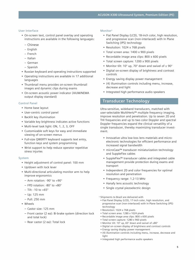
User Interface
• On-screen text, control panel overlay and operating instructions are available in the following languages:
– Chinese – English
– French
– Italian
– German
– Spanish
• Russian keyboard and operating instructions supported
• Operating instructions are available in 17 additional languages
• Thumbnail menu provides on-screen thumbnail images and dynamic clips during exams
• On-screen acoustic power indicator (AIUM/NEMA output display standard)
Control Panel
• Home base layout
• User-centric control panel
• Backlit key illumination
• Variable key brightness indicates active functions
• Multi-level task light: ON, 1, 2, 3, OFF
• Customizable soft keys for easy and immediate viewing of on-screen menus
• Full-size QWERTY keyboard supports text entry, function keys and system programming
• Wrist support to help reduce operator repetitive stress injuries
System
• Height adjustment of control panel: 100 mm
• Up/down with lock lever
• Multi-directional articulating monitor arm to help improve ergonomics
– Arm rotation: -90° to +90°
– FPD rotation: -80° to +80°
– Tilt: -10 to +65°
– Up: 125 mm
– Pull: 250 mm
• Wheels
– Castor size: 125 mm
– Front castor (2 ea): Bi-brake system (direction lock and total lock)
– Rear castor (2 ea): Total lock
Monitor1
• Flat Panel Display (LCD), 18-inch color, high resolution, and progressive scan (non-interlaced) with In Plane Switching (IPS) technology
• Resolution: 1024 x 768 pixels
• Total screen area: 1400 x 900 pixels
• Recordable image area clips: 800 x 600 pixels
• Total screen capture: 1200 x 900 pixels
• Monitor tilt: 10° up, 70° down and swivel of ± 90°
• Digital on-screen display of brightness and contrast controls
• Energy saving display power management
• (4) illumination controls including menu, increase, decrease and light
• Integrated high performance audio speakers
Ultra-sensitive, wideband transducers, matched with user-selectable MultiHertz™ multiple frequency imaging, improve resolution and penetration. Up to seven 2D and THI frequencies and up to two color Doppler and spectral Doppler frequencies expand the clinical versatility of a single transducer, thereby maximizing transducer invest- ment.
• Innovative ultra low-loss lens materials and micro-
electronic technologies for efficient performance and increased signal bandwidth
• microCase™ transducer miniaturization technology and SuppleFlex cables
• SuppleFlex™ transducer cables and integrated cable management provide protection during exams and transport
• Independent 2D and color frequencies for optimal resolution and penetration
• Frequency range: 1.2-13 MHz
• Hanafy lens acoustic technology
• Single crystal piezoelectric design
1 Shipments to Brazil are delivered with • Flat Panel Display (LCD), 17-inch color, high resolution, and
progressive scan (non-interlaced) with In Plane Switching (IPS) technology
• Resolution: 1024 x 768 pixels • Total screen area: 1280 x 1024 pixels • Recordable image area clips: 800 x 600 pixels • Total screen capture: 1280 x 960 pixels • Monitor tilt: 10° up, 65° down and swivel of ±80° • Digital on-screen display of brightness and contrast controls • Energy saving display power management • (4) illumination controls including menu, increase, decrease and
light • Integrated high performance audio speakers
Transducer Technology
ACUSON X300 Ultrasound System, Premium Edition (PE)
6
• Universal, stainless steel and disposable biopsy
guides for specified linear and curved array trans- ducers
• Speciality stainless steel and disposable needle guide attachments for the BP9-4 transducer with Live Bi-Plane
Specialty Transducers
The ACUSON X300 PE supports a range of transducers and capabilities, including intra-operative, TEE and Intra- cardiac Echocardiography transducers. For more detailed transducer information, please consult the Transducer Flyer.
Intracardiac Echocardiography Transducers
• ACUSON AcuNav 10F ultrasound catheter – Adult intracardiac echocardiography
• ACUSON AcuNav 8F ultrasound catheter – Adult intracardiac echocardiography
• CartoSound™ Communication – Adult intracardiac echocardiography with SoundStar™ 3D catheter+
Note: See dedicated transducer flyer for more information.
Transducer Ports
• (3) active transducer ports that support high density phased array, curved array and linear array transducers
• Electronic transducer selection (instantaneous switching between transducers)
• Leftmost transducer port supports specialty trans- ducers including the TEE transducer using micro- pinless (MP) adapter, mechanical 3D/4D transducer and Live Bi-Plane transducer
• Industrial design provides easy access to the transducer ports
Transducer Storage
• (6) configurable transducer holders support all transducer designs and provide gel bottle storage
• Special transducer holder provides secure storage and easy access to endocavity transducer
• Transducer holders can be removed for cleaning
• Optional side pocket storage for (1) transducer connector and (1) standard gel bottle
Hard Drive
• Internal 300 GB hard drive
• Allows storage of patient studies that include images, clips, reports and measurements
• Image storage capacity up to 150,000 images with compression (B/W and color)
• For more detailed information, please consult the Digital Patient Study Storage and Archiving section on page 12.
Physiological Interface
• Standard 3-lead ECG interface
• Auxillary Input for ECG and other physiology signals from third party devices
• Continuous display in all real-time modes
• R-Wave single and dual trigger function
• Respiratory trace
• Heart rate display
• Adjustable gain and trace position on-screen
• Recovery Time: Less than 2 seconds
2D
• Fundamental 2D
• Phased THI
• Filtered THI
Color
• Velocity-based color Doppler
• Power Doppler
• Directional power Doppler
• Color M-mode
• Color Doppler tissue imaging
Spectral Doppler
• Pulsed wave
• Steerable continous wave (SCW)
• Continuous wave – pencil probe
• Spectral Doppler tissue imaging
• Duplex and Triplex modes
M-mode
• Color M-mode
• Anatomical M-mode
• ECG trace in all modes
* For purchase or inquires, contact Biosense Webster: USA (1-909-839-8500 and 1-800-729-9010), Belgium (+32-2-352-1411).
Operating Modes
ACUSON X300 Ultrasound System, Premium Edition (PE)
7
• Selectable split screen display formats in 2D or 2D/color with M-mode and/or spectral Doppler mode: top-bottom or side-by-side in real-time and digital cine replay
• 4B-mode allowing simultaneous display of 4 static B-mode images
• Virtual Format
• Dual from freeze
• Split/Zoom
• Live Bi-Plane
Flexible combination of imaging modes in side-by-side Dual and Dual Select in real-time, and digital cine replay.
2D-mode Image Processing
• All-digital parallel signal processing with frame rates up to 1172 fps (transducer dependent)
• MultiHertz imaging: Up to 7 user-selectable transmit frequencies
• Res/Speed selection: 6 levels
• Persistence: 5 levels
• Edge enhancement: 4 levels
• Dynamic range selection: 30-90 dB in 5 dB increments
• Gain: 0-60 dB in 1 dB increments
• DTO technology: 3 levels
• Dynamic TCE technology: 3 levels
• Depth/Gain Compensation: 8 controls
• User-selectable gain balance: 9 maps
• User-selectable 2D colorization: 16 maps
• Maximum Display Depth: 30 cm
• Minimum Display Depth: 2 cm
MultiHertz Multiple Frequency Imaging
Siemens’ unique MultiHertz multiple frequency imaging is designed to combine the resolution and penetration of several transducers into one. At the push of a button, the user can independently change frequencies for 2D, THI, color and spectral Doppler to select the optimal combina- tion for image resolution, penetration and sensitivity.
• Transmit frequencies: Up to 7 user-selectable
frequencies
– 2D and M-mode: Up to 4 frequencies
– THI: Up to 5 frequencies
– Color, power, or pulsed wave Doppler modes: Up to 2 frequencies
– SCW Doppler mode: 1 frequency
Tissue Harmonic Imaging (THI)
Selectable harmonic frequencies increase success with difficult-to-image patients, improving diagnostic confi- dence, and dramatically improving contrast and spatial resolution by reducing noise and clutter in the image.
• MultiHertz multiple frequency imaging capability
in THI
• Available on the BP9-4, CH5-2, C6-2, C7F2, C8-5, P8-4, P4-2, P5-1, EC9-4, EV9-4, EV9F4, P9-4, VF8-3, VF10-5, VF13-5SP, VF13-5 and V5M transducers
Focusing
• Transmit focal zones: Up to 4 zones
• Digital dynamic receive focusing with dynamic apodization
• Multi-position, user-selectable position
• Can use multiple focal zones simultaneously
SynAps Synthetic Aperture Technology
• SynAps™ synthetic aperture technology is available on the CH5-2, C6-2, C7F2, C8-5, VF8-3 and VF10-5 transducers
• Higher image resolution at depth
• User can turn SynAps technology On and Off
2D Image Display
• Full screen, Split, Quad and Dual Select screen formats as well as Dual, Dual seamless, Dual select and Dual from Freeze
• Curved Vector format
• L/R flip and U/D flip for all formats in real-time and digital cine replay
• Split/Zoom
• Image depth: 2-30 cm in 1.0 cm increments (transducer dependent)
• Virtual Format Imaging (transducer dependent)
– Left/right steer
– Trapezoid Imaging
• Digital read/write Zoom with image pan
– Available on live and cine replay images
– At least 2.5x and up to 10x zoom (transducer dependent)
• 4B-mode
• Live Bi-Plane: Displays both longitudinal and trans- verse views simultaneously in real-time 2D imaging (transducer dependent)
• 90° image rotation (all linear transducers)
Display Modes

ACUSON X300 Ultrasound System, Premium Edition (PE)
8
2D Calipers – Generic Measurements and Calculations
• Multiple cursor sets on frozen, live, dual screen and cine playback images
• Distance measurements: Up to 8 measurements per screen
– Distance measurement
– Depth measurement from skin line
– Angle measurement
– Area and circumference: ellipse, trace
• Compound Measurements
– Volume: user-selectable preset by 1 distance, 2 distance, 3 distance, or 1 ellipse and 1 distance
– Flow volume: 1 velocity and 1 distance, or 1 velocity and 1 ellipse
– Stenosis: user-selectable preset calculated by 2 ellipse, or 2 distance measurements
Color Doppler Velocity Imaging
• Available on all imaging array transducers
• Multi-beam formation technology provides quad signal processing for color Doppler frame rates up to 171 fps (transducer dependent)
• Transmit frequencies: Up to 2 user-selectable frequencies per transducer
• Left/right steer on all linear transducers
• Color Doppler invert
• Advanced processing in color Doppler mode resulting in excellent spatial resolution and superior Flash sup- pression
• AutoColor flow state optimization with high, medium and low flow settings
• Color Doppler velocity maps: Up to 9 user-selectable maps (7 velocity and 2 velocity/variance)
• Velocity scale range: ±0.6 - ±244.4 cm/sec
• PRF scale range: 100-19,500 Hz (transducer dependent)
• Gain: -20-20 dB in 1 dB increments
• Color Doppler line density: 6 selections
• Wall filter: 4 selections
• Color smoothing: 4 levels
• Tissue/color priority: 5 selections
• Color Doppler persistence: 5 levels
• Velocity tag
• Peak hold: Off, 1 sec, 2 sec and 3 sec
• DTI Doppler tissue imaging capability available on select transducers
Power Doppler Imaging/Directional Power Doppler
• Available on all imaging array transducers
• Multi-beam formation technology provides quad signal processing for power Doppler frame rates up to 170 fps (transducer dependent)
• Left/right steer on all linear array transducers
• Transmit frequencies: Up to 2 user-selectable frequencies per transducer
• Power Doppler maps: Up to 16 maps (8 directional and 8 non-directional)
• PRF scale range: 100-9,500 Hz (transducer dependent)
• Gain: -20-20 dB in 1 dB increments
• Power Doppler line density: 6 selections
• Wall filter: 4 selections
• Power Doppler smoothing: 4 levels
• Tissue/power Doppler priority: 5 selections
• Color persistence: 5 levels
Color and Power Doppler Display
• 2D/C mode, Split 2D-2D/C mode
• Dual real-time 2D/C mode
• 2D/C/D mode (simultaneous triplex), 2D/C/D mode (update)
Pulsed Wave Spectral Doppler
• Available on all imaging array transducers
• Transmit frequencies: Up to 2 user-selectable frequencies per transducer
• DTI Doppler tissue imaging available on select transducers
• Sweep speed: 8 selections
• Post-processing gray maps: 8 maps
• Doppler colorization maps: 12 user-selectable maps
• Gain: 0-90 dB in 1 dB increments
• PRF range: 100-19,500 Hz
• Velocity scale range: ±1.5 - ±350 cm/sec with 0° angle correction
• Angle correction: 0-89° in 1° increments
• Gate size: 0.2-20 mm
• Wall filter: 8 selections (transducer dependent)
• Baseline shift: 17 levels
• Spectral invert
• Autotrace function

ACUSON X300 Ultrasound System, Premium Edition (PE)
9
Steerable Continuous Wave (SCW) Doppler
• Available on all phased array transducers when Advanced Cardiovascular option is purchased
• Transmit frequency: 1 frequency
• Sweep speed: 8 selections
• Post-processing gray maps: 8 maps
• Doppler colorization: 12 maps
• Gain: 0-90 dB in 1 dB increments
• PRF range: 1.56-34.7 kHz sample rate
• Velocity scale range: ±30 - ±650 cm/sec with 0° angle correction
• Wall filter: 8 selections (transducer dependent)
• Baseline shift: 17 levels
• Spectral invert
• Autotrace function is not supported in SCW mode
Continuous Wave Doppler
• Supported on the CW2 and CW5 transducers
Spectral Doppler Display
• Full screen Doppler trace, 2D/Doppler mode, triplex or update 2D/C/Doppler
• Imaging display: 4 formats
– Top-bottom: 1/3-2/3, 1/2-1/2, 2/3-1/3
– Side-by-side: 40-60
Spectral Doppler Calipers – Generic Measurements and Calculations
• Multiple cursor sets on frozen and cine playback images
• Velocity/Frequency/Pressure Gradient
• Heart rate/Heart cycle/Time
• Autotrace measurements in real-time and freeze including calculations for PS, ED, TAMx, TAMn, PI, RI and S/D
• Resistive Index (RI)
• Pulsatility Index (PI), including Peak-to-Peak method
• Time Average Velocity max (TAV)
• Systolic/diastolic ratio (S/D)
• Velocity Time Integral (VTI)
• Acceleration/Deceleration
• Flow volume using combined velocity and distance, or velocity and ellipse measurements
• Doppler angle correction after measurement
M-mode
• Available on all imaging array transducers
• Anatomical M-mode – live, cineloop and DIMAQ image review
• Frequencies: Up to 5 user-selectable frequencies, including fundamental and harmonics
• Edge enhancement: 4 selections
• Display dynamic range: 30-70 dB in 5 dB increments
• Gain: 0-60 dB in 1 dB increments
• Gray maps: 9 maps
• M-mode colorization maps: 16 maps
• Sweep speed: 8 selections
M-mode Image Display
• Full screen M-mode, 2D/M-mode
• Imaging display: 4 formats
– Top-bottom: 1/3-2/3, 1/2-1/2, 2/3-1/3
– Side-by-side: 40-60
M- mode Calipers – Generic Measurements and Calculations
• Multiple cursor sets on frozen and cine playback images
– Distance
– Time
– Slope
– Heart rate
ACUSON X300 Ultrasound System, Premium Edition (PE)
10
QuickSet User-Programmable System Parameters
All imaging modes and parameters are customizable and programmable using QuickSet™ user-programmable system parameters.
• Up to 32 QuickSet parameters supported
• QuickSet parameters combine all preferred imaging mode parameters, annotation, text and measure- ments into a single user preset
Cine Review
The cine feature is standard and offers the ability to review real-time acquired data. All real-time, post-acquisi- tion optimization functions are available in cine review.
• Frame-by-frame cineloop review and continuous cine
motion review, including control of playback rate
• Independent cine review in mixed modes (2D/M, 2D/Doppler, 2D/C/Doppler)
• Independent cine review in 2D Dual Select mode with image align playback feature
• Cine memory: 256 MB
• Cine store capacity: Up to 2729 frames
• Acoustic clip capture from cine review
• Anatomical M-mode available
Post-Processing Features in Freeze Frame or Cine
• 2D-mode
– Zoom/pan
– Gray map
– Colorization map
– Measurements/reports/annotations/pictograms
• Color Doppler
– Zoom/pan
– Color map
– Color invert
– Measurements/reports/annotations/pictograms
• Spectral Doppler
– Gray map
– Doppler colorization map
– Angle correct
– Measurements/reports/annotations/pictograms
• M-mode
– Gray map
– M-mode colorization map
– Measurements/reports/annotations/pictograms
The ACUSON X300 PE is designed to support many multi-specialty imaging applications. Factory-supplied exam and transducer dependent imaging presets have been carefully optimized for each application to provide consistency, reliability and increased productivity. All applications include body markers, text and annotation labels. Selected applications support customized work- sheets and reports. The following study types are supported:
• Abdominal
• Renal
• Obstetrics
• Gynecology
• Early Obstetrics
• Adult Cardiac (Transthoracic)
• Pediatric Cardiac (Transthoracic)
• Neonatal Echo
• TEE Adult
• Intracardiac Echo (ICE)
• Vascular (C-Vas, P-Vas, Venous)
• Small Parts (Breast, Testicle, Thyroid)
• Orthopedics
• Musculoskeletal
• Urology (Prostate)
• Cranial (TCI)
• Emergency Medicine (EM)
• Penile
• Pediatric Abdomen
• Neonatal Head
• Lithotripsy
All measurement and report packages are available for use with all exam types.
All exam-specific measurement and reports support:
• All general measurements and calculations
• Comprehensive, customizable, patient reports and worksheets
• Customizable Anatomy Descriptions
• Physician summary utility – supports on-system report generation including customizable letterhead, patient data, results, graphs, images, comments, recommen- dations and a customizable signature line
Exam-Specific Measurements and Reports
Study Types
Freeze, Cine and Cine Post-Processing Functions
ACUSON X300 Ultrasound System, Premium Edition (PE)
11
The following Measurements and Reports packages are available on the ACUSON X300 PE:
Abdomen
Obstetrics
• Early Obstetrics Gestational Age (GA) measurements are MSD, CRL, and Yolk Sac
• Gestational Age parameter labels are MSD, CRL, BPD, OFD, HC, AC, ATD, ASD, FL, HL, UL, TL, FT, FTA and BN
• 20 user-defined measurement labels
• Calculations include: EFW from the selected reference, HC/AC, TCD/AC, LVW/HW, CorBPD, FL/AC, FL/BPD, CI, AFI, AXT
• Comprehensive Fetal Heart measurements and calculations
• Facial Angle
– Nuchal Translucency and Nuchal Fold measurements
• Calculations for both Gestational Age (GA) and Estimated Date of Confinement (EDC)
• Early OB and Standard OB patient reports include worksheets for viewing the progress of the report and editing during the exam process
• Multiple fetus reporting capabilities
– Maximum of 4
• Growth Analysis Graphs with exam file linking
• Detailed Fetal Heart report page
Gynecology
• Micturated and residual volume calculation
• Uterus, Right and Left Ovary, Right and Left Follicle, CRL, MSD, GS and Yolk Sac measurements
• Follicle Measurement supports up to 15 follicles
• Follicles measurement methods supported
– Distance
– 2Dist + Avg
– 3Dist + Avg
– 2Dist Avg
– 3Dist Avg
– Area
– Volume
– Circumference
Cardiac
• Adult and pediatric standard measurements
• Volume formulas for Left Ventricular function assess- ment in 2D-mode and M-mode
• 2D-mode, M-mode and Doppler calculations
• M-mode Slope, Heart Rate, Time and Distance measurements
• Spectral Doppler Acceleration, Trace, Heart Rate, Time and Velocity measurements
• Cardiac patient report and worksheet for 2D-mode, M-mode and spectral Doppler
Cerebrovascular
• CCA prox, CCA mid, CCA dist, ICA prox, ICA mid, ICA dist, ECA and VA measurements
• Area Percent Stenosis and Diameter Percent Stenosis measurements
Peripheral Vascular
• CIA, EIA, CFA, PFA, SFA prox, SFA mid, SFA dist, POP A, TRUNK ATA, PTA, PER A and DPA measurements
• Right and left extremity measurements
Venous
• Right and left extremity measurements
Thyroid
• Rt. Lobe, Lt, Lobe Isthmus and nodule measurements
• Supports measurement of up to 15 separate nodules
• Measurements can display all 3 measurements plus volume calculation
ACUSON X300 Ultrasound System, Premium Edition (PE)
12
Urology
• Pre- and post-void volume, Prostate volume, PSAD and micturated volume calculations
• Includes both Prostate and Urology report packages
Testicle
Orthopedic
• Right and left hip angle measurement
• Classification and Graf Sonometer
• Hip angle patient report
TCI
• MCA, ICA-Siphon, ACA-A1, ACA-A2, ACoA, PCA-P1, PCA-P2, PCoA, PCA, Basilar A and Vert A measurement
Penile
• Corp Cav, Corp Spong, Cav A, Pre-Inj Cav A, Post-Inj Cav A and Urethra B mode measurement
• Iliac A, Dorsal A, Urethral A, Bulbar A, Brach A, Cav A, Pre-Inj Cav A, Post-Inj Cav A, Sup Dorsal V and Dp Penile V D mode measurement
Emergency Medicine
• FAST – Focused Assessment with Sonography for Trauma reporting
• Aorta – Essential aorta measurements and reporting to support emergency medicine
• OB – Subset of essential OB measurements and reporting
• Supports compatibility with Telexy Q-path2 Ultra- sound Workflow Manager. Please see Digital Patient Study Storage and Archiving for more information.
The ACUSON X300 PE supports customizable labeled measurements (20 B-mode, 5 Doppler and 5 M-mode) for the following exam types: Abdomen, Musculoskeletal, Breast, Thyroid, Testicle, Venous, Renal, Pediatric Abdomen, Neonatal Head, Superficial Musculoskeletal, Digital, Small Parts and Aorta. Additionally, all reporting packages support customizable user-defined labels (up to 39) and descriptors.
The DIMAQ-IP integrated workstation allows for digital acquisition, storage and review of complete ultrasound studies, including static images, dynamic clips, measure- ments, calculations and reports.
2 Sold separately by Telexy Healthcare and requires a wireless or LAN connection
Studies can be reviewed and quantified on-board, stored on the system hard drive and transferred to the built-in DVD multi-drive (DVD-R/RW & CD-R/RW) or USB Flash drive for cost-effective archival.
Patient Study Management
Playback of digitally stored images in a selectable 1-up, 4-up, 9-up, 16-up or 25-up screen format. The patient study screen allows search, selection and deleting of studies or export to DVD multi-drive (DVD-R/RW and CD-R/RW).
• 100 GB of the 160 GB internal hard drive reserved for
patient data management
– Patient database sorting by Name, ID and Study Date
• Hard drive capacity:
– Approximately 150,000 B/W and color images
• Storage and retrieval of static images
• Storage and retrieval of cine clips
– Retrospective clip capture during real-time imaging with a selectable duration of 1, 2, or 4 seconds or a selectable duration of 1, 2, 3 or 4 beat capture; ECG-triggerable
– Prospective clip capture during real-time imaging with a selectable duration of 1 to 120 seconds or a selectable duration of 1 to 120 beat capture; ECG triggerable
• Export of patient studies from hard drive
• Storage and retrieval of reports
• Supports measurements and calculations on archived study and on saved and retrieved images
• Acoustic clip capture from cine review
• M-mode still frame scroll and store
• PW spectral Doppler still frame scroll and store
• Export of patient studies from hard drive to DVD-R/RW and CD-R/RW drive. Studies can be indi- vidually selected or batched copied
• The system supports the following data export file formats RTF, PDF, TIFF, AVI, JPG and DICOM. Connec- tivity to PACS, other off-line storage (such as USB flash drive) or EMR device is achieved via LAN or WLAN connection.
• Compatible with removable 650 MB, 700 MB and 790 MB CD-R and 650 MB or 700 MB CD-RW
• Removable 4.7 GB single layer DVD and 8.5 GB single side double layer DVD
• Supports export to USB Flash Drive
Supports compatibility with Telexy Q-path2 Ultrasound Workflow Manager for point-of-care reporting, quality assurance, billing, research, teaching and credentialing.
Digital Patient Study Storage and Archiving
ACUSON X300 Ultrasound System, Premium Edition (PE)
13
PLUS (Option)
The Plus Option is a beamforming hardware option that improves image quality, penetration and sensitivity for high density transducers on the ACUSON X300 PE. The following transducers deliver enhanced performance with installation of the Plus Option: BP9-4, C7F2, EV9F4, C6-2, CH5-2, VF8-3, VF10-5, C8-5, VF13-5 and VF13-5SP.
Integrated Gel Warmer (Option)
• Temperature control precision: ± 1°
– Low: 31° C
– Medium: 34° C
– High: 37° C
• Easy-to-use Power on/off control switch
• LED color for status indicator
– Standby mode: Off
– Operating mode: Orange
• Safety protection for electrical overload
• Weight: ~ 385 g
• Size: 87 mm x 170 mm x 85 mm
QuikStart (Option)
QuikStart reduces time required for power-up and power-down events by allowing the system to enter a specialized suspend mode.
• Standby mode power-down in 3 sec; complete
boot-up in less than 12 sec
• Standby time: > 30 min
• Full recharge: < 180 min
Control Panel Cover (Option)
• Allows for easy cleaning and disinfection
• Single piece with DCG access
• Individual knob covers for full rotary control and complete coverage
• Soft molded silicone cover allows maximum func- tionality
Dynamic TCE Technology (Option)
Dynamic TCE technology is an advanced method for speckle reduction and edge enhancement delivering improved contrast resolution. Three levels are available: low, medium and high.
Tissue Grayscale Optimization (TGO) (Option)
TGO tissue grayscale optimization technology provides one-button image optimization. It automatically adjusts image brightness to the tissue type being imaged and equalizes the overall image gain. The user-definable threshold accommodates different user preferences for gain settings and various room lighting conditions. TGO technology improves the consistency and quality of the ultrasound images to enhance productivity by removing time-consuming and operator dependent manual adjustments. TGO technology can be used with every transducer, for every exam type and at every imaging frequency, including THI.
SieClear Multi-View Spatial Compounding (Option)
The SieClear compounding option uses multiple lines of sight to increase contrast resolution and improve tissue differentiation of low contrast lesions by reducing image speckle. Tissue boundaries and interfaces appear sharper and more continuous. SieClear compounding is accessible in THI and is compatible with other advanced imaging options including SieScape imaging, TGO ultra- sound technology and Clarify vascular enhancement technology.
Clarify Vascular Enhancement (VE) Technology (Option)
Clarify VE technology is a real-time, adaptive, pixel-by- pixel analysis implemented through a simple, time-saving user interface that provides multiple levels of clarification to optimize tissue contrast resolution and definition of both tissue and vessel walls according to user preference. Clarify VE technology is available on all curved and linear transducers.
SieScape Panoramic Imaging (Option)
Grayscale panoramic imaging allows acquisition and display of images up to 60 cm in length to a maximum curvature of 360°.
• Available on all curved and linear transducers
• Can be displayed in full field acquisition or zoomed for detail viewing
• Measurements available
Options

ACUSON X300 Ultrasound System, Premium Edition (PE)
14
3D/4D Options
• 3-Scape™ real-time 3D imaging
– Provides a freehand acquisition technique
– Supported on the CH5-2, C6-2 and EV9-4 transducers
• fourSight 4D Transducer Technology
– Provides real-time 3D images
– Utilizes mechanical acquisition
– Up to 30 vol/sec
– C7F2 and the EV9F4 4D transducers supported
– Offers an easy-to-use interface for rapid acquisition and volume rendering Curved Top VOI
– 4D cine
– MPR Measurements
• Advanced fourSight Technology
– Offers enhanced 3D/4D acquisition, data rendering and post-processing functionality
– MultiSlice format allows the user to select range, slice spacing and format for viewing each slice. The MultiSlice format supports up to 36 slices at once.
– Thick Slice Imaging (TSI) enables definition of a view plane and creates a thick slice around the region of interest. TSI delivers improved contrast resolution and provides more information in a single image.
– Curved MPR enables real-time multiplanar reformatting of images into any linear or curved plane. This permits the user to set points along a curved object in order to bring all objects along this line into the same plane for viewing, such as the fetal spine.
Advanced Cardiovascular (Option)
Contains the prerequisites to perform cardiac and certain vascular exams:
• Physiological interface
– Standard 3-lead ECG interface
– Aux ECG interface
– R-Wave single and dual trigger function
– Heart rate display
– Adjustable gain and trace position on screen
– Selection for external ECG input
– Respiratory function
• Steerable Continuous Wave Doppler module
• Auxiliary Continuous Wave Doppler module
Spectral and Color Doppler Tissue Imaging (DTI) (Option)
• Supports both color DTI and spectral DTI functionality and includes a quantification package
• Enables assessment of LV diastolic function on the ACUSON X300 PE
– Spectral Doppler DTI capability utilizes real-time Doppler shift information from moving tissue to better visualize and quantify myocardial diastolic function
– The spectral Doppler DTI calculation package provides guided velocity and acceleration measure- ments and includes a measurement report package
– Color DTI capability can be used for qualitative evaluation of wall motion and displays the relative change of velocities
– Color DTI M-mode
– DTE (Doppler Tissue Energy)
Advanced Physio Module (Option)
• Provides the ability to configure ECG capabilities for specialty applications that do not require Continuous Wave Doppler capabilities
• AUX ECG and Respiratory functions are supported in addition to conventional Physio Module
CartoSound Communication (Option)
This license enables Ethernet communication between the ACUSON X300 PE and the Biosense Webster CartoSound™ system.
syngo Auto OB Measurements (Option)
The syngo Auto OB measurements option contains an innovative algorithm which provides the ability to auto- matically measure the most common fetal structures required for fetal biometry: BPD, HC, AC, FL and HL. This technology reduces user variance, providing consistency between operators and reduces keystrokes. All measure- ment results are saved to the OB report.
Stress Echo Imaging (Option)
The stress echo package provides tools for ECG triggered acquisition, display, selection comparison, evaluation and archiving of multiple cardiac loops during various stages of a stress echo examination.

ACUSON X300 Ultrasound System, Premium Edition (PE)
15
• Standard acquisition protocols for treadmill, ergo-
metric and pharmacological stress including:
– Multiple factory default stress echo protocols
– Customizable stress echo protocols
– Flexible combination of imaging modes while in stress echo package
– Ability for customized studies through Protocol Editor, with up to 12 stages, 6 views per stage, 20 loops per view or 120 sec prospective clip capture
– Full screen or ROI (region of interest) acquisition
• Complete R-R capture with clip editing
• Easy workflow throughout the exam protocol
• Stage Timer
• Prospective continuous capture (up to 120 seconds) or Retrospective labeled capture
• Reference image display during acquisition
• Immediate review of acquired loops
• Flexibility to skip views or stages
• Flexibility to re-acquire and overwrite already acquired images
• Indication of current view, acquired views and skipped views in the workflow diagram
• Wall Motion Scoring, 16/17 segment model with graphical display and report printing
• LV Volume Measurements with report printing
syngo fourSight TEE View (Option)
An integrated method which provides acquisition, review, manipulation and dynamic display capabilities of gated 3D datasets using the V5M transesophageal transducer.
• 3D surface (volume) rendering for detailed anatomical
display
• D’Art is an intuitive navigation tool with adjustable indicator for line of sight direction
• Utilizes 2 cut planes positioned perpendicular to the selected line of sight
• Dynamic MultiPlanar reconstruction
• Viewing presentation enhanced by zoom, pan and rotate functions
• Easy to use, rapid acquisition, enhanced workflow
syngo Mitral Valve Assessment (MVA) (Option)
An integrated method which, in conjunction with syngo fourSight TEE view, enables a detailedassessment of mitral valve morphology from 3D images for a more accurate and complete diagnosis of pathology location and size.
Performs measurements for:
• Angle
• Size
• Distance
• Area
syngo Arterial Health Package (AHP) (Option)
syngo Arterial Health Package provides the clinician with the capability to measure Carotid Intima-Media Thickness (CIMT) and the option to reference normative tables that have been validated and published in peer-reviewed studies.3 The information is intended to provide a straight- forward tool for communicating with patients the relative state of their cardiovascular system.
This feature should be utilized according to the ASE Con sensus Statement, “Use of Carotid Ultrasound to Identify Subclinical Vascular Disease and Evaluate Cardiovascular Disease Risk: A Consensus Statement from the American Association of Echocardiography; Carotid IntimaMedia Thickness Task Force, Endorsed by the Society for Vascular Medicine.”
syngo Velocity Vector Imaging (VVI) Technology (Option)
syngo VVI technology visualizes, measures and displays global and regional myocardial motion and mechanics from a 2D image and sophisticated tracking algorithm. Using individual vectors to display direction and relative velocity (based on length of the vectors) of frame-to- frame tissue movement, syngo VVI technology delivers motion measurement at any point in the cardiac cycle. syngo VVI technology provides a unique graphical presentation of the mechanics of myocardial function.
• Algorithm processes ultrasound clips obtained in all
views of the heart, as well as generic moving tissue (e.g. vessel wall)
• Not limited by Doppler angle dependencies, frame rate or mean velocities
• ECG signal is not required, allowing use with fetal echo
• Tracking algorithm incorporates multiple sources of information, including speckle tracking
3 ARIC-G Howard, et al: „Carotid artery intimal-media thickness
distribution in general populations as evaluated by B-mode ultra- sound, ARIC Investigation”: Stroke 1993;24;1297-1304.

ACUSON X300 Ultrasound System, Premium Edition (PE)
16
• Displays include:
– Visual assessment of wall motion and vector dynamics
– Time curves of the selected velocity vector components
– 3D representations of parametric color M-mode
– Selected components of tissue velocity (tissue strain and tissue strain rate)
– Time curves of global and segmental LV volumes and Ejection Fraction
– Synchrony analysis including time-to-peak
• Compatible with standard acquisition frame rate clips and acoustic clip capture (e.g. isovolumetric contrac- tion and relaxation events)
syngo Auto Left Heart (Auto LH) Technology (Option)
• Calculated Confidence
– Uses pattern recognition technology based on a comprehensive database representing typical adult transthoracic exams
– Provides robust performance and expert-like consistency in every exam
– Excels in technically difficult studies as learning is largely independent of image quality
• Automated Workflow
– Bypasses the cumbersome “mark-and-trace” method to calculate EF, end diastole volume and end systole volume
– Automatically identifies ED and ES frames and tracks endocardial borders frame to frame
– Promotes smooth, efficient workflow with manual edit options
– Reduces observer variability to yield more consistent, standardized measurements
• Consistent, Reliable and Convenient
– Extends calculation accuracy to broaden clinical confidence
– Presents EF measurement in complete perspective – with image frames for ED and ES with numerical and graphical data
– Available off the system with syngo® US Workplace
– Operates on DICOM clips from select Siemens and non-Siemens ultrasound systems
Axius Edge Assisted Ejection Fraction (Option)
Axius ejection fraction calculates left ventricle volumes and ejection fractions quickly and reliably.
• Enhances workflow by allowing the user to indicate
three boundary points for measurement instead of a manual trace of the ventricle
• Utilizes the Simpson method for LV volume calcula- tions based on LV endocardial border trace
Intracardiac Echocardiography (ICE) Imaging Options
Adult and pediatric intracardiac echocardiography (ICE) using the ACUSON AcuNav ultrasound catheter family.
• Disposable ACUSON AcuNav 8F ultrasound catheter
– 8 French catheter (cross-sectional diameter 3.3 mm), 110 cm insertable length
• Disposable ACUSON AcuNav 10F ultrasound catheter
– 10 French catheter (cross-sectional diameter 2.7 mm), 90 cm insertable length
• Ultrasound catheters must be purchased separately
• Reusable SwiftLink catheter connector (DL-260 type) (additional SwiftLink connectors are optional)
• 10 sterile covers
• Complete echocardiography exam capabilities during intracardiac minimally invasive procedures, such as atrial fibrillation ablation, transcatheter septal closure device placement, pacemaker lead placement, trans- septal catheterization and balloon valvuloplasty or septostomy
• Visualization of cardiac anatomy and regional myocardial tissue motion, great vessels and vascular anatomy, blood flow direction, blood flow velocity and other devices located within the heart
• Sterile, steerable, single-use catheter
• High resolution, high frame rate imaging in multiple modes, including: 2D, M-mode, PW spectral Doppler, CW spectral Doppler, color Doppler velocity and Doppler tissue imaging (optional)
• Digital phased array technology
• Imaging penetration up to 15 cm allows for visuali- zation of left-sided cardiac anatomy from within the right atrium or right ventricle
• Two planes of bi-directional steering of the catheter tip (160° in each direction: anterior-posterior/left right) for maneuverability, rapid anatomic orientation and micro-positioning
• Longitudinal side-fire imaging provides standard echocardiographic views, similar to TEE, for easier orientation
• QuikSet user-programmable system parameters pro- vide instant image optimization
• Tension control knob for holding the desired catheter curvature
• 64-element digital phased array with simultaneous processing for the highest imaging resolution in all modes

ACUSON X300 Ultrasound System, Premium Edition (PE)
17
• Access to advanced imaging modes provides full
echocardiography capabilities, including:
– 2D imaging
– M-mode
– Pulsed Wave (PW) spectral Doppler
– Continuous Wave (CW) spectral Doppler
– Color Doppler velocity capability
– DTI Doppler Tissue Imaging capability (optional)
– Comprehensive measurements and calculation, including volume, function and hemodynamics
• MultiHertz imaging
• Zoom capabilities
Live Bi-Plane Endorectal Transducer (Option)
Displays both the longitudinal and transverse view simultaneously in real-time 2D imaging.
• BP9-4 transducer only
• Available in biopsy mode
• Indicator to demonstrate point of intersection on biopsy
• Disposable dual needle guide attachments support both straight and angled needle guidance
• Single path reusable stainless steel needle guide
• Transducer starter kit with package of five dual disposable needle guide kits
Cadence Contrast Agent Imaging4 (Option)
Provides excellent detection of the responses from con- trast agent microbubbles, coupled with a high resolution display. The ability to simultaneously detect both the signals returning from the contrast agent and the signals arising from tissue allows the user to switch between a contrast-only display and a tissue-only display for instan- taneous confirmation of contrast presence and position, providing real-time flexibility and enhanced diagnostic confidence.
• Optimized for CH5-2 transducer for abdominal
applications
• MultiHertz imaging provides improved fine-tuning for low-MI contrast investigations
• Integrated burst/reflow control for destruction, reperfusion investigations
• On-screen stopwatch feature
• Frame rate triggering with extended clip capture times of up to 20 minutes
• Burst (bubble destruction) mode
4 At the time of publication, the U.S Food and Drug Administration has cleared ultrasound contrast agents only for use in LVO. Check current regulations for the country in which you are using this system for contrast agent clearance.
Barcode Reader (Option)
• Allows fast and accurate patient information data input
• Easy attachment to USB port
• Supports 2D and 1D patient barcode
• Can scan up to 3 individual barcodes: patient, physician and sonographer
• Inputs the following patient identifying data:
– Patient Name (First and Last)
– Patient ID
– Physician ID
– Sonographer
Veterinary Imaging (Option)
• Required for use of the ACUSON X300 PE for veterinary scanning.
• Includes a veterinary-specific user guide supplement
• Includes “For Veterinary Use Only” labels for the system and transducers
• Supports all transducers except V5Ms (TEE) and the ACUSON AcuNav 8F and 10F ultrasound catheters
Modularis SW Interface (Option)
An optional SW interface for use with the Siemens Modularis lithotripter for accurate out-of-plane ultrasound localization of urinary stones.
• Uses a uniquely designed isocentric arm which
enables optimal positioning of the transducer to the patient
• Standard movements of up to -15° relative to the horizontal axis
• Release/brake allows the position to be locked into place or changed
• Used for both under-table and over-table positions.
• Requires the CH5-2 transducer
• Tissue Harmonic Imaging (THI) included
DICOM 3.0 Connectivity (Option)
Enables digital data transfer via a DICOM network for both printing and storage. The ACUSON X300 PE acts as a DICOM Storage Class User and DICOM Print Class User.
Functionality supported:
• Connectivity to PACS system for storage of all digital
images and dynamic clips with patient demographic data
• In-Progress Store during the exam
• Image printing to DICOM color and grayscale printers
• DICOM Storage Commitment

ACUSON X300 Ultrasound System, Premium Edition (PE)
18
• DICOM Exchange Media export to DVD-R/RW and
CD-R/RW
• DICOM Region Calibration
• DICOM interchange media viewer software SHOWCASE®
• Interchange media database that identifies the CD to which the patient study has been burned
DICOM Modality Worklist (Option)
Enables query and direct download of the patient work- list schedule from the Hospital/Radiology Information System (HIS/RIS) to the ACUSON X300 PE, automatically populating the “New Patient” screen with patient demo- graphic information. (Requires DICOM 3.0 Connectivity option.)
DICOM MPPS – Modality Performed Procedure Step (Option)
Enables automatic exchange of Modality Performed Procedure Step information with the Hospital/Radiology Information System (HIS/RIS). (Requires DICOM 3.0 Connectivity option and DICOM Modality Worklist option.)
DICOM Structured Reporting (Option)
DICOM Structured Reporting (SR) provides a standardized report architecture to allow for easy transfer of measure- ments to offline PCs, workstations and archiving systems. DICOM Structured Reporting will automatically populate measurements to their respective fields in an external software package. (To send the SR data over the network, the DICOM 3.0 connectivity option is required.) This option is available for the following applications:
• OB/GYN
• Vascular
• Cardiac
Wireless Data Transfer (Option)
Utilizes USB dongle to enable wireless connectivity between the ultrasound system and the facility’s LAN to provide functionality equivalent to a wired network.
The Wireless Option supports connectivity with:
• DICOM services - Modality worklist, print, storage commitment and store
• Siemens Remote Service – Remote update handling for storage distribution and NetViewer for remote application support and remote trouble-shooting
• Telexy Q-view5
5 Sold separately by Telexy Healthcare and requires a wireless or LAN connection
Technical Specification
• Standards : IEEE 802.11n, 802.11g, 802.11b, 802.11a
• Security features: WEP, WPA, WPA2 personal, WPA and WPA2 Enterprise
Ultrasound System Security – Virus Protection (Option)
Embedded virus protection solution that protects the system against advanced persistent threats, viruses, malware and other executing software by detecting and preventing any unwanted change to improve IT compli- ance and security.
Optional On-Board Video Devices
• Up to 2 documentation devices (B/W printer and color printer/DVD recorder) can be integrated into the system cart and controlled from the system control panel
• Supported devices:
– Mitsubishi USB P95DW B/W Printer
– Sony UP-D897 B/W Printer
– Mitsubishi USB CP30DW Color Printer
– Sony UP-D25MD Color Printer
– TEAC DVR UR-50BD-S (NTSC/PAL switchable)
– Mitsubishi S-VHS VCR NTSC version (MD3000UM)
– Mitsubishi S-VHS VCR PAL version (MD3000E)
Video Standard
• PAL/CCIR: 625 lines, 50 Hz
• NTSC/EIA: 525 lines, 60 Hz
Video/Audio Input
• (1) Composite color Video in, BNC-type
• (1) Y/C Video in, S-terminal (SVHS)
• (1) 2-Channel Audio in (Right/Left), RCA jack type
Video/Audio Output
• (1) Composite B/W Video out, BNC-type
• (1) Composite color Video out, BNC-type
• (1) RGB and Composite Sync out, mini D-SUB (15 pin)
• (1) Y/C Video out, S-terminal (SVHS)
• (1) 2-Channel Audio (Right/Left), RCA jack type
• (1) VGA out, mini D-SUB (15 pin)
System Input/Output
Documentation Devices
ACUSON X300 Ultrasound System, Premium Edition (PE)
19
Other Input/Output
• (1) Foot switch connector, phone jack-type
• (1) Remote control connector, mini-jack (stereo)
System Interface Connections
• Network
– (1) Ethernet connector, type RJ45 (10/100 BaseT)
• Peripherals
– (2) Serial port RS-232C connector, D-SUB (9-pin)
– (2) USB 2.0 ports
– (2) AC Main Outlet
Acoustic Output Management
• On-screen acoustic power indicator (AIUM/NEMA output display standard)
• Height: 138.3-163.6 cm (54.45-64.41 in)
• Width: 51.8 cm (20.4 in)
• Depth: 88.0 cm (34.6 in)
• Weight: 102 kg (225 lbs)
– 98 kg (216 lbs) without OEM’s
The ACUSON X300 PE is available in one mainframe configuration, suitable for use in 100/115 V and 230 V environments.
• Power connections: 100-120/200-240 VAC, 50/60 Hz
• Built-in AC isolation transformer
• Power consumption: maximum 600 VA with OEM’s
• Atmospheric pressure range: 700-1060 hPa (525-795 mm Hg) or up to 3000 m (9,483 ft)
• Ambient temperature range (without OEM’s): 10-40° C (50-104° F)
• Humidity: 30-80%, non-condensing, during operation
• Maximum heat output: 2150 BTU/hr
• Average noise level output: 46-47 dB
Having all relevant information at one’s fingertips is a prerequisite for optimal and efficient patient care. Seam- less integration of the hospital’s IT and Imaging Systems and their capabilities to exchange information without restriction are key success factors for facilitating daily work. This is why Siemens has been instrumental in launch- ing and advancing the IHE (Integrating the Healthcare Enterprise) Initiative. Our commitment and dedication enable us to provide clinicians with the ACUSON X300 PE one of many innovative products embedded with the building blocks necessary in supporting clinicians’ need for seamless health information exchange.
For more information on the ACUSON X300 PE and the Siemens commitment to the IHE initiative, please visit www.siemens.com/IHE.
The ACUSON X300 PE is in compliance with the following standards, including all applicable amendments at the time of product release.
Quality Standards
• FDA QSR 21 CFR Part 820
• ISO 9001
• ISO 13485
Design Standards
• UL 60601-1
• CSA C22.2 No. 601-1
• EN 60601-1 and IEC 60601-1
• EN 60601-1-1 and IEC 60601-1-1
• EN 60601-1-2 and IEC 60601-1-2 (Class B)
• EN 60601-2-18 and IEC 60601-2-18
• EN 60601-2-37 and IEC60601-2-37
• EN 60601-2-25 and IEC 60601-2-25
• EN 60601-1-4 and IEC 60601-1-4
• EN60601-1-6 and IEC 60601-1-6
• ISO 14971
• EN 62304 and IEC 62304
Standards Compliance
Integrating the Healthcare Enterprise (IHE)
Electrical/Environmental Specifications
System Dimensions

ACUSON X300 Ultrasound System, Premium Edition (PE)
20
Acoustic Output Standards
• IEC 61157 (Declaration of Acoustic Power)
• IEC 62359 (Test Methods for the Determination of TI and MI)
• AIUM/NEMA UD-2, Acoustic Output Measurement Standard for Diagnostic Ultrasound
• AIUM/NEMA UD-3, Standard for Real-time Display of Thermal and Mechanical Acoustic Output Indices on Diagnostic Ultrasound Equipment
This product is provided with a CE marking in accordance with the regulations stated in Council Directive 93/42/EEC of June 14, 1993 concerning Medical Devices. The CE marking
only applies to medical devices that have been put on the market according to the above referenced Council Directive. Unauthorized changes to this product are not covered by the CE marking and the related Declaration of Conformity.
Siemens AG Healthcare GmbH Henkestrasse 127 91052 Erlangen Germany Phone: +49 9131 84 0
Siemens reserves the right to modify the design and specifications contained herein without prior notice. Please contact your local Siemens Healthcare Sales Representative for the most current information.
EU Authorized Representative
CE Declaration

ACUSON X300 Ultrasound System, Premium Edition (PE)
21

The products/features mentioned in this document may not be commercially avail- able in all countries. Due to regulatory reasons their future availability cannot beguaranteed. Please contact your local Siemens organization for further details.
3-Scape, ACUSON X300, AcuNav, Axius, Cadence, Clarify, DTI, DTO, Dynamic TCE, ErgoDynamic, fourSight, microCase, MultiHertz, Native, QuickSet, SieClear, SieScape, SuppleFlex, SwiftLink, SynAps, TGO and Velocity Vector Imaging are trademarks of Siemens Medical Solutions USA, Inc., and syngo is a trademark of Siemens AG.
CartoSound and SoundStar are trade- marks of BioSense Webster, Inc.
SHOWCASE is a registered trademark of Trillium Technology, Inc.
Microsoft and Windows are registered trademarks of the Microsoft Corporation in the United States and other countries
Q-view is a trademark of Telexy Healthcare.
Siemens Healthcare Headquarters Siemens Healthcare GmbH Henkestraße 127 91052 Erlangen Germany Telephone: +49 9131 84-0 siemens.com/healthcare
Legal Manufacturer Siemens Medical Solutions USA, Inc. Ultrasound 685 E. Middlefield Road Mountain View, CA 94043 USA Telefon: 1-888-826-9702 www.siemens.com/ultrasound
CG US 3474 1015 online | © Siemens Medical Solutions USA, Inc.
siemens.com/ultrasound