Activation of Phasic Pontine-Wave Generator Prevents Rapid Eye Movement Sleep Deprivation-Induced...
12
Behavioral/Systems/Cognitive Activation of Phasic Pontine-Wave Generator Prevents Rapid Eye Movement Sleep Deprivation-Induced Learning Impairment in the Rat: A Mechanism for Sleep-Dependent Plasticity Subimal Datta, Vijayakumar Mavanji, Jagadish Ulloor, and Elissa H. Patterson Sleep Research Laboratory, Department of Psychiatry and Program in Behavioral Neuroscience, Boston University School of Medicine, Boston, Massachusetts 02118 Animal and human studies of sleep and learning have demonstrated that training on various tasks increases subsequent rapid eye movement (REM) sleep and phasic pontine-wave (P-wave) activity, followed by improvement in performance on the learned task. It is well documented that REM sleep deprivation after learning trials blocks the expected improvement in performance on subsequent retesting. Our aim was to test whether experimentally induced P-wave generator activation could eliminate the learning impairment produced by post-training REM sleep deprivation. Rats were trained on a two-way active avoidance-learning task. Immediately there- after, two groups of those rats received a control vehicle (100 nl saline) microinjection and one group received a carbachol (50 ng in 100 nl saline) microinjection into the P-wave generator. The carbachol-injected group and one of the two control saline microinjected groups were selectively deprived of REM sleep during a 6 hr polygraphic recording session. All rats were then tested on the avoidance-learning task. The rats that received both the control saline injection and REM sleep deprivation showed learning deficits compared with the control saline-injected rats that were allowed to sleep normally. In contrast, the rats that received the carbachol microinjection and REM sleep deprivation demonstrated normal learning. These results demonstrate, for the first time, that carbachol-induced activation of the P-wave generator prevents the memory-impairing effects of post-training REM sleep deprivation. This evidence supports our hypothesis that the activation of the P-wave generator during REM sleep deprivation enhances a physiological process of memory, which occurs naturally during post-training REM sleep. Key words: pontine wave; learning and memory; REM sleep; plasticity; carbachol; brainstem; locus subcoeruleus; rat; two-way active avoidance; consolidation Introduction Behavioral studies of learning and memory in both humans and animals provide considerable evidence to support the hypothesis that post-training rapid eye movement (REM) sleep is critical for and is the most favorable behavioral state for memory processing and improvement of learning (for review, see Fishbein and Gutwien, 1977; McGrath and Cohen, 1978; Pearlman, 1979; Smith, 1985, 1995; Dujardin et al., 1990; Karni et al., 1994; Stickgold, 1998; Datta, 2000; Maquet et al., 2003). Other studies have demonstrated that REM sleep is critical for neuronal plasticity, which is a critical mech- anism for memory processing (Bramham and Srebro, 1989; Frank et al., 2001; Campbell et al., 2002; Guzman-Marin et al., 2003). Different classes of memory formation appear to be processed by distinct memory systems in the brain (Cohen and Squire, 1980; Gabrieli, 1998; Kesner, 1998; Kim and Baxter, 2001; White and McDonald, 2002). Many recent studies have shown that the amygdala and hippocampus may be platforms for sleep- dependent memory processing (Pavlides and Winson, 1989; Abel et al., 1997; Ribeiro et al., 1999, 2002; Poe et al., 2000; Abel and Lattal, 2001; Graves et al., 2001; Louie and Wilson, 2001). Al- though the amygdala, hippocampus, and possibly some other parts of the cerebral cortex process acquired information, it is not clear how REM sleep-regulating structures influence the way the hippocampus and amygdala process that information. During REM sleep and in part of slow-wave sleep (SWS), phasic field potentials called pontine waves (P-waves) are gener- ated in the pons (Brooks and Bizzi, 1963; Laurent and Ayalaguer- rero, 1975; Sakai et al., 1976; Datta and Hobson, 1995). These field potentials are a reflection of phasic activation of a specific group of cells in the pons (Sakai and Jouvet, 1980; Datta et al., 1992, 1998; Datta and Hobson, 1994). It has been demonstrated that the functionally identified P-wave generator cells project to the hippocampus and amygdala (Datta et al., 1998). Behavioral studies have shown that sleep-dependent improvement in learn- Received June 18, 2003; revised Dec. 18, 2003; accepted Dec. 19, 2003. This work was supported by National Institutes of Health Grants NS34004 and MH59839. We thank Soma M. Datta for statistical analysis and for writing system software. Correspondence should be addressed to Subimal Datta, Sleep Research Laboratory, Department of Psychiatry, Boston University School of Medicine, M-913, 715 Albany Street, Boston, MA 02118. E-mail: [email protected]. DOI: 10.1523/JNEUROSCI.4111-03.2004 Copyright © 2004 Society for Neuroscience 0270-6474/04/241416-12$15.00/0 1416 • The Journal of Neuroscience, February 11, 2004 • 24(6):1416 –1427
-
Upload
independent -
Category
Documents
-
view
2 -
download
0
Transcript of Activation of Phasic Pontine-Wave Generator Prevents Rapid Eye Movement Sleep Deprivation-Induced...

Behavioral/Systems/Cognitive
Activation of Phasic Pontine-Wave Generator PreventsRapid Eye Movement Sleep Deprivation-Induced LearningImpairment in the Rat: A Mechanism forSleep-Dependent Plasticity
Subimal Datta, Vijayakumar Mavanji, Jagadish Ulloor, and Elissa H. PattersonSleep Research Laboratory, Department of Psychiatry and Program in Behavioral Neuroscience, Boston University School of Medicine, Boston,Massachusetts 02118
Animal and human studies of sleep and learning have demonstrated that training on various tasks increases subsequent rapid eyemovement (REM) sleep and phasic pontine-wave (P-wave) activity, followed by improvement in performance on the learned task. It iswell documented that REM sleep deprivation after learning trials blocks the expected improvement in performance on subsequentretesting. Our aim was to test whether experimentally induced P-wave generator activation could eliminate the learning impairmentproduced by post-training REM sleep deprivation. Rats were trained on a two-way active avoidance-learning task. Immediately there-after, two groups of those rats received a control vehicle (100 nl saline) microinjection and one group received a carbachol (50 ng in 100nl saline) microinjection into the P-wave generator. The carbachol-injected group and one of the two control saline microinjected groupswere selectively deprived of REM sleep during a 6 hr polygraphic recording session. All rats were then tested on the avoidance-learningtask. The rats that received both the control saline injection and REM sleep deprivation showed learning deficits compared with thecontrol saline-injected rats that were allowed to sleep normally. In contrast, the rats that received the carbachol microinjection and REMsleep deprivation demonstrated normal learning. These results demonstrate, for the first time, that carbachol-induced activation of theP-wave generator prevents the memory-impairing effects of post-training REM sleep deprivation. This evidence supports our hypothesisthat the activation of the P-wave generator during REM sleep deprivation enhances a physiological process of memory, which occursnaturally during post-training REM sleep.
Key words: pontine wave; learning and memory; REM sleep; plasticity; carbachol; brainstem; locus subcoeruleus; rat; two-way activeavoidance; consolidation
IntroductionBehavioral studies of learning and memory in both humans andanimals provide considerable evidence to support the hypothesisthat post-training rapid eye movement (REM) sleep is critical forand is the most favorable behavioral state for memory processingand improvement of learning (for review, see Fishbein and Gutwien,1977; McGrath and Cohen, 1978; Pearlman, 1979; Smith, 1985,1995; Dujardin et al., 1990; Karni et al., 1994; Stickgold, 1998; Datta,2000; Maquet et al., 2003). Other studies have demonstrated thatREM sleep is critical for neuronal plasticity, which is a critical mech-anism for memory processing (Bramham and Srebro, 1989; Frank etal., 2001; Campbell et al., 2002; Guzman-Marin et al., 2003).
Different classes of memory formation appear to be processed
by distinct memory systems in the brain (Cohen and Squire,1980; Gabrieli, 1998; Kesner, 1998; Kim and Baxter, 2001; Whiteand McDonald, 2002). Many recent studies have shown that theamygdala and hippocampus may be platforms for sleep-dependent memory processing (Pavlides and Winson, 1989; Abelet al., 1997; Ribeiro et al., 1999, 2002; Poe et al., 2000; Abel andLattal, 2001; Graves et al., 2001; Louie and Wilson, 2001). Al-though the amygdala, hippocampus, and possibly some otherparts of the cerebral cortex process acquired information, it is notclear how REM sleep-regulating structures influence the way thehippocampus and amygdala process that information.
During REM sleep and in part of slow-wave sleep (SWS),phasic field potentials called pontine waves (P-waves) are gener-ated in the pons (Brooks and Bizzi, 1963; Laurent and Ayalaguer-rero, 1975; Sakai et al., 1976; Datta and Hobson, 1995). Thesefield potentials are a reflection of phasic activation of a specificgroup of cells in the pons (Sakai and Jouvet, 1980; Datta et al.,1992, 1998; Datta and Hobson, 1994). It has been demonstratedthat the functionally identified P-wave generator cells project tothe hippocampus and amygdala (Datta et al., 1998). Behavioralstudies have shown that sleep-dependent improvement in learn-
Received June 18, 2003; revised Dec. 18, 2003; accepted Dec. 19, 2003.This work was supported by National Institutes of Health Grants NS34004 and MH59839. We thank Soma M.
Datta for statistical analysis and for writing system software.Correspondence should be addressed to Subimal Datta, Sleep Research Laboratory, Department of Psychiatry,
Boston University School of Medicine, M-913, 715 Albany Street, Boston, MA 02118. E-mail: [email protected]: 10.1523/JNEUROSCI.4111-03.2004
Copyright © 2004 Society for Neuroscience 0270-6474/04/241416-12$15.00/0
1416 • The Journal of Neuroscience, February 11, 2004 • 24(6):1416 –1427
ing is positively correlated with P-wave activity during REM sleep(Datta, 2000; Mavanji and Datta, 2003). Together, these anatom-ical and behavioral studies indicate that activation of the P-wavegenerator might be involved in REM sleep-dependent memoryprocessing in the rat.
To understand the role of P-wave generator activity in REMsleep-dependent learning and memory processing, the first taskof the present study was to develop a method to selectively de-prive REM sleep without disrupting SWS. Using this successfulnew method, we examined the effects of selective REM sleepdeprivation on learning performance. We evaluated rat perform-ance deficits immediately after training trials and REM sleep de-privation in relation to rats that received training followed byexperimentally induced P-wave generator activation and REMsleep deprivation.
Materials and MethodsSubjects. Experiments were performed on 30 male Sprague Dawley rats(Charles River, Wilmington, MA) weighing between 200 and 300 gm.Rats were housed individually at 24°C with ad libitum access to food andwater. Lights were on from 7 A.M. to 7 P.M. (light cycle) and off from 7P.M. to 7 A.M. (dark cycle). Principles for the care and use of laboratoryanimals in research, as outlined by the National Institutes of Health(1985), were strictly followed.
Surgical procedures for guide tube and electrode implantation. All surgi-cal procedures were performed stereotaxically under aseptic conditionsand were in accordance with the guidelines approved by the institutionalanimal care and use committee (Protocol 00 – 006). Animals were anes-thetized by intramuscular injection of a mixture of ketamine (80 mg/ml)and xylazine (10 mg/ml) at a volume of 1 ml/kg body weight. Anesthe-tized rats were placed in a stereotaxic apparatus and secured using bluntrodent ear bars as described previously (Paxinos and Watson, 1997). Asurgical plane of anesthesia was maintained with supplemental injectionsof the ketamine and xylazine mixture (0.25 ml/kg, i.m.) every 1–2 hr, asnecessary. The appropriate depth of anesthesia was judged by the absenceof palpebral reflexes and absence of response to a tail pinch. Core bodytemperature was maintained at 37° � 1°C with a thermostatic heatingpad and a rectal feedback thermister probe. The scalp was cleaned andpainted with providone iodine. A scalp incision was made, and the skinwas retracted. The skull surface was cleaned in preparation for guide tubeand electrode implantation. After completion of the surgical procedure,animals were administered saline (5 cc, s.c.) to prevent dehydration andthe antibiotic gentamicin (0.1 cc, i.m.) to control any potential postsur-gical infection. Potential postoperative pain was controlled with bu-prenorphine (0.05 cc, s.c.).
To record the behavioral states of vigilance, cortical electroencephalo-gram (EEG), dorsal neck muscle electromyogram (EMG), electrooculo-gram (EOG), hippocampal EEG (to record theta wave), and pontine EEG(to record P-wave) recording electrodes were chronically implanted, asdescribed previously (Datta, 2000, 2002; Datta et al., 2001). In addition, astainless steel guide tube (26 gauge) with an equal length stylet inside wasstereotaxically implanted 2 mm above the P-wave recording electrodesfor the microinjection of control saline or carbachol solution into theP-wave generator (in relation to stereotaxic “0”: posterior, 0.80; lateral,1.3; horizontal, 2.0) of freely moving rats as described previously (Ma-vanji and Datta, 2003). The bipolar P-wave recording electrode and guidetube were placed so that the tip of the injector terminated close to theP-wave recording electrode (Mavanji and Datta, 2003). All electrodesand guide tubes were secured to the skull with dental acrylic. Electrodeswere crimped to mini-connector pins and brought together in a plasticconnector. Immediately after surgery, animals were placed in recoverycages and monitored for successful recovery from anesthesia and surgery.Successful recovery was gauged by the return of normal postures, volun-tary movement, and grooming. At this point animals were transferred totheir normal housing. After a postsurgical recovery period of 3–7 d, ratswere habituated to the sound-attenuated recording cage (electrically
shielded: 2.5 � 1.5 � 1.5 feet), shuttle box, and free-moving recordingconditions for 7 d.
Avoidance learning. The apparatus that was used is an automated two-way shuttle scan shock-avoidance box (45.7 � 20.3 � 30.5 cm) with sidesmade of high-grade acrylic. This apparatus has been described in detailpreviously (Datta, 2000; Mavanji and Datta, 2003). After 15 min of accli-matization, learning trials began. During acclimatization and the learn-ing trials, the rats could move freely from one compartment to the otherwithin the shuttle box. Rats were trained on a massed 30-trial shuttle boxtwo-way active avoidance (TWAA) task. The procedures for the condi-tioned stimulus (CS) and unconditioned stimulus (UCS) have been de-tailed previously (Datta, 2000; Mavanji and Datta, 2003). In brief, a tone(3600 Hz, 65 db) and a pulsatile light (2.5 Hz) were presented as a CS inthe compartment with the animal and paired 5 sec later with 0.3 mAscrambled foot shock (UCS) delivered through the floor grid. To avoidreceiving a foot shock, the rat had 5 sec to move to the opposite compart-ment. If the animal did not move to the other compartment, UCS wasdelivered for a maximum of 5 sec and CS ended with UCS. While receiv-ing UCS, if the animal moved to the other compartment, both CS andUCS ended immediately. The intertrial interval was variable with a meanof 60 sec.
Since the discovery of REM sleep, animal studies of sleep and learninghave used various hippocampally and non-hippocampally mediatedlearning paradigms (for review, see Smith, 1985; Stickgold, 1998). In thisstudy, we have used a two-way active avoidance-learning task that in-volves both hippocampal and non-hippocampal structures for learningand memory processing (Smith and Young 1980; Ambrosini et al., 1988;Ramirez and Carrer, 1989; Bramham et al., 1994). The involvement ofthe hippocampus and some non-hippocampal structures in learning andmemory processing is supported by many other studies (Squire et al.,1990; LeDoux, 1992; Silva et al., 1992; Izquierdo et al., 1995; Hatfield etal., 1996; Rempel-Clower et al., 1996; Poremba and Gabriel, 1997; Younget al., 1997; Gallagher et al., 1999; Vazdarjanova and McGaugh, 1999).One recent anatomical study provided evidence that P-wave-generatingcells project monosynaptically to both the hippocampus and non-hippocampal structures involved in the learning process (Datta et al.,1998). Because P-wave-generating cells project to both hippocampal andnon-hippocampal structures, the activation of P-wave-generating cellsmay modulate both hippocampally and non-hippocampally mediatedlearning processes. Therefore, the selection of a two-way activeavoidance-learning task, which involves both hippocampal and non-hippocampal structures, was appropriate to study the relationship be-tween P-waves, REM sleep, and learning.
Intracerebral microinjection system. The microinjection system con-sisted of a 32 gauge stainless steel injector cannula with a 26 gauge collarthat extended 2.0 mm beyond the implanted guide tube. The collar wasconnected to a 1.0 �l motor-driven microsyringe with polyethylene (PE)20 tubing. After the injection system was filled with control vehicle orcarbachol, a small air bubble was introduced into the PE tubing to mon-itor the fluid movement during the injection. While the animal was con-nected to the recording system, the stylet was removed and a controlvehicle-filled (100 nl volume of 0.9% saline) or carbachol-filled (50 ng in100 nl of saline) injector was introduced through the guide tube. Oneminute after the insertion of the injector cannula, 100 nl of control salineor carbachol was microinjected over a 60 sec period. The cannula wasgently withdrawn 2 min after the injection, and the stylet was reintro-duced inside the guide tube. All of these injections were unilateral. Dur-ing the microinjections, animals were free to move around the cage withthe cannula in place. The extended tubing makes it possible to injectwhile the animals are moving freely (Datta et al., 2002). Immediately afterthe microinjection procedure, polygraphic variables were recorded con-tinuously for 6 hrs (between 10 A.M. and 4 P.M.) when rats wouldnormally be sleeping (Datta, 2000). The optimum dose of carbachol (50ng) was predetermined from our earlier P-wave generator mapping stud-ies (Datta et al., 1998, 1999; Mavanji and Datta, 2003).
Adaptation recording session. After the postsurgical recovery period of3–7 d, rats were habituated to the experimenter, the sound-attenuatedrecording cage, the shuttle box, and the free-moving recording condi-tions for 7 d. During recovery, habituation, and free-moving recording
Datta et al. • Pontine-Wave Generator and Memory Consolidation J. Neurosci., February 11, 2004 • 24(6):1416 –1427 • 1417

conditions (adaptation recording sessions), all rats were housed underthe same 12 hr light/dark cycle with ad libitum access to food and water.
Polygraphic recordings and REM sleep deprivation setup. To record cor-tical EEG, EMG, EOG, hippocampal EEG, and pontine EEG in a freelymoving condition, each rat’s head plug was mated to a 24-pin maleconnector that in turn was connected to a 24-pin commutator. Signalsfrom this commutator were sent to a polygraph (located in the nextroom; Grass Model 79; Grass Instrument Co., Quincy, MA) via its elec-trode board (located inside the recording chamber). To allow rats tomove freely inside the recording cage while maintaining the head plugconnection, a counterbalanced connecting cable and a mechanical pulleysystem (attached to the roof of the recording chamber) were used. In aseparate room, polygraphic signs and the activities of the rat were con-tinuously observed on a computer and a video monitor, respectively, toidentify ongoing behavioral stages.
For the purpose of REM sleep deprivation, the beginning of each REMsleep episode was identified by observation of ongoing polygraphicrecords. From the room adjacent to the rat, the experimenter pressed amechanical lever within 2–3 sec of REM sleep onset, the animal’s headwas gently lifted, and the animal was awakened. Because this is the firststudy to use this technique for the selective deprivation of physiologicallyidentified REM sleep in the rat, following is the technical description ofthis method. This “head-lifting method” for REM sleep deprivation re-quires a small, spring-action mechanical lever, three pulleys with equalwheel diameter, and a flexible, lightweight wire. The first pulley is posi-tioned on the ceiling at a 90° angle, 3 feet above the commutator. Thesecond pulley is located in the next room, positioned at the same height asthe first pulley. The second pulley hangs from the ceiling above the com-puter monitor used for observing polygraphic signs. The third pulley ison the table with the computer monitor. The mechanical lever is fixed tothe table �6 inches in front of the monitor. One end of the wire is tied tothe commutator, and the other end goes up, passes through the first,second, and third pulleys, and then is tied to the mechanical lever. Arelaxed spring keeps the mechanical lever in the up position. As needed,manually applied incremental downward pressure on the lever handleproduces incremental lever action to raise the rat’s head by up to 2 inchesand terminate REM sleep. Using this method during the experimentalrecording session, in two groups of rats (groups 2 and 3) REM sleepepisodes were terminated prematurely within 3–5 sec of theirappearance.
Typically, the following three methods are used to deprive REM sleepin the rat. (1) The flowerpot method, also known as water tank, platform,disc over water, or pedestal method (Bhanot et al., 1989; Rechtschaffen etal., 1989; Thakkar and Mallick, 1993; Hicks et al., 1997): The flowerpotmethod has been shown to induce high stress, and the resultant sleepdeficits, including disruption of SWS, are considered to be caused byoverwhelming nonspecific stress rather than selective REM sleep depri-vation (Vogel, 1975; Rechtschaffen et al., 1999; Hamdi, 2000). (2) Themoving disc or drum method (Stefurak et al., 1977; Rechtschaffen andBergmann, 1995; Feng et al., 2000; Campbell et al., 2002): Although lessstressful than the flowerpot method, this procedure significantly reducesthe amount of SWS and introduces an unrelated variable physical activ-ity. (3) Gentle handling (Vogel, 1975; Ocampo-Garces et al., 2000; Sei etal., 2000; Vyazovskiy et al., 2002): This method disrupts SWS in additionto REM sleep. To reduce some of the disadvantages encountered with theexisting methods of REM sleep deprivation, we designed a new methodthat should minimize extraneous stress and physical activity and elimi-nate the need for the experimenter’s physical proximity to the rat. Thehead-lifting method successfully eliminated 90 –95% of total REM sleepfor the 6 hr recording sessions without significantly reducing SWS. Theseresults indicate that this technique is a significant improvement overexisting methods of selective REM sleep deprivation. It is also importantto note that this improved technique further substantiates the results ofearlier seminal studies that used other REM sleep deprivation methods toshow that post-training REM sleep deprivation can partially or eventotally block improved task performance on subsequent retesting(Smith, 1995).
Determination of behavioral states. For the purpose of determiningpossible effects on sleep and wakefulness, polygraphic data were captured
on-line in a computer using “Gamma” software (Grass product group,Astro-Med, West Warwick, RI). From this captured data, four behavioralstates were distinguished and scored visually using “Rodent Sleep Stager”software (Grass product group, Astro-Med). These four states were asfollows: (1) wakefulness (W): low voltage (50 – 80 �V) and fast (30 –50Hz) cortical EEG, high-amplitude tonic and phasic EMG bursts, pres-ence of eye movements in the EOG, gross bodily movements, and anabsence of P-waves; (2) SWS: spindling and high-voltage (200 – 400 �V)slow waves (0.3–15 Hz) in the cortical EEG, EMG tonus lower thanduring W, absence of eye movements, and absence of P-waves; (3) transitionstate between SWS and REM sleep (tS-R): during this stage, cortical EEG is amixture of partly low-amplitude (50–80 �V), high-frequency (15–25 Hz)and high-amplitude (200–300 �V), low-frequency (5–10 Hz) waves. TheEMG tone is absent or progressively diminished. Eye movements are absentin the EOG record. Theta frequency waves start to appear in the hippocam-pal EEG. Spiky P-waves (10–20 per minute) start to appear in the pontineEEG. These P-waves are mostly the single-spike type. (4) REM sleep: lowvoltage (50–100 �V) and fast (20–40 Hz) cortical EEG, presence of muscleatonia, rapid eye movements, and theta waves (4–7 Hz) only in the hip-pocampal EEG, and increased occurrence of P-waves, most of them occur-ring in clusters of two to three. The behavioral states of W, SWS, tS-R, andREM sleep were scored in successive 5 sec epochs. This epoch length allowedus to quantify the short periods of REM sleep (3–5 sec) in groups 2 and 3.These nascent 3–5 sec periods of REM sleep were necessary to identify theongoing REM sleep episode so that it could be terminated. We calculated thetotal amount of time spent in motor activities (exploratory and groomingbehavior) after carbachol and control saline microinjections into the P-wavegenerator, as observed in the video monitor. Mean EMG amplitude wascalculated from the polygraphic records.
Experimental design. After the adaptation recording sessions, all ratsunderwent two sessions of baseline recording for electrode testing andadditional habituation with the recording setup. During these baselinerecording sessions, pontine EEG was studied carefully to identify ratswith good P-wave activity during REM sleep. Of the original 38 rats, 30exhibited good quality P-wave activity during REM sleep and thus un-derwent a third, and final, baseline recording session. For baseline re-cordings, rats were placed in the shuttle box for 45 min (9:00 –9:45 A.M.)and transferred to a recording cage for 6 hr of polygraphic recordings(between 10 A.M. and 4 P.M.) on 3 consecutive days. On the final base-line recording day, animals received a single microinjection (between9:58 A.M. and 10:00 A.M.) of control saline in the P-wave generator (n �30 rats). On the day after the final baseline recording session, rats wereplaced in the shuttle box at 9 A.M., and after 15 min of acclimatization,the active avoidance-learning paradigm or “training trial session” withCS began. After 30 trials (as described above), rats were transferred to thepolygraphic recording cage. At this point, the 30 rats were randomlydivided into three treatment groups. (1) Group 1 (n � 10 rats): While theanimals were connected to the polygraphic recording system, rats weremicroinjected with control saline (100 nl). They were then recorded for 6hr (between 10 A.M. and 4 P.M.) of undisturbed sleep–wakefulness[hereafter group 1 is labeled as “normal sleep control” (NSC)]. At the endof the 6 hr recording session, the “test trial session” began; that is, animalswere again tested on the CS–UCS task for 30 trials (between 4:05 and 4:50P.M.). (2) Group 2 (n � 10 rats): The experimental protocol for theseanimals was almost identical to the protocol described above for NSCgroup, except that for group 2 animals, REM sleep episodes were termi-nated at the beginning (within 3–5 sec) of each episode while the animalswere connected to the polygraphic recording system [hereafter group 2 islabeled as “REM sleep deprived” (RSD)]. (3) Group 3 (n � 10 rats): Theexperimental protocol for these animals was almost identical to the pro-tocol described above for the RSD group, except that this group receiveda microinjection of carbachol (50 ng in 100 nl) in the P-wave generatorinstead of control saline [hereafter group 3 is labeled as “REM sleepdeprived and P-wave generator activated” (RSD-PA)].
At the end of all recording sessions and before perfusion, with the useof the same injector used for control and carbachol microinjections, 100nl of black ink was microinjected at each injection site. Rats were thendeeply anesthetized with pentobarbital (60 mg/kg, i.p.) and perfusedtranscardially with heparinized cold phosphate buffer (0.1 M, pH 7.4)
1418 • J. Neurosci., February 11, 2004 • 24(6):1416 –1427 Datta et al. • Pontine-Wave Generator and Memory Consolidation

followed by 4% paraformaldehyde in 0.1 M phosphate buffer. The brainswere then removed and processed for staining and histological localiza-tion of injection sites as described previously (Datta et al., 2001; Mavanjiand Datta, 2003).
Data analysis. The polygraphic measures provided the following de-pendent variables that are quantified for each trial: (1) percentage ofrecording time spent in W, SWS, tS-R, and REM sleep, (2) latency toonset of the first episode of REM sleep after the onset of recordings, (3)total number of REM sleep episodes, (4) mean duration of REM sleepepisodes, and (5) P-wave density (waves per minute) in REM sleep. Thenumber of REM sleep episodes in group NSC was counted by how manytimes the animal entered into REM sleep that lasted at least 3 sec. Ingroups RSD and RSD-PA (REM sleep-deprived animals), the number ofREM sleep episodes was the number of times REM was terminated usingthe head-lifting method. For latency analysis, the data collection beganimmediately after the microinjection. All of these variables from the finalbaseline recording day (before shuttle box trials) were analyzed usingone-factor ANOVA (between groups NSC, RSD, and RSD-PA), usingStatView statistical software (Abacus Concepts, Berkeley, CA). Theseanalyses were performed to confirm that the three distinct treatmentgroups were not statistically distinguishable before the shuttle box trials.Baseline recording variables of the three treatment groups were thencombined and labeled as baseline treatment group for the next steps ofstatistical analysis. After shuttle box training trials, all of the above men-tioned sleep–wake variables in groups NSC, RSD, and RSD-PA rats wereanalyzed using one-factor ANOVAs to determine the sleep–wake effectof avoidance learning and carbachol microinjection into the P-wave gen-erator. After ANOVA, post hoc Scheffe F tests were performed to deter-mine the individual levels of significant difference between group NSCand group RSD, group NSC and group RSD-PA, and group RSD andgroup RSD-PA treatment protocols.
For the analysis of performance on the TWAA-learning task, the 30learning trials of each session were divided into six blocks of five trials,and the percentage of successful avoidances was calculated for each block(Datta, 2000; Mavanji and Datta, 2003). The percentages of avoidancedata during training trials were subjected to two-way ANOVA (group �blocks) to ensure that the learning curves of the three treatment groupswere not different during the training trials. For the next steps of statis-tical analysis, training trial data of the three groups was combined intoone group. Next, the percentages of avoidance data during training trials(combined group) and test trials of the three different treatment groupswere analyzed using two-way ANOVA (trial � block) with block as arepeated measure, followed by a post hoc Scheffe F test. These analyseswere performed to determine the differences in learning curves betweenthe first session of combined group (training trials session, before 6 hrundisturbed polygraphic recordings) and the second sessions (test trialssessions, after 6 hr polygraphic recordings) of NSC, RSD, and RSD-PAtreatment groups. This difference represents a quantitative measure ofthe amount of information retained from the first training session. Sim-ilarly, the comparison between NSC, RSD, and RSD-PA treatmentgroups test trials was done to determine whether there was any effect onthe learning curve after REM sleep deprivation with or without microin-jection of carbachol into the P-wave generator. The improvement ofperformance between training trials and test trials was calculated by sub-tracting the percentage of avoidance in the first two blocks of trainingfrom the percentage of avoidance in the first two blocks of test as de-scribed previously (Datta, 2000; Mavanji and Datta, 2003).
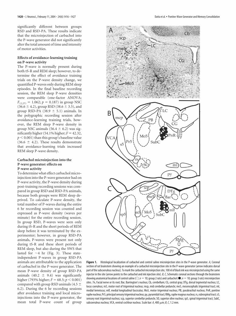
ResultsIn the first two baseline polygraphic recording (electrode testing)sessions, 30 rats exhibited good quality P-waves during tS-R andREM sleep. On the basis of in vivo pharmacological responses tocarbachol microinjection into the P-wave generator and post-mortem histological identification, recording and microinjectionsites were identified as within the P-wave generator (Fig. 1). His-tological examination revealed that our microinjection of a 100 nlvolume of dye diffused only 0.1– 0.15 mm from the center ofmicroinjection (Fig. 1), indicating that the microinjection of car-
bachol did not diffuse outside of the P-wave generator. Beforelearning trials, in the final 6 hr baseline recording session, totalpercentages of time spent in W, SWS, tS-R, and REM sleep andP-wave density were not significantly different (one-factorANOVA) between the three groups of animals. Thus, the groupswere initially equal in terms of time spent in W, SWS, tS-R, andREM sleep and P-wave density for the final 6 hr baseline record-ing session.
Effects of REM sleep deprivation on wake–sleep statesAfter a session of training trials, rats in groups NSC and RSDreceived control saline microinjections, and group RSD-PA re-ceived a carbachol microinjection into the P-wave generator. Allgroups were then recorded in the polygraph for 6 hr. During thispolygraphic recording session, rats in groups RSD and RSD-PAwere subjected to the REM sleep deprivation protocol. The totalpercentages of REM sleep in groups RSD (0.58 � 0.03%) andRSD-PA (0.56 � 0.02%) were drastically reduced compared withthe total percentage of REM sleep in group NSC (14.12 � 2.59%)(Fig. 2). These results demonstrate that the deprivation methodused for this study effectively reduced the total amount of REMsleep. Statistical comparisons (one-factor ANOVAs) between thethree groups revealed significant differences in the time spent inW (F(2,27) � 14.945; p � 0.0001), tS-R (F(2,27) � 15.728; p �0.0001), and REM sleep (F(2,27) � 165.076; p � 0.0001) but not inSWS (F(2,27) � 1.956; p � 0.1609). To determine the effects ofREM sleep deprivation alone, sleep–wake data were comparedbetween groups NSC and RSD. Post hoc Scheffe F test showed thatgroup RSD animals spent significantly more time in wakefulnessthan group NSC animals (69.78% more; F � 12.0; p � 0.001)(Fig. 2). Group RSD animals spent significantly less time thangroup NSC in tS-R (62.7% less; F � 10.3; p � 0.001) and REMsleep (95.9% less; F � 123.63; p � 0.001) (Fig. 2). Although thepercentage of REM sleep in the group RSD animals was signifi-cantly less than in the group NSC animals, the number of REMsleep episodes was significantly higher in the RSD group (Fig. 2).The latencies to REM sleep episode in the NSC and RSD groups ofanimals were comparable (Fig. 2). This increased number ofREM sleep episodes in the RSD group of animals is likely causedby the increased REM sleep pressure caused by the REM sleepdeprivation. The fact that the REM sleep-deprived rats had atendency to enter REM sleep directly from SWS without enteringinto tS-R suggests that increased REM sleep pressure in the REMsleep-deprived rats may also be responsible for the reduction inthe total amount of time spent in tS-R.
Sleep–wake state effects of carbachol microinjection into theP-wave generatorTo determine the effects of carbachol microinjection into theP-wave generator, sleep–wake data collected during the post-training recording sessions were compared between groups RSDand RSD-PA. Post hoc Scheffe F test and one-factor ANOVAsshowed that the total percentages of W, SWS, tS-R, and REMsleep were not significantly different between groups RSD andRSD-PA (Fig. 2). The latencies to the first episode of REM sleepand total numbers of REM sleep episodes were not significantlydifferent in the group NSC and group RSD animals (Fig. 2).These results demonstrate that the microinjection of carbacholinto the P-wave generator did not significantly change the sleep–wake parameters measured. Similarly, the total percentages oftime spent in active motor behavior (saline vs carbachol: 9.8 �3.8 vs 7.9 � 4.1%) and mean EMG amplitudes during thoseactive motor behaviors (198 � 34 vs 185 � 41 �V) were not
Datta et al. • Pontine-Wave Generator and Memory Consolidation J. Neurosci., February 11, 2004 • 24(6):1416 –1427 • 1419
significantly different between groupsRSD and RSD-PA. These results indicatethat the microinjection of carbachol intothe P-wave generator did not significantlyalter the total amount of time and intensityof motor activities.
Effects of avoidance-learning trainingon P-wave activityThe P-wave is normally present duringboth tS-R and REM sleep; however, to de-termine the effect of avoidance trainingtrials on the P-wave density change, wequantified P-waves only during REM sleepepisodes. In the final baseline recordingsession, the REM sleep P-wave densitieswere comparable (one-factor ANOVA;F(2,27) � 1.062; p � 0.187) in group NSC(36.6 � 4.2), group RSD (38.6 � 3.5), andgroup RSD-PA (38.9 � 5.1) animals. Inthe polygraphic recording session afteravoidance-learning training trials, how-ever, the REM sleep P-wave density ingroup NSC animals (56.4 � 6.2) was sig-nificantly higher (54.1% higher; F � 42.32,p � 0.001) than this group’s baseline value(36.6 � 4.2). These results demonstratethat avoidance-learning trials increasedREM sleep P-wave density.
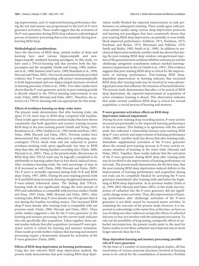
Carbachol microinjection into theP-wave generator: effects onP-wave activityTo determine what effect carbachol micro-injection into the P-wave generator had onP-wave activity, the P-wave density duringpost-training recording sessions was com-pared in group RSD and RSD-PA animals,because both groups were REM sleep de-prived. To calculate P-wave density, thetotal number of P-waves during the entire6 hr recording session was counted andexpressed as P-wave density (waves perminute) for the entire recording session.In group RSD, P-waves were seen onlyduring tS-R and the short periods of REMsleep before it was terminated by the ex-perimenter; however, in group RSD-PAanimals, P-waves were present not onlyduring tS-R and these short periods ofREM sleep, but also during the SWS thatlasted for �4 hr (Fig. 3). These state-independent P-waves in group RSD-PAanimals are attributable to the applicationof carbachol in the P-wave generator. Themean P-wave density of group RSD-PAanimals (40.2 � 9.4) was significantlyhigher (793% higher; F � 68.5; p � 0.001)compared with group RSD animals (4.5 �6.2). During the 6 hr recording sessionsafter avoidance training and local micro-injections into the P-wave generator, themean total P-wave count of group
Figure 1. Histological localization of carbachol and control saline microinjection sites in the P-wave generator. A, Coronalsection of rat brainstem showing an example of a carbachol microinjection site in the P-wave generator (arrow indicates dorsalpart of the subcoeruleus nucleus). To mark the carbachol microinjection site, 100 nl of black ink was microinjected using the sameinjector in the site (arrow points to the carbachol and ink injection site). B, C, Schematic coronal sections through the brainstemshowing anatomical locations of control saline (E; n � 10; group 2 rats) and carbachol (F; n � 10; group 3 rats) microinjectionsites. 7n, Facial nerve or its root; Bar, Barrington’s nucleus; Cb, cerebellum; CG, central gray; DTg, dorsal tegmental nucleus; LC,locus coeruleus; m5, motor root of trigeminal nucleus; mcp, mid cerebellar peduncle; me5, mesencephalic trigeminal tract; ml,medial lemniscus; mlf, medial longitudinal fasciculus; Mo5, motor trigeminal nucleus; PB, parabrachial nucleus; PnR, pontineraphe nucleus; Pr5, principal sensory trigeminal nucleus; py, pyramidal tract; RMg, raphe magnus nucleus; rs, rubrospinal tract; s5,sensory root trigeminal nucleus; scp, superior cerebellar peduncle; SO, superior olive nucleus; sp5, spinal trigeminal tract; SubC,subcoeruleus nucleus; VCA, ventral cochlear nucleus. Scale bar: A, 400 �m; B, C, 1.2 mm.
1420 • J. Neurosci., February 11, 2004 • 24(6):1416 –1427 Datta et al. • Pontine-Wave Generator and Memory Consolidation
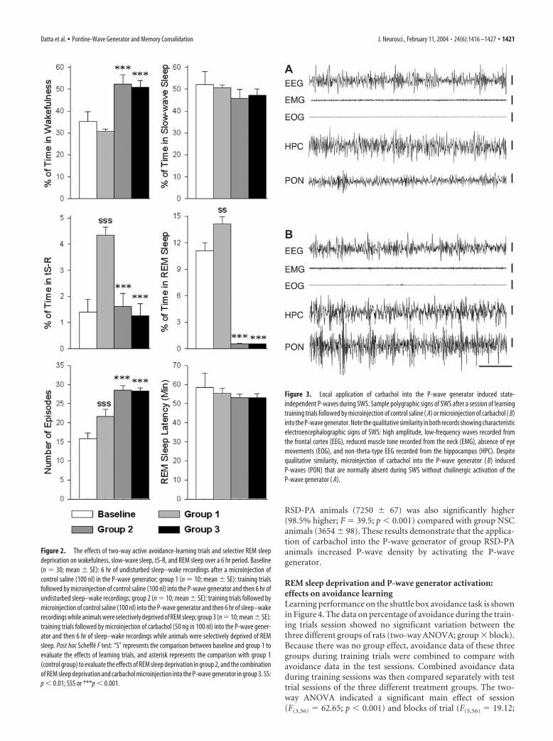
RSD-PA animals (7250 � 67) was also significantly higher(98.5% higher; F � 39.5; p � 0.001) compared with group NSCanimals (3654 � 98). These results demonstrate that the applica-tion of carbachol into the P-wave generator of group RSD-PAanimals increased P-wave density by activating the P-wavegenerator.
REM sleep deprivation and P-wave generator activation:effects on avoidance learningLearning performance on the shuttle box avoidance task is shownin Figure 4. The data on percentage of avoidance during the train-ing trials session showed no significant variation between thethree different groups of rats (two-way ANOVA; group � block).Because there was no group effect, avoidance data of these threegroups during training trials were combined to compare withavoidance data in the test sessions. Combined avoidance dataduring training sessions was then compared separately with testtrial sessions of the three different treatment groups. The two-way ANOVA indicated a significant main effect of session(F(3,56) � 62.65; p � 0.001) and blocks of trial (F(5,56) � 19.12;
Figure 2. The effects of two-way active avoidance-learning trials and selective REM sleepdeprivation on wakefulness, slow-wave sleep, tS-R, and REM sleep over a 6 hr period. Baseline(n � 30; mean � SE): 6 hr of undisturbed sleep–wake recordings after a microinjection ofcontrol saline (100 nl) in the P-wave generator; group 1 (n � 10; mean � SE): training trialsfollowed by microinjection of control saline (100 nl) into the P-wave generator and then 6 hr ofundisturbed sleep–wake recordings; group 2 (n � 10; mean � SE): training trials followed bymicroinjection of control saline (100 nl) into the P-wave generator and then 6 hr of sleep–wakerecordings while animals were selectively deprived of REM sleep; group 3 (n �10; mean�SE):training trials followed by microinjection of carbachol (50 ng in 100 nl) into the P-wave gener-ator and then 6 hr of sleep–wake recordings while animals were selectively deprived of REMsleep. Post hoc Scheffe F test: “S” represents the comparison between baseline and group 1 toevaluate the effects of learning trials, and asterisk represents the comparison with group 1(control group) to evaluate the effects of REM sleep deprivation in group 2, and the combinationof REM sleep deprivation and carbachol microinjection into the P-wave generator in group 3. SS:p � 0.01; SSS or ***p � 0.001.
Figure 3. Local application of carbachol into the P-wave generator induced state-independent P-waves during SWS. Sample polygraphic signs of SWS after a session of learningtraining trials followed by microinjection of control saline ( A) or microinjection of carbachol ( B)into the P-wave generator. Note the qualitative similarity in both records showing characteristicelectroencephalographic signs of SWS: high amplitude, low-frequency waves recorded fromthe frontal cortex (EEG), reduced muscle tone recorded from the neck (EMG), absence of eyemovements (EOG), and non-theta-type EEG recorded from the hippocampus (HPC). Despitequalitative similarity, microinjection of carbachol into the P-wave generator ( B) inducedP-waves (PON) that are normally absent during SWS without cholinergic activation of theP-wave generator ( A).
Datta et al. • Pontine-Wave Generator and Memory Consolidation J. Neurosci., February 11, 2004 • 24(6):1416 –1427 • 1421
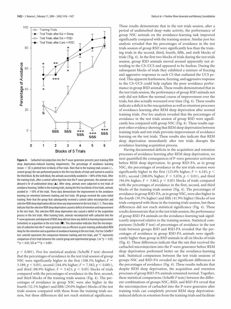
p � 0.001). Post hoc statistical analysis (Scheffe F test) showedthat the percentages of avoidance in the test trial session of groupNSC were significantly higher in the first (108.3% higher; F �3.189; p � 0.05), second (244.4% higher; F � 7.582; p � 0.001),and third (88.9% higher; F � 3.421; p � 0.05) blocks of trialscompared with the percentages of avoidance in the first, second,and third blocks of the training trials session (Fig. 4). The per-centages of avoidance in group NSC were also higher in thefourth (32.1% higher) and fifth (29.0% higher) blocks of the testtrials session compared with those from the training trials ses-sion, but these differences did not reach statistical significance.
These results demonstrate that in the test trials session, after aperiod of undisturbed sleep–wake activity, the performance ofgroup NSC animals on the avoidance-learning task improvedsignificantly compared with the training session. Similar post hocanalysis revealed that the percentages of avoidance in the testtrials session of group RSD were significantly less than the train-ing trials in the second, third, fourth, fifth, and sixth blocks oftrials (Fig. 4). In the first two blocks of trials during the test trialssession, group RSD animals moved around apparently not at-tending to the CS–UCS and appeared to be fearless. During thesubsequent blocks of trials they exhibited a mixture of freezingand aggressive responses to each CS that outlasted the UCS pe-riod. This apparent fearlessness, freezing, and aggressive responseto the CS–UCS could help explain the poor avoidance perfor-mance in group RSD animals. These results demonstrated that inthe test trials session, the performance of group RSD animals notonly did not follow the normal course of improvement over thetrials, but also actually worsened over time (Fig. 4). These resultsindicate a deficit in the reacquisition as well as retention processesof avoidance learning after REM sleep deprivation after normaltraining trials. Post hoc analysis revealed that the percentages ofavoidance in the test trials session of group RSD were signifi-cantly less compared with group NSC (Fig. 4). These results sup-port other evidence showing that REM sleep deprivation betweentraining trials and test trials prevents improvement of avoidancelearning on the test trials. These results also indicate that REMsleep deprivation immediately after test trials disrupts theavoidance-learning acquisition process.
Having documented deficits in the acquisition and retentionprocesses of avoidance learning after REM sleep deprivation, wenext quantified the consequences of P-wave generator activationbefore REM sleep deprivation. In group RSD-PA, as in groupNSC, the percentages of avoidance in the test trials session weresignificantly higher in the first (125.0% higher; F � 4.245; p �0.05), second (200.0% higher; F � 5.076; p � 0.01), and third(94.4% higher; F � 3.862; p � 0.05) blocks of trials comparedwith the percentages of avoidance in the first, second, and thirdblocks of the training trials session (Fig. 4). The percentages ofavoidance in group RSD-PA, as in group NSC, were also higher inthe fourth (39.3% higher) and fifth (41.9% higher) blocks of testtrials compared with those in the training trials session, but thesedifferences did not reach statistical significance (Fig. 4). Theseresults demonstrate that in the test trials session, the performanceof group RSD-PA animals on the avoidance-learning task signif-icantly improved relative to the training session. Statistical com-parisons (Scheffe F test) of percentages of avoidance in the testtrials between groups RSD and RSD-PA revealed that the per-centages of avoidance in group RSD-PA animals were signifi-cantly higher than group in RSD animals in all six blocks of trials(Fig. 4). These differences indicate that the rats that received thecarbachol microinjection into the P-wave generator before REMsleep deprivation performed better on the avoidance-learningtask. Statistical comparison between the test trials sessions ofgroups NSC and RSD-PA revealed no significant differences inthe percentages of avoidance (Fig. 4). These results indicate thatdespite REM sleep deprivation, the acquisition and retentionprocesses of group RSD-PA animals remained normal. Together,these statistical comparisons (Scheffe F tests) between the differ-ent combinations of groups NSC, RSD, and RSD-PA reveal thatthe microinjection of carbachol into the P-wave generator aftertraining trials can completely prevent REM sleep deprivation-induced deficits in retention from the training trials and facilitate
Figure 4. Carbachol microinjection into the P-wave generator prevents post-training REMsleep deprivation-induced learning impairments. The percentage of avoidance learning(mean � SE) is plotted here in blocks of five trials. Note that in the training trials session (‚,control group) the rats performed poorly in the first two blocks of trials and started to avoid inthe third block. By the sixth block, the animals successfully avoided in �80% of the trials. Afterthe training trials, after a control saline injection into the P-wave generator, the animals wereallowed 6 hr of undisturbed sleep (Œ). After sleep, animals were subjected to test trials ofavoidance learning. Unlike in the training trials, during the first two blocks of test trials, animalsavoided in �50% of the trials. These data demonstrate the improvement in the avoidancelearning (or retention) between training and test trials. All groups received the same initialtraining. Note that the group that subsequently received a control saline microinjection andselective REM sleep deprivation did not show any improvement in the test trials (E). These dataindicate that the selective REM sleep deprivation caused a deficit of retention and improvementin the test trials. This selective REM sleep deprivation also caused a deficit in the acquisitionprocess in the test trials. After training trials, animals microinjected with carbachol into theP-wave generator and deprived of REM sleep did not show any deficit in learning improvement(retention) or acquisition in the test trials (F). This observation indicates that the microinjec-tion of carbachol into the P-wave generator was as effective as post-training undisturbed REMsleep for the retention and acquisition of avoidance learning in the test trials. Post hoc Scheffe Ftest: asterisk represents the comparison between training and test trials, and “S” representscomparison of test trials between the control group and experimental groups. S or *p � 0.05;**p � 0.01; SSS or ***p � 0.001.
1422 • J. Neurosci., February 11, 2004 • 24(6):1416 –1427 Datta et al. • Pontine-Wave Generator and Memory Consolidation

acquisition processes in the test trials of two-way active avoidancelearning.
In this study, microinjection of carbachol into the P-wavegenerator rescued REM sleep deprivation-induced deficit in theimprovement of learning performance. Is it possible that the mi-croinjection of carbachol into the P-wave generator might havediffused into the neighboring structure, the locus coeruleus? Hy-pothetically, diffusion of carbachol into the locus coeruleus mayincrease motor performance by activating noradrenergic cells inthe locus coeruleus (Berridge and Foote, 1991, 1996). We believethat carbachol microinjected into the P-wave generator did notdiffuse into the locus coeruleus or any other major structures ofthe brainstem, for the following reasons. First, in our earlierP-wave generator mapping study, we showed that 100 nl of car-bachol or BDA microinjection into the P-wave generator diffusesonly 0.1– 0.15 mm in diameter (Datta et al., 1998, 1999, 2003).Second, previous studies by other investigators have shown thatthe microinjections of 100 nl volume of drug into the brainstemcan effectively diffuse only 0.3– 0.5 mm from the center of aninjection site (Myers and Hoch, 1978; Vanni-Mercier et al., 1989;Vertes et al., 1993). Third, carbachol microinjection into the lo-cus coeruleus suppresses slow-wave sleep by increasing wakeful-ness (Berridge and Foote, 1991, 1996), and in this study carbacholmicroinjection into the P-wave generator did not change the totalamount of slow-wave sleep. Therefore, it is not likely that the micro-injection of carbachol into the P-wave generator directly activatedthe locus coeruleus. Fourth, the activation of neighboring structureslike the dorsal raphe and locus coeruleus suppresses P-wave activity,but in this study, carbachol microinjections into the P-wave gener-ator induced P-wave activity. Taken together, this evidence indicatesthat it is highly unlikely that the carbachol microinjection-inducedimprovement in learning behavior was caused by the diffusion ofcarbachol in structures other than the P-wave generator. Finally, inthe present study, histological examination confirmed that the 100 nlvolume of dye diffused only 0.1 mm from the center of the microin-jection (Fig. 1).
Relationship between P-wave density and improvement inavoidance learningBecause the REM sleep P-wave density of group NSC increasedsignificantly during the experimental recording session, and be-cause in our earlier study we demonstrated a positive correlationbetween the post-training REM sleep P-wave density and im-provement of learning in the retest session (Datta, 2000), weexpected to see a precise relationship between the REM sleepP-wave density change from baseline to experimental recordingsessions and the improvement in performance. Indeed, a strongcorrelation was observed (Pearson correlation coefficient: r �0.84; F � 44.59; p � 0.0002) (Fig. 5). These results suggest that theincrease in P-wave density during REM sleep after training trialsis correlated with effective task performance in the test trials.Next, to determine the correlation between the P-wave generatoractivation-induced P-wave density change and improvement ofperformance in RSD-PA group, P-wave density change was cal-culated by subtracting baseline P-wave density (expressed aswaves per minute for the entire 6 hr period) from the P-wavedensity in the post-training recording session. This carbachol-induced P-wave density change in group RSD-PA showed a sta-tistically significant positive slope with the percentage of im-provement in the test trials session (r � 0.93; F � 116.5; p �0.0001). These results demonstrate that the level of P-wave gen-erator activation after training trials is directly correlated with theimprovement in the test trials.
DiscussionThe principal findings of this study are that (1) a newly designedREM sleep deprivation method selectively eliminated REM sleepwithout changing SWS, (2) REM sleep deprivation after trainingtrials prevented improvement on test trial performance, (3) mi-croinjection of carbachol into the P-wave generator after trainingtrials prevented REM sleep deprivation-induced deficits in learn-
Figure 5. Relationship between the P-wave density change and the improvement in avoid-ance learning. A, The percentage of improvement for each animal (F) is shown as a function ofthe percentage of REM sleep P-wave density change between the last baseline recording ses-sion and the post-training recording session (n � 10 rats). The plot of linear regression best-fit(solid line; Pearson product–moment correlation) shows a statistically significant positive slope(r � 0.84; p � 0.001). These data indicate that the level of improvement of learning in the testsession depends positively on the percentage of P-wave density increase during post-trainingREM sleep. B, The percentage of improvement for each animal (filled rectangles) is shown as afunction of the percentage of P-wave density change (for the entire 6 hr period) between thelast baseline recording session and post-training P-wave generator carbachol-microinjectedrecording session (n � 10 rats). The plot of linear regression best-fit (solid line; Pearson pro-duct–moment correlation) shows a statistically significant positive slope (r � 0.93; p �0.001). These data indicate that the level of improvement of learning in the test session isproportional to the carbachol microinjection-induced state-independent P-wave density.
Datta et al. • Pontine-Wave Generator and Memory Consolidation J. Neurosci., February 11, 2004 • 24(6):1416 –1427 • 1423
ing improvement, and (4) improved learning performance dur-ing the test trial session was proportional to the level of P-wavegenerator activation. These results suggest that the activation ofthe P-wave generator during REM sleep enhances a physiologicalprocess of memory processing that occurs naturally during post-learning REM sleep.
Methodological considerationsSince the discovery of REM sleep, animal studies of sleep andlearning have used various hippocampally and non-hippocampally mediated learning paradigms. In this study, wehave used a TWAA-learning task that involves both the hip-pocampus and the amygdala (Smith and Young, 1980; Ambro-sini et al., 1988; Ramirez and Carrer, 1989; Bramham et al., 1994;Mavanji and Datta, 2003). One recent anatomical study providedevidence that P-wave-generating cells project monosynapticallyto both hippocampal and non-hippocampal structures involvedin learning processes (Datta et al., 1998). Two other studies haveshown that the P-wave generator activity in post-training periodsis directly related to the TWAA-learning improvement in testtrials (Datta, 2000; Mavanji and Datta, 2003). Therefore, the se-lection of a TWAA-learning task was appropriate for this study.
Effects of avoidance learning on sleep–wake statesThe present study demonstrates that after learning trials, ratsspent 27.2% more time in REM sleep compared with baseline.These results agree with previous animal studies that have shownconsistently that both appetitive and aversive training increaseREM sleep in the post-training period (Smith and Wong, 1991;Bramham et al., 1994; Giuditta et al., 1995; Smith and Rose, 1997;Datta, 2000; Mavanji and Datta, 2003). Previous studies havedemonstrated that control rats subjected to nonlearning, non-contingent TWAA shock trials and rats subjected to passiveavoidance-training trials spent significantly less time in REMsleep than they did during baseline recording days (Datta, 2000;Mavanji et al., 2003). Thus, in the present study, this increase inREM sleep after TWAA trials may be logically considered to beattributable to learning rather than to foot shock-induced stress.After avoidance-learning trials, the total percentage of time intS-R increased as well (Datta, 2000; Mavanji and Datta, 2003).The P-wave is normally expressed during both tS-R and REMsleep (Datta, 1997, 2000). During the post-training period, bothtS-R and REM sleep increased, showing a heightened demand forP-wave-related behavioral states. The finding that TWAA-learning trials do not significantly change the total amount ofSWS and wakefulness is compatible with previous studies (Smithand Rose, 1997; Datta, 2000; Mavanji and Datta, 2003). Aftertraining trials, REM sleep P-wave density was 54% greater than itwas during the baseline recording session. This increased REMsleep P-wave density after training trials is compatible with ourprevious study (Datta, 2000; Mavanji and Datta, 2003). Theseearlier studies suggested a role for the P-wave generator in thelearning and memory processing, but the current study indicatesthat role specifically after acquisition. Taken together, these ani-mal studies indicate that after acquisition, increased P-wave gen-erator activity is critical for learning and memory formation.These results provide further evidence that learning and memoryprocessing require a homeostatic demand for activation of theP-wave generator (Datta, 2000).
Effects of REM sleep deprivation on learning performanceUsing this new selective REM sleep deprivation method, thepresent study demonstrates that post-training REM sleep depri-
vation totally blocked the expected improvement on task per-formance on subsequent retesting. These results agree with pre-vious animal studies using various sleep-deprivation protocolsand learning test paradigms that have consistently shown thatpost-training REM sleep deprivation can partially or even totallyblock improved performance (Fishbein, 1971; Pearlman, 1973;Pearlman and Becker, 1973; Shiromani and Fishbein, 1979;Smith and Butler, 1982; Smith et al., 1998). In addition to me-chanical deprivation methods, another study has shown that dur-ing the post-training REM sleep window, intraperitoneal injec-tion of the general protein synthesis inhibitor anisomycin and thecholinergic antagonist scopolamine induces marked learning–memory impairment in the rat (Smith et al., 1991). This evidencesuggests that post-training REM sleep is critical for the improve-ment of learning performance. Post-training REM sleep-dependent improvement in learning indicates that increasedREM sleep after training trials may be involved in some physio-logical processes that influence the retention of learned behavior.The present study demonstrates that after a 6 hr period of REMsleep deprivation, the expected improvement in acquisition ofactive avoidance learning is absent. This observation indicatesthat under normal conditions REM sleep is critical for normalacquisition, a crucial process of learning and memory.
P-wave generator activation: effects on REM sleepdeprivation-induced impairmentDuring the post-training sleep recording session, P-wave activityincreased proportionally to the improved learning performancein the test session. This finding is in agreement with the earlierstudy that indicated a relationship between post-training REMsleep P-wave activity and improvement of learning performance(Datta, 2000). Another study has shown that after training trials,immediate supplemental activation of the P-wave generatorabove the normal post-training increase in P-wave activity in-creases retention of learning in the retest trials (Mavanji andDatta, 2003). Together, these results indicate that the activationof the P-wave generator during REM sleep after training trialsmay be involved in the improvement of learning performance ontest trials. The present study demonstrates, for the first time, thatthe post-training REM sleep deprivation-induced deficits in theimprovement of learning performance and acquisition duringtest trials can be completely blocked by activating the P-wavegenerator immediately after training trials and before the begin-ning of REM sleep deprivation. As in previous studies (Datta etal., 1999, 2003; Mavanji and Datta, 2003), in this study microin-jection of carbachol into the P-wave generator did not signifi-cantly change motor activities. Thus, this improvement of learn-ing performance after cholinergic activation of the P-wavegenerator is not likely caused by increased motor activities. Inevaluating the outcome of the present study, however, it is im-portant to acknowledge at the outset that, at this time, we have noway of ruling out other unknown nonspecific effects of carbacholthat may or may not interfere with the subsequent test session. Torule out the possibility of long-lasting, nonspecific effects of car-bachol microinjection, the present results point to the need forfuture studies to test these carbachol-microinjected rats at muchlonger intervals than the 6 hr.
Sleep-dependent learning and memory processing: possiblerole of P-wave generatorOn the basis of a number of neurophysiological studies, off-linereactivation of various neuronal structures involved in learningseems to be critical for the consolidation of memories (Pavlides
1424 • J. Neurosci., February 11, 2004 • 24(6):1416 –1427 Datta et al. • Pontine-Wave Generator and Memory Consolidation
and Winson, 1989; Skaggs and McNaughton, 1996; Qin et al.,1997; Kudrimoti et al., 1999; Poe et al., 2000). In these studies,off-line hippocampal reactivations were seen during both non-REM and REM sleep. This reactivation hypothesis of memoryconsolidation is also supported by a number of electrical stimu-lation studies (Stein and Chorover, 1968; Erickson and Patel,1969; Destrade et al., 1973; Landfield et al., 1973; Destrade andCardo, 1974). These studies reported that mice and rats receivingpost-trial hippocampal stimulation showed better retention oflearning than control animals. These studies also showed thatwhen the hippocampus was reactivated by electrical stimulationthere was no need for sleep for the improvement of learning.
The present study demonstrates that immediately after train-ing trials, the need for REM sleep for the improvement of learningcan be substituted by the cholinergic activation of the P-wavegenerator. This finding extends and gives a specific meaning tothose earlier studies that demonstrated that electrical stimulationof the rostral brainstem after training improves performance inthe rat (Leconte et al., 1974; DeWeer, 1976; Bloch et al., 1977;Devietti et al., 1977; Sara et al., 1980; Bloch and Laroche, 1981;Hennevin et al., 1989). The improvement in learning perform-ance by post-trial brainstem stimulation was as effective as hip-pocampal stimulation. Post-trial brainstem stimulation wasshown to facilitate a classically conditioned association and alsothe development of associative changes in neuronal activity in thehippocampus (Bloch and Laroche, 1981, 1984). Moreover, whenstimulation was administered after each long-term potentiation(LTP)-inducing stimulus, it enhanced the magnitude of LTP atthe synapses of the perforant path on dentate granular cells andprolonged its duration by several days. Brainstem stimulationduring the post-acquisition period appeared to substitute theneed for REM sleep by decreasing the post-training REM sleepelevation and abolishing most of the learning impairment pro-duced by post-trial REM sleep deprivation (Bloch et al., 1977).Although these brainstem stimulation studies did not definitivelylocalize a specific structure, it is well known that the rostral brain-stem is an important part of the reticular formation that containsa number of specific cell groups involved in the generation ofdifferent signs of REM sleep (Datta, 1995). During REM sleep,different parts of the brainstem are activated to generate differentphasic and tonic signs of REM sleep, including P-waves (Vertes,1984; Datta, 1995, 1997). The present results demonstrate thatpost-training activation of the P-wave generator is sufficient toimprove learning even when REM sleep is absent, indicating thatthe post-learning trial-increased homeostatic demand for REMsleep may be caused, specifically, by a heightened demand forP-wave generator activity. These results support the hypothesisthat the activation of the P-wave generator is part of the mecha-nism for REM sleep-dependent memory consolidation.
In conclusion, activation of P-wave-generating cells duringREM sleep may reactivate the forebrain and cortical memoryprocessing structures to reprocess recently stored informationaiding in the maintenance of memory and facilitating its laterexpression. The activation of the P-wave generator may have acausal role in sleep-dependent learning and memory processing.
ReferencesAbel T, Lattal KM (2001) Molecular mechanisms of memory acquisition,
consolidation and retrieval. Curr Opin Neurobiol 11:180 –187.Abel T, Nguyen PV, Barad M, Deuel TA, Kandel ER, Bourtchouladze R
(1997) Genetic demonstration of a role for PKA in the late phase of LTPand in hippocampus-based long-term memory. Cell 88:615– 626.
Ambrosini MV, Sadile AG, Gironi-Carnevale UA, Mattiaccio A, Giuditta A(1988) The sequential hypothesis of sleep function. II. A correlative study
between sleep variables and newly synthesized brain DNA. Physiol Behav43:339 –350.
Berridge CW, Foote SL (1991) Effects of locus coeruleus activation of elec-troencephalographic activity in neocortex and hippocampus. J Neurosci11:3135–3145.
Berridge CW, Foote SL (1996) Enhancement of behavioral and electroen-cephalographic indices of waking after stimulation of noradrenergicb-receptors within the medial septal region of the basal forebrain. J Neu-rosci 16:6999 –7009.
Bhanot JL, Chinna GS, Singh B, Sachdeva U, Kumar VM (1989) REM sleepdeprivation and food intake. Ind J Physiol Pharmacol 33:139 –145.
Bloch V, Laroche S (1981) Conditioning of hippocampal cells: its accelera-tion and long-term facilitation by post-trial reticular stimulation. BehavBrain Res 3:23– 42.
Bloch V, Laroche S (1984) Facts and hypotheses related to the search for theengram. In: Neurobiology of learning and memory (Lynch G, McGaughJL, Weinberger NM, eds), pp 249 –260. New York: Guilford.
Bloch V, Hennevin E, Leconte P (1977) Interaction between post-trial retic-ular stimulation and subsequent paradoxical sleep in memory consolida-tion processes. In: Neurobiology of sleep and memory (Drucker-ColinRR, McGaugh JL, eds), pp 255–272, New York: Academic.
Bramham CR, Srebro B (1989) Synaptic plasticity in the hippocampus ismodulated by behavioral state. Brain Res 493:74 – 86.
Bramham CR, Maho C, Laroche S (1994) Suppression of long-term poten-tiation induction during alert wakefulness but not during “enhanced”REM sleep after avoidance learning. Neuroscience 59:501–509.
Brooks DC, Bizzi E (1963) Brain stem electrical activity during deep sleep.Arch Ital Biol 101:648 – 665.
Campbell IG, Guinan ML, Horowitz JM (2002) Sleep deprivation impairslong-term potentiation in rat hippocampal slices. J Neurophysiol88:1073–1076.
Cohen NJ, Squire LR (1980) Preserved learning and retention of pattern-analyzing skill in amnesia: dissociation of knowing how and knowingthat. Science 210:207–210.
Datta S (1995) Neuronal activity in the peribrachial area: relationship tobehavioral state control. Neurosci Biobehav Rev 19:67– 84.
Datta S (1997) Cellular basis of pontine ponto-geniculo-occipital wave gen-eration and modulation. Cell Mol Neurobiol 17:341–365.
Datta S (2000) Avoidance task training potentiates phasic pontine-wavedensity in the rat: a mechanism for sleep-dependent plasticity. J Neurosci20:8607– 8613.
Datta S (2002) Evidence that REM sleep is controlled by the activation ofbrain stem pedunculopontine tegmental kainate receptor. J Neurophysiol87:1790 –1798.
Datta S, Hobson AJ (1994) Neuronal activity in the caudolateral peribra-chial pons: relationship to PGO waves and rapid eye movements. J Neu-rophysiol 71:95–109.
Datta S, Hobson AJ (1995) Suppression of ponto-geniculo-occipital wavesby neurotoxic lesions of pontine caudo-lateral peribrachial cells. Neuro-science 67:703–712.
Datta S, Calvo JM, Quatrochi JJ, Hobson JA (1992) Cholinergic micro-stimulation of the peribrachial nucleus in the cat. I. Immediate and pro-longed increases in ponto-geniculo-occipital waves. Arch Ital Biol130:263–284.
Datta S, Siwek DF, Patterson EH, Cipolloni PB (1998) Localization of pon-tine PGO wave generation sites and their anatomical projections in therat. Synapse 30:409 – 423.
Datta S, Patterson EH, Siwek DF (1999) Brainstem afferents of the cholinocep-tive pontine wave generation sites in the rat. Sleep Res Online 2:79–82.
Datta S, Spoley EE, Patterson EH (2001) Microinjection of glutamate intothe pedunculo pontine tegmentum induces REM sleep and wakefulnessin the rat. Am J Physiol 280:R752–R759.
Datta S, Spoley EE, Mavanji VK, Patterson EH (2002) A novel action ofpedunculopontine tegmental kainate receptors: a mechanism of REMsleep generation in the rat. Neuroscience 114:157–164.
Datta S, Mavanji VK, Patterson EH, Ulloor J (2003) Regulation of rapid eyemovement sleep in the freely moving rat: local microinjection of seroto-nin, norepinephrine, and adenosine into the brainstem. Sleep26:513–520.
Destrade C, Cardo B (1974) Effects of post-trial hippocampal stimulationon time-dependent improvement of performance in mice. Brain Res78:447– 454.
Datta et al. • Pontine-Wave Generator and Memory Consolidation J. Neurosci., February 11, 2004 • 24(6):1416 –1427 • 1425
Destrade C, Soumireu-Mourat B, Cardo B (1973) Effects of posttrial hip-pocampal stimulation on acquisition of operant behavior in the mouse.Behav Biol 8:713–724.
Devietti TL, Conger GL, Kirkpatrick BR (1977) Comparison of the en-hancement gradients of retention obtained with stimulation of the mes-encephalic reticular formation after training or memory reactivation.Physiol Behav 19:549 –554.
DeWeer B (1976) Selective facilitative effect of post-trial reticular stimula-tion in discriminative learning in the rat. Behav Proc 1:243–257.
Dujardin K, Guerrien A, Leconte P (1990) Sleep, brain activation and cog-nition. Physiol Behav 47:1271–1278.
Erickson CK, Patel JB (1969) Facilitation of avoidance learning by post-trial hippocampal electrical stimulation. J Comp Physiol Psychol68:400 – 406.
Feng P, Vogel GW, Obermeyer W, Kinney GG (2000) An instrumentalmethod for long-term continuous REM sleep deprivation of neonatalrats. Sleep 23:175–183.
Fishbein W (1971) Disruptive effects of rapid eye movement sleep depriva-tion on long term-memory. Physiol Behav 6:279 –282.
Fishbein W, Gutwien BM (1977) Paradoxical sleep and memory storageprocesses. Behav Biol 19:425– 464.
Frank MG, Issa NP, Stryker MP (2001) Sleep enhances plasticity in the de-veloping visual cortex. Neuron 30:275–287.
Gabrieli JDE (1998) Cognitive neuroscience of human memory. Annu RevPsychol 49:87–115.
Gallagher M, McMahan RW, Schoenbaum G (1999) Orbitofrontal cortexand representation of incentive value in associative learning. J Neurosci19:6610 – 6614.
Giuditta A, Ambrosini MV, Montagnese P, Mandile P, Cotugno M, GrassiZucconi G, Vescia S (1995) The sequential hypothesis of the function ofsleep. Behav Brain Res 69:157–166.
Graves L, Pack A, Abel T (2001) Sleep and memory: a molecular perspective.Trends Neurosci 24:237–243.
Guzman-Marin R, Suntsova N, Stewart DR, Gong H, Szymusiak R, McGintyD (2003) Sleep deprivation reduces proliferation of cells in the dentategyrus of the hippocampus in rats. J Physiol (Lond) 549:563–571.
Hamdi A (2000) Regulation of cardiac and renal peripheral benzodiazepinereceptor binding in rapid eye movement sleep-deprived rats. Life Sci67:3015–3022.
Hatfield T, Han J-S, Conley M, Gallagher M, Holland P (1996) Neurotoxiclesions of basolateral, but not central, amygdala interfere with Pavloviansecond-order conditioning and reinforcer devaluation effects. J Neurosci16:5256 –5265.
Hennevin E, Hars B, Bloch V (1989) Improvement of learning by mesence-phalic reticular stimulation during postlearning paradoxical sleep. BehavNeural Biol 51:291–306.
Hicks RA, Okuda A, Thomsen D (1997) Depriving rats of REM sleep: theidentification of a methodological problem. Am J Psychol 90:95–102.
Izquierdo I, Fin C, Schmitz PK, DaSilva RC, Jerusalinsky D, Quillfeldt JA,Ferreira MBG, Medina JH, Bazan NG (1995) Memory enhancement byintrahippocampal, intraamygdala, or intraentorhinal infusion of platelet-activating factor measured in an inhibitory avoidance task. Proc NatlAcad Sci USA 92:5047–5051.
Karni A, Tanne D, Rubenstein BS, Askenasy JJ, Sagi D (1994) Dependenceon REM sleep of overnight improvement of a perceptual task. Science265:679 – 682.
Kesner RP (1998) Neurobiological views of memory. In: Neurobiology oflearning and memory (Martinez Jr JL, Kesner RP, eds), pp 361– 416. SanDiego: Academic.
Kim JJ, Baxter MG (2001) Multiple brain-memory systems: the whole doesnot equal the sum of its parts. Trends Neurosci 24:324 –330.
Kudrimoti HS, Barnes CA, McNaughton BL (1999) Reactivation of hip-pocampal cell assemblies: effects of behavioral state, experience, and EEGdynamics. J Neurosci 19:4090 – 4101.
Landfield PW, Tusa RJ, McGaugh JL (1973) Effects of posttrial hippocam-pal stimulation memory storage and EEG activity. Behav Biol 8:485–505.
Laurent JP, Ayalaguerrero F (1975) Reversible suppression of ponto-geniculo-occipital waves by localized cooling during paradoxical sleep incats. Exp Neurol 49:356 –369.
Leconte P, Hennevin E, Bloch V (1974) Duration of paradoxical sleep nec-essary for the acquisition of conditioned avoidance in the rat. PhysiolBehav 13:675– 681.
LeDoux JE (1992) Emotion and the amygdala. In: The amygdala: neurobi-ological aspects of emotion, memory, and mental dysfunction (AggletonJ, ed), pp 339 –351. New York: Wiley.
Louie K, Wilson MA (2001) Temporally structured replay of awake hip-pocampal ensemble activity during rapid eye movement sleep. Neuron29:145–156.
Maquet P, Smith C, Stickgold R (2003) Sleep and brain plasticity. Oxford:Oxford UP.
Mavanji V, Datta S (2003) Activation of the phasic pontine-wave generatorenhances improvement of learning performance: a mechanism for sleep-dependent plasticity. Eur J Neurosci 17:359 –370.
Mavanji VK, Siwek DF, Patterson EH, Spoley EE, Datta S (2003) Effects ofpassive-avoidance training on sleep-wake state-specific activity in the baso-lateral and central nuclei of the amygdala. Behav Neurosci 117:751–759.
McGrath MJ, Cohen DB (1978) REM sleep facilitation of adaptive wakingbehavior: a review of the literature. Psychol Bull 85:24 –57.
Myers RD, Hoch B (1978) 14-C dopamine microinjected into the brainstemof rat: dispersion kinetics, site content and functional dose. Brain Res Bull3:601– 609.
Ocampo-Garces A, Molina E, Rodriguez A, Vivaldi EA (2000) Homeostasisof REM sleep after total and selective sleep deprivation in the rat. J Neu-rophysiol 84:2699 –2702.
Pavlides C, Winson J (1989) Influences of hippocampal place cell firing inthe awake state on the activity of these cells during subsequent sleepepisodes. J Neurosci 9:2907–2918.
Paxinos G, Watson C (1997) The rat brain in stereotaxic coordinates. SanDiego: Academic.
Pearlman C (1973) REM sleep deprivation impairs latent extinction in rats.Physiol Behav 11:233–237.
Pearlman C (1979) REM sleep and information processing: evidence fromanimal studies. Neurosci Biobehav Rev 3:57– 68.
Pearlman C, Becker M (1973) Brief posttrial REM sleep deprivation impairsdiscrimination learning in rats. Physiol Psychol 1:373–376.
Poe GR, Nitz DA, McNaughton BL, Barnes CA (2000) Experience-dependent phase-reversal of hippocampal neuron firing during REMsleep. Brain Res 855:176 –180.
Poremba A, Gabriel M (1997) Amygdalar lesions block discriminativeavoidance learning and cingulothalamic training-induced neuronal plas-ticity in rabbits. J Neurosci 17:5237–5244.
Qin Y, McNaughton BL, Skaggs WE, Barnes CA (1997) Memory reprocess-ing in cortico-cortical and hippocampo-cortical neuronal ensembles.Philos Trans R Soc Lond B Biol Sci 352:1525–1533.
Ramirez OA, Carrer HF (1989) Correlation between threshold to inducelong-term potentiation in the hippocampus and performance in a shuttlebox avoidance response in rats. Neurosci Lett 104:152–156.
Rechtschaffen A, Bergmann BM (1995) Sleep deprivation in the rat by thedisk-over-water method. Behav Brain Res 69:55– 63.
Rechtschaffen A, Bergmann BM, Everson CA, Kushida CA, Gilliland MA(1989) Sleep deprivation in the rat: X. Integration and discussion of thefindings. Sleep 12:68 – 87.
Rechtschaffen A, Bergmann BM, Gilliland MA, Bauer K (1999) Effects ofmethod, duration, and sleep stage on rebounds from sleep deprivation inthe rat. Sleep 22:11–31.
Rempel-Clower NL, Zola SM, Squire LR, Amaral DG (1996) Three cases ofenduring memory impairment after bilateral damage limited to the hip-pocampal formation. J Neurosci 16:5233–5255.
Ribeiro S, Goyal V, Mello CV, Pavlides C (1999) Brain gene expression dur-ing REM sleep depends on prior waking experience. Learn Mem6:500 –508.
Ribeiro S, Mello CV, Velho T, Gardner TJ, Jarvis ED, Pavlides C (2002)Induction of hippocampal long-term potentiation during wakingleads to increased extrahippocampal zif-268 expression during ensu-ing rapid-eye-movement sleep. J Neurosci 22:10914 –10923.
Sakai K, Jouvet M (1980) Brainstem PGO-on cells projecting directly to thecat dorsal lateral geniculate nucleus. Brain Res 194:500 –505.
Sakai K, Petitjean F, Jouvet M (1976) Effects of pontomesencephalic lesionsand electrical stimulation upon PGO waves and EMPs in unanesthetizedcats. Electroencephalogr Clin Neurophysiol 41:49 – 63.
Sara SJ, Deweer B, Hars B (1980) Reticular stimulation facilitates retrieval ofa “forgotten” maze habit. Neurosci Lett 18:211–217.
1426 • J. Neurosci., February 11, 2004 • 24(6):1416 –1427 Datta et al. • Pontine-Wave Generator and Memory Consolidation

Sei H, Saitoh D, Yamamoto K, Morita K, Morita Y (2000) Differential effectof short-term REM sleep deprivation on NGF and BDNF protein levels inthe rat brain. Brain Res 877:387–390.
Shiromani P, Fishbein W (1979) Development of learning and memory inmice after brief paradoxical sleep deprivation. Physiol Behav 22:971–978.
Silva AJ, Stevens CF, Tonegawa S, Wang Y (1992) Deficient hippocampallong-term potentiation in alpha-calcium-calmodulin kinase II mutantmice. Science 257:201–206.
Skaggs WE, McNaughton BL (1996) Replay of neuronal firing sequences inrat hippocampus during sleep following spatial experience. Science271:1870 –1873.
Smith C (1985) Sleep states and learning: a review of the animal literature.Neurosci Biobehav Rev 9:157–168.
Smith C (1995) Sleep states and memory processes. Behav Brain Res69:137–145.
Smith C, Butler S (1982) Paradoxical sleep at selective times following train-ing is necessary for learning. Physiol Behav 29:469 – 473.
Smith C, Rose GM (1997) Posttraining paradoxical sleep in rats is increasedafter spatial learning in the Morris water maze. Behav Neurosci111:1197–1204.
Smith C, Wong PTP (1991) Paradoxical sleep increases predict successfullearning in a complex operant task. Behav Neurosci 105:282–288.
Smith C, Young J (1980) Reversal of paradoxical sleep deprivation by amyg-daloid stimulation during learning. Physiol Behav 24:1035–1039.
Smith C, Tenn C, Annett R (1991) Some biochemical and behavioral aspectsof the paradoxical sleep window. Can J Psychol 45:115–125.
Smith C, Conway JM, Rose GM (1998) Brief paradoxical sleep deprivationimpairs reference, but not working, memory in the radial arm maze task.Neurobiol Learn Mem 69:211–217.
Squire LR, Amaral DG, Press GA (1990) Magnetic resonance imaging of thehippocampal formation and mammillary nuclei distinguish medial tem-poral lobe diencephalic amnesia. J Neurosci 10:3106 –3117.
Stefurak SJ, Stefurak ML, Mendelson WB, Gillin JC, Wyatt RJ (1977) Amethod for sleep depriving rats. Pharmacol Biochem Behav1:137–139.
Stein DG, Chorover S (1968) Effects of post-trial electrical stimulation ofhippocampus and caudate nucleus on maze learning in the rat. PhysiolBehav 3:787–791.
Stickgold R (1998) Sleep: off-line memory reprocessing. Trends Cogn Sci2:484 – 492.
Thakkar M, Mallick BN (1993) Rapid eye movement sleep-deprivation-induced changes in glucose metabolic enzymes in rat brain. Sleep16:691– 694.
Vanni-Mercier G, Sakai K, Lin JS, Jouvet M (1989) Mapping of cholinocep-tive brainstem structures responsible for the generation of paradoxicalsleep in the cat. Arch Ital Biol 127:133–164.
Vazdarjanova A, McGaugh JL (1999) Basolateral amygdala is involved inmodulating consolidation of memory for classical fear conditioning.J Neurosci 19:6615– 6622.
Vertes RP (1984) Brainstem control of the events of REM sleep. Prog Neu-robiol 22:241–288.
Vertes RP, Colom LV, Fortin WJ, Bland BH (1993) Brainstem sites for thecarbachol elicitation of the hippocampal theta rhythm in the rat. ExpBrain Res 96:419 – 429.
Vogel GW (1975) A review of REM sleep deprivation. Arch Gen Psychiatry32:749 –761.
Vyazovskiy VV, Deboer T, Rudy B, Lau D, Borbely AA, Tobler I (2002)Sleep EEG in mice that are deficient in the potassium channel subunitK.v.3.2. Brain Res 947:204 –211.
White NM, McDonald RJ (2002) Multiple parallel memory systems in thebrain of the rat. Neurobiol Learn Mem 77:125–184.
Young B, Otto T, Fox GD, Eichenbaum H (1997) Memory representationwithin the parahippocampal region. J Neurosci 17:5183–5195.
Datta et al. • Pontine-Wave Generator and Memory Consolidation J. Neurosci., February 11, 2004 • 24(6):1416 –1427 • 1427