Activation of LKB1Akt pathway independent of PI3 Kinase plays a critical role in the proliferation...
17
Activation of LKB1-Akt pathway independent of PI3 Kinase plays a critical role in the proliferation of hepatocellular carcinoma from NASH Nuria Martínez-López 1,† , Marta Varela-Rey 1,† , David Fernández-Ramos 1 , Ashwin Woodhoo 1 , Mercedes Vázquez-Chantada 1 , Nieves Embade 1 , Luis Espinosa-Hevia 1 , Francisco Javier Bustamante 2 , Luis A Parada 1 , Manuel S Rodriguez 1 , Shelly C Lu 3 , José M Mato 1 , and Maria L Martínez-Chantar 1 1 CIC bioGUNE, Centro de Investigación Biomédica en Red de Enfermedades Hepáticas y Digestivas (Ciberehd), Technology Park of Bizkaia, 48160-Derio, Bizkaia, Spain. 2 Hospital de Cruces. University of Basque Country, Bizkaia, Spain. 3 Division of Gastrointestinal and Liver Diseases, USC Research Center for Liver Diseases, Southern California Research Center for Alcoholic Liver and Pancreatic Diseases and Cirrhosis, Keck School of Medicine, University of Southern California, Los Angeles, CA 90033. Abstract LKB1, originally considered as a tumor suppressor, plays an important role in hepatocyte proliferation and liver regeneration. Mice lacking Methionine adenosyltransferase 1A (MAT1A- KO mice) gene show chronic reduction in hepatic SAMe levels, basal activation of LKB1 and spontaneously develop non-alcoholic steatohepatitis (NASH) and hepatocellular carcinoma (HCC). These results are relevant for human health since patients with liver cirrhosis, who are at risk to develop HCC, have a marked reduction in hepatic MAT1A expression and SAMe synthesis. In our current work we have isolated a cell line, SAMe-D (SAMe-Deficient) from the HCC of MAT1A-KO mice to examine the role of LKB1 in the development of liver tumors derived from metabolic disorders. We find that LKB1 is required for cell survival in SAMe-D cells. LKB1 regulates Akt-mediated survival independently of PI3K, AMPK and mTORC2. In addition, LKB1 controls the apoptotic response through phosphorylation and retention of p53 in the cytoplasm, and the regulation of HAUSP and HuR nucleo-cytoplasmic shuttling. We identify HAUSP as a target of HuR. Finally, we observed cytoplasmic staining of p53 and p- LKB1(Ser428) in a NASH-HCC animal model (from MAT1A-KO mice) and in liver biopsies obtained from human HCC derived from both (alcoholic steatohepatitis) ASH and NASH etiology. Conclusions—SAMe-D cell line is a relevant model of HCC derived from NASH disease where LKB1 is the principal conductor of a new regulatory mechanism and could be considered as a practical tool to uncover new therapeutic strategies. Keywords NASH; HCC; LKB1; AMPK; HuR Contact information: ML Martinez-Chantar, CIC bioGUNE, Technology Park of Bizkaia, 48160 Derio, Bizkaia, Spain. [email protected]; Tel: +34-944-061318; Fax: +34-944-061301. † NML and MVR contributed equally to this paper. Conflict of interest: The authors have declared that no conflict of interest exists. NIH Public Access Author Manuscript Hepatology. Author manuscript; available in PMC 2011 November 1. Published in final edited form as: Hepatology. 2010 November ; 52(5): 1621–1631. doi:10.1002/hep.23860. NIH-PA Author Manuscript NIH-PA Author Manuscript NIH-PA Author Manuscript
-
Upload
independent -
Category
Documents
-
view
2 -
download
0
Transcript of Activation of LKB1Akt pathway independent of PI3 Kinase plays a critical role in the proliferation...

Activation of LKB1-Akt pathway independent of PI3 Kinase playsa critical role in the proliferation of hepatocellular carcinomafrom NASH
Nuria Martínez-López1,†, Marta Varela-Rey1,†, David Fernández-Ramos1, AshwinWoodhoo1, Mercedes Vázquez-Chantada1, Nieves Embade1, Luis Espinosa-Hevia1,Francisco Javier Bustamante2, Luis A Parada1, Manuel S Rodriguez1, Shelly C Lu3, José MMato1, and Maria L Martínez-Chantar1
1CIC bioGUNE, Centro de Investigación Biomédica en Red de Enfermedades Hepáticas yDigestivas (Ciberehd), Technology Park of Bizkaia, 48160-Derio, Bizkaia, Spain.2Hospital de Cruces. University of Basque Country, Bizkaia, Spain.3Division of Gastrointestinal and Liver Diseases, USC Research Center for Liver Diseases,Southern California Research Center for Alcoholic Liver and Pancreatic Diseases and Cirrhosis,Keck School of Medicine, University of Southern California, Los Angeles, CA 90033.
AbstractLKB1, originally considered as a tumor suppressor, plays an important role in hepatocyteproliferation and liver regeneration. Mice lacking Methionine adenosyltransferase 1A (MAT1A-KO mice) gene show chronic reduction in hepatic SAMe levels, basal activation of LKB1 andspontaneously develop non-alcoholic steatohepatitis (NASH) and hepatocellular carcinoma(HCC). These results are relevant for human health since patients with liver cirrhosis, who are atrisk to develop HCC, have a marked reduction in hepatic MAT1A expression and SAMesynthesis. In our current work we have isolated a cell line, SAMe-D (SAMe-Deficient) from theHCC of MAT1A-KO mice to examine the role of LKB1 in the development of liver tumorsderived from metabolic disorders. We find that LKB1 is required for cell survival in SAMe-Dcells. LKB1 regulates Akt-mediated survival independently of PI3K, AMPK and mTORC2. Inaddition, LKB1 controls the apoptotic response through phosphorylation and retention of p53 inthe cytoplasm, and the regulation of HAUSP and HuR nucleo-cytoplasmic shuttling. We identifyHAUSP as a target of HuR. Finally, we observed cytoplasmic staining of p53 and p-LKB1(Ser428) in a NASH-HCC animal model (from MAT1A-KO mice) and in liver biopsiesobtained from human HCC derived from both (alcoholic steatohepatitis) ASH and NASHetiology.
Conclusions—SAMe-D cell line is a relevant model of HCC derived from NASH disease whereLKB1 is the principal conductor of a new regulatory mechanism and could be considered as apractical tool to uncover new therapeutic strategies.
KeywordsNASH; HCC; LKB1; AMPK; HuR
Contact information: ML Martinez-Chantar, CIC bioGUNE, Technology Park of Bizkaia, 48160 Derio, Bizkaia, [email protected]; Tel: +34-944-061318; Fax: +34-944-061301.†NML and MVR contributed equally to this paper.Conflict of interest: The authors have declared that no conflict of interest exists.
NIH Public AccessAuthor ManuscriptHepatology. Author manuscript; available in PMC 2011 November 1.
Published in final edited form as:Hepatology. 2010 November ; 52(5): 1621–1631. doi:10.1002/hep.23860.
NIH
-PA Author Manuscript
NIH
-PA Author Manuscript
NIH
-PA Author Manuscript
IntroductionNon-alcoholic fatty liver disease (NAFLD) is characterized by triglyceride accumulation inhepatocytes (hepatic steatosis) and steatosis with inflammation (NASH), which mayprogress to cirrhosis and hepatocellular carcinoma (HCC) (1). It has been recentlydemonstrated that tumor suppressor genes such as p53, pRb, M6P/IGF2 receptor and E-cadherin are involved in the development and progression of HCC (2–4). LKB1 (serine/threonine protein kinase 11) is a tumor suppressor whose inactivation by germ-line orsomatic mutations increases the risk of cancer development (5). However, LKB1 can alsoplay an anti-apoptotic role in tumor cells with constitutively active Akt (6) and can mediatephosphorylation of p53 by decreasing its nuclear import (7), suggesting that LKB1 may bepotentially oncogenic. During hepatocyte proliferation and regeneration after partialhepatectomy, hepatocyte growth factor (HGF) induces LKB1-mediated AMPK activation,which controls the nucleo-cytoplasmic shuttling of HuR, an RNA-binding protein, that leadsto an increased half-life of target mRNAs involved in cell-cycle progression (8,9).
S-Adenosylmethionine (SAMe), the main biological methyl donor and a precursor ofglutathione synthesis (10,11), blocks hepatocyte proliferation and liver regeneration byinhibiting HGF-induced LKB1/AMPK/HuR activation and cyclin D1 and A2 expression(8,9). Methionine adenosyltransferase (MAT), encoded by two genes, MAT1A (expressed inthe adult liver), and MAT2A (expressed in extrahepatic tissues and during liverproliferation), catalyzes SAMe synthesis (10,11). MAT1A-KO mice show a chronicdeficiency in hepatic SAMe levels and spontaneously develop NASH and HCC resemblingthe human pathology where the HCC are characterized by low levels of SAMe and reducedMAT1A expression (12,13). Hepatic LKB1 and AMPK are also activated, cytoplasmiclocalization of HuR is increased and levels of several mRNAs involved in cell proliferationare elevated (6,8).
In the current work, we have isolated a cell line from the MAT1A-KO mice HCC (SAMe-D) as a model of a NASH-derived tumor cell. The results obtained in these cells, and inhuman HCC from ASH (alcoholic steatohepatitis) and NASH etiologies, suggest that p53inactivation by cytoplasmic sequestration and activation of LKB1 can contribute to HCCdevelopment.
MethodsSAMe-D cells isolation
Procedures performed in MAT1A-KO and wild-type (WT) mice were done in compliancewith the institutional guidelines of laboratory animal use. Fresh tumor specimens withprimary HCC from 15-month-old MAT1A-KO mice were enzymatically dissociated (14)and several cell clones of SAMe-D cells were maintained in 10%-FBS DMEM.
Isolation of hepatocytesHepatocytes were isolated from 3-month-old male WT mice by collagenase perfusion(Gibco-BRL) (8,15). Hepatocytes were serum-deprived for 16 hours before the UVC (20J)treatment with a CL-1000UV Crosslinker (254 nm).
RNA isolation and real-time PCRTotal RNA was isolated using RNeasy Mini Kit (Qiagen). 1.5 µg of total RNA wasretrotranscribed into cDNA using SuperScriptIII retrotranscriptase (Invitrogen). PCRs wereperformed using BioRad iCycler Thermalcycler.
Martínez-López et al. Page 2
Hepatology. Author manuscript; available in PMC 2011 November 1.
NIH
-PA Author Manuscript
NIH
-PA Author Manuscript
NIH
-PA Author Manuscript

Protein isolation and Western blotExtraction of total protein from cultured cells has been described previously (9). Cytosolicand membrane lysates from cultured cells were prepared with the subcellular proteomeextraction kit (Calbiochem). 30 µg of protein were electrophoresed on SDS-polyacrylamidegels and transferred onto membranes. Membranes were incubated as described in Table SI.
Human SamplesSurgically resected specimens of 12 liver cirrhosis patients with HCC were examined.Patients gave informed consent to all clinical investigations performed in accordance withthe principles embodied in the Declaration of Helsinki. The data and type of bio-specimenused were provided by the Basque Biobank for Research with appropriate ethics approval.The etiology of liver cirrhosis was considered to be ASH in 10 patients, and NASH in 2patients.
ImmunohistochemistryImmunohistochemistry of paraffin sections of formalin-fixed liver samples were performedas described before (9). Images were taken with a 100x objective from an epifluorescencemicroscope AXIO Imager.D1 (Zeiss).
ImmunocytochemistryCells fixed with ethanol or methanol, were blocked for 30 minutes with PBS containing,0.1% BSA, 10% horse serum and 0.1% Triton and incubated overnight at 4°C with primaryantibodies in blocking solution without Triton (Table SII). Cells were incubated withfluorescein isothiocyanate (FITC)-conjugated to rabbit or mouse IgG and Hoescht nucleardye. Cells were examined under Leica TCS-SP (UV) confocal laser microscope using a 60×objective.
Gene SilencingSAMe-D cells were transfected with 100 nM siRNA constructs (Quiagen) (Table SIII) usingLipofectamine 2000 (Invitrogen) twice, every 24 hours during 48 hours. Silencing mix wasleft overnight and afterwards medium was replaced with fresh 10%-FBS DMEM.
Protein immunoprecipitation500 µg of total cellular protein were immunoprecipitated overnight at 4°C with 10 µg ofIgG1 (BD Pharmingen), HAUSP or Akt (Cell signalling) antibodies and protein AShepharose (Sigma).
RNA immunoprecipitationRNA immunoprecipitation was done as described previously (14) and mRNA bound to HuRwas measured by real time PCR, and normalized with GAPDH.
Biotin pull-downSynthesis of biotinylated transcripts and analysis of HuR bound to biotinylated RNA wasdone as described before (9,16).
Statistical analysisExperiments were performed in triplicate with data expressed as means ± SEM. Statisticalsignificance was estimated with Student’s t test.
Martínez-López et al. Page 3
Hepatology. Author manuscript; available in PMC 2011 November 1.
NIH
-PA Author Manuscript
NIH
-PA Author Manuscript
NIH
-PA Author Manuscript

ResultsCharacterization of SAMe-D cells
In SAMe-D cells, we observed several kinases [p-Akt(Ser473), p-AMPK(Thr172) and p-LKB1(Ser428)], proteins involved in p53 signalling [Mdm2, p-Mdm2(Ser166), Bax, PUMAand HAUSP] as well as apoptotic (cleaved caspase-3) and anti-apoptotic markers (Bcl-2,HuR) highly expressed compared with WT hepatocytes, which correlated with positivelevels of proliferative markers such as cyclin D1 and PCNA (Figure S1). Similar resultswere observed in another SAMe-D cell clone (Clone 2) (Figure S2A).
In normal cells, expression of p53 is maintained at low levels by the control of the ubiquitinproteasome system (UPS) (17). In SAMe-D cells, however, the p53 gene and protein wereover-expressed compared to WT hepatocytes (Table SIV and Figure S1A). A fluorescent insitu (FISH) analysis in SAMe-D cells showed p53 genomic amplification having four copiesof chromosome 11, each harbouring one p53 signal (Figure S3), which was not affected byany mutation after sequencing analysis (Table SV).
Cellular localization can regulate the apoptotic function of p53, and in several human tumorsp53 accumulates in the cytoplasm (18–20). p53 was predominantly cytoplasmic in SAMe-Dcells and clone 2 (Figures S4A and S2C). P53 silencing confirmed the specificity of thestaining (Figure S4B). Treatment with the nuclear export inhibitor leptomycin B (20 nM)induced the nuclear accumulation of HuR (21) but not of p53 (Figure S4C), indicating that ahyperactive nuclear export is not responsible for p53 cytosolic localization.
Delay in the apoptotic response in SAMe-D cellsWe then investigated the p53-dependent apoptotic response to short wavelength UVCirradiation in SAMe-D cells. In SAMe-D cells, cleaved caspase-3 and cytosolic cytochromec appeared at 12 and 24 hours respectively after UVC irradiation. In WT hepatocytes, thisresponse was observed at 2 and 1 hours respectively (Figure 1A), suggesting that theapoptotic response in SAMe-D cells upon UVC treatment was substantially delayed.
In SAMe-D cells, UVC irradiation increased p53 levels (Figure 1B), mainly in thecytoplasm (Figure 1C). A low-level nuclear staining was observed after 6 hours of UVCstimulation whereas in hepatocytes, p53 was detected exclusively as nuclear speckles after30 minutes of UVC treatment (Figure 1C).
Mdm2 is a ubiquitin-protein ligase that regulates the stability of various essential cellularfactors, including p21 and p53, as well as its own degradation (22). UVC irradiation induceda sustained decrease of p21 and Mdm2 but a large increase in p53 content despite the activep-Mdm2(Ser166) (Figures 1E, 1D and 1B), suggesting that Mdm2 might not be able toregulate p53 degradation in SAMe-D cells. In addition, an increase of Bax and a decrease ofBcl-xL were observed at the time when apoptosis occurs.
LKB1 and Akt in the apoptotic response of SAMe-D cellsAkt is a serine/threonine kinase that promotes cell survival and anabolic processes (23,24).SAMe-D cells expressed high levels of p-Akt(Ser473) that increased after UVC irradiation,peaked at 18 hours and returned to basal levels after 24 hours (Figure 2A). In hepatocytes,however, p-Akt returned to basal levels 4 hours after UVC irradiation. The levels of PTENphosphatase, the major negative regulator of PI3K/Akt signalling pathway (25), were normalin SAMe-D cells (Figure 2A).
In WT hepatocytes, wortmannin (PI3K inhibitor) and LY294002 (dual mTOR and PI3Kinhibitor) prevented UVC-mediated activation of Akt (phosphorylation at Ser473 and
Martínez-López et al. Page 4
Hepatology. Author manuscript; available in PMC 2011 November 1.
NIH
-PA Author Manuscript
NIH
-PA Author Manuscript
NIH
-PA Author Manuscript

Thr308). Unexpectedly, only a slight attenuation of Akt activation was detected in SAMe-Dcells treated with LY294002 or wortmannin (Figure 2B), suggesting that Akt activation afterUVC-treatment in SAMe-D cells was PI3K-independent.
mTOR is a PI3K-related protein that regulates Akt phosphorylation by interacting withproteins such as Raptor (forming mTOR complex 1) and Rictor (forming mTOR complex 2)(26). The basal and UVC-stimulated levels of p-mTOR2(Ser2481) were inhibited byLY294002, suggesting that in SAMe-D cells p-Akt(Ser473) was independent of mTORC2(27). Furthermore, as observed by others (28), wortmannin did not inhibit UVC-induced p-mTOR2(Ser2481) or p-mTOR1(Ser2448) and its downstream effector S6K(Thr389). Incontrast, LY294002 inhibited p-mTOR1(Ser2448) and p-S6K(Thr389) (Figure 2B and S5).These data are in agreement with previous studies showing that S6K phosphorylation inresponse to UVC is mediated through mTOR1 and PI3K but Akt-independent (29). Rictorsilencing did not change p-Akt (Ser473 and Thr308) either at basal levels or after UVCstimulation, and had no effect on wortmannin sensitivity (Figures 2C and S6). In addition,Rictor knockdown downregulated p-mTOR2 without affecting p-mTOR1 and p-S6K,confirming the high substrate specificity of the two TOR complexes (30).
Since SAMe-D cells expressed high levels of p-LKB1(Ser428), we examined its responseafter UVC treatment in this cell line. UVC-induced LKB1 phosphorylation was independentof PI3K activity (Figure 2B). Interestingly, LKB1 knockdown reduced p-Akt(Ser473 andThr308) after UVC treatment and, most importantly, rendered the cells responsive towortmannin (Figures 2C and S6). In addition, LKB1 silencing did not induce notablechanges in p-mTOR2(Ser2481), p-S6K(Thr389) or p-PTEN(Ser370) (23, 25, 28). Doublesilencing of LKB1 and Rictor reduced p-Akt(Ser473 and Thr308) and sensitized the cells towortmannin although this effect was not additive compared to only LKB1 silencing. Theseresults are in agreement with the observation that Rictor silencing alone did not affect LKB1phosphorylation. Importantly, we also observed that the decrease in p-Akt induced by LKB1knockdown led to an increase in PARP cleavage even without apoptotic stimulus (Figures2C and S6).
Even though AMPK has recently been described as a kinase of Akt (31), UVC did notinduce AMPK activation in hepatocytes or in SAMe-D cells (Figure S7A). Furthermore,AMPK silencing did not affect Akt activation or render SAMe-D cells responsive towortmannin, suggesting that this kinase is not involved in this signalling pathway (FigureS7B).
Together, these results indicate that the phosphorylation of Akt at Ser473 and Thr308 inSAMe-D cells is not fully dependent of PI3K, mTORC2 or even AMPK, and suggest aparallel pathway involving LKB1 as part the pro-survival mechanism.
Finally, we studied the extrinsic apoptotic response in SAMe-D cells by stimulation with theFas agonist Jo2 antibody (32). While a single dose of Jo2 (2 µg/ml) was enough fordetection of cleaved PARP in primary hepatocytes at 12 hours, SAMe-D cells did notrespond to the treatment even at 48 hours (Figure S8A). A second dose of Jo2 treatment foran additional 24 hours was necessary to induce cleaved PARP in SAMe-D cells at 48 hours,together with a decrease in p-LKB1(Ser428) and p-Akt(Ser473) (Figure S8B).
LKB1 and the apoptotic response in SAMe-D cellsWe observed an increase in nuclear p-LKB1(Ser428) during the apoptotic response inhepatocytes after UVC treatment, while untreated SAMe-D cells showed a mostly basal-cytoplasmic p-LKB1 that moved to the nucleus after UVC stimulus (Figure 3A, upper
Martínez-López et al. Page 5
Hepatology. Author manuscript; available in PMC 2011 November 1.
NIH
-PA Author Manuscript
NIH
-PA Author Manuscript
NIH
-PA Author Manuscript

panel), and decreased 12 hours later (Figure 3A, lower panel). This decrease coincided withthe apoptotic response observed in these cells.
LKB1 silencing reduced p-Akt(Ser473) in untreated SAMe-D cells, accompanied by adecrease in p-Mdm2(Ser166) and in total Mdm2 levels (Figure 3B and C). Akt controls theMdm2/p53 signalling pathway by decreasing Mdm2 proteasomal degradation (33).Consistent with the specific regulation of LKB1 on Mdm2 content, increased levels ofnuclear p53 were detected after LKB1 ablation (Figure 3C). Previous reports showed thatLKB1-mediated phosphorylation of p53(Ser389) regulates its transcriptional activity (7),and that p53 post-translational modifications play an important role in the stabilization andactivation of p53. We observed p-p53(Ser389) in the cytoplasm of unstimulated SAMe-Dcells. Hepatocytes showed a nuclear expression 2 hours after UVC treatment (Figure 3A),whereas in SAMe-D cells, the nuclear staining was observed at 12 hours. After LKB1knockdown, p-p53(Ser389) was mainly localized in the nuclear compartment in SAMe-Dcells (Figure 3C).
Finally, a reduction in total and active Mdm2 was found after Akt knockdown in SAMe-Dcells and this effect was accompanied by a slight decrease in p-LKB1 (Figure 3D).Immunoprecipitation analysis showed a complex between p-LKB1 and Akt (Figure 3E) atbasal levels in SAMe-D cells, suggesting a crosstalk between both kinases.
LKB1 regulates HAUSP localizationHAUSP, a nuclear ubiquitin-specific protease, targets p53 and Mdm2 as substrates and, inconcert with Mdm2, plays a dynamic role in p53 functionality (34). SAMe-D cells expressedhigher levels of HAUSP than hepatocytes both in the cytoplasm and nucleus (Figure S9).Hepatocytes showed predominantly nuclear HAUSP accumulation after UVC treatment(Figure S9). A defect in interaction with HAUSP has been shown to cause cytoplasmicaccumulation of p53 (34). We detected HAUSP-p53 interaction in SAMe-D cells (Figure4A), suggesting that this cytosolic HAUSP could be responsible for p53 cytosoliclocalization in these cells. In fact, we found that HAUSP silencing in SAMe-D cells induceda decrease in Mdm2 levels (Figure 4B), the major substrate stabilized by HAUSP (35), andan increase in nuclear localization of p53 after UVC treatment (Figure 4C left panel).
LKB1 knockdown in SAMe-D cells also induced a slight increase in the nuclear levels ofHAUSP after UVC treatment (Figure 4C right panel). This correlated with a reduction inMdm2 levels, nuclear accumulation of p53 and activation of apoptosis (Figure 3C). Ourfindings suggest that localization of HAUSP is dynamically regulated and it has previouslybeen shown that phosphorylation in Ser18 and Ser963 could be responsible for this (35).
Identification of HAUSP as a novel target of HuRHuR binds to and regulates many mRNAs that encode stress response, proliferative proteinsand anti-apoptotic and apoptotic proteins (21,37). High HuR levels characterize SAMe-Dcells (Figure S1), correlating with its pro-survival function during cell division (37), and thepromotion of a malignant phenotype (38). Computer analysis of HAUSP mRNA revealed alarge 3´ UTR of 2,000 bp (Figure 4D upper panel) suggesting that HAUSP could be a targetof HuR. We performed a biotin pull down with the cDNAs corresponding to either the 5´UTR, the coding region (CR) or the 3´UTR of HAUSP. We found that HuR waspredominantly bound to the 3´UTR transcript in the specific regions indicated (Figure 4Dlower panel). IP assays of HuR-bound mRNAs from hepatocytes and SAMe-D cell lysatesrevealed a HuR-HAUSP mRNA interaction in both cell types, although the amount ofHAUSP mRNA and HuR-HAUSP mRNA complex was clearly elevated in SAMe-D cells
Martínez-López et al. Page 6
Hepatology. Author manuscript; available in PMC 2011 November 1.
NIH
-PA Author Manuscript
NIH
-PA Author Manuscript
NIH
-PA Author Manuscript

compared to hepatocytes (Figure 4E). Finally, HuR knockdown induced a clear decrease inHAUSP levels, emphasizing that HAUSP is a target of HuR (Figure 4F).
HuR activity is regulated by LKB1 in SAMe-D cellsHuR is predominantly nuclear and the translocation to the cytoplasm has been linked to itsmRNA-stabilizing function (21). We observed a delay in HuR translocation to the cytoplasmin SAMe-D cells compared to hepatocytes after UVC treatment (Figure 5A). LKB1knockdown promoted faster cytoplasmic localization of HuR after UVC treatment (Figure5B). In summary, our results suggest a novel signalling network between LKB1, HuR,HAUSP and p53. We identify LKB1 as a key regulator of HuR and HAUSP localizationand, consequently, of their functionality, while HuR regulates HAUSP mRNA stability.
p53 and p-LKB1 in liver from MAT1A-KO mice and human HCCLiver tumors from MAT1A-KO mice, highly expressed cytoplasmic p53 and p-LKB1(Ser428) compared to WT mice (Figure 6A). In HCC samples surgically resectedfrom patients with ASH and NASH we observed cytoplasmic staining of p53 and p-LKB1(Ser428) (Figure 6B), suggesting that this localization and activation may becharacteristic of HCC in patients with steatohepatitis.
DiscussionAlthough LKB1 has been traditionally considered as a tumor suppressor (6) the resultspresented here suggest that LKB1 might play a role in cell survival in liver tumorsoriginated from metabolic disorders, such as NASH.
HCC are characterized by low levels of SAMe and reduced expression of MAT1A. AlthoughMAT1A-KO mice is a chronic model of SAMe deficiency, the mice develop steatosis,NASH and HCC in a spontaneous way (8). Liver LKB1 and AMPK arehyperphosphorylated in those animals during the progression of the disease (8). LKB1localization is predominantly nuclear and its activation takes place mainly in the cytoplasm(39). In HCC from MAT1A-KO mice, p-LKB1(Ser428) was found to be mostlycytoplasmic. SAMe-D is a cell line isolated from the HCC of MAT1A-KO mice as a modelof a NASH-derived tumor cell. In this model, the cytoplasmic hyperphosphorylation ofLKB1 (Ser 428) prevents UVC induced-apoptosis partially through the Akt survivalpathway. In SAMe-D cells, the PI3K inhibitors LY294002 and wortmannin did not affectAkt phosphorylation, and LKB1 depletion was necessary to induce the Akt inhibition afterwortmannin treatment. We observed an interaction between LKB1 and Akt proteins. Inaddition, UVC-induced Akt phosphorylation was independent of mTORC2, AMPK orPTEN activity. Furthermore, LKB1 knockdown induced an increase in PARP cleavage,even without any apoptotic stimuli. These results provide the first evidence of a crosstalkbetween LKB1 and Akt in response to an apoptotic signal, leading us to consider thispathway as a compensatory and salvage mechanism in SAMe-D global response.
It is well known that LKB1 plays a dual role regulating cell death and proliferation throughfunctions linked to the tumor suppressor p53. LKB1 binds to and phosphorylates p53 atSer389 reducing the efficiency in its nuclear import (39). In SAMe-D cells, the total p53 andits phosphorylated form (Ser389) were mostly present in the cytoplasm, although a nuclearaccumulation of p-p53 was observed 12 hours after UVC treatment. LKB1 knockdowndecreased the amount of p-p53 in SAMe-D cells and increased p53 nuclear accumulation,confirming the existence of a crosstalk between these two proteins.
p53 cytoplasmic staining was observed in the HCC of MAT1A-KO mice and in ASH- andNASH-derived human HCC. A defect in HAUSP-p53 interaction has been previously
Martínez-López et al. Page 7
Hepatology. Author manuscript; available in PMC 2011 November 1.
NIH
-PA Author Manuscript
NIH
-PA Author Manuscript
NIH
-PA Author Manuscript
shown to be a cause of p53 accumulation (40). However, in SAMe-D cells the levels ofHAUSP were higher, and there was a functional interaction between p53 and HAUSP.
LKB1 could play a critical role in the control of the cellular localization of HAUSP inSAMe-D cells. It had been previously reported that phosphorylation of HAUSP is sufficientto achieve changes in the localization of the enzyme (35). In SAMe-D cells, the partialreduction of LKB1 levels induces nuclear accumulation of HAUSP compartment after UVCtreatment. Thus, LKB1 could be regulating HAUSP localization in SAMe-D cells directlyby phosphorylation, or indirectly by regulating interactions with its partners, Mdm2 and p53(34). Consistent with the latter, LKB1 knockdown reduced Mdm2 levels. This could cause amodification in Mdm2 partners and in this case, alter the localization of HAUSP. Inaddition, we investigated the influence of HAUSP on the apoptotic response in SAMe-Dcells. HAUSP silencing destabilized Mdm2, which is constitutively self-ubiquitinated anddegraded in vivo, and led to nuclear accumulation of p53 in SAMe-D cells.
Following the analysis of HAUSP regulation, we have identified a translational mechanisminvolved in this process. For the first time we have demonstrated that HuR, highly expressedin SAMe-D, can specifically bind the 3´UTR of HAUSP stabilizing the mRNA andincreasing HAUSP levels.
Finally, our findings are summarized in a representative model (Figure 7). In normal cells,p53 is maintained at low levels, and Mdm2 is responsible for its ubiquitination anddegradation. The levels of Mdm2 are limited by its own partner p53 by the transactivation ofits gene. In addition, Mdm2 levels are controlled by post-translational modifications andthrough the deubiquitinase HAUSP that modulates its stability. In SAMe-D cells, low SAMelevels are due to loss of MAT1A expression, as occurs in different types of HCC. In SAMe-D cells, cytoplasmic LKB1 phosphorylation is maintained at high levels and acts in differentways: (i) phosphorylating Akt that upregulates p-Mdm2(Ser166) inhibiting its binding top53, and (ii) phosphorylating p53(Ser389), making p53 protein more stable; (iii) regulatingthe cytoplasmic localization of HuR and therefore stabilizing the mRNA of HAUSP; and (iv)regulating the cytoplasmic accumulation of HAUSP, allowing HAUSP-p53 interaction andleading to a more stable p53 in the cytoplasm. In summary, LKB1 seems to control thesurvival pathway through Akt activation and the apoptotic response through p53 and HuR.
Taken together, SAMe-D cell line is a new model of NASH-derived HCC with a chronicdeficiency in MAT1A, in which LKB1 plays a crucial role as principal conductor of a newregulatory mechanism. Our findings might represent a useful tool to uncover newtherapeutic strategies for HCC.
Supplementary MaterialRefer to Web version on PubMed Central for supplementary material.
AcknowledgmentsFinancial Support: This work is supported by grants from NIH AT-1576 (to S.C.L., M.L.M-C. and J.M.M.), SAF2008-04800, HEPADIP-EULSHM-CT-205 and ETORTEK-2008 (to M.L.M.-C), Fundación “La Caixa” (toM.L.M.-C.) and Sanidad Gobierno Vasco 2008 (to M.L.M.-C), Ciberehd is funded by the Instituto de Salud CarlosIII.
Our gratefulness to Dr. Juan Burgos for the selection of the human samples. We thank Begoña Rodríguez and LauraGomez Santos for their technical assistance.
Martínez-López et al. Page 8
Hepatology. Author manuscript; available in PMC 2011 November 1.
NIH
-PA Author Manuscript
NIH
-PA Author Manuscript
NIH
-PA Author Manuscript

References1. Varela-Rey M, Embade N, Ariz U, et al. Non-alcoholic steatohepatitis and animal models:
understanding the human disease. Int J Biochem Cell Biol. 2009; 41:969–976. [PubMed: 19027869]2. Thorgeirsson SS, Grisham JW. Molecular pathogenesis of human hepatocellular carcinoma. Nat
Genet. 2002; 31:339–346. [PubMed: 12149612]3. Vogelstein B, Lane D, Levine AJ. Surfing the p53 network. Nature. 2000; 408:307–310. [PubMed:
11099028]4. Yahagi N, Shimano H, Matsuzaka T, et al. p53 involvement in the pathogenesis of fatty liver
disease. J Biol Chem. 2004; 279:20571–20575. [PubMed: 14985341]5. Mehenni H, Gehrig C, Nezu J, et al. Loss of LKB1 kinase activity in Peutz-Jeghers syndrome, and
evidence for allelic and locus heterogeneity. Am J Hum Genet. 1998; 63:1641–1650. [PubMed:9837816]
6. Zhong D, Liu X, Khuri FR, et al. LKB1 is necessary for Akt-mediated phosphorylation ofproapoptotic proteins. Cancer Res. 2008; 68:7270–7277. [PubMed: 18794113]
7. Zeng PY, Berger SL. LKB1 is recruited to the p21/WAF1 promoter by p53 to mediatetranscriptional activation. Cancer Res. 2006; 66:10701–10708. [PubMed: 17108107]
8. Vazquez-Chantada M, Ariz U, Varela-Rey M, et al. Evidence for LKB1/AMP-activated proteinkinase/ endothelial nitric oxide synthase cascade regulated by hepatocyte growth factor, S-adenosylmethionine, and nitric oxide in hepatocyte proliferation. Hepatology. 2009; 49:608–617.[PubMed: 19177591]
9. Martinez-Chantar ML, Vazquez-Chantada M, Garnacho M, et al. S-adenosylmethionine regulatescytoplasmic HuR via AMP-activated kinase. Gastroenterology. 2006; 131:223–232. [PubMed:16831604]
10. Mato JM, Lu SC. Role of S-adenosyl-L-methionine in liver health and injury. Hepatology. 2007;45:1306–1312. [PubMed: 17464973]
11. Mato JM, Martinez-Chantar ML, Lu SC. Methionine metabolism and liver disease. Annu RevNutr. 2008; 28:273–293. [PubMed: 18331185]
12. Lu SC, Alvarez L, Huang ZZ, et al. Methionine adenosyltransferase 1A knockout mice arepredisposed to liver injury and exhibit increased expression of genes involved in proliferation.Proc Natl Acad Sci U S A. 2001; 98:5560–5565. [PubMed: 11320206]
13. Martinez-Chantar ML, Corrales FJ, Martinez-Cruz LA, et al. Spontaneous oxidative stress andliver tumors in mice lacking methionine adenosyltransferase 1A. Faseb J. 2002; 16:1292–1294.[PubMed: 12060674]
14. Morikawa K, Walker SM, Nakajima M, et al. Influence of organ environment on the growth,selection, and metastasis of human colon carcinoma cells in nude mice. Cancer Res. 1988;48:6863–6871. [PubMed: 2846163]
15. Leffert HL, Koch KS, Moran T, et al. Liver cells. Methods Enzymol. 1979; 58:536–544. [PubMed:423789]
16. Vazquez-Chantada M, Fernandez-Ramos D, Embade N, et al. HuR/methyl-HuR and AUF1regulate the MAT expressed during liver proliferation, differentiation and carcinogenesis. 2010;138:1943–1953.
17. Von Mikecz A, Chen M, Rockel T, et al. The nuclear ubiquitin-proteasome system: visualizationof proteasomes, protein aggregates, and proteolysis in the cell nucleus. Methods Mol Biol. 2008;463:191–202. [PubMed: 18951170]
18. Moll UM, Riou G, Levine AJ, et al. Two distinct mechanisms alter p53 in breast cancer: mutationand nuclear exclusion. Proc Natl Acad Sci U S A. 1992; 89:7262–7266. [PubMed: 1353891]
19. Moll UM, LaQuaglia M, Benard J, et al. Wild-type p53 protein undergoes cytoplasmicsequestration in undifferentiated neuroblastomas but not in differentiated tumors. Proc Natl AcadSci U S A. 1995; 92:4407–4411. [PubMed: 7753819]
20. Bosari S, Viale G, Bossi P, et al. Cytoplasmic accumulation of p53 protein: an independentprognostic indicator in colorectal adenocarcinomas. J Natl Cancer Inst. 1994; 86:681–687.[PubMed: 8158699]
Martínez-López et al. Page 9
Hepatology. Author manuscript; available in PMC 2011 November 1.
NIH
-PA Author Manuscript
NIH
-PA Author Manuscript
NIH
-PA Author Manuscript

21. Brennan CM, Steitz JA. HuR and mRNA stability. Cell Mol Life Sci. 2001; 58:266–277.[PubMed: 11289308]
22. Alarcon-Vargas D, Ronai Z. p53-Mdm2--the affair that never ends. Carcinogenesis. 2002; 23:541–547. [PubMed: 11960904]
23. Datta SR, Brunet A, Greenberg ME. Cellular survival: a play in three Akts. Genes Dev. 1999;13:2905–2927. [PubMed: 10579998]
24. Romashkova JA, Makarov SS. NF-kappaB is a target of AKT in anti-apoptotic PDGF signalling.Nature. 1999; 401:86–90. [PubMed: 10485711]
25. Yang Z, Yi J, Li X, et al. Correlation between loss of PTEN expression and PKB/AKTphosphorylation in hepatocellular carcinoma. J Huazhong Univ Sci Technolog Med Sci. 2005;25:45–47. [PubMed: 15934306]
26. Bhaskar PT, Hay N. The two TORCs and Akt. Dev Cell. 2007; 12:487–502. [PubMed: 17419990]27. Sarbassov DD, Guertin DA, Ali SM, et al. Phosphorylation and regulation of Akt/PKB by the
rictor-mTOR complex. Science. 2005; 307:1098–1101. [PubMed: 15718470]28. Kristof AS, Marks-Konczalik J, Billings E, et al. Stimulation of signal transducer and activator of
transcription-1 (STAT1)-dependent gene transcription by lipopolysaccharide and interferon-gamma is regulated by mammalian target of rapamycin. J Biol Chem. 2003; 278:33637–33644.[PubMed: 12807916]
29. Huang C, Li J, Ke Q, et al. Ultraviolet-induced phosphorylation of p70(S6K) at Thr(389) andThr(421)/Ser(424) involves hydrogen peroxide and mammalian target of rapamycin but not Aktand atypical protein kinase C. Cancer Res. 2002; 62:5689–5697. [PubMed: 12384526]
30. Guertin DA, Stevens DM, Thoreen CC, et al. Ablation in mice of the mTORC components raptor,rictor, or mLST8 reveals that mTORC2 is required for signalling to Akt-FOXO and PKCalpha, butnot S6K1. Dev Cell. 2006; 11:859–871. [PubMed: 17141160]
31. Levine YC, Li GK, Michel T. Agonist-modulated regulation of AMP-activated protein kinase(AMPK) in endothelial cells. Evidence for an AMPK -> Rac1 -> Akt -> endothelial nitric-oxidesynthase pathway. J Biol Chem. 2007; 282:20351–20364. [PubMed: 17519230]
32. Gonzalez-Rodriguez A, Alba J, Zimmerman V, et al. S6K1 deficiency protects against apoptosis inhepatocytes. Hepatology. 2009; 50:216–229. [PubMed: 19437488]
33. Ogawara Y, Kishishita S, Obata T, et al. Akt enhances Mdm2-mediated ubiquitination anddegradation of p53. J Biol Chem. 2002; 277:21843–21850. [PubMed: 11923280]
34. Stevenson LF, Sparks A, Allende-Vega N, et al. The deubiquitinating enzyme USP2a regulates thep53 pathway by targeting Mdm2. Embo J. 2007; 26:976–986. [PubMed: 17290220]
35. Fernandez-Montalvan A, Bouwmeester T, Joberty G, et al. Biochemical characterization of USP7reveals post-translational modification sites and structural requirements for substrate processingand subcellular localization. Febs J. 2007; 274:4256–4270. [PubMed: 17651432]
36. Westmark CJ, Bartleson VB, Malter JS. RhoB mRNA is stabilized by HuR after UV light.Oncogene. 2005; 24:502–511. [PubMed: 15543229]
37. Wang W, Caldwell MC, Lin S, et al. HuR regulates cyclin A and cyclin B1 mRNA stability duringcell proliferation. Embo J. 2000; 19:2340–2350. [PubMed: 10811625]
38. Lopez de Silanes I, Lal A, Gorospe M. HuR: post-transcriptional paths to malignancy. RNA Biol.2005; 2:11–13. [PubMed: 17132932]
39. Ullrich SJ, Sakaguchi K, Lees-Miller SP, et al. Phosphorylation at Ser-15 and Ser-392 in mutantp53 molecules from human tumors is altered compared to wild-type p53. Proc Natl Acad Sci U SA. 1993; 90:5954–5958. [PubMed: 8327466]
40. Becker K, Marchenko ND, Maurice M, et al. Hyperubiquitylation of wild-type p53 contributes tocytoplasmic sequestration in neuroblastoma. Cell Death Differ. 2007; 14:1350–1360. [PubMed:17380154]
Martínez-López et al. Page 10
Hepatology. Author manuscript; available in PMC 2011 November 1.
NIH
-PA Author Manuscript
NIH
-PA Author Manuscript
NIH
-PA Author Manuscript

Figure 1. SAMe-D cells response to UVCHepatocytes and SAMe-D cell line were treated with UVC light. (A) Western blot of totalextract (upper) and cytosolic and membrane (bottom) fractions. (B) Western blot of p53 intotal extract, and (C) immunocytochemical analysis of p53. (D, E) Response of SAMe-Dcells to UVC analyzed by Western blot.
Martínez-López et al. Page 11
Hepatology. Author manuscript; available in PMC 2011 November 1.
NIH
-PA Author Manuscript
NIH
-PA Author Manuscript
NIH
-PA Author Manuscript

Figure 2. UVC-induced AKT phosphorylation(A) Hepatocytes and SAMe-D cells treated with UVC light. (B) Hepatocytes and SAMe-Dcells, were incubated for 1 hour with or without LY204002 (10 µM) or Wortmannin (100nM), before treatment with UVC light for 1 hour. (C) SAMe-D cells transfected withcontrol, LKB1, Rictor, or LKB1 and Rictor siRNA, were incubated for 1 hour with orwithout Wortmannin, before treatment with UVC light for 1 hour. Protein extracts wereanalyzed via western blotting. Densitometric analysis is represented in Figures S5 and S6.
Martínez-López et al. Page 12
Hepatology. Author manuscript; available in PMC 2011 November 1.
NIH
-PA Author Manuscript
NIH
-PA Author Manuscript
NIH
-PA Author Manuscript

Figure 3. LKB1 and apoptosis(A) Hepatocytes and SAMe-D cells were treated with UVC light, and p-LKB1 and p-p53(Ser389) detected by immunostaining (upper), and p-LKB1 by Western blot (bottom).(B) Western blot analysis of lysates from SAMe-D cells, transfected with control or LKB1siRNA. (C) immunocytochemistry of p53, p-p53(Ser389) and Mdm2 in SAMe-D cellstransfected with control and LKB1 siRNA. (D) Western blot of lysates from SAMe-D cellstransfected with control or AKT siRNA. (E) Lysates from SAMe-D cells wereimmunoprecipitated with Akt antibody and with a non-specific IgG. Inmunoprecipates (IP)and lysates (input) were analyzed by western blot.
Martínez-López et al. Page 13
Hepatology. Author manuscript; available in PMC 2011 November 1.
NIH
-PA Author Manuscript
NIH
-PA Author Manuscript
NIH
-PA Author Manuscript
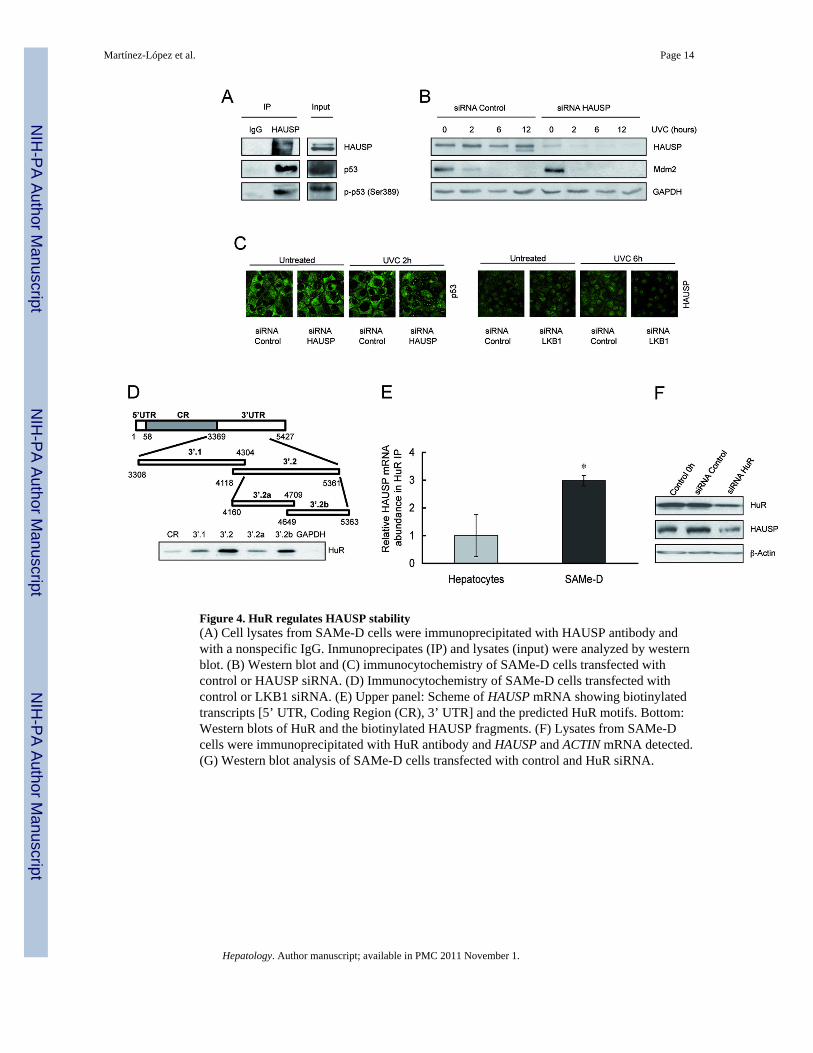
Figure 4. HuR regulates HAUSP stability(A) Cell lysates from SAMe-D cells were immunoprecipitated with HAUSP antibody andwith a nonspecific IgG. Inmunoprecipates (IP) and lysates (input) were analyzed by westernblot. (B) Western blot and (C) immunocytochemistry of SAMe-D cells transfected withcontrol or HAUSP siRNA. (D) Immunocytochemistry of SAMe-D cells transfected withcontrol or LKB1 siRNA. (E) Upper panel: Scheme of HAUSP mRNA showing biotinylatedtranscripts [5’ UTR, Coding Region (CR), 3’ UTR] and the predicted HuR motifs. Bottom:Western blots of HuR and the biotinylated HAUSP fragments. (F) Lysates from SAMe-Dcells were immunoprecipitated with HuR antibody and HAUSP and ACTIN mRNA detected.(G) Western blot analysis of SAMe-D cells transfected with control and HuR siRNA.
Martínez-López et al. Page 14
Hepatology. Author manuscript; available in PMC 2011 November 1.
NIH
-PA Author Manuscript
NIH
-PA Author Manuscript
NIH
-PA Author Manuscript

Figure 5. HuR activity is regulated by LKB1Immunocytochemical analysis of (A) hepatocytes and SAMe-D or (B) SAMe-D cellstransfected with control or LKB1 siRNA and treated with UVC light.
Martínez-López et al. Page 15
Hepatology. Author manuscript; available in PMC 2011 November 1.
NIH
-PA Author Manuscript
NIH
-PA Author Manuscript
NIH
-PA Author Manuscript

Figure 6. p53 and p-LKB1 in HCCHematoxylin-eosin staining and immunohistochemical analysis of p53 and p-LKB1 proteinin liver samples from (A) MAT1A-KO tumors and (B) human HCC of ASH and NASHetiology.
Martínez-López et al. Page 16
Hepatology. Author manuscript; available in PMC 2011 November 1.
NIH
-PA Author Manuscript
NIH
-PA Author Manuscript
NIH
-PA Author Manuscript

Figure 7. Schematic representation of p53 behavior in SAMe-D cells(A) In normal cells p53 is kept at low levels due to the activity of its negative regulatorMdm2. Mdm2, binds and poly-ubiquitinates p53 for proteasomal degradation. The de-ubiquitinating enzyme HAUSP contributes to the stability of Mdm2, impairing its self-ubiquitilation and degradation. (B) In SAMe-D cells, p53 is mostly cytosolic andhyperphosphorylated by several kinases, such as p-LKB1, which is basally hyperactivated.The hyperphosphorylation of p53 and its interaction with HAUSP blocks the negativeregulation by Mdm2. p-LKB1 is responsible for two more processes: (1) the activation ofsurvival Akt which leads to the phosphorylation of Mdm2 decreasing the HAUSP-Mdm2interaction, and (2) the cytosolic translocation of HuR, which stabilizes p53 and HAUSPmRNA.
Martínez-López et al. Page 17
Hepatology. Author manuscript; available in PMC 2011 November 1.
NIH
-PA Author Manuscript
NIH
-PA Author Manuscript
NIH
-PA Author Manuscript