Accelerating Membrane Insertion of Peripheral Proteins with a Novel Membrane Mimetic Model
10
Accelerating Membrane Insertion of Peripheral Proteins with a Novel Membrane Mimetic Model Y. Zenmei Ohkubo, Taras V. Pogorelov, Mark J. Arcario, Geoff A. Christensen, and Emad Tajkhorshid* Department of Biochemistry, Beckman Institute for Advanced Science and Technology, and Center for Biophysics and Computational Biology, University of Illinois at Urbana-Champaign, Urbana, Illinois ABSTRACT Characterizing atomic details of membrane binding of peripheral membrane proteins by molecular dynamics (MD) has been significantly hindered by the slow dynamics of membrane reorganization associated with the phenomena. To expedite lateral diffusion of lipid molecules without sacrificing the atomic details of such interactions, we have developed a novel membrane representation, to our knowledge, termed the highly mobile membrane-mimetic (HMMM) model to study binding and insertion of various molecular species into the membrane. The HMMM model takes advantage of an organic solvent layer to represent the hydrophobic core of the membrane and short-tailed phospholipids for the headgroup region. We demonstrate that using these components, bilayer structures are formed spontaneously and rapidly, regardless of the initial position and orientation of the lipids. In the HMMM membrane, lipid molecules exhibit one to two orders of magnitude enhancement in lateral diffusion. At the same time, the membrane atomic density profile of the headgroup region produced by the HMMM model is essentially identical to those obtained for full-membrane models, indicating the faithful representation of the membrane surface by the model. We demonstrate the efficiency of the model in capturing spontaneous binding and insertion of peripheral proteins by using the membrane anchor (g-carboxyglutamic-acid-rich domain; GLA domain) of human coagulation factor VII as a test model. Achieving full insertion of the GLA domain consistently in 10 independent unbiased simulations within short simulation times clearly indicates the robustness of the HMMM model in capturing membrane association of peripheral proteins very effi- ciently and reproducibly. The HMMM model will provide significant improvements to the current all-atom models by accelerating lipid dynamics to examine protein-membrane interactions more efficiently. INTRODUCTION The cellular membrane plays an important role in signaling, transport, and metabolic processes in all living cells. In hu- mans, nearly one third of the 34,000 identified proteins are estimated to function in membrane-associated forms (1). Though initially perceived as a passive environment, the central role of the membrane not only in proper function, but also in tight regulation of membrane-associated proteins and phenomena has been well established (2–4). Membrane-mediated effects on protein function can be generally attributed to two major mechanisms: the overall mechanical impact of the embedding lipid bilayer on protein structure and dynamics (5–7) and specific interactions between the membrane lipids (particularly the headgroups that form the membrane surface) and their respective binding sites in the protein (8–12). Furthermore, the overall membrane affinity of a given protein can also be modulated through variation of the lipid composition of the membrane (13–16). Indeed, this mechanism is widely used in biolog- ical systems to target and localize peripheral proteins to distinct areas of the membrane or toward specific cell types, and to regulate their activity in a lipid-dependent manner. Despite the central role of the membrane in protein func- tion and activity, due to the semifluid and highly dynamical nature of lipid bilayers, obtaining sufficiently detailed struc- tural information on peripheral proteins in their membrane- bound form has proven extremely challenging. Limited information on the proximity of certain regions in membrane-binding proteins can be inferred from experi- mental methods such as small-angle x-ray scattering (17), electron paramagnetic resonance (18,19), Fo ¨rster resonance energy transfer (18,20,21), and mutagenesis studies (22,23). Nevertheless, more details are needed to fully char- acterize the nature of protein-lipid interactions that are crit- ical for binding and function of peripheral proteins. Another challenging aspect of studying peripheral proteins is that they often employ relatively small membrane- engaging parts (membrane anchors). Making predictions about such domains is far more difficult than for integral membrane proteins, whose orientation and depth of membrane insertion can be fairly reliably predicted by well-established protocols (24–26). Molecular dynamics (MD) offers a promising method of gaining structural and dynamical information on such protein-lipid complexes and of characterizing the interactions therein that govern the membrane-controlled effects on protein function. The method combines high spatial Submitted September 29, 2011, and accepted for publication March 6, 2012. *Correspondence: [email protected] Geoff A. Christensen’s present address is Department of Biochemistry, University of Missouri, Columbia, MO. This is an Open Access article distributed under the terms of the Creative Commons-Attribution Noncommercial License (http://creativecommons. org/licenses/by-nc/2.0/), which permits unrestricted noncommercial use, distribution, and reproduction in any medium, provided the original work is properly cited. Editor: Scott Feller. Ó 2012 by the Biophysical Society 0006-3495/12/05/2130/10 $2.00 doi: 10.1016/j.bpj.2012.03.015 2130 Biophysical Journal Volume 102 May 2012 2130–2139
Transcript of Accelerating Membrane Insertion of Peripheral Proteins with a Novel Membrane Mimetic Model

2130 Biophysical Journal Volume 102 May 2012 2130–2139
Accelerating Membrane Insertion of Peripheral Proteins with a NovelMembrane Mimetic Model
Y. Zenmei Ohkubo, Taras V. Pogorelov, Mark J. Arcario, Geoff A. Christensen, and Emad Tajkhorshid*Department of Biochemistry, Beckman Institute for Advanced Science and Technology, and Center for Biophysics and Computational Biology,University of Illinois at Urbana-Champaign, Urbana, Illinois
ABSTRACT Characterizing atomic details of membrane binding of peripheral membrane proteins bymolecular dynamics (MD)has been significantly hindered by the slow dynamics of membrane reorganization associated with the phenomena. To expeditelateral diffusion of lipid molecules without sacrificing the atomic details of such interactions, we have developed a novelmembrane representation, to our knowledge, termed the highly mobile membrane-mimetic (HMMM) model to study bindingand insertion of various molecular species into the membrane. The HMMM model takes advantage of an organic solvent layerto represent the hydrophobic core of the membrane and short-tailed phospholipids for the headgroup region. We demonstratethat using these components, bilayer structures are formed spontaneously and rapidly, regardless of the initial position andorientation of the lipids. In the HMMMmembrane, lipid molecules exhibit one to two orders of magnitude enhancement in lateraldiffusion. At the same time, the membrane atomic density profile of the headgroup region produced by the HMMM model isessentially identical to those obtained for full-membrane models, indicating the faithful representation of the membrane surfaceby the model. We demonstrate the efficiency of the model in capturing spontaneous binding and insertion of peripheral proteinsby using the membrane anchor (g-carboxyglutamic-acid-rich domain; GLA domain) of human coagulation factor VII as a testmodel. Achieving full insertion of the GLA domain consistently in 10 independent unbiased simulations within short simulationtimes clearly indicates the robustness of the HMMM model in capturing membrane association of peripheral proteins very effi-ciently and reproducibly. The HMMMmodel will provide significant improvements to the current all-atom models by acceleratinglipid dynamics to examine protein-membrane interactions more efficiently.
INTRODUCTION
The cellular membrane plays an important role in signaling,transport, and metabolic processes in all living cells. In hu-mans, nearly one third of the 34,000 identified proteins areestimated to function in membrane-associated forms (1).Though initially perceived as a passive environment, thecentral role of the membrane not only in proper function,but also in tight regulation of membrane-associated proteinsand phenomena has been well established (2–4).Membrane-mediated effects on protein function can begenerally attributed to two major mechanisms: the overallmechanical impact of the embedding lipid bilayer on proteinstructure and dynamics (5–7) and specific interactionsbetween the membrane lipids (particularly the headgroupsthat form the membrane surface) and their respectivebinding sites in the protein (8–12). Furthermore, the overallmembrane affinity of a given protein can also be modulatedthrough variation of the lipid composition of the membrane(13–16). Indeed, this mechanism is widely used in biolog-
Submitted September 29, 2011, and accepted for publicationMarch 6, 2012.
*Correspondence: [email protected]
Geoff A. Christensen’s present address is Department of Biochemistry,
University of Missouri, Columbia, MO.
This is an Open Access article distributed under the terms of the Creative
Commons-Attribution Noncommercial License (http://creativecommons.
org/licenses/by-nc/2.0/), which permits unrestricted noncommercial use,
distribution, and reproduction in any medium, provided the original work
is properly cited.
Editor: Scott Feller.
� 2012 by the Biophysical Society
0006-3495/12/05/2130/10 $2.00
ical systems to target and localize peripheral proteins todistinct areas of the membrane or toward specific cell types,and to regulate their activity in a lipid-dependent manner.
Despite the central role of the membrane in protein func-tion and activity, due to the semifluid and highly dynamicalnature of lipid bilayers, obtaining sufficiently detailed struc-tural information on peripheral proteins in their membrane-bound form has proven extremely challenging. Limitedinformation on the proximity of certain regions inmembrane-binding proteins can be inferred from experi-mental methods such as small-angle x-ray scattering (17),electron paramagnetic resonance (18,19), Forster resonanceenergy transfer (18,20,21), and mutagenesis studies(22,23). Nevertheless, more details are needed to fully char-acterize the nature of protein-lipid interactions that are crit-ical for binding and function of peripheral proteins.Another challenging aspect of studying peripheral proteinsis that they often employ relatively small membrane-engaging parts (membrane anchors). Making predictionsabout such domains is far more difficult than for integralmembrane proteins, whose orientation and depth ofmembrane insertion can be fairly reliably predicted bywell-established protocols (24–26).
Molecular dynamics (MD) offers a promising method ofgaining structural and dynamical information on suchprotein-lipid complexes and of characterizing the interactionstherein that govern the membrane-controlled effects onprotein function. The method combines high spatial
doi: 10.1016/j.bpj.2012.03.015

Highly Mobile Membrane Mimetic Model 2131
(sub-Angstrom) and temporal (femtosecond) resolutions witha dynamical description, thereby offering an ideal approachtoward studying membrane-associated phenomena. Unfortu-nately, membrane lipids are known to exhibit slow dynamicson the timescales achievable by atomistic MD simulations ofmembrane proteins, which are currently performed routinelyonly for tens to hundreds of nanoseconds (27–29). The slowlateral diffusion of membrane lipids at ambient temperaturesis typically on the order of 10�8 cm2 s�1, requiring simulationsat least an order of magnitude longer to describe.
To fill this timescale gap, coarse-grained (CG) MD hasbeen employed as an efficient method (30–37), in whichthe number of particles in the simulated system is reducedby representing groups of atoms (such as a moiety, a wholeside chain, or even several residues) by a CG bead. CGmodels have successfully described various properties oflipid structures and associated phenomena, e.g., sponta-neous formation of lipid bilayers (30), formation of thegramicidin A (gA) ion channel (38), formation of micelle(31) and disclike (39) structures, helix dimerization of gly-cophorin A (GpA) in a bilayer (32), and membrane sculpt-ing (34) and liposome remodeling (35) induced by BARdomains. The efficiency of the CG method, however, isachieved by sacrificing the atomic resolution, which is indis-pensable for accurate description of specific protein-lipidinteractions. In addition to CG methods, the generalizedBorn theory (40), based on Poisson-Boltzmann continuumelectrostatics, has been utilized in the construction ofimplicit membranes (41) to probe the binding of peripheralproteins to the membrane. The method has been success-fully used to describe the partitioning (42,43) and even olig-omerization of peptides within the membrane (44). Similarto CG models, implicit representations do not take into
account specific protein-lipid interactions that are key tobinding and activation of peripheral proteins. Moreover,these models ignore the crucial headgroup region of thebilayer, treating the interface as an abrupt switch froma polar region to a nonpolar region.
As an alternative approach, we present what to our knowl-edge is a novel membrane representation for atomistic MDsimulations, termed the highly mobile membrane-mimetic(HMMM) model, which is composed of a combination ofa biphasic solvent system (45) and short-tailed lipids(st-lipids) at the aqueous-organic interface (Fig. 1). Wedemonstrate that the designed st-lipids spontaneously formbilayerlike structures and that the lipid diffusion has beenaccelerated by 1–2 orders of magnitude in the resultingbilayer. The results indicate that the HMMM model is idealfor studying membrane-associated phenomena via MDwithout compromising the atomic details necessary for accu-rate description of the interactions between the headgroupsand the protein. Furthermore, we use the membrane-anchoring domain of a coagulation factor to demonstratethe efficiency of theHMMMmodel in capturing spontaneousmembrane association and insertion of peripheral proteinswithout imposing any bias or external force. Coagulationproteins constitute a prominent class of peripheralmembraneproteins whose function is highly dependent on, and indeedregulated by, their ability to bind to specific regions of biolog-ical membranes (46). Modulation of the lipid composition ofthe membrane, and thus the membrane affinity of the coagu-lation proteins, is one of the main physiological regulatorymechanisms in the clotting cascade (7,13). The lipid depen-dence of these processes makes coagulation factors an idealcase for testing the robustness of the HMMM model incapturing key lipid-protein interactions at an atomic level.
FIGURE 1 Contrasting the HMMM and
conventional representations of a membrane. In
an HMMM model, a large fraction of the acyl tails
(left, yellow sticks) of the membrane-forming
lipids is replaced by a liquid organic phase (right,
yellow area). In this study, a full DOPS lipid mole-
cule (inset) is represented by a short-tailed DVPS
molecule (the circled fraction of the molecule),
and the space vacated by the removal of the C6–
C18 portion of the acyl tails is filled with an organic
solvent (yellow). Oxygen atoms are red, nitrogen
blue, phosphorus gold, and carbon ice blue, except
the C6–C18, which are in yellow. Bulk water mole-
cules are shown in light blue.
Biophysical Journal 102(9) 2130–2139

2132 Ohkubo et al.
MATERIALS AND METHODS
Here, we provide a brief description of the simulation systems and condi-
tions used for this study. A detailed description of the methods is provided
in the Supporting Material. Table 1 summarizes all the simulation systems
described in this study.
HMMM simulations
In the HMMM membrane used in this study, 1,1-dichloroethane (DCLE) is
used to model the hydrophobic core, and divalerylphosphatidylserine
(DVPS) molecules to represent the phosphatidylserine (PS) lipids. To inves-
tigate the efficiency and robustness of the HMMMmodel, its ability to form
and maintain bilayers was tested in a simulation in which the st-lipids were
initially distributed on a grid spanning the whole volume of the biphasic
solvent box, with approximately half of the molecules in the organic phase
and the other half in water (Fig. 2, upper left). Another set of five simula-
tions were performed to calculate the lateral diffusion constant of the lipids
in the HMMM model and its dependence on different configurations.
Full-membrane simulations
As a reference, a full membrane composed of 288 1,2-dioleoyl-sn-glycero-
3-phospho-L-serine (DOPS) molecules was also simulated with either Naþ
or Ca2þ as the counterion (we refer to these conventional membrane models
as full membranes to contrast them with the HMMM model). The system
was simulated for 20 ns as an NPnAT (where Pn is the constant
membrane-normal pressure and A is the membrane area) ensemble and
using the conditions described elsewhere (47).
Insertion of the GLA domain into the HMMMmodel
Ten independent simulations were performed to test the ability and effi-
ciency of the HMMMmodel in capturingmembrane binding of a membrane
anchor in unbiased simulations. The g-carboxyglutamic-acid-rich (GLA)
domain of hFVII with seven bound Ca2þ ions (48), preequilibrated in solu-
TABLE 1 System configurations
System Lipid n Counterion AL (A2)
D
(10�8cm2 s�1)
HMMM Simulations
HMMM-0 DVPS 5 Naþ 133.0 —
HMMM-1 DVPS 5 Naþ 294.0 314.0
HMMM-2 DVPS 5 Naþ 144.0 171.0
HMMM-3 DVPS 5 Ca2þ 144.0 134.0
HMMM-4 DVPS 5 Naþ 100.0 97.7
HMMM-5 DVPS 5 Naþ 68.6 26.4
GLA Domain Binding
HMMM-GLA (�10) DVPS 5 Naþ 127.0 —
Full Membrane Simulations
Full-1 DOPS 18 Naþ 65.3 4.93
Full-2 DOPS 18 Ca2þ 65.3 2.10
Systems simulated in this study for spontaneous membrane formation
(HMMM-0), diffusion constant calculation (HMMM-1 to HMMM-5,
Full-1, and Full-2), and GLA-domain binding (HMMM-GLA) are listed
along with the membrane-forming lipid, the number of acyl tail carbons
(n), the counterion, the area/lipid (AL), and the measured lateral diffusion
constant of the lipids (D). —, not measured. Simulation of system
HMMM-GLA was repeated 10 times. An expanded version of this table
including more details is provided as Supporting Material.
Biophysical Journal 102(9) 2130–2139
tion (49) was positioned in the bulk water at least 8 A above the membrane
(see Fig. 5). The system was then fully equilibrated for 50 ns.
Simulation protocols
All the simulations were performed using NAMD2 (50) with the
CHARMM36 force field parameter set (51), the CMAP corrections (52)
for proteins, and the TIP3P model for water (53) in the NPnAT ensemble.
Langevin dynamics with a damping coefficient, g, of 0.5 ps�1 and Langevin
piston Nose-Hoover methods (54,55) were employed to maintain the
temperature at 310 K and the pressure at 1 atm, respectively. To evaluate
long-range electrostatic forces without truncation, the particle mesh Ewald
(PME) method (56), with a grid density of slightly finer than 1 A�3, was
used. The cut-off for van der Waals interaction was set at 12 A. Integration
time steps were set at 2, 2, and 4 fs for bonded, nonbonded, and PME calcu-
lations, respectively. For the simulations of full DOPS membranes, integra-
tion time steps of 1, 1, and 2 fs (for bonded, nonbonded, and PME) and
CHARMM27 force field parameters were used; other conditions were the
same as described above.
RESULTS AND DISCUSSION
Spontaneous formation of stable bilayerby the HMMM model
To examine whether the employed st-lipids partition prop-erly into the interfacial region of the biphasic solvent usedin the HMMM model, and to test their robustness towardforming and sustaining a bilayer structure within affordablesimulation time, a 40-ns-long MD simulation was per-formed in which the st-lipids (54 DVPS molecules) werescattered initially on a grid spanning the two solvent phaseswith approximately half of the lipids in the organic phaseand the other half in the water phase (Fig. 2). The DVPSmolecules rapidly partitioned to the organic-aqueous inter-face, with their headgroups optimally positioned in waterand their acyl tails immersed in the organic phase. The st-lipids initially positioned in the organic phase exhibitedfaster partitioning; within<6 ns all of the lipids in this phaseemerge from the organic layer, with 72% of them (13 lipids)doing so within only 1 ns and 83% (15 st-lipids) within 2 ns(Fig. 2). DVPS lipids in the aqueous phase remained inwater longer, on average, before partitioning to the inter-face. Nevertheless, 83% of water-immersed DVPS lipidsreached the interface within 10 ns, with the remaining threemolecules completing their partitioning into the interfacewithin 14, 16, and 20 ns. This behavior is fully expectedfrom a surfactantlike molecule such as DVPS, whosecharged headgroup establishes strong favorable interactionwith water. Overall, the entire process of the formation ofthe bilayer is very fast and completed within 20 ns. Oncepartitioned into the interface, DVPS lipids remain therefor the remainder of the 40-ns simulation (Fig. 2; data shownonly for the first 22 ns). The results clearly indicate the effi-ciency of the model toward capturing the formation ofa stable bilayerlike structure within short and affordablesimulation times without compromising the atomic resolu-tion of the system.

FIGURE 2 Spontaneous formation of the
HMMM membrane. The formation of a bilayer-
like structure is depicted using snapshots taken
from the membrane-formation simulation at t ¼0, 0.25, 2, 7, and 17 ns (upper, left to right).
Short-tailed DVPS molecules were initially scat-
tered over the simulation box on a grid to have
an even distribution among different phases. The
positions along the membrane normal, z, of indi-
vidual phosphorus atoms of DVPS molecules as
a function of the simulation time is shown with
red lines (lower left). Also shown is a snapshot of
the system (to scale) at t ¼ 22 ns that depicts the
fully formed bilayer (lower right). The z coordi-
nate for individual lipids has been calculated in
each frame in reference to the actual center of
mass (COM) of the organic phase, i.e., in each
frame, z ¼ 0 is set to the COM of the DCLE layer.
Note that the time axis is in the log scale for t ¼0–2 ns and linear for the rest, to highlight fast
movement of the lipids during the early phase of
the simulation.
Highly Mobile Membrane Mimetic Model 2133
A key attribute of a membrane-mimetic model is itsability to reproduce the structure and atomic distributionof various chemical groups within the membrane, especiallyin the headgroup region, where specific interactions withcoagulation proteins are formed. To examine this aspect ofthe HMMM model, atomic density profiles were calculatedfor the HMMM bilayer structure and compared with thoseof a full DOPS membrane (Fig. 3). Regarding interactionwith a coagulation protein, the most important characteris-tics of these profiles are the positions and shapes of thedensity peaks for the headgroup moieties, which exhibitremarkable similarities in the two profiles (Fig. 3). Theextent of hydration and the degree of counterion penetrationare also similar for the two different membrane representa-tions. The results clearly indicate that the HMMM modelsuccessfully captures the essential properties of a fullmembrane, despite the marked difference in acyl tail length(18 carbons for DOPS vs. 5 for DVPS) and lipid density.Minor differences include the relative distribution of the car-boxy carbons, which indicates a more upright arrangementof the lipids in the DOPS membrane compared to themembrane composed of DVPS, an effect that is partiallydue to the lower lipid density of the latter model.
Enhanced lipid mobility in the HMMM model
The main objective in constructing the HMMM model wasto achieve enhanced mobility of the lipid molecules within
the membrane without compromising atomic resolutionof the headgroups. Fig. 4 A compares the lipid mobility ofthe HMMM and full-membrane models. The enhancedmobility of the lipids in the HMMM model is quite evident,even though the trajectory used for plotting full lipids is10 times longer than the one used for the HMMM model(10 ns vs. 1 ns). Although DVPS lipids rapidly exchangepositions within only 1 ns (average RMSDxy of over10 A), DOPS lipids essentially retain their initial positions(average RMSDxy of only ~2 A) within the 10-ns segmentused to generate the plot (Fig. 4). The calculated lateraldiffusion constant, D (Eq. S1 in the Supporting Material),for the HMMM and full-membrane models are 2.5 �10�6 cm2 s�1 and 4.0 � 10�8 cm2 s�1, respectively, indi-cating the enhanced lateral lipid diffusion achieved by theHMMM model.
A more systematic comparison of the lateral diffusionconstants for the HMMM models constructed and simu-lated using different configurations (different counterionsand area/lipid values, AL) is provided in Fig. 4 B. Theaverage slopes of the mean-square displacement of thephosphorus atoms as a function of the time lag, D(t),defined in Eq. S1, are plotted for full DOPS membranesand different HMMM membranes. The lateral diffusionconstant, D, was quantified as the asymptote of D(t) byaveraging D(t) over larger t (10–20 ns), with the calcu-lated values also summarized in the rightmost column ofTable 1.
Biophysical Journal 102(9) 2130–2139

FIGURE 3 Structural comparison of full and HMMM membranes. The
membrane profiles of a full DOPS membrane (upper) and an HMMM
DVPS membrane (lower) aligned by the peak of the PO�4 groups. The
atomic densities are plotted along the membrane normal, z, using a bin
size of 1 A for most of the heavy atoms: oxygen of bulk water (blue
line, �0.1), Ca2þ counterion (red), carboxy carbon (thin red), amine
nitrogen (thin black), phosphate phosphorus (black), carbonyl carbon
(thin black), C2 (black), C3 (green), C4 (red), C5 (blue), and C1 of DCLE
(maroon,�0.1). Note that the distance between the COMs of the membrane
and the PO�4 peak is different for the two membrane models (20.0 A for
DOPS and 33.0 A for DVPS) due to different membrane thicknesses. The
difference in the absolute heights of the density peaks are related to the
different AL of the two membranes (AL of 65.3 A2 for DOPS vs. 133 A2
for DVPS). The lefthand-side y axes (atomic density) are scaled so that
the PO�4 peaks are shown on the same scale.
2134 Ohkubo et al.
When simulated at an experimental area/lipid ratio, AL,with Naþ as the counterion, D of PS lipids is enhanced bymore than a factor of 5 for the HMMM model (2.64 �10�7 cm2 s�1, compared to 4.93 � 10�8 cm2 s�1 for a fullmembrane composed of DOPS; Table 1), an effect mainlycaused by shortening of the acyl tails. Around an order ofmagnitude further enhancement in lateral mobility is attainedby reducing the lipid density, e.g., 1.71 � 10�6 cm2 s�1 and3.14 � 10�6 cm2 s�1 for DVPS with AL ¼ 144 A2 and294 A2, respectively.
In comparison with Naþ ions, divalent Ca2þ counterionsinteract with PS headgroups more strongly and form farlonger-lived lipid-ion clusters, rigidifying the membranelipids (57) and thus resulting in a smaller D for both the
Biophysical Journal 102(9) 2130–2139
fullDOPSmembrane andHMMMmodels studied.Thecalcu-lated D for the DOPS membrane is 4.93 � 10�8 cm2 s�1
when simulated with Naþ, and 2.10 � 10�8 cm2 s�1 withCa2þ. This counterion effect is also clearly present in theHMMM simulations, although to a lesser degree, e.g.,1.71 � 10�6 cm2 s�1 with Naþ vs. 1.34 � 10�6 cm2 s�1
with Ca2þ.
Spontaneous membrane insertion of coagulationfactor VII
The main motivation for developing the HMMM model isto have an efficient model that allows for accelerated estab-lishment of lipid-protein interactions and a membrane suffi-ciently mobile to allow the insertion of proteins to becaptured without the need of additional forces or biasedpotential within affordable simulation times. To test theaccuracy and efficiency of the HMMMmodel in this regard,the membrane-anchoring domain of a coagulation proteinwas used as an example. Binding to specific regions ofthe cellular membrane constitutes a pivotal step in the acti-vation of almost all coagulation factors. Through largelyunknown mechanisms, membrane binding and assemblyof coagulation proteins result in an increase of severalorders of magnitude in their catalytic activity, a processvital to the entire clotting cascade (58). The importanceof membrane binding in the function of these proteins canbe best highlighted by the presence of specializedmembrane anchors in almost all coagulation factors, de-signed to bind to specific areas of the membrane that arerich in anionic lipids, particularly PS (58). Specifically,for the simulations presented here, we have used the GLAdomain of hFVII. The GLA domain is a commonmembrane-anchoring domain in coagulation proteins andis known to bind to PS-rich regions of the cellularmembrane. In one of our earlier reports (49), we con-structed a membrane-bound model of the same anchoringdomain by using steered MD (SMD) (59,60) simulationsin which external forces were employed for insertion ofthe GLA domain of hFVII into a full DOPS membraneand the subsequent extended free equilibration. Althoughin that study the membrane-bound model was developedthrough biased simulations and computational costpermitted only one simulation (49), its comparison withthe results obtained here will be helpful in further assessingthe quality of the resulting structures from the HMMMsimulations.
Given the efficiency of the HMMM model, the simula-tions of membrane insertion of the GLA domain wererepeated 10 times, resulting in spontaneous insertion ofthe domain into the membrane in all cases. Here, weprovide details of a typical scenario qualitatively describingthe process for one of the simulations (Fig. 5; several addi-tional examples are also provided in the Supporting Mate-rial). The GLA domain, initially placed in bulk water

FIGURE 4 Enhanced lipid lateral diffusion in
the HMMM membrane. (A) Comparison of the
trajectory of phosphorus atoms within the
membrane in a 10-ns-long segment of a simulation
of a full DOPS membrane (left) with a 1-ns-long
segment of a simulation of an HMMM DVPS
membrane (right). In the DVPS membrane
segment, some of the trajectory lines are depicted
in red to make the distinction between different
lipids more clear. Despite an order-of-magnitude-
shorter simulation, the HMMM model exhibits
clearly much larger lateral mobility for the lipids.
(B) Lateral diffusion constants of DOPS and
DVPS in different environments. The slope of the
mean-square displacement of lipids (within the xy
plane) as a function of time lag is plotted for
membranes composed of pure DOPS (thin lines)
or DVPS (thick lines) neutralized with either Naþ
(black) or Ca2þ (red). The phosphate phosphorus
atoms were used for the calculations. The area/
lipid, AL, of individual systems is also shown.
Highly Mobile Membrane Mimetic Model 2135
with the tip of the N-terminal u-loop ~8 A away from thesurface of the HMMM membrane (i.e., the headgroups ofDVPS lipids; cf. Fig. 3), approaches the membrane withinthe first 1 ns, establishing direct interaction with the PSheadgroups by its basic side chains (Arg9 and Lys18),leaning on the N-terminal helix side (Fig. 5, upper right).By t ¼ 2.5 ns, the u-loop has completely landed on, andis fully engaged with, the lipid surface (Fig. 5). The GLAdomain continues its rapid insertion into the DVPSmembrane, with the bound Ca2þ ions reaching the carboxylayer of PS lipids around 3 ns, at which point the GLAdomain recovers a more upright configuration on thesurface of the membrane. The structurally bound Ca2þ
ions observed in the established membrane-bound modelof the GLA domain by t ¼ 6 ns (Fig. 5, middle right) showsa level of penetration into the membrane (converged to thelevel of the lipid phosphates) essentially identical to that ob-tained by >60 ns of SMD in a full DOPS membrane (Fig. 5of Ohkubo and Tajkhorshid (49)).
The GLA domain completes its insertion into themembrane rather quickly during the first part of the simu-lation and does not further penetrate the membrane duringthe remainder of the simulation. Nevertheless, specific
contacts between the PS headgroups and the GLA domaincontinue to form over a longer timescale (Fig. 5). Thecontact pattern between the GLA-bound Ca2þ ions andthe PS headgroups (Fig. 6, upper) indicates that, similarto the results obtained from full-DOPS-membrane simula-tions (49), the HMMM model also captures two distinctroles for the GLA-bound Ca2þ ions. The Ca2þ ions locatedtoward the periphery of the GLA domain participateprimarily in direct interaction with the headgroups,whereas the ions in the middle remain buried inside theprotein, thereby stabilizing the fold of the membraneanchor. The HMMM model, therefore, captures the essen-tial features of membrane binding of the GLA domain,including the insertion depth and the orientation of theprotein on the membrane as well as the specific patternsof lipid-Ca2þ interactions observed in full membranesystems (49). However, we also observe additional patternsof interaction between basic side chains and the head-groups in the HMMM model. For example, Lys32, whichhas been reported to affect membrane binding (61), formscontacts with both the phosphate and the carboxy groups inthe HMMM model, whereas it did not do so in the fullDOPS membrane simulation (49). We note that the above
Biophysical Journal 102(9) 2130–2139
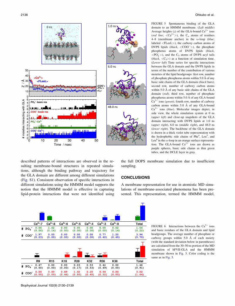
FIGURE 5 Spontaneous binding of the GLA
domain to an HMMM membrane. (Left middle)
Average heights (z) of the GLA-bound Ca2þ ions
(red line; <Ca2þ>), the Ca atoms of residues
4–8 (membrane anchor) in the u-loop (blue;
labeled <FLeeL>), the carboxy carbon atoms of
DVPS lipids (black, <COO�>), the phosphate
phosphorus atoms of DVPS lipids (black,
<PO�4 >), and the C5 atoms of DVPS acyl tails
(black, <C5>) as a function of simulation time.
(Lower left) Time series for specific interactions
between the GLA domain and the DVPS lipids in
terms of the number of the contribution of various
moieties of the lipid headgroups: first row, number
of phosphate phosphorus atoms within 5.0 A of any
basic side chains of the GLA domain (black bars);
second row, number of carboxy carbon atoms
within 5.0 A of any basic side chains of the GLA
domain (red); third row, number of phosphate
phosphorus atoms within 5.0 A of any GLA-bound
Ca2þ ions (green); fourth row, number of carboxy
carbon atoms within 5.0 A of any GLA-bound
Ca2þ ions (blue). Molecular images depict, in
side view, the whole simulation system at 0 ns
(upper left) and close-up snapshots of the GLA
domain interacting with DVPS lipids at 1.0 ns
(upper right), 6.0 ns (middle right), and 48.0 ns
(lower right). The backbone of the GLA domain
is drawn in a thick violet tube representation with
the hydrophobic side chains of Phe4, Leu5, and
Leu8 in the u-loop in an orange surface representa-
tion. The GLA-bound Ca2þ ions are drawn as
purple spheres, basic side chains as thin green
tubes, and the DCLE layer in gray.
2136 Ohkubo et al.
described patterns of interactions are observed in the re-sulting membrane-bound structures in repeated simula-tions, although the binding pathway and trajectory forthe GLA domain are different among different simulations(Fig. S1). Consistent observation of specific interactions indifferent simulations using the HMMM model supports thenotion that the HMMM model is effective in capturinglipid-protein interactions that were not identified using
Biophysical Journal 102(9) 2130–2139
the full DOPS membrane simulation due to insufficientsampling.
CONCLUSIONS
A membrane representation for use in atomistic MD simu-lations of membrane-associated phenomena has been pre-sented. This representation, termed the HMMM model,
FIGURE 6 Interactions between the Ca2þ ions
and basic residues of the GLA domain and lipid
headgroups. The average number of phosphate or
carboxy groups within 5.0 A of each moiety
(with the standard deviation below in parentheses)
are calculated from the 30–50 ns portion of the MD
simulation of hFVII-GLA and the HMMM
membrane shown in Fig. 5. Color coding is the
same as in Fig. 5.
Highly Mobile Membrane Mimetic Model 2137
which to our knowledge is new, is based on the conceptuallynovel idea of ‘‘selective fragmentation’’, i.e., replacing longacyl chains of lipids with an organic phase, in which thehydrophobic interactions are well represented. The atom-istic details of the lipid headgroups are fully preserved byhaving st-lipids occupy the aqueous/organic interfacialregion(s). We have demonstrated the unparalleled efficiencyof this atomistic model in describing the self-assemblyprocess of lipids into stable bilayerlike structures withinseveral nanoseconds.
The lateral diffusion constant of the lipids in the studiedHMMM membranes is in the range of 10�5–10�6 cm2s�1,which exhibits an increase of one to two orders of magni-tude in comparison to the conventional full-membranerepresentation. This improvement will allow for more effi-cient sampling of headgroup configurations, for example,during the binding and insertion of peripheral proteins.Using the membrane-binding domain of a coagulationprotein (the GLA domain of hFVII) as an example, wehave also demonstrated the robustness of the HMMMmodelin describing spontaneous membrane insertion of peripheralproteins and in capturing membrane-bound forms of theseproteins at the all-atom level within routinely accessiblesimulation times. Given the efficiency of the model, wehave been able to simulate the process of membrane inser-tion 10 times and achieved a converged picture and anunprecedented level of statistics on the lipid-protein interac-tions involved in this process.
The HMMM model can be readily used in MD simula-tions employing the standard and generalized (62,63)ensembles. The fully atomistic representation of the modelmakes it easy to use for any atomistic force field of choice,avoiding the need to develop a model-specific force field(64) or repeated transformation to different resolutions(65,66). Most important, the central concept of ‘‘selectivefragmentation’’ (representing the membrane by smallerfragments) in the HMMM model may be further extendedand used in other lipid structures, or even more generallyin other molecular systems.
In addition to the large number of peripheral membraneproteins, the HMMM model can potentially be used forstudying any membrane-associated phenomena in whichatomistic details are desired but slow diffusion of the fullyrepresented lipids is prohibitive of observing suchphenomena. The immediate applications of the HMMMmodel include such membrane phenomena as diffusionand domain formation of lipids (67) and proteins (68,69),pore formation by (antimicrobial) peptides (65,66,70,71),hydrophobic matching of transmembrane helices (72), andmembrane-associated assembly of coagulation factors(73–75). We note, however, that the HMMM model is bydesign not expected to reproduce all characteristics ofa full membrane; in particular, mechanical properties suchas volume/area compressibility and bending moduli can beonly partially captured by the model (preliminary results
not shown). The main purpose of the model is to providea more flexible and mobile environment that allows for rapidrearrangement and displacement of the lipid headgroups,thereby facilitating any phenomenon that might be inacces-sible with conventional membrane models due to the inher-ently slow dynamics of full lipids therein.
Finally, we note that the resulting membrane-boundmodels developed by the HMMMmodel can be readily con-verted into full-membrane representations using variousmolecular modeling techniques. For example, one mightmutate the st-lipids into full lipids in one step or alterna-tively grow the lipid tails gradually (one carbon or two ata time) to their full length while subjecting the system toMD relaxations at each step. Our preliminary applicationsof such approaches have yielded promising results.
SUPPORTING MATERIAL
Materials and Methods in detail, a table summarizing all simulated systems,
three figures illustrating the results of three more examples of spontaneous
insertion of the GLA domain into HMMM membranes, and references
(76,77) are available at http://www.biophysj.org/biophysj/supplemental/
S0006-3495(12)00325-6.
Full membrane simulations were performed by G.A.C. as part of his B.Sci.
thesis in Biochemistry. The authors thank Dr. Eduardo Cruz-Chu for tech-
nical assistance. All simulations were performed on TeraGrid resources of
the National Science Foundation (grant No. MCA06N060) mainly on the
Big Red cluster at Indiana University and the Kraken cluster at NICS.
This work was supported by grants from the National Institutes of Health
(R01-GM086749, R01-GM067887, U54-GM087519, and P41-RR05969
to E.T. and Molecular Biophysics Training Grant to M.J.A.).
REFERENCES
1. Casadio, R., P. Fariselli,., G. von Heijne. 2008. The state of the art ofmembrane protein structure prediction: from sequence to 3D structure.In Modern Genome Annotation: The BioSapiens Network.D. Frishman and A. Valecia, editors. Springer-Verlag, Vienna.309–328.
2. Carruthers, A., and D. L. Melchior. 1986. How bilayer lipids affectmembrane protein activity. Trends Biochem. Sci. 11:331–335.
3. Edidin, M. 2003. Lipids on the frontier: a century of cell-membranebilayers. Nat. Rev. Mol. Cell Biol. 4:414–418.
4. Phillips, R., T. Ursell, ., P. Sens. 2009. Emerging roles for lipids inshaping membrane-protein function. Nature. 459:379–385.
5. Andersen, O. S., and R. E. Koeppe, 2nd. 2007. Bilayer thickness andmembrane protein function: an energetic perspective. Annu. Rev.Biophys. Biomol. Struct. 36:107–130.
6. Sachs, J. N., and D. M. Engelman. 2006. Introduction to the membraneprotein reviews: the interplay of structure, dynamics, and environmentin membrane protein function. Annu. Rev. Biochem. 75:707–712.
7. Tavoosi, N., R. L. Davis-Harrison,., J. H. Morrissey. 2011. Moleculardeterminants of phospholipid synergy in blood clotting. J. Biol. Chem.286:23247–23253.
8. Huang, M., A. C. Rigby, ., B. C. Furie. 2003. Structural basis ofmembrane binding by Gla domains of vitamin K-dependent proteins.Nat. Struct. Biol. 10:751–756.
Biophysical Journal 102(9) 2130–2139

2138 Ohkubo et al.
9. Ohkubo, Y. Z., J. H. Morrissey, and E. Tajkhorshid. 2010. Dynamicalview of membrane binding and complex formation of human factorVIIa and tissue factor. J. Thromb. Haemost. 8:1044–1053.
10. Igarashi, K., M. Kaneda, ., M. Umeda. 1995. A novel phosphatidyl-serine-binding peptide motif defined by an anti-idiotypic monoclonalantibody. Localization of phosphatidylserine-specific binding sites onprotein kinase C and phosphatidylserine decarboxylase. J. Biol.Chem. 270:29075–29078.
11. Goldschmidt-Clermont, P. J., L. M. Machesky,., T. D. Pollard. 1990.The actin-binding protein profilin binds to PIP2 and inhibits its hydro-lysis by phospholipase C. Science. 247:1575–1578.
12. Hamada, K., T. Shimizu, ., T. Hakoshima. 2000. Structural basis ofthe membrane-targeting and unmasking mechanisms of the radixinFERM domain. EMBO J. 19:4449–4462.
13. Shaw, A. W., V. S. Pureza, ., J. H. Morrissey. 2007. The local phos-pholipid environment modulates the activation of blood clotting.J. Biol. Chem. 282:6556–6563.
14. Martel, V., C. Racaud-Sultan, ., C. Albiges-Rizo. 2001. Conforma-tion, localization, and integrin binding of talin depend on its interactionwith phosphoinositides. J. Biol. Chem. 276:21217–21227.
15. Gudi, S., J. P. Nolan, and J. A. Frangos. 1998. Modulation of GTPaseactivity of G proteins by fluid shear stress and phospholipid composi-tion. Proc. Natl. Acad. Sci. USA. 95:2515–2519.
16. Sunshine, C., and M. G. McNamee. 1994. Lipid modulation of nico-tinic aceytlcholine receptor function: the role of membrane lipidcomposition and fluidity. Biochim. Biophys. Acta. 1191:59–64.
17. Denisov, I. G., Y. V. Grinkova, ., S. G. Sligar. 2004. Directed self-assembly of monodisperse phospholipid bilayer nanodiscs withcontrolled size. J. Am. Chem. Soc. 126:3477–3487.
18. Kohout, S. C., S. Corbalan-Garcıa, ., J. J. Falke. 2003. C2 domain ofprotein kinase Ca: elucidation of the membrane docking surface bysite-directed fluorescence and spin labeling. Biochemistry. 42:1254–1265.
19. Lin, Y., R. Nielsen, ., M. H. Gelb. 1998. Docking phospholipase A2
on membranes using electrostatic potential-modulated spin relaxationmagnetic resonance. Science. 279:1925–1929.
20. Lakowicz, J. R. 1999. Principles of Fluorescence Spectroscopy, 2nd ed.Springer, New York.
21. McCallum, C. D., B. Su, ., A. E. Johnson. 1997. Tissue factor posi-tions and maintains the factor VIIa active site far above the membranesurface even in the absence of the factor VIIa Gla domain. A fluores-cence resonance energy transfer study. J. Biol. Chem. 272:30160–30166.
22. Nelsestuen, G. L. 1999. Enhancement of vitamin-K-dependent proteinfunction by modification of the g-carboxyglutamic acid domain:studies of protein C and factor VII. Trends Cardiovasc. Med. 9:162–167.
23. Harvey, S. B., M. D. Stone,., G. L. Nelsestuen. 2003. Mutagenesis ofthe g-carboxyglutamic acid domain of human factor VII to generatemaximum enhancement of the membrane contact site. J. Biol. Chem.278:8363–8369.
24. Woolf, T. B., and B. Roux. 1994. Molecular dynamics simulation of thegramicidin channel in a phospholipid bilayer. Proc. Natl. Acad. Sci.USA. 91:11631–11635.
25. Lomize, A. L., I. D. Pogozheva,., H. I. Mosberg. 2006. Positioning ofproteins in membranes: a computational approach. Protein Sci.15:1318–1333.
26. Jo, S., T. Kim, and W. Im. 2007. Automated builder and database ofprotein/membrane complexes for molecular dynamics simulations.PLoS ONE. 2:e880.
27. Arkhipov, A., Y. Yin, and K. Schulten. 2009. Membrane-bendingmechanism of amphiphysin N-BAR domains. Biophys. J. 97:2727–2735.
28. Grossfield, A., S. E. Feller, and M. C. Pitman. 2006. A role for directinteractions in the modulation of rhodopsin by u-3 polyunsaturatedlipids. Proc. Natl. Acad. Sci. USA. 103:4888–4893.
Biophysical Journal 102(9) 2130–2139
29. Sgourakis, N. G., and A. E. Garcia. 2010. The membrane complexbetween transducin and dark-state rhodopsin exhibits large-amplitudeinterface dynamics on the sub-microsecond timescale: insights fromall-atom MD simulations. J. Mol. Biol. 398:161–173.
30. Shelley, J. C., M. Y. Shelley, ., M. L. Klein. 2001. Simulations ofphospholipids using a coarse grain model. J. Phys. Chem. B. 105:9785–9792.
31. Marrink, S. J., A. H. de Vries, and A. E. Mark. 2004. Coarse grainedmodel for semiquantitative lipid simulations. J. Phys. Chem. B. 108:750–760.
32. Bond, P. J., and M. S. P. Sansom. 2006. Insertion and assembly ofmembrane proteins via simulation. J. Am. Chem. Soc. 128:2697–2704.
33. Marrink, S. J., H. J. Risselada,., A. H. de Vries. 2007. The MARTINIforce field: coarse grained model for biomolecular simulations. J. Phys.Chem. B. 111:7812–7824.
34. Arkhipov, A., Y. Yin, and K. Schulten. 2008. Four-scale description ofmembrane sculpting by BAR domains. Biophys. J. 95:2806–2821.
35. Ayton, G. S., E. Lyman, and G. A. Voth. 2010. Hierarchical coarse-graining strategy for protein-membrane systems to access mesoscopicscales. Faraday Discuss. 144:347–357, discussion 445–481.
36. Izvekov, S., and G. A. Voth. 2005. A multiscale coarse-graining methodfor biomolecular systems. J. Phys. Chem. B. 109:2469–2473.
37. Izvekov, S., and G. A. Voth. 2005. Multiscale coarse graining of liquid-state systems. J. Chem. Phys. 123:134105.
38. Shi, Q., S. Izvekov, and G. A. Voth. 2006. Mixed atomistic and coarse-grained molecular dynamics: simulation of a membrane-bound ionchannel. J. Phys. Chem. B. 110:15045–15048.
39. de Joannis, J., F. Y. Jiang, and J. T. Kindt. 2006. Coarse-grained modelsimulations of mixed-lipid systems: composition and line tension ofa stabilized bilayer edge. Langmuir. 22:998–1005.
40. Feig, M., and C. L. Brooks, 3rd. 2004. Recent advances in the develop-ment and application of implicit solvent models in biomolecule simu-lations. Curr. Opin. Struct. Biol. 14:217–224.
41. Chen, J., W. Im, and C. L. Brooks, 3rd. 2006. Balancing solvation andintramolecular interactions: toward a consistent generalized Born forcefield. J. Am. Chem. Soc. 128:3728–3736.
42. Im, W., and C. L. Brooks, 3rd. 2005. Interfacial folding and membraneinsertion of designed peptides studied by molecular dynamics simula-tions. Proc. Natl. Acad. Sci. USA. 102:6771–6776.
43. Mondal, J., X. Zhu,., A. Yethiraj. 2010. Sequence-dependent interac-tion of b-peptides with membranes. J. Phys. Chem. B. 114:13585–13592.
44. Bu, L., W. Im, and C. L. Brooks, 3rd. 2007. Membrane assembly ofsimple helix homo-oligomers studied via molecular dynamics simula-tions. Biophys. J. 92:854–863.
45. Arcario, M. J., Y. Z. Ohkubo, and E. Tajkhorshid. 2011. Capturingspontaneous partitioning of peripheral proteins using a biphasicmembrane-mimetic model. J. Phys. Chem. B. 115:7029–7037.
46. Stace, C. L., and N. T. Ktistakis. 2006. Phosphatidic acid- and phospha-tidylserine-binding proteins. Biochim. Biophys. Acta. 1761:913–926.
47. Wang, Y., Y. Z. Ohkubo, and E. Tajkhorshid. 2008. Gas conduction oflipid bilayers and membrane channels. In Current Topics inMembranes: Computational Modeling of Membrane Bilayers.S. Feller, editor. Academic Press, New York. 343–367.
48. Banner, D. W., A. D’Arcy, ., D. Kirchhofer. 1996. The crystal struc-ture of the complex of blood coagulation factor VIIa with soluble tissuefactor. Nature. 380:41–46.
49. Ohkubo, Y. Z., and E. Tajkhorshid. 2008. Distinct structural and adhe-sive roles of Ca2þ in membrane binding of blood coagulation factors.Structure. 16:72–81.
50. Phillips, J. C., R. Braun, ., K. Schulten. 2005. Scalable moleculardynamics with NAMD. J. Comput. Chem. 26:1781–1802.
51. Vanommeslaeghe, K., E. Hatcher, ., A. D. Mackerell, Jr. 2010.CHARMM general force field: A force field for drug-like molecules
Highly Mobile Membrane Mimetic Model 2139
compatible with the CHARMM all-atom additive biological forcefields. J. Comput. Chem. 31:671–690.
52. Mackerell, Jr., A. D., M. Feig, and C. L. Brooks, 3rd. 2004. Extendingthe treatment of backbone energetics in protein force fields: limitationsof gas-phase quantum mechanics in reproducing protein conforma-tional distributions in molecular dynamics simulations. J. Comput.Chem. 25:1400–1415.
53. Jorgensen, W. L., J. Chandrasekhar,., M. L. Klein. 1983. Comparisonof simple potential functions for simulating liquid water. J. Chem.Phys. 79:926–935.
54. Martyna, G. J., D. J. Tobias, and M. L. Klein. 1994. Constant pressuremolecular dynamics algorithms. J. Chem. Phys. 101:4177–4189.
55. Feller, S. E., Y. Zhang, ., B. R. Brooks. 1995. Constant pressuremolecular dynamics simulation: the Langevin piston method.J. Chem. Phys. 103:4613–4621.
56. Darden, T., D. York, and L. G. Pedersen. 1993. Particle mesh Ewald:An N log(N) method for Ewald sums in large systems. J. Chem.Phys. 98:10089–10092.
57. Boettcher, J. M., R. L. Davis-Harrison, ., C. M. Rienstra. 2011.Atomic view of calcium-induced clustering of phosphatidylserine inmixed lipid bilayers. Biochemistry. 50:2264–2273.
58. Zwaal, R. F. A., P. Comfurius, and E. M. Bevers. 1998. Lipid-proteininteractions in blood coagulation. Biochim. Biophys. Acta. 1376:433–453.
59. Isralewitz, B., M. Gao, and K. Schulten. 2001. Steered moleculardynamics and mechanical functions of proteins. Curr. Opin. Struct.Biol. 11:224–230.
60. Sotomayor, M., and K. Schulten. 2007. Single-molecule experimentsin vitro and in silico. Science. 316:1144–1148.
61. Shah, A. M., W. Kisiel,., G. L. Nelsestuen. 1998. Manipulation of themembrane binding site of vitamin K-dependent proteins: enhanced bio-logical function of human factor VII. Proc. Natl. Acad. Sci. USA.95:4229–4234.
62. Sugita, Y., and Y. Okamoto. 1999. Replica-exchange moleculardynamics method for protein folding. Chem. Phys. Lett. 314:141–151.
63. Mitsutake, A., Y. Sugita, and Y. Okamoto. 2001. Generalized-ensemblealgorithms for molecular simulations of biopolymers. Biopolymers.60:96–123.
64. Yesylevskyy, S. O., L. V. Schafer, ., S. J. Marrink. 2010. Polarizablewater model for the coarse-grained MARTINI force field. PLOS Com-put. Biol. 6:e1000810.
65. Thøgersen, L., B. Schiøtt, ., E. Tajkhorshid. 2008. Peptide aggrega-tion and pore formation in a lipid bilayer: a combined coarse-grainedand all atom molecular dynamics study. Biophys. J. 95:4337–4347.
66. Rzepiela, A. J., D. Sengupta, ., S. J. Marrink. 2010. Membrane pora-tion by antimicrobial peptides combining atomistic and coarse-graineddescriptions. Faraday Discuss. 144:431–443, discussion 445–481.
67. Apajalahti, T., P. Niemela,., I. Vattulainen. 2010. Concerted diffusionof lipids in raft-like membranes. Faraday Discuss. 144:411–430,discussion 445–481.
68. Mbamala, E. C., A. Ben-Shaul, and S. May. 2005. Domain formationinduced by the adsorption of charged proteins on mixed lipidmembranes. Biophys. J. 88:1702–1714.
69. Khelashvili, G., H. Weinstein, and D. Harries. 2008. Protein diffusionon charged membranes: a dynamic mean-field model describes timeevolution and lipid reorganization. Biophys. J. 94:2580–2597.
70. Psachoulia, E., D. P. Marshall, and M. S. P. Sansom. 2010. Moleculardynamics simulations of the dimerization of transmembrane a-helices.Acc. Chem. Res. 43:388–396.
71. Tew, G. N., R. W. Scott, ., W. F. Degrado. 2010. De novo design ofantimicrobial polymers, foldamers, and small molecules: fromdiscovery to practical applications. Acc. Chem. Res. 43:30–39.
72. Yin, F., and J. T. Kindt. 2010. Atomistic simulation of hydrophobicmatching effects on lipid composition near a helical peptide embeddedin mixed-lipid bilayers. J. Phys. Chem. B. 114:8076–8080.
73. Adams, T. E., M. F. Hockin,., S. J. Everse. 2004. The crystal structureof activated protein C-inactivated bovine factor Va: Implications forcofactor function. Proc. Natl. Acad. Sci. USA. 101:8918–8923.
74. Ngo, J. C. K., M. Huang,., B. Furie. 2008. Crystal structure of humanfactor VIII: implications for the formation of the factor IXa-factorVIIIa complex. Structure. 16:597–606.
75. Lee, C. J., V. Chandrasekaran, ., L. G. Pedersen. 2010. Recent esti-mates of the structure of the factor VIIa (FVIIa)/tissue factor (TF)and factor Xa (FXa) ternary complex. Thromb. Res. 125 (Suppl 1):S7–S10.
76. Roark, M., and S. E. Feller. 2009. Molecular dynamics simulationstudy of correlated motions in phospholipid bilayer membranes.J. Phys. Chem. B. 113:13229–13234.
77. Petrache, H. I., S. Tristram-Nagle, ., J. F. Nagle. 2004. Structure andfluctuations of charged phosphatidylserine bilayers in the absence ofsalt. Biophys. J. 86:1574–1586.
Biophysical Journal 102(9) 2130–2139