Abstracts - SWISS KNIFE
84
SGC Journal SSC Abstracts 101. Jahreskongress der Schweizerischen Gesellschaft für Chirurgie 101 e congrès annuel de la Société Suisse de Chirurgie 20 credits www.chirurgiekongress.ch Bern/Berne, 21–23 Mai/mai 2014 swiss knife 2014; 11: special edition
-
Upload
khangminh22 -
Category
Documents
-
view
3 -
download
0
Transcript of Abstracts - SWISS KNIFE

SGC Journal SSC
Abstracts101. Jahreskongress der Schweizerischen Gesellschaft für Chirurgie101e congrès annuel de la Société Suisse de Chirurgie
20 credits
www.chirurgiekongress.ch
Bern/Berne, 21–23 Mai/mai 2014 swiss knife 2014; 11: special edition
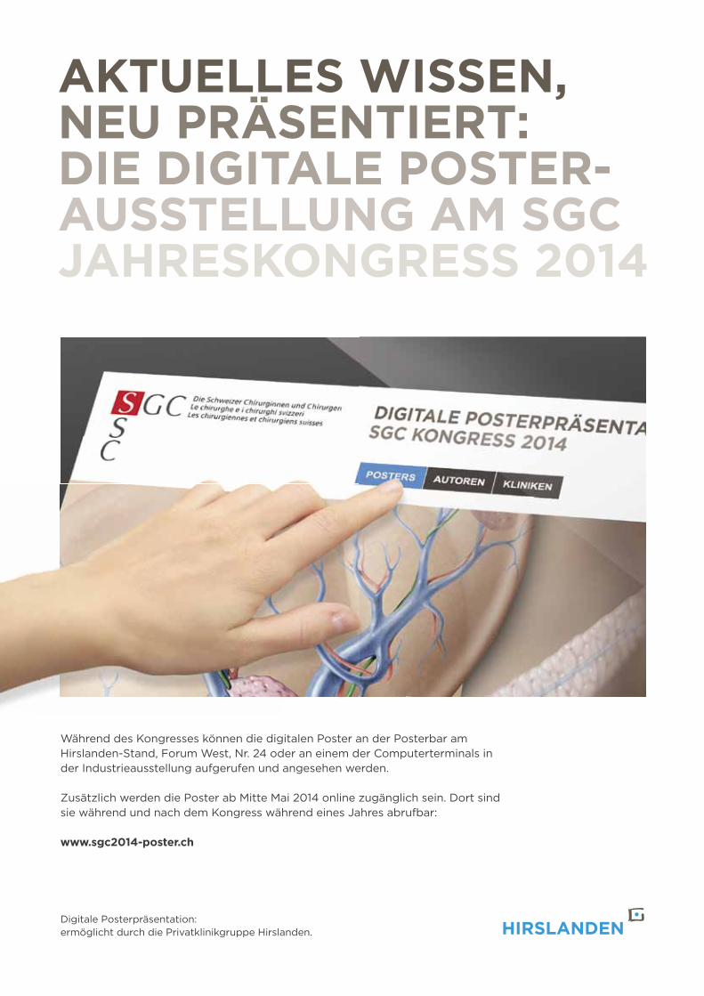
AKTUELLES WISSEN, NEU PRÄSENTIERT: DIE DIGITALE POSTER-AUSSTELLUNG AM SGC JAHRES KONGRESS 2014
Während des Kongresses können die digitalen Poster an der Posterbar am Hirslanden- Stand, Forum West, Nr. 24 oder an einem der Computerterminals in der Industrie ausstellung aufgerufen und angesehen werden.
Zusätzlich werden die Poster ab Mitte Mai 2014 online zugänglich sein. Dort sind sie während und nach dem Kongress während eines Jahres abrufbar:
www.sgc2014-poster.ch
Digitale Posterpräsentation: ermöglicht durch die Privatklinikgruppe Hirslanden.
14044_Inserat_SGC_A4.indd 1 17.04.14 09:40

Editorial
Dear colleagues & friends,
The present 11th special edition of swiss knife is offered
aigan in a pdf format on the web.
Enjoy reading a selection of this year’s abstracts with
colored illustrations!
On behalf of the editorial board of swiss knife I wish you a
highly interactive congress of the Swiss Society of Surgery
2014 in Berne!
Markus Zuber M.D.
Guest editor
swiss knife, special edition 2014
Le Congrès 2014 de la Société Suisse de Chirurgie
Kongress 2014 der Schweizerischen Gesellschaft für Chirurgie
swiss knife 2014; 11: special edition 3
2015102e congrès annuel de la Société Suisse de Chirurgie
Berne, 20 – 22 may 2015
Abstract Deadline: 12. janvier 2015
2015102. Jahreskongress der Schweizerischen Gesellschaft für Chirurgie
Bern, 20. – 22. Mai 2015
Abstract-Deadline: 12. Januar 2015

COVIDIEN, COVIDIEN with logo, Covidien logo and ™ marked brands are U.S. and/or internationally registered trademarks of Covidien AG© 2009 Covidien. All rigths reserved. - V-VS-P-LPIce/GB
LigaSure Precise™. Get Cooler.
Consistent seals at lower temperatures
V VS P LPIce GB_Layout 1 04.03.14 12:06 Seite 1

Content
ImpressumHerausgeber: Schweizerische Gesellschaft für Chirurgie SGC/SSC, Seltisbergerstrasse 16, CH-4419 Lupsingen, Switzerland, Tel. +41 (0)61 815 96 60, [email protected] in Zusammenarbeit mit Frehner Consulting AG, Unternehmensberatung für PR, CH-9014 St.Gallen, Tel. +41 (0)71 272 60 80, [email protected] Produktion und Inseratemarketing: MetroComm AG, Bahnhofstrasse 8, CH-9001 St.Gallen, Tel. +41 (0)71 272 80 50, [email protected] Projektleitung: Prof. Dr. Markus Zuber Redaktion: Prof. Dr. Markus Zuber Projektverantwortung: Dr. Stephan Ziegler Ge-schäftsleitung: Natal Schnetzer Fotos: Bodo Rüedi, swiss-image.ch, zVg Anzeigenleitung: Herbert Keller Gestaltung: Beatrice Lang swiss knife special edition 2014 (may)Nachdruck, auch auszugsweise, nur mit Genehmigung der MetroComm AG. Sonderausgabe des offiziellen Publikationsorgans swiss knife der Schweizerischen Gesellschaft für Chirurgie SGC-SSC. Sonderausgabe erscheint jährlich. Für alle Mitglieder der Schweizerischen Gesellschaft für Chirurgie SGC-SSC.
swiss knife 2014; 11: special edition 5
Abstracts
Session Topic Page
07 Upper GI 07
09 Transplantation 08
11 Lower Extremities 10
16 Vascular Surgery I 11
17 Bariatric Surgery 13
21 Video I 15
23 Endocrine Surgery 16
26 Thoracic Surgery I 16
27 Cancer 18
28 Emergencies & Polytraumas 20
29 Video II 22
33 Colorectal Cancer 22
34 Thoracic Surgery II 24
35 Inflammation 25
41 Vascular Surgery II 27
42 Diverticulitis 29
44 Regeneration in Transplantation 31
46 Video III 33
48 Felix Largiadèr Prize Session 34
49 Clinical Research 36
50 SGACT & SGVC 38
51 Basic Science 39
53 Upper Extremities 40
54 Video IV 42
58 Colon, Rectum, & Proctology 43
61 Osteoporosis 45
65 Posters I 47
66 Hepato-Pancreatico-Biliary Surgery 49
71 Posters II 52
72 Perioperative Management 57
73 Mixed Session I 59
74 Visceral Surgery 61
77 Mixed Session II 64
P Posters 67
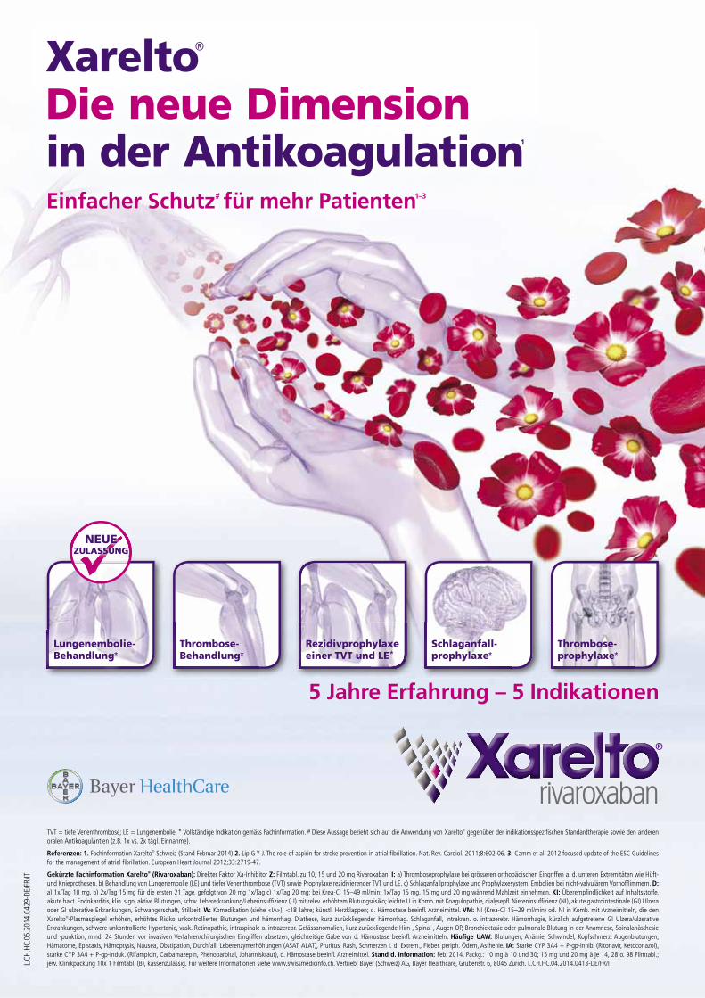
Der erste ORALE, direkte Faktor-Xa-Inhibitor
Antikoagulation – so einfach wie noch nie*TVT = tiefe Venenthrombose; LE = Lungenembolie. * Vollständige Indikation gemäss Fachinformation. # Diese Aussage bezieht sich auf die Anwendung von Xarelto® gegenüber der indikationsspezifi schen Standardtherapie sowie den anderen oralen Antikoagulantien (z.B. 1x vs. 2x tägl. Einnahme).
Referenzen: 1. Fachinformation Xarelto® Schweiz (Stand Februar 2014) 2. Lip G Y J. The role of aspirin for stroke prevention in atrial fi brillation. Nat. Rev. Cardiol. 2011;8:602-06. 3. Camm et al. 2012 focused update of the ESC Guidelines for the management of atrial fi brillation. European Heart Journal 2012;33:2719-47.
Gekürzte Fachinformation Xarelto® (Rivaroxaban): Direkter Faktor Xa-Inhibitor Z: Filmtabl. zu 10, 15 und 20 mg Rivaroxaban. I: a) Thromboseprophylaxe bei grösseren orthopädischen Eingriffen a. d. unteren Extremitäten wie Hüft- und Knieprothesen. b) Behandlung von Lungenembolie (LE) und tiefer Venenthrombose (TVT) sowie Prophy laxe rezidivierender TVT und LE. c) Schlaganfallprophylaxe und Prophylaxesystem. Embolien bei nicht-valvulärem Vorhoffl immern. D: a) 1x/Tag 10 mg. b) 2x/Tag 15 mg für die ersten 21 Tage, gefolgt von 20 mg 1x/Tag c) 1x/Tag 20 mg; bei Krea-Cl 15–49 ml/min: 1x/Tag 15 mg. 15 mg und 20 mg während Mahlzeit einnehmen. KI: Überempfi ndlichkeit auf Inhaltsstoffe, akute bakt. Endokarditis, klin. sign. aktive Blutungen, schw. Lebererkrankung/Leberinsuffi zienz (LI) mit relev. erhöhtem Blutungsrisiko; leichte LI in Komb. mit Koagulopathie, dialysepfl . Niereninsuffi zienz (NI), akute gastrointestinale (GI) Ulzera oder GI ulzerative Erkrankungen, Schwangerschaft, Stillzeit. W: Komedikation (siehe «IA»); <18 Jahre; künstl. Herzklappen; d. Hämostase beeinfl . Arzneimittel. VM: NI (Krea-Cl 15–29 ml/min) od. NI in Komb. mit Arzneimitteln, die den Xarelto®-Plasmaspiegel erhöhen, erhöhtes Risiko unkontrollierter Blutungen und hämorrhag. Diathese, kurz zurückliegender hämorrhag. Schlaganfall, intrakran. o. intrazerebr. Hämorrhagie, kürzlich aufgetretene GI Ulzera/ulzerative Erkrankungen, schwere unkontrollierte Hypertonie, vask. Retinopathie, intraspinale o. intrazerebr. Gefässanomalien, kurz zurückliegende Hirn-, Spinal-, Augen-OP, Bronchiektasie oder pulmonale Blutung in der Anamnese, Spinalanästhesie und -punktion, mind. 24 Stunden vor invasiven Verfahren/chirurgischen Eingriffen absetzen, gleichzeitige Gabe von d. Hämostase beeinfl . Arzneimitteln. Häufi ge UAW: Blutungen, Anämie, Schwindel, Kopfschmerz, Augenblutungen, Hämatome, Epistaxis, Hämoptysis, Nausea, Obstipation, Durchfall, Leberenzymerhöhungen (ASAT, ALAT), Pruritus, Rash, Schmerzen i. d. Extrem., Fieber, periph. Ödem, Asthenie. IA: Starke CYP 3A4 + P-gp-Inhib. (Ritonavir, Ketoconazol), starke CYP 3A4 + P-gp-Induk. (Rifampicin, Carbamazepin, Phenobarbital, Johanniskraut), d. Hämostase beeinfl . Arz neimittel. Stand d. Information: Feb. 2014. Packg.: 10 mg à 10 und 30; 15 mg und 20 mg à je 14, 28 o. 98 Filmtabl.; jew. Klinikpackung 10x 1 Filmtabl. (B), kassenzulässig. Für weitere Informationen siehe www.swissmedicinfo.ch. Vertrieb: Bayer (Schweiz) AG, Bayer Healthcare, Grubenstr. 6, 8045 Zürich. L.CH.HC.04.2014.0413-DE/FR/IT
Einfacher Schutz# für mehr Patienten1–3Einfacher Schutz für mehr Patienten
Xarelto®
Die neue Dimension in der Antikoagulation1
5 Jahre Erfahrung – 5 Indikationen
Thrombose-Behandlung*
Lungenembolie-Behandlung*
Thrombose-prophylaxe*
Schlaganfall-prophylaxe*
Rezidivprophylaxeeiner TVT und LE*
NEUEZULASSUNG
L.CH.
HC.0
5.20
14.0
429-
DE/F
R/IT
Xarelto_Ins_2014_A4_dt.indd 1 07.05.14 14:02
swiss knife 2014; 11: special edition 7
Upper GI 077.1Analysis of risk factors related to venous thromboembolic events after oncological esophagectomyS. Mantziari1, C. Gronnier2, N. Demartines1, C. Mariette2, M. Schäfer1 (1Lausanne, 2Lille/FR)
Objective: Esophageal cancer patients are at increased risk of venous thromboembolic events (VTE) during the postoperative course. Reported incidence rates of pulmonary thromboembolism (PTE) and deep venous thrombosis (DVT) reach up to 2.5% and 7.3-11% respectively, although real incidences of asymptomatic events risk being even higher. The aim of this study was to assess the relationship between several perioperative parameters and the occurrence of thromboembolic events (VTE) after oncological esophagectomy.Methods: All patients undergoing esophagectomy for cancer from 2000 to 2010 in France have been included in the prospective French National Esophagus Cancer Database. This data base has been used for the current study. In a first step, an univariate analysis performed and 54 clinically relevant peri- and intraoperative variables were analyzed as potential risk factors related to VTE by means of the chi-square test, Fischer’s test or the Mann-Whitney test. Variables of clinical significance and those with a P-value of <=0.1 were then included in a backward stepwise multiple logistic regression to identify significant predictors for VTE. Significance threshold was set at p<=0.05.Results: There were 2944 patients included in the present study, 2427 men and 517 women with a median age of 63 years (range 41-83). Overall incidence for VTE was 2.85% (84 patients) with 44 PTE (1.49%) and 40 DVT cases (1.36%). Mortality was significantly higher in VTE patients (29% ver-sus 7.36%, p <=0.001) and mean hospital stay was also longer in this group (33 ±24 versus 25±21 days, p<=0.001). Multivariate analysis identified ASA class III/ IV (p=0.08), pulmonary complications excluding PE (p=0.041), pneumonia (p=0.02) and ARDS (p=0.15) as independent risk factors for thromboembolic complications.Conclusion: Based on these results, patients with an ASA class III or IV who develop pulmonary com-plications, especially pneumonia and ARDS, can be considered as a high-risk subgroup for throm-boembolic events after oncological esophagectomy. Given the high mortality rates and the significant increase of hospital stay related to this potentially preventable complication, individualized anticoagu-lation strategies must be discussed for these patients.
7.2Timing of surgery after neoadjuvant radiochemotherapy of locally advanced esophageal cancerL. Stoll, S. Däster, A. Zettl, M. von Flüe, C. Ackermann (Basel)
Objective: Neoadjuvant radiochemotherapy for locally advanced esophageal cancer is a widely ac-cepted treatment. In most of the therapy protocols surgery is commonly performed 4 to 6 weeks after completion of radiochemotherapy. There is evidence from other cancer studies that patients might benefit from prolonging the interval to surgery beyond 6 to 8 weeks. In our study we investigated, if patients who underwent surgery after more than 8 weeks showed a better histopathological response and if later surgery was associated with higher morbidity.Methods: 48 patients with a locally advanced (T3 or N+) carcinoma of the esophagus and the gastro-esophageal junction were included. All patients underwent neoadjuvant radiochemotherapy followed by abdominothoracal esophago-gastrectomy (Ivor Lewis). Patients were divided into an early group (surgery within 8 weeks) and a late group (surgery after more than 8 weeks) after neoadjuvant treat-ment. Histopathological response according to Mandards` tumour regression grade and postopera-tive morbidity was compared between the early and the late groupResults: Mean interval to surgery after completion of neoadjuvant therapy was 44 days in the early group (n=36) and 62 days in the late group (n=12) ( p<0,05). The rate of responders (tumour regres-sion grade 1 and 2) was similar in both groups: 61% in the early group, 67% in the late group (n.s.). Postoperative morbidity was not significantly different in both groups.Conclusion: Increasing the interval between neoadjuvant radiochemotherapy and surgery beyond 8 weeks is not associated with higher postoperative morbidity. In our investigation there was no signifi-cantly better histopathological response when patients underwent surgery later than 8 weeks. The op-timal timing of surgery still remains unclear, but the slight trend to a better histopathological response after more than 8 weeks after completion of neoadjuvant treatment could indicate a possible benefit for these patients.
7.3The Merendino procedure for benign or early malignant lesions of the distal esophagus/gastro-esophageal junction is feasible, safe and functionally satisfyingM. Walensi, A. Zuse, S. Münchow, R. Meier, C. A. Maurer (Liestal)
Objective: To analyse retrospectively the early postoperative and functional outcome of patients (pts) after Merendino procedure for benign or early malignant lesions of the distal part of the esophagus or the gastroesophageal junction.Methods: Between 2004 and 2013, 14 pts had transhiatal resection of median 8 (6-10) cm of distal esophagus including the gastroesophageal junction. Locoregional lymphadenectomy was performed in all patients with (suspected) early adenocarcinoma. In case of vagus nerve resection, pyloromyec-tomy and cholecystectomy were done. A pediculated isoperistaltic jejunal segment of 15-18 cm length was interponed between esophageal stump and remaining stomach by end-to-side anastomoses. At least one postoperative contrast examination of the anastomoses was done in all pts. Quality of life was assessed by using a modification of the EORTC QLQ-OES18 survey, recording patients’ general activity and condition and specific gastrointestinal symptoms.
Results: Pts (m: 12, f: 2) had a median age of 71 (32-91) years. The following ASA scores were re-corded: ASA 1, 2, 3, and 4 in 1, 7, 5, and 1 pt(s), respectively. The indications for Merendino procedure were: early esophageal cancer (n = 5), high-grade dysplasia (n = 3), esophageal perforation (n = 3), GIST (n = 2), and peptic stenosis (n = 1). Median intraoperative blood loss was 250 (75-600) ml and median hospitalization time was 21 (3-58) days. Median number of examined lymph nodes in pts with (suspected) early adenocarcinoma was 11 (4-16). No postoperative mortality occurred and no patient needed reoperation during hospitalization. No anastomotic leakage was observed. The follow-ing postoperative complications were recorded: pneumonia (n = 4), pleural effusion (n = 2) and need for tracheotomy (n = 1). One patient required readmission and reoperation 6 weeks after surgery due to chylothorax. The functional results with regard to postoperative gastroesophageal reflux, dysphagia, weight loss and malnutrition were good to excellent in all pts.Conclusion: In the present series, the Merendino procedure proved to be feasible and safe, even in aged patients and emergencies, and to provide satisfying functional results. The Merendino procedure should belong to the armamentarium of an upper GI surgeon.
7.4Perioperative outcomes of esophageal cancer surgery – analysis of a 10-year-periodS. Däster, L. Stoll, M. von Flüe, C. Ackermann (Basel)
Objective: Centralization of esophageal cancer surgery to high-volume institutions has been shown to improve perioperative outcomes in several studies. However, there is an ongoing debate, whether defined minimal annual hospital volumes for esophagectomies are required for quality assurance. The aim of the study was to assess perioperative outcomes of esophagectomies in a single institution in Switzerland.Methods: Data from a prospective database of esophagectomies performed between 2004 and 2013 was analyzed regarding tumor characteristics and perioperative outcomes. All operated esophageal cancers and cancers of the esophagogastric junction were included into the study. Perioperative mor-bidity was assessed according to the Clavien-Dindo classification. Postoperative mortality was defined as death from any cause within the same hospital stay.Results: A total of 143 esophagectomies (126 transthoracic, 17 transhiatal resections) were per-formed in the surveyed 10-year-period (mean annual hospital volume of 14.3). Two surgeons per-formed 91% of all procedures. 121 (85%) were adenocarcinomas and 22 (15%) squamous cell car-cinomas; 71% of patients received neoadjuvant treatment. Complications with a Clavien-Dindo score of III/IV (requiring surgical, endoscopic, or radiological intervention) occurred in 19 cases (13.4%), including 2 patients with chylothorax (1.4%). The overall anastomotic leak rate was 2%. Pulmonary complications were the most frequent postoperative problems (21% of patients). In-hospital mortality was 0.7%. Mean length of hospital stay was 18 days in patients with no complication and 24 days if there was any complication (p < 0.0001).Conclusion: Esophagectomy is a complex operation with significant risk of morbidity. The most com-mon postoperative problems are pulmonary complications, usually responding well to non-invasive treatment. Appropriate patient selection and a comprehensive multidisciplinary care pathway can provide a low perioperative mortality rate, also in a mid-volume institution.
7.518-FDG PET-CT in the preoperative workup of esophageal cancer: a useful insight into tumour biologyS. Mantziari, A. Pomoni, P. Mitsakis, N. Demartines, J. Prior, M. Schäfer (Lausanne)
Objective: 18F-fluorodeoxyglucose (FDG) PET/CT is increasingly used to the preoperative workup of esophageal cancer patients, mostly in the research of distant metastases. However, its role in the metabolic imaging of the tumor has been only partly elucidated. Aim of our study was to investigate the prognostic value of 18-FDG PET/CT image-derived parameters, such as maximum standardized uptake value (SUVmax) and total lesion glycolysis (TLG) in the preoperative assessment of the tumour biology, as well as patients’ overall survival.Methods: From April 2006 to October 2012, 84 patients underwent oncological esophagectomy in our department, and 47 of them had an 18-FDG PET/CT during the preoperative workup. Tumours were de-lineated on pretreatment PET/CT scans with a 42% threshold to derive maximum standardized uptake value (SUVmax) and TLG. Overall survival was determined by Kaplan-Meier curves and the Log-rank test was used to determine significance at a p<=0.05 level.Results: A significant correlation was found between TLG and tumour grading (G) in particular be-tween G1 vs. G3 tumours (p=0.03), but not with SUVmax. However, neither SUVmax nor TLG values were a significant prognostic factor for overall survival (p= 0.5 and 0.13 respectively).Conclusion: Image-derived TLG in 18-FDG PET/CT in the preoperative assessment of oesophageal can-cer was associated with tumour grading, in particular to identify undifferentiated tumours (G3). This element could be of great value in preoperative decision-making, especially in case of inconclusive biopsies. Moreover, as higher TLG values help identify more aggressive tumours, this could have an impact on the choice of perioperative treatment.
7.6What is the evidence supporting the routine use of nasogastric tubes after esophagectomy?V. Guarnero, M. Schäfer, A. Dayer-Jankechova, N. Demartines, P. Allemann (Lausanne)
Objective: Since decades, routine nasogastric tube decompression was considered as a standard of care after esophagectomy. It was supposed to protect the anastomosis, to avoid gastric tube disten-sion, and to reduce the risk of massive tracheal aspiration. With the emergence of enhanced recovery programs, this statement is now challenged, and an argument is that gastric tube may ease tracheal micro-aspiration through swallow impairment, and therefore to increase pulmonary complications. Moreover, it decreases patient’s mobility and is also a source of discomfort. The aim of this study was

8 swiss knife 2014; 11: special edition
to critically review the current literature comparing postoperative outcomes after esophagectomy with or without routine nasogastric tube.Methods: A systematic review of the literature published between January 1990 and December 2013 was performed. Medline, Cochrane databases, EMBASE, Web of science and Google scholar were scrutinized. English language was the single limitation. The terms used were ‘esophagectomy’, ‘es-ophageal resection’, ‘tube’, ‘nasogastric’, ‘decompression’, ‘Lewis’, ‘McKeown’, ‘Orringer’, ‘Akiyama’, ‘transhiatal’ and ‘transthoracic’. Based on the reference table provided in each article, cross-match was done.Results: The initial search revealed 63 articles. After exclusion of unrelated studies based on the ab-stract analysis, three randomised controlled trials (RCTs) and one retrospective study were kept for final analysis, including 348 patients. As reported outcomes were variable between the 3 RCT, a meta-analysis of pooled data was not possible. The sample size of each study was small. Results were discordant: one RCT concluded that routine nasogastric decompression was necessary. The second RCT concluded that early removal (48hours) of the nasogastric tube was safe, while the third RCT even advocated not performing routine nasogastric decompression. The retrospective study found that nasogastric tube could be omitted safely.Conclusion: Despite its extensive and routine use, the role of nasogastric tube insertion after es-ophagectomy remains controversial; and of note, current literature is very limited. As long as no further data is provided, no evidence-based recommendations can be done, and there is a need for further high-quality data.
7.7Intraoperative measurement of serosal microvascular tissue oxygenation using a Visible Light Spec-troscopy Device (VLS) during intestinal resection: Preliminary results of 12 patientsH. Hoffmann, C. Nebiker, E. Mujagic, M. Kraljevic, J. Schäfer, D. Kalbermatten, L. Gürke, T. Delko, R. Rosenthal, C. Kettelhack (Basel)
Objective: Ischemia at the anastomotic site has been hypothesized to predict anastomotic leakage (AL) and is one of the most important risk factors for AL following intestinal resection. Visual assess-ment of intestinal perfusion during surgery has been found to be inefficient to predict AL. However, reliable intraoperative assessment of intestinal microvascular tissue oxygenation is not yet established. We therefore investigate gut microperfusion and its alterations during intestinal resection using a Vis-ible Light Spectroscopy (VLS) measuring serosal microvascular tissue oxygenation (StO2 in%).Methods: Patients undergoing elective intestinal resection between July and November 2013 were re-cruited. The following tissue oxygenation measurements were conducted: First, a reference measure-ment was performed at the caecum (M1), followed by a measurement at the proximal part (M2) of the planned resection. After mobilization, measurements were carried out proximal (M3) and distal (M4) to the planned resection margins. After anastomosis, measurements were conducted 2cm proximal (M5) and 2cm distal (M6) to the anastomosis.Results: Twelve patients were recruited, 7 male (58%) and 5 female (42%). Median age was 64.5 years (interquartile range (IQR) 53.3, 71.5). Median length of hospital stay was 9 days (IQR 6.3, 18.8). Most procedures were conducted minimally invasively (n=9, 75%). The main operation was sigmoid-ectomy (n=9, 75%). Median operative time was 218 min (IQR 196, 274). The median duration of VLS measurement per operation was 1:56 min (IQR 1:20, 2:40). The following median (IQR) serosal StO2 values were observed (figure 1): before mobilization M1: 64.5% (51.8, 68.5) and M2: 65% (54.5, 68.0), after mobilization M3: 66% (55.0, 76.3) and M4: 65.5% (47.0, 73.0), and after anastomosis M5: 64.5% (48.8, 72.3) and M6: 67.5% (51.0, 72.8). There was no device-related complication. With the exception of one AL (8.3%), the postoperative course of patients was uneventful.Conclusion: Intraoperative assessment of serosal microvascular tissue oxygenation using VLS seems to be feasible and of clinical interest. Our preliminary results suggest that serosal microperfusion is li-able to alterations during surgery with an increased spread after bowel mobilization and anastomosis compared to the initial reference measurements. However, more patients need to be included in the study to gain reliable data.
0
10
20
30
40
50
60
70
80
90
100
1 2 3 4 5 6
StO
2 in
%
Measurement points M1-M6
Microvascular serosal tissue oxygenation (M1-M6)
7.8Postoperative life quality in patients after undergoing surgical therapy of upside down stomachN. Stoffel, P. Villiger, R. Joos (Chur)
Objective: Background: Large hiatal hernia with herniation grade IV, Upside Down Stomach (UDS), leads to symptoms such as dyspnea, reflux, postprandial abdominal fullness, epigastric pain and
swallow disorders. Thereby the indication for operative therapy is given. Emergent surgical intervention in case of life threatening complications such as incarceration, gastric perforation and severe bleeding should be avoided.Methods: We evaluated postoperative life quality in patients after undergoing laparoscopic reduction and repair of USD by absorbable or biological mesh insertion, accomplished of a fundoplication. Pa-tients after UDS surgery between January 2011 and September 2013 were included, using a standard questionnaire completed by specific questions concerning specific disease related symptoms such as persistence of dyspepsia, gastric oesophageal reflux, problems of food intake and other afflictions.Results: We included 17 patients (16 female, 1 male, aged between 63 and 86 years, mean age 73.8 +/- 6.3 year). In timespan mortality was 0%. Medial follow up was 15.9 months, from the survival patients lost of follow up was 0%. Based on postoperative recorded complications as well as on sub-jective answers on life quality and restrictions, we were able to show that patients could benefit from operative therapy. The improvement of life quality before and after the operation of mean 5.0 points (range 1 to 10) was achieved. Also symptoms such as dyspepsia, problems with food intake and reflux were in most of the cases after the operation absent or considerably improved. 95% of the patients were clear about a repeated consent in operative therapy in case of deciding between an operation or conservative therapy.Conclusion: In conclusion, surgical therapy of UDS was shown as safe and remote related with postop-erative morbidity and no mortality accompanied by sight improvements in life quality.
Transplantation 099.1Validation of a dropout assessment model of candidates with/without hepatocellular carcinoma on a common liver transplant waiting listC. Toso, P. Majno, T. Berney, P. Morel, G. Mentha, C. Combescure (Geneva)
Objective: The model of end-stage liver disease (MELD) score is often used for liver graft allocation, and patients with hepatocellular carcinoma (HCC) receive exception points (22 in the US, 14 in Swit-zerland). A better model is desirable for HCC patients as they tend to have a privileged access to trans-plantation, without taking HCC characteristics into account.Methods: A new simpler model designed from a training set of US patients (n=49’026) was tested on two validation sets (US and UK patient cohorts with respectively n=20’475 and n=1’781).Results: The risk of drop-out was between 3.2 and 7.8% at three months in HCC patients, and was captured into a score, including HCC size, HCC number, AFP and MELD (-29.2+2.1*MELD+7.8 if HCC Nb ≥2 +2.0*HCC size +1.3*LnAFP). This new model could be validated on external US and UK liver candidate cohorts. It provided a dynamic and more accurate assessment of drop-out than the use of exception MELD (C-indices of 67.3-73.1% vs. 53.7-56.5%). In addition, the model showed a similar distribution as MELD for non-HCC patients.Conclusion: The proposed new deMELD allows to estimate the risk of drop-out in HCC candidates ac-cording to HCC characteristics and MELD. It could be used in combination with MELD for the manage-ment of patients with and without HCC on a common waiting list.
9.2Outcome and renal function of elderly donors after living donor kidney transplantation: a single center cohort studyD. Gero, E. Melloul, N. Demartines, M. Matter (Lausanne)
Objective: To overcome the shortage of organs, living donor kidney transplantation (LDKT) has be-come the treatment of choice for patients with end stage renal disease. Current data suggest that the survival and the risk of renal disease in carefully screened living kidney donors is similar to that of the general population. The evidence on the outcome and renal function of the elderly LDKT patients is scarce. We aimed to evaluate the outcome and renal function of donors aged ≥60 years.Methods: We performed a retrospective cohort study based on a prospective database of all consecu-tive living donors who underwent laparoscopic nephrectomy for LDKT in our tertiary center. Study pe-riod: September 1998-November 2013. We divided the cohort into donors aged ≥60 years (elderly) and <60 years (young). Demographics, renal function (pre-operative, peak inpatient, 1 month and 1 year creatinine and estimated glomerular filtration rates (eGFR)) and complications according to the Clavien-Dindo classification were compared between the two groups.Results: Of 213 living donors, 49 were elderly and 164 young. Mean age was 67 and 46 years, respec-tively. Female ratio was 67% in both groups. Operative time (149 versus 152 minutes), conversion rate (2 versus 1.8%), Clavien-Dindo grade III-IV complications (4.1 versus 1.8%) did not differ significantly. Elderly patients had more often hypertension pre-operatively (14 versus 3%) and presented more grade I-II complications (18 versus 4%). Creatinine levels were not significantly different between the two groups at any time point. eGFR values were similar pre-operatively in elderly and young (80 versus 84 mL/min/1.73 m2), however the elderly presented significantly lower eGFR during the inpatient pe-riod (45.8 versus 51.5 mL/min/1.73 m2, p<0.001), at one month (50.9 versus 57 mL/min/1.73 m2, p<0.001) and at one year (52.9 versus 60 mL/min/1.73 m2, p=0.03).Conclusion: Following LDKT, elderly donors have still the possibility to improve eGFR but to a signifi-cantly lesser extent than young donors. Cautious patient selection criteria and strenuous follow-up should be applied for donors ≥60 years. There is a need for consensus on a post-operative monitoring protocol for elderly after LDKT.
swiss knife 2014; 11: special edition 9
9.3Fungal contamination in pancreatic islet isolation and transplantationB. Bédat, J. Meyer, G. Parnaud, V. Lavallard, N. Pernin, P. Morel, D. Bosco, T. Berney (Geneva)
Objective: Unlike bacterial contamination, fungal contamination of the preservation solution is less common but more difficult to eradicate during islet isolation. The aims of this study were to investigate the causes and analyze the consequences of fungal contamination, and to assess the effectiveness of pancreas decontamination.Methods: Data from 334 consecutive islet isolation procedures performed at our institution from Janu-ary 2007 to October 2013 were analyzed. Auto-transplantation and files with incomplete bacteriologi-cal data were excluded leaving 315 pancreases for analysis. We extracted basic donor, procurement, preservation and isolation characteristics. Fungal contamination was assessed on the preservation solution. Risk factors for a positive result were identified by univariate logistic regression. Prior to isola-tion, all pancreases were decontaminated with amphotericin B solution. Decontamination efficiency was assessed by comparing fungal loads of preservation fluid with those of the islet preparation.Results: Fungal contamination of the preservation solution was shown in 7.3% of pancreases. Only Candida sp. was found, with a majority of Candida albicans (58%). Secondary warm ischemia time during procurement was the only factor significantly associated with fungal contamination (P<0.05). Cold ischemia time, type of preservation solution, duration in intensive care unit, donor age and pro-curement region did not influence fungal contamination rate. With respect to successful islet isola-tions (N=145), pancreas decontamination was effective in eradicating fungal contamination in 10/16 preparations (69%; P<0.05). Pre-transplant Gram stain of identified the presence of fungi in only 3 of the remaining 6 preparations, which were discarded. Three falsely negative preparations were trans-planted with no clinical consequence. There was no de novo fungal contamination during the islet isolation process. In comparison, all bacteria associated to fungal contamination were eradicated during the procedure.Conclusion: Although infrequent, fungal contamination of the pancreas preservation fluid can be ob-served and is a matter of concern. The only associated factor is length of secondary warm ischemia during procurement. Pancreas decontamination with amphotericin B is insufficient to consistently eradicate fungi. Pretransplant Gram staining does not seem to be reliable.
9.4Hand-assited two-port retroperitoneoscopic living donor nephrectomy: still condemned to the left side?C. E. Oberkofler, A. Palma, D. A. Raptis, S. Löb, O. de Rougemont, A. Schnyder, P.-A. Clavien, J. Brock-mann (Zürich)
Objective: Hand-assisted retroperitoneoscopic (HARS) living donor nephrectomy combines safety of hand-assistence and advantages of retroperitoneal access. To date, the feasibility has been exclusively described for left side kidney donation. HARS technique for left and right living donor nephrectomies were compared.Methods: Retrospectively single center analysis of 47 LDN from 2010 to 2012 with a two-port HARS procedure was perfromed. Selection algorithm for donation at Zurich University Hospital is the kidney with the lowest glomerural filtration rate despite anatomy.Results: 27 patients underwent right and 20 left LDN. Median follow-up was 12 months. Postoperative complication rate was 6%, one case each side. Median length of hospital stay was 5 (IQR: 4-6) days. No conversion to open surgery became necessary. Median length of renal artery and vein was 44mm and 50mm respectively for left kidneys whereas 30mm and 20mm for right kidneys. Mean operation times were 150 minutes for left versus 125 minutes for right sided donations. There was no technical failure during and post transplantation.Conclusion: Hand-assisted retroperitoneoscopic living donor nephrectomy is a feasible and safe pro-cedure for both left and right kidneys.
9.5Long-term follow-up of 403 lung transplant recipients supports conservative treatment of uncompli-cated diverticulitisD. Vetter, M. Schuurmans, P.-A. Clavien, A. Nocito (Zurich)
Objective: Our aim was to assess incidence, treatment options and outcome of complicated and un-complicated diverticulitis in highly immunosuppressed lung transplant recipients (LTRs).Methods: Retrospective analysis of a prospective database of 403 LTRs transplanted between 1992 and 2013 at a median follow-up of 110 months.Results: 4.46% of LTRs (n=18) developed diverticulitis. Eight LTRs developed a total of 12 episodes of uncomplicated diverticulitis all of which were successfully treated with antibiotics. Of these, 37.5% (n=3) received prophylactic secondary elective surgery with 40% severe postoperative complications (Grade 3b; 2/5 procedures). Of the patients who received only conservative treatment (n=5), 40% developed no recurrence, while 60% had 1-2 additional episodes of uncomplicated diverticulitis as well as and 1-2 diverticular bleeding episodes. Ten LTRs presented with 11 episodes of perforated diverticulitis with a 30 day mortality of 9.1%. Hartmann’s procedure was performed in 8 patients with Hinchey I(n=3), -II(n=1), -III(n=1) and -IV(n=3). Sigmoid resection with primary anastomosis and pro-tective ileostomy was performed in 3 patients with Hinchey I(n=2), and II(n=1). Two of these patients developed anastomotic leakage and required a secondary HP.Conclusion: LTRs with uncomplicated diverticulitis should be treated conservatively, as even recur-rences can be successfully treated conservatively, and morbidity of elective surgery is high. In contrast, LTR patients with perforated diverticulitis should receive the Hartmann procedure, as resection with primary anastomosis and protective ileostomy is often followed by a secondary HP due to anastomo-sis insufficiency.
9.6CD pigs’ kidneys analyzed by MRI to assess ex-vivo their viabilityF. Lazeyras, L. Buhler, S. Moll, R. Ruttimann, A. Nastasi, J. Kasten, P. Morel, J.-B. Buchs (Genève)
Objective: MRI-Gadolinium-perfusion was applied in simulated Donation after Cardiac Death in porcine kidneys to measure intra-renal perfusion. ATP-resynthesis during Oxygenated Hypothermic Perfusion was compared to evaluate the “ex-vivo organ viability”. Adenine Nucleotide (A.N.) was measured by 31P Nuclear Magnetic Resonance spectroscopy. Whereas this latter technique requires sophisticated hardware, Gadolinium-perfusion can be realized using any standard proton-MRI. The aim of this work was to establish a correlation between the two methods.Methods: 22 porcine kidneys presenting up to 90 min Warm Ischemia were perfused with oxygena-tion at 4°C using our Magnetic Resonance-compatible machine. During the perfusion, 31P (N.M.R.) spectroscopy and Gadolinium-perfusion sequences were performed. Measures obtained from the Gadolinium-perfusion were the speed of elimination of the cortical Gadolinium and the presence or absence of a cortico-medullar shunt. For ATP- resynthesis analysis, 31P Chemical Shift Imaging (C.S.I.) was acquired and analyzed. All the kidneys have been submitted to histologic examination.Results: ATP-resynthesis was observed in all organs presenting a cortical Gadolinium elimination slope of -23 ° or greater. In organs with lower Gadolinium elimination, no AN, or only precursors were detected. This study reveals a link between the two methods, and demonstrates “ex vivo viability” in 93% of the analyzed kidneys. Benefits and side effects of both methods are discussed.Conclusion: Oxygenated hypothermic perfusion enables the evaluation of kidneys in DCD simulated situation; Gadolinium-perfusion can be introduced into any center equipped with a proton MRI scanner allowing results superposable with ATP measurement.
9.7It is time for a more sensitive endpoint for assessing postoperative outcome in randomized control trials – the novel Comprehensive Complication Index (CCI)K. Slankamenac1, N. Nederlof2, J. de Jonge2, P. Pessaux3, R. Graf1, C. E. Oberkofler1, S. Breitenstein4, M. A. Puhan1, P.-A. Clavien1 (1Zurich, 2Rotterdam/NL, 3Strasbourg/FR, 4Winterthur)
Objective: An unsolved issue in surgical RCTs is the choice of the most relevant endpoint. Mortality is no longer an acceptable endpoint and morbidity is often vague due to various definitions. For the first time the newly developed comprehensive complication index (CCI) integrates all postoperative com-plications including their respective severities in a continuous scale ranging from 0 (no complication) to 100 (death). It is currently unknown how sensitive the CCI reflects postoperative outcome. Therefore this study investigates whether the CCI is more sensitive than traditional morbidity endpoints to detect between-group differences in randomized control trials (RCT).Methods: The CCI was investigated in three recently published RCTs from different European centers evaluating the surgical outcome following pancreaticoduodenectomy, esophagectomy and colon re-section. To compare the sensitivity of the CCI with traditional morbidity endpoints, such as the presence of any (yes/no) or most severe (≥grade IIIb) complications, each complication was assessed and re-evaluated to calculate the CCI. Treatment effects were then compared using the CCI vs. traditional end-points in each trial, respectively. Spearman’s correlation was used to validate the CCI with in-hospital costs, length of hospital (LOS) and ICU stay.Results: Each trial failed to show between-group differences using the traditional endpoints. In contrast, the CCI revealed significant differences between treatment groups in two RCTs; after pancreaticoduo-denectomy (0 (IQR 0-26.2) vs. 20.9 (IQR 0-9.6), p=0.037) and esophageal surgery (10.5 (IQR 0-24.4) vs. 22.6 (IQR 0-41.2),p=0.020). The CCI in the RCT on colon resections confirmed the negative results (p=0.39). In each RCT the CCI highly correlated with LOS, ICU stay and with costs (r=0.60, p<0.001).Conclusion: This study shows a higher sensitivity of the CCI to traditional morbidity endpoints by detect-ing between-group differences. Furthermore, the CCI correlates highly with LOS, ICU stay and costs, which support its validation as a clinically relevant endpoint. Thus, the CCI may serve as an appealing endpoint for future RCTs with likewise the need for smaller sample sizes.
9.8The LiMAx method: a new bedside test to predict postoperative liver functionS. Möller, R. Käppeli, I. Tarantino, C. Lüthi, B. Schmied, S. Müller (St. Gallen)
Objective: Liver failure after hepatic surgery remains one of the main causes of mortality. The surgical outcome is difficult to predict before the operation. In recent years a new bedside test called LiMAx became available to predict postoperative liver function. The LiMAx test can also be used to monitor liver disease in other fields than surgery.Methods: Functional capacity of the liver is tested by the activity of the hepatocyte-specific enzyme cytochrome P450 1A2. This enzyme can convert the artificial substrate methacetin into paracetamol and CO
2 (carbon dioxide). A solution of 2 mg/kg body weight 13C- labelled methacetin is injected
intravenously (13C is a non-radioactive isotope of the common 12C). The exhaled 13CO2 is detected
by infrared spectroscopy and compared to the amount of 12CO2. The test takes about 60 minutes and
can be performed on the bedside or intraoperatively. A value higher than 315ug/kg/h represents a normal liver function.Results: The LiMAx test result is combined with CT volumetry. The residual liver volume is determined by virtual resection and the postoperative LiMAx value is anticipated. Before the introduction of the LiMAx test, liver enzymes such as transaminases, liver produced proteins such as blood clotting factors, as well as general classification systems were used to determinate postoperative liver function. However, most of them were not very reliable.Conclusion: Different studies have shown that the LiMAx test in combination with CT-volumetry can predict a residual liver function with good reliability. The test is easily applied in a variety of clinical settings and is capable of monitoring the actual situation of the patient. The test might significantly improve postoperative outcome after hepatic surgery.
10 swiss knife 2014; 11: special edition
9.9Outcome in neonates with necrotizing enterocolitis and congenital heart diseaseU. Kessler1,2, F. Schulte2, D. Cholewa2, M. Nelle2, S. C. Schaefer2, P. M. Klimek2, S. Berger2 (1Neuchâtel, 2Bern)
Objective: Cardiogenic necrotizing enterocolitis (CNEC) occurs in infants with congenital heart disease (CHD). Since there is no agreement of the influence of underlying CHD on outcome in NEC, we aimed to assess the influence of CHD on NEC outcomes.Methods: A retrospective study of 146 infants with established NEC from a tertiary center (1981 to 2011) was performed. Outcome (death, disease severity, need for surgery, hospitalization duration), surgical findings including histologies, as well as clinical and laboratory parameters were compared between nonCNEC (n=102) and CNEC (n=44), including 29 patent ductus arteriosus (CNEC-PDA) and 14 other CHD (CNEC-nonPDA). Multivariate and univariate logistic regression was performed (SPSSv19).Results: Birth weight and gestational age were significantly different between groups: 1. CNEC-PDA 1120g (1009- 1562g), 28.4w (27.8- 30.5w, median (CI95)); 2. nonCNEC 1580g (1593- 1905g), 32.4w (31.8- 33.5w); 3. CNEC-nonPDA 2565g (2112- 2982g), 38.5w (35.5- 38.9w, p<0.05, respec-tively). Disease severity and frequency of surgery were not different between groups (p>0.05, respec-tively). Hospitalization duration was longer in CNEC than in nonCNEC (p<0.05). The risk of NEC- at-tributable fatality was significantly higher in CNEC (32%) than in nonCNEC (14%) (multivariate OR 4.2, CI95 1.5- 11.6, p<0.01). Subgroup analysis revealed that, compared with nonCNEC, mortality risk was elevated in CNEC-PDA (35%, univariate OR 3.3, CI95 1.8- 8.6, p<0.05) but not in CNEC-nonPDA (27%, univariate OR 2.3, CI95 0.6- 8.2, p>0.05).Conclusion: In established NEC, patients with underlying CHD require longer hospital stay and have a higher mortality risk than patients without CHD.
Lower Extremities 1111.1Loss in the level of mobility following stabilization of trochanteric fracture in geriatric patients: the key role of the neglected greater trochanter fragmentP. Studer, N. Bless, Q. Wang, R. Rosenthal, N. Suhm, M. Jakob (Basel)
Objective: Patients suffering from trochanteric fractures are most likely to experience a drop in the level of mobility and home independence irrespective of the surgical technique used for stabilization. The importance of the greater trochanter to the ability to walk is well accepted, but the influence of a dis-located or malunited greater trochanter fragment in a trochanteric fracture on patient’s mobility is not known. The aim of this study is to determine if there is an association between the greater trochanter position and the level of mobility following internal fixation of trochanteric fractures.Methods: Between January 2011 and March 2012, all patients treated for a fragility trochanteric frac-ture were prospectively assessed for their mobility before and one year after the fracture treatment using the Parker mobility score. In a multivariate analysis, the influence of a dislocated or malunited greater trochanter on patient’s mobility at one-year follow-up was assessed, adjusted for age, gender, body mass index, Charlson comorbidity index, AO fracture classification, and Parker mobility score before fracture.Results: In the study period, 133 patients with a median (interquartile range [IQR]) age of 85 years (79-91) were operated, out of which 105 (79%) were female. During follow up, 66 (50%) patients had a displaced or malunited greater trochanter fragment. One year mortality rate was twenty-four per cent (n=32). The median (IQR) Parker mobility score before fracture and at one-year follow-up was 9 (4-9) and 7 (3-9) in patients without, and 8 (4-9) and 3 (2-5) in patients with displacement or malunion of the greater trochanter. In multivariable analysis, a malunited or displaced greater trochanter was significantly associated with a lower Parker mobility score (-2.09, 95% confidence interval -2.75, -1.44, p<0.01).Conclusion: Greater trochanter position following internal fixation of trochanteric fractures has a major impact on the drop of mobility and home independency. Therefore, surgeons have to focus on the adequate reduction and stabilization of this fragment during internal fixation. Moreover, future implant developments are needed in conjunction with existing nails to allow minimally invasive reduction and stabile fixation of this key fragment.
11.2Medial bony support as an indicator for the selection of the osteosynthesis procedure in complex bicondylar proximal tibia fracturesB. Johannson, P. Stillhard, C. Sommer (Chur)
Objective: Various methods for the fixation of complex bicondylar proximal tibia fractures (AO 41-C-type) are published. Lateral locking plate fixation and medial-lateral double plate fixation are based on different biomechanical concepts. Our hypothesis was, that lateral locking plate fixation using a strong angular stable implant yields similar results compared to double plate fixation even in cases with missing medial bony buttress.Methods: We performed a retrospective radiological analysis of a comparable group of patients treat-ed either with a single less invasive lateral proximal stabilization system (LISS-PLT) or medio-lateral double LCP fixation. Included were local resident patients with complete radiological follow-up until fracture consolidation. The main outcome parameter was the measurement of the medial proximal tibia angle (MPTA) postoperatively and after fracture healing.
Results: From 2002 until 2012, 137 proximal tibia C-type fractures were treated at our institution. 60 patients (30 patients in both groups) were locally resident and could be evaluated. Patients’ charac-teristics were similar in sex, age, fracture classification and concomitant soft tissue injuries. In the LISS group, 18 showed no medial bony buttress. 13 of these showed an increased varisation between two and five degrees, whereas only 1 out of 12 patients with medial bony buttress revealed varisation (p<0.001). A total of 14/30 patients in the LISS group showed an increased varisation compared to 0/30 patient in the LCP group (p<0.001; OR 4.4)Conclusion: A unilateral LISS fixation of complex proximal tibia fractures showed to be sufficient in frac-tures with bony medial support. Due to an increase in varisation, double plate osteosynthesis should be preferred in fractures lacking medial bony buttress.
11.3An alternative lateral para-patellar approach for osteosynthesis in tibia fracturesA. Ladurner, Y. Acklin, T. Müller, C. Sommer (Chur)
Objective: Medial para-patellar or trans-patellar ligament approaches are commonly used for nail os-teosynthesis in tibia fractures. The lower leg is normally in a hanging position to allow guide wire inser-tion or reaming of the tibia. This position however, complicates image intensifier use and also retention of the fracture reduction. The purpose of this study was to introduce a lateral para-patellar incision with horizontal positioned lower leg, facilitating reduction and image intensifier use and comparing this approach to the medial para-patellar and trans-patellar incision.Methods: 73 patients with OA type 42 A-C tibial fractures using one of the aforementioned approaches were analyzed regarding operation procedure, i.e. operation and fluoroscopy time.Results: Of all 73 patients, a transpatellar approach was used in 29, a medial in 18 and a lateral in 29 patients. Patients’ characteristics were similar regarding gender and body mass index. Operation time was significantly shorter using the lateral approach (96min ±29) compared to the transpatellar (126min ± 30) or the medial (105min ± 29) approach. Likewise, shorter image intensifier time was documented in the lateral approach (211sec ±189) compared to the transpatellar (347sec ± 204) or the medial (241sec ± 222).Conclusion: The extra-articular semi extended tibial nailing technique using a lateral parapatellar ap-proach was associated with a significant decrease in time of surgery, while fluoroscopy time was shorter but not significantly different between the three groups.
11.4Minimalinvasive Plattenosteosynthese bei instabilen Femurschaftfrakturen im KindesalterP. Schmid, F. Aregger, M. Rudin, K. Käch (Winterthur)
Objective: Im Schulkindalter gilt bei Femurschaftfrakturen im mittleren Drittel die Versorgung mittels endomedullärer Schienung durch elastische Titannägel (TEN) als Standard. Bei mehrfragmentären, instabilen Frakturen oder langstreckigen Torsionsfrakturen besteht die Gefahr einer Re-Dislokation im Rahmen einer Teleskopierung oder Verkürzung mit konsekutivem Rotationsfehler. Für eine rigide Marknagelosteosynthese muss ein genügend weiter Markkanal vorhanden sein, wobei zudem die Epiphysenfugen nicht tangiert werden dürfen. Als Therapieoption besteht die Applikation eines Fixateur externe, welcher jedoch bezüglich Patientenkomfort und Narbenbildung eingeschränkt zufriedens-tellende Resultate bringt. Deshalb zeigt sich ein Trend zu geschlossener Reposition und Retinierung mittels überbrückender, minimalinvasiver Plattenosetosynthese (MIPO). Diese Methode kann zudem auch bei Frakturen im proximalen und distalen Drittel angewendet werden.Methods: Wir schildern fünf Fälle von Femurschaftfrakturen bei Kindern (5 bis 10 Jahre alt), welche zwischen Oktober 2009 und September 2013 (4 davon allein 2013) mittels MIPO versorgt wurden. Vier der Kinder wurden direkt, eines nach Versagen einer extern primär mittels endomedullärer Schienung versorgten Femurschaftfraktur auf diese Art therapiert. Es erfolgte eine Inzision im Bere-ich des Trochanter major. Anschliessend wurde eine 3.5mm LC-Platte subfaszial nach distal einge-schoben mit einer weiteren Inzision über dem distalen Plattenende. Nach definitiver Reposition wurde die Platte durch winkelstabile Schrauben fixiert, wobei auf eine korrekte Länge, Rotation und Achsen-stellung geachtet wurde. Postoperativ erfolgte für 4 Wochen eine Teilbelastung mit anschliessendem Übergang zur Vollbelastung.Results: Postoperativ kam es zu keinen allgemeinen oder operationsspezifischen Komplikationen. Bei allen Kindern zeigte sich eine zeitgerechte Konsolidation, so dass die Osteosynthesematerialentfer-nung 4 bis 6 Monate postoperativ durchgeführt werden konnte. Bereits nach 3 Monaten bestand ein vollständiges Erreichen der prätraumatisch bestehenden Funktion ohne jegliche Schmerzangabe.Conclusion: MIPO stellt im Kindesalter bei instabilen Femurschaftfrakturen über die gesamte Diaphy-senlänge eine valable Alternative zur Versorgung mittels Fixateur externe dar. Überdies zeigen sich bessere Resultate bezüglich Patientenkomfort und Wundkosmetik.
11.5Folienbehandlung von Amputationsverletzungen an Fingerendgliedern – Validierung eines Behan-dlungskonzeptsD. Hoigné, M. Meoli (St. Gallen)
Objective: Amputationsverletzungen an den Fingerendglieder gehören zu den häufigsten Verletzungen an der Hand. Solche Verletzungen wurden oft operativ mit lokalen Lappenplastiken behandelt. Es wur-den aber auch nicht operative Methoden mit einem Folienverband beschrieben. Die damit erreichten funktionellen und ästhetischen Resultate sind hervorragend. Selbst freiliegender Knoche ist keine Kon-traindikation für eine Folien-Behandlung. Es konnte gezeigt werden, dass amputiertes Weichgewebe zu fast 90% der ursprünglichen Dicke regeneriert. Leider wird die Methode in den Lehrbüchern bis heute kaum erwähnt. Aufgrund von über 200 prospektiv dokumentierten Fällen wurde ein umfas-sendes Behandlungskonzept geschaffen.

swiss knife 2014; 11: special edition 11
Methods: Das Behandlungskonzept wurde anhand einer Analyse der letzten 70 prospektiv dokumen-tierten Fälle validiert.Results: Es ist zweimal zu einer operationsbedürftigen Komplikation gekommen: Einmal hat ein zu enger Verband zu einem Compartmentsyndrom geführt und einmal wurde mit Silbernitrat verätzt, so das es zu einer eitrigen Wundheilungsstörung gekommen ist. In beiden Fällen war das Behandlung-skonzept dem behandelnden Arzt nicht bekannt. In keinem einzigen Fall mit befolgtem Behandlung-skonzept kam es zu Komplikationen.Conclusion: Wir schliessen daraus, dass das hier vorgestellte Konzept eine sichere Behandlung er-möglicht.
11.6Mini-plate hemi-cerclage – a new tissue sparing alternative to wire cerclage for treating multifragmen-tary extra-articular fractures with intramedullary nailingS. Meili, M. Rudin, K. Käch (Winterthur)
Objective: Complex diaphyseal fractures of the long bones (Type C fractures according to the AO Clas-sification) are often a challenge to treat. If large meta- or diaphyseal fragments are present reduction can be difficult. Yet it is important to achieve adequate reduction in order to restore length, rotation and axis. A common method is to use wire cerclages to address these situations. Cerclages however embrace the entire circumference of the bone compromising blood supply, important for bone healing. Nerves that lie close to the shaft can also be endangered. Therefore with reduced endosteal blood sup-ply secondary to intramedullary nailing, the use of cerclages should be avoided if possible.In order to bypass or minimize these two risks and still achieve adequate stability and fragment contact we have successfully applied unicortical mini-plates that function as a so called hemi-cerclages. Methods: Anatomical reduction and stable contact of the intermediary meta-/diaphyseal fragments to the distal or proximal main fragments enable better overall reduction. By applying a mini-plate of the dimension of 2.0mm or 2.4mm through an additional minimal approach or only slightly extended existing approach, the fracture is converted to a simpler Type-B fracture. With the increased stability of the fracture and the reduced amounts of unstable intermediate fragments, it is much easier to obtain length, rotation and axis of the fractured bone.Results: We have applied the method of hemi-cerclage in 10 selected cases: 5 tibial, 3 femoral and 2 humeral fractures. 5 were in polytraumatized patients, 3 of them were open fractures. All fractures showed successful healing with no adverse events. The average radiological time to union was 7.1 months. There was no case of a non-union. All cases showed satisfactory results concerning reduc-tion. One open fracture case needed secondary bone grafting to treat a large bone defect.Conclusion: By applying monocortical mini-plates, complex diaphyseal Type C fractures can be re-duced to less complex Type B fractures. This allows better handling of rotational malreduction and malalignement as well as length restoration. In selected cases, we recommend the use of mini-plate hemi-cerclages instead of conventional wire cerclages to minimize soft tissue, vascular and nerve damage.
11.7The Masquelet two stage bone reconstruction technique in patients with infected pseudarthrosis of the lower limbJ. Winkler, D. Bauer (Luzern)
Objective: Several treatment options for large bone defects as a result of acute traumatic injury or infection of the fracture site are currently in use. In these cases vascularized fibular autograft and callus distraction are widely used as treatment options of choice to restore bony continuity. The two-staged approach recently described by Masquelet et al. utilizes inducement of foreign body granulation tis-sue surrounding a polymethyl methacrylate (PMMA) cement spacer inserted at the site of the bone defect to form a closed compartment, followed by grafting with either autologous spongiosa, bone material yielded using the reamer/irrigator/aspirator device (RIA, Synthes, West Chester, PA) or both in combination.Methods: Twelve Patients with bony defects of the lower limb treated according to the technique de-scribed by Masquelet et al. were included in this retrospective analysis. Eight patients presented with grade three open injuries, including eleven patients with fractures of the tibia (5 proximal third, 6 distal third) and one patient with a fracture of the distal femur. Patient files were reviewed for infection of the fracture site, defect-size, time from intervention to bone healing and among others required additionally surgical procedures. Bone healing determined by visible callus formation bridging at least two cortices in a biplane x-ray was considered as the primary endpoint of this analysis. In four cases the bone graft was mixed with BMP7. Cancellous bone alone was used in 1 case and in combination with RIA in 2 cases, respectively. The remaining cases were treated with RIA alone. Additional surgical intervention was necessary in two cases. Two patients were lost to follow-up.Results: The average defect size was 30.3 mm (range 10-5 mm. Infection of fracture site was detected in 11 cases. Bone healing was observed in all cases within an average time of 14.8 weeks (range 8-24 weeks). The average time from insertion of cement spacer to defect grafting with autogenous bone was 12.7 weeks (range 6-26 weeks.Conclusion: The technique described by Masquelet et. al. appears to be both, safe and practicable, allowing the treatment of large bone defects. The two-step procedure provides a reasonable treatment option especially in case of infected pseudarthrosis to avoid more complex and protracted surgical techniques.
11.8Distraction test of the posterior superior iliac spine (PSIS) in the diagnosis of sacroiliac joint arthro-pathyG. Osterhoff, A. Hoch, L. Gautier, M. König, H.-P. Simmen, C. Werner (Zürich)
Objective: The sacroiliac joint (SIJ) is a frequently underestimated cause of lower back pain. The gold standard for the diagnosis of SIJ-generated pain is the SIJ infiltration. It would be desirable, to have a simple clinical test of sufficient validity to minimize the number of invasive diagnostic procedures. It was the aim of this study to evaluate the diagnostic value of a new PSIS distraction test for the clinical detection of SIJ arthropathy and to compare it to several commonly used clinical testsMethods: Consecutive patients where an SIJ pathology had been confirmed by an SIJ infiltration were analysed (case group, 61 SIJs in 46 patients). Before infiltration, patients were tested for pain with PSIS distraction by a punctual force on the PSIS in medial-to-lateral direction (PSIS distraction test), pain with pelvic compression, pelvic distraction, Gaenslen test, Thigh Thrust, and Faber (or Patrick’s) test. In addition, these clinical tests were applied to both SIJs of a population of individuals without history of LBP (control group, 64 SIJs in 32 patients).Results: Within the investigated cohort, the PSIS distraction test showed a sensitivity of 100% and a specificity of 89% for SIJ pathology. The accuracy of the test was 94%, the positive predictive value (PPV) was 90% and the negative predictive value (NPV) was 100%. Pelvic compression, pelvic distrac-tion, Gaenslen test, Thigh Thrust, and Faber test were associated with a good specificity (> 90%) but a poor sensitivity (< 35%).Conclusion: Within our population of patients with confirmed SIJ arthropathy the PSIS distraction test was found to be of high sensitivity, specificity and accuracy. In contrast, common clinical tests showed a poor sensitivity. The PSIS distraction test seems to be an easy-to-perform and clinically valuable test for SIJ arthropathy.
Vascular Surgery I 1616.1Outcome of endovascular treatment of traumatic aortic injuryV. Makaloski, D. Dai-Do, M. Widmer, J. Schmidli (Bern)
Objective: The purpose of these study was to analyze our experience of thoracic endovascular aortic repair (TEVAR) in patients with traumatic aortic injury.Methods: This is a consecutive case series of all patients referred to our hospital with traumatic aortic injury between January 2005 and December 2013. All data were registered prospectively and their medical records were analyzed regarding demographic data, operative variables and early and long-term outcome. The patients were monitored with CT angiography before discharge, after 6 and 12 months and after two years thereafter. Long-term outcome was analyzed in terms of patency, reinter-vention and survival.Results: Twenty five patients with traumatic aortic injury needing TEVAR were identified. Median age was 45 years (range, 28-66 years) and 22 patients (88%) were men. Twenty one patients (84%) were operated within 24 hours and 3 patients (12%) within 1 week after the accident. One patient (4%) was operated elective one year after the accident due to missed injury at initial hospitalization, present-ing lately with an intimal flap. In all cases, the aortic injury was located in the proximity of the origin of the left subclavian artery. In 13 patients (52%) the left subclavian artery (LSA) was intentionally covered and in 3 of those patients (12%) an additional left carotid-subclavian bypass was performed. In-hospital mortality was 8%. One patient died during the operation due to severe hemorrhage from the aortic injury and one on 5th postoperative day from brain injury. The former died without stent graft implantation. One stent graft was needed to exclude the injured part of the aorta in 22 patients (88%) and one patient had two stent grafts. The median hospital stay was 8 days (range, 2-62 days). Median follow-up was 34 months (range, 1-104 months) in 16 patients (67%). Six patients were lost to follow-up. Neither reintervention nor death case was registered during the follow-up in the controlled patients. All stent grafts and carotid-subclavian bypasses are patent and no long-term negative consequences were observed in the patients with coverage of LSA.Conclusion: Endovascular treatment of traumatic aortic injury in the descending aorta is safe proce-dure and allows rapid and minimally invasive therapy in severe injured patients. The long-term results are very promising in terms of patency, survival and reintervention.
16.2Endovascular aneurysm sealing (EVAS) – a new treatment option for abdominal aortic aneurysmsR. Seelos, S. Ockert (Luzern)
Objective: EVAS using the Endologix Nellix® sac-anchoring endograft is a promising alternative to standard EVAR and open repair in terms of sac exclusion, migration and prevention of endoleaks by polymer filling of the aneurysm cavity. The aim of this report is to focus on the implantation technique and our initial clinical experience with this concept.Methods: Case report on two patients treated by EVAS regarding implantation technique and short term postoperative outcome.Results: Patient 1 (P1) was a 79 year old, obese male patient with a 5.7 cm AAA, patent lumbar arter-ies and significant comorbidities. Patient 2 (P2) was a 68 year old male with a 6.6 cm AAA and a short infrarenal neck (10 mm) who refused open surgery with no option for FEVAR due to renal morphology. In both patients implantation performed in general anesthesia was uneventful. In P2 additional pre-deployment angioplasty of the left iliac artery to treat a stenosis had to be performed. Injected polymer was 65 ml (P1) and 74 ml (P2). Operating time/Fluoro time was 110/11.5 min (P1) and 120/13.75 min (P2) respectively. Intraoperative completion angiographies and postoperative CT scans (day one) showed correct endograft positioning and no detectable endoleak.Conclusion: EVAS is relatively easy to perform and a straightforward procedure as no contralateral limb cannulation is necessary and preoperative sizing is simple. The interpretation of the postoperative
12 swiss knife 2014; 11: special edition
CT scan is different from standard EVAR. In our early experience complete aneurysm sac exclusion and limb patency could be achieved in the postoperative control.
16.3Hepatic artery aneurysm, still a place for open surgery: report of 2 cases and review of the literatureL. Salomon1, A. Cristaudi2, T. Holzer1, S. Engelberger1, F. Saucy1, N. Halkic1, J.-M. Corpataux1, S. Déglise1 (1Lausanne, 2Lugano)
Objective: The incidence rate of visceral artery aneurysms ranges from 0.1% to 2% and 20% of them involved the hepatic artery. Due to the elevated mortality rate associated to rupture, indications of treat-ment include diameter of more than 2 cm, symptoms, childbearing age or intra-hepatic localization. Half of them are related to false aneurysm and they are often solitary. The aim of this study is to de-scribe 2 cases of hepatic artery aneurysm due to initial dissection and to conduct a targeted review of the literature.Methods: Two men of 71 and 59 years old presented with a 3 x 5 cm and a 2.5 x 4 cm hepatic artery aneurysm extending from the celiac trunk to the hepatic bifurcation, documented on a computed to-mographic scan. Both were asymptomatic. An endovascular approach was not possible due to unsuit-able anatomy. Indeed, this aneurysm was associated to an intimal dissection beginning in the celiac trunk and extending all along the aneurysm. The vascularization of the stomach and the pancreas was therefore in concern and there were no proximal or distal necks where to land.Results: We performed a prosthetic bypass between the celiac trunk and the hepatic bifurcation with preservation of the left gastric and splenic artery through transverse laparotomy. Post-operative was uneventful with a patent bypass and no liver necrosis in both cases.Conclusion: Most of the hepatic aneurysms are asymptomatic at the time of diagnosis but rupture occurred in up to 80% if left untreated. Various treatments have been described in the literature with an overall mortality of about 10-20%. Technical success rates up to 98% have been reported, especially with endovascular therapy. However, despite advances in endovascular techniques, the management of these visceral artery aneurysms remains a challenge. Endovascular approaches seem to be favored if possible due to lower morbidity and good results but surgery brings more long-term guarantees of end-organ perfusion, especially in cases of complex anatomy in young and healthy patients.
16.4An alternative in the endovascular treatment of juxtarenal aortic aneurysms: the open chimney techniqueS. Déglise1, L. Salomon1, T. Holzer1, S. Engelberger1, F. Saucy1, E. Ducasse2, J.-M. Corpataux1 (1Lausanne, 2Bordeaux/FR)
Objective: In the endovascular repair of abdominal aortic aneurysm (EVAR), a proximal neck shorter than 10 mm is considered as an unsuitable landing zone for the majority of “off-the-shelf” endovascu-lar grafts. Fenestrated-EVAR has been proposed to seal above the renal arteries, showing good results at short- and mid-term follow-up. However, it requires a great experience in EVAR and a delay due to the graft manufacture. Therefore, the so-called “chimney” technique has been developed to use stand-ard grafts with a more proximal sealing and to maintain perfusion in the renal arteries using covered stents. The aim of this study is to present our experience with a new technique when the 2 renal arteries arise from the aorta at different levels using uncovered self-expanding stents (SES): the Open Chimney technique (OCh-EVAR).Methods: From November 2011 to September 2013, OCh-EVAR was performed in our 2 Departments in 16 patients with juxta-renal aortic aneurysms. These patients were considered at high-risk for open sur-gery. The inclusion criteria for OCh-EVAR were a distance from the highest renal artery to the aneurysm ≥ 10 mm and a diameter ≤ 32 mm. All procedures were performed under general anesthesia in the operating room with a C-Arm fluoroscope. The technique consists of delivering a standard endograft at the level of the highest renal artery. Through a left brachial approach using a 4-6 Fr sheath, a SES is placed in the lowest renal artery to ensure its patency.Results: The immediate technical success was 100% with exclusion of the aneurysms and patency of the renal stents. Two type Ia endoleaks disappeared at 3 months. Medical post-operative complica-tions were reported in 4 patients, including renal failure, stroke and ischemic colitis. There was no death in the post-operative period. During the median follow-up period of 13 months (3-24), all stents remained patent and no type I or III endoleaks developed.Conclusion: Despite the small number of patients treated, the OCh-EVAR seems to be a safe and effec-tive technique in selected patients with juxta-renal aortic aneurysms. It should be considered as an suit-able alternative to fenestrated-EVAR or skorkel technique by reducing the complexity of the procedure.
16.5Hemodialysis vascular access : retrospective analysis of four swiss centerK. Dessi, F. Minervini, L. Mezzetto, R. Rosso, L. Giovannacci (Lugano)
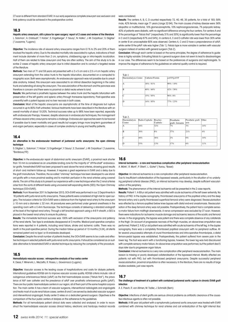
Objective: Vascular access is the leading cause of hospitalizations and costs for dialysis patients. International guidelines KDOQI aim to improve vascular access quality. KDOQI criteria include: rate of autogenous arteriovenous fistula (aAVF) as the first haemodialysis access (“fistula first”), the preva-lence of AVF over catheter and the prevalence of aAVF over prosthetic arteriovenous grafts (pAVF). There are four public haemodialysis centers in our region, all of them part of the same hospital corpora-tion. The main center A has a team of vascular surgeons, interventional radiologists and angiologists and treats most of acute renal failure cases, centers B and C are served by dedicated vascular surgeon and interventional angiologist, finally center D relies on a dedicated general surgeon. Objectives is the comparison of the four public centers of dialysis on the adherence to the guidelines.Methods: For all hemodialysis patient clinical data were collected and analysed. In order to recon-struct the haemodialysis vascular access clinical history, electronic and hardcopy medical records
were considered.Results: The centers A, B, C, D counted respectively 72, 40, 46, 35 patients, for a total of 193; 58% male, 42% female, mean age 71 years (range 32-94). The main causes of kidney disease were: 56% idiopathic or multifactorial, 10% glomerulonephritis, 10% nephroangiosclerosis, 7% polycystic kidney. 40% of patients were diabetic, with no significant difference among the four centers. For centers A and B the percentage of “fistula first” (respectively 31% and 30%) is significantly lower than the percentage in C and D (respectively 67% and 54%). In centers A, C and D catheter rate was lower than 30% while in center B an unacceptable 60% was observed. Centers A, C and D have a preponderance of aAVF, while center B the pAVF rate was higher (Tab 1). Fistula type is more variable in centers with vascular surgeon instead of centers with general surgeon (Tab 2).Conclusion: Although each center is based on the same principles, the degree of adherence to guide-lines is highly variable. Entrusting fistula to a general surgeon does not seem to lead to disadvantages in our case. The differences seem to be based on the preferences of surgeons and nephrologists. To improve the degree of adherence to the guidelines an external quality control is required.
Tab 1 Haemodialysis Center Catheter/Fistula aAVF/pAVF
A 18% / 81% 90% / 10%
B 40% / 60% 42% / 58%
C 15% / 85% 96% /4%
D 14% / 85% 77% / 23%
Tab 2. Haemodialysis Center
Radio-Cephalic Brachio-Cephalic
Prosthetic graft- forearm
Prosthetic graft- arm
Other
A 44% 32% 7% 3% 14%
B 30% 8% 37% 21% 4%
C 48% 36% 2% 0% 14%
D 53% 13% 17% 6% 11%
16.6Interval ischaemia – a rare and hazardous complication after peripheral revascularisationA. Märki1, R. Marti1, P. Stierli1, L. Gürke2 (1Aarau, 2Basel)
Objective: An interval ischaemia is a rare complication after peripheral revascularisation. Due to insufficient collateralisation of the bypassed vessels, particularly in the situation of no underly-ing peripheral arterial disease (PAD), an interval ischaemia may develop, despite sufficient vasculari-sation of the periphery.Methods: The phenomenon of the interval ischaemia will be presented in the 2 case reports.Results: Patient 1: A 69 yr old patient was admitted with acute ischaemia of the left lower extremity. No history of PAD. In the duplex sonography, thrombosed aneurysms of the popliteal artery and the deep femoral artery and a partly thrombosed superficial femoral artery were diagnosed. Revascularisation was effected by a femoro-popliteal below knee bypass with distal end-end anastomosis. Revasculari-sation of the deep femoral artery was not possible. Postoperatively, the patient had a complete senso-ry-motor failure from midthigh downwards. A nerve compression was excluded by CT scan. However, there were indications for ischaemic muscle damage and ischaemic lesions of the sciatic and femoral nerves. In the angiography, the bypass was patent and there was complete absence of any collaterals in the thigh. On account of progressive necrosis of the thigh muscles, an above-knee amputation was performed. Patient 2: A 82 yr old patient was admitted with acute ischaemia of the left leg. In the duplex sonography, there was a completely thrombosed popliteal aneurysm with no peripheral outflow. Af-ter several unsuccessful attempts of crural thrombectomies and intra-operative thrombolysis, a distal femoro-pedal bypass was established. Postoperatively, the patient suffered from severe pain in the lower leg. The foot was warm with a functioning bypass, however, the lower leg was livid discoloured with complete sensory-motor failure. An above-knee amputation was performed, but the patient died 5 days later due to progressive organ failure.Conclusion: Interval ischaemia is a very rare complication after peripheral revascularisation. The main reason is missing or poorly developed collateralisation of the bypassed interval. Mostly affected are patients not with PAD, but with thrombosed peripheral aneurysms. Despite successful peripheral revascularisation, proximal amputation is often necessary. In the literature, there are no results of large studies available, just case reports.
16.7The challenge of treatment of a patient with contained juxtarenal aortic rupture in chronic EVAR graft infectionA. E. Pasch, R. von Allmen, M. Tadler, J. Schmidli (Bern)
Objective: Infections of stent grafts are serious clinical problems as antibiotic clearance of the causa-tive infectious agents is often not possible.Methods: A 66 year old patient with a symptomatic juxtarenal aortic aneurysm was treated with EVAR combined with chimney technique for renal arteries and coil embolization of the right internal iliac

swiss knife 2014; 11: special edition 13
artery. One month later he developed an endoleak type Ia, which was repeatedly treated with coil em-bolization and onyx injection. Furthermore, a type Ib endoleak on the left side was treated by endog-raft extension. Following a sigmoidal infection, infection of the stentgraft with E. coli and E. faecalis occurred (positive blood cultures). The patient underwent continuous antibiotic treatment, including injections directly into the aneurysm sac, but developed an abscess of the psoas muscle, which was repeatedly drained. An endoleak Ia recurred and the patient developed fever and back pain. He was ad-mitted to our hospital and a contained rupture of a mycotic juxtarenal aortic aneurysm was diagnosed. He lost 38 kg of weight during the last 2 years. A decision was made for open surgery type IV repair with excision of the endovascular material and extensive debridement. The aneurysm sac was filled with thrombus, fresh clots and pus. The aorta was replaced with a handmade bovine pericardium tube graft. Great saphenous vein bypasses were performed to both renal arteries. The patient needed mas-sive transfusion of a total of 207 units of blood products, including erythrocytes, fresh frozen plasma, platelets, fibrinogen and 3.7l blood from the cellsaver. Open abdomen treatment was necessary.Results: The patient recovered from acute renal failure needing temporary dialysis and the abdomen could be closed after one week. Repeated take-backs to the OR were needed for bleeding complica-tions and the patient had to be resuscitated in hypovolemic shock. After one month the patient sud-denly developed deep coma due to hyperemic dysfunction of the brain stem and died. Recurrence of infection was not found in autopsy.Conclusion: Treatment of a patient with endograft infection and contained rupture of the juxtarenal segment can be challenging. Massive transfusion requirements in a single patient may bring a tertiary referral centre to the edge in terms of care and resources.
16.8Das etwas andere „rupturierte abdominale Aortenaneurysma“: ein Fall eines rupturierten Perigraft-seromR. Marti1, A. Naumann1, C. Geppert1, L. Gürke2, P. Stierli1 (1Aarau, 2Basel)
Objective: Das Auftreten eines Perigraftserom ist ein bekanntes Phänomen nach Implantation von Graftmaterial, vornehmlich PTFE. Ursächlich werden verschiedene Faktoren diskutiert, allen voran eine Serum-Ultrafiltration durch die Graft-Matrix, eine Biofilm-Infektion oder eine abnorme Immunantwort als Reaktion auf das Graftmaterial. Als weiteres werden fibrinolytische Prozesse, bedingt durch die Thrombocytenaggregationhemmung, welche die transmurale Filtration begünstigen, diskutiert. Eine Ruptur eines Perigraftserom als Extremform ist äusserst selten.Methods: Fallbericht.Results: Ein 83-jähreiger Patient wir mit Verdacht auf ein rupturiertes abdominales Aortenaneurysma notfallmässig zugewiesen. Es bestehen starke Unterbauchschmerzen, klinisch zeigt sich eine grosse paraumbilicale Resistenz mit lokaler Druckdolenz. Es stellt sich schliesslich heraus, dass vor Jahren eine Y-Prothese implantiert wurde. Im CT Nachweis eines riesigen Perigraftserom mit Ruptur links-lat-eral mit lokalem Austritt von Flüssigkeit in den Retroperitonealraum. Zusätzlich zeigte sich eine Stauung des linken Ureter und der linken Nierenvene. Kein KM-Austritt nachweisbar. Leuc: 13.4, CRP 108. Es erfolgte eine Re-Laparotomie, Ausräumung des Perigraftserom, TEA/Resektion der Aneurysmawand mit Deckung der Prothese mit einer Omentumplombe. Die entnommene Flüssigkeit war steril. Der post-operative Verlauf war unauffällig. Nach 3 Monaten zeigten sich im CT kein Hinweis auf ein Rezidiv.Conclusion: Die Ruptur eines Perigraftserom ist eine äusserst seltene Komplikation nach aortaler Prothesenimplantation, hauptsächlich bei PTFE-Prothesen. Neben einer möglichen Biofilm-Infektion oder einer abnormen immunologischen Reaktion, wird vorallem eine Ultrafiltration durch die Proth-esenwand als ursächlich angenommen. Zur Vorbeugung eines Perigraftserom wird die TEA des An-eurysmasack im Rahmen der Erstoperation empfohlen. Als Therapieoptionen bei nicht-rupturierten Seromen werden das expektative Vorgehen die, die Aspiration oder der Graftersatz diskutiert. Im Falle einer Ruptur wird eine offene Ausräumung des Serom mit TEA bzw. Resektion der Wand empfohlen. Zur Vorbeugung eines rezidiv und zum Schutz der Prothese sollte der Graft mit einer Omentum-Plombe gedeckt werden.
16.9Major treatment with minor success: a challenging case of extensive deep venous thrombosisT. Wyss, R. Bühlmann, L. Alberio, M. Daskalakis, J. Schmidli (Bern)
Objective: According to current guidelines early surgical thrombus removal can be considered in se-lected patients with proximal deep venous thrombosis (DVT). We report a complex case of extensive acute bilateral DVT.Methods: A 55-year-old female suffered from bilateral acute on chronic subdural hemorrhage due to autoimmune thrombocytopenia of unknown etiology. After treatment with intravenous immunoglob-ulins and steroids a rise in platelet count was achieved. Subsequent bilateral pulmonary embolism occurred prompting heparin treatment and vena cava filter implantation. One week later a brain CT scan showed progression of the subdural hemorrhage, hence heparinization was reduced and con-servative treatment continued. After another week, both legs presented with acute massive swelling and a CT scan confirmed bilateral proximal DVT extending up to the infrarenal vena cava and above the cava filter. In addition, an immune-mediated heparin-induced thrombocytopenia was diagnosed. A catheter directed thrombolysis could not be performed due to the cerebral situation. Argatroban (a direct thrombin inhibitor) anticoagulation in a low-therapeutic level was initiated and we decided to treat the patient surgically (to remove the filter and prevent severe post-thrombotic syndrome) under strict waiver of heparin use for any purpose (e.g. catheter flushing). We performed bilateral venous thrombectomy and a lumbotomy for open surgical vena cava filter retrieval.Results: Although Argatroban anticoagulation and pneumatic compression stocking therapy were continued postoperatively, bilateral proximal DVT re-occurred after two days. We considered re-opera-tion but the patient refused. The hospital course was uneventful, with conservative treatment, initiation of warfarin anticoagulation and discharge with marked but painless leg swelling.Conclusion: Treatment guidelines are inadequate to manage such complex cases. Immune-mediated
heparin-induced thrombocytopenia and subdural hemorrhage clearly limited the options, only leading to transient technical success.
Bariatric Surgery 1717.1Laparoscopic versus robotic Roux-en-Y gastric bypass: lessons and long-term follow-up learned from a large prospective monocentric studyN. Buchs, P. Morel, M. Jung, D. Azagury, G. Chassot, O. Huber, M. Hagen, F. Pugin (Geneva)
Objective: Laparoscopic RYGB has become the procedure of choice for the treatment of morbid obes-ity. Recently, several reports have shown the potential advantages of the robotic approach, notably by reducing complications. The aim of this study is to report our long-term experience with robotic Roux-en-Y gastric bypass (RYGB) and to compare outcomes with the laparoscopic approach.Methods: From January 2003 to September 2013, 777 consecutive minimally invasive RYGB have been performed in our institution: 389 laparoscopically (50.1%) and 388 robotically (49.9%). During the study period, all the data regarding these consecutive RYGB has been prospectively collected in a dedicated database.Results: While longer in duration compared to laparoscopy (+30 minutes; p=0.0001), the robotic approach had a lower conversion rate (0.8 vs. 4.9%; p=0.0007), and less complications (11.6% vs. 16.7%; p=0.05), in particular less gastrointestinal leaks (0.3 vs. 3.6%; p=0.0009). There were also less early reoperations (1 vs. 3.3%; p=0.05) and a shorter hospital stay in the robotic group (6.2 vs. 10.4 days; p=0.0001). There were no statistical differences between the early and the current robotic experi-ence, except in operative time and hospital stay, which were shorter for the last 100 cases. Finally, the BMI loss was significantly higher in the laparoscopic group starting at the first post-operative year.Conclusion: Robotic RYGB is not only safe and feasible, but also a valid option in comparison to lapar-oscopy. At the cost of a longer operative time, we observed better short-term outcomes with the robotic approach.
17.2Linear compared to circular stapler anastomosis in laparoscopic Roux-en-Y gastric bypass leads to fewer complications but equal weight loss: a matched pair studyR. Schneider, B. Kern, M. Gass, T. Peters, M. Slavic, M. Gebhart, R. Peterli (Basel)
Objective: In laparoscopic Roux-en-Y gastric bypass (LRYGB) a tight gastroenterostomy (GE) may lead to better weight loss but can be accompanied with a higher rate of local complications such as stric-tures and ulcers. Different operative techniques for Roux-en-Y reconstruction may also influence rate of internal hernia, a typical late complication of LRYGB. The objective of this study was to compare rate of strictures, ulcerations, internal hernias and weight loss of linear and circular stapler anastomosis.Methods: Retrospective analysis of prospectively collected data in a 1:2-matched pair study. A total of 240 patients were matched according to BMI, age and gender. The average follow-up was 2.4 years. In group C (circular), 80 patients were operated with a 25 mm circular stapler technique (average BMI: 44.4 kg/m2, age: 42.9 y., 79% female). In group L (linear), 160 patients were operated using a linear stapler (approxomativly 30 mm, average BMI: 44.2 kg/m2, age: 43.2 years,79% female). A propensity score matching and a Logrank Test were used for statistical analysis.Results: In group L, patients had significantly lower overall stricture rate at the GE compared to group C (2 [1.3%] vs. 8 [10.1%], p= 0.008), equal local ulcerations (1 [0.6%] vs. 1 [1.3%] p= 0.956) and a lower rate of internal herniation (3 [1.9%] vs. 7 [8.9%] p= 0.02). The average excessive BMI loss (EBMIL) after 1 year was equal (69% in group C vs 71% in group L).Conclusion: Linear stapler anastomosis in LRYGB compared to circular anastomosis leads to fewer strictures and lower rate of internal hernias but to an equal weight loss. The linear stapler anastomosis is a safe and effective method and should be preferred for the GE in LRYGB.
17.3Effect of preoperative weight loss on postoperative weight development after bariatric interventionR. Kraus1, D. Stekhoven2, W. Marti1 (1Aarau, 2Berikon)
Objective: In the last years various studies investigated effects of preoperative weight loss before bari-atric surgery. Regrettably, none of these were able to show significant benefits. While a lot is known about preoperative diets and weight loss individually, we rather wanted to know if a diet executed 14 days preoperatively, has an impact on the weight loss postoperatively.Methods: All patients undergoing Roux-en-Y gastric bypass surgery from September 2009 to April 2012 at our institution were included. All patients had been operated by the same surgeon and had been treated preoperatively with the same diet during 14 days. Body weight had been measured before the diet, at the day of the operation and at least three times postoperatively in irregular time intervals. We used a linear mixed effect model to assess the relationship of preoperative weight loss with post-operative weight loss during the first two postoperative years.Results: The 87 patients (68 female/19 male) had a mean starting weight of 125.0 kg (female 120.1 kg/male 142.6 kg). During the preoperative diet the patients lost on average 5.0 kg (female 4.6 kg/male 6.5 kg) and during the two postoperativefollow-up years the patients lost on average 34.1 kg (female 34.1 kg/male 34.0 kg). This amounts to an average excess weight loss (%EWL) of 56.7% (female 57.8%/male 52.7%). 40 patients (28 female/12 male) achieved an%EWL of more than or equal to 60%. The longitudinal data analysis
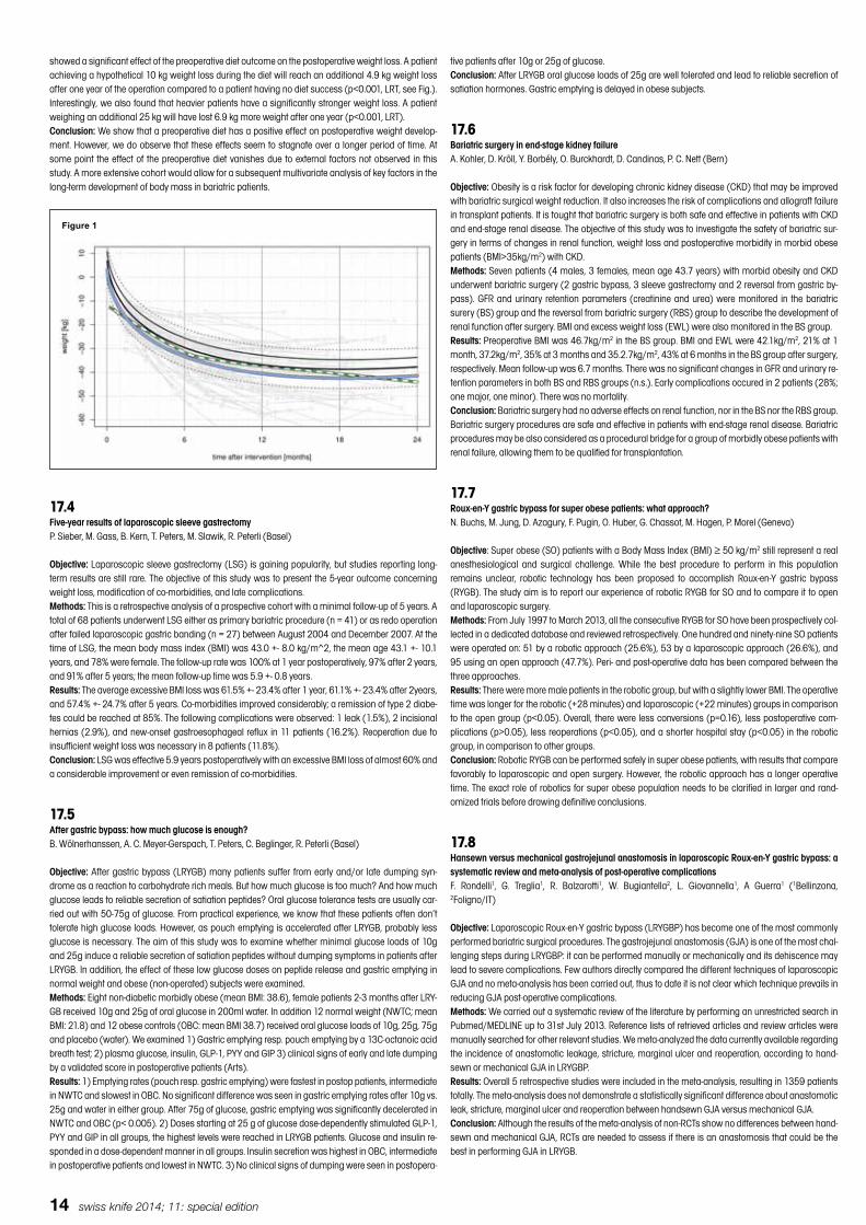
14 swiss knife 2014; 11: special edition
showed a significant effect of the preoperative diet outcome on the postoperative weight loss. A patient achieving a hypothetical 10 kg weight loss during the diet will reach an additional 4.9 kg weight loss after one year of the operation compared to a patient having no diet success (p<0.001, LRT, see Fig.). Interestingly, we also found that heavier patients have a significantly stronger weight loss. A patient weighing an additional 25 kg will have lost 6.9 kg more weight after one year (p<0.001, LRT).Conclusion: We show that a preoperative diet has a positive effect on postoperative weight develop-ment. However, we do observe that these effects seem to stagnate over a longer period of time. At some point the effect of the preoperative diet vanishes due to external factors not observed in this study. A more extensive cohort would allow for a subsequent multivariate analysis of key factors in the long-term development of body mass in bariatric patients.
Figure 1
Figure 1. Model weight profiles for different diet performances. The black bold straight line indicate the model weight profile for a 0 kg weight loss during the diet. The black straight lines above and below indicate the model weight profiles for +/-10 kg during the diet, respectively. The straight blue framed line shows the model weight profile for the median weight loss during the diet in our data (4.6 kg). In the background the patient profiles are printed in light grey including their smoothed overall trend (green dashed framed line).
17.4Five-year results of laparoscopic sleeve gastrectomyP. Sieber, M. Gass, B. Kern, T. Peters, M. Slawik, R. Peterli (Basel)
Objective: Laparoscopic sleeve gastrectomy (LSG) is gaining popularity, but studies reporting long-term results are still rare. The objective of this study was to present the 5-year outcome concerning weight loss, modification of co-morbidities, and late complications.Methods: This is a retrospective analysis of a prospective cohort with a minimal follow-up of 5 years. A total of 68 patients underwent LSG either as primary bariatric procedure (n = 41) or as redo operation after failed laparoscopic gastric banding (n = 27) between August 2004 and December 2007. At the time of LSG, the mean body mass index (BMI) was 43.0 +- 8.0 kg/m^2, the mean age 43.1 +- 10.1 years, and 78% were female. The follow-up rate was 100% at 1 year postoperatively, 97% after 2 years, and 91% after 5 years; the mean follow-up time was 5.9 +- 0.8 years.Results: The average excessive BMI loss was 61.5% +- 23.4% after 1 year, 61.1% +- 23.4% after 2years, and 57.4% +- 24.7% after 5 years. Co-morbidities improved considerably; a remission of type 2 diabe-tes could be reached at 85%. The following complications were observed: 1 leak (1.5%), 2 incisional hernias (2.9%), and new-onset gastroesophageal reflux in 11 patients (16.2%). Reoperation due to insufficient weight loss was necessary in 8 patients (11.8%).Conclusion: LSG was effective 5.9 years postoperatively with an excessive BMI loss of almost 60% and a considerable improvement or even remission of co-morbidities.
17.5After gastric bypass: how much glucose is enough?B. Wölnerhanssen, A. C. Meyer-Gerspach, T. Peters, C. Beglinger, R. Peterli (Basel)
Objective: After gastric bypass (LRYGB) many patients suffer from early and/or late dumping syn-drome as a reaction to carbohydrate rich meals. But how much glucose is too much? And how much glucose leads to reliable secretion of satiation peptides? Oral glucose tolerance tests are usually car-ried out with 50-75g of glucose. From practical experience, we know that these patients often don’t tolerate high glucose loads. However, as pouch emptying is accelerated after LRYGB, probably less glucose is necessary. The aim of this study was to examine whether minimal glucose loads of 10g and 25g induce a reliable secretion of satiation peptides without dumping symptoms in patients after LRYGB. In addition, the effect of these low glucose doses on peptide release and gastric emptying in normal weight and obese (non-operated) subjects were examined.Methods: Eight non-diabetic morbidly obese (mean BMI: 38.6), female patients 2-3 months after LRY-GB received 10g and 25g of oral glucose in 200ml water. In addition 12 normal weight (NWTC; mean BMI: 21.8) and 12 obese controls (OBC: mean BMI 38.7) received oral glucose loads of 10g, 25g, 75g and placebo (water). We examined 1) Gastric emptying resp. pouch emptying by a 13C-octanoic acid breath test; 2) plasma glucose, insulin, GLP-1, PYY and GIP 3) clinical signs of early and late dumping by a validated score in postoperative patients (Arts).Results: 1) Emptying rates (pouch resp. gastric emptying) were fastest in postop patients, intermediate in NWTC and slowest in OBC. No significant difference was seen in gastric emptying rates after 10g vs. 25g and water in either group. After 75g of glucose, gastric emptying was significantly decelerated in NWTC and OBC (p< 0.005). 2) Doses starting at 25 g of glucose dose-dependently stimulated GLP-1, PYY and GIP in all groups, the highest levels were reached in LRYGB patients. Glucose and insulin re-sponded in a dose-dependent manner in all groups. Insulin secretion was highest in OBC, intermediate in postoperative patients and lowest in NWTC. 3) No clinical signs of dumping were seen in postopera-
tive patients after 10g or 25g of glucose. Conclusion: After LRYGB oral glucose loads of 25g are well tolerated and lead to reliable secretion of satiation hormones. Gastric emptying is delayed in obese subjects.
17.6Bariatric surgery in end-stage kidney failureA. Kohler, D. Kröll, Y. Borbély, O. Burckhardt, D. Candinas, P. C. Nett (Bern)
Objective: Obesity is a risk factor for developing chronic kidney disease (CKD) that may be improved with bariatric surgical weight reduction. It also increases the risk of complications and allograft failure in transplant patients. It is tought that bariatric surgery is both safe and effective in patients with CKD and end-stage renal disease. The objective of this study was to investigate the safety of bariatric sur-gery in terms of changes in renal function, weight loss and postoperative morbidity in morbid obese patients (BMI>35kg/m2) with CKD.Methods: Seven patients (4 males, 3 females, mean age 43.7 years) with morbid obesity and CKD underwent bariatric surgery (2 gastric bypass, 3 sleeve gastrectomy and 2 reversal from gastric by-pass). GFR and urinary retention parameters (creatinine and urea) were monitored in the bariatric surery (BS) group and the reversal from bariatric surgery (RBS) group to describe the development of renal function after surgery. BMI and excess weight loss (EWL) were also monitored in the BS group.Results: Preoperative BMI was 46.7kg/m2 in the BS group. BMI and EWL were 42.1kg/m2, 21% at 1 month, 37.2kg/m2, 35% at 3 months and 35.2.7kg/m2, 43% at 6 months in the BS group after surgery, respectively. Mean follow-up was 6.7 months. There was no significant changes in GFR and urinary re-tention parameters in both BS and RBS groups (n.s.). Early complications occured in 2 patients (28%; one major, one minor). There was no mortality.Conclusion: Bariatric surgery had no adverse effects on renal function, nor in the BS nor the RBS group. Bariatric surgery procedures are safe and effective in patients with end-stage renal disease. Bariatric procedures may be also considered as a procedural bridge for a group of morbidly obese patients with renal failure, allowing them to be qualified for transplantation.
17.7Roux-en-Y gastric bypass for super obese patients: what approach?N. Buchs, M. Jung, D. Azagury, F. Pugin, O. Huber, G. Chassot, M. Hagen, P. Morel (Geneva)
Objective: Super obese (SO) patients with a Body Mass Index (BMI) ≥ 50 kg/m2 still represent a real anesthesiological and surgical challenge. While the best procedure to perform in this population remains unclear, robotic technology has been proposed to accomplish Roux-en-Y gastric bypass (RYGB). The study aim is to report our experience of robotic RYGB for SO and to compare it to open and laparoscopic surgery.Methods: From July 1997 to March 2013, all the consecutive RYGB for SO have been prospectively col-lected in a dedicated database and reviewed retrospectively. One hundred and ninety-nine SO patients were operated on: 51 by a robotic approach (25.6%), 53 by a laparoscopic approach (26.6%), and 95 using an open approach (47.7%). Peri- and post-operative data has been compared between the three approaches.Results: There were more male patients in the robotic group, but with a slightly lower BMI. The operative time was longer for the robotic (+28 minutes) and laparoscopic (+22 minutes) groups in comparison to the open group (p<0.05). Overall, there were less conversions (p=0.16), less postoperative com-plications (p>0.05), less reoperations (p<0.05), and a shorter hospital stay (p<0.05) in the robotic group, in comparison to other groups.Conclusion: Robotic RYGB can be performed safely in super obese patients, with results that compare favorably to laparoscopic and open surgery. However, the robotic approach has a longer operative time. The exact role of robotics for super obese population needs to be clarified in larger and rand-omized trials before drawing definitive conclusions.
17.8Hansewn versus mechanical gastrojejunal anastomosis in laparoscopic Roux-en-Y gastric bypass: a systematic review and meta-analysis of post-operative complicationsF. Rondelli1, G. Treglia1, R. Balzarotti1, W. Bugiantella2, L. Giovannella1, A Guerra1 (1Bellinzona, 2Foligno/IT)
Objective: Laparoscopic Roux-en-Y gastric bypass (LRYGBP) has become one of the most commonly performed bariatric surgical procedures. The gastrojejunal anastomosis (GJA) is one of the most chal-lenging steps during LRYGBP: it can be performed manually or mechanically and its dehiscence may lead to severe complications. Few authors directly compared the different techniques of laparoscopic GJA and no meta-analysis has been carried out, thus to date it is not clear which technique prevails in reducing GJA post-operative complications.Methods: We carried out a systematic review of the literature by performing an unrestricted search in Pubmed/MEDLINE up to 31st July 2013. Reference lists of retrieved articles and review articles were manually searched for other relevant studies. We meta-analyzed the data currently available regarding the incidence of anastomotic leakage, stricture, marginal ulcer and reoperation, according to hand-sewn or mechanical GJA in LRYGBP.Results: Overall 5 retrospective studies were included in the meta-analysis, resulting in 1359 patients totally. The meta-analysis does not demonstrate a statistically significant difference about anastomotic leak, stricture, marginal ulcer and reoperation between handsewn GJA versus mechanical GJA.Conclusion: Although the results of the meta-analysis of non-RCTs show no differences between hand-sewn and mechanical GJA, RCTs are needed to assess if there is an anastomosis that could be the best in performing GJA in LRYGB.
swiss knife 2014; 11: special edition 15
17.9Les fistules après sleeve gastrectomie: etude expérimentale de la théorie mécanique portant sur la résistance de l’agrafage linéaire gastriqueP. Fournier1,2, G. De Courville2, N. Demartines1, J.-P. Marmuse2 (1Lausanne, 2Paris/FR)
Objective: Deux théories (mécanique ou ischémique) peuvent expliquer la déhiscence de la ligne d’agrafes après sleeve gastrectomie par laparoscopie (LSG).Nous analysons dans cette étude l’effet de la hauteur de la ligne d’agrafe, ainsi que l’utilisation d’un renfort tissulaire sur la survenue expéri-mentale d’une fistule.Methods: Il s’agit d’une étude expérimentale sur le modèle porcin.Les LSG ont été réalisées ex vivo. Une fistule a été crée en insufflant de l’air dans le tube gastrique immergé dans l’eau. La 1ère expérience compare 8 LSG avec l’utilisation des chargeurs verts sur toute la longueur de la ligne d’agrafe et 8 LSG avec l’utilisation conjointe de 3 chargeurs verts pour la partie basse puis des chargeurs bleus pour la partie haute de l’agrafage. La 2ème expérience utilise la même séquence avec un renforcement par du Biosyn; 3 LSG avec des chargeurs verts et 4 LSG avec des des chargeurs verts puis bleus. La 3ème expérience utilise la dernière génération d’agrafage Tri-Staple en comparant 5 LSG avec chargeurs noirs et 5 LSG avec utilisation de chargeurs noirs puis violets. Le critère de jugement est la pression exercée en mmHg pour créer expérimentalement une fistule.Results: Quelque soit l’expérience,la localisation de la fistule se situe à la partie supérieure de l’agrafage dans plus de 80% des cas.L’expérience n°1 montre que la survenue d’une fistule nécessite une pres-sion plus importante dans les groupes des chargeurs verts puis bleus (m=122,4 mmHg) comparés au groupe des chargeurs verts(m=94,2 mmHg) de façon très significative (p=0,0017). L’expérience n°2 montre des résultats identiques mais avec des pressions de survenue plus élevées avec les chargeurs verts puis bleus (m=189 mmHg) qu’avec des chargeurs verts seuls (m=152,3 mmHg ); La différence est également significative. L’expérience n°3 ne montre pas de différence significative (p=0,62) entre les chargeurs noirs (m=130,3 mmHg) et les chargeurs noirs puis violets (m=134 mmHg).Conclusion: Quelque soit l’expérience, les valeurs moyennes et minimales des pressions retrouvées sont bien supérieures à la pression intragastrique chez l’homme après LSG. Les agrafes de faibles hauteurs résistent mieux à la pression. Le renforcement de la ligne d’agrafage augmente encore la résistance. La théorie mécanique n’explique pas seule l‘apparition des fistules après LSG.
Video I 2121.1Surgical technique of transaxillar robotic hemithyroidectomy: first experiences in SwitzerlandG. Wille, F. Grochola, A. Dudli, S. Breitenstein (Winterthur)
Objective: The recent incorporation of robotic technology in thyroid surgery proposes a feasible, safe, extracervical approach that alleviates most of the technical difficulties associated with endoscopic surgery. Robotic assisted thyreoidectomy has been reported from only a few authors worldwide showing that this transaxillar technique parallels the high standards of the open approach. In order to benefit from the advantages of this novel approach, such as excellent cosmetic results and less musculoskeletal discomfort to surgeons, we performed the first two transaxillary robotic lobectomies ever in Switzerland. Methods: Two cases with small total gland volume (up to 30ml) and small nodule diameter (up to 3cm) were chosen. The nodule of the first patient had no signs of malignancy and the patient was operated for cervical discomfort. In the second case, an incidentally found nodule (TIRADS 2, Bethesda III) was removed for diagnostic reasons.Results: Approach: An ipsilateral 6 cm anterior axillar fold incision was made with the lesion side arm elevated. Approach was established dissecting a subcutaneous skin flap over the anterior surface of the pectoral mayor muscle and clavicle. Thyroid working space was developed between sternal and clavicular heads of sternocleidomastoid muscle and mobilisation of the strap muscles from the anterior surface of the gland. A retractor was inserted through the skin incision. Docking: 4 robotic arms were used through the axilla. An 8mm/30 ° endoscope was placed on the central arm followed by Harmonic curved shears, a dissector and forceps under vision control. Console part: Whole gland mobilisation, including division of thyroid superior vessels and isthmus division, was achieved with Harmonic shears. Countertraction was ensured with the Maryland dissector and Prograsp forceps. Recurrent laryngeal nerve was monitored and parathyroid-sparing mobilisation of thyroid lobe could be performed in both cases. The specimen was extracted through the axillary skin incision. Closure: Skin closure in the anterior axillar fold to ensure the scar is completely covered when the arm is in its natural position. Conclusion: Transaxillary robot-assisted lobectomy is safe and technically feasible in selected cases. It provides excellent cosmetic results with avoidance of a neck scar.
21.2Transrectal hybrid-NOTES anterior resection using an articulating HD 3D laparoscopic surgical video systemD. C. Steinemann, A. Zerz, S. Lamm (Bruderholz)
Objective: Although, in standard laparoscopy anterior resection can usually be performed using two five millimetres incisions and two twelve millimetre trocar (for insertion of the Endostapler), there re-mains the need for a minilaparotomy for specimen removal. The removal of the specimen through the rectum avoids a minilaparotomy and therefore aditional harm to the abdominal wall. In our opinion the rigid hybrid-NOTES technique is the perfect tool to convert laparoscopic assisted operations into totally
laparoscopic procedures.Methods: In this instructive HD 3D video we present our technique of transrectal laparoscopic anterior resection in diverticular disease.Results: After positioning of the patient the procedure is started with our standard medial mobilization of the left hemicolon. Afterwards the infrapancreatic ligation of the inferior mesenteric vein and the central ligation of the inferior mesenteric artery are performed with the energy device. The mobilization is completed from lateral. Afterwards the proximal rectum is preparated and the anterior resection is performed. The specimen is extracted transrectally, followed by an intracorporal purse-string suture after insertion of the anvil in the oral colon. A circular stapler is inserted transanally and the end-to-end anastomosis is performed.Conclusion: In our experience on 27 patients the transrectal hybrid-NOTES anterior resections is a safe and feasible evolution of the standard laparoscopic procedure. The 3 D depth perception improves the accuracy and precision of surgical tasks.
21.3Teaching the teacher: Laparoscopic Competency Assessment Tool (LCAT) for mentored laparoscopic cholecystectomy-the step by step videoM. Jung, N. C. Buchs, S. de Sousa, A. Mennet, P. Morel, F. Ris (Geneva)
Objective: After implementation of a laparoscopic competency assessment tool (LCAT) for the evalu-ation of technical surgical skills in laparoscopic cholecystectomy at the resident level at our institution, we think it is important to demonstrate assessing in a standardized manner. The aim of this teaching video is to offer an illustration of the tested domains and to help assessors to standardize adequate rating of the procedure.Methods: Teaching video with emphasis on the 4 following steps: exposure of the operating field, dis-section of the calot’s triangle, execution of cholangiography and resection of the gallbladder.Results: Each of these 4 steps is divided in 4 sub-domains: use of instruments, tissue handling, errors, and the quality of the end product. The main subdomains of the evaluation are illustrated in this video with defined procedures according to the rating system from 1 to 4 (1. dangerous, 2. inadequate, 3. safe and 4. expert). Conclusion: This video is an informative support to assess technical performance in laparoscopic cholecystectomy, training the trainer for objective assessment, as well as a teaching tool for the trainee to improve his performance towards a safe or expert procedure.
21.4Laparoscopic Heller myotomy and partial anterior fundoplication for achalasia after esophageal per-foration, due to iterative endoscopic dilationP. Allemann, A. Dayer-Jankechova, M. Winiker, N. Demartines, M. Schäfer (Lausanne)
Objective: Endoscopic balloon dilatation of the gastro-esophageal junction for achalasia is associated with a iatrogenic perforation rate of 1-12%. While the majority of these lesions are managed conserva-tively without major consequences, the subsequent peri-esophageal inflammation eventually render further surgical intervention challenging.Methods: To present a laparoscopic Heller myotomy associated with partial anterior fundoplication (Dor) after esophageal perforation due to iterative endoscopic dilation.Results: The patient was a 34-year old woman with long history of dysphagia and regurgitation. En-doscopy, gastrographin swallow and HD esophageal manometry revealed a primary achalasia. In first intention, she was treated with iterative endoscopic balloon dilation without any symptoms relief. During the last attempt, a 1 cm esophageal perforation occurred, treated conservatively with success. Six month later, the patient was still complaining of solid and liquid dysphagia. A severe stenosis was confirmed by endoscopy. Laparoscopic exploration was performed with a 12mm optical trocar and a 30° scope inserted 3cm cranial from the umbilicus. Three additional working trocars were placed in a half circle directed to the hiatus and a Nathanson liver retractor was inserted percutaneously. The phreno-esophageal membrane was opened and the distal esophagus dissected. A 9cm long myo-tomy was performed, using a monopolar hook dissector (6cm on the esophagus and 3cm on the stomach). After minimal fundic mobilisation, an anterior antireflux valve was created (Dor procedure), with two rows of four 2.0 non-absorbable stitches on each sides, covering completely the site of the myotomy. An important peri-esophageal inflammation was observed, but this precluded neither the hi-atal dissection, nor the myotomy. The immediate postoperative course was uneventful and the patient was asymptomatic two months postoperatively, with complete food tolerance.Conclusion: Laparoscopic Heller myotomy after iatrogenic esophageal perforation is feasible, despite important fibrosis and hiatal inflammation. According to current guidelines, in younger patients with proven achalasia, surgery should be considered as first choice treatment even if secondary operation remains possible.
21.5Laparoscopic Tru-Cut biopsy of the spleen: a case reportS. A. Käser, C. A. Maurer (Liestal)
Objective: Probably due to concerns regarding the risk of bleeding, surgeons hesitate to do laparo-scopic Tru-Cut biopsy of the spleen, although it is an accepted procedure in literature. We report the technical details of an uneventful Tru-Cut biopsy in a patient with nodular involvement in sarcoidosis.Methods: Osteolytic lesions were diagnosed in the lumbar spine of a 34-year old man with low back pain. Lymphoproliferative disease was suspected as PET-CT scan showed hypermetabolic mediastinal lymph nodes and nodules in the spleen, but no pulmonary involvement. Blood tests (LDH, ACE, haemo-gram, immunoelectrophoresis, AFP, B-HCG, B2-microglobuline), bone-marrow puncture, bronchosco-py with bronchial lavage and cytology showed no pathologic results. Vaccination against pneumococ-

16 swiss knife 2014; 11: special edition
cus, H. influenza, and meningococcus was done because splenectomy was anticipated. Diagnostic laparoscopy confirmed the nodules in the spleen and revealed little spots on the liver surface. Tangen-tial laparoscopic Tru-Cut biopsies of the spleen and of segment 4a of the liver were taken. Post-biopsy bleeding was controlled by argon plasma coagulation and by application of a fibrin sealant.Results: The postoperative course was uneventful; postoperative sonography excluded splenic bleed-ing. Histologically non-caseous granulomas in the spleen and in the liver were diagnosed, while lym-phoproliferative disease was excluded. Tuberculosis was excluded by PCR, thus sarcoidosis remained the most probable granulomatous disease.Conclusion: If done properly, laparoscopic splenic Tru-Cut biopsy is a safe surgical procedure. It should be considered before diagnostic splenectomy is done.
Endocrine Surgery 2323.1Does normotension at the end of thyroidectomy prevent postoperative haemorrhage?S. Bock1,2, W. Kolb2, T. Clerici2 (1Grabs, 2St. Gallen)
Objective: Postoperative haemorrhage is a potential life-threatening complication after thyroid surgery requiring immediate revision. Some risk factors have been reported in the literature, but these comprise mainly conditions that can hardly be influenced by the surgeon, like elderly age, male sex or extent of the resection. The frequency of postoperative bleeding reported by specialized centres after thyroid-ectomy usually ranges from 0 to 6.5% .We hypothesize that postoperative haemorrhage might partly be due to an inadequate surgical haemostasis as a result of hypotonic blood pressure, preventing a proper assessment and identification of potential sources of haemorrhage during the final phase of the operation.Methods: Having set up this hypothesis after a series of postoperative haemorrhages in 2010, we started to ask our anaesthetists to provide normotension during haemostasis in the final phase of surgery. We present a retrospective analysis of our data covering the last 10 years, focussing on the rate of postoperative bleeding before and after the introduction of our request for normotension. We investigated the blood pressures preoperatively and at the crucial time of the operation during the end phase and looked at other known risk factors for postoperative haemorrhage.Results: 1870 patients were subjected to uni- or bilateral thyroidectomy from January 2003 to August 2013; 1204 of them before the introduction of normotension at the end of surgery as a standard proce-dure (group A) and 666 after this (group B). The mean difference in systolic/diastolic blood pressure between the preoperative value and the value measured during the end phase of the operation was 29/19 mmHg in group A and 16/10 mmHg in group B (p(sys)=0.001, CI 5-22, p(dias)=0,001, CI 4-14). 24 of our patients had postoperative haemorrhage, 21 in group A and 3 in group B, accounting for a rate of postoperative haemorrhage of 1.7% before the introduction of normotension at the end of the operation and 0.5% thereafter (p= 0.02). Median time to revision was 11h and did not differ signifi-cantly before and after the key patient, nor did the source of bleeding change.Conclusion: To accomplish normotension during final haemostasis seems to be a valuable method to minimize this dreadful complication in thyroid surgery and is easy to implement. We therefore highly recommend this as a standard of care in thyroid surgery.
23.2Successful minimally invasive surgery for primary hyperparathyroidism: influence of preoperative imaging and intraoperative parathyroid hormone levelsG.-R. Joliat, N. Demartines, L. Portmann, A. Boubaker, M. Matter (Lausanne)
Objective: Adenoma is the main parathyroid disorder leading to primary hyperparathyroidism (PHP). Minimally invasive surgery (MIS) is recognized as a safe and efficient procedure for PHP operations. This technique requires, however, precise preoperative localization with methoxyisobutylisonitrile (MIBI) scan and peroperative confirmation of successful tumoral excision with intraoperative parathor-mone (PTH) dosages. The goal of this study was to assess the success of surgery in relation to these two parameters.Methods: From January 2005 to December 2013, all patients operated for PHP at our institution were retrospectively reviewed. MIBI scans were always reviewed by the nuclear medicine unit. PTH levels were done before incision (T0), 10 (T10) and 15 (T15) minutes after resection. Intraoperative PTH value should either return to normal level or decrease more than 50% of PTH at T0.Results: There were 191 PHP operations during the study period: 104 met criteria for MIS (i.e., adeno-mas identified with MIBI scan), and 87 were done by standard cervicotomy (adenoma or hyperplasia with inconclusive MIBI scan). Among the 104 MIS cases, imaging was not accurate in 3 cases: one adenoma found more caudal than described (thyroid artefact), one treatment failure (no adenoma found), and one double adenoma not visible on imaging. 77 intraoperative PTH levels regained normal value at T15, and 18 decreased more than 50% compared to PTH values at T0. In 8 successfully cured patients intraoperative PTH did not come back to normal or decrease more than 50% after resection. Among these 8 patients, normal PTH levels occurred on day one in 6 patients, and 2 patients had inap-propriate PTH values (false measurements). One patient with treatment failure (the same who had an inaccurate MIBI scan) had abnormal postoperative PTH values.Conclusion: Imaging, ideally single photon emission computed tomography-computed tomography using MIBI, is the key examination for functional and morphological localization of parathyroid ad-enomas. Retrospectively 3 ambiguous imaging were trusted inappropriately (3%). Intraoperative PTH level was not helpful in 8 patients but imaging was so obvious that it was decided not to perform a four-gland exploration, and treatment was successful anyway. One treatment failure occurred in both
MIS group and standard cervicotomy group confirming the low rate of failure (1%) in the surgical treat-ment of PHP.
23.3Intestinal neuroendocrine tumors: diagnosis and treatmentP. Meschberger, O. Ofrim, A. Meyer, B. Egger (Fribourg)
Objective: There are different diagnostic tools for neuroendocrine tumors. Chromogranin A is the most commonly used biomarker, but has its limitations as does urinary 5HIAA and the proliferative marker Ki-67. The most effective available imaging modalities are the Somatostatin receptor scintigraphy and the 68Ga-DOTATOC-PET. Surgical resection may provide cure. Cytotoxic therapy, peptide receptor radio-nuclide therapy and molecular-targeted therapies are used for the treatment of recurrent or metastatic disease.Methods: In a retrospective review of our prospective database a total of 29 patients treated at our institution between 2008 and 2013 for neuroendocrine neoplasms of the midgut have been identi-fied. Patient’s histories have been analyzed concerning prodromal symptoms until diagnosis has been established.Results: The mean age at diagnosis was 62 (12-82) years. Localization of the lesion was the small intestine (15x), the appendix (8x), mesenteric lymph nodes (4x) and the right colon (2x). 10 patients presented with synchronous mesenteric lymph node metastases and 6 with liver or peritoneal me-tastases. Prodromal symptoms were unspecific abdominal pain in 7, nausea and diarrhea in 2, flush and diarrhea in 1 and anemia in 1 patient. In all other cases diagnosis was made by chance: 8x at appendectomy, 7x at laparotomy due to other reasons and 2x at screening colonoscopy. As radiologi-cal investigation CT scan has been employed in 22 patients with a false negative result in 3 cases. An Octreo-Scan was positive in 15 out of 20 patients; Chromogranin A was tested positive in 7 out of 16 and 5HIAA in 7 out of 11 patients. Surgery was applied in 22 patients. 7 have been sent directly or postoperatively for DOTATOC-treatment and 3 underwent Somatostatin-injections alone.Conclusion: The symptoms of neuroendocrine tumors are not all specific. The incidence of mesenteric lymph node metastases in intestinal neuroendocrine tumors is as high as 70-90% in the literature and 55% in our series. The Somatostatin receptor scintigraphy may help to establish the diagnosis. Com-plete surgical resection is the treatment of choice. Prophylactic or therapeutic application of Somato-statin analogs may be discussed; short-acting radioactive Somatostatin analogs (DOTATOC) may be administered in unresectable and recurrent tumors.
Thoratic Surgery I 2626.1Repeated lung volume reduction surgery is successful in selected casesA. Kostron, M. Horn-Tutic, D. Franzen, D. Schneiter, I. Opitz, M. Kohler, W. Weder (Zürich)
Objective: Lung volume reduction surgery (LVRS) is a successful palliative therapy for carefully se-lected patients with end-stage emphysema. The benefit maybe sustained for several years. The aim of this study was to evaluate the effects and outcome of redo LVRS in patients who did not benefit anymore from their initial bilateral LVRS.Methods: In the last 10 years 21 patients (8 females) with advanced emphysema underwent Re-LVRS after 60 months (range: 25-196) following their initial LVRS. Re-LVRS was performed unilaterally by video-assisted thoracoscopic technique in 19 patients and in two patients by minithoracotomy due to adhesions. The patient’s characteristics and pulmonary function data are summarized in table 1.Results: Pulmonary function was similar at the time of Re-LVRS compared to the time of prior to the first LVRS (table 1). There was no perioperative mortality. The first patient died 15 months post-operatively. The median hospitalization time was 14 days (range: 6-59). The most frequent complication was pro-longed air leak with a median drainage time of 9 days (range: 3-32) with 7 necessitating reoperations. 5 patients did not have any complications. Pulmonary function and dyspnea score (MRC breathless-ness scale) improved significantly for up to twelve months after Re-LVRS.Conclusion: Re-LVRS can be performed successfully in carefully selected patients with an acceptable morbidity and may lead to satisfactory outcome with significantly improved pulmonary function and dyspnea for at least 12 months postoperatively.
Parameter pre LVRS
3 months after LVRS
12 months after LVRS
pre Re-LVRS 3 months after Re-LVRS
12 months after Re LVRS
Age 59±7 65±1.5Drainage time
8±1.2 11±1.7
FEV1 [L] 0.79±0.04
1.3±1.0* 1.03±0.1* 0.77±0.04 0.9±0.06* 0.9±0.1*
FEV1 [%] 27±1.8 45±2.9* 36±3.5* 27.3±2.1 33.±2.7* 33±2.6*RV [L] 5.60±1.3 3.9±0.2* 4.2±0.3* 5.6±0.3 4.3±0.5* 4.2±0.4*RV [%] 256±58 181±9.3* 185±11* 235±15 187±2.5* 168±28*TLC [L] 7.7±0.3 7.7±0.3* 7.8±0.4* 7.7±0.4 7.23±0.4* 7.4±0.8*TLC [%] 125±5 124±3.1* 127±3.4* 133±5.1 117±7.9* 112±10*RV/TLC 0.66±0.0
10.52±0.01* 0.53±0.02* 0.67±0.02 0.62±0.02* 0.55±0.03*
DLCO [%] 44±4 48±3.0* 47±2.7* 36±2.5 33±2.2* 41±6*MRC scale [1-5]
3.3±0.1 0.8±0.2* 1.4±0.26* 3.7±0.1 2.1±0.2* 1.0±0.7*
Table 1 (mean ± Standard Error, *=p<0.05: compared to preoperative values)
swiss knife 2014; 11: special edition 17
26.2Tracheo-carinal reconstructions using pediculated extrathoracic muscle flaps: an 18-year, single center, experienceJ. Y. Perentes, J. Blatter, M. Gonzalez, T. Krueger, H.-B. Ris, A. Lovis, (Lausanne)
Objective: Retrospective evaluation of tracheo-carinal airway reconstructions using pediculated extrathoracic muscle flaps to close airway defects after non-circumferential tracheal resections or fol-lowing carinal resections as part of the reconstruction to alleviate anastomotic tension.Methods: From january 1996 to december 2013, 67 patients underwent 68 tracheo-carinal airway reconstructions using 73 extrathoracic muscle flaps (latissimus dorsi, n= 45; serratus anterior, n= 24; pectoralis major, n=4) to close airway defects resulting from (a) bronchopleural fistulas (BPF) with short bronchial stumps after right upper lobectomy (n=2), completion pneumonectomy (n=3), pneumonectomy (n=8) and extrapleural pneumonectomy (n=4); (b) pneumonectomy/sleeve lobec-tomy associated with partial carinal resections for centrally localized tumours (n=26); (c) partial non-circumferential tracheal resections for tracheal tumours (n=7), tracheo-oesophageal fistulas (TEF, n=7) and chronic tracheal injury (n=6); (d) carinal resections with the integration of a muscle patch in specific parts of the anastomotic reconstruction to alleviate anastomotic tension (n=5). The airway defects ranged from 1x1to 10x4cm and involved up to 50% of the airway circumference. The patients were followed by clinical examination, repeated bronchoscopy, pulmonary function testing and CT scans. The minimum follow-up time was 6 months.Results: Ninety-day mortality was 7.0% (5/67 patients). Six patients (9%) sustained muscle flap necrosis during hospital stay which required re-operation and flap replacement/reinforcement without subsequent mortality. During the first three postoperative months, airway dehiscence was observed in 6/67 patients (9%) and airway stenosis occurred in 2/67 patients (3%). These complications were in majority managed by temporary endobronchial stents. Follow-up on clinical grounds, by CT scans and repeated bronchoscopy, revealed airtight, stable and epithelialized airways and no recurrence of BPF or TEF in all surviving patients.Conclusion: Tracheo-carinal airway defects can be closed by pediculated extrathoracic muscle flaps after non-circumferential resections and after carinal resections with the muscle patch as part of the reconstruction for alleviation of the anastomotic tension.
26.3Surgical management of tracheo- and broncho-oesophageal fistula: a 10-year experienceP. Rosskopfova, J. Perentes, T. Krueger, E. Abdelnour, A. Lovis, H. Ris, M. Gonzalez (Lausanne)
Objective: Acquired esophageo-tracheal/bronchial fistulas are rare in adults and may cause life-threat-ening complications such as malnutrition and persistent pulmonary infections leading to bronchiecta-sis and lung destruction. The aim of the study was to evaluate its surgical repair and interposition of an intrathoracically transposed extrathoracic muscle flaps between esophagus and airway.Methods: A retrospective single institution review of all adult patients with surgical repair of benign tracheo/broncho-esophageal fistulas by use of an intrathoracically transposed extrathoracic pedicled muscle flap between 2003 and 2013.Results: Twelve patients (8 men/4 women) with a mean age of 55 years (range 29-70 years) under-went surgical repair of acquired esophageo-tracheal fistulas. The underlying causes included previ-ously performed Lewis esophagectomy with dehiscence of intrathoracically performed esophago-gastric anastomosis (n=7), successful radiotherapy for mediastinal lymphoma (n=1), postoperative irradiation after carinal resection for adenoid cystic carcinoma (n=1), prior laryngectomy (n=1), Bo-erhaave syndrome (n=1) and postoperative dehiscence after repair of esophageal diverticula (n=1). The airway defect was closed by muscle flaps or sleeve resections in 8 and 4 patients, respectively. Esophageal repair consisted in direct suture (n=4), esophageal resection (n=2), online muscle flap repair (n=5) and intraluminal stent (n=1). The 90-day postoperative mortality was 1/12 (8%), one patient died from multi-organ failure after Boerhaave syndrome but with intact airway and esophageal repair. The median length of hospitalization was 64 days (24-142 days). Airway healing was suc-cessfully achieved in all patients. However, esophageal fistula recurred in 3 patients (25%), two with muscle flap repair and one with direct suture repair. One of these patients died from tumor progres-sion 6 months postoperatively with persistent fistula whereas the two other patents were successfully treated by temporarily stenting.Conclusion: Successful closure of tracheo-esophageal fistula may be achieved with interposition of intrathoracically transposed extrathoracic muscular flap. Whereas airway healing can be expected in these patients, esophageal fistula recurrence can occur in a significant proportion of patients but can then be successfully managed by temporarily esophageal stenting.
26.4Successful interventional treatment of chylothorax after pulmonary resection for NSCLCF. Marra, L. Hechelhammer, W. Nagel, J. Kick (St. Gallen)
Objective: The development of a chylothorax after thoracic surgical resection for NSCLC is a complica-tion which is estimated in the literature with up to 1%. The therapy can be conservative, the success rates vary between 20-80%. A surgical revision with ligation of the thoracic duct and / or pleurodesis are successful in 25-95% of cases, combined with high mortality interventional strategies with percu-taneous transabdominal embolization of the thoracic duct after prior inguinal intranodal lymphangiog-raphy seem to be an treatment option.Methods: During the last 2 years a chylothorax with flow rates >1000ml/d was observed in 3 pa-tients after lobectomy with mediastinal lymph node dissection in our hospital. All patients were fed parenterally directly after the onset of chylus and prepared for interventional radiological procedures. The first patient had an middle lobectomy, the second a right lower lobectomy and the third one a left lower lobectomy. The lymphatic fistulas occurred within 2 day after surgery. Inguinal lymph nodes were punctured guided by sonography. Then Lipiodol was injected into the lymph nodes to contrast the
lymphatic vessels, followed by transabdominal puncture of the retroperitoneal lymphatic vessels close to the cisterna chylii. After catheterization of the thoracic duct and localization of the leak, embolization of the thoracic duct is primarily done by microcoils and Histoacryl/Lipidol-mixture. Results: In 2 patients the chylus fistula stopped immediately after interventional therapy. In one patient, the fistula stopped spontaneously after 24 days, an interventional procedure was not feasible because of absence of enlarged lymph nodes inguinal.Conclusion: A chylus fistula is a rare complication after thoracic surgery leading to chylothorax. Con-servative management is successful in 20-80%. While “low-output” fistula (<500 ml/d) often heals with conservative therapy, “high-output” fistula (>1000ml/d) persists. Due to successful intervention and immediate closure of the fistula morbidity and hospitalization can be reduced. Interventional expe-rience of the radiologist is a limitation of method. Percutaneous embolization with coils and Histoacryl is an effective method for minimally invasive treatment of chylothorax after thoracic surgery and should be considered the method of choice for this complication.
26.5Impact of extended cold ischemic times on the outcome of clinical lung transplantation using Ex Vivo Lung Perfusion (EVLP)T. Krueger1, T. Machuca2, V. Linacre2, M. Chen2, L. Singer2, K. Yasufuku2, M. de Perrot2, A. Pierre2, T. Waddell2, S. Keshavjee2, M. Cypel2 (1Lausanne, 2Toronto/CAD)
Objective: EVLP has become a valuable strategy to assess and treat damaged donor lungs. EVLP allows for significantly prolonged graft total preservation time (TPT) which includes a period of cold ischemic preservation (CIP) before and after EVLP. We evaluated the effect of extended CIP in the set-ting of EVLP followed by lung transplantation (LTx).Methods: Retrospective single center study including consecutive cases of clinical EVLP followed by LTx from 09/2008-09/2013. Patients bridged to LTx by extracorporeal life support (ECLS) were exclud-ed. Donor lungs were selected for EVLP by specific criteria, including pulmonary edema, a PaO2/FiO2 less than 300 mmHg, poor deflation or inflation during procurement, or donation after cardiac death. Lungs underwent normothermic EVLP using the Toronto method. The outcome of patients receiving a graft with a conventionally accepted CIP below 8 hours (Group A) was compared to outcome after extended CIP above 8 hours (Group B). Endpoints were primary graft dysfunction (PGD), need for ECLS after LTx, PaO2/FiO2 on arrival in ICU, duration of mechanical ventilation (MV), ICU and hospital stay, airway complications and 30-day mortality.Results: During the observation period 105 donor lungs underwent EVLP, and 84 of those were used for LTx. Seven patients were bridged with ECLS and excluded from analysis. Forty-six patients were in group A and 31 in group B. TPT (median, range) were 740 min (390-988) in group A, 947 min (711-1239) in group B. Median CIP were 396 min (135-477) and 598 (490-887), median warm ischemic time was 64 min (44-107) and 69 (44-139). Duration of EVLP was 242 min (60-440) in group A and 288 (60-375) in group B. Patient outcomes after LTx in group A and B were as follows (median, range): PaO2/FiO2 on arrival in ICU 345 mmHg (60-590) vs 347 (60-573); grade 3 PGD at 24 (72) hrs after LTx 4 (2) vs 4 (3) patients (ns); 30-day mortality 2 vs 0 patients (ns); ECLS for severe PGD 1 vs 2 patients (ns); airway complications 0 vs 1 patients (ns); MV 2 (0-33) vs 2 (0-26) days (ns); ICU stay 3 (1-99) vs 4(1-30) days (ns); hospital stay 20 (7-101) vs 22 (11-57) days (ns).Conclusion: LTx with grafts preserved by EVLP with CIP times above 8hrs have similar outcome com-pared to those receiving a graft with shorter CIP. EVLP allows for safe prolonged graft preservation with extended cold ischemic preservation times reaching 15h and total preservation times over 20 hrs.
26.6Perioperative outcome for mesothelioma patients undergoing induction chemotherapy followed by extrapleural pneumonectomy compared to pleurectomy/decorticationA. Kostron, M. Friess, P. Kestenholz, D. Schneiter, I. Inci, S. Hillinger, W. Weder, I. Opitz (Zurich)
Objective: To assess the perioperative outcome after extrapleural pneumonectomy (EPP), or pleurec-tomy/decortication (P/D) following induction chemotherapy for mesothelioma patients.Methods: Between september 1999 and december 2013 155 patients received a multimodality treat-ment consisting of cisplatin-based induction chemotherapy followed by macroscopic complete resec-tion (MCR) by EPP (n= 140), or P/D (extended P/D: 7 or P/D: 8 in the last 2 years). Descriptive statisti-cal analysis was performed comparing perioperative mortality (30d and 90d mortality), postoperative morbidity and duration of hospitalization.Results: Patients treated with EPP had more postoperative complications necessitating also substan-tially more re-operations compared to patients treated with P/D. However, 50% of the P/D group pre-sented with prolonged air leak. Duration of hospitalization was equal in both groups (Table1).Conclusion: Induction chemotherapy followed by MCR is feasible with acceptable morbidity and mortality for MPM patients. P/D seems to be well tolerated although prolonged air leak is a relevant problem.

18 swiss knife 2014; 11: special edition
EPP (140) extended P/D & P/D (17) Age at surgery 61 (36-72) 65 (45-77)
Duration of Hospitalization
14 (4-38) 14 (8-40)
Days ICU 3 (1-34) 2 (1-6) Surgery duration 360
(230-600) 455
(210-680) Reoperation 80 (57%) 1 (6%) Number of
reoperations 1 (0-20) 0 (0-4)
Mortality In hospital mortality 7 (5%) 0
30d mortality 6 (5%) 0 90d mortality 14 (11%) 0
Morbidity 128 (91%) 11 (65%) Prolonged air leak 0 9 (53%)
Transfusion 59 (42%) 8 (47%) Atrial fibrillation 47 (34%) 0 Mediastinal shift 37 (27%) 0
Seroma 33 (24%) 0 Empyema 31 (22%) 1 (6%)
Bronchopleural fistula 16 (11%) 0 Horner syndrome 13 (10%) 0
Chylothorax 10 (7%) 1 (6%) Post OP hemorrhage 8 (6%) 0
Recurrens palsy 7 (5%) 0 Patch failure 6 (4%) 0 Pneumonia 4 (3%) 0
Pulmonary embolism 3 (2%) 0
26.7Accelerated treatment concept for postpneumonectomy empyemaE. Balli, D. Schneiter, I. Opitz, I. Inci, P. Kestenholz, S. Hillinger, W. Weder (Zurich)
Objective: Postpneumonectomy empyema (PPE) remains a challenging clinical problem and is still often treated with open window thoracostomy. Herewith we present our 12 years’ experience with our accelerated PPE treatment concept leading to definitive closure of the chest within one week.Methods: Retrospective analysis of our patient cohort treated for PPE with or without bronchopleural fistula (BPF) between February 2001 and December 2013 was performed. All patients underwent re-petitive radical debridement of the chest cavity. In case of BPF omentoplasty or pedicled muscle flap transposition was performed. Finally the pleural space was obliterated with antibiotic solution.Results: 58 patients were treated for PPE, 21 patients presented additional BPF. The patients underwent a median of 3 (2-8) interventions until definitive closure of the chest usually within one week. In case of BPF, bronchial stump was closed by either omentoplasty or muscle flap if direct closure was not feasible. The median hospitalisation time was 16 days (4-75 days). 3 patients died during hospitalisa-tion, but only in case cause of death was related to PPE. Primary success rate of accelerated treatment concept was 93%, whereas 4 patients presented with a relapse of PPE with 1 patient being readmitted twice. All patients with a relapse were treated successfully with the same concept, with a median hos-pitalisation time of 15 days (10-40) and a median number of 4 (2-5) operations.Conclusion: 12 years’ experience with our accelerated PPE treatment concept with repetitive surgical debridement leads to very high treatment success rate avoiding open window thoracostomy.
Cancer 2727.1Chronic stress accelerates pancreatic cancer growth and invasiveness in a mouse model: a critical role for beta-adrenergic signaling in the pancreatic microenvironmentC. Kim-Fuchs1,2, C. Le2, B. Gloor1, D. Candinas1, D. Stroka1, F. Hollande2, E. Angst1,3, E. Sloan2,4 (1Bern, 2Melbourne/AUS, 3Schaffhausen, 4Los Angeles/USA)
Objective: Pancreatic cancer cells intimately interact with a complex microenvironment that influences pancreatic cancer progression. The pancreas is innervated by fibers of the sympathetic nervous sys-tem (SNS), however the effect of SNS signalling on tumour progression within the pancreatic microen-vironment has not previously been investigated.Methods: To investigate the effect of SNS signalling on cancer progression we used an orthotopic mouse model that replicates the complex interaction between pancreatic tumour cells and their micro-environment. Luciferase-tagged PANC-1 pancreatic carcinoma cells were injected into the pancreatic tail. To track primary tumour growth and metastasis over time (42 days) bioluminescence imaging
was used. To activate the SNS we used a restraint stress (2 hours daily) or a pharmacological acti-vation (isoprenaline). To block the SNS signalling pathway we used a nonspecific beta blocker (pro-pranolol). Five mice were used per group. Expression of matrix metalloproteases 2 and 9 (MMP2 and MMP9) was used as marker for tumour invasiveness.Results: Physiological SNS activation increased primary tumour mass by 5-fold (7.5 mg ± 5 vs. 41mg ± 13; p = 0.03). Stress induced metastatic dissemination of tumour cells to distant organs in 50% of mice compared to control where no distant metastasis was detected. Mice were treated with pro-pranolol to block signalling through β-adrenoceptors. Propranolol decreased the effect of stress on primary tumour growth by 2-fold (stress + placebo: 41 mg ± 13 vs. stress + propranolol: 21 mg ± 5). Beta-blockade reduced also the effect of stress on metastasis. Isoprenaline increased primary tumour mass by 1.9-fold (p = 0.04) and induced metastatic dissemination to distant organs in 50% of mice. In vitro isoprenaline induced modest expression of MMP2 and MMP9 (2 to 4-fold increase, p < 0.01) in Panc-1 cells, and this was associated with increased invasion through Matrigel (p < 0.01). Tumour cell invasion was blocked by propranolol, indicating a requirement for β-adrenoceptor signalling. Species-specific qRT-PCR found that stress increased expression of MMPs by 20 to 50-fold in tumour cells (p < 0.01) and by up to 100-fold in tumour stromal cells (p < 0.01).Conclusion: Activation of the SNS through chronic stress leads to tumour progression in an orthotopic mouse model and can be blocked by beta blocker.
27.2Inositol trispyrophosphate (ITPP) and its anti-hypoxic potential in colorectal metastases of the liverP. Limani1, C. Tschour1, A. Schlegel1, E. Kachaylo1, M. Linecker1, J.-H. Jang1, V. Georgiopolou1, J.-M. Lehn2, R. Graf1, B. Humar1, P.-A. Clavien1 (1Zurich, 2Strasbourg/FR)
Objective: The hypoxic response in the core of growing malignancies not only promotes angiogenesis, but also a number of other processes associated with aggressive tumour behavior. Hence, reduction of hypoxia per se might be more efficient than purely antiangiogenic approaches. The recently designed molecule ITPP promotes oxygen release from hemoglobin. In this study, we assessed whether ITPP can prevent hypoxia and tumor growth in a mouse model of colorectal cancer (CRC) liver metastases.Methods: Two syngeneic orthotopic mouse models of hepatic CRC metastases were established by selective portal vein injection of CRC cells. Small animal MR imaging was used to follow metastatic development in vivo. Oxygen dissociation kinetics from hemoglobin were determined by tonometry. Localization of hypoxic areas was achieved by pimonidazole staining on histological sections.Results: ITPP treatment increased oxygen release from hemoglobin in mice. Compared to controls, mice treated with ITPP had a significant survival benefit of 20 days (57 days versus 37 days, p<0.01, n=6), along with a reduced tumor burden (p<0.01). In tumors, ITPP had an antihypoxic effect as dem-onstrated by a down-regulation of HIFs and a reduction in pimonidazole staining. Furthermore, ITPP ap-pears to counteract the hypoxic response in terms of the Warburg effect, inflammatory changes, and the epithelial-mesenchymal transition. Additionally, circulatory markers indicating metastatic activity and invasiveness were decreased in animals treated with ITPP. Ex vivo studies confirmed a reduced invasiveness and a diminished tumorigenic potential of ITPP-treated cancer cells. Notably, after cessa-tion of treatment with ITPP, we observed a more benign phenotype for a period of at least one month.Conclusion: ITPP is a potent inhibitor of the hypoxic tumor response. Its anti-hypoxic action seems to favor a more benign tumor phenotype that is accompanied by reduced tumor invasiveness and increased survival. A direct comparison with current antiangiogenic therapies will reveal whether ITPP holds promise as a new class of anti-hypoxic agents.
27.3PAK1-dependent AKT reactivation induces resistance to PI3K inhibition in cancer cellsS. Faes, C. Pythoud, N. Demartines, O. Dormond (Lausanne)
Objective: The PI3K/AKT/mTORC1 pathway regulates multiple cellular processes and plays a key role in many malignancies due to overactivation. The inhibition of PI3K has been a promising approach in cancer therapy. However, the development of resistances, notably the reactivation of AKT, has consid-erably limited the anticancer efficacy of PI3K inhibitors. The aim of this study was to investigate the molecular mechanism involved in AKT reactivation, in particular the role of PAK1 and PAK4, docking proteins known to activate AKT independently of PI3K.Methods: The expression of PAK1 and PAK4 was examined in a panel of cancer cells. Inhibition of PAK1 and PAK4 were achieved using chemical inhibitors or RNA interference. The effects of single inhibition of PAK1 and PAK4 and combined inhibition of PAK and PI3K on AKT signaling and cell prolif-eration were studied by Western blot and proliferation assay.Results: Expression of PAK1 and PAK4 was found in all examined colorectal, renal and breast cancer cell lines. PAK4 displayed a uniform distribution. Above-mentioned cells also expressed PAK1, however, the degree of expression of PAK1 was unhomogeneously distributed and stronger in colorectal cancer cells. Melanoma cells did not express PAK1 and PAK4. Inhibitors of PAK1 and PAK4 had an effect on the proliferation of colorectal as well as renal cancer cells. The half maximal inhibitory concentration (IC50) for the PAK1 inhibitor was 2.5-5mcM for colorectal and renal cancer cells. The IC50 for the PAK4 inhibitor was below 0.1mcM for three out of four colorectal cancer cell lines, but 0.5-1mcM for all non-colorectal cell lines. Inhibition of PAK1 but not PAK4 prevented reappearance of AKT signaling in cancer cells exposed to PI3K inhibitors. Compared to a single treatment with a PI3K inhibitor (inhibi-tion of 5-20%) or a PAK1 inhibitor (inhibition of 30-40%), combined PI3K and PAK1 inhibition induced stronger anti-proliferative effects with an inhibition of 60-80%.Conclusion: PAK1 but not PAK4 carries a distinct potential of inducing resistance to PI3K targeting therapy. The mechanism involves the reactivation of AKT signaling. Furthermore, these results provide a rationale to combine PAK1 and PI3K inhibition in cancer therapy.

swiss knife 2014; 11: special edition 19
27.4The inhibition of CD26/DPPIV reduces lung tumour growth in miceJH. Jang1, I. De Meester2, W. Weder1, W. Jungraithmayr1 (1Zürich, 2Antwerp/BE)
Objective: In spite of improved treatment in surgery, chemo- and radiation therapy, lung cancer remains the most common cause of death in men worldwide. The molecule CD26/DPP4 (dipeptidyl peptidase 4) is a transmembrane glycoprotein, that is constitutively expressed on most of hematopoetic cells, also found on lung parenchymal cells. Increased expression of DPP4 was shown to be associated with tumor growth in breast cancer and lymphoma. Here, we hypothesize that the activity of DPP4 has a critical role in lung tumors and that the experimental inhibition could result in tumor reduction. Methods: Syngeneic mouse tumor cell lines (Lewis Lung Carcinoma (LLC), colorectal carcinoma (MC38) were injected into the vena cava, harvested 3 weeks after injection. Animals were assigned to 4 groups: control, pre-treatment (3 days prior injection), after-treatment (10 days after cell line injection), and continuous-treatment for DPP4 inhibitor administration. For quantification, green fluorescence pro-tein (GFP) was measured by fluorescence photometer. The enzymatic activity of DPP4 was assayed by measuring fluorogenic substrate. Lung adenocarcinoma samples (N=38) from patients were tested for DPP4 by activity.Results: Tumors were harvested from mouse lungs 1, 2, and 3 weeks after injection. Tumor size signifi-cantly correlated with plasma DPP4 activity (p=0.0383). MC38 cell growth was significantly reduced by pre-treatment (82.4±18.4% reduction vs. control group), after-treatment (52.6±21%), and continu-ous-treatment (77.3±17.4%) measured by GFP levels. Consistently with animal data, the DPP4 activity of human lung adenocarcinoma samples was higher compared to normal lung tissue (2.9 fold).Conclusion: The inhibition of CD26/DPP4 effectively suppresses tumor growth in different experimental treatment settings. This therapeutic approach could be a novel and fascinating concept in the treatment of malignant lung tumors.
27.5High frequency of interleukin-22-positive tumor infiltrating immune cells independently predicts better outcome in human colorectal cancerS. Däster, R. A. Droeser, P. Vavken, S. D. Soysal, T. Delko, M. Heberer, C. Kettelhack, U. von Holzen, G. Iezzi, L. Tornillo, D. Oertli (Basel)
Objective: The tumor microenvironment, especially tumor infiltration by immunocompetent cells, has been found to significantly influence the outcome of colorectal cancer (CRC). Interleukin-22 (IL-22), a cytokine secreted by Th17 cells, is known to play a crucial role in inflammatory bowel disease, whereas its role in CRC remains unknown. There is some evidence of a rather pro- than an anti-tumorigenic effect in subcutaneous and colitis-induced murine tumor models. However, the prognostic impact of IL-22-positive tumor infiltrating immune cells (IL-22+ TICs) in human CRC is still unclear.Methods: A tissue microarray (TMA) of CRC specimens was stained with an IL-22 specific antibody. Total numbers of IL-22+ TICs per biopsy punch were counted by two independent observers. Addition-ally, intensity and percentage of IL-22 expression in tumor cells were assessed. Data of IL-17-positive immune cell infiltration was available from a previous study. Results were correlated with clinicopatho-logical data and an overall survival analysis was performed.Results: A total of 423 specimens were included for final analysis. In a univariate analysis, high num-bers of IL-22+ TICs were significantly associated with early T stage (p = 0.035) and mismatch repair deficient microsatellite instability (p < 0.001), but not with lymph node metastases (p = 0.549), tumor grade (p = 0.258), tumor border configuration (p = 0.529), or vascular invasion (p = 0.791). High num-bers of IL-22+ TICs correlated with improved overall survival in an univariate Cox proportional hazard regression with a hazard ratio (HR) of 0.996 (95% CI 0.993-0.998, p = 0.004) and in a Kaplan-Meier survival analysis using the median of 20 cells as cut-off (p = 0.025). Furthermore, neither the expres-sion of IL-22 in the tumor cells nor the frequency of IL-17-positive tumor infiltrating cells did have any prognostic effect in this cohort (p = 0.403, p = 0.259). In a multivariate analysis including T and N stage, vascular invasion, tumor border configuration, and microsatellite instability, high numbers of IL-22+ TICs were found to be a significant predictor for better survival (HR 0.997, 95% CI 0.994-0.999, p = 0.009).Conclusion: In a TMA of 423 CRC samples, high density of IL-22-positive tumor infiltrating immune cells independently predicts favorable outcome.
27.6Effect of acidic tumor microenvironment on endothelial cells and tumor angiogenesisS. Faes, A. Dormond-Meuwly, C. Pythoud, N. Demartines, O. Dormond (Lausanne)
Objective: Cancers frequently thrive in an acidic microenvironment due to their upregulated glycolysis and lactic acid production. Consequences are acid-resistant tumor phenotypes with a considerable growth advantage, a facilitated tumor invasion due to destruction of adjacent tissue and an inhibition of host immunity. The influence of acidity on endothelial cells, tumor angiogenesis and the efficacy of anti-angiogenic therapy is, however, poorly characterized. The aim of our study was to investigate the impact of low pH on endothelial cells and angiogenesis.Methods: Endothelial cells were subjected to different pH ranging from 6.4 to 7.4 for different time pe-riods. Proliferation, migration and survival assays were performed. The influence of acidity on the pro-angiogenic effect of the vascular endothelial growth factor (VEGF) and on anti-VEGF therapies such as sunitinib or sorafenib was analyzed. The expression, transcription, translation and proteasomal degradation of angiogenic receptors were examined. Tumor xenograft growth was investigated in a mouse model.Results: Acidity decreased proliferation of endothelial cells with a median reduction of 92.5% at cell count and 50.9% at MTA proliferation. Furthermore, it restrained the pro-angiogenic effect of VEGF on endothelial cells as demonstrated by proliferation assay (proliferative effect decreased by a median of 42.5%) and by a lack of activation of effector proteins displayed by Western blot. The restoration of a
physiological pH resulted in the recovery of cellular proliferation and VEGF effect. In addition, sunitinib (median inhibition of proliferation 33.0% at pH 7.4 and 2.7% at pH 6.4) and sorafenib (median inhibi-tion of proliferation 40.9% at pH 7.4 and 10% at pH 6.4) lost their anti-angiogenic efficacy at low pH. The expression of VEGF receptor 2 ceased in acidic condition, but was restored after pH normalization. Despite acidity, the transcription of receptor mRNA was maintained, and an increase of proteasomal degradation was not present, suggesting an effect of acidity on the translation of VEGF receptor 2.Conclusion: Acidity reduces the pro-angiogenic effect of VEGF and the efficacy of anti-angiogenic therapy due to a decreased expression of VEGF receptor 2. Our results further suggest, that strategies aiming to increase the intratumoral pH might potentiate the anti-tumoral effects of VEGF inhibition.
27.7Acidic Tumor Microenvironment: acidity induces resistance of cancer cells to mTOR inhibitorsS. Faes, A. Dormond-Meuwly, A. Duval, C. Pythoud, N. Demartines, O. Dormond (Lausanne)
Objective: Mechanistic target of rapamycin complex 1 (mTORC1) regulates many cellular processes and is frequently overactivated in tumors. Blocking mTORC1 by rapamycin or rapalogs has been a promising approach in cancer therapy. However, in clinical trials, the anti-tumor efficacy of mTORC1 inhibitors was rather modest, primarily due to the development of resistance by cancer cells. Acidic extracellular pH is often encountered in tumors, but its influence on targeted therapy and resistance formation is barely known. The aim of our study was to investigate the contribution of acidic extracel-lular pH to the resistance of cancer cells to mTORC1 inhibitors.Methods: A panel of cancer cell lines was exposed to different pH ranging from 6.4 to 7.4 for different time periods. Cell lysates were examined for MAPK and AKT/mTORC1 pathway activation by Western blot. Proliferation assays were performed at different pH values in the presence or absence of rapamy-cin. Tumor xenograft growth was measured in nude mice treated with rapamycin and receiving so-dium bicarbonate to increase the intratumoral pH.Results: At physiological pH, rapamycin caused a reduction of cancer cell proliferation of 60%. This effect was markedly decreased at low pH with a reduction of only 5%. Inhibition of mTORC1 by RNA interference produced similar effects (55% reduction at pH 7.4 and only 15% reduction at pH 6.8 and 6.4), thus postulating a loss of rapamycin activity due to acidity-induced inactivation. The antiprolif-erative activity of other pathway inhibitors, such as AKT inhibitors was not affected by acidic pH. In addition, the disappearance of the bands of the effector proteins pS6 and 4E-BP1(Ser65) at Western blot analysis displayed a reduction of mTORC1 activity at or below pH 6.8. The latter suggests, that mTORC1 does not contribute to cancer cell proliferation in acidic conditions. Attenuation of mTORC1 signaling by acidity was reversible as could be demonstrated by a reappearance of the pS6 band 30 minutes after pH restoration. The effect of rapamycin on tumor growth (62% growth reduction) in xenografts was increased in the presence of bicarbonate (97% growth reduction).Conclusion: Cancer cells proliferate independently of mTORC1 in acidic conditions, rendering mTORC1 inhibitors ineffective. Strategies increasing intratumoral pH might potentiate the anti-cancer efficacy of mTORC1 inhibitors.
27.8Verteporfin impairs proliferation and cell de-differentiation of HCC cells in vitroV. Banz Wüthrich, A. Born, D. Candinas, D. Stroka (Bern)
Objective: Yes-associated protein 1 (YAP1) is a transcriptional co-activator downstream of the Hippo signalling pathway and is involved in cell proliferation and apoptosis. YAP overexpression has been reported as a negative prognostic indicator for several human tumours including hepatocellular carci-noma (HCC). Knockdown experiments have provided evidence to suggest the use of YAP1 inhibitors as a therapeutic approach in tumour control.Methods: Verteporfin was recently described to disrupt the YAP/TEAD transcriptional complex. Hep3B and HepG2 HCC cell lines with were incubated with 1, 5, 10, 20 microM Verteporfin (Sigma) for 72 hours. Cell proliferation was measure by Alamar Blue Assay and markers of differentiation and YAP target genes by qRT-PCR.Results: Cell proliferation was significantly impaired in Hep3B cells treated with 5, 10 and 20 microM Verteporfin compared to DMSO treated controls with a dose dependent effect of 21% (p=0.03), 50% (p=0.006) and 58% (p=0.003) inhibition, respectively. Together with blocking cell proliferation, Verte-porfin altered the expression of HCC differentiation markers. Cell treated with 10 and 20 microM Verte-porfin showed a significant reduction in the amount of mRNA remaining for alpha feta protein (11% and 2%) and glypican-3 (37% and 19%). In addition, Verteporfin impaired the constitutive activation of YAP1 target genes CTGF and BIRC5/survivin by 50%. These results were confirmed in the HepG2 cell line.Conclusion: Treatment of HCC remains challenging as most patients suffer from advanced liver dis-ease, making them unsuitable for potentially curative surgical (resection/liver transplantation) or inter-ventional therapies. Targeting the Hippo/YAP pathway is of emerging interest in the context of tumour biology. Our in vitro studies show that addition of the small molecule inhibitor Verteporfin, not only significantly impairs HCC cell line proliferation but also effects the expression of HCC differentiation. Identifying YAP as a potential drug target may aid in new multi-step approaches in treating patients with HCC.
27.9Mitochondrial dysfunction in morbid obese patients is associated with non-alcoholic fatty liver dis-ease/steatohepatitis and obesity-related comorbiditiesP. C. Nett, R. Fahrner, Y. Borbély, F. Cavalloni, D. Kröll, O. Burckhardt, S. Djafarzadeh, S. M. Jakob (Bern)
Objective: Fatty liver disease associated with obesity is an important medical problem and the mecha-nisms for lipid accumulation in hepatocytes are not fully elucidated yet. Recent findings indicate that

20 swiss knife 2014; 11: special edition
mitochondria and its functional complexes play an important role in this process. The objective of this study was to investigate the association of mitochondrial function in the liver with its histology and obesity-related comorbidities in morbid obese patients (BMI>35kg/m2) at the time of the bariatric procedure.Methods: Prospective study with 19 obese patients (10 males, 9 females, mean age 42.6 years) that underwent bariatric surgery. Obesity-related comorbidities were classified by the Edmonton Obesity Stage System (EOSS, classes 1-4). At the time of surgery, the liver biopsy was examined for the amount steatosis, non-alcoholic fatty liver disease (NAFDL) and/or steatohepatitis (NASH). Mitochondrial func-tion was determined by the activity of complexes I, II, IV and V.Results: Men and women had comparable baseline characteristics such as BMI (45.0kg/m2 vs. 45.4kg/m2; n.s.) and age (43.9years vs. 39.1years; n.s.). Obesity-related comorbidities (EOSS) tend to be more severe in men (2.7) compared to women (1.4; p<0.05). Hepatic histology showed a higher percentage of micro- and macrosteatosis as well as NAFDL and NASH in men than in women (all p<0.05). Correspondingly, mitochondrial function of complex I, II and V showed lower activity in men than in women (all p<0.05) whereas mitochondrial activity of complex IV were comparable (n.s.).Conclusion: Consistent with the role of mitochondria in fatty acid metabolism, impaired mitochondrial function in the liver seems to be the hepatic manifestation of the metabolic syndrome correlated with its histology and the obesity-related comorbidities in morbid obese patients. Even so, it is not clear whether the mitochondrial dysfunction is the cause or the consequence of its metabolic changes in morbid obesity.
27.10OX40 expression improves the prognostic value of CD8 positive lymphocyte density in colorectal carcinomaB. Weixler1, R. Droeser1, T. Delko1, C. Nebiker1, G. Spagnoli1, C. Kettelhack1, U. von Holzen1, M. Heberer1, C. Tornillo1, C. Sconocchia2, D. Oertli1 (1Basel, 2Rome/IT)
Objective: It is well known that the immunological microenvironment plays an important prognostic role in colorectal cancer (CRC). CD8 positive T lymphocyte infiltration is an established prognostic marker clearly associated with improved outcome. In contrast, there is limited information about the prognostic role of OX40 positive immune cell infiltration in CRC. There is increasing evidence that OX40 positive immune cell infiltration as a single biomarker has a positive prognostic effect in CRC patients. To our knowledge this is the first study assessing the effect of the combination of OX40 positive im-mune cells and CD8 positive T lymphocytes.Methods: A tissue microarray composed of 441 biopsies was evaluated for OX40 positive immune cells and CD8 positive T lymphocyte infiltration by immunohistochemistry. The intratumoral number of tumor infiltrating cells positive for OX40 and CD8 antigens was evaluated by 2 independent investiga-tors achieving an optimal concordance rate of 90%. According to the frequency and staining intensity, we identified four subgroups of cell population: OX40low/CD8low, OX40high/CD8low, OX40low/CD8high and OX40high/CD8high. They were correlated with clinicopathological data, and a com-bined survival analysis was performed.Results: Of the 441 biopsies 230 (52%) were from female and 211 (48%) from male CRC patients. Mean age of was 69.9 ±10.7 years. Both, OX40 and CD8 positive cell infiltration had a significant, prog-nostic impact on the clinical outcome of the disease as individual markers (p<0.0001 and p=0.007, respectively). The combination of OX40 and CD8 positive cell infiltration (OX40high/CD8high) was significantly associated with an improved overall survival (p<0.0001). These results were persistent after a multivariate Cox regression analysis including T and N stages, grade, vascular invasion, tumor border configuration, microsatellite stability and CD8 and OX40 positive cell infiltration.Conclusion: The combination of OX40 and CD8 positive cell infiltration is significantly associated with an improved overall survival compared to OX40 and CD8 positive cell infiltration alone. OX40high/CD8high positive cell infiltration is an independent, favorable, prognostic marker in CRC.
Emergencies & Polytraumas 2828.1Evaluation of pelvic ring injuries using SPECT/CTM. J. Scheyerer, M. Hüllner, C. Pietsch, H.-P. Simmen, C. Werner, P. Veit-Haibach (Zürich)
Objective: The incidence of pelvic fractures is relative low compared to other fracture locations. The low incidence is in great contrast to the high morbidity and mortality. Especially in elderly with appar-ently isolated fractures of the pubic rami these observations are believed to be due to additional occult lesions of the posterior pelvic ring. In these cases diagnosis cannot be established by conventional imaging alone and SPECT/CT is considered as diagnostic adjuncts.Methods: In all patients (n=13) without obvious lesions of the posterior pelvic ring in X-rays and CT-scan or with suspicious but inconclusive findings in the CT-scan an additional SPECT/CT was done in a sub-acute setting within 3 days after trauma.Results: In all cases additional lesions within the pelvic ring were found. Most lesions were vertical sacral fractures followed by transverse fractures, one non-dislocated fracture of the acetabulum on the side of the pubic rami fracture, and one posttraumatic dilatation of the iliosacral joint with increased tracer-uptake.Conclusion: Following our results after SPECT/CT all patients with pubic rami fracture suffered addi-tional lesions, none detectable before by X-ray or CT-scan, within the pelvic ring. In this context SPECT/CT has proved to be very helpful in clinical routine to visualize occult fractures and instability within the iliosacral joint.
28.2The predictive value of the pelvic ring injury classification systems by tile and by young and burgessG. Osterhoff, M. Scheyerer, Y. Fritz, S. Bouaicha, G. Wanner, H.-P. Simmen, C. Werner (Zürich)
Objective: The relevance of radiology-based classifications of pelvic ring injuries for the prognosis of morbidity and mortality are disputed in the literature. The aim of this study was to evaluate differences between the pelvic ring injury classification systems by Tile and by Young and Burgess with regard to their predictive value on mortality, transfusion/infusion requirement and concomitant injuries.Methods: Two-hundred-eighty-five consecutive patients with pelvic ring fractures were analyzed for mortality within 30 days after admission, number of blood units and total volume of fluid infused dur-ing the first 24 hours after trauma, the Abbreviated Injury Severity (AIS) scores for head, chest, spine, abdomen and extremities as a function of the Tile and the Young-Burgess classifications.Results: There was no significant relationship between occurrence of death and fracture pattern but a significant relationship between fracture pattern and need for blood units / total fluid volume for Tile (p < .001 / p < .001) and Young-Burgess (p < .001 / p < .001). In both classifications, open book fractures were associated with more fluid requirement and more severe injuries of the abdomen, spine and ex-tremities (p < .05). When divided into the larger subgroups “partially stable” and “unstable”, unstable fractures were associated with a higher mortality rate in the Young-Burgess system (p = .036). In both classifications, patients with unstable fractures required significantly more blood transfusions (p < .001) and total fluid infusion (p < .001) and higher AIS scores.Conclusion: In this first direct comparison of both classifications, we found no clinical relevant dif-ferences with regard to their predictive value on mortality, transfusion/infusion requirement and con-comitant injuries.
28.3Influence of the Prehospital Trauma Life Support (PHTLS) training on the preclinical treatment and outcome of severely injured patientsK. Sprengel, A. Isler, N. Löhr, M. Brüesch, D. Rittirsch, R. Moos, H.-P. Simmen, G. A. Wanner (Zürich)
Objective: The Prehospital Trauma Life Support (PHTLS) training has been implemented by local ground-based emergency medical system (EMS) in 2008. The main goal of the PHTLS training is to manage the trauma patient in a structured approach with focus on life threatening conditions in order to reduce the time of the prehospital phase. However, to date there is still a lack of evidence for the actual efficacy of the PHTLS concept in European countries.Methods: Trauma patients with an ISS >= 16 admitted to our institution directly from the scene in 2009 and 2010 were retrospectively compared to the trauma patients in 2006/2007 before implementa-tion of the PHTLS training. Regarding the ABC-algorithm of the PHTLS training, all practical procedures were collected using the prehospital medical record sheets. Normal distribution was tested with the Kolmogorov-Smirnov Test. Person’s chi-squared test followed by Fischer’s exact tests were used and the significance level was set at P<.05.Results: In total, 195 patients were included. There was a significant reduction in clearing the airways and endotracheal intubation in the prehospital setting of approximately 22%. To a similar extent, ecg monitoring decreased. Immobilization of the patients using a spine board increased by 17%, whereas there was no significant difference when compared to other immobilization devices that had been used prior to PHTLS implementation. Subgroup analyses regarding injury severity (different ISS groups: 16-25, 26-49, 50-75; 16-36, 37-75) and injury pattern (specific AIS regions with an MAIS >=3) re-vealed no significant change on the overall survival by employment of the PHTLS concept.Conclusion: Although there were some changes in the practical procedures performed by the EMS after introduction of PHTLS, in our patient cohort no benefit on the overall survival could be seen, in con-trast to previous reports from non-European countries. Therefore, it remains unclear whether it is useful to change well established rescue systems. On the other hand, implementation of PHTLS provides a basis for prehospital and clinical trauma care due to its common language with the ATLS concept.
28.4Was „bringt“ das Polytrauma-CT in der Trauma-Schockraumabklärung?K. Römer1, A. Nikolova2, L. Meier2, T. Gross2 (1Visp, 2Aarau)
Objective: Das Polytrauma Ganzkörper-Computertomogramm (PT-CT) etabliert sich zunehmend in der Schockraum (SR)-Unfalldiagnostik. Mit der Einführung des PT-CT in den Notfall-Abklärungsalgo-rithmus unserer Klinik gemäss Alarmierungskriterien der Deutschen Gesellschaft für Unfallchirurgie (DGU) interessierte uns der daraus resultierende Informationsgewinn.Methods: Konsekutive Analyse der innerhalb eines Jahres in unserem Traumazentrum standardisiert durchgeführten PT-CTs bei V.a. Schwerverletzung. Injury Severity Score, ISS. Mean±SD; chi-square; p<0.05.Results: In der 12-monatigen Beobachtungsperiode wurde bei 204 im SR versorgten Verletzten im Mittel 21±11 Minuten nach einer ersten konventionellen Rx-Thorax- oder -Beckenaufnahme ein PT-CT durchgeführt. Die betroffenen Patienten waren 50±22 Jahre alt, 69% männlich, mit einem mittleren ISS von 15,5±13,5; 71% (n=145) wiesen einen ISS>=8 auf, 9,3% (n=19) verstarben im Spital. Bei 48% (n=98) der SR-Versorgten zeigten sich im PT-CT Schädel-, in 38% (n=77) Thorax-, in 36% (n=74) Wirbelsäulen-, in 17% (n=35) Abdominal- und in 16% (n=33) Beckenverletzungen. Bei 21% der Un-tersuchten konnten keine Traumafolgen im PT-CT nachgewiesen werden. Unfallunabhängig wurden bei 49 Patienten relevante Zufallsbefunde (z.B. Raumforderungen,fortgeschrittene Leberzirrhose, Aneurysmata) diagnostiziert: Trotz schriftlicher Empfehlung seitens der Radiologen diese Fälle weiter abzuklären oder regelmässig zur kontrollieren, erfolgte dies oder zumindest eine Dokumentation im Austrittsbericht nur bei 16 Patienten (33%). Bei 12,3% (n=25) der Untersuchten konnten weder Trau-mafolgen noch relevante Zufallsbefunde im PT-CT nachgewiesen werden.Conclusion: Das Polytrauma-CT erwies sich in der untersuchten Patienten-Serie als frühzeitiges und sensitives Instrument der Notfall-Diagnostik. Dass es bei mindestens jedem achten Verletzten sicher

swiss knife 2014; 11: special edition 21
unnötig durchgeführt wurde, verweist auf die noch nicht gelöste Herausforderung einer optimalen Triage / Selektion der Patienten hierfür: Mit dem Ziel einerseits möglichst spezifischer Indikationskri-terien, ohne andererseits Patienten fälschlicherweise nicht zu untersuchen. Warum 2/3 der radiolo-gischerseits für abklärungswürdig erklärten Zufallsbefunde chirurgischerseits trotzdem nicht weiter angegangen wurden, bedarf weiterer Untersuchungen.
28.5HSM-Schwerverletztenversorgung im Zentrumspital: besser primär oder sekundär versorgt?K. J. Kornmann, C. Reemts, N. Renner, T. Gross (Aarau)
Objective: Im Rahmen der hochspezialisierten Medizin (HSM) sind seit 2012 in der Schweiz nur noch 12 Zentren zur Versorgung von Schwerverletzten (Injury Severity Score, ISS>=20 oder Schädelhirn-trauma) zugelassen. Wir interessierten uns für potentielle Unterschiede zwischen primär vom Unfal-lort eingetroffenen vs. sekundär aus einem anderen Spital zuverlegten Verletzten an einem solchen Traumazentrum.Methods: Retrospektive Analyse der 2010-2012 notfallmässig versorgten (innert <=24h nach Unfall u. Verlegung von Notfallstation) Schwerverletzten gemäss HSM-Definition (=ISS ≥20 u. initiale Glasgow Coma Scale, GCS<13). Abbreviated Injury Scale, AIS; Mean±SD; T-test, chi-square; p<0.05.Results: Im Beobachtungszeitraum wurden 896 Patienten mit einem Verletzungsgrad newISS (NISS)>8 notfallmässig versorgt. N=287 qualifizierten als HSM-Schwerletzte: 197 (69%) wurden primär versorgt (Gr. A) vs. 90 (31%) zuverlegt (Gr. B). Es fand sich kein Unterschied bzgl. Geschlecht (Männer je 68%), Alter (Jahre, A:55±21; B:57±21) oder Traumaschwere (ISS, A:23±11, B:22±11). Zu-verlegte Patienten wiesen eher gravierende Schädel-Verletzungen auf (AIS 1; B:3,5±1,3 vs. A:2,9±1,6; p=0,002 bzw. erster GCS p=0,023). Primär Behandelte waren häufiger polytraumatisiert (A:64% vs. B:49%; p=0,02) und erlitten schwerere Abdominal- (p=0.018) oder Thoraxverletzungen (p=0.001). 25,4% (n=50) in Gr. A vs. 16,7% (n=15) in Gr. B verstarben im Spital (p=0.102), bei einer erwarteten Letalität gemäss Revised Injury Severity Classification (RISC)- Score von 20,8% (A) vs. 23,5% (B)(p=0,412). Bei sekundär Versorgten wurde häufiger auf (erneute) radiolog. Abklärungen verzichtet (p<=0,005) und eine erste Schockraum-Diagnostik im Mittel später durchgeführt als bei sekundär Zu-verlegten (p<0,001). Bzgl. Intensiv- oder Spitalaufenthaltsdauer, Pflegeaufwand sowie Kostenträger-rechnung fanden sich keine Unterschiede zwischen den Beobachtungsgruppen.Conclusion: Erwartungsgemäss wiesen in ein Traumazentraum (mit Neurochirurgie) zuverlegte HSM-Verletzte häufiger schwere Schädelhirntraumata auf. Entgegen unseren Erwartungen zeigte sich jedoch bei vergleichbarem Alter, ISS und Spitalaufwand für zuverlegte Patienten eine besser als er-wartete, hingegen für primär Versorgte eine schlechter als erwartete Letalitätsrate. Inwieweit dies mit spezifischen Verletzungskonstellationen erklärt werden kann bedarf weiterer Abklärungen.
28.6Impact of standard operating procedures (SOPs) in the resuscitation room for the survival of the se-verely injured patientK. Sprengel, A. Sattler, D. Rittirsch, K. O. Jensen, C. M. Werner, H.-P. Simmen, G. A. Wanner (Zürich)
Objective: Standardized protocols have been shown to improve outcome in medical care. Key ele-ments in the treatment of severely injured patients are diagnostic imaging, resuscitation and transfu-sion management as well as surgical procedures. In our institution we implemented SOPs including primary whole-body-CT-scan, a goal-directed transfusion protocol and damage-control-surgery. The aim of the study was to evaluate the effect of these SOPs on the outcome of severely injured patients.Methods: All trauma patients admitted to our resuscitation room directly from the scene with an ISS >= 16 between 2004 and 2010 were retrospectively reviewed. The patient cohort was divided in two groups: The years 2004-06 represent the group before (group A) and the years 2009-10 represent the group after implementation of SOPs (group B). One way analysis of variance and Person’s chi-squared test followed by Fischer’s exact tests with Bonferroni correction were used and the significance level was set at P<.05. For data that were not normally distributed, the Kruskal-Wallis test or the Mann-Whitney rank-sum test was applied.Results: A total of 929 patients could be included. The mean age in group A (466 patients, 75% male) was 40+-17 years and 46+-20 years in group B (463 patients, 72% male). The mean ISS was 35+-13 (A) and 30+-14 (B). When divided into different ISS groups (16-25, 26-49, 50-75; 16-36, 37-75), there was a significant reduction of mortality for most patients ( ISS 16-25: 23% to 7%, ISS 26-49 27% to 17%, ISS 16-36: 24% to 8%), while only for the most severely injured patients (ISS 50-75; ISS 37-75) no significant difference in survival rate could be found. With respect to the injury pattern, patients with severe traumatic brain or thoracic injuries (MAIS >=3) also showed a significant reduction of mortality when SOPs were applied (head 37% to 20%, thorax 29% to 18%). Furthermore, the transfusion rate within the first 24 and 48 hours could be reduced significantly from 4.7/5.4 units (A) to 2.3/2.8 units (B).Conclusion: The present study indicates that implementation of SOPs in the resuscitation room for the treatment of severely injured patients reduces the overall mortality rate. Although the impact of the vari-ous factors that contribute to the outcome after severe trauma is unclear, it is important that implemen-tation of SOPs comprises all key elements of trauma patient treatment in an interdisciplinary approach.
28.7The impact of mild induced hypothermia on the rate of transfusion and the mortality in severely injured patientsK. O. Jensen1, A. Kraus1, S. Angst1, F. Hildebrand3, P. Mommsen2, G. A. Wanner1, P. Steiger1, R. M. Moos1, L. Held1, H. P. Simmen1, K. Sprengel1 (1Zürich, 2Hannover/DE, 3Aachen/DE)
Objective: Hypothermia is an acknowledged therapy for patients with cardiac arrest and has positive effects on traumatic brain injuries. The aim of our retrospective study was to prove that induced hypo-
thermia in severely injured patients does neither have a negative effect on the transfusion rate within the first 48 hours nor the mortality.Methods: This retrospective multicenter study of three level 1 trauma centers was designed as a non-inferiority trial and reviewed all patients with an ISS >= 16 of the years 2009- 2011. Using a clinically approved algorithm patients qualified for induced hypothermia in hospital A by age <= 50 and an AIS Head of >= 3. Permissive hypothermia was defined as an aim of core body temperature of 35 degrees Celsius despite of other regions injured. Included patients were matched each with two patients. The matching group was composed of patients from hospital B and C by using age and AIS Head. The correlation between permissive hypothermia, transfusion rate, complications and mortality was inves-tigated. To eliminate statistical bias due to interhospital therapy protocol differences excluded patients were analyzed in the same manner. Statistical analysis was done by the division of biostatistics.Results: A total number of 564 severely injured patients were enrolled in the present study. “Hypo-therm” data set was composed of 129 patients and the “normotherm” data set out of 435 patients. The mean age of patients in the hypotherm group was 36.4 years, mean injury severity score was 32 points and sex distribution showed 78.3% male. Patients treated with permissive hypothermia had a mortality of 16.3% vs. 34.9%, a mean number of units of packed red blood cells of 1.86 vs. 4.2, a mean of fresh frozen plasma of 384.1ml vs. 641.45ml and a number of thrombocyte units of 11.9ml vs. 41.65ml were transfused. Aside the fact that they showed a higher risk for lung failure (39.5% vs. 9.3%), sepsis (23.3% vs. 8.15%) and thromboembolisms (20.9% vs. 4.65%) no significant inferiority for permissive hypothermia has been detected.Conclusion: On the one hand these results outline for the first time permissive hypothermia as a safe option without a higher rate of transfusion or mortality in severely injured patients. On the other hand morbidity should be taken into account. Based on this, further studies are necessary.
28.8Injury severity and mortality of adult zebra crosswalk and non-zebra crosswalk road crossing acci-dents: a cross-sectional analysisC. A. Pfortmüller1, M. Marti1, M. Kunz2, G. Lindner1, A. K. Exadaktylos1 (1Bern, 2Winterthur)
Objective: Over a million people worldwide die each year from road traffic injuries and more than 10 million sustain permanent disabilities. Many of these victims are pedestrians. The present retrospec-tive study analyzes the severity and mortality of injuries suffered by adult pedestrians, depending on whether they used a zebra crosswalk.Methods: Our retrospective data analysis covered adult patients admitted to our emergency depart-ment between 1 January 2000 and 31 December 2012 after being hit by a vehicle while crossing the road as a pedestrian. Patients were identified by using a string term. Medical, police and ambulance records were reviewed for data extraction.Results: A total of 347 patients were eligible for study inclusion. Two hundred and three (203; 58.5%) patients were on a zebra crosswalk and 144 (41.5%) were not. The mean ISS was 12.1 (SD 14.7, range 1-75). The vehicles were faster in non-zebra crosswalk accidents (47.7km/n, versus 41.4km/h, p< 0.027). The mean ISS score was higher in patients with non-zebra crosswalk accidents; 14.4 (SD 16.5, range 1-75) versus 10.5 (SD13.14, range 1-75) (p< 0.019). Zebra crosswalk accidents were associ-ated with less risk of severe injury (OR 0.61, 95% CI 0.38-0.98, p< 0.042). Accidents involving a truck were associated with increased risk of severe injury (OR 3.53, 95%CI 1.21-10.26, p<0.02). Conclusion: Accidents on zebra crosswalks are more common than those not on zebra crosswalks. The injury severity of non-zebra crosswalk accidents is significantly higher than in patients with zebra crosswalk accidents. Accidents involving large vehicles are associated with increased risk of severe injury. Further prospective studies are needed, with detailed assessment of motor vehicle types and speed.
28.9Blood Transfusion as a risk factor for surgical site infection in posterior thoracic spine surgeryK. Sprengel, L. Burla, H.-P. Simmen, C. Werner, G. Osterhoff (Zürich)
Objective: Surgical site infection is a negative predictor for the outcome in surgery. Both morbidity and mortality increase and the prolonged hospital stay generates additional cost for the health care system. Recently, preoperative blood transfusion and the thickness of subcutaneous fat had been described as risk factors for infection in other surgical areas. The purpose of this study was to assess the impact of these two factors on the occurrence of surgical site infection in posterior thoracic spine surgery.Methods: In total, 244 patients (age 53.9 years) admitted to our institution for posterior thoracic spine fusion from 2008 to 2012 were reviewed retrospectively. The thickness of the subcutaneous fat at the level of surgery was measured on CT scans. Patient characteristics, blood transfusion rate, Body-Mass-Index, duration of surgery, preoperative blood hemoglobin level, and the status of immunosuppression (HIV, diabetes mellitus, steroids) were documented. After testing for normal distribution, preoperative blood transfusion and the thickness of subcutaneous fat were analyzed as risk factors.Results: The rate of surgical side infection was 10.6% (n=26). In most cases a staphylococcus aureus or a coagulase negative staphylococcus were found as causative pathogen. Patients with surgical site infection were older (p=.035) and the duration of surgery was longer (p=.045), Preoperative blood transfusion was found to be an independent factor for the occurrence of surgical site infection (p=.006). The relative risk for having an infection was 2.5 after blood transfusion.Conclusion: The risk for surgical site infection increases with age and duration of surgery. In addition, preoperative blood transfusion is an independent factor for the occurrence of surgical site infection. The immunologic response after blood transfusion must not be underestimated. A conservative blood transfusion regime might reduce the infection rates after thoracic spine surgery.

22 swiss knife 2014; 11: special edition
Video II 2929.1Laparoscopic computer-navigated ablation of liver metastasesP. Tinguely, D. Ribes, M. Fusaglia, M. Peterhans, S. Weber, D. Candinas (Bern)
Objective: In order to benefit from the obvious advantages of minimally invasive liver surgery there is a need to develop high precision tools for intraoperative anatomical orientation, navigation and safety control. In a pilot study we adapted a newly developed system for computer-assisted liver surgery (CALS) in terms of accuracy and technical feasibility to the specific requirements of laparoscopy. Here, we present practical aspects related to laparoscopic computer assisted liver surgery (LCALS).Methods: Our video relates to a patient presenting with 3 colorectal liver metastases in Seg. II, III and IVa who was selected in an appropriate oncological setting for LCALS using the CAScination system combined with 3D MEVIS reconstruction. After minimal laparoscopic mobilization of the liver, a 4-land-mark registration method was applied to enable navigation. Placement of microwave needles was performed using the targeting module of the navigation system and correct needle positioning was confirmed by intraoperative sonography. Ablation of each lesion was carried out by application of mi-crowave energy at 100 Watts for 1 minute.Results: To acquire an accurate (less 0.5 cm) registration, 4 registration cycles were necessary. In to-tal, seven minutes were required to accomplish precise registration. Successful ablation with complete response in all treated areas was assessed by intraoperative sonography and confirmed by postopera-tive CT scan.Conclusion: This teaching video demonstrates the theoretical and practical key points of LCALS with a special emphasis on preoperative planning, intraoperative registration and accuracy testing by laparo-scopic methodology. In contrast to mere ultrasound-guided ablation of liver lesions, LCALS offers a more dimensional targeting and higher safety control. This is currently also in routine use to treat van-ishing lesions and other difficult to target focal lesions within the liver.
29.2Total inverse transgastric resection with transoral specimen removalS. Lamm, G. R. Linke, R. Stoll, A. Zerz, D. C. Steinemann (Bruderholz)
Objective: Laparoscopic local excision is accepted for gastrointestinal stromal tumors (GIST) and be-nign lesions of the stomach. Yet, tumors at the gastroesophageal junction, on the posterior wall, or in the distal antrum are difficult to approach. Such tumors often must be exposed via gastrotomy or using a rendez-vous maneuver. We describe a novel method for total intragastric laparoscopic resection us-ing ‘pneumogastrum’, a rigid laparoscope and conventional laparoscopic instruments.Methods: Two cases of total inverse transgastric resection involved resection of a submucosal gas-trointestinal stromal tumor (GIST), one at the front wall of the cardia, and the other on the posterior wall of the antrum. The third case required excision of a large prepyloric cystic lesion leading to a gastric outlet stenosis. After insertion of three to five 5- to 12-mm trocars under laparoscopic control, a 12-mm Versastep ™ trocar was introduced into the stomach and ’pneumogastrum’ was established. Two additional 5-mm trocars were intragastrally placed. Intragastric endoscopy with a rigid 5-mm optic provided an excellent view. The tumor was exposed resected with a linear stapler. The specimen was inserted into a Endo Pouch ™ which was sutured to an orally inserted gastric tube. The Endo Pouch ™ was gently pulled transorally. After removal of the intragastric trocars the entrance points were laparo-scopically closed.Results: From the first and second cases, we retrieved GIST tumors (pT2). In the third case, we retrieved a gastritis cystica profunda. Postoperative course was uneventful.Conclusion: Total intragastric resection of submucosal lesions using a pneumogastrum enables the use of optimal rigid instruments, including endo staplers and laparoscopic suturing. The use of a rigid laparoscopic endoscope instead of a gastroscope optimizes exposure. The possibility of transoral specimen removal makes the procedure a full laparoscopic operation. In cases with bulky specimens, the specimen could alternatively be removed through a 12-mm trocar or by enlarging the trocar en-trance site. Lesions in the fundus and back-wall cardia, which are difficult to reach by gastroscope, become accessible.
29.3Laparoscopic total colectomy with transvaginal specimen extraction (hybrid-NOTES)M. Durband, W. R. Marti (Aarau)
Objective: Clinical natural orifice surgery (NOTES) has been applied for abdominal surgery in recent years, mostly by vaginal access. In this video we present a hybrid-NOTES technique for laparoscopic total colectomy with transvaginal specimen extraction in a step-by-step way. The objective of this video was to deliver some important keypoint informations to surgeons and patients.Methods: The 22-year-old female presented a genetically proven Familial Adenomatous Polyposis (FAP) developing innumerable polyps throughout the entire colon. The rectum was free of any lesion. Not jeopardizing fertility we performed a total colectomy only using a 4 port approach with the patient in a modified lithotomy position. Medial to lateral mobilization of the colon ascendens and descendens were performed. The transvaginal approach is established under visual guidance and is used for anvil placement and specimen extraction. The side-to-end ileorectal anastomosis was performed with a cir-cular stapling device. The video footage was taken by a high resolution chip-on-the-tip camera. Footage cutting was performed with Apple Final Cut Pro under MacOS X. Some basic procedures (eg. stapling technique) and keypoint procedures are explained by illustrative animations created by the first author with Cinema 4D and DAZStudio.
Results: The result is a illustrative und informative video documentation for a wide public, both profes-sional and non-professional.Conclusion: Hybrid-NOTES technique for total colectomy is a feasible and safe alternative in our clinical experience, promising excellent cosmetic results.
29.4Laparoscopic appendectomy during pregnancyM. Veit, B. Mosimann, D. Candinas, B. Schnüriger (Bern)
Objective: Acute appendicitis is the most common reason for non-obstetrical surgery during preg-nancy with an incidence ranging from 0.15-2.10 in 1000 pregnant women. The management of acute appendicitis in the gravid patient is demanding due to physiological changes and the additional con-siderations regarding the health of the unborn. Pregnant patients are prone for a delay in treatment and therefore at increased risk of appendiceal perforation, and maternal and fetal complications. With increasing size of the uterus, the appendix displaces cranially making the operative technique and placement of trocars more demanding. Here, we present a video showing a laparoscopic appendec-tomy in a 34 5/7 weeks pregnant woman.Methods: Case report and video of a laparoscopic appendectomy in a patient at 34 5/7 weeks of pregnancy.Results: Case report: A 34-year old woman, pregnant in the 34th 5/7 week, presented with a history of right-sided abdominal pain for 24 hours with local peritoneal signs. Blood analysis revealed a CRP level of 15 mg/L, and a leukocytosis of 10.7 G/l. Ultrasound examination was not conclusive. A laparoscop-ic appendectomy of a gangrenous appendicitis was performed (video). Perioperatively the patient received i.v.-antibiotics (amoxicillin/clavulanic acid 2.2g). The patient was placed in a french position with the right side elevated to avoid pressure to the inferior vena cava and to be prepared in case of an emergent delivery. The site of the initial abdominal access was adjusted according to fundal height of the uterus and performed according to classic open Hasson technique. Abdominal CO
2-insufflation
pressures were maintained at 12mmHg. Two additional trocars were placed in a modified position in the upper abdomen. Intraoperatively the vital signs of the fetus were monitored and repeatedly as-sessed by ultrasound by a scrubbed-in obstetrician. No intra- or postoperative complications occurred. The patient was discharged home after 4 days of hospitalisation. The further follow-up was uneventful and the patient delivered a healthy girl 5 weeks after discharge. Conclusion: Laparoscopic appendectomy as presented in this case is a valuable treatment option that can be performed at all trimesters of pregnancy. In this video the physiologic characteristics and tech-nical aspects of a laparoscopic appendectomy in a pregnant patient are presented.
29.5Laparoscopic repair of a Morgagni Larrey hernia complicated with incarcerationB. Hummel, A. Rossetti, B. Schmied, W. Brunner (St. Gallen)
Objective: Giovanni Batista Morgagni first described one hernia through anterior sternocostal hiatus of diaphragm in 1761. It is a rare entity and occurs mostly on the right side. Patients are often asymp-tomatic or with not specific symptoms like abdominal discomfort, respiratory dysfunction or signs of bowel obstruction. Objective of this video is to show the laparoscopic treatment of this pathology using an abdominal mesh.Methods: An 84-year-old female was admitted with signs and symptoms of a bowel obstruction. A plain X-ray showed air-fluid levels and bowel loops located within the chest. An abdominal CT-scan confirmed the diagnosis of a right-sided diaphragmatic hernia with enterothorax. The patient was ini-tially treated with nasogastric tube decompression and admitted to our hospital for surgical treatment.Results: An explorative laparoscopy was performed. The evaluation of the abdomen showed the pres-ence of a huge anterior diaphragmatic defect of 8 cm. Transvers colon and omentum were both in the hernia sac. All the contents could be fully reduced. The repair was performed with interrupted non-ab-sorbable suture in order to close the defect and placement of a 15x20cm ParietexTM-composite-mesh. Because of cardiac problems temporary postoperative care at the intensive care unit was necessary. Normal bowel function was early recovered and the patient was discharged after 10 days in hospital. A postoperative CT-scan showed an appropriate position of the mesh.Conclusion: Surgical treatment is recommended even in asymptomatic anterior diaphragmatic herni-as to prevent complications due to incarceration. Laparoscopic repair for this type of hernias provides minimal trauma, early recovery, short hospital stay and good cosmetic results. Our video shows the laparoscopic approach as a safe and feasible method for repairing diaphragmatic hernias compli-cated with incarceration with the use of an abdominal mesh.
Colorectal Cancer 3333.1Prognostic relevance of palliative primary tumor removal in 37,793 metastatic colorectal cancer pa-tients: a population-based propensity score adjusted trend analysisI. Tarantino1, R. Warschkow2, M. Worni3, T. Cerny2, A. Ulrich1, B. Schmied2, U. Güller2 (1Heidelberg/DE, 2St. Gallen, 3Bern)
Objective: The objective was to assess whether palliative primary tumor resection in colorectal can-cer patients with incurable stage IV disease is associated with improved overall and cancer-specific survival.Methods: Stage IV colorectal cancer patients were identified in the Surveillance, Epidemiology, and

swiss knife 2014; 11: special edition 23
End Results database between 1998 and 2009. Patients undergoing surgery to metastatic sites were excluded. Overall and cancer-specific survival were compared between patients with and without pal-liative primary tumor resection using risk-adjusted Cox proportional hazard regression models and stratified propensity score methods.Results: Overall, 37’793 stage IV colorectal cancer patients were identified. Of those, 23’004 (60.9%) underwent palliative primary tumor resection. The rate of patients undergoing palliative primary cancer resection decreased from 68.4% in 1998 to 50.7% in 2009 (P<0.001). In Cox regression analysis af-ter propensity score matching primary cancer resection was associated with a significantly improved overall survival (hazard ratio of death = 0.40, 95%CI = 0.39-0.42, P<0.001) and cancer-specific sur-vival (hazard ratio of death = 0.39, 95%CI = 0.38-0.40, P<0.001). The benefit of palliative primary cancer resection persisted during the time period 1998-2009 with hazard ratios equal to or less than 0.47 for both overall and cancer-specific survival.Conclusion: Based on this population-based cohort of stage IV colorectal cancer patients, palliative primary tumor resection was associated with improved overall and cancerspecific survival. Therefore, the dogma that an asymptomatic primary tumor never should be resected in patients with unresect-able colorectal cancer metastases, must be questioned.
33.2Metastatic colorectal cancer is a risk factor for leakage of colonic anastomosisS. A. Käser, C. A. Maurer (Liestal)
Objective: Most surgeons prefer staged liver and colorectal resections for metastatic colorectal cancer to simultaneous resections. If metastatic colorectal cancer itself is the reason for increased periop-erative morbidity and mortality remains controversial. We investigated if metastatic colorectal cancer (UICC stage IV disease) represents a risk factor for anastomotic leakage after colonic anastomosis without major hepatic resection.Methods: This retrospective cohort study is based on an existing prospective colorectal database with all consecutive colorectal resections done at the authors’ institution from 07/2002 to 07/2012 (n=2104). All patients with colorectal resection for colorectal cancer were identified (n=510). A defunc-tioning stoma was constructed in low rectal anastomosis up to 6 cm from anal verge (n=129 cases; 25%). Routine contrast enema was done in about the half of the patients (n=254), either at the occa-sion of another prospective study or prior to closure of defunctioning stoma UICC stage IV disease was present in n=98 patients (19%), while n=412 had UICC stage I to III disease.Results: The overall anastomotic leak rate was 2.5% (13/510). N=2 were subclinical, n=2 were man-aged conservatively, n=9 (1.8%) required revision laparotomy. In the case of UICC stage IV disease, the anastomotic leak rate was 6.1% (6/98); in the case of UICC stage I to III disease the leak rate was 1.7% (7/412), p=0.023.Conclusion: Patients with stage IV colorectal cancer have an increased leak rate after colonic anasto-mosis. Whether this is due to concomitantly impaired immune system remains speculation. However, in patients with distant metastasis of colorectal cancer, the construction of a defunctioning stoma has to be considered.
33.3Therapy for rectal cancer of the upper third: neoadjuvant radiochemotherapy or surgery?B. Kern, M. Guenin, R. Peterli, C. Ackermann, M. von Flüe (Basel)
Objective: Neoadjuvant radiochemotherapy (RCT) is the standard for mid and lower rectal cancer stage II or III. There is a controversy if patients with rectal cancer of the upper third will benefit from RCT. The present study demonstrates perioperative results, overall- and disease-free-survival for patients with upper rectal cancer with and without RCTMethods: We included in this single center study patients from 1/2004 to 12/2012. Data from patients with neoadjuvant RCT came from our prospective database, data from patients without RCT were ana-lyzed retrospectively. Cancer of the upper third was defined as a tumor >=10cm and <=15cm from the anal verge, measured by rectoscopy. Patients with neoadjuvant RCT received 50.4 Gy and Capecitab-ine followed by surgery. The follow-up was documented in our tumor data base.Results: Neoadjuvant RCT received 35 patients, 77 patients had primary surgery. There was a sig-nificant difference concerning age (63 years vs. 71 years) but no significant difference concerning length of surgery and length of stay. Major complications occurred in 6% and 5% (p=ns). There was one anastomotic leakage in each group. 3- and 5-year overall survival was better for the neoadjuvant group (3 year 97% vs. 80%; 5 year 66% vs. 59%), p=0.22. 3-year disease-free survival was 84% in the neoadjuvant group compared to 70% in the non-neoadjuvant group (p=ns). In the follow-up metasta-sis were observed in 14% in the neoadjuvant group and in 19% in the other group. No local recurrence was seen in the neoadjuvant group, three in the other group (p=0.55).Conclusion: Patients with locally advanced rectal cancer of the upper third have the same periopera-tive morbidity with or without neoadjuvant RCT. Long-time follow-up might be probably better for the neoadjuvant group. In particular no local recurrence was seen in the RCT-group. A prospective rand-omized study should be done to answer this question.
33.4Pelvic exenteration program for locally advanced or recurrent rectal cancerF. Ris, N. C. Buchs, G. Herren, P. Morel (Geneva)
Objective: 25% of rectal cancer are locally advanced (LARC) and up to 10% of rectal cancer will re-cur (RRC) after surgery. We describe our pelvic exenteration program among the last 2 years. In the literature, pelvic exenteration offers a 60% R0 rate and a 30% chance of 5 year survival in patients with otherwise a very short median survival.Methods: Prospective database of all pelvic exenteration between January 2012 and January 2014
was maintained. The strategy of treatment was discussed during oncologic multidisciplinary meeting, then the patient received radio-chemotherapy when necessary. Perioperative data, mortality, morbidity and survival were assessed and reported.Results: During the study period, we performed 20 pelvic exenteration for LARC (n=12), RRC (n=4) and other rare diseases (n=4). Between 2 and 5 surgical specialties, depending on the case, were involved for the resection. 5 patients (25%) had a radical cystectomy with an ileal conduit. In addition, we per-formed a bone resection in 4 (20%) patients, with a sacrectomy at the level S2-3(n=1) and extended coxxygectomies (n=3). 11 patients had a middle compartment resection (proctectomy associated to hysterectomy and partial or complete colpectomy). Median operative time was 418 min(292-698), median blood loss was 800 ml(50-4300ml) and blood transfusion was required in 7 patients, but only 2 patients needed ITU.Plastic surgeons were involved in 10 cases and a flap has been used in 20% of patients. There was no peri-operative mortality and morbidity was acceptable with a 50% rate of com-plications, mainly Clavien I-II (7 patients). Reoperations were needed in 2 patients. The mean length of stay was 18 days (10-104). Looking at oncological outcomes, R0 resection rate was achieved in 74% of patients. Median follow up was 8 months (0.5-24months), with 14 patients without evidence of disease. Four patients presented with distant metastasis and 2 patients died after R1/R2 resection (2-9 months). Quality of life was good at one year.Conclusion: Building an exenteration program is meaningful and survival can be significantly im-proved in otherwise palliative patients. A multidisciplinary approach and a good patient selection are mandatory to achieve an acceptable R0 resection rate as reported herein (74%) and the lowest pos-sible morbidity and mortality. Long-term outcomes need to be further addressed.
33.5Is a sustaining rod necessary for diverting loop ileostomy? An interim analysis of a randomized con-trolled trialM. Baechtold, C. Gygax, V. Banz, B. Schnüriger, L. Brügger, D. Candinas (Bern)
Objective: Peristomal dermatitis due to leakage frequently complicates the postoperative course after formation of a diverting loop ileostomy (LI). Generally, LI are secured at skin level with a supporting de-vice in order to prevent retraction of the ostomy. A supporting rod may render stoma bag application dif-ficult, resulting in the leakage of faeces even with correct eversion of the limbs. We therefore postulate that a supporting rod increases morbidity and time to self-sufficient stoma-care in patients having a LI.Methods: Interim analysis of an ongoing multicentre randomized controlled trial. Stoma related mor-bidity was assessed according to a scoring system ranging from 0 points (=no complications) to 4 (=severe complications) for bleeding, necrosis, skin irritation, abscess, stenosis, retraction, fistula, pro-lapse, parastomal hernia and incomplete diversion. Quality of life (QoL) was evaluated by a validated stoma specific scale. Continuous variables were expressed as median (95% CI). Inter-group compari-sons used the Mann-Whitney U test, categorical variables the x2 test.Results: A total of 47 patients requiring LI (21 with rod, 26 rodless) were available for analysis. Uni-variate analysis revealed no differences when comparing patients’ characteristics and demographics. Although not statistically significant, the morbidity score tended to be higher in the rod group. Moreover, for the rodless technique a trend towards shorter operative time was found. There was no difference in hospital length of stay between the two groups. Further follow-up revealed similar time to self-sufficient stoma-care and postoperative QoL, respectively.Conclusion: Here we present a first interim analysis of a randomized controlled trial investigating the role of a sustaining rod in the formation of a diverting LI. While the rodless technique seems to favour lower complication rates and a shorter operative time, the current small group sizes do not allow for any statistically significant differences to be found. Definitive statements regarding which technique is superior, can only be made once the trial has been completed.
33.6Baseline-mortality adjusted survival in colon cancer patientsK. Ukegjini1, M. Zadnikar1, I. Tarantino2, R. Warschkow1,2, U. Güller1, S. Müller1, B. Schmied1, L. Marti1 (1St. Gallen, 2Heidelberg/DE)
Objective: This investigation assessed the baseline-mortality adjusted survival after colon cancer re-section.Methods: The survival was analyzed in 523 consecutive stage I-IV colon cancer patients between 1996 and 2008 using risk-adjusted Cox proportional hazard regression models with adjustment for population-based baseline-mortality (relative survival). The overall survival was related to the baseline-mortality according to gender, age and calendar year of the operation.Results: Median follow-up of all patients was 25 months and for those alive 39 months. The five-year relative and overall survival were 63.2% (95% CI 57.3% to 69.6%) and 52.4% (95% CI 47.6% to 57.7%). After curative resection of stage I-III colon cancer, 40% of the observed deaths were cancer-related and 60% reflected the baseline mortality. The five-year relative survival for stage I of 103.2% (95% CI: 91.4% to 116.5%), did not differ from the national baseline mortality (p = 0.820).In Multivariable analysis, higher age was protective for relative survival, but unfavorably for overall sur-vival and female gender was favorably exclusively for overall survival.Conclusion: The analysis of relative survival in a consecutive cohort of exclusively colon cancer pa-tients revealed that stage I colon cancer is fully curable. Investigations of survival after stage I-III rectal cancer resection should adjust for the baseline-mortality. Conflicting influences of age and gender might explain conflicting results obtained so far from studies based on overall survival only.

24 swiss knife 2014; 11: special edition
33.7Low anterior resection for rectal cancer: should the mesenteric defect be closed?L. Marti1, W. Brunner1, R. Warschkow1, S. Bischofberger1, M. Maurus1, I. Tarantino2, B. Schmied1, K. Ukegjini1 (1St. Gallen, 2Heidelberg/DE)
Objective: The incidence of internal hernia after low anterior resection (LAR) is unknown. Whether the defect in the mesentery should be closed to prevent it, is still a subject of discussion. The existing evidence is weak and contradictory. The aim of this study was to analyse this problem in our patients.Methods: All patients, in whom a rectal cancer was resected from January 2011 to December 2013, were analysed retrospectively. Of 208 Patients found, those treated with local (2), Hartmann’s (19), and abdominoperineal resection (37) were excluded. It was investigated, if laparoscopy, or closure of the mesenteric defect did influence the development of an internal hernia. Furthermore anastomotic leakage, reoperations, prior abdominal operations, tumour stage, way and height of anastomosis, gender, and partial or total mesorectal excision were included in the analysis. During the time period laparoscopy for LAR was introduced. Laparoscopic access and closure of the mesenteric defect were up to surgeon’s decision.Results: Included were 150 patients receiving a LAR with primary anastomosis (median age 68 years (range 29-88), (91 male/ 59 female). Median follow-up was 1.2 years (range 0.01-3.0). Laparoscopic LAR was done in 53 patients (35%) and converted in 10 (7%); (primary open: 87 (58%)). The me-senteric defect was closed in 77 patients (51%), but only in 3 laparoscopic cases. An internal hernia was seen in 4 patients (2.7%). All of these had a laparoscopic operation without initial closure of the mesentery (8% hernias in this subpopulation). Laparoscopic revision was possible in two, whereas two needed open revision. In one no further problems were noted; in the others a life threatening condi-tion developed: a necrosis of the colon, an anastomotic leakage, and a rupture of the herniated small bowel. An association of development of internal hernias with laparoscopic operation (p< 0.01) as well as with open mesenteric defect (p= 0.04) was seen. None of the other values analysed showed any significant association. Closure of the mesenteric defect didn’t cause a higher incidence of anas-tomotic leakage.Conclusion: In up to 8% of laparoscopic LAR an internal hernia might develop. The problem seems to be underestimated in the literature. Because internal hernia in the mesenteric defect is prone to cause severe complications, a closure of it is recommended.
33.8Robotic-assisted laparoscopic surgery improves the quality of TME for rectal cancer. Results of a case-matched analysisP. Allemann, C. Duvoisin, L. Di Mare, M. Hübner, N. Demartines, D. Hahnloser (Lausanne)
Objective: Robotic rectal cancer resection remains controversial. The use of a robotic surgical system is claimed to allow precise traction and counter-traction using the Endo Wrist, especially in a narrow pelvis. Whether this translates to improvement of the quality of the resected specimen is still debated. The aim of the study was to compare the quality of the TME between robotic-assisted and standard laparoscopic rectal cancer resections.Methods: 18 consecutive robotic TME for rectal cancer (Rob group) were each matched 1:2 to 36 standard laparoscopic resections (Lap group) for gender, body mass index (BMI) and level of the cancer. The quality of TME was assessed by 2 experimented pathologists and reported according to international standardized guidelines.Results: There were no differences in median BMI (25.8 kg/m2 vs 24.5 kg/m2, p=0.2) or level of tumor (p=0.22). Operative time (5h13 vs 4h51, p=0.24) and 30-day morbidity (50% vs 38%, p=0.3) were similar. There was no mortality. The conversion rate to open surgery seems somewhat lower in the robotic-assisted group (5% vs 21%), but was not significant (p=0.11). The median length of hospital stay was longer in the standard laparoscopic resections (6 days vs 10 days, p=0.02). The quality of the TME was better in the Robotic group (complete TME: 94% vs 51%; p=0.001, nearly complete TME 5% vs 37%; p=0.04). A trend for higher positive circumferential margin was observed in the Lap group (14% vs 34%), but did not reached significant level (p=0.14).Conclusion: These preliminary results suggest that robotic-assisted laparoscopic TME improves the quality of TME for rectal cancer. Whether this translates to better oncological outcome needs to be further investigated on a larger series.
33.9Transanal endoscopic microsurgery using a single incision port: does it live up to the expectations?Ö. Ögredici1, W. Brunner1, S. Bischofberger1, C. Maurus1, L. Marti1,2 (1St. Gallen, 2Mannheim/DE)
Objective: With the introduction of single incision ports in transanal minimal invasive surgery (TAMIS) using normal laparoscopic equipment a cheaper, more handsome and easier to adopt alternative to the TEM / TEO approach evolved. The aim of our retrospective study was to assess the results of this new procedure.Methods: All TAMIS procedures conducted from October 2010 to December 2013 were enrolled in the analysis. Completeness of resection, morbidity and conversion-rate to anterior resection were as-sessed. To evaluate the learning curve the first five procedures done were compared to the following resections. A standardized follow-up with rectoscopy at 3 and 6 months was conducted.Results: A total of 35 transanal procedures were performed using a single incision port. 19 procedures were due to rectal tumours (14 adenomas, 3 neuroendocrine carcinomas, 2 adenocarcinomas; me-dian age 64 y (47-87); 10m/9f). All tumours were removed completely. Two procedures had to be converted to a low anterior resection. The diameter of the lesion was < 3cm in 3, < 5cm in 9 and > 5cm in 7 patients. R0 was achieved in 84%, R1 occurred in 3 adenomas (16%). One of the carcinomas fi-nally was a T3, treated by APR secondary. Two patients had to be reoperated (transanal haemorrhage, trocar site bleeding after conversion to LAR). 10 times small anastomotic leakages were oversewn, 6 were other procedures (e.g. closure of fistulas). All the anastomotic leakages healed completely (60%
primary, 20% with small persisting dehiscence, healing within 12 weeks, 20% with further operative treatment). Two of the first five procedures needed conversion to LAR, compared to none in the follow-ing (p= 0.005), mean operating time was 272, 124minutes (p=0.004) respectively. Furthermore one of the severe complications occurred in the first five cases (morbidity 20% vs. 7%). Tumour size > 5 cm was associated with significantly more conversions to LAR (2 vs. 0; p= 0.02) and showed a trend to more R1 resections (2(29%) vs. 1(8%)).Conclusion: TAMIS using common laparoscopic equipment and a single incision port is an alterna-tive to conventional TEM for small rectal tumours as well as to treat anastomotic leakage in selected patients. The learning curve may well be as short as five cases. Special care to perform a complete resection must be taken in adenomas with a size of more than 5 cm in diameter.
Thoratic Surgery II 3434.1Pre-operative localization of solitary pulmonary nodule with computed tomography-guided hook wire: Experience with 181 patientsM. Hanauer, J. Perentes, T. Krueger, H. B. Ris, P. Bize, S. Schmidt, A. Lovis, M. Gonzalez (Lausanne)
Objective: Video-assisted thoracic surgery is nowadays currently performed to diagnose and treat soli-tary pulmonary nodule (SPN). However, intra-operative identification of the lesion may be problematic due to the inability to palpate the lung during thoracoscopy. The aim of the study is to report the utility and the results of pre-operative computed tomography (CT)-guided hookwire localization of SPN.Methods: All records of the patients undergoing CT-guided hook wire localization prior to VATS resec-tion for SPN between 2001 and 2013 were reviewed with respect of efficacy of pre-operative localiza-tion, hook wire complications, necessity of conversion thoracotomy, and histology of the SPN.Results: 181 patients (91 males, 90 females, mean age 63y, range 28-82y) underwent 187 pulmonary resections after CT-guided hook wire localization. The mean SPN diameter was 10.3mm and ranged from 4 to 29mm. The mean distance from the lesion to the pleural surface was 11.6mm and ranged from 0 to 45mm. The mean time interval from hook wire insertion to VATS resection was 224 min (range 54-622 min). Post hook wire pneumothorax requiring chest tube drainage and parenchymal hemorraghe occurred in 4 (2.1%) and 11(5.9%) patients, respectively. Dislodgment of the hook wire was reported in 7 patients (3.7%) but VATS resection was successfully accomplished in all these pa-tients due to intraoperative localisation of the site of lung puncture. 3 patients underwent additional wedge resection by VATS during the same procedure due to the absence of identifiable lesion in the surgical specimen. Conversion thoracotomy was required in 13 patients (7%) for centrally localized lesions (6 patients) and pleural adhesions (7 patients).The mean operation time was 60 min (range 18-135 min). Pathological examination revealed a malignant lesion in 107 patients (59%). The diag-nostic yield was 100%.Conclusion: VATS resection for SPN after CT- guided hook wire localization for SPN is safe and allows proper diagnostic with a low conversion thoracotomy rate.
34.2Roboter-assistierter Hybrid-Zugang zur Resektion grosser mediastinaler TumoreD. Schneiter, P. B. Kestenholz, I. Inci, S. Hillinger, I. Opitz, W. Weder (Zürich)
Objective: Die radikale Resektion von voluminösen mediastinalen Tumoren erfordert in der Regel eine offene Vorgehensweise. Um eine (Teil-)Sternotomie oder eine Hemi-Clemshell-Inzision zu vermeiden, haben wir einen kombinierten Zugang mittels roboterassistierter Tumormobilisation mit dem Da Vinci Surgical System von der einen Seite und vervollständigen der Resektion von der Gegenseite evaluiert.Methods: Im Zeitraum zwischen 01/2009 und 07/2013 wurden 10 konsekutive Patienten (medianes Alter 47.3 Jahre, von 19-76; 6 Frauen) mit grossen mediastinalen Tumoren (medianer Durchmesser 9.5 cm, von 6-14) im vorderen und mittleren oberen Mediastinum über den erwähnten Hybrid-Zugang operiert. Die Hälfte (5) dieser Läsionen waren Thymuspathologien. Um eine radikale Resektion zu er-reichen, musste bei zwei Patienten eine partielle Perikardresektion oder eine Lungenteilresektion und in einem Fall gar eine extrapleurale Lobektomie (linker Oberlappen) durchgeführt werden. In allen Pa-tienten wurden die V. brachiocephalis und die Thymusvenen von der Seite der Mobilisation mit dem Da Vinci kontrolliert um die Resektion des voluminösen Tumors danach gefahrlos über eine antero-laterale Thorakotomie (9x; 1x Kragenschnitt-Inzision) der Gegenseite vorvollständigen zu können.Results: Bei allen Patienten konnte eine radikale Resektion wie präoperativ geplant ohne intraoperative oder postoperative Komplikationen und ohne Mortalität durchgeführt werden.Die mediane Hospitalisationszeit lag bei 7 Tagen (5-13 Tage). Die definitive Histologie zeigte 5 Thy-mome (3xAB, 1xB2, 1xB3), 2 Teratome, 1 Sequester des Oberlappens, 1 nicht-seminomatöses Karzi-nom des Mediastinums und bei 1 Patienten aberrierendes Schilddrüsengewebe paratracheal rechts. Bei keinem der Patienten kam es bis zum aktuellen Zeitpunkt zu einem Rezidiv.Conclusion: Ein Hybrid-Zugang als Kombination der Tumormobilisation und Kontrolle der Gefässe du-rch das Da Vinci Surgical Systems von der einen Seite und vervollständigen der Resektion von der Ge-genseite durch eine antero-laterale Thorakotomie ist machbar und sicher. Er bietet eine Alternative für die Operation von grossen mediastinalen Tumoren, vermeidet eine Narbe in der Mittellinie (Kosmetik) und bietet Vorteile bei erweiterten Resektionen.

swiss knife 2014; 11: special edition 25
34.3Local recurrence after pulmonary metastasectomy by thoracoscopy for solitary lung metastasisE. Abdelnour, J. Perentes, T. Krueger, C. Beigelman, S. Peters, H. B. Ris, A. Lovis, M. Gonzalez (Lausanne)
Objective: Palpation of the lung is recommended to detect additional metastases in patients undergo-ing surgical metastasectomy in curative intent. However, due to the progress of radiological imaging with thin-slice high-resolution CT-Scan, solitary lung metastasis is increasingly resected by video-as-sisted thoracic surgery (VATS). The aim of the study was to determine the local recurrence rate and outcome after thoracoscopic metastasectomy for isolated lung metastasisMethods: Retrospective study from 2006 to 2013 of a consecutive series of 50 patients with solitary lung metastasis who underwent pulmonary metastasectomy by thoracoscopy in curative intent. All patients were strictly followed before and after the operation by thin-sliced high resolution CT-scan.Results: 50 patients (25 male/25 female; median age 62 years, range: 20-77y) underwent pulmonary metastasectomy for solitary lung metastasis by use of VATS (47 wedge resections/3 lobectomies). Primaries histologies were colorectal adenocarcinoma in 21 patients, melanoma in 11 patients, soft tissue sarcoma in 9 patients, and others in 8 patients. Postoperatively, tumor progression was ob-served in 30 patients (60%). 16 patients (32%) had pulmonary recurrence only; 7 patients (14%) had a controlateral recurrence in the non-operated lung, 6 patients (12%) had an ipsilateral recurrence in the operated lung and 3 patients (6%) presented multiple bilateral pulmonary metastases. Local recurrence in the operated lung developped 4, 6, 10, 12, 36 and 48 months after VATS metastasec-tomy, respectively. All 6 of these patients underwent secondary lung metastasectomy by thoracoscopy (n=5) or thoracotomy (n=1). Overall 5 year-survival of the whole collective was 54% (median survival 70 months), and 80% in case of repeated metastasectomy (median survival: 70 months) (p=0.5)Conclusion: Thoracoscopic management of solitary pulmonary metastasis in conjunction with thin slice high-resolution CT-Scan may be considered as a therapeutic option in selected patients provided a close CT-scan follow-up is performed and aggressive local treatment is installed in case of isolated recurrent lung disease.
34.4Molecular analysis improves pathogen detection of pleural empyema in adults, prospective cohort studyK. Furrer, D. Goldenberger, B. Babouee, M. Wiese, M. Toffel, D. Lardinois (Basel)
Objective: The incidence of pleural empyema has not changed in recent years and remains a world-wide problem. The rate of organisms isolated from pleural fluid in proven thoracic empyema is low, ranging from 17% to 42%. Broad-range bacterial PCR followed by sequencing targeting the 16S rRNA gene is able to detect all bacterial species in a single assay. Therefore we set up a prospective cohort study to explore this culture-independent molecular technique for establishing the etiology and intro-duction of targeted antibiotic therapy.Methods: Pleural fluid and 2 or more pleural biopsies were prospectively collected from patients with suspicion of pleural empyema. The results by broad-spectrum PCR and sequencing using RipSeq soft-ware in mixed sequences to identify up to 3 organisms in one amplicon and Streptococcus pneumo-niae antigen detection in pleural fluid and tissue of patients treated with decortication were compared with bacteriological findings using conventional microscopy and culture.Results: From 01/2013 to 12/2013, 25 patients, median age 64 years, who were admitted to surgery with thoracic empyema, were prospectively included. N=9 in early stage of empyema were operated by VATS and N=16 by thoracotomy. There were 7 conversions to open thoracotomy in the VATS group. N=9 (36%) were culture positive and N=16 (64%), P=0.027 were PCR positive. PCR were positive in 7 (28%), P=0.013 with negative culture results. Organisms detected by PCR were predominantly Fusobacterium nucleatum (N=5), Streptococcus pneumonia (N=2) and different pathogens originat-ing from the pharyngeal flora. Polymicrobial infection was detected in 2 and 3 patients using culture and PCR, respectively.Conclusion: These preliminary results support the complementary use of the broad-range bacterial PCR approach applied on tissue specimens in combination with conventional methods. Broad-range PCR is a promising tool to increase the pathogen detection and identification from patients with culture-negative empyema and allows the detection of rare, non-cultivable organisms.
34.5Resection of small pulmonary nodules radio-guided and visualized by handheld SPECT – First results of the ThoHSpEkt-studyC. Zeisel, J. Müller, T. Schneider, W. Nagel, J. Kick (St. Gallen)
Objective: Surgical resection of pulmonary nodules is a common need for treatment and diagnosis of benign and malign diseases. Resection is favourably done by thoracoscopy, wich can be demanding to impossible in case of small nodules and/or greater distance to pleural surface. Several techniques of preoperative and introperative localisation of pulmonary nodules are therefore used including hook-wires, methylene blue injection, radionuclide injection or intraoperative ultrasound. Newly the visualisa-tion of radioactive marked nodules came possible using a handheld-SPECT(Single Photon Emission Tomography) which is used in sentinel imaging of breast-cancer, melanoma or uterus-carcinoma.Methods: The design of this feasibility study aims to include the first 10 Patients to undergo CT-guided localisation with Technetium-99m macro-albumin-aggregate (Tc-99m MAA). The following 10 Patients will be marked by electromagnetically-guided-bronchoscopy. We do not use additional radiocontrast injection and therefore correct marking of the nodules is verified by SPECT-CT. In addition to the verifica-tion of the correct marking, the SPECT-CT slides can be used in the operation room for visualisation of the nodule and the exact position of the radioactivity in relation to it.Results: In the first 3 patients enrolled in the study, CT-guided marking and thoracoscopic resection could be performed successfully. Histological findings where lung tissue necrosis with foreign body granuloma, perivascular epitheloid cell neoplasm, and metastasis from a known rectum carcinoma.
Radioguidance is, as shown by others, a major help in detecting small pulmonary nodules and simpli-fies and fastens the procedure. In addition, the visualisation of the SPECT image over the thoracoscopic image gives an overwiev where the activitiy is located in the lung tissue. Unfortunatly SPECT image gets useless when specimen is moved due to the actual excisting delay in imaging of the SPECT.Conclusion: First results are very encouraging, small pulmonary nodules can be detected fast and safe with the gamma-probe and due to the handheld-SPECT also be visualized on the thoracoscopic image. As technology is developing rapidly we may soon have a system which can render the images from the SPECT in real time and therefore be an even better help.
34.6Bronchoplastic procedure for central carcinoid tumors – clinical experienceP. Dorn, R. A. Schmid (Bern)
Objective: Bronchial carcinoid tumors are an uncommon group of pulmonary neoplasms accounting for 1-2% of all lung tumors. They are characterized by slow, mainly endobronchial growth and by their relatively indolent clinical behavior. Although originally referred to as bronchial adenomas, these tu-mors are now recognized as malignant neoplasms because of their potential to metastasize. Despite a possible association between bronchial carcinoids and smoking, no clear risk factors are known. In most cases, they are located centrally and are well visible at bronchoscopy. Surgical resection is the preferred treatment approach and the only therapeutic option offering cure.Methods: We present two actual cases of patients with bronchial carcinoid tumors both located en-tirely intraluminal in the left main bronchus. In both patients our goal was en-bloc resection of the entire neoplasm with preservation of as much functional lung as possible. Bronchial sleeve resection was performed as bronchoplastic techniques avoiding parenchymal resection. After determination of the proximal and distal points under bronchoscopic guidance and resection of the tumor, the continuous suture line was covered by a pedicled pericardial fat pad. Systematic mediastinal lymph node dissec-tion was carried out.Results: The postoperative course was in both cases uneventful. Bronchoscopic examinations showed no deformation or stenosis. Histopathological findings confirmed typical carcinoids, Stage IA in both cases. No further therapy was necessary.Conclusion: Bronchoplastic procedures without resection of the lung parenchyma appear to be tech-nic of choice for selected cases of endobronchial carcinoid tumors of the main bronchus. The most ap-propriate candidates for parenchyma-sparing resection are patients who present with an intraluminal tumor with no evidence of extraluminal component in the CT Scan. Tumors with more extensive central involvement and atypical carcinoids require more extensive surgery.
34.7Lung transplantation for emphysema: Impact of age on short and long-term survivalI. Inci, M. Schuurmans, J. Ehrsam, S. Hillinger, D. Schneiter, P. Kestenholz, C. Benden, W. Weder (Zürich)
Objective: Emphysema is the most common indication for lung transplantation. The majority of pa-tients present with chronic obstructive lung disease (COPD) and less frequently with alpha-1 antit-rypsin deficiency (AAT). We analyzed the results of our lung transplantations for emphysema to identify the impact of age on short and long-term outcome.Methods: A retrospective analysis was undertaken of the 108 consecutive lung transplants for emphy-sema performed at our institution from November 1992 to August 2013 (77 COPD, 31 AAT). Retrans-plantations were excluded.Results: The median age was 56.6 years (range, 31-68). The 30-day mortality rate was 3.7%. One- and 5-year survival rates in COPD and AAT recipients were comparable(p=0.87, log rank test). The one- and 5-year survival rates for recipients aged < 60 years were significantly better than the age group of 60 years and older (91% and 79% versus 84% and 54%, p=0.05, log rank test). Since 2007 the one- and 5-year survival for these two age groups were at 96% and 92% versus 86% and 44%, respectively, p=0.04, log rank test). For following aspects no significant influence on survival rates were found: Use of ECMO during transplantation, waiting list time, sex, size reduction, body mass index, diagnosis COPD or AAT. Age at transplantation (≥60) was a risk factor in univariate analysis (HR 2.1; 95% con-fidence interval(CI) 1.09-4.09, p=0.02). In multivariate analysis (cox regression, backward stepwise) unilateral lung transplantation (HR 0.04; 95%CI 0.01-0.2), Zurich Recipient Comorbidity Score (ZRCS) 3 or more (HR 6.3; 95%CI 2.01-19.8), and Zurich Donor Score (ZDS) of 3 or more (HR 2.4; 95%CI 1.06- 5.6) were independent risk factors for mortality. Conclusion: COPD and AAT recipients have similar overall long-term survival. Recipients aged >60 years and those with multiple co-morbidities as well as marginal donors with additional co-morbidities were important risk factors for mortality.
Inflammation 3535.1Local heat preconditioning to prevent wound breakdown and skin necrosis: A translational studyD. Schmauss1, T. Finck1, S. Mehta2, J. Farhadi2, T. Egana1, H.-G. Machens1, Y. Harder1 (1München/DE, 2London/UK)
Objective: Wide dissections or transfer of large tissue compounds (flaps) – nowadays used on a regular base in order to reshape bodies respectively reconstruct soft tissue defects – are dependent on sufficient perfusion, particularly within the randomly perfused area distant to the flap’s pedicle. Ac-

26 swiss knife 2014; 11: special edition
cording to the procedure, inadequate perfusion is associated with a rate of wound breakdown up to 68% respectively skin necrosis up to 45%. This morbidity can be reduced using „surgical delay“, i.e. the stepwise dissection of a flap in order to induce hypoxia and so angiogenesis. Experimental ap-proaches have shown that this invasive and long lasting procedure can be replaced by tissue precon-ditioning (PC). (Harder Y, Ann Surg 2005; Rezaeian F, Ann Surg 2008; Tobalem M, J Surg Res 2013) PC is able to increase ischemic tolerance of tissues and/or maintain microvascular perfusion within the tissue at risk. This translational study aims at analysing the efficacy of local heat PC in standard breast procedures.Methods: Prospective randomised trial including 15 patients for reduction mammaplasty (RMP: unilat-eral heating) and 30 patients for skin sparing mastectomy and immediate breast reconstruction (SSM: 15x non-heated: control and 15x heated). Local heat PC was performed ~17 hours prior to surgery using a water-cuff heated up to 43°C. The cuff was applied to the breast for three 30-min cycles inter-rupted by spontaneous cooling to room temperature for 30 min each. Tissue perfusion (laser Doppler), rate of wound breakdown, skin flap necrosis, total healing time, length of hospital stay and expression of Heat Shock Protein (HSP: histology) were assessed.Results: RMP: Local heat PC resulted in reduction of wound breakdown that completely healed by 6 weeks from 33% to 13% (p<0.05). Tissue PC was associated with an up-regulation of HSP-70 (p<0.05). SSM: Local heat PC increased skin perfusion which resulted in reduction of skin flap necro-sis from 36% to 12% (p<0.05). Decreased morbidity was associated with shortened hospital stay of 5 days compared to 9 days (p<0.001).Conclusion: Local heat PC of skin is a simple, non-invasive, effective and eventually economical meth-od to thwart ischemia-induced complications of soft tissues that increase morbidity, such as wound breakdown and skin necrosis. Portable cuffs applicable to any body part will allow to deliver heat easily to any area of the skin.
35.2Soluble ST2 as a marker of the systemic inflammatory response after multiple injuryD. Rittirsch, V. Schoenborn, S. Märsmann, C. M. Werner, H.-P. Simmen, G. A. Wanner (Zurich)
Objective: Multiple injury triggers a systemic inflammatory response that can lead to adverse events, including the development of multi-organ failure (MOF) and sepsis. The soluble receptor ST2 (sST2) functions as a decoy receptor for interleukin-33 (IL-33), which belongs to the group of ‘danger-asso-ciated molecular patterns’ (DAMPs) that are released upon tissue injury and play a crucial role in the initiation of the immune response. Soluble ST2 has been linked to the pathogenesis of various inflam-matory conditions, including sepsis, asthma, autoimmune diseases, and cardiovascular diseases. In general, sST2 is considered as a marker of poor prognosis. In the present study, the systemic levels of sST2 in patients with major trauma were evaluated.Methods: Plasma samples from patients with multiple injury (n=32; injury severity score ISS > 16 points) were analyzed for sST2 by ELISA over a period of 21 days after trauma and correlated with the clinical outcome.Results: Soluble ST2 was found to be elevated in all patients with severe trauma during the whole ob-servation period, as compared to healthy volunteers, with a sharp peak on day 1 after trauma. Plasma concentrations of sST2 correlated with the ISS. With respect to the injury pattern, patients with abdomi-nal or thoracic trauma showed significantly elevated levels of sST2. Subgroup analyses revealed that levels of sST2 were greatly suppressed during the later course in patients with abdominal trauma who underwent emergency splenectomy due to severe injury of the spleen, in comparison to patients in whom the spleen was uninjured. Most importantly, elevated sST2 levels during the early phase after trauma were associated with the development of sepsis.Conclusion: Based on its kinetics of release, sST2 represents a potential marker for assessment of the inflammatory response after trauma, reflecting the injury severity as well as the injury pattern. Moreo-ver, determination of sST2 after multiple injury may help identify those trauma patients with a high risk of developing septic complications.
35.3Transcriptomic profiling of the inflammatory response after traumaD. Rittirsch1, V. Schoenborn1, S. Märsmann1, C. M. Werner1, R. Claus2, M. Bauer2, P. Cinelli1, H.-P. Simmen1, G. A. Wanner1 (1Zurich, 2Jena/DE)
Objective: Trauma and tissue damage trigger an inflammatory response required for postinjury regen-eration and tissue repair. In the case of severe trauma, overwhelming systemic inflammation can result in additional damage to the host cells and the development of multi-organ failure (MOF) and sepsis. This is taken into account by the concept of ‘damage control surgery’ for the treatment of multiply injured patients in order to avoid ‘second hit’ phenomena. However, to date there are no ideal markers available to specifically assess the immune response in trauma patients und to identify the patients who are at risk for the development of complications.Methods: RNA was isolated from whole blood samples from 115 patients with multiple injury (injury severity score ISS > 16 points) over a period of 21 days after trauma and analyzed for gene expression changes by microarray technology followed by gene pathway analysis. The identified candidate genes were validated by QRT-PCR. Expression clusters were defined by correlating gene expression data with clinical outcome parameters.Results: The samples were clustering according to their analysis group and it was possible by means of the gene expression profiling to distinguish between patients with SIRS and patients who developed sepsis. Explorative gene set analysis using DAVID and the gene expression pathway analysis soft-ware GeneGO indicated that the strongest changes occur in pathogen-recognition-receptor pathways (TLR4, TLR5), the haptoglobin-hemoglobin complex and in ribosome-associated genes.Conclusion: Based on our data, transcriptomic profiling in trauma patients represents a promising diagnostic tool for the prediction of post-traumatic complications including sepsis and MOF. In future, it is conceivable that this approach could be used to evaluate the inflammatory status of severely injured
patients as basis for surgical decision making in clinical practice.
35.4Prospective isolation of perivascular stem cells for the treatment of critical size bone injuriesM. A. König, D. Cadosch, C. M. Werner, H. P. Simmen, P. Cinelli, G. A. Wanner (Zurich)
Objective: Multipotent mesenchymal stem cells (MSCs) can be isolated from different adult tissues (e.g. bone marrow and adipose tissue) and represent an interesting source of cells for therapeutic applications. MSCs consist of a heterogenous population of stem/progenitor cells, which exhibit os-teogenic, chondrogenic, and adipogenic potentials. An interesting subpopulation is represented by the perivascular cells (pericytes), which can be prospectively identified by the expression of CD146, NG2, and PDGF-Rbeta and absence of hematopoietic, endothelial, and myogenic cell markers.Methods: We isolated CD146+ NG2+ and CD45- cells from adipose tissue from C57BL/6 mice by magnetic activated cell sorting. We tested the potential of these cells to differentiate to osteoblast and their regenerative capacity in a mouse model of critical size bone defect.Results: Upon seeding on scaffolds (e.g. cancellous bone, tricalcium-phosphate) CD146+ NG2+ and CD45- isolated cells were differentiated for two weeks in osteogenic medium. Alizarin red staining and QRT-PCR confirmed the capacity of these cells to generate osteoblasts and induce calcium deposition (mineralization). In a last step, as a proof of principle, we tested the regenerative capacity of these cells in a mouse model for femoral segmental critical-sized defect. This model is based on a 3.5-mm-long segmental bone defect where the bone fixed by a titanium microlocking plate with four locking screws (RISystem AG). The freshly isolated cells were seeded on a collagenous bone scaffold which upon overnight cultivation in vitro was inserted in the segmental bone gap. A control group with collagenous bone scaffold but without cells was used. Eight weeks upon operation the bones were isolated and analyzed by micro-computer tomography. In the animals which obtained a collagenous bone graft with perivascular stem cells an evident stronger mineralization was present as compared with the scaffold only controls. Conclusion: Perivascular stem cells (CD146+NG2+CD45-) isolated from ASCs are able to contribute to bone regeneration and might represent a valuable alternative for improving bone healing in critical size bone injuries.
35.5Albumin is a marker for surgical stress and a predictor for clinical outcomeM. Hübner, S. Mantziari, N. Demartines, F. Pralong, P. Coti-Bertrand, M. Schäfer (Lausanne)
Objective: Major surgery entails various metabolic stress responses, which are related to adverse clinical outcomes. Several therapeutic interventions are available to modulate overshooting systemic inflammation and early quantification of stress response would thus be useful. This study aimed to evaluate the acute phase protein albumin as quantifier for surgical stress and potential predictor for adverse outcomes.Methods: This prospective pilot study included 70 patients undergoing frequent abdominal procedures of different magnitude. Albumin (Alb) and C-reactive protein (CRP) levels were measured once daily starting the day before surgery until postoperative day (POD) 5. Maximal Albumin decrease (Alb delta min) was correlated with important surrogates for surgical stress and with clinical outcome.Results: Albumin values dropped immediately after surgery by about 10g/l (42.2±4.5 vs. 33.8±5.3g/l, P<=0.001). Albumin delta min was closely correlated with operation time (Pearson β=0.470, P<=0.001), estimated blood loss (β=0.605, P<=0.001), and maximal CRP values (β=0.391, P=0.002). Albumin delta min levels were significantly higher in patients with complications (10.0±.5.4 vs. 6.1±5.2 vs., P=0.005). Albumin drop was further related with hospital stay (β=0.285, P<=0.020).Conclusion: Albumin delta minimum appears to reflect the surgical trauma and is correlated with clini-cal outcome. Its major interest is a very early availability few hours after surgery. Its promising role as early marker for stress response deserves further prospective evaluation.
35.6In vitro correction of afibrinogenemia caused by truncated FGA by lentiviral transduction of patient hepatocytesA. Keogh1, D. Stroka1, M. Neerman-Arbez2, M. Stoffel1, V. Banz1, B. Laemmle1, D. Candinas1 (1Bern, 2Geneva)
Objective: Congenital afibrinogenemia is a rare inherited autosomal recessive disorder in which a mu-tation in one of three genes coding for the fibrinogen polypeptide chains Aβ, Bβ or β leads to the absence of a functional coagulation protein. Urgent treatment was needed for a patient diagnosed with con-genital afibrinogenemia, caused by a homozygous deletion in the FGA gene encoding the fibrinogen Aβ-chain. He suffered from recurrent life-threatening complications under conventional fibrinogen replace-ment therapy, including multiple arterial embolic events and recurrent, debilitating bleeding episodes.Methods: As a curative option he underwent an orthotopic liver transplantation and hepatocytes iso-lated from the patient’s explanted liver were used to explore a more targeted therapeutic approach. Hepatocytes were infected with lentiviral vectors expressing human fibrinogen Aβ-chain protein.Results: Transplantation resulted in the complete restoration of normal hemostasis. The genetically altered hepatocytes were able to produce fully functional fibrinogen in vitro.Conclusion: We show for the first time that orthotopic liver transplantation is a possible rescue therapy for failure of “on demand’ fibrinogen replacement therapy. In addition, we provide evidence that hepa-tocytes homozygous for deletion of FGA encoding the fibrinogen Aβ-chain can be genetically modified to gain Aβ-chain expression and restore functional fibrinogen secretion. This supports the concept of targeting hepatocytes for the cure of afibrinogenemia.

swiss knife 2014; 11: special edition 27
35.7Biocompatibility of bioengineered colon tissue in normally colonized versus completely germ-free miceR. Inglin1, L. Brügger1, D. Eberli2, D. Candinas1 (1Bern, 2Zurich)
Objective: Grafting of different biomaterials upon the colon has been investigated in order to achieve closure of a defect. Despite some reported success these techniques have not found its way into clini-cal application yet. A major concern of surgeons regarding implantation of foreign material into colonic wall is its physiologic colonisation by the gut derived commensal flora. According to the literature, intra-luminal bacteria seem to positively influence wound healing in the colon. However, very little is known about the impact of intestinal bacteria on integration of implanted biomaterial. After having gathered preliminary results in normally colonized animals we aimed at investigating whether there is a differ-ence in integration of the grafted biomaterial under totally germ-free (= axenic) conditions.Methods: We were using a composite biomaterial consisting of a decellularized basal membrane and a wet-bond layer of electrospun Poly(lactic-co-glycolic acid) (PLGA) fibers. Patches of the biomaterial of 5x5 mm – alone or previously populated with cells (smooth muscle cells [SMC] and oral mucosa cells [OMC], respectively) – were implanted into the colon of normally colonized black six mice, and the biomaterial alone in axenic animals housed in a axenic mouse facility, respectively. Colon samples containing the implanted biomaterial were harvested at postoperative day 14 or 28 to evaluate its biocompatibility in vivo.Results: Surgical implantation of patches of biomaterial – regardless whether previously populated or not – with smooth muscle or oral mucosa cells showed to heal equally well into the colon wall of both, colonized, and germ-free black six mice (BL/6), respectively. Seeding of cells onto the biomaterial be-fore implantation resulted in a considerable reduction of graft shrinkage. Total absence of commensal bacteria significantly reduced contraction of native implants as compared to colonized BL/6.Conclusion: Bioengineered tissue is well compatible with colon of both, normally colonized and axenic BL/6. Seeding of different cell types onto the biomaterial prior to its implantation reduces the risk of graft shrinkage and enhances incorporation of the implant. Absence of gut derived flora also signifi-cantly decreases contraction of the unseeded biomaterial, thus matching the effect of cell seeding onto the biomaterial in colonised animals.
Diameter: Diameter of the colon segment containing the implanted patch; colonized: colon containing commensal flora; axenic: completley germ-‐free animals; Biomat: Biomaterial alone; SMC: Biomaterial populated with smooth muscle cells prior to implantation; OMC: Biomaterial populated with oral mucosal cells prior to implantation; SMCOMC: Biomaterial populated with smooth muscle cells and oral mucosal cells prior to implantation
35.8Alginate-PEG biomaterial for cell microencapsulation and xenotransplantationE. Montanari1, R. P. Meier1, R. Mahou2, D. Bosco1, F. Borcard2, A. Bollinger1, J. D. Seebach1, C. Wandrey2, P. Morel1, C. Gonelle-Gispert1, L. Bühler1 (1Geneva, 2Lausanne)
Objective: We developed a new hydrogel consisting of calcium alginate (Ca-alg) serving as matrix for covalently crosslinked poly(ethylene glycol) (PEG) to form microspheres with increased mechani-cal resistance. Our first aim was to test the biocompatibility of our new material; for this purpose we used human mesenchymal stem cells (MSC) and islets of Langerhans. Our second aim was to co-encapsulate islets with MSC and test their functionality in vitro and in vivo.Methods: We encapsulated human MSC and islets alone, and both combined using the new hydrogel. MSC and islet viability and function were tested in vitro and in vivo after encapsulation. Encapsulated cells were transplanted intraperitoneally into streptozotocine induced diabetic mice. Islet function was followed by measuring blood glucose levels and by intraperitoneal glucose tolerance test (IPGTT).Results: Encapsulated MSCs had similar viability, proliferation and differentiation capacities in vitro as free MSC. In vivo, MSC survived up to 8 months after xenotransplantation in mice. Insulin release was significantly higher for islets co-encapsulated with MSC (12.2±0.5 fold increase to insulin content)
compared to islets alone (4.2±0.4, p<0.05). Encapsulated islets alone reversed diabetes in mice after intra-peritoneal tx and allowed to maintain normoglycemia for 37±13 days. Free islets were rejected in 6±1 days (p<0.0001, Mantel Cox). Tx of co-encapsulated human islets and MSC maintained nor-moglycemia in mice for >47±17 days. IPGTT was performed on day 15 and mice transplanted with MSC-islets showed an improved glycemic response compared to mice with islets alone.Conclusion: Co-encapsulation of MSCs and pancreatic islets with Ca-alg PEG increased insulin pro-duction of islets and prolonged graft survival. This new polymer is a promising strategy for further testing in large animal models and for clinical application in type I diabetic patients.
35.9Islet isolation in young donorsR. Meier1, I. Sert2, Y. Muller1, S. Borot1, P. Morel1, D. Bosco1, T. Berney1 (1Geneva, 2Izmir/TR)
Objective: Islets isolated from young donors (YD) allow the best clinical outcomes. However, donors aged 20 years and younger are often associated with poor islet isolation yield. We aimed to compare the outcomes of islet isolation between young and older donors (OD).Methods: We retrospectively analyzed 564 pancreases isolations performed at our institution between 2002 and 2013. Donor characteristics and isolation outcomes were compared between donors aged 20 years and younger (n=42, YD) and donors aged 21 year and older (n=522, OD).Results: The main donor characteristics, ischemia and preservation times, and isolation procedure were identical in the two groups. YD had lower BMI and included more males compared to OD. The mean pancreas weight and non-digested tissue weight were lower in YD. Pre-purification mean is-let equivalent (IEQ)/g pancreas ratio was higher in YD compared to OD (5’598±3’481 IEQ/g vs. 4’389±2152 IEQ/g, p=0.002). Inversely, post-purification IEQ/g pancreas ratio was lower in YD com-pared to OD (2’412±1789 IEQ/g vs. 3’194±1892 IEQ/g, respectively, p=0.013). The final yield was 180’982±128’073 islet equivalents (IEQ) in YD and 244’167±134’137 IEQ in OD, (p=0.003). The recovery rate was significantly lower in YD compared to OD (48% vs. 76%, p<0.001). The preparation purity in YD was inferior to OD (51% vs. 60%, p=0.037). The in vitro stimulation index was non-signif-icantly higher in YD compared to OD (4.5±5.1 vs. 3.0±5.7, p=0.350). The proportion of transplanted preparations was similar in both groups, 38% (16/42) in YD vs. 43% (224/522) in OD, p=0.628.Conclusion: In spite of isolation and purification difficulties, pancreases from YD allowed similar islet transplantation rate as OD. Efforts should be directed at improving islet extraction in these donors to realize their full potential for islet transplantation.
Vascular Surgery II 4141.1Early experience with the Innova™ self expanding stent system in the treatment of femoropopliteal occlusive diseaseS. Engelberger, L. Salomon du Mont, T. Holzer, C. Dubuis, F. Saucy, S. Déglise, J.-M. Corpataux (Lausanne)
Objective: In Europe, the new Innova™ self expanding stent system is available on the marked since early 2012. It is designed to sustain patency in the superficial femoral and popliteal artery segments due to its high radial strength, durability and good flexibility. The stent is expected to be safe in applica-tion as well as to provide low stent fracture and restenosis rates at mid- and long-term follow-up. To date, there is neither prospective nor retrospective clinical data published. The aim of this study is to present our early experience with the Innova™ stent system.Methods: Between January 2012 and November 2013, 86 limbs in 81 patients have been treated for femoropopliteal occlusive disease in our Department using the Innova™ stent (Boston Scientific). Outcomes were defined as procedural technical success rate and primary patency rates (defined as restenosis <50% on duplex ultrasound) at 1 and 3 months. Several demographic and procedural pa-rameters that could be predictors of primary patency have been recorded and analyzed.Results: Among the 81 patients treated, there were 51 males (60%), the mean age was 72.9 (±11.4) and 28 (33%) were diabetics. Indication was intermittent claudication in 54% cases, critical limb ischemia in 40% and acute ischemia in 6%. The lesions were classified as TASC A/B in 63% of cases and C/D in 37%. Only 1 stent was required in 81% of cases. The mean length of the stented segment was 103 mm (± 57 mm). The immediate technical success rate was 100%. The primary patency reached 94% and 88% at 1 and 3 months, respectively. The only factor associated to primary patency (p<0.01) was a good run-off.Conclusion: In our early experience, the Innova™ stent system provides a high technical success rate. Short-term follow up shows a low rate of early restenosis. However, more data are required and only the long-term results of this new stent system including fracture rates will allow us to conclude on the equivalence or even superiority of the results over other stent systems for the treatment of femoro-popliteal segments. As a demonstrated risk factor, the presence of a poor run-off should become a significant influence criterion in the preoperative assessment of risk.
41.2Management of abdominal aortic graft infection with enteric fistula by in-situ replacement with cus-tom made bifurcated bovine pericardium xenograft (BBPX)R. Seelos, S. Ockert (Luzern)
Objective: Abdominal aortic graft infection with enteric fistula results in high mortality due to sepsis,

28 swiss knife 2014; 11: special edition
comorbidities, age and surgical technical challenges in affected patients. For in-situ replacement of the infected grafts, various strategies have been reported in the literature with inconsistent results. The aim of this report is to review our early experience with self-designed BBPX in aortic graft replacement to treat graft-enteric fistulae and to focus on the technical aspects of the procedure.Methods: Case report on two patients treated with self-designed BBPX for abdominal aortic graft-enteric fistula with short term clinical follow upResults: Two patients have been treated with BBPX. Patient 1 (73 years, male) had a duodenal fistula to a juxatrenal Dacron graft 3.5 years after implantation and CLI with BK stump necrosis. Patient 2 (83 years, male) had a jejununal fistula to an aorto-biiliac Dacron graft 8 years post implantation. Both grafts had a mixed bacterial and fungal infection and were diagnosed by positive blood cultures, posi-tive CRP/leukocyte count and CT/PET-CT scans. The Dacron grafts were replaced by custom-made BBPX (Lemaitre Xeno Sure®) with geometrical design according to preop CT scan and intraoperative sizing. Additional local debridement, bowel resection, omentoplasty was done and long term antibiotic therapy started. Both patients are alive at 201 and 48 days postop without signs of persisting infection.Conclusion: Self-designed BBPX in-situ replacement of infected aortic grafts is technically feasible with good short term results to control the infection in our small patient cohort. Problems like deep femoral vein harvesting, homograft supply restriction and aortic stump bleeding in extraanantomic repair can be avoided and therefore it may be an alternative strategy to be considered.
41.3Transsection of the left common iliac artery during discectomy – a rare and lifethreatening compli-cationC. Förster, G. Hutter, A. Leiser, R. Seelos (Lucerne)
Objective: Bleeding problems due to injury of iliac vessels are extremely rare but typical complications of lumbar spinal disc surgery. Immediate recognition and bleeding control is important to avoid life threatening exsanguination. To raise the awareness regarding this condition we report a case with hemorrhagic shock after iliac transsection successfully treated by laparotomy and iliac replacement.Methods: Clinical case reportResults: A 32 year old female patient had lumbar spine disc surgery with micro fenestration at the L4-L5 level to treat disc herniation with refractory pain. The operation was performed in prone position. During resection of disc tissue, a profound drop in blood pressure and tachycardia occured without bleeding in the situs. Suspicion on a iliac injury was raised, the patient turned into supine position and emergency laparotomy performed under hemorrhagic shock conditions. The source of bleeding was a transsected common iliac artery which was replaced by a 6mm Bionova Omniflow Graft. The postop-erative course was uneventful besides pleural effusions and abdominal paralysis.Conclusion: Iliac vessel injury during disc surgery can be lethal if not diagnosed and treated immedi-ately. For this purpose, spine surgeons, anesthesiologists and vascular surgeons should be aware of this rare but typical condition to act properly.
41.4Asymptomatisches, rupturiertes und mykotisches Aneurysma der Aorta abdominalis bei chronischer Q-Fieber-InfektionS. Richarz1, E. Mujagic1, T. Wolff1, T. Lattmann1, L. Gürke2, P. Stierli1 (1Basel, 2Basel)
Objective: In der ursprünglichen Definition sind mykotische Aneurysmata mit einer bakteriellen Endok-arditis assoziiert. Bakterielle Embolisation führt zu einer Infektion und aneurysmatischen Veränderung der Aortenwand. Von vielen Autoren wird der Terminus „mykotisch“ jedoch verwendet, um infizierte Aneurysmata, unabhängig ihrer Ätiologie, zu beschreiben.Methods: Ein 64 jähriger, asymptomatischer Patient stellte sich nach auswärtiger Duplex – und CT – Untersuchung mit Verdacht auf gedeckte Ruptur eines bekannten Aortenaneurysmas vor. Anam-nestisch ergeben sich keine Hinweise für ein Rupturereignis oder eine vorausgegangene Infektion. Die wiederholte CT – Untersuchung zeigte das infrarenale Aneurysma ohne eindeutige Rupturzeichen. Im nachfolgenden Angio – MRI fand sich ein deutliches Wandenhancement, verdächtig für das Vorliegen eines mykotischen Aneurysmas. Eindeutige Rupturzeichen fanden sich weiterhin nicht. Es erfolgte die dringliche operative Versorgung mittels Homograft – Prothese. Intraoperativ zeigte sich eine direkt der Wirbelsäule angrenzende Perforation der dorsalen Aortenwand. Das umliegende Gewebe imponierte makroskopisch infiziert. Somit konnten sowohl der präoperative Verdacht der Ruptur wie auch der In-fektion, bestätigt werden. In den Biopsien wurde bis zum Entlassungstag kein Erreger isoliert, histolo-gisch zeigte sich das Bild einer nekrotisierenden Aortitis.Results: Am 9. postoperativen Tag konnte erstmalig bakterielle DNA nachgewiesen werden. Nach Sequenzierung wurde nach 19 Tagen Coxiella burnetii identifiziert. Die weiteren serologischen Abk-lärungen ergaben einen deutlich erhöhten Phase I – AK-Titer, womit die chronische Q-Fieber-Infektion bestätigt wurde. Eine septische Endokarditis als Ursache wurde ausgeschlossen. Die etablierte anti-biotische Therapie mit Doxycycline und Hydrochloroquine sollte, gemäß aktueller Studienlage, mind-estens 18 Monate durchgeführt werden. Conclusion: Dieser Fall zeigt, dass sowohl ein rupturiertes wie auch mykotisches Aneurysma asymp-tomatisch sein können. Erst nach diversen radiologischen Abklärungen wurde der Verdacht einer in-fektiösen Genese geäusssert. Bei Vorliegen eines mykotischen Aneurysmas mit okkulter Bakteriämie muss eine mikrobiologische Diagnose erzwungen werden, um die adäquate antibiotische Therapie zu etablieren.
41.5Modified Palma procedure using the basilic vein in a young patient with acute ilio-femoral venous thrombosis and preexisting occlusion of the inferior caval vein – case reportT. Wolff1, S. Richarz1, E. Mujagic1, D. Staub1, L. Gürke1, P. Stierli2 (1Basel, 2Aarau)
Objective: The Palma procedure was first described in 1960 for the treatment of chronic iliac vein obstruction. It utilizes the contralateral saphenous vein, which is tunneled subcutaneously to the groin of the affected leg and anastomosed end-to-side to the common femoral vein. The technique is nowa-days rarely used because of advances in endovascular treatment of deep venous obstruction. We describe a patient where a modified Palma procedure was successful in treating venous phlegmasia.Results: At the age of 17 our patient had suffered severe trauma to the lumbar spine after a fall. In the aftermath of the trauma, occlusion of the inferior caval vein was diagnosed. The patient made a full recovery and never had any symptoms of venous hypertension. He was very active, including endur-ance sports. 12 years later thrombosis to the left iliac vein occurred and was treated conservatively. The patient again made a full recovery. 5 years later anticoagulation was terminated because of strong patient preference. 3 months later, now at the age of 34, the patient presented with severe swelling and incapacitating pain of the left thigh. Sonography showed thrombosis of the left femoral and iliac vein. Pain and swelling persisted despite renewed anticoagulation. Catheter thrombolysis was performed for 24 hours. Control phlebography showed a free femoral vein but no drainage at all at the iliac level. The left femoral vein thrombosed again immediately. Phlebography of the right leg showed an open femoral and external iliac vein and very rapid venous drainage through massively enlarged lumbar veins in the setting of chronic obstruction of the common iliac and inferior caval vein. Urgent surgery was performed. After open thrombectomy of the left femoral vein, a venous femoro-femoral crossover bypass using a 20 cm long basilic vein segment was performed. The basilic vein was used because the right saphenous vein was of insufficient size. Pain and swelling disappeared immediately after surgery and 3 months later the patient is free of symptoms with an open bypass.Conclusion: The case presented is unusual because successful venous crossover bypass commonly depends on an open contralateral iliac and caval vein. The Palma procedure, possibly modified by the use of basilic rather than saphenous vein, should be kept in mind when other strategies for the treat-ment of chronic iliac vein obstruction fail.
41.6Hypofibrinogenemia: an atypical cause of acute ischemia of the upper limbD. Celio, S. Regazzoni, R. Rosso, L. Giovannacci (Lugano)
Objective: Fibrinogen, a precursor of fibrin, is involved in clot formation, platelet aggregation and fibri-nolysis, playing a key role in haemostasis. It is composed of two heterotrimers, each consisting of one Aβ, one Bβ and one β-chain, which are encoded respectively by the FGA, FGB and FGG genes on chromo-some 4. Correct conformation of fibrinogen is crucial for its function. Congenital hypofibrinogenemia is a rare disorder caused by mutations in one of the three fibrinogen genes. We report a case of upper arm embolism related to a fibrinogen disorder, not yet known in the literature so far.Methods: A caucasian 52-year-old woman was referred to our emergency room complaining about pain and paresthesia of the left upper forearm. At clinical examination the left upper extremity ap-peared pale, cold and pulsless with reduced sensitivity in the fingers I-IV. At doppler examination the radial and ulnar pulses were missed, while the proximal brachial pulse was present. In front of an acute ischemia of the left upper arm a successful embolectomy of the left brachial artery was performed on emergency and an anticoagulation therapy was started.Results: The patient history showed at least three episodes of deep vein thormboses, one lung embo-lism and two miscarriages. No haemorrhagic complications were reported. The family history revealed other family members with history of thromboembolic events. A transthoracic ecocardiogram did not show any contractility disorder or other possible source of embolism and a Holter-ECG showed no arrythmia. The only pathological hematological finding was a low level of fibrinogen. DNA screening of the fibrinogen genes in the patient’s blood revealed two mutated allels responsible for a congenital hypofibrinogenemia (homozygous mutation) FGB: c.1415 G>T (exon 8) p.Gly472Val.Conclusion: Although quantitative congenital fibrinogen disorders are generally characterized by het-erogeneous severity of bleeding manifestations, venous and arterial thrombosis have been also re-ported. To our knowledge this is the first report of a BβGly472Val mutation, since it was not reported on the GEHT-database. In front of an atypical manifestation of thromboembolism accurate investigations should be performed, in order to exclude a secondary etiology of the disease.
41.7Carotid surgery for symptomatic octogenarian patients: a single centre, retrospective analysisF. Minervini, L. Mezzetto, K. Dessi, I. Tschurtschenthaler, P. Merlani, L. Giovannacci, R. Rosso (Lugano)
Objective: carotid endarterectomy is proven to be the most effective treatment for symptomatic carotid artery stenosis of 50% or greater. In our single-center retrospective analysis, we compared patients and results for carotid endarterectomy in symptomatic octogenarians and younger patients.Methods: We reviewed 275 consecutive carotid endarterectomies (43 in octogenarians and 232 in younger patients) between 2009 and 2013.127 symptomatic endarterectomies were considered: 33 octogenarians(Group A)and 94 younger(Group B). Primary outcomes were peri-operative ipsilateral neurological events and death.Secondary outcomes were surgical re-operations for bleeding and he-modynamic post-operative instability.Results: Mean age of Group A and B was 83 (min 80-max 91) and 70 (min 47-max 79) respectively. The two groups were similar in term of cardio-vascular risk factors. The indication for the treatment differed between groups: Group A was more often symptomatic than Group B (Group B: 134: asympto-matic, 94 symptomatic / Group A: 10 asymptomatic, 33 symptomatic patients; p<0.0001) Operative time, use of selective shunt, delay from the ischemic episode to the surgical procedure were similar in both groups. As to primary outcome, no differences were found between the patients (1TIA/33

swiss knife 2014; 11: special edition 29
patients of Group A vs 1 TIA,1Stroke,1 Death/94 patients of Group B; p=0.99). As to the secondary outcomes,the rate of all complications was not statistically different between groups (p=0,43); also the incidence of specific secondary complications were similar(surgical re-operation 1 vs 3, p=0,99; hemodynamic instability 0 vs 9, p=0,11). No uni-or multivariate analysis was able to highlight age or other risk factors significantly associated with an increased number of post-operative complications.Length of hospital stay was similar in the two groups. The uni-and multi-variate analysis of factors associated with the length of stay highlighted a similar length of stay(Group A: 8.4days vs Group B: 5.6days; p=0.08); but,in the presence of any complication, older patients increased dramatically their length of stay compared with younger patients with complications(25.3±32.7 vs 5.7±4.0; p<0.0001).Conclusion: Selected symptomatic octogenarian patients may benefit of the surgical procedure as the younger ones,with not-inferior post-operative results.They present the same number of post-operative complications as younger ones.
41.8TEVAR first or new strategies in treatment of Type B-DissectionS. Ockert, R. Reutersberg, H.-H. Eckstein (Luzern)
Objective: Treatment options for acute B-Dissection needs to be discussed based on actual study data (IRAD; INSTEAD). A paradigm change seems to be inevitable for strict conservative treatment of acute-uncomplicated Type B-Dissection. Furthermore there is a need to create new time-based and a patient-at-risk classification to improve clinical outcome. The analysis describes the treatment for Type B-dissection at an aortic-center in daily practice.Methods: Inclusion of all patients with Type B-Dissection (acute/chronic- uncomplicated/complicated) during 1/2004-8/2013. Retrospective analysis based on a prospective database with a standardized follow-up protocol (clinical examination/CT-Scan).Results: During examination time of 9.7 years 106 patients were treated for Type B-Dissection. The mean age was 60 years (68% male and 32 female). 47 patients were uncomplicated and 56 compli-cated. In 71% of complicated cases TEVAR was performed (Hybrid TEAVR 35.6%). Singular/additional Interventional treatment was performed in 17 patients (30.3%). In-hospital mortality was 8.5% (30-d mortality 6.8%); Follow-up mortality was 6.8% (32 months) results in an all-over mortality of 15.3% for complicated type B-Dissection. In the uncomplicated group in-hospital mortality was 2.1% (30-d- mortality 4.2%). Follow up mortality was 2.1% with an all-over mortality of 6.4%. Aortic remodeling showed an increased aortic diameter in 47% of cases for uncomplicated dissection versus only 17% with reduced max. aortic diameter. 52% of uncomplicated patients were free and 32.5% showed an incomplete false lumen thrombosis. Unfortunately there was a high lost-of follow up rate (>40%) in the uncomplicated group.Conclusion: Clinical presentation of Type B-Dissection is heterogeneous with a wide range of complica-tion and treatment options. Our single center data show that more than 50% of patients with Dissec-tion needed surgical/interventional treatment for complicated disease. The group of uncomplicated patients showed a fairly acceptable outcome but reasons for high rate of lost-of follow up are unknown. Absence of false lumen thrombosis or only partial lumen thrombosis in >80% of cases are known to be a poor prognostic outcome factor for Type B-Dissection. There might be a need for early endovascular treatment in uncomplicated Type B-Dissection.
41.9Management of juxtarenal aortic aneurysms in high risk patients: first experiences with the Anaconda F-EVARS. Hofer, G. Heller, P. Knüsel, M. Furrer (Chur)
Objective: Since the availability of fenestrated stentgrafts (F-EVAR) the management of high risk pa-tients with juxtarenal aneurysms has changed significantly. What is the percentage of such patients finally having successfully excluded their aneurysm by a fenestrated graft? What is the fate of the oth-erwise treated patients?Methods: High risk patients for open surgery presenting with an abdominal aortic aneurysm with no or minimal infrarenal neck length (less than 5 mm) were prospectively included (intention to treat) in the study for an eventual F-EVAR implantation (Anaconda, Vascutek). The decision making process, the time until the eventual operation and the intermediate term outcome (with a follow-up after 3, 9 and 15 months) were/will be analysed in all patients.Results: Seven high risk patients (5m/2f, mean age 78, range 64-85) are included so far. Three pa-tients had a F-EVAR implantation with a mean waiting time of 12 (8-16) weeks after inclusion, in one patient the procedure is planned. All F-EVAR patients had a successful implantation and an uneventful course so far. Three patients were excluded for a F-EVAR procedure because of anatomical reasons of the perirenal aortic zone. Of these three patients one denied an open procedure and remains under conservative follow-up, one had an open repair with an uneventful course and one patient had an off-label use of an infrarenal stentgraft with successful implantation and reintervention because of an endoleak Type II with a growing aneurysm sac.Conclusion: In our limited initial experience about half of the high risk patients presenting with juxtare-nal aneurysms could be treated with the Anaconda fenestrated stentgraft. In patients rejected for a F-EVAR procedure an individual decision making process should evaluate alternative treatment options that might be performed successfully as well.
Diverticulitis 4242.1Systematic evaluation of surgical strategies in perforated left colonic diverticulitis with generalized peritonitis (Hinchey III/IV)T. Ismail1, S. Schmidt1, C. Soll1, M. A. Puhan2, S. Breitenstein1 (1Winterthur, 2Zurich)
Objective: Surgical strategy for perforated diverticulitis with generalized peritonitis (Hinchey Stage 3 and 4) remains controversial. Sigmoid resection with primary anastomosis (PA) or without (Hartmann Procedure) as well as laparoscopic lavage are among the available procedures. This study aims to compare the outcome of the different surgical techniques based on a systematic review of the litera-ture.Methods: A systematic literature search was conducted, searching Medline, Embase, Cochrane Cen-tral Register, Health Technology Assessment Database as well as bibliographies of included studies to identify randomized and non-randomized controlled trials which compared two surgical procedures in perforated left-sided colonic diverticulitis. There was no language restriction. Colonic perforations due to other causes than diverticulitis (e.g. cancer, traumatic) were excluded. The primary endpoint of this analysis was mortality. Methodological quality of included studies was assessed systematically and a metaanalysis of pooled data was performed.Results: After screening 2578 titles and abstracts published between 1958 and 2012, 82 were select-ed for full text assessment. 12 studies with 742 patients were finally included. Eleven studies compared Hartmann’s procedure vs. primary anastomosis while only one study evaluated primary anastomosis vs. laparoscopic lavage. In general, the quality of studies was limited, with only 2 randomized control-led trials. 3 out of 11 studies were not considered for metaanalysis because of relevant selection bias. A lower relative risk for mortality after primary anastomosis as compared to the Hartmann procedure was identified in metaanalysis of 2 RCTs (2.09 (0.66, 6.65; p=0.454)), and also in metaanalysis of 6 observational studies (1.53 (0.89, 2.65; p=0.987). However, these differences were not statistically significant. Stoma reversal rates, reported in 7 out of 11 studies, were higher after PA (78%) compared to HP (67%).Conclusion: Primary anastomosis appears not to be inferior to the Hartmann Procedure in perforated colonic diverticulitis with generalized peritonitis. Thus, PA is the preferred procedure, since restoration rates of the bowel are higher. There is a lack of published evidence regarding the outcome of laparo-scopic lavage.
42.2Surgical treatment of uncomplicated diverticulitis in Switzerland: comparison of population-based data over two time periodsM. von Strauss und Torney1,2, H. Hoffmann2, R. Rosenthal2, C. Kettelhack2 (1Aarau, 2Basel)
Objective: During the past decade, a paradigm shift in the treatment of acute diverticulitis has been postulated. While in the past, resection after the second or third episode of acute uncomplicated diver-ticulitis used to be standard of care, this regimen was left in recent years in favor of a more conservative and individualized approach. This investigation therefore aims to analyse current surgical practice in the treatment of acute diverticulitis in Switzerland.Methods: A retrospective analysis of all hospital admissions in Switzerland provided by the Swiss federal statistical office of the years 2004/2005 and 2010/2011 was performed. Diverticulitis with perforation and/or abscess formation and/or bleeding was classified as complicated. The incidence of hospital admission due to acute complicated and uncomplicated diverticulitis and their treatment options (operative vs. conservative) in the two periods were compared. Patient baseline characteris-tics and regional distribution of treatment strategies were identified and analysed.Results: During the two time periods, a total of 36’.530 (2004/2005: 17’166; 2010/2011: 19’364) patients were admitted to Swiss hospitals for acute diverticulitis. In total 75.5% (n=27’566) were clas-sified as uncomplicated (2004/2005: 77.3% (n=13’267); 2010/2011: 73.8% (n=14’299)), yielding in an overall incidence of 119 admissions per 100000 inhabitants per year for acute diverticulitis (2004/2005: 115/100000; 2010/2011 122/100000). Median age of the patients was 65 (IQR 55-76) years in 2004/2005 and 66 (IQR 55-77) in 2010/2011. 17.1% (n=6’240) of admitted patients were 50 years or younger (2004/2005: 17.2% (n=2’956); 2010/2011 16.7% (n=3’284). Fifty-seven percent of patients were female in both time periods. Considering uncomplicated diverticulitis only, op-erative treatment was performed in 29.4% of all cases (n=8’095) with a significant decline from 30.3% (n=4’020) in 2004/2005 to 28.5% (n=4’075) in 2010/2011 (p=0.001). Operative treatment for un-complicated diverticulitis was less frequent in the French speaking part of Switzerland than in the Ger-man speaking part in both time periods (2004/2005: 26.5% vs. 31.7%; 2010/2011: 18.8% vs. 31.7%).Conclusion: The paradigm shift towards individualized, conservative treatment of recurrent acute un-complicated diverticulitis has not yet led to a decline in elective colonic resections in these patients in all parts of Switzerland.
42.3Did short intra-venous treatment in uncomplicated diverticulitis increase the risk of recurrence com-pared to long intra-venous treatment?C. R. Scarpa, N. C. Buchs, F. Ris, P. Morel, P. Gervaz, B. Konrad-Mugnier, A. Poncet (Geneva)
Objective: The best management of a first episode of uncomplicated diverticulitis remains controver-sial. The aim of this study was to review all the hospitalized patients treated for a first episode of uncom-plicated diverticulitis. The risk of recurrence was evaluated comparing a short versus a long course of intravenous (IV) antibiotic therapy.Methods: Prospective database from January 2007 to February 2012 of all the patients presenting a
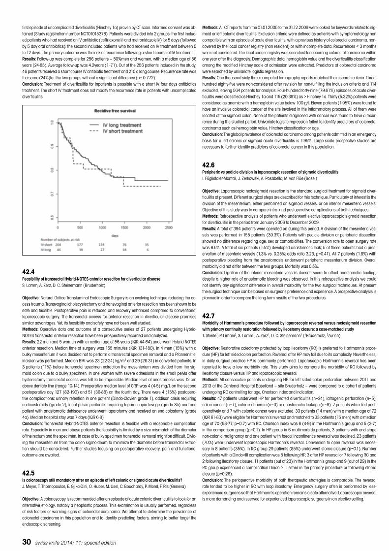
30 swiss knife 2014; 11: special edition
first episode of uncomplicated diverticulitis (Hinchey 1a) proven by CT scan. Informed consent was ob-tained (Study registration number NCT01015378). Patients were divided into 2 groups: the first includ-ed patients who had received an IV antibiotic (ceftriaxone® and metronidazole®) for 5 days (followed by 5 day oral antibiotics); the second included patients who had received an IV treatment between 5 to 12 days. The primary outcome was the risk of recurrence following a short course of IV treatment.Results: Follow-up was complete for 256 patients – 50%men and women, with a median age of 56 years (24-85). Average follow-up was 4.2years (1.-7.1). Out of the 256 patients included in the study, 46 patients received a short course IV antibiotic treatment and 210 a long course. Recurrence rate was the same (24%)for the two groups without a significant difference (p= 0.772). Conclusion: Treatment of diverticulitis for inpatients is possible with a short IV four days antibiotics treatment. The short IV treatment does not modify the recurrence rate in patients with uncomplicated diverticulitis.
42.4Feasibility of transrectal Hybrid-NOTES anterior resection for diverticular diseaseS. Lamm, A. Zerz, D. C. Steinemann (Bruderholz)
Objective: Natural Orifice Transluminal Endoscopic Surgery is an evolving technique reducing the ac-cess trauma. Transvaginal cholecystectomy and transvaginal anterior resection has been shown to be safe and feasible. Postoperative pain is reduced and recovery enhanced compared to conventional laparoscopic surgery. The transrectal access for anterior resection in diverticular disease promises similar advantages. Yet, its feasibility and safety have not been well studied.Methods: Operative data and outcome of a consecutive series of 27 patients undergoing Hybrid-NOTES transrectal anterior resection have been prospectively recorded and analyzed.Results: 22 men and 5 women with a median age of 56 years (IQR 44-64) underwent Hybrid-NOTES anterior resection. Median time of surgery was 155 minutes (IQR 131-180). In 4 men (15%) with a bulky mesenterium it was decided not to perform a transrectal specimen removal and a Pfannenstiel incision was performed. Median BMI was 23 (22-24) kg/m2 and 29 (26-31) in converted patients. In 3 patients (11%) before transrectal specimen extraction the mesenterium was divided from the sig-moid colon due to a bulky specimen. In one women with severe adhesions in the small pelvis after hysterectomy transrectal access was felt to be impossible. Median level of anastomosis was 12 cm above dentate line (range 10-14). Preoperative median level of CRP was 4 (4-5) mg/l, on the second postoperative day 127 (82-190) and 51 (36-88) on the fourth day. There were 4 (15%) postopera-tive complications: urinary retention in one patient (Dindo-Clavien grade 1), addison crisis requiring corticosteroids (grade 2), local pelvic peritonitis requiring laparoscopic lavage (grade 3b) and one patient with anastomotic dehiscence underwent laparotomy and received an end colostomy (grade 4a). Median hospital stay was 7 days (IQR 6-8).Conclusion: Transrectal Hybrid-NOTES anterior resection is feasible with a reasonable complication rate. Especially in men and obese patients the feasibility is limited by a size mismatch of the diameter of the rectum and the specimen. In case of bulky specimen transrectal removal might be difficult. Divid-ing the mesenterium from the colon sigmoideum to minimize the diameter before transrectal extrac-tion should be considered. Further studies focusing on postoperative recovery, pain and functional outcome are awaited.
42.5Is colonoscopy still mandatory after an episode of left colonic or sigmoid acute diverticulitis?J. Meyer, T. Thomopoulos, E. Gjika-Dini, O. Huber, M. Usel, C. Bouchardy, P. Morel, F. Ris (Geneva)
Objective: A colonoscopy is recommended after an episode of acute colonic diverticulitis to look for an alternative etiology, notably a neoplastic process. This examination is usually performed, regardless of risk factors or warning signs of colorectal carcinoma. We attempt to determine the prevalence of colorectal carcinoma in this population and to identify predicting factors, aiming to better target the endoscopic screening.
Methods: All CT reports from the 01.01.2005 to the 31.12.2009 were looked for keywords related to sig-moid or left colonic diverticulitis. Exclusion criteria were defined as patients with symptomatology non compatible with an episode of acute diverticulitis, with a previous history of colorectal carcinoma, non-covered by the local cancer registry (non resident) or with incomplete data. Recurrences < 3 months were not considered. The local cancer registry was searched for occurring colorectal carcinoma within one year after the diagnosis. Demographic data, hemoglobin value and the diverticulitis classification among the modified Hinchey scale at admission were extracted. Predictors of colorectal carcinoma were searched by univariate logistic regression.Results: One-thousand sixty-three computed tomography reports matched the research criteria. Three-hundred eighty-five were non-considered after revision for non-fulfilling the inclusion criteria and 114 excluded, leaving 564 patients for analysis. Four-hundred forty-nine (79.61%) episodes of acute diver-ticulitis were classified as Hinchey 1a and 115 (20.39%) as > Hinchey 1a. Thirty (5.32%) patients were considered as anemic with a hemoglobin value below 100 g/l. Eleven patients (1.95%) were found to have an invasive colorectal cancer at the site involved in the inflammatory process. All of them were located at the sigmoid colon. None of the patients diagnosed with cancer was found to have a recur-rence during the studied period. Univariate logistic regression failed to identify predictors of colorectal carcinoma such as hemoglobin value, Hinchey classification or age.Conclusion: The global prevalence of colorectal carcinoma among patients admitted in an emergency basis for a left colonic or sigmoid acute diverticulitis is 1.95%. Large scale prospective studies are necessary to further identify predictors of colorectal cancer in this population.
42.6Peripheric vs pedicle division in laparoscopic resection of sigmoid diverticulitisI. Füglistaler-Montali, J. Zerkowski, A. Posabella, M. von Flüe (Basel)
Objective: Laparoscopic rectosigmoid resection is the standard surgical treatment for sigmoid diver-ticulitis at present. Different surgical steps are described for this technique. Particularly of interest is the division of the mesenterium, either performed on sigmoid vessels, or on inferior mesenteric vessels. Objective of this study was to compare intra- and postoperative complications of both techniques.Methods: Retrospective analysis of patients who underwent elective laparoscopic sigmoid resection for diverticulitis in the period from January 2006 to December 2009. Results: A total of 394 patients were operated on during this period. A division of the mesenteric ves-sels was performed in 155 patients (39.3%). Patients with pedicle division or peripheric dissection showed no difference regarding age, sex or comorbidities. The conversion rate to open surgery rate was 6.5%. A total of six patients (1.5%) developed anastomotic leak; 5 of these patients had a pres-ervation of mesenteric vessels (1,3% vs. 0.25%; odds ratio 3.23, p=0.41). All 7 patients (1.8%) with postoperative bleeding from the anastomosis underwent peripheric mesenterium division. Overall morbidity did not differ between the two groups. Mortality was 0.5%.Conclusion: Ligation of the inferior mesenteric vessels doesn’t seem to affect anastomotic healing, despite a higher rate of anastomotic bleeding was observed. In this retrospective analysis we could not identify any significant difference in overall morbiditiy for the two surgical techniques. At present the surgical technique can be based on surgeons preference and experience. A prospective analysis is planned in order to compare the long-term results of the two procedures.
42.7Morbidity of Hartmann’s procedure followed by laparoscopic reversal versus rectosigmoid resection with primary continuity restoration followed by ileostomy closure: a case-matched studyT. Stierle1, P. Limani2, S. Lamm1, A. Zerz1, D. C. Steinemann1 (1Bruderholz, 2Zurich)
Objective: Restorative colectomy protected by loop ileostomy (RC) is preferred to Hartmann’s proce-dure (HP) for left sided colon perforation. Reversal after HP may fail due to its complexity. Nevertheless, in daily surgical practice HP is commonly performed. Laparoscopic Hartmann’s reversal has been reported to have a low morbidity rate. This study aims to compare the morbidity of RC followed by ileostomy closure versus HP and laparoscopic reversal.Methods: All consecutive patients undergoing HP for left sided colon perforation between 2011 and 2013 at the Cantonal Hospital Baselland – site Bruderholz – were compared to a cohort of patients undergoing RC controlling for age, Charlson index and indication.Results: 47 patients underwent HP for perforated diverticulitis (n=24), iatrogenic perforation (n=5), colon cancer (n=7), colon ischaemia (n=3) or anastomotic leakage (n=8). 7 patients who died post-operatively and 7 with colonic cancer were excluded. 33 patients (14 men) with a median age of 72 (IQR 61-83) were eligible for Hartmann’s reversal and matched to 33 patients (15 men) with a median age of 70 (58-77; p=0.7) with RC. Charlson index was 6 (4-9) in the Hartmann’s group and 5 (3-7) in the comparison group (p=0.1). In HP group in 6 multimorbide patients, 3 patients with end-stage non-colonic malignancy and one patient with faecal incontinence reversal was declined. 23 patients (70%) were underwent laparoscopic Hartmann’s reversal. Conversion to open reversal was neces-sary in 8 patients (35%). In RC group 29 patients (85%) underwent stoma closure (p=0.1). Number of patients with a Dindo>III complication was 8 following HP, 3 after HP reversal or 7 following RC and 2 following ileostomy closure. 11 patients (out of 23) in the Hartmann’s group and 9 (out of 29) in the RC group experienced a complication Dindo > III either in the primary procedure or following stoma closure (p=0.26). Conclusion: The perioperative morbidity of both therapeutic strategies is comparable. The reversal rate tended to be higher in RC with loop ileostomy. Emergency surgery often is performed by less-experienced surgeons so that Hartmann’s operation remains a safe alternative. Laparoscopic reversal is more demanding and reserved for experienced laparoscopic surgeons in an elective setting.
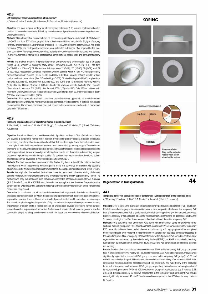
swiss knife 2014; 11: special edition 31
42.8Left emergency colectomies: is stoma a friend or foe?H. Teixeira-Farinha, E. Melloul, D. Hahnloser, N. Demartines, M. Hübner (Lausanne)
Objective: The ideal surgical strategy for left emergency colectomy (EC) remains controversial and is decided on a case-by-case basis. This study describes current practice and outcomes in patients who underwent a left EC.Methods: This retrospective review includes all consecutive patients who underwent left EC between July 2006 and June 2013. Demographic data, patient co-morbidities, indication for EC, type of surgery (primary anastomosis (PA), Hartmann’s procedure (HP), PA with protective ostomy (PAO), two-stage procedure (TS)), and postoperative outcomes were entered in a database after approval by the local ethic committee. Two-stage procedure defined patients who underwent a left EC followed by a delayed PA or HP. Outcomes of interest were postoperative complications, hospital stay and permanent ostomy rate.Results: The analysis includes 153 patients (94 men and 59 women), with a median age of 76 years (range 22-95) with left EC during the study period. There were 45% (n=70) PA, 4% (n=5) PAO, 48% (n=73) HP and 4% (n=5) TS. Median hospital stays were 12 (5-40), 20 (18-55), 19 (4-89), and 27 (1-127) days, respectively. Compared to patients with PA, patients with HP, TS or PAO had significantly more ischemic heart disease (13 vs. 33, 60, and 60%, p=0.005). Similarly, patients with HP or PAO had more chronic renal failure (8 vs. 37 and 40%, p<0.001). Clavien-Dindo grade III to V complications rate was 30% after PA, 41% after HP, 40% after PAO and 100% after TS. In-hospital mortality was 4% (n=3) after PA, 11% (n=8) after HP, 60% (n=3) after TS, while no patients died after PAO. The rate of anastomotic leak was 7% (5/70) after PA and 20% (1/5) after PAO. Only 26% of patients with Hartmann underwent continuity rehabilitation within a year after primary EC, mainly because of death (33%) or severe co-morbidities (52%).Conclusion: Primary anastomosis with or without protective ostomy appears to be a safe treatment option for patients with low co-morbidity undergoing emergency left colectomy. In patients with severe co-morbidities, Hartmann’s procedure does not prevent adverse outcomes and entails a permanent ostomy in 75% of them.
42.9Promising approach to prevent parastomal hernia: a Swiss innovationP. Kirchhoff1, H. Hoffmann1, D. Oertli1, U. Zingg2, D. Hahnloser3, P. Kirchhoff1 (1Basel, 2Schlieren, 3Lausanne)
Objective: Parastomal hernia is a well known clinical problem, and up to 50% of all stoma patients will develop a parastomal hernia within the first 3 years after primary surgery. Surgical procedures for repairing parastomal hernia are difficult and their failure rate is high. Several recent studies found a prophylactic effect of incorporation of a sublay mesh placed during primary surgery. The results are promising for the prevention of parastomal hernias, although there is still the risk of organ adhesion to the foreign material, lack of knowledge about long-term results and it remains a demanding surgical procedure to place the mesh in the right position. To address the specific needs of the stoma patient and the surgeon we developed a innovative ring solution (KORING).Methods: The device consists of a non-absorbable, flexible ring that is sutured to the anterior sheath of the abdominal wall. It thus prevents weakening of the fascia that surrounds the intestine. A lip seals the abdominal cavity. We developed the ring from scratch to the European market approval within 3 years.Results: We implanted the medical device three times for permanent colostomy during abdomino- perineal resection. The implantation of the ring prolonged operating time by approximately 15 min. The material was easy to handle and fixed with 8 non-absorbable interrupted sutures. Correct diameter (2.5, 3.5 and 4.5 cm) of the KORING was chosen by measuring the bowel diameter. The postoperative 30-day course was uneventful. Long term follow up within an observational study and a randomized clinical trial are planned.Conclusion: In conclusion, parastomal hernia is a relevant ostomy-complication in terms of morbidity and socio-economic impact, for which the concept of prophylactic mesh insertion has shown promis-ing results. However, it has not become a standard procedure due to still unresolved shortcomings. The new stomaplasty ring has the potential of high impact on future prevention of parastomal hernias, improvement of quality of life of treated patients as well as cost savings by avoiding further surgical interventions due to parastomal herniation. Furthermore it should attract more surgeons to use be-cause of its simple handling, small contact are with the tissue and less necessary tissue mobilization.
KORING
Regeneration in Transplantation 4444.1Temporary portal vein occlusion does not compromise liver regeneration of the occluded lobesA. Wirsching1, E. Melloul2, R. Graf1, P.-A. Clavien1, M. Lesurtel1 (1Zurich, 2Lausanne)
Objective: Liver lobe volume manipulation using temporary portal vein embolization (PVE) could con-tribute to make liver surgery or transplantation safer. In mice, we previously showed that temporary PVE is as efficient as permanent PVE or portal vein ligation to induce hypertrophy of the non-occluded lobes. However, recovery of the occluded lobes after revascularization remains to be assessed. Study Aims: To assess histological and functional recovery of embolized liver lobes after temporary PVE.Methods: Forty BL6/male mice underwent 70% portal vein embolization using either powdered ab-sorbable material (temporary PVE) or embospheres (permanent PVE). Fourteen days after temporary PVE, revascularization of the occluded lobes was confirmed by MRI angiography and hypertrophied non-occluded lobes were resected. In the permanent PVE group, non-occluded lobes were resected at the same time point. Mice undergoing 30% hepatectomy without previous PVE served as controls. Liver regeneration was assessed by liver-to-body weight ratio (LBW-R) and Ki-67 immunohistochemistry, liver function by bilirubin serum levels, liver injury by AST and ALT serum levels and fibrosis by sirius red staining.Results: Survival after non-occluded lobe resection was 100% in the temporary PVE group compared to 81% after permanent PVE. Twenty-four hours after resection, AST, ALT and bilirubin serum levels were significantly higher in the permanent PVE group compared to the temporary PVE group (p <0.05 and <0.001, respectively). Periportal fibrosis was observed almost exclusively after permanent PVE. After seven days, non-occluded lobe resection induced 156 and 263% liver hypertrophy of the occluded lobes in the temporary and permanent PVE groups, respectively. Mean liver-to-body-weight-ratios of temporary PVE, permanent PVE and 30% hepatectomy groups at postoperative day 7 reached 3.51, 3.54 and 4.2 respectively. Ki-67 positive hepatocytes in the temporary and permanent PVE groups were significantly increased 48 and 72h after resection compared to the 30% hepatectomy controls (p <0.001).

32 swiss knife 2014; 11: special edition
Conclusion: While temporary PVE induces hypertrophy of non-occluded lobes, revascularized liver lobes are functional and able to hypertrophy after resection of the non-occluded lobes.
44.2Activation of the Constitutive Androstane Receptor (CAR) reverses deficient liver regeneration in the Small-For-Size Syndrome via Foxm1b dependent pathwaysC. Tschuor1, E. Kachaylo1, P. Limani1, M. Linecker1, D. A. Raptis1, E. Melloul2, Y. Tian1, K. Lehmann1, B. Humar1, R. Graf1, P.-A. Clavien1 (1Zurich, 2Lausanne)
Objective: Resectability of liver tumors is limited since patients left with marginal liver remnants are at risk for developing the Small-for-Size Syndrome (SFSS). SFSS is characterized by an insufficient recov-ery due to delayed regeneration. Strategies are needed to overcome regenerative limits, rendering large liver tumors resectable. Activation of the Constitutive Androstane Receptor (CAR) induces liver hyper-plasia. We investigated the potential of the CAR agonist TCPOBOP (TCP) to ameliorate experimental SFSS, thereby enabling extended oncological liver resections.Methods: The effects of TCP on liver regeneration were assessed in four murine models: 68% hepate-ctomy (Hx) (control), 86% Hx (model of SFSS), 91% Hx (lethal model), and 30% liver transplantation in BL6 and CAR-/- mice. Serum bilirubin and albumin served as measures of SFSS-like features. Pro-liferation-associated molecules, including Foxm1b, p21 and miR375/YAP-dependent pathways were analysed. Functional relevance of the molecules was assessed via siRNA knockdown. Humanized mice and human tissue microarrays served for evaluation of the translational potential.Results: Reduced survival in SFSS is associated with deficient regeneration due to deregulation of Foxm1b and YAP/miR375 pathways along with p21 upregulation. TCP markedly improved survival following 86% Hx, 91% Hx and 30% transplantation. Likewise, SFSS-associated features were normal-ized by TCP, while TCP did not improve survival or liver regeneration in CAR-/- mice. Functional experi-ments reveal that CAR activation promotes Foxm1b, reversing abnormal induction of p21 and restoring deficiency in liver regeneration. Human HCC and adenoma TMA’s reveal that only 40% of tumors are CAR positive.Conclusion: TCP rescues mice exposed to SFSS after extended liver resections and liver transplanta-tion. Molecular changes induced by CAR activation act on downstream targets of pathways promoting cell cycle progression and uncoupling from organ size control to override the regenerative deficits of marginal liver remnants. Reduced CAR expression in tumors suggests a subset of HCCs may not re-spond to CAR activation, pointing to a patient group amenable to this treatment. Studies in humanized mice and on ex vivo human liver tissue will address the clinical potential of CAR activation by enhanc-ing liver regeneration after extended resections for primarily unresectable liver tumors.
44.3Human platelets are internalized by liver sinusoidal endothelial cells and enhance secretion of EGFE. Lejmi1, J. Meyer1, K. Sadoul2, P. Fontana1, E. Perroud1, A. Bollinger1, P. Morel1, C. Gonelle-Gispert1, L. Bühler1 (1Geneva, 2Grenoble/FR)
Objective: Several observations demonstrated that liver sinusoidal endothelial cells (LSEC) and plate-lets are important players in liver regeneration. In addition, it has been shown that activated platelets can be taken up by LSEC. The aim of this study was to investigate whether the interaction and internali-zation of platelets by LSEC plays a role during liver regeneration.Methods: Mouse, human LSEC and hepatocytes were isolated simultaneously using collagenase di-gestion combined with density gradient centrifugation and magnetic sorting with anti-CD146 beads for LSEC. Characterization of LSEC was performed using FACS analysis and immunofluorescence staining. Carboxyfluorescein stained mouse and human platelets, activated or not with thrombin (1U/ml), were co-cultured with LSEC for 1 hour and platelet internalization was assessed using confocal mi-croscopy. IL6 and EGF were measured by ELISA in the co-culture media. Proliferation of LSEC and hepa-tocytes was analyzed in triple co-cultures with platelets using Ki67 staining and EdU incorporation.Results: Freshly isolated mouse LSEC express CD146, CD31, CD309, VWF and not CD11b, LY6C (mac-rophage markers). Platelets were internalized by LSEC and internalization was enhanced by thrombin activation (results are expressed as number of internalized platelets by 100 LSEC: for mouse, resting platelets versus activated platelets 32 ± 8 vs 62 ±21; and for human 45 ± 16 vs 132 ± 25, respec-tively). Secreted EGF levels increased when LSEC were co-cultured with platelets, and EGF levels were modulated by platelet activation. Mouse isolated hepatocytes were highly proliferating. This high prolif-erative index did not allow to observe potential differences when co-cultured with LSEC and platelets. However, the presence of hepatocytes was necessary to induce LSEC proliferation.Conclusion: Human platelets are internalized by LSEC and promote secretion of EGF, cytokine known to be implicated in liver regeneration.
44.4Liver regeneration is not impacted in the absence of intestinal microbiotaT. Malinka, A. Keogh, R. Kudira, S. Moghadamrad, K. McCoy, D. Stroka, D. Candinas, G. Beldi (Bern)
Objective: Liver regeneration after partial hepatectomy is a very complex process that involves a vari-ety of different liver cell types via cytokine- and growth-factor-mediated pathways. Bacterial products and LPS circulate in the portal blood stream directly through the hepatic sinusoids and have been proposed to initiate liver regeneration by activation of parenchymal and non-parenchymal cells in liver after partial hepatectomy. Our aim was to evaluate liver regeneration in the absence of microbes and microbial products.Methods: Two third partial hepatectomy (PH) was performed in germ-free (axenic) mice and mice colonized with altered schaedler flora (ASF) were used as controls. Surgery was performed in axenic conditions to maintain sterility throughout the course of the experiment (Clean Mouse Facility; Univer-sity of Bern). Liver regeneration, injury and hepatic fractions of immune cells were assessed in tissue
and serum samples 48 hours post partial hepatectomy.Results: Liver injury (ALT/AST) and survival were not significantly different between germ free and ASF colonized mice. Liver regeneration was measured by the number of Ki67 positive cells and there was no significant difference between germ-free and ASF mice (p=0,84). Absence of microbiota was proved by fluorescence microscopy and faecal cultures. Fractions of CD4 T cells, NKT cells and B cells were altered at baseline between germ-free and ASF mice, but not after partial hepatectomy.Conclusion: Liver regeneration after PH is not impaired in the absence of microbes and microbial prod-ucts. These findings contrast previous studies and reveal that the gut microbiota is not an essential for the priming or maintenance of liver regeneration.
44.5Modulation of hippo pathway in liver regenerationG. Loforese1, T. Malinka1, A. Keogh1, T. Halazoneti1, 2, D. Candinas1, D. Stroka1 (1Bern, 2Geneva)
Objective: The Hippo pathway controls organ size by regulating cell proliferation and apoptosis. Par-tial hepatectomy (PH) provokes quiescent, differentiated hepatocytes to re-enter cell cycle to replace liver mass. We investigated how the Hippo pathway is involved in the physiological process of liver regeneration to better understand how to target it to improve the regenerative capacity of small-for-size livers. We analyzed the regulation of Hippo pathway kinases (MST1 & 2, LATS1 & 2) as well as up- and down-stream components in a murine PH model.Methods: Mice were subjected to 33, 65 or 91% PH (n=6 per group) and total RNA from livers was iso-lated 6, 24 and 48hrs post surgery and analysed by PCR microarray. Results were confirmed by qRT-PCR and proteins regulated at the mRNA were confirmed by Westernblot and immunohistochemistry.Results: Livers of mice subjected to 33 and 65% PH had Ki67 positive hepatocytes, whereas, mice with 91% showed less staining for hepatocyte proliferation. We observed a regulation of Hippo pathway proteins in mice subjected to 65% PH compared to 91%. There was 2-fold down regulation of kinases MST1 & MST2 at 6 (p=0.02) and 24hrs (0.001) post 65% PH compared to 91%. Yap1 target genes CTGF and Birc5/survivin were increased at 6hrs (p=0.01) and 48hrs (p=0.0003), respectively, in 65% PH, whereas no increase was observed in non-regenerating 91% PH livers.Conclusion: Taken together our data demonstrates that components of the Hippo pathway are regu-lated during liver regeneration after PH and suggest Hippo pathway as a new candidate for driving hepatic regeneration.
44.6Warm or cold machine perfusion to rescue DCD liver grafts prior to transplantationA. Schlegel, R. Graf, P. Kron, P.-A. Clavien, P. Dutkowski (Zürich)
Objective: Machine liver perfusion has been suggested to protect liver grafts donated after cardiac death. We compared in this study three concurrent approaches in a DCD liver transplant model.Methods: Rat livers were subjected to 30 and 60 min in situ warm ischemia without previous appli-cation of heparin. Subsequently, livers were excised and cold stored for 4hr, mimicking DCD organ procurement followed by conventional organ transport. In further experimental groups, DCD liver grafts received a 4hr normothermic oxygenated perfusion through the portal vein and the hepatic artery in-stead of cold storage. The perfusate consisted of either full blood or leukocyte depleted blood. In a last step, livers underwent hypothermic oxygenated perfusion (HOPE) for 1 hr after warm ischemia and 4 hr cold storage. Liver injury was assessed during machine perfusion and after orthotopic liver transplantation (OLT).Results: Normothermic oxygenated perfusion activated Kupffer and endothelial cells already be-fore transplantation. Injury of non-parenchymal cells correlated well with extension of donor warm ischemia and was independent from the presence of leukocytes and platelets in the perfusate. Despite that, reperfusion injury after OLT was less in normothermic perfused DCD livers, as compared to trans-planted cold stored DCD livers. However, with extension of donor warm ischemia up to 60 minutes, normothermic perfusion failed to protect from lethal injury. In contrast, HOPE treatment prevented from Kupffer- and endothelial cell activation and led to 90% (9/10) and 64% (5/8) 7 day survival, respec-tively, after 30 and 60 minutes donor warm ischemia.Conclusion: This is the first study comparing cold vs. normothermic machine perfusion techniques in a rat DCD liver transplant model. Normothermic oxygenated perfusion with blood or isolated erythro-cytes was superior compared to cold storage. However, one hour HOPE applied after cold storage was most protective in all endpoints.
44.7HOPE treatment protects from rejection in a rodent model of liver transplantationA. Schlegel, P. Kron, R. Graf, P.-A. Clavien, P. Dutkowski (Zürich)
Objective: While liver transplantation offers a unique possibility to treat un-resectable hepatic tumors, one major shortcoming remains the persistent need for lifelong immunosuppressive treatment. Ac-cordingly, the well known high risk of tumor recurrence under immunosuppressive therapy in cases with advanced tumors is regarded as contraindication for transplantation. Novel strategies decreasing the need of immunosuppressive treatment are urgently needed. We analyzed therefore the effect of end-ischemic hypothermic oxygenated machine liver perfusion (HOPE) on acute rejection in a rodent model of liver transplantation.Methods: The livers from Lewis rats were transplanted into Brown Norway recipients, reflecting a well-known model of acute liver rejection. The recipients were untreated or treated with Tacrolimus (FK506:1mg/kg). To compare the effect of immunosuppression with HOPE treatment, lewis livers underwent hypothermic oxygenated perfusion for one hour prior to implantation, without immunosup-pressive treatment of these recipients. Furthermore, we combined HOPE treatment with a minimal im-munosuppression (FK506:0.3mg/kg). Appropriate syngeneic grafts from Brown Norway donor rats

swiss knife 2014; 11: special edition 33
served as controls.Results: Acute rejection occurred in untreated recipients of allogeneic liver transplants, and all died within 14 days. In contrast, recipients of HOPE treated grafts survived beyond 30 days. These recipients developed only a mild delayed rejection as detected by a decreased number of activated Kupffer- and T cells and less fibrosis (FACS analysis, cytokine release). Accordingly, administration of a reduced amount of Immunosuppression was sufficient to protect from any rejection in recipients transplanted with HOPE treated grafts.Conclusion: We show here, for the first time, that graft treatment by hypothermic oxygenated perfusion strongly modulates the immune response, leading to improved survival after liver transplantation. Based on this, we suggest that recipients of HOPE treated grafts have a decreased need of immuno-suppression after transplantation. Accordingly, such easy and practical machine perfusion opens the door for future innovative transplant therapy concepts.
44.8The CD26-costimulatory pathway is critical for Th17-mediated lung allograft acceptanceY. Yamada1, J. Jang1, I. De Meester2, I. Inci1, W. Weder1, W. Jungraithmayr1 (1Zürich, 2Antwerp/BE)
Objective: The molecule CD26, a type II transmembrane glycoprotein, expressed on activated T cells leading to classical T-cell co-stimulation and plays an important role in regulating CD4+ T cell activa-tion in autoimmune diseases. Recently, Th-type 17-differentiated CD4+ T cells have been shown to be phenotypically characterized by a high expression of CD26. Here, we hypothesize that the co-stimu-latory blockade of CD26 during acute rejection (AR) results in reduced CD4+ Th-17 cell alloreactivity, thereby promoting mouse lung transplant (Tx) acceptance.Methods: In vitro experiments (n=4): mixed lymphocyte reaction (MLR) was performed between sort-ed CD4+ T cells from C57BL/6 mice as targets (T) and splenocytes from BALB/c being effectors (E) upon CD26-inhibition using the inhibitor Vildagliptin in increasing concentrations (Fig. 1A). In vivo ex-periments (n=11): MHC class I and II mismatched mice, BALB/c (donors) and C57BL/6 (lung Tx recipi-ents) were used. CD26-/- recipient mice were used for gain of function experiments. CD26-inhibition in recipients was achieved by daily application of 10 mg/kg BW of Vildagliptin from Tx until day 5. Mac-roscopy, histology, immunohistochemistry (CD3+ T cells) and IL-17A levels (ELISA) were analyzed.Results: Levels of IL-17A were significantly reduced in MLR experiments when increasing the concen-trations of Vildagliptin (p*=0.003) (Fig.1A). Upon Tx, macroscopically, allografts from untreated mice (control) show severe AR, histologically A4, from Vildagliptin-inhibited mice show attenuated AR, histo-logically A1, and from CD26-/- mice show minimal AR, histologically A2.Conclusion: Our in vitro experiments show that CD26-inhibition results in a reduction of CD4+Th17+ derived IL-17A. Also, we found that in vivo blockade of the CD26-costimulatory pathway leads to an at-tenuation of AR macroscopically as well as to a reduced infiltration of alloreactive CD3+ T cells. These findings identify CD26 as an important, yet underestimated co-stimulatory molecule in CD4+Th17-mediated lung allograft rejection.
Control CD26/DPPIV-inhibitor CD26-/-
* * *
* A
B
Figure 1A: Mixed lymphocyte reaction (from 4 independent experiments) between sorted
CD4+ T cells from C57BL/6 mice being targets and splenocytes from BALB/c being effectors in
a E:T ratio of 1:2. Levels of IL-17, as determined by ELISA, were significantly reduced by
increasing the concentrations of CD26/DPP4 inhibitor (p*=0.003)
Figure 1B: Allogeneic mouse single lung transplantation (n=11) between BALB/c (donors) and
C57BL/6 (recipient) mice. Allografts (*) from non treated mice (control) show robust acute
rejection (AR), from CD26/DPPIV-inhibited mice (CD26/DPPIV-inhibitor) show attenuated AR,
and from CD26 knock out mice (CD26-/-) show minimal AR.
44.9A novel intra-abdominal cooling system prevents graft warming during implantation for robotic-assist-ed renal transplantationM. Hagen, C. Joliat, N.C. Buchs, J.-B. Buchs, A. Nastasi, R. Ruttimann, F. Lazeyras, P. Morel, L. Bühler (Geneva)
Objective: Robotic kidney implantation -which has been performed in a small number of patients- has been described as a method to reduce the invasiveness of the surgical access. However, warm ischemia and thus graft function remain valid concerns. Therefore, the purpose of this study was to measure the impact of robotic-assisted kidney transplantation on organ warming and to assess a novel intra-abdominal cooling system device for implantation.Methods: A porcine model was used for all experiments. The left kidney was procured by open surgery, flushed with preservation solution at 4C, and then re-implanted on the abdominal aorta and vena cava into the same animal. Group 1: A porcine model was used for all experiments. The left kidney was procured by open surgery, flushed with preservation solution at 4C, and then re-implanted on the ab-dominal aorta and vena cava into the same animal. The following groups were allocated: Group 1: con-ventionally performed open auto-transplant – the grafts were rinsed with cold saline every 5 min during implantation. Group 2: robotically assisted auto-transplants without cooling system – no cooling was provided. Group 3: robotically assisted auto-transplants with cooling system – plastic chamber sur-rounding the kidney continuously perfused by a tubing system with saline at 4C. A thermal probe was placed into the renal cortex prior to the procurement to monitor the temperature until revascularization. At 6 hours after implantation, a magnetic resonance imaging with gadolinium injection was performed to measure the perfusion of the grafts.Results: The mean time of implantation was 39.4 ±7.8 minutes for the open group and 68.6 ±8 min-utes overall for the robotic cohorts (p=0.0021). The temperature of the grafts at the end of implantation was 23,5C ±7C for group 1, 29±2C for group 2 and 12C ±5C for group 3 (p=0.3972 and 0.0467 respectively). The gadolinium perfusion of the grafts and urine out-put was similar in all groups.Conclusion: The use of robotic assisted renal transplantation prolongs implantation time and increas-es the warm ischemia of the graft. The use of a simple intra-abdominal cooling device prevents warm-ing of grafts significantly and keeps temperature levels lower than those observed with open surgery which might prevent warm ischemia damage. Further research is necessary to assess the effects of robotic techniques for kidney implantation.
Video III 4646.1Transanal endoscopic rectal cancer resection: down-to-Up total mesorectal excisionR. Troller, H. Gelpke, F. Grieder, S. Breitenstein (Winterthur)
Objective: Conventional low anterior rectal resection, particularly in patients with a high waist-hip-ratio, are technically highly challenging. This is mainly because of the limited view and exposure deep in the pelvis. Transanal natural orifice transluminal endoscopic surgery (NOTES) promises a safe and less morbid alternative. First reports of “Down to up” total mesorectal excision via transanal minilaparosco-py-assisted NOTES have shown satisfactory short-term oncologic results. This video aims to demon-strate the feasibility of transanal minilaparoscopy-assisted rectum resection in case of a rectal cancer.Methods: We show a viedo of a transanal minilaparoscopy-assisted total mesorectal excision in a 64-year-old patient suffering from rectal cancer. Neoadjuvant radio-chemotherapy was not indicated.Results: Transanal approach: As a first step the wall of the rectum was dissected transanally at the level of the pelvic floor and a distance of 2cm aboral of the tumor. The lumen of the rectum, distal of the tumor was closed by a purse-string suture. The perirectal space for total mesorectal excision was opened. Then, a single port trocar (Covidien, SILS trocar) was placed in the anal channel and gas insufflation was installed. Laparoscopic single port total mesorectal excision was performed from down-to-top. After reaching the abdominal cavity by opening the peritoneum in the pelvis, the approach was changed to transabdominal laproscopy. Transabdominal laparoscopy: The left colon including the left flexure was completely mobilized by transabdominal laparoscopy. The superior rectal artery and inferior mesenteric vein were clipped and dissected. To complete total mesorectal excision the peritoneum was incised around the rectum followed by removal of the colorectal specimen via a 6cm caesarean incision. A stapled side-to-end colorectal anastomosis was performed 3cm proximal of the dental line and finally a diverting loop ileostomy was created. The postoperative course was uneventful except for a symptomatic urolithiasis.Conclusion: This video demonstrates feasibility of a transanal minilaparoscopy-assisted rectal resec-tion with down-to top total mesorectal excision. This approach may offer advantages compared to the complete transabdominal laparoscopic technique with regard to the dissection of the distal me-sorectum.
46.2Transrectal specimen retrieval – an option for further trauma reduction in single port combined laparo-scopic sigmoid resectionsW. Brunner, S. Bischofberger, L. Traine, N. Kalak (Rorschach)
Objective: Laparoscopic sigmoid resection for diverticular disease is a standard procedure. Single port access leads to trauma reduction to the abdominal wall, whereas specimen retrieval needs suprapu-bic or umbilical incision enlargement. This didactic video demonstrates step by step our technique of combined single port laparoscopic sigmoid resection with transrectal specimen retrieval.

34 swiss knife 2014; 11: special edition
Methods: An open approach at the umbilical site is performed with an incision < 3.5 cm for introduction of the single port system with integrated protection folie, allowing the use of up to 4 instruments. The key steps and potential pitfalls inherent to any colorectal resection are demonstrated, while the specif-ics of single port surgery and the transrectal specimen retrieval are highlighted.Results: A standard medial to lateral mobilisation of the colon is performed, adhesiolysis done if neces-sary using only straight or one articulating instrument additional. The dissection of the specimen is completed intraabdominal. For means of stapling the upper rectum an additional 12 mm trocar in a suprapubic position can be helpful. A retrieval bag is introduced transrectal after distinct cleaning of the rectal stump and transecting the stapling line. The entire specimen is placed in the bag and retrieved, the opened rectal stump is stapled again. Anvil placement in the descending colon is achieved tran-sumbilical, anastomosis is performed in a usual manner.Conclusion: Single port laparoscopic sigmoid resection is a safe and efficient procedure. Transrectal specimen retrieval offers further trauma reduction to the abdominal wall and potentially reduces inci-sion related complications as wound infection and hernia.
46.3Epiphrenische Oesophagusdivertikel – minimalinvasive Behandlung mittels laparoskopischer tran-shiataler Divertikelresektion kombiniert mit Oesophagokardiomyotomie und anteriorer FundoplikatioA. Scheiwiller (Luzern)
Objective: Epiphrenische Divertikel stellen eine seltene Form der Oesophagusdivertikel dar. Es handelt sich dabei um Pulsionsdivertikel, meist auf dem Boden von Motilitätsstörungen der Speiseröhre oder Relaxationsstörungen des unteren Oesophagussphinkters. In der Vergangenheit wurden sie meist mit-tels Thorakotomie behandelt. Die laparoskopische, transhiatale Vorgehensweise hat sich in den letzten Jahren auch für dieses Krankheitsbild bewährt.Methods: Wir präsentieren ein Video unserer Technik mit laparoskopischer, transhiataler Divertikelre-sektion, Oesophagokardiomyotomie und anteriorer Fundoplikatio anhand eines Patienten mit 10cm grossem epiphrenischem Divertikel, zunehmender Dysphagie und Unmöglichkeit der Nahrungsauf-nahme. Nach endoskopischer Ausräumung des Divertikels und Verbesserung des Ernährungsstatus folgt die operative Sanierung. Der Patient wird in Gallenblasenlagerung und 40 Grad Antitrendelen-burgposition operiert. Nach Eröffnung des Hiatus oesophageus wird das Divertikel unter gastrosko-pischer Beleuchtung identifiziert und zirkulär bis auf den Oesophagus freipräpariert. Um eine Sten-osierung zu verhindern, wird der Oesophagus mit einem dicken Magenschlauch geschient und das Divertikel mit Hilfe eines laparoskopischen linearen Klammernahtgerätes reseziert. Die Oesophagus-muskulatur wird darüber vernäht, eine Oesophagokardiomyotomie durchgeführt und eine anteriore Fundoplikatio angelegt.Results: Der postoperative Verlauf ist unkompliziert, die radiologische Kontrolle zeigt eine unbehinderte Oesophaguspassage. Anlässlich der Verlaufskontrolle berichtet der Patient über eine problemlose Nahrungsaufnahme ohne Erbrechen oder Regurgitation und keine Refluxsymptomatik.Conclusion: Die laparoskopische transhiatale Divertikelresektion kombiniert mit einer Oesophagokar-diomyotomie und einer anterioren Fundoplikatio ist in unseren Augen die Therapie der Wahl bei symp-tomatischen epiphrenischen Oesophagusdivertikel.
46.4Modified pancreatoduodenectomy (Kausch-Whipple procedure) for primary leiomysarcoma of the pancreatic headP. Limani, C. Tschuor, M. Linecker, P.-A. Clavien, H. Petrowski, R. Graf (Zürich)
Objective: Primary leiomyosarcomas of the pancreas are extremely rare and account for less than 0.1% of malignant pancreatic neoplasms. At the time of diagnosis, these tumors are often large and at an advanced stage. Hence, palliative treatment with an impaired patient survival remains often the only treatment option. Nevertheless, in select cases extensive surgical resection is still indicated. However, the surgical technique is controversial and depends on tumor size and localization. We report a case of primary pancreatic leiomyosarcoma surgically treated with a Kausch-Whipple pancreatoduodenec-tomy.Methods: We present a case of a 54-year old man who was admitted to our outpatient clinic from a peripheral hospital. The patient complained of a two week history of upper abdominal pain, jaundice, light stools, dark urine, and weight loss. Externally repeated abdominal ultrasound and MRI revealed a slight dilatation of intra and extrahepatic bile ducts without signs of neoplasm or inflammation. Blood tests showed elevated liver function test as well as bilirubin, alkaline phosphatase, GGT, and lipase. Fine needle biopsy of the pancreas head revealed a myogenic spindle-cell shaped proliferation of un-known entity. PET/CT showed a highly metabolic active mass in the pancreas head without signs of lymph node or distant metastases. Pancreas-Dotarem-MRI confirmed the mass as well as a dilation of the intra and extrahepatic bile ducts without infiltration of the neighbouring abdominal vessels. ERCP with papillotomy and stent insertion was performed to relieve the stenosis of the common bile duct. The indication for surgical resection was confirmed by an interdisciplinary tumor board.Results: Part one of our video presents the medical case with focus on the preoperative imaging. Part two shows the modified Kausch-Whipple pancreatoduodenctomy: to adapt to the extensive peritu-moral inflammation the preparation of the superior mesenteric vein (SMV) was performed right before resection of the pancreas head and highlights the removal of the tumor specimen. Part three focuses on the reconstruction of the bowel.Conclusion: In cases of primary pancreatic leiomyosarcoma without signs of metastasis, an extensive surgical resection should be considered. In the present case, the pancreatoduodenectomy was well tolerated with disease-free follow up of one year so far.
46.5Voluminous rectal foreign body extraction by transanal insufflated access platform: a case reportR. Meier, G. Herren, A. Mennet, V. Belfontali, P. Morel, F. Ris (Geneva)
Objective: We report a case of voluminous rectal foreign body extracted by transanal insufflated ac-cess platform.Methods: We used a transanal access platform that enables insufflated access to the rectum and lower sigmoid colon for the extraction of a voluminous rectal foreign body under general anaesthesia.Results: A previously healthy 34-years old patient was admitted to the emergency department three hours after the introduction of a metal tube for erotic purpose. The tube was 3.8 cm in diameter and 18.3 cm long. On admission he was alert and stable (blood pressure: 149/109, heart rate: 95/’, tem-perature 36.2°C). His abdomen was tender in the right lower quadrant without guarding or rebound tenderness. Digital rectal examination allowed palpating the distal part of the object; no lesion was identified. An abdominal plain film revealed the presence of the metallic foreign body located at the junction of the rectal ampulla and the distal sigmoid colon, and no sign of intestinal perforation. After induction of general anaesthesia and placement of the usual monitors, the perineal region was disin-fected, and a transanal access platform was placed into the anal canal. The rectum was insufflated transanally (12 mm Hg) and the camera and two laparoscopic forceps were introduced through the device. After careful progression along the anal canal and the rectal ampulla, the foreign body was grasped and removed by traction. A retrograde inspection revealed no mucosal lesion and there was no evidence of an air leak. The duration of the procedure was 15 minutes.Conclusion: The therapeutic options for rectal foreign body removal include transanal extraction un-der conscious sedation, extraction under general anaesthesia, minilaparotomy or laparoscopy, and laparotomy with colotomy. Voluminous objects often require a minilaparotomy to achieve extrac-tion. We report the extraction of a voluminous rectal foreign body using a transanal insufflated ac-cess platform. This original approach was safe, of short duration, and effective, and allowed to avoid laparotomy.
Felix Largiadèr Prize Session 4848.1A single preoperative dexamethasone dose prevents nausea and vomiting after thyroidectomy. A ran-domized, double-blind, placebo-controlled trial (the tPONV study)I. Tarantino1, R. Warschkow2, U. Beutner2, W. Kolb2, A. Lüthi2, B. Schmied2, T. Clerici2 (1Heidelberg/DE, 2St. Gallen)
Objective: Patients undergoing thyroidectomy and a bilateral superficial cervical plexus block are an ideal, homogenous cohort in which to investigate the efficacy of dexamethasone for postoperative nausea and vomiting (PONV). These patients exhibit a high incidence of PONV and do not require opioids. They have no abdominal surgery that can cause nausea and vomiting via paralytic ileus. Com-bined with the highly standardized anesthesia protocol in use at our institution, this setting allows all known biases to be controlled.Methods: The tPONV study was a single institution, 1:1 randomized, double-blind, placebo-controlled trial with two parallel arms, comparing a single, perioperative dose of 8mg dexamethasone (treatment group) with saline (placebo group) in a superiority analysis (clinical trial number NCT01189292)Results: The overall incidence of PONV in the ITT-analysis was 65 of 152 patients (42.8%, 95% CI: 35.2%-50.7%). The incidence of PONV in the treatment arm was 22 of 76 patients (28.9%, 95% CI: 20.0%-40.0%) compared to 43 of 76 patients (56.6%, 95% CI: 45.4%-67.1%) in the control arm (P=0.001 odds ratio 0.31, 95%CI 0.16-0.61, absolute risk reduction 27.6%, 95%CI 12.0%-41.9%). The number needed to treat was 4. The incidence of PONV was significantly different only 4 hours after sur-gery. Dexamethasone did not influence intensity of pain, length of hospital stay, the amount of anaes-thetic medication required. No diabetes mellitus, no wound infection or other dexamethasone-related adverse events were recorded.Conclusion: A single preoperative dexamethasone dose is cheap, safe and effective for prevention of PONV after thyroidectomy. Therefore the dexamethasone administration before thyroidectomy should be introduced as a new standard in clinical practice. Furthermore the results of present study might be generalizable to all type of operations that involve general anaesthesia.
48.2Donor characteristics and the risk of hepatocellular carcinoma recurrence after liver transplantationL. A. Orci, T. Berney, P. E. Majno, S. Lacotte, G. Oldani, P. Morel, G. Mentha, C. Toso (Geneva)
Objective: Hepatocellular carcinoma (HCC) recurrence after liver transplantation occurs in 10-15% of patients. To date, studies assessing the risk of post-transplant HCC recurrence have focused on recipi-ents’ characteristics. Here, we investigate the impact of donor characteristics and liver graft quality on the risk of post-liver transplantation HCC recurrence.Methods: Using the Scientific Registry of Transplant Recipients, we included adult patients with a di-agnosis of HCC receiving liver transplantation between 2004 and 2011. Post-transplant HCC recur-rence was determined by using malignancy follow-up data and scrutinizing causes of death files. A multivariate Cox regression was fitted, evaluating the role of donor characteristics on post-transplant HCC recurrence, after adjusting for confounders such as total tumor volume, alpha-feto protein and transplant centre.Results: 9724 recipients were included. The 5-year cumulative recurrence rate was 7.8% (95%CI: 7.2-8.5). After adjusting for confounders, patients receiving a graft from a donor older than 60 years (haz-

swiss knife 2014; 11: special edition 35
ard ratio (HR) 1.41 (95%CI: 1.12-1.77, P=0.003)), a donor with a history of diabetes (HR 1.43 (95%CI: 1.11-1.84, P=0.005)) and a donor with obesity class II (body mass index ≥ 35 kg/m2) or more (HR 1.35 (95%CI: 1.04-1.76, P=0.025)), had significantly higher post-transplant HCC recurrence rates. In a post-hoc analysis of patients with documented steatosis, severe steatosis (>60%) was also linked to an increased risk of HCC recurrence (HR 1.61, 95%CI: 1.05-2.48, P=0.030).Conclusion: In addition to tumour characteristics, donor-related factors such as elevated donor age, increasing body mass index, diabetes, and steatosis should be taken into account when predicting the risk of post-liver transplant HCC recurrence.
48.3Randomized clinical trial on Epidural versus Patient-controlled Analgesia (EvA) for laparoscopic color-ectal surgery within an enhanced recovery pathwayM. Hübner, C. Blanc, D. Roulin, M. Winiker, N. Demartines (Lausanne)
Objective: Epidural analgesia is mainstay of multimodal pain concepts within enhanced recovery pathways (ERAS®). For laparoscopic colorectal resections, the use and benefit of epidurals remain controversial and epidurals might be unnecessary.Methods: A total of 128 consecutive patients undergoing elective laparoscopic colorectal resections were enrolled in a randomized clinical trial comparing epidural analgesia (EDA) versus patient-con-trolled opioid-based analgesia (PCA). Primary endpoint was medical recovery. Overall complications, hospital stay, perioperative vasopressor requirements, and postoperative pain scores were secondary outcomes. Analysis was performed according to the intention-to-treat principle.Results: Final analysis included 65 EDA patients and 57 PCA patients. Both groups were comparable regarding baseline characteristics. Medical recovery required 5 days (IQR 3;7.5) in patients with EDA and 4 days (IQR 3;6) in the PCA group (P= 0.082). PCA patients had significantly less overall complica-tions (19 (33%) vs. 35 (54%); P= 0.029) and a shorter hospital stay (5 days (IQR 4;8) vs. 7 days (IQR 4.5;12); P= 0.434). Significantly more EDA patients needed vasopressor treatment perioperatively (90 vs. 74%, P= 0.018), the day of surgery (27 vs. 4%, P< 0.001), and on postoperative day 1 (29 vs. 4%, P< 0.001), while no difference in postoperative pain scores was noted.Conclusion: Epidurals appear to slow down recovery after laparoscopic colorectal resections with-out adding obvious benefits. They can therefore not be recommended as part of ERAS® pathways in laparoscopic surgery. Registration number: NCT00508300 (http://www.clinicaltrials.gov).
48.4Tutorial assistance for surgical board certification: frequency and associated costsR. Mechera1, S. Dell-Kuster1, M. von Strauss und Tornay1, I. Langer2, H. Bucher1, R. Rosenthal1 (1Basel, 2Bern)
Objective: Tutorial assistance remains to be the cornerstone of surgical education, although various training options based on simulated environments are currently available. The aim of this study is to i) assess the distribution of surgical interventions across surgeon experience levels, ii) estimate the dif-ference in duration of surgery of tutorial assistance versus autonomous surgery, and iii) calculate the overall costs for board-certification.Methods: In a retrospective observational study, we analysed prospectively collected data of all sur-geries performed in hospitals reporting to the Swiss Association for Quality Management in Surgery between 2008 and 2012. We defined four levels of surgical experience: resident (R), junior consultant (JC), senior consultant (SC) and attending surgeon (AS). The evaluation of distribution was carried out on the subsample of patients with general insurance operated in public hospitals. All operations were categorised according to the surgical logbook for board certification.Results: A total of 356’374 operations were carried out, of which 100’688 cases were excluded due to missing values, resulting in 255’686 operations for preliminary analysis. Two percent were carried out in university hospitals, 32% in A, 3% in B1, 28% in B2, 30% in B3 hospitals, and 6% in private institu-tions, respectively. Out of these operations, 193’389 were carried out in patients with general insur-ance operated in public hospitals. R carried out 23%, JC 34%, SC 35%, and AS 8%, respectively. For the example of 21’629 open hernia repairs, 30%, 36%, 33%, and 1%, were carried out by R, JC, SC, and AS, respectively. The median difference in operating time between R and SC was 10 minutes. Based on OR minute costs of a university hospital of 38.72 Swiss francs (CHF), this would result in incremental cost associated with tutorial assistance in open hernia repair of CHF 387.20. Similarly, 31%, 48%, 20% and 1% of 13’680 appendectomies were carried out by R, JC, SC, and AS, respectively, with a median difference in operating time of 15 minutes.Conclusion: Although open hernia repair and appendectomy are typical examples of a teaching sur-gery, residents carried out less than one third of these interventions only. Therefore, changes in the system of surgical education are crucial to ensure highly qualified surgeons. The associated costs should be compensated for.
48.5Impact of preoperative local water-filtered infrared-A (wIRA) irradiation on postoperative wound heal-ing – a randomized patient- and observer-blinded controlled clinical trialB. Künzli1,2, F. Liebl2, P. Nuhn2, T. Schuster2, H. Friess2, M. Hartel2 (1Bern, 2Munich/DE)
Objective: In addition to a preoperative antibiotic single shot prophylaxis we tested the impact of a one-time preoperative water-filtered infrared-A irradiation (wIRA) on postoperative wound healing of patients. Methods: 400 consecutive patients undergoing gastrointestinal surgery were randomly assigned to the treatment group (A) or placebo group (B). We applied wIRA for 20 minutes while patients were pre-pared for surgery. Patients and observer were blinded to group assignment. Primary endpoints were wound infection, wound healing, rate and level of pain within 30 days after surgery. Primary efficacy
analysis was carried out based on an intention-to-treat (ITT) population and a full-analysis set (FAS). Missing values of primary outcome variables were considered as wound infections and maximum pain levels in the ITT analysis, respectively.Results: FAS: The incidence of wound infections was 9 out of 178 patients (9/178; 5.1%) within group A compared to 22/182 (12.1%) within group B (p = 0.018; relative risk (RR) = 0.42; 95%CI: 0.18 to 0.93). ITT: 32/200 (16%) wound infections occurred within group A and 39/200 (20%) within group B (p = 0.248) with a RR of 0.74 (95%CI: 0.43 to 1.28). The wIRA group showed lower postoperative pain at both the ITT (p = 0.092) and the FAS analysis (p = 0.045).Conclusion: This trial indicates a clinically relevant benefit of one time application of preoperative wIRA as a supportive addition to prophylactic antibiotics. wIRA contributes to both reduced wound infec-tion rates and postoperative pain but also effectively decreases morbidity and related expenses in the health care system.
48.6Economic and medical aspects of surgical emergency patients in a cantonal hospital in the era of SwissDRGY. Knoblauch1, T. Schuster2, F. Immer1, T. Kocher1, R. Rosenberg1 (1Baden, 2Montreal/CAD)
Objective: Hospitals are faced with rising cost pressure in a time of increasing health costs. The aim of this study was to compare emergency with elective surgical patients with regard to clinical outcome and cost-effectiveness in the era of SwissDRG reimbursement.Methods: A total of 2,495 patients underwent inpatient surgical treatment at a cantonal hospital in 2011 with a case mix performance of 3,649. Surgical procedures, morbidity, mortality and their rela-tion to financial outcomes for emergency and elective patients, respectively, were exploratively ana-lysed by multiple linear regression models.Results: The sum of the case mix performance of the 815 emergency patients (32.7%) was 1,385 of in total 3,649 points (38.0%). Operative procedures differed significantly between both groups. Com-pared to elective patients, emergency patients were more likely to experience postoperative compli-cations (risk differences 7% and 10%), reoperations (16.1% versus 7.7%), intensive care admissions (15.0% versus 5.3%), postoperative mortality (3.9% versus 0.4%; p<0.001, respectively) and a longer in-hospital stay (10 versus 5.8 days; p<0.05). Further, emergency patients resulted in longer hospital stays (median length 6d vs. 3d), more outliers (12.4% versus 3.6%; p<0.001) and had a higher case mix index (1.70 versus 1.35; p<0.05).The financial results showed significant differences with regard to cost of operations (17,194 CHF vs. 12,028 CHF per case; p<0.05), revenues (15,729 CHF vs. 12,464 CHF per case; p<0.05) and net results (loss of -1,465 CHF vs. income of + 436 CHF per case; p<0.05). Among emergency patients, operative procedures, complications, reoperations, stay at ICU, postop-erative mortality, type of insurance and in-hospital stay were significant predictors of the net result.Conclusion: Management of emergency patients in a surgical department has high requirements in process and outcome quality. The financial loss from treating emergency patients result either from inadequate reimbursement rates introduced by the new SwissDRG system or from inefficient hospital process management.
48.7Impact of epidural analgesia on perioperative kidney function in hepatic surgeryP. Kambakamba, K. Slankamenac, C. Tschuor, P. Kron, P.-A. Clavien, M. Lesurtel (Zürich)
Objective: To determine the impact of EDA on kidney function in patients who undergo liver resection. Postoperative acute kidney failure occurs in 10 to 16% of liver resections and is dramatically associ-ated with morbidity and mortality. Epidural analgesia (EDA) comes along with sympatholysis, con-secutive vasodilatation, and may induce a drop of the mean arterial pressure below a certain threshold which compromises renal blood pressure autoregulation and leads to kidney injury. We hypothesized that EDA is a risk factor for postoperative acute liver failure after liver surgery.Methods: The incidence of acute kidney injury (AKI) according to the AKI criteria was investigated in a series of 453 patients who underwent liver resection with or without EDA from 2002 to 2007. Uni- and multivariate analysis was performed including the usual preoperative (diabetes, cardiovascular disease, chronic renal disease, ALT) and intraoperative (blood transfusions, colloids, diuretics, cateh-colamines, oliguria, hepaticojejunostomy) predictors of post-hepatectomy renal failure.Results: In 80% of liver resections (n=364) an EDA was used. Thirty percent of patients with EDA experienced a procedure-associated adverse event including hypotension (74%), which resulted in an early postoperative withdrawal of EDA in 6%. Overall postoperative morbidity was comparable in both groups (51 vs. 44%, p=0.6). Ninety percent of patients with EDA did not need any postoperative opioids. AKI occurred in 8% of the patients. The EDA group had a significantly higher incidence of AKI (9% vs. 2%, p=0.02). No significant difference was observed between the 2 groups in case of minor liver resections (1% vs. 3%, p=0.6), while EDA was remarkably associated with AKI in case of major liver resections (15% vs. 2%, p=0.02). The need for postoperative hemofiltration was rare (2%) and was not significantly different between the 2 groups (3% vs. 1%, p=0.3 for all, and 4% vs. 3%, p=0.6 for major resections). On multivariate analysis, EDA was an independent risk factor for AKI after major liver resections (p=0.03).Conclusion: EDA is a risk factor for postoperative acute kidney failure after major hepatectomy. While patients undergoing minor hepatectomy seem to benefit from the analgesic potential of EDA without increasing risk for AKI, EDA should be avoided in patients undergoing major hepatectomy.

36 swiss knife 2014; 11: special edition
48.8Associating Liver Partition and Portal Vein Ligation for Staged Hepatectomy (ALPPS) evokes a pro-found inflammatory response as compared to conventional two stage proceduresC. Tschuor, E. Schadde, T. Reding, M. Linecker, P. Limani, U. Ungethüm, P. Kambakamba, B. Humar, R. Graf, P.-A. Clavien (Zurich)
Objective: Associating Liver Partition and Portal Vein Ligation for Staged Hepatectomy (ALPPS) induc-es rapid hypertrophy. It was introduced to allow resection of primarily non-resectable liver tumors with insufficient future liver remnants. The procedure carries an increased risk of morbidity and mortality. Understanding mechanisms involved in initiation of rapid hypertrophy may help to improve clinical outcome. The aim of this study was to identify mechanisms and associated risks involved in initiation of rapid hypertrophy in ALPPS compared to conventional two stage procedures using portal vein ligation (PVL) or embolization (PVE).Methods: Liver volume increase of 26 patients undergoing ALPPS and 22 patients undergoing PVE/PVL was assessed by MRI volumetry. Proliferation was confirmed by immunohistochemistry. Micro-array analysis of liver biopsies obtained during surgery was used in selected patients to identify al-terations in early gene expression in the first stage of ALPPS and PVL. Gene expression changes were confirmed by qPCR, compared to respective routine clinical markers of synthesis (quick, factor V and albumin), clearance (bilirubin) and inflammation (WBC, CRP and IL-6) and analyzed according to out-come and complications.Results: Early expression analysis revealed 45 significantly upregulated genes in ALPPS and 7 in PVL, without overlap. Gene clusters representing proliferation and the inflammatory response were signifi-cantly upregulated in ALPPS (IL-6, TNF alpha, MCP-1, STAT3, ADAM 17, Cyclins). This correlated with a higher volume increase in ALPPS per day (24.05 cc/day vs. 2.69 cc/day, p<0.0001) and a higher pro-liferation index (0.4 to 11.67%) compared to PVE/PVL. Correspondingly, selected clinical markers for inflammation (WBC, CRP, IL-6) were higher in ALPPS after stage I. Moreover, prolonged inflammatory response and higher bilirubin levels at day 5 correlated with poorer outcome after stage II.Conclusion: ALPPS induces rapid liver hypertrophy characterized by early alterations in expression of gene clusters involved in proliferation and inflammatory response. Moreover, an increased and pro-longed inflammatory response after stage I is associated with severe complications after stage II in patients undergoing ALPPS.
Clinical Research 4949.1Endovascular versus open repair for degenerative descending thoracic aortic aneurysm: linked hos-pital data for EnglandR. S. von Allmen1,2, A. Anjum2, J. T. Powell2 (1Bern, 2London)
Objective: The population-based effectiveness of thoracic endovascular aortic repair (TEVAR) versus open surgery for degenerative descending thoracic aortic aneurysm remains in doubt. The aim of this study was to compare the effectiveness of open surgery versus TEVAR, using a national administrative dataset.Methods: Patients (>50 years, without history of dissection) undergoing repair of thoracic aneurysms, from 2006 to 2011, were assessed using mortality-linked individual patient data from Hospital Episode Statistics (England). The principal outcomes were 30-day operative mortality, long-term survival (5 years) and aortic-related re-interventions. TEVAR and open repair were compared using crude and multivariate models (adjusting for age and sex).Results: Overall, 759 patients underwent aneurysm repair, mainly for intact aneurysms (n=618, 81.4%). Median ages of TEVAR and open cohorts were 73 and 71 years, respectively (P<0.001) with more men undergoing TEVAR (β2, P=0.004). For intact aneurysms, operative mortality was similar for TEVAR and open repair (6.5% versus 7.6%, odds ratio (OR) 0.79, 95%CI 0.41-1.49) but 5-year survival was significantly worse after TEVAR (54.2% versus 65.6%, adjusted hazard ratio 1.45, 95%CI 1.08-1.94). After 5 years aortic-related mortality was similar in both groups but cardio-pulmonary mortality was higher after TEVAR. TEVAR was associated with more aortic-related re-interventions (23.1% versus 14.3%, adjusted HR 1.70, 95%CI 1.11-2.60). There were 141 repairs for ruptures (97 TEVAR, 44 open) with the survival advantage of TEVAR not achieving significance.Conclusion: In England, operative mortality for degenerative descending thoracic aneurysm was simi-lar after either TEVAR or open repair. TEVAR patients appeared to have a higher re-intervention rate and worse long-term survival, possibly due to cardio-pulmonary and other selection bias. A robust cost-effectiveness evaluation of TEVAR is required.
49.2Follow-up index as indirect measure of reliability of follow-up studiesS. Weiss, R. von Allmen, C. Kümmerli, J. Schmidli, H. Tevaearai, F. Dick (Bern)
Objective: Accuracy of reported treatment results depends on completeness of follow-up information, which however is rarely declared. Assuming that ‘no news are good news’ carries the risk of ignoring relevant outcome events. Aim was to evaluate whether a novel measure to quantify follow-up com-pleteness, the follow-up index (FUI), correlated with study accuracy.Methods: FUI was defined as ratio between existing and maximal length of follow-up per patient at a given study date. Thus, FUI range between 0 and 1; and patients with complete follow-up have a FUI of 1.0. To evaluate the hypothesised correlation between FUI and detected mortality, a consecutive series of patients undergoing thoracic aortic repair (06/2001-12/2012) with regular clinical surveil-
lance thereafter was investigated twice per 09/2013. Scenario 1 emulated a retrospective analysis using information available from hospital and outpatient records at the study date (FUI≤1), whereas scenario 2 set the reference by contacting every single patient (or relatives) at the same date (FUI=1). Length of follow-up, absolute proportions of discovered deaths and their cumulative Kaplan Meier es-timates at the end of follow-up were compared between scenarios and along quartiles of FUI among supposed survivors.Results: 438 patients were investigated. Scenario 1 disposed of 16 months follow-up information (in-terquartile range 4; 37) and discovered 53 deaths at a FUI of 0.63±0.34. 40 patients had never been heard of again and were tagged ‘lost to follow-up’ (9%). In contrast, theoretically available follow-up (FUI=1) was 29 months (15; 58, P<0.001, scenario 2) and brought forward 114 deaths. The discrep-ancy of 61 deaths (26% vs 12%, P<0.001) affected cumulative survival estimates significantly. Scenar-io 1 assumed 79.3% surviving patients at the end of follow-up, whereas in scenario 2 only 52.9% were estimated alive (log rank P<0.001, Figure). The difference diminished along quartiles of increasing FUI.Conclusion: This study highlights to what extent cursory follow-up studies may underestimate mor-tality and exposed the term ‘lost to follow-up’ as a too crude and misleading parameter for quality assessment. The FUI directly measures follow-up completeness and correlated well with the accuracy of outcome estimates. It should be standard reporting for outcome studies to enable critical appraisal of study reliability.
Scenario 1No. at risk 256 110 51 28 13 Est. survival 0.89 0.87 0.86 0.86 0.79
Scenario 2No. at risk 342 178 101 51 24 Est. survival 0.84 0.77 0.74 0.67 0.56
time to death (y)0 1 3 5 7 9 11
Scenario 2 (FUI=1)
Scenario 1 (FUI=0.63)
53%
79%
P<0.001, log rank
Figure
49.3Aortic surgery in patients with combined abdominal aortic aneurysm and aorto-iliac occlusive dis-ease is associated with a high 30-day and 1 year-mortalityS. Richarz, E. Mujagic, R. Rosenthal, S. Dell-Kuster, T. Lattmann, P. Stierli, L. Gürke, T. Wolff (Basel)
Objective: It is well known that aortic surgery for abdominal aortic aneurysm (AAA) is associated with a higher perioperative mortality than aortic surgery for aorto-iliac occlusive disease (AIOD). The present study aims to investigate whether patients suffering from a combination of AAA and AIOD have a particularly bad outcomeMethods: We performed a retrospective analysis of all elective or urgent open surgical procedures to the abdominal aorta from 2003-2013 at our institution. Patients operated for ruptured aortic aneurysm or acute leg ischemia (acute Leriche Syndrome) were not included in the analysis.Results: 312 patients were operated for AAA, 67 patients for AIOD and 25 patients for a combination of AAA and AIOD (12 patients with AAA as the primary indication for surgery and concomitant AIOD, 13 patients with AIOD as the primary indication for surgery and concomitant AAA). AAA patients and AAA+AIOD patients were older than patients with isolated AIOD (70.9y and 70.0y versus 62.8y). 30 day follow-up was 100%, 1 year follow-up for patients operated more than a year ago was 96.2%. 30d mortality was 3.9% in the AAA group, 1.5% in the AIOD group and 8.0% in the AAA+AIOD group. 1y mortality was 6.7% in the AAA group, 1.7% in the AIOD group and 13.6% in the AAA+AIOD group. All deaths in the AAA+AIOD group were due to extensive mesenteric ischemia.Conclusion: This is the first study in the literature investigating the outcome of aortic surgery in patients with a combination of AAA and AIOD. 30d mortality and 1y mortality was very high in these patients, suggesting that they carry a particularly high atherosclerotic burden. Mesenteric ischemia was the prominent severe complication. In AIOD patients, the aortic anastomosis of aorto-bifemoral bypass is commonly performed end-to-side and thus perfusion of the inferior mesenteric artery, the lumbar arteries and the hypogastric arteries is not affected. It is conceivable that the loss of lumbar arteries and the inferior mesenteric artery associated with AAA surgery is more problematic in patients with preexisting occlusion of the internal iliac arteries or relevant disease to the superior mesenteric artery, as is common in AIOD. Further analysis of the non-lethal surgical complications in the different groups is planned and may help to explain the observed differences.
49.4Efficacy of microvascular lymph node transfer for causal treatment of chronic lymphœdemaY. Harder, D. Müller, M.-S. Kwak, L. Kovacs, M. Eder, H.-G. Machens (München/DE)
Objective: Chronic lymphœdema requires lifelong conservative treatment that is only symptomatic and most often associated with significant loss of quality of life. So far, causal treatment consisted mainly of microvascular transplantation of lymph vessels or lympho-venous anastomoses. Though, the relatively high morbidity and limited rate of decongestion of these approaches have resulted in

swiss knife 2014; 11: special edition 37
only little acceptance. Microvascular lymphnode transfer without lympho-lymphonodular anastomosis represents a noteworthy alternative. The study aim was therefore to analyse the efficacy of this surgical technique.Methods: Between March 2011 to December 2013, 41 patients suffering from chronic lymphœdema (23 legs; 19 arms (6 in combination with breast reconstruction); 1 face) and with maxed out complete physical decongestive therapy (compression garment, nocturnal wrapping and taping) underwent 45 microvascular lymphnode transfers (donor site: 30x axilla; 15x groin). Pre- and postoperative prospec-tive data collection consisted of patient history, photography, as well as volumetric (circumference, 3D-surface scan) and functional assessment (MRI).Results: 34 patients presented with secondary lymphœdema, of which 24 patients had had adjuvant radiotherapy. Microvascular lymphnode transfer lasted 280 min on average (range: 170-761 min), mean hospital stay was 8 days (range: 4-15 days). Peri- and postoperative morbidity consisted of 6 lymphoceles (5x donor site (DS); 1x recipient site (RS)), 4 hematomas (1xDS; 3xRS), 4 wound in-fections (RS) and 5 wound breakdowns (3x DS; 2x RS). 5 of the 20 complications required surgical revision. All patients suffering from meteorosensitivity or pain were relieved. At 12 months, grade of compression, nocturnal wrapping and frequency of lymphatic drainage per week were reduced by 25-100%. Transient aggravation of lymphœdema was observed in 2 cases. Tissue softening was achieved in 90% of the cases. Total œdema volume decreased by 4%. Lymphnode harvesting did not induce any lymphœdema.Conclusion: Microvascular lymphnode transfer is associated with an acceptable morbidity. It may constitute a valuable alternative or adjunct to current standard surgical procedures to treat chronic lymphœdema with maxed out conservative treatment. Early follow-up results with regard to pain-relief, reduced decongestive therapy, improved mobility and decreased œdema volume are worthwhile to be further evaluated.
24.5Elevated focal adhesion kinase levels protect rodent skeletal muscle against ischemia reperfusion injury via a mitochondria-bound pathwayF. Dick1, R. von Allmen1, C. Ferrié2, H. Tevaearai1, M. Flück2 (1Berne, 2Zurich)
Objective: In cardiac muscle, ischemia reperfusion injury is influenced by mitochondrial function, which may be upregulated by focal adhesion kinase (FAK). Aim was to determine whether increased expression of FAK reduced rhabdomyolysis also in skeletal muscle and to what extent any protective effect depended on oxidative muscle fiber type (i.e. its mitochondria content).Methods: In a translational in-vivo experiment, rat lower limbs were subjected to 4 hours of ischemia followed by 24 (or 72) hours of reperfusion. FAK expression was stimulated seven days before by so-matic gene tranfer and outcomes were measured against empty transfer controls. Slow oxidative (i.e. mitochondria-rich) and fast glycolytic (i.e. mitochondria-poor) type muscles were analysed separately for signs of rhabdomyolysis, apoptosis and inflammation. Severity of reperfusion injury was assessed using paired non-ischemic controls whereas systemic effects were assessed using sham controls.Results: After 24 hours of reperfusion marked rhabdomyolysis was found in non-transfected and empty transfected fast type muscle with physiologically elevated levels of FAK and apoptosis markers (i.e. BAX protein). As expected, previous FAK gene transfer increased FAK concentration significantly (p<0.05). In contrast, BAX levels were reduced 6-fold (p<0.05) with a consistent trend for 2-fold re-duced rhadomyolysis (p=0.08). Slow oxidative muscle physiology reacted differently. Here rhabdomy-olysis did not appear before 72 hours of reperfusion and FAK levels were not altered by gene transfec-tion (p=0.66).Conclusion: Reperfusion-induced rhabdomyolysis in skeletal muscle is a fiber type-specific phenom-enon that is modulated by functional mitochondrial reserves. Stimulation of FAK expression to exploit these reserves is a potential therapeutic approach in fast type muscle to reduce apoptotic tissue loss associated with acute limb ischemia.
24.6Endoscopic venous harvesting for lower extremity vascular reconstruction: is patency in danger?D. Danzer1, J.-P. Becquemin2 (1Sion, 2Créteil/FR)
Objective: Since the late fifties, peripheral arterial reconstructions have been performed using the great saphenous vein when possible to achieve best results in terms of patency rate and limb salvage. In order to reduce surgical trauma of venous peripheral bypass, endoscopic vein harvesting (EVH) has been developed since the mid nineties. Recent controversial publications in cardiac surgery raised suspicion about long term patency of such harvested grafts. Aim of this study was to assess durability and safety of systematic endoscopic venous harvesting compare to open technique.Methods: Retrospective analysis of a prospectively collected patient computerized database. All patient who underwent EVH for lower limb arterial reconstruction from October 2010 (introduction of systematic EVH) to November 2012 were compared with those operated from February 2008 to October 2010. Overall comparison and matching comparison with a traditional harvest group was undertaken to minimise selection bias. Primary patency and survival were defined as primary end-points, length of hospital stay (LOS) and postoperative complication as secondary endpoints. Baseline characteristics according to surgery repair group were compared using Student test and Wilcoxon-Mann-Whitney tests. Survival was described by Kaplan Meier curve. Cox Proportional Hazard Model was used to estimate Hazard Ratio and 95% confidence Interval (CI). Multivariate analysis was used when appropriate.Results: There were 238 patients (255 bypass), operated for lower limb arterial reconstruction during the study period. For the matching process there were a total of 200 patients with infrainguinal venous bypasses (cross over excluded), 92 with EVH, 108 with traditional open harvesting. The venous by-pass cohort median age was 67 years (IQ 58.5-76), median FU duration was 514days (IQ 130-954). Survival analysis of the venous bypass groups didn’t show any difference in event free survival in terms of overall survival and loss of primary patency (HR 1.72, CI 0.36-8.14 and HR 0.89, CI 0.49-1.63 re-
spectively). Overall complication were more frequent in the traditional harvest group (p=0.01) with a longer hospital stay (p<0.001).Conclusion: Endoscopic venous harvesting seems to give equivalent results in term of graft primary patency on the mid to long term. Furthermore, postoperative complication and LOS are reduced which could positively impact on medical costs.
49.7Endovascular infrarenal aortic aneurysm repair using the Anaconda system: lessons learned in a con-secutive series of 86 casesA. Zollikofer1, T. Wyss2, G. Heller1, M. Furrer1 (1Chur, 2Bern)
Objective: Despite controversial long-term results there is a steady increase in endovascular repair of abdominal aortic aneurysm (EVAR). The aim of this study is to report our personal experience with a modular, repositionable endograft system including mid-term follow-up results.Methods: From 2008-2013 data from EVAR patients using the Anaconda® (Vascutek-Terumo) system were prospectively collected. An analysis concerning technical feasibility, in-hospital course and clini-cal and radiological mid-term follow-up in a single institution was performed.Results: 86 consecutive patients were enrolled in the study protocol. Two intraoperative conversions to open procedures had to be performed in the early period (technical success rate 98%) and three pa-tients underwent reinterventions because of early postoperative problems (endoleak type I, type II and graft occlusion), resulting in a 30-day morbidity of 6%. In-hospital mortality was zero. In 15 patients (17%) complications during the follow-up period had to be treated (one endoleak type I, one endoleak type II, one endoleak type III, four graft occlusions, one graft stenosis and seven arterio-arterial em-bolic complications). Since 2011 we recommend a dual platelet anti-aggregation during the first three months after the intervention. However, we still had four patients with thrombo-embolic events.Conclusion: Despite an expected learning curve, technical success was high and the rate of complica-tions seems to be acceptable, with special regard to zero mortality. Incidence and means of prevention of thrombo-embolic complications should be included in follow-up surveillance protocols to address this issue.
49.8Embolization of pelvi-perineal varicose veins is a safe and efficient treatment – clinical outcomesA. Vuagniaux, E. Haesler, I. Milesi-Haesler, C. Bron, H. Probst (Morges)
Objective: Pelvi-perineal venous insufficiency (PPVI) is a common and debilitating pathology in wom-en. Chronic pelvic pain and varices extending to the lower limbs are the most common clinical pres-entation. Until the introduction of venous embolization treatment options have remained a challenge. The aim of the study is to assess the safety and efficacy of percutaneous embolization for symptomatic PPVI. Methods: Retrospective monocentric study of 47 female patients undergoing ambulatory pelvic venog-raphy and embolization between January and December 2012. PPVI is diagnosed by Duplex scan-ning, angio-CT or angio-MRI. Intervention is performed under local anaesthesia. Lower limbs varices are treated at the same time if needed. Patient’s follow-up is carried out between 6 and 12 months post-operatively by duplex scanning.Results: The median age was 44.7 years. 5 patients (10.6%) presented pelvic congestion syndrome alone, 42 patients (89.4%) presented PPVI extending to lower limbs. Doppler ultrasonography show equal number of ovarian and hypogastric varices (57%) with a high predominance for left sided ovar-ian veins. Fourty-two women (89.4%) reported a clinical improvement and 4 patients (10.6%) illus-trated no change. We noted 2 patients (4.8%) with recurrent varices of the lower limbs. No immediate complication was reported in terms of bleeding at the puncture site, pelvic pain or coil migration.Conclusion: Symptoms of PPVI are improved with a low complication rate following embolization. PPVI can be treated safely and efficiently by embolization on an out-patient program.
49.9Percutaneous retrograde access for challenging femoropopliteal and below-the-knee occlusionsS. Déglise, F. Saucy, T. Holzer, S. Engelberger, L. Salomon du Mont, J.-M. Corpataux (Lausanne)
Objective: Antegrade recanalisation of the femoropopliteal (FP) segment or the below-the-knee (BTK) vessels carries a failure rate of 10-20% despite subintimal technique and use of re-entry devices. In such cases, a retrograde approach through the popliteal artery or the tibial vessels could offer a solu-tion. Since the first descriptions 10 years ago, new techniques and materials have been developed to optimize this access. The aim of this study is to report our experience as vascular surgeons performing recanalization of the femoropopliteal segment or tibial vessels in case of failed antegrade angioplasty using a retrograde approach.Methods: Between January 2012 and November 2013, 19 retrograde procedures were performed in 19 patients with FP or BTK chronic occlusions after failure of an antegrade attempt. Puncture was performed under fluroroscopy or through road map using a 21 G needle. The guidewire was inserted in the true lumen and pushed proximally through the occlusion. The approach for angioplasty and stenting was from antegrade. Hemostasis at the site of puncture was done by digital compression or balloon inflation.Results: Indications for the treatment were critical ischemia with necrosis in 16 patients (84%) and claudication ion the remaining 3 patients. There were 15 men (79%) and the mean age was 74 years old. Successful puncture was achieved in 100% of cases but the recanalization failed in 6 patients who finally required a distal venous bypass. The access vessels were the popliteal artery in 5 patients, the peroneal artery in 8, the anterior tibial in 4 and the posterior tibial in 2. Balloon angioplasty was per-formed in all successful recanalizations and provisional stenting was necessary in 11 patients (84%). Three (16%) minor complications (small hematomas) were noted without requiring a reintervention.

38 swiss knife 2014; 11: special edition
The primary patency at 6 months was 84%.Conclusion: The retrograde access through popliteal or tibial arteries seems to be safe and a feasible option in patients with chronic occlusions. It should be reserved for cases of failed antegrade approach However, this technique has a place in the armamentarium of vascular surgeons dealings with pa-tients with peripheral vascular disease.
SGACT & SGVC 5050.1Beurteilung der Attraktivität der Weiterbildungsstätte durch Facharztanwärter chirurgischer Diszi-plinenA.-K. Holtz, R. Westkämper, J. Lange, M. Adamina (St. Gallen)
Objective: Der Mangel an chirurgischen Fachkräften wird zu einem immer grösseren Problem. Die At-traktivität von chirurgischen Kliniken als Weiterbildungsstätte nimmt deshalb an Bedeutung zu. Diese Arbeit untersucht die Faktoren, die für Facharztanwärter den größten Stellenwert innehaben.Methods: Teilnehmer des Basisexamens Chirurgie 2013 wurden zur Arbeitgeberattraktivität chirur-gischer Kliniken befragt. Die Arbeitgeberattraktivität wurde mittels 81 Fragen in die Bereiche Umge-bungsfaktoren, das Spital und seine Organisation, Kommunikation, Berufsalltag und Weiterbildung auf eine Skala von 1-6 gemessen (1 am schlechstesten, 6 am besten). Die Resultate wurden in Mittelwerte (mw), Standardabweichung und Range wiedergegeben.Results: 261 Bögen wurden ausgewertet, es ergab sich eine Rücklaufquote von 88,4% (223 Bern). Die Geschlechtverteilung betrug 102 Frauen (39.4%) und 157 Männer (60.6%). Die grosse Mehrheit der Teilnehmer (N=252, 96.6%) waren Assistenzärzte, davon waren beinahe zweidrittel (N=163, 62.5%) zwischen 25-30 Jahre alt. Die 3 häufigsten Facharztziele stellten Orthopädie und Traumatologie (N=88, 35.9%), Chirurgie (N=84, 34.3%) und Neurochirurgie (N=14, 5.7%) dar. Der ausschlagge-bendste Punkt zur Attraktivität chirurgischer Kliniken stellte die regelmäßige und strukturierte Vermit-tlung von praktischem Wissen im Bereich Weiterbildung (mw 5.15+/-0.82, 2-6) dar. Im Berufsalltag spielte Spaß und Interesse an der Arbeit die größte Rolle (mw 5.56+/0.74, 2-6). Im Bereich Kommu-nikation war das gute Verhältnis zu Kollegen (mw 5.46+/-0.51, 2-6) wichtig. Gute Verkehrsanbindung ebenso im Bereich Umgebungsfaktoren (mw 5.42+/-0.84, 1-6). Schliesslich waren eine konstante Gehaltssteigerung im Bereich Gehalt führend (mw 4.98 +/-0.92, 1-6), sowie die Einhaltung der 50 Stunden Woche (mw 4.30+/-1.42, 1-6) in Bezug zur Arbeitszeit .Conclusion: Der am wichtigsten bewertete Punkt zur Attraktivität chirurgischer Kliniken zum Zeitpunkt des Basisexamens Chirurgie stellte die regelmäßige und strukturierte Vermittlung praktischer Fähig-keiten dar. Soziale Faktoren belegten den zweiten Rang. Die Einhaltung der Regelarbeitszeit den dritten Rang. Somit sind die wichtigsten Faktoren, welche die Wahl einer Ausbildungsstätte entscheiden kön-nen, im Einflussbereich der Kaderärzte jeder Weiterbildungsstätte.
50.2Die Allgemeinchirurgie in der Schweiz, Stand 2012, „Die Stimme des B-Spitals“D. Heim, S. Schulé (Frutigen)
Objective: 1990 erhoben Biaggi et al mittels Fragebogen die Situation der Allgemeinchirurgie in der Schweiz. 20 Jahre später soll die heutige Situation des Allgemeinchirurgen in der Schweiz erneut er-fasst und mit der Erhebung von Biaggi 1990 verglichen werden.Methods: Dazu diente eine erneute Fragenerhebung unter den Mitgliedern des Oltener Chirurgen Clubs, OCC, und Interviews mit chirurgischen und orthopädischen Ordinarien in der Schweiz.Results: Der Rücklauf der Fragebogen betrug 58%. 96% der Spitäler verfügen über einen 24 Stunden-dienst. Durchschnittlich 3425 Operationen pro Jahr werden durchgeführt, 56% sind elektiv, 44% not-fallmässige Eingriffe. Ein Operateur führt durchschnittlich 603 Eingriffe (100-1500) pro Jahr durch. Der Vergleich zu 1990 zeigt, dass die orthopädischen und gynäkologischen Eingriffe abgenommen, die urologischen aber zugenommen haben. 96% der Chirurgen sind auch heute noch trotz zunehmender Anzahl der orthopädischen Kaderärzte an den Spitälern für das Skelett-Trauma zuständig. Die Kad-eräerzte arbeiten meist über 80 Stunden pro Woche, ihr jetziges Durchschnittalter beträgt 50 Jahre, 1990 knapp 40 Jahre. Zukunft der Allgemeinchirurgie: Die chirurgischen Vorsteher der Universitäten sind aufgrund der komplexeren heutigen Medizin der Meinung, dass die Allgemeinchirurgie nicht mehr als solche existieren kann und den Rahmen sprengen würde. Die Spezialisierung der Medizin sei zunehmend von der Bevölkerung gefragt. Deshalb wird die Unterteilung in knochenfreie und knochen-lastige Allgemeinmedizin vorgeschlagen. Andere sehen die Allgemeinchirurgie an die kleinen Spitäler gebunden und sind der Meinung, dass diese bei Fortbestehen der kleineren Spitäler heute und auch in Zukunft benötigt würde.Conclusion: Aufgrund der zunehmenden Alterung der Allgemeinchirurgen besteht dringend Han-dlungsbedarf und es stellt sich die Frage der Aus- und Weiterbildung. Es wird ein neue Definition der Allgemeinchirurgie und ein dazu passendes Curriculum benötigt. Die Attraktivität für die allge-meinchirurgische Tätigkeit in der Peripherie ist anzuheben. „A general surgeon is something to be“! (frei nach John Lennon). Literatur: OCC-Erhebung zum Berufsbild des Allgemeinchirurgen / „die Stimme des B-Spitals“, Informationsbulletin Nr. 15 der Union Schweizerischer Chirurgischer Fachges-ellschaften 1992).
50.3Sollte die Art und Weise der Publikation von operativen Qualitätsindikatoren durch das Bundesamt für Gesundheit (BAG) überarbeitet werden?Y. Knoblauch, F. Immer, T. Kocher, R. Rosenberg (Baden)
Objective: Das Bundesamt für Gesundheit (BAG)hat im September 2013 die Ergebnisse der aktualisi-erten Qualitätsindikatoren 2011 für die Schweizer Spitäler publiziert. Diese Daten sind im Internet frei zugänglich. Ziel war die Analyse der vom BAG publizierten Mortalitätsdaten des Kantonsspitals für die Prozedur E.4.1.M (Kolorektale Resektionen insgesamt) im Jahr 2011.Methods: Die vom BAG identifizierten postoperativ verstorbenen Patientenfälle unseres Kantons-spitals der Prozedur “Kolorektale Resektionen insgesamt” im Jahr 2011 wurden analysiert. Der Qualitätsindikator “Kolorektale Resektionen insgesamt”wurde nach der RUMBA-Regel (relevant, un-derstandable, measurable, behaviourable and achievable) geprüft. Die Ergebnisse unseres Kantons-spitals wurden mit den publizierten Ergebnissen zweier Spitäler aus demselben Kanton verglichen.Results: Laut BAG wurden im Jahr 2011 insgesamt 246 kolorektale Resektionen in unserem Spital durchgeführt. Siebzehn Patienten verstarben während des Spitalaufenthaltes. Die beobachtete Mor-talität (BM) betrug somit 6.9%. Die erwartete Mortalität wurde vom BAG mit 4.4% angegeben. Die Analyse der 17 Fälle ergab einen nicht identifizierbaren Fall, vier postoperative Todesfälle aufgrund von chirurgischen Komplikationen, jedoch auch sechs Todesfälle aufgrund nicht chirurgischer Ursachen (Alter und Co-Morbidität in einem Fall, Notfall-OP mit schwerstem Krankheitsbild in zwei Fällen und drei Fälle mit fortgeschrittenem metastasierten und palliativen Krankheitsbild) sowie sechs Fälle, in denen eine Darmresektion als Folge einer Komplikation durchgeführt werden musste (nach urologischer Voroperation, nach gynäkologischer Voroperation, nach unfallchirurgischer Voroperation, nach einer Thorakoskopie, als Folge einer nekrotisierenden Pankreatitis sowie nach Operation eines rupturierten Bauchaortenaneurysmas). Die Analyse der 17 Fälle zeigte, dass die im Internet publizierte Darstellung des Qualitätsindikator weder relevant noch nachvollziehbar ist. Er war weder indikationsbezogen noch wirklich risikoadjustiert. Der Vergleich unserer Ergebnisse mit zwei Spitälern ergab ein Spital mit sch-lechteren Ergebnissen bei einer BM von 10.5% sowie ein Spital mit besseren Ergebnissen (BM 1.7%).Conclusion: Die aktuelle Art und Weise der unkommentierten und via Internet öffentlich zugänglichen Daten suggerieren Ergebnisse, die möglicherweise zu Fehlinterpretationen führen können.
50.4Is nighttime the right time?M. Eichin, S. Stoll, S. Meili, K. Käch (Winterthur)
Objective: The treatment of supracondylar fractures with crossed Kirschner wires is a common opera-tion in pediatric surgery. But which internal hospital factors contribute to the quality of service? We investigated cases from the past 7 years at our hospital.Methods: Data were acquired retrospectively and consisted of time of operation, duration of surgery, fluoroscopy time, surgical positioning and postoperative complications. Exclusion criteria were open fractures, open repositions, age under 12 months or over 15 years, vascular-nerve injuries of the af-fected limb, as well as systemic neurological injuries.Results: The collected data consisted of 80 patients (n=80). The overall mean operative time (MOT) was 44.7min, and the overall median operating time (MeOT) was 39.5min. The overall mean fluoros-copy time (MFT) was 3.4min, and the overall median fluoroscopy time (MFeT) was 1.9min. The group of patients operated on during nighttime (NOP, 20:00-08:00, n=47) showed a longer surgical time than the patient group operated on during daytime (DOP, 08:00-20:00, n=33): 47.5min vs. 40.5min. MFT during nighttime was also longer (4.1min MFT in NOP vs.2.9min MFT in DOP). In the NOP-sub-group of patients operated on between 20:00 and 00.00 (n=22), the MOT (38min) and the MeOT (34min.) were shorter than in the subgroup of patients operated on between 00:00-08:00 (n=25, MOT 40.3min, MeOT 40min). However, these differences were not statistically significant. The duration of surgery was not affected by the operation position (back vs. prone position: 44.3min vs. 44.6min). The overall postoperative complication rate was 6.1% (n = 5, 3 nerve injuries, 2 rotation deviations). All complications occurred exclusively in the NOP group (20:00 to 8:00), and the distribution of complica-tions showed a statistical significance in the Fisher exact test (p = 0.0093).Conclusion: Surgical time and fluoroscopy time of supracondylar fractures in children are increased during nighttime. Nonetheless this increase is not statistically significant. However, all postoperative complications were found exclusively in our NOP group and this distribution is statistically significant. Although the treatment of supracondylar fractures has few surgical complications, we recommend in the absence of compelling indications for immediate surgery (vascular-nerve injuries, open fractures) treatment during the day.
50.5Teamwork reduces physiological stress in junior surgeonsF. Tschan1, M. Huguenin2, J. C. Seelandt1, N. K. Semmer2, S. Keller1, J-M. Nuoffer2, D. Candinas2, G. Beldi2 (1Neuchâtel, 2Bern)
Objective: The quality of teamwork depends not only on communication skills but also on team famili-arity and hierarchical structures. The aim of the present study is to evaluate the physiological impact of close teamwork between senior and junior surgeons performing elective open abdominal surgery for six months in stable teams.Methods: Physiological measurements of the main and junior surgeons were taken in a total of 40 procedures. Cumulative stress was assessed by the measurements of urine catecholamines (Adrena-line, Noradrenaline, Dopamine, Metanephrine, Normetanephrine). Heart rate variability was measured to assess temporal aspects of stress. The procedures were observed by a trained team of work psy-chologists. Direct observations of distractors, team interactions and communication were performed. Specific questionnaires were filled by members of the surgical team that include surgeons, nurses and anesthetists.

swiss knife 2014; 11: special edition 39
Results: In junior surgeons, physiological stress is reduced over a period of close collaboration. Case-related communication is not stressful. However, tension within the surgical team is associated with increased levels of catecholamine in the urine of the senior surgeon. The difficulty of the operation impacts on heart-rate variability of the junior but not of the senior surgeon.Conclusion: Junior surgeons may require months of teamwork within one stable team in order to re-duce levels of physiological stress. Senior surgeons are more resistant to stressful clinical situations compared to junior surgeons but are vulnerable to tension within the surgical team.
50.6Observed tension and tension release influence perceived teamwork quality in the operating roomS. Keller1, F. Tschan1, N. K. Semmer2, J. C. Seelandt1, A. Kurmann2, D. Candinas2, G. Beldi2 (1Neuchâtel, 2Bern)
Objective: Surgery depends on high teamwork quality among surgeons, nurses, and anesthetists. Teamwork quality can be seen as a) smoothness of task-related cooperation or b) emotional quality of team climate. We investigated the influence of (a) observed density of case-relevant communication and (b) of emotion-laden communication (tension and tension release) on perceived teamwork qual-ity of senior surgeons, attending surgeons, scrub nurses, circulating nurses and anesthesiologists in long, open, elective abdominal surgeries.Methods: A total of 106 procedures were observed by a team of trained work psychologists. Direct observations of distractors, communication, tension (open conflicts, swearing etc.) and tension re-lease (laughter, joking) were performed. Surgeons, nurses and anesthetists responded to specific questionnaires.Results: Tension and release tension are the main predictors of perceived quality of teamwork within the sterile team. Case-relevant communication increases quality of teamwork for nurses. Higher noise distractor density decreases perceived quality of teamwork for senior surgeons. Longer surgeries de-creased quality of teamwork for anesthesiologist.Conclusion: Perception teamwork quality is influenced by emotion-laden aspects of the communica-tion, and mostly by tension, for all members of the sterile team (senior surgeon, attending surgeon, and scrub nurse). This indicates that the perception of teamwork quality is judged based on the socio-emotional climate in a given surgery more than on task-related collaboration.
50.7The learning curve in Single Port Laparoscopic Transabdominal Preperitoneal Repair (TAPP) for in-guinal herniaS. Notz, L. Traine, S. Bischofberger, N. Kalak, W. Brunner (Rorschach)
Objective: Single Port surgery to reduce the trauma to the abdominal wall is most criticized due to technical challenge for the surgeon to perform, especially for non-laparoscopic-skilled junior surgeons. Herein we report the learning curves of laparoscopic-skilled senior surgeons and a non-laparoskopic-skilled junior surgeon in Single Port TAPP hernia repair.Methods: Between July 2011 and January 2014 we recorded and compared operation-times of two laparoscopic-skilled senior surgeons and one non-laparoscopic-skilled junior surgeons starting to learn the SP-TAPP.Results: In the time period two senior surgeons performed 125 respectively 46 procedures with an average operation-time during first half of 101 resp. 76 minutes, and the second half with 71 resp. 67 minutes. The junior surgeon performed 15 SP-TAPP procedures with an average operation-time during the first half of 80 minutes and the second half of operations showed no significant change with 84 minutes operation-time. Looking on the course of procedures operation-times seem to depend on the amount of the performed operations. Conclusion: SP-TAPP requires a certain kind of familiarization even for the laparoscopic skilled sur-geon. It seems that operation times and need for additional trocars depend on the amount of per-formed operations. SP-TAPP seems to need about 30 to 40 procedures to reduce the operation-times as well for the laparoscopic skilled surgeon as for the non-laparoscopic junior surgeons.
50.8Emergency versus elective operations: inequity in the SwissDRG remuneration?A. Meyer, P. Perritaz, M. Benoit, B. Egger (Fribourg)
Objective: Two years after the introduction of the new hospital financing system in Switzerland, it’s possible to analyze different situations allowing judging the system adequacy. We investigated the difference in remuneration between patients tagged with the same SwissDRG and admitted in an emergency or an elective setting. Knowing that emergency and elective patients often have a com-pletely different follow-up one would also expect a significant difference in costs between both groups. It is important that remuneration follows the costs; if not some institution may perform a selection of patients. Since the percentage of emergency admittances /operations is as high as 50% in public hospitals, such a possible inadequacy of the system would be related for them with much higher costs.Methods: The databases of 12 public hospitals in Latin Switzerland were analyzed. The return of in-vestment of emergency and elective patients was evaluated for two different SwissDRG codes. For comparison purposes, an equal baserate was utilized for all participating hospitals, which was CHF 11400.-. The real costs were obtained by the method of analytic accounting REKOLE (REvision KOsten-rechnung LEistungserfassung).Results: For the first code H08B (laparoscopic cholecystectomy), the financial output for the emer-gency group was CHF -3.13 million and for the elective group CHF -242’378.-. For the second code G18B (colonic operation), emergency patients were in an adverse balance of CHF -2.2 million and the elective ones in a positive balance of CHF +1.9 million. The results for the two evaluated SwissDRG codes are clearly in disfavor of the emergency group. These findings have to be confirmed by further
evaluations considering other codes. If again a significant difference would be found, e.g. based on much more high outliers in the emergency group, the remuneration has to be corrected somehow. Either the value of the costweight would be adapted accordingly or a special compensation/remunera-tion is necessary to correct the observed deficit.Conclusion: The goal of the new SwissDRG system is to promote efficiency and competition. But the retribution must correspond to the real costs; otherwise a selection of patient will take place and will result in an important bias what concerns the benchmarking between institutions.
Basic Science 5151.1Light dose plays a critical role for the distribution of chemotherapy and tumor response in mesothe-lioma xenografts pre-treated by photodynamic therapyJ. Y. Perentes, X. Wang, Y. Wang, M.-A. Le Bitoux, G. Wagnieres, H. Vandenbergh, M. Gonzalez, H.-B. Ris, T. Krueger (Lausanne)
Objective: The pre-conditioning of tumor vessels by low-dose photodynamic therapy (L-PDT) was shown to enhance the distribution of chemotherapy in different tumor types. However, how light dose variation affects drug distribution and tumor response is unknown. Here we determined the effect of L-PDT light dose on vascular transport in human mesothelioma xenografts. The best L-PDT conditions were then combined with Lipoplatin™ to determine tumor response.Methods: Nude mice bearing dorsal skinfold chambers were implanted with MPM cells. Tumors were treated by Visudyne® (0.4mg/kg) mediated photodynamic therapy with an irradiance of 50mW/cm2
and a total light dose of 5J, 10J, 30J and 50J (n=5/group). FITC-Dextran (FITC-D 2MDa) distribution was assessed in real time in tumor and normal tissues. Tumor response was then assessed with best L-PDT conditions combined to Lipoplatin™ (400ug/kg) and compared to controls using repeated caliper measurements (n=7/group).Results: Tumor uptake of FITC-D following L-PDT was significantly enhanced by 10-fold in the 10J, 30J and 50J groups compared to controls. Normal surrounding tissue uptake of FITC-D following L-PDT was significantly enhanced in the 30 and 50J groups compared to controls. Altogether, the FITC-D tumor to normal tissue ratio was significantly higher in the 10J group compared others. Tumor growth and tumor metabolic activity was significantly decreased in the L-PDT 10J + Lipoplatin™ group com-pared to controls (no treatment, L-PDT and Lipoplatin™ alone).Conclusion: Light dose is crucial for the optimal distribution and effect of subsequently administered chemotherapy. These findings are important for the translation of L-PDT in the clinics.
51.2Experimental Ex vivo Lung Perfusion (EVLP) of donor lungs inoculated with streptococcus pneu-moniaeE. Abdelnour, X. Wang, R. N. Parapanov, F. Gronchi, G. Prod’hom, C. Durussel, J. Perentes, H. B. Ris, L. Liaudet, G. Greub, T. Krueger (Lausanne)
Objective: Normothermic EVLP allows for functional preservation of donor lung grafts and has the potential to provide a window to treat frequent donor lung injuries, such as pneumonia. We therefore sought to develop an acute model of pneumonia and EVLP in rodents which may serve to assess the effect of ex-vivo lung perfusion on infected lung grafts.Methods: Two groups of 6 male Sprague-Dawley rats were used for EVLP. Following cardiac arrest the heart-lung blocks were flushed with cold Perfadex®, extracted and stored at 4°C over 90 minutes in an inflated state (FiO2 0.5). Donor lungs were native (control group) or inoculated three hours before harvesting with Streptococcus pneumonia (intratracheal instillation of 200μl PBS, 108 germs). Nor-mothermic EVLP was performed during 4 hours using a customized rodent EVLP circuit primed with Steen solution®. Functional graft status during EVLP was assessed by measurements of differential oxygen partial pressures in Steen solution (DvapO2), pulmonary vascular resistance (PVR), dynamic lung compliance (DC) and weight gain. Bacterial load of lungs was assessed by repeated measure-ment of colony forming units (CFU) in bronchoalveolar lavage (BAL) and Steen solution.Results: Analysis of Steen solution sampled from EVLP with infected lungs displayed growth of Strep-tococcus pneumonia in 2 of 6 animals at 4 hours of perfusion but not before; analysis of BAL revealed presence of Streptococcus pneumonia in 5 of 6 animals. Streptococcus pneumonia was not found in control animals. Both, control lungs and infected lungs showed stable DvapO2, PVR and DC during 4 hrs of EVLP, without significant changes over time or significant differences among groups. Weight gain of heart lung blocks during EVLP was low and did not differ significantly between native and infected lungs.Conclusion: This experimental model of EVLP allows for ex vivo assessment and preservation of na-tive and damaged rat lungs during 4 hours. In this study, intratracheal inoculation of Streptococcus pneumonia translocated to the Steen solution® in some cases. However, acute bacterial pneumonia did not alter the lung function as assessed by EVLP.

40 swiss knife 2014; 11: special edition
51.3Functional assessment of variably damaged lung grafts in a novel rodent model of Ex vivo Lung Per-fusion (EVLP)Y. Wang1, X. Wang2, E. Abdelnour2, R. N. Parapanov2, F. Gronchi2, J. Perentes2, M. Gonzalez2, H. B. Ris2, L. Liaudet2, T. Krueger2 (1Chengdu/CN, 2Lausanne)
Objective: Therapeutic interventions during normothermic EVLP provide an opportunity to repair dam-aged donor lungs for subsequent transplantation. Here we describe an experimental EVLP platform in rodents and assessed its effect on variably damaged lungs.Methods: We developed a customized circuit for EVLP with Steen® solution that was tested on five groups of six male Sprague-Dawley rats. Groups 1 and 2 underwent cardiac arrest followed by cold Perfadex® flush, lung heart bloc extraction and 3 hours cold storage inflated with a FiO2 of 0.21 and 0.5 respectively. Groups 3 and 4 underwent 1 hour warm ischemia followed by 2 hours of cold stor-age with a FiO2 of 0.21 and 0.50 inflation respectively. Group 5 underwent 2 hours warm ischemia followed by 1 hour cold ischemia inflated with a Fio2 of 0.21. For each animal we determined dynamic lung compliance (DC), pulmonary vascular resistance (PVR), differential arteriovenous oxygen partial pressures (ΔpavO2) and lung weight gain over time along EVLP. At the end of the experiment, protein and oxidative stress protein levels (protein carbonyl) were determined on bronchoalveolar lavage and lung tissue respectively.Results: EVLP was performed in all groups for 3 hours. DC increased over time in groups 1-2, remained stable in groups 3-4 and decreased significantly in group 5. PVR increased in all groups although no significant differences between groups were found. ΔpavO2 remained stable over time in groups 1 to 4 but decreased significantly in group 5. Lung weight remained stable in groups 1-2, increased non-significantly in groups 3 -4 and increased significantly in group 5. Protein BAL content was low in groups 1-2, increased non-significantly in groups 3-4 and significantly in group 5. Protein carbonyl levels were high in all groups although no significant differences between all five groups were found.Conclusion: Our novel rodent EVLP model allowed the assessment of quantitative parameters that reflected the degree of damage to the lung in accordance with the oxidative stress conditions applied. This model could be further exploited for the development of novel EVLP treatment strategies.
51.4Pharmacological inhibition of peroxynitrite and poly (ADP-ribose) polymerase during ex- vivo lung perfusion (EVLP) markedly reduces ischemia-reperfusion injury in the explanted donor lungX. Wang1, E. Abdelnour1, Y. Wang2, R. N. Parapanov1, F. Gronchi1, J. Perentes1, M. Gonzalez1, H. B. Ris1, L. Liaudet1, T. Krueger1 (1Lausanne, 2Chengdu/CN)
Objective: EVLP has been successfully introduced in clinical lung transplantation to assess and repair damaged donor lungs. Ischemia-reperfusion injury is a crucial mechanism of lung damage during lung transplantation, which could be alleviated by targeted therapies applied during EVLP. The produc-tion of oxidants such as peroxynitrite, and the activation of the enzyme poly (ADP-ribose) polymerase (PARP) are key processes involved in reperfusion injury. We therefore sought to determine the thera-peutic potential of the peroxynitrite scavenger MnTBAP and the PARP inhibitor 3-aminobenzamide (3-AB) during EVLP of explanted rat lungs.Methods: 3 groups of 6 male Sprague-Dawley rats were used. Following cardiac arrest and a warm ischemic time of 1h, the heart-lung blocks were flushed with cold Perfadex®, extracted and stored at 4°C over 2h in an inflated state (FiO2 0.5). Normothermic EVLPs were performed using a custom-ized circuit primed either with Steen solution® only (Control), or supplemented with 3-AB (1mg/ml) or MnTBAP (0.3mg/ml). Differential oxygen partial pressures in Steen solution (DvapO2), pulmonary vascular resistance (PVR), dynamic lung compliance (DC) and lung weight gain were measured. At the end of EVLP, the levels of protein, lactate dehydrogenase (LDH), protein carbonyl (index of oxdative stress) and interleukin 6 (IL-6), were measured in bronchoalveolar lavage (BAL) or lung tissue.Results: 3h EVLP in damaged lungs was associated with a significant reduction of dynamic compli-ance, a marked increase in lung weight and high concentrations of proteins and LDH in BAL fluid, point-ing to significant alveolar damage. In contrast, PVR and DvapO2 remained stable. Both MnTBAP and 3-AB markedly improved compliance, reduced lung weight gain and largely suppressed the increase in protein and LDH in BAL. Furthermore, both compounds reduced protein carbonyl levels in lung tissues and 3-AB also reduced the lung levels of interleukin-6.Conclusion: Pharmacological strategies reducing oxidative/nitrosative stress and inhibiting poly (ADP-ribose) polymerase markedly protect lungs from ischemia-reperfusion injury during EVLP.
51.5Perfusion increases chemoresistance in multicellular pleural cancer spheroids cultured in a micro-fluidic chipJ. Ruppen, E. Marconi, L. Cortes-Dericks, G. Karoubi, R. A. Schmid, R. Peng, T. Marti, O. Guenat (Berne)
Objective: The variability in disease patterns calls for patient-specific treatments. Microfluidic systems offer great opportunities to tackle this challenge by better mimicking in vivo conditions and enabling the handling of small amounts of cells. The aim is to develop a microfluidic chip to study the influence of perfusion on the chemosensitivity of multicellular spheroids from malignant pleural mesothelioma (MPM), an aggressive tumor of the lung with poor prognosis.Methods: Eight 160μm MPM spheroids (H2052) can be loaded, trapped and perfused with cisplatin during 48h in this device (Fig.1). The effects of cisplatin were analyzed by sampling the supernatant and monitoring the apoptotic factors caspase 3 and 7. The 50% growth inhibitory concentration (GI50) was compared with those for spheroids cultured in static conditions and in monolayer cultures.Results: The analysis of the supernatants represents a reproducible and time-saving approach to moni-tor the caspase activity in a very small amount of cells. The GI50 concentration of cisplatin in perfused MPM spheroids was found to be twice as high as in spheroids cultured in static conditions (Table 1).Conclusion: This chemoresistance increase in perfused 3D cultures may be due to the constant supply
of nutrients and oxygen that may protect the spheroids from the drug.
Fig. 1: H2052 spheroid trapped in the microfluidic chip.
Table 1: Comparison of the GI50 of monolayer cultures (2D) and of spheroids treated in static (3D) and perfused (3D perfused) conditions.
200µm
51.6Evaluation of tumor imaging techniques for malignant pleural mesothelioma orthotopic rat modelM. Meerang, A. Boss, E. Felley-Bosco, O. Lauk, S. Arni, B. Bitanihirwe, R. Stahel, W. Weder, I. Opitz (Zurich)
Objective: An orthotopic rat tumor model for malignant pleural mesothelioma (MPM) provides clinical similarity to patients. The model is useful for several applications such as drug testing and surgical intervention. For non-invasive and repetitive visualization of tumor burden, a reliable imaging method is required. Here, we compared 2 techniques namely bioluminescence (Bli) imaging and magnetic resonance (MR) imaging.Methods: Immune competent rats (n=5) were implanted subpleurally with 500,000 syngeneic rat MPM cells transfected with luciferase. At day 8 or 10 after the inoculation, MRI was performed using 4.7 Tesla small animal MR scanner equipped with a 1H whole-body rat coil. Image data sets were acquired with T2-weighted fast spin-echo sequences in transverse orientation. Bli was measured by bioluminescent imager following intra-peritoneal injection of Bli substrate (D-Luciferin). Tumor burden was expressed either as volume (mm3) calculated from sequential MR images or as maximum Bli intensity (photon/second). Finally the tumor burden was correlated (Pearson correlation) with macro-scopic tumor (ellipsoid) volume performed during autopsy at the same day.Results: In all rats, a single tumor nodule was found at the inoculation site with a median volume of 52 mm3 (36-278). Tumor burden quantified from MR images correlated significantly with tumor volume measured (p<0.0001; r=0.99). However, signal intensity of Bli did not correspond with tumor volume measured neither by the macroscopic observation (p=0.50; r=-0.41) nor by MRI (p=0.50; r=-40; see figure).Conclusion: Our results showed that MR imaging allowed more reliable assessment of MPM tumor burden in the present model. This non-invasive imaging technique could also be performed repeti-tively. Bli has been shown to be a sensitive detection method; however reliable quantitation could be influenced by several factors such as the expression level of luciferase in cells or the absorption of D-luciferin.
Upper Extremities 5353.1Influence of surgeon and patient factors on distal radius fracture treatmentV. Neuhaus1,2, A. Bot2, T. Guitton2, D. Ring2 (1Zurich, 2Boston/USA)
Objective: There is a trend to recommend operative treatment for distal radius fractures. The influences of surgeon and patient factors on recommendation for operative treatment are not well studied. The purpose was to evaluate surgeon and patient factors influencing the recommendation for operative treatment in distal radius fractures.Methods: In a web-based study 252 orthopaedic surgeons from a variety of countries reviewed 30 consecutive sets of radiographs of patients that presented to our emergency department with a frac-ture of the distal radius. Surgeons were randomly assigned to receive either “Radiographs only” or “Radiographs and clinical information”. Surgeon and patient factors associated with a recommenda-tion for operative treatment were sought in bivariate and multivariable analysis. Fleiss kappa (K) was used to assess and compare the interobserver agreement.Results: Surgery was recommended 52% of the time whether or not surgeons received clinical infor-mation. Female surgeons, surgeons with less than 21 years of experience, and hand and wrist sur-geons were more likely to recommend operative treatment, but these factors explained only 1% of the variation in recommendation of operative treatment. Radiographic criteria (intra-articular fractures,

swiss knife 2014; 11: special edition 41
ulnar styloid fractures, dorsal comminution, and dorsal tilt) explained 48% of the variation. The overall agreement on treatment was moderate, and was slightly, but significantly higher among surgeons that received radiographs alone.Conclusion: The observation that clinical information lowered agreement among surgeons, but did not influence treatment recommendations suggests that additional study is merited to determine whether patient preferences and circumstances are adequately considered.
53.2Radial nerve palsy in humeral shaft fractures with internal fixation: Analysis of management and outcomeT. Schwab, P. Stillhard, C. Sommer (Chur)
Objective: The incidence of radial nerve injury after humeral shaft fractures is 12% representing the most common peripheral nerve injury associated with long bone fractures. The purpose of this study was to analyze our current policy related to the outcome.Methods: We retrospectively analyzed the data of patients with surgically treated humeral shaft frac-ture from 01/2003 until 02/2013. Analysis included fracture type, soft tissue injury, type of fixation, and, if present, management and outcome of radial nerve palsy.Results: 152 humeral shaft fractures were fixed (89 plates, 62 nails, 1 ex fix). 20 (13%) showed a primary radial palsy (11 plates, 9 nails). 5 of them were open fractures. Primary nerve exploration was performed in 9 cases. Mean follow up was 14 months. 9 Patients were lost to follow up. 8 patients showed complete, 4 partial neural recovery. 2 of them underwent a revision procedure. In 9 cases a postoperativ palsy occurred (7 plates, 1 nail, 1 ex fix). 5 cases in which the nerve was not exposed dur-ing the initial operation underwent revision surgery with exploration. In 4 cases with former exposure of the nerve during initial surgery, no revision was performed. Mean follow up was 11 months. 5 showed a complete, 2 a partial recovery. 2 patients were lost to follow up.Conclusion: Our study showed a rate of 13% of post traumatic radial nerve palsy after humeral shaft fractures. The risk of secondary palsy after surgery is high and mainly seen after plate fixation. In these cases we recommend immediate nerve exploration when not performed during initial operation.
53.3Internal fixation of the midshaft clavicle with 3.5mm reconstruction LC-Plate – a safe, adaptable and costeffective optionA. Frischknecht, S. Fleischmann, A. Platz (Zurich)
Objective: The aim was to assess the role of the 3.5mm reconstruction LC-plate for internal fixation of the midshaft clavicula.Methods: Patient registry (04/2004 to 06/2013) was reviewed for internal fixation of the midshaft clavicula with 3.5mm reconstruction LC-plate (reco-LCP). Demographics, clinical data and complica-tions were abstracted.Results: During the 10-year study period 176 internal fixations in 169 patients were performed, mean age was 41 years (range 13-88). Indications were 161 acute midshaft fracture (91.5%), 8 nonunions (4.5%), 6 delayed-unions (3.5%) and 1 reosteosynthesis (0.5%). Fracture pattern was simple in 29 (16.5%) and multifragmentary in 147 (83.5%) cases. All reco-LCP were placed in an antero-superior position with bicortical screw insertion. Mean plate length was 8.8 holes. Postoperative course was un-eventful in 159 cases (90.5%), overall complication rate was 9.5%. Complications contained 1 nonun-ion (0.6%), 3 delayed unions (1.7%). Fifteen mechanical problems (8.5%) occured: 11 mechanical fail-ures with 7 implant breakages and 4 plate detachments, 3 implant loosenings and 1 plate angulation. Two mechanical failures were due to trauma, poor surgical technique (insufficient fracture analysis, short plates, neglect of lever arms, absent compression at the fracture site, impaired bone vascularity) contributed to mechanical problems in 12 of 15 patients. Reoperation rate was 7.3%. Ninety patients (51%)underwent implant removal. Estimated plate related costs savings were CHF 93’800 compared to a precontoured LCP.Conclusion: Evidence of implant choice is scarce. With new competing implants concerns raised about biomechanical accuracy of the 3.5mm reco-LCP. Eden et al. attributed significantly less in vitro axial strength to the 7-hole reco-LCP compared to the precontoured clavicle LCP. Though a longer plate (10-hole) outweigh axial strength of a clavicle LCP. Also in our collective we used long plates (8.8 holes). Reported mechanical failure rates range up to 16%. Failure rate of our cohort was 6.25%, mainly caused by inadequate technique. Furthermore good contouring abilities allow 3D-contouring resulting in increased in vivo stability. Consistent with our results the 3.5mm reco-LCP is suitable for the majority of midshaft clavicular fixations. Reduction of mechanical failure rate is more a question of meticolous surgical technique than a question of implant choice.
53.4Plate fixation of extraarticular fractures of the proximal phalanx – are problems solved with new im-plants?P. Brei1, E. Vögelin1, T. Franz2 (1Bern, 2Uster)
Objective: Limited range of finger motion is a frequent complication after plate fixation of phalangeal fractures. The purpose of this study is to evaluate the results of plate fixation of extraarticular fractures of the proximal phalanx using current low-profile mini-fragment-systems.Methods: From 2006 to 2012, 32 patients with 36 extraarticular fractures of the proximal phalanx of the triphalangeal fingers were treated with open reduction and plate fixation (ORPF) using 1.2 mil-limeter and 1.5 millimeter mini-fragment systems. Patients presenting with 2nd and 3rd degree open fractures or relevant laceration of adjacent structures were excluded from the study. We retrospectively evaluated the rate of malunion or non union after ORPF, the need for revision surgery, for plate removal, and for tenolysis. Data were analysed for further complications with regard to infections or complex
regional pain syndrome (CRPS).Results: No infections were noted. Five patients developed symptoms of CRPS. Six weeks postopera-tively, total active finger motion (TAM) averaged 183°, and all 32 patients required hand therapy. At the latest follow-up or at the time of plate removal, respectively, the mean TAM improved to 213°. Extension lags of proximal interphalangeal joints were found in 67% of all fractured fingers. Secondary surgery was necessary in 14 of 32 patients (2 corrective osteotomies, 12 plate removals including 7 proce-dures explicitly because of reduced mobility).Conclusion: Despite of new implant designs (improved surface properties, low plate profile, use of locking screws), significant problems persist. Adhesions of extensor tendons leading to limited range of finger motion are still the most frequent complications after ORPF of proximal phalangeal fractures, even in absence of significant soft-tissue damage. Hence, functional-conservative casts are recom-mended for treatment of well-reduced extraarticular fractures.
53.5Open reduction and plate fixation of displaced midshaft clavicular fractures – complication analysis of 163 consecutive casesA. Frischknecht, S. Fleischmann, A. Platz (Zürich)
Objective: The aim was to review our institutional experience with plate fixation of midshaft clavicular fractures, evaluate the incidence of complications and perform a complication analysis.Methods: Patient registry (04/2004 to 06/2013) was reviewed for acute (surgery within 6 weeks) midshaft fractures treated by plate fixation. Indications were displacement (no fragment contact, shortening >20mm), comminution, danger of skin perforation, open fractures and floating shoulder. Demographics, clinical data and complications were abstracted.Results: During the 10-year study period plating of 163 midshaft clavicle fractures in 161 patients was performed (139 men, 22 women), mean age was 41 years (range 13-88). Injury mechanisms are out-lined in Table 1. Mean time between injury and surgery was 4 days. Fracture pattern was simple in 25 (15%) and multifragmentary in 138 cases (85%). Implants used were 3.5mm reconstruction LC-plate (161 cases) and 3.5mm LC-plate (2 cases). Mean plate length was 8.8 holes. Course was uneventful in 147 cases (90%), overall 16 complications occured (10%). Complications contained 2 nonunions (1.2%), 2 delayed unions (1.2%), 13 mechanical problems (7.9%) with 9 failures (5 implant break-ages, 4 plate detachments), 3 implant loosenings and 1 plate angulation, 1 infection (0.6%), 1 CRPS (0.6%), no neurovascular complications. Two mechanical failures were due to trauma. Poor surgical technique as insufficient fracture analysis, short plates, neglect of lever arms, lacking compression at the fracture site and impaired vascularity contributed to mechanical problems in 75%. Complications mandated reoperation in 11 cases 27 (6.7%). Implant removal was performed in 84 patients (51.5%).Conclusion: Historically, non-operative treatment was widely accepted for midshaft fractures. Reported nonunion rates up to 15%, growing evidence of lower nonunion rates (2.2%) with internal fixation and a better functional outcome induced a paradigm change towards surgical treatment. Consistent with current reported results our collective has a nonunion rate of 1.2%. However, one case in ten has a complicated course and every second patient undergoes a further intervention for implant removal, especially in a young, active collective. Proper patient selection and attention to a meticolous surgi-cal technique with adherence to the principles of fracture fixation may prevent complications of plate fixation.
Table 1. Mechanism of injury (n=163).
Mechanisms of injury Number of Patients (N = 163)
Percentage (100%)
Bicycle accidents 66 40.5%
Fall 33 20.2%
Motorvehicle accidents 29 17.8%
Sport-‐related injuries 28 17.2%
Other 7 4.3%
53.6Return-to-work following open reduction and internal fixation of proximal humerus fracturesM. Wasmer1, C. Spross2, A. Platz1, M. Dietrich1 (1Zurich, 2St. Gallen)
Objective: Shoulder disorders have an important impact on a patient’s capacity to work. We investi-gated whether there is a relationship between subjective or objective outcome measures and the ability and time for returning to work (RTW) after a proximal humerus fracture (PHF).Methods: From 03/2003 to 06/2008, all PHF, stabilized with a PHILOS®, were respected for this single institutional study. Unemployment at time of injury was an exclusion criteria. Routine follow-up examinations (X-ray, Constant-Murley Score (CMS), Short-Form 36 (SF-36)) were performed prospec-tively at 1.5, 3, 6 and 12 months postoperatively or until RTW. Primary interest was the comparison of the outcome scores with the time needed for RTW.Results: 72 patients (52 years (22-64), 37 (51%) women) fulfilled the inclusion criteria. We distin-guished “office-workers” (OW) (n=49, 68%) from patients who worked at a physically demanding job (PW) (n=23, 32%). Although time for RTW was fundamentally different (42 (OW) vs. 118 days (PW), p<0.001), CMS (64.7 vs. 64.1) and SF-36 (66.8 vs. 69.9) at time of RTW were almost identical. At

42 swiss knife 2014; 11: special edition
follow-up, CMS and SF-36 were always lower in the PW group.Conclusion: Jobs, which require higher physical demands, were likely to influence and to delay RTW. This study identifies cut off values for CMS and SF-36 at which a patient feels capable or willing to RTW after PHF. These values show the importance and impact of a patient’s occupation or demands on RTW. We were able to show, that besides age, sex and fracture, the type of occupation might alter the scores in postoperative outcomes.
53.7Erste Erfahrungen mit dem winkelstabilen, retrograd eingebrachten Humerusnagel (Retron®) zur Behandlung von subkapitalen HumerusfrakturenB. Hüttenmoser, T. Junker, D. Effinger, O. Graubitz, F. Hetzer (Schaffhausen)
Objective: Die operative Behandlung subkapitaler Humerusfrakturen erfolgt im Wesentlichen durch winkelstabile Platten oder anterograd eingebrachte Marknägel mit winkelstabilen Verriegelungss-chrauben. Beide Methoden haben den Nachteil der Invasivität im Subakromialraum und im Bereich der Rotatorenmanschette. Diesbezüglich schonender und minimal invasiv ist der Retronnagel®. Wir stellen unsere ersten Erfahrungen mit dem neuen Implantat vor.Methods: Zwischen 23.1.2012 und 10.1.2014 haben wir 8 Patienten im Altersdurchschnitt von 73 Jahren (47-94) mit dislozierten A2 und A3 Frakturen des proximalen Humerus mittels Retronnagel® versorgt. Dieser wird retrograd, analog einer DHS am proximalen Femur, durch eine subdeltoidale, lat-erale Inzision eingebracht und mittels Zielbügel mit winkelstabilen Verriegelungsschrauben versorgt. Der Subakromialraum und die Rotatorenmanschette bleiben dabei unangetastet.Results: Es konnten 7 von 8 Patienten nachuntersucht werden. Die durchschnittliche Operationszeit be-trug 117 Minuten (86-170). An Frühkomplikationen traten eine Kopfsinterung mit folglicher Schrauben-perforation und Verfahrenswechsel 2 Wochen postoperativ auf (Philosplatte). Von den 6 verbliebenen und nachkontrollierten Patienten mit Retronnagel verstarb eine Patientin an einer Hirnblutung 5 Wo-chen postoperativ. Die 5 nachkontrollierten Patienten 3,5,6,11 und 12 Monate postoperativ erreichten eine Schulterbeweglichkeit von durchschnittlich 90% der gesunden Seite und die Alltagstätigkeiten sind gegenüber präoperativ nicht eingeschränkt. Radiologisch kam es bei den 5 nachkontrollierten Retronnägel zu keiner Kopfnekrose oder einem Implantatversagen. In einem Fall wurde das Implantat 12 Monate postoperativ, in einem zweiten 9 Monate postoperativ wegen leichten Schmerzen an der Nageleintrittsstelle nach Konsilidation entfernt.Conclusion: Erste klinische Erfahrungen mit dem Retronnagel® zeigen eine einfache und sichere Handhabung. Die postoperative Morbidität, insbesondere die Schmerzen, sind dank dem minimal in-vasiv einzubringenden Implantat gering und das funktionelle Resultat vielversprechend.
53.8Klinisches Ergebnis nach offener Reposition und Fixierung von Frakturen des distalen Radius mit einer palmaren Zwei-Säulen LCP mit variablem VerriegelungswinkelS. Günkel, F. P. Schäfer, G. Wanner, H. P. Simmen (Zürich)
Objective: Die distale Radiusfraktur ist eine der häufigsten Frakturen des Menschen und ein großer Bestandteil der täglichen Praxis eines Unfallchirurgen. Aktuell stehen vielfältige Versorgungsmögli-chkeiten mit einer Großzahl an verschiedenen Osteosynthesesystemen zur Auswahl. In den letzten Jahren ist ein Trend zur Versorgung der distalen Radiusfraktur von palmar mit winkelstabilen Plattende-signs zu beobachten. Insbesondere mehrfragmentäre intraartikuläre Frakturen und Frakturen des osteoporotischen Skeletts stellen eine große Herausforderungen dar. Ziel der Arbeit war, ein solches modernes palmares Osteosynthesesystem hinsichtlich Reposition und Retention sowie funktionellem Ergebnis und Einflussfaktoren zu analysieren.Methods: In der vorliegenden Studie wurden 112 konsekutive Patienten (durchschnittlich 58 Jahre alt) mit einer distalen Radiusfraktur einer unfallchirurgischen Institution retrospektiv untersucht. Zur Über-prüfung der individuellen Zufriedenheit dienten standardisierte Fragebögen (DASH, Mayo Wrist Score) bei diesen Patienten.Results: Die Analyse zeigte einen deutlich höheren Anteil an Radiusfrakturen bei Frauen (w=70,8%, m=28,3%). Die rechte und linke Seite waren gleich häufig betroffen, mit einer etwas geringeren Beteili-gung der dominanten Hand von 46,9%.Die versorgten Frakturen verteilten sich wie folgt: AO A2 5,4%, AO A3 18,8%, AO B2 5,4%, AO B3 6,3%, AO C1 20,5%, AO C2 26,8% und AO C3 17%. In mehr als der Hälfte der Fälle war der Proc. styloideus ulnea (PSU) mit 51% und das DRUG mit knapp 60% mitbetrof-fen. Intraoperativ konnte eine nahezu anatomische Reposition mit einer durchschnittlichen palmaren Neigung von 9,9° und einer radialen Inklination von 22,4° erzielt werden. Diese Winkel wurden über den mittleren Beobachtungszeitraum von 7,9 Monaten statistisch signifikant gehalten. Ein negativer Ulnavorschub von 0,5mm verringerte sich auf durchschnittlich 0,2mm, respektive der distale Radius verkürzte sich im Untersuchungszeitraum um 0,3mm. Der PSU zeigt über den Beobachtungszeitraum eine Heilung von nur 15,9% das DRUG hingegen von 44,2%. Eine Fraktur der radiolunatalen Gelenk-fläche ist mit einem schlechteren klinischen Outcome assoziiert. Conclusion: In der vorliegenden Studie konnte gezeigt werden, dass die distale Radiusfraktur mit einem palmaren winkelstabilen Osteosynthesesystem gut reponiert, retiniert und damit therapiert werden kann.
53.9Patient satisfaction after shoulder arthroscopy in outpatient surgeryJ. Böttcher, N. Clerici, S. Castelanelli, C. Candrian (Lugano)
Objective: Arthroscopic shoulder surgery in Switzerland is performed almost exclusively inpatient care. We are aiming with our study at determining patient satisfaction after ambulatory shoulder arthros-copy.Methods: We retrospectively identified all patients after day surgery for shoulder arthroscopy in our
hospital between 2009-2012. General statistical patient data were recorded. Afterwards, they were interviewed by phone about the satisfaction with the ambulatory management, about perioperative pain (Visual Analog Scale, VAS during the first night) and sleep patterns during the first night. P value was calculated with T Test. Postoperative discharge medication consisted in NSAID o Metamizole, Pa-racetamol, not generally opiate.Results: In this period, 52 patients were identified. 39/52 patients could be reached by phone. These patient 4 rotator cuff, 5 bursectomy and decompression and 4 resection of the AC joint, 5 biceps ten-otomy / tenodesis, 17 stabilisation, 2 extraction of foreign material / calcification. 8 women, 29 men, mean age 33 years. 3/46 patients planed for ambulatory surgery could not be released at home after surgery because of not sufficiently controlled pain. The mean VAS of ambulatory patients was 5.4 in the first night. 24 were able to sleep well, and 10 not at all. 18% would have preferred in hindsight hospitalization, 82% would repeat the surgery again outpatient. Dividing the group into two age groups (under 28 years and above 29 years), shows a not significant tendency that the pain was rather less marked (VAS 5.0 vs 5.7, p= 0.5) and 14 of 18 (vs. 12/18) would perform the surgery again outpatient. Women compared to men felt more pain (VAS 6.7vs 5.0, p= 0.05), but still 7/8 women would perform the operation again as planned. Patients operated on the dominant extremity felt more pain and would prefer hospital stay compared to patients operated on the non-dominant side (VAS 6.0 vs 4.2 p= 0.05), 22% vs 8.3%, would prefer a hospital stay. Conclusion: There is a high patient satisfaction with outpatient arthroscopic shoulder surgery. Young patients with slightly different operation (Mainly arthroscopic stabilization) and when the non-domi-nant limb was affected were more satisfied with the outpatient procedure. Patient satisfaction is related to the control of pain. The progress in the regional anesthetic techniques will lead to greater satisfaction with the outpatient procedure.
Video IV 5454.1Down to up – TAMIS (transanal minimal invasive surgery) – approach to TME – is this the way to go?W. Brunner, S. Bischofberger, N. Kalak, L. Marti (Rorschach)
Objective: Low rectal cancer especially in men with narrow pelvis remains a surgical challenge re-garding oncological and nerve preserving TME for open and laparoscopic approach as well. Since the introduction of TEM System transanal approach to rectal cancer became common for low risk small tumors. Single Port systems are more flexible and allow the use of standard laparoscopic instruments to approach the distal part of the rectum, enabling much better view and resectability. This video dem-onstrates a possibly better approach to low rectal cancer, combining transabdominal and transanal laparoscopic surgery.Methods: An open approach at the planned ileostomy site is performed with an incision < 3.5 cm for introduction of the single port system with integrated protection folie, allowing the use of up to 4 instruments. Furthermore the transanal approach is highlighted and the key steps of the procedure are demonstrated.Results: Starting from the abdominal site a laparoscopic standard medial to lateral mobilisation of the colon is performed, including the left flexure. The dissection is stopped 2 cm above the peritoneal fold and the transanal approach starts with open transection respecting the distance to the tumor. A perstring suture is performed and a single port system introduced. Insufflation is applied and down to up dissection starting posterior if ever possible is continued. After completing dissection a stapled or hand – sewn anastomosis and a loop ileostomy is performed.Conclusion: Transabdominal and transanal combined laparoscopic TME (TAMIS-TME) of low rectal cancer allows better view and approach to the lowest third of the rectum and probably better preserva-tion of nerval structures.
54.2Robotic single-site cholecystectomy: a new technique with benefits for both patient and surgeonA. Zehnder, F. Grochola, G. Wille, H. Gelpke, S. Breitenstein (Winterthur)
Objective: Laparoscopic single-site surgery has developed as a result of the search for less painful and almost scar-less surgery. However, many technical constraints, such as lack of triangulation, in-strument collisions and cross-handing hamper this approach. It is assumed that DaVinci Single-Site (Si) Robotic System may overcome some of these difficulties experienced in conventional single-site surgery. Reports on this novel approach are still rare in the literature; however, all data indicate that Single-Site robot-assisted surgery matches the high standards of conventional laparoscopic surgery in selected patients. In particular, it enables the surgeon to work in a true right-handed fashion and, moreover, adds depth to perception in 3D view.Methods: We show a video of a robotic-assisted single site cholecystectomy of a 50-year old patient suffering from uncomplicated gallstone disease. The video focuses primarily on the technical advan-tages and comfort for the surgeon compared to conventional single port laparoscopy.Results: Approach: Access to the peritoneal cavity was gained through a 2.5 cm skin incision within the umbilicus with hasson technique, followed by the placement of the multi-channel port. The patient was positioned in reverse Trendelenburg. Docking: 3 robotic arms were connected. Working trocars were curved in oder to triangulate instruments intraabdominally to optimize manipulation and to prevent clash of the robotic arms. An 8mm/30° endoscope was used on the central arm. Console part: Robot-ic-assisted cholecytectomy was perfomed under optimal surgical and optical exposure. Manipulation of the joysticks by the surgeon at the console was easy and comfortable. Closure: Single resorbable sutures in respect of the abdominal wall layers.

swiss knife 2014; 11: special edition 43
Conclusion: This video shows the practicality of robotic-assissted single port cholecystectomy. It dem-onstrates how instruments and platform design seem to overcome some of the technical limitations of manual single-port laparoscopic cholecystectomy. Further comparative studies are necessary to assess advantages, efficacy and safety of this computer-assisted approach.
54.3Robotic-assisted laparoscopic anterior rectopexy (D’Hoore procedure)P. Allemann, L. Di Mare, N. Demartines, D. Hahnloser (Lausanne)
Objective: Robotic-assisted laparoscopic surgery for rectal prolapse has the potential to overcome the difficulties of dissection in the narrow pelvis and may facilitate suturing.Methods: To present a step-by-step standardized technique of robotic-assisted laparoscopic anterior rectopexy (D’Hoore procedure) for rectal prolapse and/or obstructed defecation syndrome (ODS).Results: The patient is placed in lithothomy Trendelenbourg position. The three robotic 8.5mm trocars and the optical 12mm trocar are placed in a half-circle directed to the pelvis, with one additional 12mm trocar in the right lower quadrant. The Da Vinci Si HD (Intuitive Surgical, Sunnyvale, US) with a 0° scope is docked. The intervention starts by opening the peritoneum in a J-shape fashion, from the anterior ridge of the Douglas pouch to the promontorium on the right side. The recto-vaginal septum is dissect-ed down to the level of the pelvic floor. A 4x13cm non-absorbable mesh is fixed with 6 single sutures of Ethibond 2-0 to the lower anterior aspect of the rectum. The cranial part of the mesh is anchored to the promontorium with slight tension using titanium tackers. Finally the peritoneum is closed using a V-lock 3.0 running suture covering entirely the mesh.Conclusion: A stepwise and reproducible robotic-assisted laparoscopic anterior mesh rectopexy is feasible.
54.4Simultaneous all arthroscopic osteosynthesis of the glenoid and repair of an osseous articular rotator cuff lesion: an instructional videoP. Grüninger, F. Wolfensperger, C. Meier (Zürich)
Objective: A 36-year old male patient fell on the right dominant shoulder with a feeling of dislocation and spontaneous reduction. During clinical examination the shoulder dislocated again and reduction was required. Plain x-ray showed an avulsion fracture of the greater tuberosity and a fracture of the glenoid consistent with a Bankart lesion. For further investigation a contrast enhanced CT-scan was obtained. The scan revealed a bony articular tendon avulsion of the supra- and infraspinatus muscle and a glenoid fracture Ideberg type 1a. Due to a massive instability of the shoulder and pathological testing of the postero-superior rotator cuff (RC) function, early arthroscopic bony RC-transtendon dou-ble row reconstruction an arthroscopic cartilago-osseus Bankart repair was performed.Methods: This video illustrates the all arthroscopic procedure step by step in terms of an instructional video.Results: The first step of the video illustrates the glenoid- and labrum reconstruction with bioabsorb-able suture anchors in Lasso Loop technique. The second step shows a transtendon technique for the medial row repair with a cannulated anchor system (HEALIX TRANSTEND™ DePuySynthes) to place single loaded 2,9mm suture anchors medial to the bony articular RC lesion. The further steps of the video explain the suture passing through the tendons with shuttles relays, medial row knotting, and suture bridge repair of the multifragmentary osseous RC lesion.Conclusion: Glenoid fractures type 1a can be addressed arthroscopically with a suture anchor tech-niques. Articular bony RC lesions are very difficult to reconstruct by open surgical techniques. In con-trast the herein described arthroscopic transtendon system facilitates the positioning of the suture an-chors and allows not only PASTA repairs but also double row suture bridge techniques even in complex cases as described above.
54.5Video: Laparoscopic repair of a synchronous para-esophageal and left diaphragmatic herniaM. K. Peter, F. Pugin, B. Egger (Fribourg)
Objective: The incidence of esophageal hiatal hernia is rising with the increase of the aging population and approximately 60% of individuals older than 50 years are affected with this disease. Esophageal hernia is defined as a prolapse of all layers of the stomach into the mediastinum. A sliding hiatal hernia also called axial hernia is the most common (95%) one; the para-esophageal hernia (PEH) is rare and accounts only for about 5%. The frequency of coincidental PEH and diaphragmatic hernia is not known. A laparoscopic approach has clear advantages over conventional surgery mainly because of to the better sight and the smaller incisions with less postoperative pain and a shorter recovery period.Methods: We report the case of an 87-year old female patient with a long history of reflux disease, severe esophagitis with Schatzki ring and difficulties in swallowing causing severe malnutrition. The preoperative investigations showed a PEH with a normal motility of the esophagus. Indication for sur-gery was given after a 6 months period of additional nutrition by a transesophageal feeding tube, placed in the upper part of the jejunum. Surprisingly, during surgery a double herniation with PEH and an additional hernia through a defect in the left diaphragm was found. This video demonstrates the laparoscopic technique of diaphragmatic hernia reduction and repair, PEH-reduction, cruroraphy, Nissen-fundoplication as well as a gastropexy to the abdominal wall.Results: There were no intra- and perioperative complications and the patient left hospital 6 days after surgery. At the 3 months outpatient’s follow-up control she remained completely asymptomatic with no symptoms of reflux and a weight gain of 3 kg.Conclusion: In this case of an 87-year-old woman, the laparoscopic approach was a fast, safe and a highly effective surgical procedure. The coincidence of a second herniation was solved within the same operation with cruroraphy and closure of the defect. Laparoscopic repair is currently the proce-
dure of choice for symptomatic PEH’s. The technique is feasible and should be part of the repertoire of abdominal surgeons treating patients with GERD who could not be managed conservatively.
Colon, Rectum, & Proctology 5858.1Rectal capacity changes do not correlate with successful percutaneous tibial nerve stimulationC. Maurus, K. Wolff, I. Brenner, U. Beutner, J. Borovicka, S. Bischofberger, L. Marti (St. Gallen)
Objective: Percutaneous tibial nerve stimulation (pTNS) is increasingly accepted as fecal incontinence therapy. The pathophysiological mechanism is unclear, among others, rectal capacity normalization was suggested. This study compared manometric data of pTNS patients.Methods: All 38 patients completing pTNS between January 2010 and November 2013 and receiving pre-and post-therapy anomanometry were included. “Success” (“S”) was defined as a Wexner score at least divided in half and subjective successful therapy. “No success” (“NS”) was defined as failure to divide a Wexner score in half, need of further sacral neuromodulation or subjective therapy failure. All values are expressed as medium +/- range.Results: Nineteen patients each were assigned to the “S” and “NS” groups. The “S”-group’s initial Wexn-er score was 12 (6-14) and decreased to 4 (1-4) after pTNS. The “NS”-group’s initial Wexner score was 11 (5-18) and decreased to 7 (3-19) after pTNS. Rectal perception threshold was initially identic in both groups (30 ml) (ref. value: 5-20 ml). After pTNS, rectal perception threshold was 25 ml (10-70 ml) in the „S“-group and 35 ml (10-330 ml) in the “NS”-group. Constant perception in the „S“-group was initially 90 ml (30-160 ml) and 100 ml (50-230 ml) after pTNS; compared with initially 105 ml (50-360 ml) in the “NS”-group and 100 ml (50-450 ml) after pTNS. The maximal tolerable rectal volume (ref. value: 150-250 ml) in the „S“-group was initially 150 ml (70-400 ml) and 200 ml (70-310 ml) after pTNS; compared with initially 150 ml (70-440) in the “NS”-group and 170 ml (80-470 ml) after pTNS. Two subgroups were analysed. All patients with initially decreased rectal constant capacity showed a moderate increase after pTNS (“S”: 80 to 100 ml; “NS”: 70-90 ml), but with no influence on pTNS success. All patients with initially decreased maximal tolerable rectal volume demonstrated a mild increase („S“:110 to 130 ml; „NS“:110 to 125 ml), again with no influence on pTNS success.Conclusion: This study failed to demonstrate a correlation between pTNS therapy success and nor-malization of rectal volume measurements, which was one of the hypothesized mechanisms of pTNS action. However, individual patients showed clear rectal volume normalization after successful pTNS therapy.
58.2Primary anastomosis for all emergency right colectomies?H. Teixeira-Farinha, E. Melloul, D. Hahnloser, N. Demartines, M. Hübner (Lausanne)
Objective: Resection and primary anastomosis is considered the standard strategy after right emer-gency colectomy (EC). The present study aimed to assess our current practice, report leak rates and evaluate alternative treatment strategies.Methods: This retrospective review includes all consecutive patients who underwent right EC between July 2006 and June 2013. Demographic data, patient co-morbidities (diabetes, obesity, hypertension, chronic renal failure, cirrhosis, ischemic heart disease), indication for EC, type of surgery (primary anastomosis (PA), split stoma (SS), two stage procedure (TS)), and postoperative outcomes were en-tered in a database after approval by the local ethic committee. Two-stage procedure defined patients who underwent a right EC followed by a delayed primary anastomosis or split stoma. Outcomes of interest were anastomotic and overall complications, hospital stay and permanent ostomy rate.Results: There were 148 patients (85 men and 63 women), with a median age of 65 years (range 15-96) with right EC during the study period. Seventy-eight per cent (n=116) of patients had PA, 9% (n=14) had a SS and 13% (n=18) had a TS. Median hospital stays were 15 (6-175), 10 (2-65), and 17 (1-86) days, respectively. Compared to patients with SS or TS, patients with PA had less chronic renal failure (9 vs. 36 vs. 28%, p=0.016), cirrhosis (0 vs. 14 vs. 17%, p=0.003), and ischemic heart disease (0 vs. 43 vs. 28%, p<0.001), respectively. Clavien-Dindo grade III to V complications rate was 22% after PA, 71% after SS and 100% after TS. Mortality was 7% (n=8) for PA, 29% (n=4) for SS and 22% (n=4) for TS. In patients with PA, the rate of anastomotic leak was 9% (10/116). Only 36% of patients with split stoma underwent continuity rehabilitation within a year after primary EC, mainly because of death (55%) or severe co-morbidities (22%).Conclusion: Anastomotic leak rates 9% but outcome remain acceptable after right-sided EC with pri-mary anastomosis. Delayed primary anastomosis or split stoma can be considered for selected high risk patients. However, morbidity, mortality, and permanent ostomy rate remains high in these severely ill patients.
58.3Robotic colorectal surgery in patients with risk factors is feasible and short-term outcomes are similar to conventional laparoscopy. A propensity-score matched analysisM. Odermatt1, J. Khan2, A. Parvaiz2 (1Zurich, 2Portsmouth/UK)
Objective: To investigate feasibility and short-term outcomes of robotic (RR) versus conventional laparoscopic (CLR) colorectal tumour resections in patients with risk factors for technical difficulties or adverse outcomes.Methods: Retrospective analysis of a prospective database. Elective and left-sided RR and CLR in cura-

44 swiss knife 2014; 11: special edition
tive intent which were associated with risk factors were 1:1 propensity-score matched (near neigh-bour, no caliper, no discards). Risk factors were defined as age ≥ 80 years, body mass index (BMI) ≥ 30, T4 tumours, mid-/low rectal cancers in male patients or preoperative radiotherapy. Covariates included in logistic regression analysis to calculate propensity scores were age, ASA grade, BMI and neo-adjuvant radiotherapy. Short-term outcomes within 30 days were compared (Chi-square- and Mann-Whitney-U test). A p-value ≤ 0.05 corrected for multiple significance testing (Bonferroni) was considered significant.Results: A total of 25 robotic (4/2013 to 12/2013) and 148 laparoscopic (2006/10 to 2010/2) at risk cases were identified and propensity-score matched (25 RR to 50 CLR). None of the covariates used for matching had a standardised mean difference exhibiting 0.25 (|d|<0.25) signifying a good bal-ance. After Bonferroni adjustment, there were no significant differences (RR versus CLR) as concerns conversion (0 vs. 0), mean operation time (292 vs. 242 minutes), complications (6 vs. 8), mean length of stay (11.5 vs. 10.4 days), reoperations (0 vs. 1), readmissions (5 vs. 5), R1 (1 vs. 1) and mean lymph node count (19 vs. 14).Conclusion: Robotic resection of colorectal tumours in patients with risk factors for technical difficul-ties or adverse outcomes is feasible and seems to have short-term outcomes similar to conventional laparoscopy.
58.4Myenteric plexitis at resection margins is a predictive marker for surgical recurrence in ileocaecal Crohn’s DiseaseH. Misteli1,2, C. Koh2,3, L. Wang2, A. Szuts2, S. Alonso-Goncalves2, N. Mortensen2, B. George2, R. Guy2 (1Basel, 2Oxford/UK, 3Sydney/AUS)
Objective: Recurrence after surgery for Crohn’s Disease (CD) is common. Identifying predictors for recurrence so as to permit improved disease surveillance or targeted therapy, thereby reducing the need for surgical re-intervention, is rational. The purpose of this study was to examine the relationship between myenteric plexitis (MP) and disease recurrence.Methods: Between 2000 and 2010, patients who underwent primary ileocaecal resection for CD at a single tertiary referral center were identified. Pathology was retrospectively reviewed for MP at the resection margins. Severity of MP was graded from 0 to 3 using a previously described classification. Information on demographics, surgical details and evidence of clinical or surgical recurrence were reviewed using medical records.Results: There were 86 patients (49 females) with median age of 31.5 years (IQR = 23.5-41.0). Sev-enty six and 77 specimens were assessable for proximal and distal MP respectively. Proximal MP was present in 53 patients (69.7%) and was classified as mild, moderate or severe in 30 (39.5%), 14 (18.4) and 9 (11.8%) respectively. MP at the distal resection margin was present in 40 (51.9%). Forty (46.5%) patients developed recurrence of which 16 (18.6%) were surgical. Clinical factors that pre-dicted for recurrence included age > 40 (p= 0.001) and presence of an anastomosis (p=0.023). On univariate analysis severe plexitis (grade 3 MP) was associated with surgical recurrence (p=0.035).Conclusion: This study supports the association between MP at the proximal resection margin and sur-gical recurrence. The prognostic value however needs to be further evaluated in a prospective setting.
58.5Cirrhosis or high-grade fibrosis of the liver is a risk factor for leakage of colonic anastomosisS. A. Käser, I. Hofmann, N. Willi, C. A. Maurer (Liestal)
Objective: Liver cirrhosis is a known risk factor for perioperative morbidity/mortality. There is contro-versy over whether it also represents a risk factor for anastomotic leakage after colonic anastomosis.Methods: This retrospective cohort study is based on an existing prospective colorectal database with all consecutive colorectal resections done at the authors’ institution from 07/2002 to 07/2012 (n=2104). All colonic and rectal anastomoses were identified (n=1875). A defunctioning ostomy was constructed in 257 cases (13.7%) either by Mannheimer peritonitis index > 29 or in low rectal anas-tomosis up to 6 cm from anal verge. Routine contrast enema was done in more than one third of the patients (n=691) either at the occasion of another prospective study or prior to closure of defunctioning ostomy. The presence of liver cirrhosis and the development of anastomotic leakage were retrospec-tively assessed based on chart review.Results: The overall anastomotic leakage rate was 2.7% (50/1875). N=6 were subclinical, n=8 were managed conservatively, n=36 (1.9%) required revision laparotomy. If liver cirrhosis or high-grade fi-brosis were present, the anastomotic leakage rate was 12.5% (3/24); if absent, the leakage rate was 2.5% (47/1851), p=0.024.Conclusion: Patients with liver cirrhosis or high-grade fibrosis had an increased leak rate after colonic anastomosis. Constructing a defunctioning stoma in colorectal surgery may be justified in the pres-ence of liver cirrhosis and/or high-grade fibrosis.
58.6Bascom cleft closure is a simple and effective day-case procedure for sacrococcygeal pilonidal sinusH. Misteli1,2, O. Jones2, R. Guy2 (1Basel, 2Oxford/UK)
Objective: Sacrococcygeal pilonidal sinus (SCPS) is common. Cure usually requires excisional surgery but, historically, rates of wound failure and recurrence are high. Eccentric excision and off-midline clo-sure, such as the Bascom cleft closure operation (BCCO), may provide higher healing rates.Methods: Patients undergoing day-case BCCO for primary or recurrent pilonidal sinus disease by two consultant surgeons between 2010 and 2012 were identified from a prospectively managed database. Surgery was performed under general anaesthesia. Sinus excision, flap mobilisation and closure were identical. Suction drains were routinely used by one consultant and removed after 3 days. All patients received antibiotic prophylaxis with extended treatment left to the surgeon’s discretion. Patients were
followed up by clinical and telephone review.Results: There were 43 patients (31 male), mean age 28 years (SD 8.6), mean BMI 27.6kg/m2. Ten patients had previous surgical drainage of abscess and 7 had recurrent disease following previous surgery. Thirty-eight (88.4%) were discharged the same day. Drains were used in 28 patients (65.1%). Extended antibiotics up to 7 days were prescribed for 33 patients (76.7%). Primary wound healing occurred in 36 patients (83.7%) at 6 weeks with delayed healing up to 12 weeks in the remaining 7 (16.3%). Drain use did not influence healing rates. At a median follow-up of 17 months, 3 patients (6.9%) had recurrent pilonidal sinus.Conclusion: The BCCO is a simple and effective surgical treatment for primary and recurrent SCPS. The majority can be performed as day-cases. Wound drainage appears to be unnecessary. Longer-term follow-up is necessary to identify delayed recurrences.
58.7Pilot study of intersphincteric Permacol® injection for the treatment of passive fecal incontienceX. Delgadillo-Pfenninger1, C. Wilder-Smith2, P. Wüthrich3, P. Gervaz4 (1La Chaux-de-Fonds, 2Bern, 3Genolier, 4Geneva)
Objective: Passive faecal incontinence (PFI) has few options for management. The use of bulking agents, as Permacol®, provides reduction of leakage and more than reasonable short-term results. We report on the outcome with this product.Methods: Between December 2011 and December 2013, patients with PFI full-filled criteria for Inter-sphincteric Permacol® injection – IPI (1.5 to 5.2 ml) under 3-D Endoanal Ultrasound (3-DEU) control. The procedure was considered successful if there was a subjective (Wexner Incontience Score-WIS) symptomatic improvement of > 50% post operatively. Chi Square Test was used for statistical analisys.Results: Eleven patients with PFI were accepted for IPI procedure under 3-DEU control. Just 5 women and one man underwent treatment, the other 5 were excluded due to objections by health insurances. Median age at first treatment was 60.0 (range55-80) years. Five patients (83.3%) had WIS improve-ment. The only man had not statistical benefit of WIS after injection and no alternative procedure were performed on him.Conclusion: Intersphincteric Permacol® injection under 3-DEU is a safe and effective procedure for the treatmemt of PFI. It has no significant adverse outcomes, and it improves PFI symptoms in almost all patients.
age gender Cause Incont Score Pre Score Post p
1.- KM 61 M Iatrogenic 12 4 >0.0001
2.- RG 55 F Rectal prolapse 12 4 >0.0001
3.- JJ 80 F Obstetric trauma 14 5 >0.0001
4.- MBE 70 F Idiopathic 11 3 >0.0001
5.- AA 57 F Obstetric trauma 12 4 >0.0001
6.- NN 60 F Obstetric trauma 14 12 n.s.
58.8Outcome after surgery for acute right-sided colonic ischemia without feasible vascular intervention: a single-center experience of 58 patients over 6 yearsS. A. Käser1, T. C. Müller2, A. Guggemos2, U. Nitsche2, M. Maak2, C. Späth2, C. A. Maurer1, K. P. Janssen2, H. Friess2, J. Kleeff2, F. G. Bader2 (1Liestal, 2Munich/DE)
Objective: The site of predilection of non-occlusive mesenteric ischemia is the right-sided colon. Surgi-cal exploration followed by segmental bowel resection and primary anastomosis or ileostomy is rec-ommended, if conservative treatment or vascular interventions are not feasible in acute right-sided colonic ischemia. We assessed the outcome of patients in this life-threatening condition.Methods: From a prospective database, 58 patients with urgent surgery for acute right-sided colonic ischemia without feasible vascular intervention were identified. Retrospectively, the patients’ character-istics, reason for ischemia, extent of resection, rate of ileostomy creation, 30-day and 1-year mortality, and rate of ileostomy-reversal at one year postoperative were assessed.Results: Radiologically mesenteric arteriosclerotic disease was present in 54% of the patients and occlusion of the superior mesenteric artery or its branches was suspected in 15%. Ten patients had (extended) right-sided hemicolectomy with primary anastomosis (30-day mortality 20%, 1-year mor-tality 30%). Sixteen patients had (extended) right-sided hemicolectomy with creation of an ileostomy (30-day mortality 44%, 1-year mortality 86%, ostomy reversal in one patient). Twenty-five patients had (sub-) total colectomy with ileostomy creation (30-day mortality 60%, 1-year mortality 72%, ostomy reversal in two patients). Seven patients had exploration only (30-day mortality 86%, 1-year mortality 86%). Overall, the 30-day mortality rate was 52% and the 1-year mortality rate was 70%. Only 7% of the patients requiring an ostomy had ostomy reversal.Conclusion: Patients with urgent surgery for acute right-sided colonic ischemia without feasible vascu-lar intervention have a very high short- and long-term mortality. Less than 10% of required ileostomies are reversed.
58.9Thorough preoperative work-up and the surgeon’s expertise are the keys to excellent onco-surgical results in rectal cancerS. Bieri, S. A. Käser, G. Cathomas, A. Lohri, D. Toia, C. A. Maurer (Liestal)
Objective: To assess the long-term local recurrence rate as the main end-point of onco-surgical quality and radicality in patients with rectal cancer.Methods: This retrospective analysis is based on a cohort of 185 consecutive prospectively registered patients (male/female ratio 1.7; median age 70 years (range 33-91 years)) with UICC stage I to IV

swiss knife 2014; 11: special edition 45
rectal cancer resected at the authors’ institution from 2002 to 2013. Measured by rigid rectoscope the tumors were located in the lower third (n = 67), in the middle third (n = 82), and in the upper third of the rectum (n = 36). Based on preoperative MRI, the reasons for preoperative radiotherapy – done in 57 patients (31%) – were: minimum distance of the cancer to the mesorectal fascia of ≤ 1mm and infiltration of the latter or adjacent organs. The following procedures were used: low anterior resection n = 125; intersphincteric resection n = 40; transanal endoscopic microsurgery (TEM) n = 5, discontinu-ity resection of the rectum (Hartmann’s procedure) n = 5, and abdominoperineal resection n = 10.Results: Sphincter-preserving resection could be achieved in 95% (n = 175). Complete local resection was achieved in 94%; circumferential margins were positive in 2.7% (n = 5), distant margins were positive in 2.7% (n = 5). UICC stages were: complete remission after radiotherapy 1% (n = 2), I: 33% (n = 62), II: 25% (n = 47), III: 25% (n = 47), and IV: 15% (n = 27). The follow-up (done according to recent guidelines and including annual MRI or CT of the pelvis) was median 5 years (range: 0.8-11.4 years). Disease-related mortality was 18% (n=34). Loco-regional recurrence – patients with TEM (n=5) and patients with locally palliative situation (n=8) were excluded from this analysis – occurred in 1.7% (n=3) of the patients and was always associated with distant recurrence.Conclusion: The results of this series support the policy of selective preoperative radiotherapy for rectal cancer. In a remarkable percentage of the patients preoperative radiotherapy with its known adverse effects was avoided. Nevertheless, a very low local recurrence rate was achieved. This result might be the consequence of a thorough preoperative work-up and of the surgeon’s expertise.
Osteoporosis 6161.1Robust-regeneration of osteoporotic bone using stem cell transplantationF. Saxer, P. Studer, A. Scherberich, S. Miot, A. Todorov, M. Haug, D. Schaefer, D. Rikli, S. Schreiner, I. Martin, M. Jakob (Basel)
Objective: First in men trial of a one-step approach using the heterogeneous adipose stromal vascular fraction (SVF) as cellular component for augmentation of osteoporotic fractures assessing safety and feasibilityMethods: Patients with indication for open reduction and internal fixation after low energy fractures of the proximal humerus were included after informed consent if liposuction seemed feasible after plastic surgical evaluation (see table 1 for in-/exclusion criteria). After liposuction the aspirate was processed in an isolation device (Celuion 800/CRS, Cytori, USA) and the final cell suspension used to cellularise a porous granulated hydroxyapatite graft via a fibrin gel. The graft was implanted at the fracture site before osteosynthesis with a PHILOS plate (Synthes, CH). Clinical and radiological follow up (FU) was performed at 6, 12 and 24 wks. postoperative including an osteodensitometrie. In case of implant removal a biopsy was taken for histology. A sample of the cells suspension was analysed using clono-genicity and differentiation assays as well as fluorescent activated cell sorting (FACS).Results: 6 patients have so far been included (see table 2) without the notion of procedural difficul-ties. Patients recovered well from the additional intervention, which though prolonged the surgery time considerably (see table 3). Three serious adverse events have been documented (see table 4) all unlikely or non-related to the novel technique. The analysis of isolated cells revealed an average clonogenicity of 9% with osteogenic potential in 36% of clones (see figure 1). FACS for mesenchymal, endothelial and hematopoietic markers showed high rates of mesenchymal progenitors proportional to the total cell count with a constant population of endothelial progenitors. Histology of 2 biopsies at 6 wks. (screw perforation) and 6 months (implant removal) revealed the presence of bone within the graft (see figure 3).Conclusion: This one-step approach using adipose tissue as cell source for bone grafting is safe and feasible. The use of the heterogeneous SVF population could support vascularisation additional to bone formation; adipose as tissue source in osteoporosis avoids the dysfunctionality of bone marrow derived stem cells. While efficacy has to be evaluated the proof of principle underlines the prospects of the approach as alternative to standard grafting procedures.
Table 1 Inclusion Criteria Exclusion Criteria Indication for open reduction and internal fixation with
- displacement of more than 10 mm between fragments
- and/or angulation of 45° or more between the fragments
- and/or dislocation of the greater tuberosity of 5 mm or more
- and/or patient specific factors like high functional demand
Psychiatric disorder severely impairing co-operation (e.g. dementia with Mini Mental Status (MMS) <24, schizophrenia, major depression)
Age > 50 years Pathological fractures caused by other conditions Postmenopausal status in women Fracture-related nerve injury Informed consent in surgery and trial participation
Known hypersensitivity to one of the graft components (i.e. thrombin, fibrinogen, silicated hydroxapatite)
BMI <20 kg/m2 Malignancies under current adjuvant treatment or
treated within the last three month (i.e. chemotherapy, radiotherapy), untreated malignancies
Participation in a clinical trial within 3 months before enrolment
Detailed in- and exclusion criteria, postmenopausal is defined as 12 month after the last menstruation
Table 2 Basic demographics as well as intraoperative characteristics for 6 enrolled patients
Patient 2 Patient 3 Patient 4 Patient 5 Patient 6 Patient 7 mean SD gender female male female female female female BMI [kg/mxm] 31 32 32 25 23 30 28.8 3.9 age 60 67 64 70 75 70 67.7 5.2 BMD (T-score) 0.45 -2.2 -1.2 -1.2 -2 pending -1.2 1 lipoaspirate [ml] 290 270 260 340 260 290 285 30.2
cell yield per ml lipoaspirate 110000 310000 730000 650000 550000 530000 480000 230044
61.2Die “Versorgungslücke” nach osteoporotischer Fraktur – nur ein Artefakt automatischer Identifika-tionsalgorithmen?S. Schreiner, S. Müller, M. Jakob, B. Savic, P. Studer, N. Suhm (Basel)
Objective: Als Akutbehandler osteoporoseassoziierter Frakturen haben wir es in der Hand, für die adä-quate Behandlung der Grundkrankheit (Osteoporose) zu sorgen. Gemäss Literatur bleibt dies in bis zu 80% der Betroffenen aus: „Versorgungslücke” nach osteoporotischer Fraktur. Die Patienten werden dabei mit Hilfe automatischer Algorithmen, z.B. anhand von Diagnosecodes identifiziert. Kommt es dadurch zur Überbewertung der Lücke?Methods: Seit 06/2013 identifizieren wir alle Patienten über 65 Jahre, die wegen einer osteoporose-assoziierten Niederenergiefraktur in unserer Klinik behandelt werden. Das eigentliche „Case Finding“ besteht im Erfragen von Risikofaktoren, früheren Osteoporoseabklärungen und ggf. -therapien. Auf Basis der Risikofaktoren werden mit dem FRAX-Tool die 10-Jahresrisiken für das Erleiden weiterer oste-oporotischer Frakturen sowie für das Erleiden einer Hüftfraktur berechnet.Results: Bislang wurden 295 Patienten auf diese Art behandelt. 10 Patienten wurden ausgeschlossen, nachdem der Unfallmechanismus als High-Energy-Trauma beurteilt wurde. Die mittels FRAX errech-neten 10-Jahresrisiken betrugen 33.6% für eine weitere osteoporotische Fraktur und 17% für eine hüft-gelenknahe Fraktur. 25% der Patienten haben in der Vergangenheit bereits eine Osteoporoseabklärung erhalten, 11% befanden sich aktuell unter einer spezifischen Osteoporosetherapie oder hatten eine solche in der Vergangenheit erhalten. Bei 196 Patienten (64%) konnte der Istzustand betreffend statt-gehabter Osteoporosediagnostik und -therapie mittels Eigenanamnese nicht ermittelt werden, sodass Zusatzinformationen beim Hausarzt eingeholt wurden. 38 Patienten (13%) beurteilten wir betreffend der weiteren Lebenserwartung als zu schlecht, um den Nutzen weiterer Massnahmen noch erleben zu können. 6 Patienten wünschten keine Abklärung. Insgesamt hatten somit 56% der identifizierten Patienten keine vorgängige Abklärung und qualifizierten daher für die Osteoporosebasisdiagnostik.Conclusion: In einem Hochrisikokollektiv kann der Anteil abklärungsbedürftiger Patienten durch Eigen- und Fremdanamnese reduziert werden. Dabei bleibt unberücksichtigt, welcher Anteil der so vorsele-ktionierten Patienten anschliessend tatsächlich zur Abklärung gelangt. Der vielzitierte therapeutische „Gap“ scheint somit zumindest teilweise ein Artefakt automatischer Identifikationsalgorithmen zu sein.
61.3Knochenbruch mit über 50 Jahren: Ist die Osteoporosetherapiebedürftigkeit nur ein Problem der Frauen?A. Müller, C. Hemmeler, M. Göhler, L. Frey, T. Gross (Aarau)
Objective: Obwohl gemäss Literatur weltweit jede dritte Frau und jeder fünfte Mann über 50 Jahren eine Osteoporosefraktur erleidet, werden die in Frage kommenden Patienten noch viel zu selten entsprechend abgeklärt und behandelt. Mit dem Ziel einer verbesserten Diagnostik und Therapie in-teressierten wir uns, wie hoch die Osteoporoserate im entsprechenden Patientensegment eines Zen-trumspitales ist.Methods: Im Rahmen eines unfallchirurgischen Qualitätsprojektes erhielten wegen einer Fraktur sta-tionär behandelten Patienten >=50 Jahren eine standardisierte Abklärung mittels Osteodensitometrie (gemäss WHO, T-Score <=-2,5: Osteoporose; -1,1 bis -2,4: Osteopenie; >=-1: Normal), Anamnese-fragebogen, Laborbestimmungen und Gesamt-Befundung mit Therapieempfehlung. Mean±SD; t-test; chi-square; p<0.05.
46 swiss knife 2014; 11: special edition
Results: Bei den erfassten 213 Patienten, welche obengenannte Komplettabklärung erfuhren, handelte es sich um 144 Frauen sowie 69 Männer im Alter von 68±11 Jahren. Es fand sich kein Unterschied im Altersdurchschnitt zwischen den Geschlechtergruppen (p=0,094). Frauen wiesen mehr distale Radi-usfrakturen auf, Männer mehr Frakturen der distalen unteren Extremität (p=0,025). In der Osteodensit-ometrie wiesen 49% der Frauen und 30% der Männer eine Osteoporose auf (p=0,027). Unter Einbezug der Zusatzrisikoabklärungen (FRAX, Labor und Riskiofaktorerhebung) resultierte eine Osteoporose-therapieempfehlung bei 69% der Frauen und 40% der Männer. Dies ergab eine „number neeeded to screen“ von 2.5 bei Männern und 1.4 bei Frauen. In beiden Gruppen fand sich eine mit höherem Alter zunehmende Osteoporosefrakturrate (p=0,001). Häufigster Indikator für eine Osteoporose waren bei Frauen hormonelle Ursache (92%) und Körpergrössenabnahme >3cm (14%), bei Männern hormo-nelle Ursachen (32%) und Alkoholkonsum (20%).Conclusion: Diese Erhebung an einem Schweizer Traumazentrum zeigt eine eindrückliche Osteoporo-setherapiebedürftigkeit bei Frauen wie Männern über 50 Jahren. Auch wenn die Wahrscheinlichkeit einer Osteoporosefraktur bei Frauen etwas höher liegt als bei Männern, so ist angesichts der hohen Inzidenz, unabhängig vom Geschlecht, eine analoge Abklärung und Therapie in dieser Altergruppe angebracht.
61.4Behandlungsresultate nach Schenkelhalsverschraubungen bei älteren PatientenF. Aregger, T. Hotz, K. Käch (Winterthur)
Objective: Die bei älteren Patienten oft osteoporotisch bedingte, mediale Schenkelhalsfraktur wird durch die demographische Entwicklung in Zukunft eine noch wichtigere Rolle spielen. Das primäre Be-handlungsziel ist die frühe Mobilisierbarkeit des Patienten. Bei dislozierten Frakturen hat sich die direkte prothetische Versorgung bewährt. Bei den selteneren undislozierten und impaktierten Frakturen kann ein konservatives Vorgehen oder eine Stabilisierung des Femurkopfes zur Prävention einer sekundären Dislokation erfolgen, die gemäss einzelnen Studien in bis zu 50% eintritt. Die Schenkelhalsverschrau-bung ist eine von mehreren osteosynthetischen Verfahren.Methods: Im Zeitraum vom 01.01.2002 bis 31.12.2011 wurden retrospektiv Patienten über 65 Jahre identifiziert, die bei medialer Schenkelhalsfraktur mit kanülierten Schrauben versorgt wurden. Erfasst wurden patientenspezifische Daten, Frakturklassifikation, Operationsdetails, Mobilisationsgrad sowie Komplikationen und notwendige Folgeeingriffe.Results: 90 Patienten im Durchschnittsalter von 80±7 Jahren und einem Frauenanteil von 76.7% wurden eruiert. 85 Frakturen waren undisloziert (94.4%). Zu allgemeinen Komplikationen kam es bei 33.3% mit einer Letalität von 2.2%. Operationsspezifische Komplikationen traten bei 6.7% auf, jedoch ohne Wundinfekte. Bei 37.2% zeigte sich im Verlauf ein teils relevanter Repositionsverlust mit 9% zusät-zlichem Cut-through. Bei 10.3% blieb eine Konsolidation aus. Femurkopfnekrose traten bei 16.7% auf, bei denen alle sekundär eine Prothese implantiert wurde.Conclusion: Die Schraubenosteosynthese erscheint perioperativ als sicheres Verfahren mit guter Mo-bilisierbarkeit der Patienten. Die Resultate im Verlauf relativieren aber das Bild mit einem relevanten Anteil an Komplikationen, die einen Folgeeingriff nötig machten. Die bisherige Literatur zeigte vor allem auch hinsichtlich Funktion bereits Vorteile für die Prothese, die sich mit der minimalinvasiven Tech-nik wahrscheinlich noch verstärken. Die prophylaktische Schenkelhalsverschraubung zur Prävention einer sekundären Dislokation sollte mindestens überdacht werden. Ist bei undislozierter Fraktur eine konservative Therapie schmerzbedingt nicht möglich (oder disloziert die Fraktur sekundär), kann ein Verfahrenswechsel direkt zur Prothese erfolgen. Zur Klärung dieser Fragen auch hinsichtlich der sozi-oökonomischen Aspekte braucht es allerdings weitere Studien.
61.5Bone formation after implantation of composite grafts with non-expanded adipose tissue derived hu-man vascular fraction cells at an orthotopic implantation site in the nude ratF. Saxer, S. Güven, L. Tchang, M. Jakob, A. Papadimitropoulos, J. Rechsteiner, F. Vinzenz, I. Martin, A. Scherberich (Basel)
Objective: Assessment of the osteogenic potential of the heterogenous human adipose stromal vascu-lar fraction (SVF) cell population at an orthotopic implantation site without addition of external growth factorsMethods: SVF cells were isolated from adipose tissue via enzymatic digestion and density gradient isolation. A fibrin gel was then used to cellularize a porous granulated hydroxyapatite carrier as com-posite graft. Following animal protection laws (“Kantonales Veterinäramt Basel-Stadt”, permission no. 2357) unilateral critical size defects were created in the femora of 18 adult male athymic rats (Crl:NIH-Foxn1mu, Charles River, Germany). In 12 rats (treatment group, cells of 6 donors) cellularized grafts were implanted before angular stable plate fixation with the RatFix system (RISystem AG, Switzerland). In 6 control animals an equivalent cell free graft was used. After 8 weeks rats were sacrificed for histo-logic and biomechanical analysis. Cells were additionally analysed according to their surface markers using fluorescent activated cell sorting.Results: SVF cells-loaded constructs led reproducibly to stability of the osteosynthesis, in 4 instances bridging the defect. In 2 cases biomechanical stability similar to those of healthy femora was reached (see figure 1). Histology revealed evidence of bone formation and vascular structures of human origin (see figure 2) from 4/6 donors. Implantation of cell free constructs consistently led to pseudoarthrosis and failure of the osteosynthesis without evidence of bone formation, vascularity or the possibility of biomechanical testing (see figure 3). Analysis of surface markers for mesenchymal, endothelial and hematopoietic markers showed a rather homogenous pattern of distribution (see figure 4) though without relation to the performance of the respective grafts.Conclusion: The present study demonstrates that non-expanded SVF cells from human adipose tissue, processed and implanted using a technique compatible with an intra-operative one-step approach, can enhance fracture healing through direct contribution of the osteoblastic and endothelial cell com-ponents. The addition of external growth factors does not seem necessary for the induction of differen-
tiation at the orthotopic site. Although no markers predictive of graft performance were identified, the study paves the way for a translation of the approach into a clinical trial.
61.6Der ältere Patient im Notfall-Schwerverletzten-Management – Dürfen wir uns mehr Zeit lassen?S. Reck, K. Römer, J. Mauch, T. Gross (Aarau)
Objective: Der Anteil älterer Menschen in der Bevölkerung nimmt stetig zu, zugleich sind die Folger-isiken einer Verletzung für diese Altersgruppe besonders hoch. Wir interessierten uns für Unterschiede zwischen jüngeren vs. älteren Erwachsenen in der Notfallversorgung Schwerverletzter.Methods: Vergleichende retrospektive Analyse aller 2010-2012 mit V.a. Schwerverletzung (new ISS, NISS >8) behandelten Patienten >18 Jahre eines Zentrumspitals. Injury severity score (ISS);Revised Injury Severity Classification (RISC). Mean±SD; t-test, chi-square, Mann-Whitney; p<0.05.Results: In der Beobachtungsperiode wurden 829 Patienten entsprechend Einschlusskriterien ver-sorgt, davon waren 491 Verletzte (59%) 18-65 Jahre alt (43,0±14,2) vs. 338 Verletzte (41%) >65 Jahre (Ø 77,1±8,0). In der Gruppe der jüngeren Verletzten fand sich ein höherer Anteil an Männern (75% vs. 58%; p<0.001), Mehrfachverletzten (69% vs. 58%; p=0,001) u. Primärversorgungen (76% vs. 69%; p=0,028) sowie im Mittel weniger schwere Schädelhirn- (p=0,001), jedoch mehr schwere Abdominal- (p<0,001) und Thoraxverletzungen (p=0,013). Die Traumaschwere gemäss ISS war iden-tisch in den beiden Altersgruppen (14,7±8,8). Bei den älteren Patienten fand sich eine höhere Letalität-srate als bei den jüngeren (17% vs. 5%; p<0.001). Die präklinische Rettungszeit bis zum Eintreffen im Schockraum (SR) war bei primärversorgten älteren Verletzten schneller als bei Jüngeren (40±14 vs. 45±16 Minuten; p=0.001). Bis zur Durchführung radiologischer SR-Untersuchungen (z.B. CT 49±55 vs. 39±39 Minuten; p=0,003) wie bis zur Verlegung weg von der Notfallstation (p=0,026) verging bei älteren Patienten mehr Zeit als bei Jüngeren. Die Hospitalisationszeit war in der Gruppe der älteren Verletzten länger (11,7±11,8 Tage vs. 10,9±9,6; p<0,001), der mittlere Pflegeaufwand (LEP) grösser als bei den Jüngeren (p<0,001).Conclusion: Mehr als jeder dritte notfallmässig versorgte Patient mit V.a. Schwerverletzung war über 65 Jahre alt. Trotz vergleichbarer Traumaschwere wiesen ältere Verletzte eine höhere, aber tendenziell besser als erwartete Sterblichkeit auf. Bei ansonsten vergleichbarem Aufwand dauerte das ‚in-house‘-Notfallmanagement älterer Patienten länger als bei jüngeren, was im Rahmen des Qualitätsmanage-ments weiter abgeklärt werden soll.
61.7Helikoptertransport in den Schockraum: gleiche Chancen für Jung und Alt?M. Bundi, L. Meier, D. Bärtschi, T. Gross (Aarau)
Objective: Helikopterlandungen suggerieren bei Schockraumpatienten mehr Dringlichkeit als eintref-fende Ambulanzfahrzeuge. Wir wollten im Rahmen unserer Traumaversorgung wissen, inwieweit sich dies objektivieren lässt.Methods: Retrospektive Analyse aller 2010-2012 infolge eines Mindesttraumas (New Injury Severity Score, NISS ≥ 8) Versorgten eines Zentrumspitals, welche mittels Helikopter (REGA/ TCS) oder Ambu-lanz direkt vom Unfallort eingeliefert wurden.[Abreviated Injury Scale, AIS; Mean±SD; t-test; chi-square]Results: Insgesamt 601 Verunfallte wiesen am häufigsten Kopf-Hals-Verletzungen auf (n=391; 65,1%), gefolgt von Becken-Extremitäten- (n=322; 53,6%), Thorax- (n=205; 34,1%), Weichteil- (n=181; 30,1%), Abdominal- (n=128; 21,3%) und Gesichts-Verletzungen (n=127; 21,1%). 77 der Verletzten (12.8%) landeten mit dem Helikopter: Sie wiesen häufiger ein Hochenergie-Trauma (Sturz >3m, Verkehr, Schuss/Stich) auf als Ambulanz-Transportierte (80,5% vs. 58,3%; p=0.001), waren im Mittel schwerer verletzt (ISS 19,6±12,1 vs. 13,5±8,3; p<0,001), jünger (46,1±19,3 vs. 53,7±23,5 Jahre; p=0,007), mehrheitlich männlich (81,8% vs. 66,6%; p=0,007) und schneller im Spital (Rettungszeit: 39,0±13,5 vs. 43,2±15,7 Min.; p=0,05). Die durchschnittlich am schwersten verletzte Körperregion war bei Heli-Transporten Kopf-Hals (AIS 1: 2,0±1,9 vs. 1,7±1,5; p=0,158), via Ambulanz Becken-/ Extremitäten (AIS 5: 1,3±1,3 vs. 1,2±1,3; p=0,375). Der Anteil Thorax- bzw. Abdominal- Verletzter war bei Luftrettungen höher als bodengebunden (61,0% vs. 30,2%; p<0,001 bzw. 29,9% vs. 20,0%; p=0,049). Bzgl. deren Schweregrad bzw. der übrigen vier AIS-Körperregionen waren keine sign. Unterschiede zw. den Ret-tungsarten auszumachen. Sowohl die erwartete, risikoadjustierte wie die effektive Letalitätsrate lag in der Heli-Gruppe tendenziell höher als in der Ambulanz- Gruppe (13,4±22,5 vs. 9,6±16,3; p=0,078 bzw. 14,3% vs. 8,6%; p=0,108). In der log. Regressionsanalyse wiesen Alter, Traumaschwere und Geschlecht zusammen einen unabhängigen Einfluss auf die Transportart auf (Nagelkerke-R2 =12%).Conclusion: Via Helikopter eingelieferte Trauma-Notfälle erwiesen sich erwartungsgemäss meist als schwerer verletzt. Warum jedoch ältere Menschen, unabhängig von der Unfallschwere oder anderen untersuchten Faktoren, eher via Ambulanz transportiert werden, sollte angesichts deren erhöhtem Le-talitätsrisiko kritisch abgeklärt werden.
61.8Reversed shoulder arthroplasty for complex fractures of the proximal humerus in elderly patients: benefit or overtreatment?F. Wolfensperger, P. Grüninger, M. Voellink, C. Meier (Zürich)
Objective: Fractures of the proximal humerus are among the most common fractures in elderly pa-tients. In these patients, the preservation of an independent life is the most important goal among others such as function, pain control and a low complication rate. Established surgical techniques frequently fail in this patient group due to poor bone quality, avascular head necrosis, and loss of func-tion of the rotator cuff. Reversed shoulder arthroplasty (RSA) may overcome some of these problems. Thus, it was the aim of this study to evaluate RSA as the primary treatment option for complex fractures of the proximal humerus in a group of independent elderly patients.Methods: Starting in 01/2012 all patients treated with RSA have been included in this prospective

swiss knife 2014; 11: special edition 47
consecutive case series. Patients with an age ≥65 years, an independent life, pre-existing rotator cuff lesions and/or a fracture pattern with a high risk for avascular necrosis of the humeral head were evaluated for primary RSA. Furthermore, patients treated with revision RSA after failed osteosynthesis or anatomical shoulder hemiarthroplasty were also included. Postoperative complications and the level of independency in respect to the preoperative status were evaluated. Follow-up included x-ray investigation, the Constant-Murley score (CMS) and the DASH score. Preliminary results after a follow-up of 6 months are presented.Results: Between 01/2012 and 12/2013 35 patients were enrolled in this study. Thirty-two patients received primary RSA whereas revision RSA was implanted in 3 patients after failed osteosynthesis or hemiarthroplasty. Mean age was 79±7 years. No local complications were recorded. Two patients were lost to follow-up. So far, 24 patients have already attended the 6-months follow-up. CMS at follow-up was 64±14, reaching 77±14% of the CMS of the uninjured contralateral shoulder. DASH score was measured 29±21. Till follow-up, all patients had returned to their previous independent life. An increase of some minor support was noted in 4 patients (17%). X-ray evaluation did not reveal any implant loosening or scapular notching.Conclusion: In our series, RSA for complex fractures of the proximal humerus in elderly patients was associated with a low complication rate and good functional outcome. All patients could return to their previous life.
Posters I 6565.1Iatrogene Gefässverletzungen bei operativen EingriffenS. Richarz1, R. Marti2, L. Gürke1, P. Stierli1 (1Basel, 2Aarau)
Objective: Gefässverletzungen sind in zwei Drittel der Fälle traumatischer Genese. Der Anteil der iatro-genen Gefässverletzungen liegt momentan bei etwa 30% mit jedoch stetiger Zuhname. Die häufigsten Ursachen sind Punktionstraumen, Tumorresektionen sowie orthopädische und unfallchirurgische Eingriffe an grossen Gelenken. In 80-90% sind die Extremitäten betroffen, am häufigsten die Arteria femoralis communis sowie die Arteria profunda femoris.Methods: Wir führten eine retrospektive Erhebung der iatrogenen Gefässverletzungen, im Rahmen op-erativer Eingriffe, zwischen 2005-1013 durch. Iatrogene Punktionsverletzungen bei endovaskülären/ kardialen Interventionen wurden nicht berücksichtigt. Untersucht wurden diese Fälle im Hinblick auf das Verletzungsmuster, die betroffene Region sowie die Art der chirurgischen Versorgung. Hierbei wurde besonderes Augenmerkt darauf gelegt, ob ein Gefässchirurg hinzugezogen werden musste.Results: Im Zeitraum zwischen 2005-2013 wurden 19 iatrogene Gefässverletzungen dokumentiert. In 14 Fällen kam es zu einem Einriss der Gefässwand, in 3 Fällen zu einer kompletten Durchtrennung und in zwei Fällen lag ein Gefässverschluss vor. Am häufigsten betroffen war die proximale untere Extremität. Die Versorgung erfolgte in 17 Fällen offen chirurgisch, wobei bei 11 Patienten eine direkte Gefässnaht durchgeführt wurde und bei weiteren 5 eine Rekonstruktion mittels Patch oder Veneninter-ponat erfolgte. In 2 Fällen konnte die Blutung interventionell gestillt werden. In allen Fällen der offenen Sarnierung wurde der Eingriff durch einen Gefässchirurgen durchgeführt.Conclusion: Zusammenfassend lässt sich sagen, dass iatrogene Gefässverletzungen zwar selten sind, aber zu schwerwiegenden Komplikationen führen können. Entscheiden ist die rechtzeitige Di-agnosestellung und das frühe Hinzuziehen eines Gefässchirurgen. Eine definitve Gefässrekonstruk-tion sollte durch einen Gefässchirurgen erfolgen. Die endovaskuläre Versorgung ist eine sinnvolle und weniger invasive Therapieoption.
65.2Klinische Relevanz der elasto-muskulären Übergangszone in der KarotisgabelJ. Janzen, J. Schmidli (Bern)
Objective: Nachweis der Existenz einer arteriellen Übergangszone (elasto-muskulärer Bautyp) in En-darteriektomiepräparaten, im klinischen Kontext werden gezielt die verschiedenen Vaskulopathien der Karotisgabel analysiert.Methods: Grundlage der Arbeit ist das VascPath-Karotisregister, in dem Endarteriektomiepräparate nach klinischen und histomorphologischen Parametern zusammengefasst sind (n=437, Stand: 12.01.2014).Results: In der überwiegenden Mehrheit der untersuchten Endarteriektomiepräparate lässt sich eine elasto-muskuläre Übergangszone nachweisen. Diese liegt normalerweise in den postbifurkationellen Segmenten (Aa. carotis interna et externa). Eine elongierte Übergangszone geht sowohl mit einem erhöhten Atherosklerosegrad als auch mit anderen Vaskulopathien (Kinking, Coiling, degenerative Veränderungen, fibromuskuläre Dysplasie und Restenose) einher.Conclusion: In der Ursachenerklärung für die verschiedenen Karotispathologien sollte die arterielle Übergangszone stets Berücksichtigung finden. Eine Elongation der Übergangszone erhöht das Risiko von Erkrankungen der Karotisgabel.
65.3A malignant round cell tumour of inguinal region: a rare entity with diagnostic challengeA. Vuagniaux, O. Gié, H. Probst (Morges)
Objective: Malignant round cell tumours are a group of highly aggressive tumours, comprising Ewing sarcoma/primitive neuroectodermal tumours. The diagnostic challenge is due to the low prevalence of this group of malignancies accompanied by the poor specificity of histo-pathological findings. We present the case of a malignant undifferentiated round cell tumour of inguinal origin complicated by a bleeding cerebral metastasis.Methods: case report.Results: We report the case of a 63-year old male presenting with a painful oedema of the left lower limb. An inguinal mass encompassing the femoral vessels was identified through CT. Duplex scan-ning showed occlusion of the superficial (SFV) and common femoral vein (CFV). The result of a fine needle biopsy was inconclusive. Invasion and occlusion were confirmed intra-operatively. A prosthetic bypass was interposed between the SFV and external iliac vein comprising an anastomosis of the deep femoral vein, further to an arteriovenous femoro-femoral fistula. Histo-pathologic and immunohis-tochemical analysis revealed a malignant undifferentiated tumour with round cells expressing CD99, and a minim reticulinic network; yet lacking the hallmark translocations t(11;22) specific to an Ewing’s sarcoma/primitive neuroectodermal tumor. Numerous intra-abdominal and mediastinal lymph nodes metastases were identified by PET imaging further to a right frontal lobe metastasis following an acute confusional state. Regarding treatment, an adjuvant carbotaxol based chemotherapy further to neuro-surgical intervention is under evaluation.Conclusion: The round cell tumours are rare, aggressive and they should be suspected in the onset of a painless soft tissue mass. The differential diagnosis is large and relies significantly on the immuno-histochemical analysis.
65.4Diagnostische „Fallstricke“ bei einem fistelnden BauchaortenaneurysmaR. Gurtner, A. Schnetzer (Solothurn)
Objective: Fallstricke werden aufgezeigt, welche bei der Diagnostik eines perforierten, in die Vena cava fistulierten Bauchaortenaneurysmas auftreten.Methods: Fallbeschreibung.Results: Der 60-jährige Patient stellte sich vor, nachdem er plötzlich synkopiert war. Sehr beunruhigt war er über das anschliessend einsetzende „Herzstolpern“.In der körperlichen Untersuchung fiel einzig eine Hypotonie von bis zu 90/40mmHg bei Pulsen um die 130/min auf. Im EKG fand sich eine Sinustachykardie mit ST-Senkungen anterolateral. Bei diesen Befunden und D-Dimeren von 4068 ug/l wurde zum Ausschluss einer A-Dissektion oder einer Lun-genembolie ein Thorax-CT durchgeführt. Bei Zeichen einer massiven Rechtsherzüberlastung wurde eine Echokardiographie durchgeführt, wobei sich normale Befunde mit einer LV-EF von 70% zeigten. In der Koronarangiographie zeigte sich eine 2-Gefässerkrankung, während des Vorschiebens des Kath-eters von Inguinal her fiel allerdings eine Verbreiterung der Aorta auf, sodass sofort anschliessend ein Angio-CT des Abdomens durchgeführt wurde. Dabei zeigte sich eine infrarenal inferior links der Aorta abdominalis verlaufende Vena cava mit Überkreuzung der Aorta auf Höhe der Nierenvenen nach re-chts an die Leber und die Mündung der Lebervenen. Infrarenal zeigte sich zudem ein grosses Baucha-ortenaneurysma mit einem maximalen Querschnitt von 9,8 x 8,6 cm. Auf Höhe der maximalen Aus-dehnung bestand eine im Querschnitt 12 x 10 mm grosse Fistel des Aneurysmas links posterolateral

48 swiss knife 2014; 11: special edition
in die Vena cava inferior. (s. Bild 1). Im Zentrumsspital erfolgte die offene Implantation einer Y-Prothese biiliacal und der Verschluss der Perforationsstelle gegen die Vena cava mitttels einer Prolene-Naht. Der Patient erholte sich postoperativ gut.Conclusion: Eine aortocavale Fistel ist eine seltene Komplikation eines rupturierten Bauchaortenan-eurysmas. Hämodynamisch führt eine aortokavale Fistel in kurzer Zeit zu einer kardiopulmonalen Dekompensation. Die hohe Flussrate im Bereich beider Nierenvenen kann durch die Abstrombe-hinderung zum akuten Nierenversagen führen. Mögliche klinische Zeichen sind Kreislaufdepression, Bauchschmerzen mit Luftnot als Zeichen einer globalen Herzinsuffizienz, Beinschwellung und Anurie. Die Fistel wird nach der Eröffnung des Aneurysmasackes mit einer fortlaufenden Naht verschlossen. Dieses Verfahren wurde zum ersten Mal 1955 durchgeführt und gilt bis heute als Standardverfahren.
65.5Dangerous beauty: coral reef carotid plaques as source of embolismA. Zientara, I. Schwegler (Zürich)
Objective: Indication and timing for carotid endarterectomy depend among other factors on morphol-ogy of the plaque and the difference between hard and soft consistency. Soft plaques are assumed to be thrombogenic because of their floppy character, while hard plaques in an asymptomatic patient with a stenosis under 70% are not an indication for operation. Beside this distinction there exist com-plex morphologies including different components that can be classified by the conventional catego-ries established by American Heart Association (AHA) as type IV-VI based on the histological structure. Moreover, in addition to luminal stenosis, certain morphological features of carotid plaques, such as large lipid cores, intraplaque haemorrhage, or thin or ruptured fibrous caps, are increasingly believed to be associated with heightened risk of stroke. Additionally, the knowledge of plaque biology empha-sises that other characteristics than the degree of stenosis are important in risk assessment. Finally, the intraoperative view often reveals new information about the layers and the surface of the plaques leading to a clearer risk stratification of the consequences of the morphology.Methods: Patient presentation: Two patients (female, 83y and 71y) presented with amaurosis fugax and hard plaques in the computed tomography with stenosis over 70%, which lead to an urgent ca-rotid endarterectomy in general anesthesia with the use of an intraopertive shunt and patch closure technique. The intra- and postoperative course was uneventful in both patients.Results: Intraoperative findings: Intraoperative findings confirmed a covered plaque surface with frag-ile, fine filaments, mimiking a coral reef (Fig. 1a+b, Fig. 2a+b). The histological analysis described a ruptured plaque formation partially covered with endothelium (Fig. 3a+b).Conclusion: A coral reef configuration of carotid plaque demonstrates a seldom embolic risk. To our knowledge this is the first report describing this special coral reef morphology, comparable to the aortic disease, in carotid plaques, that underlines the severe and potential risk by its thrombogenic surface. The risk might be independent of the degree of the stenosis.
65.6Embolizing intimal sarcoma – a rare cause of peripheral arterial occlusionE. Arn, D. Becker, P. Stalder, C. Binkert, P. Wigger (Winterthur)
Objective: Aortic intimal sarcoma is an aggressive and rare type of vascular tumor. Most reported cases present with embolisation and involve the descending thoracic and abdominal aorta.Methods: A 83-year-old man presented with acute ischemia of the right lower extremity. He was an-ticoagulated because of atrial fibrillation with an INR of 2.9 on admission. The Angio- CT-scan of the pelvis and lower extremity showed an embolic occlusion of the right tibiofibular trunk and bilateral aneurysm of the common iliac artery. Aspiration embolectomy was successfully performed. Because of the atypical appearance of the removed embolus, a histopathologic examination was performed, which showed a suspicion of myxoid neoplasia. A transthoracal echocardiography showed no signs of an atrial myxoma. Two month later he presented with a repeat embolic occlusion of the right leg. A thoraco-abdominal CT scan showed an endoluminal mass in the thoracic aorta, which we interpreted as the embolizing myxoma. In consideration of age and comorbidities of the patient we decided to cover the tumor with a stentgraft to prevent further embolization. Unfortunately the patient developed postoperatively a Brown-Sequard-Syndrom and an ileum ischemia due to intraoperative embolization. The revised histopathologic report of the embolic material in the intestinal arteries showed an intimal sarcoma.Results: The most common cause of peripheral arterial occlusion is cardioembolic. Aortic aneurysm or progressive arteriosclerosis can be other sources of peripheral arterial embolization. The intraaortic sarcoma is rather rarely reported in the literature but overall the most common type of primary malig-nant tumors of the aorta. The symptoms are due to tumor embolisation with ischemia of extremities or specific organs. Tumor suspicious emboli should undergo histopathologic evaluation. The histological interpretation is difficult, so immunohistochemical stain is demanded to differentiate from other endo-luminal tumors.Conclusion: Uncommon sources for peripheral arterial embolization should be considered if suspi-cious thrombotic material is found. In these cases an histopathologic work-up of the thrombotic mate-rial can be helpful to identify intraarterial tumors as an embolic source.

swiss knife 2014; 11: special edition 49
65.7Deep vein thrombosis after major lower extremity amputation – not such a rare complication, but rarely symptomaticC. Geppert1, T. Lattmann2, L. Gürke2, P. Stierli2 (1Aarau, 2Basel)
Objective: We report two incidences of symptomatic deep vein thrombosis (DVT) after major lower extremity amputation (MLE) within 1 year in our institution.Methods: A 73yr old male patient with known peripheral arterial disease (PAD) Rutherford V of the left leg underwent a below-knee amputation. Despite continued therapy with aspirin® and prophylac-tic intravenous heparin respectively low-molecular weight heparin (LMWH), the patient developed a painful swelling of the amputated limb after 10 days. Duplex sonography demonstrated an iliofemoral thrombosis. Due to the severe symptoms, we performed a venous thrombectomy. He was started on therapeutic anticoagulation. The recovery thereafter was uneventful. In the second case, a 50yr old male patient with thrombotic thrombocytopenic purpura (TTP) underwent a below-knee amputation left for PAD Rutherford IV. His treatment with aspirin® was continued throughout the perioperative phase. In addition, he received prophylactic LMWH for 5 weeks. 10 days after discharge, he developed swelling of his left limb. Duplex sonography and CT scan revealed an extensive iliofemoral thrombosis protruding into the inferior vena cava. Therapeutic heparin and compression therapy of the left leg was successful within 24 hours. Treatment with rivaroxaban was started in addition to aspirin. No surgery was performed in this case and further recovery was uneventful. In both cases, there was no evidence of a May-Thurner syndrome.Results: In the literature, the incidence of DVTs after MLE amputations varies from 12-26%. When con-sulting the Cochrane Database, there is insufficient evidence regarding the most effective thrombo-prophylaxis in patients with MLE amputations. Per annum, we perform 60-80 MLE amputations. All our patients receive prophylactic LMWH. These two cases within one year are the first symptomatic DVTs after MLE amputations in our institution in the past 10 years.Conclusion: We are aware, that the incidence of DVTs may well be much higher than these two events, but we don’t routinely investigate patients for DVTs. This begs the question: Should we be investigating and treating our patients with MLE amputations differently, possibly with therapeutic anticoagulation?
65.8A isolated true aneurysm of the deep femoral artery: an uncommon but dangerous pathologyL. Salomon, S. Engelberger, T. Holzer, C. Dubuis, F. Saucy, J.-M. Corpataux, S. Déglise (Lausanne)
Objective: Most of the aneurysms located at the groin are false aneurysm after arterial puncture. True aneurysms of the deep femoral artery (DFA), by representing 5% of all femoral aneurysms are uncom-mon, carrying a high risk of rupture. They are often associated to aneurysms elsewhere.Methods: We report the case of a 83-year-old patient with a painless pulsatile groin mass due to an aneurysm of the deep femoral artery.Results: This patient without any cardiovascular risk factors didn’t present any aneurysms at other localization. The etiology remained unclear because no recent history of local traumatism or puncture was found. Moreover, there was neither an episode of fever nor any signs of infection. An angio-CT scan was performed, revealing a true isolated DFA aneurysm with a diameter of 90mm, beginning 1 cm after its origin. There were no signs of rupture or distal emboli. Due to unsuitable anatomy for an en-dovascular approach, the patient was operated through open surgery, with exclusion of the aneurysm and interposition of a 8-mm Dacron graft to preserve DFA flow.Conclusion: Due to their localization, the diagnosis and the management can be difficult. Options are surgical exclusion or endovascular approach in the absence of symptoms or as a bridging therapy. If possible, blood flow to the distal deep femoral artery should be maintained, the decision depending also on the patency of the superficial femoral artery. In case of large size, DFA should be treat without any delay.
65.9Two-stage amputation in patients with unsalvageable limbs and severe foot infectionH. L. Chan, P. Meschberger, M. Menth, B. Egger (Fribourg)
Objective: Critical limb ischemia is a frequent finding in daily clinical practice. If not treated promptly and appropriately it may lead to septic gangrene, septicaemia and death. For those patients without any possibilities of (re-)revascularization, amputation remains the only option to save their life. Primary definitive amputation performed with the presence of an acute progressive foot infection carries a high risk of post interventional wound infections (>50%). To reduce this risk several authors have advocated an amputation with secondary wound closure. However, in such patients we adopted a different strat-egy and report here our experience with a two-stage amputation procedure. The first stage consists of a guillotine ankle amputation and the second one of a definitive amputation above or below the knee.Methods: A retrospective study was conducted from January 2006 to November 2013 and all patients with unsalvageable limbs were included. Patients with severe foot infection, based on clinical and bio-chemical signs, underwent a two-stage amputation, all others a primary definitive amputation.Results: 77 patients with an unsalvageable limb were included in the study. 10 patients with and 67 without severe foot infection underwent a two-stage amputation and a primary definitive amputation, respectively. In the two-stage amputation group, the amputation level was above the knee in 2 and below in 8 patients. Mean time of surgery was 43 and 82 minutes for the first and second intervention, respectively, and mean time between the two interventions was 8,5 days (5 -20). One patient (10%) presented a deep stump infection with an abscess and needed another amputation higher up. In the primary amputation group, the amputation level was above the knee in 31 and below in 36 patients. Mean time of surgery was 90 minutes. 5 patients (7.5%) presented with a deep stump infection and needed another amputation higher up.Conclusion: Study-patients suffering from severe foot infection and undergoing a two-stage ampu-tation demonstrated a similar incidence of stump infections when compared to those without foot
infection undergoing a primary definitive amputation. Thus, we conclude that in our patients without possibility of (re-)revascularization and suffering from severe foot infection a two-stage amputation is the treatment of choice.
65.10Retrograde migration within subcutaneous of totally implanted vascular accessI. Fournier, D. Danzer (Sion)
Objective: Spontaneous fracture or distal intravascular dislodging of the tip of implantable port cath-eter is an uncommon but widely described complication.We report one case of progressive retrograde catheter tip migration with total subcutaneous exteri-orization and review the current literature in order to understand the underlying mechanism of this displacement. Methods: Case report: A 63 years old female showed, 5 months after implantation of a port cham-ber catheter via surgical venous cut down of the left cephalic vein, [Fig 1] a progressive retrograde migration without clinical consequence until the tip was completely extra vascular leading to a non functional catheter with subcutaneous contrast filling on the venography. [Fig 2]. Surgical explantation didn’t show any mechanical explanation. Hopefully the patient who was in remission didn’t need any further endovenous treatment. Results: Actual literature review shows that catheter tip migration occurs in various ways with cath-eter tip migration remaining in the vascular compartment (secondary displacement) but to our ac-tual knowledge there is no description of retrograde total exteriorization. Technical amelioration with silicone catheter being more flexible for better long term compliance seems to promote this kind of problem due its non integration into the subcutaneous tissue. Cephalic or subclavian placement of the catheter might be followed by minimal but significant retraction of the catheter during abduction with insufficient propulsion during adduction of the shoulder because of friction forces at the vascular entry site [Fig 3 and 4]. Explanation for intravascular migration such as coughing, sneezing, elevation of thoracic pressure by Vasalva or even “jetting effects” have been suggested.Conclusion: If chamber-clavicula distance modification during abduction and adduction maneuver of the shoulder is the trigger effect of extravascular migration even jugular access could not prevent these displacements. There is still place for technical or material improvement for indwelling totally implantable catheter.
[Fig 1 shows the initial placement of the catheter.]
Hepato-Pancreatico-Biliary Surgery 6666.1Liver resection in patients with numerous (8 or more) colorectal liver metastases. Outcomes and a score to select the candidates for surgeryP. Majno1, L. Viganò2, L. Capussotti2, P. Majno1, A. Ferrero2, C. Toso1, G. De Rosa2, P. Morel1, L. Rubbia Brandt1, G. Mentha1 (1Geneva, 2Torino/IT)
Objective: Patients with numerous colorectal liver metastases (CRLM) are potential candidates for resection, but the benefit from surgery is unclear. The aim of the present study was to investigate the outcomes and the prognostic factors of operative management in these patients.Methods: 849 patients undergoing resection for CRLM between 1998 and 2012 in two surgical units were analyzed. Patients were categorized into two groups: 743 with 1-7 metastases (Group A) and 106 with ≥8 metastases (Group B).Results: Peroperative mortality (90 days) was 0.4% (3/849, all group A patients). Group A had higher overall survival: 44.2% vs. 20.1%, at 5 years (p<0.0001 median follow-up 37.4 months)(Figure 1), and five-year recurrence-free survival 29.2% vs. 14.8%, p<0.0001. Overall and recurrence-free survival rates were similar for patients with 8-10 (n=48), 11-15 (n=40) or >15 metastases (n=18) (Figure 2). In group B, multivariate analysis identified three adverse preoperative prognostic factors: rectal cancer (p=0.039), extra-hepatic disease (p=0.010), no radiological response to chemotherapy (p=0.023).
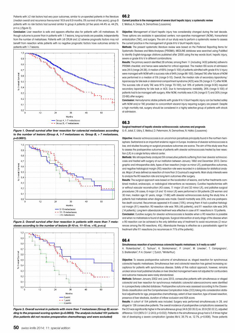
50 swiss knife 2014; 11: special edition
Patients with ≥2 risk factors had very poor outcomes, similar to un-operated patients in the literature (median overall and recurrence free-survival 16.9 and 6.9 months, 0% survival at five years); group B patients with no risk factors had survival similar to group A patients (at five years 44.4% vs. 44.2%, p=n.s.) (figure 3).Conclusion: Liver resection is safe and appears effective also for patients with ≥8 metastases. Al-though outcome is poorer than in patients with 1-7 lesions, long survivals are possible, independently from the number of metastases. Patients with ≥8 CRLM and ≥2 adverse prognostic factors may not benefit from resection while patients with no negative prognostic factors have outcomes similar to patients with 1-7 lesions.
Figure 1. Overall survival after liver resection for colorectal metastases according to the number of lesions (Group A, 1-7 metastases vs. Group B, > 7 metastases, p<0.0001).
Patients at risk Total 1 year 2 years 3 years 4 years 5 years
Group A, 1-7 metastases 743 611 452 330 213 151
Group B, >7 metastases 106 86 55 32 19 7
Figure 1. Overall survival after liver resection for colorectal metastases according to the number of lesions (Group A, 1-7 metastases vs. Group B, > 7 metastases, p<0.0001).
Figure 2. Overall survival after liver resection in patients with more than 7 metastases according to the number of lesions (8-10 vs. 11-15 vs. >15, p=n.s).
Patients at risk Total 1 year 2 years 3 years 4 years 5 years
8-10 metastases 48 36 23 16 11 2
11-15 metastases 40 34 21 12 6 4
>15 metastases 18 16 11 4 2 1
Figure 2. Overall survival after liver resection in patients with more than 7 meta-stases according to the number of lesions (8-10 vs. 11-15 vs. >15, p=n.s)
Figure 3. Overall survival in patients with more than 7 metastases (Group B) according to the
proposed scoring system (p=0.0003). The analysis included 101 patients (five patients did not
receive preoperative chemotherapy and were excluded)
Patients at risk Total 1 year 2 years 3 years 4 years 5 years
0 point 36 29 20 12 10 4
1 point 52 44 29 17 7 3
2 points 13 9 4 1 1 0
Figure 3. Overall survival in patients with more than 7 metastases (Group B) accor-ding to the proposed scoring system (p=0.0003). The analysis included 101 patients (five patients did not receive preoperative chemotherapy and were excluded)
66.2Current practice in the management of severe blunt hepatic injury: a systematic reviewE. Melloul, A. Denys, N. Demartines (Lausanne)
Objective: Management of blunt hepatic injury has considerably changed during the last decade. Three options are available in specialized centers: non-operative management (NOM), transarterial embolization (TAE), and surgery. The aim of our study was to perform a systematic review to assess the current practice in the management of grade III to V blunt hepatic injury.Methods: The present systematic literature review was based on the Preferred Reporting Items for Systematic Reviews and Meta-Analyses (PRISMA). MEDLINE database was searched using PubMed to identify English-language citations published after 2000 using the key words blunt, hepatic injury, severe or grade III to V in different combinations.Results: The primary search identified 28 articles; among them 11 (including 1432 patients) adhere to PRISMA checklist, and hence were selected for critical appraisal. The median ISS score at admission was 24.5 (range 24-38). A median of 65% (range 5-100) of patients admitted with grade III to V injury were managed with NOM with a success rate of 94% (range 86-100). Delayed TAE after failure of NOM was performed in a median of 5% (range 0-10). Overall, the median rate of secondary laparotomy/laparoscopy for bile leak or abdominal compartment syndrome (ACS) was 3% (range 3-11) after NOM. The success rate of early TAE was 91% (range 70-100), but 18% of patients (range 9-30) required secondary laparotomy for bile leak or ACS. Due to hemodynamic instability, 26% (range 5-100) of patients had to be managed with surgery. After NOM, mortality was 4.3% (range 0-7) and 30% (range 22-68) after surgery.Conclusion: Hemodynamic stable patients with grade III to V blunt hepatic injury can be treated safely with NOM and/or TAE provided no concomitant visceral injury requiring surgery are present. Despite a high mortality risk, surgery should be considered in a highly selective group of patients with shock at admission.
66.3Surgical treatment of hepatic alveolar echinococcosis: outcomes and prognosisG.-R. Joliat, E. Uldry, E. Melloul, D. Petermann, N. Demartines, N. Halkic (Lausanne)
Objective: Alveolar echinococcosis is an uncommon parasitosis principally found in the northern hem-isphere. Switzerland is an important endemic region in Europe. Incidence of alveolar echinococcosis is low, and studies focusing on surgical procedure outcomes are scarce. The aim of this study was thus to assess the postoperative outcomes of patients with alveolar echinococcosis treated by liver resec-tion (LR) in a single tertiary referral center.Methods: We retrospectively analyzed 59 consecutive patients suffering from liver alveolar echinococ-cosis and treated with surgery at our institution between January 1992 and December 2013. Demo-graphic and intraoperative data, types of liver resection (major vs minor LR), postoperative outcomes, and negative histological margin (R0) resection rate were recorded in a database for statistical analy-sis. Major LR was defined as resection of more than 3 Couinaud’s segments. Main study interests were to analyze the R0 resection rate and long-term outcomes after surgery.Results: The surgical approach was based on the localization of lesions, and further treatments com-bined medical, endoscopic, or radiological interventions as necessary. Curative hepatectomies with or without vascular reconstruction (43 cases, 11 major LR and 32 minor LR), and palliative surgical procedures (16 cases, 6 major LR and 10 minor LR) were performed in 59 patients (29 women and 30 men, median age: 61 years, range: 17-88) with alveolar echinococcosis during the study time. 4 patients had metastasis when diagnosis was made. Overall morbidity was 20%, and one postopera-tive death occurred. Recurrences appeared in 6 cases (10%); among them 4 had a positive histologi-cal margin (R1) resection. R0 resection rate was 76% (45 patients), and R1 resection rate was 24% (14 patients). Long-term albendazole treatment was effective in case of R1 resections in 10 patients.Conclusion: Curative surgery for alveolar echinococcosis is feasible when a R0 resection is possible and when no metastasis is found at diagnosis. Surgical intervention at early stage of the disease when a R0 resection can be achieved is the only definitive way of treatment to avoid recurrences (2 recur-rences among the R0 resections, 4%). Albendazole therapy is effective as a parasitostatic agent for treatment after R1 resections (no recurrences in 71% of the patients).
66.4Simultaneous resection of synchronous colorectal hepatic metastases: is it really so safe?A. Rickenbacher1, C. Tschuor1, K. Slankamenac1, P. Limani1, M. Linecker1, T. Campagnaro1, S. Breitenstein2, P.-A. Clavien1 (1Zurich, 2Winterthur)
Objective: To assess postoperative outcome of simultaneous vs. staged resection for synchronous colorectal hepatic metastases. Simultaneous liver and colorectal resection has gained increasing ac-ceptance for patients with synchronous disease. Safety of this radical approach, however, remains unclear since most published studies on liver directed management were not adjusted for confounders and outcome measures were rarely standardized. Methods: Between January 2002 and June 2013, consecutive patients with simultaneous or staged colorectal and liver resection for synchronous metastatic colorectal adenocarcinoma were identified in a prospectively collected database. Postoperative outcome was assessed according to the Clavien-Dindo classification and the Comprehensive Complication Index (CCI) taking into consideration statis-tical adjustments for age, preoperative chemotherapy, extent of liver resection, type of bowel resection, presence of liver steatosis, duration of inflow occlusion and ASA score.Results: A cohort of 154 patients was included. Surgery was performed simultaneously in 28, and staged in 126 consecutive patients. The overall incidence of postoperative complications assessed by the CCI was significantly higher in the simultaneous group 34.6 (SD 35.5) vs. 20.9 (SD 22.2), adjusted difference 13.0 (95% CI 1.2-24.9, p=0.032). Patients in the simultaneous group had a 5.4 times higher risk of developing a severe complication (grades IIIb-V, 35.7% vs. 12.7%, p=0.008). Those patients

swiss knife 2014; 11: special edition 51
undergoing simultaneous major hepatic resection (>=3 segments) had a 3.5 times higher risk (35.7% vs. 18%, p=0.13) and a significantly higher mean CCI of 39.8 (SD 35.1) vs. 21.7 (SD 20.8), adjusted difference 18.0 (95% CI 0.5-35.5, p=0.044). There was, however, neither significant difference in inten-sive care unit stay (p=0.09) nor in the length of hospital stay between the groups (p=0.60).Conclusion: Simultaneous liver and colorectal resection is associated with higher risk for overall mor-bidity and for more severe complications, particularly in combination with major hepatic resection.
66.5First experience with surgical navigation technology for liver surgeryF. Volonté1, N. C. Buchs1, F. Pugin1, C. Toso1, D. Ribes2, M. Fusaglia2, K. Gavaghan2, M. Peterhans2, S. Weber2, D. Candinas2, P. Morel1 (1Geneva, 2Bern)
Objective: Surgical navigation technology is an established technical support tool in neurosurgery and enables precise and safe treatments through guidance of instruments on pre-operative image data. In this work we report on the introduction of a navigation system for liver surgery to our clinical practice and on the identification of indications where computer aided liver surgery (CALS) potentially enables better and/or safer treatments.Methods: Within the framework of a multi-center clinical trial (CTI Project nr. 13745.1 PFFLI-LS, Geneva, Bern, Lausanne), the CAS-One Liver Navigation System (CAScination AG, Switzerland) was used dur-ing open liver surgery in 33 cases. For all cases, the pre-operative image data was analysed using MeVis 3D surgery planning (MeVis Distant Services, Germany). After mobilisation of the liver, registra-tion process was applied to enable navigation. Success of registration was assessed intra-operatively using actual position of the dissection tool according to the position of the virtual one.Results: In average 3 registration attempts were necessary to obtain an accurate registration (< 5mm). Registration attempts were performed again if liver was displaced from its initial registration position. Use of navigation technology appeared to be relevant for atypical resections where vascular orienta-tion was not possible.Conclusion: From our first experience with navigation technology for liver surgery, it appears to be a promising approach for non-anatomical tumour resection. Enabling real-time control of dissection planes will lead to a greater precision in resection margins and ensure total resection of the tumoral tissue while sparing a maximal amount of healthy cells. This tool opens the way for many other ap-plications in the field of augmented reality, laparoscopic surgery, 3D and robotics.
66.6Indications for stereotactic liver surgery: findings from a single center study over 4 yearsV. Banz Wüthrich, P. Tinguely, M. Worni, P. Müller, D. Inderbitzin, D. Ribes, M. Peterhans, S. Weber, D. Candinas (Bern)
Objective: In major, complex hepatic surgery, virtual resection planning is imperative, aiding the pre- and intra-operative setting. More recently, stereotactic image guidance (navigation) of surgical instru-ments has advanced to a clinically applicable level. This resulted in higher geometric accuracy during tumor resection/ablation as well as better alignment of the surgical site with available preoperative imaging. Our study aims to identify suitable indications for image-guidance in open liver surgery, based on the findings of a multi-year technological development.Methods: Retrospective, single-center analysis of 65 patients treated during 3 phases, characterized by the availability of specific registration methods (alignment between site and preoperative image data): registration by means of surface landmarks only (phase 1); single surface and parenchymal landmarks (phase 2) and ultrasound (US)-based volume registration (phase 3). Primary endpoint: accuracy. Secondary endpoints: Application feasibility for certain indications/general usabilityResults: Phase 1(03/09-12/11): 24 patients underwent image-guided liver surgery (IGLS): 15 color-ectal liver metastases (CRLM), 1 neuroendocrine liver metastases (NET), 5 HCC, 3 others. Phase 2(01/12-03/13): n=19; CRLM =9, NET=1, HCC=6, others=3. Phase 3 (04/13-12/13): n=22, CRLM =11, NET=7, HCC=1, others=3. In phase 1 (accuracy: 8mm, navigated instrument: CUSA) application of IGLS was done in a clinical feasibility protocol were therapeutic quality was not yet clear. Improved technology in phase 2 (accuracy: 8mm in selected volume of interest (VOI), navigated instruments: CUSA; microwave ablation (MWA)) allowed its use in a broader range of indications. In phase 3(accu-racy: 5 mm in VOI, navigated instrument: MWA), a higher accuracy allowed for use of IGLS in vanishing and very small, difficult to target lesions. A possible intraoperative setting is given in Figure1.Conclusion: In the light of increasing geometric accuracy owing to utilization of navigated US, patients requiring MWA +/- targeted resection of multiple, smaller (vanishing) lesions in the parenchyma (bilobar CRLM/NET) profit the most. However, clinically relevant outcome measures with oncologic endpoints such as recurrence-free and overall survival should be subject to further prospective and multicentric clinical trials.
a) b) c)
Figure 1: Guidance and visualisation modes of the navigation interface: a) 3D visualisation of preoperative image data and
together with virtual tool (here: microwave ablation needle); b) 2D targeting visualisation for visualisation of lateral and
longitudinal displacements of needle relative to target structures; c) 2D augmentation of native sonography imagery with co-
registered preoperative image data.
Figure 1: Guidance and visualisation modes of the navigation interface: a) 3D vi-sualisation of preoperative image data and together with virtual tool (here: micro-wave ablation needle); b) 2D targeting visualisation for visualisation of lateral andlongitudinal displacements of needle relative to target structures; c) 2D augmen-tation of native sonography imagery with coregistered preoperative image data.
66.7Can major postoperative morbidity after pancreaticoduodenectomy be predicted by using a simple score? External validation of the Braga scoreG.-R. Joliat, D. Petermann, N. Demartines, M. Schäfer (Lausanne)
Objective: Pancreas surgery remains associated with important morbidity rates while mortality de-creased below 5% in high-volume centers. Complications negatively impact on postoperative out-comes, e.g., length of hospital stay, long-term survival, quality of life, or costs. Most commonly, efforts are concentrated on decreasing postoperative morbidity, but early detection of patients at risk could be another valuable strategy. To this end, a simple prognostic score has recently been published (Braga M. et al., Ann Surg 2011;245:702-8). The aim of this current study was to validate this prognostic score.Methods: 245 patients (147 men, 98 women, median age 65 years) were included in our present vali-dation study. All underwent pancreaticoduodenectomy (PD) from 2000 to 2012 in a single institution, among them 75% for malignancies. Complications were classified according to the Dindo-Clavien classification, and grades III to V were considered major complications. The Braga score is based on four items: pancreatic texture (soft or hard), pancreatic duct diameter (>= or <=3mm), operative blood loss (>= or <=700ml), and ASA score (I, II, III). For each item 0 to 6 points are diversely attributed. An overall risk score ranging from 0 to 15 can be calculated for each patient. While a risk score of 0 has 6% risk of developing major complications, a risk score of 15 inherits 42% risk of major complications. To assess the discriminant score power, the expected and observed numbers of patients of the dif-ferent outcome categories were compared, and calibration was tested for a possible significance by using a Hosmer-Lemeshow goodness-of-fit test (C-statistic index).Results: Major complications occurred in 29% of patients compared to 16% in Braga’s data. Two of the four score parameters (pancreatic texture, blood loss) were independently statistically significant for increased morbidity. The areas under curve were 0.95 and 0.99 for 4-risk categories and for individual scores, respectively. Hence, our data validated the discriminant power of the Braga score for major complications after PD.Conclusion: The Braga score discriminates well between minor and major complications as assessed by the Dindo-Clavien classification. The clinical implications, i.e. whether postoperative treatment strat-egies should be adapted according to the patient’s individual risk, remain to be elucidated.
66.8Validation and cost analysis of a new management strategy for patients at intermediate risk of CBD stone migrationP. Iranmanesh, J.-L. Frossard, P. Morel, G. Mentha, C. Toso (Geneva)
Objective: A recent prospective, randomized, controlled trial concluded that patients at intermediate risk of common bile duct stone had a shorter length of stay and underwent less endoscopic investi-gations if treated by cholecystectomy as a first step. The aim of the present study was to confirm the results of the previously mentioned trial and analyze if this strategy decreased the overall costs.Methods: All patients admitted in emergency with an intermediate risk of common bile duct stone, as defined by the guidelines of the American Society for Gastrointestinal Endoscopy, had a cholecystec-tomy with intraoperative cholangiogram performed as a first step, followed if needed by a postopera-tive endoscopic retrograde cholangio-pancreatography (ERCP). Prospectively collected data included length of hospital stay, number of invasive procedures, common bile duct clearance rates and morbid-ity/mortality. Estimated overall costs were calculated for each patient and compared with a previous cohort of patients who underwent a classical strategy with systematic common bile duct assessment before cholecystectomy.Results: Ninety-five patients were included in the study. All of them had a cholecystectomy performed as a first step. The median length of stay was 6 [5;8] days. Thirty-four patients (35.8%) had a com-mon bile duct stone found on intraoperative cholangiogram and required a postoperative ERCP. Three patients (3.1%) had a false negative intraoperative cholangiogram and underwent postoperative ERCP as well. The overall ERCP clearance rate was 100%. Three patients with multiple common bile duct stones required more than one ERCP. There were no severe complications (Dindo/Clavien grade III or above). Two patients (2.1%) had a post-ERCP pancreatitis, treated conservatively. The estimated median cost per patient was 9750 CHF [8300;14500] in the prospective cohort and 13900 CHF [11950;21000] in the classical strategy cohort (P=0.006).Conclusion: This study confirms the results found by the previously mentionned trial in terms of length of stay, number of endoscopic procedures, common bile duct clearance rates and morbidity/mortal-ity. The estimated cost per patient with this strategy is significantly lower compared to a strategy with systematic common bile duct assessment as a first step. Patients at intermediate risk of common bile duct stone should therefore undergo cholecystectomy as a first step.
66.9Laparoscopic Competency Assessment Tool (LCAT) for technical surgical performance in laparoscop-ic cholecystectomy at the resident levelM. Jung, N. C. Buchs, S. de Sousa, A. Mennet, P. Morel, F. Ris (Geneva)
Objective: Current training in Switzerland does not include a formal assessment tool of technical surgi-cal performance in laparoscopic cholecystectomy. The aim was to develop a competency assess-ment tool by adapting a validated assessment tool for laparoscopic colorectal surgery to laparoscopic cholecystectomy and to implement it at the resident level in a educational training program. Methods: The developped assessment tool consists of a marking sheet including 4 tasks: exposure of the operating field, dissection of the calot’s triangle, execution of cholangiography and resection of the gallbladder. Each of these 4 tasks is divided in 4 sub- domains: use of instruments, tissue handling, errors and the quality of the end product. The resulting 16 sub- domains were rated from 1 to 4 (1. dangerous, 2. inadequate, 3. safe, 4. expert). In addition, the assessors were asked to give an overall statement (if the surgeon is able to perform this operation independently: clearly not, no, borderline no,

52 swiss knife 2014; 11: special edition
undecided, borderline yes, yes, clearly yes). From July 2013 to december 2013 supervised laparoscop-ic cholecystectomies of ASA 1 and 2 with BMI lower than 35 were included in a prospective database. The video of each procedure was evaluated by a second independent assessor. Any operation during which, part of it was performed by the assessing staff surgeon was excluded. Residents were divided in two groups: senior trainees and junior trainees. Junior trainees were trainees with an experience of less than 5 performed cholecystecomies. Results: During the study period 53 cases performed by 17 different residents were included. 6 out of 17 registrars were junior trainees with an experience of less than 5 cholecystectomies. Mean number of mentored evaluated cholecystecomies per trainee was 3.1 (SD +/- 2.3). Senior trainees scored over-all significantly better than junior trainees (3.3 vs 2.7; P = 0.007). 3 registrars could be signed off the program and allowed to perform cases on their own. This evaluation was highly reproducible among trainers.Conclusion: This is to our knowledge the first competency- based evaluation of technical skills of resi-dents at a teaching hospital in Switzerland. The implementation of such a tool allows measurement of the trainee’s performance, offering an objective reproducible evaluation of technical skills. LCAT is adaptable to other procedures in our hospital.
Posters II 7171.1Risk factors for prolonged treatment and hospital-readmission in negative-pressure wound therapyG. Osterhoff, P. Zwolak, C. Krüger, V. Wilzeck, H.-P. Simmen, G. Jukema (Zürich)
Objective: Prolonged duration of negative-pressure wound therapy (NPWT) is associated with in-creased bacterial growth. Therefore, a decreased duration of the treatment is desireable. The purpose of this study was to identify potential risk factors for the duration from first application of NPWT to secondary wound closure as well as factors that increase the rate of hospital-readmission.Methods: We performed a retrospective cohort study with inclusion of 261 patients (46±19 years, 70 female) who underwent 280 treatments with NPWT. Patient specific and demographic characteristics and the presence of several risk factors were documented. The duration from first application of NPWT to secondary wound closure, the number of interventions, the duration of hospital stay, and the inci-dence of readmissions due to complications of the wound treated by NPWT were recorded and a risk factor analysis was performed.Results: The median number of NPWT procedures was 2.0±2.0, the duration of NPWT was 6.0±14.7 days and the length of hospital stay was 16.0±27.9 days. Presence of an open fracture (p=.002) and increased age (p=.004) were identified as independent risk factors for a prolonged duration of NPWT. Patients who smoked (p=.001) or patients with alcohol/drug abuse (p=.015) were more likely to return to hospital (smoking: 18/58 cases; alcohol/drug abuse: 7/19 cases). No such association was seen for diabetes (p=.702), PVD (p=.052), immunosuppressive medication (p=.187), immunodeficiency (p=.404), trauma (p=.358), infection (p=.298), and open fracture (p=.061).Conclusion: Patient age and presence of an open fracture are independent predictors of a prolonged duration from first application of NPWT to secondary wound closure. These results should be taken into account for the calculation of average costs and anticipated hospital stay associated with this therapy.
71.2„Minimal invasive“ Kocher-Keilexzision bei Unguis incarnatusS. Habelt, F. Goti (Wattwil)
Objective: Die klassische Keilexzision nach Kocher beinhaltet eine Resektion eines seitlichen Anteils des Zehennagels, der Nagelwuchszone proximal des entfernten Nagelanteils und des seitlichen Nagelwalls. Besonders die Inzision über den proximalen Nagelwall hinaus verursacht häufig eine kosmetisch oder auch mechanische störende Narbe. Wir präsentieren eine retrospektive Studie mit 27 Patienten, welche wir operativ mittels minimalisierter Kocher-Keilexzision unter Zuhilfenahme eines CO
2-Lasers behandelt haben.
Methods: 27 Patienten mit einem Unguis incarnatus (mit oder ohne Infekt) wurden behandelt. Bei allen Patienten wurde in Lokalanästhesie und Blutsperre zunächst der Nagel seitlich bis zur Nagelwuch-szone inzidiert, wobei wurde darauf geachtet, dass der proximale Nagelwall dabei intakt blieb. Mit ein-er Zange wurde der eingewachsenen Nagelanteil herausgezogen, im Bereich des lateralen Nagelwalls wurde das Granulationsgewebe teils scharf mit der Schere oder mit dem CO
2-Laser (ultrapulse, 15W)
entfernt. Danach wurde der proximale Nagelwall mit einem Haken angehoben, die Nagelwuchszone mit dem CO
2-Laser unter Sicht vaporisiert. Danach offene Wundbehandlung durch den Patienten
selbst. Befragung der Patienten mittels standardisierten Telefoninterviews.Results: Die 27 Patienten waren intraoperativ beschwerdefrei. Bei 4 Patienten lag sowohl medial also auch lateral ein Unguis incarnatus vor. In der Anfangsphase der Studie kam es in 2 Fällen zu einem Rezidiv infolge insuffizienter Entfernung der Nagelwuchszone. Nach den ersten 10 Patienten trat dies nicht mehr auf. Bei den Rezidiven wurde einzig die Nagelwuchszone erneut exzidiert. Die Wunden heilten bei allen Patienten ohne Komplikationen ab, alle Patienten sind in der Folge beschwerdefrei. Das kosmetische Ergebnis wurde in allen Fällen als gut bis sehr gut beurteilt.Conclusion: Die „minimal invasive“ Kocher-Keilexzision stellt eine einfaches Verfahren mit gutem funk-tionellen und kosmetischen Ergebnis dar. Eine hohe Patientenzufriedenheit ist zu verzeichnen.
71.3Pneumomediastinum: a harmless finding or a serious disease?K. Szentesi, E. Cereghetti, K. Vogt, E. Angst, F. Hetzer (Schaffhausen)
Objective: The presence of air in the mediastinum is defined as pneumomediastinum. This condition may be secondary to an event introducing air to the mediastinum by trauma of adjacent organs. This is a serious disease and specialized treatments including antibiotics are needed. In primary spontane-ous pneumomediastinum no clear etiology may be defined. Here we present the case of a spontane-ous pneumomediastinum.Results: An 18-year-old lean, male smoker presented with sudden onset of dyspnea and chest pain to the emergency room. The symptoms suddenly developed after coughing. He had no pulmonary or cardiac history. He denied any recent external trauma or vomiting. He described a sharp, retrosternal pain radiating to the back. The findings of the physical examinations were normal. The chest X-ray and pulmonary CT-scan showed a pneumomediastinum and an additional small spontaneous pneumot-horax. The patient was admitted to the hospital for observation and was treated with analgesics and bed rest. On the next day the patient reported the complete resolution of the symptoms. The follow-up chest X-ray confirmed a resolution of the findings, and the patient was discharged from the hospital.Conclusion: Spontaneous pneumomediastinum is a rare, largely benign, self-limiting disease. Sponta-neous pneumomediastinum occurs predominantly in young male smokers or patients with pre-exist-ing pulmonary condition. In patients with spontaneous pneumomediastinum, chest pain and dyspnea are the predominant symptoms often misleading the diagnosistic process. Once complications have been excluded the treatment includes bed rest, analgesics and follow-up, no antibiotic treatment is necessary.
71.4Eindeutig-mehrdeutige Diagnose AppendizitisT. Rauer, R. Tedaldi, G. Teufelberger (Muri)
Objective: Die Diagnostik des rechtsseitigen Unterbauchschmerzes kann auch für den erfahrenen Kliniker schwierig sein. Eine sorgfältige Zusammenschau der Anamnese und der klinischen Befunde (Klinik, Labor, Bildgebung) erlaubt es die negative Appendektomierate zu senken. Trotzdem ist auch der erfahrene Chirurg nicht vor intraoperativen Überraschungen gefeit.Methods: Ein 28-jähriger, ansonsten gesunder Patient stellt sich mit einer 20-stündigen Anamnese von rechtsseitigen Unterbauchschmerzen auf unserer Notfallstation vor. Bei ausgeprägter Druckdo-lenz und Loslassschmerzen im rechten Unterbauch sowie positivem Psoas-Zeichen sind die laborche-mischen Entzündungsparameter erhöht (Lc 16.5x10^5; CRP 16mg/l). Die Abdomensonographie weist eine retrocoecale, entzündlich alterierte Appendix vermiformis nach, so dass die Indikation zur laparoskopischen Appendektomie gestellt wird.Results: Intraoperativ zeigt sich in der Übersicht eine ca. 8 cm lange, stark entzündlich veränderte, in der parakolischen Rinne liegende Appendix vermiformis, welche multiple Divertikel aufweist. Der restliche Abdomensitus, insbesondere das einsehbare Kolon ist divertikelfrei und unauffällig. Es folgt die problemlose laparoskopische Appendektomie. Der Patient kann am zweiten postoperativen Tag in gutem Allgemeinzustand nach Hause entlassen werden. Die histologische Aufarbeitung des Re-sektates weist eine ulzero-phlegmonöse Entzündung der Appendix mit reizloser Divertikulose nach.Conclusion: Die 1893 durch Kelynack erstbeschriebene Appendix-Divertikulitis stellt eine seltene Differ-entialdiagnose des rechtsseitigen Unterbauchschmerzes dar. Sie findet sich in 0.004 bis 2.1% der Ap-pendektomiepräparate. Man unterscheidet zwischen der sehr seltenen angeborenen und der häufig-eren erworbenen Form und unterteilt die Erkrankung in vier Subtypen: Typ I akut entzündetes Divertikel bei blander Appendix; Typ II akut entzündetes Divertikel mit Begleit-Appendizitis; Typ III Appendizitis mit nicht entzündlich veränderten Divertikeln; Typ IV unauffällige Divertikel ohne Hinweis auf Appendizitis. Die Histologie unseres Patienten entsprach einem Typ III. Die Appendix-Divertikulitis weist im Vergleich zur Appendizitis ein höheres Perforations- und Mortalitätsrisiko auf. Daher ist die Appendektomie die Therapie der Wahl sowohl in den entzündlichen Stadien als auch bei reizlosen Zufallsbefunden.
71.5Incarcerated arcuate line hernia: rare clinical manifestation of a not so rare anatomical variation? a case reportS. Geuss, M. Koehli, S. Prica, J. Peltzer (Delémont)
Objective: Arcuate line hernia (ALH) is a rare diagnosis caused by herniation of abdominal structures in a peritoneal recess between the posterior lamina of the rectus sheath and the posterior surface of the rectus abdominis muscle ascending cranially from the arcuate line (AL). A PubMed search only revealed eight cases published until 2013. A recent series reviewing 315 abdominal computed tom-ography exams (CT) in asymptomatic patients reports an impressive 8.6% prevalence of ALH though lacking surgical confirmation. We present a case of AHL with omental incarceration treated laparo-scopically in our department.Results: Case description: a 68-year-old man presented to the emergency department with a painful bulge in the abdominal wall about 5 cm left and slightly caudally of the umbilicus. CT imaging of the region demonstrated a 3.1 x 0.9 cm protrusion of adipose tissue ascending 3 cm between the parietal peritoneum and the posterior aspect of the rectus abdominis muscle. Laboratory findings were normal. Laparoscopic exploration revealed bilateral peritoneal recess ascending from the AL between the rec-tus abdominis muscle and the posterior lamina of the rectus sheath. Lateralized to the left, a small por-tion of the omentum was incarcerated in the medial segment of the peritoneal recess accompanied by local peritoneal inflammation. The entrapped omental portion was reduced and subsequently resect-ed. No hernia repair was performed. Postoperative recovery and 10 month follow-up were uneventful.Conclusion: To our knowledge, this is the first photo documented case of incarcerated AHL. This pathol-ogy might be underdiagnosed because AHL tends to reduce spontaneously in the supine position used during CT examination and surgical exploration. AHL should be considered in patients presenting with

swiss knife 2014; 11: special edition 53
chronic recurrent periumbilical pain. Combined clinical and radiological suspicion of an abdominal wall process at the level of the AL should prompt laparoscopic exploration. Furthermore, we recom-mend routine inspection of anterior abdominal wall for intraparietal AL recess during laparoscopic exploration for unexplained abdominal pain. We refrained from immediate hernia repair because of undetermined local inflammation. One proposed method is to reduce the peritoneal sac, then fixing the AL to the rectus muscle using spiral tackers. While closure of the peritoneal recess seems in general reasonable, consensus among surgeons is lacking due to the rarity of the condition.
Figure 1. Abdominal CT showing subperitoneal adipose tissue of the left anterior abdominal wall (arrow)
Figure 2. Intraoperative view of the peritoneal fold ascending from the left AL (arrow) after hernia reduction (*)
*
Figure 1. Abdominal CT showing subperitoneal adipose tissue of the left anterior abdominal wall (arrow)
Figure 1. Abdominal CT showing subperitoneal adipose tissue of the left anterior abdominal wall (arrow)
Figure 2. Intraoperative view of the peritoneal fold ascending from the left AL (arrow) after hernia reduction (*)
*
Figure 2. Intraoperative view of the peritoneal fold ascending from the left AL (arrow) after hernia reduction (*)
71.6Successful treatment of extended pretibial necrosis with bone involvement by locoregional flapsS. Maksimovic, S. Gujer, P.-M. Sutter, H.-U. Würsten, C. Viehl (Biel)
Objective: The following is a report on a case of successful treatment by two locoregional muscular flaps and split skin grafts in a 54-year-old man with extended unilateral pretibial ulcerations with corti-cal bone involvement. The patient presented to the emergency department with extended progressive ulceration with exposed necrotic bone due to repeated injecting drug use.Methods: Initial treatment by the general surgical team was wound debridement. However, because of a microvascular deficit and considering the personal situation of the patient, the plastic surgery team gave the recommendation for thigh amputation. However, as the patient had clinically intact calf muscles the team decided not to amputate and instead treat with two locoregional muscular flaps and split skin grafts utilising the following steps.1. initial local debridement of soft tissue necrosis2. soft tissue conditioning with VAC-therapy3. radical bone debridement4. medial proximal pediculated gastrocnemius muscle flap5. distal pediculated hemisoleus muscle flap6. split skin graft7. VAC-therapyResults: After only a 3 weeks hospital stay, the patient developed a secure vital coverage of the tibial bone and an epithelization of 80% occurred at the site. Upon discharge the patient was followed up at the out patient wound care clinic.
Conclusion: The exposure of bare bone and its deperiostation are cause of progressive circulatory bone necrosis, infection, sequestration, pathological fracture and consequentially amputation. Regard-less of different presenting etiologies, we have shown that the disease progression can be stopped with the above-mentioned steps and thereby avoiding amputation. Low-compliance patients (such as injecting drug users, psychiatric patients or late 4th-age patients) are often considered high risk can-didates for microvascular flaps. However, we have shown that success can be achieved even among individuals in this “high-risk” group. Locoregional flaps are a sufficient, simple and overall cost-effective alternative to – otherwise – unavoidable amputation in a general surgical department.
71.7Illustrated operative management of spontaneous bleeding and compartment syndrome of the lower extremity in a patient with acquired hemophilia a: a case reportT. Jentzsch, B. Brand-Staufer, F. P. Schäfer, G. A. Wanner, H.-P. Simmen (Zürich)
Objective: Spontaneous bleeding and compartment syndrome (CS) due to acquired hemophilia A (AHA) is rare. There are no reports on operative management of this entity. This is in contrast to the rather common CS due to trauma. AHA is defined by factor VIII (fVIII) deficiency caused by an inhibitory antibody to fVIII, affects the elderly and occurs at a rate of approximately one person per million each year. We present a case of atraumatic CS of the lower leg due to AHA, in an elderly white male, who was treated surgically with a long postoperative course (Tbl. 1).Methods: An 83-year-old white man male presented to a community hospital with a two-day history of increasing pain and swelling in his left lower leg without previous trauma.Results: Compartment pressure was elevated. Prothrombin time measured 87%. Diagnosis of CS was made. The patient underwent immediate lateral fasciotomy. Two secondary hematomas were evacu-ated. The patient was transferred to a larger regional hospital. Partial thromboplastin time was finally measured and elevated. Diagnosis of AHA was made on the fifth postoperative day. Due to unstoppa-ble bleeding, recombinant factor VIIa (rfVIIa), NovoSeven®, was used successfully as a single bleed-ing control agent up to four times a day and corticosteroids were administered for inhibitor eradication. Due to further secondary hemorrhage, the patient was transferred to a university hospital. The wound (Fig. 1) was covered with polyvinylacetat foam. After one month, fVIII had risen to 41% and VAC therapy was applied. With fVIII at 88% after six weeks, split-thickness skin graft (STSG) was carried out and rFVIIa administration was stopped. After three and a half months, another small STSG was needed. Wound healing (Fig. 2) followed and prednisone therapy was stopped. Overall, rfVIIa was given 49 times, costing $340,000, while the hospital received $60,000 for the diagnosis-related group (DRG).Conclusion: Awareness, prompt diagnosis and effective treatment of CS caused by a rare may spare patients from surgery if early administration of rFVIIa is effective. Operative management of CS and AHA may lead to a prolonged course of disease. An interdisciplinary approach with meticulous bleed-ing management with rfVIIa, whiched was very helpful pre-operatively, inhibitor eradication with corti-costeroids and and careful surgical treatment may be expensive and successful.
Figure 1
Figure 2
Figure 1
Figure 2
71.8A very rare case of right-sided, traumatic diaphragmatic ruptureO. Ismail1, F. Hauswirth2, V. Schreiber1, G. A. Melcher1 (1Uster, 2Zürich)
Objective: Right-sided traumatic diaphragmatic rupture is a rare entity of injury and accurate diagnosis is easily missed upon initial presentation.Methods: A 80-year-old man suffered a collapse and fell down on his right chest. Physical examination showed pain due to thoracic compression. Conventional radiographs of the chest taken in two planes studies showed multiple fractures of the lower ribs VII-IX on the right side. No other pathologically find-ings could be detected. 36 hours later the patient presented with diffuse abdominal pain with tender-ness in the right upper abdomen and rising white blood cells as well as CRP. Emergency abdominal

54 swiss knife 2014; 11: special edition
sonography was obtained and revealed gall stones, but no other specific pathologically findings. Due to proceeding pain another conventional radiograph of the chest taken (CT-scan was actually planned but malfunctioning) suggested a diaphragmatic rupture because of an intestinal loop and suggested air-fluid level visible in the right hemithorax ( see radiographs). Indication for emergent operation was made.Results: Intraoperative findings showed an incarcerated colon owing to a traumatic rupture of the right-sided diaphragm. The affected segment of the colon was resected with end-to-end anastomosis and the lesion oft the diaphragm was sutured directly. Postoperative course was unproblematic.Conclusion: The rupture has probably been caused by (intraoperatively) palpable, fractured ribs. Though, right-sided, diaphragmatic rupture is a very rare event due to protection of the liver and caused mostly by elevated intrabdominal pressure. Diaphragmatic rupture in general is a frequently delayed confirmed diagnosis. However awareness of this injury can trigger right and early diagnosis with prompt treatment.
71.9Proximal esophageal perforation in blunt cervical traumaP. Valente, M. Giuliani, A. Guerra (Bellinzona)
Objective: Traumatic ruptures of the esophagus represents 4-14% of esophageal perforations, pen-etrating or iatrogenic trauma being the most frequent causes. Proximal esophagus perforations due to blunt trauma is estimated to occur in less than 2% of patients experiencing blunt cervical trauma.Methods: A 70 years old patient in good health, accidentally fell from three meters through a trap-door. She was brought to the emergency room where ECG, laboratory tests and conventional radiological workup of the cervical spine and chest’s X-rays were performed. Three days after, the patient return with acute dysphagia, voice changements, haematemesis and fever.Results: Diagnostic exams during the first consultation did not show any traumatic injuries: the lady was discharged. Upon readmission 3 days later a cervical-thoracic CT scan showed bilateral abscess of the neck with gas reaching the upper border of the mediastinum likely due to a perforation of the up-per esophagus. Pharyngeal fiberoptic examination excluded mucosal lesions at oro-pharyngeal level. A esophago-gastro-duodenoscopy showed a tear of the esophageal mucosa at the level of the upper esophageal sphincter highly suggestive for a microperforation. The patient was brought to the operat-ing room for a bilateral cervicotomy: the cervical esophagus was completely explored and no trans-mural injury was found but a prominent, fractured cervical spine osteophyte was identified, wich we believe was responsible for the esophageal microperforation with bilateral cervical abscess formation. The cervical area was irrigated with saline solution and drained. Intraoperative bacteriology showed gram-positive anaerobic cocci. Broad-spectrum antibiotic therapy was introduced and after few days the patient was in stable conditions, afebrile and after two weeks discharged.Conclusion: According to a several case reports cervical spine flexion-hyperextension is most likely the mechanism of proximal esophageal perforation in blunt cervical trauma. Early diagnosis and treat-ment is crucial to prevent descending mediastinitis, a condition with mortality up to 50%.
71.10Développement d’un pôle d’excellence en chirurgie au Cameroun basé sur le modèle SuisseP. Sikiminywa Kambale, E. Pezzetta, M. Schäfer, N. Demartines (Montreux)
Objective: Le Cameroun est un pays en voie de développement mais avec des indicateurs de santé alarmants. L’espérance de vie est estimé à 55 ans aussi bien en 1987 qu’en 2013. L’auteur de ce projet est originaire du Cameroun formé en Suisse. Le but de ce travail est d’analyser le potentiel de jumelage entre un hôpital universitaire Suisse et un hôpital camerounais. L’objectif est de fonder un nouvel hôpital au Cameroun.Methods: Consultation des registres de la délégation provincial de la santé de l’Ouest. En 2005, ac-quisition d’un terrain de 1575m2 à Bafoussam et planification de la construction d’un hôpital. Con-struction d’un bâtiment de deux étages sur une surface de 400m2, le Centre médico-chirurgical spé-cialisé de Bafoussam. Création de l’association pour la promotion de la santé au Cameroun, ASPSC basée à Neuchâtel pour le soutient du projet. Evaluation des coût de formation de spécialiste et de l’organisation d’un service de chirurgie sur base du modèle suisse.Results: En 2012, 50% des interventions chirurgicales sont dues à des péritonites d’origine infectieuse. 25% sont des pathologies urologiques et 20% des cas de traumatologie. Les cas de chirurgie pour cancer représentent moins de 5%. Depuis fin 2013, le bâtiment est prêt. Il compte 22 lits, 2 salles d’opération, 1 salle d’accouchement et 1 salle d’urgence. En attente de construction la radiologie, la stérilisation et le laboratoire. Le projet prévoit que la formation des futurs médecins spécialistes se fasse au Cameroun, avec en permanence un tournus de superviseurs venus de la suisse. Les mé-decins en formation pourront passer une année en Suisse. Ce système de fonctionnement devra avoir une durée déterminée en vue de transférer complètement la prise en charge aux médecin locaux. Pour cela, il faudra assurer la formation d’au moins 6 spécialistes dont 3 chirurgiens, 1 radiologue, 1 anesthésiste et 1 gastroentérologue. Il faudra aussi former des collaborateurs notamment des in-strumentistes, infirmières et un responsable de la stérilisation. Le coût estimé pour la formation d’un spécialiste est d’environ 40 000CHF par année. Il y aura ainsi des services de chirurgie partenaires en suisse.Conclusion: Le modèle de management de la chirurgie en Suisse peut servir de base pour créer un pôle d’excellence dans une région en voie de developpement et initier un principe d’autonomisation dans les projets humanitaires.
71.11Lumbar facet joint arthritis is associated with more coronal orientation of the facet joints at the upper lumbar spineT. Jentzsch, J. Geiger, S. M. Zimmermann, K. Slankamenac, T. D. Nguyen-Kim, C. M. Werner (Zürich)
Objective: Controversies about facet joints (FJs) still exist. Study samples have been rather small for FJ arthritis on CT scans. Gender predilection has not been reported consistently. It remains unclear whether FJ arthritis is associated with FJ orientation and/or FJ asymmetry. Previous studies have only reported an increase in FJ arthritis with more sagittally oriented FJs at the lower lumbar spine. Yet, it is unknown if changes in FJ orientation at the upper lumbar spine lead to different FJ orientation. Thus, our goal was to clarify these remaining issues by quantifying the degree of radiographically detectable FJ arthritis on CT scans of the lumbar spine from L2-S1 in regard to age, gender, FJ orientation, and FJ asymmetry.Methods: We retrospectively analyzed CT scans of 620 individuals, who presented to our traumatology department between 2008 and 2010. The mean age was 42.5 (range, 14-94) years. FJs of the lumbar spine were evaluated for arthritis and orientation between the L2 and S1.Results: FJ arthritis was present in 308 (49.7%) individuals with a mean grade of 1. It was seen in 27% of individuals ≤40 years and in 75% of individuals ≥41 years (P < 0.0001) (Fig. 1) as well as in 52% of females and 49% of males (P = 0.61). Mean FJ orientation was 30.4° at L2/3, 38.7° at L3/4, 47° at L4/5, and 47.3° at L5/S1 (Fig. 2). FJ arthritis was significantly associated with more coronal (in-creased degree) FJ orientation at L2/3 (P = 0.03) with a cutoff point at ≥32°. FJs were more coronally oriented (48.8°) in individuals ≤40 years and more sagittally oriented (45.6°) in individuals ≥41 years at L5/S1 (P = 0.01) (Fig. 2). Mean FJ asymmetry was 4.89° at L2/3, 6.01° at L3/4, 6.67° at L4/5, and 7.27° at L5/S1, without a significant difference for FJ arthritis (Fig. 2).Conclusion: FJ arthritis is common affecting about half of individuals, increases with age, and affects both genders equally. Coronally oriented facet joints (≥32°) in the upper lumbar spine, namely, at L2/3 may be an individual risk factor and surrogate for development of FJ arthritis in the entire lumbar spine, which is worth further investigations. Besides, coronal FJ orientation increases craniocaudally, while sagittal orientation at the lower lumbar spine increases with age. FJ asymmetry is not associ-ated with FJ arthritis, is more common in females at the lower lumbar spine, and also increases in a craniocaudal fashion.
Figure 1
Figure 2
Table 1 Level Facet Joint Orientation (°) Facet Joint Asymmetry (°)
L2/3 30.4 4.89
L3/4 38.7 6.01
L4/5 47 6.67
L5/S1 47.3 7.27
71.12Hyperlordosis is associated with facet joint pathology at the lower lumbar spineT. Jentzsch, J. Geiger, M. A. König, C. M. Werner (Zürich)
Objective: Our study opted to clarify the remaining issues of lumbar lordosis (LL) in regard to (1) its physiologic values, (2) age, (3) gender, (4) facet joint (FJ) arthritis and orientation using CT scans. Recent studies have questioned whether LL really decreases with age, but study sample sizes have been rather small and mostly been based on X-rays. Since hyperlordosis increases the load transferred through the FJs, it seems plausible that hyperlordosis may lead to FJ arthritis at the lower lumbar spine.Methods: We retrospectively analyzed CT scans of 620 individuals, with a mean age of 42.5 (range, 14-94) years, who presented to our traumatology department and underwent a whole body CT scan, between 2008 and 2010. LL was evaluated between the superior endplates of L1 and S1. FJs of the lumbar spine were evaluated for arthritis and orientation between the L2 and S1.Results: (1) The mean LL was 49.0° (SD 11.1°, range 11.4-80.1°). (2) LL increased with age and there was a significant difference in LL in our age groups (≤30, 31-50, 51-70 and ≥71 y) (P=0.02) (Fig. 1). (3) There was no significant difference in LL between females and males (50° and 49°) (P=0.17). (4) LL showed a significant linear association with FJ arthritis (P=0.0026, OR=1.022 [1.008-1.036]) and

swiss knife 2014; 11: special edition 55
sagittal FJ orientation at L5/S1 (P=0.001). In a logistic regression analysis the cut-off point for LL was 49.4° (Fig. 2).Conclusion: This is the largest CT-based study on LL and FJs. LL significantly increases with age. As a novelty finding, hyperlordosis is significantyly associated with FJ arthritis and sagittal FJ orientation at the lower lumbar spine. Thus hyperlordosis may present with back pain and patients may benefit from surgical correction, for example in the setting of trauma.
Figure 1
Figure 2
71.13Epidemiologie von TrottinettunfällenK. Löbig, D. Heim (Frutigen)
Objective: Trottinettfahren in der Alpenregion erfreut sich seit Jahren zunehmender Beliebtheit. Im Ver-gleich zum Wintersport gibt es bisher noch keine epidemiologischen Studien zu Trottinetteunfällen in der Schweiz. 2012 wurde von uns erstmals über eine Erfassung eines einzelnen Sommers berichtet. Unterdessen ist diese Unfallerhebung an unserem Spital zum Standard geworden.Methods: Seit Sommer 2011 wird an unserem peripheren Bergspital eine prospektive Studie zu Unfäl-len durch Trottinettfahren in der Region Adelboden und Kiental durchgeführt. Es wurden alle ambu-lanten und stationären Patienten erfasst und Daten zu Unfallmechanismus, Präventionsmassnahmen und Diagnose erhoben.Results: Es wurden 88 Patienten im Alter zwischen 4 und 76 Jahren erfasst. Das mediane Alter lag bei 30 Jahren. Die meisten Verletzungen entstanden durch Selbstunfälle (93%) und in den meisten Fällen wurde ein Protektor getragen (89%). 44% der Patienten erlitten eine Fraktur. Es wurden 36% der Patienten stationär behandelt, wovon 7 Patienten auf Grund der Schwere der Verletzungen oder Komorbiditäten nach Stabilisierung verlegt werden mussten.Conclusion: Schwere Verletzungen wurden hauptsächlich in höheren Altersgruppen gefunden, wobei insgesamt auffällt, dass Trottinettfahren als Breitensport nicht nur von jungen, sportlichen Menschen betrieben wird. Unsere Daten zeigen, dass beim Trottinettfahren ein relevantes Verletzungsrisiko be-steht, für das die Bevölkerung sensibilisiert werden sollte. Weitere Massnahmen zur Prävention sollten ergriffen werden, um die Anzahl und Schwere der Verletzungen zu verringern.
71.14Patient satisfaction after anterior cruciate ligament reconstruction in outpatient surgeryJ. Böttcher, N. Clerici, S. Castelanelli, C. Candrian (Lugano)
Objective: The anterior cruciate ligament (ACL) reconstruction is performed in Switzerland, almost exclusively in a hospital setting. Some international authors have published good results with high patient satisfaction performing ACL reconstruction ambulatory (1,3) and reducing general cost’s (2) We are aiming with our study at determining patient satisfaction after ambulatory ACL reconstruction.Methods: We retrospectively identified all patients after day surgery for ACL reconstruction in our hos-pital between 2009-2012. General statistical data on patient age, gender, ASA classification were re-corded. Patients were interviewed by phone about the satisfaction with the ambulatory management,
about perioperative pain (Visual Analog Scale, VAS) and sleep patterns during the first night. Post-operative discharge medication consisted in NSAID o Metamizole, Paracetamol, not generally opiate.Results: In this period, out of 92 patients operated 22 patients were identified. 17/22 patients could be reached by telephone. 3/17 patients planed for ambulatory surgery could not be released at home after surgery because of not well controlled pain or nausea. Of the remaining 14 patients (all men, median age of 25.5 year),. 50% of the patients would not perform the procedure again, 50% of the patients could sleep the first night, the other little or not at all.In the subgroup of the patients under 25 years (n=7) 6 patients would not perform again outpatient setting and had a mean value of 6.5 on the VAS the first night, only one was able to sleep through the night. Interestingly, in the subgroup over 26 years (n=7) 6 would do the surgery again in an ambulatory care, mean value of VAS was 5.2, 6/7 could sleep during the night.Conclusion: In the majority of the patients in our study, pain was not sufficiently controlled during the first night and because of this most of the patients would not want to perform such surgery in out-patient. Only 7/17 patients would repeat the outpatient surgery, interestingly most of them were in the subgroup older than 26 years. Or the postoperative pain management is improved (postopera-tive infiltration, regular opiate medication as usual in another countries (4)) or we will suggest to the patients to stay overnight.
71.15Long PHILOS plate fixation in a series of humeral fracturesM. Bilici1, B. Arumilli2, N. Suhm1, M. Jakob1, D. Rikli1 (1Basel, 2Manchester/UK)
Objective: The purpose of the article is to highlight a specific fracture pattern encountered by us in the osteoporotic upper humerus. We present our results of management of such metadiaphyseal fractures of the upper humerus with less invasive plating. The additional steps taken to improve final outcome and the reasoning behind each are discussed.Methods: In our department, a total of 13 fractures (in 12 patients) were managed for a metadiaphy-seal fracture of the upper humerus between 2010 and 2013. There were 2 males and 10 females. The average age in the cohort was 74.3 (52-95) years. In 9 fractures, the fracture line was extending above the surgical neck. All patients were managed with a locking compression plate (long PHILOS or LCP) using two approach windows (proximal deltopectoral and a distal anterior or lateral). Patients were evaluated for clinical outcome using the Quick DASH score and assessed for radiological union, complications or re-operations retrospectively.Results: The mean follow-up was 14.3 months (4-36). All fractures were united, and there was no evidence of avascular necrosis or non-union. Two patients showed varus collapse of the anatomical head of which one patient needed change of screws at 12 weeks from index surgery. In patients, when a distal lateral window was used, 2 patients out of 4 had radial nerve palsy post-operatively. In the rest, when the plate was twisted by 45° to allow anterior placement using the brachialis split, none had radial nerve injury.Conclusion: The osteoporotic bone failing under a low-energy mechanism seemed to dictate this frac-ture pattern. The fracture is either a bending wedge or a long spiral with or without a large butterfly and often extends into the humeral head. The fractures are better managed surgically, a primary reduction allowing contact of fragments is essential, and using an anterior window distally with a 45° contoured plate will achieve good plate placement as well as decreases the risk of radial nerve injury consider-ably compared to total lateral plate positioning.
Figs. 1 and 2 Pre-operative anteroposterior and lateral radiograph showing a long spiral metadiaphyseal fracture of the right hurnerus with a large lateral butterfly fragment
56 swiss knife 2014; 11: special edition
Figs. 3 and 4 Post-operative anteroposterior and lateral radiographs of the right humerus at 3-month follow-up showing union
Figs. 6 and 7 Anteroposterior and lateral views of a contoured10-hole PHILOS plate applied to a humerus model
Fig. 8 End on view of a 45° contoured long, PHILOS plate to appr-eciate the ori-entation
71.16Diagnostic significance of high sensitivity troponin in diagnosis of blunt cardiac injuryC. A. Pfortmüller, G. Lindner, A. B. Leichtle, G. M. Fiedler, A. K. Exadaktylos (Bern)
Objective: Blunt cardiac injury (BCI) is commonly associated with major thoracic trauma and influ-ences morbidity and mortality to a great extent. Despite much discussion in recent years no diagnostic “gold standart” exists. Therefore the primary aim of this study is to evaluate the diagnostic significance of high sensitivity troponin in patients with BCI.Methods: In this cross-sectional analysis, all patients who received serial measurements of high-sensitive troponin T between 01 August 2010 and 31 October 2012 at our Department of Emergency Medicine (ED), Inselspital admitted with trauma were included.Results: Out of 86 patients with trauma in a total of 31 patients blunt cardiac trauma was suspected. These patients did not differ in either sex or age from others (p<0.21, respectively p<0.089). The me-dian ISS-Score and injury count was significantly higher in patients with BCI (9.13, SD 9.53, p<0.001, respectively 4.71, SD 2.5, p<0.0001). The delta of first to the second high sensitivity troponin T meas-urement was significantly higher in patients with clinically suspected BCI than in others (0.003ug/l in-terquartile range 0.00-0.12 respectively 0.002ug/l interquartile range 0.00-0.005, p<0.001). The delta of the first to the second high sensitivity troponin T correlated positively with the ISS-score (p<0.04).Conclusion: Our findings suggest that high sensitivity troponin T measurement in trauma patients is a reliable biologic marker for estimating myocardial injury. But due to its low specifity not the actual value and more the dynamics should be considered when assessing patients with suspected BCI.
71.17The glenohumeral joint – a mismatching system? A morphological analysis of the cartilaginous and osseus curvature of the humeral head and the glenoid cavityV. Zumstein, M. Kraljevic, S. Hoechel, A. M. Nowakowski, M. Müller-Gerbl (Basel)
Objective: Radial mismatch, glenohumeral conformity ratios and differences between cartilaginous and osseus radii highly depend on the measured plane. The comparison of cartilaginous radii between humeral head and glenoid in different planes provides new information to understand the degree of conformity during abduction of the upper limb.Methods: To investigate the radii, CT-images in soft-tissue kernel of 9 specimen wereanalysed using an image visualization software. Statistical analysis of the obtained data was per-formed using the t-test.Results: Measurements of the radii in the glenoid revealed a significantly larger radius for bone than cartilage, whereas for the humeral head the opposite was the case. Highest ratios for cartilage in the transverse plane were found in the inferior and central areas of the joint surface, whereas the smallest ratios were found in the superior area. The radial mismatch varied between 0.1 mm and 13.6 mm, depending on the measured plane.Conclusion: The results suggest that in abduction, the cartilaginous guidance of the humeral head decreases. This might permit the humeral head an anterior-posterior shifting as well as superior-inferior translation. Surgical reconstruction of the normal glenohumeral relationships necessitates precise information about the glenohumeral morphology to ensure proper sizing and correct placement of prosthetic components and osteochondral allografts.

swiss knife 2014; 11: special edition 57
71.18Serial bilateral subtrochanteric femur fracture under prolonged bisphosphonate use – a case reportS. Stoll, M. L. Falkensammer, M. Rudin, K. P. Käch (Winterthur)
Objective: Bisphosphonates (BPs) reduce bone loss and prevent fractures in patients with osteoporo-sis. Under prolonged use of BPs a small percentage of patients have been recognised to develop atypi-cal femur fractures (AFFs).Methods: We report a case of a 78 year old woman, who sustained an atypical subtrochanteric fer-moral fracture on her left side after a fall at home. Due to polymyalgia rheumatica she was under long- term steroid therapy. A DEXA scan 8 years before showed osteopenia. Therefore alendronate had been administered since then. The fracture was stabilized with a proximal femoral nail (PFNA). Aftertreatment was carried out with full weight bearing. 4 weeks after the first operation the patient fell again on the way to physical therapy and broke her right femur with a similar fracture pattern.Results: The right subtrochanteric femur fracture was also operated with closed reduction and internal fixation with PFNA. Postoperative care took place in a rehabilitation centre. 24 days later the patient complained of severe left thigh and groin pain. No new trauma was reported. The patient was readmit-ted to our hospital. X-ray investigation revealed implant breakage and refracture of the proximal femoral diaphysis just above the dynamic locking bolt. Implant removal and reosteosynthesis with a long PFNA was performed. Meanwhile the bisphosphonate therapy was stopped.Conclusion: The advantage of BPs to prevent osteoporotic fractures is far better than the risk of sus-taining an AFF. Among patients with AFF the use of BPs shows a high prevalence. Nevertheless, the absolute risk is small. Prophylactic nail osteosynthesis is recommended for incomplete fractures ac-companied by pain. Our first x- ray showed periosteal widening but no increased cortical lucency on the unfractured (right) side. The prodromal symptoms of groin or thigh pain were present one week before the first fracture occurred and were bilateral. In such a case limited weight-bearing would be desirable. However the fracture and osteosynthesis on the left side made this impossible. Prophylactic osteosynthesis of the contralateral (right) side could have been taken into consideration.
71.19Neues (M&M) Konzept für die Behandlung von atypischen FemurfrakturenS. Cronenberg, S. Krajinovic, N. Suhm (Basel)
Objective: Seit wird 2005 auf „atypischen Femurfrakturen“ hingewiesen. Trotz hohem relativen Risiko unter Langzeittherapie mit Bisphosphonaten bleibt das absolute Risiko für das Erleiden einer solchen Fraktur sehr gering. Somit sind wir auf Berichte und Expertenmeinungen angewiesen.Methods: In unserem Fall (St. n. 10 Jahren Bisphosphonattherapie bei sekundärer Osteoporose) be-stehen atraumatisch Schmerzen im rechten Femur mit unauffälliger Beckenübersicht. 30 Tage später zeigt sich eine typisch atypische Femurfraktur. Die MR Tomographie am Vortag zeigt retrospektiv bere-its die Frakturlinie mit perifokalem Oedem. Die Fraktur wurde trotz erfüllter ASBMR Kriterien (Medialer Spickel, fehlende Zertrümmerung, kurzer horizontaler Frakturverlauf, minimales Unfallereignis, Beginn unterhalb des Trochanter Minor und Ende oberhalb der Femurkondylen) vor osteosynthetischer Ver-sorgung nicht als atypische Femurfraktur erkannt. Es erfolgte die Osteosynthese lediglich mit kurzem Marknagel und typischer Varus- Fehlstellung. Im Verlauf ergab sich die Situation eines „Delayed heal-ing“. Nach Zuweisung der Patientin erfolgte in einem ersten Schritt eine Korrektur der Mechanik: Dy-namisierung des Nagels, woraus sich eine Spontankorrektur der Achse ergab. Weiterhin wurde die notwendige medikamentöse Begleitbehandlung begonnen. Die spezifisch antiresorptive Therapie wird gestoppt, Vitamin D supplementiert und osteoanabole Begleitbehandlung mit Parathormon 1xd imple-mentiert. Wir bezeichnen das hier geschilderte Vorgehen als M&M Konzept: korrekte Mechanik wird mit medikamentöser Begleitbehandlung kombiniert. Results: Im weiteren Verlauf kam es zum komplikationslosen Ausheilen der ersten Fraktur. Zwischen-zeitlich konnte sogar die OSME durchgeführt werden.
Conclusion: Der präsentierte Fall zeigt, dass bei Verdachtsfällen die Diagnostik bis zum sicheren Auss-chluss über die konventionelle Technik hinaus fortgeführt werden. Die Kombination einer adequaten mechanischen Stabilisierung mit der empfohlenen medikamentösen Begleittherapie kann selbst im Falle einer initialen Fehlbehandlung noch eine komplikationslose Ausheilung erzielen.
71.20A new orthotic for pressure reduction in a chronic heel ulcerO. Bézard1, C Roth1, P. Nussbaumer2 (1Lachen, 2Herisau)
Objective: Elevated plantar pressure is a causative factor in the development of most plantar pressure ulcers in diabetic and non diabetic patients. Therefore plantar pressure reduction by off loading is one of the key factors for a successful treatment. This can be achieved by various means such as non removable devices like casts, conventional footwear or customized foot orthotic therapy. Depending on the aetiology of the heel ulcer the mean healing time takes as long as 4 months. We present the case of a patient with a chronic heel ulcer where a new type of orthotic therapy led to rapid healing.Results: We present a 72 year old female patient suffering from a chronic plantar heel ulcer of a me-chanical ethnology for almost a year prior to being admitted to our wound clinic. She did not have any significant co morbidity, above all no diabetes or peripheral arterial occlusive disease. The one compli-cating factor was a contra lateral foot drop due to a palsy of the peroneal nerve. The ulcer was 2 cm in diameter, showed no signs of infection and concomitant osteomyelitis were excluded by X-ray exam. Local therapy comprised of alginate dressings. Initial therapy with a cast and partial weight bearing did not promote significant wound healing and restricted the patient severely in her ambulation. In collaboration with our orthopaedic technician we designed a new type of orthotic including a socket in-volving the distal femur. Pressure reduction was achieved despite full weight bearing. Complete wound healing was accomplished within 40 days. Manufacturing cost was 2500.– SFr., corresponding to 62.– SFr. per day.Conclusion: Pressure reduction by off loading is one of the key factors for a successful treatment of heel ulcers. With this newly designed orthotic including a socket involving the distal femur we achieved rapid wound healing. Compared to standard methods the new device seems cost effective. Normal ambulation was possible despite the contra lateral neurological disorder.
Perioperative Management 7272.1Randomized controlled trial of fast-track colorectal surgery dedicated to senior patients allows en-hanced recovery and better independence preservationS. Ostermann, E. Schiffer, F. Ris, C. Klopfenstein, B. Konrad, P. Bucher, P. Morel (Geneva)
Objective: Senior patients (≥70y) account for more than 46% of colorectal procedures during last decade. Fast-track (FT) perioperative care after colorectal surgery should be even more dedicated to enhance recovery in this fail population. To date, no randomized controlled trial (RCT) has been re-ported for these patients. Study aim of this specifically senior’s-designed RCT was to assess feasibility and efficacy of FT perioperative care after colorectal surgery to reduce postoperative morbidity and preserve independence.Methods: 150 seniors requiring elective open or laparoscopic colorectal surgery were randomly as-signed to receive either FT or standard care (SC). Primary endpoint was 30-day postoperative morbid-ity & mortality according to Dindo-Clavien. Secondary endpoints were Return of bowel function (RBF), length of hospital stay (LOS), time to achieve discharge criteria (no fewer/organ dysfunction/tolerance to 3 solid meals/pain control [VAS≤3] with oral analgesia/RBF/independent mobilization/willingness to go home), readmission rate and preservation of independency. Results are reported in an intent-to-treat analysis.Results: Baseline data did not differ between the two groups. Median age was 79 (70-90) and 78 (70-92) years for FT and SC respectively. Overall 30-day postoperative morbidity was significantly reduced by almost 50% in FT group compared to SC (35% vs 65%; p<0.001). Total number of complications was reduced by nearly 60% (55 vs.118 complications, p:0.001). Major complications (≥ grade III) were reported in 11 and 18 patients for FT and SD, with 2 and 1 deaths respectively (p:0.15). FT group led to significantly faster RBF (gas: 2 [0-6] vs. 3 [0-11]d, p< 0.001) and time to resume oral diet (1 [1-10] vs. 4 [0-18]d, p<0.001). Median LOS was significantly shorter in FT group (7 vs 12d, p:0.004) as well as time to achieve discharge criteria (5 vs 9d, p:0.04) and independency at discharge (87 vs 67%; p: 0.014). Readmission rate at 30 days was similar (8 vs. 7%).Conclusion: This RCT demonstrates, for the first time, that a specifically fast-track protocol dedicated to seniors is safe, leads to faster recovery, earlier hospital discharge, significantly reduces postoperative morbidity and allows better independence preservation as compared to standard care.
72.2The application of ERAS® pathway in ileostomy closure: a case-control trial with surprising resultsJ. Slieker, M. Hübner, C. Duvoisin, N. Demartines, D. Hahnloser (Lausanne)
Objective: Enhanced recovery after surgery (ERAS®) pathways have proven to reduce overall morbid-ity rates and shorten the length of hospital stay after colorectal resections. The aim of the current study was to investigate whether ERAS® has similar beneficial effects for loop ileostomy closure.Methods: From July 2012 on, all patients undergoing ileostomy closure were systematically included in our ERAS® program. In this retrospective analysis based on our prospective ERAS database, we com-

58 swiss knife 2014; 11: special edition
pared the first consecutive 46 ERAS® patients with 48 consecutive control patients operated before the introduction of ERAS®. Outcome measures were day of first stool, vomiting, ileus, overall complica-tions, and length of hospitalization.Results: The groups were comparable regarding baseline characteristics. Median day of first stool was 2 (IQR 2-3) before vs. 2 (IQR 1-2) after ERAS® implementation (P=0.082). The rate of vomiting was 26% vs. 45% (P=0.066), rate of ileus was 17% vs. 24% (P=0.410), and rate of overall complica-tions was 27% vs. 41% (P=0.146) comparing patients before vs. after ERAS® implementation. Median length of stay was 5 days in both groups (P=0.221).Conclusion: The application of ERAS® protocol for loop ileostomy closure did neither allow to reduce the length of stay, nor the complication rate. There was also no difference in resumption of bowel function. Our results raise the question if closure of loop ileostomy might be related with a slower re-sumption of oral intake. These preliminary results deserve further investigations on a larger number of patients.
72.3Does perioperative nutrition and oral carbohydrate load sustainably preserve muscle mass after bari-atric surgery? A randomized control trialF. Ris, D. Azagury, C. Pichard, N. C. Buchs, F. Volonte, P. Morel, O. Huber (Geneva)
Objective: The impact of perioperative nutrition and preoperative carbohydrate load (CHO) is still poor-ly established for gastro-intestinal surgery, and no data exists on morbidly obese patients. With the increased frequency of bariatric surgery worldwide, benefits of these novel approaches remain to be determined as muscle mass or lean body mass (LBM) is potentially an important factor in maintaining long-term weight loss. We aimed to establish the long-term impact of these modalities on this highly homogeneous population.Methods: Two hundred and three patients undergoing laparoscopic Roux-en-y gastric bypass were randomized to standard management or intervention consisting in 800ml of CHO drink 12 hours and 400ml 2 hours before induction, followed by immediate iv peripheral nutrition until adequate oral intake was achieved. Primary outcome was LBM at one month and one-year post op measured by Bioelectri-cal Impedance Analysis (BIA). Secondary outcomes included weight loss, 30-day complication rate and length of stay.Results: Of the 203 patients, 198 were included in the analysis, 97 in the intervention group and 101 in the control group. All 101 patients in the control group completed the one-year follow up and 76 completed the BIA. In the intervention group 93 patients completed the one-year follow-up and 71 completed the BIA. Both groups were similar in terms of patient demographics. There was no statisti-cally significant different in term of age, BMI or comorbidities. There was no difference in body mass composition, LBM or weight loss either at one month or one-year post op. Similarly there was no differ-ence in operative outcomes, complications rates, or length of stay. There was no adverse effect noted in the intervention group secondary to preoperative CHO or postoperative parenteral nutrition. Conclusion: In a highly homogeneous group with long-term follow-up, perioperative nutrition and CHO did not lead to significant or sustained muscle mass preservation after bariatric surgery. There was no modification in EBWL or LBM at one month or one year postop. There was no significant decrease in complications or length of stay. Our study did confirm safety of these interventions, even in previously unstudied diabetic patients: 38 type 2 diabetic patients were included in this study and no adverse events were noted in this group after CHO.
72.4Enhanced recovery after surgery program for pancreas surgery – first experiences are encouragingD. Petermann, M. Hübner, N. Halkic, M. Schäfer, N. Demartines (Lausanne)
Objective: Enhanced Recovery After Surgery (ERAS®) programs significantly reduce complications, hospital stay and costs after colorectal surgery. For other type of major surgery however, it is not dem-onstrated yet if similar good results can be achieved. The objective of our present study was to assess the safety and effectiveness of an ERAS® program dedicated to pancreas surgery.Methods: This prospective cohort study included all consecutive, non-selected patients admitted for pancreaticoduodenectomy (PD) at a single center from October 2012 to December 2013. Periopera-tive care were based on recently published ERAS® guidelines, and included as key elements, avoid-ance of fluid overload, no nasogastric tubes, early drain removal, immediate mobilization and early food intake. ERAS® patients were compared with a consecutive series of historical control patients before ERAS® implementation. Primary outcomes were mortality, morbidity and hospital stay. Impact of cessation of preemptive nasogastric tube (NGT) insertion on Delayed Gastric Emptying (DGE) was assessed as secondary endpoint.Results: There were 43 ERAS® patients and 43 control patients who were comparable regarding demographics, operative characteristics and pathological diagnosis. Of note, 85% of all patients in both patient groups underwent PD for cancer. Overall postoperative morbidity was 63% in the ERAS® compared to 79% in the control group (p=0.128). Severe complications (grade of ≥3a) occurred in 35% and 44%, respectively (P=0.51). We observed less surgical and medical complications in ERAS® patients without reaching statistical significance (72% to 35%, and 40% to 28%; P=0.26, P=0.36). Postoperative median length of stay was reduced from 20 days in the control group to 14 days with ERAS® care (p=0.0027). Preemptive NGT insertion was reduced from 86% to 12% with no modifica-tion of the reinsertion rate (37% in the ERAS® group compared to 33% in the traditional group) or occurrence of DGE (28% vs. 40%, p=0.36).Conclusion: Implementation of an ERAS® protocol for PD was safe and associated with significant reduced hospital length of stay. Nasogastric tubes can be safely omitted without increasing postop-erative re-insertion rates. These promising results are preliminary and need to be confirmed by larger prospective cohorts.
72.5Does perioperative anesthetic management play a role in morbidity after liver surgery?E. Uldry, T. Kokudo, N. Halkic, N. Demartines (Lausanne)
Objective: The last 3 decades saw dramatical improvement in liver surgery with extended indications and improved outcomes. However, even if mortality decreased, morbidity remains high (30-45%). The aim of our study was to correlate anesthesia management and postoperative morbidity.Methods: Between 2009 and 2012, the pre-, per- and postoperative data of 260 consecutive patients was collected prospectively and analyzed retrospectively by univariate analysis. The relation between various anesthetic parameters like hemodynamic status, use of epidural analgesia, fluids or transfu-sion, and post-operative complications was assessed.Results: Overall complication rate (Dindo grade >2) was 25%. Univariate statistical analysis identified intraoperative hemodynamic instability (defined as use of NA >10) as risk factor for post-operative surgical and medical complications (p< 0.001), more specifically for bilioma, thromboembolic events and liver insufficiency (p=0.03, 0.01 and 0.05 respectively). Total fluid balance (< or > 2000mL) was also a significant risk factor for postoperative surgical morbidity (overall, bilioma and SSI (p<0.05)) and medical morbidity related to liver dysfunction or infectious complications. No association to car-diac, pulmonary, digestive or renal complication was observed. Neither intraoperative central venous pressure of >5mmHg during liver transsection, nor the use of epidural anesthesia did significantly in-crease morbidity. However, grade of complication was higher (p=0.03 and 0.01 respectively). Intraop-erative blood transfusion, was associated to higher both surgical (p>0.05) and medical complication (p<0.05) Patients with complications had a significant longer hospital stay (p>0.0001).Conclusion: The analysis of our single center series of 260 liver resections allowed to identify specific anesthetic factors associated to postoperative morbidity. Those were hemodynamic instability (use of NA >10), total fluid balance >2000mL and blood transfusions.
72.6The impact of an enhanced recovery pathway on nursing workloadJ. Slieker, M. Hübner, V. Addor, E. Lecureux, C. Blanc, M. Schäfer, N. Demartines (Lausanne)
Objective: Some data suggests that the success of enhanced recovery (ERAS®) protocols depends on preferably complete fulfilment of ERAS® individual items. In ERAS® protocols, patients are more active and more involved, and due to intense mobilisation, early feeding and close pain and nausea monitoring, this could possibly lead to an higher nursing workload. ERAS® is therefore frequently sub-jectively conceived as extra-burden. For this reason, the aim of the present study was to analyze nurs-ing workload before, during and after systematic implementation of a comprehensive ERAS® pathway in colorectal surgery.Methods: ERAS® was introduced systematically in our institution in 2011. Nursing workload was pro-spectively assessed for all patients on a routine basis by use of a standardized point system (PRN). In a retrospective analysis, we (I) compared nursing workload before, during and after ERAS® implemen-tation and (II) correlated nursing workload with the compliance with the ERAS® protocol.Results: The study cohort included 50 patients (2010) before ERAS® implementation and 68 (2011) and 147 (2012) consecutive patients after; the comparative groups were similar regarding baseline characteristics. Mean PRN values were 61.2±19.7 in 2010 and decreased to 52.3±13.7 (P=0.002) and 51.6±18.6 (P<0.001) in 2011 and 2012, respectively. Increasing compliance to ERAS® protocol was significantly correlated with a decrease in nursing workload (β=0.48; P<0.001).Conclusion: Nursing workload is – against the common belief – decreased by implementation of ERAS®. The higher the compliance with the pathway, the lower the burden for the nurses.
72.7Prospective evaluation of discharge after colorectal surgery within an ERAS® pathwayJ. Slieker, D. Clerc, D. Hahnloser, N. Demartines, M. Hübner (Lausanne)
Objective: Enhanced recovery after surgery (ERAS®) pathways proved to reduce complications and hospital stay after colorectal surgery. Criteria for discharge after colorectal surgery have been estab-lished by Delphi-rounds, however, actual hospital stay depends not only on medical recovery (=man-datory discharge criteria fulfilled). This study aimed to investigate medical recovery and hospital stay in colorectal ERAS® patients, and to analyze reasons for delayed discharge.Methods: Seventy-four consecutive ERAS® patients were included in this prospective analysis. Based on our own previous ERAS data, hospital stay was targeted at 3 days after stoma closure and 4 and 5 days after colectomy and rectal resections, respectively. Fulfillment of individual discharge criteria were assessed daily for all patients and reasons for prolonged hospital stay were documented.Results: All mandatory discharge criteria were fulfilled in 51% of patients on the defined discharge day. It was observed that 22% patients were discharged before the target date and 35% patients left hos-pital “on time”. Actual hospital stay was about 2 days longer than time to medical recovery. Principal reasons for prolonged hospital stay were postoperative complications (28%), slow recovery (23%), logistic reasons (8%), and refusal of the patient (4%).Conclusion: Only one third of colorectal surgical patients were discharged at or before the aimed date. Interestingly, the reason for increased stay was the patient’s desire in 4% of cases only and logistic in 8%. Therefore, an improved coordinated approach should allow to discharge patients when criteria are fulfilled and not two days later.
72.8Nutritional screening using the nutritional risk score performed by surgeons and nutritionistsM. Benoit, F. Grass, N. Demartines, P. Coti-Bertrand, M. Schäfer, M. Hübner (Lausanne)
Objective: The Nutritional Risk Score (NRS) is a validated tool to identify patients who should benefit of

swiss knife 2014; 11: special edition 59
nutritional interventions. However, nutritional screening has not yet been widely adopted by surgeons. Furthermore, the question about reliability of nutritional assessment performed by surgeons is still un-answered.Methods: Data was obtained from a recent randomised trial including 146 patients with an NRS≥3 as assessed by the surgeons. Additional detailed nutritional assessment was performed for all patients by nutritional specialists and entered prospectively in a dedicated data base. In this retrospective analysis we correlated overall NRS scores as well as its components assessed by surgeons vs. nutritionists (considered as gold standard).Results: Prospective NRS scores by surgeons and nutritionists were available for 141 patients (97%). Nutritionists calculated a NRS of 6, 5, 4, 3 and 2 in 8, 26, 47, 57, 3 patients, respectively. Surgeons’ as-sessment was entirely correct in 56 patients (40%), while at least the final score was consistent in 63 patients (45%). Surgeons overrated the NRS in 21% of patients and underestimated the score in 29%. Evaluation of the nutritional status showed most of the discrepancies (54%).Conclusion: Surgeon’s assessment of nutritional status is modest at best. Close collaboration with nutritional specialists should be recommended in order to avoid misdiagnosis and under-treatment of patients at nutritional risk.
72.9A metabolic approach to define major surgeryS. Mantziari, M. Hübner, P. Coti-Bertrand, F. Pralong, N. Demartines, M. Schäfer (Lausanne)
Objective: The term major surgery is not well defined in the literature, with various types of operations grouped together, often based on imprecise and simplistic criteria. This study aimed to (I) study the metabolic and inflammatory reaction after different surgical procedures and to correlate this with perti-nent surgical details, and (II) to identify risk factors for postoperative complications.Methods: From December 2011 to July 2012, 70 patients were enrolled in this prospective, observa-tional pilot study. We included 10 consecutive patients for each of the following groups: (1) laparo-scopic cholecystectomy, (2) incisional hernia repair, (3) laparoscopic and (4) open colon surgery, (5) upper gastrointestinal resections, (6) hepatic, and (7) pancreatic resections. The biochemical profile for each patient included white blood cell count (WBC), C-reactive protein (CRP), glucose, triglycerides (TG), albumin (Alb), and pre-albumin (Pre-Alb), from the day before to five days after surgery. Proce-dures were labeled as minor or major, according to specific surgical or clinical criteria, and biologi-cal markers were compared for major vs. minor surgery groups. Adjustment for confounding factors was performed using a multiple logistic regression model and p<=0.05 was set as the significance threshold.Results: Common findings in all groups were the acute inflammatory response, with a constant in-crease in WBC and CRP levels and a decrease in TG, Alb and Pre-Alb. ‘Major’ surgery groups (4 to 7) were distinguished by significantly longer OR times (>= 240min), higher blood loss (>=330ml), later resumption of oral diet, more complications and a longer hospital stay (P<=0.001 for all of these outcomes). From a biological point of view, only CRP and albumin appeared to reflect the magnitude of surgical trauma. Univariate analysis identified as risk factors for postoperative complications the following parameters: age, male gender, malignant disease, longer OR time, higher blood loss, high CRP and low albumin levels. However, none of these factors was significant in multivariate analysis.Conclusion: In the immediate postoperative phase, CRP and albumin variations are a representative reflection of major surgical procedures, which are associated with more extensive surgical trauma and thus increased OR time and blood loss, more postoperative complications and a longer hospital stay.
Mixed Session I 7373.1Initial experience with the Covered Endovascular Reconstruction of the Aortic Bifurcation (CERAB) techniqueT. Holzer, S. Deglise, S. Engelberger, C. Dubuis, L. Salomon, F. Saucy, J.-M. Corpataux (Lausanne)
Objective: In cases of aorto-iliac occlusive disease, despite good patency rates, the open surgery is associated with post-operative rates of mortality and morbidity of 4% and 16%, respectively. For these reasons, the endovascular therapy associating angioplasty and nude stens has progressively sup-planted the surgery. However, the main limitng factor is the rate of in stent restenosis due to myointimal hyperplasia. A new endovascular approach consisting in Covered Endovascular Reconstruction of the Aortic Bifurcation (CERAB) using covered stents has been recently developed and we reported here our initial experience.Methods: The CERAB technique consists of the reconstruction of the occluded or stenosed aortic bifurcation using Advanta V12 (Atrium) covered stents. It is assumed to be hemodynamically better tolerated with few dead spaces, thereby reducing zones of turbulent flow and the risk of intimal hyper-plasia. Five patients with a median age of 57 years old have been treated in our Department in the last 2 years using this technique. All had stenosis or occlusion of the distal aorta and iliac bifurcation with Rutherford classes 3 or 4.Results: The immediate technical success was 100%. In the post-operative time, one occlusion of the right leg of a CREAB occurred and necessitated a chimney stent starting in the aorta and finishing in the right common iliac artery. No other complications were observed. The median length of stay was 3 days. All patients benefited of dual anti-aggregation for 3 months. At a follow-up of one year, they were no reoperation. All reconstructions remained patent and the patients were in Rutherford class 1 or 2. No death occurred.Conclusion: Despite the small number of patients treated, the CERAB technique seems to be a feasi-
ble and effective to treat aorto-iliac occlusive disease. It is safe and presents good results at a short term follow-up. Its long term results remain to be investigated and compared to the available and gold standard techniques.
73.2Successful treatment of synchronous chronic colic and gastric ischemia using a retrograde ilio-hepa-tico-mesenteric bypassG. Cito, D. Danzer (Conthey)
Objective: Chronic mesenteric ischemia is a relatively rare condition responsible for only 5% of intes-tinal ischemia. The most frequent etiology being atherosclerotic vascular disease, older patients with cardio vascular risk factor are usually affected. Typical presentation combines postprandial abdominal pain, severe weight loss, and altered bowel habit. Tonometry can be useful in case of unclear diagno-sis because of the natural vascular collateralization, which in many case can supply visceral needs although severe vascular impairment. When necessary, treatment goal is to restore blood flow to the splanchnic vasculature in order to correct symptoms and prevent end-organ ischemic damage. Surgi-cal revascularization of visceral arteries provides effective long-term treatment for chronic intestinal ischemia.Methods: We describe hereunder the case of a 76-old woman with a history of chronic inferior right abdominal quadrant pain with atherosclerotic obstruction of the celiac trunk and superior mesenteric artery. A failed attempt of endovascular revascularization a year earlier was followed by conservative treatment because of sustained weight and aspecific colic endoscopic findings. She was admitted in our clinic because of incapacitating persisting post prandial pain, nausea and occasional diarrhea. Endoscopic and histological examinations showed evidence of chronic ischemic colitis associated with a non Helicobacter P. related gastritis.Results: A retrograde intestinal bypass from the right common iliac artery sequentially anastomosed to the common hepatic and superior mesenteric artery in a C-loop fashion with a 7mm PTFE graft was performed without peri or post operative complication. Symptoms subsided quickly after the operation. Oral intake was started without any complaints. The patient was discharged from hospital in good condition, and a fully patent bypass was observed on duplex and CT follow-up.Conclusion: This atypical presentation with concomitant gastric and colic ischemia without weight loss illustrates the need for extensive clinical and endoscopic workout when suspecting chronic me-senteric ischemia. Surgical revascularization is an effective long-term treatment allowing complete revascularization. One step multiple revascularization easily performed by open surgery might explain the long term superiority in term of symptom free survival over endovascular reconstruction.
73.3Knee dislocation and associated popliteal artery injuryC. Dubuis, N. Tebib, L. Salomon du Mont, S. Engelberger, T. Holzer, F. Saucy, J.-M. Corpataux (Lausanne)
Objective: Vascular lesions associated to knee dislocation are rare but limb or even life-threatening if not discovered and treated. Indeed, in case of knee lesion, the popliteal artery but also the collaterals could be interrupted leading to an acute and drastic limb ischemia. Therefore, quick diagnosis and prompt treatment are necessary but not always easy to perform to ensure good results in terms of limb-salvage. The aim of this study is to review the management of the patients treated for a knee dislocation and a potential arterial injury in our institutionMethods: This is a retrospective review of the datas of all the patients treated in our institution between 2005 and 2012 for a knee dislocation. During this period of interest, 18 with 19 knee lesions have been identified. Dislocations have been characterized using the Wachser classification (KD). All the patients underwent clinical examination followed by angiological and radiological work-up if necessary.Results: Among the 18 patients, there were 61% of men and the mean age was 38 years old. There were 1 dislocation KD-1 (6%), 1 KD-2 (6%), 6 KD-3 (33%), 1 KD-4 (6%) and 10 KD-5 (50%). In the 19 knee dislocations, 5 (26%) vascular lesions have been identified. All of them except one had di-minished or absent pulses at the initial evaluation. Two others patients presented with no pulses but no vascular lesions were identified. The radiological work-up performed comprised 14 angio-CT scan and 3 angiographies. The 5 arterial injuries required an intervention. There were 4 venous bypasses and 1 arterial suture. In 1 case, fasciotomies were performed in the post-operative period. All vascular reconstructions remained patent except one that required a successful overbypass. The limb salvage rate was 100%.Conclusion: Arterial injury following knee dislocation is a rare cause of acute ischemia. However, the diagnosis can be difficult, especially in obese patient and a high index of suspicion is necessary to perform adequate radiological work-up. Today, the angio-CT scan is the exam of choice to confirm the vascular injury, leading to a quick and accurate diagnosis, allowing surgery in a reasonable delay, to ensure limb salvage rate of about 100%.
73.4Severe acute ischemic steal syndrome after prosthetic brachio-subclavian shunt- a rare complicationA. Müller1, A. Naumann1, C. Geppert1, L. Gürke2, R. Marti1, P. Stierli1 (1Aarau, 2Basel)
Objective: The occurrence of a severe arterial steal syndrome only a few hours after creating a shunt is an extremely rare but serious complication of vascular access procedures.Methods: Presentation of a case report with a severe arterial steal syndrome just two hours after crea-tion of a brachio-central shunt to the subclavian vein on the left side.Results: A prosthetic brachio-central hemodialysis access (4.7mm prosthesis) from the distal brachial artery to the subclavian vein on the left side was created electively in a 79 year old female patient with renal failure based on a interstitial nephropathy of unknown cause. A diffuse sclerosis of the upper and lower extremity arteries was present without a stenosis. An autogenous access was not possible
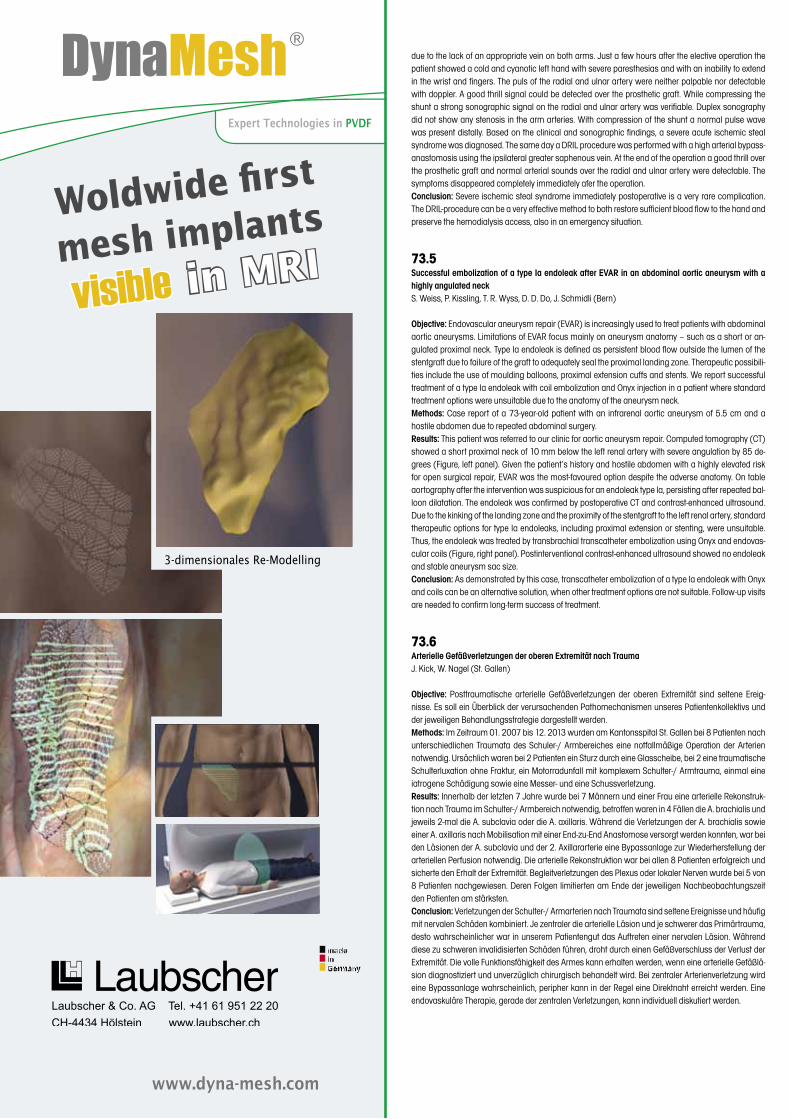
due to the lack of an appropriate vein on both arms. Just a few hours after the elective operation the patient showed a cold and cyanotic left hand with severe paresthesias and with an inability to extend in the wrist and fingers. The puls of the radial and ulnar artery were neither palpable nor detectable with doppler. A good thrill signal could be detected over the prosthetic graft. While compressing the shunt a strong sonographic signal on the radial and ulnar artery was verifiable. Duplex sonography did not show any stenosis in the arm arteries. With compression of the shunt a normal pulse wave was present distally. Based on the clinical and sonographic findings, a severe acute ischemic steal syndrome was diagnosed. The same day a DRIL procedure was performed with a high arterial bypass-anastomosis using the ipsilateral greater saphenous vein. At the end of the operation a good thrill over the prosthetic graft and normal arterial sounds over the radial and ulnar artery were detectable. The symptoms disappeared completely immediately afer the operation. Conclusion: Severe ischemic steal syndrome immediately postoperative is a very rare complication. The DRIL-procedure can be a very effective method to both restore sufficient blood flow to the hand and preserve the hemodialysis access, also in an emergency situation.
73.5Successful embolization of a type Ia endoleak after EVAR in an abdominal aortic aneurysm with a highly angulated neckS. Weiss, P. Kissling, T. R. Wyss, D. D. Do, J. Schmidli (Bern)
Objective: Endovascular aneurysm repair (EVAR) is increasingly used to treat patients with abdominal aortic aneurysms. Limitations of EVAR focus mainly on aneurysm anatomy – such as a short or an-gulated proximal neck. Type Ia endoleak is defined as persistent blood flow outside the lumen of the stentgraft due to failure of the graft to adequately seal the proximal landing zone. Therapeutic possibili-ties include the use of moulding balloons, proximal extension cuffs and stents. We report successful treatment of a type Ia endoleak with coil embolization and Onyx injection in a patient where standard treatment options were unsuitable due to the anatomy of the aneurysm neck.Methods: Case report of a 73-year-old patient with an infrarenal aortic aneurysm of 5.5 cm and a hostile abdomen due to repeated abdominal surgery.Results: This patient was referred to our clinic for aortic aneurysm repair. Computed tomography (CT) showed a short proximal neck of 10 mm below the left renal artery with severe angulation by 85 de-grees (Figure, left panel). Given the patient’s history and hostile abdomen with a highly elevated risk for open surgical repair, EVAR was the most-favoured option despite the adverse anatomy. On table aortography after the intervention was suspicious for an endoleak type Ia, persisting after repeated bal-loon dilatation. The endoleak was confirmed by postoperative CT and contrast-enhanced ultrasound. Due to the kinking of the landing zone and the proximity of the stentgraft to the left renal artery, standard therapeutic options for type Ia endoleaks, including proximal extension or stenting, were unsuitable. Thus, the endoleak was treated by transbrachial transcatheter embolization using Onyx and endovas-cular coils (Figure, right panel). Postinterventional contrast-enhanced ultrasound showed no endoleak and stable aneurysm sac size.Conclusion: As demonstrated by this case, transcatheter embolization of a type Ia endoleak with Onyx and coils can be an alternative solution, when other treatment options are not suitable. Follow-up visits are needed to confirm long-term success of treatment.
73.6Arterielle Gefäßverletzungen der oberen Extremität nach TraumaJ. Kick, W. Nagel (St. Gallen)
Objective: Posttraumatische arterielle Gefäßverletzungen der oberen Extremität sind seltene Ereig-nisse. Es soll ein Überblick der verursachenden Pathomechanismen unseres Patientenkollektivs und der jeweiligen Behandlungsstrategie dargestellt werden.Methods: Im Zeitraum 01. 2007 bis 12. 2013 wurden am Kantonsspital St. Gallen bei 8 Patienten nach unterschiedlichen Traumata des Schuler-/ Armbereiches eine notfallmäßige Operation der Arterien notwendig. Ursächlich waren bei 2 Patienten ein Sturz durch eine Glasscheibe, bei 2 eine traumatische Schulterluxation ohne Fraktur, ein Motorradunfall mit komplexem Schulter-/ Armtrauma, einmal eine iatrogene Schädigung sowie eine Messer- und eine Schussverletzung.Results: Innerhalb der letzten 7 Jahre wurde bei 7 Männern und einer Frau eine arterielle Rekonstruk-tion nach Trauma im Schulter-/ Armbereich notwendig, betroffen waren in 4 Fällen die A. brachialis und jeweils 2-mal die A. subclavia oder die A. axillaris. Während die Verletzungen der A. brachialis sowie einer A. axillaris nach Mobilisation mit einer End-zu-End Anastomose versorgt werden konnten, war bei den Läsionen der A. subclavia und der 2. Axillararterie eine Bypassanlage zur Wiederherstellung der arteriellen Perfusion notwendig. Die arterielle Rekonstruktion war bei allen 8 Patienten erfolgreich und sicherte den Erhalt der Extremität. Begleitverletzungen des Plexus oder lokaler Nerven wurde bei 5 von 8 Patienten nachgewiesen. Deren Folgen limitierten am Ende der jeweiligen Nachbeobachtungszeit den Patienten am stärksten.Conclusion: Verletzungen der Schulter-/ Armarterien nach Traumata sind seltene Ereignisse und häufig mit nervalen Schäden kombiniert. Je zentraler die arterielle Läsion und je schwerer das Primärtrauma, desto wahrscheinlicher war in unserem Patientengut das Auftreten einer nervalen Läsion. Während diese zu schweren invalidisierten Schäden führen, droht durch einen Gefäßverschluss der Verlust der Extremität. Die volle Funktionsfähigkeit des Armes kann erhalten werden, wenn eine arterielle Gefäßlä-sion diagnostiziert und unverzüglich chirurgisch behandelt wird. Bei zentraler Arterienverletzung wird eine Bypassanlage wahrscheinlich, peripher kann in der Regel eine Direktnaht erreicht werden. Eine endovaskuläre Therapie, gerade der zentralen Verletzungen, kann individuell diskutiert werden.
3-dimensionales Re-Modelling
Expert Technologies in PVDF
www.dyna-mesh.com
Woldwide first
mesh implants
visible in MRI

swiss knife 2014; 11: special edition 61
73.7Establishing ultrasound surveillance after endovascular aortic repair (EVAR) – a single center experi-enceC. Kohler, M. Birrer, A. Engwicht, T. Huber, U. Schneider, A. Stellmes (Baden)
Objective: Today EVAR is established in the treatment of aortic aneurysm. However, long-time surveil-lance, basically by CT angiography, is mandatory. Frequent radiation and use of potentially nephrotoxic contrast agent are disadvantages of this modality. Contrast enhanced ultrasound surveillance (CEUS) seems to be a promising option. We present our single center experience establishing a post EVAR surveillance program with ultrasound in October 2012. Methods: Retrospective analysis of prospective collected data.Results: Since October 2012 48 post EVAR ultrasound controls were performed in 33 patients. Six endoleaks, all Type II were detected. In three patients detection of endoleak led to further treatment by occlusion of the inferior mesenteric artery. 27 CT scans were saved. No rupture after EVAR occurred during follow-up. Conclusion: In our single center experience establishing a post EVAR surveillance protocol implement-ing CEUS reduced exposure to radiation and potentially nephrotoxic contrast agent. Growing experi-ence in this field might increase this favourable effect in the future.
73.8“Shoe lace injury” following varicose vein treatmentW. Mouton, S.-L. Hool (Thun)
Objective: Nerve injuries unfortunately do occur as complications of varicose vein surgery or endov-enous vein ablations. We present an injury of the medial branch of the superficial peroneal nerve as a case report.Methods: A 60 year old femal patient presented with ankle oedema and perimalleolar skin changes due to a left short saphenous vein insufficiency and right perforating vein insufficiency (C4 according the CEAP classification). Cardiovascular risk factors were a positive family history and obesity (BMI 34 kg/m2). Due to the number of varicose side branches the procedure was performed under spinal anaesthesia. The patient was positioned on the abdomen. A left short saphenous vein high ligation and stripping was performed as well as a right perforator ligation and stab avulsions of the side branches on both legs. There were no stab avulsions on the dorsum of the feet and the varicose side branches were localised on the dorsal sides of the legs.Results: As the patient lives in an alpine valley she wished to stay over night. During the night she had the feeling that the bandage was too tight but did not bother to disturb the nurse. When the bandage was changed the next morning the patient complained of numbness on the dorsal aspect of the first three toes on both sides. Clinically and later confirmed by neurological examination the deficit cor-responded to an injury of the medial branches of the superficial peroneal nerve. There was no indica-tion that the injury was caused by spinal anaesthesia,surgery or retractors. Until proven otherwise the nerve trauma was caused by the bandage applied with too much pressure – this type of nerve trauma is normally seen when (hiking) shoe laces are too tight. The neurological recovery was complete on the right side, on the left side it is now – 10 months after surgery – nearly complete. The patient feels no limitations due to the left-sided minimal neurological rest deficit. Conclusion: An injury to the medial branches of the superficial peroneal nerve following varicose vein surgery is rare and can be caused by the bandage. When patients complain about tight bandage or numbness – following varicose vein surgery or endovenous vein ablation – this should be taken seri-ously and the bandage immediately loosened.
73.9Aorto-kolische Fistel nach gerader Aortenprothese: ein seltener FallI. Rosenblum, C. von Arx-Küng, C. Rouden, C. Koella (Bruderholz)
Objective: Aorto-kolische Fisteln sind extrem selten und werden meist erst aufgrund von gastrointes-tinalen (GI) Blutungen entdeckt. Häufiger finden sich aorto-enterische Fisteln (AEF) ins Duodenum. AEF sind schwierig zu diagnostizieren, verlaufen aber häufig lebensbedrohlich. In diesem Case Re-port berichten wir über einen Fall einer AEF ins Kolon sigmoideum mit erfolgreicher chirurgischer Ver-sorgung.Methods: Ein 75-jähriger Mann wird von einem externen Spital mit Verdacht auf eine aorto-kolische Fistel zugewiesen, nachdem er sich mit rezidivierendem, schwallartigem Blutabgang ab ano auf der Notfallstation vorgestellt hatte. Nach unauffälliger Kolonoskopie und Gastroskopie fand sich in der CT-Angiographie eine AEF ins Kolon sigmoideum. Status fünf Jahre nach aorto-aortaler Dacron-Prothese wegen inflammatorischem, infrarenalen Aortenaneurysma. Drei Monate zuvor wurde bereits aufgrund einer Allgemeinzustandsreduktion, leicht erhöhtem CRP und abdominalen Schmerzen ein Prothesen-infektverdacht mit einer Antigranulozytenszinitgraphie in SPECT-Technik vermeintlich ausgeschlossen. Nach Diagnosestellung anlässlich der aktuellen Hospitalisation erfolgte die notfallmässige Operation. Wir fanden eine abszedierende Sigmadivertikulitis mit Kontakt zur distalen Anastomose der Aortenpro-these. Die Prothese wurde reseziert, die Aorta infrarenal verschlossen und ein subclavio-bifemoraler Bypass angelegt. Zusätzlich wurde eine Sigma-Diskontinuitätsresektion durchgeführt.Results: AEF treten nur in 0,8-1,6% der Fälle nach Anlage von Aortenprothesen auf. Sehr selten tritt eine Fistel ins Kolon auf. Eine Kolonoskopie oder Gastroskopie kann eine AEF nicht immer darstellen (30%). Eine CT bestätigt die Diagnose ebenfalls nur in 33-80% der Fälle. Auch eine Szintigraphie PET/SPECT kann die Diagnose nicht immer sichern. Sekundäre AEF entstehen am häufigsten zum distalen Duode-num, können jedoch auch an andern Lokalisationen im Verlauf des Darmes auftreten und präsentieren sich oft unspezifisch mit Schmerz, GI-Blutung, Fieber oder Sepsis.Conclusion: AEF ist eine seltene, aber lebensbedrohliche Komplikation nach Aortenrekonstruktion. Bei Status nach Aortenprothese mit Auftreten von GI-Blutungen muss eine AEF gesucht werden. Trotz allen heute verfügbaren diagnostischen Modalitäten ist die Diagnosestellung nicht immer einfach.
Visceral Surgery 7474.1Does it matter how to close the appendiceal stump? A comparison of polymeric clips with endoloop ligation and endostaplerC. Soll1, P. Wyss1, H. Gelpke1, D. A. Raptis2, S. Breitenstein1 (1Winterthur, 2Zurich)
Objective: Polymeric clips (hem-o-lok ligation system) are a novel alternative for securing the base of the appendix during laparoscopic appendectomy. There is a lack of systematic evaluation of different tools to close the appendiceal stump with regard to surgical site infections. The aim of this study was to evaluate three ligation systems involving polymeric clips (hem-o-lok ligation system), endoloop and endostapler.Methods: The outcome of 1086 consecutive patients operated laparoscopically between 2009 and 2013 for suspected appendicitis were analyzed. Data were collected prospectively as part of a national outcome survey for acute appendicitis. The risk of intra-abdominal surgical site infections by three dif-ferent methods to close the appendiceal stump involving hemo-o-lok clips, endoloop ligatures and titanium staples were investigated by univariate and multivariate analysis. On multivariate analysis resection technique, histology (perforated, non-perforated appendicitis and no/other pathology), age, gender, ASA grade, intraoperative complications and postoperative use of antibiotics, and duration of operation were included.Results: Hem-o-lok clips were used in 40.9% (n=444); endoloop sutures in 36.5% (n=396) and sta-ples were applied in 22.7% (n=246) of the patients. On multivariate analysis the duration of the opera-tion, postoperative use of antibiotics and the resection technique were the only independent predictive values for the development of postoperative intra-abdominal surgical site infections. When comparing the endoloop ligature with the hem-o-lok clips the Odds ratio was 3.6 (95% CI: 1.24-10.44), endosta-pler with the hem-o-lok the Odds ratio was 2.4 (95% CI: 0.8-7.18) for postoperative intra-abdominal surgical site infections.Conclusion: This is the first analysis evaluating the efficacy of polymeric clips to secure the base of the appendix in large cohort study. The analysis reveals that closure of the appendiceal stump us-ing a non-absorbable hem-o-lok ligation system did not result in an increased risk for intra-abdominal surgical site infections as compared to conventional methods using stapler devices or absorbable endoloop sutures.
74.2Adnexectomy in patients with peritoneal carcinomatosisK. Lehmann, R. Vonlanthen (Zürich)
Objective: Cytoreductive surgery (CRS) and hyperthermic intraperitoneal chemotherapy (HIPEC) is a therapeutic option in patients with limited peritoneal carcinomatosis from appendix tumors, mesothe-lioma, or colorectal cancer. Multivisceral surgery and extensive peritonectomy procedures are often required for extensive disease. In contrast, resection of the greater omentum and focal peritonectomy is sufficient for limited carcinomatosis. In young female patients, management of the pelvis is a criti-cal issue, and organ preservation would be desirable, particularly for low grade disease or limited carcinomatosis.Methods: Patients operated between the years 2009 and 2013 prospectively entered our database. Overall, 121 patients underwent surgical exploration, including 67 female patients.Results: Those women had peritoneal carcinomatosis from appendix tumors (40%), colorectal cancer (33%), mesothelioma (8%), and others (18%). Appendix tumors included low grade (22%) and high grade (18%) tumors. Median age was 55 (range 13-73 years). In 29 (43%) woman, bilateral adn-exectomy was performed, in six (9%), at least one ovary was preserved. The remaining 32 patients had previous adnexectomy. Overall, 60 ovaries were resected and analyzed, and the majority (62%) was tested positive, 27% were negative, and in 12%, only one ovary was positive. In four of six (67%) patients after organ preservation, tumor recurrence was suggested during follow-up imaging.Conclusion: Ovaries and fallopian tubes are a frequent site of metastasis in patients with peritoneal carcinomatosis. Preservation of the ovaries in these patients carries a high risk of recurrence.
74.3Long-term survival is not impaired after complete resection of neuroendocrine tumors of the appendixS. Ebinger, T. Steffen, R. Warschkow, B. Schmied, T. Clerici (St. Gallen)
Objective: Appendiceal neuroendocrine tumors (aNET) are a frequently occurring problem in routine medical care, since their rate per appendectomy is as high as 0.3 to 0.9%. However, 5-year-relative survival rates vary substantially between 71% and 100% and surveillance rates beyond five years are scarcely described. This investigation assessed the long-term mortality after complete resection of aNET. Additionally, prognostic factors for the oncologic outcome were assessed.Methods: The ten-year survival was analyzed in 79 patients operated between 1990 and 2003 us-ing risk-adjusted Cox proportional hazard regression models with adjustment for population-based baseline-mortality (relative survival) and with the random survival forest method.Results: Median follow-up of all patients was 12.1 years and for those alive 13.7 years. All patients underwent curative R0-resection. No distant synchronous or metachronous metastases were diag-nosed. The ten-year-overall and relative survival were 83.6% (95% CI: 75.5% to 92.6%) and 96.7% (95% CI: 87.5% to 107%), respectively. The survival after complete resection of aNET did not differ from the survival in the average national population with the same age and gender (P = 0.947). Infiltration of the meso-appendix (hazard ratio of death 7.7, 95% CI: 0.9 to 65.1) and second synchronous or metachronous primary malignancies (hazard ratio of death 4.5, 95% CI: 1.3 to 16.0) were identified as

62 swiss knife 2014; 11: special edition
Unsere Vorteile GrößereThermofusionszone SichereDissektionexaktinderMitte Homogener,glatterSchnitt SchnellesAbkühlenderBranchen OptimalePräparationseigenschaften
π-VorteilDurchdieschalenförmigeBrancheentstehteinebreitereFusionszonealsbeiallenanderenInstrumentenmit5-mm-Schaft.DiesschafftzusätzlicheSicherheit.
ThermofUsionieren Und dissezieren miT dem π-VorTeil
erBe BiCision®
Fusionszone konventionell
Fusionszone BiCision mit π-Vorteil
ERBE Swiss AG | Winterthur | Telefon 052 233 37 27 | [email protected] | www.erbe-swiss.ch
AZ_ERBE_BiCision_185x130_Swiss.indd 1 16.01.2012 15:22:55
significant prognosticators for long-term relative survival in risk-adjusted Cox regression.Conclusion: Long-term survival is not significantly depreciated after complete resection of neuroendo-crine tumors of the appendix.
74.4Da Vinci in general surgery: a safe and structured start of a comprehensive robotics programmeL. F. Grochola, A. Zehnder, G. Wille, H. Gelpke, S. Breitenstein (Winterthur)
Objective: Due to the perceived advantages of robot-assisted laparoscopic surgery in urological pro-cedures, this technique has been broadly adopted in the field of urology. However, despite its potential advantages and the on-site availability of Da Vinci Systems, only few general surgery departments worldwide have so far successfully established a robotics programme. This is in part due to the com-plexity of the implementation of such a challenging project. Therefore, in order to overcome those chal-lenges and benefit from the perceived advantages of this rapidly evolving technology, this project was designed to efficiently launch a comprehensive robotics programme at a general surgery unit.Methods: Within a few months, we have initiated a robotic programme comprising procedures such as transaxillary robotic thyroidectomy, Single-Site cholecystectomy and colorectal resection. First, a structured, 3-step training course was completed. This consisted of an online training module, an on-site Da Vinci System induction and a multi-module simulator training. Subsequently, the first robotic procedures performed on patients were a series of multi-port cholecystectomies. Next, following ad-ditional human cadaver and live pig training courses at international training centres (EEC and IRCAD), more advanced surgical procedures have been successively introduced in intervals of 1-2 months with the one-time presence of a proctor. This project comprises a prospective database that collects a dataset related to robot-assisted and corresponding conventional laparoscopic surgeries. In addi-tion, a randomized controlled trial is conducted in order to minimize biases in the analysis of potential advantages of this technology.Results: All robotic procedures have been safely and efficiently introduced into the daily hospital rou-tine. Early results indicate that all procedures can be performed with at least similar peri- and postop-erative outcomes compared to conventional laparoscopy.Conclusion: The launch of a robotic programme at a general surgery unit is safe and feasible utiliz-ing a structured training strategy and a step-wise introduction of surgical procedures. Early data sug-gest that the robotic platform can at least match the high standards of conventional laparoscopy. This project can serve as a blueprint for the implementation of this technology in a clinical setting.
74.5LiMAx – Klinische Anwendung in der Medizin – erste Resultate im KSSGR. Käppeli, S. Möller, I. Tarantino, B. Schmied, C. Luethi, S. Müller (St. Gallen)
Objective: Leberfunktionsmessungen sind bei Patienten mit Lebererkrankungen, vor Lebereingriffen und in der Intensivmedizin von grosser Bedeutung. Diverse Tests zur Erfassung der quantitativen Leb-erfunktion sind in der Literatur beschrieben. Keiner dieser Tests konnte verlässliche Aussagen zur Leb-erfunktion machen. Seit 2003 wird der an der Klinik Charité in Berlin entwickelte LiMAx-Test (maximal liver function capacity based on 13^C-methacetin kinetics) angewandt. Seit 10/2012 werden im KSSG bei gegebener Indikation ebenfalls LiMAx-Messungen durchgeführt.Methods: Beim LiMAx-Test wird dem Patienten Methacetin 2mg/kg KG intravenös verabreicht. Diese Substanz wird durch das hepatozytenspezifische Enzym CYP1A2 in Paracetamol und CO
2 metabolisi-
ert. In der Atemluft wird das metabolisierte CO2 erfasst und der LiMAx-Wert errechnet. Ein LiMAx-Wert
von mindestens 315 ug/kg/h entspricht einer normalen Leberfunktion. Eine LiMAx-Test kann am Pa-tientenbett, bei intubierten Patienten sowie intraoperativ angewandt werden.Results: Vor Lebereingriffen bei primären und sekundären Lebertumoren kommen LiMAx-Tests stand-ardmässig zum Einsatz. In Studien werden zurzeit weitere Indikationen des Tests evaluiert (Chemother-apien, nach TIPS, Adipositas). Zwischen 10/2012 und 08/2013 wurden 19 Messungen bei Patienten mit primären Lebertumoren durchgeführt. Bei 4 dieser Patienten wurde aufgrund eines zu geringen präoperativen LiMAx-Werts auf eine Tumorresektion verzichtet. Retrospektiv zeigt sich seit Anwendung des Tests im KSSG eine Morbidität von 28% bzw. eine Mortalität von 6.6% (1/15 Fällen) nach Leber-chirurgie bei primären Lebertumoren. Bei sekundären Lebertumoren wurden 4 von 44 Patienten bei zu geringer Leberfunktion für eine chirurgische Resektion abgelehnt (postoperative Morbidität von 24% bzw. Mortalität von 0% bei sekundären Lebertumoren). In der Intensivmedizin werden LiMAx-Messun-gen bei Leberversagen angewandt. Im Verlauf kann die hepatische Funktion beim Patienten bestimmt und prognostische Aussagen gemacht werden werden. In Zusammenarbeit mit Gastroenterologie im KSSG wird zurzeit in einer Studie die Anwendungsmögli-chkeit der LiMAx-Tests nach TIPSS untersucht.Conclusion: LiMAx-Messungen kommen in der Leberchirurgie, Hepatologie und Intensivmedizin zum Einsatz. Unsere erfassten Resultate bestätigen die prädiktive Aussage des Tests. Weitere Indikationen scheinen in Zukunft möglich.
74.6Conservative treatment of chyle leaks by oral nutrition replacing long-chain tiglycerides by medium-chain triglyceridesA. Frischknecht, A. Schnider, M. Weber (Zurich)
Objective: To investigate the impact of conservative management of chylothorax and chylous ascites by oral nutrition avoiding long-chain triglycerides and additional supplementation with medium-chain triglycerides (MCT).Methods: Retrospective database analysis of patients with chylothorax or chylous ascites from 01/2008 to 12/2013. Diagnostic criteria were fluid triglyceride levels higher than 1.24 mmol/l or clini-

swiss knife 2014; 11: special edition 63
cal features after having excluded a pancreatic fistula. An individual diet regimen without long chain triglycerides was established. Long-chain triglycerides were replaced by oral short- and medium-chain triglycerides. Our diet regimen contained a physiological composition of 65% carbohydrates, 15% pro-teins and 20% lipids (90% MCT).Results: Three cases of chylothorax and 7 cases of chylous ascites were identified: Seven postsurgical cases, one injury after trauma, one malignancy and one case of unknown origin. Demographic and clinical characteristics are outlined in Table 1. In all patients, the diet led to a considerable reduction of fluid drainage. Chyle leakage stopped in all 10 patients within a mean time of 10.6 days (range 2-33), 70% of them within one week. No patient required operative treatment. Seven patients had parenteral supplementary nutrition for 9 to 19 days due to lack of sufficient oral calory intake. Nine patients ad-ditionally received diuretics, two of them also octreotide.Conclusion: Optimal treatment strategy of chyle leakage is still a matter of debate. Low-lipid diet has been reported in literature with success rates of 50-84% preserving the advantage of enteral nutrition when compared to total parenteral nutrition. Our therapeutic concept suggests that a majority of these patients can be managed conservatively by oral nutrition when long-chain triglycerides are replaced by short- and medium-chain triglycerides.
74.7Transumbilical single-port laparoscopic transabdominal preperitoneal repair of inguinal hernia: progress in reducing invasivenessS. Ihne, B. Widmann, L. Traine, S. Bischofberger, N. Kalak, W. Brunner (Rorschach)
Objective: Conventional laparoscopic repair of inguinal hernia usually requires 3 ports. Recently, single-port laparoscopic surgery techniques have been developed to further minimize surgical trauma and invasiveness compared to conventional laparoscopic surgery. We want to present our experience in single-port transabdominal preperitoneal (TAPP) repair of inguinal hernia.Methods: This is a retrospective analysis of our prospectively collected single-port TAPP data includ-ing all 288 patients undergoing transumbilical single-port TAPP between August 2011 and November 2013. Until now 148 patients are followed up by phone survey to evaluate patient satisfaction and com-plications. In case of possible postoperative complications patients received a clinical examination.Results: 368 Single-Port TAPP procedures (208 unilateral, 80 bilateral and 17 recurrent hernia) were performed in 288 patients (243 male, 45 female, mean age 49.1a, mean BMI 25.15kg/m2). 16.3% of the patients have had previous abdominal surgery. Mean operation time was 62.3 min (± 24.7 min) for unilateral and 96.1 min (± 38.5 min) for bilateral hernia. Mean fascial incision length was 25 mm (± 7 mm), mean skin incision length was 32 mm (± 9 mm). Additional trocars were needed in 18.8% corresponding to the experience of the surgeons. No conversions to open surgery were necessary. No intraoperative complications were observed. According to the follow up, patients are very satisfied with general result (1.45 ± 0.82, 1 = very satisfied, 5 = very unsatisfied) and generaly do not suffer from
m = male, f = female, TG = triglycerides, MCT = medium chain triglycerides, EN = enteral nutrition, PN = parenteral nutrition, NA = not available
Patient No.
Age (years)
Sex Aetiology of Chyle Leak
Contributing Diagnosis
Fluid Appearance
Fluid Analysis
TG (mmol/l)
Nutrition Regimen
Adjunctive Treatment
Chylothorax
1 83 f Trauma (injury): blunt thoracic trauma (rib fractures costae 11, 12), vertebral trauma (Chance fracture L1, fracture of procc. spinosi and transversi L1-3)
pneumonia orange/ turbid
concordant exudate
13.3 low-lipid MCT EN, supportive PN
diuretics, antibiotics
2 63 m Trauma (postsurgical): thoraco-abdomino-cervical esophagectomy (esophageal cancer)
purulent bronchitis and atelectasis (right)
yellow / turbid
transudate 2.2 low-lipid MCT EN, supportive PN
diuretics, antibiotics
3 77 f Unknown: ev. obstruction by thoracic aortic aneurysm
hypertensive heart disease (diastolic dysfunction)
chylous concordant exudate, lymphocyte predominant
35.1 low-lipid MCT EN, supportive PN
diuretics
Chylous ascites
4 53 f Trauma (postsurgical): thoraco-abdominal
esophagectomy, sleeve resection left lower lobe (esophageal and small cell lung cancer)
renal infarction, pulmonary embolism, pulsless electrical activity
yellow / turbid
transudate 2.9 low-lipid MCT EN, supportive PN
diuretics, antibiotics
5 53 m Trauma (postsurgical): explorative laparotomy, adhesiolysis, gastrojejunostomy, Braun’s anastomosis (panreatic cancer)
chylous unclassified NA low-lipid MCT EN, supportive PN
diuretics
6 65 m Trauma (postsurgical): explorative laparotomy, dissection of hepatoduodenal ligament, biliodigestive anastomosis (pancreatic cancer)
chylous unclassified NA low-lipid MCT EN diuretics, octreotide
7 89 f Malignant: retroperitoneal spread malignant disease (ev. pancreatic carcinoma)
chylous protein discordant exudate, lymphocyte predominant
24.9 low-lipid MCT EN
8 77 m Trauma (postsurgical): explorative laparotomy, transversostoma (colon ischemia)
congestive heart failure
chylous transudate 2.5 low-lipid MCT EN, supportive PN
diuretics
9
10
82
57
f
m
Trauma (postsurgical): open rectosigmoid resection (diverticulitis)
Trauma (postsurgical): pancreaticoduo-denectomy (Whipple procedure) (cholangiocarcinoma)
chylous
chylous
unclassified
unclassified
NA
NA
low-lipid MCT EN, supportive PN
low-lipid MCT EN
diuretics
diuretics, octreotide
Table 1. Demographic and clinical data of patients with chyle leak (n = 10).
postoperative discomfort (1.60 ± 0.9). Moreover the cosmetic result is evaluated very well (1.18 ± 0.48). Up to now there was only one recurrence of inguinal hernia, but no trocar hernia.Conclusion: Single-port TAPP represents a safe and generally applicable surgery technique (even for the repair of recurrent hernia). The use of additional trocars is mainly due to improve patient safety and correspond to the experience of the surgeon. Moreover, it is nearly scarless and minimizes surgical trauma.
74.8Short- and long-term results following abdominal resection-rectopexy for descending perineum syn-dromeA. Heigl, S. A. Käser, A. Zuse, D. Toia, C. A. Maurer (Liestal)
Objective: To evaluate the short- and long-term outcome after conventional dorsal resection-rectopexy for descending perineum syndrome (DPS).Methods: From 1/2003 to 8/2011, open or laparoscopic abdominal rectopexy and tightening of Doug-las pouch was done in 108 patients with DPS, usually consisting of rectal prolapse, rectocele, ente-rocele and dolichosigma. The following procedures were done additionally: sigmoid resection (n = 93), subtotal colectomy (n = 8), combined abdomino-perineal approach according to Zaccharin-Marti (n = 11), anal sphincter repair (n = 2), hysterectomy (n = 5) and colposuspension (n = 9). Preoperative def-

64 swiss knife 2014; 11: special edition
ecatory function was assessed by standardized questions and controlled using the same questions at 3 and 12 months postoperatively. At the same time points, clinical examinations were performed. Further, 86/108 (80%) patients responded to a questionnaire after a median follow-up time of 57.5 (12-146) months. Fifteen patients deceased meanwhile due to other reasons, 4 could not respond to the questionnaire due to dementia and 2 patients refused to respond.Results: The median patients’ age at time of surgery was 67 (28-90) years. One aged patient of 91y died (1/108, 1%). One anastomotic leakage and 3 wound infections occurred, but allowed conserva-tive management. No recurrence of rectal prolaps or enterocele was observed during the follow-up period. Two patients needed transperineal correction of the rectocele later-on. The following alterations in bowel habits comparing preoperative vs. postoperative status were registered: median number of defecations per day 1 vs. 1.5 (p = 0.0174), wearing a pad 88% vs. 63% (p = 0.0255), straining to defecate 83% vs. 50% (p = 0.0344), incomplete evacuation 97% vs. 43% (p < 0.0001), need for digital emptying 31% vs. 27% (p = 0.6405), stay at home for incontinence 57% vs. 8% (p < 0.0001). The suc-cess of the operation was classified by the patients as little (9%), middle (34%), big (26%) and very big (6%). No improvement was declared by 24% of all patients. 83% of the patients who answered the questionnaire would undergo surgery again.Conclusion: In this series, abdominal resection-rectopexy seems to be a safe and reliable repair of DPS. Despite restored anatomy, postoperative defecatory function improved in 75% of patients, only.
74.9Ligation of intersphincteric fistula track (LIFT) for transsphicteric anal fistula: initial experienceB. Pravini, R. Rosso, D. Christoforidis (Lugano)
Objective: Healing rates after surgery for complex fistulas using sphincter sparing techniques are often disappointing. The ligation of intersphincteric fistula track (LIFT) is a relatively new technique that is gaining popularity due to promising results in terms of fistula closure and preservation of continence. We reviewed our initial results with this technique.Methods: Data of all patients who underwent a LIFT procedure in our institution were retrospectively as-sessed. All patients underwent endoanal ultrasound prior to surgery for precise assessment of fistula anatomy. LIFT was performed after all sepsis had been adequately drained. Patients were followed at the proctology clinic, those considered healed at the last visit were contacted by telephone to assess their current status.Results: Between April 2012 and November 2013, 13 patients (5 female, median age 49 years) un-derwent LIFT for a cryptoglandular transsphincteric fistula (9 upper sphincter third, 1 middle third, 3 lower third, one horse-shoe). Median (range) operative time was 49 (25-70) minutes, all patients but one were discharged on the same day. Primary success rate was 5/13 (follow-up 15 (2-21) months). In 6/13 patients the fistula recurred through the surgical incision as an intersphincteric or low trans-sphincteric track and was successfully treated by lay-open fistulotomy (follow-up 6 (2-10) months) in all patients. Only in 2/13 the fistula recurred through the initial transsphincteric track. No patient suffered post-operative complications. Continence was perfect in all but one patient who referred oc-casional gas incontinence. Conclusion: In our hands, patients treated with the LIFT procedure had a rather high recurrence rate but mostly through the intersphincteric surgical incision. Those patients could be treated by simple lay-open fistulotomy with minimal impact on continence and an overall secondary success rate of 11/13.
Mixed Session II 7777.1Radioguided occult breast lesion localization: Initial experience in 33 patientsJ. Sichitiu, C. Becciolini, T. Cazaentre, D. Constantinescu, M.-J. Chevénement, B. Christen (La Chaux-de-Fonds)
Objective: Assessement of the accuracy and effectiveness of the ROLL (radioguided occult lesion lo-calization) and SNOLL (ROLL plus sentinel node localization) procedure in terms of success of breast lesion localization, sentinel node (SN) localization and free surgical margins.Methods: From November 2012 to October 2013, we prospectively included 33 consecutive patients for 39 procedures ; ROLL = 11 cases, SNOLL = 28 cases. All of them presented with non-palpable but ultrasound-positive breast lesions. All were eligible for breast-conserving therapy. Under ultrasound guidance, we injected 21.4±6.8 (mean±SD) MBq of 99mTc-nanocolloid intratumorally, with a com-plementary periareolar injection of 10.8±4.5 (mean±SD) MBq for the SNOLL procedure. Surgery was performed the next day, 19.4±1.8(mean±SD) hours after.Results: Intraoperative breast lesion localization was successfull in all cases but one (38/39), yielding a localization rate of 97.4%. The intraoperative SN localization rate was 100% despite the fact that in one patient, no SN was visible on the preoperative lymphoscintigraphy. The surgery was sufficiently radical (free margins) in 30 of the 31 patients (96.7%). Additional exicisions to ensure free tissue mar-gins were performed during the first procedure for 6% of them (2/31).Conclusion: Our results encourage the consideration of the SNOLL / ROLL technique in the manage-ment of non-palpable nodular breast cancer.
77.2Expression of PDL-1 in breast cancer is associated with poor prognosisA. Schaerli1, S. Muenst1, S. Däster1, R. A. Droeser1, D. Oertli1, U. von Holzen1, F. Gao2, W. E. Gillanders2, G. Spagnoli1, W. P. Weber1, S. D. Soysal1 (1Basel, 2St. Louis, Missouri/USA)
Objective: Programmed death 1 (PD-1) is a coinhibitory receptor in the CD28/CTL-4 family, and func-tions as a negative regulator of the immune system. Tumor-infiltrating lymphocytes (TIL) in many epi-thelial cancers express PD-1, and the PD-1 ligand PD-L1 is expressed by a variety of epithelial cancers, suggesting that antitumor immunity may be modulated by the PD-1/PD-L1 signaling pathway, and promising results from two recent clinical trials with monoclonal antibodies targeting PD-1 or PD-L1 confirm the clinical relevance of this pathway in human cancer.Methods: To explore the role of PD-L1 in human breast cancer, we performed immunohistochemistry on a tissue microarray encompassing 676 breast cancer cases with detailed clinical annotation and outcomes data.Results: Expression of PD-L1 on tumor cells was present in 207 (30.7%) of the 676 breast cancer cas-es. Its presence was associated with tumor size, grade and lymph node status, and was differentially associated with the intrinsic subtypes of breast cancer. In univariate survival analyses, the expression of PD-L1 was associated with a significantly worse overall survival. Furthermore, there was a strong correlation between the expression of PD-L1 and PD-1+ TIL.Conclusion: This is the first study to demonstrate that the expression of PD-L1 is associated with poor prognosis in human breast cancer, with important implications for the potential application of antibody therapies targeting the PD-1/PD-L1 signaling pathway in this disease.
77.3Surgical site mark – a physician’s task?J. Schäfli-Thurnherr, A. Biegger, L. Maybüchen, G. A. Melcher (Uster)
Objective: Marking the surgical site (SS) has become a routine procedure before surgery. Thereby, safety being the main point of interest. It seems to be a doctrine, that the operating surgeon has to be the one performing the marking. In daily business this often leads to problems because the surgeon is in the OR (“same-day-surgery”). Therefore, we decided to integrate the nurses into the marking proc-ess. Our goal was to check, if the procedure of marking the SS can be safely performed by the nurse taking care of the patient.Methods: Legal check: We consulted the Swiss Patients Safety, the Swiss Society of Nurses and the health department of Zürich with the question, whether the task of placing the SS mark can be del-egated to a trained nurse. In 2012 we launched a three months pilot project, in which previously trained nurses performed the SS marking. To the pre-existing preoperative forms, we added a checklist the nurse had to sign after marking the SS. Evaluation of the pilot project and extension of the project to all surgical wards in 2013. After one year of performance, a second evaluation was performed on the basis of questionnaires handed out to surgeons and nurses.Results: The assessment of our pilot project showed, that 76% of the nurses rated the training as very helpful. Nurses felt save when marking the SS in 90% of the time. Otherwise, they got help from a surgeon. 92% of the nurses experienced a relief of stress when performing the procedure themselves, rather then being on the look for a surgeon. The results after 1 year of performance are compared to those of the pilot project. During the one year period, no serious mistakes were made marking the SS.Conclusion: We have shown that the SS mark can be placed safely and reliably by a trained nurse and probably improves patient safety. The strength of our study is a thorough planning and evaluation of the pilot project, as well as the evaluation of the results after one year of performance. Considering the rapid turn-over of the doctors and nurses in our hospital, there remain only few people having known the situation before delegating the action to the nurses. Therefore comparison of the results of both evaluations should be considered with care. In attempt to facilitate the preoperative procedure and consequently further improve safety, studies in larger surgical wards could fortify our results.
77.4The use of checklists in surgery: a paper tiger or a cost-effective life-saving device?D. De Faoite, B. Hanson (Dübendorf)
Objective: Healthcare professionals are under pressure from different stakeholders to reduce costs. The use of simple checklists has been advanced as a low-tech, low-cost way to improve surgical per-formance. We wanted to investigate if this has proven to be the case.Methods: A two-pronged review of the literature was conducted. Firstly, general information about checklists was collected from medical and non-medical sources. A comprehensive PubMed search was conducted to locate articles on the health and financial outcomes once a checklist had been implemented. Information on the acceptance of checklists was also extracted.Results: General: Checklists should only be used for simple problems. Their main advantage is acting as a cognitive aid to ensure that all of the main elements for a procedure have been addressed – for even experts forget! Attitudes: Surgeons and surgical teams may be reluctant to use checklists at first, an attitudinal change is required for their widespread acceptance. Changes in practice have to be inte-grated into daily routines. There are strategies available to help inculcate a checklist culture in a clinic. Once in use, the majority of the surgical team appears to appreciate the benefits that checklists bring and its use very quickly reaches nearly full compliance. Large improvements in inter-team communica-tion have also been recorded. A Road to Damascus type conversion was also anecdotally reported whereby a previously strident opponent realized that the checklist had helped identify a problem. As a result, they completely changed their opinion of checklists. Surgical Results: Checklists have a wide ap-plication among different disciplines. Even just asking people to create one leads to improved results. An international study on the WHO Safe Surgery Checklist recorded a fall in complications in surgical patients from 11% to 7% and a fall in mortality from 1.5 to 0.8%, while other studies also recorded improvements in outcomes.Conclusion: Research has demonstrated checklist effectiveness in surgery. The WHO estimates that up to 500,000 patients per year could avoid fatal complications if its Safe Surgery Checklist was imple-mented in all cases. Checklists require careful content planning and rollout in clinics, however, they do bring noticeable and at times dramatic improvements. Checklists should also be continually adapted to suit a clinic’s present situation.
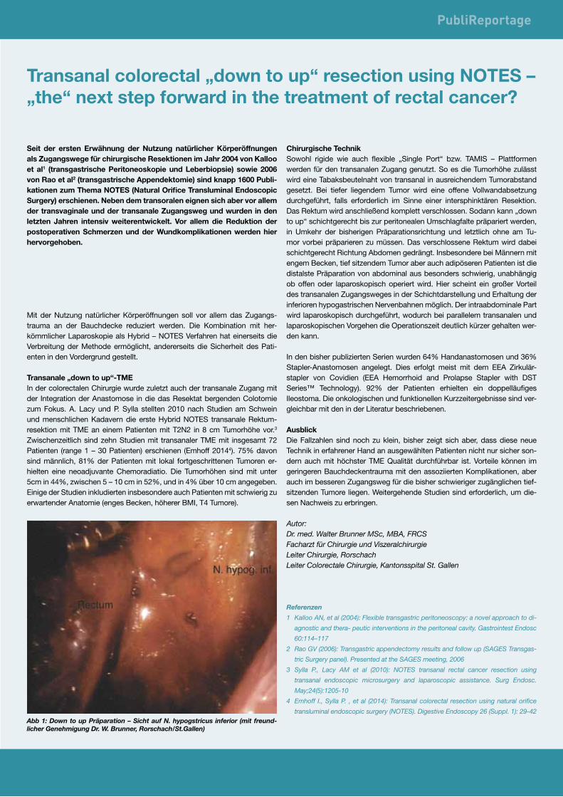
swiss knife 2014; 11: special edition 65
Transanal colorectal „down to up“ resection using NOTES – „the“ next step forward in the treatment of rectal cancer?
Seit der ersten Erwähnung der Nutzung natürlicher Körperöffnungen als Zugangswege für chirurgische Resektionen im Jahr 2004 von Kalloo et al1 (transgastrische Peritoneoskopie und Leberbiopsie) sowie 2006 von Rao et al2 (transgastrische Appendektomie) sind knapp 1600 Publi-kationen zum Thema NOTES (Natural Orifice Transluminal Endoscopic Surgery) erschienen. Neben dem transoralen eignen sich aber vor allem der transvaginale und der transanale Zugangsweg und wurden in den letzten Jahren intensiv weiterentwickelt. Vor allem die Reduktion der postoperativen Schmerzen und der Wundkomplikationen werden hier hervorgehoben.
Mit der Nutzung natürlicher Körperöffnungen soll vor allem das Zugangstrauma an der Bauchdecke reduziert werden. Die Kombination mit herkömmlicher Laparoskopie als Hybrid – NOTES Verfahren hat einerseits die Verbreitung der Methode ermöglicht, andererseits die Sicherheit des Patienten in den Vordergrund gestellt.
Transanale „down to up“-TME In der colorectalen Chirurgie wurde zuletzt auch der transanale Zugang mit der Integration der Anastomose in die das Resektat bergenden Colotomie zum Fokus. A. Lacy und P. Sylla stellten 2010 nach Studien am Schwein und menschlichen Kadavern die erste Hybrid NOTES transanale Rektumresektion mit TME an einem Patienten mit T2N2 in 8 cm Tumorhöhe vor.3 Zwischenzeitlich sind zehn Studien mit transanaler TME mit insgesamt 72 Patienten (range 1 – 30 Patienten) erschienen (Emhoff 20144). 75% davon sind männlich, 81% der Patienten mit lokal fortgeschrittenen Tumoren erhielten eine neoadjuvante Chemoradiatio. Die Tumorhöhen sind mit unter 5cm in 44%, zwischen 5 – 10 cm in 52%, und in 4% über 10 cm angegeben. Einige der Studien inkludierten insbesondere auch Patienten mit schwierig zu erwartender Anatomie (enges Becken, höherer BMI, T4 Tumore).
Chirurgische TechnikSowohl rigide wie auch flexible „Single Port“ bzw. TAMIS – Plattformen werden für den transanalen Zugang genutzt. So es die Tumorhöhe zulässt wird eine Tabaksbeutelnaht von transanal in ausreichendem Tumorabstand gesetzt. Bei tiefer liegendem Tumor wird eine offene Vollwandabsetzung durchgeführt, falls erforderlich im Sinne einer intersphinktären Resektion. Das Rektum wird anschließend komplett verschlossen. Sodann kann „down to up“ schichtgerecht bis zur peritonealen Umschlagfalte präpariert werden, in Umkehr der bisherigen Präparationsrichtung und letztlich ohne am Tumor vorbei präparieren zu müssen. Das verschlossene Rektum wird dabei schichtgerecht Richtung Abdomen gedrängt. Insbesondere bei Männern mit engem Becken, tief sitzendem Tumor aber auch adipöseren Patienten ist die distalste Präparation von abdominal aus besonders schwierig, unabhängig ob offen oder laparoskopisch operiert wird. Hier scheint ein großer Vorteil des transanalen Zugangsweges in der Schichtdarstellung und Erhaltung der inferioren hypogastrischen Nervenbahnen möglich. Der intraabdominale Part wird laparoskopisch durchgeführt, wodurch bei parallelem transanalen und laparoskopischen Vorgehen die Operationszeit deutlich kürzer gehalten werden kann.
In den bisher publizierten Serien wurden 64% Handanastomosen und 36% StaplerAnastomosen angelegt. Dies erfolgt meist mit dem EEA Zirkulärstapler von Covidien (EEA Hemorrhoid and Prolapse Stapler with DST Series™ Technology). 92% der Patienten erhielten ein doppelläufiges Ileostoma. Die onkologischen und funktionellen Kurzzeitergebnisse sind vergleichbar mit den in der Literatur beschriebenen.
AusblickDie Fallzahlen sind noch zu klein, bisher zeigt sich aber, dass diese neue Technik in erfahrener Hand an ausgewählten Patienten nicht nur sicher sondern auch mit höchster TME Qualität durchführbar ist. Vorteile können im geringeren Bauchdeckentrauma mit den assoziierten Komplikationen, aber auch im besseren Zugangsweg für die bisher schwieriger zugänglichen tiefsitzenden Tumore liegen. Weitergehende Studien sind erforderlich, um diesen Nachweis zu erbringen.
Autor:Dr. med. Walter Brunner MSc, MBA, FRCSFacharzt für Chirurgie und ViszeralchirurgieLeiter Chirurgie, RorschachLeiter Colorectale Chirurgie, Kantonsspital St. Gallen
Abb 1: Down to up Präparation – Sicht auf N. hypogstricus inferior (mit freund-licher Genehmigung Dr. W. Brunner, Rorschach/St.Gallen)
Referenzen
1 Kalloo AN, et al (2004): Flexible transgastric peritoneoscopy: a novel approach to di-
agnostic and thera- peutic interventions in the peritoneal cavity. Gastrointest Endosc
60:114–117
2 Rao GV (2006): Transgastric appendectomy results and follow up (SAGES Transgas-
tric Surgery panel). Presented at the SAGES meeting, 2006
3 Sylla P., Lacy AM et al (2010): NOTES transanal rectal cancer resection using
transanal endoscopic microsurgery and laparoscopic assistance. Surg Endosc.
May;24(5):1205-10
4 Emhoff I., Sylla P. , et al (2014): Transanal colorectal resection using natural orifice
transluminal endoscopic surgery (NOTES). Digestive Endoscopy 26 (Suppl. 1): 29-42
PubliReportage

66 swiss knife 2014; 11: special edition
77.5Persistent and worsening weight loss one month after major gastrointestinal surgeryM. Benoit, P. Coti Betrand, M. Hübner, F. Grass, N. Demartines, F. Pralong (Lausanne)
Objective: Malnutrition is well described as an important risk factor for early complications after major gastrointestinal cancer surgery. Guidelines recommend using perioperative nutrition to decrease post-operative morbidity and mortality, length of stay and hospital costs. Few data are published on delayed complications and specifically on body composition and functional outcome.Methods: Data were obtained from a cohort of 146 patients with a nutritional risk score (NRS) >3 un-dergoing major surgery for gastrointestinal cancer. They were assigned in a randomized trial assess-ing the clinical benefit of preoperative nutrition. Outcomes (postoperative complications, nutritional status, and functional status) were measured 10 days before (D-10) and 30 days (D30) after surgery.Results: The prevalence of moderate and severe malnutrition is high and increases from 61% to 83% respectively at D-10 and D30. A weight loss > 10% affects respectively 32% patients at D-10 and in-creases to 61% of patients at D30. The percentage of patients with a BMI < 18.5 increases from 14% at D-10 to 22% at D30. The mean arm muscle circumference decreases from 241 to 231 mm between D-10 and D30. Appetite and nutritional intake cover less than 66% of energy needs in 49% patients at D-10 and the gap persists in 50% patients at D30. Overall physical activity level assessed according to the lifestyle shows an increase of poorly active patients from 29 to 39% of them between D-10 and D30.Conclusion: Weight loss, malnutrition and insufficient nutritional intake are frequent in patients un-dergoing major gastrointestinal cancer surgery. They worsen despite hospital perioperative nutrition, compromise muscle mass and consequently functional status of patients one month after surgery. To restore an optimal nutritional and functional status, nutritionists should be systematically involved in outpatient follow-up.
77.6Pedicled bilateral muscular gracilis flaps for restoration of large scrotal tissue defectsJ. Rheinberger, D. F. Kalbermatten, J. Eigenmann, B. Egger (Fribourg)
Objective: Extensive defects of the scrotum with exposure of the testes are difficult to reconstruct and patients often necessitate an interdisciplinary approach. Such large tissue defects are seen after Fournier’s gangrene or skin burns. In the literature there are few cases reported describing extensive tissue defects of the scrotal region due to a hidradenitis suppurativa. The disease has an incidence of 6.0 per 100,000 per year. An applied technique to restore tissue defects in these cases are pedicled muscular gracilis flaps.Methods: We report here the case of a 62 years old patient known for a long-standing hidradenitis suppurativa with recurrent abscesses and fistulas of the scrotal region. He had to be hospitalized due to another episode of multiples abscesses and and a beginning ssepticaemia. Following that repeated local necrosectomies had to be performed leaving the patient with a large scrotal tissue defect and a complete exposure of the testes. In order to prepare the wound ground for reconstructive interventions a radical removal of all remaining scrotal and penile skin tissue was performed. Then the patient un-derwent scrotal reconstruction with bilateral pedicled gracilis flaps followed by applications of mesh-grafts to cover the muscles. At the same time, a Thiersch’s skin graft of the penis without mesh was also performed. The operation was finished by placing a VAC with aspiration of – 120mmHg during 5 days. As a prophylaxis for further infections antibiotics were prescribed for 3 weeks following the op-eration. It is well known that antibiotics penetrate well into the transplanted tissue due to the good vas-cularization of the muscle flap, which also allows to have an improved infection control of the disease.Results: The patient underwent a completely uneventful postoperative follow-up with entire flap and tissue survival and with a good, subjective as well as objective functional and esthetic result.Conclusion: The combination of applying a pedicled muscle flap together with Thiersch’s grafts result-ed in an excellent short-term outcome for this patient suffering from a nasty disease. Furthermore infec-tion control of the disease is much more likely due to the good vascularization of the muscle flap tissue.
77.7Milzzystenrezidiv – minimal invasive TherapieD. Ardüser, S. Hoederath, D. De Lorenzi (Grabs)
Objective: Milzzysten sind selten. Anhand eines nichtparasitären, kongenitalen Milzzystenrezidivs wol-len wir die lap. Teilsplenektomie mit Zystenentfernung als Therapiemöglichkeit präsentieren.Methods: Case-Report.Results: Eine 29-jährige Patientin stellte sich knapp 2 Jahre nach einer lap. Milzzystenabdeck-elung (deroofing) aufgrund linksseitiger Oberbauchschmerzen vor. Es konnte ein Milzzystenrezidiv (12x12x13 cm) mit z.T. intraparenchymatösen Anteilen im Milzoberpol nachgewiesen werden. Bei bereits histologisch nachgewiesener benigner mesothelialer Milzzyste (Voroperation), negativer Echi-nococcusserologie und durchgeführter Immunisierung wurde die Indikation zur lap. Teilsplenektomie und ggf. Splenektomie gestellt. Intraoperativ konnten nach Mobilisation und Darstellung des Milzhilus die Oberpolgefässe problemlos dargestellt und mit Clips verschlossen werden. Nach abgeschlossener Demarkierung der Durchblutung erfolgte die Durchtrennung mit einem Sicherheitsabstand (5 mm) mittels Thunderbeat®. In der Folge konnte der betroffene Milzteil nach Erweiterung des umbilicalen Zuganges geborgen werden. Nach Kontrolle der Hämostase mittels Argonbeamer, Versiegelung mit Tachosil® und Reposition sowie Fixation des jetzt sehr mobilen Milzrestes (Unterpol) mit Fibrinkleber konnte die Operation erfolgreich beendet werden. In der hist. Untersuchung konnte erneut eine gutar-tige mesotheliale Milzzyste festgestellt werden. Der postop. Verlauf gestaltete sich komplikationslos, sodass die Patientin am 6. postop. Tag entlassen werden konnte.Conclusion: Aufgrund der Rarität von nichtparasitären Milzzysten besteht bislang kein standardisiertes operatives Management. Asymptomatische Zysten, welche in ihrem Durchmesser <5 cm sind und in der Bildgebung keine Unregelmäßigkeiten der zystischen Wand aufweisen, werden meist konservativ
behandelt, während Zysten, welche >5 cm sind, operativ saniert werden. Die Art der operativen Ver-sorgung ist von patientenspezifischen Faktoren sowie der Lokalisation der Zyste abhängig. Bei der Zystenabdeckelung stellt das Rezidivrisiko den grössten Nachteil dar, wie das auch in unserem Fall-beispiel deutlich wird. Eine Teilsplenektomie ist im Gegensatz zur Zystenabdeckelung ein definitiver Therapieansatz und stellt vor allem bei intraparenchymatösen Milzzysten die Therapie der Wahl dar, insbesondere um nicht das Morbiditätsrisiko der kompletten Splenektomie (OPSI-Syndrom) einzuge-hen.
77.8Cystic lymphangioma – a rare cause of symptomatic splenomegalyL. Klippgen, M. Zuercher, G. Melcher (Uster)
Objective: We report the course of a patient with symptomatic splenomegaly who consecutively under-went splenectomy. Histologically it turned out to be a cystic lymphangioma (CL). On occasion of this case we performed a literature search to highlight the current state of knowledge about this extremely rare disease.Methods: A 63 years old female had been suffering for 6 months from increasing left upper quadrant abdominal pain with postprandial exacerbation. Multiple splenic cysts had been diagnosed 11 years before, however an expectative attitude was chosen at that time, based on the benign morphologi-cal appearance on sonography. Now the patient becoming increasingly symptomatic with a palpable mass, follow-up sonography was performed which confirmed progression of the cystic splenomegaly. A CT-scan was initiated as well as numerous laboratory screenings, none of which showed evidence of a malignant process or of hyatid cysts. Moreover the spleen was considered to be noneffective with verifiable Howell-Jolly bodies in the haematogram. Consecutively open splenectomy was performed.Results: The Patient was discharged 7 days postoperatively with an uncomplicated course so far. His-tology revealed a CL in the splenic specimen measuring 21x 9x 10cm and weighing 660g. Resection margins were clean.Conclusion: CL is a benign malformation of lymphatic vessels predominantly affecting subcutaneous tissue of the craniocervical region. It is therefore mostly discovered in early childhood. Common forms in adults are mesenteric or retroperitoneal whereas splenic appearance is highly rare with only a hand-ful of case reports so far. Clinical representation is pleomorphic and depending on the localization, but mostly caused by the mass effect. Along with pain, intraabdominal disease can also cause complica-tions such as intestinal obstruction or cystic haemorrhage. Since CL is a benign condition, therapy is based on the clinical symptomatology. Occasionally a conservative approach in terms of continuous surveillance may be chosen (provided that the diagnosis is proven, which might be challenging), as even spontaneous decrease has been described. In case of surgical removal complete resection is crucial to prevent recurrence.

swiss knife 2014; 11: special edition 67
Posters PP1Modified orr-loygue laparoscopic ventral rectopexy using biological mesh: postoperative and long-term functional resultsM. Assalino, M. Podetta, G. Zufferey, K. Skala, J. Robert-Yap, B. Roche, F. Ris (Geneva)
Objective: Modified Orr-Loygue laparoscopic ventral rectopexy (MOLVR) is a surgical treatment option for internal and external rectal prolapse with low perioperative morbidity. The use of biologic meshes in the pelvis reduces the risk of fistulation, erosion and dyspareunia associated with synthetic mesh, but long term outcome is unknown. The aim of this study was to review our experience for MOLVR using biologic mesh and to evaluate long term efficacy.Methods: Retrospective review of all cases of MOLVR using porcine dermal collagen (Permacol® or Strattice®) in our institution between 2006 and 2013. The MOLVR is an unilateral posterior dissec-tion of the rectum associated with a classical ventral mesh rectopexy. Data collection included patient characteristics, peri-operative, postoperative, and follow-up datas.Results: 101 patients (7 males, 94 females, with median age of 69 years, range 17-95) underwent MOLPR using biologic mesh (included 9 robotic assisted cases) Indications included rectocele (7%), high grade internal rectal prolapse (42%), full-thickness external rectal prolapse (50%) and other (1%). Median operative time was 137 min (range 77- 345 min). Eleven patients required conversion to open (6%) because of extensive adhesions in ten cases and iliac vein damage in one case. Median hospi-tal stay was 3 days (1-35 days). Overall postoperative complication rate was 7,8%, with 3 cases of major complications: 2 grade IV (ICU with cardiac failure) and 1 grade II (intra-abdominal haemor-rhage treated by transfusion). There was no peri-operative mortality. Late Mesh-related complication (occurring after 30 days) was reported in only one case (recto-vaginal fistula). 64 patients presented fecal incontinences at diagnosis. The median Miller score 6 months after surgery was lower than the median score before surgery (2 versus 16; p< 0.01) and sustained over time. After a mean follow-up of 48 (SEM +/-2 months) recurrence of prolapse was observed in 3 patients (3,3%).Conclusion: MOLVR with biologic mesh is associated with excellent postoperative outcomes and low long-term recurrence rate.
P2Large midline abdominal wall defects repair using total anterior aponeurotic flap. Preliminary ana-tomical study: feasibility and comparison with Ramirez’s techniqueW. Staszewicz, M. Tobalem, M. Assalino, P. Morel, J. Fasel, B. Stimec (Geneva)
Objective: Many techniques of repair have been proposed for closure and reconstruction of large ab-dominal wall defects. Ramirez’s muscular component separation is widely adopted technique giving an important gain of lateral width for midline defect closure. However this method may provide an in-sufficient covering in the case of giant hernia or asymmetrical defect. The aim of this work is to explore the anatomical possibility of mobilising the total anterior fascial layer in a continuous flap for maximum covering of very large abdominal hernia.Methods: Two fresh frozen cadavers without abdominal surgery or hernia history were chosen for dis-section study. External oblique aponeurosis was opened from the border of costal arch to the anterior iliac spine along anterior axillary line. Detachment of external oblique fascia from the muscle was initi-ated laterally and carried out up to anterior rectus sheath which was open longitudinally between ex-ternal and internal oblique tendon whilst keeping the continuity with external oblique fascia in order to be able to lift an uninterrupted anterior aponeurotic flap. A total of three flaps were prepared on the two cadavers. The measures of lateral and total width gain were taken. Abdominal volume increase after closing a bilateral flap on the midline was assessed. The comparison with Ramirez technique was made by taking two 1 cm thick cross-sectional slices of unilateral abdominal wall which were prepared respectively according to Ramirez and our new technique and measured before/after.Results: Total anterior aponeurotic flap mobilisation was successfully carried out in all the three cases. Measured lateral width gain was from 12 to 15 cm on each abdominal side at the umbilical level. The cross-sectional slices of total anterior aponeurotic flaps were longer by 50 to 70% than slices dissected in Ramirez technique. There was approximately double inraabdominal volume increase after closing the abdominal wall.Conclusion: Total anterior aponeurotic flap mobilisation is feasible and reproducible technique pro-viding very large covering of abdominal wall defect with respect to the natural anatomical planes. It provides larger extension of musculo-aponeurotic layer than Ramirez’s technique and may be a valid alternative for repair of giant and asymmetrical hernia. Further studies and clinical tests are necessary to evaluate practical validity of this concept.
.
Comparison of Ramirez (left) and total anterior aponeuritic flap (right) technique.
Comparison of Ramirez (left) and total anterior aponeuritic flap (right) technique.
Ramirez and total anterior aponeuritic flap incision lines.
Total anterior aponeurotic flap ready to mobilization on the left side.
Result of flap lifting.
Comparison of Ramirez and total anterior aponeuritic flap lenght.
Ramirez and total anterior aponeuritic flap incision lines.
Total anterior aponeurotic flap ready to mobilization on the left side.
Result of flap lifting.
Comparison of Ramirez and total ante-rior aponeuritic flap lenght.
External oblique aponeurosis anytomy detail.
Lenght gain.
Abdominal volume gain.
Completed flap. External oblique – rectus sheath transition zone visible.
External oblique aponeurosis anytomy detail.
Lenght gain.
Abdominal volume gain.
Completed flap. External oblique – rec-tus sheath transition zone visible.
P35mm laparoskopische Cholezystektomie, eine Alternative?J. Nägeli (Chur)
Objective: Ziel der Studie ist, eine Alternativ-Methode zur Multiport- (MP), SILS- und NOTES- Cholezys-tektomie aufzuzeigen. Bislang wird die laparoskopische Cholezystektomie mit 5mm Winkeloptik und 5mm Clips durchgeführt. Die Extraktion der Gallenblase erfolgte bis 2009 durch einen 12mm Trokar periumbilikal rechts. Mit Einführung der SILS und NOTES haben wir nach einer Alternativlösung zur ko-smetisch nicht optimalen periumbilicalen 12mm Inzision gesucht unter Einhaltung der bislang guten Ergebnisse der MP laparoskopischen Cholezystektomie.Methods: Sämtliche laparoskopischen Cholezystektomien, die vom 1.7.2009 bis 31.12.2013 mit der modifizierten 5mm Technik durchgeführt wurden, sind prospektiv erfasst worden. Nach Anlegen des Pneumoperitoneums mit der Veres-Nadel periumbilical werden drei 5mm Trokare, periumbilical rechts, im medianen und rechten Oberbauch eingeführt. Nach Präparation von Ductus- und Arteria cystica werden beide nach doppelter 5mm Clip Applikation durchtrennt. Nach antegrader Mobilisation der Gallenblase mit der Schere unter fortlaufender Blutstillung erfolgt die Extraktion der Gallenblase aus der Abdominalhöhle im Endobag durch einen 10mm suprapubisch tangential eingeführten Trokar. Die vordere Rektusfaszie wird suprabubisch mit 2-0 resorbierbarer EKN verschlossen. Die 5mm Inzisionen werden lediglich mittels Steristrip oder Intracutan-Naht versorgt.Results: 92 Patienten/innen wurden erfasst. Die OP-Zeit betrug im Durchschnitt 43min (29-135min). Bei 12 Patienten mit akuter Cholezystitis musste ein weiterer 5mm Trokar subcostal rechts eingeführt werden. Bei einem Patienten ereignete sich eine Cysticusstumpfinsuffizienz, welche durch Einlage

68 swiss knife 2014; 11: special edition
eines Choledochusstent erfolgreich behandelt wurde. Die Hospitalisationsdauer betrug im Durch-schnitt 2 Tage. Kein Patient musste wegen Narbenhernie reoperiert werden.Conclusion: Die 5mm Trokar-Technik ist sicher und hat sich bewährt. Die Extraktion der Gallenblase du-rch eine suprapubische Inzision ist kosmetisch vorteilhaft, kann oft im Bereich vorbestehender Narben (Pfannenstiel, Appendektomie) durchgeführt werden und zeichnet sich durch postoperativ geringe Schmerzen aus. Die 5mm Oberbauchinzisionen sind kaum sichtbar und postoperativ komplikations-frei. Die Methode wird von Patientinnen der SILS- und NOTES- Cholezystekomie vorgezogen.
P4Outcome of salvage surgery for anal squamous cell carcinomaM. Assalino, T. Zilli, B. Roche, F. Ris (Geneva)
Objective: Almost 160 new cases of anal canal squamous cell carcinoma (SCCA) are diagnosed every year in Switzerland with an increasing incidence (2/100 000 people). The aim of this study was to review our experience for salvage surgery (SS) after failure of combined chemoradiotherapy (CRT) with curative intent.Methods: Retrospective review of all SCCA patients in our institution between April 2000 and Octo-ber 2012, after failure of curative CRT. Data collection included patient characteristics, primary tumor grade, initial treatment, salvage surgery related data, postoperative complications and 2- and 5-year overall survival (OS).Results: Of the 133 patients with a SCCA, salvage abdominoperineal resection was needed and pos-sible in 17 while 32 escaped curative treatment. A median dose of 60 Gy was delivered to primary tumor with two cures of concomitant 5-FU and Mitomycin C chemotherapy. One patient was excluded because of concomitant vulvar squamous cell carcinoma carcinoma. Median age was 58 years (range 41-86), 3 male, 13 female. Nine patients had a stage II disease, one IIIA, five IIIB and one a stage IV. There were 3 grade III (wound dehiscence, ureteric injury and stenosis of the stoma) and one grade IV complications (ICU with cardiac failure). Side-effects were observed only in patients over 70 years. Median length of stay was 16 days (range 6-60 days). There was no perioperative mortality. The median follow-up time was 21 months (range 10-162). 8 of 14 patients with a R0 resection had a recurrence and died of disease progression with a median survival time of 18 (range 10-30) months. The 2 patients with positive resection margins (R1) (12%) survived 13 months. Two-year OS was 46% while 5-year OS was 12% only. Patients with unresectable disease had a 12 months median OS.Conclusion: Prognosis in patients relapsing locally after curative CRT remains poor. SS may improve survival and quality of life of selected patients with acceptable morbidity, especially below 70 years.
P5Intussusception after laparoscopic Roux-en-Y Gastric Bypass: Primary reconstruction better than sim-ple reductionJ. Peters, A. Metzger, C. Ruzza, Y. Fringeli (Visp)
Objective: Intussusception is a rare long-term complication after laparoscopic Roux-en-Y Gastric By-pass (LRYGB). It occurs usually near the jejuno-jejunostomy (JJ) and all three limbs of the gastric bypass can be affected. Dysmotility disorders are the most accepted etiology for intussusception af-ter LRYGB. Simple reduction +/- plicature and resection with reconstruction of the JJ are two feasible therapeutic options.Methods: We report a case of a patient presenting with such complication 28 months after primary operation and discuss its surgical management.Results: A 24-year old woman underwent a LRYGB (antecolic and antegastric) in May 2011 for the treatment of morbid obesity (BMI 41.5 kg/m2). In September 2013 she presented herself in our emer-gency room with acute crampy abdominal pain, loss of appetite, nausea, vomiting and diarrhea for the preceding 24 hours. Her BMI had decreased to 25.8 kg/m2 (Excess bodyweight loss of 95.1%). A CT-Scan showed a jejuno-jejunal intussusception of the alimentary loop with the typical “target sign”. The decision was made to perform an explorative laparoscopy, which revealed a 10 cm retrograde in-tussuscepted segment located 10 cm proximal to the JJ. Initially a sole reduction of the intussuscepted segment was performed. Due to persisting abdominal pain, an explorative laparoscopy was repeated 3 days postoperatively. It did not show any pathological findings and no recurrence. With hypothesis of transient intussusception we performed a total resection and reconstruction of the JJ. Recovery was uneventful. The patient was able to be discharged home 8 days after the second surgery in good condition. Up to now no recurrence has occurred.Conclusion: With unspecific symptoms and possible intermittent presentation, intussusception is challenging to diagnose. CT-Scan is the method of choice to confirm the diagnosis. In the case of an inconclusive CT-scan and persisting symptoms, an explorative laparoscopy is mandatory. As shown in our case, simple reduction leads to a possible recurrence of intussusception. There is no current consensus for the optimal treatment, but the literature shows a significantly higher recurrence rate of 31.5% for only reduction +/- plication in comparison to 7.7% for resection with reconstruction of the JJ. For this reason and from our experience, we advocate the latter therapeutic option.
P6Treatment of a solitary cervical lymph node recurrence following curative resection for thoracic oesophageal cancerA. Wilhelm, A. Rossetti, W. Nagel (St. Gallen)
Objective: Squamous cell esophagus carcinoma (SCEC) is an extremely aggressive cancer with poor survival. En bloc tumor resection with two or three-field lymphadenectomy is the recommended surgi-cal therapy. The incidence of cervical lymph node recurrence after radical esophagectomy with two-field lymphadenectomy is comparatively low. Here we present a case of late recurrence of a SCEC in a cervical node, half a year after thoraco-abdominal esophagus resection with two-field lymphadenec-
tomy.Methods: A 55-years-old man was diagnosed with SCEC in January 2012 (uT3 uN1 cM0). After neo-adjuvant radiochemotherapy a thoraco-abdominal esophagectomy and two-field lymphadenectomy was performed (ypT3 yN0(0/16) LV1 TRG4). In January 2013, after adjuvant chemotherapy with ce-tuximab a thoracoabdominal CT-scan showed a singular lymph node metastasis of the right neck region (retroclavicular region). Surgical exploration revealed tumor invasion of the carotid artery, jugu-lar vein, inferior laryngeal nerve and vagus nerve, thus the tumor was not resected. A second cycle of radiochemotherapy achieved no benefit. Clinical and oncological re-evaluation showed no other signs for metastases, however worsening pain in the right arm and on urgent request from the oncologists, a radical resection was performed.Results: Radical resection of the lymph node metastasis was accomplished via cervico-sternotomy, ex-tending to the phrenic nerve, inferior laryngeal nerve, vagus nerve and the sympathetic trunk. A vascu-lar resection of the carotid artery, jugular vein and the subclavian artery and vein was also performed. The carotid and subclavian artery were reconstructed with a 6mm-Dacron graft, the subclavian vein with a 8mm-Dacron graft. The initial post-operative course was very good. After 5 months a CT-scan showed distant lung metastasis and the patient was treated with palliative chemotherapy.Conclusion: Although extended lymphadenectomy for thoracic esophageal cancer is widely practiced, solitary supraclavicular lymph node recurrence can occur. Salvage lymphadenectomy for solitary su-praclavicular lymph node recurrence should be performed only after careful patient selection consider-ing among others, the extent of lymph node metastasis and the disease-free interval. For solitary lymph node recurrence, loco-regional therapy can lead to a substantial survival improvement.
P7Reduced port colectomy through the stoma site following short term loop transversostomy for occlusive sigmoid cancerA. Cristaudi, K. Galetti, R. Rosso, D. Christoforidis (Lugano)
Objective: Management of patients presenting with acute occlusion due to left sided colon cancer is often associated with emergency surgery through laparotomy, prolonged periods of diverting stomas, higher morbidity and worse oncologic outcomes. Colonic stenting as a bridge to surgery is not always available and its safety has been recently seriously questioned. We propose a 2-stage treatment strat-egy during the same hospital admission using minimal invasive surgery that may minimize morbidity and duration of stoma and optimize oncologic management.Methods: Three consecutive patients presenting at our emergency department with occlusive left sided colorectal cancer were managed with the following approach: 1) emergency loop transversostomy through a minilaparotomy in the right upper quadrant; 2) clinical recovery, oncological staging includ-ing complete endoscopy through colostomy; 3) during the same hospital stay, colostomy closure and laparoscopic colectomy using a single port access through the stoma site and 2 additional trocars.Results: There were 2 female and one male patient, aged 80, 90, and 83 years. They all received a loop transversostomy within 12h from admission. Colonic decompression, clinical recovery and on-cologic staging were completed in all patients. Definitive surgery (stoma reversal and colectomy with anastomosis) was performed after 9, 18 and 15 days, respectively, with no conversion to laparotomy. There were no post-operative complications. Pathology showed a pT4 stage in all patients with clear resection margins and a lymph node count of 20, 11, and 18, respectively.Conclusion: Primary loop transversostomy followed by early stoma closure and reduced port colec-tomy through the stoma site during the same hospital admission, may be a valid approach for the management of occlusive left sided colorectal cancer.
P8Cystic lymphangioma of the adrenal gland: report of a case and review of the literatureG.-R. Joliat, E. Melloul, R. Djafarrian, S. Schmidt, S. Fontanella, Y. Pu, N. Demartines, N. Halkic (Lausanne)
Objective: Cystic lymphangioma is a rare tumor of the lymphatic vessels. Location of this pathology can be almost everywhere but most commonly occurs in genitals. Cystic lymphangioma originating from the adrenal tissue is a very uncommon entity. For unknown reason, these tumors occur more frequently in men. The goal of this study was to report a case of cystic lymphangioma of the adrenal gland, and to review the literature regarding this rare pathology.Methods: A 38-year-old woman was complaining of abdominal pain and was diagnosed with a cystic retroperitoneal mass. Review of the literature regarding adrenal cystic lymphangioma was performed using MEDLINE via PubMed.Results: After further investigations, a cytopuncture was performed, and cytology did not show any ma-lignant cells, but was compatible with a cystic lymphangioma. As the patient was suspected to have a left adrenal cystic lymphangioma, she underwent successful open left adrenalectomy as curative treatment, and the diagnosis of cystic lymphangioma of the left adrenal was confirmed at histology. The postoperative course was uneventful.Conclusion: Clinical management and diagnosis of cystic lymphangioma of the adrenals can be aided
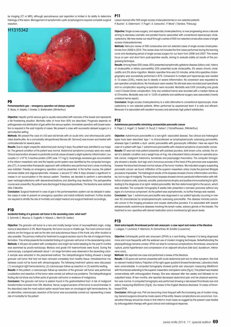
swiss knife 2014; 11: special edition 69
by imaging (CT or MRI), although percutaneous cyst aspiration is limited in its ability to determine histology of the lesion. Management of symptomatic cystic lymphangioma requires complete surgical resection.
1
Fig. 2 Macroscopic specimen.
P9Portomesenteric gas – emergency operation not always requiredR. Wyss, H. Gelpke, F. Grieder, S. Breitenstein (Winterthur)
Objective: Hepatic portal venous gas is usually associated with necrosis of the bowel and represents a life threatening situation. Mortality rates of more than 50% are described. Prognosis depends on pathogenesis and distribution of gas within the venous system. Immediate operation with bowel resec-tion is required in the vast majority of cases. We present a case with successful delayed surgery in a semi-elective setting.Methods: We present the case of a 62-year-old female with an acute intra- and retromesocolic perfo-rated diverticulitis. As a comorbidity retroperitoneal fibrosis (M. Osmond) was known and treated with corticosteroids for several years.Results: Due to slight unspecific abdominal pain during 5 days, the patient was admitted to our hospi-tal. The general condition of the patient was normal. Abdominal symptoms (cramps) were very weak. Clinical examination showed no peritonitis and lab values showed a slight systemic inflammation: Leu-cocytes 3.1 x10^9, C-reactive protein (CRP) was 117 mg/l). Surprisingly excessive gas accumulation in the inferior mesenteric vein and the hepatic portal system was identified by the computed tomogra-phy (CT). A conservative therapeutic approach with antibiotics was performed and a close monitoring was installed. Thereby an emergency operation could be prevented. In the further course, the patient remained stable and oligosymptomatic. However, a second CT after 8 days showed a significant in-crease in air accumulation in the venous system. Therefore, we decided to perform a semi-elective open rectosigmoidectomy with descendorectostomy and diverting loop ileostomy. The perioperative course was uneventful, the patient was discharged 9 days postoperatively. The Ileostomy was restored after 3 Months.Conclusion: Surgical treatment in case of gas in the portomesenteric system can be delayed in selec-tive cases in order to prevent an emergency operation and install antibiotic treatment. Further studies are required to stratify the risk of mortality and adapt medical and surgical treatment accordingly.
P10Incidental finding of a granular cell tumor in the ascending colon: what next?E. Schmidt, C. Maurus, S. Cogliatti, H. Nüesch, L. Marti (St. Gallen)
Objective: The granular cell tumor is a rare and mostly benign tumor of neuroepithelial origin, malig-nancy is described in 2-3%. Most frequently, the tumor occurs in middle age. The most common locali-sations are the tongue as well as the skin and subcutaneous tissue of the trunk; any other location is also possible. The primary method for treatment is surgical excision due to the risk of malignant trans-formation. This article presents the incidental finding of a granular cell tumor in the ascendening colon.Methods: A 49-year-old patient with constipation and bright red rectal bleeding for the past 6 months was examined by procto-rectoscopy. Mariscs and grade II-III haemorrhoids were found. During the colonoscopy, a polypoid yellowish about 1 cm large formation was observed in the ascending colon. A sample was extracted in the peacemeal-method. The histopathological finding showed a benign granular cell tumor that had not been removed completely from healthy tissue. Intraabdominal me-tastases could not be found with ultrasonography. The following treatment options were discussed: (1) oncological right hemicolectomy, (2) colonoscopic follow-up resection or (3) watchful waiting.Results: In this patient, a colonoscopic follow-up resection of the granular cell tumor was performed. Localisation and resection of the tumor were carried out without any problems. The histopathological finding did not show any further parts of granular cell tumor or evidence of malignancy.Conclusion: The granular cell tumor is seldom found in the gastrointestinal tract. A risk of malignant transformation is known from ORL literature. Hence, surgical excision of the tumor is recommended. In the described case the most radical option would have been an oncological right hemicolectomy. As an alternative, colonoscopic resection of the tumor was successfully carried out, representing a lower risk of morbidity for the patient.
P11Lesson learned after 500 single access cholecystectomies in non selected patientsP. Bucher1, S. Ostermann1, F. Pugin2, A. Carecchio1, P. Morel1 (1Genève, 2Fribourg)
Objective: Single access surgery, and especially cholecystectomy, is now progressing since a decade aiming to decrease cosmetic and parietal trauma associated with conventional laparoscopic chole-cystectomy. We here review our result through a series of 500 non selected consecutive cases of single access cholecystectomies.Methods: Retro-pro review of 500 consecutive and non selected cases of single access cholecystec-tomies from 2008 to 2014. This review does not included the first cases performed during the learning curve and developing period of single access surgery for our team from 2006 and 2007. This review focus on peri- and short term post-operative results, aiming to evaluate safety ad results of this pro-gressing technique.Results: Among these 500 cases, 65% presented symptomatic gallstone disease (biliary colic, history of cholecystitis or biliairy pancreatitis) 25% presented acute cholecystitis, 5% severe chronic chole-cystitis and 5% stone migration. Median operative time was 53 minutes, while intra-operative cholan-giography was successfully performed in 87%. Conversion to multiple port laparoscopy was needed in 13 cases (2.6%), mainly due to obesity or severe inflammation. No conversion was requested by peri-operative complications. No transfusion were needed. No bile leak were recorded post-operatively and no complication requiring re-operation were recorded. Morbidity was 0.6% (including only grade I and II Clavien-Dindo complication. Only one umbilical hernia was recorded with a median follow-up of 37months. Morbidity was null. In 13.6% of patients and additional surgery was associated (mainly umbilical hernia repair).Conclusion: Single access cholecystectomy is a valid alternative to conventional laparoscopic chole-cystectomy in non selected patients. When performed by experienced team it is safe and efficient. Moreover it is associated with excellent recovery and extremely high patient satisfaction.
P12Autoimmune pancreatitis mimicking unresectable pancreatic cancerK. Papp1, E. Angst1, S. Seidel1, R. Flury2, F. Hetzer1 (1Schaffhausen, 2Winterthur)
Objective: Autoimmune pancreatitis is a rare IgG4- associated disease. Two clinical and histological types have been described: type 1 is characzterized as lymphoplasmacytic sclerosing pancreatitis, whereas type 2 exhibits a duct- centric pancreatitis with granulocytic infiltration. Here we report the case of a patient with type 1 autoimmune pancreatitis with classical symptoms of pancreatic cancer.Results: 67-year-old male patient presented with painless jaundice, dark brown urine, acholia, the on-set of a diabetic condition and a weight loss of 4 kg. His antecedent history showed an operated pros-tate cancer, malignant melanoma, borreliosis and polymyalgia rheumatica. The computer tomogra-phy showed a double- duct sign and a tumorous process of the head of the pancreas was suspected, however the blood test showed normal values of the tumor markers. We indicated surgery suspecting pancreatic cancer: we found. Infiltration of the superior mesenteric artery made the planned Whipple procedure impossible. The histological results of the biopsies showed chronic inflammation and fibro-sis, but no sign of malignity. The secondary biopsies showed chronic periductal inflammation with infil-tration of plasma cells, sclerosis, venulitis, parenchymal atrophy and positive immunostaining for IgG4, leading to the diagnosis of an IgG4- related pancreatitis. We then determined IgG4 in the serum, which was elevated. The computer tomography 6 weeks later presented a narrower pancreas without any signs of a tumorous component. As the patient was asymptomatic, no further therapy was needed.Conclusion: Type 1 autoimmune pancreatitis was diagnosed in our patient typically occurring in men over 50 characterized by lymphoplasmacytic sclerosing pancreatitis. This disease mimicks pancre-atic cancer in the imaging procedure and causes obstructive jaundice. It is associated with several extrapancreatic autoimmune diseases involving the lymph nodes, salivary glands or the breasts. The treatment is non- operative with steroid medication and is monitored by IgG serum levels.
P13Giant extra-hepatic thrombosed portal vein aneurysm: a case report and review of the literatureI. Labgaa, Y. Lachenal, P. Allemann, N. Demartines, M. Schäfer (Lausanne)
Objective: Extrahepatic portal vein aneurysm (EPVA) is a rare finding. However it is being diagnosed more and more frequently with the widened use of modern imaging techniques. Notwithstanding the physiopathology remains unclear. EPVA can lead to numerous complications: thrombosis, aneurismal rupture, portal hypertension and compression of an adjacent structure (bile duct, duodenum, inferior vena cava).Methods: We reported one case and performed a review of the literature.Results: A 50-years-old woman presented with acute abdominal pain but no other symptom. She had no relevant medical history. Palpation of the right upper quadrant showed tenderness. Laboratory tests were unremarkable. A computed tomography showed portal vein aneurysm measuring 88x65mm with thrombosis extending to the superior mesenteric and splenic veins (Fig.A). The patient was treated conservatively with anticoagulation therapy. She was released after two weeks and followed on an outpatient basis. At two months, she reported decreased abdominal pain and her physical examina-tion was normal. A computed tomography was performed showing a decreased thrombosis size and extent, measuring 80x55mm (Fig.B). Our review of the English literature disclosed 14 cases of throm-bosed EPVA.Conclusion: Although rare, PVA are becoming more frequent with the increasing use of modern imag-ing. General surgeons should be made aware of this entity. Severe complications are uncommon. Con-servative therapy should be chose in first intent in most cases as suggest by the present case treated by anticoagulation therapy with good clinical and radiological response.

70 swiss knife 2014; 11: special edition
P14Surgical management of severe bevacizumab-induced ischemic pancolitisH. L. Chan, I. Rotas, F. Pugin, B. Egger (Fribourg)
Objective: Bevacizumab is a murine-anti-human monoclonal antibody directed against vascular en-dothelial growth factor-A, which disrupts endothelial cell survival mechanisms and the development of new tumour blood supply. Bevacizumab has been shown to prolong survival of patients with metastat-ic colorectal cancer when combined with chemotherapy. Serious adverse effects have been reported. We report here a case of bevacizumab-induced ischemic pancolitis and its surgical management.Methods: A 56-year-old male patient with a rectal cancer (cT2N2M0) underwent neoadjuvant chemo-radiotherapy followed by restorative low anterior resection with total mesorectal excision and protec-tive ileostomy. Following that adjuvant chemotherapy (FOLFOX ) was administered. In the follow-up the patient developed lung metastases and underwent chemotherapy with FOLFIRI and Bevacizumab (Avastin®). Due to progression of the disease, a second-line chemotherapy (Irinotecan, 5-Fluorour-acil) was added for 6 months. During this last treatment, the patient was admitted to our emergency department with abdominal pain and vomiting. Physical examination revealed a diffuse peritonitis and CT-scan showed free intraabdominal fluid but no free air. At emergency laparotomy a massive necrotiz-ing pancolitis was found. Total colonic resection with terminal ileostomy and an ultra-short Hartmann stump was performed.Results: Histopathology confirmed the diagnosis with multiple fibrous thrombi present in the submu-cosa and an extended transmural inflammation. These histological findings were consistent with a bevacizumab-induced ischemic colitis. Follow-up was completely uneventful and the patient was dis-charged home 13 days later.Conclusion: Side effects of bevacizumab are hypertension, proteinuria, arterial thrombosis, minor bleeding events and delayed wound healing. Furthermore, gastrointestinal perforation has been ob-served in up to 2%. However, extensive colonic ischemia seems to be an extremely rare complication. The mechanism is unclear and believed to be associated with the inhibition of vascular endothelium growth factor leading to thrombosis of smaller splanchnic vessels. In conclusion, bevacizumab-induced ischemic pancolitis is a rare but very serious complication and surgery needs to be applied before perforation occurs.
P15Carcinoid tumour of the kidney: a rare primary siteA.-R. Schärli, A. Zettl, M. von Flüe, B. Kern (Basel)
Objective: Carcinoid tumours are low-grade malignant neoplasms that can be found in almost every organ. They are common in the gastrointestinal and respiratory tract. An extremely rare primary mani-festation is the kidney.Methods: We report the case of a 59-year-old asymptomatic woman who was referred to our hospital for further diagnostic workup and treatment of an incidentally detected mass on the right kidney.Results: Positron emission tomography with computed tomography (PET/CT) and magnetic reso-nance imaging (MRI) revealed a mass in the right renal pelvis measuring approximately 7 cm x 5 cm x 6 cm. The tumour showed a 18F-FDG uptake with a SUVmax of 3.8. We performed a radical nephrectomy including the adrenal gland on the right side. Histopathological examination revealed a primary carcinoid tumour of the renal pelvis with one local lymph node metastasis. Immunohisto-chemistry was positive for chromogranin A, synaptophysin and CD56. Ki-67 showed a cell proliferation rate of 2-3%. The postoperative course was uneventful and the patient could be discharged 8 days after surgery. Postoperative Ga-68-DOTATOC-PET/CT was without pathological findings and provided a baseline for follow-up with regular imaging.
Conclusion: The described case shows a rare primary manifestation of a carcinoid tumour in the kid-ney. Nephrectomy is regarded as definitive treatment in localized disease.
P16Bilocal recurrence of a neuroendocrine carcinoma of the small intestine, a case reportS. Peiffer, R. Cathomas, P. Villiger (Chur)
Objective: With an incidence of 0,29/100.000 the malignant forms of neuroendocrine tumors (NET) of the small intestine represent a very rare disease. Recurrence of completely resected NET of the small in-testine occurs in xy% and depends on the proliferation rate (Ki-67). We report a recurrence in a 64-year-old female with NET of the proximal ileum after 4 years.Methods: A 64-year-old female with family history for colorectal and gastric carcinoma underwent regular screening by her GP. Because of asymptomatic elevation of CA 19-9 a CT-scan was carried out and revealed a tumor of the small intestine of 14 mm in diameter. The tumor was resected and histolog-ical workup classified it as a neuroendocrine tumor (pT4, N1, G1, Ki-67 < 1%). After 4 years of follow-up, CT-scan showed two suspicious lesions of the small intestine. Gallium DOTATOC-PET-CT verified tumor recurrence in both sites and segmental resection of the small intestine was performed again. Histological exam of the tumor showed the same histology of NET as before (pT2, N1, G1, Ki-67 < 1%).Results: Bilocal recurrence of NET of the small intestine with high differentiation and low proliferation rate is rather unlikely with no exact data for recurrence rate in current literature. Following the latest ENETS guidelines of 2009 follow-up should be carried out with regular CT-scan and measurement of chromogranin A according to TNM stage and Ki67 rate at diagnosis. For exact diagnosis and locali-zation somatostatin receptor imaging, either as octreotide scintigraphy or Gallium DOTATOC-PET-CT should be used. As DOTATOC-PET may have higher sensitivity than octreotide scintigraphy, according to latest results it should nowadays be favored. Therefore the two new detected tumors in 2013 could just have been missed by octreotide scintigraphy in 2009.Conclusion: Even in case of low proliferation index, follow-up for NET should be performed according to current guidelines in order not to miss rare but possible tumor recurrence. In case of changing diag-nostic matters reduced comparability has to be taken in concern.
P17Combined mini-invasive endoscopic-laparoscopic treatment of an acute post-traumatic diaphrag-matic herniaF. Villa, M. Marengo, I. Marsteller, A. Guerra (Bellinzona)
Objective: To demonstrate that a combined mini-invasive endoscopic-laparoscopic treatment may be a safe and effective alternative to reduce a complicated acute diaphragmatic hernia.Methods: We present the case of a 26-year-old man with sudden onset of heavy epigastric pain, vomit-ing and aphagia. One year before, he has had a blunt throracic trauma with left-sided multiple rib fractures and left-sided hemopneumothorax. An acute complicated diaphragmatic hernia was sus-pected. The working hypothesis was confirmed by contrast-enhanced CT scan, gastrografin swallow and finally by exploratory laparoscopy in combination with intraoperative upper endoscopy. Of note, that no hernial defect had been reported on previous CT scans.Results: The patient underwent exploration in our emergency operating room. Initially, an exclusive endoscopic approach to reduce the hernia failed. The parallel endoscopic-laparoscopic approach pro-vided evaluation of the gastric mucosa (hypoxic pattern) and endoscopic decompression with further local guidance of the surgeon, thus allowing a speedy reduction of the herniation through the defect. The reduction of the hernia and direct closure of the diaphragmatic defect was performed with multiple non-absorbable suture. The defect resulted 4 cm in diameter, and was located at 3 cm distance in left-lateral position to the esophageal hiatus. No perioperative complication occured.Conclusion: Best surgical corrective procedure is controversial in treating symptomatic diaphragmatic hernias. However, there is evidence that mini-invasive techniques should be preferred even in the case of major complications, in order to avoid pain, blood loss and long-term hospital stay. In this report, we describe a successful combined mini-invasive endoscopic-laparoscopic treatment of an acute diaphragmatic hernia with gastric strangulation and ischemia, one year after blunt thoracic trauma. This report supports further research on combined mini-invasive approaches for treatment of acute diaphragmatic hernias.

swiss knife 2014; 11: special edition 71
P18Rectal perforation due to enterocolic lymphocytic phlebitisF. Ramadani, A. Kühn, A. Meyer, B. Egger (Fribourg)
Objective: Enterocolic lymphocytic phlebitis is a recently described new entity. It is extremely rare and characterized by phlebitis of the bowel wall without any arterial involvement or systemic vasculitis.Methods: A 90 year-old woman was admitted with severe abdominal pain and with signs of general peritonitis. A CT scan revealed massive free intra-abdominal air and a surgical approach was per-formed after discussion with the patient and her family. At explorative laparoscopy rectal perforation was found and due to the peritonitis the intervention was converted to a laparotomy. The perforation of about 15mm in the upper rectum presented with a marked peri-lesional induration as it is seen in per-forated rectal ulcer disease. For security reason an oncological anterior rectal resection and Hartmann procedure was performed. However, histology did not demonstrate any malignancy but an abscess around the perforated ulcer together with multiple other small colonic ulcers and a general vasculitis of the medium sized bowel veins. In addition a moderate diverticulosis was described. A follow-up panendoscopy was performed some days later, showing multiples specific ulcers in all investigated parts of the bowel and the presence of lymphocytic phlebitis at histology of the tissue gathered by biopsies. A treatment with low dose steroids was introduced in order to treat the ulcers and calm down the important oedematous lesions. Further follow-up was then completely uneventful and the patient dismissed home.Conclusion: Enterocolic lymphocytic phlebitis is an extremely rare entity. It most commonly manifests with signs of an acute abdomen. Patients may present with an abdominal mass, mimicking, clini-cally and radiologically, a tumor of the intestinal tract. This fact has been attributed to the concomitant oedema and inflammation of the disease which is associated to extended venous thrombosis of the medium sized bowel veins. Whereas etiology and pathogenesis is still unexplained, surgical resection is the treatment of choice because usually curative. In our case, the pathology turned out to be present all along the whole bowel investigated. Since curative resection of all these lesions is highly unrealistic, future complications cannot be excluded. However, publication of this case may help to better under-stand and also treat this rare clinical manifestation.
P19Sinistroposition of the gallbladder – a case report of an incidental laparoscopic findingS. Tomaszek, C. Frei-Lanter, H. Gelpke, S. Breitenstein (Winterthur)
Objective: Transposition of the gallbladder to the opposite side of the falciforme ligament has a preva-lence of 0.3% in patients undergoing cholecystectomy for symptomatic cholecystolithiasis. This ana-tomical variation is rarely diagnosed preoperatively. In sinistroposition the cystic artery usually crosses in front of the common bile duct from right to left. The cystic duct may converge on the left or right side of the common hepatic duct. This anatomic abnormality increases the risk for intraoperative technical difficulties and incidental injury of the hepatic vessels or the main bile duct.Methods: We demonstrate a case report of a 53-year-old man who was admitted to our department due to acute cholecystitis. Sinistropostioning of the gallbladder was detected during laparoscopic cholecystectomy.Results: The patient presented with typical clinical, laboratory and sonographic findings of an acute cholecystitis. Sonographically no anatomic abnormality was identified. A standard surgical approach for laparoscopic cholecystectomy with 4-trocars was performed. Intraoperatively the modified position of the gallbladder to the left of the falciforme ligament was realized. Technical difficulties in skeletoniz-ing the structures in Calot’s triangle due to inflamed structures and anatomical variation consumed additional operating time. The cystic artery was identified anterior of the cystic duct. After ligation of the artery with metallic clips, a intraoperative cholangiography was conducted to verify bile duct anatomy. Thereafter the cystic duct was dissected between Hem-o-Lok-Clips. Finally the gallbladder was evacu-ated to an endobag via the perimbilical incision. The duration of the operation was 152 minutes. The postoperative course was uneventful and the patient was discharged 2 days postoperatively.Conclusion: Transposition of the gallbladder to the left side of the falciforme ligament is associated with variation to the cystic artery and the bile ducts. Intraoperative cholangiography clarifies bile duct anatomy and provides a safe laparoscopic cholecystectomy.
P20Acute hemoperitoneum after tubal rupture in a patient with synchronous intrauterine and ectopic pregnancy: A rare and life-threatening conditionC. R. Scarpa, T. Berney, O. Julen, P. Morel (Geneva)
Objective: Heterotopic pregnancy is defined as the coexistence of normal intrauterine and ectopic pregnancies. It is a rare event in natural conception cycles, occurring in around 1: 30,000 pregnancies, but is much more frequent in the case of in-vitro fertilization pregnancy. (1:500) [1,3,4]. This rare event can be fatal for both mother and fetus, and can sometimes be challenging to diagnose. This case re-port illustrates the fact that, even in a setting of natural intrauterine pregnancy, urgent explorative lapar-oscopy should be considered in a patient with acute abdominal pain and free intra-abdominal fluid. Methods: A report of a single case of a disease, usually with an unexpected presentation. Results: A 32-year-old patient, with an 8-week documented intrauterine pregnancy, presented with acute abdominal pain, localized peritonitis in the right iliac fossa and without inflammatory signs or hemodynamic instability. Gynaecological examination was reported as normal. Ultrasonography revealed an intrauterine evolutive pregnancy, moderate free intra-abdominal fluid with an image in the right iliac fossa compatible with appendicitis therefore the patient was transferred to the surgi-cal emergency room (ER). Since supplementary radiological exams could not be performed, it was decided, with the patient’s consent, to perform an emergency exploratory laparoscopy. During surgery, a hemoperitoneum originating from a heterotopic pregnancy with right tubal rupture was identified. Consequently, the patient underwent a rightsalpingectomy.
She was discharged after 48 hours without any postoperative complications and with progressive evolution of the intrauterine pregnancy.Conclusion: The heterotopic pregnancy occurs in very rare cases during a natural pregnancy. The di-agnostic process is difficult, but in case of acute abdominal pain with evidence of free intra-abdominal fluid on the ultrasonography study, a diagnosis of concomitant heterotopic pregnancy should be sus-pected. In such cases, and when the diagnosis in unclear, explorative laparoscopy may contribute to a rapid diagnosis and reduce maternal and foetal morbidity and mortality, i.e. establishing and treating the condition while preserving foetal viability.
FIGURES LEGEND
Figure 1
Ultrasonography, showing an intrauterine 8-‐week pregnancy
Figure 2
Laparoscopic view, hemoperitoneum in right flank and hypochondrium
Acute hemoperitoneum after tubal rupture in a patient with synchronous intrauterine and ectopic pregnancy: A rare and life-threatening condition
Ultrasonography, showing an intrauterine 8- week pregnancy
Laparoscopic view, hemoperitoneum in right flank and hypochondrium
Laparoscopic view, hemorragic right ampullary pregnancy
Laparoscopic view of Fitz- ‐Hugh- ‐Curtis adherences

P21Das seltene Insulinom mit verzögerter Diagnose: ein Case ReportS. Oettli, A. Häfliger, R. Rosenberg, S. Stadlmann, T. Kocher (Baden)
Objective: Neuroendokrine Tumoren (NET) gehören zu den seltenen Neoplasien des Pankreas mit ein-er Inzidenz von 0.4-1.5 / J /100’000. Die meisten NET sind hochdifferenziert und zu 50-60% funktionell aktiv. Zu den häufigen hormonaktiven NET zählt das Insulinom, welches von den B-Zellen der Langer-hansinseln ausgeht und sich mit der typischen Symptomatik der Whipple-Trias mit Hypoglykämien, damit assoziierten neurologischen und autonomen Symptomen sowie rascher Beschwerderegre-dienz auf Glucosegabe präsentiert.Methods: Wir berichten von einem 55-jährigen Patienten mit einer geistigen Retardierung nach post-partaler Hirnblutung. Nebendiagnostisch besteht eine chronischen Epilepsie mit Grand-Mal-Anfällen und seit 2012 vorhandenen Absencen. Die endokrinologischen Abklärungen ergaben einen patholo-gischen Fastentest mit rezidivierenden Hypoglykämien und erhöhtem Proinsulin. Anamnestisch be-stätigte sich die Klinik der Whipple-Trias. Im CT fand sich ein kontrastmittelaufnehmender Tumor von 9x11mm im ventralen Pankreaskorpus. Der auf ein Insulinom verdächtige Tumor konnte operativ prob-lemlos enukleiert werden. Die Schnellschnittuntersuchung zeigte eine endokrine Neoplasie.Results: Die histologische Aufarbeitung ergab einen NET des Pankreas (pT2, pNx, G2, L0, V0, pN0, cM0). Intraoperativ kam es bereits nach wenigen Minuten zu einem Blutzuckeranstieg von 3.5 auf 3.9mmol/l. Die postoperativen Glucosewerte lagen unter diätetischen Massnahmen im Normbereich, es traten keine Hypogyklämien auf. Der weitere Hospitalisationsverlauf gestaltete sich komplikation-slos.Conclusion: Die chirurgische Therapie ist der einzige kurative Therapieansatz beim Insulinom. Die Heilungsrate nach vollständiger Resektion liegt bei nahezu 100%. Die bereits seit 2012 bestehenden Abscencen müssen rückblickend am ehesten im Rahmen einer Hypoglykämie aufgrund des Insuli-noms interpretiert werden. Somit wurde die Diagnose wahrscheinlich aufgrund der geistigen Retar-dierung und chronischen Epilepsie verzögert gestellt.
P22An unusual internal hernia: right colon herniation through the foramen of WinslowE. Pezzetta, P. Sikiminywa, F. Barros, M. Trojansky, D. Roulet (Montreux)
Objective: Internal hernia constitute a wide spectrum of clinical and morphological pictures that may de difficult to detect and recognize preoperatively in front of a situation of gastro-intestinal tract obstruc-tion. Intestinal herniation through the epiploic (Winslow) foramen is a rare clinical event in this respect and only few cases have been reported therein.Methods: A patient presenting with herniation of the right colon through the foramen of Winslow is described with particular emphasis on imaging and intra-operative management.Results: A 69-year-old patient presented to our emergency room with progressive dull abdominal pain and distension without nausea, vomiting or bowel movements alteration. Physical examination showed pain with moderate guarding in the right upper and lower quadrants. A plain abdominal X-ray and a CT Scan were performed, radiological findings suggested the diagnosis of an internal hernia around the epiploic foramen and concerning the right colon with important caecal distention. Surgical exploration was then decided and an open approach was advocated; at laparotomy we confirmed an internal herniation of the caecum and ascending colon through the foramen of Winslow, multiple patchy areas of caecal necrosis were observed, a formal right hemi-colectomy was therefore per-formed. The post operative period was uneventful.Conclusion: Bowel herniation through the foramen of Winslow is a serious an uncommon type of inter-nal hernia that warrants surgical confirmation and treatment with prompt exploration and resection in case of suspected or overt bowel necrosis.
P23Necrotising enteritis and septic shock: a case reportA. Fulco, A. Cusimano, A. Donadini (Lugano-Besso)
Objective: Necrotising enteritis has been described in literature as a clinicopathological entity of an acute abdominal disorder, characterized by patchy or segmental necrosis of varying extent of the wall of jejunum, ileum or colon. It can be associated with gangrene, toxaemia and eventually death in a relevant number of cases.Methods: This report describes the case of a patient with medical history of rectal cancer which was treated with abdominal perineal resection in December 2011. He was admitted to our hospital with a three day history of abdominal pain associated with nausea after eating raw fish. Abdominal x-rays and computer tomography revealed dilated a small intestine loop with a caliber change in proximity of the terminal colostomy, therefore a naso-gastric tube was placed with clinical improvement. However, during the night, his general condition worsened with rapidly increasing shock parameters. A surgi-cal treatment was decided. At first the patient underwent through a diagnostic laparotomy showing a patchy necrosis of the colon and small bowels wall without any mechanical obstruction. After im-provement in intensive care unit he underwent through a second laparotomy with an ileocaecal resec-tion (150cm of small bowel) and cholecystectomy because of segmental necrosis. We performed an ileocolic anastomosis during the third laparotomy after complete resuscitation. Microbiological laboratories were always negative. After 19 days in intensive care unit he was admitted to the ward where total parenteral nutrition was progressively decreased and the oral alimentation was gradually reintroduced.Results: Review of literature cases of necrotising enteritis showed a high mortality rate; delay in diagno-sis and treatment represents a threat to the patient’s life. Necrotic intestinal segments must be removed as soon as possible, relaparotomy may be necessary, especially in intra-abdominal infections caused by Clostridium Perfrigens.Conclusion: Surgery might be a life-saving procedure if effectuated without delay.
P24Vertebral discitis after laparoscopic resection rectopexy: a rare differential diagnosisP. Probst, N. Obrist, S. Breitenstein (Winterthur)
Objective: Vertebral discitis usually arises from haematogenous spread of pathogens to the discs and bones. During lumbar instrumentation pathogens can reach the bone directly and vertebral discitis is a well-known complication in neurosurgery. Vertebral discitis can rarely occur as a complication after laparoscopic operations with fixating sutures on the promontory.Methods: We report the case of an 81 year-old woman, who underwent a laparoscopic resection rectopexy because of a third-degree rectal prolapse. Weeks after the operation the patient developed lower back pain with radiation to both legs not responding to symptomatic therapy. At the same time she developed Pseudomonas sepsis without an obvious source of infection and was treated with ap-propriate antibiotics for 14 days.Results: After initial improvement her pain worsened and she experienced one-sided, great toe exten-sor paresis. Two months later an MRI of the lumbar spine showed vertebral osteomyelitis and discitis. A CT-guided needle biopsy was positive for Pseudomonas aeruginosa. The patient was treated with high dose ceftazidime for 20 days followed by oral ciprofloxacin for three months.Conclusion: A fixation on the promontory may be sufficiently traumatic to the spine to pave the way for subsequent infection. Signs, symptoms and laboratory findings of vertebral discitis are not specific. A high index of suspicion should be raised in patients with persistent, severe back pain and signs of systemic inflammation. Anamnesis, imageing and an adequate specimen from the affected area for microbiological analysis are crucial for timely diagnosis and appropriate management involving tar-geted and prolonged antimicrobial therapy to avoid unnecessary postoperative morbidity by persistent neurological deficits because of this rare complication.
P25Immunohistochemical verification of clinically assumed lynch syndrome among patients with color-ectal and uterine cancerC. Strub-Antonucci1, M. Nowak2, J. Diebold1, J. Metzger1, M. Sykora1 (1Lucerne, 2Schlieren)
Objective: Lynch syndrome (LS) is the most common type of hereditary colorectal disease. It counts for 2-4% of all Colorectal Carcinomas (CRC). Further, the incidence of LS in patients with uterine cancer is still undervalued. Until the start of our pilot study the Amsterdam-II and Bethesda criteria, which are used for the clinical diagnosis of LS, were used sporadically in our institution and no immunohisto-chemical examination was performed. We wanted to verify these two clinical tests by Immunohisto-chemy (IHC) to improve the detection rate of patients with LS in Central Switzerland, as it has been shown that 10 to 25% of the population remains undiagnosed.Methods: The ethical committee of Lucerne approved this study. All patients who were planned for an operation because of a diagnosed or suspected CRC or uterine carcinoma and signed the patient consent form were included in this study. Revised Bethesda and Amsterdam-II criteria were assessed and postoperatively an IHC was conducted. Patients with high risk for Lynch Syndrome were sent to a geneticist to be assessed further.Results: We examined 34 patients, 2 of them with uterine cancer, the other patients with a colorectal carcinoma. There were 2 patients lacking the expression of MLH1, PMS2, MSH2 or/and MSH6 (mu-tated MMR genes leading to a microsatellite instability responsible for LS) in the immunohistology. Both had negative Amsterdam-II and revised Bethesda criteria. No patient fulfilled the Amsterdam-II-criteria and 5 patients fulfilled the Bethesda criteria, but none of them showed a positive immunohistochem-istry.Conclusion: With this study we show that Lynch Syndrome should not only be considered in patients with CRC, but also in those with uterine cancer, as one of our two gynecological patients had a positive IHC. We also found both patients with a positive IHC neither fulfilling the revised Bethesda nor the Am-sterdam-II-criteria. This is why we decided to continue to ask for an IHC on all our patients with a CRC and uterine cancer. We also found the revised Bethesda-criteria to be little specific as 5 patients fulfilled it, but didn’t have a positive IHC. We propose that Amsterdam-II and revised Bethesda criteria may be of use for finding families with a high risk for hereditary CRC, but are not useful for families with Lynch Syndrome. Having a small sample size in this study we propose further examination of our results.
P26Single port laparoscopic combined transvaginal colorectal surgery: experience from a single centreS. Bischofberger, L. Traine, N. Kalak, W. Brunner (Rorschach)
Objective: The reduction of interventional trauma is considered a main goal in modern surgery. Innova-tive techniques, such as single port laparoscopy, have been developed to further minimize surgical access trauma. Using a transvaginal access, suprapubic or umbilical incision enlargement for speci-men retrieval, can be avoided.Methods: From March 2011 to December 2013 a total of 104 single-port left hemicolectomies, sigmoid and rectal resection were performed (56 m, 48 f). In 21 females a combined transvaginal access and specimen retrieval was performed (Mean age 58y (36-84), mean BMI was 25.5). 57% of females had undergone previous abdominal surgery incl. hysterectomy in 5. In our study, 17 patients underwent surgery for diverticulitis, 3 for malignant intestinal neoplasia and one for benigne rectal polyp. 2 left hemicolectomies, 2 rectal resections and 17 sigmoid resections have been performed. Data was col-lected in a prospective single centre-database for single port-procedures.Results: Transvaginal specimen retrieval was possible in all 21 females. In 6 cases one additional tro-car was used. No conversion to open surgery was necessary. Severe complication rate (Clavien-Dindo IIIb or higher) was 9.6%, one reoperated by laparoscopic approach, in one case open surgery was necessary. No vaginal complications or wound infections occurred. Mean discharge was at day 6 (no complications). Mean fascia incision length was 2.6cm (2.1-3.6)Conclusion: Single port-access is an innovative and still evolving way towards further reduction of in-

swiss knife 2014; 11: special edition 73
terventional trauma in colorectal surgery. The technique is safe and feasible. Transvaginal specimen retrieval offers further trauma reduction to the abdominal wall and potentially reduces incision related complications as wound infection and hernia.
P27Early combined parenteral and enteral nutrition – a new strategy to feed patients after pancreati-coduodenectomyP. Probst, N. Obrist, H. A. Haller, R. Imoberdorf, M. Rühlin, H. Gelpke, S. Breitenstein (Winterthur)
Objective: Existing guidelines for nutritional support after pancreaticoduodenectomy (PD) are contro-versial. Whereas American guidelines favour oral intake only, the society of “Enhanced Recovery After Surgery” (ERAS) favours early enteral nutrition (EN). There have been some novel studies which have provided evidence that supplementation of parenteral nutrition (PN) in combination with EN improves outcome. The aim of this study was to assess feasibility and outcome of Early Combined Parenteral and Enteral Nutrition (ECPEN) after PD.Methods: We retrospectively reviewed 79 consecutive cases involving PD procedures performed be-tween 2003 and 2012. Enteral and parenteral nutritional support was standardized and started im-mediately after surgery. EN was increased 40 ml/h over 24 hours, while PN was supplemented based on an energy target of 25 kcal/kg of ideal bodyweight per day. The primary endpoint was the caloric intake of patients per day and as total amount during the entire hospital stay in relation to the individual energy target (caloric balance). Secondary endpoints were mortality, morbidity, the influence of feed-ing routes (nasojejunal vs. jejunostomy) and preoperative immunonutrition.Results: 79 patients supported with ECPEN after PD were analysed. Overall in 1324 out of 1853 days of hospitalisation (71%) the energy target was achieved. The total caloric deficit in relation to the tar-geted total amount of calories during hospitalisation was 754 kcal (range: 0-6350 kcal). The catheter jejunostomy was associated with a significantly higher rate of days with sufficient caloric intake as compared to the nasojeunal catheter (75% vs 59%, p= 0.01). Mortality was 5% (4 out of 79 Patients) while major complication (Dindo-Clavien lll-V) occurred in 24% (19 out of 79). The rate of postoperative pancreatic fistulas was 15.2% (Grade A: 2, B: 7, C: 3). Major complications were associated with a sig-nificant higher deficit of postoperative total caloric intake (30kcal/kg vs. 8kcal/kg; p= 0.04). However, preoperative immunonutrition did not affect outcome at any level.Conclusion: This is the first european study of Early Combined Parenteral and Enteral nutrition after PD. ECPEN provides a comprehensive coverage of the nutritional requirements during the postoperative phase. Sufficient caloric intake is associated with a lower rate of major complications.
P28Real-time near-infrared fluorescent cholangiography could shorten operative time during robotic single-site cholecystectomyN. Buchs, M. Jung, F. Pugin, D. Azagury, F. Volonté, M. Hagen, P. Morel (Geneva)
Objective: With the introduction of a new platform, robotic single-site cholecystectomy (RSSC) has been reported as feasible and safe for selected cases. In parallel, the development of real-time near-infrared fluorescent cholangiography using indocyanine green (ICG) has been seen as a help during the dissection, even if the data is still preliminary. The study aim is to report our experience with ICG RSSC and compare the outcomes to standard RSSC.Methods: From february 2011 to december 2011, 44 selected patients underwent RSSC for sympto-matic cholelithiasis. Among them, 23 (52.3%) were included in an experimental protocol using the ICG, and the remainder (47.7%) underwent standard RSSC. There was no randomization. The end-points were the peri-operative outcomes. This is a prospective study, approved by our local ethics com-mittee.Results: There were no differences in terms of patients’ characteristics, except that there were more male patients in the ICG group (47.8% vs. 9.5%; p=0.008). Regarding the surgical data, the overall operative time was shorter for the ICG group, especially for patients with a Body Mass Index (BMI) ≤ 25 (minus 24 minutes), but without reaching statistical significance (p=0.06). For BMI >25, no differences were observed. Otherwise, there were no differences in terms of conversion, complications or length of stay between both groups.Conclusion: A RSSC with a real-time near-infrared fluorescent cholangiography can be performed safely. In addition, for selected patients with a low BMI, ICG could shorten the operative time during RSSC. Larger studies are still required before drawing definitive conclusions.
P29Results and postoperative quality of life in patients greater than 80 years old after colorectal surgeryD. Schraffl, R. Joos, C. Hänsli, P. Villiger (Chur)
Objective: There are several challenges in surgical treatment of colorectal diseases in patients over 80. Our goal was to determine outcomes after colorectal surgery with postoperative quality of life (evalu-ated according to SF-36) and mortality.Methods: This study includes postoperative colorectal surgery patients over 80 years after between January 1, 2010 and December 31, 2013. Data was taken from our hospital database, general prac-titioner and patient interviews. We included curative and palliative as well as elective and emergency interventions.Results: Eighty-four patients (39 male, 45 female, age between 80 and 99 years, mean age 86,9 +/- 4,6 years) were studied. Median follow up was 17 months. Four (4,7%) patients were lost to follow up. The overall mortality rate was 32,1%. The 30-day mortality rate of all patients was 11,9%. Nine of 10 patients that died within the first 30 days had an emergency surgery. Based on SF-36 results, most of surviving patients had an improvement in postoperative quality of life. Patients without benefit most often claimed aggravation of pre-existing defecation problems. The correlation between ASA score and
functional renal impairment mortality was highest in first 30 days.Conclusion: Operation in patients over 80 years is safe. Emergency surgery has a much higher mortal-ity rate than elective surgery. Risk factors include a high ASA score and functional renal impairment. Postoperative survivors demonstrated superior SF-36 scores and improved quality of life.
P30Mesenterico-portal system septic vein thrombosis complicating acute appendicitisE. Pezzetta, M. Trojansky, F. Barros, P. Sikiminywa, O. Martinet (Montreux)
Objective: Pylephlebitis or infective thrombosis of the main portal vein or its tributaries is a serious, uncommon and often clinically unrecognized condition with significant morbidity and still mortality, that may develop after various intra-abdominal infections including acute appendicitis. In this situation the diagnosis can be difficult and delayed because of aspecific symptoms such as fever and mild abdominal pain. Methods: We wish to present and illustrate two consecutive cases presenting with thrombophlebitis of the portal system occurring in the setting of acute appendicitis.Results: The first patient, a 45 years old female, presented 10 days after an uncomplicated laparo-scopic appendectomy with bouts of high fever (max 39.6°C) without associated digestive or abdomi-nal symptoms. A Ct Scan demonstrated a thrombosis of the proximal superior mesenteric vein without portal involvement and a small right iliac fossa collection. The second patient came to our attention 12 days after a laparoscopic appendectomy and excision of an a ovarian cyst with high fever ( 39.9°C) and dull right upper quadrant abdominal pain; on Ct Scan we also discovered a thrombosis of the superior mesenteric vein extending to the proximal portal vein and splenic vein. In both cases pro-longed antibiotherapy and oral anticoagulation were successfully undertaken with rapid recovery ; a mesenterico-portal Doppler ultrasound was proposed to the 2nd patient in order to confirm definitive portal patency.Conclusion: Even if extremely rare after inroduction of antibiotic prophylaxis / treatment in acute appen-dicitis a septic thrombophlebitis of the main portal vein or one of the branches must be kept in mind as a possible complication in patients presenting with high fever and abdominal pain after appendectomy / appendicitis. Non operative approach with prompt and prolonged (4-6 w) antibiotic therapy associ-ated with systemic anticoagulation is the mainstay of treatment of this serious complication. Follow up with Doppler ultrasound up is recommended in order to evaluate patency and flow of the portal system.
P31Combined surgical and endoscopic management after delayed diagnosis of Boerhaave’s syndromeA. Miftaroski, A. Stanescu, A. Meyer, F. Pugin, B. Egger (Fribourg)
Objective: Boerhaave’s syndrome or spontaneous esophageal rupture is a rare but potentially lethal disease. The transmural perforation is mostly provoked by violent vomiting with high intraesophageal pressure. The disease is associated with a high mortality (20% to 50%) especially when there is a treatment delay lasting longer than 24 hours.Methods: We are presenting the case of a 56-years old male patient admitted in a peripheric hospital after an episode of violent vomiting followed by chest pain and dyspnea. Chest x-ray performed in the ER was not suspicious. Upon clinical deterioration a CT scan was performed the next morning with the finding of a distal esophageal rupture and left pleura effusion. After immediate thoracic drainage the patient was then transferred to our hospital 19 hours after the initial event. Emergency laparotomy with anterior phrenotomy, primary esophageal repair, Dor’s fundoplication and intraesophageal stenting by endoscopy was performed. Furthermore a lavage of the left thoracic cavitiy was done through the pleura tear in the mediastinum. The post-operative period was then marked by the suspicion of pleural empyema on day 3 p.i. (post intervention) with placement of two CT-guided drainages. Afterwards an uneventful follow-up with oral re-feeding on day 5 p.i was performed. The esophageal stent was evacu-ated on day 14 p.i. and the patient discharged home the day after. The first pleural drain was removed in the outpatient clinics on day 20 and the second on day 33 p.i.Results: There is no consensus in the literature concerning the optimal treatment of esophageal leaks. However, Boerhaave’s syndrome has to be distinguished from little iatrogenic lesions, which may be successfully treated conservatively (endoscopically). The 3-4cm big Boerhaave’s tear typically occurs in the left posterolateral aspect of the distal esophagus and is often accompanied by a synchronous rupture of the left pleura with the additional risk of pleural empyema. In our opinion there is no place for a conservative treatment in such situations and surgical exploration with repair and drainage is mandatory.Conclusion: According to the literature delayed surgical treatment lasting longer than 24h increases mortality up to 50%. In such cases a combined endoscopic (stent) and surgical approach may save lives and therefore be the treatment of choice.
P32A case of spontaneous right colon volvulus with herniation through the foramen of winslowS. Epp, J. Chmielewski, C. Apestegui, S. Lionetto (Wil)
Objective: To present an unusual and interesting case of right colon volvulus with subsequent internal hernia.Methods: Spontaneous internal hernias are rare, representing between 0.5-8% of the intestinal oc-clussions. Left paraduodenal hernias are the most common type (40%). Internal hernias through the Foramen of Winslow are a subtype of internal hernias, they represent 8% of internal hernias, and only 0.1% of all abdominal hernias. We present an spontaneous case of right colon volvulus with subse-quent herniation through the foramen of Winslow in a young female patient.Results: A 36-years old woman was admitted in our emergency department with acute abdominal pain, nausea and vomiting. At clinical examination, she presented generalized pain, with slightly
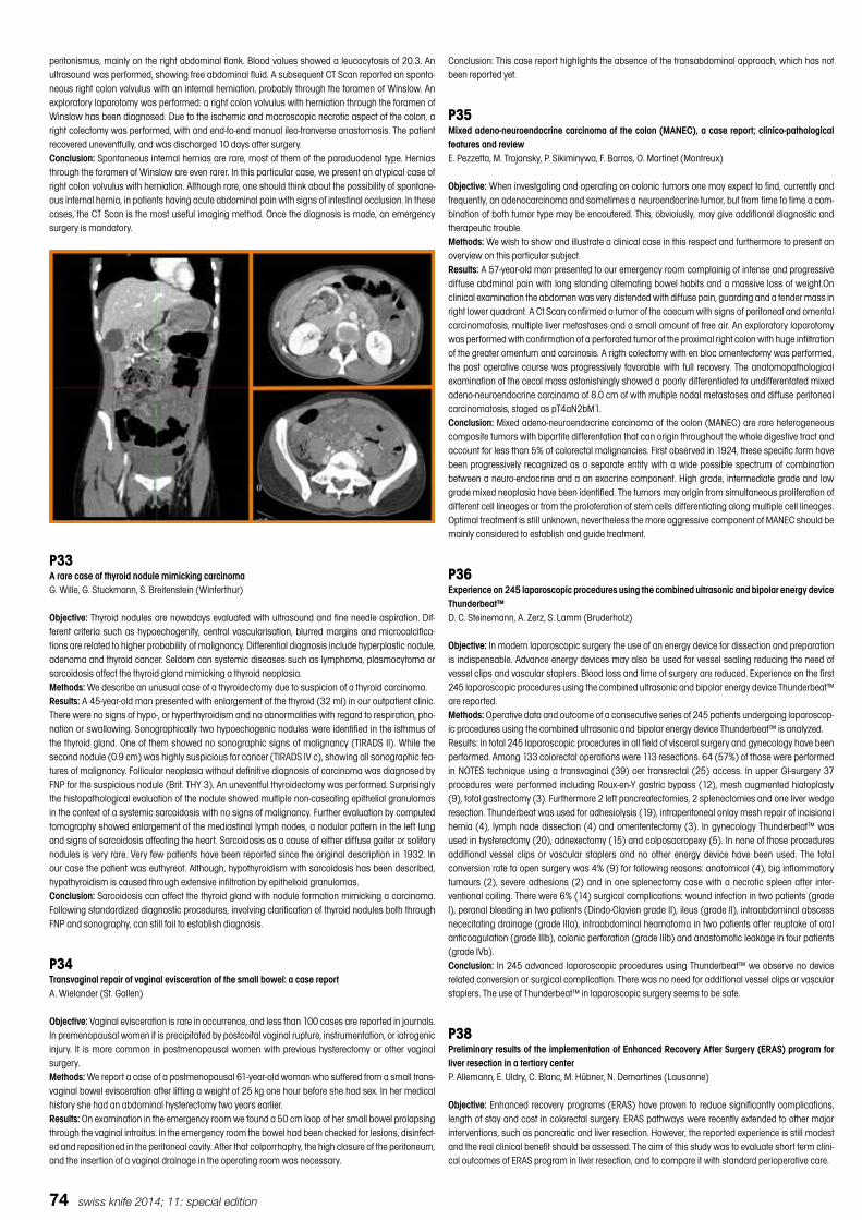
74 swiss knife 2014; 11: special edition
peritonismus, mainly on the right abdominal flank. Blood values showed a leucocytosis of 20.3. An ultrasound was performed, showing free abdominal fluid. A subsequent CT Scan reported an sponta-neous right colon volvulus with an internal herniation, probably through the foramen of Winslow. An exploratory laparotomy was performed: a right colon volvulus with herniation through the foramen of Winslow has been diagnosed. Due to the ischemic and macroscopic necrotic aspect of the colon, a right colectomy was performed, with and end-to-end manual ileo-tranverse anastomosis. The patient recovered uneventfully, and was discharged 10 days after surgery.Conclusion: Spontaneous internal hernias are rare, most of them of the paraduodenal type. Hernias through the foramen of Winslow are even rarer. In this particular case, we present an atypical case of right colon volvulus with herniation. Although rare, one should think about the possibility of spontane-ous internal hernia, in patients having acute abdominal pain with signs of intestinal occlusion. In these cases, the CT Scan is the most useful imaging method. Once the diagnosis is made, an emergency surgery is mandatory.
P33A rare case of thyroid nodule mimicking carcinomaG. Wille, G. Stuckmann, S. Breitenstein (Winterthur)
Objective: Thyroid nodules are nowadays evaluated with ultrasound and fine needle aspiration. Dif-ferent criteria such as hypoechogenity, central vascularisation, blurred margins and microcalcifica-tions are related to higher probability of malignancy. Differential diagnosis include hyperplastic nodule, adenoma and thyroid cancer. Seldom can systemic diseases such as lymphoma, plasmocytoma or sarcoidosis affect the thyroid gland mimicking a thyroid neoplasia.Methods: We describe an unusual case of a thyroidectomy due to suspicion of a thyroid carcinoma. Results: A 45-year-old man presented with enlargement of the thyroid (32 ml) in our outpatient clinic. There were no signs of hypo-, or hyperthyroidism and no abnormalities with regard to respiration, pho-nation or swallowing. Sonographically two hypoechogenic nodules were identified in the isthmus of the thyroid gland. One of them showed no sonographic signs of malignancy (TIRADS II). While the second nodule (0.9 cm) was highly suspicious for cancer (TIRADS IV c), showing all sonographic fea-tures of malignancy. Follicular neoplasia without definitive diagnosis of carcinoma was diagnosed by FNP for the suspicious nodule (Brit. THY 3). An uneventful thyroidectomy was performed. Surprisingly the histopathological evaluation of the nodule showed multiple non-caseating epithelial granulomas in the context of a systemic sarcoidosis with no signs of malignancy. Further evaluation by computed tomography showed enlargement of the mediastinal lymph nodes, a nodular pattern in the left lung and signs of sarcoidosis affecting the heart. Sarcoidosis as a cause of either diffuse goiter or solitary nodules is very rare. Very few patients have been reported since the original description in 1932. In our case the patient was euthyreot. Although, hypothyroidism with sarcoidosis has been described, hypothyroidism is caused through extensive infiltration by epithelioid granulomas.Conclusion: Sarcoidosis can affect the thyroid gland with nodule formation mimicking a carcinoma. Following standardized diagnostic procedures, involving clarification of thyroid nodules both through FNP and sonography, can still fail to establish diagnosis.
P34Transvaginal repair of vaginal evisceration of the small bowel: a case reportA. Wielander (St. Gallen)
Objective: Vaginal evisceration is rare in occurrence, and less than 100 cases are reported in journals. In premenopausal women it is precipitated by postcoital vaginal rupture, instrumentation, or iatrogenic injury. It is more common in postmenopausal women with previous hysterectomy or other vaginal surgery.Methods: We report a case of a postmenopausal 61-year-old woman who suffered from a small trans-vaginal bowel evisceration after lifting a weight of 25 kg one hour before she had sex. In her medical history she had an abdominal hysterectomy two years earlier.Results: On examination in the emergency room we found a 50 cm loop of her small bowel prolapsing through the vaginal introitus. In the emergency room the bowel had been checked for lesions, disinfect-ed and repositioned in the peritoneal cavity. After that colporrhaphy, the high closure of the peritoneum, and the insertion of a vaginal drainage in the operating room was necessary.
Conclusion: This case report highlights the absence of the transabdominal approach, which has not been reported yet.
P35Mixed adeno-neuroendocrine carcinoma of the colon (MANEC), a case report; clinico-pathological features and reviewE. Pezzetta, M. Trojansky, P. Sikiminywa, F. Barros, O. Martinet (Montreux)
Objective: When investgating and operating on colonic tumors one may expect to find, currently and frequently, an adenocarcinoma and sometimes a neuroendocrine tumor, but from time to time a com-bination of both tumor type may be encoutered. This, obvioiusly, may give additional diagnostic and therapeutic trouble.Methods: We wish to show and illustrate a clinical case in this respect and furthermore to present an overview on this particular subject.Results: A 57-year-old man presented to our emergency room complainig of intense and progressive diffuse abdminal pain with long standing alternating bowel habits and a massive loss of weight.On clinical examination the abdomen was very distended with diffuse pain, guarding and a tender mass in right lower quadrant. A Ct Scan confirmed a tumor of the caecum with signs of peritoneal and omental carcinomatosis, multiple liver metastases and a small amount of free air. An exploratory laparotomy was performed with confirmation of a perforated tumor of the proximal right colon with huge infiltration of the greater omentum and carcinosis. A rigth colectomy with en bloc omentectomy was performed, the post operative course was progressively favorable with full recovery. The anatomopathological examination of the cecal mass astonishingly showed a poorly differentiated to undifferentated mixed adeno-neuroendocrine carcinoma of 8.0 cm of with mutiple nodal metastases and diffuse peritoneal carcinomatosis, staged as pT4aN2bM1.Conclusion: Mixed adeno-neuroendocrine carcinoma of the colon (MANEC) are rare heterogeneous composite tumors with bipartite differentation that can origin throughout the whole digestive tract and account for less than 5% of colorectal malignancies. First observed in 1924, these specific form have been progressively recognized as a separate entity with a wide possible spectrum of combination between a neuro-endocrine and a an exocrine component. High grade, intermediate grade and low grade mixed neoplasia have been identified. The tumors may origin from simultaneous proliferation of different cell lineages or from the proloferation of stem cells differentiating along multiple cell lineages.Optimal treatment is still unknown, nevertheless the more aggressive component of MANEC should be mainly considered to establish and guide treatment.
P36Experience on 245 laparoscopic procedures using the combined ultrasonic and bipolar energy device Thunderbeat™D. C. Steinemann, A. Zerz, S. Lamm (Bruderholz)
Objective: In modern laparoscopic surgery the use of an energy device for dissection and preparation is indispensable. Advance energy devices may also be used for vessel sealing reducing the need of vessel clips and vascular staplers. Blood loss and time of surgery are reduced. Experience on the first 245 laparoscopic procedures using the combined ultrasonic and bipolar energy device Thunderbeat™ are reported.Methods: Operative data and outcome of a consecutive series of 245 patients undergoing laparoscop-ic procedures using the combined ultrasonic and bipolar energy device Thunderbeat™ is analyzed.Results: In total 245 laparoscopic procedures in all field of visceral surgery and gynecology have been performed. Among 133 colorectal operations were 113 resections. 64 (57%) of those were performed in NOTES technique using a transvaginal (39) oer transrectal (25) access. In upper GI-surgery 37 procedures were performed including Roux-en-Y gastric bypass (12), mesh augmented hiatoplasty (9), total gastrectomy (3). Furthermore 2 left pancreatectomies, 2 splenectomies and one liver wedge resection. Thunderbeat was used for adhesiolysis (19), intraperitoneal onlay mesh repair of incisional hernia (4), lymph node dissection (4) and omententectomy (3). In gynecology Thunderbeat™ was used in hysterectomy (20), adnexectomy (15) and colposacropexy (5). In none of those procedures additional vessel clips or vascular staplers and no other energy device have been used. The total conversion rate to open surgery was 4% (9) for following reasons: anatomical (4), big inflammatory tumours (2), severe adhesions (2) and in one splenectomy case with a necrotic spleen after inter-ventional coiling. There were 6% (14) surgical complications: wound infection in two patients (grade I), peranal bleeding in two patients (Dindo-Clavien grade II), ileus (grade II), intraabdominal abscess nececitating drainage (grade IIIa), intraabdominal heamatoma in two patients after reuptake of oral anticoagulation (grade IIIb), colonic perforation (grade IIIb) and anastomotic leakage in four patients (grade IVb).Conclusion: In 245 advanced laparoscopic procedures using Thunderbeat™ we observe no device related conversion or surgical complication. There was no need for additional vessel clips or vascular staplers. The use of Thunderbeat™ in laparoscopic surgery seems to be safe.
P38Preliminary results of the implementation of Enhanced Recovery After Surgery (ERAS) program for liver resection in a tertiary centerP. Allemann, E. Uldry, C. Blanc, M. Hübner, N. Demartines (Lausanne)
Objective: Enhanced recovery programs (ERAS) have proven to reduce significantly complications, length of stay and cost in colorectal surgery. ERAS pathways were recently extended to other major interventions, such as pancreatic and liver resection. However, the reported experience is still modest and the real clinical benefit should be assessed. The aim of this study was to evaluate short term clini-cal outcomes of ERAS program in liver resection, and to compare it with standard perioperative care.

swiss knife 2014; 11: special edition 75
Methods: Since July 2013, all patients operated for liver resection in our tertiary center were systemati-cally included in an ERAS program, irrespective of the extend of the resection or the pathology. Items included in our program were critically selected from colorectal and pancreas guidelines published by the ERAS society and were discussed in a multidisciplinary board, including surgeons, anesthe-siologists and specialized nurses. Perioperative data were registered in a prospective database. The primary outcome was the 30-day complication rate and the secondary outcome was the length of hospital stay.Results: Since July 2013, all patients operated for liver resection in our tertiary center were systemati-cally included in an ERAS program, irrespective of the extend of the resection or the pathology. Items included in our program were critically selected from colorectal and pancreas guidelines published by the ERAS society and were discussed in a multidisciplinary board, including surgeons, anesthe-siologists and specialized nurses. Perioperative data were registered in a prospective database. The primary outcome was the 30-day complication rate and the secondary outcome was the length of hospital stay.Conclusion: ERAS program implementation for liver resection is feasible and safe, even in non-selected patients. These preliminary results show that such clinical pathways may reduce both immediate com-plication rate and length of hospital stay but need to be confirmed on larger series.
P39Colo-colic intussusception secondary to endometriosisI. Fournier1, S. Aellen1,2, M. Christodoulou1 (1Sion, 2Morges)
Objective: Endometriosis is a frequent gynaecological pathology. The digestive tract is involved in 5 to 10% of case, mostly at the recto-sigmoid junction. Usually asymptomatic, these lesions are discovered during laparoscopic staging for endometriosis. However, endometrial implants on the digestive tract can lead to chronic inflammation, making the thickened wall at risk for intussesception.Methods: We report the case of a 58-year-old woman known for endometriosis with a previous MRI staging showing a stable disease with only tubary and uterine involvement. She was addressed to the emergency department complaining of colicky abdominal pain associated with bloody mucous diar-rhoea. The initial diagnosis was a gastro-enteritis and the patient was hospitalized. Two days later, the patient presented a worsening left flank tenderness associated with a white cell count and C reactive protein elevation. The stool cultures were negative. A colonoscopy showed a circumferential turgid mucosa at the level of left colic flexure preventing further progression of the endoscope. The CT-scan revealed a thickened transverse colon with a target-shaped lesion. (figure 1 and 2).Results: Exploratory laparotomy confirmed a complete colo-colic intussusception of the right into the distal transverse colon. A right and transverse colectomy extended to the left colonic flexure was per-formed with an ileo-transverse anastomosis. Histopathological analysis revealed a totally invaginated right into the transverse colon due to a full thickness wall endometriosis associated with fibro-hyalinic inflammation (figure 3 and 4). The patient had an uneventful recovery.Conclusion: Although very rare, colonic intussusception in adults is mostly secondary to cancer. Resection is thus mandatory for diagnosis and treatment purpose. Intussusception secondary to en-dometriosis exposes patient at high risk of recurrence making resection the gold standard in these rare, particular cases.
P40Postoperative dysphagia following laparoscopic gastric bypass due to isolated unilateral hypoglossal nerve palsyM. Naef, A. Metzger, C. Hansen, A. Glättli, R. Steffen (Bern)
Objective: Hypoglossal nerve palsy is a recognized but rare complication of oropharyngeal manipula-tion such as orotracheal intubation, laryngeal mask airway, bronchoscopy or carotid endarterectomy. The symptoms include tongue discomfort, dysarthria and dysphagia.Methods: We present an unusual case of postoperative dysphagia of a 37-year-old patient undergoing laparoscopic gastric bypass surgery for morbid obesity.Results: The female patient with morbid obesity (BMI 42.8 kg/m2) underwent laparoscopic proximal gastric bypass Roux-Y in March 2013. Personal history showed a condition after pulmonary embolism due to factor V Leiden mutation, gastro-esophageal reflux disease and cigarette smoking (>10 pack years). Preoperatively oral anticoagulation was stopped and low molecular weight heparin adminis-tered. Orotracheal intubation with a 7.0 mm endotracheal tube with prior muscle relaxation and the ad-ministration of general anesthesia proceeded without any difficulties and the operation (laparoscopic proximal gastric bypass Roux-Y with antecolic alimentary limb) was performed without incidents. The operation time was 100 minutes and after the patient awoke from anesthesia, extubation was performed without any difficulties. The next day after surgery, the patient reported discomfort while moving her tongue and slight difficulty with articulation. After beginning a semi-liquid diet at day 3, the patient suffered from severe dysphagia. An otorhinolaryngology consultation showed a deviation of the tongue to the left side, a slight swelling on the left and impairment of sensibility on the left side, sug-gesting a loss of the left hypoglossal nerve function. The symptoms, especially dysphagia, improved

76 swiss knife 2014; 11: special edition
gradually in the following 4 weeks without any pharmacological treatment. The patient made a full recovery within 2 months.Conclusion: Possible mechanisms of injury to the hypoglossal nerve in the present case include in-direct trauma from positioning/stretching of the neck and direct trauma from continuous pressure of the endotracheal tube cuff, resulting in a transient isolated unilateral hypoglossal nerve palsy due to neuropraxia, which is a rare cause for postoperative dysphagia after gastric bypass, otherwise a com-mon finding following this type of surgery.
P41Combined single port and reduced port procedures: “one size fits all”B. Widmann, S. Bischofberger, L. Traine, A. Rossetti, W. Brunner (Rorschach)
Objective: Single port (SP) surgery allows the performance of a variety of procedures by one standard-ised incision. Procedures in different quadrants of the abdomen can easily be combined via the same solely incision. Additional trocar placement can be reduced.Methods: This is a retrospective analysis of a prospective auditable SP database. All patients undergo-ing a SP procedure with surgical sites in different regions of the abdomen (combined procedures) between July 2011 and January 2014 were included.Results: Out of a total amount of 1040 patients undergoing 1197 single port procedures, in 34 pa-tients (17 female, 17 male, age 58(15-91), BMI 25(17-30)) 69 combined surgical procedures were performed (21 cholecystectomies, 14 hernia repairs, 16 colon/rectal resections, 8 appendectomies, 4 adnexal surgeries, 6 others). 11 (32%) had preoperative surgery. The mean hospital stay was 5 days (range: 2-17 days). Additional trocars were used in 17 (50%), mostly due to adhesiolysis or triangula-tion and only necessary for one of the procedures in the perfect position. Two times drains were used. No conversion to open surgery was required. The post-operative mortality was 0, overall morbidity was 15% with a reoperation rate of 5.9% (bleeding, leakage).Conclusion: SP approach offers access to all quadrants in the abdomen. Different procedures can be combined without increasing the number of necessary trocars.
P42Pre-operative preparation is the key in managing advanced pancreatic carcinoma in Jehovah witnessP. Bucher, T. Nguyen, J. Sierra, F. Cavalero (Genève)
Objective: Jehovah Witness may represent a challenge, when formally refusing blood transfusion, and need an major abdominal surgery such as radical resection of locally advanced pancreatic cancer. Although morbidity and mortality rates associated with pancreaticoduodenectomy (PD) have been improving over the past several decades, peri-operative transfusions are often needed especially when vascular resection is indicated. Here, we review the preoperative planning and preparation as well as overall management of a Jehovah’s Witness patient with locally advanced pancreatic cancer who would not accept blood transfusion.Methods: We present the case of a jehovah witness patient, formally refusing transfusion, but accept-ing the use of cell saver if needed peri-operativelly, who presented a locally advanced cephalic pan-creatic carcinoma with portal vein infiltration. She presented with a jaundice and pre-operative biopsy confirmed the diagnosis of pancreatic carcinoma. Her hemoglobin level were at 119g/L at presenta-tion. Pre-operative treatment combining EPO, folate, vitamin B12, iron and prophylactic anticoagulation with LMWH was introduced.Results: A Jehovah witness patient presented with relative anemia, 119g/L, when advanced pancre-atic carcinoma was detected, but after 4 weeks of treatment combining EPO, folate, vitamin B12 and iron, her hemoglobin level were at 164g/L. We thus undergo radical pancreaticoduodenectomy with partial portal vein resection. Blood lost were estimated at 50mL, and cell saver was not used. After two post-operative day her hemoglobin level were at 153g/L, and a two weeks post-surgery at 157g/L. Owing to excellent post-operative recovery, adjuvant therapy was started at 5 weeks post-surgery, while her hemoglobin level was at 151g/L. She supported her adjuvant therapy uneventfully and never presented sign of anemia. At 9 months post-surgery, she is fine without sing of residual or recurrent disease and hemoglobin level are at 134g/L.Conclusion: These strategies, once in place, may be able to reduce transfusions needs in all patients having major resections for malignancy and should probably be used more frequently at least in frail patients. They may allow to reduced transfusion rate, facilitate recovery and adjuvant therapy admin-istration in frail oncologic patients.
P43The Emmental-Method minimal invasiv laparoscopic cholecystectomy with suprapubic gallbladder removalB. Enodien, S. Vorburger (Burgdorf)
Objective: Our objectiv was to show that minimal invasiv laparoscopic cholecystectomy with suprapu-bic gallbladder removal is feasible and safe, with insignificant postoperativ portsite pain and superior cosmetic results.Methods: In an retrospectiv cohort study we analyzed the data of 120 Patients who undervent electiv minimal invasiv cholecystectomy between 2011 and 2014. The cholecystectomy was performed in modified american position with 3 mm instruments and 5 mm optic. As modification we used a su-prapubic 12 mm port instead of the right hypochondriac port.Results: We observed minimal postoperativ portsite pain and no portsite hernias. All operation where perfomed laparoscopic without conversion. No complications due to the technic where observed.Conclusion: Minimal invasiv laparoscopic cholecystectomy with suprapubic galbladder removal, results in insigificant early postoperative portsite pain and avoids portsite hernia. combined with su-prapubic gallbaldder removal it also produced superior cosmetic results.
P44Two consecutive cases of drug induced cholestatic liver injury after Whipple procedureA. Miftaroski, I. Rotas, A. Meyer, D. Hayoz, B. Egger (Fribourg)
Objective: Drug induced liver injury (DILI) is a rare event occurring in 1/10000 to 1/100000 subjects. It is a serious medical problem with clinical presentation varying between mild and acute forms and even lethal liver failure. It is also the most common cause for drug withdrawal from the market. The diagnosis relies on clinical probability scores and laboratory tests after exclusion of other possible etiologies.Methods: We present here two consecutive cases of DILI that underwent Whipple procedure for ma-lignancy at our department towards the end of 2012. The first case is a 52 years old female patient who received Ceftriaxone followed by Imipenem for a hospital acquired pneumonia day 10 after the intervention. She developed cholestatic liver injury (ratio ALT/PA=0.8) seven days after drug initiation. The second case is a 51 years old male patient treated by Ciprofloxacin at day 8 after surgery for an urinary infection. He also developed a severe cholestatic DILI (ALT/PA=1.5) at day 5 after introduc-tion of the drug. Diagnosis was made utilizing the clinical probability score (CIOMS/RUCAM scale) and laboratory findings after excluding a mechanical biliary obstruction (US, CT, MR). Both patients showed a rapid clinical and biological improvement after withdrawal of the suspected drugs which finally proved the diagnosis of DILI.Results: According to the literature antibiotics are in up to 45% of DILI the cause for the disease. There are three different patterns of DILI: hepatocellular, cholestatic and mixed. The diagnosis of DILI is often difficult as the relationship between exposure and hepatic toxicity is not always clear. Actually there is no gold standard and no specific serum biomarker that reliably identifies a drug as the cause of toxicity. There are several key elements for attributing liver injury to a drug: 1) Onset after exposure to a drug (5-90 days); 2) Exclusion of the underlying liver disease; 3) Improvement after withdrawal of the sus-pected drug. The CIOMS/RUCAM scale for establishing causal relationship between a drug and liver damage is the most widely used and seems currently to be the standard method for diagnosis of DILI.Conclusion: The early recognition and prompt cessation of the suspected drug is the key for an ac-curate treatment/management.
P45Multiple choice in management of gallstone ileus: 2 casesF. Agri, V. Ott, A. Munday (Payerne)
Objective: Gallstone ileus is an uncommon cause of bowel obstruction. It accounts for 1-4% of all small bowel obstructions. Women are concerned 3-16 times more than men. First described in 1654, gallstone ileus involves adhesions between the gallbladder and adjacent bowel structures with a sub-sequent cholecysto-enteric fistula enlarged by pressure necrosis. The actual mortality reported varies from 12-27%. The management strategy of this rare condition remains controversial. We report two well documented cases of gallstone ileus.Methods: A 82-year old female and a 78-year old male, both known for having multiple asymptomatic gallstones discovered on a previous abdominal CT-scan (figures 1, 2). Both described a history of up-per abdominal pain, distension and vomiting with gradual evolution over 1 week for the female and 3 for the male. They present maximal abdominal pain at the right upper quadrant but the entire abdomen is distended and painful. The two had a scanner which showed signs of gallstone ileus. The female had 2 stones more than 2.5cm in diameter in the distal jejunum (figure 3, 4). The male had 1 stone at the jejuno-ileal junction and another in the stomach (figure 5, 6).Results: An emergent laparoscopy was planned for the female and a laparotomy was chosen for the male. The female had a single enterolithotomy with extraction of both calculi by a limited transverse infra-umbilical incision after a laparoscopic spotting of the obstruction site. The male had a laparotomy and a segmental resection was performed at the jejuno-ileal junction because of segmental necrosis. An anterior gastrotomy did not permit the extraction of the 3cm diameter stone. It was subsequently endoscopically removed (figure 7). No other stones was palpated throughout the bowel. The outcome was satisfactory in both cases, however with an extended intensive care unit stay for the male.Conclusion: Laparoscopic approach can be safe gallstone ileus. The entire bowel should be palpated to rule out further stones. Surgical literature does not help define a management strategy, though there is a consensus that entherolythotomy alone is the safest option in frail elderly patients.

swiss knife 2014; 11: special edition 77
Figure 1 Figure 2
Figure 5 Figure 3
Figure 6 Figure 4
Figure 7

78 swiss knife 2014; 11: special edition
P46An odd localization for a gastric cancer histologyP. Kuczma, J. Vidal Fortuny, E. Saiji, P. Morel, A. De Lassus, F. Ris (Geneva)
Objective: We report a case of a primary signet-ring cell carcinoma of the colon and provide a short review of literature in order to raise awareness about caecal malignancies that can present with ap-pendicitis like symptoms and be misdiagnosed as such, to educate others about the signet-ring cell carcinoma as a rare subtype of the colorectal carcinoma presenting usually as advanced stage dis-ease with a dismal prognosis, and to provide treatment recommendations.Methods: Case report of signet-ring cell carcinoma of the colon with demonstrative peroperative find-ings, imaging and histology.Results: We report a case of a 55-year old man in good general condition who was operated for a caecal mass evocative of an appendicitis abscess.Intraoperatively, we discovered a large ileo-caecal mass patched on the ileac vessels with several ad-enopathies. The further workup revealed a primary signet-ring cell carcinoma of the colon with multiple lymph nodes and osteolytic bony metastases.Conclusion: Primary SRC carcinoma of the colon and rectum is very rare, accounting for 0.1%-2.4% of all colorectal cancers. It presents usually as an advanced stage disease with a dismal prognosis. It spreads mainly to the lymph nodes and to the peritoneum and very rarely to the liver. There are very few reports about this type of cancer and its characteristics and pathophysiology are not well understood. Usually only palliative surgery is possible.The importance of an early diagnosis of this tumor should be stressed.
P47Resection of residual pancreas six and a half years after whipple operationR. X. Sousa Da Silva, M. Durband, R. Marti, O. Tschalèr, W. R. Marti (Aarau)
Objective: Pancreatic ductal adenocarcinoma has a poor overall survival rate. In the majority of cases it occurs in the pancreatic head and is resectable in curative intervention only in 15% to 20% percent of the cases. Standardised adjuvant chemotherapy with gemcitabine after a curative resection signifi-cantly improves the overall survival and the disease-free survival rates. However, most patients with a curative resection suffer from cancer recurrence within the first two post-operative years. If the recur-rence is locally defined, resection of the residual pancreas improves the mean survival rate.Methods: We describe a 54-year old female patient who underwent a duodenopancreatectomy six and a half years ago: now due to the emergence of a new pancreatic tumour, a resection of the residual pancreas combined with a splenectomy was performed.Results: Six and a half years ago the patient suffered from epigastric pain. Diagnostic examination revealed strong suspicion of a locally defined pancreatic carcinoma in the pancreatic head. The patient underwent a Whipple operation and 6 cycles of chemotherapy with gemcitabine. The histopathologi-cal analysis showed a moderately differentiated pancreatic ductal adenocarcinoma. Some weeks ago the patient presented herself with similar epigastric pain. Suspecting a cancer recurrence or a second-ary pancreatic cancer, a computer tomography of the thorax and abdomen was made and a new tumour of 5cm in size in the residual pancreas was detected. Further diagnostic examination showed no signs of metastasis. The patient then underwent a resection of the residual pancreas combined with a splenectomy. Histopathological analysis showed again a moderately differentiated pancreatic ductal adenocarcinoma.Conclusion: This patient is a rare case of longer-term success of pancreatic ductal adenocarcinoma surgical therapy. The resection of the residual pancreas most likely increases the survival chances of this patient be it in terms of cancer recurrence or a secondary pancreatic cancer.
P48Technische Aspekte der Bergung endoluminaler Fremdkörper des D. choledochusM. Hufschmidt, P. Tschann, E. Wenzl (Feldkirch/AT)
Objective: Endoluminale Fremdkörper des d. choledochus bieten bei erheblicher Lithogenizität einen nicht abschätzbaren klinischen Verlauf mit hohem Komplikationspotential. Sie stellen somit eine zwin-gende Indikation zur Bergung dar. Zwei case reports illustrieren die endoskopische Bergetechnik.Methods: I. Case report einer 69-jährigen Patientin, die wegen eines Dünndarm-Adhäsionsileus operi-ert werden musste bei 15 Jahre zuvor stattgehabter laparoskopischer Cholecystektomie. Im Initial-CT wurde der asymptomatische intraduktale Clip detektiert und im postoperativen Verlauf per ERCP und EPT mittels Dormia-Körbchen extrahiert. II. Case report eines 75-jährigen Patienten, der im Rahmen der Chirurgischen Versorgung einer Leberruptur zur Entlastung des Gallengangssystemes eine Transzys-tikus-Drainage erhielt, die jedoch beim Versuch der Entfernung intrakorporal abriß. Die endoskopische Bergung erfolgte in zweizeitiger Vorgehensweise mit 8-wöchigem Intervall. Im Verlauf der ersten ERCP erfolgten die EPT und ein frustraner transpapillärer Extraktionsversuch bei weiterer Alveranhebel-bed-ingter Ruptur des Fragmentes mit konsekutiver Teilluxation ins Duodenallumen. Diese Situation wurde durch einen Plastikstent gesichert und gefolgt von einer zweiten Sitzung zur vollständigen Extraktion.Results: Die größte Gruppe endocholedochealer Fremdkörper bilden Operationsresiduen. Die Migra-tion von Clips stellt keine spezifische Komplikation minimal invasiver Techniken dar. Die möglichen physiopathologisch favorisierende Faktoren, die sich aus konventionellem oder minimal invasivem Operationsduktus ergeben, sowie Therapienotwendigkeit und -optionen auch asymptomatischer intracholedochaler Nidus und mögliche intraoperative Vorsichtsmaßnahmen zur Vermeidung des Phänomens der Clip-migration bzw. der unbeabsichtigten Fixationen von Drainagen werden erörtert.Conclusion: Die Autoren schließen, dass dieses seltene Ereignis im Dekurs kompliziert verlaufender Cholecystektomien vor allem bei rezidivierender Choledocholithiasis differentialdiagnostisch in Betra-cht gezogen werden muß und unterstreichen die präponderante Rolle der therapeutischen Endosko-pie.
P49A rare cause of spontaneous pneumoperitoneum and surgical management of idiopatic pneumatosis intestinalisG. Cito, T. Ambrosetti, P. Richard, S. Aellen (Conthey)
Objective: Pneumatosis intestinalis (PI) is defined as the presence of gas in the bowel wall and is often associated with severe disease or clinical conditions. Rarely, no cause is found and the clinical presen-tation is less severe, making the diagnosis idiopathic. The overall incidence of idiopathic PI in the gener-al population has been reported to be as low as 0.03% based on autopsy series. It is generally a benign condition that may be complicated by obstruction or bleeding. Patients are usually asymptomatic but some might present with mild abdominal discomfort, which is usually related to the underlying associ-ated medical condition. Physical examination is rarely abnormal unless peritoneal signs from gross intestinal perforation are present, as seen in cases of pneumatosis due to life-threatening conditions.Methods: We report herein the case of a 79-years-old man complaining of a 7-day history of upper abdominal pain, vomiting, diarrhea and fever. On presentation at hospital, patient was confused with a distended swollen and diffusely sensitive abdomen without tenderness or muscular guarding. Abdom-inal computed tomography-scan showed intra-abdominal free-air with the presence of a gas bubbles, which extend along the wall of several small intestinal loops and mesentery.Results: Clinical presentation and radiologic images were highly suggestive of pneumoperitoneum due to intestinal pneumatosis and perforation. Exploratory laparoscopy confirmed pneumatosis of an intestinal segment without free liquid or contamination. A small laparotomy was performed for a 20 cm-resection of the proximal emphysematous jejunum with an end-to-end jejuno-jejunal anastomosis. Histopathological analysis revealed the presence of multiple gas-filled cysts in the submucosa and the subserosa with a final diagnosis of idiopatic pneumatosis intestinalis. The clinical course was unevent-ful, and the patient was discharged seven days postoperatively.Conclusion: Conservative therapy is generally recommended for most patients with idiopathic PI. In more severe cases with guarding, fever and severe peritonitic and systemic signs, a more aggressive, surgical management is mandatory.
P50Benign metastasizing leiomyoma, what else – an history of pulmonary nodules in a young womanE. Pezzetta, Z. Gunga, F. Barros, M. Trojansky, P. Sikiminywa, O. Martinet (Montreux)
Objective: Multiple parenchymal pulmonary nodules may develop and appear in a wide array of situ-ations; when considerig this picture in young women or in patients already submitted to hysterectomy for uterine leiomyomas one should remember, in a wide and comprehensive differential diagnosis, metastasizing benign leiomyoma (BML).Methods: We wish to present and fully illustrate the clinico-pathological pictures of a case of BML in a patient that presented to our practice with multiple pulmonary nodules.Results: A 45-year-old non smoking asymptomatic woman was referred for evaluation and invasive diagnosis of multiple bilateral pulmonary nodules. She was previously treated for gynecological tuber-culosis that had led to a right annexectomy. Multiple pulmonary nodules were incidentally discovered on chest imaging ( X-ray, CT) performed after trauma following a slip and a bad fall. A transthoracic, CT guided biopsy was carried out but was unsuccessfull, the patient was therefore sent in order to obtain a definitive diagnosis. A videoassisted wedge resection of a left upper lobar nodule was performed, the definitive anatomo-pathological evaluation astonishingly showed a benign metastasizing leiomyoma without morphological signs of malignancy.In this setting, after gynocological evaluation, an hysterec-tomy was proposed to the patients and achieved 6 months later; the analysis revealed multiple uterine corpus leiomyoma with signs of intra-venous leiomyomatosis.Conclusion: BML is a interesting entity defined as a histologically benign uterine smooth muscle tumor spread that acts in a somewhat malignant fashion developing thus benign metastases. It is a rare cause of pulmonary nodular disease that should be considered in asymptomatic women of reproduc-tive age with a history of synchronous uterine leiomyoma or a previous hysterectomy, who present with solitary or multiple pulmonary nodules. The exact pathogenesis is not known and unclear, hema-togenous spread and/or independent multiple focal muscular proliferation have been suggested. Pul-monary biopsy is essential in order to obtain a definitive diagnosis.The management of this situation is

swiss knife 2014; 11: special edition 79
controversial and varies from resection and hysterectomy to nonsurgical treatments such as hormonal therapy with gonadotrophin-releasing hormone agonists.
P51Pulmonary giant cysts – an extraordinary presentation of LAM?A. Taha, P. Dorn, R. A. Schmid (Bern)
Objective: Pulmonary lymphangioleiomyomatosis (LAM) is a rare lung disease usually affecting young women. Categorized as a generally benign mesenchymal neoplasm with perivascular epitheloid cell differentiation its most common presentations are restricticve (progressive dyspnea or spontaneous pneumothorax). The diagnosis of pulmonary LAM may be strongly suggested by the clinical presenta-tion and findings at the CTscan. Homogeneous, thin-walled and small cysts (2-40 mm in diameter) are typical for this disease.Methods: We report a case of a 54-year-old female smoker (60py) who was referred to our hospital by her pulmonolgist for evaluation of multiple slow growing bilateral giant (12cm) pulmonary cysts over 4 years. Despite an unchanged stress dyspnea, there were no further clinical symptoms. Hematological survey was unsuggestive of echinococcosis and aspergillosis. There was only a COPD Gold I (FEV1 1.53l (62%)) in medical history. In addition, in 1998, a thoracoscopic bullectomy was performed be-cause of spontaneous pneumothorax, histopathological examination showed subpleural located lym-phangioleiomyomatosis. Then, no further therapy was initiated. To confirm diagnosis and to exclude malignant disease, video-assisted thoracoscopic cyst resection followed by bilateral pleurodesis was performed. The postoperative course was uneventful, the patient recovered well.Results: Histological examination showed a typical proliferation of smooth muscle cells, but no immu-noacting for HMB-45, which is a immunohistochemical marker and determines a particular subtype of perivascular smooth muscle cells. No findings for an infectious or malignant etiology.Conclusion: Despite of missing typical immunohistochemical characteristics, LAM is strongly suspect-ed in our patient because of the radiological and histopathological findings. Consequently, we report a rare case of pulmonary LAM in a patient with giant cysts as incidental finding. Although some studies support a hormonal therapy with medroxyprogesterone acetate, tamoxifen or luteinizing hormone-releasing hormone, no therapy is of proven benefit. Because of progressive respiratory failure, patients with LAM may require lung transplantation.
P52Giant bulla mimicking massive pneumothorax together with an accessory cardiac bronchus: the in-teresting case of Mr. FC. Caviezel, F. Gambazzi (Aarau)
Objective: A 39-year old male was referred to our division because of pneumothorax on the left side. Personal history was uneventful besides nicotine and cannabis abuse. He complained about dyspnea on exertion which had been increasing during the last few months. The patients relatively good condi-tion was not adequate for his x-ray which seemed to show a massive pneumothorax on the left side.Methods: Because of doubts concerning the pneumothorax we performed a CT-scan of the chest where a giant bulla instead of a pneumothorax could be seen. Because of the almost completely non-viable left lung we decided to proceed with surgery.Results: We performed video-assisted thoracoscopic surgery (VATS) on the left side and resected the giant bulla. The postoperative course was uneventful and the x-ray showed normal conditions. The patient was discharged the second postoperative day.Conclusion: In cases where the clinical condition and the x-ray do not fit, bullae instead of a pneu-mothorax should be suspected. Especially in cases of chronic obstructive lung disease, old ages or young ages with massive nicotine or cannabis abuse there is a higher incidence of bullous disease. Sometimes bullae can easily be differentiated from pneumothorax, sometimes they mimick one. In the latter case patients often suffer from unnecessary tube thoracostomy. In doubtful situations the treating physician should consider seeking advice from a thoracic surgeon or pulmonologist. Giant bullae shoud be resected, preferably with minimal-invasive surgery (VATS). As a secondary finding our patient showed a so called „accessory cardiac bronchus“. This anomalous bronchus arises from the medial wall of the bronchus intermedius, most often just distal to the level and opposite to the orifice of the right upper lobe. It is found in 0.09% to 0.5% of the general population. It may be only a blind stump like in our case or it has a number of rudimentary or nearly normal branches associated with lung parenchyma. Mostly the anomalous bronchus is asymptomatic but may give rise to symptoms like hemoptysis as the result of infection.
P53Endoscopy-assisted rib fixation for stabilizing serial traumatic rib fracturesS. Tomaszek, A. K. Leucht, H. Gelpke, K. P. Käch (Winterthur)
Objective: Traumatic serial rib fractures are frequently associated with increased pain, respiratory com-plications and a prolonged hospitalization and recovery. Various reports showed that surgical rib fixa-tion decreases the length of mechanical ventilation time and reduces morbidity. However, stabilizing multipart, dorsal rib fractures may be technically challenging and associated with extensive surgical approaches. With the endoscopy-assisted technique the exposure of dorsal rib fractures over limited incision is possible.Methods: This is a case report of a 67-year-old man who was admitted to the emergency unit with dislocated, multipart fractures of the dorsal 3rd to 7th rib of the right chest wall associated with a pneumothorax after falling down a flight of stairs. A chest drain was placed immediately to relieve the pneumothorax. Despite intensive physiotherapy and adequate analgesia with an epidural catheter, on day 5 after admission the lung capacity was still impaired due to a flail chest. Hence a surgical rib fixation with a limited surgical approach was planned applying the MatrixRIB™ (Synthes) system.
After a lateral incision of the skin over the 6th rib the anterior parts of the Latissimus dorsi and Serratus muscle were mobilized. For fixation of the reduced fracture of the 5th rib a 8-whole-plate was slid dor-sally under the muscles. After fixation of the plate to the ventral fragment, a 5.5 mm Thoracoport was placed through small stab incisions to position the three dorsal screws. The analogue muscle sparing approach was used for the fracture of the 6th rib, the 4th rib was stabilized by an intramedullary splint. At last a thoracoscopy was performed to drain the haemothorax of 700ml and to inspect the interior chest wall which was properly stabilized.Results: Operating time was 149 minutes. Length of hospitalization was 15 days, as the postoperative course was complicated by a urinary tract infection and a postoperative delirium. The patient was discharged to pulmonary rehabilitation on postoperative day 9. At a follow-up visit after 2.5 months the patient was pain free without respiratory limitation. Computer tomography showed consolidated rib fractures.Conclusion: A limited, endoscopy-assisted surgical approach is feasible. Further studies are warranted to assess morbidity and clinical outcome.
P54Dissecting the mechanism of chemoresistance in NSCLCR.-W. Peng, T. M. Marti, S. Hall, L. Froment, S.-Q. Liang, P. Dorn, S. C. Schäfer, R. A. Schmid (Bern)
Objective: To unravel how chemoresistance in lung cancer occurs, we have investigated the mode by which non-small cell lung cancer (NSCLC) confers resistance to pemetrexed (MTA), a standard chemotherapeutic agent for advanced NSCLC. Methods: NSCLC cell lines were treated with MTA and the MTA-resistant fraction were subjected for phenotypic and functional analyses.Results: Different NSCLC cells varied in MTA response, however, a fraction that survived the cytotoxic effect of MTA was constantly observed. Phenotypic and functional analyses of the MTA-resistant subset indicate that they were enriched with gene expression signatures and with functional properties remi-niscent of cancer stem cells (CSCs).Conclusion: We conclude that MTA resistance in NSCLC is mediated by CSC-like cells. Development of multimodal therapy by targeting both CSC-like and non-CSC cells may hold promise for the benefit of patients with lung cancer.
P55Determination of chest wall invasion in lung cancer by Ct-scan procedureI. Fournier, V. Soubeyran, M. E. Kamel, M. Christodoulou (Sion)
Objective: R0 resection is the goal in lung cancer surgery. If tumor size is less than 7 cm, parietal involvement raises the TNM staging from T2b to T3. Moreover, tumor invading the chest wall remains surgically challenging with the supplementary need of parietal resection. Thoracodynia is the key symptom determining tumoral invasion of the chest wall. Methods: We report the case of a 60-years old female, long-date smoker, who presented a suggestive image at the CT-scan of chest wall invasion of a postero-apical mass. We proposed a simple, but at the moment never described, CT-scan based procedure that could rule out chest wall invasion. This procedure consists in turning the patient from dorsal to ventral decubitus on the CT-scan table. Results: A gravitational displacement of the patient’s mass was observed, suggesting that the chest wall was not invaded by the tumoral process (Figure 1&2). After complete staging the patient, un-derwent surgical resection, showing as suggested on the CT-scan, no chest wall invasion. The final histological diagnosis was a pT2b (5.5cm) pN0 cM0 R0 adenocarcinoma.Conclusion: The simplicity and easily reproducibility of this procedure appears as an interesting tool in preoperative assessing for resectable lung masses.
P566 years experience with the Da Vinci Robotic System in thoracic surgery – a single center reportB. Hoksch, E. Hofmann, R. A. Schmid (Berne)
Objective: One of the most important technical developments of minimally invasive thoracic surgery is the robotic-assisted operative procedure. Robotic-assisted thymectomy is the most advanced method in this field. The acceptance of robotic-assisted thymectomy for myasthenia and/or thymoma and mediastinal tumors is growing rapidly. Over the past decade, the development of the robotic-assisted operative procedure has led to less frequent use of the classic transsternal approach for thymectomy even in case of Myasthenia Gravis. Furthermore, robotic-assisted lobectomies in case of bronchial car-cinoma have been carried out successfully worldwide.Methods: This is a retrospective analysis of a prospectively kept database of more than 60 patients un-dergoing robotic-assisted operative procedure at the division of general thoracic surgery at Inselspital Berne between 2007 and 2013.Results: The Da-Vinci robotic system has been used in different operating procedures such as lobec-tomies, thymectomies, resection of mediastinal tumors, neurinomas, and in case of lymph node dis-section. There were no intraoperative or postoperative mortalities. The most common intraoperative complication was a bleeding from a small artery in one case of lobectomy, for which the procedure was converted to open. Main postoperative complications occurred in two patients (phrenic nerve in-jury after hilar lymph node dissection and chylothorax after lymph node dissection paraoesophageal).Conclusion: Robot-assisted operations in the thoracic cavity can be performed safely and efficiently. The improved visualization and dexterity of this instrument and its advanced technology may facilitate the minimally invasive approach to the different anatomic structures.
80 swiss knife 2014; 11: special edition
P57Inflammatory myofibroblastic tumor mimicking lymphomaM. Ahler, B. Hoksch, R. A. Schmid (Berne)
Objective: Inflammatory myofibroblastic tumor is an uncommon benign neoplasm, which has been reported in most organs and tissues in the body.Methods: We present a case of inflammatory pseudotumor mimicking lymphoma in a 42 year old male. The patient was refferred to our clinic with inspiratory intrathoracic sharp pain, no dyspnoe or cough and persistent fever for seven days despite antibiotic therapy. CT-scan showed a solid mediasti-nal mass left side paraaortic and pathologically enlarged peribronchial lymph nodes. In the abdomen no further lymphadenopathy or tumor manifestation was seen. Lymphoma was suspected and a tho-racoscopic biopsy was carried out.Results: The histologic examination confirmed a manifestation of an inflammatory myofibroblastic tumor. Conclusion: The treatment of choice for myofibroblastic tumor is surgery, and recurrence is known to be rare. The optimal treatment method is not well-known for patients ineligible for surgery. In this case no further surgical therapy was possible because of the anatomic situation. We recommended NSAR, and the patient is six month after the biopsy in very good general condition with no tumor progression.
P58Long term survival after extended resection of primary atrial myxosarcomaD. Kakaty, M. Grapow, A. Tzankov, A. Hiebinger, M. Wiese, F. Eckstein, D. Lardinois (Basel)
Objective: Primary cardiac sarcomas are rare and generally associated with a poor prognosis with a median overall survival of 17 months. The identification of features that distinguish a cardiac sarcoma from a benign myxoma is difficult both clinically and histologically.Methods: A 48-year-old female presented with progressive dyspnea, weakness and loss of weight. A giant left atrial tumor was discovered. The patient underwent resection of the atrial septum including the atrial roof with subsequent reconstruction of the left atrium and the left upper pulmonary vein. Histopathological examination revealed left atrial myxoma and thus no further adjuvant therapy was performed. Followup imaging with PET-CT at 14 months revealed evidence of local recurrence. An open biopsy was performed, revealing myxosarcoma. A new analysis of the initially resected tumour con-firmed the diagnosis of myxosarcoma. Following 4 cycles of chemotherapy, the patient underwent an extended reoperation with complete resection of the tumour mass including a left pneumonec-tomy due to massive infiltration of the hilus. Recovery was uneventful. Subsequent follow-up at 4 years shows no radiological evidence of any further recurrence. The patient is satisfied with good quality of life.Results: Histopathological diagnosis of primary myxosarcoma can be challenging. Therapy usually consists of an induction treatment followed by complete resection if possible. Despite such an aggres-sive therapy concept, survival often remains poor.Conclusion: In our case, we were able to demonstrate that with an aggressive therapy and an interdis-ciplinary approach, a long term disease free survival can be achieved.
P59Extralobar pulmonary sequestrationU. Wenger, I. Hofmann, C. Kull, C. A. Maurer (Liestal)
Objective: Bronchopulmonary sequestration is a rare congenital malformation of the lower respiratory tract. The sequester consists of nonfunctioning lung tissue and lacks normal communication with the tracheobronchial tree. The arterial blood supply arises from the systemic circulation. This malforma-tion may cause intrauterine hydrops and polyhydramnion, postpartal pulmonary distress or recurrent pulmonary infections in childhood or adulthood. Many patients are asymptomatic.Methods: A case of a 31-year old woman with an incidental finding of a big intensely perfused tumor-ous mass in the left upper mediastinum is reported. Due to unclear etiology of this tumor, a diagnostic thoracoscopy and biopsy were performed.Results: The intraoperative histopathology showed altered lung tissue with lots of vessels and bronchi and two big cysts filled with mucus compatible with an ectopic pulmonary lobe. The decision for a posterolateral thoracotomy and resection of the pulmonary sequester was made. The postoperative course was uneventful and the patient was discharged on day 8. Three weeks later, intercurrent pneu-monia of the left lower lobe was successfully treated by tazobactam/piperacillin.Conclusion: Bronchopulmonary sequestrations are classified anatomically in extrapulmonary and intrapulmonary sequestrations. The first is located outside the lung with an own visceral pleura, the second is located within an normal lobe. With 0.15 to 6.4 percent of all congenital pulmonary disorders pulmonary sequestration is a rare entity. Its embryologic genesis is unclear. Mostly its diagnosed by routine prenatal ultrasound. Others are diagnosed in childhood or adolescence in patients with recur-rent pulmonary infections. Many patients are asymptomatic and the lesion is detected as an incidental finding on chest radiography. In symptomatic patients bronchopulmonary sequestration is treated by surgical excision. In asymptomatic patients the optimal management is controversely discussed.
P60Spontaneous chylothorax: two cases with different clinical courseA. Rusu, B. Hoksch, R. A. Schmid (Berne)
Objective: Chylothorax is defined as the collection of lymph, originating from the intestinal system, in the pleural space. In most cases a chylothorax develops secondary to a lesion of the lymphatic duct such as caused by trauma or malignancies, but the accumulation of chyle without an obvious cause is considered as being spontaneous until the cause can be revealed.
Methods: By presenting two quite contrasting cases of spontaneous chylothorax we hope to shed some light on this rare condition and help standardise the diagnostic and therapeutic approach.Results: The first patient presented himself with an acute pneumopathy of the lower right lobe initially. Six months later a similar episode occurred and after performing a bronchoscopy chyle was revealed in the bronchial system. Due to the large amounts of chyle repeated bronchoscopies were done. A lymphangiography showed a lymphatic malformation that created a communication between the tho-racic duct, a preaortal lymphatic conglomerate, and the pulmonary lymphatic vessels. The leak was successfully treated through ligation of the thoracic duct and the resection of the preaortal lymphatic conglomerate by thoracotomy. The second patient was diagnosed with rheumatoid arthritis a month prior to the detection of chyle in the pleural space. He presented nodular formations of the right visceral pleura with a significant chylous pleural effusion. A clipping of the thoracic duct was performed.Conclusion: Spontaneous chylothorax can have rare causes, but a thorough diagnosis can reveal a readily treatable condition with rapid causal therapy and excellent outcome.
P61Peritoneal and pulmonary sarcoidosis: an extremely rare entityH. L. Chan, F. Pugin, M. Menth, B. Egger (Fribourg)
Objective: Sarcoidosis is a chronic, multisystemic, granulomatous disease of unknown etiology which may affect every organ system. However, >95% of patients with sarcoidosis have lung involvement and peritoneal lesions are quite rare. Most of these patients present with ascites and abdominal pain. We present here one of this rare cases with peritoneal and pulmonary manifestations of sarcoidosis.Methods: A 29 year-old Ethiopian female patient, asylum seeker, was admitted for investigation of dyspnea, dry cough and epigastric pain. She reported fatigue, night sweats and a weight loss of 6 kg during the last 6 months. CT-scan revealed pleural effusion and also the presence of ascites together with nodular lesions of peritoneal surface. Thoracic and abdominal liquids punctured were serous with a lymphocytic predominance. Ziehl-Neelsen stain and cultures for tuberculosis were negative, no malignant cells were found. Laboratory values demonstrated a moderate systemic inflammation but normal angiotensin-converting enzyme levels. Last but not least a diagnostic laparoscopy with biopsies of some peritoneal nodules was performed.Results: Histology revealed noncaseating granulomas with multinucleated giant cells. Ziehl-Neelsen and Gomori methenamine silver stains were negative and there were no signs for malignancy. Based on all these findings the diagnosis of sarcoidosis was made. The patient underwent a corticosteroid administration and was discharged home 15 days later. The 3-months follow-up was completely un-eventful.Conclusion: Sarcoidosis is a granulomatous disorder of unknown cause with a worldwide annual in-cidence of 0.56 per 100000. Multiple organs may be affected, predominantly the lung, the eyes and the skin. Peritoneal involvement is an infrequent site of its manifestation, especially in the absence of splenic, hepatic, adnexal, or small intestinal involvement. Patients are usually female and the disease manifests between the second and fourth decade of life. Most cases have a benign course, resolv-ing either spontaneously or with the help of corticosteroid administration. Young patients presenting pleural effusions and ascites should always be investigated for sarcoidosis. However, diagnosis may be difficult to establish without a laparoscopic exploration and biopsies for histological examination.
P62Traumat. Bauchwandhernie und DünndarmperforationT. Ekingen, G. Gubler, P. Müller, C. Wichmann, Ü. Can (Zürich)
Objective: Der 14-j. Patient wird via Schockraum nach Sturz vom Fahrrad zugewiesen. Hierbei fuhr er Halfpipe runter, stürzte wobei der Lenker des Fahrrads in den linken Oberbauch stiess. Nach dem Sturz traten starke Schmerzen im linken Oberbauch auf und es zeigte sich inspektorisch eine eindrückliche Prellmarke in Form eines Lenkerabdrucks sowie eine deutliche und starke Druckdolenz im linken Ober-bauch im Bereich der Prellmarke sowie eine etwas abge-schwächtere Druckdolenz im restl. Abdomen. Die übrige klein. Untersuchung erwies sich als unauffällig. Aufgrund der ausgeprägten 4-Quadranten-symptomatik entschieden wir uns zur Durchführung eines CT-Abdomens.Methods: Im CT zeigte sich eine Bauchwandhernie, welche genau im kontusionierten Bereich loka-lisiert und vor dem Unfall glaubhaft bestätigt nicht bestand. Die Bruchlückengrösse betrug axial 1,8 cm, kraniokaudal 3,5 cm bei einem 2,8 x 4,9 x 5 cm grossen Bruchsack mit Herniation von Jejunum.Bei traumat. Bauchwandhernie war die Indikation zur operativen Versorgung gestellt. Über eine kleine quere Laparotomie konnte die pararektale Bruchlücke von 4cm Länge mit Durch-tritt von Jejunum dargestellt werden. Das Jejunum wies eine 8cm längsverlaufende Dünn-darmläsion bis in die Lamina muscularis reichend. Results: Revision des Restdünndarms und des Transversum zeigte keine weitere Verletzung. Ent-scheid zur Dünndarmsegmentresektion und End-zu-End-Jejuno-Jenunostomie. Hernienver-sorgung erfolgte mittels Direktverschluss.Postoperativ rasche Erholung, komplikationsloser VerlaufConclusion: Traumat. Bauchwandhernien sind schwer zu erkennen. Bildgebung ist daher dringend not-wendig. Entscheidend für die Indikationsstellung waren das Zusammenkommen von Klinik und des Unfallmechanismus. Die Anwendung von vorgängigen bildgeb. Verfahren hätten wahrsch. nur eine Verzögerung der operativen Versorgung bedeutet. Somit erbrachte der Entscheid zum CT einen Zeitgewinn im Hinblick auf die Operation. Für das Ausmass der erst intraoperativ ersichtli-chen Bauchwand- und Dünndarmläsion wäre vermeintlich ein erhebli-cherer Hautdefekt zu erwart-en gewesen. In der Literatur wurde solch ein Fall bisher nicht be-schrieben. Unklar bleibt, ob eine Bauchwandhernie bei dem Patienten bereits vorbestehend war und es zufällig zu einem Anprall des Lenkers an der Bruchstelle kam. Somit wäre die Verletzung an einer vulnerablen Stelle enstanden.
swiss knife 2014; 11: special edition 81
P63An unlucky combination: a rare case of talus and scaphoid pseudarthrosisR. A. Mazzucchelli, J. Mueller, C. Candrian (Lugano)
Objective: Is to report a rare case of talus and scaphoid pseudarthrosis in the same patient with a review of the literature.Methods: A 59-year old man without previous medical history sustained a motorcycle accident abroad during a holiday. A fracture of the left lateral malleolus was diagnosed at the local hospital. Upon return-ing back to Switzerland, conservative treatment of the malleolar fracture with a cast and partial weight bearing was established. Three months later the patient was complaining left-sided foot and wrist pain and was readmitted to the emergency department. At that time X-ray showed an undisplaced talar neck fracture (Hawkins Type I) and a scaphoid fracture of the left wrist. Conservative treatment with allowed full weight bearing was carried on without any improvement. Seven months after the initial trauma, CT-scans of the ankle and the wrist were performed showing a complete non-union of both the talar neck and the scaphoid. Resection of the pseudarthrosis of the talus, cancellous bone grafting from the iliac crest and stabilization with 4.5mm HCS screws was performed. The scaphoid was revised with tricortical bone graft and fixed with a compression screw in a classical Matti-Russe Technique.Results: The postoperative recovery was uneventful. After six weeks of partial weight bearing, the pa-tient could switch to full weight bearing showing notable improvement. Six months after surgery the patient was completely symptomless and showed excellent bone consolidation on X-ray.Conclusion: Both talus and scaphoid fractures are difficult to diagnose on plain X-rays and should not be missed upon assessing a patient. Whereas scaphoid fractures are well known for causing pain-ful non-unions, there are no reported cases in the literature of pseudarthrosis after undisplaced talar neck fractures Hawkins Type I. A review of the literature shows an overall risk of 5,7% for an avascular necrosis of the talus body after Hawkins Type I fractures. Malunions or Non-unions are only described as a consequence of displaced fractures of the neck of the Talus Type II-IV. In our special case, healing of the talus was achieved with bone grafting and stabilization with screws. In conclusion, patients with a normal X-ray, persisting pain and clinically only a slight suspicion of a fracture should nowadays undergo a CT scan without delay.
P64Efficacy of negative pressure wound therapy in treatment of infected and traumatic wounds of the upper extremityP. Zwolak, M. A. König, G. Osterhoff, V. Wilzeck, H.-P. Simmen, G. Jukema (Zürich)
Objective: The presented study reviews results of the treatment of infected and traumatic wounds of the upper extremity using negative pressure wound therapy.Methods: In university hospital 38 patients with infected or traumatic wounds were treated in the period between January to December 20011, using negative pressure wound therapy. Our protocol of the therapy consisted of repeated debridements, irrigation and temporary closure with negative pressure wound therapy. There were 38 patients (6 women and 32 men; mean age 41.5, range 16-76 years); 21 patients with infected wounds and 17 patients with traumatic wounds.Results: The time from begin of infection to surgery and treatment with negative pressure wound ther-apy in patients with infections in upper extremity was mean 9.1 range 1-62 days. The total number of debridements, irrigation, and negative pressure wound therapy changes, in the group of patients with infected wounds was mean 1.3, range 1-5 and in the group of patients with traumatic wounds was mean 1.4, range 1-3. All surgical debridements and negative pressure therapy changes have been performed in operating room. The hospital stay for patients with infected wounds was mean 10.9, range 5-51 days, and for patients with traumatic wounds was mean 9.8, range 3-17 days. The mean duration of antibiotic therapy after initial debridement was 16.9, range 4-46 days for patients with infected wounds, and 10.3, range 1-26 days for patients with traumatic wounds. In group of patients with infected wounds we could achieve the secondary wound closure in 19 patients, and in ten pa-tients with traumatic wounds. The skin graft was used in two patients with infected wounds and in six patients with traumatic wounds. In one patient with traumatic wound we used the biceps fascia flap with skin graft for closure.Conclusion: Our study reviewed the role of negative pressure wound therapy in treatment of infected and traumatic wounds of the upper extremity without hand. This data shows that this approach is a very effective mode of treatment in both patients’ populations. Level of evidence: Therapeutic study. Level IV.
P65Round ligament (liver) infiltration as the only radiological sign for an underlying cholangitisM. Benoit, I. Rotas, D. Vo, B. Egger (Fribourg)
Objective: Tumoral or infectious proximity infiltration of the round ligament of the liver has been pub-lished previously.Methods: We present the case of a 45 year old male who was admitted with a history of jaundice dur-ing the past five days with discoloration of faces and urine as well as ongoing pain in the right upper abdomen. On examination the right upper abdomen was tender on palpation without any signs of peritonitis. Laboratory tests revealed a mild inflammation with a WBC of 10,9 G/l and a CRP of 62mg/l. Furthermore there were signs of cholestasis with a Bilirubin of 59mcmol/l, an Alcaline Phosphatase of 298U/I and a Gamma-GT of 512U/I. Upper abdominal ultrasonography was completely normal with-out signs of any gallbladder inflammation or stones present in the gallbladder and without dilatation of the common bile duct and biliary tree. CT scan revealed an infiltration of the round ligament of the liver, a strip of perivesicular free fluid and a slight enhancement of the common bile duct. MR imaging con-firmed these findings. With the hypothesis of a beginning cholangitis an intravenous antibiotic treat-ment with co-amoxcicillin and a symptomatic treatment with painkillers were introduced. Following
that the patient recovered rapidly and could be discharged home. At one month’s follow-up the patient was completely free of symptoms. Repeated CT scan demonstrated a marked decrease of the round ligament infiltration of more than 50%. Considering that the origin of the infection is most likely caused by the passage of a stone or sludge through the biliary tract an elective laparoscopic cholecystectomy has been performed with a completely uneventful follow-up.Results: This case illustrates the appearance of an infectious infiltration of the round ligament of the liver due to an underlying infection of the biliary system.Conclusion: Infiltration of the round ligament of the liver is a rare occurrence but may be the only ra-diological sign of an infection of the biliary system. Immediate antibiotic treatment is recommended to resolve the problem. Further investigations evaluating the cause of cholangitis is necessary and, as far as no other underlying reason is found, a passage of sludge or stones has to be suspected even without stones present in the gallbladder. In conclusion, an elective cholecystectomy is reasonable in such cases.
P66Large submucosal lipoma of the left transverse colon causing intermittent obstructionM. K. Peter, A. Meyer, F. Pugin, B. Egger (Fribourg)
Objective: Lipomas are the second most common benign tumors arising in the small bowel and the most common type of submucosal polyps in the colon. They are most often found incidentally in the colon, but may develop in any part of the gastrointestinal tract. Most of them are asymptomatic but some may present with gastro-intestinal symptoms such as abdominal pain, pseudo-obstruction or changes in bowel habits. The caecum and ileocaecal valve are most commonly affected (45.4%), fol-lowed by the rectum and sigmoid (30.3%), descending (15.2%) and transverse colon. (9.1%) Lesions larger than 2cm often complicate with GI bleeding.Methods: A 52-year-old patient was hospitalized with pain in the left flank and intermittent bloating. Blood tests only revealed an elevation of the CRP (24 mg/L) without any other perturbation. A large polyp in the left part of the transverse colon associated with an intussusception of the splenic angle of the transverse colon was seen in the CT-scan. Further investigation with a colonoscopy revealed a large polypoid lesion of the left colic angle that involved more than 50% of the bowel lumen. Endo-scopic biopsies taken were non-conclusive. Because of the size of the mass and the inability to remove it endoscopically, the patient was operated on with a partial resection of the transverse colon and primary anastomosis. Histopathology demonstrated a lipoma and no signs of malignancy. Postop-erative follow-up was completely uneventful and the patient was discharged home 6 days after the intervention in good general conditions.Conclusion: A colonic lipoma is mostly asymptomatic but may cause bowl habit changes, gastroin-testinal bleeding, abdominal pain and pseudo-obstruction, according to its size and location. At colon-oscopy, submucosal lipomas are often described, however, a colonic lipoma causing intussusception and therefore obstruction of the large bowel is not reported yet. Larger lipoma may more often be symptomatic, therefore, resection should be considered for those bigger than 20 mm in diameter. If endoscopic removal is not possible a surgical approach with excision by colotomy or segmental re-section seems to be the ideal choice of treatment, especially, when malignancy cannot be completely excluded.
P67Unique case of Loa loa filariasis in a thyroid goiter reported in SwitzerlandA. Miftaroski, A. Kühn, A. Meyer, J. Ducry, V. Erard, E. Fontana, B. Egger (Fribourg)
Objective: Loa loa filariasis is an endemic parasitosis with geographical distribution in West Africa. Commonly known as the « African eye worm » it migrates in human subcutaneous tissues typically causing localized angioedema known as Calabar swellings. Occasionally the worm also crosses into the subconjunctival tissue (African eye worm) where it can be easily detected. Systematic dissemina-tion is extremely rare.Methods: We are presenting the case of a 56 years old African woman from Cameroon, living in Swit-zerland since 2010. She has been addressed for an evaluation of a nodular thyroid goiter increasing in size. In the setup of the regular diagnostic workshop a fine needle aspiration (FNA) was performed which surprisingly revealed a massive presence of the parasite Loa loa in the thyroid nodule. After a systemic eradication with Dietylcarbamazine (DTC) for several months the patient underwent a hemithyroidectomy. Surgery as well as postoperative follow-up was completely uneventful No Loa loa microfilariasis could be proven any more at histological examination of the resected specimen.Conclusion: This is a unique case reported in Switzerland, of a systemic dissemination of the Loa loa worm into the thyroid. A successful eradication has been performed by a DTC-treatment for several month which was followed by a surgical hemithyroidectomy. In the literature an increasing frequency of Loa loa filariasis in the western society is reported, which is most likely associated with the ac-tually increasing emigration rate out of West Africa. Commonly eye symptoms and skin lesions are described. Atypical manifestations and symptoms due to a dissemination of the parasite into other organs, as found in our case, are described on extremely rare occasions.
P68Ulnar nerve palsy of the terminal motor branch following external fixator installation after distal radius fracture: a case reportN. Diwersi, M. Graf, R. Babst (Luzern)
Objective: So far there have only been cases describing ulnar nerve palsy associated with the fracture of the distal radius in the literature, but no nerve palsy after external fixator application was found in our literature search. We report here on a not jet described complication with application of a joint bridging fixateur of the wrist.

82 swiss knife 2014; 11: special edition
Methods: Descriptive case report.Results: We report the case of a 78-year old female patient with ulnar nerve palsy of the terminal mo-tor branch following external fixator application after a displaced multi-fragmentary intra-articular dis-tal radius fracture of the right arm (AO/OTA 23 C3). Six weeks after surgery the patient described a strength deficit in her first, second and third finger of the right hand. No lack of or loss in sensibility is being described by the patient at this point. Reviewing of the intraoperative c-arm pictures showed a penetration of the Schanz pin, which could have been the cause of the above mentioned lesion of the terminal motor branch of the ulnar nerve.Conclusion: When placing a joint bridging external fixator after a distal radius fracture, it is important to be aware of possible anomalies in ulnar nerve branching of the terminal motor branch of the ulnar nerve. A subtle insertion technique is mandatory and a detailed clinical check-up should be done after external fixator installation to detect neuropathies early on and to discuss early intervention to avoid long term damage.
P69Gastric trichobezoar causing pain and intestinal pseudo-obstructionM. Chevallay, M. Peter, B. Peiry, B. Egger (Fribourg)
Objective: Bezoars are masses formed by the concretion of incompletely digested substances from different origins such as plants, drugs or hair. The latter is called trichobezoar. The gastric acid can’t digest the hair fibers and they get stuck in the mucosal folds and may cause intermitted pseudobstruc-tion. It is almost exclusively seen in young females, 90% are younger than 20 years. The incidence of formation of a trichobezoar in patients suffering from trichophagia can be as high as 37% and are frequently associated with psychiatric disorders.Methods: We present the case of an 11-year old girl addressed by his pediatrician with a two weeks his-tory of epigastric pain, worse after eating and eased after vomiting. Her parents noticed a weight loss of 3kg in these two weeks and the absence of a stool passage for a couple of days. The girl presented a history of trichotillomania and trichophagia lasting at presentation 6 months with the development of a subtly alopecia. Abdominal ultrasound confirmed a 6 cm mass in the stomach with a complete loss of posterior echoes. Following that gastroscopy was conducted, which confirmed the diagnosis of a trichobezoar in the stomach. Unfortunately, the extraction of the bezoar by the endoscopic but also by a coordinated laparoscopic and endoscopic approach failed. Finally, a mini-laparotomy with gastrotomy was successful with extraction of a 11 x 6cm mass formed by thousands of partially digested hairs. The postoperative follow-up was completely uneventful with a well-tolerated re-alimentation. The girl was discharged home 6 days after the intervention in good general condition.Conclusion: The presence of a gastric Trichobezoar is a rare condition that may be a diagnostic chal-lenge. Often, diagnosis is primarily based on the clinical history. In young females with abdominal pain and pseudo-obstruction gastric trichobezoar should be part of the differential diagnosis. Conventional laparotomy is still the treatment of choice to remove large bezoars. However, psychiatric consultations are necessary to prevent relapses.
P70Acute colonic obstruction: do not forget diverticular disease!P. Bucher, A. Carecchio (Genève)
Objective: Diverticular stenosis is a complication of diverticular disease. While its frequency is decreas-ing owing to improved patient care and follow-up it is remains an infrequent indication to sigmoïdec-tomy. But infrequently diverticular stenosis may present as acute and severe colonic occlusion.Methods: We here review to recent cases of severe acute colonic occlusion due to diverticular stenosis. We review these cases approach, treatment and iconography in an effort to enlighten what could help the surgeon to suspect the correct diagnosis pre-operativelly when emergency prevent endoscopic or radiological work-up to elucidate the differential diagnosis of colonic stenosis and occlusion.Results: To patients were admitted for acute abdomen in relation with severe acute colonic occlusion, all two with signs of sepsis. Emergency computed tomography scanner showed short and well defined sigmoïd stenosis with enlarged bowel wall. One was located at the colo-rectal junction and the second in the mid-sigmoïd segment. None of them had a clear history of previous acute diverticulitis. Surgical exploration requested an open approach due to severity of bowel distention. In none of these case peri-operative diagnosis of diverticular stenosis, exclusion of cancer, could be achieved before opening of the resected segment, implying to performed in all a first intention oncologic resection. Owing to the absence of oncologic disease, primary handsewn colorectal anastomosis were performed. No compli-cation, especially anastomosis dehiscence were recorded. All patient had post-operative colonoscopy, at 6 weeks, which show absence of polyps.Conclusion: Acute sigmoïd colon occlusion may be a diagnostic challenge when emergency surgery is indicated. Absence of pre-operative diagnosis request that the surgeon performed an oncologic re-section, as until proved the stenosis should be considered as malignant. But as confirmed by these observations: diverticular stenosis still exist and may present as acute occlusion.
P71Flying kayaks cause a very uncommon fracture type: first report of an “atypical monteggia like” fractureJ. Nelson, R. Jenni, P. Stillhard, C. Sommer (Chur)
Objective: Traumas which are retrospectively seen as obvious can initially be overlooked, especially if the fracture combinations are rare and unexpected. The combination of a two part ulnar fracture of the shaft and a lateral condylar fracture of the humerus is very rare. No reports of this combination could be found in literature.Methods: October 2013, a 46 year old man was delivered by ambulance to our emergency department
after he had been hit by a load of kayaks while riding his motorcycle. When 2 kayaks and their rooftop attachment system came flying off a car driving in front of him, the patient had tried to protect himself by shielding his face with his left arm. Due to the impact he fell from his motorcycle at high speed. How-ever, he could not report the exact details of his fall. He had immediate pain in his left arm. At arrival the patient was cardiopulmonary stable and conscious with a GCS-Score of 15. On physical examination the left arm was very painful and there were some abrasions of the hands.Results: The x-ray showed a severely displaced segmental fracture of the ulna shaft like a monteggia type however without dislocation of the radial head. Focused on the ulna, the fracture was fixed with a long LCP. First day after surgery the patient felt pain in the left elbow during mobilisation which led to reconsultation of the x-rays. The lateral condylar fracture of the left humerus was also fractured, which was overlooked on the initial x-ray. Secondary the lateral condylar fracture of the humerus was fixed with a plate and screws.Conclusion: In this case an extremely rare mechanism caused a very uncommon fracture type. Based upon the first x-ray we expected a monteggia fracture but could not find the dislocated radial head next to the ulna fracture. Never be blinded by the obvious fracture on x-rays even if there is an uncommon combination. Look at the x-rays in detail and expect rare combinations of fractures. Retrospectively our case is a clear monteggia mechanism where the energy went through the lateral humeral condyle instead of the radiohumeral joint. We call it an “atypical monteggia like” fracture.
P72Explorative laparoscopy/laparotomy in the management of relevant blunt abdominal traumaN. Campanile, A. Miftaroski, M. Menth, B. Egger (Fribourg)
Objective: Trauma is the fourth overall cause of death in the US and abdominal injuries contribute to a large extent of these deaths. The initial management of blunt abdominal trauma is challenging and requires a multidisciplinary team approach. After initial resuscitation and stabilization of such patients, blood analysis and CT-scan needs to be performed. The decision to go for an exploratory laparoscopy or even laparotomy is not always easily done.Methods: We present the case of a 62 year old man who was hit on the abdomen and right arm by a falling heavy metal plate at his working place. When he arrived at the emergency department his condition was hemodynamically stable. Laboratory tests showed a Hb of 115 g/L, WBC of 7.5G/L, and normal clotting values. An urgently performed thoraco-abdominal CT-scan showed massive free ab-dominal fluid but no free air as a possible sign for an intestinal perforation. According to the guidelines of ATLS and suspecting a vascular lesion an emergency laparotomy was performed.Results: On laparotomy an important amount of fresh blood was present with the additional finding of a disrupted ileo-caecal mesentery including the vascular trunk leading to ischemic distal small bowel of 150cm. Furthermore a small jejunal perforation and a laceration of the right and left Toldt fascia were found. A jejuno-ileo-caecal resection with primary anastomosis and a direct suture of the jejunal perfo-ration was performed. Next day’s second-look laparotomy did not reveal any further lesions especially no further ischemic bowel or colon. Following that the patient suffered from diarrhea due to a transient short-bowel syndrome. Finally he was discharged home in good general conditions after 11 days.Conclusion: In case of a relevant blunt abdominal trauma and the finding of free abdominal fluid a rigorous trauma management considering the ATLS rules is most important. Even without clear signs of bowel perforation explorative laparoscopy or even laparotomy should be performed in due time in order not to miss any life-threatening lesions.

swiss knife 2014; 11: special edition 83
TachoSil®: Z: Wirkstoffe: humanes Fibrinogen, humanes Thrombin. Hilfsstoffe: Kolla-gen vom Pferd, humanes Albumin, Ribofl avin (E 101), Natriumchlorid, Natriumcitrat, L-Argininhydrochlorid. I: Unterstützende Behandlung in der Chirurgie zur Verbesse-rung der Hämostase, zur Förderung der Gewebeversiegelung und zur Nahtsicherung in der Gefässchirurgie, wenn Standardtechniken insuffi zient sind. D: Die Anzahl der zu verwendenden TachoSil®-Schwämme sollte sich stets nach der zugrunde lie-genden klinischen Situation des Patienten richten. Die Anzahl der zu verwendenden TachoSil®-Schwämme hängt von der Grösse der Wundfl äche ab. K: Überempfi nd-lichkeit gegen die Wirkstoffe oder einen der Hilfstoffe gemäss Zusammensetzung. V: Nur zur lokalen Anwendung. Nicht intravaskulär anwenden. IA: Es wurden keine formalen Interaktionsstudien durchgeführt. Der Schwamm kann durch alkohol-, jod- oder schwermetallhaltige Lösungen (z.B. antiseptische Lösungen) denaturiert werden. SS/St: Anwendung bei Schwangeren und Stillenden nur bei strenger Indikationsstel-lung. UAW: Hypersensitivität oder allergische Reaktionen. Bei versehentlicher intra-vaskulärer Anwendung kann es zu thromboembolischen Komplikationen kommen. Antikörper gegen Komponenten von Fibrinkleberprodukten können in seltenen Fällen auftreten. P: 1 Schwamm zu 9,5 cm x 4,8 cm; 2 Schwämme zu je 4,8 cm x 4,8 cm, 1 Schwamm zu 3,0 cm x 2,5 cm. Abgabekategorie: B. Vertrieb: Takeda Pharma AG, Freienbach. Ausführlichere Informationen: vgl. Arzneimittelkompendium der Schweiz.
10 Jahre TACHOSIL® IN DER SCHWEIZ3,6 Millionen Anwendungen weltweit*
Seit 2004 ist TachoSil® auf dem Schweizer Markt erhältlich als eine inno-vative Fixkombination eines kollagenen Trägermaterials mit den humanen gerinnungsaktiven Substanzen Fibrinogen und Thrombin. Zu Beginn wur-de es zur intraoperativen Hämostase eingeführt. Seit 2009 ist TachoSil® von Swissmedic zusätzlich zur Versiegelung von Gewebeoberfl ächen zu-gelassen1. Mit beiden Indikationen ist es das erste und einzige Produkt, das nicht nur zur Hämostase, sondern auch zur Gewebeversiegelung und zur Nahtsicherung in der Gefässchirurgie zugelassen ist1.
Bis heute wurde TachoSil® weltweit 3,6 Millionen Mal angewendet*. Ta-choSil® wird aufgrund seiner vielseitigen und gut dokumentierten Eigen-schaften in den meisten Bereichen der Chirurgie eingesetzt. Studien zei-gen, dass mit TachoSil® sowohl in der Viszeral2,- und Thoraxchirurgie3, der Herz-und Gefässchirurgie4 als auch nach Lymphadenektomien5 Blutungen gestillt, Leckagen abgedichtet, Nähte gesichert und Insuffi zienzraten redu-ziert werden können.
Gesundheitsökonomische Berechnungen6 zeigen dabei einen positiven Einfl uss auf die Gesamtkosten, da Komplikationen reduziert, Re-Interven-tionen vermieden und Liegezeiten verkürzt werden können.
1 Fachinformation TachoSil®, Stand Mai 2012 (www.swissmedicinfo.ch)2 Kleeff J et al. Distal pancreatectomy: risk factors for surgical failure in 302 consecutive cases. Ann Surg. 2008; 247(2):392-3.3 Anegg U et al. Effi ciency of fl eece-bound sealing (TachoSil®) of air leaks in lung sur- gery: a prospective randomised trial. Eur J Cardiothorac Surg 2007; 31: 198–202.4 Maisano et al. TachoSil® surgical patch versus conventional haemostatic fl eece material for control of bleeding in cardiovascular surgery: a randomised controlled trial. European Journal of Cardio-Thoracic Surgery Eur J Cardiothorac Surg 2009 Jul 10; p S1873-734X.5 Simonato A et al. The Use of a Surgical Patch in the Prevention of Lymphoceles After Extraperitoneal Pelvic Lymphadenectomy for Prostate Cancer: A Randomized Pro- spective Pilot Study. J Urol 2009; 182(5):2285-90.6 Kallinowski F et al. Qualitätsmanagement in der chirurgischen Intervention – eine prospektive Versorgungsforschungsstudie zu vliesgebundener Gewebeklebung (TachoSil®). Gesundhökon Qual manag 2005; 10: 151-60.
*IMS Pharmapanel
TachoSil® am SGC 2014 in BernDas TachoSil®-Team würde sich freuen, Sie am SGC vom 21.-23. Mai 2014 in Bern persönlich begrüssen zu dürfen und Ihnen für Ihr Vertrauen in TachoSil® zu danken.
SymposiumDonnerstag22. Mai 201408:30-10:00 UhrIm Rahmen des SGC-Kongress möchten wir Sie herzlich zu unserem Symposium zum Thema «Future Perspectives in Surgery» einladen. Es erwarten Sie hochkarätige Referenten aus dem In- und Ausland und interessante Vorträge.
Wir würden uns sehr über Ihre Teilnahme freuen.
Bitte vormerken!
105.
14-T
AC
105.14-TAC_TaS-Publireportage_SwissKnife.indd 1 15.04.14 21:21
PubliReportage

DAS ERGEBNIS ZEICHNET SIE AUS
Zuverlässige Blutstillung und Versiegelung in einem!
TachoSil® hält dicht!
TachoSil®: Z: Wirkstoffe: humanes Fibrinogen, humanes Thrombin. Hilfsstoffe: Kollagen vom Pferd, humanes Albumin, Ribofl avin (E 101), Natriumchlorid, Natriumcitrat, L-Argininhydrochlorid. I: Unterstützende Behandlung in der Chirurgie zur Verbesserung der Hämostase, zur Förderung der Gewebeversiegelung und zur Nahtsicherung in der Gefässchirurgie, wenn Standardtechniken insuffi zient sind. D: Die Anzahl der zu verwendenden TachoSil®-Schwämme sollte sich stets nach der zugrunde liegenden klinischen Situation des Patienten richten. Die Anzahl der zu verwendenden TachoSil®-Schwämme hängt von der Grösse der Wundfl äche ab. K: Überempfi ndlichkeit gegen die Wirk-stoffe oder einen der Hilfstoffe gemäss Zusammensetzung. V: Nur zur lokalen Anwendung. Nicht intravaskulär anwenden. IA: Es wurden keine formalen Interaktionsstudien durchgeführt. Der Schwamm kann durch alkohol-, jod- oder schwermetallhaltige Lösungen (z.B. an-tiseptische Lösungen) denaturiert werden. SS/St: Anwendung bei Schwangeren und Stillenden nur bei strenger Indikationsstellung. UAW: Hypersensitivität oder allergische Reaktionen. Bei versehentlicher intravaskulärer Anwendung kann es zu thromboembolischen Kompli-kationen kommen. Antikörper gegen Komponenten von Fibrinkleberprodukten können in seltenen Fällen auftreten. P: 1 Schwamm zu 9,5 cm x 4,8 cm; 2 Schwämme zu je 4,8 cm x 4,8 cm, 1 Schwamm zu 3,0 cm x 2,5 cm. Abgabekategorie: B. Vertrieb: Takeda Pharma AG, Freienbach. Ausführlichere Informationen: vgl. Arzneimittelkompendium der Schweiz. 1
04
.14
-TAC
* IMS Pharmapanel
10 JahreTACHOSIL® IN DER SCHWEIZ 3,6 Millionen Anwendungen weltweit*
104.14-TAC_TaS_Inserat A4_CHd.indd 1 15.04.14 15:07