Publikationen der Deutschen Gesellschaft für Photogrammetrie,
Upload
khangminh22Category
view
2download
0
AbstrActs
https:// doi.org/ 10.1007/ s00062- 019- 00826-9
Clin Neuroradiol (2019) (Suppl 1) 29:S1–S129
Online publiziert: 13 September 2019© Springer-Verlag GmbH Germany, part of Springer Nature 2019
Abstracts
54. Jahrestagung der Deutschen Gesellschaft für Neuroradiologie e. V. und
27. Jahrestagung der Österreichischen Gesellschaft für Neuroradiologie
9.–12. Oktober 2019Kap Europa, Frankfurt a. M.
KongresspräsidentenProf. Dr. Claus Zimmer
(München)Prim. Dr. Johannes Trenkler
(Linz)
Dieses Supplement wurde von der Deutschen Gesellschaft für Neuroradiologie finanziert.
S2
123
Clin Neuroradiol (2019) (Suppl 1) 29:S1–S129
Inhaltsverzeichnis
Abstracts S3
1. KI – Künstliche Intelligenz . . . . . . . . . . . . . . . . . . . . . . . . . . . . . . . . . . . . . . . . . . . . . . S3 Abstracts: 73, 97, 104, 124, 163, 187, 192, 208, 230, 241, 244, 254, 262, 264, 266, 280, 286, 345, 360, 363
2. Neuroonkologie . . . . . . . . . . . . . . . . . . . . . . . . . . . . . . . . . . . . . . . . . . . . . . . . . . . . S15 Abstracts: 38, 88, 98, 176, 189, 220, 245, 311
3. Entzündliche ZNS-Erkrankungen . . . . . . . . . . . . . . . . . . . . . . . . . . . . . . . . . . . . . . . . . . . S19 Abstracts: 74, 87, 101, 106, 110, 142, 166, 194, 214, 296, 303
4. Neurodegeneration. . . . . . . . . . . . . . . . . . . . . . . . . . . . . . . . . . . . . . . . . . . . . . . . . . . S27 Abstracts: 157, 171, 180, 184, 204, 211, 256, 289, 310, 358
5. Wirbelsäule . . . . . . . . . . . . . . . . . . . . . . . . . . . . . . . . . . . . . . . . . . . . . . . . . . . . . . S34 Abstracts: 96, 111, 128, 164, 199, 205, 314
6. Zerebrovaskuläre Erkrankungen . . . . . . . . . . . . . . . . . . . . . . . . . . . . . . . . . . . . . . . . . . . . S41 Abstracts: 39, 42, 53, 63, 64, 65, 68, 69, 70, 75, 79, 81, 82, 86, 89, 94, 99, 100, 103, 108, 112, 115, 117, 121, 125, 127, 129, 130, 132, 134, 135, 136, 137, 138, 139, 144, 147, 148, 158, 162, 172, 174, 181, 183, 190, 193, 195, 196, 197, 203, 218, 222, 223, 224, 225, 234, 237, 246, 249, 251, 255, 259, 260, 263, 271, 274, 276, 277, 281, 285, 287, 288, 290, 291, 292, 293, 297, 298, 300, 301, 304, 309, 313, 320, 322, 323, 324, 329, 331, 340, 341, 347, 348, 349, 355, 356, 357, 364, 365, 370
7. Sonstiges . . . . . . . . . . . . . . . . . . . . . . . . . . . . . . . . . . . . . . . . . . . . . . . . . . . . . . . . S102 Abstracts: 51, 57, 62, 67, 80, 92, 105, 109, 126, 143, 145, 150, 178, 186, 188, 200, 207, 226, 229, 252, 253, 267, 268, 269, 270, 284, 294, 299, 305, 312, 321, 333, 343, 362, 367
Autorenverzeichnis S126
S3
123
Clin Neuroradiol (2019) (Suppl 1) 29:S1–S129
1. KI—Künstliche Intelligenz
73
IMPACT OF SLICE THICKNESS ON ROBUSTNESS OF ELECTRONIC ALBERTA STROKE PROGRAM EARLY COMPUTED TOMOGRAPHY SCORES (E-ASPECTS)
Ulf Neuberger*1, Johannes Pfaff1, Simon Nagel2, Peter Arthur Ringleb2, Christian Herweh1, Martin Bendszus1, Markus Möhlenbruch1, Philipp Kickingereder1
1Universitätsklinikum Heidelberg Neurologische Klinik: Neuroradiologie, Heidelberg, Deutschland2Universitätsklinikum Heidelberg Neurologische Klinik: Neurologie, Heidelberg, Deutschland
Background: The clinical utility and non-inferiority of electronical-ly derived Alberta Stroke Program Early CT Scores (e-ASPECTS) to quantify signs of acute ischemic infarction could be demonstrated in multiple studies. Still, the impact of different slice thicknesses of CT scans on the system’s robustness are not sufficiently known.Methods: A consecutive series of n = 258 patients diagnosed between 06/2016 and 01/2019 with middle cerebral artery occlusion and avail-ability of baseline non-contrast CT scans with 1 mm slice thickness (ST) were included. Axial multiplanar reconstructions (MPR) between 2 and 10 mm ST (increment 1 mm) and maximum-intensity-projec-tions (MIP) of 5 and 10 mm ST were generated and used to calculate e-ASPECTS scores with Brainomix software. Parameters from all re-constructions were univariately compared against those of the 1 mm dataset (which was defined as ground truth) and correlated with base-line stroke severity (National Institutes of Health Stroke Scale, NIHSS) as well as clinical outcome (modified Rankin Scale, mRS) and hemor-rhagic events using generalized logistic regressions.Result: There was no significant difference of e-ASPECTS scores (median, 9; interquartile range, 8–10) derived from 1 mm ST and those from MPR with 2–6 mm or MIP with 5 or 10 mm ST. However there was a significant difference in parameters derived from MPR recon-structions with >6 mm slice thickness (p < 0.031).
There was a significant correlation of lower e-ASPECTS and in-creasing baseline NIHSS values for all reconstructions. In general-ized logistic regressions, MIP of 10 mm yielded the overall highest effects on clinical outcome after 90 days with odds ratios and confi-dence intervals for moderate outcome (mRS 2–6, 0.74 [0.62–0.88]), for poor outcome (mRS 3–6, 0.69 [0.58–0.83] and for fatal outcome (0.76 [0.64–0.90].Discussion: The implementation of electronically derived ASPECTS by fully automated software solutions has been shown to be a clinically useful tool. The purpose of this study was to test the impact of differ-ent STs of CT scans on the robustness of a widely distributed software to generate e-ASPECTS for evaluation of early ischemic changes in patients with AIS.Conclusion: Brainomix software can generate robust e-ASPECTS based on axial MPR images up to 6 mm ST without additional benefit of thinner slices. Furthermore, MIP reconstructions of 10 mm ST also produced robust results and had even higher effects as predictor of the clinical outcome after 90 days.
97
HOW TO BREAK THE BOTTLENECK OF AI-DRIVEN RADIOLOGY AND GET YOUR HANDS ON THE NEW GOLD: “RIPE” A SOLUTION FOR DATA EXTRACTION AND ANALYSIS PIPELINE
Andreas Georg Junge*1 2 3, Chang Gyu Cho2, Johannes Böhme2, Alex Förster2, Christoph Groden2, Thomas Ganslandt3, Holger Wenz2, Máté Maros2 3
1Hochschule Mannheim—University of Applied Sciences, Technical Informatics, Mannheim, Deutschland2Medical Faculty Mannheim, Heidelberg University, Department of Neuroradiology, Mannheim, Deutschland3Medizinische Fakultät Mannheim der Universität Heidelberg, Abteilung für Biomedizinische Informatik am Heinrich-Lanz-Zentrum, Mannheim, Deutschland
Hintergrund: In the new era of AI-driven radiology, data is the new gold. Still, data extraction and generation of high-quality labeled co-horts is extremely tedious and work intensive. It takes up 90% of the total effort of most machine learning (ML) projects. Ultimately, the quality of these input data defines the usability of the final ML model.
Here, we present an open-source, customizable data extraction and pre-processing pipeline (Radiology Information and PACS Extractor; RIPE) that enables an automated bulk information retrieval tailored for consequent deployment of prognostic and predictive models us-ing computer assisted feedback for translational research in the daily praxis.Methoden: We developed a vendor-independent fully open-source solution on top of the local RIS/PACS system (Syngo, Siemens) using well-established programming environments (C++, Python), libraries and frameworks (Qt, DCMTK, and Postgres). Building upon common-ly used standards enabled us to create an easily expandable framework to streamline the complete information retrieval process from data col-lection, filtering, preprocessing up to final model deployment.Ergebnisse: We present the use case of developing a complete end-to-end pipeline based on RIPE for primary (n = 243) and metastatic (n = 252) brain tumor classification. The pipelines enabled a > 15 × fast-er cohort (n = 497, t = 20 h) extraction compared to manual data (t = 2 months) retrieval (p < 0.01) when scanning over 7000 cases from the last 5 years. By detailed query specification, we could achieve signif-icantly higher data purity (p < 0.01) at first retrieval, thus substantially limiting manual review and quality control efforts. Furthermore, RIPE allows for a fully automated deep learning-based segmentation, classi-fication, and prognostic evaluation of cases.Diskussion: We present a customizable open-source software solution (RIPE), that is once set up, can not only diminish the need for manu-al data retrieval and preprocessing but also significantly speed up the whole development and deployment process of AI algorithms.Fazit: Extractor frameworks like RIPE can support radiologists to pro-vide precision medicine and to improve patient care at the earliest stage of diagnosis.
104
EVALUATION OF AUTOMATED MENINGIOMA SEGMENTATION USING DEEP-LEARNING ON MULTIPARAMETRIC MRI
Kai Roman Laukamp*, Lenhard Pennig, Frank Thiele, Robert Peter Reimer, David Zopfs, Lukas Görtz, Marco Timmer, Georgy Shakirin, Michael Perkuhn, Jan Borggrefe
Uniklinik Köln, Köln, Deutschland
Hintergrund: In clinical management, volumetric assessment of men-ingiomas is valuable for surgery or radiation planning and evaluation of tumour growth, as it allows for more sensitive detection of tumour

S4
123
Clin Neuroradiol (2019) (Suppl 1) 29:S1–S129
growth than conventional diameter-methods. In this study, we evalu-ated a dedicated meningioma deep-learning-model and evaluated its performance in automated segmentation as a potential substitute for manual-segmentations.Methoden: One hundred twenty-six MRI-datasets (T1-/T2-weighted, T1-weighted contrast-enhanced [T1CE], FLAIR) of patients with in-tracranial meningiomas (grade I: 97, grade II: 29) were included. An established deep-learning-model architecture (DeepMedic, BioMed-IA) was used for automated segmentation. Two tumour components were segmented: (i) contrast-enhancing-tumour volume in T1CE and (ii) total-lesion-volume (union of lesion volume in T1CE and FLAIR [including solid tumour, necrosis and surrounding oedema]). Regis-tration, skull-stripping, resampling, and normalization was applied for preprocessing of imaging data. Initially, the deep-learning-model was trained on manual segmentations of two independent readers from 70 patients. The trained model was then validated on 56 patients by com-parison of automated to ground-truth manual segmentations, which were performed by two experienced readers in consensus.Ergebnisse: Comparing automated and manual segmentations re-vealed average dice-coefficients of 0.91 ± 0.08 for contrast-enhanc-ing-tumour volume and 0.82 ± 0.12 for total-lesion-volume depicted. In the training cohort, interreader-variabilities of manual readers were 0.92 ± 0.07 for contrast-enhancing-tumour and 0.88 ± 0.05 for total-le-sion-volume.Diskussion: Automated segmentation by deep-learning showed high segmentation accuracy, which was comparable to manual interread-er-variability.Fazit: In future, time-consuming manual segmentations might there-fore be omitted.
Literatur 1. Akkus Z, Galimzianova A, Hoogi A, et al. Deep Learning for
Brain MRI Segmentation: State of the Art and Future Directions. J Digit Imaging. 2017;:1–11.
2. Kamnitsas K, Ledig C, Newcombe VFJ, et al. Efficient Mul-ti-Scale 3D CNN with Fully Connected CRF for Accurate Brain Lesion Segmentation. Med Image Anal. 2016;36:61–78.
124
AI IN MULTIPLE SCLEROSIS IMAGING—IMPROVING LESION DETECTION WITH SYNTHETIC DIR
Tom Finck*1, Lioba Grundl1, Hongwei LI2, Johannes Paetzold2, Paul Eichinger1, Claus Zimmer1, Björn Menze2, Benedikt Wiestler1
1Abteilung für Diagnostische und Interventionelle Neuroradiologie, Klinikum rechts der Isar, München, Deutschland2Department of Computer Science, Technische Universität München, München, Deutschland
Background: Synthetic MR imaging enables the reconstruction of various image contrasts from 1 scan and potentially allows reducing scan times and improving image quality. We have implemented a neu-ral network which produces an artificial DIR sequence based primarily on T1, T2 and FLAIR images. Here we want to compare the artificial DIR sequence with conventional sequences in patients with multiple sclerosis (MS).Methods: We have recently proposed a novel Generative Adversarial Networks (GAN) architecture, called DiamondGAN, which is capa-ble of synthesizing images from multiple inputs, and have trained a model to create a high-contrast DIR image from T1, T2 and FLAIR input. Two-rater visual evaluation of white-matter lesion count (jux-tacortical, periventricular, infratentorial and subcortical) in synthetic DIR images was performed and compared to the lesion counts on input FLAIR as well as a physically acquired DIR in 20 multiple sclerosis (MS) patients.Result: Inter-rater reliability was comparable for FLAIRConclusion: A high-contrast DIR image, synthesized from standard FLAIR, T1 and T2, significantly increased lesion detection compared to input FLAIR and was comparable to a physically acquired DIR. Our method may facilitate a more widespread use of DIR sequences with only modest technical effort. Moreover, it may serve as an example of how computational methods can generate synthetic high-contrast im-ages tailored to specific pathologies.
Fig. 1 | 104
Fig. 1 | 124
S5
123
Clin Neuroradiol (2019) (Suppl 1) 29:S1–S129
163
NEOPLASTIC AND NON-NEOPLASTIC ACUTE INTRACEREBRAL HEMORRHAGE IN CT BRAIN SCANS: MACHINE LEARNING-BASED PREDICTION OF DIGNITY USING RADIOMIC IMAGE FEATURES
Jawed Nawabi*1, Helge Kniep2, Reza Kabiri2, Christian Thaler2, Gabriel Broocks2, Tobias D. Faizy2, Gerhard Schoen2 3, Jens Fiehler2, Uta Hanning2
1Charité Universitätsmedizin, Radiologie, Berlin, Deutschland2Universitätsklinikum Hamburg-Eppendorf, Diagnostische und Interventionelle Neuroradiologie, Hamburg, Deutschland3Universitätsklinikum Hamburg-Eppendorf, Institut für Biometrie, Hamburg, Deutschland
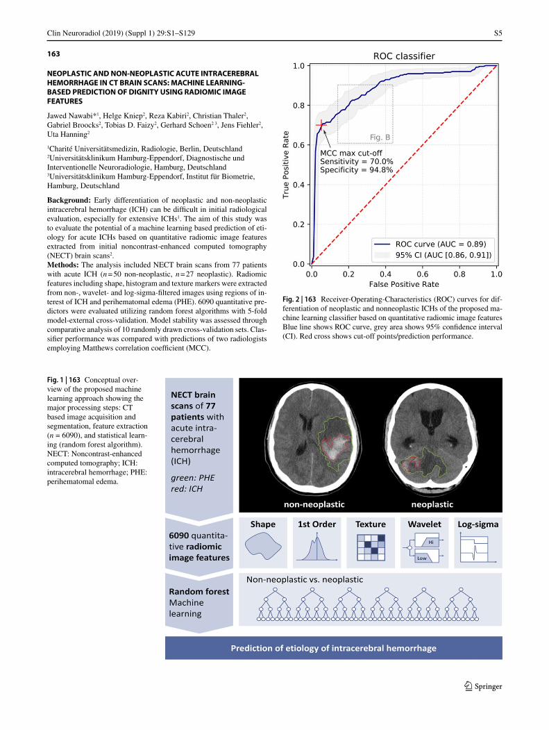
Background: Early differentiation of neoplastic and non-neoplastic intracerebral hemorrhage (ICH) can be difficult in initial radiological evaluation, especially for extensive ICHs1. The aim of this study was to evaluate the potential of a machine learning based prediction of eti-ology for acute ICHs based on quantitative radiomic image features extracted from initial noncontrast-enhanced computed tomography (NECT) brain scans2.Methods: The analysis included NECT brain scans from 77 patients with acute ICH (n = 50 non-neoplastic, n = 27 neoplastic). Radiomic features including shape, histogram and texture markers were extracted from non-, wavelet- and log-sigma-filtered images using regions of in-terest of ICH and perihematomal edema (PHE). 6090 quantitative pre-dictors were evaluated utilizing random forest algorithms with 5-fold model-external cross-validation. Model stability was assessed through comparative analysis of 10 randomly drawn cross-validation sets. Clas-sifier performance was compared with predictions of two radiologists employing Matthews correlation coefficient (MCC).
Fig. 1 | 163 Conceptual over-view of the proposed machine learning approach showing the major processing steps: CT based image acquisition and segmentation, feature extraction (n = 6090), and statistical learn-ing (random forest algorithm). NECT: Noncontrast-enhanced computed tomography; ICH: intracerebral hemorrhage; PHE: perihematomal edema.
Fig. 2 | 163 Receiver-Operating-Characteristics (ROC) curves for dif-ferentiation of neoplastic and nonneoplastic ICHs of the proposed ma-chine learning classifier based on quantitative radiomic image features Blue line shows ROC curve, grey area shows 95% confidence interval (CI). Red cross shows cut-off points/prediction performance.
S6
123
Clin Neuroradiol (2019) (Suppl 1) 29:S1–S129
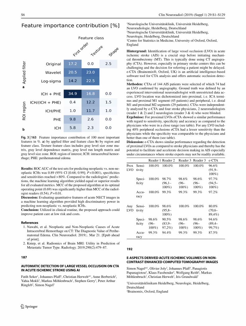
Results: ROC AUC of the test sets for predicting neoplastic vs. non-ne-oplastic ICHs was 0.89 (95% CI [0.68; 0.99]; P < 0.001), specificities and sensitivities reached > 80%. Compared to the radiologists’ predic-tions, the machine learning algorithm yielded equal or superior results for all evaluated metrics. MCC of the proposed algorithm at its optimal operating point (0.69) was significantly higher than MCC of the radiol-ogist readers (0.54); P = 0.01.Discussion: Evaluating quantitative features of acute NECT images in a machine learning algorithm provided high discriminatory power in predicting non-neoplastic vs. neoplastic ICHs.Conclusion: Utilized in clinical routine, the proposed approach could improve patient care at low risk and costs.
References 1. Nawabi, et al. Neoplastic and Non-Neoplastic Causes of Acute
Intracerebral Hemorrhage on CT: The Diagnostic Value of Perihe-matomal Edema. Clin Neuroradiol. 2019;:. Mar 21. [Epub ahead of print].
2. Kniep, et al. Radiomics of Brain MRI: Utility in Prediction of Metastatic Tumor Type. Radiology. 2019;290(2):479–87.
187
AUTOMATIC DETECTION OF LARGE VESSEL OCCLUSION ON CTA IN ACUTE ISCHEMIC STROKE USING AI
Fatih Seker1, Johannes Pfaff1, Christian Herweh*1, Anne Berberich2, Yahia Mokli2, Markus Möhlenbruch1, Stephen Gerry3, Peter Arthur Ringleb2, Simon Nagel2
1Neurologische Universitätsklinik, Universität Heidelberg, Neuroradiologie, Heidelberg, Deutschland2Neurologische Universitätsklinik, Universität Heidelberg, Neurologie, Heidelberg, Deutschland3Centre for Statistics in Medicine, University of Oxford, Oxford, England
Hintergrund: Identification of large vessel occlusion (LVO) in acute ischemic stroke (AIS) is a crucial step before initiating mechani-cal thrombectomy (MT). This is typically done using CT angiogra-phy (CTA). However, especially in primary stroke centers this can be challenging and the decision for referring a patient might be delayed. e-CTA (Brainomix®, Oxford, UK) is an artificial intelligence-based software tool for CTA analysis and offers automatic occlusion detec-tion.Methoden: CTAs of 144 AIS patients were selected of which 74 had an LVO confirmed by angiography. Ground truth was defined by an experienced interventional neuroradiologist with unrestricted data ac-cess. LVO location was dichotomized into proximal, i. e. ICA termi-nus and proximal M1 segment (45 patients) and peripheral, i. e. distal M1 and proximal M2 segments (29 patients). CTAs were independent-ly analyzed by e-CTA and four stroke physicians, 2 neuroradiologists (reader 1 & 2) and 2 neurologists (reader 3 & 4) who were blinded.Ergebnisse: For proximal LVOs eCTA showed a similar performance with regard to sensitivity, specificity and accuracy as compared to the physicians who were in a close range (see table). For any LVO includ-ing 40% peripheral occlusions eCTA had a lesser sensitivity than the physicians while the specificity was comparable to the physicians and better than one of them (see table).Diskussion: e-CTA shows similar performance regarding the detection of proximal LVOs as compared to stroke physicians and thereby has the potential to facilitate and accelerate decision making in AIS especially under circumstances where stroke experts may not be readily available.
Reader 1 Reader 2 Reader 3 Reader 3 e-CTA
Prox LVO
Sensi-tivity
100.0% 100.0% 100.0% 100.0% 96.6%(91,9–100%)
Speci-ficity
100.0% 98.7%(96,1–100%)
98.6%(96–100%)
98.6%(96–100%)
97.7%(94,5–100%)
Accu-racy
100.0% 99.3% 99.3% 99.3% 97.2%
Any LVO
Sensi-tivity
100.0% 98.6%(95,8–100%)
100.0% 100.0% 80.0%(70,6–89,4%)
Speci-ficity
98.6%(96–100%)
90.5%(83,9–97,2%)
98.6%(96–100%)
98.6%(96–100%)
94.6%(89,4–99,7%)
Accu-racy
99.3% 94.4% 99.3% 99.3% 87.5%
192
E-ASPECTS DERIVED ACUTE ISCHEMIC VOLUMES ON NON-CONTRAST ENHANCED COMPUTED TOMOGRAPHY IMAGES
Simon Nagel*1, Olivier Joly2, Johannes Pfaff3, Panagiotis Papanagiotou4, Klaus Fassbender5, Wolfgang Reith6, Markus Möhlenbruch3, Christian Herweh3, Iris Grundwald7
1Universitätsklinikum Heidelberg, Neurologie, Heidelberg, Deutschland2Brainomix, Oxford, England
Fig. 3 | 163 Feature importance contribution of 100 most important features in %. a) by applied filter and feature class b) by region and feature class. Texture feature class includes gray level size zone ma-trix, gray level dependence matrix, gray level run length matrix and gray level size zone. ROI: region of interest; ICH: intracerebral hemor-rhage; PHE: perihematomal edema

S7
123
Clin Neuroradiol (2019) (Suppl 1) 29:S1–S129
3Univesitätsklinikum Heidelberg, Neuroradiologie, Heidelberg, Deutschland4KLinikum Bremen, Neuroradiologie, Bremen, Deutschland5Universität des Saarlandes, Neurologie, Homburg, Deutschland6Universität des Saarlandes, Neuroradiologie, Homburg, Deutschland7Angia Ruskin University, Neuroscience, Essex, England
Background: e-ASPECTS is an AI supported software and here we aimed to Validate Automatically derived Acute Ischemic Volumes (AAIV) from e-ASPECTS on Non-Contrast Computed Tomography (NCCT)Methods: Data from three studies were reanalyzed with e-ASPECTS Version 7. The National Institute of Health Stroke Scale (NIHSS) de-termined stroke severity at baseline and clinical outcome was meas-ured with the modified Rankin Scale (mRS) between 45 to 120 days. Spearman ranked correlation coefficients (R) of AAIV and e-AS-PECTS scores with NIHSS and mRS as well as Pearson correlation of AAIV with diffusion weighted imaging (DWI) and CT Perfusion (CTP) estimated infarct core volumes were calculated. Multivariate re-gression analysis (odds ratio, OR with 95% confidence intervals, CI) and Bland–Altman plots (BA) were performed.Result: We included 388 patients. Mean AAIV was 11.6+/–18.9 ml and e-ASPECTS was 9 (8–10: median and interquartile range). AAIV, respectively e-ASPECTS correlated with NIHSS at baseline (R = 0.35, p < 0.001; R = –0.36, p < 0.001) and follow-up mRS (R = 0.29, p < 0.001; R = –0.3, p < 0.001). In subsets of patients AAIV within the affected hemisphere correlated strongly with DWI (n = 37, R = 0.68, p < 0.001) and CTP derived infarct core (n = 41, R = 0.76, p < 0.001) lesion vol-ume and BA plots showed a bias close to zero (–2.65 ml for DWI and 0.45 ml for CTP core). Within the whole cohort the AAIV (OR 0.98 per ml, 95% CI 0.96–0.99) and e-ASPECTS scores (OR 1.3, 95%CI 1.07–1.57) were independent predictors of good outcomeConclusion: AAIV on NCCT correlated moderately with clinical se-verity but strongly with DWI lesion and CTP infarct core volumes and predicted clinical outcome.
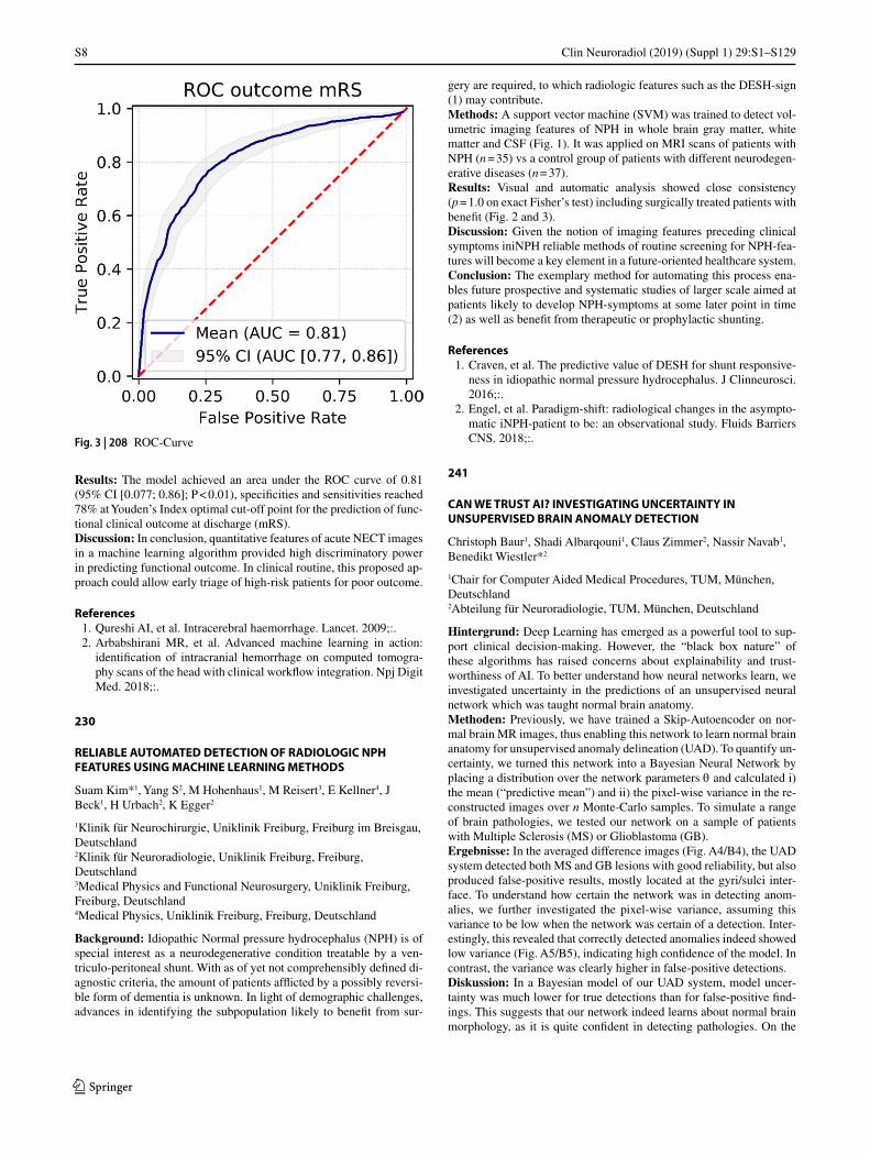
208
ADVANCED MACHINE LEARNING IN ACTION: RADIOMIC BASED OUTCOME PREDICTION OF ACUTE INTRACRANIAL HEMORRHAGE ON COMPUTED TOMOGRAPHY
Jawed Nawabi*1 2, Helge Kniep2, Peter Sporns3, Sarah Elsayed2, Gabriel Broocks2, Gerhard Schoen4, Jens Fiehler2, Uta Hanning2
1Charite—Campus Mitte, Institut für Radiologie, Berlin, Deutschland2Universitätsklinikum Hamburg-Eppendorf, Institut für Diagnostische und Interventionelle Neuroradiologie, Hamburg, Deutschland3Universitätsklinikum Münster, Institut für Radiologie, Münster, Deutschland4Universitätsklinikum Hamburg-Eppendorf, Institut für Biometrie, Hamburg, Deutschland
Background: Intracranial hemorrhage (ICH) requires prompt diag-nosis to optimize patient outcomes 1. We hypothesized that machine learning algorithms could automatically analyze non-contrast comput-ed tomography (NECT) of the head and predict clinical outcome of ICH patients 2.Methods: 300 NECTs with acute spontaneous ICH between 2014–2019 were retrospectively included from the database at a tertiary university hospital. A binary outcome was defined as Modified Rank-ing Scale (mRS) 0–3 (good outcome) and mRS 4–6 (bad outcome) at discharge. Radiomic features including shape, histogram and texture markers were extracted from non-, wavelet- and log-sigma-filtered im-ages using regions of interest of ICH. The quantitative predictors were evaluated utilizing random forest algorithms with 5-fold model-exter-nal cross-validation.
Fig. 1 | 208 Conceptual overview
Fig. 2 | 208 Feature importance contribution
S8
123
Clin Neuroradiol (2019) (Suppl 1) 29:S1–S129
Results: The model achieved an area under the ROC curve of 0.81 (95% CI [0.077; 0.86]; P < 0.01), specificities and sensitivities reached 78% at Youden’s Index optimal cut-off point for the prediction of func-tional clinical outcome at discharge (mRS).Discussion: In conclusion, quantitative features of acute NECT images in a machine learning algorithm provided high discriminatory power in predicting functional outcome. In clinical routine, this proposed ap-proach could allow early triage of high-risk patients for poor outcome.
References 1. Qureshi AI, et al. Intracerebral haemorrhage. Lancet. 2009;:. 2. Arbabshirani MR, et al. Advanced machine learning in action:
identification of intracranial hemorrhage on computed tomogra-phy scans of the head with clinical workflow integration. Npj Digit Med. 2018;:.
230
RELIABLE AUTOMATED DETECTION OF RADIOLOGIC NPH FEATURES USING MACHINE LEARNING METHODS
Suam Kim*1, Yang S2, M Hohenhaus1, M Reisert3, E Kellner4, J Beck1, H Urbach2, K Egger2
1Klinik für Neurochirurgie, Uniklinik Freiburg, Freiburg im Breisgau, Deutschland2Klinik für Neuroradiologie, Uniklinik Freiburg, Freiburg, Deutschland3Medical Physics and Functional Neurosurgery, Uniklinik Freiburg, Freiburg, Deutschland4Medical Physics, Uniklinik Freiburg, Freiburg, Deutschland
Background: Idiopathic Normal pressure hydrocephalus (NPH) is of special interest as a neurodegenerative condition treatable by a ven-triculo-peritoneal shunt. With as of yet not comprehensibly defined di-agnostic criteria, the amount of patients afflicted by a possibly reversi-ble form of dementia is unknown. In light of demographic challenges, advances in identifying the subpopulation likely to benefit from sur-
gery are required, to which radiologic features such as the DESH-sign (1) may contribute.Methods: A support vector machine (SVM) was trained to detect vol-umetric imaging features of NPH in whole brain gray matter, white matter and CSF (Fig. 1). It was applied on MRI scans of patients with NPH (n = 35) vs a control group of patients with different neurodegen-erative diseases (n = 37).Results: Visual and automatic analysis showed close consistency (p = 1.0 on exact Fisher’s test) including surgically treated patients with benefit (Fig. 2 and 3).Discussion: Given the notion of imaging features preceding clinical symptoms iniNPH reliable methods of routine screening for NPH-fea-tures will become a key element in a future-oriented healthcare system.Conclusion: The exemplary method for automating this process ena-bles future prospective and systematic studies of larger scale aimed at patients likely to develop NPH-symptoms at some later point in time (2) as well as benefit from therapeutic or prophylactic shunting.
References 1. Craven, et al. The predictive value of DESH for shunt responsive-
ness in idiopathic normal pressure hydrocephalus. J Clinneurosci. 2016;:.
2. Engel, et al. Paradigm-shift: radiological changes in the asympto-matic iNPH-patient to be: an observational study. Fluids Barriers CNS. 2018;:.
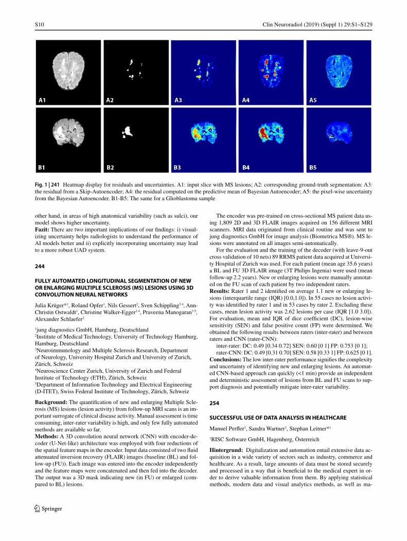
241
CAN WE TRUST AI? INVESTIGATING UNCERTAINTY IN UNSUPERVISED BRAIN ANOMALY DETECTION
Christoph Baur1, Shadi Albarqouni1, Claus Zimmer2, Nassir Navab1, Benedikt Wiestler*2
1Chair for Computer Aided Medical Procedures, TUM, München, Deutschland2Abteilung für Neuroradiologie, TUM, München, Deutschland
Hintergrund: Deep Learning has emerged as a powerful tool to sup-port clinical decision-making. However, the “black box nature” of these algorithms has raised concerns about explainability and trust-worthiness of AI. To better understand how neural networks learn, we investigated uncertainty in the predictions of an unsupervised neural network which was taught normal brain anatomy.Methoden: Previously, we have trained a Skip-Autoencoder on nor-mal brain MR images, thus enabling this network to learn normal brain anatomy for unsupervised anomaly delineation (UAD). To quantify un-certainty, we turned this network into a Bayesian Neural Network by placing a distribution over the network parameters θ and calculated i) the mean (“predictive mean”) and ii) the pixel-wise variance in the re-constructed images over n Monte-Carlo samples. To simulate a range of brain pathologies, we tested our network on a sample of patients with Multiple Sclerosis (MS) or Glioblastoma (GB).Ergebnisse: In the averaged difference images (Fig. A4/B4), the UAD system detected both MS and GB lesions with good reliability, but also produced false-positive results, mostly located at the gyri/sulci inter-face. To understand how certain the network was in detecting anom-alies, we further investigated the pixel-wise variance, assuming this variance to be low when the network was certain of a detection. Inter-estingly, this revealed that correctly detected anomalies indeed showed low variance (Fig. A5/B5), indicating high confidence of the model. In contrast, the variance was clearly higher in false-positive detections.Diskussion: In a Bayesian model of our UAD system, model uncer-tainty was much lower for true detections than for false-positive find-ings. This suggests that our network indeed learns about normal brain morphology, as it is quite confident in detecting pathologies. On the
Fig. 3 | 208 ROC-Curve

S9
123
Clin Neuroradiol (2019) (Suppl 1) 29:S1–S129
Fig. 1 | 230 VEOmorph (www.veobrain.com) analysis results with abnormal GM and CSF voxels superimposed in color on individual 3D T1w brain MRI (increase of CSF volume in red to yellow, and decrease of GM volume in dark to light blue)
Fig. 2 | 230 ROC analysis of generated probability for NPH as shown in Fig. 1 in comparison to clinically conformed diagnosis Fig. 3 | 230 NPH probability in percent against visual grade of NPH
typical changes (0 = no NPH typical changes, 1 = probable NPH typical changes, 2 = definite NPH typical changes)
S10
123
Clin Neuroradiol (2019) (Suppl 1) 29:S1–S129
other hand, in areas of high anatomical variability (such as sulci), our model shows higher uncertainty.Fazit: There are two important implications of our findings: i) visual-izing uncertainty helps radiologists to understand the performance of AI models better and ii) explicitly incorporating uncertainty may lead to a more robust UAD system.
244
FULLY AUTOMATED LONGITUDINAL SEGMENTATION OF NEW OR ENLARGING MULTIPLE SCLEROSIS (MS) LESIONS USING 3D CONVOLUTION NEURAL NETWORKS
Julia Krüger*1, Roland Opfer1, Nils Gessert2, Sven Schippling3 4, Ann-Christin Ostwaldt1, Christine Walker-Egger3 4, Praveena Manogaran3 5, Alexander Schlaefer2
1jung diagnostics GmbH, Hamburg, Deutschland2Institute of Medical Technology, University of Technology Hamburg, Hamburg, Deutschland3Neuroimmunology and Multiple Sclerosis Research, Department of Neurology, University Hospital Zurich and University of Zurich, Zürich, Schweiz4Neuroscience Center Zurich, University of Zurich and Federal Institute of Technology (ETH), Zürich, Schweiz5Department of Information Technology and Electrical Engineering (D-ITET), Swiss Federal Institute of Technology, Zürich, Schweiz
Background: The quantification of new and enlarging Multiple Scle-rosis (MS) lesions (lesion activity) from follow-up MRI scans is an im-portant surrogate of clinical disease activity. Manual assessment is time consuming, inter-rater variability is high, and only few fully automated methods are available so far.Methods: A 3D convolution neural network (CNN) with encoder-de-coder (U-Net-like) architecture was employed with four reductions of the spatial feature maps in the encoder. Input data consisted of two fluid attenuated inversion recovery (FLAIR) images (baseline (BL) and fol-low-up (FU)). Each image was entered into the encoder independently and the feature maps were concatenated and then fed into the decoder. The output was a 3D mask indicating new (in FU) or enlarged (com-pared to BL) lesions.
The encoder was pre-trained on cross-sectional MS patient data us-ing 1,809 2D and 3D FLAIR images acquired on 156 different MRI scanners. MRI data originated from clinical routine and was sent to jung diagnostics GmbH for image analysis (Biometrica MS®). MS le-sions were annotated on all images semi-automatically.
For the evaluation and the training of the decoder (with leave-9-out cross validation of 10 nets) 89 RRMS patient data acquired at Universi-ty Hospital of Zurich was used. For each patient (mean age 35.6 years) a BL and FU 3D FLAIR image (3T Philips Ingenia) were used (mean follow-up 2.2 years). New or enlarging lesions were manually annotat-ed on the FU scan of each patient by two independent raters.Results: Rater 1 and 2 identified on average 1.1 new or enlarging le-sions (interquartile range (IQR) [0.0,1.0]). In 55 cases no lesion activi-ty was identified by rater 1 and in 53 cases by rater 2. Excluding these cases, mean lesion activity was 2.62 lesions per case (IQR [1.0 3.0]). For evaluation, mean and IQR of dice coefficient (DC), lesion-wise sensitivity (SEN) and false positive count (FP) were determined. We obtained the following results between raters (inter-rater) and between raters and CNN (rater-CNN):
inter-rater: DC: 0.49 [0.34 0.72] SEN: 0.60 [0 1] FP: 0.753 [0 1];rater-CNN: DC: 0.49 [0.31 0.70] SEN: 0.58 [0.33 1] FP: 0.625 [0 1].
Conclusions: The low inter-rater performance signifies the complexity and uncertainty of identifying new and enlarging lesions. An automat-ed CNN-based approach can quickly (<1 min) provide an independent and deterministic assessment of lesions from BL and FU scans to sup-port diagnosis and potentially mitigate inter-rater variability.
254
SUCCESSFUL USE OF DATA ANALYSIS IN HEALTHCARE
Manuel Perfler1, Sandra Wartner1, Stephan Leitner*1
1RISC Software GmbH, Hagenberg, Österreich
Hintergrund: Digitalization and automation entail extensive data ac-quisition in a wide variety of sectors such as industry, commerce and healthcare. As a result, large amounts of data must be stored securely and processed in a way that is beneficial to the medical expert in or-der to derive valuable information from them. By applying statistical methods, modern data and visual analytics methods, as well as ma-
Fig. 1 | 241 Heatmap display for residuals and uncertainties. A1: input slice with MS lesions; A2: corresponding ground-truth segmentation: A3: the residual from a Skip-Autoencoder; A4: the residual computed on the predictive mean of Bayesian Autoencoder; A5: the pixel-wise uncertainty from the Bayesian Autoencoder. B1-B5: The same for a Glioblastoma sample
S11
123
Clin Neuroradiol (2019) (Suppl 1) 29:S1–S129
chine learning, the existing knowledge is analyzed in context with the recorded data. Using methods from the field of artificial intelligence, knowledge is generated and recommendations for action are formulat-ed for these experts (expert-in-the-loop).Methoden: Data analyses consists of 3 steps:
Data Engineering and Modeling: Health and sensor data must be in-telligently combined, filtered and linked. Checking the correctness and completeness of the data is also relevant.
Data Analytics and Artificial Intelligence: In order to gain knowl-edge from collected and filtered data, mathematical, statistical and arti-ficial intelligence methods, as well as visual analytics are used.
Data Presentation: The visual processing of data and analysis re-sults is an important tool for gaining new insights. An essential factor for the interpretation of analysis results is the strong involvement of domain experts.
In order to technically enable and guide researchers and analysts in medicine to fulfil their role in the process of data analysis, we devel-oped an ontology-based research and analytics infrastructure. Due to its approach, the generic infrastructure can be adapted to any research domain of interest by the researchers themselves.Ergebnisse: In a research cooperation we built an ontology model of stroke patients and have been gathering data for several years. We want to showcase the data analysis process and benefits of visual analytics as well as give an outlook of possible machine learning algorithms that can be performed on medical image data of stroke patients.Diskussion: As a major part of our research is focused on guiding med-ical experts through the data analysis process, we are always looking for feedback and suggestions on the features and guidance that is re-quired by the community.Fazit: Our ontology based data analysis system combines data com-plexity, interface diversity and visualization with user-friendliness. Do-main experts are supported by the modeling, integration, validation, processing and evaluation of their data.
262
MORPHOMETRIE UND MACHINE LEARNING ZUR ERKENNUNG VON ALZHEIMER-DEMENZ: EIN METHODENVERGLEICH
Tobias Raffelsberger*1, Raimund Kleiser1, Friedrich Leblhuber2, Sibylle Wimmer1, Johannes Trenkler1
1Kepler Universitätsklinikum, Neuromed Campus, Institut für Neuroradiologie, Linz, Österreich2Kepler Universitätsklinikum, Neuromed Campus, Abteilung für Gerontologie, Linz, Österreich
Hintergrund: Morphometrie in Verbindung mit Support Vector Ma-chines (SVMs) hat sich für eine automatisierte Früherkennung von Mild Cognitive Impairment (MCI) und Alzheimer-Demenz (AD) be-reits als vielversprechend erwiesen [1,2]. Die vorliegende Arbeit ver-gleicht die diesbezügliche Performance der Morphometrie-Software CAT12 mit jenen der bereits länger etablierten Softwares FreeSurfer und SPM12.Methoden: Als Trainingsgrundlage für die SVMs dienten 240 Daten-sätze (80 MCI, 80 AD und 80 gesunde Kontrollen (HC)) aus der ADNI-Datenbank. Für diese wurden mittels der Softwares FreeSurfer, SPM12 und CAT12 Gehirnvolumen beziehungsweise -dicke in vordefinierten Regions-of-interest (ROIs) bestimmt. Die Unterteilung in ROIs erfolg-te dabei unter Verwendung von vier unterschiedlichen funktionellen Atlanten. Vektoren bestehend aus den ermittelten ROI-Daten ergänzt von Geschlecht und Alter der Versuchspersonen dienten als Trainings-daten für binäre SVM-Klassifikatoren (HC vs. AD, HC vs. MCI und MCI vs. AD). Mittels 10-facher Kreuzvalidierung wurde die Treffer-quote der Klassifikatoren ermittelt. Eine Selektion jener ROIs, die sich als am relevantesten für die Klassifikation erwiesen, erfolgte mittels Rekursiver Feature Elimination (RFE).
Ergebnisse: Alle untersuchten morphometrischen Methoden erzielten Trefferquoten von über 85%. Unter Verwendung der Volumenbestim-mung unter CAT12 wurde mit 95.1% das Top-Resultat erzielt. Die RFE verbesserte dabei die Trefferquoten im Durchschnitt um 11.5%.Diskussion: Nennenswerte Unterschiede ergeben sich einerseits zwi-schen den morphometrischen Methoden, andererseits zwischen den funktionellen Atlanten. CAT12 auf der einen Seite und der Atlas zur Verfügung gestellt von Neuromorphometrics Inc. auf der anderen Seite stechen positiv hervor.Fazit: Erstklassige Klassifikationsergebnisse sowie eine vergleichs-weise kurze Verarbeitungsdauer von zirka 45 Minuten pro Datensatz machen CAT12 zu einem weiteren vielversprechenden Kandidaten für den morphometrischen Part einer zukünftigen automatisierten Früher-kennung von dementiellen Erkrankungen.
Literatur 1. Gupta, et al. Alzheimer’s Disease Diagnosis Based on Cortical and
Subcortical Features. J Healthc Eng. 2019;2019:13. 2. Cuingnet, et al. Automatic classification of patients with Alzheim-
er’s disease from structural MRI: A comparison of ten methods using the ADNI database. Neuroimage. 2011;56:766–81.
264
ICOBRAIN’S NATURAL LANGUAGE RADIOLOGICAL REPORTING FOR MULTIPLE SCLEROSIS FOLLOW-UP
Diana Sima1, Guido Wilms1, Thijs Vande Vyvere1, Martin Seelhorst1, Wim Van Hecke1, Dirk Smeets1
1icometrix, Leuven, Belgien
Hintergrund: Radiological reporting in multiple sclerosis (MS) and other neurological conditions is an important tool that facilitates the communication between radiologist and neurologist. In this work we assess the added value of automatically generated natural language radiologic reports based on the automated icobrain software for MS follow-up.Methoden: Longitudinal MRI acquisitions approximately 1 year apart were collected from 25 multiple sclerosis patients. The automated soft-ware icobrain was used to compute lesion load (including new and en-larging lesions) and brain volumetry (including annualized atrophy). Natural language reports containing lesion evolution and atrophy find-ings were automatically generated. Each dataset was presented in ran-dom order to an experienced radiologist twice: with and without au-tomatically generated report. Final reports were created either from scratch or by verifying and adapting the automatically generated re-port. The time necessary to complete each report was kept.Ergebnisse: With respect to timing, the median time for convention-al radiological reporting was 7min17s (interquartile range 4min40s), while the median computer-aided radiologic reporting time was 4min37s (interquartile range 3min35s), with the latter significant-ly faster (paired t-test and Wilcoxon signed-rank test p < 0.01).With respect to findings, the computer-aided reports indicated 7 stable pa-tients (normal atrophy, no lesion activity), 7 patients with slight pro-gression (slightly abnormal atrophy rate or enlarging lesions), and 11 progressive patients (5 with new lesions, 10 with abnormal atrophy rate for their age). Conventional radiological reports indicated 19 sta-ble patients (no lesion activity, no apparent atrophy) and 6 progressive patients (new lesion formation or lesion enlargement, also identified above as progressive).Diskussion: Computer-aided radiological reports are faster than con-ventional reporting, with approximately 8 conventional reports per hour versus 13 computer-aided reports per hour. Stable patients identi-fied by the computer-aided technique were also deemed stable by con-

S12
123
Clin Neuroradiol (2019) (Suppl 1) 29:S1–S129
ventional radiological reading, but the software also reported cases of subtle cerebral atrophy.Fazit: Standardization of radiologic reporting and incorporation of brain atrophy are steps towards future improvements in the clinical management of MS patients.
266
EVALUATION OF A NEW AI-BASED SINGLE-RUN 3D-ANGIOGRAPHY (3D-A) FOR VISUALIZATION OF CEREBRAL AVMS
Stefan Lang*1, Philip Hölter1, Felix Eisenhut1, Manuel Schmidt1, Christian Kaethner2, Arnd Dörfler1
1Neuroradiologie, Universitätsklinikum Erlangen, Erlangen, Deutschland2Siemens Healthineers AG, Erlangen, Deutschland
Background: The AI-based 3D-A is a new postprocessing method that allows a significant reduction of the patient dose (PD) necessary for 3D-imaging of cerebral vessels. Our aim was evaluation of 3D-A for visualization of cerebral AVMs.Methods: 3D-DSA datasets of cerebral AVMs have been reconstruct-ed using conventional and prototype software (Siemens Healthineers AG, Germany). Corresponding reconstructions have been analyzed by 2 experienced neuroradiologists in consensus reading in terms of quan-titative (AVM size [S] in mm) and qualitative parameters (image qual-ity 0–4 [e.g., IQ = 4 means excellent], main feeder [e.g. ACA, MCA, PCA, etc.], localization [eloquent vs. non-eloquent], venous drainage [superficial, deep, mixed]). Martin-Spetzler-Scores (MSS) were as-sessed in both reconstructions.Result: In total 10 datasets (nmale = 6;nfemale = 4;agemean = 54.3 years) have been successfully reconstructed using both conventional and pro-totype software. In all cases corresponding reconstructions demon-strated complete congruency in terms of IQ (IQ3D-DSA/3D-A = 4), locali-zation (neloquent 3D-DSA/3D-A = 8; nnon-eloquent 3D-DSA/3D-A = 2), main feeder (nACA
3D-DSA/3D-A = 1; nMCA 3D-DSA/3D-A = 3; nPCA 3D-DSA/3D-A = 6), venous drainage (nsuperficial 3D-DSA/3D-A = 4; ndeep 3D-DSA/3D-A = 4; nmixed 3D-DSA/3D-A = 2) and MSS (MSS13D-DSA/3D-A = 1; MSS23D-DSA/3D-A = 4; MSS33D-DSA/3D-A = 5). Measure-ment of AVM size (S3D-DSA = 20.2 ± 11.2 mm; S3D-A = 19.9 ± 10.9 mm) correlated well (r = 0.994; p = 0.0001) in both reconstructions.Discussion: The AI-based 3D-A shows comparable results to 3D-DSA in terms of quantitative and qualitative parameters.Conclusion: 3D-A is a promising method for visualization of AVMs and might help to reduce PD for the diagnostic work-up of AVMs.
280
THE HIDDEN PROWESS OF NLP AND RADIOLOGICAL REPORTS: A TEXT MINING PIPELINE FOR MACHINE LEARNING-ASSISTED DIAGNOSTICS AND REPORTING
Andreas Georg Junge1 2 3, Benedikt Kämpgen4, Chang Gyu Cho2, Christoph Groden2, Thomas Ganslandt3, Holger Wenz2, Máté Maros*2
3
1Hochschule Mannheim—University of Applied Sciences, Technical Informatics, Mannheim, Deutschland2Heidelberg University, Medical Faculty Mannheim, Department of Neuroradiology, Mannheim, Deutschland3Heidelberg University, Medical Faculty Mannheim, Department of Biomedical Informatics at the Heinrich Lanz Center, Mannheim, Deutschland4Empolis, Information Management GmbH, Kaiserslautern, Deutschland
Background: The value of radiological reports is often neglected and advancements in natural language processing (NLP) are forced to take a backseat behind the predominance of computer vision applications of deep learning in radiology. Furthermore, the language of radiologi-cal reports is extremely heterogeneous. Efforts were put forward to uni-fy reporting language by using structured reporting (SR). However, the adoption of SR is still incremental with controversial aspects regarding the usability of SR for data extraction. To illustrate the value of radiolog-ical texts and the prowess of NLP, we present our text mining pipeline with three use cases that include i) AI algorithm-based indexing for im-proved data extraction, ii) report quality assessment and iii) clinical key information content with particular focus on neurovascular emergencies.Methods: We mirrored our local RIS (Syngo, Siemens) between 2014–2018 including ~48.000 CT and MRI reports, and associated billing in-formation. All reports were RadLex® annotated using a proprietary NLP pipeline1 (Empolis Information Management GmbH) that is free for research purposes. Further, we performed i) comprehensive index-ing of the corpus and created language models using open-source soft-ware (Elasticsearch and PyTorch), and ii) latent space analyses to iden-tify characteristics specific for certain clinical scenarios or styles of attending radiologists.Result: We created a RadLex® annotation pipeline that can automatical-ly provide quality score for each report based on predefined local or in-ternational guidelines2. Since its implementation the browser-embedded assistive NLP-algorithm significantly increased (p < 0.05) the occurrence of ASPECT scores in reports about vascular emergencies. Further, the pipeline can support junior readers in creating reports similar to the style of the attending radiologist by whom these are going to be signed off.Discussion: Computer assisted reporting tools can speed up knowl-edge transfer to junior radiologists, improve their report quality and reduce the burden of signing off reports on senior radiologists.Conclusion: Our use cases of NLP applied on radiological reports strongly argue for the tremendous value hidden in radiological text to streamline the reporting process and to improve its quality.
References 1. Jungmann F, et al. Radiologe. 2019. 2. Maros ME, et al. In Vivo. 2018.
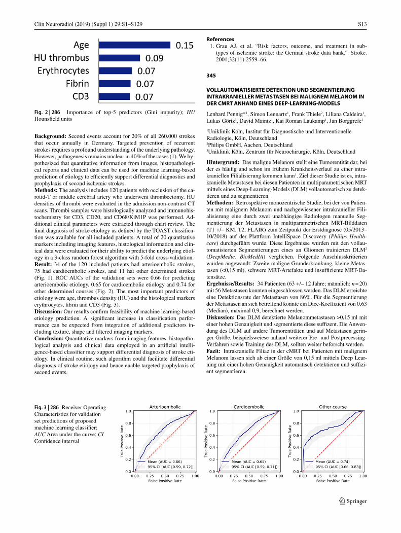
286
PREVENTION OF RECURRENT STROKE: AI-BASED CLASSIFICATION OF ETIOLOGY FROM IMAGING FEATURES, HISTOPATHOLOGICAL REPORTS AND CLINICAL DATA
Helge Kniep*1, Peter Sporns2, Moritz Wildgruber2, Astrid Jeibmann3, Jens Minnerup4, Jens Fiehler1, Uta Hanning1
1Klinik und Poliklinik für Neuroradiologische Diagnostik und Intervention, Universitätsklinikum Hamburg-Eppendorf, Hamburg, Deutschland2Institut für Klinische Radiologie, Universitätsklinikum Münster, Münster, Deutschland3Institut für Neuropathologie, Universitätsklinikum Münster, Münster, Deutschland4Klinik für Neurologie mit Institut für Translationale Neurologie, Universitätsklinikum Münster, Münster, Deutschland
Fig. 1 | 286 Demographic characteristics of study cohort; # Number of; SD standard deviation
S13
123
Clin Neuroradiol (2019) (Suppl 1) 29:S1–S129
Background: Second events account for 20% of all 260.000 strokes that occur annually in Germany. Targeted prevention of recurrent strokes requires a profound understanding of the underlying pathology. However, pathogenesis remains unclear in 40% of the cases (1). We hy-pothesized that quantitative information from images, histopathologi-cal reports and clinical data can be used for machine learning-based prediction of etiology to efficiently support differential diagnostics and prophylaxis of second ischemic strokes.Methods: The analysis includes 120 patients with occlusion of the ca-rotid-T or middle cerebral artery who underwent thrombectomy. HU densities of thrombi were evaluated in the admission non-contrast CT scans. Thrombi samples were histologically analyzed and immunohis-tochemistry for CD3, CD20, and CD68/KiM1P was performed. Ad-ditional clinical parameters were extracted through chart review. The final diagnosis of stroke etiology as defined by the TOAST classifica-tion was available for all included patients. A total of 20 quantitative markers including imaging features, histological information and clin-ical data were evaluated for their ability to predict the underlying etiol-ogy in a 3-class random forest algorithm with 5-fold cross-validation.Result: 34 of the 120 included patients had arterioembolic strokes, 75 had cardioembolic strokes, and 11 hat other determined strokes (Fig. 1). ROC AUCs of the validation sets were 0.66 for predicting arterioembolic etiology, 0.65 for cardioembolic etiology and 0.74 for other determined courses (Fig. 2). The most important predictors of etiology were age, thrombus density (HU) and the histological markers erythrocytes, fibrin and CD3 (Fig. 3).Discussion: Our results confirm feasibility of machine learning-based etiology prediction. A significant increase in classification perfor-mance can be expected from integration of additional predictors in-cluding texture, shape and filtered imaging markers.Conclusion: Quantitative markers from imaging features, histopatho-logical analysis and clinical data employed in an artificial intelli-gence-based classifier may support differential diagnosis of stroke eti-ology. In clinical routine, such algorithm could facilitate differential diagnosis of stroke etiology and hence enable targeted prophylaxis of second events.
References 1. Grau AJ, et al. “Risk factors, outcome, and treatment in sub-
types of ischemic stroke: the German stroke data bank.”. Stroke. 2001;32(11):2559–66.
345
VOLLAUTOMATISIERTE DETEKTION UND SEGMENTIERUNG INTRAKRANIELLER METASTASEN BEI MALIGNEM MELANOM IN DER CMRT ANHAND EINES DEEP-LEARNING-MODELS
Lenhard Pennig*1, Simon Lennartz1, Frank Thiele2, Liliana Caldeira1, Lukas Görtz3, David Maintz1, Kai Roman Laukamp1, Jan Borggrefe1
1Uniklinik Köln, Institut für Diagnostische und Interventionelle Radiologie, Köln, Deutschland2Philips GmbH, Aachen, Deutschland3Uniklinik Köln, Zentrum für Neurochirurgie, Köln, Deutschland
Hintergrund: Das maligne Melanom stellt eine Tumorentität dar, bei der es häufig und schon im frühem Krankheitsverlauf zu einer intra-kraniellen Filialisierung kommen kann1. Ziel dieser Studie ist es, intra-kranielle Metastasen bei diesen Patienten in multiparametrischen MRT mittels eines Deep-Learning-Models (DLM) vollautomatisch zu detek-tieren und zu segmentieren.Methoden: Retrospektive monozentrische Studie, bei der von Patien-ten mit malignem Melanom und nachgewiesener intrakranieller Fili-alisierung eine durch zwei unabhängige Radiologen manuelle Seg-mentierung der Metastasen in multiparametrischen MRT-Bilddaten (T1 +/– KM, T2, FLAIR) zum Zeitpunkt der Erstdiagnose (05/2013–10/2018) auf der Plattform IntelliSpace Discovery (Philips Health-care) durchgeführt wurde. Diese Ergebnisse wurden mit den vollau-tomatisierten Segmentierungen eines an Gliomen trainierten DLM2 (DeepMedic, BioMedIA) verglichen. Folgende Auschlusskritierien wurden angewandt: Zweite maligne Grunderkrankung, kleine Metas-tasen (<0,15 ml), schwere MRT-Artefakte und insuffiziente MRT-Da-tensätze.Ergebnisse/Results: 34 Patienten (63 +/– 12 Jahre; männlich: n = 20) mit 56 Metastasen konnten eingeschlossen werden. Das DLM erreichte eine Detektionsrate der Metastasen von 86%. Für die Segmentierung der Metastasen an sich betreffend konnte ein Dice-Koeffizient von 0,63 (Median), maximal 0,9, berechnet werden.Diskussion: Das DLM detektierte Melanommetastasen >0,15 ml mit einer hohen Genauigkeit und segmentierte diese suffizent. Die Anwen-dung des DLM auf andere Tumorentitäten und auf Metastasen gerin-ger Größe, beispielsweiese anhand weiterer Pre- und Postprecessing-Verfahren sowie Training des DLM, sollten weiter beforscht werden.Fazit: Intrakranielle Filiae in der cMRT bei Patienten mit malignem Melanom lassen sich ab einer Größe von 0,15 ml mittels Deep Lear-ning mit einer hohen Genauigkeit automatisch detektieren und suffizi-ent segmentieren.
Fig. 2 | 286 Importance of top-5 predictors (Gini impurity); HU Hounsfield units
Fig. 3 | 286 Receiver Operating Characteristics for validation set predictions of proposed machine learning classifier; AUC Area under the curve; CI Confidence interval

S14
123
Clin Neuroradiol (2019) (Suppl 1) 29:S1–S129
Literatur 1. Zhang D, et al. Incidence and prognosis of brain metastases in cu-
taneous melanoma patients: a population-based study. Melanoma Res. 2019;29(1):77–84. Feb.
2. Perkuhn B, et al. Clinical Evaluation of a Multiparametric Deep Learning Model for Glioblastoma Segmentation Using Heteroge-neous Magnetic Resonance Imaging Data From Clinical Routine. Invest Radiol. 2018;53(11):647–54.
360
UNSUPERVISED DEEP LEARNING FOR DETECTION OF BRAIN DISEASE IN MR IMAGING
Rami Eisawy1 2, Julia Moosbauer3, Tom Finck4, Claus Zimmer4, Björn Menze1, Franz MJ Pfister*2 3, Benedikt Wiestler4
1Department of Computer Science, Technical University of Munich, Munich, Deutschland2Digital Helix GmbH, R&D, Munich, Deutschland3Department of Statistics, Ludwig Maximilian University, Munich, Deutschland4Klinikum rechts der Isar, Technical University of Munich, Munich, Deutschland
Background: Manual detection and interpretation of suspicious find-ings in radiological exams is a slow and lengthy process, requiring the highest level of attention and expertise. Introducing an automatic ap-proach to distinguish abnormal from normal anatomy and physiology has the potential to speed up the diagnostic process and avoid errors by serving as a second read. Recently, there have been numerous at-tempts to solve this problem with the use of supervised machine learn-ing. However, this poses three major limitations: Costliness of data an-notation, risk of annotation errors being reproduced by the algorithm and lack of training data for rare diseases.Methods: To avoid those limitations, an unsupervised, progressive-ly growing adversarial autoencoder model is presented. The model is exclusively trained on images from a healthy cohort, thus learning the normal anatomy and signal of the brain. Subsequently, pathologies are automatically detected as deviations from this normality. We evalu-ate the model on datasets containing a variety of MS lesions, WMH, stroke, and GBM across the FLAIR MR sequence.Results: The evaluation shows the effectiveness of the method and its ability to highlight even small abnormalities in brain MRI exams, yielding state-of-the-art results in terms of overlap error and dis-tance-based measures (Average DSC = 0.614 ± 0.135) in diseases the network has never seen before.Discussion: Qualitative results effectively show the power of the pro-posed autoencoder model. Reconstructed samples show high detail while simultaneously not reconstructing the pathological areas, facil-itating a segmentation pipeline. Additionally, no modifications to the model are needed to allow for extensive generalisability to various ap-plications such as detecting other white matter hyperintensities, tum-ors or inflammation, or even other modalities (e. g. CT) or parts of the body.Conclusion: To further evaluate the model and achieved results thus far, additional experiments on a larger dataset consisting of various different pathologies are scheduled. Clinical studies proving model ef-ficacy is subject to ongoing research and intended to substantiate the use of the model as a clinical tool.
363
CAPSNETS DETECT SIGNS OF INFARCTION IN NECT STROKE PATIENTS WITH SUPER-HUMAN PERFORMANCE
Christian Riedel*1
1Dept. of Neuroradiology, University of Göttingen, Göttingen, Deutschland
Hintergrund/Background: To evaluate if a hybrid approach of a convolutional neural network with a shallow capsular network can be trained to reach super-human performance in recognizing early signs of infarction and dense artery signs in NECT images of acute ischemic stroke patients.Methoden: We trained a shallow capsular neural network which re-ceived inputs from 5 convolutional layers with NECT images of 150 patients with an acute stroke and 150 patients with a different patho-logical condition. The training used labeling for early signs of infarc-tion derived from CTA-, CTP and subsequent NECT imaging as well as labels of hyperdense artery signs. After training, the ROC curves for correct identification of patients with acute ischemic stroke indicators were compared to those found by three expert reviewers blinded with respect to the clinical situation for a total of 80 NECT test sets.Ergebnisse: The ROC curve for the correct identification of more than one early sign of infarction was found to have an area under the curve of 0.925, whereas the expert reviewers had an AUC between 0.783 and 0.854.Diskussion: While classical convolutional neural networkst end to require at least several hundred training data sets for a complex task such as detection of stroke indicators from NECT images, the capsular network approach delivers high perfomance detection at a moderate amount of training data.Fazit: Using the capsular network approach, complex spatial hierar-chies in 3D medical images can be used in order to recognize early signs of infarction even with a super-human performance.
S15
123
Clin Neuroradiol (2019) (Suppl 1) 29:S1–S129
2. Neuroonkologie
38
MULTIVARIATE NONINVASIVE PREDICTION OF IDH MUTATIONAL STATUS IN WHO GRADE II AND III GLIOMAS WITH ADVANCED MRI T2-MAPPING TECHNIQUES
Timo Alexander Auer*1, Maike Kern2, Uli Fehrenbach1, Martin Misch3, Edzard Wiener4
1Klinik für Neuroradiologie, Charite—Universitätsmedizin Berlin, Klinik für Radiologie, Berlin, Deutschland2Klinik für Neuroradiologie, Charite—Universitätsmedizin Berlin, Berlin, Deutschland3Klinik für Neurochirurgie, Charite—Universitätsmedizin Berlin, Berlin, Deutschland2Klinik für Neuroradiologie, Charite—Universitätsmedizin Berlin, Berlin, Deutschland
Hintergrund: To investigate multivariate analyses for noninvasive prediction of the IDH-mutational status in grade II and III gliomas in-cluding evaluation of T2-“Mapping”-sequences.Methoden: MR examinations with pathologically proven WHO-grade II and III gliomas were retrospectively enrolled. Multivariate ROC-analyses to predict IDH mutational status were performed con-taining quantitative T2 mapping analyses and qualitative character-istics (sex, age, localization, heterogeneity, edema, necrosis and di-ameter). Relaxation times were calculated pixelwise by means of a standardized ROI-analyses. Interobserver variability also was tested.Ergebnisse: Out of 32 patients (mean age: 50.7; range: 32–83) in the collective, 28% (9/32) were grade II gliomas and 72% (24/32) grade III, while 59.5% showed a positive IDH mutated-state (IDHm) and 40.5% were wildtype (IDHw). Multivariate ROC-analyses were calcu-lated for relaxation time and range, localization and age with a cumu-lative 0.955 AUC (p < 0.001), while central T2-relaxation time had by far the highest single variable sensitivity (AUC: 0.873; Range: 0.762; Age: 0.809; Localization: 0.713). Age (cut off: 49 years; p = 0.031) and localization (p = 0.014) were the only qualitative parameters found to be significant as IDHw gliomas were older and IDHm gliomas were preferentially located fronto-temporal.Diskussion: Analyses of T2-mapping relaxation times seem to be suita-ble for predicting the correct IDH mutational state in grade II and III gli-oma. To the best of our knowledge this is the first study evaluating quan-titative T2-mapping sequences for prediction of the IDH mutational state in low grade gliomas. Furthermore, age and localization seem also to be reliable qualitative characteristics to correlate with the mutational state.Fazit: T2-Mapping may be a promising technique for predicting the correct IDH mutational state in combination with the so far known cor-relating qualitative characteristics.
88
THE CORRELATION OF (B)LOOD-(O)XYGEN-(L)EVEL)-(D)EPENDENT SIGNAL AND RADIOLOGICAL DEFINED TUMOR GROWTH PATTERNS IN PATIENTS WITH GLIOBLASTOMA
Katharina Rosengarth*1, Katharina Hense2 3, Isabel Wiesinger2, Christian Ott1, Christian Doenitz1, Alexander Brawanski1, Christina Wendl2
1Universitätsklinikum Regensburg, Institut für Neurochirurige, Regensburg, Deutschland2Universitätsklinikum Regensburg, Institut für Radiologie, Regensburg, Deutschland3Universität Regensburg, Institut für Psychologie, Regensburg, Deutschland
Hintergrund: Functional magnetic resonance imaging (fMRI) in glio-blastoma (GBM) is challenging because these tumors exhibit an im-paired neurovascular coupling which decreases fMRI signal and may lead to misinterpretations of fMRI signals. In this study we wanted to investigate if a radiologically defined tumor growth pattern may influ-ence the integrity of the fMRI signal and interact with tumor proximity in patients suffering from GBM.Methoden: 62 patients with primary GBM were included. Patients were stratified in 2 groups according to their radiologically defined tumor growth pattern (R-GBM, 18 patients: rim-like tumor growth patter; P-GBM 44 patients: palisading growth patter). Functional im-aging was performed at a 3T Siemens Scanner. During the fMRI par-adigm subjects had to perform covertly a verb generation task. Data analysis was done by using SPM 12 for analysis and region of inter-est (ROI) definition. To assess the influence of tumor proximity and growth pattern on (language associated) fMRI-signal integrity percent signal change and finite impulse response functions in four ROIs were defined located one proximate, one peripheral and two distal (one tu-mor ipsilateral and one tumor contralateral) to the tumor. Results were adjusted for tumor size, necrosis size and age.Ergebnisse: Qualitatively, percent signal changes showed reason-able values in all ROIs independently of their location (min = 0.3% max = 1.7%) but appearing lower proximal to a tumor. The peak values of the FIR-functions and % signal changes were significantly lower in ROIs close to a tumor compared to ROIs distal to a tumor inde-pendently of tumor growth pattern. The comparison of tumor prox-imate and tumor peripheral fMRI-signal revealed that only R-GBM patients showed significant higher peak values of the FIR-functions and % signal changes in the peripheral ROI. This was not significantly apparent in patients with P-GBMs.Diskussion: Preliminary results suggest that the fMRI signal is valid in proximity of a GBM. Thereby the signal increases with growing distance to the tumor. The results also support that fMRI is among other factors strongly influenced by radiologically defined tumor growth pattern.Fazit: fMRI in GBM patients should be interpreted in the context of tumor growth pattern.
98
KORREKTUR DER WASSERKONTAMINATION IN DER DTI: VERBESSERTE VORHERSAGE DER REZIDIVLOKALISATION IN GLIOBLASTOMEN BEREITS IM PRÄOPERATIVEN MRT.
Marie-Christin Metz1, Miguel Molina-Romero2, Jana Lipkova2, Friederike Liesche3, Björn Menze2, Claus Zimmer1, Benedikt Wiestler*1
1Abteilung für Diagnostische und Interventionelle Neuroradiologie, Technische Universität München, München, Deutschland2Institut für Informatik, Technische Universität München, Lehrstuhl für Informatikanwendungen in der Medizin und Augmented Reality, Garching, Deutschland3Institut für Allgemeine Pathologie und Pathologische Anatomie, Technische Universität München, Fachgebiet für Neuropathologie, München, Deutschland
Hintergrund: Für die Vorhersage der Lokalisation von Glioblastom-Rezidiven haben sich die Diffusions-Tensor-Bildgebung (DTI) und ins-besondere die daraus ableitbare Fraktionelle Anisotropie (FA) als ein vielversprechender Ansatz herausgestellt. Ein bisher ungelöstes Pro-blem dabei war die Wasserkontamintation der DTI im Perifokalödem. Mit unserer Arbeit stellen wir einen neuen Ansatz vor, der basierend auf Deep-Learning-Methoden die Wasserkontamination korrigiert und somit eine genauere Vorhersage des Tumorrezidivs ermöglicht.Methoden: Wir untersuchten die präoperativen MRT-Datensätze von 35 Glioblastompatienten unserer prospektiven Kohorte, sowie die je-weils ersten Verlaufsbilder, welche ein Tumorrezidiv zeigten. Mittels semi-automatischer Segmentierung markierten wir dabei Kontrastmit-
S16
123
Clin Neuroradiol (2019) (Suppl 1) 29:S1–S129
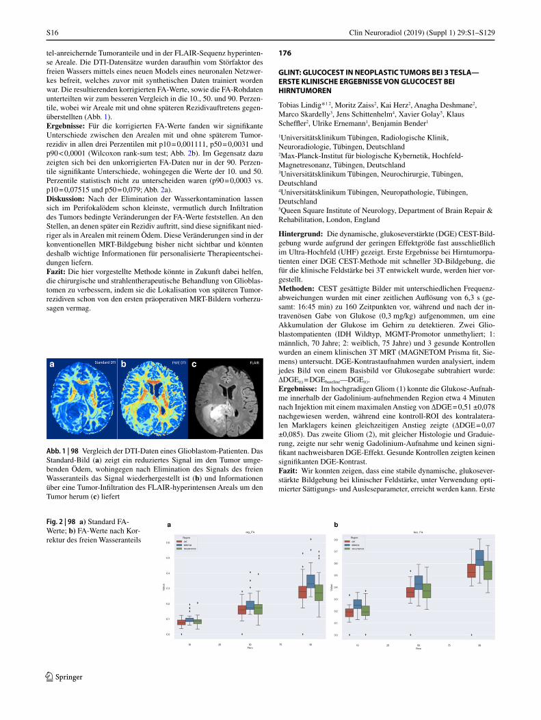
tel-anreichernde Tumoranteile und in der FLAIR-Sequenz hyperinten-se Areale. Die DTI-Datensätze wurden daraufhin vom Störfaktor des freien Wassers mittels eines neuen Models eines neuronalen Netzwer-kes befreit, welches zuvor mit synthetischen Daten trainiert worden war. Die resultierenden korrigierten FA-Werte, sowie die FA-Rohdaten unterteilten wir zum besseren Vergleich in die 10., 50. und 90. Perzen-tile, wobei wir Areale mit und ohne späteren Rezidivauftretens gegen-überstellten (Abb. 1).Ergebnisse: Für die korrigierten FA-Werte fanden wir signifikante Unterschiede zwischen den Arealen mit und ohne späterem Tumor-rezidiv in allen drei Perzentilen mit p10 = 0,001111, p50 = 0,0031 und p90 < 0,0001 (Wilcoxon rank-sum test; Abb. 2b). Im Gegensatz dazu zeigten sich bei den unkorrigierten FA-Daten nur in der 90. Perzen-tile signifikante Unterschiede, wohingegen die Werte der 10. und 50. Perzentile statistisch nicht zu unterscheiden waren (p90 = 0,0003 vs. p10 = 0,07515 und p50 = 0,079; Abb. 2a).Diskussion: Nach der Elimination der Wasserkontamination lassen sich im Perifokalödem schon kleinste, vermutlich durch Infiltration des Tumors bedingte Veränderungen der FA-Werte feststellen. An den Stellen, an denen später ein Rezidiv auftritt, sind diese signifikant nied-riger als in Arealen mit reinem Ödem. Diese Veränderungen sind in der konventionellen MRT-Bildgebung bisher nicht sichtbar und könnten deshalb wichtige Informationen für personalisierte Therapieentschei-dungen liefern.Fazit: Die hier vorgestellte Methode könnte in Zukunft dabei helfen, die chirurgische und strahlentherapeutische Behandlung von Glioblas-tomen zu verbessern, indem sie die Lokalisation von späteren Tumor-rezidiven schon von den ersten präoperativen MRT-Bildern vorherzu-sagen vermag.
176
GLINT: GLUCOCEST IN NEOPLASTIC TUMORS BEI 3 TESLA—ERSTE KLINISCHE ERGEBNISSE VON GLUCOCEST BEI HIRNTUMOREN
Tobias Lindig*1 2, Moritz Zaiss2, Kai Herz2, Anagha Deshmane2, Marco Skardelly3, Jens Schittenhelm4, Xavier Golay5, Klaus Scheffler2, Ulrike Ernemann1, Benjamin Bender1
1Universitätsklinikum Tübingen, Radiologische Klinik, Neuroradiologie, Tübingen, Deutschland2Max-Planck-Institut für biologische Kybernetik, Hochfeld-Magnetresonanz, Tübingen, Deutschland3Universitätsklinikum Tübingen, Neurochirurgie, Tübingen, Deutschland4Universitätsklinikum Tübingen, Neuropathologie, Tübingen, Deutschland5Queen Square Institute of Neurology, Department of Brain Repair & Rehabilitation, London, England
Hintergrund: Die dynamische, glukoseverstärkte (DGE) CEST-Bild-gebung wurde aufgrund der geringen Effektgröße fast ausschließlich im Ultra-Hochfeld (UHF) gezeigt. Erste Ergebnisse bei Hirntumorpa-tienten einer DGE CEST-Methode mit schneller 3D-Bildgebung, die für die klinische Feldstärke bei 3T entwickelt wurde, werden hier vor-gestellt.Methoden: CEST gesättigte Bilder mit unterschiedlichen Frequenz-abweichungen wurden mit einer zeitlichen Auflösung von 6,3 s (ge-samt: 16:45 min) zu 160 Zeitpunkten vor, während und nach der in-travenösen Gabe von Glukose (0,3 mg/kg) aufgenommen, um eine Akkumulation der Glukose im Gehirn zu detektieren. Zwei Glio-blastompatienten (IDH Wildtyp, MGMT-Promotor unmethyliert; 1: männlich, 70 Jahre; 2: weiblich, 75 Jahre) und 3 gesunde Kontrollen wurden an einem klinischen 3T MRT (MAGNETOM Prisma fit, Sie-mens) untersucht. DGE-Kontrastaufnahmen wurden analysiert, indem jedes Bild von einem Basisbild vor Glukosegabe subtrahiert wurde: ∆DGE(t) = DGEbaseline—DGE(t).Ergebnisse: Im hochgradigen Gliom (1) konnte die Glukose-Aufnah-me innerhalb der Gadolinium-aufnehmenden Region etwa 4 Minuten nach Injektion mit einem maximalen Anstieg von ∆DGE = 0,51 ±0,078 nachgewiesen werden, während eine kontroll-ROI des kontralatera-len Marklagers keinen gleichzeitigen Anstieg zeigte (∆DGE = 0,07 ±0,085). Das zweite Gliom (2), mit gleicher Histologie und Graduie-rung, zeigte nur sehr wenig Gadolinium-Aufnahme und keinen signi-fikant nachweisbaren DGE-Effekt. Gesunde Kontrollen zeigten keinen signifikanten DGE-Kontrast.Fazit: Wir konnten zeigen, dass eine stabile dynamische, glukosever-stärkte Bildgebung bei klinischer Feldstärke, unter Verwendung opti-mierter Sättigungs- und Ausleseparameter, erreicht werden kann. Erste
Abb. 1 | 98 Vergleich der DTI-Daten eines Glioblastom-Patienten. Das Standard-Bild (a) zeigt ein reduziertes Signal im den Tumor umge-benden Ödem, wohingegen nach Elimination des Signals des freien Wasseranteils das Signal wiederhergestellt ist (b) und Informationen über eine Tumor-Infiltration des FLAIR-hyperintensen Areals um den Tumor herum (c) liefert
Fig. 2 | 98 a) Standard FA-Werte; b) FA-Werte nach Kor-rektur des freien Wasseranteils

S17
123
Clin Neuroradiol (2019) (Suppl 1) 29:S1–S129
Ergebnisse sind vielversprechend und deuten darauf hin, dass Gluco-CEST eher einer Störung der Blut-Hirnschranke mit Gadolinium-Auf-nahme entspricht als dem molekularen Tumorprofil oder der Graduie-rung des Tumors.
189
MR SPECTROSCOPY-BASED RADIOGENOMIC ANALYSIS OF BRAIN TUMOR PATIENTS
Urs Würtemberger*1, Horst Urbach1, Irene Hübschle2, Pamela Heiland2, Dieter Henrik Heiland2
1Uniklinik Freiburg—Klinik für Neuroradiologie, Freiburg im Breisgau, Deutschland2Uniklinik Freiburg—Klinik für Neurochirurgie, Freiburg im Breisgau, Deutschland
Background: The 2016 revision of the WHO brain tumor classifica-tion incorporates for the first time genetic and molecular parameters, and thereby challenges the tasks of MR-imaging. We report a prospec-tive imaging study evaluating MR spectroscopy in brain tumor imag-ing. The study aim was a precise preoperative molecular prediction of brain tumors. We mapped the landscape of metabolic patterns and its heterogenic distribution in health and disease.Methods: 105 patients received anatomical MRI and multivoxel MR spectroscopy using a semiLASER-sequence with 5 × 5 × 20 mm vox-el size. Tumor regions were segmented and registered to equivalent spectroscopic voxels. We extracted the metabolic pattern of 15760 sin-gle-voxel and mapped the distribution of metabolic pattern. Clinical data were collected and used for further validation of our computa-tional model.Result: Cluster analysis identified 15 distinct clusters based on gap-sta-tistic algorithm (5 main clusters C1-5 and 2–3 subclusters). C1 consist-
ed primarily of spectra corresponding to the normal appearing matter. C2 was composed of spectra associated with the ventricles and subven-tricular regions. C3-5 represent various voxels in pathological lesions and brain tumors. Within clusters C3-5 an accurate separation of all glioma subgroups in 59.2% of the voxels was determined. The conven-tionally used metabolites which were used to classify tumor-spectra showed a subordinate role.Discussion: Radiomic studies focus on structural imaging, but no as-sociation between radiomic features and genetic alterations can be demonstrated so far. MR spectroscopy allows the detection of meta-bolic changes and provides insights into the molecular properties of the tumour. We demonstrate strong metabolic heterogeneity of brain tum-ors complicating sufficient prediction of genetic subgroups. However, a large part of the tumor localized spectra can be distinctly assigned to their underlying genetic alterations.Conclusion: MR-spectroscopy based radiogenomic prediction of ge-netic and molecular subtypes in brain tumor patients shows to be a robust tool in preoperative non-invasive imaging work-up. MR-spec-troscopy has the potential to include the need of “personalized medi-cine” by improving neurooncologic imaging and could influence in the future the clinical and surgical management of brain tumor patients.
References 1. Diamandis E, et al. MR-spectroscopic imaging of glial tumors
in the spotlight of the 2016 WHO classification. J Neurooncol. 2018;139(2):431–40. Sep.
Fig. 1 | 189
Fig. 2 | 189

S18
123
Clin Neuroradiol (2019) (Suppl 1) 29:S1–S129
220
PSEUDOPROGRESSION IN PEDIATRIC LOW-GRADE GLIOMA FOLLOWING PRIMARY RADIOTHERAPY—FIRST RESULTS FROM THE GERMAN LGG—STUDY COHORT
Annika Stock*1, Caroline-Victoria Hancken1, Daniela Kandels2, Astrid K. Gnekow2, Rolf-Dieter Kortmann3, Beate Timmermann4, Torsten Pietsch5, Brigitte Bison1, Mirko Pham1, Monika Warmuth-Metz1
1Universitätsklinikum Würzburg, Institut für Diagnostische und Interventionelle Neuroradiologie, Würzburg, Deutschland2SIOP-LGG-Studienzentrale, Universitätsklinikum Augsburg, Augsburg, Deutschland3Klinik für Strahlentherapie und Radioonkologie, Universitätsklinikum Leipzig, Leipzig, Deutschland4Klinik für Partikeltherapie (WPE), Universitätsklinikum Essen, Essen, Deutschland5Institut für Neuropathologie, Universitätsklinikum Bonn, Bonn, Deutschland
Background: Early expansion of tumor volume and/or edema follow-ing radiotherapy may indicate pseudoprogression (PsPD). The differ-entiation between true progression (PD) and PsPD is frequently diffi-cult but nevertheless of utmost importance because PD usually calls for a change in treatment. The aim of this study was to examine the incidence of PsPD in pediatric low-grade gliomas (LGG) with regard to the individual radiation modalities.Methods: 74 pediatric patients of the SIOP-LGG 2004 study cohort and LGG-registry cohort after primary radiotherapy (RT) were includ-ed, representing nearly half of all documented patients with sufficient image data sets). MRI controls at 3, 6, 12, 18 and 24 months post-treat-ment were evaluated. PsPD was considered upon significant increase in tumor size, increasing perifocal edema or increase of contrast medium enhancement. PsPD was determined when these changes decreased on further follow-up MRIs or a reduction in tumor size was noted despite progressive edema or contrast enhancement.Result: Treatment modalities were iodine seeds in 27/74 patients (36.5%), photon-therapy in 35/74 (47.3%) and proton-therapy in 12/74 (16.2%). PsPD most often started at month 3 and 6 and was charac-terized by progressive edema in most cases. We found a total of 34/74 (45.9%) PsPD (iodine seeds 12/27 [44.4%], photon-therapy 17/35 [48.6%] and proton-therapy 5/12 [41.7%]). PsPD did not correlate to the mode of RT applied (Chi-square test).Conclusion: PsPD is a frequent phenomenon and much more common in LGG than in high-grade glioma1. PsPD frequency does not seem to differ significantly between iodine seed-, proton- and photon-therapy.
References 1. Carceller F. et al. Pseudoprogression in children, adolescents and
young adults with non-brainstem high grade glioma and diffuse intrinsic pontine glioma. J Neurooncol. 2016;129:109–21.
245
LONGITUDINAL, LEAKAGE CORRECTED AND UNCORRECTED RCBV DURING THE FIRST-LINE TREATMENT OF GLIOBLASTOMA: A PROSPECTIVE STUDY.
Eike Steidl*1, Mathias Müller2, Andreas Müller2, Ulrich Herrlinger3, Elke Hattingen1
1Institut für Neuroradiologie, Universitätsklinikum Frankfurt , Frankfurt, Deutschland2Radiologische Klinik, Universitätsklinikum Bonn, Bonn, Deutschland3Klinik und Poliklinik für Neurologie, Universitätsklinikum Bonn, Sektion Klinische Neuroonkologie, Bonn, Deutschland
Hintergrund: Dynamic susceptibility contrast (DSC) MR-perfusion is becoming a standard of care for the monitoring of glioblastoma. Yet, technical standards are lacking and measurements without leakage correction are still common [1]. Also, data on leakage corrected meas-urements during stable disease is scarce. In this study we hypothesized that basic leakage correction would significantly enhance data quality during stable disease and improve progress detection. We furthermore investigated whether longitudinal data could increase diagnostic per-formance.Methoden: Patients with histologically proven glioblastoma undergo-ing first-line therapy were prospectively recruited. We conducted DSC perfusion measurements without prebolus administration in 6-week intervals from the end of radiotherapy until progression. Maximum relative cerebral volume values (rCBVmax) with and without leakage correction were calculated using Philips IntelliSpace®.Ergebnisse: We recruited 16 patients and conducted 82 MRI scans with a mean follow up of 7.2 month. During stable disease, corrected rCBVmax was significantly more stable than uncorrected rCBVmax. Detection of progression with a rCBVmax cutoff was better for correct-ed (specificity 86%) than for uncorrected rCBVmax (specificity 41%). Interestingly, the increase of corrected rCBVmax upon progression also had a good diagnostic performance with a combination of both cutoffs delivering the best result (sensitivity/specificity 89%/93%).Diskussion: In our prospective study we demonstrated, that even an easy approach to leakage correction significantly improves data qual-ity and diagnostic performance in detecting glioblastoma progression. Corrected rCBVmax supports the imaging finding of a stable disease and large increases during longitudinal observation support the diag-nosis of tumor progression.Fazit: Further studies to investigate the value of longitudinal rCBV dynamics for the differentiation of real tumor progression from pseu-doprogression are warranted.
Literatur 1. Welker K, Boxerman J, Kalnin A, Kaufmann T, Shiroishi M, Win-
termark M. ASFNR recommendations for clinical performance of MR dynamic susceptibility contrast perfusion imaging of the brain. Ajnr Am J Neuroradiol. 2015;36:E41–51. https://doi.org/10.3174/ajnr.A4341.
Fig. 1 | 220 Massive PsPD after photon-therapy. a) Initially the tumor showed no perifocal ede-ma. b) Edema grew from little at 6 months to c) massive at 8 months post-therapy. d) At 12 months post-therapy perifocal edema regressed
S19
123
Clin Neuroradiol (2019) (Suppl 1) 29:S1–S129
311
RESTRICTED KETOGENIC DIET AND FASTING IN COMBINATION WITH RE-IRRADIATION IN GLIOBLASTOMA IMPACT INTRACELLULAR PH AND INTRACEREBRAL METABOLISM AS DETERMINED BY 1H/31P MR-SPECTROSCOPY
Katharina Wenger-Alakmeh*1, Marlies Wagner1, Nina von Mettenheim2, Patrick Harter3, Kea Franz4, Jörg Bojunga5, Elke Hattingen1, Joachim Steinbach2, Oliver Bähr2, Emmanouil Fokas6, Detlef Imhoff6, Claus Rödel6, Johannes Rieger7, Ulrich Pilatus1, Martin Voss2
1Department of Neuroradiology, University Hospital Frankfurt, Frankfurt, Deutschland2Dr. Senckenberg Institute of Neurooncology, University Hospital Frankfurt, Frankfurt, Deutschland3Institute of Neuropathology (Edinger-Institute), University Hospital Frankfurt, Frankfurt, Deutschland4Department of Neurosurgery, University Hospital Frankfurt, Frankfurt, Deutschland5Department of Medicine 1, University Hospital Frankfurt, Frankfurt, Deutschland6Department of Radiation Oncology, University Hospital Frankfurt, Frankfurt, Deutschland7Departement of Neurology, University Hospital Tuebingen, Frankfurt, Deutschland
Background: Some Glioblastoma (GBM) cells show increased glycoly-sis even in the presence of oxygen and may lack metabolic flexibility to oxidize ketone bodies (KB). This could result in a glucose dependent tu-mor being starved during fasting and unable to maintain a reversed pH gra-dient. The aim of this study was to explore metabolic effects of combining a calorie restricted ketogenic diet (CRKD)/fasting (CR) with re-irradia-tion therapy (RT) in patients with recurrent GBM using MR-Spectroscopy.Methods: The prospective multicentric, randomized study included an extended MRS protocol with 3D FID 31P CSI and a 2D 1H PRESS sequence. 50 patients with recurrent GBM and indication for RT were randomized (1:1) in 2 groups: Group A keeping a CRKD/CR for 9 days and group B keeping a balanced diet. RT was performed on day 4–8. 32 patients received MRS at baseline (day –1) and 23/32 on day 6. Voxels were selected from the area of recurrent tumor and the contralateral hemisphere (control). A basis set for LCModel including 3-hydroxybu-tyrate (ßOHB), acetone (Acn) and acetoacetate (AcAc) was simulated and metabolites were quantified.Results: 20 patients in Group A and 22 patients in Group B completed dietary intervention. 17/25 patients in group A had a serum ketone level of >0.5 mmol/l at day 6 during CRKD/CR. There was a significant dif-ference at day 6 between groups (p < 0.01). An Acn signal at 2.22 ppm was detected in 4 patients at day 6 (Group A). In one patient Acn signal was fitted within tumor tissue and NAWM, in all other patients within NAWM only (concentration range 0.12–0.28 mmol/l). Group A showed a significant increase in pHi in tumor voxels comparing baseline levels to day 6 (p = 0.028); there was no significant change for Group B. ATP levels were stable in tumor or control voxels in both groups.Discussion: With no significant decrease in ATP levels during CRKD/CR and maintenance of an alkaline pHi, our study suggests a meta-bolic flexibility of GBM cells with a possible breakdown of KB and shift towards oxidative phosphorylation. Correspondingly, metabolic inflexibility of tumor cells has been recently contradicted in rat glioma models with upregulation of monocarboxylate transporter 1 (MCT-1), facilitating uptake and oxidation of KB.1 Acn is freely diffusible across the BBB and might be detected earlier intracerebrally than AcAc and ßOHB, which depend on MCT upregulation.
References 1. de Feyter HM, Behar KL, Rao JU, et al. A ketogenic diet increases
transport and oxidation of ketone bodies in RG2 and 9L gliomas without affecting tumor growth. Neuro-oncology. 2016.
3. Entzündliche ZNS-Erkrankungen
74
SHORT-TERM REORGANIZATION OF CORTICAL INFORMATION FLOW IN MULTIPLE SCLEROSIS: A LONGITUDINAL FMRI EFFECTIVE CONNECTIVITY STUDY
Vinzenz Fleischer*1, Muthuraman Muthuraman1, Abdul Rauf Anwar2, Gabriel Gonzalez-Escamilla1, Angela Radetz1, René-Maxime Gracien3, Stefan Bittner1, Felix Luessi1, Sven G. Meuth4, Frauke Zipp1, Sergiu Groppa1
1Department of Neurology, Focus Program Translational Neuroscience (FTN), University Medical Center of the Johannes Gutenberg University, Mainz, Germany, Mainz, Deutschland2Bio-medical Engineering Department, University of Engineering & Technology, Lahore, Pakistan, Lahore, Pakistan3Department of Neurology and Brain Imaging Center, Goethe University, Frankfurt (Main), Germany, Frankfurt, Deutschland4Department of Neurology with Institute of Translational Neurology, University of Muenster, Germany, Muenster, Deutschland
Hintergrund: Effective connectivity (EC) as derived from functional MRI is able to explore causal effects between brain areas and can be traced longitudinally to depict reorganization processes in brain diseas-es like Multiple Sclerosis (MS). Thus, the application of EC analysis in MS may provide an imaging biomarker for disease progression. Here, we examined EC dynamics in MS patients and healthy controls (HC) at short-term intervals and in relation to clinical impairment.Methoden: Serial longitudinal 3-Tesla resting-state functional MRI was performed every 12-weeks over one year in twelve MS patients (mean age 41.7 ± 11.5 years). Twelve healthy subjects served as con-trols (mean age 33.5 ± 9.6 years). Two recently established approach-es for EC quantification were used: Causal Bayesian Network (CBN) and Time-resolved Partial Directed Coherence (TPDC). EC was de-termined between frontal, prefrontal, temporal, occipital, and parietal lobes, the cerebellum and deep grey matter nuclei. The EC strength was correlated with the Expanded Disability Status Scale (EDSS) and the Fatigue Scale for Motor and Cognitive functions (FSMC).Ergebnisse: Our findings demonstrated a longitudinal increase in EC between specific brain regions, detected in both the CBN and the TPDC analysis in MS patients. EC from the deep grey matter, fron-tal, prefrontal and temporal regions showed a continuous increase over the study period, whereas the EC from the remaining regions did not change over one year. No longitudinal changes in EC were attested in HC over the same period of time in either method. Furthermore, we observed an association between clinical performance and EC strength. In particular, the EC increase in fronto-cerebellar connections showed an inverse correlation with the EDSS and patients’ fatigue score.Diskussion: Our data depict continuous functional reorganization be-tween specific brain regions indicated by increasing EC over time in MS, which is not detectable in healthy controls. In particular, fron-to-cerebellar connections, which are closely related to clinical perfor-mance, may provide a marker of brain plasticity and functional reserve in MS.Fazit: Our longitudinal EC data provide novel insights into the recruit-ment of directed connections in MS patients over time, by showing for the first time where and when functional reorganization processes occur in clinically stable patients (neither inflammatory activity nor disease progression).
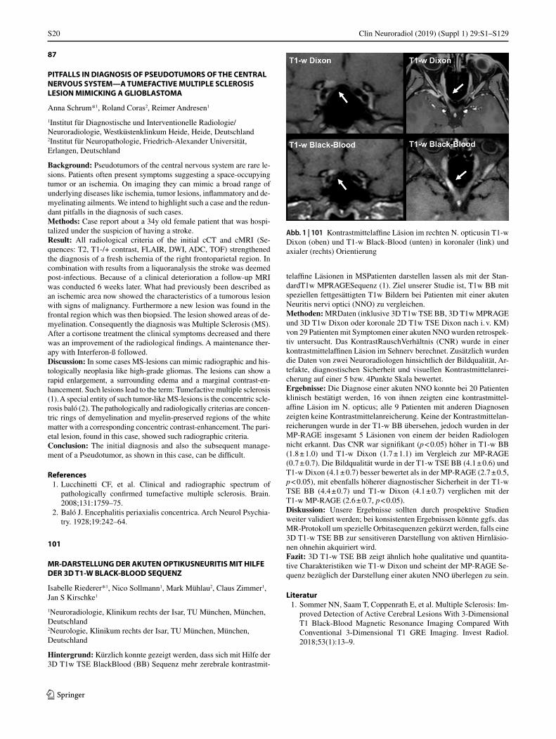
S20
123
Clin Neuroradiol (2019) (Suppl 1) 29:S1–S129
87
PITFALLS IN DIAGNOSIS OF PSEUDOTUMORS OF THE CENTRAL NERVOUS SYSTEM—A TUMEFACTIVE MULTIPLE SCLEROSIS LESION MIMICKING A GLIOBLASTOMA
Anna Schrum*1, Roland Coras2, Reimer Andresen1
1Institut für Diagnostische und Interventionelle Radiologie/Neuroradiologie, Westküstenklinkum Heide, Heide, Deutschland2Institut für Neuropathologie, Friedrich-Alexander Universität, Erlangen, Deutschland
Background: Pseudotumors of the central nervous system are rare le-sions. Patients often present symptoms suggesting a space-occupying tumor or an ischemia. On imaging they can mimic a broad range of underlying diseases like ischemia, tumor lesions, inflammatory and de-myelinating ailments. We intend to highlight such a case and the redun-dant pitfalls in the diagnosis of such cases.Methods: Case report about a 34y old female patient that was hospi-talized under the suspicion of having a stroke.Result: All radiological criteria of the initial cCT and cMRI (Se-quences: T2, T1-/+ contrast, FLAIR, DWI, ADC, TOF) strengthened the diagnosis of a fresh ischemia of the right frontoparietal region. In combination with results from a liquoranalysis the stroke was deemed post-infectious. Because of a clinical deterioration a follow-up MRI was conducted 6 weeks later. What had previously been described as an ischemic area now showed the characteristics of a tumorous lesion with signs of malignancy. Furthermore a new lesion was found in the frontal region which was then biopsied. The lesion showed areas of de-myelination. Consequently the diagnosis was Multiple Sclerosis (MS). After a cortisone treatment the clinical symptoms decreased and there was an improvement of the radiological findings. A maintenance ther-apy with Interferon-ß followed.Discussion: In some cases MS-lesions can mimic radiographic and his-tologically neoplasia like high-grade gliomas. The lesions can show a rapid enlargement, a surrounding edema and a marginal contrast-en-hancement. Such lesions lead to the term: Tumefactive multiple sclerosis (1). A special entity of such tumor-like MS-lesions is the concentric scle-rosis baló (2). The pathologically and radiologically criterias are concen-tric rings of demyelination and myelin-preserved regions of the white matter with a corresponding concentric contrast-enhancement. The pari-etal lesion, found in this case, showed such radiographic criteria.Conclusion: The initial diagnosis and also the subsequent manage-ment of a Pseudotumor, as shown in this case, can be difficult.
References 1. Lucchinetti CF, et al. Clinical and radiographic spectrum of
pathologically confirmed tumefactive multiple sclerosis. Brain. 2008;131:1759–75.
2. Baló J. Encephalitis periaxialis concentrica. Arch Neurol Psychia-try. 1928;19:242–64.
101
MR-DARSTELLUNG DER AKUTEN OPTIKUSNEURITIS MIT HILFE DER 3D T1-W BLACK-BLOOD SEQUENZ
Isabelle Riederer*1, Nico Sollmann1, Mark Mühlau2, Claus Zimmer1, Jan S Kirschke1
1Neuroradiologie, Klinikum rechts der Isar, TU München, München, Deutschland2Neurologie, Klinikum rechts der Isar, TU München, München, Deutschland
Hintergrund: Kürzlich konnte gezeigt werden, dass sich mit Hilfe der 3D T1w TSE BlackBlood (BB) Sequenz mehr zerebrale kontrastmit-
telaffine Läsionen in MSPatienten darstellen lassen als mit der Stan-dardT1w MPRAGESequenz (1). Ziel unserer Studie ist, T1w BB mit speziellen fettgesättigten T1w Bildern bei Patienten mit einer akuten Neuritis nervi optici (NNO) zu vergleichen.Methoden: MRDaten (inklusive 3D T1w TSE BB, 3D T1w MPRAGE und 3D T1w Dixon oder koronale 2D T1w TSE Dixon nach i. v. KM) von 29 Patienten mit Symptomen einer akuten NNO wurden retrospek-tiv untersucht. Das KontrastRauschVerhältnis (CNR) wurde in einer kontrastmittelaffinen Läsion im Sehnerv berechnet. Zusätzlich wurden die Daten von zwei Neuroradiologen hinsichtlich der Bildqualität, Ar-tefakte, diagnostischen Sicherheit und visuellen Kontrastmittelanrei-cherung auf einer 5 bzw. 4Punkte Skala bewertet.Ergebnisse: Die Diagnose einer akuten NNO konnte bei 20 Patienten klinisch bestätigt werden, 16 von ihnen zeigten eine kontrastmittel-affine Läsion im N. opticus; alle 9 Patienten mit anderen Diagnosen zeigten keine Kontrastmittelanreicherung. Keine der Kontrastmittelan-reicherungen wurde in der T1-w BB übersehen, jedoch wurden in der MP-RAGE insgesamt 5 Läsionen von einem der beiden Radiologen nicht erkannt. Das CNR war signifikant (p < 0.05) höher in T1-w BB (1.8 ± 1.0) und T1-w Dixon (1.7 ± 1.1) im Vergleich zur MP-RAGE (0.7 ± 0.7). Die Bildqualität wurde in der T1-w TSE BB (4.1 ± 0.6) und T1-w Dixon (4.1 ± 0.7) besser bewertet als in der MP-RAGE (2.7 ± 0.5, p < 0.05), mit ebenfalls höherer diagnostischer Sicherheit in der T1-w TSE BB (4.4 ± 0.7) und T1-w Dixon (4.1 ± 0.7) verglichen mit der T1-w MP-RAGE (2.6 ± 0.7, p < 0.05).Diskussion: Unsere Ergebnisse sollten durch prospektive Studien weiter validiert werden; bei konsistenten Ergebnissen könnte ggfs. das MR-Protokoll um spezielle Orbitasequenzen gekürzt werden, falls eine 3D T1-w TSE BB zur sensitiveren Darstellung von aktiven Hirnläsio-nen ohnehin akquiriert wird.Fazit: 3D T1-w TSE BB zeigt ähnlich hohe qualitative und quantita-tive Charakteristiken wie T1-w Dixon und scheint der MP-RAGE Se-quenz bezüglich der Darstellung einer akuten NNO überlegen zu sein.
Literatur 1. Sommer NN, Saam T, Coppenrath E, et al. Multiple Sclerosis: Im-
proved Detection of Active Cerebral Lesions With 3-Dimensional T1 Black-Blood Magnetic Resonance Imaging Compared With Conventional 3-Dimensional T1 GRE Imaging. Invest Radiol. 2018;53(1):13–9.
Abb. 1 | 101 Kontrastmittelaffine Läsion im rechten N. opticusin T1-w Dixon (oben) und T1-w Black-Blood (unten) in koronaler (link) und axialer (rechts) Orientierung

S21
123
Clin Neuroradiol (2019) (Suppl 1) 29:S1–S129
106
SIMPLE MR PLANIMETRIC MEASUREMENTS FOR DIAGNOSIS AND OUTCOME PREDICTION OF MULTIPLE SCLEROSIS
Stephanie Mangesius*1 2, Sergiy Pereverzyev Jr.1 2, Lukas Lamplmayr1
2, Lukas Lenhart1 2, Gabriel Bsteh3 4, Sebastian Wurth3, Florian Deisenhammer3, Elke Gizewski1 2
1Medical University Innsbruck, Department of Neuroradiology, Innsbruck, Österreich2Medical University Innsbruck, Neuroimaging Core Facility, Insnbruck, Österreich3Medical University Innsbruck, Department of Neurology, Innsbruck, Österreich4Medical University Vienna, Department of Neurology, Wien, Österreich
Hintergrund: Brain volume change has been suggested as a reliable MRI predictor of disability in different stages of multiple sclerosis (MS). However, the measurement of volume change is not being used in daily clinical practice neither as a prognostic factor nor to monitor treatment effects, as no accepted methodology for routine clinical use exists to date. In this pilot study, the main objective was to investi-gate whether a number of easily applicable planimetric measurements proposed for different neurologic disease entities can predict the more complex volumetric measurements in patients with MS.Methoden: For estimating the volumetric characteristics (gray matter [GM], white matter [WM], cerebrospinal fluid [CSF]) using planimet-ric characteristics in 10 MS patients, we first employed a planimet-ric characteristic selection based on Spearman’s correlation coefficient (SC). For constructing volumetric characteristics estimators using the selected planimetric characteristics, we then used neural networks (NNs) functions.Ergebnisse: Planimetric characteristics with a high correlation with volumetric characteristics are listed in Table 1. In the considered pa-tient set, we constructed the NNs that predict the volumes of GM, WM, and CSF using the corresponding planimetric characteristics, given in in Table 1. The relative prediction errors of these NNs were below 11% for GM, 5% for WM and 15% for CSF (Fig. 1).
Diskussion: Even in this low patient number, we observed a high cor-relation between MR planimetric and volumetric measurements and a low prediction error, which makes the proposed approach very prom-ising.Fazit: Further studies in larger patient cohorts are needed to test MR planimetry as an easy to apply and robust imaging biomarker to eval-uate cerebral and ventricular volume in patients with MS with similar accuracy and strong correlations with volumetric measurements.
110
MEAN KURTOSIS WITHIN THE CORPUS CALLOSUM IS ASSOCIATED WITH CLINICAL IMPAIRMENT IN MULTIPLE SCLEROSIS
Christian Thaler*1, Tobias D. Faizy1, Anna Kyselyova1, Maxim Bester1, Sune Jespersen2, Brian Hansen2, Jan-Patrick Stellmann3, Christoph Heesen3, Jens Fiehler1, Susanne Siemonsen1
1Universitätsklinikum Hamburg-Eppendorf, Klinik für neuroradiologische Diagnostik und Intervention, Hamburg, Deutschland2Universität Aarhus, Department of Clinical Medicine, Center of Functionally Integrative Neuroscience, Aarhus, Dänemark3Universitätsklinikum Hamburg-Eppendorf, Klinik für Neurologie, Hamburg, Deutschland
Hintergrund: Clinical-radiological correlations are in general low in Multiple Sclerosis (MS). Recent studies, that focused on diffusional kurtosis imaging (DKI), have shown that mean kurtosis (MK) is sensi-tive to diffuse white and gray matter injury in MS and might improve clinical-radiological correlations.[1]Methoden: Fourteen patients with relapsing-remitting MS received brain imaging on a 3T MR scanner at baseline and again after 12 months. Scan protocols included a sagittal 3-dimensional FLAIR se-quence and a fast DKI sequence, recently introduced by Hansen et al.[2] Subsequently, MK was measured within the corpus callosum (CC), which was automatically segmented using FreeSurfer, and within a FLAIR lesion mask which was manually outlined for each patient. Ad-ditionally, imaging results were correlated with clinical impairment
Table 1 | 106 Planimetric characteristics with high correlation with volumetric characteristics
volume correlation planimetric characteristics
GM |SC|>0.85 Frontooccipital diameter, Midbrain diameter, Pontine area, Midbrain-to-pontine diameter ratio
WM |SC|>0.80 transverse skull diameter, Frontooccipital diameter, Midbrain diameter, Pontine area
CSF |SC|>0.80 Third ventricle width unaligned, Anterior segment of corpus callosum/genu, corpus callosum index (CCI), regional anterior CCI
Fig. 1 | 106 Relative prediction errors of the constructed NNs for predicting the volumes of GM, WM, CSF
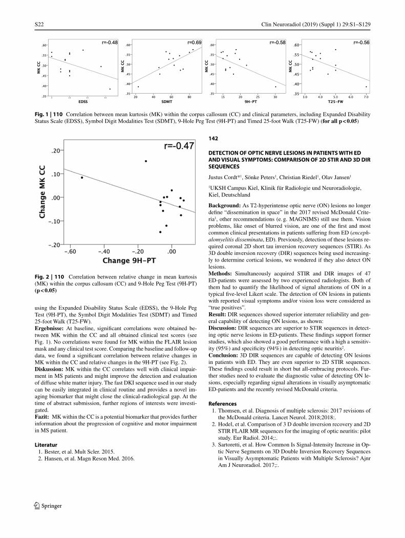
S22
123
Clin Neuroradiol (2019) (Suppl 1) 29:S1–S129
using the Expanded Disability Status Scale (EDSS), the 9-Hole Peg Test (9H-PT), the Symbol Digit Modalities Test (SDMT) and Timed 25-foot Walk (T25-FW).Ergebnisse: At baseline, significant correlations were obtained be-tween MK within the CC and all obtained clinical test scores (see Fig. 1). No correlations were found for MK within the FLAIR lesion mask and any clinical test score. Comparing the baseline and follow-up data, we found a significant correlation between relative changes in MK within the CC and relative changes in the 9H-PT (see Fig. 2).Diskussion: MK within the CC correlates well with clinical impair-ment in MS patients and might improve the detection and evaluation of diffuse white matter injury. The fast DKI sequence used in our study can be easily integrated in clinical routine and provides a novel im-aging biomarker that might close the clinical-radiological gap. At the time of abstract submission, further regions of interests were investi-gated.Fazit: MK within the CC is a potential biomarker that provides further information about the progression of cognitive and motor impairment in MS patient.
Literatur 1. Bester, et al. Mult Scler. 2015. 2. Hansen, et al. Magn Reson Med. 2016.
142
DETECTION OF OPTIC NERVE LESIONS IN PATIENTS WITH ED AND VISUAL SYMPTOMS: COMPARISON OF 2D STIR AND 3D DIR SEQUENCES
Justus Cordt*1, Sönke Peters1, Christian Riedel1, Olav Jansen1
1UKSH Campus Kiel, Klinik für Radiologie und Neuroradiologie, Kiel, Deutschland
Background: As T2-hyperintense optic nerve (ON) lesions no longer define “dissemination in space” in the 2017 revised McDonald Crite-ria1, other recommendations (e. g. MAGNIMS) still use them. Vision problems, like onset of blurred vision, are one of the first and most common clinical presentations in patients suffering from ED (enceph-alomyelitis disseminata, ED). Previously, detection of these lesions re-quired coronal 2D short tau inversion recovery sequences (STIR). As 3D double inversion recovery (DIR) sequences being used increasing-ly to determine cortical lesions, we wondered if they also detect ON lesions.Methods: Simultaneously acquired STIR and DIR images of 47 ED-patients were assessed by two experienced radiologists. Both of them had to quantify the likelihood of signal alterations of ON in a typical five-level Likert scale. The detection of ON lesions in patients with reported visual symptoms and/or vision loss were considered as “true positives”.Result: DIR sequences showed superior interrater reliability and gen-eral capability of detecting ON lesions, as shown:Discussion: DIR sequences are superior to STIR sequences in detect-ing optic nerve lesions in ED-patients. These findings support former studies, which also showed a good performance with a high a sensitiv-ity (95%) and specificity (94%) in detecting optic neuritis2.Conclusion: 3D DIR sequences are capable of detecting ON lesions in patients with ED. They are even superior to 2D STIR sequences. These findings could result in short but all-embracing protocols. Fur-ther studies need to evaluate the diagnostic value of detecting ON le-sions, especially regarding signal alterations in visually asymptomatic ED-patients and the recently revised McDonald criteria.
References 1. Thomsen, et al. Diagnosis of multiple sclerosis: 2017 revisions of
the McDonald criteria. Lancet Neurol. 2018;2018:. 2. Hodel, et al. Comparison of 3 D double inversion recovery and 2D
STIR FLAIR MR sequences for the imaging of optic neuritis: pilot study. Eur Radiol. 2014;:.
3. Sartoretti, et al. How Common Is Signal-Intensity Increase in Op-tic Nerve Segments on 3D Double Inversion Recovery Sequences in Visually Asymptomatic Patients with Multiple Sclerosis? Ajnr Am J Neuroradiol. 2017;:.
Fig. 1 | 110 Correlation between mean kurtosis (MK) within the corpus callosum (CC) and clinical parameters, including Expanded Disability Status Scale (EDSS), Symbol Digit Modalities Test (SDMT), 9-Hole Peg Test (9H-PT) and Timed 25-foot Walk (T25-FW) (for all p < 0 05)
Fig. 2 | 110 Correlation between relative change in mean kurtosis (MK) within the corpus callosum (CC) and 9-Hole Peg Test (9H-PT) (p < 0 05)
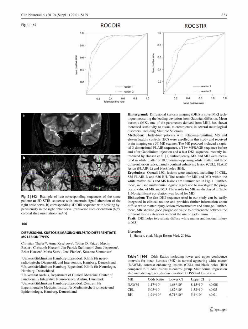
S23
123
Clin Neuroradiol (2019) (Suppl 1) 29:S1–S129
166
DIFFUSIONAL KURTOSIS IMAGING HELPS TO DIFFERENTIATE MS LESION TYPES
Christian Thaler*1, Anna Kyselyova1, Tobias D. Faizy1, Maxim Bester1, Christoph Heesen2, Jan-Patrick Stellmann2, Sune Jespersen3, Brian Hansen3, Maria Stark4, Jens Fiehler1, Susanne Siemonsen1
1Universitätsklinikum Hamburg-Eppendorf, Klinik für neuro-radiologische Diagnostik und Intervention, Hamburg, Deutschland2Universitätsklinikum Hamburg-Eppendorf, Klinik für Neurologie, Hamburg, Deutschland3Universität Aarhus, Department of Clinical Medicine, Center of Functionally Integrative Neuroscience, Aarhus, Dänemark4Universitätsklinikum Hamburg-Eppendorf, Zentrum für Experimentelle Medizin, Institut für Medizinische Biometrie und Epidemiologie, Hamburg, Deutschland
Hintergrund: Diffusional kurtosis imaging (DKI) is novel MRI tech-nique measuring the leading deviation from Gaussian diffusion. Mean kurtosis (MK), one of the parameters derived from MKI, has shown increased sensitivity to tissue microstructure in several neurological disorders, including Multiple Sclerosis.Methoden: Thirty-four patients with relapsing-remitting MS and eleven healthy controls (HC) were enrolled in this study and received brain imaging on a 3T MR scanner. The MR protocol included a sagit-tal 3-dimensional FLAIR sequence, a T1w MPRAGE sequence before and after Gadolinium injection and a fast DKI sequence, recently in-troduced by Hansen et al. [1] Subsequently, MK and MD were meas-ured in white matter of HC, normal-appearing white matter and three different lesion types, namely contrast enhancing lesion (CEL), FLAIR lesion (FLAIR-L) and black holes (BH).Ergebnisse: Overall 1501 lesions were analyzed, including 30 CEL, 835 FLAIR-L and 636 BH. The results for MK and MD within the white matter ROIs and MS lesions are summarized in Fig. 1. Further-more, we used multinomial logistic regression to investigate the prog-nostic value of MK and MD. The results for MK are displayed in Table 1. No significant correlation was found for MD.Diskussion: The fast DKI sequence used in our study can be easily integrated in clinical routine and provides further information about diffuse white matter injury, lesion microstructure and damage. Further-more, MK showed good prognostic value to differentiate between the different lesion categories without the use of gadolinium.Fazit: DKI helps to evaluate diffuse white matter and lesional injury in MS.
Literatur 1. Hansen, et al. Magn Reson Med. 2016;:.
Fig. 1 | 142
Fig. 2 | 142 Example of two corresponding sequences of the same patient: a) 2D STIR sequence with uncertain signal alteration of the right optic nerve, b) corresponding 3D DIR sequence with striking hy-perintensity in the right optic nerve [transverse slice orientation (left), coronal slice orientation (right)]
Table 1 | 166 Odds Ratios including lower and upper confidence intervals for mean kurtosis (MK) in normal-appearing white matter (NAWM), contrast enhancing lesions (CEL) and black holes (BH) compared to FLAIR lesions as control group. Multinomial regression also included age, sex, disease duration, EDSS and lesion size
MK Odds Ratio Lower CI Upper CI p
NAWM 1.17*105 1.68*104 8.15*105 <0.001
CEL 5.03*101 1.82*100 1.52*103 <0.05
BH 1.91*10-1 6.71*10-2 5.4*10-1 <0.01
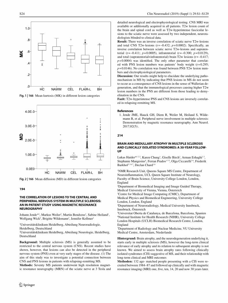
S24
123
Clin Neuroradiol (2019) (Suppl 1) 29:S1–S129
194
THE CORRELATION OF LESIONS TO THE CENTRAL AND PERIPHERAL NERVOUS SYSTEM IN MULTIPLE SCLEROSIS: AN IN-PATIENT STUDY USING MAGNETIC RESONANCE NEUROGRAPHY
Johann Jende*1, Markus Weiler2, Martin Bendszus1, Sabine Heiland1, Wolfgang Wick2, Brigitte Wildemann2, Jennifer Kollmer1
1Universitätsklinikum Heidelberg, Abteilung Neuroradiologie, Heidelberg, Deutschland2Universitätsklinikum Heidelberg, Abteilung Neurologie, Heidelberg, Deutschland
Background: Multiple sclerosis (MS) is generally assumed to be restricted to the central nervous system (CNS). Recent studies have shown, however, that lesions can also be detected in the peripheral nervous system (PNS) even at very early stages of the disease. (1) The aim of this study was to investigate a potential connection between CNS and PNS lesions in patients with relapsing-remitting MS.Methods: Seventy MS patients underwent high resolution magnet-ic resonance neurography (MRN) of the sciatic nerve at 3 Tesla and
detailed neurological and electrophysiological testing. CNS MRI was available or additionally acquired in all patients. T2w lesion count of the brain and spinal cord as well as T2w-hyperintense fascicular le-sions to the sciatic nerve were assessed by two independent, neurora-diologists blinded to clinical data.Result: There was an inverse correlation of sciatic nerve T2w-lesions and total CNS T2w-lesions (r = –0.432; p = 0.0002). Specifically, an inverse correlation between sciatic nerve T2w-lesions and supraten-torial (r = –0.411; p = 0.0005), infratentorial (r = –0.300; p = 0.0129), and total (supratentorial+infratentorial) brain T2w lesions (r = –0.417; p = 0.0004) was identified. The only other parameter that correlat-ed with PNS lesion numbers was patients’ body weight (r = 0.295; p = 0.0146). No correlation was found between PNS T2w lesion num-bers and electrophysiological parameters.Discussion: Our results might help to elucidate the underlying patho-mechanism in MS by indicating that PNS lesions in MS do not seem to occur as a consequence of CNS lesions in the sense of Wallerian de-generation, and that the immunological processes causing higher T2w lesion numbers in the PNS are different from those leading to demy-elination in the CNS.Fazit: T2w-hyperintense PNS and CNS lesions are inversely correlat-ed in relapsing-remitting MS.
References 1. Jende JME, Hauck GH, Diem R, Weiler M, Heiland S, Wilde-
mann B, et al. Peripheral nerve involvement in multiple sclerosis: Demonstration by magnetic resonance neurography. Ann Neurol. 2017;82(5):.
214
BRAIN AND MEDULLARY ATROPHY IN MULTIPLE SCLEROSIS AND CLINICALLY ISOLATED SYNDROMES: A 30-YEAR FOLLOW-UP
Lukas Haider*1 2 2, Karen Chung1, Giselle Birch1, Arman Eshaghi1 3, Stephanie Mangesius4, Ferran Prados1 3 5, Olga Ciccarelli1 6, Frederik Barkhof1 3 6 7, Declan Chard1 6
1NMR Research Unit, Queens Square MS Centre, Department of Neuroinflammation, UCL Queen Square Institute of Neurology, Faculty of Brain Science, University College London, London, England2Department of Biomedical Imaging and Image Guided Therapy, Medical University of Vienna, Vienna, Österreich3Centre for Medical Image Computing (CMIC), Department of Medical Physics and Biomedical Engineering, University College London, London, England4Department of Neuroradiology, Medical University Innsbruck, Innsbruck, Österreich5Universitat Oberta de Catalunya, de Barcelona, Barcelona, Spanien6National Institute for Health Research (NIHR), University College London Hospitals (UCLH) Biomedical Research Centre, London, England7Department of Radiology and Nuclear Medicine, VU University Medical Centre, Amsterdam, Niederlande
Hintergrund: Brain atrophy, and the neurodegeneration underlying it, starts early in multiple sclerosis (MS), however the long-term clinical relevance of early atrophy and its relation to subsequent atrophy is not known. We aimed to assess brain atrophy rates following clinically isolated syndromes (CIS) suggestive of MS, and their relationship with long-term clinical and MRI outcomesMethoden: 132 age- matched people presenting with a CIS were re-cruited between 1984–87 and followed up clinically and with magnetic resonance imaging (MRI) one, five, ten, 14, 20 and now 30 years later.
Fig. 1 | 166 Mean kurtosis (MK) in different lesion categories
Fig. 2 | 166 Mean diffusion (MD) in different lesion categories
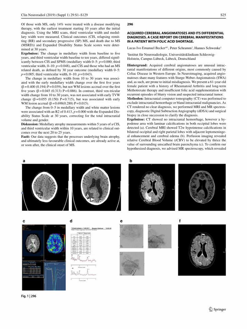
S25
123
Clin Neuroradiol (2019) (Suppl 1) 29:S1–S129
Of those with MS, only 14% were treated with a disease modifying therapy, with the earliest treatment starting 10 years after the initial diagnosis. Using the MRI scans, third ventricular width and medul-lary width were measured. Clinical outcomes (CIS, relapsing remit-ting (RR) and secondary progressive (SP) MS, and death due to MS (MSRD)) and Expanded Disability Status Scale scores were deter-mined at 30 years.Ergebnisse: The change in medullary width from baseline to five years, and third ventricular width baseline to ten years, differed signif-icantly between CIS and SPMS (medullary width 0–5: p = 0.000; third ventricular width, 0–10: p = 0.048), and CIS and those who had an MS related death, as defined by 30 year outcome (medullary width 0–5: p = 0.005; third ventricular width, 0–10: p = 0.043).
The change in medullary width from 10 to 30 years was associ-ated with the early medullary width change over the first five years (β = 0.400 (0.194) P = 0.039), but not WM lesions accrual over the first five years (β = 0.045 (0.313) P = 0.886). In contrast, third ven-tricular width change from 10 to 30 years, was not associated with early TVW change (β = 0.055 (0.150) P = 0.715), but was associated with early WM lesion accrual (β = 0.698(0.288) P = 0.015).
The change from 0–5 in medullary width and white matter lesions were associated with an R2 of 0.413, p = 0.000 with the Expanded Dis-ability Status Scale at 30 years, correcting for the total intracranial volume and gender.Diskussion: Medullary atrophy measurements within 5 years of a CIS, and third ventricular width within 10 years, are related to clinical out-comes over the next 20 to 25 years.Fazit: Our data suggests that the processes underlying brain atrophy, and ultimately less favourable clinical outcomes, are already active at, or soon after, the clinical onset of MS.
296
ACQUIRED CEREBRAL ANGIOMATOSES AND ITS DIFFERENTIAL DIAGNOSES. A CASE REPORT ON CEREBRAL MANIFESTATIONS IN A PATIENT WITH FOLIC ACID SHORTAGE.
Lucas Ivo Emanuel Becker*1, Peter Schramm1, Hannes Schwenke1
1Institut für Neuroradiologie, Universitätsklinikum Schleswig-Holstein, Campus Lübeck, Lübeck, Deutschland
Hintergrund: Acquired cerebral angiomatoses are unusual intrac-ranial manifestations of different origins, most commonly caused by Celiac Disease in Western Europe. In Neuroimaging, acquired angio-matoses share many features with Sturge-Weber-Angiomatosis (SWA) and, as such, are prone to initial misdiagnosis. We present a 61-year old female patient with a history of Rheumatoid Arthritis and long-term Methotrexate therapy and insufficient folic acid supplementation with recurrent episodes of blurry vision and suspected intracranial tumor.Methoden: Intracranial computer tomography (CT) was performed to exclude intracranial hemorrhage or bland intracranial malignancies. As CT rendered no clear diagnosis, we performed MRI and MR spectros-copy, diagnostic Digital Subtraction Angiography (dDSA) and surgical biopsy in close succession to clarify the diagnosis.Ergebnisse: CT showed no intracranial hemorrhage, however a hy-podense area with laminar calcifications in both occipital lobes were detected (a). Cerebral MRI showed T2w hypointense calcifications in bilateral occipital and right parietal lobes with adjacent leptomeninge-al enhancement and cerebral edema (b). Perfusion imaging revealed relative Cerebral Blood Volume (rCBV) to be elevated by thrice the value of surrounding unscathed brain parenchyma (c). To confirm our hypothesized diagnosis, we advised MR spectroscopy, which revealed
Fig. 1 | 296
S26
123
Clin Neuroradiol (2019) (Suppl 1) 29:S1–S129
an unusual intralesional choline peak (d). dDSA revealed bilateral oc-cipital blush (e). A follow-up MRI after 3 months revealed a regressive cerebral edema and constant leptomeningeal enhancement under folic acid supplementation (f). No more neurological deficit was seen after folic acid levels were replenished.Fazit: Other than in most publications, all possible imaging modali-ties (CT, MRI, MR Perfusion Imaging, MR Spectroscopy, dDSA) have been carried out in this case. MRI undoubtedly renders most informa-tion. Leptomeningeal calcifications with angiomatosis are rare and of-ten expressions of SWA. Gobbi-Syndrome should be considered when a history of Celiac Disease or a history of Methotrexate therapy with insufficient folic acid supplementation is present
Literatur 1. Wyllie E, Gupta A, Lachhwani DK. The treatment of epilepsy,
principles & practice. Lippincott Williams & Wilkins. (2006) ISBN:0781749956.
303
EXPLORATION OF MYELINATION DIFFERENCES IN INDIVIDUAL MULTIPLE SCLEROSIS LESIONS WITH MYELIN WATER IMAGING
Hagen H. Kitzler1, Hannes Wahl1, Paul Kuntke1, Tjalf Ziemssen2, Jennifer Linn1, Caroline Koehler1
1Institut für Diagnostische und Interventionelle Neuroradiologie, Medizinische Fakultät Carl Gustav Carus, Technische Universität Dresden, Dresden, Deutschland2Klinik und Poliklinik für Neurologie, Medizinische Fakultät Carl Gustav Carus, Technische Universität Dresden, Dresden, Deutschland
Background: Multiple sclerosis (MS) is an inflammatory disease of the central nervous system characterized by demyelinated lesions. Conventional magnetic resonance imaging (MRI) detects various pathological changes in the tissue but is non-specific for myelin. The in vivo MRI technique Multi-Component Driven Equalibrium Single Pulse Observation of T1 and T2 (mcDESPOT) [1], which allows the determination of relative myelination by measuring the myelin water fraction (MWF) [2], can enable the quantification of demyelination in lesions. The aim of this study was to assess the intra- and interindivid-ual variability of MWF in lesions to analyze differences in focal myelin loss in an RRMS cohort.Methoden: FLAIR images and mcDESPOT data were measured from n = 112 relapsing remitting patients. A 1.5T MR scanner and an 8-chan-nel head coil was used to obtain the multi-component T1 and T2 infor-mation from sets of Fast Low Angle SHot (FLASH) and True Fast Im-aging with Steady State Precession (TrueFISP) sequences over a range of flip angles measured at constant TR [1]. The co-registration of the MR data was done with Statistical Parametric Mapping (SPM) [3]. The MWF maps were calculated according to the established method from the mcDESPOT sequences [1]. The lesions were segmented into bina-ry masks. An algorithm was developed to automate the reading of the MWF of individual lesions for analysis of myelination differences. To compare two groups, the two-sided Wilcoxon rank sum test was used (significance level 5%).Results: The median MWF in the white matter (WM) of healthy con-trols showed a significant difference (p = 0.0011) to RRMS patients. 2787 WM FLAIR lesions were examined. The median MWF loss with-in the lesions varies from 6% to 70% for different patients compared to control white matter MWF levels. In a group comparison it could be shown that patients with a higher degree of disability of EDSS ≥ 4 significantly (p = 0.00026) have lower MWF values than patients with EDSS ≤ 2.
Discussion: Significant reduction and heterogeneity of MWF in NAWM and MS lesions has been identified, reflecting varying degrees of demyelination.Conclusion: This technique may be helpful to differentiate patients with early onset of strong myelin loss from those with low myelin loss.
References 1. Deoni, et al. Gleaning multicomponent T1 and T2 information
from steady-state imaging data. Magn Reson Med. 2008; 6:1372–87
2. Laule, et al. Magnetic resonance imaging of myelin. Neurothera-peutics. 2007;4(3):460–84.
3. https://www.fil.ion.ucl.ac.uk/spm/software
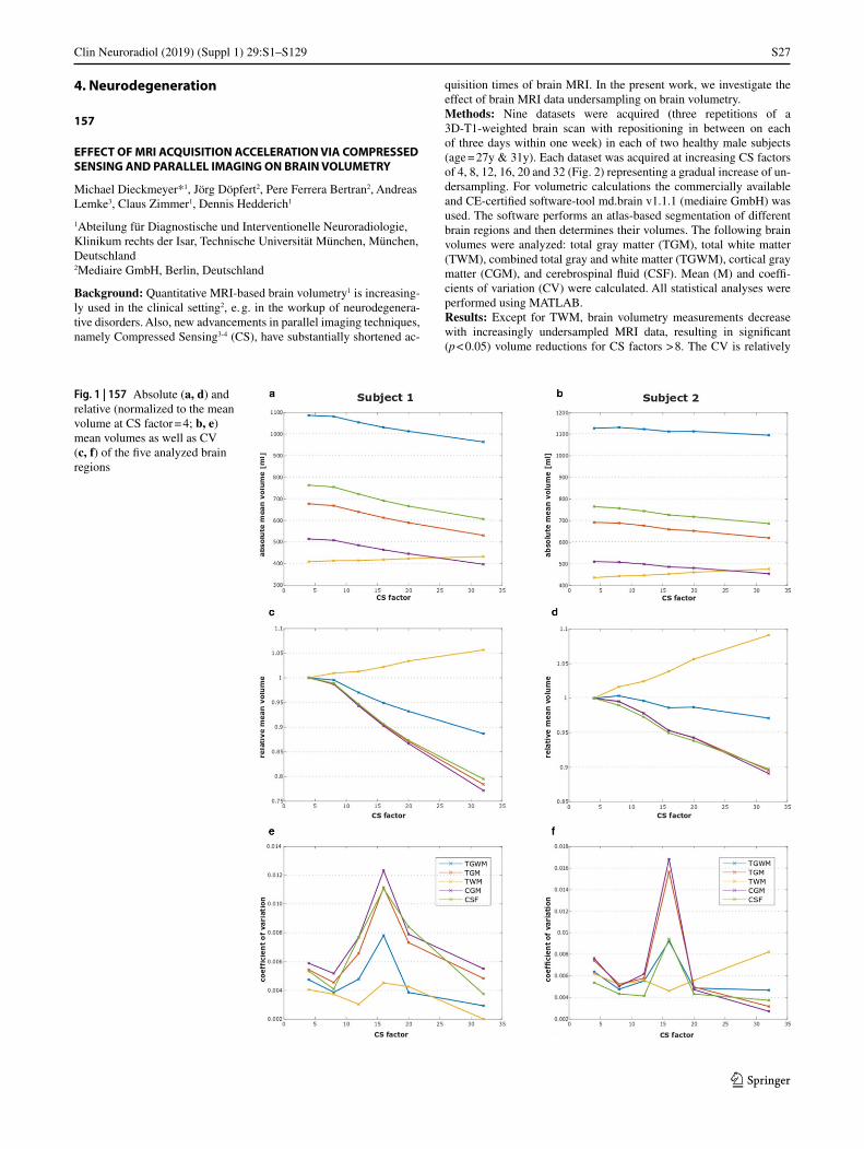
S27
123
Clin Neuroradiol (2019) (Suppl 1) 29:S1–S129
4. Neurodegeneration
157
EFFECT OF MRI ACQUISITION ACCELERATION VIA COMPRESSED SENSING AND PARALLEL IMAGING ON BRAIN VOLUMETRY
Michael Dieckmeyer*1, Jörg Döpfert2, Pere Ferrera Bertran2, Andreas Lemke3, Claus Zimmer1, Dennis Hedderich1
1Abteilung für Diagnostische und Interventionelle Neuroradiologie, Klinikum rechts der Isar, Technische Universität München, München, Deutschland2Mediaire GmbH, Berlin, Deutschland
Background: Quantitative MRI-based brain volumetry1 is increasing-ly used in the clinical setting2, e. g. in the workup of neurodegenera-tive disorders. Also, new advancements in parallel imaging techniques, namely Compressed Sensing3-4 (CS), have substantially shortened ac-
quisition times of brain MRI. In the present work, we investigate the effect of brain MRI data undersampling on brain volumetry.Methods: Nine datasets were acquired (three repetitions of a 3D-T1-weighted brain scan with repositioning in between on each of three days within one week) in each of two healthy male subjects (age = 27y & 31y). Each dataset was acquired at increasing CS factors of 4, 8, 12, 16, 20 and 32 (Fig. 2) representing a gradual increase of un-dersampling. For volumetric calculations the commercially available and CE-certified software-tool md.brain v1.1.1 (mediaire GmbH) was used. The software performs an atlas-based segmentation of different brain regions and then determines their volumes. The following brain volumes were analyzed: total gray matter (TGM), total white matter (TWM), combined total gray and white matter (TGWM), cortical gray matter (CGM), and cerebrospinal fluid (CSF). Mean (M) and coeffi-cients of variation (CV) were calculated. All statistical analyses were performed using MATLAB.Results: Except for TWM, brain volumetry measurements decrease with increasingly undersampled MRI data, resulting in significant (p < 0.05) volume reductions for CS factors > 8. The CV is relatively
Fig. 1 | 157 Absolute (a, d) and relative (normalized to the mean volume at CS factor = 4; b, e) mean volumes as well as CV (c, f) of the five analyzed brain regions

S28
123
Clin Neuroradiol (2019) (Suppl 1) 29:S1–S129
low for CS factors 4, 8, 20 and 32 and thus suggests a high precision whereas for CS factors 12 and 16 the CV.Discussion/Conclusion: We have shown that the deployment of par-allel imaging for brain MRI has a systematic biasing effect on volu-metric measurements of the brain that scales linearly with the degree of acceleration. Acceptable precision is reached for CS factors 4 and 8 which translates into acquisition times as low as 2 min 30sec for a whole brain volumetric dataset. Further research is needed to investi-gate the cause of the observed systematic bias and to analyze the effect on smaller brain subregions.
References 1. Ashburner J. . 2000;:. 2. Giorgio A. . 2013;:. 3. Donoho, D. (2006) 4. Candes, E. J. (2006)
171
DECREASE OF BRAIN REGIONAL CORTICAL THICKNESS IN AMYOTROPHIC LATERAL SCLEROSIS PATIENTS
Tong Fu*1, Kobeleva Xenia2, Birte Schmitz1, Paul Bronzlik1, Patric Nösel1, Mete Dadak1, Heinrich Lanfermann1, Susanne Petri2, Xiao-Qi Ding1
1Institute of Diagnostic and Interventional Neuroradiology, Hannover Medical School, Hannover, Deutschland2Clinics for Neurology, Hannover Medical School, Hannover, Deutschland
Hintergrund: Amyotrophic lateral sclerosis (ALS) is a fatal neuro-degenerative disease with heterogeneous motor and non-motor defi-
cits. This cross-section study aimed to assess the brain regional corti-cal thickness (rCTh) change in ALS in vivo by using quantitative MRI analysis.Methoden: 18 ALS patients (39–78 years, 58.22 ± 12.20 years, female: male = 4: 14) diagnosed according to EI Escorial criteria1 and 19 heathy control (HC) (39–70 years, 53.63 ± 7.50 years, female: male = 7: 12) underwent MRI examination. Surface based morphology were per-formed with CAT12 toolbox implanted in SPM12 to obtain global cortical thickness (gCTh), estimated total intracranial volume (eTIV), gray matter volume (GMV), and white matter volume (WMV); as well as rCTh defined based on DK_40 atlas2. The two-sided t-tests in gen-eral linear model were performed for the comparison between ALS pa-tients and HC. The threshold-free cluster enhancement3 was performed for multiple comparison correction (FDR, p < 0.05).Ergebnisse: In comparison to HC, significant lower GMV (p = 0.005) in ALS patients was found, as well as significant decreases of rCTh overlaid 20 of 34 ROIs (FDR, p = 0.016–0.042) in left hemisphere, and 15 of 34 ROIs (FDR, p = 0.016–0.046) in right hemisphere. These ROIs located in frontal (13 ROIs), temporal (11 ROIs), parietal (8 ROIs), occipital (1 ROI) lobes and insula.Diskussion: Thinning of regional cortical thickness with a frontal-tem-poral-parietal lobes distribution pattern seems to be major morpholog-ical alterations associated with ALS, which may be responsible to the motor and non-motor deficits in patients.Fazit: The decreases of rCTh may be used to fully understand and monitor pathological progress of ALS.
Literatur1. World Federation of Neurology Research Group on Neuromuscular
Diseases E1.. E1 Escorial World Federation of Neurology Criteria for the Diagnosis of Amyotrophic Lateral Sclerosis. J Neurol Sci. 1994;124:96–107.
Fig. 2 | 157 Sample axial reconstructions of the acquired 3D T1-weighted brain images at increasing CS factors of one of the subjects
S29
123
Clin Neuroradiol (2019) (Suppl 1) 29:S1–S129
2. Desikan RS, Ségonne F, Fischl B, et al. An automated labeling sys-tem for subdividing the human cerebral cortex on MRI scans into gyral based regions of interest. Neuroimage. 2006;31:968–80.
3. Smith SM, Nichols TE. Threshold-free cluster enhancement: Ad-dressing problems of smoothing, threshold dependence and locali-sation in cluster inference. Neuroimage. 2009;44:83–98.
184
RHEOLOGICALLY ESSENTIAL SURFACTANT PROTEINS OF THE CSF INTERACTING WITH PERIVENTRICULAR WHITE MATTER CHANGES IN HYDROCEPHALUS PATIENTS—IMPLICATIONS FOR CSF DYNAMICS AND THE GLYMPHATIC SYSTEM
Alexander Weiß*1, Stefan Schob2, Matthias Krause3, Wolfgang Härtig4, Karl-Titus Hoffmann2, Ulf Quäschling2, Anika Stockert5, Cindy Richter2, Joana Puchta2 4, Pervinder Bhogal6, Alexander Emmer7, Cordula Scherlach2
1Klinik für Radiologie und Nuklearmedizin, Universitätsklinikum Magdeburg, Magdeburg, Deutschland2Abteilung für Neuroradiologie, Universitätsklinikum Leipzig, Leipzig, Deutschland3Klinik für Neurochirurgie, Universitätsklinikum Leipzig, Leipzig, Deutschland4Paul-Flechsig-Institut für Hirnforschung, Universität Leipzig, Leipzig, Deutschland5Klinik für Neurologie, Universitätsklinikum Leipzig, Leipzig, Deutschland6Klinik für Interventionelle Neuroradiologie, Royal London Hospital, London, England7Klinik für Neurologie, Universitätsklinikum Halle-Wittenberg, Halle (Saale), Deutschland
Surfactant proteins (SP) are multi-systemic proteins playing crucial roles in the regulation of rheological properties of physiological flu-ids, host defense, and the clearance of potentially harmful metabo-lites. Hydrocephalus patients suffer from disturbed cerebrospinal fluid (CSF) homeostasis and remarkably altered SP concentrations within it. A connection between CSF-SPs, CSF flow, and ventricular dilatation, a morphological hallmark of hydrocephalus, has been reported previ-ously. Currently, there are no studies investigating the link between SPs and periventricular white matter changes caused by impaired CSF microcirculation in hydrocephalic conditions.
The present study included 47 individuals. CSF specimens were analyzed for concentrations of SP-A, SP-C, and SP-D by using ELI-SAs. Axial T2w TIRM MR imaging was employed in all cases. Using a MATLAB-based tool for quantification of MR signal intensities (SI) in the brain, parameters related to disturbed deep white matter CSF microcirculation were estimated (TIRM SI–mean, min, max, median, mode, std, percentiles p10th, p25th, p75th, p90th, and kurtosis, skew-ness, entropy of the SI distribution). Statistical analysis was performed to identify differences between hydrocephalic patients and healthy in-dividuals and to investigate the connections between CSF-SP changes and deep white matter SI.
SP-A and SP-C differed between healthy controls and hydroceph-alus patients in a significant manner. Corresponding quantification of white matter SI revealed differences between hydrocephalus patients and controls: SImean, SImax, SImedian, SImode, SIstd, all percen-tiles, skewness, and entropy. There were no differences for SP-D lev-els in both groups. In the acute hydrocephalic subgroup, SP-A showed a significant correlation with SImax, SIstd, SIp90, and inverse corre-lation with entropy. SP-C correlated inversely with entropy. For the chronic hydrocephalus subgroup, SP-A only correlated with kurtosis. SP-C correlated with SImean, SImax, SImedian, SImode, SIstd, SIp25, SIp75, and SIp90.
SP-A and SP-C are involved in different aspects of CSF physiology. SP-A appears to play an essential compensatory role in acute hydro-cephalus. In contrary, SP-C and white matter changes are remarkably connected in CSF of chronic hydrocephalus patients. Considering the association between CSF flow phenomena, white matter changes, and SP-C profiles, the latter may especially contribute to the regulation of glymphatic physiology. SP-D apparently does not play a major role in CSF homeostasis.
204
AUTOMATED AGE-SPECIFIC VOLUMETRIC ESTIMATION OF REGIONAL BRAIN ATROPHY: WORKFLOW AND FEASIBILITY
Julian Caspers*1, Adrian Heeger1, Bernd Turowski1, Christian Rubbert1
1Düsseldorf, Universitätsklinikum Düsseldorf, Institut für Diagnostische und Interventionelle Radiologie, Düsseldorf, Deutschland
Background: Estimating regional brain volume relative to the patient’s age norm from structural MRI is challenging and entails immense var-iations across readers. An automated framework for age-specific es-timation of brain volume changes relative to a standard population is presented and evaluated.Methods: The proposed workflow uses age- and sex-specific grey-mat-ter (GM) templates derived from a large population-based cohort to es-timate GM deviations in single subjects.
T1w MRIs of 693 healthy subjects aged between 16 and 77 years from the publicly available enhanced Nathan Kline Institute Rockland Sample were preprocessed in a standardized way comprising GM seg-mentation, normalization to MNI152 and 8 mm smoothing as imple-mented in CAT12 for SPM12. For each sex and age between 18 and 75, voxel-wise mean and standard deviation (SD) maps were generated across subjects aged two years under to two years over the respective age.
To estimate volume changes of a specific individual, his or her 3D T1w scan is preprocessed in the same way as for template generation. Then, voxel-wise z-values are generated from the resulting subject’s normalized GM map and the respective mean and SD template for his or her age and sex. The z-map is transformed into subject space, color coded and fused with the structural MRI.
The workflow was evaluated on T1w brain images of 205 subjects (age-range 55–75y, mean age 70y 67% females; 80 Alzheimer’s dis-ease patients, 76 mild cognitively impaired patients, 49 healthy con-trols) from the Alzheimer’s Dementia Neuroimaging Initiative (ADNI) by assessing the rate of technical success in atrophy map generation.Result: Atrophy map generation with the proposed workflow was suc-cessful in all 205 ADNI subjects (100%). A parieto-temporal atrophy pattern was clearly visible in most AD patients. Processing time for each subject was less than 10 min on a state-of-the-art server.Discussion: The atrophy maps have high potential to improve radio-logic evaluation of brain MRI by semi-quantitative assessment of atro-phy. The workflow will be made publicly available as open source with an integrated Picture Archiving and Communication System (PACS) interface.Conclusion: Automated brain volume change estimation with the pro-posed workflow is feasible and technically reliable.
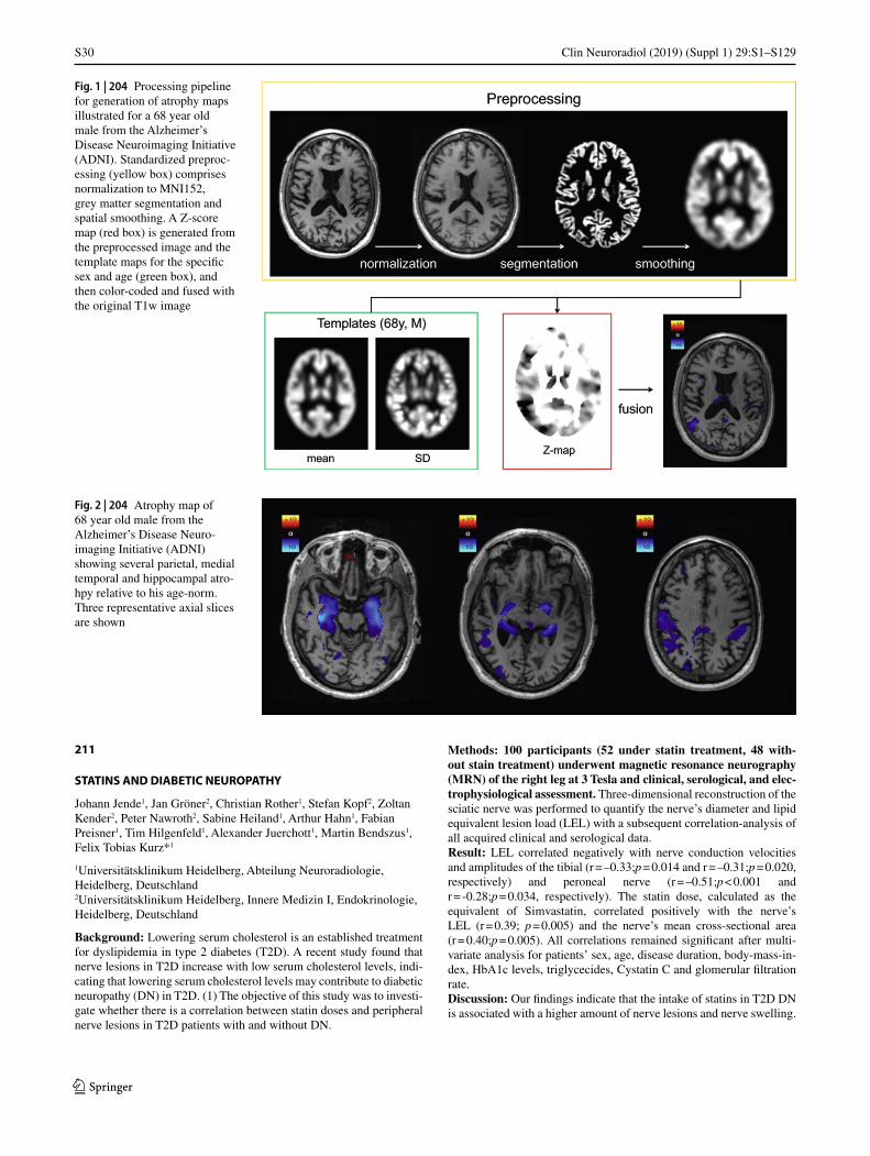
S30
123
Clin Neuroradiol (2019) (Suppl 1) 29:S1–S129
211
STATINS AND DIABETIC NEUROPATHY
Johann Jende1, Jan Gröner2, Christian Rother1, Stefan Kopf2, Zoltan Kender2, Peter Nawroth2, Sabine Heiland1, Arthur Hahn1, Fabian Preisner1, Tim Hilgenfeld1, Alexander Juerchott1, Martin Bendszus1, Felix Tobias Kurz*1
1Universitätsklinikum Heidelberg, Abteilung Neuroradiologie, Heidelberg, Deutschland2Universitätsklinikum Heidelberg, Innere Medizin I, Endokrinologie, Heidelberg, Deutschland
Background: Lowering serum cholesterol is an established treatment for dyslipidemia in type 2 diabetes (T2D). A recent study found that nerve lesions in T2D increase with low serum cholesterol levels, indi-cating that lowering serum cholesterol levels may contribute to diabetic neuropathy (DN) in T2D. (1) The objective of this study was to investi-gate whether there is a correlation between statin doses and peripheral nerve lesions in T2D patients with and without DN.
Methods: 100 participants (52 under statin treatment, 48 with-out stain treatment) underwent magnetic resonance neurography (MRN) of the right leg at 3 Tesla and clinical, serological, and elec-trophysiological assessment Three-dimensional reconstruction of the sciatic nerve was performed to quantify the nerve’s diameter and lipid equivalent lesion load (LEL) with a subsequent correlation-analysis of all acquired clinical and serological data.Result: LEL correlated negatively with nerve conduction velocities and amplitudes of the tibial (r = –0.33;p = 0.014 and r = –0.31;p = 0.020, respectively) and peroneal nerve (r = –0.51;p < 0.001 and r = -0.28;p = 0.034, respectively). The statin dose, calculated as the equivalent of Simvastatin, correlated positively with the nerve’s LEL (r = 0.39; p = 0.005) and the nerve’s mean cross-sectional area (r = 0.40;p = 0.005). All correlations remained significant after multi-variate analysis for patients’ sex, age, disease duration, body-mass-in-dex, HbA1c levels, triglycecides, Cystatin C and glomerular filtration rate.Discussion: Our findings indicate that the intake of statins in T2D DN is associated with a higher amount of nerve lesions and nerve swelling.
Fig. 2 | 204 Atrophy map of 68 year old male from the Alzheimer’s Disease Neuro-imaging Initiative (ADNI) showing several parietal, medial temporal and hippocampal atro-hpy relative to his age-norm. Three representative axial slices are shown
Fig. 1 | 204 Processing pipeline for generation of atrophy maps illustrated for a 68 year old male from the Alzheimer’s Disease Neuroimaging Initiative (ADNI). Standardized preproc-essing (yellow box) comprises normalization to MNI152, grey matter segmentation and spatial smoothing. A Z-score map (red box) is generated from the preprocessed image and the template maps for the specific sex and age (green box), and then color-coded and fused with the original T1w image
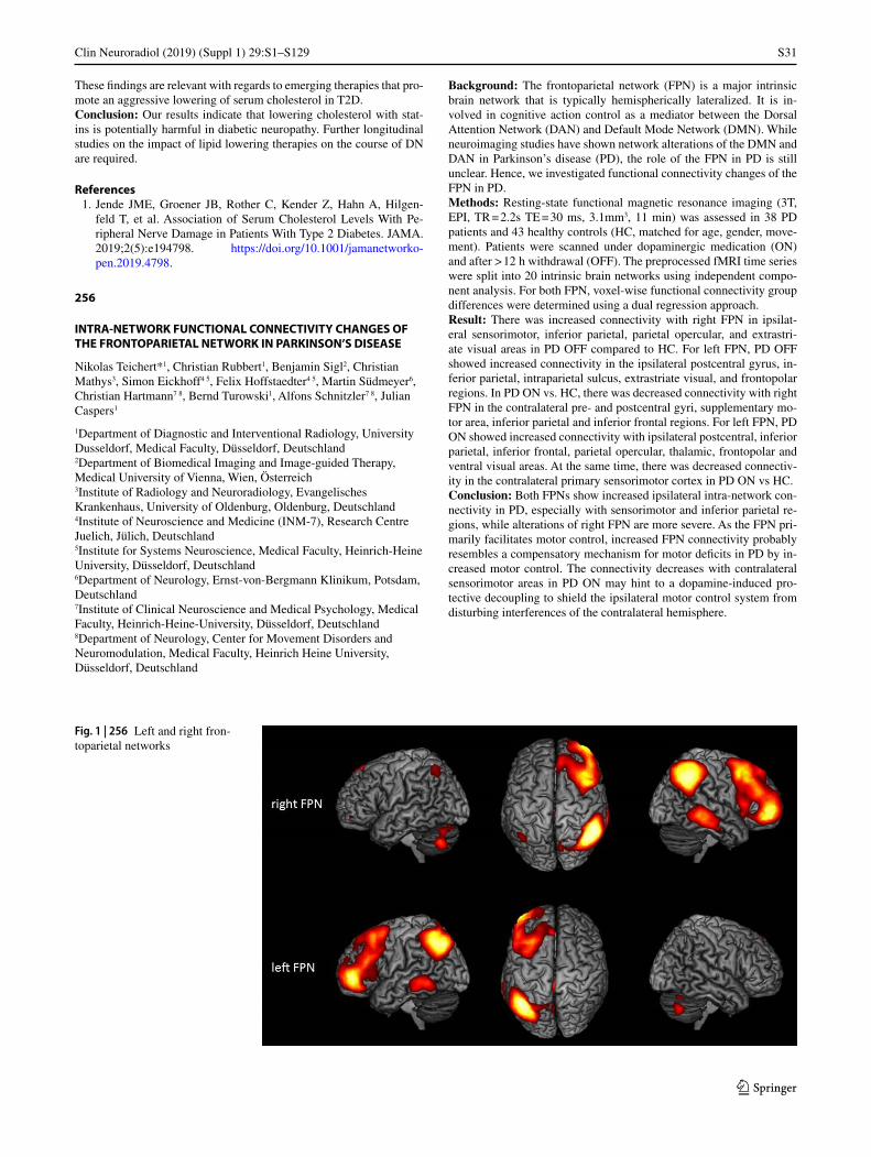
S31
123
Clin Neuroradiol (2019) (Suppl 1) 29:S1–S129
These findings are relevant with regards to emerging therapies that pro-mote an aggressive lowering of serum cholesterol in T2D.Conclusion: Our results indicate that lowering cholesterol with stat-ins is potentially harmful in diabetic neuropathy. Further longitudinal studies on the impact of lipid lowering therapies on the course of DN are required.
References 1. Jende JME, Groener JB, Rother C, Kender Z, Hahn A, Hilgen-
feld T, et al. Association of Serum Cholesterol Levels With Pe-ripheral Nerve Damage in Patients With Type 2 Diabetes. JAMA. 2019;2(5):e194798. https://doi.org/10.1001/jamanetworko-pen.2019.4798.
256
INTRA-NETWORK FUNCTIONAL CONNECTIVITY CHANGES OF THE FRONTOPARIETAL NETWORK IN PARKINSON’S DISEASE
Nikolas Teichert*1, Christian Rubbert1, Benjamin Sigl2, Christian Mathys3, Simon Eickhoff4 5, Felix Hoffstaedter4 5, Martin Südmeyer6, Christian Hartmann7 8, Bernd Turowski1, Alfons Schnitzler7 8, Julian Caspers1
1Department of Diagnostic and Interventional Radiology, University Dusseldorf, Medical Faculty, Düsseldorf, Deutschland2Department of Biomedical Imaging and Image-guided Therapy, Medical University of Vienna, Wien, Österreich3Institute of Radiology and Neuroradiology, Evangelisches Krankenhaus, University of Oldenburg, Oldenburg, Deutschland4Institute of Neuroscience and Medicine (INM-7), Research Centre Juelich, Jülich, Deutschland5Institute for Systems Neuroscience, Medical Faculty, Heinrich-Heine University, Düsseldorf, Deutschland6Department of Neurology, Ernst-von-Bergmann Klinikum, Potsdam, Deutschland7Institute of Clinical Neuroscience and Medical Psychology, Medical Faculty, Heinrich-Heine-University, Düsseldorf, Deutschland8Department of Neurology, Center for Movement Disorders and Neuromodulation, Medical Faculty, Heinrich Heine University, Düsseldorf, Deutschland
Background: The frontoparietal network (FPN) is a major intrinsic brain network that is typically hemispherically lateralized. It is in-volved in cognitive action control as a mediator between the Dorsal Attention Network (DAN) and Default Mode Network (DMN). While neuroimaging studies have shown network alterations of the DMN and DAN in Parkinson’s disease (PD), the role of the FPN in PD is still unclear. Hence, we investigated functional connectivity changes of the FPN in PD.Methods: Resting-state functional magnetic resonance imaging (3T, EPI, TR = 2.2s TE = 30 ms, 3.1mm3, 11 min) was assessed in 38 PD patients and 43 healthy controls (HC, matched for age, gender, move-ment). Patients were scanned under dopaminergic medication (ON) and after > 12 h withdrawal (OFF). The preprocessed fMRI time series were split into 20 intrinsic brain networks using independent compo-nent analysis. For both FPN, voxel-wise functional connectivity group differences were determined using a dual regression approach.Result: There was increased connectivity with right FPN in ipsilat-eral sensorimotor, inferior parietal, parietal opercular, and extrastri-ate visual areas in PD OFF compared to HC. For left FPN, PD OFF showed increased connectivity in the ipsilateral postcentral gyrus, in-ferior parietal, intraparietal sulcus, extrastriate visual, and frontopolar regions. In PD ON vs. HC, there was decreased connectivity with right FPN in the contralateral pre- and postcentral gyri, supplementary mo-tor area, inferior parietal and inferior frontal regions. For left FPN, PD ON showed increased connectivity with ipsilateral postcentral, inferior parietal, inferior frontal, parietal opercular, thalamic, frontopolar and ventral visual areas. At the same time, there was decreased connectiv-ity in the contralateral primary sensorimotor cortex in PD ON vs HC.Conclusion: Both FPNs show increased ipsilateral intra-network con-nectivity in PD, especially with sensorimotor and inferior parietal re-gions, while alterations of right FPN are more severe. As the FPN pri-marily facilitates motor control, increased FPN connectivity probably resembles a compensatory mechanism for motor deficits in PD by in-creased motor control. The connectivity decreases with contralateral sensorimotor areas in PD ON may hint to a dopamine-induced pro-tective decoupling to shield the ipsilateral motor control system from disturbing interferences of the contralateral hemisphere.
Fig. 1 | 256 Left and right fron-toparietal networks

S32
123
Clin Neuroradiol (2019) (Suppl 1) 29:S1–S129
289
IMPLICIT MOTOR LEARNING MODULATES FUNCTIONAL CONNECTIVITY OF DBS THERAPEUTIC TARGET FOR TREMOR.
Amr Farahat*1 2, Hermann Hinrichs3 4 5 6, Hans-Jochen Heinze3 4 5, Catherine Sweeney-Reed1
1Universitätsklinikum Magdeburg, Neurokybernetik und Rehabilitation Arbeitsgruppe, Abteilung Neurologie, Magdeburg, Deutschland2Universitätsklinikum Magdeburg, Neuroradiologie, Magdeburg, Deutschland3Universitätsklinikum Magdeburg, Abteilung Neurologie, Magdeburg, Deutschland4Leibniz-Institut für Neurobiologie, Abteilung Verhaltensneurologie, Magdeburg, Deutschland5Deutsches Zentrum für Neurodegenerative Erkrankungen e. V. (DZNE), Magdeburg, Deutschland6Forschungscampus STIMULATE, Magdeburg, Deutschland
Hintergrund: The ventral intermediate nucleus of the thalamus (VIM) is the therapeutic target of deep brain stimulation (DBS) for pharmacoresistant essential tremor (ET) and tremor-dominant Parkin-son’s disease (tPD). VIM-DBS effectiveness in tremor control is at-tributed to VIM involvement in the cerebello-thalamo-cortical circuit. Striato-cerebello-thalamo-cortical circuits are also thought to underlie motor learning. Even though the VIM involvement in these circuits are not proven, motor learning side-effects of VIM DBS were reported. Here we investigated whether the VIM is part of motor learning circuits by examining task-related VIM functional connectivity (FC) modula-tions in 30 right-handed healthy participants during 7T fMRI scanning.Methoden: A multiband GE-EPI sequence with parallel imaging was recorded during 4 runs of a serial reaction time task, which involves implicit motor learning. Each run comprised 6 alternating random (R) and learned (L) blocks to ensure implicit learning. Each L block com-prised 4 repetitions of a 12-trial sequence.Ergebnisse: Using the difference in the change in average reaction times (RTs) between R and L blocks across runs to indicate motor learning, we observed a significant reduction in the RTs between the first and the second, third, and fourth runs only during L blocks. A psychophysiological interactions (PPI) analysis of the fMRI data using the left VIM as a seed ROI was applied to examine change in VIM-FC from the first to fourth run. FC increased between the VIM and right
and left putamen, globus pallidus, amygdala and supplementary motor area (SMA), left precentral gyrus and superior and middle frontal gyri, suggesting VIM involvement in motor learning and a functional rela-tionship between VIM and basal ganglia.Diskussion: Structural connectivity has previously been identified between intraoperatively electrophysiologically-defined tremor-sup-pressive VIM-DBS sites and the pallidum. Moreover, resting state FC between VIM and the right putamen, SMAs and parietal lobe areas was also recently shown to correlate with ET severity. We provide evi-dence for VIM involvement also in implicit motor learning through the putamen and motor cortical areas, which are also involved in tremor generation.Fazit: These findings could explain VIM-DBS motor learning side-ef-fects and suggest a potential role for preoperative fMRI in DBS plan-ning to avoid potential side-effects.
Fig. 2 | 256 Functional connec-tivity increases (hot colors) and decreases (cool colors) of PD patients OFF and ON medica-tion within right and left FPN
Fig. 1 | 289
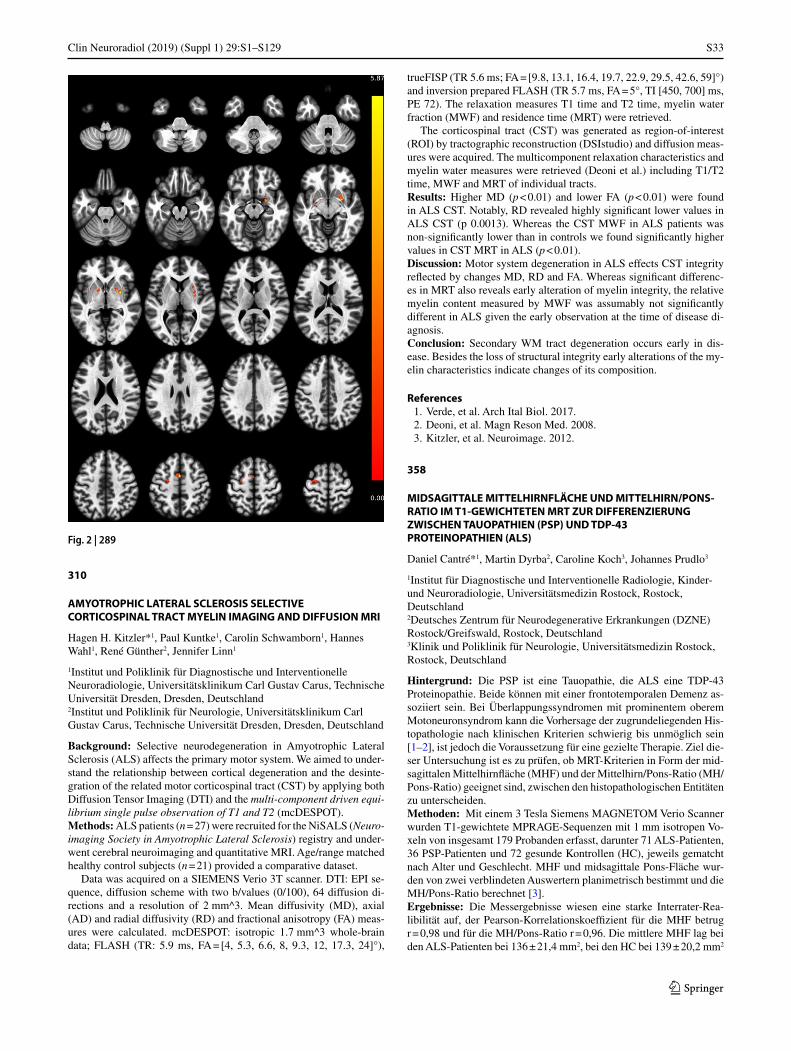
S33
123
Clin Neuroradiol (2019) (Suppl 1) 29:S1–S129
310
AMYOTROPHIC LATERAL SCLEROSIS SELECTIVE CORTICOSPINAL TRACT MYELIN IMAGING AND DIFFUSION MRI
Hagen H. Kitzler*1, Paul Kuntke1, Carolin Schwamborn1, Hannes Wahl1, René Günther2, Jennifer Linn1
1Institut und Poliklinik für Diagnostische und Interventionelle Neuroradiologie, Universitätsklinikum Carl Gustav Carus, Technische Universität Dresden, Dresden, Deutschland2Institut und Poliklinik für Neurologie, Universitätsklinikum Carl Gustav Carus, Technische Universität Dresden, Dresden, Deutschland
Background: Selective neurodegeneration in Amyotrophic Lateral Sclerosis (ALS) affects the primary motor system. We aimed to under-stand the relationship between cortical degeneration and the desinte-gration of the related motor corticospinal tract (CST) by applying both Diffusion Tensor Imaging (DTI) and the multi-component driven equi-librium single pulse observation of T1 and T2 (mcDESPOT).Methods: ALS patients (n = 27) were recruited for the NiSALS (Neuro-imaging Society in Amyotrophic Lateral Sclerosis) registry and under-went cerebral neuroimaging and quantitative MRI. Age/range matched healthy control subjects (n = 21) provided a comparative dataset.
Data was acquired on a SIEMENS Verio 3T scanner. DTI: EPI se-quence, diffusion scheme with two b/values (0/100), 64 diffusion di-rections and a resolution of 2 mm^3. Mean diffusivity (MD), axial (AD) and radial diffusivity (RD) and fractional anisotropy (FA) meas-ures were calculated. mcDESPOT: isotropic 1.7 mm^3 whole-brain data; FLASH (TR: 5.9 ms, FA = [4, 5.3, 6.6, 8, 9.3, 12, 17.3, 24]°),
trueFISP (TR 5.6 ms; FA = [9.8, 13.1, 16.4, 19.7, 22.9, 29.5, 42.6, 59]°) and inversion prepared FLASH (TR 5.7 ms, FA = 5°, TI [450, 700] ms, PE 72). The relaxation measures T1 time and T2 time, myelin water fraction (MWF) and residence time (MRT) were retrieved.
The corticospinal tract (CST) was generated as region-of-interest (ROI) by tractographic reconstruction (DSIstudio) and diffusion meas-ures were acquired. The multicomponent relaxation characteristics and myelin water measures were retrieved (Deoni et al.) including T1/T2 time, MWF and MRT of individual tracts.Results: Higher MD (p < 0.01) and lower FA (p < 0.01) were found in ALS CST. Notably, RD revealed highly significant lower values in ALS CST (p 0.0013). Whereas the CST MWF in ALS patients was non-significantly lower than in controls we found significantly higher values in CST MRT in ALS (p < 0.01).Discussion: Motor system degeneration in ALS effects CST integrity reflected by changes MD, RD and FA. Whereas significant differenc-es in MRT also reveals early alteration of myelin integrity, the relative myelin content measured by MWF was assumably not significantly different in ALS given the early observation at the time of disease di-agnosis.Conclusion: Secondary WM tract degeneration occurs early in dis-ease. Besides the loss of structural integrity early alterations of the my-elin characteristics indicate changes of its composition.
References 1. Verde, et al. Arch Ital Biol. 2017. 2. Deoni, et al. Magn Reson Med. 2008. 3. Kitzler, et al. Neuroimage. 2012.
358
MIDSAGITTALE MITTELHIRNFLÄCHE UND MITTELHIRN/PONS-RATIO IM T1-GEWICHTETEN MRT ZUR DIFFERENZIERUNG ZWISCHEN TAUOPATHIEN (PSP) UND TDP-43 PROTEINOPATHIEN (ALS)
Daniel Cantré*1, Martin Dyrba2, Caroline Koch3, Johannes Prudlo3
1Institut für Diagnostische und Interventionelle Radiologie, Kinder- und Neuroradiologie, Universitätsmedizin Rostock, Rostock, Deutschland2Deutsches Zentrum für Neurodegenerative Erkrankungen (DZNE) Rostock/Greifswald, Rostock, Deutschland3Klinik und Poliklinik für Neurologie, Universitätsmedizin Rostock, Rostock, Deutschland
Hintergrund: Die PSP ist eine Tauopathie, die ALS eine TDP-43 Proteinopathie. Beide können mit einer frontotemporalen Demenz as-soziiert sein. Bei Überlappungssyndromen mit prominentem oberem Motoneuronsyndrom kann die Vorhersage der zugrundeliegenden His-topathologie nach klinischen Kriterien schwierig bis unmöglich sein [1–2], ist jedoch die Voraussetzung für eine gezielte Therapie. Ziel die-ser Untersuchung ist es zu prüfen, ob MRT-Kriterien in Form der mid-sagittalen Mittelhirnfläche (MHF) und der Mittelhirn/Pons-Ratio (MH/Pons-Ratio) geeignet sind, zwischen den histopathologischen Entitäten zu unterscheiden.Methoden: Mit einem 3 Tesla Siemens MAGNETOM Verio Scanner wurden T1-gewichtete MPRAGE-Sequenzen mit 1 mm isotropen Vo-xeln von insgesamt 179 Probanden erfasst, darunter 71 ALS-Patienten, 36 PSP-Patienten und 72 gesunde Kontrollen (HC), jeweils gematcht nach Alter und Geschlecht. MHF und midsagittale Pons-Fläche wur-den von zwei verblindeten Auswertern planimetrisch bestimmt und die MH/Pons-Ratio berechnet [3].Ergebnisse: Die Messergebnisse wiesen eine starke Interrater-Rea-libilität auf, der Pearson-Korrelationskoeffizient für die MHF betrug r = 0,98 und für die MH/Pons-Ratio r = 0,96. Die mittlere MHF lag bei den ALS-Patienten bei 136 ± 21,4 mm2, bei den HC bei 139 ± 20,2 mm2
Fig. 2 | 289

S34
123
Clin Neuroradiol (2019) (Suppl 1) 29:S1–S129
und bei den PSP-Patienten bei 94.3 ± 19,2 mm2 (ALS vs. HC p = 0,77; ALS vs. PSP p < 0,0001). Die MH/Pons-Ratio betrug bei den ALS-Pa-tienten 0,246 ± 0,039, bei den HC 0,250 ± 0,036 und bei den PSP-Pa-tienten 0,185 ± 0,034 (ALS vs. HC p = 0,67; ALS vs. PSP P < 0,0001).Diskussion: MHF und MH/Pons-Ratio der ALS-Patienten liegen sig-nifikant oberhalb der Werte der PSP-Patienten und weisen keinen Un-terschied gegenüber gesunden Kontrollen auf. Beide Parameter eignen sich daher zur Differenzierung von ALS und PSP.Fazit: Die Messung der MHF ist ein geeigneter Surrogatmarker, um TPD 43-Proteinopathien (ALS) von Tauopathien (PSP) zu differenzie-ren. Als Einzel-Parameter ist sie in der klinischen Routine einfach zu bestimmen. Die MH/Pons-Ratio ist ebenfalls geeignet, aber aufwendi-ger zu bestimmen.
Literatur 1. McCluskey LF, et al. Amyotrophic lateral sclerosis-plus syndrome
with TAR DNA-binding protein-43 pathology. Arch Neurol. 2009;66(1):121–4. Jan.
2. Nagao S, et al. Progressive supranuclear palsy presenting as pri-mary lateral sclerosis but lacking parkinsonism, gaze palsy, apha-sia, or dementia. J Neurol Sci. 2012;15;323(1–2):147–53. Dec.
3. al Möller L. Manual MRI morphometry in Parkinsonian syn-dromes. Mov Disord. 2017;32(5):778–82. May.
5. Wirbelsäule
96
STATISTICAL PARAMETRIC MAPPING OF REGIONAL BONE DENSITY AT THE THORACOLUMBAR SPINE FOR OPPORTUNISTIC OSTEOPOROSIS SCREENING
Maximilian T. Löffler*1, Alexander Valentinitsch1, Alina Jacob1, Benita Schmitz-Koep1, Thomas Baum1, Claus Zimmer1, Yu-MI Ryang2 3, Jan Kirschke1
1Department of Diagnostic and Interventional Neuroradiology, School of Medicine, Klinikum rechts der Isar, Technical University of Munich, München, Deutschland2Department of Neurosurgery, School of Medicine, Klinikum rechts der Isar, Technical University of Munich, München, Deutschland3Department of Neurosurgery, Helios Klinikum Berlin-Buch, Berlin, Deutschland
Background: Osteoporosis is a major risk factor for procedure related complications in neurosurgical operations at the spine [1]. With statis-tical parametric mapping (SPM) regional bone loss at the thoracolum-bar spine can be analyzed using routine CT data [2]. The aim of this study was to compare regional trabecular bone mineral density (BMD) between patients with normal, low, and osteoporotic bone mass as de-fined by dual energy X-ray absorptiometry (DXA).Methods: In this retrospective study, 252 patients with lumbar DXA and a CT scan within 12 months were included. Clinical CT scans were performed for reasons other than densitometry on four different CT scanners each asynchronously calibrated to yield volumetric BMD. SPM of thoracolumbar vertebral bodies were calculated by registra-tion, normalization and voxel-wise statistics (gender-adjusted ANOVA and t-test, p = 0.05 corrected for multiple testing). Patients with spinal metastasis and vertebrae with compression fractures or degenerative sclerosis were excluded from the analysis.Result: According to DXA, 84 patients had osteoporosis, 91 had low bone mass, and 77 were healthy with normal bone mass. In healthy patients, mean BMD was lowest at L3 with 129.0 ± 54.3 mg/cm³ and highest at T1 with 208.7 ± 54.8 mg/cm³ (Fig. 1). Compared to healthy patients, statistically significant decreases in BMD were observed in
Fig. 1 | 96 Characteristics of the reference group (normal bone mass: DXA T-score > −1) and comparison groups (low bones mass: −2.5 < T ≤ −1 and osteoporosis: T ≤ −2.5) stratified by vertebral level with the num-ber of included vertebrae (n). BMD, volumetric bone mineral density; SD standard deviation, DXA dual energy X-ray absorp-tiometry

S35
123
Clin Neuroradiol (2019) (Suppl 1) 29:S1–S129
osteoporotic patients at all vertebral levels with an emphasis on the upper thoracic (minimum at T4 with T-score = –3.2 and 91% affected volume) and on the thoracolumbar junction/upper lumbar spine (min-
imum at L2 with T-score = –0.94 and 83% affected volume; Fig. 2, Fig. 3). Similar trends were observed for patients with low bone mass.Discussion: SPM revealed reduced trabecular BMD in osteoporotic patients pronounced at the upper thoracic spine and thoracolumbar junction. The mid-thoracic spine (T6–T10) with the least overall mo-tion range due to ribcage stiffness seems to be less prone to osteoporo-sis-related deterioration of trabecular BMD.Conclusion: SPM can quantify and visualize regional bone loss at the spine for opportunistic osteoporosis screening and may impact neuro-surgical planning.
References 1. Zaidi Q, Danisa OA, Cheng W. Measurement Techniques and
Utility of Hounsfield Unit Values for Assessment of Bone Quality Prior to Spinal Instrumentation: A Review of Current Literature. Spine. 2019;:.
2. Valentinitsch A, Trebeschi S, Alarcón E, et al. Regional analysis of age-related local bone loss in the spine of a healthy population using 3D voxel-based modeling. Bone. 2017;:.
Fig. 2 | 96 Gender-adjusted T-score of patients with low bone mass and osteoporosis compared to the reference group and affected density regions (ADR), defined as proportion of significant voxel on P-maps with decreased bone mineral density
Fig. 3 | 96 Statistical parametric maps of volumetric trabecular BMD at the thoracolumbar spine calculated between patients with normal bone mass (reference group, not shown) and low bone mass (osteopenia) or osteoporosis. Diagnostic categories of osteoporosis (T ≤ −2.5) and low bone mass (−2.5 < T ≤ −1) are defined by DXA reference standard. Gender-adjusted ANOVA yielded P-maps illustrating in green color regions of statistically significant differences in BMD between the reference group and patients with low bone mass or osteoporosis (p = 0.05 corr.). Cor-responding T-maps visualize the magnitude of regional BMD loss between the reference group and patients with low bone mass or osteoporosis (p = 0.05 corr.). BMD bone mineral density; ANOVA analysis of variance, DXA dual energy X-ray absorptiometry; corr. corrected for multiple comparisons per vertebra
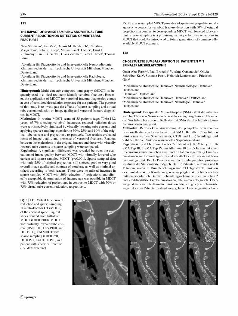
S36
123
Clin Neuroradiol (2019) (Suppl 1) 29:S1–S129
111
THE IMPACT OF SPARSE SAMPLING AND VIRTUAL TUBE CURRENT REDUCTION ON DETECTION OF VERTEBRAL FRACTURES
Nico Sollmann1, Kai Mei2, Dennis M. Hedderich1, Christian Maegerlein1, Felix K. Kopp2, Maximilian T. Löffler1, Ernst J. Rummeny2, Jan S. Kirschke1, Claus Zimmer1, Peter B. Noel2, Thomas Baum1
1Abteilung für Diagnostische und Interventionelle Neuroradiologie, Klinikum rechts der Isar, Technische Universität München, München, Deutschland2Abteilung für Diagnostische und Interventionelle Radiologie, Klinikum rechts der Isar, Technische Universität München, München, Deutschland
Hintergrund: Multi-detector computed tomography (MDCT) is fre-quently used in clinical routine to identify vertebral fractures. Howev-er, the application of MDCT for vertebral fracture diagnostics comes at cost of considerable radiation exposure for the patients. The purpose of this study is to investigate the effects of sparse sampling and virtual tube current reduction on image quality and vertebral fracture diagnos-tics in MDCT.Methoden: In routine MDCT scans of 35 patients (age: 70.6 ± 14.2 years, 65.7% showing vertebral fractures), reduced radiation doses were retrospectively simulated by virtually lowering tube currents and applying sparse sampling, considering 50%, 25%, and 10% of the orig-inal tube current and projections, respectively. Two readers evaluated items of image quality and presence of vertebral fractures. Readout between the evaluations in the original images and those with virtually lowered tube currents or sparse sampling were compared.Ergebnisse: A significant difference was revealed between the eval-uations of image quality between MDCT with virtually lowered tube current and sparse-sampled MDCT (p < 0.001). Sparse-sampled data with only 25% of original projections still showed good to very good overall image quality and contrast of vertebrae as well as minimal ar-tifacts according to both readers. There were no missed fractures in sparse-sampled MDCT with 50% reduction of projections, and clini-cally acceptable determination of fracture age was possible in MDCT with 75% reduction of projections, in contrast to MDCT with 50% or 75% virtual tube current reduction, respectively.
Fazit: Sparse-sampled MDCT provides adequate image quality and di-agnostic accuracy for vertebral fracture detection with 50% of original projections in contrast to corresponding MDCT with lowered tube cur-rent. Sparse sampling is a promising technique for dose reductions in MDCT that could be introduced in future generations of commercially available MDCT scanners.
128
CT-GESTÜTZTE LUMBALPUNKTION BEI PATIENTEN MIT SPINALER MUSKELATROPHIE
Omar Abu-Fares*1, Paul Bronzlik1 2 3, Alma Osmanovic4, Olivia Schreiber-Katz4, Susanne Petri4, Heinrich Lanfermann1, Friedrich Götz1
1Medizinische Hochschule Hannover, Neuroradiologie, Hannover, Deutschland2Hannover, Deutschland3Medizinische Hochschule Hannover, Hannover, Deutschland4Medizinische Hochschule Hannover, Neurologie, Hannover, Deutschland
Hintergrund: Bei spinaler Muskelatrophie (SMA) stellt die intrathe-kale Injektion von Nusinersen derzeit die einzige zugelassene Therapie dar. Wir haben bei unserem Kollektiv mit SMA die durchführten Lum-balpunktionen analysiert.Methoden: Retrospektive Auswertung des prospektiv erfassten Pa-tientenkollektiv von Erwachsenen mit SMA. Bei allen CT-geführten Punktionen wurden Scanparameter, CTDI und DLP, Scanlänge und Zahl der für die Punktion verwendeten Sequenzen erfasst.Ergebnisse: Seit 11/17 wurden bei 27 Patienten (10 SMA Typ II, 16 SMA Typ III, 1 SMA Typ IV) im Alter von 18 bis 65 Jahren mit einer Erkrankungsdauer zwischen zwei und 61 Jahren regelmäßig Lumbal-punktionen zur Liquordiagnostik und intrathekalen Nusinersen-Thera-pie durchgeführt. Bei 15 Patienten war die Lumbalpunktion problem-los durch die Stationsärzte möglich. Bei 12 Patienten, 4 Frauen und 8 Männern, waren 11 Durchleuchtungs- und 53 CT-gestützte Punktion des lumbalen Wirbelkanals wegen ausgeprägten Wirbelsäulendefor-mitäten erforderlich. Gemäß Behandlungsschema wurden zwischen 2 und 7 bildgestützte Lumbalpunktionen, alle waren erfolgreich. Über-wiegend war eine interlaminäre Punktion möglich; gelegentlich musste wegen der vom Patientenzustand vorgegebenen Lagerungsmöglichkei-
Fig. 1 | 111 Virtual tube current reduction and sparse sampling in multi-detector CT (MDCT) of the cervical spine. Sagittal slices derived from full-dose MDCT (D100 P100), MDCT with virtually lowered tube cur-rent (D50 P100, D25 P100, and D10 P100), and MDCT with sparse sampling (D100 P50, D100 P25, and D100 P10) in a patient with a cervical fracture (C2, dens fracture)

S37
123
Clin Neuroradiol (2019) (Suppl 1) 29:S1–S129
ten ein transforaminaler Zugang gewählt werden. Die Strahlenexpositi-on resultiert aus dem Spiraldatensatz zur Wirbelsäulendarstellung und den erforderlichen Kontrollen während der Punktion. Die durchschnitt-lichen Werte betragen für CTDI 7,2 mGy, DLP 156 mGy-cm, Scanlän-ge 10,3 cm und Zahl der für die Punktion verwendeten Sequenzen 4. Komplikationen traten bei keiner der durchgeführten Punktionen auf.Diskussion: Wegen schweren krankheitsbedingten Wirbelsäulende-formitäten ist bei etwa der Hälfte der Erwachsenen mit SMA eine CT-gestützte Punktion des lumbalen Wirbelkanals erforderlich. Sowohl in-terlaminäre als auch transforaminale Zugänge können erfolgreich und komplikationslos durchgeführt werden.Fazit: Bei Anwendung von low dose CT-Technik ist diese Maßnahme mit einer relativ geringen Strahlenexposition durchführbar
164
SYSTEMATIC EVALUATION OF LOW-DOSE IMAGING FOR CT-GUIDED PERIRADICULAR INFILTRATIONS
Simon Schön*1, Kai Mei2, Isabelle Riederer1, Felix K. Kopp2, Claus Zimmer1, Jan Kirschke1 3, Peter Noel2 4, Thomas Baum1, Nico Sollmann3 5
1Department of Diagnostic and Interventional Neuroradiology, Klinikum rechts der Isar, Technische Universität München, Munich, Germany, München, Deutschland
2Department of Diagnostic and Interventional Radiology, Klinikum rechts der Isar, Technische Universität München, Munich, Germany, München, Deutschland3TUM-Neuroimaging Center, Klinikum rechts der Isar, Technische Universität München, München, Deutschland4Department of Radiology, Perelman School of Medicine, University of Pennsylvania, Philadelphia, United States5Department of Diagnostic and Interventional Neuroradiology, Klinikum rechts der Isar, Technische Universität München, München, Deutschland
Background: Periradicular infiltrations are frequently performed in neuroradiological routine and are planned and guided by multi-detec-tor computed tomography (MDCT). The objective of this study was to evaluate image quality and confidence for planning of periradicular infiltrations using virtually lowered tube currents and iterative recon-struction (IR).Methods: Twenty patients (age: 54.9 ± 13.1 years) underwent MDCT for planning purposes of periradicular infiltrations at the level of the lumbosacral spine using a standard imaging protocol provided by the MDCT vendor. Planning scans were simulated as if they were made at 50% (D50), 10% (D10), 5% (D5), and 1% (D1) of the original tube current. Image reconstruction was achieved with two levels of IR (A: similar to clinical reconstructions, B: ten times stronger regulariza-tion). Two readers (R1 and R2) performed qualitative image evaluation considering overall image quality and artifacts, image contrast, deter-
Fig. 2 | 111 Virtual tube current reduction and sparse sampling in multi-detector CT (MDCT) of the cthoracic and lumbar spine. Sagittal slices derived from full-dose MDCT (D100 P100), MDCT with virtually lowered tube current (D50 P100, D25 P100, and D10 P100), and MDCT with sparse sampling (D100 P50, D100 P25, and D100 P10) are shown in a patient with five thoratic fractures (T3, T6, T8, T10 and T11)

S38
123
Clin Neuroradiol (2019) (Suppl 1) 29:S1–S129
mination of nerve roots (scoring: 1—possible, 2—unclear, and 3—not possible), and confidence for intervention planning (scoring: 1—high, 2—medium, and 3—low confidence).Result: Level A of IR was favorable regarding overall image quality, artifacts, and image contrast according to both readers, with preserved very good to excellent scores for D10. Similarly, level A of IR led to better scores for determination of nerve roots, with D10 still allowing for clear nerve root depiction in the majority of patients (D10 A: R1: 1.4 ± 0.5 vs. R2: 1.5 ± 0.5, p > 0.05). The confidence for intervention planning remained high for tube currents virtually lowered down to 5% of the original dose (D5 A: 1.4 ± 0.5 vs. R2: 1.4 ± 0.5, p > 0.05). In-ter-reader agreement of confidence for intervention planning was good to excellent (weighted Cohen’s kappa > 0.60).Conclusion: MDCT for planning purposes of lumbosacral periradicu-lar infiltrations may be conducted with tube currents lowered down to 5% of standard imaging without restrictions in intervention planning confidence. Thus, radiation exposure could be decreased considerably.
199
PRÄOPERATIVE EMBOLISATION UND CHIRURGISCHE RESEKTION MIT SPONDYLODESE: THERAPIEREGIME FÜR AGGRESSIV WACHSENDE SPINALE HÄMANGIOME UND IHRE REZIDIVE
Christiane Pöckler-Schöniger*1, Jörg Drumm2, Christoph Berthold2, Tobias Pitzen2, Michael Ruf2
1Diagnostische Radiologie/Neuroradiologie, SRH Klinikum Karlsbad-Langensteinbach, Karlsbad, Deutschland2Zentrum für Wirbelsäulenchirurgie, Orthopädie und Traumatologie, SRH Klinikum Karlsbad-Langensteinbach, Karlsbad, Deutschland
Hintergrund: Spinale Hämangiome sind meist gutartige, vaskuläre Tumoren mit einem Anteil von 11 % der benignen Tumore der Wirbel-säule. In 0.9–1.2 % können sogenannte aggressive Hämangiome große Anteile des Wirbelkörpers aushöhlen und durch intraspinales Wachs-tum oder Frakturen Schmerzen und neurologische Defizite verursa-chen. Behandlungsoptionen sind transarterielle Embolisation, Radi-atio, Vertebroplastie/Kyphoplastie auch in Kombination mit Radiatio und chirurgische Resektion mit Fixation. Allerdings sehen wir unter dieser Therapie eine relevante Anzahl von lokalen Rezidiven, ebenso primär lokal aggressiv wachsende Tumore mit Invasion des Spinal-kanals und Kompression des Rückenmarks oder der Nervenwurzeln.
Wir präsentieren unsere Erfahrungen mit präoperativer Embolisa-tion und chirurgischer Resektion der aggressiv wachsenden Häman-giomeMethoden: Die Daten stammen aus dem klinikeigenen Register eines großen deutschen Wirbelsäulenzentrums von 1/1997–10/2018. Ausge-wertet wurden demografische Daten, klinische Symptomatik, Tumor-lokalisation, vorausgegangene Therapien, endgültige Versorgung des Rezidivs bzw. primäre Tumortherapie in unserer Klinik mit präope-rativer Bildgebung und Tumorembolisation mittels Mikrospiralen und PVA-Partikeln.Ergebnisse: 24 Patienten, Alter 55 + 17 Jahre, Follow up 1 Mo-nat–19 Jahre, 14F und 10M. 75 % der Patienten gaben lokale Schmer-zen an, 25 % zeigten neurologische Defizite. Hauptlokalisation war die BWS (62 %), immer war der Wirbelkorpus betroffen mit epiduraler Tumorausdehnung. 33 % der Patienten waren vorbehandelt, 3 × Verte-broplastie, 5 × Dekompression und Spondylodese. Die endgültige Ver-sorgung in unserer Klinik erfolgte mit präoperativer Embolisation in 19/24 Fällen, die chirurgische Therapie erfolgte überwiegend mit en bloc Resektion und Spondylodese (83 %). Bislang keine Rezidive im FU.Fazit: Für aggressiv wachsende Hämangiome, die zu lokalen Schmer-zen oder neurologischen Defiziten führen, ist unserer Erfahrung nach eine Dekompression mit Instrumentation bzw. Vertebroplastie nicht ausreichend. Sowohl bei Rezidiven als auch bei primär aggressivem intraspinalem Wachstum scheint die präoperative Embolisation mit ra-dikaler Resektion und Spondylodese sinnvoll.
205
ASSOCIATION OF THIGH AND PARASPINAL MUSCLE COMPOSITION IN HEALTHY ADULTS USING CHEMICAL SHIFT ENCODING-BASED WATER–FAT MRI
Egon Burian*1, Stephanie Inhuber2, Sarah Schlaeger3, Michael Dieckmeyer4, Elisabeth Klupp4, Daniela Franz3, Nico Sollmann4, Maximilian Löffler4, Ansgar Schwirtz2, Ernst Rummeny3, Claus Zimmer4, Jan Kirschke4, Dimitrios Karampinos3, Thomas Baum4
1Department of Diagnostic and Interventional Neuroradiology, Klinikum rechts der Isar, Technical University of Munich, München, Deutschland2Department of Sport and Health Sciences, Technical University of Munich, München, Deutschland3Department of Diagnostic and Interventional Radiology, Klinikum rechts der Isar, Technical University of Munich, München, Deutschland4Department of Diagnostic and Interventional Neuroradiology, Klinikum rechts der Isar, Technical University of Munich, München, Deutschland
Background: Paraspinal and thigh muscles comprise the major mus-cle groups of the human body. Their function is essential for stability and mobility and is affected by various diseases including degenerative changes, metabolic disorders, sarcopenia and cachexia. We investigat-ed the composition of the psoas, erector spinae, quadriceps femoris and hamstring muscle groups and their association to each other using chemical shift encoding-based water–fat magnetic resonance imaging (MRI) in young, healthy adults.Methods: 25 healthy subjects (14 males, age: 30.3 ± 5.0 years, body mass index [BMI]: 27.6 ± 3.0 kg/m2 and 11 females, age: 30.2 ± 7.0 years, BMI: 26.0 ± 1.8 kg/m²) were recruited for this study. Proton density fat fraction (PDFF) of the psoas, erector spinae, quadriceps femoris and hamstring muscle groups were determined (Fig. 1 and 2). For the quadriceps and the hamstring muscle group the intramuscu-lar PDFF for each dedicated muscle was assessed separately (Fig. 2).Results: After consideration of age and BMI as control variables, PDFF values of the erector spinae correlated significantly with those
Fig. 1 | 164 Virtual tube current reduction and sparse sampling in mul-ti-detector CT (MDCT) performed for planning of periradicular infil-trations. Tube currents were lowered down to 50% (D50), 10% (D10), 5% (D5) and 1% (D1) compared to original imaging (D100). Two lev-els of iterative reonstruction (IR; A and B) were used
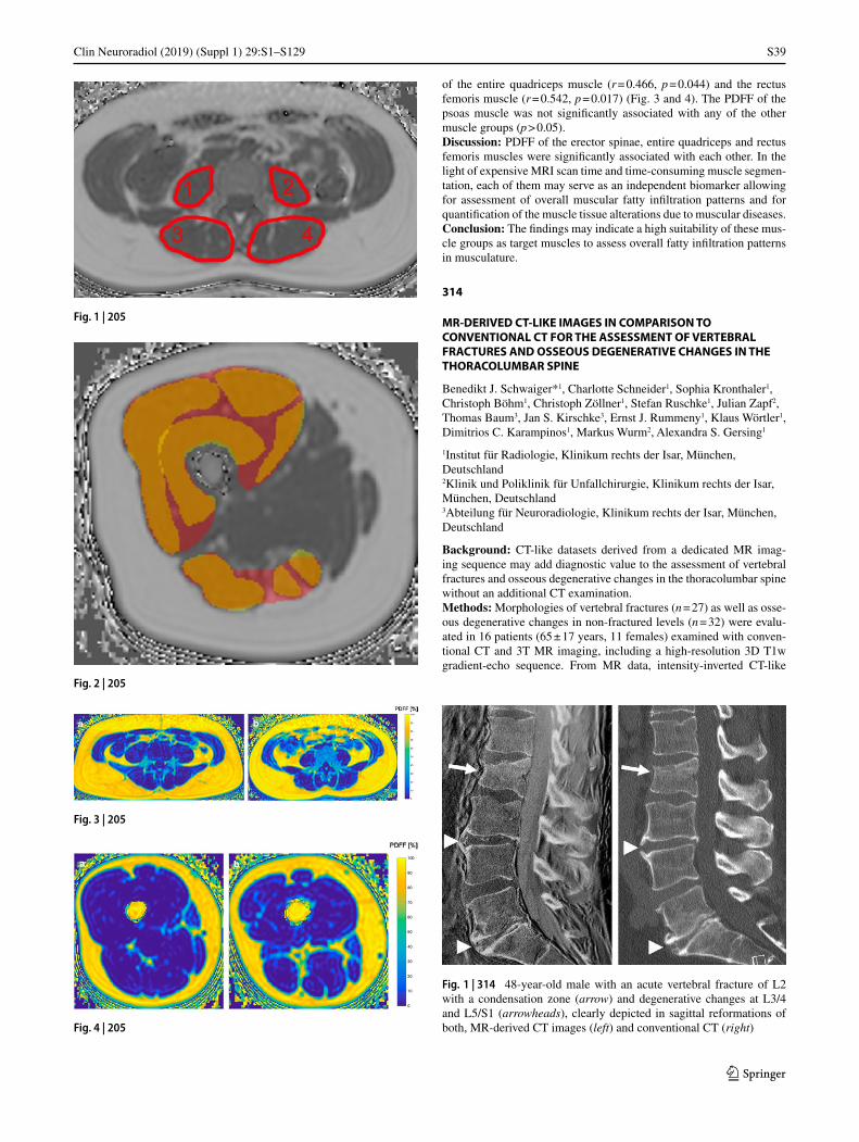
S39
123
Clin Neuroradiol (2019) (Suppl 1) 29:S1–S129
of the entire quadriceps muscle (r = 0.466, p = 0.044) and the rectus femoris muscle (r = 0.542, p = 0.017) (Fig. 3 and 4). The PDFF of the psoas muscle was not significantly associated with any of the other muscle groups (p > 0.05).Discussion: PDFF of the erector spinae, entire quadriceps and rectus femoris muscles were significantly associated with each other. In the light of expensive MRI scan time and time-consuming muscle segmen-tation, each of them may serve as an independent biomarker allowing for assessment of overall muscular fatty infiltration patterns and for quantification of the muscle tissue alterations due to muscular diseases.Conclusion: The findings may indicate a high suitability of these mus-cle groups as target muscles to assess overall fatty infiltration patterns in musculature.
314
MR-DERIVED CT-LIKE IMAGES IN COMPARISON TO CONVENTIONAL CT FOR THE ASSESSMENT OF VERTEBRAL FRACTURES AND OSSEOUS DEGENERATIVE CHANGES IN THE THORACOLUMBAR SPINE
Benedikt J. Schwaiger*1, Charlotte Schneider1, Sophia Kronthaler1, Christoph Böhm1, Christoph Zöllner1, Stefan Ruschke1, Julian Zapf2, Thomas Baum3, Jan S. Kirschke3, Ernst J. Rummeny1, Klaus Wörtler1, Dimitrios C. Karampinos1, Markus Wurm2, Alexandra S. Gersing1
1Institut für Radiologie, Klinikum rechts der Isar, München, Deutschland2Klinik und Poliklinik für Unfallchirurgie, Klinikum rechts der Isar, München, Deutschland3Abteilung für Neuroradiologie, Klinikum rechts der Isar, München, Deutschland
Background: CT-like datasets derived from a dedicated MR imag-ing sequence may add diagnostic value to the assessment of vertebral fractures and osseous degenerative changes in the thoracolumbar spine without an additional CT examination.Methods: Morphologies of vertebral fractures (n = 27) as well as osse-ous degenerative changes in non-fractured levels (n = 32) were evalu-ated in 16 patients (65 ± 17 years, 11 females) examined with conven-tional CT and 3T MR imaging, including a high-resolution 3D T1w gradient-echo sequence. From MR data, intensity-inverted CT-like
Fig. 2 | 205
Fig. 3 | 205
Fig. 4 | 205
Fig. 1 | 205
Fig. 1 | 314 48-year-old male with an acute vertebral fracture of L2 with a condensation zone (arrow) and degenerative changes at L3/4 and L5/S1 (arrowheads), clearly depicted in sagittal reformations of both, MR-derived CT images (left) and conventional CT (right)
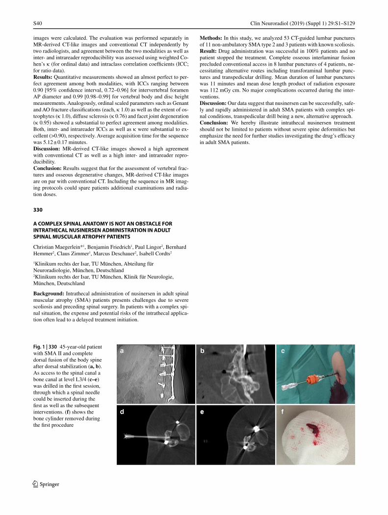
S40
123
Clin Neuroradiol (2019) (Suppl 1) 29:S1–S129
images were calculated. The evaluation was performed separately in MR-derived CT-like images and conventional CT independently by two radiologists, and agreement between the two modalities as well as inter- and intrareader reproducibility was assessed using weighted Co-hen’s κ (for ordinal data) and intraclass correlation coefficients (ICC; for ratio data).Results: Quantitative measurements showed an almost perfect to per-fect agreement among both modalities, with ICCs ranging between 0.90 [95% confidence interval, 0.72–0.96] for intervertebral foramen AP diameter and 0.99 [0.98–0.99] for vertebral body and disc height measurements. Analogously, ordinal scaled parameters such as Genant and AO fracture classifications (each, κ 1.0) as well as the extent of os-teophytes (κ 1.0), diffuse sclerosis (κ 0.76) and facet joint degeneration (κ 0.95) showed a substantial to perfect agreement among modalities. Both, inter- and intrareader ICCs as well as κ were substantial to ex-cellent (>0.90), respectively. Average acquisition time for the sequence was 5.12 ± 0.17 minutes.Discussion: MR-derived CT-like images showed a high agreement with conventional CT as well as a high inter- and intrareader repro-ducibility.Conclusion: Results suggest that for the assessment of vertebral frac-tures and osseous degenerative changes, MR-derived CT-like images are on par with conventional CT. Including the sequence in MR imag-ing protocols could spare patients additional examinations and radia-tion doses.
330
A COMPLEX SPINAL ANATOMY IS NOT AN OBSTACLE FOR INTRATHECAL NUSINERSEN ADMINISTRATION IN ADULT SPINAL MUSCULAR ATROPHY PATIENTS
Christian Maegerlein*1, Benjamin Friedrich1, Paul Lingor2, Bernhard Hemmer2, Claus Zimmer1, Marcus Deschauer2, Isabell Cordts2
1Klinikum rechts der Isar, TU München, Abteilung für Neuroradiologie, München, Deutschland2Klinikum rechts der Isar, TU München, Klinik für Neurologie, München, Deutschland
Background: Intrathecal administration of nusinersen in adult spinal muscular atrophy (SMA) patients presents challenges due to severe scoliosis and preceding spinal surgery. In patients with a complex spi-nal situation, the expense and potential risks of the intrathecal applica-tion often lead to a delayed treatment initiation.
Methods: In this study, we analyzed 53 CT-guided lumbar punctures of 11 non-ambulatory SMA type 2 and 3 patients with known scoliosis.Result: Drug administration was successful in 100% patients and no patient stopped the treatment. Complete osseous interlaminar fusion precluded conventional access in 8 lumbar punctures of 4 patients, ne-cessitating alternative routes including transforaminal lumbar punc-tures and transpedicular drilling. Mean duration of lumbar punctures was 11 minutes and mean dose length product of radiation exposure was 112 mGy cm. No major complications occurred during the inter-ventions.Discussion: Our data suggest that nusinersen can be successfully, safe-ly and rapidly administered in adult SMA patients with complex spi-nal conditions, transpedicular drill being a new, alternative approach.Conclusion: We hereby illustrate intrathecal nusinersen treatment should not be limited to patients without severe spine deformities but emphasize the need for further studies investigating the drug’s efficacy in adult SMA patients.
Fig. 1 | 330 45-year-old patient with SMA II and complete dorsal fusion of the body spine after dorsal stabilization (a, b). As access to the spinal canal a bone canal at level L3/4 (c–e) was drilled in the first session, through which a spinal needle could be inserted during the first as well as the subsequent interventions. (f) shows the bone cylinder removed during the first procedure

S41
123
Clin Neuroradiol (2019) (Suppl 1) 29:S1–S129
6. Zerebrovaskuläre Erkrankungen
39
ENDOVASCULAR MECHANICAL THROMBECTOMY FOR CEREBRAL VENOUS SINUS THROMBOSIS: A SINGLE CENTER EXPERIENCE
Hanna Styczen*1, Ioannis Tsogkas1, Jan Liman1, Volker Maus1, Marios-Nikos Psychogios1
1Göttingen, Universitätsmedizin Göttingen, Abteilung für Diagnostische und Interventionelle Neuroradiologie, Göttingen, Deutschland
Background: Cerebral venous thrombosis (CVT) is a rare entity with a potentially fatal outcome. Especially patients who do not respond to standard medical therapy alone may benefit from endovascular treat-ment options. We evaluate the angiographic and clinical results of me-chanical thrombectomy (MT) in patients with severe CVT.Methods: Endovascular procedures were performed in 13 patients with CVT additionally to standard anticoagulation therapy between 2011 and 2018. Clinical and radiological parameters, procedure de-tails, angiographic and clinical outcomes were reviewed retrospective-ly.
Result: In total, 14 thrombectomy procedures were executed. Success-ful recanalization of the occluded sinus was achieved in 86% of cases (12/14); of those, 29% were recanalized completely. Procedural com-plications included perforation of the transverse sinus in one case (7%). Worsening of intracranial hemorrhage occurred in 14% (2/14) of cases. Favorable clinical outcome (modified Rankin Scale 0–2) was achieved in 12/13 patients (92%).Discussion: The recanalization rate at final angiogram was slightly lower with 86% than described in recent systemic reviews [1,2]. In correlation to our results, recent studies reported mortality rates in pa-tients undergoing MT due to severe CVT between 4–14% (8% in our study) with excellent (mRS 0–1) and favorable (mRS 0–2) outcomes of 72% and 84%, respectively [1,2,3]. This is comparable to our results with 77% excellent and 92% favorable outcome despite of lower rate of complete recanalization.Conclusion: Transvenous MT is feasible, safe and effective leading to high degree of successful recanalization rate of occluded dural sinus. It may be a salvage treatment for selected patients with severe CVT refractory to standard medical treatment.
References 1. Ilyas A, Chen CJ, Raper DM, et al. Endovascular mechanical
thrombectomy for cerebral venous sinus thrombosis: a sys-tematic review. J Neurointerv Surg. 2017;9:1086–92. https://doi.org/10.1136/neurintsurg-2016-012938.
2. Siddiqui FM, Dandapat S, Banerjee C, et al. Mechanical thrombectomy in cerebral venous thrombosis: systematic review of 185 cases. Stroke. 2015;46(5):1263–8. https://doi.org/10.1161/STROKEAHA.114.007465.
3. Konakondla S, Schirmer CM, Li F, et al. New Developments in the Pathophysiology, Workup, and Diagnosis of Dural Venous Sinus Thrombosis (DVST) and a Systematic Review of Endovascular Treatments. Aging Dis. 2017;8:136–48. https://doi.org/10.14336/AD.2016.0915.
42
PREDICTORS OF PERIPHERAL EMBOLI AFTER MECHANICAL THROMBECTOMY: A PROSPECTIVE STUDY USING HIGH RESOLUTION DWI
Michael Schönfeld*1, Reza Kabiri1, Helge Kniep1, Gabriel Broocks1, Marielle Sophie Ernst1, Lukas Meyer1, Jens Fiehler1, Uta Hanning1
1Klinik und Poliklinik für Neuroradiologische Diagnostik und Intervention, Universitätsklinikum Hamburg-Eppendorf, Hamburg, Deutschland
Background: To analyze the factors influencing the incidence of pe-ripheral emboli after mechanical thrombectomy (MT) of intracranial large vessel occlusions (LVO).Methods: We performed a prospective analysis of patients successful-ly treated with MT for an LVO of the anterior circulation that received a 1.5T MRI with diffusion-weighted imaging (DWI) in high resolution on the day following the intervention. Recanalization success was as-sessed on post-thrombectomy digital subtraction angiography (DSA) using the extended thrombolysis in cerebral infarction (eTICI) scale. Punctuate DWI lesions distant to the DWI core lesion were classified as peripheral emboli. Thrombus properties (location, length, density, density ratio) were measured on pre-interventional CT imaging. Pro-cedural details of medical management (iv alteplase treatment) and mechanical thrombectomy (primary aspiration or stent retrieval with or without balloon occlusion, number of passes) were collected. The influence of thrombus properties and procedural details on the number of peripheral emboli was analyzed using univariate and multivariate linear regression models.
Fig. 2 | 39
Fig. 1 | 39

S42
123
Clin Neuroradiol (2019) (Suppl 1) 29:S1–S129
Result: 37 patients undergoing successful MT met the inclusion cri-teria. In 35/37 (94.6%) patients a total of 411 peripheral emboli were seen on DWI. The number of emboli correlated with eTICI score (Spearman’s rho 0.605; p < 0.001). Use of a balloon guide catheter was associated with a reduced number of peripheral emboli with a median number of peripheral DWI lesions of 5 (IQR 1.25–8.25) with and 12 (IQR 4–19) without the use of a balloon-guide catheter. Use of a bal-loon-guide catheter and recanalization success were the only factors that independently contributed to the number and volume of peripheral emboli (odds ratio –0.355 95% CI –15.746—–0.798; p = 0.031).Conclusion: Together with a more complete recanalization the use of a balloon-guide catheter may reduce peripheral emboli leading to pe-ripheral ischemias after mechanical thrombectomy. Proximal flow-ar-rest with a balloon guided catheter therefore should be the standard of practice whenever possible.
Intravenous alteplase treatment pre-MT does not appear to influence the number of peripheral ischemias later seen on DWI
53
UNTERSUCHUNG DES FMRT-BOLD-SIGNALVERLAUFS WÄHREND KURZER ATEMPAUSEN ZUR BEURTEILUNG DER ZEREBROVASKULÄREN REAKTIVITÄT
Leonie Zerweck*1, Till-Karsten Hauser2, Constantin Roder3, Uwe Klose2
1Universitätsklinikum Tübingen, Radiologische Klinik, Abteilung für Diagnostische und Interventionelle Neuroradiologie, Tübingen, Deutschland2Universitätsklinikum Tübingen, Radiologische Klinik, Abteilung für Diagnostische und Interventionelle Neuroradiologie, Tübingen, Deutschland3Universitätsklinikum Tübingen, Klinik für Neurochirurgie, Tübingen, Deutschland
Hintergrund: CO2-stimulierte fMRT-Messungen können zur Beurtei-lung der zerebrovaskulären Reaktivität (CVR) von Moyamoya-Patien-ten, bei denen die CVR gegenüber Hyperkapnie vermindert ist, dienen [1,2]. Im gesunden Gewebe führt durch Atempausen hervorgerufene Hyperkapnie zu gesteigertem zerebralen Blutfluss und BOLD (Blood-Oxygen-Level-Dependent)-Signalanstieg [3]. Ziel dieser Arbeit war die Ermittlung der notwendigen Dauer der Atempausen für replizier-bare Signalanstiege.Methoden: Bei 14 gesunden Probanden wurden prospektiv fMRT-Messungen (3T) während abwechselnd endexspiratorischer Atempau-sen jeweils unterschiedlicher Dauer (3s, 6s, 9s, 12s) und Phasen nor-maler Atmung durchgeführt.
Um die Befolgung der Atempausen zu überprüfen, wurden die At-mungsbewegungen der Probanden aufgezeichnet.
Der im Kortex gemessene Signalverlauf wurde über alle Probanden gemittelt (Abb. 1).
Das Integral des Signalverlaufs über ein Zeitfenster von 12s wäh-rend des maximalen Signalanstiegs wurde über alle Probanden gemit-telt und nach Normalisierung räumlich dargestellt (Abb. 2).Ergebnisse: Atempausen von 12s, 9s und 6s führten zu Signalanstie-gen von 0,9 %, 0,8 % und 0,3 % (Abb. 1). Nach Atempausen von 3s waren keine vergleichbaren Signalanstiege messbar.
Zusätzlich zu den erwarteten Signalanstiegen waren initial kleine-re Signalanstiege zu beobachten. Diese zeigten sich besonders, wenn die Probanden vor den endexspiratorischen Atempausen auffällig tief einatmeten.Diskussion: Für replizierbare Signalanstiege wurden Atempausen von mindestens 9s benötigt. Eine weitere Verlängerung der Atempausen führte nur zu geringfügigem Signalzuwachs.
Den initialen Signalanstieg erklären wir durch tiefe Inhalation vor der Atempause.
Die wie erwartet [3] höhere Aktivierung in der grauen Substanz deutet auf die Differenzierungsfähigkeit des Verfahrens von Hirnregi-onen unterschiedlicher CVR hin.Fazit: In der klinischen Routine eignen sich endexspiratorische Atem-pausen von 9s, da diese im gesunden Gewebe zu deutlichen BOLD-Signalanstiegen führen und leicht durchzuführen sind.
Literatur 1. Neuroimage Clin. 2019;22:101713. 2. AJNR Am J Neuroradiol. 2010;31(5):862–7. 3. Stroke. 1998;29(12):2641–5.
Fig. 1 | 53 Relative Signaländerungen nach Atempausen unterschiedli-cher Dauer. Die farbigen Rechtecke stellen die entsprechenden Atem-pausen dar. Es zeigten sich höhere BOLD-Signalanstiege in der grauen als in der weißen Substanz (Abb
Fig. 2 | 53 Farbcodiertes Aktivierungsmuster (blau > grün > rot > schwarz)
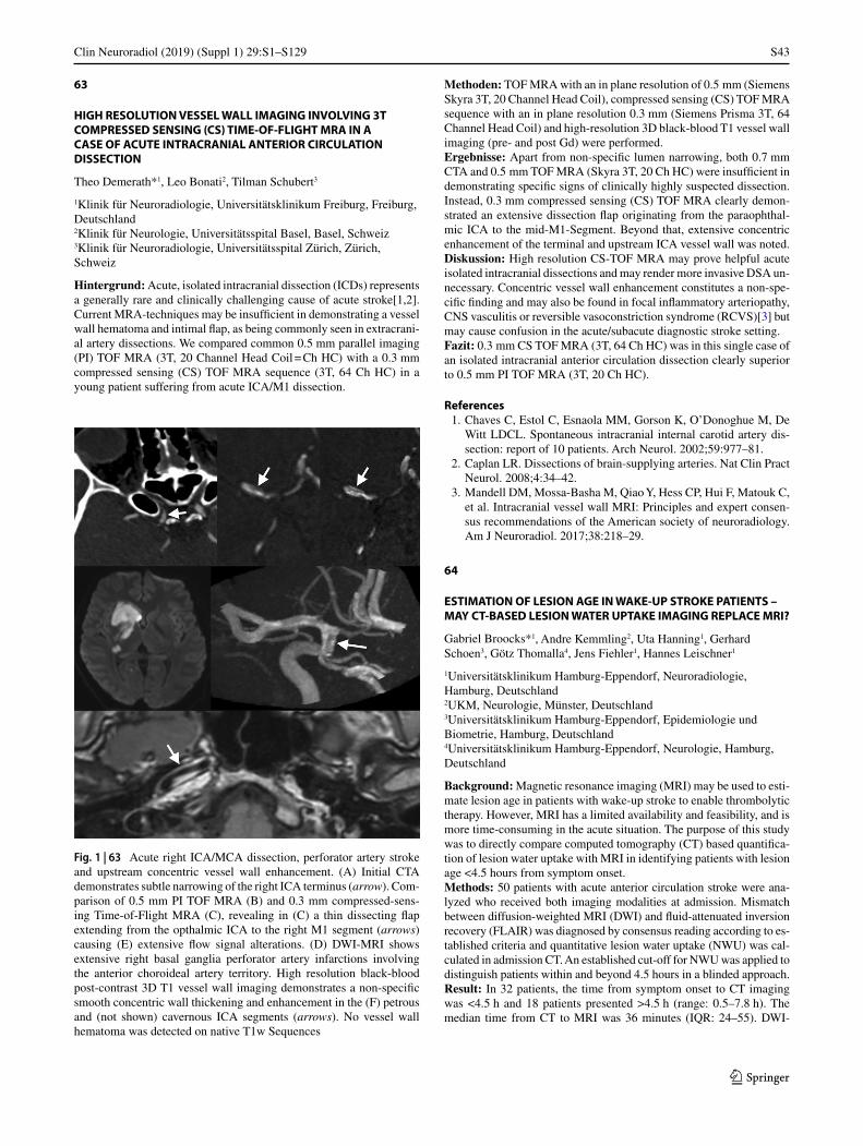
S43
123
Clin Neuroradiol (2019) (Suppl 1) 29:S1–S129
63
HIGH RESOLUTION VESSEL WALL IMAGING INVOLVING 3T COMPRESSED SENSING (CS) TIME-OF-FLIGHT MRA IN A CASE OF ACUTE INTRACRANIAL ANTERIOR CIRCULATION DISSECTION
Theo Demerath*1, Leo Bonati2, Tilman Schubert3
1Klinik für Neuroradiologie, Universitätsklinikum Freiburg, Freiburg, Deutschland2Klinik für Neurologie, Universitätsspital Basel, Basel, Schweiz3Klinik für Neuroradiologie, Universitätsspital Zürich, Zürich, Schweiz
Hintergrund: Acute, isolated intracranial dissection (ICDs) represents a generally rare and clinically challenging cause of acute stroke[1,2]. Current MRA-techniques may be insufficient in demonstrating a vessel wall hematoma and intimal flap, as being commonly seen in extracrani-al artery dissections. We compared common 0.5 mm parallel imaging (PI) TOF MRA (3T, 20 Channel Head Coil = Ch HC) with a 0.3 mm compressed sensing (CS) TOF MRA sequence (3T, 64 Ch HC) in a young patient suffering from acute ICA/M1 dissection.
Methoden: TOF MRA with an in plane resolution of 0.5 mm (Siemens Skyra 3T, 20 Channel Head Coil), compressed sensing (CS) TOF MRA sequence with an in plane resolution 0.3 mm (Siemens Prisma 3T, 64 Channel Head Coil) and high-resolution 3D black-blood T1 vessel wall imaging (pre- and post Gd) were performed.Ergebnisse: Apart from non-specific lumen narrowing, both 0.7 mm CTA and 0.5 mm TOF MRA (Skyra 3T, 20 Ch HC) were insufficient in demonstrating specific signs of clinically highly suspected dissection. Instead, 0.3 mm compressed sensing (CS) TOF MRA clearly demon-strated an extensive dissection flap originating from the paraophthal-mic ICA to the mid-M1-Segment. Beyond that, extensive concentric enhancement of the terminal and upstream ICA vessel wall was noted.Diskussion: High resolution CS-TOF MRA may prove helpful acute isolated intracranial dissections and may render more invasive DSA un-necessary. Concentric vessel wall enhancement constitutes a non-spe-cific finding and may also be found in focal inflammatory arteriopathy, CNS vasculitis or reversible vasoconstriction syndrome (RCVS)[3] but may cause confusion in the acute/subacute diagnostic stroke setting.Fazit: 0.3 mm CS TOF MRA (3T, 64 Ch HC) was in this single case of an isolated intracranial anterior circulation dissection clearly superior to 0.5 mm PI TOF MRA (3T, 20 Ch HC).
References 1. Chaves C, Estol C, Esnaola MM, Gorson K, O’Donoghue M, De
Witt LDCL. Spontaneous intracranial internal carotid artery dis-section: report of 10 patients. Arch Neurol. 2002;59:977–81.
2. Caplan LR. Dissections of brain-supplying arteries. Nat Clin Pract Neurol. 2008;4:34–42.
3. Mandell DM, Mossa-Basha M, Qiao Y, Hess CP, Hui F, Matouk C, et al. Intracranial vessel wall MRI: Principles and expert consen-sus recommendations of the American society of neuroradiology. Am J Neuroradiol. 2017;38:218–29.
64
ESTIMATION OF LESION AGE IN WAKE-UP STROKE PATIENTS – MAY CT-BASED LESION WATER UPTAKE IMAGING REPLACE MRI?
Gabriel Broocks*1, Andre Kemmling2, Uta Hanning1, Gerhard Schoen3, Götz Thomalla4, Jens Fiehler1, Hannes Leischner1
1Universitätsklinikum Hamburg-Eppendorf, Neuroradiologie, Hamburg, Deutschland2UKM, Neurologie, Münster, Deutschland3Universitätsklinikum Hamburg-Eppendorf, Epidemiologie und Biometrie, Hamburg, Deutschland4Universitätsklinikum Hamburg-Eppendorf, Neurologie, Hamburg, Deutschland
Background: Magnetic resonance imaging (MRI) may be used to esti-mate lesion age in patients with wake-up stroke to enable thrombolytic therapy. However, MRI has a limited availability and feasibility, and is more time-consuming in the acute situation. The purpose of this study was to directly compare computed tomography (CT) based quantifica-tion of lesion water uptake with MRI in identifying patients with lesion age <4.5 hours from symptom onset.Methods: 50 patients with acute anterior circulation stroke were ana-lyzed who received both imaging modalities at admission. Mismatch between diffusion-weighted MRI (DWI) and fluid-attenuated inversion recovery (FLAIR) was diagnosed by consensus reading according to es-tablished criteria and quantitative lesion water uptake (NWU) was cal-culated in admission CT. An established cut-off for NWU was applied to distinguish patients within and beyond 4.5 hours in a blinded approach.Result: In 32 patients, the time from symptom onset to CT imaging was <4.5 h and 18 patients presented >4.5 h (range: 0.5–7.8 h). The median time from CT to MRI was 36 minutes (IQR: 24–55). DWI-
Fig. 1 | 63 Acute right ICA/MCA dissection, perforator artery stroke and upstream concentric vessel wall enhancement. (A) Initial CTA demonstrates subtle narrowing of the right ICA terminus (arrow). Com-parison of 0.5 mm PI TOF MRA (B) and 0.3 mm compressed-sens-ing Time-of-Flight MRA (C), revealing in (C) a thin dissecting flap extending from the opthalmic ICA to the right M1 segment (arrows) causing (E) extensive flow signal alterations. (D) DWI-MRI shows extensive right basal ganglia perforator artery infarctions involving the anterior choroideal artery territory. High resolution black-blood post-contrast 3D T1 vessel wall imaging demonstrates a non-specific smooth concentric wall thickening and enhancement in the (F) petrous and (not shown) cavernous ICA segments (arrows). No vessel wall hematoma was detected on native T1w Sequences

S44
123
Clin Neuroradiol (2019) (Suppl 1) 29:S1–S129
FLAIR mismatch correctly assigned 34/50 patients (68%) with a sen-sitivity of 70% (95%CI: 51–85%) and specificity of 72% (95%CI: 47–90%). CT-based NWU correctly assigned 43/50 (86%) with a sen-sitivity of 91% (95%CI: 75–98%) and specificity of 78% (95%CI: 75–94%) using 11.5% as established threshold.
Conclusion: In this cohort, CT-based quantitative NWU was superior to visual assessment of DWI-FLAIR mismatch in identifying patients within the thrombolysis time window. Future trials could use quantitative NWU as imaging biomarker to stratify unknown onset stroke patients.
References 1. Minnerup J, Broocks G, Kalkoffen J, Langner S, Knauth M,
Psychogios MN, et al. Computed tomography-based quantifica-tion of lesion water uptake identifies patients within 4.5 hours of stroke onset: A multicenter observational study. Ann Neurol. 2016;80:924–34.
2. Thomalla G, Cheng B, Ebinger M, Hao Q, Tourdias T, Wu O, et al. DWI-FLAIR mismatch for the identification of patients with acute ischaemic stroke within 4.5 h of symptom onset (PRE-FLAIR): a multicentre observational study. Lancet Neurol. 2011;10:978–86.
65
STRAHLENEXPOSITION BEI DER ENDOVASKULÄREN SCHLAGANFALLTHERAPIE – REFERENZLEVEL FÜR ISCHÄMISCHE SCHLAGANFÄLLE DER VORDEREN ZIRKULATION
Charlotte Weyland*1, Johannes Pfaff1, Felix Hemmerich1, Markus Möhlenbruch1, Martin Bendszus1
1Klinik für Neurologie, Universitätsklinikum Heidelberg, Abteilung für Neuroradiologie, Heidelberg, Deutschland
Hintergrund: Aufgrund der weltweiten Etablierung der mechanischen Rekanalisation als Schlaganfall-Standardtherapie ist die Strahlenexpo-
Fig. 1 | 64
Fig. 2 | 64 Stratification of lesion age <4.5 hours according to DWI-FLAIR mismatch
Fig. 3 | 64 Stratification of lesion age <4.5 hours according to CT based NWU 11.5 %
S45
123
Clin Neuroradiol (2019) (Suppl 1) 29:S1–S129
sition (SE) der endovaskulären Schlaganfalltherapie (EST) von stei-gender Signifikanz. Mit der hier vorliegenden Analyse wurde erst-mals die SE abhängig von der Anzahl der interventionellen Manöver für unkomplizierte Interventionen untersucht, um Dosis-Referenzlevel (DRL) für die EST der vorderen Zirkulation zu ermitteln.Methoden: In dieser monozentrischen Analyse der SE (gemäß Do-sisflächenprodukt (DFP) in µGy*cm²) wurden alle Patienten, die eine EST der vorderen Zirkulation zwischen 04/2016 und 04/2018 erhiel-ten, eingeschlossen.
Komplizierte ESTs wurden von der Analyse ausgeschossen (intra-prozeduralen Komplikationen, Ballonangioplastie, eingeschränkte Er-fahrung des Interventionalisten (<25 Prozeduren), DYNA-CT).Ergebnisse: Insgesamt wurden 356 Fälle (männl.: n = 149 (41.9 %), Al-ter: Mittelwert (SD): 74 (13), NIHSS (Median (IQR)) von 15 (10–20), TICI 2b-3 87,4 %) analysiert. Am häufigsten waren Verschlüsse der A. cerebri media im M1- (n = 177 (49.7 %)) oder M2- Segment (n = 99 (27.8 %)), gefolgt von Carotis-T-Verschlüssen (n = 77 (21.6 %)).
Für alle analysierten Fälle belief sich das Dosisflächenprodukt (DFP im Median (IQR)) auf 10752 (6852–15402) mit 2 Interventionsma-növern im Median (IQR: 1–3). Der Vergleich der ESTs nach Manö-veranzahl zeigt einen signifikanten Anstieg der SE zwischen Fällen mit einem Manöver (DFP: 6846 (5102–9962)) und zwei Manövern (DFP: 10724 (7853–15015); p-Wert: <0.0001) sowie bei Fällen mit zwei Manövern und drei Manövern (DFP: 13927 (10851–18454); p-Wert: 0.011).Diskussion: Erwartungsgemäß steigt die SE der EST mit steigender Manöveranzahl signifikant an. Mit dieser Analyse konnten erstmalig DRL für unkomplizierte Interventionen der vorderen Zirkulation er-mittelt werden.Fazit: Die hiermit mögliche Etablierung von DRL kann maßgeblich zum Strahlenschutz für Patienten und Interventionalisten im Rahmen der immer häufiger werdenden Schlaganfallinterventionen beitragen und damit das stochastische Risiko für Strahlenfolgen minimieren so-wie deterministische Strahlenfolgen vermeiden.
Literatur 1. Farah, et al. Dose reference levels and clinical determinants in
stroke neuroradiology interventions. Eur Radiol. 2019;29:645–53. 2. Alexander, et al. Patient radiation exposure during diagnostic and
therapeutic interventional neuroradiology procedures. J Neuroint-ervent Surg. 2010;2:6–10.
68
CEREBRAL VASOSPASM AFTER SPONTANEOUS SUBARACHNOID HEMORRHAGE: VALIDATION OF RISK FACTORS FOR RECURRENCE AND RELATED INFARCTION SUBSEQUENT TO TRANSLUMINAL BALLOON ANGIOPLASTY
Alexander Neumann*1, Jan Küchler2, Claudia Ditz2, Linda Aulmann1, Peter Schramm1, Hannes Schacht1
1Institut für Neuroradiologie, UKSH, Campus Lübeck, Lübeck, Deutschland2Klinik für Neurochirurgie, UKSH, Campus Lübeck, Lübeck, Deutschland
Background: After spontaneous subarachnoid hemorrhage (sSAH), cerebral vasospasm (CVS) is common and potentially resulting in de-layed cerebral ischemia and related cerebral infarction mainly respon-sible for an unfavorable outcome. Besides the local therapy with calci-um channel blockers (in particular nimodipine), transluminal balloon angioplasty (TBA) is also widely used, but exact principles of its use and impact are still unknown. We aimed to identify risk factors of re-current CVS and related infarction following treatment with TBA.
Methods: We analyzed 35 patients with sSAH (initial treatment mo-dality: 6 clipping and 28 endovascular, 1 angiogram-negative) and con-secutive CVS who received TBA after previous intra-arterial nimodip-ine infusion with 41 procedures in a total of 99 vessel segments. All were characterized for the point of time after admission, localization and precise technique (compliant/non-compliant) of TBA. Further-more, the angiographic response, the recurrence of CVS and the onset of related infarction in computed tomography within 48 h after TBA were described. Also depending on demographic characteristics and clinical grading, the localization of the aneurysm, the initially chosen treatment modality and previous infarction, univariate and multivariate logistic regression analysis for recurrence of CVS and related infarc-tion were evaluated. Statistical significance was accepted at p < 0.05.Results: TBA was carried out all in one with only one artery dissection as a complication and averagely in two vessel segments at day 9 after admission, based on diverse localizations of CVS. In 16 of 99 vessel segments (16%), who were former treated with TBA, we documented recurrent CVS. The angiographic response after TBA was mostly clas-sified as good (87%), it was negatively associated with recurrence of CVS (p = 0.004) and related infarction (p = 0.001). Furthermore, male gender was associated with a higher incidence of CVS-related infarc-tion (p = 0.040).Discussion/Conclusion: Our data support TBA as a safe and, if imple-mented successfully, effective therapy for CVS. Our results will aug-ment the still sparse evidence concerning risk factors for recurrence and CVS-related infarction subsequent to TBA and provide new in-formation for individual considerations. Further precise analyses with large collectives are necessary.
69
TITEL: MECHANISCHE THROMBEKTOMIE BEI PATIENTEN MIT ZEREBRALEM GROßGEFÄßVERSCHLUSS DER VORDEREN ZIRKULATION: ZUSAMMENHANG DER SCHLAGANFALLÄTIOLOGIE MIT DEM KOLLATERALSTATUS
Eva Hassler*1, Markus Kneihsl2, Hannes Deutschmann1, Nicole Hinteregger1, Kurt Niederkorn2, Christian Enzinger1 2, Simon Fandler2, Sebastian Eppinger2, Franz Fazekas2, Thomas Gattringer2
1Medizinische Universität Graz, Universitätsklinik für Radiologie, Klinische Abteilung für Neuroradiologie, Vaskuläre und Interventionelle Radiologie, Graz, Österreich2Medizinische Universität Graz, Universitätklinik für Neurologie, Klinische Abteilung für Allgemeine Neurologie, Graz, Österreich
Hintergrund: Der zerebrale Kollateralstatus ist bedeutend für das kli-nische Outcome beim akuten intrakraniellen Großgefäßverschluss. Über den Zusammenhang zwischen der Schlaganfallätiologie und Ausprägung der zerebralen Kollateralen ist noch wenig bekannt. In dieser retrospektiven Studie haben wir untersucht, ob ein besserer ze-rebraler Kollateralstatus im Kontext einer vorbestehenden zervikalen Karotisstenose im Vergleich zu Patienten ohne Makroangiopathie mit einem besseren klinischen Outcome korreliert ist.Methoden: Es wurden alle Schlaganfallpatienten, die zwischen 2010 und 2017 am LKH-Universitätsklinikum Graz aufgrund eines Groß-gefäßverschlusses der vorderen Zirkulation mittels mechanischer Thrombektomie behandelt worden waren, eingeschlossen und in eine „atherothrombotische“ (≥50 %ige ipsilaterale Karotisstenose) und eine „nicht-atherothrombotische“ Gruppe unterteilt. Ausgeschlossen wur-den Patienten mit einem extrakraniellen Karotisverschluss oder einer Dissektion. Der zerebrale Kollateralstatus wurde in der präinterventi-onellen MR- oder CT- Angiographie nach dem Tan Score ausgewertet und in fehlende/schlechte und moderate/gute Kollateralen eingeteilt. Weiters wurden die finale Infarktgröße, postinterventionelle Kompli-kationen (symptomatische Hirnblutung/Gefäßreokklusion) und der mRS nach 90 Tagen evaluiert.

S46
123
Clin Neuroradiol (2019) (Suppl 1) 29:S1–S129
Ergebnisse: 281 Patienten wurden eingeschlossen. Davon wie-sen 46 (16 %) eine atherothrombotische und 235 (84 %) eine nicht-atherothrombotische Schlaganfallgenese auf. Die Patienten der atherothrombotischen Gruppe hatten einen geringeren initialen Schlag-anfallschweregrad (medianer NIHSS: 14 vs. 15, p = 0,068) und in der präinterventionellen Bildgebung häufiger einen guten Kollateralstatus (76 % vs. 46 %, p < 0,001).
Die Reperfusionsrate, der mRS nach 90 Tagen und die postinterven-tionelle Infarktgröße waren vergleichbar (p > 0,1). In der „atherothrom-botischen“ Gruppe konnten häufiger Komplikationen im Rahmen der Thrombektomie beobachtet werden (20 % vs. 6 %, p = 0,007).Diskussion: Patienten mit atherothrombotischer Schlaganfallätiolo-gie, die auf Grund eines Großgefäßverschlusses im vorderen Kreislauf thrombektomiert wurden, zeigten eine bessere zerebrale Kollateralisie-rung als jene mit nicht-atherothrombotischer Genese. Dieser Befund war jedoch nicht mit einem besseren klinischen Outcome assoziiert, was auf die höhere Rate an postinterventionellen Komplikationen in dieser Gruppe zurückzuführen sein könnte.
70
COLLATERAL SCORING IN ACUTE STROKE PATIENTS WITH LOW ASPECTS – AN UNNECESSARY OR UNDERESTIMATED TOOL FOR TREATMENT SELECTION?
Gabriel Broocks*1, Helge Kniep1, Peter Schramm2, Uta Hanning1, Lukas Meyer1, Fabian Flottmann1, Hannes Leischner1, Jens Fiehler1, Andre Kemmling3
1Universitätsklinikum Hamburg-Eppendorf, Neuroradiologie, Hamburg, Deutschland2Universitätsklinikum Schleswig-Holstein, Campus Lübeck, Neuroradiologie, Lübeck, Deutschland3Westfälische Wilhelms-Universität Münster, Münster, Deutschland
Background: Benefit of thrombectomy in patients with a low initial Alberta Stroke Program Early CT score (ASPECTS) is still uncertain. We hypothesized that, despite low ASPECTS, patients may benefit from endovascular recanalization if good collaterals are present.
Methods: Ischemic stroke patients with large vessel occlusion in the anterior circulation and an ASPECTS of ≤5 were analyzed. Collater-al status (CS) was assessed using a 5-point-scoring system in CT-an-giography with poor CS defined as CS = 0–1. Clinical outcome was determined using modified Rankin Scale (mRS) score after 90 days. Edema formation was measured in admission and follow-up CT by net water uptake.Result: 27/100 (27%) patients exhibited a CS of 2–4. 50 patients un-derwent successful vessel recanalization and 50 patients had a persis-tent vessel occlusion. In multivariable logistic regression analysis, col-lateral status (odds ratio [OR] 3.0; p = 0.003) and vessel recanalization (OR 12.2; p = 0.009) significantly increased likelihood of a good out-come (mRS 0–3). A 1-point increase in CS was associated with 1.9% lowered lesion water uptake in follow-up CT (95%CI: 0.2–3.7%).Conclusion: Endovascular recanalization in patients with ASPECTS of ≤5 but good collaterals was linked to improved clinical outcome and attenuated edema formation. Collateral status may serve as selection criterion for thrombectomy in low ASPECTS patients.
References 1. Broocks G, Hanning U, Flottmann F, Schoenfeld M, Faizy T,
Sporns P, et al. Clinical Benefit of Thrombectomy in Stroke Pa-tients with Low ASPECTS is Mediated by Edema Reduction. Brain. 2019;142:1399–407.
2. Tan BY, Wan-Yee K, Paliwal P, Gopinathan A, Nadarajah M, Ting E, et al. Good Intracranial Collaterals Trump Poor ASPECTS (Alberta Stroke Program Early CT Score) for Intravenous Throm-bolysis in Anterior Circulation Acute Ischemic Stroke. Stroke. 2016;47:2292–8.
Fig. 1 | 70 Modified Rankin scale scores at 90 days in the study cohort

S47
123
Clin Neuroradiol (2019) (Suppl 1) 29:S1–S129
75
ESTIMATION OF „TISSUE CLOCK“ USING CT BASED QUANTITATIVE LESION WATER UPTAKE – A SUITABLE METHOD TO SELECT PATIENTS FOR THROMBECTOMY IN THE EXTENDED TIME WINDOW?
Gabriel Broocks1, Svenja Tessarek*1, Helge Kniep1, Fabian Flottmann1, Götz Thomalla2, Jens Fiehler1, Uta Hanning1
1Universitätsklinikum Hamburg-Eppendorf, Neuroradiologie, Hamburg, Deutschland2Universitätsklinikum Hamburg-Eppendorf, Neurologie, Hamburg, Deutschland
Background: Patients presenting in the extended time window may benefit from endovascular treatment (ET) according to recent trials; however, selection for ET in this patient group has only been per-
formed using imaging platforms such as RAPID, which is not compre-hensively available and may overestimate ischemic core volume lead-ing to exclusion of patients from ET. We hypothesized that quantitative lesion water uptake per time may serve as imaging biomarker of “tissue clock” and predicts clinical outcome in extended time window patients undergoing ET.Methods: In this pilot study, 46 acute middle cerebral artery stroke patients presenting 4.5–24 hours after symptom onset, who received initial multimodal CT and underwent ET were analyzed. Quantitative lesion net water uptake (NWU) was calculated in admission CT and NWU per time was determined after dividing NWU by the logarithm of time from onset (NWU/time). Prediction of clinical outcome was performed using univariable receiver operating curve (ROC) analysis and multivariable logistic regression analysis. Clinical outcome was assessed using modified Rankin Scale scores (mRS) after 90 days.
Fig. 2 | 70 Impact of recanal-ization, age, and collateral score on functional outcome in multivariable logistic regression analysis
Fig. 1 | 75 Two patients pre-senting 6 hours after onset who which subsequent successful thrombectomy
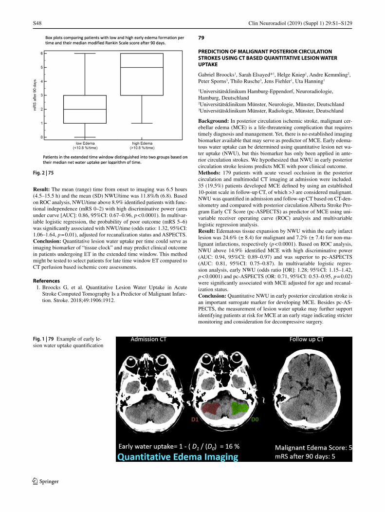
S48
123
Clin Neuroradiol (2019) (Suppl 1) 29:S1–S129
Result: The mean (range) time from onset to imaging was 6.5 hours (4.5–15.5 h) and the mean (SD) NWU/time was 11.8%/h (6.8). Based on ROC analysis, NWU/time above 8.9% identified patients with func-tional independence (mRS 0–2) with high discriminative power (area under curve [AUC]: 0.86, 95%CI: 0.67–0.96, p < 0.0001). In multivar-iable logistic regression, the probability of poor outcome (mRS 5–6) was significantly associated with NWU/time (odds ratio: 1.32, 95%CI: 1.06–1.64, p = 0.01), adjusted for recanalization status and ASPECTS.Conclusion: Quantitative lesion water uptake per time could serve as imaging biomarker of “tissue clock” and may predict clinical outcome in patients undergoing ET in the extended time window. This method might be tested to select patients for late time window ET compared to CT perfusion based ischemic core assessments.
References 1. Broocks G, et al. Quantitative Lesion Water Uptake in Acute
Stroke Computed Tomography Is a Predictor of Malignant Infarc-tion. Stroke. 2018;49:1906:1912.
79
PREDICTION OF MALIGNANT POSTERIOR CIRCULATION STROKES USING CT BASED QUANTITATIVE LESION WATER UPTAKE
Gabriel Broocks1, Sarah Elsayed*1, Helge Kniep1, Andre Kemmling2, Peter Sporns3, Thilo Rusche3, Jens Fiehler1, Uta Hanning1
1Universitätsklinikum Hamburg-Eppendorf, Neuroradiologie, Hamburg, Deutschland2Universitätsklinikum Münster, Neurologie, Münster, Deutschland3Universitätsklinikum Münster, Radiologie, Münster, Deutschland
Background: In posterior circulation ischemic stroke, malignant cer-ebellar edema (MCE) is a life-threatening complication that requires timely diagnosis and management. Yet, there is no established imaging biomarker available that may serve as predictor of MCE. Early edema-tous water uptake can be determined using quantitative lesion net wa-ter uptake (NWU), but this biomarker has only been applied in ante-rior circulation strokes. We hypothesized that NWU in early posterior circulation stroke lesions predicts MCE with poor clinical outcome.Methods: 179 patients with acute vessel occlusion in the posterior circulation and multimodal CT imaging at admission were included. 35 (19.5%) patients developed MCE defined by using an established 10-point scale in follow-up CT, of which >3 are considered malignant. NWU was quantified in admission and follow-up CT based on CT-den-sitometry and compared with posterior circulation Alberta Stroke Pro-gram Early CT Score (pc-ASPECTS) as predictor of MCE using uni-variable receiver operating curve (ROC) analysis and multivariable logistic regression analysis.Result: Edematous tissue expansion by NWU within the early infarct lesion was 24.6% (± 8.4) for malignant and 7.2% (± 7.4) for non-ma-lignant infarctions, respectively (p < 0.0001). Based on ROC analysis, NWU above 14.9% identified MCE with high discriminative power (AUC: 0.94, 95%CI: 0.89–0.97) and was superior to pc-ASPECTS (AUC: 0.81, 95%CI: 0.75–0.87). In multivariable logistic regres-sion analysis, early NWU (odds ratio [OR]: 1.28; 95%CI: 1.15–1.42, p < 0.0001) and pc-ASPECTS (OR: 0.71, 95%CI: 0.53–0.95, p = 0.02) were significantly associated with MCE adjusted for age and recanal-ization status.Conclusion: Quantitative NWU in early posterior circulation stroke is an important surrogate marker for developing MCE. Besides pc-AS-PECTS, the measurement of lesion water uptake may further support identifying patients at risk for MCE at an early stage indicating stricter monitoring and consideration for decompressive surgery.
Fig. 2 | 75
Fig. 1 | 79 Example of early le-sion water uptake quantification
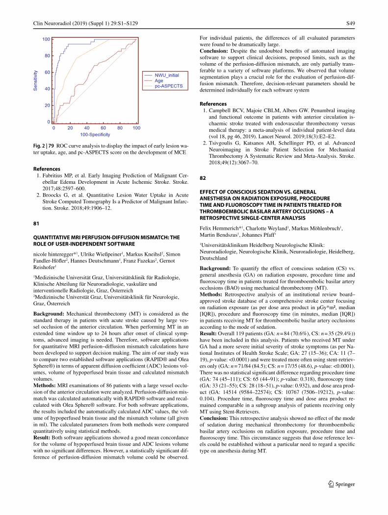
S49
123
Clin Neuroradiol (2019) (Suppl 1) 29:S1–S129
References 1. Fabritius MP, et al. Early Imaging Prediction of Malignant Cer-
ebellar Edema Development in Acute Ischemic Stroke. Stroke. 2017;48:2597–600.
2. Broocks G, et al. Quantitative Lesion Water Uptake in Acute Stroke Computed Tomography Is a Predictor of Malignant Infarc-tion. Stroke. 2018;49:1906–12.
81
QUANTITATIVE MRI PERFUSION-DIFFUSION MISMATCH: THE ROLE OF USER-INDEPENDENT SOFTWARE
nicole hinteregger*1, Ulrike Wießpeiner1, Markus Kneihsl2, Simon Fandler-Höfler2, Hannes Deutschmann1, Franz Fazekas2, Gernot Reishofer1
1Medizinische Universität Graz, Universitätsklinik für Radiologie, Klinische Abteilung für Neuroradiologie, vaskuläre und interventionelle Radiologie, Graz, Österreich2Medizinische Universtät Graz, Universitätsklinik für Neurologie, Graz, Österreich
Background: Mechanical thrombectomy (MT) is considered as the standard therapy in patients with acute stroke caused by large ves-sel occlusion of the anterior circulation. When performing MT in an extended time window up to 24 hours after onset of clinical symp-toms, advanced imaging is needed. Therefore, software applications for quantitative MRI perfusion–diffusion mismatch calculations have been developed to support decision making. The aim of our study was to compare two established software applications (RAPID® and Olea Sphere®) in terms of apparent diffusion coefficient (ADC) lesions vol-umes, volume of hypoperfused brain tissue and calculated mismatch volumes.Methods: MRI examinations of 86 patients with a large vessel occlu-sion of the anterior circulation were analyzed. Perfusion-diffusion mis-match was calculated automatically with RAPID® software and recal-culated with Olea Sphere® software. For both software applications, the results included the automatically calculated ADC values, the vol-ume of hypoperfused brain tissue and the mismatch volume (all given in ml). The calculated parameters from both methods were compared quantitatively using statistical methods.Result: Both software applications showed a good mean concordance for the volume of hypoperfused brain tissue and ADC lesions volume with no significant differences. However, a statistically significant dif-ference of perfusion-diffusion mismatch volume could be observed.
For individual patients, the differences of all evaluated parameters were found to be dramatically large.Conclusion: Despite the undoubted benefits of automated imaging software to support clinical decisions, proposed limits, such as the volume of the perfusion-diffusion mismatch, are only partially trans-ferable to a variety of software platforms. We observed that volume segmentation plays a crucial role for the evaluation of perfusion-dif-fusion mismatch. Therefore, decision-relevant parameters should be determined individually for each software system
References 1. Campbell BCV, Majoie CBLM, Albers GW. Penumbral imaging
and functional outcome in patients with anterior circulation is-chaemic stroke treated with endovascular thrombectomy versus medical therapy: a meta-analysis of individual patient-level data (vol 18, pg 46, 2019). Lancet Neurol. 2019;18(3):E2–E2.
2. Tsivgoulis G, Katsanos AH, Schellinger PD, et al. Advanced Neuroimaging in Stroke Patient Selection for Mechanical Thrombectomy A Systematic Review and Meta-Analysis. Stroke. 2018;49(12):3067–70.
82
EFFECT OF CONSCIOUS SEDATION VS. GENERAL ANESTHESIA ON RADIATION EXPOSURE, PROCEDURE TIME AND FLUOROSCOPY TIME IN PATIENTS TREATED FOR THROMBOEMBOLIC BASILAR ARTERY OCCLUSIONS – A RETROSPECTIVE SINGLE-CENTER ANALYSIS
Felix Hemmerich*1, Charlotte Weyland1, Markus Möhlenbruch1, Martin Bendszus1, Johannes Pfaff1
1Universitätsklinikum Heidelberg Neurologische Klinik: Neuroradiologie, Neurologische Klinik, Neuroradiologie, Heidelberg, Deutschland
Background: To quantify the effect of conscious sedation (CS) vs. general anesthesia (GA) on radiation exposure, procedure time and fluoroscopy time in patients treated for thromboembolic basilar artery occlusions (BAO) using mechanical thrombectomy (MT).Methods: Retrospective analysis of an institutional review board–approved stroke database of a comprehensive stroke center focusing on radiation exposure (as per dose area product in µGy*m², median [IQR]), procedure and fluoroscopy time (in minutes, median [IQR]) in patients receiving MT for thromboembolic basilar artery occlusions according to the mode of sedation.Result: Overall 119 patients (GA: n = 84 (70.6%), CS: n = 35 (29.4%)) have been included in this analysis. Patients who received MT under GA had a more severe initial severity of stroke symptoms (as per Na-tional Institutes of Health Stroke Scale; GA: 27 (15–36); CA: 11 (7–19), p-value: <0.0001) and were treated more often using stent-retriev-ers only (GA: n = 71/84 (84.5); CS: n = 17/35 (48.6), p-value: <0.0001). There was no statistical significant difference regarding procedure time (GA: 74 (45–111); CS: 65 (44–91); p-value: 0.318), fluoroscopy time (GA: 33 (21–55); CS: 28 (18–51), p-value: 0.932), and dose area prod-uct (GA: 14514 (9584–22574); CS: 10767 (7506–19212), p-value: 0.104). Procedure time, fluoroscopy time and dose area product re-mained comparable in a subgroup analysis of patients receiving only MT using Stent-Retrievers.Conclusion: This retrospective analysis showed no effect of the mode of sedation during mechanical thrombectomy for thromboembolic basilar artery occlusions on radiation exposure, procedure time and fluoroscopy time. This circumstance suggests that dose reference lev-els could be established without a particular need to regard a specific type on anesthesia during MT.
Fig. 2 | 79 ROC curve analysis to display the impact of early lesion wa-ter uptake, age, and pc-ASPECTS score on the development of MCE
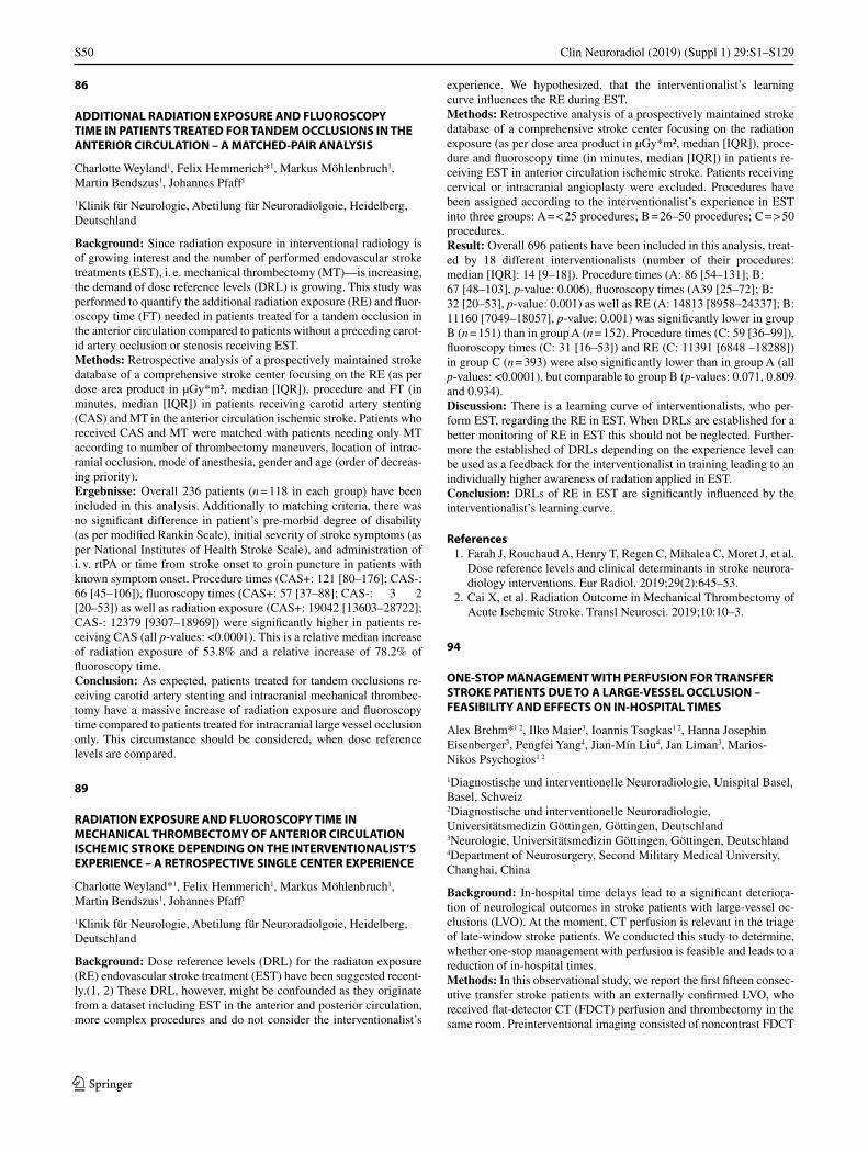
S50
123
Clin Neuroradiol (2019) (Suppl 1) 29:S1–S129
86
ADDITIONAL RADIATION EXPOSURE AND FLUOROSCOPY TIME IN PATIENTS TREATED FOR TANDEM OCCLUSIONS IN THE ANTERIOR CIRCULATION – A MATCHED-PAIR ANALYSIS
Charlotte Weyland1, Felix Hemmerich*1, Markus Möhlenbruch1, Martin Bendszus1, Johannes Pfaff1
1Klinik für Neurologie, Abetilung für Neuroradiolgoie, Heidelberg, Deutschland
Background: Since radiation exposure in interventional radiology is of growing interest and the number of performed endovascular stroke treatments (EST), i. e. mechanical thrombectomy (MT)—is increasing, the demand of dose reference levels (DRL) is growing. This study was performed to quantify the additional radiation exposure (RE) and fluor-oscopy time (FT) needed in patients treated for a tandem occlusion in the anterior circulation compared to patients without a preceding carot-id artery occlusion or stenosis receiving EST.Methods: Retrospective analysis of a prospectively maintained stroke database of a comprehensive stroke center focusing on the RE (as per dose area product in µGy*m², median [IQR]), procedure and FT (in minutes, median [IQR]) in patients receiving carotid artery stenting (CAS) and MT in the anterior circulation ischemic stroke. Patients who received CAS and MT were matched with patients needing only MT according to number of thrombectomy maneuvers, location of intrac-ranial occlusion, mode of anesthesia, gender and age (order of decreas-ing priority).Ergebnisse: Overall 236 patients (n = 118 in each group) have been included in this analysis. Additionally to matching criteria, there was no significant difference in patient’s pre-morbid degree of disability (as per modified Rankin Scale), initial severity of stroke symptoms (as per National Institutes of Health Stroke Scale), and administration of i. v. rtPA or time from stroke onset to groin puncture in patients with known symptom onset. Procedure times (CAS+: 121 [80–176]; CAS-: 66 [45–106]), fluoroscopy times (CAS+: 57 [37–88]; CAS-: 3 2 [20–53]) as well as radiation exposure (CAS+: 19042 [13603–28722]; CAS-: 12379 [9307–18969]) were significantly higher in patients re-ceiving CAS (all p-values: <0.0001). This is a relative median increase of radiation exposure of 53.8% and a relative increase of 78.2% of fluoroscopy time.Conclusion: As expected, patients treated for tandem occlusions re-ceiving carotid artery stenting and intracranial mechanical thrombec-tomy have a massive increase of radiation exposure and fluoroscopy time compared to patients treated for intracranial large vessel occlusion only. This circumstance should be considered, when dose reference levels are compared.
89
RADIATION EXPOSURE AND FLUOROSCOPY TIME IN MECHANICAL THROMBECTOMY OF ANTERIOR CIRCULATION ISCHEMIC STROKE DEPENDING ON THE INTERVENTIONALIST’S EXPERIENCE – A RETROSPECTIVE SINGLE CENTER EXPERIENCE
Charlotte Weyland*1, Felix Hemmerich1, Markus Möhlenbruch1, Martin Bendszus1, Johannes Pfaff1
1Klinik für Neurologie, Abetilung für Neuroradiolgoie, Heidelberg, Deutschland
Background: Dose reference levels (DRL) for the radiaton exposure (RE) endovascular stroke treatment (EST) have been suggested recent-ly.(1, 2) These DRL, however, might be confounded as they originate from a dataset including EST in the anterior and posterior circulation, more complex procedures and do not consider the interventionalist’s
experience. We hypothesized, that the interventionalist’s learning curve influences the RE during EST.Methods: Retrospective analysis of a prospectively maintained stroke database of a comprehensive stroke center focusing on the radiation exposure (as per dose area product in µGy*m², median [IQR]), proce-dure and fluoroscopy time (in minutes, median [IQR]) in patients re-ceiving EST in anterior circulation ischemic stroke. Patients receiving cervical or intracranial angioplasty were excluded. Procedures have been assigned according to the interventionalist’s experience in EST into three groups: A = < 25 procedures; B = 26–50 procedures; C = > 50 procedures.Result: Overall 696 patients have been included in this analysis, treat-ed by 18 different interventionalists (number of their procedures: median [IQR]: 14 [9–18]). Procedure times (A: 86 [54–131]; B: 67 [48–103], p-value: 0.006), fluoroscopy times (A39 [25–72]; B: 32 [20–53], p-value: 0.001) as well as RE (A: 14813 [8958–24337]; B: 11160 [7049–18057], p-value: 0.001) was significantly lower in group B (n = 151) than in group A (n = 152). Procedure times (C: 59 [36–99]), fluoroscopy times (C: 31 [16–53]) and RE (C: 11391 [6848 –18288]) in group C (n = 393) were also significantly lower than in group A (all p-values: <0.0001), but comparable to group B (p-values: 0.071, 0.809 and 0.934).Discussion: There is a learning curve of interventionalists, who per-form EST, regarding the RE in EST. When DRLs are established for a better monitoring of RE in EST this should not be neglected. Further-more the established of DRLs depending on the experience level can be used as a feedback for the interventionalist in training leading to an individually higher awareness of radation applied in EST.Conclusion: DRLs of RE in EST are significantly influenced by the interventionalist’s learning curve.
References 1. Farah J, Rouchaud A, Henry T, Regen C, Mihalea C, Moret J, et al.
Dose reference levels and clinical determinants in stroke neurora-diology interventions. Eur Radiol. 2019;29(2):645–53.
2. Cai X, et al. Radiation Outcome in Mechanical Thrombectomy of Acute Ischemic Stroke. Transl Neurosci. 2019;10:10–3.
94
ONE-STOP MANAGEMENT WITH PERFUSION FOR TRANSFER STROKE PATIENTS DUE TO A LARGE-VESSEL OCCLUSION – FEASIBILITY AND EFFECTS ON IN-HOSPITAL TIMES
Alex Brehm*1 2, Ilko Maier3, Ioannis Tsogkas1 2, Hanna Josephin Eisenberger3, Pengfei Yang4, Jian-Mín Liu4, Jan Liman3, Marios-Nikos Psychogios1 2
1Diagnostische und interventionelle Neuroradiologie, Unispital Basel, Basel, Schweiz2Diagnostische und interventionelle Neuroradiologie, Universitätsmedizin Göttingen, Göttingen, Deutschland3Neurologie, Universitätsmedizin Göttingen, Göttingen, Deutschland4Department of Neurosurgery, Second Military Medical University, Changhai, China
Background: In-hospital time delays lead to a significant deteriora-tion of neurological outcomes in stroke patients with large-vessel oc-clusions (LVO). At the moment, CT perfusion is relevant in the triage of late-window stroke patients. We conducted this study to determine, whether one-stop management with perfusion is feasible and leads to a reduction of in-hospital times.Methods: In this observational study, we report the first fifteen consec-utive transfer stroke patients with an externally confirmed LVO, who received flat-detector CT (FDCT) perfusion and thrombectomy in the same room. Preinterventional imaging consisted of noncontrast FDCT
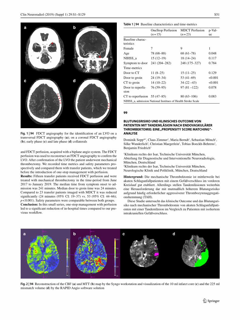
S51
123
Clin Neuroradiol (2019) (Suppl 1) 29:S1–S129
and FDCT perfusion, acquired with a biplane angio system. The FDCT perfusion was used to reconstruct an FDCT angiography to confirm the LVO. After confirmation of the LVO the patient underwent mechanical thrombectomy. We recorded time metrics and safety parameters pro-spectively and compared them with transfer patients, which we treated before the introduction of one-stop management with perfusion.Results: Fifteen transfer patients received FDCT perfusion and were treated with mechanical thrombectomy in the time-period from June 2017 to January 2019. The median time from symptom onset to ad-mission was 241 minutes. Median door to groin time was 24 minutes. Compared to 23 transfer patients imaged with MDCT it was reduced significantly (24 minutes (95% CI: 19–37) vs. 53 (95% CI: 44–66); p < 0.001). Safety parameters were comparable between both groups.Conclusion: In this small series, one-stop management with perfusion led to a significant reduction of in-hospital times compared to our pre-vious workflow.
99
BLUTUNGSRISIKO UND KLINISCHES OUTCOME VON PATIENTEN MIT TANDEMLÄSION NACH ENDOVASKULÄRER THROMBEKTOMIE: EINE „PROPENSITY SCORE MATCHING“-ANALYSE
Dominik Sepp*1, Claus Zimmer1, Maria Berndt1, Sebastian Mönch1, Silke Wunderlich2, Christian Maegerlein1, Tobias Boeckh-Behrens1, Benjamin Friedrich1
1Klinikum rechts der Isar, Technische Universität München, Abteilung für Diagnostische und Interventionelle Neuroradiologie, München, Deutschland2Klinikum rechts der Isar, Technische Universität München, Neurologische Klinik und Poliklinik, München, Deutschland
Hintergrund: Die mechanische Thrombektomie ist mittlerweile bei akuten Schlaganfallpatienten mit einem Gefäßverschluss im vorderen Kreislauf gut etabliert. Allerdings stellen Tandemläsionen weiterhin eine Herausforderung dar mit mutmaßlich höherem Blutungsrisiko aufgrund häufig erforderlicher aggressiverer Thrombozytenaggregati-onshemmung (TAH).
Diese Studie untersucht das klinische Outcome und das Blutungsri-siko nach mechanischer Thrombektomie von akuten Schlaganfallpati-enten mit einer Tandemläsion im Vergleich zu Patienten mit isoliertem intrakraniellen Gefäßverschluss.
Table 1 | 94 Baseline characteristics and time-metrics
OneStop Perfusion (n = 15)
MDCT Perfusion (n = 23)
p-Val-ue
Baseline charac-teristics
Female 7 9 1
Age 78 (68–88) 68 (61–78) 0.048
NIHSS_a 15 (12–19) 18 (14–24) 0.117
Symptom to door 241 (204–282) 248 (175–327) 0.784
Time-metrics
Door to CT 11 (8–25) 15 (11–25) 0.129
Door to groin 24 (19–34) 53 (41–69) <0.001
CT to groin 14 (10–22) 34 (22 –43) <0.001
Door to reperfu-sion
76 (59–95) 97 (81 –122) 0.078
CT to reperfusion 55 (47–85) 80 (63–106) 0.083NIHSS_a, admission National Institues of Health Stroke Scale
Fig. 1 | 94 FDCT angiography for the identification of an LVO on a transversal FDCT angiography (a); on a coronal FDCT angiography (b); early phase (c) and late phase (d) collaterals
Fig. 2 | 94 Reconstruction of the CBF (a) and MTT (b) map by the Syngo workstation and visualization of the 10 ml infarct core (c) and the 225 ml mismatch volume (d) by the RAPID Angio software solution

S52
123
Clin Neuroradiol (2019) (Suppl 1) 29:S1–S129
Methoden: Eingeschlossen wurden 63 konsekutive Schlaganfallpa-tienten mit einer Tandemläsion (proximaler Gefäßverschluss im vor-deren Kreislauf sowie der ipsilateralen A. carotis interna), die in den Jahren 2014–2018 mechanisch rekanalisiert wurden. Als Kontrollen wurden 111 Schlaganfallpatienten mit einem isolierten intrakraniellen Gefäßverschluss ausgewählt nach „propensity score matching“-Ana-lyse in einem 1:2 Verhältnis (Kovariate: Geschlecht, Alter, Einordnung als „wake-up stroke“, rtPA-Gabe und NIHSS).Ergebnisse: Die Quote der erfolgreichen Rekanalisierungen (TICI 2b/3) unterschied sich in den Gruppen nicht (93 % vs 87 %, p = 0,19). Periprozedurale Komplikationen traten bei Tandemläsionen nicht häufiger auf. Obwohl Patienten mit Tandemläsion häufiger mit ag-gressiverer TAH behandelt wurden, ergab sich in dieser Gruppe kein schlechteres klinisches Outcome und keine erhöhte Inzidenz von sym-ptomatischen intrakraniellen Blutungen (7,9 % vs 5,4 %, p = 0,51).
Unter den Patienten mit einer Tandemläsion war eine aggressivere TAH (Tirofiban oder doppelte TAH) nicht mit einem höheren Risiko für eine symptomatische intrakranielle Blutung assoziiert (5,6 % vs 11,1 %, p = 0,42).Diskussion: Der Therapieerfolg und das Risiko einer symptomatischen intrakraniellen Blutung nach mechanischer Rekanalisation waren bei akuten Schlaganfallpatienten mit einer Tandemläsion vergleichbar mit Patienten mit einem isolierten intrakraniellen Gefäßverschluss. Die häufig notwendige aggressivere TAH bei Tandemläsionen war nicht mit einem erhöhten Risiko einer symptomatischen Blutung assoziiert.Fazit: Die komplexeren technischen Anforderungen einer mechani-schen Thrombektomie bei Tandemläsionen und das befürchtete höhe-re Blutungsrisiko durch die häufig notwendige Gabe von TAH sollten nicht zu einer zurückhaltenderen Therapie von Tandemläsionen führen, die die Chance auf ein gutes klinisches Outcome verringern könnte.
100
INITIAL EXPERIENCE WITH TRANSRADIAL ACCESS FOR CEREBROVASCULAR PROCEDURES: IS IT FEASIBLE AND SAFE?
Hanna Styczen*1 2, Volker Maus3, Ioannis Tsogkas1 4, Dan Meila5, Jan-Hendrik Buhk6, Marios-Nikos Psychogios1 4
1Institut für Diagnostische und Interventionelle Neuroradiologie, Universitätsmedizin Göttingen, Göttingen, Deutschland2Institut für Diagnostische und Interventionelle Radiologie und Neuroradiologie, Universitätsklinikum Essen, Essen, Deutschland3Institut für Diagnostische und Interventionelle Radiologie, Neuroradiologie und Nuklearmedizin, Universitätsklinikum Knappschaftskrankenhaus Bochum, Bochum, Deutschland4Abteilung für Neuroradiologie, Universitätsspital Basel, Basel, Schweiz5Abteilung für Interventionelle Neuroradiologie, Johanna Etienne Krankenhaus, Neuss, Deutschland6Abteilung für Diagnostische und Interventionelle Neuroradiologie, Universitätsklinikum Hamburg-Eppendorf, Hamburg, Deutschland
Background: Despite the exceptional benefit of transradial access over transfemoral access in cardiac procedures, the transition for cere-brovascular procedures has been reluctant. Here we present our expe-rience with transradial access in cerebral diagnostic angiographies and neurointerventional procedures.Methods: We performed a retrospective analysis of patients who un-derwent transradial access for cerebrovascular procedures in 3 German centers between February 2017 and May 2019. Demographics, techni-cal features and complications were evaluated.Result: Transradial access was successful in 40 endovascular proce-dures, resulting in a success rate of 89% (40/45). Selected catheteri-zation of the intended vessels was obtained in 95% (40/42) of cases. The rate of procedure-related vascular complications was 2% (1/45).
Discussion: Various recent studies documented a 92.2–98.6% success rate of transradial cerebral angiography [1–4]. In the present analysis the success rate with 89% (including neurointerventional and diagnos-tic procedures) was slightly lower mostly due to puncture and cannula-tion failures. Catheterization of the intended supra-aortic vessels suc-ceeded in 95% (40/42) patients and is comparable to rates reported in previous studies [1, 3–5].Conclusion: During the initial experience with transradial access, the alternative approach proved to be safe and efficient, although a certain learning curve existed. Follow-up one-vessel cerebral angiographies via transradial access in particular could be performed on ambulatory basis prospectively.
References 1. Jo KW, Park SM, Kim SD, et al. Is transradial cerebral angi-
ography feasible and safe? A single center’s experience. J Ko-rean Neurosurg Soc. 2010;47:332–7. https://doi.org/10.3340/jkns.2010.47.5.332.
2. Snelling BM, Sur S, Shah SS, et al. Transradial cerebral angiography: techniques and outcomes. J Neurointervent Surg. 2018;10:874–81. https://doi.org/10.1136/neurintsurg-2017-013584.
3. Matsumoto Y, Hongo K, Toriyama T, et al. Transradial approach for diagnostic selective cerebral angiography: results of a consec-utive series of 166 cases. Ajnr Am J Neuroradiol. 2001;22:704–8.
4. Park JH, Kim DY, Kim JW, et al. Efficacy of transradial cerebral angiography in the elderly. J Korean Neurosurg Soc. 2013;53:213–7. https://doi.org/10.3340/jkns.2013.53.4.213.
5. Goland J, Doroszuk GF, Garbugino SL, et al. Transradial approach to treating endovascular cerebral aneurysms: Case series and tech-nical note. Surg Neurol Int. 2017;8:73. https://doi.org/10.4103/sni.sni_393_16.
103
BASILAR ARTERY OCCLUSIONS WITH WORSE OUTCOME: EARLY IDENTIFICATION OF UNDERLYING STENOSIS
Maria Berndt*1, David Pree1, Fabian Mück2, Dominik Sepp1, Silke Wunderlich3, Claus Zimmer1, Stefan Wirth2, Christian Maegerlein1, Benjamin Friedrich1, Johannes Kaesmacher4, Tobias Boeckh-Behrens1
1Technische Universität München, Fakultät für Medizin, Klinikum rechts der Isar, Neuroradiologie, München, Deutschland2DONAUISAR Klinikum Deggendorf, Radiologie, Deggendorf, Deutschland3Technische Universität München, Fakultät für Medizin, Klinikum rechts der Isar, Neurologie, München, Deutschland4University Hospital Bern, Inselspital, Neuroradiologie, Bern, Deutschland
Background: Recent studies show that mechanical thrombectomy of acute basilar artery occlusions (BAO) results in high successful reca-nalization and good outcomes. However, the impact of pathogenesis, especially the presence of an underlying basilar stenosis (BS) is not fully understood. Aim of the study was to investigate angiographic and clinical outcome parameter for this entity and test the potential of known perviousness measures for an early identification.Methods: All consecutive patients with acute BAO, treated with sec-ond-generation thrombectomy devices at a single-center between March 2008 and August 2017 (n = 134), were included. Underlying pathogenesis (basilar stenosis) was examined in respect of angiograph-ic (complete reperfusion TICI 3) and clinical (NIHSS/MRS) outcome parameters, that were existing for n = 115 patients. Thrombus permea-bility measures (change in thrombus attenuation ∆t and corrected void fraction ε, n = 58) were assessed in admission CT imaging and correlat-ed to different etiological subgroups and clinical outcome (see Fig. 1).

S53
123
Clin Neuroradiol (2019) (Suppl 1) 29:S1–S129
Results: For the BS-group, less patients were successfully recan-alized (86% vs. 93% for BAO without BS) and higher NIHSS/MRS values were obtained (p = 0.001/0.002). The group of BS turned out to have significant lower native density values (39.9 ± 16.1 HU) and higher perviousness measures ∆t (32.8 ± 36.7) and ε (0.21 ± 0.21) than thrombi without an underlying BS (54.8 ± 15.2/12.8 ± 16.7/0.07 ± 0.10, p = 0.003/0.007/0.001).Discussion: Concordant to previous studies, outcome of mechanical thrombectomy of BAO depends on pathogenesis. For the subgroup of underlying basilar stenosis worse recanalization success and clinical presentation was found, that raise the need for an early identification. It was further shown, that perviousness has the potential to differentiate BAO with and without BS in admission imaging.Conclusion: Perviousness may be valuable as an early imaging marker for identifying underlying basilar stenosis in acute BAO, that presents with worse angiographic and clinical performance. This could be rele-vant for timely therapeutic and rehabilitative decisions.
108
WEB + STENT ZUR BEHANDLUNG INTRAKRANIELLER ANEURYSMEN: SUPPORTIVES STENTING BEHANDELT WEB-PROTRUSIONEN IN DAS TRÄGERGEFÄß EFFEKTIV
Christin Clajus*1, Donald Lobsien1, Joachim Klisch1
1Institut für diagnostische und interventionelle Radiologie und Neuroradiologie, Helios Klinikum Erfurt, Erfurt, Deutschland
Hintergrund: Zur Prävention von Aneurysmaresten und -rezidiven ist die optimale Größenauswahl eines WEB Device essenziell. Gelegent-
lich tritt eine leichte Protrusion des WEB Device an der Basis des An-eurysmas in das Trägergefäß auf, teils durch moderates Oversizing des Device bedingt. Zur Vermeidung flussrelevanter Stenosen am Träger-gefäß wurde die supportive Implantation eines Stents im Rahmen der WEB-Implantation beurteilt.Methoden: Von insgesamt 240 WEB Device-Implantationen (10/2010–04/2019) erforderten WEB-Protrusionen bei 14 Aneurysma-Patienten (5,8 %) die zusätzliche Implantation eines Stents. In 11 Fäl-len (78,6 %) erfolgte die Behandlung elektiv, bei 3 Patienten (21,4 %) im Rahmen einer akuten SAB. Die behandelten intrakraniellen An-eurysmen befanden sich vor allem in der vorderen Zirkulation (n = 9, 64,3 %), zu einem geringen Anteil in der hinteren Zirkulation (n = 5, 35,7 %). Für diese kombinierte Technik wurden 13 Solitaire-Stents (92,9 %) und 1 LVIS jr. Stent (7,1 %) verwendet.Ergebnisse: Die kombinierte Behandlung konnte in 100 % der Fälle technisch komplikationslos durchgeführt werden. Ein SAB-Patient er-litt dennoch eine frühe Re-Blutung aus einem Aneurysmarest. Dieser Patient wurde mittels Coiling nachbehandelt und zeigte ein gutes klini-sches Outcome. Ein SAB-Patient (7,1 %) erlitt kleine Infarkte der hin-teren Zirkulation, 1 weiterer Patient (7,1 %) ein inguinales Aneurysma. DSA-Kontrollen bei 9 Patienten (64,3 %) durchschnittlich 15 Monate nach Intervention zeigten bei 8 Aneurysmen (85,7 %) einen vollstän-digen Verschluss, 1 weiteres Aneurysma (11,1 %) bedurfte einer Re-Intervention aufgrund eines Rezidiv-Aneurysmas.Diskussion: Die Verschlussraten der Aneurysmen in den Follow-ups liegen in dieser kleinen Fallserie über dem Durchschnitt unserer al-lein mit WEB Device versorgten Aneurysmen1. Nachteilig gegenüber der vorgenannten Patientengruppe erscheint die Notwendigkeit der Thrombozytenaggregation nach intrakraniellem Stenting.Fazit: Supportives Stenting mit WEB Device versorgter Aneurysmen ist eine technisch gut durchführbare und sichere Behandlungsmethode.
Fig. 1 | 103 Example of an acute basilar occlusion with underly-ing basilar stenosis. Assessment of perviousness measures ∆t and ε after coaligment of native CT scan (a) and CT-angiography (b). Corresponding pre- (c) and post-thrombectomy (d) angiog-raphy in anteroposterior view
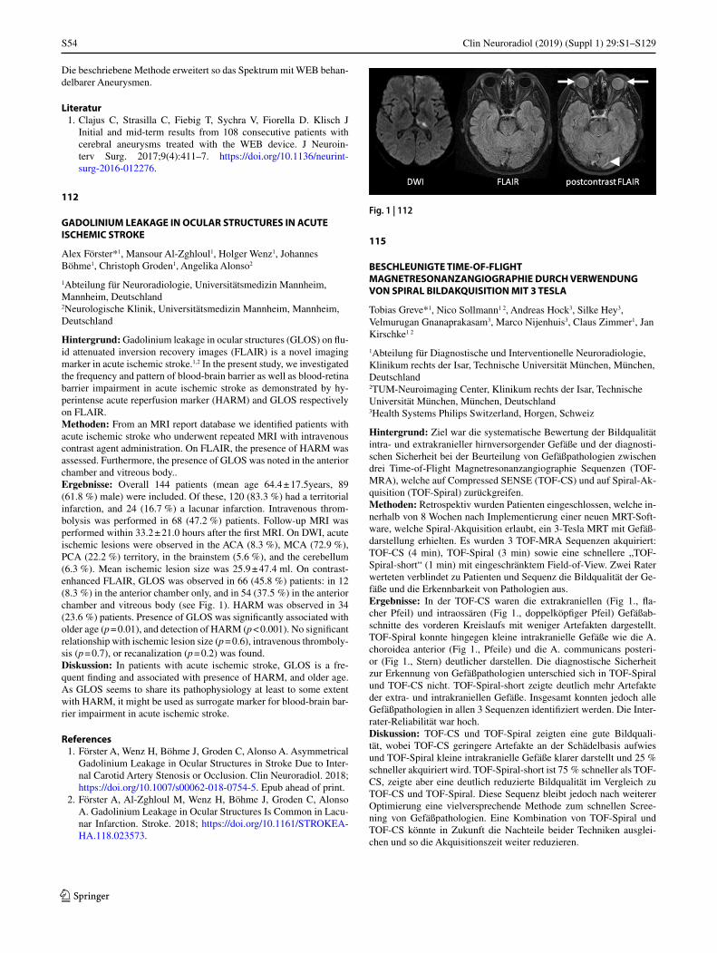
S54
123
Clin Neuroradiol (2019) (Suppl 1) 29:S1–S129
Die beschriebene Methode erweitert so das Spektrum mit WEB behan-delbarer Aneurysmen.
Literatur 1. Clajus C, Strasilla C, Fiebig T, Sychra V, Fiorella D. Klisch J
Initial and mid-term results from 108 consecutive patients with cerebral aneurysms treated with the WEB device. J Neuroin-terv Surg. 2017;9(4):411–7. https://doi.org/10.1136/neurint-surg-2016-012276.
112
GADOLINIUM LEAKAGE IN OCULAR STRUCTURES IN ACUTE ISCHEMIC STROKE
Alex Förster*1, Mansour Al-Zghloul1, Holger Wenz1, Johannes Böhme1, Christoph Groden1, Angelika Alonso2
1Abteilung für Neuroradiologie, Universitätsmedizin Mannheim, Mannheim, Deutschland2Neurologische Klinik, Universitätsmedizin Mannheim, Mannheim, Deutschland
Hintergrund: Gadolinium leakage in ocular structures (GLOS) on flu-id attenuated inversion recovery images (FLAIR) is a novel imaging marker in acute ischemic stroke.1,2 In the present study, we investigated the frequency and pattern of blood-brain barrier as well as blood-retina barrier impairment in acute ischemic stroke as demonstrated by hy-perintense acute reperfusion marker (HARM) and GLOS respectively on FLAIR.Methoden: From an MRI report database we identified patients with acute ischemic stroke who underwent repeated MRI with intravenous contrast agent administration. On FLAIR, the presence of HARM was assessed. Furthermore, the presence of GLOS was noted in the anterior chamber and vitreous body..Ergebnisse: Overall 144 patients (mean age 64.4 ± 17.5years, 89 (61.8 %) male) were included. Of these, 120 (83.3 %) had a territorial infarction, and 24 (16.7 %) a lacunar infarction. Intravenous throm-bolysis was performed in 68 (47.2 %) patients. Follow-up MRI was performed within 33.2 ± 21.0 hours after the first MRI. On DWI, acute ischemic lesions were observed in the ACA (8.3 %), MCA (72.9 %), PCA (22.2 %) territory, in the brainstem (5.6 %), and the cerebellum (6.3 %). Mean ischemic lesion size was 25.9 ± 47.4 ml. On contrast-enhanced FLAIR, GLOS was observed in 66 (45.8 %) patients: in 12 (8.3 %) in the anterior chamber only, and in 54 (37.5 %) in the anterior chamber and vitreous body (see Fig. 1). HARM was observed in 34 (23.6 %) patients. Presence of GLOS was significantly associated with older age (p = 0.01), and detection of HARM (p < 0.001). No significant relationship with ischemic lesion size (p = 0.6), intravenous thromboly-sis (p = 0.7), or recanalization (p = 0.2) was found.Diskussion: In patients with acute ischemic stroke, GLOS is a fre-quent finding and associated with presence of HARM, and older age. As GLOS seems to share its pathophysiology at least to some extent with HARM, it might be used as surrogate marker for blood-brain bar-rier impairment in acute ischemic stroke.
References 1. Förster A, Wenz H, Böhme J, Groden C, Alonso A. Asymmetrical
Gadolinium Leakage in Ocular Structures in Stroke Due to Inter-nal Carotid Artery Stenosis or Occlusion. Clin Neuroradiol. 2018; https://doi.org/10.1007/s00062-018-0754-5. Epub ahead of print.
2. Förster A, Al-Zghloul M, Wenz H, Böhme J, Groden C, Alonso A. Gadolinium Leakage in Ocular Structures Is Common in Lacu-nar Infarction. Stroke. 2018; https://doi.org/10.1161/STROKEA-HA.118.023573.
115
BESCHLEUNIGTE TIME-OF-FLIGHT MAGNETRESONANZANGIOGRAPHIE DURCH VERWENDUNG VON SPIRAL BILDAKQUISITION MIT 3 TESLA
Tobias Greve*1, Nico Sollmann1 2, Andreas Hock3, Silke Hey3, Velmurugan Gnanaprakasam3, Marco Nijenhuis3, Claus Zimmer1, Jan Kirschke1 2
1Abteilung für Diagnostische und Interventionelle Neuroradiologie, Klinikum rechts der Isar, Technische Universität München, München, Deutschland2TUM-Neuroimaging Center, Klinikum rechts der Isar, Technische Universität München, München, Deutschland3Health Systems Philips Switzerland, Horgen, Schweiz
Hintergrund: Ziel war die systematische Bewertung der Bildqualität intra- und extrakranieller hirnversorgender Gefäße und der diagnosti-schen Sicherheit bei der Beurteilung von Gefäßpathologien zwischen drei Time-of-Flight Magnetresonanzangiographie Sequenzen (TOF-MRA), welche auf Compressed SENSE (TOF-CS) und auf Spiral-Ak-quisition (TOF-Spiral) zurückgreifen.Methoden: Retrospektiv wurden Patienten eingeschlossen, welche in-nerhalb von 8 Wochen nach Implementierung einer neuen MRT-Soft-ware, welche Spiral-Akquisition erlaubt, ein 3-Tesla MRT mit Gefäß-darstellung erhielten. Es wurden 3 TOF-MRA Sequenzen akquiriert: TOF-CS (4 min), TOF-Spiral (3 min) sowie eine schnellere „TOF-Spiral-short“ (1 min) mit eingeschränktem Field-of-View. Zwei Rater werteten verblindet zu Patienten und Sequenz die Bildqualität der Ge-fäße und die Erkennbarkeit von Pathologien aus.Ergebnisse: In der TOF-CS waren die extrakraniellen (Fig 1., fla-cher Pfeil) und intraossären (Fig 1., doppelköpfiger Pfeil) Gefäßab-schnitte des vorderen Kreislaufs mit weniger Artefakten dargestellt. TOF-Spiral konnte hingegen kleine intrakranielle Gefäße wie die A. choroidea anterior (Fig 1., Pfeile) und die A. communicans posteri-or (Fig 1., Stern) deutlicher darstellen. Die diagnostische Sicherheit zur Erkennung von Gefäßpathologien unterschied sich in TOF-Spiral und TOF-CS nicht. TOF-Spiral-short zeigte deutlich mehr Artefakte der extra- und intrakraniellen Gefäße. Insgesamt konnten jedoch alle Gefäßpathologien in allen 3 Sequenzen identifiziert werden. Die Inter-rater-Reliabilität war hoch.Diskussion: TOF-CS und TOF-Spiral zeigten eine gute Bildquali-tät, wobei TOF-CS geringere Artefakte an der Schädelbasis aufwies und TOF-Spiral kleine intrakranielle Gefäße klarer darstellt und 25 % schneller akquiriert wird. TOF-Spiral-short ist 75 % schneller als TOF-CS, zeigte aber eine deutlich reduzierte Bildqualität im Vergleich zu TOF-CS und TOF-Spiral. Diese Sequenz bleibt jedoch nach weiterer Optimierung eine vielversprechende Methode zum schnellen Scree-ning von Gefäßpathologien. Eine Kombination von TOF-Spiral und TOF-CS könnte in Zukunft die Nachteile beider Techniken ausglei-chen und so die Akquisitionszeit weiter reduzieren.
Fig. 1 | 112
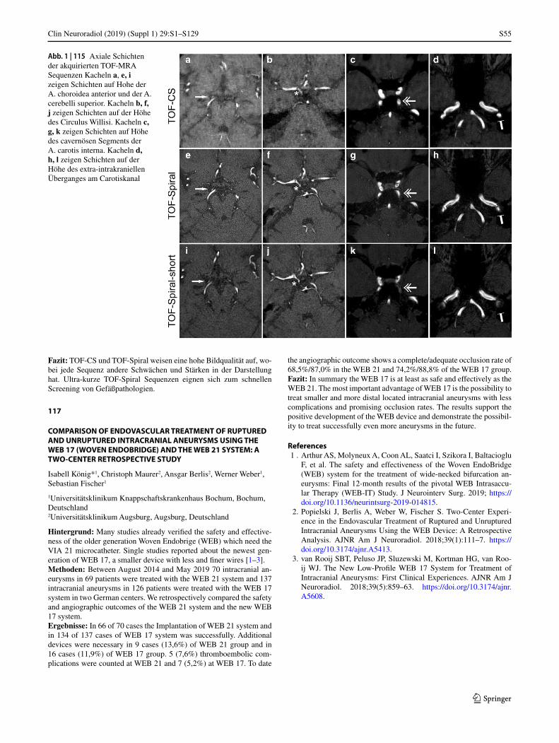
S55
123
Clin Neuroradiol (2019) (Suppl 1) 29:S1–S129
Fazit: TOF-CS und TOF-Spiral weisen eine hohe Bildqualität auf, wo-bei jede Sequenz andere Schwächen und Stärken in der Darstellung hat. Ultra-kurze TOF-Spiral Sequenzen eignen sich zum schnellen Screening von Gefäßpathologien.
117
COMPARISON OF ENDOVASCULAR TREATMENT OF RUPTURED AND UNRUPTURED INTRACRANIAL ANEURYSMS USING THE WEB 17 (WOVEN ENDOBRIDGE) AND THE WEB 21 SYSTEM: A TWO-CENTER RETROSPECTIVE STUDY
Isabell König*1, Christoph Maurer2, Ansgar Berlis2, Werner Weber1, Sebastian Fischer1
1Universitätsklinikum Knappschaftskrankenhaus Bochum, Bochum, Deutschland2Universitätsklinikum Augsburg, Augsburg, Deutschland
Hintergrund: Many studies already verified the safety and effective-ness of the older generation Woven Endobrige (WEB) which need the VIA 21 microcatheter. Single studies reported about the newest gen-eration of WEB 17, a smaller device with less and finer wires [1–3].Methoden: Between August 2014 and May 2019 70 intracranial an-eurysms in 69 patients were treated with the WEB 21 system and 137 intracranial aneurysms in 126 patients were treated with the WEB 17 system in two German centers. We retrospectively compared the safety and angiographic outcomes of the WEB 21 system and the new WEB 17 system.Ergebnisse: In 66 of 70 cases the Implantation of WEB 21 system and in 134 of 137 cases of WEB 17 system was successfully. Additional devices were necessary in 9 cases (13,6%) of WEB 21 group and in 16 cases (11,9%) of WEB 17 group. 5 (7,6%) thromboembolic com-plications were counted at WEB 21 and 7 (5,2%) at WEB 17. To date
the angiographic outcome shows a complete/adequate occlusion rate of 68,5%/87,0% in the WEB 21 and 74,2%/88,8% of the WEB 17 group.Fazit: In summary the WEB 17 is at least as safe and effectively as the WEB 21. The most important advantage of WEB 17 is the possibility to treat smaller and more distal located intracranial aneurysms with less complications and promising occlusion rates. The results support the positive development of the WEB device and demonstrate the possibil-ity to treat successfully even more aneurysms in the future.
References 1 . Arthur AS, Molyneux A, Coon AL, Saatci I, Szikora I, Baltacioglu
F, et al. The safety and effectiveness of the Woven EndoBridge (WEB) system for the treatment of wide-necked bifurcation an-eurysms: Final 12-month results of the pivotal WEB Intrasaccu-lar Therapy (WEB-IT) Study. J Neurointerv Surg. 2019; https://doi.org/10.1136/neurintsurg-2019-014815.
2. Popielski J, Berlis A, Weber W, Fischer S. Two-Center Experi-ence in the Endovascular Treatment of Ruptured and Unruptured Intracranial Aneurysms Using the WEB Device: A Retrospective Analysis. AJNR Am J Neuroradiol. 2018;39(1):111–7. https://doi.org/10.3174/ajnr.A5413.
3. van Rooij SBT, Peluso JP, Sluzewski M, Kortman HG, van Roo-ij WJ. The New Low-Profile WEB 17 System for Treatment of Intracranial Aneurysms: First Clinical Experiences. AJNR Am J Neuroradiol. 2018;39(5):859–63. https://doi.org/10.3174/ajnr.A5608.
Abb. 1 | 115 Axiale Schichten der akquirierten TOF-MRA Sequenzen Kacheln a, e, i zeigen Schichten auf Hohe der A. choroidea anterior und der A. cerebelli superior. Kacheln b, f, j zeigen Schichten auf der Höhe des Circulus Willisi. Kacheln c, g, k zeigen Schichten auf Höhe des cavernösen Segments der A. carotis interna. Kacheln d, h, l zeigen Schichten auf der Höhe des extra-intrakraniellen Überganges am Carotiskanal

S56
123
Clin Neuroradiol (2019) (Suppl 1) 29:S1–S129
Fig. 1 | 121 a-d: Exemplary parameter maps Oxygen extraction fraction (rOEF, a), cerebral blood flow (CBF, b), oxygen diffusivity (d, c) and carotid artery territories used for group-level analysis (d). CBF and diffusivity are ipsilaterally decreased (arrows). e–f: Paired scatterplot of oxygen diffusiv-ity lateralization Diffusivity is ipsilaterally decreased in ICAS (t-test, p < 0.01) and symmetri-cal in HC (p = 0.73). Later-alization is also significantly different between ICAS and HC (two-sample-test, p < 0.01)
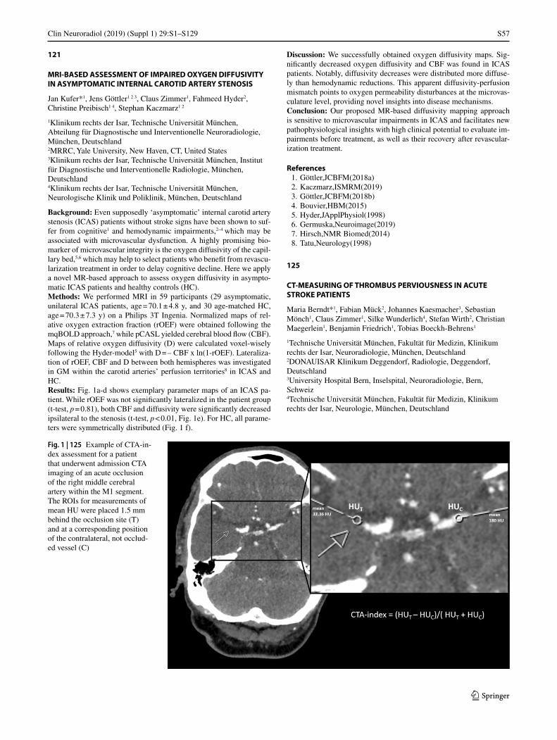
S57
123
Clin Neuroradiol (2019) (Suppl 1) 29:S1–S129
121
MRI-BASED ASSESSMENT OF IMPAIRED OXYGEN DIFFUSIVITY IN ASYMPTOMATIC INTERNAL CAROTID ARTERY STENOSIS
Jan Kufer*1, Jens Göttler1 2 3, Claus Zimmer1, Fahmeed Hyder2, Christine Preibisch1 4, Stephan Kaczmarz1 2
1Klinikum rechts der Isar, Technische Universität München, Abteilung für Diagnostische und Interventionelle Neuroradiologie, München, Deutschland2MRRC, Yale University, New Haven, CT, United States3Klinikum rechts der Isar, Technische Universität München, Institut für Diagnostische und Interventionelle Radiologie, München, Deutschland4Klinikum rechts der Isar, Technische Universität München, Neurologische Klinik und Poliklinik, München, Deutschland
Background: Even supposedly ‘asymptomatic’ internal carotid artery stenosis (ICAS) patients without stroke signs have been shown to suf-fer from cognitive1 and hemodynamic impairments,2–4 which may be associated with microvascular dysfunction. A highly promising bio-marker of microvascular integrity is the oxygen diffusivity of the capil-lary bed,5,6 which may help to select patients who benefit from revascu-larization treatment in order to delay cognitive decline. Here we apply a novel MR-based approach to assess oxygen diffusivity in asympto-matic ICAS patients and healthy controls (HC).Methods: We performed MRI in 59 participants (29 asymptomatic, unilateral ICAS patients, age = 70.1 ± 4.8 y, and 30 age-matched HC, age = 70.3 ± 7.3 y) on a Philips 3T Ingenia. Normalized maps of rel-ative oxygen extraction fraction (rOEF) were obtained following the mqBOLD approach,7 while pCASL yielded cerebral blood flow (CBF). Maps of relative oxygen diffusivity (D) were calculated voxel-wisely following the Hyder-model5 with D = – CBF x ln(1-rOEF). Lateraliza-tion of rOEF, CBF and D between both hemispheres was investigated in GM within the carotid arteries’ perfusion territories8 in ICAS and HC.Results: Fig. 1a-d shows exemplary parameter maps of an ICAS pa-tient. While rOEF was not significantly lateralized in the patient group (t-test, p = 0.81), both CBF and diffusivity were significantly decreased ipsilateral to the stenosis (t-test, p < 0.01, Fig. 1e). For HC, all parame-ters were symmetrically distributed (Fig. 1 f).
Discussion: We successfully obtained oxygen diffusivity maps. Sig-nificantly decreased oxygen diffusivity and CBF was found in ICAS patients. Notably, diffusivity decreases were distributed more diffuse-ly than hemodynamic reductions. This apparent diffusivity-perfusion mismatch points to oxygen permeability disturbances at the microvas-culature level, providing novel insights into disease mechanisms.Conclusion: Our proposed MR-based diffusivity mapping approach is sensitive to microvascular impairments in ICAS and facilitates new pathophysiological insights with high clinical potential to evaluate im-pairments before treatment, as well as their recovery after revascular-ization treatment.
References 1. Göttler,JCBFM(2018a) 2. Kaczmarz,ISMRM(2019) 3. Göttler,JCBFM(2018b) 4. Bouvier,HBM(2015) 5. Hyder,JApplPhysiol(1998) 6. Germuska,Neuroimage(2019) 7. Hirsch,NMR Biomed(2014) 8. Tatu,Neurology(1998)
125
CT-MEASURING OF THROMBUS PERVIOUSNESS IN ACUTE STROKE PATIENTS
Maria Berndt*1, Fabian Mück2, Johannes Kaesmacher3, Sebastian Mönch1, Claus Zimmer1, Silke Wunderlich4, Stefan Wirth2, Christian Maegerlein1, Benjamin Friedrich1, Tobias Boeckh-Behrens1
1Technische Universität München, Fakultät für Medizin, Klinikum rechts der Isar, Neuroradiologie, München, Deutschland2DONAUISAR Klinikum Deggendorf, Radiologie, Deggendorf, Deutschland3University Hospital Bern, Inselspital, Neuroradiologie, Bern, Schweiz4Technische Universität München, Fakultät für Medizin, Klinikum rechts der Isar, Neurologie, München, Deutschland
Fig. 1 | 125 Example of CTA-in-dex assessment for a patient that underwent admission CTA imaging of an acute occlusion of the right middle cerebral artery within the M1 segment. The ROIs for measurements of mean HU were placed 1.5 mm behind the occlusion site (T) and at a corresponding position of the contralateral, not occlud-ed vessel (C)
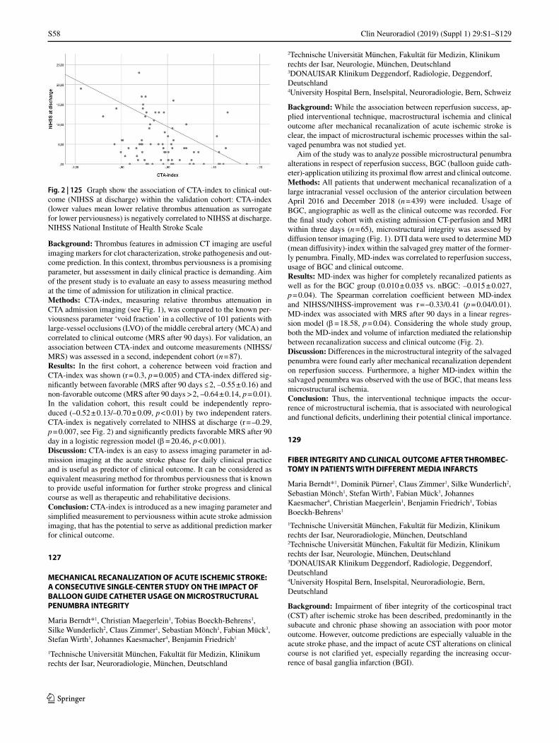
S58
123
Clin Neuroradiol (2019) (Suppl 1) 29:S1–S129
Background: Thrombus features in admission CT imaging are useful imaging markers for clot characterization, stroke pathogenesis and out-come prediction. In this context, thrombus perviousness is a promising parameter, but assessment in daily clinical practice is demanding. Aim of the present study is to evaluate an easy to assess measuring method at the time of admission for utilization in clinical practice.Methods: CTA-index, measuring relative thrombus attenuation in CTA admission imaging (see Fig. 1), was compared to the known per-viousness parameter ‘void fraction’ in a collective of 101 patients with large-vessel occlusions (LVO) of the middle cerebral artery (MCA) and correlated to clinical outcome (MRS after 90 days). For validation, an association between CTA-index and outcome measurements (NIHSS/MRS) was assessed in a second, independent cohort (n = 87).Results: In the first cohort, a coherence between void fraction and CTA-index was shown (r = 0.3, p = 0.005) and CTA-index differed sig-nificantly between favorable (MRS after 90 days ≤ 2, –0.55 ± 0.16) and non-favorable outcome (MRS after 90 days > 2, –0.64 ± 0.14, p = 0.01). In the validation cohort, this result could be independently repro-duced (–0.52 ± 0.13/–0.70 ± 0.09, p < 0.01) by two independent raters. CTA-index is negatively correlated to NIHSS at discharge (r = –0.29, p = 0.007, see Fig. 2) and significantly predicts favorable MRS after 90 day in a logistic regression model (β = 20.46, p < 0.001).Discussion: CTA-index is an easy to assess imaging parameter in ad-mission imaging at the acute stroke phase for daily clinical practice and is useful as predictor of clinical outcome. It can be considered as equivalent measuring method for thrombus perviousness that is known to provide useful information for further stroke progress and clinical course as well as therapeutic and rehabilitative decisions.Conclusion: CTA-index is introduced as a new imaging parameter and simplified measurement to perviousness within acute stroke admission imaging, that has the potential to serve as additional prediction marker for clinical outcome.
127
MECHANICAL RECANALIZATION OF ACUTE ISCHEMIC STROKE: A CONSECUTIVE SINGLE-CENTER STUDY ON THE IMPACT OF BALLOON GUIDE CATHETER USAGE ON MICROSTRUCTURAL PENUMBRA INTEGRITY
Maria Berndt*1, Christian Maegerlein1, Tobias Boeckh-Behrens1, Silke Wunderlich2, Claus Zimmer1, Sebastian Mönch1, Fabian Mück3, Stefan Wirth3, Johannes Kaesmacher4, Benjamin Friedrich1
1Technische Universität München, Fakultät für Medizin, Klinikum rechts der Isar, Neuroradiologie, München, Deutschland
2Technische Universität München, Fakultät für Medizin, Klinikum rechts der Isar, Neurologie, München, Deutschland3DONAUISAR Klinikum Deggendorf, Radiologie, Deggendorf, Deutschland4University Hospital Bern, Inselspital, Neuroradiologie, Bern, Schweiz
Background: While the association between reperfusion success, ap-plied interventional technique, macrostructural ischemia and clinical outcome after mechanical recanalization of acute ischemic stroke is clear, the impact of microstructural ischemic processes within the sal-vaged penumbra was not studied yet.
Aim of the study was to analyze possible microstructural penumbra alterations in respect of reperfusion success, BGC (balloon guide cath-eter)-application utilizing its proximal flow arrest and clinical outcome.Methods: All patients that underwent mechanical recanalization of a large intracranial vessel occlusion of the anterior circulation between April 2016 and December 2018 (n = 439) were included. Usage of BGC, angiographic as well as the clinical outcome was recorded. For the final study cohort with existing admission CT-perfusion and MRI within three days (n = 65), microstructural integrity was assessed by diffusion tensor imaging (Fig. 1). DTI data were used to determine MD (mean diffusivity)-index within the salvaged grey matter of the former-ly penumbra. Finally, MD-index was correlated to reperfusion success, usage of BGC and clinical outcome.Results: MD-index was higher for completely recanalized patients as well as for the BGC group (0.010 ± 0.035 vs. nBGC: –0.015 ± 0.027, p = 0.04). The Spearman correlation coefficient between MD-index and NIHSS/NIHSS-improvement was r = –0.33/0.41 (p = 0.04/0.01). MD-index was associated with MRS after 90 days in a linear regres-sion model (β = 18.58, p = 0.04). Considering the whole study group, both the MD-index and volume of infarction mediated the relationship between recanalization success and clinical outcome (Fig. 2).Discussion: Differences in the microstructural integrity of the salvaged penumbra were found early after mechanical recanalization dependent on reperfusion success. Furthermore, a higher MD-index within the salvaged penumbra was observed with the use of BGC, that means less microstructural ischemia.Conclusion: Thus, the interventional technique impacts the occur-rence of microstructural ischemia, that is associated with neurological and functional deficits, underlining their potential clinical importance.
129
FIBER INTEGRITY AND CLINICAL OUTCOME AFTER THROMBEC-TOMY IN PATIENTS WITH DIFFERENT MEDIA INFARCTS
Maria Berndt*1, Dominik Pürner2, Claus Zimmer1, Silke Wunderlich2, Sebastian Mönch1, Stefan Wirth3, Fabian Mück3, Johannes Kaesmacher4, Christian Maegerlein1, Benjamin Friedrich1, Tobias Boeckh-Behrens1
1Technische Universität München, Fakultät für Medizin, Klinikum rechts der Isar, Neuroradiologie, München, Deutschland2Technische Universität München, Fakultät für Medizin, Klinikum rechts der Isar, Neurologie, München, Deutschland3DONAUISAR Klinikum Deggendorf, Radiologie, Deggendorf, Deutschland4University Hospital Bern, Inselspital, Neuroradiologie, Bern, Deutschland
Background: Impairment of fiber integrity of the corticospinal tract (CST) after ischemic stroke has been described, predominantly in the subacute and chronic phase showing an association with poor motor outcome. However, outcome predictions are especially valuable in the acute stroke phase, and the impact of acute CST alterations on clinical course is not clarified yet, especially regarding the increasing occur-rence of basal ganglia infarction (BGI).
Fig. 2 | 125 Graph show the association of CTA-index to clinical out-come (NIHSS at discharge) within the validation cohort: CTA-index (lower values mean lower relative thrombus attenuation as surrogate for lower perviousness) is negatively correlated to NIHSS at discharge. NIHSS National Institute of Health Stroke Scale
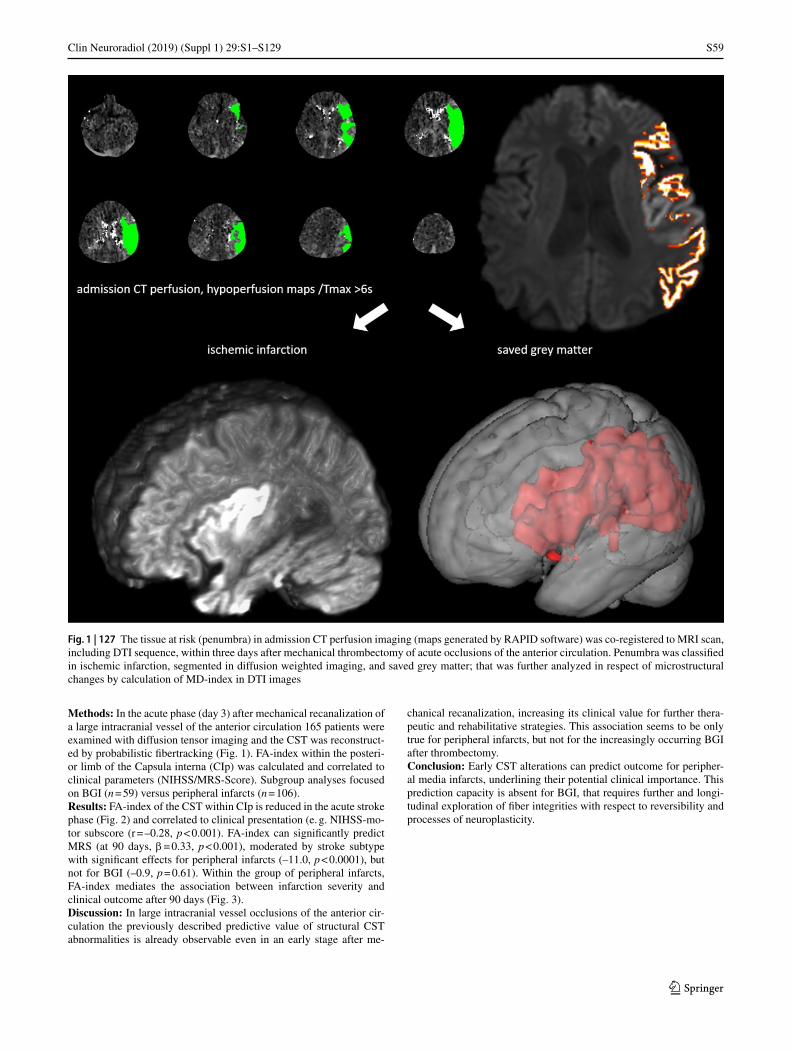
S59
123
Clin Neuroradiol (2019) (Suppl 1) 29:S1–S129
Methods: In the acute phase (day 3) after mechanical recanalization of a large intracranial vessel of the anterior circulation 165 patients were examined with diffusion tensor imaging and the CST was reconstruct-ed by probabilistic fibertracking (Fig. 1). FA-index within the posteri-or limb of the Capsula interna (CIp) was calculated and correlated to clinical parameters (NIHSS/MRS-Score). Subgroup analyses focused on BGI (n = 59) versus peripheral infarcts (n = 106).Results: FA-index of the CST within CIp is reduced in the acute stroke phase (Fig. 2) and correlated to clinical presentation (e. g. NIHSS-mo-tor subscore (r = –0.28, p < 0.001). FA-index can significantly predict MRS (at 90 days, β = 0.33, p < 0.001), moderated by stroke subtype with significant effects for peripheral infarcts (–11.0, p < 0.0001), but not for BGI (–0.9, p = 0.61). Within the group of peripheral infarcts, FA-index mediates the association between infarction severity and clinical outcome after 90 days (Fig. 3).Discussion: In large intracranial vessel occlusions of the anterior cir-culation the previously described predictive value of structural CST abnormalities is already observable even in an early stage after me-
chanical recanalization, increasing its clinical value for further thera-peutic and rehabilitative strategies. This association seems to be only true for peripheral infarcts, but not for the increasingly occurring BGI after thrombectomy.Conclusion: Early CST alterations can predict outcome for peripher-al media infarcts, underlining their potential clinical importance. This prediction capacity is absent for BGI, that requires further and longi-tudinal exploration of fiber integrities with respect to reversibility and processes of neuroplasticity.
Fig. 1 | 127 The tissue at risk (penumbra) in admission CT perfusion imaging (maps generated by RAPID software) was co-registered to MRI scan, including DTI sequence, within three days after mechanical thrombectomy of acute occlusions of the anterior circulation. Penumbra was classified in ischemic infarction, segmented in diffusion weighted imaging, and saved grey matter; that was further analyzed in respect of microstructural changes by calculation of MD-index in DTI images
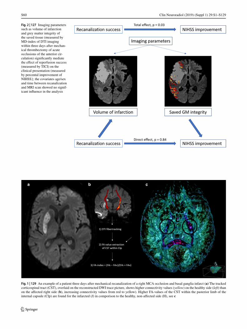
S60
123
Clin Neuroradiol (2019) (Suppl 1) 29:S1–S129
Fig. 2 | 127 Imaging parameters such as volume of infarction and grey matter integrity of the saved tissue (measured by MD-index of DTI imaging within three days after mechan-ical thrombectomy of acute occlusions of the anterior cir-culation) significantly mediate the effect of reperfusion success (measured by TICI) on the clinical presentation (measured by percental improvement of NIHSS}; the covariates age/sex and time between recanalization and MRI scan showed no signif-icant influence in the analysis
Fig. 1 | 129 An example of a patient three days after mechanical recanalization of a right MCA occlusion and basal ganglia infarct (a) The tracked corticospinal tract (CST), overlaid on the reconstructed DWI trace picture, shows higher connectivity values (yellow) on the healthy side (left) than on the affected right side (b), increasing connectivity values from red to yellow). Higher FA-values of the CST within the pasterior limb of the internal capsule (Clp) are found for the infarcted (I) in comporison to the healthy, non-affected side (H), see c

S61
123
Clin Neuroradiol (2019) (Suppl 1) 29:S1–S129
130
STROKE LESION TOPOGRAPHY AND ITS RELATION TO SEVERITY OF NIHSS AND EARLY RECOVERY POTENTIAL IN ACUTE LEFT MCA ISCHEMIA
Jessica Jesser*1, Bianca Dinse1, Martin Bendszus1
1Universitätsklinikum Heidelberg, Neuroradiologie, Heidelberg, Deutschland
Stroke lesion topography and its relation to severity of NIHSS and ear-ly recovery potential in acute left MCA ischemiaBackground: It has been shown that stroke lesion size alone only poorly predicts the clinical situation and recovery potential of acute stroke patients. This study aims to improve predictions on severity of neurological symptoms and early recovery potential by taking lesion topography into account.Methods: Patients were included when suffering from acute left-sid-ed media infarction without DWI (diffusion weighted imaging)—PWI (perfusion weighted imaging)—mismatch. Furthermore NI-HSS-Scores at admission and two days after stroke were assessed.
DWI lesions were manually segmented. Recovery Score was defined as “0” if there was no improvement of the NIHSS at admission and at 48 hours after admission, as “1” if there was only minor improvement and as “2” if there was more than 5 points improvement for initial NI-HSS > 10, more than 2 points improvement for initial NIHSS of 5–10 and at least 1 point improvement for initial NIHSS of < 5.Results: 75 patients were included into the analysis of acute NIHSS scores. 49 patients were analyzed for early recovery potential. Vox-el-based lesion symptom (VLSM) analysis revealed five critical re-gions associated with high NIHSS Scores: two of them cortically situ-ated—in the left pars triangularis of the frontal inferior gyrus and in the left supramarginal gyrus, three of them subcortically—in the anterior and dorsal centrum semiovale and in the corona radiata. Patients with no lesions in critical brain areas showed the best early recovery poten-tial followed by patients with only cortical lesions, even if situated in the predefined critical regions. Patients with both subcortical and cor-tical lesions or only subcortical lesions showed a significantly worse early recovery than the groups aforementioned.Discussion: Our results indicate that especially lesions in integrative white matter regions cause high NIHSS Scores in the acute stage of the disease and limit early recovery potential. Cortical lesions in the Pars triangularis and in the supramarginal gyrus cause high NIHSS scores in the acute stage, but if not associated with white matter lesions show a very high recovery potential, probably via early cortical reorganiza-tion mechanisms.Conclusion: Lesion location is most relevant when correlating image information with acute NIHSS and when predicting early recovery po-tential.
132
THROMBOZYTOPENIE UND ABFALL DER THROMBOZYTENZAHL – PRÄDIKTOREN FÜR MORTALITÄT UND KLINISCHES OUTCOME NACH MECHANISCHER REKANALISATION
Sebastian Mönch*1, Tobias Boeckh-Behrens1, Kornelia Kreiser1, Philipp Blüm2, Dennis Hedderich1, Christian Maegerlein1, Maria Berndt1, Silke Wunderlich3, Claus Zimmer1, Benjamin Friedrich1
1Abteilung für diagnostische und interventionelle Neuroradiologie, Klinikum rechts der Isar, Technische Universität Müncheb, München, Deutschland2Klinik für Hämatoonkologie, Klinikum rechts der Isar, Technische Universität Müncheb, München, Deutschland3Klinik für Neurologie, Klinikum rechts der Isar, Technische Universität Müncheb, München, Deutschland
Hintergrund: Der akute ischämische Schlaganfall hat gutuntersuchte Risikofaktoren. Die Bedeutung der Thrombozyten bei Patienten, die mittels mechanischer Thrombektomie behandelt wurden, ist jedoch bisher nicht untersucht worden. Das Ziel dieser Studie war zu unter-suchen, ob eine Assoziation zwischen einer vorbestehenden Thrombo-zytopenie bei Aufnahme oder einem Abfall der Thrombozytenzahl im Verlauf der Hospitalisierung mit dem klinischem Outcome, der Mor-talität oder von intrakraniellen Blutungen bei akuten Schlaganfallpa-tienten, die mittels mechanischer Thrombektomie behandelt wurden, besteht.Methoden: In einer Fall-Kontroll-Studie wurden konsekutive mecha-nisch rekanalisierte Patienten analysiert. Eine multivariate logistische Regressionsanalyse wurde verwendet, um gutes klinisches Outcome (mRS 90 Tage < = 2) und Mortalität adjustiert für Alter, NHISS bei Aufnahme, Vorbehandlung mit intravenöser Lyse/Statinen/Throm-bozyteninhibitoren, Okklusionsstelle, Zeit vom Symptombeginn bis zur Rekanalisation, vorbestehender Thrombozytopenie bei Aufnahme (< 150 × 109/L) and einem deutlichen Abfall der Thrombozytenzahl (> 26 %) zu untersuchen. Zudem wurden die Raten von intrakraniellen Blutungen verglichen.
Fig. 3 | 129 For the subgroup of peripheral infarcts, FA-index of the corticospinal tract (CST) within the posterior limb of the internal capsu/e (Cip) significantly mediates the effect of infarction severity (measured by ischemic demarcation) on the clinical outcome (meas-ured by MRS after 90 days)
Fig. 2 | 129 Boxplots of FA-index of the CST within the posterior limb of the internal capsule (CPi) for the different strake subtypes (basal ganglia infarction [BGI], peripheral infarction further divided into in-farctian severity [mild: below 1/3, middle: between 1/3 and 2/3, high: above 2/3 of the media territory])

S62
123
Clin Neuroradiol (2019) (Suppl 1) 29:S1–S129
Ergebnisse: Von 294 eingeschlossenen Patienten hatten 9.6 % eine vorbestehende Thrombozytopenie bei Aufnahme und 23.8 % einen Abfall der Thombozytenzahl im Verlauf von > 26 %. Die Mortalitäts-rate bei Patienten mit normwertigen Thrombozytenzahlen bei Aufnah-me war 26.1 % vs. 48.3 % (p = 0.002) bei Patienten mit einer vorbeste-henden Thrombozytopenie bei Aufnahme mit einer aOR of 3.47 (CI 1.28–9.4, p = 0.005), wohingegen keine Unterschiede bei gutem kli-nischen Outcome (p = 0.204) und den Raten von intrakraniellen Blu-tungen (p = 0.180) vorlagen. Ein deutlicher Abfall der Thrombozyten-zahl von mehr als 26 % in den ersten fünf Tagen der Hospitalisierung prognostizierte die Mortalitätsrate (aOR 2.4 CI 1.14–5.04, p = 0.021) sowie die Chance für gutes klinisches Outcome (aOR 0.291 CI 0.128–0.666, p = 0.003) ohne signifikante Unterschiede in den Raten von int-rakraniellen Blutungen (p = 0.735).Fazit: Bei Patienten mit akutem ischämischen Schlaganfall, welche mittels mechanischer Rekanalisation behandelt wurden, war eine vorbestehende Thrombozytopenie bei Aufnahme ein unabhängiger Prädiktor für eine erhöhte Mortalität und ein deutlicher Abfall der Thrombozyten im Verlauf ein unabhängiger Prädiktor für erhöhte Mor-talitätsraten und ungünstigeres klinisches Outcome.
134
TIME METRICS TO ENDOVASCULAR THROMBECTOMY IN THREE TRIAGE CONCEPTS: A PROSPECTIVE, OBSERVATIONAL STUDY (NEUROSQUAD TRIAL)
Fatih Seker*1, Jens Fiehler2, Markus Möhlenbruch1, Friederike Heimann1, Fabian Flottmann2, Peter Arthur Ringleb3, Götz Thomalla4, Thorsten Steiner5, Christoffer Kraemer6, Caspar Brekenfeld2, Martin Bendszus3
1Universitätsklinikum Heidelberg, Neuroradiologie, Heidelberg, Deutschland2Universitätsklinikum Hamburg-Eppendorf, Neuroradiologie, Hamburg, Deutschland3Universitätsklinikum Heidelberg, Neurologie, Heidelberg, Deutschland4Universitätsklinikum Hamburg-Eppendorf, Neurologie, Hamburg, Deutschland5Klinikum Frankfurt Höchst, Neurologie, Frankfurt am Main, Deutschland6Klinikum Lüneburg, Neurologie, Lüneburg, Deutschland
Background: NEUROSQUAD is a prospective, observational, bi-center trial comparing three triage pathways in endovascular stroke treatment: Mothership (MS), drip and ship (DS) and transferring a neu-rointerventionalist to a remote hospital for thrombectomy (drive the doctor, DD).Methods: Between February and October 2018, all stroke patients un-dergoing thrombectomy at two university hospitals and two associated remote hospitals were included. Primary outcome measures were time from onset to groin puncture and time from imaging to groin puncture. Secondary outcome measures were time from onset to imaging and time from onset to thrombolysis.Results: In total, 447 patients were included of which 145 (32.4%) were in the MS group, 250 (55.9%) in the DS group and 52 (11.6%) in the DD group. Median time from onset to groin puncture (170 min) and time from imaging to groin puncture (52 min) were the shortest in the MS group. Time from onset to groin puncture (DD median 225 min vs. DS median 301 min, P = 0.001) and time from imaging to groin puncture (DD median 118 min vs. DS median 172 min, P < 0.001) were shorter in the DD group compared to DS. Time from onset to imaging was similar among MS, DS and DD (P = 0.332). In patients receiving thrombolysis, time from onset to needle was similar among the three groups as well (P = 0.619).
Conclusion: The NEUROSQUAD study adds evidence that DD may be a feasible alternative to DS, leading to shorter delay between symp-tom onset and groin puncture. Both are time-wise inferior compared to MS, though.
135
DIAGNOSTIC PERFORMANCE OF FIVE COLLATERAL SCORES IN THE PREDICTION OF CLINICAL OUTCOME AFTER ENDOVASCULAR THROMBECTOMY IN ISCHEMIC STROKE
Fatih Seker*1, Benjamin Pereira-Zimmermann2, Johannes Pfaff1, Jan Purrucker3, Christoph Gumbinger3, Silvia Schönenberger3, Martin Bendszus1, Markus Möhlenbruch3
1Universitätsklinikum Heidelberg, Neuroradiologie, Heidelberg, Deutschland2University of Valparaiso, Radiology, Valparaiso, Chile3Universitätsklinikum Heidelberg, Neurologie, Heidelberg, Deutschland
Background: Several collateral scores have been published for stroke in the middle cerebral artery territory, each considering different as-pects of cerebral collateralization. This retrospective study aimed at comparing five collateral scores regarding their prediction of clinical outcome.Methods: Inclusion criteria were M1 occlusion, premorbid modi-fied Rankin Scale of 0–2, treatment with endovascular thrombectomy, groin puncture within 6 hours after stroke onset, and Thrombolysis In Cerebral Infarction score of 2b–3. Maas, Miteff, Tan, ASITN/SIR and mCTA collateral scores were assessed in multiphase CTA images and correlated with 90 day modified Rankin Scale (90d-mRS). Good out-come was defined as 90d-mRS 0–2.Results: In total, 69 patients were included of which 49.3% achieved a good outcome. Area Under the Curve values of Receiver Operating Characteristic curve analysis for Maas, Miteff, Tan, ASITN/SIR and mCTA scores were 0.64 (0.53–0.76), 0.62 (0.53–0.71), 0.65 (0.53–0.76), 0.65 (0.53–0.78) and 0.66 (0.54–0.79), respectively. Correlation between 90d-mRS and Maas (r = –0.39, P < 0.001), Miteff (r = –0.40, P < 0.001), Tan (r = –0.42, P < 0.001), ASITN/SIR (r = –0.44, P < 0.001) and mCTA (r = –0.44, P < 0.001) scores was moderate.Conclusion: Maas, Miteff, Tan, ASITN/SIR and mCTA collateral scores show similarly moderate diagnostic performance in the predic-tion of clinical outcome when assessed in multiphase CTA.
136
MOTHERSHIP VERSUS DRIP AND SHIP – A RETROSPECTIVE ANALYSIS OF A REGIONAL STROKE REGISTRY WITH 2797 PATIENTS
Fatih Seker*1, Susanne Bonekamp1, Susanne Rode2, Sonja Hyrenbach2, Martin Bendszus1, Markus Möhlenbruch1
1Universitätsklinikum Heidelberg, Neuroradiologie, Heidelberg, Deutschland2Qualitätssicherung im Gesundheitswesen Baden-Württemberg GmbH, Stuttgart, Deutschland
Background: This study aims at comparing short-term clinical out-come after thrombectomy in patients directly admitted to a comprehen-sive stroke center (mothership, MS) with patients secondarily trans-ferred from a primary stroke center (drip and ship, DS).Methods: In a prospective regional stroke registry, all stroke patients with premorbid modified Rankin Scale (mRS) 0–2 who were admitted within 24 hours after stroke onset and treated with thrombectomy be-tween 2014 and 2017 were analyzed retrospectively. MS and DS were

S63
123
Clin Neuroradiol (2019) (Suppl 1) 29:S1–S129
compared regarding the proportion of good outcome (discharge mRS 0–2), median discharge mRS, mRS shift (difference between premor-bid mRS and mRS on discharge) and occurrence of symptomatic in-tracranial hemorrhage.Results: Of 2797 patients, 1051 (37.6%) achieved good clinical out-come. In the MS group (n = 1657), proportion of good outcome was higher (MS 42.2% vs. DS 30.9%, P < 0.001) and median discharge mRS (MS 3 vs. DS 4, P < 0.001) and median mRS shift (MS 3 vs. DS 4, P < 0.001) were lower. The rate of symptomatic intracranial hemor-rhage was similar in both groups (MS 9.3% vs. DS 7.5%, P = 0.101). Multivariate analysis revealed that direct admission (MS) was an in-dependent predictor of good clinical outcome (adjusted OR 1.32, CI 1.09–1.60, P = 0.004).Conclusion: These results confirm prior studies stating that direct ad-mission to a comprehensive stroke center (MS) leads to better outcome compared to DS in stroke patients undergoing thrombectomy.
137
IMPACT OF BRIDGING THROMBOLYSIS ON CLINICAL OUTCOME IN STROKE PATIENTS UNDERGOING THROMBECTOMY
Fatih Seker*1, Susanne Bonekamp1, Susanne Rode2, Sonja Hyrenbach2, Martin Bendszus1, Markus Möhlenbruch1
1Universitätsklinikum Heidelberg, Neuroradiologie, Heidelberg, Deutschland2Qualitätssicherung im Gesundheitswesen Baden-Württemberg GmbH, Stuttgart, Deutschland
Background: It is unclear whether stroke patients should receive in-travenous thrombolysis (IVT) prior to endovascular thrombectomy (EVT). This study aims at analyzing the impact of bridging IVT on short-term clinical outcome.Methods: In a prospective regional stroke registry, all stroke patients with premorbid modified Rankin Scale (mRS) 0–2 who were admitted within 4.5 hours after onset and treated with EVT were analyzed ret-rospectively. Patients receiving IVT prior to EVT (IVEVT) were com-pared to those undergoing EVT only regarding the proportion of good outcome (discharge mRS 0–2), discharge mRS, mRS shift (difference between premorbid mRS and mRS on discharge), hospital mortality and occurrence of symptomatic intracranial hemorrhage.Results: In total, 2022 patients were included of which 816 patients (40.4%) achieved good clinical outcome. 1293 patients (63.9%) re-ceived bridging IVT. There was no significant difference between both groups regarding the proportion of good outcome (IVEVT 41.4% vs. EVT 38.5%, P = 0.231), discharge mRS (median, IVEVT 3 vs. EVT 3, P = 0.178) and mRS shift (median, IVEVT 3 vs. EVT 3, P = 0.960). Hospital mortality was non-significantly higher in IVEVT (IVEVT 19.3% vs. 11.0%, P = 0.984). Bridging IVT was not associated with clinical outcome (P = 0.231). Symptomatic intracranial hemorrhage was significantly more frequent in IVEVT (9.3% vs. 5.2%, P = 0.001).Conclusion: According to this analysis of a regional stroke registry with 2022 patients, bridging thrombolysis does not lead to better clin-ical outcome. However, it is associated with a higher rate of sympto-matic intracranial hemorrhage.
138
ACUTE OCCLUSIONS AND THROMBOSES OF DUAL-LAYER CAROTID STENTS IN ENDOVASCULAR STROKE TREATMENT OF TANDEM OCCLUSIONS – A RETROSPECTIVE MULTICENTER EXPERIENCE
Johannes Pfaff*1, Christoph Maurer2, Erasmia Müller-Thies-Broussalis3, Hendrik Janssen4 5, Raphael Blanc6, Cyril Dargazanli7,
Vincent Costalat7, Michel Piotin6, Frank Runck2, Ansgar Berlis2, Monika Killer3, Johannes Hensler8, Martin Bendszus1, Fritz Wodarg8, Markus Möhlenbruch1
1Universitätsklinikum Heidelberg, Abteilung für Neuroradiologie, Heidelberg, Deutschland2Augsburg, Deutschland3Salzburg, Österreich4Ingolstadt, Deutschland5Nürnberg, Deutschland6Paris, Frankreich7Montpellier, Frankreich8Kiel, Deutschland
Hintergrund: A recent retrospective single center study showed a rate of 45% of thrombotic occlusion of dual-layer carotid stents within 72 hours after emergent CAS during treatment of a tandem occlusion. (1) As the sample size in this study is small and this study appear to show a higher-than-anticipated occlusion rate, we hypothesize that the rate of occlusion is lower and rather comparable to previously reported occlu-sion rates in single layer stents and propose a retrospective multi-center evaluation to increase the power to assess this.Methoden: Multicentric retrospective data collection and analysis of stroke databases of 7 comprehensive stroke centers from 3 European countries.Ergebnisse: Overall 160 patients (age: mean (SD) = 66 (12); male: n = 104 (65%); median baseline NIHSS (IQR): 14 (9–18), IV lysis: n = 97 (60.6%) were treated for a cervical carotid artery occlusion or stenosis using a CASPER Stent (CASPER Stent, Carotid Artery Stent designed to Prevent Embolic Release, MicroVention, Aliso Viejo, CA) and received mechanical thrombectomy for an intracranial occlu-sion between April 2014 and November 2018. During the procedure or within 72 h, formation of thrombus and complete occlusion of the CASPER-Stent was observed in 33/160 patients (20.8%), respective-ly in 12/160 patients (7.5%). In 25/33 (75.8%), respectively in 9/12 patients (75%) this occurred during the procedure. No statistical sig-nificant difference was observed between patients with and without thrombus formation with regard to a pre-existing long-term medication with anticoagulants or intraprocedural administration of either heparin, ASA or heparin and ASA. Favorable early neurological outcome was similar in patients with (n = 15; 45.5%) and without (n = 63; 49.6%) thrombus formation at the CASPER-Stent.Diskussion:Fazit: Acute thrombosis or occlusion of CASPER-stents in thrombec-tomy patients receiving emergent extracranial internal carotid artery stenting for tandem occlusions can be observed more often during the procedure rather than within 72 h follow-up, were less frequent then previously reported and showed no impact on early neurological out-come.
References 1. Yilmaz U, Korner H, Muhl-Benninghaus R, Simgen A, Kraus C,
Walter S, et al. Acute Occlusions of Dual-Layer Carotid Stents After Endovascular Emergency Treatment of Tandem Lesions. Stroke. 2017;48(8):2171–5
139
PRÄDIKTIVER WERT DER PERFUSIONS-BILDGEBUNG AUF DIE VORHERSAGE DER HÄMORRHAGISCHEN TRANSFORMATION NACH ENDOVASKULÄRER SCHLAGANFALLS-THERAPIE
Antonia Carlotta Fischer, Olav Jansen, Patrick Langguth, Fritz Wodarg, Naomi Larsen, Friederike Austein
Department of Radiology and Neuroradiology, University Hospital Schleswig-Holstein, Campus Kiel, Kiel, Deutschland
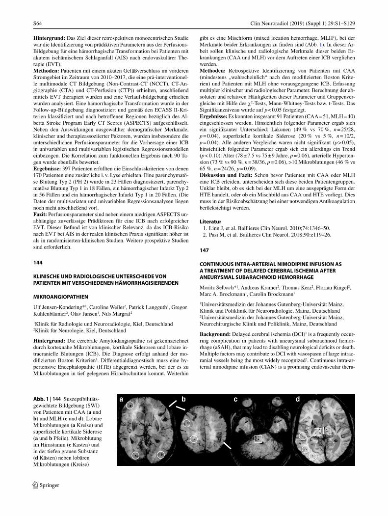
S64
123
Clin Neuroradiol (2019) (Suppl 1) 29:S1–S129
Hintergrund: Das Ziel dieser retrospektiven monozentrischen Studie war die Identifizierung von prädiktiven Parametern aus der Perfusions-Bildgebung für eine hämorrhagische Transformation bei Patienten mit akutem ischämischem Schlaganfall (AIS) nach endovaskulärer The-rapie (EVT).Methoden: Patienten mit einem akuten Gefäßverschluss im vorderen Stromgebiet im Zeitraum von 2010–2017, die eine prä-interventionel-le multimodale CT Bildgebung (Non-Contrast-CT (NCCT), CT-An-giographie (CTA) und CT-Perfusion (CTP)) erhielten, anschließend mittels EVT therapiert wurden und eine Verlaufsbildgebung erhielten wurden analysiert. Eine hämorrhagische Transformation wurde in der Follow-up-Bildgebung diagnostiziert und gemäß den ECASS II-Kri-terien klassifiziert und nach betroffenen Regionen bezüglich des Al-berta Stroke Program Early CT Scores (ASPECTS) aufgeschlüsselt. Neben den Auswirkungen ausgewählter demografischer Merkmale, klinischer und therapieassoziierter Faktoren, wurden insbesondere die unterschiedlichen Perfusionsparameter für die Vorhersage einer ICB in univariablen und multivariablen logistischen Regressionsmodellen einbezogen. Die Korrelation zum funktionellen Ergebnis nach 90 Ta-gen wurde ebenfalls bewertet.Ergebnisse: 397 Patienten erfüllten die Einschlusskriterien von denen 170 Patienten eine zusätzliche i. v. Lyse erhielten. Eine parenchymatö-se Blutung Typ 2 (PH 2) wurde in 23 Fällen diagnostiziert, parenchy-matöse Blutung Typ 1 in 18 Fällen, ein hämorrhagischer Infarkt Typ 2 in 56 Fällen und ein hämorrhagischer Infarkt Typ 1 in 20 Fällen. (Die Daten der multivariaten und univariablen Regressionsanalysen liegen noch nicht abschließend vor).Fazit: Perfusionsparameter sind neben einem niedrigen ASPECTS un-abhängige zuverlässige Prädiktoren für eine ICB nach erfolgreicher EVT. Dieser Befund ist von klinischer Relevanz, da das ICB-Risiko nach EVT bei AIS in der realen klinischen Praxis signifikant höher ist als in randomisierten-klinischen Studien. Weitere prospektive Studien sind erforderlich.
144
KLINISCHE UND RADIOLOGISCHE UNTERSCHIEDE VON PATIENTEN MIT VERSCHIEDENEN HÄMORRHAGISIERENDEN
MIKROANGIOPATHIEN
Ulf Jensen-Kondering*1, Caroline Weiler2, Patrick Langguth1, Gregor Kuhlenbäumer2, Olav Jansen1, Nils Margraf2
1Klinik für Radiologie und Neuroradiologie, Kiel, Deutschland2Klinik für Neurologie, Kiel, Deutschland
Hintergrund: Die cerebrale Amyloidangiopathie ist gekennzeichnet durch kortexnahe Mikroblutungen, kortikale Siderosen und lobäre in-tracranielle Blutungen (ICB). Die Diagnose erfolgt anhand der mo-difizierten Boston Kriterien1. Differentialdiagnostisch muss eine hy-pertensive Encephalopathie (HTE) abgegrenzt werden, bei der es zu Mikroblutungen in tief gelegenen Hirnabschnitten kommt. Weiterhin
gibt es eine Mischform (mixed location hemorrhage, MLH2), bei der Merkmale beider Erkrankungen zu finden sind (Abb. 1). In dieser Ar-beit sollen klinische und radiologische Merkmale dieser beiden Er-krankungen (CAA und MLH) vor dem Auftreten einer ICB verglichen werden.Methoden: Retrospektive Identifizierung von Patienten mit CAA (mindestens „wahrscheinlich“ nach den modifizierten Boston Krite-rien) und Patienten mit MLH ohne vorausgegangene ICB. Erfassung multipler klinischer und radiologischer Parameter. Berechnung der ab-soluten und relativen Häufigkeiten dieser Parameter und Gruppenver-gleiche mit Hilfe des χ2-Tests, Mann-Whitney-Tests bzw. t-Tests. Das Signifikanzniveau wurde auf p < 0.05 festgelegt.Ergebnisse: Es konnten insgesamt 91 Patienten (CAA = 51, MLH = 40) eingeschlossen werden. Hinsichtlich folgender Parameter ergab sich ein signifikanter Unterschied: Lakunen (49 % vs 70 %, n = 25/28, p = 0.04), superfizielle kortikale Siderose (20 % vs 5 %, n = 10/2, p = 0.04). Alle anderen Vergleiche waren nicht signifikant (p > 0.05), hinsichtlich folgender Parameter ergab sich ein allerdings ein Trend (p < 0.10): Alter (78 ± 7.5 vs 75 ± 9 Jahre, p = 0.06), arterielle Hyperten-sion (73 % vs 90 %, n = 38/36, p = 0.06), >10 Mikroblutungen (46 % vs 65 %, n = 24/26, p = 0.09).Diskussion und Fazit: Schon bevor Patienten mit CAA oder MLH eine ICB erleiden, unterscheiden sich diese beiden Patientengruppen. Unklar bleibt, ob es sich bei der MLH um eine ausgeprägte Form der HTE handelt, oder ob ein Mischbild aus CAA und HTE vorliegt. Dies muss in der Risikoabschätzung bei einer notwendigen Antikoagulation berücksichtigt werden.
Literatur 1. Linn J, et al. Baillieres Clin Neurol. 2010;74:1346–50. 2. Pasi M, et al. Baillieres Clin Neurol. 2018;90:e119–26.
147
CONTINUOUS INTRA-ARTERIAL NIMODIPINE INFUSION AS A TREATMENT OF DELAYED CEREBRAL ISCHEMIA AFTER ANEURYSMAL SUBARACHNOID HEMORRHAGE
Moritz Selbach*1, Andreas Kramer2, Thomas Kerz2, Florian Ringel2, Marc A. Brockmann1, Carolin Brockmann1
1Universitätsmedizin der Johannes Gutenberg-Universität Mainz, Klinik und Poliklinik für Neuroradiologie, Mainz, Deutschland2Universitätsmedizin der Johannes Gutenberg-Universität Mainz, Neurochirurgische Klinik und Poliklinik, Mainz, Deutschland
Background: Delayed cerebral ischemia (DCI)1 is a frequently occur-ring complication in patients with aneurysmal subarachnoid hemor-rhage (aSAH), that may lead to disabling neurological deficits or death. Multiple factors may contribute to DCI with vasospasm of large intrac-ranial vessels being the most widely recognized2. Continuous intra-ar-terial nimodipine infusion (CIAN) is a promising endovascular thera-
Abb. 1 | 144 Suszeptibilitäts-gewich tete Bildgebung (SWI) von Patienten mit CAA (a und b) und MLH (c und d). Lobäre Mikroblutungen (a Kreise) und superfizielle kortikale Siderose (a und b Pfeile). Mikroblutung im Hirnstamm (c Kasten) und in der tiefen grauen Substanz (d Kästen) neben lobären Mikroblutungen (Kreise)

S65
123
Clin Neuroradiol (2019) (Suppl 1) 29:S1–S129
peutic option in severe cases of DCI, which are refractory to standard therapy (hemodynamic therapy and oral NDP).Methods: CIAN was initiated and ended based on an individual in-terdisciplinary evaluation of the clinical and diagnostic course of the patient using transcranial Doppler (TCD), CT angiography (CTA), CT perfusion (CTP) and digital subtraction angiography (DSA). One or two microcatheters were placed into the internal carotid or vertebral artery according to the site of suspected DCI. Nimodipine (NDP) was administered continuously in a rate of 0.5–2 mg/ h. During CIAN, in-tensive care was conducted. The outcome measures were the Glasgow Outcome Scale (GOS) at discharge and within 1 year after aSAH and the occurrence of cerebral infarction in subsequent CT-scans.Results: In the period of May 2016 to January 2018, 17 patients re-ceived CIAN. The median duration was 5 (1–13) days. Favorable out-come (GOS 4 or 5) was achieved in 9 patients (53%) at discharge and in 13 patients within 1 year (76%). Follow-up imaging showed minor infarction in 5 and major infarction in 3 patients. One patient developed a localized cerebral edema as a possible side effect. One patient died due to malignant posthemorrhagic edema. Normalization of CTP-pa-rameters within 2 days was observed in 9/17 patients. The remaining 6 patients showed clinical response and thus did not receive short-term CTP imaging.Discussion: Our results concerning the outcome within one year are in line with previously published retrospective studies3,4. The study is limited by its retrospective character, the lack of a control group and the small number of patients.Conclusion: CIAN is a feasible, safe and effective therapeutic option for patients with severe therapy-refractory delayed cerebral ischemia. A prospective randomized clinical trial is needed to confirm the posi-tive retrospective results of this study and previously published studies.
References 1. Vergouwen M, Vermeulen M, Gijn J, et al. 2010. 2. Budohoski KP, Guilfoyle M, Helmy A, et al. 2014. 3. Bele, S., Proescholdt, M. A., Hochreiter, A. et al. 2015. 4. Hockel K, Diedler J, Steiner J, et al. 2016.
148
STABLE LOCALIZATION OF WATERSHED AREAS AFTER REVASCULARIZATION THERAPY IN PATIENTS WITH ASYMPTOMATIC INTERNAL CAROTID ARTERY
STENOSIS
Lena Schmitzer*1, Stephan Kaczmarz1, Nico Sollmann1, Claus Zimmer1, Christine Preibisch1, Jens Göttler1
1Klinikum Rechts der Isar der Technischen Universität München, Abteilung für diagnostische und interventionelle Neuroradiologie, München, Deutschland
Hintergrund: Watershed areas (WSA) are located between vascular territories and thus especially vulnerable to hypoperfusion.1 This can be caused by internal carotid artery stenosis (ICAS), a disease account-ing for approx. 10% of all strokes.2,3
While stenosis treatment is known to alter cerebral hemodynamics,4 it remains unclear how WSA localization is affected. We addressed this issue in a follow up study in ICAS patients after revascularization treatment.Methoden: Sixteen patients (age 73.0 ± 6.1y, five females) and 17 healthy elderly (age 72.3 ± 5.5y, eleven females) were examined on a clinical 3T Philips Ingenia after patients had undergone carotid endar-terectomy or stenting.
WSAs were defined semi-automatically based on Time-to-Peak-maps5 (Fig. 1).Every subject’s WSAs mask was normalized to MNI-space and probability maps were calculated for each group. Spatial overlap of each subject’s WSA mask of both scans was assessed by the Dice coefficient (DC) and compared between groups.
TTP values were extracted within each individual WSA and com-pared between hemispheres.Ergebnisse: WSAs could be reliably segmented in both groups (Fig. 2). The mean DC for the WSA comparison between scans 1 and 2 was similar (DC≈0.6) in controls and patients (non-significant dif-ference, two-sample t-test, p = 0.287). In patients, relative TTP (rTTP) in WSAs was significantly reduced in the affected vs. the unaffected hemisphere pre-interventionally (p < 0.001), whereas no sig significant difference was observed post-interventionally (p = 0.446, Fig. 3).Diskussion: Similar DCs in patients and controls (≈0.6) suggest that the spatial extent of WSAs was not changed by revascularization ther-
Fig. 1 | 148 Defining WSAs (marked red) semi-automatically based on TTP derived from dynamic susceptibility contrast (DSC) and adaption them to the individual’s anatomy by omitting sinuses (1) and the ventricular system (2)
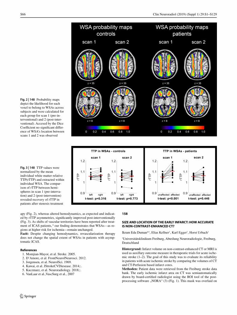
S66
123
Clin Neuroradiol (2019) (Suppl 1) 29:S1–S129
apy (Fig. 2), whereas altered hemodynamics, as expected and indicat-ed by rTTP asymmetries, significantly improved post-interventionally (Fig. 3). As shifts of vascular territories have been reported after treat-ment of ICAS patients, 6 our finding demonstrates that WSAs—as re-gions at higher risk for ischemia—remain unchanged.Fazit: Despite changing hemodynamics, revascularization therapy does not change the spatial extent of WSAs in patients with asymp-tomatic ICAS.
References 1. Momjian-Mayor, et al. Stroke. 2005. 2. D’Amore, et al. FrontNeurolNeurosci. 2012. 3. Jorgensen, et al. NeurolSci. 1969. 4. Kawai, et al. JStrokeCVDiesease. 2014;:. 5. Kaczmarz, et al. Neuroradiology. 2018;:. 6. VanLaar et al.,VascSurg et al., 2007
158
SIZE AND LOCATION OF THE EARLY INFARCT: HOW ACCURATE IS NON-CONTRAST-ENHANCED CT?
Ikram Eda Duman*1, Elias Kellner1, Karl Egger1, Horst Urbach1
1Universitätsklinikum Freiburg, Abteilung Neuroradiologie, Freiburg, Deutschland
Hintergrund: Infarct volume on non-contrast-enhanced CT or MRI is used as auxillary outcome measure in therapeutic trials for acute ische-mic stroke (1–2). The goal of this study was to evaluate its reliability in patients with acute ischemic stroke by comparing the volumes of CT and CT-Perfusion based infarct cores.Methoden: Patient data were retrieved from the Freiburg stroke data bank. The early ischemic infarct area on CT was semiautomatically drawn by board-certified radiologist using the ROI tool of the post-processing software „NORA“ (3) (Fig. 1). This mask was overlaid on
Fig. 3 | 148 TTP values were normalized by the mean individual white matter relative TTP(rTTP) and extracted within individual WSA. The compar-ison of rTTP between hemi-spheres in scan 1 (pre-interva-tion) and 2 (post-intervention) revealed recovery of rTTP in patients after stenosis treatment
Fig. 2 | 148 Probability maps depict the likelihood for each voxel to belong to WSAs across subjects and were calculated for each group for scan 1 (pre-in-terventional) and 2 (post-inter-ventional). Accesed by the Dice Coefficient no significant differ-ence of WSA’s location between scans 1 and 2 was observed

S67
123
Clin Neuroradiol (2019) (Suppl 1) 29:S1–S129
the CT-P-based infarct core generated by the processing software „Ve-oCore“. Volumetry was compared by calculating DICE coefficients. Follow-up CT or MRI was performed to prove infarcts demarcations.Ergebnisse: 45 patients (age 76 (51–92), NIHSS 14 (2–23) were in-cluded in the analysis. A total of 38 patients showed early infarct signs. In 2 patients, the early ischemic infarcts were overlooked (false nega-tives). In 6 patients, ischemic infarcts were falsely drawn on CT (fal-se positives) (Fig 2.). Mean early infarct volumes on non-contrast CT was 32.3 (0–248) mL and 31.7 (0 –158) mL on CT-P. Mean DICE coefficient between non-contrast-enhanced CT and CT-P infarct core maps was 0.203, and between non-contrast-enhanced CT and CT-P hy-poperfusion maps 0.198. Correlation coefficients were 0.30 and 0.36, respectively.Fazit: Low DICE coefficients indicate a low accuracy of non-con-trast-enhanced CT to depict the early ischemic infarct.
References 1. Song D, Yoo J, Baek JH, et al. Infarct Core Expansion on Com-
puted Tomography before and after Intravenous Thromboly-sis. Yonsei Med J. 2018;59(2):310–6. https://doi.org/10.3349/ymj.2018.59.2.310.
2. Han M, Choi JW, Rim NJ, et al. Cerebral infarct volume meas-urements to improve patient selection for endovascular treatment. Medicine (Baltimore).95(35):e4702. https://doi.org/10.1097/MD.0000000000004702.
3. Kellner E, Reisert M, Kiselev VG, Maurer CJ, Beume LA, Urbach H, et al. Automated Infarct Core Volumetry Within the Hypoper-fused Tissue: Technical Implementation and Evaluation. J Comput Assist Tomogr. 2017;41(4):515–20.
162
TRANSKRANIELLE MOTORISCH EVOZIERTE POTENZIALE IN DER ENDOVASKULÄREN
REVASKULARISATIONSTHERAPIE ISCHÄMISCHER SCHLAGANFÄLLE – WORK IN PROGRESS.
Tobias Greve*1, Arthur Wagner2, Bernhard Meyer2, Claus Zimmer1, Silke Wunderlich3, Ehab Shiban2, Kornelia Kreiser1
1Abteilung für Diagnostische und Interventionelle Neuroradiologie, Klinikum rechts der Isar der Technischen Universität München, München, Deutschland2Neurochirurgische Klinik und Poliklinik, Klinikum rechts der Isar der Technischen Universität München, München, Deutschland3Neurologische Klinik und Poliklinik, Klinikum rechts der Isar der Technischen Universität München, München, Deutschland
Hintergrund: Die Aussagekraft motorisch evozierter Potenziale (MEPs) bei der endovaskulären mechanischen Rekanalisation (EMK) ischämischer Schlaganfälle unter Allgemeinanästhesie ist bisher we-nig untersucht. Wir korrelierten die Amplituden (AMPs) von MEPs der oberen Extremitäten vor und nach EMK mit Symptombesserung (SYM_D7) und Infarktdemarkation in der cMRT (DEMARK_D7) an Tag 7.Methoden: Prospektiv wurden bis 05/2019 Patienten mit Hemisyn-drom und Verschluss der A. carotis interna/media (ICA/MCA) bzw. A. basilaris (BA) eingeschlossen, bei denen das MEP-Setup die EMK nicht verzögerte. mRS- und NIHSS wurden am Behandlungstag (D0) und Tag 7 (D7) erfasst. MEPs wurden an C3/C4 transkraniell elektrisch stimuliert und die AMPs am Abductor pollicis brevis (APB) bilateral vor und nach der EMK abgeleitet.Ergebnisse: 26 Patienten (12w; 73,1 ± 12,2J; 4 ICA-/18 MCA-/4 BA-Verschlüsse) wurden bislang eingeschlossen. Zwischen Symptombe-
Fig. 1 | 158
Fig. 2 | 158
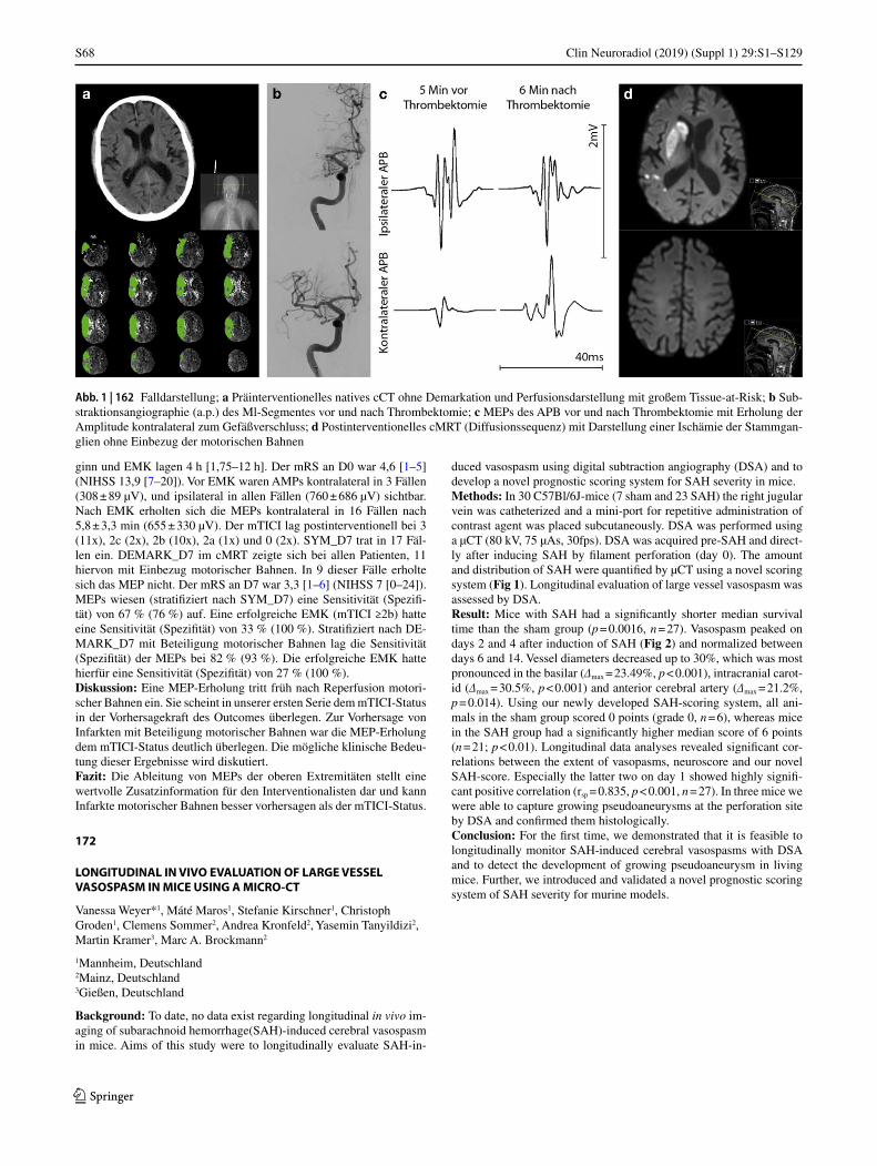
S68
123
Clin Neuroradiol (2019) (Suppl 1) 29:S1–S129
ginn und EMK lagen 4 h [1,75–12 h]. Der mRS an D0 war 4,6 [1–5] (NIHSS 13,9 [7–20]). Vor EMK waren AMPs kontralateral in 3 Fällen (308 ± 89 µV), und ipsilateral in allen Fällen (760 ± 686 µV) sichtbar. Nach EMK erholten sich die MEPs kontralateral in 16 Fällen nach 5,8 ± 3,3 min (655 ± 330 µV). Der mTICI lag postinterventionell bei 3 (11x), 2c (2x), 2b (10x), 2a (1x) und 0 (2x). SYM_D7 trat in 17 Fäl-len ein. DEMARK_D7 im cMRT zeigte sich bei allen Patienten, 11 hiervon mit Einbezug motorischer Bahnen. In 9 dieser Fälle erholte sich das MEP nicht. Der mRS an D7 war 3,3 [1–6] (NIHSS 7 [0–24]). MEPs wiesen (stratifiziert nach SYM_D7) eine Sensitivität (Spezifi-tät) von 67 % (76 %) auf. Eine erfolgreiche EMK (mTICI ≥2b) hatte eine Sensitivität (Spezifität) von 33 % (100 %). Stratifiziert nach DE-MARK_D7 mit Beteiligung motorischer Bahnen lag die Sensitivität (Spezifität) der MEPs bei 82 % (93 %). Die erfolgreiche EMK hatte hierfür eine Sensitivität (Spezifität) von 27 % (100 %).Diskussion: Eine MEP-Erholung tritt früh nach Reperfusion motori-scher Bahnen ein. Sie scheint in unserer ersten Serie dem mTICI-Status in der Vorhersagekraft des Outcomes überlegen. Zur Vorhersage von Infarkten mit Beteiligung motorischer Bahnen war die MEP-Erholung dem mTICI-Status deutlich überlegen. Die mögliche klinische Bedeu-tung dieser Ergebnisse wird diskutiert.Fazit: Die Ableitung von MEPs der oberen Extremitäten stellt eine wertvolle Zusatzinformation für den Interventionalisten dar und kann Infarkte motorischer Bahnen besser vorhersagen als der mTICI-Status.
172
LONGITUDINAL IN VIVO EVALUATION OF LARGE VESSEL VASOSPASM IN MICE USING A MICRO-CT
Vanessa Weyer*1, Máté Maros1, Stefanie Kirschner1, Christoph Groden1, Clemens Sommer2, Andrea Kronfeld2, Yasemin Tanyildizi2, Martin Kramer3, Marc A. Brockmann2
1Mannheim, Deutschland2Mainz, Deutschland3Gießen, Deutschland
Background: To date, no data exist regarding longitudinal in vivo im-aging of subarachnoid hemorrhage(SAH)-induced cerebral vasospasm in mice. Aims of this study were to longitudinally evaluate SAH-in-
duced vasospasm using digital subtraction angiography (DSA) and to develop a novel prognostic scoring system for SAH severity in mice.Methods: In 30 C57Bl/6J-mice (7 sham and 23 SAH) the right jugular vein was catheterized and a mini-port for repetitive administration of contrast agent was placed subcutaneously. DSA was performed using a µCT (80 kV, 75 µAs, 30fps). DSA was acquired pre-SAH and direct-ly after inducing SAH by filament perforation (day 0). The amount and distribution of SAH were quantified by µCT using a novel scoring system (Fig 1). Longitudinal evaluation of large vessel vasospasm was assessed by DSA.Result: Mice with SAH had a significantly shorter median survival time than the sham group (p = 0.0016, n = 27). Vasospasm peaked on days 2 and 4 after induction of SAH (Fig 2) and normalized between days 6 and 14. Vessel diameters decreased up to 30%, which was most pronounced in the basilar (∆max = 23.49%, p < 0.001), intracranial carot-id (∆max = 30.5%, p < 0.001) and anterior cerebral artery (∆max = 21.2%, p = 0.014). Using our newly developed SAH-scoring system, all ani-mals in the sham group scored 0 points (grade 0, n = 6), whereas mice in the SAH group had a significantly higher median score of 6 points (n = 21; p < 0.01). Longitudinal data analyses revealed significant cor-relations between the extent of vasopasms, neuroscore and our novel SAH-score. Especially the latter two on day 1 showed highly signifi-cant positive correlation (rsp = 0.835, p < 0.001, n = 27). In three mice we were able to capture growing pseudoaneurysms at the perforation site by DSA and confirmed them histologically.Conclusion: For the first time, we demonstrated that it is feasible to longitudinally monitor SAH-induced cerebral vasospasms with DSA and to detect the development of growing pseudoaneurysm in living mice. Further, we introduced and validated a novel prognostic scoring system of SAH severity for murine models.
Abb. 1 | 162 Falldarstellung; a Präinterventionelles natives cCT ohne Demarkation und Perfusionsdarstellung mit großem Tissue-at-Risk; b Sub-straktionsangiographie (a.p.) des Ml-Segmentes vor und nach Thrombektomie; c MEPs des APB vor und nach Thrombektomie mit Erholung der Amplitude kontralateral zum Gefäßverschluss; d Postinterventionelles cMRT (Diffusionssequenz) mit Darstellung einer Ischämie der Stammgan-glien ohne Einbezug der motorischen Bahnen
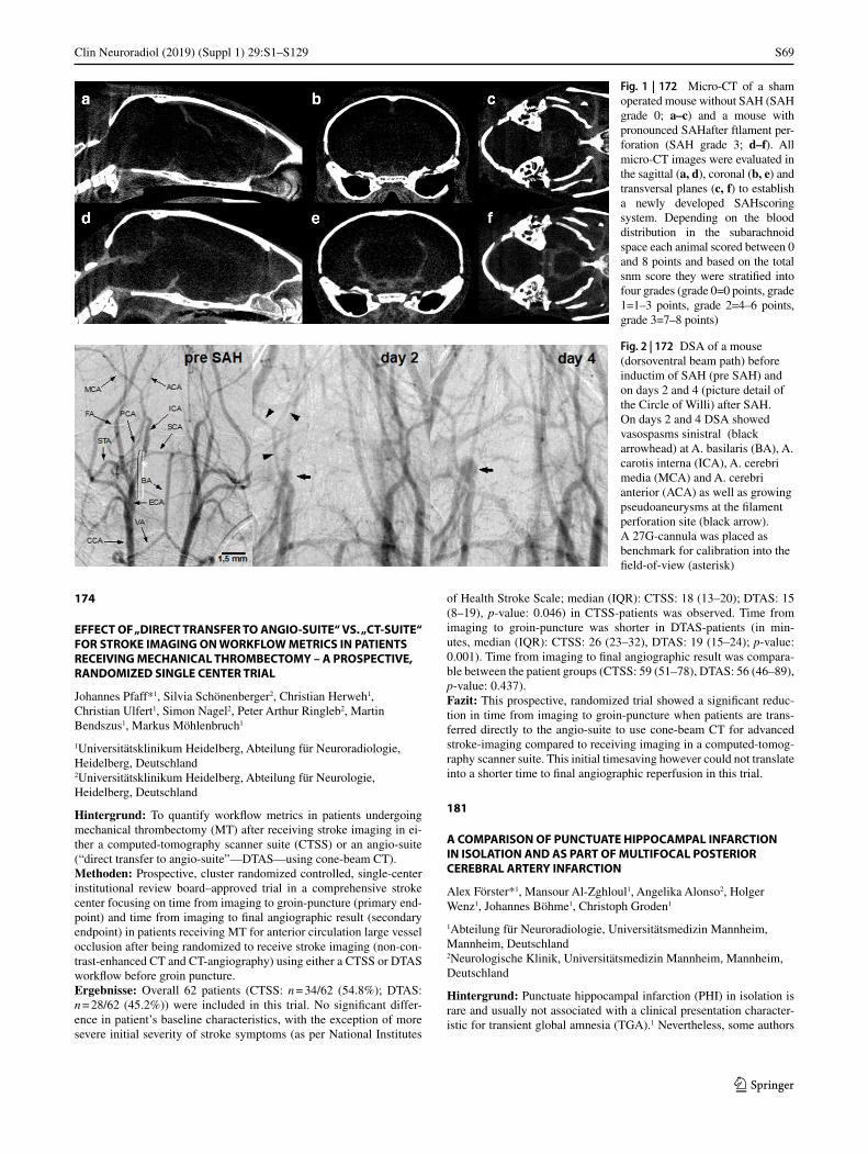
S69
123
Clin Neuroradiol (2019) (Suppl 1) 29:S1–S129
174
EFFECT OF „DIRECT TRANSFER TO ANGIO-SUITE“ VS. „CT-SUITE“ FOR STROKE IMAGING ON WORKFLOW METRICS IN PATIENTS RECEIVING MECHANICAL THROMBECTOMY – A PROSPECTIVE, RANDOMIZED SINGLE CENTER TRIAL
Johannes Pfaff*1, Silvia Schönenberger2, Christian Herweh1, Christian Ulfert1, Simon Nagel2, Peter Arthur Ringleb2, Martin Bendszus1, Markus Möhlenbruch1
1Universitätsklinikum Heidelberg, Abteilung für Neuroradiologie, Heidelberg, Deutschland2Universitätsklinikum Heidelberg, Abteilung für Neurologie, Heidelberg, Deutschland
Hintergrund: To quantify workflow metrics in patients undergoing mechanical thrombectomy (MT) after receiving stroke imaging in ei-ther a computed-tomography scanner suite (CTSS) or an angio-suite (“direct transfer to angio-suite”—DTAS—using cone-beam CT).Methoden: Prospective, cluster randomized controlled, single-center institutional review board–approved trial in a comprehensive stroke center focusing on time from imaging to groin-puncture (primary end-point) and time from imaging to final angiographic result (secondary endpoint) in patients receiving MT for anterior circulation large vessel occlusion after being randomized to receive stroke imaging (non-con-trast-enhanced CT and CT-angiography) using either a CTSS or DTAS workflow before groin puncture.Ergebnisse: Overall 62 patients (CTSS: n = 34/62 (54.8%); DTAS: n = 28/62 (45.2%)) were included in this trial. No significant differ-ence in patient’s baseline characteristics, with the exception of more severe initial severity of stroke symptoms (as per National Institutes
of Health Stroke Scale; median (IQR): CTSS: 18 (13–20); DTAS: 15 (8–19), p-value: 0.046) in CTSS-patients was observed. Time from imaging to groin-puncture was shorter in DTAS-patients (in min-utes, median (IQR): CTSS: 26 (23–32), DTAS: 19 (15–24); p-value: 0.001). Time from imaging to final angiographic result was compara-ble between the patient groups (CTSS: 59 (51–78), DTAS: 56 (46–89), p-value: 0.437).Fazit: This prospective, randomized trial showed a significant reduc-tion in time from imaging to groin-puncture when patients are trans-ferred directly to the angio-suite to use cone-beam CT for advanced stroke-imaging compared to receiving imaging in a computed-tomog-raphy scanner suite. This initial timesaving however could not translate into a shorter time to final angiographic reperfusion in this trial.
181
A COMPARISON OF PUNCTUATE HIPPOCAMPAL INFARCTION IN ISOLATION AND AS PART OF MULTIFOCAL POSTERIOR CEREBRAL ARTERY INFARCTION
Alex Förster*1, Mansour Al-Zghloul1, Angelika Alonso2, Holger Wenz1, Johannes Böhme1, Christoph Groden1
1Abteilung für Neuroradiologie, Universitätsmedizin Mannheim, Mannheim, Deutschland2Neurologische Klinik, Universitätsmedizin Mannheim, Mannheim, Deutschland
Hintergrund: Punctuate hippocampal infarction (PHI) in isolation is rare and usually not associated with a clinical presentation character-istic for transient global amnesia (TGA).1 Nevertheless, some authors
Fig. 1 | 172 Micro-CT of a sham operated mouse without SAH (SAH grade 0; a–c) and a mouse with pronounced SAHafter ftlament per-foration (SAH grade 3; d–f). All micro-CT images were evaluated in the sagittal (a, d), coronal (b, e) and transversal planes (c, f) to establish a newly developed SAHscoring system. Depending on the blood distribution in the subarachnoid space each animal scored between 0 and 8 points and based on the total snm score they were stratified into four grades (grade 0=0 points, grade 1=1–3 points, grade 2=4–6 points, grade 3=7–8 points)
Fig. 2 | 172 DSA of a mouse (dorsoventral beam path) before inductim of SAH (pre SAH) and on days 2 and 4 (picture detail of the Circle of Willi) after SAH. On days 2 and 4 DSA showed vasospasms sinistral (black arrowhead) at A. basilaris (BA), A. carotis interna (ICA), A. cerebri media (MCA) and A. cerebri anterior (ACA) as well as growing pseudoaneurysms at the filament perforation site (black arrow). A 27G-cannula was placed as benchmark for calibration into the field-of-view (asterisk)

S70
123
Clin Neuroradiol (2019) (Suppl 1) 29:S1–S129
suggested that it might rather be an atypical form of TGA than ischem-ic infarction. In the present study, we present a detailed comparison of PHI in isolation and as part of multifocal posterior cerebral artery (PCA) infarction.Methoden: From an MRI database we identified 270 patients with hippocampal infarction. Of these, 14 patients (median age 67 years, 8 male) had PHI in isolation and 23 patients (median age 68 years, 15 male) as part of multifocal PCA infarction and were compared with re-gard to risk factors, clinical symptoms, and MRI findings.Ergebnisse: Regarding clinical presentation, none of the patients showed TGA typical symptoms. Nausea was more common in iso-lated PHI (p = 0.02), while hemiparesis was more common in patients with PHI as part of multifocal PCA infarction (p < 0.001). On DWI, no significant differences between both groups were found regarding le-sion distribution, size, or relative ADC. On FLAIR, isolated PHI (see Fig. 1a) was less frequently associated with chronic ischemic stroke lesions (p = 0.02) and more extensive (Fazekas score 2/3) age related white matter lesions (ARWML, p = 0.03) compared to PHI as part of multifocal PCA infarction (see Fig. 1B). On MR angiography a vascu-lar pathology of the basilar artery or PCA was more frequent in PHI as part of multifocal PCA infarction (p = 0.04).Diskussion: Since PHI in isolation lacks clinical symptoms typical for TGA and shares cerebrovascular risk factors with PHI as part of mul-tifocal PCA infarction, it should be treated as acute ischemic stroke. However, subtle differences in clinical presentation, underlying vessel
pathologies, as well as associated chronic vascular lesions could indi-cate a different aetiology of PHI in isolation and as part of multifocal PCA infarction and should provide an incentive for future studies to reveal the true nature of PHI in isolation.
References 1. Förster A, Al-Zghloul M, Wenz H, Böhme J, Groden C, Neumai-
er-Probst E. Isolated punctuate hippocampal infarction and transient global amnesia are indistinguishable by means of MRI. Int J Stroke. 2017;12(3):292–6. https://doi.org/10.1177/1747493016676613.
183
ONE-STOP CARDIAC-STROKE CT IN ACUTE STROKE FOR EARLY DETECTION OF MAJOR SOURCES OF CARDIOEMBOLIC STROKE MAY OFFER A RELEVANT OPTION TO IMPROVE PATIENT CARE
Patrick Langguth*1, Naomi Larsen1, Mona Salehi Ravesh1, Hannes Gottschalk1, Johannes Meyne2, Thomas Lindner1, Annett Lebenatus1, Jakob Engel1, Tim-Christian Piesch1, Olav Jansen1, Friederike Austein1
1Department of Radiology and Neuroradiology, University Hospital Schleswig-Holstein, Campus Kiel, Kiel, Deutschland2Department of Neurology, University Hospital Schleswig-Holstein, Campus Kiel, Kiel, Deutschland
Background: Currently, transesophageal (TEE) and transthorac-ic echocardiography (TTE) are considered as standard work-up for identifying sources of cardioembolic stroke (CES). This approach is time-consuming if performing TEE, semi-invasive, and not always fea-sible in patients with acute ischemic stroke (AIS), potentially delaying secondary stroke prevention. We assessed a one-stop cardiac-stroke CT (CS-CT) protocol including delayed-phase cardiac acquisition com-pared to standard work-up.Methods: Patients with AIS verified by conventional stroke CT and subsequently examined by CS-CT were included in this study. Accu-racy of CS-CT were compared to standard-of-care echocardiography.Result: In this ongoing-trial currently 51 AIS patients were included; 37 of them received echocardiography in follow-up. Median time in-terval between stroke and echocardiography was 4 (IQR 2–6) days. Re-garding the major sources of CES, three cases of intracardiac thrombus were observed with CS-CT but not detected with echocardiography (Fig. 1). Four patients with cardiac thrombus in the CS-CT received no echocardiography during their clinical stay. In total, CS-CT compared to echocardiography demonstrated a higher rate in detection of major sources of CES.
Fig. 1 | 181
Fig. 1 | 183 This case presents a patient in whom a thrombus decreased over time, even without administration of therapeutic lysis therapy due to early cerebral infarct bleeding. In the initial CTA, an oval contrast agent recess with a diameter of 24 mm in the sense of a thrombus can be seen (a, white arrow). In cardiac CT examination on the following day (b, c), 17 hours after initial imaging, in the arterial (b) and venous phase (c) a smaller thrombus (10 mm, white dotted arrows) could be detected. Note the LAA circulatory disorder with triangular contrast agent sparing in the arterial phase (b). The TEE on day 7 shows spontaneous echo contrast (SEC) or „smoke-like“ echo (white stars), without solid thrombus detection (d)
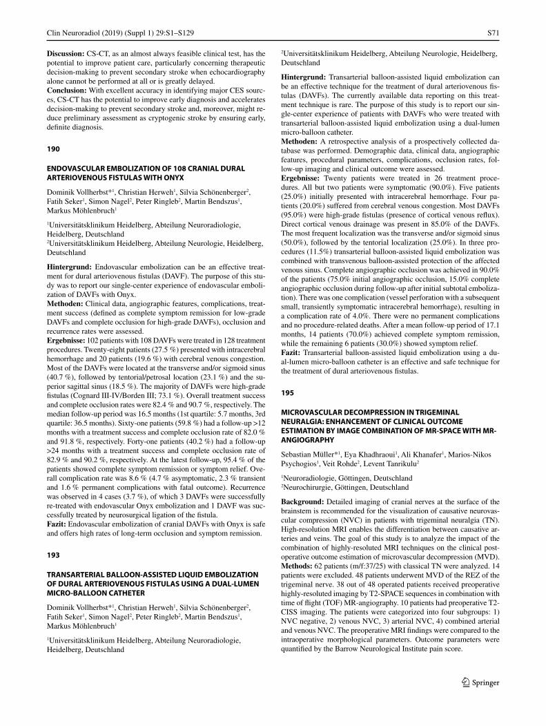
S71
123
Clin Neuroradiol (2019) (Suppl 1) 29:S1–S129
Discussion: CS-CT, as an almost always feasible clinical test, has the potential to improve patient care, particularly concerning therapeutic decision-making to prevent secondary stroke when echocardiography alone cannot be performed at all or is greatly delayed.Conclusion: With excellent accuracy in identifying major CES sourc-es, CS-CT has the potential to improve early diagnosis and accelerates decision-making to prevent secondary stroke and, moreover, might re-duce preliminary assessment as cryptogenic stroke by ensuring early, definite diagnosis.
190
ENDOVASCULAR EMBOLIZATION OF 108 CRANIAL DURAL ARTERIOVENOUS FISTULAS WITH ONYX
Dominik Vollherbst*1, Christian Herweh1, Silvia Schönenberger2, Fatih Seker1, Simon Nagel2, Peter Ringleb2, Martin Bendszus1, Markus Möhlenbruch1
1Universitätsklinikum Heidelberg, Abteilung Neuroradiologie, Heidelberg, Deutschland2Universitätsklinikum Heidelberg, Abteilung Neurologie, Heidelberg, Deutschland
Hintergrund: Endovascular embolization can be an effective treat-ment for dural arteriovenous fistulas (DAVF). The purpose of this stu-dy was to report our single-center experience of endovascular emboli-zation of DAVFs with Onyx.Methoden: Clinical data, angiographic features, complications, treat-ment success (defined as complete symptom remission for low-grade DAVFs and complete occlusion for high-grade DAVFs), occlusion and recurrence rates were assessed.Ergebnisse: 102 patients with 108 DAVFs were treated in 128 treatment procedures. Twenty-eight patients (27.5 %) presented with intracerebral hemorrhage and 20 patients (19.6 %) with cerebral venous congestion. Most of the DAVFs were located at the transverse and/or sigmoid sinus (40.7 %), followed by tentorial/petrosal location (23.1 %) and the su-perior sagittal sinus (18.5 %). The majority of DAVFs were high-grade fistulas (Cognard III-IV/Borden III; 73.1 %). Overall treatment success and complete occlusion rates were 82.4 % and 90.7 %, respectively. The median follow-up period was 16.5 months (1st quartile: 5.7 months, 3rd quartile: 36.5 months). Sixty-one patients (59.8 %) had a follow-up >12 months with a treatment success and complete occlusion rate of 82.0 % and 91.8 %, respectively. Forty-one patients (40.2 %) had a follow-up >24 months with a treatment success and complete occlusion rate of 82.9 % and 90.2 %, respectively. At the latest follow-up, 95.4 % of the patients showed complete symptom remission or symptom relief. Ove-rall complication rate was 8.6 % (4.7 % asymptomatic, 2.3 % transient and 1.6 % permanent complications with fatal outcome). Recurrence was observed in 4 cases (3.7 %), of which 3 DAVFs were successfully re-treated with endovascular Onyx embolization and 1 DAVF was suc-cessfully treated by neurosurgical ligation of the fistula.Fazit: Endovascular embolization of cranial DAVFs with Onyx is safe and offers high rates of long-term occlusion and symptom remission.
193
TRANSARTERIAL BALLOON-ASSISTED LIQUID EMBOLIZATION OF DURAL ARTERIOVENOUS FISTULAS USING A DUAL-LUMEN MICRO-BALLOON CATHETER
Dominik Vollherbst*1, Christian Herweh1, Silvia Schönenberger2, Fatih Seker1, Simon Nagel2, Peter Ringleb2, Martin Bendszus1, Markus Möhlenbruch1
1Universitätsklinikum Heidelberg, Abteilung Neuroradiologie, Heidelberg, Deutschland
2Universitätsklinikum Heidelberg, Abteilung Neurologie, Heidelberg, Deutschland
Hintergrund: Transarterial balloon-assisted liquid embolization can be an effective technique for the treatment of dural arteriovenous fis-tulas (DAVFs). The currently available data reporting on this treat-ment technique is rare. The purpose of this study is to report our sin-gle-center experience of patients with DAVFs who were treated with transarterial balloon-assisted liquid embolization using a dual-lumen micro-balloon catheter.Methoden: A retrospective analysis of a prospectively collected da-tabase was performed. Demographic data, clinical data, angiographic features, procedural parameters, complications, occlusion rates, fol-low-up imaging and clinical outcome were assessed.Ergebnisse: Twenty patients were treated in 26 treatment proce-dures. All but two patients were symptomatic (90.0%). Five patients (25.0%) initially presented with intracerebral hemorrhage. Four pa-tients (20.0%) suffered from cerebral venous congestion. Most DAVFs (95.0%) were high-grade fistulas (presence of cortical venous reflux). Direct cortical venous drainage was present in 85.0% of the DAVFs. The most frequent localization was the transverse and/or sigmoid sinus (50.0%), followed by the tentorial localization (25.0%). In three pro-cedures (11.5%) transarterial balloon-assisted liquid embolization was combined with transvenous balloon-assisted protection of the affected venous sinus. Complete angiographic occlusion was achieved in 90.0% of the patients (75.0% initial angiographic occlusion, 15.0% complete angiographic occlusion during follow-up after initial subtotal emboliza-tion). There was one complication (vessel perforation with a subsequent small, transiently symptomatic intracerebral hemorrhage), resulting in a complication rate of 4.0%. There were no permanent complications and no procedure-related deaths. After a mean follow-up period of 17.1 months, 14 patients (70.0%) achieved complete symptom remission, while the remaining 6 patients (30.0%) showed symptom relief.Fazit: Transarterial balloon-assisted liquid embolization using a du-al-lumen micro-balloon catheter is an effective and safe technique for the treatment of dural arteriovenous fistulas.
195
MICROVASCULAR DECOMPRESSION IN TRIGEMINAL NEURALGIA: ENHANCEMENT OF CLINICAL OUTCOME ESTIMATION BY IMAGE COMBINATION OF MR-SPACE WITH MR-ANGIOGRAPHY
Sebastian Müller*1, Eya Khadhraoui1, Ali Khanafer1, Marios-Nikos Psychogios1, Veit Rohde2, Levent Tanrikulu2
1Neuroradiologie, Göttingen, Deutschland2Neurochirurgie, Göttingen, Deutschland
Background: Detailed imaging of cranial nerves at the surface of the brainstem is recommended for the visualization of causative neurovas-cular compression (NVC) in patients with trigeminal neuralgia (TN). High-resolution MRI enables the differentiation between causative ar-teries and veins. The goal of this study is to analyze the impact of the combination of highly-resoluted MRI techniques on the clinical post-operative outcome estimation of microvascular decompression (MVD).Methods: 62 patients (m/ f:37/25) with classical TN were analyzed. 14 patients were excluded. 48 patients underwent MVD of the REZ of the trigeminal nerve. 38 out of 48 operated patients received preoperative highly-resoluted imaging by T2-SPACE sequences in combination with time of flight (TOF) MR-angiography. 10 patients had preoperative T2-CISS imaging. The patients were categorized into four subgroups: 1) NVC negative, 2) venous NVC, 3) arterial NVC, 4) combined arterial and venous NVC. The preoperative MRI findings were compared to the intraoperative morphological parameters. Outcome parameters were quantified by the Barrow Neurological Institute pain score.

S72
123
Clin Neuroradiol (2019) (Suppl 1) 29:S1–S129
Results: 29 pure arterial NVC (SCA: 27, AICA: 2), 8 venous NVC and 3 combined NVC (SCA+vein) were detected. In 8 cases no NVC was noticed during MVD. The neurovascular findings from the MRI corre-lated in 91.7 % of the treated patients with the intraoperative findings. In the examined cases with a combined image analysis of T2-SPACE/CISS with MR-TOF the correlation was enhanced to 94.7 % of the ex-amined subjects. The clinical outcome was significantly higher in the patients with arterial NVC compared to venous NVC.Discussion: Comparing our study with the other literature a stand-ard preoperative imaging workup by highly-resoluted T2-SPACE and MR-angiography should be performed in all patients with trigeminal neuralgia. A detailed multidisciplinary analysis of the patients should be perfomed by the neurologist, neuroradiologist and the neurosurgeon in order to optimize the surgical result for pain-freedom.Conclusion: Combined imaging of T2-SPACE/CISS with MR-TOF enables an optimized delineation of arterial and venous neurovascular conflicts and may allow a better prediction of clinical outcome com-pared to T2 imaging data alone.
196
EFFECT OF DIRECT THROMBOASPIRATION VS. STENT-RETRIEVER-THROMBECTOMY ON RADIATION EXPOSURE, PROCEDURE TIME AND FLUOROSCOPY TIME IN PATIENTS TREATED FOR ANTERIOR CIRCULATION ISCHEMIC STROKE – A MATCHED-PAIR ANALYSIS
Charlotte Weyland*1, Felix Hemmerich1, Markus Möhlenbruch1, Martin Bendszus1, Johannes Pfaff1
1Klinik für Neurologie, Abteilung für Neuroradiologie, Heidelberg, Deutschland
Background: To quantify the effect of stent-retriever thrombectomy vs. direct thromboaspiration on radiation exposure (RE), procedure time (PT) and fluoroscopy time (FT) in patients with endovascular stroke treatment (EST) for anterior circulation ischemic stroke.Methods: Retrospective analysis of an institutional review board–ap-proved stroke database of a comprehensive stroke center focusing on RE (as per dose area product in µGy*m², median [IQR]), procedure and fluoroscopy time (in minutes, median [IQR]) in patients receiving EST in anterior circulation ischemic stroke. Patients who received EST using direct thromboaspiration (DT) were matched with patients treat-ed using stent-retriever thrombectomy (SR) according to location of
intracranial occlusion, mode of anesthesia, number of thrombectomy maneuvers and gender (order of decreasing priority).Result: Overall 136 patients (n = 68 in each group) have been included in this analysis. Apart from patient age (in years, mean (SD): DT = 74 (13); SR = 79 (11), p-value: 0.023), there is no significant difference in patient’s baseline characteristics. PT (DT: 26 (21–38); SR: 49 (37–77); p-value: <0.0001) and FT (DT: 12 (7–18); SR: 26 (14–43), p-value: <0.0001) are shorter in patients who received direct throm-boaspiration. RE (DT: 6255 (4170–8944); SR: 8984 (5367–13.168), p-value: 0.034) is significantly lower in patients who received EST us-ing direct thromboaspiration. This is a relative median increase of RE of 43.6%, a relative increase of 116.6% of FT and of 88.5% of PT in patients who received stent-retriever assisted thrombectomy.Discussion: In this retrospective analysis EST using direct throm-boaspiration was associated with lower RE, FT and PT compared to matched patients who received stent-retriever thrombectomy. This re-sult is contradictory to the RE – analysis of Farah et. al [1] who showed no difference in RE concerning the EST maneuvers in their evaluation of dose reference level for EST.Conclusion: Our analysis shows a difference in RE between maneu-vers in endovascular stroke treatment with a higher RE in stent-retriev-er thrombectomy compared with direct thromboaspiration.
References 1. Farah J, Rouchaud A, Henry T, Regen C, Mihalea C, Moret J, et al.
Dose reference levels and clinical determinants in stroke neurora-diology interventions. Eur Radiol. 2019;29(2):645–53.
197
RADIATION EXPOSURE, PROCEDURE TIME AND FLUOROSCOPY TIME IN PATIENTS
TREATED FOR ACUTE BASILAR ARTERY OCCLUSION – A MATCHED-PAIR ANALYSIS
Charlotte Weyland*1, Felix Hemmerich1, Markus Möhlenbruch1, Martin Bendszus1, Johannes Pfaff1
1Klinik für Neurologie, Universitätsklinik Heidelberg, Abteilung für Neuroradiologie, Heidelberg, Deutschland
Background: Since the establishment of dose reference levels (DRLs) for radiation exposure (RE) in endovascular stroke treatment (EST) of
Fig. 1 | 195 Comparison of operation pictures, MRI and MR-A
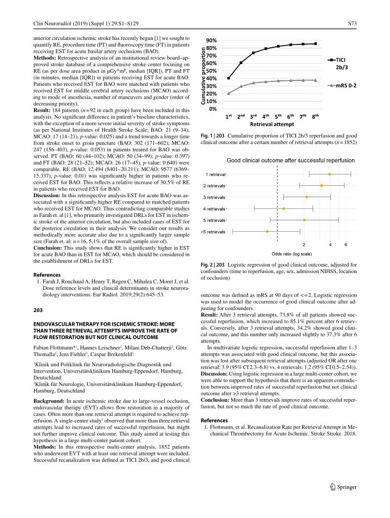
S73
123
Clin Neuroradiol (2019) (Suppl 1) 29:S1–S129
anterior circulation ischemic stroke has recently begun [1] we sought to quantify RE, procedure time (PT) and fluoroscopy time (FT) in patients receiving EST for acute basilar artery occlusions (BAO).Methods: Retrospective analysis of an institutional review board–ap-proved stroke database of a comprehensive stroke center focusing on RE (as per dose area product in µGy*m², median [IQR]), PT and FT (in minutes, median [IQR]) in patients receiving EST for acute BAO. Patients who received EST for BAO were matched with patients who received EST for middle cerebral artery occlusions (MCAO) accord-ing to mode of anesthesia, number of maneuvers and gender (order of decreasing priority).Result: 184 patients (n = 92 in each group) have been included in this analysis. No significant difference in patient’s baseline characteristics, with the exception of a more severe initial severity of stroke symptoms (as per National Institutes of Health Stroke Scale; BAO: 21 (9–34); MCAO: 17 (14–21), p-value: 0.025) and a trend towards a longer time from stroke onset to groin puncture (BAO: 302 (171–602); MCAO: 247 (156–403), p-value: 0.053) in patients treated for BAO was ob-served. PT (BAO: 60 (44–102); MCAO: 50 (34–99); p-value: 0.397) and FT (BAO: 28 (21–52); MCAO: 26 (17–45), p-value: 0.640) were comparable. RE (BAO: 12.494 (8401–20.211); MCAO: 9577 (6369–15.337), p-value: 0.01) was significantly higher in patients who re-ceived EST for BAO. This reflects a relative increase of 30.5% of RE in patients who received EST for BAO.Discussion: In this retrospective analysis EST for acute BAO was as-sociated with a significantly higher RE compared to matched patients who received EST for MCAO. Thus contradicting comparable studies as Farah et. al [1], who primarily investigated DRLs for EST in ischem-ic stroke of the anterior circulation, but also included cases of EST for the posterior circulation in their analysis. We consider our results as methodically more accurate also due to a significantly larger sample size (Farah et. al: n = 16, 5,1% of the overall sample size of).Conclusion: This study shows that RE is significantly higher in EST for acute BAO than in EST for MCAO, which should be considered in the establishment of DRLs for EST.
References 1. Farah J, Rouchaud A, Henry T, Regen C, Mihalea C, Moret J, et al.
Dose reference levels and clinical determinants in stroke neurora-diology interventions. Eur Radiol. 2019;29(2):645–53.
203
ENDOVASCULAR THERAPY FOR ISCHEMIC STROKE: MORE THAN THREE RETRIEVAL ATTEMPTS IMPROVE THE RATE OF FLOW RESTORATION BUT NOT CLINICAL OUTCOME
Fabian Flottmann*1, Hannes Leischner1, Milani Deb-Chatterji2, Götz Thomalla2, Jens Fiehler1, Caspar Brekenfeld1
1Klinik und Poliklinik für Neuroradiologische Diagnostik und Intervention, Universitätsklinikum Hamburg-Eppendorf, Hamburg, Deutschland2Klinik für Neurologie, Universitätsklinikum Hamburg-Eppendorf, Hamburg, Deutschland
Background: In acute ischemic stroke due to large-vessel occlusion, endovascular therapy (EVT) allows flow restoration in a majority of cases. Often more than one retrieval attempt is required to achieve rep-erfusion. A single-center study1 observed that more than three retrieval attempts lead to increased rates of successful reperfusion, but might not further improve clinical outcome. This study aimed at testing this hypothesis in a large multi-center patient cohort.Methods: In this retrospective multi-center analysis, 1852 patients who underwent EVT with at least one retrieval attempt were included. Successful recanalization was defined as TICI 2b/3, and good clinical
outcome was defined as mRS at 90 days of < = 2. Logistic regression was used to model the occurrence of good clinical outcome after ad-justing for confounders.Result: After 3 retrieval attempts, 73.8% of all patients showed suc-cessful reperfusion, which increased to 85.1% percent after 6 retriev-als. Conversely, after 3 retrieval attempts, 34.2% showed good clini-cal outcome, and this number only increased slightly to 37.3% after 6 attempts.
In multivariate logistic regression, successful reperfusion after 1–3 attempts was associated with good clinical outcome, but this associa-tion was lost after subsequent retrieval attempts (adjusted OR after one retrieval: 3.9 (95% CI 2.3–6.8) vs. 4 retrievals: 1.2 (95% CI 0.5–2.54)).Discussion: Using logistic regression in a large multi-center cohort, we were able to support the hypothesis that there is an apparent contradic-tion between improved rates of successful reperfusion but not clinical outcome after >3 retrieval attempts.Conclusion: More than 3 retrievals improve rates of successful reper-fusion, but not so much the rate of good clinical outcome.
References 1. Flottmann, et al. Recanalization Rate per Retrieval Attempt in Me-
chanical Thrombectomy for Acute Ischemic. Stroke Stroke. 2018.
Fig. 2 | 203 Logistic regression of good clinical outcome, adjusted for confounders (time to reperfusion, age, sex, admission NIHSS, location of occlusion)
Fig. 1 | 203 Cumulative proportion of TICI 2b/3 reperfusion and good clinical outcome after a certain number of retrieval attempts (n = 1852)

S74
123
Clin Neuroradiol (2019) (Suppl 1) 29:S1–S129
218
TRUE FIRST-PASS EFFECT FOR THE POSTERIOR CIRCULATION: FIRST-PASS COMPLETE REPERFUSION IMPROVES CLINICAL OUTCOME IN STROKE THROMBECTOMY PATIENTS
Nuran Abdullayev*1, Volker Maus2, Daniel Behme3, Jan Borggrefe1, Marc Schlamann1, Thomas Liebig4, Christoph Kabbasch1, Anastasios Mpotsaris5
1Department of Radiology, University Hospital of Cologne, Köln, Deutschland2Institute of Diagnostic and Interventional Radiology, Neuroradiology and Nuclear Medicine, Knappschaftskrankenhaus Bochum, Ruhr University Bochum“ Bochum, Deutschland3Department of Neuroradiology, University Medical Center Goettingen, Goettingen, Deutschland4Institute of Neuroradiology, University Hospital Munich (LMU), München, Deutschland5Department of Neuroradiology, RWTH Aachen, Aachen, Deutschland
Background: Complete reperfusion (mTICI 3) in stroke patients after a single mechanical thrombectomy (MT) pass has been hypothesized as a predictor for favorable outcome (mRS 0–2) and a true first-pass effect defined as improved clinical outcome after complete reperfusion in the anterior circulation with one pass has been already scientifical-ly proven. But a true first-pass effect with complete reperfusion in the posterior circulation with one vs. multiple passes has not yet been spe-cifically addressed in the literature.Methods: Patients with occlusions in the posterior circulation (car-ried out between 2010 and 2017) were analyzed and clinical outcome of basilar artery occlusions (BAO) patients with complete reperfusion and known symptom onset were compared. After adjustment for mul-tiple confounding factors such as demographics, time intervals, stroke severity (NIHSS), early stroke signs (PC ASPECTS) and comorbidity, we compared outcome at 90 days between matched cohorts of firstpass complete reperfusion vs. multipass complete reperfusion.Result: A total of 90 MTs of BAO were analyzed. We compared clin-ical outcome of 56 patients with BAO and known symptom onset, in whom we achieved complete reperfusion (mTICI 3), depending on whether complete reperfusion was achieved after a single thrombecto-my pass (n = 28) or multiple thrombectomy passes (n = 28). Multivaria-ble analysis of our cohort of 56 nonmatched patients revealed that there was a highly significant association between first-pass complete rep-erfusion and favorable clinical outcome (p = <0.0001). In our matched cohorts (n = 7 vs. n = 7), favorable clinical outcome was only seen if complete reperfusion was achieved after one pass (86% vs. 0%).Discussion: Rapid and complete reperfusion is widely accepted to be crucial for favorable outcome1,2. Our results imply that the first thrombectomy maneuver should be undertaken with the technique with the highest first-pass complete reperfusion rates, but our study cannot address the question how to best achieve first-pass complete reperfusion.Conclusion: First-pass complete reperfusion in the posterior circu-lation is an independent predictor of favorable outcome. Achieving complete reperfusion after multiple passes might simply not be good enough in the posterior circulation.
References 1. Khatri P, et al. Good clinical outcome after ischemic stroke with
successful revascularization is timedependent. Neurology. 2. Zaidat OO, et al. First pass effect: A new measure for stroke
thrombectomy devices. Stroke.
222
TUBING-TUNING: EINFLUSS DES TUBINGS AUF DIE ASPIRATIONSRATE GÄNGIGER ASPIRATIONSPUMPEN
Hannes Schwenke*1, Astrid Aumann1, Peter Schramm1
1Institut für Neuroradiologie, UKSH, Campus Lübeck, Lübeck, Deutschland
Hintergrund: Zu den Einflussgrößen auf die Thrombusaspiration zählt neben dem Innendurchmesser des Aspirationskatheters auch die Leistung des mechanischen Aspirationssystems, bestehend aus einer Aspirationspumpe und dem dazugehörigen Tubing. Im Rahmen der vorliegenden Arbeit wurde in vitro untersucht, welche Kombinationen von verschiedenen Aspirationspumpe und Tubings den optimalen Ein-fluss auf eine Steigerung der maximalen Aspirationsrate der Aspirati-onskatheter hat. Insbesondere wurde untersucht, ob sich die Aspirati-onsrate der Pumpen unabhängig vom Tubing unterscheidet.Methoden: In n = 280 Messungen á 20 Sek. Messdauer wurde die As-pirationsrate verschiedener Aspirationskatheter (Penumbra: ACE 64, ACE 68, 5 MAX, 4 MAX, 3 MAX, 5 MAX ACE; Stryker: Catalyst 6) über eine Messung im Messzylinder ermittelt. Hierbei wurde Glycerin-Wasser-Gemisch aspiriert und die Aspirationsmenge pro Zeit mit ei-ner Hochpräzisionswaage (A&D EK-2000i, 10 Datenpunkte pro Sek.) erfasst. Die Pumpen (Penumbra MAX; Medela Dominant Flex) und die Tubings (jeweils zugehöriges Penumbra-Tubing; Medela-Tubing) wurden systematisch variiert. Die Datenanalyse erfolgte über ANO-VAs mittels Matlab 2018b.Ergebnisse: Die mittlere Aspirationsrate beider Pumpen unabhängig vom Tubing unterschied sich in unserem Experiment nicht signifikant vonei-nander (Abb. 1). Die Anwendung des Medela-Tubings führte zu einer sig-nifikant höheren Aspirationsrate als das Penumbra-Tubing (Abb. 2), unab-hängig von der angewendeten Pumpe. Die Aspirationsrate der mit einem Medela-Tubing ausgestatteten Medela-Pumpe war signifikant höher als die mit einem Penumbra-Tubing ausgestattete Penumbra-Pumpe (Abb. 2).
Abb. 1 | 222

S75
123
Clin Neuroradiol (2019) (Suppl 1) 29:S1–S129
Diskussion: Die maximale Aspirationsrate kann mittels Wechsel auf ein Tubing mit großem Innendurchmesser gesteigert werden („Tubing-Tuning“). In unserem Experiment führte ein Wechsel auf das großlumi-gere Tubing ohne integrierte On-Off-Adapterzu einer Verbesserung der Aspirationsrate um ca. 20 %. Die Aspirationsrate verringerte sich dem-entsprechend bei beiden Pumpen mit Anwendung des Tubings, wel-ches den eingebauten, flussreduzierenden On-Off-Konnektor aufweist.Fazit: Insbesondere das Tubing und weniger die Pumpen stellen einen Einflussfaktor auf die Aspirationsraten der Aspirationssysteme dar. Lu-menreduzierende Adapter innerhalb der Tubings können im Direktver-gleich die Aspirationsrate in vitro signifikant reduzieren.
223
TREATMENT OF INTRACRANIAL ANEURYSMS WITH THE NEW-GENERATION LOW-PROFILE ACANDIS ACCLINO MICROSTENT: LONG-TERM SAFETY AND EFFICACY IN A MULTICENTER STUDY
Lukas Görtz*1, Michael Smyk2, Anastasios Mpotsaris3, Jan Borggrefe1, Franziska Dorn4, Thomas Liebig4, Marc Schlamann1, Bernd Turowski2, Christoph Kabbasch1
1Köln, Deutschland2Düsseldorf, Deutschland3Aachen, Deutschland4München, Deutschland
Background: Stent-assisted coiling (SAC) represents an established treatment option for complex intracranial aneurysms. The Acandis Ac-clino is a novel self-expandable closed-cell nitinol microstent which complies with a 0.0165” or 0.017” microcatheter. We report long-term clinical and angiographic outcome in a multicenter setting.Methods: The Acclino micro-stent was used in 98 consecutive patients (55.4 ± 13.5 years) for treatment of 98 aneurysms (28 unruptured, 20
recurrent, 50 ruptured) at three German tertiary care centers within a 6-year period. We retrospectively analyzed technical success, compli-cation rates, clinical outcome and angiographic results.Result: Stent implantation was technically successful in all aneu-rysms achieving immediate complete occlusion in 89.8%. Among 65 patients (66.3%) with 6-month angiographic follow-up, complete and near-complete occlusion was obtained in 92.3% and 98.5%, respec-tively. At long-term follow-up (38 patients, mean: 21 months), the complete and near-complete occlusion rates were 81.2% and 89.5%, respectively. Aneurysm recurrence between mid- and long-term fol-low-up was observed in 14.3%. Retreatment was performed in 11.3%. The overall complication rate was 8.2% (8/98), including three throm-boembolic events (3.1%) and five hemorrhagic events (5.1%). Neu-rological complications occurred in three patients, of which one was device-related (one ischemic stroke in a patient with subarachnoid hemorrhage). Among scheduled patients, procedural and device-relat-ed morbidity rates were 2.1% (1/48) and 0% (0/48), respectively.Discussion: To date, robust data on long-term angiographic outcome is available for the Enterprise stent system, achieving 59% and 81% com-plete or near-complete occlusion rates in a study by Fargen et al. [1]. The results of the current study compare favourably and may indicate a more durable long-term aneurysm occlusion of the low-profile Acclino stent with minimal complication and morbidity rates.Conclusion: The present study demonstrates a high safety profile of the Acclino microstent, with an ischemic stroke rate of 1.0%. Long-term aneurysm rates compare favorably to first-generation stents and justify further investigation of this device.
References 1. Fargen KM, Mocco J, Neal D, Dewan MC, Reavey-Cantwell
J, Woo HH, et al. A multicenter study of stent-assisted coiling of cerebral aneurysms with a Y configuration. Neurosurgery. 2013;73:466–72.
224
LOW-PROFILE WEB 17 VERSUS PREDECESSOR WEB 21 SYSTEM FOR TREATMENT OF SMALL INTRACRANIAL ANEURYSMS: COMPARATIVE ANALYSIS OF PROCEDURAL SAFETY AND FEASIBILITY
Lukas Görtz*1, Thomas Liebig2, Eberhard Siebert3, Moritz Herzberg2, Marc Schlamann1, Jan Borggrefe1, Franziska Dorn2, Christoph Kabbasch1
1Köln, Deutschland2München, Deutschland3Berlin, Deutschland
Background: Woven EndoBridge (WEB) represents an efficient tech-nique for endovascular treatment of wide-necked and bifurcation aneu-rysms. The WEB 17 system has been recently introduced for facilitated endovascular treatment of small bifurcation aneurysms (≤7 mm) with low profile microcatheters. We compared the WEB 17 with its prede-cessor versions in terms of procedural safety and feasibility.Methods: This is a multicenter review of aneurysms ranging from 3 to 7 mm treated with the WEB between 2011 and 2019. Aneurysm characteristics, procedural parameters and complications were retro-spectively compared between treatment with the WEB 17 and a control group that was treated with its predecessor 21-version using inverse probability of treatment weighting (IPTW).Results: The study cohort consisted of 38 patients treated with a WEB 17 (mean size: 4.9 ± 1.5 mm) and 70 with a WEB 21 (mean size: 5.6 ± 1.4 mm). The 21 system had a higher failure rate (10.3%) than WEB 17 (0%, p = 0.05). Additional stenting was performed more of-ten with WEB 21 (10%) than with WEB 17 (2.6%, adjusted p = 0.005).
Abb. 2 | 222
S76
123
Clin Neuroradiol (2019) (Suppl 1) 29:S1–S129
Thromboembolic events occurred more often in the WEB 21 group (14.3%) than in the WEB 17 group (5.3%, adjusted p = 0.002). Neu-rological complications (WEB 17: 2.6%, WEB 21: 2.9%, adjusted p = 1.0) and immediate complete aneurysm occlusion rates (WEB 17: 57.9%, WEB 21: 54.3%, adjusted p = 0.21) did not differ significantly between both groups.Discussion: Small wide-necked aneurysms are generally difficult to treat by endovascular means. The WEB 17 system consists of fewer and thinner nitinol wires than the predecessor WEB 21 and is there-fore compatible with a 0.017” low-profile microcatheter. This system aims to increase flexibility of the device and to facilitate deployment in small aneurysms. In our study, the WEB 17 was associated with a higher success rate and a significantly reduced thromboembolic com-pared to the WEB 21 in the adjusted analysis. Long-term angiographic outcome data will be necessary to investigate the efficacy of the new WEB 17 system.Conclusion: The low-profile WEB 17 provides a feasible and safe ap-proach for endovascular treatment of small wide-necked aneurysms. Long-term outcome analysis will be necessary to draw a definite con-clusion.
225
DOES THE SIZE MATTER? 6F ADAPT ONLY VS 5F ADAPT AND PROXIMAL FLOW ARREST IN A M1 OCCLUSION MODEL
Jawid Madjidyar*, Lars Nerkada, Olav Jansen
Klinik für Radiologie und Neuroradiologie, Universitätsklinikum Schleswig-Holstein, Campus Kiel, Kiel, Deutschland
Hintergrund: A direct aspiration first pass technique (ADAPT) has proven to be a highly effective thrombectomy option in patients with stroke due to large vessel occlusion. Simultaneously, balloon guide catheters seem to lead to better results in mechanical recanalization, mostly in combination with stent retrievers. At the moment the 6F as-piration devices do not fit through a balloon guide catheter on the mar-ket. In this experimental study the efficacy of ADAPT only via 6F as-piration device and ADAPT combined with BGC using a 5F aspiration device were compared.Methoden: Two different clot models were used: A fibrin rich (white) clot and a RBC rich (red) clot. Each clot was placed into the M1 seg-ment of a transparent silicon phantom of the anterior circulation. Phy-siological hemodynamic conditions were maintained by a programma-ble piston pump. Thrombectomy was performed with 1) SOFIA 6 F via 6F Neuron MAX Long Sheath and 2) SOFIA 5F via 8F FlowGate Balloon Guide Catheter. The thrombectomy was performed under di-rect visual control. Primary endpoints were the number of passes and the occurrence/number of distal emboli.Ergebnisse: 10 experiments were made with each clot model and thrombectomy setup (total of n = 40). First pass full recanalization could be achieved by the 6F ADAPT only technique in 80 % of the cases in red clots and 90 % in white clots. However, the catheter was clogged by the white clots in every experiment, though it had to be pulled back into the long sheath and distal emboli were caused in 20 % of the experiment (10 % in red clots).
Using 5F ADAPT combined with BGC a first pass full recanaliza-tion rate of 90% could be achieved in red clots and 100% in white clots. In both clot models a 10% rate of distal emboli occurred. In 85% of the experiments the thrombi clogged the catheter and had to be pulled back into the BGC.Diskussion: In this experimental setting the smaller inner diameter of the aspiration catheter is more than compensated by the additional use of the BGC, but there is no statistical significance. Both 6F ADAPT and 5F ADAPT combined with BGC show a high first pass efficacy and a low distal emboli rate. As soon as the aspiration catheter was clogged
the proximal aspiration via the long sheath or BGC was crucial to gu-arantee the thrombectomy.Fazit: The size depends on the setting: ADAPT via a 6F aspiration catheter was equal to ADAPT via a 5F aspiration catheter combined with BGC. Both techniques were highly effective, though a cost analy-sis could be made to decide which technique to choose.
234
RESULTS AFTER CAROTID STENTING WITH THE CGUARD STENT AS PART OF INTRACRANIAL AND EXTRACRANIAL THROMBECTOMY
Teodor Catalin Sabou*, Reimer Andresen, Christian Wissgott, Christoph Kopetsch, Fabian Scheer
Institute of Diagnostic and Interventional Radiology/Neuroradiology, Westküstenklinikum Heide, Academic Teaching Hospital of the Universities of Kiel, Luebeck and Hamburg, Heide, Deutschland
Background: The purpose of the study was to evaluate the MicroNet coated CGuard Nitinol stent for safety and efficacy in the treatment of acute occlusions of the cerebral arteries.Methods: In our department for interventional neuroradiology in the period between 01.01.2016 and 03.03.2019, a total of 20 patients un-derwent aspiration thrombectomy after acute occlusion of the cerebral arteries due to a high-grade stenosis or occlusion at the origin of the in-ternal carotid artery with concomitant occlusion of the middle cerebral artery. During the same intervention, the internal carotid artery lesion was treated with the double-layered CGuard nitinol stent. Postinterven-tionally, 18 patients underwent a Therapy with aspirin (100 mg daily) and clopidogrel (75 mg daily)(11 patients only aspirin, 2 patiens only clopidogrel, 8 patiens combined and 2 patients only heparin). The stent patency was performed using dopple ultrasonography.Result: The stent was successfully place in all 20 patients. Postinter-ventional dopple ultrasound was performed in the neurological depart-ment in 17 patients after a mean time of 4,18 days (MIN 1; MAX 9). No in-stent stenosis and no stent thrombosis were detected. Postinter-ventionally, dual platelet aggregation inhibition was administered after 2,9 days (STDEV ± 2,3; MIN 0: MAX 9). Preinterventional systemic lysis was administered in 9 patients. Intracranial hemorrhage in the infarct area was detected in 11 patients. Two patients have died due to intracranial hemorrhage.Discussion: The recommendations of the European Society for Vascu-lar Surgery regarding the endovascular treatment of the carotid artery stenosis are a premedication with aspirin (300 mg initially for up to 14 days followed by 75 mg daily if not already taking aspirin) and clopi-dogrel (75 mg daily) 3 days prior to carotid artery stenting (1). In our cases the intervention was performed without a premedication. No in-stent stenosis or thrombosis were observed in the follow-up.Conclusion: The use of the CGuard stent for the treatment of internal carotid artery stenosis or occlusion in acute stroke treatment is a safe and effective method. No in-stent occlusions or stenosis were detected despite late administration of platelet aggregation inhibitors and lack of premedication.
References 1. Naylor AR, et al. Editor’s Choice – Management of Atheroscle-
rotic Carotid and Vertebral Artery Disease: 2017 Clinical Practice Guidelines of the European Society for Vascular Surgery (ESVS). Eur J Vasc Endovasc Surg. 2018;55(1):3–81.
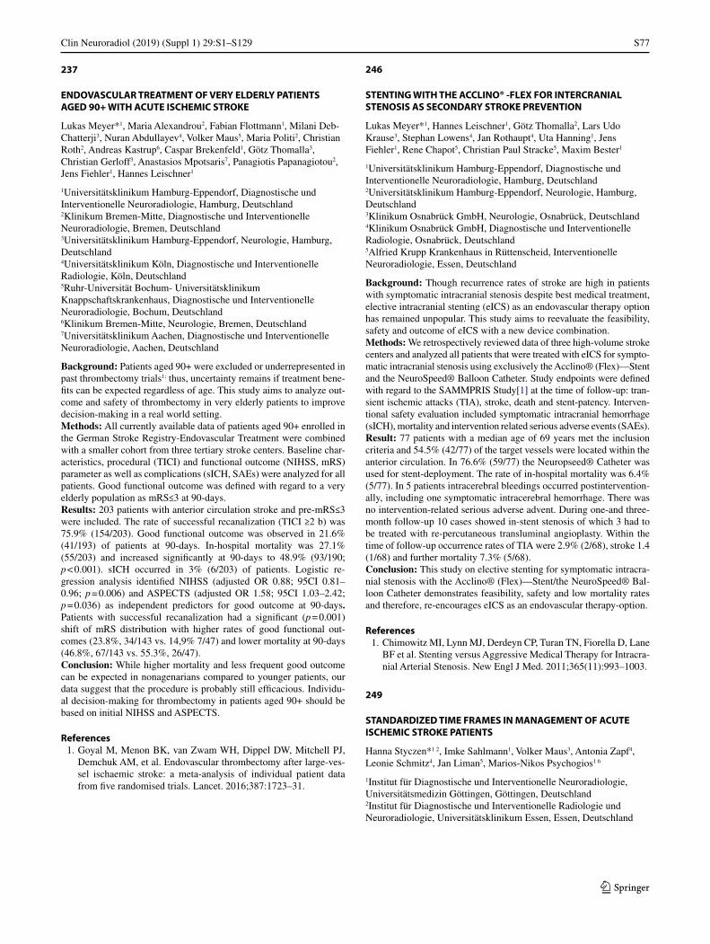
S77
123
Clin Neuroradiol (2019) (Suppl 1) 29:S1–S129
237
ENDOVASCULAR TREATMENT OF VERY ELDERLY PATIENTS AGED 90+ WITH ACUTE ISCHEMIC STROKE
Lukas Meyer*1, Maria Alexandrou2, Fabian Flottmann1, Milani Deb-Chatterji3, Nuran Abdullayev4, Volker Maus5, Maria Politi2, Christian Roth2, Andreas Kastrup6, Caspar Brekenfeld1, Götz Thomalla3, Christian Gerloff3, Anastasios Mpotsaris7, Panagiotis Papanagiotou2, Jens Fiehler1, Hannes Leischner1
1Universitätsklinikum Hamburg-Eppendorf, Diagnostische und Interventionelle Neuroradiologie, Hamburg, Deutschland2Klinikum Bremen-Mitte, Diagnostische und Interventionelle Neuroradiologie, Bremen, Deutschland3Universitätsklinikum Hamburg-Eppendorf, Neurologie, Hamburg, Deutschland4Universitätsklinikum Köln, Diagnostische und Interventionelle Radiologie, Köln, Deutschland5Ruhr-Universität Bochum- Universitätsklinikum Knappschaftskrankenhaus, Diagnostische und Interventionelle Neuroradiologie, Bochum, Deutschland6Klinikum Bremen-Mitte, Neurologie, Bremen, Deutschland7Universitätsklinikum Aachen, Diagnostische und Interventionelle Neuroradiologie, Aachen, Deutschland
Background: Patients aged 90+ were excluded or underrepresented in past thrombectomy trials1; thus, uncertainty remains if treatment bene-fits can be expected regardless of age. This study aims to analyze out-come and safety of thrombectomy in very elderly patients to improve decision-making in a real world setting.Methods: All currently available data of patients aged 90+ enrolled in the German Stroke Registry-Endovascular Treatment were combined with a smaller cohort from three tertiary stroke centers. Baseline char-acteristics, procedural (TICI) and functional outcome (NIHSS, mRS) parameter as well as complications (sICH, SAEs) were analyzed for all patients. Good functional outcome was defined with regard to a very elderly population as mRS≤3 at 90-days.Results: 203 patients with anterior circulation stroke and pre-mRS≤3 were included. The rate of successful recanalization (TICI ≥2 b) was 75.9% (154/203). Good functional outcome was observed in 21.6% (41/193) of patients at 90-days. In-hospital mortality was 27.1% (55/203) and increased significantly at 90-days to 48.9% (93/190; p < 0.001). sICH occurred in 3% (6/203) of patients. Logistic re-gression analysis identified NIHSS (adjusted OR 0.88; 95CI 0.81–0.96; p = 0.006) and ASPECTS (adjusted OR 1.58; 95CI 1.03–2.42; p = 0.036) as independent predictors for good outcome at 90-days Patients with successful recanalization had a significant (p = 0.001) shift of mRS distribution with higher rates of good functional out-comes (23.8%, 34/143 vs. 14,9% 7/47) and lower mortality at 90-days (46.8%, 67/143 vs. 55.3%, 26/47).Conclusion: While higher mortality and less frequent good outcome can be expected in nonagenarians compared to younger patients, our data suggest that the procedure is probably still efficacious. Individu-al decision-making for thrombectomy in patients aged 90+ should be based on initial NIHSS and ASPECTS.
References 1. Goyal M, Menon BK, van Zwam WH, Dippel DW, Mitchell PJ,
Demchuk AM, et al. Endovascular thrombectomy after large-ves-sel ischaemic stroke: a meta-analysis of individual patient data from five randomised trials. Lancet. 2016;387:1723–31.
246
STENTING WITH THE ACCLINO® -FLEX FOR INTERCRANIAL STENOSIS AS SECONDARY STROKE PREVENTION
Lukas Meyer*1, Hannes Leischner1, Götz Thomalla2, Lars Udo Krause3, Stephan Lowens4, Jan Rothaupt4, Uta Hanning1, Jens Fiehler1, Rene Chapot5, Christian Paul Stracke5, Maxim Bester1
1Universitätsklinikum Hamburg-Eppendorf, Diagnostische und Interventionelle Neuroradiologie, Hamburg, Deutschland2Universitätsklinikum Hamburg-Eppendorf, Neurologie, Hamburg, Deutschland3Klinikum Osnabrück GmbH, Neurologie, Osnabrück, Deutschland4Klinikum Osnabrück GmbH, Diagnostische und Interventionelle Radiologie, Osnabrück, Deutschland5Alfried Krupp Krankenhaus in Rüttenscheid, Interventionelle Neuroradiologie, Essen, Deutschland
Background: Though recurrence rates of stroke are high in patients with symptomatic intracranial stenosis despite best medical treatment, elective intracranial stenting (eICS) as an endovascular therapy option has remained unpopular. This study aims to reevaluate the feasibility, safety and outcome of eICS with a new device combination.Methods: We retrospectively reviewed data of three high-volume stroke centers and analyzed all patients that were treated with eICS for sympto-matic intracranial stenosis using exclusively the Acclino® (Flex)—Stent and the NeuroSpeed® Balloon Catheter. Study endpoints were defined with regard to the SAMMPRIS Study[1] at the time of follow-up: tran-sient ischemic attacks (TIA), stroke, death and stent-patency. Interven-tional safety evaluation included symptomatic intracranial hemorrhage (sICH), mortality and intervention related serious adverse events (SAEs).Result: 77 patients with a median age of 69 years met the inclusion criteria and 54.5% (42/77) of the target vessels were located within the anterior circulation. In 76.6% (59/77) the Neuropseed® Catheter was used for stent-deployment. The rate of in-hospital mortality was 6.4% (5/77). In 5 patients intracerebral bleedings occurred postintervention-ally, including one symptomatic intracerebral hemorrhage. There was no intervention-related serious adverse advent. During one-and three-month follow-up 10 cases showed in-stent stenosis of which 3 had to be treated with re-percutaneous transluminal angioplasty. Within the time of follow-up occurrence rates of TIA were 2.9% (2/68), stroke 1.4 (1/68) and further mortality 7.3% (5/68).Conclusion: This study on elective stenting for symptomatic intracra-nial stenosis with the Acclino® (Flex)—Stent/the NeuroSpeed® Bal-loon Catheter demonstrates feasibility, safety and low mortality rates and therefore, re-encourages eICS as an endovascular therapy-option.
References 1. Chimowitz MI, Lynn MJ, Derdeyn CP, Turan TN, Fiorella D, Lane
BF et al. Stenting versus Aggressive Medical Therapy for Intracra-nial Arterial Stenosis. New Engl J Med. 2011;365(11):993–1003.
249
STANDARDIZED TIME FRAMES IN MANAGEMENT OF ACUTE ISCHEMIC STROKE PATIENTS
Hanna Styczen*1 2, Imke Sahlmann1, Volker Maus3, Antonia Zapf4, Leonie Schmitz4, Jan Liman5, Marios-Nikos Psychogios1 6
1Institut für Diagnostische und Interventionelle Neuroradiologie, Universitätsmedizin Göttingen, Göttingen, Deutschland2Institut für Diagnostische und Interventionelle Radiologie und Neuroradiologie, Universitätsklinikum Essen, Essen, Deutschland
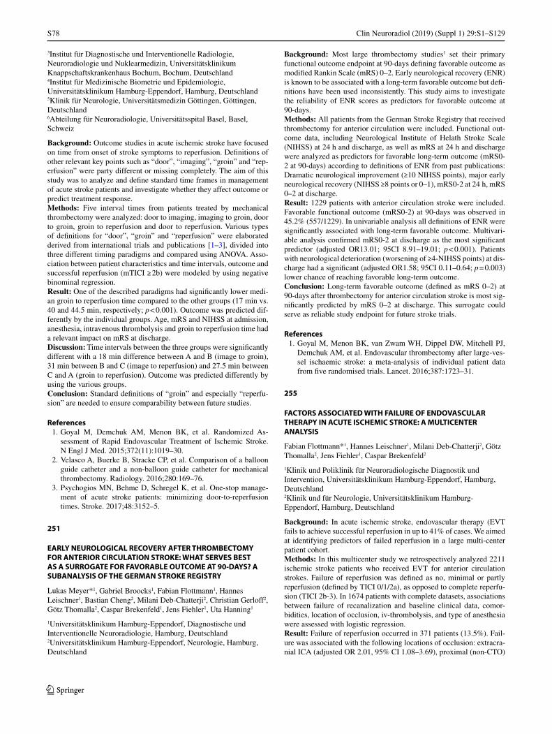
S78
123
Clin Neuroradiol (2019) (Suppl 1) 29:S1–S129
3Institut für Diagnostische und Interventionelle Radiologie, Neuroradiologie und Nuklearmedizin, Universitätsklinikum Knappschaftskrankenhaus Bochum, Bochum, Deutschland4Institut für Medizinische Biometrie und Epidemiologie, Universitätsklinikum Hamburg-Eppendorf, Hamburg, Deutschland5Klinik für Neurologie, Universitätsmedizin Göttingen, Göttingen, Deutschland6Abteilung für Neuroradiologie, Universitätsspital Basel, Basel, Schweiz
Background: Outcome studies in acute ischemic stroke have focused on time from onset of stroke symptoms to reperfusion. Definitions of other relevant key points such as “door”, “imaging”, “groin” and “rep-erfusion” were party different or missing completely. The aim of this study was to analyze and define standard time frames in management of acute stroke patients and investigate whether they affect outcome or predict treatment response.Methods: Five interval times from patients treated by mechanical thrombectomy were analyzed: door to imaging, imaging to groin, door to groin, groin to reperfusion and door to reperfusion. Various types of definitions for “door”, “groin” and “reperfusion” were elaborated derived from international trials and publications [1–3], divided into three different timing paradigms and compared using ANOVA. Asso-ciation between patient characteristics and time intervals, outcome and successful reperfusion (mTICI ≥ 2b) were modeled by using negative binominal regression.Result: One of the described paradigms had significantly lower medi-an groin to reperfusion time compared to the other groups (17 min vs. 40 and 44.5 min, respectively; p < 0.001). Outcome was predicted dif-ferently by the individual groups. Age, mRS and NIHSS at admission, anesthesia, intravenous thrombolysis and groin to reperfusion time had a relevant impact on mRS at discharge.Discussion: Time intervals between the three groups were significantly different with a 18 min difference between A and B (image to groin), 31 min between B and C (image to reperfusion) and 27.5 min between C and A (groin to reperfusion). Outcome was predicted differently by using the various groups.Conclusion: Standard definitions of “groin” and especially “reperfu-sion” are needed to ensure comparability between future studies.
References 1. Goyal M, Demchuk AM, Menon BK, et al. Randomized As-
sessment of Rapid Endovascular Treatment of Ischemic Stroke. N Engl J Med. 2015;372(11):1019–30.
2. Velasco A, Buerke B, Stracke CP, et al. Comparison of a balloon guide catheter and a non-balloon guide catheter for mechanical thrombectomy. Radiology. 2016;280:169–76.
3. Psychogios MN, Behme D, Schregel K, et al. One-stop manage-ment of acute stroke patients: minimizing door-to-reperfusion times. Stroke. 2017;48:3152–5.
251
EARLY NEUROLOGICAL RECOVERY AFTER THROMBECTOMY FOR ANTERIOR CIRCULATION STROKE: WHAT SERVES BEST AS A SURROGATE FOR FAVORABLE OUTCOME AT 90-DAYS? A SUBANALYSIS OF THE GERMAN STROKE REGISTRY
Lukas Meyer*1, Gabriel Broocks1, Fabian Flottmann1, Hannes Leischner1, Bastian Cheng2, Milani Deb-Chatterji2, Christian Gerloff2, Götz Thomalla2, Caspar Brekenfeld1, Jens Fiehler1, Uta Hanning1
1Universitätsklinikum Hamburg-Eppendorf, Diagnostische und Interventionelle Neuroradiologie, Hamburg, Deutschland2Universitätsklinikum Hamburg-Eppendorf, Neurologie, Hamburg, Deutschland
Background: Most large thrombectomy studies1 set their primary functional outcome endpoint at 90-days defining favorable outcome as modified Rankin Scale (mRS) 0–2. Early neurological recovery (ENR) is known to be associated with a long-term favorable outcome but defi-nitions have been used inconsistently. This study aims to investigate the reliability of ENR scores as predictors for favorable outcome at 90-days.Methods: All patients from the German Stroke Registry that received thrombectomy for anterior circulation were included. Functional out-come data, including Neurological Institute of Helath Stroke Scale (NIHSS) at 24 h and discharge, as well as mRS at 24 h and discharge were analyzed as predictors for favorable long-term outcome (mRS0-2 at 90-days) according to definitions of ENR from past publications: Dramatic neurological improvement (≥10 NIHSS points), major early neurological recovery (NIHSS ≥8 points or 0–1), mRS0-2 at 24 h, mRS 0–2 at discharge.Result: 1229 patients with anterior circulation stroke were included. Favorable functional outcome (mRS0-2) at 90-days was observed in 45.2% (557/1229). In univariable analysis all definitions of ENR were significantly associated with long-term favorable outcome. Multivari-able analysis confirmed mRS0-2 at discharge as the most significant predictor (adjusted OR13.01; 95CI 8.91–19.01; p < 0.001). Patients with neurological deterioration (worsening of ≥4-NIHSS points) at dis-charge had a significant (adjusted OR1.58; 95CI 0.11–0.64; p = 0.003) lower chance of reaching favorable long-term outcome.Conclusion: Long-term favorable outcome (defined as mRS 0–2) at 90-days after thrombectomy for anterior circulation stroke is most sig-nificantly predicted by mRS 0–2 at discharge. This surrogate could serve as reliable study endpoint for future stroke trials.
References 1. Goyal M, Menon BK, van Zwam WH, Dippel DW, Mitchell PJ,
Demchuk AM, et al. Endovascular thrombectomy after large-ves-sel ischaemic stroke: a meta-analysis of individual patient data from five randomised trials. Lancet. 2016;387:1723–31.
255
FACTORS ASSOCIATED WITH FAILURE OF ENDOVASCULAR THERAPY IN ACUTE ISCHEMIC STROKE: A MULTICENTER ANALYSIS
Fabian Flottmann*1, Hannes Leischner1, Milani Deb-Chatterji2, Götz Thomalla2, Jens Fiehler1, Caspar Brekenfeld2
1Klinik und Poliklinik für Neuroradiologische Diagnostik und Intervention, Universitätsklinikum Hamburg-Eppendorf, Hamburg, Deutschland2Klinik und für Neurologie, Universitätsklinikum Hamburg-Eppendorf, Hamburg, Deutschland
Background: In acute ischemic stroke, endovascular therapy (EVT fails to achieve successful reperfusion in up to 41% of cases. We aimed at identifying predictors of failed reperfusion in a large multi-center patient cohort.Methods: In this multicenter study we retrospectively analyzed 2211 ischemic stroke patients who received EVT for anterior circulation strokes. Failure of reperfusion was defined as no, minimal or partly reperfusion (defined by TICI 0/1/2a), as opposed to complete reperfu-sion (TICI 2b-3). In 1674 patients with complete datasets, associations between failure of recanalization and baseline clinical data, comor-bidities, location of occlusion, iv-thrombolysis, and type of anesthesia were assessed with logistic regression.Result: Failure of reperfusion occurred in 371 patients (13.5%). Fail-ure was associated with the following locations of occlusion: extracra-nial ICA (adjusted OR 2.01, 95% CI 1.08–3.69), proximal (non-CTO)

S79
123
Clin Neuroradiol (2019) (Suppl 1) 29:S1–S129
ICA (adjusted OR 1.79, 95% CI 1.05–2.98) and M2 segment (adjust-ed OR 1.86, 95% CI 1.21–2.84). Furthermore, it was associated with proximal ICA stenosis (adjusted OR 2.90, 95% CI 1.69–4.97), stroke of other determined etiology (adjusted OR 2.73, 95% CI 1.36–5.39) and treatment out of daytime routine (adjusted OR 1.41, 95% CI 1.07–1.86).
Successful reperfusion (i. e. negative correlation with failure) was associated with ASPECTS (adjusted OR 0.85, 95% CI 0.79–0.92), general anesthesia (adjusted OR 0.72, 95% CI 0.54–0.96), and con-comitant stenting of ICA stenosis (adjusted OR 0.20, 95% CI 0.11–0.38). Furthermore, it occurred more often when more than one EVT technique (stentriever or aspiration devices) was used.Discussion: Failure of reperfusion occurs more often when vessels other than CTO or M1 are affected. Proximal ICA stenosis is a risk factor, which can be successfully treated when concomitant ICA stent-ing is possible. EVT is less successful in strokes that are not of cardi-oembolic or atherosclerotic etiology. Obviously, whenever the EVT technique had to be switched, successful reperfusion was less likely.
Higher ASPECTS on admission, general anesthesia and treatment during daytime routine were associated with improved odds of suc-cessful reperfusion after EVT.Conclusion: The “non-standard” EVT patient (e. g. proximal ICA oc-clusion with low ASPECTS treated in the middle of the night) has a higher risk of unsuccessful reperfusion.
259
HÄMOSIDERINABLAGERUNGEN IM SPLENIUM DES CORPUS CALLOSUM BEI SCHWEREM ARDS NACH PULMONALEM BEFALL EINES ANAPLASTISCH-GROßZELLIGEM LYMPHOMS
Michael Schnitzbauer*1, Marius Hartmann1
1Institut für Neuroradiologie, HELIOS Klinikum Berlin-Buch, Berlin, Deutschland
Hintergrund: Hämosiderinablagerungen im Splenium des Corpus callosum wurden bei intakter intracerebraler Perfusion mit Hypoxämie bisher bei Bergsteigern, die ein höhenassoziiertes Hirnödem (HACE) mit schwerer ARDS überlebt haben und bei Intensivpatienten unter ECMO-Therapie beobachtet.
Fallbeschreibung/Case Report: Ein 17-jähriger Patient stellte sich in der Rettungsstelle mit einer cervikalen Lymphadenopathie und B-Symptomatik vor. Im PET-CT zeigten sich flächenhafte pulmonale Konsolidierungen im apikalen Unterlappen links, progrediente Pleu-raergüsse bds., Aszites bei Hepatosplenomegalie und eine deutliche Lymphadenopathie cervikal, paraaortal/iliacal und inguinal. Nach
bioptischer Sicherung wurde die Diagnose eines anaplastisch-großzel-ligen ALK-NPM positiven Lymphoms (ALCL) gestellt.
Unter Chemotherapie (nach NHL-BFM-Register 2012) zeigte sich eine kardiopulmonale Dekompensation, interkurrenter Knochen-markaplasie und Neutropenie bei schwerem ARDS mit Sepsis bei ausgeprägtem pulmonalem Lymphombefall. Unter fortgeführter In-tensivtherapie mit Antiobiose kam es zuerst zu einer Besserung der Symptomatik. Im Verlauf entwickelt der Patient während des Intensiv-aufenthalts starke Kopfschmerzen und zum Ausschluss einer Sinus-venenthrombose wurde ein MRT des Schädels durchgeführt. Hier sah man auf den SWI-Aufnahmen multiple Hämosiderinablagerungen im Splenium des Corpus callosum.
Nach Fortführung der Chemotherapie wurde ein Frührezidiv des Lymphoms festgestellt mit Nachweis einer Meningeosis supra- und infratentoriell. Die weitere Therapie erfolgte eskalierter Chemothera-pie und kraniospinaler Bestrahlung, die bei starker Neutropenie des Patienten abgebrochen wurde. Der Patient verstarb mit einer schweren Kachexie an den Folgen des Lymphoms auf der Intensivstation.Diskussion/Discussion: Der pulmonale Befall durch ein anaplastisch-großzelliges Lymphom mit schwerem ARDS kann zu intrakranieller Hypoxämie bei erhaltener Perfusion unter CPAP-Therapie führen. Da-durch resultiert eine temporäre fokale Störung der Blut-Hirn-Schranke mit erhöhter Permeabilität und Einwanderung von Erythrozyten mit Hämosiderinablagerung bevorzugt im Splenium des Corpus callosum. Der aktuelle Fall zeigt, dass auch durch den Abfall des Pao2/FiO2 un-ter CPAP-Therapie eine Hypoxämie bei erhaltener Perfusion induziert werden kann, die wie beim HACE zu Hämosiderinablagerungen im Splenium des Corpus callosums führen.
260
PRÄVALENZ NEUROVASKULÄRER KOMPRESSION DER ROSTRALEN VENTROLATERALEN MEDULLA OBLONGATA IN EINER POPULATIONSBASIERTEN MRT-STUDIE
Daniel Cantré*1, Friederike Groth2, Sascha Groth2, Steffen Fleck3, Eiko Rathmann2, Sönke Langner1
1Institut für Diagnostische und Interventionelle Radiologie, Kinder- und Neuroradiologie, Universitätsmedizin Rostock, Rostock, Deutschland2Institut für Diagnostische Radiologie und Neuroradiologie, Universitätsmedizin Greifswald, Greifswald, Deutschland3Klinik für Neurochirurgie, Universitätsmedizin Greifswald, Greifswald, Deutschland
Fig. 1 | 255 Logistic regression analysis of failure of reperfusion (TICI 0-2a) in 1674 patients with complete datasets, adjusted for multiple confounders
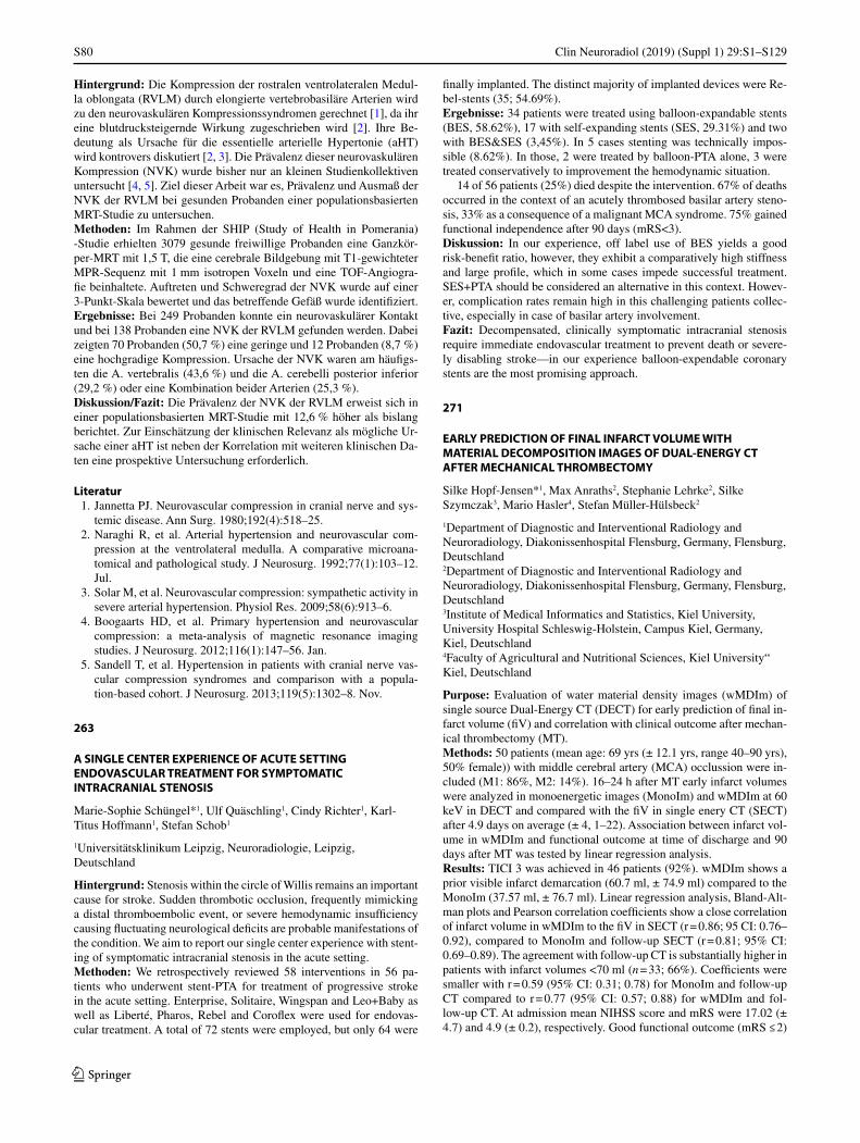
S80
123
Clin Neuroradiol (2019) (Suppl 1) 29:S1–S129
Hintergrund: Die Kompression der rostralen ventrolateralen Medul-la oblongata (RVLM) durch elongierte vertebrobasiläre Arterien wird zu den neurovaskulären Kompressionssyndromen gerechnet [1], da ihr eine blutdrucksteigernde Wirkung zugeschrieben wird [2]. Ihre Be-deutung als Ursache für die essentielle arterielle Hypertonie (aHT) wird kontrovers diskutiert [2, 3]. Die Prävalenz dieser neurovaskulären Kompression (NVK) wurde bisher nur an kleinen Studienkollektiven untersucht [4, 5]. Ziel dieser Arbeit war es, Prävalenz und Ausmaß der NVK der RVLM bei gesunden Probanden einer populationsbasierten MRT-Studie zu untersuchen.Methoden: Im Rahmen der SHIP (Study of Health in Pomerania) -Studie erhielten 3079 gesunde freiwillige Probanden eine Ganzkör-per-MRT mit 1,5 T, die eine cerebrale Bildgebung mit T1-gewichteter MPR-Sequenz mit 1 mm isotropen Voxeln und eine TOF-Angiogra-fie beinhaltete. Auftreten und Schweregrad der NVK wurde auf einer 3-Punkt-Skala bewertet und das betreffende Gefäß wurde identifiziert.Ergebnisse: Bei 249 Probanden konnte ein neurovaskulärer Kontakt und bei 138 Probanden eine NVK der RVLM gefunden werden. Dabei zeigten 70 Probanden (50,7 %) eine geringe und 12 Probanden (8,7 %) eine hochgradige Kompression. Ursache der NVK waren am häufigs-ten die A. vertebralis (43,6 %) und die A. cerebelli posterior inferior (29,2 %) oder eine Kombination beider Arterien (25,3 %).Diskussion/Fazit: Die Prävalenz der NVK der RVLM erweist sich in einer populationsbasierten MRT-Studie mit 12,6 % höher als bislang berichtet. Zur Einschätzung der klinischen Relevanz als mögliche Ur-sache einer aHT ist neben der Korrelation mit weiteren klinischen Da-ten eine prospektive Untersuchung erforderlich.
Literatur 1. Jannetta PJ. Neurovascular compression in cranial nerve and sys-
temic disease. Ann Surg. 1980;192(4):518–25. 2. Naraghi R, et al. Arterial hypertension and neurovascular com-
pression at the ventrolateral medulla. A comparative microana-tomical and pathological study. J Neurosurg. 1992;77(1):103–12. Jul.
3. Solar M, et al. Neurovascular compression: sympathetic activity in severe arterial hypertension. Physiol Res. 2009;58(6):913–6.
4. Boogaarts HD, et al. Primary hypertension and neurovascular compression: a meta-analysis of magnetic resonance imaging studies. J Neurosurg. 2012;116(1):147–56. Jan.
5. Sandell T, et al. Hypertension in patients with cranial nerve vas-cular compression syndromes and comparison with a popula-tion-based cohort. J Neurosurg. 2013;119(5):1302–8. Nov.
263
A SINGLE CENTER EXPERIENCE OF ACUTE SETTING ENDOVASCULAR TREATMENT FOR SYMPTOMATIC INTRACRANIAL STENOSIS
Marie-Sophie Schüngel*1, Ulf Quäschling1, Cindy Richter1, Karl-Titus Hoffmann1, Stefan Schob1
1Universitätsklinikum Leipzig, Neuroradiologie, Leipzig, Deutschland
Hintergrund: Stenosis within the circle of Willis remains an important cause for stroke. Sudden thrombotic occlusion, frequently mimicking a distal thromboembolic event, or severe hemodynamic insufficiency causing fluctuating neurological deficits are probable manifestations of the condition. We aim to report our single center experience with stent-ing of symptomatic intracranial stenosis in the acute setting.Methoden: We retrospectively reviewed 58 interventions in 56 pa-tients who underwent stent-PTA for treatment of progressive stroke in the acute setting. Enterprise, Solitaire, Wingspan and Leo+Baby as well as Liberté, Pharos, Rebel and Coroflex were used for endovas-cular treatment. A total of 72 stents were employed, but only 64 were
finally implanted. The distinct majority of implanted devices were Re-bel-stents (35; 54.69%).Ergebnisse: 34 patients were treated using balloon-expandable stents (BES, 58.62%), 17 with self-expanding stents (SES, 29.31%) and two with BES&SES (3,45%). In 5 cases stenting was technically impos-sible (8.62%). In those, 2 were treated by balloon-PTA alone, 3 were treated conservatively to improvement the hemodynamic situation.
14 of 56 patients (25%) died despite the intervention. 67% of deaths occurred in the context of an acutely thrombosed basilar artery steno-sis, 33% as a consequence of a malignant MCA syndrome. 75% gained functional independence after 90 days (mRS<3).Diskussion: In our experience, off label use of BES yields a good risk-benefit ratio, however, they exhibit a comparatively high stiffness and large profile, which in some cases impede successful treatment. SES+PTA should be considered an alternative in this context. Howev-er, complication rates remain high in this challenging patients collec-tive, especially in case of basilar artery involvement.Fazit: Decompensated, clinically symptomatic intracranial stenosis require immediate endovascular treatment to prevent death or severe-ly disabling stroke—in our experience balloon-expendable coronary stents are the most promising approach.
271
EARLY PREDICTION OF FINAL INFARCT VOLUME WITH MATERIAL DECOMPOSITION IMAGES OF DUAL-ENERGY CT AFTER MECHANICAL THROMBECTOMY
Silke Hopf-Jensen*1, Max Anraths2, Stephanie Lehrke2, Silke Szymczak3, Mario Hasler4, Stefan Müller-Hülsbeck2
1Department of Diagnostic and Interventional Radiology and Neuroradiology, Diakonissenhospital Flensburg, Germany, Flensburg, Deutschland2Department of Diagnostic and Interventional Radiology and Neuroradiology, Diakonissenhospital Flensburg, Germany, Flensburg, Deutschland3Institute of Medical Informatics and Statistics, Kiel University, University Hospital Schleswig-Holstein, Campus Kiel, Germany, Kiel, Deutschland4Faculty of Agricultural and Nutritional Sciences, Kiel University“ Kiel, Deutschland
Purpose: Evaluation of water material density images (wMDIm) of single source Dual-Energy CT (DECT) for early prediction of final in-farct volume (fiV) and correlation with clinical outcome after mechan-ical thrombectomy (MT).Methods: 50 patients (mean age: 69 yrs (± 12.1 yrs, range 40–90 yrs), 50% female)) with middle cerebral artery (MCA) occlussion were in-cluded (M1: 86%, M2: 14%). 16–24 h after MT early infarct volumes were analyzed in monoenergetic images (MonoIm) and wMDIm at 60 keV in DECT and compared with the fiV in single enery CT (SECT) after 4.9 days on average (± 4, 1–22). Association between infarct vol-ume in wMDIm and functional outcome at time of discharge and 90 days after MT was tested by linear regression analysis.Results: TICI 3 was achieved in 46 patients (92%). wMDIm shows a prior visible infarct demarcation (60.7 ml, ± 74.9 ml) compared to the MonoIm (37.57 ml, ± 76.7 ml). Linear regression analysis, Bland-Alt-man plots and Pearson correlation coefficients show a close correlation of infarct volume in wMDIm to the fiV in SECT (r = 0.86; 95 CI: 0.76–0.92), compared to MonoIm and follow-up SECT (r = 0.81; 95% CI: 0.69–0.89). The agreement with follow-up CT is substantially higher in patients with infarct volumes <70 ml (n = 33; 66%). Coefficients were smaller with r = 0.59 (95% CI: 0.31; 0.78) for MonoIm and follow-up CT compared to r = 0.77 (95% CI: 0.57; 0.88) for wMDIm and fol-low-up CT. At admission mean NIHSS score and mRS were 17.02 (± 4.7) and 4.9 (± 0.2), respectively. Good functional outcome (mRS ≤ 2)
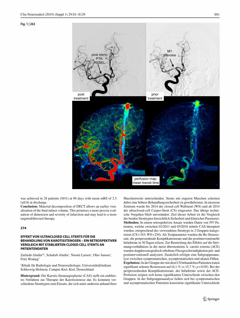
S81
123
Clin Neuroradiol (2019) (Suppl 1) 29:S1–S129
was achieved in 28 patients (56%) at 90 days with mean mRS of 2.5 (±0.8) at discharge.Conclusion: Material decomposition of DECT allows an earlier visu-alization of the final infarct volume. This promises a more precise eval-uation of dimension and severity of infarction and may lead to a more targeted/directed therapy.
274
EFFEKT VON ULTRACLOSED CELL STENTS FÜR DIE BEHANDLUNG VON KAROTISSTENOSEN – EIN RETROSPEKTIVER VERGLEICH MIT ETABLIERTEN CLOSED CELL STENTS AN PATIENTENDATEN
Zarlasht Aludin*1, Schekeb Aludin1, Naomi Larsen1, Olav Jansen1, Fritz Wodarg1
1Klinik für Radiologie und Neuroradiologie, Universitätsklinikum Schleswig-Holstein, Campus Kiel, Kiel, Deutschland
Hintergrund: Die Karotis-Stentangioplastie (CAS) stellt ein etablier-tes Verfahren zur Therapie der Karotisstenose dar. Es kommen ver-schiedene Stenttypen zum Einsatz, die sich unter anderem anhand ihrer
Maschenweite unterscheiden. Stents mit engeren Maschen scheinen dabei eine höhere Behandlungssicherheit zu gewährleisten. In unserem Zentrum wurde bis 2014 der closed-cell Wallstent (WS) und ab 2014 der ultraclosed-cell Casper-Stent (CS) eingesetzt. Das übrige techni-sche Vorgehen blieb unverändert. Ziel dieser Arbeit ist der Vergleich der beiden Stenttypen hinsichtlich Sicherheit und klinischer Parameter.Methoden: In einem retrospektiven Ansatz wurden Daten von 597 Pa-tienten, welche zwischen 01/2011 und 03/2018 mittels CAS therapiert wurden, entsprechend des verwendeten Stenttyps in 2 Gruppen katego-risiert (CS = 343; WS = 254). Als Testparameter wurden die Re-Stenose-rate, die periprozedurale Komplikationsrate und die postinterventionelle Infarktrate in 30 Tagen erfasst. Zur Beurteilung des Effekts auf die Strö-mungsverhältnisse in der meist überstenteten A. carotis externa (ACE) wurden dopplersonografisch erhobene Flussgeschwindigkeiten prä- und postinterventionell analysiert. Zusätzlich erfolgte eine Subgruppenana-lyse zwischen symptomatischen, asymptomatischen und akuten Fällen.Ergebnisse: In der Gruppe der mit dem CS behandelten Patienten traten signifikant seltener Restenosen auf (6,1 % vs 15,7 %; p < 0.05). Bei der periprozeduralen Komplikationsrate, der Infarktrate sowie der ACE-Perfusion zeigten sich keine signifikanten Unterschiede zwischen den Gruppen. In der Subgruppenanalyse ließen sich bei symptomatischen und asymptomatischen Patienten konsistent signifikante Unterschiede
Fig. 1 | 263

S82
123
Clin Neuroradiol (2019) (Suppl 1) 29:S1–S129
bezüglich der Restenoserate feststellen. Bei akuten Fällen zeigten sich keine signifikanten Unterschiede zwischen den Gruppen.Diskussion: Anhand unserer Daten scheint die Verwendung von ult-raclosed-cell Stents im Vergleich zu closed-cell Stents eine geringere Restenoserate zu gewährleisten. Trotz der engeren Maschen scheint der Fluss in der ACE nicht relevant beeinflusst zu werden. Bezüglich der Komplikations- und Infarktraten ergaben sich keine Unterschiede zwischen den Gruppen.
276
TREATMENT OF IN-STENT RESTENOSIS OF DUAL-LAYER MICROMESH CAROTID ARTERY STENTS
Silke Hopf-Jensen*1, Michael Preiß1, Stefan Müller-Hülsbeck1
1Department of Diagnostic and Interventional Radiology and Neuroradiology, Diakonissenhospital Flensburg, Flensburg, Deutschland
Purpose: To analyse the incidence and treatment options of restenosis in patients treated with super-closed dual-layer carotid artery stents.Methods: Between 02/2014–05/2019 208 pts were treated with 213 dual-layer stents (191 RoadsaverTM stents, Terumo, Tokyo, Japan: 89.7% and 22 CGuard stentsTM, Inspire MD, Tel Aviv, Israel: 10.3%). Stent patency was evaluated by ultrasound before discharge, after 3 months and then every 6 months during follow up. Stenosis degree > 60% for asymptomatic and >50% for symptomatic stenosis (NAS-CET) are indicated for re-treatment.Results: During mid-term follow up (mean 3.4 years, range 4 weeks–5.2 years) 7 pts with asymptomatic restenosis (3%) and 3 pts with asymptomatic stent occlusions (1.4%) were detected. All resteno-sis were characterized as neointimal hyperplasia. In cases with stent occlusion 2 cases with progressive atherosclerotic plaques and one case with soft thrombo-embolic material were recorded.
In RoadsaverTM stents 7 restenosis were treated as follows: 2 re-PTA (with drug-coated balloon) with new, but asymptomatic DWI lesion in MRI, 4 re-stenting of the distal flair ends with 2 single-layer stents (WallstentTM, Natick, MA, USA) with technical and clinical success and 2 dual-layer stents (1 RoadsaverTM and 1 CGuardTM, latter with following symptomatic occlusion). 3 asymptomatic stent occlusion af-ter CAS with RoadsaverTM stents were detected: 1 case 12 month after treatment of a tandem lesion with simultaneous intracranial thrombec-tomy, 1 case 4 days after acute stenting with thrombectomy in a set-ting without given ASA in the acute situation and 1 case 6 month after emergency stenting within 12 hours after symptoms onset.
One CGuardTM restenosis was treated with re-stenting with sin-gle-layer stent (XactTM Abbott Vascular, Santa Clara, CA, USA) with good technical and clinical success.Conclusions: The restenosis rate of dual-layer stents seems to be low compared to conventional single-layer stents, but treatment of resteno-sis of the dual-layer cell design differ fundamentally. Re-PTA can in-crease intracranial thrombembolic complications due to the small cell design. Re-stenting with single-layer stents seems to be superior to du-al-layer stents again to preserve the blood vessel diameter.
277
NEUROPHYSIOLOGICAL MONITORING IN THE ENDOVASCULAR THERAPY OF NEURYSMS
Silke Hopf-Jensen*1, Michael Preiß1, Gisa Andresen2, Wolfgang Börm3, Ulf Linstedt2, Stefan Müller-Hülsbeck1
1Department of Diagnostic and Interventional Radiology and Neuroradiology, Diakonissenhospital Flensburg, Flensburg, Deutschland
2Department of Anesthesiology and Intensive Care Therapy, Diakonissenhospital Flensburg, Flensburg, Deutschland3Department of Neurosurgery, Diakonissenhospital Flensburg, Flensburg, Deutschland
Purpose: To evaluate the relevancy of neurophysiological monitoring techniques with somatosensory evoked potentials (SSEP) during end-ovascular treatment of cerebral aneurysms.Methods: Embolization of 20 aneurysms (ACom: 5 ICA: 9, Carot-id-T: 2, MCA: 4) were monitored by SSEPs using bilateral median or posterior tibial nerve stimulation. Critical SSEP changes were defined as an amplitude reduction of >50% or a latency delay (N19/P24). The techniques included coil embolization (14), balloon-remodelling coil-ing (3) and stent-assisted coiling (3).Results: 20 patients (female: 16 patients, mean 58 years” range: 42–86 years) with 12 ruptured (60%) and 8 incidental (40%) aneurysms were treated. There were transient SSEP changes in 5 patients (25%) during embolization, with 2 of these 5 events occuring during balloon-remod-elling technique without visible embolus or delayed perfusion. The re-maining 3 cases were associated with thrombus formation at the aneu-rysm basis, in one case within an underlying stent. SSEP changes lead to an immediate closer inspection of the whole angiological status, es-pecially the peripheral angiogram, and initiation of thrombolysis thera-py, i. e. Glycoprotein IIb/IIIa inhibitors. No neurological changes were found in all cases postprocedural after successful thrombus clearing.Conclusion: Somatosensory evoked potential monitoring is sensitive for the early detection of subcortical ischemia during aneurysm embo-lization. Neuromonitoring provides additional safety tool during tech-nical complex endovascular treatment of cerebral aneurysms.
278
CEREBRO-VASCULAR REACTIVITY MAPPING (CVRM) – PROMISES AND PITFALLS
Andreas Bartsch*1 2
1Radiologie Bamberg, Bamberg, Deutschland2WIN (FMRIB) Oxford, JR Hospital, Oxford, England
Hintergrund: CVRM has been promoted to assess the cerebrovas-cular reserve capacity (CVRC) in steno-occlusive arteriopathies such as moyamoya. CVRM promises to detect „neurovascular uncoupling“ (NVU): Insufficient perfusion and collateralization is presumed to lead to vasodilation irresponsive to hypercapnic challenges requiring revas-cularization such as bypass surgery [1].Methoden: We performed BOLD-CVRM (3T, TR = 3s/TE = 30 ms) by 10 endexpiratory breath-hold (BH) maneuvers for 21s, alternating with 10 episodes of normal breathing for 42s, in 2 moyamoya-patients: 1 primary (50 year-old female) and 1 secondary due to neurofibromatosis type 1 (23 year-old male; Fig. 1A). In addition, dynamic susceptibil-ity contrast (DSC-)perfusion imaging (TR = 1.17s/same TE) and vas-omotor reserve (VMR) testing with transcranial duplex (TCD) under Carbogen inhalation were performed. BH-CVRM and DSC-perfusion were analyzed using a model- (i. e., respiratory response = RRF [2] and arterial input = AIF function-based) and data-driven (ICA-based) ap-proach in FSL (www.fmrib.ox.ac.uk/fsl).Ergebnisse: Model-driven CVRM by a single Gaussian RRF-predic-tor (explanatory variable EV), which is conventionally used for gen-eral linear modelling (GLM), suggested absent CVR in the affected media-territory of both cases (Fig. 1B). However, VMR was intact (45 and 33 % velocity increase under Carbogen) and time-to-peak (TTP) delays of DSC-perfusion matched exactly data-driven CVRM delays of the measured BOLD signal in both cases (7 and 11s; Fig. 1D for patient 2), which is remarkable in that the two modalities based on exo- (DSC) and endogenous (BH-CVRM) T2*-weighted contrast con-verged precisely.
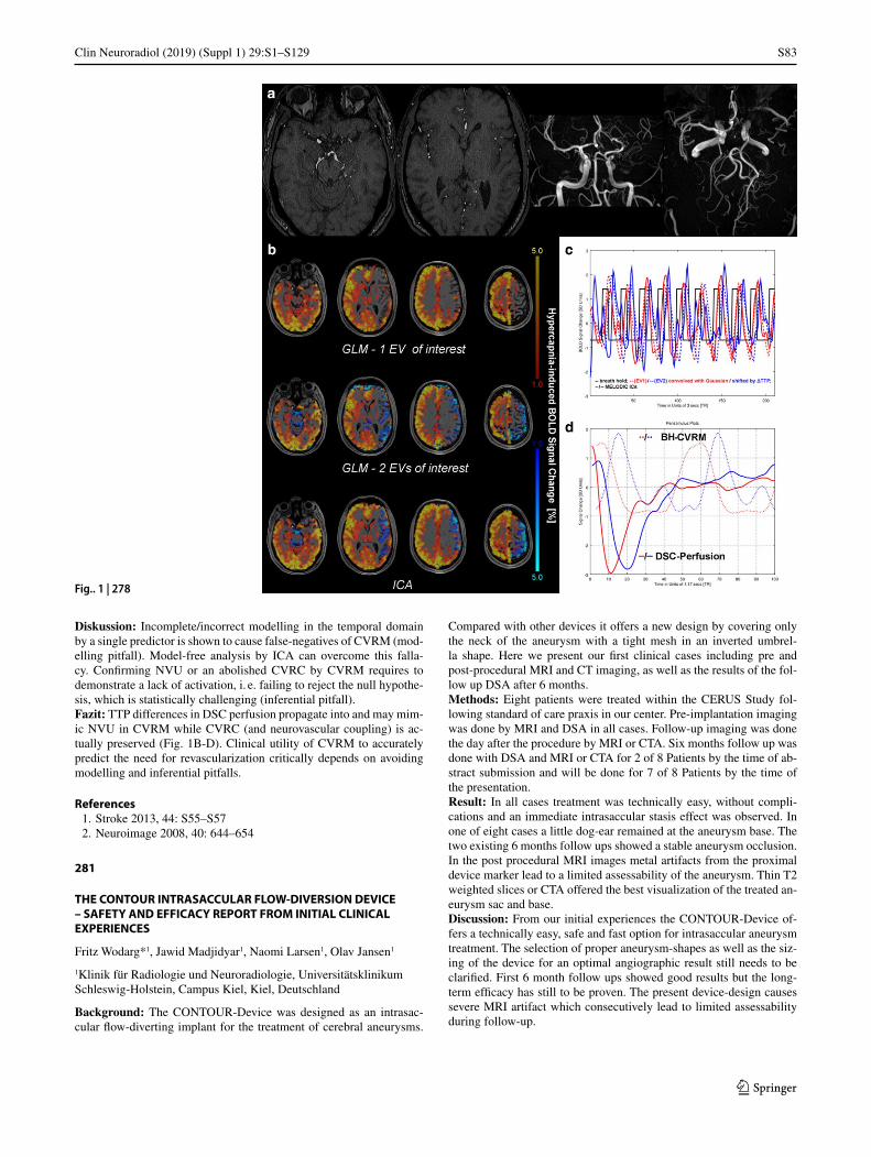
S83
123
Clin Neuroradiol (2019) (Suppl 1) 29:S1–S129
Diskussion: Incomplete/incorrect modelling in the temporal domain by a single predictor is shown to cause false-negatives of CVRM (mod-elling pitfall). Model-free analysis by ICA can overcome this falla-cy. Confirming NVU or an abolished CVRC by CVRM requires to demonstrate a lack of activation, i. e. failing to reject the null hypothe-sis, which is statistically challenging (inferential pitfall).Fazit: TTP differences in DSC perfusion propagate into and may mim-ic NVU in CVRM while CVRC (and neurovascular coupling) is ac-tually preserved (Fig. 1B-D). Clinical utility of CVRM to accurately predict the need for revascularization critically depends on avoiding modelling and inferential pitfalls.
References 1. Stroke 2013, 44: S55–S57 2. Neuroimage 2008, 40: 644–654
281
THE CONTOUR INTRASACCULAR FLOW-DIVERSION DEVICE – SAFETY AND EFFICACY REPORT FROM INITIAL CLINICAL EXPERIENCES
Fritz Wodarg*1, Jawid Madjidyar1, Naomi Larsen1, Olav Jansen1
1Klinik für Radiologie und Neuroradiologie, Universitätsklinikum Schleswig-Holstein, Campus Kiel, Kiel, Deutschland
Background: The CONTOUR-Device was designed as an intrasac-cular flow-diverting implant for the treatment of cerebral aneurysms.
Compared with other devices it offers a new design by covering only the neck of the aneurysm with a tight mesh in an inverted umbrel-la shape. Here we present our first clinical cases including pre and post-procedural MRI and CT imaging, as well as the results of the fol-low up DSA after 6 months.Methods: Eight patients were treated within the CERUS Study fol-lowing standard of care praxis in our center. Pre-implantation imaging was done by MRI and DSA in all cases. Follow-up imaging was done the day after the procedure by MRI or CTA. Six months follow up was done with DSA and MRI or CTA for 2 of 8 Patients by the time of ab-stract submission and will be done for 7 of 8 Patients by the time of the presentation.Result: In all cases treatment was technically easy, without compli-cations and an immediate intrasaccular stasis effect was observed. In one of eight cases a little dog-ear remained at the aneurysm base. The two existing 6 months follow ups showed a stable aneurysm occlusion. In the post procedural MRI images metal artifacts from the proximal device marker lead to a limited assessability of the aneurysm. Thin T2 weighted slices or CTA offered the best visualization of the treated an-eurysm sac and base.Discussion: From our initial experiences the CONTOUR-Device of-fers a technically easy, safe and fast option for intrasaccular aneurysm treatment. The selection of proper aneurysm-shapes as well as the siz-ing of the device for an optimal angiographic result still needs to be clarified. First 6 month follow ups showed good results but the long-term efficacy has still to be proven. The present device-design causes severe MRI artifact which consecutively lead to limited assessability during follow-up.
Fig.. 1 | 278
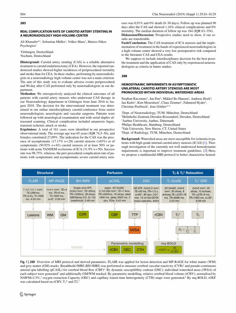
S84
123
Clin Neuroradiol (2019) (Suppl 1) 29:S1–S129
285
REAL COMPLICATION RATE OF CAROTID ARTERY STENTING IN A NEURORADIOLOGY HIGH-VOLUME-CENTER
Ali Khanafer*1, Sebastian Müller1, Volker Maus2, Marios-Nikos Psychogios1
1Göttingen, Deutschland2bochum, Deutschland
Hintergrund: Carotid artery stenting (CAS) is a reliable alternative treatment to carotid endarterectomy (CEA). However, the reported ran-domized studies showed higher incidences of periprocedural mortality and stroke than for CEA. In these studies, performing by neuroradiolo-gists in a neuroradiology high-volume center was not a main criterion. The aim of this study was to evaluate adverse events periprocedural and 90-day after CAS performed only by neuroradiologists in our de-partment.Methoden: We retrospectively analyzed the clinical outcomes of all patients with carotid artery stenosis who underwent CAS therapy in our Neuroradiology department in Göttingen from June 2016 to Au-gust 2018. The decision for the interventional treatment was deter-mined in our online interdisciplinary carotid-conference from senior neuroradiologists, neurologists and vascular surgeons. Patients were followed up with neurological examination and with serial duplex ul-trasound scanning. Clinical complication included amaurosis fugax, transient ischemic attack or stroke.Ergebnisse: A total of 161 cases were identified in our prospective observational study. The average age was 65 years (IQR 74,5–56), and females constituted 27,09%. The indication for the CAS was the pres-ence of asymptomatic (17.17% n = 28) carotid stenosis (>65%) or of symptomatic (50.92% n = 83) carotid stenosis of at least 50% or pa-tients with acute TANDEM occlusions of ICA (31.9% n = 50). Success rate was 98,75%; whereas, the peri-procedural complication rate of pa-tients with symptomatic and asymptomatic severe carotid artery sten-
oses was 0,91% and 0% death (0–30 days). Follow up was planned 90 days after the CAS and showed 1,16% clinical complications and 0% mortality. The median duration of follow up was 184 (IQR 63–354).Diskussion/Discussion: Prospective studies need to show, if our re-sults are transferable.Fazit/Conclusion: The CAS treatment of ICA stenosis and the imple-mentation of treatment in the hands of experienced neuroradiologists in a high-volume center showed a very low postoperative risk compared to the literature CAS and CEA results.
We suppose to include interdisciplinary decision for the best possi-ble treatment and the application of CAS only by experienced neurora-diologists as criteria in future studies.
288
HEMODYNAMIC IMPAIRMENTS IN ASYMPTOMATIC UNILATERAL CAROTID ARTERY STENOSIS ARE MOST PRONOUNCED WITHIN INDIVIDUAL WATERSHED AREAS
Stephan Kaczmarz1, Jan Petr2, Mikkel Bo Hansen3, Andreas Hock4, Jan Kufer1, Kim Mouridsen3, Claus Zimmer1, Fahmeed Hyder5, Christine Preibisch1, Jens Göttler*1 6
1Dept. of Neuroradiology, TUM, München, Deutschland2Helmholtz-Zentrum Dresden-Rossendorf, Dresden, Deutschland3Aarhus University, Aarhus, Dänemark4Philips Healthcare, Hamburg, Deutschland5Yale University, New Haven, CT, United States6Dept. of Radiology, TUM, München, Deutschland
Background: Watershed areas are most susceptible for ischemia in pa-tients with high-grade internal carotid artery stenosis (ICAS) [1]. Thor-ough investigation of the currently not well understood hemodynamic impairments is important to improve treatment guidelines. [2] Here, we propose a multimodal-MRI protocol to better characterise hemod-
Fig. 1 | 288 Overview of MRI protocol and derived parameters. FLAIR was applied for lesion detection and MP-RAGE for white matter (WM) and grey matter (GM) masks. Breathhold-fMRI (BH-fMRI) was performed to measure cerebral vascular reactivity (CVR)3 and pseudo-continuous arterial spin labelling (pCASL) for cerebral blood flow (CBF)4. By dynamic susceptibllity contrast (DSC), individual watershed areas (iWSA) of each subject were generated7 and additionally GM/WM masked. By parametric modelling, relative cerebral blood volume (rCBV), normalised by NAWM=2.5%,5 oxygen extraction Capacity (OEC) and capillary transit-time heterogeneity (CTH) maps were generated.6 By mq-BOLD, rOEF was calculated based on rCBV, T2* and T2.5

S85
123
Clin Neuroradiol (2019) (Suppl 1) 29:S1–S129
ynamic impairments in asymptomatic ICAS with increased sensitivity within individual watershed areas (iWSA).Methods: Twenty-nine asymptomatic, unilateral ICAS pa-tients (age = 70.1 ± 4.8y), and 30 age-matched healthy controls (age = 70.3 ± 7.3y) underwent 3T-MRI. Imaging yielded maps of cere-brovascular reactivity (CVR) [3], cerebral blood flow (CBF) [4], rela-tive oxygen extraction fraction (rOEF), [5] relative cerebral blood vol-ume (rCBV), capillary transit-time heterogeneity (CTH), and oxygen extraction capacity (OEC) [6] (Fig. 1). Based on DSC-derived time-to-peak (TTP) maps, iWSAs were defined for each participant (Fig. 2a) [7]. Mean hemodynamic parameter values within each hemisphere were compared between ICAS-patients vs. HC and inside vs. outside iWSAs (Fig. 2a, b) within GM and WM.Result: We found significant lateralisation of CBF, CVR, rCBV, CTH, and OEC for ICAS-patients (all p < 0.05), whereas no significant rOEF
lateralisation was found (Fig. 2). Inside iWSAs, lateralisation was en-hanced for CBF and CVR (p < 0.05), with a strong trend for rCBV. Overall, lateralisation was stronger within WM than GM (Fig. 2I). Contrary, OEC and CTH were indeed lateralised, but comparable in-side vs. outside iWSAs (Fig. 2I). For HC, all parameters were symmet-rical between hemispheres (data not shown).Discussion: Observed impairments of CBF, CVR, and CBV are in line with recent studies [8]. As proposed, CBF and CVR impairments are specifically pronounced within iWSAs (Fig. 2I). Interestingly, CTH and OEC were lateralized, however not specifically changed within iWSAs, indicating an independently impaired hemodynamic mecha-nism.Conclusion: CBF and CVR reductions may be indicative of the se-verity of hemodynamic changes within iWSAs, and thus future stroke risk. CTH and OEC impairments are independent of iWSA locations.
Fig. 2 | 288 Exemplary data of iWSA (a) and haemodynamics (c–h) with impairments ipsilateral to the stenosis (arrows). On group Ievei, Iate-raiisaiien between ipsilateral and contralateral hemispheres was evaluated for all parameters (1). Four VOI’s were compared (see inlay): inside iWSA in WM (red) & GM (orange) and outside iWSA in WM (dark blue) & GM (light blue). Asterisks indicate statistically significant differences (I-test, p < 0.05)

S86
123
Clin Neuroradiol (2019) (Suppl 1) 29:S1–S129
References 1. Momjian-Mayor, et al. Stroke. 2005;36(3):567–77. 2. Jalbert, et al. JAMANeurol. 2015;72(3):276–86. 3. Pillai, et al. AmJNeurorad. 2014;36(1):7–13. 4. Alsop, et al. MRM. 2015;73(1):102–16. 5. Hirsch, et al. NMRBiomed. 2014;27(7):853–62. 6. Jespersen, et al. JCBFM. 2012;32(2):264–77. 7. Kaczmarz, et al. Neurorad. 2018;60(3):311–23. 8. Bouvier, et al. HBM. 2015;36:695–706.
290
RECOVERY OF CEREBROVASCULAR REACTIVITY AFTER TREATMENT OF ASYMPTOMATIC CAROTID ARTERY STENOSIS IS ASSESSABLE BY NON-INVASIVE BREATH-HOLD FMRI WITHIN GLOBAL WATERSHED AREAS
Stephan Kaczmarz1, Jan Petr2, Nico Sollmann1, Andreas Hock3, Claus Zimmer1, Fahmeed Hyder4, Christine Preibisch1, Jens Göttler*1 5
1Dept. of Neuroradiology, TUM, München, Deutschland2Helmholtz-Zentrum Dresden-Rossendorf, Dresden, Deutschland3Philips Healthcare, Hamburg, Deutschland4Yale University, New Haven, CT, Deutschland5Dept. of Radiology, TUM, München, Deutschland
Background: Treatment of asymptomatic internal carotid artery ste-nosis (ICAS) patients remains still controversial [1]. Hemodynamic biomarkers such as the cerebrovascular reactivity (CVR) are promis-ing to identify patients who benefit from revascularization precedures [2–4]. However, commonly employed methods are invasive acetazola-mide or complicated gas applications [2–6]. The aim of our study was therefore to measure CVR recovery in ICAS-patients after treatment by easily-applicable breath-hold fMRI (BH-fMRI) with increased sen-sitivity by evaluation within global watershed areas (gWSAs) [7].Methods: Thirty-three participants (16 asymptomatic, unilater-al ICAS-patients, age = 71.4 ± 5.8y, and 17 healthy controls [HC], age = 70.8 ± 5.3y, see Fig. 1) underwent MRI on a 3T Philips Ingenia. All participants were scanned twice, patients before and at least three months after treatment, HC at similar follow-up delays. BH-fMRI com-prised five breath-holds à 15s each; CVR-maps were calculated by da-ta-driven analysis [8] (Fig. 2a, b). Lateralization of CVR was calculated in GM of gWSAs between hemispheres for each participant (Fig. 2c).Result: Exemplary ICAS-patient’s data shows impaired CVR before treatment, which recovered after treatment (Fig. 1A,B). On group lev-el, CVR was significantly impaired ipsilateral to the stenosis before treatment (Fig. 3a, t-test, p = 0.0038). After treatment, CVR signifi-cantly recovered (2-sample t-test, p = 0.0495) resulting in symmetrical CVR between hemispheres (t-test, p = 0.25). HC data was symmetrical between hemispheres (Fig. 3b, p > 0.60).Discussion: BH-fMRI based evaluation within gWSAs was sensitive to CVR impairments in asymptomatic ICAS, indicating chronic vaso-dilation [5]. Specificity was affirmed by symmetrical HC results. Con-sistent with current literature, CVR recovered after ICAS-treatment [4–7], demonstrating improved hemodynamic status.Conclusion: We successfully analyzed CVR recovery after ICAS treatment by easily applicable, tolerable and non-invasive BH-fMRI within clinically feasible scan times. This technique could potentially improve future treatment decisions.
References 1. Alberts, et al. JAMANeurol. 2015;72(3):263–4. 2. Pillai, et al. AmJNeurorad. 2014;36(1):7–13. 3. Mandell, et al. AJNR. 2011;32(4):721–7. 4. Vagal, et al. AJNR. 2009;30(5):876–88. 5. Sam, et al. BMJopen. 2015;5(2):e6014.
6. Han, et al. Stroke. 2011;42(11):3047–54. 7. Momjian-Mayor, et al. Stroke. 2005;36(3):567–77. 8. Vondrácková, et al. ISMRM. 2016;24:3801.
291
KONTRAINDIKATIONEN FÜR DIE ENDOVASKULÄRE THERAPIE (EVT) BEI SCHLAGANFALLPATIENTEN, DIE AN EIN ZENTRUM
VERLEGT WERDEN
Kevin Hädrich*1, Johannes Gerber1, Kay Engellandt1, Angela Müller1, Jessica Barlinn2, Volker Puetz2, Jennifer Linn1
1Institut für Neuroradiologie, Universitätsklinikum Carl Gustav Dresden, Dresden, Deutschland2Klinik für Neurologie, Universitätsklinikum Carl Gustav Dresden, Dresden, Deutschland
Hintergrund: Ziel eines Netzwerks ist, die Abläufe so zu optimie-ren, dass zur EVT verlegte Schlaganfallpatienten auch interventionell behandelt werden können. Gelegentlich werden aus Netzwerk-Kran-kenhäusern transferierte Schlaganfallpatienten, trotz dort festgestellter Indikation zur EVT, am Zentrum nicht interventionell behandelt. Wir untersuchen die Kontraindikationen.Methoden: Wir schlossen alle Schlaganfallpatienten ein, die zwischen 01/2016 und 12/2018 aus Netzwerk-Krankenhäusern an das Zentrum verlegt wurden. Wir analysierten die EVT-Kontraindikationen dieser Patientengruppe. Datenquelle war die prospektiv geführte Netzwerk-datenbank.Ergebnisse: Im untersuchten Zeitraum wurden 487 Patienten aus Netzwerk-Krankenhäusern an das Zentrum verlegt (316 [65 %] hatten eine IVT erhalten). Einen Gefäßverschluss (LVO) im vorderen Kreis-lauf hatten 422 Patienten (209 Männer, medianes Alter 74 Jahre [IQR 28–93], medianer NIHSS 17 [IQR 5–32]), von denen 237 [56 %] eine EVT erhielten. Die Zeit von Symptombeginn bis Aufnahme am Zen-trum (onset to door) betrug im Median 207 min (IQR 174–254), bis zur Leistenpunktion vergingen 275 min (IQR 243–325 min) und bis zur Rekanalisation 351 min (IQR 310–414).
Gründe die EVT insgesamt 185 Patienten vorzuenthalten, war bei 109 [59 %] ein großer Infarkt (ASCECTS 0–5), keine LVO in der CTA bei 56 [30 %] (davon 52 [88 %] mit i. v. rtPA vorbehandelt), kein Mis-match in der CT-Perfusion bei 8 [4 %], keine oder geringe Symptoma-tik nach Verlegung bei 7 [4 %] sowie sonstige Ausschlusskriterien bei 6 Patienten [3 %] (limitierende Nebenerkrankungen etc.).Diskussion und Fazit: Der ausgedehnte Infarkt war die häufigste EVT-Kontraindikation bei sekundär verlegten Patienten. Ein Infarkt-wachstum über die Zeit ist anzunehmen. Die Prozeduren-Zeiten müs-sen daher monitoriert und optimiert werden: vom Symptombeginn bis zur Aufnahme in der Netzwerk-Klinik, von der Ankunft im Zentrum bis zur Reperfusion. Es ist zu diskutieren, welche Strukturen geschaf-fen werden können, um EVT-geeignete Schlaganfallpatienten aus dem Netzwerk direkt am Zentrum aufzunehmen.
292
IMPACT OF CASCADE ON FURTHER COURSE OF ARTERIOPATHY AND RECURRENCE OF CHILDHOOD STROKE
Maik Böhmer*1, Thomas Niederstadt2, Walter Heindel1, Moritz Wildgruber1, Ronald Sträter3, Uta Hanning4, André Kemmling5, Peter Sporns1
1Institut für Klinische Radiologie, Universitätsklinik Münster, Münster, Deutschland2Klinik für Neuroradiologie, Clemenshospital Münster, Münster, Deutschland
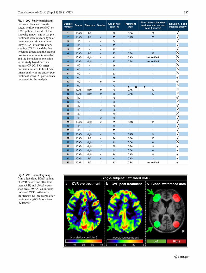
S87
123
Clin Neuroradiol (2019) (Suppl 1) 29:S1–S129
Fig. 1 | 290 Study participants overview. Presented are the status, healthy control (HC) or ICAS-patient; the side of the stenosis; gender; age at the pre treatment scan in years; type of treatment, carotid endarterec-tomy (CEA) or carotid artery stenting (CAS); the delay be-tween treatment and the second post treatment scan in months; and the inclusion or exclusion to the study based on visual ratings (CP, JG, SK). After exclusion, related to low CVR image quality in pre and/or post treatment scans, 20 participants remained for the analysis
Fig. 2 | 290 Exemplary maps from a left-sided ICAS-patient of CVR before and after treat-ment (A,B) and global water-shed area (gWSA, C). lnitially impaired CVR ipsilateral to the stenosis (A) recovered after treatment at gWSA-Iocations (8, arrows).

S88
123
Clin Neuroradiol (2019) (Suppl 1) 29:S1–S129
3Klinik für Kinder- und Jugendmedizin – Pädiatrische Hämatologie und Onkologie, Universitätsklinik Münster, Münster, Deutschland4Klinik und Poliklinik für Neuroradiologische Diagnostik und Intervention, Universitätsklinikum Hamburg-Eppendorf, Hamburg, Deutschland5Klinik für Neurologie mit Institut für Translationale Neurologie, Universitätsklinik Münster, Münster, Deutschland
Background: Arterial ischemic stroke (AIS) in childhood is a severe disease with potentially lifelong restrictions. Apart from cardiac or pro-thrombotic embolism, arteriopathy has been identified as a major cause and significant target of secondary stroke prevention. The Childhood Arterial Ischemic Stroke Standardized Classification and Diagnostic Evaluation (CASCADE) criteria facilitate a structured tool for cate-gorizing and reporting childhood strokes according to the underlying cause. Our study aims to identify the prognostic value of CASCADE classification on admission for further course of arteriopathy and risk of stroke recurrence.Methods: Between 2004 and 2017, we identified 86 children with an acute arterial ischemic stroke classified in the 7-basic and 19-expand-ed subgroups of the acute CASCADE criteria. All included children were diagnosed and followed-up by magnetic resonance imaging. All arteriopathic strokes were further categorized into the chronic CAS-CADE criteria, including progressive, stable, reversible, and indeter-minate course. Outcomes were defined as stroke recurrence and course of arteriopathy according to chronic CASCADE criteria. Associations were assessed by Fisher exact test and Mann-Whitney U test.Result: A total of 86 children were included; of these, 57 presented with arteriopathic stroke (CASCADE 1–4) and 29 as nonarteriopathic. Unilateral cerebral arteriopathy (CASCADE 2; P = 0.036) and bilateral cerebral arteriopathy (CASCADE 3; P = 0.016) significantly correlat-ed with stroke recurrence, and progressive arteriopathy significantly correlated with unilateral focal cerebral arteriopathy (P < 0.001). Time
points of progress of arteriopathy differed; whereas patients with uni-lateral focal cerebral arteriopathy presented with early median progress after 11 days, patients with bilateral cerebral arteriopathy had a signif-icantly later median progress after 124 days (P = 0.005).Discussion: Our results are limited by small patient numbers, as well as the retrospective and single-center design. However, for pediatric stroke populations, we present a comparably large consecutive cohort of pediatric stroke patients with a homogenous MRI-based imaging work up and with frequent and long follow-ups.Conclusion: Initial CASCADE classification is associated with risk of recurrent strokes and progress of arteriopathy. Moreover, time points of arteriopathic progress vary according to the underlying cause.
293
IDENTIFICATION OF ACUTE LARGE VESSEL STROKE PATIENTS WITH POINT-OF-CARE ULTRASOUND
Ivaylo Dimitrov1, Kolja Schürmann1, Jan Philipp Bach1, Arno Reich1, Martin Wiesmann2, Omid Nikoubashmann*2
1Neurologie, UKA, Aachen, Deutschland2Neuroradiologie, UKA, Aachen, Deutschland
Hintergrund: We investigated the feasibility of ultrasound for the de-tection of large-vessel-occlusion acute stroke.Methoden: Two neurologists (one experienced and one junior reader) retrospectively analyzed point-of-care ultrasound exams of the com-mon carotid arteries of 30 patients with and without large-vessel-oc-clusion.Ergebnisse: Ultrasound of the carotid arteries allows detecting large-vessel-occlusion with a sensitivity and positive predictive value of 73.3% and 95.7%, respectively. Inter-rater reliability was excellent (Cohen’s Kappa, .813; p <.001). Positive predictive value of ultrasound for the detection of LVO was higher than all clinical scores and sensi-tivity was higher than all clinical scores except for the NIHSS-cutoff at 7 points. Without clinical information, agreement between predicted and actual value was moderate/substantial for the experienced reader (Cohen’s Kappa, .568; p <.001) and the junior reader (Cohen’s Kappa, .644; p<.001). Agreement between the junior reader and the experi-enced reader was moderate (Cohen’s Kappa, .581; p <.001). With clin-ical information, agreement between predicted and actual value was moderate/substantial for the experienced reader (Cohen’s Kappa, .717; p <.001) and the junior reader (Cohen’s Kappa, .769; p<.001). Agree-ment between the junior reader and the experienced reader was excel-lent (Cohen’s Kappa, .813; p <.001).Diskussion/Discussion: Ultrasound of the carotid arteries allows de-tecting large-vessel-occlusion with a high sensitivity and positive pre-dictive value.Fazit/Conclusion: Point-of-care ultrasound in the acute stroke setting is feasible and may serve as a complementary tool for the detection of proximal large-vessel-occlusion that may be missed otherwise.
297
EMERGENCY EVERSION CAROTID ENDARTERECTOMY VERSUS CAROTID ARTERY STENTING IN ENDOVASCULAR ACUTE STROKE TREATMENT.
Raveena Singh1, Nora Schäferhenrich1, Hani Ridwan1, Anastasios Mpotsaris1, Arno Reich2, Johannes Kalder3, Ahmend E Othman4, Martin Wiesmann1, Omid Nikoubashmann*1
1Neuroradiologie, UKA, Aachen, Deutschland2Neurologie, UKA, Aachen, Deutschland3Gefäßchirurgie, UKA, Aachen, Deutschland4Radiologie, UK Tübingen, Tübingen, Deutschland
Fig. 3 | 290 Lateralisation of CVR within GM of gWSA between both hemispheres in both scans for ICAS-patients and healthy controls. Each data point depicts the CVR lateralisation of one participant, lines connect both sessions of the same participant. CVR was significantly lateralised before treatment (a, t-test, p = 0.0038). After treatment, the lateralisation significantly improved (a, 2-sample t-test, p = 0.0495) and shows no CVR difference between the hemispheres anymore (a, t-test, p = 0.25). Asterisks indicate statistically significant differences. Both HC scans were symmetrical (b)
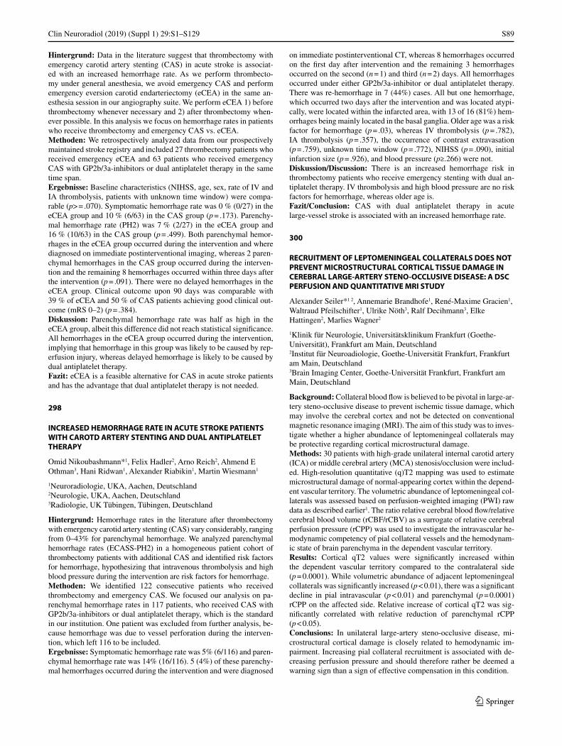
S89
123
Clin Neuroradiol (2019) (Suppl 1) 29:S1–S129
Hintergrund: Data in the literature suggest that thrombectomy with emergency carotid artery stenting (CAS) in acute stroke is associat-ed with an increased hemorrhage rate. As we perform thrombecto-my under general anesthesia, we avoid emergency CAS and perform emergency eversion carotid endarteriectomy (eCEA) in the same an-esthesia session in our angiography suite. We perform eCEA 1) before thrombectomy whenever necessary and 2) after thrombectomy when-ever possible. In this analysis we focus on hemorrhage rates in patients who receive thrombectomy and emergency CAS vs. eCEA.Methoden: We retrospectively analyzed data from our prospectively maintained stroke registry and included 27 thrombectomy patients who received emergency eCEA and 63 patients who received emergency CAS with GP2b/3a-inhibitors or dual antiplatelet therapy in the same time span.Ergebnisse: Baseline characteristics (NIHSS, age, sex, rate of IV and IA thrombolysis, patients with unknown time window) were compa-rable (p> = .070). Symptomatic hemorrhage rate was 0 % (0/27) in the eCEA group and 10 % (6/63) in the CAS group (p = .173). Parenchy-mal hemorrhage rate (PH2) was 7 % (2/27) in the eCEA group and 16 % (10/63) in the CAS group (p = .499). Both parenchymal hemor-rhages in the eCEA group occurred during the intervention and where diagnosed on immediate postinterventional imaging, whereas 2 paren-chymal hemorrhages in the CAS group occurred during the interven-tion and the remaining 8 hemorrhages occurred within three days after the intervention (p = .091). There were no delayed hemorrhages in the eCEA group. Clinical outcome upon 90 days was comparable with 39 % of eCEA and 50 % of CAS patients achieving good clinical out-come (mRS 0–2) (p = .384).Diskussion: Parenchymal hemorrhage rate was half as high in the eCEA group, albeit this difference did not reach statistical significance. All hemorrhages in the eCEA group occurred during the intervention, implying that hemorrhage in this group was likely to be caused by rep-erfusion injury, whereas delayed hemorrhage is likely to be caused by dual antiplatelet therapy.Fazit: eCEA is a feasible alternative for CAS in acute stroke patients and has the advantage that dual antiplatelet therapy is not needed.
298
INCREASED HEMORRHAGE RATE IN ACUTE STROKE PATIENTS WITH CAROTD ARTERY STENTING AND DUAL ANTIPLATELET THERAPY
Omid Nikoubashmann*1, Felix Hadler2, Arno Reich2, Ahmend E Othman3, Hani Ridwan1, Alexander Riabikin1, Martin Wiesmann1
1Neuroradiologie, UKA, Aachen, Deutschland2Neurologie, UKA, Aachen, Deutschland3Radiologie, UK Tübingen, Tübingen, Deutschland
Hintergrund: Hemorrhage rates in the literature after thrombectomy with emergency carotid artery stenting (CAS) vary considerably, ranging from 0–43% for parenchymal hemorrhage. We analyzed parenchymal hemorrhage rates (ECASS-PH2) in a homogeneous patient cohort of thrombectomy patients with additional CAS and identified risk factors for hemorrhage, hypothesizing that intravenous thrombolysis and high blood pressure during the intervention are risk factors for hemorrhage.Methoden: We identified 122 consecutive patients who received thrombectomy and emergency CAS. We focused our analysis on pa-renchymal hemorrhage rates in 117 patients, who received CAS with GP2b/3a-inhibitors or dual antiplatelet therapy, which is the standard in our institution. One patient was excluded from further analysis, be-cause hemorrhage was due to vessel perforation during the interven-tion, which left 116 to be included.Ergebnisse: Symptomatic hemorrhage rate was 5% (6/116) and paren-chymal hemorrhage rate was 14% (16/116). 5 (4%) of these parenchy-mal hemorrhages occurred during the intervention and were diagnosed
on immediate postinterventional CT, whereas 8 hemorrhages occurred on the first day after intervention and the remaining 3 hemorrhages occurred on the second (n = 1) and third (n = 2) days. All hemorrhages occurred under either GP2b/3a-inhibitor or dual antiplatelet therapy. There was re-hemorrhage in 7 (44%) cases. All but one hemorrhage, which occurred two days after the intervention and was located atypi-cally, were located within the infarcted area, with 13 of 16 (81%) hem-orrhages being mainly located in the basal ganglia. Older age was a risk factor for hemorrhage (p = .03), whereas IV thrombolysis (p = .782), IA thrombolysis (p = .357), the occurrence of contrast extravasation (p = .759), unknown time window (p = .772), NIHSS (p = .090), initial infarction size (p = .926), and blood pressure (p≥.266) were not.Diskussion/Discussion: There is an increased hemorrhage risk in thrombectomy patients who receive emergency stenting with dual an-tiplatelet therapy. IV thrombolysis and high blood pressure are no risk factors for hemorrhage, whereas older age is.Fazit/Conclusion: CAS with dual antiplatelet therapy in acute large-vessel stroke is associated with an increased hemorrhage rate.
300
RECRUITMENT OF LEPTOMENINGEAL COLLATERALS DOES NOT PREVENT MICROSTRUCTURAL CORTICAL TISSUE DAMAGE IN CEREBRAL LARGE-ARTERY STENO-OCCLUSIVE DISEASE: A DSC PERFUSION AND QUANTITATIVE MRI STUDY
Alexander Seiler*1 2, Annemarie Brandhofe1, René-Maxime Gracien1, Waltraud Pfeilschifter1, Ulrike Nöth3, Ralf Decihmann3, Elke Hattingen2, Marlies Wagner2
1Klinik für Neurologie, Universitätsklinikum Frankfurt (Goethe-Universität), Frankfurt am Main, Deutschland2Institut für Neuroadiologie, Goethe-Universität Frankfurt, Frankfurt am Main, Deutschland3Brain Imaging Center, Goethe-Universität Frankfurt, Frankfurt am Main, Deutschland
Background: Collateral blood flow is believed to be pivotal in large-ar-tery steno-occlusive disease to prevent ischemic tissue damage, which may involve the cerebral cortex and not be detected on conventional magnetic resonance imaging (MRI). The aim of this study was to inves-tigate whether a higher abundance of leptomeningeal collaterals may be protective regarding cortical microstructural damage.Methods: 30 patients with high-grade unilateral internal carotid artery (ICA) or middle cerebral artery (MCA) stenosis/occlusion were includ-ed. High-resolution quantitative (q)T2 mapping was used to estimate microstructural damage of normal-appearing cortex within the depend-ent vascular territory. The volumetric abundance of leptomeningeal col-laterals was assessed based on perfusion-weighted imaging (PWI) raw data as described earlier1. The ratio relative cerebral blood flow/relative cerebral blood volume (rCBF/rCBV) as a surrogate of relative cerebral perfusion pressure (rCPP) was used to investigate the intravascular he-modynamic competency of pial collateral vessels and the hemodynam-ic state of brain parenchyma in the dependent vascular territory.Results: Cortical qT2 values were significantly increased within the dependent vascular territory compared to the contralateral side (p = 0.0001). While volumetric abundance of adjacent leptomeningeal collaterals was significantly increased (p < 0.01), there was a significant decline in pial intravascular (p < 0.01) and parenchymal (p = 0.0001) rCPP on the affected side. Relative increase of cortical qT2 was sig-nificantly correlated with relative reduction of parenchymal rCPP (p < 0.05).Conclusions: In unilateral large-artery steno-occlusive disease, mi-crostructural cortical damage is closely related to hemodynamic im-pairment. Increasing pial collateral recruitment is associated with de-creasing perfusion pressure and should therefore rather be deemed a warning sign than a sign of effective compensation in this condition.

S90
123
Clin Neuroradiol (2019) (Suppl 1) 29:S1–S129
301
COMBINING HIGH-RESOLUTION VESSEL WALL IMAGING WITH IMAGE-BASED FLOW EVALUATION IN UNRUPTURED MIDDLE CEREBRAL ANEURYSMS
Philipp Berg*1, Naomi Larsen2, Charlotte Flüh3, Samuel Voss1, Sylvia Saalfeld1
1Forschungscampus STIMULATE, Otto-von-Guericke Universität Magdeburg, Magdeburg, Deutschland2Institut für Neurochirurgie, Universitätsklinikum Schleswig-Holstein Campus Kiel, Kiel, Deutschland 3Institut für Radiologie und Neuroradiologie, Universitätsklinikum Schleswig-Holstein Campus Kiel, Kiel, Deutschland
Hintergrund: The assessment of intracranial aneurysm rupture risk remains challenging due to a highly individual appearance in size and shape, as well as an unpredictable course of the disease. However, high-resolution magnetic resonance vessel wall imaging (MR-VWI) was recently used to associate wall enhancement with inflammatory cell invasion in intracranial aneurysms.Methoden: Within this study twenty-five unruptured saccular mid-dle cerebral artery (MCA) aneurysms who underwent pre-operative high-resolution MR-VWI showing wall enhancement, digital subtrac-tion angiography, microsurgical aneurysm clipping, and histologic analysis of the resected aneurysm wall were investigated using im-age-based blood flow simulations.Ergebnisse: The computational results reveal that vessel wall en-hancement in MCA aneurysms is associated with lower cycle-aver-aged wall shear stresses, decreased oscillatory shear, and increased low shear areas compared to the overall aneurysm surfaces. Furthermore, flow structures perpendicular to the main flow occur in the region of enhancement.Diskussion/Discussion: These findings highlight that a correlation be-tween local hemodynamics and measurable processes occurring within the aneurysmal vessel wall exists. Hence, patient-specific flow condi-tions can be assessed using image-based modeling to identify poten-tially endangered aneurysm wall regions.Fazit/Conclusion: Overall, MR-VWI may be beneficial in the evalua-tion of unruptured aneurysms regarding rupture risk assessment.
References 1. Matouk CC, et al. Vessel wall magnetic resonance imaging identi-
fies the site of rupture in patients with multiple intracranial aneu-rysms: Proof of principle. Neurosurgery. 2013;72(3):492–6.
2 . Larsen N, et al. Vessel wall enhancement in unruptured intracrani-al aneurysms: An indicator for higher risk of rupture? High-reso-lution MR imaging and correlated histologic findings. AJNR Am J Neuroradiol. 2018;39(9):1617–21.
304
DIRECT ASPIRATION THROMBECTOMY IN DISTAL VESSEL OCCLUSIONS EITHER WITH 5F SOFIA CATHETER OR WITH HEADWAY 27 MICROCATHETER
Dan Meila*1, Ingmar Born1, Ralf Rulands1, Gebhard Schmid1
1Johanna Etienne Krankenhaus, Neuss, Deutschland
Hintergrund: Recent studies suggest that the proven benefits of thrombectomy for the treatment of large vessel occlusions are trans-ferable to more peripheral distal vessel occlusions (DVO). Safely ac-cessing and extracting these thrombi however remains challenging, particularly in more tortuous peripheral arteries. For such cases we have utilized either 5F Sofia catheter or Headway 27 microcatheter thrombectomy, with the aim of reducing potential trauma associated with negotiating stent retrievers into the peripheral intracranial vascu-lature. We describe our technique as well as present angiographic and clinical outcomes.Methoden: Retrospective review of our institution’s prospectively col-lected thrombectomy registry for the 12 months spanning June 2018 to May 2019. Data on all cases of distal vessel occlusions were collected and analyzed.Ergebnisse: Direct aspiration thrombectomy in distal vessel occlu-sions was performed 13 times over the study period, with a recanal-ization rate of 92 %. DVO strokes in multiple locations were treated, including the A3, M3 and P2 segments. No complications relating to the technique were recorded. No perforation, no dissection, no bleed-ing and no vasospasm was notedFazit/Conclusion: The aspiration technique either with 5F Sofia or with Headway 27 microcatheter is an atraumatic technique that enlarg-es the range of TICI 2c/3 thrombectomies even in far distal occlusions. Due to certain limitations sole aspiration is not always feasible. Further development of material is crucial.
References 1. Grieb D, Schlunz-Hendann M, Brinjikji W, Melber K, Greling
B, Lanfermann H, et al. Mechanical thrombectomy of M2 occlu-sions with distal access catheters using ADAPT. J Neuroradiol. 2019;46(4):231–7. Jul.
2. Crockett MT, Phillips TJ, Chiu AHY. Dual suction Headway27 microcatheter thrombectomy for the treatment of distal intrac-ranial arterial occlusion strokes: initial experience with the mi-cro-ADAPT technique. J Neurointerv Surg. 2018;24.
Fig. 1 | 301 a) Illustration of the 3D DSA data with an MCA aneurysm (arrow). b) Corresponding MR-VWI dataset with yellow contours masking the enhanced regions. c) 3D model of the enhancement. d) Co-registered 3D DSA aneurysm surface mesh and 3D MR-VWI enhancement surface mesh

S91
123
Clin Neuroradiol (2019) (Suppl 1) 29:S1–S129
309
COMPRESSED-SENSING (CS) 3D TOF MRA IN A CLINICAL SETTING: A QUALITATIVE COMPARISON STUDY IN 45 PATIENTS.
Theo Demerath*1, Grzegorz Karwacki2, Tilman Schubert3
1Klinik für Neuroradiologie, Universitätsklinikum Freiburg, Freiburg, Deutschland2Klinik für Radiologie und Nuklearmedizin, Universitätsspital Basel, Basel, Schweiz3Klinik für Neuroradiologie, Universitätsspital Zürich, Zürich, Schweiz
Hintergrund: Compressed-sensing (CS) techniques can be used to improve spatial resolution in TOF MRA while keeping acquisition time constant. Our aim was to compare image quality in CS TOF MRA to „conventional“ (con) TOF MRA in a non-selected clinical popula-tion of 45 patients.Methoden: CS TOF MRA with a voxel size of 0.14 mm3
(0.6 × 0.6 × 0.4 mm) was compared to con TOF MRA with a voxel size of 0.49 mm3 (0.78 × 1.05 × 0.6 mm) in a 3T (Siemens Skyra/Skyra fit, 20 CH Head coil) MRI, giving a 70 % decrease in voxel size from con TOF to CS TOF MRA. General image quality and vessel delin-eation (A3, M3, M4, P2, P3, ACoA, PCom Segments and superficial temporal arteries (STA)) were blindly assessed by 2 board-certified neuroradiologists by using a 1–4 point (gen. image quality) and 1–5 (vessel delineation) point scale. Mean Scales were compared by using Mann-Whitney-U-Test; interrater agreement ĸ-statistics were moder-ate to low.
Ergebnisse: Mean general image quality (1.59 vs. 1.84 SE 0.093/0.079) and vessel delineation scales were rated higher in A3, M3, P2, P3, ACoA and STA Segments in CS TOF MRA compared to con TOF MRA.Diskussion: General image quality was rated slightly lower in CS TOF MRA, which might be attributable to intrinsic image noise. Ves-sel delineation in most intracranial and STA segments were rated higher in CS TOF MRA compared to con TOF MRA while keeping acquisition time constant, which is conformable with sparse availa-ble data [2].Fazit: Besides being used to reduce acquisition time, Compressed-sensing (CS) techniques can lead to improved spatial resolution while keeping acquisition time constant, which may improve peripheral ves-sel delineation.
References 1. Fushimi Y, et al. Clinical evaluation of time-of-flight MR angiog-
raphy with sparse undersampling and iterative reconstruction for cerebral aneurysms. Nmr Biomed. 2017;30(11).
2. Lin Z, et al. Clinical feasibility study of 3D intracranial magnetic resonance angiography using compressed sensing. J Magn Reson Imaging. 2019.
Fig. 1 | 309 Mean vessel delin-eation scores in intracranial and extracranial (STA) segments rated on a 1–5 point scale

S92
123
Clin Neuroradiol (2019) (Suppl 1) 29:S1–S129
313
INTRAKRANIELLES STENTING IN DER ZEIT NACH SAMMPRIS: “LETZTER RETTUNGSVERSUCH FÜR HOFFNUNGSLOSE FÄLLE” ODER “EFFEKTIVE BEHANDLUNGSOPTION”?
Dimah Hasan1 2, Annemarie Brandhofe3 3, Joachim Berkefeld1, Fee Keil*1
1Neuroradiologie, Universitätsklinikum JWG Universität Frankfurt, Frankfurt, Deutschland2Neuroradiologie, Uniklinikum Aachen, Aachen, Deutschland3Universitätsklinikum JWG Universität Frankfurt, Neurologie, Frankfurt, Deutschland
Hintergrund: Nach den für das Stenting intrakranieller Stenosen ne-gativen Ergebnissen der SAMMPRIS-Studie1 wurden die Indikations-stellung auf Patienten mit unter optimierter medikamentöser Thera-pie rezidivierenden Symptomen und Infarkten und auf Fälle mit einem akuten Schlaganfall und Gefäßobstruktionen auf dem Boden einer Ste-nose beschränkt. Ziel dieser Studie war es, bei den in einem einzelnen Zentrum verbliebenen Fällen Rekanalisationsergebnisse und klinische Behandlungsergebnisse zu analysieren.Methoden: Im Zeitraum zwischen 2011 und 2018 wurden im neurora-diologischen Institut des Frankfurter Universitätsklinikums insgesamt 45 Patienten mit intrakraniellen Stenosen mit Stents behandelt. In 28 Fällen wurde die Intervention zusätzlich zur Thrombektomie bei aku-ten Schlaganfällen mit atherothrombotischem Gefäßverschluss durch-geführt, die übrigen 17 Patienten kamen überwiegend mit subakuten, meist hämodynamisch bedingten rezidiverenden Symptomen und In-farkten. Die interventionelle Behandlung erfolgte mit ballonexpandie-renden oder selbstexpandierenden Stents.Ergebnisse: Ein technisch erfolgreiches Stenting mit einer mehr als 50% igen Lumenerweiterung gelang bei allen 45 Patienten. Beim Stenting plus Thrombektomie kam es in 2 von 28 Fällen zu einer raschen Reth-rombose. Eine erfolgreiche Rekanalisation TICI IIb/III konnte bei den übrigen Patienten erreicht werden. Die Rate an guten klinischen Ergeb-nissen, mRS 0–2 nach 3 Monaten lag bei den Thrombektomiepatienten bei 44%. Symptomatische Blutungen traten in zwei Fällen auf.Diskussion: Unter Berücksichtigung der Komplikationsrate kann ein großzügigerer Einsatz des intrakraniellen Stentings bei frustranen Thrombektomieversuchen gerechtfertigt sein. Bei Patienten mit rezi-divierender Symptomatik unter optimierter medikamentöser Therapie kann auch eine prologierte Indikationsstellung zum Stenting negative Auswirkungen auf das outcome haben.Fazit: In einem erfahrenen Zentrum ist das intrakranielle Stenting von Patienten mit atherothrombotischen Gefäßobstruktionen mit akzepta-blen Komplikationsraten durchführbar. Bei im Rahmen der Schlagan-fallbehandlungen durchgeführten Interventionen sind die Rekanalisa-tionsergebnisse und klinische Behandlungsergebnisse ähnlich wie bei Thrombektomien embolischer Gefäßverschlüsse.
Literatur 1. Chimowitz, et al. Stenting vs aggressive medical therapy for in-
tracranial arterial stenosis. N Eng J Med. 2011.
320
EARLY VERSUS LATE ESTABLISHMENT OF FULL DUAL ANTIPLATELET THERAPY AFTER EMERGENCY CAROTID ARTERY STENTING IN TANDEM OCCLUSIONS
Alena Haußmann*1, Ruben Mühl-Benninghaus1, Andreas Simgen1, Wolfgang Reith1, Umut Yilmaz1
1Universitätsklinikum des Saarlandes, Diagnostische und Interventionelle Neuroradiologie, Homburg, Deutschland
Hintergrund: Currently emergency carotid artery stenting is having a re-naissance since a series of randomized trials demonstrated the benefit of mechanical thrombectomy in acute large vessel occlusions of the anterior circulation and 30% of those are accompanied by an additional occlusion or high grade stenosis of the extracranial ICA that requires PTA and/or stenting in order to gain access to the intracranial clot. However emergen-cy stenting bears the risk of acute stent occlusions due to the emergency setting with an insufficient preparation with antiplatelet medication and on the other hand the risk of hemorrhagic complications due to the ap-plication of antiplatelet medication in combination with iv-thrombolysis.
The purpose of this study was to compare occlusion rates and hem-orrhagic complications when dual antiplatelet therapy is started within 24 hours after endovascular treatment or thereafter.Methoden: Patients with acute tandem occlusions of the anterior cir-culation who were endovascularly treated at our institution were iden-tified from our registry of neuroendovascular interventions.
Clinical, angiographic and neuroimaging data was analyzed. End-points included acute occlusions of the carotid stents and symptomatic ICH.Ergebnisse: 36 patients were included. Full dual antiplatelet therapy was established within 24 hours after intervention in 18 patients. Rates of acute stent occlusions did not differ between patients who received anti-platelet therapy within 24 h and those who did not (n = 2 in either group).Fazit: Late establishment of full dual antiplatelet therapy after emer-gency carotid artery stenting in tandem occlusions did not have a high-er risk of acute stent occlusion in this retrospective analysis.
References 1. Yilmaz U, Körner H, Mühl-Benninghaus R, et al. Acute Occlu-
sions of Dual-Layer Carotid Stents After Endovascular Emergency Treatment of Tandem Lesions. Stroke. 2017;4:.
322
SIMULTANEOUS TREATMENT OF ATHEROSCLEROTIC TANDEM OCCLUSIONS IN ACUTE ISCHEMIC STROKE USING RETRIEVER WIRE SUPPORTED CAROTID ARTERY REVASCULARIZATION (REWISED CARE).
Volker Maus*1, Daniel Behme2, Christoph Maurer3, Andrei Tropine4, Stephanie Tritt4, Ansgar Berlis3, Marios Psychogios2
1Bochum, Deutschland2Göttingen, Deutschland3Augsburg, Deutschland4Wiesbaden, Deutschland
Background: Endovascular treatment of cerebral large vessel occlu-sions and concomitant atherosclerotic lesions of the ipsilateral internal carotid artery (ICA) are matter of debate. We report a multi-center ex-perience using the “Retriever wire supported carotid artery revascular-ization” (ReWiSed CARe) technique allowing simultaneous therapy of both lesions.Methods: We retrospectively analyzed 23 patients with acute tandem occlusions (TO) undergoing ReWised CARe at 3 German centers. Clinical (including demographics and NIHSS) and procedural (includ-ing angiographic evaluation of recanalization) data were evaluated. Fa-vorable clinical outcome was defined as modified Rankin Scale (mRS) ≤2 at 90 days.Result: Median age was 70 years (IQR 65–80) and 70% were male. Median baseline NIHSS was 15 (IQR 11–17). Out of 23 patients, 22 (96%) had an intracranial occlusion of the anterior circulation. Suc-cessful stent retriever deployment with subsequent carotid artery treat-ment was feasible in all cases without dislocation of the stent retriever during the procedure. Overall, successful reperfusion (mTICI ≥2b) was achieved in 22/23 (96%) patients with 10/23 (44%) individuals com-
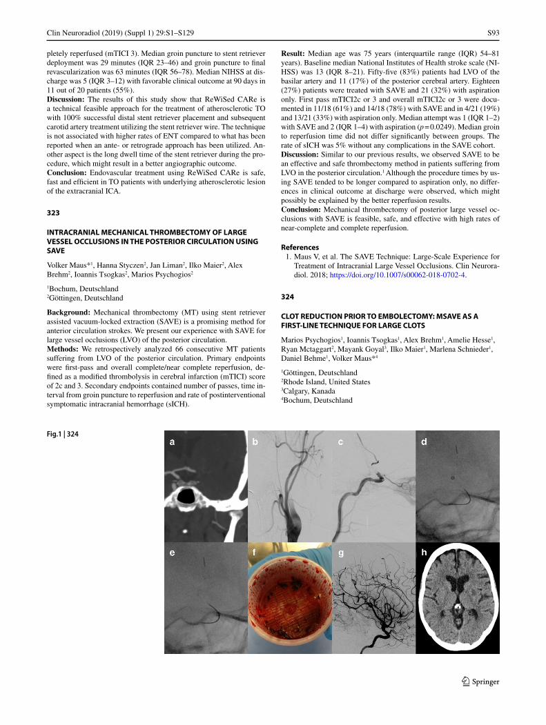
S93
123
Clin Neuroradiol (2019) (Suppl 1) 29:S1–S129
pletely reperfused (mTICI 3). Median groin puncture to stent retriever deployment was 29 minutes (IQR 23–46) and groin puncture to final revascularization was 63 minutes (IQR 56–78). Median NIHSS at dis-charge was 5 (IQR 3–12) with favorable clinical outcome at 90 days in 11 out of 20 patients (55%).Discussion: The results of this study show that ReWiSed CARe is a technical feasible approach for the treatment of atherosclerotic TO with 100% successful distal stent retriever placement and subsequent carotid artery treatment utilizing the stent retriever wire. The technique is not associated with higher rates of ENT compared to what has been reported when an ante- or retrograde approach has been utilized. An-other aspect is the long dwell time of the stent retriever during the pro-cedure, which might result in a better angiographic outcome.Conclusion: Endovascular treatment using ReWiSed CARe is safe, fast and efficient in TO patients with underlying atherosclerotic lesion of the extracranial ICA.
323
INTRACRANIAL MECHANICAL THROMBECTOMY OF LARGE VESSEL OCCLUSIONS IN THE POSTERIOR CIRCULATION USING SAVE
Volker Maus*1, Hanna Styczen2, Jan Liman2, Ilko Maier2, Alex Brehm2, Ioannis Tsogkas2, Marios Psychogios2
1Bochum, Deutschland2Göttingen, Deutschland
Background: Mechanical thrombectomy (MT) using stent retriever assisted vacuum-locked extraction (SAVE) is a promising method for anterior circulation strokes. We present our experience with SAVE for large vessel occlusions (LVO) of the posterior circulation.Methods: We retrospectively analyzed 66 consecutive MT patients suffering from LVO of the posterior circulation. Primary endpoints were first-pass and overall complete/near complete reperfusion, de-fined as a modified thrombolysis in cerebral infarction (mTICI) score of 2c and 3. Secondary endpoints contained number of passes, time in-terval from groin puncture to reperfusion and rate of postinterventional symptomatic intracranial hemorrhage (sICH).
Result: Median age was 75 years (interquartile range (IQR) 54–81 years). Baseline median National Institutes of Health stroke scale (NI-HSS) was 13 (IQR 8–21). Fifty-five (83%) patients had LVO of the basilar artery and 11 (17%) of the posterior cerebral artery. Eighteen (27%) patients were treated with SAVE and 21 (32%) with aspiration only. First pass mTICI2c or 3 and overall mTICI2c or 3 were docu-mented in 11/18 (61%) and 14/18 (78%) with SAVE and in 4/21 (19%) and 13/21 (33%) with aspiration only. Median attempt was 1 (IQR 1–2) with SAVE and 2 (IQR 1–4) with aspiration (p = 0.0249). Median groin to reperfusion time did not differ significantly between groups. The rate of sICH was 5% without any complications in the SAVE cohort.Discussion: Similar to our previous results, we observed SAVE to be an effective and safe thrombectomy method in patients suffering from LVO in the posterior circulation.1 Although the procedure times by us-ing SAVE tended to be longer compared to aspiration only, no differ-ences in clinical outcome at discharge were observed, which might possibly be explained by the better reperfusion results.Conclusion: Mechanical thrombectomy of posterior large vessel oc-clusions with SAVE is feasible, safe, and effective with high rates of near-complete and complete reperfusion.
References 1. Maus V, et al. The SAVE Technique: Large-Scale Experience for
Treatment of Intracranial Large Vessel Occlusions. Clin Neurora-diol. 2018; https://doi.org/10.1007/s00062-018-0702-4.
324
CLOT REDUCTION PRIOR TO EMBOLECTOMY: MSAVE AS A FIRST-LINE TECHNIQUE FOR LARGE CLOTS
Marios Psychogios1, Ioannis Tsogkas1, Alex Brehm1, Amelie Hesse1, Ryan Mctaggart2, Mayank Goyal3, Ilko Maier1, Marlena Schnieder1, Daniel Behme1, Volker Maus*4
1Göttingen, Deutschland2Rhode Island, United States3Calgary, Kanada4Bochum, Deutschland
Fig.1 | 324
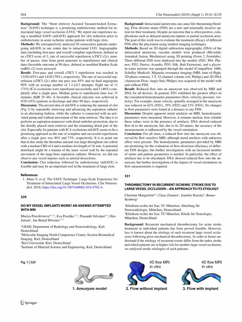
S94
123
Clin Neuroradiol (2019) (Suppl 1) 29:S1–S129
Background: The “Stent retriever Assisted Vacuum-locked Extrac-tion” (SAVE) technique is a promising embolectomy method for in-tracranial large vessel occlusion (LVO).1 We report our experience us-ing a modified SAVE (mSAVE) approach for clot reduction prior to embolectomy in acute ischemic stroke patients with large clots.Methods: We retrospectively analyzed 20 consecutive patients under-going mSAVE in our center due to intracranial LVO. Angiographic data (including first-pass and overall complete reperfusion, defined as an eTICI score of 3, rate of successful reperfusion (eTICI ≥2c), num-ber of passes, time from groin puncture to reperfusion) and clinical data (favorable outcome at 90 days, defined as modified Rankin Scale (mRS) ≤2) were assessed.Result: First-pass and overall eTICI 3 reperfusion was reached in 13/20 (65%) and 14/20 (70%), respectively. The rate of successful rep-erfusion (eTICI ≥2c) after one pass was 85% and on final angiogram 90% with an average number of 1.1 ± 0.3 attempts. Eight out of 11 (73%) ICA occlusions were reperfused successfully and 5 (46%) com-pletely after a single pass. Median groin to reperfusion time was 33 minutes (IQR 25–46). A favorable clinical outcome was achieved in 9/20 (45%) patients at discharge and after 90 days, respectivelyDiscussion: The pivotal idea of mSAVE is reducing the amount of clot (Fig 1) by repeatedly retrieving and advancing the aspiration catheter in an alternating fashion, while the catheter is connected with an acti-vated pump and without movement of the stent retriever. The idea is to perform an aspiration maneuver with distal embolus protection, due to the distally placed stent retriever and the remaining distal part of the clot. Especially for patients with ICA occlusions mSAVE seems to be a promising approach as the rate of complete and successful reperfusion after a single pass was 46% and 73%, respectively. It is to point out that in this study the thrombus amount was large throughout our cohort with a median CBS of 4 and a median clot length of 18 mm. A potential drawback might be a trauma of the inner vessel wall by the repeated movement of the large-bore aspiration catheter. However, we did not observe any vessel injuries such as arterial dissections.Conclusion: Clot reduction followed by embolectomy (mSAVE) is feasible and may be an important tool in the treatment of large clots.
References 1. Maus V, et al. The SAVE Technique: Large-Scale Experience for
Treatment of Intracranial Large Vessel Occlusions. Clin Neurora-diol. 2018; https://doi.org/10.1007/s00062-018-0702-4.
329
DO MY VESSEL IMPLANTS WORK? AN ANSWER ATTEMPTED WITH MRI
Mariya Pravdivtseva*1 2 3, Eva Peschke1 2 3, Prasanth Velvaluri3 4, Olav Jansen1, Jan-Bernd Hövener1 2 3
1UKSH, Department of Radiology and Neuroradiology, Kiel, Deutschland2Molecular Imaging North Competence Center, Section Biomedical Imaging, Kiel, Deutschland3Kiel Universität, Kiel, Deutschland4Institute of Material Science and Engineering, Kiel, Deutschland
Background: Intracranial aneurysms can cause life-threatening bleed-ing. Flow diverter stents (FDS) are a new and minimally invasive op-tion for their treatment. Despite an outcome that is often positive, com-plications such as delayed aneurysm rupture or partial occlusion arise. The goal of this work was to evaluate the treatment efficacy of different FDS after the placement using modern imaging techniques.Methods: Based on 3D digital subtraction angiography (DSA) of the intracranial aneurysm, vascular models were produced (Mevislab, Autodesk fusion, Meshmixer) using 3D printing (Form 2, Formlabs). Three different FDS were deployed into the models (FD1: P64, Phe-nox; FD2: Derivo, Acandis; FD3: Silk, Balt Extrusion), and a glycer-in-water mixture was pumped through the model (CompuFlow 1000, Schelley Medical). Magnetic resonance imaging (MRI, time-of-flight, 3D phase-contrast, 3 T, 32-channel volume coil, Philips) and 2D DSA (Aneurysm Flow; Angio Suit, Philips) was acquired of the model with and without FDS.Result: Reduced flow into an aneurysm was observed by MRI and DSA for all devices. In general, FD1 exhibited the greatest effect on the considered hemodynamic parameters (flow, velocity, pressure, vor-ticity). For example, mean velocity spatially averaged in the aneurysm was reduced on 62% (FD1), 39% (FD2) and 23% (FD3). No changes in flow parameters were found at a distance to any FDS.Discussion: Despite apparent metal artefacts on MRI, hemodynamic parameters were measured. However, it remains unclear, how reliable these values were in the presence of artefacts. DSA showed reduced flow in to the aneurysm, but, due to its 2D nature, the accuracy of the measurements is influenced by the vessel orientation.Conclusion: For all stens, a reduced flow into the aneurysm was ob-served by flow-sensitive MRI, although metal artefacts with unknown effect were present. The hemodynamic parameters provided by MRI are promising for the evaluation of flow-diversion efficiency of differ-ent FDS designs, but further investigation with an increased number of stents and patient geometries is needed. In particular, the effect of artefacts has to be elucidated. DSA showed reduced flow into the an-eurysm, but further investigation of the impact of vessel orientation on flow measurements is required.
331
THROMBECTOMY IN RECURRENT ISCHEMIC STROKE DUE TO LARGE VESSEL OCCLUSION—AN APPROACH TO ITS ETIOLOGY
Christian Maegerlein*1, Claus Zimmer1, Jennifer Roesler2, Benno Ikenberg2
1Klinikum rechts der Isar, TU München, Abteilung für Neuroradiologie, München, Deutschland2Klinikum rechts der Isar, TU München, Klinik für Neurologie, München, Deutschland
Background: Recurrent mechanical thrombectomy for acute stroke treatment in individual patients has been proved feasible. However, less is known about the etiology of such recurrent large vessel occlu-sions following prior mechanical thrombectomy. In order to better un-derstand if the etiology of recurrent events differ from the index stroke and which patients are at higher risk for another large vessel occlusion, we analyzed stroke etiologies of such patients.
Fig. 1 | 329
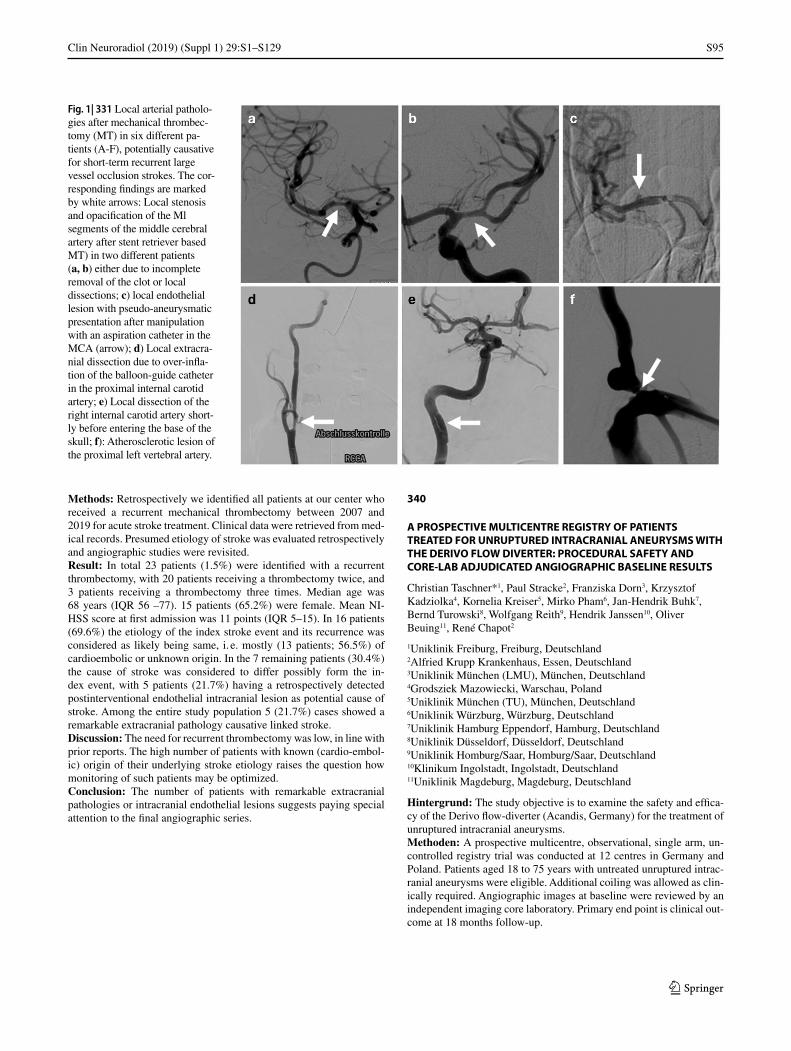
S95
123
Clin Neuroradiol (2019) (Suppl 1) 29:S1–S129
Methods: Retrospectively we identified all patients at our center who received a recurrent mechanical thrombectomy between 2007 and 2019 for acute stroke treatment. Clinical data were retrieved from med-ical records. Presumed etiology of stroke was evaluated retrospectively and angiographic studies were revisited.Result: In total 23 patients (1.5%) were identified with a recurrent thrombectomy, with 20 patients receiving a thrombectomy twice, and 3 patients receiving a thrombectomy three times. Median age was 68 years (IQR 56 –77). 15 patients (65.2%) were female. Mean NI-HSS score at first admission was 11 points (IQR 5–15). In 16 patients (69.6%) the etiology of the index stroke event and its recurrence was considered as likely being same, i. e. mostly (13 patients; 56.5%) of cardioembolic or unknown origin. In the 7 remaining patients (30.4%) the cause of stroke was considered to differ possibly form the in-dex event, with 5 patients (21.7%) having a retrospectively detected postinterventional endothelial intracranial lesion as potential cause of stroke. Among the entire study population 5 (21.7%) cases showed a remarkable extracranial pathology causative linked stroke.Discussion: The need for recurrent thrombectomy was low, in line with prior reports. The high number of patients with known (cardio-embol-ic) origin of their underlying stroke etiology raises the question how monitoring of such patients may be optimized.Conclusion: The number of patients with remarkable extracranial pathologies or intracranial endothelial lesions suggests paying special attention to the final angiographic series.
340
A PROSPECTIVE MULTICENTRE REGISTRY OF PATIENTS TREATED FOR UNRUPTURED INTRACRANIAL ANEURYSMS WITH THE DERIVO FLOW DIVERTER: PROCEDURAL SAFETY AND CORE-LAB ADJUDICATED ANGIOGRAPHIC BASELINE RESULTS
Christian Taschner*1, Paul Stracke2, Franziska Dorn3, Krzysztof Kadziolka4, Kornelia Kreiser5, Mirko Pham6, Jan-Hendrik Buhk7, Bernd Turowski8, Wolfgang Reith9, Hendrik Janssen10, Oliver Beuing11, René Chapot2
1Uniklinik Freiburg, Freiburg, Deutschland2Alfried Krupp Krankenhaus, Essen, Deutschland3Uniklinik München (LMU), München, Deutschland4Grodsziek Mazowiecki, Warschau, Poland5Uniklinik München (TU), München, Deutschland6Uniklinik Würzburg, Würzburg, Deutschland7Uniklinik Hamburg Eppendorf, Hamburg, Deutschland8Uniklinik Düsseldorf, Düsseldorf, Deutschland9Uniklinik Homburg/Saar, Homburg/Saar, Deutschland10Klinikum Ingolstadt, Ingolstadt, Deutschland11Uniklinik Magdeburg, Magdeburg, Deutschland
Hintergrund: The study objective is to examine the safety and effica-cy of the Derivo flow-diverter (Acandis, Germany) for the treatment of unruptured intracranial aneurysms.Methoden: A prospective multicentre, observational, single arm, un-controlled registry trial was conducted at 12 centres in Germany and Poland. Patients aged 18 to 75 years with untreated unruptured intrac-ranial aneurysms were eligible. Additional coiling was allowed as clin-ically required. Angiographic images at baseline were reviewed by an independent imaging core laboratory. Primary end point is clinical out-come at 18 months follow-up.
Fig. 1| 331 Local arterial patholo-gies after mechanical thrombec-tomy (MT) in six different pa-tients (A-F), potentially causative for short-term recurrent large vessel occlusion strokes. The cor-responding findings are marked by white arrows: Local stenosis and opacification of the Ml segments of the middle cerebral artery after stent retriever based MT) in two different patients (a, b) either due to incomplete removal of the clot or local dissections; c) local endothelial lesion with pseudo-aneurysmatic presentation after manipulation with an aspiration catheter in the MCA (arrow); d) Local extracra-nial dissection due to over-infla-tion of the balloon-guide catheter in the proximal internal carotid artery; e) Local dissection of the right internal carotid artery short-ly before entering the base of the skull; f): Atherosclerotic lesion of the proximal left vertebral artery.
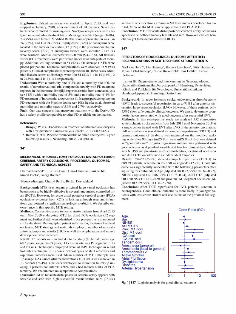
S96
123
Clin Neuroradiol (2019) (Suppl 1) 29:S1–S129
Ergebnisse: Patient inclusion was started in April, 2013, and was stopped in January, 2018, after enrolment of104 patients. Seven pa-tients were excluded for missing data. Ninety-seven patients were ana-lysed on an intention-to-treat basis. Mean age was 54.1 (range 18–86), 73 (75%) were female. Modified Rankin score at presentation was 0 in 74 (75%), and 1 in 24 (25%). Eighty-three (86%) of aneurysms were located in the anterior circulation, 12 (12%) in the posterior circulation. Seventy-seven (79%) of aneurysms treated were saccular, 21 (21%) were fusiform. Median diameter was 9.0 mm (5.4–13.5). All flow-di-verter (FD) treatments were performed under dual anti-platelet thera-py. Additional coiling occurred in 51 (52%). On average 1.2 FD were placed per patient. Technical complications were observed in 9 (9%) patients. Clinical complications were reported in 10 (10%) cases. Mod-ified Rankin scores at discharge were 0 in 81 (83%), 1 in 14 (14%), 2 in 2 (2%), and 4 in 1 (1%), respectively.Diskussion: With a morbidity rate of 3% and a mortality rate of 0, the results of our observational trial compare favourably with FD treatment reported in the literature. Brinjikji reported results from a metaanalysis (n = 1451) with a morbidity rate of 5% and a mortality rate of 4% for FD treatment of intracranial aneurysms (1). In a prospective registry on FD treatment with the Pipeline device (n = 108) Becske et al. observed morbidity and mortality rates of 5.6% and 2.7% respectively.Fazit: Our data suggests that aneurysm treatment with the Derivo FD has a safety profile comparable to other FD available on the market.
References 1. Brinjikji W, et al. Endovascular treatment of intracranial aneurysms
with flow diverters: a meta-analysis. Stroke. 2013;44(2:442–7. 2. Becske T, et al. Pipeline for uncoilable or failed aneurysms: 3-year
follow-up results. J Neurosurg. 2017;127(1):81–8.
341
MECHANICAL THROMBECTOMY FOR ACUTE DISTAL POSTERIOR CEREBRAL ARTERY OCCLUSIONS -PROCEDURAL OUTCOMES, SAFETY AND TECHNICAL ASPECTS
Eberhard Siebert*1, Justus Kleine1, Hans-Christian Bauknecht1, Simon Fuchs1, Georg Bohner1
1Neuroradiologie, Charité Berlin, Berlin, Deutschland
Background: MTE in emergent proximal large vessel occlusion has been shown to be highly effective in several randomized controlled tri-als (RCTs). However, for acute distal posterior cerebral artery (PCA) occlusions evidence from RCTs is lacking although resultant infarc-tions can portend a significant neurologic morbidity. We describe our experience in this specific MTE setting.Methods: Consecutive acute ischemic stroke patients from April 2013 until May 2019 undergoing MTE for distal PCA occlusion (P2 seg-ment and further distal) were identified in our prospectively maintained stroke database. Demographic patient information, location of arterial occlusion, MTE strategy and materials employed, number of recanali-zation attempts and results (TICI) as well as complications and infarct development were recorded.Result: 17 patients were included into the study (10 female, mean age 68,3 years, range 36–89 years). Occlusion site was P2 segment in 13 and P3 in 4. Techniques employed were ADAPT technique in 4 and Solumbra technique in 13 cases. Several types of stent retrievers and aspiration catheters were used. Mean number of MTE attempts was 1,8 (range 1–5). Successful recanalization (TICI 2b/3) was achieved in 13 patients (76,4%). 4 patients developed no infarct on follow-up im-aging, 7 patients had infarcts < 50% and 7 had infarcts > 50% of PCA territory. We encountered no symptomatic complications.Discussion: MTE for acute distal posterior cerebral artery appears both feasible and safe with high successful recanalization rates (76,4%)
similar to other locations. Common MTE techniques developed for ca-rotid, MCA or BA MTE can be applied to distal PCA MTE.Conclusion: MTE for acute distal posterior cerebral artery occlusions appears to be both technically feasible and safe. However, clinical ben-efit has yet to be demonstrated in RCTs.
347
PREDICTORS OF GOOD CLINICAL OUTCOME AFTER TICI3 RECANALIZATIONS IN ACUTE ISCHEMIC STROKE PATIENTS
Noel van Horn*1, Uta Hanning1, Hannes Leischner1, Götz Thomalla2, Milani Deb-Chatterji2, Caspar Brekenfeld1, Jens Fiehler1, Fabian Flottmann1
1Institut für Diagnostische und Interventionelle Neuroradiologie, Universitätsklinikum Hamburg-Eppendorf, Hamburg, Deutschland2Klinik und Poliklinik für Neurologie, Universitätsklinikum Hamburg-Eppendorf, Hamburg, Deutschland
Background: In acute ischemic stroke, endovascular thrombectomy (EVT) leads to successful reperfusion in up to 71%1 after anterior cir-culation large vessel occlusion (LVO). However, of these patients, only 55%2 show a favourable clinical outcome. We aim to determine prog-nostic factors associated with good outcome after successful EVT.Methods: In this retrospective study we analyzed 452 consecutive acute ischemic stroke patients from July 2015 until November 2018 at a single center treated with EVT after LVO of the anterior circulation. Full recanalization was defined as complete reperfusion (TICI 3) and primary outcome of disability was measured on the modified rank-ing scale after 90 days (mRS 90), were mRS 90 of 0–2 was defined as “good outcome”. Logistic regression analysis was performed with good outcome as dependant variable and baseline clinical data, admis-sion NIHSS and pre-stroke mRS, comorbidities, location of occlusion and ASPECTS on admission as independent variables.Result: 159/452 (35.2%) showed complete reperfusion (TICI 3). In 68/159 patients, outcome on mRS 90 was “good” (42.7%). Good out-come was significantly associated with the following parameters after adjusting for confounders: Age (adjusted OR 0.92, 95% CI 0.87–0.97), NIHSS (adjusted OR 0.86, 95% CI 0.78–0.94), ASPECTS (adjusted OR 1.50, 95% CI 1.12–2.09) and proximal M1-segment occlusion (ad-justed OR 5.29, 95% CI 1.34–23.56).Conclusion: After TICI3 reperfusion for LVO, patients’ outcome is heterogeneous. Good clinical outcome is more likely in younger pa-tients with less severe strokes and occlusions of the proximal M1 seg-ment.
Fig. 1 | 347 Logistic analysis for good clinical outcome
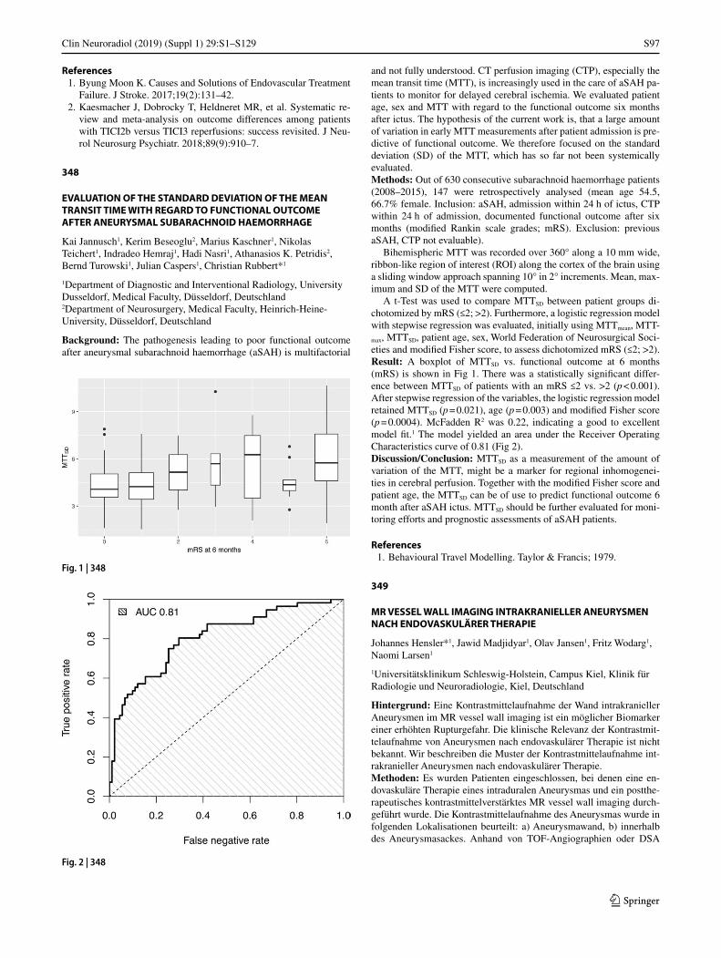
S97
123
Clin Neuroradiol (2019) (Suppl 1) 29:S1–S129
References 1. Byung Moon K. Causes and Solutions of Endovascular Treatment
Failure. J Stroke. 2017;19(2):131–42. 2. Kaesmacher J, Dobrocky T, Heldneret MR, et al. Systematic re-
view and meta-analysis on outcome differences among patients with TICI2b versus TICI3 reperfusions: success revisited. J Neu-rol Neurosurg Psychiatr. 2018;89(9):910–7.
348
EVALUATION OF THE STANDARD DEVIATION OF THE MEAN TRANSIT TIME WITH REGARD TO FUNCTIONAL OUTCOME AFTER ANEURYSMAL SUBARACHNOID HAEMORRHAGE
Kai Jannusch1, Kerim Beseoglu2, Marius Kaschner1, Nikolas Teichert1, Indradeo Hemraj1, Hadi Nasri1, Athanasios K. Petridis2, Bernd Turowski1, Julian Caspers1, Christian Rubbert*1
1Department of Diagnostic and Interventional Radiology, University Dusseldorf, Medical Faculty, Düsseldorf, Deutschland2Department of Neurosurgery, Medical Faculty, Heinrich-Heine-University, Düsseldorf, Deutschland
Background: The pathogenesis leading to poor functional outcome after aneurysmal subarachnoid haemorrhage (aSAH) is multifactorial
and not fully understood. CT perfusion imaging (CTP), especially the mean transit time (MTT), is increasingly used in the care of aSAH pa-tients to monitor for delayed cerebral ischemia. We evaluated patient age, sex and MTT with regard to the functional outcome six months after ictus. The hypothesis of the current work is, that a large amount of variation in early MTT measurements after patient admission is pre-dictive of functional outcome. We therefore focused on the standard deviation (SD) of the MTT, which has so far not been systemically evaluated.Methods: Out of 630 consecutive subarachnoid haemorrhage patients (2008–2015), 147 were retrospectively analysed (mean age 54.5, 66.7% female. Inclusion: aSAH, admission within 24 h of ictus, CTP within 24 h of admission, documented functional outcome after six months (modified Rankin scale grades; mRS). Exclusion: previous aSAH, CTP not evaluable).
Bihemispheric MTT was recorded over 360° along a 10 mm wide, ribbon-like region of interest (ROI) along the cortex of the brain using a sliding window approach spanning 10° in 2° increments. Mean, max-imum and SD of the MTT were computed.
A t-Test was used to compare MTTSD between patient groups di-chotomized by mRS (≤2; >2). Furthermore, a logistic regression model with stepwise regression was evaluated, initially using MTTmean, MTT-max, MTTSD, patient age, sex, World Federation of Neurosurgical Soci-eties and modified Fisher score, to assess dichotomized mRS (≤2; >2).Result: A boxplot of MTTSD vs. functional outcome at 6 months (mRS) is shown in Fig 1. There was a statistically significant differ-ence between MTTSD of patients with an mRS ≤2 vs. >2 (p < 0.001). After stepwise regression of the variables, the logistic regression model retained MTTSD (p = 0.021), age (p = 0.003) and modified Fisher score (p = 0.0004). McFadden R2 was 0.22, indicating a good to excellent model fit.1 The model yielded an area under the Receiver Operating Characteristics curve of 0.81 (Fig 2).Discussion/Conclusion: MTTSD as a measurement of the amount of variation of the MTT, might be a marker for regional inhomogenei-ties in cerebral perfusion. Together with the modified Fisher score and patient age, the MTTSD can be of use to predict functional outcome 6 month after aSAH ictus. MTTSD should be further evaluated for moni-toring efforts and prognostic assessments of aSAH patients.
References 1. Behavioural Travel Modelling. Taylor & Francis; 1979.
349
MR VESSEL WALL IMAGING INTRAKRANIELLER ANEURYSMEN NACH ENDOVASKULÄRER THERAPIE
Johannes Hensler*1, Jawid Madjidyar1, Olav Jansen1, Fritz Wodarg1, Naomi Larsen1
1Universitätsklinikum Schleswig-Holstein, Campus Kiel, Klinik für Radiologie und Neuroradiologie, Kiel, Deutschland
Hintergrund: Eine Kontrastmittelaufnahme der Wand intrakranieller Aneurysmen im MR vessel wall imaging ist ein möglicher Biomarker einer erhöhten Rupturgefahr. Die klinische Relevanz der Kontrastmit-telaufnahme von Aneurysmen nach endovaskulärer Therapie ist nicht bekannt. Wir beschreiben die Muster der Kontrastmittelaufnahme int-rakranieller Aneurysmen nach endovaskulärer Therapie.Methoden: Es wurden Patienten eingeschlossen, bei denen eine en-dovaskuläre Therapie eines intraduralen Aneurysmas und ein postthe-rapeutisches kontrastmittelverstärktes MR vessel wall imaging durch-geführt wurde. Die Kontrastmittelaufnahme des Aneurysmas wurde in folgenden Lokalisationen beurteilt: a) Aneurysmawand, b) innerhalb des Aneurysmasackes. Anhand von TOF-Angiographien oder DSA
Fig. 1 | 348
Fig. 2 | 348
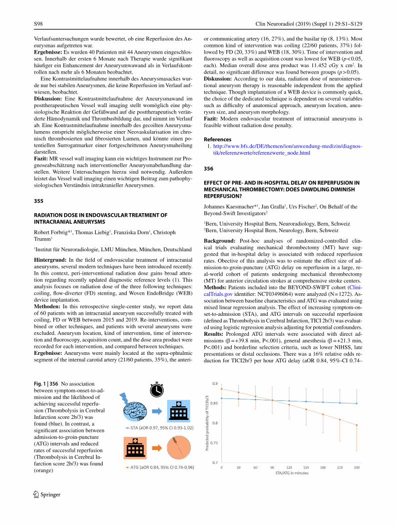
S98
123
Clin Neuroradiol (2019) (Suppl 1) 29:S1–S129
Verlaufsuntersuchungen wurde bewertet, ob eine Reperfusion des An-eurysmas aufgetreten war.Ergebnisse: Es wurden 40 Patienten mit 44 Aneurysmen eingeschlos-sen. Innerhalb der ersten 6 Monate nach Therapie wurde signifikant häufiger ein Enhancement der Aneurysmwawand als in Verlaufskont-rollen nach mehr als 6 Monaten beobachtet.
Eine Kontrastmittelaufnahme innerhalb des Aneurysmasackes wur-de nur bei stabilen Aneurysmen, die keine Reperfusion im Verlauf auf-wiesen, beobachtet.Diskussion: Eine Kontrastmittelaufnahme der Aneurysmawand im posttherapeutischen Vessel wall imaging stellt womöglich eine phy-siologische Reaktion der Gefäßwand auf die posttherapeutisch verän-derte Hämodynamik und Thrombusbildung dar, und nimmt im Verlauf ab. Eine Kontrastmittelaufnahme innerhalb des gecoilten Aneurysma-lumens entspricht möglicherweise einer Neovaskularisation im chro-nisch thrombosierten und fibrosierten Lumen, und könnte einen po-tentiellen Surrogatmarker einer fortgeschrittenen Aneurysmaheilung darstellen.Fazit: MR vessel wall imaging kann ein wichtiges Instrument zur Pro-gnoseabschätzung nach interventioneller Aneurysmabehandlung dar-stellen. Weitere Untersuchungen hierzu sind notwendig. Außerdem leistet das Vessel wall imaging einen wichtigen Beitrag zum pathophy-siologischen Verständnis intrakranieller Aneurysmen.
355
RADIATION DOSE IN ENDOVASCULAR TREATMENT OF INTRACRANIAL ANEURYSMS
Robert Forbrig*1, Thomas Liebig1, Franziska Dorn1, Christoph Trumm1
1Institut für Neuroradiologie, LMU München, München, Deutschland
Hintergrund: In the field of endovascular treatment of intracranial aneurysms, several modern techniques have been introduced recently. In this context, peri-interventional radiation dose gains broad atten-tion regarding recently updated diagnostic reference levels (1). This analysis focuses on radiation dose of the three following techniques: coiling, flow-diverter (FD) stenting, and Woven EndoBridge (WEB) device implantation.Methoden: In this retrospective single-center study, we report data of 60 patients with an intracranial aneurysm successfully treated with coiling, FD or WEB between 2015 and 2019. Re-interventions, com-bined or other techniques, and patients with several aneurysms were excluded. Aneurysm location, kind of intervention, time of interven-tion and fluoroscopy, acquisition count, and the dose area product were recorded for each intervention, and compared between techniques.Ergebnisse: Aneurysms were mainly located at the supra-ophtalmic segment of the internal carotid artery (21/60 patients, 35%), the anteri-
or communicating artery (16, 27%), and the basilar tip (8, 13%). Most common kind of intervention was coiling (22/60 patients, 37%) fol-lowed by FD (20, 33%) and WEB (18, 30%). Time of intervention and fluoroscopy as well as acquisition count was lowest for WEB (p < 0.05, each). Median overall dose area product was 11.452 cGy x cm2. In detail, no significant difference was found between groups (p > 0.05).Diskussion: According to our data, radiation dose of neurointerven-tional aneurysm therapy is reasonable independent from the applied technique. Though implantation of a WEB device is commonly quick, the choice of the dedicated technique is dependent on several variables such as difficulty of anatomical approach, aneurysm location, aneu-rysm size, and aneurysm morphology.Fazit: Modern endovascular treatment of intracranial aneurysms is feasible without radiation dose penalty.
References 1. http://www.bfs.de/DE/themen/ion/anwendung-medizin/diagnos-
tik/referenzwerte/referenzwerte_node.html
356
EFFECT OF PRE- AND IN-HOSPITAL DELAY ON REPERFUSION IN MECHANICAL THROMBECTOMY: DOES DAWDLING DIMINISH REPERFUSION?
Johannes Kaesmacher*1, Jan Gralla1, Urs Fischer2, On Behalf of the Beyond-Swift Investigators2
1Bern, University Hospital Bern, Neuroradiology, Bern, Schweiz2Bern, University Hospital Bern, Neurology, Bern, Schweiz
Background: Post-hoc analyses of randomized-controlled clin-ical trials evaluating mechanical thrombectomy (MT) have sug-gested that in-hospital delay is associated with reduced reperfusion rates. Obective of this analysis was to estimate the effect size of ad-mission-to-groin-puncture (ATG) delay on reperfusion in a large, re-al-world cohort of patients undergoing mechanical thrombectomy (MT) for anterior circulation strokes at comprehensive stroke centers.Methods: Patients included into the BEYOND-SWIFT cohort (Clini-calTrials.gov identifier: NCT03496064) were analyzed (N = 1272). As-sociation between baseline characteristics and ATG was evaluated using mixed linear regression analysis. The effect of increasing symptom-on-set-to-admission (STA), and ATG intervals on successful reperfusion (defined as Thrombolysis in Cerebral Infarction, TICI 2b/3) was evaluat-ed using logistic regression analysis adjusting for potential confounders.Results: Prolonged ATG intervals were associated with direct ad-missions (β = +39.8 min, P<.001), general anesthesia (β = +21.3 min, P<.001) and borderline selection criteria, such as lower NIHSS, late presentations or distal occlusions. There was a 16% relative odds re-duction for TICI2b/3 per hour ATG delay (aOR 0.84, 95%-CI 0.74–
Fig. 1 | 356 No association between symptom-onset-to-ad-mission and the likelihood of achieving successful reperfu-sion (Thrombolysis in Cerebral Infarction score 2b/3) was found (blue). In contrast, a significant association between admission-to-groin-puncture (ATG) intervals and reduced rates of successful reperfusion (Thrombolysis in Cerebral In-farction score 2b/3) was found (orange)
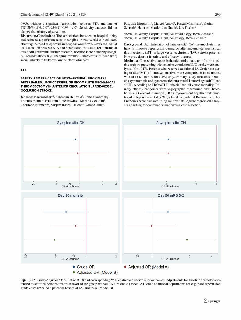
S99
123
Clin Neuroradiol (2019) (Suppl 1) 29:S1–S129
0.95), without a significant association between STA and rate of TICI2b/3 (aOR 0.97, 95%-CI 0.93–1.02). Sensitivity analyses did not change the primary observations.Discussion/Conclusion: The association between in-hospital delay and reduced reperfusion rates is tangible in real world clinical data, stressing the need to optimize in-hospital workflows. Given the lack of an association between STA and reperfusion, the causal relationship of this finding warrants further research, because mere pathophysiologi-cal considerations (i. e. changing thrombus characteristics over time) seem unlikely to fully explain the effect observed.
357
SAFETY AND EFFICACY OF INTRA-ARTERIAL UROKINASE AFTER FAILED, UNSUCCESSFUL OR INCOMPLETE MECHANICAL THROMBECTOMY IN ANTERIOR CIRCULATION LARGE-VESSEL OCCLUSION STROKE.
Johannes Kaesmacher*1, Sebastian Bellwald2, Tomas Dobrocky1, Thomas Meinel2, Eike Immo Piechowiak1, Martina Goeldlin2, Christoph Kurmann1, Mirjam Rachel Heldner2, Simon Jung2,
Pasquale Mordasini1, Marcel Arnold2, Pascal Mosimann1, Gerhart Schroth1, Heinrich Mattle2, Jan Gralla1, Urs Fischer2
1Bern, University Hospital Bern, Neuroradiology, Bern, Schweiz2Bern, University Hospital Bern, Neurology, Bern, Schweiz
Background: Administration of intra-arterial (IA) thrombolysis may help to improve reperfusion during or after incomplete mechanical thrombectomy (MT) in large-vessel occlusions (LVO) stroke patients. However, data on its safety and efficacy is scarce.Methods: Consecutive acute ischemic stroke patients of a prospec-tive registry presenting with anterior circulation LVO stroke were ana-lyzed (N = 1017). Patients who received additional IA Urokinase dur-ing or after MT (+/– intravenous tPA) were compared to those treated with MT (+/– intravenous tPA) only. Primary safety measures includ-ed asymptomatic and symptomatic intracranial hemorrhage (aICH and sICH) according to PROACT-II criteria, and all-cause mortality. Pri-mary efficacy endpoints were angiographic reperfusion and Throm-bolysis in Cerebral Infarction (TICI) improvement, together with func-tional independence at day 90 (defined as modified Rankin Scale ≤2). Endpoints were assessed using multivariate logistic regression analy-ses adjusting for confounders underlying case selection.
Fig. 1 | 357 Crude/Adjusted Odds Ratios (OR) and corresponding 95% confidence intervals for outcomes. Adjustments for baseline characteristics tended to shift the point estimates in favor of the group without IA Urokinase (Model A), while additional adjustments for e. g. poor reperfusion grade cases revealed a potential benefit of IA Urokinase (Model B)

S100
123
Clin Neuroradiol (2019) (Suppl 1) 29:S1–S129
Results: Additional IA Urokinase was administered in 102 of 1017 pa-tients (10.0%). The most common reason for administering IA Uroki-nase was incomplete reperfusion (TICI<3) after MT (52.9%). After adjusting for baseline characteristics underlying case selection, IA Urokinase did not increase the risk of aICH (0.51, 95%-CI 0.29–0.92), sICH (aOR 0.95, 95%-CI 0.39–2.32) or 3-month mortality (0.71, 95%-CI 0.40–1.26). In approximately two-thirds of patients treated with IA Urokinase, early reperfusion improvement was observed, with around one-third being TICI-grade tangible. Correspondingly, patients treat-ed with IA Urokinase had higher rates of functional independence af-ter adjusting for the selection bias favoring poor TICI cases in the IA Urokinase group (aOR 1.90, 95%-CI 1.10–3.30).Discussion/Conclusion: In selected patients, adjunctive treatment with IA Urokinase during or after MT seems to be safe, without an increased risk of intracranial bleedings. Additionally, IA Urokinase harbors the potential to improve the reperfusion status of patients un-dergoing endovascular interventions and may thus improve outcomes.
364
GADOLINIUM LEAKAGE IN OCULAR STRUCTURES IN TRANSIENT ISCHEMIC ATTACK
Alex Förster*1, Johannes Böhme1, Holger Wenz1, Christoph Groden1, Angelika Alonso2
1Abteilung für Neuroradiologie, Universitätsmedizin Mannheim, Mannheim, Deutschland2Neurologische Klinik, Universitätsmedizin Mannheim, Mannheim, Deutschland
Hintergrund: Gadolinium leakage in ocular structures (GLOS) on flu-id attenuated inversion recovery images (FLAIR) is a novel imaging marker in acute ischemic stroke.1,2 In the present study, we investigated the frequency and pattern of blood-brain barrier as well as blood-retina barrier impairment in patients with transient ischemic attack (TIA) as demonstrated by hyperintense acute reperfusion marker (HARM) and GLOS respectively on FLAIR.Methoden: From an MRI report database we identified patients with TIA who underwent repeated MRI with intravenous contrast agent ad-ministration. On FLAIR, the presence of HARM was assessed. Fur-thermore, the presence of GLOS was noted in the anterior chamber and vitreous body.Ergebnisse: Overall 28 TIA patients (mean age 66.5 ± 17.2 years, 17 (51.5%) male) were included. Follow-up MRI was performed within 34.4 ± 18.8 hours after the first MRI. On DWI, acute ischemic lesions were observed in 21 (75%), while the remainder was DWI negative. On contrast-enhanced FLAIR, GLOS was observed in 11 (39.3%) pa-tients: in 1 (3.6%) in the anterior chamber only, and in 10 (35.7%) in the anterior chamber and vitreous body. HARM was observed in 3 (10.7%) patients. In one TIA patient without ischemic lesion on DWI or HARM on FLAIR, GLOS was observed in the anterior chamber and vitreous body (see Fig. 1). Presence of GLOS was significantly associated with detection of HARM (p = 0.023) whereas no significant relationship with older age (p = 0.06), or presence of an ischemic lesion on DWI (p = 0.12) was found.Diskussion: In patients with TIA, GLOS is a frequent finding and as-sociated with presence of HARM on contrast-enhanced FLAIR. As GLOS was observed in one TIA patient without presence of an ischem-ic lesion or HARM, it might be useful as an additional imaging marker of transient cerebral ischemia in TIA patients.
References 1. Förster A, Wenz H, Böhme J, Groden C, Alonso A. Asymmetrical
Gadolinium Leakage in Ocular Structures in Stroke Due to Inter-nal Carotid Artery Stenosis or Occlusion. Clin Neuroradiol. 2018; https://doi.org/10.1007/s00062-018-0754-5. Epub ahead of print.
2. Förster A, Al-Zghloul M, Wenz H, Böhme J, Groden C, Alonso A. Gadolinium Leakage in Ocular Structures Is Common in Lacu-nar Infarction. Stroke. 2018; https://doi.org/10.1161/STROKEA-HA.118.023573. STROKEAHA118023573.
365
MIDTERM TREATMENT RESULTS USING THE FRED FLOWDIVERTER
Christian Ulfert*1, Christian Herweh1, Martin Bendszus1, Markus Möhlenbruch1
1Heidelberg University Hospital, Heidelberg, Deutschland
Hintergrund: Flow diverter have become an essential alternative or addition to conventional coiling in the endovascular treatment of in-tracranial aneurysms.
For the newer generation of flow diverters that have been introduced in the last 5 years there is sufficient data covering immediate and short term outcomes but little covering the mid to long term results.
We report a series of 70 patients that were treated with the Flow-Re-direction Intraluminal Device (FRED) flow diverter and in whom there is at least 2 years of follow up imaging available.Methoden: We retrospectively selected all endovascular procedures from our aneurysm database that were treated with a FRED flowdivert-er and have obtained at least 24 months of follow up imaging.
All patients received regular follow ups as well as clinical exami-nations. Clinical outcome was assessed using the mRs. Aneurysm oc-clusion was measured using the 3 point modified Raymond Roy Oc-clusion classification. Imaging was performed in a 3T MRI acquiring unenhanced and contrast enhanced time of flight angiography.
All base characteristics of the patients were obtained, including age, gender, intervention date and base mRs.Ergebnisse: Overall 70 patients harbouring 70 aneurysms were in-cluded. 48 (68.6%) were female 22 (31.4%) were male. Four of the patients (5.7%) had suffered an SAH directly prior to treatment.
Mean age of the patients was 54 years (15–80). Aneurysm dimen-sions were on average 9.1 × 5.7 × 5.8 mm (width x depth x height).
All patients were successfully treated with a FRED flowdiverter. The median follow up time was 34 months ranging from 24 to 68 months.
In their last available follow up 57 (81.4%) of the aneurysms were completely occluded (ROC 1), 12 (17.1%) aneurysms had a neck rem-nant and 1 (1.4%) showed persisting aneurysm perfusion..Diskussion: Most of the available publications on mid to long term flowdiverter follow up data show ‘adequate’ occlusion rates around 90%. In this series ‘adequate’ occlusion was achieved in 98.5% of the patients after a median follow up time of 34 months showing good long term efficacy of the FRED flowdiverter.Fazit: Intracranial aneurysms treated with the FRED flowdiverter show stable mid term results with an adequate aneurysms occlusion rate of 98,5%.
Fig. 1 | 365
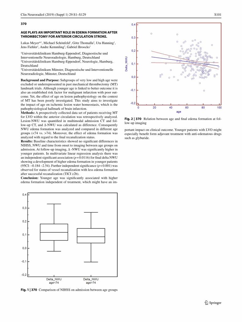
S101
123
Clin Neuroradiol (2019) (Suppl 1) 29:S1–S129
370
AGE PLAYS AN IMPORTANT ROLE IN EDEMA FORMATION AFTER THROMBECTOMY FOR ANTERIOR CIRCULATION STROKE.
Lukas Meyer*1, Michael Schönfeld1, Götz Thomalla2, Uta Hanning1, Jens Fiehler1, Andre Kemmling3, Gabriel Broocks1
1Universitätsklinikum Hamburg-Eppendorf, Diagnostische und Interventionelle Neuroradiologie, Hamburg, Deutschland2Universitätsklinikum Hamburg-Eppendorf, Neurologie, Hamburg, Deutschland3Universitätsklinikum Münster, Diagnostische und Interventionelle Neuroradiologie, Münster, Deutschland
Background and Purpose: Subgroups of very low and high age were excluded or underrepresented in past mechanical thrombectomy (MT) landmark trials. Although younger age is linked to better outcome it is also an established risk factor for malignant infarction with poor out-come. Yet, the effect of age on lesion pathophysiology on the context of MT has been poorly investigated. This study aims to investigate the impact of age on ischemic lesion water homeostasis, which is the pathophysiological hallmark of brain infarction.Methods: A prospectively collected data set of patients receiving MT for LVO within the anterior circulation was retrospectively analyzed. Lesion-NWU was quantified in multimodal admission CT and fol-low-up CT, and ∆-NWU was calculated as difference. Consequently NWU edema formation was analyzed and compared in different age groups (<74 vs. >74). Moreover, the effect of edema formation was analyzed with regard to the final recanalization status.Results: Baseline characteristics showed no significant differences in NIHSS, NWU and time from onset to imaging between age groups on admission. At follow-up imaging, ∆ -NWU was significantly higher in younger patients. In multivariate linear regression analysis there was an independent significant association (p = 0.0116) for final delta NWU showing a development of higher edema formation in younger patients (95CI: –0.184 –2.54). Further independent significance (p < 0.001) was observed for status of vessel recanalization with less edema formation after successful recanalization (TICI ≥2b).Conclusion: Younger age was significantly associated with higher edema formation independent of treatment, which might have an im-
portant impact on clinical outcome. Younger patients with LVO might especially benefit form adjuvant treatment with anti-edematous drugs such as glyburide.
Fig. 1 | 370 Comparison of NIHSS on admission between age groups
Fig. 2 | 370 Relation between age and final edema formation at fol-low-up imaging
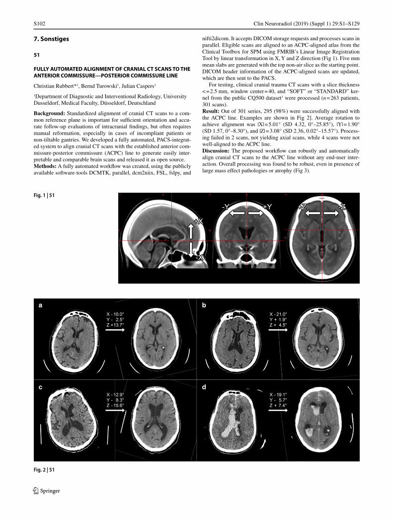
S102
123
Clin Neuroradiol (2019) (Suppl 1) 29:S1–S129
7. Sonstiges
51
FULLY AUTOMATED ALIGNMENT OF CRANIAL CT SCANS TO THE ANTERIOR COMMISSURE—POSTERIOR COMMISSURE LINE
Christian Rubbert*1, Bernd Turowski1, Julian Caspers1
1Department of Diagnostic and Interventional Radiology, University Dusseldorf, Medical Faculty, Düsseldorf, Deutschland
Background: Standardized alignment of cranial CT scans to a com-mon reference plane is important for sufficient orientation and accu-rate follow-up evaluations of intracranial findings, but often requires manual reformation, especially in cases of incompliant patients or non-tiltable gantries. We developed a fully automated, PACS-integrat-ed system to align cranial CT scans with the established anterior com-missure-posterior commissure (ACPC) line to generate easily inter-pretable and comparable brain scans and released it as open source.Methods: A fully automated workflow was created, using the publicly available software-tools DCMTK, parallel, dcm2niix, FSL, fslpy, and
nifti2dicom. It accepts DICOM storage requests and processes scans in parallel. Eligible scans are aligned to an ACPC-aligned atlas from the Clinical Toolbox for SPM using FMRIB’s Linear Image Registration Tool by linear transformation in X, Y and Z direction (Fig 1). Five mm mean slabs are generated with the top non-air slice as the starting point. DICOM header information of the ACPC-aligned scans are updated, which are then sent to the PACS.
For testing, clinical cranial trauma CT scans with a slice thickness < = 2.5 mm, window center = 40, and “SOFT” or “STANDARD” ker-nel from the public CQ500 dataset1 were processed (n = 263 patients, 301 scans).Result: Out of 301 series, 295 (98%) were successfully aligned with the ACPC line. Examples are shown in Fig 2]. Average rotation to achieve alignment was |X| = 5.01° (SD 4.32, 0°–25.85°), |Y| = 1.90° (SD 1.57, 0°–8.30°), and |Z| = 3.08° (SD 2.36, 0.02°–15.57°). Process-ing failed in 2 scans, not yielding axial scans, while 4 scans were not well-aligned to the ACPC line.Discussion: The proposed workflow can robustly and automatically align cranial CT scans to the ACPC line without any end-user inter-action. Overall processing was found to be robust, even in presence of large mass effect pathologies or atrophy (Fig 3).
Fig. 1 | 51
Fig. 2 | 51
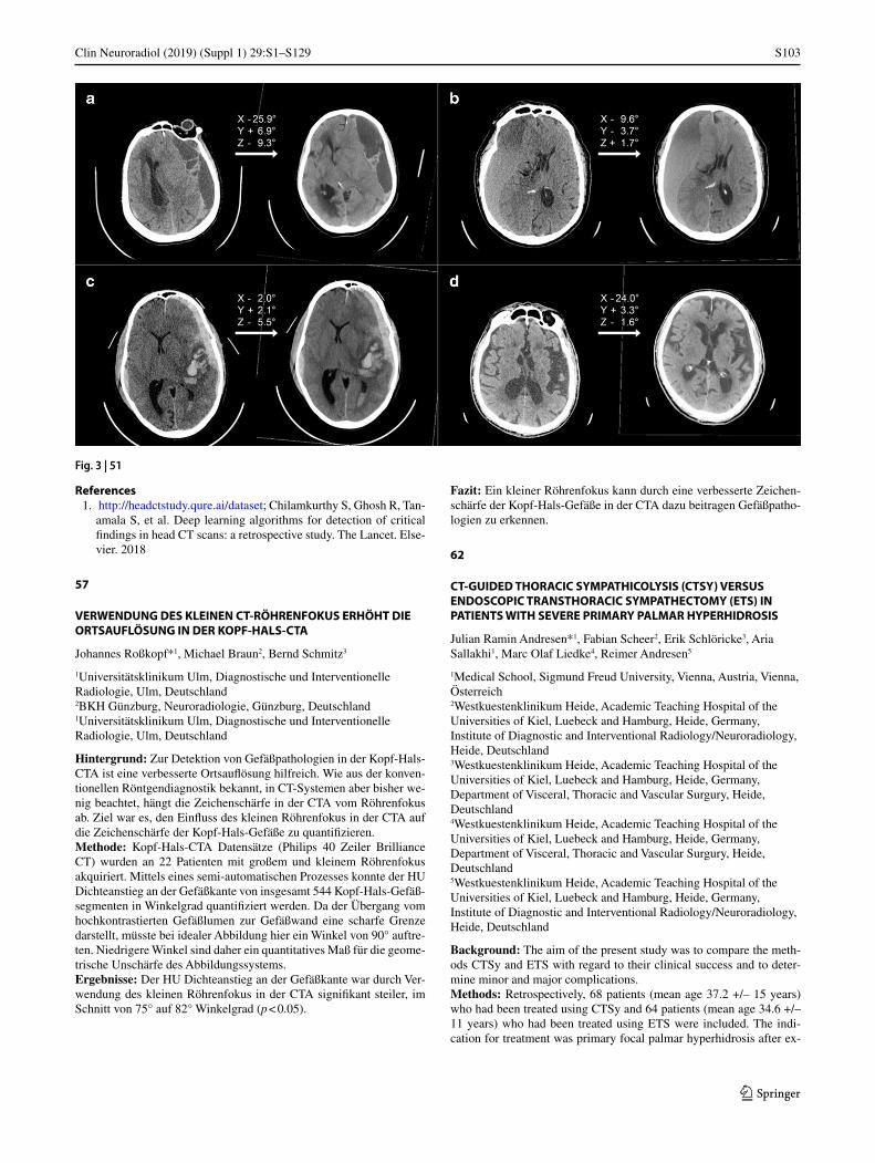
S103
123
Clin Neuroradiol (2019) (Suppl 1) 29:S1–S129
References 1. http://headctstudy.qure.ai/dataset; Chilamkurthy S, Ghosh R, Tan-
amala S, et al. Deep learning algorithms for detection of critical findings in head CT scans: a retrospective study. The Lancet. Else-vier. 2018
57
VERWENDUNG DES KLEINEN CT-RÖHRENFOKUS ERHÖHT DIE ORTSAUFLÖSUNG IN DER KOPF-HALS-CTA
Johannes Roßkopf*1, Michael Braun2, Bernd Schmitz3
1Universitätsklinikum Ulm, Diagnostische und Interventionelle Radiologie, Ulm, Deutschland2BKH Günzburg, Neuroradiologie, Günzburg, Deutschland1Universitätsklinikum Ulm, Diagnostische und Interventionelle Radiologie, Ulm, Deutschland
Hintergrund: Zur Detektion von Gefäßpathologien in der Kopf-Hals-CTA ist eine verbesserte Ortsauflösung hilfreich. Wie aus der konven-tionellen Röntgendiagnostik bekannt, in CT-Systemen aber bisher we-nig beachtet, hängt die Zeichenschärfe in der CTA vom Röhrenfokus ab. Ziel war es, den Einfluss des kleinen Röhrenfokus in der CTA auf die Zeichenschärfe der Kopf-Hals-Gefäße zu quantifizieren.Methode: Kopf-Hals-CTA Datensätze (Philips 40 Zeiler Brilliance CT) wurden an 22 Patienten mit großem und kleinem Röhrenfokus akquiriert. Mittels eines semi-automatischen Prozesses konnte der HU Dichteanstieg an der Gefäßkante von insgesamt 544 Kopf-Hals-Gefäß-segmenten in Winkelgrad quantifiziert werden. Da der Übergang vom hochkontrastierten Gefäßlumen zur Gefäßwand eine scharfe Grenze darstellt, müsste bei idealer Abbildung hier ein Winkel von 90° auftre-ten. Niedrigere Winkel sind daher ein quantitatives Maß für die geome-trische Unschärfe des Abbildungssystems.Ergebnisse: Der HU Dichteanstieg an der Gefäßkante war durch Ver-wendung des kleinen Röhrenfokus in der CTA signifikant steiler, im Schnitt von 75° auf 82° Winkelgrad (p < 0.05).
Fazit: Ein kleiner Röhrenfokus kann durch eine verbesserte Zeichen-schärfe der Kopf-Hals-Gefäße in der CTA dazu beitragen Gefäßpatho-logien zu erkennen.
62
CT-GUIDED THORACIC SYMPATHICOLYSIS (CTSY) VERSUS ENDOSCOPIC TRANSTHORACIC SYMPATHECTOMY (ETS) IN PATIENTS WITH SEVERE PRIMARY PALMAR HYPERHIDROSIS
Julian Ramin Andresen*1, Fabian Scheer2, Erik Schlöricke3, Aria Sallakhi1, Marc Olaf Liedke4, Reimer Andresen5
1Medical School, Sigmund Freud University, Vienna, Austria, Vienna, Österreich2Westkuestenklinikum Heide, Academic Teaching Hospital of the Universities of Kiel, Luebeck and Hamburg, Heide, Germany, Institute of Diagnostic and Interventional Radiology/Neuroradiology, Heide, Deutschland3Westkuestenklinikum Heide, Academic Teaching Hospital of the Universities of Kiel, Luebeck and Hamburg, Heide, Germany, Department of Visceral, Thoracic and Vascular Surgury, Heide, Deutschland4Westkuestenklinikum Heide, Academic Teaching Hospital of the Universities of Kiel, Luebeck and Hamburg, Heide, Germany, Department of Visceral, Thoracic and Vascular Surgury, Heide, Deutschland5Westkuestenklinikum Heide, Academic Teaching Hospital of the Universities of Kiel, Luebeck and Hamburg, Heide, Germany, Institute of Diagnostic and Interventional Radiology/Neuroradiology, Heide, Deutschland
Background: The aim of the present study was to compare the meth-ods CTSy and ETS with regard to their clinical success and to deter-mine minor and major complications.Methods: Retrospectively, 68 patients (mean age 37.2 +/– 15 years) who had been treated using CTSy and 64 patients (mean age 34.6 +/– 11 years) who had been treated using ETS were included. The indi-cation for treatment was primary focal palmar hyperhidrosis after ex-
Fig. 3 | 51

S104
123
Clin Neuroradiol (2019) (Suppl 1) 29:S1–S129
haustion of all conservative treatment options and a continued high level of suffering. All patients evaluated their sense of discomfort with-in the context of a quality control before the start of treatment, two days postinterventionally/postoperatively, and six and twelve months after treatment, on the basis of a Dermatology Quality of Life Index (DLQI) as well as the side effects that occurred. As statistical test procedures, the Wilcoxon rank-sum test was applied to test the differences in DLQI values over the course and the Mann-Whitney U test to compare the different methods.Results: Both methods were technically feasible in all patients. Both treatments led to a marked reduction in the preinterventional sense of discomfort two days postinterventionally/postoperatively as well as six and twelve months after treatment (p < 0.001), whereby mild recur-rent sweating occurred over the further course, which was significantly higher in the patients who had undergone CTSy. Temporary miosis and ptosis was found in 8 of 68 (11.8%) patients after CTSy and in 6 of 64 (9.4%) patients after ETS. As the most common side effect, temporary compensatory sweating, predominantly on the trunk, was reported by 16 of 68 (23.5%) patients after CTSy and 18 of 64 (28.1%) patients af-ter ETS. Pneumothoraces developed during the surgical intervention in 26.6%, which were treated adequately by inserting a drain.Conclusion: For patients with severe palmar hyperhidrosis, CTSy and ETS represented a minimally invasive treatment option that provided a high and largely equivalent level of benefit. Recurrent sweating is slightly more common and more pronounced after CTSy than after ETS. However, a higher rate of major complications is seen in the case of ETS.
67
MECHANICAL THROMBECTOMY IN ACUTE MIDDLE CEREBRAL ARTERY M2 SEGMENT OCCLUSION WITH REGARD TO VESSEL INVOLVEMENT
Vivien Lorena Ivan*1, Christian Rubbert1, Julian Caspers1, John-Il Lee2, Michael Gliem2, Sebastian Jander2, Bernd Turowski1, Marius Kaschner1
1Department of Diagnostic and Interventional Radiology, University Dusseldorf, Medical faculty, Duesseldorf, Deutschland2Department of Neurology, University Dusseldorf, Medical faculty, Duesseldorf, Deutschland
Background: Mechanical thrombectomy (MTE) is an established pro-cedure in patients with acute ischemic stroke due to occlusion of the proximal (M1) segment of the middle cerebral artery. However, the effectiveness of MTE in acute M2-segment occlusions has so far not been systematically evaluated.Methods: All patients with M2-segment-occlusions treated by MTE in the local department (01/2012–12/2017) were included (n = 57, mean National Institutes of Health Stroke Scale of 11, range 0 to 20). Pa-tients were grouped according to the localization of the M2-occlu-sion (Cohort A (n = 14): central region only, B (n = 24): central region and involvement of frontal vessels, C (n = 19): parietal, occipital and/or temporal vessels). Differences in proximal (M2-trunk, n = 34) and distal (M2-branches, n = 23) occlusions were also examined. Reperfu-sion (Thrombolysis in Cerebral Infarction (TICI)), early clinical out-come at discharge (modified Rankin Scale, mRS) and complications (hemorrhage, emboli to new territories) were noted. Mann-Whitney-U, Kruskal-Wallis-, Fisher’s-Exact-Tests and one-way ANOVA were per-formed, as needed.Result: Successful reperfusion (TICI 2b–3) was found in 49 patients (86.0%). Favorable early clinical outcome (mRS 0–2) was achieved in n = 19 (37.7%). Compared to admission, mRS at discharge improved significantly (median mRS at admission 5 vs. median mRS at discharge 4, p < 0.001, Mann-Whitney-U). Early clinical outcome was more fa-vorable in patients with better reperfusion (mean mRS of 4.43 ± 1.40,
median 4, for TICI 0–2a vs. mean mRS of 3.00 ± 1.68, median 4, for TICI 2b-3; p = 0.043, unpaired Mann-Whitney-U). Symptomatic in-tracranial hemorrhage (sICH) was observed in only 6 patients (10.5%). Four patients died (7.0%).
No significant differences in favorable clinical outcome (mRS at discharge ≤2: cohort A 42.9% vs. B 50.0% vs. C 16.7%, p = 0.14, Fish-er’s Exact) were found with regard to the vessel involvement. Early clinical outcome did not differ in proximal and distal occlusions.Discussion/Conclusion: MTE in patients with acute M2-occlusion is efficient and leads to a significant clinical improvement at discharge. No significant differences in clinical outcome or complications were found with regard to the localization of the M2-occlusion.
References 1. Dorn F, et al. Mechanical Thrombectomy of M2-Occlusion.
J Stroke Cerebrovasc Dis. 2015;24(7):1465–70.
80
EINFLUSS DER NARKOSEFÜHRUNG AUF DIE STRAHLEN-EXPOSITION UND DURCHLEUCHTUNGSZEIT WÄHREND DER ENDOVASKULÄREN SCHLAGANFALLTHERAPIE
Felix Hemmerich*1, Charlotte Weyland1, Markus Möhlenbruch1, Martin Bendszus1, Johannes Pfaff1
1Universitätsklinikum Heidelberg, Neurologische Klinik, Neuroradiologie, Heidelberg, Deutschland
Hintergrund: Die steigenden Behandlungszahlen in der endovasku-lären Schlaganfalltherapie (EST) lässt eine Standardisierung zur Si-cherung der Behandlungsqualität notwendig werden. Hierunter zählt unseres Erachtens auch eine Berücksichtigung der Strahlenexpositi-on. Zur Strahlenexposition bestehen jedoch keine systematischen Da-ten.
Unsere Hypothese ist, dass – im Vergleich zu einer Behandlung in Vollnarkose – die vermehrte Bewegung der Patienten, die in Wachnar-kose behandelt werden, und der damit mutmaßlich verbundenen er-höhten Schwierigkeit der Navigation von (Mikro-)Kathetern, (Mikro-)Drähten und Stent-Retrievern während der endovaskulären Schlagan-falltherapie, zu einer höheren Strahlenexposition sowie zu längeren Durchleuchtungszeiten führt.Methoden: In diese klinisch-retrospektive Analyse wurden alle Pa-tienten mit akutem ischämischen Schlaganfall in der vorderen Strom-bahn eingeschlossen, die in der Zeit von Jan. 2013 bis April 2018 eine EST mittels moderner Stent-Retriever-Thrombektomie und/oder di-rekter Aspirationsthrombektomie durch erfahrene Neuroradiologen (mind. 25 vorherige EST-Eingriffe) erhielten.Ergebnisse: In der Gesamtpopulation dieser Studien (n = 679) be-steht zwischen den Patienten, die in Intubationsnarkose (GA, n = 196) behandelt wurden, und den Patienten, die in Wachnarkose behandelt wurden (CS, n = 483) mit Hinblick auf das Patientenalter, die initiale Schwere des Schlaganfalls und der Verschlusslokalisation ein signifi-kanter Unterschied (siehe Tabelle). Das Dosisflächenprodukt DFP (in µGy*cm², Median (IQR): GA: 11747; CS: 11352; p-Wert: 0.419) und die Durchleuchtungszeit (in Minuten, Median (IQR): GA: 35 (21–59); CS: 31 (17–54); p-Wert: 0.095) sind hingegen vergleichbar.
Bei einer isolierten Betrachtung der Patienten mit einem Media-hauptstammverschluss (GA: n = 89, CS: n = 219), bei der sich mit Aus-nahme der initialen Schwere der Symptomatik keine signifikanten Un-terschiede der Patientencharakteristika ergeben, sind das DFP (GA: 10643; CS: 9582; p-Wert: 0.68) und die Durchleuchtungszeit (GA: 33; CS: 25; p-Wert: 0.19) ebenfalls nicht signifikant unterschiedlich.Fazit: Das Ergebnis der Analyse der Thrombektomie-Datenbank zeigt, dass es keinen signifikanten Einfluss der Narkoseführung auf die Durchleuchtungszeit und die Strahlenexposition während der endovas-

S105
123
Clin Neuroradiol (2019) (Suppl 1) 29:S1–S129
kulären Schlaganfalltherapie gibt. Die erhobenen Werte zur Strahlen-exposition und Durchleuchtungszeit könnten zukünftig als Orientie-rung im Sinne von Dosis-Referenz-Werten dienen.
92
NUCLEUS ACCUMBENS PROJECTIONS: VALIDITY AND RELIABILITY OF FIBER RECONSTRUCTIONS BASED ON HIGH-RESOLUTION DIFFUSION-WEIGHTED MRI
Thilo Rusche*1, Jörn Kaufmann2, Jürgen Voges3
1Institut für Klinische Radiologie, Universitätsklinikum Münster, Münster, Deutschland2Klinik für Neurologie, Universitätsklinikum Magdeburg, Otto-von-Guericke Universität, Magdeburg, Deutschland3Klinik für Stereotaktische Neurochirurgie, Universitätsklinikum Magdeburg, Otto-von-Guericke Universität, Magdeburg, Deutschland
Background: The N. accumbens (NAc) is a key relay in the mesolim-bic dopaminergic reward system. As such, it is a promising target for deep brain stimulation (DBS) in patients with psychiatric diseases. In the present study, we aimed to reconstruct the neural projections con-
necting the NAc with the medial prefrontal cortex (mPFC), the ante-rior cingulate cortex (ACC), the amygdala, the hippocampus, the dor-somedial thalamus (dmT) and the ventral tegmental area (VTA) using probabilistic fiber tractography based on diffusion-weighted MR-im-aging (dMRI).Methods: MR-data (T1-MPRAGE; FLAIR; DWI: 1.6 mm isotrop-ic resolution, 60 gradient directions) for 11 healthy subjects were acquired in seven sessions on a Siemens MAGNETOM Prisma 3T MRI-scanner. For each subject, the bilateral NAc, mPFC, ACC, amyg-dala, hippocampus, dmT and VTA were manually/automated segment-ed based on the T1 and FLAIR data and transformed to the session-spe-cific DWI space for probabilistic fiber tractography. The results were subject to detailed visual inspection to assess their validity in terms of anatomical plausibility by comparing them with the relevant literature. To quantitatively assess the reliability of the reconstructions, exempla-rily the individual fiber-tracts between the NAc and mPFC for each ses-sion and subject were clustered and transformed to a main-tract before performing an intra-subject comparison.Results: Using MRI data from 11 healthy subjects, we were able to reconstruct neural projections connecting the NAc with the mPFC (see figure 1/2), ACC, amygdala, hippocampus, dmT and VTA.
Fig. 1 | 80 Gesamtes Patienten-Kollektiv 01/2013–04/2019
Fig. 2 | 80 Patienten mit iso-liertem Mediahauptstammver-schluss
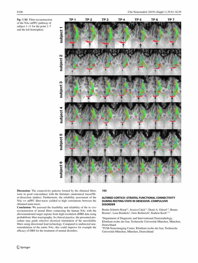
S106
123
Clin Neuroradiol (2019) (Suppl 1) 29:S1–S129
Discussion: The connectivity patterns formed by the obtained fibers were in good concordance with the literature (anatomical tracer/fib-er-dissection studies). Furthermore, the reliability assessment of the NAc ↔ mPFC fiber-tracts yielded to high correlations between the obtained main-tracts.Conclusion: We assessed the feasibility and reliability of the in vivo reconstruction of neural fibers connecting the human NAc with the abovementioned target regions from high-resolution dMRI data using probabilistic fiber-tractography. In clinical practice, the presented pro-cedure may guide selective electrical stimulation of the mesolimbic fibers using directional lead technology. Compared to undirected neu-romodulation of the entire NAc, this could improve for example the efficacy of DBS for the treatment of mental disorders.
105
ALTERED CORTICO–STRIATAL FUNCTIONAL CONNECTIVITY DURING RESTING STATE IN OBSESSIVE–COMPULSIVE DISORDER
Benita Schmitz-Koep*1, Jessica Calzà1 2, Deniz A. Gürsel1 2, Benno Bremer1, Lena Reinholz3, Götz Berberich4, Kathrin Koch1 2 5
1Department of Diagnostic and Interventional Neuroradiology, Klinikum rechts der Isar, Technische Universität München, München, Deutschland2TUM-Neuroimaging Center, Klinikum rechts der Isar, Technische Universität München, München, Deutschland
Fig. 1 | 92 Fiber-reconstruction of the NAc-mPFC-pathway of subject 1–11 for the point 1–7 and the left hemisphere

S107
123
Clin Neuroradiol (2019) (Suppl 1) 29:S1–S129
3Department of Psychology, Ludwig-Maximilians-Universität, München, Deutschland4Windach Institute and Hospital of Neurobehavioural Research and Therapy (WINTR), Windach, Deutschland5Graduate School of Systemic Neurosciences, Ludwig-Maximilians-Universität, München, Deutschland
Hintergrund: Neuroimaging studies show that obsessive–compulsive disorder (OCD) is characterized by an alteration of the cortico–stri-ato–thalamo–cortical (CSTC) system [1]. As resting-state functional connectivity (FC) studies investigated only specific parts of the CSTC in patients with OCD up to now, the present study aimed at exploring FC in the CSTC as a whole.Methoden: We investigated potential alterations in resting-state FC within the CSTC system in 44 OCD patients and 40 healthy controls by taking into consideration all relevant nodes of the direct and indi-rect CSTC loop.Ergebnisse: Compared to healthy controls, OCD patients showed an increased FC between the left subthalamic nucleus (STN) and the left external globus pallidus (GPe), as well as an increased FC between the left GPe and the left internal globus pallidus (GPi).Diskussion: The results further support CSTC involvement in OCD pathology. However, our findings showing increased FC between rele-
vant structures of the indirect pathway contradict the current hypothe-sis. The results highlight the role of the STN which is a relevant target region of deep brain stimulation (DBS) in patients with treatment-re-fractory OCD [2]. Hence, our findings could contribute a neurobiologi-cal framework to a better comprehension of the fundamental processes underlying DBS.Fazit: Our findings highlight the importance of the indirect pathway, especially of the STN, in OCD pathology and could introduce an al-tered interpretation of the classical model of the CSTC. Furthermore, the results may improve insight on DBS as a treatment for severe OCD. They could provide a basis for future investigations improving the ac-curate choice of DBS targets and thus increasing clinical outcome for patients as well as reducing the probability of adverse events.
References 1. Saxena S, Rauch SL. Functional neuroimaging and the neuro-
anatomy of obsessive-compulsive disorder. Psychiatric Clinics of North America. 2000;23(3):563–86.
2. Mulders AEP, Plantinga BR, Schruers K, Duits A, Janssen MLF, Ackermans L, et al. Deep brain stimulation of the subthalamic nucleus in obsessive-compulsive disorder: Neuroanatomical and pathophysiological considerations. Eur Neuropsychopharmacol. 2016;26(12):1909–19.
Fig. 2 | 92 Fiber-reconstruction of the NAc-mPFC-pathway of subject 1–11 for the point 1–7 and the left hemisphere

S108
123
Clin Neuroradiol (2019) (Suppl 1) 29:S1–S129
109
TUBE CURRENT REDUCTION IN COMPUTED TOMOGRAPHY ANGIOGRAPHY—HOW LOW CAN WE GO IN PATIENTS WITH SUSPECTED ACUTE STROKE?
Nico Sollmann1, Kai Mei2, Isabelle Riederer1, Simon Schön1, Felix K. Kopp2, Maximilian T. Löffler1, Jan S. Kirschke1, Ernst J. Rummeny2, Claus Zimmer1, Peter B. Noel2, Thomas Baum1
1Abteilung für Diagnostische und Interventionelle Neuroradiologie, Klinikum rechts der Isar, Technische Universität München, München, Deutschland2Abteilung für Diagnostische und Interventionelle Radiologie, Klinikum rechts der Isar, Technische Universität München, München, Deutschland
Hintergrund: Computed tomography angiography (CTA) as part of first-line stroke imaging protocols entails considerable radiation expo-sure. However, research on dose reductions for head and neck CTA is scarce. The aim of this study is to evaluate image quality, detectability of large vessel occlusion (LVO) or dissection, and diagnostic confi-dence in CTA with virtually lowered tube current and iterative recon-struction (IR) in patients with suspected acute stroke.Methoden: Thirty patients (age: 79.0 ± 10.4 years, 50% with LVO or dissection) underwent CTA of the supraaortal up to the intracranial ar-terial vessels. CTA scans were simulated as if they were made at 50% (D50), 25% (D25), and 10% (D10) of the original tube current. Image
reconstruction was achieved with two levels of IR (A: similar to clin-ical reconstructions, B: two times stronger regularization). Two read-ers (R1 and R2) performed qualitative image evaluation considering overall image quality and artifacts, vessel contrast, detection of vessel pathology, and diagnostic confidence (scoring: 1—low, 2—medium, and 3—high confidence).Ergebnisse: Level B of IR was favorable regarding overall image qual-ity and artifacts for D10, while level A was favorable for D100 and D50. CTA scans with D25 and both levels of IR still showed good vessel contrast, with even peripheral arterial branches of the anterior, middle, and posterior cerebral artery being clearly detectable. Further, CTA scans with D25 using level A of IR showed an adequate level of diagnostic confidence without any missed LVO or dissection according to evaluations of both readers (D25 A: R1: 2.5 ± 0.6 vs. R2: 2.6 ± 0.6, p > 0.05). Inter-reader agreement was high for all items of evaluations, particularly when level A of IR was used.Fazit: CTA using IR and tube currents lowered down to 25% of origi-nal imaging is feasible without drawbacks regarding vessel contrast or detection of vessel pathology in patients with suspected acute stroke. Our approach enables reductions in radiation exposure for head and neck CTA, with an average decrease from 8.5 ± 1.5 mGy to 2.1 mGy in our patients.
126
BRAIN METABOLISM OF PATIENTS UNDER IMMUNO-SUPPRESSION WITH TACROLIMUS AFTER KIDNEY TRANSPLANTATION—A SINGLE VOXEL 1H-MR SPECTROSCOPY STUDY
Patrick Nösel*1, Henning Pflugrad2 3, Birte Schmitz1, Heinrich Lanfermann1, Hannelore Barg-Hock4, Jürgen Klempnauer3 4, Mario Schiffer3 5, Karin Weissenborn2 3, Xiao-Qi Ding1
1Institute of Diagnostic and Interventional Neuroradiology, Hannover Medical School, Hannover, Deutschland2Department of Neurology, Hannover Medical School, Hannover, Deutschland3Integrated Research and Treatment Centre Transplantation, Hannover, Deutschland4Clinic for Visceral and Transplant Surgery, Hannover Medical School, Hannover, Deutschland5Clinic for Nephrology, Hannover Medical School, Hannover, Deutschland
Hintergrund: While the immunosuppression therapy with tacrolimus has remarkably improved the survival of patients after organ trans-plantation, long-term side effects of tacrolimus medication become an important research topic with respect to improvement of patient care. This work aimed to assess possible alterations of brain metabolism in patients under long-term tacrolimus medication after kidney trans-plantation (KTx-patients) by using single voxel 1H-MR spectroscopy (1H-MRS).Methoden: 40 KTx- patients (age: 55.57 ± 8.34 years, standard tacroli-mus dose: 7.19 ± 0.9 µg/l, years after KTx: 7.24 ± 4.03) and 16 age-, sex- and education- adjusted healthy controls underwent MR examina-tions at 3T (Verio, Siemens, Erlangen), which included a single vox-el spectros-copy (SVS) of 4 ml brain tissue in parieto-occipital white matter. SVS data were analyzed by using software LCModel to de-termine concentrations of brain metabolites N-acetylaspartate (NAA), Choline (Cho), Creatine (Cr), Glutamate and Glutamine (Glx), and Myo- Inositol (ml) in reference to internal water and as ratio to Cr. The Battery for the Assessment of Neuro-psychological Status (RBANS) was used to assess the cognitive functions of the subjects. The results were compared between patients and controls by using nonparametric median test with post hoc Bonferroni pairwise comparison. For data quality assurance, only metabolite values with a Cramer-Rao lower
Fig. 1 | 109 84-year-old man with right-sided thrombotic occlusion of the middle cerebral artery (M1 segment). CT angiograms were pro-cessed with two iterative reconstruction (IR) algorithms (A and B) and simulated with 50% (D59), 25% (D25), and 10% (D10) of original tube current (D100)
Fig. 2 | 109 81-year-old wowan with left-sided dissection oft he ex-tracranial internal carotid artery. CT angiograms were processed with two iterative reconstraction (IR) algorithms (A and B) and simulated with 50% (D59), 25% (D25), and 10% (D10) of original tube current (D100)
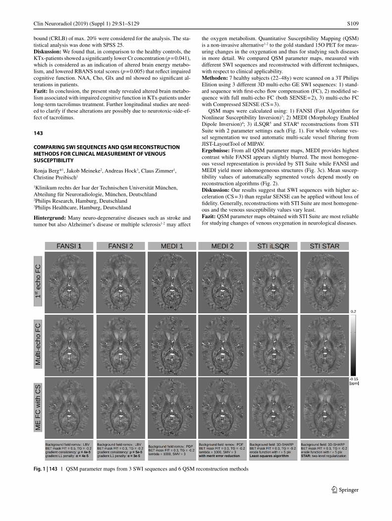
S109
123
Clin Neuroradiol (2019) (Suppl 1) 29:S1–S129
bound (CRLB) of max. 20% were considered for the analysis. The sta-tistical analysis was done with SPSS 25.Diskussion: We found that, in comparison to the healthy controls, the KTx-patients showed a significantly lower Cr concentration (p = 0.041), which is considered as an indication of altered brain energy metabo-lism, and lowered RBANS total scores (p = 0.005) that reflect impaired cognitive function. NAA, Cho, Glx and ml showed no significant al-terations in patients.Fazit: In conclusion, the present study revealed altered brain metabo-lism associated with impaired cognitive function in KTx-patients under long-term tacrolimus treatment. Further longitudinal studies are need-ed to clarify if these alterations are possibly due to neurotoxic-side-ef-fect of tacrolimus.
143
COMPARING SWI SEQUENCES AND QSM RECONSTRUCTION METHODS FOR CLINICAL MEASUREMENT OF VENOUS SUSCEPTIBILITY
Ronja Berg*1, Jakob Meineke2, Andreas Hock3, Claus Zimmer1, Christine Preibisch1
1Klinikum rechts der Isar der Technischen Universität München, Abteilung für Neuroradiologie, München, Deutschland2Philips Research, Hamburg, Deutschland3Philips Healthcare, Hamburg, Deutschland
Hintergrund: Many neuro-degenerative diseases such as stroke and tumor but also Alzheimer’s disease or multiple sclerosis1-2 may affect
the oxygen metabolism. Quantitative Susceptibility Mapping (QSM) is a non-invasive alternative1-2 to the gold standard 15O PET for meas-uring changes in the oxygenation and thus for studying such diseases in more detail. We compared QSM parameter maps, measured with different SWI sequences and reconstructed with different techniques, with respect to clinical applicability.Methoden: 7 healthy subjects (22–48y) were scanned on a 3T Philips Elition using 3 different 3D multi-echo GE SWI sequences: 1) stand-ard sequence with first-echo flow compensation (FC), 2) modified se-quence with full multi-echo FC (both SENSE = 2), 3) multi-echo FC with Compressed SENSE (CS = 3).
QSM maps were calculated using: 1) FANSI (Fast Algorithm for Nonlinear Susceptibility Inversion)3; 2) MEDI (Morphology Enabled Dipole Inversion)4; 3) iLSQR5 and STAR6 reconstructions from STI Suite with 2 parameter settings each (Fig. 1). For whole volume ves-sel segmentation we used automatic multi-scale vessel filtering from JIST-LayoutTool of MIPAV.Ergebnisse: From all QSM parameter maps, MEDI provides highest contrast while FANSI appears slightly blurred. The most homogene-ous vessel representation is provided by STI Suite while FANSI and MEDI yield more inhomogeneous structures (Fig. 3c). Mean suscep-tibility values of automatically segmented voxels depend mostly on reconstruction algorithms (Fig. 2).Diskussion: Our results suggest that SWI sequences with higher ac-celeration (CS = 3) than regular SENSE can be applied without loss of fidelity. Generally, reconstructions with STI Suite are most homogene-ous and the venous susceptibility values vary least.Fazit: QSM parameter maps obtained with STI Suite are most reliable for studying changes of venous oxygenation in neurological diseases.
Fig. 1 | 143 1 QSM parameter maps from 3 SWI sequences and 6 QSM reconstruction methods
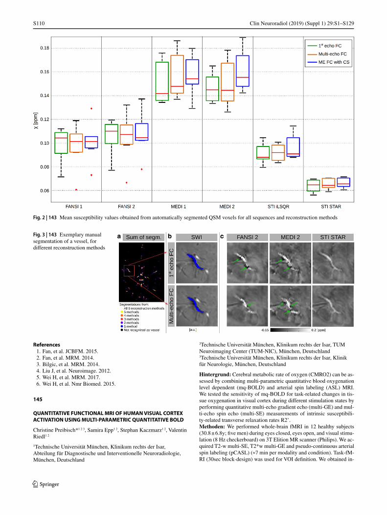
S110
123
Clin Neuroradiol (2019) (Suppl 1) 29:S1–S129
References 1. Fan, et al. JCBFM. 2015. 2. Fan, et al. MRM. 2014. 3. Bilgic, et al. MRM. 2014. 4. Liu J, et al. Neuroimage. 2012. 5. Wei H, et al. MRM. 2017. 6. Wei H, et al. Nmr Biomed. 2015.
145
QUANTITATIVE FUNCTIONAL MRI OF HUMAN VISUAL CORTEX ACTIVATION USING MULTI-PARAMETRIC QUANTITATIVE BOLD
Christine Preibisch*1 2 3, Samira Epp1 2, Stephan Kaczmarz1 2, Valentin Riedl1 2
1Technische Universität München, Klinikum rechts der Isar, Abteilung für Diagnostische und Interventionelle Neuroradiologie, München, Deutschland
2Technische Universität München, Klinikum rechts der Isar, TUM Neuroimaging Center (TUM-NIC), München, Deutschland3Technische Universität München, Klinikum rechts der Isar, Klinik für Neurologie, München, Deutschland
Hintergrund: Cerebral metabolic rate of oxygen (CMRO2) can be as-sessed by combining multi-parametric quantitative blood oxygenation level dependent (mq-BOLD) and arterial spin labeling (ASL) MRI. We tested the sensitivity of mq-BOLD for task-related changes in tis-sue oxygenation in visual cortex during different stimulation states by performing quantitative multi-echo gradient echo (multi-GE) and mul-ti-echo spin echo (multi-SE) measurements of intrinsic susceptibili-ty-related transverse relaxation rates R2’.Methoden: We performed whole-brain fMRI in 12 healthy subjects (30.8 ± 6.8y; five men) during eyes closed, eyes open, and visual stimu-lation (8 Hz checkerboard) on 3T Elition MR scanner (Philips). We ac-quired T2-w multi-SE, T2*w multi-GE and pseudo-continuous arterial spin labeling (pCASL) (≈7 min per modality and condition). Task-fM-RI (30sec block-design) was used for VOI definition. We obtained in-
Fig. 2 | 143 Mean susceptibility values obtained from automatically segmented QSM voxels for all sequences and reconstruction methods
Fig. 3 | 143 Exemplary manual segmentation of a vessel, for different reconstruction methods
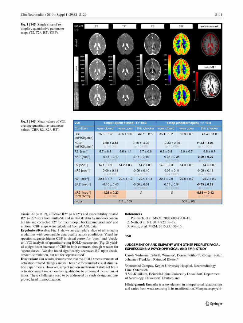
S111
123
Clin Neuroradiol (2019) (Suppl 1) 29:S1–S129
trinsic R2 (= 1/T2), effective R2* (= 1/T2*) and susceptibility related R2’ (= R2*-R2) from multi-SE and multi-GE data by mono-exponen-tial fits and corrected T2* for macroscopic background gradients1 and motion.2 CBF maps were calculated from pCASL data.3
Ergebnisse/Results: Fig. 1 shows an exemplary slice of all imaging modalities with comparable data quality across conditions. Visual in-spection suggests higher CBF in visual cortex for ‘open’ and ‘check-er’. VOI analysis of quantitative mq-BOLD parameters (Fig. 2) yield-ed a significant increase of CBF in both contrasts, though weaker for ‘open>closed’. We also found significantly decreased R2’ upon check-erboard stimulation, but not for ‘open>closed’.Diskussion: Our results demonstrate that mq-BOLD measurements of activation-related changes are well feasible for standard visual stimula-tion experiments. However, subject motion and transient states of brain activation might impact on data quality due to prolonged measurement times. These challenges need to be addressed by study design and im-proved head immobilization.
References 1. Preibisch, et al. MRM. 2008;60(4):908–16. 2. Noth, et al. NI. 2014;92:106–19. 3. Alsop, et al. MRM. 2015;73:102–16.
150
JUDGEMENT OF AND EMPATHY WITH OTHER PEOPLE’S FACIAL EXPRESSIONS: A PSYCHOPHYSICAL AND FMRI STUDY
Carola Widmann1, Sibylle Wimmer1, Denise Potthoff2, Rüdiger Seitz2, Johannes Trenkler1, Raimund Kleiser*1
1Neuromed Campus, Kepler University Hospital, Neuroradiology, Linz, Österreich2LVR-Klinikum, Heinrich-Heine-University Düsseldorf, Department of Neurology, Düsseldorf, Deutschland
Hintergrund: Empathy is a key element in interpersonal relationships and varies from weak to strong in its manifestation. Many neuropsychi-
Fig. 1 | 145 Single slice of ex-emplary quantitative parameter maps (T2, T2*, R2’, CBF)
Fig. 2 | 145 Mean values of VOI average quantitative parameter values (CBF, R2, R2*, R2’)
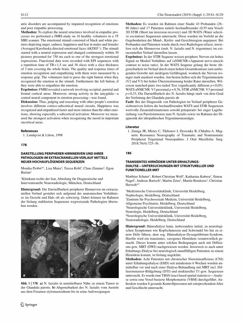
S112
123
Clin Neuroradiol (2019) (Suppl 1) 29:S1–S129
atric disorders are accompanied by impaired recognition of emotions and poor empathic processing.Methoden: To explore the neural structures involved in empathic pro-cesses we performed a fMRI-study on 16 healthy volunteers in a 3T MRI scanner. The emotional stimuli consisted of black and white pic-tures depicting anger, sadness, happiness and fear in males and females (Averaged Karolinska directed emotional faces AKDEF 1). The stimuli started with a neutral expression and changed continuously within 30 pictures presented for 750 ms each to one of the strongest emotional expressions. Functional data were recorded with EPI sequences with a repetition time of TR = 1,5 sec and 38 slices with a slice thickness of 3 mm covering the whole brain. The quality and response times of emotion recognition and empathizing with them were measured by a response grip. The volunteers had to press the right button when they recognized the emotion in the stimuli. Furthermore the left button, if they were able to empathize the emotion.Ergebnisse: FMRI revealed a network involving occipital, parietal and frontal cortical areas. Moreover, strong activity in the amygdala—a central neural component in emotion processing—was found.Diskussion: Thus, judging and reasoning with other people’s emotion involves different cortico-subcortical neural circuits. Happiness was recognized and empathized faster and more intense than the other emo-tions, showing especially a subcortical activation. Moreover we meas-ured the strongest activation when recognizing the mood in important sucortical areas.
References 1. Lundqvist & Litton, 1998
178
DARSTELLUNG PERIPHERER HIRNNERVEN UND IHRER PATHOLOGIEN IM EXTRACRANIELLEN VERLAUF MITTELS NEUER HOCHAUFLÖSENDER SEQUENZEN
Monika Probst*1, Lisa Maier2, Teresa Robl2, Claus Zimmer2, Egon Burian2
1Klinikum rechts der Isar, Abteilung für Diagnostische und Interventionelle Neuroradiologie, München, Deutschland
Hintergrund: Die Darstellbarkeit peripherer Hirnnerven im extracra-niellen Verlauf gestaltet sich aufgrund der anatomischen Verhältnis-se im Gesicht und Hals oft als schwierig. Dabei können im Rahmen der bislang etablierten Sequenzen wegweisende Pathologien überse-hen werden.
Methoden: Es wurden im Rahmen einer Studie 43 Probanden (28–80 Jahre) und 17 Patienten mittels hochauflösender (0,55 mm Voxel) 3D STIR (Short tau inversion recovery) und 3D WATS (Water selecti-ve excitation) Sequenzen untersucht. Diese wurden im Vorfeld an die Gegebenheiten der Mund-, Kiefer- und Gesichtsregion angepasst. Bei Probanden und Patienten wurde durch zwei Radiologen erfasst, inwie-fern sich die Hirnnerven (insb. N. fazialis und N. trigeminus) im ext-racraniellen Verlauf darstellen lassen.Ergebnisse: In der STIR Sequenz weisen periphere Nerven ein hohes Signal zu Muskel Verhältnis auf (aNMCNR = Apparent nerve-muscle contrast to noise ratio). In der WATS Sequenz gelang die beste Ab-grenzbarkeit im Verlauf durch einen hohen Gesamtkontrast zum umlie-genden Gewebe mit niedrigem Gefäßsignal, wodurch die Nerven we-niger stark maskiert wurden. Am besten ließen sich die Trigeminusäste (V2 und V3) bei hoher Übereinstimmung beider Rater darstellen. Wil-coxon matched-pairs two-tailed Test (significantly different p < 0,05): WATS aNMCNR: V3 proximal p = 0,76, STIR aNMCNR: V3 proximal p = 0,33, Die Darstellbarkeit des N. fazialis hängt stark von dem Grad der Verfettung der Glandula parotis ab.Fazit: Bei der Diagnostik von Pathologien im Verlauf peripherer Ge-sichtsnerven liefern die hochauflösenden WATS und STIR Sequenzen wertvolle Zusatzinformationen sowohl präoperativ bei enger Lagebe-ziehung von Parotistumoren zum N. fazialis sowie im Rahmen der Di-agnostik der idiopathischen Trigeminusneuralgie.
Literatur 1. Zuniga JR, Mistry C, Tikhonov I, Dessouky R, Chhabra A. Mag-
netic Resonance Neurography of Traumatic and Nontraumatic Peripheral Trigeminal Neuropathies. J Oral Maxillofac Surg. 2018;76(4):725–36.
186
TRANSIENTES HIRNÖDEM UNTER ERHALTUNGS-DIALYSE—UNTERSUCHUNGEN MIT STRUKTURELLER UND FUNKTIONELLER MRT
Matthias Schaier1, Robert Christian Wolf2, Katharina Kubera2, Simon Nagel3, Andreas Bartsch4, Martin Zeier1, Martin Bendszus4, Christian Herweh*4
1Medizinische Universitätsklinik, Universität Heidelberg, Nephrologie, Heidelberg, Deutschland2Zentrum für Psychosoziale Medizin, Universität Heidelberg, Allgemeine Psychiatrie, Heidelberg, Deutschland3Neurologische Universitätsklinik, Universität Heidelberg, Neurologie, Heidelberg, Deutschland4Neurologische Universitätsklinik, Universität Heidelberg, Neuroradiologie, Heidelberg, Deutschland
Hintergrund: Hämodialyse kann, insbesondere initial, zu neurologi-schen Symptomen wie Kopfschmerzen und Schwindel bis hin zu ei-nem Delir führen, dem sog. Hämodialyse-Dysequilibrium-Syndrom. Hierfür wird ein transientes, vasogenes Hirnödem verantwortlich ge-macht. Dieses konnte unter solchen Bedingungen auch mit Diffusi-ons-gew. MRT (DWI) nachgewiesen werden. Inwieweit es auch unter Erhaltungs-Dialyse bei neurologisch unauffälligen Patienten zu einem Hirnödem kommt, ist bislang ungeklärt.Methoden: Acht Patienten mit chronischer Niereninsuffizienz (CNI) unter Erhaltungsdialyse (EHD) seit mindestens 6 Wochen wurden un-mittelbar vor und nach einer Dialyse-Behandlung mit MRT incl. Dif-fusionstensor-Bildgebung (DTI) und struktureller T1-gew. Sequenzen untersucht. Es wurde eine TBSS (tract based spatial statistics)—Analy-se sowie eine Voxel-basierte Morphometrie (VBM) durchgeführt. Au-ßerdem wurden 8 gesunde Kontrollpersonen mit entsprechendem Alter und Geschlecht untersucht.
Abb. 1 | 178 a) N. fazialis in unmittelbarer Nähe zu einem Tumor in der Glandula parotis. b) Abgrenzbarkeit des N. fazialis vom Austritt aus dem Foramen stylomastoideum bis in seine Aufzweigungen
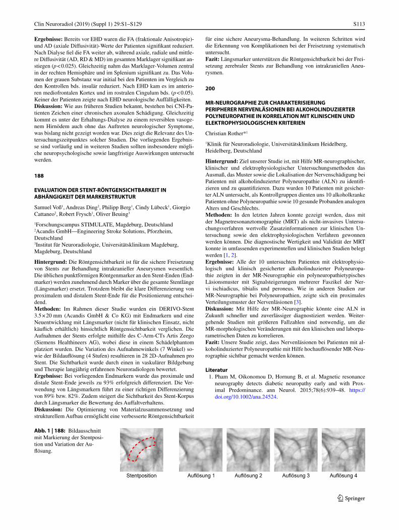
S113
123
Clin Neuroradiol (2019) (Suppl 1) 29:S1–S129
Ergebnisse: Bereits vor EHD waren die FA (fraktionale Anisotropie)- und AD (axiale Diffusivität)-Werte der Patienten signifikant reduziert. Nach Dialyse fiel die FA weiter ab, während axiale, radiale und mittle-re Diffusivität (AD, RD & MD) im gesamten Marklager signifikant an-stiegen (p < 0.025). Gleichzeitig nahm das Marklager-Volumen zentral in der rechten Hemisphäre und im Splenium signifikant zu. Das Volu-men der grauen Substanz war initial bei den Patienten im Vergleich zu den Kontrollen bds. insulär reduziert. Nach EHD kam es im anterio-ren mediofrontalen Kortex und im rostralen Cingulum bds. (p < 0.05). Keiner der Patienten zeigte nach EHD neurologische Auffälligkeiten.Diskussion: Wie aus früheren Studien bekannt, bestehen bei CNI-Pa-tienten Zeichen einer chronischen axonalen Schädigung. Gleichzeitig kommt es unter der Erhaltungs-Dialyse zu einem reversiblen vasoge-nem Hirnödem auch ohne das Auftreten neurologischer Symptome, was bislang nicht gezeigt worden war. Dies zeigt die Relevanz des Un-tersuchungszeitpunktes solcher Studien. Die vorliegenden Ergebnis-se sind vorläufig und in weiteren Studien sollten insbesondere mögli-che neuropsychologische sowie langfristige Auswirkungen untersucht werden.
188
EVALUATION DER STENT-RÖNTGENSICHTBARKEIT IN ABHÄNGIGKEIT DER MARKERSTRUKTUR
Samuel Voß1, Andreas Ding2, Philipp Berg1, Cindy Lübeck1, Giorgio Cattaneo2, Robert Frysch1, Oliver Beuing3
1Forschungscampus STIMULATE, Magdeburg, Deutschland2Acandis GmbH—Engineering Stroke Solutions, Pforzheim, Deutschland3Institut für Neuroradiologie, Universitätsklinikum Magdeburg, Magdeburg, Deutschland
Hintergrund: Die Röntgensichtbarkeit ist für die sichere Freisetzung von Stents zur Behandlung intrakranieller Aneurysmen wesentlich. Die üblichen punktförmigen Röntgenmarker an den Stent-Enden (End-marker) werden zunehmend durch Marker über die gesamte Stentlänge (Längsmarker) ersetzt. Trotzdem bleibt die klare Differenzierung von proximalem und distalem Stent-Ende für die Positionierung entschei-dend.Methoden: Im Rahmen dieser Studie wurden ein DERIVO-Stent 3.5 × 20 mm (Acandis GmbH & Co KG) mit Endmarkern und eine Neuentwicklung mit Längsmarker (nicht für klinischen Einsatz, nicht käuflich erhältlich) hinsichtlich Röntgensichtbarkeit verglichen. Die Aufnahmen der Stents erfolgte mithilfe des C-Arm-CTs Artis Zeego (Siemens Healthineers AG), wobei diese in einem Schädelphantom platziert wurden. Die Variation des Aufnahmewinkels (7 Winkel) so-wie der Bildauflösung (4 Stufen) resultieren in 28 2D-Aufnahmen pro Stent. Die Sichtbarkeit wurde durch einen in vaskulärer Bildgebung und Therapie langjährig erfahrenen Neuroradiologen bewertet.Ergebnisse: Bei vorliegenden Endmarkern wurde das proximale und distale Stent-Ende jeweils zu 93% erfolgreich differenziert. Die Ver-wendung von Längsmarkern führt zu einer richtigen Differenzierung von 89% bzw. 82%. Zudem steigert die Sichtbarkeit des Stent-Korpus durch Längsmarker die Bewertung des Auffaltverhaltens.Diskussion: Die Optimierung von Materialzusammensetzung und strukturellem Aufbau ermöglicht eine verbesserte Röntgensichtbarkeit
für eine sichere Aneurysma-Behandlung. In weiteren Schritten wird die Erkennung von Komplikationen bei der Freisetzung systematisch untersucht.Fazit: Längsmarker unterstützen die Röntgensichtbarkeit bei der Frei-setzung zerebraler Stents zur Behandlung von intrakraniellen Aneu-rysmen.
200
MR-NEUROGRAPHIE ZUR CHARAKTERISIERUNG PERIPHERER NERVENLÄSIONEN BEI ALKOHOLINDUZIERTER POLYNEUROPATHIE IN KORRELATION MIT KLINISCHEN UND ELEKTROPHYSIOLOGISCHEN KRITERIEN
Christian Rother*1
1Klinik für Neuroradiologie, Universitätsklinikum Heidelberg, Heidelberg, Deutschland
Hintergrund: Ziel unserer Studie ist, mit Hilfe MR-neurographischer, klinischer und elektrophysiologischer Untersuchungsmethoden das Ausmaß, das Muster sowie die Lokalisation der Nervenschädigung bei Patienten mit alkoholinduzierter Polyneuropathie (ALN) zu identifi-zieren und zu quantifizieren. Dazu wurden 10 Patienten mit gesicher-ter ALN untersucht, als Kontrollgruppen dienten uns 10 alkoholkranke Patienten ohne Polyneuropathie sowie 10 gesunde Probanden analogen Alters und Geschlechts.Methoden: In den letzten Jahren konnte gezeigt werden, dass mit der Magnetresonanztomographie (MRT) als nicht-invasives Untersu-chungsverfahren wertvolle Zusatzinformationen zur klinischen Un-tersuchung sowie den elektrophysiologischen Verfahren gewonnen werden können. Die diagnostische Wertigkeit und Validität der MRT konnte in umfassenden experimentellen und klinischen Studien belegt werden [1, 2].Ergebnisse: Alle der 10 untersuchten Patienten mit elektrophysio-logisch und klinisch gesicherter alkoholinduzierter Polyneuropa-thie zeigten in der MR-Neurographie ein polyneuropathietypisches Läsionsmuster mit Signalsteigerungen mehrerer Faszikel der Ner-vi ischiadicus, tibialis und peroneus. Wie in anderen Studien zur MR-Neurographie bei Polyneuropathien, zeigte sich ein proximales Verteilungsmuster der Nervenläsionen [3].Diskussion: Mit Hilfe der MR-Neurographie könnte eine ALN in Zukunft schneller und zuverlässiger diagnostiziert werden. Weiter-gehende Studien mit größeren Fallzahlen sind notwendig, um die MR-morphologischen Veränderungen mit den klinischen und laborpa-rametrischen Daten zu korrelieren.Fazit: Unsere Studie zeigt, dass Nervenläsionen bei Patienten mit al-koholinduzierter Polyneuropathie mit Hilfe hochauflösender MR-Neu-rographie sichtbar gemacht werden können.
Literatur 1. Pham M, Oikonomou D, Hornung B, et al. Magnetic resonance
neurography detects diabetic neuropathy early and with Prox-imal Predominance. ann Neurol. 2015;78(6):939–48. https://doi.org/10.1002/ana.24524.
Abb. 1 | 188: Bildausschnitt mit Markierung der Stentposi-tion und Variation der Au-flösung.

S114
123
Clin Neuroradiol (2019) (Suppl 1) 29:S1–S129
2. Kollmer J, Sahm F, Hegenbart U, et al. Sural nerve injury in familial amyloid polyneuropathy. Baillieres Clin Neurol. 2017;89(5):475–84. https://doi.org/10.1212/WNL.0000000000004178.
3. Kollmer J, Hund E, Hornung B, et al. In vivo detection of nerve injury in familial amyloid polyneuropathy by magnetic reso-nance neurography. 2015;201(4):549–62. https://doi.org/10.1093/awu396.
207
CLINICAL IMPLEMENTATION OF MRI FOR DETECTION OF INFERIOR ALVEOLAR NERVE INVOLVEMENT IN COURSE OF MANDIBLE FRACTURE—A FEASIBILITY STUDY
Egon Burian*1, Florian Probst2, Lisa Maier1, Claus Zimmer1, Lucas Ritschl3, Monika Probst1
1Department of Diagnostic and Interventional Neuroradiology, Klinikum rechts der Isar, Technische Universität München, München, Deutschland2Department of Oral and Maxillofacial Surgery and Facial Plastic Surgery, LMU München, München, Deutschland3Department of Oral and Maxillofacial Surgery and Facial Plastic Surgery, Klinikum rechts der Isar, Technical University of Munich, München, Deutschland
Background: Computed tomography (CT) or cone beam computed tomography (CBCT) are the gold standard in clinical traumatology im-aging when structures of the head and neck region are involved. How-ever, CT and CBCT harbor inherent limitations in regard of soft tissue visualization. In course of mandible fracturs particularly the inferior alveolar nerve (IAN) is at risk for damage. The aim of this study was to assess the IAN pre- and postoperatively and correlate elevated signal-to-noise-ratios (SNR) with potential sensitivity impairment.Methods: 10 patients suffering from a mandible fracture were exam-ined on a 3T scanner. The conducted sequence protocol consisted of a 3D STIR, 3D WATS and a 3D T1 FFE “black bone”-sequence (15 min scan time). Nerve function was assessed as: normal, hypesthesia, anesthesia or allodynia.Results: The 3D T1 TFE sequences allowed for fracture detection in all patients. The 3D STIR sequence showed the highest SNR with re-
gard to potential nerve damage (p < 0.05). Besides same diagnostic ac-curacy in fracture detection like CT the direct visualization of nerval structures was possible (Fig. 1). Positive clinical nerve testing correlat-ed with physiological SNR.Discussion: The study highlights the feasibility of MRI for fracture detection and affection of IAN. The applied sequence protocol showed comparable diagnostic power in detection mandible fractures as CT. Additionally, a reliable assessment of potential IAN damage with good correlation to clinical sensory testing could be achieved.Conclusion: Applying the described sequence protocol allowed for precise fracture detection as well as reliable nerve damage assessment.
226
MRI OF THE INFERIOR ALVEOLAR NERVE AND LINGUAL NERVE—BENCHMARK VALUES AND ANATOMICAL VARIETY IN HEALTHY SUBJECTS
Egon Burian*1, Florian Probst2, Teresa Robl1, Claus Zimmer1, Lucas Ritschl3, Monika Probst1
1Department of Diagnostic and Interventional Neuroradiology, Klinikum rechts der Isar, Technische Universität München, München, Deutschland2Department of Oral and Maxillofacial Surgery and Facial Plastic Surgery, LMU München, München, Deutschland3Department of Oral and Maxillofacial Surgery and Facial Plastic Surgery, Technische Universität München, München, Deutschland
Background: Radiation based imaging techniques like panoramic ra-diography, computed tomography (CT) or cone beam computed to-mography (CBCT) are currently the standard for depicting the man-dibular canal prior to surgical procedures. However, these modalities are lacking sufficient soft tissue visualization of the inferior alveolar nerve (IAN) and the lingual nerve (LN), which is particularly exposed to injury during wisdom tooth removal. Magnetic resonance imaging (MRI) using dedicated imaging sequences showed promising results in the direct visualization of the IAN and the LN with high spatial res-olution and signal-to-noise-ratio (SNR) quantification. The aim of this study was to define standard specifications in MRI which allow for benchmark definition of SNR values in healthy subjects.
Fig. 1 | 207 Fracture of the man-dible depicted with CT and the conducted MRI protocol con-sisting of a 3D T1 TFE “black bone”, 3D WATS and 3D STIR sequence. IAN nerve continuity is preserved and SNR is within the physiological range (yellow arrow: IAN, blue arrow: vein). Scan time is 15 min
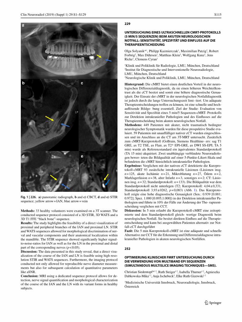
S115
123
Clin Neuroradiol (2019) (Suppl 1) 29:S1–S129
Methods: 33 healthy volunteers were examined on a 3T scanner. The conducted sequence protocol consisted of a 3D STIR, 3D WATS and a 3D T1 FFE “black bone”-sequence.Results: The study highlights the feasibility of a direct visualization of proximal and peripheral branches of the IAN and proximal LN. STIR and WATS sequences allowed for morphological discrimination of ner-val and vascular components and their anatomical localization within the mandible. The STIR sequence showed significantly higher signal-to-noise-ratios for IAN as well as for the LN in the proximal and distal part of the corresponding nerves (p < 0.05).Discussion: The data presented in this study reveal, that a direct visu-alization of the course of the IAN and LN is feasible using high reso-lution STIR and WATS sequences. Furthermore, the imaging protocol conducted not only allowed for reproducible nerve diameter measure-ments but also for subsequent calculation of quantitative parameters like aSNR.Conclusion: MRI using a dedicated sequence protocol allows for de-tection, nerve signal quantification and morphological characterization of the course of the IAN and the LN with its variant forms in healthy subjects.
229
UNTERSUCHUNG EINES ULTRASCHNELLEN CMRT-PROTOKOLLS (5 MIN/5 SEQUENZEN) BEIM AKUTEN NEUROLOGISCHEN NOTFALL: SENSITIVITÄT, SPEZIFITÄT UND EINFLUSS AUF DIE THERAPIEENTSCHEIDUNG
Olga Solyanik*1, Philipp Kazmierczak1, Maximilian Patzig2, Robert Forbrig2, Max Dührsen1, Matthias Klein3, Wolfgang Kunz1, Jens Ricke1, Clemens Cyran1
1Klinik und Poliklinik für Radiologie, LMU, München, Deutschland2Institut für Diagnostische und Interventionelle Neuroradiologie, LMU, München, Deutschland3Neurologische Klinik und Poliklinik, LMU, München, Deutschland
Hintergrund: Die cMRT bietet einen deutlichen Vorteil in der neuro-logischen Differenzialdiagnostik, da sie einen höheren Weichteilkon-trast als die cCT besitzt und somit eine höhere diagnostische Genau-igkeit. Der Einsatz der cMRT in der neurologischen Notfalldiagnostik ist jedoch durch die lange Untersuchungszeit limi- tiert. Um adäquate Therapieentscheidungen treffen zu können, ist eine schnelle und hoch-auflösende Bildge- bung essentiell. Ziel der Studie: Evaluation von Sensitivität und Spezifität eines 5 min/5 Sequenzen-cMRT- Protokolls zur Detektion intrakranieller Pathologien und des Einflusses auf die Therapieentscheidung beim akuten neurologischen Notfall.Methodens: 449 Patienten mit akuter, nicht traumatisch bedingter neurologischer Symptomatik wurden für diese prospektive Studie eva-luiert. 59 Patienten mit unauffälliger nativer cCT wurden eingeschlos-sen und im Anschluss an die CT am 3T-MRT untersucht. Zusätzlich zum cMRT-Kurzprotokoll (GoBrain, Siemens Healthine- ers: sag T1 GRE, ax T2 TSE, ax Flair, ax T2* EPI-GRE, ax DWI SS-EPI; TA 5 min) wurde als Referenzstandard ein äquivalentes Standardprotokoll (TA 15 min) akquiriert. Zwei unabhängige verblindete Neuroradiolo-gen bewer- teten die Bildqualität auf einer 5-Punkte-Likert-Skala und befundeten die cMRT hinsichtlich intrakranieller Pathologien.Ergebnisse: Verglichen mit der nativen cCT detektierte die Kurzpro-tokoll-cMRT 93 zusätzliche intrakranielle Läsionen (Läsionen insg. n = 125, akute Ischämie n = 21, Mikroblutung n = 27, Ödem n = 2, Marklagerläsion n = 38, alter Infarkt n = 3, sonstiges n = 2; CT: Läsio-nen insg. n = 32; Standardprotokoll: n = 133). Die Bildqualität war dem Standardprotokoll nicht unterlegen (T2; Kurzprotokoll: 4,04 ± 0,331, Standardprotokoll 3,93 ± 0262, p = 0,083) (Abb. 1). Das Kurzproto-koll zeigte eine hohe diagnostische Genauigkeit (Sen.: 0.939 [0.881; 0.972]; Spez. 1.000 [0.895;1.000]) in der Detektion intrakranieller Pa-thologien und führte in 10% der Fälle zur Änderung der The- rapieent-scheidung verglichen mit CCT.Diskussion: In 5 min erlaubt die Kurzprotokoll-cMRT eine zeitopti-mierte und dem Standardprotokoll gleich- wertige Diagnostik beim neurologischen Notfall. Sie besitzt direkten Einfluss auf die Therapie-entscheidung und kann bei ausgewählten Patienten alternativ zur Not-fall-cCT durchgeführtFazit: Die 5 min Kurzprotokoll-cMRT ist eine adäquate und schnelle Alternative zur CCT für die Erkennung und Differenzialdiagnose intra-kranieller Pathologien in akuten neurologischen Notfällen.
252
OPTIMIERUNG KLINISCHER FMRT UNTERSUCHUNG DURCH DIE VERWENDUNG VON MULTIBAND EPI SEQUENZEN (SIMULTANEOUS MULTISLICE IMAGING TECHNIQUES—SMS).
Christian Siedentopf*1 2, Ruth Steiger1 2, Isabella Thurner1 2, Agnieszka Dabkowska-Mika1 2, Anja Ischebeck3, Elke Ruth Gizewski1 2
1Medizinische Universität Innsbruck, Neuroradiologie, Innsbruck, Österreich
Fig. 1 | 226. a) panoramic radiograph, b and c) CBCT, d and e) STIR sequence; yellow arrow = IAN, blue arrow = vein

S116
123
Clin Neuroradiol (2019) (Suppl 1) 29:S1–S129
2medizinische Universität Innsbruck, Neuroimaging Research Core Facility, Innsbruck, Österreich3Institut für Psychologie, Universität Graz, Graz, Österreich
Hintergrund: Klinische fMRT Untersuchungen haben besonders bei der prächirugischen Operationsplanung eine große Bedeutung erlangt. Besonders Bewegungsartefakte bei längerer Messzeit stellen ein gro-ßes Problem dar. Ziel der Studie war, ob die Verwendung von SMS Sequenzen zur Optimierung der verwendeten Paradigmen beitragen kann.Methoden: An der Untersuchung nahmen 20 junge gesunde Proban-den teil. Es wurde ein Standard fMRT Paradigma im Blockdesign (6 ON Blöcke) und ein motorisches Finger-Paradigma auf einem 3T MRT Siemens Skyra mit 64 Kanalspule die Single Shot EPI (SSE)-Sequenz mit einem TR von 2.5 s durchgeführt. Im Vergleich dazu die Multiband SMS Sequenzen mit einer TR von 1.25 s und 0.625 s. Die Auswertung erfolgte mit SPM 12 und Matlab R2018a. Das Blockdesignmodell wur-de mit 6 ON Phasen ausgewertet und dann schrittweise jeweils um den letzten ON Block gekürzt bis auf eine Gesamtlänge von 2 ON Blöcken.Ergebnisse: Es zeigten sich Aktivierungen im kontralateralen Hand-areal, der SMA sowie ipsilateral im Kleinhirn. Die SMS Sequenz zeig-te eine signifikante Zunahme der Aktivierungsstärke (T-Werte) im Ver-gleich zur SSE Sequenz um bis zu 28,6%. Der T-Wert im Handareal bei der Berücksichtigung von nur 2 ON Blöcken bei SMS Sequenzen übertraf immer noch die T-Werte bei SSE Sequenz mit 6 ON Blöcken. Außerdem zeigt sich eine signifikante Abnahme der Clustergröße des Handareals bei Verwendung der SMS Sequenzen um bis zu 24,9% bei gleichzeitig stärkeren T Werten.Diskussion: Die Verwendung von SMS Sequenzen führt zu einer si-gnifikanten Zunahme der T Werte als Ausdruck einer robusteren Ak-tivierung bei besserer räumlicher Fokussierung. Bei einer Verkürzung des Paradigmas um 2/3 wird die Aktivierungsstärke der SSE Sequenz ohne Verkürzung übertroffen. Der so gewonnen Signalzugewinn kann in eine deutliche Verkürzung der Untersuchung ohne qualitative Ein-bußen investiert werden.Fazit: Diese Messzeitverkürzung reduziert die Gefahr von Bewe-gungsartefakten deutlich, welche das Hauptproblem bei präoperativen fMRT Untersuchungen darstellt.
253
VALIDATION OF A VIRTUAL ANEURYSM-CLIPPING SIMULATOR
Wolfgang Fenz*1, Matthias Gmeiner2, Andreas Gruber2, Johannes Trenkler3, Michael Giretzlehner1
1RISC Software GmbH, Research Unit Medical Informatics, Hagenberg, Österreich2Kepleruniversitätsklinikum Linz, Department of Neurosurgery, Linz, Österreich3Kepleruniversitätsklinikum Linz, Department of Neuroradiology, Linz, Österreich
Background: Improving the quality and efficiency of neurosurgical education in a safe and realistic environment is highly desirable. The caseload reduction due to endovascular treatment options has led to re-maining cerebral aneurysms being more complex. One way to allevi-ate this problem is the integration of a virtual surgical simulator for the training of both standard procedures and complex cases into resident education. Therefore, the aim of this study was to develop and evaluate a patient-specific virtual aneurysm-clipping simulator.
Fig. 1 | 253 a, b Virtual cerebral aneurysm clipping using CTA data of a patient with right MCA bifurcation aneurysm; c Virtual blood flow simulation; d, e Microsurgical clipping with a ring clip; c ICG angiog-raphy showing complete occlusion of the aneurysm
Fig. 1 | 229 a) Native CCT; b, e) FLAIR; c, f) DWI; d, g) ADC-Map des cMRT-Standardpro-tokolls (obere Reihe) und des cMRT-Kurzprotokolls (untere Reihe) bei einem 72-jährigen Mann mit Dysarthrie, Fazialis-parese und Feinmotorikstörung links. Die CCT zeigte keine Zeichen einer frischen Ischämie oder Blutung. Die MR-Bilder zeigen einen akuten Infarkt in einer Capsula interna und in der Corona radiata rechts. Die Bildqualität des cMRT-Kurzpro-tokolls ist dem Standardpro-tokoll nicht unterlegen
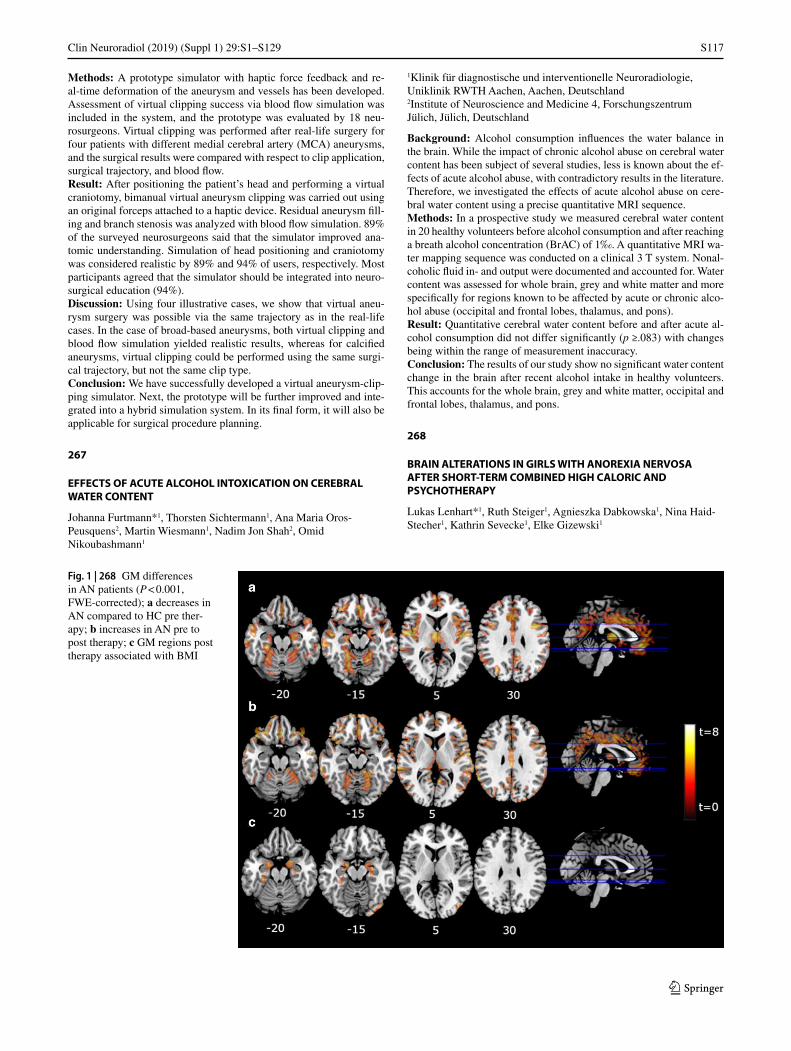
S117
123
Clin Neuroradiol (2019) (Suppl 1) 29:S1–S129
Methods: A prototype simulator with haptic force feedback and re-al-time deformation of the aneurysm and vessels has been developed. Assessment of virtual clipping success via blood flow simulation was included in the system, and the prototype was evaluated by 18 neu-rosurgeons. Virtual clipping was performed after real-life surgery for four patients with different medial cerebral artery (MCA) aneurysms, and the surgical results were compared with respect to clip application, surgical trajectory, and blood flow.Result: After positioning the patient’s head and performing a virtual craniotomy, bimanual virtual aneurysm clipping was carried out using an original forceps attached to a haptic device. Residual aneurysm fill-ing and branch stenosis was analyzed with blood flow simulation. 89% of the surveyed neurosurgeons said that the simulator improved ana-tomic understanding. Simulation of head positioning and craniotomy was considered realistic by 89% and 94% of users, respectively. Most participants agreed that the simulator should be integrated into neuro-surgical education (94%).Discussion: Using four illustrative cases, we show that virtual aneu-rysm surgery was possible via the same trajectory as in the real-life cases. In the case of broad-based aneurysms, both virtual clipping and blood flow simulation yielded realistic results, whereas for calcified aneurysms, virtual clipping could be performed using the same surgi-cal trajectory, but not the same clip type.Conclusion: We have successfully developed a virtual aneurysm-clip-ping simulator. Next, the prototype will be further improved and inte-grated into a hybrid simulation system. In its final form, it will also be applicable for surgical procedure planning.
267
EFFECTS OF ACUTE ALCOHOL INTOXICATION ON CEREBRAL WATER CONTENT
Johanna Furtmann*1, Thorsten Sichtermann1, Ana Maria Oros-Peusquens2, Martin Wiesmann1, Nadim Jon Shah2, Omid Nikoubashmann1
1Klinik für diagnostische und interventionelle Neuroradiologie, Uniklinik RWTH Aachen, Aachen, Deutschland2Institute of Neuroscience and Medicine 4, Forschungszentrum Jülich, Jülich, Deutschland
Background: Alcohol consumption influences the water balance in the brain. While the impact of chronic alcohol abuse on cerebral water content has been subject of several studies, less is known about the ef-fects of acute alcohol abuse, with contradictory results in the literature. Therefore, we investigated the effects of acute alcohol abuse on cere-bral water content using a precise quantitative MRI sequence.Methods: In a prospective study we measured cerebral water content in 20 healthy volunteers before alcohol consumption and after reaching a breath alcohol concentration (BrAC) of 1‰. A quantitative MRI wa-ter mapping sequence was conducted on a clinical 3 T system. Nonal-coholic fluid in- and output were documented and accounted for. Water content was assessed for whole brain, grey and white matter and more specifically for regions known to be affected by acute or chronic alco-hol abuse (occipital and frontal lobes, thalamus, and pons).Result: Quantitative cerebral water content before and after acute al-cohol consumption did not differ significantly (p ≥.083) with changes being within the range of measurement inaccuracy.Conclusion: The results of our study show no significant water content change in the brain after recent alcohol intake in healthy volunteers. This accounts for the whole brain, grey and white matter, occipital and frontal lobes, thalamus, and pons.
268
BRAIN ALTERATIONS IN GIRLS WITH ANOREXIA NERVOSA AFTER SHORT-TERM COMBINED HIGH CALORIC AND PSYCHOTHERAPY
Lukas Lenhart*1, Ruth Steiger1, Agnieszka Dabkowska1, Nina Haid-Stecher1, Kathrin Sevecke1, Elke Gizewski1
Fig. 1 | 268 GM differences in AN patients (P < 0.001, FWE-corrected); a decreases in AN compared to HC pre ther-apy; b increases in AN pre to post therapy; c GM regions post therapy associated with BMI
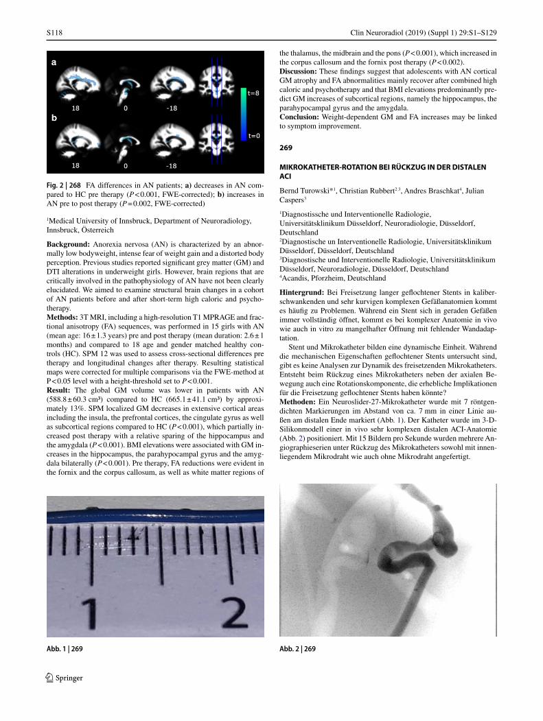
S118
123
Clin Neuroradiol (2019) (Suppl 1) 29:S1–S129
1Medical University of Innsbruck, Department of Neuroradiology, Innsbruck, Österreich
Background: Anorexia nervosa (AN) is characterized by an abnor-mally low bodyweight, intense fear of weight gain and a distorted body perception. Previous studies reported significant grey matter (GM) and DTI alterations in underweight girls. However, brain regions that are critically involved in the pathophysiology of AN have not been clearly elucidated. We aimed to examine structural brain changes in a cohort of AN patients before and after short-term high caloric and psycho-therapy.Methods: 3T MRI, including a high-resolution T1 MPRAGE and frac-tional anisotropy (FA) sequences, was performed in 15 girls with AN (mean age: 16 ± 1.3 years) pre and post therapy (mean duration: 2.6 ± 1 months) and compared to 18 age and gender matched healthy con-trols (HC). SPM 12 was used to assess cross-sectional differences pre therapy and longitudinal changes after therapy. Resulting statistical maps were corrected for multiple comparisons via the FWE-method at P < 0.05 level with a height-threshold set to P < 0.001.Result: The global GM volume was lower in patients with AN (588.8 ± 60.3 cm³) compared to HC (665.1 ± 41.1 cm³) by approxi-mately 13%. SPM localized GM decreases in extensive cortical areas including the insula, the prefrontal cortices, the cingulate gyrus as well as subcortical regions compared to HC (P < 0.001), which partially in-creased post therapy with a relative sparing of the hippocampus and the amygdala (P < 0.001). BMI elevations were associated with GM in-creases in the hippocampus, the parahypocampal gyrus and the amyg-dala bilaterally (P < 0.001). Pre therapy, FA reductions were evident in the fornix and the corpus callosum, as well as white matter regions of
the thalamus, the midbrain and the pons (P < 0.001), which increased in the corpus callosum and the fornix post therapy (P < 0.002).Discussion: These findings suggest that adolescents with AN cortical GM atrophy and FA abnormalities mainly recover after combined high caloric and psychotherapy and that BMI elevations predominantly pre-dict GM increases of subcortical regions, namely the hippocampus, the parahypocampal gyrus and the amygdala.Conclusion: Weight-dependent GM and FA increases may be linked to symptom improvement.
269
MIKROKATHETER-ROTATION BEI RÜCKZUG IN DER DISTALEN ACI
Bernd Turowski*1, Christian Rubbert2 3, Andres Braschkat4, Julian Caspers3
1Diagnostissche und Interventionelle Radiologie, Universitätsklinikum Düsseldorf, Neuroradiologie, Düsseldorf, Deutschland2Diagnostische un Interventionelle Radiologie, Universitätsklinikum Düsseldorf, Düsseldorf, Deutschland3Diagnostische und Interventionelle Radiologie, Universitätsklinikum Düsseldorf, Neuroradiologie, Düsseldorf, Deutschland4Acandis, Pforzheim, Deutschland
Hintergrund: Bei Freisetzung langer geflochtener Stents in kaliber-schwankenden und sehr kurvigen komplexen Gefäßanatomien kommt es häufig zu Problemen. Während ein Stent sich in geraden Gefäßen immer vollständig öffnet, kommt es bei komplexer Anatomie in vivo wie auch in vitro zu mangelhafter Öffnung mit fehlender Wandadap-tation.
Stent und Mikrokatheter bilden eine dynamische Einheit. Während die mechanischen Eigenschaften geflochtener Stents untersucht sind, gibt es keine Analysen zur Dynamik des freisetzenden Mikrokatheters. Entsteht beim Rückzug eines Mikrokatheters neben der axialen Be-wegung auch eine Rotationskomponente, die erhebliche Implikationen für die Freisetzung geflochtener Stents haben könnte?Methoden: Ein Neuroslider-27-Mikrokatheter wurde mit 7 röntgen-dichten Markierungen im Abstand von ca. 7 mm in einer Linie au-ßen am distalen Ende markiert (Abb. 1). Der Katheter wurde im 3-D-Silikonmodell einer in vivo sehr komplexen distalen ACI-Anatomie (Abb. 2) positioniert. Mit 15 Bildern pro Sekunde wurden mehrere An-giographieserien unter Rückzug des Mikrokatheters sowohl mit innen-liegendem Mikrodraht wie auch ohne Mikrodraht angefertigt.
Abb. 2 | 269
Fig. 2 | 268 FA differences in AN patients; a) decreases in AN com-pared to HC pre therapy (P < 0.001, FWE-corrected); b) increases in AN pre to post therapy (P = 0.002, FWE-corrected)
Abb. 1 | 269
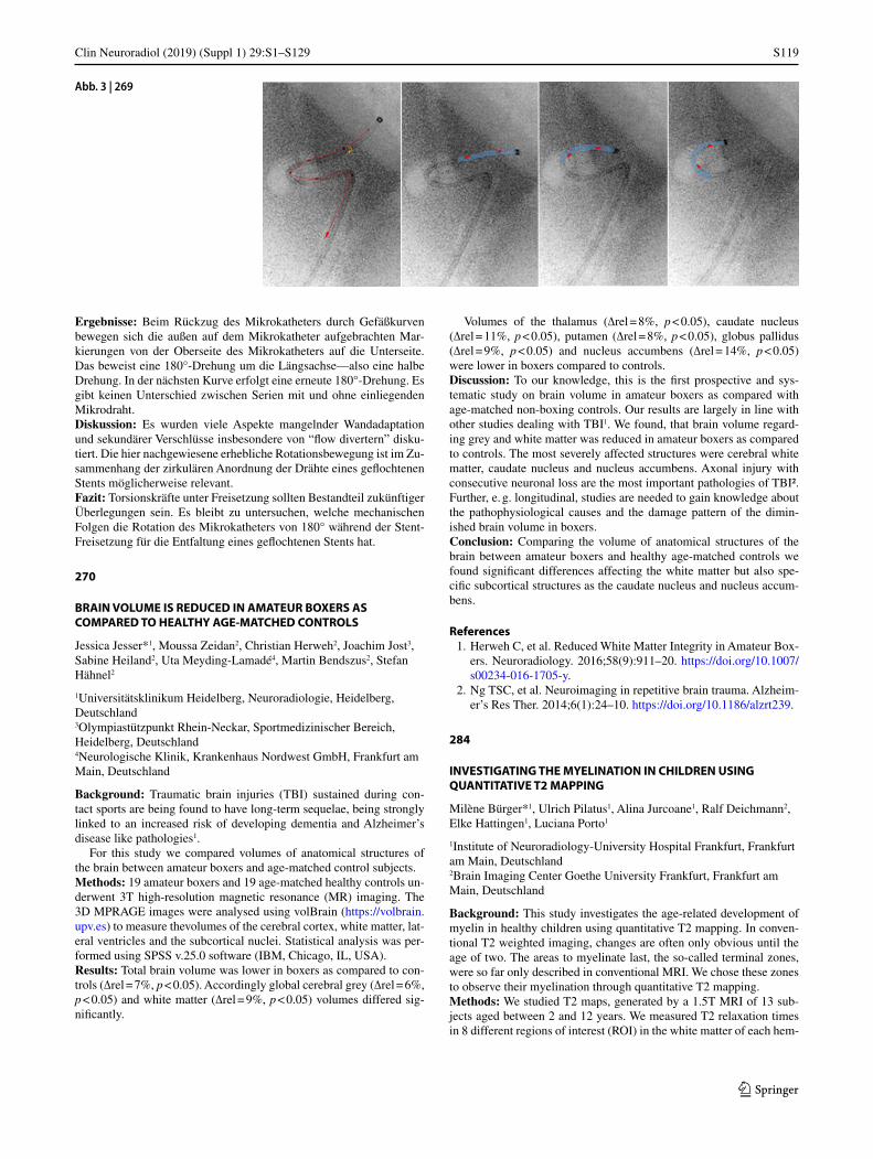
S119
123
Clin Neuroradiol (2019) (Suppl 1) 29:S1–S129
Ergebnisse: Beim Rückzug des Mikrokatheters durch Gefäßkurven bewegen sich die außen auf dem Mikrokatheter aufgebrachten Mar-kierungen von der Oberseite des Mikrokatheters auf die Unterseite. Das beweist eine 180°-Drehung um die Längsachse—also eine halbe Drehung. In der nächsten Kurve erfolgt eine erneute 180°-Drehung. Es gibt keinen Unterschied zwischen Serien mit und ohne einliegenden Mikrodraht.Diskussion: Es wurden viele Aspekte mangelnder Wandadaptation und sekundärer Verschlüsse insbesondere von “flow divertern” disku-tiert. Die hier nachgewiesene erhebliche Rotationsbewegung ist im Zu-sammenhang der zirkulären Anordnung der Drähte eines geflochtenen Stents möglicherweise relevant.Fazit: Torsionskräfte unter Freisetzung sollten Bestandteil zukünftiger Überlegungen sein. Es bleibt zu untersuchen, welche mechanischen Folgen die Rotation des Mikrokatheters von 180° während der Stent-Freisetzung für die Entfaltung eines geflochtenen Stents hat.
270
BRAIN VOLUME IS REDUCED IN AMATEUR BOXERS AS COMPARED TO HEALTHY AGE-MATCHED CONTROLS
Jessica Jesser*1, Moussa Zeidan2, Christian Herweh2, Joachim Jost3, Sabine Heiland2, Uta Meyding-Lamadé4, Martin Bendszus2, Stefan Hähnel2
1Universitätsklinikum Heidelberg, Neuroradiologie, Heidelberg, Deutschland3Olympiastützpunkt Rhein-Neckar, Sportmedizinischer Bereich, Heidelberg, Deutschland4Neurologische Klinik, Krankenhaus Nordwest GmbH, Frankfurt am Main, Deutschland
Background: Traumatic brain injuries (TBI) sustained during con-tact sports are being found to have long-term sequelae, being strongly linked to an increased risk of developing dementia and Alzheimer’s disease like pathologies1.
For this study we compared volumes of anatomical structures of the brain between amateur boxers and age-matched control subjects.Methods: 19 amateur boxers and 19 age-matched healthy controls un-derwent 3T high-resolution magnetic resonance (MR) imaging. The 3D MPRAGE images were analysed using volBrain (https://volbrain.upv.es) to measure thevolumes of the cerebral cortex, white matter, lat-eral ventricles and the subcortical nuclei. Statistical analysis was per-formed using SPSS v.25.0 software (IBM, Chicago, IL, USA).Results: Total brain volume was lower in boxers as compared to con-trols (∆rel = 7%, p < 0.05). Accordingly global cerebral grey (∆rel = 6%, p < 0.05) and white matter (∆rel = 9%, p < 0.05) volumes differed sig-nificantly.
Volumes of the thalamus (∆rel = 8%, p < 0.05), caudate nucleus (∆rel = 11%, p < 0.05), putamen (∆rel = 8%, p < 0.05), globus pallidus (∆rel = 9%, p < 0.05) and nucleus accumbens (∆rel = 14%, p < 0.05) were lower in boxers compared to controls.Discussion: To our knowledge, this is the first prospective and sys-tematic study on brain volume in amateur boxers as compared with age-matched non-boxing controls. Our results are largely in line with other studies dealing with TBI1. We found, that brain volume regard-ing grey and white matter was reduced in amateur boxers as compared to controls. The most severely affected structures were cerebral white matter, caudate nucleus and nucleus accumbens. Axonal injury with consecutive neuronal loss are the most important pathologies of TBI². Further, e. g. longitudinal, studies are needed to gain knowledge about the pathophysiological causes and the damage pattern of the dimin-ished brain volume in boxers.Conclusion: Comparing the volume of anatomical structures of the brain between amateur boxers and healthy age-matched controls we found significant differences affecting the white matter but also spe-cific subcortical structures as the caudate nucleus and nucleus accum-bens.
References 1. Herweh C, et al. Reduced White Matter Integrity in Amateur Box-
ers. Neuroradiology. 2016;58(9):911–20. https://doi.org/10.1007/s00234-016-1705-y.
2. Ng TSC, et al. Neuroimaging in repetitive brain trauma. Alzheim-er’s Res Ther. 2014;6(1):24–10. https://doi.org/10.1186/alzrt239.
284
INVESTIGATING THE MYELINATION IN CHILDREN USING QUANTITATIVE T2 MAPPING
Milène Bürger*1, Ulrich Pilatus1, Alina Jurcoane1, Ralf Deichmann2, Elke Hattingen1, Luciana Porto1
1Institute of Neuroradiology-University Hospital Frankfurt, Frankfurt am Main, Deutschland2Brain Imaging Center Goethe University Frankfurt, Frankfurt am Main, Deutschland
Background: This study investigates the age-related development of myelin in healthy children using quantitative T2 mapping. In conven-tional T2 weighted imaging, changes are often only obvious until the age of two. The areas to myelinate last, the so-called terminal zones, were so far only described in conventional MRI. We chose these zones to observe their myelination through quantitative T2 mapping.Methods: We studied T2 maps, generated by a 1.5T MRI of 13 sub-jects aged between 2 and 12 years. We measured T2 relaxation times in 8 different regions of interest (ROI) in the white matter of each hem-
Abb. 3 | 269
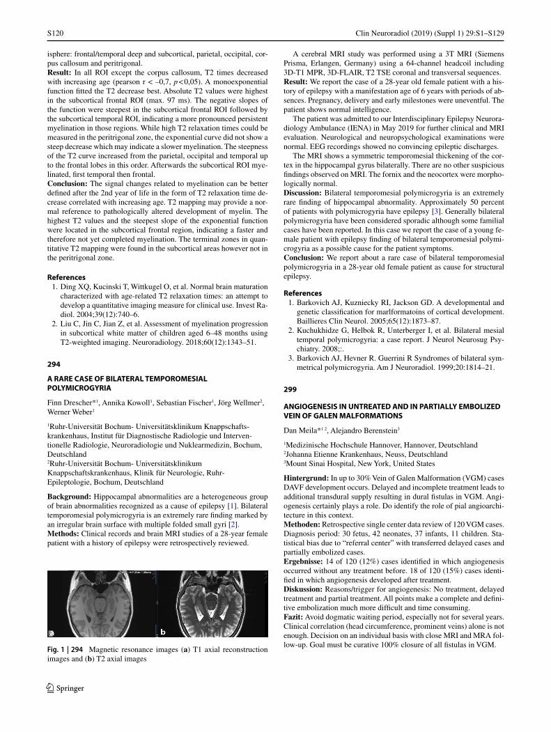
S120
123
Clin Neuroradiol (2019) (Suppl 1) 29:S1–S129
isphere: frontal/temporal deep and subcortical, parietal, occipital, cor-pus callosum and peritrigonal.Result: In all ROI except the corpus callosum, T2 times decreased with increasing age (pearson r < –0,7, p < 0,05). A monoexponential function fitted the T2 decrease best. Absolute T2 values were highest in the subcortical frontal ROI (max. 97 ms). The negative slopes of the function were steepest in the subcortical frontal ROI followed by the subcortical temporal ROI, indicating a more pronounced persistent myelination in those regions. While high T2 relaxation times could be measured in the peritrigonal zone, the exponential curve did not show a steep decrease which may indicate a slower myelination. The steepness of the T2 curve increased from the parietal, occipital and temporal up to the frontal lobes in this order. Afterwards the subcortical ROI mye-linated, first temporal then frontal.Conclusion: The signal changes related to myelination can be better defined after the 2nd year of life in the form of T2 relaxation time de-crease correlated with increasing age. T2 mapping may provide a nor-mal reference to pathologically altered development of myelin. The highest T2 values and the steepest slope of the exponential function were located in the subcortical frontal region, indicating a faster and therefore not yet completed myelination. The terminal zones in quan-titative T2 mapping were found in the subcortical areas however not in the peritrigonal zone.
References 1. Ding XQ, Kucinski T, Wittkugel O, et al. Normal brain maturation
characterized with age-related T2 relaxation times: an attempt to develop a quantitative imaging measure for clinical use. Invest Ra-diol. 2004;39(12):740–6.
2. Liu C, Jin C, Jian Z, et al. Assessment of myelination progression in subcortical white matter of children aged 6–48 months using T2-weighted imaging. Neuroradiology. 2018;60(12):1343–51.
294
A RARE CASE OF BILATERAL TEMPOROMESIAL POLYMICROGYRIA
Finn Drescher*1, Annika Kowoll1, Sebastian Fischer1, Jörg Wellmer2, Werner Weber1
1Ruhr-Universität Bochum- Universitätsklinikum Knappschafts-krankenhaus, Institut für Diagnostische Radiologie und Interven-tionelle Radiologie, Neuroradiologie und Nuklearmedizin, Bochum, Deutschland2Ruhr-Universität Bochum- Universitätsklinikum Knappschaftskrankenhaus, Klinik für Neurologie, Ruhr-Epileptologie, Bochum, Deutschland
Background: Hippocampal abnormalities are a heterogeneous group of brain abnormalities recognized as a cause of epilepsy [1]. Bilateral temporomesial polymicrogyria is an extremely rare finding marked by an irregular brain surface with multiple folded small gyri [2].Methods: Clinical records and brain MRI studies of a 28-year female patient with a history of epilepsy were retrospectively reviewed.
A cerebral MRI study was performed using a 3T MRI (Siemens Prisma, Erlangen, Germany) using a 64-channel headcoil including 3D-T1 MPR, 3D-FLAIR, T2 TSE coronal and transversal sequences.Result: We report the case of a 28-year old female patient with a his-tory of epilepsy with a manifestation age of 6 years with periods of ab-sences. Pregnancy, delivery and early milestones were uneventful. The patient shows normal intelligence.
The patient was admitted to our Interdisciplinary Epilepsy Neurora-diology Ambulance (IENA) in May 2019 for further clinical and MRI evaluation. Neurological and neuropsychological examinations were normal. EEG recordings showed no convincing epileptic discharges.
The MRI shows a symmetric temporomesial thickening of the cor-tex in the hippocampal gyrus bilaterally. There are no other suspicious findings observed on MRI. The fornix and the neocortex were morpho-logically normal.Discussion: Bilateral temporomesial polymicrogyria is an extremely rare finding of hippocampal abnormality. Approximately 50 percent of patients with polymicrogyria have epilepsy [3]. Generally bilateral polymicrogyria have been considered sporadic although some familial cases have been reported. In this case we report the case of a young fe-male patient with epilepsy finding of bilateral temporomesial polymi-crogyria as a possible cause for the patient symptoms.Conclusion: We report about a rare case of bilateral temporomesial polymicrogyria in a 28-year old female patient as cause for structural epilepsy.
References 1. Barkovich AJ, Kuzniecky RI, Jackson GD. A developmental and
genetic classification for marlformatoins of cortical development. Baillieres Clin Neurol. 2005;65(12):1873–87.
2. Kuchukhidze G, Helbok R, Unterberger I, et al. Bilateral mesial temporal polymicrogyria: a case report. J Neurol Neurosug Psy-chiatry. 2008;:.
3. Barkovich AJ, Hevner R. Guerrini R Syndromes of bilateral sym-metrical polymicrogyria. Am J Neuroradiol. 1999;20:1814–21.
299
ANGIOGENESIS IN UNTREATED AND IN PARTIALLY EMBOLIZED VEIN OF GALEN MALFORMATIONS
Dan Meila*1 2, Alejandro Berenstein3
1Medizinische Hochschule Hannover, Hannover, Deutschland2Johanna Etienne Krankenhaus, Neuss, Deutschland3Mount Sinai Hospital, New York, United States
Hintergrund: In up to 30% Vein of Galen Malformation (VGM) cases DAVF development occurs. Delayed and incomplete treatment leads to additional transdural supply resulting in dural fistulas in VGM. Angi-ogenesis certainly plays a role. Do identify the role of pial angioarchi-tecture in this context.Methoden: Retrospective single center data review of 120 VGM cases. Diagnosis period: 30 fetus, 42 neonates, 37 infants, 11 children. Sta-tistical bias due to “referral center” with transferred delayed cases and partially embolized cases.Ergebnisse: 14 of 120 (12%) cases identified in which angiogenesis occurred without any treatment before. 18 of 120 (15%) cases identi-fied in which angiogenesis developed after treatment.Diskussion: Reasons/trigger for angiogenesis: No treatment, delayed treatment and partial treatment. All points make a complete and defini-tive embolization much more difficult and time consuming.Fazit: Avoid dogmatic waiting period, especially not for several years. Clinical correlation (head circumference, prominent veins) alone is not enough. Decision on an individual basis with close MRI and MRA fol-low-up. Goal must be curative 100% closure of all fistulas in VGM.
Fig. 1 | 294 Magnetic resonance images (a) T1 axial reconstruction images and (b) T2 axial images
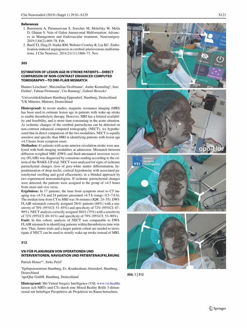
S121
123
Clin Neuroradiol (2019) (Suppl 1) 29:S1–S129
References 1. Berenstein A, Paramasivam S, Sorscher M, Molofsky W, Meila
D, Ghatan S. Vein of Galen Aneurysmal Malformation: Advanc-es in Management and Endovascular treatment. Neurosurgery. 2019;1;84(2):469–78. Feb.
2. Buell TJ, Ding D, Starke RM, Webster Crowley R, Liu KC. Embo-lization-induced angiogenesis in cerebral arteriovenous malforma-tions. J Clin Neurosci. 2014;21(11):1866–71. Nov.
305
ESTIMATION OF LESION AGE IN STROKE PATIENTS—DIRECT COMPARISON OF NON-CONTRAST ENHANCED COMPUTED TOMOGRAPHY—TO DWI-FLAIR MISMATCH
Hannes Leischner1, Maximilian Groffmann1, Andre Kemmling2, Jens Fiehler1, Fabian Flottmann1, Uta Hanning1, Gabriel Broocks1
1Universitätsklinikum Hamburg-Eppendorf, Hamburg, Deutschland2UK Münster, Münster, Deutschland
Hintergrund: In recent studies, magnetic resonance imaging (MRI) has been used to estimate lesion age in patients with wake-up stroke to enable thrombolytic therapy. However, MRI has a limited availabil-ity and feasibility, and is more time-consuming in the acute situation. As ischemic changes of the cerebral parenchyma can be detected on non-contrast enhanced computed tomography (NECT), we hypothe-sized that in direct comparison of the two modalities, NECT is equally sensitive and specific than MRI in identifying patients with lesion age <4.5 hours from symptom onset.Methoden: 41 patients with acute anterior circulation stroke were ana-lyzed with both imaging modalities at admission. Mismatch between diffusion-weighted MRI (DWI) and fluid-attenuated inversion recov-ery (FLAIR) was diagnosed by consensus reading according to the cri-teria of the WAKE-UP trial. NECT were analyzed for signs of ischemic parenchymal changes (loss of grey-white matter differentiation, hy-poattenuation of deep nuclei, cortical hypodensity with associated pa-renchymal swelling and gyral effacement), in a blinded approach by two experienced neuroradiologists. If ischemic parenchymal changes were detected, the patients were assigned to the group of >4.5 hours from onset and vice versa.Ergebnisse: In 17 patients, the time from symptom onset to CT im-aging was <4.5 h and 24 patients presented >4.5 h (range: 0.5–7.6 h). The median time from CT to MRI was 36 minutes (IQR: 24–55). DWI-FLAIR mismatch correctly assigned 28/41 patients (68%) with a sen-sitivity of 70% (95%CI: 51–85%) and specificity of 72% (95%CI: 47–90%). NECT analysis correctly assigned 30/41 (75%) with a sensitivity of 72% (95%CI: 69–91%) and specificity of 79% (95%CI: 53–90%).Fazit: In this cohort, analysis of NECT was comparable to DWI-FLAIR mismatch in identifying patients within thrombolysis time win-dow. Thus, future trials and a larger patient cohort are needed to inves-tigate if NECT can be used to stratify wake-up stroke instead of MRI.
312
VSI FÜR PLANUNGEN VON OPERATIONEN UND INTERVENTIONEN, NAVIGATION UND PATIENTENAUFKLÄRUNG
Patrick House*1, Sirko Pelzl2
1Epilepsiezentrum Hamburg, Ev. Krankenhaus Alsterdorf, Hamburg, Deutschland2apoQlar GmbH, Hamburg, Deutschland
Hintergrund: Mit Virtual Surgery Intelligence (VSI, www.vsi.health) lassen sich MRTs und CTs durch eine Mixed Reality Brille 3-dimen-sional mit beliebiger Perspektive als Projektion im Raum betrachten.
Abb. 1 | 312
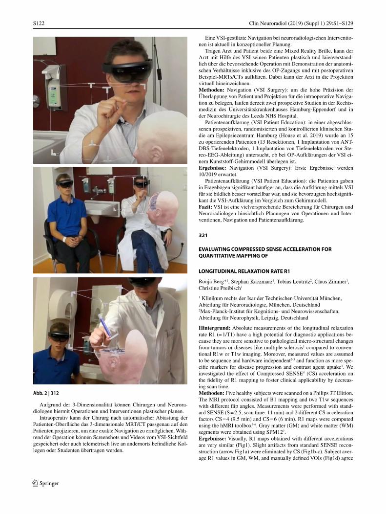
S122
123
Clin Neuroradiol (2019) (Suppl 1) 29:S1–S129
Aufgrund der 3-Dimensionalität können Chirurgen und Neurora-diologen hiermit Operationen und Interventionen plastischer planen.
Intraoperativ kann der Chirurg nach automatischer Abtastung der Patienten-Oberfläche das 3-dimensionale MRT/CT passgenau auf den Patienten projizieren, um eine exakte Navigation zu ermöglichen. Wäh-rend der Operation können Screenshots und Videos vom VSI-Sichtfeld gespeichert oder auch telemetrisch live an andernorts befindliche Kol-legen oder Studenten übertragen werden.
Eine VSI-gestützte Navigation bei neuroradiologischen Interventio-nen ist aktuell in konzeptioneller Planung.
Tragen Arzt und Patient beide eine Mixed Reality Brille, kann der Arzt mit Hilfe des VSI seinen Patienten plastisch und laienverständ-lich über die bevorstehende Operation mit Demonstration der anatomi-schen Verhältnisse inklusive des OP-Zugangs und mit postoperativen Beispiel-MRTs/CTs aufklären. Dabei kann der Arzt in die Projektion virtuell hineinzeichnen.Methoden: Navigation (VSI Surgery): um die hohe Präzision der Überlappung von Patient und Projektion für die intraoperative Naviga-tion zu belegen, laufen derzeit zwei prospektive Studien in der Rechts-medizin des Universitätskrankenhauses Hamburg-Eppendorf und in der Neurochirurgie des Leeds NHS Hospital.
Patientenaufklärung (VSI Patient Education): in einer abgeschlos-senen prospektiven, randomisierten und kontrollierten klinischen Stu-die am Epilepsiezentrum Hamburg (House et al. 2019) wurde an 15 zu operierenden Patienten (13 Resektionen, 1 Implantation von ANT-DBS-Tiefenelektroden, 1 Implantation von Tiefenelektroden vor Ste-reo-EEG-Ableitung) untersucht, ob bei OP-Aufklärungen der VSI ei-nem Kunststoff-Gehirnmodell überlegen ist.Ergebnisse: Navigation (VSI Surgery): Erste Ergebnisse werden 10/2019 erwartet.
Patientenaufklärung (VSI Patient Education): die Patienten gaben in Fragebögen signifikant häufiger an, dass die Aufklärung mittels VSI für sie bildlich besser vorstellbar war, und sie bevorzugten hochsignifi-kant die VSI-Aufklärung im Vergleich zum Gehirnmodell.Fazit: VSI ist eine vielversprechende Bereicherung für Chirurgen und Neuroradiologen hinsichtlich Planungen von Operationen und Inter-ventionen, Navigation und Patientenaufklärung.
321
EVALUATING COMPRESSED SENSE ACCELERATION FOR QUANTITATIVE MAPPING OF
LONGITUDINAL RELAXATION RATE R1
Ronja Berg*1, Stephan Kaczmarz1, Tobias Leutritz2, Claus Zimmer1, Christine Preibisch1
1 Klinikum rechts der Isar der Technischen Universität München, Abteilung für Neuroradiologie, München, Deutschland2Max-Planck-Institut für Kognitions- und Neurowissenschaften, Abteilung für Neurophysik, Leipzig, Deutschland
Hintergrund: Absolute measurements of the longitudinal relaxation rate R1 (= 1/T1) have a high potential for diagnostic applications be-cause they are more sensitive to pathological micro-structural changes from tumors or diseases like multiple sclerosis1 compared to conven-tional R1w or T1w imaging. Moreover, measured values are assumed to be sequence and hardware independent2-3 and function as more spe-cific markers for disease progression and contrast agent uptake1. We investigated the effect of Compressed SENSE4 (CS) acceleration on the fidelity of R1 mapping to foster clinical applicability by decreas-ing scan time.Methoden: Five healthy subjects were scanned on a Philips 3T Elition. The MRI protocol consisted of B1 mapping and two T1w sequences with different flip angles. Measurements were performed with stand-ard SENSE (S = 2.5, scan time: 11 min) and 2 different CS acceleration factors CS = 4 (9.5 min) and CS = 6 (6 min). R1 maps were computed using the hMRI toolbox5-6. Gray matter (GM) and white matter (WM) segments were obtained using SPM127.Ergebnisse: Visually, R1 maps obtained with different accelerations are very similar (Fig1). Slight artifacts from standard SENSE recon-struction (arrow Fig1a) were eliminated by CS (Fig1b-c). Subject aver-age R1 values in GM, WM, and manually defined VOIs (Fig1d) agree
Abb. 2 | 312

S123
123
Clin Neuroradiol (2019) (Suppl 1) 29:S1–S129
well between the 3 protocols (Fig2). The largest variance of R1 val-ues across subjects is found with SENSE, while CS reduces R1 value variations across subjects (Fig2). Both CS protocols show comparable results.Diskussion: Our comparison of R1 mapping protocols with different accelerations reveals R1 maps with comparable quality. Quantitative R1 values depend neither on acceleration technique (SENSE vs. CS) nor factor (CS = 4 vs. CS = 6).
Our results suggest that compressed SENSE, with acceleration fac-tors up to at least 6, can be used for quantitative R1 mapping without loss of fidelity but with the advantage of clearly reduced scan times.Fazit: CS is highly promising to establish quantitative R1 mapping within clinically feasible scan times.
References 1. Tsialios 2017 JPhys 2. Preibisch 2009 MRM 3. Baudrexel 2018 MRM 4. Geerts-Ossevoort 2018 Philips 5. Tabelow 2019 Neuroimage 6. Weiskopf 2015 CurrOpinNeurol 7. www.fil.ion.ucl.ac.uk/spm
333
TWO RARE CASES OF BRAIN ABSCESSES CAUSED BY ACTINOMYCES MEYERI
Katharina Walner2, Isabell König2, Christina Schulze3, Sebastian Fischer2, Werner Weber2, Annika Kowoll2
1Diagnostische und interventionelle Radiologie und Nuklearmedizin, Knappschaftskrankenhaus Bochum Langendreer, Bochum, Deutschland2Diagnostische und interventionelle Radiologie und Nuklearmedizin, Knappschaftskrankenhaus Bochum-Langendreer, Bochum, Deutschland3Medizinische Klinik, Knappschaftskrankenhaus Bochum-Langendreer, Bochum, Deutschland
Background: We present two cases of brain abscesses, caused by Ac-tinomyces meyeri. Brain abscesses caused by Actinomyces meyeri are rare, only a few cases are known in literature [1].Methods: Clinical information and cranial MRI studies of a two pa-tients with microbiologically confirmed Actinomyces meyeri-brain-ab-scesses were retrospectively reviewed regarding their symptoms, their predisposing factors, their imaging as well as to their response to an-tibiotic therapy.
Fig. 1 | 321 Exemplary R1 maps of a single subject with different accelerations
Fig. 2 | 321 Comparison of subject average quantitative R1 values from automatically segmented and manually selected VOIs in GM and WM
Fig. 1 | 333 MRI of 60 Year old patient; a) T1 post-KM transversal; b) DWI transversal, 27 year old patient; c) T1 post-KM transversal; d) DWI transversal
S124
123
Clin Neuroradiol (2019) (Suppl 1) 29:S1–S129
Result: A 60-year old male patient and a 27-year old male patient presented at our hospital with neurological symptoms and raised tem-peratures. Both had no history of dental extractions, iv drug-abuse or immunosuppression. In both cases the MRI showed a singular irregu-lar bounded lesion with a ring-enhancement as well as strong diffusion restriction and a perifocal oedema. In both cases an abscess caused by Actinomyces meyeri was microbiologically determined as the cause. IV antibiotic therapy was started in both cases.Discussion: Brain abscesses caused by Actinomyces meyeri in the lit-erature are very rare. Until know it is not explained how Actinomyces passes the blood brain barrier. In current literature there is described a case of Actinomyces-brain-abscess after dental extraction [2] but both of our patients did not have a dental intervention before suffering under brain abscesses. So in both cases the etiology remains unclear.Conclusion: We report about two cases of rare brain abscesses caused by Actinomyces meyeri. Both patients did not have any predisposing factors.
References 1. Yang WT, Grant M. Actinomyces neuii: a case report of a rare
cause of acute infective endocarditis and literature review. BMC Infect Dis. 2019;19(1):511. https://doi.org/10.1186/s12879-019-4149-2.
2. Clancy U, Ronayne A, Prentice MB, Jackson A. Actinomyces meyeri brain abscess following dental extraction. Bmj Case Rep. 2015;13:2015. https://doi.org/10.1136/bcr-2014-207548.
343
TRANS-RADIAL EMBOLIZATION OF MIDDLE-MENINGEAL ARTERY IN TWO PATIENTS WITH CHRONIC SUBDURAL HEMATOMAS
Johannes Hensler*1, Jawid Madjidyar1, Fritz Wodarg1, Olav Jansen1
1Universitätsklinikum Schleswig-Holstein, Campus Kiel, Klinik für Radiologie und Neuroradiologie, Kiel, Deutschland
Background: Chronic subdural hematomas are a common neurosur-gical disease. In addition to conventional surgical therapy, e. g. burr hole, encouraging data regarding interventional therapy with angio-graphic embolization of the meningeal media artery (MMA) as prima-ry therapy or prevention of recurrence have been published recently [1]. We describe two cases with chronic subdural hematomas in which this procedure was performed under local anesthesia using the trans-ra-dial approach.Methods: Angiographic embolization of MMA in chronic subdural hematoma was performed in two patients via trans-radial approach with a 5F-Glidesheath Slender® (Terumo, Japan). A bi-axial setup with Envoy-5F guiding-catheter (Synthes, PA) was placed in common carotid artery. Embolization was performed via a 2.4F microcatheter (Progreat, Terumo) in MMA distal to spinous foramen with polyvi-nyl-alcohol-particles (PVA, Contour 250–355 µm, Boston Scientific, MA). In one case additional coil-embolization was performed with two pushable coils (Fig. 8–18, Boston Scientific).Result: In case 1 (male, 79y) embolization of the left MMA with addi-tional coils, in case 2 (male, y) embolization of the right MMA anterior branch was performed via the trans-radial approach in local anesthesia. In both cases a successful and complete embolization of the MMA or the anterior branch was achieved.Discussion: There were no peri- or post-interventional complications. Embolization was performed to prevent recurrence 5 and 3 days after surgical burr hole and drainage or osteoplastic trepanation, respective-ly. Patient 1 could be discharged without neurological deficits on the day of intervention, patient 2 showed a clinical improvement at dis-
charge and a complete regression of the hematoma at 90-d follow-up CCT with no remaining neurological deficits.Conclusion: Embolization of MMA via the trans-radial approach with PVA particles and coils was performed successfully and safe and may be a minimally invasive therapy option for prevention of recurrence of chronic subdural hematomas. Nevertheless, further studies regarding this procedure are necessary.
References 1. Link TW, Boddu S, Paine SM, et al. Middle Meningeal Artery Em-
bolization for Chronic Subdural Hematoma: A Series of 60 Cases. Neurosurgery. 2018; https://doi.org/10.1093/neuros/nyy521.
362
KORRELATION VON RADIOLOGISCHEN PARAMETERN BEI MORBUS MENIÈRE
Desiree Grosser*1, Friedrich Götz1, Kerstin Willenborg2, Thomas Lenarz2, Heinrich Lanfermann1, Anja Giesemann1
1Diagnostische und Interventionelle Neuroradiologie, Medizinische Hochschule Hannover, Hannover, Deutschland2Klinik für Otorhinolaryngologie, Medizinische Hochschule Hannover, Hannover, Deutschland
Hintergrund: Die Diagnostik des M. Menière wurde radiologisch durch die Hydrops-Darstellung mittels MRT erweitert. Der M. Me-nière umfasst eine Tieftonhörminderung, Tinnitus und Schwindel. Im MRT kann eine Differenzierung in Beteiligung von Cochlea und Vesti-bulum erfolgen. CT Morphologisch ist der vestibuläre Aquädukt (VA) bei M. Menière schmaler ausgeprägt. Es soll der Zusammenhang zwi-schen Hydropsdarstellung (vestibulär und/oder cochleär) und einem engen bzw. nicht erkennbaren VA untersucht werden.Methoden: Es wurden 30 Patienten (60 Ohren) mit MR-morpholo-gisch eindeutigem Hydrops mit der Weite des im CT sichtbaren VA ausgewertet. Der Hydrops wurde unterteilt nach Lokalisation (nur cochleär (G1), nur vestibulär oder beides (G2)). Die Graduierung des VA im CT erfolgte in nicht abgrenzbar, schmal (Weite < = 1 mm), nor-male Weite (>1 mm). Die Daten wurden mittels Excel korreliert.Ergebnisse: Von den 60 Ohren wiesen 24 einen nur cochleären Hy-drops, 4 einen isoliert vestibulären und 29 einen sowohl cochleär und vestibulären Hydrops auf. 3 kontralaterale Ohren zeigten keinen Hy-drops. In G1 betrug der Anteil des nicht abgrenzbaren VA 17%, in G2 42%. Ein schmaler VA wurde in G1 in 54% gefunden, in G2 in 52%. Ein normaler VA fand sich insgesamt in nur 14%. Die 3 normalen Oh-ren weisen einen normal weiten VA auf. Es fand sich ein schmaler oder nicht abgrenzbarer VA auch häufiger bei vestibulärer Beteiligung als ohne.Diskussion: Bei nachgewiesenem Hydrops ist auch der VA häufig schmaler ausgeprägt bzw. nicht abgrenzbar. Dies bestätigt die Daten von Krombach et al. [1]. Damit liegt mit dem CT Befund eines nicht abgrenzbaren VA ein Hinweis für M. Menière vor. Die Ätiologie des M. Menière ist weiterhin nicht geklärt. Ein Erklärungsmodell, welches einen Endolymphtransport in den Saccus endolyphaticus beinhaltet, der sicherlich bei geringem Volumen desselben eingeschänkt ist kann durch diese Daten unterstützt werden.Fazit: Die Daten können die Hypothese, dass der Druckausgleich in den Saccus endolymphaticus eine Rolle für die Entstehung spielt [2], unterstützen.
Literatur 1. Krombach GA, van den Boom M, Di Martino E, Schmitz-Rode T,
Westhofen M, Prescher A, et al. Computed tomography of the in-ner ear: size of anatomical structures in the normal temporal bone
S125
123
Clin Neuroradiol (2019) (Suppl 1) 29:S1–S129
and in the temporal bone of patients with Menière’s disease. Eur Radiol. 2005;15(8):1505–13.
2. Plontke SK, Gürkov R. Menière’s Disease. Laryngorhinootologie. 2015;94(8):530–54.
367
EARLY MRN-PARAMETERS TO PREDICT SUCCESSFUL NERVE REGENERATION FOLLOWING SEVERE INJURY
Daniel Schwarz*1, Martin Moll1, Alfred Tessmann2, Volker Sturm1, Manuel Fischer1, Ulrich Kneser2, Martin Bendszus1, Leila Harhaus2, Sabine Heiland1, Florian Neubrech2 3
1Universitätsklinikum Heidelberg, Abteilung für Neuroradiologie, Heidelberg, Deutschland2BG Unfallklinik Ludwigshafen, Klinik für Hand-, Plastische und Rekonstruktive Chirurgie, Schwerbrandverletztenzentrum, Klinik für Plastische und Handchirurgie der Universität Heidelberg, Ludwigshafen, Deutschland3BG Unfallklinik Frankfurt am Main, Abteilung für Plastische, Hand- und Rekonstruktive Chirurgie, Akademisches Lehrkrankenhaus Goethe-Universität Frankfurt am Main, Frankfurt, Deutschland
Hintergrund: Early predictors of successful nerve regeneration are urgently needed to avoid a waste of time and thus functional outcome following severe nerve injury. Up to date, however, clinical decision making mainly relies on a watch-and-wait strategy to identify poten-tial candidates for surgical intervention in the case of failing re-inner-vation [1].
We used a ultra high field magnetic resonance neurography (MRN) approach in a rodent model of peripheral nerve injury to identify early MR-parameters within the nerve which may be used to predict func-tional outcome.Methoden: 12 female S.-Dawley rats underwent transection of the right Median nerve at upper arm level. In the gold standard group (n = 6, “Gold”), an autograft of reversed Median nerve (1 cm) was re-implanted while in the negative control group (n = 6, “Neg”), a 1 cm nerve gap was left in place. These two groups were imaged four times at forearm level on a 9.4 T MR-scanner 3, 10, 30 and 90 d post surgery. The protocol comprised a set of T2-w RARE, T1-w FLASH (± MT-pulse), DTI and MSME sequences. Groups were followed behaviour-ally and histological parameters were obtained after sacrifice. Results were compared longitudinally and between groups.Ergebnisse: At 30 d, significant differences between Gold and Neg are noted for the magnetization transfer ratio (MTR), apparent diffusion coefficient (ADC) and radial diffusivity (RD) while other parameters show similar pathological alterations, namely T2-Time, proton density, fractional anisotropy and cross sectional area (CSA). At that time, only first signs of clinical recovery (~10% of the grip strength plateau) are present in the Gold while no function is seen in the Neg. The first time points do not show such group differences and at the last time point, only a larger CSA within the Gold is of note. Substantial phenotypical differences persist with the Neg only regaining ~20% grip strength of the Gold and showing a markedly reduced axon density.Fazit: Using ultra high field MRN in a rigorous preclinical in vivo model we identify MTR, ADC and RD as early in vivo parameters of the regenerating nerve which may potentially be used in a translational setting to predict functional outcome and guide therapeutic decision making in the future.
References 1. Schwarz D, Pedro MT, Brand C, Bendszus M, Antoniadis G.
Nerve injuries and traumatic lesions of the brachial plexus: Imag-ing diagnostics and therapeutic options. Radiologe. 2017;57:184–94. https://doi.org/10.1007/s00117-017-0207-1.
S126
123
Clin Neuroradiol (2019) (Suppl 1) 29:S1–S129
Autorenverzeichnis
A
Abdullayev, Nuran 218, 237Abu-Fares, Omar 128Albarqouni, Shadi 241Alexandrou, Maria 237Alonso, Angelika 112, 181, 364Aludin, Schekeb 274Aludin, Zarlasht 274Al-Zghloul, Mansour 112, 181Andresen, Gisa 277Andresen, Julian Ramin 62Andresen, Reimer 62, 87, 243Anraths, Max 271Anwar, Abdul Rauf 74Arnold, Marcel 357Auer, Timo Alexander 38Aulmann, Linda 68Aumann, Astrid 222Austein, Friederike 139, 183
B
Bach, Jan Philipp 293Bähr, Oliver 311Barg-Hock, Hannelore 126Barkhof, Frederik 214Barlinn, Jessica 291Bartsch, Andreas 186Bauknecht, Hans-Christian 341Baum, Thomas 96, 109, 111,
164, 205,314Baur, Christoph 241Beck, J 230Becker, Lucas Ivo Emanuel 296Behme, Daniel 218, 322, 324Bellwald, Sebastian 357Bender, Benjamin 176Bendszus, Martin 65, 73, 80, 82,
86, 89, 130, 134, 135, 136, 137, 138, 174, 186, 190, 193, 194, 196, 197, 211, 270, 365, 367
Berberich, Anne 187Berberich, Götz 105Berenstein, Alejandro 299Berg, Philipp 188, 301Berg, Ronja 143, 321Berkefeld, Joachim 313Berlis, Ansgar 117, 138, 322Berndt, Maria 99, 103, 125, 127,
129, 132Berthold, Christoph 199Beseoglu, Kerim 348Bester, Maxim 110, 166, 246Beuing, Oliver 188, 340Bhogal, Pervinder 184Birch, Giselle 214Bison, Brigitte 220Bittner, Stefan 74Blanc, Raphael 138
Blüm, Philipp 132Boeckh-Behrens, Tobias 99,
103, 125, 127, 129, 132Böhm, Christoph 314Böhme, Johannes 97, 112, 181,
364Böhmer, Maik 292Bohner, Georg 341Bojunga, Jörg 311Bonati, Leo 63Bonekamp, Susanne 136, 137Borggrefe, Jan 104, 218, 223,
224, 345Börm, Wolfgang 277Brandhofe, Annemarie 300, 313Braschkat, Andres 269Braun, Michael 57Brawanski, Alexander 88Brehm, Alex 94, 323, 324Brekenfeld, Caspar 134, 203,
237, 251, 255, 347Bremer, Benno 105Brockmann, Carolin 147Brockmann, Marc A. 147, 172Bronzlik, Paul 128, 171Broocks, Gabriel 42, 64, 70, 75,
79, 163, 208, 251, 305, 370Bsteh, Gabriel 106Buhk, Jan-Hendrik 100, 340Bürger, Milène 284Burian, Egon 178, 205, 207, 226
C
Caldeira, Liliana 345Calzà, Jessica 105Cantré, Daniel 260, 358Caspers, Julian 51, 67, 204, 256,
269, 287, 348Cattaneo, Giorgio 188Chapot, Rene 246, 340Chard, Declan 214Cheng, Bastian 251Cho, Chang Gyu 97, 280Chung, Karen 214Ciccarelli, Olga 214Clajus, Christin 108Coras, Roland 87Cordt, Justus 142Cordts, Isabell 330Costalat, Vincent 138Cyran, Clemens 229
D
Dabkowska-Mika, Agniesz-ka 252, 268
Dadak, Mete 171Dargazanli, Cyril 138Deb-Chatterji, Milani 203, 237,
251, 255, 347
Deichmann, Ralf 284, 300Deisenhammer, Florian 106Demerath, Theo 309, 63Deschauer, Marcus 330Deshmane, Anagha 176Deutschmann, Hannes 69, 81Dieckmeyer, Michael 157, 205Dimitrov, Ivaylo 293Ding, Andreas 188Ding, Xiao-Qi 126, 171Dinse, Bianca 130Ditz, Claudia 68Dobrocky, Tomas 357Doenitz, Christian 88Döpfert, Jörg 157Dörfler, Arnd 266Dorn, Franziska 223, 224, 340,
355Drescher, Finn 294Drumm, Jörg 199Dührsen, Max 229Duman, Ikram Eda 158Dyrba, Martin 358
E
Egger, Karl 158, 230Eichinger, Paul 124Eickhoff, Simon 256Eisawy, Rami 360Eisenberger, Hanna Josephin 94Eisenhut, Felix 266Elsayed, Sarah 79, 208Emmer, Alexander 184Engel, Jakob 183Engellandt, Kay 291Enzinger, Christian 69Epp, Samira 145Eppinger, Sebastian 69Ernemann, Ulrike 176Ernst, Marielle Sophie 42Eshaghi, Arman 214
F
Faizy, Tobias D. 110, 163, 166Fandler, Simon 69Fandler-Höfler, Simon 81Farahat, Amr 289Fassbender, Klaus 192Fazekas, Franz 69, 81Fehrenbach, Uli 38Fenz, Wolfgang 253Ferrera Bertran, Pere 157Fiehler, Jens 42, 64, 70, 75, 79,
110, 134, 163, 166, 203, 208, 237, 246, 251, 255, 286, 305, 347, 370
Finck, Tom 124, 360Fischer, Antonia Carlotta 139Fischer, Manuel 367
Fischer, Sebastian 117, 294, 333Fischer, Urs 356, 357Fleck, Steffen 260Fleischer, Vinzenz 74Flottmann, Fabian 70, 75, 134,
203, 237, 251, 255, 305, 347Flüh, Charlotte 301Fokas, Emmanouil 311Forbrig, Robert 229, 355Förster, Alex 97, 112, 181, 364Franz, Daniela 205Franz, Kea 311Friedrich, Benjamin 99, 103,
125, 127, 129, 132, 330Frysch, Robert 188Fu, Tong 171Fuchs, Simon 341Furtmann, Johanna 267
G
Ganslandt, Thomas 69, 97, 280Gerber, Johannes 291Gerloff, Christian 237, 251Gerry, Stephen 187Gersing, Alexandra S. 314Gessert, Nils 244Giesemann, Anja 362Giretzlehner, Michael 253Gizewski, Elke Ruth 106, 252,
268Gliem, Michael 67, 287Gmeiner, Matthias 253Gnanaprakasam, Velmuru-
gan 115Gnekow, Astrid K. 220Goeldlin, Martina 357Golay, Xavier 176Gonzalez-Escamilla, Gabriel 74Görtz, Lukas 104, 223, 224, 345Göttler, Jens 121, 148, 288, 290Gottschalk, Hannes 183Götz, Friedrich 128, 362Goyal, Mayank 324Gracien, René-Maxime 74, 300Gralla, Jan 356, 357Greve, Tobias 115, 162Groden, Christoph 97, 112, 181,
280, 172, 364Groffmann, Maximilian 305Gröner, Jan 211Groppa, Sergiu 74Grosser, Desiree 362Groth, Friederike 260Groth, Sascha 260Gruber, Andreas 253Grundl, Lioba 124Grundwald, Iris 192Gumbinger, Christoph 135Günther, René 310Gürsel, Deniz A. 105
S127
123
Clin Neuroradiol (2019) (Suppl 1) 29:S1–S129
H
Hadler, Felix 298Hädrich, Kevin 291Haensch, Carl-Albrecht 287Hahn, Arthur 211Hähnel, Stefan 270Haider, Lukas 214Haid-Stecher, Nina 268Hancken, Caroline-Victoria 220Hanning, Uta 42, 64, 70, 75, 79,
163, 208, 246, 251, 286, 292, 305, 347, 370
Hansen, Brian 110, 166Hansen, Mikkel Bo 288Harhaus, Leila 367Harter, Patrick 311Härtig, Wolfgang 184Hartmann, Christian 256Hartmann, Marius 259Hasan, Dimah 313Hasler, Mario 271Hassler, Eva 69Hattingen, Elke 245, 284, 300,311Hauser, Till-Karsten 53Haußmann, Alena 320Hedderich, Dennis 111, 132, 157Heeger, Adrian 204Heesen, Christoph 110, 166Heiland, Dieter Henrik 189Heiland, Pamela 189Heiland, Sabine 194, 211, 270,
367Heimann, Friederike 134Heindel, Walter 292Heinze, Hans-Jochen 289Heldner, Mirjam Rachel 357Hemmer, Bernhard 330Hemmerich, Felix 65, 80, 82,
86, 89, 196, 197,Hemraj, Indradeo 348Hense, Katharina 88Hensler, Johannes 138, 349, 343Herrlinger, Ulrich 245Herweh, Christian 174, 186,
187, 190, 192, 193, 270, 365, 73
Herz, Kai 176Herzberg, Moritz 224Hesse, Amelie 324Hey, Silke 115Hilgenfeld, Tim 211Hinrichs, Hermann 289Hinteregger, Nicole 69, 81Hock, Andreas 115, 143, 288,
290Hoffmann, Karl-Titus 184, 263Hoffstaedter, Felix 256Hohenhaus, M 230Hölter, Philip 266Hopf-Jensen, Silke 271, 276,
277House, Patrick 312Hövener, Jan-Bernd 329Hübschle, Irene 189
Hyder, Fahmeed 121, 288, 290Hyrenbach, Sonja 136, 137
I
Ikenberg, Benno 331Imhoff, Detlef 311Inhuber, Stephanie 205Ischebeck, Anja 252Ivan, Vivien Lorena 67
J
Jacob, Alina 96Jander, Sebastian 67, 287Jannusch, Kai 348Jansen, Olav 139, 142, 144 183,
225, 274, 281, 329, 343, 349Janssen, Hendrik 138, 340Jeibmann, Astrid 286Jende, Johann 194, 211Jensen-Kondering, Ulf 144Jespersen, Sune 110, 166Jesser, Jessica 130, 270Joly, Olivier 192Jost, Joachim 270Juerchott, Alexander 211Jung, Simon 357Junge, Andreas Georg 97, 280Jurcoane, Alina 284
K
Kabbasch, Christoph 218, 223, 224
Kabiri, Reza 42, 163Kaczmarz, Stephan 121, 148,
288, 290, 145, 321Kadziolka, Krzysztof 340Kaesmacher, Johannes 103, 125,
127, 129, 356, 357Kaethner, Christian 266Kalder, Johannes 297Kämpgen, Benedikt 280Kandels, Daniela 220Karampinos, Dimitrios C. 205,
314Karwacki, Grzegorz 309Kaschner, Marius 67, 287, 348Kastrup, Andreas 237Kaufmann, Jörn 92Kazmierczak, Philipp 229Keil, Fee 313Kellner, Elias 158, 230Kemmling, Andre 64, 70, 79,
292, 305, 370Kender, Zoltan 211Kern, Maike 38Kerz, Thomas 147Khadhraoui, Eya 195Khanafer, Ali 195, 285Kickingereder, Philipp 73Killer, Monika 138Kim, Suam 230
Kirschke, Jan S. 96, 101, 109, 111, 115, 164, 205, 314
Kirschner, Stefanie 172Kitzler, Hagen H. 303, 310Klein, Matthias 229Kleine, Justus 341Kleiser, Raimund 150, 262Klempnauer, Jürgen 126Klisch, Joachim 108Klose, Uwe 53Klupp, Elisabeth 205Kneihsl, Markus 69, 81Kneser, Ulrich 367Kniep, Helge 42, 70, 75, 79,
163, 208, 286Koch, Caroline 358Koch, Kathrin 105Koehler, Caroline 303Kollmer, Jennifer 194König, Isabell 117, 333Kopetsch, Christoph 234Kopf, Stefan 211Kopp, Felix K. 109, 111, 164Kortmann, Rolf-Dieter 220Kowoll, Annika 294, 333Kraemer, Christoffer 134Kramer, Andreas 147Kramer, Martin 172Krause, Lars Udo 246Krause, Matthias 184Kreiser, Kornelia 132, 162, 340Kronfeld, Andrea 172Kronthaler, Sophia 314Krüger, Julia 244Kubera, katharina 186Küchler, Jan 68Kufer, Jan 121, 288Kuhlenbäumer, Gregor 144Kuntke, Paul 303, 310Kunz, Wolfgang 229Kurmann, Christoph 357Kurz, Felix Tobias 211Kyselyova, Anna 110, 166
L
Lamplmayr, Lukas 106Lanfermann, Heinrich 126, 128,
171, 362Lang, Stefan 266Langguth, Patrick 139, 144, 183Langner, Sönke 260Larsen, Naomi 139, 183, 274,
281, 301, 349Laukamp, Kai Roman 104, 345Lebenatus, Annett 183Leblhuber, Friedrich 262Lee, John-Ih 287Lee, John-Il 67Lehrke, Stephanie 271Leischner, Hannes 64, 70, 203,
237, 246, 251, 255, 305, 347Leitner, Stephan 254Lemke, Andreas 157Lenarz, Thomas 362
Lenhart, Lukas 106, 268Lennartz, Simon 345Leutritz, Tobias 321Li, Hongwei 124Liebig, Thomas 218, 223, 224,
355Liedke, Marc Olaf 62Liesche, Friederike 98Liman, Jan 39, 94, 249, 323Lindig, Tobias 176Lindner, Thomas 183Lingor, Paul 330Linn, Jennifer 303, 291, 310Linstedt, Ulf 277Lipkova, Jana 98Liu, Jian-Mín 94Lobsien, Donald 108Löffler, Maximilian T. 96, 109,
111, 205Lowens, Stephan 246Lübeck, Cindy 188Luessi, Felix 74
M
Madjidyar, Jawid 225, 281, 349, 343
Maegerlein, Christian 99, 103, 111, 125, 127, 129, 132, 330, 331
Maier, Ilko 94, 323, 324Maier, Lisa 178, 207Maintz, David 345Mangesius, Stephanie 106, 214Manogaran, Praveena 244Margraf, Nils 144Maros, Máté 97, 280, 172Mathys, Christian 256Mattle, Heinrich 357Maurer, Christoph 117, 138, 322Maus, Volker 39, 100, 218, 237,
249, 285, 322, 323, 324,McTaggart, Ryan 324Mei, Kai 109, 111, 164Meila, Dan 299, 304, 100Meineke, Jakob 143Meinel, Thomas 357Menze, Björn 98, 124, 360Metz, Marie-Christin 98Meuth, Sven G. 74Meyding-Lamadé, Uta 270Meyer, Bernhard 162Meyer, Lukas 42, 70, 237, 246,
251, 370Meyne, Johannes 183Minnerup, Jens 286Misch, Martin 38Möhlenbruch, Markus 65, 73,
80, 82, 86, 89, 134, 135, 136, 137, 138, 174, 187, 190, 192, 193 196, 197, 365
Mokli, Yahia 187Molina-Romero, Miguel 98Moll, Martin 367

S128
123
Clin Neuroradiol (2019) (Suppl 1) 29:S1–S129
Mönch, Sebastian 99, 125, 127, 129, 132
Moosbauer, Julia 360Mordasini, Pasquale 357Mosimann, Pascal 357Mouridsen, Kim 288Mpotsaris, Anastasios 218, 223,
237, 297Mück, Fabian 103, 125, 127,
129Mühlau, Mark 101Mühl-Benninghaus, Ruben 320Müller, Andreas 245Müller, Angela 291Müller, Mathias 245Müller, Sebastian 195, 285Müller-Hülsbeck, Stefan 271,
276, 277Müller-Thies-Broussalis, Eras-
mia 138Muthuraman, Muthuraman 74
N
Nagel, Simon 73, 174, 186, 187, 190, 192, 193
Nasri, Hadi 348Navab, Nassir 241Nawabi, Jawed 163, 208Nawroth, Peter 211Nerkada, Lars 225Neuberger, Ulf 73Neubrech, Florian 367Neumann, Alexander 68Niederkorn, Kurt 69Niederstadt, Thomas 292Nijenhuis, Marco 115Nikoubashmann, Omid 293,
297, 298, 267Noel, Peter B. 109, 111, 164Nösel, Patrick 126, 171Nöth, Ulrike 300
O
Opfer, Roland 244Oros-Peusquens, Ana Maria 267Osmanovic, Alma 128Ostwaldt, Ann-Christin 244Othman, Ahmend E 297, 298Ott, Christian 88
P
Paetzold, Johannes 124Papanagiotou, Panagiotis 192,
237Patzig, Maximilian 229Pelzl, Sirko 312Pennig, Lenhard 104, 345Pereira-Zimmermann, Benja-
min 135Pereverzyev Jr., Sergiy 106Perfler, Manuel 254
Perkuhn, Michael 104Peschke, Eva 329Peters, Sönke 142Petr, Jan 288, 290Petri, Susanne 128, 171Petridis, Athanasios K. 348Pfaff, Johannes 65, 73, 80, 82,
86, 89, 135, 138, 174, 187, 192, 196, 197
Pfeilschifter, Waltraud 300Pfister, Franz MJ 360Pflugrad, Henning 126Pham, Mirko 220, 340Piechowiak, Eike Immo 357Piesch, Tim-Christian 183Pietsch, Torsten 220Pilatus, Ulrich 284, 311Piotin, Michel 138Pitzen, Tobias 199Pöckler-Schöniger, Christi-
ane 199Politi, Maria 237Porto, Luciana 284Potthoff, Denise 150Prados, Ferran 214Pravdivtseva, Mariya 329Pree, David 103Preibisch, Christine 121, 143,
145, 148, 288, 290, 321Preisner, Fabian 211Preiß, Michael 276, 277Probst, Florian 207, 226Probst, Monika 178, 207, 226Prudlo, Johannes 358Psychogios, Marios 322, 323,
324Psychogios, Marios-Nikos 94,
195, 285, 39, 100, 249Puchta, Joana 184Puetz, Volker 291Pürner, Dominik 129Purrucker, Jan 135
Q
Quäschling, Ulf 184, 268
R
Radetz, Angela 74Raffelsberger, Tobias 262Rathmann, Eiko 260Reich, Arno 293, 297, 298Reimer, Robert Peter 104Reinholz, Lena 105Reisert, M 230Reishofer, Gernot 81Reith, Wolfgang 192, 320, 340Riabikin, Alexander 298Richter, Cindy 184, 263Ricke, Jens 229Ridwan, Hani 297, 298Riedel, Christian 142, 363Riederer, Isabelle 101, 109, 164Riedl, Valentin 145
Rieger, Johannes 311Ringel, Florian 147Ringleb, Peter Arthur 73, 134,
174, 187, 190, 193Ritschl, Lucas 207, 226Robl, Teresa 178, 226Rode, Susanne 136, 137Rödel, Claus 311Roder, Constantin 53Roesler, Jennifer 331Rohde, Veit 195Rosengarth, Katharina 88Roßkopf, Johannes 57Roth, Christian 237Rothaupt, Jan 246Rother, Christian 200, 211Rubbert, Christian 51, 67, 204,
256, 269, 287, 348Ruf, Michael 199Rummeny, Ernst J. 109, 111,
205, 314Runck, Frank 138Rusche, Thilo 79, 92Ruschke, Stefan 314Ryang, Yu-Mi 96
S
S, Yang 230Saalfeld, Sylvia 301Sabou, Teodor Catalin 234Sahlmann, Imke 249Salehi Ravesh, Mona 183Sallakhi, Aria 62Schacht, Hannes 68Schäferhenrich, Nora 297Schaier, Matthias 186Scheer, Fabian 62, 234Scheffler, Klaus 176Scherlach, Cordula 184Schiffer, Mario 126Schippling, Sven 244Schittenhelm, Jens 176Schlaefer, Alexander 244Schlaeger, Sarah 205Schlamann, Marc 218, 223, 224Schlöricke, Erik 62Schmidt, Manuel 266Schmitz, Bernd 57Schmitz, Birte 126, 171Schmitz, Leonie 249Schmitzer, Lena 148Schmitz-Koep, Benita 96, 105Schneider, Charlotte 314Schnieder, Marlena 324Schnitzbauer, Michael 259Schnitzler, Alfons 256Schob, Stefan 184, 263Schoen, Gerhard 64, 163, 208Schön, Simon 109, 164Schönenberger, Silvia 135, 174,
190, 193Schönfeld, Michael 42, 370Schramm, Peter 68, 70, 222, 296Schreiber-Katz, Olivia 128
Schroth, Gerhart 357Schrum, Anna 87Schubert, Tilman 63, 309Schulze, Christina 333Schüngel, Marie-Sophie 263Schürmann, Kolja 293Schwaiger, Benedikt J. 314Schwamborn, Carolin 310Schwarz, Daniel 367Schwenke, Hannes 222, 296Schwirtz, Ansgar 205Seelhorst, Martin 264Seiler, Alexander 300Seitz, Rüdiger 150Seker, Fatih 134, 135, 136, 137,
187, 190, 193Selbach, Moritz 147Sepp, Dominik 99, 103Sevecke, Kathrin 268Shah, Nadim Jon 267Shakirin, Georgy 104Shiban, Ehab 162Sichtermann, Thorsten 267Siebert, Eberhard 224, 341Siedentopf, Christian 252Siemonsen, Susanne 110, 166Sigl, Benjamin 256Sima, Diana 264Simgen, Andreas 320Singh, Raveena 297Skardelly, Marco 176Smeets, Dirk 264Smyk, Michael 223Sollmann, Nico 101, 109, 111,
115, 148, 164, 205, 290Solyanik, Olga 229Sommer, Clemens 172Sporns, Peter 79, 208, 286, 292Stark, Maria 166Steidl, Eike 245Steiger, Ruth 252, 268Steinbach, Joachim 311Steiner, Thorsten 134Stellmann, Jan-Patrick 110, 166Stock, Annika 220Stockert, Anika 184Stracke, Christian Paul 246Stracke, Paul 340Sträter, Ronald 292Sturm, Volker 367Styczen, Hanna 39, 100, 249,
323Südmeyer, Martin 256Sweeney-Reed, Catherine 289Szymczak, Silke 271
T
Tanrikulu, Levent 195Tanyildizi, Yasemin 172Taschner, Christian 340Teichert, Nikolas 256, 348Tessarek, Svenja 75Tessmann, Alfred 367Thaler, Christian 110, 163, 166
S129
123
Clin Neuroradiol (2019) (Suppl 1) 29:S1–S129
Thiele, Frank 104, 345Thomalla, Götz 64, 75, 134, 203,
237, 246, 251, 255, 347, 370Thurner, Isabella 252Timmer, Marco 104Timmermann, Beate 220Trenkler, Johannes 150, 253, 262Tritt, Stephanie 322Tropine, Andrei 322Trumm, Christoph 355Tsogkas, Ioannis 39, 94, 100,
323, 324Turowski, Bernd 51, 67, 204,
223, 256, 269, 287, 340, 348
U
Ulfert, Christian 174, 365Urbach, Horst 158, 189, 230
V
Valentinitsch, Alexander 96Van Hecke, Wim 264van Horn, Noel 347Vande Vyvere, Thijs 264Velvaluri, Prasanth 329Voges, Jürgen 92Vollherbst, Dominik 190, 193von Mettenheim, Nina 311Voss, Martin 311Voss, Samuel 301Voß, Samuel 188
W
Wagner, Arthur 162Wagner, Marlies 300, 311Wahl, Hannes 303, 310Walker-Egger, Christine 244Walner, Katharina 333Warmuth-Metz, Monika 220Wartner, Sandra 254Weber, Werner 117, 294, 333Weiler, Caroline 144Weiler, Markus 194Weiß, Alexander 184Weiß, Daniel 287Weissenborn, Karin 126Wellmer, Jörg 294Wendl, Christina 88Wenger-Alakmeh, Katharina 311Wenz, Holger 97, 112, 181, 280,
364Weyer, Vanessa 172Weyland, Charlotte 65, 80, 89,
196, 197, 82, 86Wick, Wolfgang 194Widmann, Carola 150Wiener, Edzard 38Wiesinger, Isabel 88Wiesmann, Martin 293, 297,
298, 267Wießpeiner, Ulrike 81
Wiestler, Benedikt 98, 124, 241, 360
Wildemann, Brigitte 194Wildgruber, Moritz 286, 292Willenborg, Kerstin 362Wilms, Guido 264Wimmer, Sibylle 150, 262Wirth, Stefan 103, 125, 127, 129Wissgott, Christian 234Wodarg, Fritz 138, 139, 274,
281, 349, 343Wolf, Robert Christian 186Wörtler, Klaus 314Wunderlich, Silke 99, 103, 125,
127, 129, 132, 162Wurm, Markus 314Würtemberger, Urs 189Wurth, Sebastian 106
X
Xenia, Kobeleva 171
Y
Yang, Pengfei 94Yilmaz, Umut 320
Z
Zaiss, Moritz 176Zapf, Antonia 249Zapf, Julian 314Zeidan, Moussa 270Zeier, Martin 186Zerweck, Leonie 53Ziemssen, Tjalf 303Zimmer, Claus 96, 98, 99, 101,
103, 109, 111, 115, 121, 124, 125, 127, 129, 132, 148, 157, 162, 164, 178, 205, 207, 226, 241, 288, 290, 330, 331, 360, 143, 321
Zipp, Frauke 74Zöllner, Christoph 314Zopfs, David 104
Copyright © 2022 FDOKUMEN