Abnormal Sympathoadrenal Development and Systemic Hypotension in PHD3-/- Mice
16
Published Ahead of Print 10 March 2008. 10.1128/MCB.02041-07. 2008, 28(10):3386. DOI: Mol. Cell. Biol. Carmeliet and Peter J. Ratcliffe H. Maxwell, Christopher W. Pugh, Alun M. Davies, Peter Diez-Juan, Jose Lopez-Barneo, Keith M. Channon, Patrick Schneider, Katie van Geyte, Dania Teixeira, Antonio I. Sutherland, Alexandra Grosfeld, Julian Aragones, Martin Ortega-Saenz, Henrik Oster, Bhathiya Wijeyekoon, Andrew Lygate, Joseph P. de Bono, Lynn G. Nicholls, Patricia Tammie Bishop, Denis Gallagher, Alberto Pascual, Craig A. Mice - / - PHD3 and Systemic Hypotension in Abnormal Sympathoadrenal Development http://mcb.asm.org/content/28/10/3386 Updated information and services can be found at: These include: REFERENCES http://mcb.asm.org/content/28/10/3386#ref-list-1 at: This article cites 43 articles, 17 of which can be accessed free CONTENT ALERTS more» articles cite this article), Receive: RSS Feeds, eTOCs, free email alerts (when new http://journals.asm.org/site/misc/reprints.xhtml Information about commercial reprint orders: http://journals.asm.org/site/subscriptions/ To subscribe to to another ASM Journal go to: on September 10, 2014 by guest http://mcb.asm.org/ Downloaded from on September 10, 2014 by guest http://mcb.asm.org/ Downloaded from
Transcript of Abnormal Sympathoadrenal Development and Systemic Hypotension in PHD3-/- Mice
Published Ahead of Print 10 March 2008. 10.1128/MCB.02041-07.
2008, 28(10):3386. DOI:Mol. Cell. Biol. Carmeliet and Peter J. RatcliffeH. Maxwell, Christopher W. Pugh, Alun M. Davies, PeterDiez-Juan, Jose Lopez-Barneo, Keith M. Channon, Patrick Schneider, Katie van Geyte, Dania Teixeira, AntonioI. Sutherland, Alexandra Grosfeld, Julian Aragones, Martin Ortega-Saenz, Henrik Oster, Bhathiya Wijeyekoon, AndrewLygate, Joseph P. de Bono, Lynn G. Nicholls, Patricia Tammie Bishop, Denis Gallagher, Alberto Pascual, Craig A. Mice
−/−PHD3and Systemic Hypotension in Abnormal Sympathoadrenal Development
http://mcb.asm.org/content/28/10/3386Updated information and services can be found at:
These include:
REFERENCEShttp://mcb.asm.org/content/28/10/3386#ref-list-1at:
This article cites 43 articles, 17 of which can be accessed free
CONTENT ALERTS more»articles cite this article),
Receive: RSS Feeds, eTOCs, free email alerts (when new
http://journals.asm.org/site/misc/reprints.xhtmlInformation about commercial reprint orders: http://journals.asm.org/site/subscriptions/To subscribe to to another ASM Journal go to:
on Septem
ber 10, 2014 by guesthttp://m
cb.asm.org/
Dow
nloaded from
on Septem
ber 10, 2014 by guesthttp://m
cb.asm.org/
Dow
nloaded from

MOLECULAR AND CELLULAR BIOLOGY, May 2008, p. 3386–3400 Vol. 28, No. 100270-7306/08/$08.00�0 doi:10.1128/MCB.02041-07Copyright © 2008, American Society for Microbiology. All Rights Reserved.
Abnormal Sympathoadrenal Development and Systemic Hypotensionin PHD3�/� Mice�
Tammie Bishop,1† Denis Gallagher,2† Alberto Pascual,3 Craig A. Lygate,4 Joseph P. de Bono,4Lynn G. Nicholls,1 Patricia Ortega-Saenz,3 Henrik Oster,4 Bhathiya Wijeyekoon,1 Andrew I. Sutherland,1
Alexandra Grosfeld,1 Julian Aragones,5,6 Martin Schneider,5,6 Katie van Geyte,5,6 Dania Teixeira,2Antonio Diez-Juan,5,6 Jose Lopez-Barneo,3 Keith M. Channon,4 Patrick H. Maxwell,7Christopher W. Pugh,1 Alun M. Davies,2 Peter Carmeliet,5,6 and Peter J. Ratcliffe1*
The Henry Wellcome Building for Molecular Physiology, University of Oxford, Headington Campus, Roosevelt Drive, Oxford OX3 7BN,United Kingdom1; School of Biosciences, Cardiff University, Museum Avenue, P.O. Box 911, Cardiff CF10 3US, United Kingdom2;
Laboratorio de Investigaciones Biomedicas, Hospital Universitario Virgen del Rocıo, Universidad de Sevilla, Sevilla, Spain3;The Henry Wellcome Building for Genomic Medicine, University of Oxford, Headington Campus, Roosevelt Drive,Oxford OX3 7BN, United Kingdom4; Department for Transgene Technology and Gene Therapy, VIB, 3000 Leuven,
Belgium5; The Center for Transgene Technology and Gene Therapy, K. U. Leuven, 3000 Leuven,Belgium6; and Renal Laboratory, Hammersmith Campus, Imperial College London,
Du Cane Road, London W12 0NN, United Kingdom7
Received 13 November 2007/Returned for modification 19 December 2007/Accepted 25 February 2008
Cell culture studies have implicated the oxygen-sensitive hypoxia-inducible factor (HIF) prolyl hydroxylasePHD3 in the regulation of neuronal apoptosis. To better understand this function in vivo, we have createdPHD3�/� mice and analyzed the neuronal phenotype. Reduced apoptosis in superior cervical ganglion (SCG)neurons cultured from PHD3�/� mice is associated with an increase in the number of cells in the SCG, as wellas in the adrenal medulla and carotid body. Genetic analysis by intercrossing PHD3�/� mice with HIF-1a�/�
and HIF-2a�/� mice demonstrated an interaction with HIF-2� but not HIF-1�, supporting the nonredundantinvolvement of a PHD3–HIF-2� pathway in the regulation of sympathoadrenal development. Despite theincreased number of cells, the sympathoadrenal system appeared hypofunctional in PHD3�/� mice, withreduced target tissue innervation, adrenal medullary secretory capacity, sympathoadrenal responses, andsystemic blood pressure. These observations suggest that the role of PHD3 in sympathoadrenal developmentextends beyond simple control of cell survival and organ mass, with functional PHD3 being required for properanatomical and physiological integrity of the system. Perturbation of this interface between developmental andadaptive signaling by hypoxic, metabolic, or other stresses could have important effects on key sympathoad-renal functions, such as blood pressure regulation.
In response to low oxygen tensions, organisms mount a wide-ranging adaptive response involving many cellular and systemicprocesses. Activation of hypoxia-inducible factor (HIF) plays acentral role in this process, inducing transcriptional targets thatenhance oxygen delivery, better adapt cells to hypoxia, or mod-ulate cell proliferation or survival pathways (reviewed in ref-erences 16 and 37). Hypoxia may, under different circum-stances, either promote or protect cells from apoptosis, andHIF itself contributes to these processes both indirectly,through the defense of cellular energy supplies, and directly,via transcriptional changes in proapoptotic or prosurvivalgenes. However, to generate the anatomical and physiologicalintegrity required for oxygen homeostasis in the intact organ-ism, these adaptive responses to hypoxia must also be accu-rately interfaced with the developmental control of growth.
Though the nature of these adaptive-developmental connec-
tions remains poorly understood, it is of interest that the cel-lular oxygen sensor PHD3 (otherwise termed EGLN3 orHPH1), one of three Fe(II)-dependent dioxygenases nowknown to negatively regulate HIF by prolyl hydroxylation ofHIF-� subunits (HIF-1� and HIF-2�) (6, 13), had been pre-viously identified as a gene involved in developmental apopto-sis of neurons (23–25, 40). Over 50% of neurons producedduring development die through apoptosis before adulthood(reviewed in references 10 and 31). This process is largelyregulated by neurotrophic factors that are secreted in limitedamounts by target tissues such that only those neurons makingappropriate connections survive. During the investigation ofthese phenomena, PHD3 mRNA was identified as a neuronaltranscript that is induced by withdrawal of the neurotrophinnerve growth factor (NGF) (24). Further studies have demon-strated that overexpression of PHD3 in primary sympatheticneurons cultured from the developing superior cervical gan-glion and in the adrenal medullary tumor cell line PC12 resultsin apoptosis even in the presence of saturating quantities ofNGF (23, 24). This effect was not reproduced by a catalyticallyinactive PHD3 mutant (23), whereas hypoxia was able to sup-press apoptosis in sympathetic neurons (44). Furthermore,knock-down of PHD3 by small interfering RNA in PC12 cellsprevented apoptosis even in the absence of NGF (23). Taken
* Corresponding author. Mailing address: The Henry WellcomeBuilding for Molecular Physiology, University of Oxford, HeadingtonCampus, Roosevelt Drive, Oxford OX3 7BN, United Kingdom.Phone: (44) 1865 287 990. Fax: (44) 1865 287 992. E-mail: [email protected].
† T.B. and D.G. contributed equally to this work.� Published ahead of print on 10 March 2008.
3386
on Septem
ber 10, 2014 by guesthttp://m
cb.asm.org/
Dow
nloaded from
together, these observations in cultured cells indicate that theoxygen-sensitive catalytic activity of PHD3 has a role in theregulation of neuronal apoptosis, raising important questionsabout the extent of PHD3-dependent neuronal apoptosis invivo and its role in the intact animal.
To address this, we have inactivated PHD3 (by homologousrecombination in the mouse) and have analyzed the develop-mental and physiological effects on neuronal apoptosis. Weshow that PHD3-dependent modulation of NGF-dependentsurvival is a lineage-specific property affecting the sympatheticnervous system and that sympathetic neurons from PHD3�/�,but not PHD2�/� or PHD1�/�, mice manifest increased NGF-promoted neurite growth as well as enhanced survival. In-creased numbers of sympathetic neurons survive to adulthood,and PHD3�/� mice have increased numbers of neurons in thesuperior cervical ganglia (SCG) and increased numbers ofchromaffin and glomus cells in the adrenal medulla and carotidbody. However, despite this increase in cell number, the sym-pathoadrenal system was dysfunctional in PHD3�/� mice, withreduced innervation of target organs and dysregulated re-sponses, including reduced catecholamine secretion and re-duced systemic blood pressure. The findings demonstrate a keyrole for PHD3 in regulating the anatomical and physiologicalintegrity of the sympathoadrenal system.
MATERIALS AND METHODS
Targeting vector and PHD3 inactivation. For construction of the PHD3 tar-geting vector, the following fragments were cloned in a pPNTLox2 vector (from5� to 3�): a 3.4-kb BamH1 fragment located 3.1 kb upstream of exon 1 (5� flank)and a neomycin resistance cassette in the opposite orientation, a 3.9-kb EcoR1fragment located 1.4 kb downstream of exon 1 (3� flank), and a thymidine kinaseselection cassette (Fig. 1A). Embryonic stem (ES) cells (129 SvEv background)were electroporated with the linearized targeting vector for PHD3 as describedelsewhere (39). Resistant clones were screened for homologous recombinationby Southern blotting (Fig. 1B) and PCR (not shown). Correctly recombined EScells were then aggregated with morula-stage embryos. To obtain PHD3�/� germline offspring in a 50% Swiss/50% 129 SvEv background, chimeric male micewere intercrossed with wild-type Swiss female mice. PHD3 mRNA transcriptswere quantified by RNase protection assay in embryos (Fig. 1C), using a ribo-probe protecting bp 217 to 383, 5� to 3�, of PHD3 mRNA, and real-time reversetranscription-PCR (RT-PCR) in mouse embryonic fibroblasts (Fig. 1D), usingthe following forward and reverse primers: 5�-TCCCACTCTTCCACCTTC-3�and 5�-CTGTAGCCGTATTCATTGTC-3� for glyceraldehyde-3-phosphate de-hydrogenase (GAPDH); 5�-CTATGGGAAGAGCAAAGC-3� and 5�-AGAGCAGATGATGTGGAA-3� for PHD3, which amplify bp 1779 to 2008, a sequencedistal to the deleted region, in response to normoxia or hypoxia (1% oxygen for16 h). PHD3 protein levels were also measured by Western blotting in mouseembryonic fibroblasts with a monoclonal mouse anti-PHD3 antibody, P3-188e(1), to confirm their absence in PHD3�/� tissues.
Animals. Wild-type, PHD3�/�, PHD1�/�, PHD2�/�, PHD3�/�; HIF-1��/�,and PHD3�/�; HIF-2��/� and HIF-2��/� mice on a mixed Swiss/129SvEv ge-netic background were used in the experiments. Mice from the same litter wereused for all comparisons, except for HIF-2��/� versus PHD3�/�; HIF-2��/�
(see Fig. 5D, below) where appropriate crosses were unlikely, by Mendelianinheritance, to produce the required genotypes within littermates. For adultmice, males of between 3 and 6 months of age were used.
Neuronal culture. SCG, dorsal root ganglia (DRG), and trigeminal ganglia(TG) were dissected from postnatal day 0 (P0) mouse pups, trypsinized in 0.05%trypsin in Hanks’ balanced salt solution (Gibco) for 20 min at 37°C, and disso-ciated by trituration. Dissociated cultures of SCG, DRG, and TG neurons werethen plated at low density (150 to 300 cells/dish) on a poly-ornithine/lamininsubstrate in 35-mm tissue culture dishes in defined, serum-free medium, asdescribed previously (11). A range of concentrations (0 to 250 ng/ml) of NGF orneurotrophin 3 (NT-3; both were from R&D Systems, Minneapolis, MN) wasadded to support the survival of cultured neurons.
Neuronal survival was measured by counting the number of neurons in a 12-by 12-mm grid in the center of the dish both 3 and 24 h after plating (as described
in reference 11). The number of neurons surviving after 24 h was expressed as apercentage of the initial number of neurons.
Total viable neuronal counts were measured by counting the number of phase-bright neurons using a Neubauer hemocytometer immediately following disso-ciation of neurons.
Neuritic morphology was measured by culturing neonatal SCG neurons for24 h with 10, 0.4, or 0.08 ng/ml NGF, as described above. Caspase inhibitors (100�M; Calbiochem) were added to cultures containing limiting levels of NGF (0.4and 0.08 ng/ml) in order to avoid biased sampling of differentially survivingneurons. Neurons were then fluorescently labeled with the vital dye calcein-AM(Invitrogen), and images were acquired using an Axioplan Zeiss laser scanningconfocal microscope. Neuritic arbors were traced using LSM510 software inorder to calculate the total neurite length and the extent of arborization by Shollanalysis. For the latter, concentric, digitally generated rings, 30 �m apart, werecentered on the cell soma, and the number of neurites intersecting each ring wascounted (38).
Quantitative RT-PCR. Dissociated SCG, DRG, and TG neurons from wild-type P0 mouse pups were cultured overnight in 10 ng/ml NGF as describedabove. Neurons were then washed with defined culture medium and grown ineither the presence or absence of NGF for 10 h (the time point at which PHD3mRNA induction is maximal in the SCG [24]) before harvesting the cells forRNA. Total RNA was isolated with the RNeasy Mini extraction kit (Qiagen,Hilden, Germany) and then reverse transcribed for 1 h at 37°C with StrataScriptreverse transcriptase (Stratagene). Reverse transcription reactions were ampli-fied using the Brilliant QPCR core reagent kit (Stratagene) according to themanufacturer’s protocol. The PCR was performed with the Mx3000P apparatus(Stratagene) for 45 cycles of 95°C for 30 s, 52°C (for PHD3) or 51°C (forGAPDH) for 1 min, and 72°C for 30 s. A melting curve was obtained to confirmthat the Sybr green (Molecular Probes) signal corresponded to a unique andspecific amplicon. Standard curves were generated for every real-time PCR runby using serial fourfold dilutions of a reverse-transcribed RNA extract from adultheart. Primers for PHD3 and GAPDH were as described above.
Histological and stereological analyses. To estimate the total tyrosine hydrox-ylase (TH)-positive cell number, the SCG, adrenal medulla, and carotid bodyfrom adult mice were fixed in formalin overnight and then transferred intophosphate-buffered saline (PBS) containing 30% sucrose. Twenty-�m sectionswere blocked for 1 h at room temperature with 10% fetal calf serum and 1 mg/mlbovine serum albumin containing 0.1% Triton X-100 in PBS and then incubatedfor 16 h at 4°C with a rabbit anti-TH polyclonal antibody (Pel-Freez; diluted1:1,000 in blocking solution). The sections were washed four times in PBS-Tritonbefore being incubated with goat anti-rabbit secondary antibody (Envision�;Dako). Stereological estimation of the number of TH-positive cells was per-formed on sections spaced 80 �m (SCG and adrenal medulla) or 40 �m (carotidbody) throughout the organ. The number of cells was estimated by systematicrandom sampling using a 106,954-�m3 optical dissector (43), excluding cells inthe superficial planes of sections. The volume of each organ was estimatedaccording to Cavalieri’s principle (8). The adrenal medulla cell volume wascalculated using a rotator vertical probe. Seven randomly selected TH-positivecells were measured in different sections per individual adrenal gland. Stereo-logical measurements were performed using the CAST grid system (Olympus)with a coefficient of error of �0.09.
To detect apoptosis in the SCG from P0 mice, tissues were fixed in formalinovernight and then transferred to 70% ethanol and paraffin embedded. Theterminal deoxynucleotidyltransferase-mediated dUTP-biotin nick end labeling(TUNEL) method was performed on 7-�m sections of the SCG using theApopTag peroxidase in situ apoptosis detection kit (Chemicon International)according to the manufacturer’s directions.
To assess the density of sympathetic innervation, the eyes and submandibularand pineal glands from adult mice (or P5 mice for the pineal glands) were fixedand cryoprotected as above. Fifteen-�m serial sections were blocked for 1 h atroom temperature with 10% normal goat serum containing 0.1% Triton X-100 in10 mM PBS and then incubated for 18 h at 4°C with a rabbit anti-TH polyclonalantibody (diluted 1:200 in PBS with 1% normal goat serum; Chemicon). Thesections were washed three times in PBS before being incubated with goatanti-rabbit secondary antibody (Alexa-Fluor, diluted 1:500 in PBS with 1% nor-mal goat serum; Invitrogen). The outlines of the iris and submandibular andpineal glands were traced using Adobe Photoshop 7. The total and TH-positiveareas were measured using automated pixel counts, and the ratio of TH-positivearea to total area was used to calculated the TH-positive density.
Pupillometry. Adult wild-type and PHD3�/� mice were dark adapted for 1 h.Subsequently, animals were removed from their home cage, immobilized byscruffing, and pupil reactions were monitored (at 0 lx followed by 150 lx of brightwhite light) using a commercial charge-coupled-device camcorder (DCR-
VOL. 28, 2008 PHD3 AND NEURONAL DEVELOPMENT 3387
on Septem
ber 10, 2014 by guesthttp://m
cb.asm.org/
Dow
nloaded from

FIG. 1. PHD3 targeting strategy. (A) Targeting strategy for PHD3 inactivation. Top: wild-type PHD3 allele diagram, indicating the position of exon 1 (darkbox in the genomic structure). Middle: outline of the targeting vector, specifying the genomic sequences used as 5� and 3� homology flanks, inserted on each sideof a neomycin resistance (Neo) cassette. A thymidine kinase (TK) gene outside the flanking homologies allowed for negative selection against random integrationevents. Bottom: replacement of exon 1 by the Neo cassette after homologous recombination. Diagnostic restriction fragments are indicated with their relativesizes by the thin lines under or above the alleles. Dark bars under the genes represent the probes used for Southern blot analysis. (B) Southern blot analysis ofgenomic DNA from recombinant ES cells, digested with HindIII and hybridized with the 5� external probe. The 3.8-kb and 5.3-kb genomic fragments correspondto PHD3 wild-type (WT) and PHD3null alleles, respectively. (C) RNase protection assay demonstrating PHD1, -2, and -3 mRNA levels in total RNA extractedfrom wild-type and PHD3�/� embryos. U6 small nuclear RNA (snRNP) was used as an internal control. (D) Quantitative RT-PCR (left panel) and Westernblot assay (right panel) showing induction of PHD3 mRNA and protein in wild-type, but not PHD3�/�, mouse embryonic fibroblasts in response to hypoxia. N,normoxia; H, hypoxia (1% oxygen for 16 h).
3388
on Septem
ber 10, 2014 by guesthttp://m
cb.asm.org/
Dow
nloaded from

HC17E; Sony, Tokyo, Japan) attached to a dissecting microscope (Olympus,Tokyo, Japan) under infrared light-emitting diode illumination (�max of 850 nm).Pupil areas were estimated by manually fitting an ellipse to digitalized video stillimages using Windows Movie Maker (Microsoft, Redmond, WA) and AdobePhotoshop (San Jose, CA) software.
In vivo hemodynamics. Adult mice were anesthetized with 2% isoflurane in100% oxygen via a nose cone. Mice were placed supine with body temperatureregulated at 37°C and allowed to breathe spontaneously. The right carotid arterywas cannulated with a 1.4 French microtipped cannula (SPR-839; Millar Instru-ments, Houston, TX) advanced retrograde into the left ventricular (LV) cavityunder ultrasound guidance. The right jugular vein was cannulated with polyeth-ylene tubing for intravenous drug administration. Isoflurane was reduced to1.5%, followed by a period of equilibration until hemodynamic indices werestable for �15 min. Pressure measurements were recorded using a Powerlab 4SPdata recorder (ADInstruments, Chalgrove, United Kingdom) at baseline in boththe LV and aorta and in the LV during infusion of 1-adrenoreceptor stimulationwith dobutamine hydrochloride (16 ng/g of body wt/min).
A subset of these mice were used to test baroreceptor reflex function byintravenous bolus administration of the �1-adrenoreceptor agonist phenyleph-rine hydrochloride (at 50 �g/kg) to induce brief periods of elevated bloodpressure (30). Baroreceptor reflex gain was calculated as the maximal change inheart rate over the maximal change in blood pressure. Drug solutions for bolusinjection were made up in 0.9% saline and a volume of 5 �l. Mice were killed bycervical dislocation or removal of the heart, and the cardiac chambers blottedand weighed.
Radiotelemetry. PAC10 radiotelemeters (Data Sciences International, MN)were implanted in the left carotid artery of adult mice under isofluraneanesthesia as previously described (7, 29). Mice were allowed to recover for10 days following the operation, and then continuous measurements of bloodpressure and activity state were recorded, at 500 Hz, for three consecutivedays using standard acquisition software (Data Sciences International, MN).Blood pressure was analyzed on a beat-per-beat basis according to the activitystate of the mouse at the time of recording (Spike 2, version 6.1; CambridgeElectronic Design, Cambridge, United Kingdom). Two activity states weredefined: rest, no movement of the animal as detected by the telemeter within30 s of measurement; activity, movement of the mouse within its cage at thetime of recording.
Amperometric recording of single-cell catecholamine secretion in isolatedslices. Adult mouse suprarenal gland dissection, slicing, and culture, as well asthe measurement of catecholamine secretion, were performed following previ-ously described procedures (14). Slices were transferred to a recording chamberand continuously perfused with a solution containing 117 mM NaCl, 4.5 mMKCl, 23 mM NaHCO3, 1 mM MgCl2, 2.5 mM CaCl2, 5 mM glucose, and 5 mMsucrose. When 40 mM or 20 mM potassium solutions were used, equimolarquantities of sodium were used to replace potassium. The solution was bubbledwith a gas mixture of 5% CO2 to adjust the pH to 7.2. All the experiments wereperformed in a chamber at 36°C. Secretion rate (femtocoulombs/min) was cal-culated as the amount of charge transferred to the recording electrode during agiven time period (32).
Plasma catecholamines. Blood from adult mice (anesthetized with Nembutal;600 �g/kg of body weight) was collected into heparinized tubes on ice that werethen centrifuged at 6,000 g for 5 min at 4°C to separate plasma. Plasmacatecholamine levels were quantified by immunoassay according to the manu-facturer’s instructions (IBL-Hamburg).
Statistical analyses. Data are expressed as means � standard errors of themeans (SEM), with the number of experiments indicated. Statistical analyseswere performed using Student’s t test. P values of �0.05 were considered statis-tically significant.
RESULTS
Generation of PHD3-deficient mice. Mice deficient in PHD3were generated by standard homologous recombination pro-cedures in ES cells. The PHD3 targeting strategy involvedremoving a genomic region encompassing the promoter andfirst exon, including the translational initiation site (Fig. 1Aand B), and cells from PHD3�/� homozygous animals ex-pressed no PHD3 transcript (Fig. 1C and D, left panel) orprotein (Fig. 1D, right panel). A small reduction in the ex-pected number of PHD3�/� live births was noted among the
offspring of heterozygous matings (PHD3�/�, 77 [29%],PHD3�/�, 145 [54%], PHD3�/�, 47 [17%]), but otherwisePHD3�/� mice appeared healthy and reached adult life with-out obvious abnormality.
Enhanced survival of sympathetic neurons from PHD3�/�
mice in vitro. Because experimental manipulation of PHD3expression in cultured sympathetic neurons and PC12 cells hasbeen reported to affect cell survival (23–25, 40), we directlycompared the survival of sympathetic neurons obtained fromthe SCG of wild-type and PHD3�/� mice. Heterozygous micewere mated to obtain offspring of all three genotypes, anddissociated low-density cultures of SCG neurons were estab-lished from newborn littermates. To minimize variability aris-ing from technical considerations, in this and all subsequentexperiments, comparisons of ganglia explanted from mice ofdifferent genotypes were performed in a pair-wise fashion atthe same experimental session. The neurons were culturedwith a range of NGF concentrations, and neuronal survival wasestimated 24 h after plating. At nonsaturating concentrationsof NGF, the survival of neurons from PHD3�/� mice wasconsistently enhanced compared to that of neurons from wild-type mice (Fig. 2A, left panel). Interestingly, loss of PHD3 didnot confer a survival advantage at very low NGF concentra-tions, suggesting that, although PHD3 reduces the capacity ofneurons to survive in the presence of NGF, it is not requiredfor the death of NGF-deprived neurons.
To test whether other prolyl hydroxylases that regulate HIFalso affect the NGF survival response, similar cultures of SCGneurons were established from PHD1�/� and PHD2�/� mice(2) and compared with those from their littermate controls(PHD2�/� mice could not be used because they die in uterobetween embryonic day 12.5 [E12.5] and E14.5 [2, 41]). Incontrast with neurons from PHD3�/� mice, no differences inNGF dose responses were observed (Fig. 2A, middle and rightpanels).
NGF promotes neuronal survival by binding to and activat-ing the receptor tyrosine kinase TrkA (10). Because NT-3 isalso capable of promoting the survival of neonatal SCG neu-rons in culture by a TrkA-dependent mechanism (12), we in-vestigated whether inactivation of PHD3 affects the survivalresponse of SCG neurons to this neurotrophin. As with NGF,SCG neurons from PHD3�/� mice survived more effectivelywith NT-3 than neurons from wild-type littermates (Fig. 2B).
In addition to sympathetic neurons, large numbers ofsensory neurons in the developing peripheral nervous sys-tem are also dependent on NGF for survival. To ascertainwhether the absence of PHD3 also affects the NGF survivalresponse of these neurons, we established low-density dis-sociated cultures from two populations of sensory neuronsthat are mostly comprised of NGF-dependent neurons:those of the DRG and TG. In contrast to SCG neurons, theNGF dose responses of DRG and TG neurons from wild-type and PHD3�/� mice were completely overlapping (Fig.2C). Since NGF withdrawal has been reported to inducePHD3 mRNA expression in sympathetic neurons (24), weconsidered whether this property might underlie the speci-ficity of PHD3-dependent survival effects. To investigatethis, we cultured SCG, DRG, and TG neurons with NGFand deprived them of this neurotrophin by extensive wash-ing 12 h after plating. Measurement of PHD3 mRNA by
VOL. 28, 2008 PHD3 AND NEURONAL DEVELOPMENT 3389
on Septem
ber 10, 2014 by guesthttp://m
cb.asm.org/
Dow
nloaded from
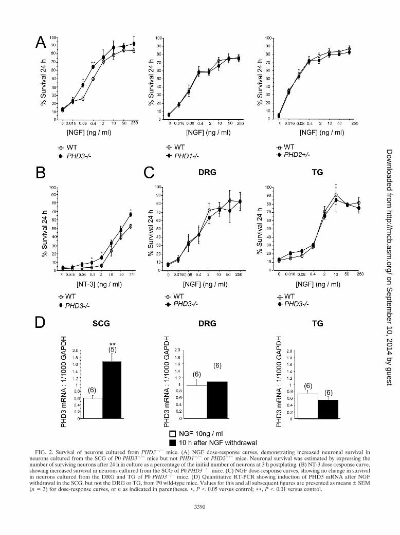
FIG. 2. Survival of neurons cultured from PHD3�/� mice. (A) NGF dose-response curves, demonstrating increased neuronal survival inneurons cultured from the SCG of P0 PHD3�/� mice but not PHD1�/� or PHD2�/� mice. Neuronal survival was estimated by expressing thenumber of surviving neurons after 24 h in culture as a percentage of the initial number of neurons at 3 h postplating. (B) NT-3 dose-response curve,showing increased survival in neurons cultured from the SCG of P0 PHD3�/� mice. (C) NGF dose-response curves, showing no change in survivalin neurons cultured from the DRG and TG of P0 PHD3�/� mice. (D) Quantitative RT-PCR showing induction of PHD3 mRNA after NGFwithdrawal in the SCG, but not the DRG or TG, from P0 wild-type mice. Values for this and all subsequent figures are presented as means � SEM(n � 3) for dose-response curves, or n as indicated in parentheses. *, P � 0.05 versus control; **, P � 0.01 versus control.
3390
on Septem
ber 10, 2014 by guesthttp://m
cb.asm.org/
Dow
nloaded from
RT-PCR 10 h after deprivation revealed a greater-than-twofold increase in PHD3 mRNA relative to GAPDHmRNA in sympathetic neurons but no significant change insensory neurons (Fig. 2D). Thus, both PHD3-dependentneuronal survival and responsiveness of PHD3 mRNA toNGF withdrawal appear to be specific to sympathetic neu-rons rather than a general property of NGF-sensitive neu-rons.
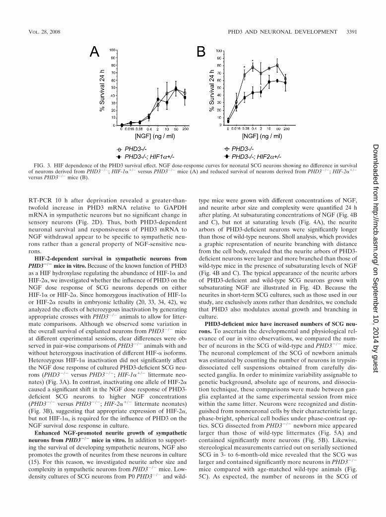
HIF-2-dependent survival in sympathetic neurons fromPHD3�/� mice in vitro. Because of the known function of PHD3as a HIF hydroxylase regulating the abundance of HIF-1� andHIF-2�, we investigated whether the influence of PHD3 on theNGF dose response of SCG neurons depends on eitherHIF-1� or HIF-2�. Since homozygous inactivation of HIF-1�or HIF-2� results in embryonic lethality (20, 33, 34, 42), weanalyzed the effects of heterozygous inactivation by generatingappropriate crosses with PHD3�/� animals to allow for litter-mate comparisons. Although we observed some variation inthe overall survival of explanted neurons from PHD3�/� miceat different experimental sessions, clear differences were ob-served in pair-wise comparisons of PHD3�/� animals with andwithout heterozygous inactivation of different HIF-� isoforms.Heterozygous HIF-1� inactivation did not significantly affectthe NGF dose response of cultured PHD3-deficient SCG neu-rons (PHD3�/� versus PHD3�/�; HIF-1��/� littermate neo-nates) (Fig. 3A). In contrast, inactivating one allele of HIF-2�caused a significant shift in the NGF dose response of PHD3-deficient SCG neurons to higher NGF concentrations(PHD3�/� versus PHD3�/�; HIF-2��/� littermate neonates)(Fig. 3B), suggesting that appropriate expression of HIF-2�,but not HIF-1�, is required for the influence of PHD3 on theNGF survival dose response in culture.
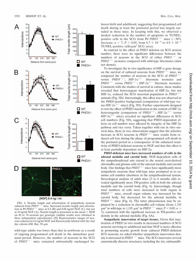
Enhanced NGF-promoted neurite growth of sympatheticneurons from PHD3�/� mice in vitro. In addition to support-ing the survival of developing sympathetic neurons, NGF alsopromotes the growth of neurites from these neurons in culture(15). For this reason, we investigated neurite arbor size andcomplexity in sympathetic neurons from PHD3�/� mice. Low-density cultures of SCG neurons from P0 PHD3�/� and wild-
type mice were grown with different concentrations of NGF,and neurite arbor size and complexity were quantified 24 hafter plating. At subsaturating concentrations of NGF (Fig. 4Band C), but not at saturating levels (Fig. 4A), the neuritearbors of PHD3-deficient neurons were significantly longerthan those of wild-type neurons. Sholl analysis, which providesa graphic representation of neurite branching with distancefrom the cell body, revealed that the neurite arbors of PHD3-deficient neurons were larger and more branched than those ofwild-type mice in the presence of subsaturating levels of NGF(Fig. 4B and C). The typical appearance of the neurite arborsof PHD3-deficient and wild-type SCG neurons grown withsubsaturating NGF are illustrated in Fig. 4D. Because theneurites in short-term SCG cultures, such as those used in ourstudy, are exclusively axons rather than dendrites, we concludethat PHD3 also modulates axonal growth and branching inculture.
PHD3-deficient mice have increased numbers of SCG neu-rons. To ascertain the developmental and physiological rel-evance of our in vitro observations, we compared the num-ber of neurons in the SCG of wild-type and PHD3�/� mice.The neuronal complement of the SCG of newborn animalswas estimated by counting the number of neurons in trypsin-dissociated cell suspensions obtained from carefully dis-sected ganglia. In order to minimize variability assignable togenetic background, absolute age of neurons, and dissocia-tion technique, these comparisons were made between gan-glia explanted at the same experimental session from micewithin the same litter. Neurons were recognized and distin-guished from nonneuronal cells by their characteristic large,phase-bright, spherical cell bodies under phase-contrast op-tics. SCG dissected from PHD3�/� newborn mice appearedlarger than those of wild-type littermates (Fig. 5A) andcontained significantly more neurons (Fig. 5B). Likewise,stereological measurements carried out on serially sectionedSCG in 3- to 6-month-old mice revealed that the SCG waslarger and contained significantly more neurons in PHD3�/�
mice compared with age-matched wild-type animals (Fig.5C). As expected, the number of neurons in the SCG of
FIG. 3. HIF dependence of the PHD3 survival effect. NGF dose-response curves for neonatal SCG neurons showing no difference in survivalof neurons derived from PHD3�/�; HIF-1��/� versus PHD3�/� mice (A) and reduced survival of neurons derived from PHD3�/�; HIF-2��/�
versus PHD3�/� mice (B).
VOL. 28, 2008 PHD3 AND NEURONAL DEVELOPMENT 3391
on Septem
ber 10, 2014 by guesthttp://m
cb.asm.org/
Dow
nloaded from
wild-type adults was lower than that in newborns as a resultof ongoing programmed cell death in the immediate post-natal period. However, the number of neurons in the SCGof PHD3�/� mice remained substantially unchanged be-
tween birth and adulthood, suggesting that programmed celldeath during at least the postnatal period was largely cur-tailed in these mice. In keeping with this, we observed amodest reduction in the number of apoptotic or TUNEL-positive cells in the SCG from P0 PHD3�/� mice ( 30%decrease, n � 7, P � 0.05, from 6.7 10�5 to 4.9 10�5
TUNEL-positive cells/�m2 SCG area).In contrast to the effect of PHD3 deletion on SCG neuron
number, there were no significant differences between thenumbers of neurons in the SCG of either PHD1�/� orPHD2�/� neonates compared with wild-type littermates (datanot shown).
To investigate the in vivo significance of HIF-� gene dosageon the survival of cultured neurons from PHD3�/� mice, wecompared the number of neurons in the SCG of PHD3�/�
versus PHD3�/�; HIF-1��/� littermate neonates andPHD3�/� versus PHD3�/�; HIF-2��/� littermate neonates.Consistent with the studies of survival in culture, these studiesrevealed that heterozygous inactivation of HIF-2�, but notHIF-1�, reduced the SCG neuronal population in PHD3�/�
animals (Fig. 5D). Interestingly, this effect was not observed inthe PHD3-positive background (comparison of wild-type ver-sus HIF-2��/� mice) (Fig. 5D). Further experiments designedto test the effect of PHD3 inactivation in the context of HIF-2�heterozygosity (comparison of PHD3�/�; HIF-2��/� versusHIF-2��/� mice) revealed no significant differences in SCGcell numbers (Fig. 5D), suggesting that PHD3-dependent ef-fects on cell number were affected by integrity of the HIF-2�pathway and vice versa. Taken together with our in vitro sur-vival data, these in vivo observations suggest that the selectiveincrease in SCG neurons in PHD3�/� mice results from re-duced cell loss during the phase of programmed cell death inthe perinatal period as a consequence of the enhanced sensi-tivity of PHD3-deficient neurons to NGF and that this effect isat least partially dependent on HIF-2�.
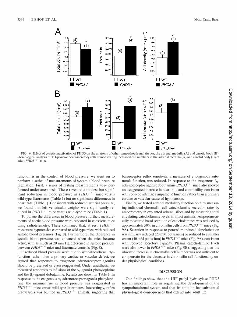
PHD3-deficient mice have increased numbers of cells in theadrenal medulla and carotid body. NGF-dependent cells ofthe sympathoadrenal axis extend to the neural crest-derivedchromaffin and glomus cells of the adrenal medulla and carotidbody. Our findings that PHD3�/� mice have significantly moresympathetic neurons than wild-type mice prompted us to ex-amine cell number elsewhere in the sympathoadrenal system.Stereological analysis of adult mice (3 to 6 months old) re-vealed significantly more TH-positive cells in both the adrenalmedulla and the carotid body (Fig. 6). Interestingly, thoughtotal numbers of cells were increased in both organs inPHD3�/� mice, overall organ volume was increased for thecarotid body, but decreased for the adrenal medulla, inPHD3�/� mice (Fig. 6). The latter phenomenon may be ex-plained by a reduction in chromaffin cell volume (from 1,743�m3 in wild-type to 1,195 �m3 in PHD3�/� mice; P � 0.05; n �7), consistent with the significant increase in TH-positive celldensity in the adrenal medulla (Fig. 6A).
Sympathetic innervation of target tissues. Given that inac-tivation of PHD3 in vivo results in increased numbers of SCGneurons surviving to adulthood and that NGF is more effectivein promoting neurite growth from cultured PHD3-deficientSCG neurons, we asked whether sympathetic innervation den-sity is increased in PHD3�/� mice. The SCG innervates severalanatomically discrete structures, including the iris, submandib-
FIG. 4. Neurite length and arborization of sympathetic neuronscultured from PHD3�/� mice. Increased neurite length and arboriza-tion in P0 PHD3�/� mice at 0.4 (B) and 0.08 ng/ml NGF (C), but notat 10 ng/ml NGF (A). Neurite length is presented as the mean � SEMon 40 to 70 neurons per genotype (similar results were obtained inthree independent experiments). (D) Representative images of neu-rons cultured in 0.4 ng/ml NGF and fluorescently labeled with the vitaldye calcein-AM. Bar, 50 �m.
3392 BISHOP ET AL. MOL. CELL. BIOL.
on Septem
ber 10, 2014 by guesthttp://m
cb.asm.org/
Dow
nloaded from

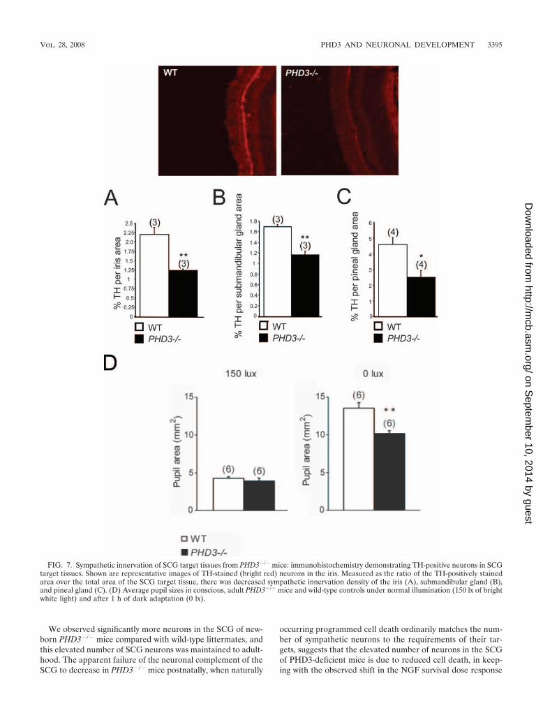
ular gland, and pineal gland, whose innervation density can berelatively easily estimated by quantifying the area occupied byTH-positive nerve fibers in tissue sections. Surprisingly, thisanalysis revealed significantly fewer TH-positive fibers in theiris, submandibular gland, and pineal gland of PHD3�/� micecompared with wild-type animals (Fig. 7A, B, and C).
Physiological effects on the sympathetic nervous system. Wenext sought to assess the integrity of physiological responsesthat are dependent on the sympathetic nervous system. Be-
cause of the reduced sympathetic innervation of the iris inPHD3�/� mice (Fig. 7A), we tested light-to-dark pupillaryresponses. While no differences in pupil diameter were seenunder normal lighting conditions (150 lx) (Fig. 7D), pupil di-ameter was significantly decreased in the dark-adapted eyefrom PHD3�/� mice (0 lx) (Fig. 7D). This implies decreasedtone in the sympathetically innervated dilator pupillae musclefibers of PHD3�/� mice.
Since one of the most important roles of sympathoadrenal
FIG. 5. Effect of genetic inactivation of PHD3 on anatomy of SCG. (A) Bright-field images of wild-type and PHD3�/� neonatal SCG. Bar, 100 �m.(B) Neuronal complement of neonatal SCG in wild-type and PHD3�/� mice; counts are of viable trypsin-dissociated neurons. (C) Stereological analysisof TH-positive neurons showing increased cell numbers in the SCG from adult PHD3�/� mice. (D) Comparison of neuronal complement of neonatalSCG in mice of the indicated genotypes. Counts are as for panel B. HIF-2� heterozygosity, but not HIF-1� heterozygosity, is associated with reducedneuronal complement.
VOL. 28, 2008 PHD3 AND NEURONAL DEVELOPMENT 3393
on Septem
ber 10, 2014 by guesthttp://m
cb.asm.org/
Dow
nloaded from
function is in the control of blood pressure, we went on toperform a series of measurements of systemic blood pressureregulation. First, a series of resting measurements were per-formed under anesthesia. These revealed a modest but signif-icant reduction in blood pressure in PHD3�/� mice versuswild-type littermates (Table 1) but no significant differences inheart rate (Table 1). Consistent with reduced arterial pressure,we found that left ventricular weights were significantly re-duced in PHD3�/� mice versus wild-type mice (Table 1).
To pursue the differences in blood pressure further, measure-ments of aortic blood pressure were repeated in conscious miceusing radiotelemetry. These confirmed that, at rest, PHD3�/�
mice were hypotensive compared to wild-type mice, with reducedsystolic blood pressures (Fig. 8). Furthermore, the difference insystolic blood pressure was enhanced when the mice becameactive, with as much as 20 mm Hg difference in systolic pressurebetween PHD3�/� mice and littermate controls (Fig. 8).
If reduced blood pressure were due to sympathoadrenal dys-function rather than a primary cardiac or vascular defect, weargued that responses to exogenous adrenoreceptor agonistsshould be preserved or even exaggerated. Under anesthesia, wemeasured responses to infusions of the �1-agonist phenylephrineand the 1-agonist dobutamine. Results are shown in Table 1. Inresponse to the exogenous �1-adrenoreceptor agonist phenyleph-rine, the maximal rise in blood pressure was exaggerated inPHD3�/� mice versus wild-type littermates. Interestingly, reflexbradycardia was blunted in PHD3�/� animals, suggesting that
baroreceptor reflex sensitivity, a measure of endogenous auto-nomic function, was reduced. In response to the exogenous 1-adrenoreceptor agonist dobutamine, PHD3�/� mice also showedan exaggerated increase in heart rate and contractility, consistentwith reduced intrinsic sympathetic function rather than a primarycardiac or vascular cause of hypotension.
Finally, we tested adrenal medullary function both by measur-ing individual chromaffin cell catecholamine secretion rates byamperometry in explanted adrenal slices and by measuring totalcirculating catecholamine levels in intact animals. Amperometri-cally measured basal secretion of catecholamines was reduced byapproximately 50% in chromaffin cells from PHD3�/� mice (Fig.9A). Secretion in response to potassium-induced depolarizationwas similarly reduced (20 mM potassium) or reduced to a smallerextent (40 mM potassium) in PHD3�/� mice (Fig. 9A), consistentwith reduced secretory capacity. Plasma catecholamine levelswere also lower in PHD3�/� mice (Fig. 9B), suggesting that theobserved increase in chromaffin cell number was not sufficient tocompensate for the decrease in chromaffin cell functionality un-der physiological conditions.
DISCUSSION
Our findings show that the HIF prolyl hydroxylase PHD3has an important role in regulating the development of thesympathoadrenal system and that its ablation has substantialphysiological consequences that extend into adult life.
FIG. 6. Effect of genetic inactivation of PHD3 on the anatomy of other sympathoadrenal tissues, the adrenal medulla (A) and carotid body (B).Stereological analysis of TH-positive neurosecretory cells demonstrating increased cell numbers in the adrenal medulla (A) and carotid body (B) ofadult PHD3�/� mice.
3394 BISHOP ET AL. MOL. CELL. BIOL.
on Septem
ber 10, 2014 by guesthttp://m
cb.asm.org/
Dow
nloaded from
We observed significantly more neurons in the SCG of new-born PHD3�/� mice compared with wild-type littermates, andthis elevated number of SCG neurons was maintained to adult-hood. The apparent failure of the neuronal complement of theSCG to decrease in PHD3�/� mice postnatally, when naturally
occurring programmed cell death ordinarily matches the num-ber of sympathetic neurons to the requirements of their tar-gets, suggests that the elevated number of neurons in the SCGof PHD3-deficient mice is due to reduced cell death, in keep-ing with the observed shift in the NGF survival dose response
FIG. 7. Sympathetic innervation of SCG target tissues from PHD3�/� mice: immunohistochemistry demonstrating TH-positive neurons in SCGtarget tissues. Shown are representative images of TH-stained (bright red) neurons in the iris. Measured as the ratio of the TH-positively stainedarea over the total area of the SCG target tissue, there was decreased sympathetic innervation density of the iris (A), submandibular gland (B),and pineal gland (C). (D) Average pupil sizes in conscious, adult PHD3�/� mice and wild-type controls under normal illumination (150 lx of brightwhite light) and after 1 h of dark adaptation (0 lx).
VOL. 28, 2008 PHD3 AND NEURONAL DEVELOPMENT 3395
on Septem
ber 10, 2014 by guesthttp://m
cb.asm.org/
Dow
nloaded from

of PHD3-deficient SCG neurons in culture to lower NGF con-centrations. Interestingly, the effect of PHD3 inactivation onNGF responsiveness in the peripheral nervous system appearsto be restricted to neurons of the sympathetic lineage, as neu-ral crest-derived, NGF-dependent sensory neurons fromPHD3�/� mice responded normally to NGF. It is unclearwhether dysregulated apoptosis might occur in regions withinthe central nervous system in PHD3�/� mice. However, thesemice are viable as adults, without obvious neurological abnor-malities. In addition, brains from PHD3�/� mice were notobviously abnormal and were not significantly different inweight (0.43 � 0.01 g versus 0.43 � 0.00 g in PHD3�/� andwild-type males; 0.42 � 0.01 g versus 0.44 � 0.01 g in PHD3�/�
and wild-type females). Nevertheless, these observations donot preclude dysregulation of apoptosis that is compensatedfor or might be revealed in pathological situations.
As well as enhancing the sensitivity of SCG neurons to thesurvival-promoting effects of NGF, deletion of PHD3 alsomakes these neurons more responsive to the neurite growth-promoting effects of NGF in culture. Because target-derivedNGF is not only required for sympathetic neuron survivalduring development in vivo but is also responsible for theterminal growth and branching of sympathetic axons in theirtargets, we expected to find increased sympathetic innervationdensity in PHD3�/� mice. Surprisingly, we observed the oppo-site in several SCG target tissues in these animals. One possi-ble explanation for this apparent paradox is that the increasednumber of sympathetic neurons innervating target tissues re-sults in elevated uptake and removal of NGF from these tissuesby retrograde axonal transport, resulting in lower ambient lev-els of NGF in the targets. Whereas retrograde transport ofNGF in signaling endosomes to the cell bodies of sympatheticneurons is required for survival, the extent of axonal growthand branching in target tissues is governed by the ambient levelof NGF in these tissues (15).
In addition to possessing elevated numbers of sympatheticneurons, PHD3�/� mice have increased numbers of chromaffinand glomus cells in the adrenal medulla and carotid body.Together with sympathetic neurons, these cells are derivedfrom the sympathoadrenal neural crest lineage (22). Whetherthe increase in chromaffin or glomus cell number in PHD3�/�
mice results from enhanced survival or from enhanced prolif-eration and/or differentiation of their precursors is not known.
Despite the increased number of cells in parts of the sym-pathoadrenal system of PHD3�/� mice, we observed impairedhomeostatic and physiological responses regulated by this sys-tem, including reduced blood pressure, impaired light-to-darkpupillary dilation, and reduced catecholamine secretion fromadrenal chromaffin cells, with lower plasma catecholamine lev-els in PHD3�/� mice. Sympathetic function is a key determi-nant of systemic blood pressure regulation (reviewed in refer-ence 17), and elevated sympathetic activity is present in manyforms of human hypertension. Apart from the findings re-ported here, we have not so far observed other basal abnor-malities in PHD3�/� mice. Thus, though PHD3 could haveother, as-yet-unknown effects on blood pressure regulation, theevidence for sympathetic hypofunction, reduced catechol-amine secretion, reduced responses to activity, and preservedresponses to exogenous adreno-agonists all support a causallink between sympathoadrenal dysregulation and blood pres-sure dysregulation in PHD3�/� animals.
Though previous cellular studies have implicated oxygen-dependent catalytic activity of PHD3 in the regulation of neu-ronal survival, these studies have given conflicting data on therole played by the known substrates HIF-1� and HIF-2� (23,44). In the current work we found that heterozygous inactiva-tion of HIF-2�, but not HIF-1�, had major effects on sympa-thetic neuronal survival and the neuronal complement of theSCG.
Interestingly, neonatal sympathetic failure has been ob-
TABLE 1. Cardiovascular function in anesthetized micea
Parameter (no. of animals in exptl groups)Result for group
P valueWild type PHD3�/�
Postmortem ventricular wt (10 WT, 7 PHD3�/�)LV/body wt (mg/g) 3.19 � 0.10 2.72 � 0.06 0.001RV/body wt (mg/g) 0.98 � 0.04 0.99 � 0.03 0.85
Arterial pressure (10 WT, 8 PHD3�/�)Systolic (mmHg) 105 � 3 94 � 5 0.009Diastolic (mmHg) 75 � 2 69 � 2 0.06MAP (mm Hg) 89 � 2 79 � 1 0.005
LV hemodynamics: baseline (10 WT, 7 PHD3�/�)dP/dtmax (mm Hg/s) 7,858 � 474 7,363 � 623 0.53Heart rate (bpm) 437 � 11 446 � 17 0.66
LV hemodynamics: dobutamine (9 WT, 7 PHD3�/�)dP/dtmax (% of baseline) 126 � 4 152 � 13 0.04Heart rate (% of baseline) 117 � 2 129 � 3 0.006
LV hemodynamics: phenylephrine (5 WT, 4 PHD3�/�)MAP (% of baseline) 135 � 5 159 � 22 0.07Heart rate (% of baseline) �31 � 5 �21 � 15 0.28Baroreceptor gain (bpm/mm Hg) �6.6 � 1.3 �2.5 � 0.9 0.04
a All values are means � SEM. WT, wild type; MAP, mean arterial pressure; bpm, beats per minute.
3396 BISHOP ET AL. MOL. CELL. BIOL.
on Septem
ber 10, 2014 by guesthttp://m
cb.asm.org/
Dow
nloaded from

served in HIF-2��/� mice, and HIF-2� mRNA has been re-ported to be strongly expressed in sympathoadrenal tissues ofthe mouse (42). Though difficulties in obtaining background-free immunohistochemical signals precluded assessment in themouse, we have also found that PHD3 is strongly expressed inthe rat sympathoadrenal system, suggesting that the two pro-teins are coexpressed in this lineage (B. Wijeyekoon and C. W.Pugh, unpublished data). Interestingly, studies of small inter-fering RNA-mediated suppression of PHDs in tissue culturehave indicated that PHD3 appears to exert greater regulatoryeffects on HIF-2� than HIF-1� (1). Taken together, the sim-plest synthesis of these findings is that PHD3 inactivation pro-motes sympathoadrenal neuronal survival, at least in part,through upregulation of HIF-2�. Nevertheless, consistent witha recent report of pharmacological PHD inhibition in sympa-thetic neurons (26), HIF-2� protein levels in the SCG of thesenormoxic animals were below the detection threshold for im-munohistochemistry, so this could not be confirmed. It shouldalso be noted that this explanation does not preclude the ex-istence of other PHD3 substrates that may contribute to theobserved effect. Indeed, the lack of effect of HIF-2� heterozy-gosity on a PHD3-positive background suggests that the rela-tionship between HIF-2� and neuronal survival is not simple
and depends on PHD3 status, perhaps implying the existenceof other substrates in addition to HIF-2�. Thus, the impor-tance of PHD3, as opposed to PHD1 or PHD2, in neuronalapoptosis might reflect the existence of a discrete, non-HIF,PHD3 substrate in this process, or the relative abundance ofPHD3 in these cells coupled to the preferred targeting ofHIF-2� by PHD3, or both.
Whether and how adult sympathetic hypofunction inPHD3�/� mice relates to dysregulation of HIF-2� is also un-clear. Together with the finding of sympathetic failure in as-sociation with complete inactivation of HIF-2� in some but notall studies (33, 42), our findings suggest that although an intactPHD3/HIF-2� system is necessary for proper sympathoadrenaldevelopment, there is no simple relationship of hyper- or hy-pofunctionality to predicted effects of these genotypes onHIF-2� levels.
While we have demonstrated physiological deficits in adultPHD3�/� mice, it is not clear whether deficits arise purely as aconsequence of PHD3 deficiency during development orwhether there is an ongoing PHD3 requirement for sympa-thetic function in the adult. A great deal of interest has beengenerated by the possibility that environmental effects on de-velopment might influence medically important aspects of
FIG. 8. Aortic blood pressures in conscious, adult PHD3�/� mice, as measured by radiotelemetry. (A) Pooled frequency histogram of systolicblood pressure over the 3-day recording period (using 1 mm Hg bins) from four (wild-type) or five (PHD3�/�) resting (solid lines) and active(dotted lines) mice. (B) Average blood pressure recordings over a 3-day recording period. Decreased systolic and diastolic blood pressures wereobserved in PHD3�/� mice. These differences were enhanced when the mice were active.
VOL. 28, 2008 PHD3 AND NEURONAL DEVELOPMENT 3397
on Septem
ber 10, 2014 by guesthttp://m
cb.asm.org/
Dow
nloaded from

FIG. 9. Catecholamine secretion in PHD3�/� mice. (A) Representative amperometric recordings of basal catecholamine release, as wellas responsiveness to induction by 20 mM and 40 mM potassium (20K and 40K), of adrenal slices isolated from adult mice. The table showsthe secretion rate (in fC/min) under basal conditions, as well as with 20 mM and 40 mM potassium (20 and 40 K). (B) Circulatingcatecholamines (adrenaline and noradrenaline) from anesthetized, adult mice. Both adrenal slice and circulating catecholamines are reducedin PHD3�/� mice.
3398 BISHOP ET AL. MOL. CELL. BIOL.
on Septem
ber 10, 2014 by guesthttp://m
cb.asm.org/
Dow
nloaded from

adult physiology, such as blood pressure regulation (reviewedin references 3 to 5 and 28). However, there are as yet fewmechanistic clues as to how such processes might operate. Thedependence of PHD activity on oxygen and cofactors, such asthe Krebs cycle intermediate 2-oxoglutarate, Fe(II), and ascor-bate (35), and inhibition of PHDs by metabolic intermediates,such as succinate, fumarate, pyruvate, and oxaloacetate (9, 18,19, 21, 27, 36), raises the interesting possibility that environ-mental influences on PHD3 activity could perturb sympatheticdevelopment in a manner that might have important effects oncardiovascular control.
In conclusion, our findings reveal an important role forPHD3 in the developing sympathoadrenal system. It is anintriguing possibility that this connection between hypoxiapathways and the developing sympathoadrenal system mightbe a means by which changes in oxygen tension could influencekey aspects of anatomical and physiological maturation of thissystem. Whether this provides a paradigm for environmentalinfluences affecting development will be of interest in the fu-ture.
ACKNOWLEDGMENTS
This work was supported by the British Heart Foundation, the Well-come Trust, and the EU Framework 6 Pulmotension Consortium.
We thank P. van Wesemael, M. Dewerchin, S. Wyns, E. Gils, B.Hermans, L. Kieckens, and L. Notebaert (Katholieke Universiteit Leu-ven, Belgium), as well as D. Adlam, S. Neubauer, R. Foster, and K.Buckler (University of Oxford, United Kingdom), for their contribu-tion.
We have declared that no conflict of interest exists.
REFERENCES
1. Appelhoff, R. J., Y. M. Tian, R. R. Raval, H. Turley, A. L. Harris, C. W. Pugh,P. J. Ratcliffe, and J. M. Gleadle. 2004. Differential function of the prolylhydroxylases PHD1, PHD2, and PHD3 in the regulation of hypoxia-induc-ible factor. J. Biol. Chem. 279:38458–38465.
2. Aragones, J., M. Schneider, K. Van Geyte, P. Fraisl, T. Dresselaers, M.Mazzone, R. Dirkx, S. Zacchigna, H. Lemieux, N. H. Jeoung, D. Lambrechts,T. Bishop, P. Lafuste, A. Diez-Juan, S. K. Harten, P. Van Noten, K. De Bock,C. Willam, M. Tjwa, A. Grosfeld, R. Navet, L. Moons, T. Vandendriessche,C. Deroose, B. Wijeyekoon, J. Nuyts, B. Jordan, R. Silasi-Mansat, F. Lupu,M. Dewerchin, C. Pugh, P. Salmon, L. Mortelmans, B. Gallez, F. Gorus, J.Buyse, F. Sluse, R. A. Harris, E. Gnaiger, P. Hespel, P. Van Hecke, F. Schuit,P. Van Veldhoven, P. Ratcliffe, M. Baes, P. Maxwell, and P. Carmeliet. 2008.Deficiency or inhibition of oxygen sensor Phd1 induces hypoxia tolerance byreprogramming basal metabolism. Nat. Genet. 40:170–180.
3. Barker, D. J. 1995. Fetal origins of coronary heart disease. BMJ 311:171–174.
4. Barker, D. J. 2002. Fetal programming of coronary heart disease. TrendsEndocrinol. Metab. 13:364–368.
5. Barker, D. J., S. P. Bagby, and M. A. Hanson. 2006. Mechanisms of disease:in utero programming in the pathogenesis of hypertension. Nat. Clin. Pract.Nephrol. 2:700–707.
6. Bruick, R. K., and S. L. McKnight. 2001. A conserved family of prolyl-4-hydroxylases that modify HIF. Science 294:1337–1340.
7. Butz, G. M., and R. L. Davisson. 2001. Long-term telemetric measurementof cardiovascular parameters in awake mice: a physiological genomics tool.Physiol. Genomics 5:89–97.
8. Cavalieri, B. 1966. Geometria degli indivisibili. Unione Tipografica Editrice,Torino, Italy.
9. Dalgard, C. L., H. Lu, A. Mohyeldin, and A. Verma. 2004. Endogenous2-oxoacids differentially regulate expression of oxygen sensors. Biochem. J.380:419–424.
10. Davies, A. M. 2003. Regulation of neuronal survival and death by extracel-lular signals during development. EMBO J. 22:2537–2545.
11. Davies, A. M., K. F. Lee, and R. Jaenisch. 1993. p75-deficient trigeminalsensory neurons have an altered response to NGF but not to other neuro-trophins. Neuron 11:565–574.
12. Davies, A. M., L. Minichiello, and R. Klein. 1995. Developmental changes inNT3 signalling via TrkA and TrkB in embryonic neurons. EMBO J. 14:4482–4489.
13. Epstein, A. C., J. M. Gleadle, L. A. McNeill, K. S. Hewitson, J. O’Rourke,
D. R. Mole, M. Mukherji, E. Metzen, M. I. Wilson, A. Dhanda, Y. M. Tian,N. Masson, D. L. Hamilton, P. Jaakkola, R. Barstead, J. Hodgkin, P. H.Maxwell, C. W. Pugh, C. J. Schofield, and P. J. Ratcliffe. 2001. C. elegansEGL-9 and mammalian homologs define a family of dioxygenases that reg-ulate HIF by prolyl hydroxylation. Cell 107:43–54.
14. Garcia-Fernandez, M., R. Mejias, and J. Lopez-Barneo. 2007. Developmen-tal changes of chromaffin cell secretory response to hypoxia studied in thinadrenal slices. Pflugers Arch. 454:93–100.
15. Glebova, N. O., and D. D. Ginty. 2005. Growth and survival signals control-ling sympathetic nervous system development. Annu. Rev. Neurosci. 28:191–222.
16. Gordan, J. D., and M. C. Simon. 2007. Hypoxia-inducible factors: centralregulators of the tumor phenotype. Curr. Opin. Genet. Dev. 17:71–77.
17. Guyenet, P. G. 2006. The sympathetic control of blood pressure. Nat. Rev.Neurosci. 7:335–346.
18. Hewitson, K. S., B. M. Lienard, M. A. McDonough, I. J. Clifton, D. Butler,A. S. Soares, N. J. Oldham, L. A. McNeill, and C. J. Schofield. 2007.Structural and mechanistic studies on the inhibition of the hypoxia-inducibletranscription factor hydroxylases by tricarboxylic acid cycle intermediates.J. Biol. Chem. 282:3293–3301.
19. Isaacs, J. S., Y. J. Jung, D. R. Mole, S. Lee, C. Torres-Cabala, Y. L. Chung,M. Merino, J. Trepel, B. Zbar, J. Toro, P. J. Ratcliffe, W. M. Linehan, andL. Neckers. 2005. HIF overexpression correlates with biallelic loss of fuma-rate hydratase in renal cancer: novel role of fumarate in regulation of HIFstability. Cancer Cell 8:143–153.
20. Iyer, N. V., L. E. Kotch, F. Agani, S. W. Leung, E. Laughner, R. H. Wenger,M. Gassmann, J. D. Gearhart, A. M. Lawler, A. Y. Yu, and G. L. Semenza.1998. Cellular and developmental control of O2 homeostasis by hypoxia-inducible factor 1 alpha. Genes Dev. 12:149–162.
21. Koivunen, P., M. Hirsila, A. M. Remes, I. E. Hassinen, K. I. Kivirikko, andJ. Myllyharju. 2007. Inhibition of hypoxia-inducible factor (HIF) hydroxy-lases by citric acid cycle intermediates: possible links between cell metabo-lism and stabilization of HIF. J. Biol. Chem. 282:4524–4532.
22. Le Douarin, N. M. 1986. Cell line segregation during peripheral nervoussystem ontogeny. Science 231:1515–1522.
23. Lee, S., E. Nakamura, H. Yang, W. Wei, M. S. Linggi, M. P. Sajan, R. V.Farese, R. S. Freeman, B. D. Carter, W. G. Kaelin, Jr., and S. Schlisio. 2005.Neuronal apoptosis linked to EglN3 prolyl hydroxylase and familial pheo-chromocytoma genes: developmental culling and cancer. Cancer Cell 8:155–167.
24. Lipscomb, E. A., P. D. Sarmiere, R. J. Crowder, and R. S. Freeman. 1999.Expression of the SM-20 gene promotes death in nerve growth factor-dependent sympathetic neurons. J. Neurochem. 73:429–432.
25. Lipscomb, E. A., P. D. Sarmiere, and R. S. Freeman. 2001. SM-20 is a novelmitochondrial protein that causes caspase-dependent cell death in nervegrowth factor-dependent neurons. J. Biol. Chem. 276:5085–5092.
26. Lomb, D. J., J. A. Straub, and R. S. Freeman. 2007. Prolyl hydroxylaseinhibitors delay neuronal cell death caused by trophic factor deprivation.J. Neurochem. 103:1897–1906.
27. Lu, H., C. L. Dalgard, A. Mohyeldin, T. McFate, A. S. Tait, and A. Verma.2005. Reversible inactivation of HIF-1 prolyl hydroxylases allows cell me-tabolism to control basal HIF-1. J. Biol. Chem. 280:41928–41939.
28. McMillen, I. C., and J. S. Robinson. 2005. Developmental origins of themetabolic syndrome: prediction, plasticity, and programming. Physiol. Rev.85:571–633.
29. Mills, P. A., D. A. Huetteman, B. P. Brockway, L. M. Zwiers, A. J. Gelsema,R. S. Schwartz, and K. Kramer. 2000. A new method for measurement ofblood pressure, heart rate, and activity in the mouse by radiotelemetry.J. Appl. Physiol. 88:1537–1544.
30. Niederhoffer, N., L. Hein, and K. Starke. 2004. Modulation of the barore-ceptor reflex by alpha 2A-adrenoceptors: a study in alpha 2A knockout mice.Br. J. Pharmacol. 141:851–859.
31. Oppenheim, R. W. 1991. Cell death during development of the nervoussystem. Annu. Rev. Neurosci. 14:453–501.
32. Pardal, R., and J. Lopez-Barneo. 2002. Carotid body thin slices: responses ofglomus cells to hypoxia and K�-channel blockers. Respir. Physiol. Neuro-biol. 132:69–79.
33. Peng, J., L. Zhang, L. Drysdale, and G. H. Fong. 2000. The transcriptionfactor EPAS-1/hypoxia-inducible factor 2� plays an important role in vascu-lar remodeling. Proc. Natl. Acad. Sci. USA 97:8386–8391.
34. Ryan, H. E., J. Lo, and R. S. Johnson. 1998. HIF-1 alpha is required for solidtumor formation and embryonic vascularization. EMBO J. 17:3005–3015.
35. Schofield, C. J., and Z. Zhang. 1999. Structural and mechanistic studies on2-oxoglutarate-dependent oxygenases and related enzymes. Curr. Opin.Struct. Biol. 9:722–731.
36. Selak, M. A., S. M. Armour, E. D. MacKenzie, H. Boulahbel, D. G. Watson,K. D. Mansfield, Y. Pan, M. C. Simon, C. B. Thompson, and E. Gottlieb.2005. Succinate links TCA cycle dysfunction to oncogenesis by inhibitingHIF-alpha prolyl hydroxylase. Cancer Cell 7:77–85.
37. Semenza, G. L. 2003. Targeting HIF-1 for cancer therapy. Nat. Rev. Cancer3:721–732.
VOL. 28, 2008 PHD3 AND NEURONAL DEVELOPMENT 3399
on Septem
ber 10, 2014 by guesthttp://m
cb.asm.org/
Dow
nloaded from

38. Sholl, D. A. 1953. Dendritic organization in the neurons of the visual andmotor cortices of the cat. J. Anat. 87:387–406.
39. Stalmans, I., Y. S. Ng, R. Rohan, M. Fruttiger, A. Bouche, A. Yuce, H.Fujisawa, B. Hermans, M. Shani, S. Jansen, D. Hicklin, D. J. Anderson, T.Gardiner, H. P. Hammes, L. Moons, M. Dewerchin, D. Collen, P. Carmeliet,and P. A. D’Amore. 2002. Arteriolar and venular patterning in retinas of miceselectively expressing VEGF isoforms. J. Clin. Investig. 109:327–336.
40. Straub, J. A., E. A. Lipscomb, E. S. Yoshida, and R. S. Freeman. 2003.Induction of SM-20 in PC12 cells leads to increased cytochrome c levels,accumulation of cytochrome c in the cytosol, and caspase-dependent celldeath. J. Neurochem. 85:318–328.
41. Takeda, K., V. C. Ho, H. Takeda, L. J. Duan, A. Nagy, and G. H. Fong. 2006.
Placental but not heart defects are associated with elevated hypoxia-induc-ible factor alpha levels in mice lacking prolyl hydroxylase domain protein 2.Mol. Cell. Biol. 26:8336–8346.
42. Tian, H., R. E. Hammer, A. M. Matsumoto, D. W. Russell, and S. L.McKnight. 1998. The hypoxia-responsive transcription factor EPAS1 is es-sential for catecholamine homeostasis and protection against heart failureduring embryonic development. Genes Dev. 12:3320–3324.
43. West, M. J. 1993. New stereological methods for counting neurons. Neuro-biol. Aging 14:275–285.
44. Xie, L., R. S. Johnson, and R. S. Freeman. 2005. Inhibition of NGF depri-vation-induced death by low oxygen involves suppression of BIMEL andactivation of HIF-1. J. Cell Biol. 168:911–920.
3400 BISHOP ET AL. MOL. CELL. BIOL.
on Septem
ber 10, 2014 by guesthttp://m
cb.asm.org/
Dow
nloaded from