A telerobotic haptic system for minimally invasive stereotactic neurosurgery
12
ORIGINAL ARTICLE A telerobotic haptic system for minimally invasive stereotactic neurosurgery A Rossi, A Trevisani, V Zanotto A Rossi, A Trevisani and V Zanotto Department of Innovation in Mechanics and Management, Universita ` di Padova, Italy Correspondence to: A Rossi, E-mail: [email protected] Abstract Medical robotics and computer assisted surgery are feasible and promising applications of robotic technology, whose main goals are surgical augmentation, information enhancement and improved surgical action. Neurosurgery probably presents the most major challenges, and can considerably benefit from the introduction of computers and robots to guide surgical procedures. This paper presents an innovative master- slave haptic robotic system for minimally invasive neurosurgery, which can help surgeons overcome human shortcomings and perform more accurate, repeatable, and reliable stereotactic neurosurgery. The system, named LANS, consists of a slave mechatronic actuator and a haptic master. The slave is designed to move linearly a laser pointer, a biopsy needle or a low-energy X-ray emitter along a pre-planned axis. The tool insertion into the brain is guided by the surgeon through the haptic master which also provides force feedback to the operator. Not only can the haptic master reproduce the contact force between the surgical tool and the treated tissue, but it can also produce virtual forces aimed at assisting surgeons during the operations. Experiments have been conducted to prove the soundness and accuracy of the overall system mechanical design and to assess the effectiveness of the control schemes synthesized for the master and the slave. Keywords: Medical robotics, computer assisted surgery, stereotactic neurosurgery, haptic display, robot design, X-ray Paper accepted: 19 November 2004 Published online: 15 January 2005. Available from: www.roboticpublications.com DOI: 10.1581/mrcas.2005.010209 INTRODUCTION Medical robotics and computer assisted surgery (CAS) span the broad areas of medical science and engineering, and aim at developing innovative devices and tools for performing surgical procedures in a more precise and effective way. Indeed, robotic systems can perform some surgical tasks with more precision, repeatability and delicacy than a human could. However decision making during any intervention is still required by surgeons, and consequently, at present, robots cannot replace physicians entirely. Nonetheless, the already proved and recognized effective interaction between robotic systems and physicians has produced an increasingly worldwide interest in the area of medical robotics, and in particular in neurosurgery. An up-to-date overview of the most significant researches conducted in this field can be found in Zamorano (1) and in the references therein. Generally speaking, there are four areas in surgery in which the use of robots is particularly useful: diagnostic procedures, teaching-training, telesurgery and therapy (2) . In diagnostic procedures for example, robotic tools can be currently employed to perform ultrasound and computer axial tomo- graphy (CAT)-guided biopsies. Additionally, new possibilities are arising in surgical instruction and training, since movements and procedures can be faithfully reproduced making use of tactile feedback and virtual reality (3, 4) . The use of haptic interfaces, providing tactile and force feedback, 64 Int J Medical Robotics and Computer Assisted Surgery 2005;1(2):64–75 E 2005 Robotic Publications Ltd. www.roboticpublications.com
-
Upload
independent -
Category
Documents
-
view
0 -
download
0
Transcript of A telerobotic haptic system for minimally invasive stereotactic neurosurgery

ORIGINAL ARTICLE
A telerobotic haptic system for minimally invasivestereotactic neurosurgery
A Rossi, A Trevisani, V Zanotto
A Rossi, A Trevisani and V ZanottoDepartment of Innovation in Mechanics and Management, Universita di Padova, ItalyCorrespondence to: A Rossi, E-mail: [email protected]
AbstractMedical robotics and computer assisted surgery are feasible and promising applications of robotic technology,whose main goals are surgical augmentation, information enhancement and improved surgical action.Neurosurgery probably presents the most major challenges, and can considerably benefit from theintroduction of computers and robots to guide surgical procedures. This paper presents an innovative master-slave haptic robotic system for minimally invasive neurosurgery, which can help surgeons overcome humanshortcomings and perform more accurate, repeatable, and reliable stereotactic neurosurgery. The system,named LANS, consists of a slave mechatronic actuator and a haptic master. The slave is designed to movelinearly a laser pointer, a biopsy needle or a low-energy X-ray emitter along a pre-planned axis. The toolinsertion into the brain is guided by the surgeon through the haptic master which also provides forcefeedback to the operator. Not only can the haptic master reproduce the contact force between the surgicaltool and the treated tissue, but it can also produce virtual forces aimed at assisting surgeons during theoperations. Experiments have been conducted to prove the soundness and accuracy of the overall systemmechanical design and to assess the effectiveness of the control schemes synthesized for the master and theslave.
Keywords: Medical robotics, computer assisted surgery, stereotactic neurosurgery, haptic display, robot design, X-ray
Paper accepted: 19 November 2004
Published online: 15 January 2005. Available from: www.roboticpublications.com
DOI: 10.1581/mrcas.2005.010209
INTRODUCTIONMedical robotics and computer assisted surgery(CAS) span the broad areas of medical science andengineering, and aim at developing innovativedevices and tools for performing surgical proceduresin a more precise and effective way.
Indeed, robotic systems can perform somesurgical tasks with more precision, repeatabilityand delicacy than a human could. However decisionmaking during any intervention is still requiredby surgeons, and consequently, at present, robotscannot replace physicians entirely. Nonetheless, thealready proved and recognized effective interactionbetween robotic systems and physicians hasproduced an increasingly worldwide interest inthe area of medical robotics, and in particular in
neurosurgery. An up-to-date overview of the mostsignificant researches conducted in this field canbe found in Zamorano (1) and in the referencestherein.
Generally speaking, there are four areas in surgeryin which the use of robots is particularly useful:diagnostic procedures, teaching-training, telesurgeryand therapy (2). In diagnostic procedures forexample, robotic tools can be currently employedto perform ultrasound and computer axial tomo-graphy (CAT)-guided biopsies. Additionally, newpossibilities are arising in surgical instruction andtraining, since movements and procedures canbe faithfully reproduced making use of tactilefeedback and virtual reality (3, 4). The use of hapticinterfaces, providing tactile and force feedback,
64
Int J Medical Robotics and Computer Assisted Surgery 2005;1(2):64–75 E2005 Robotic Publications Ltd.
www.roboticpublications.com

joined with telemetry and robotic technologies arealso likely to expand the development of telesurgeryprocedures.
Telesurgery provides surgeons with new capabil-ities, with the aim of performing less invasive andmore accurate surgical interventions, reducing post-operative hospital stay and improving clinical out-comes. In particular, minimally invasive surgery(MIS) is acknowledged to be a revolutionarysurgical technique (5). Minimally invasive proce-dures use remotely controlled robots to enablesurgeons to operate inside patients without makinglarge incisions. The main advantage of this techni-que is a substantial reduction of tissue trauma, whichis a main cause of post-operative pain and longerhospital stays.
Among all the cited areas, therapy is certainly the
most promising field of application of robotic
technologies to surgery and will be the subject of
extensive future research (2). This paper presents a
new telerobotic system for minimally invasive
neurosurgical therapy, designed and manufactured
at the Department of Innovation in Mechanics
and Management of the University of Padova.
The system, named LANS (Linear Actuator for
NeuroSurgery) has been conceived specifically to
perform biopsies and neurosurgical interventions
by means of a miniature X-ray source (the PRS,
Photon Radiosurgery System, by Carl Zeiss),
whose emitting tip must be placed accurately inside
the patient’s brain.
The LANS robotic system comprises a haptic
master module, operated by the surgeon, and a slave
mechatronic module that makes a PRS probe, or a
biopsy needle, move along a predefined emission
axis in accordance with the master position decided
by the surgeon. So as to orient the LANS along
the established emission axis, a NeuroMate robot is
employed in a frame-based configuration which
ensures the highest possible accuracy.
The system has been designed by assuming that
during the surgical operation only the LANS (which
is very accurate, and provides the surgeon with force
feedback) is in active mode while the NeuroMate
is powered off. This allows many of the problems
associated with the complex nature of this surgical
therapy to be overcome. Moreover, very precise
and repeatable movements of the biopsy needle and
of the X-ray source can be obtained, thus improving
the overall intervention outcomes.
The proposed system therefore greatly enlargesthe NeuroMate performances when dealing withthose neurosurgical therapies making use of toolsthat must be accurately and delicately inserted intothe brain. In particular, the tool positioningaccuracy and repeatability is increased by more thanone order of magnitude and a very precise anddelicate insertion of the tool is assured.Furthermore, the fact that surgeons do not movethe tool directly, but through a master input handle,gives the possibility of both scaling the motion(therefore improving human dexterity limits), andproviding them with force feedback. Force feedbackand control, is not only important to recreaterealistic feelings during the intervention but alsobecause the surgeon’s instrument guidance can beeffectively and naturally improved by exertingsuitable virtual forces on the master handle. Suchforces prevent incorrect movements, associatedwith anxiety, fatigue or age, which might seriouslydamage healthy brain tissues. Thus, by means of thehaptic master, the surgeon can be placed in contactwith a virtual environment where the surgical task isconsiderably simplified.
NEUROSURGERY BY MEANS OF PRS:ADVANTAGES AND OPEN ENGINEERINGISSUESConventional radiosurgery is based on the use ofhighly collimated external beams or on the useof interstitial radiation (IR) with radioisotopes.The use of external beams requires a preventivehistological diagnosis, in addition to expensivestructures and dedicated facilities. IR with radio-isotopes, on the other hand, presents the usualinconveniences associated with the manipulation ofradioactive sources.
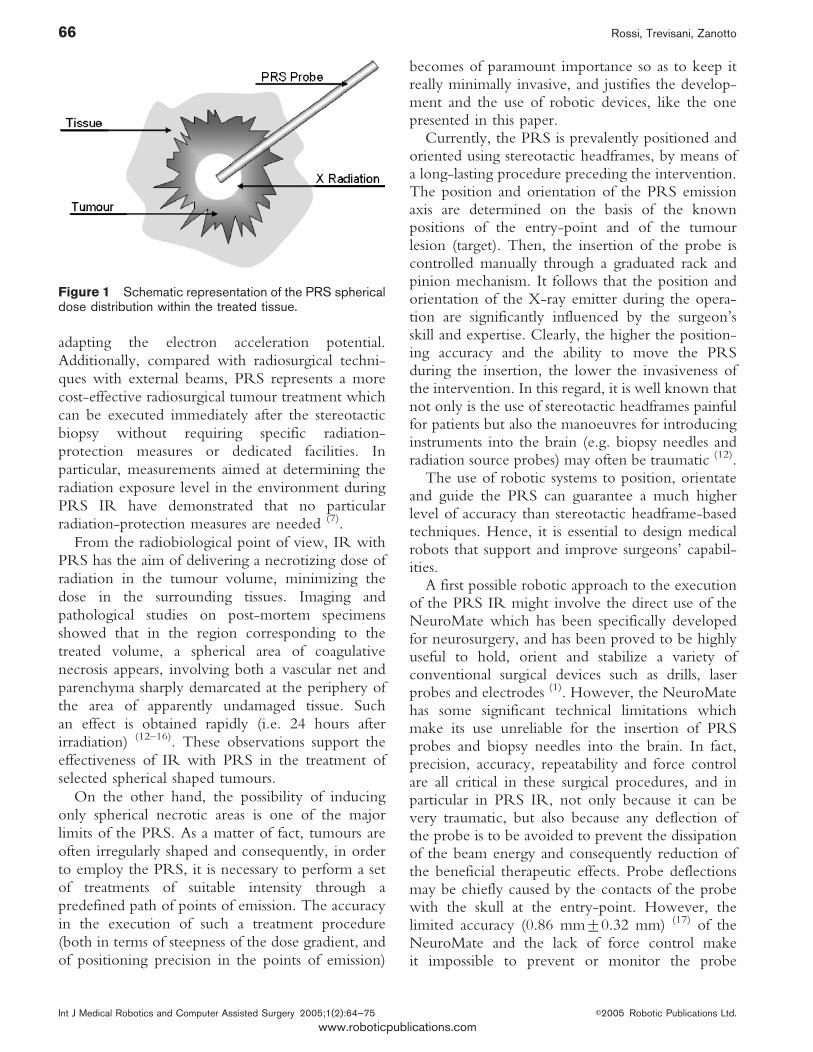
The PRS is a new miniaturized radio-surgicaldevice for the IR of brain lesions (6, 7) which canconsiderably reduce the aforementioned drawbacks,if appropriately employed. The device emits lowenergy X-rays from the tip of a cylindrical probe.The probe length is 10 cm, the diameter 3,2 mmand the dose distribution almost spherical (8, 9), asshown in Figure 1.
The use of PRS in neurosurgery providesconsiderable advantages in comparison with tradi-tional techniques (10, 11). In particular, comparedwith conventional radiosurgery, IR with PRSprovides dosimetric advantages since adjustable doserates and steep dose gradients can be obtained by
A telerobotic haptic system for minimally invasive stereotactic neurosurgery 65
E2005 Robotic Publications Ltd. Int J Medical Robotics and Computer Assisted Surgery 2005;1(2):64–75
www.roboticpublications.com
adapting the electron acceleration potential.Additionally, compared with radiosurgical techni-ques with external beams, PRS represents a morecost-effective radiosurgical tumour treatment whichcan be executed immediately after the stereotacticbiopsy without requiring specific radiation-protection measures or dedicated facilities. Inparticular, measurements aimed at determining theradiation exposure level in the environment duringPRS IR have demonstrated that no particularradiation-protection measures are needed (7).
From the radiobiological point of view, IR withPRS has the aim of delivering a necrotizing dose ofradiation in the tumour volume, minimizing thedose in the surrounding tissues. Imaging andpathological studies on post-mortem specimensshowed that in the region corresponding to thetreated volume, a spherical area of coagulativenecrosis appears, involving both a vascular net andparenchyma sharply demarcated at the periphery ofthe area of apparently undamaged tissue. Suchan effect is obtained rapidly (i.e. 24 hours afterirradiation) (12–16). These observations support theeffectiveness of IR with PRS in the treatment ofselected spherical shaped tumours.
On the other hand, the possibility of inducingonly spherical necrotic areas is one of the majorlimits of the PRS. As a matter of fact, tumours areoften irregularly shaped and consequently, in orderto employ the PRS, it is necessary to perform a setof treatments of suitable intensity through apredefined path of points of emission. The accuracyin the execution of such a treatment procedure(both in terms of steepness of the dose gradient, andof positioning precision in the points of emission)
becomes of paramount importance so as to keep itreally minimally invasive, and justifies the develop-ment and the use of robotic devices, like the onepresented in this paper.
Currently, the PRS is prevalently positioned andoriented using stereotactic headframes, by means ofa long-lasting procedure preceding the intervention.The position and orientation of the PRS emissionaxis are determined on the basis of the knownpositions of the entry-point and of the tumourlesion (target). Then, the insertion of the probe iscontrolled manually through a graduated rack andpinion mechanism. It follows that the position andorientation of the X-ray emitter during the opera-tion are significantly influenced by the surgeon’sskill and expertise. Clearly, the higher the position-ing accuracy and the ability to move the PRSduring the insertion, the lower the invasiveness ofthe intervention. In this regard, it is well known thatnot only is the use of stereotactic headframes painfulfor patients but also the manoeuvres for introducinginstruments into the brain (e.g. biopsy needles andradiation source probes) may often be traumatic (12).
The use of robotic systems to position, orientateand guide the PRS can guarantee a much higherlevel of accuracy than stereotactic headframe-basedtechniques. Hence, it is essential to design medicalrobots that support and improve surgeons’ capabil-ities.
A first possible robotic approach to the executionof the PRS IR might involve the direct use of theNeuroMate which has been specifically developedfor neurosurgery, and has been proved to be highlyuseful to hold, orient and stabilize a variety ofconventional surgical devices such as drills, laserprobes and electrodes (1). However, the NeuroMatehas some significant technical limitations whichmake its use unreliable for the insertion of PRSprobes and biopsy needles into the brain. In fact,precision, accuracy, repeatability and force controlare all critical in these surgical procedures, and inparticular in PRS IR, not only because it can bevery traumatic, but also because any deflection ofthe probe is to be avoided to prevent the dissipationof the beam energy and consequently reduction ofthe beneficial therapeutic effects. Probe deflectionsmay be chiefly caused by the contacts of the probewith the skull at the entry-point. However, thelimited accuracy (0.86 mm¡0.32 mm) (17) of theNeuroMate and the lack of force control makeit impossible to prevent or monitor the probe
Figure 1 Schematic representation of the PRS sphericaldose distribution within the treated tissue.
66 Rossi, Trevisani, Zanotto
Int J Medical Robotics and Computer Assisted Surgery 2005;1(2):64–75 E2005 Robotic Publications Ltd.
www.roboticpublications.com

deflections and hence to ensure an actually mini-mally invasive procedure.
Nonetheless the NeuroMate can be very usefulalso in PRS IR since it can be effectively employedto hold and orientate statically robotic systemsspecifically designed for the accurate insertion ofbiopsy needles and PRS probes into the brain. Inaccordance with the aforementioned requirements,these robotic systems, besides being very accurate,must allow the simultaneous measurement of thecontact force between the surgical tool and thetreated tissue. Such a force, properly scaled, has thento be fed back to the surgeon by means of a suitablehaptic interface used to guide the tool insertion.This is the robotic configuration proposed andadopted in this work.
In the following sections the two chief compo-nents of the LANS are presented: the mechatronictool actuator (the slave module) and the hapticinterface (the master module).
THE TOOL ACTUATORThe most important LANS component is amechatronic device which can be mounted directlyon the NeuroMate (see Figure 2, red box) andallows the movement of surgical instruments withhigh accuracy along the axis defined by the entry-point and the target established for the therapy. Thechief components of the designed tool actuator areshown in Figure 3.
Basically, the tool actuator comprises two mainparts: an aluminium alloy base frame, which can be
fixed to the NeuroMate arm, and a moving carriagewith a neurosurgical tool holder linked to a loadcell.
A current-controlled DC mini-motor, placed atan extremity of the frame, drives a miniature ballscrew through a synchronous belt (velocity ratio12:28). The backlash-free preloaded single nut ofthe ball screw shifts the carriage. The actuator isdevoid of a braking system because no backdrivingoccurs.
Motion linearity is ensured by a minirailrecirculating ball guideway. The angular positionof the DC motor is measured by a shaft-positionoptical encoder whose resolution is 400 pulses perrevolution. The overall accuracy and repeatability ofthe tool actuator are better than 0.05 mm.
In order to guarantee patients’ safety, the carriagemotion range has been restricted to 90 mm bymeans of two mechanical stops incorporated in thesystem frame.
Appropriate mechanical adapters have beendesigned to couple neurosurgical tools, such as laserpointers (Figure 4) biopsy needles (Figure 5) andPRS sources (Figure 6) to the tool holder mountedon the carriage. A micro-sensor has been inserted inthe tool holder so as to assure continuous monitor-ing of the correct placing of the adapters, and henceof the surgical tools.
The tool holder is directly linked to a homemadealuminium load cell. The load cell can measure thecontact forces between the tools and the cerebral
Figure 2 The LANS tool actuator mounted on theNeuroMate.
Figure 3 The chief components of the LANS toolactuator.
A telerobotic haptic system for minimally invasive stereotactic neurosurgery 67
E2005 Robotic Publications Ltd. Int J Medical Robotics and Computer Assisted Surgery 2005;1(2):64–75
www.roboticpublications.com

tissues with a resolution of 1/50 N. These forces,suitably amplified by the LANS controller, are fedback to the surgeon through the handle of the hapticconsole.
For safety and hygienic reasons the actuator hasbeen provided with a stainless steel external cover.The total weight of the LANS slave module is1.8 kg.
The technical specifications of the overall slavesystem are summarized in Table 1
THE HAPTIC INTERFACEThe haptic master developed for the LANS isshown in Figure 7 and basically consists of a 40 mmdiameter haptic handle positioned on an ergonomicconsole. Using the handle the surgeon can guide thesurgical tool along a pre-planned axis and can feelthe interaction forces between the tool and thecerebral tissue.
The interface meets the important requirement ofbeing almost transparent to the operator (18), in thesense that the dynamic behaviour of the mechanismcan be hardly perceived by the surgeon. The forcesare reproduced by means of a current-controlledDC motor directly coupled to the handle and whoseinertia and friction are very small. The angular
Figure 4 The laser pointer mounted on the tool actuator.
Figure 5 The mechanical adapter specifically designedto couple biopsy needles to the tool holder.
Figure 6 The PRS mounted on the tool actuator.
Table 1 Technical specifications of the tool actuator
Tool movement range [mm] 90
Movement linearity [mm] 0.01
Accuracy [mm] ¡0.05
Max. carriage speed [mm/s] 13
Max. pay load [N] 15
Tool holder axis max. pitch displacement [deg.] ,0.02
Figure 7 The LANS haptic interface and visual devices.
68 Rossi, Trevisani, Zanotto
Int J Medical Robotics and Computer Assisted Surgery 2005;1(2):64–75 E2005 Robotic Publications Ltd.
www.roboticpublications.com

position of the motor drive shaft is measured by ashaft-position optical encoder with a resolution of400 pulses per revolution. For safety reasons, themaximum torque the motor can exert on thesurgeon’s hand is kept below 160 Nmm.
The coupling device between the handle and themotor shaft has been designed to allow one touch‘‘push-pull lock’’ (Figure 8). This assures a quickconnection and disconnection of the handle andtherefore simplifies the sterilization procedures forthe master: only the handle has to be sterilizedbefore a surgical operation, while all the other partsof the console can be easily covered with a sterilizeddrape (as shown in Figure 7).
The console is also provided with a set ofindicators which let the surgeon monitor somechief status variables of the LANS system: thesevisual devices are described in the Safety Issuessection below.
MASTER-SLAVE HAPTIC CONTROLDesigning a control scheme for the LANS is achallenging issue, since it must assure:
N accurate, safe, robust and stable position controlfor the slave
N safe, robust and stable force control for the hapticmaster
N redundant control for fail-safe operations
Figure 9 shows a block diagram of the controlscheme synthesized for the LANS. The surgeon,who is represented by an unknown impedance andan active force at the top-left of the figure, acts onthe master handle, whose angular position hM ismeasured by the encoder. The handle position isconverted into the reference position xR of theslave, by means of the velocity ratio KP. Thesurgeon can modify the system responsiveness bysimply modifying the velocity ratio KP: the greaterKP, the smaller the rotation of the handle necessaryto produce the same displacement of the surgicaltool. The slave tracking of the reference positions isassured by a standard PID controller.
As far as force control is concerned, the load cellon the slave measures the interaction force fEbetween the surgical tool and the cerebral tissue.The surgeon can modulate the tactile sensibility ofthe cell, and hence the torque perceived on thehandle, during the tool insertion by modifying theamplifying factor KF which is basically a force-to-torque ratio.
The graphical user interface shown in Figure 10,has been designed both to give the surgeon thepossibility of setting the most appropriate values forKP and KF during the operation, and to provide himwith some essential data, required to control thestatus of the overall LANS system. For example, by
Figure 8 The one touch push-pull lock of the masterhandle.
Figure 9 Block diagram of the LANS control scheme.
A telerobotic haptic system for minimally invasive stereotactic neurosurgery 69
E2005 Robotic Publications Ltd. Int J Medical Robotics and Computer Assisted Surgery 2005;1(2):64–75
www.roboticpublications.com

means of two views on the main window, thesurgeon can monitor the real-time performances ofthe tracking system and the values of the forcesgenerated by the interaction between the surgicaltools with the cerebral tissue. At the top of thegraphical interface, a text window helps the surgeonduring each phase of the surgical procedure, bysupplying him with information on the currentsystem status and the next action to be completed.
The tactile force signal made available by the loadcell is then added to the force generated by the‘‘virtual environment’’ designed to assist the surgeonduring the operation. As is better explained below,the value taken by such a virtual force depends onthe planned therapy and on the status of the overallLANS system. The sum of these two signals(associated to actual and virtual forces) becomesthe torque command exerted on the handle by themaster actuator.
The virtual environment has been introduced tosimplify the surgeons’ movements during anysurgical procedure employing the LANS, and tomake them safer. When it is necessary, the surgeonmay be prevented from moving the surgical toolfreely, and dangerously, by making him feel asthough the tool were against a virtual wall and couldnot be moved any further. In practice, the virtualenvironment allows implementation of an activeguiding system which is exploited, in particular, toperform three different surgical procedures:
N single target positioning, which implies moving thesurgical tool to a certain depth along thetranslation axis. In this procedure a virtual wall(Figure 11) is placed orthogonal to the axis, just
next to the target, in order to prevent the surgeonfrom moving the instrument beyond this point;
N multiple target positioning, in which the formerprocedure is repeated at several different positionsalong the translation axis: a virtual wall (Figure 12)is placed next to each target position. When usingthe PRS, this procedure allows delivery ofdifferent X-ray doses at different positions so asto treat non-spherical tumours, which is notpossible when manual insertion of the probe isperformed (19, 20);
N continuous path positioning, which consists ofmoving the tool along the linear path inaccordance with a pre-planned trajectory in time.In this case a time-varying bilateral virtual wall
Figure 10 The graphical user interface.
Figure 11 Virtual wall modelling with single targetpositioning.
Figure 12 Virtual wall modelling with multiple targetpositioning.
70 Rossi, Trevisani, Zanotto
Int J Medical Robotics and Computer Assisted Surgery 2005;1(2):64–75 E2005 Robotic Publications Ltd.
www.roboticpublications.com

needs to be generated, so as to guide the surgeonalong the path (Figure 13).
The status supervisor shown in Figure 9 monitorsthe status of the overall system continuously andinteracts with the virtual environment generator: if afault occurs, the supervisor inhibits the slave motion.As a consequence, the slave is stopped instanta-neously and a bilateral virtual wall is set on themaster at the current position. Then, the robotassisted surgical operation can only be restarted if thefault is corrected. However, if the fault cannot becorrected, the removal of the surgical tool isguaranteed by an emergency extraction procedure.Further details on this procedure are provided in theSafety Issues section below.
The master-slave control scheme presented abovehas been implemented in a hard real-time environ-ment making use of a Pentium P3 based PC runningVenturcom RTX on MS Windows 2000. The PChas been instrumented with a multifunction I/Oboard Humusoft MF604 and has allowed operationof the slave position and the master force controlloops at the sample rates of respectively 5 kHz and2 kHz.
The experimental recordings shown in Figure 14prove the effectiveness of the proposed controlscheme both when the slave has to track thereference positions imposed by the surgeon, andwhen the target position has been reached, andconsequently, a virtual wall has to be generated toprevent any surgeon’s positioning errors. In the firstplot, it can be noticed that the slave actual position(solid red line) accurately follows the reference
generated by the master handle (dashed blue line) aslong as the target position is reached. The effec-tiveness of the scheme in this phase is proved by thefact that the master-slave tracking error, shown inthe second diagram, is always below 0.01 mm andthe steady state error is almost nil. When the targetposition is achieved, the slave stops moving the toolforward (at the target position the surgical tool canbe only moved backwards to prevent damage tohealthy cerebral tissues) and a unilateral virtual wallis set on the master handle so that the surgeon canfeel a torque which prevents overshooting the targetposition. The time-history of the virtual torque isshown in the third plot: such a torque is obviouslyequal to zero before the target position is reached.
SAFETY ISSUESSafety is definitely the most important issue insurgical robot design (21–23). Since medical roboticsis a new field of application, the investigation on themost appropriate safety solutions is still an openresearch field and unfortunately, adequate literatureis not yet available.
Medical robot design is generally carried outfollowing the guidelines of the machine safetyregulations (e.g. CE marking requirements).Admittedly, further requirements are to be metwhen dealing with medical robots, since patients,and often also surgeons, are necessarily inside theoperating workspace of the robot. Moreover in ahaptic master-slave robotic application both thesurgeon and the patient interact with an activemechanical device (24).
The safety measures which are generally takeninclude the design of special-purpose robots whosecapabilities are tailored to the task at hand, the use ofredundant sensors and controllers, and the imple-mentation of fail-safe procedures which allowremoval of the robot from the operating field incase it fails, and completing the procedure withoutrobotic assistance (21, 25).
As far as the LANS is concerned, ensuring patientsafety has been the primary objective. In fact onlypatients need to be kept in the workspace of theslave robot, while surgeons just act on the hapticmaster. Indeed, the haptic master is an activemechanical device, but ensuring surgeons’ safety isfairly easy, since it is enough to keep the torqueapplied by the handle small, by means, for example,of a current limit circuit on the motor driver. Onthe contrary patients’ safety is related to surgical tool
Figure 13 Virtual wall modelling with continuous pathpositioning.
A telerobotic haptic system for minimally invasive stereotactic neurosurgery 71
E2005 Robotic Publications Ltd. Int J Medical Robotics and Computer Assisted Surgery 2005;1(2):64–75
www.roboticpublications.com

Table 2 Technical specifications of the chief components of the tool actuator
DC brush mini-motor Max. speed [rpm] 7300
Escap 23DT12-216E DC Torque constant [Nm/A] 0.023
Max. continuous current [A] 0.90
Optical Encoder Line counts 100
Escap E9-100-3.0-01 Bandwidth [kHz] 200
Motor Driver Max. continuous current [A] 5
Copley Control Mod 403 Peak current [A] 10
Miniature ball screw Lead [mm] 0.5
STEINMEYER Max. speed [rpm] 1800
1112-0,5.8.145.170 Travel [mm] 100
Minirail recirculating ball guideway Running accuracy in height [mm] 2
SCHNEEBERGER MN 9–195–7.5-7.5–G1–V1 Lateral running accuracy [mm] 2
Max. speed [m/s] 5
Static loading capacities [N] 2770
Max. roll static moment [Nm] 12.9
Max. pitch static moment [Nm] 10.2
Max. yaw static moment [Nm] 10.2
Displacement force [N] 0.05
Load Cell Rated Output (R.O.) [mV/V nom.] 1
Capacity [N] 20
Resolution [N] 0.02
Safe overload [% of R.O.] 150
Nonlinearity [% of R.O.] ,0.01
Nonrepeatability [% of R.O.] ,0.005
Figure 14 Experimental time histories of the slave reference (master) and actual positions, of the slave tracking error,and of the virtual torque exerted on the master handle when the operator tries to move the tool beyond the target position.
72 Rossi, Trevisani, Zanotto
Int J Medical Robotics and Computer Assisted Surgery 2005;1(2):64–75 E2005 Robotic Publications Ltd.
www.roboticpublications.com

movements. Therefore the slave actuator holdingthe instruments must be continuously monitoredand its motion range needs to be appropriatelybounded. Mechanical end stops are one means ofassuring safety but they usually make the roboticapplication rather rigid. A more versatileapproach (26, 27) is to constrain dynamically therange of motion by means of software controlledlimits, and to keep the mechanical end stops beyondthe software controlled ones so as to make thesystem inherently safe.
In the LANS, the control of the tool range ofmotion is performed at three increasing safetyprotection levels exploiting respectively softwarecontrolled limits, electrical switches and mechanicalend stops.
Software safety control makes use of the signalsfrom two fixed inductive sensors (A,B in Figure 15)to bound the master-slave movements in accordancewith the target positions defined for the surgicalprocedure. In particular, when multi-target posi-tioning is to be performed, the overall roboticsystem is positioned so that the inductive sensor B isjust beyond the position where the carriage has tobe moved to make the surgical tool reach thefurthest target position. Obviously, sensor A plays a
less significant role in terms of safety, and is chieflyemployed to establish a zero reference (home)position for the carriage. When one of the inductivesensors is reached, a software controlled procedureinhibits tracking commands beyond such limits, bystopping the slave and by creating a virtual wallwhich obstructs the movements of the masterhandle. Since the position of the inductive sensorB is always kept very close to the furthest targetposition (the most appropriate distance can be set bythe surgeon through the graphical interface), neitherdoes this safety procedure cause the system to stopnor does it require special steps to restore the fullfunctionality of the system.
If a software error occurs, the slave motion maybecome uncontrolled and the first protection levelmight not be effective. So, the LANS system isprovided with a second protection level making useof the electromechanical limit-switches C and Dpainted in green in Figure 15. These switches areplaced just beyond the inductive sensors. When thecarriage touches a switch, it cuts off the powersupply of the slave actuator and stops its motion.However, if the slave goes beyond the limit-switchC, its position is not dangerous for patients and asoftware controlled reset procedure is sufficient torestore the normal functionality. Instead, if the slavegoes beyond the limit-switch D, its position maycause damage to the cerebral tissue, and so not onlyhas the system to be stopped but also the tool has tobe carefully moved backwards by means of a suitableemergency procedure (the manual mode describedbelow).
The third and highest protection level makes useof mechanical end stops obtained by suitably shapingsome parts of the carriage and the frame (E inFigure 15). The mechanical end stops are clearlybeyond the previous software controlled andelectromechanical switches and avoid any farthermovement.
Figure 15 Electrical and mechanical parts used to limitthe range of motion of the tool holder (not shown in thedrawing). A,B – Inductive sensors, C,D –Electromechanical limit switches, E - Mechanical end stop.
Table 3 Technical specifications of the chiefcomponents of the haptic interface
DC brush motor Max. speed [rpm] 3000
RSGPN9 Torque constant [Nm/A] 0.044
Max. continuous current [A] 6.85
Optical Encoder Line counts 100
HP HEDS 9040 Bandwidth [kHz] 100
Motor Driver Max. continuous current [A] 5
Copley Control Model 403 Peak current [A] 10
A telerobotic haptic system for minimally invasive stereotactic neurosurgery 73
E2005 Robotic Publications Ltd. Int J Medical Robotics and Computer Assisted Surgery 2005;1(2):64–75
www.roboticpublications.com

So as to ensure, at the same time, high safety andease of use during surgical procedures, the LANSprovides six operating modes: free mode, targetmode, trajectory mode, standby mode, reset modeand manual mode.
In the free mode, master-slave tracking works asdescribed in the previous section: the slave followsthe master position and the interaction forces withthe cerebral tissue are fed back to the surgeonthrough the haptic handle.
When the target is reached the system switchesinto target mode, where each slave movementbeyond the target is prevented and a virtual wall iscreated on the master.
In the trajectory mode, implementing the afore-mentioned continuous path positioning, the systemcan guide the surgeon through a set of softwarecontrolled master positions defined in accordancewith a pre-planned therapy. The trajectory modewill be mostly useful in future studies designedto investigate the effectiveness of dynamic dosedeliveries with the PRS.
The standby mode is the mode in which thesystem is held until a foot-switch is pressed or aslong as the surgical tool is not correctly positionedon the tool-holder, as recognized by the micro-sensor shown in Figure 3.
The system switches into reset mode when a
generic fault occurs, such as backward over-travel
(beyond sensor C) or motor drivers switch off.
These faults are not critical because the patient’s
safety is still preserved. Nonetheless the system
warns the surgeon that a fault has happened through
a warning message on the haptic interface monitor
(see Figure 7), and by switching on a yellow lamp.
Additionally, what is more important, both the slave
and the master are immediately disabled. By means
of the reset button (the yellow button on the master
console: see Figure 7) the surgeon can re-establish
the system’s normal functioning.
Finally, the occurrence of an irreversible fault,
caused, for example, by lack of power supply or by
forward over-travel (beyond sensor D) leads the
system in manual mode. In such a mode a red lamp
on the console is switched on, the safety circuit
command is enabled, and the standard control
software is off. By means of the emergency button
(the blue one on the master console) the surgeon
can only move the tool backwards. The manual
mode is also enabled by the emergency stop switch
(red pushbutton). In all the cases mentioned, ifnecessary, the surgeon can remove the tool from theslave actuator by means of an ergonomic screwplaced on the tool-holder (the screw can be seen inFigure 2).
CONCLUSIONSIn this paper, a novel master-slave telerobotic hapticsystem for stereotactic neurosurgery has beenpresented. The system, named LANS, has beendesigned to allow surgeons to guide neurosurgicaltools along a pre-planned axis with excellentaccuracy and force feedback. Among the surgicaltools which can be used with the LANS, biopsyneedles and the PRS miniature X-ray emitter arecertainly the most important.
The LANS has been designed to be used as theend effector of the NeuroMate robot. TheNeuroMate is employed to bring the LANS closeto the insertion point and to give the correctorientation to the surgical tool. Then, theNeuroMate task is over and the tool insertion intothe brain is entirely executed by means of theLANS. This procedure allows decoupling themotion of the overall robotic system intothe NeuroMate ‘‘gross’’ motion and the LANS‘‘fine’’ translational motion, which enhances thesystem overall safety and performance.
The high precision ensured by the LANS in thetool positioning and the availability of a scalableforce feedback can considerably reduce the inva-siveness and improve the outcome of stereotacticneurosurgical procedures which are currently exe-cuted making use of stereotactic headframes andmanual tool insertion mechanisms.
The LANS prototype described in this paper hasbeen entirely built. Laboratory tests have alreadyproved the system’s accuracy, safety, reliability andease of use, therefore proving that the system meetsall the requirements for effective clinical use.
Clinical evaluation, in cooperation with the staffof the Neurosurgical Clinic of the University ofFlorence, are scheduled for the next few months.The earliest significant clinical results will beavailable within a year.
ACKNOWLEDGEMENTSThe authors wish to acknowledge the Italian Ministry ofEducation, University and Research for providing part of thefunds necessary for this research. The authors are also indebtedto Ing. Mattia Andreolli and Ing. Aldo Dalla Via for their
74 Rossi, Trevisani, Zanotto
Int J Medical Robotics and Computer Assisted Surgery 2005;1(2):64–75 E2005 Robotic Publications Ltd.
www.roboticpublications.com

support in the control scheme implementation and prototypemanufacturing.
REFERENCES1 Zamorano L, Li Q, Jain S, Kaur G. Robotics in Neurosurgery:
State of the Art and Future Technological Challenges. Int JMedical Robotics and Computer Assisted Surgery.2004;1(1):7–22.doi:10.1581/mrcas.2004.010113
2 Gaspari AL, Di Lorenzo N. State of the Art of Robotics inGeneral Surgery. Business Briefing: Global Healthcare.2003. Available from: www.bbriefings.com/pdf/33/gs031_r_10gaspari.pdf
3 Rovetta A, Bejczy AK, Sala R. Telerobotic Surgery:Applications on Human Patients and Training with VirtualReality. Studies in Health Technology Information.1997;39:508–17
4 Gorman PJ, Meier AH, Rawn C, Krummel TM. The Future ofMedical Education is no Longer Blood and Guts, it is Bits andBytes. American Journal of Surgery. 2000;180(5):353–56.doi:10.1016/S0002-9610(00)00514-6
5 Kwon DS, Woo KY, Song SK, Kim WS, Cho HS,Microsurgical Telerobot System. Proceedings of the IEEE/RSJ International Conference on Intelligent Robots andSystems. 1998:945–50.
6 Beatty J, Biggs PJ, Gall K, Okunieff P, Pardo FS, Harte KJ, etal. A new miniature X-ray device for interstitial radiosurgery:Dosimetry. Med Phys. 1996;23:53–62.doi:10.1118/1.597791
7 Dinsmore M, Harte KJ, Sliski AP, Smith DO, Nomikos PM,Dalterio MJ, et al. A new miniature X-ray source for interstitialradiosurgery: device description. Med Phys. 1996;23:45–52.doi:10.1118/1.597790
8 Douglas RM, Beatty JF, Gall K, Valenzuela RF, Biggs P,Okunieff P, et al. Dosimetric results from a feasibility study ofa novel radiosurgical source for irradiation of intracranialmetastases. Int J Radiat Oncol Biol Phys. 1996;36:443–50.doi:10.1016/S0360-3016(96)00293-3
9 Hakim R, Zervas NT, Hakim F, Butler WE, Beatty J, Yanch JC,et al. Initial characterization of the dosimetry and radiobiologyof a device for administering interstitial stereotacticradiosurgery. Neurosurgery. 1997;40:510–17.doi:10.1097/00006123-199703000-00016
10 Cosgrove CR, Hochberg FH, Zervas NT, Pardo FS,Valenzuela RF, Chapman P. Interstitial irradiation of braintumors, using a miniature radiosurgery device: initialexperience. Neurosurgery. 1997;40:518–23.doi:10.1097/00006123-199703000-00018
11 Cosgrove GR, Loeffler JS, Zervas NT. Stereotactic interstitialradiosurgery in the management of brain tumors. In:Schmidek & Sweet, editors. Operative neurosurgicaltechniques: indications, methods, and results. 4th Edition.Philadelphia: Saunders, 2000;1:707–13.
12 Gallina P, Francescon P, Cavedon C, Casamassima F,Mungai R, Terrin G, et al. Stereotactic interstitial radiosurgeryand intraoperative radiotherapy with a miniature X-ray devicein the treatment of selected brain tumors: preliminary results.In: Kondiziolka D, McDermott M, Regis J, Smee R, TakukaraK, editors. Radiosurgery. Basel: Karger. 2002;4:167–78
13 Gallina P, Francescon P, Cavedon C, Casamassima F,Mungai RP, Perrini P, et al. Stereotactic interstitialradiosurgery with a miniature X-ray device in the
minimally-invasive treatment of selected in the thalamus andbasal ganglia. Stereotact Funct Neurosurg. 79:201–13.
14 Gallina P, Francescon P, Cavedon C, Casamassima F,Mungai RP, Perrini P, et al. Interstitial radiosurgery with anovel miniature low-energy X-ray source in the minimally-invasive treatment of selected brain tumors. Proceedings ofthe 5th International stereotactic radiosurgery SocietyCongress, 2001 Jun 10–13;Las Vegas.
15 Gallina P, Francescon P, Cavedon C, Casamassima F,Mungai R, Perrini P, et al. Interstitial radiosurgery with a novelminiature low energy X-ray source in the minimally-invasivetreatment of selected brain tumors. Neuro-Oncology.2001;3(1):70.
16 Gallina P, Francescon P, Cavedon C, Casamassima F,Mungai R, Perrini P, et al. Stereotactic interstitial radiosurgeryusing the Photon Radiosurgery System in the minimally-invasive treatment of selected tumors in the thalamus and thebasal ganglia. Neuro-Oncology. 2002;4(1):35.
17 Li Q, Zamorano L, Pandya A, Perez R, Gong J, Diaz F. TheApplication Accuracy of the NeuroMate Robot—AQuantitative Comparison with Frameless and Frame-BasedSurgical Localization Systems. Computer Aided Surgery.2002;7:90–8.doi:10.1002/igs.10035
18 Lawrence DA. Stability and Transparency in BilateralTeleoperation. IEEE Transactions on Robotics andAutomation. 1993:624–637.doi:10.1109/70.258054
19 Oldham M, McJury M. A case study comparing the relativebenefit of optimizing beam weights, wedge angles, beamorientations and tomotherapy in stereotactic radiotherapy ofthe brain. Phys Med Biol. 1998;43(8): 2123–2146.doi:10.1088/0031-9155/43/8/010
20 Wagner TH, Yi T, Meeks SL, Bova FJ, Brechner BL, Chen Y,et al. A geometrically based method for automatedradiosurgery planning. Int J Radiat Oncol Biol Phys.2000;48:1599–611.doi:10.1016/S0360-3016(00)00790-2
21 Cleary K, Nguyen C. State of the Art in Surgical Robotics:Clinical Applications and Technology Challenges. ComputAided Surg. 2001;6(6):312–28.doi: 10.1002/igs.10019
22 Elder M, Knight. Specifying user interfaces for safety-criticalmedical systems. In Proc. of the 2nd Annual InternationalSymposium on Medical Robotics and Computer AssistedSurgery, 1995 Nov:148–55.
23 Davies BL. A Discussion of Safety Issues for MedicalRobotics. In: MIT Press, editors. Computer-IntegratedSurgery. 1996:287–96.
24 Troccaz J, Berkelman P, Cinquin P, Vilchis A. Surgical robotdependability: propositions and examples. Proceedings ofthe IARP workshop on robot dependability. Toulouse 2002.
25 Boschetti G, Dalla Via A, Rosati G, Zanotto V. Safety issuesin neurosurgical robotic applications: improving fail safedesign, Proceedings.of 7th Int Conf ROBTEP 2004 -Automation/Robotics in theory and practice, 2004 May;VysnRuzbachy, Slovak Republic.
26 Troccaz J, Delnondedieu Y. Robots in Surgery. In IARPWorkshop on Medical Robotics, 1996 Oct;Vienne, Autriche.
27 Rossi A, Gasparetto A, Trevisani A, Zanotto V. A roboticapproach to stereotactic radio-surgery. Proceedings of the7th Biennial ASME Conference Engineering Systems Designand Analysis, 2004 July 19–22, Manchester, UK.
A telerobotic haptic system for minimally invasive stereotactic neurosurgery 75
E2005 Robotic Publications Ltd. Int J Medical Robotics and Computer Assisted Surgery 2005;1(2):64–75
www.roboticpublications.com