A systematic review of the drug-induced Stevens-Johnson syndrome and toxic epidermal necrolysis in...
11
www.ijdvl.com May-Jun 2013 | Volume 79 | Issue 3 In the Issue… l What’s new in the management of acne? l Improving case detection is more important than achieving elimination of leprosy in Odisha l Role of insulin resistance and diet in acne l Laser and light based treatments of acne l Polycystic ovarian syndrome l Hormone therapy in acne SYMPOSIUM DERMATOPATHOLOGY Appearances in dermatopathology: The diagnostic and the deceptive l
-
Upload
independent -
Category
Documents
-
view
1 -
download
0
Transcript of A systematic review of the drug-induced Stevens-Johnson syndrome and toxic epidermal necrolysis in...

www.ijdvl.com
May-Jun 2013 | Volume 79 | Issue 3
In the Issue…lWhat’s new in the management of acne?lImproving case detection is more important than achieving
elimination of leprosy in OdishalRole of insulin resistance and diet in acnelLaser and light based treatments of acnelPolycystic ovarian syndromelHormone therapy in acneSYMPOSIUM DERMATOPATHOLOGY
Appearances in dermatopathology: The diagnostic and the deceptive
lIndian Journal of D
ermato
log
y, Ven
ereolo
gy &
Lep
rolo
gy • V
olume 79 • Issue 3 • M
ay -Jun
e 2013 • Pages 00-00**

389Indian Journal of Dermatology, Venereology, and Leprology | May-June 2013 | Vol 79 | Issue 3
A systematic review of the drug‑induced Stevens‑Johnson syndrome and toxic epidermal necrolysis in Indian population
Tejas K. Patel, Manish J. Barvaliya1, Dineshchandra Sharma, Chandrabhanu Tripathi1
Original Article
ABSTRACT
Background: Stevens‑Johnson syndrome (SJS) and toxic epidermal necrolysis (TEN) are rare severe cutaneous drug reactions. No large scale epidemiological data are available for this disorder in India. Aims: To carry out a systematic review of the published evidence of the drug‑induced SJS and TEN in Indian population. Methods: Publications from 1995 to 2011 describing SJS and TEN in Indian population were searched in PubMed, MEDLINE, EMBASE and UK PUBMED Central electronic databases. Data were collected for the causative drugs and other clinical characteristics of SJS and TEN from the selected studies. Results: From 225 references, 10 references were included as per selection criteria. The major causative drugs were antimicrobials (37.27%), anti‑epileptics (35.73%) and non‑steroidal anti‑inflammatory drugs (15.93%). Carbamazepine (18.25%), phenytoin (13.37%), fluoroquinolones (8.48%) and paracetamol (6.17%) were most commonly implicated drugs. Regional differences were observed for fluoroquinolones, sulfa drugs and carbamazepine. Total 62.96% of patients showed systemic complications. Most common complications were ocular (40.29%) and septicemia (17.65%). Higher mortality was observed for TEN as compared to SJS (odd ratio‑7.19; 95% confidence interval (CI) 1.62‑31.92; p = 0.0023). Observed mortality is higher than expected as per SCORTEN score 3. Duration of hospital stay was significantly higher in TEN (20.6 days; 95% CI 14.4‑26.8) as compared to SJS (9.7 days; 95% CI 5.8‑13.6; p = 0.020). Cost of management was significantly higher in TEN (` 7910; 95% CI 5672‑10147; p < 0.0001) as compared to SJS (` 2460; 95% CI 1762‑3158). No statistical data were described for steroid use in the studies included. Conclusion: Carbamazepine, phenytoin, fluoroquinolones and paracetamol were the major causative drugs. TEN is showing higher mortality, morbidity and economic burden than SJS.
Key words: Causative drugs, corticosteroids, India, SCORTEN score, Stevens‑Johnson syndrome, toxic epidermal necrolysis
Department of Pharmacology, GMERS Medical College, Gotri, Vadodara, and 1Government Medical College, Bhavnagar, Gujarat, India
Address for correspondence: Dr. Tejas K. Patel, Department of Pharmacology, GMERS Medical College, Gotri ‑ 390 021, Gujarat, India. E‑mail: [email protected]
How to cite this article: Patel TK, Barvaliya MJ, Sharma D, Tripathi C. A systematic review of the drug‑induced Stevens‑Johnson syndrome and toxic epidermal necrolysis in Indian population. Indian J Dermatol Venereol Leprol 2013;79:389‑98.
Received: October, 2012. Accepted: December, 2012. Source of Support: Nil. Conflict of Interest: None declared.
INTRODUCTION
Stevens‑Johnson syndrome (SJS) and toxic epidermal necrolysis (TEN) are rare but severe cutaneous drug
reactions endangering patient’s life. Incidence of SJS and TEN is 2.6‑7.1 persons per million populations per year in United States.[1,2] It is 1.1 and 0.93 per million per year for SJS and TEN respectively in Germany.[3] Drugs are most commonly implicated for causing 77‑95% of cases.[4,5] SJS/TEN have been observed with more than 100 drugs. Common culprits are antimicrobials, anti‑epileptic drugs and non‑steroidal anti‑inflammatory agents (NSAIDs).
SJS and TEN involve <10% and >30% of the body surface area respectively. The third condition named as SJS‑TEN
Access this article online
Quick Response Code: Website: www.ijdvl.com
DOI: 10.4103/0378-6323.110749
PMID:*****
zaheer
Rectangle

Patel, et al. Systematic review of Stevens‑Johnson syndrome and toxic epidermal necrolysis
Indian Journal of Dermatology, Venereology, and Leprology | May-June 2013 | Vol 79 | Issue 3390
overlap falls in‑between SJS and TEN.[6] Patient may initially present with SJS, which subsequently evolves into TEN or SJS‑TEN overlap. Diagnosis mainly relies on clinical signs and histopathology of skin lesions.[7] The exact mechanism of SJS/TEN still remains largely unknown. Immunological mechanisms, reactive drug metabolites or interactions between these two are proposed. Interactions between CD95 L and Fas (CD 95) are directly involved in the epidermal necrolysis.[8,9] Granulysin is also considered as a key mediator for disseminated keratinocyte death in SJS/TEN.[8]
Being a rare disease, there is a lack of large sample Indian studies. The main purpose of this study is to carry out a systematic review of published literature to generate a large‑scale database in the form of causative drugs, differences in the regional distribution of drugs, clinical outcome and economic burden in Indian population.
METHODS
The search was focused on publications describing drug‑induced SJS/TEN in Indian population. The search strategy included the following key terms: ‘TEN’ OR ‘SJS’ OR ‘cutaneous adverse drug reactions (ADR)’ AND (‘India’ OR ‘Indian population’). The search included the electronic databases‑PubMed, MEDLINE, EMBASE and UK PUBMED Central. It also included the review of bibliographies of relevant articles. Article published from 1995 to 2011 were included. Articles only in English language were considered. Studies were selected by two reviewers independently by the search from July 2011 to February 2012. Title, abstract and if required full articles from the retrieved references were assessed for possible inclusion into the study. Review articles, commentaries and editorials were excluded. Protocol of this study was registered in the PROSPERO register for the systematic review (Center for reviews and dissemination (CRD) 42011001872).
Inclusion criteria• Studies in Indian population• Case‑control, cohort studies and case
series (including at least 10 cases) of SJS and TEN
• Studies of ADR or cutaneous ADR (prospective or retrospective) including at least 10 cases of SJS and TEN
• All age groups and clinical settings (indoor or
outdoor patients)Exclusion criteria• Studies not conducted in Indian population• Studies not specifically describing the causative
drugs• Case series or case reports with less than
10 cases of SJS and TEN
Review methodsThe ‘STROBE statement’‑a reporting guideline with checklists for the observational studies that are considered essential for good reporting was used to assess the quality of the included studies.[10,11] For each study, information was collected for the causative drugs for SJS/TEN. Selected studies were divided into four regions (North, South, East and West) according to Central Drug Standard Control Organization (CDSCO) zonal distribution to compare the primary outcome variable – causative drugs.[12] Other data collected were demographic, type of clinical settings, ADR causality assessments, incubation period, prodromal and clinical features, co‑morbid conditions, complications, duration of hospital stay, cost of management, SCORTEN score, mortality and use of corticosteroids.
Outcome analysisData for primary outcome variable were extracted from the studies and summarized using absolute numbers of cases and percentage. Subgroup analysis was performed for four regions of India and Chi‑square test was used to compare the proportion of causative drugs. Data for secondary outcome variables were extracted and summarized using ranges, means and proportions as provided by the authors. Combined mean and standard deviation were calculated for the incubation period. Prodromal and clinical features were summarized qualitatively. Data for the complications, co‑morbid conditions, abnormal laboratory parameters, mortality and SCORTEN score were pooled and presented as proportions. Percentage of mortality between SJS and TEN was compared by Chi‑square test. Duration of hospital stay, cost of management and use of the corticosteroids were summarized as presented by the authors. Any disagreements were discussed and resolved by a consensus and by a third reviewer. SPSS software version 17.0 was used for statistical analysis. p < 0.05 was considered significant.
RESULTS
Literature searchThe literature search yielded 225 references, of
Patel, et al. Systematic review of Stevens‑Johnson syndrome and toxic epidermal necrolysis
391Indian Journal of Dermatology, Venereology, and Leprology | May-June 2013 | Vol 79 | Issue 3
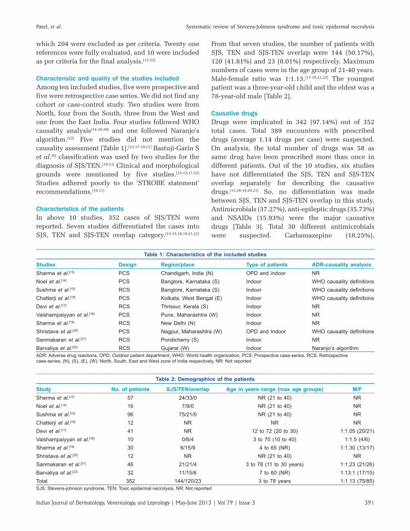
which 204 were excluded as per criteria. Twenty one references were fully evaluated, and 10 were included as per criteria for the final analysis.[13‑22]
Characteristic and quality of the studies includedAmong ten included studies, five were prospective and five were retrospective case series. We did not find any cohort or case‑control study. Two studies were from North, four from the South, three from the West and one from the East India. Four studies followed WHO causality analysis[14‑16,20] and one followed Naranjo’s algorithm.[22] Five studies did not mention the causality assessment [Table 1].[13,17‑19,21] Bastuji‑Garin S et al.[6] classification was used by two studies for the diagnosis of SJS/TEN.[19,21] Clinical and morphological grounds were mentioned by five studies.[13‑15,17,22] Studies adhered poorly to the ‘STROBE statement’ recommendations.[10,11]
Characteristics of the patientsIn above 10 studies, 352 cases of SJS/TEN were reported. Seven studies differentiated the cases into SJS, TEN and SJS‑TEN overlap category.[13‑15,18,19,21,22]
From that seven studies, the number of patients with SJS, TEN and SJS‑TEN overlap were 144 (50.17%), 120 (41.81%) and 23 (8.01%) respectively. Maximum numbers of cases were in the age group of 21‑40 years. Male‑female ratio was 1:1.13.[17‑19,21,22] The youngest patient was a three‑year‑old child and the eldest was a 78‑year‑old male [Table 2].
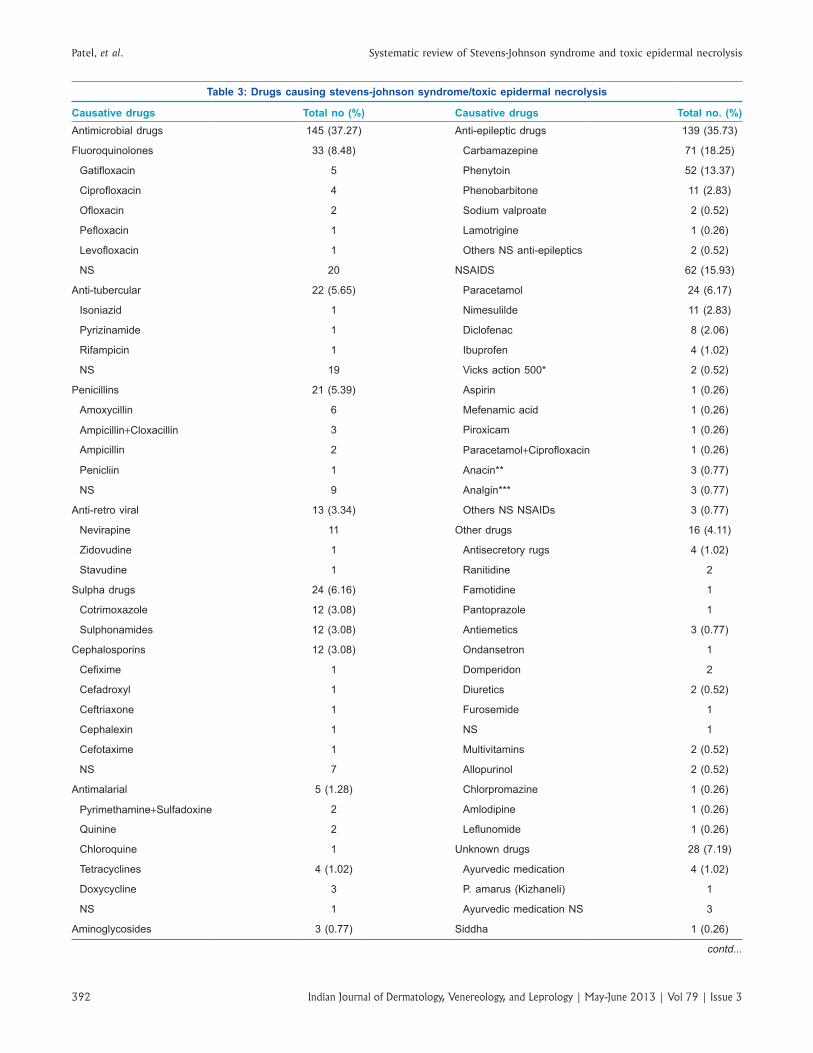
Causative drugsDrugs were implicated in 342 (97.14%) out of 352 total cases. Total 389 encounters with prescribed drugs (average 1.14 drugs per case) were suspected. On analysis, the total number of drugs was 58 as same drug have been prescribed more than once in different patients. Out of the 10 studies, six studies have not differentiated the SJS, TEN and SJS‑TEN overlap separately for describing the causative drugs.[13,16‑18,20,21] So, no differentiation was made between SJS, TEN and SJS‑TEN overlap in this study. Antimicrobials (37.27%), anti‑epileptic drugs (35.73%) and NSAIDs (15.93%) were the major causative drugs [Table 3]. Total 30 different antimicrobials were suspected. Carbamazepine (18.25%),
Table 1: Characteristics of the included studies
Studies Design Region/place Type of patients ADR‑causality analysisSharma et al.[13] PCS Chandigarh, India (N) OPD and indoor NRNoel et al.[14] PCS Banglore, Karnataka (S) Indoor WHO causality definitionsSushma et al.[15] RCS Banglore, Karnataka (S) Indoor WHO causality definitionsChatterji et al.[16] PCS Kolkata, West Bengal (E) Indoor WHO causality definitionsDevi et al.[17] RCS Thrissur, Kerala (S) Indoor NRVaishampaiyyan et al.[18] PCS Pune, Maharashtra (W) Indoor NRSharma et al.[19] RCS New Delhi (N) Indoor NRShristava et al.[20] PCS Nagpur, Maharashtra (W) OPD and Indoor WHO causality definitionsSanmakaran et al.[21] RCS Pondicherry (S) Indoor NRBarvaliya et al.[22] RCS Gujarat (W) Indoor Naranjo’s algorithmADR: Adverse drug reactions, OPD: Outdoor patient department, WHO: World health organization, PCS: Prospective case-series, RCS: Retrospective case-series, (N), (S), (E), (W): North, South, East and West zone of India respectively, NR: Not reported
Table 2: Demographics of the patients
Study No. of patients SJS/TEN/overlap Age in years range (max age groups) M/FSharma et al.[13] 57 24/33/0 NR (21 to 40) NRNoel et al.[14] 16 7/9/0 NR (21 to 40) NRSushma et al.[15] 96 75/21/0 NR (21 to 40) NRChatterji et al.[16] 12 NR NR NRDevi et al.[17] 41 NR 12 to 72 (20 to 30) 1:1.05 (20/21)Vaishampaiyyan et al.[18] 10 0/6/4 3 to 70 (10 to 40) 1:1.5 (4/6)Sharma et al.[19] 30 6/15/9 4 to 65 (NR) 1:1.30 (13/17)Shristava et al.[20] 12 NR NR (21 to 40) NRSanmakaran et al.[21] 46 21/21/4 3 to 78 (11 to 30 years) 1:1.23 (21/26)Barvaliya et al.[22] 32 11/15/6 7 to 60 (NR) 1.13:1 (17/15)Total 352 144/120/23 3 to 78 years 1:1.13 (75/85)SJS: Stevens-johnson syndrome, TEN: Toxic epidermal necrolysis, NR: Not reported
Patel, et al. Systematic review of Stevens‑Johnson syndrome and toxic epidermal necrolysis
Indian Journal of Dermatology, Venereology, and Leprology | May-June 2013 | Vol 79 | Issue 3392
Table 3: Drugs causing stevens‑johnson syndrome/toxic epidermal necrolysis
Causative drugs Total no (%) Causative drugs Total no. (%)Antimicrobial drugs 145 (37.27) Anti-epileptic drugs 139 (35.73)
Fluoroquinolones 33 (8.48) Carbamazepine 71 (18.25)
Gatifloxacin 5 Phenytoin 52 (13.37)
Ciprofloxacin 4 Phenobarbitone 11 (2.83)
Ofloxacin 2 Sodium valproate 2 (0.52)
Pefloxacin 1 Lamotrigine 1 (0.26)
Levofloxacin 1 Others NS anti-epileptics 2 (0.52)
NS 20 NSAIDS 62 (15.93)
Anti-tubercular 22 (5.65) Paracetamol 24 (6.17)
Isoniazid 1 Nimesulilde 11 (2.83)
Pyrizinamide 1 Diclofenac 8 (2.06)
Rifampicin 1 Ibuprofen 4 (1.02)
NS 19 Vicks action 500* 2 (0.52)
Penicillins 21 (5.39) Aspirin 1 (0.26)
Amoxycillin 6 Mefenamic acid 1 (0.26)
Ampicillin+Cloxacillin 3 Piroxicam 1 (0.26)
Ampicillin 2 Paracetamol+Ciprofloxacin 1 (0.26)
Penicliin 1 Anacin** 3 (0.77)
NS 9 Analgin*** 3 (0.77)
Anti-retro viral 13 (3.34) Others NS NSAIDs 3 (0.77)
Nevirapine 11 Other drugs 16 (4.11)
Zidovudine 1 Antisecretory rugs 4 (1.02)
Stavudine 1 Ranitidine 2
Sulpha drugs 24 (6.16) Famotidine 1
Cotrimoxazole 12 (3.08) Pantoprazole 1
Sulphonamides 12 (3.08) Antiemetics 3 (0.77)
Cephalosporins 12 (3.08) Ondansetron 1
Cefixime 1 Domperidon 2
Cefadroxyl 1 Diuretics 2 (0.52)
Ceftriaxone 1 Furosemide 1
Cephalexin 1 NS 1
Cefotaxime 1 Multivitamins 2 (0.52)
NS 7 Allopurinol 2 (0.52)
Antimalarial 5 (1.28) Chlorpromazine 1 (0.26)
Pyrimethamine+Sulfadoxine 2 Amlodipine 1 (0.26)
Quinine 2 Leflunomide 1 (0.26)
Chloroquine 1 Unknown drugs 28 (7.19)
Tetracyclines 4 (1.02) Ayurvedic medication 4 (1.02)
Doxycycline 3 P. amarus (Kizhaneli) 1
NS 1 Ayurvedic medication NS 3
Aminoglycosides 3 (0.77) Siddha 1 (0.26)
contd...
Patel, et al. Systematic review of Stevens‑Johnson syndrome and toxic epidermal necrolysis
393Indian Journal of Dermatology, Venereology, and Leprology | May-June 2013 | Vol 79 | Issue 3
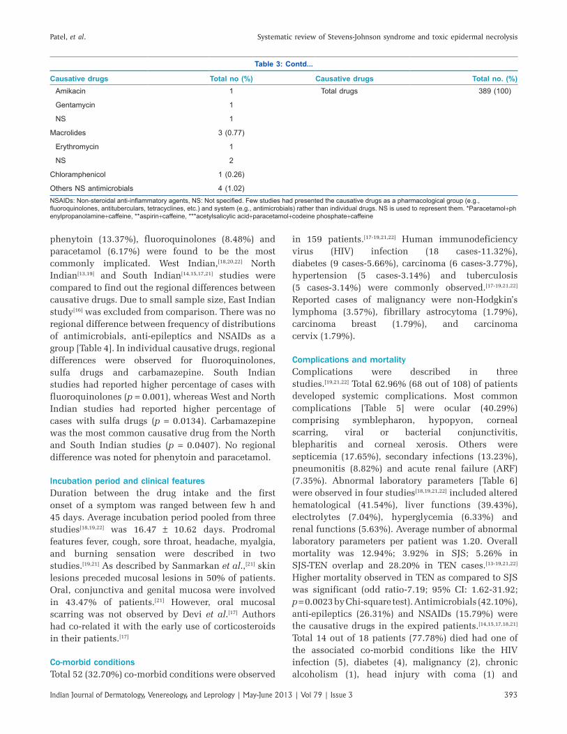
phenytoin (13.37%), fluoroquinolones (8.48%) and paracetamol (6.17%) were found to be the most commonly implicated. West Indian,[18,20,22] North Indian[13,19] and South Indian[14,15,17,21] studies were compared to find out the regional differences between causative drugs. Due to small sample size, East Indian study[16] was excluded from comparison. There was no regional difference between frequency of distributions of antimicrobials, anti‑epileptics and NSAIDs as a group [Table 4]. In individual causative drugs, regional differences were observed for fluoroquinolones, sulfa drugs and carbamazepine. South Indian studies had reported higher percentage of cases with fluoroquinolones (p = 0.001), whereas West and North Indian studies had reported higher percentage of cases with sulfa drugs (p = 0.0134). Carbamazepine was the most common causative drug from the North and South Indian studies (p = 0.0407). No regional difference was noted for phenytoin and paracetamol.
Incubation period and clinical featuresDuration between the drug intake and the first onset of a symptom was ranged between few h and 45 days. Average incubation period pooled from three studies[18,19,22] was 16.47 ± 10.62 days. Prodromal features fever, cough, sore throat, headache, myalgia, and burning sensation were described in two studies.[19,21] As described by Sanmarkan et al.,[21] skin lesions preceded mucosal lesions in 50% of patients. Oral, conjunctiva and genital mucosa were involved in 43.47% of patients.[21] However, oral mucosal scarring was not observed by Devi et al.[17] Authors had co‑related it with the early use of corticosteroids in their patients.[17]
Co‑morbid conditionsTotal 52 (32.70%) co‑morbid conditions were observed
in 159 patients.[17‑19,21,22] Human immunodeficiency virus (HIV) infection (18 cases‑11.32%), diabetes (9 cases‑5.66%), carcinoma (6 cases‑3.77%), hypertension (5 cases‑3.14%) and tuberculosis (5 cases‑3.14%) were commonly observed.[17‑19,21,22] Reported cases of malignancy were non‑Hodgkin’s lymphoma (3.57%), fibrillary astrocytoma (1.79%), carcinoma breast (1.79%), and carcinoma cervix (1.79%).
Complications and mortalityComplications were described in three studies.[19,21,22] Total 62.96% (68 out of 108) of patients developed systemic complications. Most common complications [Table 5] were ocular (40.29%) comprising symblepharon, hypopyon, corneal scarring, viral or bacterial conjunctivitis, blepharitis and corneal xerosis. Others were septicemia (17.65%), secondary infections (13.23%), pneumonitis (8.82%) and acute renal failure (ARF) (7.35%). Abnormal laboratory parameters [Table 6] were observed in four studies[18,19,21,22] included altered hematological (41.54%), liver functions (39.43%), electrolytes (7.04%), hyperglycemia (6.33%) and renal functions (5.63%). Average number of abnormal laboratory parameters per patient was 1.20. Overall mortality was 12.94%; 3.92% in SJS; 5.26% in SJS‑TEN overlap and 28.20% in TEN cases.[13‑19,21,22] Higher mortality observed in TEN as compared to SJS was significant (odd ratio‑7.19; 95% CI: 1.62‑31.92; p = 0.0023 by Chi‑square test). Antimicrobials (42.10%), anti‑epileptics (26.31%) and NSAIDs (15.79%) were the causative drugs in the expired patients.[14,15,17,18,21] Total 14 out of 18 patients (77.78%) died had one of the associated co‑morbid conditions like the HIV infection (5), diabetes (4), malignancy (2), chronic alcoholism (1), head injury with coma (1) and
Table 3: Contd...
Causative drugs Total no (%) Causative drugs Total no. (%)Amikacin 1 Total drugs 389 (100)
Gentamycin 1
NS 1
Macrolides 3 (0.77)
Erythromycin 1
NS 2
Chloramphenicol 1 (0.26)
Others NS antimicrobials 4 (1.02)NSAIDs: Non‑steroidal anti‑inflammatory agents, NS: Not specified. Few studies had presented the causative drugs as a pharmacological group (e.g., fluoroquinolones, antituberculars, tetracyclines, etc.) and system (e.g., antimicrobials) rather than individual drugs. NS is used to represent them. *Paracetamol+phenylpropanolamine+caffeine, **aspirin+caffeine, ***acetylsalicylic acid+paracetamol+codeine phosphate+caffeine

Patel, et al. Systematic review of Stevens‑Johnson syndrome and toxic epidermal necrolysis
Indian Journal of Dermatology, Venereology, and Leprology | May-June 2013 | Vol 79 | Issue 3394
hypothyroidism (1).[15,17,18,21] The presence of a severe systemic illness before the onset of SJS/TEN was a bad prognostic factor.[17] The most common cause of death was septicemia.[13,17‑19,21] ARF, acute respiratory distress syndrome (ARDS) and multiple organ dysfunctions were other reported causes.[13,17,21]
SCORTEN scoreSCORTEN was reported in three studies.[18,21,22] Sanmarkan et al. mentioned its range from 0 to 5 in the TEN patients. Data were pooled from other two studies to calculate the percentage of mortality according to score at the time of admission.[18,22] Observed mortality rate matches with the expected mortality rate,[23] except higher mortality was observed for SCORTEN score 3 [Table 7].
Duration of hospital stay and management costData of duration of hospital stay and management cost were described by Barvaliya et al.[22] Hospitalization was 9.7 days (95% CI: 5.8‑13.6) in SJS, 16.1 days (95% CI: 5.0‑27.4) in SJS‑TEN overlap and 20.6 days (95% CI: 14.4‑26.8) in TEN. It was significantly higher in TEN as compared to SJS (p = 0.020, Tukey‑Kramer Multiple comparisons test). Management cost was Rs 2460 (95% CI: 1762‑3158) in SJS, Rs. 4857 (95% CI: 2118‑7587) in SJS‑TEN overlap and Rs 7910 (95% CI: 5672‑10147) in TEN that was significantly higher as compared to SJS (p < 0.0001, Tukey‑Kramer Multiple comparisons test).[22] The cost was based on drugs, investigations and consumables used. Direct nonmedical and indirect costs were not mentioned.[22]
Use of corticosteroidsNo detailed description of corticosteroid use was found in the studies. Two studies have recommended early short‑term use of dexamethasone.[19,21] Sharma et al.[19] described the use of parenteral dexamethasone (4‑8 mg/day) or equivalent steroids for a short course of 2‑12 days in tapering doses in 29 patients with early active disease or with the reappearance of erythema in recovering
Table 4: Regional distribution of causative drugs
Causative drugs South India n (%)
West India n (%)
North India n (%)
p value
Antimicrobials 72 (40) 40 (49.38) 33 (28.69) 0.1393Fluoroquinolones 26 6 1 0.0010*Sulpha drugs 5 10 11 0.0134*
Antiepilepsy drugs 68 (37.36) 16 (19.75) 46 (40) 0.0704Carbamazepine 42 7 17 0.0407*Phenytoin 20 8 20 0.2810
NSAIDS 27 (14.83) 18 (22.22) 16 (13.91) 0.3685Paracetamol 8 10 6 0.0688Total drugs 182 (100) 81 (100) 115 (100) -
NSAIDs: Non‑steroidal anti‑inflammatory agents, *p<0.05 by Chi-square test
Table 5: Complications associated with stevens‑johnson syndrome/toxic epidermal necrolysis
Complications No. (%)Ocular 27 (40.29)Septicemia 12 (17.65)Secondary infection 9 (13.23)Pneumonitis 6 (8.82)Acute renal failure 5 (7.35)Urinary tract infection 2 (2.94)Congestive cardiac failure 2 (2.94)Pulmonary edema 2 (2.94)Metabolic encephalopathy 1 (1.47)Hepatic encephalopathy 1 (1.47)Intracranial bleed 1 (1.47)Total 68 (100)Table 6: Abnormal laboratory parameters
Laboratory parameters No. (%)Hematological 59 (41.54)Leucocytosis 25Leucopenia 12Lymphopenia 4Eosinopenia 3Anemia 11Thrombocytopenia 4Hepatic 56 (39.43)Increased aminotransferases 26Increased alkaline phosphatases 18Hyperbilirubinemia 5Altered liver functions 7Electrolytes 10 (7.04)Hypokalemia 5Reduced HCO3 4Hyperkalemia 1Renal-Raised BUN 8 (5.63)Hyperglycemia 9 (6.33)Total 142 (100)HCO3: Bicarbonate, BUN: Blood urea nitrogen
Table 7: Comparison between expected and observed mortality by SCORTEN score
SCORTEN score at admission
Total number of cases
Observed mortality %
Expected mortality % (95% CI)[23]
0-1 21 9.52 3.1 (0.1-16.7)2 14 14.28 12.1 (5.4-22.5)3 05 80 35.3 (19.8-53.3)4 02 50 58.3 (36.6-77.9)5 or more 00 00 >90 (55.5-99.8)CI: Confidence interval

Patel, et al. Systematic review of Stevens‑Johnson syndrome and toxic epidermal necrolysis
395Indian Journal of Dermatology, Venereology, and Leprology | May-June 2013 | Vol 79 | Issue 3
patients. Lag period for the starting of treatment in early active cases was not mentioned. Sanmakaran et al.[21] reported onset of healing on 3rd day in patients on the dexamethasone pulse. Short term dexamethasone may reduce mortality without improvement in healing. Earlier onset of healing i.e., on 2nd day and lowest period of hospitalization was also noted for methyl‑prednisolone therapy. However, dosage, route and lag period for administration of dexamethasone and methyl‑prednisolone were not described.[21] Devi et al.[17] described the use of systemic dexamethasone (4‑12 mg/day in divided doses parenterally) for a period of two weeks in tapering doses under cover of antibiotics in 36 patients within 2‑3 days of onset of symptoms. They observed complete recovery in patients without having underlying systemic disease and no mucosal scaring in the eyes.[17]
DISCUSSION
In the present study, clinical characteristics of SJS/TEN in Indian population are systematically reviewed from the selected studies from 1995 to 2011. The age of patients with SJS/TEN ranges from 3 years to 78 years. The majority of the patients are in the range of 21‑40 years that matches with the reports from other countries.[24] Female preponderance observed here is similar to other studies abroad.[24,25] No seasonal variation is documented in any of these studies as reported by Wanat et al.[26]
The major causative drugs are antimicrobials, anti‑epileptics and NSAIDs. Thirty three antimicrobials from 11 groups are implicated. Most common are fluoroquinolones (8.48%), sulpha drugs (6.68%), anti‑tubercular drugs (5.65%), penicillins (5.39%), anti‑retro virals (3.34%) and cephalosporins (3.08%). One study from Singapore[27] reports highest incidence with β‑lactam antibiotics that almost matches this study if penicilins and cephalosporins above are considered together. Cephalosporins are the most common culprit reported from Japan.[24] Sulphonamides (50.6%) and nevirapine (23.6%) are most commonly implicated drugs from Togo.[28] Association of antimicrobials with SJS/TEN is suspected to be a confounding factor if they are given during fever before the appearance of skin manifestations. However, severe cutaneous adverse reactions (SCAR) study has found significant increase risk of SJS with sulfa drugs, aminopenicillins, quinolones and cephalosporins after ruling out recent infections as a confounding factor.[29] In EuroSCAR study, nevirapine is the leading culprit in HIV‑positive
patients.[25] Though the total reported cases with nevirapine are 11 (2.83%) in our study; this drug should be monitored because of its widespread use as a first‑line anti‑retroviral drug in India. It should be discontinued as soon as eruption occurs.[30] Patterns of the most common reported antimicrobials varies from country to country. In our study, anti‑tubercular drugs are found as the third most common cause which does not match with European studies probably due to low disease burden there. In Togo, eight cases of SJS/TEN have been observed in HIV‑infected patients taking combination of rifampicin and isoniazid.[31] Few cases of SJS/TEN due to tetracyclines, antimalarials, aminoglycosides, macrolides and chloramphenicol are reported in our study. Considering their widespread use across India, risk seems minimal.
In this study, carbamazepine (18.25%) and phenytoin (13.37%) are most commonly involved anti‑epileptic drugs. They are widely used in India.[32,33] Carbamazepine and phenytoin are also reported as most common anti‑epileptic drugs for SJS/TEN in Taiwan.[34] Carbamazepine is the most the common and the only anti‑epileptic reported from Japan and Singapore.[24,27] SCAR study has reported association of SJS/TEN with phenobarbitone, carbamazepine, phenytoin and sodium valproate.[29] Subsequent EuroSCAR study did not find significant risk with sodium valproate.[25] The association of sodium valproate seems to be confounded by concomitant use of other anti‑epileptic drugs.[25,35] In our study, merely two cases are suspected due to sodium valproate. Considering its widespread use,[33] risk of SJS/TEN seems insignificant in India. EuroSCAR found a significant risk with lamotrigine.[25] The risk estimates of SJS/TEN vary between 1 and 10 per 10,000 in current users of carbamazepine, lamotrigine, phenytoin and phenobarbitone.[36] We have found only one case with lamotrigine. This is probably due to its limited utilization in our country.[33] Among cases exposed to anti‑epileptic drugs in EuroSCAR study, 85‑100% of patients have initiated their treatment less than 8 weeks before the reaction.[25] Risk for the development of SJS/TEN with anti‑epileptics is largely confined to initial 8 weeks.[25,35] This should be considered for analyzing the causal relationship between anti‑epileptic drugs and SJS/TEN.
Among the NSAIDs, paracetamol and nimesulide are most common reported in this study. SCAR study has found an overall risk of SJS with oxicam derivatives. It reports increased risk with paracetamol from

Patel, et al. Systematic review of Stevens‑Johnson syndrome and toxic epidermal necrolysis
Indian Journal of Dermatology, Venereology, and Leprology | May-June 2013 | Vol 79 | Issue 3396
Germany, Italy, and Portugal except France. There is no increased risk with diclofenac, salicylates and pyrazolone derivatives.[29] EuroSCAR study found weak association of paracetamol with SJS.[25] Paracetamol is confounded by its use to treat nonspecific symptoms such as fever or pain, the early signs of the adverse reaction or infection both.[25] However, pararacetamol is found to be a potential risk factor in children when data from pediatric patients from the SCAR and EuroSCAR studies are pooled.[37] Out of the 10 included studies, eight had mentioned the paracetamol as a suspected causative agent.[14,15,17‑22] However, causality analysis was done by four studies only. Three of them used WHO causality definitions,[14,15,20] and one used Naranjo’s algorithm.[22] No details about use of paracetamol along with antimicrobials or other agents are mentioned in any of the studies under review. Higher reporting of paracetamol may be because it is widely prescribed and available over‑the‑counter. Many other less prescribed drugs have more ADR than paracetamol. In contrast to SCAR and EuroSCAR,[25,29] we found only one case of SJS with piroxicam. Drug utilization studies of NSAIDs from India show the less use of piroxicam as compared to paracetamol, ibuprofen and diclofenac.[38,39]
No regional differences in frequency of reporting of antimicrobials, anti‑epileptic drugs and NSAIDs as a group are observed. Carbamazepine, phenytoin and paracetamol are the most common culprit drugs from the South, North and West India respectively. A relative risk of SJS/TEN usually remains uniform geographically. The prevalence of exposure to medication may vary between countries and with the time period.[37] The observed regional differences for fluoroquinolones, sulfa drugs and carbamazepine could be due to different utilization pattern of these drugs or because of under‑reporting. We have not found any regional difference with paracetamol as described by SCAR study.[29] Out of these drugs, genetic predisposition is only reported with the carbamazepine. Strong association between carbamazepine‑induced SJS with human leucocyte antigen (HLA‑B*) 1502 is reported in the Han Chinese population.[40] HLA‑B*1502 allele is not a universal marker for this disease and it depends on ethnicity.[41] The association has not been found in Caucasian patients. However, this allele is seen in high frequency in many Asian populations. The US FDA has made a label change in drug information about carbamazepine for a strong correlation between
HLA‑B*1502 and SJS/TEN.[42] One study from India reports HLA‑B*1502 in six out of eight patients of carbamazepine‑induced SJS/TEN.[43] Phenytoin possesses an aromatic ring like carbamazepine. In one case‑control study from Taiwan, HLA‑B*1502 was present in 8 out of 26 (30.8%) patients of phenytoin‑induced SJS and 3 out of 3 (100%) in patients of oxacarbazepine‑induced SJS. These drugs should also be avoided in HLA‑B*1502 carriers.[44]
HIV is the most common co‑morbid condition in the patients of SJS/TEN in India. Observed proportion of HIV‑positive patients in the present study is lower than South Africa (78.67%), Togo (54.6%) and higher than France (7.3%).[28,45,46] Incidence rate of SJS is 1,000 fold higher in HIV‑positive patients as compared to general population in Germany.[47] In contrast to our study, higher cases of malignancy (10.6%) than HIV infection (6.6%) were reported in EuroSCAR.[25] The patients had complications involving eye, liver, kidneys, lungs, and blood. In addition to septicemia, other problems in the course were ARF, ARDS and multiple organ dysfunctions. Surviving sepsis campaign (SSC) guidelines can be followed to reduce the mortality due to septicemia, particularly in TEN patients. It is developed by the international group of the experts.[48] Various studies have shown the reduced mortality following implementation of the SSC guidelines.[49,50] There is a wide difference in the mortality between SJS (3.92%) and TEN (28.2%). Higher mortality, duration of stay and management cost are observed in TEN as compared to SJS that is in accordance with the studies abroad.[24,27,51] Data of duration of stay and management cost were based on one study only. More studies from India are required to substantiate the data. Availability of facilities and the level of care may have affected the mortality and duration of stay.
SCORTEN uses seven independent risk factors to predict the risk of death: Age, malignancy, heart rate, epidermal detachment, serum urea, glucose and bicarbonate at admission.[23] In our study, in patients having SCORTEN score 3 at admission show higher than expected mortality. The SCORTEN is associated with a greater risk of day to day varying mortality during first 5 days of hospitalization. Its performance is best at day three.[52] Vaishampaiyyan et al.[18] observed the progression of score from the day of admission to day five. SCORTEN score should be observed up to day 5 for the accurate prediction of mortality.

Patel, et al. Systematic review of Stevens‑Johnson syndrome and toxic epidermal necrolysis
397Indian Journal of Dermatology, Venereology, and Leprology | May-June 2013 | Vol 79 | Issue 3
Corticosteroids are considered controversial in the management of SJS/TEN despite their use for more than 30 years. No randomized controlled trials are available to establish their efficacy.[53] It is difficult to analyze the effect of steroids from this study due to limited description of their use. Early use of short term dexamethasone therapy seems beneficial. Short term dexamethasone therapy (1.5 mg/kg/day) on three consecutive days at an early stage of the reaction may reduce mortality without affecting the healing time.[54] A recent systematic review in children has shown the beneficial effect of steroids. Patients treated with dressing and support treatments alone without a steroid have longer hospitalization, remission, and are more prone to complications and death.[55]
There are several limitations to this study. As this systematic review was based on the case‑series only, there was an absence of a control group and lack of denominator data, i.e., total number of prescriptions with suspected drugs in study population. So, we could not quantify the risk of SJS/TEN associated with the use of medication. There is also the strong possibility of under‑reporting. All the parameters were not available for analysis from the selected studies. Six of the 10 studies did not specify separately which patients produced SJS or TEN or SJS‑TEN overlap. For the important parameters like complications and SCORTEN only three and two studies were available respectively. So the sample for analysis is very small. No study differentiated the profile of pediatric and adult cases with respect to drugs implicated, course of illness and mortality. Statistically substantiated reason against the use of corticosteroids cannot be given. More studies for SJS/TEN are necessary from India. Registry system for SJS/TEN is required in India to strengthen the database to design effective treatment modalities.
REFERENCES
1. Chan HL, Stern RS, Arndt KA, Langlois J, Jick SS, Jick H, et al. The incidence of erythema multiforme, Stevens‑Johnson syndrome, and toxic epidermal necrolysis. A population‑based study with particular reference to reactions caused by drugs among outpatients. Arch Dermatol 1990;126:43‑7.
2. Strom BL, Carson JL, Halpern AC, Schinnar R, Snyder ES, Shaw M, et al. A population‑based study of Stevens‑Johnson syndrome. Incidence and antecedent drug exposures. Arch Dermatol 1991;127:831‑8.
3. Schöpf E, Stühmer A, Rzany B, Victor N, Zentgraf R, Kapp JF. Toxic epidermal necrolysis and Stevens‑Johnson syndrome. An epidemiologic study from West Germany. Arch Dermatol 1991;127:839‑42.
4. Roujeau JC, Stern RS. Severe adverse cutaneous reactions to
drugs. N Engl J Med 1994;331:1272‑85.5. Sehgal VN, Srivastava G. Toxic epidermal necrolysis (TEN)
Lyell’s syndrome. J Dermatolog Treat 2005;16:278‑86.6. Bastuji‑Garin S, Rzany B, Stern RS, Shear NH, Naldi L,
Roujeau JC. Clinical classification of cases of toxic epidermal necrolysis, Stevens‑Johnson syndrome, and erythema multiforme. Arch Dermatol 1993;129:92‑6.
7. Harr T, French LE. Toxic epidermal necrolysis and Stevens‑Johnson syndrome. Orphanet J Rare Dis 2010;5:39.
8. Saha K. Toxic epidermal necrolysis: Current concepts in pathogenesis and treatment. Indian J Dermatol Venereol Leprol 2000;66:10‑7.
9. Borchers AT, Lee JL, Naguwa SM, Cheema GS, Gershwin ME. Stevens‑Johnson syndrome and toxic epidermal necrolysis. Autoimmun Rev 2008;7:598‑605.
10. von Elm E, Altman DG, Egger M, Pocock SJ, Gøtzsche PC, Vandenbroucke JP, et al. The Strengthening the Reporting of Observational Studies in Epidemiology (STROBE) statement: Guidelines for reporting observational studies. J Clin Epidemiol 2008;61:344‑9.
11. Vandenbroucke JP, von Elm E, Altman DG, Gøtzsche PC, Mulrow CD, Pocock SJ, et al. Strengthening the Reporting of Observational Studies in Epidemiology (STROBE): Explanation and elaboration. Epidemiology 2007;18:805‑35.
12. Central Drugs Standard Control Organization (Medical devices division). Guidance document on application for grant of Licence in Form‑28 for manufacture of Medical Devices in India under CLAA Scheme, 2010. Available from: http://cdsco.nic.in/Guidance%20document%20on%20application%20 for%20grant%20of%20Licence%20in%20Form‑28%20 for%20manufacture%20of%20Medical%20Devices%20in%20India%20under%20CLAA%20Scheme.PDF [Last cited 2012 Jan 11].
13. Sharma VK, Sethuraman G, Kumar B. Cutaneous adverse drug reactions: Clinical pattern and causative agents – A 6 year series from Chandigarh, India. J Postgrad Med 2001;47:95‑9.
14. Noel MV, Sushma M, Guido S. Cutaneous adverse drug reactions in hospitalized patients in a tertiary care center. Indian J Pharmacol 2004;36:292‑5.
15. Sushma M, Noel MV, Ritika MC, James J, Guido S. Cutaneous adverse drug reactions: A 9‑year study from a South Indian Hospital. Pharmacoepidemiol Drug Saf 2005;14:567‑70.
16. Chatterjee S, Ghosh AP, Barbhuiya J, Dey SK. Adverse cutaneous drug reactions: A one year survey at a dermatology outpatient clinic of a tertiary care hospital. Indian J Pharmacol 2006;38:429‑31.
17. Devi K, George S, Criton S, Suja V, Sridevi PK. Carbamazepine – The commonest cause of toxic epidermal necrolysis and Stevens‑Johnson syndrome: A study of 7 years. Indian J Dermatol Venereol Leprol 2005;71:325‑8.
18. Vaishampayan SS, Das AL, Verma R. SCORTEN: Does it need modification? Indian J Dermatol Venereol Leprol 2008;74:35‑7.
19. Sharma VK, Sethuraman G, Minz A. Stevens Johnson syndrome, toxic epidermal necrolysis and SJS‑TEN overlap: A retrospective study of causative drugs and clinical outcome. Indian J Dermatol Venereol Leprol 2008;74:238‑40.
20. Shrivastava M, Uchit G, Chakravarti A, Joshi G, Mahatme M, Chaudhari H. Adverse drug reactions reported in Indira Gandhi Government Medical College and Hospital, Nagpur. J Assoc Physicians India 2011;59:296‑9.
21. Sanmarkan AD, Sori T, Thappa DM, Jaisankar TJ. Retrospective analysis of Stevens‑Johnson syndrome and toxic epidermal necrolysis over a period of 10 years. Indian J Dermatol 2011;56:25‑9.
22. Barvaliya M, Sanmukhani J, Patel T, Paliwal N, Shah H, Tripathi C. Drug‑induced Stevens‑Johnson syndrome (SJS), toxic epidermal necrolysis (TEN), and SJS‑TEN overlap: A multicentric retrospective study. J Postgrad Med 2011;57:115‑9.
23. Bastuji‑Garin S, Fouchard N, Bertocchi M, Roujeau JC, Revuz J,

Patel, et al. Systematic review of Stevens‑Johnson syndrome and toxic epidermal necrolysis
Indian Journal of Dermatology, Venereology, and Leprology | May-June 2013 | Vol 79 | Issue 3398
Wolkenstein P. SCORTEN: A severity‑of‑illness score for toxic epidermal necrolysis. J Invest Dermatol 2000;115:149‑53.
24. Yamane Y, Aihara M, Ikezawa Z. Analysis of Stevens‑Johnson syndrome and toxic epidermal necrolysis in Japan from 2000 to 2006. Allergol Int 2007;56:419‑25.
25. Mockenhaupt M, Viboud C, Dunant A, Naldi L, Halevy S, Bouwes Bavinck JN, et al. Stevens‑Johnson syndrome and toxic epidermal necrolysis: Assessment of medication risks with emphasis on recently marketed drugs. The EuroSCAR‑study. J Invest Dermatol 2008;128:35‑44.
26. Wanat KA, Anadkat MJ, Klekotka PA. Seasonal variation of Stevens‑Johnson syndrome and toxic epidermal necrolysis associated with trimethoprim‑sulfamethoxazole. J Am Acad Dermatol 2009;60:589‑94.
27. Tan SK, Tay YK. Profile and pattern of Stevens‑Johnson syndrome and toxic epidermal necrolysis in a general hospital in Singapore: Treatment outcomes. Acta Derm Venereol 2012;92:62‑6.
28. Saka B, Kombaté K, Mouhari‑Toure A, Akakpo S, Tchangaï‑Walla K, Pitché P. Stevens‑Johnson syndrome and toxic epidermal necrolysis in a teaching hospital in Lomé, Togo: Retrospective study of 89 cases. Med Trop (Mars) 2010;70:255‑8.
29. Roujeau JC, Kelly JP, Naldi L, Rzany B, Stern RS, Anderson T, et al. Medication use and the risk of Stevens‑Johnson syndrome or toxic epidermal necrolysis. N Engl J Med 1995;333:1600‑7.
30. Fagot JP, Mockenhaupt M, Bouwes‑Bavinck JN, Naldi L, Viboud C, Roujeau JC, EuroSCAR Study Group. Nevirapine and the risk of Stevens‑Johnson syndrome or toxic epidermal necrolysis. AIDS 2001;15:1843‑8.
31. Pitche P, Mouzou T, Padonou C, Tchangai‑Walla K. Stevens‑Johnson syndrome and toxic epidermal necrolysis after intake of rifampicin‑isoniazid: Report of 8 cases in HIV‑infected patients in Togo. Med Trop (Mars) 2005;65:359‑62.
32. Radhakrishnan K, Nayak SD, Kumar SP, Sarma PS. Profile of antiepileptic pharmacotherapy in a tertiary referral center in South India: A pharmacoepidemiologic and pharmacoeconomic study. Epilepsia 1999;40:179‑85.
33. Mathur S, Sen S, Ramesh L, Kumar S. Utilization pattern of antiepileptic drugs and their adverse effects, in a teaching hospital. Asian J Pharm Clin Res 2010;3:55‑9.
34. Yang CY, Dao RL, Lee TJ, Lu CW, Yang CH, Hung SI, et al. Severe cutaneous adverse reactions to antiepileptic drugs in Asians. Neurology 2011;77:2025‑33.
35. Rzany B, Correia O, Kelly JP, Naldi L, Auquier A, Stern R. Risk of Stevens‑Johnson syndrome and toxic epidermal necrolysis during first weeks of antiepileptic therapy: A case‑control study. Study Group of the International Case Control Study on Severe Cutaneous Adverse Reactions. Lancet 1999;353:2190‑4.
36. Mockenhaupt M, Messenheimer J, Tennis P, Schlingmann J. Risk of Stevens‑Johnson syndrome and toxic epidermal necrolysis in new users of antiepileptics. Neurology 2005;64:1134‑8.
37. Levi N, Bastuji‑Garin S, Mockenhaupt M, Roujeau JC, Flahault A, Kelly JP, et al. Medications as risk factors of Stevens‑Johnson syndrome and toxic epidermal necrolysis in children: A pooled analysis. Pediatrics 2009;123:e297‑304.
38. Gupta M, Malhotra S, Jain S, Aggarwal A, Pandhi P. Pattern of prescription of non‑steroidal antiinflammatory drugs in orthopaedic outpatient clinic of a north Indian tertiary care hospital. Indian J Pharmacol 2005;37:404‑5.
39. Paul AD, Chauhan CK. Study of usage pattern of nonsteroidal anti‑inflammatory drugs (NSAIDs) among different practice categories in Indian clinical setting. Eur J Clin Pharmacol 2005;60:889‑92.
40. Chung WH, Hung SI, Hong HS, Hsih MS, Yang LC, Ho HC, et al. Medical genetics: A marker for Stevens‑Johnson syndrome. Nature 2004;428:86.
41. Lonjou C, Thomas L, Borot N, Ledger N, de Toma C, LeLouet H, et al. A marker for Stevens‑Johnson syndrome: Ethnicity matters. Pharmacogenomics J 2006;6:265‑8.
42. Ferrell PB Jr, McLeod HL. Carbamazepine, HLA‑B*1502 and risk of Stevens‑Johnson syndrome and toxic epidermal necrolysis: US FDA recommendations. Pharmacogenomics 2008;9:1543‑6.
43. Mehta TY, Prajapati LM, Mittal B, Joshi CG, Sheth JJ, Patel DB, et al. Association of HLA‑B*1502 allele and carbamazepine‑induced Stevens‑Johnson syndrome among Indians. Indian J Dermatol Venereol Leprol 2009;75:579‑82.
44. Hung SI, Chung WH, Liu ZS, Chen CH, Hsih MS, Hui RC, et al. Common risk allele in aromatic antiepileptic‑drug induced Stevens‑Johnson syndrome and toxic epidermal necrolysis in Han Chinese. Pharmacogenomics 2010;11:349‑56.
45. Kannenberg SM, Jordaan HF, Koegelenberg CF, Von Groote‑Bidlingmaier F, Visser WI. Toxic epidermal necrolysis and Stevens‑Johnson syndrome in South Africa: A 3‑year prospective study. QJM 2012;105:839‑46.
46. Fagot JP, Mockenhaupt M, Bouwes‑Bavinck JN, Naldi L, Viboud C, Roujeau JC, et al. Nevirapine and the risk of Stevens‑Johnson syndrome or toxic epidermal necrolysis. AIDS 2001;15:1843‑8.
47. Rzany B, Mockenhaupt M, Stocker U, Hamouda O, Schöpf E. Incidence of Stevens‑Johnson syndrome and toxic epidermal necrolysis in patients with the acquired immunodeficiency syndrome in Germany. Arch Dermatol 1993;129:1059.
48. Dellinger RP, Levy MM, Carlet JM, Bion J, Parker MM, Jaeschke R, et al. Surviving Sepsis Campaign: International guidelines for management of severe sepsis and septic shock: 2008. Intensive Care Med 2008;34:17‑60.
49. Levy MM, Dellinger RP, Townsend SR, Linde‑Zwirble WT, Marshall JC, Bion J, et al. The Surviving Sepsis Campaign: Results of an international guideline‑based performance improvement program targeting severe sepsis. Intensive Care Med 2010;36:222‑31.
50. Castellanos‑Ortega A, Suberviola B, García‑Astudillo LA, Holanda MS, Ortiz F, Llorca J, et al. Impact of the Surviving Sepsis Campaign protocols on hospital length of stay and mortality in septic shock patients: Results of a three‑year follow‑up quasi‑experimental study. Crit Care Med 2010;38:1036‑43.
51. Yap FB, Wahiduzzaman M, Pubalan M. Stevens‑Johnson syndrome (SJS) and Toxic Epidermal Necrolysis (TEN) in Sarawak: A four years’ review. Egypt Dermatol Online J 2008;4:1‑13.
52. Guégan S, Bastuji‑Garin S, Poszepczynska‑Guigné E, Roujeau JC, Revuz J. Performance of the SCORTEN during the first five days of hospitalization to predict the prognosis of epidermal necrolysis. J Invest Dermatol 2006;126:272‑6.
53. Majumdar S, Mockenhaupt M, Roujeau J, Townshend A. Interventions for toxic epidermal necrolysis. Cochrane Database Syst Rev 2002:CD001435.
54. Kardaun SH, Jonkman MF. Dexamethasone pulse therapy for Stevens‑Johnson syndrome/toxic epidermal necrolysis. Acta Derm Venereol 2007;87:144‑8.
55. Del Pozzo‑Magana BR, Lazo‑Langner A, Carleton B, Castro‑Pastrana LI, Rieder MJ. A systematic review of treatment of drug‑induced Stevens‑Johnson syndrome and toxic epidermal necrolysis in children. J Popul Ther Clin Pharmacol 2011;18:e121‑33.