A Systematic Review of Orthosiphon stamineus Benth. in the ...
25
Citation: Wang, Q.; Wang, J.; Li, N.; Liu, J.; Zhou, J.; Zhuang, P.; Chen, H. A Systematic Review of Orthosiphon stamineus Benth. in the Treatment of Diabetes and Its Complications. Molecules 2022, 27, 444. https:// doi.org/10.3390/molecules27020444 Academic Editors: Weishuo Fang and Yu Zhang Received: 11 December 2021 Accepted: 5 January 2022 Published: 10 January 2022 Publisher’s Note: MDPI stays neutral with regard to jurisdictional claims in published maps and institutional affil- iations. Copyright: © 2022 by the authors. Licensee MDPI, Basel, Switzerland. This article is an open access article distributed under the terms and conditions of the Creative Commons Attribution (CC BY) license (https:// creativecommons.org/licenses/by/ 4.0/). molecules Review A Systematic Review of Orthosiphon stamineus Benth. in the Treatment of Diabetes and Its Complications Qirou Wang 1 , Jia Wang 1 , Nannan Li 1 , Junyu Liu 1 , Jingna Zhou 1 , Pengwei Zhuang 2 and Haixia Chen 1, * 1 Tianjin Key Laboratory for Modern Drug Delivery & High-Efficiency, School of Pharmaceutical Science and Technology, Tianjin University, Tianjin 300072, China; [email protected] (Q.W.); [email protected] (J.W.); [email protected] (N.L.); [email protected] (J.L.); [email protected] (J.Z.) 2 Haihe Laboratory of Modern Chinese Medicine, Chinese Materia Medica College, Tianjin University of Traditional Chinese Medicine, Tianjin 301617, China; [email protected] * Correspondence: [email protected]; Tel.: +86-22-2740-1483 Abstract: (1) Background: Orthosiphon stamineus Benth. is a traditional medicine used in the treatment of diabetes and chronic renal failure in southern China, Malaysia, and Thailand. Diabetes is a chronic metabolic disease and the number of diabetic patients in the world is increasing. This review aimed to systematically review the effects of O. stamineus in the treatment of diabetes and its complications and the pharmacodynamic material basis. (2) Methods: This systematic review was conducted following Preferred Reporting Items for Systematic Reviews and Meta-Analyses (PRISMA), using the databases ScienceDirect, PubMed, and Web of Science. (3) Results: Thirty-one articles related to O. stamineus and diabetes were included. The mechanisms of O. stamineus in the treatment of diabetes and its complications mainly included inhibiting α-amylase and α-glucosidase activities, antioxidant and anti-inflammatory activities, regulating lipid metabolism, promoting insulin secretion, ameliorating insulin resistance, increasing glucose uptake, promoting glycolysis, inhibiting gluconeogenesis, promoting glucagon-likepeptide-1 (GLP-1) secretion and antiglycation activity. Phenolic acids, flavonoids and triterpenoids might be the main components for hypoglycemia effects in O. stamineus. (4) Conclusion: O. stamineus could be an antidiabetic agent to treat diabetes and its complications. However, it needs further study on a pharmacodynamic substance basis and the mechanisms of effective constituents. Keywords: Orthosiphon stamineus; diabetes; diabetic complications; hypoglycemic activity; antidia- betic mechanisms 1. Introduction Orthosiphon stamineus Benth. (Lamiaceae) is a perennial herb [1,2]. O. stamineus is widely distributed in the tropical and subtropical regions [3], including southeast Asian countries (Indonesia, Malaysia, Thailand, Vietnam, Myanmar, Philippines) [4,5], southern China [6], India [7], Australia [5], etc. In addition to Orthosiphon stamineus Benth., it also has other scientific names, Clerodendranthus spicatus (Thunb) c. y. wu and Orthosiphon aristatus (Blume) Miq. [8–10]. It is usually called “Shencha” in Chinese. It is also called Cat’s whiskers [11], Misai Kucing [12], Java tea [13], and kumis kucing [14] in some Southeast Asian countries. O. stamineus is a popular Chinese folk medicine and also a traditional medicine of Dai nationality of Yunnan Province in China [15]. It has been used to treat diabetes and some kidney diseases with a long history. Modern pharmacological studies show that O. stamineus has many pharmacological activities, including antioxidant, anti-inflammatory, kidney protection, antibacterial, anti-tumor, immunoregulation, and especially effective antidiabetic activities. [15,16]. It has been used for the treatment of diabetes and chronic renal failure clinically. It is also reported to have good therapeutic effects on some diabetic Molecules 2022, 27, 444. https://doi.org/10.3390/molecules27020444 https://www.mdpi.com/journal/molecules
-
Upload
khangminh22 -
Category
Documents
-
view
2 -
download
0
Transcript of A Systematic Review of Orthosiphon stamineus Benth. in the ...
�����������������
Citation: Wang, Q.; Wang, J.; Li, N.;
Liu, J.; Zhou, J.; Zhuang, P.; Chen, H.
A Systematic Review of Orthosiphon
stamineus Benth. in the Treatment of
Diabetes and Its Complications.
Molecules 2022, 27, 444. https://
doi.org/10.3390/molecules27020444
Academic Editors: Weishuo Fang and
Yu Zhang
Received: 11 December 2021
Accepted: 5 January 2022
Published: 10 January 2022
Publisher’s Note: MDPI stays neutral
with regard to jurisdictional claims in
published maps and institutional affil-
iations.
Copyright: © 2022 by the authors.
Licensee MDPI, Basel, Switzerland.
This article is an open access article
distributed under the terms and
conditions of the Creative Commons
Attribution (CC BY) license (https://
creativecommons.org/licenses/by/
4.0/).
molecules
Review
A Systematic Review of Orthosiphon stamineus Benth. in theTreatment of Diabetes and Its ComplicationsQirou Wang 1, Jia Wang 1, Nannan Li 1, Junyu Liu 1, Jingna Zhou 1, Pengwei Zhuang 2 and Haixia Chen 1,*
1 Tianjin Key Laboratory for Modern Drug Delivery & High-Efficiency, School of Pharmaceutical Scienceand Technology, Tianjin University, Tianjin 300072, China; [email protected] (Q.W.);[email protected] (J.W.); [email protected] (N.L.); [email protected] (J.L.);[email protected] (J.Z.)
2 Haihe Laboratory of Modern Chinese Medicine, Chinese Materia Medica College, Tianjin University ofTraditional Chinese Medicine, Tianjin 301617, China; [email protected]
* Correspondence: [email protected]; Tel.: +86-22-2740-1483
Abstract: (1) Background: Orthosiphon stamineus Benth. is a traditional medicine used in the treatmentof diabetes and chronic renal failure in southern China, Malaysia, and Thailand. Diabetes is a chronicmetabolic disease and the number of diabetic patients in the world is increasing. This review aimed tosystematically review the effects of O. stamineus in the treatment of diabetes and its complications andthe pharmacodynamic material basis. (2) Methods: This systematic review was conducted followingPreferred Reporting Items for Systematic Reviews and Meta-Analyses (PRISMA), using the databasesScienceDirect, PubMed, and Web of Science. (3) Results: Thirty-one articles related to O. stamineusand diabetes were included. The mechanisms of O. stamineus in the treatment of diabetes and itscomplications mainly included inhibiting α-amylase and α-glucosidase activities, antioxidant andanti-inflammatory activities, regulating lipid metabolism, promoting insulin secretion, amelioratinginsulin resistance, increasing glucose uptake, promoting glycolysis, inhibiting gluconeogenesis,promoting glucagon-likepeptide-1 (GLP-1) secretion and antiglycation activity. Phenolic acids,flavonoids and triterpenoids might be the main components for hypoglycemia effects in O. stamineus.(4) Conclusion: O. stamineus could be an antidiabetic agent to treat diabetes and its complications.However, it needs further study on a pharmacodynamic substance basis and the mechanisms ofeffective constituents.
Keywords: Orthosiphon stamineus; diabetes; diabetic complications; hypoglycemic activity; antidia-betic mechanisms
1. Introduction
Orthosiphon stamineus Benth. (Lamiaceae) is a perennial herb [1,2]. O. stamineus iswidely distributed in the tropical and subtropical regions [3], including southeast Asiancountries (Indonesia, Malaysia, Thailand, Vietnam, Myanmar, Philippines) [4,5], southernChina [6], India [7], Australia [5], etc. In addition to Orthosiphon stamineus Benth., it alsohas other scientific names, Clerodendranthus spicatus (Thunb) c. y. wu and Orthosiphonaristatus (Blume) Miq. [8–10]. It is usually called “Shencha” in Chinese. It is also called Cat’swhiskers [11], Misai Kucing [12], Java tea [13], and kumis kucing [14] in some SoutheastAsian countries.
O. stamineus is a popular Chinese folk medicine and also a traditional medicine ofDai nationality of Yunnan Province in China [15]. It has been used to treat diabetes andsome kidney diseases with a long history. Modern pharmacological studies show thatO. stamineus has many pharmacological activities, including antioxidant, anti-inflammatory,kidney protection, antibacterial, anti-tumor, immunoregulation, and especially effectiveantidiabetic activities. [15,16]. It has been used for the treatment of diabetes and chronicrenal failure clinically. It is also reported to have good therapeutic effects on some diabetic
Molecules 2022, 27, 444. https://doi.org/10.3390/molecules27020444 https://www.mdpi.com/journal/molecules
Molecules 2022, 27, 444 2 of 25
complications, especially diabetic nephropathy [6]. Thus, it is worthy of study for thediscovery for new antidiabetic drugs from O. stamineus.
Diabetes is a chronic metabolic diseases caused by deficiency in insulin secretionand insulin resistance [17]. In 2021, diabetic patients were estimated to be approximately537 million all over the world [18]. This number is on the rise, the reasons for whichare an aging population, obesity, and unhealthy diets [19]. Diabetes can be classifiedinto two major types: Type I Diabetes Mellitus (T1DM) and Type II Diabetes Mellitus(T2DM). T1DM is caused by insulin deficiency. The islet β-cells are damaged, leading to anabsolute deficiency of insulin secretion. Patients need long-term exogenous insulin injection.However, for T2DM patients, metabolic disorder results in lower insulin sensitivity, insulinresistance, and relative insulin deficiency [17,19–21]. DM can damage organs and tissuesand result in many complications, such as diabetic nephropathy, diabetic retinopathy,diabetic foot, diabetic neuropathy, etc.
Diabetes is treated with oral hypoglycemic drugs and insulin injection to reduce bloodglucose levels, improve insulin secretion, and enhance insulin sensitivity. Besides, there alsoare natural products used in the treatment of diabetes, especially with good hypoglycemiceffects. In classical antidiabetic drugs, exenatide is from the venom of Gila monster andacarbose is produced from Actinoplanes sp. by the large-scale fermentation [22,23]. Be-sides, metformin is a natural product derivative that originated from herbal medicineGalega officinalis and its constituent galegine [24]. Many other natural products, such ascurcumin, cinnamon, pumpkin, bitter melon, Lycium barbarum, Portulaca oleracea, Aloe vera,etc., have also been proven to have antidiabetic activities but without general clinicalpractice [20,21,25–27]. Orthosiphon stamineus also has potential against diabetes.
More than 200 compounds have been isolated from O. stamineus. Diterpenoids,flavonoids, triterpenoids, phenolic acids, and their derivatives are the main chemical con-stituents. There are almost 50 phenolic acids and their derivatives isolated from O. stamineus,including rosmarinic acid, caffeic acid, and their derivatives, and many others [6,28]. Morethan 20 flavonoids have been isolated from O. stamineus. Most of them are flavones, es-pecially polymethoxy substituted flavones [29]. Besides, more than 60 diterpenoids havebeen isolated up to now and they have various skeleton types, including isopimarane [30],staminane [31], secoisopimarane [30], norstaminane [31], secostaminane, and some othertypes. Besides, there are also almost 20 triterpenoids isolated from O. stamineus [5,32,33]. Inaddition to the above four main types, there are also other kinds of compounds isolatedfrom O. stamineus, such as two alkyl glycosides (clerspides A and B) [34], coumarins, etc.Many of the compounds studied have acted as the main pharmacodynamic material basisof O. stamineus in the treatment of diabetes and its complications.
In a review by Omar Z. Ameer, the traditional uses, phytochemical studies, phar-macological studies, and toxicology were summarized. In Kamran Ashraf’s review, onlyphytochemical studies and pharmacological studies until 2018 were summarized. Thepharmacological activities of O. stamineus mentioned in the two reviews included anti-inflammatory, antioxidant, diuretic, hypouricemic, hepatoprotective, gastroprotective,nephroprotective, analgesic, antipyretic, cytotoxic, antiproliferative, antihypertensive, hy-poglycemic, hypolipidemic, anti-obesity, and antibacterial activities [35,36]. In Yin-SirChung’s review, the protective actions of O. stamineus on the central nervous system, safetyand toxicity, and pharmacokinetics studies were summarized [8]. Although there are manystudies on the potential of O. stamineus in the treatment of diabetes and its complications,no article has reviewed the antidiabetic mechanisms and pharmacodynamic material basisof O. stamineus in detail.
In this systematic review, the mechanisms and toxicology of O. stamineus in thetreatment of diabetes and its complications were summarized as per Preferred ReportingItems for Systematic Reviews and Meta-Analyses (PRISMA) [37,38]. PRISMA providesguidance for authors to prepare transparent, complete, and accurate systematic reviews.Research progress on clinical applications and the main pharmacodynamic material basis
Molecules 2022, 27, 444 3 of 25
of O. stamineus was reviewed, providing a reference for the application of O. stamineus andfurther research in the treatment of diabetes and its complications.
2. Results2.1. Literature Search Results
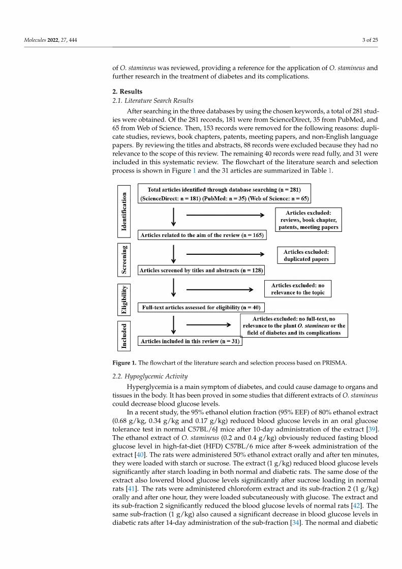
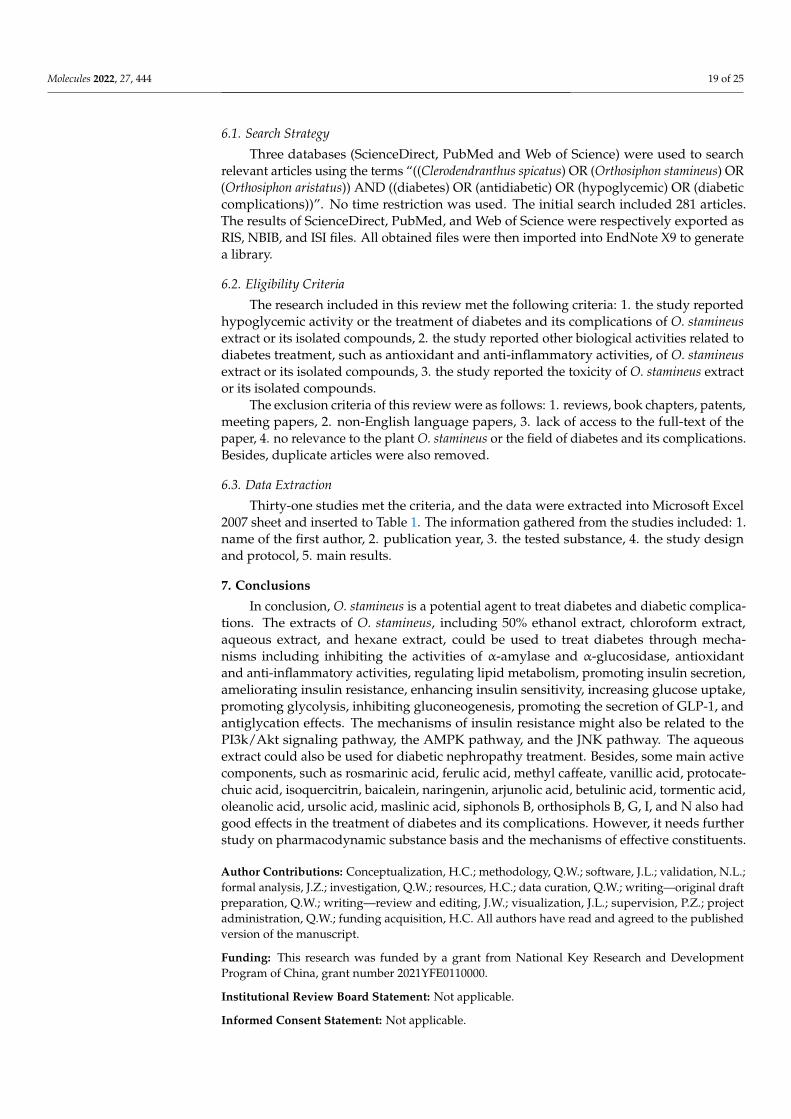
After searching in the three databases by using the chosen keywords, a total of 281 stud-ies were obtained. Of the 281 records, 181 were from ScienceDirect, 35 from PubMed, and65 from Web of Science. Then, 153 records were removed for the following reasons: dupli-cate studies, reviews, book chapters, patents, meeting papers, and non-English languagepapers. By reviewing the titles and abstracts, 88 records were excluded because they had norelevance to the scope of this review. The remaining 40 records were read fully, and 31 wereincluded in this systematic review. The flowchart of the literature search and selectionprocess is shown in Figure 1 and the 31 articles are summarized in Table 1.
Figure 1. The flowchart of the literature search and selection process based on PRISMA.
2.2. Hypoglycemic Activity
Hyperglycemia is a main symptom of diabetes, and could cause damage to organs andtissues in the body. It has been proved in some studies that different extracts of O. stamineuscould decrease blood glucose levels.
In a recent study, the 95% ethanol elution fraction (95% EEF) of 80% ethanol extract(0.68 g/kg, 0.34 g/kg and 0.17 g/kg) reduced blood glucose levels in an oral glucosetolerance test in normal C57BL/6J mice after 10-day administration of the extract [39].The ethanol extract of O. stamineus (0.2 and 0.4 g/kg) obviously reduced fasting bloodglucose level in high-fat-diet (HFD) C57BL/6 mice after 8-week administration of theextract [40]. The rats were administered 50% ethanol extract orally and after ten minutes,they were loaded with starch or sucrose. The extract (1 g/kg) reduced blood glucose levelssignificantly after starch loading in both normal and diabetic rats. The same dose of theextract also lowered blood glucose levels significantly after sucrose loading in normalrats [41]. The rats were administered chloroform extract and its sub-fraction 2 (1 g/kg)orally and after one hour, they were loaded subcutaneously with glucose. The extract andits sub-fraction 2 significantly reduced the blood glucose levels of normal rats [42]. Thesame sub-fraction (1 g/kg) also caused a significant decrease in blood glucose levels indiabetic rats after 14-day administration of the sub-fraction [34]. The normal and diabetic

Molecules 2022, 27, 444 4 of 25
rats were administered the aqueous extract orally and after ten minutes, they were loadedwith glucose. In normal rats, the aqueous extract (0.5 g/kg and 1.0 g/kg) reduced plasmaglucose concentration by 15% and 34%, respectively, after one hour of glucose loading. Themaximum reduction of the extract (0.5 g/kg and 1.0 g/kg) on diabetic rats was 21% and24% after 210 min of glucose loading. Besides, the diabetic rats were also treated with theextract (0.5 g/kg) for 14 days and showed reduction in plasma glucose concentration [43].
2.3. Mechanisms of O. stamineus in the Treatment of Diabetes2.3.1. Antioxidant Activity
Hyperglycemia metabolism and excessive free fatty acids can lead to the produc-tion of lots of free radicals, such as reactive oxygen species (ROS) and reactive nitrogenspecies (RNS). These free radicals can cause oxidative stress, impair the structures andfunctions of islet β-cells, and cause insulin secretion deficiency. Besides, they can also leadto insulin resistance by affecting multiple insulin signaling pathways. The antioxidantactivity of O. stamineus is related to protecting islet cells and reducing insulin resistance. Re-searchers have always tested antioxidant activity by 1,1-diphenyl-2-picrylhydrazyl radical2,2-diphenyl-1-(2,4,6-trinitrophenyl)hydrazyl (DPPH) assay, ferric ion reducing antioxidantpower (FRAP) assay, and 2,2′-azino-bis-(3-ethylbenzothiazoline-6-sulphonate) (ABTS) as-say. The activity of superoxide dismutase (SOD) and the level of malondialdehyde (MDA)are also used to determine antioxidant activities. SOD can scavenge free radicals and MDAis the end product of lipid oxidation [44,45].
The antioxidant properties of the ethanol extracts of some genotypes ranged up to15.55 µmol trolox equivalent (TE)/g dry weight (DW) in DPPH assay, and ranged up to1.60 mmol TE/g DW in FRAP assay [46]. The half maximal inhibitory concentration (IC50)value of the 70% ethanol extract was 58.85 ± 7.11 µg/mL in DPPH assay, a little higherthan 15.05 ± 2.03 µg/mL of the positive control, rosmarinic acid [47]. The concentrationvalue for 50% of maximal effect (EC50) of methanol extract was 0.67 mg/mL in DPPH as-say [48]. The IC50 values of 50% methanol extract of O. stamineus leaves were 0.145 ± 0.030,1.143 ± 0.056, 0.192 ± 0.012, and 0.013 ± 0.001 mg/mL in DPPH, ABTS, iron chelating andFRAP assays, respectively, a little higher than the positive control, rutin and caffeic acid [49].The EC50 values of O. stamineus aqueous extract were 53.51 and 284.9 µg/mL, respectively,in DPPH and ABTS assays, higher than the positive control, ascorbic acid [50]. The an-tioxidant capacities of aqueous extract were higher than 20 mg ascorbic acid equivalents(VCEAC)/100 mL in ABTS assays, and about 40 mg VCEAC/100 mL in FRAP assays [51].The DPPH free radical-scavenging activities of aqueous, 50% methanol, methanol, 70%acetone, and chloroform extracts (0.05 mg/mL) were about 85%, 90%, 88%, 83% and 70%,respectively, higher than some positive controls [52].
From these studies, it could be seen that the aqueous extract, ethanol extract, 70%ethanol extract, methanol extract, 50% methanol extract, 70% acetone extract, and chloro-form extract all had free radical-scavenging activities in different assays.
O. stamineus ethanol extract (200 and 400 mg/kg) enhanced SOD activity and reducedMDA level in the liver homogenate of the high-fat diet group. Thus, O. stamineus extractmight counteract oxidative stress in the liver [40]. The 50% ethanol extracts of O. stamineusroots, stems, and leaves (50 µg/mL) scavenged intracellular ROS and significantly increasedcell viability under oxidative stress in IPEC-J2 cells. They could also decrease the MDAlevel in jejunal homogenates compared to the high-fat group. The extracts of roots andleaves significantly increased the jejunal SOD activity of mice [53].
2.3.2. Anti-Inflammatory Activity
In the pathogenesis of diabetes, inflammatory factors, such as interleukin (IL)-1β, IL-8,tumor necrosis factor (TNF)-α, and induced nitric oxide synthase (iNOS), are importantfactors related to insulin sensitivity. They interfere with insulin signal transduction byparticipating in the insulin signaling pathway, leading to insulin resistance. They alsopossibly damage islet β-cells. In addition, inflammatory factors also interact with oxidative
Molecules 2022, 27, 444 5 of 25
stress, further aggravating insulin resistance. Therefore, anti-inflammatory activity isessential to attenuate the inflammatory response, protect islet cells, and improve insulinresistance. It is always tested through the levels of inflammatory factors and the inhibitionof nitric oxide (NO) production in cells [54,55].
The swelling in auricle was inhibited by the treatment of ethanol extract, ethyl ac-etate (EtOAc), and aqueous fractions in acute inflammatory mice induced by xylene. Theinhibition ratios were 48.2%, 63.3%, and 46.0% at the dose of 200 mg/kg. Some com-pounds isolated from EtOAc fractions, orthosiphol M, orthosiphonone A, orthosiphol B,neoorthosiphol A, orthosiphol D, fragransin B1, sinensetin and 5, 6, 7, 4′-tetramethoxyflavone,also showed marked repression in the observed auricle swelling at the dose of 50 mg/kg.Besides, some of these compounds inhibited pro-inflammatory cytokines production inlipopolysaccharide (LPS)-induced HK-2 cells, such as the levels of TNF-α, IL-1β, andIL-8 [56]. The isolated compounds (clerodens A–D) were studied for anti-inflammatoryactivities on LPS-induced NO production in RAW264.7 macrophages. The results showedthat clerodens A–D had inhibitory activities with IC50 values of 18.9 ± 1.2, 14.7 ± 0.48,12.4 ± 1.5, and 6.8 ± 0.92 µmol/L, respectively, a little higher than the positive controlaminoguanidine [16]. Neoorthosiphonone A, isolated from O. stamineus, showed obvi-ous inhibitory activity on NO production in LPS-activated macrophage-like J774.1 cellswith the IC50 value of 7.08 µmol/L, which was more potent than the positive controlNG-monomethyl-L-arginine (L-NMMA) [57]. The isolated siphonols A–E also inhibited NOproduction in LPS-activated macrophage-like J774.1 cells [58].
2.3.3. Regulate Lipid Metabolism
Diabetic patients often have abnormal lipid metabolism. In the pathogenesis of dia-betes, disorders in lipid metabolism increase the levels of free fatty acids and total triglyc-erides (TG), damaging islet β-cells and leading to insulin resistance in other tissue cells.Because of insulin resistance, the serum levels of TG, total cholesterol (TC), and low-densitylipoprotein cholesterol (LDL-C) increase, while the level of high-density lipoprotein choles-terol (HDL-C) decreases [59]. In addition, leptin and adiponectin, which are secreted fromadipocytes, are also associated with insulin resistance. Leptin can antagonize insulin andproduce insulin resistance, while adiponectin can improve insulin sensitivity by increasingfatty acid oxidation and glucose uptake in skeletal muscle cells [60,61].
The inhibitory effect of O. stamineus ethanol extract against pancreatic lipase in vitrowas determined by using orlistat as the positive control. The IC50 value of the extract was5.7 mg/mL, compared to the value of orlistat (0.1 mg/mL). In vivo study, the mice werefed on HFD. The ethanol extract reduced the serum levels of TG, TC, LDL-C, and lipase.It also decreased the leptin level and increased the adiponectin level. The extract alsoattenuated excessive accumulation of fat in liver tissues through histological examination.These results all showed that the extract might regulate lipid metabolisms in adipocytes,downregulate lipid accumulation in the liver [40]. The aqueous extract lowered TC leveland increased the ghrelin level in diabetic rats [62]. The aqueous extract also lowered TGlevel and increased HDL-C level in diabetic rats [43]. 3-Hydroxybutyrate (3-HBT) andacetoacetate were the representative metabolites of fatty acid metabolism, so their levelsmight be related to the lipid metabolism in the liver. In 1H-NMR spectroscopic analysisof urine of Azam’ study, aqueous extract showed a remarkable drop in acetoacetate and3-HBT levels. The reason for that might be that the extract inhibited the abnormal lipid andfatty acid metabolism and re-established energy metabolism [63].
2.3.4. Inhibit the Activities of α-Amylase and α-Glucosidase
α-Amylase and α-glucosidase are the two key enzymes in the digestion and absorptionof carbohydrates in the body. α-Amylase breaks down long-chain carbohydrates, and α-glucosidase hydrolyzes glucoside bonds to release glucose. They are directly involved inthe metabolism of starch and glycogen. Therefore, inhibiting the activities of α-amylaseand α-glucosidase can reduce the release of glucose from carbohydrate hydrolysis, slow
Molecules 2022, 27, 444 6 of 25
down the absorption of glucose in the small intestine, and effectively lower postprandialblood glucose level [44,64,65]. The inhibitory activities of these enzymes were always testedin vitro.
Rosmarinic acid and 2-caffeoyl-L-tartaric acid were two constituents isolated fromO. stamineus. In a recent study, their inhibition ratios on α-glucosidase (0.5 U/mL) were71.06 ± 1.82% and 69.85 ± 1.27%, respectively, both higher than that of positive control,acarbose, at concentration of 5 mg/mL. Molecular docking results showed that the bindingenergy of 2-caffeoyl-L-tartaric acid and α-glucosidase was −7.7 kcal/mol, and there were3 hydrogen bonds between them. The binding energy of rosmarinic acid and α-glucosidasewas −8.6 kcal/mol. In the conformation of α-glucosidase-rosmarinic acid complex, therewere 6 hydrogen bonds [66]. The 95% EEF showed higher α-glucosidase (86 µg/mL)inhibitory activity (IC50 = 40 ± 0.73 µg/mL) than acarbose (IC50 = 250 ± 1.05 µg/mL) [39].The ethanol extract (1000 µg/mL) of some genotypes of O. stamineus inhibited α-glucosidaseup to 62.84% [46]. The ethanol extract at concentration of 50 µg/mL inhibited α-glucosidase(0.57 U/mL) at 40.74%, α-amylase (1.6 U/mL) at 81.48%, higher than acarbose[67]. The50% ethanol extract and the isolated compound sinensetin both showed inhibitory activityon α-glucosidase and α-amylase. The IC50 values on α-glucosidase (1.0 U/mL) were4.63 ± 0.413 and 0.66 ± 0.025 mg/mL, and on α-amylase (0.5 mg/mL) were 36.70 ± 0.546and 1.13 ± 0.026 mg/mL, respectively. The IC50 values of acarbose on α-glucosidase andα-amylase were 1.93 ± 0.281 mg/mL and 4.89 ± 0.397 mg/mL, respectively [68].
2.3.5. Promote Insulin Secretion, Ameliorate Insulin Resistance, Enhance Insulin Sensitivity
Insulin is a hormone secreted by islet β-cells. It can control blood glucose level andregulate glucose and lipid metabolism. Insulin promotes glucose uptake and utilization inthe liver, muscle, and adipose cells to reduce postprandial blood glucose level. However,these functions can be achieved only by combining with insulin receptors (IR). IRs arewidely distributed in the body. Muscle, fat, and liver are all insulin target organs ortissues. Insulin resistance occurs when insulin receptors become less sensitive to insulindue to various factors [69]. Normally, glucose is transported and utilized mainly underthe stimulation of insulin through a variety of insulin signaling pathways, such as thephosphoinositide 3-kinase/protein kinase B (PI3k/Akt) pathway. Insulin binds to IRson the cell membrane, causing tyrosine phosphorylation of insulin receptor substrates(IRS), activating the PI3k/Akt signaling pathway and increasing glucose uptake. Anyabnormality in insulin signaling pathway may lead to insulin resistance [70,71]. In addition,protein tyrosine phosphatase 1B (PTP1B) is also associated with insulin resistance. HighPTP1B activity can lead to the dephosphorylation of IR and IRS tyrosine and weaken insulinsignal transduction, leading to insulin resistance [72,73]. In some investigations, it hasbeen proved that the extract of O. stamineus and its active components promoted insulinsecretion, improved insulin resistance, and enhanced insulin sensitivity.
Inhibition of PTP1B activity might improve IR and IRS, leading to the improve-ment of insulin resistance and enhancement of insulin sensitivity. Hence, five diterpenesisolated from O. stamineus were tested for PTP1B inhibitory activity. The IC50 valuesof siphonol B, orthosiphols B, G, I, and N were 8.18 ± 0.41, 9.84 ± 0.33, 3.82 ± 0.20,0.33 ± 0.07, and 1.60 ± 0.17 µmol/L, respectively, compared to the positive control, ursolicacid (3.42 ± 0.26 µmol/L). The inhibition types of these five diterpenes on PTP1B weremixed-competitive, non-competitive, non-competitive, competitive, and uncompetitive,respectively [74]. The hexane fraction of 70% ethanol extract slightly increased insulinsecretion in both basal and glucose-stimulated states, and also elevated the mRNA expres-sion of insulin and pancreatic duodenal homeobox-1 (PDX-1) in INS-1 cells under normaland high-glucose conditions. PDX-1 is an essential transcription factor for insulin geneexpression. Its main functions are to promote the proliferation of islet β-cells, inhibit theapoptosis of islet β-cells, and regulate the transcription of insulin genes. The fraction alsoincreased p-PI3K levels and Akt phosphorylation in INS-1 cells [75]. The ethanol extract
Molecules 2022, 27, 444 7 of 25
reduced the levels of homeostasis model assessment of insulin resistance (HOMA-IR) indexin HFD-induced rats [40].
From these studies, it could be seen that the hexane fraction of 70% ethanol extractcould promote insulin secretion and enhance insulin sensitivity. Besides, the ethanol extractand five diterpenes isolated from O. stamineus could both enhance insulin sensitivity.
2.3.6. Reduce the Absorption of Intestinal Glucose, Increase Glucose Uptake byPeripheral Cells
Hyperglycemia is a typical characteristic of diabetes. Carbohydrates are absorbed byintestinal epithelial cells in the form of glucose after digestion by enzymes. The uptakeand utilization of glucose mainly exist in peripheral tissues or cells, such as liver, muscle,and adipose cells. Therefore, reducing the absorption of intestinal glucose and promotingglucose uptake by peripheral cells are very important to reduce blood glucose [76].
The sub-fraction 2 of chloroform extract significantly inhibited the glucose absorp-tion from the small intestine at concentrations of 0.5, 1.0, and 2.0 mg/mL. Sub-fraction 2(2.0 mg/mL) significantly increased the glucose uptake of hemi-diaphragms during the90-min incubation period [34]. Some diterpenes in O. stamineus had 2-deoxy-2-((7-nitro-2,1,3-benzoxadiazol-4-yl)amino)-D-glucose (2-NBDG) uptake effect in 3T3-L1 adipocytes.2-NBDG was always used as a substrate to evaluate the action of compounds as insulinmimickers. Siphonol B, orthosiphols B, G, I, and N stimulated glucose uptake at the concen-tration of 5 and 10 µmol/L [74]. The aqueous extract of O. stamineus significantly enhancedglucose uptake and glucose consumption in 3T3-L1 adipocytes [77]. The O. stamineusaqueous extract could increase the glucose uptake in cells by measuring the traces ofradiolabelled glucose in 3T3-L1 adipocytes model [78].
2.3.7. Promote Glycolysis, Inhibit Gluconeogenesis
Gluconeogenesis and glycolysis are two metabolic mechanisms to ensure glucosehomeostasis. Glycolysis is the process of breaking down glucose to produce pyruvate,which is one of the most important pathways of glucose metabolism in the body. Increasingthe expression of glucokinase and pyruvate kinase can promote glycolysis and reduceblood glucose. Gluconeogenesis is the process of converting non-sugar substances intoglucose. Liver is the main organ for gluconeogenesis. Both insulin and glucagon canregulate liver gluconeogenesis through different signaling pathways [79,80].
In 1H-NMR spectroscopic analysis of urine of diabetic rats, aqueous extract increasedthe levels of pyruvate, succinate, and citrate compared to the model group. Pyruvate is anend product of glycolysis, and it can enter tricarboxylic acid (TCA) cycle. High glucose levelinhibits glycolytic enzymes and decreases the generation of pyruvate, thereby reducing theTCA cycle activity, and thus may contribute to mitochondrial dysfunction. Mitochondrialdysfunction may induce diabetes by affecting insulin secretion of islet β-cells and aggra-vating insulin resistance. Citrate and succinate are the TCA cycle intermediates. Thus, theincreased levels of pyruvate, citrate, and succinate showed that the aqueous extract mightreduce blood glucose level by increasing glycolysis and decreasing gluconeogenesis, and itmight also modulate TCA cycle and improve mitochondrial dysfunction [63].
2.3.8. Increase the Level of GLP-1
GLP-1 is released from intestinal cells and maintains blood glucose homeostasis byincreasing insulin secretion and inhibiting glucagon secretion [81]. The aqueous extractof O. stamineus (0.1 g/100 g of body weight) increased GLP-1 level in diabetic rats—non-pregnant or pregnant [62].
2.4. Mechanisms of O. stamineus in the Treatment of Diabetic Complications
Chronic hyperglycemia may cause damage to vessels and microvessels, and also dam-age tissues and organs in the body, leading to diabetic nephropathy, diabetic retinopathy,diabetic foot, diabetic peripheral neuropathy, and diabetic cardiovascular complications.
Molecules 2022, 27, 444 8 of 25
These diabetic complications are related to oxidative stress, nonenzymatic glycation ofprotein, and inflammatory factors [82].
In addition to antioxidant and anti-inflammatory activity, O. stamineus also has anti-glycation effects. The glycation process is the formation of Amadori products at firstthrough the chemical reactions between amino acid residues in proteins and reducingsugars. These products transform into advanced glycation end products (AGEs) by de-hydration and rearrangement reactions. The accumulation of AGEs is toxic to cells andtissues, leading to diabetic complications. The aqueous extract of O. stamineus had in-hibitory capacities (more than 70%) on the formation of AGEs in bovine serum albumin(BSA)-glucose system [51].
Diabetic nephropathy (DN) is one of the main complications of diabetes. It may leadto renal failure. The O. stamineus aqueous extract lowered the 24 h urine albumin excretionrate (UAER), glomerular filtration rate (GFR), the index of kidney weight to body weightand MDA level in kidney tissues of diabetic rats. It also improved the activity of SODin renal tissues. Under a light microscope, O. stamineus obviously improved the lesionsof renal tissues. The protective effect of O. stamineus on diabetic rats may be related toantioxidative activity, anti-inflammatory activity, and inhibition of the proliferation ofmesangial cells [83].
2.5. Toxicity
Even though most traditional herbal medicines are generally recognized as safe, theyalso need to be evaluated the safety and toxicity. Toxicology studies have led to a betterunderstanding of human physiology and drug interactions with the body.
There was no cytotoxicity effect of O. stamineus aqueous extract on 1.1B4, 3T3-L1,and WRL-68 cells viability during 24 h treatment at a concentration of 1.0 mg/mL. In fishembryo acute toxicity (FET) test on zebrafish, there was also no mortality on zebrafishembryos at 1.0 mg/mL [50].
Several studies were about the possible toxicity of O. stamineus in rats. In an acutetoxicity study, the aqueous, 50% ethanol and ethanol extracts of O. stamineus (5000 mg/kg)were administered orally to rats for 14 days. In other acute studies, methanol extract and50% ethanol extract were also administered to rats. While in the subchronic toxicity study,the 50% ethanol extract was administered orally at doses of 1250, 2500, and 5000 mg/kg for28 days. There was no mortality or any signs of toxicity during the experiment periods.There was also no significant difference in body weight, organ weights, haematologicalparameters, and microscopic appearance of the organs from the treatment groups. Thus,the extract with these doses would not cause any acute or subchronic toxicity and organdamages in rats. The oral median lethal dose (LD50) might be more than 5000 mg/kg bodyweight [84–86].
The O. stamineus aqueous extract (0, 250, 500, 1000, and 2000 mg/kg/day) did notchange pregnancy body weight gain, food and water consumption, and caused no othersign of maternal toxicity in pregnant rats on gestation days 6–20. There was no embryolethality and prenatal growth retardation either [87].
The genotoxicity of O. stamineus aqueous extract was evaluated by the Salmonella/microsome mutation assay and the mouse bone marrow micronucleus test. The resultshowed that O. stamineus extract was not toxic to Salmonella strains and did not have anypotential to induce gene mutations in Salmonella strains. The aqueous extract was also nottoxic to the mouse bone marrow. Thus, the use of O. stamineus aqueous extract had nogenotoxic risk [88].

Molecules 2022, 27, 444 9 of 25
Table 1. Summary of articles reported for antidiabetic effects and toxicity of O. stamineus.
No. Tested Substances Study Design and Protocol Ref.
1 2-Caffeoyl-L-tartaric acid, rosmarinic acid α-Glucosidase inhibitory activity and molecular docking [66]
2 95% EEF of 80% ethanol extractOral glucose tolerance test in normal C57BL/6J mice
[39]α-Glucosidase inhibitory activity
3 Ethanol extract α-Glucosidase and α-amylase inhibitory activity [46]
4Ethanol extract, aqueous and EtOAc fractions ofethanol extract, 25 compounds isolated from EtOAcfraction
Measurement of pro-inflammation cytokinein vitro [56]Xylene-induced acute inflammatory model of mice
5 Ethanol extractα-Glucosidase inhibitory activity
[46]Antioxidant activity (DPPH and FRAP assays)
6 Siphonol B, orthosiphols B, G, I and N Measurement of 2-NBDG uptake in 3T3-L1 adipocytes[74]PTP1B inhibitory activity
7 Aqueous extractOral glucose tolerance test
[62]Plasma analysis (insulin, cholesterol, GLP-1, and ghrelinlevels) in diabetic rats
8 70% Ethanol extract and 9 fractions Antioxidant activity (DPPH assay) [47]
9 50% Methanol extract Antioxidant activity (DPPH, ABTS, iron chelating andFRAP assays) [49]
10 Ethanol extract
Pancreatic lipase inhibitory activity in vitro
[40]
Biochemical serum analysis (TG, TC, LDL, lipase, andglucose levels) in HFD-induced ratsMeasurement of leptin, adiponectin, insulin, andHOMA-IR index in HFD-induced ratsDetermination of antioxidant activity in liver tissue inHFD-induced ratsHistological assessment of liver tissues inHFD-induced rats
11 Aqueous extract Antioxidant activity (DPPH and ABTS assays)[50]Cytotoxicity assay, embryotoxicity assay
12 Aqueous extract 1H-NMR spectroscopic analysis of urine of diabetic rats [63]
13 Aqueous, 50% ethanol and ethanol extracts Acute toxicity study in rats [84]
14 Clerodens A–D Assay for inhibitory ability against LPS-induced NOproduction in RAW264.7 macrophages [16]
15 50% Ethanol extract Oral carbohydrate challenge tests in normal and diabeticrats (respectively starch, surcose, and glucose loading) [41]
16 Hexane fraction of 70% ethanol extractGlucose stimulated insulin secretion test
[75]Real time-polymerase chain reaction
17 Aqueous extract Effects on glucose uptake [78]
18 Aqueous extract The developmental toxicity study in pregnant rats [87]
19 Sub-fraction 2 of chloroform extract
Determination of blood glucose level in diabetic rats
[34]Measurement of glucose absorption in the everted ratjejunum, measurement of glucose uptake in isolated rathemi-diaphragms
20 Methanol extract Antioxidant activity (DPPH assay) [48]
21 Aqueous extract Effects on glucose uptake and glucose consumption [77]
22 50% Ethanol extract and sinensetin α-Glucosidase and α-amylase inhibitory activity [68]
23 Aqueous extract Antioxidant activity (ABTS and FRAP assays)[51]Determination of anti-AGEs formation capacity
Molecules 2022, 27, 444 10 of 25
Table 1. Cont.
No. Tested Substances Study Design and Protocol Ref.
24 Aqueous extract Salmonella/microsome mutation assay, mouse bonemarrow micronucleus test [88]
25 Chloroform extract and its sub-fraction 2 Subcutaneous glucose tolerance test in normal rats [42]
26 50% Ethanol extractAcute toxicity study in rats
[86]Subchronic toxicity study in rats
27 Methanol extract Acute toxicity study in rats [85]
28 Aqueous extract Oral glucose tolerance test and plasma analysis in normaland diabetic rats [43]
29 Aqueous, 50% methanol, methanol, 70% acetone andchloroform extracts Antioxidant activity (DPPH assay) [52]
30 Neoorthosiphonone A Assay for inhibitory ability against LPS-induced NOproduction in macrophage-like J774.1 cells [57]
31 Siphonols A–E Assay for inhibitory ability against LPS-induced NOproduction in macrophage-like J774.1 cells [58]
3. Clinical Applications
The medical plant O. stamineus has been used in the treatment of some kidney diseasesand to improve the renal function for many years in clinical in China, including diabeticnephropathy, chronic nephritis, chronic renal failure, etc. [89].
In a clinical study, the effective rate of the prescription of Cordyceps sinensis andO. stamineus on diabetic nephropathy was 76.7% among 30 patients. The prescription coulddecrease the levels of fasting and postprandial blood glucose, glycosylated hemoglobin(HbA1c), urinary protein and serum creatinine, and increase endogenous creatinine clear-ance rate [90]. In another clinical study, the effective rate of the capsule of Cordycepssinensis and O. stamineus on diabetic nephropathy was 83.3% among 30 patients. Thecapsule could decrease the levels of urine protein, serum creatinine, and urea nitrogen.O. stamineus might have a good effect on diabetic nephropathy by improving the functionof renal function [91]. The Chongcaoshencha capsules used in the literature were preparedby Heilongjiang University of Chinese Medicine, including 1 g Cordyceps sinensis, 40 graw Astragalus membranaceus, 2 g leeches, 10 g rhubarb, 15 g Alpinia katsumadai, and 20 gO. stamineus. Each capsule was 0.45 g [92,93].
4. The Pharmacodynamic Material Basis4.1. Phenolic Acids
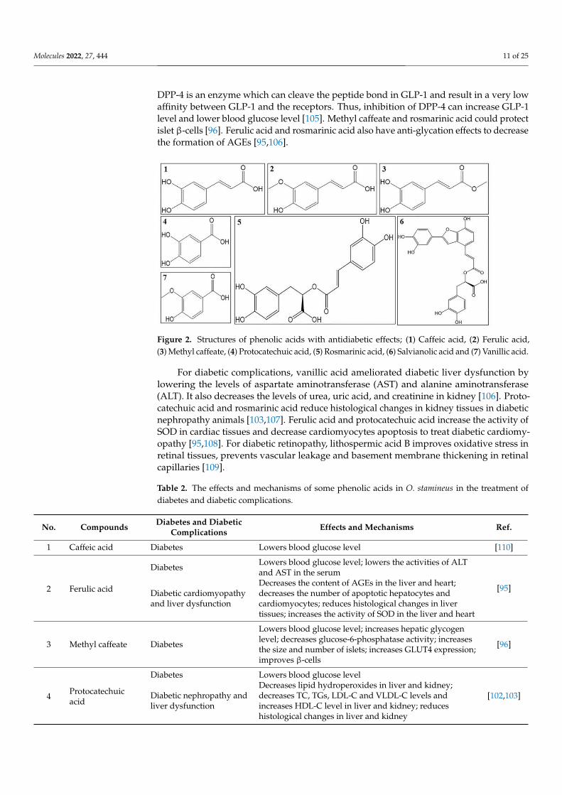
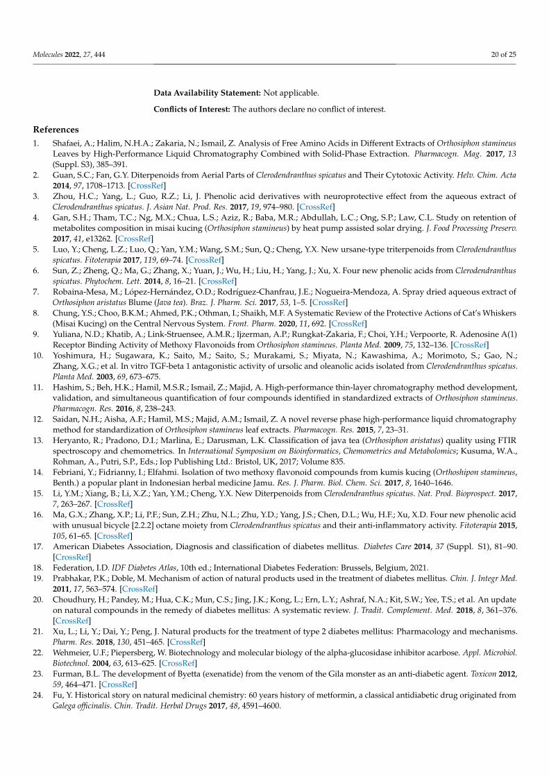
There are almost 50 phenolic acids and their derivatives isolated from O. stamineusup to now. The structures of antidiabetic phenolic acids are summarized in Figure 2 andthe mechanisms of these compounds are summarized in Table 2. Ferulic acid, methyl caf-feate, vanillic acid, protocatechuic acid and rosmarinic acid lower blood glucose levelin vivo [94–96]. Salvianolic acid C and rosmarinic acid had been proved to have in-hibitory activity on α-glucosidase [97,98]. Vanillic acid and rosmarinic acid are both antiox-idants [94,99]. Rosmarinic acid also have anti-inflammatory activity, which reduce NO pro-duction and the levels of pro-inflammatory cytokines such as TNF-α, IL-1β, IL-6 [100,101].Protocatechuic acid and rosmarinic acid regulate the lipid metabolism in diabetic animals.Protocatechuic acid lowers TC, TG, LDL-C levels and increases HDL-C level [102,103].Methyl caffeate increases hepatic glycogen level and reduces gluconeogenesis throughlowering glucose-6-phosphatase activity. It also increases glucose uptake by higher GLUT4expression [96]. Rosmarinic acid also increases the glucose uptake of muscle cells throughactivation of adenosine 5′-monophosphate-activated protein kinase (AMPK) phosphory-lation and glucose transporter-4 (GLUT4) expression. It promotes insulin secretion andimproves insulin resistance by inhibiting dipeptidyl peptidase-4 (DPP-4) and PTB1B [104].
Molecules 2022, 27, 444 11 of 25
DPP-4 is an enzyme which can cleave the peptide bond in GLP-1 and result in a very lowaffinity between GLP-1 and the receptors. Thus, inhibition of DPP-4 can increase GLP-1level and lower blood glucose level [105]. Methyl caffeate and rosmarinic acid could protectislet β-cells [96]. Ferulic acid and rosmarinic acid also have anti-glycation effects to decreasethe formation of AGEs [95,106].
Figure 2. Structures of phenolic acids with antidiabetic effects; (1) Caffeic acid, (2) Ferulic acid,(3) Methyl caffeate, (4) Protocatechuic acid, (5) Rosmarinic acid, (6) Salvianolic acid and (7) Vanillic acid.
For diabetic complications, vanillic acid ameliorated diabetic liver dysfunction bylowering the levels of aspartate aminotransferase (AST) and alanine aminotransferase(ALT). It also decreases the levels of urea, uric acid, and creatinine in kidney [106]. Proto-catechuic acid and rosmarinic acid reduce histological changes in kidney tissues in diabeticnephropathy animals [103,107]. Ferulic acid and protocatechuic acid increase the activity ofSOD in cardiac tissues and decrease cardiomyocytes apoptosis to treat diabetic cardiomy-opathy [95,108]. For diabetic retinopathy, lithospermic acid B improves oxidative stress inretinal tissues, prevents vascular leakage and basement membrane thickening in retinalcapillaries [109].
Table 2. The effects and mechanisms of some phenolic acids in O. stamineus in the treatment ofdiabetes and diabetic complications.
No. Compounds Diabetes and DiabeticComplications Effects and Mechanisms Ref.
1 Caffeic acid Diabetes Lowers blood glucose level [110]
2 Ferulic acid
Diabetes Lowers blood glucose level; lowers the activities of ALTand AST in the serum
[95]Diabetic cardiomyopathyand liver dysfunction
Decreases the content of AGEs in the liver and heart;decreases the number of apoptotic hepatocytes andcardiomyocytes; reduces histological changes in livertissues; increases the activity of SOD in the liver and heart
3 Methyl caffeate Diabetes
Lowers blood glucose level; increases hepatic glycogenlevel; decreases glucose-6-phosphatase activity; increasesthe size and number of islets; increases GLUT4 expression;improves β-cells
[96]
4 Protocatechuicacid
Diabetes Lowers blood glucose level
[102,103]Diabetic nephropathy andliver dysfunction
Decreases lipid hydroperoxides in liver and kidney;decreases TC, TGs, LDL-C and VLDL-C levels andincreases HDL-C level in liver and kidney; reduceshistological changes in liver and kidney
Molecules 2022, 27, 444 12 of 25
Table 2. Cont.
No. Compounds Diabetes and DiabeticComplications Effects and Mechanisms Ref.
5 Rosmarinic acidDiabetes
Reduces blood glucose, TC, TGs and lipid peroxides levels;inhibitors of α-amylase, α-glucosidase, DPP-IV andPTB1B; lowers the formation of MDA and AGEs; reducesthe levels of pro-inflammatory cytokines such as TNF-α,IL-1β, IL-6, NO and nuclear factor kappa-B (NF-κB);increases the activity of SOD; increases the glucose uptakeof muscle cells through activation of AMPKphosphorylation; improves insulin sensitivity; increasesGLUT4 expression in skeletal muscle; protects pancreaticβ-cells
[98–101,104,106]
Diabetic vasculardysfunction
Decreases IL-1β and TNF-αlevels and the expression ofendothelin converting enzyme-1; improves structuralalterations in the endothelium
[111]
6 Salvianolic acid C Diabetic cardiomyopathy
Enhances intracellular adenosine triphosphate (ATP)content in the myocardial tissues; reduces ROS, lipidperoxidation and protein carbonylation level inmyocardial tissues; improves SOD level in cardiac tissues;reduces histological abnormality
[108]
7 Vanillic acid
Diabetes Lowers blood glucose level; decreases the concentration oflipid hydroperoxides
[94]Diabetic nephropathy andliver dysfunction
Increases the activities of antioxidants in kidney and liver;reduces the levels of AST and ALT in liver; decreases thelevels of urea, uric acid, and creatinine in kidney; reduceshistological changes in liver and renal tissues
4.2. Flavonoids
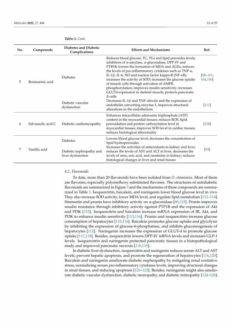
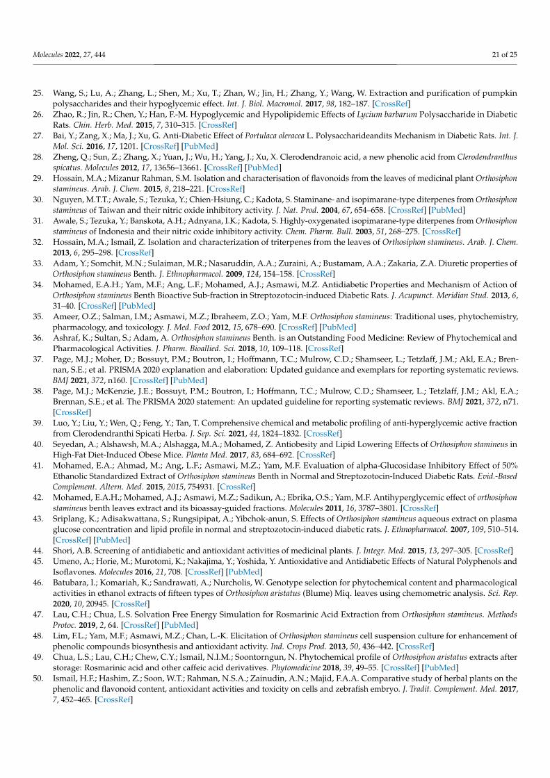
To date, more than 20 flavonoids have been isolated from O. stamineus. Most of themare flavones, especially polymethoxy substituted flavones. The structures of antidiabeticflavonoids are summarized in Figure 3 and the mechanisms of these compounds are summa-rized in Table 3. Isoquercitrin, baicalein, and naringenin lower blood glucose level in vivo.They also increase SOD activity, lower MDA level, and regulate lipid metabolism [112–114].Sinensetin and prunin have inhibitory activity on α-glucosidase [68,115]. Prunin improvesinsulin resistance through inhibitory activity against PTP1B and the expression of Aktand PI3K [115]. Isoquercitrin and baicalein increase mRNA expression of IR, Akt, andPI3K to enhance insulin sensitivity [113,116]. Prunin and isoquercitrin increase glucoseconsumption of hepatocytes [115,116]. Baicalein promotes glucose uptake and glycolysisby inhibiting the expression of glucose-6-phosphatase, and inhibits gluconeogenesis ofhepatocytes [112]. Naringenin increases the expression of GLUT-4 to promote glucoseuptake [117,118]. Besides, isoquercitrin lowers DPP-IV mRNA levels and increases GLP-1levels. Isoquercitrin and naringenin protected pancreatic tissues in a histopathologicalstudy and improved pancreatic necrosis [116,119].
In diabetic liver dysfunction, isoquercitrin and naringenin reduces serum ALT and ASTlevels, prevent hepatic apoptosis, and promote the regeneration of hepatocytes [116,120].Baicalein and naringenin ameliorate diabetic nephropathy by mitigating renal oxidativestress, normalizing serum pro-inflammatory cytokines levels, improving structural changesin renal tissues, and reducing apoptosis [120–123]. Besides, naringenin might also amelio-rate diabetic vascular dysfunction, diabetic neuropathy, and diabetic retinopathy [124–126].
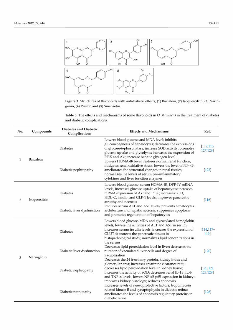
Molecules 2022, 27, 444 13 of 25
Figure 3. Structures of flavonoids with antidiabetic effects; (1) Baicalein, (2) Isoquercitrin, (3) Narin-genin, (4) Prunin and (5) Sinensetin.
Table 3. The effects and mechanisms of some flavonoids in O. stamineus in the treatment of diabetesand diabetic complications.
No. Compounds Diabetes and DiabeticComplications Effects and Mechanisms Ref.
1 Baicalein
Diabetes
Lowers blood glucose and MDA level; inhibitsgluconeogenesis of hepatocytes; decreases the expressionsof glucose-6-phosphatase; increase SOD activity; promotesglucose uptake and glycolysis; increases the expression ofPI3K and Akt; increase hepatic glycogen level
[112,113,127,128]
Diabetic nephropathy
Lowers HOMA-IR level; restores normal renal function;mitigates renal oxidative stress; lowers the level of NF-κB;ameliorates the structural changes in renal tissues;normalizes the levels of serum pro-inflammatorycytokines and liver function enzymes
[122]
2 Isoquercitrin
Diabetes
Lowers blood glucose, serum HOMA-IR, DPP-IV mRNAlevels; increases glucose uptake of hepatocytes; increasesmRNA expression of Akt and PI3K; increases SOD,HDL-C, insulin and GLP-1 levels; improves pancreaticatrophy and necrosis [116]
Diabetic liver dysfunctionReduces serum ALT and AST levels; prevents hepatocytesarchitecture and hepatic necrosis; suppresses apoptosisand promotes regeneration of hepatocytes
3 Naringenin
Diabetes
Lowers blood glucose, MDA and glycosylated hemoglobinlevels; lowers the activities of ALT and AST in serum;increases serum insulin levels; increases the expression ofGLUT-4; protects the pancreatic tissues inhistopathological study; normalizes lipid concentrations inthe serum
[114,117–119]
Diabetic liver dysfunctionDecreases lipid peroxidation level in liver; decreases thenumber of vacuolated liver cells and degree ofvacuolisation
[120]
Diabetic nephropathy
Decreases the 24 h-urinary protein, kidney index andglomerular area; increases creatinine clearance rate;decreases lipid peroxidation level in kidney tissue;increases the activity of SOD; decreases renal IL-1β, IL-6and TNF-α levels; lowers NF-κB p65 expression in kidney;improves kidney histology; reduces apoptosis
[120,121,123,129]
Diabetic retinopathy
Increases levels of neuroprotective factors, tropomyosinrelated kinase B and synaptophysin in diabetic retina;ameliorates the levels of apoptosis regulatory proteins indiabetic retina
[126]

Molecules 2022, 27, 444 14 of 25
Table 3. Cont.
No. Compounds Diabetes and DiabeticComplications Effects and Mechanisms Ref.
4 Prunin DiabetesInhibitory activity against PTP1B and α-glucosidase;stimulates glucose uptake; increases the expression ofp-Akt and p-PI3K
[115]
5 Sinensetin Diabetes Inhibitory activity on α-glucosidase and α-amylase [68]
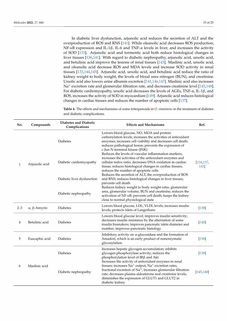
4.3. Triterpenoids
There are almost 20 triterpenoids isolated from O. stamineus. The structures of antidia-betic triterpenoids are summarized in Figure 4 and the mechanisms of these compoundsare summarized in Table 4. α, β-Amyrin, arjunolic acid, betulinic acid, tormentic acid,oleanolic acid, and ursolic acid lower blood glucose level in vivo. Among them, oleanolicacid and ursolic acid have an inhibitory activity on α-glucosidase [130,131]. Arjunolicacid, oleanolic acid, and ursolic acid have antioxidant activities to scavenge free radicals,while oleanolic acid and ursolic acid also have anti-inflammatory activities [100,132–134].α, β-Amyrin, arjunolic acid, tormentic acid, oleanolic acid, and ursolic acid lower thelevels of TC, TG, LDL-C and leptin, increase serum HDL-C level to regulate the lipidmetabolism [133,135–138]. Maslinic acid, oleanolic acid, and ursolic acid improve insulinresistance and enhance insulin sensitivity respectively by a higher expression of IR, IRS, Akt,and PIP1B inhibitory activity [132,133,139]. Tormentic acid promotes glucose uptake byincreasing the levels of phospho-AMPK and GLUT4 in skeletal muscle [136]. Oleanolic acidinhibits gluconeogenesis by decreasing expression of glucose-6-phosphatase [133]. Maslinicacid and ursolic acid increase the hepatic glycogen accumulation [135,139]. α, β-Amyrin,arjunolic acid, and betulinic acid protect islet cells and decrease cell death [138,140,141].Oleanolic acid has anti-glycation effects to inhibit the formation of AGEs products [142].
Figure 4. Structures of triterpenoids with antidiabetic effects; (1) Arjunolic acid, (2) α-Amyrin, (3) β-Amyrin, (4) Betulinic acid, (5) Euscaphic acid, (6) Maslinic acid, (7) Oleanolic acid, (8) Tormentic acidand (9) Ursolic acid.
Molecules 2022, 27, 444 15 of 25
In diabetic liver dysfunction, arjunolic acid reduces the secretion of ALT and theoverproduction of ROS and RNS [141]. While oleanolic acid decreases ROS production,NF-κB expression and IL-1β, IL-6 and TNF-α levels in liver, and increases the activityof SOD [133]. Arjunolic acid and tormentic acid both reduce histological changes inliver tissues [136,141]. With regard to diabetic nephropathy, arjunolic acid, ursolic acid,and betulinic acid improve the lesions of renal tissues [143]. Maslinic acid, ursolic acid,and oleanolic acid decrease ROS and MDA levels and increase SOD activity in renaltissues [133,144,145]. Arjunolic acid, ursolic acid, and betulinic acid reduce the ratio ofkidney weight to body weight, the levels of blood urea nitrogen (BUN), and creatinine.Ursolic acid also lowers urine albumin excretion [141,146,147]. Maslinic acid also increasesNa+ excretion rate and glomerular filtration rate, and decreases creatinine level [145,148].For diabetic cardiomyopathy, ursolic acid decreases the levels of AGEs, TNF-α, IL-1β, andROS, increases the activity of SOD in myocardium [149]. Arjunolic acid reduces histologicalchanges in cardiac tissues and reduces the number of apoptotic cells [137].
Table 4. The effects and mechanisms of some triterpenoids in O. stamineus in the treatment of diabetesand diabetic complications.
No. Compounds Diabetes and DiabeticComplications Effects and Mechanisms Ref.
1 Arjunolic acid
Diabetes
Lowers blood glucose, NO, MDA and proteincarbonylation levels; increases the activities of antioxidantenzymes; increases cell viability and decreases cell death;reduces pathological lesion; prevents the expression ofc-Jun N-terminal kinase (JNK)
[134,137,141]
Diabetic cardiomyopathy
Reduces the levels of vascular inflammation markers;increases the activities of the antioxidant enzymes andcellular redox ratio; decreases DNA oxidation in cardiactissue; reduces histological changes in cardiac tissues;reduces the number of apoptotic cells
Diabetic liver dysfunctionReduces the secretion of ALT, the overproduction of ROSand RNS; reduces histological changes in liver tissues;prevents cell death
Diabetic nephropathy
Reduces kidney weight to body weight ratio, glomerulararea, glomerular volume, BUN and creatinine; reduces theactivation of NF-κB; prevents cell death; keeps the kidneyclose to normal physiological state
2–3 α, β-Amyrin Diabetes Lowers blood glucose, LDL, VLDL levels; increases insulinlevels; protects islets of Langerhans [138]
4 Betulinic acid Diabetes
Lowers blood glucose level; improves insulin sensitivity;decreases insulin resistance by the alternation of someinsulin biomakers; improves pancreatic islets diameter andnumber; improves pancreatic histology
[140]
5 Euscaphic acid DiabetesInhibitory activity on α-glucosidase and the formation ofAmadori, which is an early product of nonenzymaticglycosylation
[150]
6 Maslinic acid
DiabetesIncreases hepatic glycogen accumulation; inhibitsglycogen phosphorylase activity; induces thephosphorylation level of IRβ and Akt
[139]
Diabetic nephropathy
Increases the activity of antioxidant enzymes in renaltissues; increases Na+ output, Na+ excretion rates,fractional excretion of Na+; increases glomerular filtrationrate; decreases plasma aldosterone and creatinine levels;diminishes the expression of GLUT1 and GLUT2 indiabetic kidney
[145,148]
Molecules 2022, 27, 444 16 of 25
Table 4. Cont.
No. Compounds Diabetes and DiabeticComplications Effects and Mechanisms Ref.
7 Oleanolic acidDiabetes
Lowers blood glucose, LDL and free fatty acids levels;increases insulin level; inhibitory activity onα-glucosidase, α-amylase and PIP1B; inhibits theformation of AGEs products; improve insulin tolerance;inhibits gluconeogenesis; increases serum HDL level;decreases levels of IL-1b, IL-6 and TNFα; increases theactivity of SOD; improve glycogen level by the increasingexpression of Akt and decreasing expression ofglucose-6-phosphatase; increases the expression of IRand IRS-1
[131,133,142,151]
Diabetic liver dysfunctionDecreases the levels of IL-1β, IL-6 and TNFα in liver;decreases the expression of NF-κB; decreases ROSproduction; increases the activity of SOD
[133,152]
8 Tormentic acid
DiabetesLowers blood glucose, leptin and total lipids levels;increases the protein contents of phospho-AMPK andGLUT4 in skeletal muscle
[136]
Diabetic liver dysfunctionReduces histological changes in liver tissues; decreases themRNA level of glucose-6-phosphatase in liver tissues;increases the protein contents of hepatic phospho-AMPK
9 Ursolic acid
Diabetes
Lowers blood glucose, MDA and LDL levels; inhibitsα-amylase and α-glucosidase activity; increases SODactivities; decreases TNF-α and IL-1β level; increases liverglycogen level; decreases the expression of PTP-1B protein;increases the expression of IRS-2 protein
[130,132,135]
Diabetic cardiomyopathy Decreases levels of AGEs, TNF-α, IL-1β and ROS;increases the activity of SOD in myocardium [149]
Diabetic nephropathy
Lowers the levels of BUN, creatinine and MDA; lowersurine albumin excretion, renal oxidative stress level,NF-κB activity; prevents the expression of JNK; improvesrenal structural abnormalities
[144,146,147]
5. Discussion
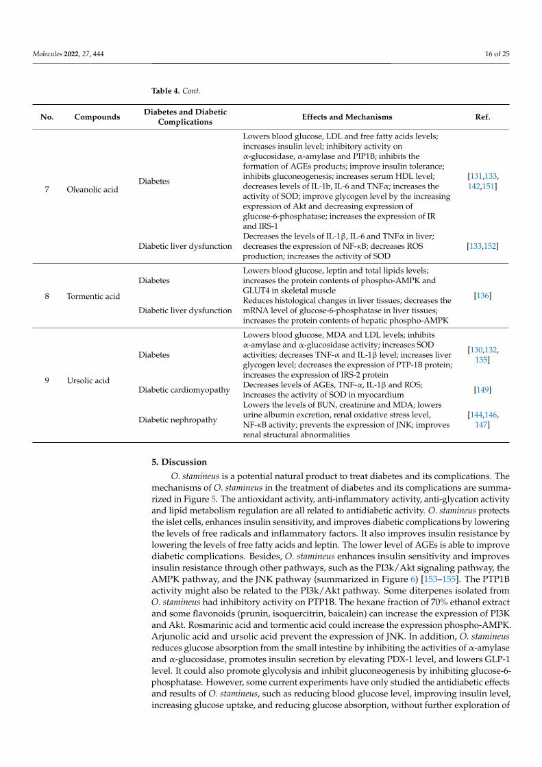
O. stamineus is a potential natural product to treat diabetes and its complications. Themechanisms of O. stamineus in the treatment of diabetes and its complications are summa-rized in Figure 5. The antioxidant activity, anti-inflammatory activity, anti-glycation activityand lipid metabolism regulation are all related to antidiabetic activity. O. stamineus protectsthe islet cells, enhances insulin sensitivity, and improves diabetic complications by loweringthe levels of free radicals and inflammatory factors. It also improves insulin resistance bylowering the levels of free fatty acids and leptin. The lower level of AGEs is able to improvediabetic complications. Besides, O. stamineus enhances insulin sensitivity and improvesinsulin resistance through other pathways, such as the PI3k/Akt signaling pathway, theAMPK pathway, and the JNK pathway (summarized in Figure 6) [153–155]. The PTP1Bactivity might also be related to the PI3k/Akt pathway. Some diterpenes isolated fromO. stamineus had inhibitory activity on PTP1B. The hexane fraction of 70% ethanol extractand some flavonoids (prunin, isoquercitrin, baicalein) can increase the expression of PI3Kand Akt. Rosmarinic acid and tormentic acid could increase the expression phospho-AMPK.Arjunolic acid and ursolic acid prevent the expression of JNK. In addition, O. stamineusreduces glucose absorption from the small intestine by inhibiting the activities of α-amylaseand α-glucosidase, promotes insulin secretion by elevating PDX-1 level, and lowers GLP-1level. It could also promote glycolysis and inhibit gluconeogenesis by inhibiting glucose-6-phosphatase. However, some current experiments have only studied the antidiabetic effectsand results of O. stamineus, such as reducing blood glucose level, improving insulin level,increasing glucose uptake, and reducing glucose absorption, without further exploration of

Molecules 2022, 27, 444 17 of 25
its mechanisms and pathways. The relationship between O. stamineus extracts and AMPK,JNK pathways should be further studied.
Figure 5. The mechanisms of O. stamineus in the treatment of diabetes and its complications. (Theblue part shows other activities related to antidiabetic activity. The grey part is the pathways, targets,and enzymes related to antidiabetic activity).
Molecules 2022, 27, 444 18 of 25
Figure 6. Summary of the PI3K/Akt, AMPK, JNK signal pathways related to insulin resistance.Arrows indicate activation, thick lines indicate inhibition.
Until now, investigations on the antidiabetic effects and mechanisms of O. stamineushave concentrated mainly on the effects of extracts, especially 50% ethanol extract andaqueous extract. The effects of extracts might be different because the levels of somemetabolites vary in the plant from different places. Through literature research, it was seenthat phenolic acids, flavonoids, and triterpenoids might be the main active components totreat diabetes and complications. To identify the major bioactive compounds responsiblefor antidiabetic effects, bioassay-guided isolation should be used. The mechanisms ofpure compounds are also required to study, and there might be synergistic effects betweenthese constituents.
In China and some southeastern Asian countries, O. stamineus has been used astraditional medicine for the treatment of diabetes and some kidney diseases for a long time.In recent years, by means of modern science and techniques, there have been more and moreinvestigations in the mechanisms of O. stamineus in the treatment of diabetes and diabeticcomplications. However, most experiments are in vitro or using experimental animalmodels in vivo, which may be different from the effects and mechanisms of O. stamineusin the human body. In addition, clinical research is very limited. O. stamineus was onlyused to treat chronic renal diseases in clinical, such as chronic glomerulonephritis [156].But because O. stamineus might be a good antidiabetic candidate to reduce blood glucoselevels and alleviate kidney injury, it could also be designed to study the clinical treatmentof diabetic nephropathy in the future.
At present, diabetes is treated with oral hypoglycemic drugs and insulin injections. Theglucose-lowering drugs include α-glucosidase inhibitors (acarbose, miglitol), insulin sensi-tizers (metformin, thiazolidinediones, biguanides), insulin secretagogues (sulfonylureas),etc. However, most of these medications may have side-effects, including hypoglycemia,weight gain, liver damage, gastrointestinal disturbance, lactic acidosis, edema, headache,dizziness, anemia, nausea, and even death. Besides, long-term use of insulin may decreaseinsulin receptor sensitivity, resulting in insulin resistance [157]. In the future, glucose-lowering drugs might be combined with O. stamineus to find out if they can reduce theseside-effects and increase antidiabetic effects. Besides, some other natural products withantidiabetic activities can also be used with O. stamineus to test the combined effects inthe treatment of diabetes and diabetic complications. Cordyceps sinensis, Astragalus mem-branaceus, Rheum officinale, and leech have been combined with O. stamineus to treat diabeticnephropathy as a Chinese traditional medicine prescription [91].
6. Methods
This review was performed and reported according to PRISMA guidelines [37,38].The flowchart of selected articles is shown in Figure 1.
Molecules 2022, 27, 444 19 of 25
6.1. Search Strategy
Three databases (ScienceDirect, PubMed and Web of Science) were used to searchrelevant articles using the terms “((Clerodendranthus spicatus) OR (Orthosiphon stamineus) OR(Orthosiphon aristatus)) AND ((diabetes) OR (antidiabetic) OR (hypoglycemic) OR (diabeticcomplications))”. No time restriction was used. The initial search included 281 articles.The results of ScienceDirect, PubMed, and Web of Science were respectively exported asRIS, NBIB, and ISI files. All obtained files were then imported into EndNote X9 to generatea library.
6.2. Eligibility Criteria
The research included in this review met the following criteria: 1. the study reportedhypoglycemic activity or the treatment of diabetes and its complications of O. stamineusextract or its isolated compounds, 2. the study reported other biological activities related todiabetes treatment, such as antioxidant and anti-inflammatory activities, of O. stamineusextract or its isolated compounds, 3. the study reported the toxicity of O. stamineus extractor its isolated compounds.
The exclusion criteria of this review were as follows: 1. reviews, book chapters, patents,meeting papers, 2. non-English language papers, 3. lack of access to the full-text of thepaper, 4. no relevance to the plant O. stamineus or the field of diabetes and its complications.Besides, duplicate articles were also removed.
6.3. Data Extraction
Thirty-one studies met the criteria, and the data were extracted into Microsoft Excel2007 sheet and inserted to Table 1. The information gathered from the studies included: 1.name of the first author, 2. publication year, 3. the tested substance, 4. the study designand protocol, 5. main results.
7. Conclusions
In conclusion, O. stamineus is a potential agent to treat diabetes and diabetic complica-tions. The extracts of O. stamineus, including 50% ethanol extract, chloroform extract,aqueous extract, and hexane extract, could be used to treat diabetes through mecha-nisms including inhibiting the activities of α-amylase and α-glucosidase, antioxidantand anti-inflammatory activities, regulating lipid metabolism, promoting insulin secretion,ameliorating insulin resistance, enhancing insulin sensitivity, increasing glucose uptake,promoting glycolysis, inhibiting gluconeogenesis, promoting the secretion of GLP-1, andantiglycation effects. The mechanisms of insulin resistance might also be related to thePI3k/Akt signaling pathway, the AMPK pathway, and the JNK pathway. The aqueousextract could also be used for diabetic nephropathy treatment. Besides, some main activecomponents, such as rosmarinic acid, ferulic acid, methyl caffeate, vanillic acid, protocate-chuic acid, isoquercitrin, baicalein, naringenin, arjunolic acid, betulinic acid, tormentic acid,oleanolic acid, ursolic acid, maslinic acid, siphonols B, orthosiphols B, G, I, and N also hadgood effects in the treatment of diabetes and its complications. However, it needs furtherstudy on pharmacodynamic substance basis and the mechanisms of effective constituents.
Author Contributions: Conceptualization, H.C.; methodology, Q.W.; software, J.L.; validation, N.L.;formal analysis, J.Z.; investigation, Q.W.; resources, H.C.; data curation, Q.W.; writing—original draftpreparation, Q.W.; writing—review and editing, J.W.; visualization, J.L.; supervision, P.Z.; projectadministration, Q.W.; funding acquisition, H.C. All authors have read and agreed to the publishedversion of the manuscript.
Funding: This research was funded by a grant from National Key Research and DevelopmentProgram of China, grant number 2021YFE0110000.
Institutional Review Board Statement: Not applicable.
Informed Consent Statement: Not applicable.
Molecules 2022, 27, 444 20 of 25
Data Availability Statement: Not applicable.
Conflicts of Interest: The authors declare no conflict of interest.
References1. Shafaei, A.; Halim, N.H.A.; Zakaria, N.; Ismail, Z. Analysis of Free Amino Acids in Different Extracts of Orthosiphon stamineus
Leaves by High-Performance Liquid Chromatography Combined with Solid-Phase Extraction. Pharmacogn. Mag. 2017, 13(Suppl. S3), 385–391.
2. Guan, S.C.; Fan, G.Y. Diterpenoids from Aerial Parts of Clerodendranthus spicatus and Their Cytotoxic Activity. Helv. Chim. Acta2014, 97, 1708–1713. [CrossRef]
3. Zhou, H.C.; Yang, L.; Guo, R.Z.; Li, J. Phenolic acid derivatives with neuroprotective effect from the aqueous extract ofClerodendranthus spicatus. J. Asian Nat. Prod. Res. 2017, 19, 974–980. [CrossRef]
4. Gan, S.H.; Tham, T.C.; Ng, M.X.; Chua, L.S.; Aziz, R.; Baba, M.R.; Abdullah, L.C.; Ong, S.P.; Law, C.L. Study on retention ofmetabolites composition in misai kucing (Orthosiphon stamineus) by heat pump assisted solar drying. J. Food Processing Preserv.2017, 41, e13262. [CrossRef]
5. Luo, Y.; Cheng, L.Z.; Luo, Q.; Yan, Y.M.; Wang, S.M.; Sun, Q.; Cheng, Y.X. New ursane-type triterpenoids from Clerodendranthusspicatus. Fitoterapia 2017, 119, 69–74. [CrossRef]
6. Sun, Z.; Zheng, Q.; Ma, G.; Zhang, X.; Yuan, J.; Wu, H.; Liu, H.; Yang, J.; Xu, X. Four new phenolic acids from Clerodendranthusspicatus. Phytochem. Lett. 2014, 8, 16–21. [CrossRef]
7. Robaina-Mesa, M.; López-Hernández, O.D.; Rodríguez-Chanfrau, J.E.; Nogueira-Mendoza, A. Spray dried aqueous extract ofOrthosiphon aristatus Blume (Java tea). Braz. J. Pharm. Sci. 2017, 53, 1–5. [CrossRef]
8. Chung, Y.S.; Choo, B.K.M.; Ahmed, P.K.; Othman, I.; Shaikh, M.F. A Systematic Review of the Protective Actions of Cat’s Whiskers(Misai Kucing) on the Central Nervous System. Front. Pharm. 2020, 11, 692. [CrossRef]
9. Yuliana, N.D.; Khatib, A.; Link-Struensee, A.M.R.; Ijzerman, A.P.; Rungkat-Zakaria, F.; Choi, Y.H.; Verpoorte, R. Adenosine A(1)Receptor Binding Activity of Methoxy Flavonoids from Orthosiphon stamineus. Planta Med. 2009, 75, 132–136. [CrossRef]
10. Yoshimura, H.; Sugawara, K.; Saito, M.; Saito, S.; Murakami, S.; Miyata, N.; Kawashima, A.; Morimoto, S.; Gao, N.;Zhang, X.G.; et al. In vitro TGF-beta 1 antagonistic activity of ursolic and oleanolic acids isolated from Clerodendranthus spicatus.Planta Med. 2003, 69, 673–675.
11. Hashim, S.; Beh, H.K.; Hamil, M.S.R.; Ismail, Z.; Majid, A. High-performance thin-layer chromatography method development,validation, and simultaneous quantification of four compounds identified in standardized extracts of Orthosiphon stamineus.Pharmacogn. Res. 2016, 8, 238–243.
12. Saidan, N.H.; Aisha, A.F.; Hamil, M.S.; Majid, A.M.; Ismail, Z. A novel reverse phase high-performance liquid chromatographymethod for standardization of Orthosiphon stamineus leaf extracts. Pharmacogn. Res. 2015, 7, 23–31.
13. Heryanto, R.; Pradono, D.I.; Marlina, E.; Darusman, L.K. Classification of java tea (Orthosiphon aristatus) quality using FTIRspectroscopy and chemometrics. In International Symposium on Bioinformatics, Chemometrics and Metabolomics; Kusuma, W.A.,Rohman, A., Putri, S.P., Eds.; Iop Publishing Ltd.: Bristol, UK, 2017; Volume 835.
14. Febriani, Y.; Fidrianny, I.; Elfahmi. Isolation of two methoxy flavonoid compounds from kumis kucing (Orthoshipon stamineus,Benth.) a popular plant in Indonesian herbal medicine Jamu. Res. J. Pharm. Biol. Chem. Sci. 2017, 8, 1640–1646.
15. Li, Y.M.; Xiang, B.; Li, X.Z.; Yan, Y.M.; Cheng, Y.X. New Diterpenoids from Clerodendranthus spicatus. Nat. Prod. Bioprospect. 2017,7, 263–267. [CrossRef]
16. Ma, G.X.; Zhang, X.P.; Li, P.F.; Sun, Z.H.; Zhu, N.L.; Zhu, Y.D.; Yang, J.S.; Chen, D.L.; Wu, H.F.; Xu, X.D. Four new phenolic acidwith unusual bicycle [2.2.2] octane moiety from Clerodendranthus spicatus and their anti-inflammatory activity. Fitoterapia 2015,105, 61–65. [CrossRef]
17. American Diabetes Association, Diagnosis and classification of diabetes mellitus. Diabetes Care 2014, 37 (Suppl. S1), 81–90.[CrossRef]
18. Federation, I.D. IDF Diabetes Atlas, 10th ed.; International Diabetes Federation: Brussels, Belgium, 2021.19. Prabhakar, P.K.; Doble, M. Mechanism of action of natural products used in the treatment of diabetes mellitus. Chin. J. Integr Med.
2011, 17, 563–574. [CrossRef]20. Choudhury, H.; Pandey, M.; Hua, C.K.; Mun, C.S.; Jing, J.K.; Kong, L.; Ern, L.Y.; Ashraf, N.A.; Kit, S.W.; Yee, T.S.; et al. An update
on natural compounds in the remedy of diabetes mellitus: A systematic review. J. Tradit. Complement. Med. 2018, 8, 361–376.[CrossRef]
21. Xu, L.; Li, Y.; Dai, Y.; Peng, J. Natural products for the treatment of type 2 diabetes mellitus: Pharmacology and mechanisms.Pharm. Res. 2018, 130, 451–465. [CrossRef]
22. Wehmeier, U.F.; Piepersberg, W. Biotechnology and molecular biology of the alpha-glucosidase inhibitor acarbose. Appl. Microbiol.Biotechnol. 2004, 63, 613–625. [CrossRef]
23. Furman, B.L. The development of Byetta (exenatide) from the venom of the Gila monster as an anti-diabetic agent. Toxicon 2012,59, 464–471. [CrossRef]
24. Fu, Y. Historical story on natural medicinal chemistry: 60 years history of metformin, a classical antidiabetic drug originated fromGalega officinalis. Chin. Tradit. Herbal Drugs 2017, 48, 4591–4600.
Molecules 2022, 27, 444 21 of 25
25. Wang, S.; Lu, A.; Zhang, L.; Shen, M.; Xu, T.; Zhan, W.; Jin, H.; Zhang, Y.; Wang, W. Extraction and purification of pumpkinpolysaccharides and their hypoglycemic effect. Int. J. Biol. Macromol. 2017, 98, 182–187. [CrossRef]
26. Zhao, R.; Jin, R.; Chen, Y.; Han, F.-M. Hypoglycemic and Hypolipidemic Effects of Lycium barbarum Polysaccharide in DiabeticRats. Chin. Herb. Med. 2015, 7, 310–315. [CrossRef]
27. Bai, Y.; Zang, X.; Ma, J.; Xu, G. Anti-Diabetic Effect of Portulaca oleracea L. Polysaccharideandits Mechanism in Diabetic Rats. Int. J.Mol. Sci. 2016, 17, 1201. [CrossRef] [PubMed]
28. Zheng, Q.; Sun, Z.; Zhang, X.; Yuan, J.; Wu, H.; Yang, J.; Xu, X. Clerodendranoic acid, a new phenolic acid from Clerodendranthusspicatus. Molecules 2012, 17, 13656–13661. [CrossRef] [PubMed]
29. Hossain, M.A.; Mizanur Rahman, S.M. Isolation and characterisation of flavonoids from the leaves of medicinal plant Orthosiphonstamineus. Arab. J. Chem. 2015, 8, 218–221. [CrossRef]
30. Nguyen, M.T.T.; Awale, S.; Tezuka, Y.; Chien-Hsiung, C.; Kadota, S. Staminane- and isopimarane-type diterpenes from Orthosiphonstamineus of Taiwan and their nitric oxide inhibitory activity. J. Nat. Prod. 2004, 67, 654–658. [CrossRef] [PubMed]
31. Awale, S.; Tezuka, Y.; Banskota, A.H.; Adnyana, I.K.; Kadota, S. Highly-oxygenated isopimarane-type diterpenes from Orthosiphonstamineus of Indonesia and their nitric oxide inhibitory activity. Chem. Pharm. Bull. 2003, 51, 268–275. [CrossRef]
32. Hossain, M.A.; Ismail, Z. Isolation and characterization of triterpenes from the leaves of Orthosiphon stamineus. Arab. J. Chem.2013, 6, 295–298. [CrossRef]
33. Adam, Y.; Somchit, M.N.; Sulaiman, M.R.; Nasaruddin, A.A.; Zuraini, A.; Bustamam, A.A.; Zakaria, Z.A. Diuretic properties ofOrthosiphon stamineus Benth. J. Ethnopharmacol. 2009, 124, 154–158. [CrossRef]
34. Mohamed, E.A.H.; Yam, M.F.; Ang, L.F.; Mohamed, A.J.; Asmawi, M.Z. Antidiabetic Properties and Mechanism of Action ofOrthosiphon stamineus Benth Bioactive Sub-fraction in Streptozotocin-induced Diabetic Rats. J. Acupunct. Meridian Stud. 2013, 6,31–40. [CrossRef] [PubMed]
35. Ameer, O.Z.; Salman, I.M.; Asmawi, M.Z.; Ibraheem, Z.O.; Yam, M.F. Orthosiphon stamineus: Traditional uses, phytochemistry,pharmacology, and toxicology. J. Med. Food 2012, 15, 678–690. [CrossRef] [PubMed]
36. Ashraf, K.; Sultan, S.; Adam, A. Orthosiphon stamineus Benth. is an Outstanding Food Medicine: Review of Phytochemical andPharmacological Activities. J. Pharm. Bioallied. Sci. 2018, 10, 109–118. [CrossRef]
37. Page, M.J.; Moher, D.; Bossuyt, P.M.; Boutron, I.; Hoffmann, T.C.; Mulrow, C.D.; Shamseer, L.; Tetzlaff, J.M.; Akl, E.A.; Bren-nan, S.E.; et al. PRISMA 2020 explanation and elaboration: Updated guidance and exemplars for reporting systematic reviews.BMJ 2021, 372, n160. [CrossRef] [PubMed]
38. Page, M.J.; McKenzie, J.E.; Bossuyt, P.M.; Boutron, I.; Hoffmann, T.C.; Mulrow, C.D.; Shamseer, L.; Tetzlaff, J.M.; Akl, E.A.;Brennan, S.E.; et al. The PRISMA 2020 statement: An updated guideline for reporting systematic reviews. BMJ 2021, 372, n71.[CrossRef]
39. Luo, Y.; Liu, Y.; Wen, Q.; Feng, Y.; Tan, T. Comprehensive chemical and metabolic profiling of anti-hyperglycemic active fractionfrom Clerodendranthi Spicati Herba. J. Sep. Sci. 2021, 44, 1824–1832. [CrossRef]
40. Seyedan, A.; Alshawsh, M.A.; Alshagga, M.A.; Mohamed, Z. Antiobesity and Lipid Lowering Effects of Orthosiphon stamineus inHigh-Fat Diet-Induced Obese Mice. Planta Med. 2017, 83, 684–692. [CrossRef]
41. Mohamed, E.A.; Ahmad, M.; Ang, L.F.; Asmawi, M.Z.; Yam, M.F. Evaluation of alpha-Glucosidase Inhibitory Effect of 50%Ethanolic Standardized Extract of Orthosiphon stamineus Benth in Normal and Streptozotocin-Induced Diabetic Rats. Evid.-BasedComplement. Altern. Med. 2015, 2015, 754931. [CrossRef]
42. Mohamed, E.A.H.; Mohamed, A.J.; Asmawi, M.Z.; Sadikun, A.; Ebrika, O.S.; Yam, M.F. Antihyperglycemic effect of orthosiphonstamineus benth leaves extract and its bioassay-guided fractions. Molecules 2011, 16, 3787–3801. [CrossRef]
43. Sriplang, K.; Adisakwattana, S.; Rungsipipat, A.; Yibchok-anun, S. Effects of Orthosiphon stamineus aqueous extract on plasmaglucose concentration and lipid profile in normal and streptozotocin-induced diabetic rats. J. Ethnopharmacol. 2007, 109, 510–514.[CrossRef] [PubMed]
44. Shori, A.B. Screening of antidiabetic and antioxidant activities of medicinal plants. J. Integr. Med. 2015, 13, 297–305. [CrossRef]45. Umeno, A.; Horie, M.; Murotomi, K.; Nakajima, Y.; Yoshida, Y. Antioxidative and Antidiabetic Effects of Natural Polyphenols and
Isoflavones. Molecules 2016, 21, 708. [CrossRef] [PubMed]46. Batubara, I.; Komariah, K.; Sandrawati, A.; Nurcholis, W. Genotype selection for phytochemical content and pharmacological
activities in ethanol extracts of fifteen types of Orthosiphon aristatus (Blume) Miq. leaves using chemometric analysis. Sci. Rep.2020, 10, 20945. [CrossRef]
47. Lau, C.H.; Chua, L.S. Solvation Free Energy Simulation for Rosmarinic Acid Extraction from Orthosiphon stamineus. MethodsProtoc. 2019, 2, 64. [CrossRef] [PubMed]
48. Lim, F.L.; Yam, M.F.; Asmawi, M.Z.; Chan, L.-K. Elicitation of Orthosiphon stamineus cell suspension culture for enhancement ofphenolic compounds biosynthesis and antioxidant activity. Ind. Crops Prod. 2013, 50, 436–442. [CrossRef]
49. Chua, L.S.; Lau, C.H.; Chew, C.Y.; Ismail, N.I.M.; Soontorngun, N. Phytochemical profile of Orthosiphon aristatus extracts afterstorage: Rosmarinic acid and other caffeic acid derivatives. Phytomedicine 2018, 39, 49–55. [CrossRef] [PubMed]
50. Ismail, H.F.; Hashim, Z.; Soon, W.T.; Rahman, N.S.A.; Zainudin, A.N.; Majid, F.A.A. Comparative study of herbal plants on thephenolic and flavonoid content, antioxidant activities and toxicity on cells and zebrafish embryo. J. Tradit. Complement. Med. 2017,7, 452–465. [CrossRef]
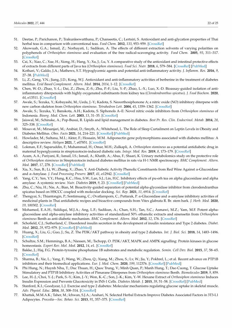
Molecules 2022, 27, 444 22 of 25
51. Deetae, P.; Parichanon, P.; Trakunleewatthana, P.; Chanseetis, C.; Lertsiri, S. Antioxidant and anti-glycation properties of Thaiherbal teas in comparison with conventional teas. Food Chem. 2012, 133, 953–959. [CrossRef]
52. Akowuah, G.A.; Ismail, Z.; Norhayati, I.; Sadikun, A. The effects of different extraction solvents of varying polarities onpolyphenols of Orthosiphon stamineus and evaluation of the free radical-scavenging activity. Food Chem. 2005, 93, 311–317.[CrossRef]
53. Cai, X.; Xiao, C.; Xue, H.; Xiong, H.; Hang, Y.; Xu, J.; Lu, Y. A comparative study of the antioxidant and intestinal protective effectsof extracts from different parts of Java tea (Orthosiphon stamineus). Food Sci. Nutr. 2018, 6, 579–584. [CrossRef] [PubMed]
54. Kothari, V.; Galdo, J.A.; Mathews, S.T. Hypoglycemic agents and potential anti-inflammatory activity. J. Inflamm. Res. 2016, 9,27–38. [PubMed]
55. Li, Z.; Geng, Y.N.; Jiang, J.D.; Kong, W.J. Antioxidant and anti-inflammatory activities of berberine in the treatment of diabetesmellitus. Evid Based Complement. Altern. Med. 2014, 2014, 1–12. [CrossRef]
56. Chen, W.-D.; Zhao, Y.-L.; Dai, Z.; Zhou, Z.-S.; Zhu, P.-F.; Liu, Y.-P.; Zhao, L.-X.; Luo, X.-D. Bioassay-guided isolation of anti-inflammatory diterpenoids with highly oxygenated substituents from kidney tea (Clerodendranthus spicatus). J. Food Biochem. 2020,44, e13511. [CrossRef]
57. Awale, S.; Tezuka, Y.; Kobayashi, M.; Ueda, J.-Y.; Kadota, S. Neoorthosiphonone A; a nitric oxide (NO) inhibitory diterpene withnew carbon skeleton from Orthosiphon stamineus. Tetrahedron Lett. 2004, 45, 1359–1362. [CrossRef]
58. Awale, S.; Tezuka, Y.; Banskota, A.H.; Kadota, S. Siphonols A–E: Novel nitric oxide inhibitors from Orthosiphon stamineus ofIndonesia. Bioorg. Med. Chem. Lett. 2003, 13, 31–35. [CrossRef]
59. Jaiswal, M.; Schinske, A.; Pop-Busui, R. Lipids and lipid management in diabetes. Best Pr. Res. Clin. Endocrinol. Metab. 2014, 28,325–338. [CrossRef]
60. Mosavat, M.; Mirsanjari, M.; Arabiat, D.; Smyth, A.; Whitehead, L. The Role of Sleep Curtailment on Leptin Levels in Obesity andDiabetes Mellitus. Obes. Facts 2021, 14, 214–221. [CrossRef] [PubMed]
61. Howlader, M.; Sultana, M.I.; Akter, F.; Hossain, M.M. Adiponectin gene polymorphisms associated with diabetes mellitus: Adescriptive review. Heliyon 2021, 7, e07851. [CrossRef]
62. Lokman, E.F.; Saparuddin, F.; Muhammad, H.; Omar, M.H.; Zulkapli, A. Orthosiphon stamineus as a potential antidiabetic drug inmaternal hyperglycemia in streptozotocin-induced diabetic rats. Integr. Med. Res. 2019, 8, 173–179. [CrossRef]
63. Azam, A.A.; Pariyani, R.; Ismail, I.S.; Ismail, A.; Khatib, A.; Abas, F.; Shaari, K. Urinary metabolomics study on the protective roleof Orthosiphon stamineus in Streptozotocin induced diabetes mellitus in rats via H-1 NMR spectroscopy. BMC Complement. Altern.Med. 2017, 17, 278. [CrossRef] [PubMed]
64. Xia, X.; Sun, B.; Li, W.; Zhang, X.; Zhao, Y. Anti-Diabetic Activity Phenolic Constituents from Red Wine Against α-Glucosidaseand α-Amylase. J. Food Processing Preserv. 2017, 41, e12942. [CrossRef]
65. Yang, C.Y.; Yen, Y.Y.; Hung, K.C.; Hsu, S.W.; Lan, S.J.; Lin, H.C. Inhibitory effects of pu-erh tea on alpha glucosidase and alphaamylase: A systemic review. Nutr. Diabetes 2019, 9, 23. [CrossRef] [PubMed]
66. Zhu, C.; Niu, H.; Nie, A.; Bian, M. Bioactivity-guided separation of potential alpha-glycosidase inhibitor from clerodendranthusspicatus based on HSCCC coupled with molecular docking. Sci. Rep. 2021, 11, 6914. [CrossRef]
67. Thengyai, S.; Thiantongin, P.; Sontimuang, C.; Ovatlarnporn, C.; Puttarak, P. α-Glucosidase and α-amylase inhibitory activities ofmedicinal plants in Thai antidiabetic recipes and bioactive compounds from Vitex glabrata R. Br. stem bark. J. Herb. Med. 2020,19, 100302. [CrossRef]
68. Mohamed, E.A.H.; Siddiqui, M.J.A.; Ang, L.F.; Sadikun, A.; Chan, S.H.; Tan, S.C.; Asmawi, M.Z.; Yam, M.F. Potent alpha-glucosidase and alpha-amylase inhibitory activities of standardized 50% ethanolic extracts and sinensetin from Orthosiphonstamineus Benth as anti-diabetic mechanism. BMC Complement. Altern. Med. 2012, 12, 176. [CrossRef]
69. Schofield, C.J.; Sutherland, C. Disordered insulin secretion in the development of insulin resistance and Type 2 diabetes. Diabet.Med. 2012, 29, 972–979. [CrossRef] [PubMed]
70. Huang, X.; Liu, G.; Guo, J.; Su, Z. The PI3K/AKT pathway in obesity and type 2 diabetes. Int. J. Biol. Sci. 2018, 14, 1483–1496.[CrossRef]
71. Schultze, S.M.; Hemmings, B.A.; Niessen, M.; Tschopp, O. PI3K/AKT, MAPK and AMPK signalling: Protein kinases in glucosehomeostasis. Expert Rev. Mol. Med. 2012, 14, e1. [CrossRef]
72. Bakke, J.; Haj, F.G. Protein-tyrosine phosphatase 1B substrates and metabolic regulation. Semin. Cell Dev. Biol. 2015, 37, 58–65.[CrossRef]
73. Sharma, B.; Xie, L.; Yang, F.; Wang, W.; Zhou, Q.; Xiang, M.; Zhou, S.; Lv, W.; Jia, Y.; Pokhrel, L.; et al. Recent advance on PTP1Binhibitors and their biomedical applications. Eur. J. Med. Chem. 2020, 199, 112376. [CrossRef] [PubMed]
74. Phi Hung, N.; Huynh Nhu, T.; Duc Thuan, H.; Quoc Trung, V.; Minh Quan, P.; Manh Hung, T.; Dao Cuong, T. Glucose UptakeStimulatory and PTP1B Inhibitory Activities of Pimarane Diterpenes from Orthosiphon stamineus Benth. Biomolecules 2019, 9, 859.
75. Lee, H.-J.; Choi, Y.-J.; Park, S.-Y.; Kim, J.-Y.; Won, K.-C.; Son, J.-K.; Kim, Y.-W. Hexane Extract of Orthosiphon stamineus InducesInsulin Expression and Prevents Glucotoxicity in INS-1 Cells. Diabetes Metab. J. 2015, 39, 51–58. [CrossRef] [PubMed]
76. Stanford, K.I.; Goodyear, L.J. Exercise and type 2 diabetes: Molecular mechanisms regulating glucose uptake in skeletal muscle.Adv. Physiol. Educ. 2014, 38, 308–314. [CrossRef]
77. Khattak, M.M.A.K.; Taher, M.; Ichwan, S.J.A.; Azahari, N. Selected Herbal Extracts Improve Diabetes Associated Factors in 3T3-L1Adipocytes. Procedia—Soc. Behav. Sci. 2013, 91, 357–375. [CrossRef]
Molecules 2022, 27, 444 23 of 25
78. Azahari, N.; Khattak, M.M.A.K.; Taher, M.; Ichwan, S.J.A. Herbal extracts exhibit anti-diabetic activities in 3T3-L1 adipocytesmodel. Prog. Nutr. 2015, 17, 301–310.
79. Hatting, M.; Tavares, C.D.J.; Sharabi, K.; Rines, A.K.; Puigserver, P. Insulin regulation of gluconeogenesis. Ann. N. Y. Acad. Sci.2018, 1411, 21–35. [CrossRef]
80. Zhang, X.; Yang, S.; Chen, J.; Su, Z. Unraveling the Regulation of Hepatic Gluconeogenesis. Front. Endocrinol. 2018, 9, 802.[CrossRef]
81. Ostergaard, L.; Frandsen, C.S.; Madsbad, S. Treatment potential of the GLP-1 receptor agonists in type 2 diabetes mellitus:A review. Expert Rev. Clin. Pharm. 2016, 9, 241–265. [CrossRef]
82. Parsamanesh, N.; Moossavi, M.; Bahrami, A.; Butler, A.E.; Sahebkar, A. Therapeutic potential of curcumin in diabetic complica-tions. Pharm. Res. 2018, 136, 181–193. [CrossRef] [PubMed]
83. Liu, G.J.; Huang, R.G.; Zheng, X.Z.; Chen, J.H.; Li, Y.T. Study on the protective effect of Kidney tea on kidney of diabetic rats andits mechanism. Chin. J. Integr. Tradit. West. Nephrol. 2007, 8, 32–34.
84. Pariyani, R.; Ismail, I.S.; Azam, A.A.; Abas, F.; Shaari, K.; Sulaiman, M.R. Phytochemical Screening and Acute Oral Toxicity Studyof Java Tea Leaf Extracts. Biomed Res. Int. 2015, 2015, 742420. [CrossRef]
85. Han, C.J.; Hussin, A.H.; Ismail, S. Toxicity study of Orthosiphon stamineus Benth (Misai Kucing) on Sprague Dawley rats. Trop.Biomed. 2008, 25, 9–16.
86. Mohamed, E.A.H.; Lim, C.P.; Ebrika, O.S.; Asmawi, M.Z.; Sadikun, A.; Yam, M.F. Toxicity evaluation of a standardised 50%ethanol extract of Orthosiphon stamineus. J. Ethnopharmacol. 2011, 133, 358–363. [CrossRef]
87. Muhammad, H.; Sulaiman, S.A.; Ismail, Z.; Paumgartten, F.J.R. Study on the developmental toxicity of a standardized extract ofin rats Orthosiphon stamineus. Rev. Bras. Farmacogn. 2013, 23, 513–520. [CrossRef]
88. Muhammad, H.; Gomes-Carneiro, M.R.; Poça, K.S.; De-Oliveira, A.C.A.X.; Afzan, A.; Sulaiman, S.A.; Ismail, Z.; Paumgartten, F.J.R.Evaluation of the genotoxicity of Orthosiphon stamineus aqueous extract. J. Ethnopharmacol. 2011, 133, 647–653. [CrossRef]
89. Li, J.Y.; Kang, L.Q. Research Progress in Exploitation and Utilization of Clerodendranthus spicatus. Acta Agric. Jiangxi 2010, 22,99–104.
90. Song, L.Q.; Pei, C.P.; Song, Y.X. Clinical Study on the Treatment of Diabetic Nephropathy with Cordyceps Sinensis and KidneyTea Decoction. Inf. Tradit. Chin. Med. 2009, 26, 38–39.
91. Song, L.Q.; Jin, L.; Song, Y. The Clinical Study of the Effect of Diabetic Nephropathy Treated by Chongcao Shencha Capsule. Chin.Arch. Tradit. Chin. Med. 2009, 27, 679–681.
92. Yu, S. Effects of Chongcao Shencha Capsule on Expression of the ECM and TIMP-1mRNA in Rat Glomerulosclerosis. DoctoralThesis, Heilongjiang University of Chinese Medicine, Harbin, China, 2008.
93. Xu, Y. The Experimental Study and Clinical Observation on the Effects of Chongcaoshencha Prescription Inflencing Early Changesof Chronic Renal Failure. Doctoral Thesis, Heilongjiang University of Chinese Medicine, Harbin, China, 2007.
94. Vinothiya, K.; Ashokkumar, N. Modulatory effect of vanillic acid on antioxidant status in high fat diet-induced changes in diabetichypertensive rats. Biomed Pharm. 2017, 87, 640–652. [CrossRef]
95. Song, Y.; Wu, T.; Yang, Q.; Chen, X.; Wang, M.; Wang, Y.; Peng, X.; Ou, S. Ferulic acid alleviates the symptoms of diabetes in obeserats. J. Funct. Foods 2014, 9, 141–147. [CrossRef]
96. Gandhi, G.R.; Ignacimuthu, S.; Paulraj, M.G.; Sasikumar, P. Antihyperglycemic activity and antidiabetic effect of methyl caffeateisolated from Solanum torvum Swartz. fruit in streptozotocin induced diabetic rats. Eur. J. Pharm. 2011, 670, 623–631. [CrossRef][PubMed]
97. Tang, H.; Ma, F.; Zhao, D.; Xue, Z. Exploring the effect of salvianolic acid C on α-glucosidase: Inhibition kinetics, interactionmechanism and molecular modelling methods. Process Biochem. 2019, 78, 178–188. [CrossRef]
98. Ngo, Y.L.; Lau, C.H.; Chua, L.S. Review on rosmarinic acid extraction, fractionation and its anti-diabetic potential. Food Chem.Toxicol. 2018, 121, 687–700. [CrossRef] [PubMed]
99. Mushtaq, N.; Schmatz, R.; Ahmed, M.; Pereira, L.B.; da Costa, P.; Reichert, K.P.; Dalenogare, D.; Pelinson, L.P.; Vieira, J.M.;Stefanello, N.; et al. Protective effect of rosmarinic acid against oxidative stress biomarkers in liver and kidney of strepotozotocin-induced diabetic rats. J. Physiol. Biochem. 2015, 71, 743–751. [CrossRef] [PubMed]
100. Ou, J.; Huang, J.; Zhao, D.; Du, B.; Wang, M. Protective effect of rosmarinic acid and carnosic acid against streptozotocin-inducedoxidation, glycation, inflammation and microbiota imbalance in diabetic rats. Food Funct. 2018, 9, 851–860. [CrossRef]
101. Wen, Y.J.; Yin, M.C. The anti-inflammatory and anti-glycative effects of rosmarinic acid in the livers of type 1 diabetic mice.Biomedicine 2017, 7, 37–41. [CrossRef]
102. Mahesh, R.; Sachinkumar, T.; Swati, K. Antiobesity, antihyperlipidemic and antidiabetic agents of protocatechuic acid in highfatty diet along with alloxan induced diabetes. Int. J. Pharm. Sci. Res. 2019, 10, 1742–1746.
103. Harini, R.; Pugalendi, K.V. Antioxidant and antihyperlipidaemic activity of protocatechuic acid on streptozotocindiabetic rats.Redox Rep. 2013, 15, 71–80. [CrossRef]
104. Runtuwene, J.; Cheng, K.C.; Asakawa, A.; Amitani, H.; Amitani, M.; Morinaga, A.; Takimoto, Y.; Kairupan, B.H.; Inui, A.Rosmarinic acid ameliorates hyperglycemia and insulin sensitivity in diabetic rats, potentially by modulating the expression ofPEPCK and GLUT4. Drug Des. Devel. 2016, 10, 2193–2202.
105. Unger, J.R.; Parkin, C.G. Glucagon-like peptide-1 (GLP-1) receptor agonists: Differentiating the new medications. Diabetes 2011, 2,29–39. [CrossRef]
Molecules 2022, 27, 444 24 of 25
106. Ramalingam, S.; Karuppiah, M.; Thiruppathi, M. Antihyperglycaemic potential of rosmarinic acid attenuates glycoprotein moietyin high-fat diet and streptozotocin-induced diabetic rats. All Life 2020, 13, 120–130. [CrossRef]
107. Jiang, W.L.; Xu, Y.; Zhang, S.P.; Hou, J.; Zhu, H.B. Effect of rosmarinic acid on experimental diabetic nephropathy. Basic Clin.Pharm. Toxicol. 2012, 110, 390–395. [CrossRef]
108. Bhattacharjee, N.; Dua, T.K.; Khanra, R.; Joardar, S.; Nandy, A.; Saha, A.; de Feo, V.; Dewanjee, S. Protocatechuic Acid, a Phenolicfrom Sansevieria roxburghiana Leaves, Suppresses Diabetic Cardiomyopathy via Stimulating Glucose Metabolism, AmelioratingOxidative Stress, and Inhibiting Inflammation. Front. Pharmacol. 2017, 8, 251. [CrossRef]
109. Jin, C.J.; Yu, S.H.; Wang, X.M.; Woo, S.J.; Park, H.J.; Lee, H.C.; Choi, S.H.; Kim, K.M.; Kim, J.H.; Park, K.S.; et al. The effect oflithospermic acid, an antioxidant, on development of diabetic retinopathy in spontaneously obese diabetic rats. PLoS ONE 2014,9, e98232.
110. Pei, Y.; Dong, X.; Tang, X.Y.; He, A.H.; Yang, H.; Zhou, H.; Zou, J.Z.; Li, H.N. Influence of Caffeic Acid on blood glucoseconcentration in rats with type 2 diabetes mellitus. China Mod. Med. 2018, 25, 16–18.
111. Sotnikova, R.; Okruhlicova, L.; Vlkovicova, J.; Navarova, J.; Gajdacova, B.; Pivackova, L.; Fialova, S.; Krenek, P. Rosmarinic acidadministration attenuates diabetes-induced vascular dysfunction of the rat aorta. J. Pharm. Pharm. 2013, 65, 713–723. [CrossRef][PubMed]
112. Yang, Z.; Huang, W.; Zhang, J.; Xie, M.; Wang, X. Baicalein improves glucose metabolism in insulin resistant HepG2 cells. Eur. J.Pharmacol. 2019, 854, 187–193. [CrossRef]
113. Sarkar, P.; Nath, K.; Banu, S. Modulatory effect of baicalein on gene expression and activity of antioxidant enzymes instreptozotocin-nicotinamide induced diabetic rats. Braz. J. Pharm. Sci. 2019, 55, e18201. [CrossRef]
114. Annadurai, T.; Muralidharan, A.R.; Joseph, T.; Hsu, M.J.; Thomas, P.A.; Geraldine, P. Antihyperglycemic and antioxidant effects ofa flavanone, naringenin, in streptozotocin-nicotinamide-induced experimental diabetic rats. J. Physiol. Biochem. 2012, 68, 307–318.[CrossRef]
115. Jung, H.A.; Ali, M.Y.; Bhakta, H.K.; Min, B.S.; Choi, J.S. Prunin is a highly potent flavonoid from Prunus davidiana stems thatinhibits protein tyrosine phosphatase 1B and stimulates glucose uptake in insulin-resistant HepG2 cells. Arch. Pharm. Res. 2017,40, 37–48. [CrossRef] [PubMed]
116. Huang, X.L.; He, Y.; Ji, L.L.; Wang, K.Y.; Wang, Y.L.; Chen, E.F.; Geng, Y.; OuYang, P.; Lai, W.M. Hepatoprotective potential ofisoquercitrin against type 2 diabetes-induced hepatic injury in rats. Oncotarget 2017, 8, 101545–101559. [CrossRef] [PubMed]
117. Den Hartogh, D.J.; Tsiani, E. Antidiabetic Properties of Naringenin: A Citrus Fruit Polyphenol. Biomolecules 2019, 9, 99. [CrossRef][PubMed]
118. Singh, A.K.; Raj, V.; Keshari, A.K.; Rai, A.; Kumar, P.; Rawat, A.; Maity, B.; Kumar, D.; Prakash, A.; De, A.; et al. Isolated mangiferinand naringenin exert antidiabetic effect via PPARgamma/GLUT4 dual agonistic action with strong metabolic regulation. Chem.Biol. Interact. 2018, 280, 33–44. [CrossRef] [PubMed]
119. Priscilla, D.H.; Jayakumar, M.; Thirumurugan, K. Flavanone naringenin: An effective antihyperglycemic and antihyperlipidemicnutraceutical agent on high fat diet fed streptozotocin induced type 2 diabetic rats. J. Funct. Foods 2015, 14, 363–373. [CrossRef]
120. Sirovina, D.; Orsolic, N.; Gregorovic, G.; Koncic, M.Z. Naringenin ameliorates pathological changes in liver and kidney of diabeticmice: A preliminary study. Arh. Hig. Rada. Toksikol. 2016, 67, 19–24. [CrossRef] [PubMed]
121. Roy, S.; Ahmed, F.; Banerjee, S.; Saha, U. Naringenin ameliorates streptozotocin-induced diabetic rat renal impairment bydownregulation of TGF-β1 and IL-1 via modulation of oxidative stress correlates with decreased apoptotic events. Pharm. Biol.2016, 54, 1616–1627. [CrossRef]
122. Ahad, A.; Mujeeb, M.; Ahsan, H.; Siddiqui, W.A. Prophylactic effect of baicalein against renal dysfunction in type 2 diabetic rats.Biochimie 2014, 106, 101–110. [CrossRef]
123. Tsai, S.J.; Huang, C.S.; Mong, M.C.; Kam, W.Y.; Huang, H.Y.; Yin, M.C. Anti-inflammatory and antifibrotic effects of naringenin indiabetic mice. J. Agric. Food Chem 2012, 60, 514–521. [CrossRef]
124. Singh, P.; Bansal, S.; Kuhad, A.; Kumar, A.; Chopra, K. Naringenin ameliorates diabetic neuropathic pain by modulation ofoxidative-nitrosative stress, cytokines and MMP-9 levels. Food Funct. 2020, 11, 4548–4560. [CrossRef]
125. Zaidun, N.H.; Sahema, Z.C.T.; Mardiana, A.A.; Santhana, R.L.; Latiff, A.A.; Syed Ahmad Fuad, S.B. Effects of naringeninon vascular changes in prolonged hyperglycaemia in fructose-STZ diabetic rat model. Drug Discov. Ther. 2019, 13, 212–221.[CrossRef]
126. Al-Dosari, D.I.; Ahmed, M.M.; Al-Rejaie, S.S.; Alhomida, A.S.; Ola, M.S. Flavonoid Naringenin Attenuates Oxidative Stress,Apoptosis and Improves Neurotrophic Effects in the Diabetic Rat Retina. Nutrients 2017, 9, 1161. [CrossRef]
127. Sun, W.; Sun, J.; Zhang, B.; Xing, Y.; Yu, X.; Li, X.; Xiu, Z.; Dong, Y. Baicalein improves insulin resistance via regulating SOCS3 andenhances the effect of acarbose on diabetes prevention. J. Funct. Foods 2017, 37, 339–353. [CrossRef]
128. Wei, X.F.; Lin, S.B.; Xiong, H.P.; Yang, Y.; Zhou, Q.; Xu, J. The Influence of Baicalein on Islet Function in Diabetic Rats. Chin. J.Integr. Med. Cardio-Cerebrovasc. Dis. 2019, 17, 2933–2935.
129. Wen, L.; Chen, L.; Sui, Y.X.; Duan, Q.R.; Wei, L.J.; Zhou, Q.; Peng, H.M.; Zhang, Z. Naringenin ameliorates kidney injury byinhibiting TGF-β1/smad signaling pathway in diabetic nephropathy rats. Basic Clin. Med. 2016, 36, 896–901.
130. Wang, J.; Zhao, J.; Yan, Y.; Liu, D.; Wang, C.; Wang, H. Inhibition of glycosidase by ursolic acid: In vitro, in vivo and in silicostudy. J. Sci. Food Agric. 2020, 100, 986–994. [CrossRef]
Molecules 2022, 27, 444 25 of 25
131. Ding, H.; Hu, X.; Xu, X.; Zhang, G.; Gong, D. Inhibitory mechanism of two allosteric inhibitors, oleanolic acid and ursolic acid onalpha-glucosidase. Int. J. Biol. Macromol. 2018, 107, 1844–1855. [CrossRef] [PubMed]
132. Ziwei, W.; Na, Z.; Shengbo, F.; Xin, Z. Effect of ursolic acid on obesity-induced insulin resistance in rat liver. Trop. J. Pharm. Res.2018, 17, 837–842. [CrossRef]
133. Wang, X.; Liu, R.; Zhang, W.; Zhang, X.; Liao, N.; Wang, Z.; Li, W.; Qin, X.; Hai, C. Oleanolic acid improves hepatic insulinresistance via antioxidant, hypolipidemic and anti-inflammatory effects. Mol. Cell. Endocrinol. 2013, 376, 70–80. [CrossRef]
134. Manna, P.; Ghosh, J.; Das, J.; Sil, P.C. Streptozotocin induced activation of oxidative stress responsive splenic cell signalingpathways: Protective role of arjunolic acid. Toxicol. Appl. Pharm. 2010, 244, 114–129. [CrossRef]
135. Bacanli, M.; Aydin, S.; Anlar, H.G.; Cal, T.; Undeger Bucurgat, U.; Ari, N.; Basaran, A.A.; Basaran, N. Protective Effects of UrsolicAcid in the Kidneys of Diabetic Rats. Turk. J. Pharm. Sci. 2018, 15, 166–170. [CrossRef] [PubMed]
136. Wu, J.B.; Kuo, Y.H.; Lin, C.H.; Ho, H.Y.; Shih, C.C. Tormentic acid, a major component of suspension cells of Eriobotrya japonica,suppresses high-fat diet-induced diabetes and hyperlipidemia by glucose transporter 4 and AMP-activated protein kinasephosphorylation. J. Agric. Food Chem. 2014, 62, 10717–10726. [CrossRef]
137. Manna, P.; Sil, P.C. Impaired redox signaling and mitochondrial uncoupling contributes vascular inflammation and cardiacdysfunction in type 1 diabetes: Protective role of arjunolic acid. Biochimie 2012, 94, 786–797. [CrossRef] [PubMed]
138. Flávia, A.S.; Julyanne, T.F.; Bruno, R.A.; Tiago, S.M.; Armenio, A.C.A.S.; Gerly, A.C.B.; Mariana, H.C.; Vietla, S.R. Antihyper-glycemic and hypolipidemic effects of α, β-amyrin, a triterpenoid mixture from Protium heptaphyllum in mice. Lipids Health Dis.2012, 11, 1–8.
139. Liu, J.; Wang, X.; Chen, Y.-P.; Mao, L.-F.; Shang, J.; Sun, H.-B.; Zhang, L.-Y. Maslinic acid modulates glycogen metabolism byenhancing the insulin signaling pathway and inhibiting glycogen phosphorylase. Chin. J. Nat. Med. 2014, 12, 259–265. [CrossRef]
140. Birgani, G.A.; Ahangarpour, A.; Khorsandi, L.; Moghaddam, H.F. Anti-diabetic effect of betulinic acid on streptozotocin-nicotinamide induced diabetic male mouse model. Braz. J. Pharm. Sci. 2018, 54, e17171. [CrossRef]
141. Manna, P.; Sil, P.C. Arjunolic acid: Beneficial role in type 1 diabetes and its associated organ pathophysiology. Free Radic. Res.2012, 46, 815–830. [CrossRef]
142. Ding, H.; Ni, M.; Zhang, G.; Liao, Y.; Hu, X.; Zhang, Y.; Gong, D. The inhibition of oleanolic acid on protein non-enzymaticglycation. LWT 2020, 125, 109253. [CrossRef]
143. Ahangarpour, A.; Oroojan, A.A.; Khorsandi, L.; Shabani, R.; Mojaddami, S. Preventive effects of betulinic acid on streptozotocin-nicotinamide induced diabetic nephropathy in male mouse. J. Nephropathol. 2016, 5, 128–133. [CrossRef]
144. Xu, H.L.; Wang, X.T.; Cheng, Y.; Zhao, J.G.; Zhou, Y.J.; Yang, J.J.; Qi, M.Y. Ursolic acid improves diabetic nephropathy viasuppression of oxidative stress and inflammation in streptozotocin-induced rats. Biomed. Pharm. 2018, 105, 915–921. [CrossRef]
145. Mkhwanazi, B.N.; Serumula, M.R.; Myburg, R.B.; van Heerden, F.R.; Musabayane, C.T. Antioxidant effects of maslinic acidin livers, hearts and kidneys of streptozotocin-induced diabetic rats: Effects on kidney function. Ren Fail 2014, 36, 419–431.[CrossRef] [PubMed]
146. Ling, C.; Jinping, L.; Xia, L.; Renyong, Y. Ursolic Acid Provides Kidney Protection in Diabetic Rats. Curr. Ther. Res. 2013, 75, 59–63.[CrossRef]
147. Zhou, Y.; Li, J.S.; Zhang, X.; Wu, Y.J.; Huang, K.; Zheng, L. Ursolic acid inhibits early lesions of diabetic nephropathy. Int. J. Mol.Med. 2010, 26, 565–570. [PubMed]
148. Mkhwanazi, B.N.; van Heerden, F.R.; Mavondo, G.A.; Mabandla, M.V.; Musabayane, C.T. Triterpene derivative improves therenal function of streptozotocin-induced diabetic rats: A follow-up study on maslinic acid. Ren Fail 2019, 41, 547–554. [CrossRef]
149. Wang, X.T.; Gong, Y.; Zhou, B.; Yang, J.J.; Cheng, Y.; Zhao, J.G.; Qi, M.Y. Ursolic acid ameliorates oxidative stress, inflammationand fibrosis in diabetic cardiomyopathy rats. Biomed Pharm. 2018, 97, 1461–1467. [CrossRef]
150. Chen, J.; Wu, Y.X.; Lv, H.; Jian, T.Y.; Ding, X.Q.; Li, J.W.; Liu, Y.; Ren, B.R. Study on in vitro activities of triterpene acids from leafof Eriobotrya japonica against diabetes and its complications. J. Plant Resour. Environ. 2020, 29, 78–80.
151. Xu, L.; Wang, C. Research Progress and Prospect of Oleanolic Acid in Treatment of Diabetes. Chin. J. Exp. Tradit. Med. Formulae2018, 24, 228–234.
152. Zhang, M.F.; Shen, Y.Q. Research Advances in Pharmacological Effects of oleanolic Acid in HyPoglycemia and AntidiabeticComplications. Anti-Infect. Pharm. 2015, 12, 801–806.
153. Joshi, T.; Singh, A.K.; Haratipour, P.; Sah, A.N.; Pandey, A.K.; Naseri, R.; Juyal, V.; Farzaei, M.H. Targeting AMPK signalingpathway by natural products for treatment of diabetes mellitus and its complications. J. Cell Physiol. 2019, 234, 17212–17231.[CrossRef] [PubMed]
154. Feng, J.; Lu, S.; Ou, B.; Liu, Q.; Dai, J.; Ji, C.; Zhou, H.; Huang, H.; Ma, Y. The Role of JNk Signaling Pathway in Obesity-DrivenInsulin Resistance. Diabetes Metab. Syndr. Obes. 2020, 13, 1399–1406. [CrossRef]
155. Dua, T.K.; Joardar, S.; Chakraborty, P.; Bhowmick, S.; Saha, A.; de Feo, V.; Dewanjee, S. Myricitrin, a Glycosyloxyflavone in Myricaesculenta Bark Ameliorates Diabetic Nephropathy via Improving Glycemic Status, Reducing Oxidative Stress, and SuppressingInflammation. Molecules 2021, 26, 258. [CrossRef] [PubMed]
156. Xie, L.; Lan, F.; Xiang, C.; Shi, W.; Xie, Y.; Tao, Z. Clinical observation on treating 63 cases of chronic glomerulonephritis withShencha. Guangxi J. Tradit. Chin. Med. 2013, 36, 29–31.
157. Chen, H.; Zhang, M. Structure and Health Effects of Natural Products on Diabetes Mellitus; Springer: Singapore, 2021.