A STUDY OF INTACT CANAL WALL TECHNIQUE IN PAEDIATRIC CHOLESTEATOMA PATIENTS FOR 2YRS IN GGH,...
11
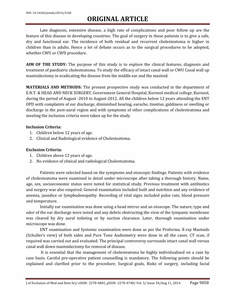
DOI: 10.14260/jemds/2014/3168 ORIGINAL ARTICLE J of Evolution of Med and Dent Sci/ eISSN- 2278-4802, pISSN- 2278-4748/ Vol. 3/ Issue 34/Aug 11, 2014 Page 9057 A STUDY OF INTACT CANAL WALL TECHNIQUE IN PAEDIATRIC CHOLESTEATOMA PATIENTS FOR 2YRS IN GGH, KURNOOL Mohammed Naveed Ahmed 1 , M. Mahendra Kumar 2 , A. Sesha Prasad 3 , S. Muneeruddin Ahmed 4 , M. Praveen Kumar 5 , C. V. Rao 6 , Aghiri Nagaraju 7 , B. Durga Prasad 8 HOW TO CITE THIS ARTICLE: Mohammed Naveed Ahmed, M. Mahendra Kumar, A. Sesha Prasad, S. Muneeruddin Ahmed, M. Praveen Kumar, C. V. Rao, Aghiri Nagaraju, B. Durga Prasad. “A Study of Intact Canal Wall Technique in Paediatric Cholesteatoma Patients for 2yrs in GGH, Kurnool”. Journal of Evolution of Medical and Dental Sciences 2014; Vol. 3, Issue 34, August 11; Page: 9057-9067, DOI: 10.14260/jemds/2014/3168 ABSTRACT: INTRODUCTION: The surgical treatment of cholesteatoma in paediatric age group has changed opinion many times in the evolution of cholesteatoma surgery. Intact canal wall technique allows provision for maintaining the depth of the middle ear and post- operative hearing is much better than in canal wall down procedure. Canal wall down method precludes regular cleaning of the large cavity left behind. In CWU method incidence of recurrent and residual CH are more than in CWD methods. It is more common because inspection of epi-tympanum and medial aspects of the ossicles is not possible in ICW procedures. As a result, the disease recurs within 6 to 12 months. MATERIALS AND METHODS: Fifty children attending the OPD of Government General Hospital Kurnool with CH, confirmed with CT scan are taken and subjected to ICW tympano-mastoidectomy with or without ossiculoplasty. They are followed for a period of 18 months for residual disease,auditory evaluation and otoscopic examination for graft take up. RESULTS: There was improvement in hearing in 78% of the patients. Graft healing was good in 80.85% of cases. Recurrence was seen in 19.15% of patients. 14% of the patients ICW had to be converted to a CWD procedure within 8 months. There were no reports of intra cranial complication in this series. CONCLUSION: ICW procedure in paediatric cholesteatoma gives better results if the disease is limited to the posterior mesotympanum and not beyond the auditus. Uneventful 18months follow up period was seen in 80.85% of the patients. Recurrent disease is less common if the mastoids are less pneumatized. Revision to CWD may be required in early recurrence; in this series it was 14% of the patients. KEYWORDS: Paediatric Cholesteatoma, ICW, CWD, Residual disease, Recurrent Cholesteatoma, Residual Cholesteatoma, Ossiculoplasty, Epitympanum, Mastoid cavity. INTRODUCTION: Paediatric Cholesteatoma often runs a malignant course, destroying the patient’s hearing and involving the surrounding structures, including the brain, thereby increasing the morbidity and mortality of those affected. Cholesteatoma is associated usually with attico-antral variety of CSOM, also called the dangerous variety due to its bone eroding properties and spreading to vital surrounding areas. Middle ear cholesteatoma is still in many ways not understood. In children, it has a more complex presentation. However cholesteatoma in children is so aggressive that it cannot be treated conservatively. There is little disagreement regarding its sinister threat. It can lead to a wide spectrum of complications, ranging from disabling facial paralysis Deka¹⁵ 1983 and labyrinthine fistulae to life threatening Lepto -meningitis and brain abscess. Furthermore, bilateral disease, with a consequent hearing impairment may profoundly retard the development of speech and acquisition of linguistic skills (Holmes and Kunz, 1969).
-
Upload
independent -
Category
Documents
-
view
0 -
download
0
Transcript of A STUDY OF INTACT CANAL WALL TECHNIQUE IN PAEDIATRIC CHOLESTEATOMA PATIENTS FOR 2YRS IN GGH,...

DOI: 10.14260/jemds/2014/3168
ORIGINAL ARTICLE
J of Evolution of Med and Dent Sci/ eISSN- 2278-4802, pISSN- 2278-4748/ Vol. 3/ Issue 34/Aug 11, 2014 Page 9057
A STUDY OF INTACT CANAL WALL TECHNIQUE IN PAEDIATRIC CHOLESTEATOMA PATIENTS FOR 2YRS IN GGH, KURNOOL Mohammed Naveed Ahmed1, M. Mahendra Kumar2, A. Sesha Prasad3, S. Muneeruddin Ahmed4, M. Praveen Kumar5, C. V. Rao6, Aghiri Nagaraju7, B. Durga Prasad8
HOW TO CITE THIS ARTICLE: Mohammed Naveed Ahmed, M. Mahendra Kumar, A. Sesha Prasad, S. Muneeruddin Ahmed, M. Praveen Kumar, C. V. Rao, Aghiri Nagaraju, B. Durga Prasad. “A Study of Intact Canal Wall Technique in Paediatric Cholesteatoma Patients for 2yrs in GGH, Kurnool”. Journal of Evolution of Medical and Dental Sciences 2014; Vol. 3, Issue 34, August 11; Page: 9057-9067, DOI: 10.14260/jemds/2014/3168
ABSTRACT: INTRODUCTION: The surgical treatment of cholesteatoma in paediatric age group has
changed opinion many times in the evolution of cholesteatoma surgery. Intact canal wall technique
allows provision for maintaining the depth of the middle ear and post- operative hearing is much
better than in canal wall down procedure. Canal wall down method precludes regular cleaning of the
large cavity left behind. In CWU method incidence of recurrent and residual CH are more than in CWD
methods. It is more common because inspection of epi-tympanum and medial aspects of the ossicles
is not possible in ICW procedures. As a result, the disease recurs within 6 to 12 months. MATERIALS
AND METHODS: Fifty children attending the OPD of Government General Hospital Kurnool with CH,
confirmed with CT scan are taken and subjected to ICW tympano-mastoidectomy with or without
ossiculoplasty. They are followed for a period of 18 months for residual disease,auditory evaluation
and otoscopic examination for graft take up. RESULTS: There was improvement in hearing in 78% of
the patients. Graft healing was good in 80.85% of cases. Recurrence was seen in 19.15% of patients.
14% of the patients ICW had to be converted to a CWD procedure within 8 months. There were no
reports of intra cranial complication in this series. CONCLUSION: ICW procedure in paediatric
cholesteatoma gives better results if the disease is limited to the posterior mesotympanum and not
beyond the auditus. Uneventful 18months follow up period was seen in 80.85% of the patients.
Recurrent disease is less common if the mastoids are less pneumatized. Revision to CWD may be
required in early recurrence; in this series it was 14% of the patients.
KEYWORDS: Paediatric Cholesteatoma, ICW, CWD, Residual disease, Recurrent Cholesteatoma,
Residual Cholesteatoma, Ossiculoplasty, Epitympanum, Mastoid cavity.
INTRODUCTION: Paediatric Cholesteatoma often runs a malignant course, destroying the patient’s
hearing and involving the surrounding structures, including the brain, thereby increasing the
morbidity and mortality of those affected. Cholesteatoma is associated usually with attico-antral
variety of CSOM, also called the dangerous variety due to its bone eroding properties and spreading
to vital surrounding areas. Middle ear cholesteatoma is still in many ways not understood. In
children, it has a more complex presentation.
However cholesteatoma in children is so aggressive that it cannot be treated conservatively.
There is little disagreement regarding its sinister threat. It can lead to a wide spectrum of
complications, ranging from disabling facial paralysis Deka¹⁵ 1983 and labyrinthine fistulae to life
threatening Lepto -meningitis and brain abscess. Furthermore, bilateral disease, with a consequent
hearing impairment may profoundly retard the development of speech and acquisition of linguistic
skills (Holmes and Kunz, 1969).
DOI: 10.14260/jemds/2014/3168
ORIGINAL ARTICLE
J of Evolution of Med and Dent Sci/ eISSN- 2278-4802, pISSN- 2278-4748/ Vol. 3/ Issue 34/Aug 11, 2014 Page 9058
Late diagnosis, extensive disease, a high rate of complications and poor follow up are the
feature of this disease in developing countries. The goal of surgery in these patients is to give a safe,
dry and functional ear. The incidence of both residual and recurrent cholesteatoma is higher in
children than in adults. Hence a lot of debate occurs as to the surgical procedures to be adopted,
whether CWU or CWD procedure.
AIM OF THE STUDY: The purpose of this study is to explore the clinical features, diagnosis and
treatment of paediatric cholesteatoma. To study the efficacy of intact canal wall or CWU Canal wall up
mastoidectomy in eradicating the disease from the middle ear and the mastoid.
MATERIALS AND METHODS: The present prospective study was conducted in the department of
E.N.T. & HEAD AND NECK SURGERY, Government General Hospital, Kurnool medical college, Kurnool,
during the period of August -2010 to August-2012. All the children below 12 years attending the ENT
OPD with complaints of ear discharge, diminished hearing, earache, tinnitus, giddiness or swelling or
discharge in the post-aural region and with symptoms of other complications of cholesteatoma and
meeting the inclusion criteria were taken up for the study.
Inclusion Criteria:
1. Children below 12 years of age.
2. Clinical and Radiological evidence of Cholesteatoma.
Exclusion Criteria:
1. Children above 12 years of age.
2. No evidence of clinical and radiological Cholesteatoma.
Patients were selected based on the symptoms and otoscopic findings. Patients with evidence
of cholesteatoma were examined in detail under microscope after taking a thorough history. Name,
age, sex, socioeconomic status were noted for statistical study. Previous treatment with antibiotics
and surgery was also enquired. General examination included built and nutrition and any evidence of
anemia, jaundice or lymphadenopathy. Recording of vital signs included pulse rate, blood pressure
and temperature.
Initially ear examination was done using a head mirror and an otoscope. The nature, type and
odor of the ear discharge were noted and any debris obstructing the view of the tympanic membrane
was cleared by dry aural toileting or by suction clearance. Later, thorough examination under
microscope was done.
ENT examination and Systemic examination were done as per the Proforma. X-ray Mastoids
(Schuller’s view) of both sides and Pure Tone Audiometry were done in all the cases. CT scan, if
required was carried out and evaluated. The principal controversy surrounds intact canal wall versus
canal wall down mastoidectomy for removal of disease.
It is essential that the management of cholesteatoma be highly individualized on a case by
case basis. Careful pre-operative patient counselling is mandatory. The following points should be
explained and clarified prior to the procedure; Surgical goals, Risks of surgery, including facial

DOI: 10.14260/jemds/2014/3168
ORIGINAL ARTICLE
J of Evolution of Med and Dent Sci/ eISSN- 2278-4802, pISSN- 2278-4748/ Vol. 3/ Issue 34/Aug 11, 2014 Page 9059
paralysis, vertigo, tinnitus, worsening of Hearing, Possible need for a staged procedure, the chronic
nature of the disease process with necessity of long term follow up.
OBSERVATION AND STATISTICAL ANALYSIS: The statistical analysis in the present study is based
on the observations made in the cases of CSOM (attico-antral) being treated surgically in the
department of E.N.T. And HEAD & NECK surgery, Government General Hospital, Kurnool Medical
College, Kurnool during the period of 2 years from August-2010 to August -2012.
AGE GROUP NO. OF PATIENTS PERCENTAGE %
5-6 2 4%
7-8 7 14 %
9-10 21 42 %
11-12 20 40 %
TOTAL 50 100
AGE DISTRIBUTION
Of the 50 cases which presented during the study period the age of the patients varied
between 6 to 12 years. The average age was 9.7 years ± 1.8 years. The youngest of children were 6
years of age and the oldest were 12 years of age.
In that, 2 patients (4%) were in the age group of 5 to 6 years, 7 patients (14%) were in the age
group 7 to 8 years, 21 patients (42%) were in the age group 9 to 12 years and 20 patients (40%)
were in the age group of 11 to 12 years. 32 children (64%) were males and 18 children (36%) were
females. 78% of the children belonged to the lower socioeconomic group. 22% belonged to Middle
group of income.
Bilateral disease was seen in 12% of children. In the present study, of the 50 cases, 26 (52%)
patients had involvement of right ear only and 18 (36%) patients had involvement of left ear.
SYMPTOMS No. of Cases PERCENTAGE
Ear discharge 50 100
Hearing loss 40 80
Pain in ear 14 28
Swelling and pain behind ear 3 6
Fever 4 8
Tinnitus 2 4
Vertigo 2 4
SYMPTOMATOLOGY
The above data reveals that the ear discharge and the hearing loss are the striking symptoms
in the present study. In this study more than 60% of the patients had the ear symptoms from six
months to five years.
DOI: 10.14260/jemds/2014/3168
ORIGINAL ARTICLE
J of Evolution of Med and Dent Sci/ eISSN- 2278-4802, pISSN- 2278-4748/ Vol. 3/ Issue 34/Aug 11, 2014 Page 9060
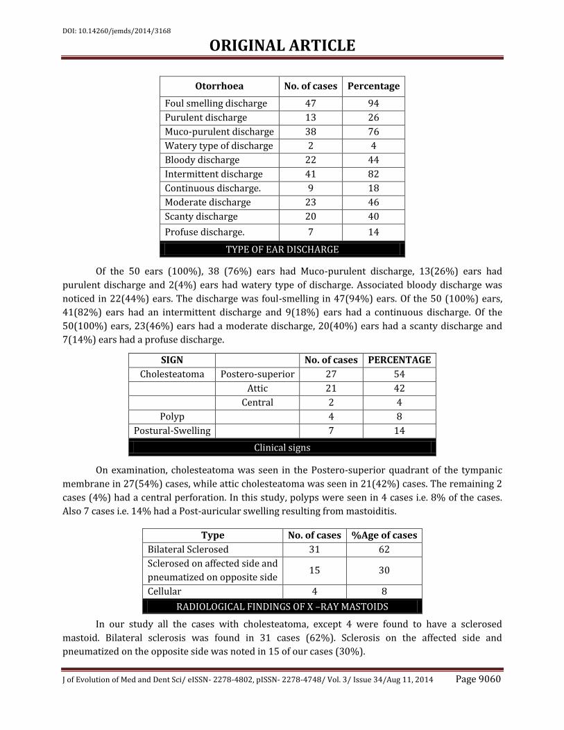
Otorrhoea No. of cases Percentage
Foul smelling discharge 47 94
Purulent discharge 13 26
Muco-purulent discharge 38 76
Watery type of discharge 2 4
Bloody discharge 22 44
Intermittent discharge 41 82
Continuous discharge. 9 18
Moderate discharge 23 46
Scanty discharge 20 40
Profuse discharge. 7 14
TYPE OF EAR DISCHARGE
Of the 50 ears (100%), 38 (76%) ears had Muco-purulent discharge, 13(26%) ears had
purulent discharge and 2(4%) ears had watery type of discharge. Associated bloody discharge was
noticed in 22(44%) ears. The discharge was foul-smelling in 47(94%) ears. Of the 50 (100%) ears,
41(82%) ears had an intermittent discharge and 9(18%) ears had a continuous discharge. Of the
50(100%) ears, 23(46%) ears had a moderate discharge, 20(40%) ears had a scanty discharge and
7(14%) ears had a profuse discharge.
SIGN No. of cases PERCENTAGE
Cholesteatoma Postero-superior 27 54
Attic 21 42
Central 2 4
Polyp 4 8
Postural-Swelling 7 14
Clinical signs
On examination, cholesteatoma was seen in the Postero-superior quadrant of the tympanic
membrane in 27(54%) cases, while attic cholesteatoma was seen in 21(42%) cases. The remaining 2
cases (4%) had a central perforation. In this study, polyps were seen in 4 cases i.e. 8% of the cases.
Also 7 cases i.e. 14% had a Post-auricular swelling resulting from mastoiditis.
Type No. of cases %Age of cases
Bilateral Sclerosed 31 62
Sclerosed on affected side and
pneumatized on opposite side 15 30
Cellular 4 8
RADIOLOGICAL FINDINGS OF X –RAY MASTOIDS
In our study all the cases with cholesteatoma, except 4 were found to have a sclerosed
mastoid. Bilateral sclerosis was found in 31 cases (62%). Sclerosis on the affected side and
pneumatized on the opposite side was noted in 15 of our cases (30%).
DOI: 10.14260/jemds/2014/3168
ORIGINAL ARTICLE
J of Evolution of Med and Dent Sci/ eISSN- 2278-4802, pISSN- 2278-4748/ Vol. 3/ Issue 34/Aug 11, 2014 Page 9061
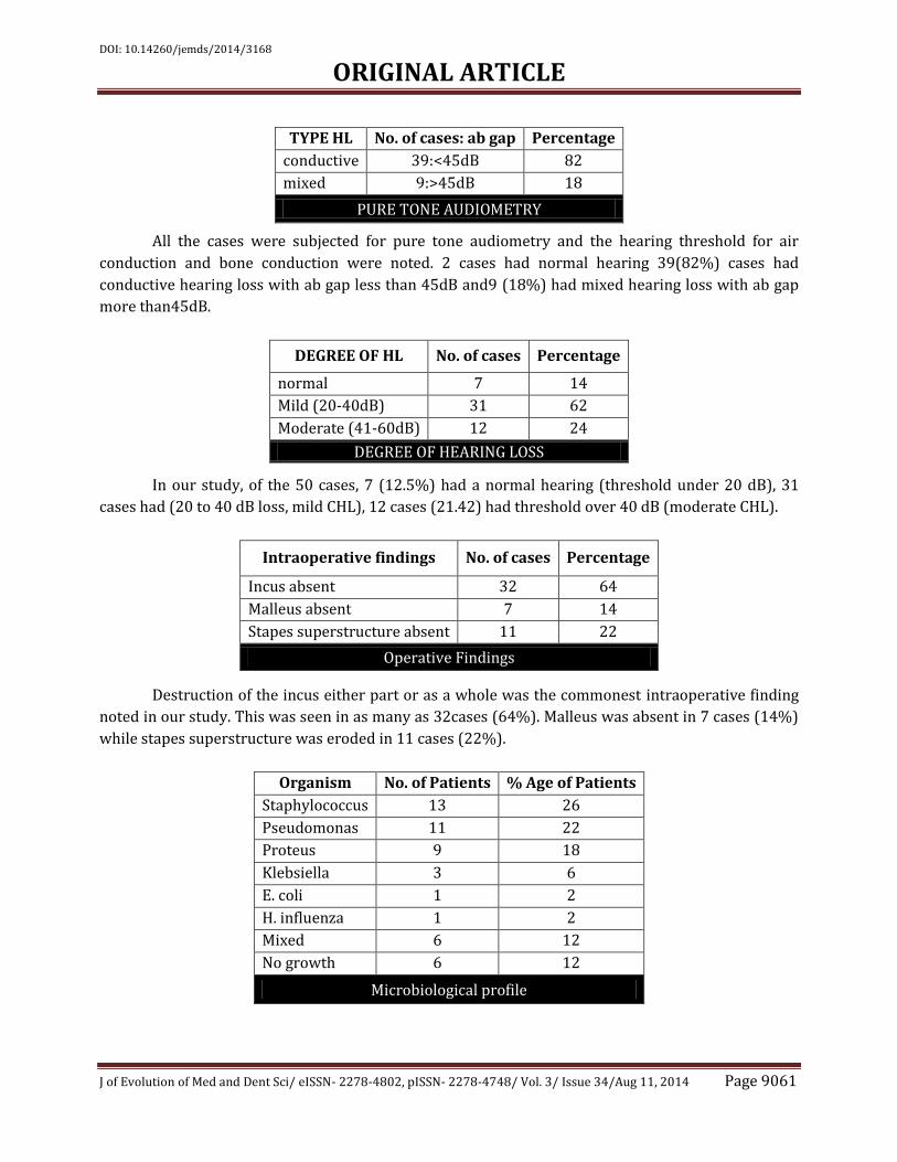
TYPE HL No. of cases: ab gap Percentage
conductive 39:<45dB 82
mixed 9:>45dB 18
PURE TONE AUDIOMETRY
All the cases were subjected for pure tone audiometry and the hearing threshold for air
conduction and bone conduction were noted. 2 cases had normal hearing 39(82%) cases had
conductive hearing loss with ab gap less than 45dB and9 (18%) had mixed hearing loss with ab gap
more than45dB.
DEGREE OF HL No. of cases Percentage
normal 7 14
Mild (20-40dB) 31 62
Moderate (41-60dB) 12 24
DEGREE OF HEARING LOSS
In our study, of the 50 cases, 7 (12.5%) had a normal hearing (threshold under 20 dB), 31
cases had (20 to 40 dB loss, mild CHL), 12 cases (21.42) had threshold over 40 dB (moderate CHL).
Intraoperative findings No. of cases Percentage
Incus absent 32 64
Malleus absent 7 14
Stapes superstructure absent 11 22
Operative Findings
Destruction of the incus either part or as a whole was the commonest intraoperative finding
noted in our study. This was seen in as many as 32cases (64%). Malleus was absent in 7 cases (14%)
while stapes superstructure was eroded in 11 cases (22%).
Organism No. of Patients % Age of Patients
Staphylococcus 13 26
Pseudomonas 11 22
Proteus 9 18
Klebsiella 3 6
E. coli 1 2
H. influenza 1 2
Mixed 6 12
No growth 6 12
Microbiological profile
DOI: 10.14260/jemds/2014/3168
ORIGINAL ARTICLE
J of Evolution of Med and Dent Sci/ eISSN- 2278-4802, pISSN- 2278-4748/ Vol. 3/ Issue 34/Aug 11, 2014 Page 9062
Staphylococcus aureus was the commonest organism isolated in this series and was seen in
13 cases i.e. 26%. This was followed by pseudomonas 11(22%), proteus 9(18%), klebsiella 3(6%),
E.coli 1(2%) and Haemophilus influenza 1(2%) cases. Mixed growth was seen in 6 cases i.e. 12%. No
organism was isolated in another 6 cases (12%).
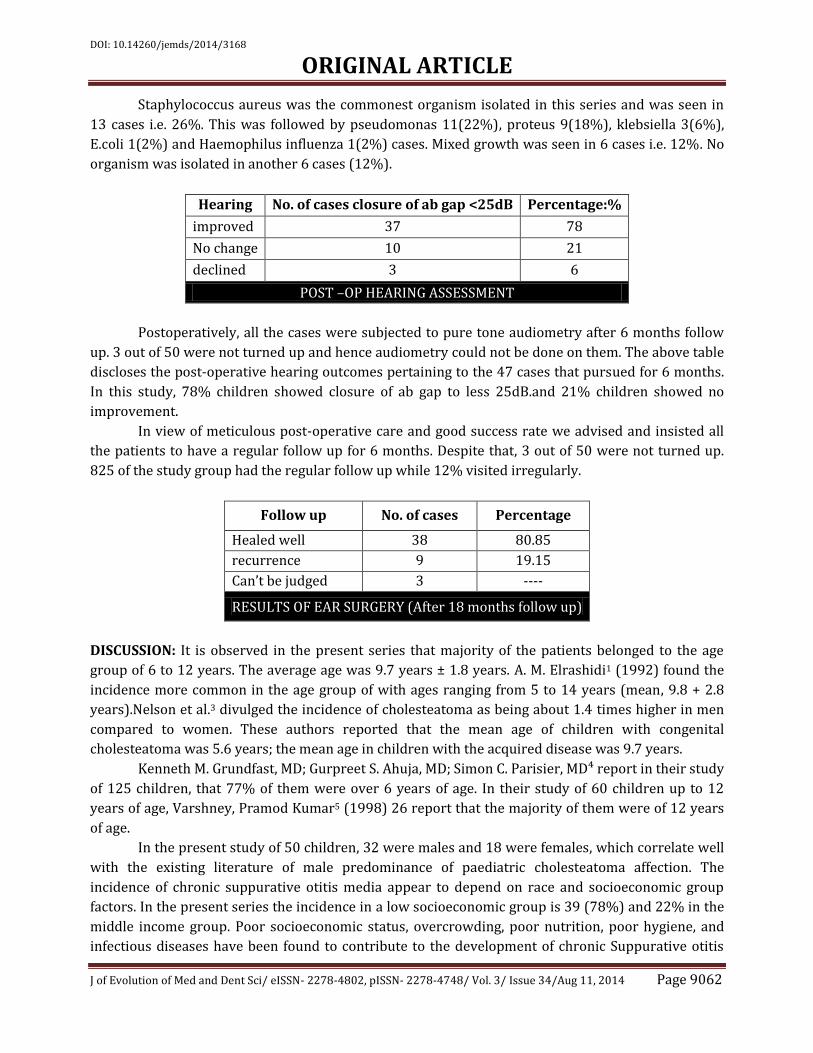
Hearing No. of cases closure of ab gap <25dB Percentage:%
improved 37 78
No change 10 21
declined 3 6
POST –OP HEARING ASSESSMENT
Postoperatively, all the cases were subjected to pure tone audiometry after 6 months follow
up. 3 out of 50 were not turned up and hence audiometry could not be done on them. The above table
discloses the post-operative hearing outcomes pertaining to the 47 cases that pursued for 6 months.
In this study, 78% children showed closure of ab gap to less 25dB.and 21% children showed no
improvement.
In view of meticulous post-operative care and good success rate we advised and insisted all
the patients to have a regular follow up for 6 months. Despite that, 3 out of 50 were not turned up.
825 of the study group had the regular follow up while 12% visited irregularly.
Follow up No. of cases Percentage
Healed well 38 80.85
recurrence 9 19.15
Can’t be judged 3 ----
RESULTS OF EAR SURGERY (After 18 months follow up)
DISCUSSION: It is observed in the present series that majority of the patients belonged to the age
group of 6 to 12 years. The average age was 9.7 years ± 1.8 years. A. M. Elrashidi1 (1992) found the
incidence more common in the age group of with ages ranging from 5 to 14 years (mean, 9.8 + 2.8
years).Nelson et al.3 divulged the incidence of cholesteatoma as being about 1.4 times higher in men
compared to women. These authors reported that the mean age of children with congenital
cholesteatoma was 5.6 years; the mean age in children with the acquired disease was 9.7 years.
Kenneth M. Grundfast, MD; Gurpreet S. Ahuja, MD; Simon C. Parisier, MD⁴ report in their study
of 125 children, that 77% of them were over 6 years of age. In their study of 60 children up to 12
years of age, Varshney, Pramod Kumar5 (1998) 26 report that the majority of them were of 12 years
of age.
In the present study of 50 children, 32 were males and 18 were females, which correlate well
with the existing literature of male predominance of paediatric cholesteatoma affection. The
incidence of chronic suppurative otitis media appear to depend on race and socioeconomic group
factors. In the present series the incidence in a low socioeconomic group is 39 (78%) and 22% in the
middle income group. Poor socioeconomic status, overcrowding, poor nutrition, poor hygiene, and
infectious diseases have been found to contribute to the development of chronic Suppurative otitis

DOI: 10.14260/jemds/2014/3168
ORIGINAL ARTICLE
J of Evolution of Med and Dent Sci/ eISSN- 2278-4802, pISSN- 2278-4748/ Vol. 3/ Issue 34/Aug 11, 2014 Page 9063
media. Browning G.G. (1997), states that there is a close relationship of chronic otitis media and
lower socio economic group.
Higher incidence is because of poor general health, malnutrition and overcrowding. In a study
by Varshney, Pramod Kumar⁵ (1998), in their study of 60 children, 75% belonged to lower socio-
economic group, while 15% belonged to middle socio- economic group.
Simon C. Parisier et al (1988), in their retrospective study of 125 cases affected, 63 were in
patients with involvement of the right ear only, 57 were in patients with involvement of the left ear
only and 5 patients had bilateral disease i.e. 120 (96%) had unilateral and 5(4%) had bilateral
involvement. Varshney, Pramod Kumar (1998), in their study of 60 cases, 42 (70%) had unilateral
involvement and 18 (30%) had bilateral affection.
In our present study of 50 cases 26 were in patients with involvement of the right ear only, 18
were in patients with involvement of the left ear only and 6 patients had bilateral affection i.e.44
(88%) had unilateral and 6 (12%) had bilateral involvement. These correlate well with the
observations of the quoted literature.
In the present series Ear discharge is present in all the patients (100%), loss of hearing in
(80%), pain in 28%, post-aural swelling 6%. None presented with any intracranial complications.
Palvaet al6 (1977) in their series of 68 ears also noted an absence of complications.
In their retrospective study, Simon C. Parisier et al8 (1988), the most common presenting
symptoms were Otorrhoea (73%), hearing loss (85%), otalgia (32%), tinnitus (8%) and vertigo(8%).
Only (0.8%) presented with an intracranial complication. Marco– Algarra J. et al7 (1991) in their
study of 52 patients (55 ears), the commonest presenting symptoms were Otorrhoea plus hearing
loss (54%), Otorrhoea only(29%), hearing loss only (7.6%), mastoiditis(5.4%), pain (13%) dizziness
(3.8%), tinnitus (3.8%), fever (1.9%).
None presented with any facial nerve involvement or any intracranial complications.
Varshney, Pramod Kumar5(1998), in their study the symptoms at presentation included aural
discharge (95%), hearing loss (100%), ear ache (65%), post aural swelling (28.3%) and vertigo
(20%). 10% presented with intracranial complications.
Cholesteatoma was found in the posterosuperior quadrant in 54% of cases while it was found
in attic in 42%. Edelstein et al9 noted 32% in posterosuperior quadrant and 20% in attic. Sade J. and
Shatz A10 (1988) noticed the following in the 109 children (109ears) studied. 21 (19.2%) ears with
attic retraction pocket cholesteatoma, 42 (38.6%) with postero- superior retraction pocket
cholesteatoma, 31 (28.4%) ears with congenital cholesteatoma, 8 (7.3%) ears with cholesteatomas
associated with central perforation and 7 ears were of an undetermined type. Of these none
presented with a facial paralysis. 1 case showed a fistula of the semicircular canal and 3 cases
presented with acute mastoiditis. Mills R. P, Padgham N.D.11
(1991)in their retrospective study of 54 children (57 ears), noted 30 (52.6%) ears with attic
cholesteatoma, 13 (22.8) ears with pars tensa marginal cholesteatoma, 8 (15.7%) ears with pars
tensa central cholesteatoma, 5 (8.7%) ears with congenital cholesteatoma and 1 (1.7%) ears being
unclassified. No report of complications associated is available. A.M. Elrashidi1 showed that the most
frequently affected locations were the epi-tympanum.
The present study showed cholesteatoma in the Postero-superior quadrant of the tympanic
membrane in 27(54%) cases, while attic cholesteatoma was seen in 21(42%) cases. The remaining 2
cases (4%) had a central perforation. In this study, polyps were seen in 4 cases i.e. 8% of the cases.

DOI: 10.14260/jemds/2014/3168
ORIGINAL ARTICLE
J of Evolution of Med and Dent Sci/ eISSN- 2278-4802, pISSN- 2278-4748/ Vol. 3/ Issue 34/Aug 11, 2014 Page 9064
Also 7 cases i.e. 14% had a Post-auricular swelling resulting from mastoiditis. This also showed that
all the cases with cholesteatoma were found to have a sclerosed mastoid. Bilateral sclerosis was
found in 31 cases (62%).
Of the 31 cases that showed bilateral sclerosis, 15 of them had bilateral disease and the rest
16 had bilateral sclerosis but unilateral disease. Pneumatized mastoid was seen only in 8% of the
patients. So it is difficult to infer whether sclerosis predisposes the mastoids to cholesteatoma
formation or if sclerosis is the result of cholesteatoma. Sclerosis on the affected side and pneumatized
on the opposite side was noted in 15 of our cases (30%).
Marco- Algarra J. et al7 (1991) in their study of 52 patients (55 ears), revealed the mastoid
bone to the sclerosed in 25 case (45%), diploeic in 10 cases (18%) and well pneumatized in 13 cases
(23%). Evidence of cholesteatoma or destruction in not mentioned.
Tiwari R. et al (1991)12report in their study of 54 patients, a sclerotic type of mastoid in 50
cases (92.58%) and a cellular type (pneumatic) of mastoid in 4 cases (7.4%). 40 cases (70.08%)
showed radiological evidence of cholesteatoma. Varshney, Pramod Kumar5 (1998) 26, in their study
of 60 cases, showed sclerosis of air cells in 100% cases, evidence of cholesteatoma in 40 (66.67%)
cases and destruction, cavitations in 13 (21.67%) cases.
In our study, of the 50 cases, 7 (12.5%) had a normal hearing (threshold under20 dB), 31
cases had (20 to 40 dB loss, mild CHL), 12 cases (21.42) had threshold over 40 dB (moderate CHL).
Marco- Algarra J. et al ⁷(1991) 30 in their study of 52 patients (55ears), report that 4 cases
(7%) had normal hearing threshold under 20 dB), 37 cases (69%) had (20 dB to 40 dB loss) 12 cases
(21%) had (threshold over 40 dB), 1 case (1.81%) had mixed loss and 1 case (1.81%) had a dead ear.
Varshney, Pramod Kumar⁵ (1998), in their study of 60 cases found conductive hearing loss in 52
(86.67%) cases, 1(1.67%) had purely sensorineural type of hearing loss and 7 (11.67%) cases had
mixed type of hearing loss.
The discharge from the ear was sent for bacterial culture and antibiotic sensitivity. The
predominant bacterium isolated in this study was staphylococcus aureus which was found in 26% of
the cases. This was followed by pseudomonas (22%), proteus (18%), klebsiella (6%), E. coli (2%) and
haemophilus influenza (2%). Mixed infection was seen in 12% and another 12% did not produce any
growth.
This was similar to that found by B.N. Rao and M. S. Reddy13 (1994) which shows
staphylococcus in 41%, pseudomonas in 21%, proteus in 18%, klebsiella in 10% and no growth in
10%. All the cases in this study underwent canal wall up mastoidectomy, operated under general
anaesthesia. Intra-operatively 64% of the cases had a necrosed incus which was higher compared to
Edelstein et al9 (1988) where it was 46% and was in accordance with Sade (1981) who noticed it in
80% of cases.
Ghodrat Mohammadi, MasoudNaderpour and Mehrnoosh Mousaviagdas14 colleagues
reported total (55.4%) or partial (30.7%) erosion of the incus was the most common pathology. The
Primary surgical treatment in all the patients is “intact canal wall technique” through a post aural
route. Preoperative broad spectrum systemic antibiotic like Ceftriaxone sodium is administered to all
cases just before the surgery. In all the patients standard Tympanomeatal flaps are raised and that
includes Posterior meatal skin flap, Superior Tympanomeatal flap, Inferior Tympanomeatal flap and
Posterior Tympanomeatal flap, which help to preserve the vascularity and thus helps in rapid
epithelialization of the Tympanic membrane. In all the patients underlay grafting is done.
DOI: 10.14260/jemds/2014/3168
ORIGINAL ARTICLE
J of Evolution of Med and Dent Sci/ eISSN- 2278-4802, pISSN- 2278-4748/ Vol. 3/ Issue 34/Aug 11, 2014 Page 9065
In all the possible cases primary ossiculoplasty was done depending upon the ossicular status.
Septal cartilage and homograft ossicles are used as prostheses for partial ossicular replacement. In
cases where Malleus is found eroded Type 2 tympanoplasty, in cases of Incus erosion Type 3
Tympanoplasty is done. Where Incus, Malleus and Stapes supra structure are eroded with the mobile
foot plate, Type 4 Tympanoplasty is done. Single stage procedure with a second stage operation,
whenever necessary is adopted in the present study.
All the patients are followed post operatively for 4 weeks, with the patient reporting to the
outpatient department. Suture removal is done after 1stweek. External canal pack removed after 3
weeks. Ear drops are allowed after 3weeks.
Among the patients 82% of them turned up regularly for follow up. 12% of them came
irregularly and 65 did not turn up for follow up. In the present study 80.85% of the patients showed
well healed dry ear, 19.15% showed recurrence. The post-operative follow up included an
Audiometric examination after a gap of 6 months to 1 year.
The closure of air bone gap in the present series is good with 10 to 15 dB of residual hearing
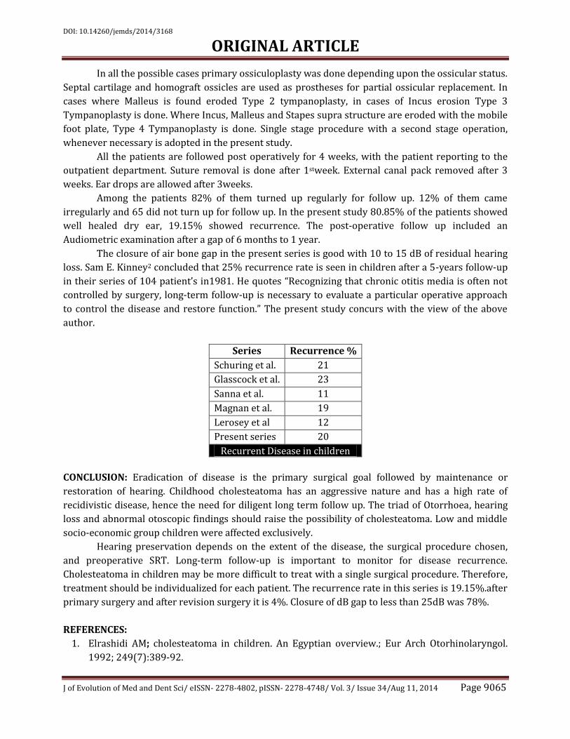
loss. Sam E. Kinney2 concluded that 25% recurrence rate is seen in children after a 5-years follow-up
in their series of 104 patient’s in1981. He quotes “Recognizing that chronic otitis media is often not
controlled by surgery, long-term follow-up is necessary to evaluate a particular operative approach
to control the disease and restore function.” The present study concurs with the view of the above
author.
Series Recurrence %
Schuring et al. 21
Glasscock et al. 23
Sanna et al. 11
Magnan et al. 19
Lerosey et al 12
Present series 20
Recurrent Disease in children
CONCLUSION: Eradication of disease is the primary surgical goal followed by maintenance or
restoration of hearing. Childhood cholesteatoma has an aggressive nature and has a high rate of
recidivistic disease, hence the need for diligent long term follow up. The triad of Otorrhoea, hearing
loss and abnormal otoscopic findings should raise the possibility of cholesteatoma. Low and middle
socio-economic group children were affected exclusively.
Hearing preservation depends on the extent of the disease, the surgical procedure chosen,
and preoperative SRT. Long-term follow-up is important to monitor for disease recurrence.
Cholesteatoma in children may be more difficult to treat with a single surgical procedure. Therefore,
treatment should be individualized for each patient. The recurrence rate in this series is 19.15%.after
primary surgery and after revision surgery it is 4%. Closure of dB gap to less than 25dB was 78%.
REFERENCES:
1. Elrashidi AM; cholesteatoma in children. An Egyptian overview.; Eur Arch Otorhinolaryngol.
1992; 249(7):389-92.
DOI: 10.14260/jemds/2014/3168
ORIGINAL ARTICLE
J of Evolution of Med and Dent Sci/ eISSN- 2278-4802, pISSN- 2278-4748/ Vol. 3/ Issue 34/Aug 11, 2014 Page 9066
2. Sam E. Kinney MD; Intact canal wall Tympanoplasty with mastoidectomy for Cholesteatoma;
Long-term follow-up. November 1988; 98(11):1190–1194
3. Jose Evandro Andrade Prudente de AquinoI; Nelson Alvares Cruz FilhoII;Julia Negro Prudente
de AquinoIIIBraz; j. otorhinolaryngol. (Impr.)May/June 2011; 77(3): 343-48
4. Kenneth M. Grundfast, MD; Gurpreet S. Ahuja, MD; Simon C. Parisier, MD;Stephanie M. Culver,
MD; Delayed Diagnosis and Fate of Congenital Cholesteatoma (Keratoma); Arch Otolaryngol
Head Neck Surg.1995;121(8):903-907.
5. Varshney, Kumar P. A cross sectional study of paediatric Cholesteatoma in the department of
ENT; District hospital, Belgaum; A dissertation (1998):45-48.
6. Palva A, Pekka K, Karja J; Cholesteatoma in children. ArchOtolaryngology; 1997; 103:74-7.
7. Marco- Algarra J et al, Cholesteatoma in children: results in open versus closed techniques; The
Journal of Laryngology and Otology, Oct.1991; (105): 820-824.
8. Levensenm ; Management of Congenital Pediatric Cholesteatomas. American Journal of Otology.
1999; 10:121-123. 9. Edelstein DR, Parisier SC. Surgical techniques and recidivism in Cholesteatoma. Amer journal of
Otology; March 1989; 10(2):78-83.
10. Sade. J and Shatz A; Cholesteatoma in children. The journal of Laryngology and Otology Nov.
1988; 102: 1003-1006.
11. Mills R.P, Padgham N; The management of childhood Cholesteatoma, Journal of Laryngology
and Otology, 1991; 105:343-345.
12. Tiwari et al; A clinicoradiological study of Cholesteatoma; IJLO; Sep1991; 43:135-137.
13. Rao B N, Reddy M S; Indian Journal of Otolaryngology; April–June 1994, 46, (2): 72-77.
14. Mohammadi G, Naderpour M, Mousaviagdas M; Iranian Journal of Otolaryngology;
2012.Summer; 24(88):125-128.
15. Deka R C MS; The Indian Journal of Paediatrics; 50; (6): 643-646.

DOI: 10.14260/jemds/2014/3168
ORIGINAL ARTICLE
J of Evolution of Med and Dent Sci/ eISSN- 2278-4802, pISSN- 2278-4748/ Vol. 3/ Issue 34/Aug 11, 2014 Page 9067
AUTHORS:
1. Mohammed Naveed Ahmed
2. M. Mahendra Kumar
3. A. Sesha Prasad
4. S. Muneeruddin Ahmed
5. M. Praveen Kumar
6. C. V. Rao
7. Aghiri Nagaraju
8. B. Durga Prasad
PARTICULARS OF CONTRIBUTORS:
1. Professor, Department of ENT, Gandhi
Medical College, Secunderabad.
2. Assistant Professor, Department of ENT,
Kurnool Medial College, Kurnool.
3. Professor, Department of ENT, Kurnool
Medical College, Kurnool.
4. Professor and HOD, Department of ENT,
Viswabharathi Medical College, Kurnool.
5. Consultant, Department of ENT, Vijyabhaskar
ENT Hospital, Sattupalli, Khammam, A. P.
6. Assistant Professor, Department of ENT,
Kurnool Medical College, Kurnool.
7. Senior Resident, Department of ENT, Kurnool
Medical College, Kurnool.
8. Medical Officer, Department of ENT, PHC,
Kurnool.
NAME ADDRESS EMAIL ID OF THE
CORRESPONDING AUTHOR:
Dr. S. Muneeruddin Ahmed,
Professor and HOD,
Department of ENT,
Viswabharathi Medical College,
Kurnool.
Email: [email protected]
Date of Submission: 18/07/2014.
Date of Peer Review: 19/07/2014.
Date of Acceptance: 31/07/2014.
Date of Publishing: 09/08/2014.