A small organic compound enhances the religation reaction of human topoisomerase I and identifies...
13
Biosci. Rep. (2013) / 33 / art:e00025 / doi 10.1042/BSR20120118 A small organic compound enhances the religation reaction of human topoisomerase I and identifies crucial elements for the religation mechanism Barbara ARN ` O*, Andrea COLETTA*, Cinzia TESAURO*, Laura ZUCCARO*, Paola FIORANI†, Sara LENTINI‡, Pierluca GALLONI‡, Valeria CONTE‡, Barbara FLORIS‡ and Alessandro DESIDERI* 1 *Department of Biology, University of Rome Tor Vergata, Via Della Ricerca Scientifica, Rome 00133, Italy, †Institute of Translational Pharmacology, National Research Council, CNR, Via Del Fosso del Cavaliere 100, 00133 Rome, Italy, and ‡Department of Chemical Sciences and Technology, Via Della Ricerca Scientifica, Rome 00133, Italy Synopsis The different steps of the human Top1 (topoisomerase I) catalytic cycle have been analysed in the presence of a pentacyclic-diquinoid synthetic compound. The experiments indicate that it efficiently inhibits the cleavage step of the enzyme reaction, fitting well into the catalytic site. Surprisingly the compound, when incubated with the binary topoisomerase–DNA cleaved complex, helps the enzyme to remove itself from the cleaved DNA and close the DNA gap, increasing the religation rate. The compound also induces the religation of the stalled enzyme–CPT (camptothecin)– DNA ternary complex. Analysis of the molecule docked over the binary complex, together with its chemical properties, suggests that the religation enhancement is due to the presence on the compound of two oxygen atoms that act as hydrogen acceptors. This property facilitates the deprotonation of the 5 DNA end, suggesting that this is the limiting step in the topoisomerase religation mechanism. Key words: camptothecin, molecular docking, religation rate, topoisomerase I Cite this article as: Arn` o, B., Coletta, A., Tesauro, C., Zuccaro, L., Fiorani, P ., Lentini, S., Galloni, P ., Conte, V., Floris, B. and Desideri, A. (2013) A small organic compound enhances the religation reaction of human topoisomerase I and identifies crucial elements for the religation mechanism Biosci. Rep. 33(2), art:e00025.doi:10.1042/BSR20120118 INTRODUCTION DNA topoisomerases are key enzymes that modulate the topolo- gical state of DNA through the breaking and rejoining of DNA strands. There are two classes of topoisomerases (types I and II), both characterized by a catalytic mechanism that involves a nucleophilic attack of a DNA phosphodiester bond by a tyrosyl residue from the enzyme, but type I cleaves a single DNA strand, whereas type II cleaves both strands. The human Top1 (topoi- somerase I) enzyme is composed of 765 amino acids and has four distinct domains: the NH 2 -terminal domain (1–214), the core domain (215–635), the linker domain (636–712) and the COOH-terminal domain (713–765) [1,2]. The three- dimensional (3D) structure of reconstituted N-terminal truncated version of human Top1 in complex with a 22 bp DNA molecule ............................................................................................................................................................................................................................................................................................................ Abbreviations used: CPT, camptothecin; Et-KuQ, 1-ethylKuQuinone; TBS, Tris-buffered saline; Top1, topoisomerase I. 1 To whom correspondence should be addressed (email [email protected]). shows the enzyme organized in multiple domains, which ‘clamp on’ the DNA molecule [3]. The active site tyrosine (Tyr 723 ) starts the catalytic cycle of the enzyme through a nucleophilic attack on the DNA backbone, resulting in the breakage of one DNA strand with the enzyme covalently attached to the 3 -phosphate to form the cleavable complex. After changing the linking num- ber a second nucleophilic attack, driven by the 5 -hydroxyl DNA end, restores an intact double-stranded DNA, and the enzyme is released. In both cases, to become a nucleophile the hydroxylic group belonging to the tyrosine or to the DNA end must donate a proton to a nearby group that has been proposed to be the lateral chain of Lys 532 [2,4]. DNA Top IB can be inhibited by several compounds that act through different mechanisms, such as prevention of DNA– topoisomerase binding, inhibition of DNA cleavage or stabiliz- ation of the cleavable complex. Inhibitors of Top1 are divided c 2013 The Author(s). This is an Open Access article distributed under the terms of the Creative Commons Attribution Non-Commercial Licence (http://creativecommons.org/licenses/ by-nc/2.5/) which permits unrestricted non-commercial use, distribution and reproduction in any medium, provided the original work is properly cited. 269 Bioscience Reports www.bioscirep.org
-
Upload
mondodomani -
Category
Documents
-
view
0 -
download
0
Transcript of A small organic compound enhances the religation reaction of human topoisomerase I and identifies...

Biosci. Rep. (2013) / 33 / art:e00025 / doi 10.1042/BSR20120118
A small organic compound enhances thereligation reaction of human topoisomerase I andidentifies crucial elements for the religationmechanismBarbara ARNO*, Andrea COLETTA*, Cinzia TESAURO*, Laura ZUCCARO*, Paola FIORANI†, Sara LENTINI‡,Pierluca GALLONI‡, Valeria CONTE‡, Barbara FLORIS‡ and Alessandro DESIDERI*1
*Department of Biology, University of Rome Tor Vergata, Via Della Ricerca Scientifica, Rome 00133, Italy, †Institute of TranslationalPharmacology, National Research Council, CNR, Via Del Fosso del Cavaliere 100, 00133 Rome, Italy, and ‡Department of ChemicalSciences and Technology, Via Della Ricerca Scientifica, Rome 00133, Italy
SynopsisThe different steps of the human Top1 (topoisomerase I) catalytic cycle have been analysed in the presence of apentacyclic-diquinoid synthetic compound. The experiments indicate that it efficiently inhibits the cleavage step ofthe enzyme reaction, fitting well into the catalytic site. Surprisingly the compound, when incubated with the binarytopoisomerase–DNA cleaved complex, helps the enzyme to remove itself from the cleaved DNA and close the DNA gap,increasing the religation rate. The compound also induces the religation of the stalled enzyme–CPT (camptothecin)–DNA ternary complex. Analysis of the molecule docked over the binary complex, together with its chemical properties,suggests that the religation enhancement is due to the presence on the compound of two oxygen atoms that act ashydrogen acceptors. This property facilitates the deprotonation of the 5′ DNA end, suggesting that this is the limitingstep in the topoisomerase religation mechanism.
Key words: camptothecin, molecular docking, religation rate, topoisomerase I
Cite this article as: Arno, B., Coletta, A., Tesauro, C., Zuccaro, L., Fiorani, P., Lentini, S., Galloni, P., Conte, V., Floris, B. and Desideri,A. (2013) A small organic compound enhances the religation reaction of human topoisomerase I and identifies crucial elements forthe religation mechanism Biosci. Rep. 33(2), art:e00025.doi:10.1042/BSR20120118
INTRODUCTION
DNA topoisomerases are key enzymes that modulate the topolo-gical state of DNA through the breaking and rejoining of DNAstrands. There are two classes of topoisomerases (types I andII), both characterized by a catalytic mechanism that involves anucleophilic attack of a DNA phosphodiester bond by a tyrosylresidue from the enzyme, but type I cleaves a single DNA strand,whereas type II cleaves both strands. The human Top1 (topoi-somerase I) enzyme is composed of 765 amino acids and hasfour distinct domains: the NH2-terminal domain (1–214), thecore domain (215–635), the linker domain (636–712)and the COOH-terminal domain (713–765) [1,2]. The three-dimensional (3D) structure of reconstituted N-terminal truncatedversion of human Top1 in complex with a 22 bp DNA molecule
. . . . . . . . . . . . . . . . . . . . . . . . . . . . . . . . . . . . . . . . . . . . . . . . . . . . . . . . . . . . . . . . . . . . . . . . . . . . . . . . . . . . . . . . . . . . . . . . . . . . . . . . . . . . . . . . . . . . . . . . . . . . . . . . . . . . . . . . . . . . . . . . . . . . . . . . . . . . . . . . . . . . . . . . . . . . . . . . . . . . . . . . . . . . . . . . . . . . . . . . . . . . . . . . . . . . . . . . . . . . . . . . . . . . . . . . . . . . . . . . . . . . . . . . . . . . . . . . . . . . . . . . . . . . . . . . . . . . . . . . . . . . . . . . . . . . . . . . . . . .
Abbreviations used: CPT, camptothecin; Et-KuQ, 1-ethylKuQuinone; TBS, Tris-buffered saline; Top1, topoisomerase I.1 To whom correspondence should be addressed (email [email protected]).
shows the enzyme organized in multiple domains, which ‘clampon’ the DNA molecule [3]. The active site tyrosine (Tyr723) startsthe catalytic cycle of the enzyme through a nucleophilic attackon the DNA backbone, resulting in the breakage of one DNAstrand with the enzyme covalently attached to the 3′-phosphateto form the cleavable complex. After changing the linking num-ber a second nucleophilic attack, driven by the 5′-hydroxyl DNAend, restores an intact double-stranded DNA, and the enzyme isreleased. In both cases, to become a nucleophile the hydroxylicgroup belonging to the tyrosine or to the DNA end must donate aproton to a nearby group that has been proposed to be the lateralchain of Lys532[2,4].
DNA Top IB can be inhibited by several compounds thatact through different mechanisms, such as prevention of DNA–topoisomerase binding, inhibition of DNA cleavage or stabiliz-ation of the cleavable complex. Inhibitors of Top1 are divided
c© 2013 The Author(s). This is an Open Access article distributed under the terms of the Creative Commons Attribution Non-Commercial Licence (http://creativecommons.org/licenses/by-nc/2.5/) which permits unrestricted non-commercial use, distribution and reproduction in any medium, provided the original work is properly cited.
269
Bio
scie
nce
Rep
ort
s
ww
w.b
iosc
irep
.org
B. Arno and others
into two classes: poisons and catalytic inhibitors. Poisons in-clude clinically used drugs, such as the derivatives of the naturalcompound CPT (camptothecin) that reversibly binds the covalentTop1–DNA complex slowing down the religation of the cleavedDNA strand, thus inducing cell death [5]. Two water-solubleCPT derivatives, topotecan and irinotecan have been approvedby the FDA (Food and Drug Administration) for clinical use.The 3D structure of the topotecan–enzyme–DNA ternary com-plex has shown that topotecan mimics a DNA base-pair and bindsat the site of DNA cleavage by intercalating between the upstream( − 1) and downstream ( + 1) base-pairs [6] interacting also withthe enzyme, acting as an interfacial uncompetitive inhibitor [5].On the other hand, catalytic inhibitors act through inhibiting anyother step of the Top1 enzymatic cycle. They include both naturaland non-natural compounds [7] and they mainly act by inhibitingthe cleavage [8–10] or the DNA binding [11,12], although someof them are able to inhibit both cleavage and religation [13,14].The CPT derivatives are widely used in clinics; however, theyhave some intrinsic limits, the main one being linked to theequilibrium of the α-hydroxylactone E-ring of CPTs withthe carboxylate form, which is inactive against Top1 and tightlybinds to serum albumin. For this reason, other drugs are under de-velopment, the most promising one being indolocarbazoles andthe indenoisoquinolines [5].
It has been shown that the ADP–ribose polymers, that is rap-idly synthesized by PARP [poly(ADP–ribose) polymerase], ac-tivated by the occurrence of a DNA break, is also able to interactwith human Topo I [15]. The interaction has been elegantly shownto produce two important effects, the first one being the cleav-age inhibition, the second one consisting in the enhancement ofthe religation [16]. The second effect is quite astonishing andpoly(ADP–ribose) has been up to now the only molecule ableto induce this effect, although the molecular mechanism is stillunclear.
In this work, in the search for a new topoisomerasepoison, we have investigated the interaction of the enzymewith Et-KuQ (1-ethylKuQuinone) (Figure 1), a pentacyclic-diquinoid synthetic compound [17]. The results indicate thatEt-KuQ inhibits cleavage and enhances religation in the pres-ence of CPT. A mechanism for such a process is providedand it is proposed to also occur in the case of poly(ADP–ribose).
MATERIALS AND METHODS
Chemicals and pH titrationThe Et-KuQ was synthesized as previously described [17].CPT (Sigma) and Et-KuQ were dissolved in DMSO to a finalconcentration of 4 and 0.5 mg/ml, respectively, and stored at− 20 ◦C. Anti-FLAG M2 monoclonal affinity gel, FLAG peptideand anti-FLAG M2 monoclonal antibody were purchased fromSigma.
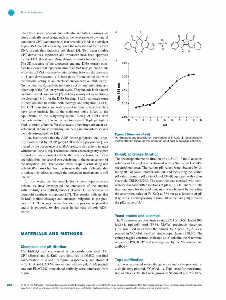
Figure 1 Structure of KuQ(A) Structure and dissociation equilibrium of Et-KuQ. (B) Spectrophoto-metric titration curve for the ionization of Et-KuQ in aqueous solution.
Et-KuQ acid-base titrationThe spectrophothometric titration of a 3.5×10− 5 mol/l aqueoussolution of Et-KuQ was performed with a Shimadzu UV-2450spectrophotometer. The various pH values were obtained by di-luting HCl or NaOH mother solutions and measuring the desiredpH value through a pH metre (Amel 334-B) equipped with a glasselectrode CRISON5203. The electrode was checked with com-mercial standard buffer solutions at pH 4.01, 7.01 and 9.26. Thetitration curve for the acid ionization was obtained by recordingthe absorption value of Et-KuQ at 384 nm as a function of pH(Figure 1); a corresponding sigmoid fit of the data [18] providesthe pKa value of 5.4.
Yeast strains and plasmidsThe Saccharomyces cerevisiae strain EKY3 (ura3-52, his3�200,leu2�1, trp1�63, top1::TRP1, MATα) previously described[19], was used to express the human Top1 gene. Top1 is ex-pressed in YCpGAL1-e-Top1 single copy plasmid [19,20]. Theepitope-tagged construct, indicated as ‘e’ contains the N-terminalsequence DYKDDDY and is recognized by the M2 monoclonalantibody.
Top1 purificationTop1 was expressed under the galactose inducible promoter ina single copy plasmid, YCpGAL1-e-Top1, used for transforma-tion of EKY3 cells, that were grown on SC-uracil plus 2 % (w/v)
. . . . . . . . . . . . . . . . . . . . . . . . . . . . . . . . . . . . . . . . . . . . . . . . . . . . . . . . . . . . . . . . . . . . . . . . . . . . . . . . . . . . . . . . . . . . . . . . . . . . . . . . . . . . . . . . . . . . . . . . . . . . . . . . . . . . . . . . . . . . . . . . . . . . . . . . . . . . . . . . . . . . . . . . . . . . . . . . . . . . . . . . . . . . . . . . . . . . . . . . . . . . . . . . . . . . . . . . . . . . . . . . . . . . . . . . . . . . . . . . . . . . . . . . . . . . . . . . . . . . . . . . . . . . . . . . . . . . . . . . . . . . . . . . . . . . . . . . . . . . . . . . . . . . . . . . . . . . . . . . . . . . . . . . . . . . . . . . . . . . . . . . . . . . . . . . . . . . . . . . . .
270 c© 2013 The Author(s). This is an Open Access article distributed under the terms of the Creative Commons Attribution Non-Commercial Licence (http://creativecommons.org/licenses/by-nc/2.5/) which permits unrestricted non-commercial use, distribution and reproduction in any medium, provided the original work is properly cited.

A novel organic compound enhances human topoisomerase I religation activity
dextrose and diluted 1:100 in SC-uracil plus 2 % (w/v) raffinose.At a D595 = 1.0, the cells were induced with 2 % (w/v) galactosefor 6 h. Cells were then centrifuged, washed with ice-cold wa-ter and resuspended in 2 ml buffer/g of cells [50 mM Tris/HCl,pH 7.4, 1 mM EDTA, 1 mM EGTA, 10 % (v/v) glycerol and pro-tease inhibitors cocktail (Roche)], supplemented with 0.1 mg/mlsodium bisulfate, 0.8 mg/ml sodium fluoride, 1 mM PMSF and1 mM DTT (dithiothreitol). After an addition of 0.5 vol of 425–600 mm diameter glass beads, the cells were disrupted by vor-texing for 30 s alternating with 30 s on ice and then were cent-rifuged. The column, containing 2 ml of anti-FLAG M2 affinitygel, was washed with 20 column volumes of TBS (Tris-bufferedsaline; 50 mM Tris/HCl pH 7.4 and 150 mM KCl), prior to load-ing the lysate. Elution of FLAG-fusion-e-Top1, was performedby competition with five column volumes of a solution contain-ing 100 mg/ml FLAG peptide in TBS. Fractions (500 μl) werecollected and 40 % (v/v) glycerol was added in all preparations,which were stored at − 20 ◦C [21]. Fractions were resolved bySDS/PAGE using the epitope-specific monoclonal antibody M2protein. The protein concentration has been estimated by dens-itometry of the chosen fraction and compared with the purifiedTop I (purchased from Topogene). Our Top1 has a concentrationof 10 ng/μl.
DNA relaxation assaysThe activity of 1 μl of Top1 was assayed in 30 μl of reac-tion volume containing 0.5 μg of negatively supercoiled pBlue-Script KSII( + ) DNA, that is present in both dimeric andmonomeric forms and reaction buffer (20 mM Tris/HCl pH 7.5,0.1 mM Na2EDTA, 10 mM MgCl2, 50 μg/ml acetylated BSAand 150 mM KCl). The effect of Et-KuQ on enzyme activity wasmeasured by adding different concentrations of the compound;the dose-dependent analysis has been performed by simultan-eously adding Top1 and the supercoiled plasmid in the presenceof DMSO as control, or with different concentrations of Et-KuQor pre-incubating the enzyme with various concentrations of Et-KuQ for 10 min at 37 ◦C before adding the DNA; the reactionswere stopped after 60 min. The relaxation activity of Top1, pre-incubated with 5 μM Et-KuQ, or with the same concentrationof DMSO, before adding the supercoiled plasmid, was also ana-lysed as a function of time at 37 ◦C. Reactions were stopped with0.5 % SDS and the samples were resolved in a 1 % (w/v) agarosegel in 48 mM Tris, 45.5 mM boric acid, 1 mM EDTA. The gelwas stained with ethidium bromide (0.5 μg/ml), destained withwater and photographed under UV illumination.
Cleavage kineticsOligonucleotide substrate CL14 (5′-GAAAAAAGACTTAG-3′)was radiolabelled with [γ -32P]ATP at the 5′ end. The CP25 com-plementary strand (5′-TAAAAATTTTTCTAAGTCTTTTTTC-3′) was 5′ phosphorylated with unlabelled ATP. The two strandswere annealed at 2-fold molar excess of CP25 over CL14, cre-ating the partial duplex suicide substrate. The cleavage reactionswere carried out in 20 mM Tris/HCl pH 7.5, 0.1 mM Na2EDTA,
10 mM MgCl2, 50 μg/ml acetylated BSA and 150 mM KCl at25 ◦C, by incubating 20 nM CL14/CP25 and an excess of Top1with DMSO, as no-drug control, or 50 μM Et-KuQ, or pre-incubating the enzyme and the compound for 10 min beforeadding the suicide substrate [22]. At various time points 5 μlaliquots were removed and the reaction stopped with 0.5 % (w/v)SDS. After ethanol precipitation samples were resuspended in5 μl of 1 mg/ml trypsin to degrade the protein and were ana-lysed by denaturing 7 M urea/20 % (w/v) PAGE in 48 mM Tris,45.5 mM boric acid, 1 mM EDTA. However, a short trypsin-resistant peptide is always left, explaining why the Cl1 migratesslower than the Cl14 oligonucleotide [23]. The percentage ofcleavage, determined by PhosphorImager and ImageQuant soft-ware, was calculated in each lane as the ratio between the amountof cleavage product and the total amount of radioactivity. Thepercentage of cleavage product, corresponding to the plateau ob-served for the experiment carried out with the substrate and theenzyme alone, was then considered as 100 % of cleavage productand all the other measurements were normalized to this value.
Religation kineticsCL14/CP25 (20 nM) was incubated with an excess of Top1 for60 min at 25 ◦C followed by 30 min at 37 ◦C in 20 mM Tris/HClpH 7.5, 0.1 mM Na2EDTA, 10 mM MgCl2, 50 μg/ml acetylatedBSA and 150 mM KCl. After the formation of the cleavablecomplex (Cl1) a 5-μl aliquot was removed and used as thetime 0 point, then DMSO or 50 μM Et-KuQ were added andthe religation reaction was started by adding a 200-fold molarexcess of R11 oligonucleotide (5′-AGAAAAATTTT-3′) overthe CL14/CP25 [21]. The KCl concentration was increased to350 mM when R11 was added, to avoid the religated product be-ing recleaved by the protein [24]. Where indicated, the compoundwas pre-incubated with the cleavable complex for 10 min at 37 ◦C,before adding the R11 oligonucleotide. At 37 ◦C 5 μl aliquotswere removed at various time points, and the reaction stoppedwith 0.5 % (w/v) SDS. After ethanol precipitation, samples wereresuspended in 5 μl of 1 mg/ml trypsin and incubated at 37 ◦Cfor 60 min. Samples were analysed by denaturing 7 M urea/20 %(w/v) PAGE in 48 mM Tris, 45.5 mM boric acid, 1 mM EDTA.The religation percentage, determined by ImageQuant software,was plotted as a function of time and normalized to the plateauvalue.
Cleavage/religation equilibriumOligonucleotide CL25 (5′-GAAAAAAGACTTAGAAAAAT-TTTTA-3′) was 5′ radiolabelled with [γ -32P]ATP. The CP25 com-plementary strand (5′-TAAAAATTTTTCTAAGTCTTTTTTC-3′) was phosphorylated at its 5′ end with unlabelled ATP.The two strands were annealed at a 2-fold molar excess ofCP25 over CL25. The assay was done by adding 20 nMduplex CL25/CP25 to Top1 alone (DMSO), in the pres-ence of 50 μM CPT, in the presence of 50 μM Et-KuQ orafter incubating the reaction mixture with 50 μM CPT for1 min and then adding 50 μM Et-KuQ, or incubating with
. . . . . . . . . . . . . . . . . . . . . . . . . . . . . . . . . . . . . . . . . . . . . . . . . . . . . . . . . . . . . . . . . . . . . . . . . . . . . . . . . . . . . . . . . . . . . . . . . . . . . . . . . . . . . . . . . . . . . . . . . . . . . . . . . . . . . . . . . . . . . . . . . . . . . . . . . . . . . . . . . . . . . . . . . . . . . . . . . . . . . . . . . . . . . . . . . . . . . . . . . . . . . . . . . . . . . . . . . . . . . . . . . . . . . . . . . . . . . . . . . . . . . . . . . . . . . . . . . . . . . . . . . . . . . . . . . . . . . . . . . . . . . . . . . . . . . . . . . . . . . . . . . . . . . . . . . . . . . . . . . . . . . . . . . . . . . . . . . . . . . . . . . . . . . . . . . . . . . . . . . .
c© 2013 The Author(s). This is an Open Access article distributed under the terms of the Creative Commons Attribution Non-Commercial Licence (http://creativecommons.org/licenses/by-nc/2.5/) which permits unrestricted non-commercial use, distribution and reproduction in any medium, provided the original work is properly cited.
271

B. Arno and others
50 μM Et-KuQ for 1 min and then adding 50 μM CPT. Re-actions were performed at 25 ◦C in 20 mM Tris/HCl pH 7.5,0.1 mM Na2EDTA, 10 mM MgCl2, 50 mg/ml acetylatedBSA and 150 mM KCl and were stopped at two different times:1 and 2 min, by adding 0.5 % (w/v) SDS, precipitating with eth-anol and digesting with trypsin [25]. Products were resolved in20 % (w/v) PAGE/7 M urea gel and quantified using ImageQuantsoftware. The values were calculated by the ratio between thecleavage amount and the total radioactivity of each lane, sub-tracting the control background.
Molecular dockingThe deprotonated Et-KuQ molecular geometry in the gas phasewas obtained at the B3LYP/6-31 + G(d,p) level of theory us-ing GAMESS-US-2011 ed. [26]. The molecular geometry of theTop1 non-covalent binary complex was obtained by modellingthe 1A36 PDB entry; after removing the DNA, the same structurewas used to obtain a model of the Top1 enzyme in the absenceof DNA. The 1T8I PDB entry was used to obtain a model forthe ternary complex. The MGLTools 1.5.6 rc1 was used to con-vert the PDB into PDBQT format, to calculate the Gasteiger-Marsilii charges and produce the input file. Grid was calcu-lated using an in-house modified version of AutoGrid 4.2.2.1using a 256×256×256 grid with step of 0.5 A (1 A = 0.1 nm)enclosing the whole Top1. 250 runs of docking were gener-ated using the standard GA algorithm with AutoDock 4.2.2.1[27] for Et-KuQ on each of the three systems. The complexeswere clusterized on the basis of their RMSD (root-mean-squaredeviation), using a cut-off value of 2 A. Figures 6–8 weregenerated with UCSF Chimera 1.9 (http://www.cgl.ucsf.edu/chimera).
RESULTS AND DISCUSSION
Et-KuQ physical-chemical propertiesKuQuinones are a new class of pentacyclic di-quinoid compoundsthat have recently been discovered and synthesized with a facileone-pot reaction [17]. The polycyclic system can be considereda naphthoquinone derivative and its name is due to the structuralsimilarities with vitamin K. In this work the Et-KuQ has been re-acted with the human Top1 enzyme. The main feature of its struc-ture is the presence of a pentacyclic skeleton (similarly to CPT)with a double-quinone functional group; the five rings consist oftwo naphthalene units fused through a 5-membered moiety thatcontains an alkyl chain (Figure 1A). Extended electronic conjug-ation is proved by the intense absorption spectrum with a broadband between 400 and 600 nm. These molecules were isolated asenol tautomers stabilized by the intramolecular hydrogen bondwith the nearest carbonyl group, as shown in the crystal structureof the Et-KuQ derivative [17]. The strong hydrogen-bond inter-action is evidenced by the very high value of the chemical shift(18 ppm) of the enolic hydrogen in the 1H NMR spectrum. On
the other hand, the shift of the relative tautomeric equilibrium to-wards the keto form appears to be strongly sensitive to solvent po-larity and hydrogen-bonding capacity. Therefore it can be reason-ably assumed that the keto tautomer is favoured in aqueous media;a complete characterization of the equilibrium is under currentinvestigation.
The pH dependence of the spectrum permits us to evaluate apKa of 5–6 for the equilibrium between a protonated and a depro-tonated form (Figure 1B). The unusual acidity of the compoundcompares well with that reported for the hipposudoric acid [28],a natural structurally related compound. In fact, the conjugatebase of Et-KuQ, i.e. the enolate species, is particularly stablebecause of the stabilization of the negative charge within themolecule.
Et-KuQ inhibits the relaxation activity of Top1The relaxation activity of native Top1 has been assessed by in-cubating the enzyme in the presence of increasing concentrationof Et-KuQ in a plasmid relaxation assay (Figure 2A). The sim-ultaneous addition of enzyme, Et-KuQ and DNA never resultedin complete inhibition of the plasmid relaxation activity up to aconcentration of 100 μM. The inhibitory effect is greater whenthe enzyme is pre-incubated with Et-KuQ before adding DNAand the compound fully inhibits the relaxation at a concentrationof 12.5 μM (Figure 2A, lane 14). An additional control has beenperformed (C2 Figure 2A, lane 20) by adding the drug to theDNA plasmid, demonstrating that the Et-KuQ does not permitDNA migration and so does not bind to DNA. The relaxation as-say carried out as function of time at an Et-KuQ concentration of5 μM indicated that the inhibitory effect is abolished after 15 min(Figure 2B, lane 16), demonstrating that the binding of the com-pound is reversible. Since Et-KuQ is dissolved in DMSO, as acontrol, the reaction in presence of this solvent has been carriedout and it shows that the enzyme, at the same DMSO concentra-tion, maintains its activity over time (Figure 2B, lanes 1–9).
Cleavage and religation analysisThe time course of the cleavage of the wild-type enzyme inthe presence and absence of Et-KuQ has been followed us-ing a suicide cleavage substrate. In detail, a 5′ end radiola-belled oligonucleotide CL14 (5′-GAAAAAAGACTTAG-3′) wasannealed to the CP25 (5′-TAAAAATTTTTCTAAGTCTTTT-TTC-3′) complementary strand, to produce a duplex with an11-base 5′ single-strand extension that is preferentially cut by theenzyme at the site indicated by the arrow (Figure 3A, top). Thereligation step is precluded, because the dinucleotide, generatedduring cleavage, is too short to be religated, leaving the enzymecovalently attached to the 3′ end of the oligonucleotide. The dataindicate that in the absence of the drug the wild-type proteinshows a typical time-dependent cleavage of the fragment (Fig-ure 3A, lanes 2–7), whereas pre-incubation of the enzyme with50 μM Et-KuQ fully inhibits the cleavage reaction (lanes 8–13).The inhibitory effect of the compound, after a simultaneous in-cubation with both the enzyme and the suicide substrate, is alsoclear (lanes 14–19), although lower than that observed for the
. . . . . . . . . . . . . . . . . . . . . . . . . . . . . . . . . . . . . . . . . . . . . . . . . . . . . . . . . . . . . . . . . . . . . . . . . . . . . . . . . . . . . . . . . . . . . . . . . . . . . . . . . . . . . . . . . . . . . . . . . . . . . . . . . . . . . . . . . . . . . . . . . . . . . . . . . . . . . . . . . . . . . . . . . . . . . . . . . . . . . . . . . . . . . . . . . . . . . . . . . . . . . . . . . . . . . . . . . . . . . . . . . . . . . . . . . . . . . . . . . . . . . . . . . . . . . . . . . . . . . . . . . . . . . . . . . . . . . . . . . . . . . . . . . . . . . . . . . . . . . . . . . . . . . . . . . . . . . . . . . . . . . . . . . . . . . . . . . . . . . . . . . . . . . . . . . . . . . . . . . .
272 c© 2013 The Author(s). This is an Open Access article distributed under the terms of the Creative Commons Attribution Non-Commercial Licence (http://creativecommons.org/licenses/by-nc/2.5/) which permits unrestricted non-commercial use, distribution and reproduction in any medium, provided the original work is properly cited.

A novel organic compound enhances human topoisomerase I religation activity
Figure 2 Relaxation of supercoiled DNA(A) Relaxation of negative supercoiled plasmid by Top1 incubated with increasing concentrations of Et-KuQ (lanes 2–9) orafter pre-incubation for 10 min at 37 ◦C with increasing concentrations of Et-KuQ (lanes 11–18). Lanes 1 and 10, no drugadded; lane 19, no drug and protein added, lane 20, no protein added. (B) Relaxation of negative supercoiled plasmidin a time course experiment for DMSO (lanes 1–9), 5 μM Et-KuQ (lanes 10–18); lane 19, no protein added. The reactionproducts are resolved in an agarose gel and visualized with ethidium bromide. The two forms of the supercoiled plasmidDNA are indicated as ‘Dimer’ and ‘Monomer’.
pre-incubation. These results can be well appreciated from theplot in Figure 3(B) where the percentage of cleavage is reportedas a function of time. In detail in the absence of the drug (blackline) the maximum of the cleaved product is observed after 4 min,while in the presence of the drug preincubated with enzyme (greyline with circles), no cleaved product is observed in the first 4 minand a small percentage is observed in the 8–15 min time window.This result unambiguously indicates that Et-KuQ is an inhibitorof cleavage and that its interaction with the enzyme is reversibledue to the appearance of a small percentage of cleaved productat long times.
In order to analyse the effect of Et-KuQ on the religation reac-tion, Top1 was incubated with the suicide substrate, to allow thecleavage reaction (Figure 4A, lane 1). The religation is then fol-lowed in the absence of the compound (Figure 4A, lanes 2–7), orby pre-incubating the cleavable suicide complex with 50 μM Et-KuQ before adding 200-fold molar excess of the complementaryR11 oligonucoleotide (Figure 4A, lanes 8–13) or adding Et-KuQtogether with the religation substrate (Figure 4A, lanes 14–19).In all the experiments, the KCl concentration is maintained at350 mM when R11 is added to the samples, in order to avoid thereligated product being cleaved again by the protein, since cleav-age reaction is inhibited at high salt concentration [24]. Aliquotshave been removed at different times, the reaction stopped by ad-dition of SDS and the products analysed by PAGE (Figure 4A).The Figure clearly shows that Et-KuQ, when preincubated for10 min with the covalent complex, stimulates the rate of religation(lanes 8–13), the amount of product formed being much higher
than in the reaction carried out in the absence of Et-KuQ, orwithout preincubating it with the covalent complex. A quantitat-ive analysis of this result is shown in the graph in Figure 4(B). Thisis to our knowledge the first time that a small organic compoundacts as a stimulator of the Top1 religation reaction. The sameexperiment, performed using an excess of Et-KuQ (200 μM),shows that, at this high Et-KuQ concentration, an increase of therate of religation is observed also when the compound is addedtogether with the R11 substrate (see Supplementary Figure S1 athttp://www.bioscirep.org/bsr/033/bsr033e025add.htm).
Cleavage/religation equilibrium assayTo understand the effect of Et-KuQ on the stability of the co-valent DNA–Top1 complex, a cleavage/religation equilibriumexperiment on the 25-mer full duplex oligonucleotide has alsobeen performed (Figure 5A). The assay was done at two differenttimes: 1 and 2 min, respectively, for the substrate and the enzymealone (lanes 1 and 2), in the presence of 50 μM CPT (lanes 3 and4), or with 50 μM Et-KuQ (lanes 5 and 6), or after incubatingthe reaction mixture with 50 μM CPT and then adding 50 μMEt-KuQ and stopping the reaction after another minute (lane 7),or incubating with 50 μM Et-KuQ for 1 min and then adding50 μM CPT and stopping the reaction after another minute (lane8). The reactions were stopped with SDS, the samples digestedwith trypsin and the products analysed by PAGE (Figure 5A).In the presence of DMSO, the cleavage/religation equilibriumis shifted towards religation and a small level of cleavage of
. . . . . . . . . . . . . . . . . . . . . . . . . . . . . . . . . . . . . . . . . . . . . . . . . . . . . . . . . . . . . . . . . . . . . . . . . . . . . . . . . . . . . . . . . . . . . . . . . . . . . . . . . . . . . . . . . . . . . . . . . . . . . . . . . . . . . . . . . . . . . . . . . . . . . . . . . . . . . . . . . . . . . . . . . . . . . . . . . . . . . . . . . . . . . . . . . . . . . . . . . . . . . . . . . . . . . . . . . . . . . . . . . . . . . . . . . . . . . . . . . . . . . . . . . . . . . . . . . . . . . . . . . . . . . . . . . . . . . . . . . . . . . . . . . . . . . . . . . . . . . . . . . . . . . . . . . . . . . . . . . . . . . . . . . . . . . . . . . . . . . . . . . . . . . . . . . . . . . . . . . .
c© 2013 The Author(s). This is an Open Access article distributed under the terms of the Creative Commons Attribution Non-Commercial Licence (http://creativecommons.org/licenses/by-nc/2.5/) which permits unrestricted non-commercial use, distribution and reproduction in any medium, provided the original work is properly cited.
273

B. Arno and others
Figure 3 Cleavage kinetics(A) Time course (0.5–15 min) of the cleavage experiment between the CL14/CP25 suicide substrate and the purified Top1alone (lanes 2–7), after 10 min pre-incubation with 50 μM Et-KuQ (lanes 8–13) or simultaneously incubated with 50 μMEt-KuQ (lanes 14–19). In lane 1 the protein has not been added. Cl1 represents the DNA substrate cleaved by Top1 atthe preferred cleavage site, indicated by an arrow over the DNA sequence. (B) The percentage of cleavage, determined byPhosphorImager and ImageQuant software, was normalized to the total amount of radioactivity in each lane. The DMSO(black line), simultaneous incubation with Et-KuQ (grey line with squares) and the experiment performed pre-incubating theTop1 with Et-KuQ (grey line with circles) are shown.
the labelled DNA strand is detected (Figure 5A, lanes 1 and 2).When the enzyme is in the presence of 50 μM CPT the cleav-age/religation equilibrium is shifted towards cleavage, since CPTreversibly binds to the covalent DNA–enzyme intermediate, sta-bilizing it and slowing down the religation rate, as indicated bythe band indicated by the asterisk corresponding to the substratecleaved at the preferred site (Figure 5A, lanes 3 and 4) [5,29,30].The band of the cleavage complex is not observed when Et-KuQis added to the enzyme together with the substrate (Figure 5A,lanes 5 and 6), confirming that the compound inhibits the cleav-age step. In line with this, incubation for 1 min of the enzyme, thesubstrate and the Et-KuQ, followed by addition and incubation ofCPT for another minute, shows a band (Figure 5A, lane 8) similar
to that observed in the presence of Et-KuQ alone, confirming thatEt-KuQ is an inhibitor of cleavage. The most striking result is ob-tained upon addition of Et-KuQ to the enzyme previously reactedwith the substrate in the presence of CPT. The Et-KuQ additionbrings to 50 % destabilization of the cleaved complex stabilizedby CPT (Figure 5A, lane 7 compared with lane 4). This resultindicates that Et-KuQ is able to stimulate the religation even inthe presence of CPT, overcoming the poisoning effect of the drugon inhibiting the religation. A quantification of the cleaved com-plex observed in the different assays is shown in the histogramof Figure 5(B), that indicates the ability of Et-KuQ to reduce theCPT stabilization of the cleaved complex (compare lane 4 withlanes 7 and 8). These data provide evidence that Et-KuQ has a
. . . . . . . . . . . . . . . . . . . . . . . . . . . . . . . . . . . . . . . . . . . . . . . . . . . . . . . . . . . . . . . . . . . . . . . . . . . . . . . . . . . . . . . . . . . . . . . . . . . . . . . . . . . . . . . . . . . . . . . . . . . . . . . . . . . . . . . . . . . . . . . . . . . . . . . . . . . . . . . . . . . . . . . . . . . . . . . . . . . . . . . . . . . . . . . . . . . . . . . . . . . . . . . . . . . . . . . . . . . . . . . . . . . . . . . . . . . . . . . . . . . . . . . . . . . . . . . . . . . . . . . . . . . . . . . . . . . . . . . . . . . . . . . . . . . . . . . . . . . . . . . . . . . . . . . . . . . . . . . . . . . . . . . . . . . . . . . . . . . . . . . . . . . . . . . . . . . . . . . . . .
274 c© 2013 The Author(s). This is an Open Access article distributed under the terms of the Creative Commons Attribution Non-Commercial Licence (http://creativecommons.org/licenses/by-nc/2.5/) which permits unrestricted non-commercial use, distribution and reproduction in any medium, provided the original work is properly cited.

A novel organic compound enhances human topoisomerase I religation activity
Figure 4 Religation kinetics(A) Gel analysis of the religation kinetics (0.5–15 min) between the wild-type suicide covalent complex (Cl1) and the R11complementary oligonucleotide (shown at the top of the Figure) in the absence of Et-KuQ (lanes 2–7), after pre-incubationwith 50 μM Et-KuQ (lanes 8–13), or incubated together with 50 μM Et-KuQ (lanes 14–19). In lane 1 the cleaved complex,which represents time 0, has been loaded. In lane 20 no protein was added. ‘Cl1’ represents the DNA fragment cleaved atthe preferred enzyme site; ‘Religation’ is the restored fully duplex oligonucleotide that is the final product of the reaction.(B) Percentage of religation, determined by ImageQuant software, was plotted as a function of time and normalized tothe plateau value for the experiment with Top1 alone. Substrate and Top1 (black circles), substrate added to the enzymepre-incubated with Et-KuQ (grey circles), simultaneous mixing of substrate, enzyme and drug (grey squares).
dual effect, inhibiting the cleavage and surprisingly promotingthe religation, even in the presence of CPT.
Molecular dockingThree molecular docking experiments were performedon the Et-KuQ deprotonated form (Figure 1A), in order to studythe molecular mechanism underlying the observed inhibition ofcleavage and the enhancement of religation. The first-docking ex-periment was performed using as a receptor the crystal structure
of the Top1–DNA non-covalent complex [3] after DNA removal.The complexes obtained have been subjected to clusterizationand the largest cluster shows that Et-KuQ interacts with the Top1catalytic pentad with an energy of − 9.6 kcal/mol (1 kcal =4.184 kJ) (Figure 6). In this structure the Et-KuQ deprotonatedoxygen molecules are at hydrogen bonding distance with the side-chains of three residues of the catalytic pentad: Arg488, Arg590 andTyr723. This conformation is consistent with the observed inhibi-tion of the DNA cleavage, and with the increase of the inhibitionin the case of pre-incubation (Figure 3).
. . . . . . . . . . . . . . . . . . . . . . . . . . . . . . . . . . . . . . . . . . . . . . . . . . . . . . . . . . . . . . . . . . . . . . . . . . . . . . . . . . . . . . . . . . . . . . . . . . . . . . . . . . . . . . . . . . . . . . . . . . . . . . . . . . . . . . . . . . . . . . . . . . . . . . . . . . . . . . . . . . . . . . . . . . . . . . . . . . . . . . . . . . . . . . . . . . . . . . . . . . . . . . . . . . . . . . . . . . . . . . . . . . . . . . . . . . . . . . . . . . . . . . . . . . . . . . . . . . . . . . . . . . . . . . . . . . . . . . . . . . . . . . . . . . . . . . . . . . . . . . . . . . . . . . . . . . . . . . . . . . . . . . . . . . . . . . . . . . . . . . . . . . . . . . . . . . . . . . . . . .
c© 2013 The Author(s). This is an Open Access article distributed under the terms of the Creative Commons Attribution Non-Commercial Licence (http://creativecommons.org/licenses/by-nc/2.5/) which permits unrestricted non-commercial use, distribution and reproduction in any medium, provided the original work is properly cited.
275

B. Arno and others
Figure 5 Cleavage/religation equilibrium(A) Gel electrophoresis of the products from the incubation of Top1 with the [γ -32P] end-labelled duplex DNA (CL25/CP25),shown at the top of the Figure. The arrow indicates the preferred cleavage site. The duplex was incubated for 1 or 2 minwith the enzyme (lanes 1 and 2), in presence of CPT (lanes 3 and 4), Et-KuQ (lanes 5 and 6), after a pre-incubation withCPT and then addition of Et-KuQ (lane 7) and after pre-incubation with Et-KuQ and then addition of CPT (lane 8). Lane9, no enzyme added. The band corresponding to the enzyme-substrate cleaved complex is indicated by an asterisk. (B)Percentage of the cleavable complex normalized to the total radioactivity of each lane.
The second docking experiment was performed on the Top1–DNA covalent complex [3]. Owing to the presence of DNA co-valently bound to the catalytic Tyr723, Et-KuQ cannot bind in theposition previously observed in Figure 6 for the docking withthe protein alone. When the docking is made with the Top1–DNA covalent complex, Et-KuQ has a binding site in prox-imity of the catalytic pentad with energy of − 9.8 kcal/mole(Figure 7). In this complex the ethyl group of the drug andits aromatic scaffold show van der Waals interactions with theDNA major grove at the level of the bases in positions − 1and + 1, while the deprotonated oxygen molecules of Et-KuQform an hydrogen bond with the lateral chain of Asn722 (Fig-ure 7). This structure permits us to provide an interpretation
of the effect of the compound during the religation process. Infact, the deprotonated oxygen molecules of Et-KuQ that rep-resent a strong hydrogen acceptor site [17] and that can thenhelp the deprotonation process that the hydroxyl group of the 5′
DNA terminal must undergo to reseal the cleaved DNA strand,are at an average distance from the DNA O5′ of 6.7 A (Fig-ure 7). In the crystal structure of the covalent enzyme–DNAbinary complex the distance between the DNA O5′ and the Nζ
Lys532, that is considered the proton acceptor site, is 7.2 A [4,6].The relatively short distance between the DNA O5′ and a pro-ton acceptor site provides an explanation for the increase of thereligation rate observed in the presence of Et-KuQ, shown inFigure 4.
. . . . . . . . . . . . . . . . . . . . . . . . . . . . . . . . . . . . . . . . . . . . . . . . . . . . . . . . . . . . . . . . . . . . . . . . . . . . . . . . . . . . . . . . . . . . . . . . . . . . . . . . . . . . . . . . . . . . . . . . . . . . . . . . . . . . . . . . . . . . . . . . . . . . . . . . . . . . . . . . . . . . . . . . . . . . . . . . . . . . . . . . . . . . . . . . . . . . . . . . . . . . . . . . . . . . . . . . . . . . . . . . . . . . . . . . . . . . . . . . . . . . . . . . . . . . . . . . . . . . . . . . . . . . . . . . . . . . . . . . . . . . . . . . . . . . . . . . . . . . . . . . . . . . . . . . . . . . . . . . . . . . . . . . . . . . . . . . . . . . . . . . . . . . . . . . . . . . . . . . . .
276 c© 2013 The Author(s). This is an Open Access article distributed under the terms of the Creative Commons Attribution Non-Commercial Licence (http://creativecommons.org/licenses/by-nc/2.5/) which permits unrestricted non-commercial use, distribution and reproduction in any medium, provided the original work is properly cited.

A novel organic compound enhances human topoisomerase I religation activity
Figure 6 Representative structure of Et-KuQ docked on Top1 in the absence of DNAThe Et-KuQ (cyan) and the residues of the catalytic pentad are represented as sticks, while the rest of the protein appearsas a ribbon. The lateral chains of the residues of the catalytic pentad are explicitly represented and labelled.
Figure 7 Representative structure of Et-KuQ docked on the covalent Top1 + DNA binary complexThe Et-KuQ is represented in cyan; the lateral chains of the residues of the catalytic pentad and of the Asn722 arerepresented as sticks and labelled, the rest of the protein is represented as ribbon. The base-pairs in position + 1 and− 1 are represented in black. A blue dashed line indicates the distance between the 5′ DNA end, represented as a redsphere and the closest Et-KuQ oxygen acceptor.
In order to confirm that this mechanism can be involved inthe displacement of CPT by Et-KuQ (Figure 5), a third dockingexperiment was performed on the CPT + TopoI + DNA ternarycomplex. In this complex the CPT interfacial inhibitor inter-calates between the cleaved DNA bases, making the distancebetween the O5′ DNA end and the Lys532 Nζ proton acceptor aslong as 11.9 A [6]. Docking experiments indicate that Et-KuQcan bind in proximity of the Top1 catalytic pentad with an en-ergy of − 10.7 kcal/mol (Figure 8), forming a complex wherethe Et-KuQ deprotonated oxygen molecules are at distance of 7.4A from the hydroxyl of the 5′ DNA end, i.e. the same distanceobserved between the 5′ end and the Lys532 in the X-ray cova-lent binary complex [3]. In the docked complex, Et-KuQ makes
contacts with the DNA bases in positions + 1 and − 1 throughits carbon scaffold and forms a hydrogen bond with the Lys755
of the CTD (C-terminal domain) of Top1 through the deproton-ated oxygen molecules. The relatively short distance between theEt-KuQ deprotonated oxygen molecules and the cleaved DNA5′-terminal (about 7 A) makes possible the 5′ deprotonation pro-cess, permitting the release of the cleaved complex even in thepresence of CPT as experimentally observed (Figure 5).
The chemical properties of Et-KuQ, characterized by the pres-ence of two deprotonated oxygen molecules that in solution dis-play a pKa = 5.5 (Figure 1B), whose value can be modulated bythe protein environment, together with the docking experiment,provide a molecular explanation for the facilitated religation in
. . . . . . . . . . . . . . . . . . . . . . . . . . . . . . . . . . . . . . . . . . . . . . . . . . . . . . . . . . . . . . . . . . . . . . . . . . . . . . . . . . . . . . . . . . . . . . . . . . . . . . . . . . . . . . . . . . . . . . . . . . . . . . . . . . . . . . . . . . . . . . . . . . . . . . . . . . . . . . . . . . . . . . . . . . . . . . . . . . . . . . . . . . . . . . . . . . . . . . . . . . . . . . . . . . . . . . . . . . . . . . . . . . . . . . . . . . . . . . . . . . . . . . . . . . . . . . . . . . . . . . . . . . . . . . . . . . . . . . . . . . . . . . . . . . . . . . . . . . . . . . . . . . . . . . . . . . . . . . . . . . . . . . . . . . . . . . . . . . . . . . . . . . . . . . . . . . . . . . . . . .
c© 2013 The Author(s). This is an Open Access article distributed under the terms of the Creative Commons Attribution Non-Commercial Licence (http://creativecommons.org/licenses/by-nc/2.5/) which permits unrestricted non-commercial use, distribution and reproduction in any medium, provided the original work is properly cited.
277

B. Arno and others
Figure 8 Representative structure of Et-KuQ docked on the CPT + Top1 + DNA ternary complexThe Et-KuQ is represented in cyan; the lateral chains of the residues of the catalytic pentad and the CPT are representedas sticks and labelled; the rest of the protein appears as a ribbon; the base-pairs in position + 1 and − 1 as black lines.A blue dashed line indicates the distance between the 5′ DNA end, represented as a red sphere, and the closest Et-KuQoxygen acceptor.
the presence of the compound. The Et-KuQ oxygen moleculesmay, in fact, act as hydrogen acceptors facilitating the religationprocess.
ConclusionThe planar pentacyclic structure of Et-KuQ permits a favourableinteraction of the compound inside the human Top1 catalytic siteas shown by the docking experiments with the enzyme alone(Figure 6), explaining the inhibition of the relaxation and of thecleavage displayed by the compound (Figures 2 and 3).
On the other hand, the proximity between the oxydryl of theDNA 5′end and the two close oxygen atoms (Figure 7), acting asefficient hydrogen acceptors, explains the ability of the compoundto act as an enhancer of the enzyme religation rate (Figure 4). TheEt-KuQ pKa defining the equilibrium between the protonated anddeprotonated form is 5.6 in solution (Figure 1) and this value canbe slightly modulated by the protein environment, facilitating theacceptance of an hydrogen by the two oxygen molecules that areat an average distance of 6.7 A from the oxydryl group of the DNA5′end in the docked complex with the covalent enzyme–DNAstructure (Figure 7). This distance is shorter than the one observedin the X-ray diffraction structure between the O5′ and the lateralchain of Lys532 (3), that is supposed to be the proton acceptor inthe reaction in the absence of any external compound [4]. Thisshort distance, coupled with the hydrogen acceptor property ofthe two oxygen molecules, explains the increased religation ratepromoted by Et-KuQ.
The same characteristics can be proposed to explain the Et-KuQ ability in displacing the stalled CPT–DNA–enzyme tern-ary complex and promoting the religation (Figure 5). In thiscase the Et-KuQ oxygen molecules-O5′ DNA distance, observedin the docked structure, is about 7 A (Figure 8), permitting thedeprotonation of the DNA oxydryl group. In this latter case two
possible mechanisms can be envisaged. (1) The DNA oxydrylgroup releases its proton to the Et-KuQ oxygen molecules andthe deprotonated 5′ end forces the CPT displacement promotingthe religation reaction. (2) The DNA hydroxyl group releasesits proton to the Et-KuQ oxygen molecules and the reversiblebinding of CPT finds the deprotonated 5′ ready to religate duringthe CPT off process. In both cases, the final result is that thereligation occurs and CPT is displaced.
Recently it has been proposed that poly(ADP–ribose) displaysa similar effect: it inhibits the cleavage, but it also reverses the pre-formed covalent Top–DNA complex, stimulating the religationactivity of the enzyme [15,16]. It is probable that the same mech-anism proposed for Et-KuQ can also work for the poly(ADP–ribose) polymer. The complex structure of the poly(ADP–ribose),made by many phosphate groups, may facilitate the deprotona-tion of the 5′ DNA end, again facilitating the religation. It canthen be concluded that facilitation of the deprotonation of the 5′
DNA end is the crucial process to promote religation.
AUTHOR CONTRIBUTION
Barbara Arno carried out the majority of the experimental part of thestudy, helped by Cinzia Tesauro and Laura Zuccaro. Sara Lentini,Pierluca Galloni, Valeria Conte and Barbara Floris performed the Et-KuQ synthesis and the Et-KuQ acid–base titration. Andrea Colettacarried out all of the docking analysis. Paola Fiorani and AlessandroDesideri designed, co-ordinated the studies and co-wrote the paper.All authors read and approved the paper.
FUNDING
This work was supported by the Associazione Italiana per la Ricercasul Cancro (AIRC) [project 10121 (to A.D.)].
. . . . . . . . . . . . . . . . . . . . . . . . . . . . . . . . . . . . . . . . . . . . . . . . . . . . . . . . . . . . . . . . . . . . . . . . . . . . . . . . . . . . . . . . . . . . . . . . . . . . . . . . . . . . . . . . . . . . . . . . . . . . . . . . . . . . . . . . . . . . . . . . . . . . . . . . . . . . . . . . . . . . . . . . . . . . . . . . . . . . . . . . . . . . . . . . . . . . . . . . . . . . . . . . . . . . . . . . . . . . . . . . . . . . . . . . . . . . . . . . . . . . . . . . . . . . . . . . . . . . . . . . . . . . . . . . . . . . . . . . . . . . . . . . . . . . . . . . . . . . . . . . . . . . . . . . . . . . . . . . . . . . . . . . . . . . . . . . . . . . . . . . . . . . . . . . . . . . . . . . . .
278 c© 2013 The Author(s). This is an Open Access article distributed under the terms of the Creative Commons Attribution Non-Commercial Licence (http://creativecommons.org/licenses/by-nc/2.5/) which permits unrestricted non-commercial use, distribution and reproduction in any medium, provided the original work is properly cited.

A novel organic compound enhances human topoisomerase I religation activity
REFERENCES
1 Wang, J. C. (1996) DNA topoisomerases. Ann. Rev. Biochem. 65,635–692
2 Champoux, J. J. (2001) DNA topoisomerases: structure, functionand mechanism. Annu. Rev. Biochem. 70, 369–413
3 Redinbo, M. R., Stewart, L., Kuhn, P., Champoux, J. J. and Hol,W. G. J. (1998) Crystal structures of human topoisomerase I incovalent and noncovalent complexes with DNA. Science 279,1504–1513
4 Interthal, H., Quigley, P. M., Hol, W. G. M. and Champoux, J. J.(2004) The role of lysine 532 in the catalytic mechanism of humantopoisomerase I. J. Biol. Chem. 279, 2984–2992
5 Pommier, Y. (2009) DNA topoisomerase I inhibitors: chemistry,biology, and interfacial inhibition. Chem. Rev. 109, 2894–2902
6 Staker, B. L., Hjerrild, K., Feese, M. D., Behnke, C. A., Burgin, A. B.Jr and Stewart, L. (2002) The mechanism of topoisomerase Ipoisoning by a camptothecin analog. Proc. Natl Acad. Sci. U.S.A.99, 15387–15392
7 Castelli, S., Coletta, A., D’Annessa, I., Fiorani, P., Tesauro, C. andDesideri, A. (2012) Interaction between natural compounds andhuman topoisomerase I. Biol. Chem. 393, 1327–1340
8 Montaudon, D., Palle, K., Rivory, L. P., Robert, J., Douat-Casassus,C., Quideau, S., Bjornsti, M. A. and Pourquier, P. (2007) Inhibitionof topoisomerase I cleavage activity by thiol-reactive compounds.Importance of vicinal cysteine 504 and 505. J. Biol. Chem. 282,14403–14412
9 Castelli, S., Campagna, A., Vassallo, O., Tesauro, C., Fiorani, P.,Tagliatesta, P., Oteri, F., Falconi, M., Majumder, H. K. and Desideri,A. (2009) Conjugated eicosapentaenoic acid inhibits humantopoisomerase IB with a mechanism different from camptothecin.Arch. Biochem. Biophys. 486, 103–110
10 Castelli, S., Vassallo, O., Katkar, P., Che, C. M., Wai-Yin Sun, R. andDesideri, A. (2011) Inhibition of human DNA topoisomerase IB by acyclometalated gold III compound: analysis on the different stepsof the enzyme catalytic cycle. Arch. Biochem. Biophys. 516,108–112
11 Boege, F., Straub, T., Kehr, A., Boesenberg, C., Christiansen, K.,Andersen, A., Jakob, F. and Kohrle, J. (1996) Selected novelflavones inhibit the DNA binding or the DNA religation step ofeukaryotic topoisomerase I. J. Biol. Chem. 271, 2262–2270
12 Minderman, H., Wrzosek, C., Cao, S., Utsugi, T., Kobunai, T.,Yamada, Y. and Rustum, Y. M. (2000) Mechanism of action of thedual topoisomerase-I and -II inhibitor TAS-103 and activity against(multi)drug resistant cells. Cancer Chemother. Pharmacol. 45,78–84
13 Tesauro, C., Fiorani, P., D’Annessa, I., Chillemi, G., Turchi, G. andDesideri, A. (2010) Erybraedin C, a natural compound from theplant Bituminaria bituminosa, inhibits both the cleavage andreligation activities of human topoisomerase I. Biochem. J. 425,531–539
14 Castelli, S., Katkar, P., Vassallo, O., Falconi, M., Linder, S. andDesideri, A. (2012) A natural anticancer agent thaspine targetshuman topoisomerase IB. Anticancer Agents Med. Chem. 13,356–363
15 Malanga, M. and Althaus, F. R. (2004) Poly(ADP-ribose) reactivatesstalled DNA topoisomerase I and induces DNA strand breakresealing. J. Biol. Chem. 279, 5244–5248
16 Malanga, M. and Althaus, F. R. (2005) The role of poly(ADP-ribose)in the DNA damage signaling network. Biochem. Cell Biol. 83,354–364
17 Coletti, A., Lentini, S., Conte, V., Floris, B., Bortolini, O., Sforza, F.,Grepioni, F. and Galloni, P. (2012) Unexpected one-pot synthesis ofhighly conjugated pentacyclic diquinoid compounds. J. Org. Chem.77, 6873–6879
18 Iglesias, E (1996) Enolization of Benzoylacetone in aqueoussurfactant Solutions: a novel method for determining enolizationconstants. J. Phys. Chem. 100, 12592–12599
19 Bjornsti, M. A., Benedetti, P., Viglianti, G. A. and Wang, J. C. (1989)Expression of human DNA topoisomerase I in yeast cells lackingyeast DNA topoisomerase I: restoration of sensitivity of the cells tothe antitumor drug camptothecin. Cancer Res. 49, 6318–6323
20 Kauh, E. A. and Bjornsti, M. A. (1995) SCT1 mutants suppress thecamptothecin sensitivity of yeast cells expressing wild-type DNAtopoisomerase I. Proc. Natl Acad. Sci. U.S.A. 92, 6299–6303
21 Fiorani, P., Tesauro, C., Mancini, G., Chillemi, G., D’Annessa, I.,Graziani, G., Tentori, L. and Muzi, A. and Alessandro Desideri(2009) Evidence of the crucial role of the linker domain on thecatalytic activity of human topoisomerase I by experimental andsimulative characterization of the Lys681Ala mutant. Nuleic AcidRes. 37, 6849–6858
22 Yang, Z. and Champoux, J. J. (2002) Reconstitution of enzymaticactivity by the association of the cap and catalytic domains ofhuman topoisomerase I. J. Biol. Chem. 277, 30815–30823
23 Stewart, L., Ireton, G. C. and Champoux, J. J. (1999) A functionallinker in human topoisomerase I is required for maximumsensitivity to campthotecin in a DNA relaxation assay. J. Biol.Chem. 274, 32950–32960
24 Elban, M. A., Sun, W., Eisenhauer, B. M., Gao, R. and Hecht, S. M.(2006) Synthesis and biological evaluation of 10,11-methylenedioxy-14-azacamptothecin. Org. Lett. 16, 3513–3516
25 Fiorani, P., Bruselles, A., Falconi, M., Chillemi, G., Desideri, A. andBenedetti, P. (2003) Single mutation in the linker domain confersprotein flexibility and camptothecin resistance to humantopoisomerase I. J. Biol. Chem. 278, 43268–43275
26 Schmidt, M. W., Baldridge, K. K., Boatz, J. A., Elbert, S. T., Gordon,M. S., Jensen, J. H., Koseki, S., Matsunaga, N., Nguyen, K. A., Su,S. et al. (1993) General atomic and molecular electronic structuresystem. J. Comp. Chem. 14, 1347–1363
27 Morris, G. M., Huey, R., Lindstrom, W., Sanner, M. F., Belew, R. K.,Goodsel, D. S. and Olson, A. (2009) AutoDock4 andAutoDockTools4: automated docking with selective receptorflexibility. J. Comp. Chem. 16, 2785–2791
28 Hashimoto, K., Saikawa, Y. and Nakata, M. (2007) Studies on thered sweat of the Hippopotamus amphibious. Pure Appl. Chem. 79,507–517
29 Pommier, Y., Pourquier, P., Fan, Y. and Strumberg, D. (1998)Mechanism of action of eukaryotic DNA topoisomerase I and drugstargeted to the enzyme. Biochim. Biophys. Acta 1400, 83–105
30 Liu, L. F., Desai, S. D., Li, T. K., Mao, Y., Sun, M. and Sim, S. P.(2000) Mechanism of action of camptothecin. Ann. NY Acad. Sci.922, 1–10
Received 15 November 2012/6 December 2012; accepted 19 December 2012
Published as Immediate Publication 31 January 2013, doi 10.1042/BSR20120118
. . . . . . . . . . . . . . . . . . . . . . . . . . . . . . . . . . . . . . . . . . . . . . . . . . . . . . . . . . . . . . . . . . . . . . . . . . . . . . . . . . . . . . . . . . . . . . . . . . . . . . . . . . . . . . . . . . . . . . . . . . . . . . . . . . . . . . . . . . . . . . . . . . . . . . . . . . . . . . . . . . . . . . . . . . . . . . . . . . . . . . . . . . . . . . . . . . . . . . . . . . . . . . . . . . . . . . . . . . . . . . . . . . . . . . . . . . . . . . . . . . . . . . . . . . . . . . . . . . . . . . . . . . . . . . . . . . . . . . . . . . . . . . . . . . . . . . . . . . . . . . . . . . . . . . . . . . . . . . . . . . . . . . . . . . . . . . . . . . . . . . . . . . . . . . . . . . . . . . . . . .
c© 2013 The Author(s). This is an Open Access article distributed under the terms of the Creative Commons Attribution Non-Commercial Licence (http://creativecommons.org/licenses/by-nc/2.5/) which permits unrestricted non-commercial use, distribution and reproduction in any medium, provided the original work is properly cited.
279

Biosci. Rep. (2013) / 33 / art:e00025 / doi 10.1042/BSR20120118
SUPPLEMENTARY DATA
A small organic compound enhances thereligation reaction of human topoisomerase I andidentifies crucial elements for the religationmechanismBarbara ARNO*, Andrea COLETTA*, Cinzia TESAURO*, Laura ZUCCARO*, Paola FIORANI†, Sara LENTINI‡,Pierluca GALLONI‡, Valeria CONTE‡, Barbara FLORIS‡ and Alessandro DESIDERI*1
*Department of Biology, University of Rome Tor Vergata, Via Della Ricerca Scientifica, Rome 00133, Italy, †Institute of TranslationalPharmacology, National Research Council, CNR, Via Del Fosso del Cavaliere 100, 00133 Rome, Italy, and ‡Department of ChemicalSciences and Technology, Via Della Ricerca Scientifica, Rome 00133, Italy
Supplementary Figure S1 is on the following page
1 To whom correspondence should be addressed (email [email protected]).
c© 2013 The Author(s). This is an Open Access article distributed under the terms of the Creative Commons Attribution Non-Commercial Licence (http://creativecommons.org/licenses/by-nc/2.5/) which permits unrestricted non-commercial use, distribution and reproduction in any medium, provided the original work is properly cited.

B. Arno and others
Figure S1 Religation kinetics(A) Gel analysis of the religation kinetics (0.5–15 min) between the wild-type suicide covalent complex (Cl1) and the R11complementary oligonucleotide (shown at the top of the Figure) (lanes 2–7), after pre-incubation of the cleavage complexwith 200 μM Et-KuQ (lanes 8–13), or added together with 50 μM Et-KuQ (lanes 14–19). In lane 1, the cleaved complex,which represents time 0, has been loaded. In lane 20, no protein was added. ‘Cl1’ represents the DNA fragment cleaved atthe preferred enzyme site; ‘Religation’ is the restored fully duplex oligonucleotide that is the final product of the reaction.(B) Percentage of religation, determined by ImageQuant software, was plotted as function of time and normalized to theplateau value for the experiment with Top1 alone (black circles), substrate added to the enzyme pre-incubatied with Et-KuQ(grey circles), simultaneous mixing of substrate, enzyme and drug (grey squares).
Received 15 November 2012/6 December 2012; accepted 19 December 2012
Published as Immediate Publication 31 January 2013, doi 10.1042/BSR20120118
. . . . . . . . . . . . . . . . . . . . . . . . . . . . . . . . . . . . . . . . . . . . . . . . . . . . . . . . . . . . . . . . . . . . . . . . . . . . . . . . . . . . . . . . . . . . . . . . . . . . . . . . . . . . . . . . . . . . . . . . . . . . . . . . . . . . . . . . . . . . . . . . . . . . . . . . . . . . . . . . . . . . . . . . . . . . . . . . . . . . . . . . . . . . . . . . . . . . . . . . . . . . . . . . . . . . . . . . . . . . . . . . . . . . . . . . . . . . . . . . . . . . . . . . . . . . . . . . . . . . . . . . . . . . . . . . . . . . . . . . . . . . . . . . . . . . . . . . . . . . . . . . . . . . . . . . . . . . . . . . . . . . . . . . . . . . . . . . . . . . . . . . . . . . . . . . . . . . . . . . . .
c© 2013 The Author(s). This is an Open Access article distributed under the terms of the Creative Commons Attribution Non-Commercial Licence (http://creativecommons.org/licenses/by-nc/2.5/) which permits unrestricted non-commercial use, distribution and reproduction in any medium, provided the original work is properly cited.