CD44 is required for two consecutive steps in HGF/c-Met signaling
Upload
independentCategory
view
2download
0
[CANCER RESEARCH 63, 7345–7355, November 1, 2003]
A Selective Small Molecule Inhibitor of c-Met Kinase Inhibits c-Met-DependentPhenotypes in Vitro and Exhibits Cytoreductive Antitumor Activity in Vivo
James G. Christensen,1 Randall Schreck, Jon Burrows, Poonam Kuruganti, Emily Chan, Phuong Le, Jeffrey Chen,Xueyan Wang, Lany Ruslim, Robert Blake, Kenneth E. Lipson, John Ramphal, Steven Do, Jingrong J. Cui,Julie M. Cherrington, and Dirk B. MendelPreclinical Research and Exploratory Development [J. G. C., R. S., J. B., P. K., E. C., J. M. C., D. B. M.], Discovery Technology and Biology [P. L., J. C., X. W., L. R., R. B.,K. E. L.], and Chemistry [J. R., S. D., J. J. C.], SUGEN, Inc., South San Francisco, California 94080
ABSTRACT
The c-Met receptor tyrosine kinase and its ligand, hepatocyte growthfactor (HGF), have been implicated in the development and progression ofseveral human cancers and are attractive targets for cancer therapy.PHA-665752 was identified as a small molecule, ATP-competitive, active-site inhibitor of the catalytic activity of c-Met kinase (Ki 4 nM). PHA-665752 also exhibited >50-fold selectivity for c-Met compared with apanel of diverse tyrosine and serine-threonine kinases. In cellular studies,PHA-665752 potently inhibited HGF-stimulated and constitutive c-Metphosphorylation, as well as HGF and c-Met-driven phenotypes such as cellgrowth (proliferation and survival), cell motility, invasion, and/or mor-phology of a variety of tumor cells. In addition, PHA-665752 inhibitedHGF-stimulated or constitutive phosphorylation of mediators of down-stream signal transduction of c-Met, including Gab-1, extracellular regu-lated kinase, Akt, signal transducer and activator of transcription 3,phospholipase C �, and focal adhesion kinase, in multiple tumor cell linesin a pattern correlating to the phenotypic response of a given tumor cell.In in vivo studies, a single dose of PHA-665752 inhibited c-Met phospho-rylation in tumor xenografts for up to 12 h. Inhibition of c-Met phospho-rylation was associated with dose-dependent tumor growth inhibition/growth delay over a repeated administration schedule at well-tolerateddoses. Interestingly, potent cytoreductive activity was demonstrated in agastric carcinoma xenograft model. Collectively, these results demonstratethe feasibility of selectively targeting c-Met with ATP-competitive small-molecules and suggest the therapeutic potential of targeting c-Met inhuman cancers.
INTRODUCTION
RTKs2 regulate many key processes in mammalian development,cell function, and tissue homeostasis. These diverse processes includecell growth and survival, organ morphogenesis, neovascularization,and tissue repair and regeneration, among others. Dysregulation ofRTKs by mutation, gene rearrangement, gene amplification, and over-expression of both receptor and ligand have been implicated ascausative factors in the development and progression of numeroushuman cancers. The validity of RTKs as therapeutic targets is illus-trated by the successes of Gleevec targeting bcr-abl in chronic my-elogenous leukemia and c-Kit in gastrointestinal stromal tumors,Herceptin in HER-2 overexpressing breast cancers, and Iressa inselect non-small cell lung cancers (1).
The c-Met RTK is the only known high-affinity receptor for HGF,
also known as scatter factor (2, 3). Binding of HGF to the c-Metextracellular ligand-binding domain results in receptor multimeriza-tion and phosphorylation of multiple tyrosine residues at the intracel-lular region. Tyrosine phosphorylation at the c-Met juxtamembrane,catalytic, and cytoplasmic tail domains regulates the internalization,catalytic activity, and docking of regulatory substrates, respectively(4–7). Activation of c-Met results in the binding and phosphorylationof adaptor proteins such as Gab-1, Grb2, Shc, and c-Cbl, and subse-quent activation of signal transducers such as PI3K, PLC-�, STATs,ERK 1 and 2, and FAK (8).
c-Met and HGF are expressed in numerous tissues, and their ex-pression is normally confined predominantly to cells of epithelial andmesenchymal origin, respectively (9, 10). The transduction of signal-ing and subsequent biological effects of HGF by c-Met has beenshown to be important in epithelial-mesenchymal interaction andregulation of cell migration, invasion, cell proliferation and survival,angiogenesis, morphogenic differentiation, and organization of 3-dimensional tubular structures (e.g., renal tubular cells, gland forma-tion, and so forth; Refs. 11, 12). In addition, c-Met and HGF are eachrequired for normal mammalian development, and they are believed tobe important in regulating epithelial-mesenchymal transitions duringorgan morphogenesis (13–15).
c-Met and HGF are dysregulated in human cancers and are alsobelieved to contribute to dysregulation of cell growth, tumor celldissemination, and tumor invasion during disease progression andmetastasis. This suggests that c-Met and HGF may be attractivecandidates for targeted cancer therapy. c-Met and HGF are highlyexpressed relative to surrounding tissue in numerous cancers, andtheir expression correlates with poor patient prognosis (12). c-Metactivating point mutations in the kinase domain are implicated as thecause of hereditary papillary renal carcinoma (16). In addition, kinasedomain mutations have been observed in sporadic papillary renalcarcinoma, ovarian cancer, childhood hepatocellular carcinoma, andgastric cancer (16–18). Furthermore, mutations in head and necksquamous cell carcinoma metastases, and mutations and gene ampli-fication in colorectal cancer metastases, implicate c-Met in the met-astatic progression of these cancers (19, 20). Cell lines engineered toexpress high levels of c-Met and HGF (autocrine loop) or mutantc-Met displayed a proliferative, motogenic, and/or invasive pheno-type, and grew as metastatic tumors in nude mice (21–25). In addition,transgenic mice overexpressing c-Met, HGF, or mutant c-Met displaya tumorigenic and metastatic phenotype (26, 27).
c-Met receptor antagonists including ribozymes, dominant-negativeproteins, and HGF kringle-variants such as NK4 have been shown toreverse c-Met/HGF biological phenotypes, and inhibit tumor growthand dissemination (28–31). Collectively, these data additionally sup-port the potential for targeting c-Met with small-molecule therapeu-tics. Recently, K252a, a Staurosporine analogue and inhibitor ofmultiple protein kinases, demonstrated submicromolar c-Met activity,and modulated wild-type and mutant c-Met-dependent function anddissemination of tumor cells in vivo supporting the concept of devel-opment of small molecule c-Met RTK inhibitors (32).
Received 4/15/03; accepted 8/26/03.The costs of publication of this article were defrayed in part by the payment of page
charges. This article must therefore be hereby marked advertisement in accordance with18 U.S.C. Section 1734 solely to indicate this fact.
1 To whom requests for reprints should be addressed, at Research Pharmacology,Pfizer Global Research and Development, 10724 Science Center Drive, San Diego, CA92121, Phone: (858) 638-6336; Fax: (858) 526-4120; E-mail: [email protected].
2 The abbreviations used are: RTK, receptor tyrosine kinase; HGF, hepatocyte growthfactor; ERK, extracellular regulated kinase; STAT, signal transducer and activator oftranscription; PLC, phospholipase C; FAK, focal adhesion kinase; PI3K, phosphatidyli-nositol 3�-kinase; FBS, fetal bovine serum; MDCK, Madin-Darby canine kidney cell;GST, glutathione S-transferase; pY, phosphotyrosine; TBS, Tris-buffered saline; VEGFR,vascular endothelial growth factor receptor; BrdUrd, bromodeoxyuridine; RIE, rat intes-tinal epithelial.
7345

To address the issue of compound selectivity in characterization ofc-Met function, we have identified a series of selective small moleculec-Met RTK inhibitors. A prototype from this small-molecule family,PHA-665752, was shown to potently inhibit c-Met enzyme with a Ki
of 4 nM and an IC50 of 9 nM. PHA-665752 was �50� selective overa large panel of other tyrosine and serine-threonine kinases, support-ing its utility in selectively characterizing c-Met-dependent signaltransduction, function, and mechanism-of-action in vivo. In cells,PHA-665752 also inhibited c-Met phosphorylation and c-Met-dependent motility, invasion, and proliferation with IC50values in thelow nanomolar range, and modulated known c-Met signal transducersincluding ERKs, Akt, and FAK. In mouse tumor models, PHA-665752 inhibited Met phosphorylation and signal transduction, whichcorrelated with tumor growth inhibition or tumor regression at well-tolerated doses. These results support the therapeutic potential oftargeting c-Met in cancers where c-Met plays a role in tumor growthor metastasis.
MATERIALS AND METHODS
Compound. PHA-665752 (3Z)-5-[(2,6-dichlorobenzyl)sulfonyl]-3-[(3,5-dimethyl-4-{[(2R)-2-(pyrrolidin-1-ylmethyl)pyrrolidin-1-yl]carbonyl}-1H-pyrrol-2-yl)methylene]-1,3-dihydro-2H-indol-2-one (Fig. 1) was synthesized atSUGEN, Inc.
Cells. NCI-H441 human lung carcinoma, A549 human lung carcinoma,BxPC-3 human pancreatic carcinoma, MDCK canine kidney epithelial, andRIE cells were obtained from American Type Culture Collection (Rockville,MD). NIH3T3 or Chinese hamster ovary cells stably transfected with Ron orFlk-1 were generated at SUGEN. The MDCK cells used for motility assayswere derived from a MDCK subclone with scattering properties. S114 cells,derived from NIH3T3 mouse fibroblasts engineered to stably express humanc-Met and HGF, were provided by Dr. George Vande Woude (Van AndelInstitute, Grand Rapids, MI). GTL-16 gastric carcinoma cells were originallysupplied by Dr. Paolo Comoglio (University Torino Medical School, Candiolo,Italy) and provided via collaboration with Pharmacia (Nerviano, Italy). Cellswere grown as monolayers in RPMI (GTL-16, NCI-H441, and BxPC-3),DMEM (S114, MDCK, NIH3T3-Flk-1, and B16-F1), or Ham’s F12K (A549)media each supplemented with 10% FBS (Life Technologies, Inc., Bethesda,MD) and maintained at 37°C in a humidified atmosphere at 5% CO2. HGFused for all of the studies was isolated from S114 cell supernatant andconcentrated (33).
In Vitro Enzyme Assays. The IC50 values of PHA-665752 for the inhibi-tion of c-Met and various other kinases were determined as described previ-ously (34). Briefly, c-Met kinase domain GST-fusion protein was used for thec-Met assay, whereas recombinant human full-length or GST-kinase domainfusion proteins were used in other enzyme assays. IC50 measurements ofcompound versus kinases were based on phosphorylation of kinase peptidesubstrates or poly-glu-tyr in the presence of ATP and divalent cation (MgCl2MnCl2 10–20 mM). The linear range (i.e., the time period over which the rateremained equivalent to the initial rate) was determined for each kinase, and all
of the kinetic measurements and IC50 determinations were performed withinthis range. Km values were calculated using the Eadie-Hofstee and Lin-eweaver-Burke methods with the final ATP concentrations within two to threetimes the Km value.
Cell Assays
Cell Proliferation Assays. S114, GTL-16, NCI-H441, or BxPC-3 cellswere seeded in 96-well plates at 9000 cells/well in medium with 10% FBS.After incubation for 48 h in low serum (0.5% FBS, S114; 0.1% FBS, GTL-16,NCI-H441, and BxPC-3), cells were treated with different concentrations ofPHA-665752 for 18 h at 37°C. HGF (50 ng/ml) was added for 18 h beforeBrdUrd for studies involving GTL-16, H441, and BxPC-3. After incubationwith BrdUrd labeling reagent for 1 h (Sigma Biochemicals, St. Louis, MO),cells were fixed and BrdUrd incorporation into newly synthesized DNA wasassessed using anti-BrdUrd peroxidase-conjugated antibody followed by col-orimetric determination at 630 nm.
Scatter Assay. MDCK cells were seeded at low density (25 cells/well) ina 96-well plate in MEM with 10% FBS and grown until small colonies of10–15 cells appeared. Cells were then stimulated with HGF/scatter factor (50ng/ml) in the presence of various concentrations of PHA-665752 diluted ingrowth medium. After overnight incubation, colonies were fixed and stainedwith 0.2% crystal violet in 10% buffered formalin and assessed for scatteringat each concentration visually and by an image analysis algorithm designed tomeasure scattering over several experimental replicates (�10).
Migration Assay. Cells were seeded in RPMI 1640 at 10% FBS, grownuntil confluent, and a gap was introduced by scraping cells with a P200 pipettetip. Cells were then stimulated to migrate across the gap with HGF (50 ng/ml)in the presence of various concentrations of PHA-665752 diluted in growthmedium. After an overnight incubation, a qualitative assessment of inhibitionof migration was performed at each concentration.
Three-Dimensional Invasion/Tubulogenesis Assay. Matrigel (MatrigelBasement Membrane Matrix; Becton Dickinson Labware) was thawed on ice.Matrigel was plated at 50 �l/well in 96-well plate and incubated for 20 min at37°C to induce gel formation. RIE-1 cells were suspended at 2 � 104 cell/mland mixed 1:1 with Matrigel on ice. We added 0.1 ml of the mixture per well.After 24 h, 0.1 ml DMEM � 80 ng/ml HGF (R&D 294-hg-005) � variousconcentrations of PHA-665752 were added on top of each Matrigel cell plug.Fresh medium � HGF was added every 3 days. Photographs were taken after6 days in culture.
Apoptosis Assay. Cells were seeded at 54,000 cells/well in 24-well platesin medium containing 2% FBS in presence and absence of HGF (50 ng/ml),and various concentrations of PHA-665752. After 72 h, medium was aspiratedand replaced by a mixture containing ethidium bromide (200 nM) and acridineorange (200 nM; both: Sigma Biochemicals, St. Louis, MO) in PBS (pH 7.4).Cells (1000/well) were counted under a microscope at �20 with a fluorescentfilter at 520 nm. Apoptotic cells stained brightly orange indicating presence ofcondensed chromatin and lack of nuclear membrane integrity. Necrotic cellsstained a dull orange and were not included in this analysis. Results wereexpressed as percentage of apoptotic cells (1000 total cells counted).
Soft Agar Assay. Cells were seeded at 5000 cells/well in a 12-well plate inmedium containing FBS (1%), in the presence and absence of HGF (100ng/ml) and PHA-665752 over a base agar layer (0.5 ml of 1:1 mixture of 20%FBS containing medium and 1.2% agar solution). The plates were incubated(37°C, 5% CO2), and the number and size of colonies were evaluated undereach condition after 7–14 days.
Antibodies. Rabbit polyclonal antibodies included antihuman-met (sc-161;Santa Cruz Biotechnology Biotech, Santa Cruz, CA); antiphosphorylated-c-Met (pY1230/4/5; Biosource, Camarillo, CA); antiphosphorylated c-Met(pY1003; Biosource); anti-pY (SUGEN); anti-Gab-1 for immunoprecipitation;Santa Cruz Biotechnology, Inc.); anti-Gab-1 for immunoblot (Upstate Bio-technology, Inc., Waltham, MA), antiphosphorylated-p44/42 ERK mitogen-activated protein kinase, (Thr 202/Tyr204; Cell Signaling Technology, Bev-erly, MA); anti-ERK mitogen-activated protein kinase, (Cell SignalingTechnology); anti-phosphorylated-Akt (Ser473, IHC specific; Cell SignalingTechnology); antiphosphorylated-Akt (Ser473, immunoblotting; Cell Signal-ing Technology, Beverly); antiphosphorylated-FAK Y861 (Biosource); anti-STAT-3 (Cell Signaling); antiphosphorylated STAT-3 (Thr705; Cell Signal-ing); anti-PLC-� (Santa Cruz Biotechnology); antiphosphorylated PLC-�Fig. 1. Structure of PHA-665752.
7346
A c-MET INHIBITOR WITH ANTITUMOR ACTIVITY
(Tyr783; Cell Signaling); and mouse monoclonal anti-FAK (BD Biosciences,San Jose, CA).
Immunoblot Analysis. The expression and phosphorylation of c-Met andits signaling proteins were evaluated by immunoblotting with antibodies listedabove. Briefly, protein lysates were made from pelleted cells or powderedfrozen tumor tissue as described previously (35) by incubation in lysis bufferwith protease and phosphatase inhibitors [50 mM HEPES (pH 7.5), 150 mM
NaCl, 1.5 mM magnesium chloride, 10% Glycerol, 1% Triton X-100, 1 mM
sodium orthovanadate, 0.5 mM sodium fluoride, aprotinin (2 �g/ml), leupeptin(2 �g/ml), pepstatin A (2 �g/ml), and phenylmethylsulfonyl fluoride (1 mM)]at 4°C. Protein lysates were cleared of cellular debris by centrifugation at15,000 � g for 15 min, resolved by electrophoresis on 8% SDS-PAGE gels,and electrotransferred to a nitrocellulose membrane. In the experiments in-volving Gab-1 lysates were immunoprecipitated for the specific protein ofinterest before electrophoresis and transfer. Proteins were detected usingstandard immunoblotting procedures using the antibodies listed above.
Immunofluorescence Analysis. Cells used in immunofluorescence studies(GTL-16, NCI-H441, and MDCK) were grown on coverslips in six-well plates.The designated amounts of HGF and/or PHA-665752 were added to coverslipsfor each experiment. Briefly, cells were fixed in 100% methanol for 30 min at�20°C and washed four times in TBS (pH 7.4). Fixed cells were then: (a)blocked in 10% FBS in TBS for 60 min; (b) incubated in primary antibody(c-Met, sc-161; 1:1000) in 10% FBS in TBS for 60 min; (c) washed four timesin TBS; (d) incubated with an antirabbit fluorescent-tagged secondary antibody(1:4000) in 10% FBS in TBS for 60 min; and (e) washed four times in TBS.Coverslips were then mounted on slides and evaluated by fluorescence mi-croscopy using a Lieca DMLB HC Photomicroscopy system.
In Vivo Studies
Animals. Female athymic mice (nu/nu, 8–12 weeks old) were obtainedfrom Charles River Breeding Laboratories (Wilmington, MA). Animals werehoused on a 12-h light/dark cycle with food and water provided ad libitum ina barrier facility fully accredited by the Association for Assessment andAccreditation of Laboratory Animal Care, International. Animals were exam-ined before the initiation of experiments to ensure that they were healthy andacclimated to the laboratory environment. All of the procedures were con-ducted in accordance with the Institute for Laboratory Animal Research Guidefor the Care and Use of Laboratory Animals and with SUGEN Animal Careand Use Committee guidelines.
Target Modulation Studies
Tumor Pharmacodynamics. In vivo target modulation studies were per-formed in mice bearing S114 or GTL-16 tumor xenografts to determine theeffect of PHA-665752 on c-Met phosphorylation in both single-dose andrepeat-dose studies. For all of the target modulation studies, mice bearingestablished tumors (200–1200 mm3) were administered PHA-665752 or ve-hicle [L-lactate (pH 4.8) and 10% polyethylene glycol] via bolus i.v. tail veininjection at the desired dose in a volume of 150 �l. At the indicated times afteradministration, a blood sample was isolated from cardiac left ventricle using asyringe primed with heparin sulfate, mice were euthanized, and their tumorswere resected. Resected tumors were immediately frozen and pulverized usinga liquid nitrogen-cooled cryomortar and pestle. Tumor powders were pro-cessed into lysates by homogenization in cold lysis buffer with protease andphosphatase inhibitors (35). The amount of phosphorylated and total c-Met ineach sample was determined by immunoblot analysis of protein lysates asdescribed above.
Efficacy Studies
Repeated-Dose Administration Tumor Growth Inhibition. Efficacystudies were performed in athymic mice bearing S114 or GTL-16 tumorxenografts to determine the effect of PHA-665752 on tumor growth. Tumorcells (5 � 106 cells/animal) were implanted s.c. into the hindflank region ofmice on day 0. Daily treatment with inhibitor was initiated when tumors were�50 mm3 or 400 mm3 in size for S114 and GTL-16, respectively. PHA-665752 or vehicle (described above) was administered as daily i.v. bolus viatail vein injection for up to 10 days. Tumors were measured twice weekly usingVernier calipers, and tumor volumes were calculated as the product of
length � width � height. At the end of the study, tumor growth inhibitionvalues were expressed as: 1- (mean treated tumor mass/mean control tumormass *100). For the GTL-16 efficacy study a representative cohort (n � 3) ofanimals were sacrificed at the times indicated, their tumors resected, and ablood sample taken from the cardiac left ventricle using a syringe primed withheparin sulfate. Resected tumors were cut in half. One half was fixed in 10%neutral buffered formalin, paraffin embedded, and sectioned for immunohis-tochemistry, and the other half immediately frozen and subsequently processedinto tumor powder, and ultimately protein lysates, for Western blot analysis asdescribed above.
Immmunohistochemical Studies. Resected mouse xenografts were fixedin 10% neutral buffered formalin (Protocol; Fisher Scientific, Pittsburgh, PA)with protease inhibitor (0.5 mM sodium fluoride) and phosphatase inhibitor (1mM sodium orthovanadate). Tissue was fixed overnight and stored in 70%ethanol until embedded in paraffin. Four �m sections were cut and baked onSuperfrost plus microscope slides (Fisher Scientific). The Ventana Benchmarkautomated staining system (Ventana Medical Systems, Tucson, AZ) was usedaccording to manufacturers instructions for deparaffinization, antigen retrieval[citrate buffer (pH 7.4) at 94°C for 30 min] and immunohistochemical staining.H&E stains were performed for each section to gain orientation. Primary rabbitpolyclonal antibodies used for immunohistochemical studies (c-Met, phospho-rylated c-Met, and phospho-ERK, Akt, and FAK antibodies) are describedabove. The secondary antibody in each case was biotinylated goat antirabbitIgG (Vector). Stained sections were analyzed and documented using a LiecaDMLB HC Photomicroscopy system.
RESULTS
Identification of PHA-665752 as a Potent and Selective Inhib-itor of c-Met Activity and Function. Putative c-Met inhibitors wereidentified using biochemical assays with isolated GST-Met kinasedomain and HGF-dependent cellular kinase, proliferation, and motil-ity assays. In biochemical assays, PHA-665752 (structure in Fig. 1)demonstrated potent, ATP-competitive inhibition of c-Met kinaseactivity with an Ki of 4 nM and IC50 of 9 nM. In addition to the potentbiochemical activity against c-Met enzyme, PHA-665752 also dem-onstrated potent inhibition of c-Met RTK autophosphorylation inNIH3T3 cells engineered to express high levels of c-Met and HGF(S114 cells), HGF-stimulated A549 lung carcinoma cells, and mouseB16-F1 melanoma cells (Table 1; Fig. 2A).
PHA-665752 was also evaluated for its activity against a panel ofenzymes representing diverse families of tyrosine and serine-threo-nine kinases to determine its selectivity for c-Met relative to otherkinases. In this evaluation, PHA-665752 exhibited �50-fold selectiv-ity for c-Met enzyme compared with the majority of kinases evaluated(Table 2). The exceptions were the Ron and VEGFR2 RTKs, againstwhich PHA-665752 exhibited IC50 values of 68 and 200 nM, respec-tively, compared with a 9 nM IC50 versus c-Met kinase. In contrast,
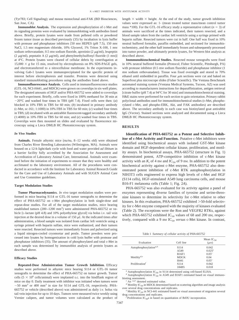
Table 1 Summary of cellular activity of PHA-665752
Evaluation Cell lineMean IC50
(�M)
Autophosphorylationa,b S114 0.042A549 0.025c
B16F1 0.05c
Motilityd,e MDCK 0.04H441 0.05c
Proliferationf S114 0.042184B5 0.018
a Autophosphorylation IC50 in S114 determined using cell-based ELISA.b Autophosphorylation IC50 in A549 and B16F1 estimated based on visual immuno-
blotting assessment.c An “*” denoted estimated value.d Motility IC50 in MDCK determined based on scattering algorithm and image analysis
over several drug concentrations and replicates.e Motility IC50 in NCI-441 estimated based on visual assessment of migration several
drug concentrations and replicates.f Proliferation IC50s in based on quantitation of BrDU incorporation.
7347
A c-MET INHIBITOR WITH ANTITUMOR ACTIVITY

�20-fold cellular selectivity for c-Met versus Ron kinase was dem-onstrated by evaluating PHA-665752 for its ability to inhibit Ronautophosphorylation in Chinese hamster ovary cells engineered toexpress Ron. Similarly, �50-fold cellular selectivity versus VEGFR2was demonstrated by evaluating PHA-665752 for its ability to inhibitVEGF-stimulated VEGFR2 phosphorylation in NIH3T3 cells engi-neered to express VEGFR2 (Table 2). These data indicate selectivityat concentrations used in cellular mechanistic studies and at levels ofcompound achieved in vivo (Table 2). Collectively, these data dem-onstrate that PHA-665752 is a potent and selective tool for theevaluation of c-Met-dependent cellular functions, signal transduction,and potentially its role in tumorigenesis.
Inhibition of c-Met kinase activity by PHA-665752 also correlatedclosely with inhibition of HGF- and c-Met-dependent functions suchas motility, invasion, and growth regulation. In the characterization ofthe role of Met and HGF in cell function, HGF has been demonstratedto cause: (a) epithelial-derived cells grown in tight islands to undergocell dissociation and scattering; (b) migration of cells across inter-stices in confluent cultures; (c) invasion and branching tubulogenesis
of epithelial and tumor cell lines; and (d) mitogenic properties inprimary epithelial cells (e.g., breast, hepatocytes, and lung) and cer-tain tumor cell types (36, 37). Consistent with these phenomena,PHA-665752 potently inhibited: (a) HGF-dependent cell scattering ofMDCK canine kidney epithelial cells; (b) HGF-dependent cell migra-tion in NCI-H441 lung carcinoma cells; (c) invasion and branchingmorphogenesis of RIE cells; and (d) HGF-dependent cell proliferation(BrdUrd incorporation) in 184B5 mammary epithelial cells and S114cells with IC50 values in the same range as those observed in kinaseassays (Table 1; Fig. 2, B, C, and E). In contrast, PHA-665752 had noeffect on cell viability in non-Met-expressing NIH3T3 mouse fibro-blasts at concentrations up to 18 �M.
Characterization of the Effect of PHA-665752 on Signal Trans-duction in Tumor Cells and Correlation with c-Met-DependentFunction. PHA-665752 was also evaluated for its effect on phospho-rylation of proteins known to transduce c-Met-dependent signals incell lines that exhibit c-Met-dependent functional properties. In eachof these cell lines, PHA-665752 was evaluated for its effect onconstitutive or HGF-stimulated cell morphology, cell growth, and
Fig. 2. Effects of PHA-665752 on (A) HGF-induced c-Met phosphorylation in A549 cells, (B)HGF-induced motility in MDCK cells, (C) HGF-induced migration in NCI-H441 cells, (D) mor-phology of GTL-16 cells, and (E) invasion/tubulo-genesis of RIE cells. A, A549 cells were exposed tothe indicated concentrations of PHA-665752 for4 h. Cells were then stimulated with HGF (50ng/ml), denoted by (�) for 10 min. c-Met proteinexpression and phosphorylation state were deter-mined after immunoprecipitation of the receptorfrom tumor lysate and subsequent immunoblotanalysis as described in “Materials and Methods.”B, MDCK cells were grown as small coloniesat low density and treated with HGF (50 ng/ml) inthe presence of various concentrations of PHA-665752. After an overnight incubation, cells inabsence or presence of HGF or PHA-665752 (0.1�M) were visualized via c-Met immunofluores-cence staining and microscopy. C, NCI-H441 cellswere grown to high density and a gap was intro-duced with a P200 pipette tip. Cells were thenstimulated to migrate across the gap with HGF (50ng/ml) in the presence of various concentrations ofPHA-665752. After an overnight incubation, cellsin absence or presence of HGF or PHA-665752(0.1 �M) were visualized via c-Met immunofluo-rescence staining and microscopy. D, GTL-16 cellswere cultured overnight in the presence and ab-sence of HGF (50 ng/ml) and PHA-665752 (0.1�M), visualized by light microscopy, and photo-graphed. E, RIE cells were added to Matrigel andgrew as small plugs. After 24 h, cells were stimu-lated with HGF (80 ng/ml) to invade and formtube-like structures in Matrigel in the presence orabsence of various concentrations of PHA-665752.
7348
A c-MET INHIBITOR WITH ANTITUMOR ACTIVITY

motogenic properties in monolayer as well as anchorage-independentgrowth in soft agar (summarized in Table 3). This analysis of c-Met-dependent signal transduction and functional response is focused onconcentrations of PHA-665752 (�0.1 �M in monolayer, 0.2 �M inagarose) that demonstrated: (a) complete inhibition of c-Met phos-phorylation (in monolayer and colonies in agarose); and (b) selectivityfor c-Met versus other kinases evaluated. PHA-665752 did not dem-onstrate an effect on cell growth or viability in c-Met-negative controlcells at 10-fold above the concentrations at which the analyses wereperformed (data not shown). [Negative control cells included cells inwhich inhibition of c-Met is predicted to have minimal consequences(e.g., NIH3T3-VEGFR2, rat-1-N-myc fibroblasts, and SK-BR-3breast)].
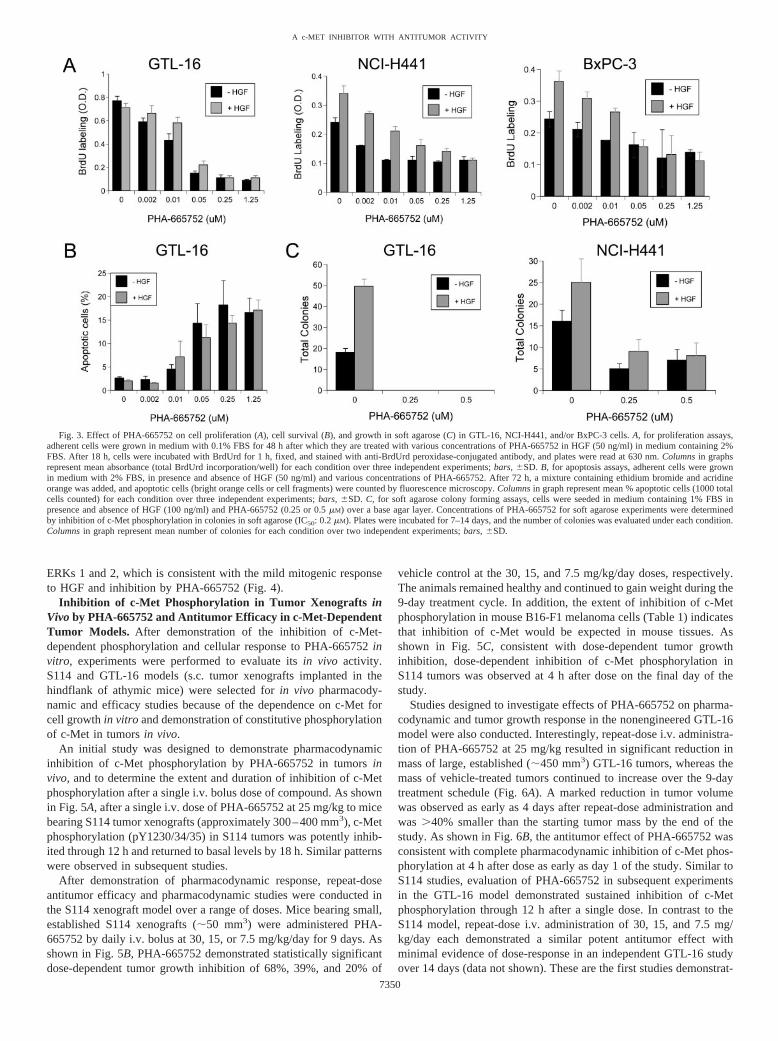
The cell lines evaluated included GTL-16 gastric carcinoma, NCI-H441 lung carcinoma, and BxPC-3 pancreatic carcinoma. GTL-16 isderived from a poorly differentiated gastric adenocarcinoma, whichcontains a c-Met gene locus amplified multiple times, resulting inexpression of high levels of constitutively active c-Met. In GTL-16cells, PHA-665752 completely inhibited growth in soft agar, inhibitedcell proliferation, and induced apoptosis in both the presence andabsence of HGF at concentrations that inhibited tyrosine phosphoryl-ation of c-Met (Fig. 3, A–C). Furthermore, treatment of GTL-16gastric carcinoma cells with PHA-665752 caused reversion from adedifferentiated, rounded morphology to a differentiated, flat mor-phology and resulted in increased cell-cell contact (Fig. 2D). PHA-665752 had no effect on GTL-16 cell migration in the presence orabsence of HGF. NCI-H441 is derived from a papillary lung adeno-carcinoma, expresses high levels of c-Met, and demonstrates consti-tutive phosphorylation of the c-Met RTK. In NCI-H441 cells, PHA-665752 inhibited growth in soft agar in both the presence and absence
of HGF, inhibited constitutive and HGF-stimulated cell proliferation,and inhibited HGF-stimulated migration at low nanomolar concentra-tions of PHA-665752 (Fig. 2C; Fig. 3, A–C). PHA-665752 had noeffect on cell morphology in NCI-H441 cells. BxPC-3 is derived froma pancreatic adenocarcinoma and expresses moderate levels of c-Met.In BxPC-3 cells, HGF induced only a weak mitogenic response inmonolayer and did not induce growth in soft agar, migration, ormorphogenesis. The weak HGF-dependent mitogenic response wasinhibited at low nanomolar concentrations of PHA-665752 (Fig. 3A).In contrast, PHA-665752 had no effect on any of the cell growth orproliferation endpoints in any of the c-Met negative control cell linesat the concentrations tested (i.e., NIH3T3-VEGFR2, rat-1-N-myc, andSK-BR-3).
As shown in Fig. 4, the effect of PHA-665752 on c-Met signaltransduction was also characterized in each of these cell lines. In eachcell line evaluated, constitutive or HGF-stimulated tyrosine phospho-rylation at the c-Met kinase domain (i.e., total pY, pY1230/34/35,pY1349, and pY1003) was completely inhibited by PHA-665752 (0.1�M). Also shown in Fig. 4, PHA-665752 potently inhibited totaltyrosine phosphorylation of Gab-1, a c-Met docking protein thatcontributes to Met-dependent signaling through PI3K, PLC-�, andCrkl in each cell line evaluated. In contrast, downstream signaltransduction proteins such as ERK, Akt, FAK, and STAT familymembers that regulate c-Met-dependent functions were affected in acell-type-specific manner by PHA-665752.
In GTL-16 cells, in which c-Met demonstrated a role in regulationof cell growth and morphology, PHA-665752 potently inhibited con-stitutive signaling through ERK, Akt, FAK, PLC-�, and STAT path-ways (Fig. 4). The constitutive phosphorylation of these signalingproteins in the absence of HGF together with inhibition of thesepathways by PHA-665752, a selective c-Met inhibitor, indicate thatthese pathways are dependent on the constitutive activity of c-Met inthis cell line. In addition, the down-regulation of multiple signalingpathways by PHA-665752 in GTL-16 is consistent with the pleiotro-pic effect of c-Met on the regulation of multiple cellular functions.
Similarly, in H441 cells, in which c-Met demonstrated a role in cellgrowth and motility, PHA-665752 potently inhibited constitutive sig-naling through the ERK, Akt, and FAK pathways, and partiallyinhibited PLC-� phosphorylation (Fig. 4). Similar to observations inGTL-16 cells, signaling molecules appeared to exhibit constitutiveactivity; however, addition of HGF did appear to additionally stimu-late FAK phosphorylation (Fig. 4). The ability of HGF to additionallyinduce c-Src-dependent FAK phosphorylation and cell motility inNCI-H441 is consistent with reports that this pathway is necessary formotility (38, 39). In addition, the down-regulation of ERK, Akt, andFAK pathways by PHA-665752 is consistent with regulation of bothcell growth and motility by c-Met in H441 cells. In contrast toGTL-16 and H441 cells, the only signaling molecules that weremarkedly modulated by HGF or PHA-665752 in BxPC-3 cells were
Table 2 PHA-665752 activity against c-Met compared with other kinases
KinaseKinase Family
and GroupaBiochemicalIC50 (�M)b
CellularIC50 (�M)c
c-Met Class X RTK 0.009 0.042Ron Class X RTK 0.068 0.9Flk-1 Class V RTK 0.2 2.5c-abl abl TK 1.4 NDd
FGFR1 Class IV RTK 3 NDEGFR Class I RTK 3.8 NDc-src src TK 6 NDIGF1R Class II RTK �10 NDPDGFR Class III RTK �10 NDAURORA2 AUR TK �10 NDPKA, PKB-� AGC STK �10 NDcdk 2, p38� CMGC STK �10 NDMK2,3 CAMK STK �10 ND
a Family and group names as follows: RTK, receptor tyrosine kinase, TK, tyrosinekinase; STK, serine-threonine kinase, AGC contains PKA, PKB, PKC families, CMGCcontains CDK, MAPK, GSK3; and CLK families, CAMK, Calcium/calmodulin-depend-ent protein kinases.
b Biochemical IC50 values based on quantitation of substrate phosphorylation byrecombinant enzyme.
c Cellular IC50 values estimated based on visual assessment of immunoblots ofreceptor tyrosine phosphorylation in cell lines engineered to express receptor of interest.
d ND, not determined.
Table 3 Summary of PHA-665752 effect on cell function and signaling
Cell line
Effect of PHA-665752 on Cell function
Effect of PHA-665752 onsignaling moleculesb
Colony growth(Soft-agar)
Cell proliferation(BrdUrd)
Cell survival(apoptosis)
Cell migration(scratch)a
Cellmorphology
GTL-16 Inhibitedc Inhibited Induced apoptosisd No effect Differentiatione Erk 1/2, Akt, FAK, PLC-�, STAT-3NCI-H441 Inhibitedc Inhibited No effect Inhibited No effect Erk 1/2, Akt, FAKBxPC-3 No colonies Inhibited No effect No effect No effect Erk-1/2a Measures HGF-stimulated migration across gap in monolayer.b Denotes inhibition of phosphorylation observed for protein listed in column.c Inhibition of number of colonies in soft agarose (colony: � 10 cells).d Compound induced apoptosis in presence and absence of HGF.e Differentiation describes flattened morphology and retained cell contact.
7349
A c-MET INHIBITOR WITH ANTITUMOR ACTIVITY
ERKs 1 and 2, which is consistent with the mild mitogenic responseto HGF and inhibition by PHA-665752 (Fig. 4).
Inhibition of c-Met Phosphorylation in Tumor Xenografts inVivo by PHA-665752 and Antitumor Efficacy in c-Met-DependentTumor Models. After demonstration of the inhibition of c-Met-dependent phosphorylation and cellular response to PHA-665752 invitro, experiments were performed to evaluate its in vivo activity.S114 and GTL-16 models (s.c. tumor xenografts implanted in thehindflank of athymic mice) were selected for in vivo pharmacody-namic and efficacy studies because of the dependence on c-Met forcell growth in vitro and demonstration of constitutive phosphorylationof c-Met in tumors in vivo.
An initial study was designed to demonstrate pharmacodynamicinhibition of c-Met phosphorylation by PHA-665752 in tumors invivo, and to determine the extent and duration of inhibition of c-Metphosphorylation after a single i.v. bolus dose of compound. As shownin Fig. 5A, after a single i.v. dose of PHA-665752 at 25 mg/kg to micebearing S114 tumor xenografts (approximately 300–400 mm3), c-Metphosphorylation (pY1230/34/35) in S114 tumors was potently inhib-ited through 12 h and returned to basal levels by 18 h. Similar patternswere observed in subsequent studies.
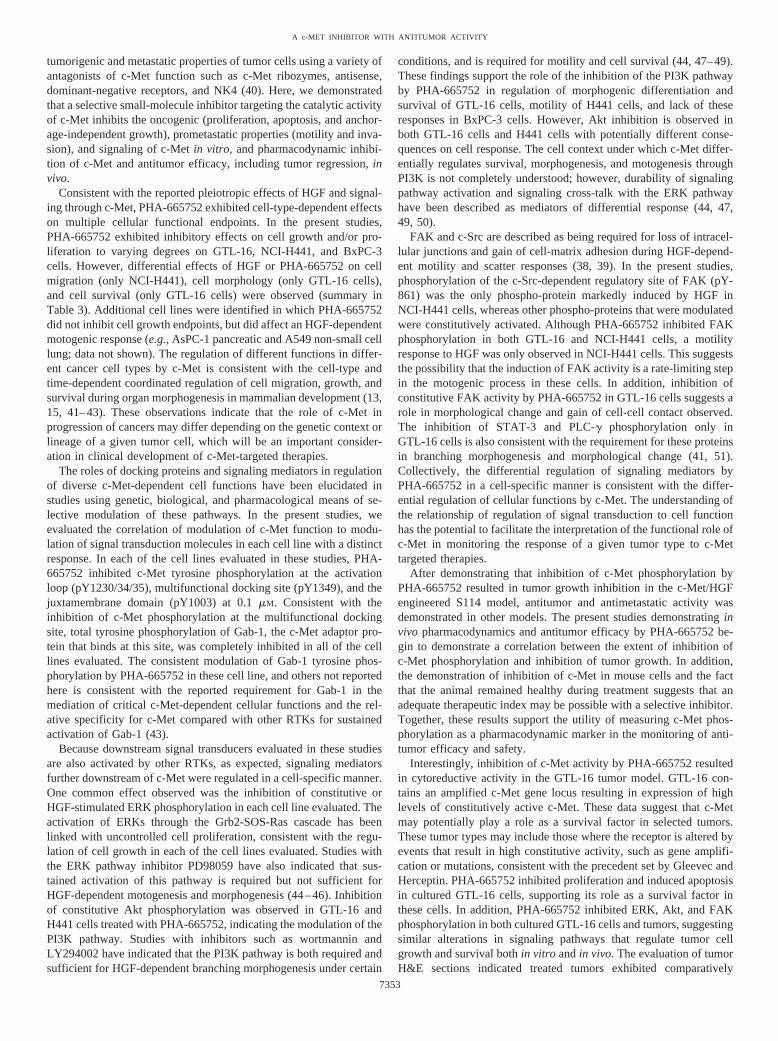
After demonstration of pharmacodynamic response, repeat-doseantitumor efficacy and pharmacodynamic studies were conducted inthe S114 xenograft model over a range of doses. Mice bearing small,established S114 xenografts (�50 mm3) were administered PHA-665752 by daily i.v. bolus at 30, 15, or 7.5 mg/kg/day for 9 days. Asshown in Fig. 5B, PHA-665752 demonstrated statistically significantdose-dependent tumor growth inhibition of 68%, 39%, and 20% of
vehicle control at the 30, 15, and 7.5 mg/kg/day doses, respectively.The animals remained healthy and continued to gain weight during the9-day treatment cycle. In addition, the extent of inhibition of c-Metphosphorylation in mouse B16-F1 melanoma cells (Table 1) indicatesthat inhibition of c-Met would be expected in mouse tissues. Asshown in Fig. 5C, consistent with dose-dependent tumor growthinhibition, dose-dependent inhibition of c-Met phosphorylation inS114 tumors was observed at 4 h after dose on the final day of thestudy.
Studies designed to investigate effects of PHA-665752 on pharma-codynamic and tumor growth response in the nonengineered GTL-16model were also conducted. Interestingly, repeat-dose i.v. administra-tion of PHA-665752 at 25 mg/kg resulted in significant reduction inmass of large, established (�450 mm3) GTL-16 tumors, whereas themass of vehicle-treated tumors continued to increase over the 9-daytreatment schedule (Fig. 6A). A marked reduction in tumor volumewas observed as early as 4 days after repeat-dose administration andwas �40% smaller than the starting tumor mass by the end of thestudy. As shown in Fig. 6B, the antitumor effect of PHA-665752 wasconsistent with complete pharmacodynamic inhibition of c-Met phos-phorylation at 4 h after dose as early as day 1 of the study. Similar toS114 studies, evaluation of PHA-665752 in subsequent experimentsin the GTL-16 model demonstrated sustained inhibition of c-Metphosphorylation through 12 h after a single dose. In contrast to theS114 model, repeat-dose i.v. administration of 30, 15, and 7.5 mg/kg/day each demonstrated a similar potent antitumor effect withminimal evidence of dose-response in an independent GTL-16 studyover 14 days (data not shown). These are the first studies demonstrat-
Fig. 3. Effect of PHA-665752 on cell proliferation (A), cell survival (B), and growth in soft agarose (C) in GTL-16, NCI-H441, and/or BxPC-3 cells. A, for proliferation assays,adherent cells were grown in medium with 0.1% FBS for 48 h after which they are treated with various concentrations of PHA-665752 in HGF (50 ng/ml) in medium containing 2%FBS. After 18 h, cells were incubated with BrdUrd for 1 h, fixed, and stained with anti-BrdUrd peroxidase-conjugated antibody, and plates were read at 630 nm. Columns in graphsrepresent mean absorbance (total BrdUrd incorporation/well) for each condition over three independent experiments; bars, �SD. B, for apoptosis assays, adherent cells were grownin medium with 2% FBS, in presence and absence of HGF (50 ng/ml) and various concentrations of PHA-665752. After 72 h, a mixture containing ethidium bromide and acridineorange was added, and apoptotic cells (bright orange cells or cell fragments) were counted by fluorescence microscopy. Columns in graph represent mean % apoptotic cells (1000 totalcells counted) for each condition over three independent experiments; bars, �SD. C, for soft agarose colony forming assays, cells were seeded in medium containing 1% FBS inpresence and absence of HGF (100 ng/ml) and PHA-665752 (0.25 or 0.5 �M) over a base agar layer. Concentrations of PHA-665752 for soft agarose experiments were determinedby inhibition of c-Met phosphorylation in colonies in soft agarose (IC50: 0.2 �M). Plates were incubated for 7–14 days, and the number of colonies was evaluated under each condition.Columns in graph represent mean number of colonies for each condition over two independent experiments; bars, �SD.
7350
A c-MET INHIBITOR WITH ANTITUMOR ACTIVITY

ing cytoreductive activity or tumor growth inhibition by a selectivesmall molecule c-Met inhibitor.
Immmunohistochemical Evaluation of c-Met Phosphorylation,Signal Transduction, and Tumor Pathobiology in GTL-16 Xe-nografts After Treatment with PHA-665752. After the observationof cytoreductive activity in the GTL-16 tumor model, studies were
performed to evaluate the ability of PHA-665752 to: (a) inhibit c-Metactivity and markers of mechanism and signal transduction at earlytime points; and (b) impact tumor pathobiology at later time points. Inthis evaluation, GTL-16 tumor-bearing mice were administered PHA-665752 for up to 8 days, and tumors from each treatment group (3mice/group) were removed and fixed at 4-h after dose at days 1, 2, 4,
Fig. 4. Effect of PHA-665752 on c-Met phos-phorylation and signal transduction in GTL-16,NCI-H441, and BxPC-3 cells. Cells were treatedwith PHA-665752 (0.1 �M) for 3 h and with HGF(50 ng/ml) for 10 or 30 min. Lysates were made forcells under each condition, and immunoblottingwas performed for each protein or phosphoproteinof interest using specific antibodies as described in“Materials and Methods.” Total Gab-1 was immu-noprecipitated from protein lysates before immu-noblotting. Representative blots from two inde-pendent experiments are shown in Fig. 4.
Fig. 5. Inhibition of c-Met phosphorylation (A and C)and tumor growth (B) in the S114 tumor xenograft model.A, athymic mice bearing S114 tumors (300–400 mm3)were given a single i.v. dose (25 mg/kg) of PHA-665752or vehicle. At several time points after administration,mice were euthanized, plasma and tumor samples werecollected, and lysates were prepared as described in “Ma-terials and Methods.” The relative abundance of phospho-c-Met [pY1230/34/35] and total c-Met in the tumor ly-sates was determined by immunoblot analysis. Additionalexperiments in S114 tumors with PHA-665752 exhibitedsimilar results. B, athymic mice bearing established (�50mm3) S114 tumors were administered daily i.v. doses ofPHA-665752 (7.5, 15, 30 mg/kg/day) or vehicle startingon day 7 through day 16. At least 10 mice were includedin each group. Results shown indicate mean tumor vol-ume; bars, �SE. C, on day 16 of the efficacy study in 6B,at 4 h after final administration of PHA-665752, micebearing S114 tumors (3/group) were euthanized, plasmaand tumor samples were collected, and lysates were pre-pared for analysis of phospho-c-Met (pY1230/34/35) andtotal c-Met by immunoblot analysis at each dose.
7351
A c-MET INHIBITOR WITH ANTITUMOR ACTIVITY

and 8 of treatment. As early as 4 h after the first administration ofPHA-665752, the inhibition of c-Met, ERK 1 and 2, Akt, and FAKphosphorylation was observed in fixed tumor sections by immuno-histochemistry compared with vehicle-treated controls, and effect wassustained throughout the treatment duration (Fig. 7). The inhibition of
the ERK 1/2 and Akt pathways is consistent with the reported role ofthese pathways in the regulation of tumor growth and apoptosis.
Because of these findings, and the inhibition of cell proliferationand induction of apoptosis observed in cultured GTL-16 cells, mark-ers of tumor cell proliferation (Ki67) and apoptosis (activatedcaspase-3) were evaluated at 4 h after dose on days 1–7. However,only regional (exterior tumor rim versus interior) inhibition of Ki67labeling and induction of activated caspase-3 was observed in GTL-16tumors at the time points evaluated, making it difficult to determinethe significance of these findings (data not shown). It is also possiblethat the narrow postdose time points selected in these studies were notappropriate to capture the optimal timing of these events. Evaluationof H&E sections indicated that PHA-665752-treated tumors over thedosing schedule exhibited increasingly marked necrotic regions in theinterior of the tumor with viable tissue on the outer rim as comparedwith vehicle-treated tumors (Fig. 7). These observations indicate thatextensive cell death occurred in the interior of the tumor throughoutthis study. However, the exact mechanism and/or timing of tumor celldeath and cytoreductive activity are not known.
DISCUSSION
The validity of therapeutic approaches to target RTKs that aredysregulated in human cancers are illustrated by the successes ofGleevec targeting bcr-abl in chronic myelogenous leukemia and c-Kitin gastrointestinal stromal tumors, Herceptin in HER-2 overexpress-ing breast cancers, and Iressa in select non-small cell lung cancers (1).The c-Met RTK has similar compelling evidence supporting its role incancer and, hence, development of therapies that inhibit c-Met func-tion. This supporting evidence includes: (a) the mutation, gene am-plification, or overexpression of c-Met in a variety of cancers; (b)tumorigenic and metastatic properties of cell lines engineered toexpress c-Met and HGF; (c) development of tumors of multipleorigins in c-Met and HGF transgenic animals; and (d) the inhibition of
Fig. 6. Evidence of cytoreductive activity (A) and inhibition of c-Met phosphorylation(B) in the GTL-16 gastric tumor xenograft model. A, athymic mice bearing largeestablished (�400 mm3) GTL-16 tumors were administered a daily i.v. dose of PHA-665752 (25 mg/kg/day) or vehicle starting on day 20 through day 28. At least 10 micewere included in each group. Results shown indicate mean tumor volume; bars, �SE. B,on day 28, at 4 h after final administration of PHA-665752, mice bearing GTL-16 tumors(3/group) were euthanized, tumor samples were collected, frozen, and pooled, and lysateswere prepared for analysis of phospho-c-Met (pY1230/34/35) and total c-Met by immu-noblot analysis.
Fig. 7. Immmunohistochemical evaluation of c-Met phosphorylation, signal transduction, and tumor pathobiology in GTL-16 xenografts after treatment with PHA-665752. Tumorsamples were collected from satellite groups from the GTL-16 study in Fig. 7 at 4 h after dose on days 1, 2, 4, and 8, fixed in neutral-buffered formalin for 24 h and placed in 70%ethanol. Fixed tumors were embedded in paraffin, cut into 4 mm sections, and baked on slides, and evaluated stained with H&E or evaluated for presence of each marker of interestas described in “Materials and Methods.” Slides from PHA-665752 and vehicle-treated groups were visually assessed for expression and distribution of proteins of interest by lightmicroscopy.
7352
A c-MET INHIBITOR WITH ANTITUMOR ACTIVITY
tumorigenic and metastatic properties of tumor cells using a variety ofantagonists of c-Met function such as c-Met ribozymes, antisense,dominant-negative receptors, and NK4 (40). Here, we demonstratedthat a selective small-molecule inhibitor targeting the catalytic activityof c-Met inhibits the oncogenic (proliferation, apoptosis, and anchor-age-independent growth), prometastatic properties (motility and inva-sion), and signaling of c-Met in vitro, and pharmacodynamic inhibi-tion of c-Met and antitumor efficacy, including tumor regression, invivo.
Consistent with the reported pleiotropic effects of HGF and signal-ing through c-Met, PHA-665752 exhibited cell-type-dependent effectson multiple cellular functional endpoints. In the present studies,PHA-665752 exhibited inhibitory effects on cell growth and/or pro-liferation to varying degrees on GTL-16, NCI-H441, and BxPC-3cells. However, differential effects of HGF or PHA-665752 on cellmigration (only NCI-H441), cell morphology (only GTL-16 cells),and cell survival (only GTL-16 cells) were observed (summary inTable 3). Additional cell lines were identified in which PHA-665752did not inhibit cell growth endpoints, but did affect an HGF-dependentmotogenic response (e.g., AsPC-1 pancreatic and A549 non-small celllung; data not shown). The regulation of different functions in differ-ent cancer cell types by c-Met is consistent with the cell-type andtime-dependent coordinated regulation of cell migration, growth, andsurvival during organ morphogenesis in mammalian development (13,15, 41–43). These observations indicate that the role of c-Met inprogression of cancers may differ depending on the genetic context orlineage of a given tumor cell, which will be an important consider-ation in clinical development of c-Met-targeted therapies.
The roles of docking proteins and signaling mediators in regulationof diverse c-Met-dependent cell functions have been elucidated instudies using genetic, biological, and pharmacological means of se-lective modulation of these pathways. In the present studies, weevaluated the correlation of modulation of c-Met function to modu-lation of signal transduction molecules in each cell line with a distinctresponse. In each of the cell lines evaluated in these studies, PHA-665752 inhibited c-Met tyrosine phosphorylation at the activationloop (pY1230/34/35), multifunctional docking site (pY1349), and thejuxtamembrane domain (pY1003) at 0.1 �M. Consistent with theinhibition of c-Met phosphorylation at the multifunctional dockingsite, total tyrosine phosphorylation of Gab-1, the c-Met adaptor pro-tein that binds at this site, was completely inhibited in all of the celllines evaluated. The consistent modulation of Gab-1 tyrosine phos-phorylation by PHA-665752 in these cell line, and others not reportedhere is consistent with the reported requirement for Gab-1 in themediation of critical c-Met-dependent cellular functions and the rel-ative specificity for c-Met compared with other RTKs for sustainedactivation of Gab-1 (43).
Because downstream signal transducers evaluated in these studiesare also activated by other RTKs, as expected, signaling mediatorsfurther downstream of c-Met were regulated in a cell-specific manner.One common effect observed was the inhibition of constitutive orHGF-stimulated ERK phosphorylation in each cell line evaluated. Theactivation of ERKs through the Grb2-SOS-Ras cascade has beenlinked with uncontrolled cell proliferation, consistent with the regu-lation of cell growth in each of the cell lines evaluated. Studies withthe ERK pathway inhibitor PD98059 have also indicated that sus-tained activation of this pathway is required but not sufficient forHGF-dependent motogenesis and morphogenesis (44–46). Inhibitionof constitutive Akt phosphorylation was observed in GTL-16 andH441 cells treated with PHA-665752, indicating the modulation of thePI3K pathway. Studies with inhibitors such as wortmannin andLY294002 have indicated that the PI3K pathway is both required andsufficient for HGF-dependent branching morphogenesis under certain
conditions, and is required for motility and cell survival (44, 47–49).These findings support the role of the inhibition of the PI3K pathwayby PHA-665752 in regulation of morphogenic differentiation andsurvival of GTL-16 cells, motility of H441 cells, and lack of theseresponses in BxPC-3 cells. However, Akt inhibition is observed inboth GTL-16 cells and H441 cells with potentially different conse-quences on cell response. The cell context under which c-Met differ-entially regulates survival, morphogenesis, and motogenesis throughPI3K is not completely understood; however, durability of signalingpathway activation and signaling cross-talk with the ERK pathwayhave been described as mediators of differential response (44, 47,49, 50).
FAK and c-Src are described as being required for loss of intracel-lular junctions and gain of cell-matrix adhesion during HGF-depend-ent motility and scatter responses (38, 39). In the present studies,phosphorylation of the c-Src-dependent regulatory site of FAK (pY-861) was the only phospho-protein markedly induced by HGF inNCI-H441 cells, whereas other phospho-proteins that were modulatedwere constitutively activated. Although PHA-665752 inhibited FAKphosphorylation in both GTL-16 and NCI-H441 cells, a motilityresponse to HGF was only observed in NCI-H441 cells. This suggeststhe possibility that the induction of FAK activity is a rate-limiting stepin the motogenic process in these cells. In addition, inhibition ofconstitutive FAK activity by PHA-665752 in GTL-16 cells suggests arole in morphological change and gain of cell-cell contact observed.The inhibition of STAT-3 and PLC-� phosphorylation only inGTL-16 cells is also consistent with the requirement for these proteinsin branching morphogenesis and morphological change (41, 51).Collectively, the differential regulation of signaling mediators byPHA-665752 in a cell-specific manner is consistent with the differ-ential regulation of cellular functions by c-Met. The understanding ofthe relationship of regulation of signal transduction to cell functionhas the potential to facilitate the interpretation of the functional role ofc-Met in monitoring the response of a given tumor type to c-Mettargeted therapies.
After demonstrating that inhibition of c-Met phosphorylation byPHA-665752 resulted in tumor growth inhibition in the c-Met/HGFengineered S114 model, antitumor and antimetastatic activity wasdemonstrated in other models. The present studies demonstrating invivo pharmacodynamics and antitumor efficacy by PHA-665752 be-gin to demonstrate a correlation between the extent of inhibition ofc-Met phosphorylation and inhibition of tumor growth. In addition,the demonstration of inhibition of c-Met in mouse cells and the factthat the animal remained healthy during treatment suggests that anadequate therapeutic index may be possible with a selective inhibitor.Together, these results support the utility of measuring c-Met phos-phorylation as a pharmacodynamic marker in the monitoring of anti-tumor efficacy and safety.
Interestingly, inhibition of c-Met activity by PHA-665752 resultedin cytoreductive activity in the GTL-16 tumor model. GTL-16 con-tains an amplified c-Met gene locus resulting in expression of highlevels of constitutively active c-Met. These data suggest that c-Metmay potentially play a role as a survival factor in selected tumors.These tumor types may include those where the receptor is altered byevents that result in high constitutive activity, such as gene amplifi-cation or mutations, consistent with the precedent set by Gleevec andHerceptin. PHA-665752 inhibited proliferation and induced apoptosisin cultured GTL-16 cells, supporting its role as a survival factor inthese cells. In addition, PHA-665752 inhibited ERK, Akt, and FAKphosphorylation in both cultured GTL-16 cells and tumors, suggestingsimilar alterations in signaling pathways that regulate tumor cellgrowth and survival both in vitro and in vivo. The evaluation of tumorH&E sections indicated treated tumors exhibited comparatively
7353
A c-MET INHIBITOR WITH ANTITUMOR ACTIVITY

marked necrotic regions on the interior of the tumor, indicating thatextensive cell death occurred in this region of the tumor duringPHA-665752 treatment. Collectively, these data support the role ofc-Met as a survival factor in selected tumors. Studies to furtherevaluate c-Met inhibitors in additional models of tumor growth andmetastasis, and the correlation of the extent and duration of inhibitionof c-Met phosphorylation to antitumor efficacy are warranted andongoing.
In summary, these studies illustrate the effects of a selective c-Metinhibitor, PHA-665752, as an inhibitor of a variety of c-Met-depend-ent functions and signaling events. These studies are the first demon-stration of inhibition of c-Met phosphorylation and antitumor efficacy,including cytoreductive activity, by a selective, small-molecule c-Metinhibitor. The diversity of response to PHA-665752 in a variety of celllines indicates the variety of roles that c-Met plays in different cellsand, putatively, tumor types. These results suggest that the role ofc-Met in tumorigenesis and progression of cancers may differ depend-ing on the genetic context of a given tumor cell. In addition, theunderstanding of the relationship of regulation of signal transductionto cell function has the potential to facilitate the interpretation of thefunctional role of c-Met in monitoring the response of a given tumortype to c-Met targeted therapies. The diverse role of c-Met in differenttumor types, as a regulator of tumor growth, survival, or metastasis,will be a critical issue in the clinical development of c-Met inhibitors,monitoring of patient response, and design of clinical studies.
ACKNOWLEDGMENTS
We thank the screening groups at Pharmacia Nerviano, Italy and Chester-field, MO for providing kinase selectivity screening data for PHA-665752. Wethank Dr. George VandeWoude and Van Andel Institute, Grand Rapids,Michigan, USA for supplying S114 cells. We also thank Dr. Paolo Comoglioand University Torino Medical School, Torino, Italy for the original supply ofGTL-16 cells.
REFERENCES
1. Laird, A. D., and Cherrington, J. M. Small molecule tyrosine kinase inhibitors:clinical development of anticancer agents. Expert Opin. Investig. Drugs, 21: 51–64,2003.
2. Naldini, L., Vigna, E., Narsimhan, R. P., Gaudino, G., Zarnegar, R., Michalopoulos,G. K., and Comoglio, P. M. Hepatocyte growth factor (HGF) stimulates the tyrosinekinase activity of the receptor encoded by the proto-oncogene c-MET. Oncogene, 6:501–504, 1991.
3. Bottaro, D. P., Rubin, J. S., Faletto, D. L., Chan, A. M., Kmiecik, T. E., VandeWoude, G. F., and Aaronson, S. A. Identification of the hepatocyte growth factorreceptor as the c-met proto-oncogene product. Science (Wash. DC), 251: 802–804,1991.
4. Naldini, L., Weidner, K. M., Vigna, E., Gaudino, G., Bardelli, A., Ponzetto, C.,Narsimhan, R. P., Hartmann, G., Zarnegar, R., and Michalopoulos, G. K. Scatterfactor and hepatocyte growth factor are indistinguishable ligands for MET receptor.EMBO J., 10: 2867–2878, 1991.
5. Rodrigues, G. A., and Park, M. Autophosphorylation modulates the kinase activityand oncogenic potential of the Met receptor tyrosine kinase. Oncogene, 9: 2019–2027, 1994.
6. Ponzetto, C., Bardelli, A., Zhen, Z., Maina, F., dalla Zonca, P., Giordano, S.,Graziani, A., Panayotou, G., and Comoglio, P. M. A multifunctional docking sitemediates signaling and transformation by the hepatocyte growth factor/scatter factorreceptor family. Cell, 77: 261–271, 1994.
7. Fournier, T. M., Lamorte, L., Maroun, C. R., Lupher, M., Band, H., Langdon, W., andPark, M. Cbl-transforming variants trigger a cascade of molecular alterations that leadto epithelial mesenchymal conversion. Mol. Biol. Cell, 11: 3397–3410, 2000.
8. Furge, K. A., Zhang, Y. W., and Vande Woude, G. F. Met receptor tyrosine kinase:enhanced signaling through adapter proteins. Oncogene, 19: 5582–5589, 2000.
9. Di Renzo, M. F., Narsimhan, R. P., Olivero, M., Bretti, S., Giordano, S., Medico, E.,Gaglia, P., Zara, P., and Comoglio, P. M. Expression of the Met/HGF receptor innormal and neoplastic human tissues. Oncogene, 6: 1997–2003, 1991.
10. Sonnenberg, E., Weidner, K. M., and Birchmeier, C. Expression of the met-receptorand its ligand, HGF-SF during mouse embryogenesis. Exs, 65: 381–394, 1993.
11. Comoglio, P. M., and Boccaccio, C. Scatter factors and invasive growth. Semin.Cancer Biol., 11: 153–165, 2001.
12. Jiang, W., Hiscox, S., Matsumoto, K., and Nakamura, T. Hepatocyte growth factor/scatter factor, its molecular, cellular and clinical implications in cancer. Crit. Rev.Oncol.-Hematol., 29: 209–248, 1999.
13. Bladt, F., Riethmacher, D., Isenmann, S., Aguzzi, A., and Birchmeier, C. Essentialrole for the c-met receptor in the migration of myogenic precursor cells into the limbbud. Nature (Lond.), 376: 768–771, 1995.
14. Schmidt, C., Bladt, F., Goedecke, S., Brinkmann, V., Zschiesche, W., Sharpe, M.,Gherardi, E., and Birchmeier, C. Scatter factor/hepatocyte growth factor is essentialfor liver development. Nature (Lond.), 373: 699–702, 1995.
15. Tsarfaty, I., Rong, S., Resau, J. H., Rulong, S., da Silva, P. P., and Vande Woude,G. F. The Met proto-oncogene mesenchymal to epithelial cell conversion. Science(Wash. DC), 263: 98–101, 1994.
16. Schmidt, L., Duh, F. M., Chen, F., Kishida, T., Glenn, G., Choyke, P., Scherer, S. W.,Zhuang, Z., Lubensky, I., Dean, M., Allikmets, R., Chidambaram, A., Bergerheim,U. R., Feltis, J. T., Casadevall, C., Zamarron, A., Bernues, M., Richard, S., Lips, C. J.,Walther, M. M., Tsui, L. C., Geil, L., Orcutt, M. L., Stackhouse, T., and Zbar, B.Germline and somatic mutations in the tyrosine kinase domain of the MET proto-oncogene in papillary renal carcinomas. Nat. Genet., 16: 68–73, 1997.
17. Lee, J. H., Han, S. U., Cho, H., Jennings, B., Gerrard, B., Dean, M., Schmidt, L., Zbar,B., and Vande Woude, G. F. A novel germ line juxtamembrane Met mutation inhuman gastric cancer. Oncogene, 19: 4947–4953, 2000.
18. Park, W. S., Dong, S. M., Kim, S. Y., Na, E. Y., Shin, M. S., Pi, J. H., Kim, B. J.,Bae, J. H., Hong, Y. K., Lee, K. S., Lee, S. H., Yoo, N. J., Jang, J. J., Pack, S.,Zhuang, Z., Schmidt, L., Zbar, B., and Lee, J. Y. Somatic mutations in the kinasedomain of the Met/hepatocyte growth factor receptor gene in childhood hepatocellularcarcinomas. Cancer Res., 59: 307–310, 1999.
19. Di Renzo, M. F., Olivero, M., Giacomini, A., Porte, H., Chastre, E., Mirossay, L.,Nordlinger, B., Bretti, S., Bottardi, S., and Giordano, S. Overexpression and ampli-fication of the met/HGF receptor gene during the progression of colorectal cancer.Clin. Cancer Res., 1: 147–154, 1995.
20. Di Renzo, M. F., Olivero, M., Martone, T., Maffe, A., Maggiora, P., Stefani, A. D.,Valente, G., Giordano, S., Cortesina, G., and Comoglio, P. M. Somatic mutations ofthe MET oncogene are selected during metastatic spread of human HNSC carcino-mas. Oncogene, 19: 1547–1555, 2000.
21. Bellusci, S., Moens, G., Gaudino, G., Comoglio, P., Nakamura, T., Thiery, J. P., andJouanneau, J. Creation of an hepatocyte growth factor/scatter factor autocrine loop incarcinoma cells induces invasive properties associated with increased tumorigenicity.Oncogene, 9: 1091–1099, 1994.
22. Jeffers, M., Rong, S., Anver, M., and Vande Woude, G. F. Autocrine hepatocytegrowth factor/scatter factor-Met signaling induces transformation and the invasive/metastastic phenotype in C127 cells. Oncogene, 13: 853–856, 1996.
23. Rong, S., Segal, S., Anver, M., Resau, J. H., and Vande Woude, G. F. Invasivenessand metastasis of NIH 3T3 cells induced by Met-hepatocyte growth factor/scatterfactor autocrine stimulation. Proc. Nat. Acad. Sci. USA, 91: 4731–4735, 1994.
24. Jeffers, M., Schmidt, L., Nakaigawa, N., Webb, C. P., Weirich, G., Kishida, T., Zbar,B., and Vande Woude, G. F. Activating mutations for the met tyrosine kinase receptorin human cancer. Proc. Nat. Acad. Sci. USA, 94: 11445–11450, 1997.
25. Rong, S., Bodescot, M., Blair, D., Dunn, J., Nakamura, T., Mizuno, K., Park, M.,Chan, A., Aaronson, S., and Vande Woude, G. F. Tumorigenicity of the metproto-oncogene and the gene for hepatocyte growth factor. Mol. Cell. Biol., 12:5152–5158, 1992.
26. Takayama, H., LaRochelle, W. J., Sharp, R., Otsuka, T., Kriebel, P., Anver, M.,Aaronson, S. A., and Merlino, G. Diverse tumorigenesis associated with aberrantdevelopment in mice overexpressing hepatocyte growth factor/scatter factor. Proc.Nat. Acad. Sci. USA, 94: 701–706, 1997.
27. Liang, T. J., Reid, A. E., Xavier, R., Cardiff, R. D., and Wang, T. C. Transgenicexpression of tpr-met oncogene leads to development of mammary hyperplasia andtumors. J. Clin. Investig., 97: 2872–2877, 1996.
28. Parr, C., Hiscox, S., Nakamura, T., Matsumoto, K., and Jiang, W. G. Nk4, a newHGF/SF variant, is an antagonist to the influence of HGF/SF on the motility andinvasion of colon cancer cells. Int. J. Cancer, 85: 563–570, 2000.
29. Michieli, P., Basilico, C., Pennacchietti, S., Maffe, A., Tamagnone, L., Giordano, S.,Bardelli, A., and Comoglio, P. M. Mutant Met-mediated transformation is ligand-dependent and can be inhibited by HGF antagonists. Oncogene, 18: 5221–5231, 1999.
30. Atabey, N., Gao, Y., Yao, Z. J., Breckenridge, D., Soon, L., Soriano, J. V., Burke,T. R., Jr., and Bottaro, D. P. Potent blockade of hepatocyte growth factor-stimulatedcell motility, matrix invasion and branching morphogenesis by antagonists of Grb2Src homology 2 domain interactions. J. Biol. Chem., 276: 14308–14314, 2001.
31. Abounader, R., Ranganathan, S., Lal, B., Fielding, K., Book, A., Dietz, H., Burger, P.,and Laterra, J. Reversion of human glioblastoma malignancy by U1 small nuclearRNA/ribozyme targeting of scatter factor/hepatocyte growth factor and c-met expres-sion. J. Natl. Cancer Inst., 91: 1548–1556, 1999.
32. Morotti, A., Mila, S., Accornero, P., Tagliabue, E., and Ponzetto, C. K252a inhibitsthe oncogenic properties of Met, the HGF receptor. Oncogene, 21: 4885–4893, 2002.
33. Rong, S., Oskarsson, M., Faletto, D., Tsarfaty, I., Resau, J. H., Nakamura, T., Rosen,E., Hopkins, R. F., 3rd, and Vande Woude, G. F. Tumorigenesis induced by coex-pression of human hepatocyte growth factor and the human met protooncogene leadsto high levels of expression of the ligand and receptor. Cell Growth Differ., 4:563–569, 1993.
34. Blake, R. A., Broome, M. A., Liu, X., Wu, J., Gishizky, M., Sun, L., and Courtneidge,S. A. SU6656, a selective src family kinase inhibitor, used to probe growth factorsignaling. Mol. Cell Biol., 20: 9019–9027, 2000.
35. Christensen, J. G., Schreck, R. E., Chan, E., Wang, X., Yang, C., Liu, L., Cui, J., Sun,L., Wei, J., Cherrington, J. M., and Mendel, D. B. High levels of HER-2 expressionalter the ability of epidermal growth factor receptor (EGFR) family tyrosine kinaseinhibitors to inhibit EGFR phosphorylation in vivo. Clin. Cancer Res., 7: 4230–4238,2001.
36. Stoker, M., Gherardi, E., Perryman, M., and Gray, J. Scatter factor is a fibroblast-derived modulator of epithelial cell mobility. Nature (Lond.), 327: 239–242, 1987.
7354
A c-MET INHIBITOR WITH ANTITUMOR ACTIVITY

37. Weidner, K. M., Sachs, M., and Birchmeier, W. The Met receptor tyrosine kinasetransduces motility, proliferation, and morphogenic signals of scatter factor/hepato-cyte growth factor in epithelial cells. J. Cell Biol., 121: 145–154, 1993.
38. Nakaigawa, N., Weirich, G., Schmidt, L., and Zbar, B. Tumorigenesis mediated byMET mutant M1268T is inhibited by dominant-negative Src. Oncogene, 19: 2996–3002, 2000.
39. Liu, Z. X., Yu, C. F., Nickel, C., Thomas, S., and Cantley, L. G. Hepatocyte growthfactor induces ERK-dependent paxillin phosphorylation and regulates paxillin-focaladhesion kinase association. J. Biol. Chem., 277: 10452–10458, 2002.
40. Maulik, G., Shrikhande, A., Kijima, T., Ma, P. C., Morrison, P. T., and Salgia, R.Role of the hepatocyte growth factor receptor, c-Met, in oncogenesis and potential fortherapeutic inhibition. Cytokine Growth Factor Rev., 13: 41–59, 2002.
41. Boccaccio, C., Ando, M., Tamagnone, L., Bardelli, A., Michieli, P., Battistini, C., andComoglio, P. M. Induction of epithelial tubules by growth factor HGF depends on theSTAT pathway. Nature (Lond.), 391: 285–288, 1998.
42. Sachs, M., Weidner, K. M., Brinkmann, V., Walther, I., Obermeier, A., Ullrich, A.,and Birchmeier, W. Motogenic and morphogenic activity of epithelial receptortyrosine kinases. J. Cell Biol., 133: 1095–1107, 1996.
43. Weidner, K. M., Di Cesare, S., Sachs, M., Brinkmann, V., Behrens, J., andBirchmeier, W. Interaction between Gab1 and the c-Met receptor tyrosine kinase isresponsible for epithelial morphogenesis. Nature (Lond.), 384: 173–176, 1996.
44. Khwaja, A., Lehmann, K., Marte, B. M., and Downward, J. Phosphoinositide 3-kinaseinduces scattering and tubulogenesis in epithelial cells through a novel pathway.J. Biol. Chem., 273: 18793–18801, 1998.
45. Tanimura, S., Nomura, K., Ozaki, K., Tsujimoto, M., Kondo, T., and Kohno, M.Prolonged nuclear retention of activated extracellular signal-regulated kinase 1/2 isrequired for hepatocyte growth factor-induced cell motility. J. Biol. Chem., 277:28256–28264, 2002.
46. Karihaloo, A., O’Rourke, D. A., Nickel, C., Spokes, K., and Cantley, L. G. Differ-ential MAPK pathways utilized for HGF- and EGF-dependent renal epithelial mor-phogenesis. J. Biol. Chem., 276: 9166–9173, 2001.
47. Potempa, S., and Ridley, A. J. Activation of both MAP kinase and phosphatidyli-nositide 3-kinase by Ras is required for hepatocyte growth factor/scatter factor-induced adherens junction disassembly. Mol. Biol. Cell, 9: 2185–2200, 1998.
48. Royal, I., Fournier, T. M., and Park, M. Differential requirement of Grb2 andPI3-kinase in HGF/SF-induced cell motility and tubulogenesis. J. Cell. Physiol., 173:196–201, 1997.
49. Xiao, G. H., Jeffers, M., Bellacosa, A., Mitsuuchi, Y., Vande Woude, G. F., andTesta, J. R. Anti-apoptotic signaling by hepatocyte growth factor/Met via the phos-phatidylinositol 3-kinase/Akt and mitogen-activated protein kinase pathways. Proc.Nat. Acad. Sci. USA, 98: 247–252, 2001.
50. Day, R. M., Cioce, V., Breckenridge, D., Castagnino, P., and Bottaro, D. P. Differ-ential signaling by alternative HGF isoforms through c-Met: activation of both MAPkinase and PI 3-kinase pathways is insufficient for mitogenesis. Oncogene, 18:3399–3406, 1999.
51. Gual, P., Giordano, S., Williams, T. A., Rocchi, S., Van Obberghen, E., andComoglio, P. M. Sustained recruitment of phospholipase C-� to Gab1 is required forHGF-induced branching tubulogenesis. Oncogene, 19: 1509–1518, 2000.
7355
A c-MET INHIBITOR WITH ANTITUMOR ACTIVITY
Copyright © 2022 FDOKUMEN