A role for cell sex in stem cell-mediated skeletal muscle regeneration: female cells have higher...
14
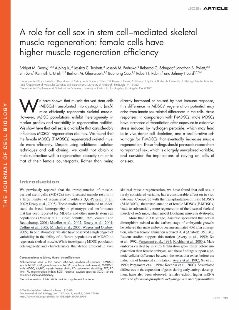
THE JOURNAL OF CELL BIOLOGY JCB: ARTICLE © The Rockefeller University Press $15.00 The Journal of Cell Biology, Vol. 177, No. 1, April 9, 2007 73–86 http://www.jcb.org/cgi/doi/10.1083/jcb.200612094 JCB 73 Introduction We previously reported that the transplantation of muscle- derived stem cells (MDSCs) into diseased muscle results in a large number of regenerated myofibers (Qu-Petersen et al., 2002; Deasy et al., 2005). These studies were initiated to under- stand the broad heterogeneity in phenotype and performance that has been reported for MDSCs and other muscle stem cell populations (Molnar et al., 1996; Schultz, 1996; Zammit and Beauchamp, 2001; Mueller et al., 2002; Deasy et al., 2004; Collins et al., 2005; Mitchell et al., 2005; Wagers and Conboy, 2005). In our laboratory, we also have observed a high degree of variability in the ability of different populations of MDSCs to regenerate skeletal muscle. While investigating MDSC population heterogeneity and characteristics that define efficient in vivo skeletal muscle regeneration, we have found that cell sex, a rarely considered variable, has a considerable effect on in vivo outcome. Compared with the transplantation of male MDSCs (M-MDSCs), the transplantation of female MDSCs (F-MDSCs) leads to substantially more regeneration of the diseased skeletal muscle of mdx mice, which model Duchenne muscular dystrophy. More than 2,000 yr ago, Aristotle speculated that sexual dimorphism existed at the earliest stage of embryonic growth; he believed that male embryos became animated 40 d after concep- tion, whereas female animation required 90 d (Aristotle, 350 BC). Recent studies support this notion (Avery et al., 1992; Xu et al., 1992; Pergament et al., 1994; Kochhar et al., 2003;). Male embryos created by in vitro fertilization grow faster before im- plantation than female embryos, and these findings support a ge- netic cellular difference between the sexes that exists before the induction of hormonal stimulation (Avery et al., 1992; Xu et al., 1992; Pergament et al., 1994; Kochhar et al., 2003). Sex-related differences in the expression of genes during early embryo develop- ment have also been observed: females exhibit higher mRNA levels of glucose-6-phosphate dehydrogenase and hypoxanthine A role for cell sex in stem cell–mediated skeletal muscle regeneration: female cells have higher muscle regeneration efficiency Bridget M. Deasy, 1,2,3 Aiping Lu, 3 Jessica C. Tebbets, 3 Joseph M. Feduska, 3 Rebecca C. Schugar, 3 Jonathan B. Pollett, 2,3 Bin Sun, 3 Kenneth L. Urish, 1,3 Burhan M. Gharaibeh, 2,3 Baohong Cao, 2,3 Robert T. Rubin, 5 and Johnny Huard 1,2,3,4 1 Department of Bioengineering, 2 Department of Orthopaedic Surgery, 3 Stem Cell Research Center, Children’s Hospital of Pittsburgh, University of Pittsburgh Medical Center, and 4 Department of Molecular Genetics and Biochemistry, University of Pittsburgh, Pittsburgh, PA 15260 5 Department of Psychiatry and Biobehavioral Sciences, University of California, Los Angeles, Los Angeles CA 90095 W e have shown that muscle-derived stem cells (MDSCs) transplanted into dystrophic (mdx) mice efficiently regenerate skeletal muscle. However, MDSC populations exhibit heterogeneity in marker profiles and variability in regeneration abilities. We show here that cell sex is a variable that considerably influences MDSCs’ regeneration abilities. We found that the female MDSCs (F-MDSCs) regenerated skeletal mus- cle more efficiently. Despite using additional isolation techniques and cell cloning, we could not obtain a male subfraction with a regeneration capacity similar to that of their female counterparts. Rather than being directly hormonal or caused by host immune response, this difference in MDSCs’ regeneration potential may arise from innate sex-related differences in the cells’ stress responses. In comparison with F-MDSCs, male MDSCs have increased differentiation after exposure to oxidative stress induced by hydrogen peroxide, which may lead to in vivo donor cell depletion, and a proliferative ad- vantage for F-MDSCs that eventually increases muscle regeneration. These findings should persuade researchers to report cell sex, which is a largely unexplored variable, and consider the implications of relying on cells of one sex. Correspondence to Johnny Huard: [email protected] Abbreviations used in this paper: ANOVA, analysis of variance; F-MDSC, female MDSC; GM, growth medium; MDSC, muscle-derived stem cell; M-MDSC, male MDSC; MyHC, myosin heavy chain; PD, population doubling; PDT, PD time; RI, regeneration index; ROS, reactive oxygen species; SCID, severe combined immunodeficiency. The online version of this article contains supplemental material.
Transcript of A role for cell sex in stem cell-mediated skeletal muscle regeneration: female cells have higher...
TH
EJ
OU
RN
AL
OF
CE
LL
BIO
LO
GY
JCB: ARTICLE
© The Rockefeller University Press $15.00The Journal of Cell Biology, Vol. 177, No. 1, April 9, 2007 73–86http://www.jcb.org/cgi/doi/10.1083/jcb.200612094
JCB 73
IntroductionWe previously reported that the transplantation of muscle-
derived stem cells (MDSCs) into diseased muscle results in
a large number of regenerated myofi bers (Qu-Petersen et al.,
2002; Deasy et al., 2005). These studies were initiated to under-
stand the broad heterogeneity in phenotype and performance
that has been reported for MDSCs and other muscle stem cell
populations (Molnar et al., 1996; Schultz, 1996; Zammit and
Beauchamp, 2001; Mueller et al., 2002; Deasy et al., 2004;
Collins et al., 2005; Mitchell et al., 2005; Wagers and Conboy,
2005). In our laboratory, we also have observed a high degree of
variability in the ability of different populations of MDSCs to
regenerate skeletal muscle. While investigating MDSC population
heterogeneity and characteristics that defi ne effi cient in vivo
skeletal muscle regeneration, we have found that cell sex, a
rarely considered variable, has a considerable effect on in vivo
outcome. Compared with the transplantation of male MDSCs
(M-MDSCs), the transplantation of female MDSCs (F-MDSCs)
leads to substantially more regeneration of the diseased skeletal
muscle of mdx mice, which model Duchenne muscular dystrophy.
More than 2,000 yr ago, Aristotle speculated that sexual
dimorphism existed at the earliest stage of embryonic growth;
he believed that male embryos became animated 40 d after concep-
tion, whereas female animation required 90 d (Aristotle, 350 BC).
Recent studies support this notion (Avery et al., 1992; Xu
et al., 1992; Pergament et al., 1994; Kochhar et al., 2003;). Male
embryos created by in vitro fertilization grow faster before im-
plantation than female embryos, and these fi ndings support a ge-
netic cellular difference between the sexes that exists before the
induction of hormonal stimulation (Avery et al., 1992; Xu et al.,
1992; Pergament et al., 1994; Kochhar et al., 2003). Sex-related
differences in the expression of genes during early embryo develop-
ment have also been observed: females exhibit higher mRNA
levels of glucose-6-phosphate dehydrogenase and hypoxanthine
A role for cell sex in stem cell–mediated skeletal muscle regeneration: female cells have higher muscle regeneration effi ciency
Bridget M. Deasy,1,2,3 Aiping Lu,3 Jessica C. Tebbets,3 Joseph M. Feduska,3 Rebecca C. Schugar,3 Jonathan B. Pollett,2,3
Bin Sun,3 Kenneth L. Urish,1,3 Burhan M. Gharaibeh,2,3 Baohong Cao,2,3 Robert T. Rubin,5 and Johnny Huard1,2,3,4
1Department of Bioengineering, 2Department of Orthopaedic Surgery, 3Stem Cell Research Center, Children’s Hospital of Pittsburgh, University of Pittsburgh Medical Center, and 4Department of Molecular Genetics and Biochemistry, University of Pittsburgh, Pittsburgh, PA 15260
5Department of Psychiatry and Biobehavioral Sciences, University of California, Los Angeles, Los Angeles CA 90095
We have shown that muscle-derived stem cells
(MDSCs) transplanted into dystrophic (mdx)
mice effi ciently regenerate skeletal muscle.
However, MDSC populations exhibit heterogeneity in
marker profi les and variability in regeneration abilities.
We show here that cell sex is a variable that considerably
infl uences MDSCs’ regeneration abilities. We found that
the female MDSCs (F-MDSCs) regenerated skeletal mus-
cle more effi ciently. Despite using additional isolation
techniques and cell cloning, we could not obtain a
male subfraction with a regeneration capacity similar to
that of their female counterparts. Rather than being
directly hormonal or caused by host immune response,
this difference in MDSCs’ regeneration potential may
arise from innate sex-related differences in the cells’ stress
responses. In comparison with F-MDSCs, male MDSCs
have increased differentiation after exposure to oxidative
stress induced by hydrogen peroxide, which may lead
to in vivo donor cell depletion, and a proliferative ad-
vantage for F-MDSCs that eventually increases muscle
regeneration. These fi ndings should persuade researchers
to report cell sex, which is a largely unexplored variable,
and consider the implications of relying on cells of
one sex.
Correspondence to Johnny Huard: [email protected]
Abbreviations used in this paper: ANOVA, analysis of variance; F-MDSC, female MDSC; GM, growth medium; MDSC, muscle-derived stem cell; M-MDSC, male MDSC; MyHC, myosin heavy chain; PD, population doubling; PDT, PD time; RI, regeneration index; ROS, reactive oxygen species; SCID, severe combined immunodefi ciency.
The online version of this article contains supplemental material.
JCB • VOLUME 177 • NUMBER 1 • 2007 74
phosphoribosyl transferase, which are two genes involved in the
detoxifi cation of reactive oxygen species (ROS; Gutierrez-Adan
et al., 2000; Peippo et al., 2002). Male and female embryonic
neurons (isolated from rats before gonad differentiation or hor-
monal stimulation) also display different cellular responses. The
female cells are more sensitive to apoptosis-inducing agents,
whereas male neurons are more sensitive to ischemia and nitro-
sative stress, and they cannot maintain the proper level of glutathi-
one, which regulates ROS levels (Du et al., 2004).
Few studies have investigated whether sex-related differ-
ences affect tissue or organ regeneration by progenitor cells.
Blankenhorn et al. (2003) demonstrated that the regrowth of car-
tilage, skin, and hair follicles in an ear pinna wound occurred
faster and more completely in female mice as compared with
male mice. Another study has shown that female mice modeling
living donor liver transplantation with ischemia and reperfusion
exhibit more effi cient liver regeneration than their male counter-
parts (Harada et al., 2003). Similarly, male rats show more tissue
growth than female rats after nephroctomy (Mulroney et al.,
1999). Chau et al. (2002) found that blocking antiapoptotic ac-
tivity in male animals resulted in sex-related differences in the
animals’ survival after endotoxic stress; the males recovered, but
there was no advantage for females. Finally, in a study of human
hematopoietic stem cell transplantations, superior survival was
observed with maternally donated recipients as compared with
recipients of paternal transplantations (Tamaki et al., 2001).
Several theories have been designed to unify cell and tissue
aging with the overall aging of organisms and, thereby, explain
females’ longer life spans. Telomeres, whose length is believed to
act as the mitotic clock of cells, are shorter in older males than in
older females, which suggests that male cells undergo more rounds
of division than female cells (Aviv et al., 2005; Vina et al., 2005).
This fi nding supports a possible link between growth rate and
aging (Rollo, 2002). Stindl (2004) has proposed that cells that have
fi nite replicative lifespans must undergo more rounds of division
to build the larger bodies of male organisms. Other research has
shown that estrogen stimulates telomerase, which slows the rate of
telomere attrition (for review see Aviv, 2002). This fi nding has ties
with the free radical theory of cell aging, according to which ROS
causes aging by damaging DNA, lipids, and proteins. Estrogens
also up-regulate pathways that induce the expression of antioxi-
dants (such as glutathione peroxidase) that reduce damage by ROS
(for example, the MAPK pathway; Vina et al., 2005). Together,
these fi ndings suggest that sex-related differences in the health of
the stem cell compartment could partially explain different rates of
aging and disease. Stem and progenitor cells are believed to persist
throughout life and contribute to the repair and maintenance of
tissue. Therefore, investigations of sex-related differences shown
by stem cells, as presented in this study, could lead to an improved
understanding of sex-related differences in aging and disease.
ResultsMDSC isolation and in vitro characterizationTo determine whether any of the standard markers for MDSC
characterization are predictive of high in vivo muscle regeneration,
we examined 25 populations of MDSCs in terms of fi ve variables:
in vivo muscle regeneration effi ciency, expression of CD34,
expression of Sca-1, expression of desmin, and cell sex. Analysis
of these variables revealed a large degree of heterogeneity in the
MDSC populations. The distribution of the populations’ regen-
eration indexes (RIs) and CD34, Sca-1, and desmin expression
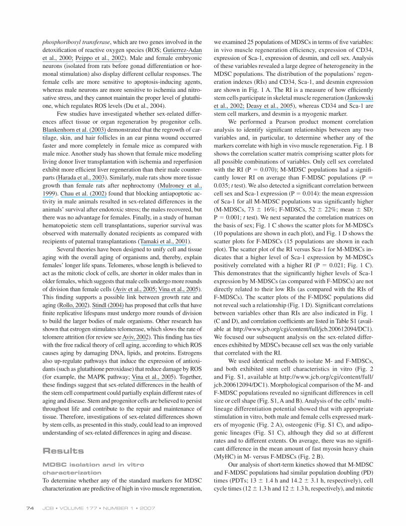
are shown in Fig. 1 A. The RI is a measure of how effi ciently
stem cells participate in skeletal muscle regeneration (Jankowski
et al., 2002; Deasy et al., 2005), whereas CD34 and Sca-1 are
stem cell markers, and desmin is a myogenic marker.
We performed a Pearson product moment correlation
analysis to identify signifi cant relationships between any two
variables and, in particular, to determine whether any of the
markers correlate with high in vivo muscle regeneration. Fig. 1 B
shows the correlation scatter matrix comprising scatter plots for
all possible combinations of variables. Only cell sex correlated
with the RI (P = 0.070); M-MDSC populations had a signifi -
cantly lower RI on average than F-MDSC populations (P =
0.035; t test). We also detected a signifi cant correlation between
cell sex and Sca-1 expression (P = 0.014): the mean expression
of Sca-1 for all M-MDSC populations was signifi cantly higher
(M-MDSCs, 73 ± 16%; F-MDSCs, 52 ± 22%; mean ± SD;
P = 0.001; t test). We next separated the correlation matrices on
the basis of sex; Fig. 1 C shows the scatter plots for M-MDSCs
(10 populations are shown in each plot), and Fig. 1 D shows the
scatter plots for F-MDSCs (15 populations are shown in each
plot). The scatter plot of the RI versus Sca-1 for M-MDSCs in-
dicates that a higher level of Sca-1 expression by M-MDSCs
positively correlated with a higher RI (P = 0.021; Fig. 1 C).
This demonstrates that the signifi cantly higher levels of Sca-1
expression by M-MDSCs (as compared with F-MDSCs) are not
directly related to their low RIs (as compared with the RIs of
F-MDSCs). The scatter plots of the F-MDSC populations did
not reveal such a relationship (Fig. 1 D). Signifi cant correlations
between variables other than RIs are also indicated in Fig. 1
(C and D), and correlation coeffi cients are listed in Table S1 (avail-
able at http://www.jcb.org/cgi/content/full/jcb.200612094/DC1).
We focused our subsequent analysis on the sex-related differ-
ences exhibited by MDSCs because cell sex was the only variable
that correlated with the RI.
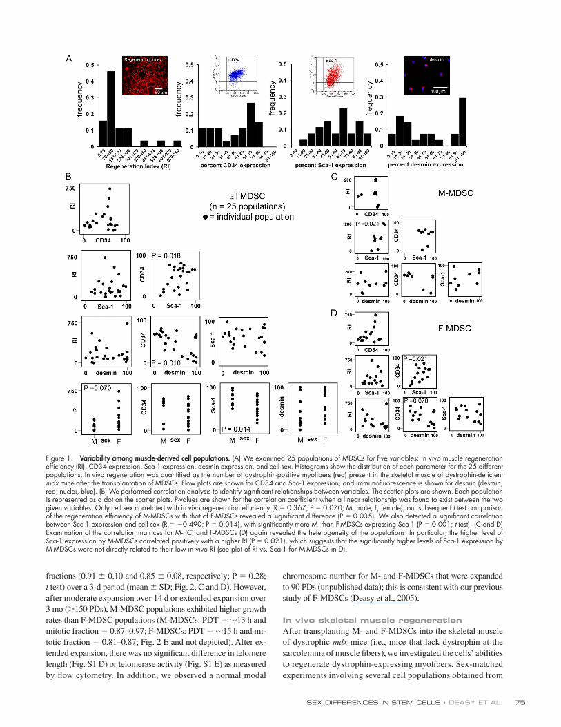
We used identical methods to isolate M- and F-MDSCs,
and both exhibited stem cell characteristics in vitro (Fig. 2
and Fig. S1, available at http://www.jcb.org/cgi/content/full/
jcb.200612094/DC1). Morphological comparison of the M- and
F-MDSC populations revealed no signifi cant differences in cell
size or cell shape (Fig. S1, A and B). Analysis of the cells’ multi-
lineage differentiation potential showed that with appropriate
stimulation in vitro, both male and female cells expressed mark-
ers of myogenic (Fig. 2 A), osteogenic (Fig. S1 C), and adipo-
genic lineages (Fig. S1 C), although they did so at different
rates and to different extents. On average, there was no signifi -
cant difference in the mean amount of fast myosin heavy chain
(MyHC) in M- versus F-MDSCs (Fig. 2 B).
Our analysis of short-term kinetics showed that M-MDSC
and F-MDSC populations had similar population doubling (PD)
times (PDTs; 13 ± 1.4 h and 14.2 ± 3.1 h, respectively), cell
cycle times (12 ± 1.3 h and 12 ± 1.3 h, respectively), and mitotic
SEX DIFFERENCES IN STEM CELLS • DEASY ET AL. 75
fractions (0.91 ± 0.10 and 0.85 ± 0.08, respectively; P = 0.28;
t test) over a 3-d period (mean ± SD; Fig. 2, C and D). However,
after moderate expansion over 14 d or extended expansion over
3 mo (>150 PDs), M-MDSC populations exhibited higher growth
rates than F-MDSC populations (M-MDSCs: PDT = �13 h and
mitotic fraction = 0.87–0.97; F-MDSCs: PDT = �15 h and mi-
totic fraction = 0.81–0.87; Fig. 2 E and not depicted). After ex-
tended expansion, there was no signifi cant difference in telomere
length (Fig. S1 D) or telomerase activity (Fig. S1 E) as measured
by fl ow cytometry. In addition, we observed a normal modal
chromosome number for M- and F-MDSCs that were expanded
to 90 PDs (unpublished data); this is consistent with our previous
study of F-MDSCs (Deasy et al., 2005).
In vivo skeletal muscle regenerationAfter transplanting M- and F-MDSCs into the skeletal muscle
of dystrophic mdx mice (i.e., mice that lack dystrophin at the
sarcolemma of muscle fi bers), we investigated the cells’ abilities
to regenerate dystrophin-expressing myofi bers. Sex-matched
experiments involving several cell populations obtained from
Figure 1. Variability among muscle-derived cell populations. (A) We examined 25 populations of MDSCs for fi ve variables: in vivo muscle regeneration effi ciency (RI), CD34 expression, Sca-1 expression, desmin expression, and cell sex. Histograms show the distribution of each parameter for the 25 different populations. In vivo regeneration was quantifi ed as the number of dystrophin-positive myofi bers (red) present in the skeletal muscle of dystrophin-defi cient mdx mice after the transplantation of MDSCs. Flow plots are shown for CD34 and Sca-1 expression, and immunofl uorescence is shown for desmin (desmin, red; nuclei, blue). (B) We performed correlation analysis to identify signifi cant relationships between variables. The scatter plots are shown. Each population is represented as a dot on the scatter plots. P-values are shown for the correlation coeffi cient when a linear relationship was found to exist between the two given variables. Only cell sex correlated with in vivo regeneration effi ciency (R = 0.367; P = 0.070; M, male; F, female); our subsequent t test comparison of the regeneration effi ciency of M-MDSCs with that of F-MDSCs revealed a signifi cant difference (P = 0.035). We also detected a signifi cant correlation between Sca-1 expression and cell sex (R = −0.490; P = 0.014), with signifi cantly more M- than F-MDSCs expressing Sca-1 (P = 0.001; t test). (C and D) Examination of the correlation matrices for M- (C) and F-MDSCs (D) again revealed the heterogeneity of the populations. In particular, the higher level of Sca-1 expression by M-MDSCs correlated positively with a higher RI (P = 0.021), which suggests that the signifi cantly higher levels of Sca-1 expression by M-MDSCs were not directly related to their low in vivo RI (see plot of RI vs. Sca-1 for M-MDSCs in D).
JCB • VOLUME 177 • NUMBER 1 • 2007 76
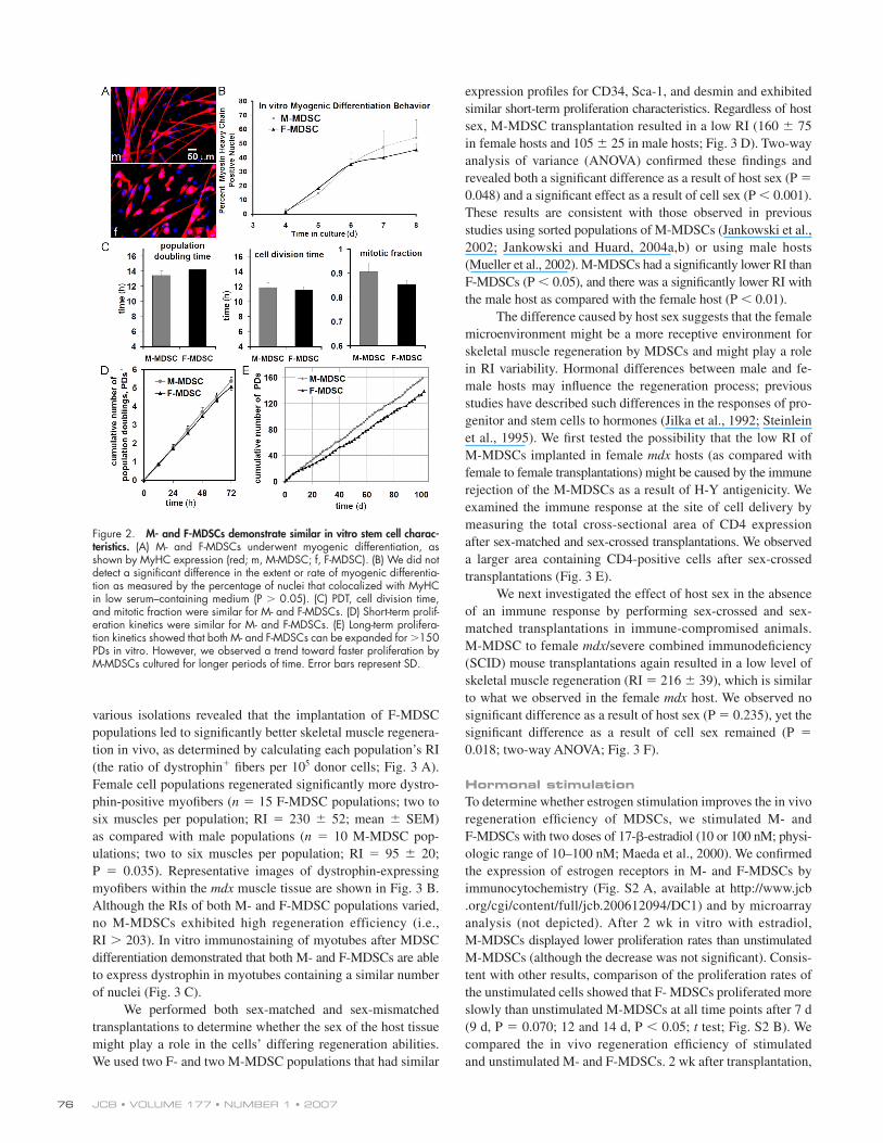
various isolations revealed that the implantation of F-MDSC
populations led to signifi cantly better skeletal muscle regenera-
tion in vivo, as determined by calculating each population’s RI
(the ratio of dystrophin+ fi bers per 105 donor cells; Fig. 3 A).
Female cell populations regenerated signifi cantly more dystro-
phin-positive myofi bers (n = 15 F-MDSC populations; two to
six muscles per population; RI = 230 ± 52; mean ± SEM)
as compared with male populations (n = 10 M-MDSC pop-
ulations; two to six muscles per population; RI = 95 ± 20;
P = 0.035). Representative images of dystrophin-expressing
myofi bers within the mdx muscle tissue are shown in Fig. 3 B.
Although the RIs of both M- and F-MDSC populations varied,
no M-MDSCs exhibited high regeneration efficiency (i.e.,
RI > 203). In vitro immunostaining of myotubes after MDSC
differentiation demonstrated that both M- and F-MDSCs are able
to express dystrophin in myotubes containing a similar number
of nuclei (Fig. 3 C).
We performed both sex-matched and sex-mismatched
transplantations to determine whether the sex of the host tissue
might play a role in the cells’ differing regeneration abilities.
We used two F- and two M-MDSC populations that had similar
expression profi les for CD34, Sca-1, and desmin and exhibited
similar short-term proliferation characteristics. Regardless of host
sex, M-MDSC transplantation resulted in a low RI (160 ± 75
in female hosts and 105 ± 25 in male hosts; Fig. 3 D). Two-way
analysis of variance (ANOVA) confi rmed these fi ndings and
revealed both a signifi cant difference as a result of host sex (P =
0.048) and a signifi cant effect as a result of cell sex (P < 0.001).
These results are consistent with those observed in previous
studies using sorted populations of M-MDSCs (Jankowski et al.,
2002; Jankowski and Huard, 2004a,b) or using male hosts
(Mueller et al., 2002). M-MDSCs had a signifi cantly lower RI than
F-MDSCs (P < 0.05), and there was a signifi cantly lower RI with
the male host as compared with the female host (P < 0.01).
The difference caused by host sex suggests that the female
microenvironment might be a more receptive environment for
skeletal muscle regeneration by MDSCs and might play a role
in RI variability. Hormonal differences between male and fe-
male hosts may infl uence the regeneration process; previous
studies have described such differences in the responses of pro-
genitor and stem cells to hormones (Jilka et al., 1992; Steinlein
et al., 1995). We fi rst tested the possibility that the low RI of
M-MDSCs implanted in female mdx hosts (as compared with
female to female transplantations) might be caused by the immune
rejection of the M-MDSCs as a result of H-Y antigenicity. We
examined the immune response at the site of cell delivery by
measuring the total cross-sectional area of CD4 expression
after sex-matched and sex-crossed transplantations. We observed
a larger area containing CD4-positive cells after sex-crossed
transplantations (Fig. 3 E).
We next investigated the effect of host sex in the absence
of an immune response by performing sex-crossed and sex-
matched transplantations in immune-compromised animals.
M-MDSC to female mdx/severe combined immunodefi ciency
(SCID) mouse transplantations again resulted in a low level of
skeletal muscle regeneration (RI = 216 ± 39), which is similar
to what we observed in the female mdx host. We observed no
signifi cant difference as a result of host sex (P = 0.235), yet the
signifi cant difference as a result of cell sex remained (P =
0.018; two-way ANOVA; Fig. 3 F).
Hormonal stimulationTo determine whether estrogen stimulation improves the in vivo
regeneration effi ciency of MDSCs, we stimulated M- and
F-MDSCs with two doses of 17-β-estradiol (10 or 100 nM; physi-
ologic range of 10–100 nM; Maeda et al., 2000). We confi rmed
the expression of estrogen receptors in M- and F-MDSCs by
immunocytochemistry (Fig. S2 A, available at http://www.jcb
.org/cgi/content/full/jcb.200612094/DC1) and by microarray
analysis (not depicted). After 2 wk in vitro with estradiol,
M-MDSCs displayed lower proliferation rates than unstimulated
M-MDSCs (although the decrease was not signifi cant). Consis-
tent with other results, comparison of the proliferation rates of
the unstimulated cells showed that F- MDSCs proliferated more
slowly than unstimulated M-MDSCs at all time points after 7 d
(9 d, P = 0.070; 12 and 14 d, P < 0.05; t test; Fig. S2 B). We
compared the in vivo regeneration effi ciency of stimulated
and unstimulated M- and F-MDSCs. 2 wk after transplantation,
Figure 2. M- and F-MDSCs demonstrate similar in vitro stem cell charac-teristics. (A) M- and F-MDSCs underwent myogenic differentiation, as shown by MyHC expression (red; m, M-MDSC; f, F-MDSC). (B) We did not detect a signifi cant difference in the extent or rate of myogenic differentia-tion as measured by the percentage of nuclei that colocalized with MyHC in low serum–containing medium (P > 0.05). (C) PDT, cell division time, and mitotic fraction were similar for M- and F-MDSCs. (D) Short-term prolif-eration kinetics were similar for M- and F-MDSCs. (E) Long-term prolifera-tion kinetics showed that both M- and F-MDSCs can be expanded for >150 PDs in vitro. However, we observed a trend toward faster proliferation by M-MDSCs cultured for longer periods of time. Error bars represent SD.
SEX DIFFERENCES IN STEM CELLS • DEASY ET AL. 77
F-MDSCs cultured in control conditions of 0 nM estradiol
had a higher RI than similarly cultured M-MDSCs (557 ± 229 vs.
115 ± 36; P = 0.06). Stimulation with estradiol had no signifi cant
effect on the RIs of M-MDSCs; however a trend toward a de-
creased RI was detected in F-MDSCs stimulated with 10 or 100 nM
estradiol (P = 0.09 and P = 0.07; n = 3; t test; Fig. S2 C).
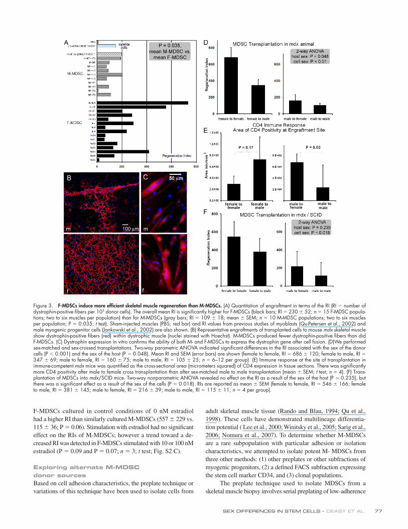
Exploring alternate M-MDSC donor sourcesBased on cell adhesion characteristics, the preplate technique or
variations of this technique have been used to isolate cells from
adult skeletal muscle tissue (Rando and Blau, 1994; Qu et al.,
1998). These cells have demonstrated multilineage differentia-
tion potential ( Lee et al., 2000; Winitsky et al., 2005; Sarig et al.,
2006; Nomura et al., 2007). To determine whether M-MDSCs
are a rare subpopulation with particular adhesion or isolation
characteristics, we attempted to isolate potent M- MDSCs from
three other methods: (1) other preplates or other subfractions of
myogenic progenitors, (2) a defi ned FACS subfraction expressing
the stem cell marker CD34, and (3) clonal populations.
The preplate technique used to isolate MDSCs from a
skeletal muscle biopsy involves serial preplating of low-adherence
Figure 3. F-MDSCs induce more effi cient skeletal muscle regeneration than M-MDSCs. (A) Quantitation of engraftment in terms of the RI (RI = number of dystrophin-positive fi bers per 105 donor cells). The overall mean RI is signifi cantly higher for F-MDSCs (black bars; RI = 230 ± 52; n = 15 F-MDSC popula-tions; two to six muscles per population) than for M-MDSCs (gray bars; RI = 109 ± 18; mean ± SEM; n = 10 M-MDSC populations; two to six muscles per population; P = 0.035; t test). Sham-injected muscles (PBS; red bar) and RI values from previous studies of myoblasts (Qu-Petersen et al., 2002) and male myogenic progenitor cells (Jankowski et al., 2002) are also shown. (B) Representative engraftments of transplanted cells to mouse mdx skeletal muscle show dystrophin-positive fi bers (red) within dystrophic muscle (nuclei stained with Hoechst). M-MDSCs produced fewer dystrophin-positive fi bers than did F-MDSCs. (C) Dystrophin expression in vitro confi rms the ability of both M- and F-MDSCs to express the dystrophin gene after cell fusion. (D)We performed sex-matched and sex-crossed transplantations. Two-way parametric ANOVA indicated signifi cant differences in the RI associated with the sex of the donor cells (P < 0.001) and the sex of the host (P = 0.048). Mean RI and SEM (error bars) are shown (female to female, RI = 686 ± 120; female to male, RI = 347 ± 69; male to female, RI = 160 ± 75; male to male, RI = 105 ± 25; n = 6–12 per group). (E) Immune response at the site of transplantation in immune-competent mdx mice was quantifi ed as the cross-sectional area (micrometers squared) of CD4 expression in tissue sections. There was signifi cantly more CD4 positivity after male to female cross transplantation than after sex-matched male to male transplantation (mean ± SEM; t test; n = 4). (F) Trans-plantation of MDSCs into mdx/SCID mice. Two-way nonparametric ANOVA revealed no effect on the RI as a result of the sex of the host (P = 0.235), but there was a signifi cant effect as a result of the sex of the cells (P = 0.018). RIs are reported as mean ± SEM (female to female, RI = 546 ± 166; female to male, RI = 381 ± 145; male to female, RI = 216 ± 39; male to male, RI = 115 ± 11; n = 4 per group).
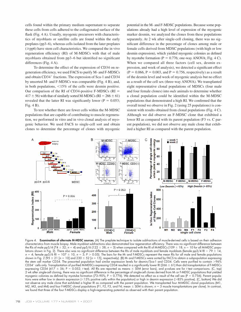
JCB • VOLUME 177 • NUMBER 1 • 2007 78
cells found within the primary medium supernatant to separate
these cells from cells adhered to the collagenated surface of the
fl ask (Fig. 4 A). Usually, myogenic precursors with characteris-
tics of myoblasts or satellite cells are found within the early
preplates (pp3–6), whereas cells isolated from the later preplates
(≥pp6) have stem cell characteristics. We compared the in vivo
regeneration effi ciency (RI) of M-MDSCs with that of male
myoblasts obtained from pp3–6 but identifi ed no signifi cant
differences (Fig. 4 A).
To determine the effect of the expression of CD34 on re-
generation effi ciency, we used FACS to purify M- and F-MDSCs
and obtain CD34+ fractions. The expression of Sca-1 and CD34
by unsorted M- and F-MDSCs was comparable (Fig. 4 B), and,
in both populations, <15% of the cells were desmin positive.
Our comparison of the RI of CD34-positive F-MDSCs (RI =
417 ± 56) with that of similarly sorted M-MDSCs (RI = 266 ± 61)
revealed that the latter RI was signifi cantly lower (P = 0.053;
Fig. 4 B).
To test whether there are fewer cells within the M-MDSC
populations that are capable of contributing to muscle regenera-
tion, we performed in vitro and in vivo clonal analysis of myo-
genic behavior. We used FACS to single-cell sort and obtain
clones to determine the percentage of clones with myogenic
potential in the M- and F-MDSC populations. Because some pop-
ulations already had a high level of expression of the myogenic
marker desmin, we analyzed the clones from these populations
separately. At 2 wk after single-cell cloning, there was no sig-
nifi cant difference in the percentage of clones among male or
female cells derived from MDSC populations (with high or low
desmin expression), which yielded myogenic colonies as defi ned
by myotube formation (P = 0.776; one-way ANOVA; Fig. 4 C).
When we compared all three factors (cell sex, desmin ex-
pression, and week of analysis), we detected a signifi cant effect
(P = 0.066, P = 0.003, and P = 0.756, respectively) as a result
of the desmin level and week of myogenic analysis but no effect
as a result of the cell sex (three-way ANOVA). We transplanted
eight representative clonal populations of MDSCs (four male
and four female clones) into mdx animals to determine whether
a clonal population could be identifi ed within the M-MDSC
populations that demonstrated a high RI. We confi rmed that the
overall trend we observe in Fig. 2 (using 25 populations) is con-
sistent with results obtained from clonal populations (Fig. 4 C).
Although we did observe an F-MDSC clone that exhibited a
lower RI as compared with its parent population (F3 vs. C par-
ent population), we did not observe any male clone that exhib-
ited a higher RI as compared with the parent population.
Figure 4. Examination of alternate M-MDSC sources. (A) The preplate technique to isolate subfractions of muscle-derived cells is based on their adhesion characteristics from muscle biopsy. Male myoblast subfractions also demonstrated low regeneration effi ciency. There was no signifi cant difference between the RIs of male pp3/4 (74 ± 32; n = 4) and pp5/6 (122 ± 38; n = 3) when compared with the RI of M-MDSCs (109 ± 18; n = 10 for all M-MDSC popu-lations shown in Fig. 2). There also was no signifi cant difference between the RIs of male myoblasts and female myoblasts (female pp3/4 RI = 79 ± 14, n = 4; female pp5/6 RI = 107 ± 10, n = 3; P > 0.05). The bars for the M- and F-MDSCs represent the mean RIs for all male and female populations shown in Fig. 2 (95 ± 21 [n = 10] and 230 ± 52 [n = 15], respectively). (B) M- and F-MDSCs were sorted by FACS to obtain a subpopulation expressing the stem cell marker CD34. The presorted population had similar expression levels for desmin/Sca-1 and CD34. Cells were purifi ed to contain >96% CD34+ cells only. Transplantation of purifi ed M-MDSCs expressing CD34 resulted in a signifi cantly lower RI (266 ± 61) than did transplantation of F-MDSCs expressing CD34 (417 ± 56; P = 0.052; t test). All RIs are reported as means ± SEM (error bars), and p-values are for t test comparisons. (C, top) 2 wk after single-cell cloning, there was no signifi cant difference in the percentage of single-cell clones derived from M- or F-MDSC populations that yielded myogenic colonies as defi ned by myotube formation (73–90%; P = 0.776). We detected no effect as a result of the cell sex (P = 0.756). Parent popula-tions were either low in desmin expression (<15% positive cells within the population) or high in desmin expression (>85% positive). (C, bottom) We did not observe any male clone that exhibited a higher RI as compared with the parent population. We transplanted four M-MDSC clonal populations (M1, M2, M3, and M4) and four F-MDSC clonal populations (F1, F2, F3, and F4; mean ± SEM is shown; n = 3 muscle transplantations per clone). In contrast, we found that three of the four F-MDSCs display a high-regenerating potential as observed with their parent population.
SEX DIFFERENCES IN STEM CELLS • DEASY ET AL. 79
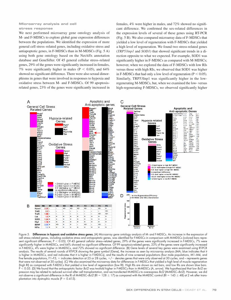
Microarray analysis and cell stress responseWe next performed microarray gene ontology analysis of
M- and F-MDSCs to explore global gene expression differences
between the populations. We identifi ed the expression of more
general cell stress–related genes, including oxidative stress and
antiapoptotic genes, in F-MDSCs than in M-MDSCs (Fig. 5 A)
using both gene ontology based on the NetAffx annotation
database and GeneSifter. Of 45 general cellular stress–related
genes, 29% of the genes were signifi cantly increased in females,
7% were signifi cantly higher in males (P < 0.05), and 64%
showed no signifi cant difference. There were also sexual dimor-
phisms in genes that were involved in responses to hypoxia and
oxidative stress between M- and F-MDSCs. Of 99 apoptosis-
related genes, 23% of the genes were signifi cantly increased in
females, 4% were higher in males, and 72% showed no signifi -
cant difference. We confi rmed the sex-related differences in
the expression levels of several of these genes using RT-PCR
(Fig. 5 B). We also compared microarray data of F-MDSCs that
yielded a low level of regeneration with F-MDSCs that yielded
a high level of regeneration. We found two stress-related genes
(TRP53inp1 and SOD1) that showed signifi cant trends in a di-
rection opposite to what we expected. For example, SOD1 was
signifi cantly higher in F-MDSCs as compared with M-MDSCs;
however, when we explored the data of F-MDSCs with low RIs
versus those with high RIs, we observed that SOD1 was higher
in F-MDSCs that had only a low level of regeneration (P < 0.05).
Similarly, TRP53Inp1 was significantly higher in the low-
regenerating M-MDSCs, but, when we examined the low- versus
high-regenerating F-MDSCs, we observed signifi cantly higher
Figure 5. Differences in hypoxic and oxidative stress genes. (A) Microarray gene ontology analysis of M- and F-MDSCs. An increase in the expression of cell stress–related genes, including oxidative stress and antiapoptotic genes, was identifi ed for F-MDSCs in comparison with M-MDSCs (colored bars repre-sent signifi cant differences; P < 0.05). Of 45 general cellular stress–related genes, 29% of the genes were signifi cantly increased in F-MDSCs, 7% were signifi cantly higher in M-MDSCs, and 64% showed no signifi cant difference. Of 99 apoptosis-related genes, 23% of the genes were signifi cantly increased in F-MDSCs, 4% were higher in M-MDSCs, and 72% showed no signifi cant difference. (B) Gene levels of several key genes were examined using RT-PCR analysis. The results of several rounds of RT-PCR showing the gene symbol (Gene), the increase as seen by microarray analysis (MA; blue indicates that it is higher in M-MDSCs, and red indicates that it is higher in F-MDSCs), and the results of nine screened populations (four male populations, M1–M4, and fi ve female populations, F1–F5; + indicates detection at 25 or 28 cycles, +/− denotes genes that were only observed at 30 cycles, and – represents genes that were not observed at 30 cycles). (C) We also examined the microarray data for differences in F-MDSCs that yielded a high level of muscle regeneration (high RI) as compared with F-MDSCs that yielded a low level of regeneration (low RI). High RIs are shown as red bars, and low RIs are shown blue bars. P < 0.05. (D) We found that the antiapoptotic factor Bcl2 was twofold higher in F-MDSCs than in M-MDSCs (A, arrow). We hypothesized that low Bcl2 ex-pression may be related to reduced survival after cell transplantation, and we transfected M-MDSCs to overexpress Bcl2 (M-MDSC–Bcl2). However, we did not observe a signifi cant difference in the RI of M-MDSC–Bcl2 (RI = 128 ± 17) as compared with the M-MDSC control (RI = 145 ± 48) at 2 wk after trans-plantation into dystrophic muscle (P = 0.415).
JCB • VOLUME 177 • NUMBER 1 • 2007 80
levels of TRP53Inp1 in the female populations with the best in
vivo regeneration (Fig. 5 C). Although these trends are interesting,
this analysis did not provide any clear indicators for genes
of importance.
In particular, we found that the antiapoptotic factor Bcl2
was twofold higher in F-MDSCs than in M-MDSCs by micro-
array analysis (Fig. 5 A, arrow) and Western blotting (not de-
picted). To test whether the overexpression of Bcl2 in M-MDSCs
could provide a gain of function in terms of in vivo skeletal
muscle regeneration, we transfected the M-MDSCs with a Bcl2
plasmid. Western blot analysis and quantifi cation showed higher
levels of Bcl2 in M-MDSCs that were transfected to over-
express Bcl2 as compared with control M- or F-MDSCs (unpub-
lished data). However, we did not observe a change in the RI
of Bcl2-engineered M-MDSCs as compared with M-MDSC
controls (Fig. 5 D).
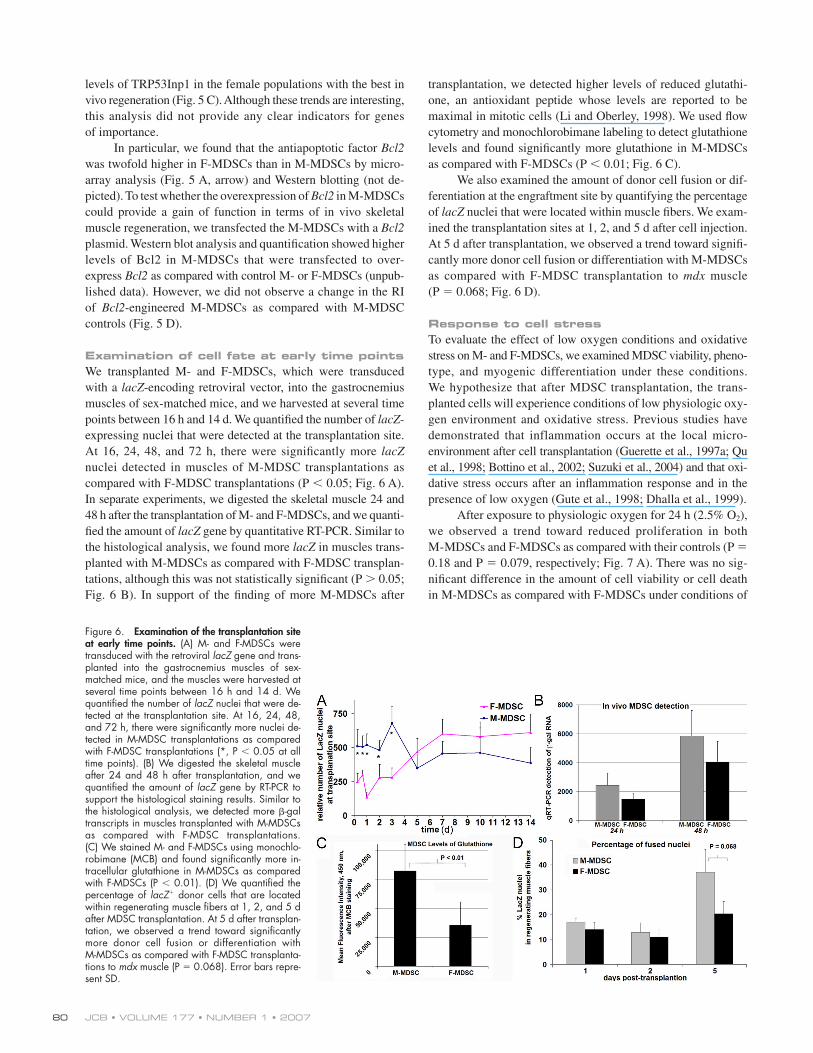
Examination of cell fate at early time pointsWe transplanted M- and F-MDSCs, which were transduced
with a lacZ-encoding retroviral vector, into the gastrocnemius
muscles of sex-matched mice, and we harvested at several time
points between 16 h and 14 d. We quantifi ed the number of lacZ-
expressing nuclei that were detected at the transplantation site.
At 16, 24, 48, and 72 h, there were signifi cantly more lacZ
nuclei detected in muscles of M-MDSC transplantations as
compared with F-MDSC transplantations (P < 0.05; Fig. 6 A).
In separate experiments, we digested the skeletal muscle 24 and
48 h after the transplantation of M- and F-MDSCs, and we quanti-
fi ed the amount of lacZ gene by quantitative RT-PCR. Similar to
the histological analysis, we found more lacZ in muscles trans-
planted with M-MDSCs as compared with F-MDSC transplan-
tations, although this was not statistically signifi cant (P > 0.05;
Fig. 6 B). In support of the fi nding of more M-MDSCs after
transplantation, we detected higher levels of reduced glutathi-
one, an antioxidant peptide whose levels are reported to be
maximal in mitotic cells (Li and Oberley, 1998). We used fl ow
cytometry and monochlorobimane labeling to detect glutathione
levels and found signifi cantly more glutathione in M-MDSCs
as compared with F-MDSCs (P < 0.01; Fig. 6 C).
We also examined the amount of donor cell fusion or dif-
ferentiation at the engraftment site by quantifying the percentage
of lacZ nuclei that were located within muscle fi bers. We exam-
ined the transplantation sites at 1, 2, and 5 d after cell injection.
At 5 d after transplantation, we observed a trend toward signifi -
cantly more donor cell fusion or differentiation with M-MDSCs
as compared with F-MDSC transplantation to mdx muscle
(P = 0.068; Fig. 6 D).
Response to cell stressTo evaluate the effect of low oxygen conditions and oxidative
stress on M- and F-MDSCs, we examined MDSC viability, pheno-
type, and myogenic differentiation under these conditions.
We hypothesize that after MDSC transplantation, the trans-
planted cells will experience conditions of low physiologic oxy-
gen environment and oxidative stress. Previous studies have
demonstrated that inflammation occurs at the local micro-
environment after cell transplantation (Guerette et al., 1997a; Qu
et al., 1998; Bottino et al., 2002; Suzuki et al., 2004) and that oxi-
dative stress occurs after an infl ammation response and in the
presence of low oxygen (Gute et al., 1998; Dhalla et al., 1999).
After exposure to physiologic oxygen for 24 h (2.5% O2),
we observed a trend toward reduced proliferation in both
M-MDSCs and F-MDSCs as compared with their controls (P =
0.18 and P = 0.079, respectively; Fig. 7 A). There was no sig-
nifi cant difference in the amount of cell viability or cell death
in M-MDSCs as compared with F-MDSCs under conditions of
Figure 6. Examination of the transplantation site at early time points. (A) M- and F-MDSCs were transduced with the retroviral lacZ gene and trans-planted into the gastrocnemius muscles of sex-matched mice, and the muscles were harvested at several time points between 16 h and 14 d. We quantifi ed the number of lacZ nuclei that were de-tected at the transplantation site. At 16, 24, 48, and 72 h, there were signifi cantly more nuclei de-tected in M-MDSC transplantations as compared with F-MDSC transplantations (*, P < 0.05 at all time points). (B) We digested the skeletal muscle after 24 and 48 h after transplantation, and we quantifi ed the amount of lacZ gene by RT-PCR to support the histological staining results. Similar to the histological analysis, we detected more β-gal transcripts in muscles transplanted with M-MDSCs as compared with F-MDSC transplantations. (C) We stained M- and F-MDSCs using monochlo-robimane (MCB) and found signifi cantly more in-tracellular glutathione in M-MDSCs as compared with F-MDSCs (P < 0.01). (D) We quantifi ed the percentage of lacZ+ donor cells that are located within regenerating muscle fi bers at 1, 2, and 5 d after MDSC transplantation. At 5 d after transplan-tation, we observed a trend toward signifi cantly more donor cell fusion or differentiation with M-MDSCs as compared with F-MDSC transplanta-tions to mdx muscle (P = 0.068). Error bars repre-sent SD.
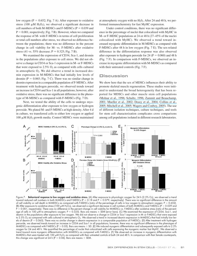
SEX DIFFERENCES IN STEM CELLS • DEASY ET AL. 81
low oxygen (P = 0.652; Fig. 7 A). After exposure to oxidative
stress (100 μM H2O2), we observed a signifi cant decrease in
cell numbers of both M-MDSCs and F-MDSCs (P = 0.039 and
P = 0.001, respectively; Fig. 7 B). However, when we compared
the response of M- with F-MDSCs in terms of cell proliferation
or total cell numbers after stress, we observed no difference be-
tween the populations; there was no difference in the percent
change in cell viability for M- vs. F-MDSCs after oxidative
stress (41 vs. 55% decrease; P = 0.325; Fig. 7 B).
We examined the expression of CD34, Sca-1, and desmin
in the populations after exposure to cell stress. We did not ob-
serve a change in CD34 or Sca-1 expression in M- or F-MDSCs
that were exposed to 2.5% O2 as compared with cells cultured
in atmospheric O2. We did observe a trend in increased des-
min expression in M-MDSCs that had initially low levels of
desmin (P = 0.065; Fig. 7 C). There was no similar change in
desmin expression in a comparable population of F-MDSCs. After
treatment with hydrogen peroxide, we observed trends toward
an increase in CD34 and Sca-1 in all populations; however, after
oxidative stress, there was no signifi cant difference in the pheno-
type of M-MDSCs as compared with F-MDSCs (Fig. 7 D).
Next, we tested the ability of the cells to undergo myo-
genic differentiation after exposure to low oxygen or hydrogen
peroxide. We plated M- and F-MDSCs at high density. After 4 d
in culture, we transferred cells to either low oxygen or applied
100 μM H202 growth media. Control MDSCs were maintained
at atmospheric oxygen with no H202. After 24 and 48 h, we per-
formed immunochemistry for fast MyHC expression.
Under control conditions, there was no signifi cant differ-
ence in the percentage of nuclei that colocalized with MyHC in
M- or F-MDSC populations at 24 or 48 h (27–45% of the nuclei
colocalized with MyHC). We observed a trend toward in-
creased myogenic differentiation in M-MDSCs as compared with
F-MDSCs after 48 h in low oxygen (Fig. 7 E). The sex-related
difference in the differentiation response was also observed
after exposure to hydrogen peroxide for 24 (P = 0.060) and 48 h
(Fig. 7 F). In comparison with F-MDSCs, we observed an in-
crease in myogenic differentiation with M-MDSCs as compared
with their untreated controls (Fig. 7 F).
DiscussionWe show here that the sex of MDSCs infl uences their ability to
promote skeletal muscle regeneration. These studies were initi-
ated to understand the broad heterogeneity that has been re-
ported for MDSCs and other muscle stem cell populations
(Molnar et al., 1996; Schultz, 1996; Zammit and Beauchamp,
2001; Mueller et al., 2002; Deasy et al., 2004; Collins et al.,
2005; Mitchell et al., 2005; Wagers and Conboy, 2005). The use
of different isolation techniques, culture techniques, and tools
for stem cell characterization complicates cross comparisons
among cell populations isolated in different research laboratories.
Figure 7. Behavioral response to low oxygen and oxidative stress. (A) After exposure to physiologic oxygen for 24 h (2.5% O2), we observed a trend toward reduced cell numbers in both M-MDSCs and F-MDSCs (P = 0.18 and P = 0.079, respectively). There was no signifi cant difference in the amount of cell viability or cell death in M-MDSCs as compared with F-MDSCs (ratio of the percentage of cells in low oxygen to atmospheric oxygen; P = 0.652). (B) After exposure to oxidative stress (100 μM H2O2), we observed a signifi cant decrease in cell numbers of both M-MDSCs and F-MDSCs (P = 0.039 and P = 0.001, respectively). There was no difference in the percent change in cell viability for M-MDSCs vs. F-MDSCs after oxidative stress (ratio of the percent-age of cells in 100 μM to atmospheric oxygen; P = 0.325). Bars are means ± SEM (error bars). (C) We examined the expression of CD34, Sca-1, and desmin in the populations after exposure to low oxygen. We did not observe a change in CD34 or Sca-1 expression in M- or F-MDSCs that were exposed to 2.5% O2 as compared with cells cultured in atmospheric O2. We observed a trend in increased desmin expression in M-MDSCs that had initially low lev-els of desmin (P = 0.065). There was no similar change in desmin expression in a comparable population of F-MDSCs. (D) After treatment with hydrogen peroxide, we observed trends toward an increase in CD34 and Sca-1 in all populations; however, there was no signifi cant difference in the phenotype of M-MDSCs as compared with F-MDSCs (P > 0.05). Bars are means ± SD. (E) We induced myogenic differentiation and subsequently exposed cells to 2.5% oxygen for 24 and 48 h. We quantifi ed the percentage of nuclei that colocalized with cells expressing the myogenic marker fast MyHC. We observed a trend toward more myogenic differentiation with M-MDSCs as compared with F-MDSCs. (F) We observed an increase in myogenic differentiation with M-MDSCs that were treated with 100 μM H2O2 as compared with their untreated controls at both 24 and 48 h. In comparison with their female counterparts, this change was signifi cant at 24 h (P = 0.06). Bars are means ± SEM.
JCB • VOLUME 177 • NUMBER 1 • 2007 82
This study identifi es sex-related differences as a factor in MDSC
variability in skeletal muscle regeneration.
The M- and F-MDSC populations isolated by the preplate
technique shared stem cell characteristics; however, extensive
in vivo screening showed that only 2/10 male populations had
an in vivo RI near 200. In comparison, 60% of the 15 female
populations had an RI higher than the mean RI of M-MDSCs
(RI = 95), and 40% of the F-MDSCs had an RI higher than the
maximal male RI (RI = 203). After transplantation into the
skeletal muscle of dystrophic mice, F-MDSCs transplanted into
hosts of either sex consistently regenerated more dystrophin-
positive myofi bers than did M-MDSCs transplanted into hosts
of either sex.
Searching for a robust M-MDSCTo determine whether the M-MDSC population is more elusive
than the F-MDSC population, we attempted to isolate cells with
the M-MDSC phenotype from other isolation subfractions.
First, we investigated whether preplate isolation might result in
the inadvertent removal of potent M-MDSCs. To determine
whether viable M-MDSCs reside in an earlier preplate passage
than previously believed, we transplanted male pp3, 4, 5, and 6
cells into dystrophic mice. However, the RIs of these male cells
were no higher than that of their parent M-MDSCs. We exam-
ined clones of M- and F-MDSCs and did not observe any male
clones that participated in muscle regeneration to a level similar
to that of F-MDSCs. We also compared M- and F-MDSC popu-
lations sorted on the basis of their expression of CD34, an es-
tablished muscle stem cell marker whose expression decreases
as cells differentiate. In side by side experiments, male CD34+
cells led to substantially less skeletal muscle regeneration than
did female CD34+ cells.
A retrospective analysis of our previous work with
muscle-derived cells provided further evidence that M-MDSCs
exhibit lower regeneration effi ciency than F-MDSCs (Jankowski
et al., 2002; Jankowski and Huard, 2004a,b). Two previous
studies performed by members of our laboratory involved the
use of Y-chromosome in situ hybridization to track male cells
injected into female hosts (before our notion of sex-related
differences; Jankowski et al., 2002; Jankowski and Huard,
2004b). The authors of both studies calculated RIs to assess the
effi ciency with which the donor cells regenerated dystrophin-
positive fi bers in dystrophic mice. These studies found that
male populations, which were sorted for CD34 expression by
magnetic antibody cell sorting (Jankowski et al., 2002) or by
FACS (Jankowski and Huard, 2004b), exhibited RIs <200,
which is a level consistent with the RIs reported in this study.
In addition, because those studies used freshly isolated popula-
tions, the authors performed >20 separate isolations from male
animals to generate a suffi cient sample size; none of the male
populations had an RI >200 (the authors scaled their RI to the
percentage of myogenic [desmin+] donor cells; we have ad-
justed for the authors’ reported values for desmin expression).
A third study using female cells obtained from rats demon-
strated a high RI (>200) after transplantation of the F-MDSCs
into mdx/SCID mice (RI = 392 ± 55 dystrophin-positive
fi bers/105 myogenic donor cells; Jankowski and Huard, 2004a).
In a study performed by another group, Mueller et al. (2002)
used MDSCs derived from our group and found a low effi -
ciency of muscle regeneration when the MDSCs were trans-
planted to male hosts, as we observe here. Collectively, these
fi ndings provide compelling evidence that MDSCs and host
animals exhibit sex-related differences.
We tested whether hormones play a role in this process.
We failed to observe increased regeneration by normal M-MDSCs
transplanted into female mice or M-MDSCs prestimulated with
physiologic estrogen levels. This fi nding parallels the results of
other studies indicating that sex-related differences might not
be exclusively hormonal (Xu et al., 1992; Gutierrez-Adan et al.,
2000; Du et al., 2004). Indeed, other dosing regimes and other
sex steroids such as progesterone or testosterone should be con-
sidered in conjunction with studying the various receptor iso-
forms to fully evaluate the potential role of hormones in the
sex-related differences exhibited by MDSCs.
Sex-related differences in cell stress responseBy microarray analysis, we observed trends in sex differences
in genes related to apoptosis, hypoxia, oxidative stress, and gen-
eral cell stress response. In particular, the RNA and protein lev-
els of the antiapoptotic gene Bcl2 were lower in M-MDSCs as
compared with F-MDSCs. The rapid cell death in muscle cell
transplantation has been previously reported (Beauchamp et al.,
1994, 1999; Fan et al., 1996). Therefore, we attempted a gain of
function in M-MDSCs by the overexpression of Bcl2. We did
not, in fact, observe an increase in muscle regeneration after
Bcl2 gene transfer. Indeed, subsequent experiments surprisingly
demonstrated that there were more M-MDSCs than F-MDSCs
at the transplantation site up to 3 d after transplantation. We also
detected higher levels of reduced glutathione in the M-MDSC
than in the F-MDSC. Glutathione’s role in protecting cells from
antioxidants could explain a better survival of M-MDSCs after
transplantation. In addition, it has also been shown that glutathi-
one levels are elevated during mitosis (Li and Oberley, 1998;
Menon et al., 2003). This fi nding, along with the fi nding of dif-
ferences in the mitotic fraction of M- and F-MDSCs, could also
suggest that the M-MDSCs were more mitotic than the F-MDSCs
after transplantation.
We also tested the cells’ abilities to undergo myogenic
differentiation after exposure to low oxygen environment or ox-
idative stress, two conditions that are expected to be present in
the microenvironment after transplantation. We found an in-
crease in the percentage of cells that expressed the myogenic
marker desmin in M-MDSCs grown in low oxygen. We also
found more MyHC expression by M-MDSCs after exposure to
oxidative stress for 24 h and a similar trend after 48 h.
We hypothesize that the effect of cell stress on myogenic
differentiation leads to in vivo sex-related differences in skeletal
muscle regeneration (Fig. 8). F-MDSCs respond to low oxygen
or oxidative stress by maintaining a low level of proliferation. In
a myogenic environment with low oxygen or oxidative stress,
F-MDSCs do not readily differentiate. Conversely, M-MDSCs
demonstrate increased myogenic differentiation in the presence
of low oxygen or oxidative stress. In vivo, we hypothesize that
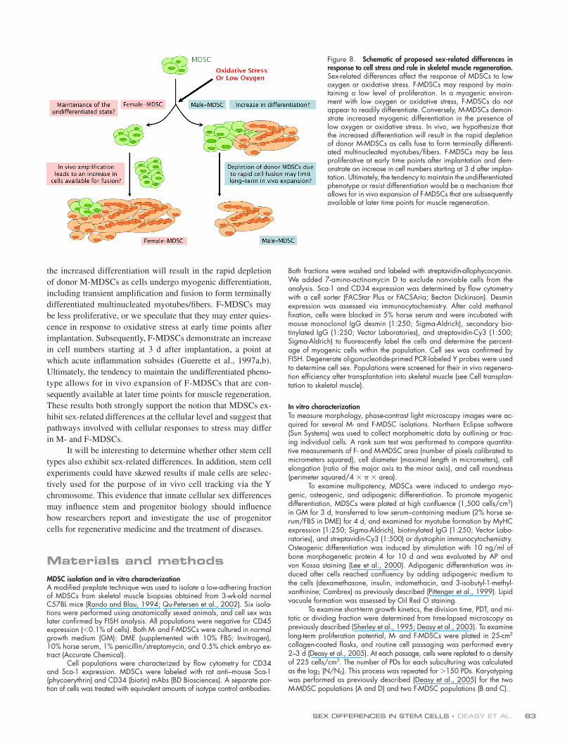
SEX DIFFERENCES IN STEM CELLS • DEASY ET AL. 83
the increased differentiation will result in the rapid depletion
of donor M-MDSCs as cells undergo myogenic differentiation,
including transient amplifi cation and fusion to form terminally
differentiated multinucleated myotubes/fi bers. F-MDSCs may
be less proliferative, or we speculate that they may enter quies-
cence in response to oxidative stress at early time points after
implantation. Subsequently, F-MDSCs demonstrate an increase
in cell numbers starting at 3 d after implantation, a point at
which acute infl ammation subsides (Guerette et al., 1997a,b).
Ultimately, the tendency to maintain the undifferentiated pheno-
type allows for in vivo expansion of F-MDSCs that are con-
sequently available at later time points for muscle regeneration.
These results both strongly support the notion that MDSCs ex-
hibit sex-related differences at the cellular level and suggest that
pathways involved with cellular responses to stress may differ
in M- and F-MDSCs.
It will be interesting to determine whether other stem cell
types also exhibit sex-related differences. In addition, stem cell
experiments could have skewed results if male cells are selec-
tively used for the purpose of in vivo cell tracking via the Y
chromosome. This evidence that innate cellular sex differences
may infl uence stem and progenitor biology should infl uence
how researchers report and investigate the use of progenitor
cells for regenerative medicine and the treatment of diseases.
Materials and methodsMDSC isolation and in vitro characterizationA modifi ed preplate technique was used to isolate a low-adhering fraction of MDSCs from skeletal muscle biopsies obtained from 3-wk-old normal C57BL mice (Rando and Blau, 1994; Qu-Petersen et al., 2002). Six isola-tions were performed using anatomically sexed animals, and cell sex was later confi rmed by FISH analysis. All populations were negative for CD45 expression (<0.1% of cells). Both M- and F-MDSCs were cultured in normal growth medium (GM): DME (supplemented with 10% FBS; Invitrogen), 10% horse serum, 1% penicillin/streptomycin, and 0.5% chick embryo ex-tract (Accurate Chemical).
Cell populations were characterized by fl ow cytometry for CD34 and Sca-1 expression. MDSCs were labeled with rat anti–mouse Sca-1 (phycoerythrin) and CD34 (biotin) mAbs (BD Biosciences). A separate por-tion of cells was treated with equivalent amounts of isotype control antibodies.
Both fractions were washed and labeled with streptavidin-allophycocyanin. We added 7-amino-actinomycin D to exclude nonviable cells from the analysis. Sca-1 and CD34 expression was determined by fl ow cytometry with a cell sorter (FACStar Plus or FACSAria; Becton Dickinson). Desmin expression was assessed via immunocytochemistry. After cold methanol fi xation, cells were blocked in 5% horse serum and were incubated with mouse monoclonal IgG desmin (1:250; Sigma-Aldrich), secondary bio-tinylated IgG (1:250; Vector Laboratories), and streptavidin-Cy3 (1:500; Sigma-Aldrich) to fl uorescently label the cells and determine the percent-age of myogenic cells within the population. Cell sex was confi rmed by FISH. Degenerate oligonucleotide-primed PCR-labeled Y probes were used to determine cell sex. Populations were screened for their in vivo regenera-tion effi ciency after transplantation into skeletal muscle (see Cell transplan-tation to skeletal muscle).
In vitro characterizationTo measure morphology, phase-contrast light microscopy images were ac-quired for several M- and F-MDSC isolations. Northern Eclipse software (Sun Systems) was used to collect morphometric data by outlining or trac-ing individual cells. A rank sum test was performed to compare quantita-tive measurements of F- and M-MDSC area (number of pixels calibrated to micrometers squared), cell diameter (maximal length in micrometers), cell elongation (ratio of the major axis to the minor axis), and cell roundness (perimeter squared/4 × π × area).
To examine multipotency, MDSCs were induced to undergo myo-genic, osteogenic, and adipogenic differentiation. To promote myogenic differentiation, MDSCs were plated at high confl uence (1,500 cells/cm2) in GM for 3 d, transferred to low serum–containing medium (2% horse se-rum/FBS in DME) for 4 d, and examined for myotube formation by MyHC expression (1:250; Sigma-Aldrich), biotinylated IgG (1:250; Vector Labo-ratories), and streptavidin-Cy3 (1:500) or dystrophin immunocytochemistry. Osteogenic differentiation was induced by stimulation with 10 ng/ml of bone morphogenetic protein 4 for 10 d and was evaluated by AP and von Kossa staining (Lee et al., 2000). Adipogenic differentiation was in-duced after cells reached confl uency by adding adipogenic medium to the cells (dexamethasone, insulin, indomethacin, and 3-isobutyl-1-methyl-xanthinine; Cambrex) as previously described (Pittenger et al., 1999). Lipid vacuole formation was assessed by Oil Red O staining.
To examine short-term growth kinetics, the division time, PDT, and mi-totic or dividing fraction were determined from time-lapsed microscopy as previously described (Sherley et al., 1995; Deasy et al., 2003). To examine long-term proliferation potential, M- and F-MDSCs were plated in 25-cm2 collagen-coated fl asks, and routine cell passaging was performed every 2–3 d (Deasy et al., 2005). At each passage, cells were replated to a density of 225 cells/cm2. The number of PDs for each subculturing was calculated as the log2 (N/N0). This process was repeated for >150 PDs. Karyotyping was performed as previously described (Deasy et al., 2005) for the two M-MDSC populations (A and D) and two F-MDSC populations (B and C).
Figure 8. Schematic of proposed sex-related differences in response to cell stress and role in skeletal muscle regeneration. Sex-related differences affect the response of MDSCs to low oxygen or oxidative stress. F-MDSCs may respond by main-taining a low level of proliferation. In a myogenic environ-ment with low oxygen or oxidative stress, F-MDSCs do not appear to readily differentiate. Conversely, M-MDSCs demon-strate increased myogenic differentiation in the presence of low oxygen or oxidative stress. In vivo, we hypothesize that the increased differentiation will result in the rapid depletion of donor M-MDSCs as cells fuse to form terminally differenti-ated multinucleated myotubes/fi bers. F-MDSCs may be less proliferative at early time points after implantation and dem-onstrate an increase in cell numbers starting at 3 d after implan-tation. Ultimately, the tendency to maintain the undifferentiated phenotype or resist differentiation would be a mechanism that allows for in vivo expansion of F-MDSCs that are subsequently available at later time points for muscle regeneration.
JCB • VOLUME 177 • NUMBER 1 • 2007 84
Acquisition and processing of images. Immunofl uorescent images of dystrophin, desmin, and MyHC were acquired at RT on a microscope (DM IRB; Leica) using a 20× NA 0.04 objective (CorrPh1 α/0–2/C; Leica). No imaging medium was used, and the fl uorochromes used are as indicated. Digital images were acquired using a camera (Regita; QImaging) and Northern Eclipse software (version 6.0; Sun Systems). Images were acquired at exposures that were based on unstained controls. The fi nal presentation of images was prepared in Photoshop versions 5.0–7.0 (Adobe), and, in some cases, only uniform brightness or contrast adjustments were performed.
Telomerase activity and telomere lengthTelomerase activity was determined using the TeloTAGGG Telomerase PCR ELISA PLUS kit (Roche) according to the manufacturer’s protocol. 2 × 10⁵ cells were lysed in 200 μl of lysis reagent, and 10 μl of the lysate was used for the telomere repeat amplifi cation protocol reaction. The experi-ment was performed in triplicate, and relative telomerase activity was re-corded and plotted. For the fl ow cytometry–based measurement of telomere length, telomeres in M- and F-MDSCs were detected with the PNA Kit for Flow Cytometry (DakoCytomation) according to the manufacturer’s protocol. 2 × 10⁶ male or female cells were divided into four 1.5-ml tubes. The cellular DNA was denatured for 10 min at 82°C and hybridized with the telomere PNA probe (FITC) at RT overnight. The cells were then analyzed by fl ow cytometry, and the relative telomere length was calculated.
Cell transplantation to skeletal muscleThe use of animals and the surgical procedures performed in this study were approved by the Institutional Animal Care and Use Committee of the Children’s Hospital of Pittsburgh (University of Pittsburgh Medical Center). mdx mice (C57BL/10ScSn-Dmdmdx) were obtained from The Jackson Labo-ratory or bred at the institution’s animal facility. mdx/SCID mice were bred by crossing C57BL/10ScSn-Dmdmdx and C57BL/6J-Prkdcscid/SzJ mice.
For all experiments involving the transplantation of MDSCs, satellite cells, or CD34-sorted populations, 1–2 × 10⁵ cells were transplanted into the gastrocnemius muscles of male or female mice as indicated and har-vested after 2 wk. For sex-mismatched experiments, two M-MDSCs (popu-lations A and D) were transplanted into female hosts, and two F-MDSCs (populations B and C) were transplanted into male hosts. The populations had similar phenotypes: populations A and C (CD34 [60–80% positive], Sca-1 [70–100% positive], and desmin [>95% positive]), populations B and D (CD34 [60–80% positive] and Sca-1 [40–80% positive], and des-min [<15% positive]). For CD34-sorting experiments, populations A and C were used.
Tissues were harvested and snap frozen in liquid nitrogen–cooled 2-methyl butane and cryosectioned at 10 μm. Either the MOM (mouse on mouse) kit (Vector Laboratories) with DYS2 antibody (1:50; Novocastra) or a donkey anti–rabbit dystrophin (1:300; Abcam) antibody was used to stain tissue sections for dystrophin. For the DYS2 antibody, the sections were fi xed with cold methanol, and the MOM kit protocol was followed (DYS2 at 1:50, biotinylated anti–mouse IgG at 1:250, and streptavidin-Cy3 T at 1:500; Sigma-Aldrich). For anti–rabbit dystrophin, sections were fi xed with 5% formalin, blocked with donkey serum, incubated with pri-mary antibody, and incubated with AlexaFluor594 donkey anti–rabbit (1:500; Invitrogen). For CD4 staining, tissue was blocked with goat serum and was incubated with rat anti–mouse CD4 (1:100; BD Biosciences), bio-tin goat anti–rat (1:400; Vector Laboratories), and peroxidase-streptavidin (1:800; DakoCytomation).
A previously described technique (Jankowski et al., 2002; Deasy et al., 2005) was used to measure the cells’ RIs. The RI is the ratio of the number of dystrophin-positive fi bers per 105 donor cells (RI is scored at the cross section of maximal engraftment). The large majority of the >150 muscle transplantations of this study were quantitated for the RI in a blind manner by more than one investigator.
The RIs or areas containing CD4-positive cells were compared by two-way ANOVA or a t test to assess the effects of cell sex and host sex. For transplantations of lacZ MDSCs, cells were fi rst labeled with retrovirus encoding for the lacZ gene as previously described (Lee et al., 2000). We performed X-gal staining to visualize β-gal activity and quantifi ed the total number of these lacZ-positive nuclei and the number that were located within muscle fi bers.
Single-cell cloning and clonal analysisWe used FACS to obtain single-cell clones (FACSAria and visual confi rma-tion) of M- and F-MDSCs (populations A–D) on 96-well plates. Clones were cultured in 50 μl GM. At 1 wk after sorting, we identifi ed viable colonies and scored these clones as myogenic or not myogenic based on the pres-ence of distinct multinucleated myotubes. At 2 wk after cloning, we again
determined the cumulative number of myogenic colonies based on the presence of distinct myotubes. For transplantation experiments using clonal populations, we similarly obtained single-cell clones from M- and F-MDSCs (populations A–D). After culture expansion, we selected eight representa-tive populations and performed transplantations as described above (Cell transplantation to skeletal muscle) to determine the RI of each clone.
Estrogen stimulationM- and F-MDSCs (populations A–D) were cultured in GM without phenol red and supplemented with 0, 10, or 100 nM 17-β-estradiol (Sigma-Aldrich). Cells were replated in fresh medium every 2–3 d for 14 d. After in vitro stimulation, 105 cells per muscle were transplanted into the gastroc-nemius of female mdx/SCID mice (n = 3 muscles per group). 2 wk after transplantation, the muscles were harvested, sectioned, and immuno-stained for dystrophin, and the RI was analyzed as described above (Cell transplantation to skeletal muscle).
Microarray analysisRNA was isolated from fi ve M- and fi ve F-MDSC populations using an RNeasy kit (QIAGEN) and analyzed on a GeneChip Mouse Genome 430A 2.0 array (Affymetrix, Inc.). Fold increase was determined using previously established techniques (Perez-Iratxeta et al., 2005). Gene ontol-ogy was determined using the NetAffx (Affymetrix, Inc.) annotation data-base (Liu et al., 2003; Harris et al., 2004). All results were confi rmed using GeneSifter (VizX Labs), a certifi ed GeneChip-compatible web-based analysis tool. The resulting data on fold increase for each population were analyzed using a t test to determine signifi cant differences (P < 0.05) be-tween the M- and F-MDSC populations.
RT-PCR and real-time quantitative PCRSeveral genes of interest from the microarray were confi rmed with RT-PCR analysis. Total RNA was extracted from 5 × 10⁵ cells using the Nucleospin RNA kit (CLONTECH Laboratories, Inc.). cDNA was synthesized with SuperScript II reverse transcriptase (Invitrogen) according to the manufac-turer’s instructions. PCR was performed with Taq polymerase (Invitrogen) according to the manufacturer’s instructions for 25, 28, and 30 cycles at 58°C annealing temperature, and PCR products were separated by electropho-resis on 1% agarose gels. The primers used are listed in Table S2 (avail-able at http://www.jcb.org/cgi/content/full/jcb.200612094/DC1).
For quantitative PCR, we used 50 ng cDNA. cDNA and β-gal prim-ers (forward, A C A G T A C G C G T A G ; reverse, C C A T C A A T C C G G T A G G T T T T-C C G G ) were added to SYBR green PCR master mix (Applied Biosystems) according to the manufacturer’s instructions. All lacZ data were normal-ized to 18S, which was used as the internal control.
Overexpression of Bcl2M- and F-MDSCs were plated at 2,500 cells/9.6 cm2 in GM. The follow-ing day, the medium was replaced with 500 μl DME (without serum or antibiotics) containing 4.0 μg Bcl2 plasmid, which also encoded for the neomycin resistance gene, and 5 μl LipofectAMINE 2000 reagent (Invitrogen). MDSCs were incubated at 37°C for 6 h in this medium, and then medium was removed and replaced with normal GM. After 5 d in culture, we began a selection process. For the next 2 wk, MDSCs were cultured in GM containing 1.5 mg/ml G418 sulfate (Cellgro). Controls were also trans-fected with GFP plasmid to confi rm transfection. Cells were maintained in GM and transplanted to male mdx/SCID mice as described above (Cell transplantation to skeletal muscle). Western blot analysis was performed using standard molecular biological techniques. Cells were lysed in Laemmli sample buffer (Bio-Rad Laboratories) and resolved on 4–20% precast gradient gels. Mouse Bcl2 was detected with anti-Bcl2 (1:1,000; BD Biosciences) with goat anti–rabbit HRP (1:2,500; BD Biosciences) and imaged using SuperSignal (Pierce Chemical Co.). β-actin levels were detected with anti–β-actin (1:5,000; Sigma-Aldrich) and goat anti–mouse HRP (1:5,000; Pierce Chemical Co.).
Response to cell stressViability. For the low oxygen environment, M- and F-MDSCs (populations A–D) were plated at a density of 500 cells/cm2 on collagen-coated fl asks with normal GM. After cell adherence (4–6 h), MDSCs were incubated for 24 h in either atmospheric (�20%) or 2.5% O2 conditions (HERAcell 150 incubator; MidAtlantic Diagnostics). For oxidative stress, M- and F-MDSCs (populations A–D) were plated at a density of 700 cells/cm2. After cell adherence, MDSCs were incubated for 24 h with either 0 or 100 μM H2O2 (under atmospheric oxygen). After 24 h in low O2 or after 24 h with 100 μM H2O2, MDSCs from the supernatant and adherent cells were collected
SEX DIFFERENCES IN STEM CELLS • DEASY ET AL. 85
(0.25% trypsin-EDTA). Cells were counted using a Neubauer hemacytometer (Fisher Scientifi c), and trypan blue staining was used to identify the live and dead cells.
Phenotype after cell stress. We examined CD34, Sca-1, and des min expression as described above (MDSC isolation and in vitro characterization).
Myogenic differentiation after cell stress. The ability of MDSCs to form myotubes under low oxygen conditions or after exposure to hydrogen peroxide was determined by fast MyHC staining. M- and F-MDSCs (popu-lations A–D) were plated at a density of 1,000 cells/cm2 on collagen-coated plates with normal GM. For low oxygen stress, after 4 d of growth in atmospheric conditions, growth media was refreshed, and cells were transferred to a 2.5% oxygen incubator. Control fl asks were main-tained in atmospheric O2 incubators. For oxidative stress, after 4 d of growth in atmospheric conditions with normal media, growth media was refreshed (control), or MDSCs received media with 100 μM H2O2. After 24 and 48 h of exposure to low oxygen or incubation with H2O2, MDSC cultures were fi xed and immunostained for MyHC as described above (In vitro characterization).
Reduced glutathione levels were determined for M- and F-MDSC populations by fl ow cytometry. Cells were plated at 1,600 cells/cm2 on collagen-coated fl asks with normal GM. After 24 h, cells were incubated in 5 μM monochlorobimane (Invitrogen) in normal growth media for 20 min at 37°C. The cells were then washed twice with PBS and harvested in 0.25% trypsin-EDTA. Intracellular glutathion levels were determined with a FACSAria machine (monochlorobimane excitation of 380 nm and emis-sion of 461 nm).
Online supplemental materialFig. S1 shows M- and F-MDSC similarities in cell morphology comparisons, multilineage marker expression, telomerase activity, and telomere length. Fig. S2 shows the in vitro and in vivo effects of estrogen stimulation on M- and F-MDSCs. Table S1 presents the correlation coeffi cients for rela-tions between variables. Table S2 presents the primers for RT-PCR. Online supplemental material is available at http://www.jcb.org/cgi/content/full/jcb.200612094/DC1.
We would like to thank Samantha Sanford, Seiji Kubo, Michele Jones, Michael Mentzer, Patrick Blake, Chris Scelfo, Michelle Wiit, and Maria Branca for technical assistance, Ryan Sauder for excellent editorial assistance, and Dr. Bruno Péault (University of Pittsburgh, Pittsburgh, PA) and Dr. Art Levine (Univer-sity of Pittsburgh School of Medicine, Pittsburgh, PA) for insightful discussions.
This work was supported by the Jesse’s Journey Foundation, the Muscular Dystrophy Association, the National Institutes of Health (grant R01 AR49684-01), the William F. and Jean W. Donaldson Chair at the Children’s Hospital of Pittsburgh, and the Henry J. Mankin Chair at the University of Pittsburgh.
Submitted: 18 December 2006Accepted: 11 March 2007
ReferencesAristotle. 350 BC. Historia Animalium: Books VII–X. 1991 edition. D.M.
Balme, editor. Harvard University Press, Cambridge, MA. 435–437.
Avery, B., C.B. Jorgensen, V. Madison, and T. Greve. 1992. Morphological de-velopment and sex of bovine in vitro-fertilized embryos. Mol. Reprod. Dev. 32:265–270.
Aviv, A. 2002. Telomeres, sex, reactive oxygen species, and human cardiovascular aging. J. Mol. Med. 80:689–695.
Aviv, A., J. Shay, K. Christensen, and W. Wright. 2005. The longevity gender gap: are telomeres the explanation? Sci. Aging Knowledge Environ. 2005:pe16.
Beauchamp, J.R., J.E. Morgan, C.N. Pagel, and T.A. Partridge. 1994. Quantitative studies of effi cacy of myoblast transplantation. Muscle Nerve. 1:S261.
Beauchamp, J.R., J.E. Morgan, C.N. Pagel, and T.A. Partridge. 1999. Dynamics of myoblast transplantation reveal a discrete minority of precursors with stem cell-like properties as the myogenic source. J. Cell Biol. 144:1113–1122.
Blankenhorn, E.P., S. Troutman, L.D. Clark, X.M. Zhang, P. Chen, and E. Heber-Katz. 2003. Sexually dimorphic genes regulate healing and regeneration in MRL mice. Mamm. Genome. 14:250–260.
Bottino, R., A.N. Balamurugan, S. Bertera, M. Pietropaolo, M. Trucco, and J.D. Piganelli. 2002. Preservation of human islet cell functional mass by anti-oxidative action of a novel SOD mimic compound. Diabetes. 51:2561–2567.
Chau, B.N., H.L. Borges, T.T. Chen, A. Masselli, I.C. Hunton, and J.Y. Wang. 2002. Signal-dependent protection from apoptosis in mice expressing caspase-resistant Rb. Nat. Cell Biol. 4:757–765.
Collins, C.A., I. Olsen, P.S. Zammit, L. Heslop, A. Petrie, T.A. Partridge, and J.E. Morgan. 2005. Stem cell function, self-renewal, and behavioral heterogeneity of cells from the adult muscle satellite cell niche. Cell. 122:289–301.
Deasy, B.M., R.J. Jankowski, T.R. Payne, B. Cao, J.P. Goff, J.S. Greenberger, and J. Huard. 2003. Modeling stem cell population growth: incorporating terms for proliferative heterogeneity. Stem Cells. 21:536–545.
Deasy, B.M., Y. Li, and J. Huard. 2004. Tissue engineering with muscle-derived stem cells. Curr. Opin. Biotechnol. 15:419–423.
Deasy, B.M., B.M. Gharaibeh, J.B. Pollett, M.M. Jones, M.A. Lucas, Y. Kanda, and J. Huard. 2005. Long-term self-renewal of postnatal muscle-derived stem cells. Mol. Biol. Cell. 16:3323–3333.
Dhalla, N.S., L. Golfman, S. Takeda, N. Takeda, and M. Nagano. 1999. Evidence for the role of oxidative stress in acute ischemic heart disease: a brief review. Can. J. Cardiol. 15:587–593.
Du, L., H. Bayir, Y. Lai, X. Zhang, P.M. Kochanek, S.C. Watkins, S.H. Graham, and R.S. Clark. 2004. Innate gender-based proclivity in response to cytotoxicity and programmed cell death pathway. J. Biol. Chem. 279:38563–38570.
Fan, Y., M. Maley, M. Beilharz, and M. Grounds. 1996. Rapid death of injected myoblasts in myoblast transfer therapy. Muscle Nerve. 19:853–860.
Guerette, B., I. Asselin, D. Skuk, M. Entman, and J.P. Tremblay. 1997a. Control of infl ammatory damage by anti-LFA-1: increase success of myoblast transplantation. Cell Transplant. 6:101–107.
Guerette, B., D. Skuk, F. Celestin, C. Huard, F. Tardif, I. Asselin, B. Roy, M. Goulet, R. Roy, M. Entman, and J.P. Tremblay. 1997b. Prevention by anti-LFA-1 of acute myoblast death following transplantation. J. Immunol. 159:2522–2531.
Gute, D.C., T. Ishida, K. Yarimizu, and R.J. Korthuis. 1998. Infl ammatory re-sponses to ischemia and reperfusion in skeletal muscle. Mol. Cell. Biochem. 179:169–187.
Gutierrez-Adan, A., M. Oter, B. Martinez-Madrid, B. Pintado, and J. De La Fuente. 2000. Differential expression of two genes located on the X chro-mosome between male and female in vitro-produced bovine embryos at the blastocyst stage. Mol. Reprod. Dev. 55:146–151.
Harada, H., K.P. Pavlick, I.N. Hines, D.J. Lefer, J.M. Hoffman, S. Bharwani, R.E. Wolf, and M.B. Grisham. 2003. Sexual dimorphism in reduced-size liver ischemia and reperfusion injury in mice: role of endothelial cell ni-tric oxide synthase. Proc. Natl. Acad. Sci. USA. 100:739–744.
Harris, T.W., N. Chen, F. Cunningham, M. Tello-Ruiz, I. Antoshechkin, C. Bastiani, T. Bieri, D. Blasiar, K. Bradnam, J. Chan, et al. 2004. WormBase: a multi-species resource for nematode biology and genomics. Nucleic Acids Res. 32:D411–D417.
Jankowski, R.J., and J. Huard. 2004a. Establishing reliable criteria for isolating myogenic cell fractions with stem cell properties and enhanced regenera-tive capacity. Blood Cells Mol. Dis. 32:24–33.
Jankowski, R.J., and J. Huard. 2004b. Myogenic cellular transplantation and regeneration: sorting through progenitor heterogeneity. Panminerva Med. 46:81–91.
Jankowski, R.J., B.M. Deasy, B. Cao, C. Gates, and J. Huard. 2002. The role of CD34 expression and cellular fusion in the regeneration capacity of myogenic progenitor cells. J. Cell Sci. 115:4361–4374.
Jilka, R.L., G. Hangoc, G. Girasole, G. Passeri, D.C. Williams, J.S. Abrams, B. Boyce, H. Broxmeyer, and S.C. Manolagas. 1992. Increased osteoclast development after estrogen loss: mediation by interleukin-6. Science. 257:88–91.
Kochhar, H.S., K.P. Kochhar, P.K. Basrur, and W.A. King. 2003. Infl uence of the duration of gamete interaction on cleavage, growth rate and sex distribution of in vitro produced bovine embryos. Anim. Reprod. Sci. 77:33–49.
Lee, J.Y., Z. Qu-Petersen, B. Cao, S. Kimura, R. Jankowski, J. Cummins, A. Usas, C. Gates, P. Robbins, A. Wernig, and J. Huard. 2000. Clonal isola-tion of muscle-derived cells capable of enhancing muscle regeneration and bone healing. J. Cell Biol. 150:1085–1100.
Li, N., and T.D. Oberley. 1998. Modulation of antioxidant enzymes, reac-tive oxygen species, and glutathione levels in manganese superoxide dismutase-overexpressing NIH/3T3 fi broblasts during the cell cycle. J. Cell. Physiol. 177:148–160.
Liu, G., A.E. Loraine, R. Shigeta, M. Cline, J. Cheng, V. Valmeekam, S. Sun, D. Kulp, and M.A. Siani-Rose. 2003. NetAffx: Affymetrix probesets and annotations. Nucleic Acids Res. 31:82–86.
Maeda, K.-I., S. Ohkura, and H. Tsukamura. 2000. Physiology of reproduction. In The Laboratory Rat: the Handbook of Experimental Animals. G.J. Krinke, editor. Academic Press, San Diego. 756 pp.
JCB • VOLUME 177 • NUMBER 1 • 2007 86
Menon, S.G., E.H. Sarsour, D.R. Spitz, R. Higashikubo, M. Sturm, H. Zhang, and P.C. Goswami. 2003. Redox regulation of the G1 to S phase transition in the mouse embryo fi broblast cell cycle. Cancer Res. 63:2109–2117.
Mitchell, P.O., T. Mills, R.S. O’Connor, T. Graubert, E. Dzierzak, and G.K. Pavlath. 2005. Sca-1 negatively regulates proliferation and differentiation of muscle cells. Dev. Biol. 283:240–252.
Molnar, G., M.L. Ho, and N.A. Schroedl. 1996. Evidence for multiple satellite cell populations and a non-myogenic cell type that is regulated differently in regenerating and growing skeletal muscle. Tissue Cell. 28:547–556.
Mueller, G.M., T. O’Day, J.F. Watchko, and M. Ontell. 2002. Effect of injecting primary myoblasts versus putative muscle-derived stem cells on mass and force generation in mdx mice. Hum. Gene Ther. 13:1081–1090.
Mulroney, S.E., C. Woda, M. Johnson, and C. Pesce. 1999. Gender differences in renal growth and function after uninephrectomy in adult rats. Kidney Int. 56:944–953.
Nomura, T., E. Ashihara, K. Tateishi, S. Asada, T. Ueyama, T. Takahashi, H. Matsubara, and H. Oh. 2007. Skeletal myosphere-derived progenitor cell transplantation promotes neovascularization in delta-sarcoglycan knock-down cardiomyopathy. Biochem. Biophys. Res. Commun. 352:668–674.
Peippo, J., A. Farazmand, M. Kurkilahti, M. Markkula, P.K. Basrur, and W.A. King. 2002. Sex-chromosome linked gene expression in in-vitro pro-duced bovine embryos. Mol. Hum. Reprod. 8:923–929.
Perez-Iratxeta, C., G. Palidwor, C.J. Porter, N.A. Sanche, M.R. Huska, B.P. Suomela, E.M. Muro, P.M. Krzyzanowski, E. Hughes, P.A. Campbell, et al. 2005. Study of stem cell function using microarray experiments. FEBS Lett. 579:1795–1801.
Pergament, E., M. Fiddler, N. Cho, D. Johnson, and W.J. Holmgren. 1994. Sexual differentiation and preimplantation cell growth. Hum. Reprod. 9:1730–1732.
Pittenger, M.F., A.M. Mackay, S.C. Beck, R.K. Jaiswal, R. Douglas, J.D. Mosca, M.A. Moorman, D.W. Simonetti, S. Craig, and D.R. Marshak. 1999. Multilineage potential of adult human mesenchymal stem cells. Science. 284:143–147.
Qu, Z., L. Balkir, J.C. van Deutekom, P.D. Robbins, R. Pruchnic, and J. Huard. 1998. Development of approaches to improve cell survival in myoblast transfer therapy. J. Cell Biol. 142:1257–1267.
Qu-Petersen, Z., B. Deasy, R. Jankowski, M. Ikezawa, J. Cummins, R. Pruchnic, J. Mytinger, B. Cao, C. Gates, A. Wernig, and J. Huard. 2002. Identifi cation of a novel population of muscle stem cells in mice: potential for muscle regeneration. J. Cell Biol. 157:851–864.
Rando, T.A., and H.M. Blau. 1994. Primary mouse myoblast purifi cation, char-acterization, and transplantation for cell-mediated gene therapy. J. Cell Biol. 125:1275–1287.
Rollo, C.D. 2002. Growth negatively impacts the life span of mammals. Evol. Dev. 4:55–61.
Sarig, R., Z. Baruchi, O. Fuchs, U. Nudel, and D. Yaffe. 2006. Regeneration and transdifferentiation potential of muscle-derived stem cells propagated as myospheres. Stem Cells. 24:1769–1778.
Schultz, E. 1996. Satellite cell proliferative compartments in growing skeletal muscles. Dev. Biol. 175:84–94.
Sherley, J.L., P.B. Stadler, and J.S. Stadler. 1995. A quantitative method for the analysis of mammalian cell proliferation in culture in terms of dividing and non-dividing cells. Cell Prolif. 28:137–144.
Steinlein, P., O. Wessely, S. Meyer, E.M. Deiner, M.J. Hayman, and H. Beug. 1995. Primary, self-renewing erythroid progenitors develop through acti-vation of both tyrosine kinase and steroid hormone receptors. Curr. Biol. 5:191–204.
Stindl, R. 2004. Tying it all together: telomeres, sexual size dimorphism and the gender gap in life expectancy. Med. Hypotheses. 62:151–154.
Suzuki, K., B. Murtuza, J.R. Beauchamp, R.T. Smolenski, A. Varela-Carver, S. Fukushima, S.R. Coppen, T.A. Partridge, and M.H. Yacoub. 2004. Dynamics and mediators of acute graft attrition after myoblast transplan-tation to the heart. FASEB J. 18:1153–1155.
Tamaki, S., T. Ichinohe, K. Matsuo, N. Hamajima, N. Hirabayashi, and H. Dohy. 2001. Superior survival of blood and marrow stem cell recipients given maternal grafts over recipients given paternal grafts. Bone Marrow Transplant. 28:375–380.
Vina, J., C. Borras, J. Gambini, J. Sastre, and F.V. Pallardo. 2005. Why females live longer than males: control of longevity by sex hormones. Sci. Aging Knowledge Environ. 2005:pe17.
Wagers, A.J., and I.M. Conboy. 2005. Cellular and molecular signatures of mus-cle regeneration: current concepts and controversies in adult myogenesis. Cell. 122:659–667.
Winitsky, S.O., T.V. Gopal, S. Hassanzadeh, H. Takahashi, D. Gryder, M.A. Rogawski, K. Takeda, Z.X. Yu, Y.H. Xu, and N.D. Epstein. 2005. Adult murine skeletal muscle contains cells that can differentiate into beating cardiomyocytes in vitro. PLoS Biol. 3:e87.
Xu, K.P., B.R. Yadav, W.A. King, and K.J. Betteridge. 1992. Sex-related differ-ences in developmental rates of bovine embryos produced and cultured in vitro. Mol. Reprod. Dev. 31:249–252.
Zammit, P., and J. Beauchamp. 2001. The skeletal muscle satellite cell: stem cell or son of stem cell? Differentiation. 68:193–204.