A Review—From Disease Metabolism to Precision Medicine
17
Review Article Human Systems Biology and Metabolic Modelling: A Review—From Disease Metabolism to Precision Medicine Claudio Angione 1,2 1 Department of Computer Science and Information Systems, Teesside University, UK 2 Healthcare Innovation Centre, Teesside University, UK Correspondence should be addressed to Claudio Angione; [email protected] Received 1 November 2018; Revised 7 February 2019; Accepted 20 May 2019; Published 9 June 2019 Academic Editor: Stanley Brul Copyright © 2019 Claudio Angione. is is an open access article distributed under the Creative Commons Attribution License, which permits unrestricted use, distribution, and reproduction in any medium, provided the original work is properly cited. In cell and molecular biology, metabolism is the only system that can be fully simulated at genome scale. Metabolic systems biology offers powerful abstraction tools to simulate all known metabolic reactions in a cell, therefore providing a snapshot that is close to its observable phenotype. In this review, we cover the 15 years of human metabolic modelling. We show that, although the past five years have not experienced large improvements in the size of the gene and metabolite sets in human metabolic models, their accuracy is rapidly increasing. We also describe how condition-, tissue-, and patient-specific metabolic models shed light on cell-specific changes occurring in the metabolic network, therefore predicting biomarkers of disease metabolism. We finally discuss current challenges and future promising directions for this research field, including machine/deep learning and precision medicine. In the omics era, profiling patients and biological processes from a multiomic point of view is becoming more common and less expensive. Starting from multiomic data collected from patients and N-of-1 trials where individual patients constitute different case studies, methods for model-building and data integration are being used to generate patient-specific models. Coupled with state-of-the-art machine learning methods, this will allow characterizing each patient’s disease phenotype and delivering precision medicine solutions, therefore leading to preventative medicine, reduced treatment, and in silico clinical trials. 1. Introduction With the advent of bioinformatics and computational biol- ogy, computational and mathematical techniques can provide accurate simulation of biological processes. e most widely used approaches to analyze omics data mainly focus on genomics, transcriptomics, and proteomics, through differ- ential expression or network-based coexpression analysis. However, genes and their expression alone do not always constitute a reliable indicator of cellular phenotype. When characterizing a phenotypic outcome, relying solely on gene or protein expression profiles will miss the highly nonlinear interaction between these biological layers. Such approaches oſten overlook the metabolic level, the dense network of biochemical reactions occurring in a cell with the aim of converting nutrients into energy and cellular building blocks. Being the best-characterized network in biological sys- tems and also the closest to the phenotype, metabolism is arguably the best indicator for the cell physiological state [1]. Once considered only a passive result of the state of a cell, it is now widely recognized as a main contributor to cellular behavior. More specifically, it is a key player in a number of diseases, including diabetes, neurodegenerative diseases, and cancer, where altered metabolism is now accepted as a hallmark [2]. e recent availability of high-throughput data regarding multiple layers of biological organization (omics) allows mapping cellular processes at the levels of genes, mRNA, proteins, and metabolites (Figure 1). In a single experiment, these measurements are oſten at both the “genotype” level (i.e., referring to the genetic elements on a genome) and at the “phenotype” level (the form and function of the cell). A fundamental question in systems biology is the definition and understanding of the genotype-phenotype relationship [3]. A mechanistic link between genotype and phenotype is offered by genome-scale metabolic models, which contain all known biochemical reactions occurring in a cell. Such models have been generated taking into account decades of studies in Hindawi BioMed Research International Volume 2019, Article ID 8304260, 16 pages https://doi.org/10.1155/2019/8304260
-
Upload
khangminh22 -
Category
Documents
-
view
1 -
download
0
Transcript of A Review—From Disease Metabolism to Precision Medicine

Review ArticleHuman Systems Biology and Metabolic Modelling:A Review—From Disease Metabolism to Precision Medicine
Claudio Angione 1,2
1Department of Computer Science and Information Systems, Teesside University, UK2Healthcare Innovation Centre, Teesside University, UK
Correspondence should be addressed to Claudio Angione; [email protected]
Received 1 November 2018; Revised 7 February 2019; Accepted 20 May 2019; Published 9 June 2019
Academic Editor: Stanley Brul
Copyright © 2019 Claudio Angione. This is an open access article distributed under the Creative Commons Attribution License,which permits unrestricted use, distribution, and reproduction in any medium, provided the original work is properly cited.
In cell andmolecular biology, metabolism is the only system that can be fully simulated at genome scale. Metabolic systems biologyoffers powerful abstraction tools to simulate all known metabolic reactions in a cell, therefore providing a snapshot that is closeto its observable phenotype. In this review, we cover the 15 years of human metabolic modelling. We show that, although thepast five years have not experienced large improvements in the size of the gene and metabolite sets in human metabolic models,their accuracy is rapidly increasing. We also describe how condition-, tissue-, and patient-specific metabolic models shed light oncell-specific changes occurring in themetabolic network, therefore predicting biomarkers of disease metabolism.We finally discusscurrent challenges and future promising directions for this research field, includingmachine/deep learning and precisionmedicine.In the omics era, profiling patients and biological processes from a multiomic point of view is becoming more common and lessexpensive. Starting from multiomic data collected from patients and N-of-1 trials where individual patients constitute differentcase studies, methods for model-building and data integration are being used to generate patient-specific models. Coupled withstate-of-the-art machine learning methods, this will allow characterizing each patient’s disease phenotype and delivering precisionmedicine solutions, therefore leading to preventative medicine, reduced treatment, and in silico clinical trials.
1. Introduction
With the advent of bioinformatics and computational biol-ogy, computational andmathematical techniques can provideaccurate simulation of biological processes. The most widelyused approaches to analyze omics data mainly focus ongenomics, transcriptomics, and proteomics, through differ-ential expression or network-based coexpression analysis.However, genes and their expression alone do not alwaysconstitute a reliable indicator of cellular phenotype. Whencharacterizing a phenotypic outcome, relying solely on geneor protein expression profiles will miss the highly nonlinearinteraction between these biological layers. Such approachesoften overlook the metabolic level, the dense network ofbiochemical reactions occurring in a cell with the aim ofconverting nutrients into energy and cellular building blocks.
Being the best-characterized network in biological sys-tems and also the closest to the phenotype, metabolism isarguably the best indicator for the cell physiological state [1].
Once considered only a passive result of the state of a cell,it is now widely recognized as a main contributor to cellularbehavior. More specifically, it is a key player in a numberof diseases, including diabetes, neurodegenerative diseases,and cancer, where altered metabolism is now accepted as ahallmark [2].
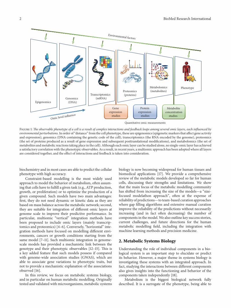
The recent availability of high-throughput data regardingmultiple layers of biological organization (omics) allowsmapping cellular processes at the levels of genes, mRNA,proteins, and metabolites (Figure 1). In a single experiment,these measurements are often at both the “genotype” level(i.e., referring to the genetic elements on a genome) and atthe “phenotype” level (the form and function of the cell).A fundamental question in systems biology is the definitionand understanding of the genotype-phenotype relationship[3]. A mechanistic link between genotype and phenotype isoffered by genome-scale metabolic models, which contain allknownbiochemical reactions occurring in a cell. Suchmodelshave been generated taking into account decades of studies in
HindawiBioMed Research InternationalVolume 2019, Article ID 8304260, 16 pageshttps://doi.org/10.1155/2019/8304260
2 BioMed Research International
Environmental factors
Omic level influence
Genomics(DNA)
Transcriptomics(mRNA)
Proteomics(proteins)
Metabolomics (metabolites)
Phenotype
Epigenomics
GLC
LAC PYR
Gene expression
studies
Protein expression
studies
Metabolite concentration
studies
Quantitative omic measurements
Figure 1:The observable phenotype of a cell is a result of complex interactions and feedback loops among several omic layers, each influenced byenvironmental perturbations. In order of “distance” from the cell phenotype, these are epigenomics (epigeneticmarkers that affect gene activityand expression), genomics (DNA containing the genetic code of the cell), transcriptomics (the RNA encoded by the genome), proteomics(the set of proteins produced as a result of gene expression and subsequent posttranslational modifications), and metabolomics (the set ofmetabolites andmetabolic reactions taking place in the cell). Although each omic layer can be studied alone, no single-omic layer has achieveda satisfactory correlationwith the phenotypic observables. As a result, in recent years, amultiomic approach has been adopted where all layersare considered together, and the effect of interactions and feedback is taken into consideration.
biochemistry and inmost cases are able to predict the cellularphenotype with high accuracy.
Constraint-based modelling is the most widely usedapproach to model the behavior of metabolism, often assum-ing that cells have to fulfill a given task (e.g., ATP production,growth, or proliferation) or to optimize the production of agiven compound. Such models have two main advantages:first, they do not need dynamic or kinetic data as they arebased onmass balance across the metabolic network; second,they are suitable for integration of different omic layers atgenome scale to improve their predictive performance. Inparticular, multiomic “vertical” integration methods havebeen proposed to include omic layers (mainly transcrip-tomics and proteomics) [4–6]. Conversely, “horizontal” inte-gration methods have focused on modelling different envi-ronments, cancers or growth conditions starting from thesame model [7–11]. Such multiomic integration in genome-scale models has provided a mechanistic link between thegenotype and their phenotypic observables [12–15]. This isa key added feature that such models possess if comparedwith genome-wide association studies (GWAS), which areable to associate gene variations to phenotypic traits, butnot to provide a mechanistic explanation of the associationsobserved [16].
In this review, we focus on metabolic systems biology,and in particular on human metabolic modelling. Originallytested and validated withmicroorganisms, metabolic systems
biology is now becoming widespread for human tissues andbiomedical applications [17]. We provide a comprehensivereview of the metabolic models developed so far for humancells, discussing their strengths and limitations. We showthat the main focus of the metabolic modelling communityhas shifted from increasing the size of the models—a “size-focused modelathon approach”, often at the expense ofreliability of predictions—to team-based curation approacheswhere gap filling algorithms and extensive manual curationimprove the reliability of the predictions without necessarilyincreasing (and in fact often decreasing) the number ofcomponents in the model.We also outline key success stories,current challenges, and future directions for the humanmetabolic modelling field, including the integration withmachine learning methods and precision medicine.
2. Metabolic Systems Biology
Understanding the role of individual components in a bio-logical system is an important step to elucidate or predictits behavior. However, a major theme in systems biology isinvestigating these systems with an integrated approach. Infact, studying the interactions between different componentsalso gives insights into the functioning and behavior of thecomponents taken independently [18].
Metabolism is the biggest biological network fullydescribed. It is a surrogate of the phenotype, being able to
BioMed Research International 3
elucidate phenotypic changes caused by single or combinedomics or physiological factors. It also has the advantageof being the only system that can be entirely modelled atgenome-scale. As a result, it plays a central role in elucidatingthe genotype-phenotype relation. Systems-based approacheshave been successfully applied over the last decade to inves-tigate metabolic networks, composed of a set of chemicalreactions and a pool of metabolites. The first examples ofmetabolic systems biology appeared in 1999 and were focusedon modelling, connecting, and simulating several cellularprocesses [19]. Whole-cell modelling, named the “grandchallenge of the 21st century”, is still an active area of research[20–24]. Inmetabolic systems biology, the value of modellingcell metabolism is not merely explanatory of a biologicalprocess, but also predictive. A model can be used to suggesthypotheses that can be tested or to pinpoint unexpectedbehaviors that can be further investigated in vitro.
There are different methods tomodel a metabolic system:steady-state analysis (e.g., FBA) involves a set of linear equa-tions, while kinetic simulations involve ordinary differentialequations (ODEs). Each variable represents the variation ofa metabolite concentration, in a dynamic or steady state,where the concentration depends on the rates of the reactionsthat produce and consume that metabolite [25]. Kineticmodels do not assume steady state and therefore are able tomodel highly dynamic mechanisms, including allosteric andposttranslational regulation, metabolite concentrations, andthermodynamics. Such ODE-based systems contain a largenumber of equations (differential or algebraic) and requireunique kinetic parameter values. They are highly effectiveat predicting the behavior of small systems where sufficientexperimental data can be collected for model calibrationand parameter estimation [26]. Furthermore, unlike standardFBA-based methods, reaction kinetics can be accounted for,and metabolite concentrations can be modelled explicitly,and therefore intracellular metabolomics data can be inte-grated directly [27].
For large systems, however, the use of kinetic mod-elling remains challenging. The increasing demand forsystems-level genome-scale analyses has recently led to thewidespread use of constraint-based steady-state models andtheir unsteady-state extensions. This has also been facilitatedby the increasing availability of multiomics data and pheno-typic information, used to constrain the model dynamicallyand therefore often compensating for the lack of regulatoryand kinetic modelling.
Following a number of successful attempts at buildingmultiscale kinetic and constraint-based models [28, 29],achieving the right combination (and therefore trade-off)of kinetic modelling and steady-state assumptions is likelythe next step for the metabolic modelling community. Tech-niques like unsteady-state FBA (uFBA) [4] have been recentlydeveloped to relax the steady-state assumption in genome-scale models, with the goal of modelling dynamic cellularstates derived from changes in the concentration of internalmetabolites. An approach to integrate reaction kinetics withsteady-state metabolic networks has also been proposed,where the genome-scale information of shared metabolitesamong different reactions is used to inform the interactions
between the reactions and predict metabolite concentrationsin a network kinetics approach [30]. More generally, due tothe larger range of predictions that kinetic models can per-form compared to steady-state approaches, expanding themtowards the genome scale or using information derived fromgenome-scalemodel simulations is a promising direction thatwill increase their spatial and temporal resolution.
2.1. Genome-Scale Metabolic Models. Genome-scale meta-bolic models contain all known metabolic reactions in anorganism and can therefore serve as functional databasesof cell-specific metabolism. A genome-scale model is builtusing the following process. First, a draft reconstruction isgenerated starting from the genome and including all thegenome-encoded metabolic reactions. The draft reconstruc-tion also includes annotated enzyme, reaction, and pathwaydata from databases like KEGG [31], BioCyc [32], andBRENDA [33]. Details on which genes control each reactionare also included. Then, a sequence of manual curation stepsimproves the draft reconstruction, by gathering evidence toprove or disprove the presence of a reaction in the network ofthe organism.
The construction of genome-scale model represents thestarting point for flux balance analysis (FBA, see followingsubsections). Finally, the model is run and validated bycomparing its predictions with existing experimental results,and new in silico experiments are performed to furtherimprove and validate the model. For more details on howto build a metabolic model from the DNA sequence of anorganism, the reader is referred to full protocols [34–36].
Most genome-scale models are annotated with curatedgene-protein-reaction associations (GPR rules), linkinggenes with enzymes (Figure 2). Such annotations pave theway for overlaying multiomic data on the models, usingthem as omic-scaffolds (see Section 2.3). Since omic datacan be quantified numerically and in a condition-, tissue-,and patient-specific way (e.g., transcriptomic profiles,protein levels, and metabolite concentrations), modelswith GPR rules can serve as a baseline for generatingpersonalized metabolic models. For instance, personalizedpredictions using suchmodels can lead to precise phenotypiccharacterization of patients (see Section 4).
2.2. Constraint-Based Modelling and Flux Balance Analy-sis. Flux balance analysis (FBA) is the most widely usedconstraint-based technique to predict flux distributions andnetwork capabilities in genome-scale models [37]. FBA hasproved useful thanks to its ability to handle large networksand predict genome-scale flux distributions. It requiresinformation about biochemical reactions and stoichiometriccoefficients but does not involve kinetic parameters. Thismakes it well suited to metabolic engineering studies thatidentify and characterize optimal perturbations such asdifferent substrates or genetic interventions (e.g., knockouts)leading to obligatory coupling between the growth rate andthe overproduction of the desired metabolite [38–42]. Ingeneral, FBA is a powerful tool for predictions of cell behaviorunder different metabolic conditions.

4 BioMed Research International
Genome-scale metabolic modelling
Proteomic dataTranscriptomic data
Environmental factorsHealthy/disease cellsPatient-specific data
Enzymatic complex
A + B -> C + D
AND
Isoenzyme
A + B -> C + D
OR
A + B -> C + D
Promiscuous enzyme
E + F -> G+ H
Metabolomic data
Gene-protein-reaction (GPR)association rules
Personalized/disease predictions
GLC
LAC
GLU
FATTY ACIDS & LIPIDS
AMINO ACIDS
AUTOPHAGY
TCA
PYR
NUCLEOTIDES
Microbiome/lifestyle
Figure 2: The integration of different types of omics data can be used to infer tissue- and condition-specific intracellular metabolicflux distributions. Intracellular metabolic reactions provide the cell with basic biochemical building blocks, as well as energy and athermodynamically favorable environment to sustain its life. Patient-specific data, molecular information, lifestyle, and environmental factorsaffect different omic levels. As a consequence, transcriptomic, proteomic, and metabolomic data need to be integrated to determine gene-protein association rules and to build genome-scale models used for personalized predictions. Given the large effect of environmental factorson omics level, determination of system-level changes in intracellular metabolic fluxes is important for understanding the fundamentalmechanisms of metabolic responses to perturbations. Indeed, environmental factors affect omics data on different levels, form epigenomicsto the cell phenotype. Omic-augmented genome-scale metabolic reconstructions have proved successful due to the ability to integrate omicmeasurements at genome scale and to give mechanistic insights into the genotype-phenotype relationship.
FBA is a linear programming technique that models thesteady-state condition in a chemical reaction network [43].The FBA representation of a genome-scale model is builtbased on a stoichiometric matrix 𝑆, containing the stoichio-metric coefficients of each metabolite (on the rows) in eachreaction of the network (on the columns). A stoichiometriccoefficient of a metabolite in a given reaction is positive ifthe metabolite is produced by the reaction and negative ifconsumed. An underdetermined linear system of equations𝑆V = 0 is defined from the stoichiometric matrix, wherethe unknowns are represented by the vector V of reactionflux rates. Additional constraints are included as lower andupper bounds of the fluxes in V (Vmin
≤ V ≤ Vmax). Theseaccount for the growth or physiological condition, and canbe used to incorporate omics data. One or more cellularobjectives (usually growth- or energy-related, e.g., biomassor ATP), or a linear combination thereof, are finally selectedto be maximized or minimized under the above-mentionedconstraints, therefore solving a linear program.
FBA is therefore based on two main assumptions:
(i) Homeostatic assumption: the organism has reached asteady state where the metabolite concentrations areconstant and a set of nutrients are being constantlyconverted to generate biomass.
(ii) (Multilevel) optimality: in each state, the organismtends to maximize one or multiple objectives, usu-ally related to growth, biotechnologically relevantcompound production (e.g., acetate exchange) andimportant energy-carrying molecules (e.g., ATP).
FBA is widely used in systems biology to quantify theentire metabolic steady state of a cell and calculate its fluxdistribution. All knownmetabolic reactions in a given cell areconsidered, and they are mathematically described in a waythat allows simulation of various states and configurationsof the chemical reaction network. Intuitively, the steady-state constraints used in FBA can be thought of as Kirchoff ’s

BioMed Research International 5
laws applied to any node representing a metabolite in thenetwork: the flux through each metabolite in the networkmust be constant, namely the input flux must equal theoutput flux. The combination of steady-state constraints andcapacity constraints on reaction fluxes is a system of linearhomogeneous equations and inequalities; thus, its solutionspace is a convex polyhedral cone representing the feasibleflux distributions.
The assumptions of FBA and the reduction of theproblem to a linear program can cause some limitations.First, incorporating or predicting metabolite concentrationsis challenging, and requires a dynamic FBA [44], relaxing thesteady state [4], flux-sum methods [45], or thermodynamicapproaches [46]. Second, the reliability of the flux distribu-tions is highly dependent on the objective function chosen(see Section 2.4), on the quality of the reconstruction andon the method used to obtain the solution. Regularized FBAmethods help alleviate this issue, as discussed later in this sec-tion and in Section 5. Finally, FBA lacks the ability to directlymodel regulatory effects or posttranscriptional regulation ofexpression levels. Likewise, changes occurring over quicktransients (e.g., perturbations to the cell microenvironment)cannot be modelled dynamically, but can be approximatedthrough step-wise before/after simulations [47]. Althoughthe steady-state rule has been challenged and probabilisticapproaches have been proposed to relax the steady-stateequality [48], this assumption enables the use of linear sys-tems and linear programming, lowering the computationalrequirements and enabling fast simulation of genome-scalemodels in a variety of growth or physiological conditions.
When solving linear programs for FBA, different solverscan give different solutions due to numerical implementationdifferences and to the existence of multiple alternate optimalsolutions [49–51]. While the value of the objective function isthe same in all the optimal solutions, the other flux rates canvary. Having a unique solution is therefore important whenthe full flux distribution is used for further analysis or as afeature of predictive algorithms. To avoid the degenerate solu-tions provided by standard FBA approaches, parsimoniousFBA (pFBA) has been proposed with the aim of minimizingthe overall flux carried by the metabolic network aftermaximization of the main objective [52]. However, in somecases, pFBA has produced less plausible results in centralcarbon metabolism and in the glycolytic pathway comparedto standard FBA methods [53]. Furthermore, pFBA makesuse of the L1-norm (minimization of the sum of absoluteflux values) which does not guarantee a unique solution asit is not strictly convex [54]. For solving such minimizationproblems with a guarantee of a unique solution, the L2-normshould be used instead (minimization of the sum of squaredflux values). Additional approaches to alleviate the problemof alternate optimal solutions include geometric FBA [55] andsampling [56].
Several tools are available for metabolic model recon-struction, constraint-based modelling, FBA and relatedanalyses. These include COBRA [57], its Python versionCOBRApy [58], RAVEN [59], PathwayTools [60], and FAME[61]. For a complete list, the reader is referred to the relatedreviews and comparison papers [62, 63].
2.3. Multiomic Flux Balance Analysis. Although some bio-logical outcomes can be elucidated through single-omicanalysis (e.g., protein-protein interaction or gene networks),the vast majority of phenotypic observables are a resultof more comprehensive interconnections among multipleomics [64]. Regulatory mechanisms also take place at dif-ferent omic levels (including transcription, translation andmetabolic reactions). The complex interplay between theselevels is responsible for cell behavior.Multiomic analyses havebeen proposed to integrate single-omic networks, based oncoexpression or interaction networks [65]. However, suchmethods often omit mechanistic links between omic layers,derived from previous molecular knowledge.
The idea of multiomic FBA is that by mapping omic dataonto in silico models of metabolism, it is possible to obtaina metabolism-enriched view of any given omic profile. Inthis context, as well as permitting the simulation of large,usually genome-scale, systems in a few seconds of CPU time,FBA also has the advantage of facilitating the introductionof additional omic layers of experimental data that can beoverlaid onto the model, using GPR rules or metaboliteannotations to place constraints on the flux bounds. Omic-informedmetabolicmodels consider themechanistic relationbetween omics, therefore correctly representing the priorinformation available on biochemical networks [66]. Mul-tiomic FBA allows assigning the importance of a gene orenzyme through its predicted function, therefore avoidingapproximations or statistical methods merely based on thevalue of its expression level.
Following this approach, FBA and its multiomic modifi-cations have been used to predict the metabolic response toa given condition in light of the multiple cellular objectivesthat a cell is required to meet [67, 68]. Recent studies havebeen also aimed at improving the predicting capability ofa metabolic model and elucidating the genotype-phenotyperelationship through computational analyses across multipleomic levels. Predictions of flux distributions after multiomicdata integration, combined with experimental methods, havebeen successfully used to formulate novel biological hypothe-ses. These techniques reduce the problem of determining theflux distribution through all reactions in the system, undera given growth or physiological condition, to a tractablelinear program, under the assumptions of steady state andoptimality. Due to their scalability and precision, these meth-ods have been used widely, e.g., to predict bacterial growthphenotypes in specific environmental conditions [69, 70], toidentify novel therapeutic targets against infections [71, 72],to characterize cancer metabolism of different cell lines [73–76], and to generate cancer-vs-normal tissue-specific modelsfor 17 tissues [77]. Further examples of how this approach hasbeen used to characterize disease metabolism are providedin Section 4. For a comprehensive review on multiomicintegration techniques in FBA models, the reader is referredto the recent reviews on omic-informed metabolic modelling[78–81].
Ongoing modelling efforts are aimed at incorporatingfurther biological knowledge in the models. To account forenzyme promiscuity when overexpressing or underexpress-ing a gene, GPR rules have been proposed as additional

6 BioMed Research International
rows of the stoichiometric matrix, as an enzyme-by-reactionssubmatrix [53]. For instance, if multiple reactions are cat-alyzed by the same promiscuous enzyme, the change in fluxas a result of underexpression/overexpression of the enzymeis distributed among the reactions (also considering theother enzymes participating in the reactions), rather thantriggering a priori an equivalent flux increase/decrease in allthe reactions. Codon usage can also be integrated throughthe GPR rules [71]. Splice isoform annotations, initially lostand then simply ignored in human reconstructions, have alsoreceived recent attention [82, 83].
Soft constraints can be used to integrate omics datain a reaction-specific fashion, rather than with a singlemathematical rule modifying the constraints for all thereactions [84]. Further constraints have been also added tointegrate transcriptional regulatory models and metabolicmodels. The idea is that a constraint models the correlationbetween the expression level of a target gene and that ofits regulating genes [6]. To further analyze the reactionflux distributions predicted by multiomic FBA, sensitivityanalysis can be carried out on genes, reactions or pathways.Definitions of sensitivity scores have been based on effectsof gene knockouts [85], real-valued gene perturbations [69],or the role of each metabolite in reducing thermodynamicuncertainty in the model [86].
2.4. Choosing an Objective Function and Accounting forMultiple Cellular Goals. The selection of an appropriateobjective function is still a challenge in metabolic modelling.A common assumption in systems biology is that cells tendto optimize their metabolic network in order to maximizethe growth rate (biomass). This is however still a matter ofdebate [87], both in terms of its composition [88] and becausein many cases the best objective for a cell is not growth-related [89]. While models of microorganisms can assumethat the cell aims at maximizing growth rate, human cellsmight not necessarily aim for maximum biomass (althoughthis is widely accepted as an objective for cancer cells).Furthermore, it is now evident that the cellular goal canchange betweendifferent cells in a tissue, between tissues, andalso over time for the same cell. For tissue-specific studies,starting from a generic humanmetabolic reconstruction (e.g.Recon) and using the general-purpose biomass compositionof generic humanmodels can prove unreliable. Recent studiesare therefore moving towards a cell-specific estimation ofthe biomass compositions [13, 90]. Algorithms for generatingand standardizing the biomass reaction [91, 92], and forautomatically generating an objective function have also beenproposed [93].
Especially in the context of human metabolism, thequestion ‘what does a particular cell do?’ has often more thanone correct answer. There is increasing evidence that cellshave to cope with multiple, usually competing, objectivesto optimize simultaneously [94]. A single FBA objectivefunction is not able to capture all of them. It is also likely thatmetabolism is not fully optimized for any particular objective[95], and evolution has shaped cells in order to reach anoptimal trade-off between all objectives [96].
Optimization processes have therefore been proposedthat take into account multiple objectives (e.g., protein orenergy production, detoxification, proliferation) with theadvantage of ensuring metabolic flexibility for possible reor-ganizations performed during adaptations to changes in theenvironmental conditions [97]. These methods include aprobabilistic approach [98], lexicographic ordering [99], andthe reduction to a single objective through the definition ofweights for the objectives [100, 101]. Approaches based onthese ideas are fast and can generate a Pareto front; however,they miss suboptimal solutions, solutions in nonconvexregions [102, 103], or they give preference to one objective[104], therefore addressing a multilevel problem rather thana strictly multiobjective problem. Although slower, meth-ods based on evolutionary algorithms are able to explorenonconvex trade-offs without requiring the combination ofthe objectives into a single objective function [105–107].Exploring a set of trade-off solutions between competingobjectives, rather than a single biomass-maximizing solution,also accounts for suboptimal solutions [108].
3. 15 Years of Human Metabolic Modelling
The study of human metabolism is becoming increasinglyimportant for biomedical applications as an approach forunderstanding many diseases and aspects of health [109,110]. A systems-level understanding of metabolic behavioris enabled by the availability of high-quality genome-scalereconstructions integrating extensive metabolic informationfrom various resources. The first genome-scale metabolicreconstruction efforts focused on bacteria (Haemophilusinfluenzae [111], followed by Escherichia coli [112]), due totheir simplicity, the available genome sequences, and thewell-known mechanisms of substrate utilization. Reconstructionsof humanmetabolism, which required amuch larger numberof pathways and a larger pool of essential nutrients, wereattempted at a later stage, after the publication of the draftand complete human genome sequences in 2001 and 2004[113, 114].
HumanCyc [115] (in 2004) and Reactome knowledgebase[116, 117] (in 2005) were the first successful attempts tobuild a curated collection of biochemical reactions in humancells. The first models of human metabolism publishedin 2007—Recon 1 [118] and EHMN (Edinburgh humanmetabolic network reconstruction, and its subsequent com-partmentalized version in 2010) [119, 120]—achieved a bettercoverage of the human metabolic network, especially incompartments other than the cytosol. Recon 1 containsapproximately 1.4 times more unique metabolites and 2.2times more reactions than HumanCyc.
In 2013, a large improvement of the number of metabolicprocesses covered in the reconstruction was achieved withRecon 2 [121]. This is a consensus model that includes allreactions from EHMN, Recon 1, HepatoNet1 [122], and amodule for acylcarnitine and fatty-acid oxidation [123]. Com-pared to Recon 1, Recon 2 represents a major improvementwith approximately 1.7 times more unique metabolites and 2times more reactions. Recon 2 was then refined in 2015 with
BioMed Research International 7
updated gene-reaction associations. Simultaneously, a largerreconstruction named HMR (Human Metabolic Reactiondatabase) [73] was built independently, based onHepatoNet1,Recon1, EHMN, Reactome, HumanCyc, KEGG [31], and theHuman Metabolic Atlas [124]. HMR was then extended in2014 to include lipidmetabolism, therefore generating HMR2[125].
Although containing a large set of reactions and being aconsensus model, the predictions from Recon 2 were incor-rect in some cases, mainly due to a number of incorrectlybalanced reactions. At this point, the main focus of thecommunity shifted to curation of existing models. Between2014 and 2015, a series of minor updates and corrections werepublished for Recon 2 [126–129]. In 2016, Swainston et al.performed an extensive curation effort on Recon 2 and itsupdates, resulting in Recon 2.2 [130]. For the first time, a sig-nificant improvement of an existing model contained fewergenes than the original. In fact, due to the removal of dupli-cates and pseudogenes, and to the new unique HGNC IDconvention, Recon 2.2 contains 112 enzyme-encoding genesless thanRecon 2, and for the first timewith unified identifiers(grouping splice isoforms into a single gene). Recon 2.2 is amajor improvement for the energy-related metabolism, witha new compartment simulating themitochondrial intramem-brane space. As a result, theATP and biomass flux predictionswere greatly improved, achieving for the first time accuratepredictions of maximum ATP yield under 14 carbon sources,both in aerobic and anaerobic conditions, and under 20 addi-tional amino acid carbon sources in aerobic conditions [130].
In 2017, a new human metabolic model named iHsa[131] was obtained as an expansion and manual curationof HMR2. iHsa is a result of a reconciliation with the ratmetabolic model iRno, also presented in the same paper.Both models were generated in parallel; rat-specific reac-tions, thermodynamically infeasible reaction loops, and otherincorrect reactions present in HMR2 and Recon 2 wereremoved from iHsa, while new reactions were added fromKEGG and MetaCyc/BioCyc [132]. For the first time, rat-specific reactions were removed from a human metabolicreconstruction. In fact, both models were reconciled andmanually curated focusing on species-specific metabolicdifferences. The clear definition of differences between thetwometabolic networks, coupled with the presence of uniquereactions in both models, opens opportunities for human/ratcross-species comparison.
In 2018, Recon 3D [133] was developed from Recon 2 byincorporating HMR2 and a number of additional reactionsets, including reactions modelling host-microbe interaction,reactions for simulating drug effects on human metabolism,reactions for absorption of dietary compounds, reactions oflipid metabolism and reactions from metabolomics datasets.3D protein structures, pharmacogenomics data and atom-atom mappings were also included in the model. Afterextensive manual curation steps and refinement ofGPR rules,the model was tested for consistency when replicating 431essential functions of the human body. To date, Recon 3Dis the largest metabolic reconstruction available for humanmetabolism, containing 1.1 times more unique metabolitesand 1.4 times more reactions than Recon 2.
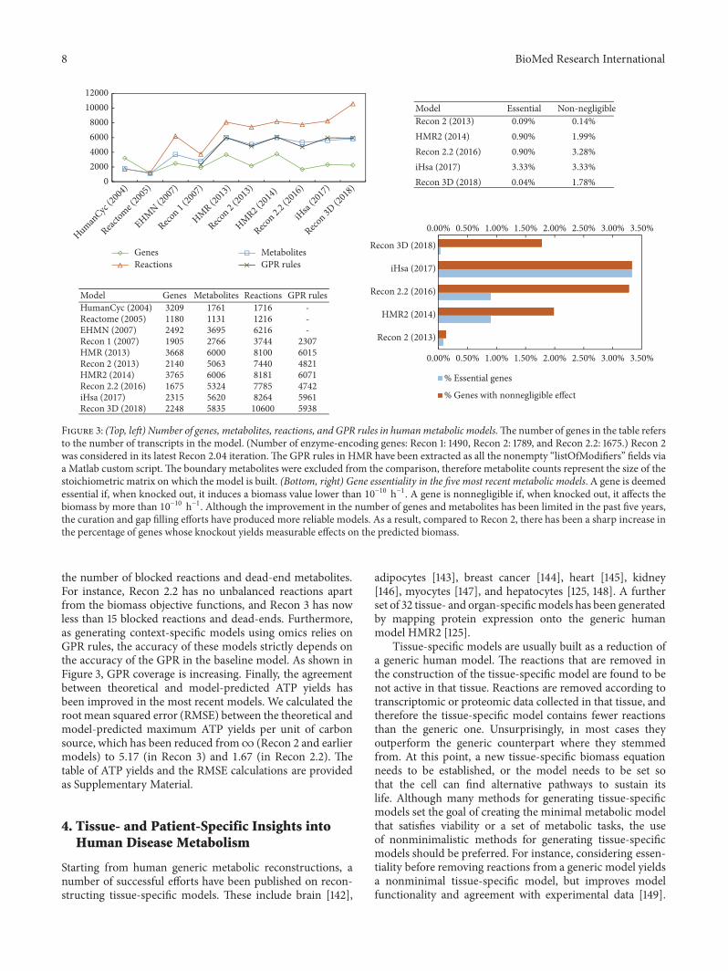
Figure 3 summarizes the progress made by the metabolicmodelling community over the last 15 years. The number ofgenes, metabolites and reactions is reported in the figure. Tobetter quantify the connectedness of a model, we computedthe number of genes whose knockout yields measurableeffects in the model as measured by the predicted biomassflux. To account for the tolerance of the linear solver, wedefined a gene essential if its knockout produces biomass< 10
−10 h−1; we defined a gene nonnegligible if its knockoutcauses a biomass variation > 10−10 h−1. Compared to Recon2, the increase in the number of genes whose variation hasa nonnegligible effect on the growth rate shows that effortshave been successfully made towards curation, gap filling,and consistency checks, as detailed in Section 3.1.
3.1. From Size-Focused “Modelathons” to Manual Curation.When the first Recon 1 [118] and EHMN [119] humanmetabolic reconstructions were published in 2007, Recon 1was preferred in many studies because it contained morereactions. In fact, this was only due to the compartmen-talization of Recon 1, with metabolites repeated in differentcompartments and transport reactions between compart-ments. The number of unique reactions was indeed greaterin EHMN (1028 more reactions and 1202 more metabolites),and a subsequent compartmentalized version of EHMN wasgenerated [120].
These frequent comparisons among different modelsindicate that the main focus was initially to include asmany reactions as possible—a size-focused “modelathon”approach—often at the expenses of curation and accuracy.Conversely, the approach of including all the available biolog-ical components in amodel is now always coupledwith exten-sive team-based curation, consistency checks, uniformity ofannotations and gap filling. It is not uncommon to achievea better model by removing reactions from an existing one.This encouraged the community to focus on the accuracy ofthe models and solvers used, rather than only on the modelsize.
Manual curation is therefore considered one of the mainsteps in generating a metabolic model. Tools like MetaNetX[134] simplify curation efforts and suggest identifier map-pings to unify reactions and metabolites identifiers. Suchcuration efforts are likely to become even more importantin the near future given the recent advances of automatedreconstruction tools, now able to generate a full workingdraft of a model, e.g., MicrobesFlux [135], Pathway Tools[136], PathwayBooster [137], CoReCo [138], andMerlin [139].Given the importance of obtaining reconciled and high-quality models, MetaNetX also provides a database of modelsgenerated after reconciliation of metabolites and biochemicalreactions [134]. More recently, CarveMe [140] and RAVEN2.0 [59] have shown high potential in automating man-ual steps, achieving for the first time an accuracy directlycomparable with manually curated models when predictingexperimental phenotypes. Tools for checking inconsistenciesand for visually inspecting the model are also available [141].
Over the last few years, the curation efforts have yielded areduction in the number of unbalanced reactions, as well as in
8 BioMed Research International
02000400060008000
1000012000
0.00% 0.50% 1.00% 1.50% 2.00% 2.50% 3.00% 3.50%
0.00% 0.50% 1.00% 1.50% 2.00% 2.50% 3.00% 3.50%
HMR2 (2014)
Recon 2 (2013)
iHsa (2017)
Recon 2.2 (2016)
Recon 3D (2018)
% Essential genes
% Genes with nonnegligible effect
Model Essential Non-negligible0.09% 0.14%0.90% 1.99%0.90% 3.28%3.33% 3.33%
Recon 2 (2013)HMR2 (2014)Recon 2.2 (2016)iHsa (2017)Recon 3D (2018) 0.04% 1.78%
Model Genes Metabolites Reactions GPR rules3209 1761 1716 -1180 1131 1216 -2492 3695 6216 -1905 2766 3744 23073668 6000 8100 60152140 5063 7440 48213765 6006 8181 60711675 5324 7785 47422315 5620 8264 5961
HumanCyc (2004)Reactome (2005)EHMN (2007)Recon 1 (2007)HMR (2013)Recon 2 (2013)HMR2 (2014)Recon 2.2 (2016)iHsa (2017)Recon 3D (2018) 2248 5835 10600 5938
HumanCyc
(2004
)
Reactome (
2005
)
EHMN (200
7)
Recon 1 (
2007
)
HMR (201
3)
Recon 2 (
2013
)
HMR2 (20
14)
Recon 2.2
(201
6)
iHsa
(2017
)
Recon 3D
(201
8)
GenesReactions
MetabolitesGPR rules
Figure 3: (Top, left) Number of genes, metabolites, reactions, and GPR rules in humanmetabolic models.Thenumber of genes in the table refersto the number of transcripts in the model. (Number of enzyme-encoding genes: Recon 1: 1490, Recon 2: 1789, and Recon 2.2: 1675.) Recon 2was considered in its latest Recon 2.04 iteration.The GPR rules in HMR have been extracted as all the nonempty “listOfModifiers” fields viaa Matlab custom script. The boundary metabolites were excluded from the comparison, therefore metabolite counts represent the size of thestoichiometric matrix on which the model is built. (Bottom, right) Gene essentiality in the five most recent metabolic models. A gene is deemedessential if, when knocked out, it induces a biomass value lower than 10−10 h−1. A gene is nonnegligible if, when knocked out, it affects thebiomass by more than 10−10 h−1. Although the improvement in the number of genes and metabolites has been limited in the past five years,the curation and gap filling efforts have produced more reliable models. As a result, compared to Recon 2, there has been a sharp increase inthe percentage of genes whose knockout yields measurable effects on the predicted biomass.
the number of blocked reactions and dead-end metabolites.For instance, Recon 2.2 has no unbalanced reactions apartfrom the biomass objective functions, and Recon 3 has nowless than 15 blocked reactions and dead-ends. Furthermore,as generating context-specific models using omics relies onGPR rules, the accuracy of these models strictly depends onthe accuracy of the GPR in the baseline model. As shown inFigure 3, GPR coverage is increasing. Finally, the agreementbetween theoretical and model-predicted ATP yields hasbeen improved in the most recent models. We calculated therootmean squared error (RMSE) between the theoretical andmodel-predicted maximum ATP yields per unit of carbonsource, which has been reduced from∞ (Recon 2 and earliermodels) to 5.17 (in Recon 3) and 1.67 (in Recon 2.2). Thetable of ATP yields and the RMSE calculations are providedas Supplementary Material.
4. Tissue- and Patient-Specific Insights intoHuman Disease Metabolism
Starting from human generic metabolic reconstructions, anumber of successful efforts have been published on recon-structing tissue-specific models. These include brain [142],
adipocytes [143], breast cancer [144], heart [145], kidney[146], myocytes [147], and hepatocytes [125, 148]. A furtherset of 32 tissue- and organ-specificmodels has been generatedby mapping protein expression onto the generic humanmodel HMR2 [125].
Tissue-specific models are usually built as a reduction ofa generic human model. The reactions that are removed inthe construction of the tissue-specific model are found to benot active in that tissue. Reactions are removed according totranscriptomic or proteomic data collected in that tissue, andtherefore the tissue-specific model contains fewer reactionsthan the generic one. Unsurprisingly, in most cases theyoutperform the generic counterpart where they stemmedfrom. At this point, a new tissue-specific biomass equationneeds to be established, or the model needs to be set sothat the cell can find alternative pathways to sustain itslife. Although many methods for generating tissue-specificmodels set the goal of creating the minimal metabolic modelthat satisfies viability or a set of metabolic tasks, the useof nonminimalistic methods for generating tissue-specificmodels should be preferred. For instance, considering essen-tiality before removing reactions from a generic model yieldsa nonminimal tissue-specific model, but improves modelfunctionality and agreement with experimental data [149].

BioMed Research International 9
Since the generic models are a superset of the tissue-specificmodels, predictions of generic models may still be correct intissue-specific problems, but less accurate.
Context-specific human metabolic models have shedlight on disease onset and progression in a range of recentcase studies. In this context, the renewed interest in diseasemetabolism is due to the fact that a holistic genome-scaleview, rather than a single-gene approach, is necessary tofully characterize most diseases. For instance, in cancer, thedifferences in the metabolic pathways between cancer cellsand their parent tissue have been characterized using omicsdata and a human metabolic model [75, 150, 151]. As a result,tissue- and cell-specific metabolic models have been suc-cessfully used to identify—and successively validate—specificdrug targets that inhibit cancer proliferation but do not affectnormal cell proliferation [74, 152]. With a similar approach,submodels built from human genome-scale models havebeen used to generate several testable hypotheses, e.g., tocompare wild-type and Fh1-deficient kidney mouse cells,and to predict further gene knockouts that affect growthin the Fh1-deficient cells but do not affect the wild-typecells, therefore suggesting targets for treating hereditaryleiomyomatosis and renal-cell cancer [153].
For specific diseases, tissue-specific models have beenused successfully to identify biomarkers and therapeutic tar-gets [125]. Using RNA-Seq expression levels in combinationwith genome-scale models has enabled the reconstructionof cancer cell line-specific models, enabling the discoveryof metabolites supporting proliferation, and antimetabo-lites leading to cell death [154]. (An antimetabolite isa compound that simultaneously inhibits those enzymesinvolved in metabolizing the associated endogenous metabo-lite. Antimetabolites can affect multiple enzymes at the sametime and can reduce proliferation, and are therefore usedas anticancer drugs.) After integration with extracellularmetabolomic data and transcriptomic profiles, metabolicmodelling has reliably characterized intracellular metabolismof lymphoblastic leukemia cell lines [155]. A combination ofdifferent omics data and flux splits have been used to generatecancer-specific models of the NCI60 panel, achieving corre-lation close to 1 in predicting the remaining flux rates [13].
Following the same direction, personalized models (e.g.,using patients’ omic data to constrain the generic reconstruc-tion) hold promise to become key for precision medicine(Figure 2). The largest study to date with patient-specificmodels is the Human Pathology Atlas [77]. The authors builtpersonalized genome-scale models of cancer in each patientacross 17 tissues. This allowed investigating the metabolicdifferences between different cancers, as well as patient-specific biological functions. In another recent study [156], 86patients with nonalcoholic fatty liver disease were recruited,and their personalized hepatocyte genome-scale metabolicmodels were built using patient-specific experimental dataon lipoprotein fluxes. A new molecular mechanism of thedisease was elucidated as a result of personalized metabolicmodelling.
Genotyping coupled with patient-specific metabolicmodelling offers a new opportunity for personalizedmedicine. In fact, new biomarkers can be predicted in a
patient-specific fashion, and personalized therapies cantherefore be designed and subsequently assessed in termsof their metabolic mechanistic effects [157]. As shown in arecent case of arginase deficiency (a urea cycle disorder),different individuals can respond differently to the samedisease and treatment, and this can often be flagged observingtheir individual metabolic response [158].
Due to the importance of the gut microbiome com-position in human health, a set of modelling approacheshas been proposed recently [159]. Using host-microbiomemodelling, e.g., combining metabolic community modellingwith human metabolic models, the interplay among the gutbacteria and their interaction with the surrounding humancells can be investigated [160, 161]. Personalized models of gutmicrobiome have been built from patient-specific metage-nomic data in order to elucidate individual-specific bileacid production in microbiomes of healthy individuals andpatients with inflammatory bowel disease [162]. Althoughseveral modelling challenges remain to be solved [163], anapproach based on metabolic modelling is likely to shedlight on the role of human gut microbial communities inhuman health, therefore suggesting potential dietary changesor personalized intervention on gut composition [164, 165].
5. Discussion and Perspective
Research in computational biology has led to detailed mod-els for a better understanding of the individual biologicalcomponents, but arguably to a less clear picture of theinteractions among the components that result in a givenphenotype [166]. Genome-scale systems biology studies caneffectively address this issue. For instance, in biomedicalapplications, this holistic view is necessary to characterize apatient’s disease phenotype and deliver precision medicine[17].
Since metabolic homeostasis and observable phenotypeare strictly linked, metabolism is nowadays considered diag-nostic of the phenotype, and therefore arguably the bestindicator of the functional state of a cell. Metabolism canalso be used to prioritize genes and assess their functionand the role of gene perturbations (including knockouts).Without such integrated analysis, a gene may e.g., incorrectlybe regarded as important only due to its highly variableexpression value.
Metabolic models are increasingly being used to con-struct multiscale, multicellular or multitissue models. In2012, a research effort by Karr et al. [21] provided the firstwhole-cell computational model of the life cycle of a smallpathogenic bacterium, Mycoplasma genitalium. The modelincludes metabolism, replication of the genome, and celldivision. Several metabolic models can be combined inframeworks to investigate the metabolic exchanges betweenindividual cells and the emerging community behavior [167],with applications ranging from microbial communities tohost-pathogen interactions and cancer proliferation [168–170].
The availability of tissue-specific models has also ledto the generation of multitissue metabolic models [171].
10 BioMed Research International
Further steps in this direction will soon allow studyingwhole-body metabolism models. The first study towards thisgoal used dynamic parsimonious FBA to combine steady-state FBA with differential equations for the concentrationof metabolites in each organ [172]. As a result, a whole-body and inherently multiscale model was generated with 14organs plus the human serum. More recently, the first whole-body metabolic models were generated named Harvey andHarvetta, where themetabolic network for every organ/tissuewas built simultaneously from Recon3D [173]. Manufactur-ing organ-on-a-chip and ultimately body-on-a-chip deviceswith metabolic models (see e.g., [174]) seems the next step inthis direction.
Given the recently renewed interest of the scientificcommunity in understanding disease metabolism, it is highlylikely that cancer metabolism (and metabolism in otherdiseases) will become the main research topic in drug devel-opment. This will complement rather than replace standardtranscriptomic-only studies, and will provide a proxy forthe phenotypic and observable outcome. The idea is that toimprove phenotypic predictions, the signatures need to betaken from sources of data that are closer to the phenotype.The integration of omics data into metabolic models hasenabled the prediction and successively the validation ofbiomarkers and therapeutic targets. In drug design, althoughgenomics, transcriptomics and proteomics are often deemedsufficient, metabolomics can elucidate mechanisms that arenot visible from genes and protein activity. More importantly,it can address cases where the genes responsible for a diseaseare well known but not druggable, but their correspondingdownstream reactions are [175].
Genomics analysis is usually able to identify knowndiseases through genemutations. However, it may not be ableto flag variants of known diseases, or identify novel diseases.This can happen, for instance, if (i) a gene mutation is notflagged as important; (ii) the gene is not screened at all; (iii)the disease is not a direct effect of a gene perturbation; (iv)individuals respond differently to the same mutation [158].Genome-scale models can elucidate mechanistic modes ofdrug action, side effects (both off-target drug binding anddownstream transcriptional effects), and potential toxicityof drugs by linking omics data to the phenotype through acondition-specific model [8, 176]. In a patient-specific frame-work, such biological data and markers can be predicted in apersonalized fashion, paving the way for in silico clinical trials[177].
The efforts of the metabolic modelling community are farfrom being complete. In this regard, we envisage a joint effortfrom metabolic modelling and genome-wide associationstudies (GWAS) communities to identify gene-metaboliteinteractions that are currently not included in metabolicmodels using state-of-the-art association mappings [178].Computational tools combining both approaches would gotowards genome-scale detection of errors and missing enzy-matic reactions, remarkably improving the predictive abilityof metabolic models. For instance, this could be achieved byminimizing the error between predictions of flux couplingand experimental coexpression data integrated with GWAS[179, 180].
Likewise, the methods for integration of omics data ingenome-scale models still show room for improvement. Forinstance, in yeast and Escherichia coli, single-omic integrationin FBA has been reported to give similar accuracy to pFBAwithout omic integration [78]. Reassuringly, reaction-specificrules to constrain flux rates can halve the normalized errorof pFBA [84]. In multicellular organisms, a better correla-tion between gene expression and metabolic fluxes can beexpected [78]. In mammalian cells, the main contributors tothe overall protein expression level aremRNA levels [181, 182],while in most normal and cancer cell lines, mRNA and pro-tein levels were found to correlate positively [183, 184]. Whiledifferent methods (and their parameters) can yield differentcontext-specific models, the accuracy in predicting essentialgenes is almost always higher in context-specific modelsthan in the generic human models [81]. Therefore, metabolicmodels can be regarded as useful tools tomechanistically linktranscriptomic data with flux rates. Usingmultiple omics dataand thermodynamic constraints simultaneously [185, 186], ora combination of regularized FBA methods and omics data,can improve the reliability of the predictions [187].
Compared to the well-characterized microbial metabolicreconstructions, gaps still present in our knowledge of humanmetabolism—including characterization of enzymes, the def-inition of cell-specific metabolic functions and tissue-specificgrowth mechanisms—make it more difficult to test (andcompare) metabolic models and omic integration methods.Humanmetabolic models and methods have been tested andcross-compared for gene essentiality through CRISPR-Cas9-mediated loss-of-function screens, and for their ability topredict growth rate and recapitulate known metabolic func-tions [81]. A dataset including both omics data and measuredmetabolic information is the NCI-60 panel, with metaboliteuptake and secretion rates published in 2012 [188]. While E.coli and yeast fluxomic data is publicly available from severalexperiments [189–191], to the best of our knowledge NCI-60is the only publicly available human-cell dataset containingexpression levels, metabolic flux rates and proliferation rates,and therefore suitable for validating FBA methods. Due tothe lack of fluxomic datasets, the difficulty in choosing areliable objective function, and the larger size compared tobacterial models, further experimental validation is neededfor human metabolic models, especially when using thefull flux distribution to inform decisions or subsequentalgorithmic steps.
Finally, despite many recent advances, gathering insightsfrom the data generated through omic-informed modelsremains a bottleneck in systems biology. We therefore envis-age that genome-scale metabolic models will be increasinglyinvestigated with machine/deep learning algorithms in apatient-specific fashion [13, 192]. In disease modelling, theusefulness of general biomarker discovery is debatable wheninformation on patients is not taken into account, becausemost biological components would be flagged as perturbedin a general “disease versus normal” analysis. Conversely,if the appropriate person-specific data is integrated witha model, biomarkers can become the central part of pre-cision medicine, where data acquired on patients drivepredictions, analysis and therapeutics [17]. In a modelling

BioMed Research International 11
analogy, machine learning algorithms alone cannot providemechanistic information in the biological processes theysimulate or mimic. However, if machine learning is cou-pled with multiomic genome-scale modelling, the combina-tion of experimentally and model-generated omic data canpredict—and explain mechanistically—personalized therapypredictions by including key biological information in thelearning process.
Conflicts of Interest
The author declares that they have no conflicts of interest.
Supplementary Materials
Agreement between theoretical and model-predicted ATPyields for the most recent human metabolic models. Wecalculate the root mean squared error (RMSE) between thetheoretical and model-predicted maximum ATP yields (perunit of carbon source) for the most recent human metabolicmodels. The RMSE has improved from ∞ (Recon 2 andearlier models) to 5.17 (in Recon 3) and 1.67 (in Recon 2.2).(Supplementary Materials)
References
[1] G. Piedrafita, M. Keller, and M. Ralser, “The impact ofnon-enzymatic reactions and enzyme promiscuity on cellularmetabolism during (oxidative) stress conditions,” Biomolecules,vol. 5, no. 3, pp. 2101–2122, 2015.
[2] N. N. Pavlova and C. B. Thompson, “The emerging hallmarksof cancer metabolism,” Cell Metabolism, vol. 23, no. 1, pp. 27–47,2016.
[3] J. T. Yurkovich and B. O. Palsson, “Solving puzzles withmissing pieces: The power of systems biology [Point of View],”Proceedings of the IEEE, vol. 104, no. 1, pp. 2–7, 2016.
[4] A. Bordbar, J. T. Yurkovich, G. Paglia, O. Rolfsson, O. E. Sig-urjonsson, and B. O. Palsson, “Elucidating dynamic metabolicphysiology through network integration of quantitative time-course metabolomics,” Scientific Reports, vol. 7, p. 46249, 2017.
[5] C. Angione, M. Conway, and P. Lio, “Multiplex methodsprovide effective integration of multi-omic data in genome-scale models,” BMC Bioinformatics, vol. 17, p. 257, 2016.
[6] E. Motamedian, M. Mohammadi, S. A. Shojaosadati, andM. Heydari, “TRFBA: An algorithm to integrate genome-scale metabolic and transcriptional regulatory networks withincorporation of expression data,” Bioinformatics, vol. 33, no. 7,pp. 1057–1063, 2017.
[7] F. Eyassu and C. Angione, “Modelling pyruvate dehydrogenaseunder hypoxia and its role in cancer metabolism,” Royal SocietyOpen Science, vol. 4, no. 10, Article ID 170360, 2017.
[8] V. Dougherty, T. J. Moutinho Jr, and J. Papin, “Acceleratingthe drug development pipeline with genome-scale metabolicnetwork reconstructions,” Systems Biology, vol. 6, 2017.
[9] E. Yaneske and C. Angione, “The poly-omics of ageing throughindividual-based metabolic modelling,” BMC Bioinformatics,vol. 19, p. 415, 2018.
[10] L. Tobalina, J. Pey, A. Rezola, and F. J. Planes, “Assessmentof FBA based gene essentiality analysis in cancer with a fast
context-specific network reconstruction method,” PLoS ONE,vol. 11, no. 5, p. e0154583, 2016.
[11] A. Occhipinti, F. Eyassu, T. J. Rahman, P. K. Rahman, and C.Angione, “In silico engineering of Pseudomonas metabolismreveals new biomarkers for increased biosurfactant produc-tion,” PeerJ, vol. 6, p. e6046, 2018.
[12] Z. Qi and E. O. Voit, “Inference of cancer mechanisms throughcomputational systems analysis,” Molecular BioSystems, vol. 13,no. 3, pp. 489–497, 2017.
[13] D. C. Zielinski, N. Jamshidi, A. J. Corbett, A. Bordbar, A.Thomas, and B. O. Palsson, “Systems biology analysis of driversunderlying hallmarks of cancer cell metabolism,” ScientificReports, vol. 7, 2017.
[14] Z. Dai and J.W. Locasale, “Understandingmetabolismwith fluxanalysis: From theory to application,” Metabolic Engineering,vol. 43, pp. 94–102, 2017.
[15] M. A. Keibler, T. M.Wasylenko, J. K. Kelleher, O. Iliopoulos, M.G. VanderHeiden, and G. Stephanopoulos, “Metabolic require-ments for cancer cell proliferation,” Cancer & Metabolism, vol.4, article no. 16, 2016.
[16] D. Welter, J. MacArthur, J. Morales et al., “The NHGRI GWASCatalog, a curated resource of SNP-trait associations,” NucleicAcids Research, vol. 42, no. 1, pp. D1001–D1006, 2014.
[17] J. Nielsen, “Systems biology ofmetabolism: a driver for develop-ing personalized and precision medicine,” Cell Metabolism, vol.25, no. 3, pp. 572–579, 2017.
[18] H. Kitano, “Systems biology: a brief overview,” Science, vol. 295,pp. 1662–1664, 2002.
[19] M. Tomita, K. Hashimoto, K. Takahashi et al., “E-CELL: Soft-ware environment for whole-cell simulation,” Bioinformatics,vol. 15, no. 1, pp. 72–84, 1999.
[20] M. Tomita, “Whole-cell simulation: A grand challenge of the21st century,”Trends in Biotechnology, vol. 19, no. 6, pp. 205–210,2001.
[21] J. R. Karr, J. C. Sanghvi, D. N. MacKlin et al., “A whole-cellcomputational model predicts phenotype from genotype,” Cell,vol. 150, no. 2, pp. 389–401, 2012.
[22] A. P. Goldberg, B. Szigeti, Y. H. Chew, J. A. Sekar, Y. D. Roth,and J. R. Karr, “Emerging whole-cell modeling principles andmethods,” Current Opinion in Biotechnology, vol. 51, pp. 97–102,2018.
[23] A. C. Babtie andM. P. H. Stumpf, “How to deal with parametersfor whole-cell modelling,” Journal of the Royal Society Interface,vol. 14, Article ID 20170237, 2017.
[24] P. Palumbo, M. Vanoni, F. Papa, S. Busti, and L. Alberghina,Whole yeast model: what and why, 2018.
[25] A. Khodayari, A. R. Zomorrodi, J. C. Liao, and C. D. Maranas,“A kinetic model of Escherichia coli core metabolism satisfyingmultiple sets ofmutant flux data,”Metabolic Engineering, vol. 25,pp. 50–62, 2014.
[26] K. Tummler and E. Klipp, “The discrepancy between datafor and expectations on metabolic models: How to matchexperiments and computational efforts to arrive at quantitativepredictions?”Current Opinion in Systems Biology, vol. 8, pp. 1–6,2018.
[27] C. Ramon, M. Gollub, and J. Stelling, “Integrating –omics datainto genome-scale metabolic network models: principles andchallenges,” Essays in Biochemistry, vol. 62, no. 4, pp. 563–574,2018.
[28] A. Khodayari and C. D. Maranas, “A genome-scale Escherichiacoli kinetic metabolic model k-ecoli457 satisfying flux data

12 BioMed Research International
for multiple mutant strains,” Nature Communications, vol. 7, p.13806, 2016.
[29] A. A.Mannan, Y. Toya, K. Shimizu, J.McFadden, A.M.Kierzek,and A. Rocco, “Integrating kinetic model of E. coli with genomescale metabolic fluxes overcomes its open system problem andreveals bistability in centralmetabolism,” PLoS ONE, vol. 10, no.10, Article ID e0139507, 2015.
[30] A. Zelezniak, S. Sheridan, and K. R. Patil, “Contribution ofnetwork connectivity in determining the relationship betweengene expression and metabolite concentration changes,” PLoSComputational Biology, vol. 10, Article ID e1003572, 2014.
[31] M. Kanehisa, M. Furumichi, M. Tanabe, Y. Sato, and K.Morishima, “KEGG: new perspectives on genomes, pathways,diseases and drugs,” Nucleic Acids Research, vol. 45, no. 1, pp.D353–D361, 2017.
[32] P. D. Karp, R. Billington, R. Caspi et al., “The BioCyc collectionof microbial genomes and metabolic pathways,” Briefings inBioinformatics, vol. 1, p. 9, 2017.
[33] S. Placzek, I. Schomburg, A. Chang et al., “BRENDA in 2017:New perspectives and new tools in BRENDA,” Nucleic AcidsResearch, vol. 45, no. 1, pp. D380–D388, 2017.
[34] D. A. Cuevas, J. Edirisinghe, C. S. Henry, R. Overbeek, T. G.O’Connell, and R. A. Edwards, “From DNA to FBA: How tobuild your own genome-scale metabolic model,” Frontiers inMicrobiology, vol. 7, 2016.
[35] M. Fondi and P. Lio, “Genome-scale metabolic network recon-struction,” Bacterial Pangenomics: Methods and Protocols, pp.233–256, 2015.
[36] I. Thiele and B. Ø. Palsson, “A protocol for generating ahigh-quality genome-scale metabolic reconstruction,” NatureProtocols, vol. 5, no. 1, pp. 93–121, 2010.
[37] Ø. Palsson, Systems Biology: Constraint-Based Reconstructionand Analysis, Cambridge University Press, 2015.
[38] M. Conway, C. Angione, and P. Lio, “Iterative multi levelcalibration of metabolic networks,” Current Bioinformatics, vol.11, no. 1, pp. 93–105, 2016.
[39] F. Shen, R. Sun, J. Yao et al., “OptRAM: In-silico strain designvia integrative regulatory-metabolic network modeling,” PLoSComputational Biology, vol. 15, Article ID e1006835, 2019.
[40] A. von Kamp and S. Klamt, “Enumeration of smallest inter-vention strategies in genome-scale metabolic networks,” PLoSComputational Biology, vol. 10, 2014.
[41] C. Angione, J. Costanza, G. Carapezza, P. Lio, and G. Nicosia,“Analysis and design of molecular machines,”Theoretical Com-puter Science, vol. 599, pp. 102–117, 2015.
[42] P. Schneider and S. Klamt, “Characterizing and ranking com-puted metabolic engineering strategies,” Bioinformatics, 2019.
[43] J. D. Orth, I. Thiele, and B. O. Palsson, “What is flux balanceanalysis?” Nature Biotechnology, vol. 28, no. 3, pp. 245–248,2010.
[44] R. Mahadevan, J. S. Edwards, and F. J. Doyle III, “Dynamicflux balance analysis of diauxic growth in Escherichia coli,”Biophysical Journal, vol. 83, no. 3, pp. 1331–1340, 2002.
[45] N. Topfer, S. Kleessen, and Z. Nikoloski, “Integration ofmetabolomics data into metabolic networks,” Frontiers in PlantScience, vol. 6, p. 49, 2015.
[46] P. Salvy, G. Fengos, M. Ataman et al., “pyTFA and matTFA:a python package and a matlab toolbox for thermodynamics-based flux analysis,” Bioinformatics, pp. 167–169, 2018.
[47] E. Brunk, R. L. Chang, J. Xia et al., “Systemic post-translationalcontrol of bacterialmetabolism regulates adaptation in dynamicenvironments,” 2018.
[48] M. MacGillivray, A. Ko, E. Gruber, M. Sawyer, E. Almaas, andA.Holder, “Robust analysis of fluxes in genome-scalemetabolicpathways,” Scientific Reports, vol. 7, p. 268, 2017.
[49] E. Ebrahim, E. Almaas, A. Bauer et al. et al., “Do genome-scalemodels need exact solvers or clearer standards?”Molecularsystems biology, vol. 11, article no. 831, 2015.
[50] L. Chindelevitch, J. Trigg, A. Regev, and B. Berger, “Reply to “dogenome-scalemodels need exact solvers or clearer standards?”,”Molecular Systems Biology, vol. 11, no. 10, 2015.
[51] P. D. Karp, D. Weaver, and M. Latendresse, “How accurateis automated gap filling of metabolic models?” BMC SystemsBiology, vol. 12, p. 73, 2018.
[52] N. E. Lewis, K. K. Hixson, T. M. Conrad et al., “Omic data fromevolved E. coli are consistent with computed optimal growthfrom genome-scale models,” Molecular Systems Biology, vol. 6,p. 390, 2010.
[53] D.Machado,M. J. Herrgard, I. Rocha, and K. R. Patil, “Stoichio-metric representation of gene–protein–reaction associationsleverages constraint-based analysis from reaction to gene-levelphenotype prediction,” PLoS Computational Biology, vol. 12, no.10, p. e1005140, 2016.
[54] M. K. Kim, A. Lane, J. J. Kelley, and D. S. Lun, “E-Flux2 andsPOT: Validated methods for inferring intracellular metabolicflux distributions from transcriptomic data,” PLoS ONE, vol. 11,Article ID e0157101, 2016.
[55] K. Smallbone and E. Simeonidis, “Flux balance analysis: ageometric perspective,” Journal of Theoretical Biology, vol. 258,no. 2, pp. 311–315, 2009.
[56] N. D. Price, J. Schellenberger, and B. O. Palsson, “Uniform sam-pling of steady-state flux spaces: Means to design experimentsand to interpret enzymopathies,” Biophysical Journal, vol. 87, no.4, pp. 2172–2186, 2004.
[57] J. Schellenberger, R. Que, R. M. T. Fleming et al., “Quantitativeprediction of cellular metabolism with constraint-based mod-els: the COBRA Toolbox v2.0,” Nature Protocols, vol. 6, no. 9,pp. 1290–1307, 2011.
[58] A. Ebrahim, J. A. Lerman, B. Ø. Palsson, and D. R. Hyduke,“COBRApy: COnstraints-based reconstruction and analysis forpython,” BMC Systems Biology, vol. 7, no. 1, article 74, 2013.
[59] H. Wang, S. Marcisauskas, B. J. Sanchez et al., “RAVEN 2.0:A versatile toolbox for metabolic network reconstruction anda case study on Streptomyces coelicolor,” PLoS ComputationalBiology, vol. 14, no. 10, p. e1006541, 2018.
[60] M. Latendresse, M. Krummenacker, M. Trupp, and P. D. Karp,“Construction and completion of flux balance models frompathway databases,” Bioinformatics, vol. 28, no. 3, pp. 388–396,2012.
[61] J. Boele, B. G. Olivier, and B. Teusink, “FAME, the flux analysisand modeling environment,” BMC Systems Biology, vol. 6,article no. 8, 2012.
[62] J. P. Faria, M. Rocha, I. Rocha, and C. S. Henry, “Methodsfor automated genome-scale metabolic model reconstruction,”Biochemical Society Transactions, vol. 46, no. 4, pp. 931–936,2018.
[63] M. Lakshmanan, G. Koh, B. K. S. Chung, and D.-Y. Lee,“Software applications for flux balance analysis,” Briefings inBioinformatics, vol. 15, no. 1, pp. 108–122, 2014.
[64] L. K. Reed, C. F. Baer, and A. S. Edison, “Considerations whenchoosing a genetic model organism for metabolomics studies,”Current Opinion in Chemical Biology, vol. 36, pp. 7–14, 2017.

BioMed Research International 13
[65] Y. Hasin, M. Seldin, and A. Lusis, “Multi-omics approaches todisease,” Genome Biology, vol. 18, p. 83, 2017.
[66] L. Heirendt, S. Arreckx, T. Pfau et al., “Creation and analysis ofbiochemical constraint-based models: the cobra toolbox v3. 0,”https://arxiv.org/abs/1710.04038, 2017.
[67] C. F. Arias, P. Catalan, S. Manrubia, and J. A. Cuesta, “ToyLIFE:A computational framework to study the multi-level organisa-tion of the genotype-phenotype map,” Scientific Reports, vol. 4,article no. 7549, 2014.
[68] S. S. Fong, A. R. Joyce, and B. Ø. Palsson, “Parallel adaptiveevolution cultures of Escherichia coli lead to convergent growthphenotypes with different gene expression states,” GenomeResearch, vol. 15, no. 10, pp. 1365–1372, 2005.
[69] C. Angione and P. Lio, “Predictive analytics of environmentaladaptability in multi-omic network models,” Scientific Reports,vol. 5, p. 15147, 2015.
[70] J. Y. Ryu, H. U. Kim, and S. Y. Lee, “Reconstruction of genome-scale human metabolic models using omics data,” IntegrativeBiology, vol. 7, no. 8, pp. 859–868, 2015.
[71] S. S. Kashaf, C. Angione, and P. Lio, “Making life difficultfor Clostridium difficile: Augmenting the pathogen’s metabolicmodel with transcriptomic and codon usage data for bettertherapeutic target characterization,” BMC Systems Biology, vol.11, p. 25, 2017.
[72] M. Larocque, T. Chenard, and R. Najmanovich, “A curated C.difficile strain 630 metabolic network: prediction of essentialtargets and inhibitors,” BMC Systems Biology, vol. 8, p. 117, 2014.
[73] A. Mardinoglu, R. Agren, C. Kampf et al., “Integration ofclinical datawith a genome-scalemetabolicmodel of the humanAdipocyte,”Molecular Systems Biology, vol. 9, p. 649, 2013.
[74] O. Folger, L. Jerby, C. Frezza, E. Gottlieb, E. Ruppin, and T.Shlomi, “Predicting selective drug targets in cancer throughmetabolic networks,” Molecular Systems Biology, vol. 7, p. 501,2011.
[75] K. Yizhak, B. Chaneton, E. Gottlieb, and E. Ruppin, “Modelingcancer metabolism on a genome scale,” Molecular SystemsBiology, vol. 11, p. 817, 2015.
[76] N. E. Lewis and A. M. Abdel-Haleem, “The evolution ofgenome-scale models of cancer metabolism,” Frontiers in Phys-iology, vol. 4, 2013.
[77] M. Uhlen, C. Zhang, S. Lee et al., “A pathology atlas of thehuman cancer transcriptome,” Science, vol. 357, no. 6352, ArticleID eaan2507, 2017.
[78] D. Machado and M. Herrgard, “Systematic evaluation of meth-ods for integration of transcriptomic data into constraint-basedmodels ofmetabolism,”PLoS Computational Biology, vol. 10, no.4, Article ID e1003580, 2014.
[79] J. S. Cho, C. Gu, T. H. Han, J. Y. Ryu, and S. Y. Lee, “Reconstruc-tion of context-specific genome-scale metabolic models usingmultiomics data to study metabolic rewiring,” Current Opinionin Systems Biology, vol. 15, pp. 1–11, 2019.
[80] S. Vijayakumar, M. Conway, P. Lio, and C. Angione, “Seeing thewood for the trees: a forest of methods for optimization andomic-network integration in metabolic modelling,” Briefings inBioinformatics, 2017.
[81] S. Opdam, A. Richelle, B. Kellman, S. Li, D. C. Zielinski, andN. E. Lewis, “A systematic evaluation of methods for tailoringgenome-scale metabolic models,” Cell Systems, vol. 4, no. 3, pp.318–329, 2017.
[82] C. Angione, “Integrating splice-isoform expression intogenome-scale models characterizes breast cancer metabolism,”Bioinformatics, vol. 34, no. 3, pp. 494–501, 2018.
[83] J. Y. Ryu, H. U. Kim, and S. Y. Lee, “Framework and resource formore than 11,000 gene-transcript-protein-reaction associationsin human metabolism,” Proceedings of the National Acadamyof Sciences of the United States of America, vol. 114, no. 45, pp.E9740–E9749, 2017.
[84] M. Tian and J. L. Reed, “Integrating Proteomic or Transcrip-tomic Data into Metabolic Models Using Linear Bound FluxBalance Analysis,” Bioinformatics, vol. 34, p. 3882, 2018.
[85] J. Costanza, G. Carapezza, C. Angione, P. Lio, and G. Nicosia,“Robust design of microbial strains,” Bioinformatics, vol. 28, no.23, pp. 3097–3104, 2012.
[86] A. Kiparissides and V. Hatzimanikatis, “Thermodynamics-based Metabolite Sensitivity Analysis in metabolic networks,”Metabolic Engineering, vol. 39, pp. 117–127, 2017.
[87] E. W. Birch, M. Udell, and M. W. Covert, “Incorporation offlexible objectives and time-linked simulation with flux balanceanalysis,” Journal ofTheoretical Biology, vol. 345, pp. 12–21, 2014.
[88] J. C.Xavier,K.R. Patil, and I. Rocha, “Integration of biomass for-mulations of genome-scale metabolic models with experimen-tal data reveals universally essential cofactors in prokaryotes,”Metabolic Engineering, vol. 39, pp. 200–208, 2017.
[89] A. M. Feist and B. O. Palsson, “What do cells actually want?”Genome Biology, vol. 17, no. 1, 2016.
[90] B. Dikicioglu, S. G. Krdar, and Oliver., “Biomass compo-sition: the elephant in the room of metabolic modelling,”Metabolomics, vol. 11, pp. 1690–1701, 2015.
[91] S. H. J. Chan, J. Cai, L. Wang, M. N. Simons-Senftle, and C.D. Maranas, “Standardizing biomass reactions and ensuringcomplete mass balance in genome-scale metabolic models,”Bioinformatics, vol. 33, no. 22, pp. 3603–3609, 2017.
[92] J.-C. Lachance, J.M.Monk, C. J. Lloyd et al., “Bofdat: generatingbiomass objective function stoichiometric coefficients fromexperimental data,” BioRxiv, Article ID 243881, 2018.
[93] L. Yang, J. Bento, J.-C. Lachance, and B. O. Palsson, “Estimat-ing Cellular Goals from High-Dimensional Biological Data,”https://arxiv.org/abs/1807.04245, 2018.
[94] R. Schuetz, N. Zamboni, M. Zampieri, M. Heinemann, and U.Sauer, “Multidimensional optimality of microbial metabolism,”Science, vol. 336, no. 6081, pp. 601–604, 2012.
[95] G.A. Parker and J.M. Smith, “Optimality theory in evolutionarybiology,” Nature, vol. 348, no. 6296, pp. 27–33, 1990.
[96] P. Zakrzewski, M. H. Medema, A. Gevorgyan, A. M. Kierzek,R. Breitling, and E. Takano, “MultiMetEval: comparative andmulti-objective analysis of genome-scale metabolic models,”PLoS ONE, vol. 7, no. 12, Article ID e51511, 2012.
[97] S. Vijayakumar, M. Conway, P. Lio, and C. Angione, “Opti-mization of multi-omic genome-scale models: Methodologies,hands-on tutorial, and perspectives,” in Metabolic NetworkReconstruction and Modeling, pp. 389–408, Springer, 2018.
[98] A. L. Knorr, R. Jain, andR. Srivastava, “Bayesian-based selectionof metabolic objective functions,” Bioinformatics, vol. 23, no. 3,pp. 351–357, 2007.
[99] J. O.H. Sendın,O. Exler, and J. R. Banga, “Multi-objectivemixedinteger strategy for the optimisation of biological networks,” IETSystems Biology, vol. 4, no. 3, pp. 236–248, 2010.
[100] Y.-G. Oh, D.-Y. Lee, S. Y. Lee, and S. Park, “Multiobjectiveflux balancing using the NISE method for metabolic networkanalysis,” Biotechnology Progress, vol. 25, no. 4, pp. 999–1008,2009.
[101] G. Xu, “An iterative strategy for bi-objective optimizationof metabolic pathways,” in Proceedings of the 2011 Fourth
14 BioMed Research International
International Joint Conference on Computational Sciences andOptimization, pp. 587-588, IEEE Computer Society, 2011.
[102] G. M. de Hijas-Liste, E. Klipp, E. Balsa-Canto, and J. R. Banga,“Global dynamic optimization approach to predict activation inmetabolic pathways,” BMC Systems Biology, vol. 8, no. 1, 2014.
[103] D. Nagrath, M. Avila-Elchiver, F. Berthiaume, A. W. Tilles, A.Messac, and M. L. Yarmush Martin L., “Soft constraints-basedmultiobjective framework for flux balance analysis,” MetabolicEngineering, vol. 12, no. 5, pp. 429–445, 2010.
[104] A. R. Zomorrodi and C. D. Maranas, “OptCom: a multi-leveloptimization framework for themetabolic modeling and analy-sis ofmicrobial communities,”PLoS Computational Biology, vol.8, no. 2, Article ID e1002363, 2012.
[105] B. A. Boghigian, H. Shi, K. Lee, and B. A. Pfeifer, “Utilizingelementary mode analysis, pathway thermodynamics, and agenetic algorithm for metabolic flux determination and optimalmetabolic network design,” BMC Systems Biology, vol. 4, articleno. 49, 2010.
[106] C. Angione, J. Costanza, G. Carapezza, P. Lio, and G.Nicosia, “Multi-target analysis and design of mitochondrialmetabolism,”PLoSONE, vol. 10, no. 9, Article ID e0133825, 2015.
[107] E. Balsa-Canto, D. Henriques, A. Gabor, and J. R. Banga,“AMIGO2, a toolbox for dynamic modeling, optimization andcontrol in systems biology,” Bioinformatics, vol. 32, no. 21, pp.3357–3359, 2016.
[108] E. H. Wintermute, T. D. Lieberman, and P. A. Silver, “Anobjective function exploiting suboptimal solutions inmetabolicnetworks,” BMC Systems Biology, vol. 7, p. 98, 2013.
[109] R. J. Deberardinis and C. B. Thompson, “Cellular metabolismand disease:What dometabolic outliers teachus?”Cell, vol. 148,no. 6, pp. 1132–1144, 2012.
[110] B. H. Goodpaster and L. M. Sparks, “Metabolic flexibility inhealth and disease,” Cell Metabolism, vol. 25, no. 5, pp. 1027–1036, 2017.
[111] C. H. Schilling and B. Ø. Palsson, “Assessment of the metaboliccapabilities of Haemophilus influenzae Rd through a genome-scale pathway analysis,” Journal of Theoretical Biology, vol. 203,no. 3, pp. 249–283, 2000.
[112] B. P. Edwards, “The escherichia coli mg1655 in silico metabolicgenotype: its definition, characteristics, and capabilities,” inProceedings of the National Academy of Sciences 97, pp. 5528–5533, 2000.
[113] C. Venter, M. D. Adams, E. W. Myers et al. et al., “The sequenceof the human genome,” Science, vol. 291, pp. 1304–1351, 2001.
[114] H. G. S. Consortium et al., “Finishing the euchromatic sequenceof the human genome,” Nature, vol. 431, p. 931, 2004.
[115] P. Romero, J. Wagg, M. L. Green, D. Kaiser, M. Krummenacker,and P. D. Karp, “Computational prediction of humanmetabolicpathways from the complete human genome.,”Genome Biology,vol. 6, no. 1, p. R2, 2005.
[116] G. Joshi-Tope, M. Gillespie, I. Vastrik et al., “Reactome: aknowledgebase of biological pathways,” Nucleic Acids Research,vol. 33, pp. D428–D432, 2005.
[117] A. Fabregat, K. Sidiropoulos, P. Garapati et al., “The reactomepathway knowledgebase,” Nucleic Acids Research, vol. 44, no. 1,pp. D481–D487, 2016.
[118] N. C. Duarte, S. A. Becker, N. Jamshidi et al., “Global recon-struction of the human metabolic network based on genomicand bibliomic data,” Proceedings of the National Acadamy ofSciences of the United States of America, vol. 104, no. 6, pp. 1777–1782, 2007.
[119] H. Ma, A. Sorokin, A. Mazein et al., “The Edinburgh humanmetabolic network reconstruction and its functional analysis,”Molecular Systems Biology, vol. 3, article 135, 2007.
[120] T.Hao, H.-W.Ma, X.-M. Zhao, and I. Goryanin, “Compartmen-talization of the edinburgh human metabolic network,” BMCBioinformatics, vol. 11, article no. 393, 2010.
[121] I. Thiele, N. Swainston, R. M. T. Fleming et al., “A community-driven global reconstruction of human metabolism,” NatureBiotechnology, vol. 31, no. 5, pp. 419–425, 2013.
[122] C. Gille, C. Bolling, A. Hoppe et al., “HepatoNet1: A compre-hensive metabolic reconstruction of the human hepatocyte forthe analysis of liver physiology,”Molecular Systems Biology, vol.6, p. 411, 2010.
[123] S. Sahoo, L. Franzson, J. J. Jonsson, and I. Thiele, “A com-pendium of inborn errors of metabolism mapped onto thehumanmetabolic network,”MolecularBioSystems, vol. 8, no. 10,pp. 2545–2558, 2012.
[124] N. Pornputtapong, I. Nookaew, and J. Nielsen, “Humanmetabolic atlas: An online resource for human metabolism,”Database, vol. 2015, Article ID bav068, 2015.
[125] A.Mardinoglu, R. Agren, C. Kampf, A. Asplund,M.Uhlen, andJ. Nielsen, “Genome-scale metabolic modelling of hepatocytesreveals serine deficiency in patients with non-alcoholic fattyliver disease,” Nature Communications, vol. 5, p. 3083, 2014.
[126] Smallbone, “Striking a balance with recon 2.1,” https://arxiv.org/abs/1311.5696, 2013.
[127] L.-E. Quek, S. Dietmair, M. Hanscho, V. S. Martınez, N. Borth,and L. K. Nielsen, “Reducing Recon 2 for steady-state fluxanalysis of HEK cell culture,” Journal of Biotechnology, vol. 184,pp. 172–178, 2014.
[128] S. Sahoo, M. K. Aurich, J. J. Jonsson, and I. Thiele, “Membranetransporters in a human genome-scale metabolic knowledge-base and their implications for disease,” Frontiers in Physiology,vol. 5, p. 91, 2014.
[129] S. Sahoo, H. S. Haraldsdottir, R. M. T. Fleming, and I. Thiele,“Modeling the effects of commonly used drugs on humanmetabolism,” FEBS Journal, vol. 282, no. 2, pp. 297–317, 2015.
[130] N. Swainston, K. Smallbone, H. Hefzi et al., “Recon 2.2: fromreconstruction to model of humanmetabolism,”Metabolomics,vol. 12, pp. 1–7, 2016.
[131] E. M. Blais, K. D. Rawls, B. V. Dougherty et al., “Reconciledrat and human metabolic networks for comparative toxicoge-nomics and biomarker predictions,” Nature Communications,vol. 8, p. 14250, 2017.
[132] R. Caspi, R. Billington, L. Ferrer et al., “The MetaCyc databaseof metabolic pathways and enzymes and the BioCyc collectionof pathway/genome databases,” Nucleic Acids Research, vol. 44,no. 1, pp. D471–D480, 2016.
[133] E. Brunk, S. Sahoo, D. C. Zielinski et al., “Recon3D enablesa three-dimensional view of gene variation in humanmetabolism,” Nature Biotechnology, vol. 36, no. 3, pp. 272–281,2018.
[134] S.Moretti, O.Martin, T. VanDuTran, A. Bridge, A.Morgat, andM. Pagni, “MetaNetX/MNXref - Reconciliation of metabolitesand biochemical reactions to bring together genome-scalemetabolic networks,” Nucleic Acids Research, vol. 44, no. 1, pp.D523–D526, 2016.
[135] X. Feng, Y. Xu, Y. Chen, and Y. J. Tang, “MicrobesFlux: aweb platform for drafting metabolic models from the KEGGdatabase,” BMC Systems Biology, vol. 6, article no. 94, 2012.

BioMed Research International 15
[136] P. D. Karp, M. Latendresse, S. M. Paley et al., “Pathway Toolsversion 19.0 update: software for pathway/genome informaticsand systems biology,” Briefings in Bioinformatics, vol. 17, no. 5,pp. 877–890, 2016.
[137] R. Liberal, B. K. Lisowska, D. J. Leak, and J. W. Pinney,“PathwayBooster: A tool to support the curation of metabolicpathways,” BMC Bioinformatics, vol. 16, article no. 86, 2015.
[138] E. Pitkanen, P. Jouhten, J. Hou et al., “Comparative genome-scale reconstruction of gapless metabolic networks for presentand ancestral species,” PLoS Computational Biology, vol. 10, no.2, Article ID e1003465, 2014.
[139] O. Dias,M. Rocha, E. C. Ferreira, and I. Rocha, “Reconstructinggenome-scale metabolic models with merlin,” Nucleic AcidsResearch, vol. 43, no. 8, pp. 3899–3910, 2015.
[140] D. Machado, S. Andrejev,M. Tramontano, and K. R. Patil, “Fastautomated reconstruction of genome-scale metabolic modelsfor microbial species and communities,”Nucleic Acids Research,vol. 46, no. 15, pp. 7542–7553, 2018.
[141] N.Martyushenko and E. Almaas, “ModelExplorer - software forvisual inspection and inconsistency correction of genome-scalemetabolic reconstructions,” BMC Bioinformatics, vol. 20, p. 56,2019.
[142] E. Ozcan and T. Cakir, “Reconstructed metabolic networkmodels predict flux-levelmetabolic reprogramming in glioblas-toma,” Frontiers in Neuroscience, vol. 10, 2016.
[143] A. Mardinoglu, C. Kampf, A. Asplund et al., “Defining thehuman adipose tissue proteome to reveal metabolic alterationsin obesity,” Journal of ProteomeResearch, vol. 13, no. 11, pp. 5106–5119, 2014.
[144] L. Jerby, L. Wolf, C. Denkert et al., “Metabolic associations ofreduced proliferation and oxidative stress in advanced breastcancer,” Cancer Research, vol. 72, no. 22, pp. 5712–5720, 2012.
[145] A. Karlstadt, D. Fliegner, G. Kararigas, H. S. Ruderisch, V.Regitz-Zagrosek, and H.-G. Holzhutter, “CardioNet: A humanmetabolic network suited for the study of cardiomyocytemetabolism,” BMC Systems Biology, vol. 6, p. 114, 2012.
[146] S. Sohrabi-Jahromi, S.-A. Marashi, and S. Kalantari, “A kidney-specific genome-scale metabolic network model for analyzingfocal segmental glomerulosclerosis,”Mammalian Genome, vol.27, no. 3-4, pp. 158–167, 2016.
[147] L. Varemo, C. Scheele, C. Broholm et al., “Proteome-andtranscriptome-driven reconstruction of the human myocytemetabolic network and its use for identification of markers fordiabetes,” Cell Reports, vol. 11, pp. 921–933, 2015.
[148] R. Pagliarini, R. Castello, F.Napolitano et al., “In silicomodelingof liver metabolism in a human disease reveals a key enzyme forhistidine and histamine homeostasis,” Cell Reports, vol. 15, no.10, pp. 2292–2300, 2016.
[149] A. Schultz, A. A. Qutub, and C. D. Maranas, “Reconstructionof tissue-specific metabolic networks using CORDA,” PLoSComputational Biology, vol. 12, Article ID e1004808, 2016.
[150] R. Agren, S. Bordel, A. Mardinoglu, N. Pornputtapong, I.Nookaew, and J. Nielsen, “Reconstruction of genome-scaleactivemetabolic networks for 69 human cell types and 16 cancertypes using INIT,” PLoS Computational Biology, vol. 8, no. 5,Article ID e1002518, 2012.
[151] Y. Wang, J. A. Eddy, and N. D. Price, “Reconstruction ofgenome-scale metabolic models for 126 human tissues usingmCADRE,” BMC Systems Biology, vol. 6, p. 153, 2012.
[152] K. Yizhak, E. Gaude, S. Le Devedec et al., “Phenotype-basedcell-specific metabolic modeling reveals metabolic liabilities ofcancer,” eLife, vol. 3, Article ID e03641, 2014.
[153] C. Frezza, L. Zheng, O. Folger et al., “Haem oxygenaseis synthetically lethal with the tumour suppressor fumaratehydratase,” Nature, vol. 477, no. 7363, pp. 225–228, 2011.
[154] P. Ghaffari, A. Mardinoglu, A. Asplund et al., “Identifying anti-growth factors for human cancer cell lines through genome-scale metabolic modeling,” Scientific Reports, vol. 5, p. 8183,2015.
[155] M. K. Aurich, G. Paglia, O. Rolfsson et al., “Prediction ofintracellular metabolic states from extracellular metabolomicdata,”Metabolomics, vol. 11, no. 3, pp. 603–619, 2015.
[156] A. Mardinoglu, E. Bjornson, C. Zhang et al., “Personal model-assisted identification of NAD+ and glutathione metabolism asintervention target in NAFLD,”Molecular Systems Biology, vol.13, no. 916, 2017.
[157] W. Zhu, J. C. Gregory, E. Org et al., “Gut microbial metaboliteTMAO enhances platelet hyperreactivity and thrombosis risk,”Cell, vol. 165, no. 1, pp. 111–124, 2016.
[158] M. J. Miller, A. D. Kennedy, A. D. Eckhart et al., “Untargetedmetabolomic analysis for the clinical screening of inborn errorsof metabolism,” Journal of Inherited Metabolic Disease, vol. 38,no. 6, pp. 1029–1039, 2015.
[159] A. Blanco-Mıguez, F. Fdez-Riverola, B. Sanchez, and A.Lourenco, “Resources and tools for the high-throughput,multi-omic study of intestinal microbiota,”Briefings in Bioinformatics,2017.
[160] A. Heinken and I. Thiele, “Systematic prediction of health-relevant human-microbial co-metabolism through a computa-tional framework,”GutMicrobes, vol. 6, no. 2, pp. 120–130, 2015.
[161] S. Magnusdottir, A. Heinken, L. Kutt et al., “Generation ofgenome-scale metabolic reconstructions for 773 members ofthe human gut microbiota,” Nature Biotechnology, vol. 35, no.1, pp. 81–89, 2017.
[162] D. A. Heinken, F. Ravcheev, L. Baldini, R. M. Heirendt, andI. Fleming, “Thiele, Personalized modeling of the humangut microbiome reveals distinct bile acid deconjugation andbiotransformation potential in healthy and ibd individuals,”bioRxiv, Article ID 229138, 2017.
[163] P. Babaei, S. Shoaie, B. Ji, and J. Nielsen, “Challenges inmodelingthe human gut microbiome,” Nature Biotechnology, vol. 36, no.8, pp. 682–686, 2018.
[164] A. Di Stefano, M. Scata, S. Vijayakumar et al., “Social dynamicsmodeling of chrono-nutrition,” PLoS Computational Biology,2019.
[165] K. C. H. van der Ark, R. G. A. van Heck, V. A. P. MartinsDos Santos, C. Belzer, and W. M. de Vos, “More than just agut feeling: constraint-based genome-scale metabolic modelsfor predicting functions of human intestinal microbes,”Micro-biome, vol. 5, p. 78, 2017.
[166] A. Bordbar, J. M. Monk, Z. A. King, and B. O. Palsson,“Constraint-based models predict metabolic and associatedcellular functions,” Nature Reviews Genetics, vol. 15, no. 2, pp.107–120, 2014.
[167] E. Bosi, G. Bacci, A. Mengoni, and M. Fondi, “Perspectivesand challenges inmicrobial communities metabolic modeling,”Frontiers in Genetics, vol. 8, p. 88, 2017.
[168] C. S. Henry, H. C. Bernstein, P. Weisenhorn et al., “Microbialcommunity metabolic modeling: a community data-drivennetwork reconstruction,” Journal of Cellular Physiology, vol. 231,no. 11, pp. 2339–2345, 2016.
[169] P. P. Sadhukhan and A. Raghunathan, “Investigating host-pathogen behavior and their interaction using genome-scale

16 BioMed Research International
metabolic network models,” Immunoinformatics, pp. 523–562,2014.
[170] C. Damiani, M. Di Filippo, D. Pescini, D.Maspero, R. Colombo,and G. Mauri, “PopFBA: Tackling intratumour heterogeneitywith Flux Balance Analysis,” Bioinformatics, vol. 33, no. 14, pp.i311–i318, 2017.
[171] A. Bordbar, A. M. Feist, R. Usaite-Black, J. Woodcock, B.O. Palsson, and I. Famili, “A multi-tissue type genome-scalemetabolic network for analysis of whole-body systems physi-ology,” BMC Systems Biology, vol. 5, p. 180, 2011.
[172] M. K. Toroghi, W. R. Cluett, and R. Mahadevan, “A multi-scale model of the whole human body based on dynamicparsimonious flux balance analysis,” IFAC-PapersOnLine, vol.49, no. 7, pp. 937–942, 2016.
[173] S. Thiele, A. Sahoo, L. Heinken et al., “Whenmetabolism meetsphysiology: Harvey and harvetta,” BioRxiv, Article ID 255885,2018.
[174] T. Ono, T. Fujii, Y. Sakai, and H. Kimura, “Developmentof organs-on-a-chip with metabolism model, in: Micro-NanoMechatronics andHuman Science (MHS),” in Proceedingsof the 2016 International Symposium on, pp. 1–3, IEEE, 2016.
[175] Y. Zhang, V. Berka, A. Song et al., “Elevated sphingosine-1-phosphate promotes sickling and sickle cell disease progres-sion,” The Journal of Clinical Investigation, vol. 124, no. 6, pp.2750–2761, 2014.
[176] D. C. Zielinski, F. V. Filipp, A. Bordbar et al., “Pharmacoge-nomic and clinical data link non-pharmacokinetic metabolicdysregulation to drug side effect pathogenesis,”Nature Commu-nications, vol. 6, 2015.
[177] M. Viceconti, A. Henney, and E. Morley-Fletcher, “In silicoclinical trials: how computer simulation will transform thebiomedical industry,” International Journal ofClinical Trials, vol.3, no. 2, pp. 37–46, 2016.
[178] T. Fuhrer, M. Zampieri, D. C. Sevin, U. Sauer, and N. Zamboni,“Genomewide landscape of gene–metabolome associations inEscherichia coli,” Molecular Systems Biology, vol. 13, article no.907, 2017.
[179] Z. Hosseini and S.-A. Marashi, “Discovering missing reactionsof metabolic networks by using gene co-expression data,”Scientific Reports, vol. 7, 2017.
[180] G. M. Calabrese, L. D. Mesner, J. P. Stains et al., “IntegratingGWAS and co-expression network data identifies bone mineraldensity genes sptbn1 and mark3 and an osteoblast functionalmodule,” Cell Systems, vol. 4, no. 1, pp. 46–59, 2017.
[181] J. J. Li, P. J. Bickel, and M. D. Biggin, “System wide analyseshave underestimated protein abundances and the importanceof transcription in mammals,” PeerJ, vol. 2, no. 1, p. e270, 2014.
[182] M. Jovanovic, M. S. Rooney, P. Mertins et al. et al., “Dynamicprofiling of the protein life cycle in response to pathogens,”Science, vol. 347, Article ID 1259038, 2015.
[183] I. Kosti, N. Jain,D.Aran, A. J. Butte, andM. Sirota, “Cross-tissueanalysis of gene and protein expression in normal and cancertissues,” Scientific Reports, vol. 6, 2016.
[184] F. Edfors, F. Danielsson, B. M. Hallstrom et al., “Gene-specificcorrelation of RNA and protein levels in human cells andtissues,”Molecular Systems Biology, vol. 12, no. 10, p. 883, 2016.
[185] N. Pandey and V. Hadadi, “Hatzimanikatis, Enhanced flux pre-diction by integrating relative expression and relative metabo-lite abundance into thermodynamically consistent metabolicmodels,” bioRxiv, Article ID 481499, 2018.
[186] D.H. Pandey, A. C.Gardiol, andV. Pepe, “Tex-fba: A constraint-based method for integrating gene expression, thermody-namics, and metabolomics data into genome-scale metabolicmodels,” bioRxiv, Article ID 536235, 2019.
[187] K. Jensen, S. Gudmundsson, and M. J. Herrgard, “EnhancingMetabolic Models with Genome-Scale Experimental Data,” inSystems Biology, RNATechnologies, pp. 337–350, Springer, 2018.
[188] M. Jain, R. Nilsson, S. Sharma et al., “Metabolite profilingidentifies a key role for glycine in rapid cancer cell proliferation,”Science, vol. 336, no. 6084, pp. 1040–1044, 2012.
[189] N. Ishii, K. Nakahigashi, T. Baba et al., “Multiple high-throughput analyses monitor the response of E. coli to pertur-bations,” Science, vol. 316, no. 5824, pp. 593–597, 2007.
[190] A. K. Holm, L. M. Blank,M. Oldiges et al., “Metabolic and tran-scriptional response to cofactor perturbations in Escherichiacoli,” The Journal of Biological Chemistry, vol. 285, no. 23, pp.17498–17506, 2010.
[191] E. Rintala,M. Toivari, J.-P. Pitkanen,M.G.Wiebe, L. Ruohonen,and M. Penttila, “Low oxygen levels as a trigger for enhance-ment of respiratory metabolism in Saccharomyces cerevisiae,”BMC Genomics, vol. 10, p. 461, 2009.
[192] S. G.Wu, Y.Wang,W. Jiang et al., “Rapid prediction of bacterialheterotrophic fluxomics using machine learning and constraintprogramming,” PLoS Computational Biology, vol. 12, Article IDe1004838, 2016.

Hindawiwww.hindawi.com
International Journal of
Volume 2018
Zoology
Hindawiwww.hindawi.com Volume 2018
Anatomy Research International
PeptidesInternational Journal of
Hindawiwww.hindawi.com Volume 2018
Hindawiwww.hindawi.com Volume 2018
Journal of Parasitology Research
GenomicsInternational Journal of
Hindawiwww.hindawi.com Volume 2018
Hindawi Publishing Corporation http://www.hindawi.com Volume 2013Hindawiwww.hindawi.com
The Scientific World Journal
Volume 2018
Hindawiwww.hindawi.com Volume 2018
BioinformaticsAdvances in
Marine BiologyJournal of
Hindawiwww.hindawi.com Volume 2018
Hindawiwww.hindawi.com Volume 2018
Neuroscience Journal
Hindawiwww.hindawi.com Volume 2018
BioMed Research International
Cell BiologyInternational Journal of
Hindawiwww.hindawi.com Volume 2018
Hindawiwww.hindawi.com Volume 2018
Biochemistry Research International
ArchaeaHindawiwww.hindawi.com Volume 2018
Hindawiwww.hindawi.com Volume 2018
Genetics Research International
Hindawiwww.hindawi.com Volume 2018
Advances in
Virolog y Stem Cells International
Hindawiwww.hindawi.com Volume 2018
Hindawiwww.hindawi.com Volume 2018
Enzyme Research
Hindawiwww.hindawi.com Volume 2018
International Journal of
MicrobiologyHindawiwww.hindawi.com
Nucleic AcidsJournal of
Volume 2018
Submit your manuscripts atwww.hindawi.com