Altered lipid metabolism in gastroschisis: A novel hypothesis
Upload
independentCategory
view
1download
0
clinical problem-solving
T h e n e w e ngl a nd j o u r na l o f m e dic i n e
In this Journal feature, information about a real patient is presented in stages (boldface type) to an expert clinician, who responds to the information, sharing his or her reasoning with
the reader (regular type). The authors’ commentary follows.
n engl j med 363;1 nejm.org july 1, 201072
From the Clinical Pathological Conference Series, Department of Medicine, Brigham and Women’s Hospital and Harvard Medical School (A.Y.L., B.D.L., J.T.K., J.L.); and the Department of Medicine, Boston Medical Center and Boston University Medical School (R.C.L.) — all in Boston. Address reprint requests to Dr. Katz at 75 Francis St., Boston, MA 02115, or at [email protected].
N Engl J Med 2010;363:72-8.Copyright © 2010 Massachusetts Medical Society.
A 40-year-old woman presented with diarrhea, reporting to her physician that she had been having loose stools for 2 years, with 4 to 5 bowel movements per day, pro-gressing over the previous 6 months to 15 large-volume, watery stools daily, including nocturnal stools. Abdominal cramps preceded these episodes and were partially relieved by bowel movements. Loperamide increased stool bulk but did not alter fre-quency. She noted no blood or oil in the stool. She had also been vomiting over the previous 2 months, more recently up to four times daily. The frequency of her diarrhea was not affected by food intake, although eating often precipitated vomiting. Despite a weight loss of nearly 7 kg (15 lb) in the previous 6 months, her abdominal girth had increased. She noted occasional palpitations and headaches but no fevers, night sweats, hematemesis, cough, or shortness of breath.
This patient has chronic watery diarrhea. Although 2 years of loose stools with abdominal cramps in a 40-year-old woman can suggest a functional disorder, the nocturnal stools, vomiting, and weight loss are worrisome. Her report that food intake has no effect on the diarrhea suggests a secretory rather than an osmotic type of diarrhea. Large-volume, watery bowel movements with malabsorption point to a disorder of the small bowel, whereas vomiting suggests gastric involvement. Peptic ulcer disease or diabetes-related gastroparesis and diarrhea from autonomic neuropathy may cause these symptoms. Crohn’s disease affecting the ileum, jejunum, and stomach can also result in vomiting and diarrhea. Other conditions that may cause diarrhea and weight loss, such as celiac disease, giardiasis, or infection with the human immunodeficiency virus (HIV) and associated opportunistic infections, such as cryptosporidiosis, are unlikely to cause vomiting. Although uncommon, an infiltrative disease such as amyloidosis could be the cause of severe, watery diarrhea and vomiting; it could also lead to organomegaly or ascites from portal hypertension, which might be manifested as increased abdominal girth, a feature of this case.
The patient had had a mild rash since infancy, characterized by fixed pink patches on her trunk and limbs that became red and pruritic when scratched or exposed to sun and heat, but not to hot showers. She reported that in adulthood the patches had be-come smaller and more diffuse, and over the previous year the itching had become more intense. She also had long-standing facial flushing, brought on by exertion or strong emotion. She took no medications regularly. Aspirin caused flushing and stomach irritation. She worked as a bank teller and lived with her husband. She had never smoked or used illicit drugs, and she did not drink alcohol because even a few sips brought on nausea and abdominal pain. She had never traveled outside New England. Her mother had died at 69 years of age from a myocardial infarction, and a
A Rash HypothesisAnne Y. Liu, M.D., Robert C. Lowe, M.D., Bruce D. Levy, M.D., Joel T. Katz, M.D.,
and Joseph Loscalzo, M.D., Ph.D.
An Interactive Medical Case
related to this Clinical Problem-
Solving article is available at
NEJM.org
The New England Journal of Medicine Downloaded from nejm.org by LOKESH VUYYURU on April 13, 2012. For personal use only. No other uses without permission.
Copyright © 2010 Massachusetts Medical Society. All rights reserved.

clinical problem-solving
n engl j med 363;1 nejm.org july 1, 2010 73
brother had died at 23 years of age from complica-tions of epilepsy. Her father’s medical history was unknown. There was no family history of cancer, immunodeficiency, or autoimmune disease, in-cluding inflammatory bowel disease.
The patient’s diarrhea may be related to her long-standing flushing and rash, which have appar-ently worsened over the past year. Carcinoid syn-drome with hepatic metastases can be manifested as flushing, diarrhea, and vomiting, but it would not have remained indolent for the three decades during which she had flushing symptoms. Celiac disease is associated with a rash, dermatitis herpetiformis, but it is typically seen as a vesicu-lar eruption on extensor surfaces. The rash in this patient may be consistent with urticaria pigmen-tosa, a fixed, hyperpigmented rash associated with systemic mastocytosis that can appear urti-carial with contact and heat. Mastocytosis may be exacerbated by ingestion of alcohol or aspirin, each of which has ill effects in this patient; ab-dominal pain, vomiting, diarrhea, and spruelike malabsorption are features of this syndrome.
On physical examination, the patient was thin but appeared well. Her temperature was 37.3°C (99.1°F); pulse, 90 beats per minute; blood pres-sure, 108/67 mm Hg; and oxygen saturation, 99% while she was breathing ambient air. The sclerae were anicteric, and the oropharynx had no ulcers or lesions. The neck was supple and without jugu-lar venous distention. There was nontender, sub-centimeter, anterior cervical lymphadenopathy. The chest was clear on auscultation. Cardiac ex-amination revealed regular heart tones, a soft crescendo–decrescendo systolic murmur at the base, and normal carotid upstrokes. The abdomen was distended, with active bowel sounds, and was soft and nontender, without a fluid wave. As mea-sured by percussion, the liver span was 18 cm, with a firm inferior edge. Splenomegaly was not noted. Brown stool was negative for occult blood. There was no peripheral edema. A diffuse, faintly erythematous rash interspersed with blanching macules was present on the patient’s trunk, scalp, and extremities, although it spared her lower legs, palms, and soles (Fig. 1). Some macules darkened and swelled when rubbed. Multiple scattered tel-angiectasias had a similar pattern of distribution. Uninvolved skin was dermographic. The neuro-logic examination was unremarkable.
The cutaneous findings help to narrow the dif-ferential diagnosis. The exacerbation of fixed urti-caria pigmentosa lesions with erythema and edema when rubbed (known as Darier’s sign) sug-gests cutaneous mastocytosis, as does the dif-fuse erythroderma. The presence of multiple tel-angiectasias raises the possibility of a variant of urticaria pigmentosa — telangiectasia macularis eruptiva perstans. Dermographism is present in up to 5% of persons who are not ill but can signify chronic urticarial disorders. The hyper-eo sinophilic syndrome produces urticaria and an gio edema or erythematous pruritic papules. Urti-carial vasculitis lesions can fade to hyperpigment-ed lesions and sometimes indicate autoimmune disease, infection, or cancer.
The patient’s hepatomegaly may be due to an infiltrative disease. It is possible that the spider telangiectasias and increased abdominal girth reflect progression to cirrhosis, with portal hyper-tension. Among the urticarial disorders associat-ed with infiltrative enteritis and watery diarrhea, systemic mastocytosis can cause hepatomegaly with fibrosis.
The patient’s sodium level was 134 mmol per liter; potassium, 2.6 mmol per liter; chloride, 102 mmol per liter; bicarbonate, 23 mmol per liter; blood urea nitrogen, 8 mg per deciliter (2.9 mmol per liter); creatinine, 0.3 mg per deciliter (26.5 μmol per liter); glucose, 68 mg per deciliter (3.8 mmol per liter); calcium, 8.3 mg per deciliter (2.1 mmol per liter); albumin, 2.9 g per deciliter; magnesium,
Figure 1. Skin on the Patient’s Back.
The diffuse, faintly erythematous rash on the patient’s back, shown here, was interspersed with blanching macules. Some macules darkened and swelled when rubbed. Photograph courtesy of Paul Ngheim, M.D.
The New England Journal of Medicine Downloaded from nejm.org by LOKESH VUYYURU on April 13, 2012. For personal use only. No other uses without permission.
Copyright © 2010 Massachusetts Medical Society. All rights reserved.

T h e n e w e ngl a nd j o u r na l o f m e dic i n e
n engl j med 363;1 nejm.org july 1, 201074
1.6 mg per deciliter (0.66 mmol per liter); alanine aminotransferase, 10 U per liter; aspartate amino-transferase, 13 U per liter; alkaline phosphatase, 204 U per liter (normal range, 36 to 118); total bili-rubin, 0.5 mg per deciliter (8.6 μmol per liter); and total protein, 6.4 g per deciliter. The white-cell count was 12,520 per cubic millimeter, with a dif-ferential count of 37% neutrophils, 25% lympho-cytes, 10% monocytes, and 28% eosinophils. The
hematocrit was 33.4%; mean corpuscular volume, 87.9 femtoliters; and red-cell distribution width, 19.1%. The platelet count was 569,000 per cubic millimeter. A smear revealed normocytic red cells with target cells. The international normalized ratio was 2.1, and the partial-thromboplastin time was 42.9 seconds (normal range, 23.8 to 36.6).
The laboratory tests are notable for hypokalemia, eosinophilia, anemia, and thrombocytosis with a prolonged international normalized ratio and partial-thromboplastin time. Severe diarrhea could result in hypokalemia. Normocytic anemia with target cells suggests liver disease. The patient’s abnormal coagulation values may signify impaired hepatic synthesis or vitamin K deficiency caused by malabsorption. Common causes of eosino-philia include allergy, asthma, and parasitic in-fection, but the patient has no history that would support these diagnoses and is taking no new medications. Possible causes of the marked eosino-philia and the gastrointestinal manifestations of her condition include eosinophilic gastroenteri-tis, systemic mastocytosis, gastrointestinal lym-phoma, eosinophilic leukemia, the hypereosino-philic syndrome, connective-tissue disorders, adrenal insufficiency, and HIV infection. Given her ongoing diarrhea and the possibility of infil-trative disease, the liver and bowel should be evaluated with the use of imaging.
Computed tomography (CT) of the abdomen and pelvis revealed extensive lymphadenopathy in the mesentery and retroperitoneum, displacing the otherwise normal pancreas, small bowel, and colon. The spleen was not enlarged but was dif-fusely calcified. Marked hepatomegaly, paraesoph-ageal varices, and minimal ascites were noted. The bones were characterized by diffuse sclerosis (Fig. 2).
The absence of bowel-wall infiltration and edema on radiography suggests a noninflammatory cause of secretory diarrhea rather than eosinophilic en-teritis or inflammatory bowel disease. Diffuse osteosclerosis has been described in cases of sys-temic mastocytosis, an association that corrobo-rates the diagnosis of systemic mastocytosis in this patient. Splenic calcifications may also oc-cur in this condition, as they do in other infiltra-tive conditions, such as lymphoma.
A
B
C
Figure 2. Axial CT Scans of the Abdomen and Pelvis.
CT scans of the abdomen reveal hepatomegaly (Panel A, arrows) and splenic calcifications (Panel A, arrow-heads), as well as extensive mesenteric and retroperi-toneal adenopathy (Panel B, arrows). A scan of the pel-vis shows diffuse osteosclerosis (Panel C, arrows).
The New England Journal of Medicine Downloaded from nejm.org by LOKESH VUYYURU on April 13, 2012. For personal use only. No other uses without permission.
Copyright © 2010 Massachusetts Medical Society. All rights reserved.

clinical problem-solving
n engl j med 363;1 nejm.org july 1, 2010 75
Stool studies were negative for Clostridium difficile, salmonella, shigella, aeromonas, plesiomonas, campylobacter, yersinia, ova and parasites, crypto-sporidium oocysts, and fecal leukocytes. Serologic tests for infection with hepatitis B and C viruses and HIV were negative. The iron level was 38 μg per deciliter (normal range, 40 to 159 [6.8 μmol per liter; normal range, 7.2 to 28.5]); total iron binding capacity, 193 μg per deciliter (normal range, 250 to 450 [34.6 μmol per liter; normal range, 44.8 to 80.6]); and ferritin level, 110 μg per liter (normal range, 10 to 170). The thyrotropin level was 1.20 mIU per liter (normal range, 0.5 to 5.0). An endomysial antibody test was negative. The level of carotene was less than 10 μg per deci-liter (normal range, 60 to 200 [0.2 μmol per liter; normal range, 1.1 to 3.7]), and the 25-hydroxy-vitamin D level was 9 ng per milliliter (normal range, 20 to 57 [22.5 nmol per liter; normal range, 49.9 to 142.3]). Esophagogastroduodenoscopy revealed grade 2 esophageal varices, mild antral erythema, and duodenal scalloping. Colonoscopy revealed pancolonic edema with scattered mucosal nodularity and rectal varices.
Deficiencies of fat-soluble vitamins such as 25- hydroxyvitamin D, beta carotene (precursor of vitamin A), and vitamin K probably result from malabsorption, given that there is no evidence of pancreatic insufficiency. The varices confirm por-tal hypertension, and scalloping suggests infiltra-tive disease. Systemic mastocytosis is the likely diagnosis. The tryptase level should be measured; a high level would further support the diagnosis, which should be confirmed with a biopsy of bone marrow or involved extracutaneous organs. Mast-cell leukemia and acute myeloid leukemia should also be considered, since they can cause mast-cell proliferation.
The serum level of total tryptase was 644 ng per milliliter (normal range, <11.5), and the level of mature tryptase was 9.6 ng per milliliter (normal range, <1). The ratio of urinary histamine to crea-tinine was 9524 nmol per gram (normal range, <386). Examination of a biopsy specimen of a hy-perpigmented skin lesion on the patient’s upper arm revealed a mast-cell infiltrate. Biopsy speci-mens of duodenal and colonic mucosa showed confluent sheets of ovoid and spindle-shaped mast-cell infiltrates associated with areas of increased
eosinophils; immunostaining of tryptase and KIT, also called CD117, highlighted the mast cells, which coexpressed CD25. A bone marrow biopsy showed mast-cell infiltration, with mast cells ac-counting for 10 to 15% of overall cellularity (Fig. 3). An assay for the KIT mutation Asp-816→Val was positive. The final diagnosis was aggressive systemic mastocytosis.
The diagnosis of systemic mastocytosis requires pathological evidence of multifocal, dense mast-cell infiltrates in tissue specimens, along with one of the following minor criteria: specific mor-phologic features of mast-cell infiltrates, aberrant mast-cell expression of CD2 or CD25, detection of a KIT point mutation at codon 816, or a se-rum concentration of total tryptase that is per-sistently greater than 20 ng per milliliter in the absence of an associated clonal, hematologic, lineage disorder of cells other than mast cells. In the absence of mast-cell infiltrates, three of the minor criteria are required for the diagnosis. Tryptase levels above 20 ng per milliliter and ele-vated levels of urinary histamine indicate a high mast-cell burden. The course of this patient’s con-dition suggests long-standing indolent mastocy-tosis that developed into aggressive disease. Sys-temic mastocytosis frequently does not develop until several decades after the appearance of cu-taneous lesions.
After consultation with an allergist and a gastro-enterologist, the patient was treated with hydroxy-urea, prednisone, ranitidine, oral cromolyn, mon-telukast, cetirizine, and vitamin supplements. She had improved appetite and weight gain with de-creased stool frequency, followed by a recrudes-cence of symptoms on tapering of the prednisone, despite the use of cytoreduction therapy with hy-droxyurea; these symptoms did not respond to the administration of oral budesonide. After 10 months of treatment with hydroxyurea, serum levels of total tryptase and mature tryptase were 664 ng per milliliter and less than 1 ng per milliliter, respec-tively. The dose of hydroxyurea was subsequently reduced in response to progressive anemia. Cur-rent treatment includes midostaurin (also known as PKC412), a small-molecule inhibitor of tyrosine kinase. The patient’s total serum tryptase level has fallen to 198 ng per milliliter, and she has had marked reductions in diarrhea and fatigue.
The New England Journal of Medicine Downloaded from nejm.org by LOKESH VUYYURU on April 13, 2012. For personal use only. No other uses without permission.
Copyright © 2010 Massachusetts Medical Society. All rights reserved.

T h e n e w e ngl a nd j o u r na l o f m e dic i n e
n engl j med 363;1 nejm.org july 1, 201076
Commen ta r y
Gastrointestinal dysregulation, skin lesions, and hepatomegaly were all a consequence of an infil-trative disease in this patient. Flushing after in-gestion of aspirin or alcohol, as well as a rash that was consistent with urticaria pigmentosa or telangiectasia macularis eruptiva perstans, were manifestations of abnormal systemic degranula-tion and the presence of circulating inflamma-tory mediators. Diffuse skeletal changes and splenic calcification are thought to occur in sys-temic mastocytosis because of excessive heparin release by mast cells, leading to glycosaminogly-can-mediated calcium binding and precipitation.
Mastocytosis is a rare, heterogeneous disease in which mast cells infiltrate and proliferate through-out various tissues and organs, resulting in syn-dromes ranging from cutaneous involvement alone to indolent or aggressive systemic mastocytosis that involves the bone marrow, liver, spleen, gas-trointestinal tract, and other lymphoid tissues and that is sometimes associated with a malig-nant or hematologic disease.1 Mast-cell degranu-lation releases mediators such as histamine and prostaglandins, which are responsible for flush-ing, urticaria, hypotension, and anaphylaxis. In-volvement of given organs accounts for specific symptoms, including but not limited to diarrhea, abdominal cramping; urticaria pigmentosa and
A B C
E
G
FD
Figure 3. Biopsy Specimens of the Skin, Bone Marrow, and Colon.
Biopsy specimens of the skin show a hyperpigmented lesion (Panel A, Giemsa stain) and an inflammatory infiltrate with mast cells (Panel B, arrows; immunoperoxidase staining for KIT, also called CD117). Specimens from a bone marrow biopsy show hypercellular marrow (Panel C, arrows; Giemsa stain) and mast-cell infiltration (Panel D, ar-rows; immunoperoxidase staining for CD117). Colonic-biopsy specimens reveal infiltration of the lamina propria by mast cells (Panel E, arrow; immunoperoxidase staining for CD117), with areas of eosinophils (Panel F, arrows; Gi-emsa stain); the mast cells aberrantly coexpressed CD25 (Panel G, arrow; immunoperoxidase staining for CD25). Formalin fixation was used for specimens of the skin and colon, and Zenker’s acetic acid fixation for bone marrow. Images courtesy of Joanna Gibson, M.D., Ph.D., and Jason Hornick, M.D., Ph.D.
The New England Journal of Medicine Downloaded from nejm.org by LOKESH VUYYURU on April 13, 2012. For personal use only. No other uses without permission.
Copyright © 2010 Massachusetts Medical Society. All rights reserved.
clinical problem-solving
n engl j med 363;1 nejm.org july 1, 2010 77
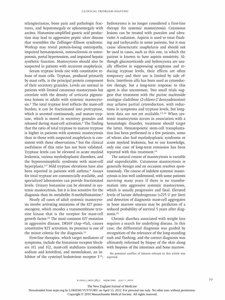
telangiectasias, bone pain and pathologic frac-tures, and hepatomegaly or splenomegaly with as cites. Histamine-amplified gastric acid produc-tion may lead to aggressive peptic ulcer disease that resembles the Zollinger–Ellison syndrome. Workup may reveal protein-losing enter opathy, impaired hematopoiesis, osteosclerosis or osteo-porosis, portal hypertension, and impaired hepatic synthetic function. Mastocytosis should also be suspected in patients with recurrent anaphylaxis.
Serum tryptase levels rise with constitutive re-lease of mast cells. Tryptase, produced primarily by mast cells, is the principal protein component of their secretory granules. Levels are normal in patients with limited cutaneous mastocytosis but correlate with the density of urticaria pigmen-tosa lesions in adults with systemic mastocyto-sis.2 The total tryptase level reflects the mast-cell burden; it can be fractionated into protryptase, which is secreted continuously, and mature tryp-tase, which is stored in secretory granules and released during mast-cell activation.3 The finding that the ratio of total tryptase to mature tryptase is higher in patients with systemic mastocytosis than in those with suspected anaphylaxis is con-sistent with these observations,4 but the clinical usefulness of this ratio has not been validated. Tryptase levels can be elevated in acute myeloid leukemia, various myelodysplastic disorders, and the hypereosinophilic syndrome with mast-cell hyperplasia.5-7 Mild tryptase elevations have also been reported in patients with asthma.8 Assays for total tryptase are commercially available, and specialized laboratories can provide fractionated levels. Urinary histamine can be elevated in sys-temic mastocytosis, but it is less sensitive for the diagnosis than its metabolite N-methylhistamine.9
Nearly all cases of adult systemic mastocyto-sis involve activating mutations of the KIT proto-oncogene, which encodes a transmembrane tyro-sine kinase that is the receptor for mast-cell growth factor.10 The most common KIT mutation in aggressive disease, D816V (Asp→Val), causes constitutive KIT activation; its presence is one of the minor criteria for the diagnosis.11
First-line therapies, which target mediators of symptoms, include the histamine receptor block-ers H1 and H2, mast-cell stabilizers (cromolyn sodium and ketotifen), and montelukast, an in-hibitor of the cysteinyl leukotriene receptor 112;
hydroxyurea is no longer considered a first-line therapy for systemic mastocytosis). Cutaneous lesions can be treated with psoralen and ultra-violet A radiation. Aspirin is used to treat flush-ing and tachycardia in some patients, but it may cause idiosyncratic anaphylaxis and should not be used in cases, such as this one, in which the patient is known to have aspirin sensitivity. Al-though glucocorticoids and hydroxyurea are usu-ally effective in suppressing symptoms and re-ducing tryptase levels, their effects are often temporary and their use is limited by side ef-fects. Interferon alfa has been used as cytoreduc-tive therapy, but a long-term response to this agent is also uncommon. Two small trials sug-gest that treatment with the purine nucleoside analogue cladribine (2-chloro-2′deoxyadeno sine) may achieve partial cytoreduction, with reduc-tions in symptoms and tryptase levels, but long-term data are not yet available.13,14 When sys-temic mastocytosis occurs in association with a hematologic disorder, treatment should target the latter. Hematopoietic stem-cell transplanta-tion has been performed in a few patients, some of whom also had myelodysplastic syndrome or acute myeloid leukemia, but to our knowledge, only one case of long-term remission has been reported with this treatment.15
The natural course of mastocytosis is variable and unpredictable. Cutaneous mastocytosis is generally benign and on occasion resolves spon-taneously. The course of indolent systemic masto-cytosis is less well understood, with some patients surviving many years if there is no transfor-mation into aggressive systemic mastocytosis, which is usually progressive and fatal. Elevated levels of lactate dehydrogenase (>225 U per liter) and detection of diagnostic mast-cell aggregates in bone marrow smears may be predictors of a reduced probability of survival 5 years after diag-nosis.16
Chronic diarrhea associated with weight loss requires a search for underlying disease. In this case, the differential diagnosis was guided by recognition of the relevance of the long-standing rash and flushing, and the correct diagnosis was ultimately informed by biopsy of the skin along with biopsies of the intestines and bone marrow.
No potential conflict of interest relevant to this article was reported.
The New England Journal of Medicine Downloaded from nejm.org by LOKESH VUYYURU on April 13, 2012. For personal use only. No other uses without permission.
Copyright © 2010 Massachusetts Medical Society. All rights reserved.

n engl j med 363;1 nejm.org july 1, 201078
clinical problem-solving
References
Valent P, Horny HP, Escribano L, et al. 1. Diagnostic criteria and classification of mastocytosis: a consensus proposal. Leuk Res 2001;25:603-25.
Brockow K, Akin C, Huber M, Met-2. calfe DD. Assessment of the extent of cutaneous involvement in children and adults with mastocytosis: relationship to symptomatology, tryptase levels, and bone marrow pathology. J Am Acad Dermatol 2003;48:508-16.
Schwartz LB, Min HK, Ren S, et al. 3. Tryptase precursors are preferentially and spontaneously released, whereas mature tryptase is retained by HMC-1 cells, Mono-Mac-6 cells, and human skin-derived mast cells. J Immunol 2003;170:5667-73.
Schwartz LB, Sakai K, Bradford TR, 4. et al. The alpha form of human tryptase is the predominant type present in blood at baseline in normal subjects and is elevat-ed in those with systemic mastocytosis. J Clin Invest 1995;96:2702-10.
Sperr WR, Jordan JH, Baghestanian M, 5. et al. Expression of mast cell tryptase by myeloblasts in a group of patients with acute myeloid leukemia. Blood 2001;98: 2200-9.
Sperr WR, Stehberger B, Wimazal F, 6. et al. Serum tryptase measurements in
patients with myelodysplastic syndromes. Leuk Lymphoma 2002;43:1097-105.
Klion AD, Noel P, Akin C, et al. Elevat-7. ed serum tryptase levels identify a subset of patients with a myeloproliferative vari-ant of idiopathic hypereosinophilic syn-drome associated with tissue fibrosis, poor prognosis, and imatinib responsive-ness. Blood 2003;101:4660-6.
Taira M, Tamaoki J, Kondo M, Kawa-8. tani K, Nagai A. Serum B12 tryptase level as a marker of allergic airway inflamma-tion in asthma. J Asthma 2002;39:315-22.
Keyzer JJ, de Monchy JG, van Door-9. maal JJ, van Voorst Vader PC. Improved diagnosis of mastocytosis by measure-ment of urinary histamine metabolites. N Engl J Med 1983;309:1603-5.
Garcia-Montero AC, Jara-Acevedo M, 10. Teodosio C, et al. KIT mutation in mast cells and other bone marrow hemato-poietic cell lineages in systemic mast cell disorders: a prospective study of the Span-ish Network on Mastocytosis (REMA) in a series of 113 patients. Blood 2006;108: 2366-72.
Nagata H, Worobec AS, Oh CK, et al. 11. Identification of a point mutation in the catalytic domain of the protooncogene c-kit in peripheral blood mononuclear cells
of patients who have mastocytosis with an associated hematologic disorder. Proc Natl Acad Sci U S A 1995;92:10560-4.
Valent P, Akin C, Escribano L, et al. 12. Standards and standardization in masto-cytosis: consensus statements on diagnos-tics, treatment recommendations and re-sponse criteria. Eur J Clin Invest 2007;37: 435-53.
Kluin-Nelemans HC, Oldhoff JM, van 13. Doormaal JJ, et al. Cladribine therapy for systemic mastocytosis. Blood 2003;102: 4270-6.
Pardanani A, Hoffbrand AV, Butter-14. field JH, Tefferi A. Treatment of systemic mast cell disease with 2-chlorodeoxyade-nosine. Leuk Res 2004;28:127-31.
Hennessy B, Giles F, Cortes J, et al. 15. Management of patients with systemic mastocytosis: review of M.D. Anderson Cancer Center experience. Am J Hematol 2004;77:209-14.
Lawrence JB, Friedman BS, Travis 16. WD, et al. Hematologic manifestations of systemic mast cell disease: a prospective study of laboratory and morphologic fea-tures and their relation to prognosis. Am J Med 1991;91:612-24.Copyright © 2010 Massachusetts Medical Society.
clinical problem-solving series
The Journal welcomes submissions of manuscripts for the Clinical Problem-Solving series. This regular feature considers the step-by-step process of clinical decision
making. For more information, please see authors.NEJM.org.
The New England Journal of Medicine Downloaded from nejm.org by LOKESH VUYYURU on April 13, 2012. For personal use only. No other uses without permission.
Copyright © 2010 Massachusetts Medical Society. All rights reserved.
Copyright © 2022 FDOKUMEN