A POLAROGRAPHIC AND POTENTIOMETRIC ... - CORE
27
A POLAROGRAPHIC AND POTENTIOMETRIC STUDY OF METAL – LIGAND EQUILIBRIA: INSTRUMENTATION AND INVESTIGATIONS OF SYSTEMS WITH NON – REVERSIBLE ELECTRODE REACTIONS Tumaini Samwel Peter Mkwizu A dissertation submitted to the Faculty of Science, University of the Witwatersrand, in fulfillment of the requirements for the degree of Master of Science Johannesburg, 2006 brought to you by CORE View metadata, citation and similar papers at core.ac.uk provided by Wits Institutional Repository on DSPACE
-
Upload
khangminh22 -
Category
Documents
-
view
5 -
download
0
Transcript of A POLAROGRAPHIC AND POTENTIOMETRIC ... - CORE
A POLAROGRAPHIC AND POTENTIOMETRIC STUDY
OF METAL – LIGAND EQUILIBRIA:
INSTRUMENTATION AND INVESTIGATIONS OF
SYSTEMS WITH NON – REVERSIBLE ELECTRODE
REACTIONS
Tumaini Samwel Peter Mkwizu
A dissertation submitted to the Faculty of Science, University of the
Witwatersrand, in fulfillment of the requirements for the degree of Master of
Science
Johannesburg, 2006
brought to you by COREView metadata, citation and similar papers at core.ac.uk
provided by Wits Institutional Repository on DSPACE
ii
DECLARATION
I declare that this dissertation is my own work. It is being submitted for the
Degree of Master of Science in the University of the Witwatersrand,
Johannesburg. It has not been submitted before for any degree or examination in
any other University.
_______________________________
(Signature of candidate)
___________ day of__________________ 2006.

iii
OUTPUTS FROM THIS WORK
Conference Papers:
Tumaini Mkwizu and Ignacy Cukrowski. Automated Instrumentation for
Potentiometry and Polarography in Metal–Ligand Equilibria Studies, Proceedings
of the 37th National Convention of the South African Chemical Institute, Pretoria
July 2004.
Publications:
Ignacy Cukrowski, Tumaini S. Mkwizu, and Philemon Magampa. Voltammetry
as Virtual Potentiometric Sensor in Modelling of a Metal/Ligand System and
Refinement of Stability Constants. Part 5. Complexation Studies of Hydrolysis–
Prone Lead(II) with Glycine and Sarcosine by Sampled–Direct–Current
Polarography Involving Virtual Potential. (Manuscript submitted for publication
in 2006).
Ignacy Cukrowski, Helder Marques, Tumaini S. Mkwizu, Philemon P. Magampa,
and Claudette Serge. Influence of electronic and steric effects on stability
constants and electrochemical reversibility of divalent ion complexes with glycine
and sarcosine. A Glass Electrode Potentiometric, Sampled Direct Current
Polarographic, Virtual Potentiometric, and Molecular Modelling study.
(Manuscript in final preparation to be submitted for publication in 2006).

iv
ABSTRACT
New possibilities in collection of polarographic and potentiometric experimental data in
studies of metal–ligand systems by automated instrumental methods, and subsequent
treatment of the polarographic data, whereby the degree of reversibility of the electrode
processes varies, have been investigated in this work. An automated instrumental set–up
was developed for applications in studies of metal–ligand solution equilibria by
potentiometry and sampled Direct Current Polarography (DCP). The new set–up was
designed based on virtual instrumentation principles whereby several commercially–
available hardware units as well as custom–built electronic components, were interfaced
to a personal computer that was equipped with appropriate hardware and control
programs. The instrumental set–up was tested and validated by studying the protonation
equilibria of the ligand glycine by Glass Electrode Potentiometry (GEP) as well as the
complexation of the ligand glycine with Cd2+ by GEP and DCP. The new set–up provides
increased versatility, accuracy and convenience in obtaining large numbers of
experimental points in solution equilibria studies by DCP and GEP as opposed to the use
of tedious and time–consuming manual methods. Nonlinear curve–fitting procedures,
based on closed–form models that were derived here from suitable theoretical equations
identified from literature, have been investigated in this work for applications in analysis
of DC curves recorded on metal–ligand systems with variation in electrochemical
reversibility. The applicability and limitations of the curve–fitting procedures developed
have been tested in analysis of the DCP data collected on several metal–ligand systems
involving Cd2+, Pb2+, Zn2+ and the ligands glycine and sarcosine, whereby the DCP
studies of these systems exhibited reversible, quasi–reversible or irreversible
electrochemical processes. Information on applicability and limitations of the proposed
methods investigated in this work was derived by comparison of the results obtained from
DCP, using the proposed methods, with either reported literature data and/or results
obtained in this work by the independent analytical technique of GEP, which was
deployed wherever it was found to be applicable to study the metal–ligand systems
considered.

v
ACKNOWLEDGEMENTS
First and foremost, I wish to express my most sincere gratitude to my research
mentor Prof. Ignacy Cukrowski. I found great pleasure in working with and
learning from him. I thank him for his patience, academic guidance, as well as
moral and financial support which he provided me throughout the duration of the
research project. My sincere appreciation also goes to Mr. Basil Chassoulas of the
Wits School of Chemistry for his tremendous assistance in the electronic aspects
related to the development of instrumentation in this project. Thank you, to all my
colleagues at the Electrochemistry Research Laboratories (at Wits University and
currently at the Department of Chemistry, University of Pretoria). Their
contributions in many ways toward the success of this project are highly
appreciated. I also wish to thank the University of the Witwatersrand for financial
support through a Postgraduate Merit Award programme. My deep gratitude also
goes to the staff of the School of Chemistry at Wits University for technical and
administrative assistance they provided me during my studentship in the School.
Finally, my deepest gratitude goes to my parents for their moral and financial
support, offered to me wholeheartedly, during my tenure as a postgraduate student
in the Republic of South Africa.

vi
TABLE OF CONTENTS
DECLARATION ......................................................................................................... ii
OUTPUTS FROM THIS WORK ................................................................................ iii
ABSTRACT................................................................................................................. iv
ACKNOWLEDGEMENTS......................................................................................... v
LIST OF FIGURES ..................................................................................................... x
LIST OF TABLES....................................................................................................... xxii
LIST OF ABBREVIATIONS...................................................................................... xxvi
CHAPTER 1 INTRODUCTION
1.1 METAL–LIGAND EQUILIBRIA: GENERAL BACKGROUND.................. 1
1.2 GENERAL CONCEPTS IN EQUILIBRIUM ANALYSIS ............................. 3
1.3 EXPERIMENTAL TECHNIQUES FOR METAL–LIGAND
EQUILIBRIA STUDIES .................................................................................. 7
1.3.1 General Survey .................................................................................. 7
1.4 POTENTIOMETRY ......................................................................................... 9
1.4.1 Basic Principles of Potentiometry...................................................... 9
1.4.2 Potentiometry and the Study of Metal–Ligand Equilibria ................. 11
1.4.3 Computer–Assisted Experiments for Potentiometry.......................... 12
1.5 POLAROGRAPHY .......................................................................................... 15
1.5.1 Basic Principles of Polarography....................................................... 15
1.5.2 Polarography and the Study of Metal–Ligand Equilibria .................. 20
1.5.3 Reversibility of Electrode Reactions ................................................. 25
1.5.4 Computer–Assisted Experiments for Polarography........................... 29
1.6 AIMS AND SCOPE OF PROJECT.................................................................. 32
1.7 SUMMARY OF CHAPTERS........................................................................... 34
1.8 REFERENCES.................................................................................................. 36
CHAPTER 2 MATERIALS AND GENERAL EXPERIMENTAL
PROCEDURES
2.1 REAGENTS...................................................................................................... 41
2.2 PREPARATION AND STANDARDISATION OF SOLUTIONS.................. 42

vii
2.3 GLASS ELECTRODE POTENTIOMETRY ................................................... 43
2.3.1 Electrodes and Instrumentation.......................................................... 43
2.3.2 Experimental Set–up.......................................................................... 44
2.3.3 Glass Electrode Calibration ............................................................... 44
2.3.4 Determination of Ligand Protonation Constants ............................... 46
2.3.5 Metal–Ligand Equilibria Studies by GEP.......................................... 48
2.4 SAMPLED DIRECT CURRENT POLAROGRAPHY.................................... 49
2.4.1 Electrodes and Instrumentation.......................................................... 49
2.4.2 Experimental Set–up.......................................................................... 51
2.4.3 Polarographic Studies of Metal–Ligand Equilibria ........................... 52
2.5 REFERENCES.................................................................................................. 55
CHAPTER 3 THEORY AND TREATMENT OF DATA
3.1 INTRODUCTION ............................................................................................ 56
3.2 GLASS ELECTRODE POTENTIOMETRY: THEORY,
MODELLING AND REFINEMENT OF PROTONATION AND
STABILITY CONSTANTS.............................................................................. 56
3.3 SAMPLED DIRECT CURRENT POLAROGRAPHY: THEORY,
MODELLING AND REFINEMENT OF
STABILITY CONSTANTS.............................................................................. 61
3.3.1 Optimisation of a Metal–Ligand Model and Refinement
of Stability Constants......................................................................... 61
3.3.2 General Concepts in Modelling of Polarographic Data ..................... 64
3.3.2.1 Variation in Half–wave Potential as
a Function of pH................................................................. 64
3.3.2.2 Variation in Half–wave Potential versus
Free Ligand Concentration ................................................. 67
3.3.2.3 Variation in Limiting Diffusion Current as
a Function of pH................................................................. 68
3.4 VIRTUAL POTENTIOMETRY....................................................................... 68
3.5 ANALYSIS OF DIRECT CURRENT POLAROGRAMS
RECORDED ON METAL–LIGAND SYSTEMS ........................................... 70
3.5.1 Electrochemical Reversibility: General Concepts ............................. 70
3.5.2 Evaluation of Electrochemical Reversibility ..................................... 75

viii
3.5.2.1 Logarithmic Analysis ......................................................... 75
3.5.2.2 Curve–fitting Method ......................................................... 80
3.5.3 Determination of Reversible Half-wave Potential and
Limiting Diffusion Current ................................................................ 83
3.5.3.1 Reversible or Nearly Reversible Systems........................... 83
3.5.3.2 Non–reversible Systems ..................................................... 84
3.5.4 Concluding Remarks.......................................................................... 91
3.6 REFERENCES ............................................................................................ 92
CHAPTER 4 DEVELOPMENT OF AUTOMATED
INSTRUMENTATION
4.1 INTRODUCTION ............................................................................................ 95
4.1.1 General Concepts on Virtual Instrumentation ................................... 96
4.2 DESCRIPTION OF THE HARDWARE.......................................................... 98
4.2.1 Data Collection and Processing Interface .......................................... 101
4.2.2 Electronic Control Box ...................................................................... 102
4.2.3 Digital pH Meter................................................................................ 103
4.2.4 Digital Burette.................................................................................... 103
4.2.5 Magnetic Stirrer ................................................................................. 104
4.2.6 Potentiostat and Current–Measuring System..................................... 105
4.2.7 Voltammetric Stand ........................................................................... 110
4.3 DESCRIPTION OF THE SOFTWARE ........................................................... 113
4.3.1 Potentiometry..................................................................................... 113
4.3.2 Sampled Direct Current Polarography............................................... 119
4.3.3 Sampled Direct Current Polarography with Potentiometry ............... 123
4.4 VALIDATION AND PERFORMANCE OF INSTRUMENTATION............. 133
4.4.1 Glass Electrode Potentiometry: Automated Titrations ...................... 133
4.4.1.1 Protonation Equilibria for Glycine ..................................... 133
4.4.1.2 A Potentiometric Study of a
Cadmium(II)–Glycine–OH system..................................... 137
4.4.2 Sampled Direct Current Polarography with
Potentiometry: Automated Titrations ................................................ 142
4.4.2.2 A Polarographic Study of a
Cadmium(II)–Glycine–OH System.................................... 142

ix
4.5 CONCLUSIONS ............................................................................................ 150
4.6 REFERENCES ............................................................................................ 151
CHAPTER 5 METAL–LIGAND SYSTEMS INVOLVING
POLAROGRAPHIC REVERSIBLE AND
NON–REVERSIBLE ELECTRODE REACTIONS
5.1 INTRODUCTION ............................................................................................ 153
5.2 RESULTS AND DISCUSSION ....................................................................... 159
5.2.1 Case Study 1: A Cadmium(II)–Sarcosine–OH System ..................... 159
5.2.1.1 A GEP Study of a Cd(II)–Sarcosine–OH
System ................................................................................ 160
5.2.1.2 Polarographic Investigations of a Cd(II)–Sarcosine–OH
System ................................................................................ 166
5.2.2 Case Study 2: A Lead(II)–Glycine–OH System................................ 186
5.2.3 Case Study 3: A Lead(II)–Sarcosine–OH System ............................. 200
5.2.4 Case Study 4: A Zinc(II)–Glycine–OH System................................. 209
5.2.4.1 A GEP Study of a Zn(II)–Glycine–OH System.................. 210
5.2.4.2 Polarographic Investigations of a Zn(II)–Glycine–OH
System ................................................................................ 215
5.2.5 The Effect of Addition of a Methyl Group to a Ligand ..................... 227
5.3 CONCLUSIONS ............................................................................................ 231
5.3.1 Applicability and Limitations of Curve–Fitting Models for
Analysis of DC Polarograms ............................................................. 231
5.3.2 Recommended Procedures in Analysis of DC Polarograms
Recorded On Metal–Ligand Systems ................................................ 234
5.4 REFERENCES.................................................................................................. 237
CHAPTER 6 GENERAL CONCLUSIONS ......................................................... 239
APPENDICES ........................................................................................................... 245

x
LIST OF FIGURES
Figure 1.1: An overall scheme for equilibrium model determination...................6
Figure 1.2: Sampled DC polarography. (a) Stepwise potential waveform.
(b) Current–time curves observed in response to the potential
steps and current sampling scheme ....................................................19
Figure 1.3: A typical sigmoidal–shaped sampled DC polarogram. ......................19
Figure 1.4: A flowchart depicting experimental tasks typically performed
in a polarographic study of a metal–ligand system at fixed LT :
MT and variable pH.. ..........................................................................31
Figure 2.1: An example of a calibration curve obtained prior to performing
a glass electrode potentiometric experiment for a metal–ligand
system.................................................................................................46
Figure 2.2: A photograph showing electrodes and probes used in studies of
metal–ligand systems at fixed LT : MT and variable pH .....................50
Figure 2.3: A photograph of the instrumental set–up in a typical automated
experiment for a metal – ligand system at fixed LT:MT ratio and
variable pH by sampled direct current polarography with glass
electrode potentiometry as the leading technique...............................51
Figure 3.1: A typical relationship of variation in half–wave potential for
Cd2+, in the presence of the protic ligand glycine, as a function
of pH...................................................................................................65
Figure 3.2: An example of a typical relationship of variation in half–wave
potential as a function of Log [L].......................................................67

xi
Figure 3.3: Schematic comparison of DC polarograms corresponding to
reversible, quasi–reversible, and irreversible reduction
processes.............................................................................................73
Figure 3.4: A sampled direct current polarogram for the reversible
reduction of Pb2+ 25 °C in 0.5 M NaNO3 ...........................................78
Figure 3.5: A logarithmic analysis corresponding to the DC polarogram for
the reversible reduction of Pb2+ at 25 °C in 0.5 M NaNO3
shown in Figure 3.4 ............................................................................78
Figure 3.6: A sampled direct current polarogram for a quasi–reversible
reduction of Cd2+ at 25 °C in 0.5 M NaNO3 in the presence of
the ligand sarcosine ............................................................................79
Figure 3.7: A logarithmic analysis of the DC polarogram in Figure 3.6 ..............80
Figure 3.8: An example of the analysis of a quasi–reversible DC
polarogram for the reduction of Cd2+ at 25 °C in 0.5 M NaNO3,
in the presence of the ligand sarcosine, using the Cukrowski’s
curve–fitting method ..........................................................................85
Figure 3.9: Logarithmic analysis of a quasi–reversible DC polarographic
wave using the Matsuda–Ayabe method ............................................88
Figure 4.1: A block diagram showing interfacing and connectivity of the
various hardware components of the instrumental set–up for
potentiometric and polarographic measurements ...............................99
Figure 4.2: A simplified circuit diagram showing connectivity and
interfacing of the electronic components used in the
instrumentation for automated DC polarographic and
potentiometric measurements .............................................................100

xii
Figure 4.3: A basic potentiostatic three–electrode system with
measurement of cell current via a current–to–voltage converter........106
Figure 4.4: The integration amplifier circuitry used for amplification of the
current response signals measured by the CV–27
voltammograph...................................................................................107
Figure 4.5: A schematic diagram showing the inert gas connections and
operating principle of the valve block and multi–mode
electrode of the 663 VA stand ............................................................111
Figure 4.6: A flow chart of the Configure Dosimat & pH meter VI (the
virtual instrument used to configure the pH meter and the
digital burette (765 Dosimat) used in automated
potentiometric–polarographic experiments).......................................114
Figure 4.7: The front panel of the Autotitrator VI, the software module
developed for automated potentiometric titrations with constant
volume additions ................................................................................115
Figure 4.8: Flow chart of the Autotitrator VI, the virtual instrument for
automated potentiometric titrations with constant volume
additions .............................................................................................116
Figure 4.9: A flowchart of the subroutine (or SubVI) Sampling 713/780 pH
Meter used to programmatically establish an equilibrium
potential reading, at a particular titration stage, during an
automated potentiometric titration......................................................118
Figure 4.10: The front panel (user–interface) of the DC (One Polarogram)
VI used for single Sampled Direct Current Polarographic scans........121
Figure 4.11: A flowchart of the DC (One Polarogram) VI used to generate
single scans of Sampled DC polarography.........................................122

xiii
Figure 4.12: The front panel of the AUTOTITRATOR-DC1 VI, a software
module used for automated titrations with acquisition of
sampled DC polarograms and potentiometric data.............................124
Figure 4.13: An example of a titration curve (pH versus volume of titrant)
obtained from a study of Cd(II)–Glycine–OH system at fixed
LT: MT ratio and variable pH ..............................................................125
Figure 4.14: A flowchart showing programmatic execution of the
Autotitrator-DC1 VI ...........................................................................126
Figure 4.15: Examples of recorded Sampled DC polarograms using the
Autotitrator-DC2 VI used in an automated potentiometric–
polarographic experiment of Pb(II)–Glycine–OH system at a
fixed LT:MT ratio and variable pH......................................................129
Figure 4.16: The front panel of the AUTOTITRATOR-DC-DYNAMIC2 VI ...........130
Figure 4.17: A flowchart showing the programmatic execution of the VI
Autotitrator-DC-Dynamic2 ................................................................131
Figure 4.18: The chemical structure of a fully–protonated glycine molecule ........133
Figure 4.19: A titration curve obtained from the titration of a glycine
solution with 0.05 M NaOH ...............................................................135
Figure 4.20: Experimental (o) and theoretical (solid line) protonation curves
of the ligand glycine obtained from refinement of the GEP data
collected using the automated instrumental set–up for
potentiometric titrations developed in this project .............................137

xiv
Figure 4.21: Experimental (o) and theoretical (solid line) potentiometric
complex formation curves obtained for the metal–ligand
models containing ML, ML2, ML3, ML(OH), and ML2(OH)
with the optimized stability constants for these complexes
obtained from the study of Cd(II)–Glycine–OH system by GEP
at various LT : MT ratios......................................................................140
Figure 4.22: A species distribution diagram for the Cd(II)–Glycine–OH
system at LT : MT = 1 : 1, [MT] = 7.771 × 10⁻³ M generated
using the model containing M(HL), ML, ML2, ML3, ML(OH)
and ML2(OH)......................................................................................141
Figure 4.23: A species distribution diagram for the Cd(II)–Glycine–OH
system at LT : MT = 3 : 1, [MT] = 4.679 × 10⁻³ M generated
using stability constants from the model containing M(HL),
ML, ML2, ML3, ML(OH), ML2(OH)..................................................142
Figure 4.24: Examples of sampled DC curves recorded during a study of
Cd(II)–Glycine–OH (LT : MT = 200 : 1(fixed) and variable pH;
[MT] = 1.016 × 10⁻⁴ M). An illustration of the curve–fitting
operations is also shown.....................................................................144
Figure 4.25: An example of interpretation of the observed shift in half–wave
potential plotted against pH for the Cd(II)–Glycine–OH system
studied by sampled DCP at experimental conditions as
indicated for Figure 4.24 ....................................................................145
Figure 4.26: An example of interpretation of the observed shift in half–wave
potential plotted against Log [L] for the Cd(II)–Glycine–OH
system studied by DCP at fixed LT : MT ratio and variable pH
at experimental conditions as indicated for Figure 4.24.....................146
Figure 4.27: Experimental (circles) and calculated (solid line) complex
formation curves obtained for the Cd(II)–Glycine–OH system
studied at a fixed LT : MT ratio of 700; [MT] = 8.456 × 10⁻⁵ M..........146

xv
Figure 4.28: Species distribution as a function of pH for the Cd(II)–Glycine–
OH system at LT : MT = 200; [MT] = 1.016 × 10⁻⁴ M.........................147
Figure 4.29: Species distribution as a function of pH for the Cd(II)–Glycine–
OH system at LT : MT = 600; [MT] = 8.051 × 10⁻⁵ M.........................149
Figure 5.1: Chemical structures of the fully–protonated forms of the
ligands Glycine, Sarcosine, Iminodiacetic acid (IDA) and N–
methyliminodiacetic acid (MIDA) .....................................................157
Figure 5.2: A plot of variation in Log �ML values plotted as a function of
the metal ion radius for the metal ions Ni(II), Zn(II), Cu(II),
Cd(II), and Pb(II) with the ligands iminodiacetic acid (IDA)
and N–methyliminodiacetic acid (MIDA) at ionic strength of
0.5 M and 25 °C ................................................................................157
Figure 5.3: A plot of variation in Log �ML values plotted as a function of
the metal ion radius for the metal ions Ni(II), Zn(II), Cu(II),
Cd(II), and Pb(II) with the ligands Glycine and Sarcosine (N–
methylglycine) at ionic strength of 0.5 M ..........................................158
Figure 5.4: Experimental (circles) and calculated (solid line) potentiometric
complex formation curves for Cd(II)–Sarcosine–OH system at
LT : MT = 2, initial [MT] = 4.797 × 10⁻³ M and LT : MT = 7,
initial [MT] = 2.041 × 10⁻³ M at 25 °C and ionic strength 0.5
M in NaNO3........................................................................................162
Figure 5.5: Species distribution as a function of pH for the Cd(II)–
Sarcosine–OH system at LT : MT = 2; [MT] = 4.797 × 10⁻³ M ...........163
Figure 5.6: Species distribution as a function of pH for the Cd(II)–
Sarcosine–OH system at LT : MT = 7; [MT] = 2.041 × 10⁻³ M ...........164
xvi
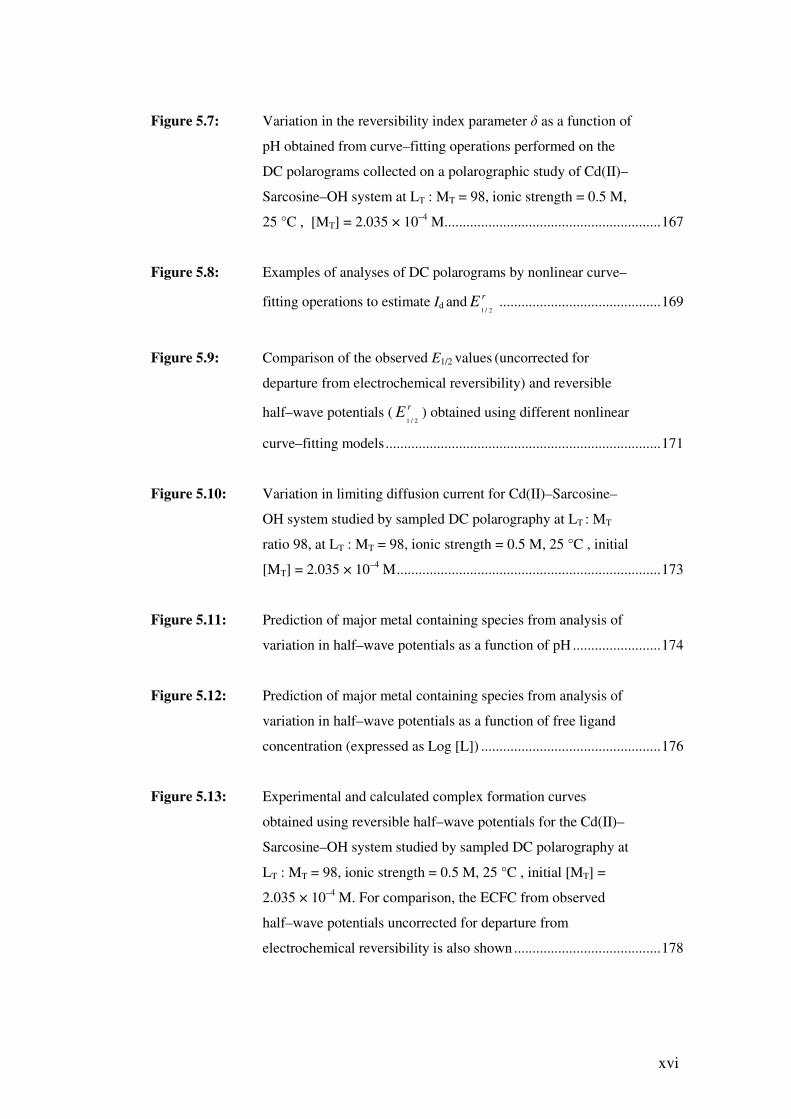
Figure 5.7: Variation in the reversibility index parameter � as a function of
pH obtained from curve–fitting operations performed on the
DC polarograms collected on a polarographic study of Cd(II)–
Sarcosine–OH system at LT : MT = 98, ionic strength = 0.5 M,
25 °C , [MT] = 2.035 × 10⁻⁴ M...........................................................167
Figure 5.8: Examples of analyses of DC polarograms by nonlinear curve–
fitting operations to estimate Id and rE2/1
............................................169
Figure 5.9: Comparison of the observed E1/2 values (uncorrected for
departure from electrochemical reversibility) and reversible
half–wave potentials ( rE2/1
) obtained using different nonlinear
curve–fitting models...........................................................................171
Figure 5.10: Variation in limiting diffusion current for Cd(II)–Sarcosine–
OH system studied by sampled DC polarography at LT : MT
ratio 98, at LT : MT = 98, ionic strength = 0.5 M, 25 °C , initial
[MT] = 2.035 × 10⁻⁴ M........................................................................173
Figure 5.11: Prediction of major metal containing species from analysis of
variation in half–wave potentials as a function of pH ........................174
Figure 5.12: Prediction of major metal containing species from analysis of
variation in half–wave potentials as a function of free ligand
concentration (expressed as Log [L]) .................................................176
Figure 5.13: Experimental and calculated complex formation curves
obtained using reversible half–wave potentials for the Cd(II)–
Sarcosine–OH system studied by sampled DC polarography at
LT : MT = 98, ionic strength = 0.5 M, 25 °C , initial [MT] =
2.035 × 10⁻⁴ M. For comparison, the ECFC from observed
half–wave potentials uncorrected for departure from
electrochemical reversibility is also shown ........................................178

xvii
Figure 5.14: Species distribution as a function of pH for the Cd(II)–
Sarcosine–OH system at LT : MT = 98; [MT] = 2.035 × 10⁻⁴ M .........181
Figure 5.15: Species distribution as a function of pH for the Cd(II)–
Sarcosine–OH system at LT : MT = 7; [MT] = 2.041 × 10⁻³ M
(GEP conditions) ................................................................................182
Figure 5.16: Virtual half–wave potential as a function of Log [M] computed
with the use of the refined stability constants from the
optimised model containing ML, ML2, ML3 for the Cd(II)–
Sarcosine–OH system studied by sampled DC polarography at
LT : MT = 98, ionic strength = 0.5 M, 25 °C , initial [MT] =
2.035 × 10⁻⁴ M....................................................................................184
Figure 5.17: Comparison of the observed E1/2 obtained using the Cukrowski
curve–fitting method and rE2/1
obtained using the Ruži�-based
curve-fitting. The variation in the reversibility index parameter
� as a function of pH (obtained from the Cukrowski curve–
fitting method) is also shown. DC polarograms collected on a
polarographic study of Pb(II)–Glycine–OH system at LT : MT =
800, ionic strength = 0.5 M, initial [MT] = 8 × 10⁻⁵ M .......................188
Figure 5.18: Variation in current as a function of pH for Pb(II)–Glycine–OH
system studied by sampled DC polarography at LT: MT ratio
800, initial [MT] = 8 × 10⁻⁵ M, ionic strength = 0.5 M and 25 °C
............................................................................................................189
Figure 5.19: Prediction of major metal containing species from analysis of
variation in virtual half–wave potentials as a function of pH for
Pb(II)–Glycine–OH system studied by sampled DC
polarography at LT: MT ratio 800, initial [MT] = 8 × 10⁻⁵ M, 25
°C and ionic strength = 0.5 M ...........................................................191

xviii
Figure 5.20: Prediction of major metal containing species from analysis of
variation in virtual half–wave potentials as a function of Log
[L] for Pb(II)–Glycine–OH system studied by sampled DC
polarography at LT: MT ratio 800, initial [MT] = 8 × 10⁻⁵ M,
ionic strength = 0.5 M and 25 °C ......................................................192
Figure 5.21: Experimental and calculated complex formation curves for the
Pb(II)–Glycine–OH system studied by sampled DC
polarography at fixed LT : MT ratios, ionic strength of 0.5 M
and 25 °C ...........................................................................................193
Figure 5.22: Species distribution as a function of pH for the Pb(II)–Glycine–
OH system at LT : MT = 800; [MT] = 8 × 10⁻⁵ M................................195
Figure 5.23: Species distribution as a function of pH for the Pb(II)–Glycine–
OH system at LT : MT = 800; [MT] = 8 × 10⁻⁵ M................................196
Figure 5.24: Species distribution as a function of pH for the Pb(II)–Glycine–
OH system generated for LT : MT = 800 and [MT] = 8 × 10⁻⁵ M
(conditions employed in this work) using the stability constants
from literature [3] for the model with M(HL), M(HL)2,
M(HL)3, ML, and ML2 together with all known stability
constants for Pbx(OH)y complexes .....................................................197
Figure 5.25: Virtual half–wave potential as a function of Log [M] computed
with the use of the refined stability constants from the
optimised model containing M(HL), ML, ML2, ML3 for the
Pb(II)–Glycine–OH studied by sampled DC polarography at LT
: MT = 600, ionic strength = 0.5 M, 25 °C , initial [MT] = 8.062
× 10–5 M..............................................................................................198
Figure 5.26: Examples of analyses of DC polarograms from a sampled DC
polarographic study of Pb(II)–Sarcosine–OH system at LT : MT
= 400, � = 0.5 M, 25 °C , [MT] = 7.990 × 10⁻⁵ M..............................202

xix
Figure 5.27: Variation in reversible and observed half–wave potentials
(uncorrected for departure from electrochemical reversibility)
as a function of pH for the Pb(II)–Sarcosine–OH system at
LT : MT = 400, [MT] = 7.990 × 10⁻⁵ M, 25 °C , � = 0.5 M.................203
Figure 5.28: Variation in limiting diffusion current Id as a function of pH for
the Pb(II)–Sarcosine–OH system studied by sampled DC
polarography at LT : MT = 400, [MT] = 7.990 × 10⁻⁵ M, 25 °C ,
�= 0.5 M .............................................................................................204
Figure 5.29: Variation in virtual half–wave potential as a function of pH for
the Pb(II)–Sarcosine–OH system .......................................................205
Figure 5.30: Variation in virtual half–wave potential as a function of free
ligand concentration (expressed as Log[L]) for the
Pb(II)–Sarcosine–OH system .............................................................206
Figure 5.31: Species distribution as a function of pH for the Pb(II)–
Sarcosine–OH system generated for LT : MT = 400 and
[MT] = 8 × 10⁻⁵ M (conditions employed in this work) using the
stability constants for the model containing M(HL), ML, ML2,
ML3, and ML2(OH)2, together with all known stability
constants for Pbx(OH)y complexes .....................................................208
Figure 5.32: Experimental (circles) and theoretical (solid line)
potentiometric complex formation curves obtained for the study
of Zn(II)–Glycine–OH system by GEP at LT : MT ratio 6, [MT]
= 1.096 × 10⁻³ M, 25 °C , and � = 0.5 M...........................................213
Figure 5.33: Species distribution as a function of pH for the Zn(II)–Glycine–
OH system at LT : MT ratio 1 : 1, [MT] = 9.838 × 10⁻³ M ..................214
Figure 5.34: Species distribution as a function of pH for the Zn(II)–Glycine–
OH system at LT : MT ratio 6 : 1, [MT] = 1.096 × 10⁻³ M ..................214
xx
Figure 5.35: Typical polarograms of Zn(II) at various pH values recorded in
a sampled DC polarographic study of Zn(II)–Glycine–OH
system.................................................................................................216
Figure 5.36: A species distribution diagram for the Zn(II)–Glycine–OH
system generated for the experimental conditions employed in
the DC polarographic study of the system (LT : MT = 240 : 1,
[MT] = 1.07 × 10⁻⁴ M) ........................................................................217
Figure 5.37: An example of analysis of a quasi–reversible DC polarogram
from a DCP study of a Zn(II)–Glycine–OH system below pH 6
at LT : MT = 240, 25 °C , � = 0.5 M, initial [MT]=1.07 × 10⁻⁴ M .......219
Figure 5.38: An example of analysis of DC polarograms by nonlinear curve–
fitting using the curve–fitting method based on the Ruži�
equation (Equation 3.49) for polarograms collected above pH 9
from a DCP study of a Zn(II)–Glycine–OH system. LT : MT =
240, initial [MT] = 1.07 × 10⁻⁴ M........................................................220
Figure 5.39: A logarithmic analysis performed on the DC polarogram shown
in Figure 5.38. The linearity and the slope confirmed full
irreversible nature of the polarogram .................................................221
Figure 5.40: An example of analysis of two overlapping DC waves from the
polarographic study of a Zn(II)–Glycine–OH system and their
resolution by curve–fitting. LT : MT = 240, initial [MT] = 1.07 ×
10–4 M, curve recorded at pH 7.091 ...................................................223
Figure 5.41: Polarographic complex formation curves for the Zn(II)–
Glycine–OH system at LT : MT = 240, initial [MT] = 1.04 × 10⁻⁴
M, 25 °C and � = 0.5 M.....................................................................225
Figure 5.42: Experimental (circles) and calculated (solid line) polarographic
complex formation curves for the Zn(II)–Glycine–OH system
from refinement of data in the pH range 5 to 7.5 ...............................226

xxi
Figure 5.43: Variation in Log �ML values as a function of the metal ion
radius for the metal ions Ni(II), Zn(II), Cu(II), Cd(II), and
Pb(II) with the ligands Glycine and Sarcosine (N-
methylglycine) at ionic strength of 0.5 M and 25 °C . Third
order polynomial functions were found to be sufficient to
generate the trend-lines shown ...........................................................229

xxii
LIST OF TABLES
Table 1.1: A list of experimental methods available for investigations of
metal–ligand equilibria.......................................................................8
Table 4.1: Some specifications for the burette cylinders (exchange units)
used with a 765 Dosimat (digital burette) ..........................................104
Table 4.2: A summary of the main features of the virtual instruments used
for automated titrations with combined Sampled DCP and
Potentiometric measurements on a sample solution...........................132
Table 4.3: (A) Dissociation constant for water (fixed in the refinement
operations). (B) Summary of protonation constants for the
ligand glycine obtained from refinement operations of GEP
data collected using the automated potentiometric instrumental
set–up developed in this project at 25 °C and ionic strength of
0.5 M in NaNO3. (C) Summary of results from refinement
operations that included refinement of initial acid
concentrations.....................................................................................136
Table 4.4: (A) Protonation constants for the ligand glycine (L–),
dissociation constant for water and overall stability constants
for Cd(II) complexes with OH– included in the Cd(II)–L–OH
model and used in the refinement procedures for GEP data.
(B) Overall stability constants for Cd(II) with glycine from the
literature and found in this work by GEP at 25 °C and ionic
strength � = 0.5 M (NaNO3) ...............................................................139

xxiii
Table 4.5: (A) Protonation constants for the ligand glycine (L–),
dissociation constant for water and overall stability constants
for Cd(II) complexes with OH– included in the Cd(II)–L–OH
model and used in the refinement procedures for Sampled DC
polarographic data. (B) Overall stability constants for Cd(II)
with glycine from the literature and found in this work by
Sampled DC polarography at 25 °C and ionic strength of 0.5 M
in NaNO3 ............................................................................................148
Table 5.1: Summary of curve–fitting methods used in analysis of DC
polarograms recorded on metal–ligand systems in order to
estimate reversible half–wave potentials and limiting diffusion
currents ...............................................................................................155
Table 5.2: (A) Protonation constants for the ligand Sarcosine (L–),
dissociation constant for water and overall stability constants
for Cd(II) complexes with OH– included in the Cd(II)–L–OH
model and used as fixed values in the refinement procedures of
GEP data. (B) Overall stability constants for Cd(II) with
sarcosine found in this work by GEP (at 25 °C and ionic
strength � = 0.5 M in NaNO3) and those reported elsewhere.............165
Table 5.3: (A) Overall stability constants for Cd(II)–Sarcosine–OH
system found in this work by Sampled DC polarography using
half–wave potential values from various curve–fitting models
used in analysis of the DC polarograms. LT : MT = 98; initial
[MT] = 2.035 × 10⁻⁴ M, at 25 °C and ionic strength of 0.5 M in
NaNO3. (B) Overall stability constants for Cd(II)–Sarcosine–
OH system found by GEP in this work and elsewhere.......................180
Table 5.4: Overall stability constants for Cd(II) with sarcosine found in
this work by virtual potentiometry (VP) (generated from
sampled DCP, LT : MT ratio 98) and combined refinement
operation of the VP and GEP data (LT : MT ratios 2 and 7) ...............185

xxiv
Table 5.5: (A) Protonation constants for the ligand Glycine (L–),
dissociation constant for water and overall stability constants
for Pb(II) complexes with OH– included in the Pb(II)–L–OH
model and used in the refinement procedures of sampled DCP
data. (B) Overall stability constants for Pb(II) with glycine
found in this work by sampled DC polarography (at 25 °C and
ionic strength � = 0.5 M in NaNO3) and those reported
elsewhere ............................................................................................194
Table 5.6: Some overall stability constants for Pb(II) with glycine found in
this work by virtual potentiometry (VP) (generated from
sampled DCP, LT : MT ratio 600) .......................................................199
Table 5.7: (A) Protonation constants for the ligand Sarcosine (L–),
dissociation constant for water and overall stability constants
for Pb(II) complexes with OH– included in the Pb(II)–L–OH
model and used in the refinement procedures of sampled DCP
data. (B) Overall stability constants for Pb(II)–Sarcosine–OH
system found in this work by Sampled DC Polarography at LT :
MT = 400; initial [MT] = 7.990 × 10⁻⁵ M, at 25 °C and � = 0.5
M in NaNO3........................................................................................207
Table 5.8: (A) Protonation constants for the ligand Glycine (L–),
dissociation constant for water and overall stability constants
for Zn(II) complexes with OH– included in the Zn(II)–L–OH
model and used as fixed values in the refinement procedures of
GEP data. (B) Overall stability constants for Zn(II) with
glycine found in this work by GEP (at 25 °C and ionic strength,
� = 0.5 M in NaNO3) and those reported elsewhere...........................212
Table 5.9: Overall stability constants for Zn(II)–Glycine–OH system
found in this work by DCP, GEP and Virtual Potentiometry
(VP) and those reported elsewhere. DCP data collected for LT :
MT = 240; initial [MT] = 1.04 × 10⁻⁴ M, at 25 °C and ionic
strength of 0.5 M in NaNO3................................................................228

xxv
Table 5.10 Stability constants (as Log �ML) for complexes between glycine
derivatives and some divalent metal ions. Stability constants are
from [3] except for Cd(II)–Glycine, Cd(II)–Sarcosine, Pb(II)–
Glycine and Pb(II)–Sarcosine [this work]. All values are at 25
°C and ionic strength = 0.5 M. ...........................................................230
xxvi
LIST OF ABBREVIATIONS
AC Alternating Current ADC Analog–to–Digital Converter AE Auxiliary Electrode (Also referred to as counter electrode) AI Analog Input AO Analog Output CCFC Calculated Complex Formation Curve CGE Combination glass electrode °C Degrees Celsius DAC Digital–to–Analog Converter DAQ card Data Acquisition card DC Direct Current DCP Direct Current Polarography (Sampled Direct Current Polarography) DME Dropping Mercury Electrode DO Digital Output
rE2/1 Reversible half-wave potential of a DC polarogram
E° Standard Potential E1/2 Half-wave potential as observed from a DC polarogram Ek Glass electrode constant from calibration Eappl Stepwise applied potential ECFC Experimental Complex Formation Curve E1/2(virt) Virtual half–wave potential emf Electromotive force; potential ESTA Equilibrium Simulation for Titration Analysis; A suite of computer
programs for analysis of potentiometric data. Exp. Experiment F Faraday Constant; 96485 C mol-1 F.W. Formula Weight of a compound GEP Glass Electrode Potentiometry H Proton; hydrogen ion; H+ [ i ] Molar concentration of species i I–E Refers to a plot of current (I) as a function of potential (E) Ib Background current corresponding to an electrochemical process at
the dropping mercury electrode as obtained from a polarogram Id Limiting diffusion current corresponding to an electrochemical
process at the dropping mercury electrode as obtained from a polarogram
Iobs Observed total current corresponding to an electrochemical process at the dropping mercury electrode as obtained from a polarogram
Ired Reduction current corresponding to an electrochemical process at the dropping mercury electrode as obtained from a polarogram
K Kelvin KHP Potassium Hydrogen Pthalate Kw Dissociation Constant for water; Kw = [H+][OH–] L Ligand (charge omitted for clarity)

xxvii
LT Total ligand concentration in moles per Liter; [LT] LT : MT Total ligand to total metal ion concentration ratio, i.e., [LT] / [MT] M As a symbol for metal ion (charge omitted for clarity) or as a unit for
molar concentration, that is, number of moles of solute per 1 Liter of solution
MBE Mass Balance Equation MME Multi–Mode Electrode MT Total metal ion concentration in moles per Liter; [MT] mV milliVolt = 1/1000 Volts n Number of electrons involved in an electrochemical reaction NBAR The average number of protons per ligand in the absence of metal ion pA –Log[L]; negative logarithm of the free deprotonated ligand
concentration PC Personal Computer pH –Log [H+]; Calculated pH using the calibration method involving
strong acid/strong base titration at fixed ionic strength and temperature.
PTFE Polytetrafluoroethylene QBAR Deprotonation function; the average number of protons released as a
result of complexation per metal ion R Universal gas constant; 8.314 J mol-1 K-1 RE Reference Electrode Refs. References s Response slope for glass electrode T Temperature (in Kelvin) T–Probe Temperature Probe VI Virtual Instrument VP Virtual Potentiometry WE Working Electrode ZBAR(H) Potentiometric Complex Formation function; the average number of
protons bound per ligand ZBAR(M) Potentiometric Complex Formation function; the average number of
ligand molecules bound per metal ion � Cathodic transfer coefficient � Overall Stability Constant � Electrochemical reversibility index or steepness coefficient parameter
from analysis of direct current polarograms by a nonlinear curve–fitting procedure
� Ionic strength 3D–CFC Three Dimensional Complex Formation Curves; A computer program
for analysis of polarographic data for refinement of stability constants