A phylogenetic analysis of the megadiverse Chalcidoidea (Hymenoptera)
77
A phylogenetic analysis of the megadiverse Chalcidoidea (Hymenoptera) John M. Heraty a, *, Roger A. Burks a,b , Astrid Cruaud a,c , Gary A. P. Gibson d , Johan Liljeblad a,e , James Munro a,f , Jean-Yves Rasplus c , Gerard Delvare g , Peter Jansˇta h , Alex Gumovsky i , John Huber j , James B. Woolley k , Lars Krogmann l , Steve Heydon m , Andrew Polaszek n , Stefan Schmidt o , D. Chris Darling p,q , Michael W. Gates r , Jason Mottern a , Elizabeth Murray a , Ana Dal Molin k , Serguei Triapitsyn a , Hannes Baur s , John D. Pinto a,t , Simon van Noort u,v , Jeremiah George a and Matthew Yoder w a Department of Entomology, University of California, Riverside, CA, 92521, USA; b Department of Evolution, Ecology and Organismal Biology, Ohio State University, Columbus, OH, 43210, USA; c INRA, UMR 1062 CBGP CS30016, F-34988, Montferrier-sur-Lez, France; d Agriculture and Agri-Food Canada, 960 Carling Avenue, Ottawa, ON, K1A 0C6, Canada; e Swedish Species Information Centre, Swedish University of Agricultural Sciences, PO Box 7007, SE-750 07, Uppsala, Sweden; f Institute for Genome Sciences, School of Medicine, University of Maryland, Baltimore, MD, 21201, USA; g Cirad, INRA, UMR 1062 CBGP CS30016, F-34988, Montferrier-sur-Lez, France; h Department of Zoology, Charles University, Vinicna 7, CZ-128 44, Praha 2, Czech Republic; i Schmalhausen Institute of Zoology, National Academy of Sciences of Ukraine, Kiev, 30 01601, Ukraine; j Natural Resources Canada, c/o Canadian National Collection of Insects, 960 Carling Ave, Ottawa, ON, K1A 0C6, Canada; k Department of Entomology, Texas A&M University, College Station, TX, 77843, USA; l Department of Entomology, State Museum of Natural History Stuttgart, Rosenstein 1, 70191, Stuttgart, Germany; m Bohart Museum of Entomology, University of California, Davis, CA, 95616, USA; n Department of Entomology, Natural History Museum, London, SW7 5BD, UK; o Staatliche Naturwissenschaftliche Sammlungen Bayerns, Zoologische Staatssammlung, M€ unchhausenstr. 21, 81247, Munich, Germany; p Department of Natural History, Royal Ontario Museum, Toronto, ON, M5S 2C6, Canada; q Department of Ecology and Evolutionary Biology, University of Toronto, Toronto, ON, M5S 1A1, Canada; r Systematic Entomology Laboratory, USDA, ARS, PSI, c/o National Museum of Natural History, Washington, DC, 20013, USA; s Abt. Wirbellose Tiere, Naturhistorisches Museum der Burgergemeinde Bern, Bernastrasse 15, 3005, Bern, Switzerland; t PO Box 2266, Waldport, OR, 97394, USA; u Natural History Department, Iziko South African Museum, PO Box 61, Cape Town, 8000, South Africa; v Department of Zoology, University of Cape Town, Private Bag, Rondebosch, 7701, South Africa; w Illinois Natural History Survey, University of Illinois, Champaign, IL, 61820, USA Accepted 19 September 2012 Abstract Chalcidoidea (Hymenoptera) is extremely diverse with an estimated 500 000 species. We present the first phylogenetic analysis of the superfamily based on both morphological and molecular data. A web-based, systematics workbench mx was used to score 945 character states illustrated by 648 figures for 233 morphological characters for a total of 66 645 observations for 300 taxa. The matrix covers 22 chalcidoid families recognized herein and includes 268 genera within 78 of 83 subfamilies. Morphological data were analysed alone and in combination with molecular data from ribosomal 18S (2105 bp) and 28S D2–D5 expansion regions (1812 bp). Analyses were analysed alone and in combined datasets using implied-weights parsimony and likelihood. Proposed changes in higher classification resulting from the analyses include: (i) recognition of Eriaporidae, revised status; (ii) recognition of Cynipencyrtidae, revised status; (iii) recognition of Azotidae, revised status; (iv) inclusion of Sycophaginae in Agaonidae, revised sta- tus; (v) reclassification of Aphelinidae to include Aphelininae, Calesinae, Coccophaginae, Eretmocerinae and Eriaphytinae; (vi) inclusion of Cratominae and Panstenoninae within Pteromalinae (Pteromalidae), new synonymy; (vii) inclusion of Epichrysomalli- nae in Pteromalidae, revised status. At a higher level, Chalcidoidea was monophyletic, with Mymaridae the sister group of Rotoiti- dae plus the remaining Chalcidoidea. A eulophid lineage was recovered that included Aphelinidae, Azotidae, Eulophidae, Signiphoridae, Tetracampidae and Trichogrammatidae. Eucharitidae and Perilampidae were monophyletic if Eutrichosomatinae (Pteromalidae) was included, and Eupelmidae was monophyletic if Oodera (Pteromalidae: Cleonyminae) was included. Likelihood recovered a clade of Eupelmidae + (Tanaostigmatidae + (Cynipencyrtus + Encyrtidae). Support for other lineages and their impact on the classification of Chalcidoidea is discussed. Several life-history traits are mapped onto the new phylogeny. © The Willi Hennig Society 201 . Cladistics Cladistics (2012) 1–77 10.1111/cla.12006 © The Willi Hennig Society 2013 3
-
Upload
naturkundemuseum-bw -
Category
Documents
-
view
0 -
download
0
Transcript of A phylogenetic analysis of the megadiverse Chalcidoidea (Hymenoptera)

A phylogenetic analysis of the megadiverse Chalcidoidea(Hymenoptera)
John M. Heratya,*, Roger A. Burksa,b, Astrid Cruauda,c, Gary A. P. Gibsond, JohanLiljeblada,e, James Munroa,f, Jean-Yves Rasplusc, Gerard Delvareg, Peter Janstah, AlexGumovskyi, John Huberj, James B. Woolleyk, Lars Krogmannl, Steve Heydonm, AndrewPolaszekn, Stefan Schmidto, D. Chris Darlingp,q, Michael W. Gatesr, Jason Motterna,
Elizabeth Murraya, Ana Dal Molink, Serguei Triapitsyna, Hannes Baurs, John D. Pintoa,t,Simon van Noortu,v, Jeremiah Georgea and Matthew Yoderw
aDepartment of Entomology, University of California, Riverside, CA, 92521, USA; bDepartment of Evolution, Ecology and Organismal Biology,
Ohio State University, Columbus, OH, 43210, USA; cINRA, UMR 1062 CBGP CS30016, F-34988, Montferrier-sur-Lez, France; dAgriculture and
Agri-Food Canada, 960 Carling Avenue, Ottawa, ON, K1A 0C6, Canada; eSwedish Species Information Centre, Swedish University of Agricultural
Sciences, PO Box 7007, SE-750 07, Uppsala, Sweden; fInstitute for Genome Sciences, School of Medicine, University of Maryland, Baltimore, MD,
21201, USA; gCirad, INRA, UMR 1062 CBGP CS30016, F-34988, Montferrier-sur-Lez, France; hDepartment of Zoology, Charles University,
Vinicna 7, CZ-128 44, Praha 2, Czech Republic; iSchmalhausen Institute of Zoology, National Academy of Sciences of Ukraine, Kiev, 30 01601,
Ukraine; jNatural Resources Canada, c/o Canadian National Collection of Insects, 960 Carling Ave, Ottawa, ON, K1A 0C6, Canada; kDepartment
of Entomology, Texas A&M University, College Station, TX, 77843, USA; lDepartment of Entomology, State Museum of Natural History
Stuttgart, Rosenstein 1, 70191, Stuttgart, Germany; mBohart Museum of Entomology, University of California, Davis, CA, 95616, USA;nDepartment of Entomology, Natural History Museum, London, SW7 5BD, UK; oStaatliche Naturwissenschaftliche Sammlungen Bayerns,
Zoologische Staatssammlung, M€unchhausenstr. 21, 81247, Munich, Germany; pDepartment of Natural History, Royal Ontario Museum, Toronto,
ON, M5S 2C6, Canada; qDepartment of Ecology and Evolutionary Biology, University of Toronto, Toronto, ON, M5S 1A1, Canada; rSystematic
Entomology Laboratory, USDA, ARS, PSI, c/o National Museum of Natural History, Washington, DC, 20013, USA; sAbt. Wirbellose Tiere,
Naturhistorisches Museum der Burgergemeinde Bern, Bernastrasse 15, 3005, Bern, Switzerland; tPO Box 2266, Waldport, OR, 97394, USA;uNatural History Department, Iziko South African Museum, PO Box 61, Cape Town, 8000, South Africa; vDepartment of Zoology, University of
Cape Town, Private Bag, Rondebosch, 7701, South Africa; wIllinois Natural History Survey, University of Illinois, Champaign, IL, 61820, USA
Accepted 19 September 2012
Abstract
Chalcidoidea (Hymenoptera) is extremely diverse with an estimated 500 000 species. We present the first phylogenetic analysis ofthe superfamily based on both morphological and molecular data. A web-based, systematics workbench mx was used to score 945character states illustrated by 648 figures for 233 morphological characters for a total of 66 645 observations for 300 taxa. Thematrix covers 22 chalcidoid families recognized herein and includes 268 genera within 78 of 83 subfamilies. Morphological datawere analysed alone and in combination with molecular data from ribosomal 18S (2105 bp) and 28S D2–D5 expansion regions(1812 bp). Analyses were analysed alone and in combined datasets using implied-weights parsimony and likelihood. Proposedchanges in higher classification resulting from the analyses include: (i) recognition of Eriaporidae, revised status; (ii) recognition ofCynipencyrtidae, revised status; (iii) recognition of Azotidae, revised status; (iv) inclusion of Sycophaginae in Agaonidae, revised sta-tus; (v) reclassification of Aphelinidae to include Aphelininae, Calesinae, Coccophaginae, Eretmocerinae and Eriaphytinae; (vi)inclusion of Cratominae and Panstenoninae within Pteromalinae (Pteromalidae), new synonymy; (vii) inclusion of Epichrysomalli-nae in Pteromalidae, revised status. At a higher level, Chalcidoidea was monophyletic, with Mymaridae the sister group of Rotoiti-dae plus the remaining Chalcidoidea. A eulophid lineage was recovered that included Aphelinidae, Azotidae, Eulophidae,Signiphoridae, Tetracampidae and Trichogrammatidae. Eucharitidae and Perilampidae were monophyletic if Eutrichosomatinae(Pteromalidae) was included, and Eupelmidae was monophyletic if Oodera (Pteromalidae: Cleonyminae) was included. Likelihoodrecovered a clade of Eupelmidae + (Tanaostigmatidae + (Cynipencyrtus + Encyrtidae). Support for other lineages and their impacton the classification of Chalcidoidea is discussed. Several life-history traits are mapped onto the new phylogeny.© The Willi Hennig Society 201 .
CladisticsCladistics (2012) 1–77
10.1111/cla.12006
© The Willi Hennig Society 2013
3

Without question, Chalcidoidea is one of the mostmegadiverse groups of insects. Their morphologicaldiversity is staggering (Fig. 1). They range in size fromsuch veritable giants as females of Leptofoenus (Ptero-malidae), which exceed 20 mm, to the minute and mor-phologically bizarre male of Dicopomorphaechmepterygis (Mymaridae), the smallest known speci-men of which is 0.13 mm long. Males of D. echmeptery-gis have lost eyes, ocelli, mouthparts, antennalflagellum, wings, tarsi except for a highly modified aroli-um, and virtually any other feature that places them asparasitic wasps (Fig. 1a). Other bizarrities include malefig wasps, which can be reduced to turtle-like fightingmachines that bear no resemblance to their correspond-ing females and are hardly recognizable as chalcidoids,or the grotesquely enlarged scutellum (Fig. 1h) ofGalearia latreillei (Eucharitidae) and the dart-shapedovipositor sheaths (Fig. 1j) of Cameronella (Pteromali-dae). Convergent morphology is also rampant, andenlarged femora, enlarged acropleura, reductions in thenumber of antennal and tarsal segments, as well asreductions in wings and wing venation have all beenproposed as being independently derived in very dis-tantly related taxonomic groups. Matching their greatmorphological diversity is their numerical diversity,with estimates of more than 500 000 species, of whichonly about 22 506 have been described (Heraty, 2009;Noyes, 2011). The extreme numerical and morphologi-cal diversity has resulted in a large number of highertaxa being described relative to other superfamilies ofparasitic Hymenoptera, with 19 families and 83 subfam-ilies prior to the present study (Noyes, 2011). As aresult, no single individual has previously been able toconduct a comprehensive phylogenetic analysis basedon morphology, and there have been only a few propos-als for higher-level relationships of Chalcidoidea thateither are not phylogenetic (Grissell, 1987; Gibson,1990; Noyes, 1990) or are based on only limited charac-ter systems and relatively few taxa (Gibson, 1986a;Heraty et al., 1997; Heraty and Schauff, 1998; Krog-mann and Vilhelmsen, 2006).The morphological and numerical diversity of Chal-
cidoidea is closely matched by their biological diver-sity. Although mostly parasitoids, phytophagousspecies are known from six families: Agaonidae, Eulo-phidae, Eurytomidae, Pteromalidae, Tanaostigmatidaeand Torymidae. Their animal host range includes 13insect orders, spiders, ticks, mites, pseudoscorpionsand even gall-forming Nematoda (Austin et al., 1998;Gibson et al., 1999). Single species such as Dibrachysmicrogastri (Pteromalidae) and Eupelmus vesicularis(Eupelmidae) can have an extremely wide host rangethat includes several orders and numerous families,
whereas many other species appear to be narrowly oli-gophagous or monophagous, for example, some Aphy-tis or Aphelinus (Aphelinidae). Species attack all lifestages from eggs to adults and, as internal parasitoids,often multiple life stages. Species can be primary, sec-ondary, or even tertiary parasitoids, with some taxarequired to parasitize their own species to completedevelopment (heteronomous autoparasitism) (Hunterand Woolley, 2001). Because of its members’ hostassociations and life-history traits, Chalcidoidea is oneof the most important groups for biological control ofother insects in both natural and agricultural ecosys-tems (Noyes and Hayat, 1994; Heraty, 2009). SomeChalcidoidea, especially Trichogramma (Trichogram-matidae) and Nasonia (Pteromalidae), are also modelorganisms for numerous studies of sex determination,the influence of bacterial endosymbionts and the genet-ics of speciation (2223 publications and 34 155 cita-tions from Web of Science, 17 August 2012).However, there has not been a robust phylogenetichypothesis to provide an evolutionary framework forthese studies.Members of the Chalcidoidea appear to have under-
gone their spectacular radiation in a relatively shorttime. Both Mymaridae and Mymarommatoidea occurin Cretaceous amber deposits, with the first mymaridsrecorded from upper Albian deposits, 97–110 Ma(Gibson et al., 2007; Poinar and Huber, 2011). “Eulo-phoid” fossils have been found in mid-Cretaceous(Yoshimoto, 1975; Schmidt et al., 2010) and lower-Cretaceous amber (Kaddumi, 2005; photo of Minu-toma yathribi Kaddumi suggests Tetracampidae:Bouceklytinae; J.T.H.), but otherwise the greatestdiversity of Chalcidoidea does not appear until theEocene with the appearance of most family groups,including Eucharitidae, Eupelmidae and Torymidae(Gibson, 2008; Heraty and Darling, 2009). Their rapidpost-Cretaceous diversification parallels similar radia-tions in the angiosperms and insects (Wiegmann et al.,2000; Hunt et al., 2007; Regier et al., 2009; Bell et al.,2010). Perhaps as a result of this rapid radiation, theclassification of Chalcidoidea has been highly unstable,with the recognition of anywhere from nine to 24 fam-ilies (Bou�cek, 1988b; Gibson et al., 1999). Nineteenfamilies were recognized prior to this study, withbetween 78 and 89 subfamilies (depending on thesource), and 2098 genera and 25 206 species described(Noyes, 2011). Previous analyses of relationships ofChalcidoidea within Hymenoptera based either onmorphological or molecular data strongly supporttheir monophyly within the Proctotrupomorpha sensuSharkey et al. (2011). K€onigsmann (1978) proposedChalcidoidea as the sister group of Cynipoidea follow-ing most earlier workers (e.g. Ashmead, 1896; Bradley,1955), whereas Rasnitsyn (1988) proposed Platygas-troidea as their sister group, primarily because of
*Corresponding author.E-mail address: [email protected]
2 J.M. Heraty et al. / Cladistics (2012) 1–77

structural similarity between the larvae of Mymaridaeand Scelionidae, two groups of egg parasitoids. How-ever, more recent analyses placed the sister group ofChalcidoidea as either Mymarommatoidea (Gibson,1986a; Sharkey et al., 2011) or Diaprioidea (Heratyet al., 2011; Munro et al., 2011; Sharkey et al., 2011).These same analyses all place Mymaridae as the sistergroup of the remaining Chalcidoidea, as originallyproposed by Gibson (1986a). The morphological anal-yses of Vilhelmsen et al. (2010) placed Chalcidoideamost frequently as sister group of the clade My-marommatoidea + Maamingidae. Using 666 chalcidoidtaxa and 56 outgroup taxa, Munro et al. (2011) pro-vided the first comprehensive molecular analysis forthe superfamily using two gene regions (18S and 28S).The results provided support for most subfamilygroups and monophyly of several families (Agaonidae,Encyrtidae, Eucharitidae, Leucospidae, Mymaridae,Ormyridae, Signiphoridae and Trichogrammatidae).Two families, Eulophidae and Tanaostigmatidae, weremonophyletic if Trisecodes and Cynipencyrtus, respec-tively, were excluded—both of which are appropriate
exclusions as discussed in the literature (Gibson, 1990;Burks et al., 2011) and below. Several other familieswere paraphyletic or polyphyletic (Aphelinidae, Chal-cididae, Eupelmidae, Eurytomidae, Pteromalidae,Tetracampidae and Torymidae). Pteromalidae has longbeen considered as a polyphyletic group (Gibson et al.,1999) and dispersion across the tree was expected, butthe absence of monophyly for Chalcididae was surpris-ing because several proposed morphological synapo-morphies support their monophyly (Wijesekara, 1997).The lack of any strong support for the backbone ofthe molecular tree beyond Mymaridae was problem-atic, and several alternative scenarios were possibledepending on alignment or method of analysis, includ-ing the monophyly of Chalcididae in at least one case(Munro et al., 2011).Compiling morphological data across Chalcidoidea
necessarily requires a collaborative approach to tapthe combined resources and knowledge of a large, glo-bal group of researchers to address a complex phyloge-netic problem. We now have the “cybertaxonomic”tools necessary to be able to undertake such a study.
(a)
(b)
(f)
(h)
(g)
(e)
(d)
(i) (j)(k) (l)
(c)
Fig. 1. Habitus images. Male: (a) Dicopomorpha echmepterygis (Mymaridae); inset is enlargement of metatarsus. Females: (b) Lycisca sp. (Ptero-malidae: Cleonyminae). (c) Cheiloneurus fulvescens (Encyrtidae). (d) Promuscidea sp. (Eriaporidae). (e) Chalcis sispes (Chalcididae). (f) Cales no-acki (Aphelinidae: Calesinae). (g) Otitesella (Pteromalidae: Otitesellinae). (h) Aphelinoidea sp. (Trichogrammatidae). (i) Galearia latreillei(Eucharitidae). (j) Ormyrus sp.1 (Ormyridae). (k) Cameronella sp. (Pteromalidae: Colotrechninae). (l) Tineobius sp. (Eupelmidae). Character stateassociations explained in text.
J.M. Heraty et al. / Cladistics (2012) 1–77 3

The web-based taxonomic workbench managementsystem mx (http://purl.org/NET/mx-database; Yoderet al., 2006–present) was used to compile, score andillustrate 233 characters and 945 character states sothat these could be interpreted unambiguously by 26different chalcidologists in 10 different countries for300 taxa across 78 subfamily taxa. The characters andcharacter states used for the analysis resulted from aseries of workshops involving the study participants,at which we initially evaluated 733 characters derivedfrom the literature and used either for phylogenetic ormorphological comparisons. Characters were evaluatedfor their applicability for inferring relationships,homology and ability to be scored across as many taxaas possible. Because of the morphological complexityof Chalcidoidea, many of these original characterswere eliminated from this analysis although they mayneed to be reconsidered in future evaluations.During the project, we developed cybertaxonomic
approaches that can be applied to any group of organ-isms. The results of the analyses of our combinedmolecular and morphological data form a new frame-work for understanding relationships within Chalcidoi-dea and the evolution of one of the most speciosegroups within the Hymenoptera. The further strength-ening of phylogenetic hypotheses within Chalcidoideashould help to accelerate the recognition and interpre-tation of new chalcid taxa, and the recruitment of newresearchers to study this fascinating and megadiversegroup of insects.
Materials and methods
Taxonomic sampling
A total of 300 operational taxonomic units (OTUs)were included in this study (Appendix 1). Theseencompass the 19 families, 78 subfamilies, 268 generaand 283 species of Chalcidoidea recognized prior tothis study. All figures and tables included in this workfollow the prior classification, except for Table 2, inwhich we present our revised family classification. Theonly subfamilies not sampled were Austroterobiinae,Elatoidinae, Louriciinae, Neodiparinae, Nefoeninae,Parasaphodinae and Storeyinae (all Pteromalidae andmonogeneric except for Austroterobiinae). One ormore specimens, including dissections whenever possi-ble, were used to score each OTU. Most internal fea-tures, such as musculature and internal cuticularfeatures, were not included because it was not possibleto score them across the breadth of taxa. For mosttaxa, we coded both male and female features for eachOTU, but with a preference for coding femalesbecause of their tendency to have more morphologicaldiversity than males. However, Eupelminae (Eupelmi-
dae) exhibit even stronger sexual dimorphism, andmales and females were treated as separated OTUswith all characters scored independently (Appendix 1).Most character scoring was done for each group bythe taxonomic expert for that group, although J.M.H.,J.L. and R.A.B. scored OTUs for almost all families.Dirhinus giffardii was purposefully coded twice by R.A.B. and G.D. as a check on investigator bias in cod-ing taxa. Four outgroup taxa included exemplars ofMymarommatoidea (Mymarommatidae, two species),Platygastroidea (Scelionidae s.s., one species) and Dia-prioidea (Diapriidae, one species).Excluding the newly proposed families, we follow
the family-group classification of Chalcidoidea ofNoyes (2011), with additional resolution from the fol-lowing: Agaonidae follows Cruaud et al. (2010); Aphe-linidae follows Hayat (1998); Chalcididae followsNarendran (1989) and Delvare and Bou�cek (1992);Cleonyminae follows Gibson (2003); Eucharitidae fol-lows Heraty (2002); Eulophidae follows Burks et al.(2011); Pteromalidae follows Delucchi (1962), Graham(1969), Hedqvist (1971) and Bou�cek (1988a); Torymi-nae follows Grissell (1995); and Trichogrammatidaefollows Owen et al. (2007). Families, subfamilies andtribes recognized herein are summarized in Table 1and Appendix 1, along with their abbreviations usedin the text and figures.
Terminology
Morphological terms generally follow Gibson(1997), with additional terms for the head from Kimand Heraty (2012), and for the mesosoma from Krog-mann and Vilhelmsen (2006). A list of abbreviationsused for structure in the text and figures is given inAppendix 2. We have vetted our terms through theHymenoptera Anatomy Ontology (HAO: http://portal.hymao.org) for consistency across Hymenoptera(Appendix 2). We use the term bristle for a stouterseta that is clearly differentiated from other shorterand thinner setae on the relevant structure, though thedifference between a bristle and seta can sometimes bedifficult to define because it is one of relative size.Where appropriate for clarity, we refer to the equiva-lent terms in HAO in the text (e.g. basal ring versuscupula). Each morphological term is defined explicitlyeither here or in the respective character and statedescriptions in mx.A few deviations from traditional terminology for
Chalcidoidea require explanation. We agree with On-agbola and Fadamiro (2008b) that the antennal struc-ture that has sometimes been called the terminalbutton in some Pteromalidae (generally the fourth cla-val segment) should be considered as a separate, 12thflagellomere that is homologous with the apical flag-ellomere of Rotoitidae; therefore, in most Chalcidoi-
4 J.M. Heraty et al. / Cladistics (2012) 1–77

Table 1Monophyly of family-group taxa of Chalcidoidea
Code Taxonomy (ML/PAR/BY) Genera Species Individuals Monophyly
AG Agaonidae (76/757) 5 5 5 +/+/+AGA “Agaoninae”a 2 2 2 �/�/�AGB “Blastophaginae” 1 1 1 n/aAGK Kradibiinae 1 1 1 n/aAGT Tetrapusinae 1 1 1 n/aAP Aphelinidae (33/1168) 18 25 25 �/�/�APA Aphelininae 7 7 7 +/+/+APAI Aphelinini 2 2 2 �/�/�APAY Aphytini 3 3 3 �/�/�APAE Eutrichosomellini 2 2 2 �/+/�APZ Azotinae 1 3 3 +/+/+APC Coccophaginae 5 9 9 �/�/�APCC Coccophagini 1 1 1 n/aAPCE Euxanthellini 1 1 1 n/aAPCY Physcini 1 1 1 n/aAPCP Pteroptricini 2 6 6 +/+/+APR Eretmocerinae 1 2 2 +/+/+APY Eriaphytinae 1 1 1 n/aAPO Eriaporinae 1 1 1 n/aAPE Euryischiinae 2 2 2 +/+/+CH Chalcididae (87/1464) 20 24 25 +/+/+CHC Chalcidinae 8 10 10 �/�/�CHCB Brachymeriini 1 1 1 n/aCHCC Chalcidini 3 5 5 +/+/+CHCR Cratocentrini 2 2 2 +/+/+CHCP Phasgonophorini 2 2 2 �/�/�CHD Dirhininae 1 2 3 +/�/+CHE Epitranininae 1 2 2 +/+/+CHH Haltichellinae 9 9 9 +/+/+CHHA Haltichellini 5 5 5 �/�/�CHHY Hybothoracini 3 3 3 �/�/�CHTR Tropimeridini 1 1 1 n/aCHS Smicromorphinae 1 1 1 n/aEN Encyrtidae (460/3735) 8 9 9 +/+/+ENE Encyrtinae 6 7 7 +/+/+ENT Tetracneminae 2 2 2 �/+/+EU Eucharitidae (55/423) 14 14 14 +/+/�EUA Akapalinae 1 1 1 n/aEUE Eucharitinae 8 8 8 +/+/+EUE Eucharitini 6 6 6 +/+/+EUE Psilocharitini 2 2 2 +/�/+EUG Gollumiellinae 2 2 2 �/+/�EUO Oraseminae 3 3 3 +/+/+EL Eulophidae (297/4472) 22 23 23 (+/+/+)*ELI Eulophidae i.s. 1 1 1 n/aELE Entedoninae 5 5 5 +/+/+ELN Entiinae 3 3 3 +/+/+ELU Eulophinae 6 7 7 �/+/+ELO Opheliminae 2 2 2 +/+/+ELT Tetrastichinae 5 5 5 +/+/+EP Eupelmidae (45/907) 13 13 18 +†/�/�EPC Calosotinae 5 5 5 +/�/+EPE Eupelminae 5 5 10 +/+/+EPN Neanastatinae 3 3 3 +/�/+EY Eurytomidae (88/1424) 16 17 17 +/+/+EYB Buresiinae 2 2 2 +/+/+EYE Eurytominae 10 10 10 +/+/+EYH Heimbrinae 1 1 1 n/aEYR Rileyinae 3 4 4 �/�/�LEU Leucospidae (4/134) 2 2 2 +/+/+MY Mymaridae (103/1424) 13 14 14 +/+/+MYI Mymaridae i.s. 1 1 1 n/a
J.M. Heraty et al. / Cladistics (2012) 1–77 5

Table 1(Continued)
Code Taxonomy (ML/PAR/BY) Genera Species Individuals Monophyly
MYA Alaptinae 3 3 3 �/�/�MYE Eubroncinae 1 1 1 n/aMYM Mymarinae 8 9 9 �/�/�ORM Ormyridae (3/125) 2 2 2 +/+/+PE Perilampidae (15/277) 12 15 15 �/�/�PEI Perilampidae i.s. 1 1 1 n/aPEC Chrysolampinae 4 5 5 +/�/+PEP Perilampinae 4 6 6 +/+/+PEM Philomidinae 3 3 3 +/+/+PT Pteromalidae (588/3506) 83 88 89 �/�/�PTI Pteromalidae i.s. 1 1 1 n/aPT01 Asaphinae 2 2 2 +/+/+PT26 Austrosystasinae 1 1 1 n/aPT02 Ceinae 1 1 1 n/aPT03 Cerocephalinae 2 2 2 +/+/+PT04 Chromeurytominae 1 1 1 n/aPT05 Cleonyminae 7 7 7 �/�/�PT05D Chalcedectini 1 1 1 n/aPT05C Cleonymini 2 2 2 +/+/+PT05L Lyciscini 3 3 3 +/+/+PT05O Ooderini 1 1 1 n/aPT06 Coelocybinae 3 3 3 �/�/�PT07 Colotrechninae 1 1 1 n/aPT08 Cratominae 1 1 1 n/aPT09 Diparinae 4 5 6 �/�PT09D Diparini 3 3 3 �/+/+PT09N Neapterolelapini 1 2 3 +/+/+PT27 Ditropinotellinae 1 1 1 n/aPT10 Epichrysomallinae 3 3 3 +/+/+PT11 Eunotinae 6 7 7 +/�/�PT11E Eunotini 4 5 5 +/�/�PT11M Moranilini 1 1 1 n/aPT11T Tomocerodini 1 1 1 n/aPT12 Eutrichosomatinae 3 3 3 �/�/�PT13 Herbertiinae 1 1 1 n/aPT28 Keiraninae 1 1 1 n/aPT14 Leptofoeninae 2 3 3 +/+/+PT15 Macromesinae 1 1 1 n/aPT16 Miscogastrinae 8 8 8 �/�/�PT16M Miscogastrini 3 3 3 �/�/�PT16S Sphegigastrini 4 4 4 +/+/+PT16T Trigonoderini 1 1 1 n/aPT17 Ormocerinae 6 6 6 �/�/�PT17I Ormocerinae i.s. 1 1 1 n/aPT17M Melanosomellini 3 3 3 �/�/�PT17S Systasini 2 2 2 +/+/+PT18 Otitesellinae 2 2 2 �/�/�PT19 Panstenoninae 1 2 2 +/+/+PT20 Pireninae 2 2 2 +/+/+PT21 Pteromalinae 13 13 13 �/�/�PTI Pteromalinae i.s. 1 1 1 n/aPT21M Micradelini 1 1 1 n/aPT21P Pteromalini 11 11 11 �/�/�PT22 Spalangiinae 1 2 2 +/+/+PT23 Sycoecinae 1 1 1 n/aPT24 Sycophaginae 5 5 5 +/+/+PT25 Sycoryctinae 2 2 2 +/�/�ROT Rotoitidae (2/2) 2 2 2 +/+/+SI Signiphoridae (4/76) 4 4 4 +/+/+SIS Signiphorinae 1 1 1 n/aSIT Thysaninae 3 3 3 �/�/�TAN Tanaostigmatidae (9/92) 3 3 3 (+/+/+)‡
6 J.M. Heraty et al. / Cladistics (2012) 1–77
dea, the maximum number of antennomeres is 14 not13. For the antenna, the subsections of the flagellum,the flagellomeres, are referred to as “segments” whenreferring to the number of antennal segments. We dis-cuss the number of flagellomeres with special referenceto the basal flagellomere and the clava. Only the basalflagellomere is considered as an anellus in this analysis,unlike most works on Chalcidoidea, in which allreduced basal flagellomeres that lack multiporous platesensilla (MPS) are considered as anelli. We interpretthe clava as consisting of 1–6 differentiated apical flag-ellomeres or clavomeres that are either broadly con-nected or fused. The flagellomeres between the anellusand clava constitute the funicle, which is comprised ofa series of narrowly connected articulating segments.The radicle in Hymenoptera is a differentiated subsec-tion of the basal segment of the antenna, the scape,and as such we do not include this as segment withinthe antennal count (Goulet and Huber, 1993) asopposed to Isidoro et al. (1996) and Onagbola andFadamiro (2008a).Terms applied to the wing venation of Chalcidoidea
have otherwise been used only for Platygastroidea(Narendran et al., 1977; Bou�cek, 1988a; Gibson,1997). Attempts to homologize their reduced wingvenation with that of other Hymenoptera were made
by Burks (1938) and Bradley (1955), largely followingRoss (1936). We have tried to summarize the homolo-gous venation of Diapriidae and selected Chalcidoideain Fig. 6. Notably, venation comparable to the dia-priid genus Belyta (Fig. 6a) occurs in some Ormoceri-nae (e.g. Espinosa, Fig. 6d,e), and even more so in theormocerine genus Plastobelyta (cf. fig. 4 of Yoshimoto,1972). The most complete wing venation occurs insome Leucospidae (Fig. 6b) and Chalcididae (Burks,1938; Narendran et al., 1977). A distinct basal vein(Rs + M) occurs in the fore wing of a very few Chalci-doidea (e.g. some Ormocerinae, Fig. 6d), whereas onlya pigmented remnant of the same vein (e.g. some Cei-nae, Fig. 6h) or a fold (e.g. Leucospidae, Fig. 6b) isfound in most Chalcidoidea. The extent of wing vena-tion in Chalcidoidea was found to be size-dependentby Danforth (1990). However, some chalcidoids, suchas Rotoita (Rotoitidae) and some Ormocerinae andCeinae, have more complete venation than most Chal-cidoidea even though they are often quite small. Theparastigma (= premarginal vein) is a unique compo-nent of the Sc + R (submarginal) vein. Distally, theparastigma is defined either by the hyaline break (hb,Fig. 6a,b,d,h,i) (= costal hinge of Bradley, 1955) or, ifthere is no hyaline break, then by the junction of thevein with the anterior margin of the wing. Proximally,
Table 1(Continued)
Code Taxonomy (ML/PAR/BY) Genera Species Individuals Monophyly
TE Tetracampidae (15/50) 8 8 8 �/�/�TEM Mongolocampinae 3 3 3 �/�/�TEP Platynocheilinae 1 1 1 n/aTET Tetracampinae 4 4 4 +/�/+TO Torymidae (68/986) 15 15 15 +/+/+TOM Megastigminae 4 4 4 +/+/+TOT Toryminae 11 11 11 +/+/+TOTI Toryminae i.s. 2 2 2 n/aTOTC Chalcimerini 1 1 1 n/aTOTM Microdonteromerini 3 3 3 +/+/+TOTN Monodontomerini 1 1 1 n/aTOTP Palachiini 1 1 1 n/aTOTO Podagrionini 1 1 1 n/aTOTT Torymini 1 1 1 n/aTOTY Torymoidini 1 1 1 n/aTR Trichogrammatidae (83/839) 4 4 4 +/+/+TRO Oligositinae 1 1 1 n/aTROO Oligositini 1 1 1 n/aTRT Trichogrammatinae 3 3 3 �/�/�TRTI Trichogrammatinae i.s. 2 2 2 n/aTRTT Trichogrammatini 1 1 1 n/aCAL Calesinae incertae sedis (1/4) 1 2 2 +/+/+
The estimated diversity (genera/species) after family names is derived from Noyes (2011). Our sampled diversity is indicated as: genera/spe-cies/individuals. Monophyly (+) is indicated for the likelihood (extended consensus)/implied weights parsimony / Bayesian results. Taxa repre-sented by a single operational taxonomic unit or incertae sedis (i.s.) were considered not applicable (n/a) for clade monophyly. Codes are theclassification abbreviations used on trees and in data matrices.
*Without Trisecodes.†Including Oodera sp.‡Without Cynipencyrtus.
J.M. Heraty et al. / Cladistics (2012) 1–77 7

Sc + R is defined by the submarginal break (smb,Fig. 6b,c) or by the junction with the basal vein,Rs + M (Fig. 6b,c,i). Even if smoothly joined, the par-astigma can usually be differentiated by being thickerthan the submarginal vein, but also by the lack of anycampaniform sensilla along the posterior margin of thevein (present on the submarginal vein of most taxa).The parastigma usually has one or more parastigmalsensilla (Fig. 6a–d,i). The fore wings of Mymaridaerepresent a special case. In Mymaridae, the apex ofthe parastigma is defined by the distal macrochaeta(dm) and the parastigma extends to the stigmal vein(Fig. 6l–m). Consequently, a marginal vein is consid-ered to be absent from most mymarids, though bothAustralomymar and Borneomymar have a distinct mar-ginal vein based on position of the dm. Using the dmas a point of reference, the parastigma may or maynot bear a campaniform sensillum, which can occurdistal or proximal to the dm in Mymaridae. The stig-mal vein, as defined for Chalcidoidea, is typically com-posed of a narrower stigmal vein (= 2r) that apicallyhas a usually wider or enlarged stigma, which isformed by the junction of Rs + 2r + r�m (Fig. 6b,c).In Mymaridae, the stigmal vein/stigma is often soshort that the two parts cannot be distinguished(Fig. 6k,m). Either the stigma or the distal extensionof Rs from the stigma always includes a series orgroup of campaniform sensilla (uncal sensilla). Thesesensilla and a distal remnant of the Rs vein form theuncus (Fig. 6g) when they extend from the stigma.Other features of the wings are described in the char-acter list.
Morphological data
A total of 233 characters were selected from an ini-tial list of 733 characters compiled from a survey of awide variety of phylogenetic studies, reviews and taxo-nomic treatments (Copland and King, 1971; Darling,1983, 1988, 1991; Schauff, 1984, 1991; Gibson, 1986a,1989, 1995, 2003, 2009b; Graham, 1987; LaSalle,1987; Woolley, 1988; Delvare, 1992; van Noort, 1992;Polaszek and Hayat, 1992; Heraty, 1994, 2002; LaSalleand Schauff, 1994; Noyes and Hayat, 1994; LaSalleet al., 1997; Wijesekara, 1997; Rasplus et al., 1998;Gibson et al., 1999; Krogmann and Vilhelmsen, 2006;Desjardins et al., 2007; Lotfalizadeh et al., 2007;Gates, 2008; Burks et al., 2011; Kim and Heraty,2012). The final 233 characters were deemed poten-tially phylogenetically informative and scorable acrossall Chalcidoidea. All characters, OTUs and imageswere entered into mx (Yoder et al., 2006–present),with each referenced to a unique mx identificationcode. A complete list of the characters evaluated isavailable at the Dryad Data Repository (doi: 10.5061/dryad.gm201).
Data compilation
Our objective was to create a “living” matrix thatcan be scored by registered users for taxa included inthis paper, and for future additions of taxa, or correc-tion of any errors of observation or interpretation ofstructure. All character data were scored in mx eitherusing a “one-click” coding method that sequentiallypresented cells (by row or column) to the user, or by atraditional table-like view in which individual cellswere clicked then coded. When making a characterstate choice for a taxon, the user was presented withthe OTU and character names, the associated states,images illustrating character states and a detailed tex-tual description. For each choice, the user selected alevel of confidence indicating whether the coding wasbased on observed structure, was a suspect observa-tion, or was from the literature or inferred based onknown congeners. The objective of the confidence lev-els was to allow cross-checking of the matrix afterscoring had taken place and to aid future analyses bydocumenting inferred or otherwise tentatively codedstates. Taxa were scored in a series of submatrices thatcould be optimized for a particular taxon (i.e. familygroup), with each submatrix contributing data to theoverall “working” matrix. Matrices are exportable asTNT, Nexus, or NeXML formats for analysis. Imagesused in the matrix were deposited as a collection inMorphBank (http://www.morphbank.net/805664). Themorphology matrix (Nexus format, with trees), com-bined matrix (TNT format) and supplementary figuresmentioned in the text are available via Dryad (doi:10.5061/dryad.gm201). Fully illustrated characterdescriptions are available at http://purl.org/NET/chal-cidoidea. A dynamic public interface to the dataset,also to appear at this URL, is presently beingdescribed in a companion publication.
Characters
Within the following character list, numbers inbrackets are the mx character identification code andreference the online mx file, which contains associatedtext and figures. Citations following each characterrefer to state descriptions that may be the same as, orsimilar to, those presented here, providing both a his-torical context and a link to further discussion.
Antenna characters (1–19).1 [877] 12th flagellomere (female): 0, not present
(not defined by suture); 1, present (defined by suture).The antenna of most Chalcidoidea has previously
been interpreted as maximally being composed of 13segments, including 11 flagellomeres with at most athree-segmented clava. However, as discussed above,we agree with Onagbola and Fadamiro (2008b) that
8 J.M. Heraty et al. / Cladistics (2012) 1–77

what has sometimes been called the “terminal button”or “terminal nipple” in Pteromalidae (Graham, 1969)should be considered a separate, 12th flagellomere thatis homologous with the apical flagellomere of Rotoiti-dae. Rotoitidae has a 12-segmented flagellum, includ-ing a six-segmented clava. Other than Rotoitidae, aclearly evident 12th flagellomere was reported in Chal-cidoidea previously only in Diglochis (Pteromalidae:Pteromalinae) and some Eucharitidae (Dzhanokmen,1979; Gibson, 1986a; Bou�cek and Noyes, 1987;Heraty, 2002). When present, there is a similar distinctborder around the terminal button as found betweenother, larger claval segments, and such a segment iscommon for several chalcidoid groups that have previ-ously been considered to have an 11-segmented flagel-lum. Unlike the preceding claval segments, the reducedterminal segment usually does not have MPS, thougha distinct MPS is found on the terminal button ofChromeurytoma (Pteromalidae: Chromeurytominae)(Fig. 2g).
2 [873] Flagellomere one MPS (female): –, inappli-cable (F1 considered as absent or fused with followingsegment); 0, without MPS; 1, with MPS.Multiporous plate sensilla (MPS) in Chalcidoidea
were described by Barlin et al. (1981) and Barlin andVinson (1980), and elaborated upon by Basibuyuk andQuicke (1999). Although the number of basal flagello-meres lacking MPS varies within Chalcidoidea, at leastthe basal flagellomere (F1) lacks MPS in all females(and all males with the exception of Mymaridae, seecharacter 19). Some Chalcidoidea have been shown tolack a homologous F1 based on other structural evi-dence. Where F1 could be interpreted confidently asabsent, we scored this as inapplicable (treated as miss-ing data). For example, within Eucharitidae, F1 wasshown to be fused with F2 in Gollumiella (Fig. 2a,b)and proposed to be lost (likely fused with F2) in allEucharitini (Heraty, 2002; Heraty et al., 2004). Ananellus (= F1) was also proposed to be absent in allAphelininae with the exception of Mashimaro (Kimand Heraty, 2012). In Marietta (Aphelininae), thepresence of a coeloconic sensillum on F2 on the min-ute basal flagellomere (character 11; cf. fig. 203 ofKim and Heraty, 2012) would support their homologywith F2 of other taxa; thus F1 would be absent andinapplicable. In both Eucharitini and Aphelinidae, thebasal flagellomere (homologous F2) usually has dis-tinct MPS, again suggesting homology to the F2 ofother taxa. Outgroups may or may not have MPS onthe true basal flagellomere. Citation: Lotfalizadehet al. (2007, char. 63).
3 [874] Flagellomere one setae: –, inapplicable (F1considered as absent or fused with following segment);0, present; 1, absent.Absence of setae from the first flagellomere is mainly
associated with taxa that have a much reduced F1, but
similarly reduced anelliform basal funiculars may alsobe bare. This character can sometimes be polymorphicwithin strongly dimorphic species. For example,females of Arachnophaga eucnemia (Eupelminae) havea distinct F1 and setae, whereas males have a muchmore reduced F1 and lack setae; however, onlyfemales were scored.
4 [875] Flagellomere one shape: –, inapplicable (F1considered as absent or fused with following segment);0, as long as or longer than broad (subquadrate toobviously longer than broad); 1, broader than long tostrongly transverse (ring-like or anelliform); 2, asym-metrically elongate, with an acute dorsal process(which may be subdivided into up to three parts).The size and shape of the first flagellomere is one of
the most widely used characters in Chalcidoidea, prov-ing useful for species-level description to family-groupclassification. A cylindrical F1 that is as long as broador longer is considered plesiomorphic (Fig. 2b,c). Aring-like F1 (state 1) can vary in shape, being discoidal(much wider than long) or wedge-shaped (some Aphe-linidae and Trichogrammatidae). An F1 with an acutedorsal process (state 2) can be difficult to assess, but isknown only for Agaonidae (Grandi, 1929). Specieshaving the projection transversely subdivided we con-sider as having only a single anellus. Citations: Schauff(1984, char. 7; 1991, char. 5); Graham (1987, char.19); Gibson (1995, char. 3); Grissell (1995, char. 2);Gibson (2003, char. 11); Lotfalizadeh et al. (2007,chars 61, 62); Gates (2008, char. 1); Kim and Heraty(2012, char. 3).
5 [880] Length of radicle: 0, not more than twotimes as long as broad; 1, four times or more as longas broad (= very long); 2, between two and four timesas long as broad (= long).The radicle is the basal, petiolate part of the scape
by which it is attached to the head. This is long orvery long in a few Chalcidoidea, including species ofCales (Aphelinidae: Calesinae), Callimomoides (Ptero-malidae: Louriciinae) and Storeya (Pteromalidae: Sto-reyinae).
6 [878] Articulation of pedicel and scape (female):0, scape without apicoventral depression, antennaappearing straight; 1, scape with apicoventral depres-sion, antenna appearing geniculate.A geniculate antenna is typical of most Chalcidoidea
and refers to the pedicel and flagellum normally beingheld at an angle to the scape (Fig. 2a–c). A non-genic-ulate antenna is characteristic of some outgroups, butis also observed in some Eucharitidae (i.e. Stilbula)and Chiloe (Rotoitidae), presumably because of sec-ondary loss of the apicoventral depression on thescape. Citations: Gibson (1986a, char. 3); Bou�cek andNoyes (1987).
7 [882] Number of flagellomeres (female): 0–9, A–F(10–15) flagellomeres (actual count).
J.M. Heraty et al. / Cladistics (2012) 1–77 9

The number of flagellomeres includes the number ofclaval segments inferred from putatively partly fusedsegments (see character 10). If fusion lines were visible,
the segments that appeared to form part of the clavawere considered as separate flagellomeres (Fig. 2b;clava 4-segmented). A clava without any discernible
(c) (d)
(g) (h)
(e) (f)
(a) (b)
Fig. 2. Antennae. (a–c) Gollumiella buffingtoni (Eucharitidae); (a) female, (b,c) male. (d) Psilocharis afra (Eucharitidae), female. (e) Moranila sp.(Pteromalidae: Moranilinae), female. (f) Dirhinus giffardi (Chalcididae), female. (g) Chromeurytoma sp. (Pteromalidae: Chromeurytominae),female clava. (h) Cales noacki (Aphelinidae: Calesinae), female. Character state associations explained in text.
10 J.M. Heraty et al. / Cladistics (2012) 1–77

remnant of fusion lines was counted as one segmenteven though likely a composite of more than one seg-ment. The fragmented first flagellomere of some spe-cies of Agaonidae was also counted as a singlesegment. Eucharissa (Eucharitidae) can have as manyas 22 flagellomeres, but the maximum number scoredis 15. Citations: Gibson (1986a, char. 1); Gates (2008,char. 4); Burks et al. (2011, char. 1).
8 [883] Number of flagellomeres between F1 and
clava (female funicle): 0–9 flagellomeres (actual count).The funicle in Chalcidoidea is typically defined as
the flagellomeres between the anellus or anelli and theclava (Fig. 2b), though here we consider only F1 as ananellus (character 2). In Eucharitini, Mymaridae andAphelininae, F1 is considered absent, and the segmentsbetween the pedicel and clava were counted as the fun-icle (Fig. 2a). The funicle is usually 3- or 4-segmentedin taxa with a reduced number of antennal segmentcounts, and 7- or 8-segmented in those with a morecomplete complement. Species of some taxa (Aditro-chus, Espinosa, Orasema) do not have a fused clavaand can have as many as nine segments between theanellus and terminal segment (F13). Only Eucharissa(Eucharitidae) can have more segments (coded hereinas the maximum of 9). Anelliform flagellomeresbeyond F1 are considered as funicular segments, andtherefore Nasonia vitripennis has seven funicular seg-ments in females under our interpretation (not the tra-ditional count of 6). A larger number of funicularsegments (7) is presumed to be plesiomorphic (Bou�cekand Heydon, 1997). Citations: Schauff (1984, char. 4);Noyes and Hayat (1994, char. 19); Woolley (1988,char. 6); Heraty (1994, char. 5; 2002, char. 25); Gibson(2003, char. 14); Lotfalizadeh et al. (2007, char. 59);Kim and Heraty (2012, char. 2).
9 [885] Number of separate claval segments in
female: 1–6 segments (actual count).The concept of a clava in chalcid literature has var-
ied, and sometimes the apical three flagellomeres havebeen considered as constituting the clava regardlesswhether they were differentiated from the other flag-ellomeres (Fig. 2a,b). Here we consider the number ofclaval segments as defined by rigid connectionsbetween segments, different from the more narrow andflexible connections of the funicular segments. Theclava may or may not be enlarged or clavate, butwhether differentiated by size or other features a fla-gellum is always considered to have a clava, and there-fore any specimen with a flagellum has at least oneclaval segment. A clava is considered as one-segmentedif there is no evidence of fusion between segments,even if it is presumed to be a fusion product of morethan one segment. The spicule extending from theclava in Cleonymus (Pteromalidae) is not interpretedas homologous with the terminal button, followingGibson (2003), and thus the clava is interpreted as
being one-segmented. This character may be redun-dant for presence of a 12th flagellomere (character1 : 1). Citations: Schauff (1984, char. 5); Woolley(1988, char. 7); Gibson (1989, char. 2; 2003, char. 12);Noyes and Hayat (1994, char. 21); Lotfalizadeh et al.(2007, char. 68); Burks et al. (2011, char. 2); Kim andHeraty (2012, char. 1).
10 [884] Coeloconic sensillum on flagellomere 2
(female): –, inapplicable (F2 absent); 0, without coelo-conic sensillum; 1, coeloconic sensilla present.A circular coeloconic sensillum can be found later-
ally on F2 (cs, Fig. 2e), but are usually not visiblewithout an SEM image or slide-mounted specimen.There are coeloconic sensilla on F2 and F4 in Tricho-grammatidae (Pinto, 2006), but we coded for sensillaonly on F2.
11 [892] Basiconic peg sensilla of antenna (female):0, absent; 1, present.Basiconic peg sensilla have been called sensilla am-
pullacea (Voegel�e et al., 1975), multiporous peg sen-silla (Barlin et al., 1981), grooved peg sensilla (Binet al., 1989), basiconic capitate peg sensilla (Amornsaket al., 1998; Onagbola et al., 2009; Mottern et al.,2011), type s4 sensilla (Gibson et al., 2007), andthermo-hygroreceptive sensilla (van Baaren et al.,2007). Their shape is highly variable, ranging fromlong and thin to short and mushroom-like (cps,Fig. 2e). These sensilla often denote the apical bound-ary of a segment, even if segments are fused. We treatall these terms as synonymous, but this characterneeds to be explored more thoroughly across all taxa,with our coding representing only a cursory analysis.
12 [1994] Socketed MPS: 0, MPS socketed; 1,MPS not socketed (Fig. 2e).Multiporous plate sensilla that are socketed at the
base occur in most other Proctotrupomorpha, includ-ing Diapriidae (Basibuyuk and Quicke, 1999), but notMymarommatidae. We coded for presence of socketedMPS in Rotoita (cf. fig. 76, Gibson et al., 2007),although these authors state this as being a “finegroove…reminiscent of a socket”. If socketed, thiswould be the only occurrence reported for Chalcidoi-dea. Citation: Basibuyuk and Quicke (1999, char. 3).
13 [888] Separation of MPS from antennal surface
in female: 0, free along most of length of MPS; 1,fused to antennal surface along most of length, butwith distal end free (Fig. 2d–g).Outgroups commonly have the MPS free along most
of their length, or they have the MPS recessed (Evanii-dae) or flush (Pelecinidae) with the antennal wall (Bas-ibuyuk and Quicke, 1999). Chalcidoidea usually havestate 1, but females of Cales (Fig. 2h) and someTrichogrammatidae have MPS parallel to the flagello-mere surface and attached only basally (Mottern et al.,2011). Acanthochalcis (Chalcididae) and other taxawith sunken MPS (characters 14, 15) were coded as
J.M. Heraty et al. / Cladistics (2012) 1–77 11

state 1 even though the distal end is not free butrecessed into the surface, as for the rest of the sensil-lum (cf. lower MPS, Fig. 2f). Citations: Gibson(1986a, char. 4); Basibuyuk and Quicke (1999, chars 6,13).
14 [886] MPS elevation relative to antennal surface
in female: 0, at least some MPS recessed into surfaceof flagellum along entire length; 1, all MPS raised onouter surface of flagellum.The MPS are often sunk below or flush with the sur-
face of the antenna in outgroups that have MPS (Basi-buyuk and Quicke, 1999). Most chalcidoids have state1, but a few have been found with some sunken MPS,especially within Chalcididae (Fig. 2f). Citation:Heraty (2002, char. 30).
15 [1987] Polymorphic MPS elevation relative to
antennal surface (female): 0, either all raised or all sun-ken below surface of flagellomere; 1, alternatingbetween raised and sunken on same transverse row; 2,some sunken, but not in a pattern; 3, alternating rowsof raised and sunken on same flagellomere.This character reflects polymorphism observed in
MPS distribution on the flagellum of some chalcidids(Dirhinus and Epitranus, Fig. 2f).
16 [893] Scape glands (male): 0, without a set ofpores; 1, with a set of pores; 2, with a set of pores con-tained in deep funnel-like depressions; 3, organizedstructures.Pores are found on the ventral surface of the male
scape in many taxa, but may be visible only with SEMor slide mounts, and range from simple pores to prom-inent structures (i.e. Aphelinus, Aphelinidae). State 2 isknown only for Chrysolampinae (Perilampidae) (cf.Darling, 1986). In Entedoninae (Eulophidae), thepores appear to be localized in raised structures alongthe male scape. For simplicity, the organized structuresof Aphelinus (Aphelinidae) and some Eulophidae werecoded the same as state 3. Citations: Schauff (1991,char. 2); LaSalle and Schauff (1994, char. 2); Heraty(1994, char. 3; 2002, char. 22).
17 [894] Number of flagellomeres (male): 0–9, A–F(10–15) flagellomeres (actual count).Coded as discussed under character 7. Citation:
Graham (1987, char. 18).18 [1984] Number of flagellomeres between F1 and
clava (male): –, inapplicable (F1 considered as absentor fused with following segment); 0–9, A–C (0–12)flagellomeres.If apical flagellomeres are not fused, the terminal
segment is considered to be the clava, as discussed forcharacter 9. Eucharitidae and most Aphelininae(Aphelinidae) lack a true F1 (see discussion for charac-ter 2), and in these families the funicle includes all flag-ellomeres between the pedicel and clava. Citations:Woolley (1988, char. 5); Heraty (1994, char. 4; 2002,char. 31).
19 [1988] MPS on first flagellomere in males: –,inapplicable (F1 considered as absent or fused withfollowing segment); 0, absent; 1, present.Coded as discussed under character 2. Based on the
number of flagellomeres in most male mymarids, thelatter can be confidently coded as uniquely havingMPS on the first flagellomere, whereas although Aphe-lininae (Aphelinidae) and some Eucharitidae haveMPS on the superficial first flagellomere, this can beshown as the result of the fusion of the first and sec-ond flagellomeres.
Head characters (20–50).20 [895] Anterior and posterior surfaces of the head
joined by an arc of pleated membrane: 0, absent; 1,present.Possessed by all extant Mymarommatoidea and
extending along the occiput from the base of eachmandible (Gibson et al., 2007). Citation: Gibson(1986a, char. 6).
21 [896] Rasp-like sculpture on frons: 0, absent; 1,present.A longitudinal row of strongly raised transverse
ridges near each eye occurs in many parasitoids ofwood-boring beetles (Vilhelmsen and Turrisi, 2011)and may be a functionally convergent trait.
22 [903] Form of antennal scrobe: 0, shallow butpresent even as a minor impression; 1, deep with mar-gins abruptly defined; 2, deep with margins notabruptly defined; 3, deep with margins carinate; 4,absent.An antennal scrobe is a depression above each toru-
lus for reception of the scape (Fig. 3a,b,e). Abruptlydefined refers to a scrobe with a steep lateral wall thathas distinct edges. Absent means that no trace of adepression is visible. Citations: Darling (1983, char. 1);LaSalle (1987, char. 2); Delvare (1992, chars 17, 18);Heraty (1994, char. 9; 2002, char. 3); Noyes and Hayat(1994, char. 14); Gibson (2003, char. 3); Lotfalizadehet al. (2007, chars 16, 26); Gates (2008, char. 13).
23 [897] Inner orbits of eyes: 0, ventrally divergent;1, ventrally subparallel.This character was developed for Cleonymini (Ptero-
malidae) and taxa with a similar head shape (Gibson,2003). The increase in the angle of divergence beginsnear the centre of the face, being abruptly and morestrongly divergent than dorsal to this point. A contin-uous angle of divergence along the entire height of theeye is not the same state. In typical cleonymines, theeye has a concave margin between two convex mar-gins along the length of the “divergence”. In Aphelini-dae (i.e. Eutrichosomellini) the eyes are ventrallydivergent but evenly convex. Citation: Gibson (2003,char. 5).
24 [899] Position of ventral margin of toruli relative
to oral cavity: 0, ventral margin of torulus near middle
12 J.M. Heraty et al. / Cladistics (2012) 1–77

of head or higher; 1, ventral margin of torulus inlower third of face or adjacent to clypeus; 2, torulilocated on lobes below main body of face.The toruli vary greatly in their position throughout
Chalcidoidea, from near the vertex (some Pteromali-nae) to lobes near the mouth (Pteromalidae: Spalangii-nae). Citation: Delvare (1992, char. 16).
25 [900] Expansion of lateral and ventral margins
of toruli: 0, lateral and ventral margins of torulus notraised; 1, lateral and ventral margins of torulusraised.
State 1 refers to when the lateral and ventral marginsof the toruli are distinctly produced forward and ori-ented in such a way that they almost face each other(Fig. 3a). Citations: Wijesekara (1997, char. 3); Lotfali-zadeh et al. (2007, char. 19); Gates (2008, char. 8).
26 [901] Distance of torulus from eye: 0, more thanone torular diameter away from eye; 1, less than onetorular diameter from eye.This distance measured as the longest diameter of a
torulus (from rim to rim). This character primarilycontrasts most Mymaridae from other Chalcidoidea.
(a) (b) (c)
(g) (h) (i)
(d) (e) (f)
Fig. 3. Heads. (a) Dirhinus giffardi (Chalcididae), female. (b,c) Psilocharis afra (Eucharitidae), female; (b) anterior, (c) posterior. (d) Acmopoly-nema varium (Mymaridae), female. (e) Eriaphytis chackoi (Aphelinidae: Eriaphytinae), female. (f) Asaphes sp. (Pteromalidae: Asaphinae), female,posterior head. (g) A. varium, female mouthparts, ventral view. (h) Cales noacki (Aphelinidae: Calesinae), female mouthparts, frontoventral view.(i) Chiloe micropteron (Rotoitidae), female, posterior. Character state associations explained in text.
J.M. Heraty et al. / Cladistics (2012) 1–77 13

27 [905] Shape of apical margin of clypeus: 0,straight, slightly concave or slightly convex; 1, bilobed;2, with three denticles arranged asymmetrically; 3, withthree denticles arranged symmetrically; 4, a singlebroad projection separated from rest of face by inci-sions; 5, triangularly pointed medially; 6, with tooth-like projection medially; 7, strongly convex medially;8, strongly concave medially; 9, with an asymmetricalsingle projection.The relative shape of the clypeus has been used with
some success to assess phylogenetic relationships inEulophidae (Gumovsky, 2011), Pteromalidae (Heydon,1989), Tetracampidae (Gumovsky, 2011) and Torymi-dae (Grissell, 1995). However, it is variable acrossChalcidoidea. State 0 approximates what can betermed a simple clypeal margin. For states 7 and 8,the convexity or concavity is at least as deep as theclypeal width. Citations: Graham (1987, char. 1);Heraty (1994, char. 12); Grissell (1995, char. 6); Lotf-alizadeh et al. (2007, chars. 4, 5); Gates (2008, char.9).
28 [908] Trabeculae (internal ingrowths) on head
anteriorly: 0, absent; 1, present.Trabeculae are defined by the presence of an internal
“bar” derived from a cuticular infolding visible in slidemounts of Mymaridae (Schauff, 1984). Externally, thetransverse trabecula of mymarids is similar to thetransfacial line found in other taxa (character 30),including Adryas (Trichogrammatidae), but only my-marids and rarely Encyrtidae (Noyes, 2010) (e.g. Oobi-us) and a eulophid species near Ceranisus (Eulophidae)(Noyes, pers. commun.) have the cuticular infolding.Citation: Gibson (1986a, char. 6).
29 [909] Upper ocular sulcus (uos) of head anteri-
orly: 0, absent; 1, present.This character refers only to a sharply incised sulcus,
not to an impressed groove or furrow. The upper ocu-lar sulcus (uos, Fig. 3d,e) connects the ocular (eye)margin with the transfacial line. In some cases it paral-lels the eye margin; in others it extends laterally fromthe eye margin to the scrobe. In Coccophagus (Aphe-linidae), this sulcus forms a transverse line across thehead. This is usually found in small, weakly sclerotizedtaxa, but is also found in a few large-bodied taxa suchas Phenaceupelmus (Eupelmidae) and some Tetra-campinae (Tetracampidae). Citations: Gibson (1986a:6); Schauff (1991, char. 7); LaSalle and Schauff (1994,char. 5); Burks et al. (2011, char. 8).
30 [910] Transfacial sulcus (tfs) of head anteriorly:0, absent; 1, present.This character refers only to a sharply incised sul-
cus, not to an impressed groove or furrow. The tfsextends transversely across, or forms the dorsal borderof, the antennal scrobe (Fig. 3d,e). No other sulcuscrosses this area, even though the tfs and uos mayform a completely continuous sulcus. This feature is
external to, and may be homologous with, the hori-zontal trabecula of mymarids, but lacks cuticularinfolding (character 28). State 1 is also found inPhenaceupelmus (Eupelmidae). Citations: Gibson(1986a, char. 6); Schauff (1991, char. 7); LaSalle andSchauff (1994, char. 5); Burks et al. (2011, char. 8);Kim and Heraty (2012, char. 7).
31 [911] Scrobal sulcus (scs) of head anteriorly: 0,absent; 1, present.This character refers only to a sharply incised sulcus,
not to an impressed groove or furrow (Fig. 3e). Ascrobal sulcus was coded as absent in Phenaceupelmus(Eupelmidae) because even though there are two quitea distinct vertical sulci on the frons, both of whichventrally are connected to the lateral limit of the trans-facial sulcus, they do not extend ventral to the transfa-cial sulcus on either side of the scrobal depression.
32 [912] Lower ocular sulcus (los) of head, below
toruli: 0, absent; 1, present; 2, present as short sulcusat ventral margin of eye.This character refers only to a sharply incised sulcus,
not to an impressed groove or furrow. This sulcusextends from the lower margin of the eye to the cly-peal margin, sometimes accompanied by an invertedU-shaped carina across the lower face (Fig. 3d). Thissulcus is not homologous to the malar sulcus that con-nects the eye and mouth margin. Citation: Schauff(1991, char. 31).
33 [913] Vertical ocellar sulcus (vos) [vertical below
median ocellus]: 0, absent; 1, present.This character refers only to a sharply incised sulcus,
not to an impressed groove or furrow (vos, Fig. 3e).Citations: Graham (1987, char. 6); Gibson (1995, char.10, 47).
34 [914] Subantennal sulcus (sas) [vertical grooves
below toruli]: 0, absent; 1, present; 2, not impressedexteriorly, but visible through cuticle.Subantennal grooves delineate the lateral limits of
the supraclypeal area (Fig. 3b). State 2 is found insome Philomidinae (Perilampidae) in which there is nosurface sulcus, but where a vertical bar is visiblethrough the cuticle. Citations: LaSalle (1987, char. 4);Heraty (2002, char. 8); Burks et al. (2011, char. 9).
35 [1969] Interantennal projection from lateral
view: 0, absent or not visible from lateral view; 1, pro-jection visible in lateral view, but simple, not discoid;2, projection visible in lateral view, discoid; 3, projec-tion visible in lateral view, with a bilobed projection.The interantennal area is the area of the face
between the toruli that is delineated by the inner mar-gins of the scrobes. This area is convex or otherwiseprojects in some chalcidoids and is therefore visible inlateral view. An expanded, discoid interantennal pro-jection occurs in some Chalcididae (Delvare, 1992;Wijesekara, 1997). State 3 is known from some Cero-cephalinae (Pteromalidae). Citations: LaSalle (1987,
14 J.M. Heraty et al. / Cladistics (2012) 1–77

char. 3); Delvare (1992, char. 15); Wijesekara (1997,char. 4); Heraty (2002, char. 6).
36 [915] Epistomal groove (dorsal clypeal groove):0, absent; 1, presentThe epistomal groove delineates the dorsal limit of
the clypeus between the tentorial pits. It is coded aspresent if visible either as a sulcus, impressed groove,channel, or ridge. Citation: Kim and Heraty (2012,char. 8).
37 [916] Lateral clypeal line (lcl): 0, absent; 1,present.The lateral clypeal line delineates the lateral limit of
the clypeus. It is coded as present if visible either as asulcus, impressed groove (Fig. 3b), channel, or ridge.Citation: Burks et al. (2011, char. 10).
38 [1967] Dimensions of clypeus: 0, less than 3times as broad as long; 1, more than three times asbroad as long.The anterior tentorial pits are used as landmarks for
the dorsolateral margins of the clypeus when the epis-tomal groove is absent. Citation: Burks et al. (2011,char. 11).
39 [1986] Inflection of clypeus: 0, not inflected, onsame plane as face; 1, inflected into oral cavity, not onsame plane as face.The clypeus is part of a strongly inflected slope in
Mymaridae and Rotoitidae, hidden in part by the pro-jecting labrum and often not visible anteriorly. Thepoint of inflection seems to be at the anterior tentorialpits, when these are visible.
40 [917] Occipital carina: 0, complete curved car-ina present; 1, only vertical lateral portion of carinapresent on either side of occipital foramen; 2, onlypresent dorsally; 3, absent.The occipital carina (occ) is distinct from the postge-
nal lamina of Asaphinae (pgl, Fig. 3f) and Moranilini(both Pteromalidae) and some Eurytomidae, whichextends laterally from the dorsal margin of the occipi-tal foramen. Citations: Delvare (1992, char. 23); Gib-son (2003, char. 9).
41 [920] Postgenal groove: 0, absent; 1, present,not accompanied by postgenal lamina; 2, present,accompanied by distinct postgenal lamina.The postgenal groove (pgg, Fig. 3f) extends laterally
from the occipital foramen and may or may not beassociated with a distinct postgenal lamina (pgl). Cita-tions: Lotfalizadeh et al. (2007, char. 39); Gates (2008,char. 11).
42 [922] Genal carina: 0, absent; 1, present; 2,angular but not carinate.The genal carina is a raised carina or sharp edge
between the gena and the postgena, especially alongthe ventral part towards the mouth corner (gc,Fig. 3f). State 2 is known for Eunotinae (Pteromali-dae) and Encyrtidae. Citations: LaSalle et al. (1997);
Wijesekara (1997, char. 9); Gibson (2003, char. 10);Lotfalizadeh et al. (2007, char. 12).
43 [1981] Subapical genal tooth: 0, absent; 1, pres-ent as a blunt corner; 2, present as a projecting tooth.In some hard-bodied chalcidoids, the genal carina
ends in a corner-like process or a projecting tooth-likeprocess, the subapical genal tooth. An extra carinausually extends from this projection to the mouth cor-ner, and some other carinae are generally associatedwith the area. This character is independent from thepresence of an actual genal carina, therefore absenceof a carina does not make this character inapplicable.Citation: Gates (2008, char. 12).
44 [918] Transoccipital sulcus: 0, absent; 1, pres-ent; 2, entire vertex area membranous.This character refers only to a sharply incised trans-
verse sulcus with sharp margins, not to an impressedgroove. Citations: Schauff (1984, char. 16); LaSalle(1987, char. 1); Burks et al. (2011, char. 7); Kim andHeraty (2012, char. 15).
45 [919] Vertical occipital sulcus: 0, absent; 1,present.This character refers only to a sharply incised sulcus,
not to an impressed groove or furrow. The verticaloccipital sulcus is medial on the occiput and usuallyconnects to the transoccipital sulcus (character 44).Citation: Schauff (1984, char. 17).
46 [1972] Sulci extending ventrally from posterior
tentorial pits: 0, absent or vestigial; 1, present.These sulci are ventral extensions of the posterior
tentorial pits and extend alongside the postocciput(Lotfalizadeh et al., 2007). Internally, extensions of theposterior tentorial arm contact the head along the sul-cus. Citation: van Noort (1992); Lotfalizadeh et al.(2007, char. 48).
47 [924] Postoral microtrichia strip (pom): 0,absent; 1, present as a set of cuticular ridges.The postoral microtrichia (pom) occur as a distinct
strip of raised cuticular ridges between the foramenmagnum and the oral cavity, and are present in mosttaxa (Fig. 3c). They are found on either the hyposto-mal bridge or the postgenal bridge, if present, orsometimes both. No suture or ornamentation is pres-ent in Chiloe (Rotoitidae) (Fig. 3i). In Asaphes (Ptero-malidae) there is a pair of postoccipital plates (pop)that mostly cover the strip, but it is visible betweenthem (Fig. 3f). The postoral microtrichia are the sameas the microtrichia of Vilhelmsen (1996) and the med-ian stripe of ornamentation on the postgenal bridge ofLotfalizadeh et al. (2007). Citations: van Noort (1992,char. 5); Lotfalizadeh et al. (2007, chars 56, 57); Gates(2008, char. 16).
48 [1973] Hypostomal carina (hyc): 0, completeacross cranial bridge; 1, curved mesally but incom-plete; 2, present as extensions from hypostomal carinae
J.M. Heraty et al. / Cladistics (2012) 1–77 15

extending dorsally between postgena and postocciput,not curved mesally (Fig. 3c); 3, entirely absent.The hypostomal carina occurs between the oral cav-
ity and the posterior surface of the head, indicating ahypothetical separation between hypostomal and genalstructures. In some Chalcidoidea, this carina has adorsal extension that proceeds dorsally to the foramenmagnum. If this extension is part of the true hyposto-mal carina, it would indicate that medial areas com-prise a true hypostomal bridge. However, the ultimatederivation of these structures in different Chalcidoideais unknown. Citation: Gates (2008, char. 17).
49 [927] Postoral bridge: 0, absent; 1, present anddistinct from hypostomal bridge; 2, present and fusedwith hypostomal bridge, but slightly elevated above it;3, uncertain, ventral part of cranial bridge not elevatedabove dorsal part, accompanied by hypostomal sulci;4, uncertain, all landmarks absent except pits; 5,uncertain, all landmarks absent except postgenae sepa-rated by a suture; 6, uncertain, no landmarks visible.The classic postgenal bridge is a mesal extension
and fusion of the postgenae over a hypostomal bridge.Therefore the two structures are separate and there areno hypostomal elements in the postgenal bridge. Caseswhere the postgenae meet but do not fuse medially(i.e. Acmopolynema, Mymaridae) are not considered asa postgenal bridge. Cleonymus (Pteromalidae) has anearly classic postgenal bridge (state 1), which is sepa-rate from and raised above hypostomal tissues. Insome Toryminae (Torymidae), however, the hyposto-mal and postgenal bridges are fused (state 2). In suchcases, the derivation of the bridge can be difficult todiscern, especially when either bridge is very shortcompared with the other. In Chalcididae, dissectedspecimens indicate that only in Cratocentrini is adefinitive postgenal bridge present, with derivation ofthe postoral bridge less certain in other members ofthe family. In Megastigmus transvaalensis and Neomeg-astigmus (Torymidae), there is a potential postgenalbridge where apparent hypostomal sulci are superfi-cially indicated along the bridge, but where the hypo-stomal carinae converge and meet medially (state 3).This also differs from other states in that the postgenalbridge is not elevated above the hypostomal bridge.States similar to this are known from outgroups, butare rare in Chalcidoidea. In some small-bodied taxa,such as Aphelinidae and Trichogrammatidae, it some-times is not clear what kind of cranial bridge is presentdue to a lack of landmarks (scored as state 4). Caseswhere a definite suture is present medially, but allother landmarks are absent, were scored as state 5.Note that “suture” does not refer to the postoral mi-crotrichia (character 47). Citations: Wijesekara (1997,char. 10); Rasplus et al. (1998, char. 2).
50 [928] Postoccipital extension: 0, absent; 1, pres-ent.
This structure occurs in Agaoninae (van Noort,1992; as median keel; Rasplus et al., 1998, as postoc-cipital bridge). These plate-like extensions of the post-occiput can be partially or completely fused.
Mouthpart characters (51–65).51 [929] Relation between labrum and clypeus: 0,
labrum with consistently exposed ventral plate, abut-ting clypeal margin; 1, labrum without exposed ventralplate; 2, labrum projecting forward as a horizontalshelf.Darling (1988) hypothesized that a broad labrum
contiguous with the apical margin of the clypeus (state0) is the ground-plan state for Chalcidoidea. This char-acter was used by Wijesekara (1997) as a synapomor-phy supporting the monophyly of Chalcididae.Citations: Wijesekara (1997, char. 7); Gates (2008,char. 14).
52 [930] Structure of labrum: 0, strongly sclero-tized, often with surface sculpture; 1, lightly sclero-tized, without surface sculpture.In Chalcididae and Philomidinae (Perilampidae), the
labrum is generally a plate-like sclerite that is similarin appearance and properties to other exposed cuticleof the head (Fig. 3a). In other Chalcidoidea, the lab-rum is composed of thinner and more flexible cuticledifferent from that of the face and body. Citation:Darling (1988).
53 [931] Labral digits: 0, absent; 1, present.Labral digits are comparatively rare, but characteris-
tic of Eutrichosomatinae (Pteromalidae), Perilampidaeand Eucharitidae (Fig. 3b) (Darling, 1988). If a med-ian pair of setae were partially digitate, but the lateralsetae were absent or sessile, this was coded as state 0(absent). Citations: Darling (1983, char. 11; 1988);Heraty (1994, char. 14; 2002, char. 16).
54 [932] Marginal labral setae: 0, setae notrestricted to apical margin, projecting upwards fromlabral surface, not in same plane as their sockets; 1,setae restricted to apical margin and projecting directlyforward in same plane as their sockets.In Perilampidae, Eucharitidae (Fig. 3b) and a few
other chalcidoids (Fig. 3h), the sockets of the labralsetae occur at the extreme margin of the labrum, suchthat the setae project straight forward from the actualmargin of the labrum (Darling, 1988). In other chal-cidoids, the marginal setae arise from sockets that arelocated on the dorsal surface of the labrum. Citations:Darling (1988); Heraty (1994, char. 14).
55 [933] Left mandible dentition: –, inapplicable(no mandible); 0, not forming any recognizable tooth;1–6, code for actual number of teeth; 7, many tinydenticles.Some taxa have a small accessory tooth at various
positions on the mandible, but this is not consideredas a true tooth if there is no associated internal rod
16 J.M. Heraty et al. / Cladistics (2012) 1–77

(cf. fig. 15, Ronquist, 1989). Mandibles are only rarelycompletely missing (e.g. Indosema, Eucharitidae). Cita-tions: Schauff (1984, char. 14); Woolley (1988, char.1); Heraty (2002, char. 15); Kim and Heraty (2012,char. 11).
56 [934] Dentition of right mandible relative to left:–, inapplicable (no mandible); 0, one less tooth; 1,equal number of teeth; 2, one more tooth.Asymmetry in mandibular tooth count occurs in
several chalcidoid groups, including many genera ofPteromalinae (Pteromalidae). If both mandibles haveseveral tiny denticles, they are considered equal (state1). Citation: Gibson (1995, char. 1).
57 [1965] Length of mandibular teeth: –, inapplica-ble (no mandible); 0, ventral tooth about the samelength as dorsal one; 1, ventral tooth much longerthan dorsal one; 2, ventral tooth much shorter thandorsal one.For mandibles with more than two teeth, the dorsal-
most and ventralmost tooth were compared.58 [1966] Mandibular tooth orientation: –, inappli-
cable (no mandible); 0, endodont; 1, exodont.Exodont mandibles have the teeth recurved out-
wards and not meeting medially when closed (Fig. 3a),whereas endodont mandibles have the teeth directed inthe same plane as the tooth and meeting or overlap-ping when closed (Fig. 2b,d,e,g,h). Endodont mandi-bles are the normal condition. Exodont mandibles areknown from Mymarommatoidea, Dirhininae (Chalcid-idae) and some other chalcidoid species. Citations:Schauff (1984, char. 15); Gibson (1986a, char. 5).
59 [939] Mandibular base: –, inapplicable (no man-dible); 0, at least dorsally concealed by genal margin;1, exposed, mouth margin thickened and incised forreception of dorsal corner of mandible; 2, exposed,condyles elongate and visible externally, mouth marginnot incised for reception of mandible lateral to clyp-eus; 3, exposed, mouth margin not incised, condylesnot visible.An exposed mandibular base of the mandible that
articulates with the genal margin is found in Chalcidi-dae (Fig. 3a). The mandible has a distinct condyle inEpitranus (state 1), but when slightly opened the mar-gin is flush with the genal margin rather than under-neath. The condition in Mymaridae and Rotoitidae isdistinct because there is a prominent external muscleor ligament attachment externally (character 60)(Fig. 3g). State 1 was used by Wijesekara (1997) as asynapomorphy supporting the monophyly of Chalcidi-dae. No other chalcidoids possess an exposed mandib-ular base. Citations: Wijesekara (1997, char. 8); Gates(2008, char. 15).
60 [1964] Exposed muscle of mandible: –, inappli-cable (no mandible); 0, not exposed, or only exposednear extreme base of mandible; 1, exposed and visible,
extending into elongate angular incision along mandi-ble.The posterior craniomandibular muscle attaches far
from the mandibular base in some chalcidoids such asMymaridae (Fig. 3g), where it is visible and tapers toa narrow apex. In most other chalcidoids, this attach-ment is more rounded and is usually hidden.
61 [935] Mandibular appendage: 0, absent; 1,present.This is a rasp-like posteriorly directed appendage on
the mandible of Agaoninae (Agaonidae) and a few Sy-coecinae (Pteromalidae). Citation: Ramirez (1991,char. 39).
62 [936] Socketed spine (peg) on ventral margin of
mandible: –, inapplicable (no mandible); 0, absent; 1,present.The form and distribution of a mandibular peg in
Chalcidoidea (Fig. 3h) is discussed in Heraty andSchauff (1998).
63 [940] Posteroventral corner of mandible: –, inap-plicable (no mandible); 0, not overlapping genal mar-gin, with or without sharp projection; 1, overlappinggenal margin as a sharp projection.State 1 is best observed in Calesinae (unplaced sub-
family) (Fig. 3h) (Mottern et al., 2011) but has alsobeen found in other chalcidoids, including Eulophidae.
64 [941] Maxillary palp segments: 0–6 segments.The last palpal segment may be present only as a
peg-like or dome-like articulated process. Citations:Noyes and Hayat (1994, char. 27); Heraty (2002, char.18); Kim and Heraty (2012, char. 12).
65 [942] Labial palp segments: 0–5 segments.Citations: Noyes and Hayat (1994, char. 28); Heraty
(2002, char. 19); Kim and Heraty (2012, char. 13).
Mesosoma characters (66–138).66 [946] Visibility of pronotum from dorsal view: 0,
mesoscutum anteriorly not abruptly convex, pronotumvisible medially in dorsal view, even if only a margin;1, mesoscutum anteriorly abruptly convex abovepronotum and concealing it in dorsal view medially.This character refers to whether or not the dorsal
aspect of the pronotum is visible medially in dorsalview (Fig. 4d–h); visibility of the neck region is notincluded. State 1 is characteristic for Eucharitidae(Fig. 4i), but is also found in Philomidinae (Perilampi-dae), some Rotoitidae and some Eulophidae. Cita-tions: Heraty (1994, char. 21; 2002, char. 37); Kim andHeraty (2012, char. 17).
67 [1995] Indication of pronotal collar: 0, collarnot indicated mesally, not distinct from collum/neckregion (therefore absent); 1, collar indicated by dorsalcurvature, but not delimited by any particular feature;2, collar delimited by a carina, edge, groove, or eleva-tion in sculpture.
J.M. Heraty et al. / Cladistics (2012) 1–77 17

This is difficult to code for male and some femaleEupelminae (Eupelmidae) where medially the prono-tum is a continuous surface in a single plane and
moveable relative to the mesonotum. Depending onthe orientation of the pronotum in any single speci-men, the collar can be vertical and not distinctly visi-
(a) (b) (c)
(d) (e) (f)
(g) (h) (i)
(j) (k) (l)
Fig. 4. Mesosoma. (a) Pachamama speciosa (Trichogrammatidae). female, lateral. Mirufens sp. (Trichogrammatidae), male, dorsal. (c–e) Coccobi-us fulvus (Aphelinidae), female; (c) ventral, (d) lateral, (e) dorsal. (f) Polstonia pelagocorypha (Pteromalidae: Pteromalinae), female, lateral. (g)Chrysolampus schwarzii (Perilampidae: Chrysolampinae), female, lateral. (h) Steffanolampus salicetum (Perilampidae: Perilampinae), female, lat-eral. (i) Neolosbanus palgravei (Eucharitidae), female, lateral. (j) Balcha indica (Eupelmidae: Calosotinae), female, dorsal. (k) Zaischnopsis bouceki(Eupelmidae: Eupelminae), female, dorsal. (l) Tanaostigma howardi (Tanaostigmatidae), female, dorsal. Character state associations explained intext.
18 J.M. Heraty et al. / Cladistics (2012) 1–77

(a)
(d) (e) (f)
(k) (l) (m)
(b) (c)
(g) (h) (i) (j)
Fig. 5. (a–k) Mesosoma. (a–c) Australomymar sp. (Mymaridae); (a) lateral; (b) dorsal; (c) ventral. (d–f) Dirhinus giffardii (Chalcididae: Dirhini-nae); (d) lateral; (e) ventral; (f) dorsal. (g) Thysanus sp. (Signiphoridae), female, dorsal. (h) Austrotoxeuma sp. (Perilampidae: Chrysolampinae),female, dorsal. (i–k) Cheiloneurus fulvescens (Encyrtidae), female; (i) dorsal; (j) lateral; (k) ventral. (l) Petiole, Chrysomalla hesperis (Perilampidae:Chrysolampinae), female, sublateral. (m) Gaster, Clytina sp. (Signiphoridae), female. Character state associations explained in text.
J.M. Heraty et al. / Cladistics (2012) 1–77 19

ble in dorsal view, or more horizontal and then visible.However, some male and most female eupelmines havea more distinctly differentiated collar laterally where adefinite change in curvature to the neck can beobserved (state 1). Male eupelmines with the pronotumalmost vertical medially were coded as state 0 becausemesally the collar is not distinct from the collum. Cita-tions: Delvare (1992, chars. 26, 27); Gibson (1995,char. 15); Krogmann and Vilhelmsen (2006, char. 2);Lotfalizadeh et al. (2007, chars. 75, 76); Burks et al.(2011, char. 12).
68 [945] Length of pronotal collar in dorsal view: –,inapplicable (lack of collar); 0, short, less than halflength of mesoscutal lateral lobe; 1, long, at least halfas long as mesoscutal lateral lobe.This character is applicable only to taxa with a
clearly defined collar. Citation: Krogmann and Vil-helmsen (2006, char. 5).
69 [948] Median division of pronotum: 0, notdivided; 1, divided by median suture, line of weakness,or full separation (Fig. 4b); 2, with a longitudinal car-ina, ridge or set of longitudinal rugae mesally.A medially divided pronotum has been found in
some Mymaridae and other taxa that are typicallysmall-bodied. A more superficially divided pronotumhas been found in some larger-bodied taxa, such assome Eupelmidae. Citations: Polaszek and Hayat(1992, char. 8); Noyes and Hayat (1994, char. 29);Gibson (1995, char. 14, 2003, char. 18); Kim andHeraty (2012, char. 16).
70 [958] Isolated large bristle on posterolateral cor-
ner of pronotum: 0, absent; 1, present.A strong bristle is located consistently in the same
position on the posterolateral corner of the pronotumin many taxa (pnb, Fig. 4d,e) and is distinct from thedorsal transverse row of setae sometimes found on thepronotum.
71 [1946] Mesothoracic spiracle enclosed by pronot-
al cuticle: 0, not enclosed; 1, enclosed; 2, partiallyenclosed.In a few chalcidoids, such as Coccobius fulvus
(Aphelinidae) and Gonatocerus ashmeadi (Mymaridae),the mesothoracic (= anterior) spiracle is entirelyenclosed by pronotal cuticle (Figs 4d and 5a). This iseasy to assess because the area is exposed and flat. Ifthe spiracle is hidden or otherwise difficult to see, it isnot enclosed. The spiracles are enclosed in some Eu-charitidae (i.e. Pseudochalcura) (Heraty, 2002). Cita-tions: Heraty (1994, char. 26; 2002, char. 70).
72 [1974] Posteroventral mesal projections of
pronotum: 0, no projection extending across prepectusventrally; 1, with a ventromesal projection that articu-lates with the mesepisternum posterior to the prepec-tus; 2, with a ventromesal projection that crosses theprepectus, but does not articulate with the mesepister-num (Fig. 4a).
States 1 and 2 occur in taxa with a pronotum that isclosely associated with the propleural/prosternal areasuch as in Heimbra (Eurytomidae) or Coccobius (Aphe-linidae) and do not occur in taxa that have a less exten-sive pronotum not closely associated with the ventralstructures. Citation: Lotfalizadeh et al. (2007, char. 78).
73 [960] Posterior margin of prosternum: 0, medianspine-like process absent (Fig. 4c); 1, median spine-likeprocess present (Fig. 5j); 2, forked process present.The form and shape of a median spine-like process
is variable, and it may articulate posteriorly with themesepisternum. Different forms of a spine-like processare lumped into state 1, but a more thorough assess-ment is required to better refine the homology withfunction.
74 [961] Prodiscriminal line: 0, absent; 1, exter-nally indicated as a channel or ridge (Fig. 5c).The prodiscriminal line is a longitudinal division on
the ventral surface of the fused mesepisterna mesally,corresponding internally to a discriminal lamella.Richards (1956) referred to the prodiscriminal line asthe median sternal groove. Externally, the line can beindicated as a uniform groove or sculptured channel,but we chose not to separate the different structuresinto different states. A channel-like groove may bevery faint, but still present. In Neanastatinae (Eupel-midae), what likely is the discriminal lamella some-times is evident through the cuticle, but is not presentexternally and therefore was coded as state 0. Cita-tions: Schauff (1984, char. 24); Krogmann and Vil-helmsen (2006, char. 13).
75 [963] Propleura posterior angle: 0, posteriormargins diverging angularly along prosternum; 1, pos-terior margins transverse, propleura meeting alongalmost their entire length.This character refers to the posterior half of the
propleura, which may either remain parallel ordiverge to form an inverted V-like angle (Fig. 5c). InPhilomidinae (Perilampidae), the propleura do notdiverge, but rather the entire prosternum is posteriorto the propleura, which was scored as state 1. Cita-tion: Gauthier et al. (2000); Burks et al. (2011, char.20).
76 [966] Shape of posterior margin of propleuron:0, convex or straight; 1, concave; 2, reflexed.The posterior margin of the propleuron is between
the procoxal attachment and the point of juncturebetween the propleura. The propleuron at the procoxalattachment and at the anterior “neck” region is argu-ably about the same in all taxa, whereas there is aclear difference in shape of the posterior margins ofthe propleura along the prosternum itself betweenthese two points. In cases where the propleura are incontact for a long distance until reaching a very shortprosternum, as in Eulophinae (Eulophidae), Spalangii-nae (Pteromalidae) and others, we ignore the surface
20 J.M. Heraty et al. / Cladistics (2012) 1–77

along the procoxal attachment and refer only to themargin bordering the prosternum. Citation: Krogmannand Vilhelmsen (2006, char. 15).
77 [1979] Size and shape of exposed lateral panel
of prepectus: –, inapplicable (fused to pronotum orabsent); 0, as high as or higher than long and morethan half length of tegula; 1, longer than high andmore than half length of tegula; 2, small, less than halftegula length.This character originated as a means of coding pre-
pectal variation within Chalcididae, and has beenexpanded to code other variation observed in otherfamilies. In Trichogrammatidae, the prepectus is verti-cal but short, less than half the length of the tegula,and therefore was scored as state 1 (Fig. 4a). Exceptfor male Eupelminae (Eupelmidae), most eupelmidshave a prepectus that is longer than high, but the mea-surement sometimes is not clear-cut for those taxa inwhich the prepectus does not extend to the tegula, orin eupelmids with the pronotum much narrower thanthe mesoscutum so that anteriorly the prepectus is sub-stantially curved. Citations: Schauff (1984, char. 60);Wijesekara (1997, char. 13); Gibson (2003, char. 28);Krogmann and Vilhelmsen (2006, char. 69); Lotfali-zadeh et al. (2007, char. 89); Gates (2008, char. 19).
78 [970] Prepectus: 0, absent; 1, present, exposedbetween pronotum and mesepisternum, at least dor-sally; 2, present, entirely concealed under posterolat-eral margin of mesoscutum.The presence or absence of a prepectus was dis-
cussed by Gibson (1999, character 11). The fused pre-pectus of most Eucharitidae and Perilampidae wascoded as state 1 (cf. Heraty, 1989). State 2 occurs inRotoitidae (Gibson, 1999). Citations: Gibson (1986a,char. 9); Wijesekara (1997, char. 12); Krogmann andVilhelmsen (2006, chars. 67, 68).
79 [973] Association between prepectus and prono-
tum: –, inapplicable (no external aspect of prepectusapparent); 0, loosely associated; 1, rigidly associatedbut not fused; 2, fused.In state 0, the prepectus is associated primarily with
the mesepisternum, in a different plane from thepronotum (Fig. 4g). In state 1, it is closely associatedwith, and more or less in the same plane as, both thepronotum and the mesepisternum (Fig. 4i). In state 2,it is completely fused with the pronotum (Fig. 4h).Citations: Heraty (1994, char. 19; 2002, char. 63);Krogmann and Vilhelmsen (2006, char. 72).
80 [1980] Prepectus relationship to tegula: –, inap-plicable (no external aspect of prepectus apparent); 0,prepectus extending to tegula; 1, prepectus not extend-ing to tegula.In state 1, the lateral lobe of the mesoscutum con-
tacts the acropleuron between the apex of the prepec-tus and base of the tegula. Citations: Gibson (1986a,
char. 10; 1989, char. 3; 1995, char. 20); Heraty (2002,char. 65, 66).
81 [971] Structure of prepectus: –, inapplicable (noexternal aspect of prepectus apparent); 0, lateral sur-face of prepectus relatively flat anteriorly, not extend-ing far over or under pronotum (Fig. 4a,d,f–i); 1,prepectus extending as bulbous lobe anteriorly, exte-rior to pronotum (Fig. 4i); 2, prepectus extending asbulbous lobe anteriorly, but interior to pronotum, pos-sibly with membranous anterior portion (cf. figs 44, 48of Gibson, 1989); 3, prepectus extending interior topronotum and divided into exterior lateral panel andinterior prepectal strut (cf. figs 87, 89, 115 and 116 ofGibson, 1989); 4, prepectus expanded anteriorly butwithout internal apodemes (cf. fig. on page 589 of Go-ulet and Huber, 1993).This character is presented and discussed in detail
by Gibson (1989). State 0 occurs in most chalcidoids,where the prepectus is often foveate or otherwisesculptured. State 1 occurs in Tanaostigmatidae(Fig. 4i), state 2 in Cynipencyrtus, and states 2 and 3in various Eupelmidae, Encyrtidae and Aphelinidaewith an enlarged acropleuron. States 1–3 are corre-lated with a secondarily lengthened internal mesoscutalapodeme to which a greatly enlarged acropleural mus-cle is attached, along with expansion of the prepectusaround the anteriorly projecting part of this apodemeso that the prepectus retains its connection to thepronotum. The condition in Philomidinae (Perilampi-dae) (state 4) is unique within Chalcidoidea. Citation:Gibson (1989, char. 10).
82 [982] Setation of lateral panel of prepectus: –,inapplicable (no external aspect of prepectus appar-ent); 0, bare; 1, setose.The prepectus is usually bare in Chalcidoidea, but
its exposed lateral panel can be setose in some taxa,such as certain Cleonyminae and other Pteromalidae(Gibson, 2003).
83 [983] Structure of lateral panel of prepectus: –,inapplicable (no external aspect of prepectus appar-ent); 0, without either fovea or raised rim, though pos-sibly heavily sculptured; 1, foveate and with posteriorand/or dorsal rim; 2, heavily sculptured but withoutraised rim.State 1 refers to any fovea on the surface of the lat-
eral panel of the prepectus, defined by a dorsal, poster-ior and/or anterior carina (Fig. 4i). A heavilysculptured lateral panel may potentially obscure anypresence of a rim, but typically state 2 refers to fea-tures of the small prepectus found in Eurytomidae andChalcididae.
84 [974] Pronotum posteroventrally: 0, not overlap-ping prepectus; 1, overlapping prepectus, reachingmesepisternum and completely covering ventrolateralaspect of prepectus.
J.M. Heraty et al. / Cladistics (2012) 1–77 21

In some smaller-bodied species, the pronotum canextend ventrally across the prepectus to contact themesepisternum. This state was not found to be depen-dent on body flexure. Citation: Kim and Heraty (2012,char. 20).
85 [980] Posterior margin of prepectus ventrally: 0,separated from mesepisternum by complete suture; 1,partially or completely fused ventromedially (betweencoxae) with mesepisternum; 2, lateral panels abuttingmesepisternum at their termini, prepectus not continu-ous ventrally.Eupelmidae were coded as state 1, but it is some-
times difficult to determine whether there is a completesuture between the prepectus and mesepisternum, orjust a line of weakness that acts as an articulation.When the mesoscutum arches during jumping, the lat-eral surface of the prepectus rotates downward to agreater or lesser degree, and the anterior surface of theprepectus is rotated upward into the body along a lineof articulation formed between the juncture of the pre-pectus and mesepisternum (cf. Gibson, 1986a; figs 37,38). Citations: Krogmann and Vilhelmsen (2006, char.73); Kim and Heraty (2012, char. 19).
86 [1000] Stout setae (bristles) on mesoscutal mid
lobe: 0, no differentiated bristles; 1, one pair of bris-tles; 2, two or more pairs of bristles.Citations: Schauff (1991, char. 4); Polaszek and Ha-
yat (1992, char. 10); LaSalle and Schauff (1994, char.4); Burks et al. (2011, char. 15); Kim and Heraty(2012, char. 21).
87 [989] Number of stout bristles on lateral lobe of
mesoscutum: 0–2, or 3 when 3 or more bristles present.Coded using the same concept of bristles as used in
character 86. Citation: Kim and Heraty (2012, char. 22).88 [986] Shape of lateral lobe of mesoscutum in
dorsal view: 0, broadly triangular or quadrate; 1, withstrongly narrowed posteromedial portion (scapularflange).This character refers to the overall shape of each
mesoscutal lateral lobe. State 1 is found in aphelinidsand similar taxa with a clearly defined scapular flange(scp, Fig. 4b). If the lateral lobes are fused with themidlobe with no discernible suture or furrow, thestructure was coded as state 0.
89 [992] Development of notauli: 0, present andextending length of mesoscutum; 1, present, but notextending posteriorly to transscutal articulation; 2,absent, no indication of notauli.The notauli are external representations of the inter-
nal notaular ridges that are found in all taxa whetheror not the notauli are present. Traces of notauli areevident anteriorly on the mesoscutum in different gen-era of Neanastatinae (Eupelmidae) depending on posi-tion of the pronotum over the mesoscutum; otherwise,the dorsal surface of the mesoscutum is flat and theywere therefore coded as having state 2 (cf. Gibson,
2009a). Female Eupelminae were scored as having no-tauli based on the furrows that separate the anteroad-median triangular part of the mesoscutum from thelateral lobes extending to the spiracles (Fig. 4k), whichindicates they are homologous with notauli. Citations:Darling (1983, char. 18); LaSalle (1987, char. 11);Woolley (1988, char. 8); Gibson (1989, char. 7; 2003,char. 19); Schauff (1991, char. 14); Delvare (1992,char. 31); Heraty (1994, char. 23; 2002, char. 42);Noyes and Hayat (1994, char. 34); Wijesekara (1997,char. 11); Krogmann and Vilhelmsen (2006, char. 33);Lotfalizadeh et al. (2007, char. 80); Gates (2008, char.26); Burks et al. (2011, char. 14).
90 [933] Appearance of notauli: –, inapplicable (noindication); 0, foveate, groove-like, or present as asuperficial line or slight change in depth of surfacesculpture; 1, sulcate (deeply and sharply incised).A foveate sulcus or shallow groove is typical of
large-bodied species, whereas small-bodied speciesoften have a deep sulcate groove (Fig. 4d,e). Foveatenotauli are often crossed by numerous transverseridges (Fig. 4i). Citation: Schauff (1984, char. 31).
91 [997] Median mesoscutal sulcus: 0, absent; 1, atleast some trace of a median longitudinal line present.A median mesoscutal sulcus sometimes occurs in
small-bodied chalcidoids (mms, Fig. 4b), as opposedto a carina found in the same location in some larger-bodied Hymenoptera.
92 [998] Transscutal articulation: 0, present andcomplete; 1, obliterated medially but present at lateraledges; 2, obliterated between axilla and lateral lobe ofmesoscutum.The transscutal articulation is the transverse line of
weakness formed by the juncture of the mesoscutumand scutellar–axillar complex, which enables the mes-oscutum and mesoscutellum to flex during flight as aresult of contraction of the dorsolongitudinal and dor-soventral flight muscles. Obliterated means completefusion with no external indication. Citations: Polaszekand Hayat (1992, char. 12); Heraty (1994, char. 24;2002, char. 44); Krogmann and Vilhelmsen (2006,char. 38).
93 [1001] Mesoscutum and scutellar–axillar com-
plex: –, inapplicable (articulation absent); 0, articula-tion hinge-like (normal); 1, articulation capable ofseparating when flexed, connected by membrane medi-ally; posterior margin of mesoscutum always overlap-ping scutellar margin.The mesoscutum flexes relative to the scutellar–axil-
lar complex along the transscutal articulation whenpresent. Normally the line of flexion acts as a hingebecause the two sclerites rotate along a line of weak-ness; however, in Encyrtidae, Tanaostigmatidae andsome Eupelmidae, the margins of the two sclerites sep-arate from each other during flexion (state 1). Citation:Gibson (1989, char. 8).
22 J.M. Heraty et al. / Cladistics (2012) 1–77

94 [1018] Separation of axillae: 0, broadly sepa-rated mesally such that scutellum truncate anteriorly;1, meeting or nearly meeting mesally such that scutel-lum acute anteriorly; 2, axillae fused and forming aunited transverse band such that scutellum separatedfrom transscutal articulation.If the scutellum was at all truncate anteriorly, it was
scored as state 0; but it was scored as state 1 if theanterior angle of the scutellum was acute (Fig. 5h,i).State 2 occurs in Philomidinae (Perilampidae), Signi-phoridae and some Eucharitini (Eucharitidae).Although coded the same, we doubt that the conditionin Signiphoridae (axillae marked by internal carinaeonly) is homologous with the perilampid lineage(Philomidinae and Eucharitidae). In slide mounts ofSigniphoridae, the mesoscutum does extend to thetransscutal articulation and thus the structure is morelikely a special derivation of state 0; however, basedon external morphology we coded this as a transverseband (state 2). There were difficulties in coding states1 and 2 because the extent to which the axillae are sep-arated medially is complicated by the breadth andangle of the scutoscutellar sulcus, which separates thescutellum from the axillae. Citations: Woolley (1988,char. 10); Krogmann and Vilhelmsen (2006, char. 40);Lotfalizadeh et al. (2007, char. 85).
95 [1022] Position of axilla: 0, not advanced; 1,slightly, but distinctly advanced; 2, anterior marginadvanced by more than one-third length of lateral lobeof mesoscutum.Coding this character is problematic because there
are no clear positional landmarks that can be usedacross families. The size and position of the axillaeimpact on features of the surrounding structures thatcorrelate with several other characters in this matrix.This character has been used to help define Aphelini-dae, Eulophidae and Trichogrammatidae and to helpdefine several pteromalid groups (Graham, 1969;Bou�cek, 1988a). The original concept was applied onlyto “strongly” advanced axillae, but intermediate struc-tures led to our attempt to indicate axillar positionmore precisely. State 0 is distinguished from state 1 byhaving a very straight transscutal articulation with noanterior curvature laterally. Citations: Schauff (1984,char. 28); Polaszek and Hayat (1992, char. 13); Krog-mann and Vilhelmsen (2006, char. 39); Burks et al.(2011, char. 16); Kim and Heraty (2012, char. 32).
96 [1004] Scutoscutellar sulcus between axilla and
scutellum: 0, present along entire junction betweenaxilla and scutellum; 1, obliterated along entire junc-tion between axilla and scutellum; 2, obliterated alongmiddle portion of junction between axilla and scutel-lum; 3, obliterated posteriorly; 4, obliterated anteri-orly; 5, obliterated laterally.Obliterated means complete fusion with no external
indication (Fig. 5g). In Encarsia (Aphelinidae) there is
a distinct internal line visible in slide mounts, butexternally there is only a very faint indication from thesurface sculpture and it was therefore coded as obliter-ated. Citations: Heraty (1994, char. 22; 2002, char.45); Krogmann and Vilhelmsen (2006, chars 48, 49);Lotfalizadeh et al. (2007, char. 86).
97 [1023] Number of bristles on axilla: 0, absent(fine setation or similarly stout setae may be present);1, one; 2, two; 3, more than two.In Jambiya (Perilampidae) the axillar setae are all
bristle-like, but because they are not differentiatedfrom one another they were coded as state 0. There issome taxonomic discrepancy as to how this characteris coded relative to the definition of bristle that wasgiven, because Aphelinidae was scored as having bris-tles even when there were no “normal” (weak, hair-like) setae for comparison. Citations: Polaszek andHayat (1992, char. 14).
98 [1007] Parascutal and axillular carinae: 0, notmeeting at transscutal articulation, or forming a Λ-shaped connection (Fig. 4f); 1, meeting at transscutalarticulation, forming a ∩-shaped connection (Fig. 5d).The parascutal carina is formed by the posterolateral
margin of the mesoscutum, whereas the axillar carina isthe carina that separates the dorsal and lateral surfacesof the axilla. State 1 was proposed as a character ofChalcididae (Wijesekara, 1997), but it also occurs inother taxa such as Encyrtidae, Coccobius (Aphelinidae),Heimbra (Eurytomidae) and female Eupelmidae. In thelatter taxa the meeting of the carinae in a ∩-shape maybe associated with a dorsal compression of the mesoso-ma and therefore not homologous with the condition inChalcididae. Citations: Wijesekara (1997, char. 18);Gates (2008, char. 30).
99 [1011] Submedian lines of scutellum: 0, absent;1, present as parallel grooves that do not meet posteri-orly; 2, present as grooves that converge or meet alongposterior margin of scutellum.The submedian lines are on the scutellum medial to
the junction with the axillula. This is not the same asan often similar groove, the axillular groove, whichseparates the scutellum from the axillula (character101). Citations: Graham (1987, char. 31); Schauff(1991, char. 15); Burks et al. (2011, char. 18).100 [1012] Bristles on scutellar disc: 0, absent (pos-
sibly with fine setae or similarly stout setae); 1, onepair; 2, two pairs; 3, more than two pairs.Citations: Graham (1987, char. 33); Schauff (1991,
char. 1); Polaszek and Hayat (1992, char. 14); LaSalleand Shauff (1994, char. 1); Burks et al. (2011, char.17); Kim and Heraty (2012, char. 24).101 [1025] Form of axillula relative to scutellum: –,
inapplicable (no external indication of axillula); 0, ax-illula not enlarged, its dorsal margin straight or notvisible in dorsal view; 1, axillula expanded andenlarged, its dorsal margin arched mesally.
J.M. Heraty et al. / Cladistics (2012) 1–77 23

The axillula in most chalcidoids is a more or less tri-angular region that forms the lateral surface of thescutellum posterior to each axilla. Because of its posi-tion, most chalcidoids lack a distinct axillula as seen indorsal view, though in lateral view it is visible as amore or less vertical region that is often differentiatedfrom the scutellum by a line below the level of the dor-sal curvature of the scutellum. State 1 is characteristicof typical Colotrechninae (Pteromalidae) and Sycopha-ginae (Agaonidae), in which the line that differentiatesthe axillula from the scutellum appears on the dorsalsurface because the axillula is enlarged and visible as adorsal region lateral to the median scutellar region.The axillular grooves are different from and can co-occur with, the submedian grooves that occur in someEulophidae (Burks et al., 2011).
102 [1956] Scutellar apex projection: 0, no projec-tion; 1, frenum projecting posteriorly; 2, area anteriorto frenum projecting.State 1 occurs in Heimbrinae (Eurytomidae) and
some Eucharitidae, and refers to a strong posteriorprojection consisting of only the frenum. State 2 refersto a scutellar projection that includes areas anterior tothe frenum, in which case the frenum is on the ventralsurface of the projection. Citations: Heraty (2002,char. 49–51); Krogmann and Vilhelmsen (2006, char.57).
103 [1008] Size of frenum: –, inapplicable (frenumabsent with no external indication laterally); 0, frenumcomprising less than half dorsal surface of scutellum;1, frenum comprising half or more dorsal surface ofscutellum.Based on position of the scutellar sensilla and the
internal cuticular invagination associated with the fre-nal line (character 104), we interpret the “posteriorscutellum” of Mymaridae (frn, Fig. 5b) as likelyhomologous with what is termed the frenum in otherchalcidoids. State 1 is defined for those mymarids inwhich the “posterior scutellum” is large, and occupiesmore than half of the scutellar disc. Citation: Schauff(1984, char. 32).
104 [1027] Frenum as defined mesally: 0, not indi-cated; 1, defined across the scutellum dorsally; 2,defined ventral to apical margin of scutellum.This character refers to how the frenum is defined
mesal to the frenal arm (character 105). State 1 reflectsa frenum that is dorsally differentiated from the scutel-lum by some sort of frenal “line” (sulcus, carina orchange in surface sculpture) (Figs 4h,i and 5h). State 2is associated with a specialized structure in which thescutellum extends posteriorly (character 103). Exceptfor male Eupelmidae, which definitely lack a dorsallydefined frenum, “jumping” eupelmids typically have ascutellum with a vertically convex apical surface that issomewhat smoother than the dorsal surface, overwhich the metascutellum rotates when the mesonotum
arches during jumping. This region was coded as state2 because it is an apically differentiated region of thescutellum, even though the region is not defined acrossthe scutellum “dorsally” as it is in other chalcidoidscoded as state 2. Citations: Delvare (1992, char. 34);Heraty (1994, char. 27; 2002, char. 48); Grissell (1995,char. 15); Wijesekara (1997, char. 20); Gibson (2003,char. 22); Krogmann and Vilhelmsen (2006, char. 59).105 [1028] Frenal indication as defined by the posi-
tion of the frenal arm: 0, frenum differentiated laterallyby frenal arm; 1, frenum not differentiated laterally.The frenal arm (fra) is a modification of a raised
rim that originates from the posterior notal wing pro-cess that extends along the posterior margin of thescutellum before typically bifurcating and divergingdorsally from the posterior margin (Fig. 4d,f,i). Thefrenal arm is the anteriorly directed portion of thebifurcation and is continuous with the frenal line inthose chalcids with the frenum defined dorsally (char-acter 104). However, those chalcidoids that lack a me-sally defined frenum usually still have the frenal armvisible as a short lateral bifurcation, and this definesthe frenum laterally (state 0; Fig. 4f). If the scutellumhas a clearly visible rim that does not have a distinctanterior branch separated from the posterior edge ofthe scutellum (i.e. some Aphelinidae), this was codedas state 1. Citation: Gibson (2003, char. 23); Krog-mann and Vilhelmsen (2006, char. 61).106 [1010] Marginal rim of scutellum: 0, absent; 1,
present.The posterior margin of the scutellum sometimes
has a strongly transverse “rim” differentiated by cren-ulae or a sulcus (Fig. 4g,h). Some chalcidoids haveboth a frenum and marginal rim (e.g. Gibson, 2003;figs 11, 14), but it can be difficult to determinewhether a strongly transverse, posteriorly differenti-ated region of the scutellum is a marginal rim or areduced frenum when only one of these region is pres-ent (e.g. Gibson, 2003; fig. 12). Various Neanastatinae(Eupelmidae) were scored as having a marginal rim,even though it is just a very thin flange apically or api-colaterally on the scutellum (cf. Gibson, 2009a). Cita-tions: Darling (1983, char. 9); Gibson (2003, char. 23);Krogmann and Vilhelmsen (2006, char. 56).107 [1036] Position of mesothoracic spiracle relative
to mesoscutum: 0, situated below and separated fromexposed lateral edge of mesoscutum by part of prono-tum; 1, situated at or above exposed lateral edge ofmesoscutum.State 1 has been postulated as a synapomorphy for
Chalcidoidea (Gibson, 1986a). Some chalcidoids havethe mesothoracic spiracle secondarily surrounded bycuticle (character 71), but the spiracle is at the level ofthe lateral edge of the mesoscutum (Fig. 4d). Cita-tions: Gibson (1986a, char. 11; 1999, char. 9).
24 J.M. Heraty et al. / Cladistics (2012) 1–77

108 [1030] Expansion of acropleuron: 0, a small dif-ferentiated area ventral to tegula, and mesopleuronusually impressed medially (Fig. 4g); 1, expanded overhalfway across mesopleuron, but leaving mesopleuralsulcus and entire mesepimeron exposed; 2, expandedalmost entirely across mesopleuron, with only lowermesepimeron exposed; 3, expanded across entire meso-pleuron, hiding mesepimeron (Fig. 5k).In most chalcidoids, the acropleuron is a small
region dorsally on the mesopleuron below the base ofthe fore wing. Internally, the region is the site ofattachment for a muscle that in “jumpers” (most Eup-elmidae as well as Encyrtidae, Tanaostigmatidae andsome Aphelinidae) is variably enlarged. Externally, thisis evident as a variably enlarged acropleuron (Gibson,1989). The different states defined represent the differ-ent extents to which the acropleuron is expanded indifferent chalcidoids. Citation: Gibson (1989, char. 3);Krogmann and Vilhelmsen (2006, char. 77); Kim andHeraty (2012, char. 30).
109 [1034] Transepimeral division: 0, present; 1,absent.The mesepimeron may (state 0, Fig. 4i) or may not
(state 1) be divided into an upper and lower mesepim-eron by a sulcus or discrete change in sculpture. Cita-tions: Heraty (2002, char. 60); Krogmann andVilhelmsen (2006, char. 83).
110 [1033] Posterior margin of upper mesepimeron:0, margin even; 1, margin with distinct notch (= spi-racle?).A notched upper mesepimeron (epn, Fig. 4h) likely
represents an opening for the metathoracic spiracle ora remnant of where the metathoracic spiracle once was(Gibson and Huber, 2000), though this has yet to beproven through dissections. Its phylogenetic signifi-cance may be problematic because it can be variableeven within species [e.g. Halticoptera dimidiata (Ptero-malidae) has a distinct notch only in some specimens].The notch is usually dorsal on the horizontal marginof the acropleuron in eupelmids with a fully expandedacropleuron.
111 [1983] Mesepimeron relative to metapleural/
propodeal complex: 0, posterior margin of mesepimer-on not overlapping metapleural/propodeal complex; 1,posterior margin of mesepimeron overlapping metaple-ural/propodeal complex.The mesepimeron may be expanded posteriorly, over-
lapping the anterior edge of the metapleuron. This occursin a variety of taxa, including Spalangiinae and Ceinae(Pteromalidae) and some Eulophidae (Darling, 1991;Gibson, 2009b). Citation: Burks et al. (2011, char. 22).
112 [1041] Mesepisternal shelf: 0, absent; 1, pres-ent, defined by ridge or carina.The mesepisternal shelf is an anterior region of the
mesepisternum just posterior to the procoxae that gen-erally appears as a flattened plate on the anterior, ven-
tral surface of the mesopectus (mss, Fig. 5e). It hasbeen termed as the mesepisternal shelf of Perilampideaand some other Pteromalinae (Pteromalidae) (Bou�cekand Heydon, 1997), and is the same as the division ofthe mesepisternum in Horismenus and similar euloph-ids (Hansson, 2009). Citations: Krogmann and Vil-helmsen (2006, char. 91); Lotfalizadeh et al. (2007,char. 105).113 [1042] Mesothoracic discrimen: 0, sulcate or
foveate (dc2, Fig. 5c); 1, raised carina, at least anteri-orly; 2, absent, or present only as a smooth unsculptedarea.The mesothoracic discrimen is a median, ventral fea-
ture of the mesopectus anterior to the mesofurcal pitthat represents the external indication of the invagi-nated mesofurca. The discriminal lamella of the meso-furca is sometimes visible through the cuticle evenwhen an external indication is absent, but a discrimenwas coded as present only if visible as an external fea-ture. Citations: Graham (1987, char. 44); Krogmannand Vilhelmsen (2006, char. 92).114 [1043] Membranous region anterior to each
mesocoxa: 0, absent; 1, present.Some chalcidoids are able to rotate their mesocoxae
anteriorly out of the mesocoxal fossae (character 115)so that the middle legs can be directed straight for-ward. In such instances, there is a membranous regionanterior to each mesocoxa that enables the coxa torotate. Citations: Gibson (1989, char. 6); Krogmannand Vilhelmsen (2006, char. 97).115 [1126] Relationship of mesotrochantinal plate
and metasternum: 0, mesotrochantinal plate inflectedinternally and extending to or almost to the anterioredge of the metasternum (Fig. 5e); 1, mesotrochantinalplate inflected internally, but with conspicuous mem-branous region separating it from the anterior edge ofthe metasternum (Fig. 5j); 2, mesotrochantinal plateinflected internally and metasternum extending anteri-orly between mesocoxal fossae so as to abut posterioredge of plate (Fig. 4c); 3, mesotrochantinal plateextending posteriorly in same plane as mesosternum.This character was defined by Gibson (1989) to
describe the different types of mesocoxal articulationfound in chalcidoids. In some Pteromalidae, the basicstructure is like state 1, but the membranous area issclerotized. State 3 is a specialized structure found inOodera (Pteromalidae), female Eupelminae, and bothsexes of Calosotinae (Eupelmidae), in which the meso-trochantinal plate extends posteriorly so that the tro-chantinal lobes are visible externally and articulatewith the base of the mesocoxae. Citation: Gibson(1989, char. 6; 1995, char. 22).116 [1045] Position of mesofurcal pit (f2p): 0, on
mesopectus adjacent to mesocoxal cavity (Fig. 5j); 1,on mesopectus but clearly removed from mesocoxalcavity (Fig. 5c); 2, on mesotrochantinal plate.
J.M. Heraty et al. / Cladistics (2012) 1–77 25

This character was problematic due to difficulty indefining what constitutes “adjacent” relative to“clearly removed” from the mesocoxal cavity. We con-sidered “adjacent” as being within less than one diam-eter of the pit from the mesocoxal cavity. State 1includes structures in which the pit is placed in a sec-ondary depression, as in Cirrospilus coachellae (Eulo-phidae). An external pit is absent from some taxa,such as Thysanus (Signiphoridae). Citation: Krogmannand Vilhelmsen (2006, char. 94).
117 [1037] Position of tegula relative to humeral
plate: 0, tegula not covering humeral plate; 1, tegulacovering humeral plate.Assessment of states was difficult in Dirhininae and
Epitraninae (Chalcididae), where the tegula reaches toabout the apex of the humeral plate but did notalways seem to completely cover it, possibly due todiffering flexion of the wing base. Citations: Wijeseka-ra (1997, char. 16); Gates (2008, char. 23).
118 [1038] Number of setae on tegula: 0–3 setae(actual count); 4 or more setae.A count of tegula setae was made to determine their
phylogenetic value. This proved difficult, even on slidemounts and SEMs, due to curvature of the tegula andspecimen breakage.
119 [1046] Longitudinal division of metascutellum
(= dorsellum): 0, undivided or at most by a shallowsculptural groove; 1, divided medially by a longitudi-nal sulcus.State 1 does not include structures in which the
metascutellum is “divided” by only a shallow sculp-tural groove (e.g. some Pteromalidae: Miscogastrinaeand Eulophidae: Tetrastichinae), but does occur inCoccobius (Aphelinidae).
120 [1047] Metanotal shape: 0, not protruding ashorizontal triangular plate posterior to scutellum; 1,protruding as a horizontal triangular plate (often alsotranslucent or light-coloured) posterior to scutellum.State 1 occurs only in Elasmus (Eulophidae) and
Euryischia (Eriaporidae) (LaSalle et al., 1997). Themetanotum of Metapelma (Eupelmidae) also protrudesas a horizontal triangular flange, but under rather thanposterior to a protuberant scutellum, and because ofthis structural difference Metapelma was coded as hav-ing state 0. In both cases, the metascutellum is not dif-ferentiated from the metanotum.
121 [1998] Metascutellum anterior extent: 0, extend-ing to anterior edge of exposed surface of metanotum;1, not extending to anterior edge of exposed surface ofmetanotum (Fig. 4f).This and character 122 assess the relative size of the
metascutellum (Krogmann and Vilhelmsen, 2006). Themetascutellum is a differentiated dorsomedian regionof the metanotum, which may or may not extend tothe anterior margin of the metanotum. The anteriormargin of the metanotum has an edge, which is gener-
ally indicated by a rim. This character could not beassessed externally in Metapelma (Eupelmidae) becausethe metascutellum protrudes as a flange under the apexof the protuberant scutellum. The posterior, convexpart does not reach the anterior margin of the metano-tum under the scutellum, and was scored as state 1.Citation: Krogmann and Vilhelmsen (2006, char. 108).122 [1999] Metascutellum posterior extent: 0,
extending to posterior edge of exposed surface of met-anotum; 1, not extending to posterior edge of exposedsurface of metanotum (Fig. 4f).This character is similar to character 121 except in that
it codes whether or not the posterior edge of the meta-scutellum extends to the posterior edge of metanotum.Citation: Krogmann and Vilhelmsen (2006, char. 109).123 [1048] Metanotal scutellar arm (mtsa): 0, over-
lapping anterior edge of propodeum (Fig. 4f); 1, abut-ting anterior edge of propodeum or separated from itby a gap (Fig. 4e); 2, overlapped by anterior edge ofpropodeum; 3, absent.The metanotum has a region (metascutellar arm
after Krogmann and Vilhelmsen, 2006) lateral to themetascutellum that we call the lateral panel (see char-acter 124) (Fig. 4f). The lateral panel usually is com-posed of a convex, flange-like posterolateral surfacethat articulates with the propodeum and typically alsoa more anterior concave area that usually has someincomplete longitudinal carinae. The more posterior,flange-like part of the lateral panel we call the metan-otal scutellar arm and the anterior, concave part wecall the metanotal trough. In most chalcidoids, themetanotal scutellar arm extends over a depression inthe anterolateral part of the propodeum, near the spi-racle (state 0). Small-bodied chalcidoids sometimeshave the metanotal scutellar arm greatly reduced. InIdioporus (Pteromalidae), the metanotal scutellar armappears to extend beneath the propodeal margin (state2), whereas the strongly reduced metanotum of Chiloe(Rotoitidae) does not have a flange (state 3). Citations:Heraty (2002, char. 52).124 [2000] Length of metascutellar arm: 0, more
than half length of lateral panel (Fig. 4f–i); 1, less thanhalf length of lateral panel but forming a convexflange and not reduced to a carina; 2, reduced to athin carina (Fig. 4d,e).This character refers to the relative structure and
size of the swollen metanotal arm. The lateral panelincludes both the metanotal scutellar arm and metan-otal trough.125 [1049] Metascutellar setae: 0, absent; 1, pres-
ent.This character codes whether or not setae are pres-
ent on the metascutellum. Citation: Gibson (2003,chars 26, 27).126 [1067] Lateral sulci delimiting triangular median
region of propodeum: 0, absent; 1, present (Fig. 5g).
26 J.M. Heraty et al. / Cladistics (2012) 1–77

State 1 occurs in Signiphoridae and Marietta (Aphe-linidae). Citation: Woolley (1988, char. 9).
127 [1068] Setal pattern of propodeal disc: 0, bare;1, setose, with setae on each side directed toward mid-line; 2, evenly setose or pilose; 3, with a few setaedirected laterally.The propodeum is divided into a median propodeal
disc, sometimes called the propodeal plical region ifdelineated by plical carinae, and a region lateral to thespiracles, the “propodeal callus” (Graham, 1969).Although the propodeal callus often has setae, this ismuch less common for the disc. Lambdobregma schwar-zii (Eupelmidae) has setae on the propodeal disc thatdid not match exactly any of the states, but probably isclosest to state 1 because most setae are more or lessdirected to the midline and the disc is not evenly setose.This is a species-specific rather than generic feature forLambdobregma. Citations: Noyes and Hayat (1994,chars 56, 57); Gibson (2003, chars 33, 34, 35); Krog-mann and Vilhelmsen (2006, char. 146); Lotfalizadehet al. (2007, char. 117); Burks et al. (2011, char. 24).
128 [1069] Shape of propodeal spiracle: 0, elliptical;1, circular; 2, reniform (= kidney shaped) (Fig. 4g).There is a marginal incision of the spiracle in some
Eucharitidae, but the spiracle was coded as circular.Citations: Delvare (1992, char. 36); Wijesekara (1997,char. 22); Heraty (2002, char. 55); Krogmann and Vil-helmsen (2006, char. 137); Gates (2008, char. 35).
129 [1070] Position of propodeal spiracle: 0, sepa-rated from hind margin of metanotum by at most itsown maximum diameter; 1, separated from metanotumby at least 1.5 times its own maximum diameter.The longest axis of the spiracle was used to measure
its diameter. State 1 is a character that in the past wasused to differentiate Ceinae (Pteromalidae), but it alsooccurs in some other taxa. In some instances, thelength of the propodeum relative to the size of the spi-racle complicates interpretation of this character. Forexample, only state 0 is possible in Signiphoridaebecause the total length of propodeum is only aboutthree times the diameter of spiracle, although the spi-racle is at midlength of the propodeum. This is thesame for most Neanastatinae and female Phenaceupel-mus (Eupelminae), in which the propodeum is more orless transverse and the spiracle is at about midlength.Because of the length of the propodeum and the sizeof the spiracle, only state 0 is possible for these taxa(Gibson, 2009a). However, male Phenaceupelmus havestate 0 because the propodeum is longer and the spira-cle is obviously closer to the anterior than posteriormargin. Citations: Graham (1987, char. 37); Krog-mann and Vilhelmsen (2006, char. 134).
130 [1071] Enclosure of propodeal spiracle (psp): 0,spiracle entirely surrounded by propodeal cuticle(Fig. 4d); 1, spiracle only partially surrounded bypropodeal cuticle, narrowly open anteriorly.
State 1 occurs in some genera of Tetrastichinae (Eu-lophidae), Trichogrammatidae and some other small-bodied species.131 [2004] Peritrema of propodeal spiracle: 0,
absent; 1, present.The peritreme is the marginal rim of the spiracle.
An enlarged, recessed peritreme that is covered by adense hydrophobic pubescence in Agaonidae sensustricto is the peritrema (Compton and McLaren,1989).132 [1050] Shape of metapleuron (pl3): –, inapplica-
ble (metapleuron not differentiated); 0, subtriangular;1, broadly rectangular or nearly square (Fig. 5d).In state 0, the metapleuron tapers strongly towards
the hind wing base, whereas in state 1 there are twodorsal corners (i.e. truncate dorsal margin), thus form-ing a rectangular metapleuron. In Torymini (Torymi-dae), metapleural shape is affected by the anteriormargin being produced anteriorly, but the dorsal angleis the same for a subtriangular metapleuron. In Rotoita(Rotoitidae), the metapleuron is linear and extends tothe base of the hind wing, but was coded as state 0because of its narrow linear aspect. Citation: Krog-mann and Vilhelmsen (2006, char. 115).133 [1842] Setation of metapleuron: –, inapplicable
(metapleuron not differentiated); 0, setose; 1, entirelybare; 2, bare in anterior part; 3, bare posteriorly, butwith at least a few setae anteriorly.The metapleuron is bare throughout many chalcid-
oid families, but can be setose in taxa containinglarge-bodied species (Fig. 5d). Citations: Gibson (1995,chars 23, 52; 2003, char. 31); Krogmann and Vilhelm-sen (2006, char. 118).134 [1051] Metapleural–propodeal supracoxal flange
(cxf): 0, absent; 1, present.The propodeum of larger-bodied chalcidoids usually
has its posterolateral margin developed into a distinctflange, the supracoxal flange, which extends over thebase of the metacoxa, the supracoxal flange (Fig. 4g).Sometimes the anterior and posterior endpoints of thesupracoxal flange are indicated by shallow pits. Pres-ence of a supracoxal flange was scored only in thosetaxa where it overlapped the base of the metacoxa, notjust the coxal articulation. The flange is absent in mostsmall-bodied Chalcidoidea.135 [1062] Metapleural sulcus (mtps): 0, present as
a furrow, row of foveae, or carina; 1, absent.The metapleuron may be separated from the prop-
odeum, at least superficially, by an impression(Fig. 4f), which is absent in some taxa, especiallyEurytominae (Eurytomidae). Citations: Krogmann andVilhelmsen (2006, char. 116); Lotfalizadeh et al. (2007,char. 119).136 [1054] Number of metafurcal pits: –, inapplica-
ble (absent); 0, single median pit; 1, lateral (paired)pits (Fig. 4c).
J.M. Heraty et al. / Cladistics (2012) 1–77 27

A median metafurcal pit can be difficult to observein some taxa (e.g. Cleonymus, Pteromalidae) and meta-furcal pits can sometimes be entirely absent (e.g. My-marommatidae). Citations: Krogmann and Vilhelmsen(2006, char. 120); Lotfalizadeh et al. (2007, char. 120).
137 [1053] Position of metafurcal pits: –, inapplica-ble (absent); 0, at anterior margin of metasternum; 1,near midlength of metasternum (Fig. 4c).Coding this character was problematic when the
metasternum is greatly reduced and because positionof the metafurcal pits varies between the two describedstates. Rarely, the metafurcal pits are absent or theymay be hidden by an expanded mesopectus.
138 [1976] Paired lobes at posteroventral edge of
metapleural shelf, directly posterior to metacoxae: 0,absent; 1, present.These are short, approximated lobes that project
posteriorly between the metacoxae, over the base ofthe metacoxal cavity. They are found in Dirhininaeand Epitraninae (Chalcididae) (Fig. 5e).
Wing characters (139–175).139 [1072] Wingedness (female): 0, macropterous
(fully winged) (Fig. 1b–k); 1, brachypterous (wingspresent but conspicuously reduced); 2, apterous (wingscompletely absent) (Fig. 1a).Brachypterous and apterous species are known from
most families of Chalcidoidea. This affects very fewtaxa in our analysis, and because we preferentiallyscore females, this is not a problem for some of thefig-associated taxa with dimorphic wingless males.Citations: Noyes and Hayat (1994, char. 42); Gibson(1995, char. 29).
140 [1073] Number of setae on humeral plate of fore
wing: 0–4 setae (actual count); 5 or more setae.We arbitrarily set a limit for counting setae at 4,
with five or more setae being considered pilose (state5). Citation: Gibson (2003, char. 36).
141 [1851] Basal posterior lobe (bpl) of fore wing: 0,absent; 1, present.The posterior margin of the fore wing of some chal-
cidoids has a small, convex, lobe-like projectiondefined by the distal narrowing of the posterior wingmargin behind the sclerotized anal vein (state 1)(Fig. 6b). This lobe had not previously been observedin Chalcidoidea, but appears to be posterior and moredistal to the vanal area described by Gibson (2004).
142 [1074] Basal vein of fore wing: 0, present as asclerotized tubular vein; 1, present as a pigmented
fold; 2, absent or present only as a non-pigmentedfold; 3, present as a setal line.The Rs + M in the basal area of the wing is the
basal vein. Some Miscogastrinae (Pteromalidae) have asclerotized tubular vein, but most chalcidoids lack atubular basal vein and have the “vein” indicated onlyby a pigmented fold (Fig. 6a,d,h) or a convex foldand/or a line of setae. Although scored, setal lines donot seem to have phylogenetic importance and are var-iable within most higher-level groups.143 [1997] Basal cell setation: 0, completely setose;
1, not completely setose.The basal cell (bc) is delineated anteriorly by the sub-
marginal vein (Sc + R), distally by the basal vein(Rs + M) (character 142) and posteriorly by the medi-ocubital fold (M + Cu) (Fig. 1c). The amount of seta-tion and setal pattern within the basal cell varies widelyacross different taxa, complicating coding of the charac-ter. Therefore we scored only complete, even setation(state 0) versus setation being variably reduced toentirely absent. Anything less than complete setationwas coded as state 1, even if the apical third of the cell issetose. Setation of the basal cell does not include thevanal region, which is posterior to the mediocubital foldand often bare. Citations: Noyes and Hayat (1994, char.48); Gibson (1995, chars 32, 55).144 [1076] Cubital vein of fore wing: 0, present as a
sclerotized tubular vein; 1, present as a pigmented fold(Fig. 6b,d); 2, absent or present only as a non-pig-mented fold; 3, present as a setal line.The cubital vein (Cu) runs parallel to the hind mar-
gin of the fore wing. It is tubular and pigmented insome Ormocerinae (Pteromalidae), Leucospidae(Fig. 6b) and Chalcididae (Bradley, 1955; Danforth,1990).145 [1079] Length of costal cell of fore wing: 0,
more than one-third fore wing length (Fig. 6b,d); 1,less than one-third fore wing length (Fig. 6c,h,k).The costal cell is the wing membrane anterior to the
submarginal vein, between the humeral plate and thejunction of the submarginal vein with the wing margin.In some taxa (e.g. Hybothoracini, Chalcididae), themarginal vein extends horizontally before the actualwing margin, and the apex of the costal cell is consid-ered to end at the base of the marginal vein. Neanastatus(Eupelmidae) has a costal cell that is less than one-thirdthe wing length, but this unusual structure is becausethe “marginal” vein is unusually long and likely is acomposite of both the marginal vein and the parastigma
Fig. 6. (a–m) Wings. (a) Belyta sp. (Diapriidae). (b) Leucospis slossonae (Leucospidae). (c) Perthiola mazaneci (Eulophidae: Opheliminae), forewing; inset is enlargement of parastigma. (d,e) Espinosa nothofagi (Pteromalidae: Ormocerinae), (d) fore wing; (e) hind wing. (f) Cirrospilus coa-chellae (Eulophidae: Eulophinae), hamuli. (g) Haeckeliania sp. (Trichogrammatidae), stigmal vein. (h,i) Spalangiopelta canadensis (Pteromalidae:Ceinae), (h) fore wing; (i) parastigma. (j) Eriaphytis chackoi (Eriaporidae), fore wing. (k) Acmopolynema varium (Mymaridae), wings. (l) Ooctonussp. (Mymaridae), fore wing SEM image. (m) Gonatocerus sp. (Mymaridae), fore wing, light microscope image. Character state associations expl-ained in text.
28 J.M. Heraty et al. / Cladistics (2012) 1–77

(a) (b)
(c) (d)
(h) (i)
(j) (k)
(l) (m)
(e) (f) (g)
J.M. Heraty et al. / Cladistics (2012) 1–77 29

of the submarginal vein because the parastigmal sensillaare on the apparent marginal vein. Citations: Schauff(1984, char. 41); Wijesekara (1997, char. 30).
146 [1078] Joining of submarginal vein with par-
astigma of fore wing: 0, smooth; 1, abrupt (Fig. 6c).An abrupt joining of the parastigma with the more
basal part of the submarginal vein was used by Gauthieret al. (2000) to define Cirrospilini (Eulophidae), but thisstructure is more widespread across Chalcidoidea,including Mymaridae. Citation: Schauff (1991, char. 9).
147 [1077] Hyaline break (unpigmented area) of
parastigma of fore wing: 0, present; 1, absent.As discussed in “Terminology”, the basal limit of
the parastigma is readily apparent in some chalcidoidsbecause a hyaline region separates it from the rest ofthe submarginal vein (Fig. 6c,h,i). The parastigma ofChrysomalla (Perilampidae) has a distinct light areamedially but the vein is not constricted (cf. Darling,1986). Citation: Darling (1991, char. 1).
148 [2130] Parastigmal constriction of fore wing: 0,parastigma not constricted medially; 1, parastigmaconstricted at hyaline break.This character differs from character 147 in that it
refers to the shape of the parastigma. The parastigmacan be distinctly pinched at the hyaline break orpinched at the expected location of the hyaline breakeven if this is absent.
149 [1087] Stout bristles on sclerotized posterior
extension of parastigma of fore wing: 0, bristles absent;1–3 bristles (actual count).In a very few chalcidoids (e.g. Aphelinidae, Eriapori-
dae), the base of the parastigma is extended as a“spur” onto the wing membrane and this can have 1–3stout bristles. Citation: Hayat (1998).
150 [1088] Tuft of erect, thickened setae on par-
astigma of fore wing: 0, absent; 1, present.This feature occurs in some Pteromalidae (Ceroceph-
alinae, Storeyinae) and Eulophidae.151 [1095] Hypochaeta of fore wing: 0, absent; 1,
one or rarely two present at parastigma.In Mymaridae, the hypochaeta is a posteriorly direc-
ted seta on the ventral surface of the fore wing thatoriginates from the membrane anterior to the parastig-ma between two macrochaeta (hy, Fig. 6m). Mymaridsusually have a single hypochaeta. Citations: Schauff(1984, char. 47); Gibson (1986a, char. 613).
152 [1086] Campaniform sensilla on parastigma of
fore wing: 0, no sensilla; 1–4 sensilla (psts, Fig. 6i)(actual count).Observation with a compound microscope is often
required to observe these correctly.153 [1099] Specular area (speculum): –, inapplicable
(wing or wing setation entirely absent); 0, bare dorsallyand ventrally; 1, setose dorsally and ventrally (specu-lum absent); 2, bare dorsally, setose ventrally; 3, setosedorsally, bare ventrally.
The speculum is a bare region between Rs + M(usually visible only as a fold or remnant of the vein)and more distal setae on the disc of the wing (state 0).Presence of a speculum can be subjective when it is rel-atively small or when it has a few setae within it. Cita-tion: Heraty (1994, char. 37).154 [1097] Linea calva of fore wing: 0, absent; 1,
present, incomplete or complete.A linea calva is interpreted here in its traditional
sense, as a narrow, oblique bare band on the fore wing(Fig. 6j). It is characteristic of Encyrtidae and someAphelininae, but also occurs in some Eupelmidae. Thelinea calva may represent a reduced speculum in sometaxa, but co-occurs with a speculum in some Aphelini-nae and Eretmocerus. Citations: Polaszek and Hayat(1992, char. 23); Noyes and Hayat (1994, char. 47);Gibson (1995, char. 30).155 [1098] Filum spinosum of fore wing: 0, absent;
1, present.The filum spinosum is a row of differentiated, thick-
ened or peg-like setae along the posterior margin ofthe linea calva, which occurs only in Encyrtidae. Cita-tion: Noyes and Hayat (1994, char. 52).156 [1080] Length of marginal vein of fore wing: 0,
shorter than length of stigmal vein including stigma; 1,between 1 and 3 times length of stigmal vein includingstigma; 2, more than three times length of stigmal veinincluding stigma.Length of the marginal vein is measured from its
base (where the costal cell ends) to its apex (junctureof the stigmal and postmarginal veins) (Graham,1969). Relative lengths of one and three times werechosen because these appeared to be the most phyloge-netically informative ratios when surveying acrosschalcidoid families. Citations: Schauff (1991, char. 7);Noyes and Hayat (1994, char. 46).157 [1096] Ventral surface of admarginal area of
fore wing: 0, with numerous undifferentiated setae; 1,with 1–3 rows of setae; 2, with elongate setae presentonly near stigma (adstigmal setae); 3, without setae(i.e. bare).Admarginal setae are one or more rows of setae on
the ventral surface of the fore wing near the marginalvein that are differentiated from the other ventral setaeby being longer and/or segregated (ams, Fig. 6c). Herewe code the ventral area of the fore wing along themarginal vein as the admarginal area so that absenceof admarginal setae, either because the region is uni-formly setose or entirely bare, can be contrasted withpresence of admarginal setae behind the marginal veinor elongate setae only near the stigma, the adstigmalsetae. Admarginal setae can be obscured if overlain bysetae on the dorsal surface of the wing. Citation: Dar-ling (1991, char. 9).158 [1081] Length of stigmal vein (including stigma)
of fore wing: 0, more than five times the narrowest
30 J.M. Heraty et al. / Cladistics (2012) 1–77

width of the stigmal vein; 1, between 2–5 times thenarrowest width of the stigmal vein; 2, less than 2times the narrowest width of the stigmal vein.The ratios chosen are arbitrary, but they parse out a
very long, intermediate length and short or sessile stig-mal vein. Citation: Schauff (1991, char. 27).
159 [1075] Rs vein of fore wing: 0, present as asclerotized tubular vein; 1, present as a pigmentedfold; 2, absent, or present only as a non-pigmentedfold; 3, present as a setal line.As discussed in “Terminology”, the Rs vein is the
continuation of the uncus toward the apical margin ofthe wing. This character codes for only that part ofthe venation beyond the uncus (Fig. 6b,c) which isdefined by the uncal sensilla (characters 161, 163).
160 [1089] Stigma of fore wing: 0, absent (not dif-ferentiated from stigmal vein) (Fig. 6b); 1, less thantwice length of stigmal vein basal to stigma (Fig. 6c,d);2, enlarged, greater than or equal to twice length ofstigmal vein basal to stigma.As discussed in “Terminology”, the stigma is a vari-
ably enlarged differentiated apical region of the stig-mal vein (2r). Citations: Darling (1983, chars. 6, 7);LaSalle (1987, char. 15); Polaszek and Hayat (1992,char. 21); Gates (2008, char. 28).
161 [1090] Arrangement of uncal sensilla of fore wing:0, grouped in a single cluster; 1, arranged in a straightline; 2, forming an L-shaped pattern with the basal sen-sillum posterior to the others; 3, forming an L-shapedpattern with the basal sensillum anterior to the others;4, forming an offset line of sensilla (Fig. 6g).Campaniform sensilla on the uncus, or on the stig-
mal vein when the uncus is not developed (see charac-ter 163), are arranged in different patterns coded asthe different states. Citation: Kim and Heraty (2012,char. 44).
162 [1091] Number of uncal sensilla of fore wing: 0–5 sensilla (actual count); 6 or more sensilla.Citation: Kim and Heraty (2012, char. 45).163 [1092] Uncus of fore wing: 0, present and pro-
jecting as a linear process; 1, absent (not differentiatedfrom stigma or stigmal vein).The uncus of the fore wing is a short, tubular or
pigmented remnant of the Rs vein that projects fromthe distal margin of the stigma or stigmal vein(Fig. 6g,h).
164 [1082] Length of postmarginal vein of fore wing:0, absent, or shorter than stigmal vein including stigma(Fig. 6j,k); 1, longer than stigmal vein includingstigma, but shorter than costal cell (Fig. 6b–d,h); 2,longer than costal cell.The length of the postmarginal vein (pmv) includes
the tubular portion of the vein only, not any distal pig-mented remnant (see also character 165). The base ofthe postmarginal vein (pmv) is measured at the junctureof the postmarginal vein with the stigmal vein (Graham,
1969). For state 2, the costal cell is assumed to be longerthan the stigmal vein (stv) including the stigma. In Ano-rasema (Eucharitidae), the pmv is extremely long andextends to the apex of the wing but is still shorter thanthe costal cell and therefore coded as state 1. The pmv isalso somewhat longer than the costal cell in Neanastatus(Eupelmidae), but regardless of length was coded asstate 1 because the costal cell is hypothesized to be sec-ondarily shortened because the parastigma is integratedwith the marginal vein (see character 145). In some Eup-elminae, the pmv is exactly the same length as the stv(e.g. Eupelmus urozonus) and was scored as state 1, butwithout confidence. Citations: Polaszek and Hayat(1992, char. 20); Heraty (1994, char. 36; 2002, char. 78);Burks et al. (2011, char. 29).165 [1100] Enlarged seta marking apex of postmar-
ginal vein of fore wing: 0, absent; 1, present.Observed in some Encyrtidae, and used as a means
of determining the end of the postmarginal vein.166 [1849] Marginal fringe setae of fore wing: 0,
absent, or short and not longer than tegula (Fig. 6a,b,d); 1, moderate, longer than tegula, but shorter thanwidth of fore wing (Fig. 6c,h,k); 2, elongate, longerthan width of fore wing.The length of the marginal fringe of setae as inter-
preted for this character is based on the longest setaethat project from the distal or posterior margin of thefore wing. Citations: Noyes and Hayat (1994, char.45).167 [1101] Hind wing structure: 0, wing membrane
extending to base of wing; 1, wing base stalk-like, withmembrane originating distally (Fig. 6k); 2, with onlystalk-like vein, without membrane and apically bifur-cate (haltere-like).State 1 was first proposed as a specialized structure
characteristic of most Mymaridae, but a few mymaridshave state 0, and some other very small chalcidoids(e.g. some Trichogrammatidae) have state 1. State 2 isa highly specialized structure that occurs only in My-marommatoidea. Citations: Schauff (1984, char. 45);Gibson (1986a, char. 15).168 [1109] Marginal vein of hind wing (male): 0, at
most only slightly thicker than submarginal vein; 1,expanded and thickened, much thicker than submar-ginal vein.An enlarged, swollen marginal vein occurs mainly in
Platynocheilinae and Mongolocampinae (Tetracampi-dae) and some Pteromalidae, in addition to a few spe-cies of other families.169 [1106] Spur vein (1r-m) of hind wing: 0, present
as a sclerotized process (Fig. 6e); 1, absent; 2, presentonly as a setal track; 3, pigmented but not a sclero-tized process.Neanastatus and Phenaceupelmus (Eupelmidae) were
coded as state 0 because at least some specimens havea very short (angular) sclerotized projection. Some eu-
J.M. Heraty et al. / Cladistics (2012) 1–77 31

pelmids have a differentiated hyaline spur or streakthat definitely is not pigmented and therefore wascoded as state 1. Citations: Wijesekara (1997, char.32); Heraty (2002, char. 81).
170 [1108] Position of hamuli: 0, not beyond mid-length of wing; 1, beyond midlength of wing (Fig. 6e).Hamuli are hook-like setae on the anterior margin
of the hind wing that interlock with the recurved pos-terior edge of the fore wing during flight (Basibuyukand Quicke, 1997).
171 [1102] Number of hamuli of hind wing: 0–4 ha-muli (actual count); 5 or more hamuli (Fig. 6f).The hamuli rarely exceed 4 in number, but if so the
number is variable, thus state 5 was chosen as a maxi-mum. Up to seven hamuli occur in Philomidinae (Peri-lampidae).
172 [1957] Shape of 1st hamulus: 0, curved towardwing surface, like the other hamuli; 1, straight or onlyslightly curved, with other hamuli strongly curvedtowards wing surface or apparently curved towardswing base (Fig. 6f).A straight first hamulus was found in some Chalci-
doidea. This state was confirmed as accurate by exami-nation of point-mounted and slide-mounted specimens.Citation: Wijesekara (1997, char. 30).
173 [1105] Location of basalmost hamulus: 0, nearthe others (Fig. 6f); 1, distant from the others.State 1 occurs in Brachymeriinae (Chalcididae).
Citation: Wijesekara (1997, char. 30).174 [1104] Additional erect setae opposite to hamuli:
0, absent; 1, present (Fig. 6f).This character refers to a group of minute setae oppo-
site the hamuli. They were proposed as a synapomorphyfor Chalcidoidea (Basibuyuk and Quicke, 1997), butseveral unrelated chalcidoid taxa lack the setae.
175 [1853] Erect setae on hind wing parastigma: 0–4setae (actual count).The parastigma of the hind wing of most Chalcidoi-
dea has one or more setae. As the numbers of setaeincrease, the number appears to be more variable bothwithin a species and even between different wings ofthe same specimen, which resulted in some polymor-phic coding.
Leg characters (176–202).176 [1110] Number of tarsomeres of fore leg: 3–5
tarsomeres (actual count).A five-segmented tarsus is plesiomorphic. A
reduced tarsomere count has been used to definehigher chalcidoid taxa, such as Eulophidae, Tricho-grammatidae and some subfamilies of Aphelinidae.Recently, some species have been found that formexceptions to these rules. Only males of Dicopomor-pha echmepterygis (Mymaridae; Fig. 1a) and males ofsome of the specialized male fig associates have fewerthan three tarsomeres. Citations: Schauff (1984, char.
51); LaSalle et al. (1997); Burks et al. (2011, char.26).177 [1111] Number of tarsomeres of midleg com-
pared with fore leg: 0, same; 1, one fewer.Macromesinae (Pteromalidae) has been character-
ized by having four tarsomeres on the midleg asopposed to five on other legs. This condition alsooccurs in males of Tetracampinae (Tetracampidae).Citation: Graham (1969); Burks et al. (2011, char. 27).178 [1113] Posterior preapical bristle of profemur:
0, absent; 1, present.There is usually only a single strong bristle in smal-
ler-bodied species, but in some larger-bodied speciesthere may be several long bristles with one distinctivebristle in a homologous position.179 [1118] Protibial carina: 0, protibia not carinate
ventrally; 1, protibia with sinuate, ventral carinatemargin over about basal half to three-quarters; 2,protibia with carinate margin along entire ventrallength.A protibial carina is present in some Lyciscini and
Heydeniini (Pteromalidae). Citation: Gibson (2003,char. 42).180 [1116] Apical margin of protibia: 0, without
horizontally directed stout spur or elongation; 1, withhorizontally directed socketed spur; 2, with two ormore socketed spurs; 3, without socketed spur but api-cal margin distinctly expanded giving the appearanceof a spur.The apical margin of the protibia of some chalcid-
oids, particularly those associated with galls or parasi-toids of wood-boring beetles, often have one (state 1)or more (state 2) stout, socketed spines or spurs, orthe margin projects into a denticle but is not socketed(state 3). Citations: Gibson (1989, char. 18); Wijeseka-ra (1997, char. 23).181 [1114] Shape of protibial spur (calcar): 0,
curved; 1, straight.Most Chalcidoidea have a curved protibial spur
termed the calcar (Fig. 7g). Eulophidae, Tetracampi-dae, Trichogrammatidae and some species of Aphelini-dae and Pteromalidae have a relatively straight spur.The variation in this character was discussed by LaSal-le et al. (1997). Citations: Woolley (1988, char. 28);Basibuyuk and Quicke (1995, char. E); Heraty (2002,char. 72); Burks et al. (2011, char. 25).182 [1115] Apex of protibial spur: 0, cleft tip; 1, sin-
gle tip (Fig. 7g).A cleft apex occurs in both types of protibial spur
scored in character 181. Citations: Woolley (1988,char. 28); Basibuyuk and Quicke (1995, char. D).183 [1121] Comb of fore basitarsus: 0, basitarsus
without any comb of seta; 1, basitarsus with comb ofclosely set, stout setae.The basitarsal comb is a series of specialized, typi-
cally spatulate setae on the ventral margin of the basal
32 J.M. Heraty et al. / Cladistics (2012) 1–77

tarsomere of the fore leg (Fig. 7g). The setae alongwith the protibial spur function as an antennal clean-
ing organ. Citation: Basibuyuk and Quicke (1995,char. J).
(a) (b) (c)
(i) (j) (k)
(d) (e)
(f) (g) (h)
Fig. 7. (a) Aphelinus sp. (Aphelinidae), head and mesosoma, dorsal view. (b,c) Acmopolynema varium (Mymaridae): (b) metasoma, lateral view;(c) apex of gaster, dorsal view. (d) Orasema sp. (Eucharitidae), apex of female gaster, lateral view. (e) Psilocharis afra (Eucharitidae), apex ofmale gaster, subventral view. (f) Anagyrus pseudococci (Encyrtidae), gaster in dorsal view, with paratergites. (g) Orasema sp., calcar and basitar-sus, inner side. (h) Nasonia vitripennis (Pteromalidae: Pteromalinae), ovipositor; inset is enlargement of laminated bridge. (i–k) hind legs: (i) Leu-cospis affinis (Leucospidae); (j) Brachymeria sp. (Chalcididae); (k) Podagrion sp. (Torymidae). Character state associations explained in text.
J.M. Heraty et al. / Cladistics (2012) 1–77 33

184 [1123] Orientation of comb of fore basitarsus:0, longitudinal or nearly so; 1, strongly oblique.In state 0, the setae of the comb are aligned longitu-
dinally in the direction of the basitarsal notch (charac-ter 185) (Fig. 7g), whereas in state 1 the setae arealigned almost transversely across the ventral side ofthe basitarsus. State 1 seems to be correlated with theabsence of a basitarsal notch. Citation: Basibuyuk andQuicke (1995).
185 [1120] Basitarsal notch of fore leg: 0, absent; 1,present.The basitarsal notch (btn) is a longitudinal, concave,
bare trough on the ventral surface of the basitarsusalong the basitarsal comb of the fore leg. Citation:Basibuyuk and Quicke (1995, char. I).
186 [1122] Setation of basitarsus of fore leg: 0, withone row of paddle-shaped setae; 1, with more thanone row of paddle-shaped setae; 2, with ordinary setae;3, without setae.Ceratogramma and possibly other Trichogrammati-
dae have three paddle-shaped setae in two rows. Cita-tion: Basibuyuk and Quicke (1995, char. K).
187 [1125] Insertion of mesocoxa: 0, anteroventralmargin of mesocoxa distinctly posterior to midline,widely separated from procoxa; 1, anteroventral mar-gin of mesocoxa at or anterior to midline of mesopleu-ron, more equidistant from insertion of pro- andmetacoxa (Fig. 5k).This character has long been used as a feature to
distinguish Eupelmidae (state 0) from Encyrtidae(state 1). Both of these taxa have an elongate, almosthorizontal mesopleuron; the difference is alwaysobvious, but it can be very difficult to assess accu-rately in some pteromalid lineage taxa and someTanaostigmatidae, in which the mesopleuron is orien-tated obliquely.
188 [1129] Preapical bristle on anterior surface of
mesofemur: 0, absent; 1, present, directed ventrally.The preapical bristle is distinct from any surround-
ing setae. This character may be partly correlated withsmall body size, but a bristle is found in some large-bodied species of certain taxa (especially Eulophidae).
189 [1130] Preapical bristle on posterior surface of
mesofemur: 0, absent; 1, present.This character varies in similar patterns as character
188, but rarely co-occurs with it.190 [1128] Mesotibial apical pegs: 0, mesotibia with
at most robust spines along anteroapical edge; 1, mes-otibia with row or patch of thickened pegs along ante-roapical edge.Most “jumping” chalcidoids with a greatly enlarged
acropleuron have a row or patch of usually dark, com-paratively short and thick pegs along the anteroapicalmargin of the mesotibia. The pegs are nothing morethan modified setae and sometimes there is little struc-tural difference between pegs and comparatively
strong, spine-like setae of some other taxa. Citation:Gibson (1989, char. 13; 1995, char. 35).191 [1132] Mesotibial spur: 0, slender; 1, robust.State 0 (slender) is clearly less than half the width of
the basitarsus. State 1 (robust) is at least half thewidth of the basitarsus. Citations: Gibson (1989, char.4); Delvare (1992, char. 42); Noyes and Hayat (1994,char. 40).192 [1133] Mesotarsal peg pattern: 0, mesotarsus
without pegs, with rows of setae along both antero-and posteroventral edges; 1, mesotarsus with row ofpegs along posteroventral edge and row of setae alonganteroventral edge; 2, mesotarsus with row of pegsalong anteroventral edge and row of setae along pos-teroventral edge; 3, mesotarsus with row of pegs alonganteroventral edge and mixed row of setae and pegsalong posteroventral edge; 4, mesotarsus with row ofpegs along both antero- and posteroventral edges; 5,mesotarsus with irregular pattern of pegs.This character was first proposed by Gibson (1989) in
an attempt to describe the different mesotarsal peg pat-terns of various “jumping” chalcidoids (states 1–5) rela-tive to most chalcidoids (state 0) that do not have amodified mesotarsus. State 5 was constructed to applyto those Encyrtidae having the pegs in complex patternsbut not aligned as a row along either edge of the basitar-sus. Citation: Gibson (1989, char. 5; 1995, char. 37).193 [1134] Metacoxal structure: 0, not enlarged; 1,
enlarged and compressed, plate-like; 2, enlarged withflat inner surface; 3, enlarged with convex inner sur-face.State 1 occurs in Elasmus (Eulophidae) and Euryishia
(Aphelinidae, Euryischiinae). An enlarged coxa with aconvex outer surface was further separated into twostates depending on whether the surface is flat (state 1)or convex (state 2) (Gates, 2008). Citations: Wijeseka-ra (1997, char. 24); Gates (2008, char. 32).194 [1135] Dorsal metacoxal surface: 0, rounded or
acutely angled, but not carinately margined; 1, cari-nately margined, often as an externally crenulate ridgealong at least half of dorsal length.This character was introduced by Gibson (2003) to
describe variation within Cleonyminae (Pteromalidae).It is not the same as the subapical ridge found in sometaxa, which is instead likely a modification for recep-tion of the femur. Citations: Gibson (2003, char. 44);Gates (2008, char. 25).195 [1138] Metafemur size: 0, not enlarged (more
than three times as long as broad); 1, enlarged (lessthan or equal to three times as long as broad).Historically, an enlarged metafemur (Figs 1e and 7i–j)
has been considered to be a feature of Chalcididae andLeucospidae, but occurs also in some Pteromalidae,Torymidae and Eulophidae. An enlarged metafemurdoes not always co-occur with specific states of anyother characters of the hind leg, but some derived
34 J.M. Heraty et al. / Cladistics (2012) 1–77

states of other characters may depend on its presence.Citation: Gates (2008, char. 27).
196 [1139] Structure of metafemur: 0, without den-ticles or teeth ventrally; 1, with single, rounded preapi-cal lobe; 2, with acutely angled lobes or spine-liketeeth of irregular size, shape and spacing, mostly orentirely within apical half; 3, with small, uniform teethsimilar to blade of saw over most of length; 4, withlarge, regular, lobelike teeth (Fig. 7i, j); 5, with irregu-lar tiny denticles.The coding for this character follows Gibson’s
(2003) coding of Cleonyminae (Pteromalidae). Cita-tions: Gibson (2003, char. 46); Gates (2008, char. 27).
197 [1142] Rows of specialized metatibial setae: 0,absent; 1, present dorsally, arranged in parallel or dia-mond-shaped patterns; 2, present ventrally, in a singlerow; 3, with vague rows of spines similar in size to thetibial fringe spines.State 1 occurs in Elasmus (Eulophidae) and Euryis-
chia (Aphelinidae). State 2 is the “pecten” of Macrog-lenes (Pteromalidae: Pireninae). Citations: Graham(1969); LaSalle et al. (1997).
198 [1977] Ventral carinae of metatibia: 0, absent; 1,two ventral carinae present, one lateral and one mesal.State 1 of this character occurs in some taxa that
have an enlarged metafemur, but does not always co-occur with that character.
199 [1140] Apex of metatibia: 0, right-angle trun-cate without sharply pointed projection or spine; 1,diagonally truncate, ventroposterior corner acute; 2,diagonally truncate, ventroposterior corner elongatedinto spine; 3, diagonally truncate, outer tibial spurincorporated into a spine.A right-angle truncation (state 0) is common in
Chalcidoidea, whereas a diagonal truncation has beenhypothesized as an apomorphy derived independentlyin Chalcididae and Leucospidae (Wijesekara, 1997).State 1 refers to a short extension of the metatibialapex, as opposed to state 2 as found in Acanthochalcis(Chalcididae). State 2 includes those structures inwhich the two metatibial spurs are placed mesally onthe elongation, whereas in state 3 the outer metatibialspur is at the apex of the acute elongation of the meta-tibia. Citations: Grissell (1995, char. 11); Wijesekara(1997, char. 26); Gibson (2003, char. 47); Gates (2008,char. 34).
200 [1141] Number of metatibial spurs: 0–2 spurs(actual count).Two metatibial spurs is hypothesized as plesiomor-
phic within Hymenoptera (Bou�cek, 1988a,b; Bou�cekand Heydon, 1997). Citations: Heraty (1994, char. 47;2002, char. 73); Wijesekara (1997, char. 27); Gates(2008, char. 33).
201 [1112] Tarsal claws: 0, simple (tapered toapex); 1, with one extra projection; 2, with two ormore extra projections.
The tarsal claws of Philomidinae and Leucospidaeare pectinate with at least two subapical spines,whereas they are simple or at most with a single sub-apical tooth in other taxa. Citations: Delvare (1992,char. 50); Wijesekara (1997, char. 28).202 [1144] Lamina on metatarsal claws: 0, absent;
1, present.The metatarsal claw of some Ormyridae and Tory-
midae is bifid with a subapical tooth that is broad andflat. Tarsal claws of the other legs are similar in shape,but with a slightly less-defined lamina, possiblybecause of size-related variation. State 0 includes struc-tures where one or more subapical teeth are presentbut not broadly truncate.
Metasoma characters (203–221).203 [1145] Relation between mesophragma and
metasoma: 0, not extending into metasoma (extendingto apex of propodeum); 1, extending into distinct peti-ole, but not through it into gaster; 2, extendingthrough petiole into gaster (Fig. 7a).States 1 and 2 occur in Aphelinidae, a few Eulophi-
dae, Mymaridae and Trichogrammatidae, which havean associated broad propodeal foramen and a petiolethat is not constricted. However, this character is notentirely correlated with petiole dimensions becausesome taxa have a broad, short petiole and do not havethe mesophragma extending into the gaster (i.e. Eupel-minae).204 [2002] Dimensions of Mt1 (petiole): 0, Mt1 nar-
row, less than twice as broad as long (gaster petiolate);1, Mt1 very broad, at least three times as broad aslong (gaster sessile).State 0 refers to Mt1 forming a distinct petiole com-
pared to the rest of the gaster and is characteristic ofmost Chalcidoidea (Fig. 1d,e,g–i). State 1 refers to thevery short and broad Mt1 that occurs in Aphelinidae,most Encyrtidae and Trichogrammatidae (Figs 1f,kand 7a). Neanastatus (Eupelmidae) is somewhat inter-mediate between the two states, but for a eupelmid itis quite narrow and was scored as state 0. Citations:Schauff (1984, char. 61; 1991, char. 21); Darling (1991,char. 4); Delvare (1992, char. 52); Krogmann andVilhelmsen (2006, char. 148); Lotfalizadeh et al. (2007,char. 138); Gates (2008, char. 47).205 [1148] Setation of elongate Mt1 (petiole): –,
inapplicable (petiole short); 0, not setose along entirelength; 1, one row of erect setae on each side alongentire length; 2, with irregularly distributed setae orwith more than one row of setae along entire length.The presence of a single lateral row of petiolar setae
was used by Gibson (2003) as a feature of Leptofoeni-nae and Nefoeninae relative to other Cleonyminae(Pteromalidae). The petiole of possibly every chalcidoidhas at least one lateral seta, which would constitutebeing setose along its entire length when the petiole is
J.M. Heraty et al. / Cladistics (2012) 1–77 35

very short. Therefore this character refers only to spe-cies with an elongate petiole. Derived states of this char-acter also occur in the presumably unrelatedPteromalidae Polstonia (Sphegigastrini) and Neapterol-elaps (Diparinae). Citations: Gibson (2003, char. 52);Krogmann and Vilhelmsen (2006, char. 151).
206 [1146] Mt1 (petiole), ventral surface: 0, mem-branous with tergal margins separated along length orat most fused by a narrow bar across apical margin; 1,fused ventrally with median suture present; 2, com-pletely fused ventrally with no suture present.In Polstonia there is a superficial line ventrally along
the petiole, but this is not a suture and therefore wascoded as state 2. Citations: Heraty (2002, char. 83);Krogmann and Vilhelmsen (2006, char. 149); Gates(2008, char. 38).
207 [1961] Fusion of Mt1 (petiole) with metasomal
sternite 2 (= Gs1): 0, not fused; 1, fused.Species with a ventrally membranous petiole were
coded as state 0. Citations: Lotfalizadeh (2007, char.198); Gates (2008, char. 48).
208 [1147] Ventral lamina of Mt1 (petiole): –, inap-plicable (petiole absent or membranous ventrally); 0,absent; 1, present.In Chrysolampinae, Philomidinae and Perilampinae
(Perilampidae), the posterior margin of the petiolegives rise to a pair of thin transverse laminae (ptla)that slide over the surface of the anterior region ofGs1 (anterior to the antecostal sulcus) (Fig. 5l). Fortaxa with a ventrally membranous petiole, the laminaewere coded either as inapplicable (Eupelmidae) orpresent because the laminae may occur in taxa thathave a largely membranous petiole, such as Chrysoma-lla (Chrysolampinae) and Torymus (Torymidae).
209 [1156] Antecostal sulcus of female metasomal
sternite 2 (= Gs1): 0, present; 1, absent.The antecostal sulcus (acs, Fig. 5l) is a transverse
sulcus medially on metasomal sternite 2 (third abdomi-nal sternite or first gastral sternite) that internally isassociated with attachment of the antecostal muscles,which attach to the middle of the sternite even whenthe sulcus is absent. The antecostal sulcus can be pres-ent in taxa lacking a ventrally fused petiole (e.g. Naso-nia and others). Citations: Heraty (1994, char. 40;2002, char. 92).
210 [1149] Anterior projections on Ms3–Ms6 in
females: 0, apodemes absent; 1, apodemes present.Anteriorly projecting apodemes from metasomal
sternites 3 through 6 (gastral sternites 2–5) occur inAzotidae and Signiphoridae (Fig. 5m). Similar projec-tions, although blunt and not rod-like, are found inEuryischiinae (Eriaporidae) and Mongolocampe bou-ckei (Tetracampidae). Citation: Woolley (1988, char.12).
211 [1155] Relationship between Mt8 and Mt9(females): 0, Mt8 and Mt9 separate and articulating
(Fig. 7f); 1, Mt8 and Mt9 fused as syntergum, at mostwith dorsal suture between cerci indicating line offusion (Fig. 7b–d); 2, Mt8 and Mt9 part of an internalovipositor system (scelionid-type ovipositor).Most female chalcidoids have only eight apparent
metasomal (seven gastral) tergites because the terminaltwo tergites are fused into a syntergum (state 0). Theepipygium of Torymidae and a few other Pteromalidaeis considered as a separate ninth metasomal tergite,following Grissell (1995), but not the anal plate ofsome Eupelmidae, following Gibson (1995). Citations:Gibson (1989, char. 15); Wijesekara (1997, char. 34);Burks et al. (2011, char. 30).212 [1153] Separation of Mt8 and Mt9 (females): –,
inapplicable (Mt8 and Mt9 fused into syntergum); 0,Mt8 and Mt9 separate, but without interceding mem-branous region; 1, Mt8 and Mt9 separated by a mem-branous region.Most chalcidoids with a separate Mt9 have it articu-
lating broadly with the posterior margin of Mt8. State1, in which the two are separated by a membranousregion, is part of a complex of states defining Torymi-dae, but similar structures are present in a few othertaxa. In Rileyinae, Buresiinae and Heimbrella (allEurytomidae) and Chalcididae, a remnant of the sepa-ration between Mt8 and Mt9 is visible as a transversecarina followed by deep depressions in which the cer-cal plates are found (Lotfalizadeh et al., 2007); thisspecial state was not coded here. Citations: Grissell(1995, char. 24); Lotfalizadeh et al. (2007, char. 150).213 [1170] Apex of hypopygium (females): 0, not
upturned; 1, sharply upturned.The hypopygium is the apical metasomal sternite.
For state 1, the apex of the hypopygium must reach orextend beyond the apex of the metasoma. State 1occurs in Parasaphodinae and Storeyinae (Pteromali-dae), both of which were not coded in this analysis,and some Aphelinidae. Citation: Bou�cek (1988a); Kimand Heraty (2012, char. 48).214 [1157] Structure and mode of extension of ovi-
positor (female): 0, ovipositor with first and secondvalvulae (= stylets) rotating ventrally during oviposi-tion; 1, ovipositor protruding posteriorly from metaso-mal apex during oviposition.State 0 is the plesiomorphic apocritan structure in
which the gastral sternites are displaced anterior to theovipositor base and the ovipositor sheaths (= thirdvalvulae) are visible ventrally along the midline of thegaster or immediately dorsal to the hypopygium if thisextends to the apex of the gaster. During oviposition,the ovipositor valvulae rotate downward and the asso-ciated sternites are also displaced downward, awayfrom the tergites. This structure is characteristic ofChalcidoidea. State 1 is a derived condition in someoutgroup taxa such as Platygastroidea and Diapriidae,in which the ovipositor is concealed within the metaso-
36 J.M. Heraty et al. / Cladistics (2012) 1–77

ma and the ovipositor is exerted posteriorly instead ofrotating ventrally during oviposition. In these taxa, theapical exposed tergites and sternites are rigidly con-nected and are not displaced during oviposition. Cita-tion: Gibson (1986a, char. 19).
215 [1158] Paratergites between Mt8 + Mt9 and
outer plates of ovipositor: 0, paratergites absent, withMt8 and Mt9 or fused syntergum forming broad con-nection with outer plates of ovipositor; 1, paratergitesabsent, but with syntergum separated from outerplates of ovipositor by membranous region; 2, par-atergite present between syntergum and outer plate ofovipositor (Fig. 7f).Paratergites are elongate, sclerotized, or at least dif-
ferentiated membranous filaments that occur in themembranous region between the syntergum(Mt8 + Mt9) and the outer plate of the ovipositor pos-terior to each cercus in some Encyrtidae (ptr, Fig. 7f).Paratergites are correlated with advanced cerci andmay represent lateral portions of the syntergum thatwere isolated from the median part of the syntergumwhen the cerci were displaced anteriorly on the synter-gum (Noyes and Hayat, 1994). They are usually scler-otized, but are membranous in some species ofTetracnemini (Encyrtidae).
216 [1167] Group of short setae anterior to cercus
(cercal brush): 0, absent; 1, present.The cercal brush is a row of rigid, straight setae on
Mt7 immediately anterior to the rim surrounding thecercus. Citation: Desjardins et al. (2007, char. 28).
217 [1161] Insertion of cercus on metasoma
(female): –, inapplicable (cerci absent); 0, cercus arisingfrom sclerotized portion of Mt9 or apparent metaso-mal tergum 8 (Mt8 + Mt9) (Fig. 7c–e); 1, cercus aris-ing from membranous area lateral to Mt9; 2, cercusarising from membranous area surrounded by isolatedpart of syntergum.State 1 is characteristic of Torymidae (Grissell,
1995). State 2 is a specialized structure of some Ency-rtidae in which the cerci arise from membrane with acrescent-shaped lateral sclerite that we interpret as anisolated part of Mt8 + Mt9. Cerci are absent fromsome species of Philomides (Perilampidae). Citation:Grissell (1995, char. 23).
218 [1162] Shape of cercus: –, inapplicable (cerciabsent); 0, digitiform (peg-like and cylindrical) withsetae apical; 1, projecting, but not forming a cylindricalpeg and with setae scattered; 2, flattened (button-like).In state 0, the cercus is cylindrical with all setae
essentially apical. In state 1, the cercus projects irregu-larly with setae scattered along its length, and only afew at the extreme apex. In state 2, the cercus is flat-tened, button-like rather than projecting (Fig. 7c–e). Itwas difficult to code some taxa because state 1 andstate 2 are variants of similar structure.
219 [1163] Position of cercus: –, inapplicable (cerciabsent); 0, not conspicuously advanced, tergites trans-verse (Fig. 7b); 1, slightly advanced, Mt7 forming aflap-like structure between cercal bases; 2, conspicu-ously advanced anteriorly, with some tergites V-likebetween, or M-like between and around, the cerci(Fig. 7f).Relative position of the cerci affects the shape of the
preceding tergites. Female Eupelminae were coded asslightly advanced because the penultimate tergum(Mt7) has a distinct curvature on its posterior marginthat partly surrounds the cercus (cf. figs 298, 300 inGibson, 1989). State 2 is a condition of Encyrtidaeand some Aphelinidae (state 2).220 [1164] Cercal setae: –, inapplicable (cerci
absent); 0, no setae kinked; 1, at least one seta kinked.Most chalcidoids have the setae arising from the cer-
cus all similar except perhaps in length (Fig. 7c,d).Tanaostigmatidae are characterized by at least one cer-cal setae strongly kinked and unusually long (Bou�cek,1988a), though a few pteromalids and eulophids havea similar setal structure. Citation: LaSalle (1987).221 [1166] Number of cercal setae (female): 0–9
setae (actual count).Citation: Schauff (1984, char. 63).
Genitalia characters (222–232).222 [1172] Ovipositor valvulae (= stylet) shape
(female): 0, needle-like; 1, expanded.Expanded valvulae (Fig. 7d) can have various
shapes from scimitar-like, e.g. Jambiya (Perilampidae)to subapically expanded, e.g. Orasema (Eucharitidae).Citations: Heraty (1994, char. 42; 2002, char. 95).223 [1173] Connection between bases of 2nd valvulae
(female): 0, laminated bridge present (Fig. 7h); 1, sin-gle transverse sclerotized bar present; 2, absent, nosclerotized connection between second valvulae atbase; 3, laminated bridge present and elongate, extend-ing far beyond bulbous articulation.The laminated bridge (state 0; lb, Fig. 7h) is appar-
ently a set of sclerotized “sheets” connecting the sec-ond valvulae near the bulbous articulation (Coplandand King, 1971; cf. fig. 9). This structure is expectedto occur in all chalcidoids except mymarids, whichhave a spur replacing the bulbous articulation (Kingand Copland, 1969). State 2 occurs in various taxa,including some Eucharitidae and Eulophus (Eulophi-dae). In these taxa there is a single sclerotized connec-tion between the valvulae. State 3 occurs in various figwasps with an elongate ovipositor.224 [1175] Sclerotized bridge between ovipositor
sheaths (second valvifers) (female): 0, sclerotized bridgebetween valvifers absent; 1, sclerotized bridge present,connecting valvifers at their junction with third valvu-lae (br, Fig. 7h); 2, sclerotized bridge present laterally,but incomplete medially.
J.M. Heraty et al. / Cladistics (2012) 1–77 37

The ovipositor sheath is comprised of the secondvalvifers distally and third valvulae proximally. Thesclerotized bridge is an internal connection across thesecond valvifers at their apices just distal to the thirdvalvulae (Heraty and Darling, 2007).
225 [1179] Tergites of metasoma (male): 0, separate;1, fused into a carapace beyond the third metasomaltergite.Within Chalcidoidea, state 1 occurs in Leucospidae
and some Ormyridae.226 [1180] Terminal metasomal sternite (male): 0,
present as a whole sclerite (Ms8, Fig. 7e); 1, dividedposterior to penultimate sternite (base of genital cap-sule exposed).State 1 occurs in some Mymaridae and Trichogram-
matidae.227 [1182] Annular basal ring of male genital cap-
sule: 0, present; 1, absent.The basal ring (= cupula) is assumed to be lost from
the male genital capsule of most Chalcidoidea, but anapparent basal ring occurs in some Eucharitidae. Thebasal ring is not the same as the membranous connec-tive tissue that forms a sheath around the base of thegenitalia in Chalcidoidea. Citation: Gibson (1986a,char. 20).
228 [1187] Aedeagal apodemes (male): 0, present; 1,absent.Aedeagal apodemes (= valvura) are rod-like anterior
extensions of the aedeagus; they are not the same assimilar internal structures of the aedeagus found inCales (unplaced subfamily).
229 [1183] Volsellar digitus (male): 0, present; 1,absent.The digitus is the apical articulating portion of the
volsellus (dig, Fig. 7e) of the male genitalia. Citations:Gibson (1986a, char. 21); Noyes and Hayat (1994,char. 64).
230 [1184] Intervolsellar process (male): 0, absent;1, present (Fig. 7e).The intervolsellar process is a ventromedian exten-
sion of the male genital capsule that extends betweenthe volsellae.
231 [1188] Parameres (male): 0, present (Fig. 7e);1, absent.Parameres are lateroventral appendages of the male
genital capsule lateral to the volsellae. If only one ven-trolateral appendage is present, it may not be possibleto determine if it is the paramere or the volsella, andin such instances this character was scored as inappli-cable. Citations: Darling (1983, char. 8); Noyes andHayat (1994, char. 64).
232 [1189] Apical bristle(s) of paramere (male): 0,present (Fig. 7e); 1, absent.The paramere may have a bristle that is subapical in
position, but both bristle positions were scored as state0. Citation: Delvare (1992, char. 63).
233 [870] Metallic body colour: 0, absent (Fig. 1d–f,h,k); 1, present, weak (Fig. 1l); 2, present, distinct(Fig. 1b,c,g,i,j).This character was not used to construct the final
phylogeny, but was mapped on the final phylogeny.For distinguishing between weak (state 1) and strong(state 2) metallic colour, state 1 was coded when themesosoma was mostly non-metallic and state 2 whenat least the mesosoma was distinctly metallic. Metalliccolour is unusual within other Proctotrupomorpha.Citations: Darling (1983, char. 19; 1991, char. 6); Gib-son (1986a, char. 22); Delvare (1992, char. 2); Noyesand Hayat (1994, char. 1); Krogmann and Vilhelmsen(2006, char. 1).
Molecular data
Data were included for two gene regions, 18S and28S D2–D5, based on the analysis of Munro et al.(2011). We used the SSME dataset from this study,which incorporated a secondary structure model forthe conserved regions following Gillespie et al. (2005),with a MAFFT E-INS-i alignment of the regions ofambiguous alignment (Katoh et al., 2002). Twosequences from that study required correction. InMunro et al. (2011), the 28S sequence for Doddifoenus(Pteromalidae) was mistakenly paired with partial 28Sof Dirhinus giffardi (Chalcididae); correction of thiserror subsequent to publication resulted in monophylyof the Leptofoeninae (Leptofoenus + Doddifoenus). ForIdioporus (Pteromalidae), the sequence was difficult toalign with other taxa, and the result was a spuriousplacement in Eucharitidae (Munro et al., 2011). Weresequenced this region for this analysis and found noerror, but a large insert was found to cause the prob-lematic alignment and it was deleted for this analysis.Thirty-two new sequences were also generated for thisstudy, following the extraction, PCR amplification andsequencing protocols of Munro et al. (2011), with allnew sequences deposited on GenBank (Appendix 1).Where possible, morphological OTUs were paired withtheir molecular counterparts (Appendix 1). Whenmolecular data were not available for a given morpho-logical OTU species, sequences from a congeneric rela-tive were chosen instead. Our final dataset containedabout one third of these interspecific chimeras (“ch”on trees). Taxa having only molecular data were thendeleted from the matrix. Removing these taxa gener-ated 56 alignment columns containing only gapswithin several regions of ambiguous alignment (RAAs)of 28S and these columns were deleted. The reducedgene partitions were 18Sa: 1–506; 18Sb: 507–1412;18Sc: 1413–2105; 28S D2: 2106–3090; 28S D3: 3091–3536; 28S D4–D5: 3537–3861. Seventeen taxa wereincluded in the analysis that had morphology data but
38 J.M. Heraty et al. / Cladistics (2012) 1–77

no molecular data (* on trees) (Appendix 1). The finaldataset included 300 OTUs and 4093 characters.
Phylogenetic analyses
The morphological and combined datasets wereanalysed separately using parsimony and probabilisticmethods. All trees were rooted with Archaeoteleia(Platygastroidea) and Belyta (Diaprioidea). Multistatemorphological characters were treated as unordered.Characters coded as inapplicable were treated as miss-ing data. Gaps were also treated as missing data.
Parsimony methods
Two parsimony approaches were taken. Equallyweighted (EW) heuristic searches were performed inTNT ver. 1.1 (Goloboff et al., 2003, 2008b) underNew Technology methods using a sectorial search,ratchet weighting probability of 5% with 50 iterations,tree-drifting of 50 cycles, tree-fusing of five rounds,and a best score hit of 10 times. Implied weights (IW)analyses using a concavity function (k) that weightsagainst homoplastic data (Goloboff, 1993) were alsoconducted. Choosing a k value is arbitrary, with lowervalues weighting more strongly against homoplasticcharacters. In combined analyses, concavity values candisproportionately affect the molecular partition,which in some cases contains a greater amount of ho-moplasious data (Goloboff et al., 2008a). Goloboffet al. (2008a) suggested that the concavity value kshould be calculated as a function of N, which is theratio of a single extra step to the cost of the mosthomoplastic character. Following Forero et al.(accepted), we calculated k for a series of N values (5,10, 15, 25, 50, 100, 500, 1000) using a TNT script writ-ten by Salvador Arias (Instituto Miguel Lillo, SanMiguel de Tucum�an, Argentina). The resulting k val-ues ranged inversely from 163.12 to 4.1. MP trees werecalculated for each k value using a Traditional Searchin TNT. An N value of 25 (k = 50.195) was the largestvalue that provided consistent monophyly of certaingroups (i.e. Rotoitidae). Smaller values of k did notprovide increased resolution, and also did not con-verge on a single solution. We chose this k (50.195) fora subsequent IW New Technology search as outlinedfor the EW analysis. Nodal supports were calculatedusing 1000 standard bootstrap replicates on the EWdata.
Probabilistic methods
Maximum likelihood (ML) analyses and associatedrapid bootstrapping (1000 replicates) were performedusing the MPI-parallelized RAxML 7.2.8-ALPHA(Stamatakis, 2006b). For the molecular dataset, we
used the same partition scheme and evolutionary mod-els as Munro et al. (2011). This includes six partitions:18Sa, 18Sb, 18Sc, 28SD2, 28SD3, 28SD4 + D5, eachunder a GTR nucleotide substitution model withamong-site rate variation modelled by a discretegamma approximation with four discrete categories. AGTRCAT approximation of models was used for MLrapid bootstrapping (Stamatakis, 2006a). For the mor-phological dataset, we used the Mk (Markov k) modelof Lewis (2001) with equal state frequencies and char-acters treated as unordered. Given that RAxML doesnot support polymorphic characters, any such charac-ter states were recoded as missing (?) prior to analysis(affecting 31 characters). To accommodate parametervariation in separate runs (Regier et al., 2009), we con-ducted 20 RAxML analyses with different seed num-bers for computing starting trees and rapid bootstrapanalyses. An extended majority-rule consensus treewas computed from the 20 resulting best known RAx-ML trees using the software Consense from the PHY-LIP package (Felsenstein, 2005). The relativeRobinson–Foulds (RF, Robinson and Foulds, 1981)distances between all 20 trees and the consensus treewere computed with Treedist, also from the PHYLIPpackage. ML analyses were conducted on a 150-coreLinux cluster at Centre de Biologie et de Gestion desPopulations (CBGP, Montferrier-sur-Lez, France).Bayesian analyses were conducted using a parallel
version of MrBayes ver. 3.2.1 (Ronquist et al., 2012).We used the same partition scheme and evolutionarymodels as for the ML analyses. While MrBayes doessupport polymorphic characters, it does not supportcharacters with more than 10 states. Thus characters(#7, 17, 18) were recoded as “additive characters” byincluding an extra character for coding. The first char-acter was coded as 0–9, and OTUs with states A–Ecoded as 9. The second character was coded as 0–5,and OTUs with states 0–9 were coded as 0, while spe-cies with states A–E were coded as 1–5. We assumedacross-partition heterogeneity in model parameters byconsidering the rate multiplier parameter m. Parametervalues for the model were initiated with default uni-form priors and branch lengths were estimated usingdefault exponential priors. To improve mixing of thecold chain and prevent it from becoming trapped inlocal optima, we used Metropolis-coupled Markovchain Monte Carlo (MCMC) with each run includinga cold chain and seven incrementally heated chains.The heating parameter was set to 0.02 in order toallow swap frequencies from 20 to 70%. We per-formed two independent runs of 100 million genera-tions. All values were sampled every 10 000generations. For determination of burn-in, we exam-ined the plot of overall model likelihood against gener-ation number to find the point where the likelihoodstarted to fluctuate around a constant value. Conver-
J.M. Heraty et al. / Cladistics (2012) 1–77 39

gence was also evaluated using Tracer ver. 1.5 (Ramb-out and Drummond, 2009). Bayesian analyses wereconducted on the Texas A&M Brazos cluster.
Character mapping
For the combined results, morphological characterswere mapped onto the IW tree and the extended con-sensus RAxML tree using Mesquite ver. 2.75 (Maddi-son and Maddison, 2012), and for the extendedconsensus RAxML tree using Winclada ver. 1.00.08(Nixon, 2002). Only unambiguous character statechanges were optimized.
Results
Morphology only
We were interested in comparing the morphologyresults using parsimony (EW TNT; 11 trees, length5296, R.I. 0.61; Fig. 8) and likelihood methods inRAxML (one tree, when mapped using parsimonywith length 5331, R.I. 0.60) because we use the samelikelihood parameters for the morphology partition inthe combined RAxML analyses. Although longer, theRAxML tree was only 0.66% longer than the EWTNT tree, and both tree-building methods showedgeneral congruence for higher-level groups (Fig. S1).Chalcidoidea was not monophyletic in either analysisdue to the placement of Mymarommatoidea, whichwas nested within a Rotoitidae–Mymaridae clade inthe likelihood (RAxML) results, and within Euchariti-dae in the TNT results (Fig. 8). Both results tend tosupport an apical grouping of the smaller, soft-bodiedtaxa (including Mymaridae), probably because theseare characterized by a large number of reduction fea-tures that were treated as derived. Families that weremonophyletic in both results include Agaonidae, Chal-cididae, Encyrtidae, Eucharitidae, Eurytomidae, Leu-cospidae, Mymaridae, Ormyridae, Eurytomidae,Signiphoridae, Tanaostigmatidae (excluding Cynipen-cyrtus, which was sister-group of Encyrtidae), Torymi-dae and Trichogrammatidae. Eulophidae was notmonophyletic in either analysis, being rendered as pa-raphyletic or polyphyletic by Ceinae (Pteromalidae),Tetracampidae and Trichogrammatidae. Aphelinidae,Eupelmidae, Perilampidae, Pteromalidae and Tetra-campidae were never monophyletic, and males andfemales of Eupelminae, which exhibit strong sexualdimorphism, grouped in separate clades. Both resultssupported a monophyletic group that included Cyni-pencyrtus and Oodera with Eupelmidae + Tanaostig-matidae + Encyrtidae.Overall, these results were expected given the strong
convergence of traits for some taxa and the strong
character support for some groups such as Chalcidi-dae. Importantly, our character coding appears tohave been consistent, with taxa historically consideredas closely related always grouping together, if not in amonophyletic assemblage then in a paraphyletic group.We recognize that, even using numerous images anddetailed character state descriptions, the diverse struc-ture encompassed by the 78 sampled subfamilies and22 families of Chalcidoidea that we recognize(Tables 1 and 2) made it difficult for each of the spe-cialists to interpret all coding the same way. A few dif-ferences were found between the coding of Dirhinusgiffardi (Chalcididae) by R.A.B. and G.D. in our sin-gle test case, but despite the differences, the two OTUsformed a monophyletic group in both analyses. Fur-ther, taxa grouped as expected, even if coded by multi-ple investigators.
Combined analyses
The equal weights (EW) parsimony analysis resultedin eight trees of length 20 572 (R.I. 0.48) (Fig. S2).The implied weights (IW) analysis resulted in a singletree, which unweighted was 20 637 steps (R.I. 0.47)(Fig. 9). The IW results were similar to likelihoodresults based on molecular data only (Munro et al.,2011) or the combined likelihood results reportedherein, with Rotoitidae monophyletic and the sistergroup of the remaining Chalcidoidea. Overall, the EWand IW results were similar, but in the EW analysisIdioporus (Pteromalidae) were basal to Rotoitidae andthe clade of Encyrtidae + Tanaostigmatidae + Eupel-midae was not recovered. The EW results were lessresolved and are not discussed further. The extendedconsensus RAxML tree (Fig. 10) was highly resolved,with most nodes found in all 20 best known likelihood(BKL) trees. Both RAxML and Bayesian analyses pro-duced essentially the same topology, but the Bayesiananalyses did not reach convergence (average deviationof split frequencies of 0.96). For the Bayesian results,we report only the posterior probability support forselected clades.The parsimony (IW), likelihood (RAxML) and
Bayesian analyses of the combined data support My-maridae as the sister group of the remaining Chalci-doidea, with Rotoitidae as the next successive group(Figs 9 and 10). The basal position of both taxa is dri-ven by the molecular data (Munro et al., 2011). Fam-ily level support for some groups occurs only in thecombined results, for example the monophyly of Eulo-phidae (excluding Trisecodes). As well, a monophyleticEupelmidae, including Oodera (Pteromalidae: Cle-onyminae), is the sister group of Tanaostigmati-dae + (Cynipencyrtus + Encyrtidae) in the likelihoodtopology (monophyletic but poorly resolved in EW orIW). This is the first time that this group and the rela-
40 J.M. Heraty et al. / Cladistics (2012) 1–77

Fig. 8. Morphology data. Equal weights results: 11 trees, tree length = 5296 steps, R.I. 0.61, 232 morphological characters. Black squares indi-cate unweighted bootstrap support > 70%; open squares indicate bootstrap support of 60–70%. Black vertical lines indicate monophyleticgroups, grey bars are paraphyletic or polyphyletic groups. Operational taxonomic unit names with prefix indicating classification (Table 1) andsuffix indicating whether taxa are interspecific chimeras (ch) or if molecular data are lacking (*). Red highlighted taxa are discussed in text.
J.M. Heraty et al. / Cladistics (2012) 1–77 41

Fig. 9. Combined data. Implied weights results: one tree, N = 25, K = 50.195, tree length = 216.06 weighted steps, 20 637 unweighted steps; 232morphological characters; 18S, 28S D2–D5. Black squares indicate unweighted bootstrap support > 70%; open squares indicate bootstrap sup-port of 60–70%. Operational taxonomic unit names with prefix indicating classification (Table 1) and suffix indicating whether taxa are interspe-cific chimeras (ch) or if molecular data are lacking (*). Red highlighted taxa are discussed in text.
42 J.M. Heraty et al. / Cladistics (2012) 1–77

Fig. 10. Combined data. RAxML results. Extended majority rule consensus for 20 best known likelihood (BKL) trees, with each analysis startedusing a different seed number. Combined data: 232 morphological characters; 18S, 28S D2–D5. Numbers below nodes correspond to number oftimes the clade occurs among the 20 trees. Black squares indicate bootstrap support > 70%; open squares indicate bootstrap support of 60–70%in all 20 trees. Operational taxonomic unit names with prefix indicating classification (Table 1) and suffix indicating whether taxa are interspecificchimeras (ch) or if molecular data are lacking (*). Red highlighted taxa are discussed in text.
J.M. Heraty et al. / Cladistics (2012) 1–77 43

tionship of Oodera with Eupelmidae have been recov-ered in a phylogenetic analysis. Perilampidae and Eu-charitidae form a monophyletic group that includesthe Spalangiinae and Eutrichosomatinae in the parsi-mony results (Fig. 9) and only the Eutrichosomatinaein the likelihood results. Aphelinidae were never recov-ered as monophyletic without the inclusion of Signi-phoridae and Trichogrammatidae in the likelihoodresults, and they are even more dispersed in the parsi-mony results. In general, more traditional subfamiliesand tribe groups are supported in the likelihoodresults, and we use these as our foundation for charac-ter mapping and group discussion.
Discussion
The following discussion of relationships is based onthe likelihood (RAxML) results (Fig. 10), but wherenecessary with reference to the IW parsimony (Fig. 9)and morphology results (Fig. 8 and Fig. S1). Unam-biguous character state changes were mapped onto theparsimony tree (Fig. S3) and the likelihood extendedconsensus tree (Fig. S4), and summarized in Tables 2and 3, along with support across the various analyses.When discussing character support for the variousgroups below, we focus only on certain characters thatmight be considered as important for defining the rela-tionships in question. Within the character discussions,a brief discussion of the character distribution is pro-vided within parentheses after the character numberand state assignment to provide some context for thedistribution of the character. We would like to stressthat there is extensive homoplasy across the trees, andthat some characters have complex models of statechange. This may complicate the interpretation andrecognition of synapomorphies in terminal taxa. Forexample, a one-segmented maxillary palpus (64 : 1) ismapped as supporting the monophyly of My-marommatoidea and Chalcidoidea, but most chalcid-oids have more segments. Also, since onlyunambiguous state changes are plotted, it may not bepossible to find all of the state changes relevant to aparticular character on the trees. More detailed infor-mation on character distribution is available as supple-mentary data (Data S1, Fig. S2).
Mymarommatoidea + Chalcidoidea
Diaprioidea has been suggested as the sister groupof Chalcidoidea in recent molecular analyses (Castroand Dowton, 2006; Heraty et al., 2011; Munro et al.,2011), but Mymarommatoidea is strongly supported asits sister group when morphological data are included(Sharkey et al., 2011) or considered (Gibson, 1986a,1999). A sister-group relationship between My-
marommatoidea and Chalcidoidea was supported inour combined analyses (Figs 9 and 10). Our outgroupsampling is extremely limited for assessing support forthis clade, but monophyly of Mymarommatoi-dea + Chalcidoidea is supported by several features(Table 3), including loss of socketed MPS (12 : 1),absence of an occipital carina (40 : 3), maxillary palpone-segmented (64 : 1), metascutellar setae absent(125 : 0), metapleuron bare (133 : 3), costal cell morethan one-third fore wing length (145 : 1), antecostalsulcus absent (209 : 1) and ovipositor rotating ven-trally during oviposition (214 : 0). Sharkey et al.(2011) proposed absence of an occipital carina as asupporting character, although various Chalcidoideahave a distinct carina. They also proposed absence oflabial palp segments as a synapomorphy, but whileMymarommatoidea lack palpi, Mymaridae and Rotoi-tidae have either one or two palpal segments, thus thecharacter is equivocal at this level. Some of the syna-pomorphies supporting Mymarommatoidea + Chalci-doidea are optimizations based on a limited outgroupsampling. For example, ovipositor rotation (214) isplesiomorphic for both Proctotrupomorpha andHymenoptera, but the alternate state (214 : 1) is char-acteristic of one of our outgroups (Platygastroidea)and was treated as the outgroup state in the analysis.Further, some features that were optimized as synapo-morphies may be parallel reductions associated withsmall size. An example is the putative synapomorphybased on a one-segmented maxillary palp, whichoccurs in multiple segments (up to 5) in many Chalci-doidea.Gibson (1986a) proposed three additional internal
features as synapomorphies for Mymarommatoi-dea + Chalcidoidea: the mesotrochanteral-depressormuscle without an fu2-tr2 or mesoscutal portion oft2-tr2, axillar phragma as the site of origin for at leastpart of t2-tr2, and the absence of a basal ring in themale genitalia. Of these, we scored only for presenceof a basal ring, which is present in some derived Chal-cidoidea (i.e. Eucharitidae), but not in any of the morebasally situated members. Presence of only t2-tr2 as amesotrochanteral depressor is a reductional feature,though presence of axillar phragmata, cuticular exten-sions of the axillae that project anteriorly below themesoscutum, is uniquely shared by Mymarommatoideaand Chalcidoidea. In Mymarommatoidea, they arecylindrical, rod-like structures that are the sole sites ofattachment of the t2-tr2 muscles, whereas in Chalcidoi-dea they are flat, plate-like structures, and the t2-tr2muscles originate from both these and the axillae (Gib-son, 1986a, 1999; Krogmann and Vilhelmsen, 2006).Gibson (1986a, 1999) hypothesized that mymarommat-ids secondarily lost the posterior portion of t2-tr2 fromthe axilla, but Vilhelmsen and Krogmann (2006) sug-gested that the axillar phragmata are serial homo-
44 J.M. Heraty et al. / Cladistics (2012) 1–77
logues of the prophragmal rods and the phragmatamay have been derived independently in Mymaromma-toidea and Chalcidoidea.Whether or not the axillar phragmata are homolo-
gous in the two superfamilies, they are important forinferring relationships. Most “Symphyta” have t2-tr2originating only from the mesoscutum, whereas para-sitic Hymenoptera either have an anterior portionoriginating from the mesoscutum (t2-tr2a) and a poster-ior portion from the axilla (t2-tr2b) (Megalyridae andCeraphronidae), or only t2-tr2b is present (Stephanidaeand non-ismarine Diapriidae), or they entirely lack atergal portion of the mesotrochanteral depressor (othergroups) (Gibson, 1999; fig. 1). Gibson (1986a) hypoth-esized that presence of axillar phragmata indicated
that Chalcidoidea evolved from some parasitic Hyme-noptera lineage that had lost t2-tr2a, and that axillarphragmata evolved secondarily in the common ances-tor of Chalcidoidea, functionally to enable secondarilyenlargement of t2-tr2b axillar muscles for increasingjumping ability. If the groundplan state for Diaprioi-dea is presence of both t2-tr2a and t2-tr2b, as indicatedby the musculature of Ismarinae, then among extantparasitic Hymenoptera only Stephanidae have t2-tr2band lack t2-tr2a. This feature, and the unique structureof the prepectus of Chalcidoidea (see below), led Gib-son (1986a) to hypothesize that Chalcidoidea representone of the more basal lineages of parasitic Hymenop-tera. Future expansion of our analysis to include inter-nal cuticular features and musculature, which may be
Table 2Monophyly and character support for families of Chalcidoidea
TaxonomySupportRAxML/TNT/Bayes Synapomorphies
Agaonidae 100/100/1.00 4(2), 7(9), 38(0), 55(1), 61(1), 65(0), 85(1), 89(2), 96(3), 113(2), 115(1) 123(0),131(1), 183(0), 185(0), 203(2), 204(1), 211(0), 212(1), 217(1), 223(3)
Aphelinidae –/–/– N/AAzotidae 100/100/1.00 7(7), 23(0), 27(4), 54(1), 81(3), 83(1), 84(1), 92(2), 97(1), 105(0), 115(2),
130(1), 146(0), 170(1), 175(1), 189(0), 212(1), 216(1), 220(1), 229(1), 231(1)Chalcididae 94–99/< 50/0.67 41(1), 42(1), 49(1), 51(0), 52(0), 59(1), 72(2), 77(2), 98(1), 112(1), 113(1),
124(2), 134(0), 144(2), 161(0), 199(2), 206(2), 230(1)Cynipencyrtidae Not applicable 3(1), 8(6), 9(1), 11(0), 23(1), 37(0), 100(1), 104(1), 113(2), 115(0), 116(1),
140(3), 141(0), 147(0), 153(0), 165(1), 166(1), 192(3), 200(1), 221(2)Encyrtidae 94–100/84/0.91 5(1), 38(1), 64(4), 65(3), 93(1), 94(1), 108(3), 128(1), 133(0), 153(1),
154(1), 187(1), 190(1), 191(1), 192(5), 219(2)Eriaporidae 48–55/63/0.92 38(0), 54(1), 64(3), 70(1), 145(0), 188(1), 218(1)Eucharitidae 76–81/63/– 94(2), 109(1), 124(1), 128(1), 134(0), 163(1), 182(1), 183(0), 206(2),
218(2), 222(1)Eulophidae (69–74/< 50/0.99)* 17(8), 95(2), 115(1), 144(3), 176(4)Eupelmidae 12–18†/–/– 37(1), 46(1), 49(2)Eurytomidae 81–88/< 50/1.00 24(0), 41(1), 46(1), 49(1), 85(1), 133(0), 135(1), 147(0), 148(1), 186(0), 207(1)Leucospidae 100/100/1.00 1(1), 2(1), 9(4), 24(0), 74(0), 104(0), 127(2), 142(1), 156(0), 159(1), 225(1)Mymaridae 100/99/1.00 19(1), 26(1), 28(1) 29(1), 32(1), 49(5), 51(0), 55(3), 60(1), 70(1), 86(1),
87(1), 96(4), 97(1), 101(1), 103(1), 144(3), 146(0), 151(1), 152(0),158(1–2), 167(1), 178(1), 188(1), 189(1), 226(1)
Ormyridae 69–75/57/0.98 40(0), 89(1), 104(0), 109(1), 158(1), 193(2), 194(1), 221(4)Perilampidae –/–/– N/APteromalidae –/–/– N/ARotoitidae 76–83/< 50/1.00 9(6), 78(2)Signiphoridae 100/99/1.00 5(1), 7(5), 8(3), 17(4), 47(0), 70(0), 89(2) 95(0), 100(0), 126(1), 132(1),
135(1), 156(2), 158(2), 160(0), 161(0), 163(1) 170(0), 217(1), 218(1), 221(3)Tanaostigmatidae (99–100/98/1.00)‡ 27(1), 46(0), 48(2), 49(0), 81(1), 82(1), 95(1), 98(0), 114(1), 123(2),
164(0), 169(0), 192(1), 220(1)Tetracampidae –/–/– N/ATorymidae 78–84/59/0.99 46(1), 110(1), 141(1), 159(3), 186(2), 211(0), 217(1), 224(0)Trichogrammatidae 99–100/93/1.00 8(1), 10(1), 18(1), 23(1), 29(1), 30(1), 34(1), 44(2), 55(3), 84(1), 88(1),
116(2), 144(0–3), 158(1), 162(4), 175(1), 176(3), 178(1), 181(1),182(1), 183(0), 210(0)
Support is reported as the range of bootstrap support in the 20 RAxML trees/bootstrap support in the unweighted Traditional Search inTNT/posterior probability for Bayesian consensus tree. Unambiguous synapomorphies [character (state)] are listed as follows: non-homoplasioussynapomorphies in bold font; synapomorphies inferred in both likelihood extended consensus tree and implied weights (IW) parsimony areunderlined; synapomorphies recovered in IW only are italicized.
–, Not monophyletic.*Without Trisecodes.†Oodera sp. included.‡Without Cynipencyrtus.
J.M. Heraty et al. / Cladistics (2012) 1–77 45

more conservative than many external features, couldhelp clarify sister-group relationships of Chalcidoidea.
Chalcidoidea
Chalcidoid monophyly has been supported consis-tently in both morphological and molecular analyses(Gibson, 1986a; Dowton and Austin, 2001; Castro andDowton, 2006; Gladun and Gumovsky, 2006; Vilhelm-sen et al., 2010; Heraty et al., 2011; Sharkey et al.,2011). Chalcidoidea was monophyletic in all our com-bined results (Figs 9 and 10). This is not unexpectedgiven our limited outgroup sampling, although My-marommatoidea was mistakenly included within Chal-cidoidea when only morphological data wereconsidered (Fig. S1). This artificial placement of My-marommatoidea within Chalcidoidea was also foundin previous morphological analyses (Krogmann andVilhelmsen, 2006). Support for monophyly of Chalci-doidea comes from structure of the multiporous platesensilla (13 : 1), presence of an exposed prepectus
(78 : 1) and position of the mesothoracic spiracle(107 : 1). These three autapomorphies were originallyproposed by Gibson (1986a). Bou�cek and Noyes(1987) originally described Rotoitidae as lacking a pre-pectus, but both rotoitid genera were shown to have aslender prepectus that normally is concealed under theposterolateral margin of the pronotum (78 : 2) (Gib-son and Huber, 2000). The latter structure washypothesized as the groundplan structure for parasiticHymenoptera by Gibson (Gibson, 1986a; fig. 2; Gib-son, 1999) and, consequently might be the groundplanstate for Chalcidoidea. Additional support for mono-phyly of Chalcidoidea based on the number and posi-tion of the metafurcal pits (136 : 1; 137 : 0; Table 3) islikely a mapping anomaly.
Mymaridae
The combined results all strongly support Mymaridaeas monophyletic and as the sister group of the remain-ing Chalcidoidea. These results are concordant with
Table 3Character support for selected clades
Clade nameSupportRAxML trees/TNT/Bayes Synapomorphies
Mymarommatoidea + Chalcidoidea 97–99/88/1.00 12(1), 40(3), 47(0), 64(1), 109(1), 112(0), 125(0), 133(1), 143(1),144(2), 145(1), 147(1), 159(2), 209(1), 214(0)
Chalcidoidea (CH) 80–86/64/1.00 13(1), 78(1), 107(1), 136(1), 137(0)Rotoidea + remaining CH 30–38/< 50/0.96 24(1), 64(2), 100(2), 104(0), 105(1), 156(1), 206(0)Remaining CH 59–68/< 50/0.85 4(1), 22(0), 71(0), 152(2), 172(1), 174(1), 184(1)Clade A 7–12/–/0.39 184(1), 185(0), 189(1), 200(1)Clade B 2–5/–/0.19 48(2), 109(0), 111(1)Clade Ba 4–9/–/– 17(0), 18(5), 146(0), 181(1), 218(1)Clade C 17–22/–/0.54 7(7), 18(3), 23(0), 70(1), 115(2), 137(1), 204(1)Clade Ca 37–46/–/– 9(1), 152(1), 162(3), 180(1), 210(1), 211(0)Clade Cb 25–34/< 50/– 97(1), 153(1), 221(3)Clade D 8–15/–/0.35 38(0), 48(1), 55(3), 65(3), 140(5), 145(0)Clade E 24–32/–/– 143(0)Clade Ea 26–34/–/– 48(2), 93(1), 94(1)Clade Eb 61–70/–/0.65 8(6), 67(1), 95(0), 98(1), 140(3)Clade Ec 12–18/–/– 37(1), 46(1), 49(2)Clade F 1–7/–/0.30 22(1), 133(0), 158(1), 206(2)Clade Fa 27–33/–/0.16 68(0), 195(1), 196(3–4), 198(1)Clade G 0–1/–/0.19 7(9), 17(9), 23(0), 82(1)Clade G: Cleonyminae 13–20/–/0.67 55(2)Clade G: Eunotinae 2–5/–/0.66 48(2), 68(0), 140(1), 164(0)Clade H 0/–/– 49(2), 212(1)Clade Ha 2–4/–/– 140(2), 157(1), 166(1)Clade Hb 13–20/–/– 46(1), 124(0), 144(3)Clade I 52–58/–/0.79 11(0), 16(1), 95(0), 158(1), 169(1)Clade Ia 8–11/–/0.35 128(2), 141(1), 152(1), 186(2), 227(0)Clade J 0–1/–/– n/aClade Ja 14–19/–/0.39 17(8), 18(6), 46(1), 67(0), 74(0), 104(0), 118(1), 140(2), 147(0),
221(4), 223(3)Clade Jb 2–3/–/– 73(1), 200(1)
Support is reported as the range of bootstrap support in the 20 RAxML trees/bootstrap support in the unweighted Traditional Search inTNT/posterior probability for Bayesian consensus tree. Unambiguous synapomorphies [character (state)] are listed as follows: non-homoplasioussynapomorphies in bold font; synapomorphies inferred in both likelihood extended consensus tree and implied weights (IW) parsimony areunderlined; synapomorphies recovered in IW only are italicized.
46 J.M. Heraty et al. / Cladistics (2012) 1–77

recent molecular (Campbell et al., 2000; Heraty et al.,2011; Munro et al., 2011) and combined results (Shar-key et al., 2011). The sister-group relationship betweenMymaridae and the remaining Chalcidoidea is consis-tently supported in all analyses, but with bootstrap sup-port only in the likelihood results. Support formonophyly of the remaining Chalcidoidea without My-maridae includes: torulus with ventral margin in lowerthird of face (24 : 1), maxillary palpi two-segmented(64 : 2), scutellar disc with two pairs of bristles(100 : 2), frenum not indicated mesally or laterally(104 : 0, 105 : 1), marginal vein 1–3 times length of stig-mal vein (156 : 1), and petiole with ventral surfacemembranous (206 : 0). None of these states offersmeaningful support, because of either their likely polar-ity or variability within Chalcidoidea. Although notindicated as support, absence of MPS from the first flag-ellomere of males (19 : 0) could be the strongest evi-dence of a monophyletic Chalcidoidea excludingMymaridae. Male mymarids are the only Chalcidoideawith 11 flagellomeres that also have MPS on the firstflagellomere. Other chalcidoids with MPS on the firstflagellomere have fewer than 11 flagellomeres, and otherstructural evidence indicates that presence of MPS onthe apparent first flagellomere results from fusion offlagellomeres 1 and 2 (see characters 2 and 19). If thepresence of MPS on all flagellomeres was a groundplanstate of Chalcidoidea, and a uniquely retained symplesi-omorphy of male Mymaridae, then absence in otherChalcidoidea (19 : 0) is evidence for monophyly ofChalcidoidea excluding Mymaridae. Quicke et al.(1992) also proposed that Chalcidoidea excluding My-maridae was monophyletic, based on the presence of anapomorphic structure of the ovipositor (asymmetricalwith overlapping dorsal valve in cross-section). Thisremains valid, though Gibson and Huber (2000) showedthat Chiloe (Rotoitidae) has an intermediate structure,and suggested this could support Rotoitidae as the sec-ond-most basal clade of Chalcidoidea. The lack of othermeaningful morphological support is probably based onthis node being supported only in molecular or com-bined results (Figs 9 and 10), never in results based onlyon morphology (Fig. S1).Mymaridae was supported by numerous apomor-
phies: toruli less than one diameter from eye (26 : 1),trabeculae present (29 : 1), various head sulci present(28 : 1, 29 : 1, 32 : 1, though widespread also in the eu-lophid clade), mandibular muscle exposed (60 : 1), fre-num (= posterior scutellum in mymarid literature) morethan half scutellar length (103 : 1) (polymorphic withinfamily), submarginal break present (146 : 1, variablewithin remaining Chalcidoidea), fore wing hypochaetapresent (151 : 1), campaniform sensilla on parastigmaabsent (152 : 0), stigmal vein relatively short (158 : 1–2), hind wing base stalk-like (167 : 1), profemur withpreapical bristle (178 : 1) (absent in outgroups and ro-
toitids, but widespread in eulophid clade), mesofemurwith preapical bristles (188 : 1, 189 : 1) and terminalmetasomal sternite of male divided (226 : 1). Of these,at least the presence of MPS on the first flagellomere ofmales (19 : 1) may represent an error of polarity, as dis-cussed above. Many of the other character state changes(characters 28, 29, 32, 146, 158, 178, 188, 189) occurindependently within the aphelinid–eulophid lineageand some states, such as a stalk-like hind wing base(167 : 1), although characteristic of most mymarids,may not be groundplan features of the family but sec-ondarily derived within the family. However, presenceof trabeculae and a hypochaeta do appear to be autapo-morphies of Mymaridae. Our relationships for generawithin Mymaridae differ from those in Munro et al.(2011) in that the clade of mymarids with five tarso-meres (Borneomymar, Gonatocerus, Litus, Ooctonus) isagain monophyletic, but nested within a group of taxathat are characterized by four tarsomeres. Our com-bined Bayesian analyses support separate clades, eachwith 4 or 5 tarsomeres, as found by Munro et al. (2011).
Superfamily relationships
Our results strongly support a sister-group relation-ship between Mymarommatoidea and Chalcidoidea,and between Mymaridae and the remaining Chalcidoi-dea. This raises the question of whether all should beclassified within an inclusive superfamily, or whetherMymaridae should be recognized as its own superfamilyseparate from Chalcidoidea. We feel that the currentsuperfamily-level classification is stable. As stated byGibson (1986a), Mymarommatoidea and Chalcidoidea,including Mymaridae, are both supported as monophy-letic and recognizable by external apomorphies. How-ever, there are no reliable apomorphies that support themonophyly of Chalcidoidea without Mymaridae, andthe principal features that support mymarommatoidsand chalcidoids as a monophyletic group are internaland not included in our study. Further, the placementof Diaprioidea or Mymarommatoidea as the sistergroup of Chalcidoidea remains equivocal whether basedonly on molecular evidence or in a combined approach.Even in our combined analysis, the external morpholog-ical evidence for monophyly of Mymarommatoidea andChalcidoidea is fairly weak, based on synapomorphiesthat are highly homoplastic within Chalcidoidea.
Rotoitidae. Rotoitidae is a well defined group withtwo genera, Rotoita in New Zealand and Chiloe in Chile(Bou�cek and Noyes, 1987; Gibson and Huber, 2000),which may represent a relict Gondwanan distribution.Rotoitidae were monophyletic in all our analyses,despite Rotoita being coded only for morphology.Monophyly was supported by two unambiguouscharacter states: females with six claval segments (9 : 6)
J.M. Heraty et al. / Cladistics (2012) 1–77 47

and prepectus present but concealed under themesoscutum (78 : 2), though this later feature mayrepresent an error of polarity and a uniquely retainedsymplesiomorphy of Rotoitidae (see above, and Gibsonand Huber, 2000). Recognition of a 12th flagellomere(1 : 1) in many chalcidoids means that a 12-segmentedflagellum is not unique for Rotoitidae.A sister-group relationship between Rotoitidae and
the remaining Chalcidoidea (= rCH) was supportedwithout bootstrap support in the combined parsimonyanalysis (Fig. 9) and in 17 of the 20 likelihood com-bined analyses (Fig. 10). Character state supportincluded: flagellomere one anelliform (4 : 1, variablewithin rCH), antennal scrobe shallow (22 : 0, eithershallow or strongly developed in most rCH), mesotho-racic spiracle not enclosed (71 : 0, partially or com-pletely enclosed in Mymaridae and rCH) andparastigma with one campaniform sensilla (152 : 1,these are likely present in Mymaridae, see Materialsand methods), proximal hamulus straight (172 : 1, var-iable in Mymaridae and rCH) and erect setae oppositethe hamuli (174 : 1; variable in Mymaridae and rCH).As noted above, Gibson and Huber (2000) further sug-gested that the structure of the second valvulae of theovipositor in Chiloe supported Rotoitidae as the sistergroup of Chalcidoidea (cf. Quicke et al., 1992).
Clade A: Eulophidae—Aphelinidae
This group of taxa was recovered only in the com-bined likelihood trees (Fig. 10) and largely representsa clade of “soft-bodied” Chalcidoidea prone to shrivel-ling when air-dried (without critical-point drying orchemical treatment). The families included are Tetra-campidae (paraphyletic), Eulophidae, Signiphoridae,Trichogrammatidae and a polyphyletic Aphelinidae.Spalangiopelta (Pteromalidae: Ceinae) was the onlymember included that would not be traditionallyplaced within this group of taxa. The four supportingcharacters are basitarsal comb oriented longitudinally(184 : 1), the absence of a basitarsal notch on the foreleg (185 : 0), anterior surface of mesofemur with pre-apical bristle (189 : 1) and presence of a single metati-bial spur (200 : 1). None of these features providesconvincing support; in particular, a single metatibialspur is possessed by many Pteromalidae. There is alsoa trend among these taxa to have one or more headsulci (characters 29–33, 44–45), which may be associ-ated with being soft-bodied, as well as reductions inthe number of flagellar segments and tarsi.
Clade B: Ceinae, Tetracampidae and Eulophidae
This group of taxa was recovered only in the com-bined likelihood trees (Fig. 10). Support for the entireclade was based on the hypostoma extending dorsally
around the postocciput (48 : 2), absence of a transepi-meral division (109 : 0) and posterior margin of themetepimeron overlapping the metapleural/propodeacomplex (111 : 1). All these features are highly vari-able within rCH. Clade Ba (Tetracampidae + Eulophi-dae) is based on having fewer than 10 antennalflagellomeres (17 : 10 or fewer), fewer than five funicu-lar segments (18 : 5 or fewer), submarginal breakabsent (146 : 0, commonly present within clade), proti-bial spur straight (181 : 1) and cercus projecting butnot peg-like (218 : 1).
Tetracampidae. Tetracampidae is likely apolyphyletic assemblage that includes three extantsubfamilies: Mongolocampinae, Platynocheilinae andTetracampinae (Gumovsky and Perkovsky, 2005). Thedifferent subfamilies have been classified inAphelinidae, Eulophidae, or Pteromalidae (Bou�cek,1988a; LaSalle et al., 1997; Gibson et al., 1999). Inmolecular-only analyses (Munro et al., 2011),Tetracampinae were scattered across the tree, withDiplesiostigma not grouping with other Tetracampinae.In our combined likelihood analyses (Fig. 10),Tetracampidae was paraphyletic with one cladecomposed of Platynocheilinae and Mongolocampinae,and the other clade composed of Tetracampinae(including Diplesiostigma) as the sister group ofEulophidae. There was less resolution of this grade inthe parsimony results (Fig. 9), with Trisecodes as thesister group of part of the Tetracampinae andTetracampidae paraphyletic relative to Eulophidae.Monophyly of Mongolocampinae and Platynocheilinaewas based only on the scutellar disc lacking bristles(100 : 0), hamuli proximal to midlength of hind wing(170 : 0), protibial spur with a simple tip (182 : 1) andmesotibia with preapical bristle absent (189 : 0). Malesof this group are also unusual in possessing acharacteristic swollen marginal vein in the hind wing(168 : 1), but this could not be scored for the basalmember of the group, Mongolocampe bouckei.Monophyly of Tetracampinae in the combined results isbased on several putative synapomorphies (18 : 6,29 : 1, 30 : 1, 39 : 1, 70 : 1, 153 : 1). TheTetracampinae–Eulophidae relationship was based onlyon having the lateral clypeal line present (37 : 1) andlateral panel of the prepectus foveate (83 : 1).
Eulophidae. Eulophidae is a well established familythat in the past has been defined by 12 or fewer antennalsegments, small and straight fore tibial spur and four orfewer tarsomeres (Gauthier et al., 2000; Burks et al.,2011). Eulophidae was monophyletic in all our analyses,with the exclusion of Trisecodes, which is the onlyproposed member having three tarsal segments,although males of Melittobia (Eulophidae:Tetrastichinae) have a three-segmented fore tarsus
48 J.M. Heraty et al. / Cladistics (2012) 1–77

(Gumovsky, 2011). The family and subfamilyrelationships match those obtained in the combinedanalyses of Burks et al. (2011), with the exception thatEulophinae were not monophyletic in the combinedlikelihood analyses, although they were in the IWanalysis. Bayesian results also supported amonophyletic Eulophinae. Morphological support forthe family includes male antenna with at most eightflagellomeres (17 : 8), axillae advanced (95 : 2),mesotrochantinal plate inflected and separated from themetasternum by membrane (115 : 1), cubital vein offore wing marked by a setal line (144 : 3) and fourtarsomeres (176 : 4). An acute (not cleft) calcar(182 : 1) has been proposed by various authors ascharacteristic of Eulophidae (Gibson et al., 1999), butthis was shown to occur only in Eulophinae andTetrastichinae (although cleft also within Aprostocetus)(Gumovsky, 2011).
Clade C: Eriaporidae, Azotidae, Signiphoridae,Trichogrammatidae and Aphelinidae
This group was monophyletic only in the likelihoodanalyses, with morphological support from the numberof flagellomeres (7 : 7) and funicular segments (18 : 3),ventrally divergent eyes (23 : 0), posterolateral pronot-al bristle present (70 : 1), mesotrochantinal plateinflected and metasternum extending anteriorly(115 : 2), metafurcal pits near midlength of metaster-num (137 : 1) and sessile gaster (204 : 1). A sister-group relationship between the Eriaporinae + Euryi-schiinae (Eriaporinae s.l.) and the other Clade C taxawas based on the scutoscutellar sulcus lost (fused)between the axilla and scutellum (96 : 1), mesothoracicdiscrimen absent (113 : 2) and the mesophragmaextending into the metasoma (203 : 2; reversed inTrisecodes, Eulophidae). The extension of the meso-phragma through the petiole (Fig. 7a) is a major fea-ture uniting this lineage. In Euryischiinae, thephragma extends into the petiole but not into the gas-ter (203 : 1). An extended phragma also occurs in My-maridae that have a sessile gaster (Anagrus, Anaphes,Stethynium and Litus in our analysis) and Agaonidae.
Eriaporidae, revised status. Eriaporinae was firstrecognized by Ghesqui�ere (1955). The varied taxonomictreatment of the included genera was summarized byHayat and Verma (1980), which included the synonymyof Euryischiidae Shafee 1974 as a tribe of Eriaporinae.However, Hayat (1998) proposed that both subfamilieswere unrelated within Aphelinidae, and furthersuggested that either subfamily might be better placed inPteromalidae. Both Eriaporinae and Euryischiinae weremonophyletic in our combined analyses, based onseveral features (Table 2). Promuscidea (Eriaporinae)was not included in the Munro et al. (2011) analysis
using only molecular data. A likelihood analysis of ourmolecular matrix herein supports a similar grouping ofEuryischiinae with Miscogastrinae (Macroglenes andGastrancistrus, Pteromalidae) that also includesEriaporinae, but Eriaporinae and Euryischiinae are notmonophyletic. Neither subfamily ever grouped withAphelinidae s.l. Eriaporinae and Euryischiinae weremonophyletic, and sister group toTetracampidae + Eulophidae in the parsimony results(Fig. 9) or to the eulophid–aphelinid lineage in thelikelihood results (Fig. 10). The only synapomorphyshared in all of the results was maxillary palp with threesegments (64 : 3). Other potential synapomorphiesinclude a transverse clypeus (38 : 0), labral setaerestricted to apical margin (54 : 1), corner of pronotumwith bristle (70 : 1), elongate costal cell (145 : 0),mesofemoral bristle present (188 : 1) and a slightlyadvanced cercus (218 : 1).
Azotidae, revised status. This group, containingAblerus, was proposed as a subfamily of Aphelinidaeby Nikol’skaya and Yasnosh (1966). Azotidae is also adistinctive group, supported by 26 putativeapomorphies (Table 2). We considered an alternateclassification in which Azotinae and Signiphorinaewould be treated as subfamilies in Signiphoridae.However, the sister-group relationship between thetwo taxa was not found in all analyses (e.g. Azotidaeis sister to Trichogrammatidae in the IW analysis,Fig. 9). A family containing the two taxa (Clade Ca,Fig. 10) would be difficult to delineatemorphologically, supported by a one-segmented clava(9 : 1), parastigma with one campaniform sensilla(152 : 1), three uncal sensilla (162 : 3), apex ofprotibia with horizontal socketed spur (180 : 1),anterior projections on Ms3–6 (210 : 1), and Mt8 andMt9 separate and articulating (211 : 0). Except for thenarrow apodemes (210 : 1), the other features arehomoplastic and found in several other groups ofChalcidoidea.
Signiphoridae. This family was monophyletic, and inour likelihood results formed a sister-grouprelationship with Azotidae. The results do not supportthe subfamily classification for Signiphorinae andThysaninae discussed by Woolley (1988, 1997). Therelationship between Signiphoridae and Azotidae (asAzotinae) was proposed by Woolley (1988) based onpresence of a series of internal apodemes onmetasomal sternites 3–6 (210 : 1). Although long,narrow projections on metasomal sternites 3–6 thatappear to be apodemes are found only in Azotidaeand Signiphorinae, the metasomal sterna ofEriaporidae (Euryischiinae) and Mongolocampeboucekei (Tetracampidae) also have wide, anterolateralprojections that may represent a modification of this
J.M. Heraty et al. / Cladistics (2012) 1–77 49

condition. Signiphoridae s.s. is a distinctive group, andstrongly supported based on 21 putative apomorphies(Table 2).
Trichogrammatidae. Trichogrammatidae is clearly amonophyletic group, with two subfamilies currentlyrecognized, a paraphyletic Trichogrammatinae and amonophyletic Oligositinae (Owen et al., 2007).Monophyly of the family was strongly supported in allour analyses (Figs 8–10), with branch support from 22character states, including two non-homoplasticsynapomorphies, a 1-segmented funicle (18 : 1, may be0 or 2 in other Trichogrammatidae not scored) and anentirely membranous vertex (44 : 2) (Table 2). A novelsister-group relationship with Trisecodes (Eulophidae)was supported by the presence of three-segmentedtarsi (176 : 3) and states of nine other characters(24 : 0, 34 : 0, 54 : 0, 83 : 1, 130 : 1, 144 : 3, 146 : 0,159 : 3, 181 : 1). Three-segmented tarsi outside of thisgroup are found only within the genus Pteroptrix(Aphelinidae: Coccophaginae), Kikiki and Enneagmus(Mymaridae), on the fore legs of male Melittobia(Eulophidae), and in the morphologically simplifiedmales of some fig-associated Pteromalidae (e.g.Otitisellinae). In Trisecodes, a petiolate gaster (203 : 0)and funicle with three flagellomeres (8 : 3; versusmaximum of two in Trichogrammatidae) were used toexclude this genus from Trichogrammatidae (Delvareand LaSalle, 2000); however, features of the antenna,including the shape of the multiporous plate sensilla,are very similar to those of Trichogrammatidae. Asister-group relationship between Trichogrammatidaeand Azotidae was recovered in the molecular-onlyresults of Munro et al. (2011). We recovered a similarrelationship in the parsimony analysis.
Aphelinidae. Aphelinidae is a paraphyletic orpolyphyletic assemblage that currently includes eightsubfamilies (Heraty et al., 1997; Hayat, 1998; Gibsonet al., 1999; Campbell et al., 2000; Munro et al.,2011). The different subfamilies have been included inEulophidae (Ashmead, 1904; Muesebeck et al., 1951),Encyrtidae (Girault, 1915; Gordh, 1979), orPteromalidae (Hayat, 1985), or as distinct families(Compere and Annecke, 1961; Hayat, 1998). Based onmolecular data, the subfamilies (includingEretmocerinae and Calesinae) were monophyletic butunrelated to each other (Campbell et al., 2000; Munroet al., 2011). At the family level, the group has beenassociated with Encyrtidae, Eulophidae, Signiphoridaeand Trichogrammatidae (Compere and Annecke, 1961;Viggiani and Bataglia, 1984; Noyes, 1990; Rosen andDeBach, 1990).Aphelinidae including eight subfamilies was not
monophyletic in any of our analyses. In the combinedlikelihood and Bayesian analyses, Aphelinidae included
Signiphoridae and Trichogrammatidae (Fig. 10), butfor the first time this group did include all of the coresubfamilies of Aphelinidae, including Calesinae, whichcurrently is unplaced within Chalcidoidea (Motternet al., 2011). The parsimony results support a polyphy-letic grade of aphelinid taxa that includes Encyrtidaeand, less likely, an association with Agaonidae(Fig. 9). Eriaporinae and Euryischiinae (Eriaporinaes.l.) always form a monophyletic group. Azotinae (pre-viously part of Aphelininae) was the sister group ofSigniphoridae. Coccophaginae is monophyletic, butonly with the inclusion of Eriaphytis (Eriaphytinae),which was based on sharing a socketed peg on themandible (62 : 0, also found in habrolepidine Encyrti-dae) and the basal cell of the fore wing being setose(143 : 0, bare in Encarsia, Aphelinidae). Eriaphytis(Eriaphytinae) was included only with morphologicaldata, and we regard this as an incorrect placement;more likely it is the sister group of Coccophaginae assuggested by Hayat (1998). Euxanthellus (Euxanthellinisensu Hayat, 1998) is included for the first time, nestedwithin Coccophaginae. Eretmocerinae, Calesinae andAphelininae form very distinct lineages, each sup-ported by several distinct apomorphies that wouldsupport their subfamily status (Fig. S3). We proposethat Aphelinidae should be restricted to include onlyAphelininae, Calesinae, Coccophaginae, Eretmocerinaeand tentatively Eriaphytinae, which together form amonophyletic group in all our analyses that includedmorphology (Clade Cb, Fig. 10). However, support forthis family group is relatively weak, and includes axillawith one bristle (97 : 1, 0–2 bristles within the group),fore wing with specular area pilose (153 : 1, can bebare ventrally) and cercus with three setae (221 : 3,with reversal to four in some taxa).
Clade D: Agaonidae—Pteromalidae
This larger group constitutes the “hard-bodied”chalcidoids. The support indicated includes clypeusmore or less quadrate (38 : 0), hypostomal carinacurved mesally but incomplete (48 : 1), left mandiblewith three teeth (55 : 3), labial palp three-segmented(65 : 3), humeral plate with more than four setae(140 : 5) and submarginal break present (145 : 0), butall these features are highly variable within the clade.
Agaonidae. The six subfamilies of Agaonidae havebeen variously placed in Agaonidae, Eurytomidae,Pteromalidae, or Torymidae (Bou�cek, 1988a).Agaoninae, Epichrysomallinae, Otitesellinae,Sycoecinae, Sycophaginae and Sycoryctinae were allincluded in Agaonidae by Bou�cek (1988a). However,Rasplus et al. (1998) revised Agaonidae, and limitedthe family to include only Agaoninae (Agaonidae s.s.),but left Sycophaginae and Epichrysomallinae
50 J.M. Heraty et al. / Cladistics (2012) 1–77
unclassified to family. Cruaud et al. (2010) analysedAgaonidae s.s. and suggested that three or foursubfamilies should be recognized (cf. Table 1).Agaonidae s.s. was strongly supported asmonophyletic in all our analyses, with nine supportingcharacter states from both analyses (Table 2),including basal flagellomere of females asymmetric(4 : 2), left mandible with a single tooth (55 : 1),mandibular appendage present (61 : 1), labial palpiabsent (65 : 0), scutoscutellar sulcus obliteratedposteriorly (96 : 3/4), mesotrochantinal plate inflectedwith membranous region (115 : 1), peritremata present(131 : 1), basitarsal comb of fore leg absent (183 : 0)and mesophragma extending into gaster (203 : 2).Overall, 21 potential synapomorphies were recognized(Table 3), but with some characters dependent onwhether Sycophaginae were (likelihood) or were not(IW) sister to Agaonidae. We recovered a sister-grouprelationship with Sycophaginae, as originally proposedby Bou�cek (1988a), in the morphology-only andcombined likelihood results (Fig. 10). Consequently,we propose inclusion of Sycophaginae in Agaonidae,revised status. Morphological support included uppermesepimeron with a distinct notch (110 : 1), antecostalsulcus of first gastral sternite present (209 : 0), Mt8&9
articulating without a membranous separation(211 : 0; 212 : 0), cercus arising from membranousarea (217 : 1), digitiform cercus (218 : 0) and anelongated laminated bridge connecting the ovipositorvalves (223 : 3). The elongate cercus and laminatedbridge features are shared with some or all,respectively, of the fig associates placed in thePteromalidae E group.
Clade E: Eupelmidae, Tanaostigmatidae,Cynipencyrtidae and Encyrtidae
Only one character state, a completely setose basalcell (143 : 0, Table 3), is mapped as a synapomorphyin the combined analysis for Clade E, but it is reversedin several taxa. Except for some Aphelinidae (e.g. Eu-trichosomellini), Clade E constitutes the chalcid“jumpers” and includes taxa with an enlarged acro-pleuron (108 : 2) (Gibson, 1986b, 1989; Noyes, 1990).Eupelminae (Eupelmidae) is the exception within thegroup. Although females have a greatly enlarged acro-pleuron, males have a small acropleuron similar toother Chalcidoidea. The monophyly of Tanaostigmati-dae and Encyrtidae was proposed by LaSalle andNoyes (1985) using features of both the adult and eggstages. Eupelmidae, Tanaostigmatidae and Encyrtidaewere monophyletic in our combined likelihood(Fig. 10) and Bayesian analyses. Their monophyly wasnot supported using parsimony (Fig. 9), but neitherwas a relationship between Tanaostigmatidae and En-cyrtidae and Cynipencyrtus was nested within Neana-
statinae (Eupelmidae) as the sister group ofNeanastatus. Cynipencyrtus was never placed in amonophyletic Tanaostigmatidae. Its correct familialclassification remains questionable (LaSalle and Noyes,1985; Gibson, 1989, 2008, 2009a; Noyes, 1990) becauseit is indicated as the sister group of Encyrtidae in ourmorphology-only (Fig. S1) and combined likelihoodanalyses (Fig. 10), but it lacks the autapomorphies ofEncyrtidae (see below).
Eupelmidae. The monophyly of Eupelmidae has notbeen justified and it is potentially a grade-level taxonthat could be paraphyletic to the encyrtid–tanaostigmatid clade (Gibson, 1989, 1993; Gibsonet al., 1999). Based in part on extinct Baltic ambertaxa, Gibson (2009a) suggested that Neanastatinaecould be more closely related to the latter clade.Oodera (Pteromalidae: Cleonyminae: Ooderini) is
included in Eupelmidae in all our results. Like maleeupelmines and other pteromalids, Oodera species lackan enlarged acropleuron, but they have posteriorlyprojecting mesotrochantinal lobes (115 : 3) and amembranous region anterior to each mesocoxa(114 : 1) such that the mesocoxae can rotate out oftheir fossae. This mesocoxal articulation structure isshared with Calosotinae and female Eupelminae,though Tanaostigmatidae and some Encyrtidae havesimilar structures (Gibson, 1989). The acropleuron isvariably enlarged in both sexes of Calosotinae (108 : 1–3) and is only partly enlarged and not convex(108 : 1) in Archaeopelma (Gibson, 1989; fig. 27),forming a transformation series within the subfamilyfrom only a partly enlarged to a greatly enlarged acro-pleuron. Oodera is the sister group of Calosotinae inour morphology-only RAxML analysis (Fig. S1), isnested within Calosotinae in our combined parsimonyanalysis (Fig. 9) and is the sister group of Neanastati-nae + Calosotinae in our combined likelihood analysis(Fig. 10). Nesting of Oodera within Calosotinae isunlikely, but a sister-group relationship with Calosoti-nae is reasonable. A sister-group relationship with Cal-osotinae + Neanastatinae is also reasonable if themodifications enabling the mesocoxae to rotate anteri-orly (114 : 1, 115 : 3) were secondarily lost fromNeanastatinae. Consequently, there is support for clas-sifying Oodera in Eupelmidae, as has sometimes beendone (Ashmead, 1904; Nikol’skaya, 1952; Graham,1969). However, for the purpose of stability, we preferto retain the existing classification of Oodera as a tribewithin Cleonyminae (Pteromalidae) until additionalevidence is found to clarify its relationships and themonophyly and correct classification of Eupelmidaeand its three subfamilies relative to Pteromalidae. Evenwithout inclusion of Oodera, no single morphologicalfeature is unique to Eupelmidae, and all the featuresused in combination to define the family are shared
J.M. Heraty et al. / Cladistics (2012) 1–77 51

with members of some other families (Gibson, 1989).This is illustrated by the strongly dimorphic males andfemales of Eupelminae, which form separate clades inour morphology-only analyses, as was found in previ-ous morphological analyses (Krogmann and Vilhelm-sen, 2006).Eupelmidae, with the inclusion of Oodera and each of
its three subfamilies, Calosotinae, Eupelminae andNeanastatinae, was monophyletic only in our combinedlikelihood analysis (Fig. 10). Character state supportfor Eupelmidae + Oodera (Clade Ec) included lateralclypeal sulcus present (37 : 1, reversed in Eupelminae),sulcate tentorial pits present (46 : 1, variable withinClade E), and postgenal bridge present and elevated(49 : 2, reversed within Eupelminae) (Table 3). Supportfor Oodera + Neanastatinae + Calosotinae includedprepectus longer than high (77 : 1, variable in Eupelmi-nae), lateral panel of prepectus setose (82 : 1, reversedin Neanastatinae), metapleuron setose (133 : 0, bare inNeanastatus) and mesotarsus with pegs along antero-and posteroventral edges (192 : 4, variable withinclade). Eupelminae was monophyletic only when molec-ular data were included (identical sequences for malesand females), and only two morphological features sup-ported monophyly of this subfamily: complete hyposto-mal carina (48 : 0) and upper mesepimeron with aspiracular notch (110 : 1). Both character states arereversed within the subfamily. Gibson (1989) postulatedtwo other synapomorphies for Eupelminae: sexualdimorphism (his character 1 : 2), and in females themesotergal-trochanteral muscle (t2-tr2) being reduced toa small tendon-like muscle originating from the antero-ventral angle of the axilla (his character 17 : 6). How-ever, dissections have since shown that females ofPhenaceupelmus, which was hypothesized as the mostbasal lineage of Eupelminae by Gibson (1995), have theplesiomorphic structure of t2-tr2 (17 : 1) (G.A.P. Gib-son, unpublished). Therefore Phenaceupelmus is sup-ported as the sister group of the remaining Eupelminae,which are supported as monophyletic by state 17 : 6.Our results never retrieved Phenaceupelmus as the basallineage of Eupelminae (Figs 8–10 and S1).
Tanaostigmatidae. Tanaostigmatidae sensu LaSalle(1987) was monophyletic in all our analyses, andsupported by states of 14 characters including thecharacteristic external extension of the prepectus(81 : 1, Fig. 4l) (Table 2). Cynipencyrtus was neverincluded within the family or as its sister group. Rather,Tanaostigmatidae was supported as the sister group ofCynipencyrtus + Encyrtidae (Clade Ea) based onstructures of the hypostomal carina (48 : 2), transscutalarticulation (93 : 1) and axillae (94 : 1) (Table 3).
Cynipencyrtidae, revised status. Cynipencyrtus wasoriginally described in Encyrtidae by Ishii (1928),
treated as the tribe Cynipencyrtini in the subfamilyEncyrtinae by Triapitzin (1973), and transferred toTanaostigmatidae by LaSalle and Noyes (1985). Asnoted above, Cynipencyrtus was never included inTanaostigmatidae or as its sister group in any of ouranalyses or of those of Munro et al. (2011) and itscurrent classification in Tanaostigmatidae is thereforenot supported. Parsimony (Fig. 9) nested Cynipencyrtusin Neanastatinae (Eupelmidae) as the sister group ofNeanastatus. This sister-group relationship is based on asingle shared state, a specialized structure of thetransscutal articulation (93 : 1) that is shared also withEncyrtidae and Tanaostigmatidae. The onlyunambiguous character state supporting inclusion ofCynipencyrtus in Neanastatinae is prepectal structure(81 : 2), but Gibson (1989) hypothesized the structurein Cynipencyrtus represents an intermediate stage in theevolution of the prepectal structure of Encyrtidae(81 : 3) and is convergent to the structure possessed byNeanastatus. Both likelihood (Fig. 10) and Bayesiancombined analyses and morphology-only (Fig. S1)analyses placed Cynipencyrtus as the sister group ofEncyrtidae (Clade Eb), based on having a 6-segmentedfunicle (8 : 6, variable in Encyrtidae), pronotal collarindicated (67 : 1, variable in Encyrtidae), axilla notadvanced (95 : 0, probable symplesiomorphy),parascutal carina meeting at the transscutal articulation(98 : 1, reversed in Acerophagus and Metaphycus) andfore wing humeral plate with three setae (140 : 3, highlyvariable) (Table 3). Prepectal structure (Gibson, 1989;fig. 48) also supports a sister-group relationship withEncyrtidae (Gibson, 1989; figs 115, 116) if 81 : 2 and81 : 3 form a single transformation series ashypothesized by Gibson (1989). ACynipencyrtus + Encyrtidae sister-group relationshipsuggests that similar, medially contiguous, transverse-triangular axillae (Gibson, 1989; character 11 : 2b, figs65, 66) represent an additional synapomorphy, thoughGibson (2008) described an extinct genus inTanaostigmatidae from Baltic amber that has the sameaxillar structure in combination with the characteristictanaostigmatid prepectal structure. Shared possessionof mesotibial apical pegs (190 : 1) (Gibson, 1989;character 13 : 2, fig. 144) and similar pronotalstructures (Gibson, 1989, figs 65, 66) could representadditional synapomorphies or also retainedsymplesiomorphies relative to Tanaostigmatidae.Gibson (1989) stated that if Cynipencyrtus was dem-
onstrated as the sister group of Encyrtidae, it probablywas better to recognize three subfamilies: Tanaostig-matinae, Cynipencyrtinae and Encyrtinae in Encyrti-dae. However, both Tanaostigmatidae and Encyrtidaeare strongly supported as monophyletic and they aretwo of the most recognizable families within Chalci-doidea. Inclusion of the three as subfamilies in a singlefamily would make delineation of Encyrtidae exceed-
52 J.M. Heraty et al. / Cladistics (2012) 1–77

ingly difficult, and possible only using a combinationof features that are also possessed by some or mostEupelmidae and Aphelinidae. Inclusion of just Cyni-pencyrtus in Encyrtidae as a subfamily would result inthe same problem of a delineating a readily definableEncyrtidae. Cynipencyrtus lacks advanced mesocoxae(187 : 0) and the fore wing has a speculum (154 : 0)rather than a linea calva and non-advanced cerci(219 : 0). Because of this, we prefer to recognize threefamilies with the following proposed relationships:Tanaostigmatidae + (Cynipencyrtidae + Encyrtidae).
Encyrtidae. Encyrtidae was monophyletic in allanalyses, with strong support by 15 putativesynapomorphies (Table 2).
Clade F: Chalcididae, Eurytomidae, Leucospidae andPteromalidae (in part)
A clade of Eurytomidae + (Chalcididae + Leucospi-dae) has been discussed in the literature, but withoutcharacter support (Gibson, 1990; Noyes, 1990; Wijese-kara, 1997; Gates, 2008). We were not able to recoverthe monophyly of this group without inclusion of thepteromalid subfamilies Cleonyminae (Chalcedectini),Leptofoeninae and Spalangiinae (Fig. 10). Under par-simony, Chalcididae, Leptofoeninae and Leucospidaeformed a clade, but Eurytomidae and Spalangiinaegrouped elsewhere (Fig. 9). Parsimony also placedLeucospidae as the sister group of a monophyleticChalcididae, whereas with likelihood, Leucospi-dae + Chalcedectini were the sister group. Clade F wasbased on a deep antennal scrobe with abrupt margins(22 : 1, variable within clade), metapleuron setose(133 : 0, reversed in a few taxa), stigmal vein length(158 : 1, variable) and petiole completely fused(206 : 2, membranous in Leucospidae and partiallyfused in Leptofoeninae). Clade Fa, Chalcedectini, Leu-cospidae and Chalcididae, was based on the pronotalcollar short (68 : 0, variable in clade), metafemurenlarged (195 : 1), metafemur toothed (196 : 3–4) andmetatibia with ventral carina (198 : 1).Monophyly of Spalangiinae and Leptofoeninae
(Pteromalidae) was based on the mesepimeron withposterior margin overlapping the metapleural complex(111 : 1), metascutellum separated from the metano-tum (121 : 1) and metasomal sternite 2 (Gs1) withoutan antecostal sulcus (209 : 0). The inclusion of thesetaxa with the leucospid–chalcidid clade is based onhaving the basal flagellomere quadrate (4 : 0), mandi-ble bidentate (55 : 2) and frenum defined across thescutellum (104 : 1). Except for the quadrate flagello-mere, these other traits are more common in the otherpteromalid subfamilies. Spalangiinae grouped withPerilampidae and Chalcedectini grouped with miscella-neous Pteromalidae in the parsimony analysis (Fig. 9),
which also seems erroneous, whereas Leptofoeninaewas still the sister group of Leucospidae and Chalcidi-dae. A closer relationship between Leptofoeninae andLeucospidae may be only slightly more tenable. Wenever recovered a sister-group relationship betweenEurytomidae and either one or both of Leucospidaeand Chalcididae. In Leptofoeninae (Leptofoenus),Chalcedectini (Chalcedectus) and some Chalcididae(Cratocentrini, Phasgonophorini and Brachymeria), adeep postoccipital pit is present and associated withthe insertion of the pronoto-postoccipital muscle (t1-poc), which would provide additional support for thisrelationship (G. Delvare, pers. obs.).
Chalcididae. Monophyly of Chalcididae is basedlargely on four putative morphological synapomorphies(Wijesekara, 1997; Gibson et al., 1999). Five subfamiliesand seven tribes are currently recognized: Chalcidinae(Chalcidini, Cratocentrini, Phasgonophorini andBrachymeriini), Dirhininae, Epitraninae, Haltichellinae(Haltichellini, Hybothoracini and Tropimerini) andSmicromorphinae (Bou�cek, 1988a; Delvare, 1992). Analternate classification by Wijesekara (1997) recognizedBrachymeriinae (Phasgonophorini and Brachymeriini),Cratocentrinae and Dirhininae (Dirhinini andEpitranini), but not Smicromorphinae (tribe ofChalcidini) and Dirhininae were treated as a tribe ofChalcidinae. In molecular analyses, the family and somesubfamilies were not monophyletic, with the groupsscattered across the resulting trees (Campbell et al.,2000; Munro et al., 2011).Chalcididae was monophyletic in all our results, with
strong bootstrap support in the likelihood analysis(Figs 8–10 and S2). The subfamily Chalcidinae wasnever monophyletic, with Cratocentrini always forminga clade independent from the other tribes. The remain-ing Chalcidini (Brachymeriini, Chalcidini, Phasgono-phorini) were monophyletic in most likelihood results(15/20). In the parsimony results, Chalcidinae includedSmicromorphinae. Our results suggest a subfamily clas-sification of Chalcidinae, Cratocentrinae, Dirhininae(with Epitranini), Haltichellinae and Smicromorphinae.The family Chalcididae was supported by 18 putativesynapomorphies (Table 2), with the following charac-ters states scored as present for all chalcidids: genal car-ina present (42 : 1), postoral bridge present (49 : 1) andlabrum sclerotized with exposed ventral plate (51 : 0,52 : 0), with additional support in the IW analyses fromthe parascutal and axillar carina ∩-shaped (98 : 1),metanotal scutellar arm reduced to thin carina (124 : 2),supracoxal flange absent (134 : 0), cubital vein presentas non-pigmented fold (144 : 2), and petiole fused ven-trally and without a suture (206 : 2).
Eurytomidae. Four subfamilies are currentlyrecognized: Buresiinae, Eurytominae, Heimbrinae and
J.M. Heraty et al. / Cladistics (2012) 1–77 53

Rileyinae (Stage and Snelling, 1986; Lotfalizadehet al., 2007; Gates, 2008). However, no morphologicalsynapomorphies have been proposed to define thefamily (Gates, 2008). As well, independent molecularand morphological analyses have failed to recovertheir monophyly (Campbell et al., 2000; Chen et al.,2004; Lotfalizadeh et al., 2007; Munro et al., 2011).Based on molecular results, Chen et al. (2004)proposed elevating Rileyinae to family status, whereasbased on morphology Lotfalizadeh et al. (2007) foundRileyinae to consist of two clades of unrelated taxa(Rileya and Macrorileya + Buresium) and placed thelatter group in Eurytominae. All our combined resultssupport a monophyletic Eurytomidae (Figs 8–10), withstrong bootstrap support in the likelihood analyses.Rileyinae was included within Eurytomidae, but withHeimbrinae grouping internally with Neorileya.Buresiinae (Macrorileya + Buresium) and Eurytomidaewere both monophyletic, and our results supportrecognition of Buresiinae as a subfamily.Morphological support for the family was based on 11putative synapomorphies that include medial positionof the toruli (24 : 0), postgenal groove present (41 : 1),posterior tentorial pits sulcate (46 : 1), postoral bridgepresent (49 : 1), prepectus fused with mesepisternumventrally (85 : 1, reversed in Bephrata), metapleuronsetose (133 : 0), metapleural suture absent (135 : 1),parastigma constricted at hyaline break (147 : 0,148 : 1), basitarsus with one row of paddle-shapedsetae (186 : 0) and petiole fused with metasomalsternite 2 (Gs1) (207 : 1, reversals in Heimbra andRileya longitergum). Further support from charactersnot scored include the anterior thoracic spiraclecovered by the pronotum and not visible externallyand mesotrochantinal plate completely sclerotized withthe mesocoxal cavities closed posteriorly.
Leucospidae. Leucospidae is a recognizedmono-phyletic group of four genera considered to beclosely related to Chalcididae (Bou�cek, 1974;Wijesekara, 1997; Gibson et al., 1999; Munro et al.,2011). The two genera included in our analysis weremonophyletic in all our results with strong bootstrapsupport (Figs 8–10; Table 2).
Clade G: Pteromalidae: Cleonyminae and Eunotinae
Four character states define Clade G (Table 3),including number of antennal flagellomeres (7 : 9;17 : 9), eyes with inner orbits ventrally divergent(23 : 0) and prepectus with lateral panel setose (82 : 1,reversed in clade). These character states are variableacross Chalcidoidea.Cleonyminae (Pteromalidae) is composed of six
tribes, Boucekiini, Chalcedectini, Cleonymini, Heyden-iini, Lyciscini and Ooderini (Gibson, 2003). The rela-
tionships between tribes are not clear. In hismorphological phylogenetic analysis, Gibson (2003)was not able to recover monophyly of Cleonyminae,which also included Louriciinae and Hetreulophini(Pteromalidae: Colotrechninae). Using molecular data,Cleonyminae were not monophyletic, and the fourtribes analysed, Chalcedectini (one genus), Cleonymini(three genera), Lyciscini (five genera) and Ooderini(one genus), were scattered (Munro et al., 2011). Inour likelihood results, Oodera (Ooderini) grouped withCalosotinae (see under Eupelmidae) and Chalcedectiniwith Leucospidae. Cleonymini and Lyciscini weremonophyletic in Clade G (Cleonymini), but based onlyon a having a bidentate mandible (55 : 2). Boucekiiniand Heydeniini were not included in our analysis.LaSalle et al. (1997) proposed that Eunotinae
(Pteromalidae) was monophyletic with inclusion of Id-ioporus. Based on molecular data alone, Eunotinaewere monophyletic, exclusive of Idioporus, whichgrouped in an unusual position with Perilampidae(Munro et al., 2011). We resequenced all genes for Id-ioporus affinis, as discussed above and deleted the 28S–D2 region for our analyses. Our resulting parsimonyanalyses did not support Eunotinae (three differentgroups; Fig. 9); however they were paraphyletic,including Idioporus, in both morphology-only analyses(Fig. S1). Eunotinae were monophyletic, including Id-ioporus, in the combined likelihood analysis (Fig. 10)with support based on dorsal extensions of the hypos-toma (48 : 2), short pronotal collar (68 : 0), humeralplate with a single seta (140 : 1, 1–3 in clade) andpostmarginal vein short or absent (164 : 0, state 2 inEunotus sp.2).
Clade H: Cerocephalinae–Torymidae
This larger group was supported by only two char-acter states: postoral bridge present and continuingventrally to hypostomal bridge (49 : 2, variable inclade) and metasomal tergites 8 and 9 separated by amembrane (212 : 1, reversed in Ormyrus rosae, andinapplicable for Diparinae, Cerocephalinae and Colo-trechninae, which have a syntergum). This is probablyan artificial group.Clade Ha (Pteromalidae A) includes Coelocybinae
(PT06), Diparinae (Diparini; PT09D) and Cerocephali-nae (PT03). Support includes humeral plate with twosetae (140 : 2, variable), fore wing admarginal areawith row of setae (157 : 1, with adstigmal setae in Ce-rocephalinae) and fore wing with marginal fringe rela-tively long (166 : 1, fringe sometimes absent in clade).The Neapterolelapini (Diparinae) grouped with Herb-ertiinae in Pteromalidae Clade C. The Diparinigrouped with Cerocephalinae in the IW analyses, withYrka (Coelocybinae) distantly related.
54 J.M. Heraty et al. / Cladistics (2012) 1–77
Clade Hb includes Colotrechninae, Ormyridae andTorymidae (Fig. 10), and is based on sulci extendingfrom the posterior tentorial pits (46 : 1, reversed inNeomegastigmus), metanotal scutellar arm broad(124 : 0) and cubital vein present as a setal line(144 : 3, reversed in Ormyrulus and Echthrodape). Aclade of Ormyridae and Torymidae was recovered withparsimony, but with a sister-group relationshipbetween Ormyridae and Cerocephalinae + Diparini(Fig. 9). With likelihood, Ormyridae is sister group ofColotrechninae (Fig. 10). The relationship betweenColotrechninae and Ormyridae was based on pronotalcollar short (68 : 0), notauli distinct but incomplete(89 : 1), marginal vein long (156 : 2), stigmal vein rela-tively long (158 : 1) and metacoxa dorsally carinate(194 : 1). The placement of Ormyridae within Torymi-dae is not supported by any of our analyses. A sister-group relationship between Ormyridae and Torymidaehas been suggested previously (Grissell, 1987; Noyes,1990).
Ormyridae. The monophyly of Ormyridae is not indoubt, but its placement with regard to otherChalcidoidea is uncertain (Hanson, 1992, 1997).Ormyridae has been included as a subfamily inPteromalidae (Burks, 1979), Torymidae (Bou�cek et al.,1981), or as its own family (Bou�cek, 1988a). Withmolecular data, Ormyridae was monophyletic butdistantly placed from Torymidae (Munro et al., 2011).The two genera included herein were monophyleticwith high bootstrap support based on eight characterstates, including occipital carina present (40 : 0, partialor complete in Torymidae and Pteromalidae), frenumnot indicated dorsally (104 : 0, variable in Torymidaeand Pteromalidae), transepimeral division absent(109 : 1, variable in Torymidae) and metacoxaenlarged with flat inner surface (193 : 2, variable inTorymidae). Males of Ormyridae also have themetasomal tergites fused into a carapace (225 : 1,scored only for Ormyrus), and we did not code for thecharacteristic pits on the gastral tergites, which areunique for Ormyridae, nor for their unusually robustand curved metatibial spurs.
Torymidae. Torymidae currently includes only twosubfamilies, the largely phytophagous Megastigminaeand the mostly parasitic Toryminae, with the latterdivided into seven tribes (Grissell, 1995). The familyhas included Agaoninae and Sycophaginae(= Idarninae), but these were removed by Bou�cek(1988a). Based on molecular data, Torymidae was notmonophyletic, with Megastigminae and Toryminae notgrouping together (Campbell et al., 2000; Munroet al., 2011). Torymidae, including Megastigminae,was monophyletic in all of our results. A sister-grouprelationship between the two subfamilies was
supported only in the combined analyses (Figs 9 and10). Monophyly of the family was supported by sulciextending from tentorial pits (46 : 1), mesepimeronwith posterior margin notched (110 : 1), fore wingwith basal lobe present (141 : 1), fore wing with Rsabsent (159 : 3), basitarsus with ordinary setae(186 : 2, variable), metasomal tergites 8 and 9articulating (211 : 0), cercus arising from membranousarea (217 : 1) and valvifers without sclerotized bridgebetween them (224 : 0).
Pteromalidae B–D
This is a grade of various subfamilies or unassoci-ated species of Pteromalidae. While some of the sub-families included are monophyletic and stronglysupported, the relationships between groups are gener-ally suspect.
Pteromalidae B
This is a disassociated group of three pteromalidtaxa including Collessina (Eutrichosomatinae; PT12,coded only for morphology), Austrosystasinae (PT26,coded only for morphology) and Systolomorpha(Pteromalidae: Ormocerinae, Melanosomellini;PT17M). Systolomorpha also did not group with theother Melanosomellini in the molecular analysis ofMunro et al. (2011). All three genera are Australian.Collesina was placed with doubt in Eutrichosomatinae,with Ormocerinae suggested as a potential alternateplacement (Bou�cek, 1988a).
Pteromalidae C
This group includes Micradelus (Pteromalinae: Mic-radelini; PT21M, morphology only), which did notgroup with the other Pteromalinae (Clade J). OtherPteromalidae included are Asaphinae (PT01), Herb-ertiinae (PT13), Neapterolelapini (Diparinae; PT09N)and Lanthanomyia (Coelocybinae; PT06). Liepara wasunplaced to subfamily, but had strong bootstrap sup-port for a grouping with Lanthanomyia based on eyeorbits divergent (23 : 0), mesoscutal lateral lobe with asingle bristle (87 : 1), scutellum with two pairs of bris-tles (100 : 2) and mesofemur with a preapical bristleon the posterior surface (189 : 1). The other two gen-era of Coelocybinae occur in Clade Ha (Yrka) andPteromalidae D (Ormyromorpha).
Pteromalidae D
This group includes Nodisoplata (Miscogastrinae:Miscogastrini; PT16M), Macromesinae (PT15), Ormyr-omorpha (Coelocybinae; PT06), Chromeurytominae
J.M. Heraty et al. / Cladistics (2012) 1–77 55

(PT04), Pireninae (PT20) and Ormocerinae (Melanoso-mellini, Systasini and incertae sedis; PT17).
Clade I: Perilampidae, Eucharitidae andEutrichosomatinae (Pteromalidae)
Eucharitidae and Perilampidae have been consideredas a monophyletic group based on having planidia-form first-instar larvae and adults with a profurcalbridge (Krogmann and Vilhelmsen, 2006). Eutrichoso-matinae have not previously been included as part ofthis complex, although they do have a structure of thelabrum (Darling, 1988) and habitus similar to Peri-lampinae. The genus Jambiya was included in Peri-lampidae by Heraty and Darling (2007), but with anuncertain position. Perilampidae and Eucharitidaewere monophyletic in some, but not all, molecular-only analyses of Munro et al. (2011). Clade I was sup-ported in our likelihood (Fig. 10) and Bayesian analy-ses. The same clade was supported in the morphology-only analyses (Figs 8 and S1), but included My-marommatoidea in the likelihood results. The com-bined parsimony analysis included the same Clade Itaxa, together with Spalangiinae (Fig. 9). Morphologi-cal support for Clade I includes antenna without basi-conic peg sensilla (11 : 0, found in somePerilampinae), male scape with glandular pores(16 : 1, includes pores in depressions, and loss of poresin Euperilampus triangularis), axillae not advanced(95 : 0, slightly advanced, state 1, in various mem-bers), stigmal vein relatively long (158 : 1, variable butalso very small or lost in some Eucharitinae) and hindwing without spur vein (r-m) (169 : 1, present inChrysolampus schwarzii). Support for Jambiya and Eu-trichosomatinae is based on labral digits present(53 : 1, absent in all Chrysolampinae that werescored), mandibular dentition unequal (56 : 2) andfore wing without hyaline break (147 : 0). In the parsi-mony analyses, Jambiya was the sister group of Eu-charitidae. Clade Ia, Eucharitidae and Perilampidaeexcluding Jambiya, was supported by the propodealspiracle reniform (128 : 2, circular in Eucharitidaes.s.), fore wing with posterior lobe (141 : 1, absent inChrysomalla), parastigma with one sensilla (152 : 1,variable), basitarsus with unmodified setae (186 : 2)and basal ring of male genitalia present (227 : 0, vari-able and scored for few taxa).
Perilampidae. Subfamilies included within the familyare Chrysolampinae, Philomidinae and Perilampinae,though the different groups have in the past beentreated as separate families or as subfamilies ofPteromalidae (Gibson, 1993; Darling, 1997; Gibsonet al., 1999). Akapala (Akapalinae) was also firstclassified in Perilampidae, but was transferred toEucharitidae by Bou�cek (1988a). Jambiya was included
in Perilampidae by Heraty and Darling (2007). Basedon molecular data, Perilampidae formed a gradecomposed of Akapalinae, Philomidinae, Chrysolampi-nae, Perilampinae and Jambiya, with Jambiya as thesister group of Eucharitidae s.s. (Munro et al., 2011).Our likelihood results also support Perilampidae as agrade, but with Jambiya as the sister group ofEutrichosomatinae and Akapalinae as the sister groupof Eucharitidae (Fig. 10). Each of Chrysolampinae,Philomidinae and Perilampinae was monophyletic. Inthe morphology-only analyses and under combinedparsimony, Perilampidae was also a grade, butChrysolampinae was not monophyletic.
Eucharitidae. Monophyly of this family is stronglysupported based on morphological features (Heraty,2002). The three subfamilies, Gollumiellinae,Oraseminae and Eucharitinae have been defined usingboth morphological and molecular data (Heraty et al.,2004; Heraty and Darling, 2009). Akapalinae andPhilomidinae were proposed by Bou�cek (1988b) asbelonging to Eucharitidae, but neither group wasincluded with Eucharitidae s.s. in the molecularanalyses of Munro et al. (2011). Our results offerstrong support for Eucharitidae s.s., the threesubfamilies, and a sister-group relationship withAkapalinae (Figs 9 and 10). Gollumiellinae were notmonophyletic in the likelihood results, but were in theparsimony analyses and in molecular studies withmore extensive taxon sampling (Heraty et al., 2004;Munro et al., 2011). Morphological support forEucharitidae including Akapalinae is based on 11putative apomorphies, including the axillae meeting orbeing fused medially (94 : 2), metanotal scutellar armreduced (124 : 1), propodeal spiracle circular (128 : 1),supracoxal flange absent (134 : 0), calcar simple andnot cleft (182 : 1), basitarsal notch absent (185 : 0)and petiole fused ventrally without a sulcus (206 : 2,reversed in Indosema and Timioderus).
Clade J: Pteromalidae E
A pteromalid complex that included Cratominae,Miscogastrinae, Otitesellinae, Panstenoninae, Ptero-malinae, Sycoecinae and Sycoryctinae was first pro-posed based on molecular data by Munro et al.(2011). We recovered the same grouping, with theaddition of Ditropinotellinae and Keiraninae, whichwere coded only for morphology and Epichrysomalli-nae (PT10), which were monophyletic but distantlyrelated to this complex in Munro et al. (2011). Therewere no unambiguous morphological features thatsupported this clade. A paraphyletic Clade J wasrecovered in the IW analysis, but without Ditropino-tellinae and Keiraninae (Fig. 9). A grouping of Ditro-pinotellinae, Keiraninae and Miscogastrinae was
56 J.M. Heraty et al. / Cladistics (2012) 1–77

recognized in the likelihood analyses based on the pro-sternum with a spine-like process (73 : 1) and the axil-lae joined or nearly joined medially (94 : 1). TheMiscogastrinae (Miscogastrini and Sphegigastrini)were defined by having a tridenticulate clypeus (27 : 2,variable within clade). The same taxa were dispersedin the IW analysis. Nodisoplata (Miscogastrini) wasdistantly placed in the Pteromalidae D group. WithinClade J, Plutothrix (Trigonoderini) did not group withthe other Miscogastrinae. Clade Ja, which includedEpichrysomallinae, Otitesellinae, Sycoryctinae and Sy-coecinae, was based on 11 unambiguous characters,including the number of male flagellomeres (17 : 8,18 : 6), pronotal collar absent (67 : 0), frenum absent(104 : 0), parastigma with hyaline break (147 : 0) andovipositor valves with an elongated laminated bridgeconnecting them (223 : 3) (Table 3). This cladeincludes all the fig wasp parasites and associates thatare not included in Agaonidae. Epichrysomallinae wasdescribed and included in Torymidae (Stouthameret al., 1992), transferred to Pteromalidae (Bou�ceket al., 1981) then to Agaonidae (Bou�cek, 1988b) andleft unclassified to family (Rasplus et al., 1998). Wepropose inclusion of Epichrysomallinae in Pteromali-dae, revised status. The elongated laminated bridge isalso shared with Agaonidae + Sycoecinae. The latterfeature may be correlated with a long ovipositor, butit does not occur in Torymidae, which also have along ovipositor. Clade Jb, which includes Cratominae,Panstenoninae and Pteromalinae, is based on proster-num with a spine-like process (73 : 1, shared withClade Ja and with reversals within clade) and metati-bia with a single spur (200 : 1, with reversals in Habri-tys and Perilampidea). Micradelus (Pteromalinae,coded for morphology only) was distantly placed inthe Pteromalidae D group.
Conclusions
Chalcidoidea repeatedly has been shown to be astrongly supported monophyletic group (Gibson,1986a; Heraty et al., 2011; Munro et al., 2011; Shar-key et al., 2011). The sister group of Chalcidoidea isDiaprioidea in molecular analyses (Castro and Dow-ton, 2006; Heraty et al., 2011; Munro et al., 2011) andMymarommatoidea or Mymarommatoidea + Maamin-gidae in morphological or combined analyses (Gibson,1986a, 1999; Sharkey et al., 2011), including the pres-ent study. Our addition of morphological data to themolecular data of Munro et al. (2011) for Chalcidoi-dea greatly improved the resolution of both familiesand family group taxa across the superfamily(Fig. 10). However, both types of data were critical forresolving the current pattern of relationships. Molecu-lar data were essential for anchoring the base of the
tree to Mymaridae and Rotoitidae, whereas morpho-logical data provide essential evidence for the mono-phyly of several families such as Chalcididae,Eurytomidae and Torymidae. With the use of a novelonline morphological character-coding system devel-oped in mx, our research community was able to score233 characters for 300 taxa in 78 subfamilies (Noyes,2011 recognizes 83 family or subfamily groups), withrepresentation of almost all tribes of Chalcidoidea.For such a morphologically complex group, our resultsshow considerable resolution and closely mirror someof the earlier intuitive concepts proposed by Bou�cek(1988b), Gibson (1990) and Noyes (1990), and in someaspects the molecular results of Munro et al. (2011).Mymaridae is demonstrably monophyletic and indi-
cated as the sister group of the remaining Chalcidoi-dea, with Rotoitidae as the sister group of the rest.With some minor exclusions or inclusions, the familiesAgaonidae s.s., Chalcididae, Encyrtidae, Eucharitidae,Eulophidae, Eupelmidae, Eurytomidae, Leucospidae,Mymaridae, Ormyridae, Rotoitidae, Signiphoridae,Tanaostigmatidae, Torymidae and Trichogrammatidaewere indicated as monophyletic. The families Aphelini-dae, Perilampidae, Pteromalidae and Tetracampidaewere never supported as monophyletic.Based on our results, changes in classification that
we institute at this time are: recognition of AzotidaeNikol’skaya and Yasnosh, 1966 (based on Azotus),revised status, Eriaporidae Ghesqui�ere, 1955 (Euryi-schiinae and Eriaporinae) revised status, and Cyni-pencyrtidae Triapitzin, 1973 (monotypic based onCynipencyrtus) revised status as families; Agaonidae toinclude Sycophaginae revised status; Aphelinidae toinclude Aphelininae, Calesinae, Coccophaginae, Eret-mocerinae and Eriaphytinae; Cratominae and Panste-noninae to be treated as synonyms with Pteromalinae,new synonymy; Pteromalidae to include Epichrysomal-linae revised status.Our results suggest recognizing seven family-level
taxa within a “soft-bodied clade”: Aphelinidae (Aphe-lininae, Calesinae, Coccophaginae, Eretmocerinae, Eri-aphytinae), Azotidae, Eriaporidae (Eriaporinae,Euryischiinae), Eulophidae, Signiphoridae (Signiphori-nae, Thysaninae), Tetracampidae, Trichogrammatidae(Oligositinae, Trichogrammatinae). Ceinae (Pteromali-dae) is included within this group in our analyses, butwe feel that its placement herein is more uncertain.The subfamilies of Signiphoridae and Trichogrammati-dae are based on more extensive analyses of thosefamilies (Woolley, 1988; Owen et al., 2007), but wefind no support for monophyly of the subfamily Thy-saninae and we propose not recognizing Thysaninae asindependent of Signiphorinae.The Agaonidae should be redefined to include
Sycophaginae. The other fig-associated subfamilies,Epichrysomallinae, Otitesellinae, Sycoecinae and Syco-
J.M. Heraty et al. / Cladistics (2012) 1–77 57

ryctinae, were monophyletic and nested distantlywithin Pteromalidae (Pteromalidae E, Fig. 10). Ourresults suggest that Cynipencyrtus should be removedfrom Tanaostigmatidae. Cynipencyrtus is indicated asthe most likely sister group of Encyrtidae. Because itlacks the distinctive morphological features by whichEncyrtidae is defined, we recognize it as its own familyrather than as a subfamily within Encyrtidae.A “planidiaform-larva” clade composed of Euchari-
tidae and Perilampidae is indicated. Relationshipsbetween subfamilies within this clade are equivocaland no changes are warranted at this time within theclade. However, Eucharitidae may have to be redefinedin the future to include Perilampidae and Eutrichoso-matinae (Pteromalidae). At issue is the treatment ofPteromalidae, which, as expected, has various relation-ships with other family groups across the tree. Whilesome of these newly proposed relationships are tena-ble, such as Ooderini (Cleonyminae) as the sistergroup of Calosotinae (Eupelmidae), any changes in theclassification of these groups needs additional atten-tion beyond the scope of this study.Within our results, several hypotheses of trait evolu-
tion can be re-evaluated, of which we chose to exam-ine metallic coloration, phytophagy, egg parasitism,heteronomy, planidiaform larvae and parasitism ofSternorrhyncha.A metallic blue/green coloration of the body is a
common feature within Chalcidoidea that has earnedone of the model organisms, Nasonia vitripennis, thecommon name “jewel wasp”. Almost all members ofsome lineages such as Toryminae and Pteromalinaehave metallic coloration over the entire body, whereasin others (Aphelinidae, Chalcididae, Mymaridae) it isa rare attribute of only some members. Within thespecies Cirrospilus vittatus (Eulophidae), females canrange from almost entirely metallic to mostly yellowwith characteristic patches of metallic coloration. Theadaptive significance of coloration is not well studiedin Hymenoptera, but wing interference pattern col-ours (WIPs), which also have a strong blue, green, orred colour, have been shown to have both species-specific and sexually dimorphic characteristics thatwould suggest colour is under strong selective pres-sure (Shevtsova et al., 2011). Within Proctotrupomor-pha, metallic coloration is otherwise exceedingly rare,occurring only in very few Platygastroidea (Oxyscelio,Sceliomorpha, Chromateleia and some Telenomus; N.Johnson, pers. commun.) and one species of Pycno-stigmus (Cynipoidea) (Buffington and van Noort,2007). As a character, weak or distinct patterns ofmetallic colour could be scored for all taxa, and weselected this as a trait of interest for analysis. Metal-lic coloration is absent in Rotoitidae and most My-maridae (some species within Anaphes group,Polynema group, and Himopolynema have blue metal-
lic colour on the head and mesosoma). Our mappingof metallic colour patterns shows a general dispersionacross that tree that shows no correlation at least tothe other life-history traits mapped in Fig. 11. It isunclear why such a distinctive colour would be devel-oped independently or lost in so many lineages ofChalcidoidea, but nowhere else. So far, we have notobserved any correlation with WIP colour patterns,which are more widespread across Proctotrupomor-pha, and are found in both metallic and non-metallicspecies.Five life-history traits that could be inferred for
most taxa were mapped across the molecular phylog-eny for Chalcidoidea of Munro et al. (2011), focusingon patterns of phytophagy, egg parasitism, planidiallarvae, Sternorrhyncha parasitism and heteronomousparasitism. The consolidation of several lineages in ourcombined results reduced the number of independentevents for some traits (Fig. 11).Phytophagy is proposed to have developed at least 10
times in Chalcidoidea: Agaonidae (including Sycophagi-nae), Eulophidae (Opheliminae and some Tetrastichi-nae), Eurytomidae (cf. Lotfalizadeh et al., 2007),Tanaostigmatidae, Tetracampidae (Mongolocampinae),Torymidae (Megastigminae and some Toryminae), thefig-associated Pteromalidae (Epichrysomallinae, Otites-ellinae, Sycoecinae and also some Sycoryctinae) thatform a monophyletic group distantly related to the Aga-onidae and gall-associated Pteromalidae (Ormocerinae:Melanosomellini) (Fig. 11). Phytophagy is not a plesio-morphic trait for the superfamily.Outside the Chalcidoidea, egg parasitism is known
to occur only within the Platygastroidea. Our com-bined results show no change in the number of poten-tial shifts to egg parasitism within Chalcidoidea (atleast nine times, Fig. 11) from the molecular-onlyresults of Munro et al. (2011). Species of Mymaridaeare almost all egg parasitoids of Hemiptera and Cole-optera (Huber, 1986), but the mode of parasitism isunknown for both Mymarommatoidea and Rotoitidae.If all three groups are egg parasitoids, then egg para-sitism would likely be ancestral for the superfamily.Even if ancestral, our results would suggest that eggparasitism has been derived numerous times in Aphe-linidae (Centrodora), Eupelmidae (Lambdobregma[Neanastatinae] and several genera of Eupelminae, e.g.Anastatus, some Arachnophaga, Brasema, Eupelmus),Encyrtidae (at least 20 genera), Eulophidae (commonin Entedoninae and Tetrastichinae), some Eurytomidae(Archirileya, Eurytoma, Macrorileya), Pteromalidae(Acroclisoides, Agiommatus, Enoggera), Signiphoridae(some Signiphora and Thysanus), Tetracampidae (Foer-sterella), Torymidae (Monodontomerini: Amoturoides,Chrysochalcissa, Oopristus, Rhynchoticida; Palachiiniand Podagrionini) and Trichogrammatidae (cf. morecomplete summaries for taxa not coded in our analysis
58 J.M. Heraty et al. / Cladistics (2012) 1–77

Fig. 11. Mapping of metallic body colour (character 233) and five life-history traits. The phylogram used for mapping is the RAxML tree withthe lowest Robinson–Fould distance to the RAxML extended consensus tree. Coloured squares refer to presence of a trait in a clade, but not ina member sampled in this study. CH, Chalcidoidea.
J.M. Heraty et al. / Cladistics (2012) 1–77 59

in Bou�cek et al., 1981; Polaszek, 1991). Depending onthe resolution of parasitism in Mymarommatidae andRotoitidae, egg parasitism may be primitive for thesuperfamily, but there are numerous obvious shifts toegg parasitism in very divergent lineages across theChalcidoidea.For the molecular-only results, we also propose a
unique origin for heteronomy (different host require-ment for different sexes) in the Coccophaginae(Fig. 11). Our results include Eriaphytinae within thislineage, but this may be an artefact since its placementis based only on morphological data. However, basedon characteristics such as presence of a mandibulartooth, they may be closely related or potentially a sis-ter group to Coccophaginae. Heteronomy is an unli-kely trait for Eriaphytinae, since E. chackoi is agregarious parasitoid, with both sexes reared from asingle host (Hayat, 1998).The presence of mobile planidial larvae is associated
with oviposition occurring somewhere other than onor in the host, with the larva responsible for gainingaccess to the host. Within Hymenoptera, planidial lar-vae are found only in the ichneumonid genus Eucerosand within Chalcidoidea, only in the Eucharitidae andPerilampidae (Heraty and Darling, 1984; Darling,1992). Our results continue to support a single originof planidial larvae in Chalcidoidea, although theimmature stages of both Jambiya (Perilampidae) andEutrichosomatinae are unknown.Four independent origins are proposed for the evo-
lution of parasitism of Sternorrhyncha: in Encyrtidae(mostly endoparasitoids, some egg predators), Eunoti-nae (Pteromalidae) (egg predators), Eulophidae (Ente-doninae: Euderomphalini) (endoparasitoids) and theClade C aphelinid–trichogrammatid lineage (ecto- andendoparasitoids) (Fig. 11). There are enough recordsof Mymaridae (Alaptus) parasitizing scale insects thatsome may well be correct, even though species ofAlaptus are considered to be reliably reared onlyfrom Psocoptera. If true, this would then represent afifth evolution of parasitism on Sternorrhyncha, butperhaps a minor one. This is a dramatic decreasefrom the 10 events proposed in Munro et al. (2011),which was largely the result of a fragmentation ofthe aphelinid subfamilies. Because of these host asso-ciations, these three groups include some of the mostspectacular successes for the biological control ofscales, aphids and whiteflies (Noyes and Hayat, 1994;Heraty, 2009).Referring primarily to the larger hard-bodied Cle-
onyminae (Pteromalidae), Bou�cek (1988a) proposedthat parasitoids of wood-boring beetles possessed thegreatest number of plesiomorphic characters, and byimplication would be basal within Chalcidoidea.Among the “hard-bodied” chalcidoid lineages, wood-boring beetle parasitoids occur in the Chalcididae
[Chalcidinae (Chalcidini, Cratocentrini, Phasgonopho-rini), Haltichellinae], Encyrtidae, Eupelmidae [Calos-otinae, Neanastatinae (Metapelma) and a fewEupelminae], Eurytomidae, Perilampidae (Monacon,Steffanolampus; Perilampinae) and Pteromalidae (Ce-rocephalinae, Cleonyminae, Leptofoeninae, Macrome-sinae, Pteromalinae) and Torymidae (Toryminae).Numerous records also occur for Eulophidae (Ente-doninae, Eulophinae, Tetrastichinae) attacking Scolyti-nae (Curculionidae), Buprestidae and Cerambycidae(Noyes, 2011). Also, as egg parasitoids, Camptoptera(Mymaridae) attack Scolytinae (Curculionidae) onconifers (Huber and Lin, 1999) and Uscana (Tricho-grammatidae) attack eggs of Bruchinae (Chrysomeli-dae) (Noyes, 2011). However, Bou�cek was referring tomorphology associated with larval parasitism only,and did not consider egg parasitoids. The morphologi-cal structure of the wood-boring beetle-larvae parasi-toids as being ancestral within Chalcidoidea is notsupported in our results. Instead, the base of our trees(Figs 9 and 10) represents a mix of diverse host associ-ations: mostly Hemiptera and Coleoptera for Mymari-dae, mostly Holometabola for the tetracampid–eulophid lineage, mostly Sternorrhyncha for the aphe-linid–trichogrammatid lineage, and phytophagy infruits of Ficus for Agaonidae. These host associationsmay simply reflect the explosive radiation of potentialhost groups.The relationships from both of the combined analy-
ses (Figs 9 and 10) are congruent with available pale-ontological records, with the main clades branchingoff in concordance with their representation (althoughlimited) in fossil resins. The basal clades are repre-sented by a grade of groups recorded from Creta-ceous ambers (78–115 Ma) that includeMymarommatidae, Mymaridae (upper Albian age;Yoshimoto, 1975; Gibson et al., 2007; Poinar andHuber, 2011; McKellar and Engel, 2012) and Rotoiti-dae (Coniacian-Santonian age, Taymyr amber, A.G.,unpublished data). The Ethiopian amber “eulophoid”inclusions from Late Cenomanian amber (93–95 Ma)(Schmidt et al., 2010) and Jordanian amber (ca.125 Ma) (Kaddumi, 2005) represent another branch—the aphelinid–trichogrammatid lineage. The remaininggroups are common in Eocene deposits (ca. 40 Ma),suggesting their origin in the late Cretaceous or soonafter. The intensive post-Cretaceous diversification ofchalcidoids may be the result of similar radiations inthe diversifications of angiosperms, their potentialhost groups, and the Cretaceous biocenotic crisis(Wiegmann et al., 2000; Hunt et al., 2007; Regieret al., 2009; Bell et al., 2010).This is the first time a comprehensive, web-based
taxonomic workbench such as mx has been used byan international research community to study phylo-genetic relationships in a megadiverse group such as
60 J.M. Heraty et al. / Cladistics (2012) 1–77

Chalcidoidea. The project would not have been possi-ble without the online tools in mx and MorphBankused to evaluate and discuss alternate proposals forcharacter state coding, together with the digitalimages supporting each character state. This newtechnology made it possible to comprehensively evalu-ate and reconcile conflicting hypotheses of homologyand resulting terminology across virtually all Chalci-doidea, which was not possible previously. The mxplatform provides novel methods for online charactercoding and facilitates the integration of multiplesources and types of data. However, this project isonly the first step in the application of cybertaxo-nomic tools in Chalcidoidea. Many more characters,which we were not able to score meaningfully acrossall of Chalcidoidea, offer potential phylogenetic infor-mation. With more focused studies on smaller phylo-genetic clusters, these data can be added to theexisting dataset and reanalysed. Our coding in mx isalso dynamic, and we hope to score more taxa tocontinually improve our taxon sampling. As we con-tinue to accumulate more genetic and morphologicalevidence, we expect to obtain a better understandingof the factors affecting the radiation and success ofthis hyperdiverse and economically invaluable groupof insects.
Acknowledgements
We would like to dedicate this paper to the mem-ory of Zden�ek Bou�cek (1924–2011) for his dedicationto the classification of Chalcidoidea and his influenceon many of the authors. We also thank Matt Buff-ington, John LaSalle and John Noyes for theirthoughtful reviews of the manuscript. Help with spec-imen imaging and sequencing was provided by LisaGonzalez, Jessica Ortiz, Cristina Martinez, MariaSaleh and Jasmine Soto. Maria Hern�andez, ThaianVu and Jocelyn Holt all participated in the first char-acter-coding workshop in 2005. Paul Hebert suppliedthe image of Cameronella. Specimens were obtainedfrom various sources, but in particular we would liketo thank Chris Burwell, Terry Erwin, Lisa Foerster,Tony van Harten, Yoshimitsu Higashiura, Jung-Wook Kim, John LaSalle, Robert Luck, LubomirMasner, Manickavasagam Sagadai, John Noyes,Alain Roques, Mike Sharkey, Richard Stouthamer,Doug Yanega and Bob Zuparko for many of them.Funding was provided by National Science Founda-tion grants TOL EF-0341149, PEET DEB-0730616 toJ.M.H. and SVV–2012–265206 to P.J., and the Alex-ander von Humboldt Foundation (Germany), SFFR(Ukraine) and BELSPO (Belgium) to A.G. Part ofthe sequencing was supported by the Genoscope, pro-ject @Speed-Id, to J.Y.R.
References
Amornsak, W., Cribb, B., Gordh, G., 1998. External morphology ofantennal sensilla of Trichogramma austricum Girault (Hymen-optera: Trichogrammatidae). Int. J. Insect Morphol. Embryol.27, 67–82.
Ashmead, W.H., 1896. The phylogeny of the Hymenoptera. Proc.Entomol. Soc. Wash. 3, 323–336.
Ashmead, W.H., 1904. Classification of the chalcid flies, or thesuperfamily Chalcidoidea, with descriptions of new species in theCarnegie Museum, collected in South America by Herbert H.Smith. Mem. Carneg. Mus. 1, 225–551.
Austin, A.D., Gibson, G.A.P., Harvey, M.S., 1998. Synopsis ofAustralian Calymmochilus Masi (Hymenoptera: Eupelmidae),description of a new Western Australian species associated with apseudoscorpion, and review of pseudoscorpion parasites. J. Nat.Hist. 32, 329–350.
van Baaren, J., Boivin, G., Bourdais, D., Roux, O., 2007. Antennalsensilla of hymenopteran parasitic wasps: variations linked tohost exploitation behavior. In: M�endez-Vilas, A., D�ıaz, J. (Eds.),Modern Research and Educational Topics in Microscopy.Formatex, Badajoz, Spain, pp. 345–352.
Barlin, M.R., Vinson, S.B., 1981. Multiporous plate sensilla inantenna of the Chalcidoidea (Hymenoptera). Int. J. InsectMorphol. Embryol. 10, 29–42.
Barlin, M.R., Vinson, S.B., Piper, G.L., 1981. Ultrastructure of theantennal sensilla of the cockroach-egg parasitoid, Tetrastichushagenowii (Hymenoptera: EuIophidae). J. Morphol. 168, 97–108.
Basibuyuk, H.H., Quicke, D.L.J., 1995. Morphology of the antennacleaner in the Hymenoptera with particular reference to non-aculeate families (Insecta). Zool. Scr. 24, 157–177.
Basibuyuk, H.H., Quicke, D.L.J., 1997. Hamuli in the Hymenoptera(Insecta) and their phylogenetic implications. J. Nat. Hist. 31,1563–1585.
Basibuyuk, H.H., Quicke, D.L.J., 1999. Gross morphology ofmultiporous plate sensilla in the Hymenoptera (Insecta). Zool.Scr. 28, 51–67.
Bell, C.D., Soltis, D.E., Soltis, P.S., 2010. The age anddiversification of the angiosperms revisited. Am. J. Bot. 97, 1296–1303.
Bin, F., Colazza, S., Isidoro, N., Solinas, M., Vinson, S.B., 1989.Antennal chemosensilla and glands, and their possible meaningin the reproductive behaviour of Trissolcus basalis (Woll.) (Hym.:Scelionidae). Entomologica 24, 33–97.
Bou�cek, Z., 1974. A revision of the Leucospidae (Hymenoptera:Chalcidoidea). Bull. Brit. Mus. (Nat. Hist.) (Entomol.) Suppl. 23,1–241.
Bou�cek, Z., 1988a. Australasian Chalcidoidea (Hymenoptera). ABiosystematic Revision of Genera of Fourteen Families, with aReclassification of Species. CAB International, Wallingford, UK.
Bou�cek, Z., 1988b. An overview of the higher classification of theChalcidoidea (Parasitic Hymenoptera). In: Gupta, V.K. (Ed.),Advances in Parasitic Hymenoptera Research. E. J. Brill, Leiden,the Netherlands, pp. 11–23.
Bou�cek, Z., Heydon, S., 1997. Chapter 17. Pteromalidae. In: Gibson,G.A.P., Huber, J.T., Woolley, J.B. (Eds.), Annotated Keys to theGenera of Nearctic Chalcidoidea (Hymenoptera). NationalResearch Council of Canada Research Press, Ottawa.
Bou�cek, Z., Noyes, J.S., 1987. Rotoitidae, a curious new family ofChalcidoidea (Hymenoptera) from New Zealand. Syst. Entomol.12, 407–412.
Bou�cek, Z., Watsham, A., Wiebes, J.T., 1981. The fig wasp fauna ofthe receptacles of Ficus thonnongii (Hymenoptera Chalcidoidea).Tijdschr. Entomol. 124, 149–233.
Bradley, J.C., 1955. The wing-venation of Chalcidoidea and someallied Hymenoptera. M�em. Soc. R. Entomol. Belgique 27, 127–137.
Buffington, M.L., van Noort, S., 2007. A world revision of thePycnostigminae (Cynipoidea: Figitidae) with descriptions ofseven new species. Zootaxa 1392, 1–30.
J.M. Heraty et al. / Cladistics (2012) 1–77 61

Burks, B.D., 1938. A study of chalcidoid wings (Hymenoptera).Ann. Entomol. Soc. Am. 31, 157–161.
Burks, B.D., 1979. Ormyridae. Catalog of Hymenoptera in AmericaNorth of Mexico. Smithsonian Press, Washington, DC, pp. 1198.
Burks, R.A., Heraty, J.M., Gebiola, M., Hansson, C., 2011.Combined molecular and morphological phylogeny ofEulophidae (Hymenoptera: Chalcidoidea), with focus on thesubfamily Entedoninae. Cladistics 27, 1–25.
Campbell, B., Heraty, J.M., Rasplus, J.-Y., Chan, K., Steffen-Campbell, J., Babcock, C., 2000. Molecular systematics of theChalcidoidea using 28S–D2 rDNA. In: Austin, A.D., Dowton,M. (Eds.), The Hymenoptera: Evolution, Biodiversity andBiological Control. CSIRO Publishing, Melbourne, pp. 57–71.
Castro, L.R., Dowton, M., 2006. Molecular analyses of Apocrita(Insecta: Hymenoptera) suggest that the Chalcidoidea are sisterto the diaprioid complex. Invertebr. Syst. 20, 603–614.
Chen, Y., Xiao, H., Fu, J., Huang, D.-W., 2004. A molecularphylogeny of eurytomid wasps inferred from DNA sequence dataof 28S, 18S, 16S, and COI genes. Mol. Phylogenet. Evol. 31, 300–307.
Compere, H., Annecke, D.P., 1961. Descriptions of parasiticHymenoptera and comments (Hymenopt.: Aphelinidae,Encyrtidae, Eulophidae). J. Entomol. Soc. South. Afr. 24, 17–71.
Compton, S.G., McLaren, F.A.C., 1989. Respiratory adaptations insome male fig wasps. Proc. K. Ned. Akad. Wet. C 92, 57–71.
Copland, M.J.W., King, P.E., 1971. The structure and possiblefunction of the reproductive system in some Eulophidae andTetracampidae. Entomologist 104, 4–28.
Cruaud, A., Jabbour-Zahab, R., Genson, G., Cruaud, C., Couloux,A., Kjellberg, F., van Noort, S., Rasplus, J.-Y., 2010. Laying thefoundations for a new classification of Agaonidae (Hymenoptera:Chalcidoidea), a multilocus phylogenetic approach. Cladistics 26,359–387.
Danforth, B.N., 1990. The evolution of hymenopteran wings: theimportance of size. J. Zool. Soc. Lond. 218, 247–276.
Darling, D.C., 1983. A review of the New World species ofEuperilampus (Hymenoptera; Chalcidoidea), with notes abouthost associations and phylogenetic relationships. Quaest.Entomol. 19, 1–40.
Darling, D.C., 1986. Revision of the New World Chrysolampinae(Hymenoptera: Chalcidoidea). Can. Entomol. 118, 913–940.
Darling, D.C., 1988. Comparative morphology of the labrum inHymenoptera: the digitate labrum of Perilampidae andEucharitidae (Chalcidoidea). Can. J. Zool. 66, 2811–2835.
Darling, D.C., 1991. Revision of the World species of Spalangiopelta(Hymenoptera: Chalcidoidea: Pteromalidae: Ceinae). Life Sci.Contrib. R. Ontario Mus. 155, 1–43.
Darling, D.C., 1992. The life history and larval morphology ofAperilampus (Hymenoptera: Chalcidoidea: Philomidinae), with adiscussion of the phylogenetic affinities of the Philominiidae.Syst. Entomol. 17, 331–339.
Darling, D.C., 1997. Chapter 16. Perilampidae. In: Gibson, G.A.P.,Huber, J.T., Woolley, J.B. (Eds.), Annotated Keys to the Generaof Nearctic Chalcidoidea (Hymenoptera). National ResearchCouncil of Canada Research Press, Ottawa, pp. 794.
Delucchi, V., 1962. R�esultats scientifiques des missions zoologiquesde l’I.R.S.A.C. en Afrique orientale (P. Basilewsky et N. Leleup,1957), 81. Hymenoptera Chalcidoidea. Ann. Mus. R. AfriqueCentrale (S�er. 8°) Sci. Zool. 110, 363–392.
Delvare, G., 1992. A reclassification of the Chalcidini with acheck list of the New World species. In: Delvare, G., Bou�cek,Z. (Eds.), On the New World Chalcididae (Hymenoptera).American Entomological Institute, Gainesville, FL, USA, pp.119–459.
Delvare, G., Bou�cek, Z., 1992. On the New World Chalcididae.Mem. Am. Entomol. Inst. 53, 1–466.
Delvare, G., LaSalle, J., 2000. Trisecodes gen. n. (Hymenoptera:Eulophidae: Entedoninae), the first eulophid with three tarsalsegments. J. Hymenopt. Res. 9, 305–312.
Desjardins, C., Regier, J.C., Mitter, C., 2007. Phylogeny ofpteromalid parasitic wasps (Hymenoptera: Pteromalidae): initial
evidence from four protein-coding nuclear genes. Mol.Phylogenet. Evol. 45, 454–469.
Dowton, M., Austin, A.D., 2001. Simultaneous analysis of 16S, 28S,COI and morphology in the Hymenoptera: Apocrita –evolutionary transitions among parasitic wasps. Biol. J. Linn.Soc. Lond. 74, 87–111.
Dzhanokmen, K.A., 1979. On the problem of the number of jointsin antennae in the Pteromalidae (Hymenoptera, Chalcidoidea).Zool. Zh. 58, 1744–1746.
Felsenstein, J., 2005. PHYLIP (Phylogeny Inference Package)Version 3.6. Distributed by the author. Department of GenomeSciences, University of Washington, Seattle.
Gates, M.E., 2008. Species Revision and Generic Systematics ofWorld Rileyinae (Hymenoptera: Eurytomidae), UC Publicationsin Entomology 127. University of California Press, Berkeley, CA.
Gauthier, N., LaSalle, J., Quicke, D.L.J., Godfray, H.C., 2000.Phylogeny of Eulophidae (Hymenoptera: Chalcidoidea), with areclassification of Eulophidae and the recognition that Elasmidaeare derived eulophids. Syst. Entomol. 25, 521–539.
Ghesqui�ere, J., 1955. Contribution �a l’�etude du genre EriaporusWaterston et genres affins (Hym. Chalcidoidea Aphelinidae).Mem. R. Entomol. Soc. Belgium 27, 217–238.
Gibson, G.A.P., 1986a. Evidence for monophyly and relationships ofChalcidoidea, Mymaridae and Mymarommatidae (Hymenoptera:Terebrantes). Can. Entomol. 118, 205–240.
Gibson, G.A.P., 1986b. Mesothoracic skeletomusculature andmechanics of flight and jumping in Eupelminae (Hymenoptera,Chalcidoidea, Eupelmidae). Can. Entomol. 118, 691–728.
Gibson, G.A.P., 1989. Phylogeny and classification of Eupelmidae,with a revision of the world genera of Calosotinae andMetapelmatinae (Hymenoptera: Chalcidoidea). Mem. Entomol.Soc. Can. 149, 1–121.
Gibson, G.A.P., 1990. A word on chalcidoid classification. ChalcidForum 13, 7–9.
Gibson, G.A.P., 1993. Superfamilies Mymarommatoidea andChalcidoidea. In: Goulet, H., Huber, J.T. (Eds.), Hymenopteraof the World: an Identification Guide to Families, ResearchBranch Publication 1894/E. Agriculture Canada, Ottawa.
Gibson, G.A.P., 1995. Parasitic wasps of the subfamily Eupelminae:classification and revision of World genera (Hymenoptera:Chalcidoidea: Eupelmidae). Mem. Entomol. Int. 5, 1–421.
Gibson, G.A.P., 1997. Chapter 2. Morphology and terminology. In:Gibson, G.A.P., Huber, J.T., Woolley, J.B. (Eds.), AnnotatedKeys to the Genera of Nearctic Chalcidoidea (Hymenoptera).National Research Council of Canada Research Press, Ottawa,pp. 16–41.
Gibson, G.A.P., 1999. Sister-group relationships of thePlatygastroidea and Chalcidoidea (Hymenoptera) – an alternativehypothesis to Rasnitsyn (1988). Zool. Scr. 28, 125–138.
Gibson, G.A.P., 2003. Phylogenetics and classification ofCleonyminae (Hymenoptera: Chalcidoidea: Pteromalidae). Mem.Entomol. Int. 16, 1–339.
Gibson, G.A.P., 2004. A new species of Ozetetes DeSantis(Hymenoptera: Chalcidoidea: Eupelmidae) attacking oothecae ofNyctibora acaciana Roth (Orthoptera: Blatellidae). J. Hymenopt.Res. 13, 13–23.
Gibson, G.A.P., 2008. Description of Leptoomus janzeni, n. gen. and n.sp. (Hymenoptera: Chalcidoidea) from Baltic amber, and discussionof its relationships and classification relative to Eupelmidae,Tanaostigmatidae and Encyrtidae. Zootaxa 1730, 1–26.
Gibson, G.A.P., 2009a. Description of three new genera and fournew species of Neanastatinae (Hymenoptera, Eupelmidae) fromBaltic amber, with discussion of their relationships to extanttaxa. Zookeys 20, 175–214.
Gibson, G.A.P., 2009b. Revision of New World Spalangiinae(Hymenoptera: Pteromalidae). Zootaxa 2259, 1–159.
Gibson, G.A.P., Huber, J.T., 2000. Review of the family Rotoitidae(Hymenoptera: Chalcidoidea), with description of a new genusand species from Chile. J. Nat. Hist. 34, 2293–2314.
Gibson, G.A.P., Heraty, J.M., Woolley, J.B., 1999. Phylogeneticsand classification of Chalcidoidea and Mymarommatoidea – a
62 J.M. Heraty et al. / Cladistics (2012) 1–77
review of current concepts (Hymenoptera: Apocrita). Zool. Scr.28, 87–124.
Gibson, G.A.P., Read, J., Huber, J.T., 2007. Diversity, classificationand higher relationships of Mymarommatoidea (Hymenoptera).J. Hymenopt. Res. 16, 51–146.
Gillespie, J.J., Munro, J.B., Heraty, J.M., Yoder, M.J., Owen, A.K.,Carmichael, A.E., 2005. A secondary structural model of the 28SrRNA expansion segments D2 and D3 for chalcidoid wasps(Hymenoptera: Chalcidoidea). Mol. Biol. Evol. 22, 1593–1608.
Girault, A.A., 1915. Australian Hymenoptera Chalcidoidea – IV.Mem. Queensl. Mus. 4, 1–184.
Gladun, D., Gumovsky, A.V., 2006. The pretarsus in Chalcidoidea(Hymenoptera Parasitica): functional morphology and possiblephylogenetic implications. Zool. Scr. 35, 607–626.
Goloboff, P.A., 1993. Estimating character weights during tree-search. Cladistics 9, 83–91.
Goloboff, P.A., Farris, J.S., Nixon, K.C., 2003. T.N.T.: Treeanalysis using New Technology. Københaven UniversitetsZoologische Museum. http://www.zmuc.dk/public/phylogeny/tnt/(accessed on 10 Oct 2011).
Goloboff, P.A., Carpenter, J.M., Arias, J.S., Esquivel, D.R.M.,2008a. Weighting against homoplasy improves phylogeneticanalysis of morphological data sets. Cladistics 24, 1–16.
Goloboff, P.A., Farris, J.S., Nixon, K.C., 2008b. TNT, a freeprogram for phylogenetic analysis. Cladistics 24, 774–786.
Gordh, G., 1979. Encyrtidae. Catalog of Hymenoptera in AmericaNorth of Mexico. Smithsonian Press, Washington, DC, pp.1198.
Goulet, H., Huber, J.T., 1993. Hymenoptera of the World: AnIdentification Guide to Families. Agriculture Canada, Ottawa.
Graham, M.V.R.d.V., 1969. The Pteromalidae of north-westernEurope (Hymenoptera: Chalcidoidea). Bull. Brit. Mus. (Nat.Hist.) (Entomol.) Suppl. 16, 1–908.
Graham, M.V.R.d.V., 1987. A reclassification of the EuropeanTetrastichinae (Hymenoptera: Eulophidae), with a revision ofcertain genera. Bull. Brit. Mus. (Nat. Hist.) (Entomol. Ser.) 55, 1–392.
Grandi, G., 1929. Studio morphologico e biologico dellaBlastophaga psenes (L.) (2a edizione riveduta). Boll. Lab.Entomol. Bologna 2, 1–147.
Grissell, E.E., 1987. Chalcidoidea. In: Hsiu-Fu, C. (Ed.), Essentialsof the Taxonomy of Parasitic Hymenoptera. Science Press,Bejing, pp. 282–292.
Grissell, E.E., 1995. Toryminae (Hymenoptera: Chalcidoidea:Torymidae): a redefinition, generic classification and annotatedworld catalogue of species. Mem. Entomol. Int. 2, 1–474.
Gumovsky, A.V., 2011. Molecular data support the existence of fourmain lineages in the phylogeny of Eulophidae (Hymenoptera).Russ. Entomol. J. 20, 273–286.
Gumovsky, A.V., Perkovsky, E.E., 2005. Taxonomic notes onTetracampidae (Hymenoptera: Chalcidoidea) with description ofa new fossil species of Dipricocampe from Rovno amber.Entomol. Prob. 35, 123–130.
Hanson, P., 1992. The Nearctic species of Ormyrus Westwood(Hymenoptera: Chalcidoidea: Ormyridae). J. Nat. Hist. 26, 1333–1365.
Hanson, P., 1997. Chapter 15. Ormyridae. In: Gibson, G.A.P.,Huber, J.T., Woolley, J.B. (Eds.), Annotated Keys to the Generaof Nearctic Chalcidoidea (Hymenoptera). National ResearchCouncil of Canada Research Press, Ottawa, pp. 499–530.
Hansson, C., 2009. Eulophidae of Costa Rica 3, the genusHorismenus. Mem. Am. Entomol. Inst. 82, 1–916.
Hayat, M., 1985. The genera of Aphelinidae (Hymenoptera) of theworld. Syst. Entomol. 8, 63–102.
Hayat, M., 1998. Aphelinidae of India (Hymenoptera: Chalcidoidea):a taxonomic revision. Mem. Entomol. Int. 13, 1–416.
Hayat, M., Verma, M., 1980. The aphelinid subfamily Eriaporinae(Hym.: Chalcidoidea). Orient. Insects 14, 29–40.
Hedqvist, K.J., 1971. Notes on Netomocera Bouc. with descriptionof new species (Hym., Chalcidoidea, Pteromalidae). Entomol.Tidskr. 92, 237–241.
Heraty, J.M., 1989. Morphology of the mesosoma of Kapala(Hymenoptera: Eucharitidae) with emphasis on its phylogeneticimplications. Can. J. Zool. 67, 115–125.
Heraty, J.M., 1994. Classification and evolution of the Oraseminaein the Old World, with revisions of two closely related genera ofEucharitinae (Hymenoptera: Eucharitidae). Life Sci. Contrib. R.Ontario Mus. 157, 1–174.
Heraty, J.M., 2002. A revision of the genera of Eucharitidae(Hymenoptera: Chalcidoidea) of the World. Mem. Am. Entomol.Inst. 68, 1–359.
Heraty, J.M., 2009. Parasitoid biodiversity and insect pestmanagement. In: Foottit, B., Adler, P. (Eds.), Insect Biodiversity:Science and Society. Springer-Verlag, The Hague, pp. 445–462.
Heraty, J.M., Darling, D.C., 1984. Comparative morphology of theplanidial larvae of Eucharitidae and Perilampidae (Hymenoptera:Chalcidoidea). Syst. Entomol. 9, 309–328.
Heraty, J.M., Darling, D.C., 2007. A new genus and species ofPerilampidae (Hymenoptera: Chalcidoidea) with uncertainplacement in the family. J. Entomol. Soc. Ont. 138, 33–47.
Heraty, J.M., Darling, D.C., 2009. Fossil Eucharitidae andPerilampidae (Hymenoptera: Chalcidoidea) from Baltic Amber.Zootaxa 2306, 1–16.
Heraty, J., Schauff, M.E., 1998. Mandibular teeth in Chalcidoidea:function and phylogeny. J. Nat. Hist. 32, 1227–1244.
Heraty, J.M., Woolley, J.B., Darling, D.C., 1997. Phylogeneticimplications of the mesofurca in Chalcidoidea (Hymenoptera),with an emphasis on Aphelinidae. Syst. Entomol. 22, 45–65.
Heraty, J.M., Hawks, D., Kostecki, J.S., Carmichael, A.E., 2004.Phylogeny and behaviour of the Gollumiellinae, a new subfamilyof the ant-parasitic Eucharitidae (Hymenoptera: Chalcidoidea).Syst. Entomol. 29, 544–559.
Heraty, J.M., Ronquist, F., Carpenter, J.C., Hawks, D.,Schulmeister, S., Dowling, A., Murray, D., Munro, J.B.,Wheeler, W.C., Schiff, N., Sharkey, M., 2011. Hymenopteranrelationships: structure of a megaradiation. Mol. Phylogenet.Evol. 60, 73–88.
Heydon, S., 1989. Relationships among holarctic genera in theCyrtogaster-group with a review of the species of North Americanorth of Mexico (Hymenoptera: Pteromalidae). J. New YorkEntomol. Soc. 97, 192–217.
Huber, J.T., 1986. Systematics, biology, and hosts of the Mymaridaeand Mymarommatidae (Insecta: Hymenoptera). Entomography4, 185–243.
Huber, J.T., Lin, N.-Q., 1999. World review of the Camptopteragroup of genera (Hymenoptera: Mymaridae). Proc. Entomol.Soc. Ont. 130, 21–65.
Hunt, T., Bergsten, J., Levkanicova, Z., Papadopoulou, A., John, O.S., Wild, R., Hammond, P.M., Ahrens, D., Balke, M., Caterino,M.S., Gomez-Zurita, J., Ribera, I., Barraclough, T.G.,Bocakova, M., Bocak, L., Vogler, A.P., 2007. A comprehensivephylogeny of beetles reveals the evolutionary origins of asuperradiation. Science 318, 1913–1916.
Hunter, M.S., Woolley, J.B., 2001. Evolution and behavioral ecologyof heteronomous aphelinid parasitoids. Ann. Rev. Entomol. 46,251–290.
Ishii, T., 1928. The Encyrtinae of Japan. I. Bull. Imp. Agric. Exp.Sta. Japan 3, 79–160.
Isidoro, N., Bin, F., Colazza, S., Vinson, S.B., 1996. Morphology ofantennal gustatory sensilla and glands in some parasitoidHymenoptera with hypotheses on their role in sex and hostrecognition. J. Hymenopt. Res. 5, 206–239.
Kaddumi, H.F., 2005. The first mymarid (Hymenoptera-Mymaridae) wasp from the lower cretaceous amber deposits ofthe Zarqa River basin. In: Kaddumi, H.F. (Ed.), Amber ofJordan: The Oldest Prehistoric Insects in Fossilized Resin.Eternal River Museum of Natural History, Amman, pp. 94–97.
Katoh, M.Y., Misawa, K., Kuma, K., Miyata, T., 2002. MAFFT: anovel method for rapid multiple sequence alignment based onfast Fourier transform. Nucleic Acids Res. 30, 3059–3066.
J.M. Heraty et al. / Cladistics (2012) 1–77 63

Kim, J., Heraty, J., 2012. A phylogenetic analysis of the genera ofAphelininae (Hymenoptera: Aphelinidae), with a generic key anddescriptions of new taxa. Syst. Entomol. 37, 497–549.
King, P.E., Copland, M.J.W., 1969. The structure and function ofthe female reproductive system in the Mymaridae (Chalcidoidea:Hymenoptera). J. Nat. Hist. 3, 349–365.
K€onigsmann, E., 1978. Das phylogenetische System derHymenoptera: Teil 3: Terebrantes (Unterordnung Apocrita).Dtsch. Entomol. Z. 25, 365–435.
Krogmann, L., Vilhelmsen, L., 2006. Phylogenetic implications ofthe mesosomal skeleton in Chalcidoidea (Hymenoptera,Apocrita) – tree searches in a jungle of homoplasy. Insect Syst.20, 615–674.
LaSalle, J., 1987. A revision of the New World Tanaostigmatidae(Hymenoptera: Chalcidoidea). Contrib. Am. Entomol. Inst. 23, 1–181.
LaSalle, J., Noyes, J.S., 1985. New family placement for the genusCynipencyrtus (Hymenoptera: Chalcidoidea: Tanaostigmatidae).J. New York Entomol. Soc. 93, 1261–1264.
LaSalle, J., Schauff, M.E., 1994. Systematics of the tribeEuderomphalini (Hymenoptera: Eulophidae): parasitoids ofwhiteflies (Homoptera: Aleyrodidae). Syst. Entomol. 19, 235–258.
LaSalle, J., Polaszek, A., Noyes, J.S., Zolnerowich, G., 1997. A newwhitefly parasitoid (Hymenoptera: Pteromalidae: Eunotinae), withcomments on its placement, and implications for classification ofChalcidoidea with particular reference to the Eriaporinae(Hymenoptera: Aphelinidae). Syst. Entomol. 22, 131–150.
Lewis, P.O., 2001. A likelihood approach to estimating phylogenyfrom discrete morphological character data. Syst. Biol. 50, 913–925.
Lotfalizadeh, H., Delvare, G., Rasplus, J.-Y., 2007. Phylogeneticanalysis of Eurytominae (Chalcidoidea: Eurytomidae) based onmorphological characters. Zool. J. Linn. Soc. 151, 441–510.
Maddison, W.P., Maddison, D.R. 2012. Mesquite version 2.75.http://mesquiteproject.org (accessed on 2 Sep 2012).
McKellar, R.C., Engel, M.S., 2012. Hymenoptera in CanadianCretaceous amber (Insecta). Cret. Res. 35, 258–279.
Mottern, J.L., Heraty, J.M., Hartop, E., 2011. Cales (Hymenoptera:Chalcidoidea): morphology of an enigmatic taxon with a reviewof species. Syst. Entomol. 36, 267–284.
Muesebeck, C.F.W., Krombein, K.V., Townes, H.K. (Eds.), 1951.Hymenoptera of America North of Mexico: Synoptic Catalog,Agriculture Monograph 2. US Government Printing Office,Washington, DC.
Munro, J.B., Heraty, J., Burks, R.A., Hawks, D., Mottern, J.L.,Cruaud, A., Rasplus, J.-Y., Jansta, P., 2011. A molecularphylogeny of the Chalcidoidea (Hymenoptera). PLoS ONE 6,e27023.
Narendran, T.C., 1989. Oriental Chalcididae (Hymenoptera:Chalcididae). University of Calicut, Kerala, India.
Narendran, T.C., Joy, P.J., Joseph, K.J., 1977. On the wingvenation, pteralia and wing coupling in Brachymeria Westwood(Hymenoptera: Chalcididae). J. Anim. Morphol. Physiol. 24, 269–276.
Nikol’skaya, M.N., 1952. Khaltsidy fauny USSR [The ChalcidFauna of the USSR, Israel Prog. Sci. Transl., Jerusulem, 1963].Academy of Sciences of USSR, Moscow.
Nikol’skaya, M.N., Yasnosh, V.A., 1966. Aphelinids of theeuropean part of the USSR and the Caucasas (Hemenoptera:Aphelinidae). Opredeliteli Faune SSSR 91, 1–296.
Nixon, K.C., 2002. Winclada Ver. 1.00.08. Published by the author,Ithaca, NY.
van Noort, S., 1992. The Systematics and Phylogenetics of theSycoecinae (Agaonidae, Chalcidoidea, Hymenoptera). RhodesUniversity, Grahamstown, South Africa.
Noyes, J.S., 1990. A word on chalcidoid classification. ChalcidForum 13, 6–7.
Noyes, J.S., 2010. Encyrtidae of Costa Rica (Hymenoptera:Chalcidoidea), 3. Subfamily Encyrtinae: Encyrtini,Echthroplexiellini, Discodini, Oobiini and Ixodiphagini,parasitoids associated with bugs (Hemiptera), insect eggs
(Hemiptera, Lepidoptera, Coleoptera, Neuroptera) and ticks(Acari). Mem. Am. Entomol. Inst. 84, 1–848.
Noyes, J.S., 2011. Universal Chalcidoidea Database. www.nhm.ac.uk/entomology/chalcidoids/index.html (accessed on 30 Sep 2011).
Noyes, J.S., Hayat, M., 1994. Oriental Mealybug Parasitoids of theAnagyrini (Hymenoptera: Encyrtidae). CAB International,Wallingford, UK.
Onagbola, E.O., Fadamiro, H.Y., 2008a. Morphology anddevelopment of Pteromalus cerealellae (Ashmead) (Hymenoptera:Pteromalidae) on Callosobruchus maculatus (F.) (Coleoptera:Chrysomelidae). Biocontrol 53, 737–750.
Onagbola, E.O., Fadamiro, H.Y., 2008b. Scanning electronmicroscopy studies of antennal sensilla of Pteromalus cerealellae(Hymenoptera : Pteromalidae). Micron 39, 526–535.
Onagbola, E.O., Boina, D.R., Hermann, S.L., Stelinski, L.L., 2009.Antennal sensilla of Tamarixia radiata (Hymenoptera:Eulophidae), a parasitoid of Diaphorina citri (Hemiptera:Psyllidae). Ann. Entomol. Soc. Am. 102, 523–531.
Owen, A., George, J., Pinto, J., Heraty, J., 2007. A molecularphylogeny of the Trichogrammatidae (Hymenoptera:Chalcidoidea), with an evaluation of the utility of their malegenitalia for higher level classification. Syst. Entomol. 32, 227–251.
Pinto, J.D., 2006. A review of the New World genera ofTrichogrammatidae (Hymenoptera). J. Hymenopt. Res. 15, 38–163.
Poinar, G. Jr, Huber, J.T., 2011. A new genus of fossil Mymaridae(Hymenoptera) from Cretaceous amber and key to Cretaceousmymarid genera. Zookeys 130, 461–472.
Polaszek, A., 1991. Egg parasitism in Aphelinidae (Hymenoptera,Chalcidoidea) with special reference to Centrodora and Encarsiaspecies. Bull. Entomol. Res. 81, 97–106.
Polaszek, A., Hayat, M., 1992. A revision of the genera DirphysHoward and Encarsiella Hayat (Hymenoptera: Aphelinidae).Syst. Entomol. 17, 181–197.
Quicke, D.L.J., Ingram, S.N., Fitton, M.G., 1992. Phylogeneticimplications of the structure and distribution of ovipositorvalvilli in the Hymenoptera. J. Nat. Hist. 26, 587–608.
Rambout, A., Drummond, A.J., 2009. Tracer v. 1.5. http://tree.bio.ed.ac.uk/software/tracer/ (accessed on 30 Nov 2009).
Ramirez, W.B., 1991. Evolution of the mandibular appendage in figwasps (Hymenoptera: Agaonidae). Rev. Biol. Trop. 39, 87–95.
Rasnitsyn, A.P., 1988. An outline of evolution of hymenopterousinsects (order Vespida). Orient. Insects 22, 115–145.
Rasplus, J.-Y., Kerdelhu�e, C., Clainche, I.l., Mondor, G., 1998.Molecular phylogeny of fig wasps. Agaonidae are notmonophyletic. C. R. Acad. Sci. III Sci. Vie 321, 517–527.
Regier, J.C., Zwick, A., Cummings, M.P., Kawahara, A.Y., Cho, S.,Weller, S., Roe, A., Baixeras, J., Brown, J.W., Parr, C., Davis,D.R., Epstein, M., Hallwachs, W., Hausmann, A., Janzen, D.H.,Kitching, I.J., Solis, M.A., Yen, S.-H., Bazinet, A.L., Mitter, C.,2009. Toward reconstructing the evolution of advanced mothsand butterflies (Lepidoptera: Ditrysia): an initial molecular study.BMC Evol. Biol. 9, 280 [21 pp.].
Richards, O.W., 1956. An interpretation of the ventral region of thehymenopterous thorax. Proc. R. Entomol. Soc. Lond. (A) 31, 99–104.
Robinson, D.F., Foulds, L.R., 1981. Comparison of phylogenetictrees. Math. Biosci. 53, 131–147.
Ronquist, F., 1989. Skeletal morphology of an archaic cynipoid,Ibalia rufipes (Hymenoptera: Ibaliidae). Entomol. Scand. Suppl.33, 1–60.
Ronquist, F., Teslenko, M., van der Mark, P., Ayres, D.L., Darling,A., Hh€ona, S., Larget, B., Liu, L., Suchard, A., Huelsenbeck, J.P., 2012. MrBayes 3.2: efficient Bayesian phylogenetic inferenceand model choice across a large model space. Syst. Biol. 61, 539–542.
Rosen, D., DeBach, P., 1990. Ectoparasites. In: Rosen, D. (Ed.),Armored Scale Insects: Their Biology, Natural Enemies, andControl. Elsevier, Amsterdam, New York, pp. 99–120.
Ross, H.H., 1936. The ancestry and wing venation of theHymenoptera. Ann. Entomol. Soc. Am. 29, 99–111.
64 J.M. Heraty et al. / Cladistics (2012) 1–77

Schauff, M.E., 1984. The Holarctic genera of Mymaridae(Hymenoptera: Chalcidoidea). Mem. Entomol. Soc. Wash. 12, 1–67.
Schauff, M.E., 1991. The Holarctic genera of Entedoninae(Hymenoptera: Eulophidae). Contrib. Am. Entomol. Inst. 26, 1–109.
Schmidt, A.R., Perrichot, V., Svojtka, M., Anderson, K.B., Belete,K.H., Bussert, R., Dorfelt, H., Jancke, S., Mohr, B., Mohrmann,E., Nascimbene, P.C., Nel, A., Nel, P., Ragazzi, E., Roghi, G.,Saupe, E.E., Schmidt, K., Schneider, H., Selden, P.A., Vavra, N.,2010. Cretaceous African life captured in amber. Proc. NatlAcad. Sci. USA 107, 7329–7334.
Sharkey, M., Carpenter, J.C., Vilhelmsen, L., Heraty, J., Dowling,A., Schulmeister, S., Murray, D., Deans, A.R., Ronquist, F.,Krogmann, L., Wheeler, W.C., 2011. Phylogenetic relationshipsamong superfamilies of Hymenoptera. Cladistics 27, 1–33.
Shevtsova, E., Hansson, C., Janzen, D.H., Kjaerandsen, J., 2011.Stable structural color patterns displayed on transparent insectwings. PNAS 108, 668–673.
Stage, G.I., Snelling, R.R., 1986. The subfamilies of Eurytomidaeand systematics of the subfamily Heimbrinae (Hymenoptera:Chalcidoidea). Contrib. Sci. 375, 1–17.
Stamatakis, A., 2006a. Phylogenetic models of rate heterogeneity: ahigh performance computing perspective. In: Proceedings of 20thIEEE/ACM International Parallel and Distributed ProcessingSymposium (IPDPS2006), High Performance ComputationalBiology Workshop, Rhodes, Greece.
Stamatakis, A., 2006b. RAxML-VI-HPC: maximum likelihood-basedphylogenetic analyses with thousands of taxa and mixed models.Bioinformatics 22, 2688–2690.
Stouthamer, R., Luck, R.F., Werren, J.H., 1992. Genetics of sexdetermination and improvement of biological control usingparasitoids. Environ. Entomol. 21, 427–435.
Triapitzin, V.A., 1973. The classification of the family Encyrtidae(Hymenoptera, Chalcidoidea). Part II. Subfamily EncyrtinaeWalker, 1837. Entomol. Obozrenie 52, 416–429.
Viggiani, G., Bataglia, D., 1984. Male genitalia in the Aphelinidae(Hym. Chalcidoidea). Boll. Lab. Entomol. Agr. Filippo Silvestridi Portici 41, 149–172.
Vilhelmsen, L., 1996. The preoral cavity of lower Hymenoptera(Insecta): comparative morphology and phylogenetic significance.Zool. Scr. 25, 143–170.
Vilhelmsen, L., Krogmann, L., 2006. Skeletal anatomy of themesosoma of Palaeomymar anomalum (Blood & Kryger, 1922)(Hymenoptera: Mymarommatidae). J. Hymenopt. Res. 15, 290–306.
Vilhelmsen, L., Turrisi, G.F., 2011. Per arborem ad astra:morphological adaptations to exploiting the woody habitat in theearly evolution of Hymenoptera. Arthropod Struct. Dev. 40, 2–20.
Vilhelmsen, L., Mik�o, I., Krogmann, L., 2010. Beyond the wasp-waist: structural diversity and phylogenetic significance of themesosoma in apocritan wasps (Insecta: Hymenoptera). Zool. J.Linn. Soc. 159, 22–194.
Voegel�e, J., Cals-Usciati, J., Pihan, J.P., Daumal, J., 1975. Structurede l’antennae femelle des Trichogrammes. Entomophaga 20, 161–169.
Wiegmann, B., Mitter, C., Regier, J.C., Friedlander, T.P., Wagner,D.M., Nielsen, E.S., 2000. Nuclear genes resolve Mesozoic ageddivergences in the insect order Lepidoptera. Mol. Phylogenet.Evol. 15, 242–259.
Wijesekara, G.A.W., 1997. Phylogeny of Chalcididae (Insecta:Hymenoptera) and its congruence with contemporaryhierarchical classification. Contrib. Am. Entomol. Inst. 29, 1–61.
Woolley, J.B., 1988. Phylogeny and classification of the Signiphoridae(Hymenoptera: Chalcidoidea). Syst. Entomol. 13, 465–501.
Woolley, J.B., 1997. Chapter 18. Signiphoridae. In: Gibson, G.A.P.,Huber, J.T., Woolley, J.B. (Eds.), Annotated Keys to the Generaof Nearctic Chalcidoidea (Hymenoptera). National ResearchCouncil of Canada Research Press, Ottawa, pp. 693–699.
Yoder, M.J., Dole, K., Seltmann, K., Deans, A.R., 2006–present. Mx, acollaborative web based content management for biologicalsystematists. http://purl.org/NET/mx-database (accessed 11 Jan 2012).
Yoshimoto, C.M., 1972. Notes on two species ofBrachyscelidiphaginae (Pteromalidae: Chalcidoidea: Hymenoptera).Can. Entomol. 104, 969–976.
Yoshimoto, C.M., 1975. Cretaceous chalcidoid fossils fromCanadian amber. Can. Entomol. 107, 499–529.
J.M. Heraty et al. / Cladistics (2012) 1–77 65

Appendix
1
Listofspecim
ensusedin
thisstudywithGenbankaccessionnumbers.Classificationabbreviations(A
bb.)referred
toin
cladogramsandfollowing
Noyes
(2011).OTU
idrefers
toreference
number
inmx.
Abb.
Classification
MorphologicalOTU
OTU
idin
Mx
MolecularOTU
Chim
eras
GenBankaccessionnumbers
18S
28S-D
2D3
28S-D
4D5
Chalcidoidea
AG
Agaonidae
AGA
“Agaoninae”
AGA
Pleistodontesblandus
10735
Pleistodontesnrblandus
chJN
623045
JN623534
JN623865
AGA
Waterstoniellasp.
10923
Waterstoniellasp.nrerrata
chGQ367635
GQ367737
GQ367830
AGB
“Blastophaginae”
AGB
Blastophagapsenes
10829
Blastophagapsenes
GQ367602
GQ367703
GQ367799
AGK
Kradibiinae
AGK
Ceratosolenappendiculatus
10830
Ceratosolenappendiculatus
GQ367597
GQ367698
GQ367794
AGT
Tetrapusinae
AGT
Tetrapuscostaricanus
10733
Tetrapusecuadoranus
chGQ367663
GQ367761
GQ367857
AP
Aphelinidae
APA
Aphelininae
APAI
Aphelinini
APAI
Aphelinussp.
19955
Aphelinussp.
JN623062
JN623550
JN623883
APAI
Protaphelinusnikolskayae
10717
n/a
n/a
n/a
n/a
APAY
Aphytini
APAY
Aphytislingnanensis
19958
Aphytislingnanensis
JN623067
AY635333//AY640359
Missing
APAY
Centrodora
sp.
19959
Centrodora
sp.6
chJN
623072
JN623557
JN623888
APAY
Marietta
leopardina
19956
Marietta
leopardina
Missing
AY635299//AY640325
Missing
APAE
Eutrichosomellini
APAE
Eutrichosomella
sp.
10700
Eutrichosomella
sp.
chJN
623065
AY635293//AY640319
Missing
APAE
Samariola
camerounensis
10714
Samariola
sp.
chJN
623066
JN623552
JN623892
APZ
Azotinae
APZ
Ablerusbharathius
11220
Ablerussp.6
chJN
623113
AY599328
JN623915
APZ
Ablerusclisiocampae
11210
Ablerusclisiocampae
JN623105
AY599322
Missing
APZ
Ablerussp.
11221
Ablerussp.1
JN623108
AY599323
JN623912
APC
Coccophaginae
APCC
Coccophagini
APCC
Coccophagusrusti
6376
Coccophagusrusti
JN623081
AY599377
JN623898
APCE
Euxanthellini
APCE
Euxanthellusphilippiae
21458
Euxanthellusphilippiae
Missing
JQ801691
JQ801691
APCY
Physcini
APCY
Coccobiusfulvus
6379
Coccobiusfulvus
GQ410673
JN623558
JN623893
APCP
Pteroptricini
APCP
Coccophagoides
moeris
11529
Coccophagoides
fuscipennis
chJN
623080
AY599378
JN623897
APCP
Encarsia
adusta
10711
Encarsia
cibcensis
chMissing
AF254227//AY599404
Missing
APCP
Encarsia
bim
aculata
10710
Encarsia
bim
aculata
JN623087
AF254218//AY599395
Missing
APCP
Encarsia
citrina
10719
Encarsia
citrina
Missing
AF254236//AY599403
Missing
APCP
Encarsia
dispersa
10729
Encarsia
guadeloupae
chMissing
AF254193//AY599383
Missing
APCP
Encarsia
form
osa
10728
Encarsia
form
osa
Missing
AF223377//AY599387
Missing
66 J.M. Heraty et al. / Cladistics (2012) 1–77
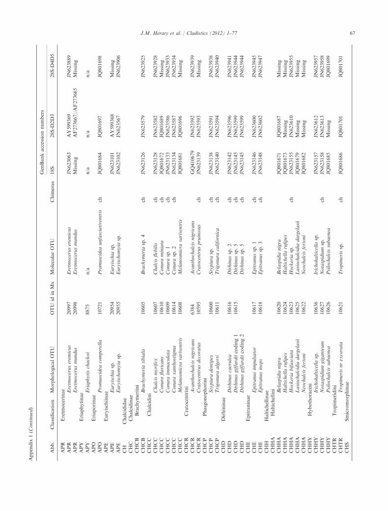
Appendix
1(C
ontinued)
Abb.
Classification
MorphologicalOTU
OTU
idin
Mx
MolecularOTU
Chim
eras
GenBankaccessionnumbers
18S
28S-D
2D3
28S-D
4D5
APR
Eretm
ocerinae
APR
Eretm
oceruserem
icus
20997
Eretm
oceruserem
icus
JN623063
AY599369
JN623889
APR
Eretm
ocerusmundus
20998
Eretm
ocerusmundus
Missing
AF273667//AF273665
Missing
APY
Eriaphytinae
APY
Eriaphytischackoi
8875
n/a
n/a
n/a
n/a
APO
Eriaporinae
APO
Promuscidea
comperella
10721
Promuscidea
unfasciativentris
chJQ
801684
JQ801697
JQ801698
APE
Euryischiinae
APE
Euryischia
sp.
20954
Euryischia
sp.
JN623101
AY599368
Missing
APE
Euryischomyia
sp.
20955
Euryischomyia
sp.
JN623102
JN623567
JN623906
CH
Chalcididae
CHC
Chalcidinae
CHCB
Brachymeriini
CHCB
Brachymeria
tibialis
10605
Brachymeria
sp.4
chJN
623126
JN623579
JN623925
CHCC
Chalcidini
CHCC
Chalcismyrifex
10607
Chalcisflebilis
chJN
623128
JN623582
JN623928
CHCC
Conura
flavicans
10610
Conura
miniata
chJQ
801672
JQ801689
Missing
CHCC
Conura
immaculata
10609
Conura
sp.1
chJN
623133
JN623586
JN623933
CHCC
Conura
xanthostigma
10614
Conura
sp.2
chJN
623134
JN623587
JN623934
CHCC
Melanosm
icra
variventris
10608
Melanosm
icra
variventris
JQ801681
JQ801696
Missing
CHCR
Cratocentrini
CHCR
Acanthochalcisnigricans
6384
Acanthochalcisnigricans
GQ410679
JN623592
JN623939
CHCR
Cratocentrusdecoratus
10595
Cratocentruspruinosus
chJN
623139
JN623593
Missing
CHCP
Phasgonophorini
CHCP
Stypiura
dentipes
10600
Stypiura
sp.
chJN
623138
JN623591
JN623938
CHCP
Trigonura
algerti
10611
Trigonura
californica
chJN
623140
JN623594
JN623940
CHD
Dirhininae
CHD
Dirhinuscaerulea
10616
Dirhinussp.2
chJN
623142
JN623596
JN623941
CHD
Dirhinusgiffardiicoding1
10615
Dirhinussp.5
chJN
623145
JN623599
JN623944
CHD
Dirhinusgiffardiicoding2
Dirhinussp.5
chJN
623145
JN623599
JN623944
CHE
Epitraninae
CHE
Epitranusim
pulsator
10617
Epitranussp.1
chJN
623146
JN623600
JN623945
CHE
Epitranusinops
10618
Epitranussp.3
chJN
623148
JN623602
JN623947
CHH
Haltichellinae
CHHA
Haltichellini
CHHA
Belaspidia
nigra
10620
Belaspidia
nigra
JQ801671
JQ801687
Missing
CHHA
Haltichella
rufipes
10624
Haltichella
rufipes
JQ801673
Missing
Missing
CHHA
Hockeria
bifasciata
10623
Hockeria
sp.
chJN
623155
JN623610
JN623955
CHHA
Lasiochalcidia
dargelasii
10625
Lasiochalcidia
dargelasii
JQ801679
Missing
Missing
CHHA
Neochalcisfertoni
10622
Neochalcisfertoni
JQ801682
Missing
Missing
CHHY
Hybothoracini
CHHY
Irichohalticella
sp.
10636
Irichohalticella
sp.
JN623157
JN623612
JN623957
CHHY
Notaspidium
giganteum
10627
Notaspidium
sp.
chJN
623158
JN623613
JN623958
CHHY
Psilochalcissubaenea
10626
Psilochalcissubaenea
JQ801685
Missing
JQ801699
CHTR
Tropim
eridini
CHTR
Tropim
erisnrexcavata
10621
Tropim
erissp.
chJQ
801686
JQ801701
JQ801701
CHS
Smicromorphinae
J.M. Heraty et al. / Cladistics (2012) 1–77 67

Appendix
1(C
ontinued)
Abb.
Classification
MorphologicalOTU
OTU
idin
Mx
MolecularOTU
Chim
eras
GenBankaccessionnumbers
18S
28S-D
2D3
28S-D
4D5
CHS
Smicromorphadoddi
10606
Smicromorphasp.
chJN
623160
JN623615
JN623959
EN
Encyrtidae
ENE
Encyrtinae
ENE
Acerophagusnrflavus
18873
Acerophagussp.
chJN
623190
AY599309
JN623974
ENE
Cheiloneurusflaccus
6389
Cheiloneurusfulvescens
chJN
623191
AY599308
Missing
ENE
Cheiloneurusfulvescens
10707
Cheiloneurusfulvescens
JN623191
AY599308
Missing
ENE
Comperiellabifasciata
18876
Comperiellabifasciata
JN623192
AY599317
Missing
ENE
Copidosomafloridanum
18875
Copidosomafloridanum
JN623193
AY599319
Missing
ENE
Metaphycusluteolus
18870
Metaphycussp.
chJN
623195
AY599310
JN623975
ENE
Trechnites
fuscitarsis
10708
Trechnites
sp.
chJN
623198
AY599318
Missing
ENT
Tetracnem
inae
ENT
Anagyruspseudococci
10702
Anagyrusnr.pseudococci
chMissing
AY599315
Missing
ENT
Savzdargia
hofferi
10699
Savzdargia
sp.
chJN
623203
JN623628
JN623979
EU
Eucharitidae
EUA
Akapalinae
EUA
Akapala
rudis
10522
Akapala
rudis
JN623320
AY672943
JN624066
EUE
Eucharitinae
EUE
Eucharitini
EUE
Ancylotropuscariniscutis
10535
Ancylotropuscariniscutis
JN623228
AY552239
JN624005
EUE
Eucharisadscendens
10541
Eucharisadscendens
Missing
AY672979
Missing
EUE
Kapala
floridana
9177
Kapala
floridana
JN623234
AY671849
Missing
EUE
Lophyrocera
pretendens
21453
Lophyrocera
pretendens
AY552304
AY552227
JN624009
EUE
Pseudochalcura
gibbosa
10540
Pseudochalcura
prolata
chMissing
GQ331918
GQ331918
EUE
Pseudometagea
schwarzii
11222
Pseudometagea
schwarzii
JN623242
AY552215
Missing
EUE
Psilocharitini
EUE
Neolosbanuspalgravei
10532
Neolosbanuspalgravei
JN623236
AY552213
JN624010
EUE
Psilocharisafra
9005
Psilocharisafra
JN623243
AY552210
JN624014
EUG
Gollumiellinae
EUG
Anorasemapallidipes
11223
Anorasemapallidipes
JN623250
AY552189
JN624019
EUG
Gollumiellalongipetiolata
10524
Gollumiellalongipetiolata
JN623252
AY552191
JN624020
EUO
Oraseminae
EUO
Indosemaindica
10525
Indosemaindica
JN623253
JN623661
JN624021
EUO
Orasemasimulatrix
10527
Orasemasimulatrix
JN623259
AY552206
JN624027
EUO
Tim
ioderusacuminatus
10596
Tim
ioderusperidentatus
chJN
623266
AY552195
JN624034
EL
Eulophidae
ELE
Entedoninae
ELE
Chrysocharissp.
14217
Chrysocharissp.
chJN
623165
HM364911//HM364981
HM364981
ELE
Dasyomphale
chilensis
14202
Dasyomphale
chilensis
JN623167
HM364938//HM365008
HM365008
ELE
Entedonsylvestris
11240
Entedonzanara
chJN
623168
JN623621
JN623964
ELE
Euderomphale
chelidonii
11236
Euderomphale
sp.
chJN
623169
HM364961//HM365022
HM365022
ELE
Neopomphale
sp.
14214
Neopomphale
sp.
JN623170
HM364941//HM365011
HM365011
ELN
Entiinae
ELN
Bellerussp.
14201
Bellerussp.
JN623176
HM364949//HM365014
HM365014
ELN
Beornia
sp.
14204
Beornia
sp.
chMissing
JQ801688
JQ801688
ELN
Euderussp.
11239
Euderussp.
chJN
623177
AY599259//HM364969
Missing
ELU
Eulophinae
ELU
Cirrospiluscoachellae
6377
Cirrospiluscoachellae
GQ410672
AY599268
JN623968
68 J.M. Heraty et al. / Cladistics (2012) 1–77

Appendix
1(C
ontinued)
Abb.
Classification
MorphologicalOTU
OTU
idin
Mx
MolecularOTU
Chim
eras
GenBankaccessionnumbers
18S
28S-D
2D3
28S-D
4D5
ELU
Elasm
uspolistis
11237
Elasm
uspolistis
JN623182
AY599264
JN623969
ELU
Eulophussp.
11241
Eulophussp.
chJN
623183
JN623623
JN623970
ELU
Euplectrusbicolor
12153
Euplectrussp.
chJN
623184
HM364953//HM365017
HM365017
ELU
Pnigaliomediterraneus
11672
Pnigaliosp.
chJN
623187
AY599279//HM364965
HM364965
ELU
Pnigaliosp.
14207
Pnigaliosp.
JN623187
AY599279//HM364965
HM364965
ELU
Zagrammosomaamericanum
14206
Zagrammosomaamericanum
JN623189
AY599263//HM364968
HM364968
ELI
Incertaesedis
ELI
Trisecodes
agromyzae
14205
Trisecodes
agromyzae
JN623171
HM364964//HM365025
HM365025
ELO
Ophelem
inae
ELO
Ophelim
usmaskelli
11242
Ophelim
usmaskelli
chJN
623172
HM364944//HM365012
HM365012
ELO
Perthiola
mazaneci
20953
Pertioloamazaneci
JN623185
JN623624
JN623971
ELT
Tetrastichinae
ELT
Aprostocetushibus
11238
Aprostocetussp.
chJN
623179
AY599265//HM364966
HM364966
ELT
Crataepusmarbis
14211
Crataepusmarbis
Missing
AY599262//HM365021
JN623967
ELT
Hadrotrichodes
waukheon
14203
Hadrotrichodes
waukheon
JN623180
HM364959//HM365020
HM365020
ELT
Melittobia
acasta
11235
Melittobia
digitata
chMissing
U02951
U02951
ELT
Minotetrastichusfrontalis
21226
Minotetrastichusfrontalis
Missing
D2753
D2753
EP
Eupelmidae
EPC
Calosotinae
EPC
Archaeopelmasp.
10679
Archaeopelmatropeotergum
chMissing
JN623630
JN623980
EPC
Balchaindica
10666
Balchasp.
chJN
623204
JN623631
JN623981
EPC
Calosota
metallica
10674
Calosota
sp.
chJN
623205
JN623632
JN623982
EPC
Eusandalum
cyaneum
10667
Eusandalum
sp.1
chJN
623206
JN623633
JN623983
EPC
Licrooides
umbilicatus
10675
Licrooides
umbilicatus
JN623209
JN623636
JN623986
EPE
Eupelminae
EPE
Anastatusfloridanusfemale
10689
Anastatussp.
chJN
623210
JN623637
JN623987
EPE
Anastatusfloridanusmale
10690
Anastatussp.
chJN
623210
JN623637
JN623987
EPE
Arachnophagaeucnem
iafemale
10695
Arachnophagaeucnem
iaJN
623211
JN623638
JN623988
EPE
Arachnophagaeucnem
iamale
10696
Arachnophagaeucnem
iaJN
623211
JN623638
JN623988
EPE
Brasemaallyniifemale
10693
Brasemasp.
chJN
623212
AY599306
JN623989
EPE
Brasemaallyniimale
10694
Brasemasp.
chJN
623212
AY599306
JN623989
EPE
Eupelmusurozonusfemale
10687
Eupelmussp.
chJN
623215
AY599307
JN623992
EPE
Eupelmusurozonusmale
10688
Eupelmussp.
chJN
623215
AY599307
JN623992
EPE
Phenaceupelmuschilensisfemale
10691
n/a
n/a
n/a
n/a
EPE
Phenaceupelmuschilensismale
10692
n/a
n/a
n/a
n/a
EPN
Neanastatinae
EPN
Lambdobregmasp.
10680
n/a
n/a
n/a
n/a
EPN
Metapelmaspectabile
10681
Metapelmasp.2
chJN
623225
JN623650
JN624002
EPN
Neanastatussp.
10682
Neanastatussp.2
chJN
623227
JN623652
JN624004
EY
Eurytomidae
EYB
Buresiinae
EYB
Buresium
rufum
10638
Buresium
naso
chJN
623270
JN623669
JN624036
EYB
Macrorileyasp.
10639
Macrorileyasp.
JQ801680
Missing
JQ801695
EYE
Eurytominae
EYE
Aiolomorphusrhopaloides
14196
Aiolomorphusrhopaloides
JN623268
JN623667
JN624035
EYE
Axim
azabriskiei
21339
n/a
n/a
n/a
n/a
EYE
Bephrata
ruficollis
10646
n/a
n/a
n/a
n/a
J.M. Heraty et al. / Cladistics (2012) 1–77 69

Appendix
1(C
ontinued)
Abb.
Classification
MorphologicalOTU
OTU
idin
Mx
MolecularOTU
Chim
eras
GenBankaccessionnumbers
18S
28S-D
2D3
28S-D
4D5
EYE
Bruchodapeignota
10645
n/a
n/a
n/a
n/a
EYE
Endobia
donacis
10644
Endobia
donacis
JQ801674
JQ801690
JQ801690
EYE
Eurytomagigantea
6378
Eurytomagigantea
GQ410671
JN623670
JN624037
EYE
Masneromaangulifera
20864
Masneromaangulifera
JN623275
JN623673
Missing
EYE
Sycophilatexana
14198
Sycophilatexana
JN623277
JN623675
Missing
EYE
Systole
albipennis
21250
Systole
coriandri
chJN
623278
JN623676
Missing
EYE
Tetramesasp.
21251
Tetramesasp.1
chJN
623279
AY599281
Missing
EYH
Heimbrinae
EYH
Heimbra
opaca
20860
Heimbra
opaca
JN623281
JN623678
JN624038
EYR
Rileyinae
EYR
Austrophotism
usdaicles
14197
Austrophotism
usdaicles
JN623282
JN623679
Missing
EYR
Neorileyaflavipes
20861
Neorileyasp.
chJQ
801675
JQ809471
JQ809471
EYR
Rileyalongitergum
14199
Rileyagrisselli
chJN
623284
JN623681
JN624039
EYR
Rileyapallidipes
10631
Rileyapallidipes
JN623286
AY599298
JN624041
LEU
Leucospidae
LEU
Leucospisaffinis
10603
Leucospisaffinis(a)
JN623290
AY599276
Missing
LEU
Micrapionsp.
11226
Micrapionsp.
JN623296
JN623689
JN624049
MY
Mymaridae
MYA
Alaptinae
MYA
Anagrusepos
9134
Anagrusepos
JN623299
JN623692
JN624051
MYA
Lituscynipseus
9133
Lituscynipseus
JN623300
JN623693
JN624052
MYA
Stethynium
ophelim
i9129
Stethynium
ophelim
iJN
623301
JN623694
JN624053
MYE
Eubroncinae
MYE
Eubroncussp.
9137
Eubroncussp.2
JN623303
JN623696
Missing
MYI
Incertaesedis
MYI
Borneomymardiscus
21242
Borneomymarmadagascar
chJN
623304
AY599254
JN624054
MYM
Mymarinae
MYM
Acm
opolynem
avarium
6387
Acm
opolynem
avarium
JN623305
AY599251
JN624055
MYM
Anaphes
sp.
9127
Anaphes
victus
JN623306
JN623697
JN624056
MYM
Australomymarsp.
6381
Australomymarsp.
chGQ410668
JN623698
JN624057
MYM
Ceratanaphes
sp.
9128
Ceratanaphes
sp.
JN623307
AY599252
JN624058
MYM
Erythmelusrosascostai
9126
Erythmelusrosascostai
JN623308
JN623699
JN624059
MYM
Gonatocerusashmeadi
6380
Gonatocerusashmeadi
JN623309
JN623700
JN624060
MYM
Gonatocerussp.
10642
Gonatocerustriguttatus
chJN
623310
AY599253
JN624061
MYM
Mymarregale
9135
Mymarregale
JN623311
JN623701
JN624062
MYM
Ooctonussp.
9131
Ooctonussp.
JN623312
JN623702
Missing
ORM
Orm
yridae
ORM
Orm
yrulussp.
10650
Orm
yrulussp.
JN623316
JN623706
JN624064
ORM
Orm
yrusrosae
10649
Orm
yrussp.1
chJN
623317
JN623707
JN624065
PE
Perilampidae
PEC
Chrysolampinae
PEC
Austrotoxeumasp.
9147
Austrotoxeumasp.
JN623322
AY552187
JN624068
PEC
Brachyelatussp.
9148
Brachyelatussp.
JN623321
AY552184
JN624067
PEC
Chrysolampusschwarzi
9149
Chrysolampusschwarzi(a)
JN623325
AY672937
Missing
PEC
Chrysolampussp.
9152
Chrysolampussp.3
JN623329
AY672939
Missing
PEC
Chrysomallahesperis
9155
Chrysomallahesperis
JN623330
AY672940
Missing
PEI
Incertaesedis
70 J.M. Heraty et al. / Cladistics (2012) 1–77

Appendix
1(C
ontinued)
Abb.
Classification
MorphologicalOTU
OTU
idin
Mx
MolecularOTU
Chim
eras
GenBankaccessionnumbers
18S
28S-D
2D3
28S-D
4D5
PEI
Jambiyavanharteni
10543
Jambiyavanharteni
JN623331
JN623711
JN624071
PEP
Perilampinae
PEP
Euperilampustriangularis
9158
Euperilampustriangularis
JN623338
AY552174
Missing
PEP
Monaconrobertsi
11479
Monaconroberstsi
JN623340
JN623716
JN624078
PEP
Perilampushyalinus
9160
Perilampushyalinus
JN623344
AY552179
JN624081
PEP
Perilampussubcarinatus
9162
Perilampussubcarinatus
JN623353
AY552175
JN624087
PEP
Perilampustristis
6395
Perilampussubcarinatus
chJN
623353
AY552175
JN624087
PEP
Steffanolampussalicetum
9163
Steffanolampussalicetum
JN623354
AY552177
JN624088
PEM
Philomidinae
PEM
Aperilampussp.
10570
Aperilampussp.
chJN
623332
AY672941
JN624072
PEM
New
Genussp.
20927
Philomidinaenew
genus
JN623334
AY672942
JN624074
PEM
Philomides
sp.
20949
Philomides
sp.
JN623333
JN623712
JN624073
PT
Pteromalidae
PT01
Asaphinae
PT01
Asaphes
sp.
6388
Asaphes
suspensus
chJN
623355
JN623727
JN624089
PT01
New
Genussp.
14018
Asaphinaenew
genus
JN623356
JN623728
JN624090
PT26
Austrosystasinae
PT26
Austrosystasisatricorpus
14248
n/a
n/a
n/a
n/a
PT02
Ceinae
PT02
Spalangiopelta
apotherisma
11488
Spalangiopelta
apotherisma
JN623358
JN623730
JN624092
PT03
Cerocephalinae
PT03
Acerocephala
sp.
14019
Acerocephala
sp.
JN623360
JN623731
JN624094
PT03
Neocalosotersp.
14020
Neocalosotersp.
JN623362
JN623733
Missing
PT04
Chromeurytominae
PT04
Chromeurytomasp.
6391
Chromeurytomasp.
chJN
623363
JN623734
JN624096
PT05
Cleonyminae
PT05D
Chalcedectini
PT05D
Chalcedectussp.
13975
Chalcedectussp.
JN623365
JN623736
JN624098
PT05C
Cleonymini
PT05C
Callocleonymussp.
14165
Callocleonymussp.
JN623364
JN623735
JN624097
PT05C
Cleonymussp.
6366
Cleonymussp.
chGQ410678
AY599278
JN624099
PT05L
Lyciscini
PT05L
Agamerioncleptideum
14031
Agamerioncleptideum
JN623367
AY599287
JN624101
PT05L
Epistenia
sp.
13974
Epistenia
sp.
JN623368
AY599282
Missing
PT05L
Proshizonotussp.
14030
Proshizonotussp.
JN623371
JN623738
JN624104
PT05O
Ooderini
PT05O
Ooderasp.
13972
Ooderasp.
JN623372
JN623739
JN624105
PT06
Coelocybinae
PT06
Lanthanomyia
sp.
13984
Lanthanomyia
sp.
JN623374
JN623741
JN624107
PT06
Orm
yromorphasp.
13976
Orm
yromorphasp.
JN623375
JN623742
JN624108
PT06
Yrkasp.
10569
Yrkasp.
chJN
623376
JN623743
JN624109
PT07
Colotrechninae
PT07
Colotrechnusignotus
10660
Colotrechnusignotus
JN623377
JN623744
JN624110
PT08
Cratominae
PT08
Cratomussp.
20867
Cratomusmegacephalus
chJN
623379
JN623746
JN624112
PT09
Diparinae
PT09D
Diparini
J.M. Heraty et al. / Cladistics (2012) 1–77 71

Appendix
1(C
ontinued)
Abb.
Classification
MorphologicalOTU
OTU
idin
Mx
MolecularOTU
Chim
eras
GenBankaccessionnumbers
18S
28S-D
2D3
28S-D
4D5
PT09D
Lelapssp.
13977
Lelapssp.
JN623381
AY599280
Missing
PT09D
Netomocera
sp.
14028
Netomocera
sp.
JN623383
JN623749
JN624115
PT09D
Paruriossp.
13809
Dipara
sp.
JN623380
JN623747
JN624113
PT09N
Neapterolelapini
PT09N
Neapterolelapssp.
14029
Neapterolelapssp.1
JN623385
JN623751
JN624117
PT09N
Neapterolelapssp.female
10662
Neapterolelapssp.2
chJN
623386
JN623752
JN624118
PT09N
Neapterolelapssp.male
10663
Neapterolelapssp.2
chJN
623386
JN623752
JN624118
PT27
Ditropinotellinae
PT27
Ditropinotellasp.
14246
n/a
n/a
n/a
n/a
PT10
Epichrysomallinae
PT10
Neosycophilasp.
11208
Neosycophilasp.1
JN623406
JN623772
JN624138
PT10
Odontofroggatiagalili
11207
Odontofroggattia
ishii
chJQ
801683
HM770696
HM770696
PT10
Sycobia
sp.
11209
Sycobia
sp.
JN623411
JN623777
JN624143
PT11
Eunotinae
PT11E
Eunotini
PT11E
Cephaleta
sp.
14027
Cephaleta
sp.
JN623414
JN623780
JN624146
PT11E
Eunotussp.
6392
Eunotussp.1
chJN
623415
AY599295
Missing
PT11E
Eunotussp.
14026
Eunotussp.2
JN623416
JN623781
JN624147
PT11E
Idioporusaffinis
6393
Idioporusaffinis
JN623417
Missing
JN624148
PT11E
Scutellista
caerulea
10549
Scutellista
caerulea
JN623418
AY599294
Missing
PT11M
Moranilini
PT11M
Moranilacalifornica
6394
Moranilasp.
chJN
623419
JN623783
JN624149
PT11T
Tomocerodini
PT11T
Tomocerodes
sp.
13982
Tomocerodes
sp.
JN623420
JN623784
JN624150
PT12
Eutrichosomatinae
PT12
Collessinapachyneura
14247
n/a
n/a
n/a
n/a
PT12
Eutrichosomamirabile
10612
Eutrichosomamirabile
JN623421
AY599286
JN624151
PT12
Peckianussp.
14160
n/a
n/a
n/a
n/a
PT13
Herbertiinae
PT13
Herbertiasp.
14021
Herbertiasp.
JN623422
JN623785
JN624152
PTI
Incertaesedis
PTI
Lieparini
PTI
Liepara
sp.
14209
Liepara
sp.
JN623382
JN623748
JN624114
PT28
Keiraninae
PT28
Keiranalongicollis
14164
n/a
n/a
n/a
n/a
PT14
Leptofoeninae
PT14
Doddifoenuswallacei
8758
Doddifoenuswallacei
JN623423
JN623786
JN624153
PT14
Leptofoenusstephanoides
10656
Leptofoenussp.1
chJN
623424
AY599283
JN624154
PT14
Leptofoenuswestwoodi
10659
Leptofoenussp.2
chJN
623425
AY599284
JN624155
PT15
Macromesinae
PT15
Macromesusamericanus
14158
Macromesusamericanus
JN623426
JN623787
JN624156
PT16
Miscogastrinae
PT16M
Miscogastrini
PT16M
Lamprotatussplendens
11478
Lamprotatussplendens
JN623429
JN623790
JN624159
PT16M
Nodisoplata
sp.
14159
Nodisoplata
sp.
JN623438
JN623798
JN624169
PT16M
Sphaeripalpussp.
14156
Sphaeripalpussp.
JN623430
JN623791
JN624160
PT16S
Sphegigastrini
72 J.M. Heraty et al. / Cladistics (2012) 1–77

Appendix
1(C
ontinued)
Abb.
Classification
MorphologicalOTU
OTU
idin
Mx
MolecularOTU
Chim
eras
GenBankaccessionnumbers
18S
28S-D
2D3
28S-D
4D5
PT16S
Cyrtogaster
vulgaris
11487
Cyrtogaster
vulgaris
JN623432
JN623792
JN624162
PT16S
Halticoptera
dim
idiata
11484
Halticoptera
laevigata
chJN
623427
JN623788
JN624157
PT16S
Polstonia
pelagocorypha
10546
Polstonia
pelagocorypha
JN623433
AY552173
JN624163
PT16S
Thinodytescyzicus
11492
Thinodytessp.
chJN
623431
AY552172
JN624161
PT16T
Trigonoderini
PT16T
Plutothrixsp.
13978
Plutothrixsp.
Missing
JN623794
JN624165
PT17
Orm
ocerinae
PT17I
Incertaesedis
PT17I
Erixestussp.
13981
Erixestussp.
JN623478
JN623832
JN624205
PT17M
Melanosomellini
PT17M
Aditrochuscohiuensis
13971
Aditrochuscoihuensis
JN623435
JN623795
JN624166
PT17M
Espinosa
nothofagi
13970
Espinosa
nothofagi
JN623436
JN623796
JN624167
PT17M
Systolomorphasp.
10580
Systolomorphasp.
JN623437
JN623797
JN624168
PT17S
Systasini
PT17S
Sem
iotellussp.
13983
Sem
iotellussp.
JN623439
JN623799
JN624170
PT17S
Systasissp.
13969
Systasissp.
chMissing
JQ801700
Missing
PT18
Otitesellinae
PT18
Heterandrium
sp.
11217
Heterandrium
sp.
JN623442
JN623802
JN624173
PT18
Walkerella
kurandensis
11219
n/a
n/a
n/a
n/a
PT19
Panstenoninae
PT19
Panstenonoxylus
11490
Panstenonoxylus
JN623444
JN623805
JN624176
PT19
Panstenonsp.
10665
Panstenonsp.
chJN
623445
JN623804
JN624175
PT20
Pireninae
PT20
Gastrancistrussp.
13968
Gastrancistrussp.
JN623446
AY599293
Missing
PT20
Macroglenes
gramineus
13967
Macroglenes
sp.
JN623447
JN623806
JN624177
PT21
Pteromalinae
PTI
Incertaesedis
PTI
Diconocara
petiolata
13980
Diconocara
petiolata
JN623455
JN623813
JN624184
PT21M
Micradelini
PT21M
Micradelusrotundus
21032
n/a
n/a
n/a
n/a
PT21P
Pteromalini
PT21P
Cheiropachusquadrum
11504
Cheiropachusquadrum
JN623451
JN624260
JN624260
PT21P
Coelopisthia
extenta
11480
Coelopisthia
extenta
JN623452
JN623810
JN624181
PT21P
Corunaclavata
11481
Corunaclavata
JN623453
JN623811
JN624182
PT21P
Habritysbrevicornis
11483
Habritysbrevicornis
JN623456
JN623814
JN624185
PT21P
Homoporusluniger
11485
Homoporussp.
chJN
623457
JN623815
JN624186
PT21P
Merisussplendidus
11493
Merisussplendidus
JN623459
JN623817
JN624188
PT21P
Nasonia
vitripennis
6382
Nasonia
vitripennis
GQ410677
JN623821
JN624192
PT21P
Pegopusinornatus
11491
Pegopussp.
chJN
623460
JN623818
JN624189
PT21P
Perilampidea
syrphi
14157
Perilampidea
sp.
chJN
623461
JN623819
JN624190
PT21P
Pseudocatolaccusnitescens
11500
Pseudocatolaccusguizoti
chJN
623462
AY552171
Missing
PT21P
Pteromalusnralbipennis
14162
Pteromalusalbipennis
JN623465
AY552170
JN624193
PT22
Spalangiinae
PT22
Spalangia
endius
14161
Spalangia
erythromera
chJN
623466
JN623822
JN624194
PT22
Spalangia
nigroaenea
10661
Spalangia
nigroaenea
JN623467
JN623823
JN624195
PT23
Sycoecinae
PT23
Diaziella
sp.
11218
Diaziella
sp.
JN623469
JN623825
JN624197
J.M. Heraty et al. / Cladistics (2012) 1–77 73

Appendix
1(C
ontinued)
Abb.
Classification
MorphologicalOTU
OTU
idin
Mx
MolecularOTU
Chim
eras
GenBankaccessionnumbers
18S
28S-D
2D3
28S-D
4D5
PT24
Sycophaginae
PT24
Anidarnes
bicolor
11203
Anidarnes
sp.
chJN
623470
JN623826
JN624198
PT24
Apocryptophagusgigas
11205
Apocryptophagusgigas
JQ801670
HM770698
HM770698
PT24
Eukoebelea
australiensis
11206
Eukobelea
sp.
JN623471
HM770667
HM770667
PT24
Pseudidarnes
minerva
11202
Pseudidarnes
sp.
chJN
623474
JN623828
JN624201
PT24
Sycophagacyclostigma
11204
Sycophagasycomori
chJN
623475
JN623829
JN624202
PT25
Sycoryctinae
PT25
Philotrypesissp.
11216
Philotrypesissp.
JN623476
JN623830
JN624203
PT25
Sycoscaptersp.
11215
Sycoscaptersp.
JN623477
JN623831
JN624204
ROT
Rotoitidae
ROT
Chiloemicropteron
6390
Chiloemicropteron
GQ410669
JN623834
JN624206
ROT
Rotoitabasalis
20856
n/a
n/a
n/a
n/a
SI
Signiphoridae
SIS
Signiphorinae
SIS
Signiphora
sp.
21257
Signiphora
sp.3
JN623481
AY599343
Missing
SIT
Thysaninae
SIT
Chartocerussp.
21256
Chartocerussp.1(a)
JN623488
AY599336
JN624212
SIT
Clytinagiraudi
21244
Clytinagiraudi
JN623495
JN623835
JN624218
SIT
Thysanusater
10676
Thysanussp.3
chJN
623498
AY599354
JN624220
TAN
Tanaostigmatidae
TAN
Cynipencyrtusflavus
10556
Cynipencyrtussp.
chJN
623500
JN623836
JN624222
TAN
Protanaostigmasp.
14219
Protanaostigmasp.
JN623501
JN623837
JN624223
TAN
Tanaostigmodes
howardii
10703
Tanaostigmodes
albiclavus(a)
chJN
623503
AY599304
Missing
TE
Tetracampidae
TEM
Mongolocampinae
Eremocampesp.
21296
Eremocampesp.
JN623506
AY599356
JN624225
Mongolocampebouceki
20857
n/a
n/a
n/a
n/a
Platyneurussp.
21335
n/a
n/a
n/a
n/a
TEP
Platynocheilinae
TEP
Platynocheiluscuprifrons
20858
Platynocheiluscuprifrons
JN623507
JN623838
JN624226
TET
Tetracampinae
TET
Diplesiostigmasp.
21334
Diplesiostigmabisetosum
chJN
623508
JN623839
Missing
TET
Epiclerustemenus
21227
Epiclerussp.1
chJN
623509
AY599357
Missing
TET
Foersterella
reptans
6383
Foersterella
reptans
JN623511
AY599360
JN624227
TET
Tetracampesp.
21294
Tetracampesp.
chJN
623512
JN623840
JN624228
TO
Torymidae
TOM
Megastigminae
TOM
Bootanelleussp.
10697
Bootanelleussp.
TOOSHORT
JN623841
JN624229
TOM
Bortesia
sp.
21249
Bortesia
sp.
JQ801677
JQ801693
JQ801693
TOM
Megastigmustransvaalensis
6385
Megastigmustransvaalensis
GQ410676
JN623843
JN624232
TOM
Neomegastigmussp.
21248
Neomegastigmussp.
JQ801676
JQ801692
JQ801692
TOT
Toryminae
TOTC
Chalcim
erini
TOTC
Chalcim
erusborceai
21243
Chalcim
erusborceai
JQ801678
JQ801694
JQ801694
TOTI
Incertaesedis
TOTI
Echthrodapesp.
10732
Echthrodapesp.
JN623516
JN623845
JN624234
TOTI
Glyphomerusstigma
10677
Glyphomerusstigma
JN623518
JN623847
JN624236
74 J.M. Heraty et al. / Cladistics (2012) 1–77

Appendix
1(C
ontinued)
Abb.
Classification
MorphologicalOTU
OTU
idin
Mx
MolecularOTU
Chim
eras
GenBankaccessionnumbers
18S
28S-D
2D3
28S-D
4D5
TOTM
Microdontomerini
TOTM
Eridontomerusarrabonicus
10706
Eridontomerusarrabonicus
Missing
JN623848
JN624237
TOTM
Idarnotorymuspulcher
10730
Idarnotorymuspulcher
JN623520
JN623851
JN624240
TOTM
Pseuderim
erusburgeri
10715
Pseuderim
erusburgeri
JN623521
JN623852
JN624241
TOTN
Monodontomerusmontivagus
10716
Monodontomerussp.
chJN
623523
JN623855
JN624243
TOTP
Palachiini
TOTP
Propalachia
borneana
10713
Propalachia
borneana
Missing
JN623856
JN624244
TOTO
Podagrionini
TOTO
Podagrionsp.
10683
Podagrionsp.
chJN
623524
AY599269
JN624251
TOTT
Torymini
TOTT
Torymusbedeguaris
10722
Torymusbedeguaris
Missing
JN624256
JN624256
TOTY
Torymoidini
TOTY
Pseudotorymussapphyrinus
10726
Pseudotorymussapphyrinus
Missing
JN624254
JN624254
TR
Trichogrammatidae
TRO
Oligositinae
TROO
Oligositini
TROO
Oligosita
sanguinea
14133
Oligosita
sanguinea
JN623530
AY623551
Missing
TRT
Trichogrammatinae
TRTT
Trichogrammatini
TRTT
Trichogrammapretiosum
14132
Trichogrammapretiosum
AY940359
AY599408
Missing
TRTI
Trichogrammatiniincertaesedis
TRTI
Ceratogrammaetiennei
11527
Ceratogrammamasneri
chAY940340
AY623492
Missing
TRTI
Haeckeliania
sperata
14131
Haeckeliania
sp.2
chAY940343
AY623497
Missing
CAL
Calesinaeincertaesedis
CAL
Calesberryi
20999
Calesberryi
JN623119
AY599257
JN623920
CAL
Calesnoacki
6375
Calesnoacki
JN623121
JN623574
JN623921
NonChalcidoidea
Proctotrupoidea
Diapriidae
BelytinaeBelyta
sp.
6398
Belyta
sp.
chJN
623118
JN624258
JN624258
Mymarommatoidea
Mymarommatidae
Mymaromella
chaoi
20995
Mymaromella
mira
GQ410666
GQ374773
GQ374773
Mymarommasp.
6386
Mymarommasp.
JN623313
JN623703
Missing
Platygastroidea
Scelionidae
Scelioninae Archaeoteleia
mellea
6397
Archaeoteleia
mellea
GQ410639
GQ374746
GQ374746
J.M. Heraty et al. / Cladistics (2012) 1–77 75

Appendix 2
Abbreviations for morphology
ab Articulating bulb HAO_0001704acr Acropleuron HAO_0001155acs Antecostal sulcus HAO_0000099aed Aedeagus HAO_0000091ax Axilla HAO_0000155btc Basitarsal comb HAO_0001180btn Basitarsal notch HAO_0000177br Bridge of second valvifer HAO_0001780cer Cercus HAO_0000191clm Pronotal collum (neck) HAO_0000837col Pronotal collar HAO_0000832cps Coeloconic peg sensillum HAO_0002001cs Coeloconic sensillum HAO_0000213cx1, 2, 3 Pro-, meso- metacoxa HAO_0000228cxf Supracoxal flange No url assigneddc2 Mesodiscrimineal line HAO_0000545dig Digitus HAO_0000385dv Dorsal valve (second valvula) HAO_0001658ep2 Mesepimeron HAO_0000299epn Epimeral notch HAO_0002006esg Epistomal groove HAO_0000306F1–12 Flagellomeres 1–12 HAO_0000342f2p Mesofurcal pit HAO_0000549f3p Metafurcal pit HAO_0000594fra Frenal arm HAO_0001903frl Frenal line HAO_0000354frn Frenum HAO_0000355gc Genal carina HAO_0001755hyc Hypostomal carina HAO_0000413lb Laminated bridge of ovipositor HAO_0001548lbp Labial palp HAO_0000450lbr Labrum HAO_0000456lcl Lateral clypeal line No url assignedlep2 Lower mesepimeron HAO_0000299llm Lateral lobe of mesoscutum HAO_0000466los Lower ocular sulcus HAO_0000299mfm Metafemur HAO_0001140mlm Median lobe of mesoscutum HAO_0000575mls Malar sulcus HAO_0000504mms Median mesoscutal sulcus HAO_0000523MPS Multiporous plate sensillum HAO_0000640Ms6 Female hypopygium HAO_0000410Ms8 Male hypopygium HAO_0000410msp Mesothoracic spiracle HAO_0000582mss Mesosternal shelf HAO_0001647Mt Metasomal tergite HAO_0002005Mt8 + 9 Syntergum HAO_0000987mtp Mesotrochantinal plate HAO_0000543mtps Metaplerual sulcus No url assignedmts Metascutellum HAO_0000625mtsp Metathoracic spiracle HAO_0000769mtsa Metanotal scutellar arm No url assignedmtsp Metasomal spiracle No url assignedmxp Maxillary palp HAO_0000515no1 Pronotum HAO_0000853no3 Metanotum HAO_0000603not Notaulus HAO_0000647occ Occipital carina HAO_0000653ov Ovipositor HAO_0000679par Paramere HAO_0000395pgg Postgenal groove HAO_0002019
76 J.M. Heraty et al. / Cladistics (2012) 1–77

pgl Postgenal lamina HAO_00020202ph Mesophragma HAO_0000558pl1 Propleuron HAO_0000862pl3 Metapleruon HAO_0001271pmr Posterior marginal rim of metascutellum No url assignedpnb Pronotal bristle No url assignedpom Postoral microtrichia HAO_0000532pop Postoccipital plate No url assignedpre Prepectus HAO_0000811prp Propodeum HAO_0000051psc Parascutal carina HAO_0000697psp Propodeal spiracle HAO_0000329ptl Petiole HAO_0000020ptla Petiolar lamina HAO_0002018ptr Paratergite No url assignedrad Radicle HAO_0000889rmd Right mandible HAO_0000506s1 Prosternum HAO_0000873sap Sternal apodemes HAO_0002007sas Subantennal sulcus HAO_0000965scp Scapular flange HAO_0001680scr Scrobe HAO_0000912scs Scrobal sulcus HAO_0001679sct Mesoscutellum HAO_0000574sss Scutoscutellar sulcus HAO_0000919tfs Transfacial sulcus HAO_0002016tgl Tegula HAO_0001508tor Torulus HAO_0001022tsa Transscutal articulation HAO_0001204uep2 Upper mesepimeron HAO_0000299uos Upper ocular sulcus No url assigned3v Third valvula HAO_00010122vf Second valvifer HAO_0000927vos Vertical ocellar sulcus HAO_0002022vv Ventral valve (first valvula) HAO_0000339Wing abbreviationsams Admarginal setae No url assignedbc Basal cell No url assignedbpl Basal posterior lobe No url assignedbv Basal vein HAO_0000736C Costal vein HAO_0000225cc Costal cell HAO_0000226Cu Cubital vein HAO_0000237dm Dorsal macrochaeta No url assignedhb Hyaline break No url assignedhpl Humeral plate HAO_0000403hy Hypochaeta No url assignedM Medial vein HAO_0000519mv Marginal vein HAO_0000512pmv Postmarginal vein HAO_0000783pst Parastigma (premarginal vein) No url assignedpsts Parastigmal sensilla No url assignedR Radial vein HAO_00007832r Second radial crossvein No url assignedr-m Radio-medial crossvein No url assignedRs Radial sector vein No url assignedSc Subcubital vein No url assignedsmb Submarginal break No url assignedsmv Submarginal vein HAO_0000972stg Stigma No url assignedstv Stigmal vein No url assigned
HAO terms are detailed in Hymenoptera Anatomy Ontology (http://purl.obolibrary.org/obo/).
J.M. Heraty et al. / Cladistics (2012) 1–77 77