A patient with McLeod syndrome showing involvement of the ...
6
CASE REPORT Open Access A patient with McLeod syndrome showing involvement of the central sensorimotor tracts for the legs Takenobu Murakami 1,2* , Dan Abe 1 , Hideyuki Matsumoto 3 , Ryo Tokimura 1 , Mitsunari Abe 4 , Amanda Tiksnadi 1 , Shunsuke Kobayashi 1 , Chikako Kaneko 5 , Yuka Urata 6 , Masayuki Nakamura 6 , Akira Sano 6 and Yoshikazu Ugawa 1,7 Abstract Background: McLeod syndrome is a rare X-linked recessive acanthocytosis associated with neurological manifestations including progressive chorea, cognitive impairment, psychiatric disturbances, seizures, and sensorimotor axonal polyneuropathy. However, no studies have investigated the functioning of central sensorimotor tracts in patients with McLeod syndrome. Case presentation: A 66-year-old man had experienced slowly progressive chorea and gait disturbance due to lower limb muscle weakness since his early fifties. Blood examinations showed erythrocyte acanthocytosis and the reduction of Kell antigens in red blood cells. Brain magnetic resonance imaging showed atrophy of the bilateral caudate nuclei and putamen. The diagnosis of McLeod syndrome was confirmed by the presence of a mutation of the XK gene on the X chromosome. Somatosensory-evoked potential and transcranial magnetic stimulation studies demonstrated that the central sensory and motor conduction times were abnormally prolonged for the lower extremity but normal for the upper extremity. Conclusions: This is the first report of the involvement of the central sensorimotor tracts for the legs in a patient with McLeod syndrome. The clinical neurophysiological technique revealed the central sensorimotor tracts involvements clinically masked by neuropathy. Keywords: McLeod syndrome, Transcranial magnetic stimulation, Central motor conduction time, Motor-evoked potential, Somatosensory-evoked potential Background McLeod syndrome is an extremely rare progressive X- linked recessive type of neuroacanthocytosis that was first reported by Allen et al. in 1961 [1]. This syndrome is a multisystem disorder with central nervous system (CNS), neuromuscular, cardiovascular, and hematological mani- festations. Red blood cell acanthocytosis in McLeod syn- drome is associated with absent expression of the Kx antigen and reduced expression of the Kell antigen on the surface membranes of erythrocytes, which are caused by truncation or no expression of XK protein. The detection of mutations in the XK gene confirms a diagnosis of McLeod syndrome. The XK protein plays pivotal roles in organogenesis, cellular structure, and nutrient exchanges [2]. Patients with McLeod syndrome lack expression of this protein, which leads to acanthocytosis and neural degeneration. Neurological symptoms in McLeod syndrome are vari- ous, including progressive chorea, cognitive impairment, psychiatric disturbances, and seizures [3]. Sensorimotor axonal neuropathy is also a typical clinical feature, which leads to distal-dominant muscular weakness with mus- cular atrophy. A previous pathological study using a mouse model of McLeod syndrome found axonopathy in the spinal cord and the sciatic nerve [4]. However, whether the central sensorimotor tracts are involved in McLeod syndrome remains unclear. For the present © The Author(s). 2019 Open Access This article is distributed under the terms of the Creative Commons Attribution 4.0 International License (http://creativecommons.org/licenses/by/4.0/), which permits unrestricted use, distribution, and reproduction in any medium, provided you give appropriate credit to the original author(s) and the source, provide a link to the Creative Commons license, and indicate if changes were made. The Creative Commons Public Domain Dedication waiver (http://creativecommons.org/publicdomain/zero/1.0/) applies to the data made available in this article, unless otherwise stated. * Correspondence: [email protected] 1 Department of Neurology, Fukushima Medical University, Fukushima, Japan 2 Department of Neurology, Tottori Prefectural Kousei Hospital, Kurayoshi, Japan Full list of author information is available at the end of the article Murakami et al. BMC Neurology (2019) 19:301 https://doi.org/10.1186/s12883-019-1526-9
-
Upload
khangminh22 -
Category
Documents
-
view
3 -
download
0
Transcript of A patient with McLeod syndrome showing involvement of the ...

CASE REPORT Open Access
A patient with McLeod syndrome showinginvolvement of the central sensorimotortracts for the legsTakenobu Murakami1,2* , Dan Abe1, Hideyuki Matsumoto3, Ryo Tokimura1, Mitsunari Abe4, Amanda Tiksnadi1,Shunsuke Kobayashi1, Chikako Kaneko5, Yuka Urata6, Masayuki Nakamura6, Akira Sano6 and Yoshikazu Ugawa1,7
Abstract
Background: McLeod syndrome is a rare X-linked recessive acanthocytosis associated with neurologicalmanifestations including progressive chorea, cognitive impairment, psychiatric disturbances, seizures, andsensorimotor axonal polyneuropathy. However, no studies have investigated the functioning of centralsensorimotor tracts in patients with McLeod syndrome.
Case presentation: A 66-year-old man had experienced slowly progressive chorea and gait disturbance due tolower limb muscle weakness since his early fifties. Blood examinations showed erythrocyte acanthocytosis and thereduction of Kell antigens in red blood cells. Brain magnetic resonance imaging showed atrophy of the bilateralcaudate nuclei and putamen. The diagnosis of McLeod syndrome was confirmed by the presence of a mutation ofthe XK gene on the X chromosome. Somatosensory-evoked potential and transcranial magnetic stimulation studiesdemonstrated that the central sensory and motor conduction times were abnormally prolonged for the lowerextremity but normal for the upper extremity.
Conclusions: This is the first report of the involvement of the central sensorimotor tracts for the legs in a patientwith McLeod syndrome. The clinical neurophysiological technique revealed the central sensorimotor tractsinvolvements clinically masked by neuropathy.
Keywords: McLeod syndrome, Transcranial magnetic stimulation, Central motor conduction time, Motor-evokedpotential, Somatosensory-evoked potential
BackgroundMcLeod syndrome is an extremely rare progressive X-linked recessive type of neuroacanthocytosis that was firstreported by Allen et al. in 1961 [1]. This syndrome is amultisystem disorder with central nervous system (CNS),neuromuscular, cardiovascular, and hematological mani-festations. Red blood cell acanthocytosis in McLeod syn-drome is associated with absent expression of the Kxantigen and reduced expression of the Kell antigen on thesurface membranes of erythrocytes, which are caused bytruncation or no expression of XK protein. The detectionof mutations in the XK gene confirms a diagnosis of
McLeod syndrome. The XK protein plays pivotal roles inorganogenesis, cellular structure, and nutrient exchanges[2]. Patients with McLeod syndrome lack expression ofthis protein, which leads to acanthocytosis and neuraldegeneration.Neurological symptoms in McLeod syndrome are vari-
ous, including progressive chorea, cognitive impairment,psychiatric disturbances, and seizures [3]. Sensorimotoraxonal neuropathy is also a typical clinical feature, whichleads to distal-dominant muscular weakness with mus-cular atrophy. A previous pathological study using amouse model of McLeod syndrome found axonopathyin the spinal cord and the sciatic nerve [4]. However,whether the central sensorimotor tracts are involved inMcLeod syndrome remains unclear. For the present
© The Author(s). 2019 Open Access This article is distributed under the terms of the Creative Commons Attribution 4.0International License (http://creativecommons.org/licenses/by/4.0/), which permits unrestricted use, distribution, andreproduction in any medium, provided you give appropriate credit to the original author(s) and the source, provide a link tothe Creative Commons license, and indicate if changes were made. The Creative Commons Public Domain Dedication waiver(http://creativecommons.org/publicdomain/zero/1.0/) applies to the data made available in this article, unless otherwise stated.
* Correspondence: [email protected] of Neurology, Fukushima Medical University, Fukushima, Japan2Department of Neurology, Tottori Prefectural Kousei Hospital, Kurayoshi,JapanFull list of author information is available at the end of the article
Murakami et al. BMC Neurology (2019) 19:301 https://doi.org/10.1186/s12883-019-1526-9
study we hypothesized that the central sensorimotortracts are involved in this disorder.We used two electrophysiological methods to evalu-
ate the conduction of CNS pathways in McLeod syn-drome. First, the central sensory conduction time(CSCT) was measured by recording median and tibialsomatosensory-evoked potentials (SEPs). The latenciesof the following components were identified: N9(Erb’s point), N11, N13 (spinal dorsal horn), and N20(primary sensory cortex) for the median SEP; and N8(near-field potential of the tibial nerve at the poplitealfossa), N21 (L5–S1 dorsal horn), and P38 (primarysensory cortex) for the tibial SEP (see Table 2 for themontage). The CSCT is calculated as the latency dif-ference between cortical and spinal components. Sec-ond, the central motor conduction time (CMCT) wasmeasured using transcranial magnetic stimulation(TMS). TMS can noninvasively elicit motor-evokedpotentials (MEPs) by stimulation of the motor cortexor spinal nerve roots; for example, TMS at neural for-amina at the C7 and L5 levels elicits MEPs of handmuscles and leg muscles, respectively. The CMCT isdefined as the latency difference of MEPs betweenmotor cortical stimulation and spinal root stimulation[5]. Precisely speaking, the CMCT does not purely
consist of the corticospinal component, instead in-cluding some peripheral component from the nerveroot inside the spinal canal. The peripheral compo-nent is estimated to be around 0.6 ms for upper-limbmuscles, and 1.5 ms or longer for lower-limb musclessince it includes the cauda equina. To overcome theunignorable cauda equina component, we recently re-ported a new CMCT parameter for the leg musclesnamed the cortico-conus motor conduction time(CCCT) [6], which is calculated as the MEP latencydifference between cortical stimulation and conusstimulation (L1 level). The CCCT can estimate thetrue central motor conduction without including per-ipheral components. The new method also allows thecauda equina conduction time (CECT) to be mea-sured, which is defined as the MEP latency differencebetween stimulation at the L5-level spinal root andL1-level conus [7, 8]. We applied this new TMSmethod to a patient with McLeod syndrome.This is the first study to systematically examine the
central conduction times in McLeod syndrome. Wefound significant prolongation of the central conductionfor the leg muscles, suggesting that the syndrome in-volves not only peripheral nerves but also the centralsensorimotor tracts.
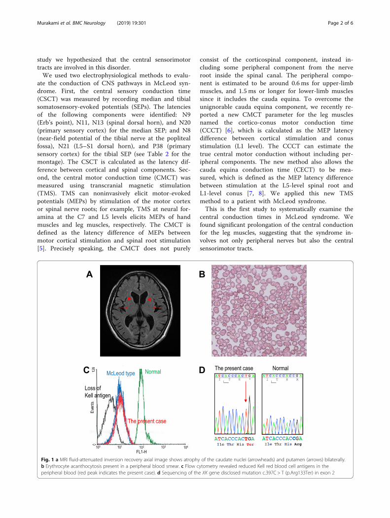
Fig. 1 a MRI fluid-attenuated inversion recovery axial image shows atrophy of the caudate nuclei (arrowheads) and putamen (arrows) bilaterally.b Erythrocyte acanthocytosis present in a peripheral blood smear. c Flow cytometry revealed reduced Kell red blood cell antigens in theperipheral blood (red peak indicates the present case). d Sequencing of the XK gene disclosed mutation c.397C > T (p.Arg133Ter) in exon 2
Murakami et al. BMC Neurology (2019) 19:301 Page 2 of 6

Case presentationsA 66-year-old man noticed involuntary movements inall extremities and weakness in the lower limb mus-cles in his early fifties. He had no particular family orpast medical history. He was admitted to our hospitalwith a chief complaint of gait disturbance. On exam-ination, he was conscious and fully oriented, butrestless and irritable. He exhibited facial grimacingbut no lip or tongue biting. He had chorea in all ex-tremities. He also had right-side-dominant, distal-dominant muscular weakness with muscular atrophy(Medical Research Council Scale grade 1 for the tibi-alis anterior (TA) muscle and gastrocnemius muscleon the right side and 3 on the left side). His vibratoryperception was impaired at the ankles, whereas super-ficial sensations were intact. He showed a positiveRomberg’s sign. Deep tendon reflexes were absent inthe extremities, and plantar reflex was indifferent. Heneeded a cane support in walking.Blood chemical examinations showed elevations of
creatine phosphokinase (1609 U/l), aspartate transamin-ase (54 U/l), alanine transaminase (78 U/l), and lactatedehydrogenase (316 mg/dl). The electrocardiography andchest X-ray findings were normal. The brain magneticresonance images (MRIs) revealed atrophy of bilateralcaudate nuclei and putamen (Fig. 1a), but spinal MRIsshowed no abnormalities. Single-photon-emission com-puted tomography with N-isopropyl-p-[123I]-iodoam-phetamine revealed decreases in the blood flow in thebasal ganglia.Table 1 presents the results of the nerve conduction
studies. The amplitudes of the compound motor actionpotentials were reduced in the tibial and fibular nerves,while the amplitudes of the sensory nerve action poten-tials were reduced in the sural nerves. Needle electro-myographic examinations performed at the TA muscleand rectus femoris muscle revealed high-amplitude andlong-duration motor unit potentials (MUPs). MUP re-cruitment was reduced during volitional contraction andfibrillation potentials were present at rest. These findings
indicate the presence of chronic denervation and rein-nervation processes.We examined the median and tibial SEPs (Table 2). The
right median nerve SEPs showed no delay in any compo-nent and no prolongation of CSCT. In the right tibialnerve SEPs, the peripheral components had a normal la-tency, but the cortical latency was prolonged. CSCT wasalso abnormally prolonged (19.5ms; normal < 13.2ms).We used TMS to examine MEPs from the right first-
dorsal interosseous (FDI) muscle and TA muscle (Table 3).MEP latencies for the FDI were normal for stimulation tothe cortex, brainstem, and cervical nerve root. The CMCT
Table 1 Results of nerve conduction studies
Motor nerve conduction study
Nerve Distallatency
Amplitude (mV) Velocity
(ms) distal proximal (m/s)
RightMedian
4.1 11.3 10.3 53
Right Ulnar 2.4 6.0 4.4 60
Right Tibial 5.1 4.3 (normal >7.0)
3.3 (normal >7.0)
44
Left Tibial 4.3 6.5 (normal >7.0)
3.9 (normal >7.0)
45
RightFibular
5.0 0.6 (normal >0.6)
0.5 (normal >0.6)
36
Left Fibular 2.6 3.8 3.7 40
Sensory nerve conduction study
Distallatency
Amplitude (μV) Velocity
(ms) distal proximal (m/s)
RightMedian
3.1 11.2 3.1 60
Right Ulnar 3.3 5.2 0.8 54
Right Sural 2.8 3.0 (normal >15.0)
50
Left Sural 3.4 3.0 (normal >15.0)
42
The bold Italic values indicate under the normal limits
Table 2 Results of SEP study
SEPs with median nerve stimulation SEPs with tibial nerve stimulation
Potential Montage Latency (ms) Normal limit (ms) Potential Montage Latency (ms) Normal limit (ms)
(1) N9o EPi-EPc 9.6 9.7 (1) N8o Pfi-K 7.4 8.7
(2) N11o C5s-Fz 11.1 11.6 (2) N21 L1 s-Icc 21.2 26.7
(3) N13o C5s-Fz 13.3 13.7 (3) P38o Cz’-Fz 40.7 38.2
(4) N20o C3’-Fz 17.4 18.0 (4) P38 Cz’-Fz 47.9 44.7
Conduction time (ms) Conduction time (ms)
CSCT [(4)–(3)] 4.1 4.8 CSCT [(3)–(2)] 19.5 13.2
SEP somatosensory-evoked potential, CSCT central sensory conduction timeEPi ipsilateral Erb’s point, EPc contralateral Erb’s pointPfi ipsilateral popliteal fossa, K ipsilateral medial popliteal fossa, Icc contralateral iliac crestThe bold Italic values indicate over the normal limits
Murakami et al. BMC Neurology (2019) 19:301 Page 3 of 6
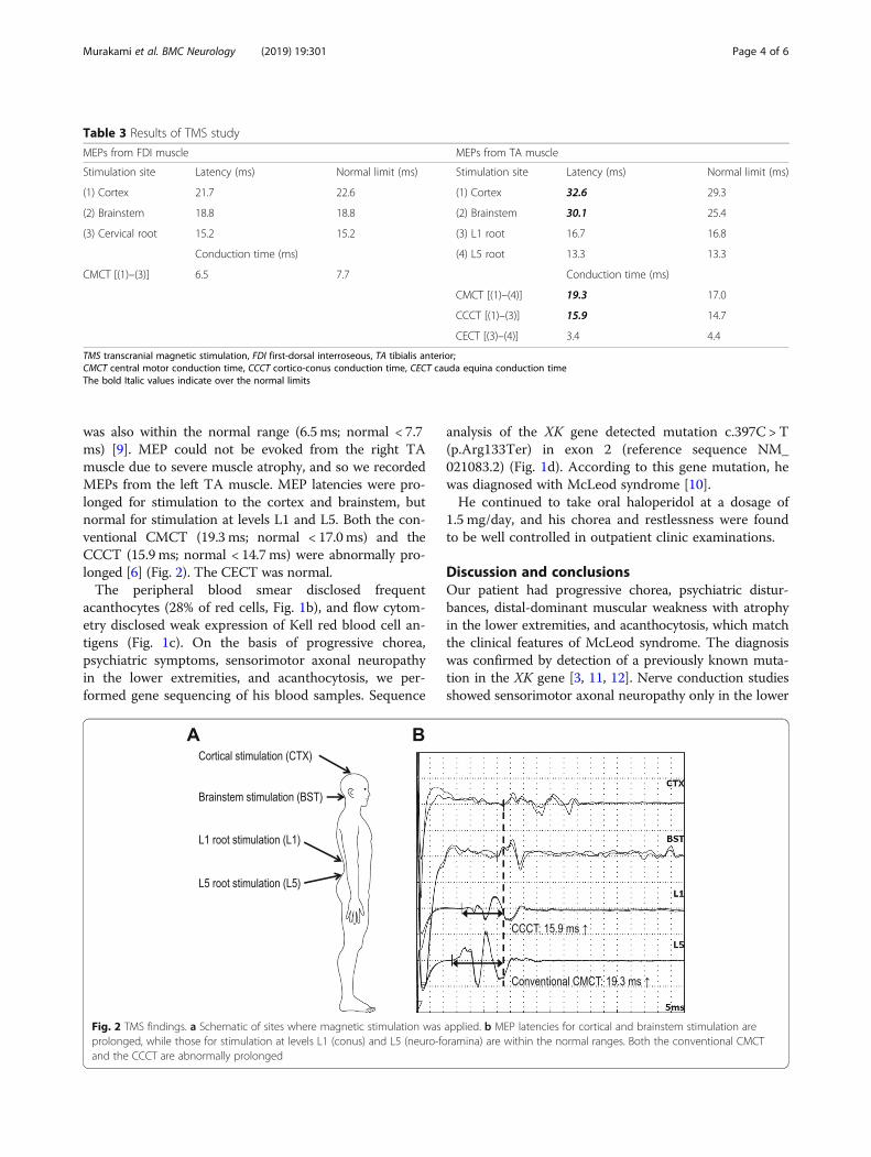
was also within the normal range (6.5ms; normal < 7.7ms) [9]. MEP could not be evoked from the right TAmuscle due to severe muscle atrophy, and so we recordedMEPs from the left TA muscle. MEP latencies were pro-longed for stimulation to the cortex and brainstem, butnormal for stimulation at levels L1 and L5. Both the con-ventional CMCT (19.3 ms; normal < 17.0ms) and theCCCT (15.9ms; normal < 14.7ms) were abnormally pro-longed [6] (Fig. 2). The CECT was normal.The peripheral blood smear disclosed frequent
acanthocytes (28% of red cells, Fig. 1b), and flow cytom-etry disclosed weak expression of Kell red blood cell an-tigens (Fig. 1c). On the basis of progressive chorea,psychiatric symptoms, sensorimotor axonal neuropathyin the lower extremities, and acanthocytosis, we per-formed gene sequencing of his blood samples. Sequence
analysis of the XK gene detected mutation c.397C > T(p.Arg133Ter) in exon 2 (reference sequence NM_021083.2) (Fig. 1d). According to this gene mutation, hewas diagnosed with McLeod syndrome [10].He continued to take oral haloperidol at a dosage of
1.5 mg/day, and his chorea and restlessness were foundto be well controlled in outpatient clinic examinations.
Discussion and conclusionsOur patient had progressive chorea, psychiatric distur-bances, distal-dominant muscular weakness with atrophyin the lower extremities, and acanthocytosis, which matchthe clinical features of McLeod syndrome. The diagnosiswas confirmed by detection of a previously known muta-tion in the XK gene [3, 11, 12]. Nerve conduction studiesshowed sensorimotor axonal neuropathy only in the lower
Table 3 Results of TMS study
MEPs from FDI muscle MEPs from TA muscle
Stimulation site Latency (ms) Normal limit (ms) Stimulation site Latency (ms) Normal limit (ms)
(1) Cortex 21.7 22.6 (1) Cortex 32.6 29.3
(2) Brainstem 18.8 18.8 (2) Brainstem 30.1 25.4
(3) Cervical root 15.2 15.2 (3) L1 root 16.7 16.8
Conduction time (ms) (4) L5 root 13.3 13.3
CMCT [(1)–(3)] 6.5 7.7 Conduction time (ms)
CMCT [(1)–(4)] 19.3 17.0
CCCT [(1)–(3)] 15.9 14.7
CECT [(3)–(4)] 3.4 4.4
TMS transcranial magnetic stimulation, FDI first-dorsal interroseous, TA tibialis anterior;CMCT central motor conduction time, CCCT cortico-conus conduction time, CECT cauda equina conduction timeThe bold Italic values indicate over the normal limits
Fig. 2 TMS findings. a Schematic of sites where magnetic stimulation was applied. b MEP latencies for cortical and brainstem stimulation areprolonged, while those for stimulation at levels L1 (conus) and L5 (neuro-foramina) are within the normal ranges. Both the conventional CMCTand the CCCT are abnormally prolonged
Murakami et al. BMC Neurology (2019) 19:301 Page 4 of 6

extremities. The cortical latency of the SEPs was also de-layed for tibial nerve stimulation, suggesting the involve-ment of the central sensory pathway that includes thedorsal cord, medial lemniscus, thalamus, and primary sen-sory cortex. In addition, the TMS technique revealed theinvolvement of the corticospinal tract for the leg muscles(prolongation of the CMCT and CCCT). Together thesefindings indicate the involvement of the sensorimotortracts in both the CNS and peripheral nervous system,mainly those for the lower extremities.Muscular atrophy in McLeod syndrome has been ex-
plained by motor axonal neuropathy. A muscle biopsycan detect a combination of neurogenic and myogenicchanges, with the former being more obvious [3]. Sen-sory symptoms have also been attributed to sensoryaxonal neuropathy [11, 13]. However, the pathogenesisof the peripheral neuropathy remains to be determined.This is the first demonstration of the involvement of the
central sensorimotor tracts in McLeod syndrome. Thefindings of this study suggest the presence of length-dependent axonal degeneration of the sensorimotor tractfibers in McLeod syndrome. This is especially interestinggiven that a previous pathological study found that modelknockout mice of McLeod syndrome showed axonopathyin the spinal cord and the sciatic nerve [4]. The presentfindings provide clinical proof for these experimental re-sults, which is important for understanding the distribu-tion of this multisystem disorder.A unique finding of the present case study is that the
TMS technique revealed the involvement of the corti-cospinal tract even in a patient whose plantar responseswere indifferent. When both the corticospinal tract andperipheral nerves are damaged, Babinski signs may notbe observed, because the peripheral pathology masks thecentral pathology. TMS can allow evaluation of corti-cospinal tract function even in this situation [9]. Thelack of evidence for corticospinal tract involvement inthe previous clinical reports might be due to coexistingperipheral neuropathy, which is common in McLeodsyndrome. Similarly, measuring SEPs is also useful fordetecting the involvement of central sensory pathways inpatients with peripheral neuropathy.In conclusion, the results obtained in this study suggest
that McLeod syndrome involves the central sensorimotortracts in addition to peripheral nerves. This involvement ofcentral sensorimotor tracts for the legs might be masked bythe presence of peripheral neuropathy in this disorder.
AbbreviationsCCCT: Cortico-conus motor conduction time; CECT: Cauda equinaconduction time; CMCT: Central motor conduction time; CNS: Centralnervous system; CSCT: Central sensory conduction time; FDI: First-dorsalinterosseous; MEP: Motor-evoked potential; MRI: Magnetic resonance image;MUP: Motor unit potential; NCS: Nerve conduction study;SEP: Somatosensory-evoked potential; TA: Tibialis anterior; TMS: Transcranialmagnetic stimulation
AcknowledgmentsThe authors thank Ms. Yumiko Tanji for assistance in the electrophysiologicalexaminations.
Authors’ contributionsTM managed the patient, conducted electrophysiological evaluations andwrote the first draft of the manuscript. DA, RT, MA, AT, SK and CK managedthe patient and revised the manuscript. HM performed electrophysiologicalevaluations and revised the manuscript. YUr, MN and AS performed geneanalyses and revised the manuscript. YUg revised the manuscript andprovided intellectual content. All authors read and approved the finalmanuscript.
FundingDr. Murakami has received research funding from the Takeda ScienceFoundation. Dr. Ugawa was partly supported by research grants from JSPSKAKENHI Grant Number 15H05881, 16H05322, and 25293206.
Availability of data and materialsNot applicable.
Ethics approval and consent to participateNot applicable.
Consent for publicationWritten informed consent was obtained from the patient and his wife forthe publication of this case report and any accompanying images. A copy ofthe written consent is available for review by the editor of this journal.
Competing interestsThe authors declare that the research was conducted in the absence of anycommercial or financial relationships that could be construed as a potentialconflict of interest.
Author details1Department of Neurology, Fukushima Medical University, Fukushima, Japan.2Department of Neurology, Tottori Prefectural Kousei Hospital, Kurayoshi,Japan. 3Department of Neurology, Mitsui Memorial Hospital, Tokyo, Japan.4Center for Neurological Disorders, Fukushima Medical University, Fukushima,Japan. 5Department of Neurology, Southern Tohoku General Hospital,Koriyama, Japan. 6Department of Psychiatry, Kagoshima University GraduateSchool of Medical and Dental Sciences, Kagoshima, Japan. 7Department ofNeuro-regeneration, Fukushima Medical University, Fukushima, Japan.
Received: 23 July 2019 Accepted: 12 November 2019
References1. Allen FH Jr, Krabbe SM, Corcoran PA. A new phenotype (McLeod) in the Kell
blood-group system. Vox Sang. 1961;6:555–60.2. Roulis E, Hyland C, Flower R, Gassner C, Jung HH, Frey BM. Molecular basis and
clinical overview of McLeod syndrome compared with otherNeuroacanthocytosis syndromes: a review. JAMA Neurol. 2018;75(12):1554–62.
3. Danek A, Rubio JP, Rampoldi L, Ho M, Dobson-Stone C, Tison F, Symmans WA,Oechsner M, Kalckreuth W, Watt JM, et al. McLeod neuroacanthocytosis:genotype and phenotype. Ann Neurol. 2001;50(6):755–64.
4. Zhu X, Cho ES, Sha Q, Peng J, Oksov Y, Kam SY, Ho M, Walker RH, Lee S.Giant axon formation in mice lacking Kell, XK, or Kell and XK: animal modelsof McLeod neuroacanthocytosis syndrome. Am J Pathol. 2014;184(3):800–7.
5. Rossini PM, Burke D, Chen R, Cohen LG, Daskalakis Z, Di Iorio R, Di LazzaroV, Ferreri F, Fitzgerald PB, George MS, et al. Non-invasive electrical andmagnetic stimulation of the brain, spinal cord, roots and peripheral nerves:basic principles and procedures for routine clinical and research application.An updated report from an I.F.C.N. committee. Clin Neurophysiol. 2015;126(6):1071–107.
6. Matsumoto H, Hanajima R, Shirota Y, Hamada M, Terao Y, Ohminami S,Furubayashi T, Nakatani-Enomoto S, Ugawa Y. Cortico-conus motor conductiontime (CCCT) for leg muscles. Clin Neurophysiol. 2010;121(11):1930–3.
7. Matsumoto H, Hanajima R, Terao Y, Hamada M, Shirota Y, Yugeta A, Nakatani-Enomoto S, Hashida H, Ugawa Y. A significant correlation between cauda equina
Murakami et al. BMC Neurology (2019) 19:301 Page 5 of 6

conduction time and cerebrospinal fluid protein in chronic inflammatorydemyelinating polyradiculoneuropathy. J Neurol Sci. 2018;384:7–9.
8. Matsumoto H, Hanajima R, Terao Y, Yugeta A, Hamada M, Shirota Y,Ohminami S, Nakatani-Enomoto S, Tsuji S, Ugawa Y. Prominent caudaequina involvement in patients with chronic inflammatory demyelinatingpolyradiculoneuropathy. J Neurol Sci. 2010;290(1–2):112–4.
9. Ugawa Y, Uesaka Y, Terao Y, Suzuki M, Sakai K, Hanajima R, Kanazawa I.Clinical utility of magnetic corticospinal tract stimulation at the foramenmagnum level. Electroencephalogr Clin Neurophysiol. 1996;101(3):247–54.
10. Urata Y, Nakamura M, Sasaki N, Shiokawa N, Nishida Y, Arai K, Hiwatashi H,Yokoyama I, Narumi S, Terayama Y, et al. Novel pathogenic XK mutations inMcLeod syndrome and interaction between XK protein and chorein. NeurolGenet. 2019;5(3):e328.
11. Dotti MT, Battisti C, Malandrini A, Federico A, Rubio JP, Circiarello G, MonacoAP. McLeod syndrome and neuroacanthocytosis with a novel mutation inthe XK gene. Mov Disord. 2000;15(6):1282–4.
12. Bansal I, Jeon HR, Hui SR, Calhoun BW, Manning DW, Kelly TJ, Lee S, BaronBW. Transfusion support for a patient with McLeod phenotype withoutchronic granulomatous disease and with antibodies to Kx and km. VoxSang. 2008;94(3):216–20.
13. Malandrini A, Fabrizi GM, Truschi F, Di Pietro G, Moschini F, Bartalucci P,Berti G, Salvadori C, Bucalossi A, Guazzi G. Atypical McLeod syndromemanifested as X-linked chorea-acanthocytosis, neuromyopathy and dilatedcardiomyopathy: report of a family. J Neurol Sci. 1994;124(1):89–94.
Publisher’s NoteSpringer Nature remains neutral with regard to jurisdictional claims inpublished maps and institutional affiliations.
Murakami et al. BMC Neurology (2019) 19:301 Page 6 of 6