A NEW SPECIES OF MEIOLANIFORM TURTLE AND A REVISION OF THE LATE CRETACEOUS MEIOLANIFORMES OF SOUTH...
17
240 A NEW SPECIES OF MEIOLANIFORM TURTLE AND A REVISION OF THE LATE CRETACEOUS MEIOLANIFORMES OF SOUTH AMERICA 1 Museo Paleontológico Egidio Feruglio, Av. Fontana 140, U9100GYO Trelew, Argentina. [email protected] 2 Museo de Historia Natural de San Rafael, Parque Mariano Moreno s/n, 5600 San Rafael, Argentina. [email protected] 3 Instituto de Investigación en Paleobiología y Geología, Universidad Nacional de Río Negro, Museo Carlos Ameghino, Belgrano 1700, Paraje Pichi Ruca (predio Marabunta) R8324CZH Cipolletti, Argentina. [email protected] 4 Consejo Nacional de Investigaciones Científicas y Técnicas, Argentina (CONICET) AMEGHINIANA - 2013 - Tomo 50 (2): 240 - 256 ISSN 0002-7014 JULIANA STERLI 1,4 , MARCELO S. DE LA FUENTE 2,4 and IGNACIO A. CERDA 3,4 AMGHB2-0002-7014/12$00.00+.50 Abstract. A new species of meiolaniform turtle, Trapalcochelys sulcata gen. nov. sp. nov. is described, based on material from the late Cam- panian–early Maastrichtian Allen Formation, Patagonia (Argentina). e postcranial remains recovered are described macroscopically (e.g., external morphology) and microscopically (e.g., histological sections of the shell). Trapalcochelys sulcata gen. nov. sp. nov. shares with other meiolaniforms the presence of sulci strongly curved anteriorly among marginal scales, and dermal bones ornamented with small foramina. is new species differs from the other Late Cretaceous meiolaniform from Patagonia —Patagoniaemys gasparinae— in the general size and in the shape of neural 1. e shell-bone histology is characterized by a diploe structure, in which well developed internal and external compact bone layers frame an area of cancellous bone. Compact bone is mostly composed by interwoven structural fiber bundles. e abundance of structural fibers in the internal cortex and the presence of large pipe-like vascular spaces in the cancellous bone are the most distinctive histological features observed for T. sulcata. All meiolaniform turtle remains of the Upper Cretaceous of South America are exhaustively re- vised. e known South American record of Upper Cretaceous meiolaniforms is restricted to Argentina and in this revision six localities with outcrops bearing these fossils have been identified. Meiolaniforms are confirmed as a component of the late Campanian–early Maastrichtian South American Allenian tetrapod assemblage. Keywords. Campanian–Maastrichtian. Patagonia. Meiolaniforms. Trapalcochelys sulcata. Morphology. Histology. Allenian tetrapod assemblage. Resumen. UNA NUEVA ESPECIE DE TORTUGA MEIOLANIFORME Y UNA REVISIÓN DE LOS MEIOLANIFORMES DEL CRETÁCICO TARDÍO DE AMÉRICA DEL SUR. Una nueva especie de meiolaniforme, Trapalcochelys sulcata gen. nov. sp. nov., de la Formación Allen (Campaniano tardío–Maastrichtiano temprano), Patagonia, Argentina, es presentada en este trabajo. Los restos postcranea- nos pertenecientes a esta nueva especie son descriptos macroscópicamente (e.g., morfología externa) y microscópicamente (e.g., cortes histo- lógicos del caparazón). Trapalcochelys sulcata gen. nov. sp. nov. comparte con otros meiolaniformes la presencia de surcos entre los escudos marginales marcadamente curvados anteriormente y la ornamentación de las placas dérmicas del caparazón constituida por pequeños forá- menes. Esta nueva especie difiere de la otra especie de meiolaniforme del Cretácico Superior de Patagonia —Patagoniaemys gasparinae— en el tamaño general y en la forma de la neural 1. La histología ósea está caracterizada por una estructura diploe, donde una capa externa e interna de tejido compacto circundan una región de hueso esponjoso. El hueso compacto está compuesto mayormente por paquetes entrelazados de fibras estructurales. La abundancia de fibras estructurales en la corteza interna y la presencia de grandes canales vasculares tubulares son los caracteres histológicos mas distintivos de T. sulcata. Además, una revisión exhaustiva de los restos de Meiolaniformes del Cretácico Superior de Sudamérica es presentada. El registro sudamericano conocido de Meiolaniformes del Cretácico Superior está restringido a Argentina y en esta revisión hasta seis localidades han sido reconocidas. Se corrobora que los Meiolaniformes son un componente de la asociación sudame- ricana Alleniana de tetrápodos de edad campaniana tardía–maastrichtiana temprana. Palabras clave. Campaniano–Maastrichtiano. Patagonia. Meiolaniformes. Trapalcochelys sulcata. Morfología. Histología. Asociación de te- trápodos Alleniana. The name Meiolaniformes was recently proposed for the stem-based clade including all taxa more closely related to Meiolania platyceps Owen, 1886, than to Cryptodira and Pleurodira (Sterli and de la Fuente, 2012). Meiolaniformes currently includes the bizarre group of extinct Cenozoic tur- tles bearing cranial horns and frills, caudal rings, and tail club known as Meiolaniidae and a number of species placed along the stem lineage leading to Meiolaniidae. e clade Meiol- aniformes spans at least the Early Cretaceous (Chubutemys copelloi Gaffney, Rich, Vickers-Rich, Constantine, Vacca and Kool, 2007) to Holocene (?Meiolania damelipi White, Worthy, Hawkins, Bedford and Spriggs, 2010). Members of the clade have been recorded in Asia, Europe, Australia, and South America. e fossil record of this clade shows two mo- ments of greatest diversification, with four Late Cretaceous and four Pleistocene–Holocene species. Herein we focus on
Transcript of A NEW SPECIES OF MEIOLANIFORM TURTLE AND A REVISION OF THE LATE CRETACEOUS MEIOLANIFORMES OF SOUTH...
240
A NEW SPECIES OF MEIOLANIFORM TURTLE AND A REVISION OF THE LATE CRETACEOUS MEIOLANIFORMES OF SOUTH AMERICA
1Museo Paleontológico Egidio Feruglio, Av. Fontana 140, U9100GYO Trelew, Argentina. [email protected] de Historia Natural de San Rafael, Parque Mariano Moreno s/n, 5600 San Rafael, Argentina. [email protected] de Investigación en Paleobiología y Geología, Universidad Nacional de Río Negro, Museo Carlos Ameghino, Belgrano 1700, Paraje Pichi Ruca (predio Marabunta) R8324CZH Cipolletti, Argentina. [email protected] Nacional de Investigaciones Científicas y Técnicas, Argentina (CONICET)
AMEGHINIANA - 2013 - Tomo 50 (2): 240 - 256 ISSN 0002-7014
JULIANA STERLI1,4, MARCELO S. DE LA FUENTE2,4 and IGNACIO A. CERDA3,4
AMGHB2-0002-7014/12$00.00+.50
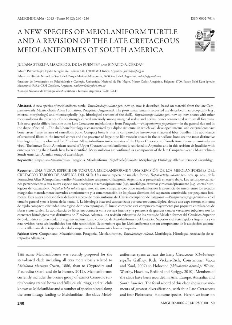
Abstract. A new species of meiolaniform turtle, Trapalcochelys sulcata gen. nov. sp. nov. is described, based on material from the late Cam-panian–early Maastrichtian Allen Formation, Patagonia (Argentina). The postcranial remains recovered are described macroscopically (e.g., external morphology) and microscopically (e.g., histological sections of the shell). Trapalcochelys sulcata gen. nov. sp. nov. shares with other meiolaniforms the presence of sulci strongly curved anteriorly among marginal scales, and dermal bones ornamented with small foramina. This new species differs from the other Late Cretaceous meiolaniform from Patagonia —Patagoniaemys gasparinae— in the general size and in the shape of neural 1. The shell-bone histology is characterized by a diploe structure, in which well developed internal and external compact bone layers frame an area of cancellous bone. Compact bone is mostly composed by interwoven structural fiber bundles. The abundance of structural fibers in the internal cortex and the presence of large pipe-like vascular spaces in the cancellous bone are the most distinctive histological features observed for T. sulcata. All meiolaniform turtle remains of the Upper Cretaceous of South America are exhaustively re-vised. The known South American record of Upper Cretaceous meiolaniforms is restricted to Argentina and in this revision six localities with outcrops bearing these fossils have been identified. Meiolaniforms are confirmed as a component of the late Campanian–early Maastrichtian South American Allenian tetrapod assemblage.Keywords. Campanian–Maastrichtian. Patagonia. Meiolaniforms. Trapalcochelys sulcata. Morphology. Histology. Allenian tetrapod assemblage.
Resumen. UNA NUEVA ESPECIE DE TORTUGA MEIOLANIFORME Y UNA REVISIÓN DE LOS MEIOLANIFORMES DEL CRETÁCICO TARDÍO DE AMÉRICA DEL SUR. Una nueva especie de meiolaniforme, Trapalcochelys sulcata gen. nov. sp. nov., de la Formación Allen (Campaniano tardío–Maastrichtiano temprano), Patagonia, Argentina, es presentada en este trabajo. Los restos postcranea-nos pertenecientes a esta nueva especie son descriptos macroscópicamente (e.g., morfología externa) y microscópicamente (e.g., cortes histo-lógicos del caparazón). Trapalcochelys sulcata gen. nov. sp. nov. comparte con otros meiolaniformes la presencia de surcos entre los escudos marginales marcadamente curvados anteriormente y la ornamentación de las placas dérmicas del caparazón constituida por pequeños forá-menes. Esta nueva especie difiere de la otra especie de meiolaniforme del Cretácico Superior de Patagonia —Patagoniaemys gasparinae— en el tamaño general y en la forma de la neural 1. La histología ósea está caracterizada por una estructura diploe, donde una capa externa e interna de tejido compacto circundan una región de hueso esponjoso. El hueso compacto está compuesto mayormente por paquetes entrelazados de fibras estructurales. La abundancia de fibras estructurales en la corteza interna y la presencia de grandes canales vasculares tubulares son los caracteres histológicos mas distintivos de T. sulcata. Además, una revisión exhaustiva de los restos de Meiolaniformes del Cretácico Superior de Sudamérica es presentada. El registro sudamericano conocido de Meiolaniformes del Cretácico Superior está restringido a Argentina y en esta revisión hasta seis localidades han sido reconocidas. Se corrobora que los Meiolaniformes son un componente de la asociación sudame-ricana Alleniana de tetrápodos de edad campaniana tardía–maastrichtiana temprana.Palabras clave. Campaniano–Maastrichtiano. Patagonia. Meiolaniformes. Trapalcochelys sulcata. Morfología. Histología. Asociación de te-trápodos Alleniana.
The name Meiolaniformes was recently proposed for the stem-based clade including all taxa more closely related to Meiolania platyceps Owen, 1886, than to Cryptodira and Pleurodira (Sterli and de la Fuente, 2012). Meiolaniformes currently includes the bizarre group of extinct Cenozoic tur-tles bearing cranial horns and frills, caudal rings, and tail club known as Meiolaniidae and a number of species placed along the stem lineage leading to Meiolaniidae. The clade Meiol-
aniformes spans at least the Early Cretaceous (Chubutemys copelloi Gaffney, Rich, Vickers-Rich, Constantine, Vacca and Kool, 2007) to Holocene (?Meiolania damelipi White, Worthy, Hawkins, Bedford and Spriggs, 2010). Members of the clade have been recorded in Asia, Europe, Australia, and South America. The fossil record of this clade shows two mo-ments of greatest diversification, with four Late Cretaceous and four Pleistocene–Holocene species. Herein we focus on
241
STERLI et al.: LATE CRETACEOUS MEIOLANIFORMES FROM SOUTH AMERICA
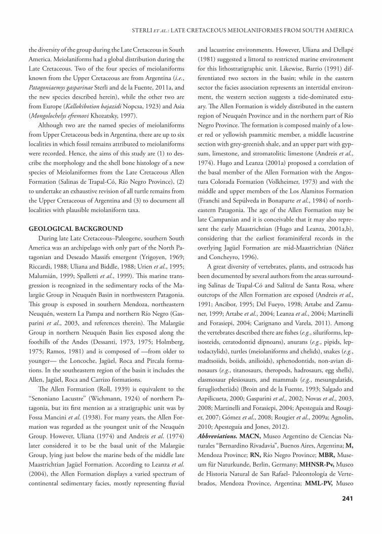
the diversity of the group during the Late Cretaceous in South America. Meiolaniforms had a global distribution during the Late Cretaceous. Two of the four species of meiolaniforms known from the Upper Cretaceous are from Argentina (i.e., Patagoniaemys gasparinae Sterli and de la Fuente, 2011a, and the new species described herein), while the other two are from Europe (Kallokibotion bajazidi Nopcsa, 1923) and Asia (Mongolochelys efremovi Khozatsky, 1997).
Although two are the named species of meiolaniforms from Upper Cretaceous beds in Argentina, there are up to six localities in which fossil remains attributed to meiolaniforms were recorded. Hence, the aims of this study are (1) to des- cribe the morphology and the shell bone histology of a new species of Meiolaniformes from the Late Cretaceous Allen Formation (Salinas de Trapal-Có, Río Negro Province), (2) to undertake an exhaustive revision of all turtle remains from the Upper Cretaceous of Argentina and (3) to document all localities with plausible meiolaniform taxa.
GEOLOGICAL BACKGROUNDDuring late Late Cretaceous–Paleogene, southern South
America was an archipelago with only part of the North Pa-tagonian and Deseado Massifs emergent (Yrigoyen, 1969; Riccardi, 1988; Uliana and Biddle, 1988; Urien et al., 1995; Malumián, 1999; Spalletti et al., 1999). This marine trans-gression is recognized in the sedimentary rocks of the Ma-largüe Group in Neuquén Basin in northwestern Patagonia. This group is exposed in southern Mendoza, northeastern Neuquén, western La Pampa and northern Río Negro (Gas-parini et al., 2003, and references therein). The Malargüe Group in northern Neuquén Basin lies exposed along the foothills of the Andes (Dessanti, 1973, 1975; Holmberg, 1975; Ramos, 1981) and is composed of —from older to younger— the Loncoche, Jagüel, Roca and Pircala forma-tions. In the southeastern region of the basin it includes the Allen, Jagüel, Roca and Carrizo formations.
The Allen Formation (Roll, 1939) is equivalent to the ‘‘Senoniano Lacustre’’ (Wichmann, 1924) of northern Pa-tagonia, but its first mention as a stratigraphic unit was by Fossa Mancini et al. (1938). For many years, the Allen For-mation was regarded as the youngest unit of the Neuquén Group. However, Uliana (1974) and Andreis et al. (1974) later considered it to be the basal unit of the Malargüe Group, lying just below the marine beds of the middle late Maastrichtian Jagüel Formation. According to Leanza et al. (2004), the Allen Formation displays a varied spectrum of continental sedimentary facies, mostly representing fluvial
and lacustrine environments. However, Uliana and Dellapé (1981) suggested a littoral to restricted marine environment for this lithostratigraphic unit. Likewise, Barrio (1991) dif-ferentiated two sectors in the basin; while in the eastern sector the facies association represents an intertidal environ-ment, the western section suggests a tide-dominated estu-ary. The Allen Formation is widely distributed in the eastern region of Neuquén Province and in the northern part of Río Negro Province. The formation is composed mainly of a low-er red or yellowish psammitic member, a middle lacustrine section with grey-greenish shale, and an upper part with gyp-sum, limestone, and stromatolitic limestone (Andreis et al., 1974). Hugo and Leanza (2001a) proposed a correlation of the basal member of the Allen Formation with the Angos-tura Colorada Formation (Volkheimer, 1973) and with the middle and upper members of the Los Alamitos Formation (Franchi and Sepúlveda in Bonaparte et al., 1984) of north-eastern Patagonia. The age of the Allen Formation may be late Campanian and it is conceivable that it may also repre-sent the early Maastrichtian (Hugo and Leanza, 2001a,b), considering that the earliest foraminiferal records in the overlying Jagüel Formation are mid-Maastrichtian (Náñez and Concheyro, 1996).
A great diversity of vertebrates, plants, and ostracods has been documented by several authors from the areas surround-ing Salinas de Trapal-Có and Salitral de Santa Rosa, where outcrops of the Allen Formation are exposed (Andreis et al., 1991; Ancibor, 1995; Del Fueyo, 1998; Artabe and Zamu-ner, 1999; Artabe et al., 2004; Leanza et al., 2004; Martinelli and Forasiepi, 2004; Carignano and Varela, 2011). Among the vertebrates described there are fishes (e.g., siluriforms, lep-isosteids, ceratodontid dipnoans), anurans (e.g., pipids, lep-todactylids), turtles (meiolaniforms and chelids), snakes (e.g., madtsoiids, boiids, anilioids), sphenodontids, non-avian di-nosaurs (e.g., titanosaurs, theropods, hadrosaurs, egg shells), elasmosaur plesiosaurs, and mammals (e.g., mesungulatids, ferugliotheriids) (Broin and de la Fuente, 1993; Salgado and Azpilicueta, 2000; Gasparini et al., 2002; Novas et al., 2003, 2008; Martinelli and Forasiepi, 2004; Apesteguía and Rougi-er, 2007; Gómez et al., 2008; Rougier et al., 2009a; Agnolin, 2010; Apesteguía and Jones, 2012).Abbreviations. MACN, Museo Argentino de Ciencias Na-turales “Bernardino Rivadavia”, Buenos Aires, Argentina; M, Mendoza Province; RN, Río Negro Province; MBR, Muse-um für Naturkunde, Berlin, Germany; MHNSR-Pv, Museo de Historia Natural de San Rafael- Paleontología de Verte-brados, Mendoza Province, Argentina; MML-PV, Museo
242
de Lamarque-Paleontología de Vertebrados, Lamarque, Río Negro Province; MPEF-PV, Paleontología de Vertebrados, Museo Paleontológico Egidio Feruglio, Trelew, Argentina; MLP, Museo de La Plata, La Plata, Argentina.
SYSTEMATIC PALEONTOLOGYTestudinata Klein, 1760
Meiolaniformes Sterli and de la Fuente, 2012
Trapalcochelys gen. nov.Type species. Trapalcochelys sulcata gen. nov. sp. nov.Derivation of name. Trapalco, in reference to the locality (Tra-pal-Có) where this species was found; chelys, turtle in Greek.Geographic location. The same as for the species.Stratigraphic location. The same as for the species.Diagnosis. The same as for the only species included.
Trapalcochelys sulcata gen. nov. sp. nov.Figures 2–5
Holotype. MLP 86-IV-5-2, fragment of nuchal bone, left peripherals 1, 2, 6 to 9, right peripherals 2, 3, 6 to 9, right costal 4 and several costal fragments, neurals? 3 and ?5 with a fragment of the neural anterior to it, several indeterminate carapace fragments, a fragment of the left hypoplastron, sev-eral indeterminate plastral fragments thoracic vertebra 9, two neural arches of caudal vertebrae, a right opisthotic. The ho-lotype MLP 86-IV-5-2 includes MLP 88-III-31-89–96 iden-tified by Broin and de la Fuente (1993) as Meiolaniidae indet. Referred material. MLP 52-IX-38-53, neural 1; MLP 88-III-31-97, sixth caudal vertebra; MLP 88-III-31-98, third caudal vertebra; MLP 88-III-31-99, fifth caudal vertebra; MLP 88-III-31-100, fourth caudal vertebra; MLP 88-III-31-101, seventh caudal vertebra. The holotype and the re-ferred material were collected in 1986 (MLP 86-IV-5-2) and 1988 (MLP 88-III-31-97–101) by M. Reguero and A. Car-lini (Museo de La Plata), respectively.Derivation of name. Sulcata, in reference to the strong sulci present on the surface of the carapace.Geographic location. At 4.6 km East of “Salinas de Trapal-Có,” Río Negro Province, Argentina (Fig. 1).Stratigraphic location. Outcrops of the Allen Formation (late Campanian–early Maastrichtian) (Hugo and Leanza, 2001b; Leanza, 1999; Leanza et al., 2004) (Fig. 1).Diagnosis. Trapalcochelys sulcata belongs to the clade Testu-dinata because of the presence of a shell enclosing the pec-toral girdle. Trapalcochelys sulcata, Patagoniaemys gasparinae,
and Mongolochelys efremovi share the presence of strongly an-teriorly curved sulci among marginal scales, and they share also with Meiolania platyceps the presence of opisthocoelous vertebrae at least in the anterior region of the tail. Trapal-cochelys sulcata shares with Patagoniaemys gasparinae, Niola-mia argentina Ameghino, 1899, and Meiolania platyceps the presence of small foramina ornamenting the dermal bones. It shares with Kallokibotion bajazidi the presence of sulci strongly curved anteriorly between vertebrals 1 and 2 and between 3 and 4. Trapalcochelys sulcata differs from Pa. gas-parinae in the general size of the shell, and in the shape of neural 1. Trapalcochelys sulcata differs from Mo. efremovi in the absence of pleural 3 on peripheral 9. Comments. Some pieces of the holotype (MLP 86-IV-5-2) and the referred specimens (MLP 88-III-31-97–101) were previously recognized as Meiolaniidae indet. by Broin and de la Fuente (1993). New remains belonging to the holo-type found in the MLP collection allowed us to describe the specimens in more detail and recognize a new species of meiolaniform.
DescriptionCarapacial bones. The description of the carapace is based mainly on the holotype (MLP 86-IV-5-2) and additional specimen represented by a neural 1 (MLP 52-IX-38-53). The peripheral series of Trapalcochelys sulcata is complete from the first to the ninth peripheral, allowing us to estimate the total length of the carapace at ca. 85 cm (Fig. 2). Cos-tal and neural bones complete the carapace fragments found in Trapal-Có. Both external (dorsal) and internal (ventral) surfaces are coarsely textured with a cross-hatched pattern and numerous vascular foramina (for more detail see Micro-anatomy and Histology section). A similar condition is seen in Patagoniaemys gasparinae, Niolamia argentina, and Meio-lania platyceps (Broin and de la Fuente, 1993; Gaffney, 1996; Sterli and de la Fuente, 2011a,b, 2012).
Only a fragment of the left part of the nuchal is preserved tightly sutured with the left peripheral bone 1. There is a nuchal notch delimited by the nuchal and the first periph-eral, however, it is shallower than in Pa. gasparinae or Mo. efremovi. The external surface is almost flat while the internal surface is slightly convex.
Peripheral 1 is represented by the proximal end of the left bone tightly sutured to the nuchal. As in the nuchal, the external surface is almost flat while the internal surface is slightly convex. Fragments of left and right peripheral 2 are preserved. The external surface of both plates is almost
AMEGHINIANA - 2013 - Tomo 50 (2): 240 - 256
243
flat and the internal surface is slightly convex. A fragment of the right peripheral 3 also shows its external surface being flat and the internal one slightly convex. Peripherals 6 to 8 form part of the bridge. These bones are roughly C-shaped in cross section and form an acute angle between the almost flat external and broadly curved internal plates. They are antero-posteriorly elongated elements. In external view, a strong ca-rina and a groove run along the lateral margin of the bridge. On the internal surface these peripheral bones are character-ized by a strong pit for the reception of the correspondent costal rib, while on the most ventromedial margin of these bones the indentation for plastral attachment are recognized. The presence of these structures shows a weak connection between the carapace and plastron. Peripheral 9 is a thick element and represent the largest remains found.
A complete neural 1 is preserved in the referred specimen MLP-52-IX-38-53. It is hexagonal with short anterolateral margins and three times longer than wide. The anterior con-
tact with the nuchal bone is narrower than the posterior con-tact with neural 2. Neural 1 probably contacted costal 1 an-terolaterally and costal 2 posterolaterally. The neural bone 1 of Mo. efremovi is a short element, almost longer than wide. Unfortunately the neural bone 1 of Pa. gasparinae is not completely preserved; however, it can be deduced to have been slightly longer than wide. A fragment of neural ?3 is preserved. Unfortunately, no natural borders are preserved. All the margins of neural bone 5 are broken and the shape of this bone can therefore not be assessed with certitude. There is only a small fragment of the neural 4 preserved in contact with neural 5. On its internal surface the scar suture of the corresponding thoracic vertebra is seen.
Fragment of right costal 4 is also recognized however the nature of its preservation precludes any description.Carapacial scutes. Marginals 1–4 and 6–10 are preserved on the peripheral bones. All the sulci between the marginals are deep and they are strongly curved anteriorly towards the mar-
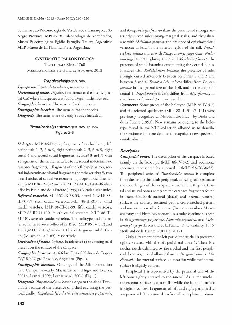
Figure 1. Locality and geological map/ Mapa geológico y de localidades. 1, Map of Patagonia showing the Trapalcochelys sulcata gen. nov. sp. nov. locality/ Mapa de Patagonia mostrando la localidad de Trapalcochelys sulcata gen. nov. sp. nov. Scale bar/ Escala= 500 km; 2, Geologic map showing Salinas de Trapal-Có locality, Río Negro Province, Argentina. Adapted from Hugo and Leanza (2001b)/ Mapa geológico mostrando la localidad Salinas de Trapal-Co, provincial de Río Negro, Argentina. Adaptado de Hugo y Leanza (2001b). Scale bar/ Escala= 10 km.
1 2
STERLI et al.: LATE CRETACEOUS MEIOLANIFORMES FROM SOUTH AMERICA
244
gin of the external and internal surface of the peripheral bones. This is also characteristic of Pa. gasparinae and Mo. efremovi.
Vertebral scale 1 covers the nuchal, neural 1, and costal 1. The sulcus between vertebral 1 and 2 is located on cos-tal 1 and neural 1 (where it is strongly curved anteriorly). Vertebral 2 is located on neurals 1, 2, and probably 3 and costals 1 and 2 and probably 3. The sulcus between vertebral 3 and 4 is located on what we interpreted as neural 5. This sulcus is strongly curved anteriorly. In Mo. efremovi, K. ba-jazidi, and Me. platyceps, the sulci between vertebral 1 and 2 and between vertebral 3 and 4 are not curved anteriorly. Comparison with Pa. gasparinae is not possible because this neural bone is not preserved in that taxon. Contrary to Mo. efremovi, pleural 3 does not reach peripheral 9 in T. sulcata; only pleural 4 is located on peripheral 9.Plastron. The fragmentary nature of the left hypoplastron (MLP 86-IV-5-2) precludes any description. What remains is part of the axillary buttress and no sulcus is recognized on this piece of bone.
Vertebrae. Only one thoracic vertebra (MLP 86-IV-5-2) is preserved that is slightly longer than wide. It contacted two thoracic ribs. The anterior contact is much better developed than the posterior one. The general morphology resembles the thoracic vertebra 9 of Pa. gasparinae.
There are at least 17 caudal vertebrae present in Pa. gas-parinae (Sterli and de la Fuente, 2011a). In Me. platyceps (in which the whole tail has been reconstructed from disarticu-lated caudal vertebrae belonging to different specimens by Gaffney, 1985) there are at least ten caudal vertebrae, ex-cluding the distal caudal vertebrae, which are enclosed in a tail club. Although the five caudal vertebrae preserved in the specimens from Trapal-Có were not found articulated, they were recovered associated. They might represent a con-tinuous caudal series from the third to the seventh vertebrae (comparisons were made with Pa. gasparinae) (Figs. 3, 4). All the caudal vertebrae preserved in T. sulcata are opisthocoe-lous as in Pa. gasparinae, M. platyceps, and in baenids (e.g., Plesiobaena in Lyson and Joyce, 2009), and they bear in the posteroventral part a ventral process that is in contact with the chevrons. Several changes in the morphology are recog-nized along the caudal segment. These modifications allow us to suggest the position of each vertebra within the series. The anteroposterior changes are represented by a slight re-duction in the size of the centra, the posterior displacement of the transverse process in relation to the margin of the cotyle, and the reduction in the angle between the axis of the centrum and the haemal arch attachment. The most anterior vertebra of T. sulcata is represented by MLP-88-III-31-98 (Figs. 3.1, 5.1), which resembles in general morphology that found in the third caudal vertebra of Pa. gasparinae (Sterli and de la Fuente, 2011a). The vertebral centrum of MLP-88-III-31-98 has a prominent anterior convexity and a deep posterior concavity. Both the cotyle and the condyle are circular in shape. Ventroposteriorly this centrum forms a broken projection (ventroposteriorly directed) that could be related with the haemal arch. Although the transverse processes are broken on both sides, the base of the left trans-verse process is present, showing that this process starts at the margin of the cotyle. The neural arch of this third caudal vertebra is preserved; however, neither of the two prezyg-apophyses are preserved. Unfortunately, the neural spine and the process that bears the postzygapophyses are lost.
The next anterior vertebra is represented by MLP-88-III-31-100 (Figs. 3.2, 4.2). This vertebra has an opisthocoe-lous centrum that has an oval cotyle and condyle. In ven-tral view, two ridges with a medial trough are recognized.
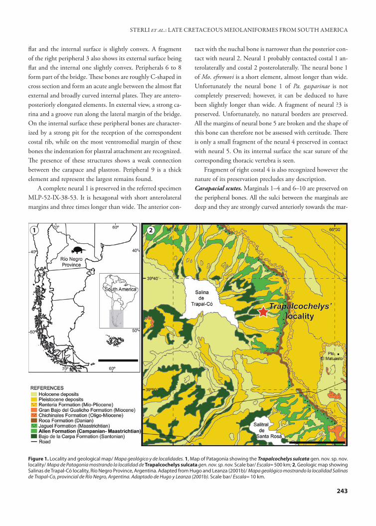
Figure 2. Trapalcochelys sulcata gen. nov. sp. nov. Allen Formation, Argentina. Carapace in external view/ Caparazón en vista externa. co, costal bone/ placa costal; MA, marginal scale/ escudo marginal; ne, neural bone/ placa neural; PL, pleural scale/ escudo pleural; pe, periph-eral bone/ placa periférica, VE, vertebral scale/escudo vertebral. Scale bar/ Escala= 10 cm.
AMEGHINIANA - 2013 - Tomo 50 (2): 240 - 256
245
Posteroventrally these ridges become longer and probably serve as haemal arch attachment sites, but their extension cannot be established, because they are broken. The neural arch is lost and only the base of the right transverse process is preserved. The position of this process is slightly differ-ent from that of the previously described caudal vertebra, because it starts near the cotyle (not in the cotyle like MLP-88-III-31-98). The whole features of this vertebra allow us
to suggest that MLP-88-III-31-100 might be a fourth caudal vertebra, because similar traits were recognized in Pa. gaspa-rinae by Sterli and de la Fuente (2011a).
The following vertebra in the series, MLP-88-III-31-99 (Figs. 3.3, 4.3), is represented by the anterior part of the centrum and most of the neural arch. The shape of the con-dyle is circular. As in MLP-88-III-31-100, two ridges are recognized in ventral view, but only their anterior part is
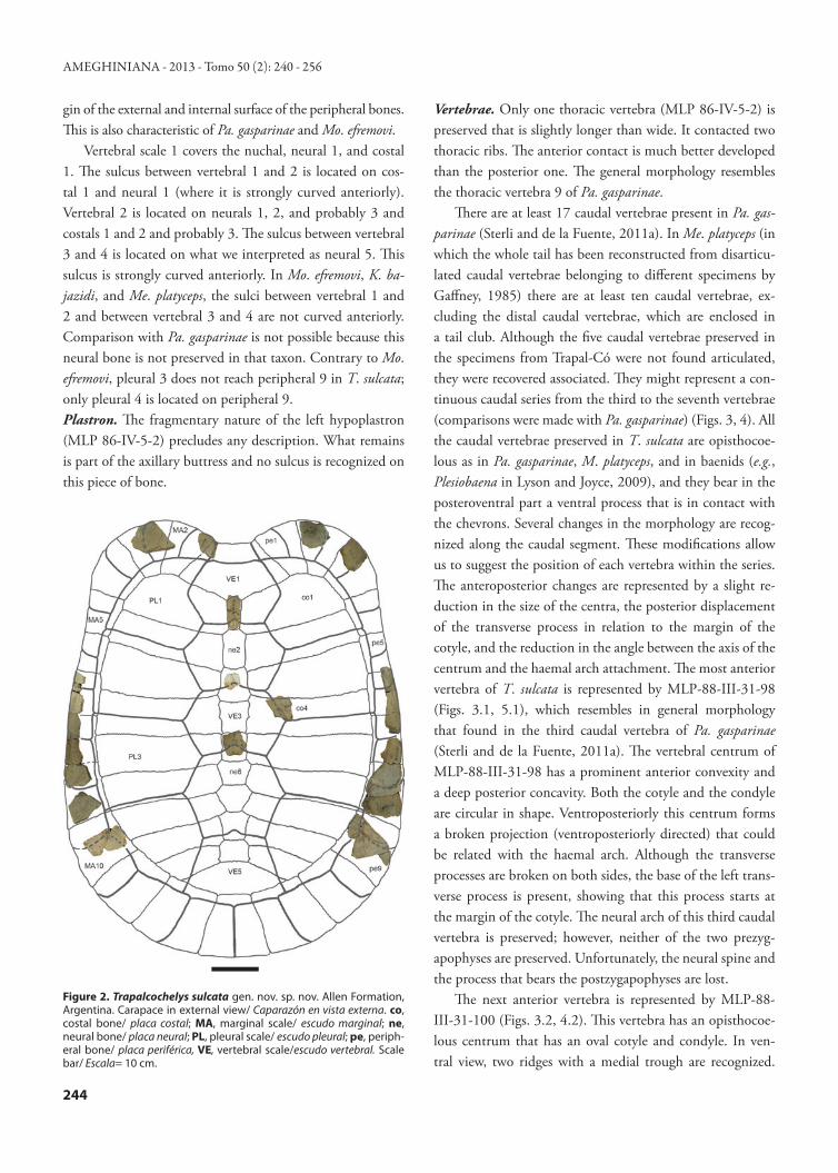
Figure 3. Trapalcochelys sulcata gen. nov. sp. nov. Allen Formation, Argentina. Caudal vertebrae in anterior, left lateral, posterior, right lateral, dorsal, ventral views/ Vértebras caudales en vistas anterior, lateral izquierda, posterior, lateral derecha, dorsal y ventral. 1, MLP-88-III-31-98, third caudal vertebra/ vértebra caudal 3; 2, MLP-88-III-31-100, fourth caudal vertebra/ vértebra caudal 4; 3, MLP-88-III-31-99, fifth caudal vertebra/ vértebra caudal 5; 4, MLP-88-III-31-97, sixth caudal vertebra/ vértebra caudal 6; 5, MLP-88-III-31-101, seventh caudal vertebra/ vértebra caudal 7. Scale bar/ Escala= 2 cm.
1
2
3
4
5
STERLI et al.: LATE CRETACEOUS MEIOLANIFORMES FROM SOUTH AMERICA
246
preserved. Only the base of the right transverse process is present and it is displaced slightly posteriorly in compari-son with the position of this process in the fourth caudal vertebra. The neural arch and the right prezygapophysis are well preserved. Contrary to the condition seen in the caudal vertebrae of Me. platyceps (Gaffney, 1985), the neural spine is blunt, slightly developed, and it slopes following the di-
rection of the process that bears the postzygapophyses. The articular facet of the prezygapophysis is oriented medially forming an acute angle with the sagittal plane. This vertebra is identified as the fifth caudal.
MLP-88-III-31-97 (Figs. 3.4, 4.4) represents the next vertebra of the series (probably the sixth). The condyle and probably the cotyle are subcircular in outline. Unfortunately,
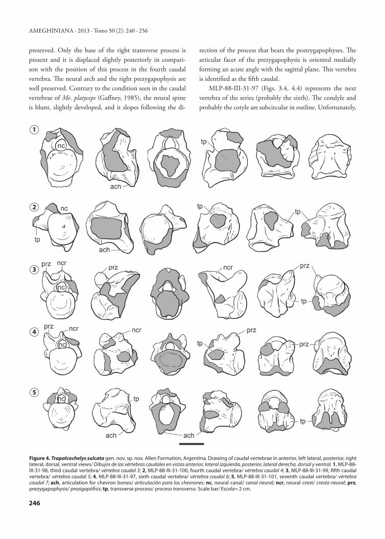
Figure 4. Trapalcochelys sulcata gen. nov. sp. nov. Allen Formation, Argentina. Drawing of caudal vertebrae in anterior, left lateral, posterior, right lateral, dorsal, ventral views/ Dibujos de las vértebras caudales en vistas anterior, lateral izquierda, posterior, lateral derecha, dorsal y ventral. 1, MLP-88-III-31-98, third caudal vertebra/ vértebra caudal 3; 2, MLP-88-III-31-100, fourth caudal vertebra/ vértebra caudal 4; 3, MLP-88-III-31-99, fifth caudal vertebra/ vértebra caudal 5; 4, MLP-88-III-31-97, sixth caudal vertebra/ vértebra caudal 6; 5, MLP-88-III-31-101, seventh caudal vertebra/ vértebra caudal 7; ach, articulation for chevron bones/ articulación para los chevrones; nc, neural canal/ canal neural; ncr, neural crest/ cresta neural; prz, prezygapophysis/ prezigapófisis; tp, transverse process/ proceso transverso. Scale bar/ Escala= 2 cm.
1
2
3
4
5
AMEGHINIANA - 2013 - Tomo 50 (2): 240 - 256
247
the posteroventral part of the centrum is lost. Only the an-terior parts of the posteroventral ridges are present, show-ing the starting point of the haemal arch attachment. Both bases of the transverse processes are preserved and they are considerably displaced posteriorly, almost reaching the mar-gin of the cotyle. The preserved neural spine and the right prezygapophyses are similar to those observed in MLP-88-III-31-99.
The last vertebra of the caudal series (MLP-88-III-31-101, probably the seventh) (Figs. 3.5, 4.5) is the smallest. It has a subcircular, prominent cotyle and condyle and there are two well developed ridges with a deep trough between them in ventral view. The angle between the axis of the caudal centrum and the haemal arch attachment is slightly obtuse. As in the previous caudal vertebra, the trans-verse processes are strongly displaced posteriorly, reaching the margin of the cotyle. The condition of the neural arch is also similar to that seen in the previous vertebrae (MLP-88-III-31-97 and MLP-88-III-31-99), but it does not preserve the pre- or postzygapophyses.
MICROANATOMY AND HISTOLOGYA fragmentary left peripheral plate from the posterior re-
gion of the carapace and the right costal 4 were sampled for histological analysis. The section-planes were oriented antero-posteriorly (perpendicular to the progression of the ribs) and lateromedially (parallel to the progression of the ribs). The thin sections were produced at the Museo Egidio Feruglio, Trelew, Argentina. The sampling followed standard petro-graphic thin-sectioning procedures (Scheyer and Sánchez-Villagra, 2007). The bone microstructure of the thin sections was studied under light microscopy using normal and po-larized light. The histological descriptions follow Francillon-Vieillot et al. (1990) and Scheyer and Sander (2004).
As described above, the microanatomy of both external and internal surfaces is characterized by a coarse, interwoven bone texture and several vascular foramina (Fig. 5.1). The sampled shell bones show a clear diploe structure, in which internal and external compact bone layers frame an interior area of cancellous bone (Figs. 5.2, 5.4). While in the costal plate the compact bone layers correspond to the external and internal cortices, in the sampled peripheral plate the internal cortex is not preserved (the diploe structure is formed by the dorsal and ventral portions of the external cortex). The dorsal and ventral portions of the external cortex in this plate have a rather equal thickness (c. 3.5 mm) and they are well differentiated from the cancellous bone.
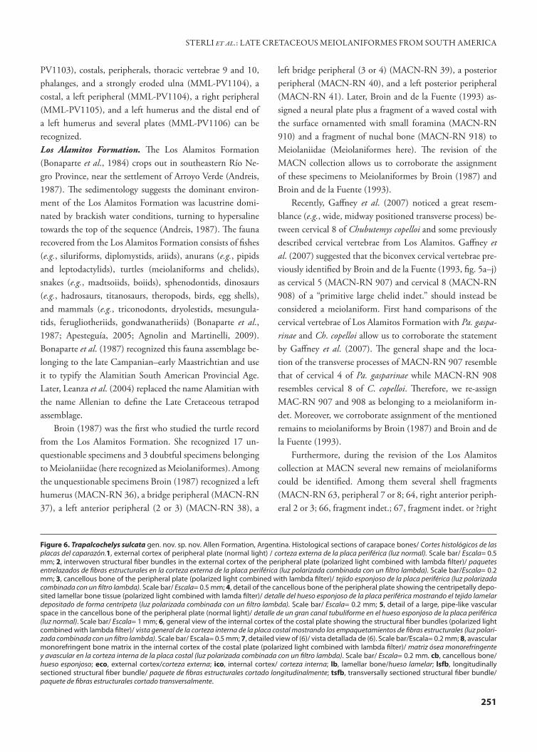
The external cortex of both peripheral and costal plates is mainly composed of mineralized collagen fiber bundles (in-terwoven structural fibers or ISF sensu Scheyer and Sander, 2004). Structural fiber bundles are coarse and show variable length and thickness. These fiber bundles extend perpen-dicular, parallel, and diagonal to the bone surface (Figs. 6.1, 6.2). Each structural fiber bundle is composed by smaller (20–30 μm thick) mineralized fiber bundles that run paral-lel to each other (clearly observed in transversally sectioned fiber bundles). Bone cell lacunae within the structural fiber bundles are mostly elongated and their orientation typically parallels the spatial arrangement of fiber bundles. Branching canaliculi are often observed. The bone is well vascularized by a mixture of scattered primary osteons, simple vascular canals, and few secondary osteons. The sulcus between mar-ginal scute IX and pleural scute IV (external cortex) is less vascularized than the other areas of the cortex. The internal cortex is slightly less vascularized than the external one. Vas-cular spaces are variable in diameter and orientation, and some anastomoses are observed. Centripetally deposited lamellar bone tissue with flattened osteocyte lacunae is ob-served in the primary and secondary osteons. Several vascu-lar spaces are connected to the outer surface by foramina. These vascular foramina are surrounded by primary bone matrix or by lamellar bone (primary or secondary). Two pos-sible growth marks are observed in the outermost region of the external cortex.
The cancellous bone is well developed in the peripheral and costal plates, occupying more than the 57 % and 67 % of the total area respectively. The cancellous bone tissue con-sists of short but overall slender bone trabeculae and cavities of small to moderate size (Fig. 6.3). The internal spaces of the cancellous bone are rounded to irregularly shaped. The spaces between trabeculae commonly coalesce to form larger cavities. The walls of the bone trabeculae are composed of lamellar bone (Fig. 6.4). A large (3.13x1.6 mm) vascular space is observed in the core of the peripheral plate (Fig. 6.5). This space is clearly observable (even macroscopically) and differs from the other vascular spaces in both shape and size. Also, the walls of this vascular canal lack centripetally deposited lamellar bone tissue. The large vascular space is anteroposteriorly oriented. Similar pipe-like vascular spaces are clearly observed on the broken surfaces of other non-sectioned bones of the carapace, being more developed in the peripheral plates (Fig. 5.3). Some of these macroscopi-cally observed vascular spaces have a smooth texture on their internal surfaces, indicating the presence of a coating of la-
STERLI et al.: LATE CRETACEOUS MEIOLANIFORMES FROM SOUTH AMERICA

248
mellar bone tissue (contrary to the one observed in the pe-ripheral thin section).
The internal cortex (only preserved in the costal plate section) consists of a thin (less than 1.5 mm) layer of com-pact bone. As described for the external cortex, the inter-nal cortex of the costal plate is mainly composed by struc-
tural fiber bundles (Fig. 6.6, 6.7). The overcrossing bundles of structural fibers are oriented mostly parallel or slightly oblique (c. 20° or less) to the surface. Vascularization of the fibrous matrix is rather scarce. In some regions of the cor-tex, the primary matrix consists of non-vascular bone tissue, monorefringent under polarized light (Fig. 6.8). The bone cell lacunae that appear in these regions are round-shaped. The histological features of this bone matrix suggest the pres-ence of coarse parallel fibered bone in which the fibers were transversally sectioned. A banding pattern —possibly related to cyclical growth marks— is observed in some areas of the primary bone.
DISCUSSIONMeiolaniform record in South America: The importance of the Campanian–Maastrichtian record in Argentina
The known record of South American non-meiolaniid meiolaniforms is restricted to Argentina and the Argentinean record is one of the most complete and extensive records of meiolaniforms in the world, whether regarding the number of localities, the morphological diversity, or the time span they represent. The record begins with one locality in the Lower Cretaceous (Turtle Town between Paso de Indios and Cerro Cóndor villages, Chubut Province) where Chubutemys copel-loi was recovered (Gaffney et al., 2007). The record follows with up to six Upper Cretaceous localities in which at least two species of meiolaniforms were present (Pa. gasparinae and T. sulcata). Finally, the youngest record is from a Paleocene locality at Punta Peligro (Chubut Province) where Peligro-chelys walshae (Sterli and de la Fuente 2012) was found. Be-tween the last record of a non-meiolaniid meiolaniform in the Paleocene of Argentina and the first record of the clade Meiolaniidae in the middle Eocene (i.e., Niolamia argentina) there is a gap of 16 million years. The lack of outcrops repre-senting the middle and late Paleocene and the early Eocene in Patagonia could be one of the reasons for this gap.
In the following paragraphs we will focus our attention to the meiolaniform record in the Upper Cretaceous (Cam-panian–Maastrichtian) of Patagonia (Argentina) and we will make some re-evaluations of the taxonomic status of all available fossil material.
There are at least six localities were meiolaniform remains can be recognized, one in the Loncoche Formation (Men-doza Province), three in outcrops of the Allen Formation (El Abra, Salinas de Trapal-Có, and Salitral de Santa Rosa, all in Río Negro Province), one in the Los Alamitos Formation (Río Negro Province), and the southernmost record is from
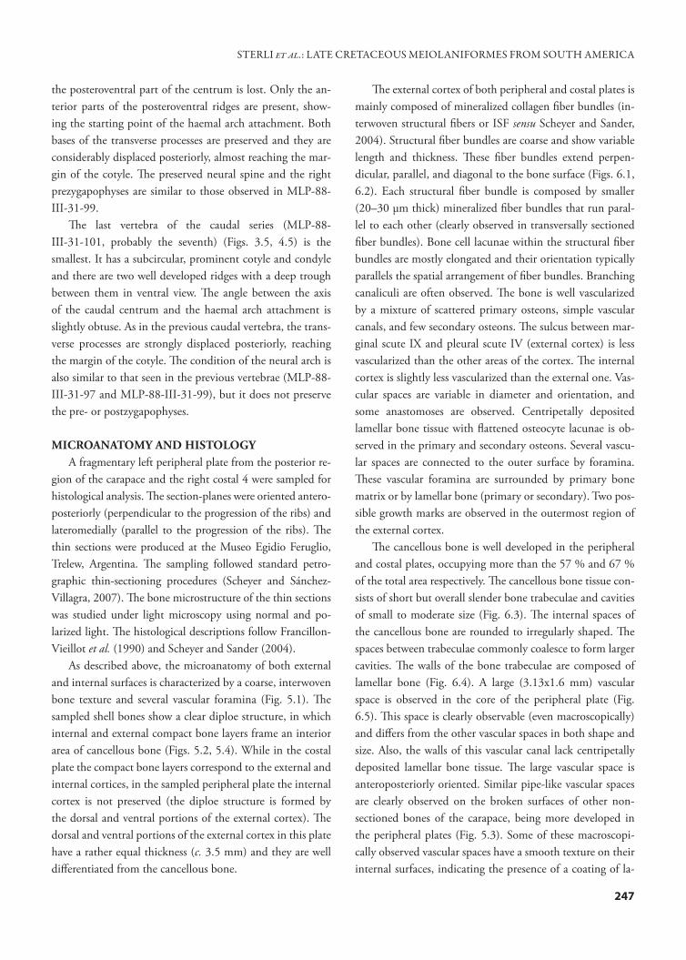
Figure 5. Trapalcochelys sulcata gen. nov. sp. nov. Allen Formation, Argentina. Microanatomy of carapace bones/ Microanatomía de las placas del caparazón. 1, detail of the plate ornamentation. SEM image at X18/ Detalle de la ornamentación. Imágenes al MEB a X18. Scale bar/ Escala = 0.5 mm; 2, Thin section of peripheral plate showing the dis-tribution of compact and cancellous bone. A large vascular space is observed in the cancellous bone (arrowhead)/ corte delgado de placa periférica mostrando la distribución de hueso compacto y esponjoso. Se observa un gran canal vascular en el tejido esponjoso (flecha). Scale bar/Escala= 10 mm; 3, broken surface of nuchal and left peripheral 1 showing the presence of large pipe-like vascular canals (arrowheads)/ superficie rota de placa nucal y periférica 1 izquierda mostrando la pres-encia de grandes canales vasculares tubuliformes (flechas). Scale bar/ Escala= 10 mm; 4, thin section of costal plate showing the distribution of compact and cancellous bone/ corte delgado de placa costal most-rando la distribución de hueso compacto y esponjoso. Scale bar/ Escala= 10 mm. cb, cancellous bone/ hueso esponjoso; eco, external cortex/corteza externa; fo; vascular foramina/forámenes vasculares; ico, inter-nal cortex/corteza interna.
1
2
3
4
AMEGHINIANA - 2013 - Tomo 50 (2): 240 - 256
249
STERLI et al.: LATE CRETACEOUS MEIOLANIFORMES FROM SOUTH AMERICA
the La Colonia Formation (Chubut Province) (Fig. 7). The tetrapod assemblage found in these lithostratigraphic units was reported by Leanza et al. (2004) as Allenian (= Alami-tian SALMA of Bonaparte et al., 1987). Leanza et al. (2004) characterized this tetrapod assemblage by the presence of te-iid lizards, chelid and meiolaniid turtles (here corrected to be meiolaniform turtles), crocodyliforms (mesoeucrocodylians and neosuchians), armoured saltasaurine titanosaurids, eu-titanosaurs, carnotaurine abelisaurid theropods, birds, had-rosaurid and ankylosaurian ornithischians, and mammals (dryolestoids, gondwanatheres, symmetrodonts, and prob-ably triconodonts) and assigned it a late Campanian–early Maastrichtian age.Loncoche Formation. The Loncoche Formation (Legarreta et al., 1989), together with the Jagüel, Roca, and Pircala formations, makes up the continental and marine sequence known as the Malargüe Group, in which the Cretaceous/Pa-leogene boundary lies (Bertels, 1969; Náñez and Concheyro, 1996). The Loncoche Formation has been correlated with the Allen, Los Alamitos, and La Colonia formations based on sedimentology (Legarreta et al., 1989; Barrio, 1990) and on common vertebrate assemblages (González Riga, 1999; Previtera and González Riga, 2008). The fauna from the Loncoche Formation is composed by fishes (e.g., rajiforms, lepisosteids, percoids, teleosts, ceratodontid dipnoans), an-urans (e.g., leptodactylids), turtles (meiolaniforms and che-lids), snakes (e.g., boiids), dinosaurs (e.g., titanosaurs, had-rosaurs, theropods), and plesiosaurs (González Riga, 1999; Previtera and González Riga, 2008). There are two known localities —both in Mendoza Province, Argentina— with vertebrate remains in the Loncoche Formation, i.e., Ran-quil-Co and Calmu-Co (González Riga, 1999; Previtera and González Riga, 2008), but meiolaniform remains have only been found in the former. 1. Ranquil-Co Locality (Mendoza Province): González Riga
(1999) described the fauna of the Loncoche Formation at Ranquil-Co. He assigned several large peripheral bones (MACN-M 25) and a bridge peripheral, a right anterior peripheral, and neural bones (MACN-M 40) to a large non-ornamented form of Chelidae indet. The revision of these specimens and of all the prepared specimens from Ranquil-Co at MACN allowed us to re-assign these speci-mens and a bridge peripheral (MACN-M 31) to Meiol-aniformes. Additional specimens collected in Ranquil-Co and
housed at the MHNSR are also recognized as belonging to Meiolaniformes. These specimens are an anterior left
peripheral (MHNSR-Pv 1143), an indeterminate periph-eral (MHNSR-Pv 1144), several fragments of peripherals (MHNSR-Pv 1145–1147), and a basisphenoid (MHNSR-Pv 1148). All carapacial bones show an ornamentation of small foramina and marginal sulci strongly curved anteriorly. The general morphology of the basisphenoid resembles that described for Peligrochelys walshae (Sterli and de la Fuente, 2012). The entrance for the cerebral carotids (fccp of Sterli et al., 2010) in MHNSR-Pv 1148 and in Pe. walshae can be seen on the ventral surface suggesting that the bifurcation of the internal carotid artery into the cerebral and palatine branches was not covered ventrally by bone. The suture with the pterygoids in MHNSR-Pv 1148 is seen in the anterior half of the basisphenoid and resembles the morphology seen in Pe. walshae. Allen Formation. In addition to the Salinas de Trapal-Có Locality there are two more localities in the Allen Formation where meiolaniforms have been found: El Abra and Salitral de Santa Rosa. 1. El Abra Locality (Río Negro Province): Broin and de la
Fuente (1993) assigned a couple of specimens (MACN-RN 520, right peripheral 1; MACN-RN 521, fragment of nuchal and right peripheral 1) from El Abra (Río Ne-gro Province) to Chelus. However, the authors did not discard the possibility of those specimens belonging in Meiolaniidae instead. The fragmentary nature of these specimens did not allow us to confirm or reject the last statement of Broin and de la Fuente (1993). Other speci-mens from El Abra (MACN-RN 522, left bridge pe-ripheral ?5; MACN-RN 706, left bridge peripheral ?3; MACN-RN 959: neural bone) are, however, assigned here to Meiolaniformes.
2. Salitral de Santa Rosa Locality (Río Negro Province): Several specimens from Salitral de Santa Rosa are housed at the Lamarque Museum and could be assigned to meio-laniforms. However, their preservation precludes a more accurate taxonomic assignation. The ornamentation and thickness of the plates and the anteriorly highly deflected sulci between marginal scales shown in peripheral bones reminds of T. sulcata and Pa. gasparinae. The large size of the plates found at Salitral de Santa Rosa resembles that of T. sulcata instead of the smaller Pa. gasparinae. Among the meiolaniform remains found at Salitral de
Santa Rosa, peripheral bones with sulci strongly bent anteri-orly (MML-PV1100); a costal bone, a fragment of a nuchal, and three peripherals (MML-PV1101), a left epiplastron and entoplastron (MML-PV1102), a right peripheral 8 (MML-
250
AMEGHINIANA - 2013 - Tomo 50 (2): 240 - 256
1 2
3 4
5
7
6
8
251
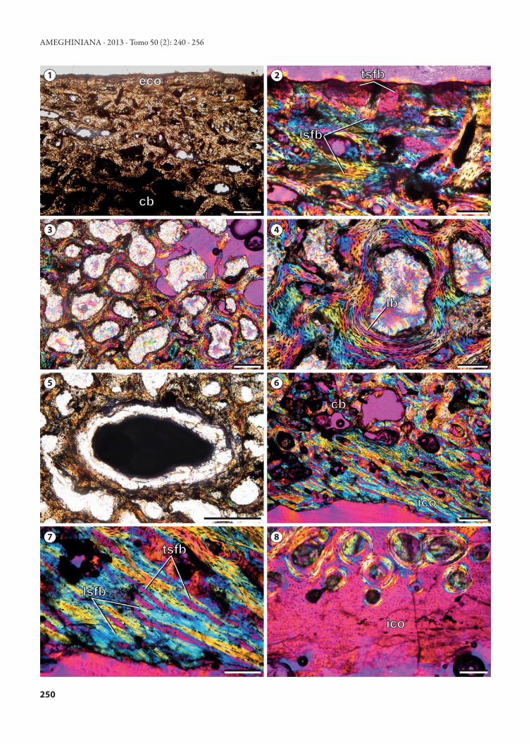
Figure 6. Trapalcochelys sulcata gen. nov. sp. nov. Allen Formation, Argentina. Histological sections of carapace bones/ Cortes histológicos de las placas del caparazón.1, external cortex of peripheral plate (normal light) / corteza externa de la placa periférica (luz normal). Scale bar/ Escala= 0.5 mm; 2, interwoven structural fiber bundles in the external cortex of the peripheral plate (polarized light combined with lambda filter)/ paquetes entrelazados de fibras estructurales en la corteza externa de la placa periférica (luz polarizada combinada con un filtro lambda). Scale bar/Escala= 0.2 mm; 3, cancellous bone of the peripheral plate (polarized light combined with lambda filter)/ tejido esponjoso de la placa periférica (luz polarizada combinada con un filtro lambda). Scale bar/ Escala= 0.5 mm; 4, detail of the cancellous bone of the peripheral plate showing the centripetally depo-sited lamellar bone tissue (polarized light combined with lamda filter)/ detalle del hueso esponjoso de la placa periférica mostrando el tejido lamelar depositado de forma centrípeta (luz polarizada combinada con un filtro lambda). Scale bar/ Escala= 0.2 mm; 5, detail of a large, pipe-like vascular space in the cancellous bone of the peripheral plate (normal light)/ detalle de un gran canal tubuliforme en el hueso esponjoso de la placa periférica (luz normal). Scale bar/ Escala= 1 mm; 6, general view of the internal cortex of the costal plate showing the structural fiber bundles (polarized light combined with lambda filter)/ vista general de la corteza interna de la placa costal mostrando los empaquetamientos de fibras estructurales (luz polari-zada combinada con un filtro lambda). Scale bar/ Escala= 0.5 mm; 7, detailed view of (6)/ vista detallada de (6). Scale bar/Escala= 0.2 mm; 8, avascular monorefringent bone matrix in the internal cortex of the costal plate (polarized light combined with lambda filter)/ matriz ósea monorefringente y avascular en la corteza interna de la placa costal (luz polarizada combinada con un filtro lambda). Scale bar/ Escala= 0.2 mm. cb, cancellous bone/ hueso esponjoso; eco, external cortex/corteza externa; ico, internal cortex/ corteza interna; lb, lamellar bone/hueso lamelar; lsfb, longitudinally sectioned structural fiber bundle/ paquete de fibras estructurales cortado longitudinalmente; tsfb, transversally sectioned structural fiber bundle/ paquete de fibras estructurales cortado transversalmente.
PV1103), costals, peripherals, thoracic vertebrae 9 and 10, phalanges, and a strongly eroded ulna (MML-PV1104), a costal, a left peripheral (MML-PV1104), a right peripheral (MML-PV1105), and a left humerus and the distal end of a left humerus and several plates (MML-PV1106) can be recognized. Los Alamitos Formation. The Los Alamitos Formation (Bonaparte et al., 1984) crops out in southeastern Río Ne-gro Province, near the settlement of Arroyo Verde (Andreis, 1987). The sedimentology suggests the dominant environ-ment of the Los Alamitos Formation was lacustrine domi-nated by brackish water conditions, turning to hypersaline towards the top of the sequence (Andreis, 1987). The fauna recovered from the Los Alamitos Formation consists of fishes (e.g., siluriforms, diplomystids, ariids), anurans (e.g., pipids and leptodactylids), turtles (meiolaniforms and chelids), snakes (e.g., madtsoiids, boiids), sphenodontids, dinosaurs (e.g., hadrosaurs, titanosaurs, theropods, birds, egg shells), and mammals (e.g., triconodonts, dryolestids, mesungula-tids, ferugliotheriids, gondwanatheriids) (Bonaparte et al., 1987; Apesteguía, 2005; Agnolin and Martinelli, 2009). Bonaparte et al. (1987) recognized this fauna assemblage be-longing to the late Campanian–early Maastrichtian and use it to typify the Alamitian South American Provincial Age. Later, Leanza et al. (2004) replaced the name Alamitian with the name Allenian to define the Late Cretaceous tetrapod assemblage.
Broin (1987) was the first who studied the turtle record from the Los Alamitos Formation. She recognized 17 un-questionable specimens and 3 doubtful specimens belonging to Meiolaniidae (here recognized as Meiolaniformes). Among the unquestionable specimens Broin (1987) recognized a left humerus (MACN-RN 36), a bridge peripheral (MACN-RN 37), a left anterior peripheral (2 or 3) (MACN-RN 38), a
left bridge peripheral (3 or 4) (MACN-RN 39), a posterior peripheral (MACN-RN 40), and a left posterior peripheral (MACN-RN 41). Later, Broin and de la Fuente (1993) as-signed a neural plate plus a fragment of a waved costal with the surface ornamented with small foramina (MACN-RN 910) and a fragment of nuchal bone (MACN-RN 918) to Meiolaniidae (Meiolaniformes here). The revision of the MACN collection allows us to corroborate the assignment of these specimens to Meiolaniformes by Broin (1987) and Broin and de la Fuente (1993).
Recently, Gaffney et al. (2007) noticed a great resem-blance (e.g., wide, midway positioned transverse process) be-tween cervical 8 of Chubutemys copelloi and some previously described cervical vertebrae from Los Alamitos. Gaffney et al. (2007) suggested that the biconvex cervical vertebrae pre-viously identified by Broin and de la Fuente (1993, fig. 5a–j) as cervical 5 (MACN-RN 907) and cervical 8 (MACN-RN 908) of a “primitive large chelid indet.” should instead be considered a meiolaniform. First hand comparisons of the cervical vertebrae of Los Alamitos Formation with Pa. gaspa-rinae and Ch. copelloi allow us to corroborate the statement by Gaffney et al. (2007). The general shape and the loca-tion of the transverse processes of MACN-RN 907 resemble that of cervical 4 of Pa. gasparinae while MACN-RN 908 resembles cervical 8 of C. copelloi. Therefore, we re-assign MAC-RN 907 and 908 as belonging to a meiolaniform in-det. Moreover, we corroborate assignment of the mentioned remains to meiolaniforms by Broin (1987) and Broin and de la Fuente (1993).
Furthermore, during the revision of the Los Alamitos collection at MACN several new remains of meiolaniforms could be identified. Among them several shell fragments (MACN-RN 63, peripheral 7 or 8; 64, right anterior periph-eral 2 or 3; 66, fragment indet.; 67, fragment indet. or ?right
STERLI et al.: LATE CRETACEOUS MEIOLANIFORMES FROM SOUTH AMERICA

252
bridge peripheral; 68, fragment indet.; 69, left peripheral 1 or 2; 70, peripheral indet.; 101, fragment of proximal end of left humerus; 709, fragment indet.), a thoracic vertebra 10 (MACN-RN 909); several opisthocoelous posterior caudal vertebrae with articulation for chevron bones (MAC-RN no number) were also recognized. La Colonia Formation. The La Colonia Formation crops out in northeastern Chubut Province, Argentina. As in the other mentioned formations, the sequence of the La Co-lonia Formation reveals a transition from continental to marginal marine sediments (Pascual et al., 2000). The ver-tebrate fauna recovered from this Formation is represented by fishes (e.g., ceratodontid dipnoans), turtles (e.g., meiol-aniforms and chelids), snakes (e.g., madtsoiids), polycotylid plesiosaurs, dinosaurs (e.g., theropods), and mammals (e.g., reigitheriids, mesungulatids, ferugliotheriids) (Bonaparte, 1985; Broin and de la Fuente, 1993; Albino, 2000; Gaspa-rini and de la Fuente, 2000; Pascual et al., 2000; Rougier et al., 2009b).
The presence of meiolaniform taxa in the La Colonia For-mation was first mentioned by Gasparini and de la Fuente (2000) who assigned a horn (MPEF-PV 859) to Meiolani-idae gen. et sp. indet. New findings of skull remains of Pa. gasparinae (unpublished data) should yield further informa-tion to reassess the taxonomic assignment of this horn either to Meiolaniidae or Meiolaniformes. The revision of the La Colonia specimens at MEF allowed us to re-interpret speci-men MPEF-PV 844 identified by Gasparini and de la Fuente (2000, p. 26) as a bridge peripheral belonging to Chelidae gen. et sp. indet. 1 as a meiolaniform. Comparisons with Pa. gasparinae and T. sulcata allowed us to reidentify this bridge peripheral as a left peripheral 6 belonging to a meiolaniform taxon (cf. Pa. gasparinae). MPEF-PV 844 shares with T. sul-cata and Pa. gasparinae the ornamentation of small foramina and the sulcus between marginals strongly bended anteriorly. However, the external surface of the plate of MPEF-PV 844 is slightly flat to concave, while in Pa. gasparinae and T. sul-cata the external surface is flat or slightly convex. Another difference is the size. MPEF-PV 844 is shorter (8 cm long) than peripheral 6 of T. sulcata (10 cm) and slightly longer (0.5 cm) than in Pa. gasparinae.
Other than the fragmentary remains mentioned above belonging to the La Colonia Formation, Sterli and de la Fuente (2011a) recently described a highly complete post-cranial skeleton of a new species of meiolaniform named Pa. gasparinae. New discoveries of skull remains and previously unknown postcranial remains (unpublished data) should
help understanding in more detail the general morphology of Late Cretaceous meiolaniform taxa from Patagonia. This should thus improve the detailed comparisons of this skull with the recently described Paleocene meiolaniform from Patagonia, i.e., Pe. walshae (Sterli and de la Fuente, 2012).
Bone histologyCarapace shell microstructure has been recently used as a
valuable tool in paleoecological studies (Scheyer, 2007; Schey-er and Sander, 2007). Based on the degree of development of
Figure 7. Map showing Upper Cretaceous localities with meiolani-forms in Patagonia, Argentina/ Mapa mostrando las localidades con meiolaniformes del Cretácico Superior de Patagonia, Argentina. Scale bar/ Escala = 500 km. EA, El Abra; LA, Los Alamitos; LC, La Colonia; RC, Ranquil-Co; SR, Salitral de Santa Rosa; ST, Salinas de Trapal-Có.
AMEGHINIANA - 2013 - Tomo 50 (2): 240 - 256
253
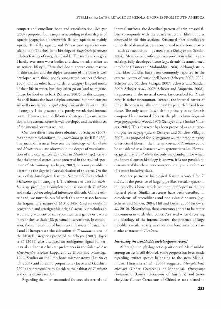
compact and cancellous bone and vascularization, Scheyer (2007) proposed four categories according to their degree of aquatic adaptation (I: terrestrial; II: semiaquatic to mainly aquatic; III: fully aquatic; and IV: extreme aquatic/marine adaptation). The shell-bone histology of Trapalcochelys sulcata exhibits features of categories I and II. The turtles in category I hardly ever enter water bodies and show no adaptations to an aquatic lifestyle. Their shell-bones appear quite massive in thin-section and the diploe structure of the bone is well developed with thick, poorly vascularized cortices (Scheyer, 2007). On the other hand, turtles of category II spend much of their life in water, but they often go on land to migrate, forage for food or to bask (Scheyer, 2007). In this category, the shell-bones also have a diploe structure, but both cortices are well vascularized. Trapalcochelys sulcata shares with turtles of category I the presence of a poorly vascularized internal cortex. However, as in shell-bones of category II, vasculariza-tion of the external cortex is well-developed and the thickness of the internal cortex is reduced.
Our data differ from those obtained by Scheyer (2007) for another meiolaniform, i.e., Meiolania sp. (MB R 2426). The main differences between the histology of T. sulcata and Meiolania sp. are observed in the degree of vasculariza-tion of the external cortex (lower in Meiolania sp.). Given that the internal cortex is not preserved in the studied spec-imen of Meiolania sp. (Scheyer, 2007), it is not possible to determine the degree of vascularization of this area. On the basis of its histological features, Scheyer (2007) included Meiolania sp. in category I. The absence of data for Meio-lania sp. precludes a complete comparison with T. sulcata and makes paleoecological inferences difficult. On the oth-er hand, we must be careful with this comparison because the fragmentary nature of MB R 2426 (and its doubtful geographic and stratigraphic origins) actually precludes an accurate placement of this specimen in a genus or even a more inclusive clade (JS, personal observations). In conclu-sion, the combination of histological features of categories I and II hampers a strict allocation of T. sulcata to one of the lifestyle categories proposed by Scheyer (2007). Joyce et al. (2011) also discussed an ambiguous signal for ter-restrial and aquatic habitat preferences in the Solemydidae Helochelydra nopcsai Lapparent de Broin and Murelaga, 1999. Studies on the limb bone microanatomy (Laurin et al., 2004) and forelimb proportions (Joyce and Gauthier, 2004) are prerequisite to elucidate the habitat of T. sulcata and other extinct turtles.
Regarding the microanatomical features of external and
internal surfaces, the described pattern of criss-crossed fi-bers corresponds with the coarse structural fiber bundles observed in the thin sections. Structural fiber bundles are mineralized dermal tissues incorporated to the bone matter —such as osteoderms— by metaplasia (Scheyer and Sander, 2004). Metaplastic ossification is a process in which a pre-existing, fully developed tissue (e.g., dermis) is transformed into bone (Haines and Mohuiddin, 1968). Although struc-tural fiber bundles have been commonly reported in the external cortex of turtle shell bones (Scheyer, 2007, 2009; Scheyer and Sánchez Villagra 2007; Scheyer and Sander, 2007; Scheyer et al., 2007; Scheyer and Anquetin, 2008), its presence in the internal cortex (as described for T. sul-cata) is rather uncommon. Instead, the internal cortex of the shell-bone is usually composed by parallel-fibered bone tissue. The only taxon in which the primary bone tissue is composed by structural fibers is the pleurodiran Stupend-emys geographicus Wood, 1976 (Scheyer and Sánchez Villa-gra, 2007). This character has been proposed as an autapo-morphy for S. geographicus (Scheyer and Sánchez Villagra, 2007). As proposed for S. geographicus, the predominance of structural fibers in the internal cortex of T. sulcata could be considered as a character with systematic value. Howev-er, given that T. sulcata is the only meiolaniform for which the internal cortex histology is known, it is not possible to determine if this character corresponds only to T. sulcata or to a more inclusive clade.
Another particular histological feature recorded for T. sulcata is the presence of large, pipe-like, vascular spaces in the cancellous bone, which are more developed in the pe-ripheral plates. Similar structures have been described in osteoderms of crocodilians and non-avian dinosaurs (e.g., Scheyer and Sander, 2004; Hill and Lucas, 2006; Farlow et al., 2010). Nevertheless, these structures appear to be rather uncommon in turtle shell bones. As stated when discussing the histology of the internal cortex, the presence of large pipe-like vascular spaces in cancellous bone may be a par-ticular character of T. sulcata.
Increasing the worldwide meiolaniform recordAlthough the phylogenetic position of Meiolaniidae
among turtles is still debated, some progress has been made regarding extinct species belonging to the stem Meiola-niidae. Hirayama et al. (2000) suggested Mongolochelys efremovi (Upper Cretaceous of Mongolia), Otwayemys cunicularius (Lower Cretaceous of Australia) and Sino-chelyidae (Lower Cretaceous of China) as taxa related to
STERLI et al.: LATE CRETACEOUS MEIOLANIFORMES FROM SOUTH AMERICA
254
Meiolaniidae. Later, Gaffney et al. (2007) proposed the Lower Cretaceous Chubutemys copelloi from Argentina as the sister group of Meiolaniidae. Anquetin (2011) recently suggested Naomichelys speciosa Hay, 1908, from the Lower Cretaceous of North America as closely related to Meio-laniidae and even more recently, Sterli and de la Fuente (2012) suggested not only Ch. copelloi and Mo. efremovi as closely related to Meiolaniidae but also O. cunicularius (Lower Cretaceous of Australia), K. bajazidi (Upper Creta-ceous of Europe), Pa. gasparinae (Upper Cretaceous of Ar-gentina), and Pe. walshae (Paleocene of Argentina). In this paper we describe a new species related to Meiolaniidae, Trapalcochelys sulcata, increasing not only the number of meiolaniforms, but spreading also their distribution. Thus, knowledge is increasing on their morphological variation, microanatomy, and histology. The more complete evolu-tionary scenario of Meiolaniformes resulting from all the new discoveries and new interpretations, suggests a more complex story about the origin and paleobiogeography of Meiolaniidae. The ghost lineages subtending Meiolani-formes suggest the clade had a Pangaean origin before the complete fragmentation of continents in the Lower Cre-taceous (Hirayama et al., 2000; Sterli and de la Fuente, 2012). These new hypotheses regarding the cosmopolitan distribution of Meiolaniformes highlight the importance of increasing the taxonomic sampling for future phylogenetic analyses on turtle evolution.
ACKNOWLEDGMENTS
We thank E. P. Tonni and M. Reguero (Museo de La Plata) for lending the material for study, D. Cabaza and L. López (Museo Lamarque) and A. Kramarz (MACN) for allowing us to see the collections under their care. We also thank J. González who drew the figures 2 and 4. Thin sections were made by M. Caffa. J. Groizard from Aluminio Argentino (ALUAR, Puerto Madryn) obtained SEM images. The detailed revision of this manuscript by W. G. Joyce and T. Scheyer greatly improved the quality of the paper. The editors, D. Pol and M. Griffin, are thanked for their editorial work. This study was partially supported by grant PIP 00795 granted by CONICET to MSDLF, PICT-2010-0646 granted by the Agencia Nacional de Promoción Científica y Tecnológica and National Geographic Society grant number 8975-11 to JS.
REFERENCESAgnolin, F. 2010. A new species of the genus Atlantoceratodus (Dipnoi-
formes: Ceratodontoidei) from the Uppermost Cretaceous of Patagonia and a brief overview of fossil dipnoans from the Cretaceous and Paleo-gene of South America. Brazilian Geographical Journal: Geosciences and Humanities Research Medium 1: 162–210.
Agnolin, F. and Martinelli, A.G. 2009. Fossil birds from the Late Creta-ceous Los Alamitos Formation, Río Negro Province, Argentina. Journal of South American Earth Sciences 27: 42–49.
Albino, A. 2000. New record of snakes from the Cretaceous of Patagonia (Argentina). Geodiversitas 22: 247–253.
Ameghino, F. 1899. Sinopsis geológica–paleontológica. Suplemento (adi-ciones y correcciones). Censo Nacional, La Plata: 1–13.
Ancibor, E. 1995. Palmeras fósiles del Cretácico Tardío de la Patagonia Ar-gentina (Bajo de Santa Rosa, Río Negro). Ameghiniana 32: 287–299.
Andreis, R.R. 1987. Stratigraphy and paleoenvironment. In: J.F. Bonaparte (Ed.), The Late Cretaceous fauna of Los Alamitos, Patagonia, Argentina. Revista del Museo Argentino de Ciencias Naturales ‘‘Bernardino Rivadavia’’ (Sección Paleontología) 3: 103–110.
Andreis, R.R., Iñíguez Rodríguez, A.M., Lluch, J.J. and Sabio, D.A. 1974. Estudio sedimentológico de las formaciones del Cretácico superior del área del Lago Pellegrini (Provincia de Río Negro, República Argentina). Revista de la Asociación Geológica Argentina 29: 85–104.
Andreis, R.A., Ancibor, E., Archangelsky, S., Artabe, A., Bonaparte, J.F. and Genise, J. 1991. Asociación de vegetales y animales en estratos del Cretácico tardío del norte de la Patagonia. Ameghiniana 28: 201–202.
Anquetin, J. 2011. Reassessment of the phylogenetic interrelationships of basal turtles (Testudinata). Journal of Systematic Palaeontology 10: 3–45.
Apesteguía, S. 2005. A Late Campanian sphenodontid (Reptilia, Diapsida) from northern Patagonia. Comptes Rendu Palevol 4: 663–669.
Apesteguía, S. and Jones, M.E.H. 2012. A Late Cretaceous “tuatara” (Lepi-dosauria: Sphenodontinae) from South America. Cretaceous Research 34: 154–160.
Apesteguía, S. and Rougier, G.W. 2007. A Late Campanian sphenodontid maxilla from Northern Patagonia. American Museum Novitates 3581: 1–11.
Artabe, A. and Zamuner, A.B. 1999. A new cycad item from the Upper Cretaceous of Patagonia, Argentina. Boletim do 5º Simpósio sobre o Cre-taceo do Brasil 1999: 309–313.
Artabe, A.E., Zamuner, A.B. and Stevenson, D.W. 2004. The new petri-fied cycad stems, Brunoa gen. nov. and Worsdellia gen. nov., from the Cretaceous of Patagonia (Bajo de Santa Rosa, Río Negro province), Ar-gentina. Botanical Review 70: 121–133.
Barrio, C.A. 1990. Late Cretaceous–Early Tertiary sedimentation in a semi-arid foreland basin (Neuquén Basin, western Argentina). Sedimentary Geology 66: 255–275.
Barrio, C.A. 1991. Controles en la sedimentación en las cuencas foreland. El ejemplo del Grupo Malargüe (Campaneano–Paleoceno) en la Cuenca Neuquina, Argentina. 6º Congreso Geológico Chileno (Santiago de Chile), Abstracts, p. 597–601.
Bertels, A. 1969. Estratigrafía del límite Cretácico–Terciario de la Patagonia septentrional. Revista de la Asociación Geológica Argentina 24: 45–54.
Bonaparte, J.F. 1985. A horned Cretaceous carnosaur from Patagonia. Na-tional Geographic Research: 149–151.
Bonaparte, J. F., Báez, A. M., Cione, A. L., Andreis, R. R., Broin, F., Powell, J. E., and Albino, A. 1987. Resume. In: J.F. Bonaparte (Ed.), The Late Cretaceous fauna of Los Alamitos, Patagonia, Argentina. Revista del Museo Argentino de Ciencias Naturales ‘‘Bernardino Rivadavia’’ (Sección Paleon-tología) 3: 172–178.
Bonaparte, J.F., Franchi, M.R., Powell, J.E. and Sepúlveda, E.G., 1984. La Formación Los Alamitos (Campaniano–Maastrichtiano) del sudeste de Río Negro, con descripción de Kritosaurus australis n. sp. (Hadro-sauridae). Significado paleogeográfico de los vertebrados. Revista de la Asociación Geológica Argentina 39: 284–299.
Broin, F. 1987. Chelonia. In: J.F. Bonaparte (Ed.), The Late Cretaceous fauna of Los Alamitos, Patagonia, Argentina. Revista del Museo Argentino de Ciencias Naturales ‘‘Bernardino Rivadavia’’ (Sección Paleontología) 3: 131–139.
Broin, F. and de la Fuente, M.S. 1993. Les tortues fossiles d’Argentine: synthèse. Annales de Paléontologie 79: 169–232.
Carignano, A.P. and Varela, J.A. 2011. Ostrácodos (Crustacea) de la For-
AMEGHINIANA - 2013 - Tomo 50 (2): 240 - 256
255
mación Allen (Cretácico tardío), cuenca neuquina, Argentina. Revista Brasileira de Paleontologia 14: 169–178.
Del Fueyo, G.M. 1998. Coniferous woods from the Upper Cretaceous of Patagonia, Argentina. Revista Española de Paleontología 13: 43–50.
Dessanti, R.N. 1973. Descripción geológica de la Hoja 29 b, Bardas Blan-cas, Provincia de Mendoza. Boletín Servicio Nacional de Minería y Ge-ología 139: 1–70.
Dessanti, R.N. 1975. Descripción Geológica de la Hoja 28 b, Malargüe. Boletín Servicio Nacional de Minería y Geología 149: 1–50.
Farlow, J.O., Hayashi, S. and Tattersall G.J. 2010. Internal vascularity of the dermal plates of Stegosaurus (Ornithischia, Thyreophora). Swiss Journal Geosciences 103: 173–185.
Fossa Mancini, E., Feruglio, E. and Yussen de Campana, J.C. 1938. Una reunión de geólogos de YPF y el problema de la terminología estratigrá-fica. Boletín de Informaciones Petroleras 15: 1–67.
Francillon-Vieillot, H., Buffrénil, V. de, Castanet, J., Géraudie, J., Meunier, F.J., Sire, J.Y., Zylberberg, L., and Ricqlès, A. de 1990. Microstructure and mineralization of vertebrate skeletal tissues. In: J.G. Carter (Ed.), Skeletal Biomineralization: Patterns, Processes and Evolutionary Trends. Van Nostrand Reinhold, New York, p. 471–530.
Gaffney, E.S. 1985. The cervical and caudal vertebrae of the cryptodiran turtle, Meiolania platyceps, from the Pleistocene of Lord Howe Island, Australia. American Museum Novitates 2805: 1–29.
Gaffney, E.S. 1996. The postcranial morphology of Meiolania platyceps and a review of the Meiolaniidae. Bulletin of the American Museum of Natural History 229: 1–165.
Gaffney, E.S., Rich, T. H., Vickers-Rich, P., Constantine A., Vacca, R. and Kool, L. 2007. Chubutemys, a new eucryptodiran turtle from the Early Cretaceous of Argentina, and the relationships of the Meiolaniidae. American Museum Novitates 3599: 1–35.
Gasparini, Z. and de la Fuente, M.S. 2000. Tortugas y plesiosaurios de la Formación La Colonia (Cretácico Superior) de Patagonia. Revista Espa-ñola de Paleontología 15: 23–35.
Gasparini, Z., Casadio, S., de la Fuente, M. S., Salgado L., Fernández, M.S. and Concheyro, A. 2002. Reptiles acuáticos en sedimentitas lacustres y marinas del Cretácico Superior de Patagonia (Río Negro, Argentino). 15º Congreso Geológico Argentino (El Calafate), Actas 1: 495–499.
Gasparini, Z., Salgado, L. and Casadio, S. 2003. Maastrichtian plesiosaurs fron northern Patagonia. Cretaceous Research 24: 157–170.
Gómez, R.O., Báez, A.M. and Rougier, G.W. 2008. An anilioid snake from the Upper Cretaceus of northern Patagonia. Cretaceous Research 29: 481–488.
González Riga, B.J. 1999. Hallazgo de vertebrados fósiles en la Formación Loncoche, Cretácico Superior de la provincia de Mendoza, Argentina. Ameghiniana 36: 401–410.
Haines, R.W. and Mohuiddin, A. 1968. Metaplastic bone. Journal of Anat-omy 103: 527–538.
Hay, O.P. 1908. The fossil turtles of North America. Publications of the Carnegie Institute of Washington 75: 1–568.
Hill, R.V. and Lucas, S.G. 2006. New data on the anatomy and relation-ships of the Paleocene crocodylian Akanthosuchus langstoni. Acta Palae-ontologica Polonica 51: 455–464.
Hirayama, R., Brinkman, D.B. and Danilov, I.G. 2000. Distribution and biogeography of non-marine Cretaceous turtles. Russian Journal of Her-petology 7: 181–198.
Holmberg, E. 1975. Descripción Geológica de la Hoja 32c, Buta Ranquil, Provincia del Neuquén y Mendoza. Boletín Servicio Nacional de Minería y Geología 152: 1–91.
Hugo, C.A. and Leanza, H.A. 2001a. Hoja Geológica 3969-IV, General Roca, provincias del Neuquén y Río Negro. Instituto de Geología y Re-cursos Naturales, SEGEMAR, Boletín 308: 1–71.
Hugo, C.A. and Leanza, H.A. 2001b. Hoja Geológica 3966-III, Villa Re-gina, provincia Río Negro. Instituto de Geología y Recursos Naturales, SEGEMAR, Boletín 309: 1–53.
Joyce,W.G. and Gauthier, J.A. 2004 Palaeoecology of Triassic stem turtles sheds new light on turtle origins. Proceedings of the Royal Society of Lon-don, series B 271:1–5.
Joyce, W.G., Chapman, S.D., Moody, R.T.J., and Walker, C.A. 2011. The skull of the solemydid turtle Helochelydra nopcsai from the Early Creta-ceous of the Isle of Wight (UK) and a review of Solemydidae. Special Papers in Palaeontology 86: 75–97.
Khozatsky, L.I.1997. Large turtles from the Late Cretaceous of Mongolia. Russian Journal of Herpetology 4: 148–154.
Klein, I.T. 1760. Klassification und kurze Geschichte der Vierfüssgen Thiere. Jonas Schmidt, Lübeck, 381 p.
Lapparent de Broin, F. de and Murelaga, X. 1999. Turtles from the Upper Cretaceous of Laño (Iberian peninsula). Estudios del Museo de Ciencias Naturales de Álava 14: 135–211.
Laurin, M., Girondot, M. and Loth, M-.M. 2004. The evolution of the long bone microstructure and lifestyle in lissanphibians. Paleobiology 30: 589–613.
Leanza, H.A. 1999. The Jurassic and Cretaceous terrestrial beds from south-ern Neuquén Basin, Argentina. Field Guide. Serie Miscelánea (INSU-GEO) 4: 1–30.
Leanza, H.A., Apesteguía, S., Novas, F.E. and de la Fuente, M.S. 2004. Cretaceous terrestrial beds from the Neuquén Basin (Argentina) and their tetrapod assemblages. Cretaceous Research 25: 61–87.
Legarreta, L., Kokogian, D.A. and Boggetti, D.A. 1989. Depositional se-quences of de Malargüe Group (Upper Cretaceous–Lower Tertiary), Neuquen Basin, Argentina. Cretaceous Research 10: 337–356.
Lyson, T.R. and Joyce, W.G. 2009. A revision of Plesiobaena (Testudines: Baenidae) and an assessment of baenid ecology across the K/T bound-ary. Journal of Paleontology 83: 833–853.
Malumián, N. 1999. La sedimentación y el volcanismo terciarios en la Pat-agonia extraandina. In: R. Caminos (Ed.), Geología Argentina. Anales del Instituto Geológico Recursos Minerales 29: 557–612.
Martinelli, A.G. and Forasiepi, A.M. 2004. Late Cretaceous vertebrates from bajo de Santa Rosa (Allen Formation), Río Negro province, Argen-tina, with the description of a new sauropod dinosaur (Titanosauridae). Revista del Museo Argentino de Ciencias Naturales 6: 257–305.
Náñez, C. and Concheyro, A. 1996. Límite Cretácico–Paleógeno. In: A. Ardolino and M. Franchi (Eds.), Geología y recursos minerales del De-partamento Añelo, provincia del Neuquén. Dirección Nacional del Servicio Geológico, Anales 25: 129–150.
Nopcsa, F.1923. On the geological importance of the primitive reptilian fauna of the Uppermost Cretaceous of Hungary; with a description of a new tortoise (Kallokibotion). Quarterly Journal of the Geological Soci-ety79: 100–116.
Novas, F.E., Canale, J.I. and Isasi, M.P. 2003. Un terópodo maniraptor del Campaniano–Maastrichthiano del norte patagónico. Ameghiniana, Suplemento Resúmenes 40: 63R.
Novas, F.E., Pol, D., Canale, J.I., Porfiri, J.D. and Calvo, J.O. 2008. A bizarre Cretaceous theropod dinosaur from Patagonia and the evolution of Gondwanan dromaeosaurids. Proceedings of the Royal Society B 276: 1101–1107.
Owen, R. 1886. Description of some fossil remains of two species of a me-galanian genus (Meiolania) from “Lord Howe’s Island.” Philosophical Transactions of the Royal Society, London 179:181–191.
Pascual, R., Goin, F.J., González, P., Ardolino, A. and Puerta, P.F. 2000. A highly derived docodont from the Patagonian Late Cretaceous: evolution-ary implications for Gondwanan mammals. Geodiversitas 22: 395–414.
Previtera, E. and González Riga, B.J. 2008. Vertebrados cretácicos de la Formación Loncoche en Calmu-Co, Mendoza, Argentina. Ameghiniana 45: 349–359.
Ramos, V.A. 1981. Descripción Geológica de la Hoja 33c, Los Chihuidos Norte, Provincia del Neuquén. Boletín Servicio Nacional de Geología y Minería 182: 1–103.
Riccardi, A.C. 1988. The Cretaceous system of southern South America. Memoirs of the Geological Society of America 168: 1–70.
STERLI et al.: LATE CRETACEOUS MEIOLANIFORMES FROM SOUTH AMERICA
256
Roll, A. 1939. [La Cuenca de los Estratos con Dinosaurios al sur del río Neu-quén. Yacimientos Petrolíferos Fiscales, Buenos Aires. Unpublished re-port].
Rougier, G.W., Chornogubsky, L., Casadío, S., Páez Arango, N. and Gial-lombardo, A. 2009a. Mammals from the Allen Formation, Late Creta-ceous, Argentina. Cretaceous Research 30: 223–238.
Rougier, G.W., Forasiepi, A.M., Hill, R.V. and Novacek, M.J. 2009b. New mammalian remains from the Late Cretaceous La Colonia Formation, Patagonia, Argentina. Acta Palaeontologica Polonica 54: 195–212.
Salgado, L. and Azpilicueta, C. 2000. Un nuevo saltasaurino (Sauropoda, Titanosauridae) de la provincia de Río Negro (Formación Allen, Cre-tácico Superior), Patagonia, Argentina. Ameghiniana 37: 259–264.
Spalletti, L.A., Franzese, J.R., Mac Donald, D. and Gómez Pérez, I. 1999. Paleogeographic evolution of southern South America during the Creta-ceous. 5° Simpósio sobre o Cretáceo do Brasil (Río Claro), Boletim: 87–95.
Scheyer, T.M. 2007. [Comparative bone histology of the turtle shell (carapace and plastron): implications for turtle systematics, functional morphology, and turtle origins. Ph.D. Thesis, University of Bonn, Bonn, 343 p. URL: http://hss.ulb.uni-bonn.de/2007/1229/1229.htm]
Scheyer, T.M. 2009. Conserved bone microstructure in the shells of long-necked and short-necked chelid turtles (Testudinata, Pleurodira). Fossil Record 12: 47–57.
Scheyer, T.M. and Anquetin, J. 2008. Bone histology of the Middle Juras-sic turtle shell remains from Kirtlington, Oxfordshire, England. Lethaia 41: 85–96.
Scheyer, T.M. and Sánchez-Villagra, M.R. 2007. Carapace bone histology in the giant pleurodiran turtle Stupendemys geographicus: Phylogeny and function. Acta Palaeontologica Polonica 52: 137–154.
Scheyer, T.M. and Sander, P.M. 2004. Histology of ankylosaur osteoderms: implications for systematics and function. Journal of Vertebrate Paleon-tology 24: 874–893.
Scheyer, T.M. and Sander, P.M. 2007. Shell bone histology indicates ter-restrial palaeoecology of basal turtles. Proceedings of the Royal Society B 274: 1884–1893.
Scheyer, T.M., Sander, P.M., Joyce, W.G., Böhme, W., and Witzel, U. 2007. A plywood structure in the shell of fossil and living soft-shelled turtles (Trionychidae) and its evolutionary implications. Organisms, Diversity and Evolution 7: 136–144.
Sterli, J. and de la Fuente, M.S.2011a. A new turtle from the La Colonia Formation (Campanian–Maastrichtian), Patagonia, Argentina, with re-marks on the evolution of the vertebral column in turtles. Palaeontology 54: 63–78.
Sterli, J. and de la Fuente, M.S. 2011b. Re-description and evolutionary remarks on the Patagonian horned turtle Niolamia argentina Ameghino,
1899 (Testudinata; Meiolaniidae). Journal of Vertebrate Paleontology 31: 1210–1229.
Sterli , J. and de la Fuente, M.S. 2012. New evidence from the Paleocene of Patagonia (Argentina) on the evolution and paleobiogeography of meiol-aniform turtles (Testudinata, new taxon name). Journal of Systematic Pal-aeontology. DOI:10.1080/14772019.2012.708674
Sterli, J., Müller, J., Anquetin, J. and Hilger, A. 2010. The parabasisphenoid complex in Mesozoic turtles and the evolution of the testudinate basi-cranium. Canadian Journal of Earth Sciences 47: 1337–1346.
Uliana, M.A. 1974. [Geología superficial de la parte este de la Cuenca Neuqui-na. Yacimientos Petrolíferos Fiscales, Gerencia de Exploración. Buenos Aires. Unpublished report].
Uliana, M.A. and Biddle, K.T. 1988. Mesozoic–Cenozoic paleogeographic and geodynamic evolution of southern South America. Revista Brasilera de Geociencias 18: 172–190.
Uliana, M.A. and Dellapé, D.A. 1981. Estratigrafía y evolución paleoambi-ental de la sucesión maestrichtiano–eoterciaria del Engolfamiento Neu-quino (Patagonia septentrional). 8º Congreso Geológico Argentino (San Luis), Actas 3: 673–711.
Urien, C.M., Zambrano, J.J. and Yrigoyen, M.R. 1995. Petroleum basins of southern South America. In: A. J. Tankard, R. Suarez and H.J. Welsink (Eds.), Petroleum basin of South America. American Association of Petro-leum Geology Memoirs 62: 63–77.
Volkheimer, W. 1973. Observaciones geológicas en el área de Ingeniero Ja-cobacci y adyacencias, provincia de Río Negro. Revista de la Asociación Geológica Argentina 28: 13–36.
White, A.W., Worthy, T.H., Hawkins, S., Bedford, S. and Spriggs, M. 2010. Megafaunal meiolaniid horned turtles survived until early human settlement in Vanuatu, Southwest Pacific. Proceedings of the National Academy of Sciences of the United States of America 107: 15512–15516.
Wichmann, R. 1924. Nuevas observaciones geológicas en la parte oriental del Neuquén y en el Territorio del Río Negro. Ministerio de Agricultura, Sección Geología 2: 1–22.
Wood, R.C. 1976. Stupendemys geographicus, the world’s largest turtle. Bre-viora 436: 1–31.
Yrigoyen, M.R. 1969. Problemas estratigráficos del Terciario de Argentina. Ameghiniana 6: 315–329.
doi: 10.5710/AMGH.16.01.2013.582
Recibido: 16 de abril de 2012Aceptado: 16 de enero de 2013
AMEGHINIANA - 2013 - Tomo 50 (2): 240 - 256