A new paradigm in respiratory hygiene: modulating respiratory secretions to contain cough bioaerosol...
13
BioMed Central Page 1 of 13 (page number not for citation purposes) BMC Pulmonary Medicine Open Access Research article A new paradigm in respiratory hygiene: modulating respiratory secretions to contain cough bioaerosol without affecting mucus clearance Gustavo Zayas* 1,2 , Juan C Valle 2 , Mauricio Alonso 2 , Henry Alfaro 2 , Daniel Vega 2 , Gloria Bonilla 2 , Miguel Reyes 2 and Malcolm King* 1 Address: 1 University of Alberta Pulmonary Research Group, Edmonton, Canada and 2 Hospital Nacional "Dr. Jose Antonio Saldaña" Neumologia y Medicina Familiar, San Salvador, El Salvador Email: Gustavo Zayas* - [email protected]; Juan C Valle - [email protected]; Mauricio Alonso - [email protected]; Henry Alfaro - [email protected]; Daniel Vega - [email protected]; Gloria Bonilla - [email protected]; Miguel Reyes - [email protected]; Malcolm King* - [email protected] * Corresponding authors Abstract Background: Several strategies and devices have been designed to protect health care providers from acquiring transmissible respiratory diseases while providing care. In modulating the physical characteristics of the respiratory secretions to minimize the aerosolization that facilitates transmission of airborne diseases, a fundamental premise is that the prototype drugs have no adverse effect on the first line of respiratory defense, clearance of mucus by ciliary action. Methods: To assess and demonstrate the primary mechanism of our mucomodulators (XLs), we have built our evidence moving from basic laboratory studies to an ex-vivo model and then to an in-vivo large animal model. We exposed anesthetized dogs without hypersecretion to different dose concentrations of aerosolized XL "B", XL "D" and XL "S". We assessed: cardio-respiratory pattern, tracheal mucus clearance, airway patency, and mucus viscoelastic changes. Results: Exposure of frog palate mucus to XLs did not affect the clearance of mucus by ciliary action. Dogs maintained normal cardio-respiratory pattern with XL administration. Tracheal mucociliary clearance in anesthetized dogs indicated a sustained 40% mean increase. Tracheal mucus showed increased filance, and there was no mucus retention in the airways. Conclusion: The ex-vivo frog palate and the in-vivo mammalian models used in this study, appear to be appropriate and complement each other to better assess the effects that our mucomodulators exert on the mucociliary clearance defence mechanism. The physiological function of the mucociliary apparatus was not negatively affected in any of the two epithelial models. Airway mucus crosslinked by mucomodulators is better cleared from an intact airway and normally functioning respiratory system, either due to enhanced interaction with cilia or airflow-dependent mechanisms. Data obtained in this study allow us to assure that we have complied with the fundamental requirement criteria established in the initial phase of developing the concept of mucomodulation: Can we modulate the physical characteristics of the respiratory secretions to reduce aerosolization without affecting normal mucociliary clearance function, or even better improving it? Published: 13 August 2007 BMC Pulmonary Medicine 2007, 7:11 doi:10.1186/1471-2466-7-11 Received: 26 August 2006 Accepted: 13 August 2007 This article is available from: http://www.biomedcentral.com/1471-2466/7/11 © 2007 Zayas et al; licensee BioMed Central Ltd. This is an Open Access article distributed under the terms of the Creative Commons Attribution License (http://creativecommons.org/licenses/by/2.0 ), which permits unrestricted use, distribution, and reproduction in any medium, provided the original work is properly cited.
-
Upload
independent -
Category
Documents
-
view
3 -
download
0
Transcript of A new paradigm in respiratory hygiene: modulating respiratory secretions to contain cough bioaerosol...

BioMed CentralBMC Pulmonary Medicine
ss
Open AcceResearch articleA new paradigm in respiratory hygiene: modulating respiratory secretions to contain cough bioaerosol without affecting mucus clearanceGustavo Zayas*1,2, Juan C Valle2, Mauricio Alonso2, Henry Alfaro2, Daniel Vega2, Gloria Bonilla2, Miguel Reyes2 and Malcolm King*1Address: 1University of Alberta Pulmonary Research Group, Edmonton, Canada and 2Hospital Nacional "Dr. Jose Antonio Saldaña" Neumologia y Medicina Familiar, San Salvador, El Salvador
Email: Gustavo Zayas* - [email protected]; Juan C Valle - [email protected]; Mauricio Alonso - [email protected]; Henry Alfaro - [email protected]; Daniel Vega - [email protected]; Gloria Bonilla - [email protected]; Miguel Reyes - [email protected]; Malcolm King* - [email protected]
* Corresponding authors
AbstractBackground: Several strategies and devices have been designed to protect health care providers fromacquiring transmissible respiratory diseases while providing care. In modulating the physical characteristicsof the respiratory secretions to minimize the aerosolization that facilitates transmission of airbornediseases, a fundamental premise is that the prototype drugs have no adverse effect on the first line ofrespiratory defense, clearance of mucus by ciliary action.
Methods: To assess and demonstrate the primary mechanism of our mucomodulators (XLs), we havebuilt our evidence moving from basic laboratory studies to an ex-vivo model and then to an in-vivo largeanimal model. We exposed anesthetized dogs without hypersecretion to different dose concentrations ofaerosolized XL "B", XL "D" and XL "S". We assessed: cardio-respiratory pattern, tracheal mucus clearance,airway patency, and mucus viscoelastic changes.
Results: Exposure of frog palate mucus to XLs did not affect the clearance of mucus by ciliary action. Dogsmaintained normal cardio-respiratory pattern with XL administration. Tracheal mucociliary clearance inanesthetized dogs indicated a sustained 40% mean increase. Tracheal mucus showed increased filance, andthere was no mucus retention in the airways.
Conclusion: The ex-vivo frog palate and the in-vivo mammalian models used in this study, appear to beappropriate and complement each other to better assess the effects that our mucomodulators exert onthe mucociliary clearance defence mechanism. The physiological function of the mucociliary apparatus wasnot negatively affected in any of the two epithelial models. Airway mucus crosslinked by mucomodulatorsis better cleared from an intact airway and normally functioning respiratory system, either due toenhanced interaction with cilia or airflow-dependent mechanisms. Data obtained in this study allow us toassure that we have complied with the fundamental requirement criteria established in the initial phase ofdeveloping the concept of mucomodulation: Can we modulate the physical characteristics of therespiratory secretions to reduce aerosolization without affecting normal mucociliary clearance function,or even better improving it?
Published: 13 August 2007
BMC Pulmonary Medicine 2007, 7:11 doi:10.1186/1471-2466-7-11
Received: 26 August 2006Accepted: 13 August 2007
This article is available from: http://www.biomedcentral.com/1471-2466/7/11
© 2007 Zayas et al; licensee BioMed Central Ltd. This is an Open Access article distributed under the terms of the Creative Commons Attribution License (http://creativecommons.org/licenses/by/2.0), which permits unrestricted use, distribution, and reproduction in any medium, provided the original work is properly cited.
Page 1 of 13(page number not for citation purposes)

BMC Pulmonary Medicine 2007, 7:11 http://www.biomedcentral.com/1471-2466/7/11
BackgroundThe respiratory system performs one of the most vitalfunctions of the body: the respiratory process, that con-sists of an appropriate gas exchange occurring in the lungsbetween blood and air. It provides oxygen to the organismand removes carbon dioxide. Air has to travel through aseries of passages, and interact with several types of tis-sues, until it reaches the alveolus where the gas exchangetakes place. Air is in contact with many bacteria, pollut-ants and particles in the environment before reaching thealveolus. Many of these components are inert partici-pants, but others are very harmful, and can disable the air-ways natural defence mechanisms against injuries. Hence,air must be cleaned, warmed and humidified.
There are several mechanisms to protect such a vital func-tion. Two main complementary mechanisms act to main-tain the airways as free as possible of foreign bodies andthe airway epithelium from damage due to inhaled path-ogens and respiratory insults: namely the mucociliaryclearance and the cough clearance. The former constitutesthe first line of defence and the latter is the backup or rein-forcement if the first one is unable to carry out the task.
Mucus is a unique materia lining all the major body cavi-ties, produced by the same type of cells: the goblet cellsand the mucous cells of the submucosal glands. In thelungs, other types of cells produce materials that contrib-ute to the unique physical properties of mucus, like theClara cell, and the type II pneumocyte, which produce thesurfactant factor [1,2]. Mucus can be considered a viscoe-lastic fluid, since it exhibits both liquid-like (viscous) andsolid-like (elastic) properties [3]. The relative proportionsof elasticity and viscosity are as important in describinghow a material such as mucus behaves when it is sub-jected to external forces, as are the absolute values ofeither parameter by itself.
The mucus lining the airways is a mobile layer constantlycleared by intimate interaction with cilia. It is also possi-ble that interaction of mucus with the low velocity airflowduring normal breathing might play a role in mucus clear-ability. This concept has not been well studied yet. Anychange occurring in one component of the mucus-ciliaunit might lead to disruption of normal mucociliary clear-ance processes, reducing lung defences and allowing res-piratory secretions to accumulate and impair pulmonaryfunction [4].
We are using an integrated approach to gain more insightinto what might occur when the airway mucus-cilia unit isexposed to novel agents that selectively raise elasticity rel-ative to viscosity of one of the components: the mucusmacromolecule. This is the reason why we are also review-ing some theoretical aspects of these components.
The mucus macromolecule (mucin) has a basic, commonand complex biochemical structure. Mucin macromole-cules consist of oligosaccharide units attached to a proteiccore [5,6]. The physical characteristics of mucus dependprincipally on the type and amount of glycoproteins. Vis-cosity and elasticity are the physical characteristics consid-ered essential for mucus to function [7], althoughadhesivity and spinnability are particularly important indescribing the cough clearability of airway mucus [8].
The frequency of mechanical response is important formucus clearance, particularly in the airways. There is anoptimum range among the physical properties that givemucus its capacity to behave in a balanced manner evenat very different frequencies or rates of displacements.Low frequency or low amplitude is related to bicellularclearance, while high frequency/high amplitude is moreclosely related to cough clearance [8].
It should be noted that the viscoelasticity relevant tocough clearance is not necessarily the same as the viscoe-lasticity relevant to mucociliary clearance. Mucociliaryclearance is defined by low frequency, low amplitude con-ditions, while cough clearance is governed by high fre-quency, high amplitude conditions. While there isgenerally an association between the low and high fre-quency/amplitude forms of viscoelasticity, it is possible todissociate these parameters to some degree.
Respiratory infection alters mucus, which becomes thick-ened with the byproducts of the infection and requiresappropriate treatment and possibly physiotherapy andagents that disrupt the intermolecular interactions of theairway mucus. This approach has clearly improved thesurvival rate of infected patients. Yet altering the physicalproperties of mucus while not respecting the effects onmucus barrier function and aerosolizability of expecto-rated secretions could lead to problems for both thepatient and the surrounding community.
We implemented this study to test the effects that a spe-cialized new generation of compounds might have on theclearance of mucus by ciliary action. This innovative ther-apy aims to modulate the mucin gel viscoelasticity byincreasing the concentration of crosslinking sites in themucin glycoprotein gel network, and/or forming poorlysoluble mucin complexes towards what might bedescribed as making the mucus "thicker" (a RespiratoryMedicine heresy?), more cohesive, more resistant tobreakup during high speed airflow interaction.
In this study we are starting to build the evidence that theproposed agents have no adverse effect on the first line ofrespiratory defence, namely clearance of mucus by ciliaryaction hence assess a previously established fundamental
Page 2 of 13(page number not for citation purposes)
BMC Pulmonary Medicine 2007, 7:11 http://www.biomedcentral.com/1471-2466/7/11
criterion: Can we modulate the physical characteristics ofthe respiratory secretions to reduce aerosolization withoutaffecting normal mucociliary clearance function, or evenbetter improving it? [9].
MethodsTo assess the effect of the mucomodulators on the cleara-bility of airway mucus by ciliary action on a ciliated epi-thelium and on the intact airways we moved from basiclaboratory to our ex-vivo frog palate exposure model thento an in-vivo large mammal model. When working withlarge mammals we also assessed the effect of the muco-modulators on the cardio-respiratory pattern, trachealmucus clearance, airway patency and mucus viscoelasticchanges. In this study we report the results on mucusclearance by ciliary action after exposure to mucomodula-tors.
Exposure modelsFrog palate preparationFrom a bullfrog, Rana catesbiana, the upper portion of thehead was removed following the procedures described inprevious work [10,11], by cutting with scissors throughfrom the junction of the posterior pharynx and esophagusout to the skin of the back and double pithing. This pro-cedure was carried out after lowering the body tempera-ture of the frog for 30 – 60 minutes inside a refrigerator toabolish pain sensations. The palate was examined formacroscopic lesions, such as ulcers, petechia or redness asevidence of inflammation. Only palates free of inflamma-tory indicators were included in this study. Any bloodremaining in the epithelial surface was carefully washedaway, then the excised head was placed palate side facingupwards on a piece of gauze saturated with frog Ringersolution (FRS) in a Petri dish.
The palate was placed inside the frog chamber, a woodenbox with a glass top and fitted glass front, and manipu-lated through glove openings. The humidity inside thebox was maintained near 100% using a Pari jet nebulizer,and the temperature was kept between 22° and 24°C bya rheostat-controlled, externally mounted light source.Before carrying out any measurement, the palate wasallowed to stabilize inside the box for 15 minutes.
The FRS was prepared by mixing standard Ringer injectionwith sterile water (2:1). The composition of standard frogRinger (in mmol/l) is 90 NaCl, 3 KCl, 2 CaCl2, and 15NaHCO3 (220 mosm/l). The experimental proceduresinvolving animals were approved by the Health SciencesAnimal Policy and Welfare Committee, University ofAlberta
Mucus transport velocity (MTV) determination in the frog palateThe palate was placed under a dissecting stereomicro-scope provided with a reticulated eyepiece. Mucociliaryclearance was determined by observing the movement ofparticles of charcoal powder gently deposited on a sampleof frog mucus on the palate surface; its clearance was vis-ually monitored and MTV determined. The displacementof 3 – 5 μL of endogenous frog mucus sample was calcu-lated by dividing the distance traveled by the transit timeacross the 0.3 in (7.62 mm) segment marked between 0.1and 0.4 inches in the graduated eyepiece. To determinethe mucus transport velocities of the control and of theexposed, at least five measurements of the time requiredfor the mucus sample to travel the defined distance weremade.
Mammalian exposure modelRandom source, mixed-breed dogs of either sex, free ofrespiratory tract infections or other observable respiratorytract pathology, weighing from 15 to 20 kg, wereemployed in this study. Fasting experimental animalswere pre-medicated with xylazine (2 mg/kg i.1 m.), andthen a vein in either anterior leg was catheterized. Afterobserving a good flow rate of the solution administeredintravenously, the dog was anesthetized with the lowestdose of sodium pentobarbital (15 – 20 mg/kg i.v. slowlyadministered) required to allow the animal to breathespontaneously and to suppress ocular reflexes. Supple-mental doses of sodium pentobarbital in bolus wereadministered as needed during the experiment.
The dogs were placed in supine position, immediatelyintubated as shallowly as possible with a # 8 to 9 endotra-cheal tube, and covered to minimize the fall in core tem-perature. Several manoeuvres were implemented to makesure that the ETT was inside the trachea, and only then wasits balloon inflated and the tube fixed with tape to theupper jaw and to the fangs to avoid displacement.
Vital signsheart rate, breaths per minute, temperature, and respira-tory-cardiac pattern were constantly monitored through-out the experiment. During the experiment each animalinhaled either nebulized normal saline solution (shamexposure) or nebulized mucomodulators diluted in nor-mal saline solution (0.9% NaCl) in three different con-centrations. They were slightly hyperosmolar compared tonormal saline solution, except for the highest dose of XL"B" (sodium tetraborate, 0.1 M), which reached an esti-mated osmolarity of 0.6 Osm, the equivalent of ca. 1.8%NaCl. XL "D" (high molecular weight dextran), based ona macromolecule, increases osmolarity very minimally.After the experiment the animals were euthanized with anoverdose of sodium pentobarbital plus potassium chlo-
Page 3 of 13(page number not for citation purposes)

BMC Pulmonary Medicine 2007, 7:11 http://www.biomedcentral.com/1471-2466/7/11
ride intravenously and the trachea was excised for his-topathological studies.
In-vivo tracheal mucus velocity (TMV) in dogsDirect evaluation of mucus clearability was determined bya bronchoscopic technique. A drop (10 μl) of a suspen-sion of powdered charcoal in saline solution was gentlydeposited onto the right side of the lower tracheal wall,approximately 1 cm above the carina. The position of themoving leading edge of the charcoal spot was located byadvancing the tip of the bronchoscope to lay above thespot.
The exterior end part of the endotracheal tube allowed usto mark that position in the bronchoscope as the initialpoint. Setting the chronometer for five minutes, westarted retracting the bronchoscope as the leading edge ofthe charcoal spot approached the upper part of the tra-chea, then marking on the bronchoscope the final pointwhere the spot reached the internal tip of the ETT, or at theend of the five minutes period, and then measuring thedistance between those two points. Transit times were fiveminutes or less depending on the rapidity with which thecharcoal front approached the inlet of the endotrachealtube. TMV was computed as the distance in millimeterstraveled divided by the time elapsed in minutes.
TMV measurements were made: a) once immediately afterinitial intubation of the trachea, prior to any drug inter-vention (baseline), b) twice after 30 minutes aerosoliza-tion of saline solution (control), and c) twice after 20minutes of each dose of the experimental drug deliveredby aerosol (six measurements). Three aerosol concentra-tions were delivered: Low = 0.001 M; Med = 0.01 M; High= 0.1 M. We made a total of nine mucociliary clearancemeasurements per animal per experiment.
The first sample of mucus (baseline) was obtained usingthe brush technique and no removal of the ETT wasrequired. Then the ETT was removed immediately afterevery TMV measurement to allow for mucus collection,and rapidly a second endotracheal tube was introducedand fixed after both doses of saline and after both doses ofthe low, medium and high concentration delivery of testdrugs. Every dog had the ETT removed and reinsertedeight times during the experiment. After every trachealintubation the animal was allowed to rest during five min-utes while normal saline solution was delivered by aero-sol.
Mucus collectionBrush techniqueThe brush technique was used only once at the beginningof the experiment for baseline determination. Mucus wascollected using the bronchoscopic technique developed
by Jeanneret-Grosjean et al [12] from the same tracheallevel but in the opposite side from where charcoal wasdeposited for TMV measurement. A cytology brush wasgently placed in direct contact with the tracheal mucosafor five minutes, thus allowing for the collection of severalmicroliters of mucus. The mucus sample was scraped fromthe brush and immediately covered with light paraffin oiland stored in a freezer at -30°C.
Endotracheal tube techniqueAirway mucus was collected: a) twice after aerosolizationof saline solution (as control), and b) twice after aerosoli-zation of the experimental drug in low (0.001 M),medium (0.01 M) and high (0.1 M) concentrations (sixcollections) using the endotracheal tube (ETT) technique[13] for a total of eight ETT mucus sample collections peranimal.
Immediately after every aerosolization and TMV measure-ment, the dog was extubated to collect mucus adhering tothe tube. Another clean endotracheal tube was immedi-ately placed in the trachea. The mucus coating the tubewas covered with light paraffin oil (No. 0-121; Fisher Sci-entific, Fair Lawn, NJ), to prevent dehydration andremoved by scraping the ETT with the edge of a forceps,placed into a 15 ml plastic test tube containing about 8 mlof light paraffin oil and stored in a deep freezer under -30°C. This method optimizes the quantity of mucus col-lected, which is important for the development of furtherbiochemical assays.
Mucomodulator exposuresFrog palate exposureWe prepared several dilutions of the mucomodulatorsemployed (from 10-1 M to 10-8 M). We determined thebaseline MTV after 15 min of stabilization of the palateinside the frog box, previous to any intervention. Weexposed the palate by gently depositing 5 μl of the dilu-tion of the agent to test on the proximal portion of the pal-ate, proceeding from the lowest concentration to the nexthigh concentration.
After baseline determination, we followed a pattern of 20minutes period of rest under 100% humidity and roomtemperature inside the frog box. Then timing every run tomeasure MTV five times after applying through an Eppen-dorf micropipette a) 5 μl of frog Ringer (control), b) 5 μlof XL 10-8 M, c) 5 μl of XL 10-7 M, let the palate rest for 20minutes, then MTV measure again after d) 5 μl of frogRinger, e) 5 μl of XL 10-6 M, f) 5 μl of XL 10-5, let the palaterest for 20 minutes, and so on until concentration 10-1 M.At the end of the experiment we have performed one MTVbaseline measurement, three MTV after FR (control) andeight MTV after every different concentration of XL.
Page 4 of 13(page number not for citation purposes)

BMC Pulmonary Medicine 2007, 7:11 http://www.biomedcentral.com/1471-2466/7/11
In-vivo dog exposureWe exposed spontaneously breathing, lightly anesthetizeddogs to three different concentrations of both mucomod-ulators XL "B" and XL "D" (10-3 M, 10-2 M and 10-1 M) andto normal saline solution XL "S" as sham exposure. Drugswere delivered by aerosol through an endotracheal tubeusing a Pari LC Star jet nebulizer system set at 8 l/minmeasured by a Puritan flow meter. We nebulized 5 – 7 mlof each concentration of both mucomodulator XL"B" andXL"D" for about 20 minutes (~0.35 ml/min) for a dose ofapproximately 0.1 mg/kg, 1 mg/kg and 10 mg/kg permucomodulator.
Baseline TMVWe measured the tracheal mucus velocity baseline, asexplained above, before any intervention was performed.This was followed by collection of a mucus sample usingthe brush technique.
Control measurementsNormal saline solution was delivered by aerosol during30 minutes, and then we measured TMV (a) for five min-utes. After the clearance of mucus was measured theendotracheal tube was removed for mucus collection (i).Another endotracheal tube was immediately insertedinside the trachea and fixed, and saline solution was aero-solized for five more minutes; after this period the animalwas left to breathe spontaneously for another five min-utes.
After the period of rest, TMV (b) was measured again forfive minutes; we then removed the ETT for mucus collec-tion (ii) and another tube was reinserted, followed by aperiod of aerosolized saline solution and a period of rest,both for five minutes.
Mucomodulator exposuresThe mucomodulator to test was randomly selected priorto initiating the experiment. A solution in low concentra-tion (0.001 M) of the selected mucomodulator (XL "S",XL "B" or XL "D") was delivered by aerosol during 20 min-utes, and then we measured TMV (c) for five minutes.After the clearance of mucus was measured the endotra-cheal tube was removed for mucus collection (iii).Another endotracheal tube was immediately insertedinside the trachea and fixed, and saline solution was aero-solized for five more minutes, after this period the animalwas left to breathe spontaneously without any interven-tion for another five minutes.
Immediately after the rest period, TMV (d) was measuredagain for five minutes, and then the ETT was removed formucus collection (iv) and another tube was inserted andfixed, followed by periods of aerosol of saline solutionand of rest, both for five minutes.
The same procedure described above for XL low concen-tration was used for delivering the subsequent XL medium(0.01 M) and XL high (0.1 M) concentration solutions ofthe selected mucomodulator.
Tracheal tissue samplesFollowing the second TMV measurement after the admin-istration of the high dose of XL and removal of the ETT, anoverdose of a cocktail of sodium pentobarbital plus potas-sium chloride was administered intravenously in bolus tothe animal. Through an open chest incision the tracheaand both mainstem bronchi were excised, and immersedin formalin 20% for immunology and histopathologystudies.
Mucomodulator agentsWe have six different approaches that have the potentialto accomplish these goals that we intend to test anddevelop [9]. However, in this study we report the effect oftwo of them, XL "B" and XL "D" as well as XL "S" (normalsaline solution) the sham exposure, on the frog palate andon the respiratory airway of lightly anesthetized dogs.
One approach we tried was the use of sodium tetraboratesolutions (mucomodulator "B", XL "B"), which causereversible crosslink formation between galactose unitsthat are the major neutral sugar component of mucins. Inmodel studies using vegetable polysaccharides, sodiumtetraborate preferentially raises elasticity relative to viscos-ity, and would favor mucociliary clearability at theexpense of cough clearability and aerosolizability [3]. Wehave recently reported studies on the effect of addedborate on the clearability and aerosolizability of mucussimulants. The results were quite striking: we were able toachieve increased "expectoration" in our cough machinemodel, along with normal mucociliary clearance in thefrog palate model, and yet achieve our desired target of asignificant reduction in fine aerosol formation [9].
A second approach was to administer high molecularweight dextran (mucomodulator "D", XL "D). HMW dex-tran has approximately the same molecular weight as thesubunits of mucin macromolecules; in this case mucin-dextran crosslinks are approximately as effective as theoriginal mucin-mucin crosslinks. Interestingly, HMW dex-tran tends to raise elasticity relative to viscosity (as indi-cated by the increase in spinnability); thus its use wouldtend to inhibit aerosolizability, which will depend onspinnability [14] while maintaining mucociliary clearabil-ity.
Dextran is a natural product used in the food, biochemicaland pharmaceutical industries for more than five decades.In vitro synthesis of microbial dextrans is obtained fromthe action of dextransucrase from Leuconostoc mesenter-
Page 5 of 13(page number not for citation purposes)

BMC Pulmonary Medicine 2007, 7:11 http://www.biomedcentral.com/1471-2466/7/11
oides on a sucrose substrate. Dextran molecular weightand the degree of branching can be influenced during syn-thesis by variables like substrate concentration and tem-perature.
Statistical analysisData are expressed as mean ± standard deviation unlessotherwise stated. A paired Student-T test was used for sim-ple comparison. Newman-Keuls analysis was applied forrepeated measurements. The Bonferroni correction wasalso used for statistical adjustment. The level of signifi-cance was set at 5%.
ResultsAll frog palates used in these experiments appeared mac-roscopically healthy, and after the stabilization periodinside the frog box, all showed normal ciliary clearancefunction.
Epithelial exposureThe frog palate exposed to XL "B" had a baseline measure-ment of mucus cleared by cilia of 0.36 mm/sec andshowed an average of MTV control measurements of 0.36
mm/sec. Applying the lowest concentration of the muco-modulator XL "B" (10-8 M), immediately after the deter-mination of the duplicate control measurement, itshowed an average of 0.43 mm/sec in both sets of runs.Following with the next concentration (10-7 M) weobtained an average of 0.45 mm/sec in both sets of runs.
After testing all the four control measurements and theeight different concentrations we obtained a grand aver-age of 0.37 mm/sec for the controls and 0.36 in mucusclearance velocity for all the XL "B" exposure. Comparisonof both groups, control and experimental, using the t-testshowed no statistical difference. However we noticed adecreasing trend in the transport rate as time elapsed (Fig-ure 1).
The frog palate exposed to XL "D" had a baseline measure-ment of mucus cleared by cilia of 0.35 mm/sec andshowed an average of MTV control measurements of 0.36mm/sec. Applying the lowest concentration of the muco-modulator XL "D" (10-8 M), immediately after the deter-mination of the duplicate control measurement, itshowed an average of 0.43 mm/sec. Following with the
Effect on mucus transport velocity of the frog palate after topical application of eight different molar concentration of muco-modulators "B" and "D"Figure 1Effect on mucus transport velocity of the frog palate after topical application of eight different molar concentration of muco-modulators "B" and "D". Duplicate (five runs) measurement per control (four) and per dose (eight) plus one baseline measure-ment. (Total runs per experiment: 65). Control Grand Mean: 36 mm/min.
0
0.1
0.2
0.3
0.4
0.5
0.6
XL-8 XL-7 XL-6 XL-5 XL-4 XL-3 XL-2 XL-1
XL's Molar Concentr
XL B
XL D
Page 6 of 13(page number not for citation purposes)

BMC Pulmonary Medicine 2007, 7:11 http://www.biomedcentral.com/1471-2466/7/11
next concentration (10-7 M) we obtained an average of0.39 mm/sec in both sets of runs.
After testing all the four control measurements and theeight different concentrations we obtained a grand aver-age of 0.36 mm/min for the control and 0.35 mm/sec inmucus velocity clearance for the XL "D" exposure. Com-
parison of both groups, control and experimental, usingthe t-test showed no statistical difference.
Mammalian exposureA total of seven male and six female dogs, 16 kg weightaverage, were exposed to mucomodulators: six of them toXL "D" (4 M – 2 F), five to XL "B" (2 M – 3 F) and two to
Effect of three different doses of mucomodulator XL "B" (sodium tetraborate, five dogs), XL"D" (high molecular weight dex-tran, six dogs) and XL "S" (normal saline solution, sham exposure, two dogs) on normalized tracheal mucus velocity in sponta-neously breathing anesthetized dogsFigure 2Effect of three different doses of mucomodulator XL "B" (sodium tetraborate, five dogs), XL"D" (high molecular weight dex-tran, six dogs) and XL "S" (normal saline solution, sham exposure, two dogs) on normalized tracheal mucus velocity in sponta-neously breathing anesthetized dogs. **Newman-Keuls: p < 0.01 with respect to control. ! Bonferroni: p < 0.05 (correlated groups). † Dunnett: p < 0.01 (treatments versus control).
80
90
100
110
120
130
140
150
160
Low Med High
DOSE
XL "S" XL "D" XL "B"
**†
**
!**
Table 1: Percentage of Change of TMV Compared with Baseline
XL "S" XL "D" XL "B"
Baseline 100 100 100 100 100 100 100 100 100
Slow -32 -30 +9 +60 +109* +66∞ +30 +65 +28*Non-Slow +24 +34 +9 +18 +4 +20 +6 +35§ +24
* = p < 0.05, compared with Baseline§ = p < 0.01, compared L vs. M doses XL "D", ∞ = p < 0.06, compared with medium dose XL "D"
Page 7 of 13(page number not for citation purposes)

BMC Pulmonary Medicine 2007, 7:11 http://www.biomedcentral.com/1471-2466/7/11
XL "S"(1 M – 1 F). The skin temperature of the dogsremained stable around 34°C throughout the experimen-tal time. The respiratory and cardiac frequency as well asthe respiratory pattern remained with normal rhythm alsostable at rate (12 breaths/min, 60 beats/min), exceptwhen the animal was restless when awakening andrequired more anesthetic.
All of the animals involved had at the start of the experi-ment a normal healthy looking airway mucosa, with nohypersecretion, no ulcers and no visible inflammation. Ofall experimental animals used, five dogs yielded either avery small sample of mucus (4) or no sample at all (1)when using the baseline brush technique collection. Onedog that yielded a very small sample using the brush tech-nique consistently yielded either a small sample or nosample at all throughout the experiment while collectingmucus using the ETT mucus collection technique.
During the mucus collection using the ETT technique itwas observed that in dogs that received either XL "D" orXL "B", even when using the lowest concentration, but notin those sham exposed (XL "S"), the mucus samples weremostly collected from inside the tube and very little fromthe outside portion of the tube. It was also observed thatthe samples of mucus collected from the inside or outsideportion of the ETT in dogs that inhaled mucomodulatorsslid off the tube very easily when it was placed in a verticalposition. The latter was not observed in those dogs shamexposed.
Every dog had its own initial control of tracheal mucocil-iary linear velocity measured at the beginning of theexperiment (baseline). We express the normalized percentof change in TMV after exposure to mucomodulatorscompared to that initial control (NTMV%). In general, theNTMV% data indicate a sustained 40% mean increase intracheal mucociliary clearance rate in the mucomodulator
Effect of normal saline solution on tracheal mucus velocity in two spontaneously breathing anesthetized control dogsFigure 3Effect of normal saline solution on tracheal mucus velocity in two spontaneously breathing anesthetized control dogs. Two measurements per dose.
0
20
40
60
80
100
120
140
160
C L M H
Concentratio
Slow NonSlow
Page 8 of 13(page number not for citation purposes)
BMC Pulmonary Medicine 2007, 7:11 http://www.biomedcentral.com/1471-2466/7/11
treated dogs after using the three different doses of bothexperimental compounds, compared to up to 7% increasein sham exposed dogs (Figure 2).
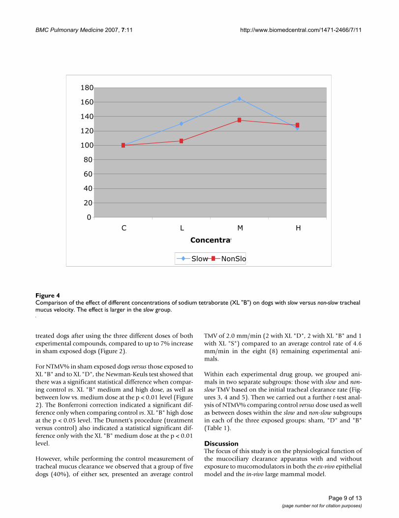
For NTMV% in sham exposed dogs versus those exposed toXL "B" and to XL "D", the Newman-Keuls test showed thatthere was a significant statistical difference when compar-ing control vs. XL "B" medium and high dose, as well asbetween low vs. medium dose at the p < 0.01 level (Figure2). The Bonferroni correction indicated a significant dif-ference only when comparing control vs. XL "B" high doseat the p < 0.05 level. The Dunnett's procedure (treatmentversus control) also indicated a statistical significant dif-ference only with the XL "B" medium dose at the p < 0.01level.
However, while performing the control measurement oftracheal mucus clearance we observed that a group of fivedogs (40%), of either sex, presented an average control
TMV of 2.0 mm/min (2 with XL "D", 2 with XL "B" and 1with XL "S") compared to an average control rate of 4.6mm/min in the eight (8) remaining experimental ani-mals.
Within each experimental drug group, we grouped ani-mals in two separate subgroups: those with slow and non-slow TMV based on the initial tracheal clearance rate (Fig-ures 3, 4 and 5). Then we carried out a further t-test anal-ysis of NTMV% comparing control versus dose used as wellas between doses within the slow and non-slow subgroupsin each of the three exposed groups: sham, "D" and "B"(Table 1).
DiscussionThe focus of this study is on the physiological function ofthe mucociliary clearance apparatus with and withoutexposure to mucomodulators in both the ex-vivo epithelialmodel and the in-vivo large mammal model.
Comparison of the effect of different concentrations of sodium tetraborate (XL "B") on dogs with slow versus non-slow tracheal mucus velocityFigure 4Comparison of the effect of different concentrations of sodium tetraborate (XL "B") on dogs with slow versus non-slow tracheal mucus velocity. The effect is larger in the slow group.
0
20
40
60
80
100
120
140
160
180
C L M H
Concentrat
Slow NonSlo
Page 9 of 13(page number not for citation purposes)

BMC Pulmonary Medicine 2007, 7:11 http://www.biomedcentral.com/1471-2466/7/11
Major findings in this study include: a) The two epithelialmodels used in this study, the ex-vivo frog palate and thein-vivo mammalian model, appear to be appropriate andcomplement each other to study the effects that ourmucomodulators exert on the mucociliary clearancedefence mechanism; b) the mucin macromolecules of theherein used in-vivo mammalian model (dog) showedselective crosslinking behavior after exposure to muco-modulators as it was previously shown in mucus simu-lants: increased elasticity, expressed by increased threadformation capacity and is more easily clearable fromintact airways; c) the physiological function of the muco-ciliary apparatus was not negatively affected in either ofthe two epithelial models; d) modulated airway mucus isbetter cleared from an intact airway system and normallyfunctioning respiratory system after exposure to muco-modulators; e) data presented in this study allow us toassure that we have complied with the fundamentalrequirement criteria established in the initial phase of
developing the concept of mucomodulation: Can wemodulate the physical characteristics of the respiratorysecretions to reduce aerosolization without affecting nor-mal mucociliary clearance function, or even betterimproving it? [9].
In this study we combined the use of two very different cil-iated epithelial models to have a better assessment of theeffect that crosslinking agents have on clearance of mucusby interaction with cilia. Several observers had raisedquestions previously about the adequacy of the frog palateas a model to assess airway mucus clearance, since in thefirst place it belongs to a non-mammalian species, inaddition that it is non-respiratory. However, this ciliatedepithelium has a well developed mucus blanket thatworks in coordination with cilia similar to that observedin other mammals including humans. This epithelialmodel also permits us to assess mucus clearance by ciliainteraction only without the airflow factor [15].
Comparison of the effect of different concentrations of high molecular weight dextran (XL "D") on dogs with slow versus non-slow tracheal mucus velocityFigure 5Comparison of the effect of different concentrations of high molecular weight dextran (XL "D") on dogs with slow versus non-slow tracheal mucus velocity. The effect is larger in the slow group.
0
50
100
150
200
250
C L M H
Concentrat
Slow NonSlo
Page 10 of 13(page number not for citation purposes)
BMC Pulmonary Medicine 2007, 7:11 http://www.biomedcentral.com/1471-2466/7/11
The in-vivo mammalian model, using anesthetized dogs,allowed us to assess the effects that our mucomodulatorsexert on the mucociliary clearance defence mechanism inan intact airway system and normally functioning respira-tory system. The intact airway system in our dog modelpermitted us to also assess the potential contribution ofother factors in clearing a modulated mucus, particularlyairflow during normal spontaneous breathing.
Data obtained from the dogs showed that inhalation ofthree different doses of mucomodulators did notadversely affect the cardio-respiratory pattern even afterthe different procedures involved and several hours thatlasted every experiment. The data support the observationthat cilia were not functionally affected after exposure tomucomodulators.
After aerosolizing the mucomodulators and while collect-ing the samples of mucus, we observed an increase inthread formation capacity of the mucus even when usinglow concentration doses. Since we did not have aFilancemeter on site, we anchored the mucus in a fang ofthe dog and used a wooden pick or a needle to pull themucus and observed it stretching and forming longthreads before rupture. With no mucomodulators, themucus threads ruptured after stretching up to 5 to 8 mm,while it ruptured after stretching around 30 to 50 mm fol-lowing exposure to mucomodulators.
During and after aerosolizing the mucomodulators andwhile collecting the samples of mucus, we examined theairways of the experimental dogs, as far down in the air-way tree as the bronchoscope allowed access, to visuallyassess bronchial patency and possible obstruction of theairways caused by the modulated mucus. We observed theairways in all experimental animals free of obstructionthroughout the study period. All dogs, either exposed toplacebo or mucomodulators, maintained a normal respi-ratory rhythm and frequency throughout the experimentthat might suggest they were free of respiratory distress.However more studies on this topic are required to arriveto a more definite conclusion.
After showing in the mammalian model that instead ofdeterioration of the mucociliary clearance mechanismthere was a clear improvement in tracheal mucus clear-ance, we carried out an additional trial to assess the effectof those mucomodulators on cough and aerosol genera-tion pattern in the same dog model. After 20 minutes ofnebulization of capsaicin solution, the dog did not coughor sneeze at all. We aborted the experiment after that timeand after the researchers were constantly coughing. Westill cannot find the reasons why capsaicin did not makethe dog cough. Veterinarians consulted about this eventfound no answer either.
The modulated "thicker" mucus neither plugged the air-ways of the experimental (mammalian) subjects, as fardown in the airways tree as the bronchoscope allowed usto assess, or was the tracheal mucus clearance rate nega-tively affected. On the contrary and unexpectedly, trachealmucus velocity systematically increased even with thelowest dose of either mucomodulator administered.
The observations addressed in the previous paragraphmay help to respond to the greatest concern expressed bysome researchers and clinicians when they first learnabout mucomodulators, "that crosslinking the mucus layerin this way, will make mucus 'thicker' and might obstruct theairways in exposed subjects". We have started to build theevidence that the proposed drugs that increase the con-centration of crosslinking sites in the mucin glycoproteingel network do not gum up airway secretions, do not plugthe airways and have no adverse effect on the clearance ofairway mucus in a normally functioning respiratory sys-tem of a mammalian model. However, the mucomodula-tors we tested in this study might still have some effects inareas that were not visible or measurable through thebronchoscope, such as in smaller airways or alveolar tis-sue. More studies are required before reaching a definiteconclusion to rule out side-effects of the mucomodulatorson respiratory secretions of smaller airways and alveolartissue or on gas exchange.
Since mucus clearance in the airways is a function of theintimate interaction of the anatomophysiological bicellu-lar unit, ciliated cells and mucus producing cells, thisstudy has allowed us to determine that no functional dis-ruption of the normal clearance of mucus occur when weselectively alter the elastic property of mucus.
The ex-vivo frog palate model is an excellent model forassessing the effect of the mucomodulators on the mucus-cilia interaction undisturbed by internal or external fac-tors like airflow. The in-vivo mammalian model is anexcellent model for assessing in an integral manner theeffect of the mucomodulators on the mucus interactingwith cilia as well as with several other respiratory compo-nents like airflow during normal breathing.
Selective modulation of the elastic properties of mucus inour mammalian model indicates that the modified mac-romolecule of mucin is engineered to work in an exquisitemanner with the low speed airflow during normal breath-ing in a normally functioning respiratory system.
Several years ago we had the opportunity to analyze asample of tracheobronchial mucus of a patient diagnosedwith primary ciliary dyskinesia and the mucus showed tohave predominantly more elastic properties. Individualswith this genetic syndrome depend completely on airflow
Page 11 of 13(page number not for citation purposes)
BMC Pulmonary Medicine 2007, 7:11 http://www.biomedcentral.com/1471-2466/7/11
interaction for airway mucus clearance since they have atotally dysfunctional cilia apparatus, i.e. immotile cilia.This finding guided us in our approach to develop themucomodulators.
An additional mechanism to explain the enhanced mucusclearance observed in our mammalian model could bethat inhaled mucomodulators stimulate cells producingsurfactant factor. The possible presence of a surfactant fac-tor both within and between the sol and gel phase of themucus may play a role in facilitating mucus clearability orcough clearability, by allowing the mucus layer to slideeasily and more effectively on top of the cilia. This mayexplain the observation of mucus sliding in an easier man-ner from the endotracheal tube surface.
There may occur a combination of these hypotheses, aswell as other possible new mechanisms, such as theincreased osmolarity composition of the aerosolized solu-tions enhancing airway secretions hydration. However,given the fact that tracheal mucus velocity peaked with theintermediate dose of XL "B" (equivalent of 1.0% NaCl),and not with the highest dose (equivalent of 1.8% NaCl),the contribution of hyperosmolarity is not likely themajor explanation for the stimulation of TMV. We antici-pate that further work is needed to clearly define themechanisms involved in the enhanced clearance of airwaysecretions when exposed to our mucomodulators.
Both mucomodulators used in this study exhibited aneffect in modulating the mucus macromolecule of the air-ways of the dogs by increasing elasticity. We may rightlyassume from the latter that poorly soluble mucin complexor more cohesive mucus have been formed that will bemore resistant to break up during high speed airflow inter-action, hence expelling less particles as aerosol.
For centuries people around the world from different non-related cultures have been extensively searching plants,minerals and animal products to best improve functionsand defence mechanisms for pulmonary disorders involv-ing abnormal mucus and hypersecretion. In this study wehave successfully used a natural product: high molecularweight dextran, a bacterial biopolymer produced by theLeuconostoc mesenteroides that showed promising proper-ties to enhance airway hygiene and airborne infectiousdiseases.
Results from this study indicate that the critical measuresof success, namely to maintain airway clearance has beenproved in both ex-vivo as well as in-vivo epithelial models.The degree of improvement in tracheal mucociliary clear-ance using our mucomodulators is far better than thatachieved by using current approaches in the same animalmodel. Our mucomodulators can be considered as an
enhanced new generation of compounds able to improveairway hygiene and respiratory host defence.
Data in this study complement with our data previouslypublished to support the concept that our mucomodula-tors can achieve a dual goal of reducing aerosolizationwithout impairing lung clearance, since the two mostimportant clearance mechanisms of the respiratory sys-tem remain functional. Mucomodulators have noreported antitussive action hence no cough mechanism isaffected.
ConclusionWe have acquired preliminary evidence that mucomodu-lators that increase cohesive interactions between mucinmacromolecules make mucus more easily clearable, mostlikely by airflow-dependent mechanisms. Increasing thecohesiveness in this manner might help to reduce fine aer-osol formation during coughing or sneezing – a desirableadvantage in helping to control the spread of airborneinfections.
Mammalian subjects in this study showed a very differenttracheal mucus velocity from other studies using a similarmodel. We do not have an explanation for this matter.Both mucomodulators enhanced tracheal mucus velocityin all those exposed. However, tracheal mucus velocitywas best enhanced in those with slower mucus clearance.This finding might be of particular use for those individu-als who have impaired mucociliary clearance.
We anticipate other potential benefits that relate toenhancing mucus clearance, a major consideration in con-ditions of impaired airway secretion management i.e.patients suffering from COPD, spinal cord injury, neu-romuscular disease or those in intensive care units and/orthose who require assisted mechanical ventilation.
These findings might assist us in enhancing the knowl-edge of the changes in the mucociliary apparatus thatoccur after exposure to mucomodulator products, withthe goal to understand how these changes relate to animproved and innovative management of chronic airwaypathologies in humans.
Competing interestsThe authors declare that they have no competing interests.
Authors' contributionsMK and GZ developed the concept of Mucomodulation,designed the study, interpreted the data and drafted themanuscript.
MR, a Virologist graduated from Karolinska University,Sweden, has contributed with valuable intellectual con-
Page 12 of 13(page number not for citation purposes)

BMC Pulmonary Medicine 2007, 7:11 http://www.biomedcentral.com/1471-2466/7/11
Publish with BioMed Central and every scientist can read your work free of charge
"BioMed Central will be the most significant development for disseminating the results of biomedical research in our lifetime."
Sir Paul Nurse, Cancer Research UK
Your research papers will be:
available free of charge to the entire biomedical community
peer reviewed and published immediately upon acceptance
cited in PubMed and archived on PubMed Central
yours — you keep the copyright
Submit your manuscript here:http://www.biomedcentral.com/info/publishing_adv.asp
BioMedcentral
cepts in his field of expertise for GZ to strengthen thestudy. MR was involved in the data acquisition during allthe experiments and played a key role to supervise andguarantee the quality of data acquisition.
JCV (Resident Year 1), Mauricio Alonso (Resident Year 2),HA (Resident Year 3) and DV (Chief of Residents) allenrolled in the Neumology (Respiratory Medicine) Resi-dency Program, in the Hospital Nacional "Dr. Jose Anto-nio Saldaña" Neumologia y Medicina Familiar. They allwere trained and supervised by GZ in the research meth-odology and procedures in large mammals (dogs), includ-ing animal preparation, airway intubation,bronchoscopic procedures, measurements of TMV, mucuscollection, mucomodulator administration and openchest surgery for tracheobronchial surgical resection forpathological studies. They all performed all these proce-dures.
GB, a Registered Nurse, organized and prepared theresearch site, catheterized front leg vein of the dog andadministered anesthesia as required to keep the animallightly anesthetized, set up and procured research sup-plies, and assisted researchers to maintain control of timeand to run the experiments smoothly. Due to the variousresearch procedures and time requirements, her contribu-tions were very valuable in successfully running the exper-iments.
All authors have read and approved the final manuscript.
AcknowledgementsThis project was funded in part by the Canadian Cystic Fibrosis Foundation and the Canadian Institutes of Health Research.
The authors gratefully acknowledge Dr. Jorge Edwin Montoya, Director of the Hospital Nacional Dr. Jose Antonio Saldaña for making the facilities availa-ble to carry out this study, and for allowing the time required for the Res-piratory Medicine Program's Residents to participate.
The authors also acknowledge the contributions and assistance provided by the Animal Control Program, Public Health Services, and Material Manage-ment personnel of the Hospital Nacional Dr. Jose Antonio Saldaña.
References1. Silberberg A, Meyer FA: Structure and function of mucus. In
Mucus in Health and Disease II Edited by: Chantler EN, Elder JB, EbsteinM. Plenum Press. New York and London; 1982:53-74.
2. Basbaum CB, Finkbeiner WE: Mucus-producing cells of the air-ways. In Lung Cell Biology Edited by: Massaro D. New York, Basel:Marcel Dekker; 1989:37-79.
3. King M, Rubin BK: Rheology of airway mucus: Relationship withclearance function. In Airway Secretion: Physiological Bases for theControl of Mucus Hypersecretion (Lung Biology in Health and DiseaseSeries) Volume Chapter 7. Edited by: Takishima T, Shimura S. NewYork: Marcel Dekker; 1994:283-314.
4. Zayas JG, Rubin BK, York E, Lien D, King M: Bronchial mucusproperties in lung cancer. Relationship with site of lesion.Can Respir J 1999, 6:246-252.
5. Lopez-Vidriero MT: Biochemical basis of physical properties ofrespiratory tract secretions. Eur J Respir Dis 1987, 71:130-135.
6. King M, Rubin BK: Pharmacological approaches to discoveryand development of new mucolytic agents. Advanced DrugDelivery Reviews 2002, 54:1475-1490.
7. King M: Magnetic microrheometer. In Methods in Bronchial Mucol-ogy Edited by: Braga PC, Allegra L. New York: Raven Press;1988:73-83.
8. King M: Role of mucus viscoelasticity in cough clearance. Bior-heology 1987, 24:589-597.
9. Zayas G, Dimitry J, Zayas A, O'Brien D, King M: A new paradigmin respiratory hygiene: increasing the cohesivity of airwaysecretions to improve cough interaction and reduce aerosoldispersion. BMC Pulm Med 2005, 5:11.
10. King M, Festa E: The evolution of the frog palate model formucociliary clearance. In Cilia, Mucus and Mucociliary InteractionsEdited by: Baum G. New York: Marcel Dekker; 1998:191-201.
11. Rubin BK, Ramirez O, King M: Mucus-depleted frog palate as amodel for the study of mucociliary clearance. J Appl Physiol1990, 69:424-429.
12. Jeanneret-Grosjean A, King M, Michoud MC, Lioté H, Amyot R: Sam-pling technique and rheology of human bronchial mucus. AmRev Respir Dis 1988, 137:707-710.
13. Rubin BK, Ramirez O, Zayas JG, Finegan B, King M: Collection andanalysis of respiratory mucus from individuals without lungdisease. Am Rev Respir Dis 1990, 141:1040-1043.
14. King M, Zahm JM, Pierrot D, Vaquez-Girod S, Puchelle E: The roleof mucus gel viscosity, spinnability, and adhesive propertiesin clearance by simulated cough. Biorheology 1989, 26:737-745.
15. Zayas G, O'Brien DW, Tai S, Ding J, Lim L, King M: Adaptation ofan Amphibian Mucociliary Clearance Model to EvaluateEarly Effects of Tobacco Smoke Exposure. Respir Res 2004, 5:9.(Epub 20 Aug 04)
Pre-publication historyThe pre-publication history for this paper can be accessedhere:
http://www.biomedcentral.com/1471-2466/7/11/prepub
Page 13 of 13(page number not for citation purposes)
http://www.ncbi.nlm.nih.gov/entrez/query.fcgi?cmd=Retrieve&db=PubMed&dopt=Abstract&list_uids=3622663
http://www.ncbi.nlm.nih.gov/entrez/query.fcgi?cmd=Retrieve&db=PubMed&dopt=Abstract&list_uids=3622663
http://www.ncbi.nlm.nih.gov/entrez/query.fcgi?cmd=Retrieve&db=PubMed&dopt=Abstract&list_uids=3502760
http://www.ncbi.nlm.nih.gov/entrez/query.fcgi?cmd=Retrieve&db=PubMed&dopt=Abstract&list_uids=2228850
http://www.ncbi.nlm.nih.gov/entrez/query.fcgi?cmd=Retrieve&db=PubMed&dopt=Abstract&list_uids=2228850
http://www.ncbi.nlm.nih.gov/entrez/query.fcgi?cmd=Retrieve&db=PubMed&dopt=Abstract&list_uids=3345048
http://www.ncbi.nlm.nih.gov/entrez/query.fcgi?cmd=Retrieve&db=PubMed&dopt=Abstract&list_uids=3345048
http://www.ncbi.nlm.nih.gov/entrez/query.fcgi?cmd=Retrieve&db=PubMed&dopt=Abstract&list_uids=2327638
http://www.ncbi.nlm.nih.gov/entrez/query.fcgi?cmd=Retrieve&db=PubMed&dopt=Abstract&list_uids=2327638
http://www.ncbi.nlm.nih.gov/entrez/query.fcgi?cmd=Retrieve&db=PubMed&dopt=Abstract&list_uids=2327638
http://www.ncbi.nlm.nih.gov/entrez/query.fcgi?cmd=Retrieve&db=PubMed&dopt=Abstract&list_uids=2611367
http://www.ncbi.nlm.nih.gov/entrez/query.fcgi?cmd=Retrieve&db=PubMed&dopt=Abstract&list_uids=2611367