A new look at Kenyapithecus based on recent discoveries in Western Kenya
31
Martin Pickford National Museum of Kenya, P.O. Box 40658, Nairobi, Kenya Received 4 November 1983 and accepted 23 February 1984 Keywords: Kenyapithecus, sexual dimorphism, history of study, new fossils, palaeoecology, phylogenetic relationships, serological divergence dates. A New Look at Kenyapithecus based on Recent Discoveries in Western Kenya A collection of fossil higher primate teeth from Maboko, Kenya, made in 1982 and 1983 has led to this re-assessment of the status of Kenyapithecus. In particular, it is concluded that some individuals of both Kenyapithecus africanus, and K. wickeri probably possessed large canines and that both had large third molars. This finding has a bearing on how one views the relationships of Kenyapithecus to Sivapithecines (Sivapithecus and Ramapithecus) and to the Hominidae. The re-assignment to K. wickeri of all teeth from Fort Ternan formerly identified as P. nyanzae, affects palaeoecological conclusions published recently. In addition, it is shown that none of the teeth collected at Maboko has close affinities to Proconsul, and it is considered unlikely that this taxon occurs there. On this basis, it is suggested that the Maboko postcranial bones, traditionally assigned to Proconsulnyanzae, may possibly belong to Kenyapithecusafrivanus and thereby acquire a fresh significance. Detailed study of the maxillae of K. africanus and K. wickeri reveals a significant number of morphological differences between the two taxa, which suggests that it may eventually be necessary to separate them at the generic level. Two size groups of dental remains assignable to Kenyapithecus occur in tile Maboko Formation. A mandible in the group of smaller specimens has a canine of female morphology, which makes it more likely that the two groups represent males and females of a single sexually dimorphic taxon, K. africunus. The total number of specimens assigned to Kenyapithecus is increased from nine to seventy-two. 1. Introduction During the past two decades a great deal has been written about the hominid status of Ramapithecus (Simons, 1963, Simons & Pilbeam, 1978), but this hypothesis now seems unlikely. In the last three or four years a growing body of data has become available which shows that Ramapithecus is rather closely related to Pongo, sharing a number ofapomorphic features with it (Andrews & Cronin, 1982; Ward & Pilbeam, 1983; Ward & Kimbel, 1983). At the same time it is becoming increasingly difficult to retain the view that the Great Apes form a natural genetic grouping traditionally known as Pongidae, a family which has until recently contained Pongo, Pan and Gorilla. Current views summarized by Ciochon (1983) are that this grouping is paraphyletic and at best represents only a grade of evolution of higher primates. Several classifications have been suggested in which Pan and Gorilla are regarded as the sister group of Homo and Australopithecus, either at the family or subfamily level. Thus Pan and Gorilla are put into Paninae or Panidae for example by Gantt (1983) and Ciochon (1983), while Homo and Auslralopithecus are classified either as Homininae or as Hominidae. Whatever the case, Ramapithecus is excluded from it, being better placed in Pongidae sensu stricto. A third alternative, which I have difficulty in acceplln~ is that Hominidae comprises the subfamilies Ponginae and Homininae (Goodman et al., 1983), but here the argument is essentially one of taxonomic rank rather than of concept. With the rejection of Ramapithecus from the Hominidae, an obvious question to consider is whether Kenyapithecus, which has frequently been synonymized with Ramapithecus should also be rejected from it; is it really a pongid sensu stricto or not?; may it not represent an ape Journal of Human Evolution (1985) 14, 113 143 0047-2484/85/020113+31 $03.00/0 (~)1985 Academic Press Inc. (l,ondon) Limited
Transcript of A new look at Kenyapithecus based on recent discoveries in Western Kenya

Martin Pickford
National Museum of Kenya, P.O. Box 40658, Nairobi, Kenya
Received 4 November 1983 and accepted 23 February 1984
Keywords: Kenyapithecus, sexual dimorphism, history of study, new fossils, palaeoecology, phylogenetic relationships, serological divergence dates.
A N e w Look at Kenyapithecus based on Recent Discoveries in Western Kenya
A collection of fossil higher primate teeth from Maboko, Kenya, made in 1982 and 1983 has led to this re-assessment of the status of Kenyapithecus. In particular, it is concluded that some individuals of both Kenyapithecus africanus, and K. wickeri probably possessed large canines and that both had large third molars. This finding has a bearing on how one views the relationships of Kenyapithecus to Sivapithecines (Sivapithecus and Ramapithecus) and to the Hominidae. The re-assignment to K. wickeri of all teeth from Fort Ternan formerly identified as P. nyanzae, affects palaeoecological conclusions published recently. In addition, it is shown that none of the teeth collected at Maboko has close affinities to Proconsul, and it is considered unlikely that this taxon occurs there. On this basis, it is suggested that the Maboko postcranial bones, traditionally assigned to Proconsul nyanzae, may possibly belong to Kenyapithecus afrivanus and thereby acquire a fresh significance. Detailed study of the maxillae of K. africanus and K. wickeri reveals a significant number of morphological differences between the two taxa, which suggests that it may eventually be necessary to separate them at the generic level.
Two size groups of dental remains assignable to Kenyapithecus occur in tile Maboko Formation. A mandible in the group of smaller specimens has a canine of female morphology, which makes it more likely that the two groups represent males and females of a single sexually dimorphic taxon, K. africunus. The total number of specimens assigned to Kenyapithecus is increased from nine to seventy-two.
1. I n t r o d u c t i o n
During the past two decades a great deal has been written about the hominid status of Ramapithecus (Simons, 1963, Simons & Pilbeam, 1978), but this hypothesis now seems unlikely. In the last three or four years a growing body of data has become available which shows that Ramapithecus is rather closely related to Pongo, sharing a number ofapomorphic features with it (Andrews & Cronin, 1982; Ward & Pilbeam, 1983; Ward & Kimbel, 1983). At the same time it is becoming increasingly difficult to retain the view that the Great Apes form a natural genetic grouping traditionally known as Pongidae, a family which has until recently contained Pongo, Pan and Gorilla. Current views summarized by Ciochon (1983) are that this grouping is paraphyletic and at best represents only a grade of evolution of higher primates. Several classifications have been suggested in which Pan and Gorilla are regarded as the sister group of Homo and Australopithecus, either at the family or subfamily level. Thus Pan and Gorilla are put into Paninae or Panidae for example by Gantt (1983) and Ciochon (1983), while Homo and Auslralopithecus are classified either as Homininae or as Hominidae. Whatever the case, Ramapithecus is excluded from it, being better placed in Pongidae sensu stricto. A third alternative, which I have difficulty in acceplln~ is that Hominidae comprises the subfamilies Ponginae and Homininae (Goodman et al., 1983), but here the argument is essentially one of taxonomic rank rather than of concept.
With the rejection of Ramapithecus from the Hominidae, an obvious question to consider is whether Kenyapithecus, which has frequently been synonymized with Ramapithecus should also be rejected from it; is it really a pongid sensu stricto or not?; may it not represent an ape
Journal of Human Evolution (1985) 14, 113 143
0047-2484/85/020113+31 $03.00/0 (~)1985 Academic Press Inc. (l,ondon) Limited
114 M. PICKFORD
close to or on the hominid (sensu stricto) line? In other words, what are the precise relationships of Kenyapithecus and Ramapithecus?
Until recently the published hypodigms of two species of Kenyapithecus totalled nine specimens. I t would have been diffficult to make a reassessment on the basis of this limited data base, but new collections from western Kenya have dramatically altered the amount of information available for study. Much of the Maboko material was collected during the somewhat mundane exercise of screening an old excavation dump. It has permitted a flesh appraisal of several isolated teeth collected many years ago, the generic attribution of which has not been settled before. In particular it allows a more precise but still incomplete, examination of the various characters, such as small canines, which were taken to be apomorphic features shared with Homo (Leakey, t962, 1967).
The aims of this paper are severalfotd. The first is to examine the history of study of Kenyan fossils variously assigned to Kenyapithecus, Sivapithecus and Ramapithecus. So complex has the detailed history of each of the specimens and indeed the species become that it is necessary to approach this section on a specimen by specimen basis. The student of fossils needs to study the detailed history of each specimen if he is to grasp the overall intentions of the authors at the time of the various studies. Without a detailed knowledge of what each author said about each specimen, many of the higher echelon inferences, statements and interpretations become difficult to compare. For example, in the long and sometimes heated debate concerning the generic assignment of the species wickeri from Fort Ternan (Leakey, 1962, 1967, 1968 ; Simons, 1963, 1968, 1969; Simons & Pilbeam, 1965, 1983; Andrews, 1971; Andrews & Walker, 1976) it is essential to know what specimens were included in the hypodigms at various stages in the debate. An important instance concerns the mandible K N M FT 45. Leakey (1968) and Simons (1969) generally referred this mandible to Pongidae, excluding it from the species wickeri (but see Simons & Pilbeam, 1978) whereas Andrews ( 1971) and Andrews & Walker (1976) included it in R. wickeri and furthermore based facial and dental arcade reconstructions on it. Consequently Leakey's views ofK. wickeri were founded on quite a different material basis from those of Andrews & Walker. Furthermore the views presented here take into account molars and canines deliberately excluded by all the above authors. The overall content and therefore the concept of K. wickeri has as a result changed dramatically over the years. By omitting the detailed history of each specimen, and concentrating only on morphological descriptions of species, as has become the custom of many palaeontologists, one is in danger of losing sight of the entire fabric of the debate as it relates to specific, generic and higher ranks.
Some of the historical notes may seem absurd, especially if taken out of context, but the aim here is not to make the authors of those notes appear ludicrous. The value to science of such specimen histories lies in the fact that the serious student of palaeontology needs to know at the most basic level, what was said about each specimen, because at a higher level what was said about the species or genus was presumably based on the individual specimens believed to belong to the taxon under discussion.
The second aim of this paper is to record new material considered to belong to Kenyapithecus. The approach is perhaps rather old-fashioned in the sense that each specimen is described individually. The dangers of issuing only an overall species morphological description, an approach which certain authors seem to prefer, are that should any specimens be removed from the hypodigm at a later stage, then the morphological descriptions will no longer be entirely valid whereas the basic specimen

A N E W L O O K A T K E N Y A P I T H E C U S 115
description will still be valid, even if extended by future study. Once the individual specimens have been described an overall morphological description can easily be obtained.
Further aims of this contribution are (a) to examine various aspects of Kenyapithecus as it relates to other closely related taxa, especially proconsulines and Eurasian ramapithecines, (b) to examine whether Kenyapithecus may have been sexually dimorphic or not (c) to determine the relationships between the two named species ofKenyapithecus, K. wickeri and K. africanus (d) to examine the phylogenetic position of Kenyapithecus and (e) to discuss the palaeoecology of the sites which yielded the samples of Kenyapithecus. Finally, it is necessary to examine closely the reported occurrence of Kenyapithecus at Rusinga, since such records, if valid, would represent the earliest known evidence for "ramapithccines" in the world.
Much as I would like to include descriptions and discussions of new middle Miocene hominoid fossils discovered at Nachola, near Baragoi (Ishida et. al., 1984) and at Buluk, in Kenya, I am not able to do so until basic descriptions have been issued by the discoverers. The Nachola material, which consists of mandibles, maxilla fragments and isolated teeth, accords well with fossils from Maboko, with a few minor differences in molar cusp morphology. Most of the Buluk material seems to be rather different from Maboko and Fort Ternan fossils, and need not particularly concern us here.
2. Historical Perspective
It is just over twenty years since Leakey (1962) proposed the new genus and species Kenyapithecus wickeri, on the basis of four specimens from Fort Ternan assigned by him to a single individual. Since Leakey's original description, three further specimens have been added to the hypodigm of K. wickeri; an upper incisor (Leakey, 1967 ), which was later rejected by Andrews & Walker (1976); a mandible fragment with P3-4 (Andrews, 1971), earlier excluded by Leakey (1968 ) and Simons (1969) on the grounds that it was a typical Dryopithecus jaw; and an isolated lower canine (Andrews & Walker, 1976) (the last specimen now considered to have been collected at Songhor and to be a deciduous canine of Proconsul major).
In 1967 and 1968, Leakey transferred a number of specimens from Sivapithecus africanus and Proconsul nyanzae into a new combination, Kenyapithecus africanus (Le Gros Clark & Leakey). After much discussion, notably by Pilbeam (1969), most of the hypodigm of K. africanus reverted to Proconsul nyanzae (at that time often referred to Dryopithecus nyanzae) where it is evidently properly placed. The hypodigm of K. africanus as recently as 1982 consisted of the holotype maxilla, a lower P3 fragment (Andrews & Motleson, 1979) and an upper M l (Leakey, 1967 ) all from Maboko Island. Pickford (1982) added a few specimens from Majiwa.
The sum total of fossils assigned to two species of Kenyapithecus until 1982 was a mere nine specimens, not even representing every tooth in the dental battery. It is somewhat surprising that this tiny palaeontologic sample has generated so much interest in scientific circles. In the two decades subsequent to the diagnosis of Kenyapithecus, over fifty scientific papers have been published on interpretations or discussions of the significance of these few specimens; papers which need to be taken into account in a revision such as this. Many more casual mentions have been made in text-books, reviews and news articles.
In July 1982 and July-August 1983, the author was collecting fossils at Maboko Island by screening the Late Archdeacon Owen's 1933 excavation dump (Owen, 1939) and that
116 M. PICKFORD
ofL. S. R. Leakey Mede nearby in 1949. The aim of this exercise was to obtain pieces of the partial skeleton of the Maboko anthropoid which Owen sold to the British Museum of Natural History in 1936 for the sum ofs Examination of this skeleton (Morbeck, 1983), which consists of a humeral shaft, two femoral shafts (one with the proximal end complete), a tibia shaft and a clavicle, reveals the presence of several fresh breaks suggesting that the specimens may have been broken up during excavation and some pieces missed by Owen's collectors. The incomplete screening programme has yielded a few additional fragments of this skeleton including the proximal end of an ulna, and an appreciable quantity of teeth assignable to K. africanus. Comparison of these teeth with those of Proconsul species, K. africanus and K. wickeri reveals that several teeth from Fort Ternan, Maboko, and possibly Rusinga, hitherto identified as Proconsul nyanzae and Rangwapithecus gordoni may in all probability belong to Kenyapithecus. Furthermore, once these specimens are assigned to Kenyapithecus the presence of Proconsul at Fort Ternan and Maboko becomes very much less certain, being based on two atypical teeth from Fort Ternan and none at Maboko. As a consequence, it becomes less likely that the large hominoid postcranial elements from Fort Ternan and Maboko belong to Proconsul, and more likely that they belong to Kenyapithecus or to some other large hominoid.
3. Hypodigm of Kenyapithecus africanus New and previously discovered material from the Maboko Formation (Maboko, Majiwa, and Kaloma) (see Tables 1 and 2).
Maxilla M 16649 (CMH6) is the holotype maxilla with left P3-MJ, the roots of M 2 and part of the atveotus of a large upper canine, This specimen has had a long and somewhat complex history and has at one time or another featured in the hypodigms of three genera. It was first assigned to Proconsul africanus by MacInnes (1943, pl, 24, fig.2). In 1950 it was selected as the holotype ofSivapithecus africanus by Le Gros Clark & Leakey (1951; pl. 6, figs 41-43). In 1965, Simons & Pilbeam synonymized S. africanus with S. sivalensis, and two years later Leakey (1967b) placed it in the genus Kenyapithecus. Le Gros Clark & Leakey ( 1951 ) gave its provenance as locality R106 on Rusinga Island, a feature which no doubt influenced Leakey in his subsequent selection of mandibles from R106 on Rusinga as being examples of Kenyapithecus (see Leakey, 1968). In 1978, Andrews identified this maxilla as Proconsul nyanzae, a point of view he did not keep for long (Andrews et al., 1979). Madden (1980) and Greenfield (1979) accepted that the specimen represented Sivapithecus africanus. The maxilla is rather primitive, differing in several respects from that of K. wickeri, but more closely resembling Proconsul. The preserved part of the zygomatic process in particular is low, has minimal flare and probably has a long sloping anterior root. The palate is flat and shallow, and the maxillary sinuses penetrate deeply towards the molar roots.
Upper Central Incisors KNM MB 104 (CMH 11) [Figure l(a)] is a left 11 with medium apical wear, which has dentine exposed along the cutting edge. It has a large interproximal facet for the opposite 11 and a smaller facet for 12. The lingual enamel is coarsely crenulated and it possesses a large lingual cingulum. Distally, on the buccal edge of the 1 ~ interstitial facet, there is a small raised fold of enamel which looks as though it may have been a mechanical brace for 12 or perhaps an eruptive guide.

A N E W L O O K A T KENYAPITHECUS 1 l 7
K N M MJ 9734 (Maj'82) [Figure 2(b)] is an isolated left 11 with the distal margin broken away, which is lightly worn apically. It is very similar in its preserved parts to K N M MB 104; the lingual enamel is coarsely crenulated, the mesial cingulum leads downwards and merges with the central tubercle which extends upwards centrally from the cingulum to two-thirds of the height of the crown.
K N M RU 1681 (CMH 9) is a right l J, said to have come from Rusinga (Le Gros Clark & Leakey, 1951, fig. 23), which was originally identified as P. nyanzae. Leakey (1967b,fig. la) later identified it as K. africanus, partly because he thought it came for locality R106 on Rusinga. Pilbeam (1969, p.119) wondered whether the provenience data was accurate, while Yulish (1970) accepted the specimen as Ken~apithecus and used it in his discussion about anterior tooth reduction in the genus. Andrews ( 1978, table 3) listed the specimen as P. nyanzae, but at that time he considered all K. africanus specimens to be indistinguishable from P. nyanzae. My own examination of the specimen leads me to agree in essence with Leakey's (1967b) view that it is more likely to represent Kenyapithecus than Proconsul, a view supported by J . Kelley (pers. comm.) who is engaged specifically on a study of anterior dentitions of Miocene higher primates.
K N M MB 11831 (MB 1381'83) is an unworn left I i missing most of the enamel, which appears to have been eaten away by acid. The root tip is broken but otherwise it is in reasonable condition. There is a central lingual pillar, from the base of which dentine folds curve apically towards both mesial and the distal edges of the crown. The occlusal margin is straight mesially, but curves rootward in the distal third of the crown.
K N M MB 11834 (MB 1218'83) is an upper central incisor germ fragment withoutroots. It is missing the mesial and distal margins but the central portion of the tooth is well preserved, and possesses a prominent central pillar and coarsely wrinkled enamel.
Upper Lateral Incisors K N M MB 17 (?Mb 33'49) is a moderately worn left 1 ~. The lingual pillar is deeply worn by contact with the lower canines. There is also tremendous wear on the distolingual surface of the root and cingulum, and an apical wear facet which is steeply inclined lingually. The lingual enamel is crenuIated and the lingual rib is large. The root is long.
K N M MB 9729 (Mb 203'82) [Figures l(b) and 2(a)]. This right 12 is well preserved and lightly worn at its apex. I t has a small facet caused by interstitial wear with 11 and a large facet due to occlusion with the lower canine. There are scratches on the distal surface of the root covering an area extending from cervix to a line 2-5 mm towards the root tip. The striae are normal to the long axis of the tooth and were presumably caused by fibrous food abrading the exposed root. I t has a prominent lingual cingulum.
Upper Canines K N M MB 70 [Figure l(c)] is a right upper canine described by Von Koenigswald (1969) as Victoriapithecus. More recently Pickford (1982) included it in Kenyapithecus. while Simons (1981) said it resembled Fort Ternan Ramapithecus. It has a large distal wear facet, a low crown and the root has shallow, open mesial and distal grooves.

118 M. PICKFORD
KNM MJ 4 (Maj 7'79) is a heavily worn right upper canine described by Pickford 1982).
Upper Fourth Premolars KNM MB 155 (Mb 556'73) [Figure 1 (d)] a left p4 with no roots is essentially a diminutive version of p4 in the holotype of K. africanus. There is no lingual cingulum. Ridges leading centrally from the paracone, define mesial, central and distal foveae.
KNM MB 9725 (Mb'82) is a rootless left p4 fragment with anterior and posterior edges missing. It is little worn, the enamel is coarsely crenulated and styles are clearly visible on the buccal surface. The preserved portions of this tooth are close in morphology to p4 in the holotype (M 16694, maxilla with P3-M1).
KNM RU 1732 (CMH 133) is a right p4 without roots. The specimen is so similar in morphology and preservation to the holotype (M16649) that its provenience data (as Rusinga) may be incorrect. Le Gros Clark & Leakey (1951) included it in their hypodigm of Proconsul nyanzae and its locality was given as Rusinga without more precise information. This specimen may represent another example of incorrect provenience data (see Andrews & Molleson, 1979). Pilbeam (1968) identified this tooth as S. africanus but Andrews (1978) identified it as P. nyanzae, as he also identified the holotype of Kenyapithecus africanus, an opinion he soon revised (Andrews & Molleson, 1979). The subequal rounded cusps, the light lingual cingulum on the rear half of the protocone, and the presence of two characterisitic buccal styles indicates that this p4 most probably belongs to K. africanus.
KNM MB 11817 (Mb 1126'83) [Figure l(e)], an unworn right upper fourth premolar, is similar to that in the holotype of K. africanus. It has a lingual cingular fold and moderate lingual flare. As in the holotype the cervix is deeply undercut anteroposteriorly, so that the roots are appreciably smaller than the crown.
Upper First Molars KNM MB 107 (CMH26), a right M l described and figured by Le Gros Clark & Leakey (1951, P1.6, fig.44) is in medium wear, has no roots and has been rolled and slightly polished. It possesses a small protocone cingulum, and there are mesial and distal interstitial facets.
KNM MB 9738 (Mb 88'82) [Figures l(h) and 2(g)] is a medium worn left M 1 with one root missing. A cingulum on the protocone is well developed but does not extend onto the hypocone. There is a small cingular fold of enamel at the base of the labial notch. Wear has progressed to the stage where enamel crenulations have been eradicated.
KNM MB 11821 + 11813 (Mb 891'83) [Figure l(f)] is a right M ~, worn flat with dentine exposed on all four main cusps and on the protoconule. It has well formed mesial and distal interstitial facets and appears to have no cingulum.
Upper Second Molars KNM MJ 8 (Maj 333'79) described by Pickford (1982) is a right M 2 with pitted and eroded enamel.

A N E W L O O K A T K E N Y A P I T H E C U S 119
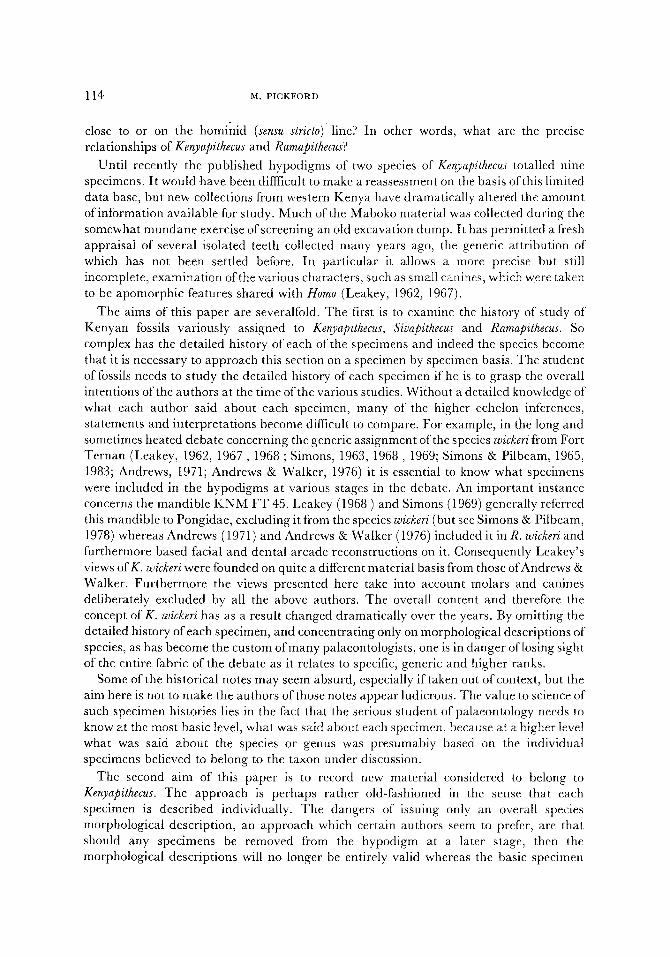
Figure 1. Upper dentition of Kenyapithecus africanus from Maboko, Kenya. (a) K N M MB 104, left 11. (b) KNM MB 9729, right 12. (c) KNM MB 70, right upper canine. (d) KNM MB 155, left p4. (e) KNM MB 11817, right p4 (f) K N M MB 11821 + 11813, right M I. (g) K N M MB 11819, right M 2. (h) K N M MB 9738, left ML (i) KNM RU 1692, right Ma. (j) KNM MB 9736, left dIZ. (k) KNM MB 11828, right dP 3. (1) KNM MB 9723, right dP 4. ( m ) K N M MB 11826, right dP ~.
(a)
(f)
(.i)
(b)
(g)
(k)
0 5
(c)
10
m m
(d) (e)
. i
i~
(h) ( i)
(I)
15
(m)

120 M. PICKFORD
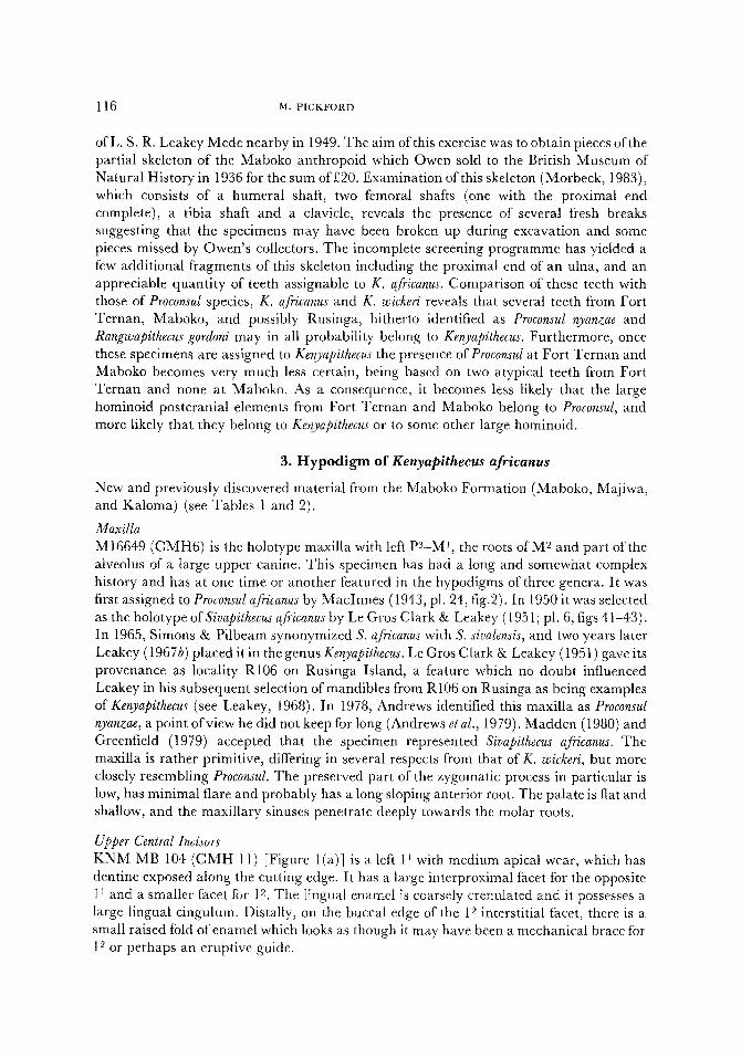
Figure 2. (a) KNM MB 9729, right upper lateral incisor, Kenyapithecus africanus. (b) KNM MJ 9734, left upper central incisor, Kenyapithecus africanus. (c) KNM MB 9723, right dP 4, Kenyapithecus @icanus? (d) KNM MB 9731, left M.% Kenyapithecus africanus. (e) KNM MB 9727, right M2, Kenyapithecus africanus. (t) KNM FT 3636, left lower canine, Kenyapithecus wickeri. (g) KNM MB 9738, left Mr, Kenyapithecus @icanus? (h) KNM MB 9726, right M:~, Kenyapithecus africanus. (i) KNM MB 9737, right P:~; KNM MB 9735, right P4 and KNM NC 9740, right M~ composite tooth row, Kenyapithecus africanus?
(a) (b) (c) (d) (e)
0 10 2O 30 m m
i i
~ :,;:~, ,, .....
(f) (g) (h) (i)
0 10 2 0 3 0 mm

A NEW LOOK AT KENYAPITHEC, US 121
Figure 3. Lower dentition of Kenyapithecus africanus from Maboko, Kenya. (a) KNM MB 11830, left I1. (b) KNM MB 15, left I2. (c) KNM MB 11820, right lower canine. (d) KNM MB 9737, right Ps. (e) KNM MB 9728, left P4. (f) KNM MB 9735, right P4- (g) KNM MB 11825, right-My (h) KNM MB 11818, right M1. (i) KNM MB 11812, left M~. (j) KNM'MB 9727, right M2. (k) KNM MB 11824, left M 2. (l) KNM MB 9731, left Ms- (m) KNM MB 11847, right lower dC. (n) KNM MB 11829, right dP3. (o) KNM MB 9722, left dP 4.
(e) (b) (c) (d) (e) (f)
(g) (h) (i) ( j ) (k) (I)
0 (m) (n)
0 5 10
mm
(o)
15

122 ta. PICKFORD
KNM MB 11819 (Mb 1379'83) [Figure 1 (g)] is an unworn, rootless right M 2 crown, which has been chemically etched. There is no lingual cingulum, although a cingulum is present on the anterior aspect of the protocone. The enamel is coarsely but not deeply wrinkled.
KNM MJ 1 (Maj 1'79) is an associated M 2-3 described by Pickford (1982).
KNM MB 9724 (Mb 230'82) is a right M 2 or M s fragment missing the anterior and buccal cusps. It has been moderately worn and the enamel is coarsely crenulated.
Upper Third Molars KNM RU 1692 (CMH 27). [Figure l(i)]. This rootless and slightly rolled right M s was originally said to have come from an unknown site in Kavirondo (Le Gros Clark & Leakey, 1951, pl.6, fig.45). Its Rusinga accession number therefore comes as a surprise as does its most recent assignment by Andrews (1978) to Rangwapithecus gordoni. 1 agree with Le Gros Clark & Leakey's original assessment that it belongs to Sivapithecus africanus, a view also held by Greenfield 1979). It has no cingulum, the enamel is thickened and the parastyle is distinct.
Upper Deciduous Incisor KNM MB 9736 (Mb 406'82) [Figure 1 (j)] is a deciduous upper left lateral incisor, slightly worn apically and disto-lingually. It has a strong lingual cingulum and a small lingual rib, and is basically a scaled down version of KNM MB 17 which is assigned to K. africanus.
Upper Deciduous Molars KNM MB 11828 (Mb 1018'83) [Figure l(k)], a right dP 3 missing the anterior root, is lightly worn at the cusp tips and along the anterior and distal crests. The buccal cusp has ridges leading into the sagittal valley which is not constricted. The lingual cusp is slightly lower than the buccal one and the buccal edge is appreciably longer than the lingual side. There are small anterior and posterior tubercles on the buccal cusp, and there is no sign of a lingual cingulum. The enamel is thin.
KNM MB 11827 (MB 873'83) is a worn, chemically etched left dP a missing the disto-buccal root. Although this specimen is not in very good condition it appears to be similar in morphology to K N M MB 11828,
KNM MB 9723 (MB 86'82) [Figures 1(1) and 2(c)] is a. lightly worn, rootless right dP 4 germ. The occlusal enamel is coarsely crenulated, and there is no lingual cingulum, although a cingular fold is visible on the protocone. Apart from increased lingual flare and an asymmetrical cervical outline this tooth looks like a miniature M ~ ofK. africanus.
KNM MB 11826 (Mb 1'83) [Figure l(m)] is a lightly worn right dP 4 in relatively good condition. The buccal roots are missing and the metacone tip is broken off. It has a prominent protocone cingulum which reaches back to the hypocone on the lingual side. The enamel is coarsely wrinkled and the crista obliqua is well developed. The hypocone is separated from the trigon by a deep but narrow valley, and the lingual root flares strongly and curves sagittally at its tip.

A N E W L O O K A T K E N Y A P I T H E C U S 123
Mandible K N M MJ 5 (Kal 1 + 2'79) is a crushed but relatively complete mandible described by Pickford (1982). The right lower canine which was recovered in 1983, is similar to canines K N M MB 5 and K N M MB 11820. The small size of the crown relative to root length suggests that this represents a female individual, as does the general crown morphology. This is an important find, because, not only is it the first Kenyan ramapithecine canine to occur in situ in a mandible, but also because its female morphology lends support to the hypothesis that the group of small ramapithecine remains from the Maboko Formation may represent females of K. africanus rather than a separate species.
Lower Central Incisors K N M MJ 6 is a right lower central incisor associated with K N M MJ 5, a mandible described by Pickford (1982).
K N M MB 11833 (Mb 409'83) is an Ii missing the tips of the crown and root. I t has been chemically etched, but its main morphological features can be discerned. It has a central lingual rib, as in K N M MJ 6.
K N M MB 11830 (Mb 755'83) [Figure 3 (a)] is an unworn left I1 in which the root tip is still open. It has a central lingual rib and moderately strong mesial and distal lingual ridges.
Lower Lateral Incisors K N M MB 15 (Mb 57'49) [Figure 3(b)], a worn left 12 with etched enamel is rather similar in its preserved parts to the 12 o f K N M MJ 5. the central rib is low and weakly developed. The root tip is distorted and curved distally. The lingual edge is bordered by low mesial and distal cingula.
Lower Canines K N M MB 5 is a right lower canine with a low crown and mcsio-distally compressed roots. The root and crown are in line, unlike the slightly canted crown of otherwise similar Proconsul africanus canines. The distal wear facet has an abrupt stop at root level which would produce apical wear in the upper canine. The mesio-lingual ridge is small but clearly defined.
K N M MB 11820 (Mb 1380'83) [Figure 2(c)] a right lower canine, slightly worn apically and distally is a little damaged by chemical etching. It has a low crown and long root as in K N M MB 5. The crown is slightly canted buccally with respect to the root. The lingual cingulum is present only on the anterior half of the lingual edge. The distal wear facet reaches onto the root where a slight stop facet has been produced.
Lower Third Premolar M 36370 (KBA) is a portion of a left P3 assigned to Sivapithecus africanus by Andrews & Molleson (1979).
K N M MB 9737 (Mb 482'82) [Figures 2(i) and 3(d)] is an unworn right lower P~ with a tiny chip of enamel missing from the buccal part of the apex of the main cusp. I t has no roots. It is unicuspidate, but the lingual ridge has an incipient cusplet developed near its

124 M. PICKFORD
base, and the enamel is finely crenulated. This specimen is compatible in size and morphology with the damaged P3S in the mandible of KNM MJ 5 (Pickford, 1982).
Lower Fourth Premolars KNM MB 9728 (Mb 177'82) [Figure 3(e)[ is a lightly worn rootless left 1~4 with an enamel flake missing from the mesiolingual corner. Dentine is exposed on the buccal cusp and there are clear wear facets around the anterior fovea caused by occlusion with P3. There is also a subsidiary wear facet on the buccal surface of the tooth which dips outwards. The talonid basin is large and there are distinct styles on the buccal surface. This tooth is closely similar to P4 o f K N M FT 45 (Andrews, 1971).
KNM MB 9735 (Mb 89'82) [Figures 2(i) and 3(t)] a rootless right P4 in moderate wear, has an enamel chip missing mesially near the cervix. It is otherwise complete and in good condition. It has clear mesial and distal interstitial facets and stylar folds are visible on the buccal surface. The anterior fovea is clearly defined and the distal fovea is large. Enamel crenulations are subdued. It is compatible in size and morphology with the P4 of the Kaloma Jaw (KNM MJ 5).
KNM MJ 2 (Maj 2'79) is rolled right P4, described by Pickford (1982).
Lower First Molars KNM NC 9740 (KGS 78'81) Figure 2(i) is a rootless but unworn right Mr. It has coarsely crenulated enamel, no cingulum and "puffy" main cusps. It is closest in general morphology, but is smaller than, KNM FT 48. It was collected from site Kaimogool South in the Nyakach Formation, Western Kenya.
KNM MB 11825 (Mb 404'83) [Figure 3(g)] is another unerupted right MI without roots. It has a buccal cingular fold and moderately coarsely wrinkled enamel.
KNM MB 11823 (Mb 114'83) is the rear halfofa right M1 worn flat. Dentine is exposed on all preserved cusps.
KNM MB 11812 (Mb 898'83) [Figure 3(i)]. This left lower M1 in medium wear has dentine exposed on all three buccal cusps but not on the lingual ones. There is an anterior contact facet for P4- It has no cingulum. The lingual notch is bordered by crests which run into it from the tips of the lingual cusps.
KNM MB 11818 (Mb 890'83) [Figure 3(h)] is a right M1 germ without roots. There is a labial cingular remnant between the main cusps. The anterior ridge is well developed and the enamel moderately coarsely wrinkled.
Lower Second Molars KNM MB 9727 (Mb 793'82) [Figures 2 (e) and 3(j)] is a slightly worn, right M2 with enamel missing from buccal, distal and lingual walls. It has two roots. There is no cingulum on the buccal surface. As in KNM MB 9731 there are accessory cusplets distally between hypoconulid and entoconid, between entoconid and metaconid and at the centric

A N E W L O O K A T K E N Y A P I T H E C U S 125
end of the crista obliqua. The groove system of the tooth is well marked and the enamel is coarsely crenulated.
KNM MB 11816 (Mb 896'83) is a damaged right M2 missing the anterior half of the mesial cusps. There are flakes of enamel missing buccally and distally and the specimen is lightly worn. A labial cingulum is present but is small and not continuous. The enamel is coarsely but not deeply wrinkled. There is an accessory cusplet distal to the antero-ling_ual cusp.
KNM MB 11824 (Mb 2'83) [Figure 3(k)] is a left M2 worn flat and missing the roots and the anterior half of the crown. The broken section reveals a low relief dentine enamel junction.
Lower Third Molars KNM MB 108 (CMH 129) is a left M3 cited by Le Gros Clark & Leakey (1951) as C M H 29. It is heavily worn, with dentine exposed on the protoconid and hypoconid. There are enamel flakes missing from its mesial, buccal and distal surfaces.
KNM MB 9731 (Mb 3778'82) [Figures 2(d) and 3(1)] is an unworn, rootless left M3 with enamel flakes missing mesially, distally and buccally. The enamel is coarsely crenulated, and there is no cingulum. Buccal flare is strongly developed, and there are small incipient cusplets at the distal end of the tooth and between the metaconid and entoconid. In addition, at the central end of the crista obliqua, there is an incipient central cusp. The talonid basin is consequently large.
KNM MB 9733 (Mb 1235'82) is the rootless crown of a left M3 with the enamel etched off the dentine table. The value of this specimen is that it reveals the subdued relief of the dentine-enamel junction (d.ed.) which is an important feature of Kenyapithecus. The presence of an almost planar d.e.j, explains why molar enamel in these animals can wear virtually flat before dentine becomes exposed. The groove system (Y5) is clearly discernible and the hypoconulid is centrally placed.
KNM MB 9726 (Mb 1454'82) [Figure 2(h)], another M3, this time from the right side, is worn flat yet shows dentine only as a tiny exposure on the protoconid. It has no buccal cingulum.
Deciduous Canines KNM MB 11847 (Mb 1594'83) [Figure 3(m)] is an unworn right deciduous lower canine. The root tip is missing but the specimen is otherwise in good condition. The main cusp has mesial and distal crests, and a small distal tubercle on the labial side of the distal crest. The cervix is undercut on all sides, the crown being large relative to the root.
Deciduous Lower Cheekteeth KNM MB 11829 (Mb 872'83) [Figure 3(n)], an unworn right dPa without roots, has two main buccal cusps and a large lingual cusplet. The anterior main cusp is joined by a ridge to the posterior buccal cusp and the large lingual cusplet in joined to the anterior cusp by another ridge. A small disto-lingual tubercle closes off the distal fovea. A small anterior crest on the lingual side of the anterior cusp resembles a cingulum. Grooves run either side of the lingual cusplet, and on the buccal surface of the crown between the two main cusps.

126 M. PICKFORD
K N M MB 9722 (Mb 474'82) [Figure 3(p)] is a rootless left dP4, lightly worn with an eroded enamel surface. It possesses a small cusplet between the metaconid and entoconid. The anterior end is narrower than the distal end and the hypoconulid is centrally placed. Buccal flare is marked, as it is in dP 4 (MB 86'82).
4. The Status o f the Maboko H o m i n o i d Postcranial Remains
My examination of all large hominoid teeth from Maboko indicates that not a single dental element from that site can convincingly be retained in Proconsul. In addition, the contemporaneous sites of Majiwa and Kaloma (Pickford, 1982) which are close to Maboko have yielded three specimens assigned to Kenyapithecus but none assignable with conviction to Proconsul sensu stricto. On parsimonius grounds, I am therefore inclined to identify the Owen collection of Maboko hominoid postcranial bones as Kenyapithecus africanus, on the basis that not only are they compatible in size with the dental remains but also come from the same area. Le Gros Clark & Leakey (1951) used a similar line of argument in order to assign them to Proconsul nyanzae, but at the time they wrote their monograph, Maboko was
Table 1 Dental measurements of Kenyapithecus africanus specimens
U p p e r den t i t i on M D BL
K N M M B 104 I z 10"l 7"4 K N M M B 9734 I ~ - - 6"7 K N M M B 11831 I l 7"2* 5'0* K N M R U 1681 11 10'0 7"3 K N M M B 17 IU 5"0* 5"0* K N M M B 9729 12 6"0 6"4 K N M M B 70 C • 6"8 8'5 K N M M J 4 C ~ 6 '9 8"6 M 16649 P:~ 8"5 12"5 K N M M B 9725 P~ - - I 1.0" K N M R U 1732 p4 7"7 12.0 K N M M B 11817 p4 6"2 9"4 K N M M B 155 p4 5"0 7"9 M 16649 M j t0 '2 11"6 K N M M B 107 M~ 10"6 11-2 K N M M B 1182I + 11813 M 1 9"7* 10-0 K N M M B 9738 M ~ 9'1 10'2 K N M M J 8 M 2 10'7 11"9 K N M M B 11819 M 2 11.2 12.1 K N M R U 1692 MY 12"1 12"0 K N M MJ 1 M :~ 12" 1 12"0 K N M M B 9736 dI2 4"5 3"9 K N M M B 11828 d P a 6"2 6"8 K N M M B 11827 d P :3 6'3 5"9 K N M M B 9723 d P 4 6-9 7"4 K N M M B 11826 d P 4 7"8 8"6
* Es t ima t ed .

Table 2
A NEW LOOK AT K E N Y A P I T H E C U S
Dental measurements of Kenyapithecus africanus specimens
Lower dentition MD BL
KNM MJ 6 I1 4'1 5-1 KNM MB 11833 I~ 4.4 4.5 KNM MB 11830 Ii 4"4 5-0 KNM MB 15 I~ 4.0 5.5 KNM MJ 5 Ix 4.0" 6-5* KNM MJ 5 C• 5"8 9.0 KNM MB 11820 Cx 5.6 8-1 KNM MB 9737 P3 6"7 7"1 KNM MJ 5 P:~ 8"3* 6"8* KNM MJ 5 134 7"5 6"5 KNM MB 9735 P4 6.6 7.4 KNM MB 9728 P4 7"5 8"9 KNM MJ 2 P4 7.7 9.2 KNM MJ 5 M t 8.0 7.5 KNM MB 11825 M1 10.0 8-1 KNM MB 11823 Mj - - 7.1 KNM MB 11818 Mi 10.2 8.6 KNM MB 11812 Mi 9.6 8.6 KNM NC 9740 M1 9.2 7-4 KNM MB 9727 M~ 12.8" l 1.0" KNM MJ 5 M~ 10.3 9-6 KNM MB 11824 M2 - - 10.5 KNM MB 11816 M~ - - 10-8 KNM MB 973I M:~ 13-4" I2-0 KNM MB 9726 M3 13"7 11.1 KNM MB 108 M:~ 12.0" 10.9 KNM MJ 5 M3 11"0 9"2 KNM MB 11847 dC• 5.0 6.7 KNM MB 11829 dP:~ 7'4 4"8 KNM MB 9722 dP4 8"1 6'0
* Estimated.
127
not separated chronologically from Rusinga and other Lower Miocene sites in Western Kenya. The i r s ta tement that Proconsul is the commonest hominoid in West Kenya (ofa size compatible with the Maboko postcrania), may be true if all sites are lumped together, but is not true if Maboko is considered by itself.
Simons & Pi lbeam (1972) briefly discussed the Maboko postcrania, and pointed out that the humerus was probably longer than the femur. Simons (1972, p. 236) compared the humerus favourably with one fi'om Eppelsheim in Germany assigned to Dryopithecus. Morbeck (1975, 1983) concluded that the Maboko humerus could be monkey-like or hominoid-like, depending on how one reconstructed the distal end. Simons (1981) pointed out that the Maboko humerus , like the one from the Vienna Basin assigned to ?Dryopithecus, was retroflexed as in terrestrial monkeys.
McHenry et al. (1976) considered that the Maboko proximal femur most closely resembled modern Nasalis on a basis of its metric features. From this, they concluded that the Maboko mater ial was cercopithecoid-like rather than hominoid-like, from which base they argued that the evolut ionary lineages of the great apes and hominids diverged late, possibly in the upper Miocene or Pliocene. They accepted Le Gros Clark & Leakey's (1951) ass ignment of this specimen to Proconsul.

128 M. PICKFORD
5. Kenyapithecus from Fort Ternan
Having carefully examined the large hominoid dental remains from Maboko, Majiwa, Kaloma and Nyakach, I wished to compare them with Kenyapithecus wi&eri and other hominoids from Fort Ternan. The most striking fact to emerge was that lower third molars from Fort Ternan hitherto assigned to Proconsul nyanzae were clearly very similar to M3s of K. africanus from Maboko and not particularly close in morphology to those of Proconsul. This discovery led to a reassessment of all the material from Fort Ternan hitherto assigned to Proconsul. The results of this research show that none of the material so assigned by previous authors can convincingly be retained in Proconsul nyanzae, as direct comparisons with P. nyanzae from Rusinga reveal. I incline to the view that these specimens have mistakenly been excluded from Kenyapithecus on the grounds that they did not fit the preconceptions of the time concerning the anticipated hominid-like morphology of Ramapithecus (vide Simons, 1963, 1964, 1967, 1968 , 1969; Simons et al., 1965, 1972, 1978; Leakey, !962; Andrews 1971; Andrews et al., 1976; Walker et al., 1973). This model was based primarily on the concept that Ramapithecus had small canines and anterior dentition and that it was likely therefore to be a hominid, from which it followed that the large canines from Fort Ternan were to be excluded from Kenyapithecus. Similarly, large third molars would also have to be excluded from Kenyapithecus, since hominids have small M3s relative to M2.
The revised hypodigm presented here accepts the view that Kenyapithecus wickeri may have had large canines, at least in male individuals, and that it also possessed large third molars.
6. Hypodigm of Kenyapithecus wickeri (see Table 3)
Maxilla KNM FT 46(a) (FT 1961 1271). This left maxilla with p4 Mr--2, the roots of p3 and alveolus of C, has generally been accepted as the holotype ofK. wickeri in spite of Leakey's original (1962) claims that three other fragments belonged to the same individual, and that all four fragments comprised the holotype
KNM FT 47 (FT 1961 1272). This right maxilla with M 1-2 and roots o f M 3 was included in the original holotype group ofK. wickeri. It is quite possible that it represents the same individual as FT 46a.
Upper Central Incisors KNM FT 49 a left 1 J, is lightly worn at the apex, with the end of the root broken away. This specimen was assigned to K. wickeri by Leakey (1967a) but was considered to be too large to fit the dental arcade reconstruction of Walker and Andrews (1973) by several authors (e.g. Frayer, 1974 (listed as FT 6); Andrews & Walker, 1976; Andrews, 1978; and Simons & Pilbeam, 1978) and was therefore removed to D. nyanzae. My own examination of the original specimens indicates that reconstruction of the dental arcade by Walker & Andrews is based on misrepresentation of the symphyseal mid-line of K N M FT 45 (the mandible), so that inferred maxillary arcade may be in error. In any case KNM FT 46 differs from Rusinga P. nyanzae upper incisors in a number of features including its more bilaterally symmetrical outline, the lack of a lingual pillar and the presence of an abrupt 11 stop facet lingually. Leakey's original assignment is probably correct.

A N E W L O O K A T K E N Y A P I T H E C U S 129
Upper Lateral Incisors K N M FT 3637 (FT 11'76), a lightly worn right 12, has wrinkled enamel on its lingual surface. The lingual cingulum is weak as is the central lingual rib. There is a well marked interstitial facet for 11 at the mesial apical corner of the tooth. On the distal lingual surface of the root is a large area of abrasional damage extending 3-5 mm along the root, possibly caused by fibrous food.
Upper Canines K N M FT 8 is an unerupted left upper canine in alveolar bone described by Andrews & Walker (1978) and Greenfield (1979).
K N M FT 39 (FT 1274'61) is a large right upper canine assigned to Dryopithecus nyanzae by Andrews & Walker (1976) and Andrews (1978). It is larger than canines ofP. nyanzae; the crown is higher and the root more robustly built (Bosler, 1981). In P. nyanzae, the crown is somewhat canted away from the root, but in FT 39 the crown and root are in line with each other. Furthermore both the root and crown of P. nyanzae canines are more compressed mesio-distally then they are in FT 39. Simons (1981, as K N M FT 37) and Ward & Pilbeam (1983) also noted the Sivapithecus--like morphology of this tooth, the latter authors raising the possibility "that a large ramamorph was present at Fort Ternan contemporaneously with R. wickeri".
K N M FT 46(b) (FT 1961 1271). An upper left canine which was for some time glued into the canine alveolus o f K N M FT 46(a). It is not certain whether this tooth can convincingly be considered the same individual as FT 46(a). Morphologically, this tooth is a down-scaled version of FT 39, although the lingual cingulum is more pronounced. This canine possibly represents a female individual. I t is remarkably similar to some canines of P. africanus (e.g. K N M RU 1942 and RU 2049) and it is not absolutely certain that it doesn't belong with FT 29 and FT 16 a p3 and M 1 which are tentatively assigned to P. africanus.
Mandibles K N M FT 45 is a left mandible with P3-P4, and partial alveoli of left C, 12, 1 l, right 11, 12 and mesial root of left M1. It is crushed and only a small portion of the symphyseal midline is preserved. It was first described by Leakey (1968a) as a Dryopithecus jaw, a view with which Simons (1969) agreed. Andrews (1971) assigned it to Ramapithecus wickeri and subsequently with Walker (1973) based reconstructions of the dental arcade on it. Von Koenigswald (1972) stated that the jaw was not that of an early hominid but that of a pongid. Later, Simons & Pilbeam (1978) agreed with its status as Ramapithecus wickeri but Greenfield (1979) put it into Sivapithecus africanus as a female individual. Simons' change of opinion concerning this specimen is somewhat difficult to understand since he ~o~lected to say why he at first thought it was Dryopithecus-like and later why he included it in Ramapithecus.
K N M FT 7 is a right mandible with P4M1, described by Walker & Andrews (1973), and mentioned by Andrews & Walker (1976), Simons & Pilbeam (1978), and Greenfield (1979).

130 M. PICKFORD
Lower Canines K N M FT 3636 (FT 11'76) [Figure 2(t)] is a left lower canine with a large wear facet down the distal edge and at the apex. It has interstitial facets for P3 along the distal edge. The tooth is straight, its enamel is crenulated and it has lingual and buccal cingula. A large distal eingular facet acts as a stop for the upper canine which must, in this individual, have been relatively low-crowned. A lingual-mesial ridge is barely perceptible. The root is slightly compressed, and is similar in size, outline and length to the empty alveolus of K N M FT 45, the mandible assigned by Andrews (1971) to K. wickeri. Its measurements are as follows: crown height, 9"4+ ram; root length, 15'6+ mm.
K N M FT 28 (FT 431'67), a right lower canine, with open roots, not fully erupted, was described by Andrews & Walker (1976) and Andrews (1978) as Dryopithecus nyanzae. Simons (1981) mentioned that this specimen was reminiscent of Sivapithecus. I t is as large as canines of P. nyanzae but the crown is higher. The lingual mesial ridge is clear but small, and there is a prominent distal ridge with a facet near the apex caused by contact with the upper canine. The distal cingular tubercle is small. I t differs from P. nyanzae canines in its higher crown, the antero-mesial groove is shallow (deeper in P. nyanzae) and the angle between mesial and distal ridges is appreciably greater (146 ~ vs. 90 ~ in Proconsul). The crown is 18'7 mm high.
Lower Third Premolars K N M FT 35 (FT 275'61), an unworn rootless P3 possibly from a male individual was identified as D. nyanzae by Andrews & Walker (1976) and Andrews (1978). I t has a prominent lingual ridge with an incipient cusplet half way up the ridge. The lingual cingulum is most prominent mesially and dies out distally. The distal cingulum borders the distal fovea which leads upwards as an open groove onto the distal surface of the main cusp.
Lower First Molars K N M FT 48 a worn right M1, with no dentine exposed, was claimed by Leakey (1962) to belong to the same individual as K N M FT 46(a). This may be so, but cannot be demonstrated on presently available evidence.
Lower Third Molars K N M FT 34 (FT 563'64), is a left Ms worn flat and possibly acid etched, with enamel flakes missing on the mesio-buccal corner. Dentine is exposed on the protoconid and hypoconid, but not elsewhere despite the heavy degree of wear. It has no cingulum.
K N M FT 40 (FT 2425'61), is a damaged right M 3 in a jaw fragment. Enamel flakes are missing from all vertical walls of the tooth. Nevertheless the heavily worn, flat occlusal surface has little enamel exposed, indicating an underlying low relief dentine-enamel junction, unlike the higher relief junction of P. nyanzae molars. There is a cingular fold between its protoconid and hypoconid. The presence of a centric cusplet is like the condition seen in Maboko M3s assigned to D. nyanzae by Andrews and Walker (1976).
The only large hominoid teeth from Fort Ternan other than those assigned here to K. wickeri are a p3 ( K N M FT 29), and M 1 (KNM FT 16) assigned to ?Proconsul africanus? (though the specimens are not typical of that taxon), and an upper canine, K N M FT 41,

Table 3
A N E W L O O K A T K E N Y A P I T H E C U S
Dental measurements of Kenyapithecus wickeri specimens
KNM FT 49 l i 9"8 7"9 KNM FT 3637 12 6'4 5"5 KNM FT39 C* 1 l'8 15"2 KNM FT 46b C* 7"2 9"4 KNM FT 46a p4 6"7 10-3 KNM FT 46a M I 10"2 10'9 KNM FT47 M L 10"2 10"8 KNM FT 46a M 2 12'2 11"9 KNM FT 47 M 2 11"5 11"9 KNM FT 3636 C• 6"5 9"2 KNM FT 28 C• 8"7 12"4 KNM FT 35 P~ 10"9 10"2 KNM FT 45 P~ ! 0"9 6'6 KNM FT 45 P4 7"6 8"3 KNM FT 7 P4 8"3 8"2 KNM FT 7 Ml 10" 9"2* KNM FT48 Ml 11"0 9"1 KNM FT 34 M:~ 12"3 10"1
* Estimated.
131
which is close in size and morpho logy to those ofRangwapithecus gordoni. T h e canine crown ( K N M F T 41) possesses two an te r ior grooves, the root bulges out below cervix level, the crown is compressed , the enamel has the appe a ra nc e of crazed chinese lacquer and its crown and root are s t rongly curved. This combina t ion of features is s imi lar in most respects to the canines in the holo type of R. gordoni from Songhor and referred mater ia l from Rus inga (e.g. K N M R U 1722), and an isolated canine from Maboko ( K N M MB 9763, M b
477, 82).
Status of K N M F T 3318 K N M F T 3318 (No field number ) , a lower left canine which featured in the reconst ruct ion of the face and den ta l a rcade o f K . wickeri by Walke r & Andrews (1973) is p robab ly not from For t Te rnan . Ma t r i x in the pulp canal is pink and closely resembles mat r ix from Songhor. In addi t ion , the p robab le an t imere of this specimen, K N M SO 5352, is from Songhor, and both are here referred to Proconsul major as deciduous canines. The somewhat myster ious a p p e a r a n c e of this specimen in the For t Te rnan collection somet ime before 1973 is difficult to explain. I t has no field number , unlike ahnost all o ther mater ia l from For t T e r n a n and it seems that it may have jo ined that collection because it was appa ren t ly compa t ib le with then current concepts of Ramapithecus wickeri.
Reass ignment of m a n y of the large hominoid denta l remains from For t T e r n a n and M a b o k o to K. wickeri or K. africanus reduces the presence of Proconsul at these sites to v i r tua l ly nothing, the two specimens still ques t ionably assigned to P. africanus at For t T e r n a n being not ent i re ly charac ter i s t ic of tha t species. The ramif icat ions are interest ing, in tha t the For t T e r n a n humerus , K N M F T 2751, may well belong to Kenyapithecus on a basis of its size. This specimen has been assigned by Andrews and Walke r (1976) to D. nyanzae, and was cons idered by M c H e n r y and Corruccini (1975) as being unique among hominoids , and by Fe ldesman (1982) as poss ibly belonging either to K. wickeri or to D. nyanzae. However , the presence of a second large hominoid with Oreop i theco id affinities at

132 M. PmKFORD
Fort Ternan (T. Harrison, pets. comm.) provides an alternative possible identification. In addition, palaeoecological statements concerning the preferred habitats of D. nyanzae and R. wickeri based on Fort Ternan specimens (Shipman et al., 1981) need reassessment in view of these re-identifications.
7. Discussion
A marked tendency on the part ofpalaeontologists has been to assign material from various East African Miocene sites to taxa originally defined on the basis of material from Rusinga and Songhor/Koru. This is true not only of scientists describing ruminants for example, but also ofpalaeoanthropologists. Because of this, fossils were often included in taxonomic categories despite morphological differences from the typical series of fossils. While most of the sites were lumped together in all embracing stratigraphic concept traditionally known as "the lower Miocene," one can visualize how this could happen (Pickford, 1981 ), but that this tendency should continue after the recognition that the East African Miocene sites span substantial periods of time and contain disparate faunas is less understandable. Fossil primate specimens from Northern Kenya (Losodok, Lodwar, Loperot) and West Kenya (Fort Ternan, Maboko) were no less prone to this kind of treatment than other mammals (e.g. Tragulidae).
A reassessment of the West Kenya fossil primates from the Maboko Formation and the Fort Ternan Beds, as well as a new specimen from the Nyakach Formation, indicates that much of the material does not fit convincingly into taxonomic categories defined on the basis of fossils from Rusinga, Songhor and Koru. For example, small hominoid fossils from Fort Ternan hitherto tentatively assigned to Limnopithecus legetet by Andrews & Walker (1976) differ from that group at least at the generic level (Harrison, 1982). In addition, specimens from Fort Ternan and Maboko assigned to Proconsul nyanzae by various authors are separable on a basis of morphological comparison from typical P. nyanzae material from Rusinga.
This paper presents an alternative view to the taxonomic positions of a portion of the higher primate faunas from middle Miocene deposits of Western Kenya.
(1) Hypodigms Fossils here assigned to Kenyapithecus africanus total 56 dental/cranial pieces and possibly six postcranial fragments, vs. the prior hypodigm of three specimens. Material in the new series includes partial deciduous and complete permanent dental representation.
Fossils here assigned to Kenyapithecus wickeri total sixteen cranio-dental specimens, compared with a prior count of six specimens. The following teeth are now represented in the hypodigm:
permanent dentition--11, 1 '~, C X, p4, M 1, Cx, P3, P4, M1, M3.
The expanded hypodigms of K. wickeri and K. africanus allow substantially more informative comparisons to be made between the two species of Kenyapithecus, between Kenyapithecus and Sivapithecines and between Kenyapithecus and Proconsulines, as well as allowing a better idea to be obtained of its relationships to the living great apes and man.
(ii) Sexual Dimorphism in Kenyapithecus Canines from Fort Ternan, here assigned to Kenyapithecus, fall clearly into two metric

A NEW LOOK AT KENYAPITHECUS 133
groups, although morphologically they are basically compatible. It is quite possible that the large canines represent males of K. wickeri while the small ones represent females (Table 4).
Table 4 External (i.e. display) crown heights of K. wickeri canines compared with those of Proconsul, Chimpanzees and Gorillas (Gorilla and Pan data from Ashton & Zuckermann, 1950)
Upper canines (displayed height) Lower canines (displayed height)
Males (mm) Females (mm) Males (ram) Females (mrn)
K. wickeri 19'5 11-5 + (worn) 21"0 9"1 + (worn) P. africanus/nyanzae 16"5-20" 1 11'5-14"8 17"2-21' 1 11-1-14"8 Pan 16-24 13-16"5 16-19"5 10-15"5 Gorilla 18"5-32"0 13' 5-19' 5 26'0-39 14-18"5
The degree of difference in crown height expressed by the admittedly small sample in Table 4 is compatible with the suggestion that the material from Fort Ternan represents a single sexually dimorphic taxon.
(iii) Relationships between the Two Species of Kenyapithecus Kenyapithecus wickeri and K. africanus are close in size and there is broad similarity in basic dental morphology in the two species. However, in some features the two are divergent, notably in the degree of development and position of the canine fossa, the number of roots in p3, in the tendency for lower molars from Maboko to possess accessory cusplets distally between the metaconid and entoconid and at the centric end of the crista obliqua, and the retention of a protocone cingulum in most upper molars from Maboko. The canine insertion angles differ markedly in the two holotypes, but this may to some extent reflect sexually dimorphic anatomy. The shapes of the preserved parts of the zygomatic process in the two taxa appear to be different. In K. africanus the anterior root of the zygomatic process of the maxilla appears to be long and sloping, while in K. wickeri it is short and vertical, being in many ways similar in morphology to the anterior zygoma in Sivapithecus and Pongo. It was this feature, (the canine fossa) that led Leakey (1962) to postulate a close relationship between K. wickeri and Homo. My own view is that this part of the Fort Ternan maxilla is not like that of Homo, but is closer in morphology to that of Pongo. The position of the zygomatic process is approximately the same in K. wickeri and Pongo, but it is more anteriorly situated in hominids. In addition the root of the zygomatic process ofK. africanus appears to be low and close to the occlusal line, while in K. wickeri, Sivapithecus and Pongo it is high and positioned laterally well away from the occlusal line. Whether these differences in the zygoma are ofphylogenetic value or merely represent individual or sexual variation cannot be determined on presently available evidence.
Table 5 summarizes the differences between K. africanus and K. wickeri. In short, it is difficult to identify close similarities between the two species other than the low relief dentine-enamel junction in the molars and thick enamel caps both of which may or may not be apomorphic characters.
I am reluctant at this stage given the paucity of material to separate the two at the generic level, and I feel it is necessary to await the recovery of more informative specimens before this step can be taken.

134
Table 5
M. PICKFORD
Character K. a fricanus K. wickeri
Number of labial roots in p3 2 1 Anterior root ofzygomatic process Long, sloping Short, vertical Protocone cingulum on upper molars Large (in most individuals) Absent Zygomatic process Low High Zygomatic flare Minimal Extensive Canine alveolus (?sexually dimorphic) Sloping Nearly vertical Palate Flat shallow Arched (?) Accessory cusplets lower molars Present Absent (?) Trigonid basin M ~-2 Large Constricted Cross ridges in p4 Strong Weaker Labial cingular remnants In labial notch Nil Central pillar in I 1 Large Absent Maxillary Sinus Low over molar roots High above molar roots
(iv) Relationships of Kenyapithecus to Asian Sivapithecines Much of the debate about Kenyapithecus concerned questions of identity or otherwise with the Asian hominoids Sivapithecus and Ramapithecus. Generally speaking K. wickeri was considered to be a species of Ramapithecus (vide Simons, 1967, 1968, 1981; Walker & Andrews, 1978) while K. aJ~icanus was thought to be a species of Sivapithecus, originally by Le Gros Clark & Leakey (1951) but latterly by Simons & Pilbeam (1965), Madden (1980) Greenfield (1979) and Simons (1981). Such a distinction has not always been made and two have been synonymized by some authors (e.g. Greenfield, 1979) as Sivapithecus africanus, in which K. wickeri was envisaged as a female of S. africanus.
There can be little doubt that the new hypodigms indicate a much greater degree of similarity between Kenyapithecus wickeri and the Siwalik sivapithecines than was allowed by Leakey (1967), principally due to his belief that K. wickeri was endowed only with small canines. It is possible to infer the existence of large canines in males ofK. africanus as first pointed out by Simons & Pilbeam (1965) and a comparable condition is suggested by the newly defined hypodigm of K. wickeri. However, the detailed relationships between Kenyan and Pakistani hominoids, in which the molars are "thick-enamelled", will continue to evade us until more substantial cranial and mandibular specimens become available. Debate based on available specimens should continue but the limitations of the data must be recognised by those involved in such a debate. I currently accept the position that Kenyapithecus africanus is probably generically distinct from Sivapithecus and Ramapithe- cus. However, both may be considered to be parts of a single adaptive radiation.
(v) Relationships of Kenyapithecus to Proconsulines Some morphological features of Kenyapithecus are so different from homologous structures in Proconsul that there is good reason for separating the two at the generic level. For example, the "thick-enamelled" molars of Kenyapithecus with reduced or absent cingula and low relief dentine tables (see for examples Kay and Simons, 1983; Andrews, 1983) are easily differentiated from the "thinner-enamelled" molars with enlarged cingula and high relief dentine tables (Martin & Andrews, 1982) of Proconsul despite the two being of comparable size. Differences between other teeth may be less startling but do nevertheless occur. These more subtle differences have not always been recognized by early workers,
A NEW LOOK AT K E N Y A P I T H E C U S 1 3 5
which is why some specimens of Kenyapithecus were incorrectly (in my view) identified as Proconsul nyanzae.
The upper central incisors of Kenyapithecus tend to be more bilaterally symmetrical than is usually the case in Proconsul. Their lingual enamel is more coarsely crenulated and the lingual pillar is usually lower than it is in Proconsul. Enamel tends to be thicker in Kenyapithecus incisors that in those of Proconsul. Upper lateral incisors of Kenyapithecus are more robustly built than are those of Proconsul and their enamel is thicker. The canines of Kenyapithecus differ from those of Proconsul in the angle at which the crown sits on the root. In Kenyapithecus upper and lower canines of both sexes, the long axis of the crown is directly in line with the root. In Proconsul canines the crown is canted buccally and the long axes of crown and root meet at an oblique angle. This difference is presumably related to differences between the sculpture of the anterior parts of the mandible and maxillae in the two genera. However, individual canine crowns of Kenyapithecus can appear to be similar to Proconsul canines. For example, the crown of the small canine KNM FT 46b is close in size and morphology to specimens from Rusinga assigned to Proconsul africanus (e.g. KNM RU 1942 and RU 2046). If anything, KNM FT 46b is closer in morphology to Proconsul canines than are the large upper canines hitherto placed in that genus (e.g. KNM FT 39).
The morphology of third molars from Fort Ternan, hitherto identified as Proconsul teeth, is as in other molars, distinctive; they lack cingula, enamel is thick and the dentine-enamel junction is characterized by low relief. They therefore differ markedly from those of Proconsul species, and were presumably not identified as Kenyapithecus because of their large size which would not fit the hominid model being established at the time.
Nevertheless, despite the clear differences as well as the more subtle distinctions from Proconsul, Kenyapithecus especially K. africanus can be viewed as a rather simple development from Proconsul. Of all the known lower Miocene hominoids, Proconsul is by far the most similar to Kenyapithecus africanus in overall morphology and size. Rangwapithecus is a much less likely candidate for ancestry of Kenyapithecus despite a few similarities such as marked wear gradient in the molars.
(vi) Phylogenetic position of Kenyapithecus (Figure 4) The major known gnathic and dental differences between Kenyapithecus and Proconsul can be ascribed to differences in diet. Features such as thickened molar enamel, reduction in relief of the dentine table, increased wear gradient in molars and more robust mandibular bodies are interrelated features, which can be viewed as a single character complex. This character complex is a definite advance over all known pre-existing hominoid gnathic configurations and can be visualized as defining an adaptive radiation among the Hominoidea. Alternatively, one could argue that the character complex evolved independently at least twice, in which case its use for determining relationships is less satisfactory. For the time being I adopt the view that the development of the character complex defined above evolved only once, and that Kenyapithecus, Sivapithecus, Ramapithecus, and other hominoids within this complex are parts of a single adaptive radiation.
The relationships of the various members of this group to each other and to other hominoids are not yet clear, although recent work has indicated a close relationship between Sivapithecus and Pongo (vide Pilbeam & Smith, 1981). I cannot subscribe to the view that Kenyapithecus wickeri is a hominid. Apart from difficulties concerning the definition of Hominidae, I can identify no characters in Kenyapithecus which occur exclusively in it, Homo

136 M. PIGKFORD
Figure 4. Simplified Cladogram summarising the relationships of Kenyapithecus spp. using selected node definitions. Node 0. Ancestral pre-bominoid morphotype. Node 1. Hominoid apomorphies in proxi- mal forelimb and distal hindlimb. Node 2. Inferior transverse torus of mandible more developed than superior transverse torus. Node 3. Development of large canine fossa bordered caudally by vertical wall of zygomatic process of maxilla. Node 4. Increase in height of zygo- matic root to position well above tips of molar roots. Node 5. Presence of fronto-ethomoidal sinus.
and Australopithecus. For example, Ciochon (1983) lists twenty characters which have at one time or another been used by various authors to indicate close relationships between ramapiths (Ramapithecus) and hominids (Australopithecus and Homo). That the characters listed are common to the two groups is generally not in question. It is the polarity of characters that is a matter of debate. Of the twenty characters listed (Ciochon, 1983, table 1) only six are considered by some authors to be derived, and there are dissenting opinions to every one of these. Character number 20 indicates that ramapiths and hominids have low canine sexual dimorphism. I f the findings presented in this paper are correct then low canine sexual dimorphism is not a character common to ramapiths and hominids and it can be removed from the list. This removal further weakens arguments in favour of ramapiths being hominids.
(vii) Palaeoecology 0fKenyapi thecus A few recently published papers have dealt with the ecology ofKenyapithecus wickeri, based mainly on reconstructions of the palaeoenvironments at Fort Ternan (Andrews, et al., 1979; Shipman, 1977; Shipman et al., 1981; Nesbit Evans et al., 1981). In general terms, the authors have leant towards an intepretation in which Fort Ternan is viewed as having been part of a wooded environment in the middle Miocene. This conception agrees well with the view that Kenyapithecus (a hominoid conceptually intermediate between Proconsul and Australopithecus) lived in woodland (a habitat conceptually intermediate between the supposed forest environment of Proconsul and the postulated savannah environment of AustraIopithecus).
Shipman et al (1981) suggested that preservation differences between R. wickeri and D. nyanzae at Fort Ternan were related to differences in degree ofpost-mortem transportation, Inferred from this was the suggestion that Proconsul lived in a distal forested biotope, while R. wickeri, because it was considered to be better preserved than D. nyanzae, was thought to have lived near Fort Ternan in wooded conditions. My own assessment ofpreservational characters of the fossils indicates virtually no significant differences between specimens formerly assigned to D. nyanzae and those identified as K. wickeri.
Nevertheless, the Fort Ternan faunal assemblage does seem to represent moist highland woodland to forest. The snail assemblage in particular (Table 6) suggests a climate somewhat colder and wetter than existed earlier at Rusinga and Maboko, a feature

A NEW LOOK AT KENYAPITHECUS 137
Table 6 The Fort Ternan terrestrial gastropod assemblage
Closest living Altitudinal Annual Taxon Qty analogue range (m) Habitat indications rainfall (ram)
Tayloria sp. 3 7: urguessensis 250-1550 Forest (lowland to upland) 255-1780 Helicarion 8 Small species of 1550-2700 Upland forest 255-1805
Helicarion Curvella 5 C. elgonensis 2500-3000 Upland forest 255-1805 Thapsia 4 Thapsiaspp. 250-3000 Upland forest 540-1805 Subulinid 100 ?Subulonaclara 1400-2500 Upland woodland to forest 1000-1780 Burtoanilotica 14 B. nilotica 1300-1700 Mid altitude woodlands 760-1015 Trochonanina 5 Trochonanina spp. 0-3000 Woodland to forest 600-1200 Gulella small sp. 6 Gulella commoda 0-4000 Woodland and forest 255-1780 Halolimnohelix 7 H. plana? 1500-4000 Upland woodland to forest 760-1780
Total 152
Table 7 The Maboko Formation terrestrial gastropod assemblage
Closest living Altitudinal Annual Taxon Qty analogue range (m) Habitat indications rainfall (mm)
Ligatetta 4 L. anceps 0-1400 Lowland forest to woodland 380-1270 Succinea 3 S. al&audi 0-2400 Seasonally waterlogged 380-1145
grasslands? Pupoides 3 P. caenopictus 0-2500 Hot, semi-arid to arid areas 125-890 Edouardia 2 E. tumida 0-500 Woodland toforest 510--1270 Rachistia 650 R. rhodotaenia 0-1200 Semi-arid woodland 760-1015 Opeas 22 ? 0-2700 Woodland - - Subulinidae 34 ?Homorus 1300-2500 Woodland to forest 890-1270 Achatina 2 ? 0-1700 Woodland to forest 380-1270 Burtoanilotica 29 B. nilotica 1300-1700 Woodland 760-1015 Limicolaria 52 L. martensiana 400-1700 Woodland to forest 510-1780 Urocyclid slugs 66+ ? ? Mostly moist areas - - Trochozonites 2 7'. medjensis 1500-2500 Forest 890-1525 Trochonanina 3 T. mozambiciensis 0-3000 Woodland to forest 510-1270 ?Zingis 2 Z. brunofaciata ? Woodland - - Thapsia 2 Thapsiasp. 300-3000 Grassland to forest 635-1905 Pseudogonaxis 1 P. cavalii 0-1700 Woodland to forest 510-1270 Edentulina 2 E. obesa 0-500 Woodland toforest 510-1270 Ptychotrema 4 P. runssoranum 1100-2700 Forest 510-1905 Gulella small sp. 6 Gulella small spp. 0-4500 Woodland to forest 510-1780 Gonaxissp. 1 Gonaxis 0-1000 Woodland to forest 510-1780
kibweziensis Total 890+
p o s s i b l y r e l a t e d to the o b s e r v a t i o n t h a t F o r t T e r n a n was p r o b a b l y a p p r e c i a b l y h i g h e r in
a l t i t u d e t h a n e i t h e r o f t he o t h e r two sites.
M o r e t h a n 9 5 % of s p e c i m e n s in t he n e w l y co l l ec ted sna i l f a u n a s f rom M a b o k o
a s s o c i a t e d w i t h K . afiicanus ( T a b l e 7), a re t o d a y c o n f i n e d to l o w l a n d s i t u a t i o n s s u c h as exis t
in t h e " n y i k a " c o u n t r y o f K e n y a . T h e " n y i k a " w h i c h is b a s i c a l l y a s e m i - a r i d
Acacia-Commiphora w o o d l a n d , w i t h g rass p a t c h e s a n d ga l l e ry fores t a l o n g s t r e a m s , a t
p r e s e n t s t r e t c h e s f r o m the c o a s t a l fo res t to the m o n t a n e fores t s of i n l a n d K e n y a a n d o c c u r s
b e t w e e n 150 m a n d 1000 m a l t i t u d e . A few s p e c i m e n s f r o m M a b o k o b e l o n g i n g to
Ptychotrema p r o b a b l y i n d i c a t e t he e x i s t e n c e o f fo res t p a t c h e s n e a r b y , b u t the b u l k o f t he
a s s e m b l a g e is o f s e m i - a r i d a spec t .

138 M. PICKFORD
Contrary to views expressed in several recent papers (Andrews et al., 1975; Van Couvering et al., 1976) the Rusinga snail assemblages which are associated with Proconsul remains do not appear to denote tropical lowland rainforest conditions. The nearest living analogies are with dry forest to woodland communities such as occur at Kibwezi in Kenya, characterised by 80-100% tree cover with a single canopy.
(viii) Serological Divergence Dates of Higher Primate Groups Arguments concerning the existence of Hominidae 12-14 million years ago (Leakey, 1970; Uzzell & Pilbeam, 1971) are based on Kenyapithecus (referred to Ramapithecus by Uzzell & Pilbeam). These authors were particularly interested in testing the timescale of divergence of the major primate groups worked out by Wilson & Sarich (1969) on a basis of their serological studies.
In both papers, critics of the serological technique suggested that the divergence date deduced for hominids and African apes was far too young at 3'5-5 million years. Leakey thought the split was well established at 12-14 m.y. on the basis ofKenyapithecus wickeri, and that it could possibly be even older (20 m.y.) if K. africanus was eventually accepted as hominid. Uzzell & Pilbeam (1971) entered into a more extensive analysis, but came to much the same conclusion at Leakey, although they were careful to indicate that Ramapithecus, in their view, had only a 75% possibility of being hominid.
The new interpretation of Kenyapithecus as a strongly dimorphic genus of hominoid with large third molars makes it much less likely that it had become a "true hominid" (cf. Leakey, 1970, p.747) (but see Goodman et al., 1983). In addition, Pakistan ramapithecines have recently been reassessed in the light of new discoveries (Pilbeam & Smith, 1981; Andrews, 1982; Ward & Kimbel, 1983), and it seems more than likely that Sivapithecus and possibly Ramapithecus, share several apomorphic features with Pongo (Ward & Kimbel, 1983). The Eurasian great apes are now generally considered to represent the sister group of African Apes and Man (Lipson & Pilbeam, 1982; Andrews & Cronin, 1982). If the Pongo-like canine fossa ofK. wickeri is an apomorphic feature common to it and Pongo, then the Pongidae sensu stricto can be viewed as diverging from the other Hominoidea at least 14 m.y. ago.
The debate about serologically deduced divergence dates will undoubtedly continue, but the African fossil record has not yet provided adequate material to test in detail the postulated divergence dates of hominids and African apes, nor for that matter of Hominoidea and Cercopithecoidea.
This reinterpretation of Kenyapithecus does, however, re-open the possibility that the estimates made by Wilson and Sarich may be of the correct order of magnitude, figures considered highly unlikely by Leakey (1970) and suspect by Uzzell & Pilbeam (1971). It is in general accord with recent estimates made by Goodman et al. (1983).
(ix) Enamel Thickness in Hominoidea Enamel thickness and its bearing on hominoid phylogeny has been very much "in the news" for the past fifteen years, initially publicised by Simons & Pilbeam (1965) who considered that, among hominoids, thick enamel was a derived character defining Hominidae. A great deal has subsequently been written about the meaning of thick vs thin enamel among hominoids, and a diversity of views has been put forward, ranging from support for Simons & Pilbeam (Kay & Simons, 1983; de Bonis, 1983) to the opposite

A N E W L O O K A T K E N Y A P I T H E C U S 139
(Gantt, 1983). Other views are that thick molar enamel among hominoids is either a- variable character (Andrews & Cronin, 1982) or that it is a convergent character indicating phenetic similarity but is of no phyletic significance (Ward & Pilbeam, 1983).
Much of the diversity of opinion concerning the polarity of thick enamel as a character in hominoids may be semantic in origin. The word "thick" does not by itself reveal much about the amount or disposition of enamel in a molar tooth. On reading the various views of the authors listed above, it seems that the determination of "thickness" of enamel in hominoid molars is estimated by various means, none of which are directly comparable. For some authors, the early exposure of dentine during wear of a tooth is enough to brand it "thin-enamelled", while conversely the late exposure of dentine generally results in the view that the enamel is thick. For others (Kay, 1981), the measured thickness at selected homologous spots is compared with an independent viariable (e.g. molar length) to obtain indices of thickness.
My own examination of wear in hominoid teeth indicates that absolute thickness of enamel is not by itself of pr imary importance in giving rise to differences in wear patterns. What is important in producing different wear patterns is the topography of the dentine-enamel junction (d.e.j.) and to a much lesser extent, differences in absolute thickness of enamel.
At present, I distinguish two main types ofd.e.j, found commonly among Hominoidea. Type 1 which is found in many taxa usually labelled "thin-enamelled", is characterized by a high relief dentine table in which the dentinal horns are shaped like domes or cones. With wear, large, often circular, dentine lakes are exposed which increase in size relatively rapidly with increase in wear. This kind ofd.e d. is found in Micropithecus and Dendropithecus. In type 2 molars, the dentine table has low relief with needle shaped dentinal horns poking upwards into the enamel. Moderate wear in type 2 teeth results in minute dentine exposure, often overlooked unless teeth are examined under a microscope. With increased wear a stage is reached where catastrophic breakthrough of dentine occurs because the occlusal wear surface and the dentine table are parallel or subparatlel to each other. Examples of the type 2d .e d. are found in Kenyapithecus and Sivapithecus. Homo has a type 1 wear pattern, but the enamel seems to be relatively thicker than it is in Micropithecus or Proconsul. The d.e d. in Homo is not particularly like that of Kenyapithecus or Sivapithecus. In humans, circular dentinal lakes generally appear with increasing wear well before the occlusal surface is worn flat, while in Kenyapithecus for example, dentine is not generally visibly exposed until the occlusal surface has lost almost all its relief. It is therefore possible to distinguish two categories of type 1 molar wear, corresponding to different thicknesses of enamel covering a dentine table with high relief.
The Kenyapithecus type ofd.e d. seems to be derived in comparison with that of Proconsul. The Homo type of d.e d. could be derived from either the Proconsul type of the Kenyapithecus type. Its polarity is therefore difficult to determine in the absence of additional information.
Enamel thickness in hominoid molars is an equivocal taxonomic character when employed on its own, but when studied in conjunction with the topography of the dentine enamel junction, may very well prove to be useful in assessing relationships between taxa.
(x) The Occurrence or Otherwise 0fKenyapithecus at Rusinga At one time or another, four specimens which are assignable with confidence to Kenyapithecus, have been said to have come from Rusinga. Since the possible presence of

140 M. P I C K F O R D
Kenyapithecus at Rusinga is of more than passing interest, it is neccessary to examine the detailed history of each specimen said to hail from there.
M 16649, the holotype maxilla ofKenyapithecus africanus was first said to have come from Rusinga Locality R106 by Le Gros Clark & Leakey (1951) although when it was first described by MacInnes (1943), no mention of its locality was made. Its Rusinga provenience was challenged by Andrews & Molleson (1979) who concluded that the matrix attached to the specimen was completely unlike matrix from other fossils known to have come from R106. They intimated that the specimen probably came from Maboko Island.
K N M RU 168t, a right 11 was said to come from Rusinga by Le Gros Clark & Leakey (1951). Pilbeam (1969) wondered whether its provenience data was correct. Its original catalogue number (CMH9) throws little light on the matter since the C M H catalogue was compiled many years after the collections were made, Its preservation characteristics resemble other upper incisors known to be from Maboko Island, and it differs in preservation from most specimens collected at Rusinga.
K N M RU 1732, a right p4 was said by Le Gros Clark & Leakey (1951) to be from Rusinga. Its C M H number (CMH 133) is of little use in deducing its provenience. In its preservation characters it is similar to M16649, and indeed it could be the antimere of the p4 in that specimen.
K N M RU 1692, an upper M 3 was originally said to be from an unknown site in the Kavirondo area (Le Gros Clark & Leakey, 1951). It was first said to be from Rusinga by Andrews (1978) but he later (Andrews & Molleson, 1979) said it was of unknown origin.
Serious doubts have thus been voiced over the locality data of all Kenyapithecus material labelled as coming from Rusinga. I t is probably of significance that all specimens so labelled were collected before the war, and that during post-war expeditions (when locality records were a considerable improvement on pre-war methods) not a single additional Kenyapithecus dental specimen has been collected at Rusinga, despite a great increase in the total number of large primate specimens collected there.
8. Conclusions
Seventy-two fossils are now assigned to Kenyapithecus, a tremendous increase on the previously known sample of nine specimens. This increase has been due to the discovery of new material in the years 1979-1983 at Majiwa, Kaloma, Maboko and Nyakach, as well as the re-assessment of fossils collected long ago, the fresh appraisal of which was made possible by collections made in 1982 and 1983 on Maboko Island. The fossils are assigned to two species of Kenyapithecus (K. wickeri and K. africanus).
The latest additions permit a reappraisal of the relationships of Kenyapithecus to Proconsulines, Sivapithecines and Hominidae. It is concluded (1) that Kenyapithecus wickeri probably belongs to the group of which Sivapithecus and Ramapithecus are also part; (2) that of all the known lower Miocene primates, Proconsul is most similar in morphology to Kenyapithecus africanus; (3) the continued assignment of Kenyapithecus to Hominidae is not considered to be warranted, unless the definition of Hominidae is made so wide as to include all living great apes as well as Sivapithecines (see Goodman et al., 1983); (4) Kenyapithecus is visualized as being a sexually dimorphic genus in which canines were large in males and small in females. The degree of sexual dimorphism is as great as that

A NEW LOOK AT KENYAPITHECUS 141
expressed in chimpanzees and gorillas (5) K. africanus and K. wickeri may eventually have to be separated at the generic level.
Final ly, the pa laeoenvi ronments of the sites which yield Kenyapithecus are discussed, and it is suggested that some recently published work is misleading. Fort T e r n a n yields fossil gastropods suggestive of up land woodland to forest habitats while Maboko has recently yielded fossil snail assemblages most like the semi-arid "Nyika" assemblages of lowland eastern Kenya.
The initial discoveries at Maboko in 1982, which prompted this study were made with a grant from the L.S.B. Leakey Trust , London. The 1983 field season was financed by the L.S.B. Leakey Foundat ion . I thank the Director of the Kenya Nat ional Museum for permission to s tudy the mater ia l and the Gove rnmen t of Kenya for research permission. Dr and Mrs T. Harr ison, K i p t a l a m Chepboi and W a m b u a Mangao helped dur ing the
screening programmes on Maboko Island. My thanks go the clan leader of Maboko and to all Maboko Is landers for their cheerful help.
References
Andrews, P.J. (1971). Ramapithecus wickeri mandible from Fort Ternan, Kenya. Nature, London 231, 192-194. Andrews, P.J. (1978). A revision of the Miocene Hominoidea of East Africa. Bulletin of the British Museum (Natural
History, A), A 30(2), 85-224. Andrews, P.J. (1982). Hominoid evolution. Nature, London 295, 185-186. Andrews, P.J. (1983). The natural history of Sivapithecus. In (R. L. Ciochon & R. S. Corruccini, Eds) New
Interpretations of Ape and Human Ancestry, pp. 441-463. New York: Plenum. Andrews, P.J. & Cronin, J. (1982). The relationships of Sivapithecus and Ramapithecus and the evolution of the
Orang-Utan. Nature, London 297, 541-546. Andrews, P.J., Lord, J. M., & Nesbitt-Evans, E. M. (1979). Patterns of ecological diversity in fossil and modern
mammalian faunas. Biological Journal of the Linnaean Society 11, 177-205. Andrews, P.J. & Mnlleson, T. I. (1979). The provenience of Sivapithecus africanus. Bulletin oft be British Museum
(Natural History, A) 32, 19-23. Andrews, P.J., & Walker, A. C. (1976). The Primate and other fauna from Fort Ternan, Kenya. In (G. Issac &
E. R. McCown, Eds) Human Origins: Louis Leakey and the East African Evidence, pp. 279-304. Menlo Park: Benjamin.
Andrews, P.J. & Van Couvering, J. A. H. (1975). Palaeoenvironments in the East African Miocene. In (F. S. Szalay, Ed.) Approaches to Paleobiology, pp. 62-103.
Ashton, E. H. & Zuckerman, S. (1950). Some quantitative dental characteristics of the Chimpanzee, Gorilla and Orang-Utan. Philosophical Transactions of the Royal Society of London, Ser. B. 616, 234, 471-484.
Bosler, W. (1981). Species groupings of early Miocene dryopithecine teeth from East Africa. Journal of Human Evolution 10, 151 158.
Ciochon, R. (1983). Hominoid cladistics and the ancestry of Modern Apes and humans. In (R. L. Ciochon & R. S. Corruccini, (Eds) New interpretations of Ape and Human Ancestry, pp. 783-843. New York: Plenum.
De Bonis, L. (1983). Phyletic relationships of Miocene hominoids and higher primate classification In (R. L. Ciochon & R. S. Corruccini, Eds) New Interpretations of Ape and Human Ancestry, pp. 625-649. New York: Plenum.
Feldesman, M. R. (1982). Morphometric analysis of the distal humerus of some Cenozoic Catarrhines: The late divergence hypothesis revisited. American Journal of Physical Anthropology 59, 73-95.
Fleagle, J. G, Kay, R. F. & Simons, E. L. (1980). Sexual dimorphism in early Anthropoids. Nature, London 287, 328-330.
Frayer, D. W. (1976). A reappraisal of Ramapithecus. Yearbook of Physical Anthropology 18, 19-30. Gann, D. G. (1983). The enamel of Neogene hominoids: Structural and phyletic implications. In (R. L. Cinchon,
& R. S. Corruccini, Eds) New Interpretations of Ape and Human Ancestry. pp. 249-298. New York: Plenum. Goodman, M, Braunitzer, G., Stangl, A. & Schrank, B. (1983). Evidence of human origins from haemoglobins of
African apes. Nature, London 303, 546-548.

142 M. PICKFORD
Greenfield, L. O. (1979). On the Adaptive pattern of Ramapithecus. American Journal of Physical Anthropology 50, 527-548.
Harrison, T. (1981). New finds of small fossil apes from the Miocene locality at Koru in Kenya. Journal of Human Evolution 10, 129-137.
Harrison, T. (I982). Small-bodied apes from the Miocene of East Africa, pp. 1-647. Ph.D. Thesis, University of London.
Ishida, H., Pickford, M., Nakaya, H. & Nakano, Y. (1984). Fossil anthropoids from Nachoh and Samburu Hills, Samburu District, Kenya. African Studies Monographs Suppl. Issue 2, 73-85.
Kay, R. F. (198t). The nut-crackers--A new theory of the adaptations of the Ramapithecinae. Amerieanjournal of Physical Anthropology 55, 141-151.
Kay, R. F. & Simons, E. L. (1983). A reassessment of the relationship between later Miocene and subsequent Hominoidea. In (R. L. Ciochon & R. S. Corruccini, Eds) New Interpretations of Ape and Human Ancestry, pp. 577-624. New York: Plenum.
Kortlandt, A. (1983). Facts and fallacies concerning Miocene Ape Habitats. In (R. L. Ciochon., & R. S. Corruceini, Eds) New Interpretations of Ape and Human Ancestry, pp. 465 514. New York: Plenum.
Leakey, L. S. B. (1962). A new lower Pliocene fossil Primate from Kenya. Annals and Magazine Natural History 13(4) 689-697.
Leakey, L. S. B. (1967a). Development of aggression as a factor in early human and Pre-human evolution. In (C. D. Clemente & D. B. Lindsley, Eds) Aggression andDefence, pp. 1-33. Proceedings of the 5th Conference on Brain Function. U.C.L.A. Forum in medical sciences. University of California Press.
Leakey, L. S. B. (1967b). An early Miocene member of Hominidae. Nature, London 213, 155-163. Leakey, L. S. B. (1968a). Upper Miocene Primates from Kenya. Nature, London 218, 527-528. Leakey, L. S. B. (1968b). Lower dentition ofKenyapithecus africanus. Nature, London 217, 827-830. Leakey, L. S. B. (1970). The relationships of African Apes, Man, and Old World Monkeys. Proceedings of the
National Academy of Sciences 67, 746-748. Le Gros Clark, W. E. & Leakey, L. S. B. (1950). Diagnosis of East African Miocene Hominoidea. Quarterly Journal
of the Geological Society of London 105, 26(~262. Le Gros Clark, W. E. & Leakey, L. S. B. ( 1951). The Miocene Hominoidea of East Africa. Fossil Mammals of Africa
1, 1-117. Lipson, S. & Pilheam, D. (1982). Ramapithecus and hominoid evolution. Journal of Human Evolution 11,545-548. MacInnes, D. G. (1943). Notes on the East African Miocene Primates. Journal of the East Africa and Uganda Natural
History Society 17, 141-181. Madden, C. T. (1980). East African Sivapithecus should not be identified as Procomul nyanzae. Primates 21(1),
133-135. Martin, L & Andrews, P.J. (1982). New ideas on the relationships of the Miocene hominoids_ Primate Eye 18, 4-7. McHenry, H. & Curruccini, R. S. (I975). Distal humerus in hominoid evolution. Folia Primatotagia 23, 227 244. McHenry, H. & Curruecini, R. S. (1976). Affinities of Tertiary Hominoid Femora. FoIia PrimatoIogia 26, 139-150. Morbeck, M. E. (1976). Problems in Reconstruction of fossil anatomy and locomotor behaviour: The
Dryopithecus Elbow complex. Journal of Human Evolution 5, 223-233. Morbeck, M. E. (1983). Miocene hominoid discoveries from Rudabanya: Implications from the postcraniala
skeleton. In (R. L. Ciochon & R. S. Corruccini, Eds) New Interpretations of Ape and Human Ancestry, pp. 369-404. New York: Plenum.
Nesbitt-Evans, E. M., Van Couvering, J. A. H. & Andrews, P.J . (1981). Palaeoecology of Miocene sites in Western Kenya. Journal of Human Evolution 10, 99-116.
Owen, W. E. (1939). An amateur field collector in Kavirondo. Journal of the Royal Africa Society 1939, 124-132. Pickford, M. (1981). Preliminary Miocene Mammalian Biostratigraphy of Western Kenya. Journal of Human
Evolution 10, 73-97. Pickford, M. (1982). New higher Primate fossil from the Middle Miocene deposits at Majiwa and Kaloma,
Western Kenya. American Journal of Physical Anthropology 58, 1-19. Pickford, M. (1983). Sequence and environments of the lower and middle Miocene hominoids of Western Kenya.
In (R. L. Ciochon & R. S. Corruccini, Eds) New Interpretations ofApe and Human Ancestry, pp. 421-439. New York: Plenum.
Pilbeam, D. R. (1969). Tertiary Pongidae of East Africa: Evolutionary Relationships and Taxanomy. Bulletin of the Peabody Museum of Natural History 31, 1-185.
Pilbeam, D. R. (1976). In (P. V. Tobias & Y. Coppens, Eds) Les Plus Anciens Hominides CNRS, Paris 39-59. Pilbeam, D. R. & Smith, R. (1981), New skull remains of Sivapithecus from Pakistan. Memoirs Geological Survey,
Pakistan 11, 1-13. Shipman, P. (1977). Palaeoecology, taphonomic history and population dynamics of the vetebrate fossil
assemblage from the Middle Miocene deposits exposed at Fort Ternan, pp. 1-395 Ph.D. Thesis, University of New York.
Shipman, P., Walker, A., Van Couvering, J. A. H. & Hooker, P.J. (1981). The Fort Ternan Hominoid site, Kenya: Geology, Taphonomy and Pateoecology. Journal of Human Evolution I0, 49-72.

A NEW LOOK AT KENYAPITHECUS 143
Slmons, E. L. (1963). Some fallacies in the study of hominid phylogenies. Science 14, 879-889. Simons, E. L. (1964). On the mandible ofRamapithecus. Proceedzngs of the NationaIAcademy of Sciences 51,528 535. Simons, E. L. (1967). The significance of Primate Palaeontology for Anthropological Studies. American Journal of
Physical Anthropology 27(3), 307-332. Simons, E. L. (1968a). On the mandible of Ramapithecus. In (G. Kurth, Ed.) Evolution and Hominisation. pp.
139-149. Stuttgart: Gustav Fischer Verlag. Simons, E. L. (1968b). A source for dental comparison of Ramapilhecus with Australopithecus and Homo. South African
Journal of Science 64(2), 92-112. Simons, E. L. (1969). Late Miocene Hominid from Fort Ternan, Kenya. Nature, London 221,448-451. Simons, E. L. (1972). Primate Evolution, pp. 1 322. New York: Macmillan. Simons, E. L. (1976). The nature of the transition in the dental mechanism from pongids to hominids.Journal of
Human Evolution 5, 511-528. Simons, E. L. (1981). Man's immediate forerunners. Philosophical Transactions of the Royal Society of London B 292,
21-41. Simons, E. L. & Pilbeam, D. R. (1965). Preliminary Revision of the Dryopithecinae (Pongidae, Anthropoidea).
FoIia Primatologia Basel 3: 81-152_ Simons, E. L. & Pilbeam, D. R. (1972). Hominoid Paleoprimatology. In (R. H. Tuttle, Ed.) TheJilnctional and
evolutionary biology of Primates, pp. 36-64. Chicago: Aldine. Simons, E. L. & Pilbeam, D. R. (1978). Ramapithecus (Hominidae, Hominoidea). In (V. J Maglio & H. B. S.
Cooke, Eds) Evolution of African Mammals, pp. 147-153. Harvard University Press. Uzzell, T. & Pilbeam, D. (1971). Phyletic divergence dates of hominoid primates: A comparison of fossil and
molecular data. Evolution 25, 615-635. Van Couvering, J. A. & Van Couvering J. A. H. (1976). Early Miocene mammal fossils from East Africa. In
(G. Isaac & E. R. McCown, Eds) Human Origins: Louis Leakey and the East African Evidence, pp. 154-207. Berkeley: Beniamin.
Verdcourt, B. (1963). The Miocene non-Marine Mollusca of Rusinga Island, Lake Victoria and other localities in Kenya. Palaeontographica (A) 121, 1-37.
Von Koenigswald, G. H. R. (1969). Miocene Cercopithecoidea and Oreopithecoidea from the Miocene of East Africa. Fossil Vertebrates of Africa 1, 39-52.
Von Koenigswald, G. H. R. (1973). Australopithecus, Meganthropus and Ramapithecus. Journal of Human Evolution 2, 487-491.
Walker, A. C. & Andrews P.J. (1973). Reconstruction of dental arcades of Ramapithecus wickeri. Nature, London 244, 313-314.
Ward, S. C. and Kimbel, W. (1983). Subnasal alveolar morphology and the systematic position of Sivapithecus. American Journal of Physical Anthropology 61, 157-171.
Ward, S. C. & Pilbeam, D. R. (1983). Maxillofaeial morphology of Miocene hominoids from Africa and Indo-Pakistan. In (R. L. Ciochon & R. S. Corruccini, Eds) New Interpretations of Ape and Human Ancestry. pp. 2t 1-238. New York: Plenum.
Wilson, A. C. & Sarich, V. M. (1969). A molecular time-scale for human evolution. Proceedings of the National Academy of Sciences 63, 1088-1093,
Yulish, S. M. (1970). Anterior tooth reductions in Ramapithecus. Primates l l , 255 263.