Evolution in closely adjacent plant populations VI. Manifold effects of gene flow
Upload
independentCategory
view
3download
0
A Longitudinal Study of Genital Human Papillomavirus Infectionin a Cohort of Closely Followed Adolescent Women
Darron R. Brown1,3, Marcia L. Shew4, Brahim Qadadri1, Nicole Neptune1, Maria Vargas5,Wanzhu Tu2,6, Beth E. Juliar2, Timothy E. Breen2, and J. Dennis Fortenberry4
1Division of Infectious Diseases, Department of Medicine, Indiana University School of Medicine,Indianapolis
2Division of Biostatistics, Department of Medicine, Indiana University School of Medicine, Indianapolis
3Department of Microbiology and Immunology, Indiana University School of Medicine, Indianapolis
4Department of Pediatrics, Indiana University School of Medicine, Indianapolis
5Department of Obstetrics and Gynecology, Indiana University School of Medicine, Indianapolis
6Regenstrief Institute, Indiana University School of Medicine, Indianapolis
AbstractBackground—We performed a study to better characterize the natural history of genital humanpapillomavirus (HPV) infection in a cohort of closely followed adolescent women.
Methods—A cohort of 60 adolescent women was followed over a 2.2-year period, on average. Amedian of 41.5 self-collected vaginal and clinician-obtained cervical swabs were obtained from eachsubject
Results—HPV was detected in 45.3% of all adequate specimens, by use of a polymerase chainreaction/reverse blot strip assay. Oncogenic—or high-risk (HR)—HPV types were detected in 38.6%of specimens, and nononcogenic—or low-risk (LR)—types were detected in 19.6% of specimens.During the entire study period, 49 of 60 subjects tested positive for HPV (cumulative prevalence,81.7%). The most frequently detected HR types were HPV types 52, 16, and 59. Infections withmultiple HPV types were common. The median duration of persistence of a specific HPV type was168 days, and HR types were more persistent than LR types. Abnormal cervical cytological resultsoccurred in 37% of the adolescent women and were significantly associated with HR HPV infection.
Conclusions—The cumulative prevalence of HPV infection in sexually active adolescent womenis extremely high, involves numerous HPV types, and frequently results in cervical dysplasia.
The marked upsurge in coital activity, pregnancy, and sexually transmitted infections (STIs)among middle-adolescent women justifies specific focus on this demographic group.Adolescents (10–19 years of age) and young adults (20–24 years of age) account for >65% ofall reported STIs [1]. Human papillomavirus (HPV) is an STI of particular interest, because ofits high prevalence rates and causal association with cervical malignancy. Our understandingof the epidemiology of HPV infection has grown and shifted immensely during the past 2decades. It is well understood that infection of the genital tract with HPV leads to a range ofpathologic states, including asymptomatic carriage of the virus, genital warts, cervical
Reprints or correspondence: Dr. Darron R. Brown, Indiana University School of Medicine, 545 Barnhill Dr., Emerson Hall, Rm. 435,Indianapolis, IN 46202 (darbrow @iupui.edu).Presented in part: 21st International Papillomavirus Conference, 20–27 February 2004, Mexico City, Mexico (abstract 223).
NIH Public AccessAuthor ManuscriptJ Infect Dis. Author manuscript; available in PMC 2008 November 24.
Published in final edited form as:J Infect Dis. 2005 January 15; 191(2): 182–192. doi:10.1086/426867.
NIH
-PA Author Manuscript
NIH
-PA Author Manuscript
NIH
-PA Author Manuscript
dysplasia, and cervical carcinoma [2]. Approximately one-third of the 100 known HPV typesregularly infect the genital tract. Certain HPV types, including HPV 16, 18, and others, areassociated with an increased risk of cervical malignancy and are known as oncogenic or high-risk (HR) types. Other types, such as HPV 6, cause genital warts and low-grade cervicaldysplastic lesions and are known as nononcogenic or low-risk (LR) types, since they are rarelyfound as solitary isolates in genital-tract malignancy.
Although nearly all cervical cancer is caused by HR HPV, this complication occurs in a smallpercentage of infected women. Most HPV infections result in no signs or symptoms, or theymay result in a minimal degree of cytologic abnormality. Women with persistent HPV infectionare at increased risk for dysplasia and cervical malignancy, but the factors influencingpersistence are not fully understood [3,4].
The epidemiology and pathogenesis of HPV infections have been redefined on multipleoccasions as techniques to improve sensitivity and specificity of viral detection have beendeveloped, and cross-sectional studies have progressed to longitudinal ones. The current studywas conducted to gain insight into HPV incidence, prevalence, type distribution, persistence,and associated dysplasia, in a group of closely followed adolescent women. These adolescentwomen underwent very frequent HPV testing, careful HPV typing, and prolonged follow-up,thus providing a unique look at HPV natural history.
PATIENTS, MATERIALS, AND METHODSPatients and specimens
A longitudinal study of sexual behavior and STIs in 250 adolescent women is currently underway. The current analysis was performed using data collected from the first 60 subjects whohave completed at least 2 full years of testing. All patients were evaluated under the mainprotocol, which was approved by the local institutional review board. Adolescent women, aged14–17 years and attending 1 of 3 primary-care clinics in Indianapolis, were enrolled in a 27-month longitudinal study. Inclusion criteria for the larger study, which were not modified forthis analysis, included that the adolescent be between the ages of 14 and 17 years, be able tounderstand English and give written consent, not have any serious psychiatric disturbances ormental handicaps, and have a parent who was able to give permission for their participation inthe study. The adolescent did not have to be sexually active to participate but could not berecruited if she were pregnant. As each subject was enrolled, informed consent and parentalconsent were obtained. All participants received financial remuneration for their time andefforts. Human-experimentation guidelines of Indiana University School of Medicine werefollowed in the conduct of this research.
All subjects participated in quarterly visits (every 3 months) at their clinic site and in as manyas 5 diary collection periods of 3 months during the study. At the quarterly visits, subjectsparticipated in face-to-face interviews and underwent a pelvic examination for STI screening,including a cervical cotton swab for HPV testing. Diary collection periods consisted of dailyreporting of behaviors and weekly self-collected vaginal (cotton) swabbing for STIs, againincluding HPV. Dry cervical swabs were delivered to the central laboratory for the ongoingstudy of STIs in adolescent women. Swabs were vortexed in a tube containing 1 mL of sterilewater, and the water was stored at −20°C. All vaginal swabs were obtained at the subject’shome when study personnel arrived weekly. Dry vaginal swabs were delivered to the samecentral laboratory for processing in a manner similar to that of the cervical swabs. An aliquotof each specimen was delivered to the HPV laboratory for further testing. Specimens were notprocessed in any particular order, and specimens from individual women were not processedtogether but rather in the order in which they were obtained.
Brown et al. Page 2
J Infect Dis. Author manuscript; available in PMC 2008 November 24.
NIH
-PA Author Manuscript
NIH
-PA Author Manuscript
NIH
-PA Author Manuscript
Cervical cytological results were obtained at intervals of 6–12 months and were determinedby standard cytologic methods. Cervical cytological testing was not performed on 3 subjectswho did not report engaging in sexual intercourse.
Detection of specific HPV types by polymerase chain reaction (PCR)/reverse blot strip assayDNA was extracted using the High Viral Pure Kit (Roche Molecular Diagnostics). The RochePCR/reverse blot strip assay was used to detect specific HPV types in the cervical and vaginalswab specimens [5,6]. This assay uses nondegenerate biotinylated primer pairs (5 upstreamprimers, designated “PGMY 11,” and 13 downstream primers, designated “PGMY 09”) toamplify 27 individual genital HPV types, including types 6, 11, 16, 18, 26, 31, 33, 35, 39, 40,42, 45, 51, 52, 53, 54, 55, 56, 57, 58, 59, 66, 68, 73, 82, 83, and 84. To determine specimenadequacy, the GH20 and PC04 human β-globin targets were coamplified. Reactions wereamplified in a Perkin Elmer TC9600 Thermal Cycler (Perkin Elmer), as described elsewhere[7]. DNA isolated from a condyloma acuminata lesion was used as a positive control. A separatePCR containing no added DNA was included in each assay as a negative control, thus assuringthat no contamination had occurred either from another subject specimen or from the technicianperforming the assay. Samples were included in the analysis for HPV if human β-globinsequences were detected by reverse blot strip assay and if the “no added DNA” control wasnegative for both β-globin and HPV.
The reverse blot strip assay that contains 29 probe lines plus 1 reference line was used, detectingthe 27 individual HPV genotypes mentioned above and 2 concentrations of the β-globin controlprobe [5]. Bovine serum albumin–conjugated probes for each HPV type are deposited in asingle line. Hybridization and detection of PCR products bound to immobilized probes wasperformed as described elsewhere [7].
Statistical measures and analysisThe presence and persistence of each individual HPV type were determined by use of acombination of quarterly cervical swabs and weekly vaginal swabs. The persistence of HPVwas assessed by the number of days that a viral type remained at a detectable level. The seriesof days on which a specific type of HPV had been present was referred to as a “run” [8]. A runwas defined as at least 2 occurrences of the same HPV type. The run came to an end when theHPV became undetectable for at least 2 consecutive weeks. Practically, a run could last untilthe end of the study, meaning that the virus maintained a detectable presence in the subjectuntil the end (or within 2 weeks of the end) of the observation. When this happened, we wouldnot be able to observe the exact length of the run; therefore, we would consider the lengthcensored. By our definition, runs were HPV type–specific. One individual could contributemultiple runs if she had >1 type of HPV. In this study, we used run length to describe thepersistence of type-specific HPV in the study subjects.
First, we reported statistical summaries of runs of HR or LR HPV and HPV persistence insubjects with and without cervical dysplasia. We categorized the HPV types as a binary variableindicating either HR or LR. Similarly, Pap smear results from within the time period betweenenrollment and 6 months after the last specimen had been collected were categorized as eithernormal or abnormal. Abnormal smears were classified as atypical squamous cells of uncertainsignificance (ASCUS), low-grade squamous intraepithelial lesions (LGSILs), or high-gradesquamous intraepithelial lesions (HGSILs). We used Kaplan-Meier curves to depict thesurvival functions of time to clearance for HR and LR HPV runs, as well as those for the runsassociated with or without abnormal Pap smears. To formally test the effect of the HPV typeson the viral persistence (run length), we used a Cox proportional hazard model with randomsubject effect (also known as the “frailty model”); the random subject effect was introducedto accommodate the correlations among the runs contributed by the same subject [9].
Brown et al. Page 3
J Infect Dis. Author manuscript; available in PMC 2008 November 24.
NIH
-PA Author Manuscript
NIH
-PA Author Manuscript
NIH
-PA Author Manuscript

RESULTSPopulation demographics
The median age of the cohort at enrollment was 15.0 years (mean, 15.3 years; SD, 0.9 years;range, 14–17 years); 85.0% of subjects were African-American, 11.7% were white, and 3.3%were Hispanic. The median follow-up period for the 60 subjects was 2.2 years (mean, 2.2 years;SD, 34 days). Three subjects did not report ever having sexual intercourse; the remaining 57subjects were sexually active. The median number of lifetime sex partners for these 57 subjectswas 2.0 (21.1%, 15.8%, and 49.1% had reported 1, 2, and ≥3 lifetime sex partners, respectively).
Analysis of swab specimens for HPVA total of 2458 swab specimens were collected (mean, 41.0 swabs/subject; SD, 9.3 swabs/subject; median, 41.5 swabs/subject; range, 18–64 swabs/subject). Of these swabs, 353 werefrom the cervix, and 2105 were self-obtained vaginal swabs. Swabs that yielded a clear positiveband on the strip assay for β-globin for both the high -and low-abundance controls wereconsidered adequate for HPV analysis (table 1). Overall, 2107 (85.7%) of 2458 swabs wereadequate for analysis of HPV. Vaginal swabs yielded a higher percentage of β-globin–positivespecimens than did cervical swabs (86.4% vs. 81.9%; p = .027). The 14.3% of swabs that didnot contain adequate cellular material, as determined by negative amplification of β-globin,may have been a result of poor collection technique on the part of the subject or the nurse.Alternatively, DNA may have been lost during the purification procedure prior to performanceof the PCR/reverse blot strip assay.
Overall, 45.3% of adequate swabs were positive for HPV. Vaginal swabs were more likely tobe positive for HPV than were cervical swabs (46.1% vs. 40.1%; p = .003). However, the higheroverall HPV prevalence in vaginal swabs was driven by a higher prevalence of LR types.Testing the significance of the proportion differences in the HR/LR strata showed a significantvalue for LR types (p = .02) but not for HR types (p = .31).
HR HPV types (16, 18, 26, 31, 33, 35, 39, 45, 51, 52, 55, 56, 58, 59, 68, 73, 82, 83, and 84)were detected in 38.6% of all adequate swab specimens (table 1). LR HPV types (6, 11, 40,42, 53, 54, 57, and 66) were detected in 19.6% (table 1).
HPV 52 was the most frequently detected type, found in 13.6% of all adequate swab specimens(figure 1). HPV 16 was found in 11.7% of all specimens, and HPV 59 was found in 6.6% ofall specimens. The most frequently detected LR types were HPV 66 (6.1% of adequatespecimens) and HPV 6 (5.6%).
Several types were detected in 3%–5% of specimens, including HR HPV types 18, 51, 56, 73,and 84 and LR HPV types 53 and 54 (figure 1). Other types were detected in <3% of all swabs.
In general, most HPV types were detected in a higher percentage of vaginal swabs than cervicalswabs (figure 1). HPV types 56, 59, and 68 were exceptions, being detected more often incervical swabs than in vaginal swabs.
Detection of multiple HPV types in swab specimensOf all 2107 adequate swab specimens, the number of HPV types detected per specimen rangedfrom 0 to 8, with 54.7% of the specimens testing negative for HPV. Of the 1818 adequateweekly vaginal swab specimens, the number of HPV types detected was between 0 and 6, with53.9% testing negative. There were 289 adequate quarterly cervical swabs. The number ofHPV types detected per cervical swab ranged from 0 to 8, with 59.9% of cervical swabs testingnegative. The mean number of HPV types detected per vaginal swab was 0.99 (SD, 1.43),
Brown et al. Page 4
J Infect Dis. Author manuscript; available in PMC 2008 November 24.
NIH
-PA Author Manuscript
NIH
-PA Author Manuscript
NIH
-PA Author Manuscript

significantly higher than the mean of 0.79 (SD, 1.21) detected in cervical swabs (p = .047). Asingle HPV type was detected in 44.1% of all the HPV-positive swabs, and multiple HPV typeswere detected in the remaining 55.9%. Overall, a similar percentage of cervical swabscontained multiple types compared with vaginal swabs (56.9% of cervical swabs; 55.7% ofvaginal swabs). However, ≥4 HPV types were detected in 12 (10.3%) of 116 of HPV-positivecervical swabs, compared with 132 (15.8%) of 838 of HPV-positive vaginal swabs.
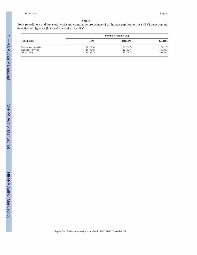
High cumulative prevalence of HPV infection in adolescent womenWe next considered how often adolescent women (in contrast to all subjects) had detectableinfection with a specific HPV type during the study (table 2). Three subjects’ specimens alltested negative for HPV (all 3 reported never having sexual intercourse). Eight subjects hadonly a single swab specimen that tested positive for any HPV; therefore, these subjects werenot considered to be infected by our definition. These single positive specimens were excludedbecause they may have represented laboratory error or deposition of HPV by a sex partnerrather than true infection. Therefore, 11 of the adolescent women were considered to be HPVnegative throughout the entire study period, and the remaining 49 subjects were considered tobe HPV positive.
The first and last specimens collected for each subject were analyzed for HPV, and the resultswere compared with the cumulative detection for every swab specimen collected for eachindividual subject. HPV positivity increased numerically during the study period, for both HRand LR HPV types (table 2). The cumulative prevalence of all HPV detection was 81.7% (table2).
Figure 2 depicts the cumulative HPV type distribution among all 60 adolescent women in thestudy. HPV 52 was detected at some point in 38.3% of all subjects, and HPV 16, the second-most-frequent type, was detected in 31.3% of subjects. Other frequently detected HR typesincluded HPV 59 (found in 23.3% of all subjects), HPV 84 (21.7%), and HPV 18 (20.0%).Frequently detected LR types included HPV 66 (28.3% of all subjects), HPV 6 (25.0%), andHPV 53 (20.0%).
Frequent detection of multiple HPV types in adolescent womenThe mean number of HPV types per HPV-positive subject (n = 49) was 4.9 (SD, 3.3 types;median, 5.0 types; range, 1–14 types). A single HPV type was detected in specimens from18.4% of these individuals, whereas multiple types were detected in 81.6% who were HPV-positive. Adolescent women with abnormal Pap smears had a mean of 6.0 different HPV types(SD, 3.4 types), compared with a mean of 4.4 types (SD, 2.9 types) in adolescents who hadnormal Pap smears.
Persistence of HPVLengths of type-specific HPV runs were analyzed to gain insight into HPV persistence. Themean number of days of follow-up for subjects with HPV infections was 794 (median, 793days; SD, 34 days; range, 712–869 days). Subjects who were HPV positive contributed 241HPV “runs.” Of these subjects, 47 had Pap smear results and thus were included in the survivalanalysis. The median length of all 239 HPV runs was 168 days. Among all the runs, 166 wereof HR HPV types, with a median length of 188 days. There were 73 LR HPV runs, with medianlength of 89 days. Figure 3 shows Kaplan-Meier estimates of HR and LR HPV persistence(runs). When the impact of the censored HPV runs was taken into account, Kaplan-Meierestimates of the median clearance time were 226 and 170 days, in HR HPV and LR HPV,respectively. Using a Cox regression model with random subject effect, we found that HPVtype was significantly associated with run length (p = .034). The risk ratio associated with HRHPV was 0.683. This implied that there was a 31.7% reduction in the probability that an HPV
Brown et al. Page 5
J Infect Dis. Author manuscript; available in PMC 2008 November 24.
NIH
-PA Author Manuscript
NIH
-PA Author Manuscript
NIH
-PA Author Manuscript
run became undetectable if the run was of an HR HPV. Persistence of any HPV was marginallyassociated with abnormal Pap smear results (p = .053). Figure 4 shows Kaplan-Meier estimatesof runs of HPV in subjects with and without abnormal Pap smear results. The Kaplan-Meierestimates of the median clearance time were 255 and 170 days, in HPV runs with and withoutabnormal Pap smear results, respectively. The risk ratio associated with an abnormal Pap smearwas 0.597, suggesting that the likelihood of an HPV run accompanied by abnormal Pap smearresults becoming undetectable was 59.7% of that in a run without an abnormal Pap smear. Inother words, HPV runs were more persistent in subjects with abnormal Pap smears. We didnot have enough observations to calculate Kaplan-Meier estimates for abnormal/normal Papsmears associated with either HR or LR HPV.
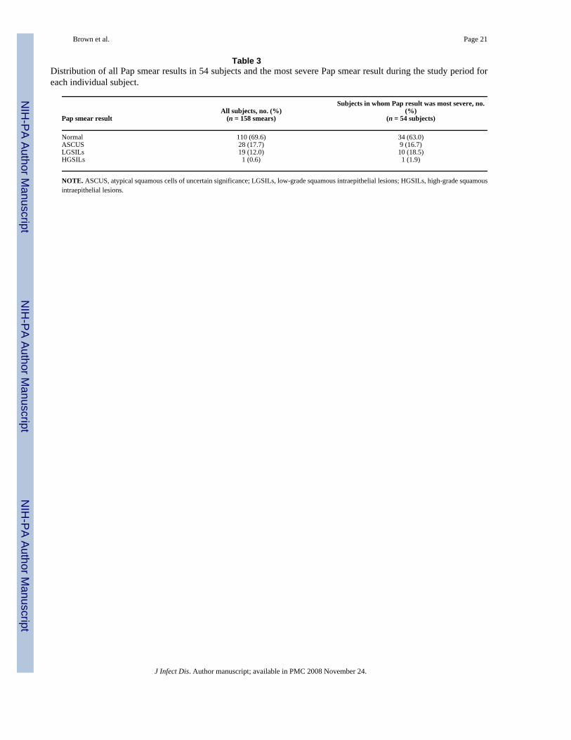
High frequency of abnormal cervical cytologic tests in adolescent womenThree subjects who did not report any past or present sexual intercourse did not undergocytologic testing. Pap smears were available for 54 of the remaining 57 adolescent women inthe study. The mean number of Pap smears acquired per subject was 3.0 (SD, 1.6; range, 1–8). Among the 161 Pap smears obtained, results were obtained for 158 (98.1%). Of these,69.6% were normal, and 30.4% were abnormal (with ASCUS, LGSILs, or HGSILs) (table 3).ASCUS was the most common abnormality, occurring in 28 Pap smears. LGSILs occurred in19 Pap smears, and HGSILs occurred in 1. Among the 54 subjects with at least 1 Pap smear,37% had at least 1 abnormal test, and 63% had entirely normal tests (table 3).
The most severe grade of Pap smear abnormality for each of the 54 adolescent women wascompared with HR HPV positivity. As expected, Pap smear abnormalities were associated withdetection of HR HPV. All 20 subjects with abnormal Pap smears had HR HPV detected atsome point during the study period, compared with 24 of 34 subjects with normal Pap smears(p = .009; Fisher’s exact test). The proportions of HR HPV– and LR HPV–positive specimenswithin the abnormal Pap smear categories were not determined; this was because we used asubject-level summary definition for Pap abnormality, whether or not any HPV-positivespecimen was ever of the HR category. The tests for HPV and Pap were different, and datesof specimen acquisition did not always coincide. Therefore, a subject may have had 1 to severalPap tests with different results, as well as many HPV tests with variable results.
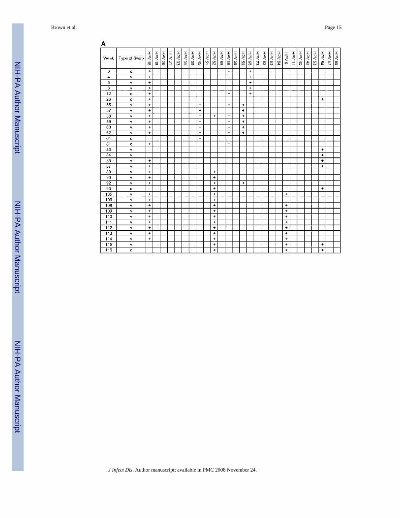
Analysis of individual adolescent womenThe complete analysis of individual subjects illustrated the acquisition and persistence ofspecific HPV types during the study. The analysis for 4 adolescent women is shown in figure5.
Subject A was infected with numerous HPV types (figure 5A). HPV 16 was detected in theswab obtained from her first visit and persisted throughout most of the study period. HPV types45, 52, 56, 59, and 68 (all HR types) were repeatedly detected. HPV types 6 and 54 (LR types)were also detected. Her most severe cytologic abnormality was LGSILs.
In subject B, HPV 56 was the only type detected, persisting for 9 months (figure 5B). Cytologictesting revealed HGSILs as her most severe abnormality.
Subject C had no HPV types detected for the first 14 months of the study (figure 5C) but thenbecame infected with numerous types that persisted throughout the study. Cytologic testingshowed no abnormalities.
Subject D had no HPV types detected for the first 12 months of the study but then becameinfected with HPV types 83, 84, and 6, all of which were undetectable after several months(figure 5D). She then became infected with HPV types 56, 68, and 54, which persisted
Brown et al. Page 6
J Infect Dis. Author manuscript; available in PMC 2008 November 24.
NIH
-PA Author Manuscript
NIH
-PA Author Manuscript
NIH
-PA Author Manuscript
throughout the remainder of the study. Cytologic testing revealed LGSILs as her most severeabnormality.
DISCUSSIONA complete understanding of the epidemiology of HPV infection in adolescent women isimperative for effective targeted interventions. This is exemplified by HPV vaccine trials, inwhich it is important to understand the significance of infection with specific HPV types andmultiple types. The intent of the present study was to more accurately characterize the naturalhistory of genital tract HPV infections early in life, at the point when such interventions maybe most effective.
As with previous studies, we found that HPV infection is extremely common in sexually activeadolescent women. The point prevalence of detectable HPV infection in our study was in therange of 25%–40%. The cumulative prevalence in our longitudinal analysis of adolescentwomen was >80%, a figure that exceeds that found in other studies.
Moscicki et al. found prevalence rates of 54.5% for all genital HPV types and 29.1% for HRHPV types in a cross-sectional study of adolescent females [10]. The youngest women in alongitudinal study of college-aged women by Ho et al. had the highest prevalence of HPVinfection. Sampling for HPV was performed every 6 months for 3 years. When women whowere positive for HPV at baseline were included, 60% were infected with HPV at some pointduring the 3-year study period [11]. In a cross-sectional study by Tarkowski et al., HPV wasdetected in 64% of 312 adolescent women; more than half of these women had multiple HPVtypes detected [12].
The very high cumulative prevalence in our study was most likely a result of the large numberof specimens obtained from each subject. Many infections were detectable for only a few weeksand would have been missed if specimens were obtained at 4- or 6-month intervals. In addition,we used an assay that detects more HPV types than did the assays used in some previous studies,thus contributing to the higher HPV prevalence. Some of the differences in prevalence may berelated to behaviors of the cohort (to be described in a forthcoming article). Thus, in part, thehigh HPV prevalence may be reflective of the at-risk population served at the clinics used forthis study. The young women were recruited from a population in which the initiation of sexualactivity occurred at a mean age of 14 years [13]. Close to 14% of adolescent females whoreceived reproductive health care in these clinics had Chlamydia trachomatis infections, and8.9% were infected with Neisseria gonorrhoeae, rates above the reported national averagesfor this population.
Self-collected vaginal swabs potentially offer an inexpensive method of collecting genitalsecretions for HPV testing. In our study, self collection of vaginal swabs was acceptable andreliable for HPV detection. In other studies, results of HPV testing of self-collected vaginalswabs also approximated those of clinician-obtained specimens from the cervix [14-16]. In thestudy by Gravitt et al., self-collected vaginal swab samples were compared with clinician-obtained cervical swab samples from 268 women participating in a study of cervical cancer[15]. The overall agreement between test results from the clinician- and self-collected swabswas excellent.
Overall, ~85% of swabs in the present study contained adequate cellular material, on the basisof β-globin amplification. Sampling errors or loss of DNA in the purification process couldaccount for the 15% of swabs that did not contain adequate cellular material. Interestingly, wefound that vaginal swabs were more likely than cervical swabs to contain adequate cellularmaterial. In addition, HPV detection in vaginal swabs exceeded that in cervical swabs. Oneexplanation is that cervical swabs, obtained by direct visualization and cotton swab sampling
Brown et al. Page 7
J Infect Dis. Author manuscript; available in PMC 2008 November 24.
NIH
-PA Author Manuscript
NIH
-PA Author Manuscript
NIH
-PA Author Manuscript

of the squamo-columnar junction, would have yielded more cellular material if a cytobrushhad been used. The higher percentage of vaginal swabs containing detectable HPV could bedue to the larger surface area of genital mucosa sampled by vaginal swabs compared withcervical swabs.
We identified a large range of HPV types in the adolescent women. Overall, HPV types 52,16, and 59 were the most frequently detected HR types. We have no obvious explanation forthe high prevalence of HPV 52. In a cohort of older women in Indianapolis of the same racialdistribution, we previously found that HPV 52 was detected much less frequently than otherHR types [17]. Thus, it is possible that HPV 52 preferentially infects younger women, butadditional studies are needed to test this hypothesis.
The most frequently detected LR types were HPV 66 (6.1% of adequate specimens) and HPV6 (5.6%). HPV 66 is a type detected with variable prevalence in cervical swab or lavagespecimens from women with normal or abnormal cervical cytological results [12,16-20]. Someexperts have classified HPV 66 as an HR type, whereas others include this type in the LRcategory. In a study of an urban population similar to the one in the present study, HPV 66 wasdetected in cervical specimens from 8.0% of adolescent women who were sampled only onceduring the study [12]. HPV 66 was the third-most frequently detected type in that study, afterHPV types 16 and 59.
Natural history studies indicate that most HPV infections “clear,” meaning that the specificHPV type is no longer detectable by current methods [18,21-25]. Our study is consistent withthis observation. However, the inability to detect HPV infection in swabs by use of PCR is notproof that clearance has occurred. It is possible that infection persists at very low levels andcan potentially reactivate later in life. As additional longitudinal studies are performed andimproved detection methods are developed, we believe that the issue of HPV clearance willneed to be reconsidered.
We found a significant difference in persistence of HR and LR HPV types on the basis ofsurvival analysis, which is consistent with findings in previous studies. For example,Londesborough et al. studied women infected with HPV and found that HPV 16 persistedlonger than other types [26]. Giuliano et al. found that the median time to clearance of HPVinfection was 9.8 months for HR HPV types, compared with 4.3 months for LR types [19].Franco et al. showed that the mean infection durations were 13.5 and 8.2 months for HR andLR HPV types, respectively [18]. In the study by Moscicki et al., HR HPV types were lesslikely to regress than were LR types [24]. HR HPV types may therefore possess a unique abilityto persist longer than LR types, and it is the persistence of HR HPV that is associated withcervical dysplasia.
We found that 37.0% of the cohort of young adolescent women had a least 1 abnormal cervicalcytologic examination during the study period. One of the participants developed an HGSILabnormality during the study period. This rate of cytologic abnormalities is within range ofthose from other studies of adolescent women. Moscicki et al. reported a combined incidenceand prevalence rate of 33.5% for LGSILs in young women infected with HPV [24].Interestingly, Moscicki found 11 women (1.8% of the total number of women) who developedHGSILs in the 40-month observation period, and the authors suggested that dysplasia inadolescents may progress more quickly than dysplasia in older women. In the above-mentionedcross-sectional study by Tarkowski et al., cytologic abnormalities were detected in 38% of 312adolescent women.
In summary, we found that a high proportion of adolescent females were infected with HPVat any given time, and, over the study period, >80% of the subjects had evidence of HPVinfection. A high percentage of these subjects had cervical cytologic abnormalities. One
Brown et al. Page 8
J Infect Dis. Author manuscript; available in PMC 2008 November 24.
NIH
-PA Author Manuscript
NIH
-PA Author Manuscript
NIH
-PA Author Manuscript
defining step in the epidemiology of HPV infection will be to determine whether infections ata very young age are those that resurge and are detected later in life, versus acquisition of anew infection. Finally, if infections do persist at very low levels from young adulthood, it isimportant to determine the factors associated with the progression from HPV infection todysplasia and malignancy.
AcknowledgementsWe thank Raymond J. Apple and Amanda Shepard (Roche Molecular Systems, Alameda, CA), for providing thepolymerase chain reaction/reverse blot assay, and Patti Gravitt (Johns Hopkins Bloomberg School of Public Health,Baltimore, MD), for critical reading of the manuscript and for her helpful suggestions.
Financial support: National Institutes of Health, National Institute of Allergy and Infectious Disease (grant U19AI43924); National Institute of Child Health and Human Development (grant RO1HD42404-01, for the developmentof statistical models used in this research).
References1. Centers for Disease Control and Prevention. Sexually transmitted disease surveillance, 1995. Atlanta:
US Department of Health and Human Services, Public Health Service; 1996.2. Schiffman, MH.; Burk, RD. Human papillomaviruses. In: Kaslow, RA., editor. Viral infections of
humans. New York: Plenum Medical Book; 1997. p. 983-1022.3. Asato T, Nakajima Y, Nagamine M, et al. Correlation between the progression of cervical dysplasia
and the prevalence of human papillomavirus. J Infect Dis 1994;169:940–1. [PubMed: 8133117]4. Koutsky L. Epidemiology of genital human papillomavirus infection. Am J Med 1997;102:3–8.
[PubMed: 9217656]5. Gravitt PE, Peyton CL, Apple RJ, Wheeler CM. Genotyping of 27 HPV types from L1 consensus PCR
products using a single hybridization, reverse line-blot detection method. J Clin Microbiol1998;36:3020–7. [PubMed: 9738060]
6. Gravitt PE, Peyton CL, Alessi TQ, et al. Improved amplification of genital human papillomaviruses.J Clin Microbiol 2000;38:357–61. [PubMed: 10618116]
7. Brown DR, Schroeder JM, Bryan JT, Stoler MH, Fife KH. Detection of multiple human papillomavirustypes in condylomata acuminata lesions from otherwise healthy and immunosuppressed patients. JClin Microbiol 1999;37:3316–22. [PubMed: 10488198]
8. Katz B, Fortenberry J, Tu W, Harezlak J, Orr D. Sexual behavior among adolescent women at highrisk for sexually transmitted infection. Sex Transm Dis 2001;28:247–51. [PubMed: 11354261]
9. Thereau, TM.; Grambsch, PM. Modeling survival data: extending the Cox model. New York: Springer;2002.
10. Moscicki AB, Ellenberg JH, Vermund SH, et al. Prevalence of and risks for cervical humanpapillomavirus infection and squamous intraepithelial lesions in adolescent girls: impact of infectionwith human immunodeficiency virus. Arch Pediatr Adolesc Med 2000;154:127–34. [PubMed:10665598]
11. Ho GYF, Bierman R, Beardsley L, Chang CJ, Burk RD. Natural history of cervicovaginalpapillomavirus infection in young women. N Engl J Med 1998;338:423–8. [PubMed: 9459645]
12. Tarkowski T, Koumans E, Sawyer M, et al. Epidemiology of human papillomavirus infection andabnormal cytologic test results in an urban adolescent population. J Infect Dis 2004;189:46–50.[PubMed: 14702152]
13. Orr DP, Fortenberry JD, Blythe MJ. Validity of self-reported sexual behaviors in adolescent womenusing biomarker outcomes. Sex Transm Dis 1997;24:261–6. [PubMed: 9153734]
14. Wright TC, Denny L, Kuhn L, Pollack A, Lorincz A. HPV DNA testing of self-collected vaginalsamples compared with cytologic screening to detect cervical cancer. JAMA 2000;283:81–6.[PubMed: 10632284]
15. Gravitt P, Lacey J, Brinton L, et al. Evaluation of self-collected cervicovaginal cell samples for humanpapillomavirus testing by polymerase chain reaction. Cancer Epidemiol Biomarkers Prev2001;10:95–100. [PubMed: 11219778]
Brown et al. Page 9
J Infect Dis. Author manuscript; available in PMC 2008 November 24.
NIH
-PA Author Manuscript
NIH
-PA Author Manuscript
NIH
-PA Author Manuscript

16. Shah K, Daniel R, Tennant M, et al. Diagnosis of human papillomavirus infection by dry vaginalswabs in military women. Sex Transm Infect 2001;77:260–4. [PubMed: 11463925]
17. Brown D, Legge D, Qadadri B. Distribution of human papillomavirus types in cervicovaginalwashings from women evaluated in a sexually transmitted diseases clinic. Sex Transm Dis2002;29:763–8. [PubMed: 12466717]
18. Franco EL, Villa LL, Sobrinho JP, et al. Epidemiology of acquisition and clearance of cervical humanpapillomavirus infection in women from a high-risk area for cervical cancer. J Infect Dis1999;180:1415–23. [PubMed: 10515798]
19. Giuliano A, Harris R, Sedjo R, et al. Incidence, prevalence, and clearance of type-specific humanpapillomavirus infections: The Young Women’s Health Study. J Infect Dis 2002;186:462–9.[PubMed: 12195372]
20. Sotlar K, Diemer D, Dethleffs A, et al. Detection and typing of human papillomavirus by E6 nestedmultiplex PCR. J Clin Microbiol 2004;42:3176–84. [PubMed: 15243079]
21. Moscicki AB, Palefsky J, Smith G, Siboshski S, Schoolnik G. Variability of human papillomavirusDNA testing in a longitudinal cohort of young women. Obstet Gynecol 1993;82:578–85. [PubMed:8397358]
22. Evander M, Edlund K, Gustafsson A, et al. Human papillomavirus infection is transient in youngwomen: a population-based cohort study. J Infect Dis 1995;171:1026–30. [PubMed: 7706782]
23. Brisson J, Bairati I, Morin C, et al. Determinants of persistent detection of human papillomavirusDNA in the uterine cervix. J Infect Dis 1996;173:794–9. [PubMed: 8603956]
24. Moscicki AB, Shiboski S, Broering J, et al. The natural history of human papillomavirus infection asmeasured by repeated DNA testing in adolescent and young women. J Pediatr 1998;132:277–84.[PubMed: 9506641]
25. Sellors J, Karwalajtys T, Kaczorowski J, et al. Incidence, clearance and predictors of humanpapillomavirus infection in women. CMAJ 2003;168:421–5. [PubMed: 12591782]
26. Londesborough P, Ho L, Terry G, Cuzick J, Wheeler C, Singer A. Human papillomavirus genotypeas a predictor of persistence and development of high-grade lesions in women with minor cervicalabnormalities. Int J Cancer 1996;69:364–8. [PubMed: 8900368]
Brown et al. Page 10
J Infect Dis. Author manuscript; available in PMC 2008 November 24.
NIH
-PA Author Manuscript
NIH
-PA Author Manuscript
NIH
-PA Author Manuscript

Figure 1.Distribution of specific human papillomavirus (HPV) types in cervical swabs, vaginal swabs,and all swabs. The upper rows show the no. of swabs that were positive for each HPV type,and the lower rows show the percentage of swabs that were positive for each type.
Brown et al. Page 11
J Infect Dis. Author manuscript; available in PMC 2008 November 24.
NIH
-PA Author Manuscript
NIH
-PA Author Manuscript
NIH
-PA Author Manuscript
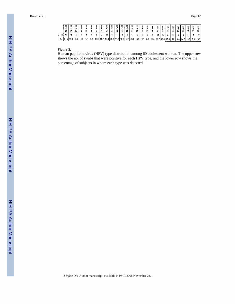
Figure 2.Human papillomavirus (HPV) type distribution among 60 adolescent women. The upper rowshows the no. of swabs that were positive for each HPV type, and the lower row shows thepercentage of subjects in whom each type was detected.
Brown et al. Page 12
J Infect Dis. Author manuscript; available in PMC 2008 November 24.
NIH
-PA Author Manuscript
NIH
-PA Author Manuscript
NIH
-PA Author Manuscript
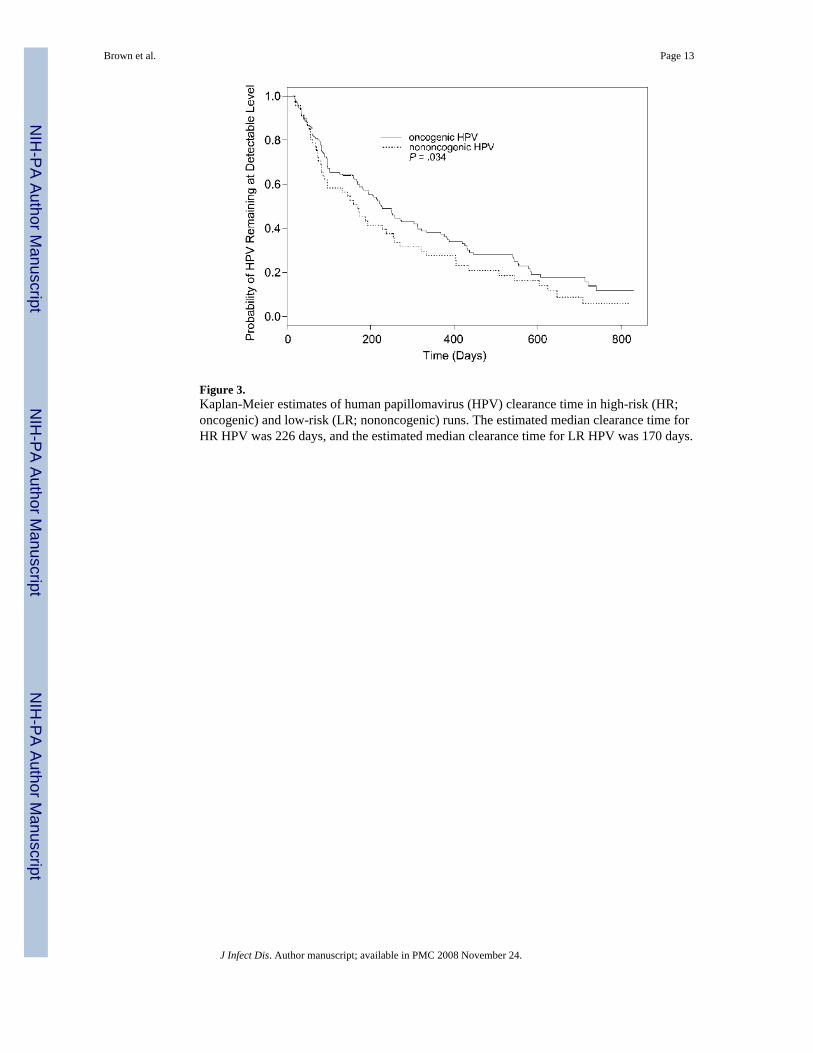
Figure 3.Kaplan-Meier estimates of human papillomavirus (HPV) clearance time in high-risk (HR;oncogenic) and low-risk (LR; nononcogenic) runs. The estimated median clearance time forHR HPV was 226 days, and the estimated median clearance time for LR HPV was 170 days.
Brown et al. Page 13
J Infect Dis. Author manuscript; available in PMC 2008 November 24.
NIH
-PA Author Manuscript
NIH
-PA Author Manuscript
NIH
-PA Author Manuscript

Figure 4.Kaplan-Meier estimates of human papillomavirus (HPV) persistence in individual subjectswith or without abnormal Pap smear results. The Kaplan-Meier estimates of the medianclearance time were 255 and 170 days, in HPV runs with and without abnormal pap smearresults, respectively.
Brown et al. Page 14
J Infect Dis. Author manuscript; available in PMC 2008 November 24.
NIH
-PA Author Manuscript
NIH
-PA Author Manuscript
NIH
-PA Author Manuscript
Brown et al. Page 15
J Infect Dis. Author manuscript; available in PMC 2008 November 24.
NIH
-PA Author Manuscript
NIH
-PA Author Manuscript
NIH
-PA Author Manuscript

Brown et al. Page 16
J Infect Dis. Author manuscript; available in PMC 2008 November 24.
NIH
-PA Author Manuscript
NIH
-PA Author Manuscript
NIH
-PA Author Manuscript
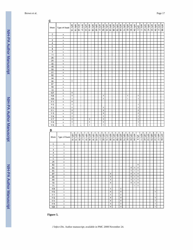
Figure 5.
Brown et al. Page 17
J Infect Dis. Author manuscript; available in PMC 2008 November 24.
NIH
-PA Author Manuscript
NIH
-PA Author Manuscript
NIH
-PA Author Manuscript
The complete analysis of 4 individual adolescent woman (subjects A, B, C, and D), shown toillustrate the acquisition and persistence of specific human papillomavirus (HPV) types duringthe study period. The week in which the specimen was acquired is shown in the left-mostcolumn. HPV types are indicated at the top of the figure. c, clinician-obtained cervical swab;v, self-collected vaginal swab. Plus (+) signs indicate positive results for the specimen.
Brown et al. Page 18
J Infect Dis. Author manuscript; available in PMC 2008 November 24.
NIH
-PA Author Manuscript
NIH
-PA Author Manuscript
NIH
-PA Author Manuscript

NIH
-PA Author Manuscript
NIH
-PA Author Manuscript
NIH
-PA Author Manuscript
Brown et al. Page 19
Table 1Cervical and vaginal swab positivity for β-globin, human papillomavirus (HPV), high-risk (HR) HPV, and low-risk(LR) HPV.
Positive swabs, no. (%)
Swab type β-globin HPV HR HPV LR HPV
Cervical (n = 353) 289 (81.9) 116 (40.1) 104 (36.0) 42 (14.5)Vaginal (n = 2105) 1818 (86.4) 838 (46.1) 710 (39.1) 372 (20.5) All (n = 2458) 2107 (85.7) 954 (45.3) 814 (38.6) 414 (19.6)
J Infect Dis. Author manuscript; available in PMC 2008 November 24.
NIH
-PA Author Manuscript
NIH
-PA Author Manuscript
NIH
-PA Author Manuscript
Brown et al. Page 20
Table 2Point (enrollment and last study visit) and cumulative prevalence of all human papillomavirus (HPV) detection anddetection of high-risk (HR) and low-risk (LR) HPV.
Positive swabs, no. (%)
Time point(s) HPV HR HPV LR HPV
Enrollment (n = 60) 17 (28.3) 13 (21.7) 7 (11.7)Last visit (n = 60) 24 (40.0) 22 (36.7) 11 (18.3)All (n = 60) 49 (81.7) 46 (76.7) 34 (56.7)
J Infect Dis. Author manuscript; available in PMC 2008 November 24.
NIH
-PA Author Manuscript
NIH
-PA Author Manuscript
NIH
-PA Author Manuscript
Brown et al. Page 21
Table 3Distribution of all Pap smear results in 54 subjects and the most severe Pap smear result during the study period foreach individual subject.
Pap smear resultAll subjects, no. (%)
(n = 158 smears)
Subjects in whom Pap result was most severe, no.(%)
(n = 54 subjects)
Normal 110 (69.6) 34 (63.0)ASCUS 28 (17.7) 9 (16.7)LGSILs 19 (12.0) 10 (18.5)HGSILs 1 (0.6) 1 (1.9)
NOTE. ASCUS, atypical squamous cells of uncertain significance; LGSILs, low-grade squamous intraepithelial lesions; HGSILs, high-grade squamousintraepithelial lesions.
J Infect Dis. Author manuscript; available in PMC 2008 November 24.
Copyright © 2022 FDOKUMEN