A holistic evolutionary and structural study of flaviviridae provides insights into the function and...
18
Submitted 3 April 2013 Accepted 24 April 2013 Published 7 May 2013 Corresponding author Sophia Kossida, [email protected] Academic editor Yong Wang Additional Information and Declarations can be found on page 15 DOI 10.7717/peerj.74 Copyright 2013 Vlachakis et al. Distributed under Creative Commons CC-BY 3.0 OPEN ACCESS A holistic evolutionary and structural study of flaviviridae provides insights into the function and inhibition of HCV helicase Dimitrios Vlachakis, Vassiliki Lila Koumandou and Sophia Kossida Bioinformatics & Medical Informatics Team, Biomedical Research Foundation, Academy of Athens, Athens, Greece ABSTRACT Viral RNA helicases are involved in duplex unwinding during the RNA replication of the virus. It is suggested that these helicases represent very promising antiviral targets. Viruses of the flaviviridae family are the causative agents of many com- mon and devastating diseases, including hepatitis, yellow fever and dengue fever. As there is currently no available anti-Flaviviridae therapy, there is urgent need for the development of efficient anti-viral pharmaceutical strategies. Herein, we report the complete phylogenetic analysis across flaviviridae alongside a more in-depth evolutionary study that revealed a series of conserved and invariant amino acids that are predicted to be key to the function of the helicase. Structural molecular modelling analysis revealed the strategic significance of these residues based on their relative positioning on the 3D structures of the helicase enzymes, which may be used as pharmacological targets. We previously reported a novel series of highly potent HCV helicase inhibitors, and we now re-assess their antiviral potential using the 3D structural model of the invariant helicase residues. It was found that the most active compound of the series, compound C4, exhibited an IC 50 in the submicromolar range, whereas its stereoisomer (compound C12) was completely inactive. Useful insights were obtained from molecular modelling and conformational search studies via molecular dynamics simulations. C12 tends to bend and lock in an almost “U” shape conformation, failing to establish vital interactions with the active site of HCV. On the contrary, C4 spends most of its conformational time in a straight, more rigid formation that allows it to successfully block the passage of the oligonucleotide in the ssRNA channel of the HCV helicase. This study paves the way and provides the necessary framework for the in-depth analysis required to enable the future design of new and potent anti-viral agents. Subjects Bioinformatics, Computational Biology, Evolutionary Studies, Drugs and Devices, Infectious Diseases Keywords Hepatitis C, Structure-based drug design, Molecular dynamics, Antiviral bioinformatics INTRODUCTION Viruses of the flaviviridae family infect vertebrates and they are primarily transmitted through arthropod vectors (mainly ticks and mosquitoes) (Neyts, Leyssen & De Clercq, 1999). The flaviviridae family includes four genera: Flavivirus (Yellow fever virus, West How to cite this article Vlachakis et al. (2013), A holistic evolutionary and structural study of flaviviridae provides insights into the function and inhibition of HCV helicase. PeerJ 1:e74; DOI 10.7717/peerj.74
-
Upload
bioacademy -
Category
Documents
-
view
2 -
download
0
Transcript of A holistic evolutionary and structural study of flaviviridae provides insights into the function and...
Submitted 3 April 2013Accepted 24 April 2013Published 7 May 2013
Corresponding authorSophia Kossida,[email protected]
Academic editorYong Wang
Additional Information andDeclarations can be found onpage 15
DOI 10.7717/peerj.74
Copyright2013 Vlachakis et al.
Distributed underCreative Commons CC-BY 3.0
OPEN ACCESS
A holistic evolutionary and structuralstudy of flaviviridae provides insightsinto the function and inhibition of HCVhelicaseDimitrios Vlachakis, Vassiliki Lila Koumandou and Sophia Kossida
Bioinformatics & Medical Informatics Team, Biomedical Research Foundation, Academy ofAthens, Athens, Greece
ABSTRACTViral RNA helicases are involved in duplex unwinding during the RNA replicationof the virus. It is suggested that these helicases represent very promising antiviraltargets. Viruses of the flaviviridae family are the causative agents of many com-mon and devastating diseases, including hepatitis, yellow fever and dengue fever.As there is currently no available anti-Flaviviridae therapy, there is urgent need forthe development of efficient anti-viral pharmaceutical strategies. Herein, we reportthe complete phylogenetic analysis across flaviviridae alongside a more in-depthevolutionary study that revealed a series of conserved and invariant amino acidsthat are predicted to be key to the function of the helicase. Structural molecularmodelling analysis revealed the strategic significance of these residues based on theirrelative positioning on the 3D structures of the helicase enzymes, which may be usedas pharmacological targets. We previously reported a novel series of highly potentHCV helicase inhibitors, and we now re-assess their antiviral potential using the 3Dstructural model of the invariant helicase residues. It was found that the most activecompound of the series, compound C4, exhibited an IC50 in the submicromolarrange, whereas its stereoisomer (compound C12) was completely inactive. Usefulinsights were obtained from molecular modelling and conformational search studiesvia molecular dynamics simulations. C12 tends to bend and lock in an almost “U”shape conformation, failing to establish vital interactions with the active site of HCV.On the contrary, C4 spends most of its conformational time in a straight, more rigidformation that allows it to successfully block the passage of the oligonucleotide inthe ssRNA channel of the HCV helicase. This study paves the way and provides thenecessary framework for the in-depth analysis required to enable the future design ofnew and potent anti-viral agents.
Subjects Bioinformatics, Computational Biology, Evolutionary Studies, Drugs and Devices,Infectious DiseasesKeywords Hepatitis C, Structure-based drug design, Molecular dynamics,Antiviral bioinformatics
INTRODUCTIONViruses of the flaviviridae family infect vertebrates and they are primarily transmitted
through arthropod vectors (mainly ticks and mosquitoes) (Neyts, Leyssen & De Clercq,
1999). The flaviviridae family includes four genera: Flavivirus (Yellow fever virus, West
How to cite this article Vlachakis et al. (2013), A holistic evolutionary and structural study of flaviviridae provides insights into thefunction and inhibition of HCV helicase. PeerJ 1:e74; DOI 10.7717/peerj.74
Nile virus, Dengue virus, Tick-borne encephalitis viruses), Hepacivirus (Hepatitis C
virus), Pestivirus (Classical swine fever virus, Bovine viral diarrhea virus) and Unclassified
Flaviviridae (Hepatitis GB virus, GB viruses) (Shepard, Finelli & Alter, 2005). Flaviviridae
have monopartite, linear, single-stranded, positive sense RNA genomes (Vlachakis, 2009),
ranging from 10 to 12.5 kilobases (kb) in length (Poynard, Bedossa & Opolon, 1997).
Because the viral RNA has positive sense, the nucleic acid itself is capable of causing an
infection in the appropriate host cells. The 3’-termini of their RNA are not polyadenylated.
The 5’-termini of the viral RNA genome in members of the Flavivirus genus have a
methylated nucleotide cap, which allows translation. Sometimes it is possible to have a
genome-linked protein (VPg) in place of the methylated nucleotide cap. In members of the
Pestivirus and Hepacivirus genera, the 5’ ends are uncapped and have an internal ribosome
entry site (IRES) instead (Nulf & Corey, 2004), which is responsible for providing a site for
the initiation of the translation process for host ribosomes (Daly & Ward, 2003). In both
genera, structural genes are located towards the 5’ end of the RNA.
Virions of the flaviviridae family are enveloped and slightly pleomorphic during
their life cycle. They are spherical in shape and usually 40–60 nm in diameter. Their
nucleocapsids are isometric and sometimes penetrated by stain. The usual size of the
nucleocapsids is 25–30 nm in diameter and they have polyhedral symmetry (Guzman &
Kourı, 2004). It is remarkable that flaviviridae viruses manage to retain high similarity in
the morphology of the virion, the organization of the viral genome, and the estimated
life cycles and replication patterns that they follow, even though the different genera
do not have common biological properties and do not show serological cross-reactivity
(Thompson & Finch, 2005; Rosenberg, 2001).
One of the most important pathogens of the flaviviridae viral family is the Hepatitis C
virus (HCV). Recent classification analysis suggests that there are 6 different genotypes of
the naturally occurring virus. Approximately 180 million people are chronically infected
with HCV, according to the World Health Organization. In addition to that, an estimated
4 million new infections occur every year (Calisher & Gould, 2003; Kadare & Haenni,
1997). The progression of the disease upon infection is rapid, as in more than 80% of
patients a persistent infection will be established, which will eventually progress to liver
cirrhosis and in most cases, hepatocellular carcinoma. The fatality rates of HCV-related
diseases are estimated to be approximately 400,000 patients per year. To date no effective
drug or vaccination agent is available. More importantly, the molecular machinery that the
virus employs in its lethal quest is still unclear.
Inhibition of the viral helicase is an emerging and very promising approach that is
becoming increasingly popular (Phoon et al., 2001). Helicases are capable of unwinding
double stranded DNA and RNA to single strands by breaking the series of hydrogen
bonds that keep the two strands together. The unwinding activity of the viral helicase is
essential to the virus during its replication process. Mutated inactive helicases in Dengue
and Bovine Diarrhea viruses led to reduced proliferation of the virus (Diana & Bailey,
1997). Therefore, it is considered that the effective inhibition of the viral helicase will
be an operative tool for the suppression of the replication rate of flaviviridae viruses
Vlachakis et al. (2013), PeerJ, DOI 10.7717/peerj.74 2/18
proliferation. The viral Helicase is coded in the NS3B region of the viral genome next
to the NS3A gene, which codes for the viral protease.
Many drug-like potent inhibitors of HCV have been reported in the literature, but
only one that is capable of covalently interacting with the HCV helicase enzyme (Kandil
et al., 2009). In this study, we present an in-depth updated phylogenetic analysis of
flaviviridae helicase protein sequences. Collectively, 87 complete polyprotein sequences
were retrieved from the Virus Pathogen Resource (ViPR) Database, the distribution of
which in the flaviviridae genera is: 69 flavivirus, 11 hepacivirus, 7 pestivirus (Vlachakis,
2009; Brancale, Vlachaki & Vlachakis, 2008; Vlachakis & Brancale, 2007). The multiple
sequence alignments of the NS3 protein from phylogenetically distinct species, reveal a
large number of conserved residues, which had not been studied previously. As sequence
conservation denotes functional importance, this result allowed for a more focused and
comprehensive molecular modelling approach of HCV, which aims to provide invaluable
insights that will rationalize the findings of our previously published work (Kandil et al.,
2009) and deliver the means required for the future de novo structure-based drug design of
potent anti-HCV agents.
METHODOLOGYDatabase sequence searchSequence data from species for which full genome data is available were obtained from the
NIAID Virus Pathogen Database and Analysis Resource (ViPR) online through the web site
at http://www.viprbrc.org (Pickett et al., 2012), as well as the NCBI RefSeq database. This
selection was checked against, and supplemented with, data from previous publications
regarding flaviviridae phylogeny (Cook & Holmes, 2006; Ferron et al., 2005; Lobo et al.,
2009). Multiple HCV genotypes were included, but otherwise, for each species, only one
strain was analysed. In total, sequences from 7 pestivirus, 11 hepacivirus (including the 6
HCV genotypes), and 69 flavivirus species were used, as shown in Table S1. About half of
these species have fully annotated genomes; in these cases the annotated NS3 sequence was
used (Table S1). For the rest, the start and end positions of the NS3 sequence within the
whole genome polyprotein was inferred from alignments with closely related annotated
species. Thus, only the amino acid sequence corresponding to NS3, determined in this way,
was used for the multiple sequence alignments and phylogenetic analysis (for details see
Table S1).
Alignments and phylogenetic analysisAlignments of the NS3 protein sequence (File S1) were created using MUSCLE, version
3.7 (Edgar, 2004), and checked visually using Jalview, version 2.7 (Waterhouse et al., 2009).
Only unambiguous homologous regions were retained for phylogenetic analysis; manual
masking and trimming was performed in MacClade, version 4.08 (Maddison & Maddison,
1989). ProtTest, version 1.3 (Abascal, Zardoya & Posada, 2005) was used to estimate the
appropriate model of sequence evolution. Phylogenetic analysis was performed by three
separate methods. To obtain the Bayesian tree topology and posterior probability values,
Vlachakis et al. (2013), PeerJ, DOI 10.7717/peerj.74 3/18
the program MrBayes, version 3.1.2 was used (Ronquist & Huelsenbeck, 2003). Analyses
were run for 1 million generations, removing all trees before a plateau established by
graphical estimation. All calculations were checked for convergence and had a splits
frequency of <0.1. Maximum-likelihood (ML) analysis was performed using PhyML,
version 3.0 (Guindon & Gascuel, 2003) and RAxML, version 7.0.0 (Stamatakis, 2006) with
100 bootstrap replicates. Trees were visualised using FigTree v1.2 (http://tree.bio.ed.ac.
uk/software/figtree/). Nodes with at least 0.95 posterior probability and 80% bootstrap
support were considered robust, and nodes with at least 0.80 posterior probability and 50%
bootstrap support are indicated.
Molecular dockingIn order to elucidate in silico the 3D structures of compounds C4, C12 and the HCV
helicase the docking suite ZDOCK, version 3.0 was used (Chen, Li & Weng, 2003).
ZDOCK is a protein-protein docking suite that utilizes a grid-based representation
of the molecular system involved. In order to efficiently explore the search space and
docking positions of the molecules as rigid bodies, ZDOCK takes full advantage of a
three-dimensional fast Fourier transformation algorithm. It uses a scoring function that
returns electrostatic, hydrophobic and desolvation energies as well as performing a fast
pairwise shape complementarity evaluation. Moreover, it uses the contact propensities of
transient complexes of proteins to perform an evaluation of a pairwise atomic statistical
potential for the docking molecular system. RDOCK was utilized to refine and quickly
evaluate the results obtained by ZDOCK ZDOCK (Li, Chen & Weng, 2003). RDOCK
performs a fast minimization step to the ZDOCK molecular complex outputs and ranks
them according to their re-calculated binding free energies.
Energy minimizationsEnergy minimization was performed initially to remove the geometrical strain from the
top-ranking hits of the docking experiments, since the proteins were treated as rigid bodies.
Protein complexes were subjected to an extensive energy minimization run using the
Amber99 (Duan et al., 2003) forcefield as it is implemented into the Gromacs, version 4.5.5
(Hess et al., 2008), via the Gromita graphical interface, version 1.07 (Sellis, Vlachakis &
Vlassi, 2009). An implicit Generalized Born (GB) solvation was chosen at this stage, in an
attempt to speed up the energy minimization process.
Molecular dynamicsIn order to further explore the interaction space and binding potential of each docking
conformation, the molecular complexes were subjected to unrestrained molecular
dynamics simulations using the Gromacs suite, version 4.5.5 (Hess et al., 2008). Molecular
dynamics took place in a periodic environment, which was subsequently solvated with SPC
water using the truncated octahedron box extending to 7A from each molecule. Partial
charges were applied and the molecular systems were neutralized with counter-ions as
required. The temperature was set to 300 K and the step size was set to 1 fs. The total run
of each molecular complex was fifty nanoseconds, using the NVT ensemble in a canonical
Vlachakis et al. (2013), PeerJ, DOI 10.7717/peerj.74 4/18
environment. NVT stands for Number of atoms, Volume and Temperature that remain
constant throughout the calculation. The results of the molecular dynamics simulations
were collected into a molecular trajectory database for further analysis.
Molecular dynamics analysisPrincipal component analysis was done using Pymol, version r0.99 (DeLano, 2002) and the
Ca atom root-mean-square function of Deep-View, version 4.0.1 (Guex & Peitsch, 1997).
Analysis of the Molecular Dynamics outputs and trajectories was therefore focused on
structural deviations of each molecular system from its original docking conformation.
The molecular dynamics final conformations were initially evaluated with a residue
packing quality function built-in the Gromacs suite, which depends on the number of
buried non-polar side chain groups and on hydrogen bonding. Moreover, the suites
Procheck, version 3.5 (Laskowski et al., 1996) Verify3D, version 1.0 (Eisenberg, Luthy &
Bowie, 1997) were employed to evaluate the structural viability of each protein complex
upon the molecular dynamics simulations. Illustrations of the molecular systems were
rendered with the aid of the Chimera suite, version 1.8 (Pettersen et al., 2004).
RESULTS/DISCUSSIONAlignments and phylogenetic analysisThe alignment of Hepacivirus NS3 protein sequences (Fig. S1), across 11 different species,
shows extremely high conservation throughout the whole length of the sequence, including
119 residues which are absolutely conserved among all the species examined. This indicates
a strong selection pressure to prevent sequence change and may highlight important
functional domains of Hepacivirus NS3, in addition to those that have been identified
previously.
The alignment of NS3 protein sequences across 87 flaviviridae species (Fig. S2) shows
good conservation throughout the whole length of the sequence, particularly between
species that belong to the same genus (i.e. Hepacivirus, Flavivirus, Pestivirus). The
annotated Pestivirus NS3 sequences (∼1136 aa) are significantly longer than the annotated
NS3 sequences from Flavivirus (∼619 aa) and Hepacivirus (∼631 aa). Pestivirus NS3 shows
extensive insertions in the N-terminal half, compared to Flavivirus and Hepacivirus NS3.
Regions conserved across all three genera likely indicate important functional domains of
the NS3 protein, and the alignment highlights 24 residues which are absolutely conserved
between all species. Many of the conserved regions identified here have not been reported
previously, and probably deserve further study.
Phylogenetic reconstruction of the NS3 protein sequences from all 87 flaviviridae
species (Fig. 1) shows clear separation of the Pestivirus, Hepacivirus, and Flavivirus
species. Within the Flavivirus, monophylletic groups for the tick-borne and insect-specific
Flavivirus are evident. The insect-specific Flavivirus, as well as TABV appear basal to
(closest to the origin of) the whole Flavivirus group. Mosquito-borne species diverge
afterwards, but before tick-borne species, and species with no known vector (NKV). This
is largely in agreement with previous analyses based on NS3 sequence data, with some
Vlachakis et al. (2013), PeerJ, DOI 10.7717/peerj.74 5/18
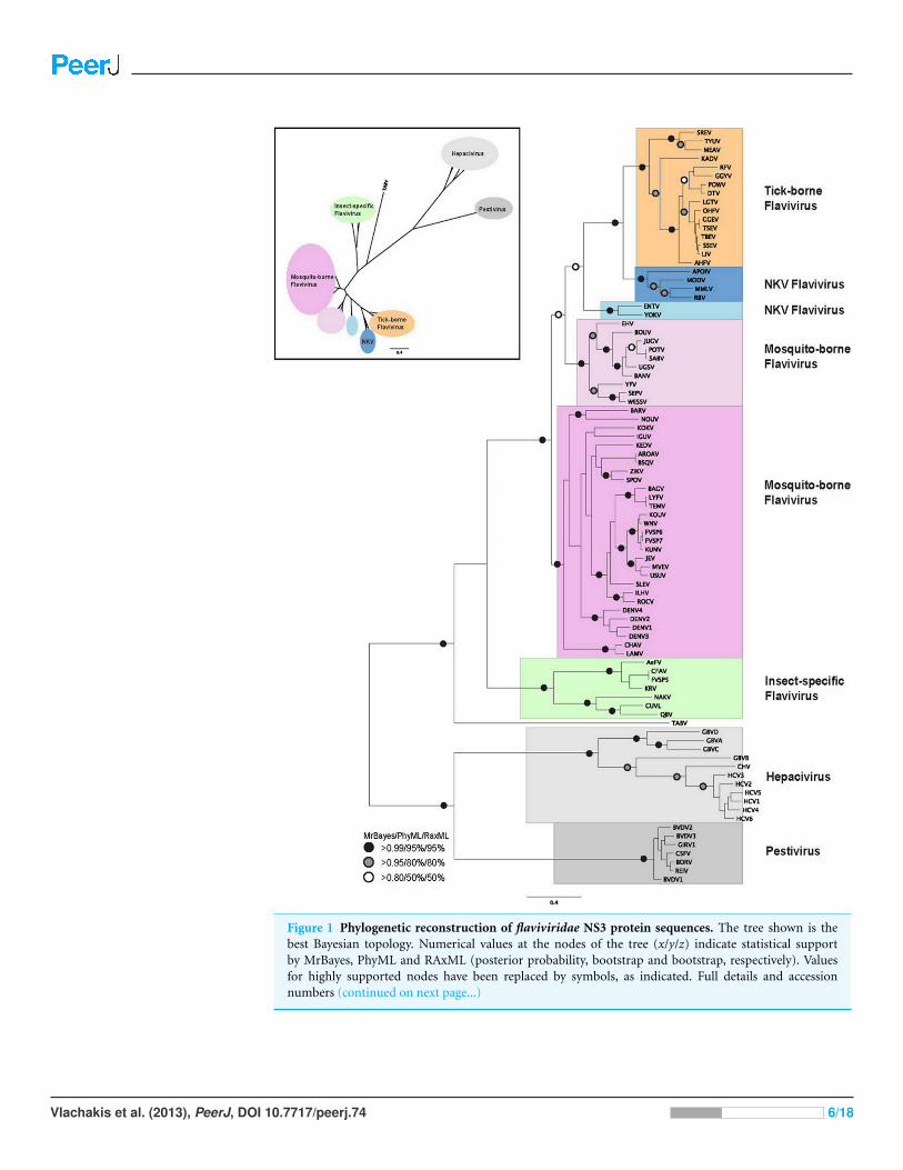
Figure 1 Phylogenetic reconstruction of flaviviridae NS3 protein sequences. The tree shown is thebest Bayesian topology. Numerical values at the nodes of the tree (x/y/z) indicate statistical supportby MrBayes, PhyML and RAxML (posterior probability, bootstrap and bootstrap, respectively). Valuesfor highly supported nodes have been replaced by symbols, as indicated. Full details and accessionnumbers (continued on next page...)
Vlachakis et al. (2013), PeerJ, DOI 10.7717/peerj.74 6/18
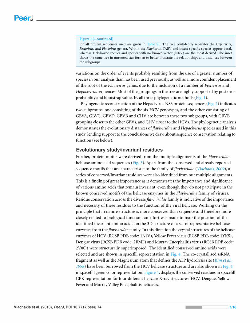
Figure 1 (...continued)
for all protein sequences used are given in Table S1. The tree confidently separates the Hepacivirs,Pestivirus, and Flavivirus genera. Within the Flavivirus, TABV and insect-specific species appear basal,whereas Tick-borne species and species with no known vector (NKV) are the most derived. The insetshows the same tree in unrooted star format to better illustrate the relationships and distances betweenthe subgroups.
variations on the order of events probably resulting from the use of a greater number of
species in our analysis than has been used previously, as well as a more confident placement
of the root of the Flavivirus genus, due to the inclusion of a number of Pestivirus and
Hepacivirus sequences. Most of the groupings in the tree are highly supported by posterior
probability and bootstrap values by all three phylogenetic methods (Fig. 1).
Phylogenetic reconstruction of the Hepacivirus NS3 protein sequences (Fig. 2) indicates
two subgroups, one consisting of the six HCV genotypes, and the other consisting of
GBVA, GBVC, GBVD. GBVB and CHV are between these two subgroups, with GBVB
grouping closer to the other GBVs, and CHV closer to the HCVs. The phylogenetic analysis
demonstrates the evolutionary distances of flaviviridae and Hepacivirus species used in this
study, lending support to the conclusions we draw about sequence conservation relating to
function (see below).
Evolutionary study/invariant residuesFurther, protein motifs were derived from the multiple alignments of the Flaviviridae
helicase amino acid sequences (Fig. 3). Apart from the conserved and already reported
sequence motifs that are characteristic to the family of flaviviridae (Vlachakis, 2009), a
series of conserved/invariant residues were also identified from our multiple alignments.
This is a finding of great importance as it demonstrates the importance and significance
of various amino acids that remain invariant, even though they do not participate in the
known conserved motifs of the helicase enzymes in the Flaviviridae family of viruses.
Residue conservation across the diverse flaviviridae family is indicative of the importance
and necessity of these residues to the function of the viral helicase. Working on the
principle that in nature structure is more conserved than sequence and therefore more
closely related to biological function, an effort was made to map the position of the
identified invariant amino acids on the 3D structure of a set of representative helicase
enzymes from the flaviviridae family. In this direction the crystal structures of the helicase
enzymes of HCV (RCSB PDB code: 1A1V), Yellow Fever virus (RCSB PDB code: 1YKS),
Dengue virus (RCSB PDB code: 2BMF) and Murray Encephalitis virus (RCSB PDB code:
2V8O) were structurally superimposed. The identified conserved amino acids were
selected and are shown in spacefill representation in Fig. 4. The co-crystallised ssRNA
fragment as well as the Magnesium atom that defines the ATP hydrolysis site (Kim et al.,
1998) have been borrowed from the HCV helicase structure and are also shown in Fig. 4
in spacefill green color representation. Figure 4, displays the conserved residues in spacefill
CPK representation for four different helicase X-ray structures: HCV, Dengue, Yellow
Fever and Murray Valley Encephalitis helicases.
Vlachakis et al. (2013), PeerJ, DOI 10.7717/peerj.74 7/18
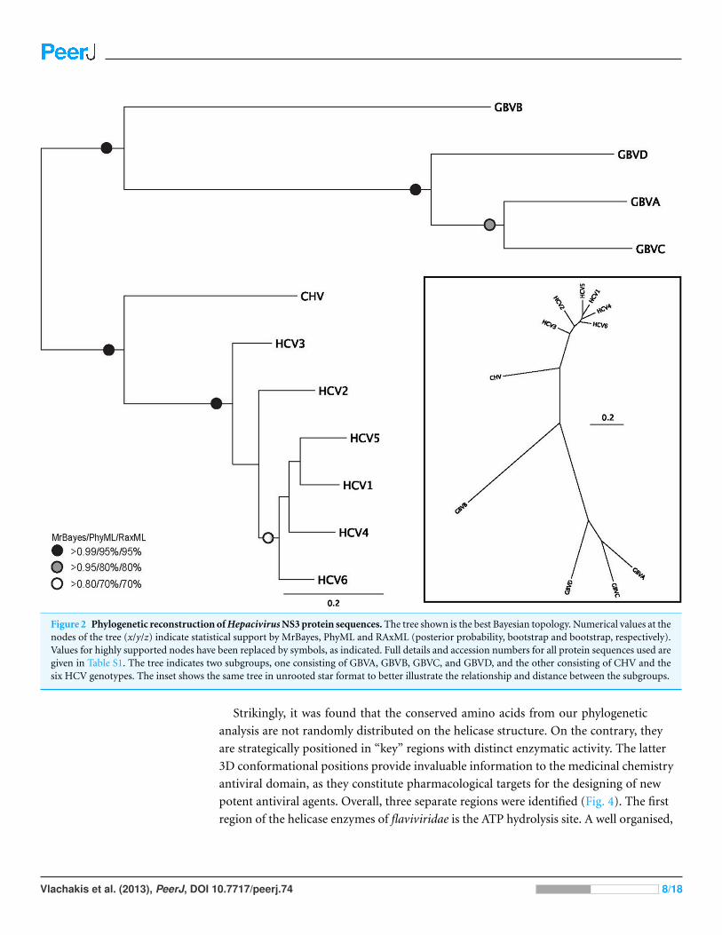
Figure 2 Phylogenetic reconstruction of Hepacivirus NS3 protein sequences. The tree shown is the best Bayesian topology. Numerical values at thenodes of the tree (x/y/z) indicate statistical support by MrBayes, PhyML and RAxML (posterior probability, bootstrap and bootstrap, respectively).Values for highly supported nodes have been replaced by symbols, as indicated. Full details and accession numbers for all protein sequences used aregiven in Table S1. The tree indicates two subgroups, one consisting of GBVA, GBVB, GBVC, and GBVD, and the other consisting of CHV and thesix HCV genotypes. The inset shows the same tree in unrooted star format to better illustrate the relationship and distance between the subgroups.
Strikingly, it was found that the conserved amino acids from our phylogenetic
analysis are not randomly distributed on the helicase structure. On the contrary, they
are strategically positioned in “key” regions with distinct enzymatic activity. The latter
3D conformational positions provide invaluable information to the medicinal chemistry
antiviral domain, as they constitute pharmacological targets for the designing of new
potent antiviral agents. Overall, three separate regions were identified (Fig. 4). The first
region of the helicase enzymes of flaviviridae is the ATP hydrolysis site. A well organised,
Vlachakis et al. (2013), PeerJ, DOI 10.7717/peerj.74 8/18
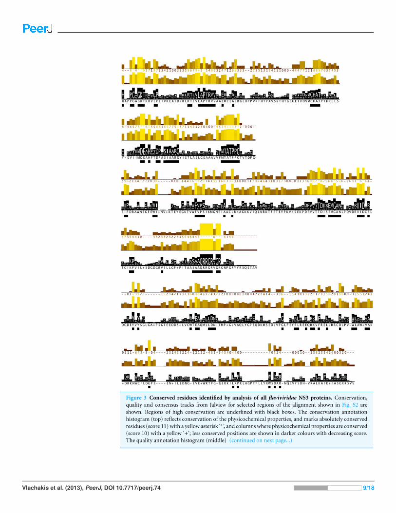
Figure 3 Conserved residues identified by analysis of all flaviviridae NS3 proteins. Conservation,quality and consensus tracks from Jalview for selected regions of the alignment shown in Fig. S2 areshown. Regions of high conservation are underlined with black boxes. The conservation annotationhistogram (top) reflects conservation of the physicochemical properties, and marks absolutely conservedresidues (score 11) with a yellow asterisk ‘*’, and columns where physicochemical properties are conserved(score 10) with a yellow ‘+’; less conserved positions are shown in darker colours with decreasing score.The quality annotation histogram (middle) (continued on next page...)
Vlachakis et al. (2013), PeerJ, DOI 10.7717/peerj.74 9/18
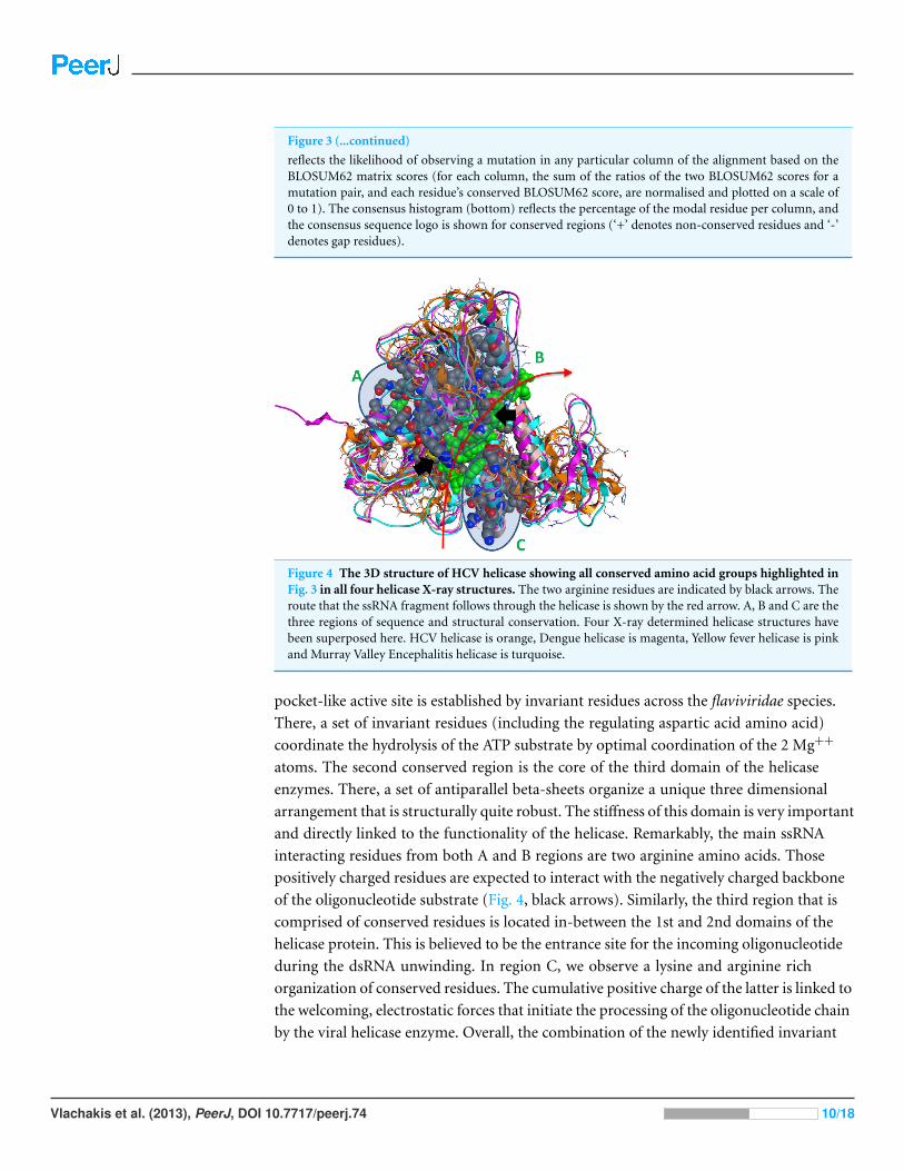
Figure 3 (...continued)
reflects the likelihood of observing a mutation in any particular column of the alignment based on theBLOSUM62 matrix scores (for each column, the sum of the ratios of the two BLOSUM62 scores for amutation pair, and each residue’s conserved BLOSUM62 score, are normalised and plotted on a scale of0 to 1). The consensus histogram (bottom) reflects the percentage of the modal residue per column, andthe consensus sequence logo is shown for conserved regions (‘+’ denotes non-conserved residues and ‘-’denotes gap residues).
Figure 4 The 3D structure of HCV helicase showing all conserved amino acid groups highlighted inFig. 3 in all four helicase X-ray structures. The two arginine residues are indicated by black arrows. Theroute that the ssRNA fragment follows through the helicase is shown by the red arrow. A, B and C are thethree regions of sequence and structural conservation. Four X-ray determined helicase structures havebeen superposed here. HCV helicase is orange, Dengue helicase is magenta, Yellow fever helicase is pinkand Murray Valley Encephalitis helicase is turquoise.
pocket-like active site is established by invariant residues across the flaviviridae species.
There, a set of invariant residues (including the regulating aspartic acid amino acid)
coordinate the hydrolysis of the ATP substrate by optimal coordination of the 2 Mg++
atoms. The second conserved region is the core of the third domain of the helicase
enzymes. There, a set of antiparallel beta-sheets organize a unique three dimensional
arrangement that is structurally quite robust. The stiffness of this domain is very important
and directly linked to the functionality of the helicase. Remarkably, the main ssRNA
interacting residues from both A and B regions are two arginine amino acids. Those
positively charged residues are expected to interact with the negatively charged backbone
of the oligonucleotide substrate (Fig. 4, black arrows). Similarly, the third region that is
comprised of conserved residues is located in-between the 1st and 2nd domains of the
helicase protein. This is believed to be the entrance site for the incoming oligonucleotide
during the dsRNA unwinding. In region C, we observe a lysine and arginine rich
organization of conserved residues. The cumulative positive charge of the latter is linked to
the welcoming, electrostatic forces that initiate the processing of the oligonucleotide chain
by the viral helicase enzyme. Overall, the combination of the newly identified invariant
Vlachakis et al. (2013), PeerJ, DOI 10.7717/peerj.74 10/18

Figure 5 Stochastic conformational search for C12 and C4. It was established that C12 only formed two relatively close clusters (coloured red),whereas C4 formed two sets of conformationally distant ones (coloured green).
residues, the charge distribution on the 3D structure of the HCV helicase and the relative
spatial arrangement of the helicase motifs constitute irreplaceable information than may
lead to the better understanding of the mechanism of action of the HCV helicase and the
consequent development of more potent anti-viral agents.
Structural study of the HCV helicaseHerein, an effort is made to combine all of the above evolution-based findings with
the available 3D structural information of flaviviridae helicases in order to explain and
rationalize the assay behaviour of our recently published HCV helicase inhibitors (Kandil
et al., 2009). The key element in this series of compounds (Fig. 5) is the Michael acceptor
moiety that they bear, which is capable of establishing strong covalent bonds with a
cysteine residue (Cys431), located in the ssRNA channel of the HCV helicase. As soon
as the Cys431 interaction is established, another hydrogen bond is formed with Arg481,
which is adjacent to the Cys431 residue (Figs. 6B and 6C). A further hydrogen bond to
Val432 helps C4, but not C12, to acquire the optimal directional axis required in the ssRNA
channel, which is in accordance with our previously proposed mode of helicase inhibition
for compounds C4 and C12. This is achieved by establishing H-bonding interactions with
the solvent-exposed Arg393 amino acid. Therefore, potent compounds of this series should
be able to not only establish covalent interactions with Cys431 and hydrogen bonding
interactions with Arg481 but also be able to retain a conformationally rigid pose and
reach the Arg393 residue. Hydrogen bonding to Arg393 would fixate the potential of those
compounds in the three dimensional space of the ssRNA HCV channel, thus inhibiting the
enzyme.
It was determined by our in-house, custom developed enzymatic assay (Kandil et al.,
2009) that whilst C4 is active in the submicromolar range, its stereoisomer counterpart
Vlachakis et al. (2013), PeerJ, DOI 10.7717/peerj.74 11/18
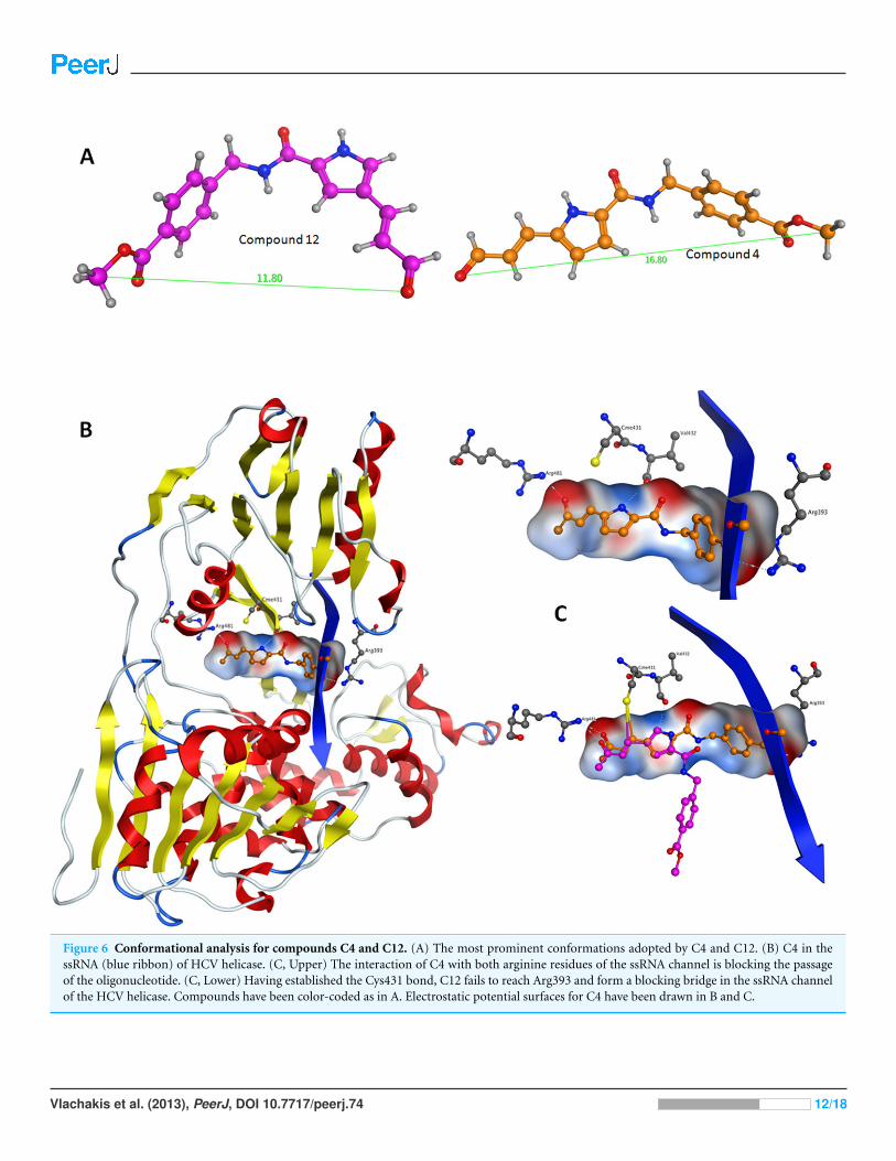
Figure 6 Conformational analysis for compounds C4 and C12. (A) The most prominent conformations adopted by C4 and C12. (B) C4 in thessRNA (blue ribbon) of HCV helicase. (C, Upper) The interaction of C4 with both arginine residues of the ssRNA channel is blocking the passageof the oligonucleotide. (C, Lower) Having established the Cys431 bond, C12 fails to reach Arg393 and form a blocking bridge in the ssRNA channelof the HCV helicase. Compounds have been color-coded as in A. Electrostatic potential surfaces for C4 have been drawn in B and C.
Vlachakis et al. (2013), PeerJ, DOI 10.7717/peerj.74 12/18
is completely inactive. That was very strange as some activity was expected from C12
too. Since the only difference between C4 and C12 is their unique stereochemical
variation, our molecular modelling approach allows us to correlate it with their observed
inhibitory potentials. Initially a stochastic conformational analysis was executed in
vacuo for compounds C4 and C12 using the implemented module in the Molecular
Operations Environment suite (MOE-2011.10). The degrees of freedom available for
each compound can be quantified and visualised, in order to identify the amount and
distribution of the 3D conformational space that each compound can occupy. To perform
a direct comparison between the two stereoisomers, the two counterpart benzene-COOH
moieties were structurally superposed and their potential in the 3D space was fixed. The
stochastic conformational search simulation was run for a total of 10000 iteration steps.
Analysis of the simulation revealed that compound C4 explored approximately 30% more
conformational space than compound C12. Namely, C4 explored 400 cubic Angstroms
(cA2), while C12 explored only 270 cA2. Moreover, while C12 seems to be trapped between
two relatively close conformational clusters, C4 explored the conformational space in all
directions between two separate conformational clusters, each one of which consisted of
two relatively close sub-clusters (Fig. 5). However, a more interesting observation was that
C12 spent most of the simulation time moving from an extended to a bended “U” like
shaped conformation (Fig. 6A). That led to a series of extensive docking and molecular
dynamics simulations, in an effort to investigate the conformational preferences of each
stereoisomer in the ssRNA channel of the HCV helicase.
The molecular docking experiments were performed using two distinctly different
algorithms: the MOE docking module and ZDock. It is of great interest to determine which
one is best for docking small molecule drug-like compounds in large docking sites, such as
the helicase ssRNA channel. Surprisingly, they produced very similar results, even though
they are based on different principles. MOE dock uses an Alpha Triangle displacement
method, suitable for rotating bonds and flexing small molecule compounds in an active
site. On the other hand, Zdock is a docking suite that utilizes a grid-based representation
of the molecular system involved. In order to efficiently explore the search space and
docking positions of the molecules as rigid bodies, ZDock takes full advantage of a three-
dimensional fast Fourier transformation algorithm. It uses a scoring function that returns
electrostatic, hydrophobic and desolvation energies as well as performing a fast pairwise
shape complementarity evaluation. A set of 100 top ranking poses for C4 and another
identical set of poses for C12 from the previously performed conformational search were
used as input structures for Zdock. For MOE-Dock an ideal energetically minimised
structure of each of the two stereoisomers was used, while the number of output poses was
limited to 100 per compound. That is because MOE-Dock can generate conformations
from a single 3D conformation by applying a collection of preferred torsion angles to the
rotatable bonds. MOE-Dock uses built-in scoring functions, where lower scores indicate
more favorable poses. The units for all scoring functions are kcal/mol. On the other hand,
the RDOCK algorithm must be utilized to refine, score and evaluate the results obtained
Vlachakis et al. (2013), PeerJ, DOI 10.7717/peerj.74 13/18
by ZDOCK. RDOCK performs a fast minimization step to the ZDOCK molecular complex
outputs and ranks them according to their re-calculated binding free energies.
Following the docking experiments a series of molecular dynamics simulations were
initiated for the two stereoisomers in the active site of the HCV helicase. The molecular
docking results were filtered and only those interacting with the previously reported key
residues of the HCV helicase channel were selected. Molecular complexes consisting of
the HCV helicase receptor and the highest ranking docking poses for C4 and C12 were
subjected to unrestrained molecular dynamics simulations using the MOE molecular
dynamics module. The Michael acceptor covalent bond between each inhibitor compound
and the receptor was manually established to ensure that the compounds would remain in
the active site’s proximity. All molecular complexes were energetically minimized prior to
molecular dynamics, in order to relax any potential geometrical strain in the starting files.
Molecular dynamics simulations were performed in a periodic box that was subsequently
solvated with simple point charge (SPC) water. Partial charges were applied using the
built-in MOE PEOE utility. Counter ions were added accordingly as required to neutralize
the molecular system. Temperature was set to 300 K, pressure to 1 atm and the step size
to 1 fs. The total run of each helicase – inhibitor molecular complex was 50 ns, using the
default NVT ensemble in a canonical environment. NVT simulations retain the number
of atoms, Volume and Temperature constant throughout the calculations. Analysis of the
molecular dynamics simulation trajectories (Figs. 6B and 6C) revealed that C12 tends to
bend, forming a 45 to 75 degree angle that gives it a “U” like shape and makes it less rigid
while, for the majority of its conformational time, the compound remains shorter than C4
at an average length of 11.8 angstroms. C4’s average length throughout the simulation was
measured to be 16,8 angstroms (Fig. 6A). C4 is far more rigid and ideal for the formation of
the necessary hydrogen bonds with the two arginine residues in HCV helicase.
Based on the biological data previously published on the two stereoisomers and the
current 3D modelling study, we conclude that the compound’s steric rigidity, which locks
it in a 3D linear axis conformation is essential for the activity of this family of compounds.
Moreover, a speculation is made on the potential activity of C4 against the Dengue helicase,
as it was observed that the ssRNA channel of Dengue helicase overlaps with high accuracy
with its corresponding channel on the HCV helicase (data not shown). More interestingly,
it was found that the cysteine residue of HCV (Cys431) superposes with another cysteine
that is located in the same position of the Dengue helicase channel, when the two enzymes
are structurally superimposed. Therefore the latter cysteine residues are key candidates
to be considered as pharmacological targets, as they are both exposed to solvent, while
strategically located in the centre of the ssRNA channel of the viral helicase. Taken together,
it was demonstrated that C4 adopts a rather linear-extended conformation throughout
the molecular dynamics simulations. This property allows it to hydrogen bond to Arg481,
Val432 and Arg393 while being covalently bound to Cys431. Collectively, it is proposed
that the hydrogen bonding to Arg393, Arg481, Val432 and the covalent bond to Cys431
are responsible for fixating C4 in the ssRNA channel of HCV, which irreversibly blocks the
passage of the incoming oligonucleotide and potently inhibits the HCV helicase enzyme.
Vlachakis et al. (2013), PeerJ, DOI 10.7717/peerj.74 14/18
Modern computer-based methodologies are now becoming an integral part of the drug
discovery process that may eventually lead to the development of promising and effective
antiviral strategies. Overall, the current systematic and thorough study of the HCV
helicase in terms of its unique structural characteristics, the underlying mechanism that is
responsible for the difference observed in the activity of the C12 and C4 stereoisomers and
the information obtained by our in-depth phylogenetic and evolutionary study, provide
invaluable insights into the mechanism of action of this viral enzyme and may be used
for the design of novel compounds that will effectively inhibit the action of the helicase,
subsequently stopping the proliferation and infection of the virus.
ACKNOWLEDGEMENTSThe authors would like to sincerely thank Spyridon Champeri Tsanira for critically
reviewing the manuscript. The authors also acknowledge the contribution of data
deposited in the ViPR database, to the present study. The Virus Pathogen Database and
Analysis Resource (ViPR) has been wholly funded with federal funds from the National
Institute of Allergy and Infectious Diseases, National Institutes of Health, Department of
Health and Human Services, under Contract No. HHSN272200900041C.
ADDITIONAL INFORMATION AND DECLARATIONS
FundingVLK is funded through an FP7-PEOPLE-2011-IEF Marie Curie fellowship. The funders
had no role in study design, data collection and analysis, decision to publish, or
preparation of the manuscript.
Grant DisclosuresThe following grant information was disclosed by the authors:
FP7-PEOPLE-2011-IEF Marie Curie fellowship.
Competing InterestsThe authors declare there are no competing interests.
Author Contributions• Dimitrios Vlachakis and Sophia Kossida conceived and designed the experiments,
performed the experiments, analyzed the data, contributed reagents/materials/analysis
tools, wrote the paper.
• Vassiliki Lila Koumandou performed the experiments, analyzed the data, contributed
reagents/materials/analysis tools, wrote the paper.
Patent DisclosuresThe following patent dependencies were disclosed by the authors:
Diana, G. Bailey, T. (1997). Compounds, compositions and methods for treatment of
hepatitis C. United States Patent No. 5,633,388.
Vlachakis et al. (2013), PeerJ, DOI 10.7717/peerj.74 15/18
Supplemental InformationSupplemental information for this article can be found online at http://dx.doi.org/
10.7717/peerj.74.
REFERENCESAbascal F, Zardoya R, Posada D. 2005. ProtTest: selection of best-fit models of protein evolution.
Bioinformatics 21:2104–2105 DOI ./bioinformatics/bti.
Brancale A, Vlachaki C, Vlachakis D. 2008. Molecular modelling study of the 3D structure of thebovine viral diarrhea virus (BVDV) helicase. In Silico Biology 8(5–6):461–469.
Calisher CH, Gould EA. 2003. Taxonomy of the virus family Flaviviridae. Advances in VirusResearch 59:1–19 DOI ./S-()-.
Chen R, Li L, Weng Z. 2003. ZDOCK: an initial-stage protein-docking algorithm. Proteins52:80–87 DOI ./prot..
Cook S, Holmes EC. 2006. A multigene analysis of the phylogenetic relationships among theflaviviruses (Family: Flaviviridae) and the evolution of vector transmission. Archives of Virology151:309–325 DOI ./s---.
Cook S, Moureau G, Kitchen A, Gould EA, de Lamballerie X, Holmes EC, Harbach RE. 2012.Molecular evolution of the insect-specific flaviviruses. Journal of General Virology 93:223–234DOI ./vir..-.
Daly M, Ward F. 2003. Liver disease. In: Walker R, Edwards C, eds. Clinical pharmacy andtherapeutics. 3rd ed. Churchill Livingstone, 209–228.
Diana G, Bailey T. 1997. Compounds, compositions and methods for treatment of hepatitis C.United States Patent No. 5,633,388.
Degos F. 1994. Epidemiology of hepatitis C virus in Europe. FEMS Microbiology Reviews14(3):267–271 DOI ./j.-..tb.x.
DeLano WL. 2002. The PyMOL user’s manual San Carlos. CA, USA: DeLano Scientific.
Duan Y, Wu C, Chowdhury S, Lee MC, Xiong G, Zhang W, Yang R, Cieplak P, Luo R, Lee T,Caldwell J, Wang J, Kollman P. 2003. A point-charge force field for molecular mechanicssimulations of proteins based on condensed-phase quantum mechanical calculations. Journal ofComputational Chemistry 24:1999–2012 DOI ./jcc..
Edgar RC. 2004. MUSCLE: multiple sequence alignment with high accuracy and high throughput.Nucleic Acids Research 32:1792–1797 DOI ./nar/gkh.
Eisenberg D, Luthy R, Bowie JU. 1997. VERIFY3D: assessment of protein models with three-dimensional profiles. Methods in Enzymology 277:396–404 DOI ./S-()-.
Ferron F, Bussetta C, Dutartre H, Canard B. 2005. The modeled structure of the RNA dependentRNA polymerase of GBV-C virus suggests a role for motif E in Flaviviridae RNA polymerases.BMC Bioinformatics 6:255 DOI ./---.
Guex N, Peitsch MC. 1997. SWISS-MODEL and the Swiss-PdbViewer: an environment forcomparative protein modeling. Electrophoresis 18:2714–2723 DOI ./elps..
Guindon S, Gascuel O. 2003. A simple, fast, and accurate algorithm to estimate large phylogeniesby maximum likelihood. Systematic Biology 52:696–704 DOI ./.
Guzman MG, Kourı G. 2004. Dengue diagnosis, advances and challenges. International Journal ofInfectious Diseases 8(2):69–80 DOI ./j.ijid....
Vlachakis et al. (2013), PeerJ, DOI 10.7717/peerj.74 16/18
Hess B, Kutzner C, van der Spoel D, Lindahl E. 2008. GROMACS 4: algorithms for highlyefficient, load-balanced, and scalable molecular simulation. Journal of Chemical Theory andComputation 4:435–447 DOI ./ctq.
Kadare G, Haenni AL. 1997. Virus-encoded RNA helicases. Journal of Virology 71:2583–2590.
Kandil S, Biondaro S, Vlachakis D, Cummins AC, Coluccia A, Berry C, Leyssen P,Neyts J, Brancale A. 2009. Discovery of a novel HCV helicase inhibitor by a de novodrug design approach. Bioorganic & Medicinal Chemistry Letters 19(11):2935–2937DOI ./j.bmcl....
Kim JL, Morgenstern KA, Griffith JP, Dwyer MD, Thomson JA, Murcko MA, Lin C, Caron PR.1998. Hepatitis C virus NS3 RNA helicase domain with a bound oligonucleotide: thecrystal structure provides insights into the mode of unwinding. Structure 6(1):89–100DOI ./S-()-.
Laskowski RA, Rullmann JA, MacArthur MW, Kaptein R, Thornton JM. 1996. AQUA andPROCHECK-NMR: programs for checking the quality of protein structures solved by NMR.Journal of Biomolecular NMR 8:477–486 DOI ./BF.
Li L, Chen R, Weng Z. 2003. RDOCK: refinement of rigid-body protein docking predictions.Proteins 53:693–707 DOI ./prot..
Lobo FP, Mota BE, Pena SD, Azevedo V, Macedo AM, Tauch A, Machado CR, Franco GR. 2009.Virus-host coevolution: common patterns of nucleotide motif usage in Flaviviridae and theirhosts. PLoS ONE 4:e6282 DOI ./journal.pone..
Maddison WP, Maddison DR. 1989. Interactive analysis of phylogeny and characterevolution using the computer program MacClade. Folia Primatol (Basel) 53:190–202DOI ./.
Neyts J, Leyssen P, De Clercq E. 1999. Infections with flaviviridae. Verhandelingen - KoninklijkeAcademie Voor Geneeskunde Van Belgie 61(6):661–697. discussion 697-9.
Nulf CJ, Corey D. 2004. Intracellular inhibition of hepatitis C virus (HCV)internal ribosomalentry site (IRES)-dependent translation by peptide nucleic acids (PNAs) and locked nucleicacids (LNAs). Nucleic Acids Research 32(13):3792–3798 DOI ./nar/gkh.
Pasta L, Marrone C, D’amico M, Bevacqua EA, D’Amico G, Pagliaro L. 2005. Familial clusteringof HCV-related liver cirrhosis and hepatocellular carcinoma. Digestive and Liver Disease37(9):716–717 DOI ./j.dld....
Pettersen EF, Goddard TD, Huang CC, Couch GS, Greenblatt DM, Meng EC, Ferrin TE. 2004.UCSF Chimera–a visualization system for exploratory research and analysis. Journal ofComputational Chemistry 25:1605–1612 DOI ./jcc..
Pickett BE, Sadat EL, Zhang Y, Noronha JM, Squires RB, Hunt V, Liu M, Kumar S, Zaremba S,Gu Z, Zhou L, Larson CN, Dietrich J, Klem EB, Scheuermann RH. 2012. ViPR: an openbioinformatics database and analysis resource for virology research. Nucleic Acids Research40:D593–D598 DOI ./nar/gkr.
Phoon C, Ng P, Ting A, Yeo S, Sim M. 2001. Biological evaluation of hepatitis C virus helicaseinhibitors. Bioorganic & Medicinal Chemistry Letters 11:1647–1650 DOI ./S-X()-.
Poynard T, Bedossa P, Opolon P. 1997. Natural history of liver fibrosis progression in patientswith chronic hepatitis C. Lancet 349:825–832 DOI ./S-()-.
Ronquist F, Huelsenbeck JP. 2003. MrBayes 3: Bayesian phylogenetic inference under mixedmodels. Bioinformatics 19:1572–1574 DOI ./bioinformatics/btg.
Vlachakis et al. (2013), PeerJ, DOI 10.7717/peerj.74 17/18
Rosenberg S. 2001. Recent advances in the molecular biology of hepatitis C virus. Journal ofMolecular Biology 313:451–464 DOI ./jmbi...
Shepard CW, Finelli L, Alter MJ. 2005. Global epidemiology of hepatitis C virus infection. TheLancet Infectious Diseases 5(9):558–567 DOI ./S-()-.
Stamatakis A. 2006. RAxML-VI-HPC: maximum likelihood-based phylogeneticanalyses with thousands of taxa and mixed models. Bioinformatics 22:2688–2690DOI ./bioinformatics/btl.
Sellis D, Vlachakis D, Vlassi M. 2009. Gromita: a fully integrated graphical user interface togromacs 4. Bioinformatics and Biology Insights 3:99–102.
Thompson B, Finch R. 2005. Hepatitis C virus infection. Clinical Microbiology and Infection11(2):86–94 DOI ./j.-...x.
Vlachakis D. 2009. Theoretical study of the Usutu virus helicase 3D structure, by means ofcomputer-aided homology modelling. Theoretical Biology and Medical Modelling 25(6):9DOI ./---.
Vlachakis D, Brancale A. 2007. The 3D structure of the Japanese Encephalitis helicase,using homology based molecular modelling techniques. Online Journal of Bioinformatics8(2):154–164.
Waterhouse AM, Procter JB, Martin DM, Clamp M, Barton GJ. 2009. Jalview Version 2–amultiple sequence alignment editor and analysis workbench. Bioinformatics 25:1189–1191DOI ./bioinformatics/btp.
Vlachakis et al. (2013), PeerJ, DOI 10.7717/peerj.74 18/18