A Histologic Evaluation of Circumferential Filing Versus ...
102
Loyola University Chicago Loyola University Chicago Loyola eCommons Loyola eCommons Master's Theses Theses and Dissertations 1981 A Histologic Evaluation of Circumferential Filing Versus Reaming A Histologic Evaluation of Circumferential Filing Versus Reaming in Canal Debridement in Canal Debridement Terry Lee Kippa Loyola University Chicago Follow this and additional works at: https://ecommons.luc.edu/luc_theses Part of the Oral Biology and Oral Pathology Commons Recommended Citation Recommended Citation Kippa, Terry Lee, "A Histologic Evaluation of Circumferential Filing Versus Reaming in Canal Debridement" (1981). Master's Theses. 3179. https://ecommons.luc.edu/luc_theses/3179 This Thesis is brought to you for free and open access by the Theses and Dissertations at Loyola eCommons. It has been accepted for inclusion in Master's Theses by an authorized administrator of Loyola eCommons. For more information, please contact [email protected]. This work is licensed under a Creative Commons Attribution-Noncommercial-No Derivative Works 3.0 License. Copyright © 1981 Terry Lee Kippa
-
Upload
khangminh22 -
Category
Documents
-
view
0 -
download
0
Transcript of A Histologic Evaluation of Circumferential Filing Versus ...

Loyola University Chicago Loyola University Chicago
Loyola eCommons Loyola eCommons
Master's Theses Theses and Dissertations
1981
A Histologic Evaluation of Circumferential Filing Versus Reaming A Histologic Evaluation of Circumferential Filing Versus Reaming
in Canal Debridement in Canal Debridement
Terry Lee Kippa Loyola University Chicago
Follow this and additional works at: https://ecommons.luc.edu/luc_theses
Part of the Oral Biology and Oral Pathology Commons
Recommended Citation Recommended Citation Kippa, Terry Lee, "A Histologic Evaluation of Circumferential Filing Versus Reaming in Canal Debridement" (1981). Master's Theses. 3179. https://ecommons.luc.edu/luc_theses/3179
This Thesis is brought to you for free and open access by the Theses and Dissertations at Loyola eCommons. It has been accepted for inclusion in Master's Theses by an authorized administrator of Loyola eCommons. For more information, please contact [email protected].
This work is licensed under a Creative Commons Attribution-Noncommercial-No Derivative Works 3.0 License. Copyright © 1981 Terry Lee Kippa
A HISTOLOGIC EVALUATION OF CIRCUMFERENTIAL
FILING VERSUS REAMING IN CANAL DEBRIDEMENT
BY Terry L. Kippa
A Thesis Submitted to the Faculty of the Graduate School
of Loyola University of Chicago in Partial Fulfillment
of the Requirements for the Degree of
Master of Science
May
1981
DEDICATION
To Virginia, my wife, who has freely given me her
love and support, and to Shelly and Jeff who have been
understanding beyond their years.
ii
ACKNOWLEDGMENTS
To Dr. Franklin Weine, my committee chairman and teacher, I
extend my sincere gratitude and respect for not only his professional
guidance, but also for his friendship and insights to personal living.
To Dr. Gary Taylor, my friend and advisor, I thank him for his
contribution to this thesis and my overall endodontic education.
To Dr. Hal McReynolds, I thank him for his friendship and
assistance with the histologic portion of this study.
To Dr. Scott Shellhammer and Dr. Lance Crawford, my friends
and fellow students, I extend my appreciation for their time spent
in helping me to complete this thesis.
iii
VITA
The author, Terry Lee Kippa, is the son of Herbert A. Kippa and
Ethel (Larson) Kippa. He was born on July 3, 1945 in Oshkosh, Wisconsin.
His elementary and secondary education were obtained in the public
schools of Oshkosh, Wisconsin, where he graduated from high school in
1963.
A Bachelor of Science in Civil Engineering was earned at the
University of Wisconsin in Madison in June 1967. While attending the
University of Wisconsin he was selected for membership in Chi Epsilon
National Honorary Civil Engineering Fraternity and Tau Beta Pi National
Honorary Engineering Society.
In October 1967, he entered the United States Navy and was com
missioned a line officer in Janaury 1968. He served in the Navy until
Septemb~r 1969.
After one year of pre-dental study at the University of Rhode
Island, he entered the University of Iowa School of Dentistry. In
August 1972 he commenced active duty in the U.S. Navy Dental Corps,
completing dental school on a Navy scholarship and receiving the degree
of Doctor of Dental Surgery in May 1974. While attending dental school
he was elected a member of Omicron Kappa Upsilon.
In July 1974 he began a general practice residency at the Naval
Hospital, Camp Pendleton, California. Upon completion of the residency,
iv

he was transferred to Pearl Harbor, Hawaii where he served as a general
dentist until June 1979.
The author•s wife is Virginia L., and their children are Michelle
Anne and Jeffrey Patrick.
v
TABLE OF CONTENTS
DEDICATION ...
ACKNOWLEDGMENTS
VITA ..... .
TABLE OF CONTENTS
LIST OF TABLES.
LIST OF FIGURES
Chapter
I. INTRODUCTION ...
.•
II. LITERATURE REVIEW ..
A. Historical Background .
B. Canal Preparation
III. MATERIALS AND METHODS.
IV. RESULTS. .
V. DISCUSSION .
SUMMARY
REFERENCES.
FIGURES ..
vi
PAGE
ii
iii
iv
vi
vii
viii
1
3
3
12
50
58
70
78
80
86
LIST OF TABLES
Table
1. Initial Instrument Sizes and Average Working Lengths .....
2. Scores from Evaluator #1
3. Scores from Evaluator #2
4. Scores from Evaluator #3
5.
6.
7.
8.
9.
10.
11.
Average Score for Each Root Combining All Levels and All Evaluators .....
Average Score at Each Level Combining All Roots and All Evaluators .
Average Score at Each Level in Each Root for Filing and Reaming . . . . . Statistically Significantly Differences Between Reaming and Filing Scores. . . Eva 1 ua tor #1 - Significant Differences Between Reaming and Filing Scores. . . Evaluator #2 - Significant Differences Between Reaming and Filing Scores. . . Evaluator #3 - Significant Differences Between Reaming and Filing Scores. . .
vii
. . .
. . . .
. . . .
. . . .
. . . .
. .
. .
. .
. .
. .
Page
62
63
64
65
66
66
67
67
68
68
69
LIST OF FIGURES
Figure
1. Radiographic Technique- X-Ray Unit ..
2. Radiographic Technique - Film in Place
3. Files in Place with Silicone Stops
4. Radiograph - Working Length Files.
5. Histologic Section - 11 Keyhole 11 Type Preparation. . . . . . . . .
6. Histologic Section - Deviation of Prepared Canal . . . . . . .
7. Histologic Section- Grooved Walls of Filed Canal ......... .
8. Histologic Section - Unstained Areas Caused by Action of NaOCl ..... .
9. Histologic Section - Canal Scored as a 11 111
10. Histologic Section - Canal Scored as a 11 211
11. Histologic Section - Canal Scored as a 11 311
12. Histologic Section - Canal Scored as a 11 411
13. Histologic Section - Canal Scored as a 11 511
14. Radiograph - Orifice Constriction ...
viii
Page
86
86
87
87
88
88
89
89
90
90
91
91
92
92
CHAPTER I
INTRODUCTION
Endodontics has evolved from a field where practitioners attempted
to remove from a patients tooth some mysterious cause for their toothache
to a currently sophisticated specialty of dentistry that enjoys a 95%
success rate. Root canal therapy is better understood and more accepted
today by both the dental profession and general public than at any other
time in history. Whereas endodontic treatment was once considered a
hazard to the patients general health, it is now recognized as a safe and
reliable means of retaining teeth in a functional state that at one time
would have been doomed to extraction.
In the first few pages that follow, a brief historical background
of the practice of endodontics will be given. This will show the progres
sion of treatment to the present day emphasis on canal preparation. This
historical background will be followed by a more detailed review of the
literature on canal preparation.
It will be seen from the literature that there are a variety of
techniques for canal preparation. Although there are strong advocates of
each of the methods, no one technique has proven to be vastly superior to
the others with regard to canal debridement. Furthermore, the literature
shows that no method of preparation has been successful in thoroughly
cleansing the critically important apical portion of the canals studied.
1
It is apparent then that further investigation into the cleansing of
canals in endodontic treatment is warranted. Therefore, it is the
purpose of this study to evaluate histologically two techniques of
2
canal preparation. These techniques will be compared as to their ability
to cleanse the apical 4 mm of the canal.
CHAPTER II
LITERATURE REVIEW
A. HISTORICAL BACKGROUND
Throughout history there have been many methods of treating teeth
which required endodontic therapy. The earliest and most basic form of
therapy was extraction. There are, however, very early records of man at-
tempting to relieve toothaches by treating the pulp rather than removing
the tooth. In the later Middle Ages, French anatomist Ambrose Pare (1517-
1592) stated, "Toothache is, of all others, the most atrocious pain that
can torment a man, being followed by death. To combat this one must re-
course to cauterization." In cauterization, he explained, .. One burns the
nerve, thus rendering it incapable of again feeling or causing pain ...
The use of instruments made specifically for total pulp removal appears
to have surfaced in the mid 1800 1 S. In 1838 Edward Maynard made barbed
broaches from the untempered steel of watch springs filed down to the fine-
ness of horse hair which were barbed on one side. Maynard also made ream-1
ers from piano wire and filed them to the desired shape.
Probably due to the lack of readily available endodontic instruments,
dentists attempted to clean these canals by other means. In 1883 the pro
, cess of 11 knocki ng out the pul p11 was described by Mi 11 s. The procedure in-
valved tapping a pointed orangewood stick.that had been dipped in carbolic
acid into the canal. The wood was left there for a minute and then pulled
3
4
out with the pulp coming with it. Although some dentists claimed this to
be a relatively painless operation, one patient stated that 11 it seemed 2
that a broom handle had been thrust up through his head. 11
Schreier, 1893, introduced a combination of sodium and potassium for 3
removing pulp tissue. In 1894, Callahan recommended sulfuric acid as an 4
aid in opening and cleaning small and tortuous canals. In 1923, Johnston
also described the use of sulfuric acid for widening and shaping small 5
curved canals. Although these techniques were crude and not based on re-
sults of sound scientific study, they do show the concern of the dentist
to clean the canal thoroughly.
In the early 1900's bacteria were implicated as etiologic agents of 6
pulp disease. This finding was followed by Hunter's classic report to
the Faculty of Medicine of McGill University which warned that oral sepsis 7
and infection could cause many systemic diseases. The principle of what
Hunter called oral sepsis was that organisms from diseased teeth would be
spread by blood and lymphatic vessels throughout the body and result in
focal infections. This theory resulted in the condemnation of all pulp-8
less and pulpally involved teeth and their wholesale extraction. This
philosophy continued for many years and was supported by many, what later
proved to be erroneous, bacteriologic studies.
Coolidge, in 1932, wrote, 11That the tooth of a young person should
be extracted just because the pulp has become exposed is one of the worst
blots on the escutcheon of dental practice in history ... He also stated
that, "The possibility of saving the tooth whose pulp has undergone pu
trefaction depends on control of the infection, mechanical cleansing of
the canal and disinfection of the dentin and inaccessible canals followed 9
by complete closing of the apical foramen."
Fish and t~aclean in their study negated the claims that the root
surfaces of pulpally involved teeth were infected with micro-organisms.
They showed that organisms cultured from the root apices of extracted
teeth or from the bloodstream after extraction had been pumped into the 10
vessels from the gingival sulcus during extraction.
5
These results gave support to dental practioners who felt that teeth
could be saved through conservative endodontic procedures. However, in
order to justify their conservative position to the advocates of the focal
infection theory and extraction, their goal was to attain a sterile canal.
This led to the use of various intracanal medicaments and irrigants, dif-8
ferent techniques of mechanical preparation and to culturing. The cul-
ture was used to prove that a sterile canal had been attained and the
tooth would no longer serve as a focus of infection.
There has been much discussion in the endodontic literature as to
the best irrigant, the best intracanal medicament or whether medicaments
are necessary at all. Also the importance of these facets of treatment
relative to the mechanical debridement has received much attention.
Many of the first endodontists were also pharmacists and they tended
to place the major emphasis on drug therapy in the root canal. For years
the endodontic literature was filled with dissertations on the role of 11
intracanal medications in rendering root canals sterile.
Cresote and phenol were introduced for use as canal medicaments in
the early 1800's and have been used in various combinations since that

time. Formocreosol became prominent in endodontic therapy following its
introduction by Buckley in 1904. Antibiotics and anti-fungal drugs are
still used within the canal by some. In 1951, Grossman introduced his
6
PBSC paste which contained penicillin, bacitracin, streptomycin and capry
late sodium. There were other drugs used and many were very caustic and
irritating to the periapical tissues.
Studies such as those by Hedman and Shovelton showed that periapical 12
lesions were either sterile to begin with or could be rendered sterile 13
by thorough cleansing of the canal. These studies as well as one by
Engstrom and Frostell have shown that it is not necessary to have bacteria
either in the canal or in the periapical tissues in order to have periap-14
ical pathology. After such findings it became clear that some of the
painful sequelae of endodontic treatment were not caused by infection but 11
by overly strong drugs used within the canal. Rothschild agreed that
potent and destructive medicaments do great harm to normal tissues.
The situation may be summed up by stating that of prime importance is the
removal of debris which nurtures bacteria rather than attempting to ster-15
ilize it in situ. No amount of medication will disinfect an unclean
canal. The realization that microorganisms and their substrates should
be removed instead of being sterilized within the root canal is one of 11
the major advances in endodontic practice. This sentiment towards
placing less importance on medicaments and more on debridement is felt by 8,16,17,18,19
many other endodontists. Bhaskar showed in a study on dogs•
teeth that root canal debridement and occlusal seal alone apparently 20
stopped the growth of apical lesions. Weine has noted this same effect
16 in humans. As a result of this shift in thinking, the use of an irri-
gant and the search for the best irrigant to be used received more em
phasis in the preparation of the canal.
7
In Coolidge's article he recommended the use of "chlorine solutions"
in irrigating canals. In the treatment of wounds in World War I, it had
proven to be a powerful and penetrating germicide that did not cause much
injury to living tissue. Walker, 1936, wrote that judicious use of a
chemical irrigant is helpful in cleansing pulp canals. For this purpose,
he recommended double strength chlorinated soda because of its germicidal 21
property and its ability to dissolve organic material.
Grossman and Meiman added further credence to the use of chlorinated
solutions when they showed that chlorinated soda is an effective solvent
of pulp tissue. They found that it will dissolve pulps of freshly ex
tracted teeth in less than 24 hours and at times in less than one hour.
They also stated that the elimination of necrotic pulp tissue from the 22
root canal is important for the ultimate success of the operation.
Contrary to these studies, Baker, et al ., reported in a comparison
of various irrigating solutions used in preparation of freshly extracted
teeth that NaOCl did not show any ability to dissolve pulpal tissue. The
removal of debris and microorganisms seemed to be a function of the quan-23
tity of irrigating solution rather than of the type of solution used.
Studies were done to evaluate the effectiveness of NaOCl as a bac-
teriocidal irrigant during canal preparation. Auerbach, in a study in-
volving 60 nonvital teeth, found that 78% of the teeth which had positive
initial cultures yielded negative cultures after debridement of the canals
24 with chlorinated soda as an irrigant.
8
Stewart in 1955 reported two successive negative cultures in approx-
imately 76% of infected canals after chemomechanical preparation in which 25
3% hydrogen peroxide and sodium hypochlorite was used.
In 1958 Ingle and Zeldow reported on a study designed to show that
a chemical irrigant was a necessary adjunct to mechanical instrumentation
in the reduction of bacterial flora of the canal. They instrumented 89
teeth with nonvital pulps with sterile distilled water as an irrigant.
Results of their study showed that only 4.6% of infected canals yielded
two successive growth-free cultures. These findings show the importance
of the antibacterial action of irrigating agents used by Auerbach and 26
Stewart.
Nicholls, 1962, reported on a study that was designed to assess the
effect of variation in irrigating agents used during instrumentation upon
the bacteriological status of the canal. Comparing alkaline chloramine,
H2o2 and NaOCl, and distilled H2o they also concluded that the reduction
in bacterial population is to some extent associated with the antiseptic 27
effect of the irrigants.
Shih, et al., reported that irrigation with full strength (5.2%Na0Cl)
Clorox does not ensure the lasting sterility of an inoculated canal. They
also concluded that a negative culture report after treatment indicates
that the bacterial population in the root canal may be highly reduced, not 28
that the canal is sterile.
Unfortunately, the use of NaOCl does not guarantee that the canal
is thoroughly debrided. Senia, et al., in 1971 reported on a study that
was designed to evaluate the solvent action of Clorox (5.2% NaOCl) in
canals of extracted mandibular molars. They found that full strength
Clorox did not appear to be very effective in removing pulp tissue which
remained after instrumentation. In their study, there was no significant
difference in the cleaning effect of Clorox as compared to normal saline
solution at the 1 mm and 3 mm levels from the apex. Neither solution was 29
effective in removing debris left by instrumentation.
This finding was confirmed by Baker, et al ., in 1975 when they re
ported a SEM study on the efficacy of various irrigating solutions in-
eluding saline, H2o2, H2o2 plus NaOCl, NaOCl, Glyoxide, Glyoxide plus
NaOCl, RC Prep, and EDTA. The study indicated that even when teeth were
instrumented and irrigated, significant amounts of tissue and debris re-23
mained in the prepared root canal system.
9
Svec and Harrison (1977) compared the cleanliness of canals prepared
with NaOCl and hydrogen peroxide to those prepared with normal saline.
Although the NaOCl and H2o2 combination was found to be significantly more 30
effective, pulpal and dentinal debris were found in almost every section.
It has become clear that no irrigant or combination of irrigants
could completely cleanse a canal of debris. Thus the emphasis in endo
dontic therapy has shifted from relying on strong intracanal medicaments
to cleansing canals with irrigants to its present day stress on the mech
anical removal of debris with the aid of an irrigant. 19 31
Reports by Walton and Rubin, et al., have reinforced the find-
ings of the studies evaluating irrigants. Their studies on instrumenta-
tion techniques both showed that the debris is removed by the mechanical
10
action of the instruments. In other words, debris remained wherever the
instruments did not actually contact and remove it.
This line of thought is confirmed by looking at the causes of endo
dontic failures. In any endeavor, careful study of one's failures can
lead to improvement in techniques. Reports on endodontic failures indi
cate that an emphasis on thorough canal debridement is definitely indica
ted. Hatton in 1928 found that teeth that were considered endodontic
failures contained a very high percentage of superficially clean roots 32
with much of the pulp tissue still remaining. Wilkinson wrote in 1929
that the fundamental problem in root canal treatment was the complete re-
moval of protein debris and that our failures were due to our inability 33
to effect that removal.
Ingle at the 1961 annual meeting of the AAE, reported on the
cause of endodontic failures in over a thousand cases reviewed at the
University of Washington Dental School. The greatest single cause of
failure was incompletely filled root canals combined with debris-laden 34
root apices.
Seltzer, et al., found that endodontic failures may be caused by
local or systemic factors. Among the local factors, poor or inadequate
debridement of the root canal was found to have a definite relationship to 36
the failure of endodontic treatment.
Malooley, et al ., found in a study on monkeys that when their fill
ing material did not obturate the apical l/3 of the canal and infected
tissue remained lateral to the sealing material, healing of periapical
lesions did not ensue. These results emphasized the importance of

11
properly preparing the apical portion of the canal in order that an apical 37
seal may be obtained with the filling material. 26 38
Ingle and Heuer both state that for endodontic therapy to be
successful all phases of treatment must be satisfactorally completed. The
three phases listed by them are biomechanical preparation, microbial con
trol, and obturation of the canal. As seen in the literature reviewed in
the preceding pages, the latter two depend heavily on the first phase. It
is generally accepted that biomechanical preparation is the most critical 38,16,19,8,39,40
step in this endodontic triad. Grossman states in his
textbook, 11 that adjuvants in the form of irrigants or antiseptics for dis
solving pulp tissue fragments or destroying microorganisms must be looked
upon as inefficient substitutes for efficient instrumentation rather than 40
efficient substitutes for inefficient instrumentation ... Heuer states
that success in endodontic therapy is unrelated to the type of intraradic
ular medication used, to whether bacteriologic controls are employed or
what materials or methods are used in filling the root canal, provided
that thorough biomechanical preparation and hermetic sealing of the root 38
apex have been met. It is not possible to attain an apical moisture-
proof seal unless the space to be filled is carefully prepared to receive 8,41
the filling material. Rothschild also states that only when instru-
mentation leaves canal walls clean, hard and free of surface residues is 15
it possible to ensure effective sealing of the canal.
According to Crump, a poorly filled canal casts doubt on the adequacy
of canal preparation and failures attributed to poor canal obturation may
in fact have resulted from failure to clean and prepare the canal
42 properly.
12
Most recently, Russin, et al., (1980), reported on their study eval-
uating apical seals obtained with various forms of obturation. They found
that most specimens leaked at the l mm level due to the fact that they 43
were unclean at that level and difficult if not impossible to seal.
The evidence is overwhelming that canal preparation is the most im
portant phase of endodontic therapy and the basis for successful results.
According to Weine, the importance of canal preparation cannot be overem-
phasized. Healing may be initiated once the irritants to the periapical 16
tissue are removed from the canal.
Because the emphasis in endodontics has shifted away from therapeu
tics to canal debridement there have been many techniques of instrumenta-
tion devised. Each method instituted with the hopes of providing more
through debridement.
B. CANAL PREPARATION
Kuttler wrote in 1955 in his classic article on root canal anatomy
that one of the main reasons for failure of root canal therapy is lack of 44
knowledge of the anatomy of the pulp cavity. Vertucci reiterated this
thought when he stated that successful endodontic treatment demands that 45
the dentist have a thorough knowledge of root canal morphology. The
failure to locate and prepare a patent canal will decrease the chances
of success significantly. Canal configuration and its endodontic
13
46,47,48,49, significance has been extensively reported in the literature. 50,51,45,52
All of these studies demonstrate that canal anatomy is highly
variable and complex.
This complexity of the canal systems and the realization of the im
portance of thorough canal debridement have resulted in the design and
manufacture of various endodontic instruments. There are basically three
instruments that are used for canal preparation. These are broaches,
files, and reamers.
There are two types of broaches: smooth and barbed. The smooth
broach is used as the initial instrument to explore the canal. The
barbed broach is formed by notching a tapered soft steel blank. This pro
duces sharp barbs which extend outward from the shaft. The instrument is
used for gross removal of debris from a canal. This includes pulp tissue,
food, paper points, and cotton pellets. It is a weak instrument and can
be easily broken if it is forced apically after its initial contact with
the walls of the canal and then twisted.
Files are manufactured by twisting square or triangular blanks. This
produces a series of cutting edges and flutes which function in the re
moval of hard tissue during canal preparation. Files manufactured in this
manner are called 11 K11 type files for the Kerr Manufacturing Company that
first manufactured them. Another type of file, the Hedstrom file, is man
ufactured by cutting triangular segments out of a round blank.
The third type of-instrument is the reamer. They are manufactured
in the same way as files except that they are not twisted as tightly and
therefore have fewer flutes per millimeter than the files.
The design of different instruments dictates the manner in which
they can be used most efficiently. Both reamers and 11 K11 type files can
be used with either a reaming or filing motion. Oliet and Sorin found
that instruments formed from triangular blanks cut more efficiently than 53
those made by twisting a square blank.
Reaming motion involves the placement of the instrument apically
until a small amount of binding is felt. The instrument is then rotated
clockwise a certain amount and withdrawn. The clockwise rotation causes
the instrument to cut into the canal walls and the dentin engaged is re-16
moved as the instrument is withdrawn.
14
Filing motion or rasping is done by scraping the walls of the canal
with the instrument on the withdrawal stroke. There is no use of rotation
in this form of instrumentation. The file is more efficient than the
reamer in this type of motion because its cutting edges are more perpen-16
dicular to the long axis of the instrument than those of a reamer.
Circumferential filing is a method of filing whereby the file is
directed against the walls of a canal in a sequential manner until all 16
walls have been planed.
Studies such as Vessey's have shown that the method of using an
instrument is more important than the type of instrument in determining 54
its effect on canal preparation.
The Hedstrom file is very sharp and can remove dentin rapidly. This
instrument. can be used only with a filing motion. The file is weak at
the points where metal has been removed and is prone to breakage if it is 16
rotated while bound in dentin.
Up to this point the instruments referred to have been hand opera
ted; however, they all have engine driven counterparts, which are made
to be operated in special handpieces designed to provide a reciproca
ting motion similar to reaming. Two representative handpieces of this
type are the Giromatic and the Racer. The Giromatic is designed to ro-
tate an instrument~ turn in alternating directions, while the operator 24
moves the handpiece in a push-pull motion, thus removing dentin. The
Racer handpiece differs from the Giromatic by supplementing oscillating
movement with a short up-and-down stroke similar to a combined reaming 56
and filing action.
Prior to 1958 endodontic instruments were not standardized in size 8
or shape. The instruments were numbered from one to twelve. Each man-
ufacturer had his own specifications, and therefore, a size number 3
15
file made by one company may not have the same taper, length, or diameter 16
of a number 3 file manufactured by another company. A great step for-
ward for the field of endodontics occurred in 1958 when the Second Inter-
national Conference on Endodontics, at the suggestion of Ingle and Le-57
vine, adopted specifications for a system of standardized instruments.
These specifications established the following:
1. A formula for the diameter and taper in each
size instrument
2. A formula for a graduated increment in size
from one instrument to the next
3. A new instrument numbering system based on 8
instrument diameter
16
Although root canal instruments and filling materials have been
standardized, there still remains much controversey as to which technique
of utilizing the instruments is the best. Grossman states that, "The
object of biomechanical preparation is to cleanse the pulp chamber and
root canals of pulp remnants, foreign debris, infected or softened den
tin in the pulp chamber or on the canal surface, to remove obstructions;
to enlarge the canal so as to receive the maximum amount of medicament or
antibiotic; to smooth the canal wall, and to prepare the canal walls so 40
as to facilitate obturation." These objectives are for the most part
agreed upon by the authors of the major endodontic textbooks in the coun-8, 16,58
try. Despite the basic agreement on the goals of canal preparation
there still is much discussion and controversy as to which is the best
method of using these instruments to achieve the above objectives. Each
of the four major textbooks on clinical endodontics advocates the use of
a slightly different technique of canal preparation.
Grossman, in his book, lists the following twelve general rules
governing biomechanical instrumentation:
1. Direct access should be obtained along straight
lines.
2. Smooth instruments should precede barbed or
rough instruments.
3. The length of the tooth should be accurately
determined.
4. Instruments should be used in sequence of sized.
5. Reamers should be given only ~ to ~ turn at a time.

6. Files should be used with a pull stroke.
7. Reamers and files should be fitted with instru-
ment stops.
8. The canal should be enlarged at least 3 sizes
greater than its original diameter.
9. A reamer or file should not be forced if it
binds.
10. All instrumentation should be done in a wet
canal.
11. Debris should not be forced through the apical
foramen.
12. Instruments should be confined to the root canal 40
so as not to traumatize periapical tissue.
Grossman advocates use of the "step-back" or serial preparation 11
as described in detail by Schilder. 8
Ingle draws an analogy between G.V. Black•s principles for cavity
17
preparation in operative dentistry to preparation of a root canal system.
As in operative dentistry, the final restoration is rarely better than the
initial cavity preparation. According to the author, principles IV, V,
and VI may be applied to endodontic therapy.
Principle IV - toilet the cavity - This step in
volves meticulous cleansing of the walls of the
root canal until they are glassy smooth and the
apical 1/3 is perfectly clean.
Principle V - retention form - The apical 1/3 of
the preparation must provide 2 to 5 mm of nearly
parallel walls to ensure the firm seating of the
primary filling point. The small amount of
taper provides retention of the point, the fit
of which usually can be measured by the 11 tug
back. 11 Coronally, from the area of retention,
the cavity walls are deliberately flared, in a
good many preparations, during toilet of the
cavity. The final 2 to 3 mm of the preparation
is most crucial and calls for meticulous care
in its preparation. This is where sealing
against future leakage or percolation into the
canal takes place.
Principle VI - resistance form - In order to suc
cessfully develop resistance form, the operator
must maintain the integrety of the natural con
striction of the apical foramen. Kuttler has
shown that the narrowest wai$t of the apical fora-44
men lies at the dentinocemental junction.
According to Ingle it is a major goal of canal preparation to de
velop a round, tapered apical seat to receive the preformed filling
materials. Depending on the shape and size of the canal system there is
an optimal method of cleaning and shaping. Different techniques are
18
19
described for preparing a Class I or Class II root canal system. A
Class I root canal system is described as an uncomplicated, mature root
canal that is either straight or gradually curved and has a constriction
at the foramen. A Class II system is a complicated mature root canal that
is severely curved or dilacerated or with an apical bifurcation, but all
with an apical constriction.
The following technique for preparing a Class I canal is given:
For this type of canal Ingle recommends that the preparation be
done by reaming action at working length until clean white dentin chips
are being removed by the instrument. The canals that sometimes may be
enlarged entirely by reaming action are the two canals of a maxillary
first premolar and the small canals of molars, particularly in older
patients in whom secondary dentin has narrowed the lumen of the canals.
For canals that cannot be prepared entirely by reaming action,
filing action must be used in the coronal two-thirds. This area is
perimeter (circumferential) filed to solid "white" dentin. During this
phase of preparation the instrument stop should be moved up 3 to 4 mm
to prevent the file from invading the apical third which has been pre
pared into the round, slightly tapered form to receive the initial fill
ing material. Recapitulation should be carried out after each instrument
is used in a filing action to ensure that the apical portion of the canal
is not clogged by debris. Recapitulation is the follow-up cleaning action
of returning full-length with the initial instrument to remove dentinal
debris that forms as the body of the canal is being shaped with larger
instruments.
In most Class I canals with large tapered preparations, gutta
percha will be used as the filling material. However, single silver
point fillings can be used in cases where narrow-lumen canals have been
reamed to the round tapered shape throughout.
Preparation of curved (Class II) canals is as follows:
The author gives general guidelines for preparation of curved
canals and then divides preparation techniques into those for silver
points and those for gutta-percha fillings.
20
The operator should always use a curved instrument in a curved
canal. Ingle states that using a curved instrument "per se" will not
necessarily ensure success; however, he categorically states that straight
instruments used in curved canals will ensure failure.
Also, when rotating small instruments in curved canals, they should
never be rotated more than half a turn because more tension leads to
breakage.
Silver points are recommended for use over gutta-percha in fine
curved canals if the dentist believes that the apical portion of the prep
aration is perfectly round. The preparation for silver points is some
what faster than that for gutta-percha; however, the completeness of
obturation rather than the speed of the procedure should be the deciding
factor in selection of the technique.
The silver point preparation is done by reaming. Starting with a
No. 10 or 15 instrument tight in the canal one advances the sized up
ward, but rarely past No. 25 or 30. At this point, clean white dentin
is removed with each cutting and the round tapered preparation is ready
21
for filling.
For gutta-percha preparation of Class II canals, a step-back method
of cavity is prepared. The technique has been described as a telescopic 59
preparation by Martin. This is a variation of the flared preparations
described by Weine and Schilder. The objective of these preparations is
to permit the proper resistance and retention form to be attained in
curved canals while minimizing the risk of apical perforation. The basic
technique is as follows:
1. The apical portion of the canal is enlarged by
reaming action to a No. 25 to 35 instrument. The
greater the apical curve the smaller the instrument
used.
2. At this point each successively larger instrument
is used with reaming action 1 mm short of the
previous instrument.
3. This step-back instrumentation is continued until
the entire curved portion of the canal has been
prepared.
4. Recapitulation is carried out frequently during 8
the step-back phase of preparation.
Schilder advocates preparation designed to be used with vertical 11
condensation of warm gutta-percha as the filling technique. He prefers
to use the term 11 cleaning and shaping .. of root canals as opposed to root
canal instrumentation, enlargement, etc. 11 Cleaning 11 refers to the removal
22
of all organic substrates and related microorganisms from root canals.
11 Shaping 11 refers to the development of a funnel shape of decreasing diam
eters to be apex in each root canal to facilitate the placement of a
permanent three-dimensional filling.
The intital preparation is aimed toward establishing patency in the
apical third of the canal. The same principles and sequences of instru
mentation apply to all canals in both anterior and posterior teeth.
The procedure to be used is as follows:
The working length is established at the radiographic apex which in
most instances is past the apical foramen. All files used apically must
be precurved and advanced to the foramen with a probing action. At work
ing length, the file should be stroked repeatedly in a 0.5 to 2 mm ampli
tude, in and out along the path of the curve. This will minimize apical
ripping and fluting associated with a strong lateral filing motion. Files
are not to be given quarter turn bites into dentin or pulled forcibly with
lateral pressure along all walls. After the No. 10 file fits freely,
proceed to a No. 15 file. Proceed in the same manner with the No. 15 file
until it will pass freely to the apical foramen.
Next a precurved No. 15 reamer is placed to working length and ro
tated 180° and withdrawn in order to assist in the removal of dentin mud
formed by the filing with the No. 10 and 15 file. This sequence is then
repeated with a No. 20 file followed by a No. 20 reamer. It is important
to remember that in this technique reamers are used to remove the dentin
mud and not to cut around curves.
In a fine canal this concludes the initial preparation of the apical
portion of the canal. In larger canals, the preparation could continue
in the same manner to larger size instruments. The apical portion should
now be patent, free of debris, and undeflected from its original path.
When instrumentation reaches the point that larger instruments will
not proceed easily to the apex, one should proceed to the preparation of
the body of the canal.
23
All further preparation is done with reamers and Gates-Glidden drills
with files no longer being used. Continuing with the case above, a No. 25
reamer is introduced into the canal until it makes contact with the walls.
It is turned 180° and withdrawn with no attempt to force the reamer ap
ically beyond the depth of the first contact. A No. 30 and 35 reamer are
used in exactly the same manner.
Now a Gates-Glidden drill is used in the cervical region of the
canal to blend the prepared canal into the access cavity.
In this technique the Gates-Glidden drills are not intended to be
used as end cutters but only the widest circumference of the bur should
make contact with the dentin walls. Usually, two consecutive size drills
are used. Typically, the initial use is with a No. 2 drill followed at a
later stage in the preparation with a No. 3 drill. After use of the first
Gates-Glidden drill, the working length should be remeasured and the first
recapitualtion completed.
Recapitulation is 11 the sequential reentry and reuse of previously
employed instruments within the root canal." It starts with there
positioning of the last reamer at the foramen and the serial reintroduc
tion of every subsequent instrument into the body of the canal.
Continuing the illustrative case from above, the No. 20 reamer is
reintroduced to working length and a new measurement film taken. Next,
the series of reamers, No. 25 to 35, are used in the same manner as
previously stated. Each instrument will penetrate deeper than it did
before because of the elimination of cervical and middle third constric-
tions. Larger size reamers may now be used in a similar manner. At
this point the second Gates-Glidden drill may be used.
Recapitulation may be repeated as often as desired in order to
prepare the apical region to the desired size. Once the cleaning and
shaping of the canal have been completed, a final working length film 11
should be taken prior to obturation.
The method of canal preparation recommended by Weine is based on
the following rules:
1. Preparation must enlarge the canal while retain
ing the preoperative shape. If the preparation
does not maintain the original canal course, the
apical foramen will not be part of the preparation
and there is no way to attain an apical seal. All
instruments must be precurved and the use of ream
ing action and chelates must be minimal.
2. Once the working length of the canal is determined,
all instruments must be kept within the confines
of the canal. This necessitates the use of some
form of stop on each instrument. It is important
for the preparation to end in solid dentin. This
24
of
apical end of the preparation acts as a matrix
against which the canal filling material can be
packed. This prepared area is called the apical
dentin matrix.
3. Instruments must be used in sequential order with
out skipping sizes. The use of reaming action or
forcing to get an instrument to working length may
cause it to deviate from the true canal.
4. Instruments must be used extravagantly, particu-
larly in the smaller sizes. Sizes 8 and 10 should
be discarded after one appointment in order to
avoid breakage.
5. Canals must be prepared in a wet environment.
Gly-Oxide is recommended for use in fine canals
to be followed by NaOCl as the canal is enlarged 16
to a size 20 or larger.
Weine also advocates use of a flare or step preparation. This
preparation provides room for pluggers and spreaders to reach the
25
type
apical few millimeters of the canal to allow for adequate condensation of
gutta-percha. Another important feature of the flared preparation is
that only the smaller more flexible instruments are used at full working
length. Thus, by not using the stiffer larger instruments near the apex
the chances of deviating from the original canal shape are decreased.
Attempting to use too large an instrument at full working length
60 can also result in ledge formation.
A typical flare or step preparation is completed in the following
manner:
1. The largest file that will go to full working
length is used until it is quite loose.
2. The canal is enlarged three full sizes larger
than the initial instrument. This third larger
instrument is called the master apical file (MAF)
and is the largest instrument used at full working
length.
3. The flaring procedure is initiated by using
the next size instrument 1 mm short of the full
working length. This is followed by use of the
MAF at full working length.
4. A file two sizes larger than the MAF is used
2 mm short of working length and is followed by
use of the MAF at the full working length.
5. A file three sizes larger than the MAF is used
3 mm short of the working length and is followed
by use of the MAF again at full working length.
Canals may be sclerotic or severely curved and may require the use
of additional procedures to facilitate safe canal preparation.
As stated earlier, all instruments must be precurved. Canal walls
may have irregularities that obstruct the passage of a file to working
length. Any rotation of a straight instrument will drive the tip of the
26
27
file into the canal wall and result in ledge formation.
In some fine curved canals, the increase in diameter of 0.05 mm
when proceeding from one standard instrument to the next may be too great.
The larger instrument may not passively reach the working length. In
these cases, incremental instrumentation should be employed. This in-
valved cutting off the tip of the instrument which creates an intermediate
size. Because of the consistant taper of standardized instruments, re
moving 1 mm of length will increase the diameter of the tip by approxi
mately 0.02 mm. Thus a size 10 file becomes a size 12.
Also, in curved canals there is a need for remeasurement of working
length during preparation. This is due to the gradual straightening of
the canal by the files. It is recommended that a new working length
film should be taken for every increase of three instrument sizes.
With regard to the use of engine-driven instruments for canal prep
aration, Weine states that he is not in favor of their use, particularly
at full working length. This is because they have no apparent time ad
vantage and they cause large deviations from the original canal shape.
Gates-Glidden burs and Peeso reamers may be used with care in the cervical
third of a canal to aid flaring. They must be used to cut only on the 16
withdrawl motion and at very slow speeds.
These methods of preparation and slight variations of them have
been evaluated in numerous studies. The standardizing of root canal
instruments and filling materials initially led to attempts by dentists
to prepare root canals round. In that way, it was theorized, a single
standardized silver or gutta-percha point would completely obturate the
28
apical portion of the canal. Therefore, many of the earlier studies were
designed to evaluate the roundness of prepared canals.
Haga (1968), who instrumented 161 canals in 131 teeth with 11 K11
type files and reaming action, concluded that it's difficult to prepare a 61
perfectly round preparation at the 2 mm level from the apex.
Vessey (1969) examined the possibility that the type of instrument
used would determine the final shape of the canal. He compared files to
reamers and filing action to reaming action on 33 lower incisors. After
preparation was completed, the teeth were examined at 1 mm intervals
starting 1 mm short of the working length and continuing up to 4 mm short
of the working length. He concluded that a rounder preparation could be
attained by using reaming action and it made no difference whether a file
or reamer was used. Therefore, how an instrument is used rather than the
type of instrument is more important in determining the final shape of 54
the canal.
Schneider (1971) reported on a study designed to determine the fre
quency with which round preparations could be produced by hand instrumen
tation in the apical l/3 of straight and curved canals. He found that
straight canals were much more readily prepared round than were curved 62
canals. At the 1 mm level, only 37% of the curved canals were round.
Davis, et al., studied the postdebridement canal anatomy of 217
teeth. They found that the prepared canal was very dissimilar to the 63
instruments used to prepare them, especially in the apical third.
In 1974, Harty and Stock reported the results of a study in which
the mesial canals of extracted mandibular molars were prepared with
either a file in a reciprocating handpiece or a hand-held file. Of the
total of 82 canals prepared, not one was round in cross-section in the 35
apical third of the canal.
The purpose of Jungman's, et al., study was to use four common
techniques of root canal instrumentation and evaluate the final shape of
the canal by measuring the canals widest and narrowest diameters at the
1~, 3, 4~ and 6 mm levels from the apex. 150 mandibular molars were
divided into three groups as follows:
Group 1 - Control, received no instrumentation
Group 2 - 1 of the mesial canals was prepared with
"K" type files and filing action and the
other canal was prepared with a reamer and
reaming action.
Group 3 - 1 of the mesial canals was prepared with
"K" type files and reaming action and
the other mesial canal was prepared with
the Giromatic handpiece using Giromatic
reamers.
Instrumentation was considered complete when each canal was en
larged 2 instrument sizes beyond the first size that was necessary to
cut dentin in the apical part of the canal.
They concluded that no technique of instrumentation will predicta
bly produce a round preparation in the apical portion. Reaming action
with a K-type file produced the roundest preparation. The least round
29

64 preparation was produced by using filing action with a K-type file.
54 These findings were in agreement with those of Vessey.
The above studies on the shape of the prepared canal all examined
cross-sectional specimens. Other investigators have looked at prepared
canals in longitudinal views.
30
Gutierrez and Garcia (1968) conducted a study designed to determine
the shape of canals after enlargement and detect any difference between
work done with files and reamers vs reamers alone. Thirty lower incisors
and thirty canines were enlarged with files and reamers. Another thirty
lower incisors and thirty canines were instrumented with just reamers.
At the completion of preparation the teeth were split longitudinally in
a bucca-lingual direction. They found that several of the prepared
canals had a constriction near the junction of the middle and apical
thirds and then widened again near the apical foramen. These root canals
had an hourglass shape. They also found no noticeable difference in the
preparations whether reamers were used alone or in conjunction with 65
fi 1 es.
Weine, Kelly and Lio used a system of clear casting resin blocks
which contained simulated curved canals in order to demonstrate the
effects of preparation procedures on canal shape. The canals were pre
pared by a variety of techniques and operators. In spite of this fact,
all of the final preparations showed the following three characterist~cs:
1. The same "hourglass .. appearance described by
Gutierrez and Garcia was present. Weine
called the constricted area the "elbow."
2. Whether the files were precurved or straight,
they tended to straighten within the canal.
3. Each succeeding file went further away from
the inner portion of the curve between the
elbow and the tip of the preparation.
31
If a canal were prepared past the apical foramen this migration of
successive instruments away from the inside of the curve gave the foramen 66
a teardrop shape. Weine called this the apical "zip." In order to
avoid this "zipping" phenomena, Weine recommended removing the flutes of 66
the file on the outside of the curve near the tip.
The results of these studies led to the increased popularity of
gutta-percha as a filling material and a sharp decrease in the use of
silver points. It was reasoned that since the prepared canals were not
round, a more adaptable material was needed to fill the prepared shape of
the canal. The use of gutta-percha instead of the much stiffer silver
points required a different type of preparation to receive the filling
material. The stiff silver points could be forced into narrow canals
whereas the more flexible gutta-percha points require a larger size to
attain the stiffness required to reach the tip of the preparation. Until
recently, canals were therefore prepared to excessively large sizes just
so the gutta-percha used would be large and stiff enough to reach the
apex. This type of instrumentation caused marked changes in canal shape
in curved canals due to the stiffness of the larger instrument sizes.
Mullins reported a study comparing three methods of canal instru-
mentation of curved canals with regard to maintaining the original shape
of the canal and preventing displacement of the apical foramen. The
32
first method he used prepared the canal to a size 25 or 30 at the working
length and then used serial preparation with recapitulation in 1 mm in
crements from working length up to a No. 40 or 45 file. The second method
used serial preparation to routinely instrument fine molar canals to a
No. 40 file at working length. The last method tested also prepared to
working length with a No. 40 file. This preparation is unique in that
it involves grooving of the canal away from the curve in order to reduce
the total curvature of the canal. 62
Using a modified Schneider's method of determining canal curva-
ture, the original degree of curvature was determined for each canal.
These measurements were compared to the corresponding measurements after
the preparations were completed. The results indicated there was a signi-
ficant rise in the incidence of producing a "zip'' when the canals were
instrumented routinely to a size 40 file. Therefore with respect to the
points considered here, it would appear better to limit apical prepara-67
tion of these fine curved canals to a size 25 or 30 file. 5
In a closely related study Miller evaluated three methods of canal
instrumentation of curved canals with regard to their abilities to main-
tain the original pathway of the canal. This study was done on extracted
maxillary incisors and the degree of curvature was determined by
Schneider's method. Teeth were separated into three groups based on their
degree of curvature as follows: straight- less than 10°; moderate- 10
to 20°; severe- greater than 20°.
The three methods of instrumentation were as follows:
Method I - A~ turn-pull method of reaming was used
to enlarge the canal to a No. 40 at work
ing length.
Method II - The canal was prepared to a No. 25 file
at working length using only a filing
motion. For succeeding instruments a
file and then a same size reamer was
used at working length until the canal
was enlarged to a No. 40. The body of
the canal was then flared using reamers
only.
Method III - The canal was prepared to working length
with a filing motion to an instrument
two sizes larger than the first file
that bound at working length. The canal
was then flared.
The final preparations were evaluated as to whether the original
canal shape was maintained, whether a new canal was formed (deviation
from original) or whether the canal was ledged short of working length.
The results indicated that methods I and II were very similar.
Respectively 61% and 69% of all curved canals had a new canal formed
or were ledged. If only the severely curved {greater than 20°) canals
33
34
were considered, methods I and II showed 100% of new canal formation or
ledging. In Method III 17/18 or 94% of the curved canals maintained the
original shape of the canal. One severely curved canal was ledged.
Thus, the flared preparation with minimal apical instrumentation
was decidedly better in maintaining canal shape. Also, as pointed out
by Weine, the flare eliminates the elbow which would restrict the place-66
ment and condensation of gutta-percha at the apex. Condensation of gutta-
percha to within 1 mm of the working length is extremely critical in 68
attaining an apical seal. It is for these reasons that some form of
flared preparation is taught by most dental schools today.
Canal preparation techniques can be evaluated primarily from two
different viewpoints. As shown in the previous studies, effect on origin
al canal shape can be used as a criteria. A second standard used to
evaluate canal preparation techniques is how thoroughly they debride
canals. As the studies on endodontic failures indicated, poor obturation
may be the result of poor canal preparation and debridement. The complete
obliteration of the canal in the apical area is the ultimate goal of endo-
dontic therapy. Therefore, in addition to meeting the above criteria a
canal preparation technique must allow for successful filling of the
canal by the method of choice. Another point to be considered is the
amount of time a technique requires. Although this point should be of
secondary importance to the above listed criteria it receives considerable
attention from the practicing dentist. According to Frank, 11 the search 24
for armentarium to minimize time spent at the chair never ends. 11 This
desire for a faster method of root canal therapy is what gave birth to
the Giromatic and other related handpieces.
Several studies have been done specifically to evaluate canal de
bridement. Other studies designed to investigate canal shape or irri-
gating effects have incidentally provided information about the cleanli
ness of prepared canals. Key studies concerning canal debridement will
now be reviewed. Hand instrument studies will be followed by a brief
review of mechanical handpiece studies.
Hatton, on examining prepared teeth, found many with much of the
pulp tissue remaining. He wrote, 11All pulp tissue cannot be removed
35
until the shape, course, and diameter of the canals are modified by filing 32
and curettment. 11
Haga used K-type standardized files to enlarge the canals in his
study. Enlargement of the canals was stopped two sizes larger than the
first instrument that started to 11 bite 11 5 to 6 mm from the apex. This
was for canals less than a size 35 instrument. Canals larger than this
were prepared three sizes larger. All types of extracted human teeth
were used except third molars. The method of enlargement was to insert
the file into the root canal until there was a definite stop and then the
instrument was given a quarter of a turn and withdrawn. This reaming
action was continued until the file reached the desired working length.
Water was used as a irrigant during all preparation.
The roots were sectioned perpendicular to the long axis of the canal
so that the preparation could be examined 2 mm and 6 mm from the tip of
the root. These two particular levels were chosen since preparation of
the root canals for filling is aimed at the apical third of the root.
The results showed that in many of the canals the instrument made
a cut only on three walls, leaving a void in the fourth wall. He con-
36
sidered a preparation inadequate when voids and irregularities were not
removed. The percentage of inadequate preparations was surprisingly high
in all teeth except maxillary central incisors. Inadequate preparations
were found in 82% of mesiobuccal canals of maxillary molars, 81% of
mesial canals of mandibular molars, and 79% and 75% in mandibular incisors
and bicuspids respectively.
Among his conclusions, Haga stated that one cannot assume that an
adequate preparation has been cut even though clinically the preparation
may 11 feel 11 adequate and 11White dentin chips 11 are being removed by the
instrument. More importantly he concluded that more attention should 61
be paid to the preparation of root canals.
The Gutierrez and Garcia study, referred to earlier, prepared lower
incisors to a size No. 6 and the canines to a size No. 100 instrument.
The exact technique of instrument manipulation was not given. The teeth
were irrigated with either saline solution, sodium hypochlorite or EDTA
solution. Their results showed that 78.3% of the incisors and 85% of
the canines (upper or lower) had canal walls which it was not feasible to
negotiate because of buccal, lingual, or mixed finlike prolongations. In
many cases, even those without prolongations, the instruments left a
pathway through the geometric center of the canal, cutting off only a
minute part of the dentin walls.
The authors stated that although it was not a main objective of
37
their article they felt it was important to call attention to these pro
longations and their role in the accumulation of pulpal debris and in the
interference with a tight root canal obturation. They also concluded
that even though all the teeth were enlarged to relatively very large 65
sizes, a high percentage of the canals were not adequately debrided.
Senia, et al ., instrumented the mesial canals of human mandibular
molars to a size No. 30 reamer and filed the walls with a No. 25 file.
Each root was cross-sectioned at 1 mm, 3 mm and 5 mm from the apex.
They found that the flutes and canal wall deviations reported by Haga
and Gutierrez and Garcia were present. They also concluded that the mech-
anical preparation by standard techniques was inadequate in most cases
and the canals were not adequately debrided and cleaned in the apical 29
5 mm.
Davis, et al., (1972) prepared the 217 teeth used in their study
"with standard endodontic instruments." During instrumentation 2.5%
NaOCl solution was used as an irrigant. Canals were prepared until the
operator thought that the canals were thoroughly debrided, the walls were
smooth, and the preparation had reached a point where any standard filling
method could be employed. The prepared canals were filled with a syringe
type of silicone impression material. The teeth were then dissolved
leaving the models of the canals.
Finlike extensions of the main canal were also found in this study.
They were most often seen in mandibular incisors, mesio-buccal roots of
maxillary molars, and in maxillary second premolars. Instrument markings
were seen in the models, especially if the canal was curved. These
markings represented the scratch marks of the instruments as they were
worked during canal preparation. In many of the curved teeth, these
markings were seen on only one wall, whereas no markings could be seen
on the opposite wall. As much as half of the surface area of the canal
is never touched by the instruments because of the tremendous anatomic
variations. The fins, irregulatities lateral canals, and accessory
canals may be filled with necrotic tissue and/or bacteria.
The authors concluded by posing questions as to how this material
can be removed, how the irregularities can be filled, or whether it is
necessary to fill them. They felt that further work needed to be done 63
in this area.
Baker, et al., (1975), in their study on irrigating solutions, in
strumented the teeth initially with reamers and then with files to
38
complete the preparation. Instrumentation was continued until clean,
white shavings were obtained and the canal walls felt smooth to the touch
when probed with an instrument. The prepared teeth were then split
longitudinally and examined with the scanning electron microscope.
As shown in earlier studies, generally one side of each canal
appeared more thoroughly debrided and cleaner than the opposite side. It
was observed in many specimens that one side of the canal was well de
brided while the opposite side of the same canal often showed significant 23
amounts of debris and remaining pulpal elements.
Coffae and Brilliant compared serial preparation to nonserial
preparation with regard to their ability to remove tissue. They prepared
the mesial canals of freshly extracted mandibular molars by one of the
following methods:
Group 1: nonserial preparation - A No. 10 or
No. 15 file was passed to or just through
the apical foramen. Each canal was en
larged at that working length to the size
of a No. 30 or No. 35 file. In this group
10 canals were irrigated with water and 42
canals with 5.25% NaOCl.
Group 2: serial preparations - Working lengths were
established as in Group 1. Each of the 50
canals was enlarged to a No. 30 or 35 at
working length. The next larger file
was then placed 1 mm short of the working
length and worked at that level. Consecu
tively larger files were used to enlarge the
canals at 1 mm increments from the apex.
This was continued until a No. 60 file
was used approximately 4 mm short of the
apex. To complete the serial preparations,
a No. 2 Gates-Glidden drill was then used
in a up-and-down motion against the walls
to a depth of 15 to 17 mm. Then a No. 3
Gates-Glidden drill was used in the same
manner to a depth of 13 to 15 mm.
39
The prepared roots were then sectioned l mm, 3 mm, and 5 mm from
the apex. The tissue content of each canal was evaluated and scored for
each level.
40
The results showed that the serial preparations were significantly
more effective than nonserial preparations in removal of tissue at all
three levels studied. However, at the l mm level the serial preparations 69
were judged to have tissue remaining in 14 of 39 or 36% of the canals.
McComb and Smith (1975) prepared recently extracted, single rooted
human teeth 11 according to accepted clinical procedures ... Canals result-
ing from the use of Kerr reamers, Kerr files, Kerr reamers and files used
alternately, Hedstroem files, and Giromatic reamers. A variety of irri
gants were used. The prepared canals were examined with a scanning elec-
tron microscope.
The results from this study indicate that most standard instrumen
tation techniques produce a canal wall that is smeared and often packed
with debris which is not suitable for mechanical or chemical bonding of a
root canal sealer. These results suggest that the currently accepted
methods of root canal preparation are inadequate for the purposes of pro-70
ducing a clean canal.
A study designed to test the efficacy of different instruments and
techniques in debriding and shaping root canals was reported by Mizrahi,
et al., in 1975. The root canals of 30 freshly extracted single rooted
human teeth were instrumented with either regular reamers, regular files,
Hedstroem files, Giromatic broaches and Giromatic files and then irrigated
with tap water. Instrumentation was considered complete when clean,
41
white shavings were obtained and when the canals felt smooth to the touch
with the final instrument. The roots were split longitudinally, examined
with the SEM, and evaluated on the basis of the quantity of debris and
microorganisms remaining on the root canal walls.
The results indicated that use of a reamer and file in combination
was the most effective means of cleaning the canal walls. They also found
that one side of the canal generally seemed more thoroughly debrided than
the other side. In some specimens the instruments did not touch both of
the walls. Their findings agree with previously cited studies in that
hand instrumentation in the conventional manner or with the use of an
oscillating contra-angle, leaves significant amounts of tissue and debris
in the root canal. The authors concluded that the criterion of stopping
instrumentation when clean, white dentin filings are obtained may not be 71
correct.
In 1976, Moodnik, et al., reported a study in which 25 freshly ex
tracted, single-rooted human teeth were mechanically instrumented to the
apex using a quarter-turn-pull technique. Twelve were instrumented with
K-type files and 13 with Hedstroem files. Twenty specimens were irrigated
with normal saline and five with 2.5% sodium hypochlorite. Instrumenta
tion was carried three sizes beyond the point where clean, white dentin
filings are seen.
The findings were somewhat different in this study in that they did
not find one half of the root canal system better instrumented than the
other half. The authors speculate that the reason for this difference is
the difference in canal preparation techniques. In agreement with other
42
23,71,70 studies were their findings that almost all cases had a layer of
sludge covering the instrumented surfaces. Also, it was observed that
the walls of the root canals contained many irregularities that trap de-
bris and harbor pulp tissue that current endodontic instruments are un-
able to remove. There was no difference between the results obtained with 72
the K-type file and those obtained with the Hedstroem file.
Also in 1976, Walton published a study in which he evaluated debride
ment of root canals by estimating the percentage of walls that had ac
tually been planed by files. The 91 canals evaluated were prepared in
situ on teeth that were to be extracted for prosthetic or periodontal
purposes. The degree of curvature of each canal was determined by
Schneider's method. Canals were divided into two groups depending on
whether their degree of curvature was greater or less than ten degrees.
In all cases irrigation was carried out with 5% NaOCl. Working lengths
of 1 to 2 mm from the radiographic apex were obtained. The canals were
prepared in one of the following three ways:
1. Filed. Instruments were teased to working length,
twisted until bound, and withdrawn by forcing
them against the walls. This type of instrumen-
tation was continued to at least two sizes beyond
that which resulted in the length of the file
being covered with clean dentin shavings and the
walls felt smooth.
2. Reamed. Files were used in a reaming motion
at working length until they could be rotated
freely. Instruments were not intentionally
forced against the walls in a filing action when
withdrawn. The criteria for completion of in
strumentation were the same as for the filed
teeth.
3. Step-back filed. The canal was prepared at
working length to a size 25 or 30 by reaming
action. From that point successively larger
files were inserted to about 0.5 to 1 mm shorter
lengths. This was continued until at least a
No. 60 file was reached. When the step-back
filing was begun, the files were rotated and
withdrawn repeatedly while forcing the instru
ments against all walls in a filing motion.
Sections of the prepared canals were obtained either at lOO~m
intervals through the long axis of the root or at 300~m intervals in
cross section. In order to evaluate whether the walls had been planed
by the instruments, the percentage of walls in each section that had the
predentin layer removed was estimated.
43
According to a statistical analysis of the results, step-back
filing consistently, in all comparisons, planed more walls than did ream
ing or filing. The authors felt that this was true because larger in
struments were used in most of the length of each canal. These larger
44
instruments were believed to cut more efficiently and were stiffer so they
could be forced against the walls.
The poorest percentage of walls planed with all methods occurred
in curved canals. Reaming and filing were the least effective. Both
methods tended to remove tooth structure on the inside of the midportion
of the curve and on the outside of the curve as it approached the apex.
The walls opposite these areas were apparently untouched and contained
layers of predentin and adherent cells and debris.
Step-back filing also tended to plane the outside of the apical
portion of the curve, but did remove structure on the outside of the mid
portion of the canal. This resulted in a tapered and more completely
debrided canal. Even though step-back filing scored the best of the three
methods, it planed only 79% of the walls in curved canals.
The authors also found that the reamed and filed canals had a more
uniform or round shape in cross section than did the step-back filing
method. However, this greater uniformity was related to fewer walls
planed.
They also found that preparing canals until the walls felt smooth
and white dentin shavings were recovered were inaccurate determinants of
total debridement.
Finally, the experimental method is useful in assessing the degree 19
of debridement of various techniques and instruments.
Svec and Harrison in their study on irrigants prepared 40 single
rooted human teeth with Kerr reamers and files. In each size a reamer
45
was followed by the same size file. Canals were enlarged to a size 35 or
60 depending on their original diameter. Irrigation was done with either
NaOCl and H2o2 or saline solution. The prepared teeth were sectioned at
the 1, 3, 5 mm levels from the anatomic apex.
The results showed that pulpal and dentinal debris were found in
almost every section using either normal saline solution or the combina-30
tion of NaOCl and H2o2.
Littman reported on a unique method of evaluating canal debride
ment. Ninety extracted human premolars were cleared of pulp tissue by
soaking in NaOCl and then a radio-opaque medium was suctioned into each
tooth. The teeth were prepared and the resulting preparations were
x-rayed to see how much of the radio-opaque medium was still remaining on
the canal walls. The teeth were prepared by one of the three methods
following:
Method 1 - hand instrumentation to a size 50
apical preparation
Method 2 - Giromatic handpiece and Giromatic
reamers to a size 50 apical preparation
Method 3 - hand instrumentation to an apical
size 35 followed by a 1 mm reduction
in working length for each succeeding
instrument up to size 60.
Three different operators were used and each operator prepared
canals by each of the three methods.
Irrigating solutions were intentionally omitted to evaluate only
the effect of mechanical cleansing.
The study showed that no technique removed all the debris from the
root canal system and that the three methods of instrumentation used are
inadequate in total canal debridement. The author also noted that the
performance of the operator appeared to have more significance than the 73
preparation technique used.
46
This concludes the review of studies on root canal debridement by
hand instrumentation. A brief review of the articles evaluating the
mechanical handpieces designed for root canal preparation will now follow.
The Giromatic and similar handpieces were introduced to make endo
dontic procedures quick and easy. The Giromatic was not introduced into
the United States until 1965.
In 1967, Frank published a study designed to present an endodon-
tist's evaluation of shortcomings and merits of the Giromatic. He used
the handpiece on countless extracted teeth and was unable to fracture a
broach or to perforate lateral walls of canals. At this time the barbed
broach was the only instrument available for use in the handpiece. He
stated that a No. 10 file or reamer is still the instrument of choice for
initial entrance into a canal. He concluded that the Giromatic can be
of assistance in mechanical preparation of fine and curved canals; how
ever, it is not to be considered the only tool for use in this practice. 55
Laws stated that, "preparing root canals remains time-consuming,
and in the case of multirooted teeth, exhausting. Any aid which can
47
facilitate the routine mechanical preparation of root canals is worth
consideration ... He prepared 54 freshly extracted teeth with the Giromatic
handpiece and broach. The prepared roots were split and examined under
a disecting microscope.
He found that the broaches would not reach the apex of every tooth
nor would they remove irregularities in the canal walls. He also observed
that holding the handpiece resulted in the loss of tactile sense, and 74
penetration of curved canals was slow and difficult.
In the early l97o•s, files and reamers were made available for oper
ation in the Giromatic handpiece. These were necessary because the 56
broaches were inadequate in removing dentin and shaping the canals.
Harty and Stock compared the preparation of mesial canals of man
dibular molars with the Girofile to their preparation with a hand held
file. They found very little difference in the time required to prepare 35
the canals by the two systems.
Canals prepared with either mechanical handpieces or conventional
hand instruments were injected with a silicone material in o•connel and
Brayton•s study. The teeth were dissolved and the impressions studied.
The canal preparations were graded on the criteria of shape, smoothness,
elimination of morphologic aberrations and apical preparation. The con
ventional hand instrumentation proved superior in every category and re
quired approximately the same'amount of time as the automated instrumen
tation.
A study designed to compare serial preparations to Giromatic
48
preparations with regard to canal debridement was reported by Klayman and
Brilliant. One hundred root canals in the mesial roots of extracted
mandibular molars were prepared by either the Giromatic handpiece and
Giro reamers or serial preparation with K-type files and Gates-Glidden
burs. The serial preparation is the same as that described by Coffae 69
and Brilliant. The prepared roots were sectioned at the 1 mm, 3 mm,
and 5 mm levels from the apex.
At the 1 mm level, the serial preparation was statistically better
at debriding the canal than the Giromatic. However, it is apparent from
this study that currently used techniques of debriding root canals are
inadequate. Serial preparation appears to be the best available alterna-75
tive.
Mizrahi, et al ., in their previously cited study found that the
Giromatic broach was the poorest of the techniques evaluated in debride-71
ment of root canals.
Weine, et al ., compared two means of hand instrumentation and two
mechanical handpieces with regard to their effect on original canal shape.
The four methods of canal instrumentation were reaming action, flaring
and filing with flutes removed, Giromatic handpiece, and W & H handpiece.
Preparations were done by all four methods on simulated curved canals in
casting resin blocks. Also, curved canals in extracted teeth were pre
pared with the mechanical handpieces and radiographed after each size
instrument was used.
The results indicated that the automated handpieces created the
widest deviations from the original canal shape. The size of apical zips
49
was minimized most by flaring and removal of flutes. The only method that
proved satisfactory with regard to final canal shape was the filed and
flared preparation with flutes removed. Also, the speed of using the
mechanical handpieces was definitely not greater than the methods of hand
instrumentation. Preparation by reaming action was by far the fastest
technique. The authors discouraged the exclusive use of the Giromatic 76
handpiece in the preparation of curved canals.
In a study published by Brown, et al., in 1979, periapical leakage
of canals prepared with the Giromatic handpiece and filled with RC-2B
paste (Group A) was compared with that of canals prepared with conven
tional methods and filled with RC-2B (Group B) or with gutta-percha and
sealer (Group C). Sixty freshly extracted single rooted human teeth were
divided into the three groups.
The filled canals were tested for periapical leakage with an auto
radiographic technique. The autoradiographs of Groups B and C showed a
pattern of no leakage while Group A showed moderate leakage. The differ
ence in the leakage patterns was probably due to the difference in tech-77
nique as well as instrument used to prepare the root canals.
CHAPTER III
MATERIALS AND METHODS
Ideally, histologic studies such as this one would be done on human
teeth, in situ. However, due to the difficulty in locating a sufficient
number of suitable teeth that could be extracted at the completion of
therapy and the virtual impossibility of obtaining patients willing to sit
through root canal therapy on teeth scheduled for extraction, it is rarely
possible to attain this ideal situation. It is then necessary to rely on
animal experimentation with the attempt to simulate true clinical situa
tions as closely as possible. The choice of a suitable animal is therefore
important.
The difficulties in handling are minimized by the use of small animals
such as rats, guinea pigs, and rabbits; however, they are not suitable for
endodontic therapy due to the smallness of their dentition and difficult
access. Thus, larger size animals must be used that have a dentition of
comparable size to that of humans. The endodontic instruments used will
then have an effect similar to their effect on human teeth. Also, the ani
mals must be readily available at a reasonable cost.and easily maintained.
The dog satisfies these criteria and therefore, was the animal of choice
for this study.
Barker and Lockett evaluated the dog and found it suitable for endo
dontic research. They recommended use of the mandibular 2nd, 3rd and 4th
50
51
premolars because they are readily accessible for all endodontic procedures
and radiographing plus they are sufficiently wide to permit instrumentation 78
with standard endodontic equipment. For this study tt was decided that
2nd, 3rd and 4th mandibular pre-molars would be used as well as the mandibu
lar 1st molar.
This study was performed on five adult Beagle dogs. The Beagle's
qualities that make it desirable as an experimental dog are its medium size,
even temperament, and adaptability to living in groups. Its greatest asset
is its excellent disposition which makes any special handling or restraint 79
unnecessary.
The dogs used were procured through the Animal Research Facility at
the Loyola University Medical Center. Upon their arrival at the Research
Facility the dogs were observed for a minimum of 10 days to insure that
they were healthy. The dogs weighed between 10 and 12 kilograms (Kg).
Each dog was identified by a numbered collar tag.
On the scheduled laboratory day, the dog was not fed in order to
avoid complications while it was under general anesthesia. Prior to indue-
tion of the anesthetic solution the dog's front legs were partially shaved
to expose the location of the large superficial veins.
General anesthesi.a was administered by intravenous injection of sodium
pentobarbital.* The dosage was calculated on the basis of one cubic centi
meter (cc) for each 2 Kg. of body weight. According to the manufacturer
*W.A. Butler Co., Columbus, Ohio
1.0 cc contained 65 milligrams of the barbiturate. Sodium pentabarbitol
is a long acting barbiturate whose principal action is depression of the
central nervous system.
plicated in all cases.
Induction of anesthesia was immediate and uncom
The dog was then secured to the operating table
52
with tape. During the procedure, anesthesia was supplemented as necessary
by injection of 1 cc doses into the same vein as the intital injection.
Immediately after induction of the general anesthesia, each dog was
given a subcutaneous injection of 2 cc of atropine. The drug atropine
sulfate is a cholinergic blocking agent. The main reason for administra
tion of this drug was to inhibit salivary flow. In the small dose used, it
also acts to stimulate the respiratory mechanism and nullify any brady
cardia.
Preoperative radiographs were taken in order to evaluate the canal
configuration and patency of the canals. The x-rays were taken with a port
able, hand held unit (fig. 1). Lead lined gloves and apron were worn to
protect the operator. The film packets were held in place by means of a
hemostat and modeling clay as shown in figure 2. This allowed the procedure
to be carried out by one person. All radiographs were developed in a port
able dark box with rapid developing and fixing solutions. This allowed for
film evaluation within sixty seconds and retakes when necessary.
The jaws were retracted by means of a spring loaded device that
attached to the maxillary and mandibular cuspids on the opposite side of
the mouth that was being instrumented. At each session work was limited to
the 4 experimental teeth on one side. Due to the complete lack of salivary
flow while the dogs were under anesthesia it was felt that a rubber dam was
not required. The teeth were isolated by buccal and lingual placement of
4 x 4 inch guaze pads.
53
Initial opening into the pulp chamber was made by reducing the entire
crown until the mesial and distal pulp horns were exposed. This was done
with a large heatless stone. At this point a number two round bur was used
to remove the remainder of the chamber roof. Access openings were made
very large in order to eliminate any tooth structure that may have inter
fered with direct access to the canal.
At this point the working length was determined by radiographs taken
using small K-type files with silicone stops in place. (fig. 3 & 4) It
was next determined for each canal what the largest file was that would
reach full working length without any forcing or rotating. This was called
the initial instrument.
The canals were then prepared by one of the two techniques of instru
mentation being studied. All instrumentation was carried out with standard
ized 21-mm K-type files. The canals were irrigated with copious amounts of
Clorox throughout the procedure. In each dog, all experimental teeth on
one side were prepared by one technique while the teeth on the opposite side
were instrumented by the other technique. Sides used for each technique
were alternated in order to avoid incorporating any possible right vs. left
side bias. All canals were prepared in two appointments as opposed to one
appointment in order to more nearly simulate a typical clinical situatior.
At the end of each session the canals were dried with paper points, a dry
cotton pellet was placed in the chamber and the canals were sealed with IRM.
The starting and finishing time for each session were recorded. In addition
the following information was recorded for each session: session number,
tag number of dog, side of mandible used, method of preparation, size of
initial instrument and MAF.
54
A total of 40 bi-canaled teeth were prepared. Therefore 40 canals
were instrumented by each technique. The two methods of canal preparation
used were as follows:
Method I - Filing with flaring. After determination
of the initial instrument, each canal was prepared
sequentially through the next three file sizes to
the full working length. This final file was desig
nated the master apical file (MAF). After prepara
tion with the MAF at working length was completed,
the next three larger instruments were sequentially
used a 1 mm, 2 mm, and 3 mm short of the full working
length respectively. At this point the preparation
was considered complete. All instruments were used
with only a circumferential filing or rasping motion.
No reaming action was used in the preparation of the
teeth in this group.
Method II - Reaming with flaring. As in Method I
these canals were prepared at full working length to
three file sizes larger than the initial instrument
size. After preparation with the MAF, the next three
larger instruments were used at 1 mm, 2 mm, and 3 mm
short of the working length respectively. At this
point preparation was considered complete. All
instruments were inserted to the desired length,
rotated approximately~ turn and withdrawn. This
was continued with each instrument until it could
be rotated freely. As the files were withdrawn,
no emphasis was placed on lateral rasping of the
canal walls.
55
The dogs were sacrificed by IV injection of Beuthanasia-0.* The active
ingredients of this preparation are pentobarbital sodium (195 mg/ml) and
phenytoin sodium (25 mg/ml). The recommended dosage is 1 ml/2kg. This was
done at the completion of final preparation appointment. The segments of
mandible containing the experimental teeth were immediately removed and
placed in formalin.
The specimens were kept in the formalin for 10 days and then the
teeth were removed from the mandibles. This was done by grinding away the
bone with a slow speed handpiece and round acrylic bur while flooding the
field with water. When all the bone and soft tissue were removed from the
teeth, the teeth were cut into a mesial and distal segment with a #699 bur
in a high-speed handpiece. In this manner each root could be placed in a
separate bottle of formalin and its identity maintained throughout the
*Burns-Biotec Laboratory, Oakland, California

study. Each root was labeled with a 4-digit and letter code as follows:
example label: 3-P2M
3 - session number - indicates method of instrumentation
P - premolar (M - molar)
2 - 2nd
M- mesial root (D- distal)
56
Each root was then wrapped in a 2 x 2 inch gauze along with its label.
After the gauze was tied securely around each root, all of the roots were
placed in one beaker containing 5% formic acid for the purpose of decalci
fication.
When the decalcification was completed, the apical delta common to
dog teeth was trimmed from each root with a razor blade under a lighted
magnifying lens. This trimming was done by the author and was stopped at
the first sight of a central canal. At this time the temporary filling
and cotton pellet were also removed.
The specimens were imbedded in paraffin and a 10 micron thick section
was then taken perpendicular to the long axis of the canal at distances
1 mm, 2 mm, 3 mm, and 4 mm from the trimmed end of the root. The sections
from each root were placed on a single slide and stained with hematoxylin
and eosin.
The sections were then scored by three evaluators without their know
ing which canals were prepared by filing or reaming. Two of the evaluators
were second year endodontic graduate students and the third is a practicing
endodontist and a member of the endodontic faculty at the Loyola University
School of Dentistry.
The following criteria were used to evaluate the preparation of the
canal in each section:
a) Percentage of walls instrumented
b) Symmetry of the preparation
c) Amount of debris in canal
The sections were each scored on a scale of 1 to 5 with a score of
57
1 being the best and 5 the worst. The results were recorded and statisti
cally analyzed by the two sample 11 t 11 test.
CHAPTER IV
RESULTS
The range of initial instrument sizes and the average working lengths
for the various roots are given in Table I. Both the initial size of the
canals and the length of the roots increased going from anterior to pos
terior.
The number of sections that exhibited debris in the canal was very
small for both reamed and filed canals. Debris in the lumen of the canal
was observed in only 6 of the 160 filed sections and 13 of the reamed sec
tions. This debris was located at the 1 mm level in 5 of the 6 filed cases
and in 11 of the 13 reamed sections. The remaining 3 sections that con
tained debris were at the 2 mm level. The debris was generally found in
an area where the prepared canal deviated from the original canal. (Fig. 5)
However, debris was not always present when the preparation deviated from
the true canal. (Fig. 6)
It was observed in the filed sections that the preparation left the
original canal a higher percentage of the time at the 4 mm level than at
the levels closer to the apex. The reverse was true in the reamed canals.
At the 1 mm level approximately the same number of filed preparations de
viated from the true canal as did reamed sections. The 11 keyhole 11 type of
preparation shown in figure 6 was present approximately twice as frequently
in the filed specimens as in the reamed sections. This was due to the fact
58
59
that the filed canals showed this type of preparation at all levels whereas
the reamed specimens showed the 11 keyhole 11 only at the 1 and 2 mm levels.
At the 1 and 2 mm levels the 11 keyhole 11 was present equally in the filed and
reamed sections. The only section that showed a migration of the prepared
canal large enough to approach a 11 Strip-type 11 perforation was at the 4 mm
level of a filed premolar. Although large deviations occurred infrequently
there were also very few canals prepared by either method that showed 100%
of the walls instrumented in a section. There were no canals that presented
sections at all four levels with 100% of the circumference instrumented.
The shape of the reamed canals was in general more nearly round than
that of the filed canals. However, in the premolars the filed canals usual
ly had smooth walls and in most cases it was very difficult to determine
the method used by looking at a section under the microscope. However, in
many of the molar canals, the indentations in the walls of the filed canals
were apparent and reflected the rasping motion used. (Fig. 7)
Many of the sections exhibited a gray or translucent area adjacent to
the canal lumen that radiated outward for a variable distance. (Fig. 8)
These unstained areas appeared to follow individual dentinal tubules as they
diverge from the canal.
With regard to the time spent on each technique, the average time for
a filing session took 4.0 hours, whereas the reaming session required an
average of 2 hours and 48 minutes.
Statistical Evaluation of Results
The Scores of the individual evaluators are given in Tables 2-4. This
60
data was used with the T-test for a statistical comparison of the reamed and
filed canals. Examples of sections scored 1 through 5 are shown in figures
9 through 13.
For the first comparison all of the filing scores were compared as a
single group to all of the reaming scores. The overall average score for
the filed canals was 2.92 and the average for the reamed canals was 2.53.
This difference was significant at the .05 level.
The next comparison was made by comparing the filing and reaming
scores on a root-by-root basis. The average scores for each root are shown
in Table 5. The average score for reaming was lower in all roots except
P2M. The difference in scores was significant at the .05 level in all roots
except P2M and P4D.
Table 6 shows the average scores for each level of preparation when
all roots were combined under each technique. The score for reaming was
lower at all four levels. The difference was not significant (P~ .05) at
the 1 mm level, but was statistically significant at the other three levels.
The final comparison made using the combined scores of all three
evaluators compared each filed root to its reamed counterpart at each of
the four levels. The average scores are shown in Table 7. Table 8 in
dicates where the differences in Table 7 are significant and which method
was scored better. Filing was scored significantly lower at the 1 mm level
of root P2M. Reaming scored lower at the 3 mm level in roots P3M and P3D
and at the 4 mm level in root P4M. Note that the 11 Total 11 column of Table 8
summarizes the statistical significance of the data shown in Table 5.
61
Next the scores of each individual evaluator were analyzed in a manner
similar to the analysis of the combined scores. The overall average score
for evaluators 1, 2, and 3 were 2.87, 2.73, and 2.51 respectively. Overall,
evaluators 1 and 3 scored reaming significantly lower than filing while
evaluator 3 found no statistically significant difference between the two
methods.
Tables 8, 9, and 10 show where each evaluator's scores showed signi
ficant (P~ .05) differences between reaming and filing and which method
scored better.
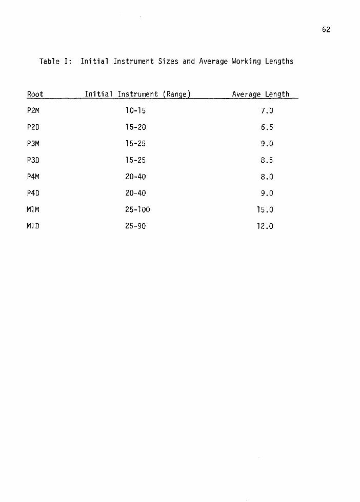
62
Table I: Initial Instrument Sizes and Average Working Lengths
Root Initi a 1 Instrument (Range) Average Length
P2M 10-15 7.0
P2D 15-20 6.5
P3M 15-25 9.0
P30 15-25 8.5
P4M 20-40 8.0
P4D 20-40 9.0
MlM 25-100 15.0
M1D 25-90 12.0
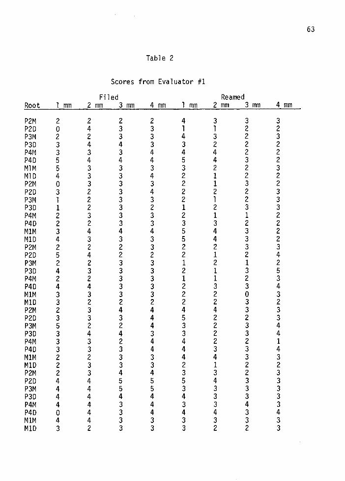
63
Table 2
Scores from Evaluator #1
Filed Reamed Root 1 mm 2 mm 3 mm 4mm 1 mm 2 mm 3 mm 4mm
P2M 2 2 2 2 4 3 3 3 P2D 0 4 3 3 1 1 2 2 P3M 2 2 3 3 4 3 2 3 P3D 3 4 4 3 3 2 2 2 P4M 3 3 3 4 4 4 2 2 P4D 5 4 4 4 5 4 3 2 MlM 5 3 3 3 3 2 2 3 MlD 4 3 3 4 2 1 2 2 P2M 0 3 3 3 2 1 3 2 P2D 3 2 3 4 2 2 2 3 P3M 1 2 3 3 2 1 2 3 P3D 1 2 3 2 1 2 3 3 P4M 2 3 3 3 2 1 1 2 P4D 2 2 3 3 3 3 2 2 M1M 3 4 4 4 5 4 3 2 M1D 4 3 3 3 5 4 3 2 P2M 2 2 2 3 2 2 3 3 P2D 5 4 2 2 2 1 2 4 P3M 2 2 3 3 1 2 1 2 P3D 4 3 3 3 2 1 3 5 P4M 2 2 3 3 1 1 2 3 P4D 4 4 3 3 2 3 3 4 M1M 3 3 3 3 2 2 0 3 M1D 3 2 2 2 2 2 3 2 P2M 2 3 4 4 4 4 3 3 P2D 3 3 3 4 5 2 2 3 P3M 5 2 2 4 3 2 3 4 P3D 3 4 4 3 3 2 3 4 P4M 3 3 2 4 4 2 2 1 P4D 3 3 3 4 4 3 3 4 M1M 2 2 3 3 4 4 3 3 M1D 2 3 3 3 2 1 2 2 P2M 2 3 4 4 3 3 2 3 P2D 4 4 5 5 5 4 3 3 P3M 4 4 5 5 3 3 3 3 P3D 4 4 4 4 4 3 3 3 P4M 4 4 3 4 3 3 4 3 P4D 0 4 3 4 4 4 3 4 M1M 4 4 3 3 3 3 3 3 M1D 3 2 3 3 3 2 2 3
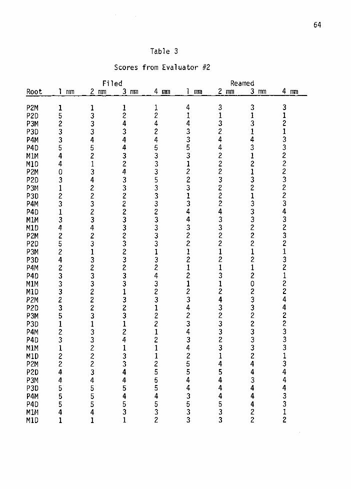
64
Tab 1 e 3
Scores from Evaluator #2
Filed Reamed Root 1 mm 2 mm 3 mm 4 mm 1 mm 2 mm 3 mm 4 mm
P2M 1 1 1 1 4 3 3 3 P2D 5 3 2 2 1 1 1 1 P3M 2 3 4 4 4 3 3 2 P3D 3 3 3 2 3 2 1 1 P4M 3 4 4 4 3 4 4 3 P4D 5 5 4 5 5 4 3 3 M1M 4 2 3 3 3 2 1 2 M1D 4 1 2 3 1 2 2 2 P2M 0 3 4 3 2 2 1 2 P2D 3 4 3 5 2 3 3 3 P3M 1 2 3 3 3 2 2 2 P3D 2 2 2 3 1 2 1 2 P4M 3 3 2 3 3 2 3 3 P4D 1 2 2 2 4 4 3 4 M1M 3 3 3 3 4 3 3 3 M1D 4 4 3 3 3 3 2 2 P2M 2 2 2 3 2 2 2 3 P2D 5 3 3 3 2 2 2 2 P3M 2 1 2 1 1 1 1 1 P3D 4 3 3 3 2 2 2 3 P4M 2 2 2 2 1 1 1 2 P4D 3 3 3 4 2 3 2 1 M1M 3 3 3 3 1 1 0 2 M1D 3 2 1 2 2 2 2 2 P2M 2 2 3 3 3 4 3 4 P2D 3 2 2 1 4 3 3 4 P3M 5 3 3 2 2 2 2 2 P3D 1 1 1 2 3 3 2 2 P4M 2 3 2 1 4 3 3 3 P4D 3 3 4 2 3 2 3 3 M1M 1 2 1 1 4 3 3 3 M1D 2 2 3 1 2 1 2 1 P2M 2 2 3 2 5 4 4 3 P2D 4 3 4 5 5 5 4 4 P3M 4 4 4 5 4 4 3 4 P3D 5 5 5 5 4 4 4 4 P4M 5 5 4 4 3 4 4 3 P4D 5 5 5 5 5 5 4 3 MU-1 4 4 3 3 3 3 2 1 M1D 1 1 1 2 3 3 2 2
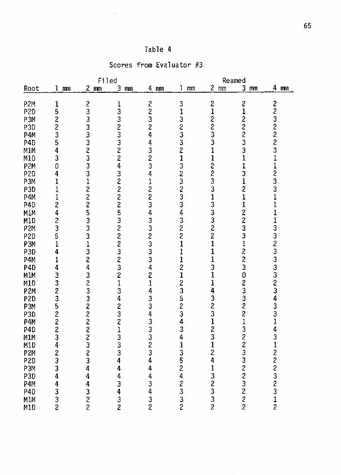
65
Table 4
Scores from Evaluator #3
Filed Reamed Root 1 mm 2mm 3 mm 4 mm 1 mm 2 mm 3 mm 4mm
P2M I 2 I 2 3 2 2 2 P2D 5 3 3 2 I I I 2 P3M 2 3 3 3 3 2 2 3 P3D 2 3 2 2 2 2 2 2 P4M 3 3 3 4 3 3 2 2 P4D 5 3 3 4 3 3 3 2 MIM 4 2 2 3 2 I 3 3 MID 3 3 2 2 I I I I P2M 0 3 4 3 3 2 I I P2D 4 3 3 4 2 2 3 2 P3M I I 2 I 3 3 I 3 P3D I 2 2 2 2 3 2 3 P4M I 2 2 2 3 I I I P4D 2 2 2 3 3 3 I I MIM 4 5 5 4 4 3 2 I MID 2 3 3 3 3 3 2 I P2M 3 3 2 3 2 2 3 3 P2D 5 3 2 2 2 2 3 3 P3M I I 2 3 I I I 2 P3D 4 3 3 3 I I 2 3 P4r~ I 2 2 3 I I 2 3 P4D 4 4 3 4 2 3 3 3 MIM 3 3 2 2 I I 0 3 MID 3 2 I I 2 I 2 2 P2M 2 3 3 4 3 4 3 3 P2D 3 3 4 3 5 3 3 4 P3M 5 2 2 3 2 2 2 3 P3D 2 2 3 4 3 3 2 3 P4M 2 2 2 3 4 I I I P4D 2 2 I 3 3 2 3 4 MIM 3 2 3 3 4 3 2 3 MID 4 3 3 2 I I 2 I P2M 2 2 3 3 3 2 3 2 P2D 3 3 4 4 5 4 3 2 P3M 3 4 4 4 2 I 2 2 P3D 4 4 4 4 4 3 2 3 P4M 4 4 3 3 2 2 3 2 P4D 3 3 4 4 3 3 2 3 MIM 3 2 3 3 3 3 2 I MID 2 2 2 2 2 2 2 2
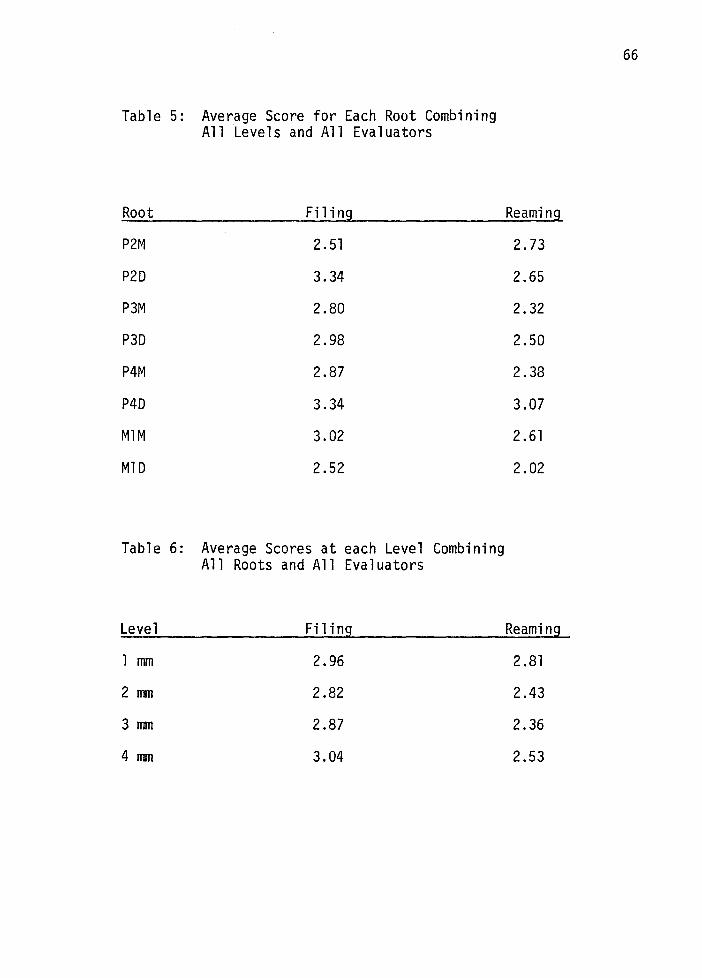
Table 5: Average Score for Each Root Combining All Levels and All Evaluators
Root Filing
P2M 2.51
P2D 3.34
P3M 2.80
P3D 2.98
P4M 2.87
P4D 3.34
MlM 3.02
MlD 2.52
Table 6: Average Scores at each Level Combining All Roots and All Evaluators
Level
lmm
2 mm
3 mm
4 mm
Filing
2.96
2.82
2.87
3.04
Reaming
2.73
2.65
2.32
2.50
2.38
3.07
2.61
2.02
Reaming
2.81
2.43
2.36
2.53
66
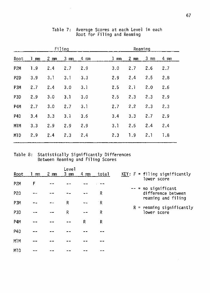
67
Table 7: Average Scores at each Level in each Root for Filing and Reaming
Fi 1 i ng Reaming
Root 1 mm 2 mm 3 mm 4 mm 1 mm 2 mm 3 mm 4 mm
P2M 1.9 2.4 2.7 2.9 3.0 2.7 2.6 2.7
P2D 3.9 3.1 3.1 3.3 2.9 2.4 2.5 2.8
P3M 2.7 2.4 3.0 3.1 2.5 2. 1 2.0 2.6
P3D 2.9 3.0 3.1 3.0 2.5 2.3 2.3 2.9
P4M 2.7 3.0 2.7 3.1 2.7 2.2 2.3 2.3
P4D 3.4 3.3 3.1 3.6 3.4 3.3 2.7 2.9
MlM 3.3 2.9 2.9 2.9 3.1 2.5 2.4 2.4
MlD 2.9 2.4 2.3 2.4 2.3 1.9 2. 1 1.8
Table 8: Statistically Significantly Differences Between Reaming and Filing Scores
Level Root 1 mm 2 mm 3 mm 4 mm total KEY: F = filing significantly
lower score P2M F
= no significant P2D R difference between
reaming and filing P3M R R
R = reaming significantly P3D R R lower score
P4M R R
P4D
MlM
MlD
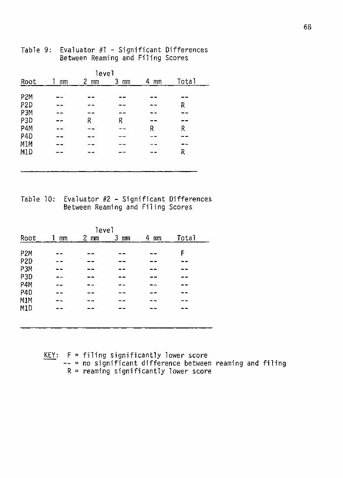
Table 9: Evaluator #1 - Significant Differences Between Reaming and Filing Scores
level Root 1 mm 2 mm 3 mm 4 mm Total
P2M P2D R P3M P3D R R P4M R R P4D MlM MlD R
Table 10: Evaluator #2 - Significant Differences Between Reaming and Filing Scores
level Root 1 mm 2 mm 3 mm 4 mm Total
P2M F P2D P3M P3D P4M P4D MlM MlD
KEY: F = filing significantly lower score -- = no significant difference between reaming and filing R = reaming significantly lower score
68
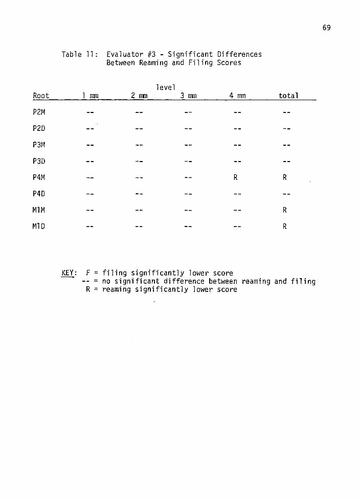
Root
P2M
P2D
p~
P3D
P4M
P4D
Table 11: Evaluator #3 - Significant Differences Between Reaming and Filing Scores
level 1 mm 2 mm 3 mm 4 mm
R
total
R
MlM R
MlD R
KEY: F = filing significantly lower score -- = no significant difference between reaming and filing R = reaming significantly lower score
69
DISCUSSION
There are many factors which must be taken into consideration when
comparing techniques of root canal preparation. These factors include the
initial and final canal size, shape, and curvature; and the final degree of
canal debridement.
Evaluating the initial canal factors is extremely important when using
experimental teeth other than human teeth. Without this information, appli
cation of the results to the true clinical situation would be impossible.
Considering the teeth used in this study, it should be kept in mind that
the canals are essentially straight and round. The only human teeth that
consistently fit into this category are the maxillary central incisors.
The initial instrument size ranged from 10 to 100 so that the full spectrum
of canal width was covered.
For the purpose of evaluating the prepared canal, studies have been
designed to look at the effect of preparation on canal shape and curvature
or the debridement of the canal. The ideal study would, however, show both
the degree of canal debridement and the amount that the preparation deviated
from the original canal. It was felt that the technique used in this study
for examining the prepared canals most nearly approximated the ideal. Under
the microscope (lOOx), the cross-sectional specimens could be examined with
regard to debridement and deviation of the preparation from the true canal.
Both methods of preparation did an excellent job of removing debris
70

71
at the 2, 3, and 4 mm level. Although not proven by this study, the com
plete lack of debris at the more cervical levels could be attributed to the
flaring of the canals. The flaring allows for better flushing of the irri
gants plus the use of larger instruments within the canal. Coffae and
Brilliant found that serial preparations removed debris at the 1, 3, 5 mm
level significantly better than non-serial preparations. They, however,
found that 36% of their serial preparations still contained tissue at the
1 mm level. That percentage is much higher than that found in this study.
The reason they had a higher percentage of canals with tissue debris is
probably due to the fact that they prepared mesial canals of human mandibu
lar molars which are curved and much more difficult to prepare than the
straight canals used in this study.
Other studies done on extracted human teeth have reported finding that
the walls of the prepared canals were smeared and packed with debris. In
the present study, that finding was not made. However, the studies that
did find the smeared walls used the scanning electron microscope to examine
canals which had been split longitudinally. That method of examination may
be more appropriate for locating this smeared layer.
When evaluating the preparation of any canal, it is generally agreed
that the apical 1 to 2 mm are the most critical. This is where the seal
should be obtained when the canal is obturated. As found in Ingles' study, 34 42
lack of apical seal was the leading cause of endodontic failures. Crump, 37 43
Malooley, and Russin noted that canals that are unclean at the l mm
level are difficult if not impossible to seal. Unfortunately, it is at this
level that debris or deviations of the prepared canal from the true canal
are to be found. In this study, debris was found primarily at the 1 mm
level. Debris was found at this level in 3% of the filed teeth and 9% of
72
the reamed teeth. These teeth represent the simplest endodontic treatment
situation (i.e. straight canals with easy access). It could be theorized
that in curved canals the above percentages would be higher.
With regard to evaluating the change in canal shape, examining cross
sections does not give the overall picture that comparing pre- and post
treatment radiographs would. However, the cross-sectional specimens do
show at a specific level how the preparation has stayed within or deviated
from the path of the original canal. In the present study, the observation
of concentric rings of secondary dentin within the canal was a very useful
way of locating the boundaries of the original canal. Figure 5 demonstrates
these rings and also shows how the preparation in this case has migrated to
one side of the canal.
It is obvious that a preparation that does not enlarge a canal in all
directions but deviates to one side will not adequately debride the entire 23,63,65,71 ,29
canal. This phenomena has been observed in other studies,
and was found to occur frequently in this study also. Through microscopic
examination it was judged that there were significant deviations to one
side of the original canal in 68 of the filed sections and 49 of the reamed
sections.
These numbers make it appear that reaming would be a much better
method for preparing this type of canal. However, when the number of
73
deviations is compared level by level it is seen that at the 1 mm level
there are essentially (16 reamed; 17 filed) the same number by each method.
As stated earlier this is the most critical level for attaining a good seal
assuring the success of the treatment.
The statistical analysis of the scores given to each of the sections
by three independent evaluators agrees with the above findings.
When the overall scores for filing were compared to reaming, reaming
was scored significantly lower. Also, when the two techniques were compared
on a root by root basis, reaming scored significantly lower in 5 of the 8
roots with the remaining 3 roots showing no significant difference between
the two techniques. However, when the scores were evaluated level by
level, which is the more meaningful and clinically relevant way to compare
these techniques, the results were somewhat different.
When filing was compared to reaming at the 1 mm level using the scores
of all the roots together, there was no statistically significant difference
between the two methods. At the 2 mm, 3 mm, and 4 mm levels reaming was
scored significantly lower.
Table 8 shows a further breakdown of the combined scoring of all
three evaluators. The two techniques were compared at each level on a root
by root basis. This analysis was done in order to determine if the differ
ence in initial size of the canal affected the efficiency of either tech
nique. It can be seen that at the 1 mm and 2 mm levels there was no sig
nificant difference between the two techniques in any root except root P2M.
In this root, filing scored significantly lower than reaming at the 1 mm
level. The initial size of the canal did not make any difference when
comparing the two techniques in this study.
74
The author found it quite surprising that straight canals which were
circumferentially filed would show preparations that migrated to one side
of the canal. During preparation extreme care was taken to insure that
the canals were filed around their complete circumferences. Apparently,
however, the orifice was the area being circumferentially filed while in
many cases more apical regions were being instrumented on only some of the
walls. A close examination of the pre-treatment or initial file radio
graphs reveals a constriction at the orifice of the canals. As shown in
the mesial root of the first molar in Figure 14, this constriction could
force an instrument to one side of the canal near the apex. Elimination
of this constriction prior to the major portion of canal instrumentation
would allow the operator to have more control over the files in the apical
portion of the canal.
This situation is analgous to human anterior teeth. If these teeth
are instrumented with inadequate access openings, interferences from either
the incisal portion of the crown or a lingual portion of the roof of the
chamber will make it impossible to control the direction of the instrument
in the apical third of the canal. These findings would tend to support
the use of preflaring as a means of eliminating cervical interferences be
fore more apical canal preparation is carried out.
The choice of the initial instrument was done very carefully in order
to select the largest possible file. It was felt that this was especially
75
critical for the reamed canals. Whereas the instrument in the circumfer-
entially canals is directed against the walls by the operator, the prepara
tion of the reamed canals depends solely on the size of the instrument
realtive to the size of the canal. In this study the canals were prepared
to three sizes larger than the initial instrument. If too small of an
initial file were selected in the reamed canals, little or no preparation
would occur by the time the master apical file is reached.
The criteria used to determine when canal preparation is complete is
somewhat arbitrary. Several studies have shown that stopping preparation
when 11 Clean, white dentin shavings,. are being removed from the canal is not 19,23,61,65,
a valid method for determining the adequacy of the preparation. 71
Moodnik even prepared the canals in his study 3 sizes beyond the point
where clean, white dentin filings were seen. He still found that almost 72
all of the canals were inadequately prepared. The present study has
shown that carrying the preparation 3 sizes past that of the initial in
strument is no guarantee that the canal is completely debrided. By observ
ing the deviations the preparations took from the original canal, it can
be surmised that use of even larger and consequently stiffer instruments
would only increase these deviations and not effect a better debridement
of the true canal.
It can be inferred from the results of this study that the satisfac-
tory cleansing of a canal depends on using a technique which will not
deviate from the path of the original canal and will, therefore enlarge
the canal equally around its circumference. Therefore, the size of the
76
MAF takes on secondary importance to the method of preparation used.
It is believed by the author that the unstained areas radiating from
lumen of many of the canals were caused by the action of the Clorox on the
soft tissue in the dentinal tubules. Eosin stains cytoplasm and inter-
cellular substances. Therefore, it appears that the NaOCl has either re~
moved these materials or altered their staining characteristics. Until it
is more fully understood, the clinical implications of this phenomenan can-
not be fully understood. If, in fact, the NaOCl did cleanse the dentinal
tubules of soft tissue, it would definitely be a beneficial effect with re
gard to endodontic therapy. Further research needs to be done in this area.
There was an appreciable difference in the amount of time required to
prepare the canals by each technique. The results of this study are in 19
agreement with Weine's study which found reaming to be the fastest tech-
nique when comparing three preparation techniques. Although time required
should not be the primary factor considered in selecting a technique it is
an important consideration in a clinical situation. With all other factors
being equal it would be logical to select the faster of two methods.
In the present study reaming was scored equal to or better than
filing according to the criteria previously listed. It was also the faster
of the two methods. However, before selecting this technique over filing,
the factor of instrument breakage should be considered. The probability
of separating an instrument in a canal by either of the two techniques and
the effect of such an occurrence on the prognosis of the case should be
taken into consideration.
Instrument breakage occurs primarily through twisting files when

77
they are bound in the canal. This occurs primarily with instruments small
er than a size 35. The risk of separating an endodontic file within a
canal by the use of a rasping motion is negligible for all instrument sizes.
A segment of a file lodged in a canal is in most cases extremely difficult
if not impossible to remove. This can adversely affect the prognosis of
the case due to the fact that the broken instrument prevents further de
bridement and adequate sealing of the canal.
Considering the above information along with the data collected in
this study, the author feels that reaming is the technique of choice when
preparing straight canals that have an initial instrument size greater than
35. For canals smaller than this, filing is recommended.
The author feels that in future studies of this type histologic sect
tions of control teeth with unprepared canals be made. These would act as
a basis or point of reference in evaluating the prepared canals. Also, it
would be adventageous to mark all roots in such a way that the buccal,
lingual, mesial, and distal directions would be apparent in the final his
tologic section. This would aid in determining possible causes for the
prepared canal deviating from the true canal.
Further study needs to be done comparing these preparation techniques
in human teeth with curved canals. The results of such a study would be
applicable to a much greater percentage of clinical cases than the results
of this study. Also, the effect of preflaring canals on the apical prepa
ration should be investigated.
SUMMARY
Eighty root canals in five Beagle dogs were prepared by standard
endodontic procedures. Using sodium hypochlorite as an irrigant, the
canals were instrumented with K-type files by one of the following tech
niques.
1. Forty canals were prepared by the use of
a circumferential filing or rasping motion.
2. Forty canals were prepared by the use of
a reaming or ~-turn and withdraw technique,
with no emphasis placed on lateral filing
as the instrument was withdrawn.
Canals prepared by both methods were enlarged at full working length
to three sizes larger than the initial instrument. They were then flared
by progressively using each of the next three larger instruments 1.0 mm.
short of the preceding instrument. All canals were prepared in two
appointments.
Histologic sections were then made perpendicular to the long axis
of the canal at levels of 1 mm., 2 mm., 3 mm., and 4 mm. from the apical
end of the canal. These sections were evaluated and scored in a blind
fashion by three evaluators using the following criteria:
1. Percentage of walls instrumented
2. Amount of debris present within the canal
3. Symmetry of the prepared canal
78
The sections were scored on a scale of 1 to 5, with 1 being the
best score and 5 the worst.
A statistical analysis of the results was done and there was no
significant difference between the scores of the two techniques at the
1 mm. level. At the 2 mm., 3 mm., and 4 mm. levels, reaming was scored
significantly better than filing.
79
Both techniques were very effective in removing debris from the
canals; although, filing proved slightly more effective in that respect.
The techniques showed an approximately equal tendency to have the prepared
canal deviate from the true canal at the 1 mm. level.
REFERENCES
1. Milas, V.B.: History, Chapter 25, Pathways of the~, C.V. Mosby Co., St. Louis, 1980.
2. Mills, G.A.: Hints and Queries. Dental Cosmos, 25:447, 1883.
3. Schreier, E.: Treatment of Infected Root Canals with Kalium and Natrium. Dental Cosmos, 35:863, 1893.
4. Callahan, J.R.: Sulphuric Acid for Opening Root Canals. Dental Cosmos, 36:957, 1894.
5. Miller, J: An Evaluation of Three Methods of Instrumentation of Curved Canals. Thesis, U. of Kentucky, Lexington, 1975.
6. Sommer, R.F., Ostrander, F.D., and Crowley, M.C.: Clinical Endodontics: A Brief History of Root Canal Therapy, W.B. Saunders Co., Philadelphia, 1961.
7. Hunter, W.: The Role of Sepsis and of Antisepsis in Medicine. Lancet 1:79, 1911.
8. Ingle, J.I. and Beveridge, E.E.: Endodontics, 2nd edition, Lea and Febiger, 1976.
9. Coolidge, E.D.: Pathology, Diagnosis and Treatment of the Pulp and Preparation of Root Canals for Filling. J. Amer. Dent. Assoc., 19:1964, 1932.
10. Fish, E.W. and MacLean, I.H.: The Distribution of Oral Streptococci in the Tissues. Brit. Dent. J., 61:336, 1936.
11. Schilder, H.: Canal Debridement and Disinfection. Pathways of the ~' 2nd edition, by S. Cohen and R.C. Burns, C.V. Mosby Co., St. Louis, 1980.
12. Shovelton, D.S.: The Presence and Distribution of Micro-Organisms within Non-Vital Teeth. Brit. Dent. J., 117:101, 1964.
13. Hedman, W.J.: An Investigation Into Residual Periapical Infection After Pulp Canal Therapy. Oral Surg., 4:1173, 1951.
80

14. Engstrom, B. and Frostell, G.: Bacteriological Studies of the NonVital Pulp in Cases with Intact Pulp Cavities. Acta. Odont. Scand., 19:23, 1961.
81
15. Rothschild, M.B.: A Second Look at the First Principles: An Evaluation of Some Endodontic Dogma. J. Brit. Endo. Soc., 5:32, 1971.
16. Weine, F.S.: Endodontic Therapy, 2nd Edition, C.V. Mosby Co., St. Louis, 1976.
17. Leavitt, J.M., Naidorf, I.J. and Shugalvsky, P.: The bacterial Flora of Root Canals as Disclosed by a Culture Medium for Endodntics. Oral Surg. 11:302, 1958.
18. Schilder, H.: Cleaning and Shaping the Root Canal. Dent. Clin. N. Amer., 18:269, 1974.
19. Walton, R.E.: Histologic Evaluation of Different Methods of Enlarging the Pulp Canal Space. J. Endod. 2:304, 1976.
20. Bhaskar, S.N.: Histologic Evaluation of Endodontic Procedures in Dogs. Oral Surg., 31:526, 1971.
21. Walker, A.A.: A Definite and Dependable Therapy for Pulpless Teeth. J. Amer. Dent. Assoc., 23:1419, 1936.
22. Grossman, L. and Meiman, B.: Solution of Pulp Tissue by Chemical Agents. J. Amer. Dent. Assoc., 28:223, 1941.
23. Baker, N.A.: Scanning Electron Microscopic Study of the Efficacy of Various Irrigating Solutions. J. Endod., 1:127, 1975.
24. Auerbach, N.B.: Antibiotics vs. Instrumentation in Endodontics. N.Y. State Dent. J., 19:225, 1953.
25. Stewart, G.G.: The Importance of Chemomechanical Preparation of the Root Canal. Oral Surg., 8:993, 1955.
26. Ingle, J.I. and Zeldow, B.J.: An Evaluation of Mechanical Instrumentation and the Negative Culture in Endodontic Therapy. J. Amer. Dent. Assoc., 57:471, 1958.
27. Nicholls, E.: The Efficacy of Cleansing of the Root Canal. Brit. Dent. J., 112:267, 1962.
28. Shih, M., Marshall, F., Rosen, S.: The Bacterial Efficiency of Sodium Hypochlorite as an Endodontic Irrigant. Oral Surg., 29:613, 1970.
29. Senia~ E.~ Marshall~ F. and Rosen~ J.: The Solvent Action of Sodium Hypochlorite on Pulp Tissue of Extracted Teeth. Oral Surg., 31 ,96, 1971.
82
30. Svec, T.A. and Harrison, J.W.: Chemomechanical Removal of Pulpal and Dentinal Debris with Sodium Hypochlorite and Hydrogen Peroxide Versus Normal Saline. J. Endod., 3:49, 1977.
31. Rubin, L.M., Skobe, Z., Krakow, A. and Gron, P.: The Effect of Instrumentation and Flushing of Freshly Extracted Teeth in Endodontic Threapy: A Scanning Electron Microscope Study. J. Endod., 5:328, 1979.
32. Hatton, E.H.: Histologic Studies of Pulpless Teeth. Dent. Cosmos, 70:49, 1928.
33. Wilkinson, F.C.: Root Canal Treatment: Report of Some Investigations Dealing with the Elimination of Necrotic Tissue from the Root Canals. Brit. Dent. J., 50:1, 1929.
34. Ingle, J.I. and Bevridge, E.E.: Endodontics, 2nd edition, Lea and Febiger, Philadelphia, 1976.
35. Harty, F.J. and Stock, C.J.R.: The Giromatic System Compared with Hand Instrumentation in Endodontics. Brit. Dent. J., 137:239, 1974.
36. Seltzer, S., Bender, I., Smith, J., Freedman, I. and Nazimov, H., Endodontic Failures: An Analysis Based on Clinical, Roentgenographic and Histologic Findings. Oral Surg., 23:500, 1967.
37. Malooley, J., Patterson, S., and Kafrawy, A.: Response of Periapical Pathosis to Endodontic Treatment in Monkeys. Oral Surg., 47:545,1979.
38. Heuer, M.A.: The Biomechanics of Endodontic Therapy. Dent. Clin. N. Amer., 13:341, 1963.
39. Fink, H.D.: Ten Basic Principles for Successful Endodontics. N.Y. J. of Dent. , 39: 1 95, 1960.
40. Grossman, L.I.: Endontic Practice, Chapter 13, 9th Edition, Lea and Febiger, Philadelphia, 1978.
41. Ingle, J.I.: A Standardized Endodontic Technique Using Newly Designed Instruments and Filling Materials. Oral Surg., 14:83, 1961.
42. Crump, M.C.: Differential Diagnosis in Endodontic Failure. Dent. Clin. N. Amer., 23:617, 1979.
43. Russin. T., Zardiackas, L., Reader, A., and Menke, R.: Apical Seals Obtained with Laterally Condensed, Chloroform-Softened GuttaPercha and Laterally Condensed Gutta-Percha and Grossman 1
S Sealer. J. Endod., 6:678, 1980.
44. Kuttler, Y.: Microscopic Investigation of Root Apexes. J. Amer. Dent. Assoc., 50:544, 1955.
83
45. Vertucci, F.J.: Root Canal Anatomy of the Mandibular Anterior Teeth. J. Amer. Dent. Assoc., 89:369, 1974.
46. Rankine-Wilson, R.W. and Henry, P.: The Bifurcated Root Canal in Lower Anterior Teeth. J. Amer. Dent. Assoc., 70:1162, 1965.
· 47. Weine, F.S., Healey, H., Gerstein, H. and Evanson, L.: Canal Configuration in the Mesiobuccal Root of the Maxillary First Molar and Its Endodontic Significance. Oral Surg., 28:743, 1965.
48. Green, D.A.: Double Canals in Single Roots. Oral Surg., 35:689, 1973.
49. Green, E.N.: Microscopic Investigation of Root Canal Diameters. J. Amer. Dent. Assoc., 57:636, 1958.
50. Green, D.A.: Morphology of the Pulp Cavity of Permanent Teeth. Oral Surg., 8:743, 1955.
51. Pineda, F. and Kuttler, Y.: Mesiodistal and Buccolingual Roentgenographic Investigation of 7275 Root Canals. Oral Surg., 33:101, 1972.
52. Skidmore, A. E. and Bjorndal, AM.M; Root Canal Morphology of the Mandibular First Molar. Oral Surg., 32:778, 1971.
53. Oliet, S. and Sarin, S.M.: Cutting Efficiency of Endodontic Reamers. Oral Surg., 36:243, 1973.
54. Vessey, R.A.: The Effect of Filing Versus Reaming on the Shape of the Prepared Root Canal. Oral Surg., 27:543, 1969.
55. Frank, A.L.: An Evaluation of the Giromatic Endodontic Handpiece. Oral Surg., 24:149, 1967.
56. Molven, 0.: A Comparison of the Dentin Removing Ability of Five Root Canal Instruments. Scand. J. Dent. Res., 78:500, 1970.
57. Ingle, J.I. and Levine, M.: The Need for Uniformity of Endodontic Instruments, Equipment, and Filling Materials. Presented to the 2nd International Conference on Endodontics, 1958.
58. Cohen, S. and Burns, R.C.: Pathways of the~. C.V. Mosby Co., St. Louis, 1980.
84
59. Martin, H.: A Telescope Technique for Endodontics. J.D.C. Dent. Soc., 49:12, 1974.
60. Mullaney, T. and Petrich, J.: The Ledged Root Canal: Cause, Prevention and Correction. J. Kentucky Dent. Assoc., 20:15, 1968.
61. Haga, C.S.: Microscopic Measurement of Root Canal Preparations Following Instrumentation. Northwestern Univ. Dent. Res., p. 11, Fall, 1967.
62. Schneider, S.W.: A Comparison of Canal Preparations in Straight and Curved Root Canals. Oral Surg., 32:271, 1971.
63. Davis, S., Brayton, S., and Goldman, M.: The Morphology of the Prepared Root Canal: A Study Using Injectable Silicone. Oral Surg., 34:642, 1972.
64. Jungman, C.L., Uchin, R.A. and Bucher, J.F.: Effect of Instrumentation on the Shape of the Root Canal. J. Endod., 1:66, 1975.
65. Gutierrez, J.H. and Garcia, J.: Microscopic and Macroscopic Investigation on Results of Mechanical Preparation of Root Canals. Oral Surg., 25:108, 1968.
66. Weine, F. S., Kelly, R.F. and Lio, P.J.: The Effect of Preparation Procedures on Original Canal Shape and on Apical Foramen Shape. J. En dod. , 8:255, 1975.
67. Mullins, R.K.: A Comparison of the Step-Back Method with Other Enlargement Techniques in Finely Curved Root Canals. Thesis, U. of Kent. , 1976.
68. Allsion, D.A., Weber, C.R., and Walton, R.E.: The Influence of the Method of Canal Preparation on the Quality of Apcial and Coronal Obturation, J. Endod., 5:298, 1979.
69. Coffae, K.P. and Brilliant, J.D.: The Effect of Serial Preparation Versus Non-Serial Preparation on Tissue Removal in the Root Canals of Extracted Mandibular Human Molars. J. Endod., 1:211' 1975.

85
70. McComb, D. and Smith, D.: A Preliminary Scanning Electron f~icroscopic Study of Root Canals After Endodontic Procedures, J. Endod., 1:238, 1975.
71. Mizrahi, S.J., Tucker, J.W., and Seltzer, S.: A Scanning Electron Microscopic Study of the Efficacy of Various Endodontic Instruments. J. Endod., 1:324, 1975.
72. Moodnick, R., Darn, S., Feldman, M., Levey, M., Borden, B.: Efficacy of Biomechanical Instrumentation: A SEM Study. J. Endod., 2:261' 1976.
73. Littman, S.H.: Evaluation of Root Canal Debridement By Use of a Radiopaque Medium. J. Endod., 3:135, 1977.
74. Laws, A.J.: Preparation of Root Canals: An Evaluation of Mechanical Aids. New Zealand Dent. J., 64:156, 1968.
75. Klayman, S.M. and Brilliant, J.D.: A Comparison of the Efficacy of Serial Preparation Versus Giromatic Preparations. J. Endod., 1:334, 1975.
76. Weine, F.S., Kelly, R. and Bray, K.: Effect of Preparation with Endodontic Handpieces on Original Canal Shape. J. Endod., 2:298, 1976.
77. Brown, B., Kafrawy, A. and Patterson, S.: Studies of Sargenti Technique of Endodontics - Autoradiographic and Scanning Electron Microscope Studies. J. Endod., 5:14, 1979.
78. Barker, B.C., Lockett, B.C.: Utilization of the Mandibular Premolars of the Dog for Endodontic Research. Aust. Dent. J., 16:280, 1971.
79. Anderson, A.C.: The Beagle~~ Experimental QQ[, The Iowa State University Press, Ames, Iowa, 1970.
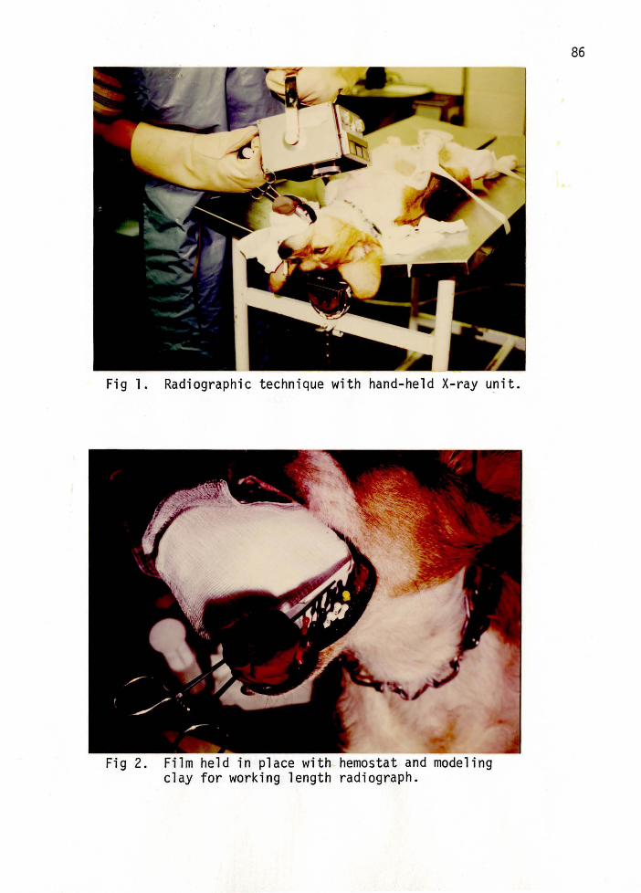
Fig 1. Radiographic technique with hand-held X-ray unit.
Fig 2. Film held in place with hemostat and modeling clay for working length radiograph.
86
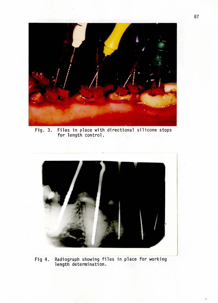
Fig. 3. Files in place with directional silicone stops for length control.
Fig 4. Radiograph showing files in place for working length determination.
87
•
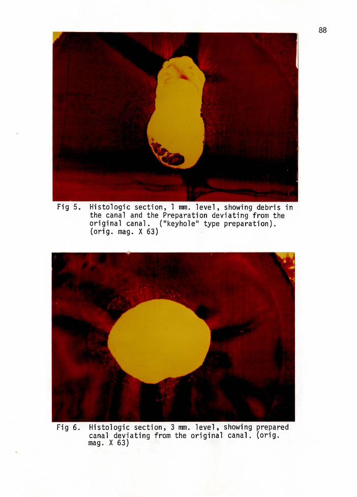
Fig 5.
Fig 6.
Histologic section, 1 mm . level, showing debris in the canal and the Preparation deviating from the original canal. ( 11 keyhole 11 type preparation). (orig. mag. X 63)
Histologic section, 3 mm. level, showing prepared canal deviating from the original canal. (orig. mag. X 63)
88
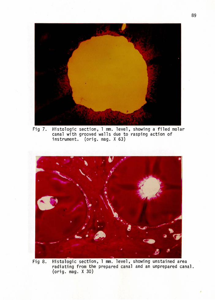
Fig 7. Histologic section, 1 mm. level, showing a filed molar canal with grooved walls due to rasping action of instrument. (orig. mag. X 63)
89
Fig 8. Histologic section, 1 mm. level, showing unstained area radiating from the prepared canal and an unprepared canal. (orig. mag. X 30)
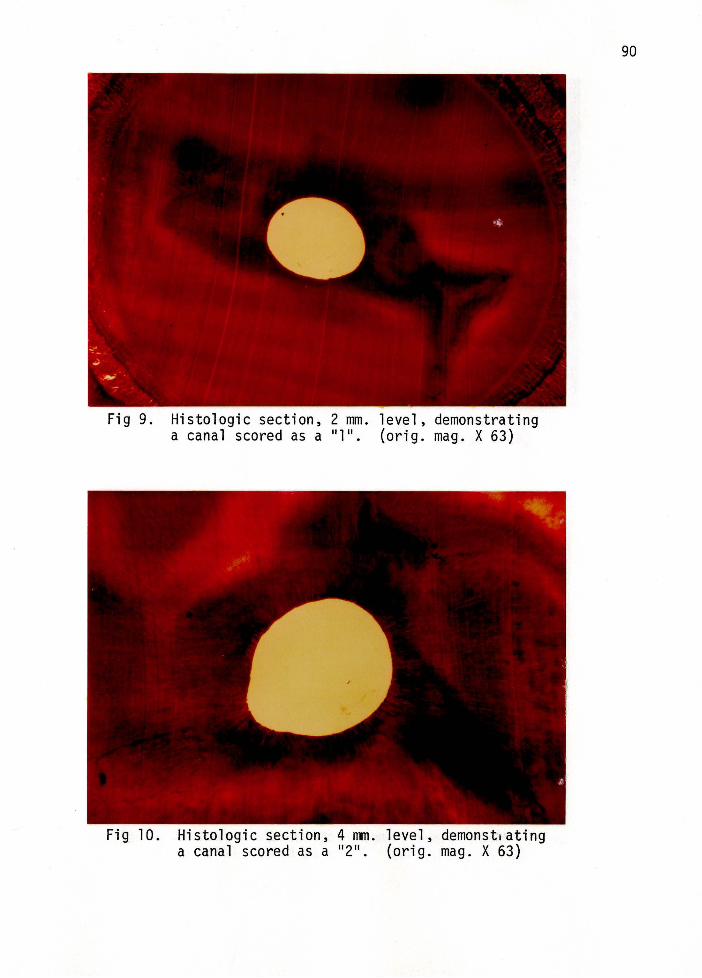
Fig 9.
Fig 10.
Histologic section, 2 mm. level, demonstrating a canal scored as a 11 111
• (orig. mag. X 63)
Histologic section, 4 mm. level, demonst1ating a canal scored as a 11 211
• (orig. mag. X 63)
90
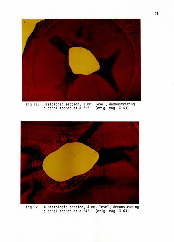
Fig 11. Histologic section, 1 mm. level, demonstrating a canal scored as a 11 311
• (orig. mag. X 63)
Fig 12. A histologic section, 4 mm. level, demonstrating a canal scored as a 11 411
• (orig. mag. X 63)
91
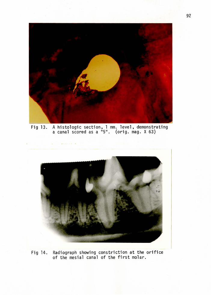
Fig 13.
Fig 14.
A histologic section, 1 mm. level, demonstrating a canal scored as a 11 511
• (orig. mag. X 63)
Radiograph showing constriction at the orifice of the mesial canal of the first molar.
92
APPROVAL SHEET
The thesis submitted by Terry L. Kippa has been read and approved by the following committee:
Dr. Franklin S. Weine, Director Professor, Endodontics, Loyola
Dr. Gary N. Taylor Associate Professor, Endodontics, Loyola
Dr. Hal D. McReynolds Associate Professor, Histology, Loyola
The final copies have been examined by the director of the thesis and the signature which appears below verifies the fact that any necessary changes have been incorporated and that the thesis is now given final approval by the Committee with reference to content and form.
The thesis is therefore accepted in partial fulfillment of the requirements for the degree of Master of Science in Oral Biology.
Director's Signature
93