A High-throughput Quantitative Multiplex Kinase Assay for Monitoring Information Flow in Signaling...
11
A High-throughput Quantitative Multiplex Kinase Assay for Monitoring Information Flow in Signaling Networks APPLICATION TO SEPSIS-APOPTOSIS* Kevin A. Janes‡, John G. Albeck§, Lili X. Peng‡, Peter K. Sorger‡§¶, Douglas A. Lauffenburger‡§¶, and Michael B. Yaffe‡§¶ To treat complex human diseases effectively, a systems- level approach is needed to understand the interplay of environmental cues, intracellular signals, and cellular be- haviors that underlie disease states. This approach re- quires high-throughput, multiplex techniques that meas- ure quantitative temporal variations of multiple protein activities in the intracellular signaling network. Here, we describe a single microtiter-based format that simulta- neously quantifies protein kinase activities in the phos- phatidylinositol 3-kinase pathway (Akt), nuclear factor-B pathway (IKK), and three core mitogen-activated protein kinase pathways (ERK, JNK1, MK2). These parallel high- throughput assays are stringently linear, redundantly spe- cific, reproducible, and sensitive compared with classical low-throughput techniques. When applied to a model of sepsis-induced colon epithelial apoptosis, this approach identified a late phase of Akt activity as a critical mediator of cell survival that quantitatively contributed to the effi- cacy of insulin as an anti-apoptotic cue. Thus, sampling parallel nodes in the intracellular signaling network iden- tified part of the molecular mechanism underlying the efficacy of insulin in the treatment of human sepsis. Molecular & Cellular Proteomics 2:463– 473, 2003. Complex patterns of signal transduction arise when cells are exposed to combinations of extracellular cues that vary in onset, duration, origin, and synchrony. Cells process these cues through an interconnected network of multifunctional, redundant molecules to elicit a set of phenotypic responses that subsequently impact function at the cell, tissue, and organ level. In order to develop a molecular understanding of the complex pathophysiology underlying human diseases and utilize this information for prognosis and therapy, a systems- level, network-biology approach should be applied to the signaling networks governing the relevant cell responses (1). This approach will require frequent temporal sampling of pro- tein activity at critical nodes within parallel signaling pathways inside the cell in a quantitative manner to characterize the flow of information accurately. Such functional measurements are likely to be as valuable, or more valuable, than measurements of simple protein abundance. By quantitatively exploring the functional response of the signaling network to distinct extra- cellular cues and correlating these molecular events with phe- notypic responses, one can construct predictive models of cue-signal and signal-response relationships. Evolving proteomic approaches to network biology have largely focused on measuring abundances of many proteins at only a few time points or under a limited number of exper- imental conditions (2). Complementary information on func- tional protein characteristics, such as enzyme activity, has been lacking in these systematic analyses, in large part be- cause there do not exist quantitatively robust, high-through- put techniques that simultaneously measure multiple protein activities in cells. Initially, this type of data collection on pro- tein functional status should focus on frequent sampling of a limited number of key molecules that sit at critical nodes in different signaling pathways (Fig. 1A). To begin to address this, we have developed a generalized assay for the multiplex analysis of multiple protein kinase activities in a 96-well format. The procedure utilizes parallel kinase-specific immunopurification steps, followed by rapid quantitative high-throughput activity measurements within the linear-rate regime for each kinase. We applied this technique to measure the activities of five kinases (extracellular-regulat- ed kinase (ERK) 1 (3), Akt, I-B kinase (IKK), c-jun N-terminal kinase 1 (JNK1), and mitogen-activated protein kinase-asso- ciated protein kinase 2 (MK2)) in a sepsis/systemic inflamma- tory response syndrome (SIRS) model system for tumor ne- crosis factor-alpha (TNF-)-induced colon epithelial cell death. From the ‡Biological Engineering Division, §Department of Biology, and ¶Center for Cancer Research, Massachusetts Institute of Tech- nology, 400 Main Street, Cambridge, MA 02139 Received, May 16, 2003, and in revised form, June 25, 2003 Published, MCP Papers in Press, June 26, 2003, DOI 10.1074/mcp.M300045-MCP200 1 The abbreviations used are: ERK, extracellular-regulated kinase; IKK, I-B kinase; JNK1, c-jun N-terminal kinase 1; MK2, mitogen- activated protein kinase-associated protein kinase 2; PC, phospho- cellulose; PI 3-K, phosphatidylinositol 3-kinase; SIRS, systemic in- flammatory response syndrome; TNF-, tumor necrosis factor-alpha; DTT, dithiothreitol; IFN-, interferon gamma; GST, glutathione S-transferase; CPM, counts per minute. Research © 2003 by The American Society for Biochemistry and Molecular Biology, Inc. Molecular & Cellular Proteomics 2.7 463 This paper is available on line at http://www.mcponline.org at Francis A. Countway Library of Medicine on April 4, 2007 www.mcponline.org Downloaded from
-
Upload
independent -
Category
Documents
-
view
0 -
download
0
Transcript of A High-throughput Quantitative Multiplex Kinase Assay for Monitoring Information Flow in Signaling...

A High-throughput Quantitative MultiplexKinase Assay for Monitoring InformationFlow in Signaling NetworksAPPLICATION TO SEPSIS-APOPTOSIS*
Kevin A. Janes‡, John G. Albeck§, Lili X. Peng‡, Peter K. Sorger‡§¶,Douglas A. Lauffenburger‡§¶, and Michael B. Yaffe‡§¶�
To treat complex human diseases effectively, a systems-level approach is needed to understand the interplay ofenvironmental cues, intracellular signals, and cellular be-haviors that underlie disease states. This approach re-quires high-throughput, multiplex techniques that meas-ure quantitative temporal variations of multiple proteinactivities in the intracellular signaling network. Here, wedescribe a single microtiter-based format that simulta-neously quantifies protein kinase activities in the phos-phatidylinositol 3-kinase pathway (Akt), nuclear factor-�Bpathway (IKK), and three core mitogen-activated proteinkinase pathways (ERK, JNK1, MK2). These parallel high-throughput assays are stringently linear, redundantly spe-cific, reproducible, and sensitive compared with classicallow-throughput techniques. When applied to a model ofsepsis-induced colon epithelial apoptosis, this approachidentified a late phase of Akt activity as a critical mediatorof cell survival that quantitatively contributed to the effi-cacy of insulin as an anti-apoptotic cue. Thus, samplingparallel nodes in the intracellular signaling network iden-tified part of the molecular mechanism underlying theefficacy of insulin in the treatment of human sepsis.Molecular & Cellular Proteomics 2:463–473, 2003.
Complex patterns of signal transduction arise when cellsare exposed to combinations of extracellular cues that vary inonset, duration, origin, and synchrony. Cells process thesecues through an interconnected network of multifunctional,redundant molecules to elicit a set of phenotypic responsesthat subsequently impact function at the cell, tissue, andorgan level. In order to develop a molecular understanding ofthe complex pathophysiology underlying human diseases andutilize this information for prognosis and therapy, a systems-level, network-biology approach should be applied to thesignaling networks governing the relevant cell responses (1).This approach will require frequent temporal sampling of pro-
tein activity at critical nodes within parallel signaling pathwaysinside the cell in a quantitative manner to characterize the flowof information accurately. Such functional measurements arelikely to be as valuable, or more valuable, than measurementsof simple protein abundance. By quantitatively exploring thefunctional response of the signaling network to distinct extra-cellular cues and correlating these molecular events with phe-notypic responses, one can construct predictive models ofcue-signal and signal-response relationships.
Evolving proteomic approaches to network biology havelargely focused on measuring abundances of many proteinsat only a few time points or under a limited number of exper-imental conditions (2). Complementary information on func-tional protein characteristics, such as enzyme activity, hasbeen lacking in these systematic analyses, in large part be-cause there do not exist quantitatively robust, high-through-put techniques that simultaneously measure multiple proteinactivities in cells. Initially, this type of data collection on pro-tein functional status should focus on frequent sampling of alimited number of key molecules that sit at critical nodes indifferent signaling pathways (Fig. 1A).
To begin to address this, we have developed a generalizedassay for the multiplex analysis of multiple protein kinaseactivities in a 96-well format. The procedure utilizes parallelkinase-specific immunopurification steps, followed by rapidquantitative high-throughput activity measurements within thelinear-rate regime for each kinase. We applied this techniqueto measure the activities of five kinases (extracellular-regulat-ed kinase (ERK)1 (3), Akt, I�-B kinase (IKK), c-jun N-terminalkinase 1 (JNK1), and mitogen-activated protein kinase-asso-ciated protein kinase 2 (MK2)) in a sepsis/systemic inflamma-tory response syndrome (SIRS) model system for tumor ne-crosis factor-alpha (TNF-�)-induced colon epithelial celldeath.
From the ‡Biological Engineering Division, §Department of Biology,and ¶Center for Cancer Research, Massachusetts Institute of Tech-nology, 400 Main Street, Cambridge, MA 02139
Received, May 16, 2003, and in revised form, June 25, 2003Published, MCP Papers in Press, June 26, 2003, DOI
10.1074/mcp.M300045-MCP200
1 The abbreviations used are: ERK, extracellular-regulated kinase;IKK, I�-B kinase; JNK1, c-jun N-terminal kinase 1; MK2, mitogen-activated protein kinase-associated protein kinase 2; PC, phospho-cellulose; PI 3-K, phosphatidylinositol 3-kinase; SIRS, systemic in-flammatory response syndrome; TNF-�, tumor necrosis factor-alpha;DTT, dithiothreitol; IFN-�, interferon gamma; GST, glutathioneS-transferase; CPM, counts per minute.
Research
© 2003 by The American Society for Biochemistry and Molecular Biology, Inc. Molecular & Cellular Proteomics 2.7 463This paper is available on line at http://www.mcponline.org
at Francis A
. Countw
ay Library of Medicine on A
pril 4, 2007 w
ww
.mcponline.org
Dow
nloaded from
We chose this experimental model because, clinically, sep-sis is the most common cause of death in critically ill patientsand is a prime example of acute, systemic disease mediatedby molecular network dysfunction (4, 5). Despite the multifac-eted etiology of sepsis, the colonic epithelium represents acommon final effector organ and is a major site for pro-grammed cell death following injury (6). Epithelial apoptosis isbelieved to cause gut barrier dysfunction (7), leading to leak-age of cytokines and possibly bacteria into the systemiccirculation, thereby exacerbating the inflammatory state.Thus, the bowel has been postulated to be the “motor” drivingthe septic state (8). TNF-� levels are drastically elevated in anumber of forms of human sepsis, and levels correlate withincreased mortality (9). The five kinases whose activities wemeasured function downstream of TNF-� as canonical hubsfor signaling pathways that control cell survival, stress re-sponses, and the onset of programmed cell death (Fig. 1A,gray nodes) (10–13). Quantitative sampling of the activity ofthese kinases in time with the high-throughput multiplex as-says provided a window into the state of the signaling networkin this model of cytokine-induced human disease.
Due to the complexity of the disease, treatments for sepsishave proven elusive, with certain monotherapies (e.g. TNFantagonists (14)) actually worsening patient survival. Intrigu-ingly, intensive insulin therapy has recently been found toreduce morbidity in critical illness substantially (15), thoughthe mechanism underlying this effect is unknown (4). A num-ber of possibilities have been proposed, including tight regu-lation of glucose levels (16), antagonism of proinflammatorymolecules (17), and reduction of circulating C-reactive proteinlevels (18).
At the molecular level, TNF-� and insulin activate a numberof common signaling pathways that are highly interconnected(Fig. 1A), and it is difficult to predict a priori how these con-flicting cues are processed intracellularly to determine cellfate in physiologically relevant cell types. We hypothesizedthat quantitative, dynamic measurement of information flowthrough these critical signaling nodes might clarify how net-work information is processed and reveal new signal-response relationships involving insulin’s mechanism of ac-tion in sepsis.
We implemented an in vitro model of TNF-induced pro-grammed cell death in a human colon epithelial cell line thatsurvives TNF-� treatment when costimulated with insulin. Thenetwork response to TNF-� and insulin as combinatorial cuesin this model system was then explored by performing over400 independent kinase activity measurements with the high-throughput assays. A significant reduction in TNF-inducedcolon epithelial death was observed upon insulin treatment.This correlated with a profound change in the pattern of Aktkinase activity, with only minor perturbations observed in theactivities of other kinases that were assayed. We were able toattribute at least half of the insulin-induced survival effect tothe specific activation of the Akt pathway at late times. Im-
portantly, we found that the addition of insulin 4 h after TNF-�
addition provided sufficient late-phase Akt activity to elicit thesame survival effect as when insulin was added simulta-neously with TNF-�, suggesting a molecular mechanism forhow insulin improves patient survival in sepsis even whenadministered hours after the initial insult. The assay formatand approach that we report here provides a common plat-form that can now be used to systematically probe multiplesignal transduction pathways at moderate sampling rates un-der diverse experimental conditions of broad scientific andmedical interest.
EXPERIMENTAL PROCEDURES
Cell Culture, Stimulation, and Lysis—HT-29 and HeLa cells (ATCC,Manassas, VA) were grown according to the manufacturer’s recom-mendations. For the time courses, HT-29 cells were seeded at 50,000cells/cm2, grown for 24 h, and then treated with 200 U/ml IFN-�(Roche) for an additional 24 h. After sensitization, cells were rinsedonce with PBS and treated with 50 ng/ml TNF-� (Peprotech, RockyHill, NJ) with or without 100 nM insulin (Sigma) for the indicated times.Cells were washed once with ice-cold PBS and lysed in 1% TritonX-100, 50 mM Tris-HCl (pH 7.5), 150 mM NaCl, 50 mM �-glycerophos-phate, 10 mM sodium pyrophosphate, 30 mM NaF, 1 mM benzamidine,2 mM EGTA, 100 �M NaVO4, 1 mM dithiothreitol (DTT), 1 mM phenyl-methylsulfonyl fluoride, 10 �g/ml aprotinin, 10 �g/ml leupeptin, 1�g/ml pepstatin, 1 �g/ml microcystin-LR. Whole cell lysates wereincubated on ice for 15 min, then clarified by centrifugation at16,000 � g for 15 min at 4 °C. Protein concentrations of clarifiedextracts were determined with a bicinchoninic acid assay (Pierce).
High-throughput Kinase Activity Assays—The microtiter-based ki-nase activity assays were performed with the following antibodies:anti-ERK1/2 CT (Upstate Biotechnology, Lake Placid, NY), anti-AktPH domain (Upstate Biotechnology), anti-JNK1 (Santa Cruz Biotech-nology, Santa Cruz, CA), anti-IKK�/� (Santa Cruz Biotechnology) oranti-MK2 (Upstate Biotechnology). Protein A or G microtiter strips(Pierce) were coated overnight with 10 �g/ml anti-kinase antibodyand washed three times with blocking buffer (1% bovine serumalbumin (Sigma) in 50 mM Tris-HCl (pH 7.5), 150 mM NaCl, 0.05%Triton X-100). Cell lysates (50 �g for ERK, 500 �g for Akt, 200 �g forJNK1, 200 �g for MK2, 600 �g for IKK) were added for 3 h (ERK, Akt,JNK1, and MK2) or overnight (IKK), then washed two times with washbuffer (50 mM Tris-HCl (pH 7.5), 150 mM NaCl) and two times withkinase wash buffer (20 mM Tris-HCl (pH 7.5), 15 mM MgCl2, 5 mM
�-glycerophosphate, 1 mM EGTA, 0.2 mM Na3VO4, 0.2 mM DTT). Thewells were resuspended in 20 �l kinase wash buffer and warmed to37 °C. Then 20 �l kinase assay buffer (kinase wash buffer plus 0.4 �M
protein kinase A inhibitor, 4 �M protein kinase C inhibitor, 4 �M
calmidazolium, 0–25 �M cold ATP, 1–5 �Ci [�-32P]ATP) was added tothe wells, followed by 20 �l of substrate (40 �g myelin basic proteinfor ERK, 10 �M Aktide (19) for Akt, 3 �g glutathione S-transferase(GST)-ATF2 (1–109) (20) for JNK1, 10 �M MK2tide2 for MK2, 10 �gGST-I�B� (1–62) (21) for IKK) to initiate the reaction. The kinasereactions were allowed to proceed for 15–120 min at 37 °C, thenterminated by 60 �l 75 mM H3PO4 or 20 mM EDTA. Exact conditionsfor each kinase assay are detailed in Table I. For EDTA-terminatedreactions, 40 �l of the terminated reaction was transferred to aphosphocellulose filter plate (Millipore, Bedford, MA) containing 100�l 75 mM H3PO4 and mixed, whereas H3PO4-terminated reactionswere added directly to the filter plates. The terminated reaction con-
2 I. A. Manke and M. B. Yaffe, manuscript in preparation.
High-throughput Multiplex Kinase Assays for Cell Signaling
464 Molecular & Cellular Proteomics 2.7
at Francis A
. Countw
ay Library of Medicine on A
pril 4, 2007 w
ww
.mcponline.org
Dow
nloaded from

tents were vacuum-filtered and washed five times with 75 mM H3PO4
and three times with 70% EtOH. The filters were punched into vialsand the radioactivity incorporated was measured by liquid scintillation(Cytoscint; ICN Pharmaceuticals, Costa Mesa, CA). The results fromblank wells, containing only lysis buffer during the immunopurificationstep, were subtracted to remove nonspecific contributions, with theexception of Akt, where this was not necessary.
Western Blotting—For determination of target capture, assayswere stopped before the kinase reaction step and resuspended in 40�l sample buffer (62.5 mM Tris-HCl (pH 6.8), 2% SDS, 10% glycerol,100 mM DTT, 0.01% bromphenol blue). Wells were boiled for 5 min,and the samples were resolved on a 10% polyacrylamide gel andtransferred to polyvinylidene difluoride (Millipore). Membranes wereblocked with 5% nonfat skim milk in 20 mM Tris-HCl (pH 7.5), 137 mM
NaCl, 0.1% Tween-20, and probed with the following primary anti-
bodies at 1:1000 dilution: anti-Akt (Upstate Biotechnology), anti-JNK1(Santa Cruz Biotechnology), anti-MAPKAPK2 (StressGen Biotechnol-ogies, Victoria, British Columbia, Canada), and anti-IKK�/� (SantaCruz Biotechnology). The membranes were then probed with horse-radish peroxidase conjugated anti-mouse or anti-rabbit secondaryantibody (Amersham Pharmacia Biotech) at 1:10,000 dilution andvisualized by enhanced chemiluminescence (Amersham PharmaciaBiotech) on a FluorS (Bio-Rad) or Kodak Image Station (Perkin Elmer).Bands were selected and quantified according to the manufacturer’srecommendations.
Autoradiography—Thirty microliters of the terminated kinase reac-tions were mixed with 10 �l 4� sample buffer and resolved on a 10%polyacrylamide glycine gel for protein substrates or a 16% polyacryl-amide tricine gel for peptide substrates (22). The dye front wasexcised, and the gel exposed overnight to a multisensitive BaFBr:
FIG. 1. A high-throughput assay ofmultiple endogenous kinase activitiescan monitor information flow throughcritical nodes in a signaling network.A, generalized network diagram describ-ing pathways activated downstream ofTNF-� and insulin: activating interac-tions (green arrows), inhibitory interac-tions (red arrows), transcriptional inter-actions (blue arrows). Gray nodeshighlight kinases that are measured bythe high-throughput multiplex kinase ac-tivity assay. Red regions indicate apop-totic pathways, green regions indicatesurvival pathways, and orange regionsindicate stress pathways that can func-tion to promote or inhibit apoptosis, de-pending on context. Note the extent ofcrosstalk in the network. Diagram is notimplied to be comprehensive (e.g. loca-tion-dependent interactions have beenabstracted). B, general schematic of thehigh-throughput multiplex kinase activityassay format. Lysates are incubatedwith Protein A or G microtiter wells pre-coated with anti-kinase antibody. Afterseveral washes, an appropriate sub-strate and [�-32P]ATP are added to theplate to initiate an in vitro phosphoryla-tion reaction. The reaction is terminatedwith H3PO4 (for Akt and JNK1 assays) orEDTA (for IKK and MK2 assays), and afraction of the reaction mix is transferredto a PC filter plate and washed to re-move free 32P.
TABLE IExperimental conditions for the individual in vitro kinase assays
Other experimental parameters were maintained as described in “Experimental Procedures.”
Kinase Antibody Substrate ATP (�M) [�-32P]ATP (�Ci) Reaction time (min) Termination
ERK Anti-ERK1/2 CT Myelin basic protein 25 1 60 H3PO4
Akt Anti-Akt PH dom. Aktide 10 5 30 H3PO4
JNK1 Anti-JNK1 GST-ATF2 (1-109) 10 2 60 H3PO4
IKK Anti-IKK�/� GST-I�B� (1-62) 0 5 120 EDTAMK2 Anti-MAPKAP K2 MK2tide 25 2 15 EDTA
High-throughput Multiplex Kinase Assays for Cell Signaling
Molecular & Cellular Proteomics 2.7 465
at Francis A
. Countw
ay Library of Medicine on A
pril 4, 2007 w
ww
.mcponline.org
Dow
nloaded from
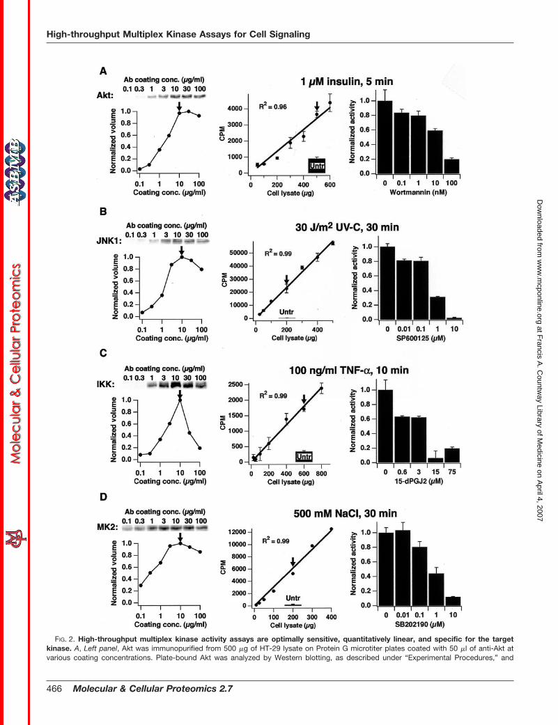
FIG. 2. High-throughput multiplex kinase activity assays are optimally sensitive, quantitatively linear, and specific for the targetkinase. A, Left panel, Akt was immunopurified from 500 �g of HT-29 lysate on Protein G microtiter plates coated with 50 �l of anti-Akt atvarious coating concentrations. Plate-bound Akt was analyzed by Western blotting, as described under “Experimental Procedures,” and
High-throughput Multiplex Kinase Assays for Cell Signaling
466 Molecular & Cellular Proteomics 2.7
at Francis A
. Countw
ay Library of Medicine on A
pril 4, 2007 w
ww
.mcponline.org
Dow
nloaded from
Eu2� screen (Perkin Elmer). The screen was visualized using a Cy-clone Storage Phosphor system (Perkin Elmer).
Cell Imaging—HT-29 cells were seeded in Delta T glass culturedishes (Bioptechs). After 30 h, cells were pretreated with IFN-� asdescribed, then rinsed and treated with 50 ng/ml TNF-� or 50 ng/mlTNF-� � 100 nM insulin for 18 h. Then 6 �g/ml Hoechst 33342(Molecular Probes, Eugene, OR) was added to the medium, andphase and fluorescence images were collected. Cells were then fixedwith 100% MeOH and stored at �20 °C overnight. After primarystaining with anti-cleaved caspase 3 (Cell Signaling, Beverly, MA) andsecondary staining with Alexa 594-conjugated anti-rabbit IgG (Mo-lecular Probes), fluorescence images were collected.
Apoptosis Assays—HT-29 cells were seeded in 96-well plates andsensitized with IFN-� as described. For inhibitor studies, 20 �M
LY294002 was added to the medium 1 h before or 3 h after cytokinestimulation. During washout experiments, the medium was removedand the wells washed once with PBS. The medium was then replacedwith medium conditioned by cells treated with the same cytokineconcentrations without inhibitor. After 18 h, cells were rinsed withPBS and trypsinized. The supernatant and rinse were saved andcombined with the trypsinized cells to ensure capture of both floatingand adherent cells. The cells were washed with PBS, then fixed in100% MeOH and stored at �20 °C. The fixed cells were stained witha fluorescein-labeled monoclonal antibody against caspase-cleavedcytokeratin (M30; Roche), plus anti-cleaved-caspase-3 (Asp 175; CellSignaling) with Alexa 647-conjugated anti-rabbit secondary (Molecu-lar Probes), according to the manufacturers’ instructions. Cells werewashed once after staining, then analyzed by flow cytometry (FACS-calibur; Becton-Dickinson, Mountain View, CA). Cells positive forcleaved cytokeratin, cleaved caspase-3, or both were scored asapoptotic. Three separate biological samples were analyzed for eachtreatment condition. Percent increased survival was calculated ac-cording to the following formula: [1 � (% apoptotic cells in thepresence of insulin)/(% apoptotic cells in the absence of insulin underthe same pretreatment conditions)] � 100%.
RESULTS AND DISCUSSION
High-throughput Kinase Activity Assay Development—Thegeneralized assay format involves four steps, shown in Fig.1B: i) parallel purification of endogenous kinase from wholecell lysates by immunoprecipitation onto Protein A or G mi-
crotiter plates that have been precoated with kinase-specificantibodies, ii) low stringency washes to remove nonpurifiedlysate components, iii) addition of [�-32P]ATP and a kinase-specific substrate to initiate an in vitro kinase reaction, and iv)termination of the in vitro reaction either by H3PO4 or EDTAand liquid transfer to a 96-well phosphocellulose (PC) filterplate to isolate phosphoproteins and remove free [�-32P]ATP.We developed assays for ERK (3), Akt, JNK1, IKK, and MK2activity in cell extracts after rigorous optimization and quan-titative validation.
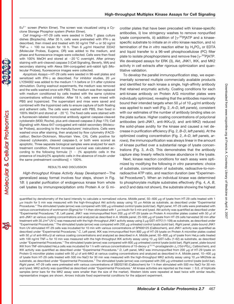
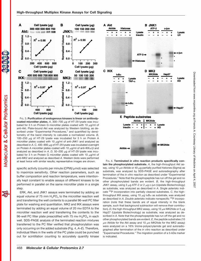
To develop the parallel immunopurification step, we exper-imentally screened multiple commercially available productsand identified for each kinase a single, high-affinity antibodythat retained enzymatic activity. Coating conditions for eachanti-kinase antibody on Protein A/G microtiter plates wereindividually optimized, revealing that the antibodies maximallybound their intended targets when 50 �l of 10 �g/ml antibodywas applied to each well (Fig. 2, A–D, left panels), consistentwith our estimates of the number of antibody binding sites onthe plate surface. Higher coating concentrations of polyclonalantibodies (anti-JNK1, anti-IKK�/�, and anti-MK2) reducedthe solid-phase avidity for the kinase and caused a net de-crease in purification efficiency (Fig. 2, B–D, left panels). At theoptimized coating concentration (Fig. 2, A–D, left panels, ar-rows), the immunopurification was always linear in the amountof kinase purified over a substantial range of lysate concen-trations (Fig. 3, A–D). This demonstrates that the antibodycapture step linearly reflects kinase abundance in the lysate.
Next, kinase reaction conditions for each assay were opti-mized by modifying the following in vitro parameters: choiceof substrate, concentration of substrate, radioactive-to-non-radioactive ATP ratio, and reaction duration (see “Experimen-tal Procedures”). When an individual kinase was determinedto phosphorylate multiple substrates effectively (Fig. 4, A, B,and D and data not shown), the substrate showing the highest
quantified by densitometry of the band intensity to calculate a normalized volume. Middle panel, 50–600 �g of lysate from HT-29 cells treated with 1�M insulin for 5 min was measured with the high-throughput Akt activity assay using 10 �M Aktide as substrate, as described under “ExperimentalProcedures.” The stimulated lysate (arrow) was compared with 500 �g untreated control lysate (solid bar). Right panel, HT-29 cells were pretreated withvarious concentrations of wortmannin (Sigma) for 1 h then stimulated with 1 �M insulin for 5 min and lysed. Akt activity was quantified as described under“Experimental Procedures.” B, Left panel, JNK1 was immunopurified from 200 �g of HT-29 lysate on Protein A microtiter plates coated with 50 �l ofanti-JNK1 at various coating concentrations and analyzed as described in A. Middle panel, 25–500 �g of lysate from HT-29 cells harvested 30 min aftertreatment with 30 J/m2 UV-C was measured with the high-throughput JNK1 activity assay using 3 �g GST-ATF2 (1-109) as substrate, as described under“Experimental Procedures.” The stimulated lysate (arrow) was compared with 200 �g untreated control lysate (solid bar). Right panel, plate-bound JNK1from UV-stimulated HT-29 cells was incubated for 10 min with various concentrations of SP600125 (Calbiochem), and JNK1 activity was quantified asdescribed under “Experimental Procedures.” C, Left panel, IKK was immunopurified from 800 �g of HT-29 lysate on Protein A microtiter plates coatedwith 50 �l of anti-IKK�/� at various coating concentrations and analyzed as described in A. Middle panel, 50–800 �g of lysate from HeLa cells treatedwith 100 ng/ml TNF-� for 10 min was measured with the high-throughput IKK activity assay using 10 �g GST-I�B� (1-62) as substrate, as describedunder “Experimental Procedures.” The stimulated lysate (arrow) was compared with 600 �g untreated control lysate (solid bar). Right panel, plate-boundIKK from TNF-stimulated HeLa cells was incubated for 1 h with various concentrations of 15-deoxy-�12,14-prostaglandin J2 (15d-PGJ2; Calbiochem), andIKK activity was quantified as described under “Experimental Procedures.” D, Left panel, MK2 was immunopurified from 200 �g of HT-29 lysate onProtein G microtiter plates coated with 50 �l of anti-MK2 at various coating concentrations and analyzed as described in A. Middle panel, 10–400 �gof lysate from HT-29 cells treated with 500 mM NaCl for 30 min was measured with the high-throughput MK2 activity assay using 10 �M MK2tide assubstrate, as described under “Experimental Procedures.” The stimulated lysate (arrow) was compared with 200 �g untreated control lysate (solid bar).Right panel, HT-29 cells were pretreated with various concentrations of SB202190 (Calbiochem) for 1 h then stimulated with 500 mM NaCl for 30 minand lysed. MK2 activity was quantified as described under “Experimental Procedures.” All kinase activities are reported as the mean � S.E. of triplicatesamples (error bars for the MK2 assay were smaller than the size of the marker). Western blots were repeated at least twice with similar results;representative images are shown. Arrows indicate fixed experimental conditions for the adjacent experiment.
High-throughput Multiplex Kinase Assays for Cell Signaling
Molecular & Cellular Proteomics 2.7 467
at Francis A
. Countw
ay Library of Medicine on A
pril 4, 2007 w
ww
.mcponline.org
Dow
nloaded from
specific activity (counts per minute (CPM)/�mol) was selectedto maximize sensitivity. Other reaction parameters, such asbuffer composition and reaction temperature, were intention-ally kept constant to enable assays of different kinases to beperformed in parallel on the same microtiter plate in a singlestep.
ERK, Akt, and JNK1 assays were terminated by adding anequal volume of 75 mm H3PO4 to the microtiter reaction welland transferring the well contents to a parallel 96-well PC filterplate for washing and quantitation. MK2 and IKK assays wereterminated by adding an equal volume of 20 mM EDTA to themicrotiter reaction well and transferring the contents to the96-well PC filter plate preacidified with 75 mM H3PO4 in eachwell. SDS-PAGE analysis of the terminated reaction mixturestransferred to the PC filter verified that phosphorylation wasonly occurring on the added substrate (Fig. 4, A–E). Therefore,individual filters in the wells of the PC plate could be punchedout for scintillation counting to accurately quantify kinase
FIG. 3. Purification of endogenous kinases is linear on antibody-coated microtiter plates. A, 300–700 �g of HT-29 lysate was incu-bated for 3 h on Protein G microtiter plates coated with 10 �g/ml ofanti-Akt. Plate-bound Akt was analyzed by Western blotting, as de-scribed under “Experimental Procedures,” and quantified by densi-tometry of the band intensity to calculate a normalized volume. B,100–250 �g of HT-29 lysate was incubated for 3 h on Protein Amicrotiter plates coated with 10 �g/ml of anti-JNK1 and analyzed asdescribed in A. C, 400–900 �g of HT-29 lysate was incubated overnighton Protein A microtiter plates coated with 10 �g/ml of anti-IKK�/� andanalyzed as described in A. D, 50–200 �g of HT-29 lysate was incu-bated for 3 h on Protein G microtiter plates coated with 10 �g/ml ofanti-MK2 and analyzed as described A. Western blots were performedat least twice with similar results; representative images are shown. FIG. 4. Terminated in vitro reaction products specifically con-
tain the phosphorylated substrate. A, the high-throughput Akt as-say, using 10 �M Aktide or 40 �g partially purified histones (Sigma) assubstrate, was analyzed by SDS-PAGE and autoradiography aftertermination of the in vitro reaction as described under “ExperimentalProcedures.” Note that the phosphopeptide has run off the gel and noother phosphorylated bands are evident. B, the high-throughputJNK1 assay, using 3 �g ATF-2 or 2 �g c-jun (Upstate Biotechnology)as substrate, was analyzed as described in A. Single asterisks indi-cate 32P incorporation into partially cleaved substrates. C, the high-throughput IKK assay, using 10 �g I�B� as substrate, was analyzedas described in A. Double asterisks indicate nonspecific 32P incorpo-ration (note that these bands are of equal intensity in the blanksample, such that background subtraction will remove their contribu-tion). D, the high-throughput MK2 assay, using 10 �M MK2tide or 5 �gHsp27 (Upstate Biotechnology) as substrate, was analyzed as de-scribed in A. Note that the phosphopeptide has run off the gel and noother phosphorylated bands are evident. E, the peptide substrates (10�M Aktide for the Akt assay and 10 �M MK2tide for the MK2 assay)were analyzed on a 16% tricine-polyacrylamide gel and autoradio-graphed after termination of the in vitro reaction as described under“Experimental Procedures.” The migration position of a 5-kDa markeris indicated.
High-throughput Multiplex Kinase Assays for Cell Signaling
468 Molecular & Cellular Proteomics 2.7
at Francis A
. Countw
ay Library of Medicine on A
pril 4, 2007 w
ww
.mcponline.org
Dow
nloaded from
activity, eliminating the low-throughput, SDS-PAGE-and-au-toradiography step required in classical immune complex ki-nase assays.
To investigate the sensitivity, dynamic range, and linearityof these assays, HT-29 and HeLa cells were treated withknown activators of each kinase, and the kinase activities(CPM on the PC filter) measured as a function of differentdilutions of the activated lysates (Fig. 2, A–D, middle panels).The 32P incorporation was linear over at least an order ofmagnitude in activity, and the absolute sensitivities of theassays were always below 200 �g, with some assays capableof measuring activity from as little as 10–25 �g of total lysate.This result is comparable to, or better than, existing assaysthat usually require several hundred micrograms of lysate foranalysis (23–25). The aggregate sensitivity of the format wasmore than sufficient to measure ERK, Akt, JNK1, MK2, and IKKactivity simultaneously from a single 10-cm plate of HT-29 cells.
The signal-to-noise and reproducibility characteristics foreach kinase assay were examined by selecting a single con-centration in the middle of the dynamic range of each assay(Fig. 2, A–D, middle panels, arrows) and comparing the kinaseactivities between lysates from stimulated and unstimulatedcells. For each agonist, this revealed a relative activation ofeach endogenous kinase that was comparable in magnitude
to that reported in the literature, with intra-assay coefficientsof variation always �10%. Thus, the assays clearly reflectedactivation of the endogenous pathways, and the end pointmeasures of activity were highly sensitive and reproducible.
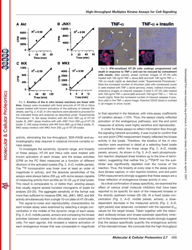
In order for these assays to reflect information flow throughthe signaling network accurately, it was crucial to confirm thatour end point CPM measurement linearly reflected the kinaseactivity in the lysate. Therefore, the kinetics of the in vitroreaction were examined in detail at a selecting fixed lysateconcentration within the linear range (Fig. 2, A–D, middlepanels, arrows). As shown in Fig. 5, A–D, each phosphoryla-tion reaction displayed linear kinetics up to the time of termi-nation, suggesting that neither the [�-32P]ATP nor the sub-strate was significantly depleted over the course of thereaction. Thus, the linearity at every step in the assay proce-dure (kinase capture, in vitro reaction kinetics, and end pointCPM measurement) strongly suggests that these assays are alinear reflection of endogenous kinase activity in vivo.
As a final confirmation of assay specificity, we examined theeffect of various small molecule inhibitors that have beenreported to be specific for each of the measured kinases orthe directly upstream kinase (26–29). At a fixed lysate con-centration (Fig. 2, A–D, middle panels, arrows), a dose-dependent decrease in the measured activity (Fig. 2, A–D,right panels) was observed, consistent with the reported IC50
values for the target kinase. Taken together with the redun-dant antibody-kinase and kinase-substrate specificity inher-ent to the measurement format, these results strongly suggestthat the assays specifically quantify the endogenous activityof the intended kinase. We conclude that the high-throughput
FIG. 5. Kinetics of the in vitro kinase reactions are linear withtime. Assays were incubated with fixed amounts of HT-29 or HeLalysates treated with known activators of the pathway of interest (fordetails, see Fig. 2, A–D). In vitro reactions were allowed to proceed forthe indicated times and analyzed as described under “ExperimentalProcedures.” A, Akt assay kinetics with Akt from 500 �g of HT-29lysate. B, JNK1 assay kinetics with with JNK1 from 200 �g of HT-29lysate. C, IKK assay kinetics with IKK from 800 �g of HeLa lysate. D,MK2 assay kinetics with MK2 from 200 �g of HT-29 lysate.
FIG. 6. IFN-sensitized HT-29 cells undergo programmed celldeath in response to TNF-� and can be rescued by cotreatmentwith insulin. Main panels, phase contrast images of HT-29 cellstreated with 100 ng/ml TNF-� alone (left) and both 100 ng/ml TNF-� �100 nM insulin (right) as described under “Experimental Procedures.”Note the increased number of shrunken cells with condensed nucleiin cells treated with TNF-� alone (arrows). Insets, indirect immunoflu-orescence images of cleaved caspase 3 (red) in HT-29 cells treatedwith 100 ng/ml TNF-� alone (left) and both 100 ng/ml TNF-� � 100 nM
insulin (right). Note the increased number of cleaved caspase 3-pos-itive cells in the TNF-� alone image. Hoechst 33342 (blue) is overlaidin all images to show nuclei.
High-throughput Multiplex Kinase Assays for Cell Signaling
Molecular & Cellular Proteomics 2.7 469
at Francis A
. Countw
ay Library of Medicine on A
pril 4, 2007 w
ww
.mcponline.org
Dow
nloaded from
multiplex assays provide reliable, quantitative measurementsthat are directly reflective of specific protein kinase activitiesin cell extracts.
Application to an In Vitro Model of Sepsis-induced ColonEpithelial Apoptosis—HT-29 cells are a human colon carci-noma cell line that die in response to TNF-�, following sensi-tization with interferon gamma (IFN-�) (30). We confirmed thatTNF-treated HT-29 cells exhibit many features of apoptosis,such as cell shrinkage, membrane blebbing, nuclear conden-sation, and caspase 3 activation (Fig. 6, left). Intriguingly, wefound that TNF-induced cell death can be reduced by �30%if HT-29 cells are costimulated with insulin (Fig. 6, right), inagreement with previous reports (31). Given the importance ofproinflammatory cytokines, such as TNF-� and IFN-�, in sep-sis (4) and the emerging role of insulin as a therapeutic forcritically ill patients (15), we considered HT-29 cells as anappropriate model system for exploring the signaling networkdynamics activated by TNF-� and TNF-� � insulin in relation
to a relevant cell phenotype: colon epithelial cell death, asobserved during the onset of sepsis (6).
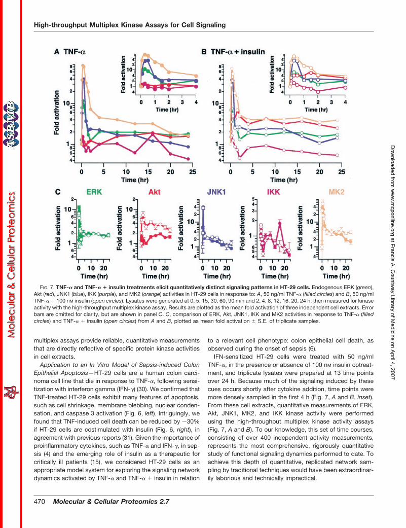
IFN-sensitized HT-29 cells were treated with 50 ng/mlTNF-�, in the presence or absence of 100 nM insulin cotreat-ment, and triplicate lysates were prepared at 13 time pointsover 24 h. Because much of the signaling induced by thesecues occurs shortly after cytokine addition, time points weremore densely sampled in the first 4 h (Fig. 7, A and B, inset).From these cell extracts, quantitative measurements of ERK,Akt, JNK1, MK2, and IKK kinase activity were performedusing the high-throughput multiplex kinase activity assays(Fig. 7, A and B). To our knowledge, this set of time courses,consisting of over 400 independent activity measurements,represents the most comprehensive, rigorously quantitativestudy of functional signaling dynamics performed to date. Toachieve this depth of quantitative, replicated network sam-pling by traditional techniques would have been extraordinar-ily laborious and technically impractical.
FIG. 7. TNF-� and TNF-� � insulin treatments elicit quantitatively distinct signaling patterns in HT-29 cells. Endogenous ERK (green),Akt (red), JNK1 (blue), IKK (purple), and MK2 (orange) activities in HT-29 cells in response to: A, 50 ng/ml TNF-� (filled circles) and B, 50 ng/mlTNF-� � 100 nM insulin (open circles). Lysates were generated at 0, 5, 15, 30, 60, 90 min and 2, 4, 8, 12, 16, 20, 24 h, then measured for kinaseactivity with the high-throughput multiplex kinase assay. Results are plotted as the mean fold activation of three independent cell extracts. Errorbars are omitted for clarity, but are shown in panel C. C, comparison of ERK, Akt, JNK1, IKK and MK2 activities in response to TNF-� (filledcircles) and TNF-� � insulin (open circles) from A and B, plotted as mean fold activation � S.E. of triplicate samples.
High-throughput Multiplex Kinase Assays for Cell Signaling
470 Molecular & Cellular Proteomics 2.7
at Francis A
. Countw
ay Library of Medicine on A
pril 4, 2007 w
ww
.mcponline.org
Dow
nloaded from
Furthermore, to deconstruct the effects of insulin therapyon the signaling network in the context of TNF-� signaling, apairwise comparison was performed for each kinase underTNF-� with and without insulin costimulation. As shown in Fig.7C, the dynamics of activation of some pathways, such as theERK pathway (Fig. 7C, green), were essentially superimpos-able. Thus insulin does not appear to influence activation ofthe ERK pathway by TNF-� under these conditions. OtherTNF-induced kinase activities were affected by insulin in atime-dependent manner. JNK1 activity, for example, waslarger at intermediate times (8–16 h), while IKK activity wassmaller at 16 h, and MK2 activity was larger at late times(16–24 h). Although potentially important, these transient sig-naling differences did not clearly reflect the insulin-inducedsurvival phenotype observed at the cellular level (Fig. 6).
Late-phase Akt Activity as a Critical Pro-survival Signal—Incontrast to the subtle influences of insulin on ERK, JNK1, IKK,and MK2 signaling, it was readily apparent that insulin dra-matically augmented Akt activity, inducing a rapid increasewithin 5 min and sustaining activation for 24 h, whereas withTNF-� alone, the Akt response was much smaller in magni-tude (Fig. 7C, red). In addition to these quantitative differ-ences, we noted that the dynamic TNF-induced Akt responsewas also qualitatively different without insulin, in that thecourse of activation was clearly biphasic, with a brief initialpeak, followed by a small (i.e. 2-fold), sustained increase inactivity after 4 h. If these data had not been rigorously quan-titative, or had the signaling network not been sampled fre-quently in time, this increase in activity would likely have beenmissed. By virtue of the high-throughput activity assays, how-ever, it was clear that Akt activity after 4 h was significantlyup-regulated in comparison to baseline activity (p � 0.05 forall times after 4 h, Student’s t test).
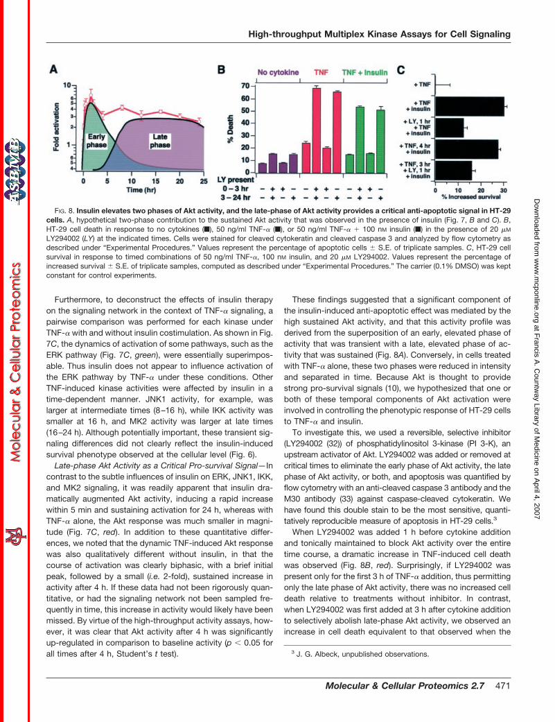
These findings suggested that a significant component ofthe insulin-induced anti-apoptotic effect was mediated by thehigh sustained Akt activity, and that this activity profile wasderived from the superposition of an early, elevated phase ofactivity that was transient with a late, elevated phase of ac-tivity that was sustained (Fig. 8A). Conversely, in cells treatedwith TNF-� alone, these two phases were reduced in intensityand separated in time. Because Akt is thought to providestrong pro-survival signals (10), we hypothesized that one orboth of these temporal components of Akt activation wereinvolved in controlling the phenotypic response of HT-29 cellsto TNF-� and insulin.
To investigate this, we used a reversible, selective inhibitor(LY294002 (32)) of phosphatidylinositol 3-kinase (PI 3-K), anupstream activator of Akt. LY294002 was added or removed atcritical times to eliminate the early phase of Akt activity, the latephase of Akt activity, or both, and apoptosis was quantified byflow cytometry with an anti-cleaved caspase 3 antibody and theM30 antibody (33) against caspase-cleaved cytokeratin. Wehave found this double stain to be the most sensitive, quanti-tatively reproducible measure of apoptosis in HT-29 cells.3
When LY294002 was added 1 h before cytokine additionand tonically maintained to block Akt activity over the entiretime course, a dramatic increase in TNF-induced cell deathwas observed (Fig. 8B, red). Surprisingly, if LY294002 waspresent only for the first 3 h of TNF-� addition, thus permittingonly the late phase of Akt activity, there was no increased celldeath relative to treatments without inhibitor. In contrast,when LY294002 was first added at 3 h after cytokine additionto selectively abolish late-phase Akt activity, we observed anincrease in cell death equivalent to that observed when the
3 J. G. Albeck, unpublished observations.
FIG. 8. Insulin elevates two phases of Akt activity, and the late-phase of Akt activity provides a critical anti-apoptotic signal in HT-29cells. A, hypothetical two-phase contribution to the sustained Akt activity that was observed in the presence of insulin (Fig. 7, B and C). B,HT-29 cell death in response to no cytokines (f), 50 ng/ml TNF-� (f), or 50 ng/ml TNF-� � 100 nM insulin (f) in the presence of 20 �M
LY294002 (LY) at the indicated times. Cells were stained for cleaved cytokeratin and cleaved caspase 3 and analyzed by flow cytometry asdescribed under “Experimental Procedures.” Values represent the percentage of apoptotic cells � S.E. of triplicate samples. C, HT-29 cellsurvival in response to timed combinations of 50 ng/ml TNF-�, 100 nM insulin, and 20 �M LY294002. Values represent the percentage ofincreased survival � S.E. of triplicate samples, computed as described under “Experimental Procedures.” The carrier (0.1% DMSO) was keptconstant for control experiments.
High-throughput Multiplex Kinase Assays for Cell Signaling
Molecular & Cellular Proteomics 2.7 471
at Francis A
. Countw
ay Library of Medicine on A
pril 4, 2007 w
ww
.mcponline.org
Dow
nloaded from

inhibitor was present for the entire 24 h. An identical patternwas observed in response to TNF-� � insulin (Fig. 8B, green),whereas LY294002 had only a small effect on basal apoptosis(Fig. 8B, blue). These data suggest that late-phase Akt acti-vation is necessary to restrain the percentage of cells under-going programmed cell death in response to TNF-� in thepresence or absence of insulin.
The importance of late Akt activity in controlling TNF-in-duced cell death, in conjunction with the strong, sustainedlate Akt activation in the presence of insulin, led to the hy-pothesis that the insulin-induced increase in cell survival wasdue, in part, to this increase in late-phase Akt signaling. If true,then addition of insulin up to 4 h after the pro-death stimulusshould elicit an equivalent reduction in the extent of TNF-induced apoptosis. Indeed, this was observed; the delayedaddition of insulin 4 h following TNF-� addition resulted in thesame increase in HT-29 cell survival as observed when insulinwas added simultaneously with TNF-� (Fig. 8C). Moreover,pretreatment with LY294002 1 h before insulin addition sig-nificantly reduced the survival response, regardless whetherinsulin was added simultaneously or 4 h after TNF-� treatment(Fig. 8C).
These experiments strongly implicate late-phase signaling,along a PI 3-K-dependent/Akt pathway, as a critical pro-survival mechanism of insulin in colon epithelia. Excessiveapoptosis in the gut leads to disruption of the epithelial per-meability barrier (7) that drives many of the complications ofthe septic state (8). Our results provide a partial molecularexplanation why intensive insulin therapy, administered hoursafter the initial septic insult, is capable of reducing sepsis-induced apoptosis of gastrointestinal epithelia and improvingthe prognostic outcome in critically ill patients.
We stress that it was the ability to robustly sample thedynamics of multiple nodes in the signaling network with thehigh-throughput multiplex kinase assay format described inthis paper that enabled the identification of Akt as an impor-tant signal for modulating cell response. Other insulin-in-duced, PI 3-K independent pathways must also attenuate theTNF-induced apoptotic phenotype in HT-29 cells, because in-hibition of PI 3-K/Akt by LY294002 did not completely abrogatethe rescue response of insulin (Fig. 8C). Thus, i) there may existadditional response-determining pathways that our measure-ments did not capture, or ii) the subtle, time-dependent quan-titative differences we measured in the JNK1, IKK, and MK2pathways (Fig. 7C) may modulate phenotypic response. We arecurrently investigating both these possibilities.
The current knowledge of intracellular signal transduction isstaggeringly complex. To identify network-level propertiesthat affect cell function, it will be necessary to mathematicallymodel the dynamic, multivariate characteristics of signalingproteins within cells (34). For such models to be realized,quantitative experimental techniques that are both high-throughput and multiplex are needed. The kinase activity as-say format presented here represents a first step in this di-
rection. The 96-well, microtiter format is highly versatile, inthat it is amenable to scale-up and automated liquid handling,yet tractable for individual scientists and more moderate stud-ies. Importantly, these assays possess linearity, reproducibil-ity, specificity, and sensitivity characteristics as good as, orbetter than, the corresponding low-throughput technique. Weanticipate that these functional assays will complement exist-ing proteomic approaches (2) and find broad applicabilitytoward biological and clinical problems involving signal trans-duction and human disease.
Acknowledgments—We thank Isaac Manke for providing theMK2tide sequence ahead of publication, as well as Roger Davis andWarner Greene for providing various plasmids.
* This work was supported by the Defense Advanced ResearchPlanning Agency Bio-Info-Micro Program, the National Institute ofGeneral Medical Sciences Cell Migration Consortium (to D. A. L.), andNational Institutes of Health Grant GM59281 (to M. B. Y.). Additionalfinancial support was provided by the Whitaker Foundation (toK. A. J.). The costs of publication of this article were defrayed in partby the payment of page charges. This article must therefore be herebymarked “advertisement” in accordance with 18 U.S.C. Section 1734solely to indicate this fact.
� To whom correspondence should be addressed: Center for Can-cer Research, Massachusetts Institute of Technology, 77 Massachu-setts Ave. E18–580, Cambridge, MA 02139. Tel.: 617-452-2103, Fax:617-452-4978; E-mail: [email protected].
REFERENCES
1. Nguyen, A., and Yaffe, M. B. (2003) Proteomics and systems biologyapproaches to signal transduction in sepsis. Crit. Care Med. 31, S1–S6
2. Tyers, M., and Mann, M. (2003) From genomics to proteomics. Nature 422,193–197
3. Asthagiri, A. R., Horwitz, A. F., and Lauffenburger, D. A. (1999) A rapid andsensitive quantitative kinase activity assay using a convenient 96-wellformat. Anal. Biochem. 269, 342–347
4. Hotchkiss, R. S., and Karl, I. E. (2003) The pathophysiology and treatmentof sepsis. N. Engl. J. Med. 348, 138–150
5. Strassheim, D., Park, J. S., and Abraham, E. (2002) Sepsis: Current con-cepts in intracellular signaling. Int. J. Biochem. Cell Biol. 34, 1527–1533
6. Hotchkiss, R. S., Schmieg, R. E., Jr., Swanson, P. E., Freeman, B. D.,Tinsley, K. W., Cobb, J. P., Karl, I. E., and Buchman, T. G. (2000) Rapidonset of intestinal epithelial and lymphocyte apoptotic cell death inpatients with trauma and shock. Crit. Care Med. 28, 3207–3217
7. Bojarski, C., Bendfeldt, K., Gitter, A. H., Mankertz, J., Fromm, M., Wagner,S., Riecken, E. O., and Schulzke, J. D. (2000) Apoptosis and intestinalbarrier function. Ann. N. Y. Acad. Sci. 915, 270–274
8. Wattanasirichaigoon, S., Menconi, M. J., Delude, R. L., and Fink, M. P.(1999) Effect of mesenteric ischemia and reperfusion or hemorrhagicshock on intestinal mucosal permeability and ATP content in rats. Shock12, 127–133
9. Hatherill, M., Tibby, S. M., Turner, C., Ratnavel, N., and Murdoch, I. A.(2000) Procalcitonin and cytokine levels: relationship to organ failure andmortality in pediatric septic shock. Crit. Care Med. 28, 2591–2594
10. Lawlor, M. A., and Alessi, D. R. (2001) PKB/Akt: a key mediator of cellproliferation, survival and insulin responses? J. Cell Sci. 114, 2903–2910
11. Davis, R. J. (2000) Signal transduction by the JNK group of MAP kinases.Cell 103, 239–252
12. Karin, M., and Ben-Neriah, Y. (2000) Phosphorylation meets ubiquitination:The control of NF-�B activity. Annu. Rev. Immunol. 18, 621–663
13. Stokoe, D., Engel, K., Campbell, D. G., Cohen, P., and Gaestel, M. (1992)Identification of MAPKAP kinase 2 as a major enzyme responsible for thephosphorylation of the small mammalian heat shock proteins. FEBS Lett.313, 307–313
14. Fisher, C. J., Jr., Agosti, J. M., Opal, S. M., Lowry, S. F., Balk, R. A., Sadoff,
High-throughput Multiplex Kinase Assays for Cell Signaling
472 Molecular & Cellular Proteomics 2.7
at Francis A
. Countw
ay Library of Medicine on A
pril 4, 2007 w
ww
.mcponline.org
Dow
nloaded from

J. C., Abraham, E., Schein, R. M., and Benjamin, E. (1996) Treatment ofseptic shock with the tumor necrosis factor receptor:Fc fusion protein.The Soluble TNF Receptor Sepsis Study Group. N. Engl. J. Med. 334,1697–1702
15. van den Berghe, G., Wouters, P., Weekers, F., Verwaest, C., Bruyninckx, F.,Schetz, M., Vlasselaers, D., Ferdinande, P., Lauwers, P., and Bouillon, R.(2001) Intensive insulin therapy in the critically ill patients. N. Engl.J. Med. 345, 1359–1367
16. Van den Berghe, G., Wouters, P. J., Bouillon, R., Weekers, F., Verwaest, C.,Schetz, M., Vlasselaers, D., Ferdinande, P., and Lauwers, P. (2003)Outcome benefit of intensive insulin therapy in the critically ill: Insulindose versus glycemic control. Crit. Care Med. 31, 359–366
17. Hirsch, I. B., and Coviello, A. (2002) Intensive insulin therapy in critically illpatients. N. Engl. J. Med. 346, 1586–1588
18. Hansen, T. K., Thiel, S., Wouters, P. J., Christiansen, J. S., and Van denBerghe, G. (2003) Intensive insulin therapy exerts antiinflammatory ef-fects in critically ill patients and counteracts the adverse effect of lowmannose-binding lectin levels. J. Clin. Endocrinol. Metab. 88, 1082–1088
19. Obata, T., Yaffe, M. B., Leparc, G. G., Piro, E. T., Maegawa, H., Kashiwagi,A., Kikkawa, R., and Cantley, L. C. (2000) Peptide and protein libraryscreening defines optimal substrate motifs for AKT/PKB. J. Biol. Chem.275, 36108–36115
20. Gupta, S., Campbell, D., Derijard, B., and Davis, R. J. (1995) Transcriptionfactor ATF2 regulation by the JNK signal transduction pathway. Science267, 389–393
21. Geleziunas, R., Ferrell, S., Lin, X., Mu, Y., Cunningham, E. T., Jr., Grant, M.,Connelly, M. A., Hambor, J. E., Marcu, K. B., and Greene, W. C. (1998)Human T-cell leukemia virus type 1 Tax induction of NF-kappaB involvesactivation of the IkappaB kinase alpha (IKKalpha) and IKKbeta cellularkinases. Mol. Cell. Biol. 18, 5157–5165
22. Judd, R. C. (1994) Electrophoresis of peptides, in Methods in MolecularBiology (Walker, J. M., ed.) p. 32, Humana Press, Totowa, NJ
23. Whitmarsh, A. J., and Davis, R. J. (2001) Analyzing JNK and p38 mitogen-activated protein kinase activity. Methods Enzymol. 332, 319–336
24. Hill, M. M., and Hemmings, B. A. (2002) Analysis of protein kinase B/Akt.Methods Enzymol. 345, 448–463
25. Kupfer, R., and Scheinman, R. I. (2002) Measurement of IKK activity inprimary rat T cells: rapid activation and inactivation. J. Immunol. Meth-ods. 266, 155–164
26. Arcaro, A., and Wymann, M. P. (1993) Wortmannin is a potent phosphati-dylinositol 3-kinase inhibitor: the role of phosphatidylinositol 3, 4,5-trisphosphate in neutrophil responses. Biochem. J. 296 (Pt 2),297–301
27. Bennett, B. L., Sasaki, D. T., Murray, B. W., O’Leary, E. C., Sakata, S. T., Xu,W., Leisten, J. C., Motiwala, A., Pierce, S., Satoh, Y., Bhagwat, S. S.,Manning, A. M., and Anderson, D. W. (2001) SP600125, an anthrapyra-zolone inhibitor of Jun N-terminal kinase. Proc. Natl. Acad. Sci. U. S. A.98, 13681–13686
28. Rossi, A., Kapahi, P., Natoli, G., Takahashi, T., Chen, Y., Karin, M., andSantoro, M. G. (2000) Anti-inflammatory cyclopentenone prostaglandinsare direct inhibitors of IkappaB kinase. Nature 403, 103–108
29. Lee, J. C., Laydon, J. T., McDonnell, P. C., Gallagher, T. F., Kumar, S.,Green, D., McNulty, D., Blumenthal, M. J., Heys, J. R., Landvatter, S. W.,Strickler, J. E., McLaughlin, M. M., Siemens, J. R., Fisher, S. M., Livi,G. P., White, J. R., Adams, J. L., and Young, P. R. (1994) A protein kinaseinvolved in the regulation of inflammatory cytokine biosynthesis. Nature372, 739–746
30. Abreu-Martin, M. T., Vidrich, A., Lynch, D. H., and Targan, S. R. (1995)Divergent induction of apoptosis and IL-8 secretion in HT-29 cells inresponse to TNF-alpha and ligation of Fas antigen. J. Immunol. 155,4147–4154
31. Remacle-Bonnet, M. M., Garrouste, F. L., Heller, S., Andre, F., Marvaldi,J. L., and Pommier, G. J. (2000) Insulin-like growth factor-I protectscolon cancer cells from death factor-induced apoptosis by potentiatingtumor necrosis factor alpha-induced mitogen-activated protein kinaseand nuclear factor kappaB signaling pathways. Cancer Res. 60,2007–2017
32. Vlahos, C. J., Matter, W. F., Hui, K. Y., and Brown, R. F. (1994) A specificinhibitor of phosphatidylinositol 3-kinase, 2-(4-morpholinyl)-8-phenyl-4H-1-benzopyran-4-one (LY294002). J. Biol. Chem. 269, 5241–5248
33. Leers, M. P., Kolgen, W., Bjorklund, V., Bergman, T., Tribbick, G., Persson,B., Bjorklund, P., Ramaekers, F. C., Bjorklund, B., Nap, M., Jornvall, H.,and Schutte, B. (1999) Immunocytochemical detection and mapping of acytokeratin 18 neo-epitope exposed during early apoptosis. J. Pathol.187, 567–572
34. Asthagiri, A. R., and Lauffenburger, D. A. (2000) Bioengineering models ofcell signaling. Annu. Rev. Biomed. Eng. 2, 31–53
High-throughput Multiplex Kinase Assays for Cell Signaling
Molecular & Cellular Proteomics 2.7 473
at Francis A
. Countw
ay Library of Medicine on A
pril 4, 2007 w
ww
.mcponline.org
Dow
nloaded from