The Effects of Pesticides Mixture on Aquatic Organisms in ...
Upload
independentCategory
view
2download
0
RESEARCH ARTICLE
J. I. Carreto Æ M. O. Carignan Æ N. G. Montoya
A high-resolution reverse-phase liquid chromatography method for theanalysis of mycosporine-like amino acids (MAAs) in marine organisms
Received: 30 March 2004 / Accepted: 23 July 2004 / Published online: 26 August 2004� Springer-Verlag 2004
Abstract Mycosporine-like amino acids (MAAs) are agroup of about 20 structurally related water-solublecompounds, widely distributed among freshwater andmarine organisms. To provide a better assessment of thediversity and concentration of MAAs in aquatic envi-ronments a high-performance liquid chromatography(HPLC) method of analysis based on reverse-phase C18
column and trifluoroacetic acid and an ammonium-containing mobile phase was developed. The improve-ments with respect to previous methods and theextraction and clean-up procedures are described here.With this method the clean-up recovery of MAAs ofhigh polarity (shinorine), medium polarity (palythinol),and low polarity (palythene) is greater than 99%(±1%). The method is selective enough to resolve in asingle run most of the characterized MAAs found inmarine organisms, including the critical and highly polarcompounds shinorine, mycosporine-2-glycine, andpalythine-serine, the medium polarity pair palythenicacid and shinorine methyl ester (M-333), and the lowpolarity isomeric pair usujirene and palythene. A chro-matogram of a mixture of over 20 MAAs such as mightbe found in complex samples of marine organisms isgiven. Good precision was obtained in the separations.The relative standard deviation for retention times wasbelow 1% and the mean relative standard deviation forintegrated area estimations was below 2%. A meancolumn recovery of standards was 99% (±1%) whereaslimits of detection (signal-to-noise, S/N=2) for differentMAAs varied between 0.08 and 0.47 pmol injected. Theapplicability of the method was tested using extracts of
three microalgae cultures, three natural phytoplanktonpopulations, two scleractinian corals, and one species ofsea anemone. Results reveal the occurrence of severalunknown MAAs not previously reported in the litera-ture. The selectivity of the method toward some recentlydiscovered MAAs makes it especially suitable not onlyfor studying new field samples, but also for re-examiningthe MAA composition of previously studied organisms.
Introduction
Considerable interest has been centered on the my-cosporine-like amino acids (MAAs) because of theirultraviolet (UV)-photoprotective role. This has beeninferred from their efficient UV absorption (Takanoet al. 1978) and from their light-dependent induction ofsynthesis (Carreto et al. 1989), which has been recentlyverified experimentally (Adams and Shick 1996; Nealeet al. 1998; Karsten et al. 1999). In addition some MAAsmay act as antioxidants to prevent cellular damageresulting from UV-induced production of toxic oxygenspecies (Dunlap et al. 2000).
These compounds, which are widely distributedamong freshwater and marine organisms (Bandarana-yake 1998; Sinha et al. 1998; Shick and Dunlap 2002),are composed of a cyclohexenone or cyclohexeniminechromophore conjugated with the nitrogen substituentof an amino acid, amino alcohol, or amino group. Thereare now more than 20 fully characterized MAAs withmaximum absorption ranging from 309 to 360 nm. Inaddition, several partially characterized and other un-known MAAs have been recently detected as a conse-quence of the increase in the number of studiedorganisms and the development of more efficient high-performance liquid chromatography (HPLC) separationtechniques (Jeffrey et al. 1999; Subramaniam et al. 1999;Carreto et al. 2001). Generally a glycine sub-unit ispresent on the C3 of the cyclohexenimine ring. SomeMAAs also contain sulfate esters (Wu Won et al. 1997).
Communicated by O. Kinne, Oldendorf/Luhe
J. I. Carreto (&) Æ M. O. Carignan Æ N. G. MontoyaInstituto Nacional de Investigacion yDesarrollo Pesquero (INIDEP),Paseo Victoria Ocampo N�1,B7602HSA, 7600 Mar del Plata,ArgentinaE-mail: [email protected]
Marine Biology (2005) 146: 237–252DOI 10.1007/s00227-004-1447-y

Other MAAs are covalently linked to oligosaccharides(Bohm et al. 1995) or among themselves (Carreto et al.2001). The structural relationship among several MAAscommonly detected in the marine environment is illus-trated in Fig. 1.
Until now, only a few HPLC methods for MAAseparation in marine organisms have been reported. Nomethod has been able to separate a mixture of over 20MAAs such as might be found in the most complexsamples from marine organisms. Techniques for theseparation of MAAs include those of Nakamura et al.(1982), Dunlap and Chalker (1986) and their modifica-tions (Stochaj et al. 1994; Shick et al. 1999), and Carreto
Fig. 1 Structural relationships between the different mycosporine-like amino acids (MAAs) and their feasible chemical and/orbiochemical conversions
238
et al. (2001). The classical HPLC method was based onreverse-phase low silanol-free group octadecylsilica (C18)columns and isocratic elution with 0.02% acetic acid asmobile phase at 15�C (Nakamura et al. 1982). Underthese conditions 7 of the 9 MAAs known at the timewere separated according to hydrophobic properties.This method had the disadvantage that it did not elutethe more hydrophobic compounds such as usujirene andpalythene and several of the new complexes of MAAsrecently described (Carreto et al. 2001). In addition itsutility for the separation of the new highly polar com-pounds recently discovered in corals (Teai et al. 1997,1998; Wu Won et al. 1997; Shick et al. 1999) was notproved.
Dunlap and Chalker (1986) first introduced the use ofmonomeric octylsilica (C8) columns. The originalmethod was based on reverse-phase non-endcapped C8
column and isocratic elution with 0.1% acetic acid and10% methanol. Under these conditions the more weaklyacidic compounds (mycosporine-glycine, palythine, as-terina-330, palythinol, and palythene) were separatelyeluted. However, it was unable to separate clearly thestrongly acidic shinorine from porphyra-334. After thediscovery of mycosporine-2-glycine and mycosporine-taurine in the sea anemone Anthopleura elegantissima(Stochaj et al. 1994) a new drawback was added as themethod was not able to separate mycosporine-2-glycinefrom porphyra-334 and mycosporine-glycine from my-cosporine-taurine. Stochaj et al. (1994) showed that thechromatographic separation of the acidic compoundscould be improved if the methanol content of the mobilephase was increased up to 75%. In this condition, thehighly polar compounds interacted with the weak anionexchange properties of the silanol groups to give animproved chromatographic separation of these com-pounds. Although these methods and their furthermodifications using the same stationary phase achievedgood separation of several compounds (Shick et al.1999), none was able to separate in a single run thestrongly acidic MAAs and the more weakly acidiccompounds. Separation of these mixtures was onlyachieved by isocratic elution with two different mobilephases (Helbling et al. 1996; Adams and Shick 2001;Shick et al. 2002). However, resolution for some criticalpairs, such as mycosporine-glycine and mycosporine-taurine, the MAA-sulfated esters and the isomeric mix-ture of usujirene and palythene, was insufficient. Eventhe most recent modifications of these methods do notcompletely resolve these MAA mixtures. The resolutionof mycosporine-glycine and the mixture of MAA-sul-fated esters was recently improved with the use of ion-exchange chromatography on a bonded-phase aminocolumn (Wu Won et al. 1997).
Dionisio-Sese et al. (1997) were the first to use re-verse-phase C18 columns and gradient elution withaqueous methanol, but their application was only suc-cessful for the separation of four MAAs. In a recentpaper Carreto et al. (2001) showed that using acetoni-trile-based eluents and polymeric double-endcapped C18
columns, a mixture of strongly acidic and neutral MAAscould be separated in a single run. This technique alsoresolved the critical isomeric pair usujirene and palyth-ene and its application revealed the occurrence of severalnew low-polarity atypical MAAs not previously re-ported in the literature. Nevertheless, this method andits further modifications (Conde et al. 2003) failed in theseparation of certain highly polar MAAs (shinorinefrom mycosporine-2-glycine and palythine-serine),characteristics of some scleractinian corals (Teai et al.1997, 1998; Wu Won et al. 1997; Shick et al. 1999).Recently Whitehead et al. (2001) and Whitehead andHedges (2002) developed a mass spectral approach toMAA characterization using liquid chromatographycoupled with electrospray ionization mass spectrometry(LC/MS). With the exceptions of the isomeric pair E/Zpalythenic acid and usujirene/palythene, this approachallows the identification of individual MAAs without theneed for a high chromatographic resolution. Howeverthe high cost of LC/MS systems and the level of exper-tise required for their operation limit their use in manylaboratories.
In the present work we describe an improved HPLCseparation method for the analysis of complex samplesof MAAs from marine organisms. This method com-bines a C18 column system with an optimized aqueousmobile phase including trifluoroacetic acid (TFA) andammonium as ion-suppression/ion-pairing agents. Theresults obtained from the analysis of algal cultures,natural phytoplankton populations, red macroalgae,and symbiotic organisms show that the new method isable to separate in a single run complex mixtures of over20 MAAs, such as might be found in the most complexsamples from marine organisms.
Materials and methods
Samples
Four algal cultures, three natural phytoplankton popu-lations, four macroalgae, and three symbiotic organismswere selected for this study to include most of the MAAsfound in marine organisms. Extracts from these sampleswere used for method development and evaluation.
Algal cultures
The four species used were Alexandrium tamarense(Lebour) Balech clone MDQ 1096 ( Dinophyceae) iso-lated from the Mar del Plata coast (Argentina),A. catenella (Weedon and Kofoid) Balech clone CC08(Dinophyceae) isolated from the XI region of Chile,Emiliania huxleyi clone CCMP 370 (Prymnesiophyceae)from the Provasoli-Guillard Centre for Culture ofmarine phytoplankton (CCMP), and Pseudonitzschiamultiseries (Bacillariophyceae) isolated from the Mar delPlata coast (Argentina).
239

Phytoplankton assemblages
Two phytoplankton samples were collected in thecoastal waters off Mar del Plata, one during a bloom ofthe heterotrophic dinoflagellate Noctiluca scintillans andthe other during a bloom of the dinoflagellate Gymn-odinium catenatum. Another sample was collected inwaters from the Brazil Current. Samples were filteredonto GF/F filters and maintained in liquid nitrogen untilanalysis.
Macroalgae
The macroalgae Palmaria decipiens and Iridea sp. werecollected at Potter Cove (South Shetland Islands, Ant-arctica) and were kindly donated by Dr. G. Ferreyra.Porphyra columbina were collected at Chubut coast(Argentina) and were kindly donated by Dr. A. Boraso.Lyophilized Porphyra sp. (Nori) was kindly donated byDr. P. Zimba. All of these organisms were stored at�20�C until analysis.
Symbiotic organisms
Lyophilized purified (C18 column) extracts of the coralPocillopora eydouxi collected at Tahiti island (FrenchPolynesia) were kindly donated by Dr. T. Teai. Lyoph-ilized purified (C18 column) extracts of the coral Stylo-phora pistillata were kindly donated by Dr. M. Shick.Specimens of the sea anemone Anthopleura elegantissimawere collected in California (USA) and were kindlydonated by Dr. M. Shick. All of these materials wereimmediately stored at �20�C.
Chemicals
All solvents used were HPLC grade from Riedel-deHaen. Distilled water was further purified to HPLCgrade by passage through a Barnstead water purificationsystem (Nanopure UV) equipped with ion exchange,carbon cartridges, and UV radiation. TFA (99.9%purity) was purchased from J. Baker and the ammoniasolution (37%) from Merck.
The MAAs shinorine, phorphyra-334, palythine, ast-erine, palythinol, mycosporine-glycine, palythenic acid,usujirene, palythene, M-320, M-333, and M-335/360were obtained as previously described (Carreto et al.2001). Palythine-serine, mycosporine-methylamine-serine, and mycosporine-methylamine threonine stan-dards purified from the coral P. eydouxi were kindlydonated by Dr. T. Teai. Secondary standards ofmycosporine-2-glycine and palythine-serine sulfate fromthe coral S. pistillata and mycosporine-taurine from thesea anemone A. elegantissima were kindly donated byDr. M. Shick. Mycosporine-2-glycine and mycosporine-taurine were isolated and purified from these sources.
Preparation of samples for HPLC analysis
Sample extraction
Algal cultures and natural phytoplankton samples wereconcentrated on 25-mm Whatman GF/F filters andwhen necessary stored at �20�C. Three different proto-cols were used to extract MAAs with HPLC grademethanol: (1) lyophilized or filtered phytoplanktonsamples were cut into small pieces and sonicated 1 minin a pulse mode at 0�C in centrifuge tubes, using 2 ml100% methanol. A Vibra Cell sonicator (Sonic andMaterials, Inc.) equipped with a 4-mm-diameter probewas operated at 40 W. The decanted extract was col-lected and the filter debris was re-extracted twice with100% methanol. The combined extracts were filtered(Whatman GF/F) to remove debris and their absorptionspectra was recorded. (2) Samples were soaked withsmall volumes of water (water:sample mass ratioapproximately 9:1) overnight in the dark at 4�C beforeapplying the extraction procedure described in proto-col 1. (3) For comparison we also investigated a com-plementary extraction method using more aggressiveconditions: samples were extracted with 25% aqueousmethanol (v:v) for 2 h in a water bath at 45�C (Som-maruga and Garcia-Pichel 1999; Tartarotti and Som-maruga 2002). The decanted extract was collected andthe filter debris was re-extracted twice in the same con-ditions with 25% aqueous methanol. Extraction effi-ciencies for these three extraction methods weredetermined after exhaustive serial extraction of theremaining pellet using protocol 3. For these extractionmethods, the recovery and stability of the main indi-vidual MAAs found in Alexandrium catenella were alsodetermined.
The obtained extracts were evaporated to drynessusing a centrifugal vacuum evaporator (Centrivap,Labconco, Co.) and the residue re-dissolved in 500 ll ofa pH 3.15 solution of aqueous trifluoroacetic acid 0.2%and ammonium hydroxide (mobile phase A). Lyophi-lized purified extracts of P. eydouxi and S. pistillata weredissolved in methanol, evaporated to dryness, and re-dissolved in mobile phase A prior to injection. To testthe stability of MAAs in 100% methanol and in mobilephase A, three replicates of a solution of a mixture ofstandards were re-injected into the HPLC after havingbeen kept at ambient temperature for 24 h and at �2 �Cin the freezer for 7 days. The recovery and stability ofmore labile MAAs were compared with those of thefresh extracts.
Clean-up procedure
The aqueous MAA extracts were passed through a 100-kDa ultrafilter (Ultra spin, Alltech) to remove water-insoluble materials and large molecules. The recoveryafter the clean-up procedure was determined using threereplicate extracts spiked with shinorine, palithynol, andpalythene standard solutions.
240

Chromatographic apparatus and conditions
HPLC
Method development was performed using a high-pres-sure gradient system HPLC Shimadzu LC 10 A con-sisting of a continuous degassing system DGU-14 A,two pumps model LC-10 AT, an auto-sampler injectorSIL-10Axl, and a column oven CTO-10 AC. An SPD-M10Avp diode array detector connected via an interfacemodule to a computer running CLASS-LC10 softwarewas used for detection and quantification. The pre-packaged columns used were (1) a polymeric double-endcapped C18 column (5 lm, 4.6 mm i.d. · 150 mmlength; Alltima, Alltech) and (2) a polymer-coated silicareversed-phase C18 column (5 lm, 4.6 mm i.d. ·250 mm length; CapCell Pak UG, Shiseido) protectedwith a guard column cartridge (4.6 mm i.d. · 20 mmlength; Alltima, Alltech). The final high-resolutionseparations were performed with the two columnsconnected in series and thermostated at 35�C.
Mobile phase and elution gradient
Eluent A was a pH 3.15 solution of aqueous trifluoro-acetic acid 0.2% and ammonium hydroxide (see below)while eluent B was a solution containing aqueous tri-fluoroacetic acid 0.2% and ammonium hydroxide atpH 2.20:methanol:acetonitrile (80:10:10,v:v:v). Theaqueous 0.2% TFA solutions were prepared as follows:2.0 ml of TFA were added to 900 ml of water in a 1-lflask and mixed using a magnetic stirrer. Ammoniasolution was then added dropwise until the desired pHwas obtained. The mixture was diluted to 1,000 ml withwater and the pH rechecked. The optimum gradientemployed after all other chromatographic conditionswere fixed is shown in Table 1.
MAA identification, resolution, and quantification
Detection was made by monitoring absorption at 360,330, 310, and also at 270 nm to ensure that the sampleswere devoid of contaminants with absorption at lowerwavelengths. Individual peaks were identified by onlineabsorption spectra, retention time, and when possible byco-chromatography with standards. Resolution (Rs)between a peak and the preceding one was calculatedusing the following equation: Rs=2(Rt2�Rt1)/Wt,where Rt2 and Rt1 are the retention times of two adja-cent peaks, and Wt is the sum of peak widths at baseline.Quantification was accomplished by comparing theareas of peaks from unknowns with those from standardsolutions calibrated using the molar extinction coeffi-cients (�) at the wavelengths of maximum absorptionreported by Bandaranayake (1998). For MAAs whosemolecular structure has not yet been completely eluci-dated or whose extinction coefficients have not beenreported we use the calibration coefficients of closelyrelated compounds.
MAA recovery during chromatography
Recovery of MAAs during chromatography was calcu-lated by comparing peak areas for shinorine, palythinol,and palythene, using the gradient and column describedabove, with those obtained using isocratic phase A andstainless steel tube instead of the columns (three repli-cates).
Results and discussion
Extraction
Extraction with methanol or aqueous methanol wasused for most MAA analysis (Nakamura et al. 1982;Dunlap and Chalker 1986; Carreto et al. 1990; Karentzet al. 1991; Karsten and Garcıa Pichel 1996; Shick et al.1999; Tartarotti and Sommaruga 2002). The literaturehas reported several extraction techniques involvingsoaking, grinding, or ultrasonic disruption in varioussolvent combinations, which have had varying degree ofsuccess. In our results (Table 2) sonication in 100%methanol followed by filtration to remove debris ap-pears to be a practical and efficient (>95%) extractiontechnique for algal cultures. It would be advantageous ifthe extract could be split and used for both pigment(Jeffrey et al. 1997) and MAA analysis. However, theefficiency of the extraction appears to be dependent onseveral factors, especially on the matrix nature. Tartar-otti and Sommaruga (2002) reported that in lyophilizedred macroalgae and freshwater phytoplankton assem-blages, the mean total concentration of MAAs obtainedin 25% aqueous MeOH at 45�C was, respectively, �13and �3 times higher than in extractions made with100% MeOH at 4�C. In contrast, Whitehead et al.(2001) reported that in lyophilized samples of thepteropod Clione antartica, extraction efficiencies, afterexhaustive serial extractions with 20% aqueous MeOHat �14�C for 12–16 h, were very low (32%). Our resultswith lyophilized organisms also showed low extractionefficiency after three serial extractions with 100%methanol (Table 2). In this matrix the MAAs appear tobe sequestered in a bound form not accessible to the
Table 1 Final analytical high-performance liquid chromatography(HPLC) gradient protocol for mycosporine-like amino acid (MAA)separation. Eluent A: 0.2% TFA + ammonium hydroxide(pH 3.15); eluent B: 0.2% TFA + ammonium hydroxide(pH 2.20):methanol:acetonitrile; 80:10:10 (v:v:v). Temperature35�C
Time (min) Percent eluent A Percent eluent B
0 100 02 100 015 80 2030 50 5050 50 50
241
methanol. However, high extraction efficiency (>90%)was obtained if lyophilized samples were previouslysoaked with water in the dark at 4�C overnight (Ta-ble 2). We suspect that hydration of polar groups ofproteins and other macromolecules could be the MAAs’liberating mechanisms. Extraction in 25% aqueousMeOH at 45�C for 2 h also increases the extractionefficiency from lyophilized samples, but in Porphyra sp.the mean total concentration obtained with this methodwas about 30% lower than in extractions made afterhydration of the sample (protocol 2).
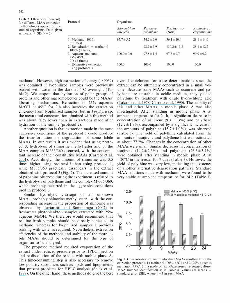
Another question is that extraction made in the mostaggressive conditions of the protocol 3 could producethe transformation or degradation of some labileMAAs. In our results it was evident that using proto-col 3, hydrolysis of shinorine methyl ester and of theMAA complex M335/360 occurred, with the concomi-tant increase of their constitutive MAAs (Carreto et al.2001). Accordingly, the amount of shinorine was 3.5times higher using protocol 3 than using protocol 1,while M335/360 practically disappears in the extractobtained with protocol 3 (Fig. 2). The increased amountof palythine observed during the experiment is related tothe hydrolysis of palythene and the complex M-335/360,which probably occurred in the aggressive conditionsused in protocol 3.
Similar hydrolytic cleavage of an unknownMAA—probably shinorine methyl ester—with the cor-responding increase in the proportion of shinorine wasobserved by Tartarotti and Sommaruga (2002) infreshwater phytoplankton samples extracted with 25%aqueous MeOH. We therefore would recommend thatroutine fresh samples should be directly sonicated inmethanol whereas for lyophilized samples a previoussoaking with water is required. Nevertheless, extractionefficiencies of the methods and stability of the more la-bile MAAs should be determined for the type oforganism to be analyzed.
The proposed method required evaporation of theextract under reduced pressure prior to HPLC injectionand re-dissolution of the residue with mobile phase A.This time-consuming step is also necessary to removelow polarity substances such as lipids and lipoproteinsthat present problems for HPLC analysis (Shick et al.1999). On the other hand, these methods do give the best
overall enrichment for trace determinations since theextract can be ultimately concentrated in a small vol-ume. Because some MAAs such as usujirene and pa-lythene are unstable in acidic medium, they yieldedpalythine by treatment with dilute hydrochloric acid(Takano et al. 1978; Carreto et al. 1990). The stability ofthis and other MAAs in mobile phase A was alsoinvestigated. After standing in mobile phase A atambient temperature for 24 h, a significant decrease inconcentration of usujirene (9.3±1.3%) and palythene(12.2±1.7%), accompanied by a significant increase inthe amounts of palythine (15.7±1.0%), was observed(Table 3). The yield of palythine calculated from theamounts of usujirene and palythene lost was estimatedat about 77.2%. Changes in the concentration of otherMAAs were small. Similar decreases in concentration ofusujirene (14.2±2.5%) and palythene (26.5±3.4%)were obtained after standing in mobile phase A at�20�C in the freezer for 7 days (Table 3). However, theyield of palythine was very low, indicating the existenceof another alternative degradation pathway. StandardMAA solutions made with methanol were found to bevery stable at ambient temperature for 24 h (Table 3),
Table 2 Efficiencies (percent)for different MAA extractionmethodologies applied on thestudied organisms. Data givenas means ± SD (n=3)
Protocol Organisms
Alexandriumcatenella
Porphyracolumbina
Porphyra sp.(Nori)
Anthopleuraelegantissima
1. Methanol 100%(3 times)
97.7±5.2 34.5±6.0 36.1±10.4 28.1±14.0
2. Rehydration + methanol100% (3 times)
– 98.9±5.9 130.2±15.8 88.1±12.7
3. Aqueous methanol25% 45�C,2 h (3 times)
100.0±0.0 97.8±1.4 97.6±0.7 99.9±0.2
4. Exhaustive extractionusing protocol 3
100.0 100.0 100.0 100.0
Fig. 2 Concentration of main individual MAAs resulting from theextraction protocols 1 ( methanol 100%, 4�C ) and 3 (25% aqueousmethanol, 45�C, 2 h ) made on an Alexandrium catenella culture.MAA number identification as in Table 4. Values are means ±standard error (SE), where n=3 in each MAA
242

but some decrease (7–10%) in concentration of allMAAs tested was observed after 7 days in the freezer(Table 3).
Interferences and clean-up
Solid phase extraction (SPE) on C-18 cartridges gavegood results and reproducibility (Teai et al. 1997; Shicket al. 1999) and specifically removed substances such aspigments and lipids that would have eluted at longretention times from the HPLC. However, care shouldbe taken to minimize loss of the less-polar MAAs.Ultrafiltration of the recomposed aqueous extract isanother alternative that, in addition to the water-insol-uble material, removes molecules larger than the mem-brane molecular weight cut-off rating. Applying thisprocedure to three replicates of control extracts spikedwith shinorine, palythinol, and usujirene, mean recov-eries of 100.4±0.3%, 100.0±0.6%, and 98.2±0.8%,respectively, were obtained.
Nevertheless, highly polar, low molecular weightsubstances such as 4-deoxygadusol—the postulatedprecursor of mycosporines—and structurally relatedcompounds such a gadusol (Chioccara et al. 1980;Bandaranayake et al. 1997) and other UV-absorbingsubstances unrelated to MAAs were also present in ex-tracts of marine organisms (Bandaranayake et al. 1997).As these interferences with absorption maxima typicallyranging between 264 and 270 nm were not removed bysolid phase extraction (Newman et al. 2000) or ultrafil-tration, care should be taken to ensure that peakabsorptions are devoid of contaminants with absorptionoverlapping detection wavelength. In our chromato-graphic conditions these interfering substances elutedtogether in a sharp peak without severe overlapping withmycosporine-2-glycine and palythine-serine.
HPLC-method development
Initially we tested the method used by Carreto et al.(2001) as it gave a good separation of most dinoflagellateMAAs. However, this method failed in the separation ofcertain highly polar MAAs [shinorine (3) frommycosporine-2-glycine (4) and palythine-serine (5)],
characteristic of some scleractinian corals (Teai et al.1997; Wu Won et al. 1997; Shick et al. 1999). To over-come such a problem we evaluated the selectivity of apolymer-coated silica reversed-phase C18 column(250 mm · 4.6 mm i.d.; CapCell Pak UG, Shiseido) asthis novel bonding reduces residual silanol groups thatcaused peak tailing and increased the selectivity forpolar compounds. The initial studies were focused onten MAAs that eluted in the early part of the chro-matogram where the elution conditions are essentiallyisocratic and the separation is difficult. Extracts of thecoral Stylophora pistillata containing a complex mixtureof highly polar MAAs (Shick et al. 1999) were used asresolution probes for optimizing the mobile phase A.The mobile phase B employed in the initial experimentswas fixed as in the Carreto et al. (2001) method, acomposition previously found to be optimal for sepa-rating low-polarity MAAs.
Measurements using isocratic elution with acetic acid0.2% have shown that the retention time of shinorine (3)on CapCell Pak UG was much larger than those ob-tained on the Alltima column, and that in relation toshinorine (3), the elution order of palythine (6) andpalythine-serine (5) was reversed. However, in theseconditions a serious band broadening, especially ofshinorine (3), occurs. MAAs are polyfunctional com-pounds with several ionizable groups (carboxyl, sulfate,imine, secondary amine), and the retention time andelution sequence of individual MAAs are very sensitiveto pH changes (Nakamura et al. 1982). On this base weexplored the use of trifluoroacetic acid (TFA) andammonia solutions instead of acetic acid 0.2%, as mo-bile phase A. For any combination, the mobile phase Acontaining TFA 0.2% plus ammonium hydroxide solu-tion at pH 2.9 provided the best separation betweenshinorine (3), mycosporine-2-glycine (4), and palytine-serine (5) but low efficiency was found. Discouraged byour unsuccessful efforts to combine high peak separationand high efficiency, we coupled the Alltima C18 ODSand the CapCell Pak C18 columns. We were fortunate,since the separation properties of the combined columnsincreased peak resolution. To study in detail the effect ofpH in these new conditions, the pH of the mobile phaseA was varied from 2.7 to 3.2 using mixtures of ammo-nium solution and TFA acid (final concentration 0.2%),
Table 3 MAA recoveries (percent) from mobile phase A and methanol solutions stored at ambient temperature for 1 day and �20�C for7 days. Data given as means ± SD (n=3)
MAA Mean recovery (%)
Eluant A Methanol 100%
25�C, 1 day �20�C, 7 days 25�C, 1 day �20�C, 7 days
Shinorine 96.6±1.9 97.0±2.3 96.8±0.6 93.4±0.4Palythine 115.7±1.0 103.6±0.5 101.2±0.1 92.7±0.5Palythinol 96.6±2.4 98.1±1.7 97.5±1.4 89.4±0.0Usujirene 90.7±1.7 85.8±2.5 95.5±0.7 93.0±1.1Palythene 87.8±1.7 73.5±3.4 97.6±0.6 89.2±0.8
243

and the resolution of polar MAAs measured. The majordifficulty encountered was the separation of mycospo-rine-2-glycine (4) from palythine-serine (5) withoutaffecting the separation of the other MAAs. Loweringthe pH of the mobile phase A, an expected increase inretention time for the most acidic MAAs was observed.The best separation of mycosporine-sulfates (1 and 2)was obtained at pH 2.7, but the pair mycosporine-2-glycine (4) and palythine-serine (5) remained unresolved(Fig. 3a).
Good resolution of mycosporine-2-glycine (4) andpalythine-serine (5) was obtained at pH 2.9 (Fig. 3b). Inthese conditions the unresolved peaks were reduced tothe pair porphyra-334 (9)/mycosporine-NMA-serine(10). Finally, at pH 3.15 the elution sequence of 9 and 10was reversed and a better resolution of this pair ofMAAs was obtained (Fig. 4). In these last conditions allcompounds were baseline separated. Only shinorine (3)and the less-polar sulfate ester (2) were partially resolved(Rs=0.98; Table 4).
The performance of the method in the central regionand the less-polar end region of the resulting chro-matogram was also examined in detail. Extracts of thecoral S. pistillata enriched by addition of available MAAstandards were used as resolution probes for optimizingthe mobile phase B. Using the original mobile phase B(aqueous acetic acid 0.2%:methanol:acetonitrile,50:25:25) the central region of the chromatogramshowed the co-elution of palythenic acid (15) withshinorine-methyl ester (16). Although different slopes inthe rate of change from mobile phase A to mobile phaseB were applied these compounds remained unresolved.Knowing about the differential selectivity of pH foracidic MAAs we evaluated the effect of lowering the pHof the mobile phase B using TFA 0.2%. For any com-bination of organic solvents the mobile phase including
TFA 0.2% plus ammonium hydroxide (pH 2.2) pro-vided better results than those containing acetic acid.The organic strength of the mobile phase B was alsoreduced to obtain a significant decreasing pH gradientbetween the beginning of separation and the elution timeof the unresolved peaks. The best results were obtainedwhen mobile phase B was TFA 0.2% plus ammoniumhydroxide (pH 2.2):methanol:acetonitrile (80:10:10,v:v:v).This reduction in the organic strength producedan expected increase in retention time of the less-polarcompounds that was partially compensated by modify-ing the gradient profile. As the columns used were sen-sitive to temperature changes we tested the temperatureat 20�C, 30�C, and 35�C. The highest efficiency wasobtained at 35�C, improving the separation betweenshinorine-methyl ester (16) with palythenic acid (15) tonear baseline resolution (Rs=0.71) and shortening theanalysis time. The final optimum temperature, gradient,and mobile phase composition are shown in Table 1.Under these conditions, substances 1–24 were separatelyeluted according to their hydrophobic properties (Fig. 4,Table 4) from 1, the lowest hydrophobicity, to 24, thehighest, within 40 min.
MAA detection and identification
A diode array detector (DAD) allowed the acquisition ofUV absorption spectra. Due to the lack of fine spectralabsorption, the only spectral characteristics available forMAA identification were the positions of the absorptionmaximum (kmax). However wavelength absorptionmaximums for some specific MAAs are identical or areonly 2 nm apart, which makes it difficult to distinguishthese compounds based on absorption spectra only.Confirmation of peak identity with a high degree ofconfidence is possible by matching the retention timeand UV spectrum with that of authentic MAAs. Whilethis methodology has proven useful, the lack of com-mercial standards makes identification and quantifica-tion of individual MAAs difficult. In addition, these two
Fig. 3a, b Effect of pH of mobile phase A on MAA resolution.a Mobile phase A at pH 2.7. b Mobile phase A at pH 2.9. Samplewas the coral Stylophora pistillata. Detection by absorbance at330 nm. Peak identification as in Table 4
244

properties alone are generally not considered sufficientfor a secure identification of organic compounds. Innatural product chemistry, co-chromatography in atleast two chromatographic systems as well as massspectrometry is required. Ion spray LC/MS is just assensitive and serves as an excellent method of confir-mation. This approach also allows the quantification ofindividual MAAs without the need for complete reso-lution. However, one limitation was the inability to
distinguish geometric isomers such as usujirene/palyth-ene (22/23) and E/Z palythenic acid (15/17). To achievemore structural information, tandem mass spectrometry(MS/MS) may be used to generate fragment ion spectra(Whitehead et al. 2001). It is interesting to note thatrecent studies on marine organisms (Carreto et al. 2001;Whitehead et al. 2001; Whitehead and Hedges 2002)using mass spectrometry detection and identification(LC/MS and LC/MS MS) showed a wide distribution of
Fig. 4 Chromatogram coveringthree polarity ranges. Samplewas an MAA extract from thecoral S. pistillata mixed withseveral MAA standardsolutions. Detection at 330 nm(the insert shows the detectionof mycosporine-glycine at310 nm). Peak identification asin Table 4
Table 4 Peak identification table. Resolution factor (Rs) between MAA pairs is indicated when Rs Wavelengths given in parenthesisdenote shoulders
Peak no. MAA Retention time (min) Rs (between peaks) kmax (nm) in eluant A
1 Palythine-serine sulfate 4.76 3202 Mycosporine sulfate ester 5.25 3183 Shinorine 5.65 0.98 (2/3) 3334 Mycosporine-2-glycine 6.19 3325 Palythine-serine 6.68 3216 Palythine 7.52 3207 Unknown from Noctiluca sp. 8.18 3108 Asterine 8.43 3299 Porphyra-334 8.87 33310 Mycosporine-methylamine-serine 9.60 32711 Mycosporine-glycine 10.11 31012 Unknown from Stillopora pistillata 11.24 32713 Unknown from Pocillopora eydouxi 12.62 32014 Palythinol 13.26 33015 Z-palythenic acid 13.83 33516 Shinorine methyl ester 14.10 0.71 (15/16) 33217 E-palythenic acid (?) from Noctiluca sp. 15.97 33718 Mycosporine-methylamine-threonine 16.88 32719 Mycosporine-taurine 17.23 0.95 (18/19) 30920 M-320 from A. tamarense 19.54 32021 Unknown from G. catenatum 21.21 37022 Usujirene 28.95 35723 Palythene 29.94 36024 M-335/360 from A. tamarense 35.71 335 (360)
245

palythenic acid (15). However using the commonlyadopted methods, this MAA has only been previouslyidentified in a small number of marine organisms (Na-kamura et al. 1982; Carreto et al. 1990). These con-trasting results are probably related to the fact thatbased on absorption spectra only, the Z-palythenic acid(15) could be misidentified as mycosporine-glycine-va-line (Whitehead et al. 2001). On the other hand, the highcost of LC/MS systems limits their wide use. Therefore ahighly selective separation method would represent agood alternative approach for studying complex sam-ples. Our proposed HPLC method is also compatiblewith ion-spray LC/MS and application of this approachonly requires slight methodological changes.
Precision and recovery
Variability in retention time between injections wasevaluated using five MAAs eluting in different regions ofthe chromatogram. Although total column length wasnow 40 cm, retention times were remarkably constant(Table 5). In addition, the relative standard deviation ofthe integrated peak area for repeated injections of cali-brated MAAs solutions made over several days was onthe order of 1–2% (Table 5).
Instrument response was established to be sufficientlylinear (r>0.999) over the range up to 80 ng injected, sothat a single-point porphyra-334 external calibrationwas routinely used. The detection limit for the differentMAAs was estimated to be between 0.08 and 0.47 pmolinjected (signal-to-noise ratio, S/N=2) with a 10-llinjection volume and the regular extraction and clean-uppresented in the experimental section. The mean recov-ery of shinorine (3), palythinol (14), and usujirene (22),using the gradient and column described above, was99.2±0.8, 98.8±1.3, and 99.2±0.8%, respectively, ofthat obtained using isocratic phase A and stainless steeltube instead of the column (three replicates).
Method applicability
HPLC analyses of MAAs in several marine organismswere carried out under the conditions described aboveand good separations were obtained in all cases.
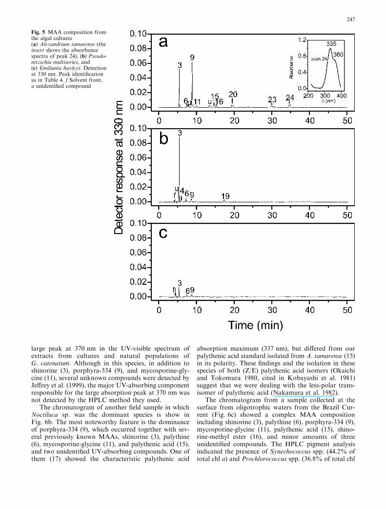
Phytoplankton cultures
The chromatogram of the toxic dinoflagellate Alexand-rium tamarense showed the most complex MAA profileamong the microalgal cultures analyzed (Fig. 5a). NineMAAs (3, 6, 9, 11, 15, 16, 20, 23, and 24) previouslyobserved in this species (Carreto et al. 1990, 2001;Whitehead and Hedges 2002; Laurion et al. 2003),including some recently partially characterized complexcompounds, such as shinorine-methyl ester (16), M-320(20), and M-335/360 (24), were baseline separated.Compared with the previous method (Carreto et al.2001), an improvement in the resolution of the criticalMAA pair palythenic acid (15)/shinorine-methyl ester(16) was observed (Table 4).
The chromatogram of the domoic acid producerdiatom Pseudo-nitzschia multiseries shows (Fig. 5b) thenet dominance of shinorine (3) with minor amounts ofmycosporine-2 glycine (4), palythine (6), porphyra-334(9), mycosporine-taurine (19), and one unknown UV-absorbing compound with absorption maximum at331 nm. The presence of mycosporine-taurine is themost noteworthy feature in this profile, as this rareMAA has only been previously detected in some spe-cies of sea anemones of the genus Anthopleura (Stochajet al. 1994; Shick et al. 2002). It is interesting to notethat similar to the sea anemones, Pseudo-nitzschiaspecies can accumulate large pools of taurine, a freeamino acid not detected in other diatom species (Smithet al. 2001).
In the bloom-forming coccolithophorid Emilianiahuxleyi only shinorine (3) was present in quantifiableamounts, whereas palythine (6) and porphyra-334 (9)were present at trace level (Fig. 5c). The inability ofE. huxleyi to accumulate MAAs could possibly berelated to this species’ poor tolerance of ultravioletradiation (Buma et al. 2000). By contrast, naturalblooms of this species appear to be promoted by highlight irradiances (Nanninga and Tyrrell 1996).
Natural phytoplankton samples
The chromatogram of a field sample collected during ared tide in the coastal waters off Mar del Plata showed(Fig. 6a) the net dominance of shinorine (3), and minoramounts of palythine-serine (5), porphyra-334 (9), my-cosporine-glycine (11), palythinol (14), palythene (23),and two unknown UV-absorbing compounds (U and21). Compound 21 shared a typical MAA absorptionspectra but its absorption maximum was centered at370 nm, which is an unusually large absorption signalfor an MAA (Fig. 6a). Microscopic observations of thissample indicated the dominance of the toxic dinofla-gellate Gymnodinium catenatum (R. Akselman, personalcommunication) but pigment analysis showed the netpredominance of the unequivocal Cryptophyceaecarotenoid marker alloxanthin (results not shown). It isinteresting to note that Jeffrey et al. (1999) observed a
Table 5 Means and standard deviations (n=8) of the retentiontimes for five typical MAAs and relative standard deviation (RSD)associated with the integrated area determination at �50-pmol level
MAA Retention time�x±r(min)
RSD ofintegrated area± (r/�x)·100
Shinorine 5.65±0.1 ±1.9%Palythine 7.52±0.1 ±0.9%Palythinol 13.26±0.1 ±1.3%Usujirene 28.95±0.2 ±1.9%Palythene 29.94±0.2 ±1.9%
246
large peak at 370 nm in the UV-visible spectrum ofextracts from cultures and natural populations ofG. catenatum. Although in this species, in addition toshinorine (3), porphyra-334 (9), and mycosporine-gly-cine (11), several unknown compounds were detected byJeffrey et al. (1999), the major UV-absorbing componentresponsible for the large absorption peak at 370 nm wasnot detected by the HPLC method they used.
The chromatogram of another field sample in whichNoctiluca sp. was the dominant species is show inFig. 6b. The most noteworthy feature is the dominanceof porphyra-334 (9), which occurred together with sev-eral previously known MAAs, shinorine (3), palythine(6), mycosporine-glycine (11), and palythenic acid (15),and two unidentified UV-absorbing compounds. One ofthem (17) showed the characteristic palythenic acid
absorption maximum (337 nm), but differed from ourpalythenic acid standard isolated from A. tamarense (15)in its polarity. These findings and the isolation in thesespecies of both (Z/E) palythenic acid isomers (Okaichiand Tokomura 1980, cited in Kobayashi et al. 1981)suggest that we were dealing with the less-polar trans-isomer of palythenic acid (Nakamura et al. 1982).
The chromatogram from a sample collected at thesurface from oligotrophic waters from the Brazil Cur-rent (Fig. 6c) showed a complex MAA compositionincluding shinorine (3), palythine (6), porphyra-334 (9),mycosporine-glycine (11), palythenic acid (15), shino-rine-methyl ester (16), and minor amounts of threeunidentified compounds. The HPLC pigment analysisindicated the presence of Synechococcus spp. (44.2% oftotal chl a) and Prochlorococcus spp. (36.8% of total chl
Fig. 5 MAA composition fromthe algal cultures(a) Alexandrium tamarense (theinsert shows the absorbancespectra of peak 24), (b) Pseudo-nitzschia multiseries, and(c) Emiliania huxleyi. Detectionat 330 nm. Peak identificationas in Table 4. f Solvent front,u unidentified compound
247

a) as major components of the phytoplankton assem-blage (J.I. Carreto et al., unpublished).
Macroalgae
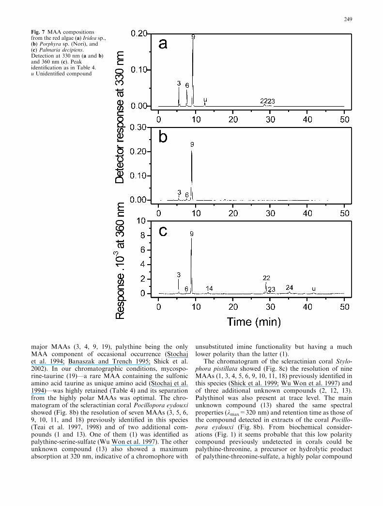
The seaweeds Iridea sp., Porphyra sp. (Nori), andPalmaria decipiens all showed the net dominance ofporphyra-334 (9) together with minor amounts of shin-orine (3) and palythine (6) (Fig. 7 a, b, c). However, theIridea sp. and P. decipiensMAA compositions were morecomplex. In addition to shinorine (3), palythine (6), andporphyra-334 (9), the chromatogram from Iridea sp.showed (Fig. 7a) minor amounts of usujirene (22),palythene (23), and one unidentified compound (u). Thechromatogram from P. decipiens also showed (Fig. 7c)the presence of usujirene (22) and palythene (23), besides
palythinol (14) and the recently partially characterizedcomplex M-335/360 (24) from A. tamarense. This is thefirst time that this compound was detected in red algae.
Symbiotic organisms
The HPLC chromatograms of MAA extracts from threesymbiotic organisms also showed interesting results(Fig. 8). In the sea anemone Anthopleura elegantissima(Fig. 8a) four MAAs were present in quantifiableamounts: shinorine (3), mycosporine-2-glycine (4),palythine (6), and mycosporine-taurine (19), with my-cosporine-2-glycine (4) being the most concentrated.Porphyra-334 was only detected at trace levels. Thisprofile was similar to that reported for this and otherspecies of Anthopleura that contained four constitutive
Fig. 6 Chromatograms ofphytoplankton MAAs fromseawater samples collected from(a) a red tide event thatoccurred in the coastal watersoff Mar del Plata (the insertshows the absorbance spectra ofpeak 21), (b) a Noctiluca sp. redtide event that occurred in thecoastal waters off Mar del Plata(the insert shows theabsorbance spectra of peaks 7and 17), and (c) surfaceoligotrophic waters from theBrazil Current. Detection at330 nm. Peak identification asin Table 4. f Solvent front,u unidentified compound
248
major MAAs (3, 4, 9, 19), palythine being the onlyMAA component of occasional occurrence (Stochajet al. 1994; Banaszak and Trench 1995; Shick et al.2002). In our chromatographic conditions, mycospo-rine-taurine (19)—a rare MAA containing the sulfonicamino acid taurine as unique amino acid (Stochaj et al.1994)—was highly retained (Table 4) and its separationfrom the highly polar MAAs was optimal. The chro-matogram of the scleractinian coral Pocillopora eydouxishowed (Fig. 8b) the resolution of seven MAAs (3, 5, 6,9, 10, 11, and 18) previously identified in this species(Teai et al. 1997, 1998) and of two additional com-pounds (1 and 13). One of them (1) was identified aspalythine-serine-sulfate (Wu Won et al. 1997). The otherunknown compound (13) also showed a maximumabsorption at 320 nm, indicative of a chromophore with
unsubstituted imine functionality but having a muchlower polarity than the latter (1).
The chromatogram of the scleractinian coral Stylo-phora pistillata showed (Fig. 8c) the resolution of nineMAAs (1, 3, 4, 5, 6, 9, 10, 11, 18) previously identified inthis species (Shick et al. 1999; Wu Won et al. 1997) andof three additional unknown compounds (2, 12, 13).Palythinol was also present at trace level. The mainunknown compound (13) shared the same spectralproperties (kmax=320 nm) and retention time as those ofthe compound detected in extracts of the coral Pocillo-pora eydouxi (Fig. 8b). From biochemical consider-ations (Fig. 1) it seems probable that this low polaritycompound previously undetected in corals could bepalythine-threonine, a precursor or hydrolytic productof palythine-threonine-sulfate, a highly polar compound
Fig. 7 MAA compositionsfrom the red algae (a) Iridea sp.,(b) Porphyra sp. (Nori), and(c) Palmaria decipiens.Detection at 330 nm (a and b)and 360 nm (c). Peakidentification as in Table 4.u Unidentified compound
249

isolated from S. pistillata (Wu Won et al. 1997). Chro-matography also revealed the presence of two high-polarity compounds (1 and 2) that eluted before shino-rine. One of them (1), which absorbed maximally at 320–321 nm and was also found in the coral P. eydouxi(Fig. 8b), was identified as palythine-serine-sulfate (WuWon et al. 1997). The presence of the other compound(2) is in agreement with recent results obtained by WuWon (in Shick et al. 1999, J.M. Shick personal com-munication). This author explained that althoughoriginal nuclear magnetic resonance spectra indicatedthe presence of only one compound in the palythine-serine-sulfate isolate from S. pistillata (Wu Won et al.1997), newer chromatography revealed a second peak in
that isolate. Thus, compound 2 may be a new mycosp-orine sulfate ester or decomposition product of thisMAA (Shick et al. 1999, 2002). The other low-polaritycompound in our chromatograms (12) that could not beidentified had a maximum absorption at 327 nm,indicative of a chromophore with an N-methyliminefunctionality, which was not 10 or 18.
Conclusions
Separation of MAAs in complex samples, such as thosefound in some marine organisms, requires high resolu-tion because of the large number of components present
Fig. 8 MAA compositionsfrom (a) the sea anemoneAnthopleura elegantissima andthe corals (b) Pocilloporaeydouxi and (c) Stylophorapistillata (the insert shows thedetection of mycosporine-glycine at 310 nm). Peakidentification as in Table 4.Detection at 330 nm
250

and the wide range of polarities encountered amongthese compounds. Good recovery, precision, and reso-lution in the separation of more than 20 compoundswere obtained with the reverse-phase HPLC gradientelution method here described. In comparison with thecommonly adopted isocratic elution method, the pro-posed method is more selective and resolves in a singlerun most of the more complex mixtures found in marineorganisms. In comparison with other HPLC gradientelution methods the proposed method gives a higherresolution of the more polar MAAs, while retaining orimproving the resolution of later-eluting MAAs.
Several partially characterized and other unknownMAAs have been recently discovered and our resultssuggest that there are still many unknown MAAs. Adiode array detector is essential to track spectroscopicresolution, but reproducible retention times and selectiveco-chromatography with authentic standards should beincluded to confirm identifications, whenever possible.For better MAA identification and characterization ofnovel compounds, ion-spray LC/MS is a robust tech-nique than can be adapted to our chromatographicconditions after slight methodological changes.
Acknowledgements We are grateful to Dr. Segel for providingAlexandrium catenella strain and to Dr. A. Boraso, Dr. G. Fer-reyra, and Dr. P. Zimba for donating biological material. Dr. T.Teai and Dr. M. Shick kindly provided MAA standards and bio-logical material key to this research. We thank Dr. V. Lutz forimproving the English grammar, and Dr. U. Karsten and twoanonymous reviewers who provided helpful comments and sug-gestions on the manuscript. This is INIDEP contribution no. 1332.
References
Adams NL, Shick JM (1996) Mycosporine-like amino acids pro-vide protection against ultraviolet radiation in eggs of the greensea urchin Strongylocentrotus droebachiensis. Photochem Pho-tobiol 64:149–158
Adams NL, Shick JM (2001) Mycosporine-like amino acids pre-vent UVB-induced abnormalities during early development ofthe green sea urchin Strongylocentrotus droebachiensis. MarBiol 138:267–280
Banaszak AT, Trench RK (1995) Effects of ultraviolet (UV)radiation on marine microalgal-invertebrate symbioses II. Thesynthesis of myccosporine-like amino acids in response toexposure to UV in Anthopleura elegantissima andCassiopeia xamachana. J Exp Mar Biol Ecol 194:233–250
Bandaranayake WM (1998) Mycosporines. Are they nature’ssunscreens? Nat Prod Rep 15:159–172
Bandaranayake WM, Bourne DJ, Sim RG (1997) Chemical com-position during maturing and spawning of the sponge Dysideaherbacea (Porifera: Demospongiae). Comp Biochem Physiol B118:851–859
Bohm GA, Pfleiderer W, Boger P, Scherer S (1995) Structure of anovel olygosaccharide-mycosporine-amino acid ultraviolet A/Bsunscreen pigment from the terrestrial cyanobacterium Nostoccomune. J Biol Chem 270:9–17
Buma AGJ, Oijen T van, Poll W van de, Veldhuis JW, Gieskes,WWC (2000) The sensitivity of Emiliania huxleyi(Prymnesiophyceae) to ultraviolet-B radiation. J Phycol 36:296–303
Carreto JI, De Marco SG, Lutz VA (1989) UV-absorbingpigments in the dinoflagellates Alexandrium excavatum and
Prorocentrum micans. Effects of light intensity. In: Okaichi T,Anderson DM, Nemoto T (eds) Red tides: biology,environmental science and toxicology. Elsevier, New York,pp 333–336
Carreto JI, Carignan MO, Daleo G, De Marco SG (1990) Occur-rence of mycosporine-like amino acids in the red-tide dinofla-gellate Alexandrium excavatum: UV-photoprotectivecompounds? J Plankton Res 12:909–921
Carreto JI, Carignan MO, Montoya NG (2001) Comparativestudies on mycosporine-like amino acids, paralytic shellfishtoxins and pigment profiles of the toxic dinoflagellatesAlexandrium tamarense, A. catenella and A. minutum. Mar EcolProg Ser 223:49–60
Chioccara F, Misuraca G, Novellino E, Prota G (1980) Mycosp-orine aminoacids and related compounds from the eggs offishes. Bull Soc Chim Belg 89:1101–1106
Conde FR, Carignan MO, Churio MS, Carreto JI (2003) In vitrocis-trans photoisomerization of palythene and usujirene.Implications on the in vivo transformation of mycosporine-likeamino acids. Photochem Photobiol 77:146–150
Dionisio-Sese ML, Ishikura M, Maruyama T, Miyachi S (1997)UV-absorbing substances in the tunic of a colonial ascidianprotect its symbiont, Prochloron sp., from damage by UV-Bradiation. Mar Biol 128:455–461
Dunlap WC, Chalker BE (1986) Identification and quantificationof near-UV absorbing compounds (S-320) in a hermatypicscleractinian. Coral Reefs 5:1–5
Dunlap WC, Shick JM, Yamamoto Y (2000) UV protection inmarine organisms I. Sunscreens, oxidative stress and antioxi-dants. In: Yoshikawa S, Toyokuni S, Yamamoto Y, Naito Y(eds) Free radicals in chemistry, biology and medicine. OICAInternational, London, pp 200–214
Helbling EW, Chalker BE, Dunlap WC, Holm-Hansen O, Vil-lafane VE (1996) Photoacclimation of Antarctic marine dia-toms to solar ultraviolet radiation. J Exp Mar Biol Ecol204:85–101
Jeffrey SW, Mantoura RFC, Wright SW (1997) Phytoplanktonpigments in oceanography: guidelines to modern methods.UNESCO, Paris
Jeffrey SW, MacTavish HS, Dunlap WC, Vesk M, Groenewould K(1999) Occurrence of UVA- and UVB-absorbing compounds in152 species (206 strains) of marine microalgae. Mar Ecol ProgSer 189:35–51
Karentz D, Mc Euen FS, Land MV, Dunlap WC (1991) Survey ofmycosporine-like amino acid compounds in Antarctic marineorganisms: potential protection from ultraviolet exposure. MarBiol 108:157–166
Karsten U, Garcıa-Pichel F (1996) Carotenoids and mycosporine-like amino acid compounds in members of the genus Microc-oleus (Cyanobacteria)—a chemosystematic study. Syst ApplMicrobiol 19:285–294
Karsten U, Bischof K, Hanelt D, Tug H, Wiencke C (1999) Theeffect of ultraviolet radiation on photosynthesis and ultraviolet-absorbing substances in the endemic Arctic macroalga Devale-raea ramentacea (Rhodophyta). Physiol Plant 105:58–66
Kobayashi J, Nakamura H, Hirata Y (1981) Isolation and struc-ture of a UV-absorbing substance from the ascidian Halocyn-thia roretzi. Tetrahedron Lett 22:3001–3002
Laurion I, Blouin F, Roy S (2003) The quantitative filter techniquefor measuring phytoplankton absorption: interference byMAAs in the UV waveband. Limnol Oceanogr Methods 1:1–9
Nakamura H, Kobayashi J, Hirata J (1982) Separation of my-cosporine-like amino acids in marine organisms using reversed-phase high-performance liquid chromatography. J Chromatogr250:113–118
Nanninga HJ, Tyrrell T (1996) Importance of light for the for-mation of algal blooms by Emiliania huxleyi. Mar Ecol Prog Ser136:195–302
Neale PJ, Banaszak AT, Jarriel CR (1998) Ultraviolet sunscreens inGymnodinium sanguineum (Dinophyceae): mycosporine-likeamino acids protect against inhibition of photosynthesis.J Phycol 34:928–938
251

Newman SJ, Dunlap WC, Nicol S, Ritz D (2000) Antarctic krill(Euphausia superba) acquire UV-absorbing mycosporine-likeamino acids from dietary algae. J Exp Mar Biol Ecol 255:93–110
Shick JM, Dunlap WC (2002) Mycosporine-like amino acids andrelated gadusols: biosynthesis, accumulation, and UV-protec-tive functions in aquatic organisms. Annu Rev Physiol 64:223–262
Shick JM, Romaine-Lioud S, Ferrier-Pages C, Gattuso JP (1999)Ultraviolet-B radiation stimulates shikimate pathway-depen-dent accumulation of mycosporine-like amino acids in the coralStylophora pistillata despite decreases in its population ofsymbiotic dinoflagellates. Limnol Oceanogr 44:1667–1682
Shick JM, Dunlap WC, Pearse JS, Pearse VB (2002) Mycosporine-like amino acid content in four species of sea anemones in thegenus Anthopleura reflects phylogenetic but not environmentalor symbiotic relationships. Biol Bull 203:315–330
Sinha RP, Klisch M, Groninger A, Hader DP (1998) Ultraviolet-absorbing/screening substances in cyanobacteria, phytoplank-ton and macroalgae. J Photochem Photobiol B 47:83–94
Smith GJ, Ladizinsky N, Miller PE (2001) Amino acid profiles inspecies and strains of Pseudo-nitzschia from Monterey BayCalifornia: Insights into the metabolic role(s) of domoic acid.In: Hallegraeff GM, Blackburn SI, Bolch CJ, Lewis RJ (eds)Harmful algal blooms 2000. IOC-UNESCO, Paris, pp 324–327
Sommaruga R, Garcıa-Pichel F (1999) UV-absorbing mycospo-rine-like compounds in planktonic and benthic organisms froma high-mountain lake. Arch Hydrobiol 144:255–269
Stochaj WR, Dunlap WC, Shick JM (1994) Two new UV-absorbing mycosporine-like amino acids from the sea anemoneAnthopleura elegantissima and the effects of zooxanthellae andspectral irradiance on chemical composition and content. MarBiol 118:149–156
Subramaniam A, Carpenter EJ, Karenz D, Falkowski PG (1999)Bio-optical properties of the marine diazotrophic cyanobacteriaTrichodesmium spp. I. Absorption and photosynthetic actionspectra. Limnol Oceanogr 44:608–617
Takano S, Uemura D, Hirata Y (1978) Isolation and structure oftwo new amino acids, palythinol and palythene, from the zo-anthid Palythoa tuberculosa. Tetrahedron Lett 49:4909–4912
Tartarotti B, Sommaruga R (2002) The effect of different methanolconcentrations and temperatures on the extraction of mycosp-orine-like amino acids (MAAs) in algae and zooplankton. ArchHydrobiol 154:691–703
Teai T, Raharivelomanana P, Bianchini JP, Faura R, MartınPMV, Cambon A (1997) Structure de deux nouvelles imino-mycosporines isolees de Pocillopora eydouxy. Tetrahedron Lett38:5799–5800
Teai T, Drollet JH, Bianchini JP, Cambon A, Martın PMV (1998)Occurrence of ultraviolet-absorbing mycosporine-like aminoacids in coral mucus and whole corals of French Polynesia. MarFreshw Res 49:127–132
Whitehead K, Hedges JI (2002) Analysis of mycosporine-likeamino acids in plankton by liquid chromatography electrosprayionization mass spectrometry. Mar Chem 80:27–39
Whitehead K, Karentz D, Hedges JI (2001) Mycosporine-likeamino acids (MAAs) in phytoplankton, a herbivorous pteropod(Limacina helicina), and its pteropod predator (Clione antarc-tica) in McMurdo Bay, Antarctica. Mar Biol 139:1013–1019
Wu Won JJ, Chalker BE, Rideout JA (1997) Two new UV-absorbing compounds from Stylophora pistillata: sulfate estersof mycosporine-like amino acids. Tetrahedron Lett 38:2525–2526
252
Copyright © 2022 FDOKUMEN