A Descriptive Review of Balamuthia and Non-Keratitis ...
108
Georgia State University Georgia State University ScholarWorks @ Georgia State University ScholarWorks @ Georgia State University Public Health Theses School of Public Health Spring 5-7-2011 A Descriptive Review of Balamuthia and Non-Keratitis A Descriptive Review of Balamuthia and Non-Keratitis Acanthamoeba Cases in the United States, 1955-2009 Acanthamoeba Cases in the United States, 1955-2009 Melanie A. Moser Georgia State University Follow this and additional works at: https://scholarworks.gsu.edu/iph_theses Part of the Public Health Commons Recommended Citation Recommended Citation Moser, Melanie A., "A Descriptive Review of Balamuthia and Non-Keratitis Acanthamoeba Cases in the United States, 1955-2009." Thesis, Georgia State University, 2011. https://scholarworks.gsu.edu/iph_theses/162 This Thesis is brought to you for free and open access by the School of Public Health at ScholarWorks @ Georgia State University. It has been accepted for inclusion in Public Health Theses by an authorized administrator of ScholarWorks @ Georgia State University. For more information, please contact [email protected].
-
Upload
khangminh22 -
Category
Documents
-
view
0 -
download
0
Transcript of A Descriptive Review of Balamuthia and Non-Keratitis ...

Georgia State University Georgia State University
ScholarWorks @ Georgia State University ScholarWorks @ Georgia State University
Public Health Theses School of Public Health
Spring 5-7-2011
A Descriptive Review of Balamuthia and Non-Keratitis A Descriptive Review of Balamuthia and Non-Keratitis
Acanthamoeba Cases in the United States, 1955-2009 Acanthamoeba Cases in the United States, 1955-2009
Melanie A. Moser Georgia State University
Follow this and additional works at: https://scholarworks.gsu.edu/iph_theses
Part of the Public Health Commons
Recommended Citation Recommended Citation Moser, Melanie A., "A Descriptive Review of Balamuthia and Non-Keratitis Acanthamoeba Cases in the United States, 1955-2009." Thesis, Georgia State University, 2011. https://scholarworks.gsu.edu/iph_theses/162
This Thesis is brought to you for free and open access by the School of Public Health at ScholarWorks @ Georgia State University. It has been accepted for inclusion in Public Health Theses by an authorized administrator of ScholarWorks @ Georgia State University. For more information, please contact [email protected].

ABSTRACT
MELANIE A. MOSER A Descriptive Review of Balamuthia and Non-Keratitis Acanthamoeba Cases in the United States, 1955-2009 (Under the direction of Richard Rothenberg, Professor) Free-living amebae are ubiquitous in the environment and occasionally invade and
parasitize host tissues causing illness in humans. Despite possibly frequent exposure to
these organisms, infection is rare and why some people, healthy or not, end up with
illness and others do not is still unclear. Human infections are rare; when illness does
occur, it is often fatal. Only two papers have examined data from the literature and cases
reported to the Centers for Disease Control and Prevention, and both were published over
twenty years ago. The purpose of this study is to better document the epidemiology of
Balamuthia and non-keratitis Acanthamoeba, give insight into trends of these infections
over time, and contribute to the scientific and medical community by producing the only
comprehensive review of all Balamuthia and non-keratitis Acanthamoeba cases in the
United States from 1955 through 2009. This study also examines cases that have
survived in an attempt to determine if there is evidence for the effectiveness of a
particular treatment regimen. Only a small number of patients have survived these
infections, so any evidence for a successful course of treatment could be crucial for future
cases.
INDEX WORDS: free-living amebae, Balamuthia mandrillaris, Acanthamoeba spp.,
non-keratitis Acanthamoeba

A DESCRIPTIVE REVIEW OF BALAMUTHIA AND NON-KERATITIS
ACANTHAMOEBA CASES IN THE UNITED STATES, 1955-2009
by
MELANIE A. MOSER B.S. FROSTBURG STATE UNIVERSITY
A Thesis Submitted to the Graduate Faculty of Georgia State University in Partial Fulfillment
of the Requirements for the Degree
MASTER OF PUBLIC HEALTH
ATLANTA, GEORGIA 2011

APPROVAL PAGE
A DESCRIPTIVE REVIEW OF BALAMUTHIA AND NON-KERATITIS
ACANTHAMOEBA CASES IN THE UNITED STATES, 1955-2009
by
MELANIE A. MOSER
Approved:
Richard Rothenberg, MD, MPH, FACP ___________________________________
Committee Chair
Sharon Roy, MD, MPH ___________________________________
Committee Member
April 13, 2011 ___________________________________
Date

DEDICATION PAGE
This thesis is dedicated to all those who have suffered from and succumb to free-living
ameba infections. May this work, in some small way, help to illuminate what is known
and what further work needs to be done to ensure others have improved outcomes.
iii

ACKNOWLEDGEMENTS
I would like to thank Dr. Govinda Visvesvara, head of the Free-Living Ameba
Laboratory at the Centers for Disease Control and Prevention, and his lab assistant, Mrs.
Rama Sriram. Without the diagnostic work performed by the FLA Lab, efforts to
investigate cases sent to the lab, maintenance of historical files, and Dr. Visvesvara’s
long and precise memory, this thesis could not have been undertaken. Dr. Visvesvara has
worked tirelessly for over 30 years to improve diagnosis of free-living ameba infections
and contribute to a greater understanding of the organisms; it was a sincere pleasure and
honor to have his assistance on this project. I would also like to thank the Waterborne
Disease Prevention Branch’s (WDPB) Dr. Sharon Roy and Mr. Jonathan Yoder for their
encouragement of this project and their willingness to serve as thesis committee member
and practicum preceptor respectively. Dr. Roy was exceedingly patient and helpful in
reviewing this document multiple times. Her suggestions on how to examine the data
were crucial to this work. The practicum experience was foundational to the
consolidation of some of the data explored in this thesis and proved to be an invaluable
learning experience in how informal surveillance systems are developed and
implemented.
I would also like to thank faculty at the Institute of Public Health at Georgia State
University. Thanks to Dr. Richard Rothenberg for teaching epidemiology courses in such
an engaging way, for always reminding students to look behind the numbers and ask
questions, and for directing this thesis project. Also thanks to Dr. Sheryl Strasser for her
encouragement in pursuing this thesis topic during her Research Methods class. I wasn’t
iv

convinced a descriptive study was important enough to undertake, but our after-class
conversations made me a believer. Although they were not involved in the thesis
process, both Professor Russell Toal and Dr. Frances McCarty are exceptional faculty.
Professor Toal was an inspiration in my first semester of graduate studies, showing real
commitment to public health and social justice. Dr. McCarty was a wealth of patience
and practicality in the biostatistics courses I had with her. Although I’ll never be a
statistician, she is the proof of why they are so needed in public health.
Lastly, I would like to thank my husband, Greg McNamara, for his
encouragement and support in my decision to return to school to pursue a graduate
degree. Public health is my passion and I thank him for his patience and willingness to
listen as I went through this course of study and for his keen eyes in reviewing this
document for errors.
v

AUTHOR’S STATEMENT
In presenting this thesis as a partial fulfillment of the requirements for a Master of Public
Health from Georgia State University, I agree that the Library of the University shall
make it available for inspection and circulation in accordance with its regulations
governing materials of this type. I agree that permission to quote from, to copy from, or
to publish this thesis may be granted by the author or, in her absence, by the professor
under whose direction it was written, or in his absence, by the Associate Dean, College of
Health and Human Sciences. Such quoting, copying, or publishing must be solely for
scholarly purposes and will not involve potential financial gain. It is understood that any
copying from or publication of this dissertation which involves potential financial gain
will not be allowed without written permission of the author.
Melanie A. Moser ____________________
Signature of Author

NOTICE TO BORROWERS
All theses deposited in the Georgia State University Library must be used in accordance
with the stipulations prescribed by the author in the preceding statement.
The author of this thesis is: Student’s Name: __Melanie A. Moser__________________________________ Street Address: ___216 Leigh’s Trace________________________________ City, State, and Zip Code: ____McDonough, GA 30253_________________ The Chair of the committee for this thesis is: Professor’s Name: ___Richard Rothenberg, MD, MPH, FACP____________ Department: ____Institute of Public Health____________________________ College: __Health and Human Sciences______________________________ Georgia State University P.O. Box 4018 Atlanta, Georgia 30302-4018 Users of this thesis who not regularly enrolled as students at Georgia State University are
required to attest acceptance of the preceding stipulation by signing below. Libraries
borrowing this thesis for the use of their patrons are required to see that each user records
here the information requested.
NAME OF USER ADDRESS DATE TYPE OF USE
(EXAMINATION ONLY OR COPYING)

Melanie Ann Moser 216 Leigh’s Trace
McDonough, GA 30253 [email protected]
Education: 1989 B.S., History, Frostburg State University, Frostburg, MD
Professional Experience: 2010-present Public Health Analyst, Centers for Disease Control and Prevention (CDC), Division of Parasitic Diseases and Malaria, Office of the Director, Atlanta, GA 2010 Public Health Analyst, CDC, Division of Enteric, Foodborne, and Waterborne Diseases, Waterborne Disease Prevention Branch, Office of the Director, Atlanta, GA 2007-2010 Public Health Analyst, CDC, Division of Parasitic Diseases, Parasitic Diseases Branch, Office of the Director, Atlanta, GA 2001-2007 Health Communication Specialist*, Atlanta Research and Education Foundation, Decatur, GA; *As Guest Researcher at CDC, Division of Parasitic Diseases, Parasitic Diseases Branch, DPDx
2000-2001 Research Analyst, Physician Workforce Resource Center, Mercer
University, Macon, GA 1999-2000 Research Assistant, Office for Social Environment and Health Research,
West Virginia University (WVU), Morgantown, WV 1996-1999 Program Assistant, Office for Health Services Research, WVU, Morgantown, WV Presentations: The DPDx Project, Where Parasitology and Technology Meet: Telediagnosis and Internet-based tools to enhance the diagnosis of parasitic diseases. Centers for Disease Control and Prevention, Science Ambassador’s supplemental lecture. July 25, 2007. Usefulness of Telediagnosis in Confirmatory Laboratory Diagnosis of Cases of Parasitic Infections. American Society of Tropical Medicine and Hygiene. Atlanta, GA. November 11, 2006.

DPDx: What is it and what can it do for me? A teleconference for U.S. public health departments. Sponsored by the National Laboratory Training Network. October 19, 2005. Encountering Food and Waterborne Intestinal Parasites. Sponsored by the National Laboratory Training Network and the Centers for Disease Control and Prevention. Various training workshops including: Albuquerque, NM, August 8-10, 2005; St. Thomas, Virgin Islands. June 27-29, 2005; Warwick, RI. September 11-12, 2003; Lionville, PA. September 8-9, 2003; Reno, NV. September 16, 2002. Bloodborne Pathogens: Plasmodium & Babesia Workshop. Sponsored by the National Laboratory Training Network and the Centers for Disease Control and Prevention. Various training workshops including: Albuquerque, NM, August 8-10, 2005; St. Thomas, Virgin Islands, June 27-29, 2005; Baltimore, MD, September 13-14, 2004; Durham, NH, August 24-25, 2004. Telediagnosis and Internet-based tools to enhance the diagnosis of parasitic diseases. [poster] American Society of Tropical Medicine and Hygiene. Miami, FL. November 11, 2004. DPDx: Laboratory Identification of Parasites of Public Health Concern. Public Health Education and Promotion Network Award Ceremony, Atlanta, GA. October 22, 2004. Food Safety Training & Education. Sponsored by the Georgia Association for Food Protection. Atlanta, GA. February 11, 2003. Publications: Moser, M., Bartlett, M., and A. da Silva. “DPDx project receives PHEP-Net’s Award of Excellence in Public Health Training.” NCID FOCUS. Winter-Spring 2005. 14(1);9. Shah, S., et al. “Malaria Surveillance --- United States, 2002,” Morbidity and Mortality Weekly Report, Surveillance Summaries. April 30, 2004. 53(SS01);21-34. Available from: URL: http://www.cdc.gov/mmwr/preview/mmwrhtml/ss5301a3.htm (Provided illustrations only) da Silva, A. and N. Pieniazek. “Latest Advances and Trends in PCR-Based Diagnostic Methods.” In Textbook-Atlas of Intestinal Infections in AIDS. Ed. Daniele Dionisio. Springer. 2003. (Provided illustrations only) Moser, M. and A. da Silva. “Featured Collection: DPDx.” D-Lib Magazine. June 2002. 8(6). Available from: URL: http://www.dlib.org/dlib/june02/06featured-collection.html Lubman, A. and M. Moser. West Virginia State Census Data Center Newsletter. Morgantown Printing and Binding: Morgantown, WV, Fall 1996-Fall 1997. Bell, R., et al. "The Burden of Diabetes in Appalachia." Poster session presented to the Centers for Disease Control, Division of Diabetes Translation. Biomedical

Communications: Morgantown, WV, August 1999. (Provided maps only) Awards and Honors: March 2009 National Center for Zoonotic, Vectorborne, and Enteric Diseases Award of Excellence for Communication Services April 2008 Award of Excellence in Public Health – Acanthamoeba keratitis Epi & Lab Research Team October 2004 Award for Excellence in Public Health Training – DPDx Team Skills and Qualifications:
• SAS, SPSS • Microsoft Access • ArcView GIS • Adobe Dreamweaver, Fireworks, Flash, Photoshop, Illustrator, and Acrobat

TABLE OF CONTENTS
Page
DEDICATION………………………………………………………………. iii
ACKNOWLEDGEMENTS…………………………………………………. iv
LIST OF TABLES…………………………………………………………... vi
LIST OF FIGURES………………………………………………………….. viii
CHAPTERS
I. INTRODUCTION…………………………………………………………. 1 Background…………………………………………………………… 1 Purpose of the Study………………………………………………….. 4 Research questions……………………………………………………. 5
II. REVIEW OF THE LITERATURE………………………………………… 7 Disseminated infections………………………………………………. 7 Granulomatous amebic encephalitis (GAE)………………………….. 11 Cutaneous acanthamoebiasis…………………………………………. 14 Acanthamoeba rhinosinusitis…………………………………………… 15 Acanthamoeba osteomyelitis………………………………………….. 15 Survivors: Acanthamoeba……………………………………………... 16 Survivors: Balamuthia…………………………………………………. 20 Ethnicity……………………………………………………………….. 23
III. METHODS AND PROCEDURES………………………………………… 25
IV. RESULTS………………………………………………………………….. 29 Demographics: Research Question 1…………….……………………. 30 Presenting Symptoms: Research Question 2.…………………………. 34 Treatment: Research Question 3………………………………………. 44 Duration of Illness: Research Question 4……………………………… 46 Further Data Analysis………………………………………………….. 46
V. DISCUSSION AND CONCLUSION……………………………………….. 52
REFERENCES………………………………………………………………….. 59

APPENDICES A. DETAILED TABLES OF ALL PUBLISHED NON-KERATITIS …… 66
ACANTHAMOEBA AND BALAMUTHIA CASES B. CASE REPORT FORM…………………………………………………. 74 C. EPIDEMIC CURVES FOR NON-KERATITIS……………………….. 85
ACANTHAMOEBA AND BALAMUTHIA CASES D. TREATMENT REGIMENS FOR SUVIVORS………………………… 89

LIST OF TABLES
Page
Table 1: Summary of Demographic for Non-Keratitis Acanthamoeba spp. and… 33 and Balamuthia mandrillaris Cases Table 2: Top Ten Presenting Symptoms of Patients Later Diagnosed with........ 34 Non-Keratitis Acanthamoeba spp. Infection
Table 3: Top Ten Presenting Symptoms of Patients Later Diagnosed with.......... 34 Balamuthia mandrillaris Infection
Table 4: Presenting General Symptoms for Cases of Non-Keratitis…………….. 35 Acanthamoeba spp. Infection by Disease Manifestation
Table 5: Presenting Visual Symptoms for Cases of Non-Keratitis....................... 37 Acanthamoeba.spp. Infection by Disease Manifestation
Table 6: Presenting Neurologic Symptoms for Cases of Non-Keratitis………….. 37 Acanthamoeba spp. Infection by Disease Manifestation
Table 7: Presenting Skin Lesions for Cases of Non-Keratitis Acanthamoeba........ 38 spp. Infection by Disease Manifestation
Table 8: Presenting General Symptoms for Cases of B. mandrillaris Infection….. 39 by Disease Manifestation
Table 9: Presenting Visual Symptoms for Cases of B. mandrillaris Infection……. 39 by Disease Manifestation
Table 10: Presenting Neurologic Symptoms for Cases of B. mandrillaris ………. 40 Infection by Disease Manifestation
Table 11: Presenting Skin Lesions for Cases of B. mandrillaris Infection……….. 41 by Disease Manifestation
Table 12: Comparison of Top Five Presenting Symptoms for Disseminated……... 41 Acanthamoeba and Balamuthia Disease
Table 13: Comparison of Top Five Presenting Symptoms for Acanthamoeba……. 43 and Balamuthia GAE
vi

Table 14: Drugs in Common for Non-Keratitis Acanthamoeba Survivors……… 45 Compared to Non-Survivors
Table 15: Drugs in Common for Balamuthia Survivors Compared to Non-..…... 45 Survivors
Table 16: Past Medical Conditions That May Be Predisposing Factors for…….. 48 Non-Keratitis Acanthamoeba spp. Infection
Table 17: Positive Non-Keratitis Acanthamoeba Patients Reporting Another….. 49 Immunocompromising Condition by HIV Status
Table 18: Past Medical Conditions That May Be Predisposing Factors for…….. 50 Balamuthia mandrillaris Infection
Table 19: Final Diagnosis of Cases of Non-Keratitis Acanthamoeba spp ……… 50 . Infection
Table 20: Final Diagnosis of Cases of Balamuthia mandrillaris Infection...…… 51
vii

LIST OF FIGURES
Page
Figure 1: Epidemic Curve for Cases of Non-Keratitis Acanthamoeba spp.— …… 29 United States, 1955-2009
Figure 2: Epidemic Curve for Cases of Balamuthia mandrillaris—United………. 30 States,1974-2009
Figure 3: Epidemic Curve by Age for Cases of Non-Keratitis Acanthamoeba...... 31 spp.—United States, 1955-2009
Figure 4: Geographic Distribution of Non-Keratitis Acanthamoeba spp.…......... 31 Cases by State of Treatment
Figure 5: Epidemic Curve by Age for Cases of Balamuthia mandrillaris—…….. 32 United States, 1974-2009
Figure 6: Geographic Distribution of Balamuthia mandrillaris Cases by….......... 33 State of Treatment
viii

CHAPTER I: INTRODUCTION
Background
Free-living amebae are ubiquitous in the environment and occasionally invade
and parasitize host tissues causing illness in humans.1,2 Although human infections are
rare, when illness does occur, it is often fatal. There are four genera of free-living
amebae known to infect humans.2 Naegleria fowleri, which can cause primary amebic
meningoencephalitis (PAM), is described in the media as the “brain-eating ameba”3,4 and
news reports of infections with this organism have likely raised awareness of free-living
amebae. Other free-living amebae, such as Balamuthia mandrillaris and Acanthamoeba
spp. can cause granulomatous amebic encephalitis (GAE), an infection of the central
nervous system. Although there is only one species of Balamuthia known to infect
humans, B. mandrillaris, there are multiple species of Acanthamoeba, at least eight, that
can cause human infection. Acanthamoebiasis and balamuthiasis can manifest as
disseminated infections throughout the body or localized in an area of the body, often
affecting the brain, skin, and/or sinuses, as well as other organs such as the lungs, liver,
kidneys or lymph nodes, either independently or in combination. Acanthamoeba spp. can
also cause keratitis, which may threaten an infected person’s vision by causing ulceration
of the cornea, diminished sight, or even blindness. Sappinia diploidea also has been
identified as the cause of one reported case of encephalitis.1
Naegleria fowleri has been isolated from warm freshwater, geothermal waters,
soil, sewage, and unchlorinated or minimally chlorinated swimming pools.5,6 Likewise
Acanthamoeba spp. have been found in soil, sewage, swimming pools, and freshwater but
1

2
have also been identified in hot tubs, sea or brackish water, heating/ventilating/air
conditioning (HVAC) units, dialysis machines, contact lens solutions, and intra-uterine
devices (IUDs).6 Balamuthia mandrillaris is found in soil.7,8 Balamuthia might also be
present in freshwater; there have been reports of Balamuthia GAE in dogs that swam in
ponds but there have been no reported human cases where the only potential exposure
was swimming.9,10 Because these amebae naturally inhabit so many locations, it is likely
that humans are frequently exposed to them. Immunologic studies of N. fowleri,
Acanthamoeba spp., and B. mandrillaris have shown that some humans have antibodies
to them.1 Two serologic studies carried out in the United States showed that persons who
had extensive contact with freshwater lakes had antibody response to N .fowleri.11
Antibodies to B. mandrillaris and Acanthamoeba spp. have been demonstrated in healthy
as well as hospitalized persons.1 Despite possibly frequent exposure, infection is rare and
why some people, healthy or not, end up with illness and others do not is still unclear.
Culbertson was the first to suggest that free-living amebae could possibly cause
infection in humans and showed pathogenicity using animal models.2,12 Butt described
the first U.S. cases of free-living ameba infection, PAM caused by N. fowleri, in 1966.13
Less than ten years later in 1972, Jager and Stamm reported the first human case of
Acanthamoeba disease, which occurred in the U.S, although it was another eight years
before it was definitively recognized that Acanthamoeba was the causal agent not
Hartmannella as was supposed in the original case report.14,15 Eventually several reports
preceding the 1966 first report of free living ameba infection were retrospectively
identified as cases of Acanthamoeba spp. as understanding of the organisms increased.2
Recognition that Balamuthia (referred to as leptomyxid ameba at the time) was also a

3
cause of human infections came in 1990 with a report of cases of GAE due to
leptomyxid; the first U.S. was later retrospectively identified as occurring in 1978.16
N. fowleri primarily affects immunocompetent children and young adults and
without treatment has a rapid clinical course resulting in death within 1 to 2 weeks after
exposure.1 In 1980, Martinez was the first to note that Acanthamoeba is an opportunistic
infection affecting persons with chronic illness.17 This characterization seems to remain
true as most reviews consider it to be an infection that typically affects
immunocompromised persons.1,2,17 It is thought that there is a subacute phase of
infection and the timeframe has not been well characterized, but once the acute phase
commences, the infection can be fatal within weeks. B. mandrillaris has been reported to
affect both immuncompromised and healthy individuals.1 Of note, recent reports of
Balamuthia infection via organ transplantation raise concerns for the organ procurement
community.18,19 The clinical course of balamuthiasis is thought to be the same as that of
acanthamoebiasis. Like Acanthamoeba, Balamuthia can also infect tissues outside the
central nervous system.1 Unlike with Acanthamoeba and Naegleria, it has been reported
that being of Hispanic ethnicity may be a risk factor for B. mandrillaris infection.1,20
There are subtle differences in the routes of human infection among the free-
living amebae. N. fowleri enter the nasal cavity, attach to the nasal mucosa, cross the
nasal-brain (cribriform) barrier, and invade the brain. This invasion results in
hemorrhagic necrosis as the amebae lyse or ingest red blood cells and/or digest or destroy
brain tissue through secretions in their food cups.1,11 Acanthamoeba spp. is thought to
invade the body via the nasopharyngeal route, breaks in the skin, or by inhalation of
cysts. The organism also produces food cups and secretes enzymes that may aid its

4
spread throughout the body. Balamuthia is thought to have some similarity to
Acanthamoeba in its route of infection and its possession of particular enzymes that aid in
its invasion process, but little is known about its pathogenesis.1
Meningoencephalitis is the swelling of the brain and its membranes. Butt first
introduced the concept of primary amebic infection to differentiate between Entamoeba
histolytica’s secondary infection of the brain (via the bloodstream) and Naegleria’s direct
infection.11 Later, Martinez differentiated between the manifestations of free-living
amebae infections caused by Acanthamoeba and Naegleria, particularly the changes
occurring in the tissues of the brain, giving further distinguishing differences between
PAM and GAE. PAM was characterized as “severe acute necrotizing and hemorrhagic
meningoencephalitis involving mainly the base of frontal lobes and posterior fossa” (p.
570) with lesions located on the surface of the brainstem, cerebellum, and cerebral
hemispheres.17 GAE’s characteristics included: leptomeninges with moderate amount of
purulent exudate over most affected cortical areas, moderate to severe edema in the
cerebral hemisphere, and “foci of encephalomalacic worsening associated with small
areas of hemorrhagic necrosis in occipital, parietal, temporal, or (less often) frontal
lobes” (p. 569). GAE lesions vary in their location and composition.17
Purpose of the Study
There have been many case reports of free-living ameba infections in the United
States, publications on diagnostic techniques and immunology, and reviews of the
organisms; but there is still much to learn about the epidemiology and clinical course of
these infections. A few case reports and reviews have attempted to summarize a limited
number of published cases of acanthamoebiasis and balamuthiasis,21,22 or have examined

5
specific aspects of these infections, such as case reports of Acanthamoeba GAE or
Acanthamoeba rhinosinusitis among AIDS patients.23,24 Only two papers have examined
data from the literature and cases reported to CDC, and both were published over twenty
years ago.6,16 A recently published article concerning the epidemiology of primary
amebic meningoencephalitis caused by Naegleria fowleri in the United States gives a
comprehensive review of cases using multiple sources of information such as the Centers
for Disease Control and Prevention’s (CDC) Free-Living Ameba (FLA) Laboratory case
registry and Waterborne Disease and Outbreak Surveillance System, as well as the
National Vital Statistics System, published case reports, and media reports.25 The
descriptive analysis presented within this thesis is modeled on the methodology of the
PAM epidemiology paper and will examine Balamuthia and non-keratitis Acanthamoeba
cases in the United States from 1955 to 2009 by accessing the FLA Laboratory case
registry and published case and media reports.
The purpose of this study is to better document the epidemiology of Balamuthia
and non-keratitis Acanthamoeba, give insight into trends of these infections over time,
and contribute to the scientific and medical community by producing the only
comprehensive review of all known U.S. cases of Balamuthia and non-keratitis
Acanthamoeba through 2009. Along with bringing about a better understanding of the
characteristics of these cases, this study will also examine cases that have survived and
attempt to determine if there is any evidence for a given treatment regimen as being
particularly effective. Only a small number of patients have survived these infections,
and any evidence for a successful course of treatment could be crucial for future cases.
Research questions

6
1. Are there any demographic characteristics that indicate commonality between
cases and suggest that persons of a particular ethnicity or race are more pre-
disposed to infection? In particular, does Balamuthia disproportionately affect
persons of Hispanic ethnicity as suggested in the literature?
2. Are there any presenting symptoms that are particular to acanthamoebiasis and
balamuthiasis that would assist clinicians in early identification of free-living
ameba infection?
3. Are there any commonalities in the treatment regimen of those who survived?
4. What is the time from onset of symptoms to death?

CHAPTER II: REVIEW OF THE LITERATURE
There have been more than 60 case reviews or reports citing cases of non-keratitis
Acanthamoeba disease and over 40 cases of Balamuthia disease in the United States
published between 1960 and 2009. The first case report of Acanthamoeba disease was
published in 1960, five years after the actual case occurred, and was reported as a
granuloma of the brain, likely due Endolimax williamsi.26 This same case was cited in
several subsequent publications as Acanthamoeba granulomatous amebic encephalitis
(GAE) upon retrospective review.6,17,21,27 The first case report of Balamuthia disease was
published in 1974; although Balamuthia was not recognized as the causal agent at the
time, it was reported as a case of free-living amebic meningoencephalitis that was not due
to Naegleria or Acanthamoeba.28 The case was cited as Balamuthia GAE in subsequent
publications.6,17,27 Appendix A has detailed tables of all published Acanthamoeba and
Balamuthia case reports.
Disseminated infections
A review of the literature reveals Acanthamoeba disease manifests itself in
various ways (disseminated, GAE, cutaneous, rhinosinusitis, osteomyelitis) as does
Balamuthia (disseminated and GAE). But unlike Acanthamoeba, disseminated
balamuthiasis always involves the brain. Disseminated Acanthamoeba disease was the
most frequently reported manifestation of acanthamoebiasis in the United States, with 29
cases published in the literature. [See Appendix A, Table 1 for all disseminated
acanthamoebiasis case references.] The majority (n=19) of reported disseminated
7

8
infections were in patients who had human immunodeficiency virus (HIV) or who had
acquired immune deficiency syndrome (AIDS). [See cases 5-11, 13-19, 21, 22, 24, 25,
and 28.] While HIV/AIDS was the predominant cause for immunosuppression among
case patients with disseminated acanthamoebiasis reported in the literature, it was
followed by those with a previous medical history of organ transplantation and
immunosuppressive drug therapy (n=4; cases 4, 23, 27, and 29) and bone marrow
transplants due to cancer (n=3; cases 12, 20, 26). One case was reported as having lupus
and two case reports cited no known predisposing health factors (cases 3, 1, and 2
respectively).
The most common form of disseminated Acanthamoeba disease was a
combination of skin and sinus infection (n=10; cases 5, 8, 11, 13-15, 21, 22, 24, 28),
followed by cases with a combination of GAE and cutaneous infection (n=3; cases 6, 9,
20). There were two reports of patients with GAE, cutaneous, and lung disease (cases 3,
29), two reports of patients with GAE and lung disease (cases 10, 26), and two cases with
cutaneous infection and osteomyelitis (cases 17, 27). The other disseminated
manifestations varied greatly among the patients. Those cases that included GAE and
other manifestations were: GAE, cutaneous, and sinus (n=1; case 19); GAE and keratitis
(n=1; case 1); GAE, lungs, and adrenals (n=1; case 12); GAE, cutaneous, and pulmonary
hyaline membrane (n=1; case 4); and GAE, cutaneous, sinus, lymph nodes, thyroid, and
adrenals (n=1; case 2). Other disseminated cases included: cutaneous, lungs, and keratitis
(n=1; case 7); cutaneous, lungs, heart, kidney, spleen, and lymph nodes (n=1; case 16);
cutaneous, sinus, and osteomyelitis (n=1; case 18); cutaneous, liver, and bronchoaveolar
lavage (n=1; case 23). One case reported sinus and lung involvement (case 25).

9
The majority of the disseminated acanthamoebiasis cases were in adult males
(n=18; cases 4-10, 13-15, 18, 19, 21, 22, 24-26, 29), and all children (<18 years of age)
reported to have disseminated acanthamoebiasis were boys (n=4; cases 1, 11, 16, 17).
Seven females were reported with disseminated disease (cases 2, 3, 12, 20, 23, 27, 28).
Three reports noted that the patients were of Hispanic ethnicity (cases 2, 11, and 14)
although two additional cases were perhaps of Hispanic ethnicity (cases 6 and 28), being
described as Mexican American and Nicaraguan, respectively. However, the other 26
case reports failed to describe ethnicity, so an assessment of proportion of cases by
ethnicity cannot be made. Only two reports included information about possible
environmental exposures and they were both water exposures, one from fishing on a
freshwater lake (case 29) and one from wading in drainage ditches (case 1). When
timeframes were able to be determined, the number of days between suspected onset of
symptoms and death ranged from 8 to 765 days (cases 1-3, 5, 6, 8, 10-13, 16, 19-23, 25,
26, 29). Three cases resolved their infections and did not die (cases 17, 23, 24) and eight
did not have adequate information on the symptom onset/death timeframe to determine
outcome status (cases 4, 7, 9, 14, 15, 18, 27, 28). The number of days between
hospitalization and death ranged from 2 to 120 days (cases 1-7, 9, 10, 14, 15, 18, 25, 26,
27, 28). Twelve cases did not have adequate information on the hospitalization/death
timeframe (cases 7, 8, 11, 12, 13, 16, 17, 19, 20, 21, 22, 28). Prognosis for those
diagnosed with disseminated disease was poor, with 25 cases having died either from
Acanthamoeba infection or from other complications (cases 1-16, 18-21, 25-29). In one
case, it is unknown whether the patient survived (cases 22). Three cases were still living
at the time of the published report, with follow-ups of one month (case 24) and 12

10
months (case 23). The third report did not give a timeframe but mentioned the patient was
still being maintained on therapy (case 17).
In contrast, only seven cases of disseminated Balamuthia have been reported.
[See Appendix A, Table 2 for all disseminated balamuthiasis case references.] The
information concerning potential health-related predisposing factors for these cases was
limited and varied, including a history of alcohol or drug abuse (n=3; cases 1, 2, 6), otitis
media (n=1, case 5), a history of a farm accident resulting in a contaminated wound and
subsequent limb amputation (n=1, case 3), and ankylosing spondylitis (n=1; case 4). One
case-patient had a history of coronary artery disease (case 7), but it is unclear whether
this medical condition was significant or not given that Balamuthia organisms were
found in the CSF and skin but nowhere else. Unlike acanthamoebiasis, all the
manifestations of disseminated balamuthiasis included GAE. The majority of cases were
GAE and cutaneous infections (n=4; cases 3, 4, 6, 7), and the rest were GAE with
infection of another organ outside the brain (n=3; cases 1, 2, 5).
As with disseminated acanthamoebiasis, the majority of cases of disseminated
balamuthiasis were in adult males (n=6; cases 1-4, 6, 7). Only one child was reported to
have this manifestation of illness and this case was female (case 5); this same case was
reported as being of Hispanic ethnicity and having a travel history to Mexico. Ethnicity
was reported in one other case (case 6, Hispanic), but the remaining 5 case-patients with
disseminated balamuthiasis had no ethnicity reported so no pattern can be determined
from these data. Environmental exposure history was limited with only two reports
providing information: both mentioned potential soil exposures (case 3, 4). When
timeframes could be determined, the number of days between suspected onset of

11
symptoms and death ranged from 24 to 568 days (cases 1-3, 5-7). One case-patient
resolved his infection and did not die, but the case report did not provide adequate
information to determine the time period between symptom onset and resolution (case 4).
The number of days between hospitalization and death ranged from 13 to 77 days (cases
1-3, 5-7). Clinical outcomes for disseminated Balamuthia disease were also poor, with
six of the case-patients dying (cases 1-3, 5-7) and one reported survivor (case 4). This
patient was followed for more than five years and seemed to have recovered from his
infection.
Granulomatous amebic encephalitis (GAE)
There are 21 published reports of granulomatous amebic encephalitis caused by
Acanthamoeba spp. from 1955 to 2009. [See Appendix A, Table 3 for all Acanthamoeba
GAE case references.] Similar to the disseminated form of acanthamoebiasis, the most
common predisposing condition among infected patients was HIV/AIDS (n=4, cases 10,
11, 14, 20), and an equal number of patients with GAE had a history of substance and
alcohol abuse (n=4; cases 2, 3, 15, 21). Other potential predisposing health conditions
included: cancer (n=2) with long-term immunosuppression (case 5) and with a stem cell
transplant (case 16); organ transplantation (n=2) with immunosuppressive therapy (cases
17) and with diabetes (case 19); lupus (n=2; case 18), where one case-patient was also
dependent on drugs and alcohol and counted above as having history of substance abuse
(case 21); mixed connective tissue disorder (n=1; case 9); and pneumonitis (n=1, case 6).
One case-patient (case 4) had a stroke, but it is unknown if this may be a predisposing
condition. Of the 21 reports, five had either unknown or unpublished health conditions
(cases 1, 7, 8, 12, 13) that may have made them more susceptible to infection.

12
The majority of cases of Acanthamoeba GAE were adult males (n=13; cases 2-5,
10-15, 17, 19, 20). Only three cases were in children under the age of 18, and all of them
were female (cases 1, 7, 8). There were five adult females with Acanthamoeba GAE
(cases 6, 9, 16, 18, 21). No case reports noted patients of Hispanic ethnicity. Two
reports noted that case-patients had water exposures; one case-patient was exposed to
water that had accumulated in a basement (case 3), but the source of the water was not
specified, and the other case-patient frequently used swimming pools for therapy after a
cerebrovascular accident (case 4). A third case-patient, a male, had multiple exposures,
including a potential occupational exposure (general contractor) and exposures to water
and soil. He played volleyball on sand courts and also gardened as a hobby. He also
enjoyed fishing and was on a trip approximately one month prior to his illness onset,
although he denied direct water contact (case 19). The remaining 18 case reports did not
comment on potential environmental exposures. When timeframes could be determined,
the number of days between suspected onset of symptoms and death ranged from 8 to
252 days (cases 1, 5, 6, 9, 10, 11, 14-21). Seven did not have adequate information on
the symptom onset/death timeframe (cases 2-4, 7, 8, 12, 13). The number of days
between hospitalization and death ranged anywhere from 4 to 63 days (cases 1, 2, 3, 5, 9,
10, 11, 14, 16, 17, 21). Ten cases did not have adequate information on the
hospitalization/death timeframe (cases 4, 6-8, 12, 13, 15, 19, 20). Among cases where
the clinical outcome was known, fatality was 100% (cases 1-7, 9-11, 14-21). Three cases
had unknown outcomes (cases 8, 12, 13).
Thirty-nine cases of Balamuthia GAE in the United States have been published in
case reports or cited in summary compilations. [See Appendix A, Table 4 for all

13
Balamuthia GAE case references.] Eleven case reports did not include information that
lent evidence of predisposing health factors, nor was the patient’s status stated as being
healthy (cases 3, 4, 6, 9, 23-25, 30, 31, 35, 36). Nine cases were reported as being
previously healthy before becoming ill (cases 7, 10, 12, 13, 15, 20, 21, 27, 32). In
instances where predisposing health factors were known, nine cases had otitis,
pharyngitis, or some other type of respiratory illness (cases 2, 5, 11, 14, 18, 19, 22, 33,
37). Five cases had a history of alcohol or drug abuse (cases 5 [also had TB as noted
above], 8, 17, 26, 29,). Other predisposing health factors included: organ transplantation
and immunosuppressive therapy (cases 34, 38, 39), HIV (case 16), prolonged steroid use
(case 28), and diabetes (case 1).
Unlike Acanthamoeba GAE, the majority of cases of Balamuthia GAE were in
males younger than the age of 18 years (n= 14; cases 2, 3, 9, 13-15, 18, 23, 24, 28, 30,
32, 36, 37) followed by adult males (n= 11; cases 5, 6, 8, 17, 25, 26, 29, 31, 33, 35, 39).
There were eight case reports on females under age 18 (cases 4, 7, 10-12, 20-22) and five
adult women (cases 1, 16, 19, 27, 38). One case report did not include any age or gender
information (case 34). Fourteen of the cases (cases 7, 10, 15, 17, 21-25, 28-31, 36) were
reported to be of Hispanic ethnicity; the rest of the case reports did not include
information on ethnicity. Environmental exposure to soil was cited in six cases (cases 7,
25, 27, 30, 33, 35). Two patients cited gardening as a hobby (cases 27, 35), two had an
occupational exposure to soil (case 25, 33), one cited motorcycling in the desert as a
hobby (case 30), and one visited a farm (case 7). One case-patient had a history of
frequent outdoor play in soil as well as exposure to an untreated well-water source for
recreation and drinking (case 37). Three additional cases cited water exposure, one to a

14
freshwater pond (case 20), one to well water (case 1), and one to a lake (the latter may
have also had soil exposure as the child of a migrant farmer, case 9). When timeframes
were able to be determined, the number of days between suspected onset of symptoms
and death ranged from 8 to 240 days (cases 1-5, 7, 10, 12-15, 17-19, 22, 24, 25, 33, 37,
38). Five cases resolved their infections and did not die (cases 21, 27, 31, 32, 39) and
fourteen did not have adequate information on the symptom onset/death timeframe (cases
6, 8, 9, 11, 16, 20, 23, 26, 28, 29, 30, 34, 35, 36). The number of days between
hospitalization and death ranged anywhere from 3 to 120 days (cases 1, 2, 9, 11, 16, 20,
26, 28-30, 35, 36). Twenty-two cases did not have adequate information on the
hospitalization/death timeframe (cases 3-8, 10, 12-15, 17-19, 22-25, 33, 34, 37, 38).
Survival for those with Balamuthia GAE was slightly better for those with GAE caused
by Acanthamoeba spp. Four patients are reported to have survived (case 21, 27, 31, 39)
and the other 35 died (cases 1-20, 22-26, 28-30, 32-38).
Cutaneous acanthamoebiasis
Twelve cases of cutaneous acathamoebiasis occurring in the United States have
been reported in the literature and all cases were among immunocompromised adults.
[See Appendix A, Table 5 for all cutaneous acanthamoebiasis case references.] The main
predisposing medical condition was HIV/AIDS (n=10; cases 1-4, 6-11). The only other
condition cited in the literature was immunosuppression due to organ transplantation
(n=2; cases 5, 12). Nine of the case-patients were males (cases 1-7, 9, 10) and three were
females (cases 8, 11, 12). Two case reports noted that the patients were of Hispanic
ethnicity (cases 2, 8), but ethnicities were not reported for the other 10 case-patients. No
soil exposures were cited in the case reports, although two reports noted cases had

15
freshwater exposure before skin lesions appeared (cases 6, 7). One case was noted to
have a local creek nearby that was used as a domestic drinking water source (case 12).
No potential environmental exposures were cited for the remaining nine case-patients.
When timeframes could be determined, the number of days between suspected onset of
symptoms and death ranged from 22 to 450 days (cases 1-4, 6, 8-11). Two cases
resolved their infections and did not die (cases 5, 12) but the symptom onset/death
timeframes could not be determined and one fatal case did not have adequate information
on the symptom onset/death timeframe (case 7). The only report of number of days
hospitalized was in a survivor who was hospitalized for approximately 150 days (case
12). The remaining cases did not have adequate information on the hospitalization/death
or hospitalization/discharge timeframes (cases 1-11). There were three reported
survivors of cutaneous Acanthamoeba (cases 5, 11, 12) and the other nine cases resulted
in death (cases 1-4, 6-11).
Acanthamoeba rhinosinusitis
Two cases of Acanthamoeba rhinosinusitis have been reported in the literature.
[See Appendix A, Table 6 for all Acanthamoeba rhinosinusitis case references.] Both
patients were immunocompromised adults, one male (case 1) and one female (case 2).
The male had HIV and recurrent sinus problems and the female had undergone a bilateral
lung transplantation due to a progressive lung disease. Both of these patients are reported
to have resolved their infections at 4 (case 1) and 3 (case 2) weeks respectively, but no
further follow-up was mentioned in either article.
Acanthamoeba osteomyelitis
A lone case of Acanthamoeba osteomyelitis was reported in 1981 in a 32-year-old

16
pre-diabetic woman. [See Appendix A, Table 7 for all Acanthamoeba osteomyelitis case
references.] She had a mass in the right mandibular area, which was removed and
replaced with a bone graft. A subsequent surgery was necessary in order to remove
necrotic bone. Inflammatory exudate from bone marrow in the necrotic bone was
positive for Acanthamoeba. The report does not cite any follow-up after the patient’s
surgery for necrotic bone removal, so it is not known if she continued to improve and
remain ameba-free (case 1). Three other cases of Acanthamoeba osteomyelitis have been
reported in the literature, but all of these case patients had disseminated Acanthamoeba
infection (see Appendix A, Table 1, cases 17, 18, 27).
Survivors: Acanthamoeba
There have been nine reports of successful treatment of acanthamoebiasis: three
disseminated (Appendix A, Table 1, cases 17, 23, 24), three cutaneous (Appendix A;
Table 4, cases 5, 11, 12), two rhinosinusitis (Appendix A, Table 5, cases 1, 2), and one
osteomyelitis (Appendix A, Table 6, case 1). There have been four published cases
where the clinical outcome was unknown due to lack of information in the reports: one
was a disseminated disease case (Appendix A, Table 1, case 22) and three were GAE
cases (Appendix A, Table 3, cases 8, 12, 13). In 1998, a report was published of a 7-
year-old male with HIV who had been diagnosed with cutaneous Acanthamoeba disease
at 5 ½ years of age (Appendix A, Table 1, case 17). Initially this case-patient was treated
with fluconazole without complete resolution of his skin lesions. Several months after
his diagnosis of cutaneous Acanthamoeba disease, he had a painful thumb and testing
showed osteomyelitis without a definitive cause. He was given pentamidine daily over
the course of a month and then maintained on itraconzole with monthly treatments of

17
pentamidine. By the time he was 6-years-old, he was diagnosed with granulomatous
amebic osteomyelitis after presenting with a swollen elbow. He was again given
pentamidine daily for one month. His cutaneous lesions and osteomyelitis recurred when
he was close to 7 years old, despite maintenance therapy. Daily pentamidine for one
month was given in combination with 5-fluorocytosine (5-FC). The child was then
maintained on 5-FC and antiretroviral treatment and was alive at the time of publication
but had occasional new skin lesions.
In 1999 a report was published of a 39-year-old woman who had undergone a
lung transplant six years earlier (Appendix A, Table 1, case 23). She was reported to
have presented with skin nodules, with new nodules continuing to develop over the next
several days. Multiple biopsies were performed before a diagnosis of Acanthamoeba was
finally made. Initial treatment included a combination of 5-FC and pentamidine, as well
as topical treatment with chlorhexidine gluconate and ketoconazole cream. Upon going
into respiratory failure and requiring mechanical ventilation, the patient’s
bronchoalveolar lavage (BAL) was found to be positive for Acanthamoeba. At this point
azithromycin was added to her drug regimen and it appears she stayed on this regimen for
close to a month, at which time her skin lesions were improving and her BAL was
negative. However, she began to have side effects that resulted in several of the
medications being discontinued. The patient had abnormal liver function tests and a
biopsy showed Acanthamoeba was present in this organ, so the patient was given lower
doses of pentamidine and 5-FC was restarted. On discharge, the patient was still being
treated with 5-FC and clarithromycin was added. The woman showed no evidence of
infection 12 months after her hospital discharge.

18
In 2002, a 37-year-old AIDS and cancer patient was reported to have nasal
obstruction that did not resolve with antibiotic treatment (Appendix A, Table 1, case 24).
He subsequently developed papular lesions on his extremities. Multiple biopsies were
inconclusive, although Cytomeglovirus was suspected and treatment was begun; the
patient did not respond well. Subsequently, the nasal obstruction became worse until the
patient needed surgery, at which point a diagnosis of Acanthamoeba was made from the
specimens obtained during surgery. The patient was given pentamidine and topical
treatments of both chlorhexidine and ketoconazole. When he had side effects due to
pentamidine it was discontinued and 5-FC was begun; this regimen was continued for a
month with successful resolution of his disease. At the time of the report, the patient was
still taking itraconazole but there was no evidence of recurrence. The report does not
mention when treatment with this drug was begun.
Three cases of cutaneous acathamoebiasis were reported to have survived. In
1992, a 31-year-old man who had undergone a kidney transplant presented with skin
nodules (Appendix A, Table 5, case 5). Upon diagnosis of Acathamoeba via positive
skin biopsies, he was given intravenous (IV) pentamidine and topical treatment with
chlorhexidine and ketoconzaole. He was treated with pentamidine for 4 weeks, although
the dosage had to be reduced due to an elevated creatinine level. At discharge from
hospital, his previous medications were discontinued and he was then treated with oral
itraconazole. He continued to take this medicine at a reduced dosage for at least two
years after his diagnosis without recurrence of infection.
In 2004, a 51-year-old woman with AIDS presented with skin papules,
photophobia, and headache (Appendix A, Table 5, case 11). Based on a skin biopsy, she

19
was diagnosed with cutaneous Acanthamoeba disease and underwent a CT scan and
lumbar puncture, which were subsequently negative for evidence of central nervous
system involvement. Her treatment regimen included pentamidine and 5-FC, but new
skin nodules continued to appear so sulfadiazine and highly active antiretroviral
treatment (HAART) were added. After 14 days of pentamidine once a day, penatmidine
was discontinued and the patient was continued on maintenance therapy of 5-FC,
sulfadiazine, and HAART. The patient was still free of CNS involvement 3 months after
release from the hospital with slow improvement of her skin lesions, possibly due to
inconsistent compliance with her treatment.
Also in 2004, a 52-year-old woman who had a lung transplant three years earlier
and remained on immunosuppressive therapy presented with nodules on her trunk and
lower extremities (Appendix A, Table 5, case 12). She was diagnosed with cutaneous
Acanthamoeba disease and unsuccessfully treated with itraconazole and metronidazole
for two weeks. CDC and an infectious disease specialist were consulted and the woman
was started on IV amphotericin B lipid complex and voriconazole. She was discharged
on the same medications delivered through an outpatient service. Her treatment with
these two drugs lasted 10 weeks before the dosage and frequency were changed and IV
voriconzole was switched to oral voriconazole. After her discharge she was maintained
on oral voriconazole for 5 months with resolution of her skin lesions and no recurrence.
Two cases of Acanthamoeba rhinosinusitis were reported to have survived their
infections. A 45-year-old man with HIV had sinus difficulties over the course of a year
which necessitated multiple sinus surgeries before he was finally diagnosed with
Acanthamoeba rhinosinusitis (Appendix A, Table 6, case 1). He had a subtotal

20
septectomy, used gentamycin nasal rinses for 4 weeks and was maintained on oral
itraconazole. No timeframe was given for the maintenance therapy, nor was the time
frame between the resolution of the infection and the publication of the paper given. In
2005, a 49-year-old woman who had undergone a lung transplant was reported to have
survived Acanthamoeba rhinosinusitis (Appendix A, Table 6, case 2). Seven months
after transplantation she had sinus difficulties and after diagnosis was treated with
amphotericin, voriconazole, and caspofungin. Three weeks after treatment began she
underwent a sinus debridement, but no Acanthamoeba were seen in specimens from this
debridement: she remained Acanthamoeba free in the month following which is as long
as the report states the patient was followed.
The only other published report of an Acanthamoeba survivor was in 1981
(Appendix A, Table 7, case 1). A 32-year-old female was diagnosed with amebic
osteomyelitis after a sequestrum of necrotic bone was removed from her jaw. The patient
does not appear to have been treated with drugs that were specific to resolving
Acanthamoeba infections. She was given IV penicillin, used betadine mouthwash after
surgery, and was discharged. The authors do not discuss long-term follow up, but as she
was reported as still alive at the time of the published report she is counted among the
survivors in this review. As with many of these cases where patients were reported to
have survived, it is difficult to determine if there were later recurrences or if patients truly
resolved their infections and never relapsed.
Survivors: Balamuthia
There have been six reports of successful treatment of balamuthiasis, one with
disseminated disease (Appendix A, Table 2, case 4) and five with Balamuthia GAE

21
(Appendix A, Table 4, cases 21, 27, 31, 32, 39). A 64-year-old man who previously had
a skin lesion that was biopsied with pending results presented with neurological
difficulties (Appendix A, Table 2, case 4). Lesions were seen in the parietal area of his
brain on both a CT scan and a MRI. He subsequently underwent a brain biopsy and was
released 10 days later. He was then re-hospitalized and received a diagnosis of B.
mandrillaris disseminated disease as Balamuthia organisms were identified in both the
brain tissue and the skin biopsy. He was treated with 5-FC, fluconazole, IV pentamidine,
sulfadiazine, and azithromycin (which was later switched to clarithromycin). The patient
was ventilated and in intensive care for seven weeks. He was discharged to a
rehabilitation unit and fluconazole was discontinued. Following a relapse, fluconazole
was added again and he was maintained on fluconazole and sulfadiazine over the course
of five years. The patient did recover enough to be able to walk, perform activities of
daily living and communicate well.
There have been five reports of survivors who were diagnosed with Balamuthia
GAE. Three of the case-patients were reported as previously healthy (Appendix A, Table
4, cases 21, 27, 32) and one had an unknown health status (Appendix A, Table 4, case
31). A report published in 2003 discusses a 5-year-old girl who, 48 days after initial
presentation, was diagnosed with Balamuthia GAE (Appendix A, Table 4, case 21). The
patient underwent several combinations of treatment. Initially she was treated with
ketoconazole and metronidazole for 34 days. These medications were then discontinued
and she was placed on clarithromycin and flucytosine for 14 days. Her treatment was
subsequently changed again and included azithromycin, fluconazole, pentamidine, and
thioridazine. Over the course of the next two-and-a-half years, her treatment varied,

22
primarily because of discontinuation of medications due to side effects and initiation of
medicines due to seizures. More than two-and-a-half years after her first hospital
admission she was still maintained on clarithromycin and fluconazole.
In 2004, a report was published of a 72-year-old woman who underwent a brain
biopsy that was positive for Balamuthia (Appendix A, Table 4, case 27). The biopsy was
excisional, and upon diagnosis, the patient was treated with pentamidine (IV),
sulfadiazine, fluconazole, and clarithromycin. The patient remained asymptomatic, but it
is unknown how much time elapsed between the case and the published report as well as
how long she was maintained on therapy. In 2007 a Balamuthia GAE survivor was
reported (Appendix A, Table 4, case 31). The report only stated that the patient was
living at the time of his last report to a physician, but he was lost to follow-up.
In 2010, a report of a 2-year-old boy with GAE was published (Appendix A,
Table 4, case 32). He was initially treated for tuberculosis but underwent a biopsy and
was given a diagnosis of Balamuthia. He was treated with pentamidine, fluconazole,
flucytosine, sulfadiazine, clarithromycin, and thioridazine. The boy was placed on a
ventilator and required a ventriculoperitoneal shunt. He was maintained on a ventilator
for almost two months and then transferred to a rehabiliation unit. Over the course of at
least 22 months, the case-patient was given clarithromycin, sulfadiazine, flucytosine, and
fluconazole without evidence of an active infection. After time in the rehabilitation unit,
the boy was able to follow simple commands, had improved postural stability, and was
attempting to speak. MRIs show that the lesions decreased in size and number.
The most recent report of a Balamuthia GAE survivor was published in 2010
(Appendix A, Table 4, case 39). A kidney transplant recipient who acquired his infection

23
through organ transplantation from an infected donor survived the infection, although he
spent two months in a coma. He slowly regained cognitive and motor functions and
entered a rehabilitation unit. He had residual neurologic effects such as right arm
paralysis and bilateral leg weakness. He also had intermittent vision loss. No treatment
regimen was included in the publication.
Ethnicity
In 2004, a letter to the editor of Emerging Infectious Diseases was published,
suggesting that Hispanic ethnicity may be a risk factor for Balamuthia GAE.20 The
primary authors of this letter were involved with the California Encephalitis Project
(CEP), which was begun in 1998 with the purpose of assisting clinicians in the State with
cases of encephalitis by offering enhanced diagnostic testing for infectious agents. The
CEP collects serum and other samples from patients in California that have presented
with encephalitis, and a subset of these samples were pulled for Balamuthia screening.
The criteria for screening included: case history that had clinical or laboratory
information suggestive of Balamuthia GAE and either an outdoor occupational exposure
or other outdoor recreational activity that might have lead to exposure to soil. Of 215
samples tested, three were positive for Balamuthia. Four additional samples, not a part of
the CEP, were also tested and were found to be positive. All seven cases were healthy
Hispanic Americans who died of GAE. The CEP contacted CDC to obtain information
about their records of Balamuthia cases, and it was determined that based on either case
report identification of ethnicity or a traditional Hispanic last name, about 50%, or 25, of
the U.S. case patients with known or suspected ethnicity were Hispanic Americans. No
information was given on the number of cases with truly unknown ethnicities, just that

24
there were approximately 50 U.S. cases at the time of publication. This same letter also
stated that 36% of Balamuthia GAE cases occurred in Latin American countries.20

CHAPTER III: METHODS AND PROCEDURES
CDC’s Free-Living Ameba (FLA) Laboratory collects information on cases of
suspected Balamuthia and non-keratitis Acanthamoeba diseases as part of their contact
with physicians and laboratorians who are seeking assistance with diagnosis. The head of
the FLA Laboratory, Dr. Govinda Visvesvara, has been working with free-living amebae
since 1964 and has been with the FLA Laboratory since 1974. He was the first scientist
to isolate and subsequently name Balamuthia mandrillaris and is considered one of the
world’s FLA experts, with more than 250 publications on the subject. The FLA
Laboratory is one of the only research laboratories in the United States performing
reference diagnostic testing for free-living amebae, and the lab handles specimens tested
through various methods such as staining of histopathologic slides, culture, and
polymerase chain reaction (PCR). The FLA Laboratory is one of only two known labs
currently performing an antibody test (indirect immunofluorescence or IIF) for the free-
living amebae; the other laboratory is at the California Department of Health Services,
which runs the California Encephalitis Project (CEP). These two labs have historically
shared data. Therefore, the FLA Laboratory holds records for most of the non-
Acanthamoeba keratitis and Balamuthia mandrillaris FLA cases diagnosed in the United
States. It is for this reason that a review of FLA laboratory records supplemented with a
review of the published literature should capture the majority of FLA cases diagnosed in
the United States, even though diseases associated with the free-living amebae are not
nationally notifiable.
Data for this study were abstracted onto case report forms from CDC laboratory
25

26
and clinical consultation files for positive cases of Balamuthia and non-keratitis
Acanthamoeba that occurred in the United States. See Appendix B for a copy of the case
report form. Data in the files ranged greatly in content from laboratory specimen
submission forms with little information about the patient’s clinical course to copies of
extensive medical records and autopsies.
A literature review was also performed to collect all published cases and media
reports of Balamuthia and non-keratitis Acanthamoeba disease occurring in the U.S. The
published cases were then compared to the CDC-diagnosed cases to determine which
ones referred to the same patients. This was done in three ways: matching similar patient
clinical and laboratory data, matching authors with contact clinicians or laboratorians,
and consultating with Dr. Govinda Visvesvara, the aforementioned head of the FLA
Laboratory. Information from published case reports took precedence over CDC records
when there was conflicting data, unless the publication conflicted with a final autopsy
report in CDC files. CDC records took precedence over any information found in the
general media. In instances where there was information on length of time but not
specific dates for illness onset, hospitalization, and/or time from illness onset to death,
dummy dates were entered for the purpose of calculating durations.
In instances where published cases could not be matched with CDC cases, the
diagnostic methods for the published cases were reviewed. Cases tested by the following
agencies/individuals were considered to be reputable cases based on their known
diagnostic methods: the California Department of Health Services; Dr. Eddy Willaert,
Institute of Tropical Medicine in Antwerp, Belgium; Dr. Julio Martinez, University of
Pittsburgh; Dr. Bruce Torian, Idaho; Dr. Gregory Booten, Ohio State University. If it

27
was not apparent that a published case was in CDC records, or if one of the laboratories
above was not cited, individual cases were discussed with Dr. Visvesvara to determine if
he was familiar with the case. Diagnostic and clinical features of the published case were
reviewed to determine if the case likely fit a diagnosis of acanthamoebiasis or
balamuthiasis.
Each case-patient identified from these records and/or the literature review was
assigned a unique identifier consisting of a prefix of AC for Acanthamoeba or BA for
Balamuthia, followed by a two digit number representing the calendar year in which the
case first presented with symptoms (when known) or was either first reported to CDC or
published in the literature. A case number in sequential order from 001 to infinity was
designated as the last three digits of the unique identifier. Case information was then
entered into a limited-user database that serves as a resource for CDC clinical
consultation and surveillance.
The database has 976 variables and contains both quantitative and qualitative
data. A major issue with the database is that many of the variables are set up as
true/false, complicating the calculation of denominators for certain areas of data. The
database allows exportation of files in Excel format and splits the database information
into eight separate files when exporting: demographics, exposure history, medical
history, current illness, laboratory tests, histopathology, diagnostic imaging, and
diagnostic and treatment outcomes. For the purposes of this study, the information was
stripped of identifiers prior to analysis. The data were then analyzed using SPSS.
Exported data were split into separate files for Acanthamoeba and Balamuthia.
Each of the eight database files for each organism were imported into SPSS, labeled and

28
assigned type, values, and measures. All eight files were also combined for each
organism for instances where information in the separate databases needed to be
examined together. Data were first explored by examination of frequencies, ranges and
means, and cross tabulations. Research question 1 was answered using these methods,
and frequencies, ranges, and means were used for research question 4. For research
question 2, frequencies were also used, but because acanthamoebiasis has so many
manifestations, it was useful to explore the data by disease manifestation. Therefore,
case summaries were run using final diagnosis as the grouping variable and presenting
symptoms in the variables column. Research question 3 was also examined using case
summaries with the survived variable as the grouping variable and treatment drugs in the
variables column. Medications given to the survivors were visualized using Excel
spreadsheets to determine commonalities in the drugs used and duration. Although the
methods used above answered the research questions, I also further analyzed some of the
demographic, exposure history, previous medical history, and outcomes data to try to
glean further information about these diseases and produced epidemologic curves for all
reported cases of acanthamoebiasis and balamuthiasis in the United States through 2009.

CHAPTER IV: RESULTS
A total of 114 cases of acanthamoebiasis was analyzed; 65 (57%) of these cases
had some information about them published in the literature. Similarly, 70 cases of
balamuthiasis were analyzed; 46 (66%) of these cases had some information about them
published in the literature. Cases of acanthamoebiasis occurred between 1955 and 2009
(Figure 1); cases of balamuthiasis occurred between 1974 and 2009 (Figure 2). See
Appendix C for larger versions of these figures.
Figure 1: Epidemic Curve for Cases of Non-Keratitis Acanthamoeba spp.—United States, 1955-2009
29

30
Figure 2: Epidemic Curve for Cases of Balamuthia mandrillaris—United States, 1974-2009
Demograhics: Research Question 1
Little information was available about the race and ethnicity of Acanthamoeba
cases. Sixty-two percent of the race data were missing (n=43/114). Among the 43 case
patients who had data available, the racial breakdown was: 63% white (n=27/43), 35%
African American (n=15/43), and 2% American Indian (n=1/43). Ninety percent of the
data were missing for the ethnicity variable (n=11/114), but among the known cases 82%
(n=9/11) were Hispanic and 18% (n=2/11) were non-Hispanic. Five out of the nine
known Hispanics were treated in the state of California. Immigration data were also
limited; 93% of the data were missing (n=8/114), but when known 75% had immigrated
to the United States (n=6/8) and 25% were natural U.S. residents (n=2/8). There was no
particular country of emigration in common among those six cases who had immigrated.
Four percent of cases were missing data for the age variable (n=110/114). Ages of cases
ranged from a minimum of 8 months old to a maximum of 79 years old, with a mean of

31
38.8 years of age; 7% (n=8/110) were children younger than 18 years of age, 87%
(n=96/110) were adults 18-64 years of age, and 6% (n=6/110) were 65 years of age or
older. Males were 73% of cases (n=82/112) and females were 27% (n=30/112); two
cases had missing information. See Figure 3 for an epidemic curve by age of
Acanthamoeba spp. [Appendix C has a larger version of this figure.] Also see Figure 4
for the geographic distribution of Acanthamoeba spp. cases by state of treatment.
Figure 3: Epidemic Curve by Age for Cases of Acanthamoeba spp.—United States, 1955-2009
Figure 4: Geographic Distribution of Non-Keratitis Acanthamoeba spp. Cases by State of Treatment

32
Limited data were available for race, ethnicity, and immigration status for
Balamuthia cases. Fifty-three percent of the race data were missing (n=33/70). Among
cases where data were available, the racial breakdown was: 58% white (n=19/33), 36%
African American (n=12/33), and 6% Asian/Pacific Islander (n=2/33). Fifty-nine percent
of the data were missing for ethnicity (n=29/70), but among the known cases 76% were
Hispanic (n=22/29) and 24% were non-Hispanic (n=7/29). Twelve of the 22 known
Hispanic case-patients were treated in the state of California. The immigration variable
had 84% of the data missing (n=11/70); 36% of case-patients with known immigrantion
status immigrated to the U.S. (n=4/11) and 64% were natural residents of the U.S.
(n=7/11). Immigrants most commonly came from Mexico (75%, n=3/4). Two cases
were missing data for the age variable, with ages ranging from a minimum of 4 months to
a maximum of 89 years of age, and a mean age of 30.4 years; 43% (n=29/68) were
children younger than 18 years of age, 47% (n=32/68) were adults 18-64 years of age,
and 10% (n=7/68) were 65 years of age or older. Males were 65% of cases (n=45/69)
and females were 35% (n=24/69); only one case was missing gender information. See
Figure 5 for an epidemic curve by age of Acanthamoeba spp. [Appendix C has a larger
version of this figure.] Also see Figure 4 for the geographic distribution of Balamuthia
mandrillaris cases by state of treatment.
Figure 5: Epidemic Curve by Age for Cases of Balamuthia mandrillaris—United States, 1974-2009

33
Figure 6: Geographic Distribution of Balamuthia mandrillaris Cases by State of Treatment
Table 1: Summary of Demographic Data for Non-Keratitis Acanthamoeba spp. and Balamuthia mandrillaris Cases
Acanthamoeba (N=114) Balamuthia (N=70) Variable Data Missing Data Missing
White 63% (27/43) 58% (19/33) African American 35% (15/43) 36% (12/33) American Indian 2% (1/43) 0% (0/33)
Race
Asian/Pacific Islander 0% (0/43)
62% (71/114)
6% (2/33)
53% (37/70)
Hispanic 82% (9/11) 76% (22/29) Ethnicity Non-Hispanic 18% (2/11)
90% (103/114) 24% (7/29)
59% (41/70)
Immigrant 75% (6/8) 36% (4/11) Immigration Non-Immigrant 25% (6/8)
93% (106/114) 64% (7/11)
84% (59/70)
Range 8 m–79 y 4 m–89 y Age Mean 38.8 years
4% (4/114) 30.4 years
3% (2/70)
Sex Male 73% (82/112) 2% (2/114)
65% (45/69) 1% (1/70)

34
Presenting Symptoms: Research Question 2
Patients presented with a variety of non-descriptive symptoms for both
Acanthamoeba and Balamuthia disease. Fever and headache were the two most common
symptoms shared by those with either disease. See Tables 2 and 3 for the top ten
symptoms patients presented with upon examination or hospitalization for illness caused
by Acanthamoeba spp. and Balamuthia mandrillaris respectively.
Table 2: Top Ten Presenting Symptoms of Patients Later Diagnosed With Non-Keratitis Acanthamoeba spp. Infection (N=95/114) Presenting Symptom No. of Cases Reporting Symptoms Other General Symptoms* 39 Fever 34 Headache 29 Sinus problems 18 Other Neurologic Deficit** 17 Lethargy/Fatigue 13 Confusion 9 Weight Loss 8 Weakness 7 Altered Mental Status 6 Cough 6 Nausea 6 Vomiting 6 *Other general symptoms include but were not limited to: chills (n=7/39), facial swelling/pain/tenderness (n=5/39), epitstaxis (n-=3/39), jaundice (n=2/39), night sweats (n=2/39), and incontinence (n=2/39). ** Other neurologic symptoms include but are not limited to: Romberg’s sign (n=1/17), Babinski sign (n=1/17), Kernig’s sign (n=1/17), Brudzinki’s sign (n=1/17).
Table 3: Top Ten Presenting Symptoms of Patients Later Diagnosed With Balamuthia mandrillaris Infection (N=63/70) Presenting Symptom No. of Cases Reporting Symptoms Fever 23 Headache 18 Lethargy/Fatigue 18 Vomiting 18 Other General Symptoms* 16 Other Neurologic Deficit** 15 Seizures 14

35
Weakness 13 Other Visual Symptoms 11 Altered Mental Status 10 Hemiparesis 9 Ataxia 8 *Other general symptoms include but were not limited to: incontinence (n=3/16), neck pain (n=2/16), and loss of appetite (n=2/16). ** Other neurologic symptoms include but are not limited to:Babinski sign (n=2/15, and unresponsiveness (n=2/15).
Because both acanthamoebiasis and balamuthiasis have multiple disease
manifestations, a further exploration was done looking at presenting symptoms by disease
manifestation. Symptoms were broken up into four categories: general presenting
symptoms (17 variables), visual symptoms (4 variables), neurologic symptoms (29
variables), and skin lesions (4 variables). Cases that had no information for every
variable in a category were taken out of the frequency counts and treated as unknowns.
Remaining cases may have had more than one symptom in each of the four presenting
symptom categories. In the case of the skin lesions category, knowns included those
cases who indicated they had skin lesions or did not have skins lesions. After analysis it
was apparent that not all of the possible variables for the symptom categories were
selected as symptoms upon presentation. See Tables 4-7 for breakdowns of
Acanthamoeba cases; see table footnotes for numbers of variables not chosen in each
category.
Table 4: Presenting General Symptoms for Cases of Non-Keratitis Acanthamoeba spp. Infection by Disease Manifestation Disease Manifestation Presenting Symptom No. of Known Cases
Reporting Symptoms Other General Symptoms 19 Fever 12 Sinus problems 11 Headache 10 Lethargy/Fatigue 5
Disseminated (n=30/36)*
Cough 4

36
Nausea 3 Weight Loss 3 Shortness of breath 2 Vomiting 1 Stiff Neck 1
Fever 16 Headache 14 Other General Symptoms 14 Lethargy/Fatigue 7 Vomiting 4 Nausea 3 Weight Loss 3 Diarrhea 2 Anorexia 1 Cough 1 Shortness of Breath 1
GAE (n=25/48)*
Sinus Problems 1
Fever 5 Headache 3 Other General Symptoms 4 Diarrhea 2 Weight Loss 2 Myalgia 2 Sinus Problems 2 Shortness of Breath 1
Cutaneous (n=10/23)*
Lethargy/Fatigue 1
Sinus Problems 4 Fever 1 Headache 1
Rhinosinusitis (n=4/6)*
Other General Symptoms 1
Osteomyelitis (n=1/1) Other General Symptoms 1 *Those reporting multiple general symptoms on initial presentation included: 19 GAE cases, 24 disseminated cases, two rhinosinusitis cases, and six cutaneous cases. Although it was possible to select up to 17 variables for presenting general symptoms, three of those variables were not selected among those cases who reported presenting symptoms.

37
Table 5: Presenting Visual Symptoms for Cases of Non-Keratitis Acanthamoeba spp. Infection by Disease Manifestation Disease Manifestation Presenting Symptom No. of Known Cases
Reporting Symptoms Blurred Vision 2 Diplopia 1
Disseminated (n=3/36)*
Other Visual Symptom 1
Blurred Vision 2 Diplopia 2 Other Visual Symptom 2
GAE (n=5/48)*
Photophobia 1
Cutaneous (n=2) Photophobia 2 *Those reporting multiple visual symptoms on initial presentation included: two GAE cases and one disseminated case.
Table 6: Presenting Neurologic Symptoms for Cases of Non-Keratitis Acanthamoeba spp. Infection by Disease Manifestation Disease Manifestation Presenting Symptom No. of Known Cases
Reporting Symptoms Confusion 5 Other Neurologic Deficit 4 Weakness 3 Altered Mental Status 3 Behavioral Changes 1 Dysphagia 1 Hallucinations 1 Seizures 1
Disseminated (n=12/36)*
Hemiparesis 1
Other Neurologic Deficit 12 Weakness 4 Hemiparesis 3 Seizures 3 Confusion 3 Altered Mental Status 2 Aphasia 2 Ataxia 2 Numbness or Facial Numbness
1
Hyperreflexia 1
GAE (n=22/48)*
Nystagmus 1

38
Other Neurologic Deficit 1 Cutaneous (n=2/23) Altered Mental Status 1
*Those reporting multiple neurologic symptoms included: 10 GAE cases and four disseminated cases. Although it was possible to select up to 29 variables for presenting neurologic symptoms, 15 of those variables were not selected among those cases who reported presenting symptoms.
Table 7: Presenting Skin Lesions for Cases of Acanthamoeba spp. Infection by Disease Manifestation Disease Manifestation Type of Skin Lesion No. of Known Cases
Reporting Symptoms Other Type 16 Ulcers 7 Erythematous Nodules 6
Disseminated (n=34/36)*
No Skin Lesions 7
Other Type 3 Ulcers 1
GAE (n=7/48)
No Skin Lesions 3
Other Type 9 Erythematous Nodules 7 Ulcers 5
Cutaneous (n=23/23)*
Type Not Stated 4
Ulcers 1 Rhinosinusitis (n=2/6) No Skin Lesions 1
*Those reporting multiple types of skin lesions on initial presentation included: four disseminated cases and two cutaneous cases. Although it was possible to select up to 4 variables for presenting skin lesions, one of those variables were not selected among those cases who reported presenting symptoms.
Although the manifestations of balamuthiasis appear to be limited to GAE and
disseminated infection, which potentially have similar presenting symptoms, a
breakdown by disease manifestation and the same four presenting symptom categories
was done for cases of Balamuthia mandrillaris. This allows for an examination of any
similarities or differences in manifestations as well as a comparison to Acanthamoeba
GAE and disseminated disease. Balamuthia cases that had no information for any

39
variable in a category were taken out of the frequency counts and treated as unknowns.
Remaining cases may have had more than one symptom in each of the 4 presenting
symptom categories. See Tables 8-11 for presenting symptoms of Balamuthia GAE and
disseminated balamuthiasis.
Table 8: Presenting General Symptoms for Cases of B. mandrillaris Infection by Disease Manifestation Disease Manifestation Presenting Symptom No. of Known Cases
Reporting Symptoms Headache 3 Nausea 2 Vomiting 2 Other General Symptoms 2 Fever 1 Diarrhea 1 Weight Loss 1 Stiff Neck 1 Disorientation 1
Disseminated (n=5/8)*
Lethargy/Fatigue 1
Fever 20 Vomiting 16 Headache 15 Lethargy/Fatigue 15 Other General Symptoms 14 Nausea 5 Anorexia 3 Stiff neck 2 Disorientation 2
GAE (n=41/62)*
Diarrhea 1 *Those reporting multiple general symptoms on initial presentation included: 25 GAE cases and three disseminated cases. Although it was possible to select up to 17 variables for presenting general symptoms, four of those variables were not selected among those cases who reported presenting symptoms. Table 9: Presenting Visual Symptoms for Cases of B. mandrillaris Infection by Disease Manifestation Disease Manifestation Presenting Symptom No. of Known Cases
Reporting Symptoms Disseminated (n=1/8) Other Visual Symptom 1

40
Other Visual Symptom 10 Diplopia 4
GAE (n=13/62)*
Photophobia 1 *Those reporting multiple visual symptoms on initial presentation included: two GAE cases. Although it was possible to select up to 4 variables for presenting visual symptoms, one of those variables were not selected among those cases who reported presenting symptoms.
Table 10: Presenting Neurologic Symptoms for Cases of B. mandrillaris Infection by Disease Manifestation Disease Manifestation Presenting Symptom No. of Known Cases
Reporting Symptoms Other Neurologic Deficit 3 Hemiparesis 2 Seizures 2 Altered Mental Status 1 Aphasia 1 Confusion 1 Hallucinations 1
Disseminated (n=5/8)*
Weakness 1
Other Neurologic Deficit 12 Weakness 12 Seizures 12 Altered Mental Status 9 Ataxia 8 Hemiparesis 7 Behavioral Changes 6 Aphasia 5 Confusion 4 Cranial Nerve Deficit VI 3 Other Cranial Nerve deficit
3
Cranial Nerve Deficit VII 2 Dysphagia 1 Facial Numbness 1 Hallucinations 1 Combativeness 1 Hyperreflexia 1 Loss of Balance 1
GAE (n=48/62)*
Nystagmus 1 Those reporting multiple neurologic symptoms on initial presentation included: 22 GAE cases and five disseminated cases.

41
Although it was possible to select up to 29 variables for presenting neurologic symptoms, 10 of those variables were not selected among those cases who reported presenting symptoms.
Table 11: Presenting Skin Lesions for Cases of B. mandrillaris Infection by Disease Manifestation Disease Manifestation
Type of Skin Lesion No. of Known Cases Reporting Symptoms
Other type 3 Disseminated (n=4/8) Ulcers 1
Other type 3 GAE (n=17/62) No Skin Lesions 14
Although it was possible to select up to 4 variables for presenting skin lesions, two of those variables were not selected among those cases who reported presenting symptoms.
In looking at a comparison of the top five presenting general symptoms for
disseminated acanthamoebiasis and balamuthiasis (Table 12), three of the top five were
the same. The differences were that disseminated acanthamoebiasis cases reported sinus
problems and lethargy/fatigue and disseminated balamuthiasis did not. But Balamuthia
cases experienced vomiting and nausea frequently enough to be in the top five general
symptoms. For presenting neurologic symptoms, the top five symptoms were similar for
both disseminated diseases but varied in that disseminated acanthamoebiasis cases
reported behavioral changes and dysphagia. Disseminated acanthamoebiasis cases
reported more visual symptoms more types of skin lesions than disseminated
balamuthiasis cases.
Table 12: Comparison of Top Five Presenting Symptoms for Disseminated Acanthamoeba and Balamuthia Disease
General Symptoms Acanthamoeba (n=30/36) Balamuthia (n=5/8)
Presenting symptom
No. of Known Cases Reporting Symptoms
Presenting symptom
No. of Known Cases Reporting Symptoms
Other General 19 Headache 3

42
Symptoms Fever 12 Nausea 2 Sinus Problems 11 Vomiting 2 Headache 10 Other General
Symptoms 2
Lethargy/Fatigue 5 Fever 2
Neurologic Symptoms Acanthamoeba (n=12/36) Balamuthia (n=5/8)
Confusion 5 Other Neurologic Symptom
3
Other Neurologic Symptom
4 Hemparesis 2
Weakness 3 Seizures 2 Altered Mental Status
3 Altered Mental Status, Aphasia, Confusion, Hallucinations, Weakness
Each symptom had a count of 1
Behavioral Changes, Dysphagia, Hallucinations, Seizures, Hemiparesis
Each symptom had a count of 1
Visual Symptoms
Acanthamoeba (n=3/36) Balamuthia (n=1/8) Blurred Vision 2 Other Visual
Symptom 1
Diplopia 1 Other Visual Symptom
1
Skin Lesions
Acanthamoeba (n=34/36) Balamuthia (n=4/8) Other Type 16 Other Type 3 Ulcers 7 Ulcers 1 Erythematous Nodules
6
No Skin Lesions 7

43
In comparing the top five presenting general symptoms for Acanthamoeba and
Balamuthia GAE (Table 13), four of the five were the same. The only difference was
that Acanthamoeba GAE cases reported nausea, whereas Balamuthia GAE cases reported
vomiting. The presenting neurologic symptoms also differed somewhat, with 3 of the 5
being the same, but Acanthamoeba GAE cases reported hemiparesis and confusion,
whereas Balamuthia GAE cases indicated having altered mental status and ataxia.
Acanthamoeba GAE case-patients reported blurred vision where as Balamuthia GAE
case-patients did not; Balamuthia GAE case-patients reported a high number of other
visual symptoms. Both had reports of skin lesions but they were not diagnosed as
cutaneous disease manifestations of Acanthamoeba or Balamuthia.
Table 13: Comparison of Top Five Presenting Symptoms for Acanthamoeba and Balamuthia GAE
General Symptoms Acanthamoeba (n=25/48) Balamuthia (n=41/62)
Presenting symptom
No. of Known Cases Reporting Symptoms
Presenting symptom
No. of Known Cases Reporting Symptoms
Fever 16 Fever 20 Headache 14 Vomiting 16 Other General Symptom
14 Headache 15
Lethargy/Fatigue 7 Lethargy/Fatigue 15 Nausea 4 Other General
Symptom 14
Neurologic Symptoms
Acanthamoeba (n=22/48) Balamuthia (n=48/62) Other Neurologic Symptoms
12 Other Neurologic Symptoms
12
Weakness 4 Weakness 12 Hemiparesis 3 Seizures 12 Seizures 3 Altered Mental
Status 9
Confusion 3 Ataxia 8
Visual Symptoms

44
Acanthamoeba (n=5/48) Balamuthia (n=13/62) Blurred Vision 2 Other Visual
Symptom 10
Diplopia 2 Diplopia 4 Other Visual Symptom
2 Photophobia 1
Photophobia 1
Skin Lesions Acanthamoeba (n=5/48) Balamuthia (n=13/62)
Other Type 3 Other Type 3 Ulcers 1 No Skin Lesions 14 No Skin Lesions 3
Treatment: Research Question 3
The survivors of Acanthamoeba infection were cases whose disease
manifestations were cutaneous, rhinosinusitis or disseminated infection, typically
involving skin and sinuses. There has not been a reported case of Acanthamoeba GAE,
or disseminated acanthamoebiasis with GAE involvement, that has survived. Among the
survivors of Acanthamoeba disease of those with known outcomes (n=9/86), there was
no common drug used in all cases; however, five cases took itraconzole and four cases
took pentamidine (Table 14). Drug commonalities by Acanthamoeba disease
manifestation are also summarized in Table 14. Non-survivors also took many of the
same drugs. Cases that had no information for any drug listed in the database, or that had
unknown outcomes, were taken out of the frequency counts and treated as unknowns.
Among Balamuthia survivors (n=6), there were also no common drugs used in all cases,
although fluconazole and pentamidine was used in all but one instance (Table 15). Drug
commonalities by Balamuthia disease manifestation are also summarized in Table 15.
Again, non-survivors also took many of the same drugs.

45
Table 14: Drugs in Common for Non-Keratitis Acanthamoeba Survivors Compared to Non-Survivors
Drug No. of Survivors With Known Treatment
No. of non-Survivors with
Known Treatment Itraconazole* 5/9 6/47 Pentamidine 4/9 11/47 Ketoconazole 3/9 6/47 Chlorhexidine topical 3/9 3/47 Amphotericin B 2/9 14/47 Flucytosine 2/9 6/47
All manifestations
Voriconazole 2/9 5/47
Itraconazole 2/3 4/21 Ketoconazole 2/3 3/21 Pentamidine 2/3 5/21
Disseminated
Chlorhexidine topical 2/3 1/21
GAE No Survivors
Cutaneous Itraconazole 2/3 2/7 Pentamidine 2/3 4/7
Rhinosinusitis No Drugs in
Common for Survivors (n=2)
Osteomyelitis 1 case
Table 15: Drugs in Common for Balamuthia Survivors Compared to Non-Survivors Drug No. of Survivors
With Known Treatment
No. of non-Survivors with
Known Treatment Fluconazole* 5/6 7/40 Pentamidine* 5/6 6/40 Clarithromycin* 4/6 1/40 Sulfadiazine* 4/6 6/40 Flucytosine* 4/6 4/40 Azithromycin* 3/6 3/40
All manifestations
Ketaconazole 2/6 0/401
Disseminated 1 case
GAE Azithromycin 2/5 3/35

46
Clarithromycin 3/5 1/35 Fluconazole 4/5 5/35 Flucytosine 3/5 4/35 Pentamidine 4/5 5/35 Sulfadiazine 3/5 4/35
See Appendix C for depictions of treatment regimens over time for survivors.
Duration of Illness: Research Question 4
For case-patients who were diagnosed as having Acanthamoeba sp. infection and
who were known to have died (n=77/114), 49% (n=38/77) had missing information on
the number of days between illness onset and death. Of the decedents with known
durations of illness (n=39/77), the mean duration of for illness (onset until death) was
101. The range was from 3 to 765 days. For case-patients with Balamuthia infections
who were known to have died (n=61/70), 39% (n=24/61) had missing information on the
number of days between illness onset and death. Of the decedants with known durations
of illness (n=37/61) the mean duration of illness (onset until death) was 74 days, with a
range of 8 days to 568 days. Based on the data that was seen during the collection and
abstraction work, it appeared that the timing of diagnosis was often quite late in the
course of illness or post-mortem.
Further Data Analysis
Exposures
There was a substantial lack of exposure data for both Acanthamoeba and
Balamuthia cases. Only 6% (n=7/114) of Acanthamoeba cases had information about
water exposured, six had exposures and one did not. Only 4% (n=4/114) had information
about soil exposures; all four cases reportedly had exposure. One case reported an
occupational exposure where the individual worked with animals. Only 20% (n=23/114)

47
of Acanthamoeba cases had information about travel and only two out of these included
information about their travel. One case-patient reportedly visited Mexico and one was
noted to have “traveled extensively,” with no further details provided.
Only 17% (n=12/70) Balamuthia cases had information about water exposures;
nine had exposures and three did not. Only 20% (n=14/70) had information about soil
exposure; 12 cases had known exposures whereas two did not. Fifteen (21%) had
occupational exposure information with four reporting actual occupations (three were
farmers/ranchers or living on a farm, one was a firefighter). Half (n=35/70) of the cases
had information about travel history and, of those, 12 gave travel location information
and 23 did not. The country cited most frequently in the travel history was Mexico (7
cases).
Medical History
HIV status was reported for 58% (n=66/114) of Acanthamoeba cases; a minimum
of 52% (n=59/114) of patients who became infected with Acanthamoeba spp. had HIV.
Patients’ AIDS status was known for 53% of cases (n=60/114); a minimum of 47%
(n=54/114) of patients with Acanthamoeba disease had AIDS, of which 81% (n=44/54)
had CD4 counts or another illness that indicated AIDS and 19% (n=10/54) did not.
Past medical history data, when available, also showed a variety of respiratory
illnesses, other immunocompromising conditions, and prescription and illegal drug use,
all of which might have predisposed patients to infection with Acanthamoeba spp. (Table
16). Twelve cases had no information on past medical history.

48
Table 16: Past Medical Conditions That May Be Predisposing Factors for Non-Keratitis Acanthamoeba spp. Infection (N=102/114) No. of Cases Reporting Condition Respiratory Conditions Pneumonitis 30 Sinusitis 14 Other 5 Deviated Septum 2 TB 2 Nasal Surgery 1 Pharyngitis 1 Immunocompromising Conditions HIV 59 Organ transplant 22 Cancer 18 Diabetes 8 Renal failure 7 Alcohol misuse 5 Lymphoproliferative disease 5 Other autoimmune disease 4 Systemic lupus erthematosus 3 Other hematologic disease 3 Cirrohosis 1
Treatment/drugs Steriod use 17 Immunosuppressants 16 Eye Infections 7 Illegal drug use 6 Excessive antibiotic use 1 Skin infections 1
Because HIV/AIDS seemed to be so frequent among the Acanthamoeba cases, a further
breakdown of other immunocompromising illnesses was done by HIV status. See Table
17.

49
Table 17: Positive Non-Keratitis Acanthamoeba Patients Reporting Another Immunocompromising Condition by HIV Status Immunocompromising Conditions
No. of HIV-Positive Cases Reporting Another Condition (n=7/59)*
No. of HIV-Negative Cases Reporting Another Condition (n=7/7)**
No. of HIV Unknown Cases Reporting Another Condition (n=27/48)***
Cancer 3 2 13 Alcohol Misuse 2 2 1 Other Hematologic Disease 2 0 1 Renal failure 1 1 5 Other Autoimmune Disease 1 1 2 Organ Transplant 0 5 17 Diabetes 0 2 6 Systemic Lupus Erythematosus
0 1 2
Lymphoproliferative Disease 0 0 4 Cirrhosis 0 0 1 *Two HIV-positive Acanthamoeba cases had multiple immunocompromising conditions. **Five HIV-negative cases had multiple immunocompromising conditions. *** Eighteen cases had multiple immunocompromising conditions. Other hematologic disease included: end-stage renal disease, hemophilia, and myelodysplastic syndrome. Other autoimmune disease included: progressive transverse myelitis, graft vs. host disease, hemolytic anemia, and discoid lupis; hypogammaglobullnemia.
Unlike Acanthamoeba spp. infection, Balamuthia cases did not typically have
HIV. Fifty-six percent of cases had an unknown HIV status (n=39/70). Of the 31 cases
with a known status, only 26% (n=8/31) were HIV positive and 74% (n=23/31) were
negative. Of the eight cases that had HIV, four had progressed to AIDS. See Table 18
for previous medical history that may predispose patients to infection with Balamuthia
mandrillaris. Twenty-four cases had no information on past medical conditions.

50
Table 18: Past Medical Conditions That May Be Predisposing Factors for Balamuthia mandrillas Infection (N=46/70) No. of Cases Reporting Condition Respiratory Conditions Otitis 6 Pharyngitis 3 Pneumonitis 3 TB 3 Other 2 Deviated Septum 1 Nasal surgery 1 Sinusitis 1 Immunocompromising Conditions Alcohol misuse 9 HIV 8 Diabetes 4 Organ transplant 3 Renal failure 3 Other hematologic disease 2 Cirrohosis 1
Treatment/drugs Illegal drug use 14 Skin infections 3 Steriod use 3 Initial and Final Diagnoses and Outcomes
Seventy-eight percent of Acanthamoeba cases had missing data for admitting
diagnosis (n=89/114). Rarely, 8% of the time, were cases diagnosed with meningitis
(n=1/25) or meningoencephalitis (1/25): such cases were typically diagnosed as other
illness (92%, n=23/25) such as toxoplasmosis (n=3/23), toxoplasmosis vs. or with
lymphoma (n=3/23), or vasculitis (n=3/23). The remaining initial diagnoses (n=14/23)
were varied. Final diagnosis of Acanthamoeba cases are shown below in Table 19.
Table 19: Final Diagnosis of Cases of Non-Keratitis Acanthamoeba spp. Infection No. of Cases Percent Acanthamoeba GAE 48 42% Disseminated acanthamoebiasis 36 32% Acanthamoeba rhinosinusitis 6 5%

51
Cutaneous acanthamoebiasis 23 20% Other: Acanthamoeba osteomyelitis
1 1%
Total 114 100%
Forty percent of Balamuthia cases had missing data for admitting diagnosis (n=28/70).
As with Acanthamoeba infection, Balamuthia cases were rarely (26%, n=12/42)
diagnosed with encephalitis (n=3/12), meningitis (n=6/12), or meningoencephalitis
(n=3/12) on initial presentation, and more commonly diagnosed with other illnesses
(74%, n=30/42) such as cysticercosis/neurocysticercosis (n=3/30), toxoplasmosis
(n=3/30), stroke (n=2/30), neoplasia (n=3/30), or acute disseminated encephalomyelitis
(ADEM) (n=2/30). The remaining initial diagnoses (n=25/30) were varied. Final
diagnosis of Balamuthia cases are shown below in Table 20.
Table 20: Final Diagnosis of Cases of Balamuthia mandrillaris Infection No. of Cases Percent Balamuthia GAE 62 89% Disseminated balamuthiasis 8 11% Total 70 100% Outcomes for both Acanthamoeba and Balamuthia infections were poor. Twenty-
eight cases of Acanthamoeba had unknown outcomes (25%, n=114). Of the cases with
known outcomes, 10% of case-patients survived (n=9/86) and 90% died (n=77/86).
Prognosis for those with Balamuthia infection was similar. Three cases had unknown
outcomes (4%, n=70). Of the cases with known outcomes, 9% survived (n=6/67)
survived and 91% died (n=61/67).

CHAPTER V: DISCUSSION AND CONCLUSION
In examining the demographics of both Acanthamoeba and Balamuthia cases, the
fact that the disease manifestations are far more common in men than women was one of
the only conclusions that could be drawn from the data. The completeness of answers for
this question was almost 100% in both Acanthamoeba and Balamuthia datasets.
Similarly, the data on age was more than 96% complete for both groups. The vast
majority (87%) of Acanthamoeba cases occurred in adults aged 18–64 years with a mean
age of 38.8 years. However, the pattern for Balamuthia cases tended towards younger
persons; while most (47%) were still adults 18–64 years of age, a large proportion of
Balamuthia cases (43%) occurred in children younger than 18 years of age.
The scarcity of ethnicity data makes it difficult to interpret an epidemiologic
pattern of this variable. More than 90% of the ethnicity data for Acanthamoeba cases
was missing. While 82% (n=9/11) of patients with known ethnicity were Hispanic, a
denominator of 11 is insufficient to draw conclusions. For Balamuthia cases, 59% of the
ethnicity data was unknown. As with Acanthamoeba case-patients, while Hispanics did
make up the majority, 76% (n=22/29), when the ethnicity of the Balamuthia case-patients
was known, a denominator of 29 may not be enough to draw conclusions from. Schuster,
Glaser, Honarmand, Maguire, and Visvesvara (2004) proposed that Hispanics may be at
increased risk for Balamuthia mandrillaris infection based on the results of seven
positive tests, all in Hispanic patients, conducted by the California Encephalitis Project
(CEP) and because 8 of 11 cases in the State occurred in Hispanic Americans.20 The
52

53
authors also searched CDC records and state that “approximately 50% of the 50 North
American patients” were Hispanic; this number is potentially biased as it is based on the
assumption that certain surnames were ethnically Hispanic. Also, because the California
State Health Department has the CEP, data from that state may over-represent Hispanics
because there are a high number of persons of that ethnicity in the state (37% based on
U.S. Census estimates in 2009).29
However, some suggestion of an ethnicity bias is present in the Balamuthia data.
This thesis also examined case records at CDC and in the published literature. In looking
at the national proportion of Hispanic ethnicity in 1999, the median year of the
Balamuthia dataset, it was11.5%.30 Even though the ethnicity was unknown for 41
Balamuthia case-patients, 22 (31%) of the total Balamuthia cases (n=70) were of known
Hispanic ethnicity, which is already greater than the population proportion. And while
the suggestion that balamuthiasis might be more common in Hispanics versus non-
Hispanics may be true, the cause of this possible association is unknown and might
reflect a number of factors, possibly including different exposure patterns.
In looking at the geographic distribution of cases, there is no apparent pattern by
state of treatment, with cases being treated in all regions of the continental United States.
For both Acathamoeba and Balamuthia, those states reporting more cases tended to be
the states with larger State health departments. Although exposure patterns might be
different in some states, it’s worth considering that the large number of cases reported in
California might be due to its ongoing CEP. Similarly the large number of cases from
Texas might be because of its proximity to and awareness of the CEP’s work. Because
the CEP conducts active surveillance on cases of encephalitis, it may be that the numbers

54
of free-living ameba cases from California and Texas indicate that cases are more
frequent than supposed and are being diagnosed because the cause of encephalitis is
being closely looked at.
Presenting symptom information is not encouraging in terms of gaining a better
understanding of how to more rapidly suspect and thereby diagnosis and treat free-living
amebae infection. Much of the presenting symptom information was missing from cases
diagnosed with Acanthamoeba GAE, ranging from 48% missing on general symptoms to
90% missing visual symptoms. Gaps in visual symptom and neurologic symptom data
were also apparent for disseminated acanthamoebiasis cases and in each symptom
category for cutaneous cases, with the exception of presenting skin lesions. Even among
the cutaneous cases, the type of lesion seen on presentation was highly variable and not
pathognomonic. While both Acanthamoeba GAE and disseminated acanthamoebiasis
had fever and headache as the predominant general symptoms and other neurologic
deficits and weakness were typical neurologic symptoms, these symptoms and signs are
rather vague symptoms and can indicate many types of illness. It may be that there are
specific neurologic deficits that could stand out if the data were more complete.
In contrast, cases diagnosed with Balamuthia GAE or disseminated balamuthiasis
had better completion of data for both presenting general symptoms (34% missing) and
neurologic symptoms (23% missing). Similar to Acanthamoeba, headache was common
among GAE and disseminated cases, but vomiting also was high on the list of common
symptoms, with fever more evident in GAE cases than in disseminated cases.
Interestingly, unlike Acanthamoeba, infection with Balamuthia was not noted to have any
symptoms related to sinus problems, nor were there documented cases of Balamuthia

55
rhinosinuitis. Also there were no standalone cases of cutaneous balamuthiasis, and skin
manifestations only occurred in cases of disseminated infections. All disseminated cases
had granulomatous amebic encephalitis and a second disease manifestation (most often
skin), whereas disseminated infections of acanthamoebiasis were more varied in their
manifestations with only 11 of the 30 cases having proven GAE.
The generality of signs and symptoms for both Acanthamoeba and Balamuthia
are reflected in the initial diagnoses. Although the data were limited, the classical
diagnoses for headaches and fevers (i.e., meningitis, encephalitis, and
meningoencephalitis) were not commonly made. Rather, other diagnoses were more
common, including: parasitic diseases (e.g., toxoplasmosis, cysticercosis,
neurocysticercosis), neoplasias, vasculitis, or stroke. The variables other neurologic
symptoms, other general symptoms, and other visual symptoms should be more closely
examined to determine if there any signs or symptoms for these variables that suggest a
unique symptom for free-living ameba infection.
Examination of the treatment regimens in survivors of acanthamoebiasis shows
that while there were several common drugs administered to the survivors, the same
drugs were used in non-survivors. A closer examination of the combination of drugs
used needs to be done to determine if there is a combined therapy that is more effective.
Balamuthiasis differs in that the two manifestations of the disease reported have always
involved the central nervous system. As with acanthamoebiasis, drugs in common usage
for the survivors were often used in non-survivors. Again, closer examination of the
combination of drugs used needs to be done. Duration and dosage are also important
factors, but unfortunately much of the dosage information or dosage timing is missing

56
from this retrospective review. So going forward in data collection for an informal
surveillance system of free-living amebas, it is essential that accurate treatment
information is collected for closer examination of whether or not certain drugs are more
effective than others.
The mean number of days from illness onset to death for cases of Balamuthia
mandrillaris was 74 and for Acanthamoeba spp. was 100 days. This seems to indicate
that there is sufficient time to make a correct diagnosis, but further evaluation of the data
is necessary to examine the timing of hospitalization, timing of a correct diagnosis, and
timing of treatment specific to free-living ameba infection. If it is determined that
sufficient time exists for a correct diagnosis, then the question becomes how to increase
awareness of these infections so they are included in a differential diagnosis when
patients present with symptoms that tend to be very common to various forms of
meningitis, and where other potential diagnoses such as toxoplasmosis,
neurocysticercosis, and tumors are often considered far more quickly.
Outside of the main research questions, much of the other data explored was
inconclusive. The epidemic curve for Acanthamoeba spp. infection shows the highest
number of cases between 1994 and 1997 with an apparent drop off in reported cases after
2003. Because the infection is rare, this is difficult to interpret. The increase of cases in
the 1990s is likely due to greater awareness of the infection and increased requests to the
Centers for Disease Control and Prevention for diagnostic assistance. Are the cases
really decreasing, or are other laboratories beginning to perform diagnostic testing and
therefore CDC is not receiving specimens or reports? It is important to continue to
monitor cases to see if there truly is this drop in cases or if the number of infections

57
remains relatively constant over time. Because Balamuthia was not recognized as the
cause of human infections until around 1990, the epidemic curve is not particularly
surprising, with some fluctuations throughout the 1990s and then a seeming increase in
cases in more recent years, again likely due to greater awareness of free-living ameba
infections.
The exposure data were too scarce to draw conclusions from in terms of modes of
transmission (i.e., water, soil). While persons with Naegleria fowleri often have a history
of swimming and recreational water activities in freshwater, persons with Acanthamoeba,
where exposure history was known, had both water and soil exposures, as did those with
Balamuthia. The types of water bodies, water conditions, and recreational water
activities are data that are not known in these cases.
The data did strongly support the supposition that Acanthamoeba infection
disproportionately affect immunocompromised persons. Outside of those with HIV, it
appears that steroids and immunosuppressant drugs may be important conditions.
Although not necessarily associated with immunosuppression, a history of pneumonitis
also seemed to be common. This might reflect a compromised portal of entry for the
pathogens or perhaps pneumonitis is a common co-morbidity in those Acanthmaoeba
patients with AIDS. The past medical history for Balamuthia mandrillaris cases is less
revealing, although drug and alcohol abuse appeared to be a relatively common
potentially immunosuppressive condition as well as a variety of respiratory conditions.
The data exploration for this thesis was limited and the data collected for CDC’s
informal surveillance system for free-living amebas has more information to be accessed
and analyzed. It will be important, as noted above, to look closer at the timing of onset,

58
hospitalization, diagnosis, and death or survival in hope that there may be some
information that might be useful in improving clinical outcomes. Because so little data
on U.S. cases is available, fewer than 200 cases with much missing data, it is crucial to
gather as much information as possible about exposure, presentation, and treatment of
future cases. This exploration has been the first comprehensive look at all known cases
of Acanthamoeba and Balamuthia infection in 20 years and it contributes to a broader
understanding of these illnesses.

REFERENCES
1. Visvesvara GS, Moura H, Schuster FL. Pathogenic and opportunistic free-living amoebae: Acanthamoeba spp., Balamuthia mandrillaris, Naegleria fowleri, and Sappinia diploidea. FEMS Immunol Med Microbiol 2007;50:1-26. 2. Schuster FL, Visvesvara GS. Free-living amoebae as opportunistic and non-opportunistic pathogens of humans and animals. Int J Parasitol 2004;34:1001-27. 3. 6 die from brain-eating amoeba after swimming. Associated Press, 2007. (Accessed March 17, 2011, at http://www.msnbc.msn.com/id/32027560/.) 4. Deadly Brain-Eating Amoeba Strikes. WebMD, 2008. (Accessed March 17, 2011, at http://www.cbsnews.com/stories/2008/05/30/health/webmd/main4138705.shtml.) 5. Marciano-Cabral F, MacLean R, Mensah A, LaPat-Polasko L. Identification of Naegleria fowleri in domestic water sources by nested PCR. Appl Environ Microbiol 2003;69:5864-9. 6. Visvesvara GS, Stehr-Green JK. Epidemiology of free-living ameba infections. J Protozool 1990;37:25S-33S. 7. Dunnebacke TH, Schuster FL, Yagi S, Booton GC. Isolation of Balamuthia amebas from the environment. J Eukaryot Microbiol 2003;50 Suppl:510-1. 8. Schuster FL, Dunnebacke TH, Booton GC, et al. Environmental isolation of Balamuthia mandrillaris associated with a case of amebic encephalitis. J Clin Microbiol 2003;41:3175-80. 9. Finnin PJ, Visvesvara GS, Campbell BE, Fry DR, Gasser RB. Multifocal Balamuthia mandrillaris infection in a dog in Australia. Parasitol Res 2007;100:423-6. 10. Foreman O, Sykes J, Ball L, Yang N, De Cock H. Disseminated infection with Balamuthia mandrillaris in a dog. Vet Pathol 2004;41:506-10. 11. Marciano-Cabral F, Cabral GA. The immune response to Naegleria fowleri amebae and pathogenesis of infection. FEMS Immunol Med Microbiol 2007;51:243-59. 12. Culbertson CG, Smith JW, Minner JR. Acanthamoeba: observations on animal pathogenicity. Science 1958;127:1506. 13. Butt CG. Primary amebic meningoencephalitis. N Engl J Med 1966;274:1473-6. 14. Jager BV, Stamm WP. Brain abscesses caused by free-living amoeba probably of the genus Hartmannella in a patient with Hodgkin's disease. Lancet 1972;2:1343-5. 15. Willaert E, Stevens AR, Healy GR. Retrospective identification of Acanthamoeba culbertsoni in a case of amoebic meningoencephalitis. J Clin Pathol 1978;31:717-20. 16. Visvesvara GS, Martinez AJ, Schuster FL, et al. Leptomyxid ameba, a new agent of amebic meningoencephalitis in humans and animals. J Clin Microbiol 1990;28:2750-6. 17. Martinez AJ. Is Acanthamoeba encephalitis an opportunistic infection? Neurology 1980;30:567-74. 18. Balamuthia mandrillaris transmitted through organ transplantation --- Mississippi, 2009. MMWR Morb Mortal Wkly Rep 2010;59:1165-70. 19. Notes from the field: transplant-transmitted Balamuthia mandrillaris --- Arizona, 2010. MMWR Morb Mortal Wkly Rep 2010;59:1182. 20. Schuster FL, Glaser C, Honarmand S, Maguire JH, Visvesvara GS. Balamuthia amebic encephalitis risk, Hispanic Americans. Emerg Infect Dis 2004;10:1510-2.
59

60
21. Martinez AJ, Garcia CA, Halks-Miller M, Arce-Vela R. Granulomatous amebic encephalitis presenting as a cerebral mass lesion. Acta Neuropathol 1980;51:85-91. 22. Rosenberg AS, Morgan MB. Disseminated acanthamoebiasis presenting as lobular panniculitis with necrotizing vasculitis in a patient with AIDS. J Cutan Pathol 2001;28:307-13. 23. Gordon SM, Steinberg JP, DuPuis MH, Kozarsky PE, Nickerson JF, Visvesvara GS. Culture isolation of Acanthamoeba species and leptomyxid amebas from patients with amebic meningoencephalitis, including two patients with AIDS. Clin Infect Dis 1992;15:1024-30. 24. Dunand VA, Hammer SM, Rossi R, et al. Parasitic sinusitis and otitis in patients infected with human immunodeficiency virus: report of five cases and review. Clin Infect Dis 1997;25:267-72. 25. Yoder JS, Eddy BA, Visvesvara GS, Capewell L, Beach MJ. The epidemiology of primary amoebic meningoencephalitis in the USA, 1962-2008. Epidemiol Infect 2010;138:968-75. 26. Kernohan JW, Magath TB, Schloss GT. Granuloma of brain probably due to Endolimax williamsi (Iodamoeba butschlii). Arch Pathol 1960;70:576-80. 27. Gullett J, Mills J, Hadley K, Podemski B, Pitts L, Gelber R. Disseminated granulomatous acanthamoeba infection presenting as an unusual skin lesion. Am J Med 1979;67:891-6. 28. Duma RJ, Helwig WB, Martinez AJ. Meningoencephalitis and brain abscess due to a free-living amoeba. Ann Intern Med 1978;88:468-73. 29. Estimates of the Resident Population by Race and Hispanic Origin for the United States and States: July 1, 2009. U.S. Government, 2009. (Accessed April 1, 2011, 2011, at http://www.census.gov/popest/states/asrh/SC-EST2009-04.html.) 30. Resident Population Estimates of the United States by Sex, Race, and Hispanic Origin: April 1, 1990 to July 1, 1999 with Short-Term Projection to November 1, 2000. U.S. Government, 2001. (Accessed April 9, 2011, 2011, at http://www.census.gov/popest/archives/1990s/nat-srh.txt.) 31. Sotelo-Avila C, Taylor FM, Ewing CW. Clinical-pathological conference. Primary amebic meningoencephalitis in a healthy 7-year-old boy. J Pediatr 1974;85:131-6. 32. Jones DB, Visvesvara GS, Robinson NM. Acanthamoeba polyphaga keratitis and Acenthamoeba uveitis associated with fatal meningoencephalitis. Trans Ophthalmol Soc U K 1975;95:221-32. 33. Martinez AJ, Sotelo-Avila C, Garcia-Tamayo J, Moron JT, Willaert E, Stamm WP. Meningoencephalitis due to Acanthamoeba SP. Pathogenesis and clinico-pathological study. Acta Neuropathol 1977;37:183-91. 34. Steinberg JP, Galindo RL, Kraus ES, Ghanem KG. Disseminated acanthamebiasis in a renal transplant recipient with osteomyelitis and cutaneous lesions: case report and literature review. Clin Infect Dis 2002;35:e43-9. 35. Grunnet ML, Cannon GH, Kushner JP. Fulminant amebic meningoencephalitis due to Acanthamoeba. Neurology 1981;31:174-6. 36. Martinez AJ. Acanthamoebiasis and immunosuppression. Case report. J Neuropathol Exp Neurol 1982;41:548-57.

61
37. Gonzalez MM, Gould E, Dickinson G, et al. Acquired immunodeficiency syndrome associated with Acanthamoeba infection and other opportunistic organisms. Arch Pathol Lab Med 1986;110:749-51. 38. Smith JH, Rotterdam HZ, Christie JD, et al. Infectious and inflammatory diseases. Mod Pathol 1990;3:223-33. 39. Sison JP, Kemper CA, Loveless M, McShane D, Visvesvara GS, Deresinski SC. Disseminated acanthamoeba infection in patients with AIDS: case reports and review. Clin Infect Dis 1995;20:1207-16. 40. Carter WW, Gompf SG, Toney JF, Greene JN, Cutolo EP. Disseminated Acanthamoeba sinusitis in a patient with AIDS: a possible role for early antiretroviral therapy. AIDS Read 2004;14:41-9. 41. Wiley CA, Safrin RE, Davis CE, et al. Acanthamoeba meningoencephalitis in a patient with AIDS. J Infect Dis 1987;155:130-3. 42. Murakawa GJ, McCalmont T, Altman J, et al. Disseminated acanthamebiasis in patients with AIDS. A report of five cases and a review of the literature. Arch Dermatol 1995;131:1291-6. 43. Cugino L, Butcher J, Hoppes WL, Doyle M, Bogden C. Acanthamoeba sinusitis in a patient with AIDS [abstract no. 451]. In: 1995 IDSA Annual Meeting; 1995: Clin Infect Dis; 1995. p. 795. 44. Tan B, Weldon-Linne CM, Rhone DP, Penning CL, Visvesvara GS. Acanthamoeba infection presenting as skin lesions in patients with the acquired immunodeficiency syndrome. Arch Pathol Lab Med 1993;117:1043-6. 45. Gardner HA, Martinez AJ, Visvesvara GS, Sotrel A. Granulomatous amebic encephalitis in an AIDS patient. Neurology 1991;41:1993-5. 46. Friedland LR, Raphael SA, Deutsch ES, et al. Disseminated Acanthamoeba infection in a child with symptomatic human immunodeficiency virus infection. Pediatr Infect Dis J 1992;11:404-7. 47. Anderlini P, Przepiorka D, Luna M, et al. Acanthamoeba meningoencephalitis after bone marrow transplantation. Bone Marrow Transplant 1994;14:459-61. 48. Helton J, Loveless M, White CR, Jr. Cutaneous acanthamoeba infection associated with leukocytoclastic vasculitis in an AIDS patient. Am J Dermatopathol 1993;15:146-9. 49. Schwarzwald H, Shah P, Hicks J, Levy M, Wagner ML, Kline MW. Disseminated Acanthamoeba infection in a human immunodeficiency virus-infected infant. Pediatr Infect Dis J 2003;22:197-9. 50. Selby DM, Chandra RS, Rakusan TA, Loechelt B, Markle BM, Visvesvara GS. Amebic osteomyelitis in a child with acquired immunodeficiency syndrome: a case report. Pediatr Pathol Lab Med 1998;18:89-95. 51. Levine S, Goldstein AE, Dahdouh M, Blank P, Hoffman C, Gropper CA. Cutaneous Acanthamoeba in a patient with AIDS: a case study with a review of new therapy; quiz 386. Cutis 2001;67:377-80. 52. Oliva S, Jantz M, Tiernan R, Cook DL, Judson MA. Successful treatment of widely disseminated acanthamoebiasis. South Med J 1999;92:55-7. 53. Teknos TN, Poulin MD, Laruentano AM, Li KK. Acanthamoeba rhinosinusitis: characterization, diagnosis, and treatment. Am J Rhinol 2000;14:387-91.

62
54. Kim SY, Syms MJ, Holtel MR, Nauschuetz KK. Acanthamoeba sinusitis with subsequent dissemination in an AIDS patient. Ear Nose Throat J 2000;79:168, 71-4. 55. Castellano-Sanchez A, Popp AC, Nolte FS, et al. Acanthamoeba castellani encephalitis following partially mismatched related donor peripheral stem cell transplantation. Transpl Infect Dis 2003;5:191-4. 56. Rivera MA, Padhya TA. Acanthamoeba: a rare primary cause of rhinosinusitis. Laryngoscope 2002;112:1201-3. 57. Duarte AG, Sattar F, Granwehr B, Aronson JF, Wang Z, Lick S. Disseminated acanthamoebiasis after lung transplantation. J Heart Lung Transplant 2006;25:237-40. 58. Readinger A, Blumberg M, Colome-Grimmer MI, Kelly E. Disseminated Acanthamoeba infection with sporotrichoid nodules. Int J Dermatol 2006;45:942-3. 59. Hoffman EO, Garcia, C., Lunseth, J., McGarry, P., Coover, J. A case of Primary Amebic Meningoencephalitis: light and electron microscopy, and immunohistologic studies. The American Journal of Tropical Medicine and Hygiene 1978;27:29-38. 60. Anzil AP, Roa C, Wrzolek MA, Sher JH, Kozlowski PB. Acanthamebic meningoencephalitis in an AIDS patient -- an autopsy report. J Neuropathol Exp Neurol 1989;48:313. 61. Anzil AP, Rao C, Wrzolek MA, Visvesvara GS, Sher JH, Kozlowski PB. Amebic meningoencephalitis in a patient with AIDS caused by a newly recognized opportunistic pathogen. Leptomyxid ameba. Arch Pathol Lab Med 1991;115:21-5. 62. Deetz TR, Sawyer MH, Billman G, Schuster FL, Visvesvara GS. Successful treatment of Balamuthia amoebic encephalitis: presentation of 2 cases. Clin Infect Dis 2003;37:1304-12. 63. Bakardjiev A, Azimi PH, Ashouri N, et al. Amebic encephalitis caused by Balamuthia mandrillaris: report of four cases. Pediatr Infect Dis J 2003;22:447-53. 64. Deol I, Robledo L, Meza A, Visvesvara GS, Andrews RJ. Encephalitis due to a free-living amoeba (Balamuthia mandrillaris): case report with literature review. Surg Neurol 2000;53:611-6. 65. Pritzker AS, Kim BK, Agrawal D, Southern PM, Jr., Pandya AG. Fatal granulomatous amebic encephalitis caused by Balamuthia mandrillaris presenting as a skin lesion. J Am Acad Dermatol 2004;50:S38-41. 66. Patras D, Andujar JJ. Meningoencephalitis due to Hartmannella (Acanthamoeba). Am J Clin Pathol 1966;46:226-33. 67. Robert VB, Rorke LB. Primary amebic encephalitis, probably from Acanthamoeba. Ann Intern Med 1973;79:174-9. 68. Kenney M. The Micro-Kolmer complement fixation test in routine screening for soil ameba infection. Health Lab Sci 1971;8:5-10. 69. Robinson G, Wilson SE, Williams RA. Surgery in patients with acquired immunodeficiency syndrome. Arch Surg 1987;122:170-5. 70. Sell JJ, Rupp FW, Orrison WW, Jr. Granulomatous amebic encephalitis caused by acanthamoeba. Neuroradiology 1997;39:434-6. 71. Feingold JM, Abraham J, Bilgrami S, et al. Acanthamoeba meningoencephalitis following autologous peripheral stem cell transplantation. Bone Marrow Transplant 1998;22:297-300. 72. Gene AH, Gardner PA, Couce Matovelle ME. Rapidly expanding brain mass. Transpl Infect Dis 2007;9:211-3.

63
73. Bloch KC, Schuster FL. Inability to make a premortem diagnosis of Acanthamoeba species infection in a patient with fatal granulomatous amebic encephalitis. J Clin Microbiol 2005;43:3003-6. 74. Uschuplich V, Mileusnic D, Johnson M. Pathologic quiz case. Progressive fatal encephalopathy in an immunosuppressed patient with a history of discoid lupus erythematosus. Subacute granulomatous meningoencephalitis (Acanthamoeba culbertsoni). Arch Pathol Lab Med 2004;128:e109-11. 75. McKellar MS, Mehta LR, Greenlee JE, et al. Fatal granulomatous Acanthamoeba encephalitis mimicking a stroke, diagnosed by correlation of results of sequential magnetic resonance imaging, biopsy, in vitro culture, immunofluorescence analysis, and molecular analysis. J Clin Microbiol 2006;44:4265-9. 76. MacLean RC, Hafez N, Tripathi S, Childress CG, Ghatak NR, Marciano-Cabral F. Identification of Acanthamoeba sp. in paraffin-embedded CNS tissue from an HIV+ individual by PCR. Diagn Microbiol Infect Dis 2007;57:289-94. 77. Cha JH, Furie K, Kay J, Walensky RP, Mullins ME, Hedley-Whyte ET. Case records of the Massachusetts General Hospital. Case 39-2006. A 24-year-old woman with systemic lupus erythematosus, seizures, and right arm weakness. N Engl J Med 2006;355:2678-89. 78. Cox EC. Amebic meningoencephalitis caused by Acanthamoeba species in a four month old child. J S C Med Assoc 1980;76:459-62. 79. Griesemer DA, Barton LL, Reese CM, et al. Amebic meningoencephalitis caused by Balamuthia mandrillaris. Pediatr Neurol 1994;10:249-54. 80. Wessel HB, Hubbard, J., Martinez, A.J., Willaert, E. Granulomatous amebic encephalitis (GAE) with prolonged clinical course, C.T. scan findings, diagnosis by brain biopsy, and effect of treatment. Neurology 1980;30:442. 81. Matson DO, Rouah E, Lee RT, Armstrong D, Parke JT, Baker CJ. Acanthameba meningoencephalitis masquerading as neurocysticercosis. Pediatr Infect Dis J 1988;7:121-4. 82. Neafie RC, Marty AM. Unusual infections in humans. Clin Microbiol Rev 1993;6:34-56. 83. Lowichik A, Rollins N, Delgado R, Visvesvara GS, Burns DK. Leptomyxid amebic meningoencephalitis mimicking brain stem glioma. AJNR Am J Neuroradiol 1995;16:926-9. 84. Popek EJ, Neafie RC. Granulomatous meningoencephalitis due to leptomyxid ameba. Pediatr Pathol 1992;12:871-81. 85. Rowen JL, Doerr CA, Vogel H, Baker CJ. Balamuthia mandrillaris: a newly recognized agent for amebic meningoencephalitis. Pediatr Infect Dis J 1995;14:705-10. 86. Zagardo MT, Castellani RJ, Zoarski GH, Bauserman SC. Granulomatous amebic encephalitis caused by leptomyxid amebae in an HIV-infected patient. AJNR Am J Neuroradiol 1997;18:903-8. 87. Denney CF, Iragui VJ, Uber-Zak LD, et al. Amebic meningoencephalitis caused by Balamuthia mandrillaris: case report and review. Clin Infect Dis 1997;25:1354-8. 88. Healy JF. Balamuthia amebic encephalitis: radiographic and pathologic findings. AJNR Am J Neuroradiol 2002;23:486-9.

64
89. Duke BJ, Tyson RW, DeBiasi R, Freeman JE, Winston KR. Balamuthia mandrillaris meningoencephalitis presenting with acute hydrocephalus. Pediatr Neurosurg 1997;26:107-11. 90. Katz JD, Ropper AH, Adelman L, Worthington M, Wade P. A case of Balamuthia mandrillaris meningoencephalitis. Arch Neurol 2000;57:1210-2. 91. Li Q, Yang XH, Qian J. September 2004: a 6-year-old girl with headache and stiff neck. Brain Pathol 2005;15:93-5. 92. Bakardjiev A, Glaser C, Schuster F, Visvesvara GS. Three-year-old girl with fever and coma. Pediatr Infect Dis J 2002;21:75, 85-6. 93. Balamuthia amebic encephalitis--California, 1999-2007. MMWR Morb Mortal Wkly Rep 2008;57:768-71. 94. Yagi S, Booton GC, Visvesvara GS, Schuster FL. Detection of Balamuthia mitochondrial 16S rRNA gene DNA in clinical specimens by PCR. J Clin Microbiol 2005;43:3192-7. 95. Perez MT, Bush LM. Fatal amebic encephalitis caused by Balamuthia mandrillaris in an immunocompetent host: a clinicopathological review of pathogenic free-living amebae in human hosts. Ann Diagn Pathol 2007;11:440-7. 96. Jung S, Schelper RL, Visvesvara GS, Chang HT. Balamuthia mandrillaris meningoencephalitis in an immunocompetent patient: an unusual clinical course and a favorable outcome. Arch Pathol Lab Med 2004;128:466-8. 97. Cary LC, Maul E, Potter C, et al. Balamuthia mandrillaris meningoencephalitis: survival of a pediatric patient. Pediatrics 2010;125:e699-703. 98. Kansagra AP, Menon JP, Yarbrough CK, et al. Balamuthia mandrillaris meningoencephalitis in an immunocompromised patient. Case report. J Neurosurg 2009;111:301-5. 99. Peleg AY, Husain S, Kwak EJ, et al. Opportunistic infections in 547 organ transplant recipients receiving alemtuzumab, a humanized monoclonal CD-52 antibody. Clin Infect Dis 2007;44:204-12. 100. May LP, Sidhu GS, Buchness MR. Diagnosis of Acanthamoeba infection by cutaneous manifestations in a man seropositive to HIV. J Am Acad Dermatol 1992;26:352-5. 101. Iyengar V, Hefter LG, Park CH, Poretz DM, Simon ML, Colman L. Isolation of Acanthamoeba spp. from multiple cutaneous lesions of an AIDS patient [abstract 1092]. In: 32nd Interscience Conference on Antimicrobial Agents and Chemotherapy; 1992; Washington DC: American Society for Microbiology; 1992. p. 292. 102. Slater CA, Sickel JZ, Visvesvara GS, Pabico RC, Gaspari AA. Brief report: successful treatment of disseminated acanthamoeba infection in an immunocompromised patient. N Engl J Med 1994;331:85-7. 103. Migueles S, Kumar P. Primary cutaneous acanthamoeba infection in a patient with AIDS. Clin Infect Dis 1998;27:1547-8. 104. Hunt SJ, Reed SL, Mathews WC, Torian B. Cutaneous Acanthamoeba infection in the acquired immunodeficiency syndrome: response to multidrug therapy. Cutis 1995;56:285-7. 105. Torno MS, Jr., Babapour R, Gurevitch A, Witt MD. Cutaneous acanthamoebiasis in AIDS. J Am Acad Dermatol 2000;42:351-4.

65
106. Chandrasekar PH, Nandi PS, Fairfax MR, Crane LR. Cutaneous infections due to Acanthamoeba in patients with acquired immunodeficiency syndrome. Arch Intern Med 1997;157:569-72. 107. Paltiel M, Powell E, Lynch J, Baranowski B, Martins C. Disseminated cutaneous acanthamebiasis: a case report and review of the literature. Cutis 2004;73:241-8. 108. Walia R, Montoya JG, Visvesvera GS, Booton GC, Doyle RL. A case of successful treatment of cutaneous Acanthamoeba infection in a lung transplant recipient. Transpl Infect Dis 2007;9:51-4. 109. Vernon SE, Acar BC, Pham SM, Fertel D. Acanthamoeba infection in lung transplantation: report of a case and review of the literature. Transpl Infect Dis 2005;7:154-7. 110. Borochovitz D, Martinez AJ, Patterson GT. Osteomyelitis of a bone graft of the mandible with Acanthamoeba castellanii infection. Hum Pathol 1981;12:573-6.

Appendix A: Detailed Tables of All Published Non-Keratitis Acanthamoeba and Balamuthia Cases
Table 1: Disseminated Acanthamoebiasis
Case (Ref)
Yr State Age (yr)
Sex Race /Ethnicity
Health Status
Type of Illness
Clinical Course in
Days*
Died
121,27,3
1-33 1972 TX 7 M Black/
Unknown Pharyngitis, water & soil exposure
GAE, keratitis
~44 (s); 7 (h)
Y
26,17,21,
27,34 1977 CA 24 F Unknown/
Hispanic Unknown GAE, skin,
sinus, lymph node, adrenals, thyroid
~229 (s); ~49 (h)
Y
36,35 1978 UT 57 F Unknown/Unknown
Systemic lupus erthematosus
GAE, lungs, skin
~8 (s); ~3 (h)
Y
46,36 1980 PA 38 M White/ Unknown
Kidney transplant
GAE, skin, pulmonary hyaline membrane
~43 (h); [Possibly 720 days (s)]
Y
522,24,3
7-40 1983 FL 29 M Unknown/
Unknown (Haitian)
AIDS sinus, skin ~138 (s); 108 (h)
Y
66,22,23,
39,41 1985 CA 34 M Uknown/
Unknown (Mexican American)
AIDS GAE, skin ~9 (s); ~2 (h); [Possibly 374 days (s)]
Y
76,40,42 1987 CA 35 M Black/ Unknown
AIDS, PCP, IVDA
skin, keratitis, lungs
~92 after diagnosis; 8 (h)
Y
86,24,43 1989 OH 34 M Unknown/ Unknown
AIDS, recurrent otitis
skin, sinus ~210 (s) Y
96,22,23,
44 1989 IL 38 M White/
Unknown AIDS, PCP GAE, skin ~24 (h) Y
1039,45 1990 MA 39 M Unknown/ Unknown
AIDS GAE, lungs ~8 (s); 4 (h)
Y
1122,24,
39,40,46 1990 PA 7 M Unknown/
Hispanic HIV, demyelinat-ing poly-neuropathy
skin, sinus ~193 (s) Y
66

67
1247 1991 TX 39 F Unknown/ Unknown
CML, bone marrow transplant, pneumonia
GAE, lungs, adrenalitis
~25 (s) Y
1322,24,
39,40,48 1991 OR 33 M Unknown/
Unknown AIDS, PCP, sinusitis
skin, sinus ~123 (s) until lesions resolved, treatment con’t for 8 mo. after
Y
1422,40,
42 1992 CA 47 M Unknown/
Hispanic AIDS, PCP, HIV wasting sickness
skin, sinus ~120 (h) Y
1522,40,
42 1992 CA 36 M American
Indian/ Unknown
AIDS, PCP skin, sinus ~120 (h) Y
1640,49 1993 TX .8 M Black/ Unknown
HIV skin, lungs, lymph node, heart, kidney, spleen
~765 (s) Y
1722,50 1994 Unknown
5 M Black/ Unknown
HIV, PCP skin, osteo-myelitis
~810 (s), with occasional skin lesions recurring
N
1822,40,
42 1994 CA 45 M White/
Unknown AIDS, PCP, chronic sinusitis
skin, sinus, bone
~120 (h) Y
1922,40,
42 1994 CA 34 M White/
Unknown AIDS, PCP, chronic sinusitis
GAE, skin, sinus
~111 (s) Y
2022,34,
47 1994?
TX 32 F Unknown/ Unknown
AML, bone marrow transplant, sinusitis
GAE, skin ~18 (s) Y
2151 1995 NY 33 M Black/ Unknown
AIDS, HIV-related cerebral atrophy
skin, sinus ~42 (s); tx for another 150 days until death
Y
2224,40 1995 MA 37 M Unknown/ Unknown
AIDS skin, sinus Min ~90 (s) Unknown
2334,52 1997 SC 39 F Black/ Unknown
Lung transplant
skin, liver, BAL
~124 (s); 118 (h)
N
2453 2000 Unknown
37 M Unknown/ Unknown
AIDS, non-Hodgkins lymphoma
skin, sinus ~131, tx continued after with no evidence of recurrent infection
N

68
2540,54 2000 Unknown
42 M Unknown/ Unknown
AIDS, peripheral neuropathy
sinus, lungs ~19 (s); 16 (h)
Y
2655 2001 GA 45 M Unknown/ Unknown
Acute myelogenous leukemia, stem cell transplant
GAE, lung ~98 (s); 3 (h)
Y
2734 2001 MD 61 F Unknown/ Unknown
Kidney transplant, renal failure
skin, bone ~26+ (h) Y
2822,40,
56 2001 FL 35 F Unknown/
Unknown (Nicaraguan)
AIDS, recurrent sinus surgery
skin, sinus Unknown Y
2957,58 2003 TX 60 M White/Unknown
Bilateral lung transplant; water exposure
GAE, skin, lung
~38 (s); 17 (h)
Y
*Clinical course may mean from symptom onset until death or resolution of illness (s) or from presentation/hospitalization until death or resolution of illness (h). AIDS = Acquired Immune Deficiency Syndrome; HIV = Human Immunodeficiency Virus; PCP = Pneumocystis carinii pneumonia, IVDA = intravenous drug abuse; CML = chronic myelogenous leukemia; AML = acute myeloid leukemia
Table 2: Disseminated Balamuthiasis
Case (Ref)
Yr State Age (yr)
Sex Race/ Ethnicity
Health Status
Type of Illness
Clinical Course in Days
Died
16,17,59 1975 LA 56 M Black/ Unknown
Alcohol misuse, G6PD
GAE, pancreas
~27 (s); 13 (h)
Y
26,16,23,
60,61 1985 NY 36 M Unknown/
Unknown HIV, IVDA GAE,
kidney, adrenals
~25 (s); ~18 (h)
Y
316,23 1989 GA 52 M Unknown/ Unknown
Amputation, soil exposure
GAE, cutaneous
~101 (s); ~77 (h)
Y
462 1996 CA 64 M White/ Unknown
Ankylosing spondylitis, gardening
GAE, cutaneous
Unknown N
563 1999 CA 2 F Unknown/Hispanic
Otitis media GAE, lungs ~24 (s); 9 (h)
Y
664 1999 TX 38 M Unknown/Hispanic
IVDA GAE, cutaneous
~568 (s); 19 (h)
Y
765 1999 TX 89 M Unknown/ Unknown
Coronary artery disease, mild congestive heart failure
CSF, cutaneous
~390 (s); 13 (h)
Y
*Clinical course may mean from symptom onset until death or resolution of illness (s) or from presentation/hospitalization until death or resolution of illness (h).

69
HIV = Human Immunodeficiency Virus ; G6PD = glucose-6-phospate dehydrogenase deficiency; IVDA = intravenous drug abuse Table 3: Acanthamoeba GAE
Case (Ref)
Yr State Age (yr)
Sex Race/ Ethnicity
Health Status Clinical Course in
Days
Died
16,17,21,26,
27 1955 AZ 6 F Unknown/
Unknown Unknown ~252 (s);
16 (h) Y
266 1964 TX 59 M Unknown/ Unknown
Drug abuse, cirrhosis
21 (h) Y
36,17,27,67 1971 PA 58 M Unknown/ Unknown
Alcohol abuse, pneumonia, water exposure
~21 (h) Y
46,27,68 1971 NY 57 M Unknown/ Unknown
History of stroke, intestinal amebiasis, water exposure
Unknown Y
56,14,15,17,
27 1972 NY? 27 M White/
Unknown Hodgkins, long-term immune-suppression, mild respiratory infections
31 (s); 5 (h)
Y
66,17,21 1976 LA 32 F White/ Unknown
Pneumonitis ~43 (s) Y
76 1978 NY 11 F Unknown/ Unknown
Unknown, soil exposure
Unknown Y
86 1978 TX 2 F Unknown/ Unknown
Unknown Unknown Unknown
96,22,23 1981 GA 31 F Unknown/ Unknown
Mixed connective tissue disorder
~40 (s); 9 (h)
Y
106,23,39,6
9 1985 CA 34 M Unknown/
Unknown HIV, PCP ~35 (s);
~28 (h) Y
116,23,39 1987 GA 34 M Unknown/Uknown
AIDS, PCP 8 (s); 4 (h) s
126 1988 MN 60 M Unknown/Unknown
Unknown Unknown Unknown
136 1988 SC 38 M Unknown/Unknown
Unknown Unknown Unknown
1423,39 1990 GA 34 M Unknown/Unknown
AIDS, PCP ~15 (s); 8 (h) Y
1570 1997?
NM 35 M Unknown/Unknown
IVDA, alcohol abuse
~11 (s) Y
1671 1997 CT 47 F Unknown/Unknown
Moncytoid B cell lymphoma, stem cell transplant
~143 (s); 11 (h)
Y
1772 2003 PA 40 M Unknown/Unknown
Multiple visceral transplant, immune-suppressants
~14 (s); 12 (h)
Y
1873,74 2003 TN 70 F White/ Unknown
Discoid lupus erthematosus
~99 (s); 63 (h)
Y

70
1975 2004 Unknwon
51 M White/ Unknown
Kidney transplant, diabetes, potential water exposure
~54 (s) Y
2076 2005 VA 41 M Black/ Unknown
HIV ~9 (s) Y
2177 2006?
MA 24 F Unknown/Unknown
Systemic lupus erythematosus, drug and alcohol dependency
~120 (s); 60 (h)
Y
*Clinical course may mean from symptom onset until death or resolution of illness (s) or from presentation/hospitalization until death or resolution of illness (h). AIDS = Acquired Immune Deficiency Syndrome; HIV = Human Immunodeficiency Virus; PCP = Pneumocystis carinii pneumonia, IVDA = intravenous drug abuse
Table 4: Balamuthia GAE
Case (Ref)
Yr State Age (yr)
Sex Race/ Ethnicity
Health Status
Clinical Course in Days
Died
16,17,27,
28 1974 VA 47 F Black/
Unknown Diabetes, well water
~21 (s); 7 (h) Y
26,16,78,
79 1978 SC .4 M White/
Unknown Seizure activity, pneumonia
~35 (s); 5 (h) Y
36,16,79,
80 1979 PA 2.5 M Unknown/
Unknown Unknown 240 (s) Y
46,16,79 1982 CA .8 F Unknown/ Unknown
Unknown 15 (s) Y
56,16 1983 FL 61 M Unknown/ Unknown
Alcohol misuse, TB
112 (s) Y
66 1984 CA 72 M Unknown/ Unknown
Unknown Unknown Y
76,16,79,
81 1986 TX 11 F Unknown/
Hispanic Healthy, soil exposure
150 (s) Y
816 1990 NV 60 M Unknown/ Unknown
Alcohol misuse
Unknown Y
982 1990 NC 1.5 M Unknown/ Unknown
Unknown, water exposure
~23 (h) Y
1083 1991 TX .11 F Unknown/ Hispanic
Healthy ~28 (s) Y
1184 1991 NV .6 F Unknown/ Unknown
Otitis, pharyngitis
~25 (h) Y
1279 1992 AZ 13 F Unknown/ Unknown
Healthy ~16 (s) Y
1385 1993 TX 5 M White/ Unknown
Healthy ~57 (s) Y
1479 1993 AZ 2.3 M Unknown/ Unknown
Otitis ~23 (s) Y
1585 1993 TX 15 M Unknown/ Hispanic
Healthy ~72 (s) Y
1686 1995 MD 34 F Unknown/ Unknown
HIV ~24 (h) Y
1787,88 1996 CA 32 M Unknown/ IVDA, ~42 (s) Y
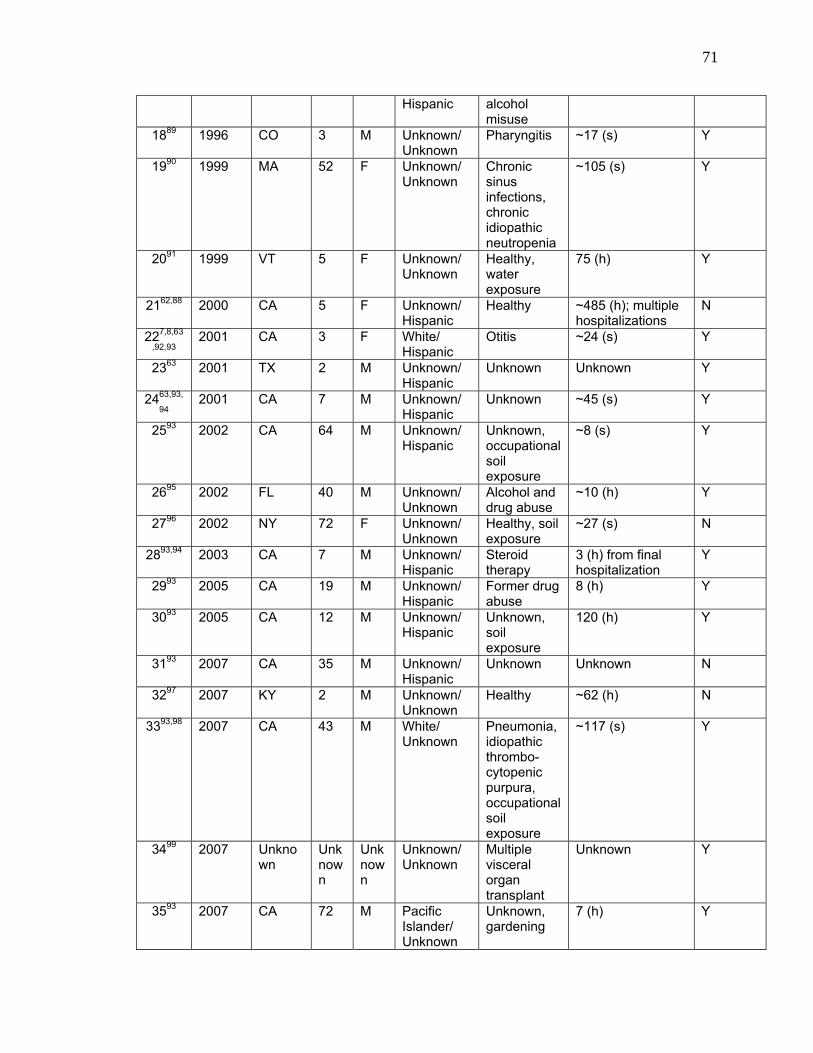
71
Hispanic alcohol misuse
1889 1996 CO 3 M Unknown/ Unknown
Pharyngitis ~17 (s) Y
1990 1999 MA 52 F Unknown/ Unknown
Chronic sinus infections, chronic idiopathic neutropenia
~105 (s) Y
2091 1999 VT 5 F Unknown/ Unknown
Healthy, water exposure
75 (h) Y
2162,88 2000 CA 5 F Unknown/ Hispanic
Healthy ~485 (h); multiple hospitalizations
N
227,8,63
,92,93 2001 CA 3 F White/
Hispanic Otitis ~24 (s) Y
2363 2001 TX 2 M Unknown/ Hispanic
Unknown Unknown Y
2463,93,
94 2001 CA 7 M Unknown/
Hispanic Unknown ~45 (s) Y
2593 2002 CA 64 M Unknown/ Hispanic
Unknown, occupational soil exposure
~8 (s) Y
2695 2002 FL 40 M Unknown/ Unknown
Alcohol and drug abuse
~10 (h) Y
2796 2002 NY 72 F Unknown/ Unknown
Healthy, soil exposure
~27 (s) N
2893,94 2003 CA 7 M Unknown/ Hispanic
Steroid therapy
3 (h) from final hospitalization
Y
2993 2005 CA 19 M Unknown/ Hispanic
Former drug abuse
8 (h) Y
3093 2005 CA 12 M Unknown/ Hispanic
Unknown, soil exposure
120 (h) Y
3193 2007 CA 35 M Unknown/ Hispanic
Unknown Unknown N
3297 2007 KY 2 M Unknown/ Unknown
Healthy ~62 (h) N
3393,98 2007 CA 43 M White/ Unknown
Pneumonia, idiopathic thrombo-cytopenic purpura, occupational soil exposure
~117 (s) Y
3499 2007 Unknown
Unknown
Unknown
Unknown/ Unknown
Multiple visceral organ transplant
Unknown Y
3593 2007 CA 72 M Pacific Islander/ Unknown
Unknown, gardening
7 (h) Y

72
3693 2008 CA 1.6 M Unknown/ Hispanic
Unknown 35 (h) Y
3718 2009 MS 4 M Unknown/ Unknown
Influenza A, water & soil exposure
~12 (s) Y
3818 2009 MS 31 F Unknown/ Unknown
Kidney transplant
~55 d (s) Y
3918 2009 MS 27 M Unknown/ Unknown
Kidney transplant
~139 (s) N
*Clinical course may mean from symptom onset until death or resolution of illness (s) or from presentation/hospitalization until death or resolution of illness (h). TB = Tuberculosis; HIV = Human Immunodeficiency Virus; IVDA = intravenous drug abuse
Table 5: Cutaneous acanthamoebiasis
Case (Ref)
Yr State Age (yr)
Sex Race/ Ethnicity
Health Status Clinical Course in Days
Died
16,22,39,1
00 1989 NY 38 M Unknown/
Unknown HIV, pneumonia
>21 (s) Y
222,23,39,
44 1990 IL 60 M Unknown/
Hispanic AIDS, PCP ~40 (s) Y
322,39,40 1991 CA 30 M Unknown/ Unknown
AIDS, PCP, chronic sinusitis
~77 (s) Y
439,101 1991 VA 42 M Unknown/ Unknown
AIDS 42 (s) Y
522,34,102 1992 NY 31 M Unknown/ Unknown
ESRD, Kidney transplant, diabetes
~420 (s), con’t treatment after lesions resolved
N
622,103 1994 DC 44 M Unknown/ Unknown
AIDS, progressive myelitis, peripheral neuropathy, water exposure
~450 (s) Y
722,40,104 1995?
CA 38 M White/ Unknown
AIDS, PCP, chronic sinusitis, water exposure
Unknown Y
822,105 1996 CA 27 F Unknown /Hispanic
AIDS ~240 (s) Y
922,106 1997?
MI 44 M Unknown/ Unknown
AIDS, TB ~180 (s) Y
1022,106 1997?
MI 38 M Unknown/ Unknown
AIDS ~62 (s) Y
11107 2003 MD 51 F Unknown/ Unknown
AIDS, PCP ~31 (s) N
12108 2004 CA 52 F Unknown/ Unknown
Lung transplant, immune- Suppressants, creek as drinking water source
~150 (h) N

73
*Clinical course may mean from symptom onset until death or resolution of illness(s) or from presentation/hospitalization until death or resolution of illness (h). AIDS = Acquired Immune Deficiency Syndrome; HIV = Human Immunodeficiency Virus; PCP = Pneumocystis carinii pneumonia, ESRD = end-stage renal disease; TB = Tuberculosis Table 6: Acanthamoeba Rhinosinusitis
Case (Ref)
Yr State Age (yr)
Sex Race/ Ethnicity
Health Status Clinical Course in
Days
Died
153 1994 Unk 45 M Unknown/ Unknown
HIV, chronic sinusitis Unknown N
2109 2005 FL 49 F Unknown/ Unknown
Bilateral lung transplant, immuno-suppressants, steroids
Unknown N
*Clinical course may mean from symptom onset until death or resolution of illness (s) or from presentation/hospitalization until death or resolution of illness (h). HIV = Human Immunodeficiency Virus
Table 7: Acanthamoeba osteomyelitis
Case (Ref)
Yr State Age (yr)
Sex Race/ \Ethnicity
Health Status
Clinical Course in Days
Died
1110 1979 PA 32 F White/ Unknown
Suspected diabetes
Unknown N
*Clinical course may mean from symptom onset until death or resolution of illness (s) or from presentation/hospitalization until death or resolution of illness (h).

Appendix B: Case Report Form
74

Revised 09/02/2010
75
Date of Report:
Demographics Patient’s Last Name First M.I.
Age
Gender:
Male Female Unknown
Ethnicity:
Hispanic Unknown
Non-Hispanic
Race:
White
Black
Asian/Pacific Islander
American Indian
Unknown
Other __
County and State of Residence: _ _________________
County and State of Treatment: __ ________________
Immigrant? Yes No □ Unknown Country of origin: _____ ___
Length of time since immigrated: _____________
Occupation: ____ _________
Exposure History
County/State of Suspected Exposure: / Number of persons exposed (if known):
Source of possible exposure, if known: (please check all that apply and provide best estimates of dates)
Type: Date(s): Type: Date(s): Type: Date(s): Canal __ __ Private Club Pool __ __ Community Pool __ __ Lake __ __ Private Home Pool __ __ Apartment Pool __ __ Pond __ __ Fill-and-Drain Pool __ __ Fountain __ __ Ocean __ __ Hotel Pool __ __ Water park __ __ River/Stream __ __ Spring (hot/cold) __ __ Well __ __ Spa/hot tub/whirlpool __ __
Water Exposures
Yes No Unknown
If “yes”, please fill out section on right
Other, specify ___________________________ Date(s):
Yes No Unknown Yes No Unknown Diving into water Snorkeling/scuba diving Inhaled water Swimming Jumped into water Water sports (skiing etc.) Swallowed water Wore nose clip or plugged
nose when jumping/diving
Water Activities Yes No Unknown
If “yes”, please fill out specifics on right
Splashed water Other, specify
Type: Date(s): Gardening __ __ Farmer/rancher Composting __ __ Firefighter Farm/Ranch __ __ Lifeguard/pool attendant
Soil Exposures Yes No Unknown
If “yes”, please fill out specifics on right
Other, specify: _____ Date(s): __
Occupational Exposures
Yes No Unknown
If “yes”, please fill out specifics on right
Other, specify: _________
Route of Entry if known:
Other, specify: Inhalation Contact
(please check all that apply)
If Water Source , Please List Source Characteristics:
Name of Water Exposure: Geospatial Coordinates: Thermally Polluted: Y / N Size of Body Water: □ < 10 acres □ 10-100 acres □ >100 acres □ Unknown
Water Turbidity: □ Clear □ Cloudy □ Murky □ Unknown
Water level: □ Low □ High □ Normal □ Flood Stage □ Unknown
Flow Rate: □ Slow □ Normal □ Fast □ Unknown
Ambient Air Temperature: F/C Water Temperature: ___F/C Depth: __ __
Ingestion Via Wound

76
Travel History last 2 years: Yes No Unknown If yes, please specify in table below:
Locations Dates (from – to)
Past Medical History:
Please check all conditions/symptoms that patient has currently or has had within past 2 years: Treatment/drugs: HIV/AIDS:
ENT/Respiratory: Other Conditions: Otitis Sinusitis Dermatitis Rhinitis Epistaxis Skin infections Broken Nose Nasal Surgery Eye infection Deviated septum Pharyngitis Other, specify: ________________________ Tuberculosis Pneumonitis Injury, specify: ________________________
Other lung disease, specify:
Current Illness
Date of Illness onset: Duration of illness: (in days)
Was patient admitted to hospital for current illness? Yes No Unknown If Yes, Date of most recent hospitalization: Duration of most recent hospitalization (in days):
Hospital Name:___ City:___ _____________ State: __
Physician Name 1:___ _____________ E-mail (if avail): ___ __________ Phone: ___ ____
Physician Name 2:___ _____________ E-mail (if avail): ___ __________ Phone: ___ ____
Other Recent Hospitalizations: Yes No Unknown
Dates (from- to) Diagnosis
HIV Yes No Unknown
AIDS Yes No Unknown
On Antiretrovirals
Yes No Unknown
Excessive antibiotic use (specify in Provider comments)
Illegal drug use, specify:__ _________ Immunosuppressants Radiation therapy Steroid use
Other Immunocompromised Conditions Alcohol misuse Diabetes mellitus G6PD deficiency Liver cirrhosis Malnourishment Pregnancy (recent) Renal failure Lymphoproliferative disease
Systemic Lupus Erythematosus (SLE) Cancer, specify:______________________________
Other hematologic disease, specify: ______________
Other autoimmune disease, specify: ______________
Organ transplant, specify: ______________________

77
History of Present Illness:
Please provide a brief description of the patient’s clinical course, prior to hospitalization:
Signs/Symptoms on Presentation (most recent hospitalization):
Vital Signs:__________________________________________________________________________________________
Temperature: _ _ F / C P: _ _ bpm R= __ _breaths/min BP: _ ___mmHg General:_____________________________________ ______ Visual______________________
Duration (days) Duration
(days) Duration
(days) Fever Myalgia Blurred vision Nausea Back Pain Diplopia Vomiting Cough Photophobia Diarrhea Shortness of breath Weight loss Sinus problems
Other visual changes, specify: _________
Anorexia Abnormal reflexes Headache Disorientation Stiff neck Lethargy/fatigue
Other general symptom/sign, specify:_ ________
Neurologic:_______________________________________________________________________________________
___
Duration (days) Duration
(days) Duration
(days) Altered mental status Dysphagia Weakness
Aphasia Facial numbness/Parathesia Hemiparesis/Hemiplegia Ataxia Hallucinations Altered sense of taste
Behavioral changes Combativeness Altered sense of smell Coma Hyperreflexia Decerebrate posturing Confusion Loss of balance Decorticate posturing Cranial nerve VI deficit Numbness Fixed, nonreactive pupils
Skin Lesions: Yes No Unknown If yes, please specify in table below:
Lesion type Anatomic location Size Number Duration (days)
Ulcers Plaques Erythematous nodules Other
Other Symptoms/Signs:
Other, specify:_ ______________________
Cranial nerve VII deficit Seizures Dilated pupils
Cranial nerve XII deficit Upgoing toes Nystagmus
Other cranial nerve deficit, specify: ______ Duration:
Other neurologic deficit, specify: Duration:

78
Signs/Symptoms developed while in hospital:
General:___________________________________________ Visual :__________________________________
Fever Myalgia Blurred vision Nausea Back Pain Diplopia Vomiting Cough Photophobia
Other visual changes, specify:_ _______ Diarrhea Shortness of breath Weight loss Sinus problems Anorexia Abnormal Reflexes Headache Disorientation Stiff neck Lethargy/fatigue Other general symptom/sign, specify:_ __________
Neurologic:__________________________________________________________________________________________
Altered mental status Dysphagia Altered sense of taste Aphasia Facial numbness/Parathesia Altered sense of smell Ataxia Hallucinations Decerebrate posturing
Behavioral changes Hemiparesis/Hemiplegia Decorticate posturing Coma Hyperreflexia Fixed, nonreactive pupils Combativeness Loss of balance Dilated pupils Confusion Numbness Nystagmus Cranial nerve VI deficit Seizures Cranial nerve VII deficit Upgoing toes Cranial nerve XII deficit Weakness
Other neurologic deficit, specify: _____ ________ Other Cranial nerve deficit, specify:
Skin Lesions: Yes No If yes, please specify in table below:
Lesion type Anatomic location Size Number
Ulcers Plaques Erythematous nodules Other
Other Symptoms/Signs:
Other, specify:_ ______________________

79
Diagnostic Tests: Note please provide dates when possible. If date not available, provide hospital day (i.e. CSF tap on Hosp. Day 2)
LABORATORY TESTING
CSF Date________________ Date_______________ Date_______________ Results Results Results Opening pressure (mmH2O) WBC count (per mm3 ) RBC count (per mm3 ) Neutrophil % Monocyte % Lymphocyte % Bands % Eosinophil % Protein (mg/100ml) Glucose (mg/100ml) Cytokines CSF Culture: * CSF PCR: * CSF latex agglutination: * CSF mount: Please indicate preparation type and findings, if any
Centrifuged Stained Wet
Amebae present? Y N
Centrifuged Stained Wet
Amebae present? Y N
Centrifuged Stained Wet
Amebae present? Y N * Please provide results for all bacteria, viral and/or parasitic testing.
Presenting Lab Values: Date: Cultures for Free Living Amebae: Results Source Date Result RBC count (per mm3 ) Negative + Balamuthia Hematocrit %
Blood + Naegleria + Acanthamoeba
WBC count (per mm3 ) Negative + Balamuthia Neutrophil %
Skin + Naegleria + Acanthamoeba
Lymphocyte % Negative + Balamuthia Monocyte %
Brain + Naegleria + Acanthamoeba
Eosinophil % Negative + Balamuthia Bands %
Abscess + Naegleria + Acanthamoeba
CD4 count (per mm3) Negative + Balamuthia Protein (mg/100ml)
Other, specify:
+ Naegleria + Acanthamoeba
Sodium (mEq/L) Potassium (mEq/L) Chloride (mEq/L) Bicarbonate (mEq/L)
PCR for Free Living Amebae: BUN (mg/100ml) Creatinine (mg/100ml) Source Date Result Glucose (mg/100ml) Negative + Balamuthia
Blood
+ Naegleria + Acanthamoeba
Serology: Negative + Balamuthia Date Result
Skin + Naegleria + Acanthamoeba
Negative + Balamuthia
Brain
+ Naegleria + Acanthamoeba
Negative + Balamuthia
Abscess
+ Naegleria + Acanthamoeba
Negative + Balamuthia
Other, specify:
+ Naegleria + Acanthamoeba

80
HISTOPATHOLOGY Brain biopsy: Yes No Unknown
Date: Date:
Location
Timing Antemortem Postmortem Unknown Antemortem Postmortem Unknown
Amebic trophozoites Amebic trophozoites Amebic cysts Amebic cysts Ameba, not specified Ameba, not specified Hemorrhage Necrosis Hemorrhage Necrosis Encephalomalacia Edema Encephalomalacia Edema Abscess Vasculitis Abscess Vasculitis Perivascular Inflammation Perivascular Inflammation Thrombosis Thrombosis Neovascularization Neovascularization Neutrophillic inflammation / infiltrate Neutrophillic inflammation / infiltrate Lymphocytic inflammation / infiltrate Lymphocytic inflammation / infiltrate Granulomatous inflammation Granulomatous inflammation Granuloma Granuloma Meningitis Encephalitis Meningitis Encephalitis
Results (check all that
apply)
Meningoencephalitis Meningoencephalitis Other Results/
Comments
Skin biopsy: Yes No Unknown
Date: Date:
Location Timing Antemortem Postmortem Unknown Antemortem Postmortem Unknown
Results No amebae seen Amebic trophozoites Amebic cysts Amebae, not specified
No amebae seen Amebic trophozoites Amebic cysts Amebae, not specified
Other Results/ Comments
Sinus biopsy: Yes No Unknown
Date: Date:
Location Timing Antemortem Postmortem Unknown Antemortem Postmortem Unknown
Results No amebae seen Amebic trophozoites Amebic cysts Amebae, not specified
No amebae seen Amebic trophozoites Amebic cysts Amebae, not specified
Other Results/ Comments
Other biopsy results:

81
DIAGNOSTIC IMAGING CT: Date of First CT:
Lesion location: (please check all that apply) Basal Ganglia Left Occipital Left Temporal Brainstem Right Occipital Right Temporal Right Cerebellum Left Parietal Thalamus Left Cerebellum Right Parietal Other, specify:_ ______________ Left Frontal Pons Right Frontal Spinal Cord
Lesion: (please check all that apply)
Additional Description, if needed:
Abscess Hyperdense Enhancing Edema Hypodense Ring enhancing Erosion Infarcts Sinusitis Hemorrhage Mass Ventriculomegaly Herniation Multifocal lesions Other, specify: __ _______________
Please list dates of subsequent CT scans and changes noted:
Date Findings
MRI: Date of First MRI:
Lesion location: (please check all that apply) Basal Ganglia Left Occipital Left Temporal Brainstem Right Occipital Right Temporal Right Cerebellum Left Parietal Thalamus Left Cerebellum Right Parietal Other, specify:_ ______________ Left Frontal Pons Right Frontal Spinal Cord
Lesion: (please check all that apply)
Additional Description, if needed:
Abscess Hyperdense Enhancing Edema Hypodense Ring enhancing Erosion Infarcts Sinusitis Hemorrhage Mass Ventriculomegaly Herniation Multifocal lesions Other, specify: __ _______________
Please list dates of subsequent MRI scans and changes noted:
Date Findings

82
Diagnosis:
Admitting diagnosis: Date of Diagnosis:
Encephalitis Meningoencephalitis Meningitis Other, Specify _ ____________
Final diagnosis: Date of Diagnosis:
GAE (Acanthamoeba spp.) GAE (Balamuthia mandillaris) PAM (Naegleria fowleri) Disseminated Acanthamoebiasis Disseminated Balamuthiasis Other, specify: Acanthamoeba rhinosinusitis Balamuthia rhinosinusitis Cutaneous acanthamoebiasis Cutaneous balamuthiasis
Diagnosis Method:
Indirect Immunofluorescence (IIF) Antemortem Postmortem Unknown PCR Antemortem Postmortem Unknown Histopathology Antemortem Postmortem Unknown CSF Wet Mount Antemortem Postmortem Unknown Culture Antemortem Postmortem Unknown Other, Specify: Antemortem Postmortem Unknown
Treatment:
Surgical resection: Yes No
Medications: (please check all that apply)
Acyclovir Ethambutol Rifampin Albendazole Fluconazole Steroid, specify__ ___ Amikacin Flucytosine Streptomycin Amphotericin B Isoniazid Sulfonamide, specify______ Amphotericin B lipid Itraconazole Sulfadiazine Amphotericin B liposomal Ketoconazole Topical Chlorhexidine Azithromycin Mannitol Trimethoprim/sulfa Ceftriaxone Metronidazole Voriconazole
Other, specify____ ____ Ciprofloxacin Miconazole Chloramphenicol Miltefosine Other, specify____ ____ Clarithromycin Ornidazole Other, specify____ ____ Co-trimoxazole Pentamidine Other, specify____ ____ Dexamethasone Pyrimethamine Other, specify____ ____
If you checked any of the medications listed above, please list below with the start and stop dates, doses, frequencies, and routes of administration. If an individual drug was given using multiple doses, over multiple dates, and/or given by multiple routes, please indicate this in the rows below.
Medication Start date: Stop date: Dose and Frequency Route of Administration

83
Other therapies: (please check all that apply) Start date: Stop date:
IV fluids Total Parenteral Nutrition (TPN) Dialysis for renal failure Other, specify_____________ Other, specify_____________ Other, specify_____________
Outcome:
Survived?
Yes No Unknown
If survived: Residual neurologic deficits?
Yes No Unknown
If Yes, Please describe neurologic deficits: __ ____________________________________________________ Date of discharge: OR Date of death: If died: Cause of death:
Brain death Removed life support Cardiorespiratory failure Other, specify:_ ___________________ Herniation
If died: Organs transplanted? Yes No If yes, which ones: _________________________________
Please provide a brief description of the patient’s clinical course, complications, and any additional comments:
CDC USE ONLY: 1st DASH # 2nd DASH # 3rd DASH # 4th DASH # 5th DASH # List additional DASH #s:
Case report citation 1 Case report citation 2 List additional case citations
Calculated durations: Incubation period (days): Illness Onset to Admission (days): Illness Onset to Death (days): Exposure to Death (days): Clinical Stage at presentation: ______

Appendix C: Epidemic Curves for Acanthamoeba and Balamuthia Cases
Epi
dem
ic C
urve
by
Yea
r of R
epor
t for
Aca
ntha
moe
ba s
pp. C
ases
85

85
Epi
dem
ic C
urve
by
Yea
r of R
epor
t for
Bal
amut
hia
man
drill
aris
Cas
es

86
Epi
dem
ic C
urve
by
Age
for A
cant
ham
oeba
spp
. Cas
es

87
Epi
dem
ic C
urve
by
Age
for B
alam
uthi
a m
andr
illar
is C
ases

Appendix D: Treatment Regimens for Survivors
89

89
Survivors of disseminated acanthamoebiasis
Survivors of cutaneous acanthamoebiasis

90
Survivors of Acanthamoeba rhinosinusitis

91
Survivor of
disseminated balamuthiasis
Survivors of Balamuthia GAE