A Comparison of Tenocytes and Mesenchymal Stem Cells for Use in Flexor Tendon Tissue Engineering
Upload
independentCategory
view
2download
0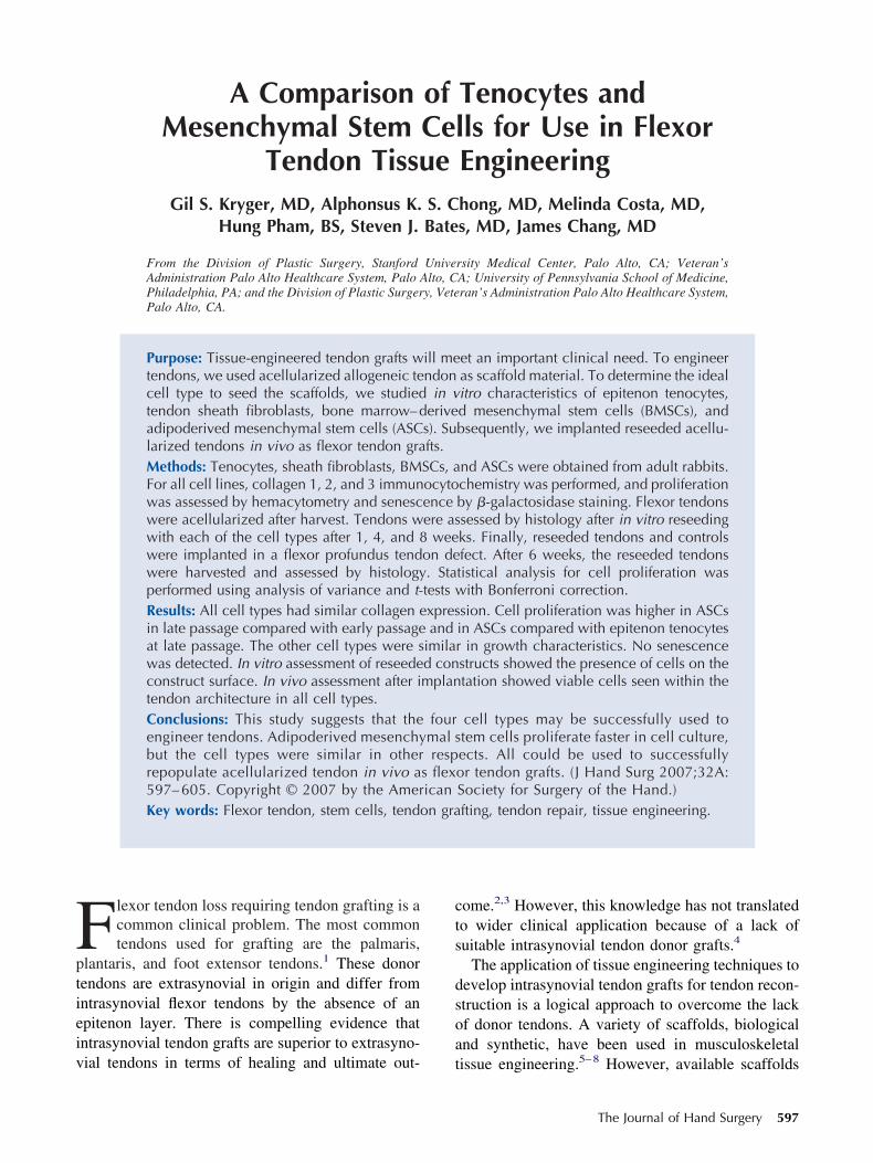
Fptieiv
A Comparison of Tenocytes andMesenchymal Stem Cells for Use in Flexor
Tendon Tissue EngineeringGil S. Kryger, MD, Alphonsus K. S. Chong, MD, Melinda Costa, MD,
Hung Pham, BS, Steven J. Bates, MD, James Chang, MD
From the Division of Plastic Surgery, Stanford University Medical Center, Palo Alto, CA; Veteran’sAdministration Palo Alto Healthcare System, Palo Alto, CA; University of Pennsylvania School of Medicine,Philadelphia, PA; and the Division of Plastic Surgery, Veteran’s Administration Palo Alto Healthcare System,Palo Alto, CA.
Purpose: Tissue-engineered tendon grafts will meet an important clinical need. To engineertendons, we used acellularized allogeneic tendon as scaffold material. To determine the idealcell type to seed the scaffolds, we studied in vitro characteristics of epitenon tenocytes,tendon sheath fibroblasts, bone marrow–derived mesenchymal stem cells (BMSCs), andadipoderived mesenchymal stem cells (ASCs). Subsequently, we implanted reseeded acellu-larized tendons in vivo as flexor tendon grafts.Methods: Tenocytes, sheath fibroblasts, BMSCs, and ASCs were obtained from adult rabbits.For all cell lines, collagen 1, 2, and 3 immunocytochemistry was performed, and proliferationwas assessed by hemacytometry and senescence by �-galactosidase staining. Flexor tendonswere acellularized after harvest. Tendons were assessed by histology after in vitro reseedingwith each of the cell types after 1, 4, and 8 weeks. Finally, reseeded tendons and controlswere implanted in a flexor profundus tendon defect. After 6 weeks, the reseeded tendonswere harvested and assessed by histology. Statistical analysis for cell proliferation wasperformed using analysis of variance and t-tests with Bonferroni correction.Results: All cell types had similar collagen expression. Cell proliferation was higher in ASCsin late passage compared with early passage and in ASCs compared with epitenon tenocytesat late passage. The other cell types were similar in growth characteristics. No senescencewas detected. In vitro assessment of reseeded constructs showed the presence of cells on theconstruct surface. In vivo assessment after implantation showed viable cells seen within thetendon architecture in all cell types.Conclusions: This study suggests that the four cell types may be successfully used toengineer tendons. Adipoderived mesenchymal stem cells proliferate faster in cell culture,but the cell types were similar in other respects. All could be used to successfullyrepopulate acellularized tendon in vivo as flexor tendon grafts. (J Hand Surg 2007;32A:597– 605. Copyright © 2007 by the American Society for Surgery of the Hand.)Key words: Flexor tendon, stem cells, tendon grafting, tendon repair, tissue engineering.
cts
dsoa
lexor tendon loss requiring tendon grafting is acommon clinical problem. The most commontendons used for grafting are the palmaris,
lantaris, and foot extensor tendons.1 These donorendons are extrasynovial in origin and differ fromntrasynovial flexor tendons by the absence of anpitenon layer. There is compelling evidence thatntrasynovial tendon grafts are superior to extrasyno-
ial tendons in terms of healing and ultimate out- tome.2,3 However, this knowledge has not translatedo wider clinical application because of a lack ofuitable intrasynovial tendon donor grafts.4
The application of tissue engineering techniques toevelop intrasynovial tendon grafts for tendon recon-truction is a logical approach to overcome the lackf donor tendons. A variety of scaffolds, biologicalnd synthetic, have been used in musculoskeletal
issue engineering.5–8 However, available scaffoldsThe Journal of Hand Surgery 597
hpbiiaebvldppf
tspcc
rcsmccvsvr
MA(vfi
CTEAufldteHs
u
t(tbscM
uraHcsFtapmc
tlssnsfrcsst
sc
IEfsPufuP1ahaf
598 The Journal of Hand Surgery / Vol. 32A No. 5 May–June 2007
ave inferior biological and material properties com-ared with the tissue they are replacing.9 Much iseing done to improve biomaterials to match biolog-cal needs. However, another approach involves us-ng acellularized allogeneic tendon repopulated withutologous cells. In this method, the material prop-rties of the tendon are maintained, and the constructest resembles native tendon. Experience with heartalves and skin have shown that allogeneic acellu-arized tissue repopulated with autogenous cells pro-uces a reliable tissue-engineered construct.10,11 Ap-lying this to tendons, we have found that it isossible to produce acellularized tendon segmentsor tissue engineering applications.
One major limitation in tissue engineering is theime interval necessary to grow cells to populatecaffolds.9 This is a concern particularly when usingrimary differentiated cell lines. A more proliferativeell line would allow a shorter time interval betweenell harvest and construct implantation in vivo.
We studied four candidate cell types for use ineseeding acellularized tendon constructs. Specifi-ally, we compared epitenon tenocytes, tendonheath fibroblasts, bone marrow–derived mesenchy-al stem cells (BMSCs), and adipoderived mesen-
hymal stem cells (ASCs) with respect to growthharacteristics, senescence, collagen production, andiability of reseeded constructs in vitro. We alsotudied the viability of reseeded tendon constructs initro and after in vivo implantation in a clinicallyelevant model of rabbit flexor tendon grafting.
aterials and Methodsdult male New Zealand white rabbits (4.0–4.5 kg)
n � 5 as cell and tendon source and n � 30 for inivo experiments) were used. All animal experimentsollowed protocols approved by our institutional an-mal ethics committee.
ell Line Isolation, Culture, and Analysishe rabbits were killed by an intravenous injection ofuthasol (Pentobarbital 390 mg/Phentoin) (VIRBACH Inc., Fort Worth, TX). Using sterile techniquender loupe magnification, the forepaw and hindpawexor digitorum profundus (FDP) equivalents wereissected and 2-cm portions of the intrasynovial dis-al FDP tendon and overlying flexor sheath werexcised. Specimens were placed into bufferedank’s saline solution with penicillin 100 IU/mL and
treptomycin 100 �g/mL.Tenocytes and sheath fibroblasts were obtained
sing a previously established protocol.12 Harvested L
endons were treated with 0.5% collagenase type IASigma, St. Louis, MO) for 10 minutes at roomemperature to obtain tenocytes. Tendon sheath fi-roblasts were obtained from sheath tissue in theame manner. The released cells were plated andultured in Ham’s F12 medium (Gibco, Rockville,D) with 10% fetal bovine serum (Gibco).Bone marrow-derived stem cells were obtained
sing established techniques.13,14 Rabbit bone mar-ow was aspirated via iliac crest puncture immedi-tely after sacrifice. The aspirate was resuspended inam’s F12 media with 10% rabbit serum and peni-
illin/streptomycin. Cellular debris was removed bytraining through a 100-�m nylon cell strainer (BDalcon, Franklin Lakes, NJ). A cell pellet was ob-
ained after centrifugation at 1,000 rpm for 5 minutest 4°C and resuspended. Cells were counted andlated at a density of 5 � 106 nucleated cells/100m3 culture dish. On the following day, unattached
ells were removed by washing.Adipoderived mesenchymal stem cells were ob-
ained from inguinal fat pads using a technique pub-ished by Zuk et al.15,16 The fat was minced withcissors and treated with 0.075% collagenase II in ahaking water bath at 37°C for 10 minutes. Collage-ase was then inactivated using fresh media. The celluspensions underwent centrifugation at 1,000 rpmor 5 minutes at 4°C, and the resulting cell pellet wasesuspended and filtered through a 100-�m nylonell strainer. After final centrifugation and resuspen-ion, the cells were counted and incubated at a den-ity of 5 � 106 nucleated cells/100 mm3 tissue cul-ure dish in Ham’s F12 media.
All cells were grown at 37°C in a humidifiedtandard incubator with 5% CO2. Culture media washanged every 3 days.
mmunocytochemistryach cell type was cultured on sterile glass coverslips
or 3 days. After washing with phosphate bufferedaline (PBS), the cells were fixed in 2% formalin inBS for 10 minutes. Hydrogen peroxide 0.3% wassed to quench endogenous peroxidase activity. Theollowing primary antibodies and dilutions weresed: anti-collagen 1, 1:100 (Oncogene Researchroducts, Schwalbach, Germany); anti-collagen 2,:100 (Chemicon International, Temucula, CA); andnti-collagen 3, 1:100 (Chemicon International) for 1our. Biotin-labeled secondary antibodies were usedt dilution 1:100 for 30 minutes. Detection was per-ormed using the Vectastain Elite ABC kit (Vector
aboratories, Burlingame, CA) and VIP peroxidase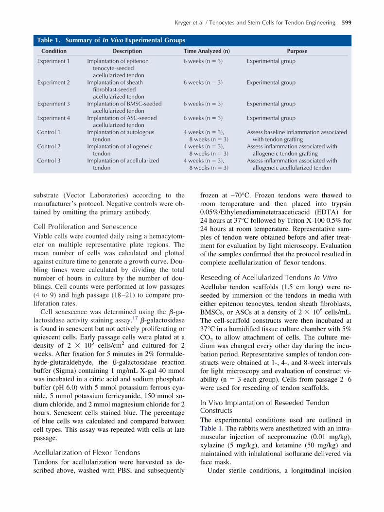
smt
CVemabnb(l
liqdwhbwbndhocp
ATs
fr022pmoc
RAseBT3Cdbsfaw
ICTTmxmf
8 we
Kryger et al / Tenocytes and Stem Cells for Tendon Engineering 599
ubstrate (Vector Laboratories) according to theanufacturer’s protocol. Negative controls were ob-
ained by omitting the primary antibody.
ell Proliferation and Senescenceiable cells were counted daily using a hemacytom-
ter on multiple representative plate regions. Theean number of cells was calculated and plotted
gainst culture time to generate a growth curve. Dou-ling times were calculated by dividing the totalumber of hours in culture by the number of dou-lings. Cell counts were performed at low passages4 to 9) and high passage (18–21) to compare pro-iferation rates.
Cell senescence was determined using the �-ga-actosidase activity staining assay.17 �-galactosidases found in senescent but not actively proliferating oruiescent cells. Early passage cells were plated at aensity of 2 � 103 cells/cm2 and cultured for 2eeks. After fixation for 5 minutes in 2% formalde-yde-glutaraldehyde, the �-galactosidase reactionuffer (Sigma) containing 1 mg/mL X-gal 40 mmolas incubated in a citric acid and sodium phosphateuffer (pH 6.0) with 5 mmol potassium ferrous cya-ide, 5 mmol potassium ferricyanide, 150 mmol so-ium chloride, and 2 mmol magnesium chloride for 2ours. Senescent cells stained blue. The percentagef blue cells was calculated and compared betweenell types. This assay was repeated with cells at lateassage.
cellularization of Flexor Tendonsendons for acellularization were harvested as de-
Table 1. Summary of In Vivo Experimental Groups
Condition Description T
Experiment 1 Implantation of epitenontenocyte-seededacellularized tendon
6
Experiment 2 Implantation of sheathfibroblast-seededacellularized tendon
6
Experiment 3 Implantation of BMSC-seededacellularized tendon
6
Experiment 4 Implantation of ASC-seededacellularized tendon
6
Control 1 Implantation of autologoustendon
4
Control 2 Implantation of allogeneictendon
4
Control 3 Implantation of acellularizedtendon
4
cribed above, washed with PBS, and subsequently
rozen at –70°C. Frozen tendons were thawed tooom temperature and then placed into trypsin.05%/Ethylenediaminetetraaceticacid (EDTA) for4 hours at 37°C followed by Triton X-100 0.5% for4 hours at room temperature. Representative sam-les of tendon were obtained before and after treat-ent for evaluation by light microscopy. Evaluation
f the samples confirmed that the protocol resulted inomplete acellularization of flexor tendons.
eseeding of Acellularized Tendons In Vitrocellular tendon scaffolds (1.5 cm long) were re-
eeded by immersion of the tendons in media withither epitenon tenocytes, tendon sheath fibroblasts,MSCs, or ASCs at a density of 2 � 106 cells/mL.he cell-scaffold constructs were then incubated at7°C in a humidified tissue culture chamber with 5%O2 to allow attachment of cells. The culture me-ium was changed every other day during the incu-ation period. Representative samples of tendon con-tructs were obtained at 1-, 4-, and 8-week intervalsor light microscopy and evaluation of construct vi-bility (n � 3 each group). Cells from passage 2–6ere used for reseeding of tendon scaffolds.
n Vivo Implantation of Reseeded Tendononstructshe experimental conditions used are outlined inable 1. The rabbits were anesthetized with an intra-uscular injection of acepromazine (0.01 mg/kg),
ylazine (5 mg/kg), and ketamine (50 mg/kg) andaintained with inhalational isoflurane delivered via
ace mask.
nalyzed (n) Purpose
s (n � 3) Experimental group
s (n � 3) Experimental group
s (n � 3) Experimental group
s (n � 3) Experimental group
s (n � 3),eks (n � 3)
Assess baseline inflammation associatedwith tendon grafting
s (n � 3),eks (n � 3)
Assess inflammation associated withallogeneic tendon grafting
s (n � 3),eks (n � 3)
Assess inflammation associated withallogeneic acellularized tendon
ime A
week
week
week
week
week8 weweek8 weweek
Under sterile conditions, a longitudinal incision

wtactlwwdctSFftrlappl
citraticinvdc
tttwrtoib
HHpscz
astttaae
SSpeTawtBfttimR
RCAffin
FicB1
600 The Journal of Hand Surgery / Vol. 32A No. 5 May–June 2007
as made on the volar surface of the middle digit ofhe right forelimb, between the metacarpophalangealnd proximal interphalangeal joints. Tissues werearefully dissected under loupe magnification untilhe flexor sheath was identified. The sheath and over-ying pulleys were sharply divided. The FDP tendonas isolated, and a 1.5-cm-long section of tendonas sharply excised. An immediate interposition ten-on graft consisting of reseeded tendon constructs orontrols (see later) was sutured proximally and dis-ally using 5-0 polypropylene (Prolene; Ethicon,omerville, NJ) using the Kessler technique. TheDP tendon was then isolated proximally in theorepaw and sharply transected to prevent loading ofhe repaired tendon. Next, the tendon pulleys wereepaired with interrupted 5-0 polypropylene (Pro-ene; Ethicon) stitches. The tourniquet was released,nd the skin was repaired using a running 5-0olypropylene (Prolene; Ethicon) suture. A soft com-ressive dressing was then applied to the right fore-imb.
The surgical procedure was modified for the threeontrol groups. Control 1 was designed to assess thenflammatory response associated with tendon au-ografts. These rabbits underwent transection andepair of the same tendon. Control 2 was designed tossess the inflammatory response associated withendon allografts. Previously harvested tendons keptn PBS for less than 2 hours were grafted into theontrol animals. Control 3 was designed to assess thenflammatory response and viability associated withonreseeded acellular tendon grafts. Previously har-ested and acellularized tendon was implanted asescribed earlier. There were six animals in eachontrol group.
Animals were fed standard lab chow and allowedo roam unrestricted in their cages. The experimentalendons were harvested at 6 weeks and the controlendons at 4 and 8 weeks after surgery. The rabbitsere killed by intravenous injection of Euthasol. The
ight forepaw incision was reopened, and the FDPendon and interpositional graft were dissected freef surrounding tissues. The graft and adjoining prox-mal and distal tendon were sharply transected enloc and preserved for histologic evaluation.
istologic Analysis of Tendonsarvested tissue was embedded with OCT Com-ound (Tissue-Tek, Sakura Finetek, Japan) andtored at –80°C until use. Histologic sections wereut at 10 �m with a cryostat (Leica CM 1800, Wet-
lar, Germany), fixed with acetone for 10 minutes, ond stored at –20°C until use. Hematoxylin and eosintaining was performed in the standard manner. His-ologic analysis was performed for tendon architec-ure with specific emphasis on intact collagen pat-ern, amount and nature of inflammatory response,nd tenocyte viability. Inflammatory response wasssessed by subjective histologic observation by anxperienced histotechnologist.
tatistical Analysistatistical analysis of the cell proliferation rates wereerformed as follows. The area under the curve forach cell proliferation experiment was calculated.hese were compared across cell types with one-waynalysis of variance (ANOVA). Subsequently, pair-ise comparisons were performed between the cell
ypes using unpaired two-tailed Student’s t-tests withonferroni correction. Early and late passage results
or each cell type were compared using unpairedwo-tailed Student’s t-tests. One-way ANOVA and-tests were performed using the Data Analysis Packn Microsoft Excel 9.0 (Microsoft Corporation, Red-ond, WA) and Bonferroni correction performed withversion 2.3.1 (http://www.R-project.org).
esultsell Culture and Analysisll cell types were successfully grown in culture. All
our cell types (epitenon tenocytes, tendon sheathbroblasts, BMSCs, and ASCs) exhibited elongateduclei and spindle-shaped cytoplasm, characteristic
igure 1. All four cell types demonstrate similar morphology,ncluding spindle-shaped cells with elongated fibroblast-likeell bodies: (A) epitenon tenocytes, (B) sheath fibroblasts, (C)MSCs, (D) ASCs. Phase-Contrast with Methylene Blue,00�.
f a fibroblast-like morphology (Fig. 1).
IAcltar
CTrssp1
e6
o5epi
RAHeoTts
TTtgATtsit
iifd
Ft(s
Ft(s
Ft(s
Kryger et al / Tenocytes and Stem Cells for Tendon Engineering 601
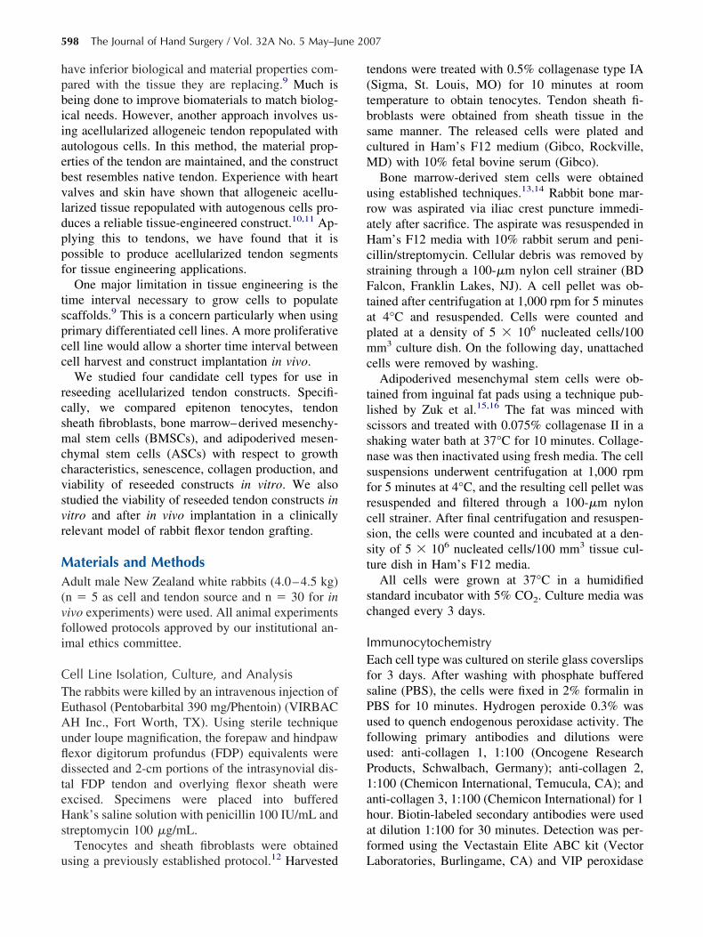
mmunocytochemistryll cell types stained strongly for collagen 1 and 3 in
ulture. Staining was strongest in the cytoplasm withight staining in the extracellular space. As expected,here was weak staining for collagen 2. There was noppreciable difference between the cell types withespect to staining (Figs. 2–4).
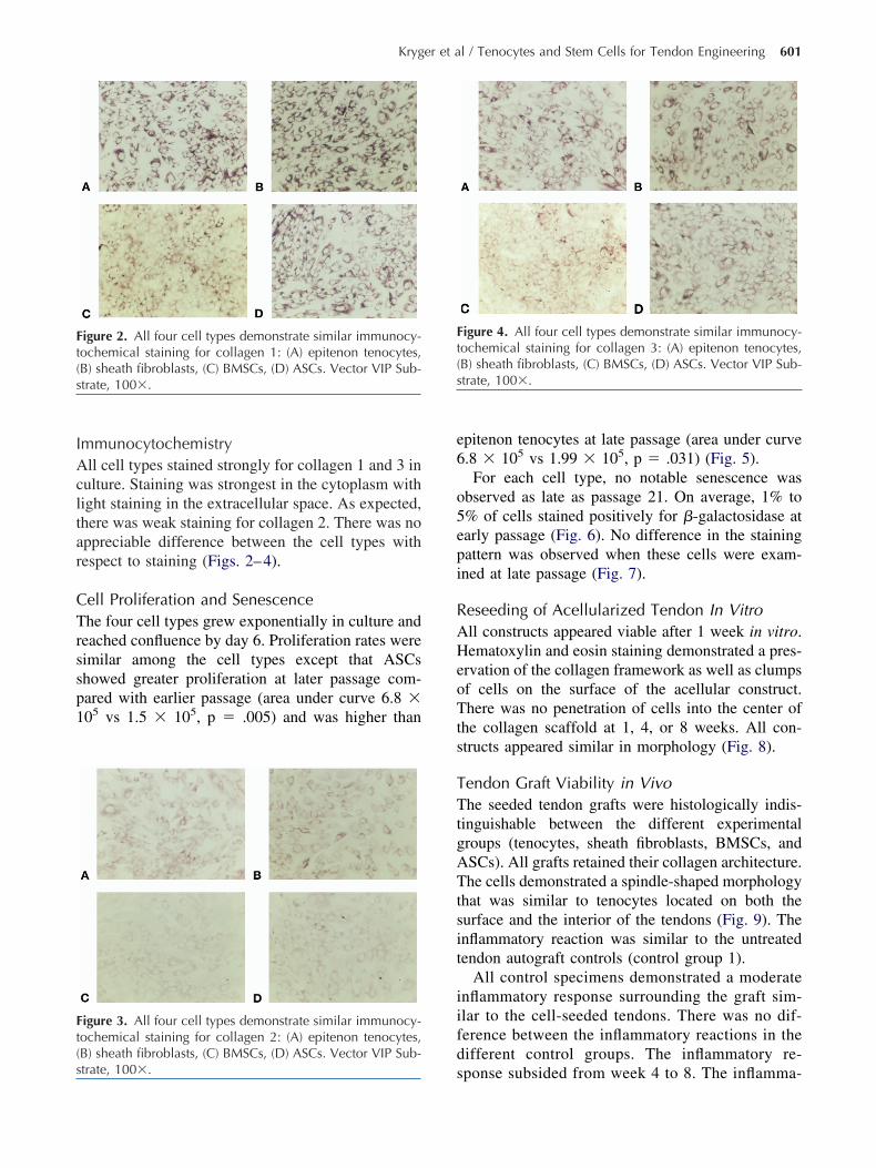
ell Proliferation and Senescencehe four cell types grew exponentially in culture and
eached confluence by day 6. Proliferation rates wereimilar among the cell types except that ASCshowed greater proliferation at later passage com-ared with earlier passage (area under curve 6.8 �05 vs 1.5 � 105, p � .005) and was higher than
igure 2. All four cell types demonstrate similar immunocy-ochemical staining for collagen 1: (A) epitenon tenocytes,B) sheath fibroblasts, (C) BMSCs, (D) ASCs. Vector VIP Sub-trate, 100�.
igure 3. All four cell types demonstrate similar immunocy-ochemical staining for collagen 2: (A) epitenon tenocytes,B) sheath fibroblasts, (C) BMSCs, (D) ASCs. Vector VIP Sub-
strate, 100�.
pitenon tenocytes at late passage (area under curve.8 � 105 vs 1.99 � 105, p � .031) (Fig. 5).
For each cell type, no notable senescence wasbserved as late as passage 21. On average, 1% to% of cells stained positively for �-galactosidase atarly passage (Fig. 6). No difference in the stainingattern was observed when these cells were exam-ned at late passage (Fig. 7).
eseeding of Acellularized Tendon In Vitroll constructs appeared viable after 1 week in vitro.ematoxylin and eosin staining demonstrated a pres-
rvation of the collagen framework as well as clumpsf cells on the surface of the acellular construct.here was no penetration of cells into the center of
he collagen scaffold at 1, 4, or 8 weeks. All con-tructs appeared similar in morphology (Fig. 8).
endon Graft Viability in Vivohe seeded tendon grafts were histologically indis-
inguishable between the different experimentalroups (tenocytes, sheath fibroblasts, BMSCs, andSCs). All grafts retained their collagen architecture.he cells demonstrated a spindle-shaped morphology
hat was similar to tenocytes located on both theurface and the interior of the tendons (Fig. 9). Thenflammatory reaction was similar to the untreatedendon autograft controls (control group 1).
All control specimens demonstrated a moderatenflammatory response surrounding the graft sim-lar to the cell-seeded tendons. There was no dif-erence between the inflammatory reactions in theifferent control groups. The inflammatory re-
igure 4. All four cell types demonstrate similar immunocy-ochemical staining for collagen 3: (A) epitenon tenocytes,B) sheath fibroblasts, (C) BMSCs, (D) ASCs. Vector VIP Sub-trate, 100�.
ponse subsided from week 4 to 8. The inflamma-

ttfs(at
sTrpw
easur
FiiSp5n
FicEAsc
602 The Journal of Hand Surgery / Vol. 32A No. 5 May–June 2007
ory infiltrate was limited to the surface of theendon graft and did not penetrate the collagenramework. Control group 2 was designed to as-ess the immune reaction when tendon allograftsuntreated tendons) were implanted into outbrednimals. Tendon architecture was retained andenocytes appeared viable. Control group 3 con-
Figure 5. Cell proliferation curves (as m
igure 6. �-galactosidase staining at low passage. Roughly 1n 100 cells stain positively, indicating senescence. (A) Ep-tenon tenocytes, (B) sheath fibroblasts, (C) BMSCs, (D) ASCs.taining solution: 1 mg/ml X-gal, 40 nM citric acid, sodiumhosphate buffer at pH 6.0, 5 mM Potassium ferrous cyanide,mM potassium ferric cyanide, 150 mM NaCl, 2 Mm Mag-
esium chloride. 100� M
isted of acellular scaffolds that were not reseeded.his group demonstrated a similar inflammatory
eaction to that of groups 1 and 2 and showed noenetration of cells into the dense collagen frame-ork at either 4 or 8 weeks (Fig. 10).
ed by hemacytometry) of all cell types.
igure 7. �-galactosidase staining at high passage. Roughly 1n 100 cells stain positively (indicated by black arrow), indi-ating senescence; which is unchanged from passage 4. (A)pitenon tenocytes, (B) sheath fibroblasts, (C) BMSCs, (D)SCs. Staining solution: 1 mg/ml X-gal, 40 nM citric acid,odium phosphate buffer at pH 6.0, 5 mM Potassium ferrousyanide, 5 mM potassium ferric cyanide, 150 mM NaCl, 2
m Magnesium chloride. 100�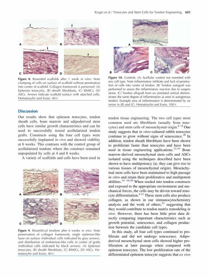
DOscugsaau
tccscatumisvmiaacccatvrgt
ldle
FciEAH
Fpla(tm
Fatpasta
Kryger et al / Tenocytes and Stem Cells for Tendon Engineering 603
iscussionur results show that epitenon tenocytes, tendon
heath cells, bone marrow and adipoderived stemells have similar growth characteristics and can besed to successfully reseed acellularized tendonrafts. Constructs using the four cell types wereuccessfully implanted in vivo and showed viabilityt 6 weeks. This contrasts with the control group ofcellularized tendons where the construct remainednpopulated by cells at 4 and 8 weeks.A variety of scaffolds and cells have been used in
igure 8. Reseeded scaffolds after 1 week in vitro. Notelumping of cells on surface of scaffold without penetrationnto center of scaffold. Collagen framework is preserved. (A)pitenon tenocytes, (B) sheath fibroblasts, (C) BMSCs, (D)SCs. Arrows indicate scaffold surface with attached cells.ematoxylin and Eosin, 40�.
igure 9. Bioartificial tendons after 6 weeks in vivo. Notereservation of collagen framework, single epitenon-likeayer on surface (individual cells indicated by gray arrows),nd distribution of endotenon-like cells in center of graftsindividual cells indicated by black arrows). (A) Epitenonenocytes, (B) sheath fibroblasts, (C) BMSCs, (D) ASCs. He-
datoxylin and Eosin, 40�.
endon tissue engineering. The two cell types mostommon used are fibroblasts (usually from teno-ytes) and stem cells of mesenchymal origin.6–8 Onetudy suggests that in vitro-cultured rabbit tenocytesontinue to grow without signs of senescence.18 Inddition, tendon sheath fibroblasts have been showno proliferate faster than tenocytes and have beensed in tissue engineering applications.12,19 Bonearrow-derived mesenchymal stem cells and ASCs
solated using the techniques described have beenhown to have multipotency (ie, they can give rise toarious tissues of mesenchymal origin). Mesenchy-al stem cells have been maintained to high passage
n vitro and retain their proliferative and multipotentbilities.14–16,20 When seeded into tendon constructsnd exposed to the appropriate environment and me-hanical forces, the cells may be driven toward teno-yte differentiation.4,21 These stem cells also produceollagen, as shown in our immunocytochemistrynalysis and the work of others,22 suggesting thathey would contribute to tendon matrix remodeling inivo. However, there has been little prior data di-ectly comparing important characteristics such asrowth potential, senescence, and collagen produc-ion between the candidate cell types.
In this study, all four cell types continued to pro-iferate and did not undergo senescence. Adipo-erived mesenchymal stem cells showed higher pro-iferation at later passage when compared withpitenon tenocytes. The low proliferation rate of the
igure 10. Controls. (A) Acellular control not reseeded withny cell type. Note inflammatory infiltrate and lack of penetra-ion of cells into center of tendon. (B) Tendon autograft waserformed to assess the inflammatory reaction due to surgerylone. (C) Tendon allograft from an unrelated animal demon-trates the same degree of inflammation as seen in autogenousendon. Example area of inflammation is demonstrated by anrrow in (B) and (C). Hematoxylin and Eosin, 100�.
ifferentiated epitenon tenocyte suggests that ex vivo
eaaae
glccttpc
dtttiifiTvwfierccit
snfdaereapdccpe
goi
ctTea
cpesectf
RF
F
aa
oF
7a
R
604 The Journal of Hand Surgery / Vol. 32A No. 5 May–June 2007
xpansion using this cell line will be slower. Inddition, given that it is easier to harvest largemounts of cells from bone marrow or fat, BMSCsnd ASCs have a practical advantage compared withpitenon tenocytes and sheath fibroblasts.
Scaffold materials used have ranged from collagenels to bioabsorbable synthetic material like poly-actide-co-glycolide. There is good evidence thatell-seeded constructs have better material propertiesompared with unseeded scaffolds.6–8 Nevertheless,hese constructs are inferior compared with nativeendon. This has major implications on the ability toerform rehabilitation and ultimate functional out-ome after tendon grafting clinically.
Gross histologic observation of the reseeded ten-ons showed no differences in inflammation betweenhe seeded constructs and autologous tendon con-rols. This supports our expectation that acellularizedendon reseeded with autologous cells will behavemmunologically like autologous tendon grafts. Onenteresting finding in our in vivo experiment was thending of viable allogeneic tendon at 4 and 8 weeks.here was no increased inflammation noted, andiable tenocytes were seen. Although no blindingas performed during the histologic assessment, ourndings are consistent with previously published lit-rature demonstrating no increase in inflammatoryesponse with use of allogeneic cells.7,23,24 It is un-lear whether the tenocytes seen are allogeneic teno-ytes or infiltrated autogenic cells. Further work us-ng X/Y fluorescent in situ hybridization will determinehe source of these tenocytes.
The rabbit model of flexor tendon repair was cho-en because the tendon has an intrasynovial compo-ent like human flexor tendons and is large enoughor surgical repair. Nevertheless, it is possible thatifferences exist in healing biology between rabbitsnd humans such that the constructs will have to bevaluated in a higher species after optimization in theabbit model. We were also unable to assess theffect on early mobilization on tendon graft viabilitynd inflammation because the tendon was dividedroximally to unload and protect the implanted ten-on graft. Although not quantitative, immunohisto-hemistry for collagen demonstrated that the variousell lines could produce collagen. This suggests theotential usefulness of these cells in tendon tissuengineering applications.
In vitro reseeding of tendon constructs showedood surface attachment of cells but no penetrationf cells into the center of the tendon scaffold. In the
n vivo part of the study, we showed that cell-seededonstructs demonstrate good repopulation in the cen-er of the scaffold within 6 weeks of implantation.hese findings in vivo suggest that the lack of pen-tration in vitro may not be an issue for clinicalpplications.
In summary, these four cell lines—epitenon teno-ytes, sheath fibroblasts, BMSCs, and ASCs—are allossible candidates for use in flexor tendon tissuengineering. These cells, combined with scaffoldsuch as acellularized tendon, offer the possibility ofxpanding the source of tendon grafts for hand re-onstruction. Further research will focus on testinghe biomechanical properties and inhibiting adhesionormation in these novel constructs.
eceived for publication October 18, 2006; accepted in revised formebruary 23, 2007.Part of this work was presented at the Adrian E. Flatt Residents and
ellows Conference 2005 and awarded the Joseph H. Boyes award.No benefits in any form have been received or will be received fromcommercial party related directly or indirectly to the subject of this
rticle.Supported by VA Merit Review grants, American Society for Surgery
f the Hand, American Association of Hand Surgery, and the Thussamily Grant.Corresponding author: James Chang, MD, Division of Plastic Surgery,
70 Welch Road, Suite 400, Palo Alto, CA 94304; e-mail: [email protected].
Copyright © 2007 by the American Society for Surgery of the Hand0363-5023/07/32A05-0002$32.00/0doi:10.1016/j.jhsa.2007.02.018
eferences1. White WL. Tendon grafts: a consideration of their source,
procurement and suitability. Surg Clin North Am 1960;40:403–413.
2. Leversedge FJ, Zelouf D, Williams C, Gelberman RH, SeilerJG III. Flexor tendon grafting to the hand: an assessment ofthe intrasynovial donor tendon-A preliminary single-cohortstudy. J Hand Surg 2000;25A:721–730.
3. Seiler JG III, Chu CR, Amiel D, Woo SL, Gelberman RH.The Marshall R. Urist Young Investigator Award. Autoge-nous flexor tendon grafts. Biologic mechanisms for incor-poration. Clin Orthop Relat Res 1997;345:239–247.
4. Zhang AY, Chang J. Tissue engineering of flexor tendons.Clin Plast Surg 2003;30:565–572.
5. Sharma B, Elisseeff JH. Engineering structurally organizedcartilage and bone tissues. Ann Biomed Eng 2004;32:148–159.
6. Cao Y, Liu Y, Liu W, Shan Q, Buonocore SD, Cui L.Bridging tendon defects using autologous tenocyte engi-neered tendon in a hen model. Plast Reconstr Surg 2002;110:1280–1289.
7. Ouyang HW, Goh JC, Mo XM, Teoh SH, Lee EH. Theefficacy of bone marrow stromal cell-seeded knitted PLGAfiber scaffold for Achilles tendon repair. Ann N Y Acad Sci2002;961:126–129.
8. Young RG, Butler DL, Weber W, Caplan AI, Gordon SL,Fink DJ. Use of mesenchymal stem cells in a collagen matrix
for Achilles tendon repair. J Orthop Res 1998;16:406–413.
1
1
1
1
1
1
1
1
1
1
2
2
2
2
2
Kryger et al / Tenocytes and Stem Cells for Tendon Engineering 605
9. Chong AK, Chang J. Tissue engineering for the handsurgeon: a clinical perspective. J Hand Surg 2006;31A:349–358.
0. Bader A, Schilling T, Teebken OE, Brandes G, Herden T,Steinhoff G, et al. Tissue engineering of heart valves—human endothelial cell seeding of detergent acellularizedporcine valves. Eur J Cardiothorac Surg 1998;14:279–284.
1. Livesey SA, Herndon DN, Hollyoak MA, Atkinson YH, NagA. Transplanted acellular allograft dermal matrix. Potentialas a template for the reconstruction of viable dermis. Trans-plantation 1995;60:1–9.
2. Klein MB, Pham H, Yalamanchi N, Chang J. Flexor tendonwound healing in vitro: the effect of lactate on tendon cellproliferation and collagen production. J Hand Surg [Am]2001;26A:847–854.
3. Colter DC, Class R, DiGirolamo CM, Prockop DJ. Rapidexpansion of recycling stem cells in cultures of plastic-adherent cells from human bone marrow. Proc Natl Acad SciU S A 2000;97:3213–3218.
4. Pittenger MF, Mackay AM, Beck SC, Jaiswal RK, DouglasR, Mosca JD, et al. Multilineage potential of adult humanmesenchymal stem cells. Science 1999;284:143–147.
5. De Ugarte DA, Morizono K, Elbarbary A, Alfonso Z, ZukPA, Zhu M, et al. Comparison of multi-lineage cells fromhuman adipose tissue and bone marrow. Cells Tissues Or-gans 2003;174:101–109.
6. Zuk PA, Zhu M, Mizuno H, Huang J, Futrell JW, Katz AJ, et
al. Multilineage cells from human adipose tissue: implicationsfor cell-based therapies. Tissue Eng 2001;7:211–228.
7. Dimri GP, Lee X, Basile G, Acosta M, Scott G, Roskelley C,et al. A biomarker that identifies senescent human cells inculture and in aging skin in vivo. Proc Natl Acad Sci U S A1995;92:9363–9367.
8. Bernard-Beaubois K, Hecquet C, Houcine O, Hayem G,Adolphe M. Culture and characterization of juvenile rabbittenocytes. Cell Biol Toxicol 1997;13:103–113.
9. Khan U, Occleston NL, Khaw PT, McGrouther DA. Differ-ences in proliferative rate and collagen lattice contractionbetween endotenon and synovial fibroblasts. J Hand Surg1998;23A:266–273.
0. Kassem M, Kristiansen M, Abdallah BM. Mesenchymalstem cells: cell biology and potential use in therapy. BasicClin Pharmacol Toxicol 2004;95:209–214.
1. Caplan AI. The mesengenic process. Clin Plast Surg 1994;21:429–435.
2. Ge Z, Goh JC, Lee EH. Selection of cell source forligament tissue engineering. Cell Transplant 2005;14:573–583.
3. Chong AK, Ang AD, Goh JC, Hui JH, Lim AY, Lee EH, etal. Bone marrow-derived mesenchymal stem cells influenceearly tendon-healing in a rabbit achilles tendon model.J Bone Joint Surg 2007;89A:74–81.
4. Ryan JM, Barry FP, Murphy JM, Mahon BP. Mesenchymalstem cells avoid allogeneic rejection. J Inflamm (Lond)
2005;2:8.Copyright © 2022 FDOKUMEN