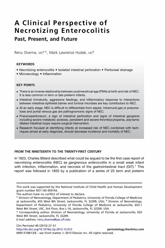
A Clinical Perspective of Necrotizing Enterocolitis Past, Present, and Future FROM THE NINETEENTH TO...
25
A Clinical Perspective of Necrotizing Enterocolitis Past, Present, and Future Renu Sharma, MD a, *, Mark Lawrence Hudak, MD b FROM THE NINETEENTH TO THE TWENTY-FIRST CENTURY In 1823, Charles Billard described what could be argued to be the first case report of necrotizing enterocolitis (NEC) as gangrenous enterocolitis in a small weak infant with infection, inflammation, and necrosis of the gastrointestinal tract (GIT). 1 This report was followed in 1850 by a publication of a series of 25 term and preterm This work was supported by the National Institute of Child Health and Human Development grant number RO1 HD 059143. The authors have no conflict of interest to declare. a Division of Neonatology, Department of Pediatrics, University of Florida College of Medicine at Jacksonville, 655 West 8th Street, Jacksonville, FL 32209, USA; b Division of Neonatology, Department of Pediatrics, University of Florida College of Medicine at Jacksonville, 653-1 West 8th Street, LRC, 3rd Floor, Box L-16, Jacksonville, FL 32209, USA * Corresponding author. Division of Neonatology, University of Florida at Jacksonville, 655 West 8th Street, Jacksonville, FL 32209. E-mail address: [email protected] KEYWORDS Necrotizing enterocolitis Isolated intestinal perforation Peritoneal drainage Microecology Inflammation KEY POINTS There is an inverse relationship between postmenstrual age (PMA) at birth and risk of NEC. It is less common in term or late preterm infants. Intestinal immaturity, aggressive feedings, and inflammatory response to interactions between intestinal epithelial barrier and luminal microbes are key contributors to NEC. At an early stage, NEC is difficult to differentiate from sepsis. Intramural gas or pneuma- tosis and portal venous gas are pathognomonic signs of NEC. Pneumoperitoneum, a sign of intestinal perforation and signs of intestinal gangrene including severe metabolic acidosis, persistent and severe thrombocytopenia, adynamic dilated intestinal loops require surgical intervention. Research focused at identifying infants at increased risk of NEC combined with tech- niques aimed at early diagnosis, should decrease incidence and mortality of NEC. Clin Perinatol 40 (2013) 27–51 http://dx.doi.org/10.1016/j.clp.2012.12.012 perinatology.theclinics.com 0095-5108/13/$ – see front matter Ó 2013 Elsevier Inc. All rights reserved.
-
Upload
independent -
Category
Documents
-
view
2 -
download
0
Transcript of A Clinical Perspective of Necrotizing Enterocolitis Past, Present, and Future FROM THE NINETEENTH TO...
A Clinical Perspective ofNecrotizing Enterocolit isPast, Present, and Future
Renu Sharma, MDa,*, Mark Lawrence Hudak, MDb
KEYWORDS
� Necrotizing enterocolitis � Isolated intestinal perforation � Peritoneal drainage� Microecology � Inflammation
KEY POINTS
� There is an inverse relationship between postmenstrual age (PMA) at birth and risk of NEC.It is less common in term or late preterm infants.
� Intestinal immaturity, aggressive feedings, and inflammatory response to interactionsbetween intestinal epithelial barrier and luminal microbes are key contributors to NEC.
� At an early stage, NEC is difficult to differentiate from sepsis. Intramural gas or pneuma-tosis and portal venous gas are pathognomonic signs of NEC.
� Pneumoperitoneum, a sign of intestinal perforation and signs of intestinal gangreneincluding severe metabolic acidosis, persistent and severe thrombocytopenia, adynamicdilated intestinal loops require surgical intervention.
� Research focused at identifying infants at increased risk of NEC combined with tech-niques aimed at early diagnosis, should decrease incidence and mortality of NEC.
FROM THE NINETEENTH TO THE TWENTY-FIRST CENTURY
In 1823, Charles Billard described what could be argued to be the first case report ofnecrotizing enterocolitis (NEC) as gangrenous enterocolitis in a small weak infantwith infection, inflammation, and necrosis of the gastrointestinal tract (GIT).1 Thisreport was followed in 1850 by a publication of a series of 25 term and preterm
This work was supported by the National Institute of Child Health and Human Developmentgrant number RO1 HD 059143.The authors have no conflict of interest to declare.a Division of Neonatology, Department of Pediatrics, University of Florida College of Medicineat Jacksonville, 655 West 8th Street, Jacksonville, FL 32209, USA; b Division of Neonatology,Department of Pediatrics, University of Florida College of Medicine at Jacksonville, 653-1West 8th Street, LRC, 3rd Floor, Box L-16, Jacksonville, FL 32209, USA* Corresponding author. Division of Neonatology, University of Florida at Jacksonville, 655West 8th Street, Jacksonville, FL 32209.E-mail address: [email protected]
Clin Perinatol 40 (2013) 27–51http://dx.doi.org/10.1016/j.clp.2012.12.012 perinatology.theclinics.com0095-5108/13/$ – see front matter � 2013 Elsevier Inc. All rights reserved.
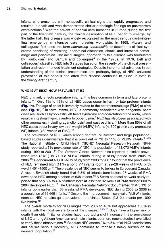
Sharma & Hudak28
infants who presented with nonspecific clinical signs that rapidly progressed andresulted in death and who demonstrated similar pathologic findings on postmortemexaminations.1 With the advent of special care nurseries in Europe during the firstpart of the twentieth century, the clinical description of NEC began to emerge; bythe latter half, this disease was widely recognized as the most serious gastrointes-tinal emergency in intensive care nurseries worldwide. In 1965, Mizrahi andcolleagues2 first used the term necrotizing enterocolitis to describe a clinical syn-drome consisting of vomiting, abdominal distension, shock, and intestinal hemor-rhage and perforation. The initial surgical approach to this disease was formulatedby Touloukian3 and Santulli and colleagues4 in the 1970s. In 1978, Bell andcolleagues5 classified NEC into 3 stages based on the severity of the clinical presen-tation and recommended treatment strategies. Despite incremental advances in ourunderstanding of the clinical presentation and pathophysiology of NEC, universalprevention of this serious and often fatal disease continues to elude us even inthe twenty-first century.
WHO IS AT RISK? HOW PREVALENT IT IS?
NEC primarily affects premature infants. It is less common in term and late preterminfants.6,7 Only 7% to 15% of all NEC cases occur in term or late preterm infants(Fig. 1A). The age of onset is inversely related to the postmenstrual age (PMA) at birth(see Fig. 1B).7 In term infants, NEC is commonly associated with congenital heartdiseases, such as hypoplastic left heart syndrome and coarctation of the aorta, whichresult in intestinal hypoxia and/or hypoperfusion.8 NEC has also been associated withother anomalies, including aganglionosis9 and gastroschisis.10 More than 85% of allNEC cases occur in very low birth weight (VLBW) infants (<1500 g) or in very premature(VP) infants (<32 weeks of PMA).11
The prevalence of NEC varies among centers. Multicenter and large population-based studies demonstrate that it is prevalent in 7% to 11% of VLBW infants.11–13
The National Institute of Child Health (NICHD) Neonatal Research Network (NRN)study reported a 7% prevalence rate of NEC in a population of 11,072 VLBW infantsduring 1999 to 2001.14 The Vermont Oxford Network also reported a similar preva-lence rate (7.4%) in 71,808 VLBW infants during a study period from 2005 to2006.15 A concurrent NICHD-NRN study from 2003 to 2007 found that the prevalenceof NEC remained high (11%) among VP infants (born at 22–28 weeks of PMA, birthweight 401–1500 g).13 The prevalence of NEC seems to be less in Europe and Canada.A recent Swedish study found that 5.8% of infants born before 27 weeks of PMAdeveloped NEC among a cohort of 638 infants.16 A Swiss neonatal network study re-ported that only 3% to 4% of infants born at less than 32 weeks of PMA during 2000 to2004 developed NEC.17 The Canadian Neonatal Network documented that 5.1% ofinfants born earlier than 33 weeks of PMA developed NEC during 2003 to 2008 ina population of 16,669 infants.18 Despite the improvement in survival of VLBW infants,neonatal NEC remains quite prevalent in the United States (0.3–2.4 infants per 1000live births).19
The overall mortality for NEC ranges from 20% to 40% but approaches 100% ininfants with the most severe form of the disease.12,15,20 Boys have a higher risk ofdeath than girls.12 Earlier studies have reported a slight increase in the prevalenceof NEC among African American and male infants, but more recent studies have failedto verify these observations.12 Because NEC affects 2% to 5% of all NICU admissionsand causes serious morbidity, NEC continues to impose a heavy burden on theneonatal population.19
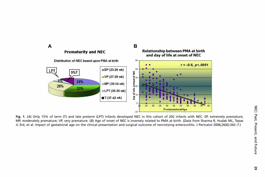
Fig. 1. (A) Only 15% of term (T) and late preterm (LPT) infants developed NEC in this cohort of 202 infants with NEC. EP, extremely premature;MP, moderately premature; VP, very premature. (B) Age of onset of NEC is inversely related to PMA at birth. (Data from Sharma R, Hudak ML, TepasJJ 3rd, et al. Impact of gestational age on the clinical presentation and surgical outcome of necrotizing enterocolitis. J Perinatol 2006;26(6):342–7.)
NEC:Past,
Present,andFu
ture
29
Sharma & Hudak30
WHAT CAUSES NEC? HOW DOES IT HAPPEN?
The pathophysiology of NEC in VP infants is not completely elucidated. Comparedwith NEC in term and late preterm infants in whom hypoxia-ischemia is a commonprecursor, recent advances in our understanding of NEC at the molecular levelsuggest that an inflammatory response in VP infants plays the inciting or dominantrole in the pathogenesis of NEC.21,22 Previously held beliefs that low Apgar scores,23
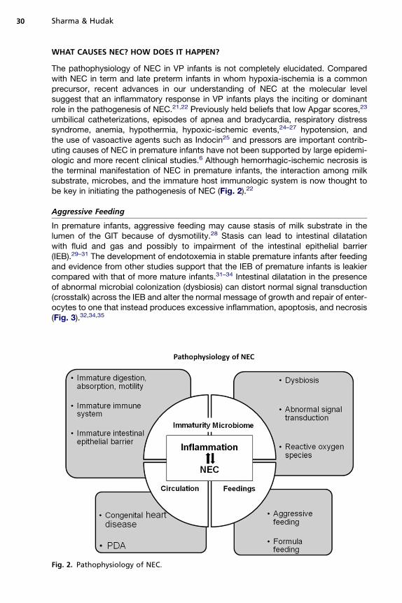
umbilical catheterizations, episodes of apnea and bradycardia, respiratory distresssyndrome, anemia, hypothermia, hypoxic-ischemic events,24–27 hypotension, andthe use of vasoactive agents such as Indocin25 and pressors are important contrib-uting causes of NEC in premature infants have not been supported by large epidemi-ologic and more recent clinical studies.6 Although hemorrhagic-ischemic necrosis isthe terminal manifestation of NEC in premature infants, the interaction among milksubstrate, microbes, and the immature host immunologic system is now thought tobe key in initiating the pathogenesis of NEC (Fig. 2).22
Aggressive Feeding
In premature infants, aggressive feeding may cause stasis of milk substrate in thelumen of the GIT because of dysmotility.28 Stasis can lead to intestinal dilatationwith fluid and gas and possibly to impairment of the intestinal epithelial barrier(IEB).29–31 The development of endotoxemia in stable premature infants after feedingand evidence from other studies support that the IEB of premature infants is leakiercompared with that of more mature infants.31–34 Intestinal dilatation in the presenceof abnormal microbial colonization (dysbiosis) can distort normal signal transduction(crosstalk) across the IEB and alter the normal message of growth and repair of enter-ocytes to one that instead produces excessive inflammation, apoptosis, and necrosis(Fig. 3).32,34,35
Fig. 2. Pathophysiology of NEC.
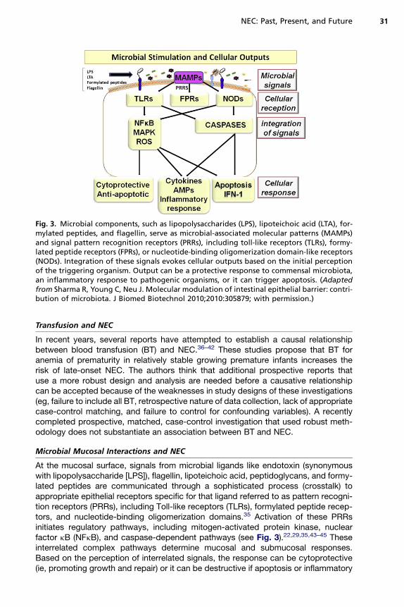
Fig. 3. Microbial components, such as lipopolysaccharides (LPS), lipoteichoic acid (LTA), for-mylated peptides, and flagellin, serve as microbial-associated molecular patterns (MAMPs)and signal pattern recognition receptors (PRRs), including toll-like receptors (TLRs), formy-lated peptide receptors (FPRs), or nucleotide-binding oligomerization domain-like receptors(NODs). Integration of these signals evokes cellular outputs based on the initial perceptionof the triggering organism. Output can be a protective response to commensal microbiota,an inflammatory response to pathogenic organisms, or it can trigger apoptosis. (Adaptedfrom Sharma R, Young C, Neu J. Molecular modulation of intestinal epithelial barrier: contri-bution of microbiota. J Biomed Biotechnol 2010;2010:305879; with permission.)
NEC: Past, Present, and Future 31
Transfusion and NEC
In recent years, several reports have attempted to establish a causal relationshipbetween blood transfusion (BT) and NEC.36–42 These studies propose that BT foranemia of prematurity in relatively stable growing premature infants increases therisk of late-onset NEC. The authors think that additional prospective reports thatuse a more robust design and analysis are needed before a causative relationshipcan be accepted because of the weaknesses in study designs of these investigations(eg, failure to include all BT, retrospective nature of data collection, lack of appropriatecase-control matching, and failure to control for confounding variables). A recentlycompleted prospective, matched, case-control investigation that used robust meth-odology does not substantiate an association between BT and NEC.
Microbial Mucosal Interactions and NEC
At the mucosal surface, signals from microbial ligands like endotoxin (synonymouswith lipopolysaccharide [LPS]), flagellin, lipoteichoic acid, peptidoglycans, and formy-lated peptides are communicated through a sophisticated process (crosstalk) toappropriate epithelial receptors specific for that ligand referred to as pattern recogni-tion receptors (PRRs), including Toll-like receptors (TLRs), formylated peptide recep-tors, and nucleotide-binding oligomerization domains.35 Activation of these PRRsinitiates regulatory pathways, including mitogen-activated protein kinase, nuclearfactor kB (NFkB), and caspase-dependent pathways (see Fig. 3).22,29,35,43–45 Theseinterrelated complex pathways determine mucosal and submucosal responses.Based on the perception of interrelated signals, the response can be cytoprotective(ie, promoting growth and repair) or it can be destructive if apoptosis or inflammatory

Sharma & Hudak32
responses is triggered (see Fig. 3).22,35,44 The terminal event in NEC is hemorrhagic-ischemic necrosis, which is the consequence of a dysregulated inflammatoryresponse mediated by endogenous factors that include platelet-activating factor(PAF), proinflammatory cytokines (such as tumor necrosis factor-a, interleukins1 & 6 [IL-1, IL-6]), chemokines (MIP-2/CXCL2), NF-kB, and the complement system.29
Because of the relative immaturity of key gastrointestinal functions, such as diges-tion, absorption, motility, and abnormalities in immune responses, a prematurenewborn is exquisitely vulnerable to gastrointestinal injury.46 The cellular responseto signal transduction as described earlier is orchestrated by the innate immunesystem in mucosa and submucosa.45,47 Microbial interaction with intestinal epithelium(PRRS) signals tissue-specific functions, such as T-cell expansion and elaboration ofcytokines and chemokines. Normally after birth, exposure to LPS downregulates IL-1receptor associated kinase 1 (IRAK), an intermediary for epithelial TLR4 signaling, andpromotes tolerance to LPS.19,22,43 This postnatal adaptation helps to achieve host-microbe homeostasis with microbial tolerance.22,44 Premature infants are exposedshortly after birth to a massive microbial antigenic challenge that may be distortedby frequent and prolonged use of antibiotics. As a result, the GIT of premature infantsgravitates toward mounting an inflammatory response rather than establishing toler-ance (T-helper-1 response).30,35,47 The premature infant seems to mimic a responsethat is similar to the immune-naıve response demonstrated by the fetus, which is char-acterized by hyper-responsiveness to LPS.22,48
Intestinal colonization with commensal Bifidobacterium and Lactobacillus speciesreduces the risk of NEC.49,50 The frequent and prolonged use of antibiotics resultsin an overgrowth of potentially pathogenic species.51 Distortion of intestinal micro-ecology by exposure to broad-spectrum antibiotics may result in colonization withinflammogenic microbial consortia that are more likely to direct the immune repertoirein the direction of an inflammatory response, thereby increasing the risk for NEC.51,52
Metagenomic studies indicate that intestinal microecology during the early postnatalperiod greatly influences the evolution of immune health.53,54 Infants who later developNEC show evidence for a dysbiotic microecology before the diagnosis of NEC.55 Arecent clinical study found that exposure to broad-spectrum antibiotics for morethan 10 days increased the risk of NEC by nearly threefold.51
Recent advances in the proteomics and microecology pertinent to NEC stronglysuggest that a distorted innate-immune response of premature infants increases thevulnerability to NEC.22,55 Glucocorticoid-mediated maturation of the IEB has beenshown to protect against NEC and to blunt the inflammatory response seen inNEC.46,48,56,57 Furthermore, the vulnerability to NEC is not unique to prematureneonates, it is also seen in the older immune-compromised population.58–60 A NEC-like illness has been described in the geriatric population and in patients with humanimmunodeficiency virus.56,58
Hypoxic-Ischemic Mechanisms
Under normal conditions, a state of high intestinal blood flow and low resting vascularresistance is maintained by nitric oxide.26 Impaired endothelial function or elaborationof proinflammatory mediators may alter the balance between vasoconstriction (asmediated by endothelin-1) and vasodilatation (as mediated by nitric oxide) and leadto a relatively ischemic state. In animal models of hypoxia-ischemia, derangementsof intestinal microcirculation develop at the premucosal arteriolar inflow locationwith a distinct stop-and-go pattern consistent with severe vascular dysfunction.61
Levels of inflammatory mediators increase markedly in real-time animal modelsof NEC.61–63 The elaboration of inflammatory mediators (eg, PAF) early in the
NEC: Past, Present, and Future 33
pathogenesis of NEC may have important secondary effects on local circulation thatcontribute to the development of the intestinal necrosis.19,27,43,63–65
Infectious Agents
Despite 4 decades of exhaustive search, no consistent single microbial species hasbeen isolated from infants with NEC.66 Enterobacteriaceae sp are the most common,followed by Staphylococcus sp and Clostridium sp.19,67–70 Outbreaks of NEC linked toconsumption of formula contaminated by Enterobacter sakazakii and breast milkcontaminated by Staphylococci have occurred.69–71 Although bacteria are mostcommonly associated with NEC, several enteric viruses (rotavirus, echovirus, corona-virus, torovirus, norovirus [NoV]) and Candida sp have also been described.72–80
Generally, NEC occurs sporadically but may also occur in clusters or outbreaks.81
Temporal clustering of such outbreaks and their cessation with the implementation ofinfection control measures supports an association of these outbreaks with a singletransmissible agent during a given outbreak.82,83 As cited earlier, varieties of organisms,including bacteria and viruses, have been linked to outbreaks of NEC. Among viruses,NoV, astrovirus, torovirus, coronavirus, and rotavirus have been linked with outbreaksof hemorrhagic gastroenteritis and necrotizing enterocolitis.75,79,84–86 In a recent study,compared with infants with rotavirus or norovirus enteritis, infants with astrovirus weremore likely to develop NEC and the systemic inflammatory response syndrome.87
Viral enterotoxin, such as the nonstructural protein (NSP4) of rotavirus, stimulatesa secretory response that is Ca11dependent that results in watery diarrhea in matureinfants but hemorrhagic enteritis or NEC in premature infants.75,88 This viral entero-toxin induces an age- and dose-dependent response.88 NSP4 increases paracellularpermeability and alters the integrity of the IEB. It binds with caveolin-1 (scaffoldingprotein) and alters tight junction assembly.89–91 Consequently, a weakened IEBpermits the translocation of microbes and the initiation of endotoxemia and the inflam-matory response characteristic of NEC.90
Regardless of whether a pathogen can be identified during a local epidemic of NEC,implementation of infection control measures are effective in stopping theoutbreak.81,83,92,93
HOW DOES IT PRESENT? HOW DO WE DIAGNOSE?Clinical Signs
During the 1980s, investigators found that NEC occurred more commonly in preterminfants between 33 to 35 weeks of PMA.94,95 More recent studies have found thata peak distribution of NEC occurs at 29 to 31 weeks of PMA (Fig. 4).7 This shift toan earlier PMAmay be a reflection of the current practice to introduce enteral feedingsearlier compared with the delayed feeding practices of the 1980s.94,95
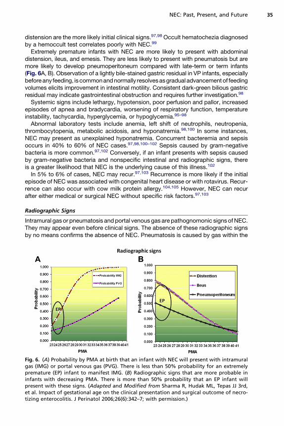
The clinical presentation of NEC can range from nonspecific signs that progressinsidiously over several days to a fulminant onset of gastrointestinal signs, multiorgansystem dysfunction, and shock over a few hours.6 Early signs of NEC are nonspecificand may be indistinguishable from those of sepsis.5,6,11,29,94,95 Clinical signs includeboth intestinal and systemic perturbations. Intestinal signs in early NEC can presentas feeding intolerance that may manifest as increased prefeeding gastric residuals,emesis, abdominal distension, and bloody stools (hematochezia). Less commonly,when the stomach is involved, NEC can present as bloody emesis or a bloody gastricresidual.95,96 During the advanced stage of NEC, the abdomen may appear shiny, dis-tended, and erythematous. Infants generally prefer to assume a frog-leg position(position of comfort, Fig. 5A) and are hyporesponsive.6,94–96 On gentle palpation,
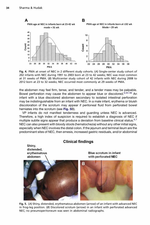
Fig. 4. PMA at onset of NEC in 2 different study cohorts. (A) Single-center study cohort of202 infants with NEC during 1991 to 2003 born at 23 to 42 weeks; NEC was most commonat 31 weeks of PMA. (B) Multicenter study cohort of 42 infants with NEC during 2008 to2012 born at 23 to 32 weeks; NEC occurred most commonly at 29 weeks of PMA.
Sharma & Hudak34
the abdomen may feel firm, tense, and tender, and a tender mass may be palpable.Bowel perforation may cause the abdomen to appear blue or discolored.6,94–96 Aninfant with a blue discolored abdomen secondary to isolated intestinal perforationmay be indistinguishable from an infant with NEC. In a male infant, erythema or bluishdiscoloration of the scrotum may appear if peritoneal fluid from perforated bowelherniates into the scrotum (see Fig. 5B).VP infants do not manifest tenderness and guarding unless NEC is advanced.
Therefore, a high index of suspicion is required to establish a diagnosis of NEC ifmultiple subtle signs appear that produce a deviation from baseline clinical status.6,7
NEC can also present with bloody stools (hematochezia) without any other initial signs,especially when NEC involves the distal colon. If the jejunum and terminal ileum are thepredominant sites of NEC, then emesis, increased gastric residuals, and/or abdominal
Fig. 5. (A) Shiny, distended, erythematous abdomen (arrow) of an infant with advanced NECin frog-leg position. (B) Discolored scrotum (arrow) in an infant with perforated advancedNEC; no pneumoperitoneum was seen in abdominal radiographs.
NEC: Past, Present, and Future 35
distension are the more likely initial clinical signs.97,98 Occult hematochezia diagnosedby a hemoccult test correlates poorly with NEC.99
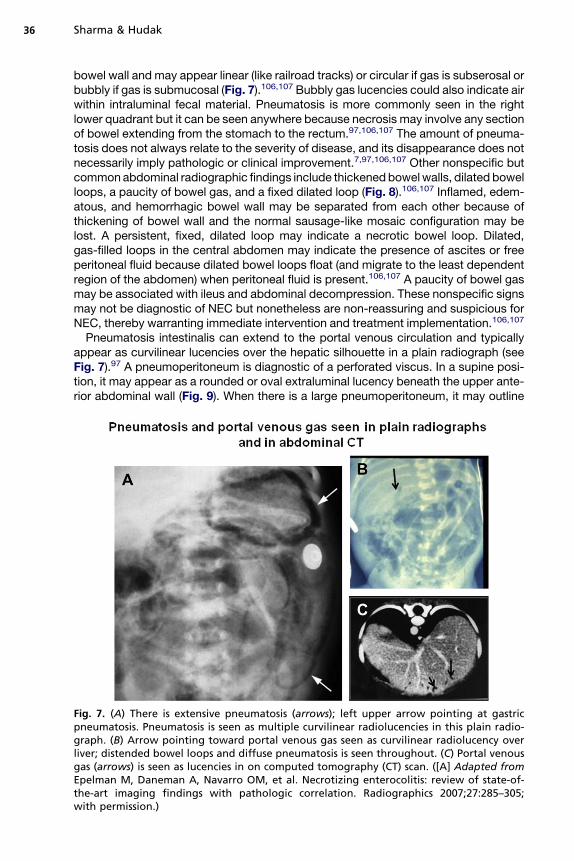
Extremely premature infants with NEC are more likely to present with abdominaldistension, ileus, and emesis. They are less likely to present with pneumatosis but aremore likely to develop pneumoperitoneum compared with late-term or term infants(Fig. 6A, B). Observation of a lightly bile-stained gastric residual in VP infants, especiallybeforeany feeding, is commonandnormally resolvesasgradual advancementof feedingvolumes elicits improvement in intestinal motility. Consistent dark-green bilious gastricresidual may indicate gastrointestinal obstruction and requires further investigation.98
Systemic signs include lethargy, hypotension, poor perfusion and pallor, increasedepisodes of apnea and bradycardia, worsening of respiratory function, temperatureinstability, tachycardia, hyperglycemia, or hypoglycemia.95–98
Abnormal laboratory tests include anemia, left shift of neutrophils, neutropenia,thrombocytopenia, metabolic acidosis, and hyponatremia.98,100 In some instances,NEC may present as unexplained hyponatremia. Concurrent bacteremia and sepsisoccurs in 40% to 60% of NEC cases.97,98,100–102 Sepsis caused by gram-negativebacteria is more common.97,102 Conversely, if an infant presents with sepsis causedby gram-negative bacteria and nonspecific intestinal and radiographic signs, thereis a greater likelihood that NEC is the underlying cause of this illness.102
In 5% to 6% of cases, NEC may recur.97,103 Recurrence is more likely if the initialepisode of NEC was associated with congenital heart disease or with rotavirus. Recur-rence can also occur with cow milk protein allergy.104,105 However, NEC can recurafter either medical or surgical NEC without specific risk factors.97,103
Radiographic Signs
Intramural gas or pneumatosis andportal venous gas are pathognomonic signs ofNEC.They may appear even before clinical signs. The absence of these radiographic signsby no means confirms the absence of NEC. Pneumatosis is caused by gas within the
Fig. 6. (A) Probability by PMA at birth that an infant with NEC will present with intramuralgas (IMG) or portal venous gas (PVG). There is less than 50% probability for an extremelypremature (EP) infant to manifest IMG. (B) Radiographic signs that are more probable ininfants with decreasing PMA. There is more than 50% probability that an EP infant willpresent with these signs. (Adapted and Modified from Sharma R, Hudak ML, Tepas JJ 3rd,et al. Impact of gestational age on the clinical presentation and surgical outcome of necro-tizing enterocolitis. J Perinatol 2006;26(6):342–7; with permission.)
Sharma & Hudak36
bowel wall and may appear linear (like railroad tracks) or circular if gas is subserosal orbubbly if gas is submucosal (Fig. 7).106,107 Bubbly gas lucencies could also indicate airwithin intraluminal fecal material. Pneumatosis is more commonly seen in the rightlower quadrant but it can be seen anywhere because necrosis may involve any sectionof bowel extending from the stomach to the rectum.97,106,107 The amount of pneuma-tosis does not always relate to the severity of disease, and its disappearance does notnecessarily imply pathologic or clinical improvement.7,97,106,107 Other nonspecific butcommonabdominal radiographic findings include thickenedbowelwalls, dilated bowelloops, a paucity of bowel gas, and a fixed dilated loop (Fig. 8).106,107 Inflamed, edem-atous, and hemorrhagic bowel wall may be separated from each other because ofthickening of bowel wall and the normal sausage-like mosaic configuration may belost. A persistent, fixed, dilated loop may indicate a necrotic bowel loop. Dilated,gas-filled loops in the central abdomen may indicate the presence of ascites or freeperitoneal fluid because dilated bowel loops float (and migrate to the least dependentregion of the abdomen) when peritoneal fluid is present.106,107 A paucity of bowel gasmay be associated with ileus and abdominal decompression. These nonspecific signsmay not be diagnostic of NEC but nonetheless are non-reassuring and suspicious forNEC, thereby warranting immediate intervention and treatment implementation.106,107
Pneumatosis intestinalis can extend to the portal venous circulation and typicallyappear as curvilinear lucencies over the hepatic silhouette in a plain radiograph (seeFig. 7).97 A pneumoperitoneum is diagnostic of a perforated viscus. In a supine posi-tion, it may appear as a rounded or oval extraluminal lucency beneath the upper ante-rior abdominal wall (Fig. 9). When there is a large pneumoperitoneum, it may outline
Fig. 7. (A) There is extensive pneumatosis (arrows); left upper arrow pointing at gastricpneumatosis. Pneumatosis is seen as multiple curvilinear radiolucencies in this plain radio-graph. (B) Arrow pointing toward portal venous gas seen as curvilinear radiolucency overliver; distended bowel loops and diffuse pneumatosis is seen throughout. (C) Portal venousgas (arrows) is seen as lucencies in on computed tomography (CT) scan. ([A] Adapted fromEpelman M, Daneman A, Navarro OM, et al. Necrotizing enterocolitis: review of state-of-the-art imaging findings with pathologic correlation. Radiographics 2007;27:285–305;with permission.)
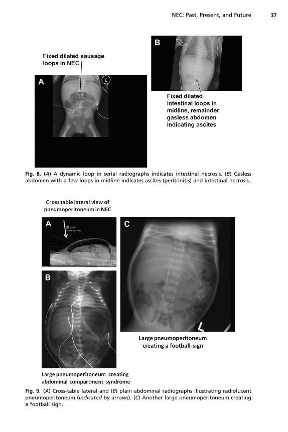
Fig. 8. (A) A dynamic loop in serial radiographs indicates intestinal necrosis. (B) Gaslessabdomen with a few loops in midline indicates ascites (peritonitis) and intestinal necrosis.
Fig. 9. (A) Cross-table lateral and (B) plain abdominal radiographs illustrating radiolucentpneumoperitoneum (indicated by arrows). (C) Another large pneumoperitoneum creatinga football sign.
NEC: Past, Present, and Future 37

Sharma & Hudak38
the falciform ligament giving it the appearance of a longitudinal strip of sutures ina football (American football and rugby but not soccer ball), leading to its designationas the football sign (see Fig. 9).106,107 A cross-table lateral film may demonstrate smallair collection just beneath the abdomen (see Fig. 9). A small pneumoperitoneum maybe difficult to visualize on a single plain film. A left lateral decubitus film will allow freeair to rise to the top over a nondependent surface, facilitating visualization of anabnormal lucency. A small amount of free air may present as a small triangular ora rectangular lucency and make a definitive diagnosis of free air difficult. Sometimesfree air may present as a double-wall sign when the bowel loop is outlined and gas ispresent along serosal and mucosal surfaces.106,107
A nonionic water-soluble contrast study (typically, an upper gastrointestinal seriesfollowed by an enema) can be obtained in circumstances when anatomic gastrointes-tinal obstruction is suspected.107 Hypertonic water-soluble agents are not routinelyrecommended because hypovolemia can result from a shift of fluid from the intravas-cular space to the intestinal lumen.107
Ultrasound
In situations when radiographic signs are nonspecific, the abdominal ultrasound (US)is another modality that can identify even small volumes of free gas. US is also thepreferred modality for visualization of abdominal fluid and ascites. Thickness andechogenicity of the bowel wall and qualitative assessment of peristalsis can be visu-alized best by US color Doppler. It can also be used to assess arterial perfusion of thebowel wall (Fig. 10). Portal venous gas can be more readily seen using abdominalsonography than plain film.107 Faingold and colleagues108 used color Doppler sonog-raphy and demonstrated 100% sensitivity for free air and absent blood flow (necroticgut) compared with 40% sensitivity by radiography. The use of computed tomographyis not advocated for the diagnosis of NEC.107
MANAGEMENTMedical
When an infant is suspected to have NEC, all enteral feedings and medications shouldbe discontinued (Fig. 11). Prompt decompression of the GIT should be accomplishedby the placement of a double-lumen gastric tube (large lumen for aspiration and smalllumen for irrigation and venting) with the institution of low, constant suction. Aspiratedvolume should be replaced with intravenous Ringer lactate solution with extra potas-sium chloride that is lost in gastric output. It is crucial to ensure the patency of thetube. If the abdomen continues to distend despite ongoing continuous suction, thetube should be checked for proper placement and for blockage and nasal continuouspositive airway pressure should be discontinued. Endotracheal intubation is preferredin infants who are deteriorating and have frequent episodes of apnea. Intravascularvolume must be monitored rigorously to ensure adequate tissue perfusion and maybe gauged by frequent assessment of serum electrolytes, hematocrit, and urinaryoutput. As a rule of thumb, anticipation of third spacing in infants with NEC will require1.5-fold maintenance fluid plus replacement of gastric output. Failure to maintaineuvolemia and proper electrolyte repletion can result in shock with hypochloremia,hyponatremia, and hypokalemia. Parenteral nutrition should be started early withadequate protein (3.5–4.0 g/kg/d) to maintain positive nitrogen balance and to allowthe repair of injured tissue.6,46,109–111
Strict implementation of infection control measures is critical. Following the cultureof blood and urine, prompt initiation of treatment with appropriate broad-spectrum
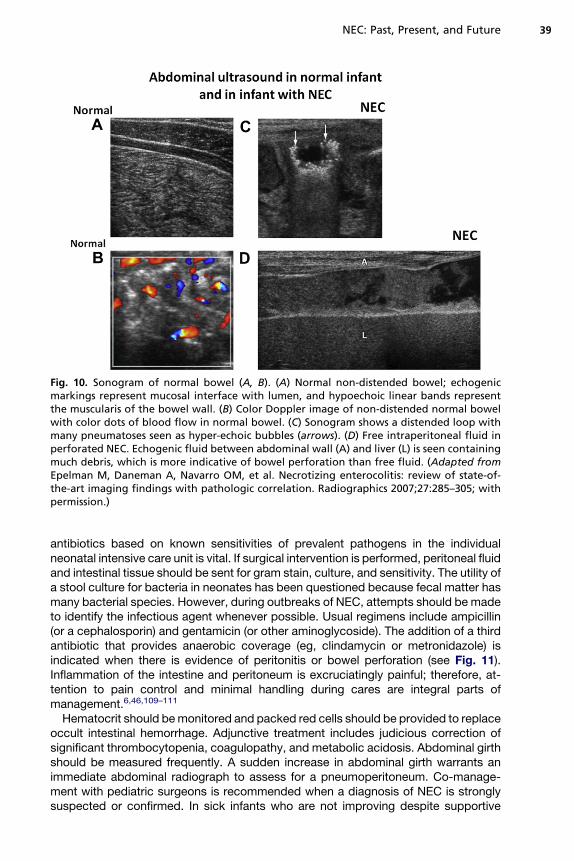
Fig. 10. Sonogram of normal bowel (A, B). (A) Normal non-distended bowel; echogenicmarkings represent mucosal interface with lumen, and hypoechoic linear bands representthe muscularis of the bowel wall. (B) Color Doppler image of non-distended normal bowelwith color dots of blood flow in normal bowel. (C) Sonogram shows a distended loop withmany pneumatoses seen as hyper-echoic bubbles (arrows). (D) Free intraperitoneal fluid inperforated NEC. Echogenic fluid between abdominal wall (A) and liver (L) is seen containingmuch debris, which is more indicative of bowel perforation than free fluid. (Adapted fromEpelman M, Daneman A, Navarro OM, et al. Necrotizing enterocolitis: review of state-of-the-art imaging findings with pathologic correlation. Radiographics 2007;27:285–305; withpermission.)
NEC: Past, Present, and Future 39
antibiotics based on known sensitivities of prevalent pathogens in the individualneonatal intensive care unit is vital. If surgical intervention is performed, peritoneal fluidand intestinal tissue should be sent for gram stain, culture, and sensitivity. The utility ofa stool culture for bacteria in neonates has been questioned because fecal matter hasmany bacterial species. However, during outbreaks of NEC, attempts should be madeto identify the infectious agent whenever possible. Usual regimens include ampicillin(or a cephalosporin) and gentamicin (or other aminoglycoside). The addition of a thirdantibiotic that provides anaerobic coverage (eg, clindamycin or metronidazole) isindicated when there is evidence of peritonitis or bowel perforation (see Fig. 11).Inflammation of the intestine and peritoneum is excruciatingly painful; therefore, at-tention to pain control and minimal handling during cares are integral parts ofmanagement.6,46,109–111
Hematocrit should bemonitored and packed red cells should be provided to replaceoccult intestinal hemorrhage. Adjunctive treatment includes judicious correction ofsignificant thrombocytopenia, coagulopathy, and metabolic acidosis. Abdominal girthshould be measured frequently. A sudden increase in abdominal girth warrants animmediate abdominal radiograph to assess for a pneumoperitoneum. Co-manage-ment with pediatric surgeons is recommended when a diagnosis of NEC is stronglysuspected or confirmed. In sick infants who are not improving despite supportive
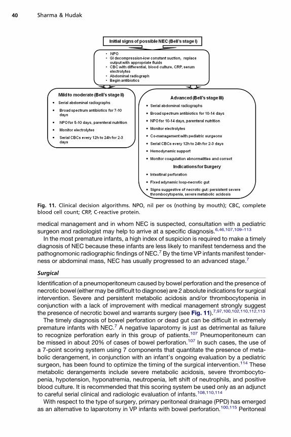
Fig. 11. Clinical decision algorithms. NPO, nil per os (nothing by mouth); CBC, completeblood cell count; CRP, C-reactive protein.
Sharma & Hudak40
medical management and in whom NEC is suspected, consultation with a pediatricsurgeon and radiologist may help to arrive at a specific diagnosis.6,46,107,109–113
In the most premature infants, a high index of suspicion is required to make a timelydiagnosis of NEC because these infants are less likely to manifest tenderness and thepathognomonic radiographic findings of NEC.7 By the time VP infants manifest tender-ness or abdominal mass, NEC has usually progressed to an advanced stage.7
Surgical
Identification of a pneumoperitoneum caused by bowel perforation and the presence ofnecrotic bowel (eithermay be difficult to diagnose) are 2 absolute indications for surgicalintervention. Severe and persistent metabolic acidosis and/or thrombocytopenia inconjunction with a lack of improvement with medical management strongly suggestthe presence of necrotic bowel and warrants surgery (see Fig. 11).7,97,100,102,110,112,113
The timely diagnosis of bowel perforation or dead gut can be difficult in extremelypremature infants with NEC.7 A negative laparotomy is just as detrimental as failureto recognize perforation early in this group of patients.107 Pneumoperitoneum canbe missed in about 20% of cases of bowel perforation.107 In such cases, the use ofa 7-point scoring system using 7 components that quantitate the presence of meta-bolic derangement, in conjunction with an infant’s ongoing evaluation by a pediatricsurgeon, has been found to optimize the timing of the surgical intervention.114 Thesemetabolic derangements include severe metabolic acidosis, severe thrombocyto-penia, hypotension, hyponatremia, neutropenia, left shift of neutrophils, and positiveblood culture. It is recommended that this scoring system be used only as an adjunctto careful serial clinical and radiologic evaluation of infants.108,110,114
With respect to the type of surgery, primary peritoneal drainage (PPD) has emergedas an alternative to laparotomy in VP infants with bowel perforation.100,115 Peritoneal
NEC: Past, Present, and Future 41
drainage serves as a definitive treatment of some patients and as a temporizingmeasure for unstable sick infants until laparotomy can be performed after theirstabilization.115–119
The choice of PPD or laparotomy as a primary surgical intervention has been arguedfor a decade.116–118 In their review of only 2 randomized controlled trials, Rao andcolleagues116 report that there were no differences with respect to mortality and dura-tion for total parenteral nutrition between the drainage and laparotomy. However,these studies did not distinguish between isolated perforation and NEC.120 Neitherof these two trials reached the recruitment target nor showed improved survivalfrom PPD or laparotomy. One of these two trials [Necrotizing Enterocolitis Trial(NET) from the United Kingdom] found that 74% of infants initially treated with PPDrequired a rescue laparotomy. The second NICHD NRN trial did not encourage rescuelaparotomy (which nonetheless was allowed) and did not distinguish NEC from iso-lated intestinal perforation (IIP).117–119 This latter trial, conducted in extremely low birthweight infants (�1000 g), found that laparotomy had an advantage over PPD withrespect to the likelihood of survival and better neurodevelopmental outcome at 18to 22 months of age.120 Death or impairment occurred in 78% of the drainage groupand in 66% of the laparotomy group.The ability to accurately distinguish NEC from IIP is important when making
a comparison between PPD and laparotomy because mortality associated withNEC is greater than with IIP.120,121 A large, single-center, prospective study showedthat bowel perforation in infants with severe NEC who were treated with primary lapa-rotomy fared better than infants treated with a PPD.100,121,122 Conversely, infants withIIP fared better with PPD. These investigators devised the 7-point metabolic derange-ment score and used it in conjunction with ultrasonography or contrast imaging todistinguish NEC from IIP.Other traditional surgical procedures at laparotomy include debridement and the
resection of clearly necrotic bowel and creation of an enterostomy.113,120,121 Some-times multiple excisions are needed to preserve bowel length. Viable ends of bowelare exteriorized as stomas with the distal end as a mucous fistula. Sometimes singlestoma with a Hartmann pouch is created. In this procedure, a colostomy or ileostomyis created and the distal limb is closed by suturing it and placing it back in the peritonealcavity as a temporary measure until the patient is ready for re-anastomosis.110,113,123
PATHOLOGY
Coagulation (hemorrhagic-ischemic) necrosis, inflammation, and bacterial overgrowthare salient features of the histopathology of NEC. Reparative tissue changes, such asepithelial regeneration, formation of granulation tissue, and fibrosis, are found in two-thirds of cases and provide evidence for a duration of tissue injury/reparationprocesses of at least several days.124,125 Damage to the intestinal tract may rangefrom mucosal injury to full-thickness necrosis with focal perforation. Gas bubbles(pneumatosis intestinalis) can be seen in mucosa, submucosa, and serosal surfaces(Fig. 12).125 Although NEC can involve the gut from the stomach to the distal colon(NEC totalis), disease is most commonly found in the terminal ileum and ascendingcolon. Rotavirus-associated NEC most often involves the distal colon.75 NEC cancause focal disease or manifest as multiple large diffuse necrotic areas alternatingwith patches of unaffected bowel. Serosal exudation without obvious perforationcan also occur. In mild stages of NEC, intense congestion of superficial mucosacan result in focal necrosis of villous tips and epithelial sloughing into bowellumen.124,125
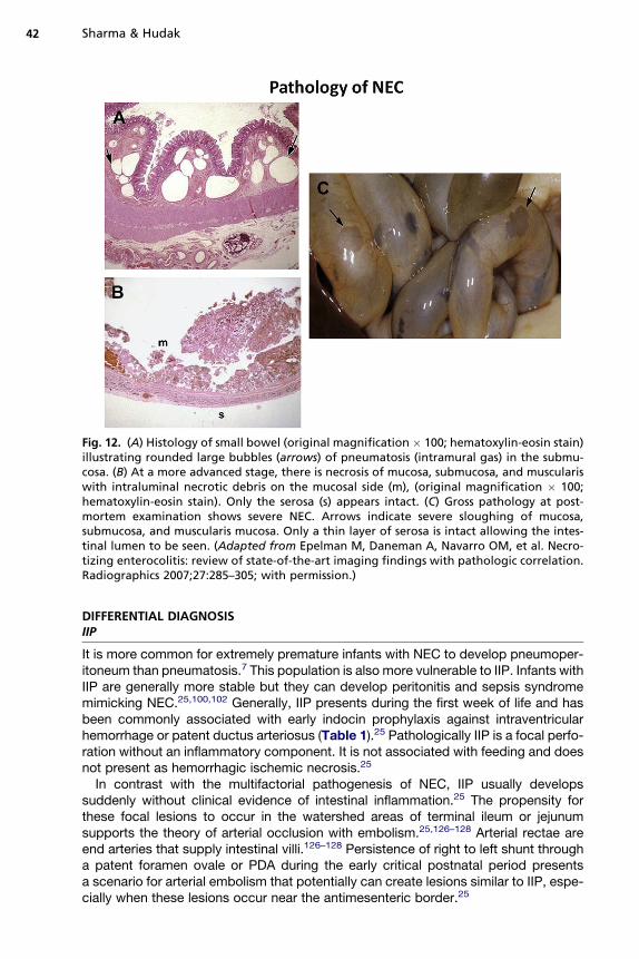
Fig. 12. (A) Histology of small bowel (original magnification � 100; hematoxylin-eosin stain)illustrating rounded large bubbles (arrows) of pneumatosis (intramural gas) in the submu-cosa. (B) At a more advanced stage, there is necrosis of mucosa, submucosa, and musculariswith intraluminal necrotic debris on the mucosal side (m), (original magnification � 100;hematoxylin-eosin stain). Only the serosa (s) appears intact. (C) Gross pathology at post-mortem examination shows severe NEC. Arrows indicate severe sloughing of mucosa,submucosa, and muscularis mucosa. Only a thin layer of serosa is intact allowing the intes-tinal lumen to be seen. (Adapted from Epelman M, Daneman A, Navarro OM, et al. Necro-tizing enterocolitis: review of state-of-the-art imaging findings with pathologic correlation.Radiographics 2007;27:285–305; with permission.)
Sharma & Hudak42
DIFFERENTIAL DIAGNOSISIIP
It is more common for extremely premature infants with NEC to develop pneumoper-itoneum than pneumatosis.7 This population is also more vulnerable to IIP. Infants withIIP are generally more stable but they can develop peritonitis and sepsis syndromemimicking NEC.25,100,102 Generally, IIP presents during the first week of life and hasbeen commonly associated with early indocin prophylaxis against intraventricularhemorrhage or patent ductus arteriosus (Table 1).25 Pathologically IIP is a focal perfo-ration without an inflammatory component. It is not associated with feeding and doesnot present as hemorrhagic ischemic necrosis.25
In contrast with the multifactorial pathogenesis of NEC, IIP usually developssuddenly without clinical evidence of intestinal inflammation.25 The propensity forthese focal lesions to occur in the watershed areas of terminal ileum or jejunumsupports the theory of arterial occlusion with embolism.25,126–128 Arterial rectae areend arteries that supply intestinal villi.126–128 Persistence of right to left shunt througha patent foramen ovale or PDA during the early critical postnatal period presentsa scenario for arterial embolism that potentially can create lesions similar to IIP, espe-cially when these lesions occur near the antimesenteric border.25
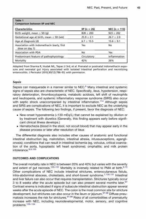
Table 1Comparison between IIP and NEC
Characteristics IIP (n 5 26) NEC (n 5 113)
Birth weight, mean � SD (g) 828 � 203 923 � 202
Gestational age at birth, mean � SD (wk) 25.9 � 2.1 26.7 � 2.0
Age at diagnosis (d) 6.7 � 15.5 15.6 � 9.1
Association with indomethacin (early, firstdose on day 1)
Yes No
Association with PDA No Yes
Predominant feature of pathophysiology Circulatory Inflammation
Mortality 42% 26%
Adapted from Sharma R, Hudak ML, Tepas JJ 3rd, et al. Prenatal or postnatal indomethacin expo-sure and neonatal gut injury associated with isolated intestinal perforation and necrotizingenterocolitis. J Perinatol 2010;30(12):786–93; with permission.
NEC: Past, Present, and Future 43
Sepsis
Sepsis can masquerade in a manner similar to NEC.6 Many intestinal and systemicsigns of sepsis also are characteristic of NEC. Specifically, ileus, hypotension, respi-ratory deterioration, thrombocytopenia, metabolic acidosis, left shift of neutrophilsand neutropenia, and systemic inflammatory response syndrome (SIRS) also occurwith septic shock unaccompanied by intestinal inflammation.114 Although sepsisand SIRS are complications of NEC, it is important to exclude NEC as the underlyingcause of sepsis. The following two findings, if present, favor the diagnosis of NEC:
� New-onset hyponatremia (<130 mEq/L) that cannot be explained by dilution orby treatment with diuretics (Generally, this finding appears early before signifi-cant clinical illness develops.)
� Hematochezia (blood in the stool, not occult blood) that may appear early in thedisease process or later after resolution of ileus
The differential diagnosis also includes other causes of anatomic and functionalintestinal obstruction (eg, malrotation, intestinal atresia, intussusceptions, agangli-onosis); conditions that can result in intestinal ischemia (eg, volvulus, critical coarcta-tion of the aorta, hypoplastic left heart syndrome); omphalitis; and milk proteinintolerance.8,9,105
OUTCOMES AND COMPLICATIONS
The overall mortality rate in NEC is between 20% and 40% but varies with the severityand extent of gut necrosis.129–131 Mortality is inversely related to PMA at birth.6,15
Other complications of NEC include intestinal strictures, enterocutaneous fistula,intra-abdominal abscess, cholestasis, and short-bowel syndrome.7,130,131 Intestinaland liver failure can also occur that requires transplantation. Strictures typically occur3 to 8 weeks after the acute episode but can also present several months later.97
Contrast enema is indicated if signs of subacute intestinal obstruction appear severalweeks after the acute episode of NEC. The colon is the most common site for stricturedevelopment, but strictures can also occur in the ileum or jejunum.97,98 Diffuse pneu-matosis increases the risk for strictures.97,98 Risks of all comorbidities of prematurityincrease with NEC, including neurodevelopmental, motor, sensory, and cognitiveproblems.129–134

Sharma & Hudak44
FUTURE DIRECTIONSPrediction
The prediction of infants at an increased risk of NEC may be possible in the futurethrough the use of methods that are currently available at a few research facilities.These methods use noninvasive indicators, such as profiling of the fecal micro-biome,55 and the identification of the expression of inflammatory proteins from buccalepithelium using buccal swab collection.135 The determination of oxidative stress bymeasuring concentrations of non–protein-bound iron, advanced oxidation proteinproducts, and total hydroxides in cord blood has been reported to be useful in predict-ing which VP infants are at risk for NEC but requires additional validation.136
Predisposing Factors and Management Planning
Earlier noninvasive diagnosis of NEC through the use of abdominal sonography ininfants with nonspecific clinical and radiographic signs and through the assessmentof metabolic derangement may result in more timely and appropriate medical treat-ment and surgical intervention.107,112 However, no studies have demonstratedimproved clinical outcomes caused by facilitated diagnosis and management. Futuretrials comparing outcomes of peritoneal drainage with laparotomy should include dis-tinguishing NEC from IIP.100
Perforation Versus NEC
Clinical studies suggest that the isolated perforation occurs when the intestinalmucosa has been weakened by processes, such as local ischemia-reperfusion orthromboembolism. Conversely, NEC involves both infectious and inflammatory mech-anisms.137 Designing strategies to prevent the occurrence of NEC and to improveoutcomes of infants who develop NEC should consider these diagnoses as fundamen-tally different disease processes.The focus of NEC research is shifting from concentrating on the distinct nature of
this disease to understanding the unique characteristics of immune-naıve prematurepatients. Hence, investigative and interventional techniques aimed at ensuringa more appropriate and mature intestinal immune response should be tested aspreventative strategies.
REFERENCES
1. Obladen M. Necrotizing enterocolitis –150 years of fruitless search of the cause.Neonatology 2009;96:203–10.
2. Mizrahi A, Barlow O, Berdon W, et al. Necrotizing enterocolitis in prematureinfants. J Pediatr 1965;66:697–705.
3. Touloukian RJ. Neonatal enterocolitis: an update on etiology, diagnosis, andtreatment. Surg Clin North Am 1976;55:376–87.
4. Santulli TV, Schullinger JN, Heird WC, et al. Acute necrotizing enterocolitis ininfancy: a review of 64 cases. Pediatrics 1975;55:376–87.
5. Bell MJ, Ternberg JL, Feigin RD, et al. Neonatal necrotizing enterocolitis. Ther-apeutic decisions based upon clinical staging. Ann Surg 1978;187(1):1–7.
6. Neu J, Walker WA. Necrotizing enterocolitis. N Engl J Med 2011;364(3):255–64.7. Sharma R, Hudak ML, Tepas JJ 3rd, et al. Impact of gestational age on the clin-
ical presentation and surgical outcome of necrotizing enterocolitis. J Perinatol2006;26(6):342–7.
NEC: Past, Present, and Future 45
8. De La Torre CA, Miguel M, Martınez L, et al. The risk of necrotizing enterocolitisin newborns with congenital heart disease, a single institution-cohort study. CirPediatr 2010;23(2):103–6 [in Spanish].
9. Raboei EH. Necrotizing enterocolitis in full-term neonates: is it aganglionosis?Eur J Pediatr Surg 2009;19(2):101–4.
10. Snyder CL. Outcome analysis for gastroschisis. J Pediatr Surg 1999;34(8):1253–6.
11. Thompson AM, Bizzarro MJ. Necrotizing enterocolitis in newborns: pathogen-esis, prevention and management. Drugs 2008;68(9):1227–38.
12. Fanaroff AA, Stoll BJ, Wright LL, et al, NICHD Neonatal Research Network.Trends in neonatal morbidity and mortality for very low birth weight infants.Am J Obstet Gynecol 2007;196(2):147.e1–8.
13. Stoll BJ, Hansen NI, Bell EF. Neonatal outcomes of extremely preterm infantsfrom the NICHD Neonatal Research Network. Pediatrics 2010;126(3):443–56.
14. Guillet R, Stoll BJ, Cotten CM, et al. Association of H2-blocker therapy andhigher incidence of necrotizing enterocolitis in very low birth weight infants.Pediatrics 2006;117(2):e137–42.
15. FitzgibbonsSC,ChingY,YuD, et al.Mortality of necrotizingenterocolitis expressedby birth weight categories. J Pediatr Surg 2009;44(6):1072–5 [discussion:1075–6].
16. EXPRESS group. Incidence of and risk factors for neonatal morbidity after activeperinatal care: extremely preterm infants study in Sweden (EXPRESS). ActaPaediatr 2010;99(7):978–92.
17. Bajwa NM, Berner M, Worley S, et al, Swiss Neonatal Network. Population basedage stratified morbidities of premature infants in Switzerland. Swiss Med Wkly2011;141:w13212.
18. Yee WH, Soraisham AS, Shah VS, et al. Incidence and timing of presentation ofnecrotizing enterocolitis in preterm infants. Pediatrics 2012;129(2):e298–304.
19. Hunter CJ, Upperman JS, Ford HR, et al. Understanding the susceptibility of thepremature infant to necrotizing enterocolitis (NEC). Pediatr Res 2008;63(2):117–23.
20. Berrington JE, Hearn RI, Bythell M, et al. Deaths in preterm infants: changingpathology over 2 decades. J Pediatr 2012;160(1):49–53.e1.
21. Neu J, Mihatsch W. Recent developments in necrotizing enterocolitis. JPEN JParenter Enteral Nutr 2012;36(Suppl 1):30S–5S.
22. Sharma R, Tepas JJ. Microecology, intestinal epithelial barrier and necrotizingenterocolitis. Pediatr Surg Int 2010;26:11–21.
23. Young CM, Kingma SD, Neu J. Ischemia-reperfusion and neonatal intestinalinjury. J Pediatr 2011;158:e25–8.
24. Neu J. The myth of asphyxia and hypoxia-ischemia as primary causes of necro-tizing enterocolitis. Biol Neonate 2005;87:97–8.
25. Sharma R, Hudak ML, Tepas JJ 3rd, et al. Prenatal or postnatal indomethacinexposure and neonatal gut injury associated with isolated intestinal perforationand necrotizing enterocolitis. J Perinatol 2010;30(12):786–93.
26. Nowicki PT, Caniano DA, Hammond S, et al. Endothelial nitric oxide synthase inhuman intestine resected for necrotizing enterocolitis. J Pediatr 2007;150(1):40–5.
27. Nankervis CA, Giannone PJ, Reber KM. The neonatal intestinal vasculature:contributing factors to necrotizing enterocolitis. Semin Perinatol 2008;32:83–91.
28. Berseth CL. Gut motility and the pathogenesis of necrotizing enterocolitis. ClinPerinatol 1994;21(2):263–70.
Sharma & Hudak46
29. Wu SF, Caplan M, Lin HC. Necrotizing enterocolitis: old problem with new hope.Pediatr Neonatol 2012;53(3):158–63.
30. Morowitz MJ, Poroyko V, Caplan M, et al. Redefining the role of intestinalmicrobes in the pathogenesis of necrotizing enterocolitis. Pediatrics 2010;125(4):777–85.
31. Sharma R, Tepas JJ 3rd, Hudak ML, et al. Neonatal gut barrier and multipleorgan failure: role of endotoxin and proinflammatory cytokines in sepsis andnecrotizing enterocolitis. J Pediatr Surg 2007;42(3):454–61.
32. Ravindranath T, Yoshioka T, Goto M, et al. Endotoxemia following enteral refeed-ing in children. Clin Pediatr (Phila) 1997;36(9):523–8.
33. Kau A, Ahren PP, Griffin NW, et al. Human nutrition, the gut microbiome, andimmune system: envisioning the future. Nature 2012;474(7351):327–36.
34. Liu Z, Li N, Neu J. Tight junctions, leaky intestines, and pediatric diseases. ActaPaediatr 2005;94(4):386–93.
35. Sharma R, Young C, Neu J. Molecular modulation of intestinal epithelial barrier:contribution of microbiota. J Biomed Biotechnol 2010;2010:305879.
36. Josephson CD, Wesolowski A, Bao G, et al. Do red cell transfusions increase therisk of necrotizing enterocolitis in premature infants? J Pediatr 2010;157(6):972–978.e1–3.
37. Christensen RD, Lambert DK, Henry E, et al. Is “transfusion-associated necro-tizing enterocolitis” an authentic pathogenic entity? Transfusion 2010;50(5):1106–12.
38. Singh R, Visintainer PF, Frantz ID 3rd, et al. Association of necrotizing enteroco-litis with anemia and packed red blood cell transfusions in preterm infants.J Perinatol 2011;31(3):176–82.
39. Paul DA, Mackley A, Novitsky A, et al. Increased odds of necrotizing enteroco-litis after transfusion of red blood cells in premature infants. Pediatrics 2011;127(4):635–41.
40. Blau J, Calo JM, Dozor D, et al. Transfusion-related acute gut injury: necrotizingenterocolitis in very low birth weight neonates after packed red blood cell trans-fusion. J Pediatr 2011;158(3):403–9.
41. dos Santos AM, Guinsburg R, de Almeida MF, et al, Brazilian Network onNeonatal Research. Red blood cell transfusions are independently associatedwith intra-hospital mortality in very low birth weight preterm infants. J Pediatr2011;159(3):371–376.e1–3.
42. El-Dib M, Narang S, Lee E, et al. Red blood cell transfusion, feeding and necro-tizing enterocolitis in preterm infants. J Perinatol 2011;31(3):183–7.
43. Afrazi A, Sodhi CP, Richardson W, et al. New insights into the pathogenesis andtreatment of necrotizing enterocolitis: toll-like receptors and beyond. Pediatr Res2011;69(3):183–8.
44. Neish AS. Microbes in gastrointestinal health and disease. Gastroenterology2009;136(1):65–80.
45. Kawai T, Akira S. Toll-like receptors and their crosstalk with other innate recep-tors in infection and immunity. Immunity 2011;34(5):637–50.
46. Lin PW, Nasr TR, Stoll BJ. Necrotizing enterocolitis: recent scientific advances inpathophysiology and prevention. Semin Perinatol 2008;32(2):70–82.
47. Neu J, ChenM, Beierle E. Intestinal innate immunity: howdoes it relate to the path-ogenesis of necrotizing enterocolitis. Semin Pediatr Surg 2005;14(3):137–44.
48. Nanthakumar N, Meng D, Goldstein AM, et al. The mechanism of excessiveintestinal inflammation in necrotizing enterocolitis: an immature innate response.PLoS One 2011;6(3):e17776.
NEC: Past, Present, and Future 47
49. Ganguli K, Walker WA. Probiotics in the prevention of necrotizing enterocolitis.J Clin Gastroenterol 2011;45(Suppl):S133–8.
50. Lin PW, Myers LE, Ray L, et al. Lactobacillus rhamnosus blocks inflammatorysignaling in vivo via reactive oxygen species generation. Free Radic Biol Med2009;47(8):1205–11.
51. Alexander VN, Northrup V, Bizzarro MJ. Antibiotic exposure in the newbornintensive care unit and the risk of necrotizing enterocolitis. J Pediatr 2011;159(3):392–7.
52. Cotten CM, Taylor S, Stoll B, et al. Prolonged duration of initial empirical antibi-otic treatment is associated with increased rates of necrotizing enterocolitis anddeath for extremely low birth weight infants. Pediatrics 2009;123:58–66.
53. Bengmark S. Gut microbiota, immune development and function. PharmacolRes 2012. pii: S1043–6618(12)00166-1. http://dx.doi.org/10.1016/j.phrs.2012.09.002.
54. Cilieborg MS, Boye M, Sangild PT. Bacterial colonization and gut developmentin preterm neonates. Early Hum Dev 2012;88(Suppl 1):S41–9 [Epub 2012Jan 28].
55. Mai V, Young CM, Ukhanova M, et al. Fecal microbiota in premature infants priorto necrotizing enterocolitis. PLoS 2011;6(6):e20647.
56. Mohankumar K, Kaza N, Jagadeeswaran R, et al. Gut mucosal injury inneonates is marked by macrophage infiltration in contrast to pleomorphic infil-trates in adult: evidence from an animal model. Am J Physiol Gastrointest LiverPhysiol 2012;303(1):G93–102.
57. Nanthakumar NN, Young C, Ko JS, et al. Glucocorticoid responsiveness indeveloping human intestine: possible role in prevention of necrotizing enteroco-litis. Am J Physiol Gastrointest Liver Physiol 2005;288(1):G85–92.
58. Desfrere L, de Oliveira I, Goffinet F, et al. Increased incidence of necrotizingenterocolitis in premature infants born to HIV-positive mothers. AIDS 2005;19(14):1487–93.
59. Cunningham SC, Fakhry K, Bass BL, et al. Neutropenic enterocolitis in adults:case series and review of the literature. Dig Dis Sci 2005;50(2):215–20.
60. Choi JH, Lee JM, Shin WS, et al. Necrotizing enterocolitis: experience of 27cases from a single Korean institution. Int J Hematol 2000;72(3):358–61.
61. Downard CD, Grant SN, Matheson PJ, et al. Altered intestinal microcirculation isthe critical event in the development of necrotizing enterocolitis. J Pediatr Surg2011;46(6):1023–8.
62. Kim M, Christley S, Alverdy JC, et al. Immature oxidative stress managementas a unifying principle in the pathogenesis of necrotizing enterocolitis: insightsfrom an agent-based model. Surg Infect (Larchmt) 2012;13(1):18–32 [Epub2012 Jan 4].
63. Chokshi NK, Guner YS, Hunter CJ, et al. The role of nitric oxide in intestinalepithelial injury and restitution in neonatal necrotizing enterocolitis [review].Semin Perinatol 2008;32(2):92–9.
64. Li N, Quidgley MC, Kobeissy FH, et al. Microbial cell components induced toler-ance to flagellin-stimulated inflammation through Toll-like receptor pathways inintestinal epithelial cells. Cytokine 2012;60(3):806–11 [PMID: 22944462].
65. Smith B, Bode S, Skov TH, et al. Investigation of the early intestinal microflora inpremature infants with/without necrotizing enterocolitis using two differentmethods. Pediatr Res 2012;71(1):115–20.
66. Peter CS, Feuerhahn M, Bohnhorst B, et al. Necrotising enterocolitis: is therea relationship to specific pathogens? Eur J Pediatr 1999;158:67–70.
Sharma & Hudak48
67. Powell J, Bureau MA, Pare C, et al. Necrotizing enterocolitis. Epidemic followingan outbreak of Enterobacter cloacae type 3305573 in a neonatal intensive careunit. Am J Dis Child 1980;134:1152–4.
68. Mollitt DL, Tepas JJ 3rd, Talbert JL. The microbiology of neonatal peritonitis.Arch Surg 1988;123:176–9.
69. Howard FM, Flynn DM, Bradley JM, et al. Outbreak of necrotising enterocolitiscaused by Clostridium butyricum. Lancet 1977;2:1099–102.
70. Van Acker J, de Smet F, Muyldermans G, et al. Outbreak of necrotizing entero-colitis associated with Enterobacter sakazakii in powdered milk formula. J ClinMicrobiol 2001;39:293–7.
71. Smith B, Bode S, Petersen BL, et al. Community analysis of bacteria colonizingintestinal tissue of neonates with necrotizing enterocolitis. BMC Microbiol 2011;11:73. http://dx.doi.org/10.1186/1471-2180-11-73.
72. Tzialla C, Civardi E, Borghesi A, et al. Emerging viral infections in neonatal inten-sive care unit. J Matern Fetal Neonatal Med 2011;24(Suppl 1):156–8 [Epub 2011Aug 31].
73. Sharma R, Hudak ML, Premachandra BR, et al. Clinical manifestations of rota-virus infection in the neonatal intensive care unit. Pediatr Infect Dis J 2002;21(12):1099–105.
74. Stuart RL, Tan K, Mahar JE, et al. An outbreak of necrotizing enterocolitis asso-ciated with norovirus genotype GII.3. Pediatr Infect Dis J 2010;29(7):644–7.
75. Sharma R, Garrison RD, Tepas JJ 3rd, et al. Rotavirus-associated necrotizingenterocolitis: an insight into a potentially preventable disease? J Pediatr Surg2004;39(3):453–7.
76. Birenbaum E, Handsher R, Kuint J, et al. Echovirus type 22 outbreak associatedwith gastro-intestinal disease in a neonatal intensive care unit. Am J Perinatol1997;14:469–73.
77. Chany C, Moscovici O, Lebon P, et al. Association of coronavirus infection withneonatal necrotizing enterocolitis. Pediatrics 1982;69:209–14.
78. Bagci S, Eis-Hubinger AM, Franz AR, et al. Detection of astrovirus in prema-ture infants with necrotizing enterocolitis. Pediatr Infect Dis J 2008;27(4):347–50.
79. Lodha A, de Silva N, Petric M, et al. Human torovirus: a new virus associatedwith neonatal necrotizing enterocolitis. Acta Paediatr 2005;94:1085–8.
80. Karlowicz MG. Risk factors associated with fungal peritonitis in very low birthweight neonates with severe necrotizing enterocolitis: a case-control study.Pediatr Infect Dis J 1993;12:574–7.
81. Boccia D, Stolfi I, Lana S, et al. Nosocomial necrotizing outbreaks: epidemiologyand control measures. Eur J Pediatr 2001;160:385–91.
82. Lemyre B, Xiu W, Bouali NR, et al. A decrease in the number of cases of necro-tizing enterocolitis associated with the enhancement of infection prevention andcontrol measures during a Staphylococcus aureus outbreak in a neonatal inten-sive care unit. Infect Control Hosp Epidemiol 2012;33(1):29–33.
83. Faustini A, Forastiere F, Giorgi-Rossi P, et al. An epidemic of gastroenteritis andmild necrotizing enterocolitis in two neonatal units of a university hospital inRome, Italy. Epidemiol Infect 2004;132(3):455–65.
84. Turcios-Ruiz RM, Axelrod P, St John K, et al. Outbreak of necrotizing enteroco-litis caused by norovirus in a neonatal intensive care unit. J Pediatr 2008;153(3):339–44.
85. Chappe C, Minjolle S, Dabadie A, et al. Astrovirus and digestive disorders inneonatal units. Acta Paediatr 2012;101(5):e208–12.
NEC: Past, Present, and Future 49
86. Moscovici O, Chany C, Lebon P, et al. Association of coronavirus infection withhemorrhagic enterocolitis in newborn infants. C R Seances Acad Sci D 1980;290(13):869–72 [in French].
87. Bagci S, Eis-Hubinger AM, Yassin AF, et al. Clinical characteristics of viral intes-tinal infection in preterm and term neonates. Eur J Clin Microbiol Infect Dis 2010;29(9):1079–84.
88. Ball JM, Tian P, Zeng CQ, et al. Age-dependent diarrhea induced by rotavirusnonstructural glycoprotein. Science 1996;272:101–4.
89. Guttman JA, Finlay BB. Tight junctions as targets of infectious agents. BiochimBiophys Acta 2009;1788(4):832–41.
90. Morris AP, Estes MK. Microbes and microbial toxins: paradigms for microbial-mucosal interactions. VIII. Pathological consequences of rotavirus infection andits enterotoxin. Am J Physiol Gastrointest Liver Physiol 2001;281(2):G303–10.
91. SchroederME, Hosteller HA, Schroeder F, et al. Elucidation of the rotavirus NSP4-caveolin-1 and-cholesterol interactions using synthetic peptides. J Amino acids2012;2012:575180. http://dx.doi.org/10.1155/2012/575180.
92. Neish AS. The gut microflora and intestinal epithelial cells: a continuing dia-logue. Microbes infect 2002;4:309–17.
93. Cilieborg MS, Boye M, Mølbak L, et al. Preterm birth and necrotizing enteroco-litis alter gut colonization in pigs. Pediatr Res 2011;69(1):10–6.
94. Kliegman RM, Hack M, Jones P, et al. Epidemiological study of necrotizingenterocolitis among low birth weight infants. J Pediatr 1982;100:440–4.
95. Kanto WP Jr, Hunter JE, Stoll BJ. Recognition and medical management ofnecrotizing enterocolitis. Clin Perinatol 1994;21(2):335–46.
96. Gephart SM, McGrath JM, Effken JA, et al. Necrotizing enterocolitis risk: state ofthe science. Adv Neonatal Care 2012;12(2):77–87.
97. Sharma R, Tepas JJ 3rd, Hudak ML, et al. Portal venous gas and surgicaloutcome of neonatal necrotizing enterocolitis. J Pediatr Surg 2005;40(2):371–6.
98. Faix RG, Nelson M. Neonatal enterocolitis: progress, problems, and prospects.In: David TJ, editor. Recent advances in Paediatrics, vol. 16. Edinburgh:Churchill Livingstone; 1998. p. 359–407.
99. Abramo TJ, Evans JS, Kokomoor FW, et al. Occult blood in stools and necro-tizing enterocolitis. Am J Dis Child 1988;142:451–2.
100. Sharma R, Tepas JJ 3rd, Mollitt DL, et al. Surgical management of bowel perfo-rations and outcome in very low-birth-weight infants (�1,200 g). J Pediatr Surg2004;39(2):190–4.
101. Cole CR, Hansen NI, Higgins RD, et al, Eunice Kennedy Shriver National Insti-tute of Child Health and Human Development’s Neonatal Research Network.Bloodstream infections in very low birth weight infants with intestinal failure.J Pediatr 2012;160(1):54–59.e2.
102. Sharma R, Tepas JJ 3rd, Hudak ML, et al. Neonatal gut injury and infection rate:impact of surgical debridement on outcome. Pediatr Surg Int 2005;21(12):977–82.
103. Stringer MD, Brereton RJ, Drake DP, et al. Recurrent necrotizing enterocolitis.J Pediatr Surg 1993;28(8):979–81.
104. SrinivasanP,BrandlerM,D’SouzaA,etal.Allergicenterocolitispresentingas recur-rent necrotizing enterocolitis in preterm neonates. J Perinatol 2010;30(6):431–3.
105. Coviello C, Rodriquez DC, Cecchi S, et al. Different clinical manifestation ofcow’s milk allergy in two preterm twins newborns. J Matern Fetal NeonatalMed 2012;25(Suppl 1):132–3.
106. Morrison SC, Jacobson JM. The radiology of necrotizing enterocolitis. Clin Peri-natol 1994;21(2):347–63.
Sharma & Hudak50
107. Epelman M, Daneman A, Navarro OM, et al. Necrotizing enterocolitis: review ofstate-of-the-art imaging findings with pathologic correlation. Radiographics2007;27:285–305.
108. Faingold R, Daneman A, Tomlinson G, et al. Necrotizing enterocolitis: assess-ment of bowel viability with color Doppler US. Radiology 2005;235(2):587–94.
109. Kanto WP Jr, Wilson R, Ricketts RR. Management and outcome of necrotizingenterocolitis. Clin Pediatr (Phila) 1985;24(2):79–82.
110. Walsh MC, Kliegman RM. Necrotizing enterocolitis: treatment based on stagingcriteria. Pediatr Clin North Am 1986;33(1):179–201.
111. Neu J. Neonatal necrotizing enterocolitis: an update. Acta Paediatr Suppl 2005;94(449):100–5.
112. Tepas JJ 3rd, Leaphart CL, Plumley D, et al. Trajectory of metabolic derange-ment in infants with necrotizing enterocolitis should drive timing and techniqueof surgical intervention. J Am Coll Surg 2010;210:847–52.
113. Musemeche CA, Kosloske AM, Ricketts RR. Enterostomy in necrotizing entero-colitis: an analysis of techniques and timing of closure. J Pediatr Surg 1987;22(6):479–83.
114. Tepas JJ 3rd, Sharma R, Leaphart CL, et al. Timing of surgical intervention innecrotizing enterocolitis can be determined by trajectory of metabolic derange-ment. J Pediatr Surg 2010;45(2):310–3 [discussion: 313–4].
115. Ein SH, Shandling B, Wesson D, et al. A 13-year experience with peritonealdrainage under local anesthesia for necrotizing enterocolitis perforation.J Pediatr Surg 1990;25:1034–6.
116. Rao SC, Basani L, Simmer K, et al. Peritoneal drainage versus laparotomy asinitial surgical treatment for perforated necrotizing enterocolitis or spontaneousintestinal perforation in preterm low birth weight infants. Cochrane DatabaseSyst Rev 2011;(6):CD006182.
117. Rees CM, Eaton S, Kiely EM, et al. Peritoneal drainage or laparotomy forneonatal bowel perforation? A randomized controlled trial. Ann Surg 2008;248:44–51.
118. Moss RL, Dimmit RA, Barnhart DC, et al. Laparotomy versus peritoneal drainagefor necrotizing enterocolitis. N Engl J Med 2006;354:2225–34.
119. Pierro A, Eaton S, Rees CM, et al. Is there a benefit of peritoneal drainage fornecrotizing enterocolitis in newborn infants? J Pediatr Surg 2010;45:2117–8.
120. Blakely ML, Tyson JE, Lally KP, et al. Laparotomy versus peritoneal drainage fornecrotizing enterocolitis or isolated intestinal perforation in extremely low birthweight infants: outcomes through 18 months adjusted age. Pediatrics 2006;117(4):e680–7.
121. Sola JE, Tepas JJ 3rd, Koniaris LG. Peritoneal drainage versus laparotomy fornecrotizing enterocolitis and intestinal perforation: a meta-analysis. J Surg Res2010;161(1):95–100.
122. Tepas JJ 3rd, Sharma R, Hudak ML, et al. Coming full circle: an evidence-baseddefinition of the timing and type of surgical management of very low-birth-weight(<1000 g) infants with signs of acute intestinal perforation. J Pediatr Surg 2006;41(2):418–22.
123. Kosloske AM. Indications for operation in necrotizing enterocolitis revisited.J Pediatr Surg 1994;29(5):663–6.
124. Ballance WA, Dahms BB, Shenker N, et al. Pathology of necrotizing enteroco-litis: a ten year experience. J Pediatr 1990;117:S6–13.
125. Gould SJ. The pathology of necrotizing enterocolitis. Semin Neonatol 1997;4:239–44.
NEC: Past, Present, and Future 51
126. Jacobs JE, Birnbaum BA, Maglinte DD. Vascular disorders of the small intestine.In: Herlinger H, Maglinte DD, Birnbaum BA, editors. Clinical imaging of the smallintestine. 2nd edition. New York: Springer-Verlag; 1999. p. 439–65.
127. Nankervis CA, Reber KM, Nowicki PT. Age-dependent changes in the postnatalintestinal microcirculation. Microcirculation 2001;8(6):377–87.
128. Nowicki PT, Miller CE, Edwards RC. Effects of hypoxia and ischemia on autore-gulation in postnatal intestine. Am J Physiol 1991;261:G152–7.
129. Sankaran K, Puckett B, Lee DS, et al, Canadian Neonatal Network. Variations inincidence of necrotizing enterocolitis in Canadian neonatal intensive care units.J Pediatr Gastroenterol Nutr 2004;39(4):366–72.
130. Abdullah F, Zhang Y, Camp M, et al. Necrotizing enterocolitis in 20,822 infants:analysis of medical and surgical treatments. Clin Pediatr (Phila) 2010;49(2):166–71.
131. Guner YS, Friedlich P, Wee CP, et al. State-based analysis of necrotizing entero-colitis outcomes. J Surg Res 2009;157(1):21–9.
132. Martin CR, Dammann O, Allred EN, et al. Neurodevelopment of extremelypreterm infants who had necrotizing enterocolitis with or without late bacteremia.J Pediatr 2010;157(5):751–756.e1.
133. Shah DK, Doyle LW, Anderson PJ, et al. Adverse neurodevelopment in preterminfants with postnatal sepsis or necrotizing enterocolitis is mediated by whitematter abnormalities on magnetic resonance imaging at term. J Pediatr 2008;153(2):170–5, 175.e1 [Epub 2008 Apr 3].
134. Pike K, Brocklehurst P, Jones D, et al. Outcomes at 7 years for babies whodeveloped neonatal necrotising enterocolitis: the ORACLE Children Study.Arch Dis Child Fetal Neonatal Ed 2012;97(5):F318–22.
135. Warner BB, Ryan AL, Seeger K, et al. Ontogeny of salivary epidermal growthfactor and necrotizing enterocolitis. J Pediatr 2007;150(4):358–63.
136. Perrone S, Tataranno ML, Negro S, et al. May oxidative stress biomarkers in cordblood predict the occurrence of necrotizing enterocolitis in preterm infants?J Matern Fetal Neonatal Med 2012;25(Suppl 1):128–31.
137. Chan KY, Leung FW, Lam HS, et al. Immunoregulatory protein profiles of necro-tizing enterocolitis versus spontaneous intestinal perforation in preterm infants.PLoS One 2012;7(5):e36977.